JP6687544B2 - Method and system for converting progenitor cells into gastric tissue by directional differentiation - Google Patents
Method and system for converting progenitor cells into gastric tissue by directional differentiation Download PDFInfo
- Publication number
- JP6687544B2 JP6687544B2 JP2016569618A JP2016569618A JP6687544B2 JP 6687544 B2 JP6687544 B2 JP 6687544B2 JP 2016569618 A JP2016569618 A JP 2016569618A JP 2016569618 A JP2016569618 A JP 2016569618A JP 6687544 B2 JP6687544 B2 JP 6687544B2
- Authority
- JP
- Japan
- Prior art keywords
- cells
- gastric
- cell
- tissue
- stem cells
- Prior art date
- Legal status (The legal status is an assumption and is not a legal conclusion. Google has not performed a legal analysis and makes no representation as to the accuracy of the status listed.)
- Active
Links
Images


Classifications
-
- C—CHEMISTRY; METALLURGY
- C12—BIOCHEMISTRY; BEER; SPIRITS; WINE; VINEGAR; MICROBIOLOGY; ENZYMOLOGY; MUTATION OR GENETIC ENGINEERING
- C12N—MICROORGANISMS OR ENZYMES; COMPOSITIONS THEREOF; PROPAGATING, PRESERVING, OR MAINTAINING MICROORGANISMS; MUTATION OR GENETIC ENGINEERING; CULTURE MEDIA
- C12N5/00—Undifferentiated human, animal or plant cells, e.g. cell lines; Tissues; Cultivation or maintenance thereof; Culture media therefor
- C12N5/06—Animal cells or tissues; Human cells or tissues
- C12N5/0697—Artificial constructs associating cells of different lineages, e.g. tissue equivalents
-
- C—CHEMISTRY; METALLURGY
- C12—BIOCHEMISTRY; BEER; SPIRITS; WINE; VINEGAR; MICROBIOLOGY; ENZYMOLOGY; MUTATION OR GENETIC ENGINEERING
- C12N—MICROORGANISMS OR ENZYMES; COMPOSITIONS THEREOF; PROPAGATING, PRESERVING, OR MAINTAINING MICROORGANISMS; MUTATION OR GENETIC ENGINEERING; CULTURE MEDIA
- C12N5/00—Undifferentiated human, animal or plant cells, e.g. cell lines; Tissues; Cultivation or maintenance thereof; Culture media therefor
- C12N5/06—Animal cells or tissues; Human cells or tissues
- C12N5/0602—Vertebrate cells
- C12N5/0679—Cells of the gastro-intestinal tract
-
- G—PHYSICS
- G01—MEASURING; TESTING
- G01N—INVESTIGATING OR ANALYSING MATERIALS BY DETERMINING THEIR CHEMICAL OR PHYSICAL PROPERTIES
- G01N33/00—Investigating or analysing materials by specific methods not covered by groups G01N1/00 - G01N31/00
- G01N33/48—Biological material, e.g. blood, urine; Haemocytometers
- G01N33/50—Chemical analysis of biological material, e.g. blood, urine; Testing involving biospecific ligand binding methods; Immunological testing
- G01N33/5005—Chemical analysis of biological material, e.g. blood, urine; Testing involving biospecific ligand binding methods; Immunological testing involving human or animal cells
-
- C—CHEMISTRY; METALLURGY
- C12—BIOCHEMISTRY; BEER; SPIRITS; WINE; VINEGAR; MICROBIOLOGY; ENZYMOLOGY; MUTATION OR GENETIC ENGINEERING
- C12N—MICROORGANISMS OR ENZYMES; COMPOSITIONS THEREOF; PROPAGATING, PRESERVING, OR MAINTAINING MICROORGANISMS; MUTATION OR GENETIC ENGINEERING; CULTURE MEDIA
- C12N2501/00—Active agents used in cell culture processes, e.g. differentation
- C12N2501/10—Growth factors
- C12N2501/11—Epidermal growth factor [EGF]
-
- C—CHEMISTRY; METALLURGY
- C12—BIOCHEMISTRY; BEER; SPIRITS; WINE; VINEGAR; MICROBIOLOGY; ENZYMOLOGY; MUTATION OR GENETIC ENGINEERING
- C12N—MICROORGANISMS OR ENZYMES; COMPOSITIONS THEREOF; PROPAGATING, PRESERVING, OR MAINTAINING MICROORGANISMS; MUTATION OR GENETIC ENGINEERING; CULTURE MEDIA
- C12N2501/00—Active agents used in cell culture processes, e.g. differentation
- C12N2501/10—Growth factors
- C12N2501/113—Acidic fibroblast growth factor (aFGF, FGF-1)
-
- C—CHEMISTRY; METALLURGY
- C12—BIOCHEMISTRY; BEER; SPIRITS; WINE; VINEGAR; MICROBIOLOGY; ENZYMOLOGY; MUTATION OR GENETIC ENGINEERING
- C12N—MICROORGANISMS OR ENZYMES; COMPOSITIONS THEREOF; PROPAGATING, PRESERVING, OR MAINTAINING MICROORGANISMS; MUTATION OR GENETIC ENGINEERING; CULTURE MEDIA
- C12N2501/00—Active agents used in cell culture processes, e.g. differentation
- C12N2501/10—Growth factors
- C12N2501/115—Basic fibroblast growth factor (bFGF, FGF-2)
-
- C—CHEMISTRY; METALLURGY
- C12—BIOCHEMISTRY; BEER; SPIRITS; WINE; VINEGAR; MICROBIOLOGY; ENZYMOLOGY; MUTATION OR GENETIC ENGINEERING
- C12N—MICROORGANISMS OR ENZYMES; COMPOSITIONS THEREOF; PROPAGATING, PRESERVING, OR MAINTAINING MICROORGANISMS; MUTATION OR GENETIC ENGINEERING; CULTURE MEDIA
- C12N2501/00—Active agents used in cell culture processes, e.g. differentation
- C12N2501/10—Growth factors
- C12N2501/119—Other fibroblast growth factors, e.g. FGF-4, FGF-8, FGF-10
-
- C—CHEMISTRY; METALLURGY
- C12—BIOCHEMISTRY; BEER; SPIRITS; WINE; VINEGAR; MICROBIOLOGY; ENZYMOLOGY; MUTATION OR GENETIC ENGINEERING
- C12N—MICROORGANISMS OR ENZYMES; COMPOSITIONS THEREOF; PROPAGATING, PRESERVING, OR MAINTAINING MICROORGANISMS; MUTATION OR GENETIC ENGINEERING; CULTURE MEDIA
- C12N2501/00—Active agents used in cell culture processes, e.g. differentation
- C12N2501/10—Growth factors
- C12N2501/155—Bone morphogenic proteins [BMP]; Osteogenins; Osteogenic factor; Bone inducing factor
-
- C—CHEMISTRY; METALLURGY
- C12—BIOCHEMISTRY; BEER; SPIRITS; WINE; VINEGAR; MICROBIOLOGY; ENZYMOLOGY; MUTATION OR GENETIC ENGINEERING
- C12N—MICROORGANISMS OR ENZYMES; COMPOSITIONS THEREOF; PROPAGATING, PRESERVING, OR MAINTAINING MICROORGANISMS; MUTATION OR GENETIC ENGINEERING; CULTURE MEDIA
- C12N2501/00—Active agents used in cell culture processes, e.g. differentation
- C12N2501/10—Growth factors
- C12N2501/16—Activin; Inhibin; Mullerian inhibiting substance
-
- C—CHEMISTRY; METALLURGY
- C12—BIOCHEMISTRY; BEER; SPIRITS; WINE; VINEGAR; MICROBIOLOGY; ENZYMOLOGY; MUTATION OR GENETIC ENGINEERING
- C12N—MICROORGANISMS OR ENZYMES; COMPOSITIONS THEREOF; PROPAGATING, PRESERVING, OR MAINTAINING MICROORGANISMS; MUTATION OR GENETIC ENGINEERING; CULTURE MEDIA
- C12N2501/00—Active agents used in cell culture processes, e.g. differentation
- C12N2501/30—Hormones
- C12N2501/38—Hormones with nuclear receptors
- C12N2501/385—Hormones with nuclear receptors of the family of the retinoic acid recptor, e.g. RAR, RXR; Peroxisome proliferator-activated receptor [PPAR]
-
- C—CHEMISTRY; METALLURGY
- C12—BIOCHEMISTRY; BEER; SPIRITS; WINE; VINEGAR; MICROBIOLOGY; ENZYMOLOGY; MUTATION OR GENETIC ENGINEERING
- C12N—MICROORGANISMS OR ENZYMES; COMPOSITIONS THEREOF; PROPAGATING, PRESERVING, OR MAINTAINING MICROORGANISMS; MUTATION OR GENETIC ENGINEERING; CULTURE MEDIA
- C12N2501/00—Active agents used in cell culture processes, e.g. differentation
- C12N2501/40—Regulators of development
- C12N2501/415—Wnt; Frizzeled
-
- C—CHEMISTRY; METALLURGY
- C12—BIOCHEMISTRY; BEER; SPIRITS; WINE; VINEGAR; MICROBIOLOGY; ENZYMOLOGY; MUTATION OR GENETIC ENGINEERING
- C12N—MICROORGANISMS OR ENZYMES; COMPOSITIONS THEREOF; PROPAGATING, PRESERVING, OR MAINTAINING MICROORGANISMS; MUTATION OR GENETIC ENGINEERING; CULTURE MEDIA
- C12N2501/00—Active agents used in cell culture processes, e.g. differentation
- C12N2501/70—Enzymes
- C12N2501/72—Transferases (EC 2.)
- C12N2501/727—Kinases (EC 2.7.)
-
- C—CHEMISTRY; METALLURGY
- C12—BIOCHEMISTRY; BEER; SPIRITS; WINE; VINEGAR; MICROBIOLOGY; ENZYMOLOGY; MUTATION OR GENETIC ENGINEERING
- C12N—MICROORGANISMS OR ENZYMES; COMPOSITIONS THEREOF; PROPAGATING, PRESERVING, OR MAINTAINING MICROORGANISMS; MUTATION OR GENETIC ENGINEERING; CULTURE MEDIA
- C12N2506/00—Differentiation of animal cells from one lineage to another; Differentiation of pluripotent cells
- C12N2506/02—Differentiation of animal cells from one lineage to another; Differentiation of pluripotent cells from embryonic cells
-
- C—CHEMISTRY; METALLURGY
- C12—BIOCHEMISTRY; BEER; SPIRITS; WINE; VINEGAR; MICROBIOLOGY; ENZYMOLOGY; MUTATION OR GENETIC ENGINEERING
- C12N—MICROORGANISMS OR ENZYMES; COMPOSITIONS THEREOF; PROPAGATING, PRESERVING, OR MAINTAINING MICROORGANISMS; MUTATION OR GENETIC ENGINEERING; CULTURE MEDIA
- C12N2506/00—Differentiation of animal cells from one lineage to another; Differentiation of pluripotent cells
- C12N2506/45—Differentiation of animal cells from one lineage to another; Differentiation of pluripotent cells from artificially induced pluripotent stem cells
Description
連邦支援研究に関する記載
本発明は、国立衛生研究所(National Institutes of Health)の助成によるDK080823、DK092456、及びGM063483に基づく連邦政府の支援を受けて行われた。政府は本発明に一定の権利を有する。
STATEMENT REGARDING FEDERALLY SPONSORED RESEARCH This invention was made with federal support under DK080823, DK092456, and GM063483, funded by the National Institutes of Health. The government has certain rights in this invention.
優先権の主張
本願は、あらゆる目的から、2014年5月28日に出願された“Methods and Systems for Converting Precursor Cells into Gastric Tissues through Directed Differentiation”と題されるWells et alに対する米国仮特許出願第62/003,719号明細書に対する優先権及びその利益を主張する。
Claiming Priority The present application, for all purposes, is entitled "Methods and Systems for Converting Precursor Cells into Gastrometric Tissues through Directed 62" US application filed on May 28, 2014. / 003,719 claims priority and its benefits.
本明細書には、幹細胞を指向性分化によって特定の組織又は器官に変換することに関する方法及びシステムが開示される。詳細には、ヒト多能性幹細胞からの胚体内胚葉形成を促進するための方法及びシステムが開示される。また、分化した胚体内胚葉からの胃オルガノイド又は胃組織形成を促進するための方法及びシステムも開示される。 Disclosed herein are methods and systems related to converting stem cells into specific tissues or organs by directional differentiation. In particular, methods and systems for promoting definitive endoderm formation from human pluripotent stem cells are disclosed. Also disclosed are methods and systems for promoting gastric organoid or gastric tissue formation from differentiated definitive endoderm.
胃の機能及び構造は、多種多様な生息場所及び食事に適応して哺乳類種間で大きく異なる。結果的に、非ヒトの胃発生及び疾患モデルには著しい限界がある。例えば、細菌ヘリコバクター・ピロリ(Helicobacter Pylori)は世界人口の50%に感染し、10%が消化性潰瘍疾患を発症し、及び1〜2%1〜3が胃癌を発症する。胃疾患は、消化性潰瘍疾患及び胃癌を含め、世界人口の10%が罹患し、概して慢性ピロリ菌(H.pylori)感染に起因する。ピロリ菌(H.pylori)誘発性疾患の現在のモデルは、感染に対するヒトの反応と同じ病態生理学的特徴を呈しない動物モデルに頼っており4、胃細胞株は生体内での胃上皮の細胞上及び構造上の複雑性を欠いている。従って、ヒトで起こるとおりのピロリ菌(H.pylori)感染の効果を研究するのに適したモデルはない。成体胃幹細胞を使用した最近の進歩により、インビトロでげっ歯類胃上皮を成長させることが可能であるが5、ヒト患者からこれらの細胞を入手しようとすれば外科的手術が必要となり得る。さらに、かかる方法は、ヒト胃の胚発生又は間質−上皮相互作用のモデル化には使用できない。胚発生及び成体胃の構造が種によって異なるため、マウスモデルはこの器官の器官形成及び発病研究に準最適なものとなる。従って、ヒト胃の発生及び疾患の根底にある機構を解明し、且つかかる疾患のヒト治療に有用な新規治療を同定するための、ロバストなインビトロシステムが必要とされている。 Gastric function and structure vary widely between mammalian species, accommodating a wide variety of habitats and diets. As a result, there are significant limitations to non-human gastric development and disease models. For example, the bacterium Helicobacter Pylori infects 50% of the world population, 10% develop peptic ulcer disease, and 1-2% 1-3 develop gastric cancer. Gastric disease affects 10% of the world population, including peptic ulcer disease and gastric cancer, and is generally due to chronic H. pylori infection. The current model of H. pylori-induced diseases relies on animal models that do not exhibit the same pathophysiological features as human response to infection 4 , gastric cell lines rely on cells of gastric epithelium in vivo. It lacks top and structural complexity. Therefore, there is no suitable model to study the effects of H. pylori infection as it occurs in humans. Recent advances in the use of adult gastric stem cells have made it possible to grow rodent gastric epithelium in vitro 5 , but attempts to obtain these cells from human patients may require surgery. Moreover, such methods cannot be used to model embryonic development or stroma-epithelial interactions in human stomach. The differences in embryonic development and adult gastric structure by species make the mouse model suboptimal for studying organogenesis and pathogenesis of this organ. Therefore, there is a need for robust in vitro systems to elucidate the mechanisms underlying human gastric development and disease, and to identify new therapies useful in human treatment of such diseases.
当該技術分野において必要とされているのは、所望の特定のタイプの組織又は生物、詳細には前述の目的の1つ以上に用いることのできる胃組織を作り出すため、ヒト多能性幹細胞などの前駆細胞の終着点を正確に制御する方法及びシステムである。 What is needed in the art is a tissue or organism of the particular type desired, in particular human pluripotent stem cells, such as human pluripotent stem cells, to produce gastric tissue that can be used for one or more of the aforementioned purposes. A method and system for accurately controlling the end point of progenitor cells.
胃オルガノイドの形態などの胃細胞及び/又は胃組織の形成を誘導する方法が開示される。胃細胞及び/又は組織の形成は、前駆細胞内の1つ以上のシグナル伝達経路を活性化及び/又は阻害することによって実施され得る。また、前駆細胞に由来する開示される胃細胞、胃組織、及び/又は胃オルガノイドの使用方法も開示される。 Disclosed are methods of inducing the formation of gastric cells and / or gastric tissue, such as forms of gastric organoids. The formation of gastric cells and / or tissues can be performed by activating and / or inhibiting one or more signaling pathways within progenitor cells. Also disclosed are methods of using the disclosed gastric cells, gastric tissue, and / or gastric organoids derived from progenitor cells.
当業者は、以下に説明する図面が例示目的に過ぎないことを理解するであろう。図面は、いかなる形であれ本教示の範囲を限定することを意図するものではない。 Those skilled in the art will understand that the drawings, described below, are for illustration purposes only. The drawings are not intended to limit the scope of the present teachings in any way.
この特許又は出願ファイルは、色彩を付して作成された少なくとも1つの図面を含んでいる。色彩図面が付された、この特許又は特許出願公開の写しは、請求及び必要な手数料の納付に基づいて、当局によって提供される。 This patent or application file contains at least one drawing executed in color. Copies of this patent or patent application publication with color drawings will be provided by the Authority upon request and payment of the necessary fee.
特に注記されない限り、用語は、関連技術分野の当業者による従来の用法に従い理解されるものとする。 Unless otherwise noted, terms are to be understood according to conventional usage by those of ordinary skill in the relevant art.
本明細書で使用されるとき、用語「全能性幹細胞」(オムニポテント幹細胞としても知られる)は、胚細胞型及び胚体外細胞型に分化することのできる幹細胞である。かかる細胞は、生存能力のある完全な生物を構築することができる。これらの細胞は卵細胞と精細胞の融合によって作られる。受精卵の最初の数回の分裂によって作られる細胞もまた全能性である。 As used herein, the term "totipotent stem cell" (also known as omnipotent stem cell) is a stem cell that is capable of differentiating into embryonic and extraembryonic cell types. Such cells are capable of constructing a viable whole organism. These cells are made by the fusion of egg cells and sperm cells. Cells produced by the first few divisions of a fertilized egg are also totipotent.
本明細書で使用されるとき、用語「多能性幹細胞(PSC)」は、生体のほぼあらゆる細胞型に分化することのできる任意の細胞、即ち、内胚葉(胃の内膜、胃腸管、肺)、中胚葉(筋肉、骨、血液、泌尿生殖器)、及び外胚葉(表皮組織及び神経系)を含む3つの胚葉(胚上皮)のいずれかに由来する細胞を包含する。PSCは、着床前(primplantation)胚盤胞の内部細胞塊細胞の子孫であってもよく、又はある種の遺伝子を強制的に発現させることによる、非多能性細胞、例えば成体体細胞の誘導を通じて得られてもよい。多能性幹細胞は、当業者が容易に理解するであろうとおり、任意の好適な供給源に由来し得る。多能性幹細胞の供給源の例としては、ヒト、げっ歯類、ブタ、ウシを含めた哺乳類供給源が挙げられるが、それに限定されるものではない。 As used herein, the term "pluripotent stem cell (PSC)" refers to any cell capable of differentiating into almost any cell type of the body, namely the endoderm (endogastric lining, gastrointestinal tract, Lungs), mesoderm (muscles, bones, blood, genitourinary organs), and cells derived from any of the three germ layers (embryonic epithelium) including ectoderm (epidermal tissue and nervous system). The PSC may be the progeny of an inner cell mass cell of a preplantation blastocyst, or of a non-pluripotent cell, such as an adult cell, by forcing the expression of certain genes. It may be obtained through induction. Pluripotent stem cells can be derived from any suitable source, as will be readily appreciated by those of skill in the art. Examples of sources of pluripotent stem cells include, but are not limited to, mammalian sources including humans, rodents, pigs, cows.
本明細書で使用されるとき、用語「人工多能性幹細胞(iPSC)」は、iPS細胞と省略されることも多く、ある種の遺伝子の「強制」発現を誘導することによって通常非多能性の細胞、例えば成体体細胞から人工的に得られる多能性幹細胞の一種を指す。 The term "induced pluripotent stem cells (iPSCs)", as used herein, is often abbreviated as iPS cells and is usually non-pluripotent by inducing "forced" expression of certain genes. It refers to a type of pluripotent stem cells that are artificially obtained from adult cells, such as adult cells.
本明細書で使用されるとき、用語「胚性幹細胞(ESC)」は、ES細胞と省略されることも多く、初期胚である胚盤胞の内部細胞塊から得られる多能性の細胞を指す。本発明の目的上、用語「ESC」は、時に胚性生殖細胞もさらに包含して広義に用いられる。 As used herein, the term “embryonic stem cell (ESC)” is often abbreviated as ES cell and refers to a pluripotent cell obtained from the inner cell mass of the blastocyst, which is an early embryo. Point to. For the purposes of the present invention, the term "ESC" is used broadly, sometimes to further include embryonic germ cells.
本明細書で使用されるとき、用語「前駆細胞」は、1つ以上の前駆細胞が自己再生能力又は1つ以上の特殊化した細胞型に分化する能力を獲得する本明細書に記載される方法において使用することのできる任意の細胞を包含する。一部の実施形態において、前駆細胞は多能性であるか、又は多能性になることが可能である。一部の実施形態において、前駆細胞は、多能性を獲得するため外部因子(例えば成長因子)の処理に供される。一部の実施形態において、前駆細胞は、全能性(又はオムニポテント)幹細胞;多能性幹細胞(人工又は非人工);多分化能幹細胞;少分化能幹細胞及び単分化能幹細胞であり得る。一部の実施形態において、前駆細胞は、胚、乳児、小児、又は成人に由来し得る。一部の実施形態において、前駆細胞は、遺伝子操作又はタンパク質/ペプチド処理によって多能性が付与されるように処理に供された体細胞であり得る。 As used herein, the term “progenitor cell” is described herein to acquire the ability of one or more progenitor cells to self-renew or to differentiate into one or more specialized cell types. It includes any cell that can be used in the method. In some embodiments, the progenitor cells are pluripotent or capable of becoming pluripotent. In some embodiments, the progenitor cells are subjected to treatment with an external factor (eg, growth factor) to acquire pluripotency. In some embodiments, the progenitor cells can be totipotent (or omnipotent) stem cells; pluripotent stem cells (artificial or non-artificial); pluripotent stem cells; oligopotent and monopotent stem cells. In some embodiments, progenitor cells can be derived from embryos, infants, children, or adults. In some embodiments, progenitor cells can be somatic cells that have been subjected to treatment so as to be pluripotent by genetic engineering or protein / peptide treatment.
発生生物学において、細胞分化は、それほど特殊化していない細胞がより特殊化した細胞型になる過程である。本明細書で使用されるとき、用語「指向性分化」は、それほど特殊化していない細胞が特定の特殊化した標的細胞型になる過程を表す。特殊化した標的細胞型の特殊性は、初期細胞の運命を定義付け又は改変するために用い得る任意の適用可能な方法により決定することができる。例示的方法としては、限定はされないが、遺伝子操作、化学的処理、タンパク質処理、及び核酸処理が挙げられる。 In developmental biology, cell differentiation is the process by which less specialized cells become more specialized cell types. As used herein, the term "directed differentiation" refers to the process by which less specialized cells become a particular specialized target cell type. The specificity of the specialized target cell type can be determined by any applicable method that can be used to define or modify the fate of early cells. Exemplary methods include, but are not limited to, genetic engineering, chemical treatment, protein treatment, and nucleic acid treatment.
本明細書で使用されるとき、用語「細胞構成物」は、個々の遺伝子、タンパク質、遺伝子を発現するmRNA、及び/又は任意の他の可変的な細胞成分又はタンパク質活性、例えば、典型的には当業者によって生物学的実験(例えばマイクロアレイ又は免疫組織化学による)で例えば計測されるタンパク質修飾(例えばリン酸化)の程度である。生物系、一般的なヒト疾患の根底にある生化学的過程の複雑なネットワークに関する重要な発見、並びに遺伝子発見及び構造決定は、現在、研究過程の一環としての細胞構成物存在量データの適用によるものであり得る。細胞構成物存在量データは、バイオマーカーを同定し、疾患サブタイプを区別し、及び毒性機構を同定する助けとなり得る。 As used herein, the term “cellular composition” refers to an individual gene, protein, mRNA expressing a gene, and / or any other variable cellular component or protein activity, eg, typically Is the degree of protein modification (eg phosphorylation) as measured, for example, in biological experiments (eg by microarray or immunohistochemistry) by a person skilled in the art. Significant discoveries about biological systems, the complex networks of biochemical processes underlying common human diseases, and gene discovery and structural determination are now based on the application of cellular constituent abundance data as part of the research process. Can be one. Cellular constituent abundance data can help identify biomarkers, distinguish disease subtypes, and identify toxic mechanisms.
幹細胞は全ての多細胞生物に見られる。幹細胞は、有糸細胞分裂によって自己複製し、且つ多様な特殊化した細胞型に分化する能力によって特徴付けられる。大まかな2種類の哺乳類幹細胞は、1)胚盤胞の内部細胞塊から単離される胚性幹細胞、及び2)成体組織に見られる成体幹細胞である。発生中の胚では、幹細胞はあらゆる特殊化した胚組織に分化し得る。成体生物では、幹細胞及びプロジェニター細胞は生体の修復システムとして働き、特殊化した細胞を補充し、また血液、皮膚、又は胃組織などの再生器官の正常な代謝回転を維持する。 Stem cells are found in all multicellular organisms. Stem cells are characterized by the ability to self-renew by mitotic division and to differentiate into a variety of specialized cell types. Roughly two types of mammalian stem cells are 1) embryonic stem cells isolated from the inner cell mass of blastocysts, and 2) adult stem cells found in adult tissues. In the developing embryo, stem cells can differentiate into any specialized embryonic tissue. In adult organisms, stem cells and progenitor cells act as repair systems in the body, recruit specialized cells and maintain normal turnover of regenerating organs such as blood, skin, or gastric tissue.
現在、幹細胞は、筋肉又は神経などの様々な組織の細胞と一致する特徴を有する特殊化した細胞へと、細胞培養によって成長させ、転換することができる。医学療法においては、臍帯血及び骨髄を含めた種々の供給源からの高度に可塑性の成体幹細胞が日常的に用いられている。治療的クローニングによって作成される胚細胞株及び自己胚性幹細胞もまた、将来的な治療法の有望な候補として提案されている。 Currently, stem cells can be grown and transformed by cell culture into specialized cells with characteristics that are consistent with cells of various tissues such as muscle or nerve. Highly plastic adult stem cells from various sources, including cord blood and bone marrow, are routinely used in medical therapy. Embryonic cell lines and autologous embryonic stem cells created by therapeutic cloning have also been proposed as potential candidates for future therapeutics.
幹細胞の古典的定義は、典型的には2つの特性:自己複製、即ち未分化状態を維持しつつ多数の細胞分裂周期を経る能力と、発生能、特殊化した細胞型に分化する能力とを指し示している。一部の実施形態において、幹細胞は全能性又は多能性のいずれかであり、即ち幹細胞は任意の成熟細胞型を生じることが可能であり、しかし多分化能又は単分化能プロジェニター細胞が幹細胞と称されることもある。 The classical definition of stem cells typically has two characteristics: self-renewal, the ability to undergo multiple cell division cycles while maintaining an undifferentiated state, and developmental ability, the ability to differentiate into specialized cell types. Pointing. In some embodiments, the stem cells are either totipotent or pluripotent, that is, the stem cells are capable of giving rise to any mature cell type, but the pluripotent or unipotent progenitor cells are stem cells. Sometimes called.
発生能は幹細胞の潜在的分化能力(異なる細胞型に分化する潜在能力)を特定する。全能性幹細胞(オムニポテント幹細胞としても知られる)は、胚細胞型及び胚体外細胞型に分化することができる。これらの細胞は、生存能力のある完全な生物を構築することができる。これらの細胞は卵細胞と精細胞の融合によって作られる。受精卵の最初の数回の分裂によって作られる細胞もまた全能性である。多能性幹細胞(PSC)は全能性細胞の子孫であり、ほぼあらゆる細胞、即ち、内胚葉(胃の内膜、胃腸管、肺)、中胚葉(筋肉、骨、血液、泌尿生殖器)、及び外胚葉(表皮組織及び神経系)を含む3つの胚葉のいずれかに由来する細胞に分化することができる。多分化能幹細胞は幾つもの細胞に分化し得るが、但し近縁の細胞ファミリーのものに限られる。少分化能幹細胞は、リンパ球系又は骨髄系幹細胞などのほんの数種の細胞に分化し得るのみである。単分化能細胞は、それ自体の、唯一つの細胞型のみを作り出すことができ、しかし自己複製の特性を有し、それによって非幹細胞と区別される(例えば筋幹細胞)。 Developmental potential specifies the potential differentiation potential of stem cells (potential to differentiate into different cell types). Totipotent stem cells (also known as omnipotent stem cells) can differentiate into germ cell and extraembryonic cell types. These cells are capable of building a viable whole organism. These cells are made by the fusion of egg cells and sperm cells. Cells produced by the first few divisions of a fertilized egg are also totipotent. Pluripotent stem cells (PSCs) are the descendants of totipotent cells, and almost any cell: endoderm (gastric intima, gastrointestinal tract, lung), mesoderm (muscle, bone, blood, genitourinary organs), and It can differentiate into cells derived from any of the three germ layers including the ectoderm (epidermal tissue and nervous system). Pluripotent stem cells are capable of differentiating into any number of cells, provided they are of a closely related cell family. Poorly differentiated stem cells are only capable of differentiating into a few types of cells such as lymphoid or myeloid stem cells. Unipotent cells are capable of producing only one cell type of their own, but have the property of self-renewal, which distinguishes them from non-stem cells (eg, muscle stem cells).
胚性幹細胞及び人工多能性幹細胞は、ヒト疾患を研究する能力及び動物モデルにおいて治療上有効な代替組織を作成する能力にかつてない影響を与えている。 Embryonic stem cells and induced pluripotent stem cells have an unprecedented impact on the ability to study human diseases and create therapeutically effective alternative tissues in animal models.
発生生物学において、細胞分化は、それほど特殊化していない細胞がより特殊化した細胞型になる過程である。ヒトPSCから治療的細胞型への分化を指向させようとする取り組みの成功のほとんどは、胚器官発生の研究に基づいている。例としては、肝細胞及び膵内分泌細胞の作成が挙げられ、これらの細胞は肝疾患及び糖尿病の動物モデルにおいて機能上の潜在能力を示している。同様に、PSCから腸への分化は、壊死性腸炎、炎症性腸疾患及び短腸症候群などの疾患に治療利益をもたらし得る。 In developmental biology, cell differentiation is the process by which less specialized cells become more specialized cell types. Most successful efforts to direct the differentiation of human PSCs into therapeutic cell types are based on studies of embryonic organ development. Examples include the generation of hepatocytes and pancreatic endocrine cells, which show functional potential in animal models of liver disease and diabetes. Similarly, PSC to intestinal differentiation may have therapeutic benefits in diseases such as necrotizing enteritis, inflammatory bowel disease and short bowel syndrome.
上記で考察したとおり、多能性幹細胞は、3つの胚葉:内胚葉(胃の内膜、胃腸管、肺)、中胚葉(筋肉、骨、血液、泌尿生殖器)、及び外胚葉(表皮組織及び神経系)のいずれかに分化する潜在能力を有する。従って、多能性幹細胞は任意の胎児又は成体細胞型を生じることができる。しかしながら、特定の多能性幹細胞の運命は、数多くの細胞シグナル伝達経路及び数多くの因子によって制御される。さらに、多能性幹細胞は潜在的に胎盤などの胚体外組織に寄与する能力を有しないため、単独では胎児又は成体動物に発育することができない。 As discussed above, pluripotent stem cells have three germ layers: endoderm (gastric intima, gastrointestinal tract, lung), mesoderm (muscle, bone, blood, genitourinary organs), and ectoderm (epidermal tissue and The nervous system). Thus, pluripotent stem cells can give rise to any fetal or adult cell type. However, the fate of a particular pluripotent stem cell is controlled by numerous cell signaling pathways and factors. Moreover, pluripotent stem cells cannot potentially develop into a fetal or adult animal by themselves, as they do not potentially have the ability to contribute to extraembryonic tissues such as the placenta.
現在までに、ヒト多能性幹細胞(hPSC)から胃組織は作成されていない。PSCを肺、肝、膵及び腸細胞に分化させる取り組みの成功は、これらの器官の胚発生の理に適った分子的理解に依存している6〜10。残念ながら、当該技術分野における問題は、内胚葉形成に続く胃発生の理解に相違が多くあることである。従って、hPSCから胃組織への分化を指向させるため、前腸の特異化及びパターン形成、胃の特異化、並びに最後に胃上皮成長及び分化を含めた幾つかの重要な胃発生初期段階を調節するシグナル伝達経路が本出願人によって同定された。加えて、より機能的で複雑な三次元組織を作成するため、本出願人は、前腸管の形態形成並びに腺及び小窩を含む胃上皮構造の形成を含めた、胃発生中に起こる幾つかの形態形成過程を誘導することを目指した。 To date, gastric tissue has not been created from human pluripotent stem cells (hPSCs). Successful efforts to differentiate PSCs into lung, liver, pancreas and enterocytes depend on a rational molecular understanding of embryonic development of these organs 6-10 . Unfortunately, a problem in the art is that there are many differences in understanding gastric development following endoderm formation. Therefore, to direct the differentiation of hPSCs into gastric tissue, it regulates several important early stages of gastric development including foregut specification and patterning, gastric specification, and finally gastric epithelial growth and differentiation. Signaling pathways that have been identified by the applicant. In addition, in order to create a more functional and complex three-dimensional tissue, Applicants have selected several that occur during gastric development, including morphogenesis of the foregut tract and formation of gastric epithelial structures including glands and pits The aim was to induce the morphogenesis process of.
本明細書に記載されるとおり、時系列の成長因子操作を用いて培養下で胎生期胃組織発生を模倣する方法及びシステムが構築される。詳細には、PSC、ヒト胚性幹細胞(hESC)及び人工多能性幹細胞(iPSC)の両方から胃組織への分化をインビトロで指向させる方法及びシステムが構築される。これらの因子は、胎児腸発生を近似する段階:アクチビン誘導性の胚体内胚葉(DE)形成と;FGF/Wnt/BMP誘導性の後方前腸パターン形成(pattering)と、最後に、胃腺及び胃小窩、増殖帯、表層及び前庭部粘液細胞、並びにガストリン、グレリン、及びソマトスタチンを発現する内分泌細胞を含む機能性の胃細胞型及び形態への胃組織成長、形態形成及び細胞分化を促進するレチノイン酸及びEFGシグナル伝達の調節によって得られるプロガストリック(pro‐gastric)培養系とを経てインビトロでのヒト腸の発生を指向させた。 As described herein, methods and systems are constructed that mimic fetal gastric tissue development in culture using a time series of growth factor manipulations. In particular, methods and systems are directed to direct the differentiation of both PSCs, human embryonic stem cells (hESCs) and induced pluripotent stem cells (iPSCs) into gastric tissue in vitro. These factors are steps that approximate fetal gut development: activin-induced definitive endoderm (DE) formation; FGF / Wnt / BMP-induced posterior foregut patterning, and finally gastric glands and stomach. Retinoin promotes gastric tissue growth, morphogenesis and cell differentiation into functional gastric cell types and morphologies including pits, growth zones, superficial and vestibular mucous cells, and endocrine cells expressing gastrin, ghrelin, and somatostatin. Human intestinal development in vitro was directed through a pro-gastric culture system obtained by modulation of acid and EFG signaling.
本出願人は、ヒトPSCから胃細胞、胃組織、及び/又は複雑な構造及び細胞組成を伴う三次元胃組織(hGO)への効率的な段階的分化を可能にする新規胎生期シグナル伝達経路を同定した。本出願人はさらに、発生中のhGOがマウスの発生中の前庭部とほぼ同一の分子的及び形態学的分化段階を経ること、及び得られる胃オルガノイドが、正常な前庭部上皮及び胎児期/生後期の胃と同等の三次元構成を成す一連の粘液細胞、内分泌細胞、及びプロジェニター細胞を含有し得ることを見出した。 Applicants have identified a novel embryonic signaling pathway that enables efficient stepwise differentiation of human PSCs into gastric cells, gastric tissue, and / or three-dimensional gastric tissue (hGO) with complex structure and cell composition. Was identified. Applicants have further demonstrated that developing hGO undergoes nearly the same molecular and morphological differentiation steps as the developing vestibular region of mice, and that the resulting gastric organoids are normal vestibular epithelium and fetal / It has been found that it can contain a series of mucous cells, endocrine cells, and progenitor cells that make up the three-dimensional organization of postnatal stomach.
開示されるヒト胃細胞、胃組織及び/又は胃オルガノイド(hGO)は、ヒト胃の発生、生理機能の新規機構を同定するためのインビトロシステムとして使用されてもよく、及びピロリ菌(H.pylori)に対する胃上皮の病態生理学的反応のモデルとして使用されてもよい。開示される胃細胞、胃組織及び/又は胃hGO及び方法は、創薬及び早期胃癌のモデル化に新しい機会をもたらす。さらに、本明細書には、ヒト胚前腸の初めての三次元作製が開示され、これは、肺及び膵臓を含む他の前腸器官組織の作成に向けた有望な出発点である。 The disclosed human gastric cells, gastric tissues and / or gastric organoids (hGO) may be used as an in vitro system to identify novel mechanisms of human gastric development, physiology, and H. pylori (H. pylori). ) May be used as a model of the pathophysiological response of the gastric epithelium to The disclosed gastric cells, gastric tissue and / or gastric hGO and methods provide new opportunities for drug discovery and modeling of early gastric cancer. In addition, disclosed herein is the first three-dimensional production of human embryonic foregut, which is a promising starting point for the production of other foregut organ tissues including lung and pancreas.
一態様において、前駆細胞から胃細胞、胃組織、及び/又は胃hGOの形成を誘導する方法が開示される。この方法は、a)前駆細胞内の1つ以上のシグナル伝達経路(1つ以上のシグナル伝達経路は、WNTシグナル伝達経路、WNT/FGFシグナル伝達経路、及びFGFシグナル伝達経路から選択される)を活性化するステップであって、それにより前駆細胞の子孫である胃細胞、胃組織及び/又は胃hGOを入手するステップを含み得る。この方法は、前駆細胞内の1つ以上のシグナル伝達経路を阻害するステップb)をさらに含み得る。阻害される1つ以上のシグナル伝達経路はBMPシグナル伝達経路を含み得る。 In one aspect, a method of inducing the formation of gastric cells, gastric tissue, and / or gastric hGO from progenitor cells is disclosed. This method comprises: a) determining one or more signaling pathways in the progenitor cell (wherein the one or more signaling pathways are selected from WNT signaling pathways, WNT / FGF signaling pathways, and FGF signaling pathways). The step of activating may include obtaining gastric cells, gastric tissue and / or gastric hGO that are progeny cell progeny. The method may further include the step b) of inhibiting one or more signaling pathways in the progenitor cells. The one or more signaling pathways that are inhibited can include the BMP signaling pathway.
この方法は、前駆細胞をレチノイン酸に接触させるステップをさらに含み得る。前駆細胞をレチノイン酸に接触させるステップは、上記の活性化させるステップ及び阻害するステップの後に行われ得る。 The method may further include contacting the progenitor cells with retinoic acid. The step of contacting progenitor cells with retinoic acid may be performed after the activating and inhibiting steps described above.
この方法は、胃オルガノイドの直径を直径約1mm超、又は直径約2mm超、又は直径約3mm超、又は直径約mm4超に増加させるのに十分な濃度及び/又は時間の長さで胃オルガノイドをEGFに接触させるステップをさらに含み得る。 This method provides gastric organoids at a concentration and / or length of time sufficient to increase the diameter of the gastric organoid to greater than about 1 mm in diameter, or greater than about 2 mm in diameter, or greater than about 3 mm in diameter, or greater than about mm4 in diameter. The method may further include the step of contacting with EGF.
一態様において、1つ以上のシグナル伝達経路は、Wntシグナル伝達経路、Wnt/β−カテニンシグナル伝達、Wnt/APCシグナル伝達、及びWnt/PCP経路シグナル伝達から選択され得る。 In one aspect, the one or more signaling pathways may be selected from the Wnt signaling pathway, Wnt / β-catenin signaling, Wnt / APC signaling, and Wnt / PCP pathway signaling.
一態様において、Wntシグナル伝達経路を活性化させるステップは、Wnt1、Wnt2、Wnt2b、Wnt3、Wnt3a、Wnt4、Wnt5a、Wnt5b、Wnt6、Wnt7a、Wnt7b、Wnt8a、Wnt8b、Wnt9a、Wnt9b、Wnt10a、Wnt10b、Wnt11、及びWnt16からなる群から選択される1つ以上の分子に前駆細胞を接触させるステップを含み得る。 In one aspect, the step of activating the Wnt signal transduction pathway comprises Wnt1, Wnt2, Wnt2b, Wnt3, Wnt3a, Wnt4, Wnt5a, Wnt5b, Wnt6, Wnt7a, Wnt7b, Wnt8a, Wnt8b, Wnt9a, Wnt9b, Wnt10a, Wnt10b, Wnt10a, Wnt10b, Wnt10a, Wnt10b, Wnt10a, Wnt10b, Wnt10a, Wnt10b. , And contacting one or more molecules selected from the group consisting of Wnt16 with progenitor cells.
一態様において、FGFシグナル伝達経路を活性化させるステップは、FGF1、FGF2、FGF3、FGF4、FGF5、FGF6、FGF7 FGF8、FGF9、FGF10、FGF11、FGF12、FGF13、FGF14、FGF16、FGF17、FGF18、FGF19、FGF20、FGF21、FGF22、及びFGF23からなる群から選択される1つ以上の分子に前駆細胞を接触させるステップを含み得る。 In one aspect, the step of activating the FGF signaling pathway comprises FGF1, FGF2, FGF3, FGF4, FGF5, FGF6, FGF7 FGF8, FGF9, FGF10, FGF11, FGF12, FGF13, FGF14, FGF16, FGF17, FGF18, FGF19, The step of contacting the progenitor cells with one or more molecules selected from the group consisting of FGF20, FGF21, FGF22, and FGF23 can be included.
一態様において、BMPシグナル伝達経路を阻害するステップは、前駆細胞をBMP阻害薬に接触させるステップを含み得る。一態様において、BMP阻害薬は、ドルソモルフィン、LDN189、DMH−1、ノギン及びそれらの組み合わせから選択され得る。一態様において、BMP阻害薬はノギンであり得る。 In one aspect, inhibiting the BMP signaling pathway can include contacting the progenitor cells with a BMP inhibitor. In one aspect, the BMP inhibitor may be selected from Dorsomorphin, LDN189, DMH-1, Noggin and combinations thereof. In one aspect, the BMP inhibitor can be Noggin.
一態様において、活性化させるステップは、インキュベーション時間と称される特定の時間にわたって前駆細胞をWnt3a、FGF4、及びBMP阻害薬に接触させるステップを含み得る。接触させるステップは同時に行われてもよく、又は他の態様では、接触させるステップは逐次行われてもよい。 In one aspect, activating may include contacting the progenitor cells with Wnt3a, FGF4, and a BMP inhibitor for a specific period of time referred to as the incubation time. The contacting steps may occur simultaneously, or in other aspects, the contacting steps may occur sequentially.
一態様において、胚体内胚葉を含み得る前駆細胞に、1)Wnt3a又はGSK阻害薬(例えば、CHIRON)と2)FGF4との組み合わせを含み得るシグナル伝達剤を第1のインキュベーション時間にわたって接触させてもよい。第1のインキュベーション時間はBMP阻害薬をさらに含み得る。第1のインキュベーション時間の後、前駆細胞を第2のインキュベーション時間に供してもよく、ここでは前駆細胞をレチノイン酸(RA)に接触させる。一態様において、第1のインキュベーション時間と第2のインキュベーション時間とは重複する。一部の実施形態において、第1のインキュベーション時間と第2のインキュベーション時間とは重複しない。 In one aspect, the progenitor cells, which may include definitive endoderm, are also contacted with a signaling agent, which may include 1) a combination of Wnt3a or a GSK inhibitor (eg, CHIRON) and 2) FGF4, for a first incubation time. Good. The first incubation time may further include a BMP inhibitor. After the first incubation time, the progenitor cells may be subjected to a second incubation time, where the progenitor cells are contacted with retinoic acid (RA). In one aspect, the first incubation time and the second incubation time overlap. In some embodiments, the first incubation time and the second incubation time do not overlap.
一態様において、第1及び/又は第2のインキュベーション時間、及び/又は第1及び第2のインキュベーション時間の合計は、24〜120時間、又は約36〜約108時間、又は約48〜約96時間、又は約60〜約84時間であり得る。一態様において、第1のインキュベーション時間は少なくとも約24時間であり得る。 In one aspect, the first and / or second incubation time, and / or the sum of the first and second incubation times is 24-120 hours, or about 36 to about 108 hours, or about 48 to about 96 hours. , Or about 60 to about 84 hours. In one aspect, the first incubation time can be at least about 24 hours.
一態様において、第2のインキュベーション時間(ここでは前駆細胞をRAに接触させ得る)は第1のインキュベーション時間の約72時間後に開始する。さらなる態様において、第2のインキュベーション時間は、培養物が前駆細胞から前腸スフェロイドを形成した後に開始する。次に、例えば前腸スフェロイドをMatrigel(商標)(Corning、BD Bioscience)に適用することにより、前腸スフェロイドを胃オルガノイドの形成に好適な成長条件下の三次元マトリックスに移し得る。Matrigelに移した後、前腸スフェロイドは第3のインキュベーション時間にわたってRAと接触させ、ここでは継続的な3D成長が起こり得る。次にスフェロイドを第4のインキュベーション時間にわたってEGFに接触させてもよく、この第4のインキュベーション時間は第3のインキュベーション時間と重複してもよい。第3のインキュベーション時間は約24時間であり得る。 In one aspect, the second incubation time, where the progenitor cells may be contacted with RA, begins about 72 hours after the first incubation time. In a further aspect, the second incubation time begins after the culture has formed foregut spheroids from the progenitor cells. The foregut spheroids can then be transferred to a three-dimensional matrix under suitable growth conditions for the formation of gastric organoids, eg, by applying the foregut spheroids to Matrigel ™ (Corning, BD Bioscience). After transfer to Matrigel, the foregut spheroids are contacted with RA for a third incubation time, where continuous 3D growth can occur. The spheroids may then be contacted with EGF for a fourth incubation time, which may overlap the third incubation time. The third incubation time can be about 24 hours.
一態様において、前駆細胞は、50〜1500ng/ml、又は約100〜約1200ng/ml、又は約200〜約1000ng/ml、又は約300〜約900ng/ml、又は約400〜約800ng/ml、又は約500〜約700ng/mlの濃度のWnt3aに接触させてもよい。 In one aspect, the progenitor cells are 50 to 1500 ng / ml, or about 100 to about 1200 ng / ml, or about 200 to about 1000 ng / ml, or about 300 to about 900 ng / ml, or about 400 to about 800 ng / ml, Alternatively, it may be contacted with Wnt3a at a concentration of about 500 to about 700 ng / ml.
一態様において、前駆細胞は、胚性幹細胞、胚性生殖細胞、人工多能性幹細胞、中胚葉細胞、胚体内胚葉細胞、後方内胚葉細胞、及び後腸細胞から選択され得る。 In one aspect, the progenitor cells can be selected from embryonic stem cells, embryonic germ cells, induced pluripotent stem cells, mesoderm cells, definitive endoderm cells, posterior endoderm cells, and hindgut cells.
一態様において、前駆細胞は、多能性幹細胞に由来する胚体内胚葉細胞であり得る。 In one aspect, the progenitor cells can be definitive endoderm cells derived from pluripotent stem cells.
一態様において、前駆細胞は、胚性幹細胞、胚性幹細胞、又は人工多能性幹細胞などの多能性幹細胞であり得る。 In one aspect, the progenitor cells can be pluripotent stem cells such as embryonic stem cells, embryonic stem cells, or induced pluripotent stem cells.
一態様において、胚体内胚葉細胞は、アクチビン、成長因子のTGF−βスーパーファミリーのBMPサブグループ;ノーダル、アクチビンA、アクチビンB、BMP4、Wnt3a、及びそれらの組み合わせから選択される1つ以上の分子に多能性幹細胞を接触させることによって得られ得る。 In one aspect, definitive endoderm cells are one or more molecules selected from activin, a BMP subgroup of the TGF-β superfamily of growth factors; Nodal, activin A, activin B, BMP4, Wnt3a, and combinations thereof. Can be obtained by contacting with pluripotent stem cells.
一態様において、胃組織は1つ以上の前駆細胞からインビトロで作製され得る。 In one aspect, gastric tissue may be made in vitro from one or more progenitor cells.
一態様において、1つ以上の前駆細胞は、胚性幹細胞、中胚葉細胞、胚体内胚葉細胞、後方内胚葉細胞、前方内胚葉細胞、前腸細胞、及び後腸細胞から選択され得る。 In one aspect, the one or more progenitor cells can be selected from embryonic stem cells, mesoderm cells, definitive endoderm cells, posterior endoderm cells, anterior endoderm cells, foregut cells, and hindgut cells.
一態様において、多能性幹細胞は、限定はされないが、ヒト多能性幹細胞、又はマウス多能性幹細胞を含めた、哺乳類多能性幹細胞であり得る。 In one aspect, the pluripotent stem cells can be mammalian pluripotent stem cells, including, but not limited to, human pluripotent stem cells or mouse pluripotent stem cells.
一態様において、ヒト多能性幹細胞は、ヒト胚性幹細胞、ヒト胚性生殖細胞、及びヒト人工多能性幹細胞から選択され得る。 In one aspect, the human pluripotent stem cells can be selected from human embryonic stem cells, human embryonic germ cells, and human induced pluripotent stem cells.
一態様において、1つ以上の前駆細胞からインビトロで作製された胃細胞、組織、又はオルガノイドを含むキットが提供される。 In one aspect, a kit is provided that includes gastric cells, tissue, or organoids made in vitro from one or more progenitor cells.
一態様において、胃細胞又は組織の吸収効果を同定する方法が提供される。この方法は、前駆細胞に由来する胃細胞、組織、又はオルガノイドを化合物に接触させるステップと;前記胃細胞又は組織による化合物の吸収レベルを検出するステップとを含み得る。 In one aspect, a method of identifying a gastric cell or tissue absorption effect is provided. The method may include contacting a gastric cell, tissue, or organoid derived from progenitor cells with the compound; and detecting the level of absorption of the compound by the gastric cell or tissue.
一態様において、胃細胞又は組織に対する化合物の毒性を同定する方法が提供される。この方法は、前駆細胞に由来する胃細胞、組織、又はオルガノイドを化合物に接触させるステップと;前記胃細胞又は組織による化合物の吸収レベルを検出するステップとを含み得る。 In one aspect, a method of identifying the toxicity of a compound to gastric cells or tissue is provided. The method may include contacting a gastric cell, tissue, or organoid derived from progenitor cells with the compound; and detecting the level of absorption of the compound by the gastric cell or tissue.
一態様において、デノボで作成された三次元ヒト胃オルガノイド(hGO)を含む組成物、及びヒト多能性幹細胞(hPSC)の指向性分化によってそれを作る方法が開示される。かかるhGOは、胃発生並びにピロリ菌(H.pylori)感染中に起こる初期イベントのモデル化に使用し得る。 In one aspect, disclosed are compositions comprising de novo generated three-dimensional human gastric organoids (hGO) and methods of making them by directional differentiation of human pluripotent stem cells (hPSCs). Such hGO can be used to model gastric development as well as early events that occur during H. pylori infection.
一態様において、ヒト多能性幹細胞(hPSC)の指向性分化によってインビトロでhGOを作成する方法が開示される。このヒト胃組織は、ヒト胃の発生及び疾患のモデル化に使用し得る。三次元腸管構造を形成するように胚体内胚葉(DE)を誘導する方法もまた開示される。一態様において、これは、FGF及びWNTシグナル伝達を活性化する一方で、同時にBMPシグナル伝達を阻害して前腸運命を促進し得ることによって実施され得る。次に前腸スフェロイドをレチノイン酸及びEGFシグナル伝達の操作によって後方前腸及び胃の運命となるように指向させると、hGOがもたらされ得る。 In one aspect, a method of making hGO in vitro by directed differentiation of human pluripotent stem cells (hPSCs) is disclosed. This human gastric tissue can be used to model human gastric development and disease. Also disclosed are methods of inducing definitive endoderm (DE) to form a three-dimensional intestinal structure. In one aspect, this can be done by activating FGF and WNT signaling while at the same time inhibiting BMP signaling and promoting foregut fate. The foregut spheroids can then be directed to the posterior foregut and stomach fate by manipulation of retinoic acid and EGF signaling, resulting in hGO.
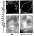
hGOの発生は、胃腺及び胃小窩、増殖帯、表層及び前庭部粘液細胞、並びにガストリン、グレリン及びソマトスタチンを発現する内分泌細胞を形成するマウス前庭部の発生とほぼ同じ分子的な及び形態形成上の変化を経るものであり得る。hGOを使用してヒト胃発生をモデル化することにより、EGFシグナル伝達が転写因子NEUROGENIN 3の上流で内分泌細胞発生を抑制することが決定されている。本出願人はさらに、hGOが、c−Metシグナル伝達及び上皮増殖の急速な活性化を含め、ピロリ菌(H.pylori)によって惹起される胃疾患の初期段階を忠実に再現することを見出した。合わせると、これらの研究は、ヒト胃の発生及び疾患の根底にある機構を解明するための新規のロバストなインビトロシステムを描き出している。
The development of hGO is similar in molecular and morphological development to that of the mouse antrum forming gastric glands and pits, growth zones, superficial and vestibular mucus cells, and endocrine cells expressing gastrin, ghrelin and somatostatin. Can be subject to change. By modeling human gastric development using hGO, it has been determined that EGF signaling suppresses endocrine cell development upstream of the
胚細胞に由来する多能性幹細胞
一態様において、本方法は、多能性であるか、又は多能性になるよう誘導することのできる幹細胞を入手するステップを含み得る。一部の実施形態において、多能性幹細胞は胚性幹細胞に由来し、一方で胚性幹細胞は初期哺乳類胚の全能性細胞に由来するもので、インビトロで無制限の未分化増殖が可能である。胚性幹細胞は、初期胚である胚盤胞の内部細胞塊に由来する多能性幹細胞である。未分化胚芽細胞から胚性幹細胞を得る方法は、当該技術分野において周知である。例えば、本明細書にある種の細胞型が例示されるが、当業者であれば、本明細書に記載される方法及びシステムを任意の幹細胞に適用可能であることを理解するであろう。
Pluripotent Stem Cells Derived from Embryonic Cells In one aspect, the method can include obtaining stem cells that are pluripotent or that can be induced to be pluripotent. In some embodiments, the pluripotent stem cells are derived from embryonic stem cells, while the embryonic stem cells are derived from totipotent cells of early mammalian embryos, allowing unlimited undifferentiated expansion in vitro. Embryonic stem cells are pluripotent stem cells derived from the inner cell mass of the blastocyst, which is an early embryo. Methods for obtaining embryonic stem cells from undifferentiated embryo cells are well known in the art. For example, while certain cell types are exemplified herein, those of skill in the art will understand that the methods and systems described herein can be applied to any stem cell.
本発明において実施形態で使用し得るさらなる幹細胞としては、限定はされないが、国立幹細胞バンク(National Stem Cell Bank:NSCB)、カリフォルニア大学(University of California)のヒト胚性幹細胞研究センター(Human Embryonic Stem Cell Research Center)、San Francisco(UCSF);Wi Cell Research InstituteのWISC細胞バンク;ウィスコンシン大学幹細胞及び再生医学センター(University of Wisconsin Stem Cell and Regenerative Medicine Center:UW−SCRMC);Novocell,Inc.(San Diego、Calif.);Cellartis AB(Goteborg、スウェーデン);ES Cell International Pte Ltd(シンガポール);テクニオン−イスラエル工科大学(Technion at the Israel Institute of Technology)(Haifa、イスラエル)が管理するデータベース;並びにプリンストン大学(Princeton University)及びペンシルバニア大学(University of Pennsylvania)が管理する幹細胞データベースによって提供されるか、又はそれに記載されるものが挙げられる。本発明において実施形態で使用し得る例示的胚性幹細胞としては、限定はされないが、SA01(SA001);SA02(SA002);ES01(HES−1);ES02(HES−2);ES03(HES−3);ES04(HES−4);ES05(HES−5);ES06(HES−6);BG01(BGN−01);BG02(BGN−02);BG03(BGN−03);TE03(13);TE04(14);TE06(16);UC01(HSF1);UC06(HSF6);WA01(H1);WA07(H7);WA09(H9);WA13(H13);WA14(H14)が挙げられる。 Additional stem cells that may be used in embodiments of the present invention include, but are not limited to, the National Stem Cell Bank (NSCB), Human Embryonic Stem Cell Center of the University of California (University of California). Research Center), San Francisco (UCSF); Wi Cell Research Institute WISC Cell Bank; University of Wisconsin Stem Cell and Regenerative Medicine, U.S.C. (San Diego, Calif.); Cellartis AB (Goteborg, Sweden); ES Cell International Pte Ltd (Singapore); Technion-Israel Institute of Technology and Israel (Technology of Israel, Israel). Those provided by or described by the stem cell database maintained by Princeton University and University of Pennsylvania. Exemplary embryonic stem cells that may be used in embodiments of the present invention include, but are not limited to, SA01 (SA001); SA02 (SA002); ES01 (HES-1); ES02 (HES-2); ES03 (HES- 3); ES04 (HES-4); ES05 (HES-5); ES06 (HES-6); BG01 (BGN-01); BG02 (BGN-02); BG03 (BGN-03); TE03 (13); TE04 (14); TE06 (16); UC01 (HSF1); UC06 (HSF6); WA01 (H1); WA07 (H7); WA09 (H9); WA13 (H13); WA14 (H14).
一部の実施形態において、幹細胞は、追加的な特性を取り入れるためさらに修飾されてもよい。例示的な修飾細胞株としては、限定はされないが、H1 OCT4−EGFP;H9 Cre−LoxP;H9 hNanog−pGZ;H9 hOct4−pGZ;H9 in GFPhES;及びH9 Syn−GFPが挙げられる。 In some embodiments, stem cells may be further modified to incorporate additional properties. Exemplary modified cell lines include, but are not limited to, H1 OCT4-EGFP; H9 Cre-LoxP; H9 hNanog-pGZ; H9 hOct4-pGZ; H9 in GFPhES; and H9 Syn-GFP.
胚性幹細胞に関するさらなる詳細については、例えば、Thomson et al.,1998,“Embryonic Stem Cell Lines Derived from Human Blastocysts”,Science 282(5391):1145−1147;Andrews et al.,2005,“Embryonic stem(ES)cells and embryonal carcinoma(EC)cells:opposite sides of the same coin”,Biochem Soc Trans 33:1526−1530;Martin 1980,“Teratocarcinomas and mammalian embryogenesis”,Science 209(4458):768−776;Evans and Kaufman,1981,“Establishment in culture of pluripotent cells from mouse embryos”,Nature 292(5819):154−156;Klimanskaya et al.,2005,“Human embryonic stem cells derived without feeder cells”,Lancet 365(9471):1636−1641;(これらの各々は本明細書によって全体として本明細書に援用される)を参照することができる。 For more details on embryonic stem cells, see, for example, Thomson et al. , 1998, "Embryonic Stem Cell Lines Derived from Human Blastocysts", Science 282 (5391): 1145-1147; Andrews et al. , 2005, "Embryonic stem (ES) cells and embryonal carcinoma (EC) cells: opposite sides of the same coin", Biochem Soc Trans 33: 1526-1530; Martin 1980, "Teratocarcinomas and mammalian embryogenesis", Science 209 (4458) : 768-776; Evans and Kaufman, 1981, "Establishment in culture of pluripotent cells from mouse embryos", Nature 292 (5819): 154-156; Klima. l. , 2005, "Human embryonic stem cells delivered without feeder cells," Lancet 365 (9471): 163-1641; (each of which is hereby incorporated by reference in its entirety).
代替的な多能性幹細胞は、有性生殖する生物の配偶子を生じる細胞である胚性生殖細胞(EGC)に由来し得る。EGCは、後期胚の生殖堤に見られる始原生殖細胞に由来し、胚性幹細胞の特性の多くを有する。胚における始原生殖細胞が発生すると、成体において生殖配偶子(精子又は卵子)を生じる幹細胞となる。マウス及びヒトでは、適切な条件下で組織培養において胚性生殖細胞を成長させることが可能である。EGC及びESCは両方ともに多能性である。本発明の目的上、用語「ESC」は広義に用いられ、時にEGCを包含する。 Alternative pluripotent stem cells can be derived from embryonic germ cells (EGCs), which are the gametogenic cells of sexually reproducing organisms. EGCs are derived from primordial germ cells found in the germ ridge of late embryos and have many of the characteristics of embryonic stem cells. When primordial germ cells in the embryo develop, they become stem cells that give rise to reproductive gametes (sperm or egg) in the adult. In mice and humans, it is possible to grow embryonic germ cells in tissue culture under suitable conditions. Both EGC and ESC are pluripotent. For purposes of the present invention, the term "ESC" is used broadly and sometimes includes EGC.
人工多能性幹細胞(iPSC)
一部の実施形態において、iPSCは、ある種の幹細胞関連遺伝子を非多能性細胞、例えば成体線維芽細胞にトランスフェクトすることによって得られる。トランスフェクションは、典型的にはレトロウイルスなどのウイルスベクターを用いて達成される。トランスフェクトされる遺伝子としては、マスター転写調節因子Oct−3/4(Pouf51)及びSox2が挙げられるが、他の遺伝子が誘導効率を増進させることも考えられる。3〜4週間後、少数のトランスフェクト細胞が形態学的及び生化学的に多能性幹細胞と類似したものになり始め、典型的には、形態学的選択、倍加時間によるか、又はレポーター遺伝子及び抗生物質選択によって単離される。本明細書で使用されるとき、iPSCとしては、限定はされないが、マウスにおける第1代iPSC、第2代iPSC、及びヒト人工多能性幹細胞を挙げることができる。一部の実施形態では、レトロウイルス系を使用して、4つの中心的遺伝子:Oct3/4、Sox2、Klf4、及びc−Mycを使用してヒト線維芽細胞を多能性幹細胞に形質転換し得る。代替的実施形態では、レンチウイルス系を使用して、OCT4、SOX2、NANOG、及びLIN28で体細胞を形質転換する。iPSCにおいてその発現を誘導し得る遺伝子としては、限定はされないが、Oct−3/4(例えば、Pou5fl);Sox遺伝子ファミリーの特定のメンバー(例えば、Sox1、Sox2、Sox3、及びSox15);Klfファミリーの特定のメンバー(例えば、Klf1、Klf2、Klf4、及びKlf5)、Mycファミリーの特定のメンバー(例えば、C−myc、L−myc、及びN−myc)、Nanog、及びLIN28が挙げられる。
Induced pluripotent stem cells (iPSC)
In some embodiments, iPSCs are obtained by transfecting certain stem cell-related genes into non-pluripotent cells, such as adult fibroblasts. Transfection is typically accomplished using viral vectors such as retroviruses. Transfected genes include the master transcription factor Oct-3 / 4 (Pouf51) and Sox2, but it is also possible that other genes enhance induction efficiency. After 3-4 weeks, a few transfected cells begin to become morphologically and biochemically similar to pluripotent stem cells, typically due to morphological selection, doubling time, or reporter gene And isolated by antibiotic selection. As used herein, iPSCs can include, but are not limited to, first-generation iPSCs, second-generation iPSCs in mice, and human induced pluripotent stem cells. In some embodiments, a retroviral system is used to transform human fibroblasts into pluripotent stem cells using four core genes: Oct3 / 4, Sox2, Klf4, and c-Myc. obtain. In an alternative embodiment, the lentivirus system is used to transform somatic cells with OCT4, SOX2, NANOG, and LIN28. Genes capable of inducing its expression in iPSC include, but are not limited to, Oct-3 / 4 (eg, Pou5fl); specific members of the Sox gene family (eg, Sox1, Sox2, Sox3, and Sox15); Klf family Specific members of the Myc family (eg, Klf1, Klf2, Klf4, and Klf5), specific members of the Myc family (eg, C-myc, L-myc, and N-myc), Nanog, and LIN28.
一部の実施形態では、非ウイルスベースの技術を用いてiPSCを作成し得る。一部の実施形態では、アデノウイルスを使用して必要な4つの遺伝子をマウスの皮膚及び肝細胞のDNAに運び込み、胚性幹細胞と同一の細胞をもたらすことができる。アデノウイルスはそれ自体の遺伝子のいずれも標的宿主と一体化することがないため、腫瘍を作り出す危険性がなくなる。一部の実施形態では、いかなるウイルストランスフェクション系も全くなしに、プラスミドによって再プログラム化を達成することができ、しかし効率は極めて低い。他の実施形態では、タンパク質の直接送達を用いてiPSCが作成され、従ってウイルス又は遺伝子修飾の必要がなくなる。一部の実施形態では、同様の方法論を用いてマウスiPSCの作成が可能である:ポリアルギニンアンカーによって細胞に供給される特定のタンパク質による細胞の反復的な処理が、多能性を誘導するのに十分であった。一部の実施形態において、多能性誘導遺伝子の発現はまた、低酸素条件下においてFGF2で体細胞を処理することによっても増加させることができる。 In some embodiments, non-virus based techniques may be used to create iPSCs. In some embodiments, adenovirus can be used to carry the required four genes into the DNA of mouse skin and hepatocytes, resulting in cells identical to embryonic stem cells. Adenovirus does not integrate any of its own genes with the target host, eliminating the risk of creating a tumor. In some embodiments, reprogramming can be achieved by the plasmid without any viral transfection system, but the efficiency is very low. In other embodiments, iPSCs are made using direct delivery of proteins, thus eliminating the need for viral or genetic modification. In some embodiments, similar methodologies can be used to generate mouse iPSCs: repetitive treatment of cells with specific proteins supplied to cells by polyarginine anchors induces pluripotency. Was enough. In some embodiments, pluripotency-inducible gene expression can also be increased by treating somatic cells with FGF2 under hypoxic conditions.
胚性幹細胞に関するさらなる詳細は、例えば、Kaji et al.,2009,“Virus free induction of pluripotency and subsequent excision of reprogramming factors”,Nature 458:771−775;Woltjen et al.,2009,“piggyBac transposition reprograms fibroblasts to induced pluripotent stem cells”,Nature 458:766−770;Okita et al.,2008,“Generation of Mouse Induced Pluripotent Stem Cells Without Viral Vectors”,Science 322(5903):949−953;Stadtfeld et al.,2008,“Induced Pluripotent Stem Cells Generated without Viral Integration”,Science 322(5903):945−949;及びZhou et al.,2009,“Generation of Induced Pluripotent Stem Cells Using Recombinant Proteins”、Cell Stem Cell 4(5):381−384;(これらの各々は本明細書によって全体として本明細書に援用される)を参照することができる。 Further details regarding embryonic stem cells can be found in, for example, Kaji et al. , 2009, "Virus free induction of pluripotency and subsequent excision of reprogramming factors", Nature 458: 771-775; Waltjen et al. , 2009, "piggyBac transposition reprogramms fibroblasts to induced pluripotent stem cells", Nature 458: 766-770; Okita et al. , 2008, "Generation of Mouse Induced Pluripotent Stem Cells Without Virtual Vectors", Science 322 (5903): 949-953; Statfeld et al. , 2008, "Induced Pluripotent Stem Cells Generated without Viral Integration", Science 322 (5903): 945-949; and Zhou et al. , 2009, "Generation of Induced Pluripotent Stem Cells Using Recombinant Proteins", Cell Stem Cell 4 (5): 381-384; (each of which is incorporated herein by reference in its entirety). You can
一部の実施形態において、例示的iPS細胞株としては、限定はされないが、iPS−DF19−9;iPS−DF19−9;iPS−DF4−3;iPS−DF6−9;iPS(Foreskin);iPS(IMR90);及びiPS(IMR90)が挙げられる。 In some embodiments, exemplary iPS cell lines include, but are not limited to, iPS-DF19-9; iPS-DF19-9; iPS-DF4-3; iPS-DF6-9; iPS (Foreskin); iPS. (IMR90); and iPS (IMR90).
iPSCはESCと同様の様式で完全分化型組織に分化する能力を有したことが示されている。例えば、iPSCは、βIII−チューブリン、チロシンヒドロキシラーゼ、AADC、DAT、ChAT、LMX1B、及びMAP2を発現するニューロンに分化した。カテコールアミン関連酵素の存在は、iPSCがhESCと同様にドーパミン作動性ニューロンに分化可能であり得ることを示唆し得る。幹細胞関連遺伝子は分化後に下方制御されることが示された。また、iPSCが、自発的に拍動し始めた心筋細胞に分化し得ることも示されている。心筋細胞は、TnTc、MEF2C、MYL2A、MYHCβ、及びNKX2.5を発現した。幹細胞関連遺伝子は分化後に下方制御された。 It has been shown that iPSCs were capable of differentiating into fully differentiated tissues in a manner similar to ESCs. For example, iPSCs differentiated into neurons expressing βIII-tubulin, tyrosine hydroxylase, AADC, DAT, ChAT, LMX1B, and MAP2. The presence of catecholamine-related enzymes may suggest that iPSCs may be capable of differentiating into dopaminergic neurons like hESCs. It has been shown that stem cell-related genes are downregulated after differentiation. It has also been shown that iPSCs can differentiate into spontaneously beating cardiomyocytes. Cardiomyocytes expressed TnTc, MEF2C, MYL2A, MYHCβ, and NKX2.5. Stem cell-related genes were downregulated after differentiation.
胃の器官及び発生
本出願人の発明以前には、胚性幹細胞及び/又はiPSCなどの前駆細胞を胃組織に変換するために利用可能なシステムはなかった。
Stomach Organs and Development Prior to Applicants' invention, there were no systems available for converting embryonic stem cells and / or progenitor cells such as iPSCs into gastric tissue.
一部の実施形態において、ESC及びiPSCなどのPSCは、初めに胚体内胚葉(DE)、次に三次元腸管構造(前腸スフェロイド)、その次に後方前腸/胃組織の形成を介して三次元胃オルガノイド(hGO)へと、段階的な形で指向性分化を経る。 In some embodiments, PSCs, such as ESCs and iPSCs, are first expressed through definitive endoderm (DE), then three-dimensional intestinal structure (foregut spheroids), and then through formation of posterior foregut / gastric tissue. It undergoes directional differentiation in a stepwise fashion into a three-dimensional gastric organoid (hGO).
一部の実施形態において、ESC及びiPSCなどのPSCは非段階的な形で指向性分化を経て、ここではDE形成を促進するための分子(例えば、成長因子、リガンド)及び続く組織形成のための分子が同時に加えられる。 In some embodiments, PSCs, such as ESCs and iPSCs, undergo directional differentiation in a non-stepwise manner, where molecules for promoting DE formation (eg, growth factors, ligands) and subsequent tissue formation are involved. Molecules are added at the same time.
胚体内胚葉
胃の上皮は、胚体内胚葉(DE)と呼ばれる単層の細胞に由来する。前方DEは前腸並びに肺、食道、胃、肝臓及び膵臓を含めたその関連器官を形成し、後方DEは中腸及び後腸を形成して、これは小腸及び大腸並びに泌尿生殖器系の部位を形成する。DEはインビボで消化管及び気道の上皮を生じる。マウス、ヒヨコ及びカエルの胚を使用した研究からは、原腸胚期におけるDEの前後パターンの確立が、続く前腸及び後腸発生に必須であることが示唆される。一部の実施形態において、ESC及びiPSCなどのPSCは、初めに胚体内胚葉(DE)、次に前方/前腸上皮(例えば、前腸スフェロイド)、その次に胃組織へと、段階的な形で指向性分化を経る。この過程には、BMP、Wnt及びFGFシグナル伝達経路が決定的に重要であると考えられている。WNT及びFGFの活性化は腸管形態形成を促進する働きをし、及びBMPシグナル伝達の阻害は前腸運命を促進する。前腸の単層立方上皮は初めに多列円柱上皮、次に胃上皮を含む腺及び小窩並びに絨毛の基部にある増殖帯(これは予定プロジェニタードメイン(presumptive progenitor domain)に対応する)へと発生する。
Definitive endoderm The epithelium of the stomach is derived from a monolayer of cells called definitive endoderm (DE). The anterior DE forms the foregut and its associated organs including the lungs, esophagus, stomach, liver and pancreas, and the posterior DE forms the midgut and hindgut, which are the small and large intestines and parts of the genitourinary system. Form. DE produces intestinal and respiratory tract epithelia in vivo. Studies using mouse, chick and frog embryos suggest that the establishment of anteroposterior patterns of DE at the gastrulation stage is essential for subsequent foregut and hindgut development. In some embodiments, PSCs such as ESCs and iPSCs are graded into first definitive endoderm (DE), then the anterior / foregut epithelium (eg, foregut spheroids), and then to gastric tissue. Shaped through directional differentiation. The BMP, Wnt and FGF signaling pathways are believed to be critical in this process. WNT and FGF activation serve to promote intestinal morphogenesis, and inhibition of BMP signaling promotes foregut fate. The monolayer cuboidal epithelium of the foregut goes first to the multi-columned columnar epithelium, then to the glands and pits containing the gastric epithelium and to the growth zone at the base of the villi, which corresponds to the presumptive progenitor domain. Occurs.
インビトロでDEから胃組織への分化を指向させるためのロバストで効率的なプロセスが確立される。一部の実施形態において、指向性分化は、iPSC及び/又はDE細胞における特定のシグナル伝達経路を選択的に活性化することによって実現される。一部の実施形態において、このシグナル伝達経路は、限定はされないが、Wntシグナル伝達経路、Wnt/APCシグナル伝達経路、FGFシグナル伝達経路、TGF−βシグナル伝達経路、BMPシグナル伝達経路;EGFシグナル伝達経路、及びレチノイン酸シグナル伝達経路を含めた、胃組織の発生において活性なものである。 A robust and efficient process for directing the differentiation of DE to gastric tissue in vitro is established. In some embodiments, directional differentiation is achieved by selectively activating specific signaling pathways in iPSC and / or DE cells. In some embodiments, the signaling pathway includes, but is not limited to, the Wnt signaling pathway, Wnt / APC signaling pathway, FGF signaling pathway, TGF-β signaling pathway, BMP signaling pathway; EGF signaling. It is active in the development of gastric tissue, including the pathway and the retinoic acid signaling pathway.
DEの発生及び/又は一般に腸の発生に関係するシグナル伝達経路の機能に関するさらなる詳細については、例えば、Zorn and Wells,2009,“Vertebrate endoderm development and organ formation”,Annu Rev Cell Dev Biol 25:221−251;Dessimoz et al.,2006,“FGF signaling is necessary for establishing gut tube domains along the anterior−posterior axis in vivo”,Mech Dev 123:42−55;McLin et al.,2007,“Repression of Wnt/{beta}−catenin signaling in the anterior endoderm is essential for liver and pancreas development”.Development,134:2207−2217;Wells and Melton,2000,Development 127:1563−1572;de Santa Barbara et al.,2003,“Development and differentiation of the intestinal epithelium”,Cell Mol Life Sci 60(7):1322−1332;Sancho et al.,2004,“Signaling Pathways in Intestinal Development and Cancer”,Annual Review of Cell and Developmental Biology 20:695−723;Logan and Nusse,2004,“The Wnt Signaling Pathway in Development and Disease”,Annual Review of Cell and Developmental Biology 20:781−810;Taipalel and Beachyl,2001,“The Hedgehog and Wnt signalling pathways in cancer”,Nature 411:349−354;Gregorieff and Clevers,2005,“Wnt signaling in the intestinal epithelium:from endoderm to cancer”,Genes&Dev.19:877−890;(これらの各々は本明細書によって全体として本明細書に援用される)を参照することができる。 For further details regarding the function of signaling pathways involved in the development of DE and / or intestinal development in general, see, eg, Zorn and Wells, 2009, "Vertebrate endotherm development and organ formation", Annu Rev Cell Dev 25-Biol. 251; Dessimoz et al. , 2006, "FGF signaling is lessening for eating gut tube domains along the anterior-posterior axis in vivo", Mech Dev 123: 42-55; al. , 2007, "Repression of Wnt / {beta} -catenin signaling in the anterior endodermisis for for liberand pancreas development". Development, 134: 2207-2217; Wells and Melton, 2000, Development 127: 1563-1572; de Santa Barbara et al. , 2003, "Development and differentiation of the intestinal epithelium", Cell Mol Life Sci 60 (7): 1322-1332; Sancho et al. , 2004, "Signaling Pathways in Intestinal Development and Cancer", Annual Review of Cell and Developmental Biology 20: 695-723; Logan and Nusse, 2004, "The Wnt Signaling Pathway in Development and Disease", Annual Review of Cell and Developmental Biology 20: 781-810; Taipalel and Beachyl, 2001, "The Hedgehog and Wnt signaling pathways in cancer", Nature 41. : 349-354; Gregorieff and Clevers, 2005, "Wnt signaling in the intestinal epithelium: from endoderm to cancer", Genes & Dev. 19: 877-890 ;, each of which is hereby incorporated herein by reference in its entirety.
多能性細胞(例えば、iPSC又はESC)から胚体内胚葉を作製する任意の方法を本明細書に記載される方法に適用することが可能である。一部の実施形態において、多能性細胞は桑実胚に由来する。一部の実施形態において、多能性幹細胞は幹細胞である。これらの方法において使用される幹細胞としては、限定はされないが、胚性幹細胞を挙げることができる。胚性幹細胞は胚内部細胞塊に由来してもよく、又は胚生殖堤に由来してもよい。胚性幹細胞又は生殖細胞は、限定はされないが、ヒトを含めた様々な哺乳類種を含め、種々の動物種を起源とし得る。一部の実施形態では、ヒト胚性幹細胞を使用して胚体内胚葉が作製される。一部の実施形態では、ヒト胚性生殖細胞を使用して胚体内胚葉が作製される。一部の実施形態では、iPSCを使用して胚体内胚葉が作製される。 Any method of making definitive endoderm from pluripotent cells (eg, iPSCs or ESCs) can be applied to the methods described herein. In some embodiments, the pluripotent cells are derived from morula. In some embodiments, the pluripotent stem cells are stem cells. Stem cells used in these methods include, but are not limited to, embryonic stem cells. Embryonic stem cells may be derived from the inner cell mass of the embryo, or may be derived from the germ ridge of the embryo. Embryonic stem cells or germ cells can originate from a variety of animal species, including but not limited to various mammalian species, including humans. In some embodiments, human embryonic stem cells are used to make definitive endoderm. In some embodiments, human embryonic germ cells are used to make definitive endoderm. In some embodiments, iPSCs are used to create definitive endoderm.
一部の実施形態において、多能性幹細胞からDE細胞への分化過程において1つ以上の成長因子が使用される。分化過程で使用される1つ以上の成長因子としては、TGF−βスーパーファミリーからの成長因子を挙げることができる。かかる実施形態において、1つ以上の成長因子は、ノーダル/アクチビン及び/又は成長因子のTGF−βスーパーファミリーのBMPサブグループを含む。一部の実施形態において、1つ以上の成長因子は、ノーダル、アクチビンA、アクチビンB、BMP4、Wnt3a又はこれらの成長因子のいずれかの組み合わせからなる群から選択される。 In some embodiments, one or more growth factors are used in the process of differentiation of pluripotent stem cells into DE cells. One or more growth factors used in the differentiation process can include growth factors from the TGF-β superfamily. In such an embodiment, the one or more growth factors comprises Nodal / Activin and / or the BMP subgroup of the TGF-β superfamily of growth factors. In some embodiments, the one or more growth factors are selected from the group consisting of Nodal, activin A, activin B, BMP4, Wnt3a or any combination of these growth factors.
一部の実施形態において、胚性幹細胞又は人工多能性細胞及びiPSCは、1つ以上の成長因子によって6時間以上;12時間以上;18時間以上;24時間以上;36時間以上;48時間以上;60時間以上;72時間以上;84時間以上;96時間以上;120時間以上;150時間以上;180時間以上;又は240時間以上にわたり処理される。 In some embodiments, the embryonic stem cells or induced pluripotent cells and iPSCs are 6 hours or longer; 12 hours or longer; 18 hours or longer; 24 hours or longer; 36 hours or longer; 48 hours or longer by one or more growth factors. 60 hours or longer; 72 hours or longer; 84 hours or longer; 96 hours or longer; 120 hours or longer; 150 hours or longer; 180 hours or longer; or 240 hours or longer.
一部の実施形態において、胚性幹細胞又は生殖細胞及びiPSCは、10ng/ml以上;20ng/ml以上;50ng/ml以上;75ng/ml以上;100ng/ml以上;120ng/ml以上;150ng/ml以上;200ng/ml以上;500ng/ml以上;1,000ng/ml以上;1,200ng/ml以上;1,500ng/ml以上;2,000ng/ml以上;5,000ng/ml以上;7,000ng/ml以上;10,000ng/ml以上;又は15,000ng/ml以上の濃度の1つ以上の成長因子によって処理される。一部の実施形態において、成長因子の濃度は処理全体を通じて一定のレベルに維持される。他の実施形態において、成長因子の濃度は処理する間に変化させる。一部の実施形態において、成長因子は、種々のHyClone濃度でウシ胎仔セリン(FBS)を含む培地中に懸濁される。当業者であれば、本明細書に記載されるレジメンを単独又は組み合わせの任意の既知の成長因子に適用可能であることを理解するであろう。2つ以上の成長因子が使用されるとき、各成長因子の濃度は独立に変えてもよい。 In some embodiments, the embryonic stem cells or germ cells and iPSCs are 10 ng / ml or higher; 20 ng / ml or higher; 50 ng / ml or higher; 75 ng / ml or higher; 100 ng / ml or higher; 120 ng / ml or higher; 150 ng / ml or higher; 150 ng / ml Or more; 200 ng / ml or more; 500 ng / ml or more; 1,000 ng / ml or more; 1,200 ng / ml or more; 1,500 ng / ml or more; 2,000 ng / ml or more; 5,000 ng / ml or more; 7,000 ng / Ml or more; 10,000 ng / ml or more; or 15,000 ng / ml or more at a concentration of one or more growth factors. In some embodiments, the growth factor concentration is maintained at a constant level throughout the treatment. In another embodiment, the growth factor concentration is varied during the treatment. In some embodiments, the growth factors are suspended in medium containing fetal bovine serine (FBS) at various HyClone concentrations. Those skilled in the art will appreciate that the regimens described herein can be applied to any known growth factor, alone or in combination. When more than one growth factor is used, the concentration of each growth factor may vary independently.
一部の実施形態において、胚体内胚葉細胞がエンリッチされた細胞集団が使用される。一部の実施形態において、胚体内胚葉細胞は単離され、又は実質的に精製される。一部の実施形態において、単離され、又は実質的に精製された胚体内胚葉細胞は、OCT4、AFP、TM、SPARC及び/又はSOX7マーカーと比べてより高度にSOX17、FOXA2、及び/又はCXRC4マーカーを発現する。 In some embodiments, a cell population enriched in definitive endoderm cells is used. In some embodiments, definitive endoderm cells are isolated or substantially purified. In some embodiments, the isolated or substantially purified definitive endoderm cells have a higher degree of SOX17, FOXA2, and / or CXRC4 relative to the OCT4, AFP, TM, SPARC and / or SOX7 markers. Express the marker.
胚体内胚葉を含む細胞集団をエンリッチする方法もまた企図される。一部の実施形態において、胚体内胚葉細胞は、胚体内胚葉細胞の表面上に存在するが混合細胞集団中の他の細胞の表面上には存在しない分子に結合する試薬にそれらの細胞を接触させて、次に試薬に結合した細胞を単離することにより、混合細胞集団から単離し、又は実質的に精製することができる。特定の実施形態において、胚体内胚葉細胞の表面上に存在する細胞構成物はCXCR4である。 Methods of enriching a cell population including definitive endoderm are also contemplated. In some embodiments, the definitive endoderm cells contact the cells with a reagent that binds to a molecule that is present on the surface of definitive endoderm cells but not on the surface of other cells in the mixed cell population. Can then be isolated or substantially purified from the mixed cell population by isolating cells that have bound the reagent. In certain embodiments, the cell constituent present on the surface of definitive endoderm cells is CXCR4.
本発明のさらに他の実施形態は、CXCR4抗体、SDF−1リガンド又は他のCXCR4リガンドに関し、これらは、エンリッチされた、単離された、又は実質的に精製された形態の胚体内胚葉細胞を入手するために使用することができる。例えば、親和性に基づく分離又は磁気に基づく分離などの方法においてCXCR4抗体、SDF−1リガンド又は別のCXCR4リガンドを試薬として使用して、該試薬に結合する胚体内胚葉細胞の調製物をエンリッチし、単離し、又は実質的に精製することができる。 Yet another embodiment of the invention relates to CXCR4 antibodies, SDF-1 ligands or other CXCR4 ligands, which contain definitive endoderm cells in enriched, isolated or substantially purified form. Can be used to obtain. For example, a CXCR4 antibody, SDF-1 ligand or another CXCR4 ligand is used as a reagent in a method such as affinity-based separation or magnetic-based separation to enrich a preparation of definitive endoderm cells that bind to the reagent. , Isolated or substantially purified.
一部の実施形態において、胚体内胚葉細胞及びhESCは1つ以上の成長因子で処理される。かかる成長因子には、TGF−βスーパーファミリーの成長因子が含まれ得る。かかる実施形態において、1つ以上の成長因子はノーダル/アクチビン及び/又は成長因子のTGF−βスーパーファミリーのBMPサブグループを含む。一部の実施形態において、1つ以上の成長因子は、ノーダル、アクチビンA、アクチビンB、BMP4、Wnt3a又はこれらの成長因子のいずれかの組み合わせからなる群から選択される。 In some embodiments, definitive endoderm cells and hESCs are treated with one or more growth factors. Such growth factors may include those of the TGF-β superfamily. In such embodiments, the one or more growth factors comprises Nodal / Activin and / or the BMP subgroup of the TGF-β superfamily of growth factors. In some embodiments, the one or more growth factors are selected from the group consisting of Nodal, activin A, activin B, BMP4, Wnt3a or any combination of these growth factors.
本発明において使用し得るDE細胞を入手し又は作り出すためのさらなる方法としては、限定はされないが、D’Amour et al.に対する米国特許第7,510,876号明細書;Fisk et al.に対する米国特許第7,326,572号明細書;Kubol et al.,2004,“Development of definitive endoderm from embryonic stem cells in culture”,Development 131:1651−1662;D’Amour et al.,2005,“Efficient differentiation of human embryonic stem cells to definitive endoderm”,Nature Biotechnology 23:1534−1541;及びAng et al.,1993,“The formation and maintenance of the definitive endoderm lineage in the mouse:involvement of HNF3/forkhead proteins”,Development 119:1301−1315;(これらの各々は本明細書によって全体として本明細書に参照により援用される)に記載されるものが挙げられる。 Additional methods for obtaining or producing DE cells that can be used in the present invention include, but are not limited to, D'Amour et al. U.S. Pat. No. 7,510,876 to Fisk et al. U.S. Pat. No. 7,326,572 to Kubol et al. , 2004, "Development of definitive endemde from embryonic stem cells in culture", Development 131: 1651-1662; D'Amour et al. , 2005, "Efficient differentiation of human embryonic stem cells to definitive endoderm", Nature Biotechnology 23: 1534-1541; and Ang et al. , 1993, "The formation and maintenance of the definitive endodelinem lineage in the mouse: involvment of HNF3, each of which is incorporated herein by reference. Described).
後方化DEの指向性分化
一部の実施形態では、アクチビン誘導性胚体内胚葉(DE)がFGF/Wnt/ノギン誘導性前方内胚葉パターン形成、前腸特異化及び形態形成、並びに最後にプロガストリック培養系をさらに経ることにより、表層粘液細胞、粘液腺細胞、内分泌、及びプロジェニター細胞を含めた機能性の胃細胞型への胃組織成長、形態形成及び細胞分化が促進され得る。一部の実施形態では、ヒトPSCを、粘液、内分泌、及びプロジェニター細胞型を含む胃上皮にインビトロで分化するように効率的に指向させる。特定のタイプの胃組織形成を促進するため、任意の発生段階で成長因子などの分子を加え得ることは理解されるであろう。
Directional Differentiation of Posterior DE In some embodiments, activin-induced definitive endoderm (DE) induces FGF / Wnt / noggin-induced anterior endoderm patterning, foregut specification and morphogenesis, and finally progas. Further passage through the trick culture system can promote gastric tissue growth, morphogenesis and cell differentiation into functional gastric cell types including superficial mucus cells, mucous gland cells, endocrine and progenitor cells. In some embodiments, human PSCs are efficiently directed to differentiate in vitro into gastric epithelium, including mucus, endocrine, and progenitor cell types. It will be appreciated that molecules such as growth factors can be added at any stage of development to promote certain types of gastric tissue formation.
一部の実施形態では、DEの前方化内胚葉細胞が1つ以上の特殊化した細胞型へとさらに発生する。 In some embodiments, the DE anterior endoderm cells further develop into one or more specialized cell types.
一部の実施形態において、可溶性FGF及びWntリガンド並びにBMP拮抗薬を使用して培養下で初期前腸特異化を模倣し、iPSC又はESCから発生したDEを指向性分化によって、主要な前庭部胃細胞型の全てを効率的に生じる前腸上皮に変換する。ヒトにおいては、DEの指向性分化は、胃発生に重要な特定のシグナル伝達経路を選択的に活性化させることによって実現する。 In some embodiments, soluble FGF and Wnt ligands and BMP antagonists are used to mimic early foregut specification in culture to direct DE generated from iPSCs or ESCs by directed differentiation to the major vestibular stomach. Converts all cell types into the efficiently occurring foregut epithelium. In humans, directed differentiation of DE is achieved by selectively activating specific signaling pathways important for gastric development.
インビトロでのヒト胃(stomach)/胃(gastric)の発生は、胎児期の腸発生;内胚葉形成、前方内胚葉パターン形成、前腸形態形成、胎児期の胃、前庭部及び胃底部発生、上皮形態形成、予定プロジェニタードメインの形成、及び胃の機能性細胞型への分化を近似する段階で起こる。 In vitro development of human stomach / gastric is fetal intestinal development; endoderm formation, anterior endoderm patterning, foregut morphogenesis, fetal stomach, vestibular and fundic development. , Epithelial morphogenesis, formation of prearranged progenitor domains, and differentiation into gastric functional cell types.
当業者は、任意のFGFリガンドと組み合わせた任意のWntシグナル伝達タンパク質の発現を改変すると、本発明における指向性分化が生じ得ることを理解するであろう。一部の実施形態において、この改変は、Wnt3、詳細にはWnt3aの過剰発現である。一部の実施形態において、この改変は、Wnt1又は他のWntリガンドの過剰発現である。 One of ordinary skill in the art will appreciate that modifying the expression of any Wnt signaling protein in combination with any FGF ligand can result in directional differentiation in the present invention. In some embodiments, the modification is overexpression of Wnt3, particularly Wnt3a. In some embodiments, the modification is overexpression of Wnt1 or another Wnt ligand.
当業者は、FGFシグナル伝達経路のシグナル伝達活性を改変することと組み合わせてWntシグナル伝達経路のシグナル伝達活性を改変すると、本発明における指向性分化が生じ得ることを理解するであろう。一部の実施形態において、この改変は、前述の経路を活性化する小分子モジュレーターの使用による。例えば、Wnt経路の小分子モジュレーターには、限定はされないが、塩化リチウム;2−アミノ−4,6−二置換ピリミジン(ヘテロ)アリールピリミジン;IQ1;QS11;NSC668036;DCA β−カテニン;2−アミノ−4−[3,4−(メチレンジオキシ)−ベンジル−アミノ]−6−(3−メトキシフェニル)ピリミジンが含まれた。 Those skilled in the art will appreciate that modifying the signaling activity of the Wnt signaling pathway in combination with modifying the signaling activity of the FGF signaling pathway can result in directional differentiation in the present invention. In some embodiments, this modification is through the use of small molecule modulators that activate the aforementioned pathways. For example, small molecule modulators of the Wnt pathway include, but are not limited to, lithium chloride; 2-amino-4,6-disubstituted pyrimidine (hetero) arylpyrimidines; IQ1; QS11; NSC668036; DCA β-catenin; 2-amino. -4- [3,4- (Methylenedioxy) -benzyl-amino] -6- (3-methoxyphenyl) pyrimidine was included.
代替的実施形態において、Wnt及び/又はFGFシグナル伝達経路に関連する細胞構成物、例えば、これらの経路の天然阻害因子又は拮抗物質を阻害して、Wnt及び/又はFGFシグナル伝達経路の活性化をもたらすことができる。 In an alternative embodiment, cellular constituents associated with the Wnt and / or FGF signaling pathways, eg, natural inhibitors or antagonists of these pathways, are inhibited to activate the Wnt and / or FGF signaling pathways. Can bring
一部の実施形態において、細胞構成物は他の細胞構成物又は外因性分子によって阻害される。Wntシグナル伝達の例示的な天然阻害因子としては、限定はされないが、Dkk1、SFRPタンパク質及びFrzBが挙げられる。一部の実施形態において、外因性分子としては、限定はされないが、WAY−316606;SB−216763;又はBIO(6−ブロモインジルビン−3’−オキシム)などの小分子を挙げることができる。 In some embodiments, cell constituents are inhibited by other cell constituents or exogenous molecules. Exemplary natural inhibitors of Wnt signaling include, but are not limited to, Dkk1, SFRP protein and FrzB. In some embodiments, the exogenous molecule can include, but is not limited to, a small molecule such as WAY-316606; SB-216763; or BIO (6-bromoindirubin-3'-oxime).
さらなる詳細については、例えば、Liu et al.,“A small−molecule agonist of the Wnt signaling pathway”,Angew Chem Int Ed Engl.44(13):1987−1990(2005);Miyabayashi et al.,“Wnt/beta−catenin/CBP signaling maintains long−term murine embryonic stem cell pluripotency”,Proc Natl Acad Sci USA.104(13):5668−5673(2007);Zhang et al.,“Small−molecule synergist of the Wnt/beta−catenin signaling pathway”,Proc Natl Acad Sci U S A.104(18):7444−7448(2007);Neiiendam et al.,“An NCAM−derived FGF−receptor agonist,the FGL−peptide,induces neurite outgrowth and neuronal survival in primary rat neurons”,J.Neurochem.91(4):920−935(2004);Shan et al.,“Identification of a specific inhibitor of the dishevelled PDZ domain”,Biochemistry 44(47):15495−15503(2005);Coghlan et al.,“Selective small molecule inhibitors of glycogen synthase kinase−3 modulate glycogen metabolism and gene transcription”,Chem.Biol.7(10):793−803(2000);Coghlan et al.,“Selective small molecule inhibitors of glycogen synthase kinase−3 modulate glycogen metabolism and gene transcription”,Chemistry&Biology 7(10):793−803;及びPai et al.,“Deoxycholic acid activates beta−catenin signaling pathway and increases colon cell cancer growth and invasiveness”,Mol Biol Cell.15(5):2156−2163(2004);(これらの各々は本明細書によって全体として参照により援用される)が参照される。 For further details, see, for example, Liu et al. , "A small-molecule agone of the Wnt signaling path", Angew Chem Int Ed Engl. 44 (13): 1987-1990 (2005); Miyabayashi et al. , "Wnt / beta-catenin / CBP signaling maintains long-term murine embryonic cell cell pluripotency", Proc Natl Acad Sci USA. 104 (13): 5668-5673 (2007); Zhang et al. , "Small-molecule synergist of the Wnt / beta-catenin signaling pathway", Proc Natl Acad Sci USA. 104 (18): 7444-7448 (2007); Neiiendam et al. , "An NCAM-derivated FGF-receptor agonist, the FGL-peptide, induces neurale output and neural surveillance in primary ratonurons", J. Am. Neurochem. 91 (4): 920-935 (2004); Shan et al. , "Identification of a specific inhibitor of the dishelled PDZ domain", Biochemistry 44 (47): 15495-15503 (2005); Coghlan et al. , "Selective small molecule inhibitors of glycogen synthase kinase-3 modulin glycenogen metabolism and gene transcription", Chem. Biol. 7 (10): 793-803 (2000); Coghlan et al. , "Selective small molecule inhibitors of glycogen synthetase kinase-3 modulo glycencogenb metabolism and genus transcription 79 and chemistry., 10 & 7", Chem & Chem. , "Deoxycholic acid activates beta-catenin signaling pathway and increases colonons cell cell cancer, growth and invasiveness", Mol Biol Cell. 15 (5): 2156-2163 (2004); (each of which is hereby incorporated by reference in its entirety).
一部の実施形態では、Wnt及び/又はFGFシグナル伝達経路に関連する細胞構成物を標的化するsiRNA及び/又はshRNAを使用してこれらの経路が活性化される。当業者であれば、標的細胞構成物には、限定はされないが、SFRPタンパク質;GSK3、Dkk1、及びFrzBが含まれ得ることを理解するであろう。 In some embodiments, these pathways are activated using siRNAs and / or shRNAs that target cellular constituents associated with the Wnt and / or FGF signaling pathways. One of ordinary skill in the art will appreciate that target cell constructs can include, but are not limited to, SFRP proteins; GSK3, Dkk1, and FrzB.
RNAiベースの技術に関するさらなる詳細については、例えば、Couzin,2002,Science 298:2296−2297;McManus et al.,2002,Nat.Rev.Genet.3,737−747;Hannon,G.J.,2002,Nature 418,244−251;Paddison et al.,2002,Cancer Cell 2,17−23;Elbashir et al.,2001.EMBO J.20:6877−6888;Tuschl et al.,1999,Genes Dev.13:3191−3197;Hutvagner et al.,Sciencexpress 297:2056−2060;(これらの各々は本明細書によって全体として参照により援用される)を参照することができる。
For more details on RNAi-based technologies, see, eg, Couzin, 2002, Science 298: 2296-2297; McManus et al. 2002, Nat. Rev. Genet. 3, 737-747; Hannon, G .; J. , 2002, Nature 418, 244-251; Paddison et al. , 2002,
線維芽細胞成長因子(FGF)は、血管新生、創傷治癒、及び胚発生に関与する成長因子のファミリーである。FGFはヘパリン結合タンパク質であり、細胞表面関連ヘパラン硫酸プロテオグリカンとの相互作用はFGFシグナル伝達に必須であることが示されている。FGFは多種多様な細胞及び組織の増殖及び分化プロセスにおける中心的存在である。ヒトにおいては、FGFファミリーの22個のメンバーが同定されており、その全てが構造上関係のあるシグナル伝達分子である。メンバーFGF1〜FGF10は全て線維芽細胞成長因子受容体(FGFR)に結合する。FGF1は酸性線維芽細胞成長因子としても知られ、FGF2は塩基性線維芽細胞成長因子しても知られる。FGFホモログ因子1〜4(FHF1〜FHF4)としても知られるメンバーFGF11、FGF12、FGF13、及びFGF14は、FGFと比べて機能上の特徴的差異を有することが示されている。これらの因子は顕著に類似した配列相同性を有するが、それらはFGFRに結合せず、FGFと無関係の細胞内過程に関与する。この集団は「iFGF」としても知られる。メンバーFGF16〜FGF23は比較的新しく、それほど十分には特徴付けられていない。FGF15はヒトFGF19のマウスオルソログである(従ってヒトFGF15は存在しない)。ヒトFGF20はアフリカツメガエルFGF−20(XFGF−20)とのその相同性に基づき同定された。他のFGFの局所的活性と対照的に、FGF15/FGF19、FGF21及びFGF23は、より全身性の効果を有する。
Fibroblast growth factor (FGF) is a family of growth factors involved in angiogenesis, wound healing, and embryonic development. FGF is a heparin-binding protein, and its interaction with cell surface-associated heparan sulfate proteoglycans has been shown to be essential for FGF signaling. FGFs are central to the proliferation and differentiation processes of a wide variety of cells and tissues. In humans, 22 members of the FGF family have been identified, all of which are structurally related signaling molecules. Members FGF1 to FGF10 all bind to the fibroblast growth factor receptor (FGFR). FGF1 is also known as acidic fibroblast growth factor and FGF2 is also known as basic fibroblast growth factor. The members FGF11, FGF12, FGF13, and FGF14, also known as
一部の実施形態において、当業者は、任意のFGFをWntシグナル伝達経路のタンパク質と併せて使用し得ることを理解するであろう。一部の実施形態において、可溶性FGFとしては、限定はされないが、FGF4、FGF2、及びFGF3を挙げることができる。 In some embodiments, those of skill in the art will understand that any FGF can be used in conjunction with proteins of the Wnt signaling pathway. In some embodiments, soluble FGFs can include, but are not limited to, FGF4, FGF2, and FGF3.
一部の実施形態において、FGFシグナル伝達経路の細胞構成物は、他の細胞構成物又は外因性分子によって阻害される。FGFシグナル伝達の例示的な天然阻害因子としては、限定はされないが、スプラウティー(Sprouty)タンパク質ファミリー及びスプレッド(Spred)タンパク質ファミリーを挙げることができる。上記で考察したとおり、タンパク質、小分子、核酸を使用してFGFシグナル伝達経路を活性化することができる。 In some embodiments, cellular constituents of the FGF signaling pathway are inhibited by other cellular constituents or exogenous molecules. Exemplary natural inhibitors of FGF signaling include, but are not limited to, the Sprouty protein family and the Spread protein family. As discussed above, proteins, small molecules, nucleic acids can be used to activate the FGF signaling pathway.
当業者は、Wnt及びFGFシグナル伝達経路に関連して本明細書に記載される方法及び組成物が例として提供されることを理解するであろう。同様の方法及び組成物を本明細書に開示される他のシグナル伝達経路に適用可能である。 Those skilled in the art will appreciate that the methods and compositions described herein in connection with the Wnt and FGF signaling pathways are provided by way of example. Similar methods and compositions are applicable to other signaling pathways disclosed herein.
一部の実施形態において、DE培養物は、本明細書に記載されるシグナル伝達経路の1つ以上の分子によって6時間以上;12時間以上;18時間以上;24時間以上;36時間以上;48時間以上;60時間以上;72時間以上;84時間以上;96時間以上;120時間以上;150時間以上;180時間以上;200時間以上、240時間以上;270時間以上;300時間以上;350時間以上;400時間以上;500時間以上;600時間以上;700時間以上;800時間以上;900時間以上;1,000時間以上;1,200時間以上;又は1,500時間以上処理され得る。 In some embodiments, the DE culture is 6 hours or longer; 12 hours or longer; 18 hours or longer; 24 hours or longer; 36 hours or longer; 48, depending on one or more molecules of the signaling pathways described herein. Time or more; 60 hours or more; 72 hours or more; 84 hours or more; 96 hours or more; 120 hours or more; 150 hours or more; 180 hours or more; 200 hours or more, 240 hours or more; 270 hours or more; 300 hours or more; 350 hours or more 400 hours or more; 500 hours or more; 600 hours or more; 700 hours or more; 800 hours or more; 900 hours or more; 1,000 hours or more; 1,200 hours or more; or 1,500 hours or more.
一部の実施形態において、DE培養物は、10ng/ml以上;20ng/ml以上;50ng/ml以上;75ng/ml以上;100ng/ml以上;120ng/ml以上;150ng/ml以上;200ng/ml以上;500ng/ml以上;1,000ng/ml以上;1,200ng/ml以上;1,500ng/ml以上;2,000ng/ml以上;5,000ng/ml以上;7,000ng/ml以上;10,000ng/ml以上;又は15,000ng/ml以上の濃度の本明細書に記載されるシグナル伝達経路の1つ以上の分子によって処理される。一部の実施形態において、シグナル伝達分子の濃度は処理全体を通じて一定に維持される。他の実施形態において、シグナル伝達経路の分子の濃度は処理する間に変化させる。一部の実施形態において、本発明におけるシグナル伝達分子は、DMEM及びウシ胎仔セリン(FBS)を含む培地中に懸濁される。FBSは、2%以上;5%以上;10%以上;15%以上;20%以上;30%以上;又は50%以上の濃度であってもよい。当業者であれば、本明細書に記載されるレジメン(regiment)が、限定はされないがWnt及びFGFシグナル伝達経路における任意の分子を含めた、単独又は組み合わせの本明細書に記載されるシグナル伝達経路の任意の既知の分子に適用可能であることを理解するであろう。 In some embodiments, the DE culture is 10 ng / ml or more; 20 ng / ml or more; 50 ng / ml or more; 75 ng / ml or more; 100 ng / ml or more; 120 ng / ml or more; 150 ng / ml or more; 200 ng / ml or more; Or more; 500 ng / ml or more; 1,000 ng / ml or more; 1,200 ng / ml or more; 1,500 ng / ml or more; 2,000 ng / ml or more; 5,000 ng / ml or more; 7,000 ng / ml or more; 10 , 5,000 ng / ml or higher; or 15,000 ng / ml or higher concentrations of one or more molecules of the signaling pathways described herein. In some embodiments, the concentration of signaling molecule is kept constant throughout the treatment. In other embodiments, the concentration of molecules in the signaling pathway is changed during processing. In some embodiments, the signaling molecule of the present invention is suspended in a medium containing DMEM and fetal bovine serine (FBS). FBS may have a concentration of 2% or higher; 5% or higher; 10% or higher; 15% or higher; 20% or higher; 30% or higher; or 50% or higher. Those skilled in the art will appreciate that the regimens described herein may include the signaling described herein, alone or in combination, including, but not limited to, Wnt and any molecule in the FGF signaling pathway. It will be appreciated that it is applicable to any known molecule in the pathway.
2つ以上のシグナル伝達分子を使用してDE培養物を処理する実施形態において、それらのシグナル伝達分子は同時に又は別々に加えることができる。2つ以上の分子を使用するとき、各々の濃度は独立に変化させてもよい。 In embodiments in which more than one signaling molecule is used to treat a DE culture, those signaling molecules can be added simultaneously or separately. When using more than one molecule, the concentration of each may be varied independently.
PSCからDE培養物への、及び続いて様々な中間的成熟胃細胞型への分化は、発生段階特異的細胞マーカーの存在によって決定し得る。一部の実施形態では、代表的な細胞構成物の発現を用いてDE形成が決定される。代表的な細胞構成物としては、限定はされないが、CMKOR1、CXCR4、GPR37、RTN4RL1、SLC5A9、SLC40A1、TRPA1、AGPAT3、APOA2、C20orf56、C21orf129、CALCR、CCL2、CER1、CMKOR1、CRIP1、CXCR4、CXorf1、DIO3、DIO30S、EB−1、EHHADH、ELOVL2、EPSTI1、FGF17、FLJ10970、FLJ21195、FLJ22471、FLJ23514、FOXA2、FOXQ1、GATA4、GPR37、GSC、LOC283537、MYL7、NPPB、NTN4、PRSS2、RTN4RL1、SEMA3E、SIAT8D、SLC5A9、SLC40A1、SOX17、SPOCK3、TMOD1、TRPA1、TTN、AW166727、AI821586、BF941609、AI916532、BC034407、N63706及びAW772192を挙げることができる。 Differentiation of PSCs into DE cultures and subsequently into various intermediate mature gastric cell types can be determined by the presence of developmental stage-specific cell markers. In some embodiments, expression of representative cell constituents is used to determine DE formation. Representative cell constituents include, but are not limited to, CMKOR1, CXCR4, GPR37, RTN4RL1, SLC5A9, SLC40A1, TRPA1, AGPAT3, APOA2, C20orf56, C21orf129, CALCR, CCL2, CER1, CKMOR1, CRX1, CRIP1, CIP1, CIP1, CIPX, CRIP1, CIPX. DIO3, DIO30S, EB-1, EHHADH, ELOVL2, EPSTI1, FGF17, FLJ10970, FLJ21195, FLJ22471, FLJ23514, FOXA2, FOXQ1, GATA4, GPR37, GPR37, GSC, LOC283537, MYL7, MYL7, NPL7, NPL4, MYL7, MYL7, MYL7, MYL7, MYL7, MYL7, MYL7, MYL7, MYL7, MYL7, MYL7, MYL7, MYL4, MYL7, MYL7, MYL7, MYL7, MYL7, MYL7, MYL7, MYL7, LYL7, MYL7, MYL7, MYL7, MYL7, MYL7, MYL7. SLC5A9, SLC40A1, SOX17, SPOCK3 TMOD1, TRPA1, TTN, AW166727, AI821586, BF941609, AI916532, BC034407, mention may be made of N63706 and AW772192.
DE形成の検出に好適なさらなる細胞構成物については、例えば、2005年6月23日に出願された米国特許出願第11/165,305号明細書;2005年12月22日に出願された米国特許出願第11/317,387号明細書;2004年12月23日に出願された米国特許出願第11/021,618号明細書;2005年4月26日に出願された米国特許出願第11/021,618号明細書、同第11/115,868号明細書;2005年12月22日に出願された米国特許出願第11/317,387号明細書;2006年6月23日に出願された米国特許出願第11/474,211号明細書;2005年6月23日に出願された米国特許出願第11/165,305号明細書;2008年8月29日に出願された米国特許出願第11/587,735号明細書;2008年2月28日に出願された米国特許出願第12/039,701号明細書;2009年3月30日に出願された米国特許出願第12/414,482号明細書;2009年6月2日に出願された米国特許出願第12/476,570号明細書;2008年7月21日に出願された米国特許出願第12/093,590号明細書;2009年10月20日に出願された米国特許出願第12/582,600号明細書;(これらの各々は本明細書によって全体として本明細書における参照に援用される)を参照することができる。 For additional cellular constituents suitable for detecting DE formation, see, eg, US Patent Application No. 11 / 165,305, filed June 23, 2005; US filed December 22, 2005. Patent Application No. 11 / 317,387; U.S. Patent Application No. 11 / 021,618 filed on Dec. 23, 2004; U.S. Patent Application No. 11 filed on Apr. 26, 2005. / 021,618, 11 / 115,868; U.S. Patent Application No. 11 / 317,387, filed Dec. 22, 2005; filed Jun. 23, 2006 US patent application No. 11 / 474,211 filed; US patent application No. 11 / 165,305 filed June 23, 2005; US filed August 29, 2008 Patent Application No. 11 / 587,735; US Patent Application No. 12 / 039,701 filed on February 28, 2008; US Patent Application No. 12 filed on March 30, 2009 / 414,482; U.S. patent application Ser. No. 12 / 476,570, filed June 2, 2009; U.S. patent application Ser. No. 12 / 093,590, filed July 21, 2008. See US patent application Ser. No. 12 / 582,600 filed Oct. 20, 2009; each of which is hereby incorporated by reference in its entirety. can do.
一部の実施形態では、DEをFGF4及びWnt3a+ノギンと共にある期間、例えば、12時間以上;18時間以上;24時間以上;36時間以上;48時間以上;60時間以上;又は90時間以上インキュベートした後の前腸形成の傾向を明らかにするため、SOX2の発現が用いられる。一部の実施形態では、CDX2の発現の長期化によって計測するとき安定した前方内胚葉表現型を達成するため、より長いインキュベーション時間が必要である。かかる実施形態において、インキュベーション時間は60時間以上;72時間以上;84時間以上;96時間以上;108時間以上;120時間以上;140時間以上;160時間以上;180時間以上;200時間以上;240時間以上;又は300時間以上であり得る。 In some embodiments, DE is incubated with FGF4 and Wnt3a + Noggin for a period of time, eg, 12 hours or longer; 18 hours or longer; 24 hours or longer; 36 hours or longer; 48 hours or longer; 60 hours or longer; or 90 hours or longer after incubation. Expression of SOX2 is used to reveal the propensity of foregut formation in the. In some embodiments, longer incubation times are required to achieve a stable anterior endoderm phenotype as measured by prolonged expression of CDX2. In such embodiments, the incubation time is 60 hours or more; 72 hours or more; 84 hours or more; 96 hours or more; 108 hours or more; 120 hours or more; 140 hours or more; 160 hours or more; 180 hours or more; 200 hours or more; 240 hours Or more; or 300 hours or more.
或いは、一部の実施形態では、細胞構成物、例えばCDX2などの後腸マーカーの欠如を用いて指向性の前腸形成を明らかにすることができる。一部の実施形態では、胃の転写因子PDX1、KLF5、及びSOX9を使用して胃の発生を表すことができる。一部の実施形態では、GATA4及び/又はGATA6タンパク質発現を使用して胃の発生を表すことができる。これらの実施形態において、インキュベーション時間は12時間以上;18時間以上;24時間以上;36時間以上;48時間以上;60時間以上;又は90時間以上であり得る。或いは、インキュベーション時間は60時間以上;72時間以上;84時間以上;96時間以上;108時間以上;120時間以上;140時間以上;160時間以上;180時間以上;200時間以上;240時間以上;又は300時間以上であり得る。 Alternatively, in some embodiments, the lack of a cellular constituent, eg, a hindgut marker such as CDX2, can be used to reveal directional foregut formation. In some embodiments, the gastric transcription factors PDX1, KLF5, and SOX9 can be used to represent gastric development. In some embodiments, GATA4 and / or GATA6 protein expression may be used to describe gastric development. In these embodiments, the incubation time can be 12 hours or longer; 18 hours or longer; 24 hours or longer; 36 hours or longer; 48 hours or longer; 60 hours or longer; or 90 hours or longer. Alternatively, the incubation time is 60 hours or longer; 72 hours or longer; 84 hours or longer; 96 hours or longer; 108 hours or longer; 120 hours or longer; 140 hours or longer; 160 hours or longer; 180 hours or longer; 200 hours or longer; 240 hours or longer; or It can be 300 hours or more.
一部の実施形態では、関連するシグナル伝達経路の分子を標的化する一次及び/又は二次抗体を使用した免疫組織化学によって細胞構成物の存在量データ、例えばタンパク質及び/又は遺伝子発現レベルが決定される。他の実施形態では、マイクロアレイ解析によって細胞構成物の存在量データ、例えばタンパク質及び/又は遺伝子発現レベルが決定される。 In some embodiments, immunohistochemistry using primary and / or secondary antibodies targeting molecules of related signaling pathways determines abundance data of cell constituents, eg, protein and / or gene expression levels. To be done. In other embodiments, microarray analysis determines abundance data of cell constituents, eg, protein and / or gene expression levels.
さらにそれに代えて、形態学的変化を用いて指向性分化の進行を表すことができる。一部の実施形態において、前腸スフェロイドがさらなる成熟のため三次元培養条件にさらに供され得る。加えて、胃オルガノイドを6日以上;7日以上;9日以上;10日以上;12日以上;15日以上;20日以上;25日以上;28日以上;32日以上;36日以上;40日以上;45日以上;50日以上;又は60日以上観察することができる。 Further alternatively, morphological changes can be used to describe the progression of directional differentiation. In some embodiments, the foregut spheroids may be further subjected to three-dimensional culture conditions for further maturation. In addition, gastric organoids for 6 days or longer; 7 days or longer; 9 days or longer; 10 days or longer; 12 days or longer; 15 days or longer; 20 days or longer; 25 days or longer; 28 days or longer; 32 days or longer; 36 days or longer; It can be observed for 40 days or longer; 45 days or longer; 50 days or longer; or 60 days or longer.
多能性幹細胞の指向性分化
一部の実施形態では、多能性幹細胞は「ワンステップ」プロセスによって胃細胞型に変換される。例えば、多能性幹細胞をDE培養物に分化させることのできる1つ以上の分子(例えばアクチビンA)が、DE培養物の指向性分化を促進することのできる追加的な分子(例えば、Wnt3a/FGF4アクチベーター及びBMP阻害薬)と組み合わされることにより、多能性幹細胞が直接処理される。
Directional Differentiation of Pluripotent Stem Cells In some embodiments, pluripotent stem cells are converted to gastric cell types by a "one step" process. For example, one or more molecules that are capable of differentiating pluripotent stem cells into DE cultures (eg, activin A) are additional molecules that are capable of promoting directional differentiation of DE cultures (eg, Wnt3a / In combination with FGF4 activator and BMP inhibitor), pluripotent stem cells are directly processed.
有用性及びキットの実施形態
一部の実施形態では、本明細書に記載される胃組織又は関連細胞型を使用して、ピロリ菌(H.Pylori)の胃取り込み及び/又は輸送及び/又は処理機構に関して薬物をスクリーニングすることができる。例えば、これは、最も容易に吸収される又は有効な薬物をスクリーニングするためハイスループット方式で行うことができ、薬物の胃取り込み及び胃毒性を試験するために行われる第1相臨床試験を増強することができる。これには、小分子、ペプチド、代謝産物、塩の細胞周囲及び細胞内輸送機構が含まれ得る。本明細書に開示される胃組織はさらに、生体適合性を評価するための、胃組織との接触が意図される任意の薬剤及び/又は装置との適合性の評価に使用され得る。
Utility and Kit Embodiments In some embodiments, gastric tissue or related cell types described herein are used to gastric uptake and / or transport and / or treatment of H. Pylori. Drugs can be screened for mechanism. For example, this can be done in a high-throughput fashion to screen for the drug that is most easily absorbed or efficacious, enhancing Phase I clinical trials performed to test gastric uptake and gastric toxicity of the drug. be able to. This may include the pericellular and intracellular transport mechanisms of small molecules, peptides, metabolites, salts. The gastric tissue disclosed herein can further be used to assess compatibility with any agent and / or device intended to contact gastric tissue to assess biocompatibility.
一部の実施形態では、本明細書に記載される胃細胞、胃組織及び/又は胃hGOを使用して、正常なヒト胃発生の分子基盤を同定することができる。 In some embodiments, the gastric cells, gastric tissue, and / or gastric hGO described herein can be used to identify the molecular basis of normal human gastric development.
一部の実施形態では、本明細書に記載される胃細胞、胃組織及び/又は胃hGOを使用して、ヒト胃発生に影響を与える先天的欠陥の分子基盤を同定することができる。 In some embodiments, the gastric cells, gastric tissue, and / or gastric hGO described herein can be used to identify the molecular basis of congenital defects that affect human gastric development.
一部の実施形態では、本明細書に記載される胃細胞、胃組織及び/又は胃hGOを使用して、遺伝子突然変異によって引き起こされる胃の先天的欠陥を修正することができる。詳細には、iPSC技術及び本明細書に記載される遺伝的に正常な胃組織又は関連細胞型を使用して、ヒト胃発生に影響を与える突然変異を修正することができる。一部の実施形態では、本明細書に記載される胃組織又は関連細胞型を使用して代替組織を作成することができる。遺伝性疾患の例としては、限定はされないが、Neurog3突然変異及び腸内分泌異常(enteric anendocrinosis)、PTF1a突然変異及び新生児糖尿病、胃の腸内分泌細胞に影響する(effect)PDX1突然変異が挙げられる。 In some embodiments, the gastric cells, gastric tissue, and / or gastric hGO described herein can be used to correct congenital defects in the stomach caused by genetic mutations. In particular, iPSC technology and the genetically normal gastric tissue or associated cell types described herein can be used to correct mutations that affect human gastric development. In some embodiments, the gastric tissue or related cell types described herein can be used to make alternative tissues. Examples of hereditary disorders include, but are not limited to, Neurog3 mutations and enteric anendocrinosis, PTF1a mutations and neonatal diabetes, PDX1 mutations that affect gastrointestinal endocrine cells.
一部の実施形態では、本明細書に記載される胃細胞、胃組織及び/又は胃hGOを使用して、消化性潰瘍疾患、メネトリエ病などの疾患又は病態のための、又は胃癌患者のための代替胃組織を作成することができる。 In some embodiments, the gastric cells, gastric tissue and / or gastric hGO described herein are used for diseases or conditions such as peptic ulcer disease, Menetrier's disease, or for gastric cancer patients. Alternative gastric tissue can be created.
一部の実施形態では、本明細書に記載される胃細胞、胃組織及び/又は胃hGOを使用して、ヒト宿主上皮及び宿主免疫とのミクロビオティック(microbiotic)な相互作用を研究することができる。 In some embodiments, the gastric cells, gastric tissue, and / or gastric hGO described herein may be used to study microbiotic interactions with human host epithelium and host immunity. it can.
一部の実施形態では、本明細書に記載される胃組織又は関連細胞型、詳細には腸内分泌細胞を使用して、胃内分泌によって媒介される摂食行動、代謝のホルモン調節を研究することができる。 In some embodiments, using gastric tissue or related cell types described herein, particularly enteroendocrine cells, to study gastroendocrine-mediated feeding behavior, hormonal regulation of metabolism. You can
一部の実施形態では、本明細書に記載される胃細胞、胃組織及び/又は胃hGO、詳細にはホルモンガストリン又はグレリンを産生する腸内分泌細胞を使用して、例えば、肥満症、メタボリックシンドローム、又は2型糖尿病の患者における代謝調節を研究し、改善することができる。
In some embodiments, gastric cells, gastric tissue and / or gastric hGO described herein, particularly enteroendocrine cells that produce the hormones gastrin or ghrelin, are used to, for example, obesity, metabolic syndrome. , Or metabolic regulation in patients with
一部の実施形態では、本明細書に記載される胃細胞、胃組織及び/又は胃hGOを使用して、それを必要としている対象における任意の損傷した又は摘出された胃組織を置換することができる。 In some embodiments, the gastric cells, gastric tissue and / or gastric hGO described herein are used to replace any damaged or excised gastric tissue in a subject in need thereof. You can
一部の実施形態では、本明細書に記載される胃細胞、胃組織及び/又は胃hGOを使用して、胃組織に作用する任意の薬物の毒性及び有効性をスクリーニングすることができる。 In some embodiments, the gastric cells, gastric tissue and / or gastric hGO described herein can be used to screen the toxicity and efficacy of any drug that acts on gastric tissue.
本明細書に記載される胃細胞、胃組織及び/又は胃hGOを使用して化合物の吸収レベルを決定する一部の実施形態では、この化合物を胃細胞、胃組織及び/又は胃hGOとある化合物と接触させ;及び胃細胞、胃組織及び/又は胃hGOによる化合物の吸収レベルを定量化することができる。一部の実施形態において、化合物は、放射性同位元素、蛍光標識及び/又は一次若しくは二次可視マーカーで標識されてもよい。 In some embodiments described herein using gastric cells, gastric tissue and / or gastric hGO to determine absorption levels of a compound, the compound is gastric cells, gastric tissue and / or gastric hGO. Contact with the compound; and the level of absorption of the compound by gastric cells, gastric tissue and / or gastric hGO can be quantified. In some embodiments, the compounds may be labeled with radioisotopes, fluorescent labels and / or primary or secondary visible markers.
一部の実施形態では、本明細書に記載される胃細胞、胃組織及び/又は胃hGOを含み且つ前述の有用性の1つ以上に基づく診断キット又はパッケージが開発される。 In some embodiments, diagnostic kits or packages containing gastric cells, gastric tissue and / or gastric hGO as described herein and based on one or more of the aforementioned utilities are developed.
本発明が詳細に説明されているが、添付の特許請求の範囲に定義される本発明の範囲から逸脱することなく改良例、変形例、及び均等な実施形態が可能であることは明らかであろう。さらには、本開示における例は全て非限定的な例として提供されることが理解されなければならない。 While the invention has been described in detail, it will be apparent that improvements, modifications and equivalent embodiments are possible without departing from the scope of the invention defined in the appended claims. Let's do it. Furthermore, it should be understood that all examples in this disclosure are provided as non-limiting examples.
以下の非限定的な例は、本明細書に開示される本発明の実施形態をさらに説明するため提供される。当業者は、以下の例に開示される技術が、本発明の実施において良好に機能することが見出された手法に相当し、従ってその実施のための態様の例を成すものと見なし得ることを理解しなければならない。しかしながら、当業者は、本開示に鑑みて、開示される具体的な実施形態において多くの変更を行うことができ、それでもなお本発明の趣旨及び範囲から逸脱することなく同様の又は類似した結果を達成し得ることを理解しなければならない。 The following non-limiting examples are provided to further illustrate the embodiments of the invention disclosed herein. A person skilled in the art can consider that the technology disclosed in the following examples corresponds to the technique found to work well in the practice of the present invention and thus constitutes an example of an embodiment for its implementation. Must understand. However, one of ordinary skill in the art can make many modifications in the specific disclosed embodiments in light of the present disclosure and still achieve similar or similar results without departing from the spirit and scope of the invention. We must understand what we can achieve.
多能性幹細胞培養
ヒト胚性幹細胞株WA01(H1)及びWA09(H9)をWiCellから入手した。ESC株及びiPSC株は、mTesR1培地(Stem Cell Technologies)においてHESC適格Matrigel(BD Biosciences)上でフィーダーフリー条件でコロニーとして維持した。細胞は4日毎にdispase(Invitrogen)を用いて常法で継代した。
Pluripotent Stem Cell Culture Human embryonic stem cell lines WA01 (H1) and WA09 (H9) were obtained from WiCell. ESC and iPSC strains were maintained as colonies under feeder-free conditions on HESC-qualified Matrigel (BD Biosciences) in mTesR1 medium (Stem Cell Technologies). The cells were subcultured every 4 days by a usual method using dispase (Invitrogen).
DE誘導
(図14に要約する)Matrigel(BD Biosciences)で被覆した24ウェルプレートのmTesR1培地+ROCK阻害薬Y27632(10μM;Stemgent)においてヒトES及びiPS細胞を単一細胞としてウェル当たり150,000細胞でプレーティングした。ROCK阻害薬は、分化のためのプレーティング後の幹細胞の生存を増強する。翌日から開始して、漸増濃度の0%、0.2%、及び2.0%既知組成ウシ胎仔血清(dFBS;Invitrogen)を含有するRPMI 1640(Invitrogen)において細胞をアクチビンA(100ng ml−1;Cell Guidance Systems)で3日間処理した。
DE induction (summarized in Figure 14) Human ES and iPS cells as single cells at 150,000 cells per well in 24-well plates mTesR1 medium + ROCK inhibitor Y27632 (10 μM; Stemgent) coated with Matrigel (BD Biosciences). I plated. ROCK inhibitors enhance survival of stem cells after plating for differentiation. Starting from the next day, cells were treated with activin A (100 ng ml-1) in RPMI 1640 (Invitrogen) containing increasing concentrations of 0%, 0.2% and 2.0% chemically defined fetal bovine serum (dFBS; Invitrogen). Cell Guidance Systems) for 3 days.
胚体内胚葉(DE)の分化
PSCを分化させるため、Matrigel(マトリゲル)で被覆した24ウェルディッシュにおいて、ROCK阻害薬Y−27632(10μM;Stemgent)を含むmTesR1にウェル当たり150,000細胞の密度でaccutase(Stem Cell Technologies)を使用して単一細胞としてプレーティングした。翌日、以前記載されているとおりPSCはDEに分化した11、35。漸増濃度の0%、0.2%、及び2.0%既知組成ウシ胎仔血清(dFBS;Invitrogen)を含有するRPMI 1640培地(Invitrogen)において細胞をアクチビンA(100ng ml−1;Cell Guidance Systems)に3日間曝露した。加えて、DE誘導の初日にBMP4(50ng ml−1;R&D Systems)を添加した。
Differentiation of Definitive Endoderm (DE) To differentiate PSCs, in a 24-well dish coated with Matrigel (matrigel), mTesR1 containing ROCK inhibitor Y-27632 (10 μM; Stemgent) at a density of 150,000 cells per well. Accutase (Stem Cell Technologies) was used to plate as single cells. The next day, PSCs differentiated into DE as previously described 11,35 . Activin A (100 ng ml-1; Cell Guidance Systems) in RPMI 1640 medium (Invitrogen) containing increasing concentrations of 0%, 0.2%, and 2.0% chemically defined fetal bovine serum (dFBS; Invitrogen). Were exposed for 3 days. In addition, BMP4 (50 ng ml-1; R & D Systems) was added on the first day of DE induction.
内胚葉パターン形成及び腸管形態形成
DE誘導に続き、2.0%dFBSを含むRPMI 1640において細胞を成長因子/拮抗薬で3日間処理した。後方前腸スフェロイドを作成するため、ノギン(200ng ml−1;R&D Systems)と、FGF4(500ng ml−1;R&D Systems)と、WNT3A(500ng ml−1;R&D Systems)又はCHIR99021(2μM;Stemgent)のいずれかとでDEを3日間処理した。CHIR99021は、Wntシグナル伝達経路を刺激する小分子である。最終日にRA(2μM;Sigma Aldrich)を添加する。三次元成長及び前庭部の特異化。後方前腸スフェロイドを以前記載されているとおりMatrigel(BD Biosciences)に包埋し10、12、続いて、N2(Invitrogen)、B27(Invitrogen)、L−グルタミン、10μM HEPES、ペニシリン/ストレプトマイシン、及びEGF(100ng ml−1;R&D Systems)を補足したアドバンストDMEM/F12(Invitrogen)において成長させた。前庭部の特異化のため、三次元成長の最初の3日間にRA及びノギンを添加した。内分泌細胞の特異化のため、30日目にEGF濃度を10ng ml−1に下げる。
Endoderm patterning and intestinal morphogenesis Following DE induction, cells were treated with growth factors / antagonists for 3 days in RPMI 1640 with 2.0% dFBS. To make posterior foregut spheroids, Noggin (200 ng ml-1; R & D Systems), FGF4 (500 ng ml-1; R & D Systems), WNT3A (500 ng ml-1; R & D Systems) or CHIR99021 (2 μM; Stemgent). Was treated with DE for 3 days. CHIR99021 is a small molecule that stimulates the Wnt signaling pathway. On the final day, add RA (2 μM; Sigma Aldrich). Three-dimensional growth and vestibular specificity. Posterior foregut spheroids were embedded in Matrigel (BD Biosciences) as previously described 10,12 , followed by N2 (Invitrogen), B27 (Invitrogen), L-glutamine, 10 μM HEPES, penicillin / streptomycin, and EGF. (100 ng ml-1; R & D Systems) was grown in Advanced DMEM / F12 (Invitrogen). RA and Noggin were added during the first 3 days of three-dimensional growth for vestibular specification. EGF concentration is reduced to 10 ng ml −1 on
内胚葉パターン形成及び前腸スフェロイド作成
DE誘導に続き、2.0%dFBS及び成長因子:WNT3A(500ng ml−1;R&D Systems)、CHIR99021(2μM;Stemgent);FGF4(500ng ml−1;R&D Systems)、及びノギン(200ng ml−1;R&D Systems)を含むRPMI 1640培地において細胞を培養した。培地は毎日交換した。3日後、WNT3A(又はCHIR99021)、FGF4、及びノギンの組み合わせにより、培養ウェルに浮遊前腸スフェロイドがもたらされた。前腸内胚葉の後方化のため、WNT/FGF/ノギン処理の3日目にRA(2μM;Sigma Aldrich)を添加した。
Endoderm patterning and foregut spheroid generation Following DE induction, 2.0% dFBS and growth factors: WNT3A (500 ng ml-1; R & D Systems), CHIR99021 (2 μM; Stemgent); FGF4 (500 ng ml-1; R & D Systems). ), And Noggin (200 ng ml-1; R & D Systems) were used to culture the cells in RPMI 1640 medium. The medium was changed every day. After 3 days, the combination of WNT3A (or CHIR99021), FGF4, and Noggin resulted in floating foregut spheroids in culture wells. RA (2 μM; Sigma Aldrich) was added on
胃オルガノイドの三次元培養
スフェロイドを以前記載されているとおりの三次元インビトロ培養系に移した5、10、12。簡潔に言えば、スフェロイドを回収し、50μl Matrigel(BD Biosciences)に再懸濁し、及び三次元ドロップレットでプレーティングした。組織培養インキュベーターにおいてMatrigelを10〜15分間固化させた後、腸培地:N2(Invitrogen)、B27(Invitrogen)、L−グルタミン、10μM HEPES、ペニシリン/ストレプトマイシン、及びEGF(100ng ml−1;R&D Systems)を含むアドバンストDMEM/F12でスフェロイドをオーバーレイした。最初の3日間、この腸培地にRA及びノギンを添加した。培地は必要に応じて3〜4日毎に取り替えた。20日目、オルガノイドを回収し、約1:12希釈で新鮮なMatrigelにリプレーティングした。
Three-dimensional culture of gastric organoids Spheroids were transferred to a three-dimensional in vitro culture system as previously described 5,10,12 . Briefly, spheroids were harvested, resuspended in 50 μl Matrigel (BD Biosciences) and plated with 3D droplets. Matrigel was allowed to solidify in a tissue culture incubator for 10-15 minutes and then intestinal medium: N2 (Invitrogen), B27 (Invitrogen), L-glutamine, 10 μM HEPES, penicillin / streptomycin, and EGF (100 ng ml-1; R & D Systems). Spheroids were overlaid with Advanced DMEM / F12 containing. RA and Noggin were added to the intestinal medium for the first 3 days. The medium was changed every 3 to 4 days as needed. On
dox誘導性hNEUROG3 hESC株の作成
過剰発現コンストラクトを作成するため、Gateway Cloning(Invitrogen)法を用いてhNEUROG3 cDNA(ダナ・ファーバー/ハーバード癌センターDNAリソースコア(Dana−Farber/Harvard Cancer Center DNA Resource Core;クローンHsCD00345898)をpInducer20レンチウイルスベクター(T.Westbrookから供与された36)にクローニングした。高力価レンチウイルス粒子はCCHMCウイルスベクターコアによって作製された。AccutaseでH1 hESCを分離し、10μM Y−27632を含むmTesR1に単一細胞懸濁液としてプレーティングし、レンチウイルスに4時間曝露した。mTesR1は毎日取り替え、2日後、培地にG418(200μg ml−1)を添加して組み込みクローンを選択した。G418耐性細胞は抗生物質において無限に維持したが、他の場合には通常どおり培養して継代した。
Preparation of dox-inducible hNEUROG3 hESC strain In order to prepare an overexpression construct, the Gateway Cloning (Invitrogen) method was used to prepare hNEUROGG3 cDNA (Dana-Farber / Harvard Cancer Center DNA Resource Core; Dana-Farber / Harvard Cancer Centers). Clone HsCD00345898) was cloned into the pInducer20 lentiviral vector ( 36 donated by T. Westbrook) High titer lentiviral particles were produced by the CCHMC viral vector core, H1 hESC was isolated with Accutase and 10 μM Y-27632. Were plated as a single cell suspension in mTesR1 containing Exposed to lentivirus for 4 hours, mTesR1 was replaced daily, and after 2 days, G418 (200 μg ml-1) was added to the medium to select integrative clones. In some cases, the cells were cultured and passaged as usual.
iPSC株の作成及び特徴付け
初代ヒト包皮線維芽細胞(HFF)は新生児ヒト包皮組織から培養し、シンシナティ大学皮膚科学科(Department of Dermatology,University of Cincinnati)を通じて2人のドナーから入手し、及びSusanne Wells PhDから供与いただいた。10%FCS(Hyclone)を補足したDMEM(Invitrogen)からなる線維芽細胞培地においてHFFを培養し、継代第5代と第8代との間の再プログラム化に使用した。本研究に使用したEBNA1/OriPベースのエピソームプラスミドpCLXE−hOct3/4−shp53、pCLXE−hSox2−Klf4、pCLXE−hLmyc−Lin28、及びpCLXE−GFPは以前記載されており37、Addgeneから入手した(ID番号:それぞれ27077、27078、27080、及び27082)。最適化されたヒト皮膚線維芽細胞Nucleofectorキット(VPD−1001;Lonza)をエピソームプラスミドによるHFFのトランスフェクションに使用した。簡潔に言えば、各トランスフェクションについて、室温において200×gで10分間遠心して1×106個のHFFをペレット化し、100μlの室温Nucleofector溶液に再懸濁し、1.25μgの各エピソームプラスミドをヌクレオフェクトした(プログラムU20)。2回のトランスフェクションからの細胞(合計2×106細胞)を、10cm組織培養プレートの線維芽細胞培地にリプレーティングし、37℃/5%CO2で培養した。トランスフェクションの6日後、1.07×106個の照射マウス胚線維芽細胞(MEF)が入ったゼラチン被覆10cmディッシュの線維芽細胞培地に4.5×105個のHFFをリプレーティングした。トランスフェクション後7日目に開始して、細胞に毎日、20%ノックアウト血清代替物、1mM L−グルタミン、0.1mM β−メルカプトエタノール、0.1mM 非必須アミノ酸、及び4ng ml−1塩基性FGF(全てInvitrogenから)を補足したDMEM/F12培地を供給した。約2週間後、hESC様の形態を有する孤立したコロニーを手動で切り出し、hESC適格matrigel(Becton Dickinson)で被覆された組織培養ディッシュのmTesR1培地(Stem Cell Technologies)にリプレーティングした。mTeSR1/matrigel培養に適応させた後、自発的分化が最小限の、ロバストな増殖及びhESC様の形態を維持したiPSCを、凍結保存及び特徴付けのため拡大した。
Generation and Characterization of iPSC Strains Primary human foreskin fibroblasts (HFF) were cultured from neonatal human foreskin tissue, obtained from 2 donors through the Department of Dermatology, University of Cincinnati, and Susanne. Donated by Wells PhD. HFF were cultured in fibroblast medium consisting of DMEM (Invitrogen) supplemented with 10% FCS (Hyclone) and used for reprogramming between
標準的な中期の広がり及びGバンド核型がCCHMC細胞遺伝学研究所(CCHMC Cytogenetics Laboratory)によって決定された。奇形腫形成のため、6ウェルディッシュの3つのウェルのiPSCを組み合わせて、氷冷DMEM/F12中に穏やかに再懸濁した。注射の直前に、約33%の終濃度となるようにmatrigelを添加し、免疫不全NOD/SCID γ C−/−マウスに細胞を皮下注射した。6〜12週間以内に腫瘍が形成された。切除した奇形腫を固定し、パラフィンに包埋し、組織学的検査のため切片をヘマトキシリン及びエオシンで染色した。 Standard metaphase spreads and G-band karyotypes were determined by the CCHMC Cytogenetics Laboratory. For teratoma formation, 3 well iPSCs in a 6 well dish were combined and gently resuspended in ice-cold DMEM / F12. Immediately before injection, cells were subcutaneously injected into immunodeficient NOD / SCID γ C − / − mice by adding matrigel to a final concentration of about 33%. Tumors formed within 6-12 weeks. Excised teratomas were fixed, embedded in paraffin and sections were stained with hematoxylin and eosin for histological examination.
胃オルガノイドの例示的プロトコル
以下の表は、前駆細胞から胃オルガノイドを発生させるための例示的処理プロトコルを示す。
Exemplary Gastric Organoid Protocol The following table shows an exemplary processing protocol for generating gastric organoids from progenitor cells.

胃底部特異化プロトコル
本出願人は、初めに、胎生期において胃底部で特異的に発現するが前庭部では発現しない遺伝子を同定しようとした。E14.5マウス胚の消化管を顕微解剖し、4つの領域に分けた:前胃(食道を含む)、胃底部、前庭部、及び十二指腸。図15を参照のこと。次にこれらの領域の領域形成マーカーをqPCRによって分析した。図15は、種々の領域で発現することが知られる対照遺伝子の発現を示す。胃底部及び前庭部は、そのSox2及びGata4の高発現、並びにP63及びCdx2の欠如によって前胃及び十二指腸と区別することができる。重要なことに、Pdx1(前庭部のマーカー)は胃底部と比べて前庭部組織においてはるかに高いレベルで発現し、正確な解剖を指示するものである。
Fundus-Specification Protocol Applicants first sought to identify genes that were specifically expressed in the fundus but not in the antrum during fetal life. The gastrointestinal tract of E14.5 mouse embryos was microdissected and divided into four areas: forestomach (including esophagus), fundus, antrum, and duodenum. See FIG. Region-forming markers in these regions were then analyzed by qPCR. FIG. 15 shows the expression of control genes known to be expressed in various regions. The fundus and antrum are distinguishable from the forestomach and duodenum by their high expression of Sox2 and Gata4 and lack of P63 and Cdx2. Importantly, Pdx1, a vestibular marker, is expressed at much higher levels in vestibular tissue compared to the fundus and is indicative of accurate dissection.
胚体マウス内胚葉及び成体ヒト胃組織の公開されているマイクロアレイデータセットのバイオインフォマティクス解析を使用して、胃底部で優先的に発現し得るが前庭部では発現しない候補遺伝子のリストを作成した。E14.5マウスセグメントにおけるこれらの推定マーカーの発現をqPCRによって調べた。Irx1、Irx2、Irx3、Irx5、及びPitx1が、実に、前庭部と比べて胃底部においてより高いレベルで発現する。従ってこれらのマーカーは、hPSC由来の前腸培養物における胃底部特異化の指標として使用し得る。図16を参照のこと。 Bioinformatics analysis of published microarray datasets of embryonic mouse endoderm and adult human gastric tissue was used to generate a list of candidate genes that can be preferentially expressed in the fundus but not in the antrum. . The expression of these putative markers in the E14.5 mouse segment was examined by qPCR. Irx1, Irx2, Irx3, Irx5, and Pitx1 are indeed expressed at higher levels in the fundus compared to the antrum. Therefore, these markers can be used as an indicator of fundus specification in hPSC-derived foregut cultures. See FIG. 16.
次に、胃オルガノイド分化プロトコルの6〜9日目における胃底部−前庭部パターン形成の調節におけるWntシグナル伝達の機能を試験した。Wnt3a(100ng/mL及び500ng/mLで)の添加はスフェロイド遺伝子発現に何ら効果を有しなかったが、それはまた、実にWnt標的遺伝子Axin2の発現も誘導した。従って、小分子CHIR99021(CHIR;2uM)の効果を試験した。CHIR99021は受容体非依存的にWntシグナル伝達を刺激する。CHIRへの曝露によりPdx1発現レベルのロバストな抑制がもたらされ、これは胃底部特異化と一致した。CHIRは腸マーカーCdx2の発現を誘導しなかった。図17を参照のこと。図18は、Pdx1の抑制と一致して、CHIRへの曝露が胃底部特異的マーカーIRX3及びIRX5の高レベルの発現を誘導したことを示す。 We next examined the function of Wnt signaling in the regulation of fundo-vestibular patterning on days 6-9 of the gastric organoid differentiation protocol. Addition of Wnt3a (at 100 ng / mL and 500 ng / mL) had no effect on spheroid gene expression, but it also induced expression of the Wnt target gene Axin2. Therefore, the effect of the small molecule CHIR99021 (CHIR; 2 uM) was tested. CHIR99021 stimulates Wnt signaling in a receptor-independent manner. Exposure to CHIR resulted in robust suppression of Pdx1 expression levels, consistent with fundus specification. CHIR did not induce expression of the intestinal marker Cdx2. See FIG. Figure 18 shows that CHIR exposure induced high levels of expression of the fundus specific markers IRX3 and IRX5, consistent with suppression of Pdx1.
ピロリ菌(H.pylori)感染
ピロリ菌(H.pylori)株G2738及びCagAが欠損している突然変異体G27株(ΔCagA)39を、以前記載されているとおり40、コロンビア寒天基礎培地(Fisher Scientific)、5%ウマ血液(Colorado Serum Company)、5μg ml−1、バンコマイシン及び10μg ml−1トリメトプリムからなる血液寒天プレートで成長させた。オルガノイド注入のため、ピロリ菌(H.pylori)をブルセラブロスに1×10⁹細菌ml−1の濃度で再懸濁し、Nanoject II(Drummond)微量注入器装置にロードした。約200nl(2×10⁵細菌を含有する)を各オルガノイドの管腔に直接注入し、注射を受けたオルガノイドを24時間培養した。陰性対照としてはブルセラブロスを注入した。
H. pylori infection H. pylori strain G2738 and CagA deficient mutant G27 strain (ΔCagA) 39 , as previously described 40 , Fisher Scientific Basal Medium (Fisher Scientific). ), 5% horse blood (Colorado Serum Company), grown on blood agar plates consisting of 5 μg ml-1, vancomycin and 10 μg ml-1 trimethoprim. For organoid injection, H. pylori was resuspended in Brucella broth at a concentration of 1 x 10⁹ bacteria ml-1 and loaded into a Nanoject II (Drummond) microinjector device. About 200 nl (containing 2 x 10⁵ bacteria) was directly injected into the lumen of each organoid and the injected organoid was cultured for 24 hours. Brucella broth was injected as a negative control.
材料及び方法
免疫蛍光染色
全ての組織を4%パラホルムアルデヒドに、凍結処理用には室温で1時間又はパラフィン処理用には4℃で一晩のいずれかで固定した。凍結切片については、組織を30%スクロース中に4℃で一晩保護し、次OCT(Tissue−Tek)に包埋し、10μmに切り出した。パラフィン切片については、組織を段階的エタノール系列、続いてキシレンで処理し、次にパラフィン包埋し、7μmに切り出した。組織培養細胞は室温で15分間固定し、直接染色した。染色については、凍結スライドを室温に解凍し、PBS中に再水和させる一方で、パラフィンスライドを脱パラフィン化して抗原回復に供した。スライドをPBS+0.5%Triton−X中の5%正常ロバ血清(Jackson Immuno Research)において室温で30分間ブロックした。一次抗体(「方法」の表1に掲載)をブロッキング緩衝液中に希釈し、4℃で一晩インキュベートした。スライドをPBSで洗浄し、二次抗体と共に室温で1時間インキュベートし、Fluoromount−G(Southern Biotech)を使用してカバースリップをマウントした。Nikon A1Rsi倒立共焦点顕微鏡で共焦点像を取得した。
Materials and Methods Immunofluorescence Staining All tissues were fixed in 4% paraformaldehyde either at room temperature for freeze treatment for 1 hour or at 4 ° C. for paraffin treatment overnight. For frozen sections, tissues were protected in 30% sucrose overnight at 4 ° C., embedded in subsequent OCT (Tissue-Tek) and cut to 10 μm. For paraffin sections, tissues were treated with a graded ethanol series followed by xylene, then embedded in paraffin and sectioned at 7 μm. Tissue culture cells were fixed at room temperature for 15 minutes and stained directly. For staining, frozen slides were thawed to room temperature and rehydrated in PBS while paraffin slides were deparaffinized for antigen retrieval. Slides were blocked in 5% normal donkey serum (Jackson Immuno Research) in PBS + 0.5% Triton-X for 30 minutes at room temperature. Primary antibody (listed in Table 1 of "Methods") was diluted in blocking buffer and incubated overnight at 4 ° C. Slides were washed with PBS, incubated with secondary antibody for 1 hour at room temperature, and coverslips mounted using Fluoromount-G (Southern Biotech). Confocal images were acquired with a Nikon A1Rsi inverted confocal microscope.
RNA単離及びqPCR
Nucleospin RNA IIキット(Machery−Nagel)を使用して組織から全RNAを単離した。Superscript VILO cDNA合成キット(Invitrogen)を製造者のプロトコルに従い使用して、100ng RNAから逆転写を実施した。CFX−96リアルタイムPCR検出システム(BioRad)でQuantitect SybrGreen Master Mix(Qiagen)を使用してqPCRを行った。ΔΔCT法を用いて分析を実施した。PCRプライマーはqPrimerDepot(http://primerdepot.nci.nih.gov)の配列を使用して設計しており、表2に掲載する。
RNA isolation and qPCR
Total RNA was isolated from tissues using the Nucleospin RNA II kit (Machery-Nagel). Reverse transcription was performed from 100 ng RNA using the Superscript VILO cDNA synthesis kit (Invitrogen) according to the manufacturer's protocol. QPCR was performed using a Quantitect SybrGreen Master Mix (Qiagen) on a CFX-96 real-time PCR detection system (BioRad). Analysis was performed using the ΔΔCT method. PCR primers were designed using the sequence of qPrimerDepot (http://primerdepot.nci.nih.gov) and are listed in Table 2.
免疫沈降及びウエスタンブロット分析
ピロリ菌(H.pylori)に感染させたオルガノイドを氷冷PBS中においてMatrigelから回収し、150gで5分間遠心した。プロテアーゼ阻害薬(Roche)を補足したM−PER哺乳類タンパク質抽出試薬(Thermo Scientific)中に組織を溶解させた。細胞溶解物からの10μg全タンパク質を抗c−Met抗体(2μg;Cell Signaling 4560)によって4℃で16時間免疫沈降させた。次にプロテインA/Gアガロースビーズ(20μl;Santa Cruz Biotechnology)を添加し、試料を4℃で16時間インキュベートした。免疫沈降物をPBSで3回洗浄し、次に、β−メルカプトエタノール(40μl;BioRad)を含有するLaemmliローディング緩衝液に再懸濁した。試料を4〜20%トリス−グリシン勾配ゲル(Invitrogen)上で泳動させ、80Vで3.5時間泳動させた。ゲルをニトロセルロース膜(Whatman Protran、0.45μm)に105Vで1.5時間転写した。膜をKPL Detectorブロック溶液(Kirkeaard&Perry Laboratories)において室温で1時間ブロックし、次に一次抗体と共に4℃で一晩インキュベートした。使用した一次抗体:抗ホスホチロシン(Santa Cruz、sc−7020;1:100)、抗c−Met(Abcam、ab59884;1:100)、及び抗ピロリ菌(H.pylori)CagA(Abcam、ab90490;1:100)。膜を洗浄し、Alexa Fluor抗マウス680(Invitrogen;1:1000)二次抗体においてインキュベートした。Odyssey赤外線イメージングソフトウェアシステム(Licor)を使用してブロットを画像化した。
Immunoprecipitation and Western Blot Analysis Organoids infected with H. pylori were recovered from Matrigel in ice cold PBS and centrifuged at 150 g for 5 minutes. Tissues were lysed in M-PER Mammalian Protein Extraction Reagent (Thermo Scientific) supplemented with protease inhibitors (Roche). 10 μg total protein from cell lysates was immunoprecipitated with anti-c-Met antibody (2 μg; Cell Signaling 4560) for 16 hours at 4 ° C. Protein A / G agarose beads (20 μl; Santa Cruz Biotechnology) were then added and the samples were incubated for 16 hours at 4 ° C. Immunoprecipitates were washed 3 times with PBS and then resuspended in Laemmli loading buffer containing β-mercaptoethanol (40 μl; BioRad). Samples were run on a 4-20% Tris-glycine gradient gel (Invitrogen) and run at 80V for 3.5 hours. The gel was transferred to a nitrocellulose membrane (Whatman Protran, 0.45 μm) at 105V for 1.5 hours. Membranes were blocked in KPL Detector blocking solution (Kirkeaard & Perry Laboratories) for 1 hour at room temperature and then incubated with primary antibody overnight at 4 ° C. Primary antibodies used: anti-phosphotyrosine (Santa Cruz, sc-7020; 1: 100), anti-c-Met (Abcam, ab59884; 1: 100), and anti-H. Pylori CagA (Abcam, ab90490; 1). : 100). Membranes were washed and incubated in Alexa Fluor anti-mouse 680 (Invitrogen; 1: 1000) secondary antibody. Blots were imaged using the Odyssey infrared imaging software system (Licor).
考察
hPSCは胚体内胚葉(DE)に分化した11。DEはインビボで胃腸管及び気道の上皮を生じる。全ての内胚葉器官の発生における次の2つの重要なイベントは、前後(A−P)軸に沿ったDEのパターン形成及び腸管形態形成であり、前方におけるSox2+前腸及び後方におけるCdx2+中・後腸の形成がもたらされる(E8.5、14体節期マウス胚、図1Aにおいて強調表示されるとおり)。この形態形成及び内胚葉と中胚葉との間の組織相互作用は、インビボ及びインビトロの両方で適切な器官形成にとって決定的に重要であるものと思われる。WNT3AとFGF4とは、以下の3つのことを行うため相乗作用することが以前実証されている:hPSC由来のDEを後方化し、間葉の拡大を促進し、及び中・後腸マーカーCDX2を発現する腸管様構造の構築を誘導する10、12。図1Aは、e8.5(14体節期)マウス胚においてSox2タンパク質が前腸内胚葉を特徴付け、及びCdx2タンパク質が中/後腸内胚葉を特徴付けることを示す。図1Bは、BMPを阻害すると中/後腸運命が抑制され、前腸マーカーSOX2の発現が促進されたことを示す。培地単独(対照)又は指示される成長因子/拮抗薬を含む培地において3日間曝露したhPSC−DE培養物におけるパターン形成マーカーのPCR解析。WNTとFGFとを合わせた活性は、既報告のとおりCdx2発現を誘導し10、一方、BMP拮抗薬ノギンはCdx2発現を抑制し、高レベルの前腸マーカーSOX2を誘導するのに十分であった。*、対照と比較してp<0.05。**、WNT/FGFと比較してp<0.005。図1Cは、Wnt/FGF/ノギンを用いて作成された前腸スフェロイドが、高レベルのCDX2を有するWnt及びFGF単独で作成したスフェロイドと比較したとき、ホールマウント免疫蛍光染色及びmRNAによって高レベルのSOX2タンパク質を有することを示す。*、p<1.0×10−6。図1Dは、e8.5、14体節期マウス胚における後方前腸が胃及び膵臓を生じ、高レベルのHnf1βタンパク質を有することを示す。図1Eは、スフェロイド作成ステップの最終日に培養物をRAに曝露すると、SOX2発現上皮においてHNF1βの発現が誘導され、後方前腸スフェロイドの形成がもたらされることを示す。*、p<0.005。図1Fは、前方及び後方前腸内胚葉の両方の形成におけるノギン及びRAのパターン形成効果を要約する系統図を示す。スケールバー、100μm。エラーバーは標準偏差を表す。
Discussion hPSCs differentiated into definitive endoderm (DE) 11 . DE gives rise to gastrointestinal and respiratory epithelia in vivo. The next two important events in the development of all endoderm organs are DE patterning and intestinal morphogenesis along the anterior-posterior (AP) axis, Sox2 + foregut in the anterior and Cdx2 + mid-posterior in the posterior. Intestinal formation results (E8.5, 14 somites mouse embryo, as highlighted in Figure 1A). This morphogenesis and tissue interaction between endoderm and mesoderm appears to be critical for proper organogenesis both in vivo and in vitro. WNT3A and FGF4 have previously been demonstrated to synergize to do three things: posteriorize hPSC-derived DE, promote mesenchymal expansion, and express the mid- and hindgut marker CDX2. Induce the construction of intestinal tract-like structures 10,12 . FIG. 1A shows that Sox2 protein characterizes foregut endoderm and Cdx2 protein characterizes mid / hindgut endoderm in e8.5 (14-segment stage) mouse embryos. FIG. 1B shows that inhibition of BMP suppressed mid / hindgut fate and promoted expression of the foregut marker SOX2. PCR analysis of patterning markers in hPSC-DE cultures exposed for 3 days in medium alone (control) or medium containing the indicated growth factors / antagonists. The combined activity of WNT and FGF induced Cdx2 expression as previously reported 10 , while the BMP antagonist Noggin suppressed Cdx2 expression and was sufficient to induce high levels of the foregut marker SOX2. . * , P <0.05 compared to control. ** , p <0.005 compared to WNT / FGF. FIG. 1C shows that foregut spheroids made with Wnt / FGF / Noggin show higher levels of whole mount immunofluorescent staining and mRNA when compared to spheroids made with Wnt and FGF alone having high levels of CDX2. It shows having SOX2 protein. * , P <1.0 × 10 −6. FIG. 1D shows that the posterior foregut in the e8.5, 14 somites mouse embryo gives rise to the stomach and pancreas and has high levels of Hnf1β protein. FIG. 1E shows that exposure of cultures to RA on the last day of the spheroid generation step induces expression of HNF1β in SOX2-expressing epithelia leading to the formation of posterior foregut spheroids. * , P <0.005. FIG. 1F shows a phylogenetic diagram summarizing the patterning effects of Noggin and RA on the formation of both anterior and posterior foregut endoderm. Scale bar, 100 μm. Error bars represent standard deviation.
hPSC由来のDEにおける前腸構造の形成を促進するため、本出願人は、WNT/FGFが腸管形態形成を刺激する能力を、後方内胚葉運命の促進におけるそれらの役割と分離しようとした。発生モデル生物によるインビボ研究に基づき13、14、A−Pパターン形成の調節におけるBMPシグナル伝達の機能を試験し、本出願人は、WNT/FGFが後腸プログラムを惹起するのにBMP活性を必要とすることを見出した。具体的には、BMPシグナル伝達を拮抗薬ノギンで阻害すると、WNT/FGFの存在下であっても、DE培養物において3日後にCDX2が抑制され、前腸マーカーSOX2が誘導された(図1B〜図1C及び図5)。重要なことに、BMPシグナル伝達の阻害は、WNT/FGFが間葉の拡大及び腸管構造の構築を促進する能力、従ってSOX2+前腸スフェロイドの形成をもたらす能力には何ら効果を有しなかった。 To promote the formation of foregut structures in hPSC-derived DE, Applicants sought to separate the ability of WNT / FGF to stimulate intestinal morphogenesis from their role in promoting posterior endoderm fate. Based on in vivo studies with developmental model organisms 13 , 14 , we tested the function of BMP signaling in the regulation of AP patterning, and applicants require BMP activity for WNT / FGF to initiate the hindgut program. And found that. Specifically, inhibition of BMP signaling with the antagonist Noggin suppressed CDX2 and induced the foregut marker SOX2 after 3 days in DE cultures, even in the presence of WNT / FGF (FIG. 1B). -FIG. 1C and FIG. 5). Importantly, inhibition of BMP signaling had no effect on the ability of WNT / FGF to promote mesenchymal expansion and intestinal tract architecture, thus leading to the formation of SOX2 + foregut spheroids. .
図5は、後方運命を促進するには、WNT及びFGFの活性化と並行してBMPシグナル伝達が必要であることを示す。図5Aは、GSK3β阻害薬CHIR99021(CHIR;2μM)が組換えWNT3Aと同じ後方化効果を誘導したことを示し、この効果はBMP阻害によって遮断することができる。図5Bは、CHIRが腸管形態形成を誘導したことを示し、WNT3Aと同じようにスフェロイド生成が起こる。図5Cは単層培養物の免疫蛍光染色を示し、これから、CHIR/FGF処理した内胚葉における高いCDX2誘導効率並びにノギン処理及びCHIR/FGF/ノギン処理した内胚葉におけるSOX2誘導が確認される。図5Dは、BMP標的遺伝子MSX1/2のqPCR解析を示し、これは、Wnt/FGFに応答したBMP活性の増加はなく、しかし標的遺伝子はノギンに応答して抑制されることを示しており、内因性BMPシグナル伝達の存在が実証される。図5Eは、BMP2(100ng mL−1)を添加してもWnt/FGFが内胚葉を後方化する能力の代わりにはならず、又はその能力は増強されなかったことを示す。これらのデータは、Wnt/FGFの後方化効果がBMPシグナル伝達の上方制御によって媒介されることはなく、しかし内因性BMP活性は実に必要であることを示している。スケールバー、図5Bでは1mm;図5Cでは100μm。エラーバーは標準偏差を表す。 FIG. 5 shows that BMP signaling is required in parallel with WNT and FGF activation to promote posterior fate. FIG. 5A shows that the GSK3β inhibitor CHIR99021 (CHIR; 2 μM) induced the same backwardizing effect as recombinant WNT3A, which effect can be blocked by BMP inhibition. FIG. 5B shows that CHIR induced intestinal morphogenesis, with spheroid formation occurring similar to WNT3A. FIG. 5C shows immunofluorescent staining of monolayer cultures, confirming high CDX2 induction efficiency in CHIR / FGF treated endoderm and SOX2 induction in noggin treated and CHIR / FGF / noggin treated endoderm. FIG. 5D shows a qPCR analysis of the BMP target gene MSX1 / 2, showing that there is no increase in BMP activity in response to Wnt / FGF, but the target gene is repressed in response to Noggin, The presence of endogenous BMP signaling is demonstrated. FIG. 5E shows that the addition of BMP2 (100 ng mL-1) did not replace or enhance the ability of Wnt / FGF to posteriorize endoderm. These data indicate that the retrogenic effect of Wnt / FGF is not mediated by upregulation of BMP signaling, but endogenous BMP activity is indeed required. Scale bar, 1 mm in FIG. 5B; 100 μm in FIG. 5C. Error bars represent standard deviation.
スフェロイド形態形成は、hESC株及びhiPSC株の両方においてロバストなプロセスであり(図6A)、90%超のスフェロイド細胞がSOX2を発現し(図1C)、前腸系統への効率的な特異化を示している。従って、WNT、FGF及びBMPの間の新しいエピスタシス関係が本出願人によって同定されており、ここでは3つ全ての経路が協働して中・後腸運命を促進する一方、WNT及びFGFは、BMPと別個に、内胚葉及び中胚葉から腸管構造への構築を駆動する働きをする。 Spheroid morphogenesis is a robust process in both hESC and hiPSC lines (FIG. 6A), with> 90% of spheroid cells expressing SOX2 (FIG. 1C), leading to efficient specification to the foregut lineage. Shows. Therefore, a new epistasis relationship between WNT, FGF and BMP has been identified by the Applicant, in which all three pathways cooperate to promote mid- and hindgut fate while WNT and FGF Independent of BMP, it serves to drive the assembly of endoderm and mesoderm into the intestinal structure.
図2A〜図2Gは、胃オルガノイド分化が効率的且つ細胞株非依存的プロセスであることを示す。図2A、2つのhESC株(H1及びH9)及び1つのiPSC株(72.3)の間のスフェロイド形成及び特徴を比較する表。図2B、H1及びiPSC 72.3細胞株に由来する34日目hGOの免疫蛍光染色。iPSC由来のオルガノイドは、hESCに由来するものと同じ形態学的及び分子的特徴を呈する。図2C、34日目hGOにおける器官上皮細胞型定量化。上皮の90%超が前庭部であり(PDX1発現及び欠損したPTF1A発現によって指示される)、一方、CDX2(腸)、アルブミン(肝臓)及びp63(扁平上皮)を含めた、内胚葉に由来する他の器官に関連するマーカーの発現は5%未満である。図2d〜g、人工多能性幹細胞株iPSC 72.3の特徴付け。図2D、iPSC 72.3は、H1 hESC株と比較したとき多能性幹細胞コロニーの正常な形態学的特徴を呈し、及び図2E、正常な46;XY核型を有する。図2F、iPSC 72.3は多能性マーカーOCT3/4及びNANOGを発現し、及び図2G、インビボ奇形腫アッセイにおいて内胚葉、中胚葉、及び外胚葉系統への分化により多能性を実証する。スケールバー、100μm。エラーバーは標準偏差を表す。 2A-2G show that gastric organoid differentiation is an efficient and cell line independent process. FIG. 2A, a table comparing spheroid formation and characteristics between two hESC lines (H1 and H9) and one iPSC line (72.3). 2B, H1 and immunofluorescent staining of day 34 hGO from the iPSC 72.3 cell line. The iPSC-derived organoids exhibit the same morphological and molecular characteristics as those derived from hESCs. Figure 2C, Organ epithelial cell type quantification in day 34 hGO. Greater than 90% of the epithelium is the antrum (indicated by PDX1 expression and defective PTF1A expression), while derived from the endoderm, including CDX2 (intestine), albumin (liver) and p63 (squamous epithelium) Expression of markers associated with other organs is less than 5%. 2d-g, characterization of induced pluripotent stem cell line iPSC 72.3. Figure 2D, iPSC 72.3 exhibits normal morphological features of pluripotent stem cell colonies when compared to the Hl hESC line, and Figure 2E, with normal 46; XY karyotype. Figure 2F, iPSC 72.3 express pluripotency markers OCT3 / 4 and NANOG, and Figure 2G, demonstrates pluripotency by differentiation into endoderm, mesoderm, and ectodermal lineages in an in vivo teratoma assay. . Scale bar, 100 μm. Error bars represent standard deviation.
インビボでは、胃の胃底部及び前庭部ドメインの両方が、膵臓、肝臓及び十二指腸に加えて、Sox2+前腸内胚葉の後方セグメントから生じる。SOX2+前腸スフェロイドを胃系統に指向させるため、本出願人は、後方前腸運命を促進するシグナル伝達経路を同定しようとした。本出願人は、後方前腸に由来する器官の発生におけるその役割を考え15〜17、レチノイン酸(RA)シグナル伝達に注目した。インビボでは、後方前腸はHnf1βの発現によって特徴付けられる(図1D。本出願人は、パターン形成/スフェロイド作成段階(FGF4/WNT3A/ノギン)の最終日(5〜6日目)にRAに24時間曝露すると、後方前腸マーカーのロバストな活性化及びSOX2/HNF1β+後方前腸スフェロイドの形成がもたらされることを突き止めた(図1E及び図7)。従って、RA、WNT、FGF、及びBMPシグナル伝達経路の正確な時間的且つコンビナトリアルな操作により、三次元後方前腸スフェロイドの作成が可能となる。 In vivo, both the fundic and vestibular domains of the stomach arise from the posterior segment of Sox2 + foregut endoderm in addition to the pancreas, liver and duodenum. To direct SOX2 + foregut spheroids to the gastric lineage, Applicants sought to identify signaling pathways that promote posterior foregut fate. Applicants have focused on retinoic acid (RA) signaling, considering its role in the development of organs derived from the posterior foregut 15-17 . In vivo, the posterior foregut is characterized by the expression of Hnf1β (FIG. 1D. Applicants have 24 RA on the last day (5-6 days) of the patterning / spheroid generation stage (FGF4 / WNT3A / Noggin)). We found that time exposure resulted in robust activation of posterior foregut markers and formation of SOX2 / HNF1β + posterior foregut spheroids (FIGS. 1E and 7), thus RA, WNT, FGF, and BMP signals. Precise temporal and combinatorial manipulation of transmission pathways allows the creation of three-dimensional posterior foregut spheroids.
図7A〜図7Dは、レチノイン酸が前腸内胚葉を後方化することを示す。図7Aは、前腸パターン形成実験の概略説明図を示す。DE培養物をWnt(CHIR)/FGF/ノギンで3日間処理してSox2陽性前腸スフェロイドを作成したことを示し、RAはパターン形成の3日目に24時間添加する。図7Bは、前腸単層培養物から生成されるスフェロイドの数がRAによって増加することを示す明視野像を示す。図7Cは、Hnf1βタンパク質が前腸の後方部分に局在化している14体節期胚の免疫蛍光像を示す図1Dの弱拡大像を示す。胚のうち枠で囲んだ領域が図1Dに示される。図7Dは、RAで処理した前腸スフェロイドにおける遺伝子発現のqPCR解析を示す。後方前腸マーカーHNF1β及びHNF6は、RAに24時間曝露することによってロバストに誘導される。*、p<0.05。スケールバー、図7Bでは1mm;図7Cでは100μm。エラーバーは標準偏差を表す。
7A-7D show that retinoic acid posteriorizes the foregut endoderm. FIG. 7A shows a schematic explanatory diagram of the foregut pattern formation experiment. We show that DE cultures were treated with Wnt (CHIR) / FGF / Noggin for 3 days to generate Sox2-positive foregut spheroids, RA is added on
後方前腸を個別的な器官系統に指向させる分子機構については、ほとんど解明されていない。発生初期、予定器官域が個別的な遺伝子発現パターンによって特徴付けられる:Sox2+胃底部、Sox2/Pdx1+前庭部、Pdx1/Ptf1α+膵臓、及びPdx1/Cdx2+十二指腸(図2B)。本出願人はこれらの分子マーカーを使用して、後方前腸スフェロイド培養物を胃系統に指向させるシグナル伝達経路を同定した。スフェロイドを三次元培養条件に移した後、RAによって72時間さらに処理すると(6〜9日目)、高いSOX2発現を維持しつつ、PDX1 mRNAレベルの100倍超の増加が生じた(図2C)。重要なことに、膵臓特異的マーカーPTF1αの発現は誘導されなかったため、他の研究で観察されたとおりの9、RA処理が膵臓運命を促進することはなかった。これらのデータは、RAシグナル伝達と三次元成長との組み合わせが後方前腸スフェロイドを初期前庭部運命の指標であるSOX2/PDX1+上皮に効率的に指向させることを実証している。 Little is known about the molecular mechanisms that direct the posterior foregut to individual organ lineages. Early in development, planned organ areas are characterized by distinct gene expression patterns: Sox2 + fundus, Sox2 / Pdx1 + antrum, Pdx1 / Ptf1α + pancreas, and Pdx1 / Cdx2 + duodenum (FIG. 2B). Applicants have used these molecular markers to identify signaling pathways that direct posterior foregut spheroid cultures to the gastric lineage. Further transfer of spheroids to three-dimensional culture conditions followed by further treatment with RA for 72 hours (days 6-9) resulted in a> 100-fold increase in PDX1 mRNA levels while maintaining high SOX2 expression (FIG. 2C). . Importantly, RA expression did not promote pancreatic fate, as observed in other studies 9 , as expression of the pancreas specific marker PTF1α was not induced. These data demonstrate that the combination of RA signaling and three-dimensional growth efficiently directs posterior foregut spheroids to the SOX2 / PDX1 + epithelium, an indicator of early vestibular fate.
図2は、概して、ヒト前庭部胃オルガノイドの特異化及び成長を示す。エラーバーは標準偏差を表す。図2Aは、hPSCから三次元胃オルガノイドへの分化を指向させるために使用されるインビトロ培養系の概略図を示し、図2Bは、Sox2、Pdx1及びCdx2を用いたマウスE10.5胚のホールマウント免疫蛍光染色による発生中の後方前腸器官の確定マーカーを示す。Sox2及びPdx1の共発現は胃上皮の遠位部分、予定前庭部(a)にユニークであり、Sox2発現は胃底部(f)を特徴付け、Pdx1(及びPtf1a)の発現は背側膵(dp)及び腹側膵(vp)を特徴付け、及びPdx1/Cdx2の共発現は十二指腸(d)を特徴付ける。図2Cは、三次元マトリックスにおいてRA(2μM)の存在下で3日間培養された後方前腸スフェロイドが、発生中の前庭部と同様に、高レベルのPDX1及びSOX2を共発現し、膵臓マーカーPTF1αは発現しなかったことを示す(*、p<0.05)。図2Dは、後方前腸(forgut)スフェロイドから胃オルガノイドに成長する間の形態学的変化を明らかにする実体顕微鏡写真を示す。4週間までに、hGOの上皮は複雑な腺状構造を呈した(スケールバー、500μm)。図2Eは、E14.5及びE18.5並びに同等のhGO発生段階における発生中のマウス前庭部の比較を示す。両方のマウス前庭部及びhGOにおいて初期多列上皮にSox2及びPdx1が共発現する。後期になると、上皮がより成熟した腺状構造に変わることに伴いSox2は下方制御される。Pdx1はインビボで成体期全体を通じて、及びhGOにおいて調べた全ての段階で前庭部に維持される(図2Eのスケールバーは100μm)。 FIG. 2 generally shows the human vestibular gastric organoid specification and growth. Error bars represent standard deviation. FIG. 2A shows a schematic diagram of an in vitro culture system used to direct the differentiation of hPSCs into three-dimensional gastric organoids, and FIG. 2B shows whole mounts of mouse E10.5 embryos with Sox2, Pdx1 and Cdx2. Figure 6 shows a definitive marker of the developing posterior foregut organ by immunofluorescence staining. Co-expression of Sox2 and Pdx1 is unique in the distal part of the gastric epithelium, the vestibular region (a), Sox2 expression characterizes the fundus (f), and Pdx1 (and Ptf1a) expression is dorsal pancreatic ( dp) and ventral pancreas (vp), and co-expression of Pdx1 / Cdx2 characterizes duodenum (d). FIG. 2C shows that posterior foregut spheroids cultured in the presence of RA (2 μM) in a three-dimensional matrix for 3 days co-expressed high levels of PDX1 and SOX2, similar to the developing vestibular region, and the pancreatic marker PTF1α. Indicates that it was not expressed ( * , p <0.05). FIG. 2D shows stereomicrographs revealing morphological changes during the growth of posterior forgut spheroids into gastric organoids. By 4 weeks, the hGO epithelium had a complex glandular structure (scale bar, 500 μm). FIG. 2E shows a comparison of developing mouse antrum at E14.5 and E18.5 and equivalent hGO developmental stages. Sox2 and Pdx1 co-express in early multi-row epithelium in both mouse antrum and hGO. At late stages, Sox2 is downregulated as the epithelium transforms into a more mature glandular structure. Pdx1 is maintained in vivo throughout adult life and in the vestibular region at all stages examined in hGO (scale bar in FIG. 2E is 100 μm).
本出願人は、SOX2/PDX1+スフェロイドを使用して、初期胃上皮の成長及び形態形成を促進する経路を同定し、高濃度のEGF(100ng mL−1)がヒト前庭部胃オルガノイド(hGO)のロバストな成長を促進するのに十分であったことを見出した。3〜4週間の間に、直径100μm未満のスフェロイドが直径2〜4mmのオルガノイドに成長した。後の培養段階で(約27日目)、hGO上皮は、胎生期胃発生の後期を連想させる一連の形態形成上の変化を経て、その間に単純で扁平な多列上皮が精巧で複雑な腺上皮に移行した(図2D)。前腸スフェロイドの初期成長はEGFに依存する(データは示さず);さらに、上皮の拡大及び腺への形態形成は、27日目に培地からEGFを取り除くと起こらない(図8)。これらの結果は、胃粘膜の適切な成長の促進におけるEGFの重要な役割を指摘する既発表の知見を裏付けている19、20。 Applicants have used SOX2 / PDX1 + spheroids to identify a pathway that promotes growth and morphogenesis of early gastric epithelium, and high concentrations of EGF (100 ng mL −1 ) are found in human antral gastric organoids (hGO). Found to be sufficient to promote robust growth of. During 3 to 4 weeks, spheroids with a diameter of less than 100 μm grew into organoids with a diameter of 2 to 4 mm. At a later culture stage (approximately day 27), the hGO epithelium undergoes a series of morphological changes that are reminiscent of the late stages of fetal gastric development, during which a simple, flat, multi-row epithelium is elaborate and complex. It was transferred to the epithelium (Fig. 2D). Early growth of foregut spheroids is EGF-dependent (data not shown); moreover, epithelial expansion and glandular morphogenesis do not occur when EGF is removed from medium on day 27 (FIG. 8). These results support the published findings pointing to the important role of EGF in promoting the proper growth of the gastric mucosa 19,20 .
図8は、胃オルガノイドにおける腺形態形成にEGFが必要であることを示す。明視野像及び免疫染色は、hGO分化の後期における上皮形態形成及び腺形成にEGFが必要であることを実証している。27日目、腺形態形成前に成長培地からEGFを取り除くと、hGO上皮は単純な立方体様構造を保ち、これは腺を形成することはできない。スケールバー、100μm。
FIG. 8 shows that EGF is required for glandular morphogenesis in gastric organoids. Bright field images and immunostaining demonstrate that EGF is required for epithelial morphogenesis and gland formation in the late stages of hGO differentiation. On
hGO成長を胎生期マウス胃の発生と比較することにより、hGO発生がインビボでの胃の器官形成に驚くほど類似していることが明らかになった。初期には(マウスにおけるE12〜14及び13日hGO)、両方ともに上皮が多列であり、管腔面の方に集中した有糸分裂細胞を含有しており(図9及び図10)、分裂間期核移動プロセスが示される21。初期hGOは適切に極性化し、頂端マーカーaPKCの発現によって大まかに説明される二次管腔を含有する22(図10)。 Comparing hGO growth with embryonic mouse gastric development revealed that hGO development was strikingly similar to gastric organogenesis in vivo. Initially (E12-14 and 13 day hGO in mice) both were multi-rowed with epithelia containing concentrated mitotic cells towards the luminal surface (FIGS. 9 and 10). The interphase nuclear transfer process is demonstrated 21 . Early hGO is properly polarized and contains a secondary lumen that is broadly explained by the expression of the apical marker aPKC 22 (FIG. 10).
E16.5から生後初期の間に、前庭部は、腺及び小窩からなる高度に構造化された機構を呈する単層円柱上皮に変わる(図2E及び図9)。インビトロで13〜34日の間、hGO上皮は同様の移行を経て、胎児後期前庭部と同様の腺状構造を有する高円柱上皮を形成する(図2E)。転写因子Sox2、Pdx1、Gata4及びKlf5の発現を解析すると、インビボ及びインビトロの両方でこれらの形態形成過程を伴うステレオタイプな時間空間的発現パターンが明らかになった(図9)。初期には、これらの因子は全て、未成熟の多列上皮において共発現する。しかしながら後期になると、上皮が初期の腺及び小窩を形成することに伴いSox2発現が下方制御され、一方で他の因子の発現は無限に維持される。これらのデータに基づくと、13日目hGOがE12〜14マウス前庭部と同様の発生段階に相当する一方、34日目hGOは胎児後期〜出生後初期の前庭部に一層近いことが推定される。さらに、hGOは正常な胚発生を再現すること、並びに前庭部発生中に起こる分子過程及び形態形成過程は、げっ歯類とヒトとの間で保存されていることが結論付けられる。 From E16.5 to early postnatal period, the antrum transforms into a monolayered columnar epithelium that exhibits a highly structured mechanism of glands and pits (FIGS. 2E and 9). For 13-34 days in vitro, hGO epithelium undergoes a similar transition to form highly columnar epithelium with glandular structures similar to the late fetal vestibule (Fig. 2E). Analysis of the expression of the transcription factors Sox2, Pdx1, Gata4 and Klf5 revealed a stereotypical spatiotemporal expression pattern with these morphogenic processes both in vivo and in vitro (FIG. 9). Initially, all of these factors are co-expressed in immature multi-row epithelium. However, at later stages, Sox2 expression is downregulated as the epithelium forms early glands and pits, while the expression of other factors is maintained indefinitely. Based on these data, day 13 hGO corresponds to a similar developmental stage to the vestibule of E12-14 mice, while day 34 hGO is presumed to be closer to the late fetal to early postnatal vestibule. . It is further concluded that hGO reproduces normal embryonic development and that the molecular and morphogenic processes that occur during vestibular development are conserved between rodents and humans.
図9は、マウス前庭部及びヒト胃オルガノイドの発生中の転写因子発現の比較を示す。インビボ前庭部発生の4つの胎生期(E12.5、E14.5、E16.5及びE18.5)及び1つの生後期(P12)を転写因子発現について分析した:Sox2、Pdx1、Gata4、Klf5、及びFoxF1。インビトロhGO発生の2つの段階(13日目及び34日目)で同じマーカーを分析し、オルガノイド発生がインビボで起こるものと似ていることが明らかになった。前庭部発生初期には、上皮マーカーSox2は遍在的に発現するが、後期になると、他の上皮転写因子、Pdx1、Gata4及びKlf5が発生全体を通じて持続的な発現を呈する一方で、Sox2は下方制御される。初期及び後期の両方のhGOが、上皮を取り囲むFoxF1陽性間葉細胞を含有する。スケールバー、100μm。図10は、初期ヒト胃オルガノイドがステレオタイプな構造及び核挙動を呈することを示す。13日目、hGOは、E12.5マウス前庭部と同様に、頂端マーカーaPKC及び側底マーカーE−カドヘリンによって特徴付けられる頂低極性を示す多列上皮を含有する。さらに、オルガノイド上皮内に、頂端膜によって覆われた二次管腔(白色矢印)が見られる。E12.5マウス前庭部及び7日目hGOの両方ともに、細胞の頂端部分のみにおける有糸分裂核pHH3の存在によって示される分裂間期核移動を経るものと見られる。スケールバー、50μm。
Figure 9 shows a comparison of transcription factor expression during mouse antrum and human gastric organoid development. Four fetal stages of in vivo vestibular development (E12.5, E14.5, E16.5 and E18.5) and one postnatal period (P12) were analyzed for transcription factor expression: Sox2, Pdx1, Gata4, Klf5, And FoxF1. The same markers were analyzed at two stages of in vitro hGO development (day 13 and 34) and revealed that organoid development resembles that occurring in vivo. In early vestibular development, the epithelial marker Sox2 is ubiquitously expressed, but in the later stages, other epithelial transcription factors, Pdx1, Gata4, and Klf5, are persistently expressed throughout development, while Sox2 is down. Controlled. Both early and late hGO contain FoxF1 positive mesenchymal cells surrounding the epithelium. Scale bar, 100 μm. FIG. 10 shows that early human gastric organoids exhibit stereotypical structure and nuclear behavior. On day 13, hGO contains multi-row epithelium with apical hypopolarity characterized by apical marker aPKC and basolateral marker E-cadherin, as well as the E12.5 mouse vestibular region. In addition, a secondary lumen (white arrow) covered by the apical membrane is found within the organoid epithelium. Both the E12.5 mouse vestibule and
前腸スフェロイドは、以前記載された中・後腸スフェロイドと同様の間葉成分を含有した10。胃オルガノイドに分化する間、間葉は拡大し、FOXF1及びBAPX1を含めた、前庭部間葉発生に関連する重要な転写因子を発現する(図10及び図11)。後期になると、hGO間葉は、概して、未成熟胃間葉の指標であるビメンチン+粘膜下線維芽細胞及びそれより少数のACTA2+上皮下筋線維芽細胞からなる(図11)。hGOは、インビボで起こるような分化した平滑筋層を形成しない。EGFを除いていかなる外因性因子も存在しない中でのかかるロバストな上皮形態形成を考えると、間葉が上皮発生において役割を果たしている可能性があるように思われる。従って、上皮が間葉のロバストな分化を促進しないように見えることは意外である。これは、胃の間葉分化においては他の刺激、恐らくは機械的な刺激が役割を果たしていることを示唆している。 Foregut spheroids contained mesenchymal components similar to the previously described mid- and hindgut spheroids 10 . During differentiation into gastric organoids, the mesenchyme expands and expresses important transcription factors associated with vestibular mesenchymal development, including FOXF1 and BAPX1 (FIGS. 10 and 11). At late stages, the hGO mesenchyme generally consists of vimentin + submucosal fibroblasts and a minor number of ACTA2 + subepithelial myofibroblasts, an indicator of immature gastric mesenchyme (Figure 11). hGO does not form a differentiated smooth muscle layer as occurs in vivo. Given such robust epithelial morphogenesis in the absence of any exogenous factors except EGF, it appears that mesenchyme may play a role in epithelial development. Therefore, it is surprising that the epithelium does not appear to promote robust differentiation of mesenchyme. This suggests that other stimuli, perhaps mechanical stimuli, play a role in gastric mesenchymal differentiation.
図11は、胃オルガノイドにおける間葉系分化を示す。図11Aは、前庭部間葉系転写因子BAPX1の時間的発現解析を示す。その既知の胚発現パターンと同様に、BAPX1はhGO分化初期において上方制御され、次に機能性細胞型マーカーの発現と一致して下方制御される。図11Bは、間葉細胞型マーカーの染色により、34日目hGOがFOXF1/ビメンチン陽性粘膜下線維芽細胞及びより少数のビメンチン/ALPHA−SM−アクチン(SMA)発現上皮下線維芽細胞を含有することが明らかになることを示す。hGOは、インビボ前庭部におけるSMA/デスミン陽性細胞によって示されるロバストな平滑筋層を欠いている。スケールバー、100μm。エラーバーは標準偏差を表す。 FIG. 11 shows mesenchymal differentiation in gastric organoids. FIG. 11A shows a temporal expression analysis of the vestibular mesenchymal transcription factor BAPX1. Similar to its known embryonic expression pattern, BAPX1 is upregulated early in hGO differentiation and then downregulated, consistent with the expression of functional cell type markers. FIG. 11B shows that day 34 hGO contains FOXF1 / vimentin-positive submucosal fibroblasts and fewer vimentin / ALPHA-SM-actin (SMA) expressing subepithelial fibroblasts by staining for mesenchymal cell type markers. It will be clear. hGO lacks the robust smooth muscle layer exhibited by SMA / desmin positive cells in the vestibule in vivo. Scale bar, 100 μm. Error bars represent standard deviation.
前庭部に見られる主要な機能性細胞型は、胃上皮を覆う保護粘液層を分泌する粘液細胞、並びにホルモンを分泌して胃腸の生理機能及び代謝恒常性を調節する内分泌細胞である24。34日目までに、hGOは、管腔に粘液を分泌し且つそのインビボ対応物と同じ高円柱形態を有する表層粘液細胞(MUC5AC/UEAI+)を含有する。hGOはまた、TFF2/GSII+前庭部腺細胞も含有し、前庭部粘液系統における適切な分化が示される(図3A)。加えて、hGOでは、基底部に限局された増殖帯及びSOX9発現によって示されるプロジェニター細胞ニッチが発生し(図4A)、しかし上皮の増殖指数は可変的であり、1〜10%の範囲である。従って、インビトロhGOは、前駆細胞型及び分化細胞型の両方を含む生理学的胃上皮を含有する。 The major functional cell types found in the antrum are the mucus cells that secrete the protective mucus layer overlying the gastric epithelium and the endocrine cells that secrete hormones to regulate gastrointestinal physiology and metabolic homeostasis 24 . By day 34, hGO contains superficial mucus cells (MUC5AC / UEAI + ) that secrete mucus into the lumen and have the same hyper-cylindrical morphology as their in vivo counterparts. hGO also contains TFF2 / GSII + vestibular gland cells, indicating proper differentiation in the vestibular mucus lineage (Fig. 3A). In addition, in hGO, a progenitor cell niche developed by a basal localized growth zone and SOX9 expression developed (FIG. 4A), but the epithelial proliferation index was variable, ranging from 1-10%. is there. Thus, in vitro hGO contains physiological gastric epithelium containing both progenitor and differentiated cell types.
図4は、ヒト胃オルガノイドがピロリ菌(H.pylori)感染に対して急性応答を呈することを示す。図4Aは、28日目のhGOが、胎生後期及び出生後のマウス前庭部と同様に、初期腺の底部の方に限られた増殖細胞(Ki67によって特徴付けられる)及びSOX9+プロジェニター細胞を含有したことを示す。図4Bは、hGOを使用してピロリ菌(H.pylori)感染のヒト特異的疾患過程をモデル化したことを示す。細菌はhGOの管腔に微量注入し、注入24時間後に管腔の細菌を明視野顕微鏡法(黒色の矢印)及び免疫蛍光染色によって可視化した。図4Cは癌遺伝子c−Metの免疫沈降を示し、ピロリ菌(H.pylori)がc−Metのロバストな活性化(チロシンリン酸化)を誘導したこと、及びこれがCagA依存的過程であることを実証する。さらに、CagAはヒト胃上皮細胞においてc−Metと直接相互作用する。図4Dは、24時間以内にピロリ菌(H.pylori)感染が、EdU取込みによって計測して、hGO上皮において増殖細胞数の2倍の増加を引き起こしたことを示す。*、p<0.05。スケールバー、aでは100μm;bでは25μm。エラーバーはs.e.m.を表す。 Figure 4 shows that human gastric organoids exhibit an acute response to H. pylori infection. FIG. 4A shows that day 28 hGO contains proliferating cells (characterized by Ki67) and SOX9 + progenitor cells that are restricted towards the bottom of the early glands, as well as late embryonic and postnatal mouse vestibules. Indicates that you have done. Figure 4B shows that hGO was used to model a human-specific disease process of H. pylori infection. Bacteria were microinjected into the lumen of hGO and 24 hours after injection the luminal bacteria were visualized by bright field microscopy (black arrow) and immunofluorescent staining. Figure 4C shows immunoprecipitation of the oncogene c-Met, showing that H. pylori induced robust activation of c-Met (tyrosine phosphorylation) and that this is a CagA-dependent process. Demonstrate. Moreover, CagA interacts directly with c-Met in human gastric epithelial cells. Figure 4D shows that within 24 hours H. pylori infection caused a 2-fold increase in proliferating cell numbers in hGO epithelium as measured by EdU incorporation. * , P <0.05. Scale bar, 100 μm for a; 25 μm for b. Error bars are s. e. m. Represents
図3は、ヒト胃オルガノイドが正常な分化前庭部細胞型を含有し、ヒト胃発生のモデル化に使用し得ることを実証する。図3Aは、hGOが主要な前庭部細胞系統を全て含有することを実証する。34日目のhGOは表層粘液細胞(Muc5AC)及び粘液腺細胞(TFF2)を有するとともに、表層粘液UEAIと粘液腺細胞GSIIとを区別するレクチン染色を有する。hGOはまた、クロモグラニンA(CHGA)によって特徴付けられるとおりの内分泌細胞も含有する。図3Bは、hGOの発生中の成長、形態形成、及び細胞型特異化におけるEGFの種々の役割の概略図である。腺形成の発生初期には高レベルのEGFが必要であったが、しかしながらそれは、発生後期には内分泌分化を抑制した;従って、30日目にEGF濃度を低下させて内分泌細胞を発生させた。図3Cは、ガストリン、グレリン、及びセロトニン(5−HT)を含め、EGFの使用を中止するとhGOにおいて全ての主要な内分泌性ホルモンが発現することを示す。図3Dは、高レベルのEGFがNEUROG3発現を抑制することを示す。30日目にEGF濃度を低下させると、qPCRによって34日目に計測されるNEUROG3発現の大幅な増加がもたらされたことから、内分泌特異化においてEGFがNEUROG3の上流で作用することが示される。*、p<0.05。図3Eは、NEUROG3がEGFの下流で作用して内分泌細胞運命を誘導することを示す。dox誘導性システムを使用したNEUROG3の強制的発現は、高EGF(100ng mL−1)の内分泌抑制効果を打ち消すのに十分であった。hGOは30日目にdox(1μg mL−1)に24時間曝露し、34日目に分析した。dox処理したオルガノイドは、ChrA発現内分泌細胞のロバストな誘導を呈した。スケールバー、100μm。エラーバーは標準偏差を表す。
Figure 3 demonstrates that human gastric organoids contain normal differentiated vestibular cell types and can be used to model human gastric development. Figure 3A demonstrates that hGO contains all major vestibular cell lines. Day 34 hGO has superficial mucus cells (Muc5AC) and mucous gland cells (TFF2), as well as lectin staining that distinguishes superficial mucus UEAI and mucous gland cells GSII. hGO also contains endocrine cells as characterized by chromogranin A (CHGA). FIG. 3B is a schematic representation of the different roles of EGF in hGO development, morphogenesis, and cell type specification. High levels of EGF were required early in development of glandogenesis, however, it suppressed endocrine differentiation in late development; therefore, on
また、34日目hGOには、ガストリン、グレリン、ソマトスタチン、及びセロトニンを発現する前庭部における4つの主要な内分泌細胞型を含め、クロモグラニン−A(CHGA)+内分泌細胞も豊富にある(図3C及び図12)。興味深いことに、本発明者らは、高レベルのEGFが内分泌細胞形成を抑制し、100ng ml−1でオルガノイド当たり1個未満の内分泌細胞となったことを観察した。対照的に、30〜34日目まで低レベルのEGF(10ng ml−1)で培養したhGOでは豊富な内分泌細胞が発生した(図13)。さらに、高EGFはまた、内分泌産生転写因子NEUROG3の発現も阻害した(図3D)。NEUROG3は膵臓及び腸において広く研究されており25〜28、ほとんどの胃内分泌系統の形成に必要である29、30。これらのデータは、NEUROG3の上流での胃内分泌細胞特異化におけるEGFRシグナル伝達の新規阻害効果を示唆している。このモデルを試験するため、本出願人は、ドキシサイクリン誘導性hNEUROG3過剰発現hESC株を使用し、NEUROG3発現が高EGF(100ng ml−1)の内分泌阻害効果に打ち勝つのに十分であり、CHGA+内分泌細胞のロバストな形成がもたらされたことを見出した(図3E及び図13)。これらの知見から、本発明者らは、EGFがNEUROG3の抑制によって内分泌プロジェニター細胞の形成を阻害すること、及びNEUROG3がヒト胃内分泌細胞の特異化に十分であることを結論付けた。 Day 34 hGO is also abundant in chromogranin-A (CHGA) + endocrine cells, including four major endocrine cell types in the antrum expressing gastrin, ghrelin, somatostatin, and serotonin (Fig. 3C and (Fig. 12). Interestingly, we observed that high levels of EGF suppressed endocrine cell formation, resulting in less than 1 endocrine cell per organoid at 100 ng ml -1 . In contrast, abundant endocrine cells developed in hGO cultured with low levels of EGF (10 ng ml −1 ) by day 30-34 (FIG. 13). Furthermore, high EGF also inhibited the expression of the endocrine production transcription factor NEUROG3 (Fig. 3D). NEUROG3 has been extensively studied in the pancreas and intestine 25-28 and is required for the formation of most gastric endocrine lineages 29,30 . These data suggest a novel inhibitory effect of EGFR signaling on gastric endocrine cell specification upstream of NEUROG3. To test this model, we used a doxycycline-inducible hNEUROG3 overexpressing hESC strain, where NEUROG3 expression was sufficient to overcome the endocrine-inhibitory effect of high EGF (100 ng ml −1 ) and CHGA + endocrine. It was found that it resulted in robust formation of cells (FIGS. 3E and 13). From these findings, we conclude that EGF inhibits the formation of endocrine progenitor cells by suppressing NEUROG3, and that NEUROG3 is sufficient for human gastric endocrine cell specification.
図12は、インビボでの胃前庭部内分泌細胞発生を示す。前庭部における内分泌細胞分化は、初めはE18.5で明らかとなるが、生後期(P12が示される)にはより決定的となる。初期には、ガストリン、グレリン、ソマトスタチン、及びセロトニン(5−HT)を含め、全ての予想される胃内分泌サブタイプが明らかである。スケールバー、100μm。図13は、EGFシグナル伝達がNEUROG3依存性胃内分泌特異化プログラムを抑制することを示す。図13Aは、汎内分泌マーカーCHGAに関する染色によって示されるとおり、高濃度のEGF(100ng mL−1)に維持したhGOが34日目に内分泌細胞をほとんど有しなかったことを示す。24日目にEGF濃度を低下させると(10ng mL−1)、胃上皮に、より生理学的な数の内分泌細胞がもたらされた。図13Bは、EGFによる内分泌分化抑制がNEUROG3の上流で起こるかどうかを試験するための、dox誘導性NEUROG3過剰発現トランス遺伝子を安定にトランスフェクトしたhESC株からのhGOの作成を示す。hGOは高EGF(100ng mL−1)に維持し、次に30日目にドキシサイクリン(1μg mL−1)で24時間処理し、次に34日目に分析した。dox処理hGOは内分泌マーカーCHGA、ガストリン、グレリン、及びソマトスタチンのロバストな活性化を示し、内分泌形態のCHGA陽性(図3A)、グレリン陽性、及びソマトスタチン陽性細胞を含有する。*、p<0.05。スケールバー、100μm。エラーバーは標準偏差を表す。
Figure 12 shows in vivo gastric antral endocrine cell development. Endocrine cell differentiation in the vestibular area is initially apparent at E18.5, but is more critical in the postnatal period (P12 is indicated). Initially, all possible gastric endocrine subtypes are apparent, including gastrin, ghrelin, somatostatin, and serotonin (5-HT). Scale bar, 100 μm. FIG. 13 shows that EGF signaling suppresses the NEUROG3-dependent gastric endocrine-specific program. FIG. 13A shows that hGO maintained at high concentrations of EGF (100 ng mL-1) had few endocrine cells at day 34, as shown by staining for the pan-endocrine marker CHGA. Decreasing EGF concentration on day 24 (10 ng mL-1) resulted in a more physiological number of endocrine cells in the gastric epithelium. FIG. 13B shows the production of hGO from hESC strains stably transfected with the dox-inducible NEUROG3 overexpressing transgene to test whether EGF-mediated inhibition of endocrine differentiation occurs upstream of NEUROG3. hGO was maintained at high EGF (100 ng mL-1), then treated with doxycycline (1 μg mL-1) for 24 hours on
臨床的エビデンスが示すところによれば、ピロリ菌(H.pylori)媒介性疾患においては前庭部の優勢なコロニー形成が重要な役割を有する31、32。従って、本出願人は、病原体ピロリ菌(H.pylori)に対するヒト胃の病態生理学的反応のモデル化にhGOを使用し得るかどうかを試験した。正常な宿主−病原体インターフェースを模倣するため、本発明者らは、ピロリ菌(H.pylori)をオルガノイドの管腔へのマイクロインジェクションによって上皮の管腔表面に直接導入し、上皮のシグナル伝達及び増殖を計測した(図4)。免疫蛍光法によって、hGO上皮に緊密に結合した細菌が観察された(図4B)。24時間以内に、本出願人は、胃癌遺伝子c−Metのロバストな活性化33及び上皮細胞増殖の2倍の増加を含めた、ピロリ菌(H.pylori)に対する有意な上皮反応を観察した。ピロリ菌(H.pylori)ビルレンス因子CagAは、疾患の発病において中心的な役割を果たす。既発表の研究と一致して34、本出願人は、CagAがオルガノイド上皮細胞に移動し、c−Metと複合体を形成することを実証した(図4C)。さらに、CagAを欠く非病原性ピロリ菌(H.pylori)株をhGOに注入すると上皮反応が解消されたことから、ピロリ菌(H.pylori)媒介性のヒトの発病におけるこの因子の重要性が裏付けられる。従って、hGOは、ピロリ菌(H.pylori)に対するその病態生理学的反応ゆえに、ピロリ菌(H.pylori)によって媒介されるヒト胃疾患を惹起するイベントについて解明するための前例のないモデルとなる。 Clinical evidence indicates that predominant vestibular colonization plays an important role in H. pylori-mediated disease 31, 32 . Accordingly, Applicants tested whether hGO could be used to model the pathophysiological response of the human stomach to the pathogen H. pylori. To mimic the normal host-pathogen interface, we introduced H. pylori directly into the luminal surface of the epithelium by microinjection of the organoid lumen into the luminal surface of the epithelium, signaling and proliferation of the epithelium. Was measured (FIG. 4). Bacteria that were tightly bound to the hGO epithelium were observed by immunofluorescence (Fig. 4B). Within 24 hours, Applicants observed a significant epithelial response to H. pylori, including robust activation of the gastric cancer gene c-Met 33 and a 2-fold increase in epithelial cell proliferation. The H. pylori virulence factor CagA plays a central role in the pathogenesis of the disease. Consistent with previously published work 34 , Applicants demonstrated that CagA migrates to organoid epithelial cells and forms a complex with c-Met (Figure 4C). Furthermore, injection of a non-pathogenic Helicobacter pylori (H. pylori) strain lacking CagA into hGO abrogated the epithelial response, thus demonstrating the importance of this factor in H. pylori-mediated human pathogenesis. Backed up. Therefore, hGO represents an unprecedented model for elucidating the events that cause human gastric disease mediated by H. pylori because of its pathophysiological response to H. pylori.
追加の参考文献
1.Wen,S.&Moss,S. F. Helicobacter pylori virulence factors in gastric carcinogenesis. Cancer Lett. 282,1-8 (2009).
2.Yuan,Y.,Padol,I. T.&Hunt,R. H. Peptic ulcer disease today. Nat Clin Pract Gastroenterol Hepatol 3,80-89 (2006).
3.Parkin,D. M. The global health burden of infection−associated cancers in the year 2002. Int. J. Cancer 118,3030-3044 (2006).
4.Peek,R. M. Helicobacter pylori infection and disease: from humans to animal models. Dis Model Mech 1,50-55 (2008).
5.Barker,N. et al. Lgr5(+ve) stem cells drive self−renewal in the stomach and build long−lived gastric units in vitro. Cell Stem Cell 6,25-36 (2010).
6.Longmire,T. A. et al. Efficient Derivation of Purified Lungand Thyroid Progenitors from Embryonic Stem Cells. Stem Cell 10,398-411 (2012).
7.Mou,H. et al. Generation of Multipotent Lung and Airway Progenitors from Mouse ESCs and Patient−Specific Cystic Fibrosis iPSCs. Stem Cell 10,385-397 (2012).
8.Si−Tayeb,K. et al. Highly efficient generation of human hepatocyte−like cells from induced pluripotent stem cells. Hepatology 51,297-305 (2010).
9.D’Amour,K. A. et al. Production of pancreatic hormone−expressing endocrine cells from human embryonic stem cells. Nat Biotechnol 24,1392-1401 (2006).
10.Spence,J. R. et al. Directed differentiation of human pluripotent stem cells into intestinal tissue in vitro. Nature 470,105-109 (2011).
11.D’Amour,K. A. et al. Efficient differentiation of human embryonic stem cells to definitive endoderm. Nat Biotechnol 23,1534-1541 (2005).
12.McCracken,K. W.,Howell,J. C.,Spence,J. R.&Wells,J. M. Generating human intestinal tissue from pluripotent stem cells in vitro. Nature Protocols 6,1920-1928 (2011).
13.Kumar,M.,Jordan,N.,Melton,D.&Grapin−Botton,A. Signals from lateral plate mesoderm instruct endoderm toward a pancreatic fate. Dev Biol 259,109-122 (2003).
14.Tiso,N.,Filippi,A.,Pauls,S.,Bortolussi,M.&Argenton,F. BMP signalling regulates anteroposterior endoderm patterning in zebrafish. Mech Dev 118,29-37 (2002).
15.Wang,Z.,Dolle,P.,Cardoso,W. V.&Niederreither,K. Retinoic acid regulates morphogenesis and patterning of posterior foregut derivatives. Dev Biol 297,433-445 (2006).
16.Martin,M. et al. Dorsal pancreas agenesis in retinoic acid−deficient Raldh2 mutant mice. Dev Biol 284,399-411 (2005).
17.Molotkov,A.,Molotkova,N.&Duester,G. Retinoic acid generated by Raldh2 in mesoderm is required for mouse dorsal endodermal pancreas development. Dev Dyn 232,950-957 (2005).
18.Kawaguchi,Y. et al. The role of the transcriptional regulator Ptf1a in converting intestinal to pancreatic progenitors. Nat Genet 32,128-134 (2002).
19.Johnson,L. R.&Guthrie,P. D. Stimulation of rat oxyntic gland mucosal growth by epidermal growth factor. Am. J. Physiol. 238,G45-9 (1980).
20.Majumdar,A. P. Postnatal undernutrition: effect of epidermal growth factor on growth and function of the gastrointestinal tract in rats. J. Pediatr. Gastroenterol. Nutr. 3,618-625 (1984).
21.Spear,P. C.&Erickson,C. A. Interkinetic nuclear migration: A mysterious process in search of a function. Develop. Growth Differ. 54,306-316 (2012).
22.Grosse,A. S. et al. Cell dynamics in fetal intestinal epithelium: implications for intestinal growth and morphogenesis. Development 138,4423-4432 (2011).
23.Verzi,M. P. et al. Role of the homeodomain transcription factor Bapx1 in mouse distal stomach development. Gastroenterology 136,1701-1710 (2009).
24.Mills,J. C.&Shivdasani,R. A. Gastric Epithelial Stem Cells. Gastroenterology 140,412-424 (2011).
25.Gradwohl,G.,Dierich,A.,LeMeur,M.&Guillemot,F. neurogenin3 is required for the development of the four endocrine cell lineages of the pancreas. Proc Natl Acad Sci USA 97,1607-1611 (2000).
26.Jenny,M. et al. Neurogenin3 is differentially required for endocrine cell fate specification in the intestinal and gastric epithelium. EMBO J 21,6338-6347 (2002).
27.Johansson,K. A. et al. Temporal control of neurogenin3 activity in pancreas progenitors reveals competence windows for the generation of different endocrine cell types. Dev Cell 12,457-465 (2007).
28.Lopez−Diaz,L. et al. Intestinal Neurogenin 3 directs differentiation of a bipotential secretory progenitor to endocrine cell rather than goblet cell fate. Dev Biol 309,298-305 (2007).
29.Schonhoff,S. E.,Giel−Moloney,M.&Leiter,A. B. Neurogenin 3−expressing progenitor cells in the gastrointestinal tract differentiate into both endocrine and non−endocrine cell types. Dev Biol 270,443-454 (2004).
30.Lee,C. S.,Perreault,N.,Brestelli,J. E.&Kaestner,K. H. Neurogenin 3 is essential for the proper specification of gastric enteroendocrine cells and the maintenance of gastric epithelial cell identity. Genes Dev 16,1488-1497 (2002).
31.Olbe,L.,Hamlet,A.,Dalenback,J.&Fandriks,L. A mechanism by which Helicobacter pylori infection of the antrum contributes to the development of duodenal ulcer. Gastroenterology 110,1386-1394 (2001).
32.Xia,H. H. et al. Antral−type mucosa in the gastric incisura,body,and fundus (antralization): a link between Helicobacter pylori infection and intestinal metaplasia? Am. J. Gastroenterol. 95,114-121 (2000).
33.Churin,Y. et al. Helicobacter pylori CagA protein targets the c−Met receptor and enhances the motogenic response. J. Cell Biol. 161,249-255 (2003).
34.Peek,R. M. et al. Helicobacter pylori cagA+ strains and dissociation of gastric epithelial cell proliferation from apoptosis. J. Natl. Cancer Inst. 89,863-868 (1997).
35.Teo,A. K. K. et al. Activin and BMP4 Synergistically Promote Formation of Definitive Endoderm in Human Embryonic Stem Cells. Stem Cells 30,631-642 (2012).
36.Meerbrey,K. L. et al. The pINDUCER lentiviral toolkit for inducible RNA interference in vitro and in vivo. Proc Natl Acad Sci USA 108,3665-3670 (2011).
37.Okita,K. et al. An efficient nonviral method to generate integration−free human−induced pluripotent stem cells from cord blood and peripheral blood cells. Stem Cells 31,458-466 (2013).
38.Covacci,A. et al. Molecular characterization of the 128−kDa immunodominant antigen of Helicobacter pylori associated with cytotoxicity and duodenal ulcer. Proc Natl Acad Sci USA 90,5791-5795 (1993).
39.Amieva,M. R.,Salama,N. R.,Tompkins,L. S.&Falkow,S. Helicobacter pylori enter and survive within multivesicular vacuoles of epithelial cells. Cell. Microbiol. 4,677-690 (2002).
40.Schumacher,M. A. et al. Gastric Sonic Hedgehog acts as a macrophage chemoattractant during the immune response to Helicobacter pylori. Gastroenterology 142,1150-1159.e6 (2012).
2. Yuan, Y. Padol, I .; T. & Hunt, R .; H. Peptic ulcer disease today. Nat Clin
3. Parkin, D.M. M. The global health burst of infection-associated cancers in the year 2002. Int. J. Cancer 118, 3030-3044 (2006).
4. Peak, R .; M. Helicobacter pylori infection and disease: from humans to animal models.
5. Barker, N.M. et al. Lgr5 (+ ve) stem cells drive self-renewal in the stomach and built long-lived gastric units in vitro.
6. Longmire, T .; A. et al. Efficient Derivation of Purified Lungand Thyroid Progenitors from Embryonic Stem Cells.
7. Mou, H .; et al. Generation of Multipotent Lung and Airway Progenitors from Mouse ESCs and Patient-Specific Cyclic Fibrosis iPSCs.
8. Si-Tayeb, K .; et al. Highly efficient generation of human hepatocyte-like cells from induced pleuripotent stem cells. Hepatology 51,297-305 (2010).
9. D'Amour, K .; A. et al. Production of pancreatic hornone-expressing endocrine cells from human embronic stem cells. Nat Biotechnol 24, 1392-1401 (2006).
10. Spence, J.M. R. et al. Directed differentiation of human pluripotent stem cells into intestinal tissue in vitro. Nature 470, 105-109 (2011).
11. D'Amour, K .; A. et al. Efficient differentiation of human embryonic stem cells to definitive endoderm. Nat Biotechnol 23, 1534-1541 (2005).
12. McCracken, K .; W. Howell, J .; C. Spence, J .; R. & Wells, J.M. M. Generating human intestinal tissu from pluripotent stem cells in vitro.
13. Kumar, M .; , Jordan, N .; Melton, D .; & Grapin-Botton, A .; Signals from lateral plate mesoderm instruct endowder a pancreatic fate. Dev Biol 259, 109-122 (2003).
14. Tiso, N .; Filippi, A .; Pauls, S .; Bortolucsi, M .; & Argenton, F.S. BMP signaling regu- lators anteroposterior end patterning in zebrafish. Mech Dev 118, 29-37 (2002).
15. Wang, Z. Dolle, P .; , Cardoso, W .; V. & Niederreither, K .; Retinoic acid regu- lations morphogenesis and patterning of posterior foregut derivatives. Dev Biol 297, 433-445 (2006).
16. Martin, M .; et al. Dorsal pancreas genesis in retinioic acid-definitive Raldh2 mutant mice. Dev Biol 284, 399-411 (2005).
17. Molotkov, A .; Molotkova, N .; & Duester, G .; Retinoic acid generated by Raldh2 in mesodermis required for mouse dorsal endorsement pancreas development. Dev Dyn 232, 950-957 (2005).
18. Kawaguchi, Y .; et al. The role of the transcriptional regulator Ptf1a in converting intestinal to pancreatic progenitors. Nat Genet 32, 128-134 (2002).
19. Johnson, L .; R. & Guthrie, P .; D. Stimulation of rat oxidative ground mucosal grow by epidermal grow factor. Am. J. Physiol. 238, G45-9 (1980).
20. Majumdar, A .; P. Postnatal undernutrition: effect of epidermal growth factor on growth and function of the gastrointestinal tract in rats. J. Pediatr. Gastroenterol. Nutr. 3,618-625 (1984).
21. Spear, P.M. C. & Erickson, C.I. A. Interkinetic nuclear migration: A mysterious process in search of a function. Development. Growth Differ. 54, 306-316 (2012).
22. Grose, A .; S. et al. Cell dynamics in petal intestinal epithelium: implications for intestinal growth and morphogenesis. Development 138, 4423-4432 (2011).
23. Verzi, M .; P. et al. Role of the homeodomain transcription factor Bapx1 in mouse digital stomal development. Gastroenterology 136, 1701-1710 (2009).
24. Mills, J.M. C. & Shivdasani, R .; A. Gastric Epithelial Stem Cells.
25. Gradwohl, G .; , Dierich, A .; LeMeur, M .; & Guillemot, F.S. neurogenin3 is required for the development of the four endocrine cell lines of the pancreas. Proc Natl Acad Sci USA 97, 1607-1611 (2000).
26. Jenny, M .; et al.
27. Johannsson, K .; A. et al. Temporal control of neurogenin3 activity in pancreas progenitors revelations compensator windows for the generation of different endocrine cells.
28. Lopez-Diaz, L .; et al.
29. Schonhoff, S.M. E. Giel-Moloney, M .; & Leiter, A .; B. Neurogenin 3-expressing progenitor cells in the gastrointestinal tract differentiate into both endocrine and non-crine cell types. Dev Biol 270, 443-454 (2004).
30. Lee, C.I. S. Perreault, N .; Brestelli, J .; E. & Kaestner, K .;
31. Olbe, L .; Hamlet, A .; Dalenback, J .; & Fandriks, L.A. A mechanism by whic helicobacter pylori infection of the antrum contributes to the development of dual ulcer.
32. Xia, H .; H. et al. Antral-type mucosa in the gastric incisura, body, and fundus (antralization): a link between Helicobacter pylori infection and intestinalista? Am. J. Gastroenterol. 95, 114-121 (2000).
33. Churin, Y. et al. Helicobacter pylori CagA protein targets thec-Met receptor and enhances the motogenic response. J. Cell Biol. 161, 249-255 (2003).
34. Peak, R .; M. et al. Helicobacter pylori cagA + strains and dissociation of gastric epithelial cell proliferation from apoptosis. J. Natl. Cancer Inst. 89, 863-868 (1997).
35. Teo, A .; K. K. et al. Activin and BMP4 Synergistically Promote Formation of Definitive Endoderm in Human Embryonic Stem Cells.
36. Meerbrey, K .; L. et al. The pINDUCER lentiviral toolkit for inducible RNA interference in vitro and in vivo. Proc Natl Acad Sci USA 108, 3665-3670 (2011).
37. Okita, K .; et al. An effective non-method to generate integration-free human-induced pluripotent stem cells from cord blood and peripheral blood cells. Stem Cells 31, 458-466 (2013).
38. Covacci, A .; et al. Molecular characterisation of the 128-kDa immunodominant antigen of of Helicobacter pylori associated with cytotoxicity and dual ulcer. Proc Natl Acad Sci USA 90, 5791-5795 (1993).
39. Amieva, M .; R. , Salama, N .; R. , Tompkins, L .; S. & Falkow, S .; Helicobacter pylori center and surviving within multiple vesicles of epithelial cells. Cell. Microbiol. 4, 677-690 (2002).
40. Schumacher, M .; A. et al. Gastric Sonic Hedgehog acts as a macrophage chemoattractant during the immunity response to Helicobacter pylori. Gastroenterology 142, 1150-1159. e6 (2012).
Claims (9)
b)ステップa)の前腸スフェロイドにレチノイン酸を接触させて、後方前腸スフェロイドを形成するステップと、
c)ステップb)の後方前腸スフェロイドにレチノイン酸、EGF及びノギンを接触させて前庭部組織を含む胃組織を形成するステップと
を含む、
インビトロで、前庭部組織を含む胃組織の形成を誘導する方法。 a) Contacting FGF4 as a drug that activates the FGF signal transduction pathway, Wnt3a or CHIR99021 as a drug that activates the WNT signal transduction pathway, and Noggin as a drug that inhibits the BMP signal transduction pathway, to definitive endoderm cells of mammals And forming a foregut spheroid ,
b) contacting the foregut spheroids of step a) with retinoic acid to form posterior foregut spheroids ;
c) contacting the posterior foregut spheroid of step b) with retinoic acid , EGF and noggin to form gastric tissue including vestibular tissue .
A method of inducing the formation of gastric tissue, including vestibular tissue, in vitro .
b)ステップa)の前腸スフェロイドにレチノイン酸を接触させて、後方前腸スフェロイドを形成するステップと、b) contacting the foregut spheroids of step a) with retinoic acid to form posterior foregut spheroids;
c)ステップb)の後方前腸スフェロイドにレチノイン酸、EGF、Wnt3a又はCHIR99021、及びノギンを接触させて胃底部組織を含む胃組織を形成するステップとc) contacting the posterior foregut spheroid of step b) with retinoic acid, EGF, Wnt3a or CHIR99021, and noggin to form gastric tissue including fundus tissue.
を含む、including,
インビトロで、胃底部組織を含む胃組織の形成を誘導する方法。A method of inducing the formation of gastric tissue, including fundus tissue, in vitro.
Applications Claiming Priority (3)
| Application Number | Priority Date | Filing Date | Title |
|---|---|---|---|
| US201462003719P | 2014-05-28 | 2014-05-28 | |
| US62/003,719 | 2014-05-28 | ||
| PCT/US2015/032626 WO2015183920A2 (en) | 2014-05-28 | 2015-05-27 | Methods and systems for converting precursor cells into gastric tissues through directed differentiation |
Related Child Applications (1)
| Application Number | Title | Priority Date | Filing Date |
|---|---|---|---|
| JP2020066495A Division JP7059317B2 (en) | 2014-05-28 | 2020-04-02 | Methods and systems for converting progenitor cells into gastric tissue by directional differentiation |
Publications (2)
| Publication Number | Publication Date |
|---|---|
| JP2017516476A JP2017516476A (en) | 2017-06-22 |
| JP6687544B2 true JP6687544B2 (en) | 2020-04-22 |
Family
ID=53385964
Family Applications (3)
| Application Number | Title | Priority Date | Filing Date |
|---|---|---|---|
| JP2016569618A Active JP6687544B2 (en) | 2014-05-28 | 2015-05-27 | Method and system for converting progenitor cells into gastric tissue by directional differentiation |
| JP2020066495A Active JP7059317B2 (en) | 2014-05-28 | 2020-04-02 | Methods and systems for converting progenitor cells into gastric tissue by directional differentiation |
| JP2022066353A Active JP7420860B2 (en) | 2014-05-28 | 2022-04-13 | Methods and systems for converting progenitor cells into gastric tissue by directed differentiation |
Family Applications After (2)
| Application Number | Title | Priority Date | Filing Date |
|---|---|---|---|
| JP2020066495A Active JP7059317B2 (en) | 2014-05-28 | 2020-04-02 | Methods and systems for converting progenitor cells into gastric tissue by directional differentiation |
| JP2022066353A Active JP7420860B2 (en) | 2014-05-28 | 2022-04-13 | Methods and systems for converting progenitor cells into gastric tissue by directed differentiation |
Country Status (10)
| Country | Link |
|---|---|
| US (3) | US10174289B2 (en) |
| EP (2) | EP3149156B1 (en) |
| JP (3) | JP6687544B2 (en) |
| CN (2) | CN113249297A (en) |
| AU (2) | AU2015267148B2 (en) |
| CA (1) | CA2949834A1 (en) |
| ES (1) | ES2860423T3 (en) |
| IL (1) | IL249253B (en) |
| SG (2) | SG11201609953XA (en) |
| WO (1) | WO2015183920A2 (en) |
Families Citing this family (25)
| Publication number | Priority date | Publication date | Assignee | Title |
|---|---|---|---|---|
| US9719068B2 (en) | 2010-05-06 | 2017-08-01 | Children's Hospital Medical Center | Methods and systems for converting precursor cells into intestinal tissues through directed differentiation |
| KR102523912B1 (en) | 2013-06-11 | 2023-04-21 | 프레지던트 앤드 펠로우즈 오브 하바드 칼리지 | SC-β CELLS AND COMPOSITIONS AND METHODS FOR GENERATING THE SAME |
| ES2860423T3 (en) | 2014-05-28 | 2021-10-05 | Childrens Hospital Med Ct | Methods and Systems for Converting Stem Cells to Gastric Tissues by Targeted Differentiation |
| US11584916B2 (en) | 2014-10-17 | 2023-02-21 | Children's Hospital Medical Center | Method of making in vivo human small intestine organoids from pluripotent stem cells |
| EP3234110B1 (en) | 2014-12-18 | 2024-02-28 | President and Fellows of Harvard College | METHODS FOR GENERATING STEM CELL-DERIVED ß CELLS AND USES THEREOF |
| WO2016100930A1 (en) | 2014-12-18 | 2016-06-23 | President And Fellows Of Harvard College | Methods for generating stem cell-derived b cells and methods of use thereof |
| US10443042B2 (en) | 2014-12-18 | 2019-10-15 | President And Fellows Of Harvard College | Serum-free in vitro directed differentiation protocol for generating stem cell-derived beta cells and uses thereof |
| WO2017136462A2 (en) * | 2016-02-01 | 2017-08-10 | EMULATE, Inc. | Systems and methods for growth of intestinal cells in microfluidic devices |
| CN116790476A (en) * | 2016-05-05 | 2023-09-22 | 儿童医院医疗中心 | Methods for in vitro manufacturing of fundus tissue and compositions related thereto |
| EP3534907A4 (en) * | 2016-11-04 | 2020-06-24 | Children's Hospital Medical Center | Compositions and methods of treating liver disease |
| CN110062764A (en) * | 2016-12-05 | 2019-07-26 | 儿童医院医学中心 | Colon organoid and its preparation and application |
| EP3551047A1 (en) | 2016-12-07 | 2019-10-16 | Progenity, Inc. | Gastrointestinal tract detection methods, devices and systems |
| WO2018140647A1 (en) | 2017-01-25 | 2018-08-02 | Cedars-Sinai Medical Center | In vitro induction of mammary-like differentiation from human pluripotent stem cells |
| US11767513B2 (en) | 2017-03-14 | 2023-09-26 | Cedars-Sinai Medical Center | Neuromuscular junction |
| WO2018176001A2 (en) | 2017-03-24 | 2018-09-27 | Cedars-Sinai Medical Center | Methods and compositions for production of fallopian tube epithelium |
| US20210401895A1 (en) | 2017-03-30 | 2021-12-30 | Progenity, Inc. | Treatment of a disease of the gastrointestinal tract with live biotherapeutics |
| CA3065759A1 (en) * | 2017-06-09 | 2018-12-13 | Children's Hospital Medical Center | Liver organoid compositions and methods of making and using same |
| WO2019048689A1 (en) | 2017-09-11 | 2019-03-14 | Imba - Institut Für Molekulare Biotechnologie Gmbh | Tumor organoid model |
| US20210189349A1 (en) * | 2017-10-10 | 2021-06-24 | Children's Hospital Medical Center | Esophageal tissue and/or organoid compositions and methods of making same |
| WO2019099725A1 (en) | 2017-11-15 | 2019-05-23 | Semma Therapeutics, Inc. | Islet cell manufacturing compositions and methods of use |
| JP2021531018A (en) * | 2018-07-26 | 2021-11-18 | チルドレンズ ホスピタル メディカル センター | Hepatobiliary pancreatic tissue and its preparation method |
| AU2019320072A1 (en) | 2018-08-10 | 2021-02-25 | Vertex Pharmaceuticals Incorporated | Stem cell derived islet differentiation |
| US20220033780A1 (en) * | 2018-11-02 | 2022-02-03 | Public University Corporation Nagoya City University | Method for preparing intestinal organoid derived from pluripotent stem cell |
| US20230002736A1 (en) * | 2019-12-12 | 2023-01-05 | The Regents Of Athe University Of California | Endoderm differentiation from pluripotent stem cell lines |
| WO2021119482A1 (en) | 2019-12-13 | 2021-06-17 | Progenity, Inc. | Ingestible device for delivery of therapeutic agent to the gastrointestinal tract |
Family Cites Families (292)
| Publication number | Priority date | Publication date | Assignee | Title |
|---|---|---|---|---|
| GB462599A (en) | 1935-09-11 | 1937-03-11 | British Celanese | Improvements in the treatment of textile materials |
| US3970108A (en) | 1973-10-23 | 1976-07-20 | Cross Manufacturing, Inc. | Priority hydraulic control valve |
| AU9031591A (en) | 1990-10-29 | 1992-05-26 | Regents Of The University Of Minnesota | A bioartificial liver |
| US5523226A (en) | 1993-05-14 | 1996-06-04 | Biotechnology Research And Development Corp. | Transgenic swine compositions and methods |
| US5912227A (en) | 1995-01-27 | 1999-06-15 | North Carolina State University | Method of enhancing nutrient uptake |
| WO1998021312A1 (en) | 1996-11-08 | 1998-05-22 | Rpms Technology Limited | Human hepatocytes in three-dimensional support systems |
| US7291626B1 (en) | 1998-04-09 | 2007-11-06 | John Hopkins University School Of Medicine | Inhibitors of hedgehog signaling pathways, compositions and uses related thereto |
| US20020010550A1 (en) * | 1998-09-14 | 2002-01-24 | George M. Grass | Pharmacokinetic-based drug design tool and method |
| US7759113B2 (en) | 1999-04-30 | 2010-07-20 | The General Hospital Corporation | Fabrication of tissue lamina using microfabricated two-dimensional molds |
| US6607501B2 (en) | 2001-05-14 | 2003-08-19 | Reynolds G. Gorsuch | Process and apparatus for utilization of in vivo extracted plasma with tissue engineering devices, bioreactors, artificial organs, and cell therapy applications |
| WO2003076564A2 (en) | 2001-05-16 | 2003-09-18 | The General Hospital Corporation | Tissue-engineered organs |
| US7033831B2 (en) | 2001-12-07 | 2006-04-25 | Geron Corporation | Islet cells from human embryonic stem cells |
| US20050170506A1 (en) | 2002-01-16 | 2005-08-04 | Primegen Biotech Llc | Therapeutic reprogramming, hybrid stem cells and maturation |
| US20030187515A1 (en) | 2002-03-26 | 2003-10-02 | Hariri Robert J. | Collagen biofabric and methods of preparing and using the collagen biofabric |
| US7160719B2 (en) | 2002-06-07 | 2007-01-09 | Mayo Foundation For Medical Education And Research | Bioartificial liver system |
| US7695958B2 (en) | 2002-08-28 | 2010-04-13 | Asahi Kasei Kuraray Medical Co., Ltd. | Cell-filled hollow fiber membranes having modified cross-section |
| TW571101B (en) | 2003-01-21 | 2004-01-11 | Ind Tech Res Inst | Fluid analysis apparatus |
| EP1596926A2 (en) | 2003-02-07 | 2005-11-23 | The Johns Hopkins University | Hypoxia induced mitogenic factor |
| EP1595867A4 (en) | 2003-02-10 | 2008-05-21 | Banyu Pharma Co Ltd | Melanin-concentrating hormone receptor antagonists containing piperidine derivatives as the active ingredient |
| DE10362002B4 (en) | 2003-06-23 | 2006-10-12 | Fraunhofer-Gesellschaft zur Förderung der angewandten Forschung e.V. | Adult pluripotent stem cells |
| US7985585B2 (en) | 2004-07-09 | 2011-07-26 | Viacyte, Inc. | Preprimitive streak and mesendoderm cells |
| US8647873B2 (en) | 2004-04-27 | 2014-02-11 | Viacyte, Inc. | PDX1 expressing endoderm |
| US7541185B2 (en) | 2003-12-23 | 2009-06-02 | Cythera, Inc. | Methods for identifying factors for differentiating definitive endoderm |
| US7625753B2 (en) | 2003-12-23 | 2009-12-01 | Cythera, Inc. | Expansion of definitive endoderm cells |
| WO2007059007A2 (en) | 2005-11-14 | 2007-05-24 | Cythera, Inc. | Markers of definitive endoderm |
| US20050266554A1 (en) | 2004-04-27 | 2005-12-01 | D Amour Kevin A | PDX1 expressing endoderm |
| DK1709159T3 (en) | 2003-12-23 | 2019-07-29 | Viacyte Inc | DEFINITIVE ENDODERM |
| US8241905B2 (en) | 2004-02-24 | 2012-08-14 | The Curators Of The University Of Missouri | Self-assembling cell aggregates and methods of making engineered tissue using the same |
| DE102004017476B4 (en) | 2004-04-08 | 2009-03-26 | Fraunhofer-Gesellschaft zur Förderung der angewandten Forschung e.V. | Process for the preparation of a cell composition containing epithelial cells |
| ES2801001T3 (en) * | 2004-04-27 | 2021-01-07 | Viacyte Inc | Endoderm expressing pdx1 |
| JP4650608B2 (en) | 2004-05-18 | 2011-03-16 | 信越化学工業株式会社 | Photomask blank and photomask manufacturing method |
| US9375514B2 (en) | 2004-05-21 | 2016-06-28 | Fraunhofer-Gesellschaft Zur Forderung Der Angewandten Forschung E.V. | Multicellular tissue and organ culture systems |
| CA2571218C (en) | 2004-06-17 | 2015-11-03 | William D. Carlson | Tdf-related compounds and analogs thereof |
| CA2966883A1 (en) * | 2004-07-09 | 2006-02-16 | Cythera, Inc. | Methods for identifying factors for differentiating definitive endoderm |
| CN105708787A (en) | 2004-07-12 | 2016-06-29 | 爱密斯菲尔科技公司 | Compositions for delivering peptide yy and pyy agonists |
| EP1771573A4 (en) | 2004-07-21 | 2009-02-18 | Ambrx Inc | Biosynthetic polypeptides utilizing non-naturally encoded amino acids |
| CA2576872C (en) | 2004-08-13 | 2013-11-12 | University Of Georgia Research Foundation, Inc. | Compositions and methods for self-renewal and differentiation in human embryonic stem cells |
| US20060236415A1 (en) | 2005-03-09 | 2006-10-19 | Silversides David W | Neural crest cells specific promoters; isolated neural crest cells; and methods of isolating and of using same |
| US7604929B2 (en) | 2005-04-21 | 2009-10-20 | In Vitro Technologies, Inc. | Cellular compositions and methods for their preparation |
| WO2006126219A1 (en) | 2005-05-26 | 2006-11-30 | Fresenius Medical Care Deutschland G.M.B.H. | Liver progenitor cells |
| GB0517382D0 (en) | 2005-08-26 | 2005-10-05 | Plasticell Ltd | Cell culture |
| WO2007027905A2 (en) | 2005-08-31 | 2007-03-08 | Science And Technology Corporation @ Unm | Human renal stem cells |
| ES2743202T3 (en) | 2005-10-27 | 2020-02-18 | Viacyte Inc | Endoderm of the dorsal and ventral proximal intestine expressing PDX1 |
| WO2007065095A2 (en) | 2005-11-29 | 2007-06-07 | Spencer Rosero | System and method for supporting a biological chip device |
| US20070239083A1 (en) | 2006-01-18 | 2007-10-11 | Axel Voss | Shock wave generators |
| US20070238169A1 (en) | 2006-04-11 | 2007-10-11 | The Board Of Trustees Of The Leland Stanford Junior University | Cell sorter and culture system |
| US8685730B2 (en) | 2006-05-02 | 2014-04-01 | Wisconsin Alumni Research Foundation | Methods and devices for differentiating pluripotent stem cells into cells of the pancreatic lineage |
| WO2007136707A2 (en) | 2006-05-17 | 2007-11-29 | University Of Utah Research Foundation | Methods and compositions related to eosinophil regulation |
| EP2040713B1 (en) | 2006-06-27 | 2014-06-18 | Intercept Pharmaceuticals Inc. | Bile acid derivatives as fxr ligands for the prevention or treatment of fxr-mediated deseases or conditions |
| CA2658768C (en) | 2006-07-21 | 2016-05-17 | Massachusetts Institute Of Technology | End-modified poly(beta-amino esters) and uses thereof |
| US8497240B2 (en) | 2006-08-17 | 2013-07-30 | Amylin Pharmaceuticals, Llc | DPP-IV resistant GIP hybrid polypeptides with selectable properties |
| WO2008075339A2 (en) | 2006-12-18 | 2008-06-26 | Ben Gurion University Of The Negev | Scaffolding for tissue regeneration or repair |
| FR2917425B1 (en) | 2007-06-18 | 2010-11-19 | Univ Nancy 1 Henri Poincare | METHOD FOR THE PROLIFERATION OF CELLS ON POLYELECTROLYTE MULTILAYERS AND ITS APPLICATION, IN PARTICULAR TO THE PREPARATION OF CELLULARIZED BIOMATERIALS |
| EP2022848A1 (en) | 2007-08-10 | 2009-02-11 | Hubrecht Institut | A method for identifying, expanding, and removing adult stem cells and cancer stem cells |
| US7695963B2 (en) | 2007-09-24 | 2010-04-13 | Cythera, Inc. | Methods for increasing definitive endoderm production |
| CA2708780A1 (en) | 2007-12-11 | 2009-06-18 | Research Development Foundation | Small molecules for neuronal differentiation of embryonic stem cells |
| US8569060B2 (en) | 2008-01-08 | 2013-10-29 | The University Of Queensland | Method of producing a population of cells |
| US20110218512A1 (en) | 2008-06-03 | 2011-09-08 | Aethlon Medical, Inc. | Enhanced antiviral therapy methods and devices |
| EP2730645A3 (en) | 2008-06-04 | 2014-10-22 | TissUse GmbH | Organ-on-a-chip-device |
| KR101611936B1 (en) | 2008-06-24 | 2016-04-12 | 더 큐레이터스 오브 더 유니버시티 오브 미주리 | Self-assembling multicellular bodies and methods of producing a three-dimensional biological structure using the same |
| US20130115673A1 (en) | 2008-07-16 | 2013-05-09 | Biotime, Inc. | Methods of Screening Embryonic Progenitor Cell Lines |
| JP2012507558A (en) | 2008-11-05 | 2012-03-29 | メルク・シャープ・エンド・ドーム・コーポレイション | Action mechanism of neuromedin U and its use |
| JP5351601B2 (en) | 2008-12-26 | 2013-11-27 | 矢崎総業株式会社 | Insulating cap manufacturing method and insulating cap manufacturing apparatus |
| GB201111244D0 (en) | 2011-06-30 | 2011-08-17 | Konink Nl Akademie Van Wetenschappen Knaw | Culture media for stem cells |
| HUE062459T2 (en) * | 2009-02-03 | 2023-11-28 | Koninklijke Nederlandse Akademie Van Wetenschappen | Culture medium for epithelial stem cells and organoids comprising said stem cells |
| EP2412800A1 (en) | 2010-07-29 | 2012-02-01 | Koninklijke Nederlandse Akademie van Wetenschappen | Liver organoid, uses thereof and culture method for obtaining them |
| US9752124B2 (en) * | 2009-02-03 | 2017-09-05 | Koninklijke Nederlandse Akademie Van Wetenschappen | Culture medium for epithelial stem cells and organoids comprising the stem cells |
| WO2010094694A1 (en) | 2009-02-23 | 2010-08-26 | F. Hoffmann-La Roche Ag | Assays to predict cardiotoxicity |
| WO2010105204A2 (en) | 2009-03-13 | 2010-09-16 | Mayo Foundation For Medical Education And Research | Bioartificial liver |
| CA2755708A1 (en) | 2009-03-17 | 2010-09-23 | Axcan Pharma Inc. | Method of treating nonalcoholic steatohepatitis with elevated doses of ursodeoxycholic acid |
| WO2010127399A1 (en) | 2009-05-06 | 2010-11-11 | Walter And Eliza Hall Institute Of Medical Research | Gene expression profiles and uses thereof |
| TWI492707B (en) | 2009-05-20 | 2015-07-21 | 卡迪歐參生物科技有限公司 | Pharmaceutical composition for the treatment of heart diseases |
| EP2435471A2 (en) | 2009-05-29 | 2012-04-04 | Novo Nordisk A/S | INDUCED DERIVATION OF SPECIFIC ENDODERM FROM hPS CELL-DERIVED DEFINITIVE ENDODERM |
| WO2010143747A1 (en) | 2009-06-10 | 2010-12-16 | 公立大学法人奈良県立医科大学 | Method for production of artificial intestinal tract |
| US8685386B2 (en) | 2009-07-16 | 2014-04-01 | Biotime, Inc | Methods and compositions for in vitro and in vivo chondrogenesis |
| CA2769030C (en) | 2009-07-30 | 2016-05-10 | Tandem Diabetes Care, Inc. | Infusion pump system with disposable cartridge having pressure venting and pressure feedback |
| US8501476B2 (en) | 2009-10-07 | 2013-08-06 | Brown University | Assays and methods for fusing cell aggregates to form proto-tissues |
| JP2013511969A (en) | 2009-11-25 | 2013-04-11 | アンスティチュ ナショナル ドゥ ラ サンテ エ ドゥ ラ ルシェルシュ メディカル | Methods for hepatic differentiation of definitive endoderm cells |
| KR101923537B1 (en) | 2009-12-23 | 2018-11-29 | 얀센 바이오테크 인코포레이티드 | Differentiation of human embryonic stem cells |
| WO2011116930A1 (en) | 2010-03-22 | 2011-09-29 | Cellartis Ab | Directed differentiation and maturation of pluripotent cells into hepatocyte like cells by modulation of wnt-signalling pathway |
| EP2380920A1 (en) | 2010-04-22 | 2011-10-26 | QGel SA | Hydrogel precursor formulation and production process thereof |
| WO2011139628A1 (en) | 2010-04-25 | 2011-11-10 | Mount Sinai School Of Medicine | Generation of anterior foregut endoderm from pluripotent cells |
| US9719068B2 (en) | 2010-05-06 | 2017-08-01 | Children's Hospital Medical Center | Methods and systems for converting precursor cells into intestinal tissues through directed differentiation |
| US8609406B2 (en) | 2010-08-24 | 2013-12-17 | Regents Of The University Of Minnesota | Non-static suspension culture of cell aggregates |
| SG187947A1 (en) * | 2010-08-31 | 2013-03-28 | Janssen Biotech Inc | Differentiation of pluripotent stem cells |
| GB2502704B (en) * | 2010-11-15 | 2019-12-04 | Accelerated Biosciences Corp | Generation of neural stem cells from human trophoblast stem cells |
| DK2658965T3 (en) | 2010-12-31 | 2016-06-13 | Universität Für Bodenkultur Wien | Process for the preparation of induced pluripotent stem cells and differentiated cells |
| US8951781B2 (en) | 2011-01-10 | 2015-02-10 | Illumina, Inc. | Systems, methods, and apparatuses to image a sample for biological or chemical analysis |
| US9200258B2 (en) | 2011-01-27 | 2015-12-01 | University Of Maryland, Baltimore | Multicellular organotypic model of human intestinal mucosa |
| EP2484750A1 (en) | 2011-02-07 | 2012-08-08 | Ecole Polytechnique Fédérale de Lausanne (EPFL) | Monitoring system for cell culture |
| CA2828110C (en) | 2011-02-28 | 2020-03-31 | President And Fellows Of Harvard College | Cell culture system |
| GB201106395D0 (en) | 2011-04-14 | 2011-06-01 | Hubrecht Inst | Compounds |
| US20140158233A1 (en) | 2011-05-09 | 2014-06-12 | President And Fellows Of Harvard College | Aerosol delivery to a microfluidic device |
| WO2012155110A1 (en) | 2011-05-11 | 2012-11-15 | Massachusetts Institute Of Technology | Microgels and microtissues for use in tissue engineering |
| KR20190042747A (en) | 2011-06-02 | 2019-04-24 | 프레지던트 앤드 펠로우즈 오브 하바드 칼리지 | Methods and uses for ex vivo tissue culture systems |
| EP2723851A4 (en) | 2011-06-23 | 2015-05-20 | Philadelphia Children Hospital | Self-renewing endodermal progenitor lines generated from human pluripotent stem cells and methods of use thereof |
| KR20140084013A (en) | 2011-09-12 | 2014-07-04 | 오가노보, 인크. | Platform for engineered implantable tissues and organs and methods of making the same |
| JP2013066414A (en) * | 2011-09-22 | 2013-04-18 | National Institute Of Advanced Industrial Science & Technology | Surface marker of stomach precursor cell |
| US20190209690A9 (en) | 2011-10-12 | 2019-07-11 | The Johns Hopkins University | Bioreducible Poly (Beta-Amino Ester)s For siRNA Delivery |
| WO2013063588A1 (en) | 2011-10-28 | 2013-05-02 | The Board Of Trustees Of The Leland Stanford Junior University | Ex vivo culture, proliferation and expansion of primary tissue organoids |
| US20140308695A1 (en) | 2011-11-04 | 2014-10-16 | Tengion, Inc. | Drug screening and potency assays |
| WO2013086502A1 (en) | 2011-12-09 | 2013-06-13 | President And Fellows Of Harvard College | Organ chips and uses thereof |
| WO2013086486A1 (en) | 2011-12-09 | 2013-06-13 | President And Fellows Of Harvard College | Integrated human organ-on-chip microphysiological systems |
| EP2795322B1 (en) | 2011-12-19 | 2015-12-09 | UMC Utrecht Holding B.V. | A rapid quantitative assay to measure cftr function in a primary intestinal culture model |
| CN104411318A (en) | 2011-12-23 | 2015-03-11 | 人类起源公司 | Organoids comprising decellularized and repopulated placental vascular scaffold |
| CA2861068C (en) | 2012-01-13 | 2023-10-24 | The General Hospital Corporation | Isolated human lung progenitor cells and uses thereof |
| JP6258224B2 (en) | 2012-01-31 | 2018-01-10 | ウェイク・フォレスト・ユニヴァーシティ・ヘルス・サイエンシズ | Innervation of engineering structures |
| CA2864702A1 (en) | 2012-02-22 | 2013-08-29 | Amgen Inc. | Autologous mammalian models derived from induced pluripotent stem cells and related methods |
| EP2634251A1 (en) | 2012-02-29 | 2013-09-04 | Technische Universität Berlin | 3D in vitro bi-phasic cartilage-bone construct |
| WO2013155060A1 (en) | 2012-04-09 | 2013-10-17 | Goodwin Thomas J | Alternating ionic magnetic resonance (aimr) multiple-chambered culture apparatus and methods of use |
| KR20160027218A (en) | 2012-05-23 | 2016-03-09 | 에프. 호프만-라 로슈 아게 | Compositions and methods of obtaining and using endoderm and hepatocyte cells |
| US20140099709A1 (en) | 2012-06-19 | 2014-04-10 | Organovo, Inc. | Engineered three-dimensional connective tissue constructs and methods of making the same |
| DK3336097T3 (en) | 2012-06-19 | 2020-09-28 | Intercept Pharmaceuticals Inc | Preparation of non-crystalline obeticholic acid |
| DE102012105540A1 (en) | 2012-06-26 | 2014-04-24 | Karlsruher Institut für Technologie | Vascular model, process for its preparation and its use |
| EP2875360B1 (en) | 2012-07-20 | 2018-08-08 | Agency For Science, Technology And Research | In vitro assay for predicting renal proximal tubular cell toxicity |
| GB201216796D0 (en) | 2012-09-20 | 2012-11-07 | Cambridge Entpr Ltd | In vitro pancreatic differentiation |
| PT2712918E (en) | 2012-09-28 | 2015-02-17 | Tissuse Gmbh | Multi-organ-chip with improved life time and homoeostasis |
| RU2636636C2 (en) | 2012-10-01 | 2017-11-24 | Ниссан Мотор Ко., Лтд. | Stability control device |
| EP2716298A1 (en) | 2012-10-03 | 2014-04-09 | Institut Pasteur | A nod2-dependant pathway of cytoprotection of stem cells |
| PL2735326T3 (en) | 2012-11-26 | 2017-08-31 | Gambro Lundia Ab | Liver support system |
| WO2014082096A1 (en) | 2012-11-26 | 2014-05-30 | The Trustees Of Columbia University In The City Of New York | Method for culture of human and mouse prostate organoids and uses thereof |
| MX2015007550A (en) | 2012-12-12 | 2017-02-02 | Broad Inst Inc | Delivery, engineering and optimization of systems, methods and compositions for sequence manipulation and therapeutic applications. |
| EP2743345A1 (en) | 2012-12-13 | 2014-06-18 | IMBA-Institut für Molekulare Biotechnologie GmbH | Three dimensional heterogeneously differentiated tissue culture |
| WO2014121083A1 (en) | 2013-01-31 | 2014-08-07 | President And Fellows Of Harvard College | Methods of increasing neuronal connectivity and/or treating a neurodegenerative condition |
| US10913933B2 (en) | 2013-02-13 | 2021-02-09 | Wake Forest University Health Sciences | Bioengineered liver constructs and methods relating thereto |
| GB201304245D0 (en) | 2013-03-08 | 2013-04-24 | Inst Quimic De Sarria | Chemical compounds |
| WO2014165056A1 (en) | 2013-03-12 | 2014-10-09 | Board Of Regents, The University Of Texas System | High throughput mechanical strain generating system for cell cultures and applications thereof |
| ES2720196T3 (en) | 2013-03-13 | 2019-07-18 | Wisconsin Alumni Res Found | Methods and materials for hematoendothelial differentiation of human pluripotent stem cells under defined conditions |
| WO2014153230A1 (en) | 2013-03-14 | 2014-09-25 | The Regents Of The University Of California | In vitro production of medial ganglionic eminence precursor cells |
| US20160022873A1 (en) | 2013-03-14 | 2016-01-28 | Research Institute At Nationwide Children's Hospital, Inc. | Tissue engineered intestine |
| DK3702443T3 (en) | 2013-03-14 | 2022-04-04 | Brigham & Womens Hospital Inc | COMPOSITIONS AND METHODS FOR THE PRODUCTION AND CULTIVATION OF EPITELIAL STEM CELLS |
| US9442105B2 (en) | 2013-03-15 | 2016-09-13 | Organovo, Inc. | Engineered liver tissues, arrays thereof, and methods of making the same |
| US20160237400A1 (en) | 2013-03-15 | 2016-08-18 | The Jackson Laboratory | Isolation of non-embryonic stem cells and uses thereof |
| WO2014153294A1 (en) | 2013-03-17 | 2014-09-25 | The Regents Of The University Of California | Method to expand and transduce cultured human small and large intestinal stem cells |
| WO2014153346A1 (en) | 2013-03-18 | 2014-09-25 | Massachusetts Institute Of Technology | Engineering a heterogeneous tissue pluripotent stem cells |
| EP2796873A1 (en) | 2013-04-25 | 2014-10-29 | QGel SA | Method for a cell-based drug screening assay and the use thereof |
| US20160245653A1 (en) | 2013-04-30 | 2016-08-25 | Sangtae Park | Cylindrical resonator gyroscope |
| US20160101133A1 (en) | 2013-05-08 | 2016-04-14 | Regenmedtx, Llc | Organoids comprising isolated renal cells and use thereof |
| US10545133B2 (en) | 2013-05-13 | 2020-01-28 | The Johns Hopkins University | Molecular signatures of invasive cancer subpopulations |
| EP3009506B1 (en) | 2013-06-10 | 2020-02-19 | Corning Incorporated | Tissue structure and manufacturing method therefor |
| EP3011014A4 (en) | 2013-06-14 | 2017-02-08 | The University of Queensland | Renal progenitor cells |
| JP6455934B2 (en) | 2013-07-23 | 2019-01-23 | 公立大学法人横浜市立大学 | Method for imparting vasculature to biological tissue |
| EP3030252B1 (en) | 2013-08-09 | 2018-11-07 | Ardelyx, Inc. | Compounds and methods for inhibiting phosphate transport |
| EP3620514A1 (en) | 2013-08-28 | 2020-03-11 | Promethera Biosciences S.A. / N.V. | Method for producing adult liver progenitor cells |
| GB201317869D0 (en) | 2013-10-09 | 2013-11-20 | Cambridge Entpr Ltd | In vitro production of foregut stem cells |
| WO2015060790A1 (en) | 2013-10-25 | 2015-04-30 | Agency For Science, Technology And Research | Culturing pluripotent stem cells |
| EP3071686B1 (en) | 2013-11-22 | 2020-07-22 | Cellectis SA | Method for generating batches of allogeneic t-cells with averaged potency |
| WO2015076388A1 (en) | 2013-11-22 | 2015-05-28 | 国立研究開発法人理化学研究所 | Method for manufacturing telencephalon or progenitor tissue thereof |
| EP2876441B1 (en) | 2013-11-26 | 2017-10-25 | Bergen Teknologioverforing AS | Quantitative analysis of contact-depending cell-to-cell transfer and disease transmission |
| GB2584034B (en) | 2013-12-20 | 2021-03-24 | Harvard College | Organomimetic devices and methods of use and manufacturing thereof |
| CN112011500B (en) | 2014-01-14 | 2023-10-24 | 耶鲁大学 | Compositions and methods for preparing airway cells |
| US11648335B2 (en) | 2014-01-31 | 2023-05-16 | Wake Forest University Health Sciences | Organ/tissue decellularization, framework maintenance and recellularization |
| KR20170008723A (en) | 2014-02-11 | 2017-01-24 | 안트로제네시스 코포레이션 | Micro-organoids, and methods of making and using the same |
| US10369254B2 (en) | 2014-02-26 | 2019-08-06 | The Regents Of The University Of California | Method and apparatus for in vitro kidney organogenesis |
| SG10202107992XA (en) | 2014-02-27 | 2021-09-29 | Public Univ Corp Yokohama City Univ | Method for generating cell condensate for self-organization |
| DE102014003465A1 (en) | 2014-03-11 | 2015-09-17 | NeuroProof GmbH | Obtaining brain region-specific neuronal cultures from three-dimensional tissue cultures of stem cells |
| WO2015140257A1 (en) | 2014-03-19 | 2015-09-24 | INSERM (Institut National de la Santé et de la Recherche Médicale) | A method for inducing human cholangiocyte differentiation |
| CA2941411A1 (en) | 2014-03-21 | 2015-09-24 | Tobira Therapeutics, Inc. | Cenicriviroc for the treatment of fibrosis |
| EP3808838B1 (en) | 2014-04-04 | 2024-04-10 | Organovo, Inc. | Engineered three-dimensional breast tissue, adipose tissue, and tumor disease model |
| WO2015157163A1 (en) | 2014-04-07 | 2015-10-15 | Rush University Medical Center | Screening assay for choice of prebiotic to prevent/treat gastrointestinal and systemic diseases |
| WO2015156929A1 (en) | 2014-04-07 | 2015-10-15 | The Trustees Of Columbia University In The City Of New York | Method for culture of human bladder cell lines and organoids and uses thereof |
| JP6865038B2 (en) | 2014-04-11 | 2021-04-28 | サイマベイ・セラピューティクス・インコーポレイテッドCymaBay Therapeutics,Inc. | Treatment of NAFLD and NASH |
| AU2015253425B2 (en) | 2014-04-27 | 2019-04-18 | The Research Foundation For The State University Of New York | Enamel products and methods of use |
| EP3143126A1 (en) | 2014-05-16 | 2017-03-22 | Koninklijke Nederlandse Akademie van Wetenschappen | Improved culture method for organoids |
| ES2860423T3 (en) | 2014-05-28 | 2021-10-05 | Childrens Hospital Med Ct | Methods and Systems for Converting Stem Cells to Gastric Tissues by Targeted Differentiation |
| WO2015184375A2 (en) | 2014-05-29 | 2015-12-03 | Whitehead Institute For Biomedical Research | Compositions and methods for promoting intestinal stem cell and/or non-stem progenitor cell function |
| WO2015184273A1 (en) | 2014-05-29 | 2015-12-03 | Icahn School Of Medicine At Mount Sinai | Method and apparatus to prepare cardiac organoids in a bioreactor system |
| US20170107498A1 (en) | 2014-06-05 | 2017-04-20 | Cedars-Sinai Medical Center | Novel and efficient method for reprogramming immortalized lymphoblastoid cell lines to induced pluripotent stem cells |
| ES2897553T3 (en) | 2014-06-06 | 2022-03-01 | Univ Brussel Vrije | Human liver 3D co-culture model and uses thereof |
| US11180734B2 (en) | 2014-06-20 | 2021-11-23 | Rutgers, The State University Of New Jersey | Single cell-derived organoids |
| EP3160503B1 (en) | 2014-06-26 | 2021-02-17 | The Trustees of Columbia University in the City of New York | Inhibition of serotonin expression in gut enteroendocrine cells results in conversion to insulin-positive cells |
| EP3169811A4 (en) | 2014-07-17 | 2017-12-27 | Celmatix Inc. | Methods and systems for assessing infertility and related pathologies |
| CN110179792A (en) | 2014-07-29 | 2019-08-30 | 深圳君圣泰生物技术有限公司 | Berberine salt, ursodeoxycholic hydrochlorate, related compound and its preparation method and application |
| WO2016015158A1 (en) | 2014-07-30 | 2016-02-04 | University Health Network | Organoids for drug screening and personalized medicine |
| SG11201701398SA (en) | 2014-08-22 | 2017-03-30 | Cambridge Entpr Ltd | Resetting pluripotent stem cells |
| ES2954834T3 (en) | 2014-08-28 | 2023-11-27 | Cellaion Sa | Method to produce adult liver progenitor cells |
| CA2955254A1 (en) | 2014-08-29 | 2016-03-03 | Immunomedics, Inc. | Identification of cancer genes by in-vivo fusion of human cancer cells and animal cells |
| CA2960284A1 (en) | 2014-09-12 | 2016-03-17 | Tobira Therapeutics, Inc. | Cenicriviroc combination therapy for the treatment of fibrosis |
| EP3204488B1 (en) | 2014-10-06 | 2019-07-17 | Organovo, Inc. | Engineered renal tissues, arrays thereof, and methods of making the same |
| JP2017534269A (en) | 2014-10-08 | 2017-11-24 | エージェンシー フォー サイエンス,テクノロジー アンド リサーチ | How to differentiate stem cells into liver cell lineage |
| US10047341B2 (en) | 2014-10-14 | 2018-08-14 | FUJIFILM Cellular Dynamics, Inc. | Generation of keratinocytes from pluripotent stem cells and maintenance of keratinocyte cultures |
| US11584916B2 (en) | 2014-10-17 | 2023-02-21 | Children's Hospital Medical Center | Method of making in vivo human small intestine organoids from pluripotent stem cells |
| US20160121023A1 (en) | 2014-10-30 | 2016-05-05 | Massachusetts Institute Of Technology | Materials and Methods for Rescue of Ischemic Tissue and Regeneration of Tissue Integrity During Resection, Engraftment and Transplantation |
| WO2016073989A2 (en) | 2014-11-07 | 2016-05-12 | The Trustees Of Columbia University In The City Of New York | Osteochondroreticular stem cells for bone and cartilage regeneration |
| WO2016086052A1 (en) | 2014-11-25 | 2016-06-02 | International Stem Cell Corporation | Derivation of nueral crest stem cells and uses thereof |
| AU2015353853B2 (en) | 2014-11-25 | 2020-10-15 | President And Fellows Of Harvard College | Methods for generation of podocytes from pluripotent stem cells and cells produced by the same |
| GB201421094D0 (en) | 2014-11-27 | 2015-01-14 | Koninklijke Nederlandse Akademie Van Wetenschappen | Culture medium |
| GB201421092D0 (en) | 2014-11-27 | 2015-01-14 | Koninklijke Nederlandse Akademie Van Wetenschappen | Culture medium |
| AU2014277667B2 (en) | 2014-12-15 | 2022-07-14 | The University Of Queensland | Differentiation of pluripotent stem cells to form renal organoids |
| WO2016103002A1 (en) | 2014-12-22 | 2016-06-30 | Ecole Polytechnique Federale De Lausanne (Epfl) | Devices for high-throughput aggregation and manipulation of mammalian cells |
| WO2016103269A1 (en) | 2014-12-23 | 2016-06-30 | Ramot At Tel-Aviv University Ltd. | Populations of neural progenitor cells and methods of producing and using same |
| MX2017008844A (en) | 2015-01-09 | 2018-03-14 | Gilead Apollo Llc | Acc inhibitor combination therapy for the treatment of non-alcoholic fatty liver disease. |
| WO2016114769A1 (en) | 2015-01-13 | 2016-07-21 | Eca Medical Instruments | Trocar device with detachable handle and associated methods |
| WO2016121512A1 (en) | 2015-01-28 | 2016-08-04 | 公立大学法人横浜市立大学 | Method for preparing bone marrow cell aggregate |
| US20160256672A1 (en) | 2015-02-10 | 2016-09-08 | Cedars-Sinai Medical Center | Induced pluripotent stem cell-derived hepatocyte based bioartificial liver device |
| CN105985395A (en) | 2015-02-13 | 2016-10-05 | 江苏奥赛康药业股份有限公司 | Obeticholic acid compound, and medicinal composition containing compound |
| WO2016140716A1 (en) | 2015-03-02 | 2016-09-09 | The Trustees Of Columbia University In The City Of New York | Injectable microtissue systems, devices, and methods |
| AU2016226178B2 (en) | 2015-03-03 | 2021-07-29 | President And Fellows Of Harvard College | Methods of generating functional human tissue |
| WO2016142690A1 (en) | 2015-03-06 | 2016-09-15 | Micromass Uk Limited | Inlet instrumentation for ion analyser coupled to rapid evaporative ionisation mass spectrometry ("reims") device |
| EP3265180A4 (en) | 2015-03-06 | 2019-04-24 | The University of North Carolina at Chapel Hill | HUMAN FIBROLAMELLAR HEPATOCELLULAR CARCINOMAS (hFL-HCCS) |
| MX2017011586A (en) | 2015-03-09 | 2017-10-26 | Intekrin Therapeutics Inc | Methods for the treatment of nonalcoholic fatty liver disease and/or lipodystrophy. |
| WO2016154330A1 (en) | 2015-03-23 | 2016-09-29 | Whitehead Institute For Biomedical Research | Reporter of genomic methylation and uses thereof |
| BR112017021311A2 (en) | 2015-04-07 | 2018-06-26 | Intercept Pharmaceuticals Inc | pharmaceutical compositions for combination therapy |
| US10087417B2 (en) | 2015-04-22 | 2018-10-02 | William J. Freed | Three-dimensional model of human cortex |
| US10557124B2 (en) | 2015-04-22 | 2020-02-11 | The Regents Of The University Of Michigan | Compositions and methods for obtaining stem cell derived lung tissue, and related uses thereof |
| CN104877964A (en) | 2015-04-24 | 2015-09-02 | 赵振民 | In vitro construction method for salivary glands organs and acinus |
| PE20180690A1 (en) | 2015-04-27 | 2018-04-23 | Intercept Pharmaceuticals Inc | OBETICOLIC ACID COMPOSITIONS AND METHODS OF USE |
| CA2980852A1 (en) | 2015-04-30 | 2016-11-03 | Helmholtz Zentrum Muenchen Deutsches Forschungszentrum Fuer Gesundheit Und Umwelt (Gmbh) | Means and methods for generation of breast stem cells |
| US11299714B2 (en) | 2015-05-11 | 2022-04-12 | The Trustees Of Columbia University In The City Of New York | Engineered adult-like human heart tissue |
| EP3760708A1 (en) | 2015-06-03 | 2021-01-06 | Takara Bio Europe AB | Maturation of mammalian hepatocytes |
| GB201510950D0 (en) | 2015-06-22 | 2015-08-05 | Cambridge Entpr Ltd | In vitro Production of Cholangiocytes |
| WO2016210313A1 (en) | 2015-06-24 | 2016-12-29 | Whitehead Institute For Biomedical Research | Culture medium for generating microglia from pluripotent stem cells and related methods |
| US20180171302A1 (en) | 2015-06-26 | 2018-06-21 | Domenico ACCILI | Genetically Modified IPS Cells That Carry a Marker to Report Expression of Neurogenin3, TPH2, FOXO1 and/or Insulin Genes |
| WO2017009263A1 (en) | 2015-07-10 | 2017-01-19 | Etablissement Francais Du Sang | Method for obtaining human brown/beige adipocytes |
| US10449221B2 (en) | 2015-07-29 | 2019-10-22 | Trustees Of Boston University | Differentiation of stem cells into thyroid tissue |
| WO2017041041A1 (en) | 2015-09-03 | 2017-03-09 | The Brigham And Women's Hospital, Inc. | Three-dimensional differentiation of epiblast spheroids to kidney organoids models stage-specific epithelial physiology, morphogenesis, and disease |
| WO2017036533A1 (en) | 2015-09-03 | 2017-03-09 | Ecole Polytechnique Federale De Lausanne (Epfl) | Three-dimensional hydrogels for culturing adult epithelial stem cells and organoids |
| WO2017048193A1 (en) | 2015-09-15 | 2017-03-23 | Agency For Science, Technology And Research (A*Star) | Derivation of liver organoids from human pluripotent stem cells |
| WO2017048322A1 (en) | 2015-09-16 | 2017-03-23 | Tobira Therapeutics, Inc. | Cenicriviroc combination therapy for the treatment of fibrosis |
| US20180258404A1 (en) | 2015-09-17 | 2018-09-13 | The Brigham And Women's Hospital, Inc. | Methods of generating nephrons from human pluripotent stem cells |
| KR20180055894A (en) | 2015-10-02 | 2018-05-25 | 웨이크 포리스트 유니버시티 헬스 사이언시즈 | A spontaneously beating heart organogenide structure and an integrated body-on-chip device containing the same |
| LU92845B1 (en) | 2015-10-08 | 2017-05-02 | Univ Du Luxembourg Campus Belval | Means and methods for generating midbrain organoids |
| US10993433B2 (en) | 2015-10-15 | 2021-05-04 | Wake Forest University Health Sciences | Method of producing in vitro testicular constructs and uses thereof |
| EP3362076A4 (en) | 2015-10-15 | 2019-03-27 | Wake Forest University Health Sciences | Methods of producing in vitro liver constructs and uses thereof |
| US10801068B2 (en) | 2015-10-16 | 2020-10-13 | The Trustees Of Columbia University In The City Of New York | JAG1 expression predicts therapeutic response in NASH |
| EP3362554A4 (en) | 2015-10-16 | 2019-03-27 | Wake Forest University Health Sciences | Multi-layer airway organoids and methods of making and using the same |
| US20180057788A1 (en) | 2016-08-29 | 2018-03-01 | EMULATE, Inc. | Development of spinal cord on a microfluidic chip |
| AU2016341880B2 (en) | 2015-10-19 | 2021-01-21 | Cedars-Sinai Medical Center | Microfluidic model of the blood brain barrier |
| AU2016342183B2 (en) | 2015-10-20 | 2022-03-03 | FUJIFILM Cellular Dynamics, Inc. | Methods for directed differentiation of pluripotent stem cells to immune cells |
| CN108291197B (en) | 2015-10-21 | 2022-09-16 | 印第安纳大学研究与技术公司 | Method for generating sensory epithelium and sensory neurons of human inner ear |
| AU2016342015B2 (en) | 2015-10-21 | 2021-07-01 | Indiana University Research And Technology Corporation | Derivation of human skin organoids from pluripotent stem cells |
| TWI812584B (en) | 2015-10-30 | 2023-08-21 | 美國加利福尼亞大學董事會 | Methods of generating t-cells from stem cells and immunotherapeutic methods using the t-cells |
| EP3371173A4 (en) | 2015-11-02 | 2019-06-19 | Carmel-Haifa University Economic Corporation Ltd. | Apoptosis related protein in the tgf-beta signaling pathway (arts) mimetic compounds, compositions, methods and uses thereof in induction of differentiation and/or apoptosis of premalignant and malignant cells, thereby restoring their normal-like phenotype |
| WO2017079632A1 (en) | 2015-11-04 | 2017-05-11 | Cedars-Sinai Medical Center | Patient-derived ctc-xenograft models |
| SG11201803442UA (en) | 2015-11-06 | 2018-05-30 | Gemphire Therapeutics Inc | Treatment of mixed dyslipidemia |
| WO2017083864A1 (en) | 2015-11-12 | 2017-05-18 | Biostage, Inc. | Systems and methods for producing gastrointestinal tissues at an anstomosis or other physiological location |
| US10597623B2 (en) | 2015-11-13 | 2020-03-24 | The Johns Hopkins University | Multiwell cell culture system having rotating shafts for mixing culture media and method of use thereof |
| EP3998073A1 (en) | 2015-12-04 | 2022-05-18 | Emulate, Inc. | Devices and methods for simulating a function of a liver |
| CA3013309A1 (en) | 2015-12-04 | 2017-06-08 | President And Fellows Of Harvard College | Devices for simulating a function of a liver tissue and methods of use and manufacturing thereof |
| CN108699528A (en) | 2015-12-23 | 2018-10-23 | 纪念斯隆-凯特琳癌症中心 | Pass through people's enteric nervous ridge pedigree feasible treatment and drug development based on cell derived from multipotential stem cell in congenital megacolon |
| KR101733137B1 (en) | 2015-12-30 | 2017-05-08 | (주)엑셀세라퓨틱스 | Method of production for 3D cartilage organoid block |
| EP3397753B1 (en) | 2015-12-30 | 2021-10-27 | FUJIFILM Cellular Dynamics, Inc. | Microtissue formation using stem cell-derived human hepatocytes |
| WO2017117571A1 (en) | 2015-12-31 | 2017-07-06 | President And Fellows Of Harvard College | Neurons and compositions and methods for producing the same |
| US20190002835A1 (en) | 2015-12-31 | 2019-01-03 | President And Fellows Of Harvard College | Methods for generating neural tissue and uses thereof |
| CN108779437A (en) | 2016-01-08 | 2018-11-09 | 麻省理工学院 | The preparation of the enteroendocrine cell and insulin-producing cells of differentiation |
| EP3190176A1 (en) | 2016-01-11 | 2017-07-12 | IMBA-Institut für Molekulare Biotechnologie GmbH | Method for tissue culture development on scaffold and differentiated tissue culture |
| WO2017123791A1 (en) | 2016-01-14 | 2017-07-20 | Ohio State Innovation Foundation | A neural organoid composition and methods of use |
| US20170205396A1 (en) | 2016-01-15 | 2017-07-20 | Salk Institute For Biological Studies | Systems and methods for culturing nephron progenitor cells |
| WO2017136462A2 (en) | 2016-02-01 | 2017-08-10 | EMULATE, Inc. | Systems and methods for growth of intestinal cells in microfluidic devices |
| US20200377863A1 (en) | 2016-02-10 | 2020-12-03 | Wake Forest University Health Sciences | Model system of liver fibrosis and method of making and using the same |
| US20190046583A1 (en) | 2016-02-11 | 2019-02-14 | Johns Hopkins Technology Ventures | Compositions and methods for neuralgenesis |
| WO2017143100A1 (en) | 2016-02-16 | 2017-08-24 | The Board Of Trustees Of The Leland Stanford Junior University | Novel recombinant adeno-associated virus capsids resistant to pre-existing human neutralizing antibodies |
| CN109072179B (en) | 2016-02-18 | 2022-04-01 | 学校法人庆应义塾 | Cell culture medium, culture method and organoid |
| JP7097814B2 (en) | 2016-02-19 | 2022-07-08 | プロセラ セラピューティクス アーベー | Genetic markers for engraftment of human ventricular progenitor cells |
| US20170267970A1 (en) | 2016-02-29 | 2017-09-21 | Whitehead Institute For Biomedical Research | Three-Dimensional Hydrogels that Support Growth of Physiologically Relevant Tissue and Methods of Use Thereof |
| GB201603569D0 (en) | 2016-03-01 | 2016-04-13 | Koninklijke Nederlandse Akademie Van Wetenschappen | Improved differentiation method |
| US20190086391A1 (en) | 2016-03-08 | 2019-03-21 | Yissum Research Development Company Of The Hebrew University Of Jerusalem Ltd. | Method and system for continuous biosensing |
| SG10201913687VA (en) | 2016-03-14 | 2020-03-30 | Agency Science Tech & Res | Generation of midbrain-specific organoids from human pluripotent stem cells |
| WO2017160671A1 (en) | 2016-03-15 | 2017-09-21 | The Johns Hopkins University | Enhanced organoid formation and intestinal stem cell renewal |
| US10772863B2 (en) | 2016-03-15 | 2020-09-15 | Yissum Research Development Company Of The Hebrew University Of Jerusalem Ltd | Methods of inducing metabolic maturation of human pluripotent stem cells— derived hepatocytes |
| JP6960140B2 (en) | 2016-03-16 | 2021-11-05 | 公立大学法人横浜市立大学 | Tumor tissue reproduction method |
| WO2017172638A1 (en) | 2016-03-29 | 2017-10-05 | Smsbiotech, Inc. | Compositions and methods for using small mobile stem cells |
| WO2017176810A1 (en) | 2016-04-04 | 2017-10-12 | Biotime, Inc. | Pluripotent stem cell-derived 3d retinal tissue and uses thereof |
| RU2018138503A (en) | 2016-04-04 | 2020-05-12 | Хумельтис | DIAGNOSTIC METHODS FOR CHOOSING A PERSONIFIED METHOD FOR TREATING CANCER |
| EP3228306A1 (en) | 2016-04-04 | 2017-10-11 | ratiopharm GmbH | Complex compound comprising obeticholic acid and cyclodextrin and pharmaceutical formulation comprising the complex compound |
| WO2017175876A1 (en) | 2016-04-05 | 2017-10-12 | 学校法人自治医科大学 | Method for reestablishing stem cells |
| EP3445385A4 (en) | 2016-04-18 | 2019-11-20 | The Trustees of Columbia University in the City of New York | Therapeutic targets involved in the progression of nonalcoholic steatohepatitis (nash) |
| CN117137917A (en) | 2016-04-22 | 2023-12-01 | 景凯生物科技股份有限公司 | Use of nalmefene, naltrexone or derivatives thereof for the treatment of (non) alcoholic steatohepatitis or non-alcoholic fatty liver disease |
| US20170321188A1 (en) | 2016-05-04 | 2017-11-09 | The Research Foundation For The State University Of New York | Methods of generating retinal progenitor cell preparations and uses thereof |
| CN116790476A (en) | 2016-05-05 | 2023-09-22 | 儿童医院医疗中心 | Methods for in vitro manufacturing of fundus tissue and compositions related thereto |
| WO2017205511A1 (en) | 2016-05-25 | 2017-11-30 | Salk Institute For Biological Studies | Compositions and methods for organoid generation and disease modeling |
| US20170349659A1 (en) | 2016-06-03 | 2017-12-07 | The Board Of Trustees Of The Leland Stanford Junior University | Wnt signaling agonist molecules |
| US20190136199A1 (en) | 2016-06-15 | 2019-05-09 | Children's Medical Center Corporation | Methods and compositions relating to lung cell differentiation |
| GB201610748D0 (en) | 2016-06-20 | 2016-08-03 | Koninklijke Nederlandse Akademie Van Wetenschappen | Improved diffrentation method |
| GB201611982D0 (en) | 2016-07-11 | 2016-08-24 | Cellesce Ltd | Cell culture |
| EP3275997A1 (en) | 2016-07-28 | 2018-01-31 | QGel SA | Hydrogel precursor formulation and the use thereof |
| EP3493846A4 (en) | 2016-08-02 | 2020-07-22 | Memorial Sloan-Kettering Cancer Center | Treating metastatic cancer and model systems for metastatic disease |
| EP3494207A4 (en) | 2016-08-03 | 2020-04-01 | Wake Forest University Health Sciences | Cancer modeling platforms and methods of using the same |
| EP3494213A4 (en) | 2016-08-04 | 2020-01-08 | Wake Forest University Health Sciences | Blood brain barrier model and methods of making and using the same |
| WO2018038042A1 (en) | 2016-08-24 | 2018-03-01 | 学校法人慶應義塾 | 2d organoid for infection and culture of human diarrhea virus, and use of said 2d organoid |
| US11920158B2 (en) | 2016-08-26 | 2024-03-05 | The Council Of The Queensland Institute Of Medical Research | Cardiomyocyte maturation |
| CA3035233A1 (en) | 2016-08-28 | 2018-03-08 | Baylor College Of Medicine | A novel chicken egg-based metastasis model for cancer |
| EP3507360A4 (en) | 2016-08-30 | 2020-07-29 | Beth Israel Deaconess Medical Center Inc. | Compositions and methods for treating a tumor suppressor deficient cancer |
| WO2018044937A2 (en) | 2016-08-30 | 2018-03-08 | Beth Israel Deaconess Medical Center | Compositions and methods for treating a tumor suppressor deficient cancer |
| EP3534907A4 (en) | 2016-11-04 | 2020-06-24 | Children's Hospital Medical Center | Compositions and methods of treating liver disease |
| CN110268056A (en) | 2016-11-23 | 2019-09-20 | Hsj增殖合伙人有限公司 | The hepatic tissue of encapsulating |
| CN110062764A (en) | 2016-12-05 | 2019-07-26 | 儿童医院医学中心 | Colon organoid and its preparation and application |
| GB201622222D0 (en) | 2016-12-23 | 2017-02-08 | Cs Genetics Ltd | Reagents and methods for molecular barcoding of nucleic acids of single cells |
| WO2018191673A1 (en) | 2017-04-14 | 2018-10-18 | Children's Hospital Medical Center | Multi donor stem cell compositions and methods of making same |
| EP3395942A1 (en) | 2017-04-25 | 2018-10-31 | IMBA-Institut für Molekulare Biotechnologie GmbH | Bi- or multi-differentiated organoid |
| US20210189349A1 (en) | 2017-10-10 | 2021-06-24 | Children's Hospital Medical Center | Esophageal tissue and/or organoid compositions and methods of making same |
| US11586805B2 (en) | 2021-07-26 | 2023-02-21 | Atlassian Pty Ltd. | Machine-learning-based natural language processing techniques for low-latency document summarization |
-
2015
- 2015-05-27 ES ES15728704T patent/ES2860423T3/en active Active
- 2015-05-27 SG SG11201609953XA patent/SG11201609953XA/en unknown
- 2015-05-27 WO PCT/US2015/032626 patent/WO2015183920A2/en active Application Filing
- 2015-05-27 EP EP15728704.6A patent/EP3149156B1/en active Active
- 2015-05-27 EP EP20170283.4A patent/EP3712254A1/en active Pending
- 2015-05-27 AU AU2015267148A patent/AU2015267148B2/en active Active
- 2015-05-27 US US15/312,939 patent/US10174289B2/en active Active
- 2015-05-27 CN CN202011131497.2A patent/CN113249297A/en active Pending
- 2015-05-27 CA CA2949834A patent/CA2949834A1/en active Pending
- 2015-05-27 JP JP2016569618A patent/JP6687544B2/en active Active
- 2015-05-27 SG SG10201801654RA patent/SG10201801654RA/en unknown
- 2015-05-27 CN CN201580034910.4A patent/CN106661548B/en active Active
-
2016
- 2016-11-28 IL IL249253A patent/IL249253B/en active IP Right Grant
-
2018
- 2018-11-13 US US16/188,597 patent/US11053477B2/en active Active
-
2020
- 2020-04-02 JP JP2020066495A patent/JP7059317B2/en active Active
-
2021
- 2021-07-01 US US17/305,196 patent/US20220056420A1/en active Pending
- 2021-10-28 AU AU2021258007A patent/AU2021258007A1/en active Pending
-
2022
- 2022-04-13 JP JP2022066353A patent/JP7420860B2/en active Active
Also Published As
| Publication number | Publication date |
|---|---|
| JP2020115872A (en) | 2020-08-06 |
| US20220056420A1 (en) | 2022-02-24 |
| IL249253A0 (en) | 2017-02-28 |
| SG11201609953XA (en) | 2016-12-29 |
| US10174289B2 (en) | 2019-01-08 |
| EP3149156B1 (en) | 2021-02-17 |
| EP3149156A2 (en) | 2017-04-05 |
| CA2949834A1 (en) | 2015-12-03 |
| US20170240866A1 (en) | 2017-08-24 |
| JP2017516476A (en) | 2017-06-22 |
| EP3712254A1 (en) | 2020-09-23 |
| ES2860423T3 (en) | 2021-10-05 |
| SG10201801654RA (en) | 2018-04-27 |
| WO2015183920A3 (en) | 2016-01-28 |
| JP7059317B2 (en) | 2022-04-25 |
| AU2015267148B2 (en) | 2021-07-29 |
| JP2022095886A (en) | 2022-06-28 |
| JP7420860B2 (en) | 2024-01-23 |
| CN106661548A (en) | 2017-05-10 |
| AU2021258007A1 (en) | 2021-11-25 |
| AU2015267148A1 (en) | 2016-12-08 |
| IL249253B (en) | 2020-04-30 |
| US20190153397A1 (en) | 2019-05-23 |
| CN113249297A (en) | 2021-08-13 |
| CN106661548B (en) | 2020-12-11 |
| US11053477B2 (en) | 2021-07-06 |
| WO2015183920A2 (en) | 2015-12-03 |
Similar Documents
| Publication | Publication Date | Title |
|---|---|---|
| JP7420860B2 (en) | Methods and systems for converting progenitor cells into gastric tissue by directed differentiation | |
| JP6355802B2 (en) | In vitro differentiation of pluripotent stem cells into pancreatic endoderm cells (PEC) and endocrine cells | |
| JP2013524836A (en) | Generation of anterior foregut endoderm from pluripotent cells | |
| KR20190088527A (en) | Colonic-like organs and methods of making and using them | |
| JP6963882B2 (en) | Methods for the in vitro production of fundic tissue and compositions associated with such methods. | |
| US20210180026A1 (en) | Hepato-biliary-pancreatic tissues and methods of making same | |
| JP2024045206A (en) | Methods and systems for converting progenitor cells into gastric tissue by directed differentiation | |
| US11857697B2 (en) | Compositions and methods for obtaining 3-dimensional lung-like epithelium and related uses thereof |
Legal Events
| Date | Code | Title | Description |
|---|---|---|---|
| A621 | Written request for application examination |
Free format text: JAPANESE INTERMEDIATE CODE: A621 Effective date: 20180523 |
|
| A977 | Report on retrieval |
Free format text: JAPANESE INTERMEDIATE CODE: A971007 Effective date: 20190319 |
|
| A131 | Notification of reasons for refusal |
Free format text: JAPANESE INTERMEDIATE CODE: A131 Effective date: 20190402 |
|
| A601 | Written request for extension of time |
Free format text: JAPANESE INTERMEDIATE CODE: A601 Effective date: 20190702 |
|
| A521 | Request for written amendment filed |
Free format text: JAPANESE INTERMEDIATE CODE: A523 Effective date: 20190902 |
|
| TRDD | Decision of grant or rejection written | ||
| A01 | Written decision to grant a patent or to grant a registration (utility model) |
Free format text: JAPANESE INTERMEDIATE CODE: A01 Effective date: 20200303 |
|
| A61 | First payment of annual fees (during grant procedure) |
Free format text: JAPANESE INTERMEDIATE CODE: A61 Effective date: 20200402 |
|
| R150 | Certificate of patent or registration of utility model |
Ref document number: 6687544 Country of ref document: JP Free format text: JAPANESE INTERMEDIATE CODE: R150 |
|
| R250 | Receipt of annual fees |
Free format text: JAPANESE INTERMEDIATE CODE: R250 |