JP6464148B2 - System and method for simultaneously recording phosphor-derived visible and infrared light images - Google Patents
System and method for simultaneously recording phosphor-derived visible and infrared light images Download PDFInfo
- Publication number
- JP6464148B2 JP6464148B2 JP2016510768A JP2016510768A JP6464148B2 JP 6464148 B2 JP6464148 B2 JP 6464148B2 JP 2016510768 A JP2016510768 A JP 2016510768A JP 2016510768 A JP2016510768 A JP 2016510768A JP 6464148 B2 JP6464148 B2 JP 6464148B2
- Authority
- JP
- Japan
- Prior art keywords
- sample
- light
- infrared
- image
- laser
- Prior art date
- Legal status (The legal status is an assumption and is not a legal conclusion. Google has not performed a legal analysis and makes no representation as to the accuracy of the status listed.)
- Active
Links
- OAICVXFJPJFONN-UHFFFAOYSA-N Phosphorus Chemical compound [P] OAICVXFJPJFONN-UHFFFAOYSA-N 0.000 title claims description 315
- 238000000034 method Methods 0.000 title claims description 112
- 230000005284 excitation Effects 0.000 claims description 247
- 238000003384 imaging method Methods 0.000 claims description 212
- 238000012545 processing Methods 0.000 claims description 193
- 230000003287 optical effect Effects 0.000 claims description 165
- 239000002131 composite material Substances 0.000 claims description 38
- MOFVSTNWEDAEEK-UHFFFAOYSA-M indocyanine green Chemical group [Na+].[O-]S(=O)(=O)CCCCN1C2=CC=C3C=CC=CC3=C2C(C)(C)C1=CC=CC=CC=CC1=[N+](CCCCS([O-])(=O)=O)C2=CC=C(C=CC=C3)C3=C2C1(C)C MOFVSTNWEDAEEK-UHFFFAOYSA-M 0.000 claims description 36
- 229960004657 indocyanine green Drugs 0.000 claims description 33
- 238000010521 absorption reaction Methods 0.000 claims description 23
- 150000003839 salts Chemical class 0.000 claims description 23
- 230000005540 biological transmission Effects 0.000 claims description 6
- 238000004148 unit process Methods 0.000 claims description 4
- 239000000523 sample Substances 0.000 description 428
- 206010028980 Neoplasm Diseases 0.000 description 94
- 210000001519 tissue Anatomy 0.000 description 46
- 230000015654 memory Effects 0.000 description 40
- 238000003860 storage Methods 0.000 description 38
- 230000005855 radiation Effects 0.000 description 34
- 239000000203 mixture Substances 0.000 description 28
- 230000008569 process Effects 0.000 description 24
- 235000002639 sodium chloride Nutrition 0.000 description 23
- 230000000670 limiting effect Effects 0.000 description 19
- 208000037265 diseases, disorders, signs and symptoms Diseases 0.000 description 15
- 239000000975 dye Substances 0.000 description 15
- 230000006870 function Effects 0.000 description 14
- 201000011510 cancer Diseases 0.000 description 13
- 201000010099 disease Diseases 0.000 description 13
- 238000001356 surgical procedure Methods 0.000 description 13
- 238000011282 treatment Methods 0.000 description 13
- 238000001802 infusion Methods 0.000 description 12
- 241001465754 Metazoa Species 0.000 description 11
- 210000004027 cell Anatomy 0.000 description 11
- 210000000056 organ Anatomy 0.000 description 11
- 239000008194 pharmaceutical composition Substances 0.000 description 11
- 239000000243 solution Substances 0.000 description 11
- 238000002372 labelling Methods 0.000 description 10
- 239000000463 material Substances 0.000 description 9
- 238000012986 modification Methods 0.000 description 9
- 230000004048 modification Effects 0.000 description 9
- 208000003174 Brain Neoplasms Diseases 0.000 description 8
- 102100032420 Protein S100-A9 Human genes 0.000 description 8
- 230000008901 benefit Effects 0.000 description 7
- 238000004891 communication Methods 0.000 description 7
- 239000007788 liquid Substances 0.000 description 7
- 239000000546 pharmaceutical excipient Substances 0.000 description 7
- 230000035945 sensitivity Effects 0.000 description 7
- 208000024891 symptom Diseases 0.000 description 7
- 230000001360 synchronised effect Effects 0.000 description 7
- 206010018338 Glioma Diseases 0.000 description 6
- 238000002073 fluorescence micrograph Methods 0.000 description 6
- 239000007850 fluorescent dye Substances 0.000 description 6
- 238000009472 formulation Methods 0.000 description 6
- 235000000346 sugar Nutrition 0.000 description 6
- 238000012546 transfer Methods 0.000 description 6
- 241000283690 Bos taurus Species 0.000 description 5
- 241000282472 Canis lupus familiaris Species 0.000 description 5
- 241000282326 Felis catus Species 0.000 description 5
- 208000032612 Glial tumor Diseases 0.000 description 5
- PEDCQBHIVMGVHV-UHFFFAOYSA-N Glycerine Chemical compound OCC(O)CO PEDCQBHIVMGVHV-UHFFFAOYSA-N 0.000 description 5
- 241000124008 Mammalia Species 0.000 description 5
- 241000700159 Rattus Species 0.000 description 5
- 239000000969 carrier Substances 0.000 description 5
- 230000000694 effects Effects 0.000 description 5
- 208000015181 infectious disease Diseases 0.000 description 5
- 230000000302 ischemic effect Effects 0.000 description 5
- 206010061311 nervous system neoplasm Diseases 0.000 description 5
- 239000007787 solid Substances 0.000 description 5
- 150000005846 sugar alcohols Chemical class 0.000 description 5
- 239000000725 suspension Substances 0.000 description 5
- 239000006188 syrup Substances 0.000 description 5
- 235000020357 syrup Nutrition 0.000 description 5
- 230000008685 targeting Effects 0.000 description 5
- 241000283086 Equidae Species 0.000 description 4
- 206010061218 Inflammation Diseases 0.000 description 4
- 241000699670 Mus sp. Species 0.000 description 4
- 241000283984 Rodentia Species 0.000 description 4
- FAPWRFPIFSIZLT-UHFFFAOYSA-M Sodium chloride Chemical compound [Na+].[Cl-] FAPWRFPIFSIZLT-UHFFFAOYSA-M 0.000 description 4
- 241000282887 Suidae Species 0.000 description 4
- 230000009471 action Effects 0.000 description 4
- 239000000443 aerosol Substances 0.000 description 4
- 238000010171 animal model Methods 0.000 description 4
- 239000003795 chemical substances by application Substances 0.000 description 4
- 150000001875 compounds Chemical class 0.000 description 4
- 238000001514 detection method Methods 0.000 description 4
- 239000000839 emulsion Substances 0.000 description 4
- 230000014509 gene expression Effects 0.000 description 4
- 208000014674 injury Diseases 0.000 description 4
- 238000004519 manufacturing process Methods 0.000 description 4
- 229920000642 polymer Polymers 0.000 description 4
- 238000002360 preparation method Methods 0.000 description 4
- 241000283707 Capra Species 0.000 description 3
- 201000009030 Carcinoma Diseases 0.000 description 3
- 241000282693 Cercopithecidae Species 0.000 description 3
- 241001272567 Hominoidea Species 0.000 description 3
- 241000282412 Homo Species 0.000 description 3
- 241000282579 Pan Species 0.000 description 3
- 206010060862 Prostate cancer Diseases 0.000 description 3
- 208000000236 Prostatic Neoplasms Diseases 0.000 description 3
- 229920002472 Starch Polymers 0.000 description 3
- 230000009286 beneficial effect Effects 0.000 description 3
- 239000012472 biological sample Substances 0.000 description 3
- 239000002775 capsule Substances 0.000 description 3
- 210000003169 central nervous system Anatomy 0.000 description 3
- 238000013461 design Methods 0.000 description 3
- 239000003085 diluting agent Substances 0.000 description 3
- 238000012377 drug delivery Methods 0.000 description 3
- 238000005516 engineering process Methods 0.000 description 3
- -1 flavorings Substances 0.000 description 3
- 238000007918 intramuscular administration Methods 0.000 description 3
- 238000007912 intraperitoneal administration Methods 0.000 description 3
- 230000003211 malignant effect Effects 0.000 description 3
- 208000023833 nerve sheath neoplasm Diseases 0.000 description 3
- 210000001428 peripheral nervous system Anatomy 0.000 description 3
- 239000011780 sodium chloride Substances 0.000 description 3
- 238000001228 spectrum Methods 0.000 description 3
- 235000019698 starch Nutrition 0.000 description 3
- 239000008107 starch Substances 0.000 description 3
- 238000007920 subcutaneous administration Methods 0.000 description 3
- 150000008163 sugars Chemical class 0.000 description 3
- 239000004094 surface-active agent Substances 0.000 description 3
- 230000005469 synchrotron radiation Effects 0.000 description 3
- 230000000699 topical effect Effects 0.000 description 3
- 238000001429 visible spectrum Methods 0.000 description 3
- XLYOFNOQVPJJNP-UHFFFAOYSA-N water Substances O XLYOFNOQVPJJNP-UHFFFAOYSA-N 0.000 description 3
- VBICKXHEKHSIBG-UHFFFAOYSA-N 1-monostearoylglycerol Chemical compound CCCCCCCCCCCCCCCCCC(=O)OCC(O)CO VBICKXHEKHSIBG-UHFFFAOYSA-N 0.000 description 2
- OWEGMIWEEQEYGQ-UHFFFAOYSA-N 100676-05-9 Natural products OC1C(O)C(O)C(CO)OC1OCC1C(O)C(O)C(O)C(OC2C(OC(O)C(O)C2O)CO)O1 OWEGMIWEEQEYGQ-UHFFFAOYSA-N 0.000 description 2
- IZHVBANLECCAGF-UHFFFAOYSA-N 2-hydroxy-3-(octadecanoyloxy)propyl octadecanoate Chemical compound CCCCCCCCCCCCCCCCCC(=O)OCC(O)COC(=O)CCCCCCCCCCCCCCCCC IZHVBANLECCAGF-UHFFFAOYSA-N 0.000 description 2
- GUBGYTABKSRVRQ-XLOQQCSPSA-N Alpha-Lactose Chemical compound O[C@@H]1[C@@H](O)[C@@H](O)[C@@H](CO)O[C@H]1O[C@@H]1[C@@H](CO)O[C@H](O)[C@H](O)[C@H]1O GUBGYTABKSRVRQ-XLOQQCSPSA-N 0.000 description 2
- 206010003571 Astrocytoma Diseases 0.000 description 2
- 208000023275 Autoimmune disease Diseases 0.000 description 2
- 208000035143 Bacterial infection Diseases 0.000 description 2
- 201000006474 Brain Ischemia Diseases 0.000 description 2
- 206010006187 Breast cancer Diseases 0.000 description 2
- 208000026310 Breast neoplasm Diseases 0.000 description 2
- 241000282421 Canidae Species 0.000 description 2
- 208000024172 Cardiovascular disease Diseases 0.000 description 2
- 241000700198 Cavia Species 0.000 description 2
- 206010008111 Cerebral haemorrhage Diseases 0.000 description 2
- 206010008120 Cerebral ischaemia Diseases 0.000 description 2
- 206010009944 Colon cancer Diseases 0.000 description 2
- SRBFZHDQGSBBOR-IOVATXLUSA-N D-xylopyranose Chemical compound O[C@@H]1COC(O)[C@H](O)[C@H]1O SRBFZHDQGSBBOR-IOVATXLUSA-N 0.000 description 2
- 206010017533 Fungal infection Diseases 0.000 description 2
- 201000010915 Glioblastoma multiforme Diseases 0.000 description 2
- 229920001503 Glucan Polymers 0.000 description 2
- WQZGKKKJIJFFOK-GASJEMHNSA-N Glucose Natural products OC[C@H]1OC(O)[C@H](O)[C@@H](O)[C@@H]1O WQZGKKKJIJFFOK-GASJEMHNSA-N 0.000 description 2
- GUBGYTABKSRVRQ-QKKXKWKRSA-N Lactose Natural products OC[C@H]1O[C@@H](O[C@H]2[C@H](O)[C@@H](O)C(O)O[C@@H]2CO)[C@H](O)[C@@H](O)[C@H]1O GUBGYTABKSRVRQ-QKKXKWKRSA-N 0.000 description 2
- GUBGYTABKSRVRQ-PICCSMPSSA-N Maltose Natural products O[C@@H]1[C@@H](O)[C@H](O)[C@@H](CO)O[C@@H]1O[C@@H]1[C@@H](CO)OC(O)[C@H](O)[C@H]1O GUBGYTABKSRVRQ-PICCSMPSSA-N 0.000 description 2
- 208000031888 Mycoses Diseases 0.000 description 2
- 241000283973 Oryctolagus cuniculus Species 0.000 description 2
- 241001494479 Pecora Species 0.000 description 2
- 239000002202 Polyethylene glycol Substances 0.000 description 2
- WCUXLLCKKVVCTQ-UHFFFAOYSA-M Potassium chloride Chemical compound [Cl-].[K+] WCUXLLCKKVVCTQ-UHFFFAOYSA-M 0.000 description 2
- 241000288906 Primates Species 0.000 description 2
- 206010063837 Reperfusion injury Diseases 0.000 description 2
- 208000006011 Stroke Diseases 0.000 description 2
- 208000030886 Traumatic Brain injury Diseases 0.000 description 2
- 208000036142 Viral infection Diseases 0.000 description 2
- 208000027418 Wounds and injury Diseases 0.000 description 2
- 230000002159 abnormal effect Effects 0.000 description 2
- 150000001413 amino acids Chemical class 0.000 description 2
- 239000003098 androgen Substances 0.000 description 2
- 208000022362 bacterial infectious disease Diseases 0.000 description 2
- WQZGKKKJIJFFOK-VFUOTHLCSA-N beta-D-glucose Chemical compound OC[C@H]1O[C@@H](O)[C@H](O)[C@@H](O)[C@@H]1O WQZGKKKJIJFFOK-VFUOTHLCSA-N 0.000 description 2
- 238000001574 biopsy Methods 0.000 description 2
- 239000000872 buffer Substances 0.000 description 2
- 239000006172 buffering agent Substances 0.000 description 2
- 150000001720 carbohydrates Chemical class 0.000 description 2
- 235000014633 carbohydrates Nutrition 0.000 description 2
- 206010008118 cerebral infarction Diseases 0.000 description 2
- 230000000295 complement effect Effects 0.000 description 2
- 230000006378 damage Effects 0.000 description 2
- 208000035475 disorder Diseases 0.000 description 2
- 239000008298 dragée Substances 0.000 description 2
- 239000003937 drug carrier Substances 0.000 description 2
- 239000003623 enhancer Substances 0.000 description 2
- 239000003925 fat Substances 0.000 description 2
- 239000000499 gel Substances 0.000 description 2
- 239000007903 gelatin capsule Substances 0.000 description 2
- 208000005017 glioblastoma Diseases 0.000 description 2
- 235000011187 glycerol Nutrition 0.000 description 2
- KWIUHFFTVRNATP-UHFFFAOYSA-N glycine betaine Chemical compound C[N+](C)(C)CC([O-])=O KWIUHFFTVRNATP-UHFFFAOYSA-N 0.000 description 2
- 239000008187 granular material Substances 0.000 description 2
- 238000005286 illumination Methods 0.000 description 2
- 238000000338 in vitro Methods 0.000 description 2
- 230000002757 inflammatory effect Effects 0.000 description 2
- 230000004054 inflammatory process Effects 0.000 description 2
- 239000004615 ingredient Substances 0.000 description 2
- 238000002347 injection Methods 0.000 description 2
- 239000007924 injection Substances 0.000 description 2
- 210000000936 intestine Anatomy 0.000 description 2
- 238000001361 intraarterial administration Methods 0.000 description 2
- 238000007913 intrathecal administration Methods 0.000 description 2
- 208000028867 ischemia Diseases 0.000 description 2
- 239000008101 lactose Substances 0.000 description 2
- 239000012669 liquid formulation Substances 0.000 description 2
- 244000144972 livestock Species 0.000 description 2
- 239000008176 lyophilized powder Substances 0.000 description 2
- HQKMJHAJHXVSDF-UHFFFAOYSA-L magnesium stearate Chemical compound [Mg+2].CCCCCCCCCCCCCCCCCC([O-])=O.CCCCCCCCCCCCCCCCCC([O-])=O HQKMJHAJHXVSDF-UHFFFAOYSA-L 0.000 description 2
- 201000001441 melanoma Diseases 0.000 description 2
- HEBKCHPVOIAQTA-UHFFFAOYSA-N meso ribitol Natural products OCC(O)C(O)C(O)CO HEBKCHPVOIAQTA-UHFFFAOYSA-N 0.000 description 2
- 230000000813 microbial effect Effects 0.000 description 2
- 238000002156 mixing Methods 0.000 description 2
- 239000001788 mono and diglycerides of fatty acids Substances 0.000 description 2
- 208000031225 myocardial ischemia Diseases 0.000 description 2
- 239000002105 nanoparticle Substances 0.000 description 2
- 239000002077 nanosphere Substances 0.000 description 2
- 102000039446 nucleic acids Human genes 0.000 description 2
- 108020004707 nucleic acids Proteins 0.000 description 2
- 150000007523 nucleic acids Chemical class 0.000 description 2
- 208000008511 optic nerve glioma Diseases 0.000 description 2
- 239000013307 optical fiber Substances 0.000 description 2
- 230000001575 pathological effect Effects 0.000 description 2
- 230000004962 physiological condition Effects 0.000 description 2
- 229920001223 polyethylene glycol Polymers 0.000 description 2
- 239000001267 polyvinylpyrrolidone Substances 0.000 description 2
- 235000013855 polyvinylpyrrolidone Nutrition 0.000 description 2
- 229920000036 polyvinylpyrrolidone Polymers 0.000 description 2
- 239000000843 powder Substances 0.000 description 2
- 102000004196 processed proteins & peptides Human genes 0.000 description 2
- 108090000765 processed proteins & peptides Proteins 0.000 description 2
- 102000004169 proteins and genes Human genes 0.000 description 2
- 108090000623 proteins and genes Proteins 0.000 description 2
- 238000002271 resection Methods 0.000 description 2
- 230000004044 response Effects 0.000 description 2
- 230000003595 spectral effect Effects 0.000 description 2
- 208000020431 spinal cord injury Diseases 0.000 description 2
- 239000003381 stabilizer Substances 0.000 description 2
- 239000000126 substance Substances 0.000 description 2
- 238000006467 substitution reaction Methods 0.000 description 2
- 230000001225 therapeutic effect Effects 0.000 description 2
- 230000032258 transport Effects 0.000 description 2
- 238000012384 transportation and delivery Methods 0.000 description 2
- 230000008733 trauma Effects 0.000 description 2
- 230000009529 traumatic brain injury Effects 0.000 description 2
- 210000003462 vein Anatomy 0.000 description 2
- 230000009385 viral infection Effects 0.000 description 2
- 230000000007 visual effect Effects 0.000 description 2
- 239000000080 wetting agent Substances 0.000 description 2
- PHIQHXFUZVPYII-ZCFIWIBFSA-N (R)-carnitine Chemical compound C[N+](C)(C)C[C@H](O)CC([O-])=O PHIQHXFUZVPYII-ZCFIWIBFSA-N 0.000 description 1
- CYDQOEWLBCCFJZ-UHFFFAOYSA-N 4-(4-fluorophenyl)oxane-4-carboxylic acid Chemical compound C=1C=C(F)C=CC=1C1(C(=O)O)CCOCC1 CYDQOEWLBCCFJZ-UHFFFAOYSA-N 0.000 description 1
- 244000215068 Acacia senegal Species 0.000 description 1
- 229920001817 Agar Polymers 0.000 description 1
- 240000007185 Albizia julibrissin Species 0.000 description 1
- 235000011468 Albizia julibrissin Nutrition 0.000 description 1
- 102000009027 Albumins Human genes 0.000 description 1
- 108010088751 Albumins Proteins 0.000 description 1
- 229920001450 Alpha-Cyclodextrin Polymers 0.000 description 1
- 239000004475 Arginine Substances 0.000 description 1
- 206010065869 Astrocytoma, low grade Diseases 0.000 description 1
- 241000282672 Ateles sp. Species 0.000 description 1
- 241000157302 Bison bison athabascae Species 0.000 description 1
- 206010006143 Brain stem glioma Diseases 0.000 description 1
- UXVMQQNJUSDDNG-UHFFFAOYSA-L Calcium chloride Chemical compound [Cl-].[Cl-].[Ca+2] UXVMQQNJUSDDNG-UHFFFAOYSA-L 0.000 description 1
- 241000282465 Canis Species 0.000 description 1
- 239000004215 Carbon black (E152) Substances 0.000 description 1
- 229920002134 Carboxymethyl cellulose Polymers 0.000 description 1
- 241000282994 Cervidae Species 0.000 description 1
- 206010008342 Cervix carcinoma Diseases 0.000 description 1
- KRKNYBCHXYNGOX-UHFFFAOYSA-K Citrate Chemical compound [O-]C(=O)CC(O)(CC([O-])=O)C([O-])=O KRKNYBCHXYNGOX-UHFFFAOYSA-K 0.000 description 1
- 208000035473 Communicable disease Diseases 0.000 description 1
- 241000699800 Cricetinae Species 0.000 description 1
- 229920000858 Cyclodextrin Polymers 0.000 description 1
- FBPFZTCFMRRESA-FSIIMWSLSA-N D-Glucitol Natural products OC[C@H](O)[C@H](O)[C@@H](O)[C@H](O)CO FBPFZTCFMRRESA-FSIIMWSLSA-N 0.000 description 1
- FBPFZTCFMRRESA-KVTDHHQDSA-N D-Mannitol Chemical compound OC[C@@H](O)[C@@H](O)[C@H](O)[C@H](O)CO FBPFZTCFMRRESA-KVTDHHQDSA-N 0.000 description 1
- HEBKCHPVOIAQTA-QWWZWVQMSA-N D-arabinitol Chemical compound OC[C@@H](O)C(O)[C@H](O)CO HEBKCHPVOIAQTA-QWWZWVQMSA-N 0.000 description 1
- FBPFZTCFMRRESA-JGWLITMVSA-N D-glucitol Chemical compound OC[C@H](O)[C@@H](O)[C@H](O)[C@H](O)CO FBPFZTCFMRRESA-JGWLITMVSA-N 0.000 description 1
- WQZGKKKJIJFFOK-QTVWNMPRSA-N D-mannopyranose Chemical compound OC[C@H]1OC(O)[C@@H](O)[C@@H](O)[C@@H]1O WQZGKKKJIJFFOK-QTVWNMPRSA-N 0.000 description 1
- 229920002307 Dextran Polymers 0.000 description 1
- 239000004375 Dextrin Substances 0.000 description 1
- 229920001353 Dextrin Polymers 0.000 description 1
- 206010014967 Ependymoma Diseases 0.000 description 1
- 241000283073 Equus caballus Species 0.000 description 1
- 239000001116 FEMA 4028 Substances 0.000 description 1
- 241000282324 Felis Species 0.000 description 1
- 229930091371 Fructose Natural products 0.000 description 1
- RFSUNEUAIZKAJO-ARQDHWQXSA-N Fructose Chemical compound OC[C@H]1O[C@](O)(CO)[C@@H](O)[C@@H]1O RFSUNEUAIZKAJO-ARQDHWQXSA-N 0.000 description 1
- 239000005715 Fructose Substances 0.000 description 1
- 108010010803 Gelatin Proteins 0.000 description 1
- 208000012766 Growth delay Diseases 0.000 description 1
- 229920000084 Gum arabic Polymers 0.000 description 1
- 206010061996 Heart valve stenosis Diseases 0.000 description 1
- 102000001554 Hemoglobins Human genes 0.000 description 1
- 108010054147 Hemoglobins Proteins 0.000 description 1
- SQUHHTBVTRBESD-UHFFFAOYSA-N Hexa-Ac-myo-Inositol Natural products CC(=O)OC1C(OC(C)=O)C(OC(C)=O)C(OC(C)=O)C(OC(C)=O)C1OC(C)=O SQUHHTBVTRBESD-UHFFFAOYSA-N 0.000 description 1
- 229920001612 Hydroxyethyl starch Polymers 0.000 description 1
- 108060003951 Immunoglobulin Proteins 0.000 description 1
- 208000008839 Kidney Neoplasms Diseases 0.000 description 1
- LKDRXBCSQODPBY-AMVSKUEXSA-N L-(-)-Sorbose Chemical compound OCC1(O)OC[C@H](O)[C@@H](O)[C@@H]1O LKDRXBCSQODPBY-AMVSKUEXSA-N 0.000 description 1
- ODKSFYDXXFIFQN-BYPYZUCNSA-P L-argininium(2+) Chemical compound NC(=[NH2+])NCCC[C@H]([NH3+])C(O)=O ODKSFYDXXFIFQN-BYPYZUCNSA-P 0.000 description 1
- WHUUTDBJXJRKMK-VKHMYHEASA-N L-glutamic acid Chemical compound OC(=O)[C@@H](N)CCC(O)=O WHUUTDBJXJRKMK-VKHMYHEASA-N 0.000 description 1
- 206010058467 Lung neoplasm malignant Diseases 0.000 description 1
- 241000282553 Macaca Species 0.000 description 1
- 241000282567 Macaca fascicularis Species 0.000 description 1
- 241000282560 Macaca mulatta Species 0.000 description 1
- 229930195725 Mannitol Natural products 0.000 description 1
- 241000283923 Marmota monax Species 0.000 description 1
- 208000000172 Medulloblastoma Diseases 0.000 description 1
- 229920000168 Microcrystalline cellulose Polymers 0.000 description 1
- 241000699666 Mus <mouse, genus> Species 0.000 description 1
- 241000282339 Mustela Species 0.000 description 1
- 208000003788 Neoplasm Micrometastasis Diseases 0.000 description 1
- 206010029098 Neoplasm skin Diseases 0.000 description 1
- 201000010133 Oligodendroglioma Diseases 0.000 description 1
- 206010073338 Optic glioma Diseases 0.000 description 1
- 206010033128 Ovarian cancer Diseases 0.000 description 1
- 206010061535 Ovarian neoplasm Diseases 0.000 description 1
- 229910019142 PO4 Inorganic materials 0.000 description 1
- 206010061902 Pancreatic neoplasm Diseases 0.000 description 1
- 208000031481 Pathologic Constriction Diseases 0.000 description 1
- 235000019483 Peanut oil Nutrition 0.000 description 1
- 208000007913 Pituitary Neoplasms Diseases 0.000 description 1
- 201000005746 Pituitary adenoma Diseases 0.000 description 1
- 206010061538 Pituitary tumour benign Diseases 0.000 description 1
- 241000276498 Pollachius virens Species 0.000 description 1
- 239000004373 Pullulan Substances 0.000 description 1
- 229920001218 Pullulan Polymers 0.000 description 1
- 206010038389 Renal cancer Diseases 0.000 description 1
- 208000006265 Renal cell carcinoma Diseases 0.000 description 1
- 208000000453 Skin Neoplasms Diseases 0.000 description 1
- VMHLLURERBWHNL-UHFFFAOYSA-M Sodium acetate Chemical compound [Na+].CC([O-])=O VMHLLURERBWHNL-UHFFFAOYSA-M 0.000 description 1
- 235000021355 Stearic acid Nutrition 0.000 description 1
- 208000005718 Stomach Neoplasms Diseases 0.000 description 1
- CZMRCDWAGMRECN-UGDNZRGBSA-N Sucrose Chemical compound O[C@H]1[C@H](O)[C@@H](CO)O[C@@]1(CO)O[C@@H]1[C@H](O)[C@@H](O)[C@H](O)[C@@H](CO)O1 CZMRCDWAGMRECN-UGDNZRGBSA-N 0.000 description 1
- 229930006000 Sucrose Natural products 0.000 description 1
- 208000024770 Thyroid neoplasm Diseases 0.000 description 1
- 208000006593 Urologic Neoplasms Diseases 0.000 description 1
- 208000006105 Uterine Cervical Neoplasms Diseases 0.000 description 1
- 241000251539 Vertebrata <Metazoa> Species 0.000 description 1
- TVXBFESIOXBWNM-UHFFFAOYSA-N Xylitol Natural products OCCC(O)C(O)C(O)CCO TVXBFESIOXBWNM-UHFFFAOYSA-N 0.000 description 1
- 230000005856 abnormality Effects 0.000 description 1
- 235000010489 acacia gum Nutrition 0.000 description 1
- 239000000205 acacia gum Substances 0.000 description 1
- 239000008272 agar Substances 0.000 description 1
- 235000010419 agar Nutrition 0.000 description 1
- 239000000556 agonist Substances 0.000 description 1
- 150000001298 alcohols Chemical class 0.000 description 1
- 208000026935 allergic disease Diseases 0.000 description 1
- HFHDHCJBZVLPGP-RWMJIURBSA-N alpha-cyclodextrin Chemical compound OC[C@H]([C@H]([C@@H]([C@H]1O)O)O[C@H]2O[C@@H]([C@@H](O[C@H]3O[C@H](CO)[C@H]([C@@H]([C@H]3O)O)O[C@H]3O[C@H](CO)[C@H]([C@@H]([C@H]3O)O)O[C@H]3O[C@H](CO)[C@H]([C@@H]([C@H]3O)O)O3)[C@H](O)[C@H]2O)CO)O[C@@H]1O[C@H]1[C@H](O)[C@@H](O)[C@@H]3O[C@@H]1CO HFHDHCJBZVLPGP-RWMJIURBSA-N 0.000 description 1
- 229940043377 alpha-cyclodextrin Drugs 0.000 description 1
- 230000004075 alteration Effects 0.000 description 1
- 235000001014 amino acid Nutrition 0.000 description 1
- 239000004469 amino acid formulation Substances 0.000 description 1
- 239000003963 antioxidant agent Substances 0.000 description 1
- 238000013459 approach Methods 0.000 description 1
- 239000007900 aqueous suspension Substances 0.000 description 1
- PYMYPHUHKUWMLA-UHFFFAOYSA-N arabinose Natural products OCC(O)C(O)C(O)C=O PYMYPHUHKUWMLA-UHFFFAOYSA-N 0.000 description 1
- ODKSFYDXXFIFQN-UHFFFAOYSA-N arginine Natural products OC(=O)C(N)CCCNC(N)=N ODKSFYDXXFIFQN-UHFFFAOYSA-N 0.000 description 1
- 229960003121 arginine Drugs 0.000 description 1
- 208000013355 benign neoplasm of brain Diseases 0.000 description 1
- SRBFZHDQGSBBOR-UHFFFAOYSA-N beta-D-Pyranose-Lyxose Natural products OC1COC(O)C(O)C1O SRBFZHDQGSBBOR-UHFFFAOYSA-N 0.000 description 1
- WHGYBXFWUBPSRW-FOUAGVGXSA-N beta-cyclodextrin Chemical compound OC[C@H]([C@H]([C@@H]([C@H]1O)O)O[C@H]2O[C@@H]([C@@H](O[C@H]3O[C@H](CO)[C@H]([C@@H]([C@H]3O)O)O[C@H]3O[C@H](CO)[C@H]([C@@H]([C@H]3O)O)O[C@H]3O[C@H](CO)[C@H]([C@@H]([C@H]3O)O)O[C@H]3O[C@H](CO)[C@H]([C@@H]([C@H]3O)O)O3)[C@H](O)[C@H]2O)CO)O[C@@H]1O[C@H]1[C@H](O)[C@@H](O)[C@@H]3O[C@@H]1CO WHGYBXFWUBPSRW-FOUAGVGXSA-N 0.000 description 1
- 235000011175 beta-cyclodextrine Nutrition 0.000 description 1
- GUBGYTABKSRVRQ-QUYVBRFLSA-N beta-maltose Chemical compound OC[C@H]1O[C@H](O[C@H]2[C@H](O)[C@@H](O)[C@H](O)O[C@@H]2CO)[C@H](O)[C@@H](O)[C@@H]1O GUBGYTABKSRVRQ-QUYVBRFLSA-N 0.000 description 1
- 229960004853 betadex Drugs 0.000 description 1
- 229960003237 betaine Drugs 0.000 description 1
- 239000011230 binding agent Substances 0.000 description 1
- 239000013060 biological fluid Substances 0.000 description 1
- 230000000903 blocking effect Effects 0.000 description 1
- 239000008280 blood Substances 0.000 description 1
- 210000004369 blood Anatomy 0.000 description 1
- 210000004204 blood vessel Anatomy 0.000 description 1
- 230000037396 body weight Effects 0.000 description 1
- 210000000481 breast Anatomy 0.000 description 1
- 239000004067 bulking agent Substances 0.000 description 1
- 239000001110 calcium chloride Substances 0.000 description 1
- 229910001628 calcium chloride Inorganic materials 0.000 description 1
- 235000011148 calcium chloride Nutrition 0.000 description 1
- PASHVRUKOFIRIK-UHFFFAOYSA-L calcium sulfate dihydrate Chemical compound O.O.[Ca+2].[O-]S([O-])(=O)=O PASHVRUKOFIRIK-UHFFFAOYSA-L 0.000 description 1
- 238000004422 calculation algorithm Methods 0.000 description 1
- 238000004364 calculation method Methods 0.000 description 1
- 239000003990 capacitor Substances 0.000 description 1
- 239000001768 carboxy methyl cellulose Substances 0.000 description 1
- 235000010948 carboxy methyl cellulose Nutrition 0.000 description 1
- 239000008112 carboxymethyl-cellulose Substances 0.000 description 1
- 229960004203 carnitine Drugs 0.000 description 1
- 230000015556 catabolic process Effects 0.000 description 1
- 230000004663 cell proliferation Effects 0.000 description 1
- 201000010881 cervical cancer Diseases 0.000 description 1
- 230000008859 change Effects 0.000 description 1
- 238000006243 chemical reaction Methods 0.000 description 1
- 239000003153 chemical reaction reagent Substances 0.000 description 1
- 230000001886 ciliary effect Effects 0.000 description 1
- 239000004927 clay Substances 0.000 description 1
- 239000011248 coating agent Substances 0.000 description 1
- 208000029742 colonic neoplasm Diseases 0.000 description 1
- 239000003086 colorant Substances 0.000 description 1
- 238000004590 computer program Methods 0.000 description 1
- 238000013270 controlled release Methods 0.000 description 1
- 238000007796 conventional method Methods 0.000 description 1
- 210000004748 cultured cell Anatomy 0.000 description 1
- 230000002950 deficient Effects 0.000 description 1
- 238000006731 degradation reaction Methods 0.000 description 1
- 230000001419 dependent effect Effects 0.000 description 1
- 238000009795 derivation Methods 0.000 description 1
- 230000006866 deterioration Effects 0.000 description 1
- 235000019425 dextrin Nutrition 0.000 description 1
- 239000008121 dextrose Substances 0.000 description 1
- 238000003745 diagnosis Methods 0.000 description 1
- 238000010586 diagram Methods 0.000 description 1
- 229940057307 dihydrate calcium sulfate Drugs 0.000 description 1
- 150000002016 disaccharides Chemical class 0.000 description 1
- 239000007884 disintegrant Substances 0.000 description 1
- BFMYDTVEBKDAKJ-UHFFFAOYSA-L disodium;(2',7'-dibromo-3',6'-dioxido-3-oxospiro[2-benzofuran-1,9'-xanthene]-4'-yl)mercury;hydrate Chemical compound O.[Na+].[Na+].O1C(=O)C2=CC=CC=C2C21C1=CC(Br)=C([O-])C([Hg])=C1OC1=C2C=C(Br)C([O-])=C1 BFMYDTVEBKDAKJ-UHFFFAOYSA-L 0.000 description 1
- 239000012153 distilled water Substances 0.000 description 1
- 239000003814 drug Substances 0.000 description 1
- 229940079593 drug Drugs 0.000 description 1
- 238000001647 drug administration Methods 0.000 description 1
- 239000003995 emulsifying agent Substances 0.000 description 1
- 210000002919 epithelial cell Anatomy 0.000 description 1
- 235000019441 ethanol Nutrition 0.000 description 1
- 210000003754 fetus Anatomy 0.000 description 1
- 239000000945 filler Substances 0.000 description 1
- 238000011049 filling Methods 0.000 description 1
- 238000000799 fluorescence microscopy Methods 0.000 description 1
- 235000003599 food sweetener Nutrition 0.000 description 1
- 239000003205 fragrance Substances 0.000 description 1
- 238000004108 freeze drying Methods 0.000 description 1
- FBPFZTCFMRRESA-GUCUJZIJSA-N galactitol Chemical compound OC[C@H](O)[C@@H](O)[C@@H](O)[C@H](O)CO FBPFZTCFMRRESA-GUCUJZIJSA-N 0.000 description 1
- 206010017758 gastric cancer Diseases 0.000 description 1
- 239000008273 gelatin Substances 0.000 description 1
- 229920000159 gelatin Polymers 0.000 description 1
- 235000019322 gelatine Nutrition 0.000 description 1
- 235000011852 gelatine desserts Nutrition 0.000 description 1
- 239000003349 gelling agent Substances 0.000 description 1
- 239000008103 glucose Substances 0.000 description 1
- 229930195712 glutamate Natural products 0.000 description 1
- 150000004676 glycans Chemical class 0.000 description 1
- 229960005150 glycerol Drugs 0.000 description 1
- 229940074045 glyceryl distearate Drugs 0.000 description 1
- 229940075507 glyceryl monostearate Drugs 0.000 description 1
- 239000003979 granulating agent Substances 0.000 description 1
- 238000000227 grinding Methods 0.000 description 1
- 201000010536 head and neck cancer Diseases 0.000 description 1
- 208000014829 head and neck neoplasm Diseases 0.000 description 1
- 206010073071 hepatocellular carcinoma Diseases 0.000 description 1
- 231100000844 hepatocellular carcinoma Toxicity 0.000 description 1
- 208000029824 high grade glioma Diseases 0.000 description 1
- 229930195733 hydrocarbon Natural products 0.000 description 1
- 150000002430 hydrocarbons Chemical class 0.000 description 1
- 229940050526 hydroxyethylstarch Drugs 0.000 description 1
- 239000012216 imaging agent Substances 0.000 description 1
- 230000002163 immunogen Effects 0.000 description 1
- 102000018358 immunoglobulin Human genes 0.000 description 1
- 229940072221 immunoglobulins Drugs 0.000 description 1
- 230000006872 improvement Effects 0.000 description 1
- 238000001727 in vivo Methods 0.000 description 1
- 230000036512 infertility Effects 0.000 description 1
- CDAISMWEOUEBRE-GPIVLXJGSA-N inositol Chemical compound O[C@H]1[C@H](O)[C@@H](O)[C@H](O)[C@H](O)[C@@H]1O CDAISMWEOUEBRE-GPIVLXJGSA-N 0.000 description 1
- 229960000367 inositol Drugs 0.000 description 1
- 230000003993 interaction Effects 0.000 description 1
- 238000001990 intravenous administration Methods 0.000 description 1
- 230000000622 irritating effect Effects 0.000 description 1
- FZWBNHMXJMCXLU-BLAUPYHCSA-N isomaltotriose Chemical compound O[C@@H]1[C@@H](O)[C@H](O)[C@@H](CO)O[C@@H]1OC[C@@H]1[C@@H](O)[C@H](O)[C@@H](O)[C@@H](OC[C@@H](O)[C@@H](O)[C@H](O)[C@@H](O)C=O)O1 FZWBNHMXJMCXLU-BLAUPYHCSA-N 0.000 description 1
- 239000007951 isotonicity adjuster Substances 0.000 description 1
- 201000010982 kidney cancer Diseases 0.000 description 1
- 239000003446 ligand Substances 0.000 description 1
- 239000002502 liposome Substances 0.000 description 1
- 201000007270 liver cancer Diseases 0.000 description 1
- 208000014018 liver neoplasm Diseases 0.000 description 1
- 239000000314 lubricant Substances 0.000 description 1
- 201000005202 lung cancer Diseases 0.000 description 1
- 208000020816 lung neoplasm Diseases 0.000 description 1
- 235000019359 magnesium stearate Nutrition 0.000 description 1
- 201000011614 malignant glioma Diseases 0.000 description 1
- 208000015486 malignant pancreatic neoplasm Diseases 0.000 description 1
- 238000007726 management method Methods 0.000 description 1
- 235000010355 mannitol Nutrition 0.000 description 1
- 239000000594 mannitol Substances 0.000 description 1
- 229960001855 mannitol Drugs 0.000 description 1
- 230000005055 memory storage Effects 0.000 description 1
- 206010027191 meningioma Diseases 0.000 description 1
- 229910044991 metal oxide Inorganic materials 0.000 description 1
- 150000004706 metal oxides Chemical class 0.000 description 1
- 230000001394 metastastic effect Effects 0.000 description 1
- 206010061289 metastatic neoplasm Diseases 0.000 description 1
- 239000008108 microcrystalline cellulose Substances 0.000 description 1
- 229940016286 microcrystalline cellulose Drugs 0.000 description 1
- 235000019813 microcrystalline cellulose Nutrition 0.000 description 1
- 238000003801 milling Methods 0.000 description 1
- 231100000324 minimal toxicity Toxicity 0.000 description 1
- 201000004058 mixed glioma Diseases 0.000 description 1
- 238000010369 molecular cloning Methods 0.000 description 1
- 238000012544 monitoring process Methods 0.000 description 1
- 150000002772 monosaccharides Chemical class 0.000 description 1
- 210000005170 neoplastic cell Anatomy 0.000 description 1
- 230000009826 neoplastic cell growth Effects 0.000 description 1
- 210000000653 nervous system Anatomy 0.000 description 1
- 201000011682 nervous system cancer Diseases 0.000 description 1
- 231100000252 nontoxic Toxicity 0.000 description 1
- 230000003000 nontoxic effect Effects 0.000 description 1
- QIQXTHQIDYTFRH-UHFFFAOYSA-N octadecanoic acid Chemical compound CCCCCCCCCCCCCCCCCC(O)=O QIQXTHQIDYTFRH-UHFFFAOYSA-N 0.000 description 1
- OQCDKBAXFALNLD-UHFFFAOYSA-N octadecanoic acid Natural products CCCCCCCC(C)CCCCCCCCC(O)=O OQCDKBAXFALNLD-UHFFFAOYSA-N 0.000 description 1
- 239000003921 oil Substances 0.000 description 1
- 235000019198 oils Nutrition 0.000 description 1
- 210000004248 oligodendroglia Anatomy 0.000 description 1
- 235000008390 olive oil Nutrition 0.000 description 1
- 239000004006 olive oil Substances 0.000 description 1
- 239000003002 pH adjusting agent Substances 0.000 description 1
- 201000002528 pancreatic cancer Diseases 0.000 description 1
- 208000008443 pancreatic carcinoma Diseases 0.000 description 1
- 239000000312 peanut oil Substances 0.000 description 1
- 239000001814 pectin Substances 0.000 description 1
- 235000010987 pectin Nutrition 0.000 description 1
- 229920001277 pectin Polymers 0.000 description 1
- 230000035515 penetration Effects 0.000 description 1
- 201000005528 peripheral nervous system neoplasm Diseases 0.000 description 1
- 239000000825 pharmaceutical preparation Substances 0.000 description 1
- 230000003285 pharmacodynamic effect Effects 0.000 description 1
- 230000000144 pharmacologic effect Effects 0.000 description 1
- NBIIXXVUZAFLBC-UHFFFAOYSA-K phosphate Chemical compound [O-]P([O-])([O-])=O NBIIXXVUZAFLBC-UHFFFAOYSA-K 0.000 description 1
- 239000010452 phosphate Substances 0.000 description 1
- 208000021310 pituitary gland adenoma Diseases 0.000 description 1
- 210000002381 plasma Anatomy 0.000 description 1
- 239000004014 plasticizer Substances 0.000 description 1
- 239000004848 polyfunctional curative Substances 0.000 description 1
- 229920001282 polysaccharide Polymers 0.000 description 1
- 239000005017 polysaccharide Substances 0.000 description 1
- 239000001103 potassium chloride Substances 0.000 description 1
- 235000011164 potassium chloride Nutrition 0.000 description 1
- 239000003755 preservative agent Substances 0.000 description 1
- 208000030266 primary brain neoplasm Diseases 0.000 description 1
- 230000035755 proliferation Effects 0.000 description 1
- 230000000069 prophylactic effect Effects 0.000 description 1
- 235000019423 pullulan Nutrition 0.000 description 1
- 239000008213 purified water Substances 0.000 description 1
- 239000002096 quantum dot Substances 0.000 description 1
- 239000000376 reactant Substances 0.000 description 1
- 230000009467 reduction Effects 0.000 description 1
- 238000011160 research Methods 0.000 description 1
- 150000004492 retinoid derivatives Chemical class 0.000 description 1
- 230000002441 reversible effect Effects 0.000 description 1
- 210000003296 saliva Anatomy 0.000 description 1
- CDAISMWEOUEBRE-UHFFFAOYSA-N scyllo-inosotol Natural products OC1C(O)C(O)C(O)C(O)C1O CDAISMWEOUEBRE-UHFFFAOYSA-N 0.000 description 1
- 239000004065 semiconductor Substances 0.000 description 1
- 210000002966 serum Anatomy 0.000 description 1
- 239000001632 sodium acetate Substances 0.000 description 1
- 235000017281 sodium acetate Nutrition 0.000 description 1
- 239000001540 sodium lactate Substances 0.000 description 1
- 229940005581 sodium lactate Drugs 0.000 description 1
- 235000011088 sodium lactate Nutrition 0.000 description 1
- 239000002904 solvent Substances 0.000 description 1
- 239000000600 sorbitol Substances 0.000 description 1
- 229960002920 sorbitol Drugs 0.000 description 1
- 235000010356 sorbitol Nutrition 0.000 description 1
- 230000006641 stabilisation Effects 0.000 description 1
- 238000011105 stabilization Methods 0.000 description 1
- 239000008117 stearic acid Substances 0.000 description 1
- 230000036262 stenosis Effects 0.000 description 1
- 208000037804 stenosis Diseases 0.000 description 1
- 239000008174 sterile solution Substances 0.000 description 1
- 230000001954 sterilising effect Effects 0.000 description 1
- 238000004659 sterilization and disinfection Methods 0.000 description 1
- 210000001562 sternum Anatomy 0.000 description 1
- 201000011549 stomach cancer Diseases 0.000 description 1
- KDYFGRWQOYBRFD-UHFFFAOYSA-L succinate(2-) Chemical compound [O-]C(=O)CCC([O-])=O KDYFGRWQOYBRFD-UHFFFAOYSA-L 0.000 description 1
- 239000005720 sucrose Substances 0.000 description 1
- 239000000375 suspending agent Substances 0.000 description 1
- 239000003765 sweetening agent Substances 0.000 description 1
- 239000000454 talc Substances 0.000 description 1
- 235000012222 talc Nutrition 0.000 description 1
- 229910052623 talc Inorganic materials 0.000 description 1
- 238000002560 therapeutic procedure Methods 0.000 description 1
- 239000002562 thickening agent Substances 0.000 description 1
- 201000002510 thyroid cancer Diseases 0.000 description 1
- 231100000331 toxic Toxicity 0.000 description 1
- 230000002588 toxic effect Effects 0.000 description 1
- 230000001052 transient effect Effects 0.000 description 1
- 230000007723 transport mechanism Effects 0.000 description 1
- 210000004881 tumor cell Anatomy 0.000 description 1
- 230000004614 tumor growth Effects 0.000 description 1
- 230000004222 uncontrolled growth Effects 0.000 description 1
- 210000002700 urine Anatomy 0.000 description 1
- 210000005166 vasculature Anatomy 0.000 description 1
- 239000003981 vehicle Substances 0.000 description 1
- 235000013343 vitamin Nutrition 0.000 description 1
- 229940088594 vitamin Drugs 0.000 description 1
- 239000011782 vitamin Substances 0.000 description 1
- 229930003231 vitamin Natural products 0.000 description 1
- 238000005406 washing Methods 0.000 description 1
- 239000002699 waste material Substances 0.000 description 1
- 238000009941 weaving Methods 0.000 description 1
- 235000010447 xylitol Nutrition 0.000 description 1
- 239000000811 xylitol Substances 0.000 description 1
- HEBKCHPVOIAQTA-SCDXWVJYSA-N xylitol Chemical compound OC[C@H](O)[C@@H](O)[C@H](O)CO HEBKCHPVOIAQTA-SCDXWVJYSA-N 0.000 description 1
- 229960002675 xylitol Drugs 0.000 description 1
Images


Classifications
-
- A—HUMAN NECESSITIES
- A61—MEDICAL OR VETERINARY SCIENCE; HYGIENE
- A61B—DIAGNOSIS; SURGERY; IDENTIFICATION
- A61B5/00—Measuring for diagnostic purposes; Identification of persons
- A61B5/0059—Measuring for diagnostic purposes; Identification of persons using light, e.g. diagnosis by transillumination, diascopy, fluorescence
- A61B5/0071—Measuring for diagnostic purposes; Identification of persons using light, e.g. diagnosis by transillumination, diascopy, fluorescence by measuring fluorescence emission
-
- A—HUMAN NECESSITIES
- A61—MEDICAL OR VETERINARY SCIENCE; HYGIENE
- A61B—DIAGNOSIS; SURGERY; IDENTIFICATION
- A61B5/00—Measuring for diagnostic purposes; Identification of persons
- A61B5/0033—Features or image-related aspects of imaging apparatus classified in A61B5/00, e.g. for MRI, optical tomography or impedance tomography apparatus; arrangements of imaging apparatus in a room
- A61B5/0035—Features or image-related aspects of imaging apparatus classified in A61B5/00, e.g. for MRI, optical tomography or impedance tomography apparatus; arrangements of imaging apparatus in a room adapted for acquisition of images from more than one imaging mode, e.g. combining MRI and optical tomography
-
- A—HUMAN NECESSITIES
- A61—MEDICAL OR VETERINARY SCIENCE; HYGIENE
- A61B—DIAGNOSIS; SURGERY; IDENTIFICATION
- A61B5/00—Measuring for diagnostic purposes; Identification of persons
- A61B5/02—Detecting, measuring or recording pulse, heart rate, blood pressure or blood flow; Combined pulse/heart-rate/blood pressure determination; Evaluating a cardiovascular condition not otherwise provided for, e.g. using combinations of techniques provided for in this group with electrocardiography or electroauscultation; Heart catheters for measuring blood pressure
- A61B5/02028—Determining haemodynamic parameters not otherwise provided for, e.g. cardiac contractility or left ventricular ejection fraction
-
- A—HUMAN NECESSITIES
- A61—MEDICAL OR VETERINARY SCIENCE; HYGIENE
- A61B—DIAGNOSIS; SURGERY; IDENTIFICATION
- A61B5/00—Measuring for diagnostic purposes; Identification of persons
- A61B5/145—Measuring characteristics of blood in vivo, e.g. gas concentration, pH value; Measuring characteristics of body fluids or tissues, e.g. interstitial fluid, cerebral tissue
- A61B5/14546—Measuring characteristics of blood in vivo, e.g. gas concentration, pH value; Measuring characteristics of body fluids or tissues, e.g. interstitial fluid, cerebral tissue for measuring analytes not otherwise provided for, e.g. ions, cytochromes
-
- A—HUMAN NECESSITIES
- A61—MEDICAL OR VETERINARY SCIENCE; HYGIENE
- A61B—DIAGNOSIS; SURGERY; IDENTIFICATION
- A61B5/00—Measuring for diagnostic purposes; Identification of persons
- A61B5/145—Measuring characteristics of blood in vivo, e.g. gas concentration, pH value; Measuring characteristics of body fluids or tissues, e.g. interstitial fluid, cerebral tissue
- A61B5/1455—Measuring characteristics of blood in vivo, e.g. gas concentration, pH value; Measuring characteristics of body fluids or tissues, e.g. interstitial fluid, cerebral tissue using optical sensors, e.g. spectral photometrical oximeters
-
- A—HUMAN NECESSITIES
- A61—MEDICAL OR VETERINARY SCIENCE; HYGIENE
- A61B—DIAGNOSIS; SURGERY; IDENTIFICATION
- A61B5/00—Measuring for diagnostic purposes; Identification of persons
- A61B5/40—Detecting, measuring or recording for evaluating the nervous system
- A61B5/4058—Detecting, measuring or recording for evaluating the nervous system for evaluating the central nervous system
- A61B5/4064—Evaluating the brain
-
- A—HUMAN NECESSITIES
- A61—MEDICAL OR VETERINARY SCIENCE; HYGIENE
- A61B—DIAGNOSIS; SURGERY; IDENTIFICATION
- A61B5/00—Measuring for diagnostic purposes; Identification of persons
- A61B5/40—Detecting, measuring or recording for evaluating the nervous system
- A61B5/4058—Detecting, measuring or recording for evaluating the nervous system for evaluating the central nervous system
- A61B5/407—Evaluating the spinal cord
-
- A—HUMAN NECESSITIES
- A61—MEDICAL OR VETERINARY SCIENCE; HYGIENE
- A61B—DIAGNOSIS; SURGERY; IDENTIFICATION
- A61B5/00—Measuring for diagnostic purposes; Identification of persons
- A61B5/48—Other medical applications
- A61B5/4845—Toxicology, e.g. by detection of alcohol, drug or toxic products
Description
発明の分野
本発明は、蛍光体由来の可視光画像及び赤外光(IR)画像を同時に記録するためのシステム及び方法を提供する。
The present invention provides systems and methods for simultaneously recording phosphor-derived visible and infrared (IR) images.
発明の背景
本明細書に引用される全ての刊行物は、それらの全体が、あたかも各個別的な刊行物または特許出願が参照により組み入れられることを具体的かつ個別的に示され場合と同程度まで参照により組み入れられる。以下の記載は、本発明を理解する上で有用となり得る情報を含む。本明細書に提供されるあらゆる情報が、先行技術であることまたは特許請求の範囲に記載された発明に関連していることを、容認するものではなく、かつ、具体的にまたは黙示的に参照された刊行物が先行技術であることを、容認するものではない。
BACKGROUND OF THE INVENTION All publications cited in this specification are to the same extent as if they were specifically and individually indicated that each individual publication or patent application was incorporated by reference. Incorporated by reference. The following description includes information that may be useful in understanding the present invention. It shall not be admitted that any information provided herein is prior art or related to the claimed invention and is specifically or implicitly referred to. It is not an admission that the published publication is prior art.
近年、臨床の場での腫瘍の外科的切除中に標識された組織、例えば腫瘍及び血管等を検出するための、赤外線(IR)色素の使用が関心を持たれている。赤外線色素は、組織をマークするための優れた標織色素であると考えられており、その理由としては、侵入深さがより高く、画像化に雑音を加え得るスペクトルの領域での自己蛍光が無く、かつ、蛍光シグナルを減少させ得るスペクトルの領域において、ヘモグロビン(すなわち血液)及び水からの吸収が無いことが挙げられる。これらの色素を例えば臨床手術室の環境で用いるためには、IRに敏感な画像化システムが必要であり、このシステムは、通常の白色光可視スペクトルにおいて高い分解能の画像を得ることができると同時に、手術中にコントラストを外科医に提供するために赤外線のシグナルを獲得して通常の可視スペクトル画像上にオーバーレイする。 In recent years, there has been interest in the use of infrared (IR) dyes to detect labeled tissues, such as tumors and blood vessels, during surgical resection of tumors in clinical settings. Infrared dyes are considered to be excellent weaving dyes for marking tissues because of their greater penetration depth and the presence of autofluorescence in the spectral region where noise can be added to imaging. There is no absorption from hemoglobin (ie blood) and water in the region of the spectrum that is absent and can reduce the fluorescence signal. In order to use these dyes, for example in a clinical operating room environment, an IR sensitive imaging system is required, which can obtain high resolution images in the normal white light visible spectrum while at the same time. Infrared signals are acquired and overlaid on normal visible spectral images to provide the surgeon with contrast during surgery.
しかしながら、外科腫瘍学において、蛍光性腫瘍リガンドの応用例は一般に無いため、現在、腫瘍を近赤外(NIR)蛍光ベースで切除する手法を最適化するために使用可能な商業的画像化システムは存在しない。既存の臨床システムは主として、未結合の血管内インドシアニングリーン(ICG)、FDA承認NIR蛍光色素を検出するために設計された。ICGは、より高い投与量により典型的に静注で投与され、かつ、画像化は注入の30〜60分後に実行される。このアプローチで達成される血管内蛍光量はより高いため、承認された臨床撮像デバイスは、これらの用途のために適切な感応性を有する。そのシステムの例としては、手術用顕微鏡(OPMI Pentero Infrared800,Carl Zeiss)に組み込まれた蛍光性モジュール、ないしSPY(登録商標)及びPinpoint(登録商標)システム(Novadaq)、及びFluoBeam(登録商標)800(Fluoptics)携帯端末が挙げられる。 However, in surgical oncology there are generally no applications for fluorescent tumor ligands, so currently there are commercial imaging systems that can be used to optimize the technique for ablating tumors on the near infrared (NIR) fluorescence basis. not exist. Existing clinical systems were designed primarily to detect unbound intravascular indocyanine green (ICG), an FDA approved NIR fluorochrome. ICG is typically administered intravenously at higher doses, and imaging is performed 30-60 minutes after injection. Because the amount of intravascular fluorescence achieved with this approach is higher, approved clinical imaging devices have adequate sensitivity for these applications. Examples of such systems include a fluorescence module incorporated into an operating microscope (OPMI Pentero Infrared 800, Carl Zeiss), or SPY® and Pinpoint® systems (Novadaq), and FluoBeam® 800. (Fluoptics) mobile terminals.
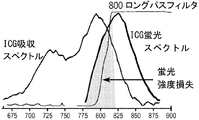
これらのシステムは、血管内画像化のために適切な感応性を有するものの、例えば標的腫瘍特異性NIR蛍光に対しての使用には、実用的ではない。例えば、Fluobeamは、白色光画像のオーバーレイのない携帯用デバイスであるものの、白色光でのHD品質画像、操縦性能、拡大倍率、照度、及びNIR画像の自動同時位置合わせを必要とする外科ツールとしての実用的用途のために設計されていない。このように感度が低い理由の1つは、画像化システムによって捕捉される蛍光光子の量が少ないことにあり、それは、このタイプのシステムは主に、ロングパスフィルタを有するカメラを1台(NIRのみ)または2台(NIR及び可視)使用し得るからである。可視及びNIRの同時捕捉の画像化システムでは、1台のカメラが可視スペクトルで画像を捕捉し、2台目のカメラが蛍光性画像を捕捉する。これは、ビームスプリッタを用いて、視野からの入射光を2個のチャネルに分割することによって達成される。一方のビームは、NIR蛍光をカメラの1台に伝送し、可視光の他方のビームは、ビームスプリッタを通過して第2のカメラに到達する。ICG等のNIR色素の蛍光励起及び放出のストークスシフトが、非常に狭いため、ロングパスフィルタは、蛍光(図1)及びその後の検出感度に、大きな損失を与えてしまう。腫瘍の蛍光画像化は、標的指向化部分がより高い特異性を達成して、癌組織と周囲の正常組織との間の確実な区別を可能にすることを必要とする。これを達成するため、投与量を低く保ち、薬物投与と画像化との間の時間を非常に長く(ほとんどの場合12〜48時間)することにより、腫瘍によるプローブの取り込み及び正常組織からの未結合の物質の洗浄を可能にする。これにより、蛍光シグナルが非常に小さくなり、現在市場に出ているシステムを検出には不十分にさせている。さらに、2つのカメラアタッチメントがあり、既存のセットアップでは完全な変更が必要であるという事実により、これらのシステムは、臨床の場での使用では扱いにくい場合がある。既存のシステムはこのように不十分であるため、これらの新規画像化剤の特異性を利用するための装置の技術革新が必要となる。 While these systems have adequate sensitivity for intravascular imaging, they are not practical for use, for example, for target tumor specific NIR fluorescence. For example, Flubeam is a portable device without white light image overlay, but as a surgical tool that requires automatic simultaneous alignment of HD quality images in white light, steering performance, magnification, illumination, and NIR images Not designed for practical use. One reason for this low sensitivity is that the amount of fluorescent photons captured by the imaging system is small, which is because this type of system mainly has one camera with a long-pass filter (NIR only). ) Or two (NIR and visible) can be used. In an imaging system with simultaneous visible and NIR capture, one camera captures an image in the visible spectrum and a second camera captures a fluorescent image. This is accomplished by splitting incident light from the field of view into two channels using a beam splitter. One beam transmits NIR fluorescence to one of the cameras, and the other beam of visible light passes through the beam splitter and reaches the second camera. Since the Stokes shift of fluorescence excitation and emission of NIR dyes such as ICG is very narrow, the long-pass filter causes a large loss in fluorescence (FIG. 1) and subsequent detection sensitivity. Fluorescence imaging of tumors requires that the targeting moiety achieve a higher specificity to allow a positive distinction between cancerous tissue and surrounding normal tissue. To achieve this, the uptake of the probe by the tumor and the unincorporation from normal tissue is maintained by keeping the dose low and the time between drug administration and imaging very long (in most cases 12-48 hours). Allows washing of bound material. This makes the fluorescent signal very small, making systems currently on the market insufficient for detection. In addition, due to the fact that there are two camera attachments and the existing setup requires a complete change, these systems may be cumbersome for clinical use. Because existing systems are so deficient, device innovations are needed to take advantage of the specificity of these novel imaging agents.
従って、蛍光色素由来の可視光画像及び赤外光画像を同時に記録することが可能である高感度のシステム及び方法の必要性が存在する。本明細書に記載される本発明は、蛍光体由来の可視光画像及び赤外光画像を同時に記録するためのシステム及び方法を提供することにより、これまで満たされていなかった必要性を満たす。 Accordingly, there is a need for a highly sensitive system and method that can simultaneously record visible and infrared light images derived from fluorescent dyes. The invention described herein satisfies a previously unmet need by providing a system and method for simultaneously recording visible and infrared light images from phosphors.
本発明の様々な実施形態は、赤外蛍光体または近赤外蛍光体を単独で含むか、または、ペプチド、タンパク質、ナノ粒子、ナノ複合体、抗体、及び核酸(例えばDNA及びRNAストランド)等の標的指向化部分に結合した、若しくは任意の他の生物学的に特異的な標的指向化実体に結合した赤外蛍光体または近赤外蛍光体を含む試料を画像化するための画像化システムを提供する。この画像化システムは、イメージセンサ、レーザー、レーザークリーンアップフィルタ、ノッチフィルタ、及び白色光源を有している。イメージセンサは、可視光及び赤外光を検出し、かつ、センサシグナルを発生させる。レーザーは、赤外蛍光体のための励起光を放出する。レーザークリーンアップフィルタは、レーザーから試料までの光路内に設置されて、励起光の波長帯を、赤外蛍光体または近赤外蛍光体のピーク吸収帯まで狭める。狭められた励起光は試料においてピーク吸収において赤外蛍光体または近赤外蛍光体を励起して、放射光を放出する。ノッチフィルタは、試料からイメージセンサまでの光路内に設置され、励起光を遮断する。白色光源は、可視光を含む光を放出する。様々な実施形態において、イメージセンサは、NIRロングパスフィルタを有していない。様々な実施形態において、画像化システムは、高速トリガユニットを更に備える。 Various embodiments of the present invention include infrared phosphors or near infrared phosphors alone, or peptides, proteins, nanoparticles, nanocomplexes, antibodies, nucleic acids (eg, DNA and RNA strands), etc. Imaging system for imaging a sample comprising an infrared or near-infrared fluorophore bound to a targeting moiety of or to any other biologically specific targeting entity I will provide a. The imaging system includes an image sensor, a laser, a laser cleanup filter, a notch filter, and a white light source. The image sensor detects visible light and infrared light, and generates a sensor signal. The laser emits excitation light for the infrared phosphor. The laser cleanup filter is installed in the optical path from the laser to the sample, and narrows the wavelength band of the excitation light to the peak absorption band of the infrared phosphor or the near infrared phosphor. The narrowed excitation light excites the infrared phosphor or near-infrared phosphor in peak absorption in the sample and emits radiated light. The notch filter is installed in the optical path from the sample to the image sensor and blocks excitation light. The white light source emits light including visible light. In various embodiments, the image sensor does not have a NIR long pass filter. In various embodiments, the imaging system further comprises a fast trigger unit.
本発明の様々な実施形態では、赤外蛍光体または近赤外蛍光体を含む試料を画像化するための画像化システムが提供される。このシステムは、イメージセンサ、レーザー、ノッチビームスプリッタ、ノッチフィルタ、及び同期モジュールを有している。イメージセンサは、可視光及び赤外光を検出し、かつセンサシグナルを発生させる。レーザーは、赤外蛍光体または近赤外蛍光体のための励起光を放出し、かつオンとオフの状態を交互に繰り返す。ノッチビームスプリッタは、レーザーから試料までの光路内及び試料からイメージセンサまでの光路内に、設置される。励起光は、ノッチビームスプリッタにより試料へ反射され、励起光は、試料中の赤外蛍光体または近赤外蛍光体を励起して放射光を放出し、放射光はノッチビームスプリッタを通してイメージセンサに伝送される。ノッチフィルタは、試料からイメージセンサまでの光路内に設置され、このノッチフィルタは、励起光を遮断する。同期(トリガ)モジュールは、イメージセンサをレーザー及び可視光と同期させることにより、単一のセンサシグナルが、レーザーのオンまたはオフの単一の状態に同期する。 In various embodiments of the present invention, an imaging system is provided for imaging a sample comprising an infrared phosphor or a near infrared phosphor. The system includes an image sensor, a laser, a notch beam splitter, a notch filter, and a synchronization module. The image sensor detects visible light and infrared light and generates a sensor signal. The laser emits excitation light for the infrared phosphor or near-infrared phosphor and repeats the on and off states alternately. The notch beam splitter is installed in the optical path from the laser to the sample and in the optical path from the sample to the image sensor. The excitation light is reflected to the sample by the notch beam splitter, and the excitation light excites the infrared phosphor or near infrared phosphor in the sample to emit radiation light, and the radiation light passes through the notch beam splitter to the image sensor. Is transmitted. The notch filter is installed in the optical path from the sample to the image sensor, and the notch filter blocks excitation light. The synchronization (trigger) module synchronizes the image sensor with the laser and visible light so that a single sensor signal is synchronized to a single state of the laser on or off.
また、試料を画像化する方法も提供される。この方法は、試料を提供するステップと、本明細書に記載される画像化システムを提供するステップと、当該画像化システムで試料を画像化するステップとを含む。 A method for imaging a sample is also provided. The method includes providing a sample, providing an imaging system described herein, and imaging the sample with the imaging system.
本発明の様々な実施形態は、腫瘍を画像化すること、診断すること、及び/または治療することという文脈において記載されるが、本発明がそのような用途に限定されると解釈されてはならない。実際、本発明では、組織の差異、すなわち組織の正常対異常、に対する全ての検出及び診断における有用性が見出されてもよく、この正常対異常は、腫瘍、損傷、外傷、虚血、感染、炎症、または自己炎症を非限定的に含むありとありうる事由による。本発明は、限定されないが、腫瘍組織、損傷組織、虚血組織、感染組織、及び炎症組織を画像化、診断、及び/または、治療することを含む、画像化システム及び広範囲にわたる用途のためのシステムを提供する。関心対象の組織(例えば癌性、損傷、虚血性、感染、または炎症組織)が、それを包囲する組織(例えば、健康な組織)と、生理的または病理学的原因のために異なっているあらゆる状況において、赤外蛍光体または近赤外蛍光体を、関心対象の組織及び周囲の組織を特異的に標識するために用いることができ、かつ、それらの領域に対して、本発明の画像化システム及び方法により画像化して、適切な診断及び治療のための視覚的ガイダンスを提供することができる。したがって、この画像化システム及び方法は、限定されないが、腫瘍、癌、外傷性脳損傷、脊髄損傷、脳卒中、脳出血、脳虚血、虚血性心疾患、虚血性再灌流傷害、心血管疾患、心臓弁狭窄、感染症、微生物感染症、ウイルス感染症、細菌感染症、真菌感染症、及び自己免疫性疾患を含む様々な状態を有する対象を画像化、診断、及び/または、治療するために用いられてもよい。本発明の画像化システムは、例えば、血管系を特定するために健康な対象の正常組織を画像化するために、用いられてもよい。
[本発明1001]
可視光及び赤外光を検出しかつセンサシグナルを発生させるためのイメージセンサ、
赤外蛍光体または近赤外蛍光体のための励起光を放出するためのレーザー、
該レーザーから該赤外蛍光体または該近赤外蛍光体を含む試料までの光路内のレーザークリーンアップフィルタであって、該レーザークリーンアップフィルタが、該励起光の波長帯を該赤外蛍光体または該近赤外蛍光体のピーク吸収帯まで狭め、かつ狭められた該励起光が、該試料中の該赤外蛍光体または該近赤外蛍光体を励起して放射光を放出する、レーザークリーンアップフィルタ、
該励起光を遮断する、該試料から該イメージセンサまでの光路内のノッチフィルタ、並びに
可視光を含む光を放出するための白色光源
を備える、該試料を画像化するための画像化システム。
[本発明1002]
前記試料が、腫瘍、細胞、組織、器官、または身体部分である、本発明1001の画像化システム。
[本発明1003]
前記試料が対象から分離されている、本発明1001の画像化システム。
[本発明1004]
前記試料が対象と一体化している、本発明1001の画像化システム。
[本発明1005]
前記赤外蛍光体または前記近赤外蛍光体が、インドシアニングリーン(ICG)、IR800、Alexa680、cy5.5、IR800の機能的等価物、Alexa680の機能的等価物、cy5.5の機能的等価物、IR800の類似体、Alexa680の類似体、cy5.5の類似体、IR800の誘導体、Alexa680の誘導体、cy5.5の誘導体、IR800の塩、Alexa680の塩、またはcy5.5の塩からなる群の1つである、本発明1001の画像化システム。
[本発明1006]
前記イメージセンサが、青色、緑色、及び赤色の画素センサを備える、本発明1001の画像化システム。
[本発明1007]
前記イメージセンサが、可視光及び赤外光を検出するため及びCCD映像シグナルを発生させるためのCCDイメージセンサである、本発明1001の画像化システム。
[本発明1008]
前記イメージセンサが、可視光及び赤外光を検出するため及びCMOS映像シグナルを発生させるためのCMOSイメージセンサである、本発明1001の画像化システム。
[本発明1009]
前記レーザーの強度を制御して、可視光によって照射されたのと同じ領域上での均一な励起を確実にする、本発明1001の画像化システム。
[本発明1010]
前記レーザーが狭帯域レーザーである、本発明1001の画像化システム。
[本発明1011]
前記ノッチフィルタの遮断範囲が、前記レーザークリーンアップフィルタの透過範囲より広い、本発明1001の画像化システム。
[本発明1012]
前記励起光が、約785nmの波長を有している光を含む、本発明1001の画像化システム。
[本発明1013]
前記レーザークリーンアップフィルタが、約785nmの波長を有する光を選択的に透過させる、本発明1001の画像化システム。
[本発明1014]
前記ノッチフィルタが、約785nmの波長を有する光を選択的に遮断する、本発明1001の画像化システム。
[本発明1015]
前記レーザーから前記試料までの光路内にノッチビームスプリッタを更に備え、前記励起光が該ノッチビームスプリッタによって該試料へ反射される、本発明1001の画像化システム。
[本発明1016]
前記白色光源から前記試料までの光路内にノッチビームスプリッタを更に備え、前記可視光が該試料に伝送される、本発明1001の画像化システム。
[本発明1017]
約700、725、または750nmの波長で光を分割するノッチビームスプリッタを更に備える、本発明1001の画像化システム。
[本発明1018]
約785nmの波長を有する光を反射するノッチビームスプリッタを更に備える、本発明1001の画像化システム。
[本発明1019]
前記試料から前記イメージセンサまでの光路内には赤外フィルタがない、本発明1001の画像化システム。
[本発明1020]
前記レーザーから前記試料までの光路内には赤外フィルタがない、本発明1001の画像化システム。
[本発明1021]
センサシグナルを処理して画像フレームを生成するための画像処理ユニットを更に備える、本発明1001の画像化システム。
[本発明1022]
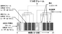
前記画像処理ユニットが、センサシグナルを処理して、前記試料が可視光のみを受ける場合には少なくとも1つの白色光フレーム(WLF)を生成し、該試料が可視光も前記励起光も受けない場合には少なくとも1つの迷光フレーム(SLF)を生成し、かつ、該試料が励起光のみを受ける場合には1つ以上の近赤外フレーム(NIF)を生成し、かつ該画像処理ユニットが、各NIFから該SLFを減算し、その後、SLFが減算されたNIFを全て合算して最終的なNIFを生成する、本発明1021の画像化システム。
[本発明1023]
前記画像処理ユニットが、前記最終的なNIFに疑似カラーを付ける、本発明1022の画像化システム。
[本発明1024]
前記画像処理ユニットが、疑似カラーを付けられた前記最終的なNIFを前記WLFに加算して、可視光及び赤外光の複合画像フレームを生成する、本発明1023の画像化システム。
[本発明1025]
前記画像処理ユニットが、30Hzの周波数で可視光及び赤外光の複合画像フレームを生成する、本発明1024の画像化システム。
[本発明1026]
前記画像処理ユニットから生成された前記画像フレームに基づいて画像を表示するための画像表示ユニットを更に備える、本発明1021の画像化システム。
[本発明1027]
前記画像表示ユニットが、30Hzの周波数で可視光及び赤外光の複合画像フレームを表示する、本発明1026の画像化システム。
[本発明1028]
前記レーザーから前記試料まで前記励起光を導通するための第1のチャネルと、前記白色光源から該試料まで前記可視光を導通するための第2のチャネルと、該試料から前記イメージセンサまで前記放射光を導通するための第3のチャネルと、該試料から該イメージセンサまで該可視光を導通するための第4のチャネルとを更に備える、本発明1001の画像化システム。
[本発明1029]
前記第1のチャネル、前記第2のチャネル、前記第3のチャネル、及び前記第4のチャネルが、4個の別々のチャネルであるか、または、組み合わされて1個、2個、もしくは3個のチャネルとなっている、本発明1028の画像化システム。
[本発明1030]
前記第1のチャネル、前記第2のチャネル、前記第3のチャネル、及び前記第4のチャネルが内視鏡または顕微鏡である、本発明1028の画像化システム。
[本発明1031]
(a)可視光及び赤外光を検出しかつセンサシグナルを発生させるためのイメージセンサであって、赤外蛍光体または近赤外蛍光体を含む試料から該イメージセンサまでの光路内には赤外フィルタがなく、かつ該イメージセンサが、青色、緑色、及び赤色の画素センサを備える、イメージセンサ、
(b)該赤外蛍光体または該近赤外蛍光体のための励起光を放出するためのレーザー、
(c)該レーザーから該試料までの光路内のレーザークリーンアップフィルタであって、該レーザークリーンアップフィルタが、該励起光の波長帯を該赤外蛍光体または該近赤外蛍光体のピーク吸収帯まで狭め、かつ狭められた該励起光が、該試料中の該赤外蛍光体または該近赤外蛍光体を励起して放射光を放出する、レーザークリーンアップフィルタ、
(d)該レーザーから該試料まで該励起光を導通させるための第1のチャネル、
(e)可視光を含む光を放出するための白色光源、
(f)該白色光源から該試料まで該可視光を導通させるための第2のチャネル、
(g)該レーザーから該試料までの光路内及び該白色光源から該試料までの光路内のノッチビームスプリッタであって、該励起光が、該ノッチビームスプリッタにより該試料へ反射され、かつ該可視光が該ノッチビームスプリッタを通して該試料に伝送される、ノッチビームスプリッタ、
(h)該試料から該イメージセンサまで該放射光を導通させるための第3のチャネル、
(i)該試料から該イメージセンサまで該可視光を導通させるための第4のチャネル、
(j)該励起光を遮断する、該試料から該イメージセンサまでの光路内のノッチフィルタ、並びに
(k)センサシグナルを処理して画像フレームを生成するための画像処理ユニットであって、該画像処理ユニットが該イメージセンサに接続され、該試料が可視光のみを受ける場合には少なくとも1つの白色光フレーム(WLF)が生成され、該試料が可視光も該励起光も受けない場合には少なくとも1つの迷光フレーム(SLF)が生成され、該試料が励起光のみを受ける場合には1つ以上の近赤外フレーム(NIF)が生成され、該画像処理ユニットが、各NIFから該SLFを減算し、その後にSLFが減算されたNIFを全て合算して最終的なNIFを生成し、該画像処理ユニットが、該最終的なNIFに疑似カラーを付け、かつ該画像処理ユニットが、疑似カラーを付けられた該最終的なNIFを該WLFに加算して、可視光及び赤外光の複合画像フレームを生成する、画像処理ユニット、
(l)該画像処理ユニットに接続された、該画像処理ユニットから生成された該画像フレームに基づいて画像を表示するための画像表示ユニット
を備える、該試料を画像化するための画像化システム。
[本発明1032]
可視光及び赤外光を検出しかつセンサシグナルを発生させるためのイメージセンサ、
赤外蛍光体または近赤外蛍光体のための励起光を放出し、かつオンとオフの状態を交互に繰り返すレーザー、
該レーザーから該赤外蛍光体または該近赤外蛍光体を含む試料までの光路内及び該試料から該イメージセンサまでの光路内のノッチビームスプリッタであって、該励起光が該ノッチビームスプリッタにより該試料へ反射され、該励起光が、該試料中の該赤外蛍光体または該近赤外蛍光体を励起して放射光を放出し、かつ該放射光が該ノッチビームスプリッタを通して該イメージセンサに伝送される、ノッチビームスプリッタ、
該励起光を遮断する、該試料から該イメージセンサまでの光路内のノッチフィルタ、並びに
該イメージセンサを該レーザー及び可視光と同期させるための同期モジュールであって、単一のセンサシグナルを、該レーザーのオンまたはオフの単一の状態に同期させる、同期モジュール
を備える、該試料を画像化するための画像化システム。
[本発明1033]
前記試料が、腫瘍、細胞、組織、器官、または身体部分である、本発明1032の画像化システム。
[本発明1034]
前記試料が対象から分離されている、本発明1032の画像化システム。
[本発明1035]
前記試料が、対象と一体化している、本発明1032の画像化システム。
[本発明1036]
前記赤外蛍光体または前記近赤外蛍光体が、インドシアニングリーン(ICG)、IR800、Alexa680、cy5.5、IR800の機能的等価物、Alexa680の機能的等価物、cy5.5の機能的等価物、IR800の類似体、Alexa680の類似体、cy5.5の類似体、IR800の誘導体、Alexa680の誘導体、cy5.5の誘導体、IR800の塩、Alexa680の塩、またはcy5.5の塩からなる群の1つである、本発明1032の画像化システム。
[本発明1037]
前記イメージセンサが、青色、緑色、及び赤色の画素センサを備える、本発明1032の画像化システム。
[本発明1038]
前記イメージセンサが、可視光及び赤外光を検出するため及びCCD映像シグナルを発生させるためのCCDイメージセンサである、本発明1032の画像化システム。
[本発明1039]
前記イメージセンサが、可視光及び赤外光を検出するため及びCMOS映像シグナルを発生させるためのCMOSイメージセンサである、本発明1032の画像化システム。
[本発明1040]
前記レーザーの強度を制御して、可視光によって照射されたのと同じ領域上での均一な励起を確実にする、本発明1032の画像化システム。
[本発明1041]
前記レーザーのオン/オフ周波数が、センサシグナルを発生する前記イメージセンサの周波数の半分である、本発明1032の画像化システム。
[本発明1042]
前記レーザーが、60Hzの周波数でオンとオフの状態を交互に繰り返す、本発明1032の画像化システム。
[本発明1043]
前記イメージセンサが、120Hzの周波数でセンサシグナルを発生させる、本発明1032の画像化システム。
[本発明1044]
前記励起光が、約785nm及び/または780nmの波長を有する光を含む、本発明1032の画像化システム。
[本発明1045]
前記ノッチビームスプリッタが、約785nm及び/または780nmの波長を有する光を選択的に反射する、本発明1032の画像化システム。
[本発明1046]
前記ノッチフィルタが、約785nm及び/または780nmの波長を有する光を遮断する、本発明1032の画像化システム。
[本発明1047]
前記試料から前記イメージセンサまでの光路内には赤外フィルタがない、本発明1032の画像化システム。
[本発明1048]
前記レーザーから前記試料までの光路内には赤外フィルタがない、本発明1032の画像化システム。
[本発明1049]
可視光を含む光を放出するための光源を更に備える、本発明1032の画像化システム。
[本発明1050]
センサシグナルを処理して画像フレームを生成するための画像処理ユニットを更に備える、本発明1032の画像化システム。
[本発明1051]
前記画像処理ユニットが、前記レーザーがオフの場合に生成された画像フレームを、該レーザーがオンの場合に生成された直前または次の画像フレームから減算し、赤外線のみの画像フレームが、2つの連続した前記画像フレーム間の差によって生成される、本発明1050の画像化システム。
[本発明1052]
前記画像処理ユニットが、赤外線のみの画像フレームに疑似カラーを付ける、本発明1051の画像化システム。
[本発明1053]
前記画像処理ユニットが、疑似カラーを付けられた前記赤外線のみの画像フレームを、前記レーザーがオフの場合に生成された前記画像フレームに再び加算し、可視光及び赤外光の複合画像フレームが生成される、本発明1052の画像化システム。
[本発明1054]
前記画像処理ユニットが、60Hzの周波数で可視光及び赤外光の複合画像フレームを生成する、本発明1053の画像化システム。
[本発明1055]
前記画像処理ユニットから生成された前記画像フレームに基づいて画像を表示するための画像表示ユニットを更に備える、本発明1050の画像化システム。
[本発明1056]
前記画像表示ユニットが、60Hzの周波数で可視光及び赤外光の複合画像フレームを表示する、本発明1055の画像化システム。
[本発明1057]
前記レーザーから前記試料まで前記励起光を導通するための第1のチャネルと、前記光源から該試料まで前記可視光を導通するための第2のチャネルと、該試料から前記イメージセンサまで前記放射光を導通するための第3のチャネルと、該試料から該イメージセンサまで該可視光を導通するための第4のチャネルとを更に備える、本発明1032の画像化システム。
[本発明1058]
前記第1のチャネル、前記第2のチャネル、前記第3のチャネル、及び前記第4のチャネルが、4個の別々のチャネルであるか、または、組み合わされて1個、2個、または、3個のチャネルとなっている、本発明1057の画像化システム。
[本発明1059]
前記第1のチャネル、前記第2のチャネル、前記第3のチャネル、及び前記第4のチャネルが内視鏡または顕微鏡である、本発明1057の画像化システム。
[本発明1060]
(a)可視光及び赤外光を検出しかつセンサシグナルを第1の周波数で発生させるためのイメージセンサであって、赤外蛍光体または近赤外蛍光体を含む試料から該イメージセンサまでの光路内には赤外フィルタがなく、かつ該イメージセンサが、青色、緑色、及び赤色の画素センサを備える、イメージセンサ、
(b)該赤外蛍光体または該近赤外蛍光体のための励起光を放出し、かつ、該第1の周波数の半分である第2の周波数でオンとオフの状態を交互に繰り返すレーザー、
(c)該レーザーから該試料まで該励起光を導通させるための第1のチャネル、
(d)可視光を含む光を放出するための光源、
(e)該光源から該試料まで該可視光を導通させるための第2のチャネル、
(f)該レーザーから該試料までの光路内及び該試料から該イメージセンサまでの光路内のノッチビームスプリッタであって、該励起光が該ノッチビームスプリッタにより該試料へ反射され、該励起光が、該試料中の該赤外蛍光体または該近赤外蛍光体を励起して放射光を放出し、かつ該放射光が該ノッチビームスプリッタを通して該イメージセンサに伝送される、ノッチビームスプリッタ、
(g)該試料から該イメージセンサまで該放射光を導通させるための第3のチャネル、
(h)該試料から該イメージセンサまで該可視光を導通させるための第4のチャネル、
(i)該励起光を遮断する、該試料から該イメージセンサまでの光路内のノッチフィルタ、
(j)該イメージセンサを該レーザー及び可視光と同期させるための同期モジュールであって、単一のセンサシグナルを該レーザーのオンまたはオフの単一の状態に同期させる、同期モジュール、
(k)センサシグナルを処理して画像フレームを生成するための画像処理ユニットであって、該画像処理ユニットが該イメージセンサに接続され、該画像処理ユニットが、該レーザーがオフの場合に生成された画像フレームを、レーザーがオンの場合に生成された直前または次の画像フレームから減算し、赤外線のみの画像フレームが、2つの連続した該画像フレーム間の差によって生成され、該画像処理ユニットが、該赤外線のみの画像フレームに疑似カラーを付け、該画像処理ユニットが、疑似カラーを付けられた該赤外線のみの画像フレームを、該レーザーがオフの場合に生成された該画像フレームに再び加算し、可視光及び赤外光の複合画像フレームが生成される、画像処理ユニット、並びに
(l)該画像処理ユニットに接続された、該画像処理ユニットから生成された該画像フレームに基づいて画像を表示するための画像表示ユニット
を備える、該試料を画像化するための画像化システム。
[本発明1061]
試料を提供すること、
前記本発明のいずれかの画像化システムを提供すること、及び、
該画像化システムを用いて該試料を画像化すること
を含む、該試料を画像化する方法。
[本発明1062]
前記試料が、腫瘍、細胞、組織、器官、または身体部分である、本発明1061の方法。
[本発明1063]
対象に対して手術を実行して、前記試料にアクセスするかまたは該試料を分離することを更に含む、本発明1061の方法。
[本発明1064]
赤外蛍光体または近赤外蛍光体で前記試料を標識することを更に含む、本発明1061の方法。
[本発明1065]
前記赤外蛍光体または前記近赤外蛍光体が、インドシアニングリーン(ICG)、IR800、Alexa680、cy5.5、IR800の機能的等価物、Alexa680の機能的等価物、cy5.5の機能的等価物、IR800の類似体、Alexa680の類似体、cy5.5の類似体、IR800の誘導体、Alexa680の誘導体、cy5.5の誘導体、IR800の塩、Alexa680の塩、またはcy5.5の塩からなる群の1つである、本発明1064の方法。
[本発明1066]
腫瘍を有する対象に赤外蛍光体または近赤外蛍光体を投与し、それにより、該赤外蛍光体または該近赤外蛍光体で該腫瘍を標識することと、
該対象に対して手術を実行して、標識された該腫瘍の領域にアクセスすることと、
前記本発明のいずれかの画像化システムを提供することと、
該画像化システムに従って、標識された該腫瘍を特定することと、
標識された該腫瘍を除去し、それにより、該腫瘍を有する該対象を治療することと
を含む、該対象を治療する方法。
[本発明1067]
デバイス上に1つ以上のプロセッサと該1つ以上のプロセッサによる実行のための1つ以上のプログラムを格納するメモリとを搭載すること
を含み、
該1つ以上のプログラムが、
イメージセンサを動作させて可視光及び赤外光を検出し、かつセンサシグナルを発生させるための命令、
レーザーを動作させて、赤外蛍光体または近赤外蛍光体のための励起光を放出させるための命令、
該レーザーから該赤外蛍光体または該近赤外蛍光体を含む試料までの光路内のレーザークリーンアップフィルタを動作させるための命令であって、該レーザークリーンアップフィルタが、該励起光の波長帯を該赤外蛍光体または該近赤外蛍光体のピーク吸収帯まで狭め、かつ狭められた該励起光が、該試料中の該赤外蛍光体または該近赤外蛍光体を励起して放射光を放出する、命令、
該試料から該イメージセンサまでの光路内のノッチフィルタを動作させるための命令であって、該ノッチフィルタが該励起光を遮断する、命令、並びに
白色光源を動作させて、可視光を含む光を放出させるための命令
を含む、該試料を画像化するためのコンピュータ実装方法。
[本発明1068]
1つ以上のプロセッサと1つ以上のプログラムを格納するためのメモリとを備え、
該1つ以上のプログラムが、
イメージセンサを動作させて可視光及び赤外光を検出し、かつセンサシグナルを発生させるための命令、
レーザーを動作させて、赤外蛍光体または近赤外蛍光体のための励起光を放出させるための命令、
該レーザーから該赤外蛍光体または該近赤外蛍光体を含む試料までの光路内のレーザークリーンアップフィルタを動作させるための命令であって、該レーザークリーンアップフィルタが、該励起光の波長帯を該赤外蛍光体または該近赤外蛍光体のピーク吸収帯まで狭め、かつ狭められた該励起光が、該試料中の該赤外蛍光体または該近赤外蛍光体を励起して放射光を放出する、命令、
該試料から該イメージセンサまでの光路内のノッチフィルタを動作させるための命令であって、該ノッチフィルタが該励起光を遮断する、命令、並びに
白色光源を動作させて、可視光を含む光を放出させるための命令
を含む、該試料を画像化するためのコンピュータシステム。
[本発明1069]
コンピュータシステムのプロセッサの1つ以上による実行のための1つ以上のプログラムが、
イメージセンサを動作させて可視光及び赤外光を検出し、かつセンサシグナルを発生させるための命令、
レーザーを動作させて、赤外蛍光体または近赤外蛍光体のための励起光を放出させるための命令、
該レーザーから該赤外蛍光体または該近赤外蛍光体を含む試料までの光路内のレーザークリーンアップフィルタを動作させるための命令であって、該レーザークリーンアップフィルタが、該励起光の波長帯を該赤外蛍光体または該近赤外蛍光体のピーク吸収帯まで狭め、かつ狭められた該励起光が、該試料中の該赤外蛍光体または該近赤外蛍光体を励起して、放射光を放出する、命令、
該試料から該イメージセンサまでの光路内のノッチフィルタを動作させて、該ノッチフィルタが該励起光を遮断する、命令、並びに
白色光源を動作させて、可視光を含む光を放出させるための命令
を含む、該試料を画像化するための該1つ以上のプログラムを格納する非一時的コンピュータ可読保存媒体。
[本発明1070]
デバイス上に1つ以上のプロセッサと該1つ以上のプロセッサによる実行のための1つ以上のプログラムを格納するメモリとを搭載すること
を含み、
該1つ以上のプログラムが、
(a)イメージセンサを動作させて、可視光及び赤外光を検出しかつセンサシグナルを発生させるための命令であって、赤外蛍光体または近赤外蛍光体を含む試料から該イメージセンサまでの光路内には赤外フィルタがなく、かつ該イメージセンサが、青色、緑色、及び赤色の画素センサを備える、命令、
(b)レーザーを動作させて、該赤外蛍光体または該近赤外蛍光体のための励起光を放出させるための命令、
(c)該レーザーから該試料までの光路内のレーザークリーンアップフィルタを動作させるための命令であって、該レーザークリーンアップフィルタが、該励起光の波長帯を該赤外蛍光体または該近赤外蛍光体のピーク吸収帯まで狭め、かつ狭められた該励起光が、該試料中の該赤外蛍光体または該近赤外蛍光体を励起して放射光を放出する、命令、
(d)第1のチャネルを動作させて、該レーザーから該試料まで該励起光を導通させるための命令、
(e)白色光源を動作させて、可視光を含む光を放出させるための命令、
(f)第2のチャネルを動作させて、該白色光源から該試料まで該可視光を導通させるための命令、
(g)該レーザーから該試料までの光路内及び該白色光源から該試料までの光路内のノッチビームスプリッタを動作させるための命令であって、該励起光が該ノッチビームスプリッタにより該試料へ反射され、かつ該可視光が該ノッチビームスプリッタを通して該試料に伝送される、命令、
(h)第3のチャネルを動作させて、該試料から該イメージセンサまで該放射光を導通させるための命令、
(i)第4のチャネルを動作させて、該試料から該イメージセンサまで該可視光を導通させるための命令、
(j)該試料から該イメージセンサまでの光路内のノッチフィルタを動作させるための命令であって、該ノッチフィルタが該励起光を遮断する、命令、
(k)画像処理ユニットを動作させて、センサシグナルを処理して画像フレームを生成させるための命令であって、該画像処理ユニットが該イメージセンサに接続され、該試料が可視光のみを受ける場合には少なくとも1つの白色光フレーム(WLF)が生成され、該試料が可視光も励起光も受けない場合には少なくとも1つの迷光フレーム(SLF)が生成され、該試料が励起光のみを受ける場合には1つ以上の近赤外フレーム(NIF)が生成され、該画像処理ユニットが、各NIFから該SLFを減算し、その後にSLFが減算されたNIFを全て合算して最終的なNIFを生成し、該画像処理ユニットが、該最終的なNIFに疑似カラーを付け、かつ該画像処理ユニットが、疑似カラーを付けられた該最終的なNIFを該WLFに加算して、可視光及び赤外光の複合画像フレームを生成する、命令、並びに
(l)該画像処理ユニットに接続されている画像表示ユニットを動作させて、該画像処理ユニットから生成された該画像フレームに基づいて画像を表示するための命令
を含む、該試料を画像化するためのコンピュータ実装方法。
[本発明1071]
1つ以上のプロセッサと1つ以上のプログラムを格納するためのメモリとを備え、
該1つ以上のプログラムが、
(a)イメージセンサを動作させて、可視光及び赤外光を検出しかつセンサシグナルを発生させるための命令であって、赤外蛍光体または近赤外蛍光体を含む試料から該イメージセンサまでの光路内には赤外フィルタがなく、かつ該イメージセンサが、青色、緑色、及び赤色の画素センサを備える、命令、
(b)レーザーを動作させて、該赤外蛍光体または該近赤外蛍光体のための励起光を放出させるための命令、
(c)該レーザーから該試料までの光路内のレーザークリーンアップフィルタを動作させるための命令であって、該レーザークリーンアップフィルタが、該励起光の波長帯を該赤外蛍光体または該近赤外蛍光体のピーク吸収帯まで狭め、かつ狭められた該励起光が、該試料中の該赤外蛍光体または該近赤外蛍光体を励起して放射光を放出する、命令、
(d)第1のチャネルを動作させて、該レーザーから該試料まで該励起光を導通させるための命令、
(e)白色光源を動作させて、可視光を含む光を放出させるための命令、
(f)第2のチャネルを動作させて、該白色光源から該試料まで該可視光を導通させるための命令、
(g)該レーザーから該試料までの光路内及び該白色光源から該試料までの光路内のノッチビームスプリッタを動作させるための命令であって、該励起光が該ノッチビームスプリッタにより該試料へ反射され、かつ該可視光が該ノッチビームスプリッタを通して該試料に伝送される、命令、
(h)第3のチャネルを動作させて、該試料から該イメージセンサまで該放射光を導通させるための命令、
(i)第4のチャネルを動作させて、該試料から該イメージセンサまで該可視光を導通させるための命令、
(j)該試料から該イメージセンサまでの光路内のノッチフィルタを動作させるための命令であって、該ノッチフィルタが該励起光を遮断する、命令、
(k)画像処理ユニットを動作させて、センサシグナルを処理して画像フレームを生成させるための命令であって、該画像処理ユニットが該イメージセンサに接続され、該試料が可視光のみを受ける場合には少なくとも1つの白色光フレーム(WLF)が生成され、該試料が可視光も該励起光も受けない場合には少なくとも1つの迷光フレーム(SLF)が生成され、該試料が励起光のみを受ける場合には1つ以上の近赤外フレーム(NIF)が生成され、該画像処理ユニットが、各NIFから該SLFを減算し、その後にSLFが減算されたNIFを全て合算して最終的なNIFを生成し、該画像処理ユニットが、該最終的なNIFに疑似カラーを付け、かつ該画像処理ユニットが、疑似カラーを付けられた該最終的なNIFを該WLFに加算して、可視光及び赤外光の複合画像フレームを生成する、命令、並びに
(l)該画像処理ユニットに接続されている画像表示ユニットを動作させて、該画像処理ユニットから生成された該画像フレームに基づいて画像を表示するための命令
を含む、該試料を画像化するためのコンピュータシステム。
[本発明1072]
コンピュータシステムのプロセッサの1つ以上による実行のための1つ以上のプログラムが、
(a)イメージセンサを動作させて、可視光及び赤外光を検出しかつセンサシグナルを発生させるための命令であって、赤外蛍光体または近赤外蛍光体を含む試料から該イメージセンサまでの光路内には赤外フィルタがなく、かつ該イメージセンサが、青色、緑色、及び赤色の画素センサを備える、命令、
(b)レーザーを動作させて、該赤外蛍光体または該近赤外蛍光体のための励起光を放出させるための命令、
(c)該レーザーから該試料までの光路内のレーザークリーンアップフィルタを動作させるための命令であって、該レーザークリーンアップフィルタが、該励起光の波長帯を該赤外蛍光体または該近赤外蛍光体のピーク吸収帯まで狭め、かつ狭められた該励起光が、該試料中の該赤外蛍光体または該近赤外蛍光体を励起して放射光を放出する、命令、
(d)第1のチャネルを動作させて、該レーザーから該試料まで該励起光を導通させるための命令、
(e)白色光源を動作させて、可視光を含む光を放出させるための命令、
(f)第2のチャネルを動作させて、該白色光源から該試料まで該可視光を導通させるための命令、
(g)該レーザーから該試料までの光路内及び該白色光源から該試料までの光路内のノッチビームスプリッタを動作させるための命令であって、該励起光が、該ノッチビームスプリッタにより該試料へ反射され、かつ該可視光が、該ノッチビームスプリッタを通して該試料に伝送される、命令、
(h)第3のチャネルを動作させて、該試料から該イメージセンサまで該放射光を導通させるための命令、
(i)第4のチャネルを動作させて、該試料から該イメージセンサまで該可視光を導通させるための命令、
(j)該試料から該イメージセンサまでの光路内のノッチフィルタを動作させるための命令であって、該ノッチフィルタが該励起光を遮断する、命令、
(k)画像処理ユニットを動作させて、センサシグナルを処理して画像フレームを生成させるための命令であって、該画像処理ユニットが該イメージセンサに接続され、該試料が可視光のみを受ける場合には少なくとも1つの白色光フレーム(WLF)が生成され、該試料が可視光も該励起光も受けない場合には少なくとも1つの迷光フレーム(SLF)が生成され、該試料が励起光のみを受ける場合には1つ以上の近赤外フレーム(NIF)が生成され、該画像処理ユニットが、各NIFから該SLFを減算し、その後にSLFが減算されたNIFを全て合算して最終的なNIFを生成し、該画像処理ユニットが、該最終的なNIFに疑似カラーを付け、かつ該画像処理ユニットが、疑似カラーを付けられた該最終的なNIFを該WLFに加算して、可視光及び赤外光の複合画像フレームを生成する、命令、並びに
(l)該画像処理ユニットに接続されている画像表示ユニットを動作させて、該画像処理ユニットから生成された該画像フレームに基づいて画像を表示するための命令
を含む、該試料を画像化するための該1つ以上のプログラムを格納する非一時的コンピュータ可読保存媒体。
[本発明1073]
デバイス上に1つ以上のプロセッサと該1つ以上のプロセッサによる実行のための1つ以上のプログラムを格納するメモリとを搭載すること
を含み、
該1つ以上のプログラムが、
イメージセンサを動作させて、可視光及び赤外光を検出しかつセンサシグナルを発生させるための命令、
レーザーを動作させて、該赤外蛍光体または該近赤外蛍光体のための励起光を放出させ、かつオンとオフの状態を交互に繰り返すための命令、
該レーザーから赤外蛍光体または近赤外蛍光体を含む試料までの光路内及び該試料から該イメージセンサまでの光路内のノッチビームスプリッタを動作させるための命令であって、該励起光が該ノッチビームスプリッタにより該試料へ反射され、該励起光が、該試料中の該赤外蛍光体または該近赤外蛍光体を励起して放射光を放出し、かつ該放射光が該ノッチビームスプリッタを通して該イメージセンサに伝送される、命令、
該試料から該イメージセンサまでの光路内のノッチフィルタを動作させるための命令であって、該ノッチフィルタが該励起光を遮断する、命令、並びに
同期モジュールを動作させて、該イメージセンサを該レーザー及び可視光と同期させるための命令であって、単一のセンサシグナルを該レーザーのオンまたはオフの単一の状態に同期させる、命令
を含む、該試料を画像化するためのコンピュータ実装方法。
[本発明1074]
1つ以上のプロセッサと1つ以上のプログラムを格納するためのメモリとを備え、
該1つ以上のプログラムが、
イメージセンサを動作させて、可視光及び赤外光を検出しかつセンサシグナルを発生させるための命令、
レーザーを動作させて、赤外蛍光体または近赤外蛍光体のための励起光を放出させかつオンとオフの状態を交互に繰り返すための命令、
該レーザーから該赤外蛍光体または該近赤外蛍光体を含む試料までの光路内及び該試料から該イメージセンサまでの光路内のノッチビームスプリッタを動作させるための命令であって、該励起光が該ノッチビームスプリッタにより該試料へ反射され、該励起光が、該試料中の該赤外蛍光体または該近赤外蛍光体を励起して放射光を放出し、かつ該放射光が該ノッチビームスプリッタを通して該イメージセンサに伝送される、命令、
該試料から該イメージセンサまでの光路内のノッチフィルタを動作させるための命令であって、該ノッチフィルタが該励起光を遮断する、命令、並びに
同期モジュールを動作させて、該イメージセンサを該レーザー及び可視光と同期させるための命令であって、単一のセンサシグナルを該レーザーのオンまたはオフの単一の状態に同期させる、命令
を含む、該試料を画像化するためのコンピュータシステム。
[本発明1075]
コンピュータシステムのプロセッサの1つ以上による実行のための1つ以上のプログラムが、
イメージセンサを動作させて、可視光及び赤外光を検出しかつセンサシグナルを発生させるための命令、
レーザーを動作させて、赤外蛍光体または近赤外蛍光体のための励起光を放出させかつオンとオフの状態を交互に繰り返すための命令、
該レーザーから該赤外蛍光体または該近赤外蛍光体を含む試料までの光路内及び該試料から該イメージセンサまでの光路内のノッチビームスプリッタを動作させるための命令であって、該励起光が、該ノッチビームスプリッタにより該試料へ反射され、該励起光が、該試料中の該赤外蛍光体または該近赤外蛍光体を励起して放射光を放出し、かつ該放射光が該ノッチビームスプリッタを通して該イメージセンサに伝送される、命令、
該試料から該イメージセンサまでの光路内のノッチフィルタを動作させるための命令であって、該ノッチフィルタが該励起光を遮断する、命令、並びに
同期モジュールを動作させて、該イメージセンサを該レーザー及び可視光と同期させるための命令であって、単一のセンサシグナルを該レーザーのオンまたはオフの単一の状態に同期させる、命令
を含む、該試料を画像化するための該1つ以上のプログラムを格納する非一時的コンピュータ可読保存媒体。
[本発明1076]
デバイス上に1つ以上のプロセッサと該1つ以上のプロセッサによる実行のための1つ以上のプログラムを格納するメモリとを搭載すること
を含み、
該1つ以上のプログラムが、
(a)イメージセンサを動作させて、可視光及び赤外光を検出しかつセンサシグナルを第1の周波数で発生させるための命令であって、赤外蛍光体または近赤外蛍光体を含む試料から該イメージセンサまでの光路内には赤外フィルタがなく、かつ該イメージセンサが、青色、緑色、及び赤色の画素センサを備える、命令、
(b)レーザーを動作させて、該赤外蛍光体または該近赤外蛍光体のための励起光を放出させるため、及び該第1の周波数の半分である第2の周波数でオンとオフの状態を交互に繰り返すための命令、
(c)第1のチャネルを動作させて、該レーザーから該試料まで該励起光を導通させるための命令、
(d)光源を動作させて、可視光を含む光を放出させるための命令、
(e)第2のチャネルを動作させて、該光源から該試料まで該可視光を導通させるための命令、
(f)該レーザーから該試料までの光路内及び該試料から該イメージセンサまでの光路内のノッチビームスプリッタを動作させるための命令であって、該励起光が該ノッチビームスプリッタにより該試料へ反射され、該励起光が、該試料中の該赤外蛍光体または該近赤外蛍光体を励起して放射光を放出し、かつ該放射光が該ノッチビームスプリッタを通して該イメージセンサに伝送される、命令、
(g)第3のチャネルを動作させて、該試料から該イメージセンサまで該放射光を導通させるための命令、
(h)第4のチャネルを動作させて、該試料から該イメージセンサまで該可視光を導通させるための命令、
(i)該試料から該イメージセンサまでの光路内のノッチフィルタを動作させるための命令であって、該ノッチフィルタが該励起光を遮断する、命令、
(j)同期モジュールを動作させて、該イメージセンサを該レーザー及び可視光と同期させるための命令であって、単一のセンサシグナルを該レーザーのオンまたはオフの単一の状態に同期させる、命令、
(k)画像処理ユニットを動作させて、センサシグナルを処理して画像フレームを生成させるための命令であって、該画像処理ユニットが該イメージセンサに接続され、該画像処理ユニットが、該レーザーがオフの場合に生成された画像フレームを、該レーザーがオンの場合に生成された直前または次の画像フレームから減算し、赤外線のみの画像フレームが、2つの連続した該画像フレーム間の差によって生成され、該画像処理ユニットが、該赤外線のみの画像フレームに疑似カラーを付け、該画像処理ユニットが、疑似カラーを付けられた該赤外線のみの画像フレームを、該レーザーがオフの場合に生成された該画像フレームに再び加算し、可視光及び赤外光の複合画像フレームが生成される、命令、並びに
(l)該画像処理ユニットに接続されている画像表示ユニットを動作させて、該画像処理ユニットから生成された該画像フレームに基づいて画像を表示するための命令
を含む、該試料を画像化するためのコンピュータ実装方法。
[本発明1077]
1つ以上のプロセッサと1つ以上のプログラムを格納するメモリとを備え、
該1つ以上のプログラムが、
(a)イメージセンサを動作させて、可視光及び赤外光を検出しかつセンサシグナルを第1の周波数で発生させるための命令であって、赤外蛍光体または近赤外蛍光体を含む試料から該イメージセンサまでの光路内には赤外フィルタがなく、かつ該イメージセンサが、青色、緑色、及び赤色の画素センサを備える、命令、
(b)レーザーを動作させて、該赤外蛍光体または該近赤外蛍光体のための励起光を放出させるため、及び該第1の周波数の半分である第2の周波数でオンとオフの状態を交互に繰り返すための命令、
(c)第1のチャネルを動作させて、該レーザーから該試料まで該励起光を導通させるための命令、
(d)光源を動作させて、可視光を含む光を放出させるための命令、
(e)第2のチャネルを動作させて、該光源から該試料まで該可視光を導通させるための命令、
(f)該レーザーから該試料までの光路内及び該試料から該イメージセンサまでの光路内のノッチビームスプリッタを動作させるための命令であって、該励起光が該ノッチビームスプリッタにより該試料へ反射され、該励起光が、該試料中の該赤外蛍光体または該近赤外蛍光体を励起して放射光を放出し、かつ該放射光が該ノッチビームスプリッタを通して該イメージセンサに伝送される、命令、
(g)第3のチャネルを動作させて、該試料から該イメージセンサまで該放射光を導通させるための命令、
(h)第4のチャネルを動作させて、該試料から該イメージセンサまで該可視光を導通させるための命令、
(i)該試料から該イメージセンサまでの光路内のノッチフィルタを動作させるための命令であって、該ノッチフィルタが該励起光を遮断する、命令、
(j)同期モジュールを動作させて、該イメージセンサを該レーザー及び可視光と同期させるための命令であって、単一のセンサシグナルを該レーザーのオンまたはオフの単一の状態に同期させる、命令、
(k)画像処理ユニットを動作させて、センサシグナルを処理して画像フレームを生成させるための命令であって、該画像処理ユニットが該イメージセンサに接続され、該画像処理ユニットが、該レーザーがオフの場合に生成された画像フレームを、該レーザーがオンの場合に生成された直前または次の画像フレームから減算し、赤外線のみの画像フレームが、2つの連続した該画像フレーム間の差によって生成され、該画像処理ユニットが、該赤外線のみの画像フレームに疑似カラーを付け、該画像処理ユニットが、疑似カラーを付けられた該赤外線のみの画像フレームを、該レーザーがオフの場合に生成された該画像フレームに再び加算し、可視光及び赤外光の複合画像フレームが生成される、命令、並びに
(l)該画像処理ユニットに接続されている画像表示ユニットを動作させて、該画像処理ユニットから生成された該画像フレームに基づいて画像を表示するための命令
を含む、該試料を画像化するためのコンピュータシステム。
[本発明1078]
コンピュータシステムのプロセッサの1つ以上による実行のための1つ以上のプログラムが、
(a)イメージセンサを動作させて、可視光及び赤外光を検出しかつセンサシグナルを第1の周波数で発生させるための命令であって、赤外蛍光体または近赤外蛍光体を含む試料から該イメージセンサまでの光路内には赤外フィルタがなく、かつ該イメージセンサが、青色、緑色、及び赤色の画素センサを備える、命令、
(b)レーザーを動作させて、該赤外蛍光体または該近赤外蛍光体のための励起光を放出させるため、及び該第1の周波数の半分である第2の周波数でオンとオフの状態に交互に繰り返すための命令、
(c)第1のチャネルを動作させて、該レーザーから該試料まで該励起光を導通させるための命令、
(d)光源を動作させて、可視光を含む光を放出させるための命令、
(e)第2のチャネルを動作させて、該光源から該試料まで該可視光を導通させるための命令、
(f)該レーザーから該試料までの光路内及び該試料から該イメージセンサまでの光路内のノッチビームスプリッタを動作させるための命令であって、該励起光が該ノッチビームスプリッタにより該試料へ反射され、該励起光が、該試料中の該赤外蛍光体または該近赤外蛍光体を励起して放射光を放出し、かつ該放射光が該ノッチビームスプリッタを通して該イメージセンサに伝送される、命令、
(g)第3のチャネルを動作させて、該試料から該イメージセンサまで該放射光を導通させるための命令、
(h)第4のチャネルを動作させて、該試料から該イメージセンサまで該可視光を導通させるための命令、
(i)該試料から該イメージセンサまでの光路内のノッチフィルタを動作させるための命令であって、該ノッチフィルタが該励起光を遮断する、命令、
(j)同期モジュールを動作させて、該イメージセンサを該レーザー及び可視光と同期させるための命令であって、単一のセンサシグナルを該レーザーのオンまたはオフの単一の状態に同期させる、命令、
(k)画像処理ユニットを動作させて、センサシグナルを処理して画像フレームを生成させるための命令であって、該画像処理ユニットが該イメージセンサに接続され、該画像処理ユニットが、該レーザーがオフの場合に生成された画像フレームを、該レーザーがオンの場合に生成された直前または次の画像フレームから減算し、赤外線のみの画像フレームが、2つの連続した該画像フレーム間の差によって生成され、該画像処理ユニットが、該赤外線のみの画像フレームに疑似カラーを付け、該画像処理ユニットが、疑似カラーを付けられた該赤外線のみの画像フレームを、該レーザーがオフの場合に生成された画像フレームに再び加算し、可視光及び赤外光の複合画像フレームが生成される、命令、並びに
(l)該画像処理ユニットに接続されている画像表示ユニットを動作させて、該画像処理ユニットから生成された該画像フレームに基づいて画像を表示するための命令
を含む、該試料を画像化するための該1つ以上のプログラムを格納する非一時的コンピュータ可読保存媒体。
[本発明1079]
デバイス上に1つ以上のプロセッサと該1つ以上のプロセッサによる実行のための1つ以上のプログラムを格納するメモリとを搭載すること
を含み、
該1つ以上のプログラムが、
試料を提供するための命令、
前記本発明のいずれかの画像化システムを提供するための命令、及び、
該画像化システムを用いて該試料を画像化するための命令
を含む、試料を画像化するためのコンピュータ実装方法。
[本発明1080]
1つ以上のプロセッサと1つ以上のプログラムを格納するためのメモリとを備え、
該1つ以上のプログラムが、
試料を提供するための命令、
前記本発明のいずれかの画像化システムを提供するための命令、及び、
該画像化システムを用いて該試料を画像化するための命令
を含む、試料を画像化するためのコンピュータシステム。
[本発明1081]
コンピュータシステムのプロセッサの1つ以上による実行のための1つ以上のプログラムが、
試料を提供するための命令、
前記本発明のいずれかの画像化システムを提供するための命令、及び
該画像化システムを用いて該試料を画像化するための命令
を含む、試料を画像化するための該1つ以上のプログラムを格納する非一時的コンピュータ可読保存媒体。
[本発明1082]
デバイス上に1つ以上のプロセッサと該1つ以上のプロセッサによる実行のための1つ以上のプログラムを格納するメモリとを搭載すること
を含み、
該1つ以上のプログラムが、
腫瘍を有する対象に赤外蛍光体または近赤外蛍光体を投与し、それにより、該赤外蛍光体または該近赤外蛍光体で該腫瘍を標識するための命令、
該対象に対して手術を実行して、標識された該腫瘍の領域にアクセスするための命令、
前記本発明のいずれかの画像化システムを提供するための命令、
該画像化システムに従って、標識された該腫瘍を特定するための命令、及び、
標識された該腫瘍を除去し、それにより、該腫瘍を有する該対象を治療するための命令
を含む、該対象を治療するためのコンピュータ実装方法。
[本発明1083]
1つ以上のプロセッサと1つ以上のプログラムを格納するためのメモリとを備え、
該1つ以上のプログラムが、
腫瘍を有する対象に赤外蛍光体または近赤外蛍光体を投与し、それにより、該赤外蛍光体または該近赤外蛍光体で該腫瘍を標識するための命令、
該対象に対して手術を実行して、標識された該腫瘍の領域にアクセスするための命令、
前記本発明のいずれかの画像化システムを提供するための命令、
該画像化システムに従って、標識された該腫瘍を特定するための命令、及び、
標識された該腫瘍を除去し、それにより、該腫瘍を有する該対象を治療するための命令
を含む、該対象を治療するためのコンピュータシステム。
[本発明1084]
コンピュータシステムのプロセッサの1つ以上による実行のための1つ以上のプログラムが、
腫瘍を有する対象に赤外蛍光体または近赤外蛍光体を投与し、それにより、該赤外蛍光体または該近赤外蛍光体で該腫瘍を標識するための命令、
該対象に対して手術を実行して、標識された該腫瘍の領域にアクセスするための命令、
前記本発明のいずれかの画像化システムを提供するための命令、
該画像化システムに従って、標識された該腫瘍を特定するための命令、及び、
標識された該腫瘍を除去し、それにより、該腫瘍を有する該対象を治療するための命令
を含む、該対象を治療するための該1つ以上のプログラムを格納する非一時的コンピュータ可読保存媒体。
[本発明1085]
デバイス上に1つ以上のプロセッサと該1つ以上のプロセッサによる実行のための1つ以上のプログラムを格納するメモリとを搭載すること
を含み、該1つ以上のプログラムが、
並列処理ソフトウェアコーディングを用いるための命令、
未加工の画像を伝送するための命令、及び、
該1つ以上のプロセッサに対する、該未加工の画像にデモザイキング処理を行うための命令
を含む、画像を取り込みかつ処理するため及びスムーズな画像表示のためのコンピュータ実装方法。
[本発明1086]
前記1つ以上のプロセッサが図形処理ユニット(GPU)を備え、かつ前記並列処理ソフトウェアコーディングが、GPUベースのコンピュータ統合デバイスアーキテクチャ(CUDA)を備える、本発明1085のコンピュータ実装方法。
[本発明1087]
前記並列処理ソフトウェアコーディングが直接ビデオカードに格納される、本発明1085のコンピュータ実装方法。
[本発明1088]
前記未加工の画像が、8ビットの未加工の画像である、本発明1085のコンピュータ実装方法。
[本発明1089]
前記画像が、毎秒300フレームのフル高解像度フレームを含み、フルHD(1080p)8ビットの画像が、約2Mbのサイズであり、PCIe3.0データ転送速度が約7Gb/sであり、かつ該画像が、300マイクロ秒で前記1つ以上のプロセッサへ伝送される、本発明1085のコンピュータ実装方法。
[本発明1090]
前記1つ以上のプロセッサに前記画像を伝送した後に画像処理操作を実行する、本発明1085のコンピュータ実装方法。
[本発明1091]
前記画像処理操作が、ベイヤーデモザイキング処理を行うこと、散乱光画像を蛍光画像から減算すること、蛍光フレームの赤色、緑色、及び青色のチャネルを加算すること、疑似カラーを蛍光画像に与えること、並びに疑似カラーを付けられた蛍光画像を白色光画像に加算することから成る群の一つである、本発明1090のコンピュータ実装方法。
[本発明1092]
速度を向上させる目的で、表示のためにシステムメモリに前記画像を戻す代わりに、前記1つ以上のプロセッサのopenGL/directx機能を動作させて、最終的な画像を表示する、本発明1085のコンピュータ実装方法。
[本発明1093]
医療グレードHD品質ビデオモニタで画像が表示される、本発明1085のコンピュータ実装方法。
[本発明1094]
1つ以上のプロセッサと1つ以上のプログラムを格納するためのメモリとを備え、
該1つ以上のプログラムが、
並列処理ソフトウェアコーディングを用いるための命令、
未加工の画像を伝送するための命令、及び、
該1つ以上のプロセッサに対する、該未加工の画像にデモザイキング処理を行うための命令
を含む、画像を取り込みかつ処理するため及びスムーズな画像表示のためのコンピュータシステム。
[本発明1095]
前記1つ以上のプロセッサが図形処理ユニット(GPU)を備え、かつ前記並列処理ソフトウェアコーディングが、GPUベースのコンピュータ統合デバイスアーキテクチャ(CUDA)を備える、本発明1094のコンピュータシステム。
[本発明1096]
前記並列処理ソフトウェアコーディングが直接ビデオカードに格納される、本発明1094のコンピュータシステム。
[本発明1097]
前記未加工の画像が、8ビットの未加工の画像である、本発明1094のコンピュータシステム。
[本発明1098]
前記画像が、毎秒300フレームのフル高解像度フレームを含み、フルHD(1080p)8ビットの画像が、約2Mbのサイズであり、PCIe3.0データ転送速度が約7Gb/sであり、かつ該画像が、300マイクロ秒で前記1つ以上のプロセッサへ伝送される、本発明1094のコンピュータシステム。
[本発明1099]
前記1つ以上のプロセッサに前記画像を伝送した後に、画像処理操作を実行する、本発明1094のコンピュータシステム。
[本発明1100]
前記画像処理操作が、ベイヤーデモザイキング処理を行うこと、散乱光画像を蛍光画像から減算すること、蛍光フレームの赤色、緑色、及び青色のチャネルを加算すること、疑似カラーを蛍光画像に与えること、並びに疑似カラーを付けられた蛍光画像を白色光画像に加算することから成る群の一つである、本発明1099のコンピュータシステム。
[本発明1101]
速度を向上させる目的で、表示のためにシステムメモリに前記画像を戻す代わりに、前記1つ以上のプロセッサのopenGL/directx機能を動作させて、最終的な画像を表示する、本発明1094のコンピュータシステム。
[本発明1102]
医療グレードHD品質ビデオモニタで画像が表示される、本発明1094のコンピュータシステム。
[本発明1103]
保存媒体のプロセッサの1つ以上による実行のための1つ以上のプログラムが、
並列処理ソフトウェアコーディングを用いるための命令、
未加工の画像を伝送するための命令、及び、
1つ以上の該プロセッサに対する、該未加工の画像にデモザイキング処理を行うための命令
を含む、画像を取り込みかつ処理するため及びスムーズな画像表示のための該1つ以上のプログラムを格納する非一時的コンピュータ可読保存媒体。
[本発明1104]
前記1つ以上のプロセッサが図形処理ユニット(GPU)を備え、かつ前記並列処理ソフトウェアコーディングが、GPUベースのコンピュータ統合デバイスアーキテクチャ(CUDA)を備える、本発明1103の保存媒体。
[本発明1105]
前記並列処理ソフトウェアコーディングが直接ビデオカードに格納される、本発明1103の保存媒体。
[本発明1106]
前記未加工の画像が、8ビットの未加工の画像である、本発明1103の保存媒体。
[本発明1107]
前記画像が、毎秒300フレームのフル高解像度フレームを含み、フルHD(1080p)8ビットの画像が、約2Mbのサイズであり、PCIe3.0データ転送速度が約7Gb/sであり、かつ該画像が、300マイクロ秒で前記1つ以上のプロセッサへ伝送される、本発明1103の保存媒体。
[本発明1108]
前記1つ以上のプロセッサに前記画像を伝送した後に、画像処理操作を実行する、本発明1103の保存媒体。
[本発明1109]
前記画像処理操作が、ベイヤーデモザイキング処理を行うこと、散乱光画像を蛍光画像から減算すること、蛍光フレームの赤色、緑色、及び青色のチャネルを加算すること、疑似カラーを蛍光画像に与えること、並びに疑似カラーを付けられた蛍光画像を白色光画像に加算することから成る群の一つである、本発明1108の保存媒体。
[本発明1110]
速度を向上させる目的で、表示のためにシステムメモリに前記画像を戻す代わりに、前記1つ以上のプロセッサのopenGL/directx機能を動作させて、最終的な画像を表示する、本発明1103の保存媒体。
[本発明1111]
医療グレードHD品質ビデオモニタで画像が表示される、本発明1103の保存媒体。
While various embodiments of the present invention are described in the context of imaging, diagnosing, and / or treating a tumor, the present invention should not be construed as limited to such applications. Don't be. In fact, the present invention may find utility in all detections and diagnoses for tissue differences, i.e. normal vs. abnormal tissue, where normal vs. abnormalities include tumor, injury, trauma, ischemia, infection. Due to possible reasons including, but not limited to, inflammation, or self-inflammation. The present invention is for imaging systems and a wide range of applications, including but not limited to imaging, diagnosing, and / or treating tumor tissue, damaged tissue, ischemic tissue, infected tissue, and inflammatory tissue. Provide a system. Any tissue of interest (eg, cancerous, injured, ischemic, infected, or inflammatory tissue) that differs from the surrounding tissue (eg, healthy tissue) due to physiological or pathological causes In the context, infrared or near-infrared phosphors can be used to specifically label the tissue of interest and the surrounding tissue, and for those regions, the imaging of the present invention. The system and method can be imaged to provide visual guidance for proper diagnosis and treatment. Thus, this imaging system and method includes, but is not limited to, tumor, cancer, traumatic brain injury, spinal cord injury, stroke, cerebral hemorrhage, cerebral ischemia, ischemic heart disease, ischemic reperfusion injury, cardiovascular disease, heart Used to image, diagnose, and / or treat subjects with various conditions including valve stenosis, infection, microbial infection, viral infection, bacterial infection, fungal infection, and autoimmune disease May be. The imaging system of the present invention may be used, for example, to image normal tissue of a healthy subject to identify the vasculature.
[Invention 1001]
An image sensor for detecting visible light and infrared light and generating a sensor signal;
A laser for emitting excitation light for infrared phosphors or near-infrared phosphors,
A laser cleanup filter in an optical path from the laser to the infrared phosphor or a sample containing the near infrared phosphor, wherein the laser cleanup filter changes the wavelength band of the excitation light to the infrared phosphor. Alternatively, a laser that narrows down to the peak absorption band of the near-infrared phosphor and that the narrowed excitation light excites the infrared phosphor or the near-infrared phosphor in the sample to emit emitted light. Cleanup filter,
A notch filter in the optical path from the sample to the image sensor that blocks the excitation light; and
White light source for emitting light including visible light
An imaging system for imaging the sample.
[Invention 1002]
The imaging system of 1001 of the present invention, wherein the sample is a tumor, cell, tissue, organ, or body part.
[Invention 1003]
The imaging system of the present invention 1001, wherein the sample is separated from an object.
[Invention 1004]
The imaging system of the present invention 1001, wherein the sample is integrated with an object.
[Invention 1005]
The infrared phosphor or the near infrared phosphor is indocyanine green (ICG), IR800, Alexa680, cy5.5, functional equivalent of IR800, functional equivalent of Alexa680, functional equivalent of cy5.5 , An analog of IR800, an analog of Alexa680, an analog of cy5.5, a derivative of IR800, a derivative of Alexa680, a derivative of cy5.5, a salt of IR800, a salt of Alexa680, or a salt of cy5.5 The imaging system of the present invention 1001 is one of the above.
[Invention 1006]
The imaging system of 1001 of the present invention, wherein the image sensor comprises blue, green, and red pixel sensors.
[Invention 1007]
The imaging system of 1001 of the present invention, wherein the image sensor is a CCD image sensor for detecting visible light and infrared light and for generating a CCD video signal.
[Invention 1008]
The imaging system of 1001 of the present invention, wherein the image sensor is a CMOS image sensor for detecting visible light and infrared light and for generating a CMOS video signal.
[Invention 1009]
The imaging system of the present invention 1001, wherein the intensity of the laser is controlled to ensure uniform excitation over the same area illuminated by visible light.
[Invention 1010]
The imaging system of the present invention 1001, wherein the laser is a narrow band laser.
[Invention 1011]
The imaging system of the present invention 1001, wherein the cut-off range of the notch filter is wider than the transmission range of the laser cleanup filter.
[Invention 1012]
The imaging system of 1001 of the present invention, wherein the excitation light comprises light having a wavelength of about 785 nm.
[Invention 1013]
The imaging system of the present invention 1001, wherein the laser cleanup filter selectively transmits light having a wavelength of about 785 nm.
[Invention 1014]
The imaging system of 1001 of the present invention, wherein the notch filter selectively blocks light having a wavelength of about 785 nm.
[Invention 1015]
The imaging system of the invention 1001, further comprising a notch beam splitter in the optical path from the laser to the sample, wherein the excitation light is reflected to the sample by the notch beam splitter.
[Invention 1016]
The imaging system of the present invention 1001, further comprising a notch beam splitter in the optical path from the white light source to the sample, wherein the visible light is transmitted to the sample.
[Invention 1017]
The imaging system of the present invention 1001, further comprising a notch beam splitter that splits light at a wavelength of about 700, 725, or 750 nm.
[Invention 1018]
The imaging system of the present invention 1001, further comprising a notch beam splitter that reflects light having a wavelength of about 785 nm.
[Invention 1019]
The imaging system of the present invention 1001, wherein there is no infrared filter in the optical path from the sample to the image sensor.
[Invention 1020]
The imaging system of the present invention 1001, wherein there is no infrared filter in the optical path from the laser to the sample.
[Invention 1021]
The imaging system of the present invention 1001, further comprising an image processing unit for processing the sensor signal to generate an image frame.
[Invention 1022]
The image processing unit processes sensor signals to generate at least one white light frame (WLF) if the sample receives only visible light, and the sample receives neither visible light nor excitation light; Generate at least one stray light frame (SLF), and if the sample receives only excitation light, generate one or more near infrared frames (NIF), and the image processing unit The imaging system of the present invention 1021, wherein the SLF is subtracted from the NIF, and then all the NIF subtracted from the SLF are summed to generate the final NIF.
[Invention 1023]
The imaging system of 1022 of the present invention, wherein the image processing unit applies a pseudo color to the final NIF.
[Invention 1024]
102. The imaging system of the present invention 1023, wherein the image processing unit adds the final colored NIF with pseudo color to the WLF to generate a composite image frame of visible light and infrared light.
[Invention 1025]
The imaging system of 1024 of the present invention, wherein the image processing unit generates a composite image frame of visible and infrared light at a frequency of 30 Hz.
[Invention 1026]
The imaging system of the present invention 1021, further comprising an image display unit for displaying an image based on the image frame generated from the image processing unit.
[Invention 1027]
The imaging system of the present invention 1026, wherein the image display unit displays a composite image frame of visible light and infrared light at a frequency of 30 Hz.
[Invention 1028]
A first channel for conducting the excitation light from the laser to the sample; a second channel for conducting the visible light from the white light source to the sample; and the radiation from the sample to the image sensor. The imaging system of the present invention 1001, further comprising a third channel for conducting light and a fourth channel for conducting the visible light from the sample to the image sensor.
[Invention 1029]
The first channel, the second channel, the third channel, and the fourth channel are four separate channels, or one, two, or three in combination The imaging system of the present invention 1028, which is the channel of the present invention.
[Invention 1030]
The imaging system of the present invention 1028, wherein the first channel, the second channel, the third channel, and the fourth channel are an endoscope or a microscope.
[Invention 1031]
(A) An image sensor for detecting visible light and infrared light and generating a sensor signal, wherein red is present in the optical path from a sample containing an infrared phosphor or a near infrared phosphor to the image sensor. An image sensor having no outer filter and the image sensor comprising blue, green and red pixel sensors;
(B) a laser for emitting excitation light for the infrared phosphor or the near infrared phosphor;
(C) a laser cleanup filter in the optical path from the laser to the sample, wherein the laser cleanup filter absorbs the wavelength band of the excitation light in the peak absorption of the infrared phosphor or the near infrared phosphor A laser cleanup filter in which the excitation light narrowed to a band and the excitation light that has been narrowed emits radiation by exciting the infrared phosphor or the near infrared phosphor in the sample;
(D) a first channel for conducting the excitation light from the laser to the sample;
(E) a white light source for emitting light including visible light;
(F) a second channel for conducting the visible light from the white light source to the sample;
(G) a notch beam splitter in the optical path from the laser to the sample and in the optical path from the white light source to the sample, wherein the excitation light is reflected to the sample by the notch beam splitter and is visible A notch beam splitter, wherein light is transmitted to the sample through the notch beam splitter;
(H) a third channel for conducting the emitted light from the sample to the image sensor;
(I) a fourth channel for conducting the visible light from the sample to the image sensor;
(J) a notch filter in the optical path from the sample to the image sensor that blocks the excitation light; and
(K) an image processing unit for processing the sensor signal to generate an image frame, wherein the image processing unit is connected to the image sensor and the sample receives only visible light and is at least one white At least one stray light frame (SLF) is generated when a light frame (WLF) is generated and the sample receives neither visible light nor the excitation light, and one or more when the sample receives only excitation light Near-infrared frames (NIF) are generated, and the image processing unit subtracts the SLF from each NIF, and then sums all the NIFs from which the SLF has been subtracted to generate the final NIF. A processing unit applies pseudo color to the final NIF, and the image processing unit adds the final NIF with pseudo color to the WLF to produce visible and infrared light. Generating a composite image frame, the image processing unit,
(L) An image display unit connected to the image processing unit for displaying an image based on the image frame generated from the image processing unit
An imaging system for imaging the sample.
[Invention 1032]
An image sensor for detecting visible light and infrared light and generating a sensor signal;
A laser that emits excitation light for an infrared or near-infrared phosphor and repeats on and off states alternately,
A notch beam splitter in the optical path from the laser to the sample containing the infrared phosphor or the near infrared phosphor and in the optical path from the sample to the image sensor, wherein the excitation light is transmitted by the notch beam splitter. Reflected by the sample, the excitation light excites the infrared phosphor or the near-infrared phosphor in the sample to emit radiation light, and the radiation light passes through the notch beam splitter and the image sensor. Transmitted to the notch beam splitter,
A notch filter in the optical path from the sample to the image sensor that blocks the excitation light; and
A synchronization module for synchronizing the image sensor with the laser and visible light, wherein the synchronization module synchronizes a single sensor signal to a single state of the laser on or off
An imaging system for imaging the sample.
[Invention 1033]
The imaging system of the present invention 1032 wherein the sample is a tumor, cell, tissue, organ, or body part.
[Invention 1034]
The imaging system of the present invention 1032 wherein the sample is separated from an object.
[Invention 1035]
The imaging system of 1032 of the present invention, wherein the sample is integrated with an object.
[Invention 1036]
The infrared phosphor or the near infrared phosphor is indocyanine green (ICG), IR800, Alexa680, cy5.5, functional equivalent of IR800, functional equivalent of Alexa680, functional equivalent of cy5.5 , An analog of IR800, an analog of Alexa680, an analog of cy5.5, a derivative of IR800, a derivative of Alexa680, a derivative of cy5.5, a salt of IR800, a salt of Alexa680, or a salt of cy5.5 The imaging system of the present invention 1032 which is one of the above.
[Invention 1037]
The imaging system of 1032 of the present invention, wherein the image sensor comprises blue, green, and red pixel sensors.
[Invention 1038]
The imaging system of 1032 of the present invention, wherein the image sensor is a CCD image sensor for detecting visible light and infrared light and for generating a CCD video signal.
[Invention 1039]
The imaging system of 1032 of the present invention, wherein the image sensor is a CMOS image sensor for detecting visible light and infrared light and for generating a CMOS video signal.
[Invention 1040]
The imaging system of the present invention 1032 to control the intensity of the laser to ensure uniform excitation over the same area illuminated by visible light.
[Invention 1041]
The imaging system of 1032 of the present invention, wherein the on / off frequency of the laser is half the frequency of the image sensor generating a sensor signal.
[Invention 1042]
The imaging system of this invention 1032 wherein the laser alternates between on and off states at a frequency of 60 Hz.
[Invention 1043]
The imaging system of 1032 of the present invention, wherein the image sensor generates a sensor signal at a frequency of 120 Hz.
[Invention 1044]
The imaging system of 1032 of the present invention, wherein the excitation light comprises light having a wavelength of about 785 nm and / or 780 nm.
[Invention 1045]
The imaging system of the present invention 1032 wherein the notch beam splitter selectively reflects light having a wavelength of about 785 nm and / or 780 nm.
[Invention 1046]
The imaging system of the present invention 1032 wherein the notch filter blocks light having a wavelength of about 785 nm and / or 780 nm.
[Invention 1047]
The imaging system of the present invention 1032 wherein there is no infrared filter in the optical path from the sample to the image sensor.
[Invention 1048]
The imaging system of the present invention 1032 wherein there is no infrared filter in the optical path from the laser to the sample.
[Invention 1049]
The imaging system of the present invention 1032 further comprising a light source for emitting light, including visible light.
[Invention 1050]
The imaging system of the present invention 1032 further comprising an image processing unit for processing the sensor signal to generate an image frame.
[Invention 1051]
The image processing unit subtracts the image frame generated when the laser is off from the previous or next image frame generated when the laser is on, so that the infrared only image frame is two consecutive The imaging system of 1050 of the present invention generated by the difference between said image frames.
[Invention 1052]
The imaging system of 1051 of the present invention, wherein the image processing unit adds pseudo color to an infrared only image frame.
[Invention 1053]
The image processing unit re-adds the pseudo-colored infrared-only image frame to the image frame generated when the laser is off to generate a composite image frame of visible and infrared light The imaging system of the present invention 1052.
[Invention 1054]
The imaging system of the present invention 1053, wherein the image processing unit generates a composite image frame of visible light and infrared light at a frequency of 60 Hz.
[Invention 1055]
The imaging system of 1050 of the invention, further comprising an image display unit for displaying an image based on the image frame generated from the image processing unit.
[Invention 1056]
The imaging system of the present invention 1055, wherein the image display unit displays a composite image frame of visible light and infrared light at a frequency of 60 Hz.
[Invention 1057]
A first channel for conducting the excitation light from the laser to the sample; a second channel for conducting the visible light from the light source to the sample; and the emitted light from the sample to the image sensor. The imaging system of the present invention 1032 further comprising a third channel for conducting and a fourth channel for conducting the visible light from the sample to the image sensor.
[Invention 1058]
The first channel, the second channel, the third channel, and the fourth channel are four separate channels, or a combination of one, two, or three The imaging system of the present invention 1057, which is a single channel.
[Invention 1059]
The imaging system of the present invention 1057, wherein the first channel, the second channel, the third channel, and the fourth channel are an endoscope or a microscope.
[Invention 1060]
(A) An image sensor for detecting visible light and infrared light and generating a sensor signal at a first frequency from a sample containing an infrared phosphor or a near-infrared phosphor to the image sensor An image sensor having no infrared filter in the optical path, and the image sensor comprises blue, green and red pixel sensors;
(B) A laser that emits excitation light for the infrared phosphor or the near-infrared phosphor, and alternately repeats the on and off states at a second frequency that is half of the first frequency. ,
(C) a first channel for conducting the excitation light from the laser to the sample;
(D) a light source for emitting light including visible light,
(E) a second channel for conducting the visible light from the light source to the sample;
(F) a notch beam splitter in the optical path from the laser to the sample and in the optical path from the sample to the image sensor, wherein the excitation light is reflected to the sample by the notch beam splitter, and the excitation light is A notch beam splitter that excites the infrared phosphor or the near infrared phosphor in the sample to emit radiation and the radiation is transmitted to the image sensor through the notch beam splitter;
(G) a third channel for conducting the emitted light from the sample to the image sensor;
(H) a fourth channel for conducting the visible light from the sample to the image sensor;
(I) a notch filter in the optical path from the sample to the image sensor that blocks the excitation light;
(J) a synchronization module for synchronizing the image sensor with the laser and visible light, wherein the synchronization module synchronizes a single sensor signal to a single state of the laser on or off;
(K) An image processing unit for processing sensor signals to generate image frames, wherein the image processing unit is connected to the image sensor, and the image processing unit is generated when the laser is off. The image frame is subtracted from the previous or next image frame generated when the laser is on, and an infrared-only image frame is generated by the difference between two successive image frames, and the image processing unit , Adding a pseudo color to the infrared only image frame and the image processing unit again adding the pseudo colored infrared image frame to the image frame generated when the laser is off. An image processing unit in which a composite image frame of visible and infrared light is generated, and
(L) An image display unit connected to the image processing unit for displaying an image based on the image frame generated from the image processing unit
An imaging system for imaging the sample.
[Invention 1061]
Providing a sample,
Providing any imaging system of the present invention; and
Imaging the sample using the imaging system
A method of imaging the sample.
[Invention 1062]
The method of the present invention 1061, wherein the sample is a tumor, cell, tissue, organ, or body part.
[Invention 1063]
The method of the present invention 1061, further comprising performing an operation on the subject to access or separate the sample.
[Invention 1064]
The method of the present invention 1061, further comprising labeling the sample with an infrared phosphor or a near infrared phosphor.
[Invention 1065]
The infrared phosphor or the near infrared phosphor is indocyanine green (ICG), IR800, Alexa680, cy5.5, functional equivalent of IR800, functional equivalent of Alexa680, functional equivalent of cy5.5 , An analog of IR800, an analog of Alexa680, an analog of cy5.5, a derivative of IR800, a derivative of Alexa680, a derivative of cy5.5, a salt of IR800, a salt of Alexa680, or a salt of cy5.5 The method of the present invention 1064, which is one of the following.
[Invention 1066]
Administering an infrared phosphor or a near infrared phosphor to a subject having a tumor, thereby labeling the tumor with the infrared phosphor or the near infrared phosphor;
Performing surgery on the subject to access the labeled area of the tumor;
Providing any imaging system of the invention;
Identifying the labeled tumor according to the imaging system;
Removing the labeled tumor, thereby treating the subject having the tumor;
A method of treating the subject.
[Invention 1067]
Mounting on a device one or more processors and memory storing one or more programs for execution by the one or more processors;
Including
The one or more programs are
Instructions for operating the image sensor to detect visible and infrared light and to generate sensor signals;
Instructions for operating the laser to emit excitation light for infrared or near-infrared phosphors,
Instructions for operating a laser cleanup filter in an optical path from the laser to the infrared phosphor or a sample containing the near infrared phosphor, wherein the laser cleanup filter has a wavelength band of the excitation light. Is narrowed down to the peak absorption band of the infrared phosphor or the near infrared phosphor, and the narrowed excitation light excites the infrared phosphor or the near infrared phosphor in the sample and emits it. Emitting light, command,
Instructions for operating a notch filter in the optical path from the sample to the image sensor, wherein the notch filter blocks the excitation light; and
Instructions for operating a white light source to emit light, including visible light
A computer-implemented method for imaging the sample.
[Invention 1068]
One or more processors and one or more programs and memory for storing programs;
The one or more programs are
Instructions for operating the image sensor to detect visible and infrared light and to generate sensor signals;
Instructions for operating the laser to emit excitation light for infrared or near-infrared phosphors,
Instructions for operating a laser cleanup filter in an optical path from the laser to the infrared phosphor or a sample containing the near infrared phosphor, wherein the laser cleanup filter has a wavelength band of the excitation light. Is narrowed down to the peak absorption band of the infrared phosphor or the near infrared phosphor, and the narrowed excitation light excites the infrared phosphor or the near infrared phosphor in the sample and emits it. Emitting light, command,
Instructions for operating a notch filter in the optical path from the sample to the image sensor, wherein the notch filter blocks the excitation light; and
Instructions for operating a white light source to emit light, including visible light
A computer system for imaging the sample.
[Invention 1069]
One or more programs for execution by one or more of the processors of the computer system;
Instructions for operating the image sensor to detect visible and infrared light and to generate sensor signals;
Instructions for operating the laser to emit excitation light for infrared or near-infrared phosphors,
Instructions for operating a laser cleanup filter in an optical path from the laser to the infrared phosphor or a sample containing the near infrared phosphor, wherein the laser cleanup filter has a wavelength band of the excitation light. Is narrowed to the peak absorption band of the infrared phosphor or the near infrared phosphor, and the narrowed excitation light excites the infrared phosphor or the near infrared phosphor in the sample, Emits synchrotron radiation, command,
Operating a notch filter in the optical path from the sample to the image sensor, wherein the notch filter blocks the excitation light; and
Instructions for operating a white light source to emit light, including visible light
A non-transitory computer readable storage medium storing the one or more programs for imaging the sample.
[Invention 1070]
Mounting on a device one or more processors and memory storing one or more programs for execution by the one or more processors;
Including
The one or more programs are
(A) Instructions for operating an image sensor to detect visible light and infrared light and to generate a sensor signal, from a sample containing an infrared phosphor or a near infrared phosphor to the image sensor Instructions, wherein there is no infrared filter in the optical path of the image sensor, and the image sensor comprises blue, green and red pixel sensors
(B) instructions for operating a laser to emit excitation light for the infrared phosphor or the near-infrared phosphor;
(C) Instructions for operating a laser cleanup filter in the optical path from the laser to the sample, wherein the laser cleanup filter changes the wavelength band of the excitation light to the infrared phosphor or the near red The excitation light narrowed to the peak absorption band of the outer phosphor and the narrowed excitation light excites the infrared phosphor or the near-infrared phosphor in the sample to emit emitted light;
(D) instructions for operating the first channel to conduct the excitation light from the laser to the sample;
(E) instructions for operating a white light source to emit light including visible light;
(F) instructions for operating a second channel to conduct the visible light from the white light source to the sample;
(G) Instructions for operating a notch beam splitter in the optical path from the laser to the sample and in the optical path from the white light source to the sample, wherein the excitation light is reflected by the notch beam splitter to the sample. And the visible light is transmitted to the sample through the notch beam splitter,
(H) instructions for operating a third channel to conduct the emitted light from the sample to the image sensor;
(I) instructions for operating a fourth channel to conduct the visible light from the sample to the image sensor;
(J) a command for operating a notch filter in an optical path from the sample to the image sensor, wherein the notch filter blocks the excitation light;
(K) A command for operating an image processing unit to process a sensor signal to generate an image frame when the image processing unit is connected to the image sensor and the sample receives only visible light At least one white light frame (WLF) is generated, and if the sample receives neither visible light nor excitation light, at least one stray light frame (SLF) is generated and the sample receives only excitation light One or more near-infrared frames (NIF) are generated, and the image processing unit subtracts the SLF from each NIF, and then sums all the NIFs from which the SLF has been subtracted to obtain the final NIF The image processing unit adds a pseudo color to the final NIF, and the image processing unit adds the final NIF with the pseudo color to the WLF. To generate a composite image frame of the visible light and infrared light, instructions, and
(L) A command for operating an image display unit connected to the image processing unit to display an image based on the image frame generated from the image processing unit.
A computer-implemented method for imaging the sample.
[Invention 1071]
One or more processors and one or more programs and memory for storing programs;
The one or more programs are
(A) Instructions for operating an image sensor to detect visible light and infrared light and to generate a sensor signal, from a sample containing an infrared phosphor or a near infrared phosphor to the image sensor Instructions, wherein there is no infrared filter in the optical path of the image sensor, and the image sensor comprises blue, green and red pixel sensors
(B) instructions for operating a laser to emit excitation light for the infrared phosphor or the near-infrared phosphor;
(C) Instructions for operating a laser cleanup filter in the optical path from the laser to the sample, wherein the laser cleanup filter changes the wavelength band of the excitation light to the infrared phosphor or the near red The excitation light narrowed to the peak absorption band of the outer phosphor and the narrowed excitation light excites the infrared phosphor or the near-infrared phosphor in the sample to emit emitted light;
(D) instructions for operating the first channel to conduct the excitation light from the laser to the sample;
(E) instructions for operating a white light source to emit light including visible light;
(F) instructions for operating a second channel to conduct the visible light from the white light source to the sample;
(G) Instructions for operating a notch beam splitter in the optical path from the laser to the sample and in the optical path from the white light source to the sample, wherein the excitation light is reflected by the notch beam splitter to the sample. And the visible light is transmitted to the sample through the notch beam splitter,
(H) instructions for operating a third channel to conduct the emitted light from the sample to the image sensor;
(I) instructions for operating a fourth channel to conduct the visible light from the sample to the image sensor;
(J) a command for operating a notch filter in an optical path from the sample to the image sensor, wherein the notch filter blocks the excitation light;
(K) A command for operating an image processing unit to process a sensor signal to generate an image frame when the image processing unit is connected to the image sensor and the sample receives only visible light Generates at least one white light frame (WLF), and if the sample receives neither visible light nor excitation light, generates at least one stray light frame (SLF) and the sample receives only excitation light In some cases, one or more near-infrared frames (NIF) are generated, and the image processing unit subtracts the SLF from each NIF, and then sums all the NIF subtracted from the SLF to obtain the final NIF And the image processing unit adds a pseudo color to the final NIF, and the image processing unit adds the final NIF with the pseudo color to the WLF. Te, and generates a composite image frame of the visible light and infrared light, instructions, and
(L) A command for operating an image display unit connected to the image processing unit to display an image based on the image frame generated from the image processing unit.
A computer system for imaging the sample.
[Invention 1072]
One or more programs for execution by one or more of the processors of the computer system;
(A) Instructions for operating an image sensor to detect visible light and infrared light and to generate a sensor signal, from a sample containing an infrared phosphor or a near infrared phosphor to the image sensor Instructions, wherein there is no infrared filter in the optical path of the image sensor, and the image sensor comprises blue, green and red pixel sensors
(B) instructions for operating a laser to emit excitation light for the infrared phosphor or the near-infrared phosphor;
(C) Instructions for operating a laser cleanup filter in the optical path from the laser to the sample, wherein the laser cleanup filter changes the wavelength band of the excitation light to the infrared phosphor or the near red The excitation light narrowed to the peak absorption band of the outer phosphor and the narrowed excitation light excites the infrared phosphor or the near-infrared phosphor in the sample to emit emitted light;
(D) instructions for operating the first channel to conduct the excitation light from the laser to the sample;
(E) instructions for operating a white light source to emit light including visible light;
(F) instructions for operating a second channel to conduct the visible light from the white light source to the sample;
(G) Instructions for operating a notch beam splitter in the optical path from the laser to the sample and in the optical path from the white light source to the sample, wherein the excitation light is transmitted to the sample by the notch beam splitter. Instructions that are reflected and the visible light is transmitted to the sample through the notch beam splitter;
(H) instructions for operating a third channel to conduct the emitted light from the sample to the image sensor;
(I) instructions for operating a fourth channel to conduct the visible light from the sample to the image sensor;
(J) a command for operating a notch filter in an optical path from the sample to the image sensor, wherein the notch filter blocks the excitation light;
(K) A command for operating an image processing unit to process a sensor signal to generate an image frame when the image processing unit is connected to the image sensor and the sample receives only visible light Generates at least one white light frame (WLF), and if the sample receives neither visible light nor excitation light, generates at least one stray light frame (SLF) and the sample receives only excitation light In some cases, one or more near-infrared frames (NIF) are generated, and the image processing unit subtracts the SLF from each NIF, and then sums all the NIF subtracted from the SLF to obtain the final NIF And the image processing unit adds a pseudo color to the final NIF, and the image processing unit adds the final NIF with the pseudo color to the WLF. Te, and generates a composite image frame of the visible light and infrared light, instructions, and
(L) A command for operating an image display unit connected to the image processing unit to display an image based on the image frame generated from the image processing unit.
A non-transitory computer readable storage medium storing the one or more programs for imaging the sample.
[Invention 1073]
Mounting on a device one or more processors and memory storing one or more programs for execution by the one or more processors;
Including
The one or more programs are
Instructions for operating the image sensor to detect visible and infrared light and generate sensor signals;
Instructions for operating a laser to emit excitation light for the infrared phosphor or the near-infrared phosphor and alternately repeating the on and off states;
Instructions for operating a notch beam splitter in the optical path from the laser to a sample containing an infrared phosphor or a near-infrared phosphor and in the optical path from the sample to the image sensor, wherein the excitation light is the Reflected by the notch beam splitter to the sample, the excitation light excites the infrared phosphor or the near infrared phosphor in the sample to emit radiation light, and the radiation light is the notch beam splitter. Instructions transmitted to the image sensor through
Instructions for operating a notch filter in the optical path from the sample to the image sensor, wherein the notch filter blocks the excitation light; and
Instructions for operating a synchronization module to synchronize the image sensor with the laser and visible light, the instruction synchronizing a single sensor signal to a single state of the laser on or off
A computer-implemented method for imaging the sample.
[Invention 1074]
One or more processors and one or more programs and memory for storing programs;
The one or more programs are
Instructions for operating the image sensor to detect visible and infrared light and generate sensor signals;
Instructions for operating the laser to emit the excitation light for the infrared or near-infrared phosphor and to alternately turn on and off,
Instructions for operating a notch beam splitter in the optical path from the laser to the sample containing the infrared phosphor or the near-infrared phosphor and in the optical path from the sample to the image sensor, the excitation light Is reflected to the sample by the notch beam splitter, the excitation light excites the infrared phosphor or the near infrared phosphor in the sample to emit radiation, and the radiation is notched Instructions transmitted to the image sensor through a beam splitter;
Instructions for operating a notch filter in the optical path from the sample to the image sensor, wherein the notch filter blocks the excitation light; and
Instructions for operating a synchronization module to synchronize the image sensor with the laser and visible light, the instruction synchronizing a single sensor signal to a single state of the laser on or off
A computer system for imaging the sample.
[Invention 1075]
One or more programs for execution by one or more of the processors of the computer system;
Instructions for operating the image sensor to detect visible and infrared light and generate sensor signals;
Instructions for operating the laser to emit the excitation light for the infrared or near-infrared phosphor and to alternately turn on and off,
Instructions for operating a notch beam splitter in the optical path from the laser to the sample containing the infrared phosphor or the near-infrared phosphor and in the optical path from the sample to the image sensor, the excitation light Is reflected to the sample by the notch beam splitter, the excitation light excites the infrared phosphor or the near infrared phosphor in the sample to emit radiation, and the radiation is Instructions transmitted to the image sensor through a notch beam splitter,
Instructions for operating a notch filter in the optical path from the sample to the image sensor, wherein the notch filter blocks the excitation light; and
Instructions for operating a synchronization module to synchronize the image sensor with the laser and visible light, the instruction synchronizing a single sensor signal to a single state of the laser on or off
A non-transitory computer readable storage medium storing the one or more programs for imaging the sample.
[Invention 1076]
Mounting on a device one or more processors and memory storing one or more programs for execution by the one or more processors;
Including
The one or more programs are
(A) A command for operating an image sensor to detect visible light and infrared light and to generate a sensor signal at a first frequency, the sample including an infrared phosphor or a near infrared phosphor A command in which there is no infrared filter in the optical path from to the image sensor and the image sensor comprises blue, green and red pixel sensors
(B) operating a laser to emit excitation light for the infrared phosphor or the near-infrared phosphor, and on and off at a second frequency that is half of the first frequency Instructions to alternate the state,
(C) instructions for operating the first channel to conduct the excitation light from the laser to the sample;
(D) instructions for operating the light source to emit light including visible light;
(E) instructions for operating a second channel to conduct the visible light from the light source to the sample;
(F) Instructions for operating a notch beam splitter in the optical path from the laser to the sample and in the optical path from the sample to the image sensor, wherein the excitation light is reflected to the sample by the notch beam splitter. The excitation light excites the infrared phosphor or the near infrared phosphor in the sample to emit radiation, and the radiation is transmitted to the image sensor through the notch beam splitter. ,order,
(G) instructions for operating a third channel to conduct the emitted light from the sample to the image sensor;
(H) instructions for operating a fourth channel to conduct the visible light from the sample to the image sensor;
(I) a command for operating a notch filter in an optical path from the sample to the image sensor, wherein the notch filter blocks the excitation light;
(J) instructions for operating a synchronization module to synchronize the image sensor with the laser and visible light, synchronizing a single sensor signal to a single state of the laser on or off; order,
(K) instructions for operating the image processing unit to process sensor signals to generate image frames, the image processing unit being connected to the image sensor, the image processing unit being The image frame generated when off is subtracted from the previous or next image frame generated when the laser is on, and an infrared-only image frame is generated by the difference between two consecutive image frames The image processing unit applies a pseudo color to the infrared only image frame, and the image processing unit generates the infrared only image frame with a pseudo color when the laser is off. Instructions to add again to the image frame to produce a composite image frame of visible and infrared light; and
(L) A command for operating an image display unit connected to the image processing unit to display an image based on the image frame generated from the image processing unit.
A computer-implemented method for imaging the sample.
[Invention 1077]
Comprising one or more processors and one or more programs storing memory;
The one or more programs are
(A) A command for operating an image sensor to detect visible light and infrared light and to generate a sensor signal at a first frequency, the sample including an infrared phosphor or a near infrared phosphor A command in which there is no infrared filter in the optical path from to the image sensor and the image sensor comprises blue, green and red pixel sensors
(B) operating a laser to emit excitation light for the infrared phosphor or the near-infrared phosphor, and on and off at a second frequency that is half of the first frequency Instructions to alternate the state,
(C) instructions for operating the first channel to conduct the excitation light from the laser to the sample;
(D) instructions for operating the light source to emit light including visible light;
(E) instructions for operating a second channel to conduct the visible light from the light source to the sample;
(F) Instructions for operating a notch beam splitter in the optical path from the laser to the sample and in the optical path from the sample to the image sensor, wherein the excitation light is reflected to the sample by the notch beam splitter. The excitation light excites the infrared phosphor or the near infrared phosphor in the sample to emit radiation, and the radiation is transmitted to the image sensor through the notch beam splitter. ,order,
(G) instructions for operating a third channel to conduct the emitted light from the sample to the image sensor;
(H) instructions for operating a fourth channel to conduct the visible light from the sample to the image sensor;
(I) a command for operating a notch filter in an optical path from the sample to the image sensor, wherein the notch filter blocks the excitation light;
(J) instructions for operating a synchronization module to synchronize the image sensor with the laser and visible light, synchronizing a single sensor signal to a single state of the laser on or off; order,
(K) instructions for operating the image processing unit to process sensor signals to generate image frames, the image processing unit being connected to the image sensor, the image processing unit being The image frame generated when off is subtracted from the previous or next image frame generated when the laser is on, and an infrared-only image frame is generated by the difference between two consecutive image frames The image processing unit applies a pseudo color to the infrared only image frame, and the image processing unit generates the infrared only image frame with a pseudo color when the laser is off. Instructions to add again to the image frame to produce a composite image frame of visible and infrared light; and
(L) A command for operating an image display unit connected to the image processing unit to display an image based on the image frame generated from the image processing unit.
A computer system for imaging the sample.
[Invention 1078]
One or more programs for execution by one or more of the processors of the computer system;
(A) A command for operating an image sensor to detect visible light and infrared light and to generate a sensor signal at a first frequency, the sample including an infrared phosphor or a near infrared phosphor A command in which there is no infrared filter in the optical path from to the image sensor and the image sensor comprises blue, green and red pixel sensors
(B) operating a laser to emit excitation light for the infrared phosphor or the near-infrared phosphor, and on and off at a second frequency that is half of the first frequency Instructions to alternate between states,
(C) instructions for operating the first channel to conduct the excitation light from the laser to the sample;
(D) instructions for operating the light source to emit light including visible light;
(E) instructions for operating a second channel to conduct the visible light from the light source to the sample;
(F) Instructions for operating a notch beam splitter in the optical path from the laser to the sample and in the optical path from the sample to the image sensor, wherein the excitation light is reflected to the sample by the notch beam splitter. The excitation light excites the infrared phosphor or the near infrared phosphor in the sample to emit radiation, and the radiation is transmitted to the image sensor through the notch beam splitter. ,order,
(G) instructions for operating a third channel to conduct the emitted light from the sample to the image sensor;
(H) instructions for operating a fourth channel to conduct the visible light from the sample to the image sensor;
(I) a command for operating a notch filter in an optical path from the sample to the image sensor, wherein the notch filter blocks the excitation light;
(J) instructions for operating a synchronization module to synchronize the image sensor with the laser and visible light, synchronizing a single sensor signal to a single state of the laser on or off; order,
(K) instructions for operating the image processing unit to process sensor signals to generate image frames, the image processing unit being connected to the image sensor, the image processing unit being The image frame generated when off is subtracted from the previous or next image frame generated when the laser is on, and an infrared-only image frame is generated by the difference between two consecutive image frames The image processing unit applies a pseudo color to the infrared only image frame, and the image processing unit generates the infrared only image frame with a pseudo color when the laser is off. Instructions to add again to the image frame to produce a composite image frame of visible and infrared light, and
(L) A command for operating an image display unit connected to the image processing unit to display an image based on the image frame generated from the image processing unit.
A non-transitory computer readable storage medium storing the one or more programs for imaging the sample.
[Invention 1079]
Mounting on a device one or more processors and memory storing one or more programs for execution by the one or more processors;
Including
The one or more programs are
Instructions for providing samples,
Instructions for providing any imaging system of the invention; and
Instructions for imaging the sample using the imaging system
A computer-implemented method for imaging a sample, comprising:
[Invention 1080]
One or more processors and one or more programs and memory for storing programs;
The one or more programs are
Instructions for providing samples,
Instructions for providing any imaging system of the invention; and
Instructions for imaging the sample using the imaging system
A computer system for imaging a sample, comprising:
[Invention 1081]
One or more programs for execution by one or more of the processors of the computer system;
Instructions for providing samples,
Instructions for providing any of the imaging systems of the present invention; and
Instructions for imaging the sample using the imaging system
A non-transitory computer readable storage medium storing the one or more programs for imaging a sample.
[Invention 1082]
Mounting on a device one or more processors and memory storing one or more programs for execution by the one or more processors;
Including
The one or more programs are
Instructions for administering an infrared phosphor or a near infrared phosphor to a subject having a tumor, thereby labeling the tumor with the infrared phosphor or the near infrared phosphor;
Instructions for performing surgery on the subject to access the labeled area of the tumor;
Instructions for providing any imaging system of the invention;
Instructions for identifying the labeled tumor according to the imaging system; and
Instructions for removing the labeled tumor, thereby treating the subject having the tumor
A computer-implemented method for treating the subject.
[Invention 1083]
One or more processors and one or more programs and memory for storing programs;
The one or more programs are
Instructions for administering an infrared phosphor or a near infrared phosphor to a subject having a tumor, thereby labeling the tumor with the infrared phosphor or the near infrared phosphor;
Instructions for performing surgery on the subject to access the labeled area of the tumor;
Instructions for providing any imaging system of the invention;
Instructions for identifying the labeled tumor according to the imaging system; and
Instructions for removing the labeled tumor, thereby treating the subject having the tumor
A computer system for treating the subject.
[Invention 1084]
One or more programs for execution by one or more of the processors of the computer system;
Instructions for administering an infrared phosphor or a near infrared phosphor to a subject having a tumor, thereby labeling the tumor with the infrared phosphor or the near infrared phosphor;
Instructions for performing surgery on the subject to access the labeled area of the tumor;
Instructions for providing any imaging system of the invention;
Instructions for identifying the labeled tumor according to the imaging system; and
Instructions for removing the labeled tumor, thereby treating the subject having the tumor
A non-transitory computer readable storage medium storing the one or more programs for treating the subject.
[Invention 1085]
Mounting on a device one or more processors and memory storing one or more programs for execution by the one or more processors;
And the one or more programs are
Instructions for using parallel processing software coding,
Instructions for transmitting raw images, and
Instructions for demosaicing the raw image to the one or more processors
A computer-implemented method for capturing and processing images and for smooth image display.
[Invention 1086]
The computer-implemented method of the present invention 1085, wherein the one or more processors comprise a graphics processing unit (GPU) and the parallel processing software coding comprises a GPU-based computer integrated device architecture (CUDA).
[Invention 1087]
The computer-implemented method of the present invention 1085, wherein the parallel processing software coding is stored directly on a video card.
[Invention 1088]
The computer-implemented method of the present invention 1085, wherein the raw image is an 8-bit raw image.
[Invention 1089]
The image includes a full high-resolution frame of 300 frames per second, a full HD (1080p) 8-bit image is about 2 Mb in size, a PCIe 3.0 data transfer rate is about 7 Gb / s, and the image Is transmitted to the one or more processors in 300 microseconds.
[Invention 1090]
The computer-implemented method of the present invention 1085, wherein an image processing operation is performed after transmitting the image to the one or more processors.
[Invention 1091]
The image processing operation performs a Bayer demosaicing process, subtracts the scattered light image from the fluorescent image, adds the red, green, and blue channels of the fluorescent frame, gives a pseudo color to the fluorescent image, And the computer-implemented method of the present invention 1090, which is one of the group consisting of adding a pseudo-colored fluorescent image to a white light image.
[Invention 1092]
The computer of the invention 1085, which operates the openGL / directx function of the one or more processors to display the final image, instead of returning the image to system memory for display, for the purpose of speed. Implementation method.
[Invention 1093]
The computer-implemented method of the present invention 1085, wherein an image is displayed on a medical grade HD quality video monitor.
[Invention 1094]
One or more processors and one or more programs and memory for storing programs;
The one or more programs are
Instructions for using parallel processing software coding,
Instructions for transmitting raw images, and
Instructions for demosaicing the raw image to the one or more processors
A computer system for capturing and processing images and for smooth image display.
[Invention 1095]
The computer system of invention 1094 wherein the one or more processors comprise a graphics processing unit (GPU) and the parallel processing software coding comprises a GPU-based computer integrated device architecture (CUDA).
[Invention 1096]
The computer system of the present invention 1094, wherein the parallel processing software coding is stored directly on a video card.
[Invention 1097]
The computer system of the present invention 1094, wherein the raw image is an 8-bit raw image.
[Invention 1098]
The image includes a full high-resolution frame of 300 frames per second, a full HD (1080p) 8-bit image is about 2 Mb in size, a PCIe 3.0 data transfer rate is about 7 Gb / s, and the image Is transmitted to the one or more processors in 300 microseconds.
[Invention 1099]
The computer system of embodiment 1094 of the present invention, wherein an image processing operation is performed after transmitting the image to the one or more processors.
[Invention 1100]
The image processing operation performs a Bayer demosaicing process, subtracts the scattered light image from the fluorescent image, adds the red, green, and blue channels of the fluorescent frame, gives a pseudo color to the fluorescent image, And the computer system of the invention 1099 which is one of the group consisting of adding a pseudo-colored fluorescent image to a white light image.
[Invention 1101]
The computer of the invention 1094 that operates the openGL / directx function of the one or more processors to display the final image, instead of returning the image to system memory for display, for the purpose of speed. system.
[Invention 1102]
The computer system of the present invention 1094, wherein an image is displayed on a medical grade HD quality video monitor.
[Invention 1103]
One or more programs for execution by one or more of the storage media processors,
Instructions for using parallel processing software coding,
Instructions for transmitting raw images, and
Instructions for demosaicing the raw image to one or more of the processors
A non-transitory computer readable storage medium storing the one or more programs for capturing and processing images and for smooth image display.
[Invention 1104]
110. The storage medium of the present invention 1103, wherein the one or more processors comprise a graphics processing unit (GPU) and the parallel processing software coding comprises a GPU-based computer integrated device architecture (CUDA).
[Invention 1105]
The storage medium of the present invention 1103, wherein the parallel processing software coding is stored directly on a video card.
[Invention 1106]
The storage medium of the present invention 1103, wherein the raw image is an 8-bit raw image.
[Invention 1107]
The image includes a full high-resolution frame of 300 frames per second, a full HD (1080p) 8-bit image is about 2 Mb in size, a PCIe 3.0 data transfer rate is about 7 Gb / s, and the image Is transmitted to the one or more processors in 300 microseconds.
[Invention 1108]
The storage medium of the present invention 1103, wherein an image processing operation is performed after transmitting the image to the one or more processors.
[Invention 1109]
The image processing operation performs a Bayer demosaicing process, subtracts the scattered light image from the fluorescent image, adds the red, green, and blue channels of the fluorescent frame, gives a pseudo color to the fluorescent image, And the storage medium of the present invention 1108, which is one of the group consisting of adding a pseudo-colored fluorescent image to a white light image.
[Invention 1110]
In order to improve speed, instead of returning the image to system memory for display, the openGL / directx function of the one or more processors is operated to display the final image. Medium.
[Invention 1111]
The storage medium of the present invention 1103, on which images are displayed on a medical grade HD quality video monitor.
発明の詳細な説明
全ての引用文献はその全体として、あたかも完全に記載されたのと同じように、参照により本明細書に組み入れられる。別段に定義しない限り、本文中の全ての技術的な及び科学的な用語は、本発明が属する分野の当業者により共通に理解されるのと同じ意味を有する。 Allen et al.,Remington: The ScIence and practice of pharmacy 22nd ed.,Pharmaceutical Press(September 15,2012);Hornyak et al.,Introduction to Nanoscience and Nanotechnology,CRC Press(2008);Singleton and Sainsbury,Dictionary of Microbiology and Molecular Biology 3rd ed.,revised ed.,J. Wiley & Sons(New York,NY 2006);Smith,March's Advanced Organic Chemistry Reactions,Mechanisms and Structure 7th ed.,J. Wiley & Sons(New York,NY 2013);Singleton,Dictionary of DNA and Genome Technology 3rd ed.,Wiley−Blackwell(November 28,2012);及びGreen and Sambrook,Molecular Cloning:A Laboratory Manual 4th ed.,Cold Spring Harbor Laboratory Press(Cold Spring Harbor,NY 2012)は、本出願で用いられる用語の多くに対する一般的な指針を、当業者に提供する。抗体を調製する方法に対する参考文献としては、Greenfield,Antibodies A Laboratory Manual 2nd ed.,Cold Spring Harbor Press(Cold Spring Harbor NY,2013);Koehler and Milstein,Derivation of specific antibody−producing tissue culture and tumor lines by cell fusion,Eur. J. Immunol. 1976 Jul,6(7):511−9;Queen and Selick,Humanized immunoglobulins、米国特許第5,585,089号(1996年12月);及びRiechmann et al.,Reshaping human antibodies for therapy,Nature 1988 Mar 24,332(6162):323−7を参照されたい。
DETAILED DESCRIPTION OF THE INVENTION All cited references are incorporated herein by reference in their entirety as if fully set forth. Unless defined otherwise, all technical and scientific terms herein have the same meaning as commonly understood by one of ordinary skill in the art to which this invention belongs. Allen et al. , Remington: The ScIence and practice of pharmacy 22 nd ed. , Pharmaceutical Press (September 15, 2012); Hornyak et al. , Introduction to Nanoscience and Nanotechnology, CRC Press (2008); Singleton and Sainsbury, Dictionary of Microbiology and
当業者は、本明細書に記載される方法及び物質と同様または等価な、多くの方法及び物質を認識し、それらは本発明の実施に用いることができるものである。本発明のその他の特徴および効果は、添付図面とともになされる以下の詳細説明から明らかになり、それらは、一例として、本発明の実施形態の様々な特徴を例示する。実際、本発明はいかなる場合も、記載される方法及び物質に限定されない。便宜のため、本明細書において、すなわち明細書、実施例、及び添付の請求項で用いる特定の用語を、ここにまとめる。 Those skilled in the art will recognize many methods and materials similar or equivalent to those described herein, which can be used in the practice of the present invention. Other features and advantages of the present invention will become apparent from the following detailed description, taken in conjunction with the accompanying drawings, which illustrate, by way of example, various features of embodiments of the present invention. Indeed, the present invention is in no way limited to the methods and materials described. For convenience, certain terms employed herein, ie, in the specification, examples, and appended claims, are collected here.
別段に述べられるか、または、文脈から内在しない限り、以下の用語及び表現は、下記に提供される意味を含む。明示的に別段に述べられるか、または、文脈から明らかでない限り、下記の用語および表現は、その用語また表現が関連する技術分野において得られている意味を除外するものではない。これら定義は、特定の実施形態の説明に役立つようにために提供され、特許請求の範囲に記載の発明を限定する意図は無く、何故なら、本発明の範囲は、特許請求の範囲の記載によってのみ限定されるからである。別段に定義されない限り、本明細書に用いられる全ての技術的及び科学的な用語は、この発明が属する技術分野の当業者に共通して理解されていると同じ意味を有する。 Unless otherwise stated or implied by context, the following terms and expressions include the meanings provided below. Unless expressly stated otherwise or apparent from the context, the following terms and expressions do not exclude the meaning obtained in the technical field to which the term or expression relates. These definitions are provided to aid in the description of specific embodiments and are not intended to limit the claimed invention, because the scope of the present invention is defined by the claims. Because it is limited only. Unless defined otherwise, all technical and scientific terms used herein have the same meaning as commonly understood by one of ordinary skill in the art to which this invention belongs.
本明細書に用いられる場合において、「備えている」または「備える」という用語は、実施形態に有用であり、さらに、有用であるか否かを問わず明示されていない要素の包含も受け入れる組成物、方法、及びそのそれぞれのコンポーネントに関して用いられる。本明細書に用いられる用語は一般に、「オープン」な用語として意図されていることが、当業者には理解されよう。(例えば、「含んでいる」という用語は、「含んでいるが、これに限定されるものではない」ことと解釈されなければならず、「有している」という用語は、「少なくとも有している」ことと解釈されなければならず、「含む」という用語は、「含むが、これに限定されるものではない」と解釈されなければならない等である。) As used herein, the term “comprising” or “comprising” is useful for embodiments and also accepts inclusion of elements that are not explicitly stated whether or not they are useful. Used for objects, methods, and their respective components. Those skilled in the art will appreciate that the terms used herein are generally intended as “open” terms. (For example, the term “comprising” should be interpreted as “including but not limited to” and the term “comprising” And the term “comprising” should be interpreted as “including but not limited to, etc.).”
特に明記しない限り、本出願の特定の実施形態(特に請求項の文脈において)を説明するという文脈において用いられる用語「1つの(a)」及び「1つの(an)」及び「その(the)」及び類似の参照用語は、単数及び複数をカバーするものと解釈することができる。数値の範囲の引用は本明細書では単に、この範囲に入る別個の数値それぞれを個々に示すための短縮した方法としての機能を果たすと、意図されるのみである。本明細書に別段に示されない限り、個別の数値のそれぞれは、あたかも個々に引用されるように、本明細書に組み入れられる。本明細書に記載される全ての方法は、本明細書に別段に示されない限り、または別段に明らかに文脈により否定されない限り、あらゆる適切な順序で実行することができる。本明細書に記載の特定の実施形態に関して提供される全ての例または例示的な言語の使用(例えば、「例えば」)は、単に適切に本出願を明らかにするのみの意図であり、別途特許請求の範囲に記載の本出願の範囲を限定するものではない。略語「例えば(e.g.)」は、ラテン語のexempli gratiaから誘導されたものであり、非限定的な例を示すために本明細書に用いられる。したがって、略語「例えば(e.g.)」(「例えば」)は、「例えば(for example)」の語と同義である。明細書の言語は、用途の実行に必須であるが特許請求の範囲に記載されていない、あらゆる要素を示すものとして解釈されてはならない。 Unless otherwise stated, the terms “a” and “an” and “the” are used in the context of describing particular embodiments of the present application, particularly in the context of the claims. And similar reference terms may be construed as covering the singular and plural. Citation of a numerical range is only intended herein to serve as an abbreviated way to individually indicate each distinct numerical value falling within this range. Unless otherwise indicated herein, each individual numerical value is incorporated herein as if individually recited. All methods described herein can be performed in any suitable order unless otherwise indicated herein or otherwise clearly contradicted by context. The use of all examples or exemplary languages provided with respect to the specific embodiments described herein (eg, “eg”) is merely intended to clarify this application appropriately and is separately patented. It is not intended to limit the scope of the application as claimed. The abbreviation “eg (eg)” is derived from the Latin empli gratia and is used herein to provide a non-limiting example. Thus, the abbreviation “for example (eg)” (“for example”) is synonymous with the word “for example”. The language of the specification should not be construed as indicating any element that is essential to the performance of the application but is not claimed.
本明細書に用いられるように、用語「治療する」、「治療」、「治療している」、または「改善」は、疾病、疾患、または病状に関して用いられる場合は、治療及び予防または防止処置のことをいい、その目的は、症状または状態の進行または重症度を予防するか、逆転させるか、軽減するか、改善するか、抑制するか、低下させるか、失速させるか、または停止することである。用語「治療している」は、状態に関する少なくとも1つの副作用または症状を減少させるかまたは軽減することを含む。1つ以上の症状または臨床マーカーが減少されれば、治療は一般に「有効である」。代替的に、疾病、疾患、または病状の進行が減少されるかまたは停止するならば、治療は「有効である」。すなわち、「治療」は、症状またはマーカーを改善した場合を含むだけでなく、治療がない場合に予想される症状の進展または悪化を停止することまたは少なくとも減速させることも含むものである。また、「治療」は、有益な結果を追求または獲得すること、または、たとえ治療が最終的に不成功であるとしても、個人の状態発症の可能性を下げることを、意味する。治療が必要な者とは、すでにその状態にある者の他、その状態を有しやすい者またはその状態を防止すべき者を含む。 As used herein, the terms “treat”, “treatment”, “treating”, or “amelioration” when used in connection with a disease, disorder, or condition are therapeutic and prophylactic or preventative treatments. The purpose is to prevent, reverse, reduce, ameliorate, suppress, reduce, stall, or stop the progression or severity of symptoms or conditions It is. The term “treating” includes reducing or alleviating at least one side effect or symptom associated with the condition. Treatment is generally “effective” if one or more symptoms or clinical markers are reduced. Alternatively, a treatment is “effective” if the progression of the disease, disorder, or condition is reduced or stopped. That is, “treatment” includes not only the improvement of symptoms or markers, but also the cessation or at least slowing of the progression or worsening of symptoms expected in the absence of treatment. “Treatment” also means pursuing or obtaining beneficial results, or reducing the likelihood of an individual developing a condition, even if treatment is ultimately unsuccessful. The person who needs treatment includes those who are already in the state, those who are likely to have the state, or those who should prevent the state.
「有益な結果」または「所望の結果」は、疾病状態の重症度を少なくするかまたは軽減すること、疾病状態の悪化を防止すること、疾病状態を治療すること、疾病状態を発症することを防止すること、患者の疾病状態発症の可能性を下げること、罹患率及び死亡率を減少させること、及び患者の生命または寿命を延長することを非限定的に含んでいてもよい。非限定的な例として、「有益な結果」または「所望の結果」は、1つ以上の症状の緩和、損失の範囲の減少、腫瘍の安定化(すなわち、悪化しない)状態、腫瘍成長の遅れまたは緩徐化、及び腫瘍に関係した症状の改良または緩和であってもよい。 A “beneficial outcome” or “desired outcome” refers to reducing or reducing the severity of a disease state, preventing a disease state from worsening, treating a disease state, or developing a disease state. It may include, but is not limited to, preventing, reducing the likelihood of a patient developing a disease state, reducing morbidity and mortality, and extending the life or life of a patient. By way of non-limiting example, a “beneficial outcome” or “desired outcome” includes one or more symptom relief, reduced loss range, tumor stabilization (ie, no worsening), tumor growth delay Or it may be slowing and improving or alleviating symptoms associated with the tumor.
本明細書で用いられる「状態」及び「疾病状態」は、悪性腫瘍性細胞増殖の疾患または疾病(例えば、腫瘍及び癌)のあらゆる形態を、非限定的に含んでいてもよい。本発明に従い、本明細書で用いられる「状態」及び「疾病状態」は、組織の差異、すなわち組織の正常対異常、に対する全ての状態を非限定的に含み、この正常対異常は、腫瘍、損傷、外傷、虚血、感染、炎症、または自己炎症を非限定的に含むありとありうる事由による。さらに本発明に従い、本明細書で用いられる「状態」及び「疾病状態」とは、関心対象の組織(例えば、癌性、損傷、虚血性、感染、または炎症組織)が、生理的または病理学的原因で、それを包囲する組織(例えば、健康な組織)と異なっているあらゆる状況を非限定的に含む。「状態」及び「疾病状態」の例は、腫瘍、癌、外傷性脳損傷、脊髄損傷、脳卒中、脳出血、脳虚血、虚血性心疾患、虚血性再灌流傷害、心血管疾患、心臓弁狭窄、感染症、微生物感染症、ウイルス感染症、細菌感染症、真菌感染症、及び自己免疫性疾患を非限定的に含む。 As used herein, “condition” and “disease state” may include, but are not limited to, any form of malignant neoplastic cell proliferation disease or condition (eg, tumor and cancer). In accordance with the present invention, as used herein, "condition" and "disease state" include, but are not limited to, all conditions for tissue differences, i.e. normal vs. abnormal tissue, which include tumor, Due to possible reasons including but not limited to injury, trauma, ischemia, infection, inflammation, or auto-inflammation. Further in accordance with the present invention, as used herein, “state” and “disease state” refers to a tissue of interest (eg, cancerous, injured, ischemic, infected, or inflamed tissue) that is physiologically or pathological. For any reason, it includes, but is not limited to, any situation that differs from the surrounding tissue (eg, healthy tissue). Examples of “condition” and “disease state” are tumor, cancer, traumatic brain injury, spinal cord injury, stroke, cerebral hemorrhage, cerebral ischemia, ischemic heart disease, ischemic reperfusion injury, cardiovascular disease, heart valve stenosis Infectious diseases, microbial infections, viral infections, bacterial infections, fungal infections, and autoimmune diseases.
本明細書で用いられる「癌」または「腫瘍」は、身体の器官及び系統の正常機能に干渉する細胞の無制御な成長、及び/または、悪性のまたは良性を問わず全ての新生細胞成長及び増殖、並びに、全ての前癌性及び癌性細胞及び組織のことをいう。癌または腫瘍を有する対象とは、対象の体内に存在する客観的に測定可能な癌細胞を有する対象である。この定義では、良性及び悪性癌、並びに、休眠中の腫瘍または微小転移巣が含まれる。もとの場所から移動して重要臓器に播種する癌は、影響を受けた器官の機能劣化を通して、結局、対象の死亡につながる。本明細書に用いられる場合において、用語「侵襲」は、周囲の組織に浸入してこれを破壊する能力のことをいう。黒色腫とは、皮膚腫瘍の浸潤性形態である。本明細書に用いられる場合において、用語「癌腫」は、上皮細胞から生じる癌のことをいう。癌の非限定的な例としては、神経系腫瘍、脳腫瘍、神経鞘腫瘍、乳癌、結腸癌、癌腫、肺癌、肝細胞性癌、胃部癌、膵癌、子宮頸癌、卵巣癌、肝癌、膀胱癌、尿路の癌、甲状腺癌、腎臓癌、腎細胞癌、癌腫、黒色腫、頭頚部癌、脳癌、並びに、アンドロゲン従属前立腺癌及びアンドロゲン非依存性前立腺癌を非限定的に含む前立腺癌が挙げられる。脳腫瘍の非限定的な例としては、良性脳腫瘍、悪性脳腫瘍、原発性脳腫瘍、続発性脳腫瘍、転移性脳腫瘍、神経膠腫、多形神経膠芽腫(GBM)、髄芽細胞腫、上衣腫、星細胞腫、毛様細胞性星状細胞腫、希突起膠細胞腫、脳幹神経膠腫、視神経膠腫、並びに、乏突起星細胞腫、低悪性度神経膠腫、高悪性度の神経膠腫、テント上方神経膠腫、テント下神経膠腫、橋膠腫、髄膜腫、下垂体腺腫、及び神経鞘腫瘍等の混合神経膠腫が挙げられる。神経系腫瘍または神経系新生物は、神経系に影響を及ぼすあらゆる腫瘍のことをいう。神経系腫瘍は、中枢神経系(CNS)内、末梢神経系(PNS)内、または、CNS及びPNS内での腫瘍であってもよい。神経系腫瘍の非限定的な例としては、脳腫瘍、神経鞘腫瘍、及び視神経膠腫が挙げられる。 As used herein, “cancer” or “tumor” refers to the uncontrolled growth of cells that interfere with the normal function of body organs and lineages, and / or all neoplastic cell growth, whether malignant or benign. Refers to proliferation and all precancerous and cancerous cells and tissues. A subject having a cancer or tumor is a subject having objectively measurable cancer cells present in the subject's body. This definition includes benign and malignant cancers, as well as dormant tumors or micrometastases. Cancer that migrates from its original location and disseminated to vital organs eventually leads to the death of the subject through functional deterioration of the affected organs. As used herein, the term “invasive” refers to the ability to invade and destroy surrounding tissue. Melanoma is an invasive form of skin tumor. As used herein, the term “carcinoma” refers to a cancer arising from epithelial cells. Non-limiting examples of cancer include nervous system tumor, brain tumor, nerve sheath tumor, breast cancer, colon cancer, carcinoma, lung cancer, hepatocellular carcinoma, stomach cancer, pancreatic cancer, cervical cancer, ovarian cancer, liver cancer, bladder Prostate cancer, including but not limited to cancer, urinary tract cancer, thyroid cancer, kidney cancer, renal cell carcinoma, carcinoma, melanoma, head and neck cancer, brain cancer, and androgen-dependent prostate cancer and androgen-independent prostate cancer Is mentioned. Non-limiting examples of brain tumors include benign brain tumor, malignant brain tumor, primary brain tumor, secondary brain tumor, metastatic brain tumor, glioma, glioblastoma multiforme (GBM), medulloblastoma, ependymoma, Astrocytoma, ciliary astrocytoma, oligodendroglioma, brainstem glioma, optic glioma, and oligodendrocyte astrocytoma, low-grade glioma, high-grade glioma Mixed gliomas such as tent superior glioma, subtent glioma, pontine glioma, meningioma, pituitary adenoma, and nerve sheath tumor. A nervous system tumor or nervous system neoplasm refers to any tumor that affects the nervous system. A nervous system tumor may be a tumor in the central nervous system (CNS), in the peripheral nervous system (PNS), or in the CNS and PNS. Non-limiting examples of nervous system tumors include brain tumors, nerve sheath tumors, and optic gliomas.
本明細書に用いられる場合において、用語「投与している」とは、作用物質の少なくとも一部分が所望の部位に局在化する方法またはルートにより、本明細書に開示される作用物質を対象に配置させることをいう。「投与のルート」は、当該技術分野で公知であるあらゆる投与経路のことを言い得るものであり、それには、限定されないが、エアゾール、経鼻、経口、経粘膜、経皮、非経口、経腸、局所または局部が含まれる。「非経口」は、一般に注入に関連する投与のルートのことをいい、これは、眼窩内、注入、動脈内、包内、心臓内、皮内、筋肉内、腹腔内、肺内、脊椎内、胸骨内、鞘内、子宮内、静脈、クモ膜下、被膜下、皮下、経粘膜、または、経気管を含む。非経口ルートに対して、組成物は、点滴のためまたは注入のための溶液または懸濁液の形態でもよいか、または凍結乾燥粉体の形態でもよい。経腸ルートに対して、薬学的組成物は、錠剤、ゲルカプセル、糖衣錠、シロップ、懸濁液、溶液、粉体、顆粒、エマルジョン類、小球体もしくはナノ球体、または徐放が可能な脂肪小胞もしくは高分子ベシクルの形態であってもよい。 As used herein, the term “administering” refers to an agent disclosed herein by a method or route that localizes at least a portion of the agent to a desired site. It means to arrange. “Route of administration” can refer to any route of administration known in the art, including but not limited to aerosol, nasal, oral, transmucosal, transdermal, parenteral, transdermal. Intestine, topical or local are included. “Parenteral” generally refers to the route of administration associated with infusion, which is intraorbital, infusion, intraarterial, intracapsular, intracardiac, intradermal, intramuscular, intraperitoneal, intrapulmonary, intravertebral. Intrasternal, intrathecal, intrauterine, vein, subarachnoid, subcapsular, subcutaneous, transmucosal, or transtracheal. For the parenteral route, the composition may be in the form of a solution or suspension for infusion or infusion, or in the form of a lyophilized powder. For the enteral route, the pharmaceutical composition can be tablets, gel capsules, dragees, syrups, suspensions, solutions, powders, granules, emulsions, globules or nanospheres, or small fats that can be sustained-released. It may be in the form of a vesicle or a polymer vesicle.
本明細書で用いられる「試料」または「生体試料」という用語は、生物学的生物体の一部を意味する。試料は、細胞、組織、器官、または身体部分であってもよい。試料は更に、生物学的生物体と一体化していてもよい。例えば、外科医が患者から胸腫瘍を除去しようとしている場合に、試料とは、赤外色素で標識され、かつ、本明細書に記載された画像化システムで画像化される胸部組織のことをいう。この状況では、試料はさらに、除去される前の患者の身体の部分である。試料は、生物学的生物体、例えば、対象から除去される腫瘍試料等から取得または分離することができる。例示的な生物学的試料は、生体液試料、血清、血漿、尿、唾液、腫瘍試料、腫瘍生検、及び/または組織試料等を非限定的に含む。また、この用語は、上記の試料の混合物も含む。また、用語「試料」は、非処理のまたは前処理された(または予備処理された)生物学的試料を含む。ある実施形態では、試料は、対象からの1つ以上の細胞を含んでもよい。ある実施形態では、試料は、腫瘍細胞試料であってもよく、例えば、試料は癌細胞、腫瘍からの細胞、及び/または腫瘍生検を含んでいてもよい。 As used herein, the term “sample” or “biological sample” means a portion of a biological organism. The sample may be a cell, tissue, organ, or body part. The sample may further be integrated with the biological organism. For example, when a surgeon is attempting to remove a breast tumor from a patient, the sample refers to breast tissue that is labeled with an infrared dye and that is imaged with the imaging system described herein. . In this situation, the sample is also a part of the patient's body before being removed. The sample can be obtained or separated from a biological organism, such as a tumor sample removed from a subject. Exemplary biological samples include, but are not limited to, biological fluid samples, serum, plasma, urine, saliva, tumor samples, tumor biopsies, and / or tissue samples. The term also includes mixtures of the above samples. The term “sample” also includes untreated or pretreated (or pretreated) biological samples. In certain embodiments, the sample may include one or more cells from the subject. In certain embodiments, the sample may be a tumor cell sample, for example, the sample may include cancer cells, cells from a tumor, and / or a tumor biopsy.
本明細書に用いられる場合において、「対象」とは、人間または動物を意味する。通常、動物は脊椎動物、例えば霊長類、齧歯動物、家畜動物、または狩猟動物等である。霊長類は、チンパンジー、カニクイザル、クモザル及び、マカク、例えばアカゲザルを含む。齧歯動物は、マウス、ラット、ウッドチャック、フェレット、ウサギ及びハムスターを含む。家畜動物、及び狩猟動物は、ウシ、ウマ、ブタ、シカ、バイソン、バッファロー、ネコ科の種、例えばイエネコ、及びイヌ科の種、例えば、イヌ、キツネ、オオカミ等を含む。用語、「患者」、「個人」、及び「対象」は、本明細書に互換可能に用いられる。一実施形態では、対象は、哺乳類である。哺乳類は、ヒト、非ヒト霊長類、マウス、ラット、イヌ、ネコ、ウマ、またはウシであってもよいが、これらの例に限定されない。付加的に、本明細書に記載される方法は、家畜化された動物及び/またはペットを治療するために用いることができる。 As used herein, “subject” means a human or animal. Usually, the animal is a vertebrate, such as a primate, rodent, domestic animal, or hunting animal. Primates include chimpanzees, cynomolgus monkeys, spider monkeys, and macaques such as rhesus monkeys. Rodents include mice, rats, woodchucks, ferrets, rabbits and hamsters. Livestock animals and hunting animals include cattle, horses, pigs, deer, bison, buffalo, feline species such as domestic cats, and canine species such as dogs, foxes, wolves and the like. The terms “patient”, “individual”, and “subject” are used interchangeably herein. In one embodiment, the subject is a mammal. The mammal may be a human, non-human primate, mouse, rat, dog, cat, horse, or cow, but is not limited to these examples. Additionally, the methods described herein can be used to treat domesticated animals and / or pets.
本明細書で用いられる「哺乳類」は、哺乳綱類のあらゆるメンバーのことをいい、ヒト及び、チンパンジー及びその他の類人猿及びサル種等の非ヒト霊長類;ウシ、ヒツジ、ブタ、ヤギ、及びウマ等の家畜;イヌ及びネコ等の飼育哺乳類、マウス、ラット、及びモルモット等の齧歯動物を含む実験動物などを、非限定的に含む。この用語は、特定の年齢または性別を意味しない。したがって、成体対象及び生まれたばかりの対象、ならびに胎児は、雌雄を問わず、この用語の範囲内に含まれると意図される。 As used herein, "mammal" refers to any member of the class Mammalia, humans and non-human primates such as chimpanzees and other apes and monkeys; cattle, sheep, pigs, goats and horses Domestic animals such as dogs and cats, laboratory animals including rodents such as mice, rats, and guinea pigs. The term does not imply a particular age or gender. Thus, adult and newborn subjects, as well as fetuses, regardless of sex, are intended to be included within the scope of this term.
対象は、治療が必要な状態(例えば腫瘍)またはこの状態に関する1つ以上の合併症に罹患しているかまたはこの状態を有していると、あらかじめ診断されたかまたは特定された対象であってもよく、かつ任意にこの状態またはこの状態に関する1つ以上の合併症に対する治療をすでに経た対象であってもよい。代替的に、対象は、ある状態またはその状態に関する1つ以上の合併症を有していると、あらかじめ診断されなかった対象であってもよい。例えば、対象は、ある状態若しくはその状態に関する1つ以上の合併症の1つ以上のリスクファクタを示す対象であってもよいか、または、リスクファクタを示さない対象であってもよい。特定の状態のための治療の「必要がある対象」とは、その状態を有することが疑われる対象、その状態を有すると診断される対象、その状態に対して既に治療がなされたかまたは治療されている対象、その状態に対する治療がされていない対象、またはその状態を発症するリスクを有する対象であってもよい。 The subject may be a subject that has been previously diagnosed or identified as suffering from or having a condition in need of treatment (eg, a tumor) or one or more complications associated with this condition. Well, and optionally, a subject who has already undergone treatment for this condition or one or more complications related to this condition. Alternatively, the subject may be a subject that has not been previously diagnosed as having a condition or one or more complications associated with the condition. For example, a subject may be a subject that exhibits one or more risk factors for a condition or one or more complications associated with the condition, or may be a subject that does not exhibit a risk factor. The “subject in need” of treatment for a particular condition is the subject suspected of having the condition, the subject diagnosed with the condition, the condition already treated or treated The subject may be a subject who has not been treated for the condition, or is at risk of developing the condition.
用語「統計的に有意である」または「有意に」とは、差が存在するという統計学上の証拠のことをいう。それは、帰無仮説が実は真である時に、その帰無仮説を拒絶することを決定する可能性として定義される。p値を用いて決定がなされることが多い。 The term “statistically significant” or “significantly” refers to statistical evidence that a difference exists. It is defined as the possibility of deciding to reject the null hypothesis when the null hypothesis is actually true. Often a determination is made using the p-value.
本発明に従い、「チャネル」とは、一箇所から他の箇所へ光を導通させるチャネルを意味する。「チャネル」は、光ファイバ、光フィルタ、光エンハンサ、光減衰器、ビームスプリッタ、コンデンサ、ディフューザ、コリメーティングレンズ、ウインドウ、ホール、ミラー、シャッター、1つのレンズ若しくは一組のレンズ、または内視鏡及び顕微鏡を非限定的に含むデバイス、またはそれらの様々な組合せであってもよい。 In accordance with the present invention, “channel” means a channel that conducts light from one location to another. A “channel” is an optical fiber, an optical filter, an optical enhancer, an optical attenuator, a beam splitter, a condenser, a diffuser, a collimating lens, a window, a hole, a mirror, a shutter, a lens or a set of lenses, or an endoscope. It may be a device including but not limited to a mirror and a microscope, or various combinations thereof.
本発明に従い、様々な赤外蛍光体または近赤外蛍光体が用いられてもよい。これらの蛍光体の非限定的な例としては、様々な赤外線または近赤外蛍光色素及び量子ドットが含まれる。それらは、単独であってもよいか、または、ペプチド、タンパク質、ナノ粒子、ナノ複合体、抗体、並びに核酸(例えばDNA及びRNAストランド)等の標的指向化部分に結合しても、若しくは他の生物学的に特異的な標的指向化実体に結合していてもよい。近赤外波長は、赤外波長の一部であり、人間の目に視認可能な放射に最も近く、中赤外及び遠赤外は、可視スペクトルから漸次遠くなる。このように、近赤外蛍光体は、赤外蛍光体の部分集合である。 Various infrared or near infrared phosphors may be used in accordance with the present invention. Non-limiting examples of these phosphors include various infrared or near infrared fluorescent dyes and quantum dots. They can be alone or linked to targeting moieties such as peptides, proteins, nanoparticles, nanocomplexes, antibodies, and nucleic acids (eg, DNA and RNA strands) or other It may be bound to a biologically specific targeting entity. Near-infrared wavelengths are part of the infrared wavelengths and are closest to radiation visible to the human eye, and the mid-infrared and far-infrared are progressively farther from the visible spectrum. Thus, the near-infrared phosphor is a subset of the infrared phosphor.
本明細書に別段定義されない限り、本出願に関連して用いられる科学的な及び技術的な用語は、本開示の属する技術分野の当業者により共通して理解されている意味を有するものである。この発明は、本明細書に記載された特定の方法論、プロトコール及び試薬、その他に限定されず、そのため、変形することが可能であることが理解されるべきである。本明細書に使用される用語は、特定の実施形態を記載する目的のみに用いられ、特許請求の範囲によってもっぱら定義される本発明の範囲を限定すると意図されない。 Unless otherwise defined herein, scientific and technical terms used in connection with the present application shall have the meanings that are commonly understood by those of ordinary skill in the art to which this disclosure belongs. . It is to be understood that the invention is not limited to the particular methodologies, protocols, and reagents described herein, and thus can be modified. The terminology used herein is used for the purpose of describing particular embodiments only and is not intended to limit the scope of the invention which is solely defined by the claims.
様々な実施形態において、本発明は、試料を画像化するための画像化システムを提供する。本発明に従って、試料は、赤外蛍光体または近赤外蛍光体を含む。この画像化システムは、イメージセンサ、レーザー、レーザークリーンアップフィルタ、ノッチフィルタ、及び白色光源を備える。イメージセンサは、可視光及び赤外光を検出し、かつセンサシグナルを発生させる。レーザーは、赤外蛍光体または近赤外蛍光体のための励起光を放出する。レーザークリーンアップフィルタは、レーザーから試料までの光路内に設置されて、励起光の波長帯を、赤外蛍光体または近赤外蛍光体のピーク吸収帯まで狭める。狭められた励起光は、試料中の赤外蛍光体または近赤外蛍光体を励起して、放射光を放出する。ノッチフィルタは、試料からイメージセンサまでの光路内に設置され、励起光を遮断する。白色光源は、可視光を含む光を放出する。本発明に従って、可視光は、400〜700nmのスペクトルを有していてもよい。様々な実施形態において、画像化システムは、高速トリガユニットを更に含む。 In various embodiments, the present invention provides an imaging system for imaging a sample. In accordance with the present invention, the sample includes an infrared phosphor or a near infrared phosphor. The imaging system includes an image sensor, a laser, a laser cleanup filter, a notch filter, and a white light source. The image sensor detects visible light and infrared light and generates a sensor signal. The laser emits excitation light for infrared or near infrared phosphors. The laser cleanup filter is installed in the optical path from the laser to the sample, and narrows the wavelength band of the excitation light to the peak absorption band of the infrared phosphor or the near infrared phosphor. The narrowed excitation light excites the infrared phosphor or near-infrared phosphor in the sample and emits emitted light. The notch filter is installed in the optical path from the sample to the image sensor and blocks excitation light. The white light source emits light including visible light. In accordance with the present invention, the visible light may have a spectrum of 400-700 nm. In various embodiments, the imaging system further includes a fast trigger unit.
ある実施形態では、赤外フィルタが、白色光源から試料までの光路内に存在する。様々な実施形態において、レーザーの強度を制御して、可視光によって照射されたのと同じ領域上での均一な励起を確実にする。レーザーが当然単色であり、すなわち広い帯域範囲を有していないことを意味するが、実際には大部分のレーザーは、隣接する色帯域に少量の放射を有する。様々な実施形態において、レーザーは、非限定的に5、10、15、または20nm以下の幅の波長範囲を有しているレーザーを含む狭帯域レーザーである。非限定的な例として、レーザーは、約775〜795nmの波長を有し約785nmにピークを有する光を放出することができる(図7)。 In some embodiments, an infrared filter is present in the optical path from the white light source to the sample. In various embodiments, the intensity of the laser is controlled to ensure uniform excitation over the same area illuminated by visible light. This means that the laser is of course monochromatic, i.e. does not have a wide band range, but in practice most lasers have a small amount of radiation in the adjacent color band. In various embodiments, the laser is a narrow band laser, including but not limited to lasers having a wavelength range of 5, 10, 15, or 20 nm or less in width. As a non-limiting example, the laser can emit light having a wavelength of about 775-795 nm and a peak at about 785 nm (FIG. 7).
様々な実施形態において、ノッチフィルタの遮断範囲は、レーザークリーンアップフィルタの透過範囲より広い。様々な実施形態において、ノッチフィルタの遮断範囲は、レーザークリーンアップフィルタの透過範囲よりも、約5〜10nm、10〜15nm、または15〜20nmだけ広い。様々な実施形態において、ノッチフィルタの遮断範囲は、レーザークリーンアップフィルタの透過範囲よりも、約5〜10%、10〜15%、15〜20%、20〜25%、25〜30%、30〜40%、40〜50%、50〜100%、または100〜200%だけ広い。非限定的な例として、レーザークリーンアップフィルタの透過範囲は、約775〜795nmであってもよく、ノッチフィルタの遮断範囲は約770〜800nm、765〜805nm、または760〜810nmであってもよい。 In various embodiments, the cutoff range of the notch filter is wider than the transmission range of the laser cleanup filter. In various embodiments, the cutoff range of the notch filter is about 5-10 nm, 10-15 nm, or 15-20 nm wider than the transmission range of the laser cleanup filter. In various embodiments, the cutoff range of the notch filter is about 5-10%, 10-15%, 15-20%, 20-25%, 25-30%, 30 than the transmission range of the laser cleanup filter. Wide by ~ 40%, 40-50%, 50-100%, or 100-200%. As a non-limiting example, the transmission range of the laser cleanup filter may be about 775-795 nm, and the cutoff range of the notch filter may be about 770-800 nm, 765-805 nm, or 760-810 nm. .
様々な実施形態において、励起光は、約785nmの波長を有する光を含む。様々な実施形態において、レーザークリーンアップフィルタは、約785nmの波長を有する光を選択的に透過させる。様々な実施形態において、ノッチフィルタは、約785nmの波長を有する光を選択的に遮断する。 In various embodiments, the excitation light includes light having a wavelength of about 785 nm. In various embodiments, the laser cleanup filter selectively transmits light having a wavelength of about 785 nm. In various embodiments, the notch filter selectively blocks light having a wavelength of about 785 nm.
様々な実施形態において、画像化システムは、レーザーから試料までの光路内に、ノッチビームスプリッタを更に含み、それにより、励起光は、ノッチビームスプリッタによって試料へ反射される。様々な実施形態において、画像化システムは、白色光源から試料までの光路内に、ノッチビームスプリッタを更に含み、それにより、可視光は、試料に伝送される。レーザーから試料までの光路内のノッチビームスプリッタ及び白色光源から試料までの光路内のノッチビームスプリッタは、単一のノッチビームスプリッタであってもよいか、または、2つの別々のノッチビームスプリッタであってもよい。一実施形態では、ノッチビームスプリッタは、約700、725、または750nmの波長で、光を分割することができる。別の実施形態では、ノッチビームスプリッタは、約785nmの波長を有する光を反射する。
In various embodiments, the imaging system further includes a notch beam splitter in the optical path from the laser to the sample, whereby the excitation light is reflected to the sample by the notch beam splitter. In various embodiments, imaging system, the optical path from the white light source to the sample, further comprising a notch beam splitter, whereby the visible light, Ru is transmitted to the sample. The notch beam splitter in the optical path from the laser to the sample and the notch beam splitter in the optical path from the white light source to the sample may be a single notch beam splitter or may be two separate notch beam splitters. May be. In one embodiment, the notch beam splitter can split light at a wavelength of about 700, 725, or 750 nm. In another embodiment, the notch beam splitter reflects light having a wavelength of about 785 nm.
様々な実施形態において、試料からイメージセンサまでの光路内には、赤外フィルタがない。様々な実施形態において、レーザーから試料までの光路内には、赤外フィルタがない。ある実施形態では、励起光を遮断する光フィルタが、試料からイメージセンサまでの光路内にある。その他の実施形態において、レーザーから試料までの光路内には、励起光を遮断する光フィルタがない。 In various embodiments, there is no infrared filter in the optical path from the sample to the image sensor. In various embodiments, there is no infrared filter in the optical path from the laser to the sample. In some embodiments, an optical filter that blocks excitation light is in the optical path from the sample to the image sensor. In other embodiments, there is no optical filter that blocks the excitation light in the optical path from the laser to the sample.
様々な実施形態において、画像化システムは、センサシグナルを処理して画像フレームを生成するための画像処理ユニットを更に備える。本発明に従って、画像処理ユニットは、イメージセンサに接続されている。様々な実施形態において、画像処理ユニットは、センサシグナルを処理して、試料が可視光のみを受ける場合には少なくとも1つの白色光フレーム(WLF)を生成し、試料が可視光も励起光も受けない場合には少なくとも1つの迷光フレーム(SLF)を生成し、かつ、試料が励起光のみを受ける場合には1つ以上の近赤外フレーム(NIF)を生成し、画像処理ユニットは、各NIFからSLFを減算し、その後にSLFが減算されたNIFを全て合算して最終的なNIFを生成する。様々な実施形態において、画像処理ユニットは、最終的なNIFに疑似カラーを付ける。様々な実施形態において、画像処理ユニットは、疑似カラーを付けられた最終的なNIFをWLFに加算して、可視光及び赤外光の複合画像フレームを生成する。様々な実施形態において、画像処理ユニットは、30Hzの周波数で可視光及び赤外光の複合画像フレームを生成する。 In various embodiments, the imaging system further comprises an image processing unit for processing the sensor signal to generate an image frame. In accordance with the present invention, the image processing unit is connected to an image sensor. In various embodiments, the image processing unit processes the sensor signal to generate at least one white light frame (WLF) when the sample receives only visible light, and the sample receives both visible and excitation light. If not, at least one stray light frame (SLF) is generated, and if the sample receives only excitation light, one or more near infrared frames (NIF) are generated. Then, the SLF is subtracted from all the NIFs after which the SLF is subtracted to generate a final NIF. In various embodiments, the image processing unit applies a pseudo color to the final NIF. In various embodiments, the image processing unit adds the pseudo-colored final NIF to the WLF to produce a composite image frame of visible and infrared light. In various embodiments, the image processing unit generates a composite image frame of visible and infrared light at a frequency of 30 Hz.
様々な実施形態において、可視光及び赤外光の複合画像フレームの1つを生成する1サイクルを行う間、画像化システムは、1つ以上のWLF、1つ以上のSLF、及び1つ以上のNIFを生成する。本発明に従って、1サイクルの間のWLF(W)、SLF(S)及びNIF(N)のシーケンスは、W-S-N、W-N−S、S-W-N、S-N-W、N-S-W、及びN-W-Sを非限定的に含む多くの適切な選択肢を有する。さらに本発明に従って、1サイクルの間のWLF(W)、SLF(S)、及びNIF(N)の数は、1W-1S-1N、1W-1S-2N、1W-1S-3N、2W-2S-6N、及び1W-1S-3N-1W-1S-3Nを非限定的に含む多くの適切な選択肢を有する。様々な実施形態において、画像化システムは、サイクルを連続的に繰り返して、リアルタイムビデオとして複合画像フレームの連続ストリームを生成する。 In various embodiments, the imaging system performs one or more WLFs, one or more SLFs, and one or more of the ones during one cycle of generating one of the combined visible and infrared light image frames. NIF is generated. In accordance with the present invention, the sequence of WLF (W), SLF (S) and NIF (N) during one cycle is W-S-N, W-N-S, S-W-N, S-N-W. , N-S-W, and N-W-S have many suitable options. Further according to the invention, the number of WLF (W), SLF (S), and NIF (N) during one cycle is 1W-1S-1N, 1W-1S-2N, 1W-1S-3N, 2W-2S. Has many suitable options including, but not limited to -6N, and 1W-1S-3N-1W-1S-3N. In various embodiments, the imaging system continuously repeats the cycle to generate a continuous stream of composite image frames as real-time video.
様々な実施形態において、画像化システムは、画像処理ユニットから生成された画像フレームに基づいて画像を表示する画像表示ユニットを更に含む。本発明に従って、画像表示ユニットは、画像処理ユニットに接続されている。画像表示ユニットの非限定的な例としては、モニタ、プロジェクタ、電話、タブレット、及びスクリーンが挙げられる。ある実施形態では、画像表示ユニットは、30Hzの周波数で可視光及び赤外光の複合画像フレームを表示する。 In various embodiments, the imaging system further includes an image display unit that displays an image based on the image frames generated from the image processing unit. In accordance with the present invention, the image display unit is connected to the image processing unit. Non-limiting examples of image display units include monitors, projectors, phones, tablets, and screens. In one embodiment, the image display unit displays a composite image frame of visible light and infrared light at a frequency of 30 Hz.
様々な実施形態において、画像化システムは、レーザーから試料まで励起光を導通するための第1のチャネルと、白色光源から試料まで可視光を導通するための第2のチャネルと、試料からイメージセンサまで放射光を導通するための第3のチャネルと、試料からイメージセンサまで可視光を導通するための第4のチャネルとを、更に備える。本発明に従って、第1、第2、第3、及び第4のチャネルは、4個の別々のチャネルであるか、または組み合わされて1個、2個、または3個のチャネルとなっている。さらに本発明に従い4個のチャネルの2個以上は、それらの光路上で、部分的にまたは完全に重なっていてもよい。様々な実施形態において、第1、第2、第3、及び第4のチャネルは、内視鏡または顕微鏡である。 In various embodiments, the imaging system includes a first channel for conducting excitation light from the laser to the sample, a second channel for conducting visible light from the white light source to the sample, and the sample to image sensor. A third channel for conducting radiated light up to and a fourth channel for conducting visible light from the sample to the image sensor. In accordance with the present invention, the first, second, third, and fourth channels are four separate channels or combined into one, two, or three channels. Further, in accordance with the present invention, two or more of the four channels may partially or completely overlap on their optical path. In various embodiments, the first, second, third, and fourth channels are endoscopes or microscopes.
様々な実施形態において、本発明は、試料を画像化するための画像化システムを提供する。本発明に従って、試料は、赤外蛍光体または近赤外蛍光体を含む。非限定的な例として、赤外蛍光体または近赤外蛍光体は、インドシアニングリーン(ICG)であってもよい。このシステムは、(a)イメージセンサ、(b)レーザー、(c)レーザークリーンアップフィルタ、(d)第1のチャネル、(e)白色光源、(f)第2のチャネル、(g)ノッチビームスプリッタ、(h)第3のチャネル、(i)第4のチャネル、(j)ノッチフィルタ、(k)画像処理ユニット、及び(l)画像表示ユニットを備える。(a)イメージセンサは可視光及び赤外光を検出して、第1の周波数でセンサシグナルを発生させる。試料からイメージセンサまでの光路内には、赤外フィルタがない。イメージセンサは、青色、緑色、及び赤色の画素センサを備える。イメージセンサの非限定的な例としては、CCDイメージセンサ及びCMOSイメージセンサが挙げられる。(b)レーザーは、赤外蛍光体または近赤外蛍光体のための励起光を放出する。(c)レーザークリーンアップフィルタは、レーザーから試料までの光路内に設置される。レーザークリーンアップフィルタは、励起光の波長帯を、赤外蛍光体または近赤外蛍光体のピーク吸収帯まで狭め、狭められた励起光は、試料中の赤外蛍光体または近赤外蛍光体を励起して、放射光を放出する。(d)第1のチャネルは、励起光を、レーザーから試料へ導通する。(e)白色光源は、可視光を含む光を放出する。(f)第2のチャネルは、可視光を、白色光源から試料へ導通する。(g)ノッチビームスプリッタは、レーザーから試料までの光路内及び白色光源から試料までの光路内に、設置される。励起光は、ノッチビームスプリッタにより試料へ反射され、可視光はノッチビームスプリッタを通して試料に伝送される。(h)第3のチャネルは、放射光を、試料からイメージセンサまで導通する。(i)第4のチャネルは、可視光を、試料からイメージセンサまで導通する。(j)ノッチフィルタは、試料からイメージセンサまでの光路内に設置され、このノッチフィルタは、励起光を遮断する。(k)画像処理ユニットは、イメージセンサに接続され、センサシグナルを処理して、画像フレームを生成する。試料が可視光のみを受ける場合には、少なくとも1つの白色光フレーム(WLF)が生成され、試料が可視光も励起光も受けない場合には、少なくとも1つの迷光フレーム(SLF)が生成され、試料が励起光のみを受ける場合には、1つ以上の近赤外フレーム(NIF)が生成される。画像処理ユニット置は、各NIFからSLFを減算し、その後にSLFが減算されたNIFを全て合算して最終的なNIFを生成する。画像処理ユニットは、最終的なNIFに疑似カラーを付け、疑似カラーを付けられた最終的なNIFをWLFに加算して、可視光及び赤外光の複合画像フレームを生成する。(l)画像表示ユニットは、画像処理ユニットに接続され、画像処理ユニットから生成された画像フレームに基づく画像を表示する。
In various embodiments, the present invention provides an imaging system for imaging a sample. In accordance with the present invention, the sample includes an infrared phosphor or a near infrared phosphor. As a non-limiting example, the infrared phosphor or near infrared phosphor may be indocyanine green (ICG). The system includes (a) an image sensor, (b) a laser, (c) a laser cleanup filter, (d) a first channel, (e) a white light source, (f) a second channel, (g) a notch beam. A splitter, (h) a third channel, (i) a fourth channel, (j) a notch filter, (k) an image processing unit, and (l) an image display unit. (A) The image sensor detects visible light and infrared light, and generates a sensor signal at the first frequency. There is no infrared filter in the optical path from the sample to the image sensor. The image sensor includes blue, green, and red pixel sensors. Non-limiting examples of image sensors include CCD image sensors and CMOS image sensors. (B) The laser emits excitation light for the infrared phosphor or near-infrared phosphor. (C) The laser cleanup filter is installed in the optical path from the laser to the sample. The laser cleanup filter narrows the wavelength band of the excitation light to the peak absorption band of the infrared phosphor or near infrared phosphor, and the narrowed excitation light is reflected by the infrared phosphor or near infrared phosphor in the sample. And emits synchrotron radiation. (D) The first channel conducts excitation light from the laser to the sample. (E) The white light source emits light including visible light. (F) The second channel conducts visible light from the white light source to the sample. (G) The notch beam splitter is installed in the optical path from the laser to the sample and in the optical path from the white light source to the sample. Excitation light is reflected to the sample by the notch beam splitter, visible light Ru is transmitted to the sample through the notch beam splitter. (h) The third channel conducts the emitted light from the sample to the image sensor. (i) The fourth channel conducts visible light from the sample to the image sensor. (J) The notch filter is installed in the optical path from the sample to the image sensor, and the notch filter blocks excitation light. (K) The image processing unit is connected to the image sensor and processes the sensor signal to generate an image frame. If the sample receives only visible light, at least one white light frame (WLF) is generated; if the sample receives neither visible light nor excitation light, at least one stray light frame (SLF) is generated; If the sample receives only excitation light, one or more near-infrared frames (NIF) are generated. The image processing unit subtracts the SLF from each NIF, and then adds up all the NIFs from which the SLF has been subtracted to generate a final NIF. The image processing unit adds a pseudo color to the final NIF and adds the final NIF with the pseudo color to the WLF to generate a composite image frame of visible and infrared light. (L) The image display unit is connected to the image processing unit and displays an image based on the image frame generated from the image processing unit.
様々な実施形態において、イメージセンサは、青色、緑色、及び赤色の画素センサを備える。一実施形態では、青色、緑色、及び赤色の画素センサの全てが、可視光及び赤外光の両方を感知する。様々な実施形態において、イメージセンサは、可視光及び赤外光を検出してCCD映像シグナルを発生させるCCDイメージセンサである。様々な実施形態において、イメージセンサは、可視光及び赤外光を検出してCMOS映像シグナルを発生させるCMOSイメージセンサである。様々な実施形態において、イメージセンサは、NIRロングパスフィルタを有していない。 In various embodiments, the image sensor comprises blue, green, and red pixel sensors. In one embodiment, all of the blue, green, and red pixel sensors sense both visible and infrared light. In various embodiments, the image sensor is a CCD image sensor that detects visible light and infrared light to generate a CCD video signal. In various embodiments, the image sensor is a CMOS image sensor that detects visible light and infrared light to generate a CMOS video signal. In various embodiments, the image sensor does not have a NIR long pass filter.
様々な実施形態において、画像化システムは、画像化システムの全てのコンポーネントを制御するソフトウェアを更に備える。図9は、1つ以上のプロセッサ930と、1つ以上のプロセッサ930での実行のための1つ以上のプログラム950を格納するメモリ940とを備えるデバイスまたはコンピュータシステム900を示す。
In various embodiments, the imaging system further comprises software that controls all components of the imaging system. FIG. 9 illustrates a device or
ある実施形態では、デバイスまたはコンピュータシステム900は、デバイスまたはコンピュータシステム900のプロセッサ930の1つ以上での実行のための1つ以上のプログラム950を格納する非一時的コンピュータ可読保存媒体960を更に備えることができる。
In certain embodiments, the device or
ある実施形態では、デバイスまたはコンピュータシステム900は、外部デバイス(図示せず)、1つ以上のプロセッサ930、メモリ940、非一時的コンピュータ可読保存媒体960、及び1つ以上の出力装置970からなる群のいずれかに対して情報を送信するかまたはそれから受信するように構成されることができる1つ以上の入力装置910を更に備えることができる。1つ以上の入力装置910は、アンテナ920、トランシーバ(図示せず)、またはその他等の無線通信のための手段によって、ワイヤレスで外部デバイスに対して情報を送信するかまたはそれから受信するように構成されることができる。
In some embodiments, the device or
ある実施形態では、デバイスまたはコンピュータシステム900は、外部デバイス(図示せず)、1つ以上の入力装置910、1つ以上のプロセッサ930、メモリ940、及び非一時的コンピュータ可読保存媒体960からなる群のいずれかに対して情報を送信するかまたはそれから受信するように構成されることができる1つ以上の出力装置970を更に備えることができる。1つ以上の出力装置970は、アンテナ980、トランシーバ(図示せず)、またはその他等の無線通信のための手段によって、ワイヤレスで外部デバイスに対して情報を送信するかまたはそれから受信するように構成されることができる。
In some embodiments, the device or
上記に特定されたモジュールまたはプログラムのそれぞれは、上記の機能を実行するための一組の命令に対応する。これらのモジュール及びプログラム(すなわち命令のセット)は、別々のソフトウェアプログラム、手順またはモジュールとして実行される必要はなく、したがって、これらのモジュールの様々な部分集合を併用してもよいか、または別段に、様々な実施形態において再編成してもよい。ある実施形態では、メモリは、上記に特定されるモジュール及びデータ構造の部分集合を格納してもよい。さらに、メモリは、上記に記載されない追加のモジュール及びデータ構造を格納してもよい。 Each of the modules or programs identified above corresponds to a set of instructions for performing the functions described above. These modules and programs (ie, the set of instructions) need not be executed as separate software programs, procedures or modules, and therefore various subsets of these modules may be used together or otherwise. , May be reorganized in various embodiments. In some embodiments, the memory may store a subset of the modules and data structures identified above. In addition, the memory may store additional modules and data structures not described above.
本開示で例示された態様は、分散コンピューティング環境で実行されてもよく、そこでは、通信網により結合されるリモート処理デバイスにより、所定のタスクが実行される。分散コンピューティング環境では、プログラムモジュールは、ローカル及びリモートメモリ記憶機器に設置することができる。 The aspects illustrated in this disclosure may be performed in a distributed computing environment, where certain tasks are performed by a remote processing device coupled by a communications network. In a distributed computing environment, program modules can be installed in local and remote memory storage devices.
更に、本明細書に記載される様々な部品は、本発明の実施形態を実行するために適切な価値のコンポーネント及び回路要素を備えうる電気回路を備えることができることが、認識されよう。さらに、様々なコンポーネントの多くを、1つ以上の集積回路(IC)チップに実装することができることが、認識できる。例えば、一実施形態では、一組のコンポーネントを単一のICチップに実装することができる。その他の実施形態において、一つ以上のそれぞれのコンポーネントは、別々のICチップ上で製作または実装される。 Further, it will be appreciated that the various components described herein can comprise electrical circuits that can comprise components and circuit elements of appropriate value for carrying out embodiments of the present invention. Further, it can be appreciated that many of the various components can be implemented on one or more integrated circuit (IC) chips. For example, in one embodiment, a set of components can be implemented on a single IC chip. In other embodiments, one or more respective components are fabricated or implemented on separate IC chips.
上に記載されたことは、本発明の実施形態の例を含む。もちろん、主張される主題を記載する目的で、コンポーネントまたは方法に対して考え得る限りの組合せを、全て記載することは不可能であるものの、この革新対象について、更に多くの組合せ及び置換を行うことは可能であることが、認識されよう。従って、主張される主題は、添付の特許請求の範囲の精神および範囲に入る全てのそのような改変、変更、及び変形を受け入れることが、意図される。更に、要約書の記載を含めた本開示に例示の上記の実施形態の記載は、網羅的であることは意図されないか、または、開示された実施形態を開示された正確な形態に限定することも意図されない。例示目的のために、具体的な実施形態及び実施例が本明細書に記載されるが、関連技術分野における当業者が認識することができるような、この実施形態及び実施例の範囲内とされる様々な変更が可能である。 What has been described above includes examples of the embodiments of the present invention. Of course, for the purpose of describing the claimed subject matter, it is impossible to describe all possible combinations of components or methods, but more combinations and substitutions will be made on this subject of innovation. It will be recognized that is possible. Accordingly, the claimed subject matter is intended to embrace all such alterations, modifications, and variations that fall within the spirit and scope of the appended claims. Furthermore, the description of the above-described embodiments, including the description of the abstract, is not intended to be exhaustive or limits the disclosed embodiments to the precise forms disclosed. Is not intended. For purposes of illustration, specific embodiments and examples are described herein, but are within the scope of these embodiments and examples, as will be appreciated by those skilled in the relevant art. Various changes are possible.
特に、かつ、上記に記載されたコンポーネント、デバイス、回路、システム等で実行される様々な機能に関して、これらのコンポーネントを説明するために用いられる用語は、特に明記しない限り、説明されたコンポーネントの特定の機能を実行するあらゆるコンポーネント(例えば、機能的等価物)と一致すると意図され、それは、たとえ開示された構造に構造的に等しくないとしても、主張される主題に対して本明細書に例示された典型的な態様において、その機能を実行する。この事に関して、本発明は、システムだけでなく、主張される主題の様々な方法の作用及び/またはイベントを実行するためのコンピュータ実行可能命令を有するコンピュータ可読保存媒体を含むことも認識されよう。 In particular, and with respect to the various functions performed by the components, devices, circuits, systems, etc. described above, the terms used to describe these components are not specific unless otherwise stated. Is intended to be consistent with any component (e.g., functional equivalent) that performs the functions of and is exemplified herein for claimed subject matter, even if not structurally equivalent to the disclosed structure. In another exemplary embodiment, that function is performed. In this regard, it will also be appreciated that the present invention includes computer-readable storage media having computer-executable instructions for performing various method actions and / or events of the claimed subject matter as well as the system.
いくつかのコンポーネント/ブロックの間でのインタラクションに関して、前述のシステム/回路/モジュールを説明してきた。このようなシステム/回路及びコンポーネント/ブロックは、それらのコンポーネントまたは特定のサブコンポーネント、特定のコンポーネントまたはサブコンポーネントのいくつか、及び/または追加のコンポーネントを含むことができ、また、前述の様々な置換及び組合せに従って構成されることが、認識できよう。サブコンポーネントは、ペアレントコンポーネント(階層的な)に含まれるのではなく、その他のコンポーネントに通信可能に結合するコンポーネントとして、実装することもできる。さらに、1つ以上のコンポーネントを組み合わせて、機能を集積させた単一のコンポーネントにしてもよいか、または、1つ以上のコンポーネントをいくつかの別々のサブコンポーネントに分割し、一体型の機能性を提供するため、任意の1つ以上の中間層、例えば管理層を、そのようなサブコンポーネントに通信可能に結合させてもよいことに、留意する必要がある。本明細書に記載されるあらゆるコンポーネントは、本明細書には具体的に記載されていないが当業者には公知である1つ以上のその他のコンポーネントとやりとりしてもよい。 The foregoing system / circuit / module has been described in terms of interaction between several components / blocks. Such systems / circuits and components / blocks may include those components or specific subcomponents, some specific components or some of the subcomponents, and / or additional components, and the various substitutions described above. It will be appreciated that they are configured according to and combinations. Subcomponents can also be implemented as components that are communicatively coupled to other components, rather than included in a parent component (hierarchical). In addition, one or more components may be combined into a single component with integrated functions, or one or more components may be divided into several separate subcomponents for integrated functionality It should be noted that any one or more intermediate layers, eg, management layer, may be communicatively coupled to such subcomponents. Any component described herein may interact with one or more other components not specifically described herein but known to those of skill in the art.
さらに、いくつかの実現形態のうちの1つのみに関して、本発明の特定の特徴を開示してきた可能性があるが、このような特徴は、所望され、かつ、所与または特定の用途に対して有利となる可能性があるため、他の実現形態の他の1つ以上の特徴と組み合わせることもできる。さらに、用語「含む」、「含んでいる」、「有する」、「含有する」、これらの変形、並びに、その他の類似の言葉が、詳細な説明または特許請求の範囲で用いられる範囲まで、これらの用語は、あらゆる追加のまたはその他の要素を排除することなく、オープンな繋ぎ言葉としての「備える」の語と同様に包括的であることが意図される。 Furthermore, although certain features of the invention may have been disclosed for only one of several implementations, such features are desirable and for a given or specific application. Can be combined with one or more other features of other implementations. Further, to the extent that the terms "include", "includes", "have", "contains", variations thereof, and other similar terms are used in the detailed description or claims. Is intended to be as inclusive as the word “comprising” as an open connective without excluding any additional or other elements.
この出願にて用いられるように、用語「コンポーネント」、「モジュール」、「システム」、またはその他とは、ハードウェア(例えば、回路)、ハードウェア及びソフトウェアの組合せ、ソフトウェア、または1つ以上の特定の機能性を有する操作機械に関する実体である、コンピュータ関連の実体のことをいうと一般に意図される。例えば、コンポーネントは非限定的に、プロセッサ(例えばデジタルシグナル処理装置)上で稼動するプロセス、プロセッサ、オブジェクト、実行ファイル、実行のスレッド、プログラム、及び/またはコンピュータであってもよい。一例として、コントローラ上で稼働するアプリケーション及びコントローラの両方共に、コンポーネントとすることができる。1つ以上のコンポーネントが、プロセス内及び/または実行のスレッドに常駐してもよく、また、コンポーネントは、1台のコンピュータにローカライズされてもよく、かつ/または2台以上のコンピュータの間に分配されていてもよい。さらに、「デバイス」は、特注のハードウェア、ソフトウェアの実行により、ハードウェアが特定の機能が実行可能となる、専門化される汎用ハードウェア、コンピュータ可読媒体に格納されるソフトウェア、またはその組合せの形態とすることができる。 As used in this application, the terms “component”, “module”, “system”, or others refer to hardware (eg, circuitry), a combination of hardware and software, software, or one or more specifics It is generally intended to refer to a computer-related entity that is an entity related to an operating machine having the functionality of: For example, a component may be, but is not limited to being, a process running on a processor (eg, a digital signal processing device), a processor, an object, an executable, a thread of execution, a program, and / or a computer. By way of illustration, both an application running on a controller and the controller can be a component. One or more components may reside in a process and / or thread of execution, and the components may be localized on one computer and / or distributed between two or more computers May be. In addition, a “device” is custom hardware, specialized hardware that allows the hardware to perform a specific function by execution of the software, software stored on a computer-readable medium, or a combination thereof. It can be in the form.
更に、語「例」または「典型的な」は、例証、事例、または例示としての機能を果たすことを意味するよう、本明細書に用いられる。本明細書に記載される「典型的な」とされるあらゆる態様またはデザインは、他の態様またはデザインよりも好ましいかまたは有利であると、必ずしも解釈されるわけではない。むしろ、語「例」または「典型的な」の使用は、具体的な形態の概念を提示すると意図される。この出願にて用いられる場合、用語は「または」は、排他的「または」ではなく包括的な「または」を意味すると意図される。すなわち、特に明記しない限り、または文脈から明白である場合は、「Xは、AまたはBを用いる」は、自然に包括するいずれの置換の意味が意図される。すなわち、XがAを用いるならば、XはBを用いるか、またはXはA及びBを用い、かつ、「Xは、AまたはBを用いる」は、当該の事例のいずれも満たしている。さらに、この出願及び添付の特許請求の範囲で用いられる冠詞「1つの(a)」及び「1つの(an)」は、特に明記しない限り、または、単数形を指示していると文脈から明白である場合を除き、「一つ以上」を意味すると一般に解釈されなければならない。 Further, the word “example” or “exemplary” is used herein to mean serving as an illustration, instance, or illustration. Any aspect or design described herein as "exemplary" is not necessarily to be construed as preferred or advantageous over other aspects or designs. Rather, use of the word “example” or “typical” is intended to present a specific form of concept. As used in this application, the term “or” is intended to mean an inclusive “or” rather than an exclusive “or”. That is, unless otherwise stated or apparent from the context, “X uses A or B” is intended to mean any permutation in nature. That is, if X uses A, then X uses B, or X uses A and B, and “X uses A or B” meets all of the relevant cases. Further, the articles “a” and “an” as used in this application and the appended claims are obvious from the context unless otherwise stated or indicate the singular. Except in the case of, it should be generally interpreted to mean “one or more”.
コンピューティングデバイスは、コンピュータ可読保存媒体及び/または通信媒体を含み得る様々な媒体を典型的に備え、このコンピュータ可読保存媒体と通信媒体という2つの用語は、本明細書では以下の通りに相互に異なる意味に用いられる。コンピュータ可読保存媒体は、コンピュータによりアクセス可能なあらゆる使用可能な保存媒体であってもよく、典型的には非一時性であり、揮発性及び不揮発性媒体と、リムーバブル及びノンリムーバブル媒体の両方を含むことができる。一例として、かつ非限定的に、コンピュータ可読保存媒体は、コンピュータ可読命令、プログラムモジュール、構造化データまたは構造化されていないデータ等、情報の保存のためのあらゆる方法または技術に関連して、実装することができる。コンピュータ可読保存媒体の非限定的な例としては、RAM、ROM、EEPROM、フラッシュメモリ若しくはその他のメモリ技術、CD-ROM、デジタル多用途ディスク(DVD)若しくはその他の光ディスク記憶装置、磁気カセット、磁気テープ、磁気ディスク記憶装置またはその他の磁気記憶装置デバイス、または所望の情報を格納するために使用可能な他の有形及び/または非一時的媒体が挙げられる。コンピュータ可読保存媒体は、その媒体に格納される情報に関する様々な操作のために、1つ以上のローカルまたはリモートコンピューティングデバイス、例えば、アクセス要求、クエリーまたはその他のデータ検索プロトコルによって、アクセス可能である。 A computing device typically comprises a variety of media that may include computer-readable storage media and / or communication media, the two terms computer-readable storage media and communication media being used herein as Used for different meanings. Computer-readable storage media can be any available storage media that can be accessed by the computer and is typically non-transitory and includes both volatile and nonvolatile media, removable and non-removable media. be able to. By way of example and not limitation, a computer-readable storage medium is implemented in connection with any method or technique for storage of information, such as computer-readable instructions, program modules, structured data or unstructured data. can do. Non-limiting examples of computer readable storage media include RAM, ROM, EEPROM, flash memory or other memory technology, CD-ROM, digital versatile disc (DVD) or other optical disc storage device, magnetic cassette, magnetic tape. , Magnetic disk storage devices or other magnetic storage devices, or other tangible and / or non-transitory media that can be used to store the desired information. A computer-readable storage medium is accessible by one or more local or remote computing devices, eg, access requests, queries or other data retrieval protocols, for various operations relating to information stored on the medium. .
他方、通信媒体は、例えば、搬送波またはその他の搬送機構等の変調されたデータシグナル等、一時的とし得るデータシグナル内の、コンピュータ可読命令、データ構造、プログラムモジュール、またはその他の構造化されたか若しくは構造化されていないデータを、典型的に具体化する。用語「変調されたデータシグナル」またはシグナルは、1つ以上のシグナルで情報をコードするような方法でその特性の一つ以上が設定または変化されたシグナルのことをいう。一例として、かつ非限定的に、通信媒体は、有線ネットワークまたは直接有線接続等の有線媒体、並びに、音響、RF、赤外、及びその他の無線媒体等の無線媒体を含む。 On the other hand, the communication medium may be computer-readable instructions, data structures, program modules, or other structured or within a data signal that may be transient, such as a modulated data signal such as a carrier wave or other transport mechanism. Unstructured data is typically embodied. The term “modulated data signal” or signal refers to a signal that has one or more of its characteristics set or changed in such a manner as to encode information in one or more signals. By way of example and not limitation, communication media includes wired media such as a wired network or direct-wired connection, and wireless media such as acoustic, RF, infrared, and other wireless media.
上記の典型的なシステムを考慮し、記載された主題に従って実行可能な方法は、各図のフローチャートを参照してより適切に認識される。説明の単純さのために、方法は、一連の作用として示され、記載される。しかしながら、この開示に従う作用は、本明細書に提示も記載もされないその他の作用と共に、様々な順序で及び/或いは同時に、行うことができる。さらに、開示された主題に従って方法を実行するためには、例示された作用が全て必要であるわけではない。さらに、この方法は、状態図またはイベントを通して一連の相互関係のある状態として代替的に示されることもあり得ることを、当業者は、理解及び認識するだろう。さらに、この明細書で開示される方法を製造品に格納して、コンピューティングデバイスにそのような方法を伝達し及び伝送することを容易にすることができる点が、理解されるべきである。本明細書に用いられる場合において、製造品という用語は、あらゆるコンピュータ可読デバイスまたは保存媒体からアクセス可能なコンピュータプログラムを包含すると意図される。 In view of the exemplary system described above, methods that can be performed in accordance with the described subject matter are better appreciated with reference to the flowcharts of the figures. For simplicity of explanation, the method is shown and described as a series of actions. However, actions in accordance with this disclosure can be performed in various orders and / or simultaneously with other actions not presented or described herein. Moreover, not all illustrated acts may be required to implement a methodology in accordance with the disclosed subject matter. Further, those skilled in the art will understand and appreciate that the method may alternatively be shown as a series of interrelated states through state diagrams or events. Further, it is to be understood that the methods disclosed herein can be stored in an article of manufacture to facilitate communicating and transmitting such methods to a computing device. As used herein, the term article of manufacture is intended to encompass a computer program accessible from any computer-readable device or storage medium.
様々な実施形態において、本発明は、赤外蛍光体または近赤外蛍光体を含む試料を画像化するためのコンピュータ実装方法を提供するものであり、この方法は、デバイス上に1つ以上のプロセッサと1つ以上のプロセッサによる実行のための1つ以上のプログラムを格納するメモリとを搭載することを含み、1つ以上のプログラムは、イメージセンサを動作させて可視光及び赤外光を検出し、かつセンサシグナルを発生させるための命令、レーザーを動作させて、赤外蛍光体または近赤外蛍光体のための励起光を放出させるための命令、レーザーから試料までの光路内のレーザークリーンアップフィルタを動作させるための命令であって、レーザークリーンアップフィルタが、励起光の波長帯を赤外蛍光体または近赤外蛍光体のピーク吸収帯まで狭め、かつ狭められた励起光が、試料中の赤外蛍光体または近赤外蛍光体を励起して、放射光を放出する、命令、試料からイメージセンサまでの光路内のノッチフィルタを動作させるための命令であって、ノッチフィルタが励起光を遮断する、命令、並びに白色光源を動作させて、可視光を含む光を放出させるための命令を含む。 In various embodiments, the present invention provides a computer-implemented method for imaging a sample containing an infrared phosphor or a near infrared phosphor, the method comprising one or more on a device. Including one or more processors and a memory storing one or more programs for execution by the one or more processors, the one or more programs operating the image sensor to detect visible and infrared light And instructions for generating sensor signals, operating the laser to emit excitation light for infrared or near-infrared phosphors, laser clean in the optical path from laser to sample The command for operating the up-filter, where the laser clean-up filter sets the excitation light wavelength band to the peak absorption band of the infrared phosphor or near-infrared phosphor. Narrow and narrowed excitation light excites the infrared phosphor or near-infrared phosphor in the sample and emits radiated light, operates the notch filter in the optical path from the sample to the image sensor Instructions for causing the notch filter to block the excitation light, as well as instructions for operating the white light source to emit light including visible light.
様々な実施形態において、本発明は、赤外蛍光体または近赤外蛍光体を含む試料を画像化するためのコンピュータシステムを提供するものであり、このコンピュータシステムは、1つ以上のプロセッサ及び1つ以上のプログラムを格納するためのメモリを備え、1つ以上のプログラムは、イメージセンサを動作させて可視光及び赤外光を検出し、かつセンサシグナルを発生させるための命令、レーザーを動作させて、赤外蛍光体または近赤外蛍光体のための励起光を放出させるための命令、レーザーから試料までの光路内のレーザークリーンアップフィルタを動作させるための命令であって、レーザークリーンアップフィルタが、励起光の波長帯を、赤外蛍光体または近赤外蛍光体のピーク吸収帯まで狭め、かつ狭められた励起光が、試料中の赤外蛍光体または近赤外蛍光体を励起して放射光を放出する、命令、試料からイメージセンサまでの光路内のノッチフィルタを動作させるための命令であって、ノッチフィルタが励起光を遮断する、命令、並びに白色光源を動作させて、可視光を含む光を放出させるための命令を含む。 In various embodiments, the present invention provides a computer system for imaging a sample comprising an infrared phosphor or a near infrared phosphor, the computer system comprising one or more processors and 1 A memory for storing one or more programs, one or more programs for operating an image sensor to detect visible light and infrared light, and for generating a sensor signal; to operate a laser; A command for emitting excitation light for an infrared phosphor or a near infrared phosphor, a command for operating a laser cleanup filter in an optical path from a laser to a sample, the laser cleanup filter However, the wavelength band of the excitation light is narrowed to the peak absorption band of the infrared phosphor or near infrared phosphor, and the narrowed excitation light is An instruction to excite an infrared phosphor or a near infrared phosphor to emit emitted light, an instruction to operate a notch filter in the optical path from the sample to the image sensor, and the notch filter blocks the excitation light Instructions for operating the white light source to emit light including visible light.
様々な実施形態において、本発明は、赤外蛍光体または近赤外蛍光体を含む試料を画像化するための1つ以上のプログラムを格納する非一時的コンピュータ可読保存媒体を提供し、コンピュータシステムのプロセッサの1つ以上による実行のためのこの1つ以上のプログラムは、イメージセンサを動作させて可視光及び赤外光を検出し、かつセンサシグナルを発生させるための命令、レーザーを動作させて、赤外蛍光体または近赤外蛍光体のための励起光を放出させるための命令、レーザーから試料までの光路内のレーザークリーンアップフィルタを動作させるための命令であって、レーザークリーンアップフィルタが、励起光の波長帯を赤外蛍光体または近赤外蛍光体のピーク吸収帯まで狭め、かつ狭められた励起光が、試料中の赤外蛍光体または近赤外蛍光体を励起して放射光を放出する、命令、試料からイメージセンサまでの光路内のノッチフィルタを動作させるための命令であって、ノッチフィルタが励起光を遮断する、命令、並びに白色光源を動作させて、可視光を含む光を放出させるための命令を含む。 In various embodiments, the present invention provides a non-transitory computer readable storage medium that stores one or more programs for imaging a sample containing an infrared phosphor or a near infrared phosphor. The one or more programs for execution by one or more of the processors are configured to operate the image sensor to detect visible and infrared light and to generate sensor signals, to operate the laser. Instructions for emitting excitation light for infrared phosphors or near infrared phosphors, instructions for operating a laser cleanup filter in the optical path from the laser to the sample, the laser cleanup filter The wavelength band of the excitation light is narrowed to the peak absorption band of the infrared phosphor or near infrared phosphor, and the narrowed excitation light becomes the infrared fluorescence in the sample. Or a command for exciting the near-infrared phosphor to emit emitted light, a command for operating a notch filter in the optical path from the sample to the image sensor, wherein the notch filter blocks the excitation light, As well as instructions for operating the white light source to emit light, including visible light.
様々な実施形態において、本発明は、赤外蛍光体または近赤外蛍光体を含む試料を画像化するためのコンピュータ実装方法を提供するものであり、この方法は、デバイス上に1つ以上のプロセッサと1つ以上のプロセッサによる実行のための1つ以上のプログラムを格納するメモリとを搭載することを含み、1つ以上のプログラムは、(a)イメージセンサを動作させて、可視光及び赤外光を検出しかつセンサシグナルを発生させるための命令であって、試料からイメージセンサまでの光路内には赤外フィルタがなく、かつイメージセンサが、青色、緑色、及び赤色の画素センサを備える、命令、(b)レーザーを動作させて、赤外蛍光体または近赤外蛍光体のための励起光を放出させるための命令、(c)レーザーから試料までの光路内のレーザークリーンアップフィルタを動作させるための命令であって、レーザークリーンアップフィルタが、励起光の波長帯を赤外蛍光体または近赤外蛍光体のピーク吸収帯まで狭め、かつ狭められた励起光が、試料中の赤外蛍光体または近赤外蛍光体を励起して放射光を放出する、命令、(d)第1のチャネルを動作させて、レーザーから試料まで励起光を導通させるための命令、(e)白色光源を動作させて、可視光を含む光を放出させるための命令、(f)第2のチャネルを動作させて、白色光源から試料まで可視光を導通させるための命令、(g)レーザーから試料までの光路内及び白色光源から試料までの光路内のノッチビームスプリッタを動作させるための命令であって、励起光がノッチビームスプリッタにより試料へ反射され、可視光がノッチビームスプリッタを通して試料に伝送される、命令、(h)第3のチャネルを動作させて、試料からイメージセンサまで放射光を導通させるための命令、(i)第4のチャネルを動作させて、試料からイメージセンサまで可視光を導通させるための命令、(j)試料からイメージセンサまでの光路内のノッチフィルタを動作させるための命令であって、ノッチフィルタが励起光を遮断する、命令、並びに(k)画像処理ユニットを動作させて、センサシグナルを処理して画像フレームを生成させるための命令であって、画像処理ユニットがイメージセンサに接続され、試料が可視光のみを受ける場合には少なくとも1つの白色光フレーム(WLF)が生成され、試料が可視光も励起光も受けない場合には少なくとも1つの迷光フレーム(SLF)が生成され、試料が励起光のみを受ける場合には1つ以上の近赤外フレーム(NIF)が生成され、画像処理ユニットが、各NIFからSLFを減算し、その後にSLFが減算されたNIFを全て合算して最終的なNIFを生成し、画像処理ユニットが、最終的なNIFに疑似カラーを付け、かつ画像処理ユニットが、疑似カラーを付けられた最終的なNIFをWLFに加算して、可視光及び赤外光の複合画像フレームを生成する、命令、(l)画像処理ユニットに接続されている画像表示ユニットを動作させて、画像処理ユニットから生成された画像フレームに基づいて画像を表示するための命令を含む。
In various embodiments, the present invention provides a computer-implemented method for imaging a sample containing an infrared phosphor or a near infrared phosphor, the method comprising one or more on a device. Including one or more processors and a memory storing one or more programs for execution by the one or more processors, the one or more programs comprising: (a) operating the image sensor, visible light and red An instruction for detecting external light and generating a sensor signal, wherein there is no infrared filter in the optical path from the sample to the image sensor, and the image sensor includes blue, green, and red pixel sensors. (B) command to operate the laser to emit excitation light for the infrared or near-infrared phosphor, (c) in the optical path from the laser to the sample A command for operating the laser cleanup filter, in which the laser cleanup filter narrows the wavelength band of the excitation light to the peak absorption band of the infrared phosphor or near-infrared phosphor, and the narrowed excitation light Instructions to excite infrared phosphors or near-infrared phosphors in the sample to emit emitted light, (d) for operating the first channel to conduct the excitation light from the laser to the sample Instructions, (e) instructions for operating the white light source to emit light including visible light, (f) instructions for operating the second channel to conduct visible light from the white light source to the sample, (G) Instructions for operating the notch beam splitter in the optical path from the laser to the sample and in the optical path from the white light source to the sample, and the excitation light is reflected to the sample by the notch beam splitter, Visual light Ru is transmitted to the sample through the notch beam splitter, instructions, (h) by operating the third channel, an instruction for conducting the emission light from the sample to the image sensor, the operation: (i) a fourth channel A command for conducting visible light from the sample to the image sensor, and (j) a command for operating a notch filter in the optical path from the sample to the image sensor, wherein the notch filter blocks the excitation light. Command and (k) a command for operating the image processing unit to process the sensor signal to generate an image frame when the image processing unit is connected to the image sensor and the sample receives only visible light Produces at least one white light frame (WLF) and at least one stray light frame if the sample receives neither visible nor excitation light. When a frame (SLF) is generated and the sample receives only excitation light, one or more near-infrared frames (NIF) are generated and the image processing unit subtracts the SLF from each NIF, after which the SLF All the subtracted NIFs are summed to produce the final NIF, the image processing unit adds a pseudo color to the final NIF, and the image processing unit WLFs the final NIF with the pseudo color Command to generate a composite image frame of visible light and infrared light, and (l) operate an image display unit connected to the image processing unit to generate an image frame generated from the image processing unit. Instructions for displaying the image based on are included.
様々な実施形態において、本発明は、赤外蛍光体または近赤外蛍光体を含む試料を画像化するためのコンピュータシステムを提供するものであり、このシステムは、1つ以上のプロセッサと1つ以上のプログラムを格納するためのメモリとを備え、1つ以上のプログラムは、(a)イメージセンサを動作させて、可視光及び赤外光を検出しかつセンサシグナルを発生させるための命令であって、試料からイメージセンサまでの光路内には赤外フィルタがなく、かつイメージセンサが、青色、緑色、及び赤色の画素センサを備える、命令、(b)レーザーを動作させて、赤外蛍光体または近赤外蛍光体のための励起光を放出させるための命令、(c)レーザーから試料までの光路内のレーザークリーンアップフィルタを動作させるための命令であって、レーザークリーンアップフィルタが、励起光の波長帯を赤外蛍光体または近赤外蛍光体のピーク吸収帯まで狭め、かつ狭められた励起光が、試料中の赤外蛍光体または近赤外蛍光体を励起して放射光を放出する、命令、(d)第1のチャネルを動作させて、レーザーから試料まで励起光を導通させるための命令、(e)白色光源を動作させて、可視光を含む光を放出させるための命令、(f)第2のチャネルを動作させて、白色光源から試料まで可視光を導通させるための命令、(g)レーザーから試料までの光路内及び白色光源から試料までの光路内のノッチビームスプリッタを動作させるための命令であって、励起光がノッチビームスプリッタにより試料へ反射され、可視光がノッチビームスプリッタを通して試料に伝送される、命令、(h)第3のチャネルを動作させて、試料からイメージセンサまで放射光を導通させるための命令、(i)第4のチャネルを動作させて、試料からイメージセンサまで可視光を導通させるための命令、(j)試料からイメージセンサまでの光路内のノッチフィルタを動作させるための命令であって、ノッチフィルタが励起光を遮断する、命令、(k)画像処理ユニットを動作させて、センサシグナルを処理して画像フレームを生成させるための命令であって、画像処理ユニットがイメージセンサに接続され、試料が可視光のみを受ける場合には少なくとも1つの白色光フレーム(WLF)が生成され、試料が可視光も励起光も受けない場合には少なくとも1つの迷光フレーム(SLF)が生成され、試料が励起光のみを受ける場合には1つ以上の近赤外フレーム(NIF)が生成され、画像処理ユニットが、各NIFからSLFを減算し、その後にSLFが減算されたNIFを全て合算して最終的なNIFを生成し、かつ、画像処理ユニットが、最終的なNIFに疑似カラーを付け、画像処理ユニットが、疑似カラーを付けられた最終的なNIFをWLFに加算して、可視光及び赤外光の複合画像フレームを生成する、命令、並びに(l)画像処理ユニットに接続されている画像表示ユニットを動作させて、画像処理ユニットから生成された画像フレームに基づいて画像を表示するための命令を含む。
In various embodiments, the present invention provides a computer system for imaging a sample comprising an infrared phosphor or a near infrared phosphor, the system comprising one or more processors and one And one or more programs are instructions for operating the image sensor to detect visible light and infrared light and generate sensor signals. The optical path from the sample to the image sensor has no infrared filter, and the image sensor is provided with blue, green, and red pixel sensors; Or a command for emitting excitation light for a near-infrared phosphor, (c) a command for operating a laser cleanup filter in the optical path from the laser to the sample. Therefore, the laser cleanup filter narrows the wavelength band of the excitation light to the peak absorption band of the infrared phosphor or near-infrared phosphor, and the narrowed excitation light becomes the infrared phosphor or near-red in the sample. Command to excite the outer phosphor to emit radiation; (d) operate the first channel to conduct the excitation light from the laser to the sample; (e) operate the white light source; Instructions for emitting light, including visible light, (f) instructions for operating the second channel to conduct visible light from the white light source to the sample, (g) in the optical path from the laser to the sample and white a command for operating the notch beam splitter in the optical path from the light source to the sample, the excitation light is reflected to the sample by the notch beam splitter, Ru is transmitted to the sample visible light through the notch beam splitter Command, (h) command to operate the third channel to conduct the radiated light from the sample to the image sensor, and (i) operate the fourth channel to conduct the visible light from the sample to the image sensor. (J) a command for operating a notch filter in the optical path from the sample to the image sensor, wherein the notch filter blocks the excitation light, (k) operating the image processing unit, Instruction for processing the sensor signal to generate an image frame, and when the image processing unit is connected to the image sensor and the sample receives only visible light, at least one white light frame (WLF) is generated. If the sample receives neither visible light nor excitation light, at least one stray light frame (SLF) is generated, and if the sample receives only excitation light One or more near-infrared frames (NIF) are generated, and the image processing unit subtracts the SLF from each NIF, and then sums all the NIFs from which the SLF has been subtracted to generate the final NIF The image processing unit adds a pseudo color to the final NIF, and the image processing unit adds the final NIF with the pseudo color to the WLF to generate a composite image frame of visible light and infrared light. And (l) an instruction for operating an image display unit connected to the image processing unit to display an image based on an image frame generated from the image processing unit.
様々な実施形態において、本発明は、赤外蛍光体または近赤外蛍光体を含む試料を画像化するための1つ以上のプログラムを格納する非一時的コンピュータ可読保存媒体を提供し、コンピュータシステムのプロセッサの1つ以上による実行のためのこの1つ以上のプログラムは、(a)イメージセンサを動作させて、可視光及び赤外光を検出しかつセンサシグナルを発生させるための命令であって、試料からイメージセンサまでの光路内には赤外フィルタがなく、かつイメージセンサが、青色、緑色、及び赤色の画素センサを備える、命令、(b)レーザーを動作させて、赤外蛍光体または近赤外蛍光体のための励起光を放出させるための命令、(c)レーザーから試料までの光路内のレーザークリーンアップフィルタを動作させるための命令であって、レーザークリーンアップフィルタが、励起光の波長帯を赤外蛍光体または近赤外蛍光体のピーク吸収帯まで狭め、かつ狭められた励起光が、試料中の赤外蛍光体または近赤外蛍光体を励起して放射光を放出する、命令、(d)第1のチャネルを動作させて、レーザーから試料まで励起光を導通させるための命令、(e)白色光源を動作させて、可視光を含む光を放出させるための命令、(f)第2のチャネルを動作させて、白色光源から試料まで可視光を導通させるための命令、(g)レーザーから試料までの光路内及び白色光源から試料までの光路内のノッチビームスプリッタを動作させるための命令であって、励起光がノッチビームスプリッタにより試料へ反射され、かつ可視光がノッチビームスプリッタを通して試料に伝送される、命令、(h)第3のチャネルを動作させて、試料からイメージセンサまで放射光を導通させるための命令、(i)第4のチャネルを動作させて、試料からイメージセンサまで可視光を導通させるための命令、(j)試料からイメージセンサまでの光路内のノッチフィルタを動作させて、ノッチフィルタが励起光を遮断するための命令、(k)画像処理ユニットを動作させて、センサシグナルを処理して画像フレームを生成させるための命令であって、画像処理ユニットがイメージセンサに接続され、試料が可視光のみを受ける場合には少なくとも1つの白色光フレーム(WLF)が生成され、試料が可視光も励起光も受けない場合には少なくとも1つの迷光フレーム(SLF)が生成され、試料が励起光のみを受ける場合には1つ以上の近赤外フレーム(NIF)が生成され、画像処理ユニットが、各NIFからSLFを減算し、その後にSLFが減算されたNIFを全て合算して最終的なNIFを生成し、画像処理ユニットが、最終的なNIFに疑似カラーを付け、かつ画像処理ユニットが、疑似カラーを付けられた最終的なNIFをWLFに加算して、可視光及び赤外光の複合画像フレームを生成する、命令、並びに(l)画像処理ユニットに接続されている画像表示ユニットを動作させて、画像処理ユニットから生成された画像フレームに基づいて画像を表示するための命令を含む。
In various embodiments, the present invention provides a non-transitory computer readable storage medium that stores one or more programs for imaging a sample containing an infrared phosphor or a near infrared phosphor. The one or more programs for execution by one or more of the processors are: (a) instructions for operating the image sensor to detect visible and infrared light and generate sensor signals; (B) an infrared phosphor or a laser by operating a laser, the instruction is provided with no infrared filter in the optical path from the sample to the image sensor, and the image sensor is provided with blue, green, and red pixel sensors; Instructions for emitting excitation light for near infrared phosphors, (c) life for operating a laser cleanup filter in the optical path from the laser to the sample The laser cleanup filter narrows the wavelength band of the excitation light to the peak absorption band of the infrared phosphor or near-infrared phosphor, and the narrowed excitation light reduces the infrared phosphor in the sample or near Command to excite the infrared phosphor to emit radiation; (d) command to operate the first channel to conduct the excitation light from the laser to the sample; and (e) operate the white light source. Instructions for emitting light, including visible light, (f) instructions for operating the second channel to conduct visible light from the white light source to the sample, (g) in the optical path from the laser to the sample and a command for operating the notch beam splitter in the optical path from the white light source to the sample, the excitation light is reflected to the sample by the notch beam splitter, and transferred to the sample visible light through the notch beam splitter Ru is, instructions, (h) by operating the third channel, an instruction for conducting the emission light from the sample to the image sensor, by operating the (i) a fourth channel, the visible light from the sample to the image sensor (J) a command for operating the notch filter in the optical path from the sample to the image sensor so that the notch filter blocks the excitation light, and (k) operating the image processing unit to operate the sensor. Instructions for processing the signal to generate an image frame, wherein when the image processing unit is connected to the image sensor and the sample receives only visible light, at least one white light frame (WLF) is generated; If the sample receives neither visible light nor excitation light, at least one stray light frame (SLF) is generated, and if the sample receives only excitation light, one or more The upper near-infrared frame (NIF) is generated, and the image processing unit subtracts the SLF from each NIF, and then adds all the NIFs from which the SLF has been subtracted to generate the final NIF. The pseudo-color to the final NIF and the image processing unit adds the pseudo-colored final NIF to the WLF to produce a composite image frame of visible and infrared light And (l) instructions for operating an image display unit connected to the image processing unit to display an image based on an image frame generated from the image processing unit.
様々な実施形態において、本発明は、赤外蛍光体または近赤外蛍光体を含む試料を画像化するためのコンピュータ実装方法を提供するものであり、この方法は、デバイス上に1つ以上のプロセッサと1つ以上のプロセッサによる実行のための1つ以上のプログラムを格納するメモリとを搭載することを含み、1つ以上のプログラムは、イメージセンサを動作させて、可視光及び赤外光を検出しかつセンサシグナルを発生させるための命令、レーザーを動作させて、赤外蛍光体または近赤外蛍光体のための励起光を放出させかつオンとオフの状態を交互に繰り返すための命令、レーザーから試料までの光路内及び試料からイメージセンサまでの光路内のノッチビームスプリッタを動作させるための命令であって、励起光がノッチビームスプリッタにより試料へ反射され、励起光が、試料中の赤外蛍光体または近赤外蛍光体を励起して放射光を放出させ、かつ放射光がノッチビームスプリッタを通してイメージセンサに伝送される、命令、試料からイメージセンサまでの光路内のノッチフィルタを動作させるための命令であって、ノッチフィルタが励起光を遮断する、命令、並びに同期モジュールを動作させて、イメージセンサをレーザー及び可視光と同期させるための命令であって、単一のセンサシグナルをレーザーのオンまたはオフの単一の状態に同期させる、命令を含む。
In various embodiments, the present invention provides a computer-implemented method for imaging a sample containing an infrared phosphor or a near infrared phosphor, the method comprising one or more on a device. Including one or more processors and a memory storing one or more programs for execution by the one or more processors, the one or more programs operating the image sensor to generate visible and infrared light Instructions for detecting and generating a sensor signal, instructions for operating a laser to emit excitation light for an infrared phosphor or a near infrared phosphor and repeating the on and off states alternately; Instructions for operating the notch beam splitter in the optical path from the laser to the sample and in the optical path from the sample to the image sensor, where the excitation light is notch beam splitted. The reflected to the sample, the excitation light, excites the infrared phosphor or near-infrared phosphors in the sample to release emitted light, and the emitted light is Ru is transmitted to the image sensor through the notch beam splitter, instructions, Instructions for operating the notch filter in the optical path from the sample to the image sensor, the notch filter blocking the excitation light, and operating the synchronization module to synchronize the image sensor with the laser and visible light Instructions for synchronizing a single sensor signal to a single state of laser on or off.
様々な実施形態において、本発明は、赤外蛍光体または近赤外蛍光体を含む試料を画像化するためのコンピュータシステムを提供するものであり、このシステムは、1つ以上のプロセッサと1つ以上のプログラムを格納するためのメモリとを備え、1つ以上のプログラムは、イメージセンサを動作させて、可視光及び赤外光を検出しかつセンサシグナルを発生させるための命令、レーザーを動作させて、赤外蛍光体または近赤外蛍光体のための励起光を放出させかつオンとオフの状態を交互に繰り返すための命令、レーザーから試料までの光路内及び試料からイメージセンサまでの光路内のノッチビームスプリッタを動作させるための命令であって、励起光がノッチビームスプリッタにより試料へ反射され、励起光が、試料中の赤外蛍光体または近赤外蛍光体を励起して放射光を放出させ、かつ放射光がノッチビームスプリッタを通してイメージセンサに伝送される、命令、試料からイメージセンサまでの光路内のノッチフィルタを動作させるための命令であって、ノッチフィルタが励起光を遮断する、命令、並びに同期モジュールを動作させて、イメージセンサをレーザー及び可視光と同期させるための命令であって、単一のセンサシグナルをレーザーのオンまたはオフの単一の状態に同期させる、命令を含む。
In various embodiments, the present invention provides a computer system for imaging a sample comprising an infrared phosphor or a near infrared phosphor, the system comprising one or more processors and one A memory for storing the above programs, and the one or more programs operate an image sensor to detect visible light and infrared light and generate a sensor signal, operate a laser. Command to emit excitation light for the infrared phosphor or near-infrared phosphor and to alternately turn on and off, in the optical path from the laser to the sample and in the optical path from the sample to the image sensor The excitation light is reflected to the sample by the notch beam splitter, and the excitation light is transmitted to the infrared phosphor in the sample. Instructions for causes release radiation to excite the near-infrared fluorescent and emitted light Ru is transmitted to the image sensor through the notch beam splitter, instructions, to operate the notch filter in the optical path from the sample to the image sensor An instruction for the notch filter to block the excitation light, and an instruction to operate the synchronization module to synchronize the image sensor with the laser and visible light, with a single sensor signal on or off of the laser Includes instructions to synchronize to a single off state.
様々な実施形態において、本発明は、赤外蛍光体または近赤外蛍光体を含む試料を画像化するための1つ以上のプログラムを格納する非一時的コンピュータ可読保存媒体を提供し、コンピュータシステムのプロセッサの1つ以上による実行のためのこの1つ以上のプログラムは、イメージセンサを動作させて、可視光及び赤外光を検出しかつセンサシグナルを発生させるための命令、レーザーを動作させて、赤外蛍光体または近赤外蛍光体のための励起光を放出させかつオンとオフの状態を交互に繰り返すための命令、レーザーから試料までの光路内及び試料からイメージセンサまでの光路内のノッチビームスプリッタを動作させて、励起光がノッチビームスプリッタにより試料へ反射され、励起光が、試料中の赤外蛍光体または近赤外蛍光体を励起して放射光を放出させ、かつ放射光がノッチビームスプリッタを通してイメージセンサに伝送される、命令、試料からイメージセンサまでの光路内のノッチフィルタを動作させるための命令であって、ノッチフィルタが励起光を遮断する、命令、並びに同期モジュールを動作させて、イメージセンサをレーザー及び可視光と同期させるための命令であって、単一のセンサシグナルをレーザーのオンまたはオフの単一の状態に同期させる、命令を含む。
In various embodiments, the present invention provides a non-transitory computer readable storage medium that stores one or more programs for imaging a sample containing an infrared phosphor or a near infrared phosphor. The one or more programs for execution by one or more of the processors are configured to operate the image sensor, to detect visible and infrared light and to generate sensor signals, to operate the laser. Instructions for emitting excitation light for infrared phosphors or near infrared phosphors and for alternately repeating the on and off states, in the optical path from the laser to the sample and in the optical path from the sample to the image sensor By operating the notch beam splitter, the excitation light is reflected to the sample by the notch beam splitter, and the excitation light is reflected by the infrared phosphor or near infrared fluorescence in the sample. To release radiation to excite, and the emitted light is Ru is transmitted to the image sensor through the notch beam splitter, instructions, a command for operating the notch filter in the optical path from the sample to the image sensor, the notch filter Commands to block excitation light, and commands to operate the synchronization module to synchronize the image sensor with the laser and visible light, with a single sensor signal in a single state of laser on or off Includes instructions to synchronize with.
様々な実施形態において、本発明は、赤外蛍光体または近赤外蛍光体を含む試料を画像化するためのコンピュータ実装方法を提供するものであり、この方法は、デバイス上に1つ以上のプロセッサと1つ以上のプロセッサによる実行のための1つ以上のプログラムを格納するメモリとを搭載することを含み、1つ以上のプログラムは、(a)イメージセンサを動作させて、可視光及び赤外光を検出しかつセンサシグナルを第1の周波数で発生させるための命令であって、試料からイメージセンサまでの光路内には赤外フィルタがなく、かつイメージセンサが、青色、緑色、及び赤色の画素センサを備える、命令、(b)レーザーを動作させて、赤外蛍光体または近赤外蛍光体のための励起光を放出させるため、及び第1の周波数の半分である第2の周波数でオンとオフの状態を交互に繰り返すための命令、(c)第1のチャネルを動作させて、レーザーから試料まで励起光を導通させるための命令、(d)光源を動作させて、可視光を含む光を放出させるための命令、(e)第2のチャネルを動作させて、光源から試料まで可視光を導通させるための命令、(f)レーザーから試料までの光路内及び試料からイメージセンサまでの光路内のノッチビームスプリッタを動作させるための命令であって、励起光がノッチビームスプリッタにより試料へ反射され、励起光が、試料中の赤外蛍光体または近赤外蛍光体を励起して放射光を放出し、かつ放射光がノッチビームスプリッタを通してイメージセンサに伝送される、命令、(g)第3のチャネルを動作させて、試料からイメージセンサまで放射光を導通させるための命令、(h)第4のチャネルを動作させて、試料からイメージセンサまで可視光を導通させるための命令、(i)試料からイメージセンサまでの光路内のノッチフィルタを動作させるための命令であって、ノッチフィルタが励起光を遮断する、命令、(j)同期モジュールを動作させて、イメージセンサをレーザー及び可視光と同期させるための命令であって、単一のセンサシグナルをレーザーのオンまたはオフの単一の状態に同期させる、命令、(k)画像処理ユニットを動作させて、センサシグナルを処理して画像フレームを生成させるための命令であって、画像処理ユニットがイメージセンサに接続され、画像処理ユニットが、レーザーがオフの場合に生成された画像フレームを、レーザーがオンの場合に生成された直前または次の画像フレームから減算し、赤外線のみの画像フレームが、2つの連続した画像フレーム間の差によって生成され、画像処理ユニットが、赤外線のみの画像フレームに疑似カラーを付け、画像処理ユニットが、疑似カラーを付けられた赤外線のみの画像フレームを、レーザーがオフの場合に生成された画像フレームに再び加算し、可視光及び赤外光の複合画像フレームが生成される、命令、並びに(l)画像処理ユニットに接続されている画像表示ユニットを動作させて、画像処理ユニットから生成された画像フレームに基づいて画像を表示するための命令を含む。
In various embodiments, the present invention provides a computer-implemented method for imaging a sample containing an infrared phosphor or a near infrared phosphor, the method comprising one or more on a device. Including one or more processors and a memory storing one or more programs for execution by the one or more processors, the one or more programs comprising: (a) operating the image sensor, visible light and red A command for detecting external light and generating a sensor signal at a first frequency. There is no infrared filter in the optical path from the sample to the image sensor, and the image sensor is blue, green, and red. (B) operating the laser to emit excitation light for the infrared phosphor or near infrared phosphor, and half of the first frequency A command for alternately switching on and off at a frequency of 2, (c) a command for operating the first channel to conduct excitation light from the laser to the sample, and (d) operating the light source. Instructions for emitting light including visible light; (e) instructions for operating the second channel to conduct visible light from the light source to the sample; (f) in the optical path from the laser to the sample and the sample The command for operating the notch beam splitter in the optical path from the image sensor to the image sensor, wherein the excitation light is reflected to the sample by the notch beam splitter, and the excitation light is an infrared phosphor or a near infrared phosphor in the sample. the excites emit synchrotron radiation, and the emitted light is Ru is transmitted to the image sensor through the notch beam splitter, instructions, (g) by operating the third channel, the image sensor from the sample (H) a command for conducting the fourth channel to conduct visible light from the sample to the image sensor, and (i) a notch filter in the optical path from the sample to the image sensor. A command to operate the synchronization module to synchronize the image sensor with the laser and the visible light, and a single notch filter blocks the excitation light. Instructions to synchronize the sensor signal of a laser to a single state of on or off of the laser, (k) instructions for operating the image processing unit to process the sensor signal to generate an image frame, When the processing unit is connected to the image sensor and the image processing unit generates an image frame generated when the laser is off, the laser is on Subtracting from the previous or next image frame generated in, an infrared only image frame is generated by the difference between two successive image frames, and the image processing unit adds a pseudo color to the infrared only image frame, The image processing unit re-adds the pseudo-colored infrared-only image frame to the image frame generated when the laser is off to produce a composite image frame of visible and infrared light, instructions And (l) instructions for operating an image display unit connected to the image processing unit to display an image based on an image frame generated from the image processing unit.
様々な実施形態において、本発明は、赤外蛍光体または近赤外蛍光体を含む試料を画像化するためのコンピュータシステムを提供するものであり、このシステムは、1つ以上のプロセッサと1つ以上のプログラムを格納するためのメモリとを有し、1つ以上のプログラムは、(a)イメージセンサを動作させて、可視光及び赤外光を検出しかつセンサシグナルを第1の周波数で発生させるための命令であって、試料からイメージセンサまでの光路内には赤外フィルタがなく、かつイメージセンサが、青色、緑色、及び赤色の画素センサを備える、命令、(b)レーザーを動作させて、赤外蛍光体または近赤外蛍光体のための励起光を放出させ、かつ第1の周波数の半分である第2の周波数でオンとオフの状態を交互に繰り返すための命令、(c)第1のチャネルを動作させて、レーザーから試料まで励起光を導通させるための命令、(d)光源を動作させて、可視光を含む光を放出させるための命令、(e)第2のチャネルを動作させて、光源から試料まで可視光を導通させるための命令、(f)レーザーから試料までの光路内及び試料からイメージセンサまでの光路内のノッチビームスプリッタを動作させるための命令であって、励起光がノッチビームスプリッタにより試料へ反射され、励起光が試料中の赤外蛍光体または近赤外蛍光体を励起して放射光を放出させ、かつ放射光がノッチビームスプリッタを通してイメージセンサに伝送される、命令、(g)第3のチャネルを動作させて、試料からイメージセンサまで放射光を導通させるための命令、(h)第4のチャネルを動作させて、試料からイメージセンサまで可視光を導通させるための命令、(i)試料からイメージセンサまでの光路内のノッチフィルタを動作させるための命令であって、ノッチフィルタが励起光を遮断する、命令、(j)同期モジュールを動作させて、イメージセンサをレーザー及び可視光と同期させるための命令であって、単一のセンサシグナルを、レーザーのオンまたはオフの単一の状態に同期させる、命令、(k)画像処理ユニットを動作させて、センサシグナルを処理して画像フレームを生成させるための命令であって、画像処理ユニットがイメージセンサに接続され、画像処理ユニットが、レーザーがオフの場合に生成された画像フレームを、レーザーがオンの場合に生成された直前または次の画像フレームから減算し、赤外線のみの画像フレームが、2つの連続した画像フレーム間の差によって生成され、画像処理ユニットが、赤外線のみの画像フレームに疑似カラーを付け、画像処理ユニットが、疑似カラーを付けられた赤外線のみの画像フレームを、レーザーがオフの場合に生成された画像フレームに再び加算し、可視光及び赤外光の複合画像フレームが生成される、命令、並びに(l)画像処理ユニットに接続されている画像表示ユニットを動作させて、画像処理ユニットから生成された画像フレームに基づいて画像を表示するための命令を含む。
In various embodiments, the present invention provides a computer system for imaging a sample comprising an infrared phosphor or a near infrared phosphor, the system comprising one or more processors and one And one or more programs (a) operate the image sensor to detect visible light and infrared light and generate a sensor signal at a first frequency. (B) the laser is operated, and there is no infrared filter in the optical path from the sample to the image sensor, and the image sensor includes blue, green and red pixel sensors. A command for emitting excitation light for the infrared phosphor or the near-infrared phosphor, and alternately repeating the on and off states at a second frequency that is half the first frequency; ) Command to operate the first channel to conduct excitation light from the laser to the sample; (d) command to operate the light source to emit light including visible light; and (e) second A command to operate the channel to conduct visible light from the light source to the sample, and (f) a command to operate the notch beam splitter in the optical path from the laser to the sample and in the optical path from the sample to the image sensor. The excitation light is reflected to the sample by the notch beam splitter, the excitation light excites the infrared phosphor or the near infrared phosphor in the sample to emit radiation light, and the radiation light passes through the notch beam splitter and the image sensor. Ru is transmitted to the instruction, (g) moving by operating the third channel, an instruction for conducting the emission light from the sample to the image sensor, the (h) a fourth channel A command for conducting visible light from the sample to the image sensor, and (i) a command for operating a notch filter in the optical path from the sample to the image sensor, wherein the notch filter blocks the excitation light. Instructions, (j) instructions for operating the synchronization module to synchronize the image sensor with the laser and visible light, synchronizing a single sensor signal to a single state of the laser on or off; (K) an instruction for operating the image processing unit to process the sensor signal to generate an image frame, wherein the image processing unit is connected to the image sensor and the image processing unit is turned off by the laser; The generated image frame is subtracted from the previous or next generated image frame when the laser is on, and the infrared An image frame is generated by the difference between two consecutive image frames, the image processing unit applies a pseudo color to the infrared only image frame, and the image processing unit applies a pseudo color infrared only image The frame is added again to the image frame generated when the laser is off, a composite image frame of visible and infrared light is generated, and (l) an image display connected to the image processing unit Instructions are included for operating the unit to display an image based on the image frame generated from the image processing unit.
様々な実施形態において、本発明は、赤外蛍光体または近赤外蛍光体を含む試料を画像化するための1つ以上のプログラムを格納する非一時的コンピュータ可読保存媒体を提供し、コンピュータシステムのプロセッサの1つ以上による実行のためのこの1つ以上のプログラムは、(a)イメージセンサを動作させて、可視光及び赤外光を検出しかつセンサシグナルを第1の周波数で発生させるための命令であって、試料からイメージセンサまでの光路内には赤外フィルタがなく、かつイメージセンサが、青色、緑色、及び赤色の画素センサを備える、命令、(b)レーザーを動作させて、赤外蛍光体または近赤外蛍光体のための励起光を放出させ、かつ第1の周波数の半分である第2の周波数でオンとオフの状態を交互に繰り返すための命令、(c)第1のチャネルを動作させて、レーザーから試料まで励起光を導通させるための命令、(d)光源を動作させて、可視光を含む光を放出させるための命令、(e)第2のチャネルを動作させて、光源から試料まで可視光を導通させるための命令、(f)レーザーから試料までの光路内及び試料からイメージセンサまでの光路内のノッチビームスプリッタを動作させるための命令であって、励起光がノッチビームスプリッタにより試料へ反射され、励起光が、試料中の赤外蛍光体または近赤外蛍光体を励起して放射光を放出し、かつこの放射光がノッチビームスプリッタを通してメージセンサに伝送される、命令、(g)第3のチャネルを動作させて、試料からイメージセンサまで放射光を導通させるための命令、(h)第4のチャネルを動作させて、試料からイメージセンサまで可視光を導通させるための命令、(i)試料からイメージセンサまでの光路内のノッチフィルタを動作させるための命令であって、ノッチフィルタが励起光を遮断する、命令、(j)同期モジュールを動作させて、イメージセンサをレーザー及び可視光と同期させるための命令であって、単一のセンサシグナルをレーザーのオンまたはオフの単一の状態に同期させる、命令、(k)画像処理ユニットを動作させて、センサシグナルを処理して画像フレームを生成させるための命令であって、画像処理ユニットがイメージセンサに接続され、画像処理ユニットが、レーザーがオフの場合に生成された画像フレームを、レーザーがオンの場合に生成された直前またはその次の画像フレームから減算し、赤外線のみの画像フレームが、2つの連続した画像フレーム間の差によって生成され、画像処理ユニットが、赤外線のみの画像フレームに疑似カラーを付け、画像処理ユニットが、疑似カラーを付けられた赤外線のみの画像フレームを、レーザーがオフの場合に生成された画像フレームに再び加算し、可視光及び赤外光の複合画像フレームが生成される、命令、並びに(l)画像処理ユニットに接続されている画像表示ユニットを動作させて、画像処理ユニットから生成された画像フレームに基づいて画像を表示するための命令を含む。
In various embodiments, the present invention provides a non-transitory computer readable storage medium that stores one or more programs for imaging a sample containing an infrared phosphor or a near infrared phosphor. The one or more programs for execution by one or more of the processors are: (a) operating an image sensor to detect visible and infrared light and generate a sensor signal at a first frequency. (B) operating the laser, wherein the command is provided with no infrared filter in the optical path from the sample to the image sensor, and the image sensor includes blue, green, and red pixel sensors; Instructions for emitting excitation light for an infrared phosphor or near-infrared phosphor, and for alternately repeating the on and off states at a second frequency that is half the first frequency (C) an instruction for operating the first channel to conduct excitation light from the laser to the sample; (d) an instruction for operating the light source to emit light including visible light; Command to operate two channels to conduct visible light from the light source to the sample, (f) command to operate the notch beam splitter in the optical path from the laser to the sample and in the optical path from the sample to the image sensor The excitation light is reflected to the sample by the notch beam splitter, the excitation light excites the infrared phosphor or near infrared phosphor in the sample to emit radiation, and the radiation is notched Ru is transmitted to Mejisensa through splitter, instructions, (g) by operating the third channel, an instruction for conducting the emission light from the sample to the image sensor, (h) a fourth channel (I) a command to operate a notch filter in the optical path from the sample to the image sensor, and the notch filter blocks the excitation light. (J) command to operate the synchronization module to synchronize the image sensor with the laser and visible light, and synchronize a single sensor signal to a single state of the laser on or off. Command, (k) command to operate the image processing unit to process the sensor signal and generate an image frame, the image processing unit is connected to the image sensor, the laser is turned off Subtract the image frame generated in the case of the previous or next image frame generated when the laser is on, An infrared only image frame is generated by the difference between two successive image frames, the image processing unit applies a pseudo color to the infrared only image frame, and the image processing unit applies an infrared only image with a pseudo color. Instructions that add the image frame back to the image frame generated when the laser is off to generate a composite image frame of visible and infrared light, and (l) an image connected to the image processing unit Instructions are included for operating the display unit to display an image based on the image frame generated from the image processing unit.
様々な実施形態において、本発明は、試料を画像化するためのコンピュータ実装方法を提供するものであり、この方法は、デバイス上に1つ以上のプロセッサと1つ以上のプロセッサによる実行のための1つ以上のプログラムを格納するためのメモリとを搭載することを含み、1つ以上のプログラムは、試料を提供するための命令、前述の請求項のいずれかの画像化システムを提供するための命令、及び画像化システムを用いて試料を画像化するための命令を含む。 In various embodiments, the present invention provides a computer-implemented method for imaging a sample, the method for execution by one or more processors and one or more processors on a device. And a memory for storing one or more programs, the one or more programs comprising instructions for providing a sample, for providing the imaging system of any of the preceding claims Instructions and instructions for imaging the sample using the imaging system.
様々な実施形態において、本発明は、試料を画像化するためのコンピュータシステムを提供し、このコンピュータシステムは、1つ以上のプロセッサと1つ以上のプログラムを格納するためのメモリとを備え、1つ以上のプログラムは、試料を提供するための命令、前述の請求項のいずれかの画像化システムを提供するための命令、及び画像化システムを用いて試料を画像化するための命令を含む。 In various embodiments, the present invention provides a computer system for imaging a sample, the computer system comprising one or more processors and a memory for storing one or more programs. The one or more programs include instructions for providing a sample, instructions for providing an imaging system of any of the preceding claims, and instructions for imaging the sample using the imaging system.
様々な実施形態において、本発明は、試料を画像化するための1つ以上のプログラムを格納する非一時的コンピュータ可読保存媒体を提供し、コンピュータシステムのプロセッサの1つ以上による実行のためのこの1つ以上のプログラムは、試料を提供するための命令、前述の請求項のいずれかの画像化システムを提供するための命令、及び画像化システムを用いて試料を画像化するための命令を含む。 In various embodiments, the present invention provides a non-transitory computer readable storage medium that stores one or more programs for imaging a sample, and this for execution by one or more of the processors of a computer system. The one or more programs include instructions for providing a sample, instructions for providing an imaging system of any of the preceding claims, and instructions for imaging the sample using the imaging system. .
様々な実施形態において、本発明は、腫瘍を有する対象を治療するためのコンピュータ実装方法を提供し、この方法は、デバイス上に1つ以上のプロセッサと1つ以上のプロセッサによる実行のための1つ以上のプログラムを格納するメモリとを搭載することを含み、1つ以上のプログラムは、対象に赤外色素を投与し、それにより赤外色素で腫瘍を標識するための命令、対象に対して手術を実行して、標識された腫瘍の領域にアクセスするための命令、前述の請求項のいずれかの画像化システムを提供するための命令、画像化システムに従って、標識された腫瘍を特定するための命令、及び、標識された腫瘍を除去し、それにより腫瘍を有する対象を治療するための命令を含む。 In various embodiments, the present invention provides a computer-implemented method for treating a subject having a tumor, the method comprising one or more processors on a device and one for execution by one or more processors. Including one or more memories storing one or more programs, wherein the one or more programs administer to the subject an infrared dye, thereby marking the tumor with the infrared dye, to the subject Instructions for performing surgery to access the area of the labeled tumor, instructions for providing the imaging system of any of the preceding claims, for identifying the labeled tumor according to the imaging system And instructions for removing the labeled tumor and thereby treating the subject with the tumor.
様々な実施形態において、本発明は腫瘍を有する対象を治療するためにコンピュータシステムを提供し、このコンピュータシステムは、1つ以上のプロセッサと1つ以上のプログラムを格納するメモリとを備え、1つ以上のプログラムは、対象に赤外色素を投与し、それにより赤外色素で腫瘍を標識するための命令、対象に対して手術を実行して、標識された腫瘍の領域にアクセスするための命令、前述の請求項のいずれかの画像化システムを提供するための命令、画像化システムに従って、標識された化腫瘍を特定するための命令、及び、標識された腫瘍を除去し、それにより腫瘍を有する対象を治療するための命令を含む。 In various embodiments, the present invention provides a computer system for treating a subject having a tumor, the computer system comprising one or more processors and a memory storing one or more programs. The above program gives instructions to administer an infrared dye to a subject, thereby labeling the tumor with the infrared dye, perform an operation on the subject, and access the area of the labeled tumor Instructions for providing an imaging system of any of the preceding claims, instructions for identifying a labeled tumor according to the imaging system, and removing the labeled tumor, thereby removing the tumor Includes instructions for treating the subject having.
様々な実施形態において、本発明は、腫瘍を有する対象を治療するための1つ以上のプログラムを格納する非一時的コンピュータ可読保存媒体を提供し、コンピュータシステムのプロセッサの1つ以上による実行のためのこの1つ以上のプログラムは、対象に赤外色素を投与し、それにより赤外色素で腫瘍を標識するための命令、対象に対して手術を実行して、標識された腫瘍の領域にアクセスするための命令、前述の請求項のいずれかの画像化システムを提供するための命令、画像化システムに従って、標識された腫瘍を特定するための命令、及び、標識された腫瘍を除去し、それより腫瘍を有する対象を治療するための命令を含む。 In various embodiments, the present invention provides a non-transitory computer readable storage medium storing one or more programs for treating a subject having a tumor for execution by one or more of the processors of the computer system. This one or more programs of the administration administer an infrared dye to the subject, thereby performing instructions on the subject to label the tumor with the infrared dye, accessing the area of the labeled tumor Instructions for providing an imaging system according to any of the preceding claims, instructions for identifying a labeled tumor according to the imaging system, and removing the labeled tumor, Includes instructions for treating a subject with a more tumor.
様々な実施形態において、本発明は、画像を取り込みかつ処理するため及びスムーズな画像表示のためのコンピュータ実装方法を提供し、この方法は、デバイス上に1つ以上のプロセッサと1つ以上のプロセッサによる実行のための1つ以上のプログラムを格納するメモリとを搭載することを含み、1つ以上のプログラムは、並列処理ソフトウェアコーディングを用いるための命令、未加工の画像を伝送するための命令、及び、1つ以上のプロセッサに対する、未加工の画像にデモザイキング処理を行うための命令を含む。 In various embodiments, the present invention provides a computer-implemented method for capturing and processing images and for smooth image display, the method comprising one or more processors and one or more processors on the device. Including one or more programs that store one or more programs for execution by the one or more programs, instructions for using parallel processing software coding, instructions for transmitting a raw image, And instructions for demosaicing the raw image to one or more processors.
1つ以上のプロセッサは、図形処理ユニット(GPU)を備えていてもよい。 One or more processors may comprise a graphics processing unit (GPU).
並列処理ソフトウェアコーディングは、GPUベースのコンピュータ統合デバイスアーキテクチャ(CUDA)を備えていてもよい。 Parallel processing software coding may comprise a GPU-based computer integrated device architecture (CUDA).
並列処理ソフトウェアコーディングは、直接ビデオカードに格納することができる。 Parallel processing software coding can be stored directly on the video card.
未加工の画像は、8ビットの未加工の画像であってもよい。 The raw image may be an 8-bit raw image.
画像は、毎秒300フレームのフル高解像度フレームを含んでもよく、フルHD(1080p)8ビット画像は、サイズが約2Mbであってもよく、PCIe3.0のデータ転送速度は、約7Gb/sであってもよく、画像は300マイクロ秒でGPUへ伝送することができる。 The image may include full high resolution frames of 300 frames per second, a full HD (1080p) 8-bit image may be approximately 2 Mb in size, and the PCIe 3.0 data transfer rate is approximately 7 Gb / s. The image can be transmitted to the GPU in 300 microseconds.
GPUに画像を伝送した後に、画像処理操作を実行することができる。画像処理操作は、ベイヤーデモザイキング処理を行うこと、散乱光画像を蛍光画像から減算すること、蛍光フレームの赤緑青のチャネルを加算すること、蛍光画像に疑似カラーを付けること、及び疑似カラーを付けられた蛍光画像を白色光画像に加算することからなる群の1つ以上であってもよい。 After transmitting the image to the GPU, an image processing operation can be performed. Image processing operations include Bayer demosaicing, subtracting the scattered light image from the fluorescence image, adding the red, green, and blue channels of the fluorescence frame, adding a pseudo color to the fluorescence image, and adding a pseudo color It may be one or more of the group consisting of adding the resulting fluorescent image to the white light image.
速度を向上させる目的で、表示のためにシステムメモリに画像を戻す代わりに、GPUのopenGL/directx機能を用いて最終的な画像を表示することができる。 Instead of returning the image to system memory for display, the final image can be displayed using the GPU's openGL / directx function for speed.
画像は、医療グレードHD品質ビデオモニタで表示することができる。 The images can be displayed on a medical grade HD quality video monitor.
様々な実施形態において、本発明は、画像を取り込みかつ処理するため及びスムーズな画像表示のためのコンピュータシステムを提供し、このコンピュータシステムは、1つ以上のプロセッサと1つ以上のプログラムを格納するためのメモリとを備え、1つ以上のプログラムは、並列処理ソフトウェアコーディングを用いるための命令、未加工の画像を伝送するための命令、及び、1つ以上のプロセッサに対する、未加工の画像にデモザイキング処理を行うための命令を含む。 In various embodiments, the present invention provides a computer system for capturing and processing images and for smooth image display, the computer system storing one or more processors and one or more programs. And the one or more programs include instructions for using parallel processing software coding, instructions for transmitting a raw image, and a raw image for one or more processors. Includes instructions for performing mosaicing.
1つ以上のプロセッサは、図形処理ユニット(GPU)を備えていてもよい。 One or more processors may comprise a graphics processing unit (GPU).
並列処理ソフトウェアコーディングは、GPUベースのコンピュータ統合デバイスアーキテクチャ(CUDA)を備えていてもよい。 Parallel processing software coding may comprise a GPU-based computer integrated device architecture (CUDA).
並列処理ソフトウェアコーディングは、直接ビデオカードに格納することができる。 Parallel processing software coding can be stored directly on the video card.
未加工の画像は、8ビットの未加工の画像であってもよい。 The raw image may be an 8-bit raw image.
画像は、毎秒300フレームのフル高解像度フレームを含んでもよく、フルHD(1080p)8ビット画像は、サイズが約2Mbであってもよく、PCIe3.0のデータ転送速度は、約7Gb/sであってもよく、画像は300マイクロ秒でGPUへ伝送することができる。 The image may include full high resolution frames of 300 frames per second, a full HD (1080p) 8-bit image may be approximately 2 Mb in size, and the PCIe 3.0 data transfer rate is approximately 7 Gb / s. The image can be transmitted to the GPU in 300 microseconds.
GPUに画像を伝送した後に、画像処理操作を実行することができる。画像処理操作は、ベイヤーデモザイキング処理を行うこと、散乱光画像を蛍光画像から減算すること、蛍光フレームの赤緑青のチャネルを加算すること、蛍光画像に疑似カラーを付けること、及び疑似カラーを付けられた蛍光画像を白色光画像に加算することからなる群の1つ以上であってもよい。 After transmitting the image to the GPU, an image processing operation can be performed. Image processing operations include Bayer demosaicing, subtracting the scattered light image from the fluorescence image, adding the red, green, and blue channels of the fluorescence frame, adding a pseudo color to the fluorescence image, and adding a pseudo color It may be one or more of the group consisting of adding the resulting fluorescent image to the white light image.
速度を向上させる目的で、表示のためにシステムメモリに画像を戻す代わりに、GPUのopenGL/directx機能を用いて最終的な画像を表示することができる。 Instead of returning the image to system memory for display, the final image can be displayed using the GPU's openGL / directx function for speed.
画像は、医療グレードHD品質ビデオモニタで表示することができる。 The images can be displayed on a medical grade HD quality video monitor.
様々な実施形態において、本発明は、画像を取り込みかつ処理するため及びスムーズな画像表示のための1つ以上のプログラムを格納する非一時的コンピュータ可読保存媒体を提供し、保存媒体のプロセッサの1つ以上による実行のためのこの1つ以上のプログラムは、並列処理ソフトウェアコーディングを用いるための命令、未加工の画像を伝送するための命令、及び、1つ以上のプロセッサに対する、未加工の画像にデモザイキング処理を行うための命令を含む。 In various embodiments, the present invention provides a non-transitory computer readable storage medium that stores one or more programs for capturing and processing images and for smooth image display, one of the storage medium processors. The one or more programs for execution by one or more include instructions for using parallel processing software coding, instructions for transmitting raw images, and raw images for one or more processors. Includes instructions for performing demosaicing.
1つ以上のプロセッサは、図形処理ユニット(GPU)を備えていてもよい。 One or more processors may comprise a graphics processing unit (GPU).
並列処理ソフトウェアコーディングは、GPUベースのコンピュータ統合デバイスアーキテクチャ(CUDA)を備えていてもよい。 Parallel processing software coding may comprise a GPU-based computer integrated device architecture (CUDA).
並列処理ソフトウェアコーディングは、直接ビデオカードに格納することができる。 Parallel processing software coding can be stored directly on the video card.
未加工の画像は、8ビットの未加工の画像であってもよい。 The raw image may be an 8-bit raw image.
画像は、毎秒300フレームのフル高解像度フレームを含んでもよく、フルHD(1080p)8ビット画像は、サイズが約2Mbであってもよく、PCIe3.0のデータ転送速度は、約7Gb/sであってもよく、画像は300マイクロ秒でGPUへ伝送することができる。 The image may include full high resolution frames of 300 frames per second, a full HD (1080p) 8-bit image may be approximately 2 Mb in size, and the PCIe 3.0 data transfer rate is approximately 7 Gb / s. The image can be transmitted to the GPU in 300 microseconds.
GPUに画像を伝送した後に、画像処理操作を実行することができる。画像処理操作は、ベイヤーデモザイキング処理を行うこと、散乱光画像を蛍光画像から減算すること、蛍光フレームの赤緑青のチャネルを加算すること、蛍光画像に疑似カラーを付けること、及び疑似カラーを付けられた蛍光画像を白色光画像に加算することからなる群の1つ以上であってもよい。 After transmitting the image to the GPU, an image processing operation can be performed. Image processing operations include Bayer demosaicing, subtracting the scattered light image from the fluorescence image, adding the red, green, and blue channels of the fluorescence frame, adding a pseudo color to the fluorescence image, and adding a pseudo color It may be one or more of the group consisting of adding the resulting fluorescent image to the white light image.
速度を向上させる目的で、表示のためにシステムメモリに画像を戻す代わりに、GPUのopenGL/directx機能を用いて最終的な画像を表示することができる。 Instead of returning the image to system memory for display, the final image can be displayed using the GPU's openGL / directx function for speed.
画像は、医療グレードHD品質ビデオモニタで表示することができる。 The images can be displayed on a medical grade HD quality video monitor.
様々な実施形態において、本発明は、試料を画像化するための画像化システムを提供する。本発明に従って、試料は、赤外蛍光体または近赤外蛍光体を含む。このシステムは、イメージセンサ、レーザー、ノッチビームスプリッタ、ノッチフィルタ、及び同期モジュールを有している。イメージセンサは、可視光及び赤外光を検出し、センサシグナルを発生させる。レーザーは、赤外蛍光体または近赤外蛍光体のための励起光を放出し、かつ、オンとオフの状態を交互に繰り返す。ノッチビームスプリッタは、レーザーから試料までの光路内、及び試料からイメージセンサまでの光路内に、設置される。励起光は、ノッチビームスプリッタにより試料へ反射され、励起光は、試料中の赤外蛍光体または近赤外蛍光体を励起して、放射光を放出し、かつ、放射光はノッチビームスプリッタを通してイメージセンサに伝送される。ノッチフィルタは、試料からイメージセンサまでの光路内に設置され、このノッチフィルタは、励起光を遮断する。同期モジュールは、イメージセンサをレーザー及び可視光と同期させることにより、単一のセンサシグナルが、レーザーのオンまたはオフの単一の状態に同期する。様々な実施形態において、画像化システムは、高速トリガユニットを更に備える。
In various embodiments, the present invention provides an imaging system for imaging a sample. In accordance with the present invention, the sample includes an infrared phosphor or a near infrared phosphor. The system includes an image sensor, a laser, a notch beam splitter, a notch filter, and a synchronization module. The image sensor detects visible light and infrared light and generates a sensor signal. The laser emits excitation light for the infrared phosphor or near-infrared phosphor, and alternately repeats the on and off states. The notch beam splitter is installed in the optical path from the laser to the sample and in the optical path from the sample to the image sensor. The excitation light is reflected to the sample by the notch beam splitter, and the excitation light excites the infrared phosphor or near infrared phosphor in the sample to emit radiation, and the radiation passes through the notch beam splitter. Ru is transmitted to the image sensor. The notch filter is installed in the optical path from the sample to the image sensor, and the notch filter blocks excitation light. The synchronization module synchronizes the image sensor with the laser and visible light so that a single sensor signal is synchronized to a single state of the laser on or off. In various embodiments, the imaging system further comprises a fast trigger unit.
様々な実施形態において、画像化システムは、可視光を含む光を放出するための光源を更に備える。本発明に従って、可視光は、400〜700nmのスペクトルを有していてもよい。ある実施形態では、赤外フィルタが、光源から試料までの光路内に存在する。本発明に従って、レーザーの強度を制御して、可視光によって照射されたのと同じ領域上での均一な励起を確実にする。 In various embodiments, the imaging system further comprises a light source for emitting light, including visible light. In accordance with the present invention, the visible light may have a spectrum of 400-700 nm. In some embodiments, an infrared filter is present in the optical path from the light source to the sample. In accordance with the present invention, the intensity of the laser is controlled to ensure uniform excitation over the same area illuminated by visible light.
本発明に従って、レーザーのオン-オフ周波数は、センサシグナルを発生させるイメージセンサの周波数の半分である。様々な実施形態において、レーザーは、60Hzの周波数で、オンとオフの状態を交互に繰り返す。様々な実施形態において、イメージセンサは、120Hzの周波数で、センサシグナルを発生させる。 In accordance with the present invention, the on-off frequency of the laser is half that of the image sensor that generates the sensor signal. In various embodiments, the laser alternates between on and off states at a frequency of 60 Hz. In various embodiments, the image sensor generates a sensor signal at a frequency of 120 Hz.
様々な実施形態において、励起光は、約785nm及び/または780nmの波長を有する光を含む。様々な実施形態において、ノッチビームスプリッタは、約785nm及び/または780nmの波長を有する光を選択的に反射する。様々な実施形態において、ノッチフィルタは、約785nm及び/または780nmの波長を有する光を遮断する。 In various embodiments, the excitation light includes light having a wavelength of about 785 nm and / or 780 nm. In various embodiments, the notch beam splitter selectively reflects light having a wavelength of about 785 nm and / or 780 nm. In various embodiments, the notch filter blocks light having a wavelength of about 785 nm and / or 780 nm.
様々な実施形態において、試料からイメージセンサまでの光路内には、赤外フィルタがない。様々な実施形態において、レーザーから試料までの光路内には、赤外フィルタがない。ある実施形態では、励起光を遮断する光フィルタが、試料からイメージセンサまでの光路内にある。その他の実施形態において、レーザーから試料までの光路内には、励起光を遮断する光フィルタがない。 In various embodiments, there is no infrared filter in the optical path from the sample to the image sensor. In various embodiments, there is no infrared filter in the optical path from the laser to the sample. In some embodiments, an optical filter that blocks excitation light is in the optical path from the sample to the image sensor. In other embodiments, there is no optical filter that blocks the excitation light in the optical path from the laser to the sample.
様々な実施形態において、画像化システムは、センサシグナルを処理して画像フレームを生成するための画像処理ユニットを更に備える。本発明に従って、画像処理ユニットは、イメージセンサに接続されている。様々な実施形態において、画像処理ユニットは、レーザーがオフの場合に生成された画像フレームを、レーザーがオンの場合に生成された直前または次の画像フレームから減算し、赤外線のみの画像フレームが、2つの連続した画像フレーム間の差によって生成される。本発明に従って、画像処理ユニットは、赤外線のみの画像フレームに疑似カラーを付ける。本発明に従って、画像処理ユニットは、疑似カラーを付けられた赤外線のみの画像フレームを、レーザーがオフの場合に生成された画像フレームに再び加算し、それにより、可視光及び赤外光の複合画像フレームが生成される。ある実施形態では、画像処理ユニットは、60Hzの周波数で可視光及び赤外光の複合画像フレームを生成する。 In various embodiments, the imaging system further comprises an image processing unit for processing the sensor signal to generate an image frame. In accordance with the present invention, the image processing unit is connected to an image sensor. In various embodiments, the image processing unit subtracts the image frame generated when the laser is off from the previous or next image frame generated when the laser is on, so that the infrared only image frame is Generated by the difference between two consecutive image frames. In accordance with the present invention, the image processing unit applies a pseudo color to the infrared only image frame. In accordance with the present invention, the image processing unit re-adds the pseudo-colored infrared-only image frame to the image frame generated when the laser is off, thereby producing a composite image of visible and infrared light. A frame is generated. In some embodiments, the image processing unit generates a composite image frame of visible and infrared light at a frequency of 60 Hz.
様々な実施形態において、画像化システムは、画像処理ユニットから生成された画像フレームに基づいて画像を表示するための画像表示ユニットを更に含む。本発明に従って、画像表示ユニットは、画像処理ユニットに接続されている。画像表示ユニットの非限定的な例としては、モニタ、プロジェクタ、電話、タブレット、及びスクリーンが挙げられる。ある実施形態では、画像表示ユニットは60Hzの周波数で、可視光及び赤外光の複合画像フレームを表示する。 In various embodiments, the imaging system further includes an image display unit for displaying an image based on the image frame generated from the image processing unit. In accordance with the present invention, the image display unit is connected to the image processing unit. Non-limiting examples of image display units include monitors, projectors, phones, tablets, and screens. In one embodiment, the image display unit displays a composite image frame of visible light and infrared light at a frequency of 60 Hz.
様々な実施形態において、画像化システムは、レーザーから試料まで励起光を導通するための第1のチャネルと、光源から試料まで可視光を導通するための第2のチャネルと、試料からイメージセンサまで放射光を導通するための第3のチャネルと、試料からイメージセンサまで可視光を導通するための第4のチャネルとを更に備える。本発明に従って、第1、第2、第3、及び第4のチャネルは、4個の別々のチャネルであるか、または組み合わされて1個、2個、または、3個のチャネルとなっている。さらに本発明に従い4個のチャネルの2個以上は、それらの光路上で、部分的にまたは完全に重なっていてもよい。様々な実施形態において、第1、第2、第3、及び第4のチャネルは、内視鏡または顕微鏡である。 In various embodiments, the imaging system includes a first channel for conducting excitation light from the laser to the sample, a second channel for conducting visible light from the light source to the sample, and from the sample to the image sensor. It further includes a third channel for conducting the emitted light and a fourth channel for conducting the visible light from the sample to the image sensor. In accordance with the present invention, the first, second, third, and fourth channels are four separate channels or combined into one, two, or three channels. . Further, in accordance with the present invention, two or more of the four channels may partially or completely overlap on their optical path. In various embodiments, the first, second, third, and fourth channels are endoscopes or microscopes.
様々な実施形態において、本発明は、試料を画像化するための画像化システムを提供する。本発明に従って、試料は、赤外蛍光体または近赤外蛍光体を含む。さらに本発明に従い、赤外蛍光体または近赤外蛍光体は、インドシアニングリーン(ICG)であってもよい。このシステムは、(a)イメージセンサ、(b)レーザー、(c)第1のチャネル、(d)光源、(e)第2のチャネル、(f)ノッチビームスプリッタ、(g)第3のチャネル、(h)第4のチャネル、(i)ノッチフィルタ、(j)同期モジュール、(k)画像処理ユニット、及び(l)画像表示ユニットを備えている。(a)イメージセンサは可視光及び赤外光を検出して、第1の周波数でセンサシグナルを発生させる。試料からイメージセンサまでの光路内には、赤外フィルタがない。イメージセンサは、青色、緑色、及び赤色の画素センサを備える。イメージセンサの非限定的な例としては、CCDイメージセンサ及びCMOSイメージセンサが挙げられる。(b)レーザーは、赤外蛍光体または近赤外蛍光体のための励起光を放出し、かつ第1の周波数の半分である第2の周波数で、オンとオフの状態を交互に繰り返す。(c)第1のチャネルは、励起光を、レーザーから試料へ導通する。(d)光源は、可視光を含む光を放出する。(e)第2のチャネルは、可視光を、光源から試料へ導通する。(f)ノッチビームスプリッタは、レーザーから試料までの光路内、及び試料からイメージセンサまでの光路内に、設置される。励起光は、ノッチビームスプリッタにより試料へ反射され、励起光は、試料中の赤外蛍光体または近赤外蛍光体を励起して、放射光を放出し、かつ、放射光はノッチビームスプリッタを通してイメージセンサに伝送される。(g)第3のチャネルは、放射光を、試料からイメージセンサまで導通する。(h)第4のチャネルは、可視光を、試料からイメージセンサまで導通する。(i)ノッチフィルタは、試料からイメージセンサまでの光路内に設置され、このノッチフィルタは、励起光を遮断する。(j)同期モジュールは、イメージセンサをレーザー及び可視光と同期させることにより、単一のセンサシグナルが、レーザーのオンまたはオフの単一の状態に同期する。(k)画像処理ユニットは、イメージセンサに接続され、かつセンサシグナルを処理して画像フレームを生成する。画像処理ユニットは、レーザーがオフの場合に生成された画像フレームを、レーザーがオンの場合に生成された直前または次の画像フレームから減算し、それにより、赤外線のみの画像フレームが、2つの連続した画像フレーム間の差によって生成される。画像処理ユニットは、赤外線のみの画像フレームに疑似カラーを付ける。画像処理ユニットは、疑似カラーを付けられた赤外線のみの画像フレームを、レーザーがオフの場合に生成された画像フレームに再び加算し、それにより、可視光及び赤外光の複合画像フレームが生成される。(l)画像表示ユニットは、画像処理ユニットに接続され、画像処理ユニットから生成された画像フレームに基づく画像を表示する。
In various embodiments, the present invention provides an imaging system for imaging a sample. In accordance with the present invention, the sample includes an infrared phosphor or a near infrared phosphor. Further in accordance with the invention, the infrared phosphor or near infrared phosphor may be indocyanine green (ICG). The system includes (a) an image sensor, (b) a laser, (c) a first channel, (d) a light source, (e) a second channel, (f) a notch beam splitter, (g) a third channel. , (H) a fourth channel, (i) a notch filter, (j) a synchronization module, (k) an image processing unit, and (l) an image display unit. (A) The image sensor detects visible light and infrared light, and generates a sensor signal at the first frequency. There is no infrared filter in the optical path from the sample to the image sensor. The image sensor includes blue, green, and red pixel sensors. Non-limiting examples of image sensors include CCD image sensors and CMOS image sensors. (B) The laser emits excitation light for the infrared phosphor or the near-infrared phosphor, and alternately repeats the on and off states at a second frequency that is half the first frequency. (C) The first channel conducts excitation light from the laser to the sample. (D) The light source emits light including visible light. (E) The second channel conducts visible light from the light source to the sample. (F) The notch beam splitter is installed in the optical path from the laser to the sample and in the optical path from the sample to the image sensor. The excitation light is reflected to the sample by the notch beam splitter, and the excitation light excites the infrared phosphor or near infrared phosphor in the sample to emit radiation, and the radiation passes through the notch beam splitter. Ru is transmitted to the image sensor. (g) The third channel conducts the emitted light from the sample to the image sensor. (h) The fourth channel conducts visible light from the sample to the image sensor. (I) The notch filter is installed in the optical path from the sample to the image sensor, and the notch filter blocks excitation light. (J) The synchronization module synchronizes the image sensor with the laser and visible light so that a single sensor signal is synchronized to a single state of the laser on or off. (K) The image processing unit is connected to the image sensor and processes the sensor signal to generate an image frame. The image processing unit subtracts the image frame generated when the laser is off from the previous or next image frame generated when the laser is on, so that the infrared only image frame is two consecutive Generated by the difference between the selected image frames. The image processing unit adds a pseudo color to the infrared image frame. The image processing unit adds the pseudo-colored infrared-only image frame back to the image frame generated when the laser is off, thereby producing a composite image frame of visible and infrared light. The (L) The image display unit is connected to the image processing unit and displays an image based on the image frame generated from the image processing unit.
様々な実施形態において、イメージセンサは、青色、緑色、及び赤色の画素センサを備える。一実施形態では、青色、緑色、及び赤色の画素センサの全てが、可視光及び赤外光の両方を感知する。様々な実施形態において、イメージセンサは、可視光及び赤外光を検出してCCD映像シグナルを発生させるCCDイメージセンサである。様々な実施形態において、イメージセンサは、可視光及び赤外光を検出してCMOS映像シグナルを発生させるCMOSイメージセンサである。様々な実施形態において、イメージセンサは、NIRロングパスフィルタを有していない。 In various embodiments, the image sensor comprises blue, green, and red pixel sensors. In one embodiment, all of the blue, green, and red pixel sensors sense both visible and infrared light. In various embodiments, the image sensor is a CCD image sensor that detects visible light and infrared light to generate a CCD video signal. In various embodiments, the image sensor is a CMOS image sensor that detects visible light and infrared light to generate a CMOS video signal. In various embodiments, the image sensor does not have a NIR long pass filter.
様々な実施形態において、本発明は、試料を画像化する方法を提供する。この方法は、試料を提供するステップ、本明細書に記載される画像化システムを提供するステップ、及び、画像化システムを用いて試料を画像化するステップを含む。さらなる実施態様では、本方法は、対象に対して手術を実行して、試料にアクセスするかまたは試料を分離するステップを更に含む。様々な実施形態において、対象は、癌を有し、癌性組織を除去するための手術を必要とし得るものであり、かつ、試料とは、癌性組織を含む身体部分のことをいう。様々な実施形態において、対象はヒトである。様々な実施形態において、対象は、哺乳類の対象であり、これには、限定されないが、ヒト、サル、類人猿、イヌ、ネコ、ウシ、ウマ、ヤギ、ブタ、ウサギ、マウス、及びラットが含まれる。さらなる実施形態においては、この方法は、赤外蛍光体または近赤外蛍光体で試料を標識するステップを更に含む。さらに本発明に従い、赤外蛍光体または近赤外蛍光体は、インドシアニングリーン(ICG)であってもよい。 In various embodiments, the present invention provides a method for imaging a sample. The method includes providing a sample, providing an imaging system as described herein, and imaging the sample using the imaging system. In a further embodiment, the method further comprises the step of performing an operation on the subject to access or separate the sample. In various embodiments, a subject has cancer, may require surgery to remove cancerous tissue, and a sample refers to a body part that contains cancerous tissue. In various embodiments, the subject is a human. In various embodiments, the subject is a mammalian subject, including but not limited to humans, monkeys, apes, dogs, cats, cows, horses, goats, pigs, rabbits, mice, and rats. . In a further embodiment, the method further comprises labeling the sample with an infrared phosphor or a near infrared phosphor. Further in accordance with the invention, the infrared phosphor or near infrared phosphor may be indocyanine green (ICG).
様々な実施形態において、本発明はまた、腫瘍を有する対象を治療する方法を提供する。この方法は、対象に赤外色素を投与し、それにより、赤外色素で腫瘍を標識するステップ、対象に対して手術を実行して、標識された腫瘍の領域にアクセスするステップ、本明細書に記載される画像化システムを提供するステップ、画像化システムに従って、標識された腫瘍を特定するステップ、及び、標識された腫瘍を除去し、それより、腫瘍を有する対象を治療するステップを含む。
In various embodiments, the present invention also provides a method of treating a subject having a tumor. The method includes administering an infrared dye to a subject, thereby labeling the tumor with the infrared dye, performing surgery on the subject to access a region of the labeled tumor, Providing an imaging system as described in
本発明の画像化システム及び方法を用いて、様々な対象から試料を画像化することができ、この対象には、限定されないが、ヒト及びチンパンジー及びその他の類人猿及びサル種等の非ヒト霊長類;ウシ、ヒツジ、ブタ、ヤギ及びウマ等の家畜;イヌ及びネコ等の飼育哺乳類、マウス、ラット及びモルモット等の齧歯動物を含む実験動物等が、含まれる。様々な実施形態において、対象は、癌を有し、癌性組織を除去するための手術を必要とし得るものであり、かつ、試料とは、癌性組織を含む身体部分のことをいう。様々な実施形態において、試料は、腫瘍、細胞、組織、器官、または身体部分である。ある実施形態では、試料は対象から分離されている。他の実施形態において、試料は、対象と一体化している。本発明に従って、試料は、赤外蛍光体または近赤外蛍光体を含む。 The imaging system and method of the present invention can be used to image samples from a variety of subjects, including but not limited to non-human primates such as humans and chimpanzees and other apes and monkey species. Livestock such as cattle, sheep, pigs, goats and horses; domestic animals such as dogs and cats; laboratory animals including rodents such as mice, rats and guinea pigs. In various embodiments, a subject has cancer, may require surgery to remove cancerous tissue, and a sample refers to a body part that contains cancerous tissue. In various embodiments, the sample is a tumor, cell, tissue, organ, or body part. In certain embodiments, the sample is separated from the subject. In other embodiments, the sample is integrated with the subject. In accordance with the present invention, the sample includes an infrared phosphor or a near infrared phosphor.
赤外蛍光体または近赤外蛍光体の実施例には、限定されないが、インドシアニングリーン(ICG)、IR800、Alexa680及びcy5.5、並びに、それらの機能的等価物、類似体、誘導体、または塩が含まれる。本明細書に記載される画像化方法及びシステムにおいて、特定の赤外蛍光体または近赤外蛍光体に適切な要素をどのように選択するかを、当業者は公知であるであろう。1つの非限定的な例として、検出される赤外色素がICG(ピークが785nmにある748〜789nm励起;ピークが825nmにある814〜851nmの放射)である場合、当業者は、本明細書に記載される様々なシステム及び方法において、約785nmの励起光を放出するレーザー、775〜795nmの光を透過するレーザークリーンアップフィルタ、770〜800nmの光を遮断するノッチフィルタ、及び/または700nmで光を分割するノッチビームスプリッタを選択するであろう。ICGが、異なる材料に対して異なるピークを有することが公知である。また、ICGは非限定的な例であり、他の蛍光体をICGの代わりに用いてもよい。この非限定的な例に記載されるように、ピークが785にない場合に、それに応じて設定を変えてもよいことが、当業者には理解されよう。例えば、システムは、レーザー励起及び光フィルタを変化させることにより、ほとんど全てのIRまたはNIR波長を用いてもよい。 Examples of infrared phosphors or near infrared phosphors include, but are not limited to, indocyanine green (ICG), IR800, Alexa680 and cy5.5, and their functional equivalents, analogs, derivatives, or Contains salt. Those skilled in the art will know how to select the appropriate elements for a particular infrared or near infrared phosphor in the imaging methods and systems described herein. As one non-limiting example, if the detected infrared dye is ICG (748-789 nm excitation with peak at 785 nm; 814-851 nm emission with peak at 825 nm), those skilled in the art In various systems and methods described in, a laser that emits about 785 nm excitation light, a laser cleanup filter that transmits 775-795 nm light, a notch filter that blocks 770-800 nm light, and / or at 700 nm One would select a notch beam splitter that splits the light. It is known that ICG has different peaks for different materials. ICG is a non-limiting example, and other phosphors may be used instead of ICG. Those skilled in the art will appreciate that if the peak is not at 785, the setting may be changed accordingly as described in this non-limiting example. For example, the system may use almost any IR or NIR wavelength by changing laser excitation and optical filters.
赤外蛍光体または近赤外蛍光体の有効量の典型的な用量は、公知の画像化化合物が用いられる場合、製造者によって推奨される範囲とすることができ、また、細胞内のインビトロでの結果または動物モデルのインビボでの結果により、当業者に示される範囲とすることができる。このような用量は典型的には、関連性のある標識活性を失わずに、多くとも約1桁の濃度または量まで、減少することができる。実際の用量は、医師の判断、患者の状態、及び、例えば、関連性のある培養細胞若しくは組織培養された組織試料のインビトロでの結果に基づくか、または、適切な動物モデルで観測されるインビボの結果に基づく、画像化の方法の効果に、依存され得る。様々な実施形態において、対象へ有効量の赤外蛍光体または近赤外蛍光体を投与するように、赤外蛍光体または近赤外蛍光体は、1日につき1回(SID/QD)、1日につき2回(BID)、1日につき3回(TID)、1日につき4回(QID)、または、これ以上で投与されてもよく、ここで、有効量は、本明細書に記載されるあらゆる投与量の1つ以上である。 Typical doses of effective amounts of infrared or near-infrared phosphors can be in the ranges recommended by the manufacturer when known imaging compounds are used, and can also be in vitro intracellularly. Or the in vivo results of the animal model can be within the range shown to those skilled in the art. Such doses can typically be reduced to a concentration or amount of at most about an order of magnitude without losing the relevant labeling activity. The actual dose will be based on the judgment of the physician, the patient's condition, and the in vitro results of, for example, relevant cultured cells or tissue cultured tissue samples, or observed in an appropriate animal model Depending on the effect of the imaging method based on the results of In various embodiments, the infrared phosphor or near infrared phosphor is administered once a day (SID / QD) to administer an effective amount of infrared phosphor or near infrared phosphor to the subject. It may be administered twice a day (BID), three times a day (TID), four times a day (QID), or more, where an effective amount is described herein One or more of any doses made.
様々な実施形態において、赤外蛍光体または近赤外蛍光体は、画像化の約5〜10、10〜20、20〜30、または30〜60分前に、対象に投与されるか、または、試料に適用される。様々な実施形態において、赤外蛍光体または近赤外蛍光体は、画像化の約1〜6、6〜12、12〜18、18〜24、24〜30、30〜36、36〜42、または42〜48時間前に対象に投与されるか、または、試料に適用される。一実施形態では、赤外蛍光体または近赤外蛍光体は、ICG、または、ICGの機能的等価物、類似体、誘導体若しくは塩である。他の実施形態において、赤外蛍光体または近赤外蛍光体は、IR800、Alexa680、cy5.5、IR800の機能的等価物、Alexa680の機能的等価物、cy5.5の機能的等価物、IR800の類似体、Alexa680の類似体、cy5.5の類似体、IR800の誘導体、Alexa680の誘導体、cy5.5の誘導体、IR800の塩、Alexa680の塩、またはcy5.5の塩からなる群の1つである。特定の実施形態では、赤外蛍光体または近赤外蛍光体は、ヒトに投与される。 In various embodiments, the infrared phosphor or near infrared phosphor is administered to the subject about 5-10, 10-20, 20-30, or 30-60 minutes prior to imaging, or Applied to the sample. In various embodiments, the infrared phosphor or near infrared phosphor is about 1-6, 6-12, 12-18, 18-24, 24-30, 30-36, 36-42, Or it is administered to the subject 42-48 hours before or applied to the sample. In one embodiment, the infrared or near infrared phosphor is ICG or a functional equivalent, analog, derivative or salt of ICG. In other embodiments, the infrared phosphor or near-infrared phosphor is IR800, Alexa680, cy5.5, functional equivalent of IR800, functional equivalent of Alexa680, functional equivalent of cy5.5, IR800. One of the group consisting of an analog of Alexa680, an analog of cy5.5, an analog of IR800, a derivative of IR800, a derivative of Alexa680, a derivative of cy5.5, a salt of IR800, a salt of Alexa680, or a salt of cy5.5 It is. In certain embodiments, the infrared phosphor or near infrared phosphor is administered to a human.
様々な実施形態において、赤外蛍光体または近赤外蛍光体は、約0.1〜0.5、0.5〜1、1〜1.5、1.5〜2、2〜3、3〜4、4〜5、5〜10、10〜20、20〜50、または50〜100mg/kgで対象に投与されるか、または、試料に適用される。様々な実施形態において、赤外蛍光体または近赤外蛍光体は、約0.001〜0.01mg/kg、0.01〜0.1mg/kg、0.1〜0.5mg/kg、0.5〜5mg/kg、5〜10mg/kg、10〜20mg/kg、20〜50mg/kg、50〜100mg/kg、100〜200mg/kg、200〜300mg/kg、300〜400mg/kg、400〜500mg/kg、500〜600mg/kg、600〜700mg/kg、700〜800mg/kg、800〜900mg/kg、または900〜1000mg/kgで対象に投与されるか、または、試料に適用される。本明細書では、「mg/kg」は対象の体重1kg当たりのmgのことをいう。一実施形態では、赤外蛍光体または近赤外蛍光体は、ICG、または、ICGの機能的等価物、類似体、誘導体、若しくは塩である。他の実施形態において、赤外蛍光体または近赤外蛍光体は、IR800、Alexa680、cy5.5、IR800の機能的等価物、Alexa680の機能的等価物、cy5.5の機能的等価物、IR800の類似体、Alexa680の類似体、cy5.5の類似体、IR800の誘導体、Alexa680の誘導体、cy5.5の誘導体、IR800の塩、Alexa680の塩、またはcy5.5の塩からなる群の1つである。特定の実施形態では、赤外蛍光体または近赤外蛍光体はヒトに投与される。 In various embodiments, the infrared phosphor or near infrared phosphor is about 0.1 to 0.5, 0.5 to 1, 1 to 1.5, 1.5 to 2, 2 to 3, 3 -4, 4-5, 5-10, 10-20, 20-50, or 50-100 mg / kg administered to a subject or applied to a sample. In various embodiments, the infrared or near infrared phosphor is about 0.001-0.01 mg / kg, 0.01-0.1 mg / kg, 0.1-0.5 mg / kg, 0, 5-5 mg / kg, 5-10 mg / kg, 10-20 mg / kg, 20-50 mg / kg, 50-100 mg / kg, 100-200 mg / kg, 200-300 mg / kg, 300-400 mg / kg, 400 -500 mg / kg, 500-600 mg / kg, 600-700 mg / kg, 700-800 mg / kg, 800-900 mg / kg, or 900-1000 mg / kg administered to a subject or applied to a sample . As used herein, “mg / kg” refers to mg per kg body weight of a subject. In one embodiment, the infrared or near-infrared phosphor is ICG or a functional equivalent, analog, derivative or salt of ICG. In other embodiments, the infrared phosphor or near-infrared phosphor is IR800, Alexa680, cy5.5, functional equivalent of IR800, functional equivalent of Alexa680, functional equivalent of cy5.5, IR800. One of the group consisting of an analog of Alexa680, an analog of cy5.5, an analog of IR800, a derivative of IR800, a derivative of Alexa680, a derivative of cy5.5, a salt of IR800, a salt of Alexa680, or a salt of cy5.5 It is. In certain embodiments, the infrared phosphor or near infrared phosphor is administered to a human.
様々な実施形態において、赤外蛍光体または近赤外蛍光体は、1回、2回、3回、またはそれ以上の頻度で対象に投与されるか、または、試料に適用される。様々な実施形態において、赤外蛍光体または近赤外蛍光体は、1日につき約1〜3回、1週間につき1〜7回、または、1ヵ月につき1〜9回、対象に投与されるか、または、試料に適用される。さらなる実施形態では、赤外蛍光体または近赤外蛍光体は、約1〜10日、10〜20日、20〜30日、30〜40日、40〜50日、50〜60日、60〜70日、70〜80日、80〜90日、90〜100日、1〜6ヵ月、6〜12ヵ月、または1〜5年の間、対象に投与されるか、または、試料に適用される。一実施形態では、赤外蛍光体または近赤外蛍光体は、ICG、または、ICGの機能的等価物、類似体、誘導体、若しくは塩である。特定の実施形態では、赤外蛍光体または近赤外蛍光体はヒトに投与される。 In various embodiments, the infrared or near-infrared phosphor is administered to the subject once, twice, three times, or more, or applied to the sample. In various embodiments, the infrared or near-infrared phosphor is administered to the subject about 1 to 3 times per day, 1 to 7 times per week, or 1 to 9 times per month. Or applied to the sample. In further embodiments, the infrared or near infrared phosphor is about 1-10 days, 10-20 days, 20-30 days, 30-40 days, 40-50 days, 50-60 days, 60-60 days. Administered to a subject or applied to a sample for 70 days, 70-80 days, 80-90 days, 90-100 days, 1-6 months, 6-12 months, or 1-5 years . In one embodiment, the infrared or near-infrared phosphor is ICG or a functional equivalent, analog, derivative or salt of ICG. In certain embodiments, the infrared phosphor or near infrared phosphor is administered to a human.
本発明に従い、赤外蛍光体または近赤外蛍光体は、適切な投与の方法、例えば、製造者によって推奨される投与の方法等を用いて、投与されてもよい。本発明に従い、特許請求の範囲に記載の方法の赤外蛍光体または近赤外蛍光体を投与するために、様々なルートが使用されてもよく、このルートには、限定されないが、エアゾール、経鼻、経口、経粘膜、経皮、非経口、埋込可能なポンプ、連続点滴、局所適用、カプセル、及び/または注入が含まれる。様々な実施形態において、レチノイド作動薬は、血管内、静注、動脈内、腫瘍内、筋内、皮下、鼻腔内、腹膜内、または、経口により投与される。 In accordance with the present invention, the infrared or near-infrared phosphor may be administered using any suitable method of administration, such as the method of administration recommended by the manufacturer. In accordance with the present invention, various routes may be used to administer the infrared phosphor or near infrared phosphor of the claimed method, including but not limited to aerosols, Nasal, oral, transmucosal, transdermal, parenteral, implantable pump, continuous infusion, topical application, capsule, and / or infusion are included. In various embodiments, the retinoid agonist is administered intravascularly, intravenously, intraarterially, intratumorally, intramuscularly, subcutaneously, intranasally, intraperitoneally, or orally.
様々な実施形態において、赤外蛍光体または近赤外蛍光体は、薬学的組成物として提供される。哺乳類に投与される場合、好ましい組成物は最小限の毒性も示す。 In various embodiments, the infrared phosphor or near infrared phosphor is provided as a pharmaceutical composition. Preferred compositions also exhibit minimal toxicity when administered to a mammal.
様々な実施形態において、本発明による薬学的組成物は、あらゆる投与ルートを経由する送達用として調製されてもよい。「投与のルート」とは、技術分野で公知であるあらゆる投与経路のことをいい得るものであり、それには、限定されないが、エアゾール、経鼻、経口、経粘膜、経皮、非経口、経腸、局所、または局部が含まれる。「非経口」とは、一般に注入に関連した投与のルートのことをいい、眼窩内、注入、動脈内、包内、心臓内、皮内、筋肉内、腹腔内、肺内、脊椎内、胸骨内、鞘内、子宮内、静脈、クモ膜下、被膜下、皮下、経粘膜、または、経気管を含む。非経口ルートに対して、組成物は、点滴のため若しくは注入のための溶液若しくは懸濁液の形態でもよいか、または凍結乾燥粉体の形態でもよい。非経口ルートに対して、組成物は、点滴のためまたは注入のための溶液または懸濁液の形態でもよい。経腸ルートを経る場合、薬学的組成物は、錠剤、ゲルカプセル、糖衣錠、シロップ、懸濁液、溶液、粉体、顆粒、エマルジョン類、小球体若しくはナノ球体、または、徐放可能な脂肪小胞若しくは高分子ベシクルの形態であってもよい。典型的に、組成物は注射によって投与される。これらの投与のための方法は、当業者には公知である。本発明に従い、薬学的組成物は、静脈、筋肉内、皮下、腹腔内、経口、または、吸入投与用に調製されてもよい。 In various embodiments, the pharmaceutical composition according to the invention may be prepared for delivery via any route of administration. “Route of administration” can refer to any route of administration known in the art, including but not limited to aerosol, nasal, oral, transmucosal, transdermal, parenteral, transdermal. Intestines, topical, or local areas are included. “Parenteral” generally refers to the route of administration associated with infusion: intraorbital, infusion, intraarterial, intracapsular, intracardiac, intradermal, intramuscular, intraperitoneal, intrapulmonary, intravertebral, sternum Internal, intrathecal, intrauterine, vein, subarachnoid, subcapsular, subcutaneous, transmucosal, or transtracheal. For the parenteral route, the composition may be in the form of a solution or suspension for infusion or infusion, or in the form of a lyophilized powder. For the parenteral route, the composition may be in the form of a solution or suspension for infusion or for infusion. When going through the enteral route, the pharmaceutical composition can be tablets, gel capsules, dragees, syrups, suspensions, solutions, powders, granules, emulsions, small spheres or nanospheres, or small fats that can be released slowly. It may be in the form of a vesicle or a polymer vesicle. Typically, the composition is administered by injection. Methods for these administrations are known to those skilled in the art. In accordance with the present invention, the pharmaceutical composition may be prepared for intravenous, intramuscular, subcutaneous, intraperitoneal, oral, or inhalation administration.
様々な実施形態において、本発明による薬学的組成物は、あらゆる薬学的に許容される賦形剤を含んでもよい。「薬学的に許容される賦形剤」とは、一般に安全であり、非毒性であり、望ましい薬学的組成物を調製する際に有用な賦形剤を意味し、ヒト用薬学的使用に対してだけでなく、獣医学的使用に対しても許容可能である、賦形剤を含む。そのような賦形剤は固体、液体、半固体でもよいか、または、エアゾール組成物の場合、ガス状でもよい。賦形剤の非限定的な例としては、デンプン、糖、微結晶性セルロース、希釈剤、造粒剤、潤滑剤、バインダー、崩壊剤、湿潤剤、乳化剤、着色剤、放出剤、コーティング剤、甘味料、着香料、芳香剤、防腐剤、抗酸化剤、可塑剤、ゲル化剤、増粘剤、硬化剤(hardener)、硬化剤(setting agent)、懸濁化剤、界面活性剤、湿潤剤、キャリア、安定剤、及びそれらの組合せが挙げられる。 In various embodiments, the pharmaceutical composition according to the invention may comprise any pharmaceutically acceptable excipient. “Pharmaceutically acceptable excipient” means an excipient that is generally safe, non-toxic, and useful in preparing the desired pharmaceutical composition and is intended for human pharmaceutical use. As well as excipients that are acceptable for veterinary use. Such excipients may be solid, liquid, semi-solid, or in the case of an aerosol composition, gaseous. Non-limiting examples of excipients include starch, sugar, microcrystalline cellulose, diluents, granulating agents, lubricants, binders, disintegrants, wetting agents, emulsifiers, colorants, release agents, coating agents, Sweeteners, flavorings, fragrances, preservatives, antioxidants, plasticizers, gelling agents, thickeners, hardeners, setting agents, suspending agents, surfactants, wetting Agents, carriers, stabilizers, and combinations thereof.
様々な実施形態において、本発明に従った薬学的組成物は、あらゆる薬学的に許容されるキャリアを含むことができる。本明細書で用いられる「薬学的に許容されるキャリア」は、身体の1つの組織、器官、または部分から、身体の他の組織、器官、または部分へ、関心対象の化合物を輸送または移送することに関与する薬学的に許容される物質、組成物、またはビヒクルのことをいう。例えば、キャリアは、液体若しくは固体の充填剤、希釈剤、賦形剤、溶媒、若しくは封入物質、またはそれらの組合せであってもよい。キャリアのそれぞれの構成物質は、製剤の他の成分と互換性を持たなければならないという意味で、「薬学的に許容され」なければならない。またそれは、それが接触し得るあらゆる組織または器官と接触させた使用に適切でなければならず、すなわちそれは、治療の利益を過度に上回る毒性、刺激、アレルギー応答、免疫原性、または、あらゆるその他の合併症のリスクを保有してはならないことを意味する。 In various embodiments, a pharmaceutical composition according to the present invention can include any pharmaceutically acceptable carrier. As used herein, a “pharmaceutically acceptable carrier” transports or transports a compound of interest from one tissue, organ, or part of the body to another tissue, organ, or part of the body. Refers to a pharmaceutically acceptable substance, composition or vehicle involved. For example, the carrier may be a liquid or solid filler, diluent, excipient, solvent, or encapsulating material, or a combination thereof. Each component of the carrier must be “pharmaceutically acceptable” in the sense of being compatible with the other ingredients of the formulation. It must also be suitable for use in contact with any tissue or organ with which it can come into contact, i.e. it is too toxic, irritating, allergic response, immunogenic, or any other that exceeds the therapeutic benefit This means that you should not carry the risk of complications.
本発明による薬学的組成物は、経口投与のために、カプセル化されてもよいか、錠剤化されてもよいか、または、エマルジョンまたはシロップに調製されてもよい。薬学的に許容される固体または液体キャリアを添加して、組成物を改良若しくは安定させてもまたは組成物の調製物を容易にしてもよい。液体キャリアには、シロップ、ピーナッツ油、オリーブ油、グリセリン、生理食塩水、アルコール類、及び精製水が含まれる。固体キャリアには、デンプン、ラクトース、硫酸カルシウム、二水和物、白土、ステアリン酸マグネシウムまたはステアリン酸、タルク、ペクチン、アラビアゴム、寒天またはゼラチンが含まれる。キャリアは、また、モノステアリン酸グリセリルまたはジステアリン酸グリセリルなどの除放性物質を単独でまたはワックスと共に含む。 The pharmaceutical composition according to the invention may be encapsulated, tableted or prepared in an emulsion or syrup for oral administration. Pharmaceutically acceptable solid or liquid carriers can be added to improve or stabilize the composition or to facilitate the preparation of the composition. Liquid carriers include syrup, peanut oil, olive oil, glycerin, saline, alcohols, and purified water. Solid carriers include starch, lactose, calcium sulfate, dihydrate, clay, magnesium stearate or stearic acid, talc, pectin, gum arabic, agar or gelatin. The carrier also includes a controlled release material, such as glyceryl monostearate or glyceryl distearate, alone or with a wax.
薬学的調製物は、薬学の従来の技術に従って作製され、これには、錠剤の形態のために必要な場合はミリング、混合、造粒、及び圧縮が含まれるか、または、ハードゼラチンカプセルの形態に対しては、粉砕、混合、及び充填が含まれる。液体キャリアが用いられる場合は、該調製物は、シロップ、エリキシル、エマルジョン、または水性若しくは非含水の懸濁液の形態である。このような液体製剤は直接経口投与されてもよいか、またはソフトゼラチンカプセルに充填されてもよい。 Pharmaceutical preparations are made in accordance with conventional techniques of pharmacy, which include milling, mixing, granulating, and compressing as required for tablet form, or in the form of hard gelatin capsules Includes grinding, mixing, and filling. When a liquid carrier is used, the preparation is in the form of a syrup, elixir, emulsion, or an aqueous or non-aqueous suspension. Such liquid formulations may be administered directly orally or filled into soft gelatin capsules.
本発明による薬学的組成物は、治療有効量で送達されてもよい。正確な治療有効量は、所定の対象内の試料を標識することの有効性に関して、最も有効な結果を与え得る組成物の量である。この量は、様々な因子に左右されて変化し、その因子には、限定されないが、赤外蛍光体または近赤外蛍光体等の標識化合物の特性(活性、薬物動態学、薬力学、及び生物学的利用能を含む)、対象の生理的状態(年齢、性別、疾病のタイプ及び段階、一般の身体状態、所定の用量に対する反応、並びに薬物のタイプを含む)、製剤中の薬学的に許容される単数または複数のキャリアの性質、及び投与のルートが含まれる。臨床分野及び薬理学的技術分野の当業者は、ルーチンの実験、例えば、化合物の投与に対する対象の反応をモニタすることによる、及び、従って用量を調整することによる、実験を通して、試料を標識するための有効量を決定することができる。追加のガイダンスのために、Remington: The Science and Practice of Pharmacy (Gennaro ed. 20th edition Williams & Wilkins PA, USA) (2000)を参照のこと。 The pharmaceutical composition according to the invention may be delivered in a therapeutically effective amount. The exact therapeutically effective amount is the amount of the composition that can give the most effective results with respect to the effectiveness of labeling a sample within a given subject. This amount will vary depending on various factors, including but not limited to the properties of the labeled compound such as, but not limited to, infrared or near infrared phosphors (activity, pharmacokinetics, pharmacodynamics, and Bioavailability), the subject's physiological condition (including age, gender, disease type and stage, general physical condition, response to a given dose, and drug type), pharmaceutically in the formulation The nature of the acceptable carrier or carriers and the route of administration are included. Those skilled in the clinical and pharmacological arts will be able to label a sample through routine experimentation, for example, by monitoring a subject's response to administration of a compound and thus adjusting the dose. The effective amount of can be determined. For additional guidance, see Remington: The Science and Practice of Pharmacy (Gennared ed. 20th edition Williams & Wilkins PA, USA) (2000).
対象への投与の前に、配合物(formulant)が、組成物に加えられてもよい。液体製剤が好ましい。例えば、これらの配合物は、油、ポリマー、ビタミン、炭水化物、アミノ酸、塩、緩衝剤、アルブミン、界面活性剤、増量剤、またはそれらの組合せを含んでもよい。 Prior to administration to a subject, a formulation may be added to the composition. Liquid formulations are preferred. For example, these formulations may include oils, polymers, vitamins, carbohydrates, amino acids, salts, buffering agents, albumin, surfactants, bulking agents, or combinations thereof.
炭水化物配合物は、糖または糖アルコール、例えば単糖、二糖、若しくは多糖類、または水溶性グルカンを含む。糖質またはグルカンは、フルクトース、デキストロース、ラクトース、グルコース、マンノース、ソルボース、キシロース、マルトース、蔗糖、デキストラン、プルラン、デキストリン、アルファ及びベータシクロデキストリン、可溶性デンプン、ヒドロキシエチル澱粉、並びにカルボキシメチルセルロース、またはこれらの混合物を含み得る。「糖アルコール」は、‐OH基を有するC4〜C8の炭化水素として定義され、ガラクチトール、イノシトール、マンニトール、キシリトール、ソルビトール、グリセロール、及びアラビトールを含む。上記のこれらの糖または糖アルコールは、個々にまたは組合せで用いられてもよい。糖または糖アルコールが水性調製物に対して可溶性である限り、用いられる量に対する一定の制限はない。一実施形態では、糖または糖アルコールの濃度は、1.0w/v%〜7.0w/v%、より好ましくは2.0〜6.0w/v%である。アミノ酸配合物は、カルニチン、アルギニン、及びベタインの左旋性(L)体を含むが、他のアミノ酸が添加されてもよい。ある実施形態では、配合物としてのポリマーは、平均分子量が2,000〜3,000のポリビニルピロリドン(PVP)、または、平均分子量が3,000〜5,000のポリエチレングリコール(PEG)を含む。 Carbohydrate formulations include sugars or sugar alcohols such as monosaccharides, disaccharides, or polysaccharides, or water-soluble glucans. Sugars or glucans are fructose, dextrose, lactose, glucose, mannose, sorbose, xylose, maltose, sucrose, dextran, pullulan, dextrin, alpha and beta cyclodextrin, soluble starch, hydroxyethyl starch, and carboxymethylcellulose, or these Mixtures can be included. “Sugar alcohol” is defined as a C4-C8 hydrocarbon having an —OH group and includes galactitol, inositol, mannitol, xylitol, sorbitol, glycerol, and arabitol. These sugars or sugar alcohols described above may be used individually or in combination. There is no fixed limit to the amount used as long as the sugar or sugar alcohol is soluble in the aqueous preparation. In one embodiment, the concentration of sugar or sugar alcohol is 1.0 w / v% to 7.0 w / v%, more preferably 2.0 to 6.0 w / v%. The amino acid formulation includes the levorotatory (L) form of carnitine, arginine, and betaine, although other amino acids may be added. In some embodiments, the polymer as a blend comprises polyvinylpyrrolidone (PVP) having an average molecular weight of 2,000 to 3,000, or polyethylene glycol (PEG) having an average molecular weight of 3,000 to 5,000.
また、真空凍結乾燥の前または還元の後に溶液のpH変化を最小にするために組成物に緩衝剤を用いることも好ましい。ほとんどの生理的緩衝剤は、使用可能であり、それには、限定されないが、シトレート、ホスフェート、スクシネート、かつ、グルタメート、またはそれらの混合物が含まれる。ある実施形態では、濃度は0.01〜0.3のモル濃度である。製剤に加えることができる界面活性剤は、EP 270,799及び同268,110に示される。 It is also preferred to use a buffer in the composition to minimize pH changes in the solution before vacuum lyophilization or after reduction. Most physiological buffers can be used, including but not limited to citrate, phosphate, succinate, and glutamate, or mixtures thereof. In certain embodiments, the concentration is 0.01 to 0.3 molar. Surfactants that can be added to the formulation are shown in EP 270,799 and 268,110.
循環の半減期を上昇させるための他の薬物送達系は、リポソームである。リポソーム送達系を調製する方法は、Gabizon et al., Cancer Research (1982) 42:4734; Cafiso, Biochem Biophys Acta (1981) 649:129及びSzoka, Ann Rev Biophys Eng (1980) 9:467で考察されている。他の薬物送達系は当該技術分野で公知であり、例えば、Poznansky et al., DRUG DELIVERY SYSTEMS (R. L. Juliano, ed., Oxford, N.Y. 1980), pp. 253−315;M. L. Poznansky, Pharm Revs (1984) 36:277に記載されている。 Another drug delivery system for increasing the half-life of circulation is the liposome. Methods for preparing liposomal delivery systems are described in Gabison et al. , Cancer Research (1982) 42: 4734; Cafiso, Biochem Biophys Acta (1981) 649: 129 and Szoka, Ann Rev Biophys Eng (1980) 9: 467. Other drug delivery systems are known in the art, see, for example, Poznansky et al. , DRUG DELIVERY SYSTEMS (R. L. Juliano, ed., Oxford, NY 1980), pp. 253-315; L. Poznansky, Pharm Revs (1984) 36: 277.
液体の薬学的組成物が調製された後、これを凍結乾燥させて、分解を防止しかつ無菌性を維持することができる。液体組成物を凍結乾燥する方法は、当該技術分野の当業者には公知である。用いる直前に、組成物は、無菌の希釈剤(例えばリンガー液、蒸留水、または無菌の食塩水)で再構成されてもよく、この希釈剤は、追加の成分を含んでもよい。再構成した後、組成物は、当業者には公知である方法を用いて、対象に投与される。 After the liquid pharmaceutical composition is prepared, it can be lyophilized to prevent degradation and maintain sterility. Methods for lyophilizing liquid compositions are known to those skilled in the art. Immediately before use, the composition may be reconstituted with a sterile diluent (eg, Ringer's solution, distilled water, or sterile saline), which may contain additional ingredients. After reconstitution, the composition is administered to the subject using methods known to those skilled in the art.
本発明の組成物は、従来から周知の殺菌技術によって殺菌されてもよい。得られた溶液は、使用のためにパッケージ化されてもよいか、または、無菌状態下でフィルタに通された後凍結乾燥されてもよく、この凍結乾燥された調製物は、投与の前に無菌液と混合される。組成物は、生理学的条件に近づけるために、必要に応じて薬学的に許容される補助物質を含んでもよく、この補助物質は例えば、pH調整剤及び緩衝化剤、等張化剤等、例えば酢酸ナトリウム、乳酸ナトリウム、塩化ナトリウム、塩化カリウム、塩化カルシウム、及び、安定剤(例えば、1〜20%のマルトース等)が挙げられる。 The composition of the present invention may be sterilized by conventionally known sterilization techniques. The resulting solution may be packaged for use, or may be lyophilized after passing through a filter under aseptic conditions, and the lyophilized preparation may be Mix with sterile solution. The composition may optionally include pharmaceutically acceptable auxiliary substances to approximate physiological conditions, such as pH adjusting and buffering agents, isotonic agents, etc. Examples include sodium acetate, sodium lactate, sodium chloride, potassium chloride, calcium chloride, and stabilizers (for example, 1 to 20% maltose and the like).
ある実施形態では、例えば、本明細書に記載される本発明は、特注のレンズソリューション(例えば、カメラ)が、使用のための全ての部品を含む完全なシステムとして、提供される。他の実施形態において、本明細書に記載される本発明は、例えば、アドオン方式として、NIRが使用可能な外視鏡及び内視鏡で用いられるように、または、手術用顕微鏡と一体化されるように、ユーザーの既存の装置を補完するために提供される。 In certain embodiments, for example, the invention described herein is provided as a complete system in which a custom lens solution (eg, a camera) includes all components for use. In other embodiments, the invention described herein may be used in endoscopes and endoscopes where NIR can be used, for example, as an add-on method, or integrated with a surgical microscope. As provided to complement the user's existing equipment.
以下の実施例は、特許請求の範囲に記載された発明をより適切に例示するために提供されるものであり、本発明の範囲を限定するもの解釈されるべきではない。特定の物質が言及される限度において、これは単に例示目的のみに提示されるものであり、本発明を限定することは意図されていない。当業者は、発明の能力を用いることなく、かつ、本発明の要旨を逸脱しない範囲で等価的手段または反応物を開発することができる。 The following examples are provided in order to better illustrate the invention described in the claims, and should not be construed as limiting the scope of the invention. To the extent that certain materials are mentioned, this is presented for illustrative purposes only and is not intended to limit the invention. One skilled in the art can develop equivalent means or reactants without using the capabilities of the invention and without departing from the spirit of the invention.
実施例1
カメラに用いられる電荷結合素子(CCD)または相補型酸化金属半導体(CMOS)センサは、400nm〜1000nm(図2)に亘る感応性の広域スペクトルを有する。赤色、緑色、及び青色センサの全ては、800〜1000nmの波長で感度を示す。市販のカメラは、図3に示すように、画像から色情報を収集するためのカラーフィルタアレイ(CFA)またはカラーフィルタモザイク(CFM)を、センサの上部に有している。このフィルタアレイに加えて、700〜1000nmの波長から光を遮断するための追加のNIRショートパスフィルタが提供される。
Example 1
Charge coupled device (CCD) or complementary metal oxide semiconductor (CMOS) sensors used in cameras have a sensitive broad spectrum ranging from 400 nm to 1000 nm (FIG. 2). All of the red, green and blue sensors show sensitivity at wavelengths between 800 and 1000 nm. As shown in FIG. 3, a commercially available camera has a color filter array (CFA) or a color filter mosaic (CFM) for collecting color information from an image at the top of the sensor. In addition to this filter array, an additional NIR short pass filter is provided to block light from wavelengths between 700 and 1000 nm.
実施例2
本発明者らは、近赤外領域(NIR)において赤色、緑色、及び青色の画素の感応性を用いて、赤外蛍光を検出する。可視光源は、関心対象の試料を照射する。また、組織中の赤外蛍光体のための励起光として、レーザーが用いられ、赤外蛍光体からの放射光はCCDカメラで検出される。その間に、励起光に対して、CCDカメラに到達する前にフィルタリングが行われて、放射光の検出の干渉が回避される。レーザーがオンである(オンフレーム)場合に、画像フレームが取り込まれる。レーザーがオフである(オフフレーム)場合に、他の画像フレームが取り込まれる。オンフレームでは、可視光及び赤外蛍光を検出し、他方、オフフレームでは可視光のみを検出する。したがって、オンフレームとオフフレームの間の強度差は、赤外蛍光シグナルに関する情報を提供する(図4)。
Example 2
We detect infrared fluorescence using the sensitivity of red, green and blue pixels in the near infrared region (NIR). The visible light source illuminates the sample of interest. Further, a laser is used as excitation light for the infrared phosphor in the tissue, and the emitted light from the infrared phosphor is detected by a CCD camera. In the meantime, the excitation light is filtered before reaching the CCD camera to avoid interference in the detection of the emitted light. An image frame is captured when the laser is on (on frame). When the laser is off (off frame), another image frame is captured. Visible light and infrared fluorescence are detected in the on-frame, while only visible light is detected in the off-frame. Thus, the intensity difference between on-frame and off-frame provides information on the infrared fluorescence signal (FIG. 4).
1.励起:
励起は、780または785nmのNIR波長(高吸収)の非常に狭い波長レーザーを用いて達成される。レーザー光線は、特殊なレンズを通過し、そこで、ノッチビームスプリッタ(例えばNFD01-785-25×36)を用いて、焦点を合わせて励起光が加えられる(図4)。レーザーは、カメラフレームレートの半分の周波数でオン及びオフされる。カメラで視認される領域と同じ領域上での均一な励起を確実にするために、レーザー強度を制御することができる。
1. excitation:
Excitation is achieved using a very narrow wavelength laser with a NIR wavelength (high absorption) of 780 or 785 nm. The laser beam passes through a special lens where the excitation light is focused and applied using a notch beam splitter (eg NFD01-785-25 × 36) (FIG. 4). The laser is turned on and off at a frequency that is half the camera frame rate. The laser intensity can be controlled to ensure uniform excitation over the same area as viewed by the camera.
2.トリガ及び同期調整:
CCDカメラで取り込まれる画像フレームと同期する外部トリガを用いて、レーザー光線が発せられる。CCDカメラのいずれのフレームも、レーザーのオン及びオフに同期する(図4)。
2. Trigger and synchronization adjustment:
The laser beam is emitted using an external trigger that is synchronized with the image frame captured by the CCD camera. Every frame of the CCD camera is synchronized with the laser on and off (FIG. 4).
3.CCD:
フレーム露出は、外部トリガを用いて制御される。一例として、レーザーがオフであるときにフレーム1が取り込まれ、レーザーがオンであるときにフレーム2が取り込まれる。フレーム1は、組織から来る正常な可視光を取り込む(図5の上パネル)。フレーム2は、追加の赤外蛍光を取り込む(図5の中パネルのピンクのウインドウ)。フレーム1をフレーム2から減算することにより、本発明者らは、赤外蛍光によって加えられる追加の強度を回復させる。この算出された赤外蛍光には、疑似カラーが与えられて、これをフレーム1に再度加えて、可視光及び赤外蛍光の複合画像フレームを示すことが可能である。このプロセスは、外科的手術中に、リアルタイムビデオを表示または録画するために、連続的に繰り返される。
3. CCD:
Frame exposure is controlled using an external trigger. As an example,
実施例3
センサの前段にあったNIRショートパスフィルタを除去することにより、全てのRGBチャネルでNIR蛍光体により放出される蛍光光を検出することが可能となる(図2)。しかしながら、可視光とNIR光を区別するため、本発明者らは、NIR画像フレームを取り込むとき、可視光がセンサ上にないことを確実にする必要がある。NIR光を取り込むためには、可視光が一切存在してはならない。一部の状況では、本発明者らは、可視光またはNIR光がないときに、1つのフレームを取り込み、その光を記録し、次いで、それをNIR取り込まれたフレームから減算する。臨床プロトタイプは図6に示される。
Example 3
By removing the NIR short-pass filter in the front stage of the sensor, it becomes possible to detect the fluorescent light emitted by the NIR phosphor in all RGB channels (FIG. 2). However, in order to distinguish between visible light and NIR light, we need to ensure that no visible light is on the sensor when capturing NIR image frames. In order to capture NIR light, no visible light should be present. In some situations, we capture one frame when there is no visible or NIR light, record the light, and then subtract it from the NIR captured frame. A clinical prototype is shown in FIG.
1.フィルタの組合せ:
本発明者らは、最も高い信号雑音比(SNR)を達成するために、非常に特殊なフィルタの組合せを用いる。最新のNIRシステムで記載される広帯域の励起を用いる代わりに、本発明者らは、785nm(ICGに対して最適、蛍光体により変化し得る)での非常に狭い帯域の励起を用い、レーザークリーンアップフィルタを用いて、励起光をさらに狭め(図7)、レーザークリーンアップフィルタよりわずかに広いノッチフィルタを用いて、標的から戻る蛍光光からの励起光を除去する。これにより、本発明者らは、図1で陰を付けた領域から蛍光を失うことなく、全体の蛍光シグナルを取り込むことを確実にする。
1. Filter combination:
We use very specific filter combinations to achieve the highest signal to noise ratio (SNR). Instead of using the broadband excitation described in modern NIR systems, we use a very narrow band excitation at 785 nm (optimal for ICG, which can vary with phosphor), and laser clean Using an up filter, the excitation light is further narrowed (FIG. 7), and a notch filter slightly wider than the laser cleanup filter is used to remove the excitation light from the fluorescent light returning from the target. This ensures that we capture the entire fluorescence signal without losing fluorescence from the shaded area in FIG.
2.レンズシステム:
レンズシステムは、2つの目標を達成する:(1)パルス状のNIR励起光及び白色光をレンズの末端部へ供給して、外科的視野の完全な照明を確実にすること、及び、放射光の光路内の励起光の強度を減少させること。このレンズシステムのケーシングは、均一な方法でNIR及び白色光を外科的視野に供給するように設計されている。(2)ビルトインタイプのノッチフィルタ(Semrock、785nmのStopLine(登録商標)、単一のノッチフィルタ、NF03-785E-25)を用いて、アポクロマートレンズが確実にカメラに最大光を取り込みかつ伝達して励起光を除去すること。
2. Lens system:
The lens system achieves two goals: (1) providing pulsed NIR excitation light and white light to the end of the lens to ensure complete illumination of the surgical field, and emitted light Reducing the intensity of the excitation light in the optical path. The casing of this lens system is designed to deliver NIR and white light to the surgical field in a uniform manner. (2) Using a built-in type notch filter (Semrock, 785 nm StopLine (registered trademark), single notch filter, NF03-785E-25), the apochromatic lens reliably captures and transmits the maximum light to the camera. Remove the excitation light.
3.フレーム取り込み時間:
フレームは、フレームグラバーを用いて、毎秒300フレームという非常に高いフレームレートで取り込まれる。これより低速または高速のフレームレートを用いることもできる。フレーム取り込みとレーザーストロボ(オン/オフ)とを、多機能DAQを用いて同期させる。これにより、本発明者らは、最後に表示される(30fps)いずれのフレームに対しても、10フレームを取り込むことが可能となっている。10フレームは、各々5つのフレームの2セットに分割される(図8)。5つの取り込みフレームは、さらに分割され、(1)第1のフレームは、WLFであり(白色光「オン」、NIR光「オフ」)、(2)第2のフレームは、SLFであり(白色光「オフ」、NIR光「オフ」)、(3)その次の3つのフレームは、NIFである(白色光「オフ」、NIR光「オン」)。3つのNIF全てからSLFを減算し、その後に、NIF RGBチャネルを共に加算し、次いで、最終的なNIFは、WLFに加算される前に、疑似カラーが与えられる。両方のフレームから生成されたフレームを最終的に加算して、表示フレームを生じさせる。このプロセスは、十分なビデオレートで鮮明なWL及びNIR画像を生じさせて、外科医に瞬時に見えるのに役立つ。WLF、SLF、及びNIFの正確な順序は、入れ替えることができる。
3. Frame capture time:
Frames are captured at a very high frame rate of 300 frames per second using a frame grabber. Lower or higher frame rates can be used. Frame capture and laser strobe (on / off) are synchronized using multi-function DAQ. Thus, the present inventors can capture 10 frames for any frame (30 fps) displayed last. Ten frames are divided into two sets of five frames each (FIG. 8). The five capture frames are further divided: (1) the first frame is WLF (white light “on”, NIR light “off”), (2) the second frame is SLF (white) Light "off", NIR light "off"), (3) the next three frames are NIF (white light "off", NIR light "on"). Subtract the SLF from all three NIFs, then add together the NIF RGB channels, then the final NIF is given a pseudo color before being added to the WLF. The frames generated from both frames are finally added to produce a display frame. This process helps to produce sharp WL and NIR images at a sufficient video rate that are immediately visible to the surgeon. The exact order of WLF, SLF, and NIF can be interchanged.
4.コンピュータアーキテクチャ、ハードウェア、およびソフトウェア:
毎秒300フレームで完全なHDフレームを取り込みかつ処理するために、本発明者らは、並列処理技術に依存してもよいが、これは、使用可能な最も高速なCPUであっても、スムーズな画像表示のために十分高いレートで必要なビデオ処理計算を実行することができる可能性が低いからである。このフレームレートで画像処理を実行するために、本発明者らは、直接ビデオカードにコーディングされたGPUベースのコンピュータ統合デバイスアーキテクチャ(CUDA)並列処理ソフトウェアを用いることができる。CUDAプログラミングを用いることへの主な限界の1つは、システムメモリからかつGPUへのおよびその逆の、データの移動のためのオーバーヘッドである。この限界を克服するため、本発明者らのアルゴリズムでは、GPUに対するデモザイキング処理の前に、未加工の8ビットの画像を伝送するようになっている。フルHD(1080p)8ビットの画像は、約2Mbのサイズである。PCIe3.0のデータが約7Gb/sのレートで伝送することを考慮すれば、本発明者らは、300μ秒でGPUに画像を伝送することができる。画像がGPUへ伝送された後、本発明者らは画像処理操作を実行し、これは例えば、ベイヤーデモザイキング処理を行うこと、散乱光画像を蛍光画像から減算すること、蛍光フレームの赤色、緑色、及び青色のチャネルを加算すること、蛍光画像に疑似カラーを付けること、並びに、最後に、疑似カラーを付けられた蛍光画像を白色光画像に加算すること等である。最後に、さらに速度を向上させるため、表示のためにシステムメモリに画像を戻す代わりに、本発明者らは、GPUのopenGL/directx機能を用いて、最終的な画像を表示させる。医療グレードHD品質ビデオモニタで、画像を表示させる。本発明者らは、これらの画像の高品位バージョンを得て、かつソフトウェアを用いて外観を調整する能力を、すでに実証した。
4). Computer architecture, hardware, and software:
In order to capture and process full HD frames at 300 frames per second, we may rely on parallel processing technology, which is smooth even with the fastest CPUs available. This is because it is unlikely that the necessary video processing calculations can be performed at a sufficiently high rate for image display. In order to perform image processing at this frame rate, we can use GPU-based computer integrated device architecture (CUDA) parallel processing software coded directly on the video card. One of the main limitations to using CUDA programming is the overhead for moving data from system memory and to the GPU and vice versa. In order to overcome this limitation, our algorithm transmits raw 8-bit images before demosaicing for the GPU. A full HD (1080p) 8-bit image is about 2 Mb in size. Considering that PCIe 3.0 data is transmitted at a rate of about 7 Gb / s, the present inventors can transmit an image to the GPU in 300 μsec. After the image is transmitted to the GPU, we perform an image processing operation, such as performing a Bayer demosaicing process, subtracting the scattered light image from the fluorescent image, red, green of the fluorescent frame. And adding the blue channel, adding a pseudo color to the fluorescent image, and finally adding the pseudo colored fluorescent image to the white light image. Finally, to further improve speed, instead of returning the image to system memory for display, we use the GPU's openGL / directx function to display the final image. Display images on a medical grade HD quality video monitor. We have already demonstrated the ability to obtain high quality versions of these images and to adjust the appearance using software.
上記の様々な方法及び技術は、本出願を遂行するための多数の方法を提供する。もちろん、必ずしも、記載される全ての目的または利点が、本明細書に記載されるあらゆる特定の実施形態に従って達成可能であるというわけではないことが、理解されるべきである。したがって、例えば、これらの方法は、本明細書に教示または示唆される他の目的または利益を必然的に達成することなく、本明細書に教示される1つの利点または複数の利益の群を、達成または最適化する方法で実行可能であることを、当業者は認識するだろう。様々な選択肢が、本明細書に言及される。一部の好ましい実施形態は、1つの、別の、または幾つかの特徴を具体的に含む一方で、他の実施形態が、1つの、別の、または幾つかの特徴を具体的に除外し、更に他の実施形態では、1つの、別の、または幾つかの有利な特徴の包含によって特定の特徴に酌量の余地を与えることが、理解されるべきである。 The various methods and techniques described above provide a number of ways to accomplish this application. Of course, it is to be understood that not necessarily all objectives or advantages described may be achieved in accordance with any particular embodiment described herein. Thus, for example, these methods provide for one benefit or group of benefits taught herein without necessarily achieving the other objects or benefits taught or suggested herein. Those skilled in the art will recognize that this can be done in a manner that is achieved or optimized. Various options are referred to herein. Some preferred embodiments specifically include one, another, or some features, while other embodiments specifically exclude one, another, or some features. In still other embodiments, it should be understood that inclusion of one, another, or several advantageous features provides a certain amount of room for a particular feature.
さらに、当業者は、異なる実施形態から様々な特徴の適用性を認識するであろう。同様に、上記に考察された様々な要素、特徴、及びステップ、並びに、これら要素、特徴またはステップそれぞれの他の公知の等価物を、当該技術分野の当業者が、様々な組合せで用いることが可能であり、それにより、本明細書に記載される原理に従って本方法を実行することができる。様々な要素、特徴、及びステップには、多様な実施形態において、いくつかは具体的に含まれ、かつ、いくつかは具体的に除外される。 Further, those skilled in the art will recognize the applicability of various features from different embodiments. Similarly, the various elements, features, and steps discussed above, and other known equivalents of each of these elements, features, or steps may be used by those skilled in the art in various combinations. Yes, so that the method can be carried out according to the principles described herein. Various elements, features, and steps are specifically included and some specifically excluded in various embodiments.
本出願は、所定の実施形態及び実施例の文脈において開示されたが、本出願の実施形態は、具体的に開示された実施形態を超えて他の代替的な実施形態及び/またはその用途及びその変更並びにその等価物に広がるものであることが、当業者によって理解されるだろう。 While this application has been disclosed in the context of certain embodiments and examples, embodiments of this application may be used to describe other alternative embodiments and / or applications beyond those specifically disclosed. It will be appreciated by those skilled in the art that the variations extend to equivalents thereof.
本発明の好ましい実施形態が本明細書に記載され、これは本出願を遂行するために本発明者らには公知である最良の様式を含む。それらの好ましい実施形態に関する変形は、当該技術分野の当業者にとって、上記の記載を読むことにより、明らかになる。当業者はそのような変形を適切となるように用いることができ、本出願は具体的に本明細書に記載されたこととは異なる方法で実施することができることが企図される。従って、準拠法によって容認されるように、この出願の多くの実施形態は、本文書に添付される特許請求の範囲で引用される主題の全ての変更及び等価物を含む。更に、本明細書に別段示されない限り、または別段文脈により明確に否定されない限り、その全ての可能な変形における上記の要素のあらゆる組合せは、本出願に包含されるものである。 Preferred embodiments of this invention are described herein, including the best mode known to the inventors for carrying out this application. Variations on these preferred embodiments will become apparent to those skilled in the art upon reading the above description. Those skilled in the art can use such variations as appropriate, and it is contemplated that the present application may be implemented in a manner different from that specifically described herein. Accordingly, as permitted by applicable law, many embodiments of this application include all modifications and equivalents of the subject matter recited in the claims appended hereto. Moreover, all combinations of the above-described elements in all possible variations thereof are intended to be encompassed by the application unless otherwise indicated herein or otherwise clearly contradicted by context.
本明細書に参照した全ての特許、特許出願、特許出願の公報、及び他の資料、例えば論説、書籍、明細書、刊行物、文書、物事、その他は、全ての目的のためにその全体が、参照により本明細書に組み入れられ、これには、本願に関連したあらゆる出願審査履歴、本文書に矛盾するかまたは両立しない同上の全て、または、本文書に関係した現在または未来の特許請求の範囲の最も広い範囲に対して限定する影響を及ぼし得る同上の全てが、除外される。一例として、組み入れられた資料のいずれかに関係する用語の記載、定義、及び/または用法と、本文書に関係したこれらとの間に、何らかの不整合または矛盾がある場合は、本文書の記載、定義、及び/または用語の使用によるものとする。 All patents, patent applications, patent application publications, and other materials referenced herein, such as editorials, books, specifications, publications, documents, things, etc., in their entirety for all purposes Incorporated herein by reference, including any application examination history related to this application, all of the same as above, inconsistent or incompatible with this document, or current or future claims related to this document. All of the above that can have a limiting effect on the widest range of ranges is excluded. By way of example, if there is any inconsistency or inconsistency between the description, definition and / or usage of terms relating to any of the incorporated material and those relating to this document, the description of this document , Definitions, and / or use of terms.
本明細書に開示される出願の実施形態は、本出願の実施形態の原理を例示するものであることが、理解されるべきである。用いることができる他の変更も、本出願の範囲内となり得る。したがって、一例として、非限定的に、本出願の実施形態の他の構成を、本明細書における教示に従って用いることができる。従って、本出願の実施形態は、精密に示され及び記載されるものに限定されない。 It is to be understood that the embodiments of the application disclosed herein are illustrative of the principles of the embodiments of the present application. Other modifications that can be used may also be within the scope of this application. Thus, by way of example and not limitation, other configurations of the embodiments of the present application may be used in accordance with the teachings herein. Accordingly, the embodiments of the present application are not limited to those precisely shown and described.
本発明の様々な実施形態は、上記の発明の詳細な説明に記載されている。これらの記載が上記の実施形態を直接説明するものであるが、当業者が、本明細書に示され記載された特定の実施形態に対して、変更及び/または変形を考えることができることが、理解される。この記載の範囲に入るそのようなあらゆる変更または変形は、同様にその中に含まれるものと意図される。具体的に注記されない限り、本明細書及び特許請求の範囲の単語及び表現は、本出願に関連する技術分野の当業者にとって通常かつ慣れ親しんだ意味が与えられることが、本発明者の意図である。 Various embodiments of the invention are described in the detailed description of the invention above. While these descriptions directly describe the above-described embodiments, those skilled in the art can envision changes and / or modifications to the specific embodiments shown and described herein. Understood. Any such modifications or variations that fall within the scope of this description are intended to be included therein as well. Unless specifically noted, it is the intent of the inventor that the words and expressions in this specification and claims are given their ordinary and familiar meaning to those skilled in the art to which this application pertains. .
本出願の出願時点で本出願人には公知である本発明の様々な実施形態の前述の記載が提示され、これらは例示及び説明のためと意図される。この記載は網羅的であることは意図せず、本発明を開示された厳密な形態に限定するための意図も無く、かつ、上記の教示に照らして多くの変更及び変形が可能である。記載された実施形態は、本発明及びその実際の応用の原理を説明する役割を担い、かつ他の当業者が、様々な実施形態及び想定される特定の使用に適した様々な変更において、本発明を用いることができる役割を担うものである。したがって、本発明は、本発明を遂行するために開示される特定の実施形態に限定されないことが、意図される。 The foregoing description of various embodiments of the invention known to the applicant at the time of filing this application is presented and is intended for purposes of illustration and description. This description is not intended to be exhaustive and is not intended to limit the invention to the precise form disclosed, and many modifications and variations are possible in light of the above teaching. The described embodiments serve to explain the principles of the invention and its practical application, and other persons skilled in the art will be able to explain the present invention in various embodiments and in various modifications suitable for the particular use envisaged. The role which can use invention is taken. Accordingly, it is intended that the invention not be limited to the particular embodiments disclosed for carrying out the invention.
本発明の特定の実施形態を示し、これを説明してきたが、本明細書における教示に基づいて、本発明及びそのより広い態様を逸脱しない範囲で、変更及び改造がなされてもよいことが、当業者にとって明らかであり、したがって、添付の特許請求の範囲は、全てのそのような変更及び改造を、この発明の真の精神および範囲の範囲内に含まれるものとして、それらの範囲内に含むものである。 While particular embodiments of the present invention have been shown and described, it will be appreciated that changes and modifications may be made based on the teachings herein without departing from the invention and its broader aspects. It will be apparent to those skilled in the art, and therefore, the appended claims will cover all such changes and modifications as fall within the true spirit and scope of this invention. It is a waste.
Claims (17)
赤外蛍光体または近赤外蛍光体のための励起光を放出するためのレーザーであって、該励起光が、該赤外蛍光体または該近赤外蛍光体を含む試料まで導通される、レーザー、
該レーザーから該試料までの励起光の光路内のレーザークリーンアップフィルタであって、該レーザークリーンアップフィルタが、該励起光の波長帯を該赤外蛍光体または該近赤外蛍光体のピーク吸収帯まで狭め、狭められた該励起光が、該試料中の該赤外蛍光体または該近赤外蛍光体を励起して放射光を放出し、該放射光がイメージサンサまで導通され、かつ該試料から該イメージサンサまでの該放射光の光路内に赤外フィルタがない、レーザークリーンアップフィルタ、
該励起光を遮断する、該試料から該イメージセンサまでの該放射光の光路内のノッチフィルタ、
可視光を含む光を放出するための白色光源であって、該可視光が該試料まで導通され、該試料が該可視光を反射し、反射された該可視光が該イメージセンサまで導通され、かつ該イメージセンサが、該試料からの該放射光と該可視光の両方を検出するように構成されかつセンサシグナルを発生させるように構成された1つのイメージセンサであり、かつ該イメージセンサが、青色、緑色、及び赤色の画素センサを備える、白色光源、ならびに
前記レーザーから前記試料までの光路内に、および前記白色光源から該試料までの光路内に備えられたノッチビームスプリッタであって、前記励起光が該ノッチビームスプリッタによって該試料へ反射され、かつ前記可視光が該ノッチビームスプリッタによって該試料に伝送される、ノッチビームスプリッタ
を備える、該赤外蛍光体または該近赤外蛍光体を含む試料を画像化するための画像化システム。 An image sensor for detecting visible light and emitted light and generating a sensor signal;
A laser for emitting excitation light for an infrared phosphor or a near-infrared phosphor, the excitation light being conducted to a sample containing the infrared phosphor or the near-infrared phosphor; laser,
A laser cleanup filter in an optical path of excitation light from the laser to the sample, wherein the laser cleanup filter has a peak absorption of the infrared phosphor or the near infrared phosphor in the wavelength band of the excitation light. The excitation light narrowed down to the band and excited the infrared phosphor or the near infrared phosphor in the sample to emit radiated light, the radiated light is conducted to an image sensor, and the A laser cleanup filter having no infrared filter in the optical path of the emitted light from the sample to the image sensor;
A notch filter in the optical path of the emitted light from the sample to the image sensor that blocks the excitation light;
A white light source for emitting light including visible light, wherein the visible light is conducted to the sample, the sample reflects the visible light, and the reflected visible light is conducted to the image sensor; And the image sensor is one image sensor configured to detect both the emitted light and the visible light from the sample and configured to generate a sensor signal, and the image sensor comprises: A white light source comprising blue, green and red pixel sensors, and a notch beam splitter provided in the optical path from the laser to the sample and in the optical path from the white light source to the sample, A notch beam splitter, wherein excitation light is reflected to the sample by the notch beam splitter and the visible light is transmitted to the sample by the notch beam splitter. Tsu comprises a motor, an imaging system for imaging a sample comprising the infrared phosphor or the near infrared fluorescent.
(a)可視光及び赤外光を検出し、かつセンサシグナルを発生させるようにイメージセンサを構成するステップ;
(b)試料中の赤外蛍光体または近赤外蛍光体のための励起光を放出するようにレーザーを構成するステップ;
(c)該レーザーから該試料までの励起光の光路内に配置されるようにレーザークリーンアップフィルタを構成するステップであって、それにより、該レーザークリーンアップフィルタが、該励起光の波長帯を試料の該赤外蛍光体または該近赤外蛍光体のピーク吸収帯まで狭めるように構成され、かつそれにより、狭められた該励起光が、該試料中の該赤外蛍光体または該近赤外蛍光体を励起して放射光を放出するように構成される、ステップ;
(d)該試料から該イメージセンサまでの該放射光の光路内に配置されるようにノッチフィルタを構成するステップであって、それにより、該ノッチフィルタが、該励起光を遮断するように構成される、ステップ;ならびに
(e)可視光を含む光を放出するように白色光源を構成するステップ。 The method for configuring an imaging system according to any one of claims 1 to 15, comprising the following steps:
(a) configuring the image sensor to detect visible light and infrared light and generate a sensor signal ;
(b) configuring the laser to emit excitation light for infrared or near-infrared phosphors in the sample ;
(c) configuring a laser cleanup filter so as to be disposed in an optical path of excitation light from the laser to the sample, whereby the laser cleanup filter changes the wavelength band of the excitation light. The infrared phosphor of the sample or the near-infrared phosphor is configured to be narrowed to the peak absorption band, and the narrowed excitation light is transmitted to the infrared phosphor or the near-red phosphor in the sample. Configured to excite the outer phosphor to emit emitted light;
(d) configuring a notch filter to be disposed in the optical path of the emitted light from the sample to the image sensor, whereby the notch filter is configured to block the excitation light Steps; and
(e) configuring the white light source to emit light including visible light.
Applications Claiming Priority (3)
| Application Number | Priority Date | Filing Date | Title |
|---|---|---|---|
| US201361814955P | 2013-04-23 | 2013-04-23 | |
| US61/814,955 | 2013-04-23 | ||
| PCT/US2014/035203 WO2014176375A2 (en) | 2013-04-23 | 2014-04-23 | Systems and methods for recording simultaneously visible light image and infrared light image from fluorophores |
Related Child Applications (1)
| Application Number | Title | Priority Date | Filing Date |
|---|---|---|---|
| JP2019000506A Division JP6797946B2 (en) | 2013-04-23 | 2019-01-07 | Systems and methods for recording images of phosphors |
Publications (3)
| Publication Number | Publication Date |
|---|---|
| JP2016518197A JP2016518197A (en) | 2016-06-23 |
| JP2016518197A5 JP2016518197A5 (en) | 2017-06-22 |
| JP6464148B2 true JP6464148B2 (en) | 2019-02-06 |
Family
ID=51792510
Family Applications (3)
| Application Number | Title | Priority Date | Filing Date |
|---|---|---|---|
| JP2016510768A Active JP6464148B2 (en) | 2013-04-23 | 2014-04-23 | System and method for simultaneously recording phosphor-derived visible and infrared light images |
| JP2019000506A Active JP6797946B2 (en) | 2013-04-23 | 2019-01-07 | Systems and methods for recording images of phosphors |
| JP2020191479A Active JP7137607B2 (en) | 2013-04-23 | 2020-11-18 | System and method for simultaneous recording of phosphor-derived visible and infrared images |
Family Applications After (2)
| Application Number | Title | Priority Date | Filing Date |
|---|---|---|---|
| JP2019000506A Active JP6797946B2 (en) | 2013-04-23 | 2019-01-07 | Systems and methods for recording images of phosphors |
| JP2020191479A Active JP7137607B2 (en) | 2013-04-23 | 2020-11-18 | System and method for simultaneous recording of phosphor-derived visible and infrared images |
Country Status (10)
| Country | Link |
|---|---|
| EP (2) | EP3718467A1 (en) |
| JP (3) | JP6464148B2 (en) |
| KR (4) | KR102069723B1 (en) |
| CN (2) | CN111329450A (en) |
| AU (3) | AU2014257125B2 (en) |
| CA (2) | CA3165974A1 (en) |
| DK (1) | DK2988654T3 (en) |
| ES (1) | ES2807199T3 (en) |
| IL (3) | IL291122B2 (en) |
| WO (1) | WO2014176375A2 (en) |
Families Citing this family (47)
| Publication number | Priority date | Publication date | Assignee | Title |
|---|---|---|---|---|
| US8498695B2 (en) | 2006-12-22 | 2013-07-30 | Novadaq Technologies Inc. | Imaging system with a single color image sensor for simultaneous fluorescence and color video endoscopy |
| EP2268194B1 (en) | 2008-03-18 | 2016-08-31 | Novadaq Technologies Inc. | Imaging system for combined full-color reflectance and near-infrared imaging |
| US9407838B2 (en) | 2013-04-23 | 2016-08-02 | Cedars-Sinai Medical Center | Systems and methods for recording simultaneously visible light image and infrared light image from fluorophores |
| US11206987B2 (en) * | 2015-04-03 | 2021-12-28 | Suzhou Caring Medical Co., Ltd. | Method and apparatus for concurrent imaging at visible and infrared wavelengths |
| US10209242B2 (en) | 2015-05-21 | 2019-02-19 | Emit Imaging, Inc. | Fluorescence histo-tomography (FHT) systems and methods |
| JP6859554B2 (en) | 2015-06-02 | 2021-04-14 | ライカ ミクロジュステムス(シュヴァイツ)アーゲー | Observation aids, information processing methods, and programs |
| KR20180048586A (en) * | 2015-08-31 | 2018-05-10 | 인비크로, 엘엘씨 | Fluorescence Tissue-Tomography (FHT) System and Method |
| CN105212946A (en) * | 2015-11-10 | 2016-01-06 | 江西大福医疗科技股份有限公司 | Portable fluorescence iraser Image detection instrument |
| CN105232003A (en) * | 2015-11-10 | 2016-01-13 | 江西大福医疗科技股份有限公司 | Fluorescence laser image detector |
| CN105259147A (en) * | 2015-11-10 | 2016-01-20 | 江西大福医疗科技股份有限公司 | Fluorescent infrared laser image detector |
| CN108289723B (en) * | 2015-11-13 | 2021-07-06 | 史赛克欧洲运营有限公司 | System and method for illumination and imaging of an object |
| WO2017117710A1 (en) * | 2016-01-05 | 2017-07-13 | 秀传医疗社团法人秀传纪念医院 | Imaging system and method for endoscopy |
| EP3408654B1 (en) | 2016-01-26 | 2022-08-03 | Stryker European Operations Limited | Fluorescence imaging system and method for fluorescence imaging |
| US10996170B2 (en) * | 2016-03-14 | 2021-05-04 | Massachusetts Institute Of Technology | Device and method for imaging shortwave infrared fluorescence |
| CN105704384B (en) * | 2016-03-23 | 2019-02-05 | 浙江东方光学眼镜有限公司 | A kind of hiccough system |
| USD916294S1 (en) | 2016-04-28 | 2021-04-13 | Stryker European Operations Limited | Illumination and imaging device |
| JP6760370B2 (en) * | 2016-06-09 | 2020-09-23 | 株式会社島津製作所 | Near infrared imaging device |
| EP3469420A4 (en) | 2016-06-14 | 2020-02-12 | Novadaq Technologies ULC | Methods and systems for adaptive imaging for low light signal enhancement in medical visualization |
| EP3481272A4 (en) * | 2016-07-07 | 2020-03-11 | Memorial Sloan Kettering Cancer Center | Imaging systems and methods for particle-driven, knowledge-based, and predictive cancer radiogenomics |
| US9730649B1 (en) * | 2016-09-13 | 2017-08-15 | Open Water Internet Inc. | Optical imaging of diffuse medium |
| KR101847435B1 (en) * | 2016-09-30 | 2018-04-10 | 부경대학교 산학협력단 | Device for real-time imaging of parathyroid |
| CN106510606A (en) * | 2016-10-27 | 2017-03-22 | 苏州国科美润达医疗技术有限公司 | Multifunctional endoscope cold light source system |
| CN106595860B (en) * | 2016-11-27 | 2018-12-14 | 苏州国科美润达医疗技术有限公司 | Multi-optical spectrum imaging system |
| CN106596497A (en) * | 2017-01-16 | 2017-04-26 | 浙江大学 | Shortwave infrared fluorescence microimaging method |
| WO2018145193A1 (en) | 2017-02-10 | 2018-08-16 | Novadaq Technologies ULC | Open-field handheld fluorescence imaging systems and methods |
| KR102579457B1 (en) | 2017-02-24 | 2023-09-14 | 삼성전자주식회사 | Method and apparatus for correcting image |
| KR101870837B1 (en) * | 2017-11-17 | 2018-06-27 | 부경대학교 산학협력단 | Parathyroid real-time imaging system |
| AU2019243317A1 (en) * | 2018-03-30 | 2020-10-15 | Blaze Bioscience, Inc. | Systems and methods for simultaneous near-infrared light and visible light imaging |
| CN108836262B (en) * | 2018-04-11 | 2021-08-31 | 秦少平 | Induced fluorescence spectrum image fusion image light path |
| CN108095701B (en) * | 2018-04-25 | 2018-07-20 | 上海凯利泰医疗科技股份有限公司 | Image processing system, fluorescence endoscope illumination imaging device and imaging method |
| KR102190398B1 (en) * | 2018-05-29 | 2020-12-11 | 한국전기연구원 | System and method for providing visible ray image and near-infrared ray image, using a single color camera and capable of those images simultaneously |
| CN109222910A (en) * | 2018-10-10 | 2019-01-18 | 南京诺源医疗器械有限公司 | Fluorescence detection device |
| CN109363768A (en) * | 2018-10-10 | 2019-02-22 | 南京诺源医疗器械有限公司 | 785nm wavelength light source near-infrared fluorescence imaging surgery guides system |
| CN109276231A (en) * | 2018-11-15 | 2019-01-29 | 豪威科技(上海)有限公司 | A kind of imaging system and imaging method |
| KR102195850B1 (en) * | 2018-11-27 | 2020-12-28 | 울산대학교 산학협력단 | Method and system for segmentation of vessel using deep learning |
| JP2019130414A (en) * | 2019-05-16 | 2019-08-08 | 株式会社三洋物産 | Game machine |
| KR20220004740A (en) * | 2019-06-07 | 2022-01-11 | 바스프 코팅스 게엠베하 | Systems and methods for object recognition under natural and/or artificial light |
| US20210052161A1 (en) * | 2019-08-23 | 2021-02-25 | Rebound Therapeutics Corporation | Fluorescence imaging system |
| CN110611779B (en) * | 2019-09-27 | 2021-11-26 | 华南师范大学 | Imaging device and imaging method for simultaneously acquiring visible light and near infrared wave bands based on single image sensor |
| EP3882681A1 (en) * | 2020-03-10 | 2021-09-22 | Leica Instruments (Singapore) Pte. Ltd. | A concept for a microscope system with an led-based illumination system |
| CN115280212A (en) * | 2020-03-19 | 2022-11-01 | 索尼奥林巴斯医疗解决方案公司 | Medical observation system, control device, and control method |
| WO2021194877A1 (en) | 2020-03-25 | 2021-09-30 | Lumeda Inc. | Optical applicator feature optimizer |
| CN112014965A (en) * | 2020-10-22 | 2020-12-01 | 南京诺源医疗器械有限公司 | Visible-near infrared real-time image fusion fluorescence microscope system |
| CN112557362B (en) * | 2020-12-04 | 2022-08-23 | 厦门大学 | Synchronous fluorescence spectrum detection method using LED light source as continuous wave excitation light source |
| CN116997837A (en) * | 2021-03-19 | 2023-11-03 | 索尼奥林巴斯医疗解决方案公司 | Medical observation system |
| CN115316959B (en) * | 2022-10-13 | 2023-04-28 | 浙江大学医学中心(余杭) | Three-color multichannel optical fiber brain information recording system |
| CN116500010B (en) * | 2023-06-25 | 2024-01-26 | 之江实验室 | Fluorescence microscopic imaging system and method thereof and fluorescence microscopic detection device |
Family Cites Families (39)
| Publication number | Priority date | Publication date | Assignee | Title |
|---|---|---|---|---|
| CA1292686C (en) | 1986-10-27 | 1991-12-03 | Ze'ev Shaked | Pharmaceutical compositions of recombinant interleukin-2 and formulation process |
| CA1294215C (en) | 1986-10-27 | 1992-01-14 | Ze'ev Shaked | Pharmaceutical compositions of recombinant beta-interferon and formulation processes |
| US5442438A (en) * | 1988-12-22 | 1995-08-15 | Renishaw Plc | Spectroscopic apparatus and methods |
| US5530101A (en) | 1988-12-28 | 1996-06-25 | Protein Design Labs, Inc. | Humanized immunoglobulins |
| WO1995011624A2 (en) * | 1993-10-29 | 1995-05-04 | Feld Michael S | A raman endoscope |
| JP3713347B2 (en) * | 1996-11-25 | 2005-11-09 | オリンパス株式会社 | Fluorescence endoscope device |
| US6293911B1 (en) * | 1996-11-20 | 2001-09-25 | Olympus Optical Co., Ltd. | Fluorescent endoscope system enabling simultaneous normal light observation and fluorescence observation in infrared spectrum |
| JP3962122B2 (en) * | 1996-11-20 | 2007-08-22 | オリンパス株式会社 | Endoscope device |
| US6075592A (en) * | 1997-12-31 | 2000-06-13 | Telcordia Technologies, Inc. | Fiber-optics based micro-photoluminescence system |
| US6734962B2 (en) * | 2000-10-13 | 2004-05-11 | Chemimage Corporation | Near infrared chemical imaging microscope |
| JP2001178672A (en) * | 1999-12-24 | 2001-07-03 | Fuji Photo Film Co Ltd | Fluorescent image display device |
| US6530882B1 (en) * | 2000-06-30 | 2003-03-11 | Inner Vision Imaging, L.L.C. | Endoscope having microscopic and macroscopic magnification |
| JP3844290B2 (en) * | 2001-01-24 | 2006-11-08 | シャープ株式会社 | Hologram laser and optical pickup |
| US6900437B2 (en) * | 2002-06-12 | 2005-05-31 | Ford Global Technologies, Llc | Color corrected laser illumination system for night vision applications |
| US7697975B2 (en) * | 2003-06-03 | 2010-04-13 | British Colombia Cancer Agency | Methods and apparatus for fluorescence imaging using multiple excitation-emission pairs and simultaneous multi-channel image detection |
| DE102004034975A1 (en) * | 2004-07-16 | 2006-02-16 | Carl Zeiss Jena Gmbh | Method for acquiring images of a sample with a microscope |
| DE102004053730B4 (en) * | 2004-11-06 | 2014-04-03 | Carl Zeiss Jena Gmbh | Method and arrangement for the suppression of false light |
| US8531562B2 (en) * | 2004-12-03 | 2013-09-10 | Fluke Corporation | Visible light and IR combined image camera with a laser pointer |
| CA2592691C (en) * | 2004-12-28 | 2017-07-04 | Hypermed, Inc. | Hyperspectral/multispectral imaging in determination, assessment and monitoring of systemic physiology and shock |
| JP2006292421A (en) * | 2005-04-06 | 2006-10-26 | Sharp Corp | Fluorescence detector |
| DE102006034912A1 (en) * | 2006-07-28 | 2008-01-31 | Carl Zeiss Microimaging Gmbh | Laser scanning microscope for fluorescence examination |
| JP4971816B2 (en) * | 2007-02-05 | 2012-07-11 | 三洋電機株式会社 | Imaging device |
| US7835012B1 (en) * | 2007-05-01 | 2010-11-16 | Lockheed Martin Corporation | Alignment interferometer telescope apparatus and method |
| JP5073579B2 (en) * | 2007-07-18 | 2012-11-14 | 富士フイルム株式会社 | Imaging device |
| EP2017591A1 (en) * | 2007-07-18 | 2009-01-21 | Fujifilm Corporation | Imaging apparatus |
| JP5531357B2 (en) | 2007-07-27 | 2014-06-25 | 京セラドキュメントソリューションズ株式会社 | Developing device and image forming apparatus having the same |
| JP5184016B2 (en) * | 2007-09-12 | 2013-04-17 | オンセミコンダクター・トレーディング・リミテッド | Imaging device |
| EP2268194B1 (en) * | 2008-03-18 | 2016-08-31 | Novadaq Technologies Inc. | Imaging system for combined full-color reflectance and near-infrared imaging |
| US8169468B2 (en) * | 2008-04-26 | 2012-05-01 | Intuitive Surgical Operations, Inc. | Augmented stereoscopic visualization for a surgical robot |
| DE602008005314D1 (en) * | 2008-10-10 | 2011-04-14 | Zeiss Carl Microimaging Gmbh | Method for imaging a sample with a microscope, microscope and data storage |
| DE102009024943A1 (en) * | 2009-06-10 | 2010-12-16 | W.O.M. World Of Medicine Ag | Imaging system and method for fluorescence-optical visualization of an object |
| KR101172745B1 (en) * | 2010-01-29 | 2012-08-14 | 한국전기연구원 | Combined apparatus for detection of multi-spectrum optical imaging coming out of organic body and light therapy |
| US20110261179A1 (en) * | 2010-04-22 | 2011-10-27 | General Electric Company | Imaging system for fluorescence guided surgery based on fixed magnification lens and digital zoom |
| JP5507376B2 (en) * | 2010-07-28 | 2014-05-28 | 三洋電機株式会社 | Imaging device |
| JP5681600B2 (en) * | 2010-09-24 | 2015-03-11 | 新日本空調株式会社 | Biological particle evaluation apparatus and biological particle evaluation method |
| JP5259882B2 (en) * | 2010-09-30 | 2013-08-07 | オリンパスメディカルシステムズ株式会社 | Imaging device |
| EP2635896A1 (en) * | 2010-11-03 | 2013-09-11 | Reametrix Inc. | Method and device for fluorescent measurement of samples |
| WO2012065163A2 (en) * | 2010-11-12 | 2012-05-18 | Emory University | Additional systems and methods for providing real-time anatomical guidance in a diagnostic or therapeutic procedure |
| US8672838B2 (en) * | 2011-08-12 | 2014-03-18 | Intuitive Surgical Operations, Inc. | Image capture unit in a surgical instrument |
-
2014
- 2014-04-23 WO PCT/US2014/035203 patent/WO2014176375A2/en active Application Filing
- 2014-04-23 EP EP20174263.2A patent/EP3718467A1/en active Pending
- 2014-04-23 ES ES14787668T patent/ES2807199T3/en active Active
- 2014-04-23 AU AU2014257125A patent/AU2014257125B2/en active Active
- 2014-04-23 KR KR1020157033174A patent/KR102069723B1/en active IP Right Grant
- 2014-04-23 CA CA3165974A patent/CA3165974A1/en active Pending
- 2014-04-23 IL IL291122A patent/IL291122B2/en unknown
- 2014-04-23 KR KR1020217000989A patent/KR102279717B1/en active IP Right Grant
- 2014-04-23 CA CA2909734A patent/CA2909734C/en active Active
- 2014-04-23 KR KR1020217022163A patent/KR102416278B1/en active IP Right Grant
- 2014-04-23 DK DK14787668.4T patent/DK2988654T3/en active
- 2014-04-23 JP JP2016510768A patent/JP6464148B2/en active Active
- 2014-04-23 EP EP14787668.4A patent/EP2988654B1/en active Active
- 2014-04-23 CN CN202010187843.2A patent/CN111329450A/en active Pending
- 2014-04-23 CN CN201480032510.5A patent/CN105263390A/en active Pending
- 2014-04-23 KR KR1020207001689A patent/KR102204330B1/en active IP Right Grant
-
2015
- 2015-10-21 IL IL242197A patent/IL242197B/en active IP Right Grant
-
2019
- 2019-01-07 JP JP2019000506A patent/JP6797946B2/en active Active
- 2019-04-09 AU AU2019202450A patent/AU2019202450B2/en active Active
-
2020
- 2020-03-26 IL IL273618A patent/IL273618B/en unknown
- 2020-11-13 AU AU2020267309A patent/AU2020267309B2/en active Active
- 2020-11-18 JP JP2020191479A patent/JP7137607B2/en active Active
Also Published As
Similar Documents
| Publication | Publication Date | Title |
|---|---|---|
| JP7137607B2 (en) | System and method for simultaneous recording of phosphor-derived visible and infrared images | |
| US11737645B2 (en) | Systems and methods for recording simultaneously visible light image and infrared light image from fluorophores | |
| US11765340B2 (en) | Goggle imaging systems and methods | |
| US20190079010A1 (en) | Device and method for imaging shortwave infrared fluorescence | |
| JP2019510220A (en) | Devices and methods for imaging shortwave infrared fluorescence |
Legal Events
| Date | Code | Title | Description |
|---|---|---|---|
| A521 | Request for written amendment filed |
Free format text: JAPANESE INTERMEDIATE CODE: A523 Effective date: 20170419 |
|
| A524 | Written submission of copy of amendment under article 19 pct |
Free format text: JAPANESE INTERMEDIATE CODE: A524 Effective date: 20170419 |
|
| A621 | Written request for application examination |
Free format text: JAPANESE INTERMEDIATE CODE: A621 Effective date: 20170419 |
|
| A977 | Report on retrieval |
Free format text: JAPANESE INTERMEDIATE CODE: A971007 Effective date: 20180306 |
|
| A131 | Notification of reasons for refusal |
Free format text: JAPANESE INTERMEDIATE CODE: A131 Effective date: 20180312 |
|
| A601 | Written request for extension of time |
Free format text: JAPANESE INTERMEDIATE CODE: A601 Effective date: 20180612 |
|
| A521 | Request for written amendment filed |
Free format text: JAPANESE INTERMEDIATE CODE: A523 Effective date: 20180808 |
|
| A131 | Notification of reasons for refusal |
Free format text: JAPANESE INTERMEDIATE CODE: A131 Effective date: 20180822 |
|
| A521 | Request for written amendment filed |
Free format text: JAPANESE INTERMEDIATE CODE: A523 Effective date: 20181121 |
|
| TRDD | Decision of grant or rejection written | ||
| A01 | Written decision to grant a patent or to grant a registration (utility model) |
Free format text: JAPANESE INTERMEDIATE CODE: A01 Effective date: 20181205 |
|
| A601 | Written request for extension of time |
Free format text: JAPANESE INTERMEDIATE CODE: A601 Effective date: 20181228 |
|
| A61 | First payment of annual fees (during grant procedure) |
Free format text: JAPANESE INTERMEDIATE CODE: A61 Effective date: 20190107 |
|
| R150 | Certificate of patent or registration of utility model |
Ref document number: 6464148 Country of ref document: JP Free format text: JAPANESE INTERMEDIATE CODE: R150 |
|
| R250 | Receipt of annual fees |
Free format text: JAPANESE INTERMEDIATE CODE: R250 |
|
| R250 | Receipt of annual fees |
Free format text: JAPANESE INTERMEDIATE CODE: R250 |
|
| R250 | Receipt of annual fees |
Free format text: JAPANESE INTERMEDIATE CODE: R250 |