JP6445429B2 - Method and use thereof for selecting and generating tailor-made selective and multispecific therapeutic molecules comprising at least two different targeting entities - Google Patents
Method and use thereof for selecting and generating tailor-made selective and multispecific therapeutic molecules comprising at least two different targeting entities Download PDFInfo
- Publication number
- JP6445429B2 JP6445429B2 JP2015519058A JP2015519058A JP6445429B2 JP 6445429 B2 JP6445429 B2 JP 6445429B2 JP 2015519058 A JP2015519058 A JP 2015519058A JP 2015519058 A JP2015519058 A JP 2015519058A JP 6445429 B2 JP6445429 B2 JP 6445429B2
- Authority
- JP
- Japan
- Prior art keywords
- binding
- antibody
- dna
- linker
- binding pair
- Prior art date
- Legal status (The legal status is an assumption and is not a legal conclusion. Google has not performed a legal analysis and makes no representation as to the accuracy of the status listed.)
- Active
Links
Images


Classifications
-
- C—CHEMISTRY; METALLURGY
- C07—ORGANIC CHEMISTRY
- C07K—PEPTIDES
- C07K16/00—Immunoglobulins [IGs], e.g. monoclonal or polyclonal antibodies
- C07K16/18—Immunoglobulins [IGs], e.g. monoclonal or polyclonal antibodies against material from animals or humans
- C07K16/28—Immunoglobulins [IGs], e.g. monoclonal or polyclonal antibodies against material from animals or humans against receptors, cell surface antigens or cell surface determinants
- C07K16/2863—Immunoglobulins [IGs], e.g. monoclonal or polyclonal antibodies against material from animals or humans against receptors, cell surface antigens or cell surface determinants against receptors for growth factors, growth regulators
-
- A—HUMAN NECESSITIES
- A61—MEDICAL OR VETERINARY SCIENCE; HYGIENE
- A61P—SPECIFIC THERAPEUTIC ACTIVITY OF CHEMICAL COMPOUNDS OR MEDICINAL PREPARATIONS
- A61P35/00—Antineoplastic agents
-
- C—CHEMISTRY; METALLURGY
- C07—ORGANIC CHEMISTRY
- C07K—PEPTIDES
- C07K16/00—Immunoglobulins [IGs], e.g. monoclonal or polyclonal antibodies
-
- C—CHEMISTRY; METALLURGY
- C07—ORGANIC CHEMISTRY
- C07K—PEPTIDES
- C07K16/00—Immunoglobulins [IGs], e.g. monoclonal or polyclonal antibodies
- C07K16/18—Immunoglobulins [IGs], e.g. monoclonal or polyclonal antibodies against material from animals or humans
-
- C—CHEMISTRY; METALLURGY
- C07—ORGANIC CHEMISTRY
- C07K—PEPTIDES
- C07K16/00—Immunoglobulins [IGs], e.g. monoclonal or polyclonal antibodies
- C07K16/18—Immunoglobulins [IGs], e.g. monoclonal or polyclonal antibodies against material from animals or humans
- C07K16/32—Immunoglobulins [IGs], e.g. monoclonal or polyclonal antibodies against material from animals or humans against translation products of oncogenes
-
- C—CHEMISTRY; METALLURGY
- C07—ORGANIC CHEMISTRY
- C07K—PEPTIDES
- C07K16/00—Immunoglobulins [IGs], e.g. monoclonal or polyclonal antibodies
- C07K16/46—Hybrid immunoglobulins
- C07K16/468—Immunoglobulins having two or more different antigen binding sites, e.g. multifunctional antibodies
-
- C—CHEMISTRY; METALLURGY
- C12—BIOCHEMISTRY; BEER; SPIRITS; WINE; VINEGAR; MICROBIOLOGY; ENZYMOLOGY; MUTATION OR GENETIC ENGINEERING
- C12Q—MEASURING OR TESTING PROCESSES INVOLVING ENZYMES, NUCLEIC ACIDS OR MICROORGANISMS; COMPOSITIONS OR TEST PAPERS THEREFOR; PROCESSES OF PREPARING SUCH COMPOSITIONS; CONDITION-RESPONSIVE CONTROL IN MICROBIOLOGICAL OR ENZYMOLOGICAL PROCESSES
- C12Q1/00—Measuring or testing processes involving enzymes, nucleic acids or microorganisms; Compositions therefor; Processes of preparing such compositions
- C12Q1/26—Measuring or testing processes involving enzymes, nucleic acids or microorganisms; Compositions therefor; Processes of preparing such compositions involving oxidoreductase
-
- C—CHEMISTRY; METALLURGY
- C12—BIOCHEMISTRY; BEER; SPIRITS; WINE; VINEGAR; MICROBIOLOGY; ENZYMOLOGY; MUTATION OR GENETIC ENGINEERING
- C12Q—MEASURING OR TESTING PROCESSES INVOLVING ENZYMES, NUCLEIC ACIDS OR MICROORGANISMS; COMPOSITIONS OR TEST PAPERS THEREFOR; PROCESSES OF PREPARING SUCH COMPOSITIONS; CONDITION-RESPONSIVE CONTROL IN MICROBIOLOGICAL OR ENZYMOLOGICAL PROCESSES
- C12Q1/00—Measuring or testing processes involving enzymes, nucleic acids or microorganisms; Compositions therefor; Processes of preparing such compositions
- C12Q1/58—Measuring or testing processes involving enzymes, nucleic acids or microorganisms; Compositions therefor; Processes of preparing such compositions involving urea or urease
-
- C—CHEMISTRY; METALLURGY
- C12—BIOCHEMISTRY; BEER; SPIRITS; WINE; VINEGAR; MICROBIOLOGY; ENZYMOLOGY; MUTATION OR GENETIC ENGINEERING
- C12Q—MEASURING OR TESTING PROCESSES INVOLVING ENZYMES, NUCLEIC ACIDS OR MICROORGANISMS; COMPOSITIONS OR TEST PAPERS THEREFOR; PROCESSES OF PREPARING SUCH COMPOSITIONS; CONDITION-RESPONSIVE CONTROL IN MICROBIOLOGICAL OR ENZYMOLOGICAL PROCESSES
- C12Q1/00—Measuring or testing processes involving enzymes, nucleic acids or microorganisms; Compositions therefor; Processes of preparing such compositions
- C12Q1/66—Measuring or testing processes involving enzymes, nucleic acids or microorganisms; Compositions therefor; Processes of preparing such compositions involving luciferase
-
- G—PHYSICS
- G01—MEASURING; TESTING
- G01N—INVESTIGATING OR ANALYSING MATERIALS BY DETERMINING THEIR CHEMICAL OR PHYSICAL PROPERTIES
- G01N33/00—Investigating or analysing materials by specific methods not covered by groups G01N1/00 - G01N31/00
- G01N33/48—Biological material, e.g. blood, urine; Haemocytometers
- G01N33/50—Chemical analysis of biological material, e.g. blood, urine; Testing involving biospecific ligand binding methods; Immunological testing
- G01N33/53—Immunoassay; Biospecific binding assay; Materials therefor
- G01N33/563—Immunoassay; Biospecific binding assay; Materials therefor involving antibody fragments
-
- G—PHYSICS
- G01—MEASURING; TESTING
- G01N—INVESTIGATING OR ANALYSING MATERIALS BY DETERMINING THEIR CHEMICAL OR PHYSICAL PROPERTIES
- G01N33/00—Investigating or analysing materials by specific methods not covered by groups G01N1/00 - G01N31/00
- G01N33/48—Biological material, e.g. blood, urine; Haemocytometers
- G01N33/50—Chemical analysis of biological material, e.g. blood, urine; Testing involving biospecific ligand binding methods; Immunological testing
- G01N33/53—Immunoassay; Biospecific binding assay; Materials therefor
- G01N33/574—Immunoassay; Biospecific binding assay; Materials therefor for cancer
- G01N33/57484—Immunoassay; Biospecific binding assay; Materials therefor for cancer involving compounds serving as markers for tumor, cancer, neoplasia, e.g. cellular determinants, receptors, heat shock/stress proteins, A-protein, oligosaccharides, metabolites
- G01N33/57492—Immunoassay; Biospecific binding assay; Materials therefor for cancer involving compounds serving as markers for tumor, cancer, neoplasia, e.g. cellular determinants, receptors, heat shock/stress proteins, A-protein, oligosaccharides, metabolites involving compounds localized on the membrane of tumor or cancer cells
-
- G—PHYSICS
- G01—MEASURING; TESTING
- G01N—INVESTIGATING OR ANALYSING MATERIALS BY DETERMINING THEIR CHEMICAL OR PHYSICAL PROPERTIES
- G01N33/00—Investigating or analysing materials by specific methods not covered by groups G01N1/00 - G01N31/00
- G01N33/48—Biological material, e.g. blood, urine; Haemocytometers
- G01N33/50—Chemical analysis of biological material, e.g. blood, urine; Testing involving biospecific ligand binding methods; Immunological testing
- G01N33/68—Chemical analysis of biological material, e.g. blood, urine; Testing involving biospecific ligand binding methods; Immunological testing involving proteins, peptides or amino acids
- G01N33/6854—Immunoglobulins
- G01N33/6857—Antibody fragments
-
- C—CHEMISTRY; METALLURGY
- C07—ORGANIC CHEMISTRY
- C07K—PEPTIDES
- C07K2317/00—Immunoglobulins specific features
- C07K2317/30—Immunoglobulins specific features characterized by aspects of specificity or valency
- C07K2317/31—Immunoglobulins specific features characterized by aspects of specificity or valency multispecific
-
- C—CHEMISTRY; METALLURGY
- C07—ORGANIC CHEMISTRY
- C07K—PEPTIDES
- C07K2317/00—Immunoglobulins specific features
- C07K2317/50—Immunoglobulins specific features characterized by immunoglobulin fragments
- C07K2317/55—Fab or Fab'
-
- C—CHEMISTRY; METALLURGY
- C07—ORGANIC CHEMISTRY
- C07K—PEPTIDES
- C07K2317/00—Immunoglobulins specific features
- C07K2317/60—Immunoglobulins specific features characterized by non-natural combinations of immunoglobulin fragments
- C07K2317/62—Immunoglobulins specific features characterized by non-natural combinations of immunoglobulin fragments comprising only variable region components
- C07K2317/622—Single chain antibody (scFv)
-
- C—CHEMISTRY; METALLURGY
- C07—ORGANIC CHEMISTRY
- C07K—PEPTIDES
- C07K2317/00—Immunoglobulins specific features
- C07K2317/90—Immunoglobulins specific features characterized by (pharmaco)kinetic aspects or by stability of the immunoglobulin
- C07K2317/94—Stability, e.g. half-life, pH, temperature or enzyme-resistance
-
- G—PHYSICS
- G01—MEASURING; TESTING
- G01N—INVESTIGATING OR ANALYSING MATERIALS BY DETERMINING THEIR CHEMICAL OR PHYSICAL PROPERTIES
- G01N2333/00—Assays involving biological materials from specific organisms or of a specific nature
- G01N2333/435—Assays involving biological materials from specific organisms or of a specific nature from animals; from humans
- G01N2333/705—Assays involving receptors, cell surface antigens or cell surface determinants
- G01N2333/71—Assays involving receptors, cell surface antigens or cell surface determinants for growth factors; for growth regulators
-
- G—PHYSICS
- G01—MEASURING; TESTING
- G01N—INVESTIGATING OR ANALYSING MATERIALS BY DETERMINING THEIR CHEMICAL OR PHYSICAL PROPERTIES
- G01N2333/00—Assays involving biological materials from specific organisms or of a specific nature
- G01N2333/90—Enzymes; Proenzymes
- G01N2333/91—Transferases (2.)
- G01N2333/912—Transferases (2.) transferring phosphorus containing groups, e.g. kinases (2.7)
Description
治療用分子の特異性が治療標的の表現型に応じて選択される、ポリペプチド-ポリヌクレオチド-複合体で作られた多重特異性治療用分子を選択および作製するための方法が本明細書において報告される。 A method for selecting and generating a multispecific therapeutic molecule made of a polypeptide-polynucleotide-complex wherein the specificity of the therapeutic molecule is selected according to the phenotype of the therapeutic target is described herein. To be reported.
発明の背景
過去数年の間に、細胞表面受容体に対する抗体、抗体断片、およびリガンドを含む多種多様な腫瘍特異的治療用タンパク質が開発されており、臨床試験されている。これらの治療用タンパク質は、数種類の治療用毒素、例えば、低分子薬物、酵素、放射性同位体、タンパク質毒素、および患者に特異的に送達するための他の毒素に結合体化されている。
BACKGROUND OF THE INVENTION During the past few years, a wide variety of tumor-specific therapeutic proteins have been developed and clinically tested, including antibodies, antibody fragments, and ligands against cell surface receptors. These therapeutic proteins are conjugated to several types of therapeutic toxins, such as small molecule drugs, enzymes, radioisotopes, protein toxins, and other toxins for specific delivery to patients.
どんな治療用分子でも効力が高く、毒性が低くなるためには、疾患部位に効果的に送達されることが必要条件である。例えば、抗体は、このような状況に関与することができる。抗体そのものが治療剤でなければ、薬物を抗体と結合体化することによって、薬物がヒト体内の望ましい部位に非常によく局在させることが可能になる。これにより、この標的領域内での薬物有効濃度が上昇し、薬剤の治療効果が最適化される。さらに、標的送達によって、臨床家は治療剤の用量-薬物ペイロードが関連毒性を有する場合、または慢性状態の治療において使用される場合に特に関連するものを減らすことができるかもしれない(例えば、McCarron, P.A., et al., Mol. Interventions 5 (2005) 368-380を参照されたい)。 In order for any therapeutic molecule to have high efficacy and low toxicity, it must be effectively delivered to the disease site. For example, antibodies can be involved in such situations. If the antibody itself is not a therapeutic agent, conjugating the drug with the antibody allows the drug to be very well localized at the desired site in the human body. This increases the effective drug concentration in the target area and optimizes the therapeutic effect of the drug. Furthermore, targeted delivery may allow clinicians to reduce what is particularly relevant when the therapeutic agent dose-drug payload has associated toxicity or is used in the treatment of chronic conditions (e.g., McCarron , PA, et al., Mol. Interventions 5 (2005) 368-380).
二重特異性抗体の作製は、例えば、WO2004/081051において報告されている。広範囲の二重特異性抗体形式が設計および開発されている(例えば、Fischer, N. and Leger, O., Pathobiology 74 (2007) 3-14を参照されたい)。キレート化組換え抗体(CRAb)はNeri, D., et al. (Neri, D., et al., J. Mol. Biol. 246 (1995) 367-373)によって最初に報告された。Wright, M.J. and Deonarain, M.P. (Molecular Immunology 44 (2007) 2860-2869)はキレート化組換え抗体を作製するためのファージディスプレイライブラリーを報告した。 The production of bispecific antibodies is reported, for example, in WO2004 / 081051. A wide range of bispecific antibody formats have been designed and developed (see, for example, Fischer, N. and Leger, O., Pathobiology 74 (2007) 3-14). Chelated recombinant antibody (CRAb) was first reported by Neri, D., et al. (Neri, D., et al., J. Mol. Biol. 246 (1995) 367-373). Wright, M.J. and Deonarain, M.P. (Molecular Immunology 44 (2007) 2860-2869) reported a phage display library for making chelated recombinant antibodies.
標的薬物送達用の分子ビヒクルが、Backer, M.V., et al., Bioconjugate Chem. 13 (2002) 462-467によって報告される。WO2010/118169は、血清薬物動態が制御されたヒトタンパク質スキャフォールドを報告する。C末端エレメントを有するペプチドおよびタンパク質に関連した方法および組成物、関連出願の相互参照がWO2009/105671において報告される。WO2007/038658では抗体-薬物結合体および使用方法が報告されている。分子担体の標的生物学的送達のための組成物および方法がWO2004/062602において報告されている。WO2002/072141では標的リガンドが報告されている。 Molecular vehicles for targeted drug delivery are reported by Backer, M.V., et al., Bioconjugate Chem. 13 (2002) 462-467. WO2010 / 118169 reports a human protein scaffold with controlled serum pharmacokinetics. Methods and compositions related to peptides and proteins with C-terminal elements, cross-references of related applications are reported in WO2009 / 105671. WO2007 / 038658 reports antibody-drug conjugates and methods of use. Compositions and methods for targeted biological delivery of molecular carriers are reported in WO2004 / 062602. WO2002 / 072141 reports a target ligand.
WO2009/037659では小さな実体の磁気検出が報告されている。均一分析物検出がWO2006/137932において報告されている。US2008/0044834では、高分子および他の分析物を検出するための三成分バイオセンサーが報告されている。二重特異性試薬の設計および合成がWO95/05399において報告されている。 WO2009 / 037659 reports magnetic detection of small entities. Homogeneous analyte detection is reported in WO2006 / 137932. US2008 / 0044834 reports a three-component biosensor for detecting macromolecules and other analytes. The design and synthesis of bispecific reagents is reported in WO95 / 05399.
US2002/051986では、核酸レポーターによって分析物を検出するための方法が報告されている。化学的および空間的に規定された架橋剤として二本鎖DNAを用いた二重特異性試薬の設計および合成がWO95/05399において報告されている。 US2002 / 051986 reports a method for detecting an analyte with a nucleic acid reporter. The design and synthesis of bispecific reagents using double-stranded DNA as a chemically and spatially defined cross-linking agent is reported in WO95 / 05399.
Gosuke, H.らは分子タグとしてのL-DNAの用途を報告する(Nucl. Acids Symp. Ser. 49 (2005) 261-262)。アビディティの高い、柔軟に連結された機能的な二量体Fv断片を大腸菌(E.coli)内で産生するための両親媒性へリックスの使用がPack, P., et al. (Biochem. 31 (1992) 1579-1584によって報告されている。Kostelny, S.A.らはロイシンジッパーを用いた二重特異性抗体の形成を報告する(J. Immunol. 148 (1992) 1547-1553)。2つの特異性とアビディティを組み合わせた二量体二重特異性ミニ抗体がMuller, K.M., et al. (FEBS Lett. 432 (1998) 45-49)によって報告されている。Goldenberg, D.M.らは、プレターゲティング(pretargeting)による改善された癌イメージングおよび療法のためのドックアンドロック(dock-and-lock)法による多機能抗体の生成を報告する(J. Nuc. Med. 49 (2008) 158-163)。 Gosuke, H. et al. Report the use of L-DNA as a molecular tag (Nucl. Acids Symp. Ser. 49 (2005) 261-262). The use of an amphipathic helix to produce highly avid, flexible linked functional dimeric Fv fragments in E. coli has been described by Pack, P., et al. (Biochem. 31 (1992) 1579-1584 Kostelny, SA et al. Report the formation of bispecific antibodies using leucine zippers (J. Immunol. 148 (1992) 1547-1553). A dimeric bispecific miniantibody combining avidity with Avid has been reported by Muller, KM, et al. (FEBS Lett. 432 (1998) 45-49) Goldenberg, DM et al. Report the generation of multifunctional antibodies by the dock-and-lock method for improved cancer imaging and therapy (J. Nuc. Med. 49 (2008) 158-163).
治療を必要とする患者において疾患、例えば、癌を治療するための、テーラーメイドの極めて特異的な多重特異性治療用分子を提供するための方法であって、治療用分子が患者の疾患の特徴および/または患者の遺伝子型/表現型に適合される方法が本明細書において報告される。 A method for providing a tailor-made highly specific multispecific therapeutic molecule for treating a disease, eg, cancer, in a patient in need of treatment, wherein the therapeutic molecule is characterized by the patient's disease and Methods that are adapted to the patient's genotype / phenotype are reported herein.
このような適合は、患者の疾患を有する細胞/疾患に冒された細胞の遺伝子型/表現型を考慮に入れてテーラーメイド分子を作ることによって達成される。 Such adaptation is achieved by creating a tailor-made molecule taking into account the genotype / phenotype of the patient's diseased cell / affected cell.
第1の工程では、治療用分子によって標的化されることが意図される細胞の遺伝子型/表現型(例えば、疾患特異的細胞表面抗原の存在および数/量)が決定される。これは、例えば、蛍光標識された単一特異性(治療用または診断用)抗体を用いた、患者細胞、例えば、血液および/または生検材料から得られた患者細胞の細胞イメージング法、例えば、免疫組織化学染色(IHC、免疫組織化学)によって実現することができる。代替として、細胞の遺伝子型/表現型は、FACSベースの方法を用いて、標識された治療用抗体または診断用抗体による染色後に分析することができる。また、患者の疾患関連細胞の遺伝子型/表現型を決定するために、光学イメージング、分子イメージング、蛍光イメージング、バイオルミネセンスイメージング、MRI、PET、SPECT、CT、および生体内顕微鏡を含むインビボイメージング法が用いられてもよい。患者の疾患関連細胞の決定された遺伝子型/表現型に応じて、標的化/結合実体のテーラーメイドの組み合わせを選択することができ/選択し、治療用分子において組み合わせる。このような治療用分子は、例えば、二重特異性抗体でもよい。 In the first step, the genotype / phenotype of the cells intended to be targeted by the therapeutic molecule (eg, the presence and number / amount of disease-specific cell surface antigen) is determined. This includes, for example, cell imaging methods of patient cells, eg, patient cells obtained from blood and / or biopsy material, using fluorescently labeled monospecific (therapeutic or diagnostic) antibodies, eg It can be realized by immunohistochemical staining (IHC, immunohistochemistry). Alternatively, cell genotype / phenotype can be analyzed after staining with labeled therapeutic or diagnostic antibodies using FACS-based methods. In vivo imaging methods including optical imaging, molecular imaging, fluorescence imaging, bioluminescence imaging, MRI, PET, SPECT, CT, and in vivo microscopy to determine the genotype / phenotype of a patient's disease-related cells May be used. Depending on the determined genotype / phenotype of the patient's disease-related cells, a tailor-made combination of targeting / binding entities can be selected / selected and combined in the therapeutic molecule. Such therapeutic molecules may be, for example, bispecific antibodies.
このようなテーラーメイド治療用分子は、(i)高度に特異的であり、(ii)優れた効力を有し、(iii)従来より選択される治療剤と比較して副作用を誘導しない。これは、例えば、意図された作用部位への治療用ペイロードの場合、改善された標的化特性および/または改善されたテーラーメイド送達特性を治療用分子に与えることによって実現することができる。 Such tailor-made therapeutic molecules are (i) highly specific, (ii) have superior efficacy, and (iii) do not induce side effects compared to conventionally selected therapeutic agents. This can be achieved, for example, by providing the therapeutic molecule with improved targeting properties and / or improved tailor-made delivery properties in the case of therapeutic payloads to the intended site of action.
治療用分子の作用部位、例えば、例えば、癌細胞への送達の改善は、従来より選択される治療剤と比較して標的化治療用分子の高い/増大した選択性および/または特異性によって実現することができる。治療用分子は、異なる抗原(例えば、2つの異なる表面マーカー)または同じ抗原上にある異なるエピトープ(例えば、同じ表面マーカー上にある2つの異なるエピトープ)に特異的に結合する少なくとも2種類の実体を含む。 Improved delivery of therapeutic molecules to the site of action, e.g., cancer cells, is realized by the high / increased selectivity and / or specificity of targeted therapeutic molecules compared to traditionally selected therapeutic agents can do. The therapeutic molecule comprises at least two types of entities that specifically bind to different antigens (e.g., two different surface markers) or different epitopes on the same antigen (e.g., two different epitopes on the same surface marker). Including.
テーラーメイド治療用分子の増大した選択性および/または特異性は、両方の標的化実体をそれぞれの標的/エピトープに同時に結合させることによって実現することができ、すなわち、アビディティ効果によって実現される。それぞれの標的/エピトープに対して低度から中程度の親和性を有する2種類の結合実体の組み合わせが特に適している。さらに、オフターゲット結合は大幅に低下するか、または完全に無くすことすらできる。 The increased selectivity and / or specificity of tailor-made therapeutic molecules can be achieved by simultaneously binding both targeting entities to their respective target / epitope, i.e., achieved by an avidity effect. Particularly suitable is a combination of two binding entities that have low to moderate affinity for each target / epitope. Furthermore, off-target binding can be greatly reduced or even completely eliminated.
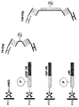
結合特異性は、多重特異性治療用分子を形成するのに用いられる出発成分によって別々に提供される。従って、単に、細胞、例えば、癌細胞に存在する表面マーカーを決定し、これらの表面マーカーに特異的に結合するそれぞれの結合実体、例えば、抗体断片を核酸に結合体化し、これらをリンカーヌクレオチドによって連結することで多重特異性治療用分子、例えば、二重特異性抗体を特別に作ることが可能である。 Binding specificity is provided separately by the starting components used to form the multispecific therapeutic molecule. Thus, it simply determines the surface markers present on the cells, eg, cancer cells, and conjugates each binding entity, eg, antibody fragment, that specifically binds to these surface markers to the nucleic acid, which is linked by a linker nucleotide. By linking, it is possible to specifically make multispecific therapeutic molecules, eg bispecific antibodies.
エフェクター部分の標的送達のために、ポリペプチドおよびポリヌクレオチド成分を含む複合体は特に有用であることが見出されている。複合体のエフェクター部分、ポリペプチド成分、およびポリヌクレオチドリンカーは互いに非共有結合される。これにより複合体の個々の成分のモジュール生成が可能になる。モジュール構造のために、複合体の他の成分を変える必要なく複合体の個々の成分を変えることができる。これにより、例えば、ライブラリーを提供するために多数の複合体変種の簡単かつ効率的な組み立てが可能になり、これに基づいてテーラーメイドの高度に特異的な多重特異性治療用分子を選択することができる。 Complexes comprising polypeptide and polynucleotide components have been found to be particularly useful for targeted delivery of effector moieties. The effector portion of the complex, the polypeptide component, and the polynucleotide linker are non-covalently bound to each other. This allows module generation of individual components of the complex. Because of the modular structure, individual components of the composite can be changed without having to change other components of the composite. This allows for easy and efficient assembly of multiple complex variants, for example, to provide a library, on which to select tailor-made highly specific multispecific therapeutic molecules Can do.
本明細書において報告される一局面は、治療剤として使用するために、(a)抗体Fab断片またはscFv抗体断片であって、それぞれが第1の結合ペアの第1のパートナーもしくはメンバーを含み、または第1の結合ペアの第1のパートナーもしくはメンバーと結合体化され、それによって、第1の細胞表面マーカーまたは第1の細胞表面マーカーの第1のエピトープに特異的に結合する抗体Fab断片またはscFv抗体断片、(b)抗体Fab断片またはscFv抗体断片であって、それぞれが第2の結合ペアの第1のパートナーもしくはメンバーを含み、または第2の結合ペアの第1のパートナーもしくはメンバーと結合体化され、それによって、第2の細胞表面マーカーまたは第1の細胞表面マーカーの第2のエピトープに特異的に結合する抗体Fab断片またはscFv抗体断片、および(c)末端の一方において第1の結合ペアの第2のメンバーを含み、それぞれの他方の末端において第2の結合ペアの第2のメンバーを含むリンカーをインキュベートすることによって、単一の多重特異性結合分子内で組み立てられた結合実体のコレクション/ライブラリーから少なくとも2つの結合実体を選択する方法である。このような薬剤は、改善された標的化/送達特性を有する。 One aspect reported herein is for use as a therapeutic agent: (a) an antibody Fab fragment or scFv antibody fragment, each comprising a first partner or member of a first binding pair; Or an antibody Fab fragment that is conjugated to the first partner or member of the first binding pair, thereby specifically binding to the first cell surface marker or the first epitope of the first cell surface marker, or scFv antibody fragment, (b) antibody Fab fragment or scFv antibody fragment, each comprising a first partner or member of a second binding pair, or binding to a first partner or member of a second binding pair An antibody Fab fragment or scFv antibody fragment that specifically binds to a second cell surface marker or a second epitope of the first cell surface marker, and (c A single multispecific binding molecule by incubating a linker comprising a second member of a first binding pair at one end and a second member of a second binding pair at each other end A method of selecting at least two binding entities from a collection / library of binding entities assembled within. Such agents have improved targeting / delivery properties.
本明細書において報告される一局面は、多重特異性結合分子を生成するための方法であって、以下の工程を含む方法である:
(i)細胞を含有する試料に存在する細胞表面マーカーを決定する工程およびi)少なくとも第1の細胞表面マーカー、任意で第2の細胞表面マーカーを選択する工程、またはii)多重特異性結合分子の結合特異性の数に対応する多数の細胞表面マーカーを選択する工程、
(ii)(a)多数の結合実体であって、それぞれが結合ペアの第1のパートナーまたはメンバーを含み、それによって、結合実体のそれぞれが異なる細胞表面マーカーもしくはそのリガンドまたは同じ細胞表面マーカーのエピトープに特異的に結合し、それによって、結合ペアのそれぞれの第1のパートナーまたはメンバーがその対応する第2のパートナーまたはメンバーにしか結合せず、結合ペアの他の第2のパートナーまたはメンバーのいずれにも結合しない多数の結合実体、および(b)結合ペアの対応する第2のメンバーを含むリンカーをインキュベートし、かつそれによって多重特異性結合分子を生成する工程。
One aspect reported herein is a method for generating a multispecific binding molecule comprising the following steps:
(I) determining a cell surface marker present in a sample containing cells and i) selecting at least a first cell surface marker, optionally a second cell surface marker, or ii) a multispecific binding molecule Selecting a number of cell surface markers corresponding to the number of binding specificities of
(Ii) (a) multiple binding entities, each comprising a first partner or member of a binding pair, whereby each of the binding entities is a different cell surface marker or ligand thereof or an epitope of the same cell surface marker Specifically binding to each of the binding partner so that each first partner or member of the binding pair binds only to its corresponding second partner or member, and any of the other second partners or members of the binding pair (B) incubating a linker comprising a plurality of binding entities that do not bind to the enzyme, and (b) a corresponding second member of the binding pair, thereby generating a multispecific binding molecule.
本明細書において報告される一局面は、二重特異性抗体を生成するための方法であって、以下の工程を含む方法である:
(i)試料中の細胞の表面に存在する細胞表面マーカーを決定し、第1の表面マーカーおよび第2の表面マーカーを選択する工程、
(ii)(a)第1の結合ペアの第1のパートナーもしくはメンバーを含む、または第1の結合ペアの第1のパートナーもしくはメンバーと結合体化された抗体Fab断片またはscFv抗体断片であって、それによって第1の細胞表面マーカーに特異的に結合する、抗体Fab断片またはscFv抗体断片、(b)第2の結合ペアの第1のパートナーもしくはメンバーを含む、または第2の結合ペアの第1のパートナーもしくはメンバーと結合体化された抗体Fab断片またはscFv抗体断片であって、それによって第2の細胞表面マーカーに特異的に結合する、抗体Fab断片またはscFv抗体断片、および(c)末端の一方において第1の結合ペアの第2のメンバーを含み、それぞれの他方の末端において第2の結合ペアの第2のメンバーを含むリンカー
をインキュベートし、かつそれによって二重特異性抗体を生成する工程。
One aspect reported herein is a method for producing a bispecific antibody, comprising the following steps:
(I) determining a cell surface marker present on the surface of a cell in the sample, and selecting a first surface marker and a second surface marker;
(Ii) (a) an antibody Fab fragment or scFv antibody fragment comprising the first partner or member of the first binding pair, or conjugated to the first partner or member of the first binding pair, An antibody Fab fragment or scFv antibody fragment thereby specifically binding to the first cell surface marker, (b) comprising the first partner or member of the second binding pair, or of the second binding pair An antibody Fab fragment or scFv antibody fragment conjugated to one partner or member, thereby specifically binding to a second cell surface marker, and (c) the end Incubating a linker containing a second member of the first binding pair on one side and a second member of the second binding pair on the other end of each Generating a isomeric antibodies.
本明細書において報告される一局面は、多重特異性結合分子のために結合実体の組み合わせを決定するための方法であって、以下の工程を含む方法である:
(i)多数の多重特異性結合分子の結合特異性および/または選択性および/または親和性および/またはエフェクター機能および/またはインビボ半減期を決定し、それによって、結合実体のそれぞれの(可能性のある)組み合わせが多数の多重特異性結合分子の中に含まれる工程、
(ii)適切な結合特異性および/または選択性および/または親和性および/またはエフェクター機能および/またはインビボ半減期を有する多重特異性結合分子を選択し、それによって、抗原結合実体の組み合わせを決定する工程。
One aspect reported herein is a method for determining a combination of binding entities for a multispecific binding molecule, comprising the following steps:
(I) determining the binding specificity and / or selectivity and / or affinity and / or effector function and / or in vivo half-life of a multiplicity of multispecific binding molecules, whereby the (possibility of each of the binding entities Wherein the combination is included in a number of multispecific binding molecules,
(Ii) selecting multispecific binding molecules with appropriate binding specificity and / or selectivity and / or affinity and / or effector function and / or in vivo half-life, thereby determining the combination of antigen binding entities Process.
本明細書において報告される一局面は、抗原結合部位の組み合わせを決定するための方法であって、以下の工程を含む方法である:
(i)第1の結合ペアの第1のメンバーを含む、または第1の結合ペアの第1のメンバーと結合体化された第1の多数群の抗体Fab断片またはscFv抗体断片の各メンバーを、第2の結合ペアの第1のメンバーを含む、または第2の結合ペアの第1のメンバーと結合体化された第2の多数群の抗体Fab断片またはscFv抗体断片の各メンバーと、および末端の一方において第1の結合ペアの第2のメンバーを含み、かつそれぞれの他方の末端において第2の結合ペアの第2のメンバーを含むリンカーと組み合わせることによって調製された多数の二重特異性抗体の結合特異性および/または選択性および/または親和性および/またはエフェクター機能および/またはインビボ半減期を決定する工程であって、第1の多数群が第1の細胞表面マーカーに特異的に結合し、第2の多数群が第2の細胞表面マーカーに特異的に結合する工程、ならびに
(ii)適切な結合特異性および/または選択性および/または親和性および/またはエフェクター機能および/またはインビボ半減期を有する二重特異性抗体を選択し、それによって、抗原結合部位の組み合わせを決定する工程。
One aspect reported herein is a method for determining a combination of antigen binding sites, comprising the following steps:
(I) each member of the first majority group of antibody Fab or scFv antibody fragments comprising or conjugated to the first member of the first binding pair; Each member of a second majority group of antibody Fab or scFv antibody fragments comprising, or conjugated to, the first member of the second binding pair, and the first member of the second binding pair; and Multiple bispecificities prepared by combining with a linker containing a second member of the first binding pair at one end and a second member of the second binding pair at each other end Determining the binding specificity and / or selectivity and / or affinity and / or effector function and / or in vivo half-life of an antibody, wherein the first majority group is specific for a first cell surface marker Combined, the second majority group Specific binding to two cell surface markers, and (ii) a bispecific antibody having appropriate binding specificity and / or selectivity and / or affinity and / or effector function and / or in vivo half-life And thereby determining a combination of antigen binding sites.
本明細書において報告される一局面は、
(a)(i)第1の表面マーカーに特異的に結合し、
(ii)第1の結合ペアの第1のメンバーと結合体化された、
第1のFab断片またはscFv抗体断片、
(b)(i)第2の表面マーカーに特異的に結合し、かつ
(ii)第2の結合ペアの第1のメンバーと結合体化された、
第2のFab断片またはscFv抗体断片、および
(c)(i)第1の結合ペアの第2のメンバーと結合体化され、かつ
(ii)第2の結合ペアの第2のメンバーと結合体化された
エナンチオマーDNAポリヌクレオチドリンカー
を含む二重特異性抗体であって、第1および第2のFab断片またはscFv抗体断片が非共有結合複合体を形成する、二重特異性抗体である。
One aspect reported herein is:
(A) (i) specifically binds to the first surface marker;
(Ii) conjugated with the first member of the first binding pair;
A first Fab fragment or an scFv antibody fragment,
(B) (i) specifically bound to a second surface marker, and (ii) conjugated to a first member of a second binding pair,
A second Fab fragment or an scFv antibody fragment, and (c) (i) conjugated with a second member of the first binding pair, and (ii) a second member and conjugate of the second binding pair A bispecific antibody comprising a conjugated enantiomer DNA polynucleotide linker, wherein the first and second Fab fragments or scFv antibody fragments form a non-covalent complex.
以下は、本明細書において報告される全ての局面の態様である。それぞれの態様はそれぞれの局面と組み合わせることができ、本明細書において示された他の全ての個々の態様とも組み合わせることができると本明細書において指摘される。 The following are embodiments of all aspects reported herein. It is pointed out herein that each embodiment can be combined with each aspect and combined with all other individual embodiments shown herein.
1つの態様において、結合実体は互いに独立して、ダルピン(darpin)ドメインベースの結合実体、アンチカリン(anticalin)ドメインベースの結合実体、T細胞受容体断片様scTCRドメインベースの結合実体、ラクダVHドメインベースの結合実体、第10フィブロネクチン3ドメインベースの結合実体、テネイシンドメインベースの結合実体、カドヘリンドメインベースの結合実体、ICAMドメインベースの結合実体、タイチンドメインベースの結合実体、GCSF-Rドメインベースの結合実体、サイトカイン受容体ドメインベースの結合実体、グリコシダーゼ阻害剤ドメインベースの結合実体、スーパーオキシドジスムターゼドメインベースの結合実体、または抗体断片(Fab断片もしくはscFv断片)より選択される。
In one embodiment, the binding entities are independent of each other, darpin domain-based binding entities, anticalin domain-based binding entities, T cell receptor fragment-like scTCR domain-based binding entities, camel VH domains Base binding entity,
全ての局面のうちの1つの態様において、多重特異性結合分子は二重特異性抗体であるか、または第1の結合実体および第2の結合実体は互いに独立して抗体断片である。 In one embodiment of all aspects, the multispecific binding molecule is a bispecific antibody, or the first binding entity and the second binding entity are antibody fragments independently of each other.
1つの態様において、抗体断片は、Fv、Fab、Fab'、Fab'-SH、F(ab')2、ダイアボディ(diabody)、直鎖抗体、scFv、scFab、およびdsFvを含む基より選択される。 In one embodiment, the antibody fragment is selected from the group comprising Fv, Fab, Fab ′, Fab′-SH, F (ab ′) 2 , diabody, linear antibody, scFv, scFab, and dsFv. The
1つの態様において、エフェクター部分、結合特異性、およびポリヌクレオチドリンカーを含む二重特異性抗体の少なくとも2つの成分は互いに非共有結合している。 In one embodiment, at least two components of the bispecific antibody comprising an effector moiety, binding specificity, and a polynucleotide linker are non-covalently bound to each other.
1つの態様において、結合実体は、抗体、抗体断片、受容体、受容体リガンド、および標的結合スキャフォールドより選択される。但し、受容体リガンドはインクレチン受容体リガンドポリペプチドでない。 In one embodiment, the binding entity is selected from an antibody, antibody fragment, receptor, receptor ligand, and target binding scaffold. However, the receptor ligand is not an incretin receptor ligand polypeptide.
1つの態様において、抗体断片は、Fv、Fab、Fab'、Fab'-SH、F(ab')2、ダイアボディ、直鎖抗体、scFv、scFab、およびdsFvを含む群より選択される。 In one embodiment, the antibody fragment is selected from the group comprising Fv, Fab, Fab ′, Fab′-SH, F (ab ′) 2 , diabody, linear antibody, scFv, scFab, and dsFv.
1つの態様において、標的結合スキャフォールドは、ダルピン、ヘモペキシン様分子、およびアンチカリンより選択される。 In one embodiment, the target binding scaffold is selected from dalpins, hemopexin-like molecules, and anticalins.
1つの態様において、受容体はT細胞受容体断片およびscTCRより選択される。 In one embodiment, the receptor is selected from T cell receptor fragments and scTCR.
1つの態様において、多重特異性結合分子は、
(a)(i)第1の細胞表面マーカーまたはそのリガンドに特異的に結合し、かつ
(ii)第1の結合ペアの第1のメンバーと結合体化された、
第1の結合実体、
(b)(i)第2の細胞表面マーカーまたはそのリガンドに特異的に結合し、かつ
(ii)第2の結合ペアの第1のメンバーと結合体化された、
第2の結合実体、および
(c)(i)第1の結合ペアの第2のメンバーと結合体化され、かつ
(ii)第2の結合ペアの第2のメンバーと結合体化された、
ポリヌクレオチドリンカー
を含む複合体である。
In one embodiment, the multispecific binding molecule is
(A) (i) specifically bound to a first cell surface marker or its ligand, and (ii) conjugated to a first member of a first binding pair,
The first binding entity,
(B) (i) specifically bound to a second cell surface marker or its ligand, and (ii) conjugated to a first member of a second binding pair,
A second binding entity, and (c) (i) conjugated with a second member of the first binding pair, and (ii) conjugated with a second member of the second binding pair,
A complex comprising a polynucleotide linker.
1つの態様において、二重特異性抗体は、
(a)(i)第1の細胞表面マーカーに特異的に結合し、かつ
(ii)第1の結合ペアの第1のメンバー結合体化された、
第1のFab断片またはscFv抗体断片、
(b)(i)第2の細胞表面マーカーに特異的に結合し、かつ
(ii)第2の結合ペアの第1のメンバー結合体化された、
第2のFab断片またはscFv抗体断片、および
(c)(i)第1の結合ペアの第2のメンバーと結合体化され、かつ
(ii)第2の結合ペアの第2のメンバー結合体化された、
ポリヌクレオチドリンカー
を含む複合体である。
In one embodiment, the bispecific antibody is
(A) (i) specifically bound to a first cell surface marker, and (ii) a first member conjugated of a first binding pair,
A first Fab fragment or an scFv antibody fragment,
(B) (i) specifically bound to a second cell surface marker, and (ii) conjugated to a first member of a second binding pair,
A second Fab fragment or an scFv antibody fragment, and (c) (i) conjugated to a second member of the first binding pair, and (ii) a second member conjugated of the second binding pair Was
A complex comprising a polynucleotide linker.
1つの態様において、複合体は非共有結合複合体である。 In one embodiment, the complex is a non-covalent complex.
1つの態様において、複合体は、(i)第2の標的に特異的に結合し、かつ(ii)第2の結合ペアの第1のメンバーと結合体化された、さらなるポリペプチドをさらに含み、ポリヌクレオチドリンカーは第2の結合ペアの第2のメンバーと結合体化される。 In one embodiment, the complex further comprises an additional polypeptide that (i) specifically binds to a second target, and (ii) is conjugated to a first member of a second binding pair. The polynucleotide linker is conjugated with the second member of the second binding pair.
1つの態様において、複合体は、ポリヌクレオチドリンカーの少なくとも一部に相補的なポリヌクレオチドと結合体化されたエフェクター部分をさらに含む。 In one embodiment, the complex further comprises an effector moiety conjugated with a polynucleotide complementary to at least a portion of the polynucleotide linker.
1つの態様において、複合体は、(i)第1または第2の結合実体またはFab断片またはscFv抗体断片と結合体化されたポリヌクレオチドの少なくとも一部に相補的であり、かつ(ii)ポリヌクレオチドリンカーに相補的でないポリヌクレオチドと結合体化されたエフェクター部分をさらに含む。 In one embodiment, the complex is (i) complementary to at least a portion of the polynucleotide conjugated to the first or second binding entity or Fab fragment or scFv antibody fragment, and (ii) a poly It further comprises an effector moiety conjugated with a polynucleotide that is not complementary to the nucleotide linker.
1つの態様において、第1および第2の結合実体またはFab断片またはscFv抗体断片は同じ標的および標的上にある非重複エピトープに結合する。 In one embodiment, the first and second binding entities or Fab fragments or scFv antibody fragments bind to the same target and non-overlapping epitopes on the target.
1つの態様において、ポリヌクレオチドリンカーは、8個、10個、15個、20個、25個、50個、100個のヌクレオチドを含む。1つの態様において、ポリヌクレオチドリンカーは、500個、750個、1000個、または2000個までのヌクレオチドを含む。1つの態様において、ポリヌクレオチドリンカーは10〜500個のヌクレオチドを含む。 In one embodiment, the polynucleotide linker comprises 8, 10, 15, 20, 25, 50, 100 nucleotides. In one embodiment, the polynucleotide linker comprises up to 500, 750, 1000, or 2000 nucleotides. In one embodiment, the polynucleotide linker comprises 10 to 500 nucleotides.
1つの態様において、ポリヌクレオチドリンカーはエナンチオマーDNAである。1つの態様において、エナンチオマーDNAはL-DNAである。1つの態様において、L-DNAは一本鎖L-DNA(ss-L-DNA)である。 In one embodiment, the polynucleotide linker is enantiomeric DNA. In one embodiment, the enantiomeric DNA is L-DNA. In one embodiment, the L-DNA is single stranded L-DNA (ss-L-DNA).
1つの態様において、エフェクター部分は、結合部分、標識部分、および生物学的に活性な部分からなる群より選択される。 In one embodiment, the effector moiety is selected from the group consisting of a binding moiety, a label moiety, and a biologically active moiety.
1つの態様において、ポリヌクレオチドリンカーは、その第1の末端または第2の末端において結合実体またはFab断片またはscFv抗体断片と結合体化される。 In one embodiment, the polynucleotide linker is conjugated with a binding entity or Fab fragment or scFv antibody fragment at its first end or second end.
1つの態様において、ポリヌクレオチドリンカーは2つの結合ペアの2つの第2のメンバーと結合体化され、それによって、第1の結合ペアの第2のメンバーはポリヌクレオチドリンカーの第1の末端と結合体化され、第2の結合ペアの第2のメンバーはポリヌクレオチドリンカーの第2の末端と結合体化される。 In one embodiment, the polynucleotide linker is conjugated with two second members of two binding pairs, whereby the second member of the first binding pair binds to the first end of the polynucleotide linker. And the second member of the second binding pair is conjugated with the second end of the polynucleotide linker.
1つの態様において、第1の結合ペアの第1のメンバーおよび第2のメンバーは、それぞれ、SEQ ID NO:05およびSEQ ID NO:08の核酸配列を含む。 In one embodiment, the first member and the second member of the first binding pair comprise the nucleic acid sequences of SEQ ID NO: 05 and SEQ ID NO: 08, respectively.
1つの態様において、第2の結合ペアの第1のメンバーおよび第2のメンバーは、それぞれ、SEQ ID NO:06およびSEQ ID NO:07の核酸配列を含む。 In one embodiment, the first member and the second member of the second binding pair comprise the nucleic acid sequences of SEQ ID NO: 06 and SEQ ID NO: 07, respectively.
1つの態様において、前記方法は、以下の工程を含む:
(a)第1の細胞表面マーカーまたはそのリガンドに特異的に結合し、かつ第1の結合ペアの第1のメンバーと結合体化された第1の結合実体、またはFab断片、またはscFv抗体断片を合成する工程、
(b)第2の細胞表面マーカーまたはそのリガンドに特異的に結合し、かつ第2の結合ペアの第1のメンバーと結合体化された第2の結合実体、またはFab断片、またはscFv抗体断片を合成する工程、
(c)第1の結合ペアの第2のメンバーと結合体化され、かつ第2の結合ペアの第2のメンバーと結合体化されたポリヌクレオチドリンカーを合成する工程、および
(d)合成された成分を組み合わせることによって複合体を形成する工程。
In one embodiment, the method comprises the following steps:
(A) a first binding entity, Fab fragment, or scFv antibody fragment that specifically binds to a first cell surface marker or its ligand and is conjugated to a first member of a first binding pair A process of synthesizing
(B) a second binding entity, Fab fragment, or scFv antibody fragment that specifically binds to a second cell surface marker or its ligand and is conjugated to a first member of a second binding pair. A process of synthesizing
(C) synthesizing a polynucleotide linker conjugated with the second member of the first binding pair and conjugated with the second member of the second binding pair; and (d) synthesized. Forming a composite by combining the components.
本明細書において報告される別の局面は、本明細書において報告される多重特異性結合分子または二重特異性抗体および任意で、薬学的に許容される担体を含む、薬学的製剤である。 Another aspect as reported herein is a pharmaceutical formulation comprising a multispecific binding molecule or bispecific antibody as reported herein and optionally a pharmaceutically acceptable carrier.
本明細書において報告されるさらなる局面は、医薬として使用するための本明細書において報告される多重特異性結合分子または二重特異性抗体である。 A further aspect as reported herein is a multispecific binding molecule or bispecific antibody as reported herein for use as a medicament.
本明細書において報告される局面はまた、癌の治療において使用するための本明細書において報告される多重特異性結合分子または二重特異性抗体である。 Aspects reported herein are also multispecific binding molecules or bispecific antibodies reported herein for use in the treatment of cancer.
本明細書において報告される別の局面は、医薬の製造における本明細書において報告される多重特異性結合分子または二重特異性抗体の使用である。 Another aspect reported herein is the use of multispecific binding molecules or bispecific antibodies reported herein in the manufacture of a medicament.
1つの態様において、医薬は、癌を治療するための医薬である。 In one embodiment, the medicament is a medicament for treating cancer.
本明細書において報告される局面は、有効量の本明細書において報告される多重特異性結合分子または二重特異性抗体を個体に投与する工程を含む、癌を有する個体を治療する方法である。
[本発明1001]
(i)試料中の細胞の表面に存在する表面マーカーを決定し、かつ第1の表面マーカーおよび第2の表面マーカーを選択する工程、
(ii)(a)第1の結合ペアの第1のメンバーと結合体化された抗体Fab断片またはscFv抗体断片であって、それによって第1の表面マーカーに特異的に結合する、抗体Fab断片またはscFv抗体断片、(b)第2の結合ペアの第1のメンバーと結合体化された抗体Fab断片またはscFv抗体断片であって、それによって第2の表面マーカーに特異的に結合する、抗体Fab断片またはscFv抗体断片、および(c)末端の一方において第1の結合ペアの第2のメンバーを含み、それぞれの他方の末端において第2の結合ペアの第2のメンバーを含むエナンチオマーDNAポリヌクレオチドリンカー
をインキュベートし、かつそれによって二重特異性抗体を生成する工程
を含む、二重特異性抗体を生成するための方法。
[本発明1002]
(i)それぞれが第1の結合ペアの同じ第1のメンバーに連結された第1の多数群の抗体Fab断片またはscFv抗体断片の各メンバーを、それぞれが第2の結合ペアの同じ第1のメンバーに連結された第2の多数群の抗体Fab断片またはscFv抗体断片の各メンバーと、および末端の一方において第1の結合ペアの第2のメンバーを含み、かつそれぞれの他方の末端において第2の結合ペアの第2のメンバーを含むエナンチオマーDNAポリヌクレオチドリンカーと、組み合わせることによって調製された多数の二重特異性抗体の結合特異性および/または親和性および/またはエフェクター機能および/またはインビボ半減期を決定する工程であって、それによって第1の多数群が第1の細胞表面マーカーに特異的に結合し、第2の多数群が第2の細胞表面マーカーに特異的に結合する、工程、ならびに
(ii)適切な結合特異性および/または親和性および/またはエフェクター機能および/またはインビボ半減期を有する二重特異性抗体を選択し、かつそれによって抗原結合部位の組み合わせを決定する工程
を含む、抗原結合部位の組み合わせを決定するための方法。
[本発明1003]
二重特異性抗体が、
(a)(i)第1の表面マーカーに特異的に結合し、かつ
(ii)第1の結合ペアの第1のメンバーと結合体化された
第1のFab断片またはscFv抗体断片、
(b)(i)第2の表面マーカーに特異的に結合し、かつ
(ii)第2の結合ペアの第1のメンバーと結合体化された
第2のFab断片またはscFv抗体断片、および
(c)(i)第1の結合ペアの第2のメンバーと結合体化され、かつ
(ii)第2の結合ペアの第2のメンバーと結合体化された
エナンチオマーDNAポリヌクレオチドリンカー
を含む複合体であることを特徴とする、前記本発明のいずれかの方法。
[本発明1004]
複合体が非共有結合複合体であることを特徴とする、前記本発明のいずれかの方法。
[本発明1005]
結合ペアがポリヌクレオチドの相補的ペアであることを特徴とする、前記本発明のいずれかの方法。
[本発明1006]
複合体が、リンカーの少なくとも一部に相補的なポリヌクレオチドと結合体化されたエフェクター部分をさらに含む、前記本発明のいずれかの方法。
[本発明1007]
第1のエフェクター部分と結合体化されたポリヌクレオチドの少なくとも一部に相補的であり、かつ(ii)ポリヌクレオチドリンカーに相補的でない、ポリヌクレオチド
と結合体化された第2のエフェクター部分を、複合体が含むことを特徴とする、本発明1006の方法。
[本発明1008]
第1および第2のFab断片またはscFv抗体断片が、同じ標的およびその上にある非重複エピトープに結合することを特徴とする、前記本発明のいずれかの方法。
[本発明1009]
ポリヌクレオチドリンカーが8〜1000個のヌクレオチドを含むことを特徴とする、前記本発明のいずれかの方法。一つの態様において、ポリヌクレオチドリンカーは10〜500個のヌクレオチドを含む。
[本発明1010]
エナンチオマーDNAがL-DNAであることを特徴とする、前記本発明のいずれかの方法。
[本発明1011]
L-DNAが一本鎖L-DNA(ss-L-DNA)であることを特徴とする、本発明1010の方法。
[本発明1012]
本発明1001〜1011のいずれかの方法によって得られた二重特異性抗体と、任意で薬学的に許容される担体とを含む、薬学的製剤。
[本発明1013]
医薬として使用するための、本発明1001〜1011のいずれかの方法によって得られた二重特異性抗体。
[本発明1014]
医薬の製造における、本発明1001〜1011のいずれかの方法によって得られた二重特異性抗体の使用。
[本発明1015]
癌を有する個体を治療する方法であって、本発明1001〜1011のいずれかの方法によって得られた二重特異性抗体の有効量を該個体に投与する工程を含む、方法。
[本発明1016]
(a)(i)第1の表面マーカーに特異的に結合し、かつ
(ii)第1の結合ペアの第1のメンバーと結合体化された
第1のFab断片またはscFv抗体断片、
(b)(i)第2の表面マーカーに特異的に結合し、かつ
(ii)第2の結合ペアの第1のメンバーと結合体化された
第2のFab断片またはscFv抗体断片、および
(c)(i)第1の結合ペアの第2のメンバーと結合体化され、かつ
(ii)第2の結合ペアの第2のメンバーと結合体化された
エナンチオマーDNAポリヌクレオチドリンカー
を含む二重特異性抗体であって、それによって第1および第2のFab断片またはscFv抗体断片ならびにエナンチオマーDNAポリヌクレオチドリンカーが非共有結合複合体を形成する、二重特異性抗体。
An aspect reported herein is a method of treating an individual with cancer comprising administering to the individual an effective amount of a multispecific binding molecule or bispecific antibody reported herein. .
[Invention 1001]
(I) determining a surface marker present on the surface of a cell in the sample and selecting a first surface marker and a second surface marker;
(Ii) (a) an antibody Fab fragment or scFv antibody fragment conjugated to the first member of the first binding pair, thereby specifically binding to the first surface marker Or an scFv antibody fragment, (b) an antibody Fab fragment or an scFv antibody fragment conjugated to a first member of a second binding pair, thereby specifically binding to a second surface marker An Fab fragment or an scFv antibody fragment, and (c) an enantiomeric DNA polynucleotide comprising a second member of the first binding pair at one of the ends and a second member of the second binding pair at each other end Linker
And thereby generating a bispecific antibody
A method for producing a bispecific antibody comprising:
[Invention 1002]
(I) each member of the first majority group of antibody Fab or scFv antibody fragments each linked to the same first member of the first binding pair, each of which is the same first member of the second binding pair Each member of a second majority group of antibody Fab or scFv antibody fragments linked to a member, and a second member of the first binding pair at one of the ends and a second at each other end Binding specificity and / or affinity and / or effector function and / or in vivo half-life of multiple bispecific antibodies prepared by combining with an enantiomeric DNA polynucleotide linker comprising a second member of the binding pair of Wherein the first majority group specifically binds to the first cell surface marker and the second majority group specifically binds to the second cell surface marker. That, the process, as well as
(Ii) selecting a bispecific antibody with appropriate binding specificity and / or affinity and / or effector function and / or in vivo half-life and thereby determining the combination of antigen binding sites
A method for determining a combination of antigen binding sites.
[Invention 1003]
Bispecific antibodies
(A) (i) specifically binds to the first surface marker, and
(Ii) conjugated with the first member of the first binding pair
A first Fab fragment or an scFv antibody fragment,
(B) (i) specifically binds to the second surface marker, and
(Ii) conjugated with the first member of the second binding pair
A second Fab fragment or scFv antibody fragment, and
(C) (i) conjugated with a second member of the first binding pair, and
(Ii) conjugated with the second member of the second binding pair
Enantiomer DNA polynucleotide linker
The method according to any one of the present inventions, characterized in that it is a complex comprising
[Invention 1004]
The method according to any one of the above-mentioned present invention, wherein the complex is a non-covalent complex.
[Invention 1005]
The method according to any one of the above-mentioned present invention, wherein the binding pair is a complementary pair of polynucleotides.
[Invention 1006]
The method of any of the preceding claims, wherein the complex further comprises an effector moiety conjugated to a polynucleotide complementary to at least a portion of the linker.
[Invention 1007]
A polynucleotide that is complementary to at least a portion of the polynucleotide conjugated to the first effector moiety and (ii) is not complementary to the polynucleotide linker
The method of claim 1006, wherein the complex comprises a second effector moiety conjugated with.
[Invention 1008]
The method according to any of the preceding inventions, characterized in that the first and second Fab fragments or scFv antibody fragments bind to the same target and non-overlapping epitopes thereon.
[Invention 1009]
The method according to any of the preceding inventions, characterized in that the polynucleotide linker comprises 8 to 1000 nucleotides. In one embodiment, the polynucleotide linker comprises 10 to 500 nucleotides.
[Invention 1010]
The method according to any one of the above-mentioned present invention, wherein the enantiomeric DNA is L-DNA.
[Invention 1011]
The method of the present invention 1010, wherein the L-DNA is single-stranded L-DNA (ss-L-DNA).
[Invention 1012]
A pharmaceutical preparation comprising a bispecific antibody obtained by any of the methods of the invention 1001 to 1011 and optionally a pharmaceutically acceptable carrier.
[Invention 1013]
A bispecific antibody obtained by any of the methods of the invention 1001 to 1011 for use as a medicament.
[Invention 1014]
Use of a bispecific antibody obtained by any of the methods of the invention 1001 to 1011 in the manufacture of a medicament.
[Invention 1015]
A method for treating an individual having cancer, comprising the step of administering to the individual an effective amount of the bispecific antibody obtained by any of the methods 1001 to 1011 of the present invention.
[Invention 1016]
(A) (i) specifically binds to the first surface marker, and
(Ii) conjugated with the first member of the first binding pair
A first Fab fragment or an scFv antibody fragment,
(B) (i) specifically binds to the second surface marker, and
(Ii) conjugated with the first member of the second binding pair
A second Fab fragment or scFv antibody fragment, and
(C) (i) conjugated with a second member of the first binding pair, and
(Ii) conjugated with the second member of the second binding pair
Enantiomer DNA polynucleotide linker
A bispecific antibody, wherein the first and second Fab or scFv antibody fragments and the enantiomeric DNA polynucleotide linker form a non-covalent complex.
発明の詳細な説明
I.定義
冠詞「1つの(a)」および「1つの(an)」は、冠詞の1つまたは複数(すなわち、少なくとも1つ)の文法上の目的語を指すために本明細書において用いられる。一例として、「1つの(an)抗体」は1つの抗体または複数の抗体を意味する。
Detailed Description of the Invention
I. Definitions The articles “a” and “an” are used herein to refer to one or more (ie, at least one) grammatical objects of an article. . By way of example, “an antibody” means one antibody or a plurality of antibodies.
「アクセプターヒトフレームワーク」は、以下で定義されるように、ヒト免疫グロブリンフレームワークまたはヒトコンセンサスフレームワークに由来する軽鎖可変ドメイン(VL)フレームワークもしくは重鎖可変ドメイン(VH)フレームワークのアミノ酸配列を含むフレームワークである。ヒト免疫グロブリンフレームワークまたはヒトコンセンサスフレームワーク「に由来する」アクセプターヒトフレームワークは同じアミノ酸配列を含んでもよく、アミノ酸配列変化を含んでもよい。一部の態様において、アミノ酸変化の数は、10以下、9以下、8以下、7以下、6以下、5以下、4以下、3以下、または2以下である。一部の態様において、VLアクセプターヒトフレームワークはVLヒト免疫グロブリンフレームワーク配列またはヒトコンセンサスフレームワーク配列と配列が同一である。 An “acceptor human framework” is a light chain variable domain (VL) framework or a heavy chain variable domain (VH) framework derived from a human immunoglobulin framework or a human consensus framework, as defined below. A framework containing an amino acid sequence. An acceptor human framework “derived from” a human immunoglobulin framework or human consensus framework may comprise the same amino acid sequence or may comprise amino acid sequence changes. In some embodiments, the number of amino acid changes is 10 or less, 9 or less, 8 or less, 7 or less, 6 or less, 5 or less, 4 or less, 3 or less, or 2 or less. In some embodiments, the VL acceptor human framework is identical in sequence to the VL human immunoglobulin framework sequence or human consensus framework sequence.
「親和性」という用語は、分子(例えば、ポリペプチドまたは抗体)の1つの結合部位と、その結合パートナー(例えば、標的または抗原)との非共有結合相互作用の合計の強さを意味する。特に定めのない限り、本明細書で使用する「結合親和性」とは、結合ペアのメンバーとの間で(例えば、ポリペプチド-ポリヌクレオチド-複合体の中の結合ペアのメンバーとの間で、またはポリペプチドとその標的との間で、または抗体とその抗原との間で)1:1の相互作用を反映する固有の結合親和性を指す。パートナーYに対する分子Xの親和性は一般的に解離定数(kD)で表すことができる。親和性は当技術分野において公知であり、本明細書において報告された方法も含む一般的な方法、例えば、表面プラズモン共鳴によって測定することができる。 The term “affinity” refers to the total strength of a non-covalent interaction between one binding site of a molecule (eg, a polypeptide or antibody) and its binding partner (eg, a target or antigen). Unless otherwise specified, as used herein, “binding affinity” refers to between members of a binding pair (eg, between members of a binding pair in a polypeptide-polynucleotide-complex. Or the inherent binding affinity that reflects a 1: 1 interaction between the polypeptide and its target, or between the antibody and its antigen). The affinity of molecule X for partner Y can generally be expressed as the dissociation constant (kD). Affinity is known in the art and can be measured by common methods, including the methods reported herein, such as surface plasmon resonance.
「親和性成熟」抗体とは、変化を有さない親抗体と比較して1つまたは複数の超可変領域(HVR)に1つまたは複数の変化があり、このような変化の結果として抗原に対する抗体の親和性が改善している抗体を指す。 An “affinity matured” antibody has one or more changes in one or more hypervariable regions (HVRs) compared to a parent antibody that has no changes, and as a result of such changes are directed against an antigen. It refers to an antibody with improved antibody affinity.
「ケージド(caged)」という用語は、血清中および体液中で制御された半減期を有する保護基によってエフェクターが保護されていることを意味する。保護基は内因性酵素によって酵素的に切断されてもよい。保護基は、注射によって外部から投与された、または経口により与えられた第2のエフェクター、例えば、アスコルビン酸によって除去、切断、分解、酵素的に消化、または代謝されてもよい。ケージドエフェクター分子は、体液中に天然にある酵素によって活性化されてもよい。ケージドエフェクター部分は、体液中にも発生する還元剤、例えば、アスコルビン酸によって活性化されてもよい。 The term “caged” means that the effector is protected by a protecting group that has a controlled half-life in serum and body fluids. The protecting group may be cleaved enzymatically by an endogenous enzyme. The protecting group may be removed, cleaved, degraded, enzymatically digested, or metabolized by a second effector administered exogenously by injection or given orally, such as ascorbic acid. Caged effector molecules may be activated by enzymes that are naturally present in body fluids. The caged effector moiety may be activated by a reducing agent that also occurs in body fluids, such as ascorbic acid.
「エフェクター部分」という用語は、細胞の中に送達されることが望ましい、および/または細胞に局在することが望ましい活性を有する任意の分子または分子の組み合わせを意味する。エフェクター部分には、標識、細胞毒(例えば、シュードモナス属(Pseudomonas)エキソトキシン、リシン、アブリン、ジフテリア毒素など)、酵素、増殖因子、転写因子、薬物、放射性核種、リガンド、抗体、抗体Fc領域、リポソーム、ナノ粒子、ウイルス粒子、サイトカインなどが含まれるが、これに限定されない。 The term “effector moiety” means any molecule or combination of molecules that has an activity that is desirably delivered into a cell and / or desired to be localized in a cell. The effector moiety includes a label, a cytotoxin (e.g., Pseudomonas exotoxin, ricin, abrin, diphtheria toxin, etc.), enzyme, growth factor, transcription factor, drug, radionuclide, ligand, antibody, antibody Fc region, Examples include, but are not limited to, liposomes, nanoparticles, virus particles, and cytokines.
本明細書において「抗体」という用語は最も広い意味で用いられ、望ましい抗原結合活性を示す限り、モノクローナル抗体および抗体断片を含むが、これに限定されない様々な抗体構造を含む。 As used herein, the term “antibody” is used in the broadest sense and includes a variety of antibody structures including, but not limited to, monoclonal antibodies and antibody fragments so long as they exhibit the desired antigen binding activity.
「抗体断片」という用語は、抗原に特異的に結合する能力を保持する完全な抗体または完全長抗体の断片を意味する。抗体断片の例には、Fv、FAB、FAB'、FAB'-SH、F(ab')2;ダイアボディ;直鎖抗体;単鎖抗体分子(例えば、scFv)が含まれるが、これに限定されない。ある特定の抗体断片の総説については、Hudson, P.J., et al., Nat. Med. 9 (2003) 129-134を参照されたい。さらに詳細には、「抗体断片」という用語の中には、(i)FAB断片、すなわち、VL、VH、CL、およびCH1ドメインからなる一価抗体断片(サルベージ(salvage)受容体結合エピトープ残基を含み、インビボ半減期の長いFABおよびF(ab')2断片の議論については、US5,869,046を参照されたい)、(ii)F(ab')2断片、すなわち、ヒンジ領域においてジスルフィド架橋によって連結された2つのFAB断片を含む二価断片、(iii)VHドメインおよびCH1ドメインからなるFd断片、(iv)1本のアームの抗体のVLドメインおよびVHドメインからなるFv断片(例えば、Plueckthun, The Pharmacology of Monoclonal Antibodies, vol. 113, Rosenburg and Moore (eds.), (Springer-Verlag, New York), (1994) pp. 269-315、WO93/16185、US5,571,894、US5,587,458を参照されたい)、(v)VHドメインからなるdAb断片(例えば、Ward, E.S., et al., Nature 341 (1989) 544-546を参照されたい)、ならびに(v(i)単離された相補性決定領域(CDR)が含まれる。さらに、Fv断片の2つのドメイン、VLおよびVHは別々の遺伝子によってコードされているが、組換え法を用いて、VL領域およびVH領域が対になって一価分子を形成した1本のタンパク質鎖として作製ことを可能にする合成リンカーによってつなぐことができる(単鎖Fv(scFv)と知られる。例えば、Bird, R.E., et al., Science 242 (1988) 423-426; Huston, J.S., et al., Proc. Natl. Acad. Sci. USA 85 (1988) 5879-5883を参照されたい)。これらの抗体断片は当業者に公知の従来の技法を用いて入手することができ、インタクトな抗体と同じやり方で結合特性についてスクリーニングすることができる。 The term “antibody fragment” means a full antibody or a fragment of a full length antibody that retains the ability to specifically bind to an antigen. Examples of antibody fragments include, but are not limited to, Fv, FAB, FAB ′, FAB′-SH, F (ab ′) 2 ; diabodies; linear antibodies; single chain antibody molecules (eg, scFv) Not. For a review of certain antibody fragments, see Hudson, PJ, et al., Nat. Med. 9 (2003) 129-134. More specifically, the term “antibody fragment” includes (i) a FAB fragment, ie, a monovalent antibody fragment consisting of the VL, VH, CL, and CH1 domains (salvage receptor binding epitope residues). (See US 5,869,046 for a discussion of FAB and F (ab ′) 2 fragments with long in vivo half-lives), (ii) F (ab ′) 2 fragments, ie by disulfide bridges in the hinge region A bivalent fragment comprising two linked FAB fragments, (iii) an Fd fragment consisting of a VH domain and a CH1 domain, (iv) an Fv fragment consisting of the VL domain and VH domain of an antibody of one arm (e.g., Plueckthun, See The Pharmacology of Monoclonal Antibodies, vol. 113, Rosenburg and Moore (eds.), (Springer-Verlag, New York), (1994) pp. 269-315, WO93 / 16185, US5,571,894, US5,587,458. (V) dAb fragments consisting of VH domains (e.g., Ward, ES, et al., Nature 341 (1989) 544-546). And (v (i) an isolated complementarity determining region (CDR). In addition, the two domains of the Fv fragment, VL and VH, are encoded by separate genes, but Using the alternative method, the VL and VH regions can be joined together by a synthetic linker that allows them to be made as a single protein chain that forms a monovalent molecule (known as single chain Fv (scFv)). (See, for example, Bird, RE, et al., Science 242 (1988) 423-426; Huston, JS, et al., Proc. Natl. Acad. Sci. USA 85 (1988) 5879-5883) These antibody fragments can be obtained using conventional techniques known to those skilled in the art and can be screened for binding properties in the same manner as intact antibodies.
参照抗体と「同じエピトープに結合する抗体」とは、競合アッセイにおいて参照抗体とその抗原との結合を50%以上ブロックする抗体を指す。逆に、参照抗体は競合アッセイにおいて抗体とその抗原との結合を50%以上ブロックする。 An “antibody that binds to the same epitope” as a reference antibody refers to an antibody that blocks 50% or more of the binding between the reference antibody and its antigen in a competition assay. Conversely, a reference antibody blocks more than 50% of the binding between the antibody and its antigen in a competition assay.
「キメラ」抗体という用語は、重鎖および/または軽鎖の一部がある特定の供給源または種に由来するが、重鎖および/または軽鎖の残りが異なる供給源または種に由来する抗体を指す。 The term “chimeric” antibody is an antibody derived from a particular source or species where some of the heavy and / or light chains are, but the remainder of the heavy and / or light chain is from a different source or species Point to.
抗体の「クラス」とは、抗体重鎖が有する定常ドメインまたは定常領域のタイプを指す。抗体には5種類の主要なクラス:IgA、IgD、IgE、IgG、およびIgMがあり、これらのいくつかは、サブクラス(アイソタイプ)、例えば、IgG1、IgG2、IgG3、IgG4、IgA1、およびIgA2にさらに分けられ得る。異なるクラスの免疫グロブリンに対応する重鎖定常ドメインはそれぞれα、δ、ε、γ、およびμと呼ばれる。
The “class” of an antibody refers to the type of constant domain or constant region possessed by the antibody heavy chain. Five major classes of antibodies: IgA, IgD, IgE, IgG, and IgM, and several of these may be further divided into subclasses (isotypes), e.g., IgG 1, IgG 2, IgG 3,
「化学療法剤」は癌の治療において有用な化合物である。化学療法剤の例には、アルキル化剤、例えば、チオテパおよびシクロスフォスファミド(cyclosphosphamide)(CYTOXAN(商標));アルキルスルホネート、例えば、ブスルファン、インプロスルファン、およびピポスルファン;アジリジン、例えば、ベンゾドーパ(benzodopa)、カルボコン、メツレドーパ(meturedopa)、およびウレドーパ(uredopa);アルトレタミン、トリエチレンメラミン、トリエチレンホスホラミド、トリエチレンチオホスホルアミドおよびトリメチロメラミンを含むエチレンイミンおよびメチルアミルアミン;ナイトロジェンマスタード、例えば、クロランブシル、クロルナファジン、クロロフォスファミド(chlorophosphamide)、エストラムスチン、イホスファミド、メクロレタミン、塩酸メクロレタミンオキシド(mechlorethamine oxide hydrochloride)、メルファラン、ノベムビシン(novembichin)、フェネステリン、プレドニムスチン、トロホスファミド、ウラシルマスタード;ニトロ尿素、例えば、カルムスチン、クロロゾトシン、ホテムスチン、ロムスチン、ニムスチン、ラニムスチン;抗生物質、例えば、アクラシノマイシン(aclacinomysin)、アクチノマイシン、アウトラマイシン(authramycin)、アザセリン、ブレオマイシン、カクチノマイシン、カリチアマイシン、カラビシン(carabicin)、カルミノマイシン、カルジノフィリン、クロモマイシン、ダクチノマイシン、ダウノルビシン、デトルビシン、6-ジアゾ-5-オキソ-L-ノルロイシン、ドキソルビシン、エピルビシン、エソルビシン、イダルビシン、マルセロマイシン、マイトマイシン、ミコフェノール酸、ノガラマイシン、オリボマイシン、ペプロマイシン、ポトフィロマイシン(potfiromycin)、ピューロマイシン、ケラマイシン(quelamycin)、ロドルビシン(rodorubicin)、ストレプトニグリン、ストレプトゾシン、ツベルシジン、ユベニメックス、ジノスタチン、ゾルビシン;代謝拮抗物質、例えば、メトトレキセートおよび5-フルオロウラシル(5-FU);葉酸類似体、例えば、デノプテリン、メトトレキセート、プテロプテリン、トリメトレキセート;プリン類似体、例えば、フルダラビン、6-メルカプトプリン、チアミプリン、チオグアニン;ピリミジン類似体、例えば、アンシタビン、アザシチジン、6-アザウリジン、カルモフール、シタラビン、ジデオキシウリジン、ドキシフルリジン、エノシタビン、フロクスウリジン、5-FU;アンドロゲン、例えば、カルステロン、プロピオン酸ドロモスタノロン、エピチオスタノール、メピチオスタン、テストラクトン;抗副腎薬(anti-adrenal)、例えば、アミノグルテチミド、ミトタン、トリロスタン;葉酸補充剤(replenisher)、例えば、フロリン酸(frolinic acid);アセグラトン;アルドホスファミドグリコシド(aldophosphamide glycoside);アミノレブリン酸;アムサクリン;ベストラブシル;ビサントレン;エダトラキサート(edatraxate);デフォファミン(defofamine);デメコルチン;ジアジコン(diaziquone);エルフォルニチン(elfornithine);酢酸エリプチニウム;エトグルシド;硝酸ガリウム;ヒドロキシウレア;レンチナン;ロニダミン;ミトグアゾン;ミトキサントロン;モピダモール;ニトラクリン;ペントスタチン;フェナメット;ピラルビシン;ポドフィリニック酸;2-エチルヒドラジド;プロカルバジン;PSK(登録商標);ラゾキサン;シゾフィラン;スピロゲルマニウム;テヌアゾン酸;トリアジコン;2,2',2''-トリクロロトリエチルアミン;ウレタン;ビンデシン;ダカルバジン;マンノムスチン;ミトブロニトール;ミトラクトール;ピポプロマン;ガシトシン(gacytosine);アラビノシド(「Ara-C」);シクロホスファミド;チオテパ;タキサン、例えば、パクリタキセル(TAXOL(登録商標)、Bristol-Myers Squibb Oncology, Princeton, NJ)およびドセタキセル(TAXOTERE(登録商標), Rh6ne-Poulenc Rorer, Antony, France);クロランブシル;ゲムシタビン;6-チオグアニン;メルカプトプリン;メトトレキセート;白金類似体、例えば、シスプラチンおよびカルボプラチン;ビンブラスチン;白金;エトポシド(VP-16);イホスファミド;マイトマイシンC;ミトキサントロン;ビンクリスチン;ビノレルビン;ナベルビン;ノバントロン;テニポシド;ダウノマイシン;アミノプテリン;ゼローダ;イバンドロネート;CPT-II;35トポイソメラーゼ阻害剤RFS2000;ジフルオロメチルオルニチン(DMFO);レチノイン酸;エスペラミシン;カペシタビン;ならびに前記のいずれかの薬学的に許容される塩、酸、または誘導体が含まれる。この定義には、腫瘍に対するホルモン作用を調節する、または阻害するように作用する抗ホルモン剤、例えば、抗エストロゲン、例えば、タモキシフェン、ラロキシフェン、アロマターゼ阻害4(5)-イミダゾール、4-ヒドロキシタモキシフェン、トリオキシフェン(trioxifene)、ケオキシフェン、LY117018、オナプリストン、およびトレミフェン(Fareston)を含む;ならびに抗アンドロゲン、例えば、フルタミド、ニルタミド、ビカルタミド、リュープロリド、およびゴセレリン;ならびに前記のいずれかの薬学的に許容される塩、酸、または誘導体も含まれる。 A “chemotherapeutic agent” is a compound useful in the treatment of cancer. Examples of chemotherapeutic agents include alkylating agents such as thiotepa and cyclosphosphamide (CYTOXANTM); alkyl sulfonates such as busulfan, improsulfan, and piperosulfan; aziridine such as benzodopa (benzodopa), carbocon, meturedopa, and uredopa; ethyleneimine and methylamylamine, including altretamine, triethylenemelamine, triethylenephosphoramide, triethylenethiophosphoramide and trimethylomelamine; nitrogen mustard For example, chlorambucil, chlornafazine, chlorophosphamide, estramustine, ifosfamide, mechlorethamine, mechlorethamine oxide hydrochloride, melphalan Novembichin, phenesterin, prednimustine, trophosphamide, uracil mustard; nitrourea, such as carmustine, chlorozotocin, hotemustine, lomustine, nimustine, ranimustine; antibiotics, such as aclacinomysin, actinomycin ram, actinomycin ram ), Azaserine, bleomycin, cactinomycin, calicheamicin, carabicin, carminomycin, cardinophilin, chromomycin, dactinomycin, daunorubicin, detorubicin, 6-diazo-5-oxo-L-norleucine, doxorubicin , Epirubicin, esorubicin, idarubicin, marcelomycin, mitomycin, mycophenolic acid, nogaramycin, olivomycin, peplomycin, potov Potomycin, puromycin, quelamycin, rhodorubicin, streptonigrin, streptozocin, tubercidine, ubenimex, dinostatin, zorubicin; antimetabolites such as methotrexate and 5-fluorouracil (5-FU); Folic acid analogues such as denopterin, methotrexate, pteropterin, trimetrexate; purine analogues such as fludarabine, 6-mercaptopurine, thiampurine, thioguanine; pyrimidine analogues such as ancitabine, azacitidine, 6-azauridine, carmofur, cytarabine , Dideoxyuridine, doxyfluridine, enocitabine, floxuridine, 5-FU; androgen such as carsterone, drmostanolone propionate, epithiostanol Mepithiostan, test lactone; anti-adrenal, such as aminoglutethimide, mitotane, trilostane; folic acid replenisher, such as frolinic acid; acegraton; aldophosphamide glycoside glycoside); aminolevulinic acid; amsacrine; vestlabcil; bisantrene; edatraxate; defofamine; demecoltin; diaziquone; elfornithine; elliptonium acetate; ethogluside; gallium nitrate; hydroxyurea; Lentinan; lonidamine; mitoguazone; mitoxantrone; mopidamol; nitracrine; pentostatin; phenmet; pirarubicin; podophyllinic acid; 2-ethylhydrazide; procarbazine; PSK®; lazoxan; schizophyllan; spirogermanium; tenuazonic acid; Riazicon; 2,2 ', 2' '-Trichlorotriethylamine; Urethane; Vindesine; Dacarbazine; Mannomustine; Mitobronitol; Mitractol; Piperoproman; Gacytosine; Arabinoside (`` Ara-C' '); Cyclophosphamide; Thiotepa; Taxane E.g. paclitaxel (TAXOL®, Bristol-Myers Squibb Oncology, Princeton, NJ) and docetaxel (TAXOTERE®, Rh6ne-Poulenc Rorer, Antony, France); chlorambucil; gemcitabine; 6-thioguanine; mercaptopurine Methotrexate; platinum analogues such as cisplatin and carboplatin; vinblastine; platinum; etoposide (VP-16); ifosfamide; mitomycin C; mitoxantrone; vincristine; vinorelbine; navelbine; novantron; teniposide; Ibandronate; CPT- II; 35 topoisomerase inhibitor RFS2000; difluoromethylornithine (DMFO); retinoic acid; esperamicin; capecitabine; as well as any of the pharmaceutically acceptable salts, acids, or derivatives described above. This definition includes antihormonal agents that act to regulate or inhibit hormonal effects on tumors, e.g., antiestrogens such as tamoxifen, raloxifene, aromatase inhibition 4 (5) -imidazole, 4-hydroxy tamoxifen, tri Including trioxifene, keoxifene, LY117018, onapristone, and toremifene (Fareston); and antiandrogens such as flutamide, nilutamide, bicalutamide, leuprolide, and goserelin; and any of the pharmaceutically acceptable of the foregoing Also included are salts, acids, or derivatives.
「抗血管新生剤」とは、血管の発達をブロックする、またはある程度まで妨害する化合物を指す。抗血管新生剤は、例えば、血管形成の促進に関与する増殖因子または増殖因子受容体に結合する低分子または抗体でもよい。抗血管新生因子は、1つの態様では、血管内皮増殖因子(VEGF)に結合する抗体である。 An “anti-angiogenic agent” refers to a compound that blocks or interferes to some extent with the development of blood vessels. The anti-angiogenic agent may be, for example, a small molecule or antibody that binds to a growth factor or growth factor receptor involved in promoting angiogenesis. The anti-angiogenic factor is, in one embodiment, an antibody that binds to vascular endothelial growth factor (VEGF).
「サイトカイン」という用語は、ある細胞集団によって放出され、細胞間メディエーターとして別の細胞に作用するタンパク質の一般的な用語である。このようなサイトカインの例は、リンホカイン、モノカイン、および従来のポリペプチドホルモンである。サイトカインの中には、成長ホルモン、例えば、ヒト成長ホルモン、N-メチオニルヒト成長ホルモン、およびウシ成長ホルモン;副甲状腺ホルモン;チロキシン;インシュリン;プロインシュリン;リラキシン;プロレラキシン(prorelaxin);糖タンパク質ホルモン、例えば、卵胞刺激ホルモン(FSH)、甲状腺刺激ホルモン(TSH)、および黄体ホルモン(LH);肝臓増殖因子;線維芽細胞増殖因子;プロラクチン;胎盤ラクトゲン;腫瘍壊死因子-aおよび-P;ミュラー阻害物質;マウスゴナドトロピン関連ペプチド;インヒビン;アクチビン;血管内皮増殖因子;インテグリン;トロンボポエチン(TPO);神経成長因子、例えば、NGF-p;血小板増殖因子;トランスフォーミング増殖因子(TGF)、例えば、TGF- aおよびTGF-p;インシュリン様増殖因子-Iおよび-II;エリスロポエチン(EPO);骨誘導因子;インターフェロン、例えば、インターフェロン-a、-P、および-y;コロニー刺激因子(CSF)、例えば、マクロファージ-CSF(M-CSF);顆粒球-マクロファージ-CSF(GM-CSF);および顆粒球-CSF(GCSF);インターロイキン(IL)、例えば、IL-I、IL-la、IL-2、IL-3、IL-4、IL-5、IL-6、IL-7、IL-8、IL-9、IL-IO、IL-II、IL-12;腫瘍壊死因子、例えば、TNF-αまたはTNF-P;ならびにLIFおよびkitリガンド(KL)を含む他のポリペプチド因子が含まれる。本明細書で使用するサイトカインという用語は、天然供給源または組換え細胞培養に由来するタンパク質および天然配列サイトカインの生物学的に活性な等価物を含む。 The term “cytokine” is a general term for a protein that is released by one cell population and acts on another cell as an intercellular mediator. Examples of such cytokines are lymphokines, monokines, and conventional polypeptide hormones. Among cytokines are growth hormones, such as human growth hormone, N-methionyl human growth hormone, and bovine growth hormone; parathyroid hormone; thyroxine; insulin; proinsulin; relaxin; prorelaxin; glycoprotein hormones, such as Follicle-stimulating hormone (FSH), thyroid-stimulating hormone (TSH), and luteinizing hormone (LH); liver growth factor; fibroblast growth factor; prolactin; placental lactogen; tumor necrosis factor-a and -P; Mueller inhibitor; mouse Gonadotropin-related peptides; inhibin; activin; vascular endothelial growth factor; integrin; thrombopoietin (TPO); nerve growth factor such as NGF-p; platelet growth factor; transforming growth factor (TGF) such as TGF-a and TGF- p; insulin-like growth factor-I and -II; erythropoietin (EPO); osteoinductive factor; Ferrons, such as interferon-a, -P, and -y; colony stimulating factor (CSF), such as macrophage-CSF (M-CSF); granulocyte-macrophage-CSF (GM-CSF); and granulocyte-CSF (GCSF); interleukin (IL), e.g. IL-I, IL-la, IL-2, IL-3, IL-4, IL-5, IL-6, IL-7, IL-8, IL- 9, IL-IO, IL-II, IL-12; tumor necrosis factors such as TNF-α or TNF-P; and other polypeptide factors including LIF and kit ligand (KL). The term cytokine as used herein includes proteins from natural sources or recombinant cell culture and biologically active equivalents of native sequence cytokines.
「fMLP」という用語は、N-ホルミルメチオニン、ロイシン、およびフェニルアラニンからなるトリペプチドを意味する。1つの態様において、エフェクター部分はfMLPまたはその誘導体である。 The term “fMLP” means a tripeptide consisting of N-formylmethionine, leucine, and phenylalanine. In one embodiment, the effector moiety is fMLP or a derivative thereof.
「患者の表現型」という用語は、患者に由来する、ある種の細胞にある細胞表面受容体の組成を意味する。組成は定性的組成ならびに定量的組成でもよい。遺伝子型が決定される/与えられる細胞はシングル細胞または細胞を含む試料でもよい。 The term “patient phenotype” refers to the composition of cell surface receptors on certain cells derived from a patient. The composition may be a qualitative composition as well as a quantitative composition. The cell whose genotype is determined / given may be a single cell or a sample containing cells.
「プロドラッグ」という用語は、親薬物と比較して腫瘍細胞に対する細胞傷害性が低く、かつ酵素的に活性化することができるか、またはさらに活性の高い親型に変換することができる、薬学的に活性のある物質の前駆体型または誘導体型を指す。例えば、Wilman, 「Prodrugs in Cancer Chemotherapy」 Biochemical Society Transactions, Vol. 14, 615th Meeting Belfast (1986) pp. 375-382 およびStella, et al., 「Prodrugs: A Chemical Approach to Targeted Drug Delivery」, Directed Drug Delivery, Borchardt, et al., (eds.), pp. 247-267, Humana Press (1985)を参照されたい。エフェクター部分として使用することができるプロドラッグには、さらに活性が高く、細胞傷害性の無い薬物に変換することができる、ホスフェート含有プロドラッグ、チオホスフェート含有プロドラッグ、サルフェート含有プロドラッグ、ペプチド含有プロドラッグ、D-アミノ酸改変プロドラッグ、グリコシル化プロドラッグ、b-ラクタム含有プロドラッグ、置換されてもよいフェノキシアセトアミド含有プロドラッグまたは置換されてもよいフェニルアセトアミド含有プロドラッグ、5-フルオロシトシンおよび他の5-フルオロウリジンプロドラッグが含まれるが、これに限定されない。本発明において使用するためのプロドラッグ型に誘導体化することができる細胞傷害性薬物の例には本明細書に記載の化学療法剤が含まれるが、これに限定されない。 The term “prodrug” is a pharmaceutical that is less cytotoxic to tumor cells compared to the parent drug and can be enzymatically activated or converted to a more active parent form. Refers to a precursor or derivative form of a chemically active substance. For example, Wilman, "Prodrugs in Cancer Chemotherapy" Biochemical Society Transactions, Vol. 14, 615th Meeting Belfast (1986) pp. 375-382 and Stella, et al., "Prodrugs: A Chemical Approach to Targeted Drug Delivery", Directed Drug See Delivery, Borchardt, et al., (Eds.), Pp. 247-267, Humana Press (1985). Prodrugs that can be used as effector moieties include phosphate-containing, thiophosphate-containing prodrugs, sulfate-containing prodrugs, peptide-containing prodrugs that can be converted into drugs that are more active and non-cytotoxic. Drugs, D-amino acid modified prodrugs, glycosylated prodrugs, b-lactam containing prodrugs, optionally substituted phenoxyacetamide containing prodrugs or optionally substituted phenylacetamide containing prodrugs, 5-fluorocytosine and other 5-fluorouridine prodrugs are included but not limited to. Examples of cytotoxic drugs that can be derivatized into a prodrug form for use in the present invention include, but are not limited to, the chemotherapeutic agents described herein.
「細胞傷害性部分」という用語は、細胞機能を阻害もしくは阻止する、および/または細胞死もしくは細胞破壊を引き起こす物質を指す。細胞傷害剤には、放射性同位体(例えば、At211、I131、I125、Y90、Re186、Re188、Sm153、Bi212、P32、Pb212、およびLuの放射性同位体);化学療法剤または化学療法薬(例えば、メトトレキセート、アドリアマイシン(adriamicin)、ビンカアルカロイド(ビンクリスチン、ビンブラスチン、エトポシド)、ドキソルビシン、メルファラン、マイトマイシンC、クロランブシル、ダウノルビシン、または他の挿入剤);増殖阻害剤;酵素およびその断片、例えば、核酸分解酵素;抗生物質;毒素、例えば、細菌、真菌、植物、または動物由来の低分子毒素または酵素活性のある毒素。その断片および/または変種を含む;ならびに本明細書において開示された様々な抗腫瘍剤または抗癌剤が含まれるが、これに限定されない。 The term “cytotoxic moiety” refers to a substance that inhibits or prevents cellular function and / or causes cell death or destruction. Cytotoxic agents include radioisotopes (e.g., radioisotopes of At 211 , I 131 , I 125 , Y 90 , Re 186 , Re 188 , Sm 153 , Bi 212 , P 32 , Pb 212 , and Lu); Chemotherapeutic agents or chemotherapeutic agents (e.g. methotrexate, adriamicin, vinca alkaloids (vincristine, vinblastine, etoposide), doxorubicin, melphalan, mitomycin C, chlorambucil, daunorubicin, or other intercalating agents); growth inhibitors; Enzymes and fragments thereof, such as nucleases; antibiotics; toxins, such as small molecule toxins or bacterially active toxins from bacteria, fungi, plants, or animals. Including fragments and / or variants thereof; and including, but not limited to, the various anti-tumor or anti-cancer agents disclosed herein.
薬剤、例えば、薬学的製剤の「有効量」とは、望ましい治療結果または予防結果を達成するために必要な投与量および期間で有効な量を指す。 An “effective amount” of an agent, eg, a pharmaceutical formulation, refers to an amount that is effective at the dosage and duration necessary to achieve the desired therapeutic or prophylactic result.
「Fc領域」という用語は、定常領域の少なくとも一部を含有する免疫グロブリン重鎖のC末端領域を定義するために本明細書において用いられる。この用語は天然配列Fc領域および変種Fc領域を含む。1つの態様において、ヒトIgG重鎖Fc領域はCys226から、またはPro230から重鎖のカルボキシル末端まで及んでいる。しかしながら、Fc領域のC末端リジン(Lys447)が存在してもよく、存在しなくてもよい。本明細書において他で特定しない限り、Fc領域または定常領域におけるアミノ酸残基のナンバリングは、Kabat, et al., Sequences of Proteins of Immunological Interest, 5th Ed. Public Health Service, National Institutes of Health, Bethesda, MD (1991)に記載のようにEUインデックスとも呼ばれるEUナンバリングシステムに従う。 The term “Fc region” is used herein to define the C-terminal region of an immunoglobulin heavy chain that contains at least part of the constant region. The term includes native sequence Fc regions and variant Fc regions. In one embodiment, the human IgG heavy chain Fc region extends from Cys226 or from Pro230 to the carboxyl terminus of the heavy chain. However, the C-terminal lysine (Lys447) of the Fc region may or may not be present. Unless otherwise specified herein, the numbering of amino acid residues in the Fc region or constant region is determined by Kabat, et al., Sequences of Proteins of Immunological Interest, 5th Ed.Public Health Service, National Institutes of Health, Bethesda, Follows the EU numbering system, also called EU index, as described in MD (1991).
「フレームワーク」または「FR」という用語は、超可変領域(HVR)残基以外の可変ドメイン残基を指す。可変ドメインのFRは、一般的に、4つのFRドメイン:FR1、FR2、FR3、およびFR4からなる。従って、HVR配列およびFR配列は、一般的に、VH(またはVL)において以下の配列:FR1-H1(L1)-FR2-H2(L2)-FR3-H3(L3)-FR4に現れる。 The term “framework” or “FR” refers to variable domain residues other than hypervariable region (HVR) residues. The FR of a variable domain generally consists of four FR domains: FR1, FR2, FR3, and FR4. Thus, HVR and FR sequences generally appear in VH (or VL) in the following sequence: FR1-H1 (L1) -FR2-H2 (L2) -FR3-H3 (L3) -FR4.
「完全長抗体」、「インタクトな抗体」、および「全抗体」という用語は、天然抗体構造に実質的に類似した構造を有する、または本明細書において定義されたFc領域を含む重鎖を有する抗体を指すために本明細書において交換可能に用いられる。一般的に、このような抗体は2本の重鎖および2本の軽鎖を含む。 The terms “full-length antibody”, “intact antibody”, and “whole antibody” have a structure that is substantially similar to the native antibody structure or has a heavy chain that includes an Fc region as defined herein. Used interchangeably herein to refer to an antibody. In general, such antibodies comprise two heavy chains and two light chains.
「ヒト抗体」とは、ヒトもしくはヒト細胞によって産生された抗体、またはヒト抗体レパートリーもしくは他のヒト抗体をコードする配列を利用する非ヒト供給源に由来する抗体のアミノ酸配列に対応するアミノ酸配列を有する抗体である。ヒト抗体のこの定義は、非ヒト抗原結合残基を含むヒト化抗体をはっきりと除外する。 “Human antibody” refers to an amino acid sequence corresponding to the amino acid sequence of an antibody produced by a human or human cell, or an antibody derived from a non-human source utilizing sequences encoding a human antibody repertoire or other human antibodies. Antibody. This definition of a human antibody explicitly excludes humanized antibodies that contain non-human antigen binding residues.
「ヒト化」抗体は、非ヒトHVRに由来するアミノ酸残基およびヒトFRに由来するアミノ酸残基を含むキメラ抗体を指す。ある特定の態様において、ヒト化抗体は、少なくとも1つ、典型的には2つの可変ドメインのうち実質的に全てを含み、ここで、HVR(例えば、CDR)の全てまたは実質的に全てが非ヒト抗体のHVR(例えば、CDR)に対応し、FRの全てまたは実質的に全てがヒト抗体のFRに対応する。ヒト化抗体は、任意で、ヒト抗体に由来する抗体定常領域の少なくとも一部を含んでもよい。「ヒト化型」の抗体、例えば、非ヒト抗体は、ヒト化されている抗体を指す。 A “humanized” antibody refers to a chimeric antibody comprising amino acid residues derived from non-human HVR and amino acid residues derived from human FRs. In certain embodiments, the humanized antibody comprises substantially all of at least one, typically two variable domains, wherein all or substantially all of the HVR (e.g., CDR) is non- It corresponds to the HVR (eg, CDR) of a human antibody, and all or substantially all of the FR corresponds to the FR of a human antibody. A humanized antibody optionally may comprise at least a portion of an antibody constant region derived from a human antibody. A “humanized” antibody, eg, a non-human antibody, refers to an antibody that is humanized.
本明細書で使用する「超可変領域」または「HVR」という用語は、配列が高頻度で変化する、および/または構造が規定されたループ(「超可変ループ」)を形成する抗体可変ドメイン領域の1つ1つを指す。一般的に、天然4鎖抗体は6つのHVRを含み、3つがVHにあり(H1、H2、H3)、3つがVL(L1、L2、L3)にある。HVRは、一般的に、超可変ループおよび/または「相補性決定領域」(CDR)に由来するアミノ酸残基を含む。後者は配列変化が最も大きい、および/または抗原認識に関与する。例示的な超可変ループは、アミノ酸残基26〜32(L1)、50〜52(L2)、91〜96(L3)、26〜32(H1)、53〜55(H2)、および96〜101(H3)(Chothia, C, and Lesk, A.M., J. Mol. Biol. 196 (1987) 901-917を参照されたい)にある。例示的なCDR(CDR-L1、CDR-L2、CDR-L3、CDR-H1、CDR-H2、およびCDR-H3)は、アミノ酸残基L1の24〜34、L2の50〜56、L3の89〜97、H1の31〜35B、H2の50〜65、およびH3の95〜102にある(Kabat et al., Sequences of Proteins of Immunological Interest, 5th ed., Public Health Service, National Institutes of Health, Bethesda, MD (1991)を参照されたい)。VHにあるCDR1を除いて、一般的に、CDRは超可変ループを形成するアミノ酸残基を含む。CDRはまた、抗原に接触する残基である「特異性決定残基」すなわち「SDR」も含む。SDRは、短縮型CDR(abbreviated-CDR)、すなわちa-CDRと呼ばれるCDR領域の中に含まれる。例示的なa-CDR(a-CDR-L1、a-CDR-L2、a-CDR-L3、a-CDR-H1、a-CDR-H2、およびa-CDR-H3)は、アミノ酸残基L1の31-34、L2の50-55、L3の89-96、H1の31-35B、H2の50-58、およびH3の95-102にある(Almagro, J.C., and Fransson, J., Front. Biosci. 13 (2008) 1619-1633を参照されたい)。特に定めのない限り、HVR残基および可変ドメインにある他の残基(例えば、FR残基)の番号は本明細書においてKabat et al., 前記に従って付けられる。 As used herein, the term “hypervariable region” or “HVR” refers to an antibody variable domain region whose sequence changes frequently and / or forms a defined loop (“hypervariable loop”). Point to each one. Generally, a natural 4-chain antibody contains 6 HVRs, 3 in VH (H1, H2, H3) and 3 in VL (L1, L2, L3). HVRs generally comprise amino acid residues derived from hypervariable loops and / or “complementarity determining regions” (CDRs). The latter has the greatest sequence variation and / or is involved in antigen recognition. Exemplary hypervariable loops are amino acid residues 26-32 (L1), 50-52 (L2), 91-96 (L3), 26-32 (H1), 53-55 (H2), and 96-101. (H3) (See Chothia, C, and Lesk, AM, J. Mol. Biol. 196 (1987) 901-917). Exemplary CDRs (CDR-L1, CDR-L2, CDR-L3, CDR-H1, CDR-H2, and CDR-H3) are amino acid residues L1-24-34, L2 50-56, L3 89 ~ 97, H1 31-35B, H2 50-65, and H3 95-102 (Kabat et al., Sequences of Proteins of Immunological Interest, 5th ed., Public Health Service, National Institutes of Health, Bethesda , MD (1991)). With the exception of CDR1 in VH, CDRs generally contain amino acid residues that form a hypervariable loop. CDRs also include “specificity determining residues” or “SDRs” that are residues that contact antigen. The SDR is contained in a CDR region called abbreviated-CDR, that is, a-CDR. Exemplary a-CDRs (a-CDR-L1, a-CDR-L2, a-CDR-L3, a-CDR-H1, a-CDR-H2, and a-CDR-H3) are amino acid residues L1 31-34, L2 50-55, L3 89-96, H1 31-35B, H2 50-58, and H3 95-102 (Almagro, JC, and Fransson, J., Front. Biosci. 13 (2008) 1619-1633). Unless otherwise specified, HVR residues and other residues in the variable domain (eg, FR residues) are numbered herein according to Kabat et al., Supra.
「免疫結合体」は、結合ペアのメンバー、核酸、またはエフェクター部分を含むが、これに限定されない1つまたは複数の非抗体由来分子と結合体化された抗体または抗体断片である。 An “immunoconjugate” is an antibody or antibody fragment conjugated with one or more non-antibody-derived molecules, including but not limited to a member of a binding pair, a nucleic acid, or an effector moiety.
「個体」または「対象」は哺乳動物である。哺乳動物には、家畜(例えば、ウシ、ヒツジ、ネコ、イヌ、およびウマ)、霊長類(例えば、ヒトおよび非ヒト霊長類、例えば、サル)、ウサギ、ならびにげっ歯類(例えば、マウスおよびラット)が含まれるが、これに限定されない。ある特定の態様において、個体または対象はヒトである。 An “individual” or “subject” is a mammal. Mammals include livestock (e.g., cows, sheep, cats, dogs, and horses), primates (e.g., human and non-human primates, e.g., monkeys), rabbits, and rodents (e.g., mice and rats). ), But is not limited to this. In certain embodiments, the individual or subject is a human.
「モノクローナル抗体」という用語は、実質的に均質な抗体の集団から得られた抗体を指す。すなわち、集団を構成する個々の抗体は同一である、および/または可能のある変種抗体、例えば、天然変異を含有する変種抗体またはモノクローナル抗体調製物の作製中に生じる変種抗体を除いて同じエピトープに結合する。このような変種は一般的に微量に存在する。典型的に、異なる決定基(エピトープ)に対して作られた異なる抗体を含むポリクローナル抗体調製物とは対照的に、モノクローナル抗体調製物のそれぞれのモノクローナル抗体は抗原上にある1つの決定基に対して作られている。従って、「モノクローナル」という修飾語は、実質的に均質な抗体集団から得られたという抗体の特徴を示し、任意の特定の方法による抗体の作製を必要とすると解釈すべきでない。例えば、本明細書において報告される複合体において用いられるモノクローナル抗体またはモノクローナル抗体断片は、ハイブリドーマ法、組換えDNA法、ファージディスプレイ法、およびヒト免疫グロブリン遺伝子座の全てまたは一部を含有するトランスジェニック動物を利用した方法を含むが、これに限定されない様々な技法によって作ることができる。このような方法およびモノクローナル抗体を作るための他の例示的な方法は本明細書に記載されている。 The term “monoclonal antibody” refers to an antibody obtained from a population of substantially homogeneous antibodies. That is, the individual antibodies that make up the population are identical and / or possible to the same epitope, except for variant antibodies that occur during the production of variant antibodies or monoclonal antibody preparations that contain natural mutations. Join. Such variants are generally present in trace amounts. Typically, each monoclonal antibody of a monoclonal antibody preparation is directed against one determinant on the antigen, as opposed to polyclonal antibody preparations that contain different antibodies made against different determinants (epitopes). It is made. Thus, the modifier “monoclonal” indicates the character of the antibody as being obtained from a substantially homogeneous antibody population, and is not to be construed as requiring production of the antibody by any particular method. For example, the monoclonal antibody or monoclonal antibody fragment used in the conjugates reported herein can be a hybridoma method, a recombinant DNA method, a phage display method, and a transgenic containing all or part of a human immunoglobulin locus. It can be made by various techniques including but not limited to methods utilizing animals. Other exemplary methods for making such methods and monoclonal antibodies are described herein.
「一価結合ポリペプチド」または「一価結合抗体断片」という用語は、その標的または抗原と結合するために1つの部位または領域しかない分子を意味する。一価結合ポリペプチドの例は、ペプチド、ペプチドミメティック、アプタマー、有機低分子(標的ポリペプチドに特異的に結合することができる阻害剤)、ダルピン、アンキリン反復タンパク質、クニッツ(Kunitz)型ドメイン、シングルドメイン抗体(Hey, T., et al., Trends Biotechnol. 23 (2005) 514-522を参照されたい)、細胞表面受容体の(天然)リガンド、完全長抗体の一価断片などである。例えば、完全長抗体は、その標的に対して2つの結合部位を有し、従って、二価である。scFvまたはFAB'抗体断片にはその標的に対して1つの結合部位しかなく、従って、一価である。一価の抗体または抗体断片がポリペプチドとして用いられたら、この部位はパラトープと呼ばれる。 The term “monovalent binding polypeptide” or “monovalent binding antibody fragment” means a molecule that has only one site or region for binding to its target or antigen. Examples of monovalent binding polypeptides are peptides, peptidomimetics, aptamers, small organic molecules (inhibitors that can specifically bind to the target polypeptide), darpins, ankyrin repeat proteins, Kunitz-type domains, Single domain antibodies (see Hey, T., et al., Trends Biotechnol. 23 (2005) 514-522), (natural) ligands of cell surface receptors, monovalent fragments of full length antibodies, and the like. For example, a full-length antibody has two binding sites for its target and is therefore bivalent. An scFv or FAB ′ antibody fragment has only one binding site for its target and is therefore monovalent. If a monovalent antibody or antibody fragment is used as a polypeptide, this site is called a paratope.
「裸の抗体」または「裸の抗体断片」という用語は、非抗体部分(例えば、核酸、または細胞傷害性部分、または放射標識)と結合体化されていない抗体または抗体断片を意味する。 The term “naked antibody” or “naked antibody fragment” means an antibody or antibody fragment that is not conjugated to a non-antibody moiety (eg, a nucleic acid, or a cytotoxic moiety, or a radiolabel).
「天然抗体」とは様々な構造を有する天然免疫グロブリン分子を指す。例えば、天然IgG抗体は、ジスルフィド結合した2本の同一軽鎖および2本の同一重鎖からなる約150,000ダルトンのヘテロ四量体糖タンパク質である。N末端からC末端にかけて、それぞれの重鎖には、可変重鎖ドメインまたは重鎖可変ドメインとも呼ばれる可変領域(VH)に続いて3つの定常ドメイン(CH1、CH2、およびCH3)がある。同様に、N末端からC末端にかけて、それぞれの軽鎖には、可変軽鎖ドメインまたは軽鎖可変ドメインとも呼ばれる可変領域(VL)に続いて定常軽鎖(CL)ドメインがある。抗体軽鎖は、その定常ドメインのアミノ酸配列に基づいて、カッパ(κ)およびラムダ(λ)と呼ばれる2つのタイプのうちの1つに割り当てられ得る。 “Natural antibody” refers to natural immunoglobulin molecules having various structures. For example, a native IgG antibody is a heterotetrameric glycoprotein of about 150,000 daltons composed of two identical light chains and two identical heavy chains that are disulfide bonded. From the N-terminus to the C-terminus, each heavy chain has three constant domains (CH1, CH2, and CH3) followed by a variable region (VH), also called a variable heavy chain domain or heavy chain variable domain. Similarly, from the N-terminus to the C-terminus, each light chain has a variable region (VL), also called a variable light domain or light chain variable domain, followed by a constant light chain (CL) domain. Antibody light chains can be assigned to one of two types, called kappa (κ) and lambda (λ), based on the amino acid sequence of their constant domains.
「薬学的製剤」という用語は、調製物であって、調製物の中に含まれる活性成分の生物学的活性が有効になるような形をとり、製剤が投与される対象に対して容認できないほど毒性のある追加成分を含有しない調製物を指す。 The term “pharmaceutical formulation” is unacceptable to the subject to which the formulation is administered and takes the form that the biological activity of the active ingredients contained in the formulation is effective. Refers to a preparation that does not contain additional components that are so toxic.
「薬学的に許容される担体」とは、薬学的製剤中にある、活性成分以外の対象に無毒な成分を指す。薬学的に許容される担体には、緩衝液、賦形剤、安定剤、または防腐剤が含まれるが、これに限定されない。 “Pharmaceutically acceptable carrier” refers to an ingredient in a pharmaceutical formulation that is non-toxic to a subject other than the active ingredient. Pharmaceutically acceptable carriers include, but are not limited to, buffers, excipients, stabilizers, or preservatives.
「ポリヌクレオチド」または「核酸配列」という用語は、少なくとも8個のヌクレオチドおよび最大で約1000個のヌクレオチドを含む短い、一般的に一本鎖のポリヌクレオチドを意味する。1つの態様において、ポリヌクレオチドの長さは、少なくとも9、または10、または11、または12、または15、または18、または21、または24、または27、または30ヌクレオチドである。1つの態様において、ポリヌクレオチドの長さは、200ヌクレオチド以下、または150ヌクレオチド以下、または100ヌクレオチド以下、または90ヌクレオチド以下、または80ヌクレオチド以下、または70ヌクレオチド以下、または60ヌクレオチド以下、または50ヌクレオチド以下、または45ヌクレオチド以下、または40ヌクレオチド以下、または35ヌクレオチド以下、または30ヌクレオチド以下である。さらなる態様において、ポリヌクレオチドの長さは、少なくとも9、または10、または11、または12、または15、または18、または21、または24、または27、または30ヌクレオチドであり、かつ200ヌクレオチド以下、または150ヌクレオチド以下、または100ヌクレオチド以下、または90ヌクレオチド以下、または80ヌクレオチド以下、または70ヌクレオチド以下、または60ヌクレオチド以下、または50ヌクレオチド以下、または45ヌクレオチド以下、または40ヌクレオチド以下、または35ヌクレオチド以下、または30ヌクレオチド以下である。 The term “polynucleotide” or “nucleic acid sequence” means a short, generally single-stranded polynucleotide comprising at least 8 nucleotides and up to about 1000 nucleotides. In one embodiment, the length of the polynucleotide is at least 9, or 10, or 11, or 12, or 15, or 18, or 21, or 24, or 27, or 30 nucleotides. In one embodiment, the length of the polynucleotide is 200 nucleotides or less, or 150 nucleotides or less, or 100 nucleotides or less, or 90 nucleotides or less, or 80 nucleotides or less, or 70 nucleotides or less, or 60 nucleotides or less, or 50 nucleotides or less. Or 45 nucleotides or less, or 40 nucleotides or less, or 35 nucleotides or less, or 30 nucleotides or less. In further embodiments, the length of the polynucleotide is at least 9, or 10, or 11, or 12, or 15, or 18, or 21, or 24, 27, or 30 nucleotides and no more than 200 nucleotides, or 150 nucleotides or less, or 100 nucleotides or less, or 90 nucleotides or less, or 80 nucleotides or less, or 70 nucleotides or less, or 60 nucleotides or less, or 50 nucleotides or less, or 40 nucleotides or less, or 35 nucleotides or less, or 30 nucleotides or less.
「L-ポリヌクレオチド」という用語は、単量体基本要素として50%超のL-ヌクレオチドを含む核酸、例えば、L-DNAを意味する。1つの態様において、L-ポリヌクレオチドはL-ヌクレオチドだけしか含まない。このようなL-ポリヌクレオチドのヌクレオチドの数は1個のL-ヌクレオチドから任意の数に及ぶことを理解しなければならない。しかしながら、1つの態様において、L-ヌクレオチドの数は、少なくとも10、または15、または20、または25、または30、または35、または40、または45、または50、または55、または60、または70、または80、または90、または100個のヌクレオチドである。L-ポリヌクレオチドは、L-A、L-G、L-C、L-U、L-T、およびその組み合わせからなり、L-AはL-リボース-アデニンなどを意味する。L-ポリデオキシヌクレオチドはL-dA、L-dG、L-dC、L-dU、L-dT、およびその組み合わせからなり、L-dAはL-デオキシリボース-アデニンなどを意味する。 The term “L-polynucleotide” means a nucleic acid, eg, L-DNA, that contains more than 50% L-nucleotide as a monomeric building block. In one embodiment, the L-polynucleotide contains only L-nucleotides. It should be understood that the number of nucleotides of such L-polynucleotides can range from one L-nucleotide to any number. However, in one embodiment, the number of L-nucleotides is at least 10, or 15, or 20, or 25, or 30, or 35, or 40, or 45, or 50, 55, or 60, or 70, Or 80, 90, or 100 nucleotides. L-polynucleotide is composed of L-A, L-G, L-C, L-U, L-T, and combinations thereof, and L-A means L-ribose-adenine and the like. L-polydeoxynucleotide is composed of L-dA, L-dG, L-dC, L-dU, L-dT, and combinations thereof, and L-dA means L-deoxyribose-adenine and the like.
「ポリヌクレオチドリンカー」という用語は、2つのヌクレオチド配列を一緒に連結する部分を意味する。1つの態様において、ポリヌクレオチドリンカーはポリヌクレオチドである。1つの態様において、ポリヌクレオチドリンカーは、少なくとも1つのポリヌクレオチドおよび少なくとも1つの非ポリヌクレオチドを含む。非ポリヌクレオチドは、ポリペプチド、ポリマー、または多糖でもよい。1つの態様において、ポリヌクレオチドリンカーは、長さが10〜30個のヌクレオチドのポリヌクレオチドおよび直鎖ポリ(エチレングリコール)を含む。 The term “polynucleotide linker” means a moiety that joins two nucleotide sequences together. In one embodiment, the polynucleotide linker is a polynucleotide. In one embodiment, the polynucleotide linker comprises at least one polynucleotide and at least one non-polynucleotide. The non-polynucleotide may be a polypeptide, polymer, or polysaccharide. In one embodiment, the polynucleotide linker comprises a polynucleotide of 10-30 nucleotides in length and linear poly (ethylene glycol).
「ポリペプチド」は、天然または合成にかかわらずペプチド結合でつながっているアミノ酸からなるポリマーである。約20アミノ酸残基未満のポリペプチドが「ペプチド」と呼ばれることがあるのに対して、2以上のポリペプチドからなる分子、または100を超えるアミノ酸残基からなる1つのポリペプチドを含む分子が「タンパク質」と呼ばれることがある。ポリペプチドはまた、炭水化物基、金属イオン、またはカルボン酸エステルなどの非アミノ酸成分を含んでもよい。非アミノ酸成分は、ポリペプチドが発現される細胞によって付加されてもよく、細胞タイプによって異なってもよい。ポリペプチドは、これらのアミノ酸バックボーン構造、またはそれをコードする核酸によって本明細書において明確にされる。炭水化物基などの付加物は一般的に明記されないが、それにもかかわらず存在してもよい。 A “polypeptide” is a polymer composed of amino acids linked by peptide bonds, whether natural or synthetic. Polypeptides of less than about 20 amino acid residues are sometimes referred to as “peptides”, whereas molecules comprising two or more polypeptides, or molecules comprising one polypeptide of more than 100 amino acid residues are “ Sometimes called "protein". Polypeptides may also contain non-amino acid components such as carbohydrate groups, metal ions, or carboxylic acid esters. Non-amino acid components may be added by the cell in which the polypeptide is expressed and may vary depending on the cell type. Polypeptides are defined herein by their amino acid backbone structure, or the nucleic acid that encodes it. Adducts such as carbohydrate groups are generally not specified, but may be present nonetheless.
「ポリペプチドエピトープ」とは、対応する一価の結合ポリペプチドが結合するポリペプチド標的上の結合部位を意味する。これは通常アミノ酸からなる。結合ポリペプチドは、直鎖エピトープ、すなわち、5〜12個の連続したアミノ酸の領域からなるエピトープに結合するか、またはポリペプチド標的のいくつかの短い領域からなる空間配置によって形成された三次元構造に結合する。結合ポリペプチド、例えば、抗体または抗体断片の抗原認識部位またはパラトープによって認識される三次元エピトープは抗原分子の三次元表面特徴だと考えることができる。これらの特徴は、結合ポリペプチドの対応する結合部位に(の中に)正確に収まり、それによって、結合ポリペプチドとその標的との結合は容易になる。 By “polypeptide epitope” is meant a binding site on a polypeptide target to which the corresponding monovalent binding polypeptide binds. This usually consists of amino acids. A binding polypeptide binds to a linear epitope, i.e., an epitope consisting of a region of 5-12 contiguous amino acids, or a three-dimensional structure formed by a spatial arrangement of several short regions of a polypeptide target To join. A three-dimensional epitope recognized by an antigen recognition site or paratope of a binding polypeptide, eg, an antibody or antibody fragment, can be considered a three-dimensional surface feature of the antigen molecule. These features fit precisely in (into) the corresponding binding site of the binding polypeptide, thereby facilitating binding of the binding polypeptide to its target.
「特異的に結合する」という用語は、10-8M以下、1つの態様では10-5M〜10-13M、1つの態様では10-5M〜10-10M、1つの態様では10-5M〜10-7M、1つの態様では10-8M〜10-13M、または1つの態様では10-9M〜10-13Mの解離定数(KD)でポリペプチドまたは抗体または抗体断片がその標的に結合することを意味する。さらに、この用語は、ポリペプチドが、存在する他の生体分子に特異的に結合しないこと、すなわち、10-4M以上、1つの態様では10-4M〜1Mの解離定数(KD)で他の生体分子に結合することを示すために用いられる。 The term “specifically binds” is 10 −8 M or less, in one embodiment 10 −5 M to 10 −13 M, in one embodiment 10 −5 M to 10 −10 M, and in one embodiment 10 A polypeptide or antibody or antibody with a dissociation constant (KD) of -5 M to 10 -7 M, in one embodiment 10 -8 M to 10 -13 M, or in one embodiment 10 -9 M to 10 -13 M It means that the fragment binds to its target. Furthermore, the term polypeptide, that do not specifically bind to other biomolecules present, i.e., 10 -4 M or more, other with a dissociation constant of 10 -4 M~1M in one embodiment (KD) Used to show binding to biomolecules.
本明細書で使用する「治療」(およびその文法上の語尾変化、例えば、「治療する(treat)」または「治療する(treating)」)とは、治療されている個体の自然過程を変えることを目的とした臨床介入を指し、予防のために、または臨床病理の間に実施することができる。治療の望ましい効果には、疾患の発生または再発の予防、症状の緩和、疾患の任意の直接的な病理学的結果または間接的な病理学的結果の減少、転移の予防、疾患進行速度の低減、疾患状態の寛解または軽減、および軽快または予後の改善が含まれるが、これに限定されない。一部の態様において、本明細書において報告される複合体は疾患の発症を遅延するために、または疾患の進行を遅らせるために用いられる。 As used herein, “treatment” (and grammatical endings such as “treat” or “treating”) alter the natural process of the individual being treated. Refers to clinical intervention aimed at and can be performed for prevention or during clinical pathology. Desirable effects of treatment include prevention of disease occurrence or recurrence, relief of symptoms, reduction of any direct or indirect pathological consequences of disease, prevention of metastasis, reduction of disease progression rate Including, but not limited to, remission or alleviation of a disease state, and improvement or improvement of prognosis. In some embodiments, the complexes reported herein are used to delay the onset of the disease or to delay the progression of the disease.
「可変領域」または「可変ドメイン」という用語は、抗体とその抗原との結合に関与する抗体重鎖または軽鎖のドメインを指す。一般的に、天然抗体の重鎖および軽鎖の可変ドメイン(それぞれ、VHおよびVL)は類似した構造を有し、それぞれのドメインは4つの保存されたフレームワーク領域(FR)および3つの超可変領域(HVR)を含む(例えば、Kindt, et al., Kuby Immunology, 6thed., W.H. Freeman and Co., 91頁(2007)を参照されたい)。抗原結合特異性を付与するために1つのVHドメインまたはVLドメインで十分な場合がある。さらに、ある特定の抗原に結合する抗体を、その抗原に結合する抗体に由来するVHドメインまたはVLドメインを用いて単離して、それぞれ、相補的なVLドメインまたはVHドメインのライブラリーをスクリーニングすることができる(例えば、Portolano, S., et al., J. Immunol. 150 (1993) 880-887, Clarckson, T., et al., Nature 352 (1991) 624-628を参照されたい)。 The term “variable region” or “variable domain” refers to the domain of an antibody heavy or light chain involved in the binding of an antibody to its antigen. In general, the heavy and light chain variable domains of natural antibodies (respectively VH and VL) have a similar structure, each domain containing 4 conserved framework regions (FR) and 3 hypervariables. region including (HVR) (e.g., Kindt, et al., Kuby Immunology, 6 th ed., WH Freeman and Co., see p. 91 to (2007)). One VH domain or VL domain may be sufficient to confer antigen binding specificity. In addition, an antibody that binds to a specific antigen is isolated using a VH domain or VL domain derived from the antibody that binds to that antigen, and a library of complementary VL or VH domains, respectively, is screened. (See, eg, Portolano, S., et al., J. Immunol. 150 (1993) 880-887, Clarckson, T., et al., Nature 352 (1991) 624-628).
本明細書で使用する「ベクター」という用語は、連結された別の核酸を増殖することができる核酸分子を指す。この用語は、自己複製核酸構造としてのベクターならびにベクターが導入されている宿主細胞のゲノムに組み込まれたベクターを含む。ある特定のベクターは、機能的に連結された核酸を発現させることができる。このようなベクターは本明細書において「発現ベクター」と呼ばれる。 As used herein, the term “vector” refers to a nucleic acid molecule capable of propagating another linked nucleic acid. The term includes vectors as a self-replicating nucleic acid structure as well as vectors integrated into the genome of the host cell into which the vector has been introduced. Certain vectors are capable of expressing functionally linked nucleic acids. Such vectors are referred to herein as “expression vectors”.
II.テーラーメイド多重特異性結合分子
ほとんどの細胞に基づく疾患では、抗体に基づく受容体分子の結合を介した疾患関連細胞の標的化は有望なアプローチの1つである。しかしながら、臨床的に関連する表面受容体(=標的)の発現レベルは患者ごとに異なり、従って、標準化された抗体に基づく薬物の効力はかなり異なる。このことは、2つの異なるエピトープ/受容体を同時に標的化するという作用様式をもつ二重特異性抗体および多重特異性結合分子に特に当てはまる。
II. Tailor-made multispecific binding molecules For most cell-based diseases, targeting disease-related cells through the binding of antibody-based receptor molecules is one promising approach. However, the level of expression of clinically relevant surface receptors (= targets) varies from patient to patient and thus the potency of drugs based on standardized antibodies is quite different. This is especially true for bispecific antibodies and multispecific binding molecules that have the mode of action of simultaneously targeting two different epitopes / receptors.
有望なアプローチの1つは、特に、それぞれの患者の特定の/個々の状況に向けた薬物(ここでは二重特異性抗体または多重特異性結合分子)を設計することである。 One promising approach is in particular to design drugs (here bispecific antibodies or multispecific binding molecules) that are directed to the specific / individual situation of each patient.
患者の疾患関連細胞上にある臨床的に関連する表面受容体の発現プロファイルデータに基づいて、一連の結合実体(例えば、Fab断片)がライブラリーから特異的に選択され、患者特異的薬物として多重特異性結合分子と組み合わされる。これらの選択された結合分子は、例えば、表面受容体の発現レベル、従って、患者一人一人の必要性および表現型に基づいて、それぞれの疾患関連細胞、例えば、腫瘍細胞に関して特異的に選択される。 Based on expression profile data for clinically relevant surface receptors on patient disease-related cells, a series of binding entities (e.g., Fab fragments) are specifically selected from the library and multiplexed as patient-specific drugs. Combined with a specific binding molecule. These selected binding molecules are specifically selected for each disease-related cell, eg, a tumor cell, based on, for example, the level of expression of the surface receptor and thus the needs and phenotypes of each patient .
結合実体を組み合わせる/つなぐリンカーの長さの違いは、両方の結合実体の同時結合、従って、選択性および/または特異性および/または効力に必要な場合がある正しい屈曲性および距離の選択を可能にする。 Differences in the length of the linkers that combine / connect the binding entities allow for the simultaneous binding of both binding entities, thus selecting the correct flexibility and distance that may be required for selectivity and / or specificity and / or potency To.
さらに、ペイロード、例えば、エフェクター機能または毒素とリンカーとの特異的ハイブリダイゼーションによって、ペイロードを加えることができる。この可能性によって治療用途の幅がさらに広がる。 In addition, the payload can be added by specific hybridization of the payload, eg, effector function or toxin and linker. This possibility further expands the range of therapeutic applications.
選択された患者特異的多重特異性結合分子は、関連する基準(例えば、最適な結合/結合パートナー、最適なリンカーの長さなど):
- ホスホチロシンキナーゼのリン酸化状況の確認
- JNK阻害の確認
- 分子によって誘導されたアポトーシスの確認
- 単一特異性結合分子対多重特異性結合分子を用いた結合アッセイの実施
- 増殖阻害の確認
について様々な細胞インビトロアッセイ/細胞試料において試験することができる。
Selected patient-specific multispecific binding molecules are relevant criteria (e.g., optimal binding / binding partner, optimal linker length, etc.):
-Confirmation of phosphorylation status of phosphotyrosine kinase
-Confirmation of JNK inhibition
-Confirmation of molecule-induced apoptosis
-Perform binding assays using monospecific versus multispecific binding molecules
-Can be tested in various cellular in vitro assays / cell samples for confirmation of growth inhibition.
このようなアプローチを用いると、テーラーメイドの、従って、高効率の治療用分子を作製することができる。これらの分子は、標的化/送達の改善(例えば、腫瘍細胞に対するペイロード)によって弱い副作用を有するだろう。標的細胞への標的化の改善は標的化成分(少なくとも2つの結合分子を含む)の高い選択性および特異性に基づいている。 With such an approach, tailor-made and thus highly efficient therapeutic molecules can be made. These molecules will have weak side effects due to improved targeting / delivery (eg, payload to tumor cells). Improved targeting to target cells is based on the high selectivity and specificity of the targeting moiety (including at least two binding molecules).
多重特異性結合分子の高い選択性および特異性は、2つの「低親和性」結合剤の組み合わせが同時結合し(アビディティ)、これにより、可能性のある「オフターゲット」結合が低下することによるものである。 The high selectivity and specificity of multispecific binding molecules is due to the combination of two “low affinity” binders that bind simultaneously (avidity), thereby reducing potential “off-target” binding. Is.
個体に由来するそれぞれの細胞は、発現された細胞表面分子、例えば、受容体を考慮すると数および種類の点で異なる。このことは癌細胞および非癌細胞に特に当てはまる。従って、細胞は、提示された細胞表面分子によって特徴付けることができる。 Each cell from an individual differs in number and type when considering expressed cell surface molecules, such as receptors. This is especially true for cancerous and non-cancerous cells. Thus, the cells can be characterized by the presented cell surface molecules.
このような特徴付けは、インビトロおよびインビボに基づく細胞イメージング技法によって行うことができる。インビボイメージング技法には、例えば、光学イメージング、分子イメージング、蛍光イメージング、バイオルミネセンスイメージング、MRI、PET、SPECT、CT、および生体内顕微鏡が含まれる。インビトロイメージング技法には、例えば、患者細胞の免疫組織化学染色、例えば、特定の細胞表面マーカーを認識する蛍光標識抗体を用いた患者細胞の免疫組織化学染色および顕微鏡による蛍光シグナルの分析が含まれる。代替として、細胞の遺伝子型/表現型は、FACSに基づく方法を用いた、標識された治療用抗体または診断用抗体による染色後に分析することができる。 Such characterization can be performed by in vitro and in vivo based cell imaging techniques. In vivo imaging techniques include, for example, optical imaging, molecular imaging, fluorescence imaging, bioluminescence imaging, MRI, PET, SPECT, CT, and in vivo microscopy. In vitro imaging techniques include, for example, immunohistochemical staining of patient cells, such as immunohistochemical staining of patient cells using fluorescently labeled antibodies that recognize specific cell surface markers and analysis of fluorescence signals by microscopy. Alternatively, the genotype / phenotype of the cells can be analyzed after staining with a labeled therapeutic or diagnostic antibody using a FACS-based method.
1つの態様において、患者由来細胞の遺伝子型/表現型はFACSに基づく方法によって決定される。1つの態様において、細胞表面マーカーは、蛍光標識された診断用抗体または治療用抗体を用いて決定される。1つの態様において、蛍光標識された治療用抗体が用いられる。 In one embodiment, the genotype / phenotype of the patient-derived cell is determined by a FACS based method. In one embodiment, cell surface markers are determined using fluorescently labeled diagnostic or therapeutic antibodies. In one embodiment, a fluorescently labeled therapeutic antibody is used.
ある特定の疾患は、特定の細胞表面分子の数の変化または新たな細胞表面分子の出現と相関付けることができる。 Certain diseases can be correlated with changes in the number of specific cell surface molecules or the appearance of new cell surface molecules.
このような疾患に罹患した個体は、ある特定の範囲内で、疾患および/または個体特異的な細胞表面マーカーパターンを示す。 Individuals afflicted with such diseases exhibit disease and / or individual specific cell surface marker patterns within a certain range.
このような個体にテーラーメイド標的治療剤を提供するために、このことを考慮に入れなければならない。 This must be taken into account in order to provide such individuals with tailor-made targeted therapeutics.
テーラーメイドの多重特異性標的化実体の選択および構築に使用することができる、細胞表面分子およびそのリガンドに対して作られた多くの治療用抗体、例えば、リツキサン/MabThera/リツキシマブ、2H7/オクレリズマブ、ゼバリン/イブリツモマブ、Arzerra/オファツムマブ(CD20)、HLL2/エプラツズマブ、イノツズマブ(CD22)、ゼナパックス/ダクリズマブ、シムレクト/バシリキシマブ(CD25)、ハーセプチン/トラスツズマブ、ペルツズマブ(Her2/ERBB2)、マイロターグ/ゲムツズマブ(CD33)、ラプティバ/エファリズマブ(Cd11a)、アービタックス/セツキシマブ(EGFR、上皮細胞増殖因子受容体)、IMC-1121B(VEGF受容体2)、タイサブリ/ナタリズマブ(α4β1およびα4β7インテグリンのα4サブユニット)、レオプロ/アブシキシマブ(gpIIb-gpIIaおよびαvβ3インテグリン)、OrthocloneOKT3/ムロモナブ-CD3(CD3)、ベンリスタ/ベリムマブ(BAFF)、Tolerx/オテリキシズマブ(CD3)、Soliris/エクリズマブ(C5補体タンパク質)、アクテムラ/トシリズマブ(IL-6R)、Panorex/エドレコロマブ(EpCAM、上皮細胞接着分子)、CEA-CAM5/ラベツズマブ(CD66/CEA、癌胎児抗原)、CT-11(PD-1、programmed death-1 T細胞抑制性受容体、CD-d279)、H224G11(c-Met受容体)、SAR3419(CD19)、IMC-A12/Cixutumumab(IGF-1R、インシュリン様増殖因子1受容体)、MEDI-575(PDGF-R、血小板由来増殖因子受容体)、CP-675、206/トレメリムマブ(細胞傷害性Tリンパ球抗原4)、R05323441(胎盤増殖因子またはPGF)、HGS1012/マパツムマブ(TRAIL-R1)、SGN-70(CD70)、ベドチン(SGN-35)/ブレンツキシマブ(CD30)、ならびにARH460-16-2(CD44)が公知である。 Many therapeutic antibodies made to cell surface molecules and their ligands that can be used to select and construct tailor-made multispecific targeting entities, such as Rituxan / MabThera / Rituximab, 2H7 / Ocrelizumab, Zevalin / Ibritumomab, Arzerra / ofatumumab (CD20), HLL2 / epratuzumab, inotuzumab (CD22), zenapacs / daclizumab, simlect / basiliximab (CD25), herceptin / trastuzumab, pertuzumab (Her2 / ERBB2) Efalizumab (Cd11a), Erbitux / cetuximab (EGFR, epidermal growth factor receptor), IMC-1121B (VEGF receptor 2), Tiesaburi / natalizumab (α4 subunit of α4β1 and α4β7 integrin), Leopro / Abciximab (gpIIb-gpIIa And αvβ3 integrin), OrthocloneOKT3 / mu Monab-CD3 (CD3), Benlista / Belimumab (BAFF), Tolerx / Oterixizumab (CD3), Soliris / Eculizumab (C5 complement protein), Actemra / Tocilizumab (IL-6R), Panorex / Edrecolomab (EpCAM, Epithelial cell adhesion molecule ), CEA-CAM5 / labetuzumab (CD66 / CEA, carcinoembryonic antigen), CT-11 (PD-1, programmed death-1 T cell inhibitory receptor, CD-d279), H224G11 (c-Met receptor), SAR3419 (CD19), IMC-A12 / Cixutumumab (IGF-1R, insulin-like growth factor 1 receptor), MEDI-575 (PDGF-R, platelet-derived growth factor receptor), CP-675, 206 / tremelimumab (cytotoxicity) Sex T lymphocyte antigen4), R05323441 (placental growth factor or PGF), HGS1012 / mapatumumab (TRAIL-R1), SGN-70 (CD70), vedotin (SGN-35) / brentuximab (CD30), and ARH460- 16-2 (CD44) is known.
試料に存在する、例えば、患者の試料に存在する細胞表面マーカーを確かめるために様々な方法が公知である。例示的な方法の1つは、蛍光標識細胞分取(FACS)、特に、特異的に染色および選別された細胞集団の分析に基づく。この方法では、試料(細胞集団)のフェノタイピングは、提示された細胞表面マーカーに関して、これらのマーカーに対して作られた蛍光標識抗体を用いて、任意で、細胞集団中の表面マーカーの統計学的分布を含めて個々の細胞を分析することによって達成される。後で特別に作られた多重特異性結合分子が診断用抗体と同じエピトープに結合することが確実になるので、この目的のために蛍光標識で標識された治療用抗体を使用することが特に適している。本明細書において報告される多重特異性結合分子/二重特異性抗体は、例えば、腫瘍疾患、心血管疾患、感染症、炎症性疾患、自己免疫疾患、代謝(例えば、内分泌)疾患、または神経学的(例えば、神経変性)疾患の治療のための医薬の調製において使用することができる。これらの疾患の例示的な非限定的な例は、アルツハイマー病、非ホジキンリンパ腫、B細胞急性リンパ性白血病および慢性リンパ性白血病、バーキットリンパ腫、ホジキンリンパ腫、毛様細胞性白血病、急性骨髄性白血病および慢性骨髄性白血病、T細胞リンパ腫および白血病、多発性骨髄腫、神経膠腫、ワルデンシュトレームマクログロブリン血症、癌腫(例えば、口腔、胃腸管、結腸、胃、肺気道(pulmonary tract)、肺、乳房、卵巣、前立腺、子宮、子宮内膜、子宮頸部、膀胱、膵臓、骨、肝臓、胆嚢、腎臓、皮膚、および精巣の癌腫)、メラノーマ、肉腫、神経膠腫、および皮膚癌、急性特発性血小板減少性紫斑病、慢性特発性血小板減少性紫斑病、皮膚筋炎、シデナム舞踏病、重症筋無力症、全身性エリテマトーデス、狼瘡腎炎、リウマチ熱、多腺性症候群、水疱性類天疱瘡、糖尿病、ヘノッホ・シェーンライン紫斑病、連鎖球菌感染後腎炎、結節性紅斑、高安動脈炎、アジソン病、慢性関節リウマチ、多発性硬化症、類肉腫症、潰瘍性大腸炎、多形性紅斑、IgA腎症、結節性多発動脈炎、強直性脊椎炎、グッドパスチャー症候群、閉塞性血栓脈管炎、シェーグレン症候群、原発性胆汁性肝硬変、橋本甲状腺炎、甲状腺中毒、硬皮症、慢性活動性肝炎、多発性筋炎/皮膚筋炎、多発性軟骨炎、尋常性天疱瘡、ウェゲナー肉芽腫症、膜性腎症、筋萎縮性側索硬化症、脊髄ろう、巨細胞性動脈炎/多発性筋痛、悪性貧血、急速進行性腎炎、乾癬、または線維性肺胞炎である。 Various methods are known for ascertaining cell surface markers present in a sample, eg, in a patient sample. One exemplary method is based on fluorescence-labeled cell sorting (FACS), particularly analysis of specifically stained and sorted cell populations. In this method, the phenotyping of a sample (cell population) is performed on the displayed cell surface markers, optionally using fluorescently labeled antibodies made against these markers, and statistics of the surface markers in the cell population. This is accomplished by analyzing individual cells, including their distribution. It is particularly suitable to use a therapeutic antibody labeled with a fluorescent label for this purpose, as it ensures that later specifically made multispecific binding molecules bind to the same epitope as the diagnostic antibody. ing. The multispecific binding molecules / bispecific antibodies reported herein are, for example, tumor diseases, cardiovascular diseases, infectious diseases, inflammatory diseases, autoimmune diseases, metabolic (e.g. endocrine) diseases, or nerves. It can be used in the preparation of a medicament for the treatment of an anatomical (eg neurodegenerative) disease. Illustrative non-limiting examples of these diseases are Alzheimer's disease, non-Hodgkin lymphoma, B-cell acute and chronic lymphocytic leukemia, Burkitt lymphoma, Hodgkin lymphoma, hairy cell leukemia, acute myeloid leukemia And chronic myelogenous leukemia, T-cell lymphoma and leukemia, multiple myeloma, glioma, Waldenstrom macroglobulinemia, carcinoma (e.g. oral cavity, gastrointestinal tract, colon, stomach, pulmonary tract, Lung, breast, ovary, prostate, uterus, endometrium, cervix, bladder, pancreas, bone, liver, gallbladder, kidney, skin, and testicular carcinoma), melanoma, sarcoma, glioma, and skin cancer, Acute idiopathic thrombocytopenic purpura, chronic idiopathic thrombocytopenic purpura, dermatomyositis, sydenom chorea, myasthenia gravis, systemic lupus erythematosus, lupus nephritis, rheumatic fever, Glandular syndrome, bullous pemphigoid, diabetes, Henoch-Schönlein purpura, streptococcal post-nephritis, erythema nodosum, Takayasu arteritis, Addison's disease, rheumatoid arthritis, multiple sclerosis, sarcoidosis, ulcer Colitis, erythema multiforme, IgA nephropathy, polyarteritis nodosa, ankylosing spondylitis, Goodpasture syndrome, obstructive thromboangiitis, Sjogren's syndrome, primary biliary cirrhosis, Hashimoto's thyroiditis, thyroid poisoning , Scleroderma, chronic active hepatitis, polymyositis / dermatomyositis, polychondritis, pemphigus vulgaris, Wegener's granulomatosis, membranous nephropathy, amyotrophic lateral sclerosis, spinal fistula, giant cell Arteritis / polymyalgia, pernicious anemia, rapidly progressive nephritis, psoriasis, or fibroalveolaritis.
多くの細胞表面マーカーおよびそのリガンドが公知である。例えば、癌細胞は、炭酸脱水酵素IX、α-フェトプロテイン、α-シチニン(ctinin)-4、A3(A33抗体に特異的な抗原)、ART-4、B7、Ba-733、BAGE、BrE3-抗原、CA125、CAMEL、CAP-1、CASP-8/m、CCCL19、CCCL21、CD1、CD1a、CD2、CD3、CD4、CDS、CD8、CD1-1A、CD14、CD15、CD16、CD18、CD19、CD20、CD21、CD22、CD23、CD25、CD29、CD30、CD32b、CD33、CD37、CD38、CD40、CD40L、CD45、CD46、CD54、CD55、CD59、CD64、CD66a-e、CD67、CD70、CD74、CD79a、CD80、CD83、CD95、CD126、CD133、CD138、CD147、CD154、CDC27、CDK-4/m、CDKN2A、CXCR4、CXCR7、CXCL12、HIF-1-α、結腸特異的抗原-p(CSAp)、CEA(CEACAM5)、CEACAM6、c-met、DAM、EGFR、EGFRvIII、EGP-1、EGP-2、ELF2-M、Ep-CAM、Flt-1、Flt-3、葉酸受容体、G250抗原、GAGE、GROB、HLA-DR、HM1.24、ヒト絨毛性ゴナドトロピン(HCG)およびそのサブユニット、HER2/neu、HMGB-1、低酸素誘導性因子(HIF-1)、HSP70-2M、HST-2or 1a、IGF-1R、IFN-γ、IFN-α、IFN-β、IL-2、IL-4R、IL-6R、IL-13R、IL-15R、IL-17R、IL-18R、IL-6、IL-8、IL-12、IL-15、IL-17、IL-18、IL-25、インシュリン様増殖因子(IGF-1)、KC4-抗原、KS-1-抗原、KS1-4、Le-Y、LDR/FUT、マクロファージ遊走阻止因子(MIF)、MAGE、MAGE-3、MART-1、MART-2、NY-ESO-1、TRAG-3、mCRP、MCP-1、MIP-1A、MIP-1B、MIF、MUC1、MUC2、MUC3、MUC4、MUC5、MUM-1/2、MUM-3、NCA66、NCA95、NCA90、膵臓癌ムチン、胎盤増殖因子、p53、PLAGL2、前立腺酸性ホスファターゼ、PSA、PRAME、PSMA、P1GF、ILGF、ILGF-1R、IL-6、IL-25、RS5、RANTES、T101、SAGE、S100、サバイビン、サバイビン-2B、TAC、TAG-72、テネイシン、TRAIL受容体、TNF-α、Tn-抗原、トムソン・フリーデンライヒ(Thomson-Friedenreich)抗原、腫瘍壊死抗原、VEGFR、ED-Bフィブロネクチン、WT-1、17-lA-抗原、補体因子C3、C3a、C3b、C5a、C5、血管形成マーカー、bcl-2、bcl-6、Kras、cMET、癌遺伝子マーカー、ならびに癌遺伝子産物を含むが、これに限定されない、以下の細胞表面マーカーおよびまたはリガンドの少なくとも1つを発現することが報告されている(例えば、Sensi et al., Clin. Cancer Res. 12 (2006) 5023-5032; Parmiani et al., J. Immunol. 178 (2007) 1975-1979; Novellino et al., Cancer Immunol. Immunother. 54 (2005) 187-207を参照されたい)。 Many cell surface markers and their ligands are known. For example, cancer cells include carbonic anhydrase IX, α-fetoprotein, α-cytinin-4, A3 (antigen specific for A33 antibody), ART-4, B7, Ba-733, BAGE, BrE3-antigen , CA125, CAMEL, CAP-1, CASP-8 / m, CCCL19, CCCL21, CD1, CD1a, CD2, CD3, CD4, CDS, CD8, CD1-1A, CD14, CD15, CD16, CD18, CD19, CD20, CD21 , CD22, CD23, CD25, CD29, CD30, CD32b, CD33, CD37, CD38, CD40, CD40L, CD45, CD46, CD54, CD55, CD59, CD64, CD66a-e, CD67, CD70, CD74, CD79a, CD80, CD83 CD95, CD126, CD133, CD138, CD147, CD154, CDC27, CDK-4 / m, CDKN2A, CXCR4, CXCR7, CXCL12, HIF-1-α, colon-specific antigen-p (CSAp), CEA (CEACAM5), CEACAM6, c-met, DAM, EGFR, EGFRvIII, EGP-1, EGP-2, ELF2-M, Ep-CAM, Flt-1, Flt-3, folate receptor, G250 antigen, GAGE, GROB, HLA-DR , HM1.24, human chorionic gonadotropin (HCG) and its subunits, HER2 / neu, HMGB-1, hypoxia-inducible factor (HIF-1), HSP70-2M, HST-2or 1a, IGF-1R, IFN -γ IFN-α, IFN-β, IL-2, IL-4R, IL-6R, IL-13R, IL-15R, IL-17R, IL-18R, IL-6, IL-8, IL-12, IL- 15, IL-17, IL-18, IL-25, insulin-like growth factor (IGF-1), KC4-antigen, KS-1-antigen, KS1-4, Le-Y, LDR / FUT, macrophage migration inhibitory factor (MIF), MAGE, MAGE-3, MART-1, MART-2, NY-ESO-1, TRAG-3, mCRP, MCP-1, MIP-1A, MIP-1B, MIF, MUC1, MUC2, MUC3, MUC4, MUC5, MUM-1 / 2, MUM-3, NCA66, NCA95, NCA90, pancreatic cancer mucin, placental growth factor, p53, PLAGL2, prostate acid phosphatase, PSA, PRAME, PSMA, P1GF, ILGF, ILGF-1R, IL-6, IL-25, RS5, RANTES, T101, SAGE, S100, Survivin, Survivin-2B, TAC, TAG-72, Tenascin, TRAIL receptor, TNF-α, Tn-antigen, Thomson Friedenreich (Thomson -Friedenreich) antigen, tumor necrosis antigen, VEGFR, ED-B fibronectin, WT-1, 17-lA-antigen, complement factors C3, C3a, C3b, C5a, C5, angiogenic markers, bcl-2, bcl-6 , Kras, cMET, cancer It has been reported to express at least one of the following cell surface markers and / or ligands, including but not limited to gene markers and oncogene products (e.g., Sensi et al., Clin. Cancer Res 12 (2006) 5023-5032; Parmiani et al., J. Immunol. 178 (2007) 1975-1979; see Novellino et al., Cancer Immunol. Immunother. 54 (2005) 187-207).
従って、疾患に関連する多くの/多数の細胞表面マーカーを特異的かつ選択的に標的化および結合するために、特定の細胞表面受容体のリガンドを含む特定の細胞表面受容体を認識する抗体を使用することができる。細胞表面マーカーは、例えば、シグナル伝達事象またはリガンド結合に関連する細胞(例えば、疾患関連細胞)の表面に位置するポリペプチドである。 Thus, antibodies that recognize specific cell surface receptors, including ligands for specific cell surface receptors, can be used to specifically and selectively target many / many cell surface markers associated with disease. Can be used. A cell surface marker is, for example, a polypeptide located on the surface of a cell (eg, a disease associated cell) associated with a signaling event or ligand binding.
1つの態様において、癌/腫瘍を治療するために、腫瘍関連抗原、例えば、Herberman,「Immunodiagnosis of Cancer」, Fleisher ed.,「The Clinical Biochemistry of Cancer」, 347頁(American Association of Clinical Chemists, 1979)ならびにUS4,150,149;US4,361,544;およびUS4,444,744において報告された腫瘍関連抗原を標的とする多重特異性結合分子/二重特異性抗体が用いられる。 In one embodiment, to treat cancer / tumor, tumor associated antigens such as Herberman, `` Immunodiagnosis of Cancer '', Fleisher ed., `` The Clinical Biochemistry of Cancer '', page 347 (American Association of Clinical Chemists, 1979 ) And US 4,150,149; US 4,361,544; and US 4,444,744 multispecific binding molecules / bispecific antibodies targeting tumor associated antigens are used.
腫瘍関連抗原(TAA)に関する報告には、Mizukami et al., Nature Med. 11 (2005) 992-997; Hatfield et al., Curr. Cancer Drug Targets 5 (2005) 229- 248; Vallbohmer et al., J. Clin. Oncol. 23 (2005) 3536-3544;およびRen et al., Ann. Surg. 242 (2005) 55-63)が含まれ、それぞれが、特定されたTAAに関して参照により本明細書に組み入れられる。 Tumor-associated antigen (TAA) reports include Mizukami et al., Nature Med. 11 (2005) 992-997; Hatfield et al., Curr. Cancer Drug Targets 5 (2005) 229-248; Vallbohmer et al., J. Clin. Oncol. 23 (2005) 3536-3544; and Ren et al., Ann. Surg. 242 (2005) 55-63), each of which is incorporated herein by reference with respect to the identified TAA. Be incorporated.
疾患がリンパ腫、白血病、または自己免疫性障害を伴う場合、標的化される抗原は、CD4、CD5、CD8、CD14、CD15、CD19、CD20、CD21、CD22、CD23、CD25、CD33、CD37、CD38、CD40、CD40L、CD46、CD54、CD67、CD74、CD79a、CD80、CD126、CD138、CD154、CXCR4、B7、MUC1またはla、HM1.24、HLA-DR、テネイシン、VEGF、P1GF、ED-Bフィブロネクチン、癌遺伝子、癌遺伝子産物(例えば、c-metまたはPLAGL2)、CD66a-d、壊死抗原、IL-2、T101、TAG、IL-6、MIF、TRAIL-R1(DR4)、およびTRAIL-R2(DR5)からなる群より選択されてもよい。 If the disease involves lymphoma, leukemia, or an autoimmune disorder, the targeted antigens are CD4, CD5, CD8, CD14, CD15, CD19, CD20, CD21, CD22, CD23, CD25, CD33, CD37, CD38, CD40, CD40L, CD46, CD54, CD67, CD74, CD79a, CD80, CD126, CD138, CD154, CXCR4, B7, MUC1 or la, HM1.24, HLA-DR, tenascin, VEGF, P1GF, ED-B fibronectin, cancer Genes, oncogene products (e.g., c-met or PLAGL2), CD66a-d, necrosis antigen, IL-2, T101, TAG, IL-6, MIF, TRAIL-R1 (DR4), and TRAIL-R2 (DR5) May be selected from the group consisting of
2つの異なる標的、例えば、BCMA/CD3、HERファミリーの異なる抗原の組み合わせ(EGFR、HER2、HER3)、CD19/CD3、IL17RA/IL7R、IL-6/IL-23、IL-1-β/IL-8、IL-6またはIL-6R/IL-21またはIL-21Rに対して作られた多くの二重特異性抗体が公知である。第1の特異性は、ルイスx構造、ルイスb構造、およびルイスy構造、GloboH-構造、KH1、Tn抗原、TF抗原およびムチンの炭水化物構造、CD44、糖脂質およびスフィンゴ糖脂質、例えば、Gg3、Gb3、GD3、GD2、Gb5、Gm1、Gm2、シアリルテトラオシルセラミド(sialyltetraosylceramide)からなる群より選択される抗原のグリコエピトープ(glycoepitope)に対して作られ、第2の特異性は、EGFR、HER2、HER3、およびHER4、GD2の組み合わせからなる群より選択されるErbB受容体型チロシンキナーゼに対して作られ、第2の抗原結合部位は、Tリンパ球NK細胞、Bリンパ球、樹状細胞、単球、マクロファージ、好中球、間葉系幹細胞、神経幹細胞からなる群より選択される免疫学的細胞、ANG2/VEGF、VEGF/PDGFR-β、血管内皮増殖因子(VEGF)アクセプター2/CD3、PSMA/CD3、EPCAM/CD3、VEGFR-1、VEGFR-2、VEGFR-3、FLT3、c-FMS/CSFIR、RET、c-Met、EGFR、Her2/neu、HER3、HER4、IGFR、PDGFR、c-KIT、BCR、インテグリンおよびMMPからなる群より選択される抗原の組み合わせに関連し、水溶性リガンドは、VEGF、EGF、PIGF、PDGF、HGF、およびアンジオポエチン、ERBB-3/C-MET、ERBB-2/C-MET、EGF受容体1/CD3、EGFR/HER3、PSCA/CD3、C-MET/CD3、ENDOSIALIN/CD3、EPCAM/CD3、IGF-1R/CD3、FAPALPHA/CD3、EGFR/IGF-1R、IL17A/F、EGF受容体1/CD3、ならびにCD19/CD16からなる群より選択される。 Two different targets, e.g. BCMA / CD3, HER family of different antigen combinations (EGFR, HER2, HER3), CD19 / CD3, IL17RA / IL7R, IL-6 / IL-23, IL-1-β / IL- 8, Many bispecific antibodies made against IL-6 or IL-6R / IL-21 or IL-21R are known. The first specificity is the Lewis x structure, Lewis b structure, and Lewis y structure, GloboH-structure, KH1, Tn antigen, TF antigen and mucin carbohydrate structure, CD44, glycolipids and glycosphingolipids such as Gg3, Gb3, GD3, GD2, Gb5, Gm1, Gm2, made against a glycoepitope of an antigen selected from the group consisting of sialyltetraosylceramide, the second specificity is EGFR, HER2 , HER3, and HER4, a combination of GD2 and an ErbB receptor tyrosine kinase selected from the group consisting of lymphocytes, T lymphocyte NK cells, B lymphocytes, dendritic cells, single cells Immunological cells selected from the group consisting of spheres, macrophages, neutrophils, mesenchymal stem cells, neural stem cells, ANG2 / VEGF, VEGF / PDGFR-β, vascular endothelial growth factor (VEGF) acceptor 2 / CD3, PSMA / CD3, EPCAM / CD3, VEGFR-1, VEGFR-2, VEGFR-3, FLT3, c -Associated with a combination of antigens selected from the group consisting of FMS / CSFIR, RET, c-Met, EGFR, Her2 / neu, HER3, HER4, IGFR, PDGFR, c-KIT, BCR, integrin and MMP, water soluble Ligands are VEGF, EGF, PIGF, PDGF, HGF, and angiopoietin, ERBB-3 / C-MET, ERBB-2 / C-MET, EGF receptor 1 / CD3, EGFR / HER3, PSCA / CD3, C-MET / CD3, ENDOSIALIN / CD3, EPCAM / CD3, IGF-1R / CD3, FAPALPHA / CD3, EGFR / IGF-1R, IL17A / F, EGF receptor 1 / CD3, and CD19 / CD16.
従って、本明細書において報告されるモジュールアプローチを使用することによってテーラーメイド二重特異性治療用抗体を提供できることが見出されている。これらの抗体は、治療を必要とする個体の細胞表面に実際に存在する細胞表面分子に関して、またはこのような細胞表面分子と相互作用するリガンドに関して特別に作られている。個体の細胞表面分子の状態を確かめることによって、治療標的のテーラーメイド組み合わせを選択することができる。 Accordingly, it has been found that tailored bispecific therapeutic antibodies can be provided by using the modular approach reported herein. These antibodies are specifically made for cell surface molecules that are actually present on the cell surface of an individual in need of treatment or for ligands that interact with such cell surface molecules. By ascertaining the state of an individual's cell surface molecules, a tailor-made combination of therapeutic targets can be selected.
同時標的化および2つの異なるエピトープへの結合を目的として2種類の単一の治療用分子の組み合わせによる、この二重特異性治療剤テーラーメイド作製を用いると、単一の治療用分子と比較して相加効果/相乗効果を期待することができる。 Using this bispecific therapeutic tailor-made by combining two single therapeutic molecules for the purpose of simultaneous targeting and binding to two different epitopes, compared to a single therapeutic molecule Additive / synergistic effects can be expected.
既に利用可能な単一特異性治療用結合実体、例えば、治療用抗体に由来する単一特異性治療用結合実体を使用することによって、必要とされる多重特異性結合分子の迅速かつ容易な作製を実現することができる。 Rapid and easy generation of required multispecific binding molecules by using already available monospecific therapeutic binding entities, eg, monospecific therapeutic binding entities derived from therapeutic antibodies Can be realized.
これらのアビディティが操作された結合分子/抗体は、シングル細胞表面に存在する2種類以上の細胞表面マーカーに結合することができる。この結合は、全ての/両方の結合実体が細胞に同時に結合した場合にのみ貪欲(avid)である。この目的のために、中〜高親和性の抗体が特に適している。他方で、これにより、スクリーニングプロセス中に結合特異性の特異的でない組み合わせを排除することも可能になる。 These avidity engineered binding molecules / antibodies can bind to two or more cell surface markers present on the surface of a single cell. This binding is only avid when all / both binding entities bind to the cell simultaneously. For this purpose, medium to high affinity antibodies are particularly suitable. On the other hand, this also makes it possible to eliminate non-specific combinations of binding specificities during the screening process.
「コンビマトリックス(Combimatrix)」アプローチ
第1の結合実体、例えば、抗体Fab断片を別の特異的結合実体、例えば、第2の抗体Fab断片と組み合わせることが望ましい。さらに、第1の結合実体を多くの異なる他の結合実体に連結した時に第1の結合実体がさらに優れた特性を示すかどうかスクリーニングすることが可能である。いわゆるコンビマトリックスアプローチを用いると、簡単な手法で結合実体の多数の組み合わせを扱うことができる。第2の結合実体は異なる標的/エピトープ/抗原に結合してもよく、同じ抗原であるが異なるエピトープに結合してもよく、同じエピトープに結合するが単一の結合実体の異なる変種(例えば、ヒト化候補)でもよいことを指摘しなければならない。
A “Combimatrix” approach It is desirable to combine a first binding entity, eg, an antibody Fab fragment, with another specific binding entity, eg, a second antibody Fab fragment. Furthermore, it is possible to screen whether a first binding entity exhibits even better properties when it is linked to many different other binding entities. With the so-called combi-matrix approach, it is possible to handle many combinations of connected entities in a simple manner. The second binding entity may bind to different targets / epitopes / antigens, may bind to the same antigen but different epitopes, binds to the same epitope but different variants of a single binding entity (e.g. It must be pointed out that it may be a humanization candidate).
このシナリオでは、自動プラットフォームが、結合実体およびその反応物または誘導体をピペットで取る、精製する、および組み合わせる作業を行うことができる。例えば、96ウェルプレートまたは他のハイスループットフォーマット、例えば、Eppendorf epMotion 5075vacピペッティングロボットを使用する任意のプラットフォームが適している。 In this scenario, an automated platform can pipette, purify, and combine binding entities and their reactants or derivatives. For example, any platform using a 96 well plate or other high throughput format such as an Eppendorf epMotion 5075vac pipetting robot is suitable.
最初に、結合実体(例えば、抗体Fab断片)をコードする構築物のクローニングが行われる。結合実体をコードする核酸を有するプラスミドは、通常、遺伝子合成によって得られ、それによって、コードされている結合実体のC末端領域はソルターゼ(sortase)モチーフおよびHis-タグを含有する。プラスミドを個々にマルチウェルプレートの別々のウェルに移す(プレート全体に加えることができる)。その後に、プラスミドを、結合実体コード領域を切断する制限酵素ミックスで消化する。全てのプラスミドに対して1種類の制限酵素ミックスだけが必要とされるやり方で全ての遺伝子合成を設計することが望ましい。その後、最適な洗浄工程によって精製DNA断片が得られる。これらの断片を、前述と同じ制限ミックスを用いてアクセプターベクターから切断されたプラスミドバックボーンと連結する。代替として、このクローニング手順はSLICを介したクローニング工程によって行うことができる。連結後、自動プラットフォームは、全ての連結ミックスを、コンピテント大腸菌細胞(例えば、Top 10 Multi Shot, Invitrogen)を含むもう1つのマルチウェルプレートに移し、形質転換反応が行われる。細胞を望ましい密度まで培養する。培養混合物のアリコートからグリセロールストックを入手することができる。(例えば、プラスミド単離ミニキット(例えば、NucleoSpin 96 Plasmid, Macherey& Nagel)を用いて)培養物からプラスミドを単離する。適切な制限ミックスを用いたアリコートの消化およびSDS-ゲル電気泳動(例えば、E-Gel 48, Invitrogen)によってプラスミド同一性をチェックする。その後、対照配列決定反応を行うために新たなプレートにプラスミドアリコートを加えることができる。
First, a construct encoding a binding entity (eg, antibody Fab fragment) is cloned. A plasmid having a nucleic acid encoding a binding entity is usually obtained by gene synthesis, whereby the C-terminal region of the encoded binding entity contains a sortase motif and a His-tag. Plasmids are individually transferred to separate wells of a multiwell plate (can be added to the entire plate). Thereafter, the plasmid is digested with a restriction enzyme mix that cleaves the binding entity coding region. It is desirable to design all gene synthesis in such a way that only one restriction enzyme mix is required for all plasmids. Thereafter, purified DNA fragments are obtained by an optimal washing step. These fragments are ligated to the plasmid backbone that has been cleaved from the acceptor vector using the same restriction mix as described above. Alternatively, this cloning procedure can be performed by a cloning step via SLIC. After ligation, the automated platform transfers the entire ligation mix to another multiwell plate containing competent E. coli cells (eg,
次の工程では、結合実体を発現させる。従って、HEK細胞をマルチウェルプレート(例えば、48ウェルプレート)に播種し、単離されたプラスミド(適切なバックボーンベクターの中に結合実体コード領域を含有する)でトランスフェクトする。トランスフェクトされたHEK細胞を数日間培養し、(例えば、真空ステーションを用いて1.2μmおよび0.22μmフィルタープレートで濾過することによって)収集する。力価は、例えば、ELISAを行うことによってモニタリングすることができる。 In the next step, the binding entity is expressed. Accordingly, HEK cells are seeded in multi-well plates (eg, 48-well plates) and transfected with the isolated plasmid (containing the binding entity coding region in an appropriate backbone vector). Transfected HEK cells are cultured for several days and collected (eg, by filtering through 1.2 μm and 0.22 μm filter plates using a vacuum station). The titer can be monitored, for example, by performing an ELISA.
ソルターゼを介したペプチド転移反応を用いて、結合実体をオリゴヌクレオチド結合ペアのそれぞれのメンバーに共有結合的に連結することができる。結合実体およびソルターゼ反応ミックスをマルチウェル形式で組み合わせる。37℃で4〜16時間インキュベートした後に、負のHis-タグ選択手順を使用することによって結合実体-オリゴヌクレオチド結合体を収集する(混合物を、例えば、His MultiTrap HPプレート(GE Healthcare)に適用し、濾過する。それによって、His-タグがまだある全ての分子がクロマトグラフィーカラムに結合するのに対して、オリゴヌクレオチド結合体のような他の全ての分子は濾液中に見出される)。濾液を用いて、例えば、結合実体-オリゴヌクレオチド結合体を限外濾過膜に適用することによって、または結合実体に特異的な親和性媒体を含有するプレートを使用することによって緩衝液交換を行わなければならない。過剰な遊離オリゴヌクレオチドも除去する緩衝液交換の後に、多重特異性結合分子になるように結合実体-オリゴヌクレオチド結合体を連結することができる。 A sortase-mediated peptide transfer reaction can be used to covalently link a binding entity to each member of an oligonucleotide binding pair. Combine the binding entity and sortase reaction mix in a multiwell format. After incubating at 37 ° C. for 4-16 hours, collect the binding entity-oligonucleotide conjugate by using a negative His-tag selection procedure (apply the mixture to a His MultiTrap HP plate (GE Healthcare), for example). So that all molecules that still have a His-tag bind to the chromatography column, whereas all other molecules such as oligonucleotide conjugates are found in the filtrate). With the filtrate, buffer exchange must be performed, for example, by applying the binding entity-oligonucleotide conjugate to an ultrafiltration membrane or by using a plate containing an affinity medium specific for the binding entity. I must. After buffer exchange that also removes excess free oligonucleotide, the binding entity-oligonucleotide conjugate can be ligated to become a multispecific binding molecule.
多重特異性結合分子はコンビマトリックスアプローチ(以下の表を参照されたい)を用いて作られる。

マルチウェルプレートの第1の横列に、等モル濃度の異なる結合実体-オリゴヌクレオチド結合体をピペットで取って、アラビア数字(例えば、1〜11)で指定した、それぞれのウェル(第1の横列の第1のウェルを除く)に入れる。同じプレートの第1の縦列には、等モル濃度の異なる結合実体-オリゴヌクレオチド結合体をピペットで取って、文字(例えば、A〜G)で指定した、それぞれのウェル(第1の縦列の第1のウェルを除く)に入れる。その後に、第1の横列の全ての結合実体-オリゴヌクレオチド結合体を第1の縦列の全ての結合実体-オリゴヌクレオチド結合体と組み合わせる(例えば、96ウェルプレートでは77通りの組み合わせが得られる)。これらを数字および文字の組み合わせ(例えば、1A〜11G)で指定した。全ての組み合わせに、結合実体-オリゴヌクレオチド結合体と等しいモル比のリンカー分子および適切な緩衝液(例えば、150mM NaCl、1.5mM MgCl2を含むPBS)を添加する。連結反応は、室温で、または混合物を60℃で変性し、次いで、ゆっくりと冷却することによって行うことができる。その後、最適な精製工程を、例えば、サイズ排除クロマトグラフィーによって行うことができる。次いで、多重特異性結合分子は細胞ベースのアッセイにおける評価に使用することができる。 In the first row of the multiwell plate, pipette equimolar concentrations of different binding entity-oligonucleotide conjugates into each well (designated by Arabic numerals (e.g., 1-11)). (Except the first well). In the first column of the same plate, pipette pipettes of different equimolar concentrations of binding entity-oligonucleotide conjugates and designate each well (first column of the first column) as designated by letters (e.g., AG). (Except 1 well). Subsequently, all binding entities-oligonucleotide conjugates in the first row are combined with all binding entities-oligonucleotide conjugates in the first column (eg, 77 combinations are obtained in a 96 well plate). These were designated by a combination of numbers and letters (eg, 1A-11G). To all combinations, a molar ratio of linker molecule equal to the binding entity-oligonucleotide conjugate and an appropriate buffer (eg, PBS containing 150 mM NaCl, 1.5 mM MgCl 2 ) is added. The ligation reaction can be performed at room temperature or by denaturing the mixture at 60 ° C. and then slowly cooling. The optimal purification step can then be performed, for example, by size exclusion chromatography. The multispecific binding molecule can then be used for evaluation in cell-based assays.
本明細書において報告される方法
本明細書において報告される一局面は、二重特異性抗体を生成するための方法であって、以下の工程を含む方法である:
(i)試料中の細胞の表面に存在する表面マーカーを決定し、第1の表面マーカーおよび第2の表面マーカーを選択する工程、
(ii)(a)第1の結合ペアの第1のパートナーまたはメンバーと結合体化された抗体Fab断片またはscFv抗体断片であって、それによって第1の表面マーカーに特異的に結合する、抗体Fab断片またはscFv抗体断片、(b)第2の結合ペアの第1のメンバーと結合体化された抗体Fab断片またはscFv抗体断片であって、それによって第2の表面マーカーに特異的に結合する、抗体Fab断片またはscFv抗体断片、および(c)末端の一方において第1の結合ペアの第2のメンバーを含み、それぞれの他方の末端において第2の結合ペアの第2のメンバーを含むリンカー
をインキュベートし、かつそれによって二重特異性抗体を生成する工程。
Methods Reported herein One aspect reported herein is a method for producing a bispecific antibody comprising the following steps:
(I) determining a surface marker present on the surface of a cell in the sample and selecting a first surface marker and a second surface marker;
(Ii) (a) an antibody Fab or scFv antibody fragment conjugated to the first partner or member of the first binding pair, thereby specifically binding to the first surface marker Fab fragment or scFv antibody fragment, (b) antibody Fab fragment or scFv antibody fragment conjugated with the first member of the second binding pair, thereby specifically binding to the second surface marker An antibody Fab fragment or scFv antibody fragment, and (c) a linker comprising a second member of the first binding pair at one of the ends and a second member of the second binding pair at each other end. Incubating and thereby generating bispecific antibodies.
本明細書において報告される一局面は、抗原結合部位の組み合わせを決定するための方法であって、以下の工程を含む方法である:
(i)第1の多数群の抗体Fab断片またはscFv抗体断片の各メンバーを、第2の多数群の抗体Fab断片またはscFv抗体断片の各メンバーと、および末端の一方において第1の結合ペアの第2のメンバーを含み、かつそれぞれの他方の末端において第2の結合ペアの第2のメンバーを含むリンカーと組み合わせることによって調製された多数の二重特異性抗体の結合特異性および/または親和性および/またはエフェクター機能および/またはインビボ半減期を決定する工程であって、第1の多数群が第1の細胞表面分子に特異的に結合し、第2の多数群が第2の細胞表面分子に特異的に結合する工程、ならびに
(ii)適切な結合特異性および/または親和性および/またはエフェクター機能および/またはインビボ半減期を有する二重特異性抗体を選択し、それによって、抗原結合部位の組み合わせを決定する工程。
One aspect reported herein is a method for determining a combination of antigen binding sites, comprising the following steps:
(I) each member of the first majority group of antibody Fab or scFv antibody fragments with each member of the second majority group of antibody Fab or scFv antibody fragments and at one of the ends of the first binding pair Binding specificity and / or affinity of multiple bispecific antibodies prepared by combining a second member and a linker containing a second member of the second binding pair at each other end And / or determining effector function and / or in vivo half-life, wherein the first majority group specifically binds to the first cell surface molecule and the second majority group is the second cell surface molecule And (ii) selecting bispecific antibodies with appropriate binding specificity and / or affinity and / or effector function and / or in vivo half-life, thereby Determining a combination of original binding sites.
1つの態様において、二重特異性抗体は、
(a)(i)第1の表面マーカーに特異的に結合し、かつ
(ii)第1の結合ペアの第1のメンバーと結合体化された
第1のFab断片またはscFv抗体断片、
(b)(i)第2の表面マーカーに特異的に結合し、かつ
(ii)第2の結合ペアの第1のメンバーと結合体化された、
第2のFab断片またはscFv抗体断片、および
(c)(i)第1の結合ペアの第2のメンバーと結合体化され、かつ
(ii)第2の結合ペアの第2のメンバーと結合体化された
ポリヌクレオチドリンカー
を含む複合体である。
In one embodiment, the bispecific antibody is
(A) (i) specifically binding to a first surface marker, and (ii) a first Fab fragment or scFv antibody fragment conjugated to a first member of a first binding pair,
(B) (i) specifically bound to a second surface marker, and (ii) conjugated to a first member of a second binding pair,
A second Fab fragment or an scFv antibody fragment, and (c) (i) conjugated with a second member of the first binding pair, and (ii) a second member and conjugate of the second binding pair It is a complex containing a modified polynucleotide linker.
以下は、本明細書において報告される全ての局面の態様である。 The following are embodiments of all aspects reported herein.
1つの態様において、複合体は非共有結合複合体である。 In one embodiment, the complex is a non-covalent complex.
1つの態様において、複合体は、ポリヌクレオチドリンカーの少なくとも一部に相補的なポリヌクレオチドと結合体化されたエフェクター部分をさらに含む。 In one embodiment, the complex further comprises an effector moiety conjugated with a polynucleotide complementary to at least a portion of the polynucleotide linker.
1つの態様において、複合体は、(i)第2の標的に特異的に結合し、かつ(ii)第2の結合ペアの第1のメンバーと結合体化された、さらなるポリペプチドをさらに含み、ポリヌクレオチドリンカーは第2の結合ペアの第2のメンバーと結合体化される。 In one embodiment, the complex further comprises an additional polypeptide that (i) specifically binds to a second target, and (ii) is conjugated to a first member of a second binding pair. The polynucleotide linker is conjugated with the second member of the second binding pair.
1つの態様において、複合体は、(i)第1のエフェクター部分と結合体化されたポリヌクレオチドの少なくとも一部に相補的であり、かつ(ii)ポリヌクレオチドリンカーに相補的でないポリヌクレオチドと結合体化されたエフェクター部分をさらに含む。 In one embodiment, the complex binds to a polynucleotide that is (i) complementary to at least a portion of the polynucleotide conjugated with the first effector moiety and (ii) not complementary to the polynucleotide linker. Further comprising an embodied effector portion.
1つの態様において、第1および第2のFab断片またはscFv抗体断片は同じ標的および標的上にある非重複エピトープに結合する。 In one embodiment, the first and second Fab fragments or scFv antibody fragments bind to the same target and non-overlapping epitopes on the target.
1つの態様において、ポリヌクレオチドリンカーは8〜1000個のヌクレオチドを含む。1つの態様において、ポリヌクレオチドリンカーは10〜500個のヌクレオチドを含む。 In one embodiment, the polynucleotide linker comprises 8 to 1000 nucleotides. In one embodiment, the polynucleotide linker comprises 10 to 500 nucleotides.
1つの態様において、ポリヌクレオチドリンカーはエナンチオマーDNAである。1つの態様において、エナンチオマーDNAはL-DNAである。1つの態様において、L-DNAは一本鎖L-DNA(ss-L-DNA)である。 In one embodiment, the polynucleotide linker is enantiomeric DNA. In one embodiment, the enantiomeric DNA is L-DNA. In one embodiment, the L-DNA is single stranded L-DNA (ss-L-DNA).
1つの態様において、エフェクター部分は、結合部分、標識部分、および生物学的に活性な部分からなる群より選択される。 In one embodiment, the effector moiety is selected from the group consisting of a binding moiety, a label moiety, and a biologically active moiety.
1つの態様において、ポリヌクレオチドリンカーは、その第1の末端または第2の末端においてFab断片またはscFv抗体断片と結合体化される。 In one embodiment, the polynucleotide linker is conjugated with a Fab fragment or scFv antibody fragment at its first or second end.
1つの態様において、ポリヌクレオチドリンカーは2つの結合ペアの2つの第2のメンバーと結合体化され、それによって、第1の結合ペアの第2のメンバーはポリヌクレオチドリンカーの第1の末端と結合体化され、第2の結合ペアの第2のメンバーはポリヌクレオチドリンカーの第2の末端と結合体化される。 In one embodiment, the polynucleotide linker is conjugated with two second members of two binding pairs, whereby the second member of the first binding pair binds to the first end of the polynucleotide linker. And the second member of the second binding pair is conjugated with the second end of the polynucleotide linker.
1つの態様において、第1の結合ペアの第1のメンバーおよび第2のメンバーは、それぞれ、SEQ ID NO:05 および SEQ ID NO:08の核酸配列を含む。 In one embodiment, the first member and the second member of the first binding pair comprise the nucleic acid sequences of SEQ ID NO: 05 and SEQ ID NO: 08, respectively.
1つの態様において、第2の結合ペアの第1のメンバーおよび第2のメンバーは、それぞれ、SEQ ID NO:06 および SEQ ID NO:07の核酸配列を含む。 In one embodiment, the first member and the second member of the second binding pair comprise the nucleic acid sequences of SEQ ID NO: 06 and SEQ ID NO: 07, respectively.
1つの態様において、前記方法は、
(a)第1の細胞表面マーカーに特異的に結合し、かつ第1の結合ペアの第1のメンバーと結合体化された第1のFab断片またはscFv抗体断片を合成する工程、
(b)第2の細胞表面マーカーに特異的に結合し、かつ第2の結合ペアの第1のメンバーと結合体化された第2のFab断片またはscFv抗体断片を合成する工程、
(c)第1の結合ペアの第2のメンバーと結合体化され、かつ第2の結合ペアの第2のメンバーと結合体化されたポリヌクレオチドリンカーを合成する工程、および
(d)合成された成分を組み合わせることによって複合体を形成する工程
を含む。
In one embodiment, the method comprises
(A) synthesizing a first Fab fragment or scFv antibody fragment that specifically binds to a first cell surface marker and is conjugated to a first member of a first binding pair;
(B) synthesizing a second Fab fragment or scFv antibody fragment that specifically binds to a second cell surface marker and is conjugated to a first member of a second binding pair;
(C) synthesizing a polynucleotide linker conjugated with the second member of the first binding pair and conjugated with the second member of the second binding pair; and (d) synthesized. Forming a complex by combining the components.
ポリペプチド-ポリヌクレオチド-複合体
多重特異性結合分子、例えば、二重特異性抗体、すなわち、非共有結合相互作用によってつながれ、それによって、単離されたRNAまたはDNA、特に、D-DNAよりインビボでのタンパク質分解および酵素分解に対する耐性が高くなった少なくとも2つの成分を含む複合体が本明細書において報告される。この複合体は、結合アビディティを利用して、その標的に対して高親和性を有し、優れた溶解度を有する。この複合体は、1つまたは複数のエフェクター部分を標的に送達するのに使用することができる。
Polypeptide-polynucleotide-complex multispecific binding molecules, such as bispecific antibodies, i.e. linked by non-covalent interactions and thereby more in vivo than isolated RNA or DNA, especially D-DNA Complexes comprising at least two components that have become more resistant to proteolysis and enzymatic degradation at the end are reported herein. This complex utilizes binding avidity, has high affinity for its target, and has excellent solubility. This complex can be used to deliver one or more effector moieties to the target.
ポリペプチド部分およびポリヌクレオチド部分、特に、L-ポリヌクレオチド部分の混合物を含む複合体がこれらの要件を満たし、インビボでのエフェクター部分の送達に特に適していることが見出されている。 It has been found that complexes comprising a mixture of polypeptide and polynucleotide moieties, particularly L-polynucleotide moieties, meet these requirements and are particularly suitable for delivery of effector moieties in vivo.
標的化される細胞が少なくとも2種類の細胞表面分子を有するのであれば、本明細書において報告される多重特異性結合分子(例えば、二重特異性抗体)はリンカーポリヌクレオチドおよび非重複エピトープに特異的に結合する2種類以上のポリペプチド(結合実体)を含み、これらの細胞表面分子に特異的に結合するポリペプチドの相乗的結合に最適な長さをリンカーポリヌクレオチドが有するように構築される。 Multispecific binding molecules (e.g., bispecific antibodies) reported herein are specific for linker polynucleotides and non-overlapping epitopes if the targeted cell has at least two cell surface molecules. Two or more types of polypeptides (binding entities) that bind together and are constructed such that the linker polynucleotide has the optimal length for synergistic binding of polypeptides that specifically bind to these cell surface molecules .
本明細書において報告される一局面は、式:
(A-a':a-S-b:b'-B)-X(n)または(A-a':a-S-b:b'-B):X(n)
のポリペプチド-ポリヌクレオチド-複合体であり、
式中、
AならびにBは、標的に特異的に結合する結合実体であり、
a':aならびにb:b'は結合ペアであり、a'およびaはbとb'の結合を妨げず、逆もまた同じであり、
Sはリンカーポリヌクレオチドであり、
(:X)は、共有結合的に、または結合ペアを介して、a'、a、b、b'、またはSの少なくとも1つに結合されたエフェクター部分を意味し、
(n)は整数であり、
-は共有結合を表し、
:は非共有結合を表す。
One aspect reported herein is the formula:
(A-a ': aSb: b'-B) -X (n) or (A-a': aSb: b'-B): X (n)
A polypeptide-polynucleotide-complex of
Where
A and B are binding entities that specifically bind to the target;
a ': a and b: b' are a binding pair, a 'and a do not interfere with the binding of b and b', and vice versa,
S is a linker polynucleotide;
(: X) means an effector moiety bound to at least one of a ′, a, b, b ′, or S, covalently or through a binding pair;
(n) is an integer,
-Represents a covalent bond,
: Represents a non-covalent bond.
一局面として、前記で概説されたように、式:
(A-a':a-S-b:b'-B)-X(n)または(A-a':a-S-b:b'-B):X(n)
のポリペプチド-ポリヌクレオチド-複合体を生成するための方法も本明細書において報告され、本方法は、
(a)A-a'およびb'-Bをそれぞれ合成する工程、
(b)リンカーa-S-bを合成する工程、および
(c)前記式の複合体を形成する工程
を含み、エフェクター部分Xは工程(a)、(b)、または(c)においてa'、a、b、b'またはSの少なくとも1つに結合される。
In one aspect, as outlined above, the formula:
(A-a ': aSb: b'-B) -X (n) or (A-a': aSb: b'-B): X (n)
Also reported herein is a method for producing a polypeptide-polynucleotide-complex of:
(A) synthesizing Aa ′ and b′-B,
(B) synthesizing linker aSb, and (c) forming a complex of the above formula, wherein effector moiety X is a ′, a, b in step (a), (b), or (c) , B ′ or S.
その個々の成分に基づいて、本明細書において報告される複合体は、複合体の個々の成分と結合体化された結合ペアのメンバー間のハイブリダイゼーションにより標準的な手順に従って入手することができる。 Based on its individual components, the complexes reported herein can be obtained according to standard procedures by hybridization between the individual components of the complex and the members of the binding pair conjugated. .
複合体を、例えば、1:1:1化学量論で得るために、クロマトグラフィーによって他の結合体化副産物から複合体を分離することができる。この手順は、色素標識結合ペアメンバーおよび/または荷電リンカーを用いることによって容易にすることができる。分離のために電荷および分子量の違いを使用することができるので、こういう種類の標識された結合ペアメンバーおよび高度に負に荷電した結合ペアメンバーを使用することによって、単一結合体化(mono conjugated)結合実体/ポリペプチドは、標識されていない結合実体/ポリペプチドおよび複数のリンカーを有する結合実体/ポリペプチドから容易に分離される。標識一価結合剤のような非結合成分から複合体を精製するために蛍光色素が有用な場合がある。 To obtain the complex, for example, with 1: 1: 1 stoichiometry, the complex can be separated from other conjugated byproducts by chromatography. This procedure can be facilitated by using dye-labeled binding pair members and / or charged linkers. Because differences in charge and molecular weight can be used for separation, by using these types of labeled binding pair members and highly negatively charged binding pair members, mono conjugated ) Binding entities / polypeptides are easily separated from unlabeled binding entities / polypeptides and binding entities / polypeptides with multiple linkers. Fluorescent dyes may be useful for purifying complexes from unbound components such as labeled monovalent binders.
本明細書において報告される一局面は、以下の成分:
(a)標的に特異的に結合し、かつ結合ペアの第1のメンバーと結合体化された結合実体、例えば、ポリペプチド、
(b)その第1の末端において結合ペアの第2のメンバーと結合体化されたポリヌクレオチドリンカー、および
(c)ポリヌクレオチドリンカーの少なくとも一部に相補的なポリヌクレオチドと結合体化されたエフェクター部分
を含む結合実体-ポリヌクレオチド-複合体を生成する方法であって、
(a)(i)標的に特異的に結合し、かつ結合ペアの第1のメンバーと結合体化された結合実体、および(ii)ポリヌクレオチドリンカーの少なくとも一部に相補的なポリヌクレオチドと結合体化されたエフェクター部分をそれぞれ合成する工程、
(b)その第1の末端において結合ペアの第2のメンバーと結合体化されたポリヌクレオチドリンカーを合成する工程、ならびに
(c)合成された成分をハイブリダイズすることによって結合実体-ポリヌクレオチド-複合体を形成する工程
を含む方法である。
One aspect reported herein includes the following ingredients:
(A) a binding entity that specifically binds to a target and is conjugated to a first member of a binding pair, eg, a polypeptide;
(B) a polynucleotide linker conjugated with the second member of the binding pair at its first end; and (c) an effector conjugated with a polynucleotide complementary to at least a portion of the polynucleotide linker. A method for producing a binding entity-polynucleotide-complex comprising a moiety comprising:
(A) (i) a binding entity that specifically binds to a target and is conjugated to a first member of a binding pair, and (ii) binds to a polynucleotide that is complementary to at least a portion of the polynucleotide linker A step of synthesizing each of the embodied effector parts,
(B) synthesizing a polynucleotide linker conjugated with a second member of the binding pair at its first end; and (c) a binding entity-polynucleotide- by hybridizing the synthesized components It is a method including the process of forming a composite_body | complex.
本明細書において報告される別の局面は、以下の成分:
(a)第1の標的に特異的に結合し、第1の結合ペアの第1のメンバーと結合体化された第1の結合実体、例えば、ポリペプチド、
(b)第2の標的に特異的に結合し、第2の結合ペアの第1のメンバーと結合体化された第2の結合実体、例えば、ポリペプチド、および
(c)その第1の末端において第1の結合ペアの第2のメンバーと結合体化され、かつその第2の末端において第2の結合ペアの第2のメンバーと結合体化されたポリヌクレオチドリンカー
を含む結合実体-ポリヌクレオチド-複合体を生成する方法であって、
(a)第1の標的に特異的に結合し、第1の結合ペアの第1のメンバーと結合体化された第1の結合実体、第2の標的に特異的に結合し、第2の結合ペアの第1のメンバーと結合体化された第2の結合実体をそれぞれ合成する工程、および
(b)その第1の末端において第1の結合ペアの第2のメンバーと結合体化され、かつその第2の末端において第2の結合ペアの第2のメンバーに結合体化されたポリヌクレオチドリンカーを合成する工程、ならびに
(c)合成された成分をハイブリダイズすることによって結合実体-ポリヌクレオチド-複合体を形成する工程
を含む方法である。
Another aspect reported herein includes the following ingredients:
(A) a first binding entity that specifically binds to a first target and is conjugated to a first member of a first binding pair, eg, a polypeptide;
(B) a second binding entity that specifically binds to the second target and is conjugated to the first member of the second binding pair, eg, a polypeptide, and (c) its first end A binding entity-polynucleotide comprising a polynucleotide linker conjugated with a second member of the first binding pair at a second end thereof and conjugated with a second member of the second binding pair at a second end thereof A method of generating a complex comprising:
(A) a first binding entity that specifically binds to a first target and is conjugated to a first member of a first binding pair, specifically binds to a second target, and second Synthesizing each of the second binding entities conjugated with the first member of the binding pair, and (b) conjugated with the second member of the first binding pair at its first end, And synthesizing a polynucleotide linker conjugated to the second member of the second binding pair at its second end, and (c) a binding entity-polynucleotide by hybridizing the synthesized components -A method comprising the step of forming a complex.
複合体は、電荷を等しくするために1つまたはいくつかの対イオンYをさらに含有してもよい。適切な負に荷電した対イオンの例は、ハロゲン化物、OH-、炭酸基、アルキルカルボン酸基、例えば、トリフルオロ酢酸基、硫酸基、ヘキサフルオロリン酸基、およびテトラフルオロホウ酸基である。ヘキサフルオロリン酸基、トリフルオロ酢酸基、およびテトラフルオロホウ酸基が特に適している。他の適切な正に荷電した対イオンは、一価カチオン、例えば、アルカリ金属イオンおよび/またはアンモニウムイオンである。 The complex may further contain one or several counter ions Y to equalize the charges. Examples of suitable negatively charged counterions are halides, OH − , carbonate groups, alkyl carboxylic acid groups such as trifluoroacetic acid groups, sulfuric acid groups, hexafluorophosphoric acid groups, and tetrafluoroboric acid groups. . Hexafluorophosphate groups, trifluoroacetic acid groups, and tetrafluoroboric acid groups are particularly suitable. Other suitable positively charged counterions are monovalent cations such as alkali metal ions and / or ammonium ions.
本明細書において報告される複合体の完全ライブラリーを容易に提供、分析することができ、必要に応じて、このようなライブラリーから適切な結合剤を大規模に生成することができる。 A complete library of complexes as reported herein can be readily provided and analyzed, and appropriate binders can be generated on a large scale from such libraries, if desired.
ライブラリーとは、結合剤について示された要件を最もよく満たすように結合実体、ポリヌクレオチドリンカーの長さが調節された本明細書において報告される一組の複合体を指す。 A library refers to the set of conjugates reported herein in which the length of the binding entity, polynucleotide linker, is adjusted to best meet the requirements set forth for the binding agent.
例えば、最初に、1nm〜100nmの範囲全体に及び、約10nmあけて段(step)を有するポリヌクレオチドリンカーラダーを使用することが容易にできる。次いで、リンカー長は、1回目に特定された最も適切な長さのあたりで容易に再びさらに精緻にされる。 For example, it may be easy to initially use a polynucleotide linker ladder that spans the entire range of 1 nm to 100 nm and has a step of about 10 nm. The linker length is then easily refined again around the most appropriate length specified for the first time.
ポリヌクレオチドリンカーの長さが異なる多数の複合体を含むライブラリーから結合実体-ポリヌクレオチド-複合体を選択するための方法も本明細書において報告される。この方法の1つの態様において、様々な長さのポリヌクレオチドリンカーを有する、いくつかのリンカー分子が合成され、長さが異なるポリヌクレオチドリンカーを含む本明細書において報告される複合体の形成において用いられる。2つの一価ポリペプチド結合剤のうち良いものと比べて少なくとも5倍のKdissの改善を有する複合体が選択される。1つの態様において、望ましいKdissを有する二価結合剤の選択は実施例に開示されるようにBIAcore分析によって行われる。 Also reported herein is a method for selecting binding entities-polynucleotide-complexes from a library comprising a number of complexes with different polynucleotide linker lengths. In one embodiment of this method, several linker molecules with different lengths of polynucleotide linkers are synthesized and used in the formation of the complexes reported herein comprising polynucleotide linkers of different lengths. It is done. Complexes with at least a 5-fold improvement in K diss compared to the better of the two monovalent polypeptide binders are selected. In one embodiment, the selection of a divalent binder having the desired K diss is done by BIAcore analysis as disclosed in the Examples.
本明細書において報告される一局面は、
(a)第1の標的に特異的に結合し、かつ第1の一本鎖L-DNA部分と結合体化された結合実体(例えば、ポリペプチド)、
(b)第2の標的に特異的に結合し、かつ第2の一本鎖L-DNA部分と結合体化された第2の結合実体(例えば、ポリペプチド)、および
(c)その第1の(または3')末端において、第1の一本鎖L-DNA部分に相補的な第1の一本鎖L-DNAリンカー部分を含み、その第2の(または5')末端において、第2の一本鎖L-DNA部分に相補的な第2の一本鎖L-DNAリンカー部分を含むリンカー
を含む複合体である。
One aspect reported herein is:
(A) a binding entity (eg, a polypeptide) that specifically binds to a first target and is conjugated to a first single-stranded L-DNA portion;
(B) a second binding entity (eg, a polypeptide) that specifically binds to a second target and is conjugated to a second single-stranded L-DNA moiety, and (c) its first A first single-stranded L-DNA linker moiety complementary to the first single-stranded L-DNA moiety at the (or 3 ') end of the first, or at the second (or 5') end, A complex comprising a linker comprising a second single stranded L-DNA linker portion complementary to two single stranded L-DNA portions.
本明細書において報告される一局面は、
(a)第1の標的に特異的に結合し、第1の一本鎖L-DNA部分と結合体化された抗体FAB断片またはscFv、
(b)第2の標的に特異的に結合し、第2の一本鎖L-DNA部分と結合体化された抗体FAB断片またはscFv、および
(c)その第1の(または3')末端に、第1の一本鎖L-DNA部分に相補的な第1の一本鎖L-DNAリンカー部分を含み、その第2の(または5')末端に、第2の一本鎖L-DNA部分に相補的な第2の一本鎖L-DNAリンカー部分を含むリンカー
を含む複合体である。
One aspect reported herein is:
(A) an antibody FAB fragment or scFv that specifically binds to a first target and is conjugated to a first single-stranded L-DNA portion;
(B) an antibody FAB fragment or scFv that specifically binds to a second target and is conjugated to a second single-stranded L-DNA portion, and (c) its first (or 3 ′) end Includes a first single-stranded L-DNA linker portion complementary to the first single-stranded L-DNA portion, the second (or 5 ′) end of the second single-stranded L-DNA A complex comprising a linker comprising a second single stranded L-DNA linker moiety complementary to the DNA moiety.
第1の一本鎖L-DNA部分は第2の一本鎖L-DNA部分とハイブリダイズせず、第2の一本鎖L-DNAリンカー部分とハイブリダイズしない。次に、第2の一本鎖L-DNA部分は第1の一本鎖L-DNA部分とハイブリダイズせず、第1の一本鎖L-DNAリンカー部分とハイブリダイズしない。 The first single-stranded L-DNA portion does not hybridize with the second single-stranded L-DNA portion and does not hybridize with the second single-stranded L-DNA linker portion. Next, the second single-stranded L-DNA portion does not hybridize with the first single-stranded L-DNA portion and does not hybridize with the first single-stranded L-DNA linker portion.
本明細書において示された全ての局面の以下の態様において以下が示される。 In the following embodiments of all aspects presented herein, the following is shown.
1つの態様において、標的に特異的に結合する結合実体は抗体または抗体断片である。1つの態様において、抗体断片はFabである。 In one embodiment, the binding entity that specifically binds to the target is an antibody or antibody fragment. In one embodiment, the antibody fragment is a Fab.
1つの態様において、第1および/または第2の一本鎖L-DNA部分の長さは10〜50ヌクレオチドである。1つの態様において、長さは15〜35ヌクレオチドある。1つの態様において、長さは20〜30ヌクレオチドである。 In one embodiment, the length of the first and / or second single stranded L-DNA portion is 10-50 nucleotides. In one embodiment, the length is 15-35 nucleotides. In one embodiment, the length is 20-30 nucleotides.
1つの態様において、リンカーは、第1の一本鎖L-DNAリンカー部分、第2の一本鎖L-DNAリンカー部分、および一本鎖ドッキング部分を含む。1つの態様において、リンカーは直鎖非ヌクレオチド部分をさらに含む。1つの態様において、直鎖非ヌクレオチド部分はポリペプチドまたは非イオン性ポリマーである。1つの態様において、非イオン性ポリマーは直鎖ポリ(エチレングリコール)である。1つの態様において、直鎖ポリ(エチレングリコール)は1〜100個のエチレングリコール単位を含む。1つの態様において、直鎖ポリ(エチレングリコール)は1〜50個のエチレングリコール単位を含む。1つの態様において、直鎖ポリ(エチレングリコール)は1〜25個のエチレングリコール単位を含む。 In one embodiment, the linker comprises a first single stranded L-DNA linker moiety, a second single stranded L-DNA linker moiety, and a single stranded docking moiety. In one embodiment, the linker further comprises a linear non-nucleotide moiety. In one embodiment, the linear non-nucleotide moiety is a polypeptide or a nonionic polymer. In one embodiment, the nonionic polymer is a linear poly (ethylene glycol). In one embodiment, the linear poly (ethylene glycol) comprises 1 to 100 ethylene glycol units. In one embodiment, the linear poly (ethylene glycol) comprises 1-50 ethylene glycol units. In one embodiment, the linear poly (ethylene glycol) comprises 1 to 25 ethylene glycol units.
1つの態様において、複合体は、
(a)第1の標的に特異的に結合し、かつ第1の一本鎖L-DNA部分と結合体化されたポリペプチド、
(b)第2の標的に特異的に結合し、かつ第2の一本鎖L-DNA部分と結合体化されたポリペプチド、および
(c)その第1の(または3')末端に、第1の一本鎖L-DNA部分に相補的な第1の一本鎖L-DNAリンカー部分を含み、その第2の(または5')末端に、第2の一本鎖L-DNA部分に相補的な第2の一本鎖L-DNAリンカー部分を含み、第1の一本鎖L-DNA部分と第2の一本鎖L-DNA部分との間に第3の一本鎖L-DNAリンカー部分を含むリンカー
を含む。
In one embodiment, the complex is
(A) a polypeptide that specifically binds to the first target and is conjugated to the first single-stranded L-DNA portion;
(B) a polypeptide that specifically binds to a second target and is conjugated to a second single-stranded L-DNA portion, and (c) at its first (or 3 ') end, A first single-stranded L-DNA linker portion complementary to the first single-stranded L-DNA portion, the second (or 5 ') end of the second single-stranded L-DNA portion A third single-stranded L between the first single-stranded L-DNA portion and the second single-stranded L-DNA portion. -Includes a linker containing a DNA linker moiety.
1つの態様において、リンカーは、3'→5'方向に、
- 第1の一本鎖L-DNA部分に相補的な第1の一本鎖L-DNAリンカー部分、
- ドッキング一本鎖L-DNA部分、および
- 第2の一本鎖L-DNA部分に相補的な第2の一本鎖L-DNAリンカー部分
を含む。
In one embodiment, the linker is in the 3 ′ → 5 ′ direction,
-A first single-stranded L-DNA linker moiety complementary to the first single-stranded L-DNA moiety,
-Docking single stranded L-DNA part, and
-Comprises a second single-stranded L-DNA linker moiety complementary to the second single-stranded L-DNA moiety.
ドッキング一本鎖L-DNA部分は第1の一本鎖L-DNA部分またはその相補的な第1の一本鎖リンカー部分とハイブリダイズせず、第2の一本鎖L-DNA部分またはその相補的な第2の一本鎖L-DNAリンカー部分とハイブリダイズしない。 The docking single-stranded L-DNA portion does not hybridize to the first single-stranded L-DNA portion or its complementary first single-stranded linker portion, and the second single-stranded L-DNA portion or its It does not hybridize to the complementary second single stranded L-DNA linker moiety.
1つの態様において、リンカーは、3'→5'方向に、
- 第1の一本鎖L-DNA部分に相補的な第1の一本鎖L-DNAリンカー部分、
- 直鎖非ヌクレオチド部分、
- ドッキング一本鎖L-DNA部分、および
- 第2の一本鎖L-DNA部分に相補的な第2の一本鎖L-DNAリンカー部分
を含む。
In one embodiment, the linker is in the 3 ′ → 5 ′ direction,
-A first single-stranded L-DNA linker moiety complementary to the first single-stranded L-DNA moiety,
-A linear non-nucleotide moiety,
-Docking single stranded L-DNA part, and
-Comprises a second single-stranded L-DNA linker moiety complementary to the second single-stranded L-DNA moiety.
1つの態様において、リンカーは、3'→5'方向に、
- 第1の一本鎖L-DNA部分に相補的な第1の一本鎖L-DNAリンカー部分、
- ドッキング一本鎖L-DNA部分、
- 非ヌクレオチド部分、および
- 第2の一本鎖L-DNA部分に相補的な第2の一本鎖L-DNAリンカー部分
を含む。
In one embodiment, the linker is in the 3 ′ → 5 ′ direction,
-A first single-stranded L-DNA linker moiety complementary to the first single-stranded L-DNA moiety,
-Docking single-stranded L-DNA part,
-A non-nucleotide moiety, and
-Comprises a second single-stranded L-DNA linker moiety complementary to the second single-stranded L-DNA moiety.
1つの態様において、リンカーは、3'→5'方向に、
- 第1の一本鎖L-DNA部分に相補的な第1の一本鎖L-DNAリンカー部分、
- 非ヌクレオチド部分、
- ドッキング一本鎖L-DNA部分、および
- 第2の一本鎖L-DNA部分に相補的な第2の一本鎖L-DNAリンカー部分
を含む。
In one embodiment, the linker is in the 3 ′ → 5 ′ direction,
-A first single-stranded L-DNA linker moiety complementary to the first single-stranded L-DNA moiety,
-Non-nucleotide moieties,
-Docking single stranded L-DNA part, and
-Comprises a second single-stranded L-DNA linker moiety complementary to the second single-stranded L-DNA moiety.
1つの態様において、リンカーは、3'→5'方向に、
- 第1の一本鎖L-DNA部分に相補的な第1の一本鎖L-DNAリンカー部分、
- 第1の非ヌクレオチド部分、
- ドッキング一本鎖L-DNA部分、
- 第2の非ヌクレオチド部分、
- 第2の一本鎖L-DNA部分に相補的な第2の一本鎖L-DNAリンカー部分
を含む。
In one embodiment, the linker is in the 3 ′ → 5 ′ direction,
-A first single-stranded L-DNA linker moiety complementary to the first single-stranded L-DNA moiety,
-A first non-nucleotide moiety,
-Docking single-stranded L-DNA part,
-A second non-nucleotide moiety,
-Comprises a second single-stranded L-DNA linker moiety complementary to the second single-stranded L-DNA moiety.
1つの態様において、第1の非ヌクレオチド部分および第2の非ヌクレオチド部分は同じであるか、または異なる。1つの態様において、直鎖非ヌクレオチド部分はポリペプチドまたは非イオン性ポリマーである。1つの態様において、非イオン性ポリマーは直鎖ポリ(エチレングリコール)である。1つの態様において、直鎖ポリ(エチレングリコール)は1〜100個のエチレングリコール単位を含む。1つの態様において、直鎖ポリ(エチレングリコール)は1〜50個のエチレングリコール単位を含む。1つの態様において、直鎖ポリ(エチレングリコール)は1〜25個のエチレングリコール単位を含む。 In one embodiment, the first non-nucleotide portion and the second non-nucleotide portion are the same or different. In one embodiment, the linear non-nucleotide moiety is a polypeptide or a nonionic polymer. In one embodiment, the nonionic polymer is a linear poly (ethylene glycol). In one embodiment, the linear poly (ethylene glycol) comprises 1 to 100 ethylene glycol units. In one embodiment, the linear poly (ethylene glycol) comprises 1-50 ethylene glycol units. In one embodiment, the linear poly (ethylene glycol) comprises 1 to 25 ethylene glycol units.
結合実体成分
モノクローナル抗体法は、特異的に結合するモノクローナル抗体またはその断片の形で特異的に結合する薬剤の生成を可能にする。モノクローナル抗体またはその断片を作り出すために、マウス、ウサギ、ハムスター、または他の任意の哺乳動物をポリペプチドすなわち抗体の標的または/およびポリペプチドをコードする核酸で免疫するなどの技法を使用することができる。代替として、モノクローナル抗体またはその断片は、scFv(単鎖可変領域)、具体的にはヒトscFvのファージライブラリーを使用することによって得ることができる(例えば、US5,885,793、WO92/01047、WO99/06587を参照されたい)。
The binding entity monoclonal antibody method allows for the production of agents that specifically bind in the form of monoclonal antibodies or fragments thereof that specifically bind. Techniques such as immunizing mice, rabbits, hamsters, or any other mammal with a polypeptide, ie, an antibody target or / and a nucleic acid encoding the polypeptide, to produce a monoclonal antibody or fragment thereof may be used. it can. Alternatively, monoclonal antibodies or fragments thereof can be obtained by using a phage library of scFv (single chain variable region), specifically human scFv (e.g., US5,885,793, WO92 / 01047, WO99 / 06587).
1つの態様において、標的に特異的に結合する結合実体は一価抗体断片である。1つの態様において、一価抗体断片はモノクローナル抗体に由来する。 In one embodiment, the binding entity that specifically binds to the target is a monovalent antibody fragment. In one embodiment, the monovalent antibody fragment is derived from a monoclonal antibody.
一価抗体断片は、Fab、Fab'-SH、シングルドメイン抗体、F(ab')2、Fv、およびscFv断片を含むが、これに限定されない。従って、1つの態様において、一価抗体断片は、Fab、Fab'-SH、シングルドメイン抗体、F(ab')2、Fv、およびscFv断片を含む基より選択される。 Monovalent antibody fragments include, but are not limited to, Fab, Fab′-SH, single domain antibodies, F (ab ′) 2 , Fv, and scFv fragments. Thus, in one embodiment, the monovalent antibody fragment is selected from groups comprising Fab, Fab′-SH, single domain antibodies, F (ab ′) 2 , Fv, and scFv fragments.
1つの態様において、本明細書において報告される複合体の結合実体の少なくとも1つはモノクローナル抗体のシングルドメイン抗体またはFab断片またはFab'断片である。 In one embodiment, at least one of the binding entities of the complexes reported herein is a single domain antibody or Fab fragment or Fab ′ fragment of a monoclonal antibody.
1つの態様において、本明細書において報告される複合体の結合実体はどちらも互いに独立してモノクローナル抗体のシングルドメイン抗体またはFab断片またはFab'断片である。 In one embodiment, the binding entities of the complexes reported herein are both single domain antibodies or Fab fragments or Fab ′ fragments of a monoclonal antibody, independent of each other.
1つの態様において、本明細書において報告される複合体の結合実体はどちらもシングルドメイン抗体またはFab断片またはFab'断片である。 In one embodiment, both binding entities of the complexes reported herein are single domain antibodies or Fab fragments or Fab ′ fragments.
1つの態様において、結合実体に特異的に結合する標的またはエピトープは重複しない。 In one embodiment, the target or epitope that specifically binds to the binding entity does not overlap.
ダイアボディは、二価または二重特異性でもよい2つの抗原結合部位を有する抗体断片である(例えば、EP0404097、WO93/01161、Hudson, P.J., et al., Nat. Med. 9 (2003) 129-134、およびHolliger, P., et al., Proc. Natl. Acad. Sci. USA 90 (1993) 6444-6448を参照されたい)。Hudson, P. J., et al., Nat. Med. 9 (2003) 129-134にはトリアボディ(triabody)およびテトラボディ(tetrabody)も記載されている。 A diabody is an antibody fragment having two antigen binding sites that may be bivalent or bispecific (e.g., EP0404097, WO93 / 01161, Hudson, PJ, et al., Nat. Med. 9 (2003) 129 -134, and Holliger, P., et al., Proc. Natl. Acad. Sci. USA 90 (1993) 6444-6448). Hudson, P. J., et al., Nat. Med. 9 (2003) 129-134 also describes triabodies and tetrabodies.
シングルドメイン抗体は、抗体の重鎖可変ドメインの全てもしくは一部または軽鎖可変ドメインの全てもしくは一部を含む抗体断片である。ある特定の態様において、シングルドメイン抗体はヒトシングルドメイン抗体(Domantis, Inc., Waltham, MA; US6,248,516)である。 Single domain antibodies are antibody fragments that contain all or part of an antibody heavy chain variable domain or all or part of a light chain variable domain. In certain embodiments, the single domain antibody is a human single domain antibody (Domantis, Inc., Waltham, MA; US6,248,516).
Fvは、完全な抗原結合部位を含有し、定常領域がない最小の抗体断片である。scFvの総説については、例えば、Plueckthun, The Pharmacology of Monoclonal Antibodies, vol. 113, Rosenburg and Moore (eds.),(Springer-Verlag, New York, 1994), pp. 269-315、WO93/16185、US5,571,894、US5,587,458を参照されたい。一般的に、6つの超可変領域(HVR)が抗原結合特異性を抗体に付与する。しかしながら、1つの可変ドメイン(または抗原に特異的なHVRを3つしか含まないFvの半分)でも、その抗原を認識および結合する能力がある。 Fv is the smallest antibody fragment that contains a complete antigen-binding site and no constant region. For reviews of scFv, see, for example, Plueckthun, The Pharmacology of Monoclonal Antibodies, vol. 113, Rosenburg and Moore (eds.), (Springer-Verlag, New York, 1994), pp. 269-315, WO93 / 16185, US5 571,894, US 5,587,458. In general, six hypervariable regions (HVRs) confer antigen binding specificity to an antibody. However, even one variable domain (or half of an Fv that contains only three HVRs specific for an antigen) is capable of recognizing and binding that antigen.
1つの態様において、一価抗体断片は、緊密に非共有結合した1つの重鎖可変ドメインおよび1つの軽鎖可変ドメインの二量体からなる2鎖Fv種である。 In one embodiment, the monovalent antibody fragment is a two chain Fv species consisting of a dimer of one heavy chain variable domain and one light chain variable domain in tight, non-covalent association.
1つの態様において、一価抗体断片は、屈曲性ペプチドリンカーによって共有結合的に連結された1つの重鎖可変ドメインおよび1つの軽鎖可変ドメインからなる単鎖Fv(scFv)種である。 In one embodiment, the monovalent antibody fragment is a single chain Fv (scFv) species consisting of one heavy chain variable domain and one light chain variable domain covalently linked by a flexible peptide linker.
抗体のFab断片は、重鎖可変ドメインおよび軽鎖可変ドメインならびに軽鎖の定常ドメインおよび重鎖の第1の定常ドメイン(CH1)を含有する。 The antibody Fab fragment contains the heavy and light chain variable domains and the constant domain of the light chain and the first constant domain (CH1) of the heavy chain.
Fab'断片は、抗体ヒンジ領域に由来する1つまたは複数のシステインを含む重鎖CH1ドメインのカルボキシ末端に数個の残基が付加されている点でFab断片と異なる。 Fab ′ fragments differ from Fab fragments by the addition of a few residues at the carboxy terminus of the heavy chain CH1 domain including one or more cysteines derived from the antibody hinge region.
Fab'-SHは、定常ドメインのシステイン残基が遊離チオール基を有するFab'を意味する。 Fab′-SH means Fab ′ in which the cysteine residue of the constant domain has a free thiol group.
抗体断片を生成するために様々な技法が開発されている。従来より、抗体断片は完全長抗体のタンパク質分解消化を介して入手することができる(例えば、Morimoto, K., et al., J. Biochem. Biophys. Meth. 24 (1992) 107-117, Brennan, M., et al., Science 229 (1985) 81-83を参照されたい)。例えば、完全長抗体をパパイン消化することによって、「Fab」断片と呼ばれ、それぞれが1つの抗原結合部位を有する2つ同一の抗原結合断片および残りの「Fc」断片が得られる。その名称は容易に結晶化する能力を表している。ある特定の抗体断片の総説については、Hudson, P.J., et al., Nat. Med. 9 (2003) 129-134を参照されたい。 Various techniques have been developed to generate antibody fragments. Traditionally, antibody fragments can be obtained via proteolytic digestion of full-length antibodies (eg, Morimoto, K., et al., J. Biochem. Biophys. Meth. 24 (1992) 107-117, Brennan , M., et al., Science 229 (1985) 81-83). For example, papain digestion of a full-length antibody results in two identical antigen-binding fragments, each with one antigen-binding site, and the remaining “Fc” fragments, referred to as “Fab” fragments. The name represents the ability to crystallize easily. For a review of certain antibody fragments, see Hudson, P.J., et al., Nat. Med. 9 (2003) 129-134.
抗体断片は組換え手段によって直接生成することもできる。Fab、FvおよびscFv抗体断片は全て、例えば、大腸菌の中で発現させ、大腸菌から分泌することができ、従って、大量のこれらの断片を容易に生成することができる。標準的な手順に従って抗体ファージライブラリーから抗体断片を単離することができる。代替として、Fab'-SH断片は大腸菌から直接回収することができる(Carter, P., et al., Bio/Technology 10 (1992) 163-167)。抗体断片を発現させ、必要に応じて分泌するために哺乳動物細胞系も使用することができる。 Antibody fragments can also be produced directly by recombinant means. Fab, Fv and scFv antibody fragments can all be expressed in and secreted from, for example, E. coli, and thus large quantities of these fragments can be readily generated. Antibody fragments can be isolated from antibody phage libraries according to standard procedures. Alternatively, Fab′-SH fragments can be recovered directly from E. coli (Carter, P., et al., Bio / Technology 10 (1992) 163-167). Mammalian cell lines can also be used to express antibody fragments and secrete them as needed.
1つの態様において、抗原に特異的に結合する結合実体はシングルドメイン抗体である。ある特定の態様において、シングルドメイン抗体はヒトシングルドメイン抗体である(例えば、US6,248,516を参照されたい)。1つの態様において、シングルドメイン抗体は抗体の重鎖可変ドメインの全てまたは一部からなる。 In one embodiment, the binding entity that specifically binds to the antigen is a single domain antibody. In certain embodiments, the single domain antibody is a human single domain antibody (see, eg, US 6,248,516). In one embodiment, the single domain antibody consists of all or part of the heavy chain variable domain of the antibody.
シングルドメイン抗体は、抗体の重鎖可変ドメインの全てもしくは一部または軽鎖可変ドメインの全てもしくは一部を含む1本のポリペプチド鎖である。 A single domain antibody is a single polypeptide chain comprising all or part of an antibody heavy chain variable domain or all or part of a light chain variable domain.
ある特定の態様において、結合実体は、<10nM、<1nM、<0.1nM、<0.01nM、または<0.001nM(例えば、10-8M以下、例えば、10-8M〜10-13M、例えば、10-9M〜10-13M)の解離定数(KD)で、その標的に結合する。 In certain embodiments, binding entity, <10nM, <1nM, < 0.1nM, <0.01nM, or <0.001 nM (e.g., 10 -8 M or less, e.g., 10 -8 M to -13 M, e.g. , 10 −9 M to 10 −13 M) with a dissociation constant (KD) that binds to its target.
ある特定の態様において、結合実体は、10-5M〜10-13Mまたは10-5M〜10-10Mまたは10-5M〜10-8Mの解離定数(KD)で、その標的に結合する。 In certain embodiments, binding entity, at 10 -5 M to -13 M or 10 -5 M to -10 M or 10 -5 M to -8 M dissociation constant (KD), to its target Join.
結合実体が抗体または抗体断片である1つの態様では、解離定数は、以下のアッセイに記載のように抗体のFab断片およびその抗原を用いて行われる放射性標識抗原結合アッセイ(RIA)によって求められる。 In one embodiment where the binding entity is an antibody or antibody fragment, the dissociation constant is determined by a radiolabeled antigen binding assay (RIA) performed with the Fab fragment of the antibody and its antigen as described in the following assay.
抗原に対するFabの溶液結合親和性は非標識抗原の滴定シリーズの存在下でFabを最小濃度の(125I)標識抗原と平衡状態に置き、次いで、抗Fab抗体によってコーティングされたプレートを用いて、結合した抗原を捕獲することによって測定される(例えば、Chen, Y., et al., J. Mol. Biol. 293 (1999) 865-881を参照されたい)。アッセイのための条件を確立するために、MICROTITER(登録商標)マルチウェルプレート(Thermo Scientific)を50mM炭酸ナトリウム(pH9.6)に溶解した5μg/mlの捕獲用抗FAB抗体(Cappel Labs)で一晩コーティングし、その後に、PBSに溶解した2%(w/v)ウシ血清アルブミンで室温(約23℃)で2〜5時間ブロックする。非吸着プレート(Nunc#269620)中で100pMまたは26pMの[125I]抗原を関心対象のFabの段階希釈液と混合する(例えば、Presta, L.G., et al., Cancer Res. 57 (1997) 4593-4599における抗VEGF抗体、Fab-12の評価と一致する)。次いで、関心対象のFABを一晩インキュベートする。しかしながら、平衡状態に到達することを確実にするために、インキュベーションはさらに長期間(例えば、約65時間)続いてもよい。その後、室温で(例えば、1時間)インキュベートするために混合物を捕獲用プレートに移す。次いで、溶液を除去し、プレートを、PBSに溶解した0.1%ポリソルベート20(TWEEN-20(登録商標))で8回洗浄する。プレートが乾燥した時に、150μM/ウェルのシンチラント(scintillant)(MICROSCINT-20(商標); Packard)を添加し、プレートをTOPCOUNT(商標)ガンマカウンター(Packard)で10分間カウントする。競合的結合アッセイにおいて使用するために最大結合の20%未満または20%に等しい結合を示す各FABの濃度を選択する。 The solution binding affinity of the Fab to the antigen was equilibrated with a minimal concentration of ( 125I ) labeled antigen in the presence of a titration series of unlabeled antigen, and then using a plate coated with anti-Fab antibody, Measured by capturing the bound antigen (see, eg, Chen, Y., et al., J. Mol. Biol. 293 (1999) 865-881). To establish the conditions for the assay, MICROTITER® multiwell plates (Thermo Scientific) were washed with 5 μg / ml capture anti-FAB antibody (Cappel Labs) dissolved in 50 mM sodium carbonate (pH 9.6). Coat overnight and then block with 2% (w / v) bovine serum albumin in PBS for 2-5 hours at room temperature (about 23 ° C.). Mix 100 pM or 26 pM [ 125 I] antigen with serial dilutions of the Fab of interest (eg, Presta, LG, et al., Cancer Res. 57 (1997) 4593) in a non-adsorbing plate (Nunc # 269620). Consistent with the evaluation of the anti-VEGF antibody Fab-12 at -4599). The FAB of interest is then incubated overnight. However, the incubation may last for a longer period (eg, about 65 hours) to ensure that equilibrium is reached. The mixture is then transferred to a capture plate for incubation at room temperature (eg, 1 hour). The solution is then removed and the plate is washed 8 times with 0.1% polysorbate 20 (TWEEN-20®) dissolved in PBS. When the plate is dry, 150 μM / well scintillant (MICROSCINT-20 ™; Packard) is added and the plate is counted in a TOPCOUNT ™ gamma counter (Packard) for 10 minutes. The concentration of each FAB that exhibits less than or equal to 20% of maximum binding is selected for use in the competitive binding assay.
別の態様によれば、解離定数は、25℃で約10応答単位(response unit)(RU)の固定化抗原CM5チップによるBIACORE(登録商標)-2000またはBIACORE(登録商標)-3000またはBIACORE(登録商標)A-100(BIAcore, Inc., Piscataway, NJ)を用いた表面プラズモン共鳴アッセイを用いて求められる。 According to another embodiment, the dissociation constant is about 25 response units (RU) of immobilized antigen CM5 chip with BIACORE®-2000 or BIACORE®-3000 or BIACORE ( It is determined using a surface plasmon resonance assay using registered trademark A-100 (BIAcore, Inc., Piscataway, NJ).
簡単に述べると、カルボキシメチル化デキストランバイオセンサーチップ(CM5, BIACORE, Inc.)を供給業者の説明書に従って塩酸N-エチル-N'-(3-ジメチルアミノプロピル)-カルボジイミド(EDC)およびN-ヒドロキシスクシンイミド(NHS)によって活性化する。約10応答単位(RU)のカップリングされたタンパク質を実現するために、抗原を10mM酢酸ナトリウム、pH4.8で5μg/ml(約0.2μM)まで希釈した後に5μl/minの流速で注入する。抗原を注入した後に、非反応基をブロックするために1Mエタノールアミンを注入する。反応速度測定のために、0.05%ポリソルベート20(TWEEN-20(商標))界面活性剤(PBST)を含むPBSに溶解したFABの2倍段階希釈液(0.78nM〜500nM)を25℃、約25μl/minの流速で注入する。会合速度(kon)および解離速度(koff)は、簡単な1:1ラングミュア結合モデル(BIACORE(登録商標)Evaluation Softwareバージョン3.2)を用いて会合センサーグラムおよび解離センサーグラムを同時にフィッティングすることによって計算する。平衡解離定数(KD)は比koff/konとして計算する(例えば、Chen, Y., et al., J. Mol. Biol. 293 (1999) 865-881を参照されたい)。オン速度(on rate)が前記の表面プラズモン共鳴アッセイによって106M-1s-1を超えたら、分光計、例えば、ストップトフローを備えた分光光度計(Aviv Instruments)または8000シリーズSLM-AMINCO(商標)分光光度計(ThermoSpectronic)と攪拌キュベットにおいて測定された時に漸増濃度の抗原の存在下でPBS、pH7.2に溶解した20nM抗抗原抗体(FAB型)の25℃での蛍光発光強度(励起=295nm;発光=340nm、16nmバンドパス(band-pass))の増加または減少を測定する蛍光クエンチング法を用いて求めることができる。 Briefly, a carboxymethylated dextran biosensor chip (CM5, BIACORE, Inc.) was prepared according to the supplier's instructions N-ethyl-N ′-(3-dimethylaminopropyl) -carbodiimide (EDC) and N- Activated by hydroxysuccinimide (NHS). To achieve approximately 10 response units (RU) coupled protein, the antigen is diluted with 10 mM sodium acetate, pH 4.8 to 5 μg / ml (approximately 0.2 μM) and then injected at a flow rate of 5 μl / min. After injecting the antigen, 1M ethanolamine is injected to block non-reactive groups. For the reaction rate measurement, a 2-fold serial dilution of FAB (0.78 nM to 500 nM) dissolved in PBS containing 0.05% polysorbate 20 (TWEEN-20 ™) surfactant (PBST) at 25 ° C., about 25 μl Inject at a flow rate of / min. The association rate (k on ) and dissociation rate (k off ) were determined by fitting the association and dissociation sensorgrams simultaneously using a simple 1: 1 Langmuir binding model (BIACORE® Evaluation Software version 3.2). calculate. The equilibrium dissociation constant (KD) is calculated as the ratio koff / kon (see, eg, Chen, Y., et al., J. Mol. Biol. 293 (1999) 865-881). When the on rate exceeds 10 6 M -1 s -1 by the surface plasmon resonance assay, a spectrometer such as a spectrophotometer with stopped flow (Aviv Instruments) or 8000 series SLM-AMINCO Fluorescence emission intensity at 25 ° C. of 20 nM anti-antigen antibody (type FAB) dissolved in PBS, pH 7.2 in the presence of increasing concentrations of antigen as measured in a spectrophotometer (ThermoSpectronic) and stirring cuvette ( Excitation = 295 nm; emission = 340 nm, 16 nm band-pass) can be determined using a fluorescence quenching method that measures the increase or decrease.
2つの結合分子が2つの独立した結合部位を認識すれば、協同的結合事象が発生する可能性があり、これはポリヌクレオチドリンカーの長さに依存する可能性がある。 If two binding molecules recognize two independent binding sites, a cooperative binding event can occur, which can depend on the length of the polynucleotide linker.
協同的結合作用は、自由ギブス結合エネルギーΔG°1およびΔG°2がΔG°coopに集約される点で物理的に特徴付けられる:ΔG°1+ΔG°2=ΔG°coop。 The cooperative binding action is physically characterized in that the free Gibbs binding energies ΔG ° 1 and ΔG ° 2 are aggregated into a ΔG ° coop : ΔG ° 1 + ΔG ° 2 = ΔG ° coop .
ギブスの式、ΔG°coop=-RTlnKDcoopによれば、ΔG°coopは親和性KD1およびKD2から積(product)を形成する。 According to the Gibbs equation, ΔG ° coop = −RTlnK Dcoop , ΔG ° coop forms a product from the affinity K D1 and K D2 .
協同作用によって自由ギブス結合エネルギーが増大することによって、結合親和性(KDcoop)および結合特異性は劇的に高まる。 By increasing the free Gibbs binding energy through cooperation, the binding affinity (K Dcoop ) and binding specificity are dramatically increased.
取り扱われた結合部位が、独立して、例えば、腫瘍細胞表面に共存し得る2つの異なる標的分子上にある時に、結合特異性はさらに高まる。 Binding specificity is further enhanced when the handled binding sites are independently on, for example, two different target molecules that may coexist on the tumor cell surface.
標的に特異的に結合する結合実体は、ある特定のカップリング試薬と潜在的に反応し得る1つまたは複数の遊離OH、COOH、NH2、および/またはSH基を有する可能性が高い。結合体化反応中の(副)反応を回避するために、以下の表1に示したカップリング化学のうちの1つを選択することができる。 A binding entity that specifically binds to a target is likely to have one or more free OH, COOH, NH 2 , and / or SH groups that can potentially react with a particular coupling reagent. In order to avoid (side) reactions during the conjugation reaction, one of the coupling chemistries shown in Table 1 below can be selected.
表1は、ポリペプチドを結合ペアのそれぞれのメンバーに共有結合させるための反応基ならびにリンカーを結合ペアのそれぞれのメンバーに共有結合させるための反応基に関する概要を示す。 Table 1 outlines reactive groups for covalently attaching a polypeptide to each member of a binding pair as well as reactive groups for covalently attaching a linker to each member of a binding pair.
(表1)
前記のバイオルトゴナル(bi-orthogonal)カップリング化学は一価結合ポリペプチドの結合体化に特に適している。2つの結合パートナーがある特定の反応性官能基を有さない場合、例えば、2つのアプタマーの組み合わせの場合、反応部位の選択に大きな自由がある。従って、さらに、または前記の表に示された対応する反応部位のペアと組み合わせて、アミノ/活性エステル(例えば、NHSエステル)、およびSH/SHまたはSH/マレイニミド(maleinimido)をオルトゴナルカップリングに使用することができる。 The bi-orthogonal coupling chemistry described above is particularly suitable for conjugation of monovalent binding polypeptides. If the two binding partners do not have a specific reactive functional group, for example in the case of a combination of two aptamers, there is great freedom in the choice of reaction site. Therefore, in addition or in combination with the corresponding reactive site pairs shown in the table above, an amino / active ester (e.g., NHS ester), and SH / SH or SH / maleinimido are orthocoupled Can be used for
一価結合ポリペプチドはまた合成ペプチドまたはペプチド模倣物でもよい。ポリペプチドが化学合成されるのであれば、このような合成の間に、オルトゴナル化学反応性を有するアミノ酸を取り入れることができる(例えば、de Graaf, A.J., et al., Bioconjug. Chem. 20 (2009) 1281-1295を参照されたい)。多種多様なオルトゴナル官能基が問題であり、合成ペプチドに導入することができるので、このようなペプチドとリンカーとの結合体化は標準的な化学である。 The monovalent binding polypeptide may also be a synthetic peptide or peptidomimetic. If the polypeptide is chemically synthesized, amino acids with ortho-chemical reactivity can be incorporated during such synthesis (eg, de Graaf, AJ, et al., Bioconjug. Chem. 20 (2009 ) See 1281-1295). Since a wide variety of ortho-functional groups are problematic and can be introduced into synthetic peptides, conjugation of such peptides with linkers is standard chemistry.
ポリヌクレオチド成分
本明細書において報告される複合体は(ポリヌクレオチド)リンカーを含む。リンカーはポリペプチドに共有結合されてもよく、(ポリヌクレオチド)リンカーおよびポリペプチドは特異的結合ペアによって互いに結合されてもよい。
Polynucleotide Component The conjugates reported herein include a (polynucleotide) linker. The linker may be covalently linked to the polypeptide, and the (polynucleotide) linker and polypeptide may be linked to each other by a specific binding pair.
異なる長さの(ポリヌクレオチド)リンカーが用いられた時に、標的に特異的に結合する第1のポリペプチドと標的に特異的に結合する第2のポリペプチドとの間の距離が異なる、結果として生じた複合体構築物を得ることができる。これにより最適な距離および/または屈曲性が可能になる。 When different length (polynucleotide) linkers are used, the distance between the first polypeptide that specifically binds to the target and the second polypeptide that specifically binds to the target is different, resulting in The resulting complex construct can be obtained. This allows for optimal distance and / or flexibility.
ポリヌクレオチドという用語は広範囲に理解されなければならず、DNAおよびRNAならびにその類似体および改変を含む。 The term polynucleotide must be understood broadly and includes DNA and RNA and analogs and modifications thereof.
1つの態様において、ポリヌクレオチドリンカーは単量体の20%超がヌクレオシドである限り、異なるタイプの単量体の混合物からなる。1つの態様において、ポリヌクレオチドリンカーは単量体の30%超がヌクレオシドである限り、異なるタイプの単量体の混合物からなる。1つの態様において、ポリヌクレオチドリンカーは単量体の40%超がヌクレオシドである限り、異なるタイプの単量体の混合物からなる。1つの態様において、ポリヌクレオチドリンカーは単量体の50%超がヌクレオシドである限り、異なるタイプの単量体の混合物からなる。 In one embodiment, the polynucleotide linker consists of a mixture of different types of monomers as long as more than 20% of the monomers are nucleosides. In one embodiment, the polynucleotide linker consists of a mixture of different types of monomers as long as more than 30% of the monomers are nucleosides. In one embodiment, the polynucleotide linker consists of a mixture of different types of monomers as long as more than 40% of the monomers are nucleosides. In one embodiment, the polynucleotide linker consists of a mixture of different types of monomers as long as more than 50% of the monomers are nucleosides.
例えば、リンカーはヌクレオシドだけからなってもよく、ヌクレオシドおよびアミノ酸ならびに/または糖残基ならびに/またはジオールならびに/またはホスホ-糖単位ならびに/または非イオン性ポリマー基本要素の混合物でもよい。 For example, the linker may consist solely of nucleosides and may be a mixture of nucleosides and amino acids and / or sugar residues and / or diols and / or phospho-sugar units and / or non-ionic polymer building blocks.
オリゴヌクレオチドは、例えば、標準的な塩基であるデオキシアデノシン(dA)、デオキシグアノシン(dG)、デオキシシトシン(dC)、デオキシチミジン(dT)、デオキシウラシル(dU)に置換基を有する置換ヌクレオチドを含有してもよい。このような置換核酸塩基の例は、5-置換ピリミジン(例えば、5-メチル-dC、アミノアリル-dUまたは-dC、5-(アミノエチル-3-アシルイミド)-dU、5-プロピニル-dUまたは-dC)、5-ハロゲン化-dUまたは-dC、N-置換ピリミジン(例えば、N4-エチル-dC)、N-置換プリン(例えば、N6-エチル-dA、N2-エチル-dG)、8-置換プリン(例えば、8-[6-アミノ)-ヘキシ-1-イル]-8-アミノ-dGまたは-dA)、8-ハロゲン化-dAまたは-dG、8-アルキル-dGまたは-dA、および2-置換-dA(例えば、2-アミノ-dA)である。 Oligonucleotides contain, for example, substituted nucleotides with substituents on the standard bases deoxyadenosine (dA), deoxyguanosine (dG), deoxycytosine (dC), deoxythymidine (dT), deoxyuracil (dU) May be. Examples of such substituted nucleobases are 5-substituted pyrimidines (e.g. 5-methyl-dC, aminoallyl-dU or -dC, 5- (aminoethyl-3-acylimide) -dU, 5-propynyl-dU or- dC), 5-halogenated-dU or -dC, N-substituted pyrimidines (e.g. N4-ethyl-dC), N-substituted purines (e.g. N6-ethyl-dA, N2-ethyl-dG), 8-substituted Purines (eg, 8- [6-amino) -hex-1-yl] -8-amino-dG or -dA), 8-halogenated-dA or -dG, 8-alkyl-dG or -dA, and 2 -Substituted-dA (eg 2-amino-dA).
オリゴヌクレオチドはヌクレオチドまたはヌクレオシド類似体を含有してもよい。すなわち、天然核酸塩基は、5-ニトロインドール-D-リボシド、3-ニトロ-ピロール-D-リボシド、デオキシイノシン(dI)、デオキシキサントシン (dX)、7-デアザ-dG、-dA、-dI、もしくは-dX、7-デアザ-8-アザ-dG、-dA、-dI、もしくは-dX、8-アザ-dA、-dG、-dI、もしくは-dX、D-ホルマイシン、プソイド-dU、プソイド-イソ-dC、4-チオ-dT、6-チオ-dG、2-チオ-dT、イソ-dG、5-メチル-イソ-dC、N8-連結-8-アザ-7-デアザ-dA、5,6-ジヒドロ-5-アザ-dC、エテノ-dA、またはピロロ-dCのような核酸塩基類似体を使用することによって交換されてもよい。当業者に明らかなように、二重鎖形成が特異的になるように相補鎖の中にある核酸塩基を選択しなければならない。例えば、5-メチル-イソ-dCが一方の鎖(例えば、(a))に用いられていれば、イソ-dGが相補鎖(例えば、(a'))になければならない。 Oligonucleotides may contain nucleotides or nucleoside analogs. That is, natural nucleobases are 5-nitroindole-D-riboside, 3-nitro-pyrrole-D-riboside, deoxyinosine (dI), deoxyxanthosine (dX), 7-deaza-dG, -dA, -dI. , Or -dX, 7-deaza-8-aza-dG, -dA, -dI, or -dX, 8-aza-dA, -dG, -dI, or -dX, D-formycin, pseudo-dU, pseudo -Iso-dC, 4-thio-dT, 6-thio-dG, 2-thio-dT, iso-dG, 5-methyl-iso-dC, N8-linked-8-aza-7-deaza-dA, 5 , 6-dihydro-5-aza-dC, etheno-dA, or nucleobase analogs such as pyrrolo-dC may be exchanged. As will be apparent to those skilled in the art, the nucleobase in the complementary strand must be selected so that duplex formation is specific. For example, if 5-methyl-iso-dC is used in one strand (eg, (a)), iso-dG must be in the complementary strand (eg, (a ′)).
1つの態様において、リンカーのオリゴヌクレオチドバックボーンは、置換糖残基、糖類似体、ヌクレオシド間リン酸部分の中にある改変を含有するように改変されるか、ならびに/またはPNA(リン酸およびd-リボースの無いバックボーンを有する)である。 In one embodiment, the oligonucleotide backbone of the linker is modified to contain modifications in the substituted sugar residue, sugar analog, internucleoside phosphate moiety, and / or PNA (phosphate and d -Has a backbone without ribose).
オリゴヌクレオチドは、例えば、置換デオキシリボース、例えば、2'-メトキシ-、2'-フルオロ-、2'-メチルセレノ-、2'-アリルオキシ-、4'-メチル-dN(式中、Nは核酸塩基、例えば、A、G、C、T、またはUである)を有するヌクレオチドを含有してもよい。 Oligonucleotides include, for example, substituted deoxyribose, such as 2'-methoxy-, 2'-fluoro-, 2'-methylseleno-, 2'-allyloxy-, 4'-methyl-dN, where N is a nucleobase. For example, A, G, C, T, or U).
糖類似体は、例えば、キシロース、2',4'-架橋リボース、例えば、(2'-O,4'-Cメチレン)(LNAと知られるオリゴマー)、または(2'-O、4'-Cエチレン)(ENAと知られるオリゴマー)、L-リボース、L-D-リボース、ヘキシトール(HNAと知られるオリゴマー)、シクロヘキセニル(CeNAと知られるオリゴマー)、アルトリトール(ANAと知られるオリゴマー)、シクロプロパン環と融合したエチレン架橋によってC3'原子およびC5'原子が接続される三環系リボース類似体(トリシクロDNAと知られるオリゴマー)、グリセリン(GNAと知られるオリゴマー)、グルコピラノース(ホモDNAと知られるオリゴマー)、カルバリボース(carbaribose)(テトラヒドロフランサブユニットの代わりにシクロペンタンを有する)、ヒドロキシメチル-モルホリン(モルホリノDNAと知られるオリゴマー)である。 Sugar analogs include, for example, xylose, 2 ′, 4′-bridged ribose, such as (2′-O, 4′-C methylene) (an oligomer known as LNA), or (2′-O, 4′- C ethylene) (oligomer known as ENA), L-ribose, LD-ribose, hexitol (oligomer known as HNA), cyclohexenyl (oligomer known as CeNA), altitol (oligomer known as ANA), cyclopropane Tricyclic ribose analogue (oligomer known as tricycloDNA), glycerin (oligomer known as GNA), glucopyranose (known as homoDNA) in which C3 'and C5' atoms are connected by an ethylene bridge fused to a ring Oligomers), carbaribose (with cyclopentane instead of tetrahydrofuran subunit), hydroxymethyl-morpholine (oligomer known as morpholino DNA).
ヌクレオシド間リン酸部分の多数の改変もハイブリダイゼーション特性を妨げないことが知られており、このようなバックボーン改変も置換ヌクレオチドまたはヌクレオチド類似体と組み合わせることができる。例は、ホスホルチオエート、ホスホルジチオエート、ホスホルアミデート、およびメチルホスホネートオリゴヌクレオチドである。 Many modifications of the internucleoside phosphate moiety are known not to interfere with hybridization properties, and such backbone modifications can also be combined with substituted nucleotides or nucleotide analogs. Examples are phosphorthioate, phosphorodithioate, phosphoramidate, and methylphosphonate oligonucleotides.
前述の修飾ヌクレオチド、ヌクレオチド類似体、ならびにポリヌクレオチドバックボーン改変は、本明細書において報告される複合体に含まれるポリヌクレオチドにおいて所望のように組み合わせることができる。 The aforementioned modified nucleotides, nucleotide analogs, and polynucleotide backbone modifications can be combined as desired in the polynucleotides included in the complexes reported herein.
(ポリヌクレオチド)リンカーの長さは1nm〜100nmである。1つの態様において、(ポリヌクレオチド)リンカーの長さは4nm〜80nmである。1つの態様において、(ポリヌクレオチド)リンカーの長さは5nm〜50nmまたは6nm〜40nmである。1つの態様において、(ポリヌクレオチド)リンカーの長さは10nm以上または15nm以上である。1つの態様において、(ポリヌクレオチド)リンカーの長さは10nm〜50nmである。 The length of the (polynucleotide) linker is 1 nm to 100 nm. In one embodiment, the (polynucleotide) linker is 4 nm to 80 nm in length. In one embodiment, the length of the (polynucleotide) linker is 5 nm to 50 nm or 6 nm to 40 nm. In one embodiment, the length of the (polynucleotide) linker is 10 nm or more or 15 nm or more. In one embodiment, the length of the (polynucleotide) linker is 10 nm to 50 nm.
1つの態様において、(ポリヌクレオチド)リンカーと結合体化された結合ペアのメンバーの長さはそれぞれ少なくとも2.5nmである。 In one embodiment, the length of each member of the binding pair conjugated with the (polynucleotide) linker is at least 2.5 nm.
(ポリヌクレオチド)リンカーの長さは、実体に化学的に類似する成分の既知の結合距離および結合角を用いることによって計算することができる。このような結合距離は一部の分子については標準的な教科書にまとめられている(例えば、CRC Handbook of Chemistry and Physics, 91st edition (2010-2011), Section 9を参照されたい)。 The length of the (polynucleotide) linker can be calculated by using known bond distances and bond angles of components that are chemically similar to the entity. Such bond distances are summarized in standard textbooks for some molecules (see, eg, CRC Handbook of Chemistry and Physics, 91st edition (2010-2011), Section 9).
スペーサーまたはリンカーの長さの計算では以下の近似値が適用される。a)非ヌクレオシド実体の長さを計算する場合、連結される原子がどういったものかに関係なく130pmの平均結合距離と180°の結合角が用いられる、b)一本鎖の中の1個のヌクレオチドは500pmで計算される、およびc)二本鎖の中の1個のヌクレオチドは330pmで計算される。 In calculating the length of the spacer or linker, the following approximation applies: a) When calculating the length of a non-nucleoside entity, an average bond distance of 130 pm and a bond angle of 180 ° is used regardless of what the atoms are connected, b) 1 in a single strand The number of nucleotides is calculated at 500 pm, and c) one nucleotide in the duplex is calculated at 330 pm.
130pmの値は、2個のC(sp3)の間の結合角109°28'および距離153pmであるC(sp3)-C(sp3)-C(sp3)鎖の2個の末端炭素原子の距離が約250pmであり、これが2個のC(Sp3)間の推定結合角180°および結合距離125pmに換算されるという計算に基づいている。PおよびSのようなヘテロ原子ならびにsp2およびsp1 C原子もリンカーの一部になり得ることを考慮に入れて、値130pmを選ぶ。リンカーがシクロアルキルまたはアリールのような環式構造を含む場合、距離を規定している原子からなる鎖全体の一部である環式構造の結合の数を数えることによって同様に距離を計算する。 A value of 130pm is the distance between the two terminal carbon atoms of the C (sp3) -C (sp3) -C (sp3) chain with a bond angle of 109 ° 28 'between two C (sp3) and a distance of 153pm Is approximately 250 pm, which is based on a calculation that translates into an estimated bond angle of 180 ° between two C (Sp3) and a bond distance of 125 pm. The value 130pm is chosen taking into account that heteroatoms such as P and S and sp2 and sp1 C atoms can also be part of the linker. If the linker contains a cyclic structure such as cycloalkyl or aryl, the distance is similarly calculated by counting the number of bonds in the cyclic structure that are part of the entire chain of atoms defining the distance.
必要に応じて、本明細書において報告される複合体の中の(ポリヌクレオチド)リンカーの長さを変えることができる。長さの異なる利用可能なリンカー、すなわち、ライブラリーを容易に作るために、このようなライブラリーの異なるリンカーを入手する簡単な合成機会を有することが適している。リンカーのコンビナトリアル固相合成が適している。リンカーを約100nmの長さまで合成しなければならないので、固相合成中に単量体合成基本要素が高効率で組み立てられるような方法で合成戦略は選択される。単量体基本要素としてホスホルアミダイトの組み立てに基づくデオキシオリゴヌクレオチドの合成はこの要件を満たしている。このようなリンカーにおいて、リンカー内の単量体単位は、どの場合でもホスフェートまたはホスフェート類似体部分を介して連結される。 If desired, the length of the (polynucleotide) linker in the conjugates reported herein can be varied. It is suitable to have a simple synthesis opportunity to obtain different linkers of different lengths, ie different libraries of such libraries, in order to easily create libraries. Combinatorial solid phase synthesis of linkers is suitable. Since the linker must be synthesized to a length of about 100 nm, the synthesis strategy is selected in such a way that the monomer synthesis building blocks can be assembled with high efficiency during solid phase synthesis. The synthesis of deoxyoligonucleotides based on the assembly of phosphoramidites as monomer building blocks meets this requirement. In such linkers, the monomer units within the linker are in each case linked via a phosphate or phosphate analog moiety.
(ポリヌクレオチド)リンカーは1つの態様では多官能性アミノ-カルボン酸、例えば、アミノ、カルボキシレート、またはホスフェートの遊離の正に荷電した基または/および負に荷電した基を含有してもよい。例えば、電荷担体は、(a)1個のアミノ基および2個のカルボキシル基、または(b)2個のアミノ基および1個のカルボキシル基を含有する三官能性アミノカルボン酸に由来してもよい。このような三官能性アミノカルボン酸の例は、リジン、オルニチン、ヒドロキシリジン、α,β-ジアミノプロピオン酸、アルギニン、アスパラギン酸およびグルタミン酸、カルボキシグルタミン酸、ならびに対称性三官能性カルボン酸、例えば、EP0618192またはUS5,519,142に記載の対称性三官能性カルボン酸である。代替として、(a)の三官能性アミノカルボン酸中のカルボキシル基の1つはホスフェート基、スルホネート基、またはサルフェート基によって交換されてもよい。このような三官能性アミノ酸の一例はホスホセリンである。 (Polynucleotide) linkers may in one embodiment contain free positively charged groups and / or negatively charged groups of multifunctional amino-carboxylic acids such as amino, carboxylate, or phosphate. For example, the charge carrier may be derived from a trifunctional aminocarboxylic acid containing (a) one amino group and two carboxyl groups, or (b) two amino groups and one carboxyl group. Good. Examples of such trifunctional aminocarboxylic acids are lysine, ornithine, hydroxylysine, α, β-diaminopropionic acid, arginine, aspartic acid and glutamic acid, carboxyglutamic acid, and symmetrical trifunctional carboxylic acids such as EP0618192 Or a symmetrical trifunctional carboxylic acid described in US 5,519,142. Alternatively, one of the carboxyl groups in the trifunctional aminocarboxylic acid of (a) may be exchanged with a phosphate group, a sulfonate group, or a sulfate group. An example of such a trifunctional amino acid is phosphoserine.
(ポリヌクレオチド)リンカーはまた1つの態様では無電荷親水基を含有してもよい。無電荷親水基の適切な例は、特に、少なくとも3つの基本要素を含む、エチレンオキシドまたはポリ(エチレンオキシド)基、例えば、エチレンオキシド、スルホキシド、スルホン、カルボン酸アミド、カルボン酸エステル、ホスホン酸アミド、ホスホン酸エステル、リン酸アミド、リン酸エステル、スルホン酸アミド、スルホン酸エステル、硫酸アミド、および硫酸エステル基である。アミド基は、1つの態様において、第一級アミド基、特に、アミノ酸側鎖にあるカルボン酸アミド残基、例えば、アミノ酸アスパラギンおよびグルタミンの第一級アミド基である。エステルは、特に、親水性アルコール、特に、C1-C3アルコール、またはジオール、またはトリオールに由来する。 The (polynucleotide) linker may also contain an uncharged hydrophilic group in one embodiment. Suitable examples of uncharged hydrophilic groups are in particular ethylene oxide or poly (ethylene oxide) groups containing at least three basic elements, such as ethylene oxide, sulfoxide, sulfone, carboxylic acid amide, carboxylic acid ester, phosphonic acid amide, phosphonic acid Ester, phosphate amide, phosphate ester, sulfonate amide, sulfonate ester, sulfate amide, and sulfate ester groups. The amide group is in one embodiment a primary amide group, in particular a carboxylic acid amide residue in the amino acid side chain, for example the primary amide group of the amino acids asparagine and glutamine. Esters are in particular derived from hydrophilic alcohols, in particular C1-C3 alcohols, or diols or triols.
エナンチオマーL-DNAは、そのオルトゴナルハイブリダイゼーション挙動、そのヌクレアーゼ耐性、および長さの異なるポリヌクレオチドの合成のしやすさで知られている。 Enantiomer L-DNA is known for its orthohybridization behavior, its nuclease resistance, and ease of synthesis of polynucleotides of different lengths.
1つの態様において、複合体中の全てのポリヌクレオチドはエナンチオマーL-DNAまたはエナンチオマーL-RNAである。1つの態様において、複合体中の全てのポリヌクレオチドはエナンチオマーL-DNAである。 In one embodiment, all polynucleotides in the complex are enantiomer L-DNA or enantiomer L-RNA. In one embodiment, all polynucleotides in the complex are enantiomeric L-DNA.
エナンチオマー一本鎖L-DNA(ss-L-DNA)は体液中での高い分子屈曲性および安定性を併せ持つ。一本鎖L-DNAが2つ以上の独立した結合分子間のリンカーとして用いられる場合、これらの結合分子は実質的に任意の結合角および結合距離まで調節することができ、結合角および結合距離はss-L-DNAリンカー長にだけ依存する。 Enantiomeric single-stranded L-DNA (ss-L-DNA) has both high molecular flexibility and stability in body fluids. When single-stranded L-DNA is used as a linker between two or more independent binding molecules, these binding molecules can be adjusted to virtually any bond angle and bond distance. Depends only on the ss-L-DNA linker length.
1つの態様において、(ポリヌクレオチド)リンカーは、互いにハイブリダイズすることができるセグメントで合成される。 In one embodiment, (polynucleotide) linkers are synthesized with segments that can hybridize to one another.
この場合、リンカーは、セグメントを互いにハイブリダイズすることによって形成することができる。結果として生じたリンカー構築物はオリゴヌクレオチド二重鎖部分を含む。リンカーがそのように構築される場合には、二重鎖を形成するハイブリダイズ可能なポリヌクレオチド実体の配列は、結合ペア核酸とのハイブリダイゼーションも干渉も起こらないように選択される。 In this case, the linker can be formed by hybridizing the segments to each other. The resulting linker construct contains an oligonucleotide duplex portion. When the linker is so constructed, the sequence of the hybridizable polynucleotide entity that forms the duplex is selected such that neither hybridization nor interference with the binding pair nucleic acid occurs.
1つの態様において、ポリヌクレオチドリンカーは、互いにハイブリダイズすることができるss-L-DNAセグメント、例えば、AおよびBで合成される。 In one embodiment, the polynucleotide linker is synthesized with ss-L-DNA segments, such as A and B, that can hybridize to each other.
この場合、ポリヌクレオチドリンカーはセグメントを互いにハイブリダイズすることによって延ばすことができる。従って、例示されるように、単に、コンカテマーを形成する基本要素、すなわち、AおよびBを連続して適用することによって、リンカー長を2つの結合部位間の距離まで自己調節することができる。リンカーは、確立されたリンカーの核酸末端がコンカテマー間融点温度(すなわち、TM2、従って、TM2>TM1)より低い融点温度(すなわち、TM1)でss-L-DNA標識結合分子とハイブリダイズすることを特徴とする。完全長リンカーの最終的な長さを分析するためには、得られた複合体を第1の融点温度より高いが第2の融点温度より低い第3の温度(すなわち、TM3)(すなわち、TM3>TM1およびTM3<TM2)でインキュベートする。温度によって溶出されたリンカーを、標準的な方法によって、例えば、臭化エチジウム染色アガロースゲルを用いて分析することができる。それぞれのコンカテマーの長さが公知であるので、リンカー長も計算することができる。1つの態様では個々のコンカテマーを標識することができる。 In this case, the polynucleotide linker can be extended by hybridizing the segments to each other. Thus, as illustrated, the linker length can be self-adjusted to the distance between the two binding sites simply by applying the basic elements forming the concatamers, namely A and B in succession. The linker will allow the nucleic acid end of the established linker to hybridize with the ss-L-DNA labeled binding molecule at a melting temperature (i.e. Features. In order to analyze the final length of the full-length linker, the resulting complex is subjected to a third temperature (i.e., TM3) (i.e., TM3) that is higher than the first melting temperature but lower than the second melting temperature. Incubate with> TM1 and TM3 <TM2). The linker eluted by temperature can be analyzed by standard methods, for example using an ethidium bromide stained agarose gel. Since the length of each concatemer is known, the linker length can also be calculated. In one embodiment, individual concatamers can be labeled.
二重鎖部分はオリゴヌクレオチドリンカーを堅くすることができる。これは、リンカーの可動性および屈曲性を小さくするために使用することができる。 The duplex portion can make the oligonucleotide linker stiff. This can be used to reduce linker mobility and flexibility.
1つの態様において、1つまたは複数のL-DNAオリゴヌクレオチドがオリゴヌクレオチドL-DNAリンカーにハイブリダイズされる。 In one embodiment, one or more L-DNA oligonucleotides are hybridized to an oligonucleotide L-DNA linker.
この態様において、オリゴヌクレオチドリンカーはL-DNA二重鎖形成を介して堅くなる。 In this embodiment, the oligonucleotide linker is stiffened via L-DNA duplex formation.
1つの態様において、L-DNA/ポリ(エチレングリコール)ハイブリッドが(オリゴヌクレオチド)リンカーとして用いられる。 In one embodiment, L-DNA / poly (ethylene glycol) hybrids are used as (oligonucleotide) linkers.
1つの態様において、L-DNA/D-DNA/ポリ(エチレングリコール)ハイブリッドが(オリゴヌクレオチド)リンカーとして用いられる。 In one embodiment, L-DNA / D-DNA / poly (ethylene glycol) hybrids are used as (oligonucleotide) linkers.
1つの態様において、L-DNA/D-DNA/ポリ(エチレングリコール)/ポリペプチドハイブリッドが(オリゴヌクレオチド)リンカーとして用いられる。 In one embodiment, L-DNA / D-DNA / poly (ethylene glycol) / polypeptide hybrids are used as (oligonucleotide) linkers.
1つの態様において、1つまたは複数のL-DNAオリゴヌクレオチドがL-DNA/ポリ(エチレングリコール)ハイブリッド(オリゴヌクレオチド)リンカーにハイブリダイズされる。 In one embodiment, one or more L-DNA oligonucleotides are hybridized to an L-DNA / poly (ethylene glycol) hybrid (oligonucleotide) linker.
1つの態様において、1つまたは複数のL-DNAオリゴヌクレオチドは長さの異なるポリ(エチレングリコール)分子と共有結合され、オリゴヌクレオチドL-DNAポリ(エチレングリコール)ハイブリッド(オリゴヌクレオチド)リンカーにハイブリダイズされる。 In one embodiment, one or more L-DNA oligonucleotides are covalently linked to poly (ethylene glycol) molecules of different lengths and hybridize to oligonucleotide L-DNA poly (ethylene glycol) hybrid (oligonucleotide) linkers. Is done.
1つの態様において、L-DNA/D-DNAハイブリッドが(オリゴヌクレオチド)リンカーとして用いられる。 In one embodiment, L-DNA / D-DNA hybrids are used as (oligonucleotide) linkers.
1つの態様において、1つまたは複数のD-DNAオリゴヌクレオチドが(オリゴヌクレオチド)リンカーのオリゴヌクレオチドD-DNA部分にハイブリダイズされて二本鎖D-DNAを形成する、L-DNA/D-DNAハイブリッドが(オリゴヌクレオチド)リンカーとして用いられる。 In one embodiment, L-DNA / D-DNA, wherein one or more D-DNA oligonucleotides are hybridized to the oligonucleotide D-DNA portion of the (oligonucleotide) linker to form double-stranded D-DNA Hybrids are used as (oligonucleotide) linkers.
1つの態様において、1つまたは複数のL-DNAオリゴヌクレオチドが(オリゴヌクレオチド)リンカーのオリゴヌクレオチドL-DNA部分にハイブリダイズされて二本鎖L-DNAを形成する、L-DNA/D-DNAハイブリッドがリンカーとして用いられる。 In one embodiment, L-DNA / D-DNA, wherein one or more L-DNA oligonucleotides are hybridized to the oligonucleotide L-DNA portion of the (oligonucleotide) linker to form double stranded L-DNA Hybrids are used as linkers.
二本鎖、すなわち、らせん状のDNA二重鎖の形成は複合体のインビボ半減期を改変または調節するのに使用することができ、これによってヌクレアーゼの酵素作用に利用できるようになる。 The formation of a double stranded, ie, a helical DNA duplex, can be used to modify or regulate the in vivo half-life of the complex, thereby making it available for nuclease enzymatic action.
(ポリヌクレオチド)リンカーを組み立てる簡単な方法は、標準的なDまたはLヌクレオシドホスホルアミダイト基本要素を使用する方法である。 A simple way to assemble a (polynucleotide) linker is to use standard D or L nucleoside phosphoramidite building blocks.
1つの態様において、dTの一本鎖領域が用いられる。 In one embodiment, a single stranded region of dT is used.
dTは塩基保護基を有する必要がないので、これは有利である。 This is advantageous because dT need not have a base protecting group.
二本鎖の長さは一本鎖と比較して短く、二本鎖は一本鎖より強固であるので、(ポリヌクレオチド)リンカー長(ポリヌクレオチドリンカーの末端にある結合ペアメンバー間の距離)およびスペーサーの屈曲性を変えるためにハイブリダイゼーションを使用することができる。 The length of the duplex is short compared to the single strand, and the duplex is stronger than the single strand, so the (polynucleotide) linker length (distance between binding pair members at the end of the polynucleotide linker) And hybridization can be used to change the flexibility of the spacer.
ハイブリダイゼーションのために、1つの態様では、機能的部分によって改変されたオリゴヌクレオチドが用いられる。 For hybridization, in one embodiment, an oligonucleotide modified with a functional moiety is used.
ハイブリダイゼーションに用いられるオリゴヌクレオチドは、リンカーとハイブリダイズしない1つもしくは2つの末端延長部分を有してもよく、および/または内部で分岐している。さらなるハイブリダイゼーション事象のために、リンカーとハイブリダイズしない(かつ結合ペアのメンバーを妨げない)このような末端延長部分を使用することができる。 Oligonucleotides used for hybridization may have one or two terminal extensions that do not hybridize with the linker and / or are internally branched. Such a terminal extension that does not hybridize to the linker (and does not interfere with the members of the binding pair) can be used for further hybridization events.
1つの態様において、末端延長部分とハイブリダイズするオリゴヌクレオチドは、エフェクター部分を含むオリゴヌクレオチドである。 In one embodiment, the oligonucleotide that hybridizes to the terminal extension is an oligonucleotide that includes an effector moiety.
さらなるハイブリダイゼーションを可能にするために、この標識オリゴヌクレオチドも末端延長部分を含んでもよく、分岐してもよく、それによって、ポリヌクレオチド凝集物またはデンドリマーを得ることができる。多標識(polylabel)を生成するために、または高い局所濃度のエフェクター部分を得るために、特に、ポリオリゴ核酸デンドリマーが用いられる。 In order to allow further hybridization, the labeled oligonucleotide may also contain a terminal extension and may be branched, thereby obtaining a polynucleotide aggregate or dendrimer. In particular, polyoligonucleic acid dendrimers are used to generate polylabels or to obtain high local concentrations of effector moieties.
ポリヌクレオチドのハイブリダイゼーションを妨げない修飾ヌクレオチドがポリヌクレオチドに組み込まれてもよい。適切な修飾ヌクレオチドはC5-置換ピリミジンまたはC7-置換7-デアザプリンである。ポリヌクレオチドは、エフェクター部分の導入に用いられる非ヌクレオチド実体によって内部が改変されてもよく、または5'末端もしくは3'末端が改変されてもよい。 Modified nucleotides that do not interfere with the hybridization of the polynucleotide may be incorporated into the polynucleotide. Suitable modified nucleotides are C5-substituted pyrimidines or C7-substituted 7-deazapurines. The polynucleotide may be modified internally by the non-nucleotide entity used to introduce the effector moiety, or may be modified at the 5 ′ end or 3 ′ end.
1つの態様において、このような非ヌクレオチド実体は、リンカーの末端と結合体化された2つの結合ペアメンバー間の(ポリヌクレオチド)リンカー内部に配置される。 In one embodiment, such a non-nucleotide entity is placed within a (polynucleotide) linker between two binding pair members conjugated to the end of the linker.
ポリヌクレオチドを構築するために多くの異なる非ヌクレオチド基本要素が文献において知られており、多種多様な非ヌクレオチド基本要素が市販されている。エフェクター部分を導入するために、非ヌクレオシド二官能性基本要素または非ヌクレオシド三官能性基本要素が末端標識用のCPGとして用いられてもよく、内部標識用のホスホルアミダイトとして用いられてもよい(例えば、Wojczewski, C, et al., Synlett 10 (1999) 1667-1678を参照されたい)。 Many different non-nucleotide building blocks are known in the literature for constructing polynucleotides, and a wide variety of non-nucleotide building blocks are commercially available. To introduce an effector moiety, a non-nucleoside bifunctional basic element or a non-nucleoside trifunctional basic element may be used as a CPG for end labeling or as a phosphoramidite for internal labeling ( For example, see Wojczewski, C, et al., Synlett 10 (1999) 1667-1678).
二官能性スペーサー基本要素は1つの態様では非ヌクレオシド成分である。例えば、このようなリンカーはC2-C18アルキル、アルケニル、アルキニル(alkinyl)炭素鎖であるが、リンカーの親水性を高めるために、アルキル、アルケニル、アルキニル鎖はさらなるエチレンオキシおよび/もしくはアミド部分または四級化(quarternized)カチオン性アミン部分によって中断されてもよい。1個または2個のC1-C6アルキル基によって置換されてもよい、環式部分、例えば、C5-C6-シクロアルキル、C4N-、C5N-、C4O-、C5O-ヘテロシクロアルキル、フェニルも非ヌクレオシド二官能性リンカーとして使用することができる。適切な二官能性基本要素はC3-C6アルキル部分およびトリ〜ヘキサエチレングリコール鎖を含む。表2aおよび2bは、異なる親水性、異なる硬さ、および異なる電荷を有するヌクレオチド二官能性スペーサー基本要素のいくつかの例を示す。1個の酸素原子は酸不安定保護基、好ましくは、ジメトキシトリチルに接続され、その他はホスホルアミダイトの一部である。 The bifunctional spacer building block is a non-nucleoside component in one embodiment. For example, such linkers are C2-C18 alkyl, alkenyl, alkynyl carbon chains, but to increase the hydrophilicity of the linker, alkyl, alkenyl, alkynyl chains may be further ethyleneoxy and / or amide moieties or quaternary. It may be interrupted by a quaternized cationic amine moiety. Cyclic moieties that may be substituted by one or two C1-C6 alkyl groups, such as C5-C6-cycloalkyl, C4N-, C5N-, C4O-, C5O-heterocycloalkyl, phenyl and non-nucleosides Can be used as a bifunctional linker. Suitable bifunctional building blocks include a C3-C6 alkyl moiety and a tri-hexaethylene glycol chain. Tables 2a and 2b show some examples of nucleotide bifunctional spacer building blocks with different hydrophilicities, different hardnesses, and different charges. One oxygen atom is connected to an acid labile protecting group, preferably dimethoxytrityl, the other being part of a phosphoramidite.
(表2a)
(表2b)
従って、三官能性基本要素は機能的部分をポリヌクレオチド内の任意の位置に配置するのを可能にする。三官能性基本要素はまた、固体支持体、例えば、ポリヌクレオチドの3'末端標識に用いられる多孔性ガラス(controlled pore glass)(CPG)を用いた合成のための必要条件である。この場合、三官能性リンカーは、C2-C18アルキル、アルケニル、アルキニル炭素鎖を介して機能的部分または-必要に応じて-保護された機能的部分に接続されるが、リンカーの親水性を高めるために、アルキル、アルケニル、アルキニル鎖はさらなるエチレンオキシ部分および/またはアミド部分によって中断されてもよく、切断可能なスペーサーを介して固相に取り付けられるヒドロキシル基および酸不安定保護基によって保護されているヒドロキシル基を含む。この保護基を除去するために、後でホスホルアミダイトと反応することができるヒドロキシル基が遊離される。 Thus, the trifunctional basic element allows the functional moiety to be placed at any position within the polynucleotide. Trifunctional building blocks are also a prerequisite for synthesis using a solid support, eg, controlled pore glass (CPG) used for 3 ′ end labeling of polynucleotides. In this case, the trifunctional linker is connected to a functional moiety or —optionally—protected functional moiety via a C2-C18 alkyl, alkenyl, alkynyl carbon chain, but increases the hydrophilicity of the linker. For this purpose, alkyl, alkenyl, alkynyl chains may be interrupted by additional ethyleneoxy and / or amide moieties, protected by hydroxyl groups and acid labile protecting groups attached to the solid phase via cleavable spacers. Containing hydroxyl groups. To remove this protecting group, a hydroxyl group is released that can later react with a phosphoramidite.
三官能性基本要素は非ヌクレオシドでもよくヌクレオシドでもよい。 The trifunctional basic element may be a non-nucleoside or a nucleoside.
非ヌクレオシド三官能性基本要素はC2-C18アルキル、アルケニル、アルキニル炭素鎖であるが、リンカーの親水性を高めるために、アルキル、アルケニル、アルキニルは、任意で、さらなるエチレンオキシ部分および/またはアミド部分によって中断される。他の三官能性基本要素は、1つまたは2つのC1-C6アルキル基によって置換されてもよい環式基、例えば、C5-C6-シクロアルキル、C4N-、C5N-、C4O-、C5O-ヘテロシクロアルキル、フェニルである。環式基および非環式基は1つの(C1-C18)アルキル-O-PG基によって置換されてもよいが、C1-C18アルキルは、(エチレンオキシ)n、(アミド)m部分を含み、nおよびmは互いに独立して0〜6であり、PGは酸不安定保護基である。好ましい三官能性基本要素は、任意で、1つのアミド結合を含み、C1-C6アルキルO-PG基で置換される、C3-C6アルキル、シクロアルキル、C5O-ヘテロシクロアルキル部分である。式中、PGは酸不安定保護基であり、好ましくは、モノメトキシトリチル、ジメトキシトリチル、ピキシル(pixyl)、キサンチルであり、最も好ましくはジメトキシトリチルである。 Non-nucleoside trifunctional building blocks are C2-C18 alkyl, alkenyl, alkynyl carbon chains, but alkyl, alkenyl, alkynyl are optionally further ethyleneoxy and / or amide moieties to increase the hydrophilicity of the linker. Interrupted by. Other trifunctional building blocks are cyclic groups that may be substituted by one or two C1-C6 alkyl groups, e.g. C5-C6-cycloalkyl, C4N-, C5N-, C4O-, C5O-hetero Cycloalkyl and phenyl. Cyclic and acyclic groups may be substituted by one (C1-C18) alkyl-O-PG group, but C1-C18 alkyl contains an (ethyleneoxy) n , (amido) m moiety, n and m are independently 0-6, and PG is an acid labile protecting group. Preferred trifunctional building blocks are C3-C6 alkyl, cycloalkyl, C5O-heterocycloalkyl moieties that optionally contain one amide bond and are substituted with a C1-C6 alkyl O-PG group. In the formula, PG is an acid labile protecting group, preferably monomethoxytrityl, dimethoxytrityl, pixyl, xanthyl, and most preferably dimethoxytrityl.
非ヌクレオシド三官能性基本要素の非限定的であるが適切な例を、例えば、表3にまとめた。 Non-limiting but suitable examples of non-nucleoside trifunctional building blocks are summarized in Table 3, for example.
(表3)
ヌクレオシド三官能性基本要素は、非改変ポリヌクレオチドと比較してポリヌクレオチドハイブリダイゼーション特性に影響を及ぼさないように、必要な時にいつでも内部標識に用いられる。従って、ヌクレオシド基本要素は、相補塩基と依然としてハイブリダイズすることができる塩基または塩基類似体を含む。本明細書において報告される複合体に含まれるa、a'、b、b'、またはSの1つまたは複数の核酸配列を標識するための標識化合物の一般式を式IIに示した。
式II:
Formula II:
ハイブリダイゼーション特性への影響を最小限にするように、このような置換のために塩基の特定の位置を選択することができる。従って、以下の位置は、a)天然塩基:C5において置換されたウラシル、C5またはN4において置換されたシトシン、C8またはN6において置換されたアデニン、およびC8またはN2において置換されたグアニン、ならびにb)塩基類似体:C7において置換された7-デアザ-Aおよび7-デアザ-G、C7において置換された7-デアザ-8-アザ-Aおよび7-デアザ-8-アザ-G、C7において置換された7-デアザ-アザ-2-アミノ-A、N1において置換されたプソイドウリジン、およびN2において置換されたホルマイシンを用いた置換に特に適している。 The specific position of the base can be selected for such substitutions to minimize the impact on hybridization properties. Thus, the following positions are a) natural bases: uracil substituted at C5, cytosine substituted at C5 or N4, adenine substituted at C8 or N6, and guanine substituted at C8 or N2, and b) Base analogs: 7-deaza-A and 7-deaza-G substituted at C7, 7-deaza-8-aza-A and 7-deaza-8-aza-G substituted at C7, substituted at C7 Particularly suitable for substitution with 7-deaza-aza-2-amino-A, pseudouridine substituted at N1, and formycin substituted at N2.
(表4)
表4において、二官能性部分の末端酸素原子または三官能性部分の末端酸素原子の1つは、完全に詳しく示されていないが当業者に明らかなホスホルアミダイトの一部である。三官能性基本要素の第2の末端酸素原子は、前記の式IIについて規定されたように酸不安定保護基PGによって保護されている。 In Table 4, one of the terminal oxygen atoms of the bifunctional moiety or the terminal oxygen atom of the trifunctional moiety is part of a phosphoramidite not shown in full detail but apparent to those skilled in the art. The second terminal oxygen atom of the trifunctional building block is protected by the acid labile protecting group PG as defined for Formula II above.
合成後改変は、共有結合した機能的部分をリンカーに導入するための別の戦略である。このアプローチでは、固相合成の間に二官能性基本要素または三官能性基本要素を使用することによってアミノ基を導入する。支持体から切断し、アミノ改変リンカーを精製した後に、リンカーを機能的部分の活性化エステルまたは1つの官能基が活性エステルである二官能性試薬と反応させる。特に適している活性エステルはNHSエステルまたはペンタフルオロフェニルエステルである。 Post-synthetic modification is another strategy for introducing covalently linked functional moieties into a linker. In this approach, amino groups are introduced by using difunctional or trifunctional basic elements during solid phase synthesis. After cleavage from the support and purification of the amino-modified linker, the linker is reacted with an activated ester of a functional moiety or a bifunctional reagent where one functional group is an active ester. Particularly suitable active esters are NHS esters or pentafluorophenyl esters.
合成後改変は、固相合成および脱保護の間に、安定性のない機能的部分を導入するのに特に有用である。例は、シュタウディンガー(Staudinger)連結用のトリフェニルホスフィンカルボキシメチルエステルを用いた改変(Wang, Charles C.-Y., et al., Bioconjugate Chemistry 14 (2003) 697-701)、ジゴキシゲニンを用いた改変、または市販のスルホSMCCを用いたマレイニミド基の導入である。 Post-synthetic modifications are particularly useful for introducing non-stable functional moieties during solid phase synthesis and deprotection. An example is the modification with triphenylphosphine carboxymethyl ester for linking Staudinger (Wang, Charles C.-Y., et al., Bioconjugate Chemistry 14 (2003) 697-701), using digoxigenin. Or the introduction of a maleinimide group using a commercially available sulfo SMCC.
結合ペア成分
1つの態様において、結合ペアの各メンバーは10kDa以下の分子量のものである/10kDa以下の分子量を有する。1つの態様において、結合ペアの各メンバーの分子量は、8kDa、または7kDa、または6kDa、または5kDa、または4kDa、またはそれより小さい。
Bonded pair components
In one embodiment, each member of the binding pair has a molecular weight of 10 kDa or less, which is of a molecular weight of 10 kDa or less. In one embodiment, the molecular weight of each member of the binding pair is 8 kDa, or 7 kDa, or 6 kDa, or 5 kDa, or 4 kDa, or less.
結合ペアの(結合ペア内での)解離定数、すなわち、結合親和性は少なくとも10-8M(=10-8mol/1=108l/mol)である。本明細書において報告される複合体中の両結合ペアのメンバーは異なる。結合ペアa:a'とb:b'との差違は、例えば、逆の結合、例えば、aならびにa'とbまたはb'との結合の解離定数がペアa:a'の解離定数の10倍以上になれば認められる。 The dissociation constant of the binding pair (within the binding pair), ie, the binding affinity is at least 10 −8 M (= 10 −8 mol / 1 = 10 8 l / mol). The members of both binding pairs in the complexes reported herein are different. The difference between the binding pair a: a ′ and b: b ′ is, for example, the reverse binding, for example, the dissociation constant of a and a ′ and b or b ′ is 10% of the dissociation constant of the pair a: a ′. It will be accepted if it becomes more than double.
1つの態様において、逆の結合、すなわち、aならびにa'とbまたはb'との結合の解離定数はそれぞれペアa:a'の解離定数の20倍以上である。1つの態様において、解離定数はペアa:a'の解離定数の50倍以上である。1つの態様において、逆の(交差反応性の)結合解離定数は結合ペア内での解離定数の100倍以上である。 In one embodiment, the dissociation constant of the reverse bond, ie, a and the bond between a ′ and b or b ′, is 20 times or more of the dissociation constant of the pair a: a ′. In one embodiment, the dissociation constant is greater than or equal to 50 times the dissociation constant of pair a: a ′. In one embodiment, the reverse (cross-reactive) binding dissociation constant is 100 times or more the dissociation constant within the binding pair.
1つの態様において、結合ペアのメンバーはロイシンジッパードメイン二量体およびハイブリダイジング核酸配列からなる群より選択される。1つの態様において、両結合ペアはロイシンジッパードメイン二量体である。 In one embodiment, the member of the binding pair is selected from the group consisting of a leucine zipper domain dimer and a hybridizing nucleic acid sequence. In one embodiment, both binding pairs are leucine zipper domain dimers.
1つの態様において、両結合ペアはハイブリダイジング核酸配列である。1つの態様において、全ての結合ペアメンバーはL-DNA配列である。1つの態様において、両結合ペアはハイブリダイジングL-DNAである。 In one embodiment, both binding pairs are hybridizing nucleic acid sequences. In one embodiment, all binding pair members are L-DNA sequences. In one embodiment, both binding pairs are hybridizing L-DNA.
1つの態様において、結合ペアの両メンバーはロイシンジッパードメインである。 In one embodiment, both members of the binding pair are leucine zipper domains.
「ロイシンジッパードメイン」という用語は、約35残基の領域に7残基ごとにロイシン残基が存在することを特徴とする二量体化ドメインを意味する。ロイシンジッパードメインは、ロイシンジッパードメインが見出されるタンパク質のオリゴマー化を促進するペプチドである。ロイシンジッパーは、いくつかのDNA結合タンパク質において最初に特定された(Landschulz, W.H., et al., Science 240 (1988) 1759-1764)。公知のロイシンジッパーの中には、二量体化または三量体化する天然ペプチドおよびその誘導体がある。可溶性多量体タンパク質を生成するのに適したロイシンジッパードメインの例は、WO94/10308において報告されたロイシンジッパードメインおよびHoppe, H.J., et al., FEBS Lett. 344 (1994) 191-195において報告された肺界面活性剤タンパク質D(SPD)に由来するロイシンジッパーである。 The term “leucine zipper domain” means a dimerization domain characterized by the presence of leucine residues every 7 residues in a region of about 35 residues. A leucine zipper domain is a peptide that promotes oligomerization of the protein in which the leucine zipper domain is found. Leucine zippers were first identified in several DNA binding proteins (Landschulz, W.H., et al., Science 240 (1988) 1759-1764). Among the known leucine zippers are natural peptides and derivatives thereof that dimerize or trimerize. Examples of leucine zipper domains suitable for producing soluble multimeric proteins are the leucine zipper domains reported in WO94 / 10308 and reported in Hoppe, HJ, et al., FEBS Lett. 344 (1994) 191-195. It is a leucine zipper derived from lung surfactant protein D (SPD).
ロイシンジッパードメインは、α-ヘリックスのコイルドコイルによって一緒になった二量体(結合ペア)を形成する。コイルドコイルは一巻きあたり3.5個の残基を有する。これは、らせん軸に対して7残基おきに対応する位置が現れることを意味する。コイルドコイル内にある通常の一続きのロイシンは疎水性かつファン・デル・ワールス相互作用によって構造を安定化する。 Leucine zipper domains form dimers (binding pairs) joined together by α-helical coiled coils. A coiled coil has 3.5 residues per turn. This means that corresponding positions appear every 7 residues with respect to the helical axis. A normal stretch of leucine within a coiled coil is hydrophobic and stabilizes the structure through van der Waals interactions.
ロイシンジッパードメインが第1の結合ペアおよび第2の結合ペアを形成する場合、両ロイシンジッパー配列は異なる、すなわち、第1の結合ペアのメンバーは第2の結合ペアのメンバーに結合しない。ロイシンジッパードメインは、このようなドメインを含有することが分かっている天然タンパク質、例えば、転写因子から単離されてもよい。一つのロイシンジッパードメインは、例えば、転写因子fosに由来してもよく、もう一つのロイシンジッパードメインは転写因子junに由来してもよい。ロイシンジッパードメインはまた当技術分野において公知の合成および設計のための標準的な技法を用いて人工的に設計および合成されてもよい。 If the leucine zipper domain forms a first binding pair and a second binding pair, both leucine zipper sequences are different, i.e., members of the first binding pair do not bind to members of the second binding pair. Leucine zipper domains may be isolated from natural proteins known to contain such domains, such as transcription factors. One leucine zipper domain may be derived from, for example, the transcription factor fos, and the other leucine zipper domain may be derived from the transcription factor jun. Leucine zipper domains may also be artificially designed and synthesized using standard techniques for synthesis and design known in the art.
1つの態様において、両結合ペアはハイブリダイジング核酸配列である。 In one embodiment, both binding pairs are hybridizing nucleic acid sequences.
従って、それぞれの結合ペアのメンバー、すなわち、aおよびa'ならびにbおよびb'はそれぞれ互いにハイブリダイズする。一方で第1の結合ペアに含まれ、他方で第2の結合ペアに含まれる核酸配列は異なる、すなわち、互いにハイブリダイズしない。 Thus, each member of the binding pair, ie, a and a ′ and b and b ′, respectively hybridize to each other. On the one hand, the nucleic acid sequences contained in the first binding pair and on the other hand in the second binding pair are different, ie do not hybridize to each other.
1つの態様において、結合ペアは両方とも、異なる結合ペアのハイブリダイジング核酸配列が互いにハイブリダイズしないハイブリダイジング核酸ペアである。 In one embodiment, both binding pairs are hybridizing nucleic acid pairs in which hybridizing nucleic acid sequences of different binding pairs do not hybridize to each other.
言い換えると、第1の結合ペアの核酸は互いにハイブリダイズするが、第2の結合ペアの核酸のいずれにも結合せず、これらのハイブリダイゼーションを妨げず、逆もまた同じである。ハイブリダイゼーション反応速度およびハイブリダイゼーション特異性は融点分析によって容易にモニタリングすることができる。他の結合ペアとの任意の可能性のある組み合わせ、または結合ペアメンバーの組み合わせと比較して結合ペアの融解温度が少なくとも20℃高ければ、結合ペアの特異的ハイブリダイゼーションおよび非干渉が認められる。 In other words, the nucleic acids of the first binding pair hybridize to each other, but do not bind to any of the nucleic acids of the second binding pair, do not interfere with their hybridization, and vice versa. Hybridization kinetics and hybridization specificity can be easily monitored by melting point analysis. Specific hybridization and non-interference of the binding pair is observed if the melting temperature of the binding pair is at least 20 ° C. higher than any possible combination with other binding pairs, or a combination of binding pair members.
結合ペアを形成する核酸配列は原則として任意の天然核酸塩基またはその類似体を含んでもよく、複数の塩基対合を介して安定した二重鎖を形成することができれば、原則として、前記の改変バックボーンまたは非改変バックボーンを有してもよい。安定したとは、二重鎖の融解温度が30℃、特に、37℃超であることを意味する。 Nucleic acid sequences forming a binding pair may in principle contain any natural nucleobase or analog thereof, and in principle, if a stable duplex can be formed through multiple base pairing, It may have a backbone or an unmodified backbone. Stable means that the melting temperature of the duplex is 30 ° C., in particular above 37 ° C.
二本鎖は、1つの態様では、2本の完全に相補的な一本鎖ポリヌクレオチドからなる。 The double stranded, in one embodiment, consists of two fully complementary single stranded polynucleotides.
しかしながら、37℃での安定性が示される限り、ミスマッチまたは挿入が可能である。 However, mismatches or insertions are possible as long as stability at 37 ° C is shown.
鎖内架橋によって核酸二重鎖をさらに安定化することができる。いくつかの適切な架橋結合法、例えば、ソラレンを使用する方法またはチオヌクレオシドに基づく方法が公知である。 Nucleic acid duplexes can be further stabilized by intrastrand cross-linking. Several suitable cross-linking methods are known, for example using psoralen or thionucleoside based methods.
結合ペアのメンバーである核酸配列は1つの態様では12〜50個のヌクレオチドからなる。1つの態様において、このような核酸配列は15〜35個のヌクレオチドからなる。 A nucleic acid sequence that is a member of a binding pair consists in one embodiment of 12-50 nucleotides. In one embodiment, such a nucleic acid sequence consists of 15-35 nucleotides.
RNAseは偏在しており、RNAベースの結合ペアおよび/またはリンカー配列の望ましくない消化を回避するために特に注意しなければならない。RNAベースの結合ペアおよび/またはリンカーを使用することができるが、DNAベースの結合ペアおよび/またはリンカーが特に適している。 RNAse is ubiquitous and special care must be taken to avoid unwanted digestion of RNA-based binding pairs and / or linker sequences. Although RNA-based binding pairs and / or linkers can be used, DNA-based binding pairs and / or linkers are particularly suitable.
2つより多いオルトゴナル相補的ポリヌクレオチドペアを提供するように適切なハイブリダイジング核酸配列を容易に設計することができ、それによって、2つより多い結合ペアを容易に作製および使用することが可能になる。本明細書において報告される複合体においてハイブリダイジング核酸配列を使用する別の利点は改変を容易に導入できることである。改変された基本要素は市販されており、例えば、これを用いると、機能的部分を含むポリヌクレオチドを容易に合成することが可能になる。このような機能的部分は、任意の望ましい位置に、ならびに第1のおよび/もしくは第2の結合ペアのメンバーならびに/またはポリヌクレオチドリンカーのいずれにも、これらが全てポリヌクレオチドであれば容易に導入することができる。 Easily design appropriate hybridizing nucleic acid sequences to provide more than two ortho-complementary polynucleotide pairs, allowing more than two binding pairs to be easily created and used become. Another advantage of using hybridizing nucleic acid sequences in the complexes reported herein is that modifications can be easily introduced. Modified basic elements are commercially available and, for example, can be used to easily synthesize polynucleotides containing functional moieties. Such functional moieties are easily introduced into any desired location, and to any of the first and / or second binding pair members and / or polynucleotide linkers if they are all polynucleotides. can do.
その末端に結合ペアのメンバーを含む(ポリヌクレオチド)リンカーを1本のポリヌクレオチドとして提供およひ合成することができる。標的に特異的に結合するポリペプチドはそれぞれハイブリダイジング核酸配列、すなわち、結合ペアのメンバーにカップリングすることができる。(ポリヌクレオチド)リンカーの長さは任意の望ましいやり方で容易に変えることができる。 A (polynucleotide) linker containing a member of a binding pair at its end can be provided and synthesized as a single polynucleotide. Each polypeptide that specifically binds to a target can be coupled to a hybridizing nucleic acid sequence, ie, a member of a binding pair. The length of the (polynucleotide) linker can be easily varied in any desired manner.
標的に特異的に結合するポリペプチドの生化学的性質に応じて、結合ペアのメンバーに結合体化するための様々な戦略がいつでも使えるようになっている。ポリペプチドが天然であるか、または組換えにより産生され、長さが50〜500アミノ酸残基であれば、教科書において報告された標準的な手順を当業者は容易に取ることができる(例えば、Hackenberger, C.P.R., and Schwarzer, D., Angew. Chem. Int. Ed. 47 (2008) 10030-10074を参照されたい)。 Depending on the biochemical nature of the polypeptide that specifically binds to the target, various strategies for conjugating to members of the binding pair are always available. If the polypeptide is natural or recombinantly produced and has a length of 50-500 amino acid residues, one skilled in the art can readily follow standard procedures reported in textbooks (e.g., Hackenberger, CPR, and Schwarzer, D., Angew. Chem. Int. Ed. 47 (2008) 10030-10074).
1つの態様において、結合体化のために、マレイニミド部分とポリペプチド内のシステイン残基との反応が用いられる。 In one embodiment, a reaction of a maleinimide moiety with a cysteine residue in the polypeptide is used for conjugation.
これは、例えば、抗体のFABまたはFAB'断片が一価結合ポリペプチドとして用いられれば特に適したカップリング化学である。 This is a particularly suitable coupling chemistry if, for example, the FAB or FAB ′ fragment of the antibody is used as a monovalent binding polypeptide.
1つの態様において、結合ペアのメンバーとポリペプチドのC末端とのカップリングが行われる。 In one embodiment, the coupling between the member of the binding pair and the C-terminus of the polypeptide is performed.
タンパク質、例えば、FAB断片のC末端改変は、例えば、記載(Sunbul, M. and Yin, J., Org. Biomol. Chem. 7 (2009) 3361-3371)のように行うことができる。 The C-terminal modification of proteins, for example, FAB fragments can be performed as described (Sunbul, M. and Yin, J., Org. Biomol. Chem. 7 (2009) 3361-3371).
一般的に、結合ペアメンバーと一価結合ポリペプチドとの部位特異的反応および共有結合カップリングは、天然アミノ酸を、ポリペプチドに存在する他の官能基の反応性に対してオルトゴナルな反応性を有するアミノ酸に変換することに基づいている。 In general, site-specific reaction and covalent coupling of a binding pair member with a monovalent binding polypeptide renders the natural amino acid reactive to the reactivity of other functional groups present in the polypeptide. It is based on the conversion to amino acids.
例えば、まれな配列文脈の中にある、ある特定のシステインを酵素によりアルデヒドに変換することができる(例えば、Frese, M-A. and Dierks, T., ChemBioChem 10 (2009) 425-427を参照されたい)。ある特定の酵素の特異的酵素反応性と所定の配列文脈の中の天然アミノ酸を利用することによって望ましいアミノ酸改変を得ることも可能である(例えば、Taki, M., et al., Prot. Eng. Des. Sel. 17 (2004) 119-126, Gautier, A., et al., Chem. Biol. 15 (2008) 128-136; Bordusa, F., Highlights in Bioorganic Chemistry (2004), Schmuck, C. and Wennemers, H., (eds.), Wiley VCH, Weinheim, pp. 389-403を参照されたい)。 For example, certain cysteines in rare sequence contexts can be enzymatically converted to aldehydes (see, for example, Frese, MA. And Dierks, T., ChemBioChem 10 (2009) 425-427). ). Desired amino acid modifications can also be obtained by utilizing the specific enzyme reactivity of a particular enzyme and the natural amino acids in a given sequence context (eg, Taki, M., et al., Prot. Eng Des. Sel. 17 (2004) 119-126, Gautier, A., et al., Chem. Biol. 15 (2008) 128-136; Bordusa, F., Highlights in Bioorganic Chemistry (2004), Schmuck, C and Wennemers, H., (eds.), Wiley VCH, Weinheim, pp. 389-403).
結合ペアメンバーと一価結合ポリペプチドとの部位特異的反応および共有結合カップリングはまた適切な改変試薬を用いた末端アミノ酸の選択的反応によって達成することもできる。 Site-specific reaction and covalent coupling of a binding pair member and a monovalent binding polypeptide can also be achieved by selective reaction of terminal amino acids with an appropriate modifying reagent.
N末端システインとベンゾニトリルとの反応性(Ren, H., et al., Angew. Chem. Int. Ed. 48 (2009) 9658-9662を参照されたい)を用いて部位特異的な共有結合カップリングを実現することができる。 Site-specific covalent cup using the reactivity of N-terminal cysteine with benzonitrile (see Ren, H., et al., Angew. Chem. Int. Ed. 48 (2009) 9658-9662) A ring can be realized.
天然の化学連結もC末端システイン残基に頼ることがある(Taylor, E., et al., Nucl. Acids Mol. Biol. 22 (2009) 65-96)。 Natural chemical linkages may also rely on C-terminal cysteine residues (Taylor, E., et al., Nucl. Acids Mol. Biol. 22 (2009) 65-96).
EP1074563は、負に荷電したアミノ酸の領域中のシステインと正に荷電したアミノ酸の領域に位置するシステインとの迅速な反応に基づく結合体化方法を報告する。 EP1074563 reports a conjugation method based on the rapid reaction of cysteines in the region of negatively charged amino acids with cysteines located in the region of positively charged amino acids.
エフェクター成分
エフェクター部分は、結合部分、標識部分、生物学的に活性な部分、および反応性部分からなる群より選択することができる。複数のエフェクター部分が複合体に存在するのであれば、それぞれのこのようなエフェクター部分は、それぞれの場合において、独立して、結合部分、標識部分、生物学的に活性な部分、および反応性部分でもよい。結合部分は、結合ペアのそれぞれと干渉しないように選択される。
Effector component The effector moiety can be selected from the group consisting of a binding moiety, a label moiety, a biologically active moiety, and a reactive moiety. If more than one effector moiety is present in the complex, each such effector moiety is independently in each case a binding moiety, a label moiety, a biologically active moiety, and a reactive moiety. But you can. The binding moieties are selected so as not to interfere with each of the binding pairs.
1つの態様において、エフェクター部分は、結合部分、標識部分、および生物学的に活性な部分からなる群より選択される。 In one embodiment, the effector moiety is selected from the group consisting of a binding moiety, a label moiety, and a biologically active moiety.
1つの態様において、エフェクター部分は結合部分である。 In one embodiment, the effector moiety is a binding moiety.
結合部分の例は、互いに特異的に相互作用することができるバイオアフィン(bioaffine)結合ペアのメンバーである。適切なバイオアフィン結合ペアは、ハプテンまたは抗原および抗体;ビオチンまたはビオチン類似体、例えば、アミノビオチン、イミノビオチン、またはデスチオビオチン、およびアビジンまたはストレプトアビジン;糖およびレクチン、ポリヌクレオチドおよび相補的ポリヌクレオチド、受容体およびリガンド、例えば、ステロイドホルモン受容体およびステロイドホルモン;ならびにウシリボヌクレアーゼAの104aa断片(S-タンパク質と知られる)およびウシリボヌクレアーゼAの15aa断片(S-ペプチドと知られる)のペアである。 An example of a binding moiety is a member of a bioaffine binding pair that can specifically interact with each other. Suitable bioaffine binding pairs include haptens or antigens and antibodies; biotin or biotin analogs such as aminobiotin, iminobiotin, or desthiobiotin, and avidin or streptavidin; sugars and lectins, polynucleotides and complementary polynucleotides , Receptors and ligands, such as steroid hormone receptors and steroid hormones; and a pair of bovine ribonuclease A 104aa fragment (known as S-protein) and bovine ribonuclease A 15aa fragment (known as S-peptide) .
1つの態様において、エフェクター部分は結合部分であり、複合体の成分の少なくとも1つと共有結合される。 In one embodiment, the effector moiety is a binding moiety and is covalently linked to at least one of the components of the complex.
1つの態様において、バイオアフィン結合ペアの小さな方のパートナー、例えば、ビオチンまたはその類似体、受容体リガンド、ハプテンまたはポリヌクレオチドは、本明細書において報告される複合体に含まれるポリヌクレオチドの少なくとも1つに共有結合される。 In one embodiment, the smaller partner of the bioaffine binding pair, such as biotin or an analog thereof, receptor ligand, hapten or polynucleotide is at least one of the polynucleotides included in the complexes reported herein. Are covalently bound to each other.
1つの態様において、エフェクター部分は、ハプテン、ビオチンまたはビオチン類似体、例えば、アミノビオチン、イミノビオチン、またはデスチオビオチン;ポリヌクレオチドおよびステロイドホルモンより選択される結合部分である。 In one embodiment, the effector moiety is a binding moiety selected from a hapten, biotin or a biotin analog such as aminobiotin, iminobiotin, or desthiobiotin; a polynucleotide and a steroid hormone.
1つの態様において、エフェクター部分は標識基である。 In one embodiment, the effector moiety is a labeling group.
標識基は任意の公知の検出可能な基より選択することができる。 The labeling group can be selected from any known detectable group.
1つの態様において、標識基は、ルミネセンス標識基、例えば、化学発光基、例えば、アクリジニウムエステルもしくはジオキセタン、または蛍光色素、例えば、フルオレセイン、クマリン、ローダミン、オキサジン、レゾルフィン、シアニンおよびその誘導体のような色素、ルミネセンス金属錯体、例えば、ルテニウム錯体またはユーロピウム錯体、CEDIA(Cloned Enzyme Donor Immunoassay、例えば、EP0061888)に用いられる酵素、微粒子またはナノ粒子、例えば、ラテックス粒子または金属ゾル、ならびに放射性同位体より選択される。 In one embodiment, the labeling group is a luminescent labeling group such as a chemiluminescent group such as an acridinium ester or dioxetane, or a fluorescent dye such as fluorescein, coumarin, rhodamine, oxazine, resorufin, cyanine and derivatives thereof. Dyes, luminescent metal complexes such as ruthenium or europium complexes, enzymes used in CEDIA (Cloned Enzyme Donor Immunoassay, eg EP0061888), microparticles or nanoparticles, eg latex particles or metal sols, and radioisotopes More selected.
1つの態様において、標識基はルミネセンス金属錯体である。この化合物は、一般式(i):
[M(L1L2L3)]n-Y-XmA (i)
の構造を有する。式中、Mは希土類イオンまたは遷移金属イオンより選択される二価または三価の金属カチオンであり、L1、L2、およびL3は同じあるか、または異なり、少なくとも2つの窒素含有複素環を有する配位子を示し、L1、L2、およびL3は窒素原子を介して金属カチオンに結合し、Xは、リンカーYを介して配位子L1、L2、およびL3の少なくとも1つに共有結合された反応性官能基であり、nは、1〜10、特に、1〜4の整数であり、mは1または2であるか、または特に1であり、Aは電荷を等しくするために必要とされ得る対イオンを示す。
In one embodiment, the labeling group is a luminescent metal complex. This compound has the general formula (i):
[M (L 1 L 2 L 3 )] n -YX m A (i)
It has the structure of. Wherein M is a divalent or trivalent metal cation selected from rare earth ions or transition metal ions, and L 1 , L 2 , and L 3 are the same or different and have at least two nitrogen-containing heterocycles Wherein L 1 , L 2 , and L 3 are bonded to the metal cation through a nitrogen atom, and X is a ligand Y through the linker Y of the ligands L 1 , L 2 , and L 3 A reactive functional group covalently bonded to at least one, n is an integer from 1 to 10, in particular from 1 to 4, m is 1 or 2, or in particular 1 and A is a charge The counter ions that may be required to equalize are shown.
金属錯体は、1つの態様では、ルミネセンス金属錯体、すなわち、適切な励起後に検出可能なルミネセンス反応を経る金属錯体である。 The metal complex, in one embodiment, is a luminescent metal complex, ie, a metal complex that undergoes a detectable luminescence reaction after appropriate excitation.
ルミネセンス反応は、例えば、蛍光または電気化学発光(electrochemiluminescense)測定によって検出することができる。この錯体中の金属カチオンは、例えば、遷移金属または希土類金属である。 The luminescence reaction can be detected, for example, by fluorescence or electrochemiluminescense measurements. The metal cation in this complex is, for example, a transition metal or a rare earth metal.
金属は、1つの態様では、ルテニウム、オスミウム、レニウム、イリジウム、ロジウム、白金、インジウム、パラジウム、モリブデン、テクネチウム、銅、クロム、またはタングステンである。ルテニウム、イリジウム、レニウム、クロム、およびオスミウムが特に適している。ルテニウムが最も適している。 In one embodiment, the metal is ruthenium, osmium, rhenium, iridium, rhodium, platinum, indium, palladium, molybdenum, technetium, copper, chromium, or tungsten. Ruthenium, iridium, rhenium, chromium and osmium are particularly suitable. Ruthenium is most suitable.
本明細書において報告される複合体中の標識部分として金ナノロッド(GNR)も使用することもできる。ナノロッドの長さは、間の全ての整数を算入して、および含めて10〜100nmでもよい。 Gold nanorods (GNR) can also be used as labeling moieties in the complexes reported herein. The length of the nanorods may be 10-100 nm, including and including all integers in between.
1つの態様において、GNRの長さの平均は70〜75nmである。 In one embodiment, the average GNR length is 70-75 nm.
GNRの直径は、間の全ての整数を算入して、および含めて5〜45nmでもよい。 The diameter of the GNR may be 5 to 45 nm, including and including all integers in between.
1つの態様において、GNRの直径の平均は25〜30nmである。GNRは純金でもよく、間の全ての整数を算入して、および含めて90%〜99%、純金でもよい。 In one embodiment, the average diameter of GNR is 25-30 nm. GNR may be pure gold, 90% to 99% pure gold, including and including all integers in between.
様々な態様において、GNRは、その表面に1%までの銀を含有してもよく、臭化セチルトリメチルアンモニウム(CTAB)を含有してもよい。 In various embodiments, the GNR may contain up to 1% silver on its surface and may contain cetyltrimethylammonium bromide (CTAB).
この点に関して、GNRは任意の適切な方法によって作ることができる。例えば、溶液中での電気化学的合成、膜テンプレーティング(membrane templating)、光化学合成、マイクロ波合成、およびシード媒介成長(seed mediated growth)は全て適しており、GNRを作る方法の非限定的な例である。 In this regard, the GNR can be made by any suitable method. For example, electrochemical synthesis in solution, membrane templating, photochemical synthesis, microwave synthesis, and seed mediated growth are all suitable and are non-limiting methods of making GNRs It is an example.
1つの態様において、金ナノロッドは、臭化セチルトリメチルアンモニウム(CTAB)においてシード媒介成長法を用いて作られる。 In one embodiment, gold nanorods are made using a seed-mediated growth method in cetyltrimethylammonium bromide (CTAB).
金ナノロッドおよびRNAポリヌクレオチドの複合体を形成するために、GNRをDNAポリヌクレオチドまたはRNAポリヌクレオチドと静電気的に複合体形成するのに適した正のゼータ電位を付与するように金ナノロッドの表面を官能化することができる。金ナノロッド上に正のゼータ電位を作り出す任意の適切な方法を使用することができる。例えば、金ナノロッドの表面を二官能性分子、例えば、チオール化-PEG-NH2またはチオール化-PEG-COOHで官能化することができる。 To form a gold nanorod and RNA polynucleotide complex, the surface of the gold nanorod is applied to provide a positive zeta potential suitable for electrostatically complexing GNR with a DNA or RNA polynucleotide. Can be functionalized. Any suitable method of creating a positive zeta potential on the gold nanorods can be used. For example, the surface of a gold nanorod can be functionalized with a bifunctional molecule, such as thiolated-PEG-NH2 or thiolated-PEG-COOH.
1つの態様において、表面官能基化は、CTABでコーティングされた金ナノロッドを最初にアニオン性高分子電解質ポリ(3,4-エチレンジオキシチ-6-フェン(ethylenedioxythi-6-phene))/ポリ(スチレンサルフェート)(PEDT/PSS)、次いで、カチオン性高分子電解質ポリ(ジアリルジメチルアンモニウムクロリド)(PDDAC)でコーティングすることによって達成される。 In one embodiment, the surface functionalization is performed by first treating the CTAB-coated gold nanorods with an anionic polyelectrolyte poly (3,4-ethylenedioxythi-6-phene) / poly This is accomplished by coating with (styrene sulfate) (PEDT / PSS) and then with the cationic polyelectrolyte poly (diallyldimethylammonium chloride) (PDDAC).
これはカチオン性表面電荷(正のゼータ電位)を有する金ナノロッドをもたらし、またCTAB層をマスクする(例えば、Ding, H., et al., J. Phys. Chem. C111 (2007) 12552-12557を参照されたい)。 This results in gold nanorods with a cationic surface charge (positive zeta potential) and also masks the CTAB layer (e.g. Ding, H., et al., J. Phys. Chem. C111 (2007) 12552-12557 See).
正に荷電している金ナノロッドは、静電相互作用を用いてDNAポリヌクレオチドと静電気的に複合体形成される。 Positively charged gold nanorods are electrostatically complexed with DNA polynucleotides using electrostatic interactions.
ナノプレックス(nanoplex)の形成は、金ナノロッドの局所的な縦方向のプラズモン共鳴ピークのレッドシフトの観察ならびにゲル電気泳動を用いたナノプレックスの電気泳動移動度の制限から確認することができる。 The formation of the nanoplex can be confirmed from the observation of the red shift of the local longitudinal plasmon resonance peak of the gold nanorod and the restriction of the electrophoretic mobility of the nanoplex using gel electrophoresis.
1つの態様において、エフェクター部分Xは治療活性のある物質である。 In one embodiment, effector moiety X is a therapeutically active substance.
治療活性のある物質には、効果を示すやり方がいろいろあり、例えば、アルキル化、架橋結合、またはDNA二本鎖切断による癌の阻害、DNAテンプレートの損傷において効果を示す。他の治療活性のある物質はインターカレーションによってRNA合成をブロックすることができる。薬剤の中には、紡錘体毒、例えば、ビンカアルカロイド、または酵素活性を阻害する代謝拮抗物質、またはホルモン剤および抗ホルモン剤もある。エフェクター部分は、アルキル化剤、代謝拮抗物質、抗腫瘍性抗生物質、ビンカアルカロイド、エピポドフィロトキシン、ニトロソ尿素、ホルモン剤および抗ホルモン剤、ならびに毒素より選択されてもよい。 Therapeutically active substances have a variety of ways of showing effects, such as inhibition of cancer by alkylation, cross-linking or DNA double-strand breaks, DNA template damage. Other therapeutically active substances can block RNA synthesis by intercalation. Among the drugs are spindle toxins such as vinca alkaloids, or antimetabolites that inhibit enzyme activity, or hormonal and antihormonal agents. The effector moiety may be selected from alkylating agents, antimetabolites, antitumor antibiotics, vinca alkaloids, epipodophyllotoxins, nitrosoureas, hormonal and antihormonal agents, and toxins.
適切なアルキル化剤は、シクロホスファミド、クロランブシル、ブスルファン、メルファラン、チオテパ、イホスファミド、またはナイトロジェンマスタードである。 Suitable alkylating agents are cyclophosphamide, chlorambucil, busulfan, melphalan, thiotepa, ifosfamide, or nitrogen mustard.
適切な代謝拮抗物質は、メトトレキセート、5-フルオロウラシル、シトシンアラビノシド、6-チオグアニン、6-メルカプトプリンである。 Suitable antimetabolites are methotrexate, 5-fluorouracil, cytosine arabinoside, 6-thioguanine, 6-mercaptopurine.
適切な抗腫瘍性抗生物質は、ドキソルビシン、ダウノルビシン、イドルビシン(idorubicin)、ニミトキサントロン(nimitoxantron)、ダクチノマイシン、ブレオマイシン、マイトマイシン、およびプリカマイシンである。 Suitable antitumor antibiotics are doxorubicin, daunorubicin, idorubicin, nimitoxantron, dactinomycin, bleomycin, mitomycin and pricamycin.
適切な紡錘体毒は、マイタンシンおよびマイタンシノイド、ビンカアルカロイドであり、エピポドフィロトキシンは、ビンクリスチン、ビンブラスチン、ビンデスチン(vindestin)、エトポシド、テニポシドによって例証され得る。 Suitable spindle venoms are maytansine and maytansinoids, vinca alkaloids, and epipodophyllotoxins can be exemplified by vincristine, vinblastine, vindestin, etoposide, teniposide.
さらに、適切なタキサン剤は、パクリタキセル、ドセタキセル、SB-T-1214によって例証され得る。 Further, suitable taxane agents can be exemplified by paclitaxel, docetaxel, SB-T-1214.
適切なニトロソ尿素はカルムスチン、ロムスチン、セムスチン、ストレプトゾシンである。 Suitable nitrosoureas are carmustine, lomustine, semustine, streptozocin.
適切なホルモン剤および抗ホルモン剤は、アドレノコルチコイド、エストロゲン、抗エストロゲン、プロゲスチン、アロマターゼ阻害剤、アンドロゲン、抗アンドロゲンである。 Suitable hormonal and antihormonal agents are adrenocorticoids, estrogens, antiestrogens, progestins, aromatase inhibitors, androgens, antiandrogens.
適切なランダム合成薬剤は、ダカルバジン、ヘキサメチルメラミン、ヒドロキシウレア、ミトタン、プロカルバジド(procarbazide)、シスプラチン、カルボプラチンである。 Suitable random synthetic drugs are dacarbazine, hexamethylmelamine, hydroxyurea, mitotane, procarbazide, cisplatin, carboplatin.
適切な単球走化性因子は、f-Met-Leu-Phe(fMLP)、f-Met-Leu-Phe-o-メチルエステル、ホルミル-ノルロイシル-フェニルアラニン、ホルミル-メチオニル-フェニルアラニンである。 Suitable monocyte chemotactic factors are f-Met-Leu-Phe (fMLP), f-Met-Leu-Phe-o-methyl ester, formyl-norleucyl-phenylalanine, formyl-methionyl-phenylalanine.
適切なNK細胞誘引因子は、IL-12、IL-15、IL-18、IL-2、およびCCL5、抗体のFC部分である。 Suitable NK cell attracting factors are IL-12, IL-15, IL-18, IL-2, and CCL5, the FC portion of the antibody.
1つの態様において、エフェクター部分Xは抗体Fc領域またはその断片である。 In one embodiment, effector moiety X is an antibody Fc region or a fragment thereof.
1つの態様において、ヒト抗体Fc領域は、ヒトIgG1サブクラス、またはヒトIgG2サブクラス、またはヒトIgG3サブクラス、またはヒトIgG4サブクラスのFc領域である。 In one embodiment, the human antibody Fc region is a human IgG1 subclass, or a human IgG2 subclass, or a human IgG3 subclass, or a human IgG4 subclass Fc region.
1つの態様において、抗体Fc領域はヒトIgG1サブクラスまたはヒトIgG4サブクラスのヒト抗体Fc領域である。 In one embodiment, the antibody Fc region is a human antibody Fc region of a human IgG1 subclass or a human IgG4 subclass.
1つの態様において、ヒト抗体Fc領域は、少なくとも、以下のアミノ酸位置228、233、234、235、236、237、297、318、320、322、329、および/または331の1つにある天然アミノ酸残基から異なる残基への変異を含む。ここで、抗体Fc領域中の残基はKabatのEUインデックスに従ってナンバリングされる。 In one embodiment, the human antibody Fc region is at least a natural amino acid at one of the following amino acid positions 228, 233, 234, 235, 236, 237, 297, 318, 320, 322, 329, and / or 331 Includes mutations from residues to different residues. Here, the residues in the antibody Fc region are numbered according to the Kabat EU index.
1つの態様において、ヒト抗体Fc領域は、位置329にある天然アミノ酸残基の変異、ならびに位置228、233、234、235、236、237、297、318、320、322、および331にあるアミノ酸残基を含む群より選択される少なくとも1つのアミノ酸残基から異なる残基への少なくとも1つのさらなる変異を含む。ここで、Fc領域中の残基はKabatのEUインデックスに従ってナンバリングされる。これらの特定のアミノ酸残基を変えると、非改変(野生型)Fc領域と比較してFc領域のエフェクター機能が変化する。 In one embodiment, the human antibody Fc region comprises a mutation of a natural amino acid residue at position 329 and amino acid residues at positions 228, 233, 234, 235, 236, 237, 297, 318, 320, 322, and 331. At least one further mutation from at least one amino acid residue selected from the group comprising the group to a different residue. Here, residues in the Fc region are numbered according to Kabat's EU index. Altering these specific amino acid residues changes the effector function of the Fc region compared to the unmodified (wild type) Fc region.
1つの態様において、ヒト抗体Fc領域は、対応する野生型IgG Fc領域を含む結合体と比較してヒトFcγRIIIAおよび/またはFcγRIIAおよび/またはFcγRIに対する親和性が低い。 In one embodiment, the human antibody Fc region has a lower affinity for human FcγRIIIA and / or FcγRIIA and / or FcγRI compared to a conjugate comprising the corresponding wild type IgG Fc region.
1つの態様において、ヒト抗体Fc領域の位置329にあるアミノ酸残基が、グリシン、またはアルギニン、またはFc領域内のプロリンサンドイッチを破壊するのに十分な大きさのアミノ酸残基によって置換される。 In one embodiment, the amino acid residue at position 329 of the human antibody Fc region is replaced with an amino acid residue that is large enough to destroy a glycine, or arginine, or proline sandwich in the Fc region.
1つの態様において、天然アミノ酸残基のヒト抗体Fc領域中の変異は、S228P、E233P、L234A、L235A、L235E、N297A、N297D、P329G、および/またはP331Sの少なくとも1つである。 In one embodiment, the natural amino acid residue mutation in the human antibody Fc region is at least one of S228P, E233P, L234A, L235A, L235E, N297A, N297D, P329G, and / or P331S.
1つの態様において、変異は、抗体Fc領域がヒトIgG1サブクラスの抗体Fc領域である場合、L234AおよびL235Aであるか、または抗体Fc領域がヒトIgG4サブクラスの抗体Fc領域である場合、S228PおよびL235Eである。 In one embodiment, the mutation is L234A and L235A when the antibody Fc region is an antibody Fc region of a human IgG1 subclass, or S228P and L235E when the antibody Fc region is an antibody Fc region of a human IgG4 subclass. is there.
1つの態様において、抗体Fc領域は変異P329Gを含む。 In one embodiment, the antibody Fc region comprises mutation P329G.
エフェクター部分は、共有結合的に、またはさらなる結合ペアを介して複合体中の成分の少なくとも1つに結合することができる。エフェクター部分は、1回〜数(n)回、本明細書において報告される複合体に含まれてもよく、(n)は整数および0または1または複数である。1つの態様において、(n)は1〜1,000,000である。1つの態様において、(n)は1,000および300,000である。1つの態様において、(n)は1〜50である。1つの態様において、(n)は1〜10または1〜5である。1つの態様において、(n)は1または2である。 The effector moiety can be bound to at least one of the components in the complex either covalently or via an additional binding pair. The effector moiety may be included in the complexes reported herein from 1 to several (n) times, where (n) is an integer and 0 or 1 or more. In one embodiment, (n) is 1 to 1,000,000. In one embodiment, (n) is 1,000 and 300,000. In one embodiment, (n) is 1-50. In one embodiment, (n) is 1-10 or 1-5. In one embodiment, (n) is 1 or 2.
エフェクター部分を複合体中の成分の少なくとも1つに共有結合するために、任意の適切なカップリング化学を使用することができる。第1および/もしくは第2の結合ペアのメンバーならびに/または(ポリヌクレオチド)リンカーを合成する時に、特に、ポリペプチドまたは(ポリヌクレオチド)リンカーと結合体化される結合ペアのメンバーの中に適切な基本要素を使用することによって機能的部分を組み込むことも可能である。 Any suitable coupling chemistry can be used to covalently attach the effector moiety to at least one of the components in the complex. When synthesizing a member of the first and / or second binding pair and / or (polynucleotide) linker, it is particularly suitable among the members of the binding pair conjugated with the polypeptide or (polynucleotide) linker. It is also possible to incorporate functional parts by using basic elements.
実質的に非免疫原性の(またはほとんど免疫原性のない)連結をもたらす結合体化方法が特に適している。従って、ペプチド-(すなわち、アミド-)、スルフィド-、(立体障害のある)、ジスルフィド-、ヒドラゾン-、またはエーテル連結が特に適している。これらの連結はほとんど免疫原性がなく、血清中で妥当な安定性を示す(例えば、Senter, P.D., Curr. Opin. Chem. Biol. 13 (2009) 235-244;WO2009/059278;WO95/17886を参照されたい)。 Conjugation methods that result in linkages that are substantially non-immunogenic (or hardly immunogenic) are particularly suitable. Thus, peptide- (ie, amide-), sulfide-, (sterically hindered), disulfide-, hydrazone-, or ether linkages are particularly suitable. These linkages are hardly immunogenic and show reasonable stability in serum (e.g. Senter, PD, Curr. Opin. Chem. Biol. 13 (2009) 235-244; WO2009 / 059278; WO95 / 17886 See).
1つの態様において、エフェクター部分は、本明細書において報告される複合体の(ポリヌクレオチド)リンカーに結合される。 In one embodiment, the effector moiety is attached to the (polynucleotide) linker of the complex reported herein.
1つの態様において、エフェクター部分は、本明細書において報告される複合体のポリペプチドまたは(ポリヌクレオチド)リンカーと結合体化された結合ペアのメンバーに共有結合される。 In one embodiment, the effector moiety is covalently bound to a member of a binding pair conjugated with a polypeptide or (polynucleotide) linker of a complex as reported herein.
エフェクター部分がハイブリダイジングポリヌクレオチドの中に配置される場合、エフェクター部分を修飾ヌクレオチドに結合させることが特に適している、またはエフェクター部分はヌクレオシド間P原子に取り付けられる(例えば、WO2007/059816を参照されたい)。 Where the effector moiety is located within the hybridizing polynucleotide, it is particularly suitable to attach the effector moiety to the modified nucleotide, or the effector moiety is attached to an internucleoside P atom (see, eg, WO2007 / 059816 I want to be)
二官能性基本要素(前記)を用いて、エフェクター部分または-必要に応じて-保護されたエフェクター部分を、5'末端(通常の合成)または3'末端(逆の合成)にある基本要素を成長中のポリヌクレオチド鎖の末端ヒドロキシル基に取り付けるためのホスホルアミダイト基に接続することができる。 Using a bifunctional basic element (as described above), an effector moiety or -optionally-protected effector moiety can be attached to a basic element at the 5 'end (normal synthesis) or 3' end (reverse synthesis). It can be connected to a phosphoramidite group for attachment to the terminal hydroxyl group of a growing polynucleotide chain.
三官能性基本要素(前記)を用いて、(i)エフェクター部分または-必要に応じて-保護されたエフェクター部分、(ii)ポリヌクレオチド合成中に、レポーターまたはエフェクター部分もしくは-必要に応じて-保護されたエフェクター部分を成長中のポリヌクレオチド鎖のヒドロキシル基にカップリングするためのホスホルアミダイト基、および(iii)酸不安定保護基、特に、ジメトキシトリチル保護基によって保護されたヒドロキシル基を接続することができる。この酸不安定保護基が取り除かれた後に、ヒドロキシル基は遊離され、さらなるホスホルアミダイトを反応することができる。 Using trifunctional building blocks (as described above), (i) an effector moiety or -optionally-protected effector moiety, (ii) during polynucleotide synthesis, a reporter or effector moiety or -optionally- Connect the phosphoramidite group to couple the protected effector moiety to the hydroxyl group of the growing polynucleotide chain, and (iii) a hydroxyl group protected by an acid labile protecting group, particularly a dimethoxytrityl protecting group can do. After this acid labile protecting group is removed, the hydroxyl group is released and can be reacted with further phosphoramidites.
エフェクター部分は、1つの態様では、第1および/もしくは第2の結合ペアのメンバーの少なくとも1つに結合されるか、またはさらなる第3の結合ペアを介してポリヌクレオチドリンカーに結合される。1つの態様において、第3の結合ペアはハイブリダイジング核酸配列のペアである。第3の結合ペアのメンバーは他の結合ペアのメンバーが互いに結合するのを妨げない。 The effector moiety, in one embodiment, is bound to at least one of the members of the first and / or second binding pair, or is linked to the polynucleotide linker via a further third binding pair. In one embodiment, the third binding pair is a pair of hybridizing nucleic acid sequences. The members of the third binding pair do not prevent members of other binding pairs from binding to each other.
エフェクター部分を結合することができる、さらなる結合ペアは、特に、ロイシンジッパードメインまたはハイブリダイジング核酸である。エフェクター部分が、さらなる結合ペアメンバーを介して第1および/もしくは第2の結合ペアのメンバーまたは(ポリヌクレオチド)リンカーの少なくとも1つに結合されるのであれば、エフェクター部分が結合される結合ペアメンバーならびに第1および第2の結合ペアメンバーは全てそれぞれ異なる特異性を有するように選択される。第1および第2の結合ペアのメンバーおよびエフェクター部分が結合される結合ペアはそれぞれ、他の結合ペアのいずれの結合も妨げることなく、それぞれのパートナーに結合する(例えば、それぞれのパートナーとハイブリダイズする)。 Further binding pairs that can bind effector moieties are in particular leucine zipper domains or hybridizing nucleic acids. A binding pair member to which the effector moiety is bound if the effector moiety is bound to at least one of the first and / or second binding pair members or (polynucleotide) linkers via an additional binding pair member And the first and second binding pair members are all selected to have different specificities. Each binding pair to which the members of the first and second binding pairs and the effector moiety are bound each binds to its respective partner (e.g., hybridizes to its respective partner without interfering with any binding of the other binding pair). Do).
1つの態様において、結合ペアの相補的核酸および/または(ポリヌクレオチド)リンカーは少なくとも部分的にL-DNAまたはL-RNAまたはLNAまたはイソ-C核酸またはイソ-G核酸またはその任意の組み合わせから作られる。1つの態様において、(ポリヌクレオチド)リンカーは少なくとも50%のL-DNAまたはL-RNAまたはLNAまたはイソ-C核酸またはイソ-G核酸またはその任意の組み合わせから作られる。1つの態様において、(ポリヌクレオチド)リンカーはL-ポリヌクレオチド(スピゲルマー(spiegelmer))である。1つの態様において、L-ポリヌクレオチドはL-DNAである。 In one embodiment, the complementary nucleic acid and / or (polynucleotide) linker of the binding pair is made at least in part from L-DNA or L-RNA or LNA or iso-C nucleic acid or iso-G nucleic acid or any combination thereof. It is done. In one embodiment, the (polynucleotide) linker is made of at least 50% L-DNA or L-RNA or LNA or iso-C nucleic acid or iso-G nucleic acid or any combination thereof. In one embodiment, the (polynucleotide) linker is an L-polynucleotide (spiegelmer). In one embodiment, the L-polynucleotide is L-DNA.
1つの態様において、(ポリヌクレオチド)リンカーはDNAである。1つの態様において、(ポリヌクレオチド)リンカーは、β-L-DNAまたはL-DNAまたは鏡像DNAとも知られるDNAのL-立体異性体である。 In one embodiment, the (polynucleotide) linker is DNA. In one embodiment, the (polynucleotide) linker is an L-stereoisomer of DNA, also known as β-L-DNA or L-DNA or mirror image DNA.
この立体異性DNAは、2本の相補的なL-DNA一本鎖間でのみ二重鎖が形成されるが、L-DNA一本鎖と相補的D-DNA鎖との間では二重鎖が形成されないことを意味するオルトゴナルハイブリダイゼーション挙動、ヌクレアーゼ耐性、および長いリンカーからでも合成がしやすいというような利点を特徴とする。合成のしやすさ、およびスペーサー長さの可変性はリンカーライブラリーを提供するのに重要である。最適な長さのポリヌクレオチドリンカーを有する本明細書において報告される複合体の特定、従って、標的に特異的に結合する2つのポリペプチド間の最適な距離の提供において長さの異なる(ポリヌクレオチド)リンカーが有用である。 In this stereoisomeric DNA, a double strand is formed only between two complementary L-DNA single strands, but a double strand is formed between the L-DNA single strand and the complementary D-DNA strand. It is characterized by advantages such as ortho-hybridization behavior, meaning that no is formed, nuclease resistance, and ease of synthesis even from long linkers. Ease of synthesis and variability in spacer length are important in providing a linker library. Identification of the complexes reported herein with optimal length polynucleotide linkers, and thus different lengths in providing optimal distance between two polypeptides that specifically bind to the target (polynucleotide ) A linker is useful.
1つの態様において、複合体は非共有結合複合体である。1つの態様において、非共有結合複合体は結合ペアを介して形成される。 In one embodiment, the complex is a non-covalent complex. In one embodiment, the non-covalent complex is formed via a binding pair.
一部の態様において、エフェクター部分は治療薬である。 In some embodiments, the effector moiety is a therapeutic agent.
例えば、エフェクター部分は、治療用放射性核種、ホルモン、サイトカイン、インターフェロン、抗体もしくは抗体断片、核酸アプタマー、酵素、ポリペプチド、毒素、細胞毒、化学療法剤、または放射線増感剤でもよい。 For example, the effector moiety may be a therapeutic radionuclide, hormone, cytokine, interferon, antibody or antibody fragment, nucleic acid aptamer, enzyme, polypeptide, toxin, cytotoxin, chemotherapeutic agent, or radiosensitizer.
本明細書において報告される一局面は、本明細書において報告される複合体を使用する方法である。 One aspect reported herein is a method of using the conjugates reported herein.
例えば、細胞を死滅させる方法であって、本明細書において報告される複合体が細胞を死滅させるのに十分な量で細胞に投与される方法が本明細書において報告される。 For example, a method of killing a cell is reported herein wherein the complex reported herein is administered to the cell in an amount sufficient to kill the cell.
1つの態様において、細胞は癌細胞である。 In one embodiment, the cell is a cancer cell.
哺乳動物において癌細胞の成長を遅らせる、または止める方法であって、本明細書において報告される複合体が、癌細胞の成長を遅らせる、または止めるのに十分な量で哺乳動物に投与される方法も本明細書において報告される。 A method of slowing or stopping the growth of cancer cells in a mammal, wherein the complex reported herein is administered to the mammal in an amount sufficient to slow or stop the growth of cancer cells. Are also reported herein.
1つの態様において、前記方法は、癌細胞の成長または増殖を阻害する方法である。 In one embodiment, the method is a method of inhibiting cancer cell growth or proliferation.
1つの態様において、標的に特異的に結合するポリペプチドは、細胞の細胞表面分子に特異的に結合する。1つの態様において、細胞表面分子は癌細胞表面に特異的に存在する。 In one embodiment, the polypeptide that specifically binds to the target specifically binds to a cell surface molecule of the cell. In one embodiment, the cell surface molecule is specifically present on the cancer cell surface.
1つの態様において、標的に特異的に結合する第1および第2のポリペプチドは互いに独立して抗体、抗体断片、単鎖可変領域抗体、ペプチド低分子、環式ポリペプチド、ペプチドミメティック、およびアプタマーからなる群より選択される。 In one embodiment, the first and second polypeptides that specifically bind to the target are independently of each other an antibody, antibody fragment, single chain variable region antibody, peptide small molecule, cyclic polypeptide, peptidomimetic, and Selected from the group consisting of aptamers.
1つの態様において、標的に特異的に結合する第1および第2のポリペプチドは一価結合ポリペプチドである。 In one embodiment, the first and second polypeptides that specifically bind to the target are monovalent binding polypeptides.
1つの態様において、ポリペプチドは抗体断片である。1つの態様において、抗体断片は、細胞表面分子に特異的に結合する内部移行抗体に由来する。 In one embodiment, the polypeptide is an antibody fragment. In one embodiment, the antibody fragment is derived from an internalizing antibody that specifically binds to a cell surface molecule.
エフェクター部分を本明細書において報告される複合体に結合体化すると、エフェクター部分を細胞表面の望ましい部位に特異的に局在化させることが可能になる。局在化によって標的細胞表面におけるエフェクター部分の有効濃度が高まり、それによってエフェクター部分の効果が最適化される。さらに、複合体を、非標的化エフェクター部分に匹敵する低い用量で投与することができる。これは、エフェクター部分が関連毒性を有する場合に、または慢性疾患の治療において用いられる場合に特に関係がある場合がある。 Conjugating the effector moiety to the complexes reported herein allows the effector moiety to be specifically localized to a desired site on the cell surface. Localization increases the effective concentration of the effector moiety at the surface of the target cell, thereby optimizing the effector effect. Furthermore, the complex can be administered at a low dose comparable to the non-targeted effector moiety. This may be particularly relevant when the effector moiety has an associated toxicity or when used in the treatment of chronic diseases.
L-DNAは、本明細書において報告される複合体の形成における有用なヌクレオチドである。L-DNAは単独では天然型のDNA(D-DNA)またはRNAにハイブリダイズしない。L-DNAは多くの酵素の天然基質ではないので、インビボでのL-DNAの安定性はD-DNAの安定性より高くなる可能性がある。L-DNA二重鎖は、溶解度、二重鎖安定性、および選択性の点でD-DNAと同じ物理的特徴を有するが、左らせんの二重らせんを形成する。本明細書で使用するL-ポリヌクレオチドはいくつかのD-ポリヌクレオチドも含んでもよいことを理解しなければならない。 L-DNA is a useful nucleotide in the formation of the complexes reported herein. L-DNA alone does not hybridize to native DNA (D-DNA) or RNA. Since L-DNA is not a natural substrate for many enzymes, the stability of L-DNA in vivo can be higher than that of D-DNA. L-DNA duplexes have the same physical characteristics as D-DNA in terms of solubility, duplex stability, and selectivity, but form a left-handed helix duplex. It should be understood that an L-polynucleotide as used herein may also include several D-polynucleotides.
L-ポリヌクレオチドの化学的特性により、これらは、L-ヌクレオチドの使用の根底をなす薬物動態がDNA特異的分解プロセスの影響を受けない、または少なくともある程度までDNA特異的分解プロセスの影響を受けないように代謝されない。従って、L-ポリヌクレオチドの高安定性を考慮すると、哺乳動物における本明細書において報告される複合体のインビボ半減期は事実上、無限である。L-ポリヌクレオチドは腎毒性でないということが特に重要である。 Due to the chemical nature of L-polynucleotides, they are not affected by the pharmacokinetics underlying the use of L-nucleotides, or at least to some extent they are not affected by the DNA-specific degradation process. Is not metabolized. Thus, given the high stability of L-polynucleotides, the in vivo half-life of the conjugates reported herein in mammals is virtually infinite. It is particularly important that the L-polynucleotide is not nephrotoxic.
1つの態様において、哺乳動物は、ヒト、サル、イヌ、ネコ、ウマ、ラット、またはマウスより選択される。1つの態様において、ポリヌクレオチドリンカーはD-DNAおよびL-DNAヌクレオチドを含む、すなわち、ポリヌクレオチドリンカーはD-DNAおよびL-DNAの混合物である。 In one embodiment, the mammal is selected from humans, monkeys, dogs, cats, horses, rats, or mice. In one embodiment, the polynucleotide linker comprises D-DNA and L-DNA nucleotides, ie, the polynucleotide linker is a mixture of D-DNA and L-DNA.
このリンカーを用いると、ポリヌクレオチドリンカーの半減期を操作することが可能になある。すなわち、オリゴヌクレオチドリンカーのインビボ半減期を特別に作り、複合体の所期の用途に合わせることができる。 With this linker it is possible to manipulate the half-life of the polynucleotide linker. That is, the in vivo half-life of the oligonucleotide linker can be specifically tailored to suit the intended use of the conjugate.
本明細書において報告される複合体に存在するポリヌクレオチドはそれぞれ1つまたは複数のエフェクター部分を含んでもよい。エフェクター部分は、疾患の治療における本明細書において報告される複合体の使用を可能にする。エフェクター部分は、例えば、担体目的に、すなわち、エフェクター機能の送達、および/または薬物動態学的挙動の調節、および/または物理化学的特性の調節に使用することができる。 Each polynucleotide present in the complexes reported herein may comprise one or more effector moieties. The effector moiety allows the use of the conjugates reported herein in the treatment of disease. The effector moiety can be used, for example, for carrier purposes, ie, delivery of effector function, and / or modulation of pharmacokinetic behavior, and / or modulation of physicochemical properties.
1つの態様において、エフェクター部分は、親油性部分、ペプチド、タンパク質、炭水化物、およびリポソームより選択される。 In one embodiment, the effector moiety is selected from a lipophilic moiety, a peptide, a protein, a carbohydrate, and a liposome.
1つの態様において、ポリヌクレオチドはL-ポリヌクレオチドである。 In one embodiment, the polynucleotide is an L-polynucleotide.
L-ポリ(デオキシ)ヌクレオチドは一本鎖ポリヌクレオチドまたは二本鎖ポリヌクレオチドとして存在してもよい。典型的に、L-ポリ(デオキシ)ヌクレオチドは一本鎖核酸として存在し、この一本鎖核酸は(規定された)二次構造を形成してもよく、三次構造も形成してもよい。このような二次構造の中にも二本鎖領域が存在してよい。しかしながら、L-ポリ(デオキシ)ヌクレオチドはまた、互いに相補的な2本の鎖がハイブリダイズされるという意味で少なくとも部分的に二本鎖分子として存在してもよい。L-ポリヌクレオチドは改変されてもよい。改変は、ポリヌクレオチドの一つ一つのヌクレオチドと関連してもよい。 L-poly (deoxy) nucleotides may exist as single-stranded polynucleotides or double-stranded polynucleotides. Typically, L-poly (deoxy) nucleotides exist as single stranded nucleic acids, which may form secondary structures (defined) or tertiary structures. A double-stranded region may exist in such secondary structure. However, L-poly (deoxy) nucleotides may also be present at least partially as double-stranded molecules in the sense that two strands complementary to each other are hybridized. The L-polynucleotide may be modified. The modification may be associated with every single nucleotide of the polynucleotide.
二次構造形成を回避するために、1つの態様では、2,4-ジヒドロキシ-5-メチルピリジン(T)を核酸塩基として使用することができる。 To avoid secondary structure formation, in one embodiment, 2,4-dihydroxy-5-methylpyridine (T) can be used as the nucleobase.
本明細書において報告される複合体中のL-ポリヌクレオチドは1つの態様では「自己ハイブリダイゼーション」を受けやすい。 The L-polynucleotides in the complexes reported herein are susceptible to “self-hybridization” in one embodiment.
従って、L-ポリヌクレオチドは相補的L-ポリヌクレオチド配列と容易にハイブリダイズすることができるが、天然のDNAまたはRNAと安定した二重鎖を形成しない。 Thus, L-polynucleotides can readily hybridize to complementary L-polynucleotide sequences, but do not form stable duplexes with natural DNA or RNA.
1つの態様において、L-DNAセグメント中のヌクレオチドのコンホメーションは1'S、3'R、および4'Sである。 In one embodiment, the conformation of the nucleotides in the L-DNA segment is 1 ′S, 3′R, and 4 ′S.
1つの態様において、L-DNAポリヌクレオチドリンカーは、その末端にある結合ペアのメンバーと複合体のポリペプチドとのハイブリダイゼーションによって結合体化される。 In one embodiment, the L-DNA polynucleotide linker is conjugated by hybridization of a member of the binding pair at its end to the complex polypeptide.
1つの態様において、ポリヌクレオチドリンカーの長さは少なくとも1nmである。1つの態様において、ポリヌクレオチドリンカーの長さは6nm〜100nmである。1つの態様において、ポリヌクレオチドリンカーの長さは少なくとも70ヌクレオチドである。 In one embodiment, the length of the polynucleotide linker is at least 1 nm. In one embodiment, the length of the polynucleotide linker is 6 nm to 100 nm. In one embodiment, the length of the polynucleotide linker is at least 70 nucleotides.
ポリヌクレオチドリンカーはまたタグ配列も含んでよい。タグ配列は、一般的に用いられるタンパク質認識タグ、例えば、
従って、本明細書において報告される方法の1つの態様において、最初に、エフェクター部分を含まない本明細書において報告される複合体を投与し、その標的に結合させ、その後に、(ポリヌクレオチド)リンカーの少なくとも一部に相補的なポリヌクレオチドと結合体化されたエフェクター部分を投与する。それによって、エフェクター部分は、インサイチューで本明細書において報告される複合体にハイブリダイズすることによって、その標的と結合した複合体と同じ場所に配置される。 Thus, in one embodiment of the methods reported herein, first a complex reported herein that does not include an effector moiety is administered and bound to its target, followed by (polynucleotide) An effector moiety conjugated with a polynucleotide complementary to at least a portion of the linker is administered. Thereby, the effector moiety is placed in the same location as the complex bound to its target by hybridizing to the complex reported herein in situ.
本明細書において報告される複合体は、任意の特定の核酸配列、または標的に特異的に結合する任意の結合実体(ポリペプチド)、または特定の細胞タイプ、または特定の条件、または特定の方法などに限定されず、従って、変更してもよく、これらの非常に多くの変更および変化が当業者に明らかであることを理解しなければならない。 The complexes reported herein can be any specific nucleic acid sequence, or any binding entity (polypeptide) that specifically binds to a target, or a specific cell type, or a specific condition, or a specific method It should be understood that these and many other changes and modifications will be apparent to those skilled in the art.
本明細書において報告される方法の1つの態様において、複合体は腫瘍細胞の細胞表面に結合し、高密度または高局所濃度のエフェクター部分になるよう局所的に濃縮する。 In one embodiment of the methods reported herein, the complex binds to the cell surface of tumor cells and is concentrated locally to be a high density or high local concentration of effector moiety.
1つの態様において、エフェクター部分は、標識されたss-L-DNAであり、これは複合体の初回標的会合と同時に、または複合体の初回標的会合の後に投与される。 In one embodiment, the effector moiety is labeled ss-L-DNA, which is administered simultaneously with the initial target association of the complex or after the initial target association of the complex.
標識されたss-L-DNAエフェクター部分は複合体のss-L-DNA(オリゴヌクレオチド)リンカーにハイブリダイズする。 The labeled ss-L-DNA effector moiety hybridizes to the ss-L-DNA (oligonucleotide) linker of the complex.
標的に結合した複合体は、自然免疫反応を活性化する、すなわち、ナチュラルキラー細胞(NK細胞)とも呼ばれる細胞傷害性リンパ球を誘引するのに用いられる。NK細胞は、腫瘍およびウイルスに感染した細胞の拒絶反応において大きな役割を果たす。NK細胞は、アポトーシスによって標的細胞の死を引き起こす、パーフォリンおよびグランザイムと呼ばれるタンパク質の小さな細胞質顆粒を放出することによって細胞を死滅させる。 The complex bound to the target is used to activate innate immune responses, ie to attract cytotoxic lymphocytes, also called natural killer cells (NK cells). NK cells play a major role in the rejection of cells infected with tumors and viruses. NK cells kill cells by releasing small cytoplasmic granules of proteins called perforins and granzymes that cause the death of target cells by apoptosis.
1つの態様において、本明細書において報告される複合体は、NK細胞を、結合した複合体の近くに誘引するのに用いられる。1つの態様において、サイトカインと結合体化されたss-L-DNAがエフェクター部分として用いられる。 In one embodiment, the complexes reported herein are used to attract NK cells to the bound complex. In one embodiment, ss-L-DNA conjugated to a cytokine is used as the effector moiety.
NK細胞を誘引するために、このサイトカイン標識エフェクター部分を使用することができる。NK活性化に関与するサイトカインには、IL-12、IL-15、IL-18、IL-2、およびCCL5が含まれる。 This cytokine labeled effector moiety can be used to attract NK cells. Cytokines involved in NK activation include IL-12, IL-15, IL-18, IL-2, and CCL5.
1つの態様において、抗体のFc部分と結合体化されたss-L-DNAはエフェクター部分として用いられる。 In one embodiment, ss-L-DNA conjugated with the Fc portion of an antibody is used as the effector portion.
NK細胞はマクロファージおよび他のいくつかの細胞タイプと一緒に、抗体のFc部分に結合する活性化生化学的受容体であるFc受容体(FcR)分子(FC-γ-RIII=CD16)を発現する。これにより、NK細胞は、体液性反応が動員されている細胞を標的化することができ、抗体依存性細胞傷害(ADCC)によって細胞を溶解することができる。 NK cells, along with macrophages and several other cell types, express Fc receptor (FcR) molecule (FC-γ-RIII = CD16), an activated biochemical receptor that binds to the Fc portion of antibodies To do. This allows NK cells to target cells that have mobilized humoral responses and to lyse cells by antibody-dependent cellular cytotoxicity (ADCC).
1つの態様において、1つまたは複数のFc部分と結合体化されたss-L-DNAの1つもしくは複数または組み合わせがエフェクター部分として用いられる。 In one embodiment, one or more or a combination of ss-L-DNA conjugated with one or more Fc moieties is used as the effector moiety.
この態様において、複合体はADCCおよび/または補体活性化(CDC)を調整するのに使用することができる。 In this embodiment, the complex can be used to modulate ADCC and / or complement activation (CDC).
1つの態様において、この複合体は、操作されたFc区画を、ADCCおよびCDCの関与における効力についてスクリーニングする方法において用いられる。 In one embodiment, the complex is used in a method of screening engineered Fc compartments for efficacy in ADCC and CDC involvement.
1つの態様において、複合体は精液ホスファターゼを阻害するのに用いられる。 In one embodiment, the complex is used to inhibit semen phosphatase.
この態様において、複合体はNK細胞不活性化を回避するのに使用することができる。 In this embodiment, the complex can be used to avoid NK cell inactivation.
1つの態様において、標的に特異的に結合するポリペプチド、例えば、細胞表面分子に特異的に結合する抗体または抗体断片は、標的を提示している細胞への複合体の取り込みを増やすために、標的受容体に対するリガンドまたは細胞のエンドサイトーシス機構によって容易に飲み込まれる大きな分子と結合体化される。 In one embodiment, a polypeptide that specifically binds to the target, e.g., an antibody or antibody fragment that specifically binds to a cell surface molecule, increases the uptake of the complex into the cell presenting the target. It is conjugated to a ligand for the target receptor or a large molecule that is easily swallowed by the cellular endocytosis mechanism.
次いで、標的に結合した複合体をエンドサイトーシスによって内部移行させ、エフェクター部分を細胞内に放出することができる。 The complex bound to the target can then be internalized by endocytosis and the effector moiety released into the cell.
標的に特異的に結合する結合実体(ポリペプチド)は1つの態様では抗体断片である。 The binding entity (polypeptide) that specifically binds to the target is in one embodiment an antibody fragment.
「単鎖可変領域断片」または「scFv」という用語は、軽鎖部分および重鎖部分が抗原結合部位を形成するのに十分な長さを有する、共有結合によって連結された1本の抗体軽鎖および1本の抗体重鎖の可変抗原結合領域を意味する。このようなリンカーは共有結合と同じくらい短くてもよい。特に適しているリンカーは2〜50個のアミノ酸残基、および特に5〜25個のアミノ酸残基を含む。 The term “single chain variable region fragment” or “scFv” refers to a single antibody light chain linked by a covalent bond, the light and heavy chain portions being of sufficient length to form an antigen binding site. And the variable antigen binding region of one antibody heavy chain. Such a linker may be as short as a covalent bond. Particularly suitable linkers comprise 2 to 50 amino acid residues, and in particular 5 to 25 amino acid residues.
他の抗体断片は、Holliger, P., et al. (PNAS (USA) 90 (1993) 6444-6448)によって最初に述べられたダイアボディである。これらは、抗体の重鎖および軽鎖を用いて、ならびに抗体の個々のCDR領域を使用することによって構築されてもよい。典型的に、ダイアボディは、短すぎて同じ鎖にある2つのドメイン間で対形成することができないリンカーによって軽鎖可変ドメイン(VL)に接続された重鎖可変ドメイン(VH)を含む。従って、ある断片のVHドメインおよびVLドメインは、別の断片の相補的なVHドメインおよびVLドメインと強制的に対形成し、それによって2つの抗原結合部位を形成する。3つの抗原結合部位を有するトリアボディも同様に構築することができる。 Another antibody fragment is the diabody first described by Holliger, P., et al. (PNAS (USA) 90 (1993) 6444-6448). These may be constructed using the heavy and light chains of the antibody as well as by using the individual CDR regions of the antibody. Typically, a diabody comprises a heavy chain variable domain (VH) connected to a light chain variable domain (VL) by a linker that is too short to pair between two domains in the same chain. Thus, the VH and VL domains of one fragment are forced to pair with the complementary VH and VL domains of another fragment, thereby forming two antigen binding sites. Triabodies with three antigen binding sites can be constructed similarly.
Fv抗体断片は、非共有結合相互作用によって一緒になったVLドメインおよびVHドメインを含む完全な抗原結合部位を含有する。Fv断片はまた、グルタルアルデヒド、分子間ジスルフィド結合、または他のリンカーによってVHドメインおよびVLドメインが架橋された構築物も含む。重鎖および軽鎖の可変ドメインを一緒に融合して、親抗体の元々の特異性を保持する単鎖可変断片(scFv)を形成することができる。Gruber, M., et al., J. Immunol. 152 (1994) 5368-5374によって初めて述べられた単鎖Fv(scFv)二量体は、抗体の重鎖および軽鎖を用いて、ならびに抗体の個々のCDR領域を使用することによって構築され得る。本明細書において報告される複合体において適切な特異的結合構築物を調製するために、当技術分野において公知の多くの技法を使用することができる(例えば、US2007/0196274、US2005/0163782を参照されたい)。 Fv antibody fragments contain a complete antigen binding site comprising a VL domain and a VH domain joined together by non-covalent interactions. Fv fragments also include constructs in which the VH and VL domains are cross-linked by glutaraldehyde, intermolecular disulfide bonds, or other linkers. The heavy and light chain variable domains can be fused together to form a single chain variable fragment (scFv) that retains the original specificity of the parent antibody. A single chain Fv (scFv) dimer, first described by Gruber, M., et al., J. Immunol. 152 (1994) 5368-5374, uses antibody heavy and light chains as well as antibody It can be constructed by using individual CDR regions. Many techniques known in the art can be used to prepare suitable specific binding constructs in the complexes reported herein (see, e.g., US2007 / 0196274, US2005 / 0163782). Wanna).
二重特異性抗体は化学的架橋結合またはハイブリッドハイブリドーマ技術によって作製することができる。代替として、二重特異性抗体分子は組換え法によって生成することができる。二量体化は、scFv断片を生成するために日常的に用いられるVHドメインおよびVLドメインをつなぐリンカーの長さを約15アミノ酸から約5アミノ酸に減らすことによって促進することができる。これらのリンカーはVHドメインおよびVLドメインの鎖内組み立てに有利である。適切な短いリンカーはSGGGS(SEQ ID NO:66)であるが、他のリンカーを使用することができる。従って、2つの断片は集合して二量体分子になる。リンカー長を0〜2個のアミノ酸残基にさらに短くすると、三量体(トリアボディ)または四量体(テトラボディ)分子を作製することができる。 Bispecific antibodies can be made by chemical cross-linking or hybrid hybridoma technology. Alternatively, bispecific antibody molecules can be produced by recombinant methods. Dimerization can be facilitated by reducing the length of the linker connecting the VH and VL domains routinely used to generate scFv fragments from about 15 amino acids to about 5 amino acids. These linkers are advantageous for intrachain assembly of VH and VL domains. A suitable short linker is SGGGS (SEQ ID NO: 66), although other linkers can be used. Thus, the two fragments assemble into a dimer molecule. By further shortening the linker length to 0-2 amino acid residues, a trimer (triabody) or tetramer (tetrabody) molecule can be made.
1つの態様において、標的に特異的に結合する結合実体(ポリペプチド)、例えば、細胞表面受容体に特異的に結合する抗体は、標的受容体に対するリガンド、または細胞のエンドサイトーシス機構によって容易に飲み込まれる大きな分子に連結されてもよい。 In one embodiment, a binding entity (polypeptide) that specifically binds to a target, e.g., an antibody that specifically binds to a cell surface receptor, is facilitated by a ligand for the target receptor, or by a cellular endocytosis mechanism. It may be linked to a large molecule that is swallowed.
この態様において、複合体は、標的を提示している細胞への複合体の取り込みを増やすのに使用することができる。次いで、標的に結合した複合体をエンドサイトーシスによって内部移行させ、エンドサイトーシス小胞がリソソームと融合した時に、エフェクター部分を酸加水分解または酵素活性によって放出することができる。 In this embodiment, the complex can be used to increase the uptake of the complex into cells presenting the target. The complex bound to the target can then be internalized by endocytosis and the effector moiety can be released by acid hydrolysis or enzymatic activity when the endocytic vesicle is fused to the lysosome.
本明細書において報告される複合体は、エフェクター部分を細胞内および細胞外に送達するのに使用することができる。複合体はインサイチューで癌細胞を認識するのに使用することができ、このために標的治療剤を開発するための魅力的な候補である。 The complexes reported herein can be used to deliver effector moieties into and out of cells. The complex can be used to recognize cancer cells in situ and is therefore an attractive candidate for developing targeted therapeutic agents.
ある成分と別の成分(または粒子もしくはカプセル)との非共有結合的会合が望ましい時、使用され得る適切な会合相互作用には、抗体-抗原、受容体-ホルモン、アビジン-ビオチンペア、ストレプトアビジン-ビオチン、金属-キレート、低分子/ポリヌクレオチド(例えば、Dervan, P.B., Bioorg. Med. Chem. 9 (2001) 2215-2235; Zahn, Z.Y. and Dervan, P.B., Bioorg. Med. Chem. 8 (2000) 2467-2474を参照されたい);ポリヌクレオチド/相補的ポリヌクレオチド(例えば、二量体へリックスおよび三量体へリックス)、アプタマー/低分子、アプタマー/ポリペプチド、コイルドコイル、およびポリヌクレオチド/ポリペプチド(例えば、DNA配列に結合する、ジンクフィンガー、ヘリックス-ターン-ヘリックス、ロイシンジッパー、およびヘリックス-ループ-ヘリックスモチーフ)が含まれるが、これに限定されない。 When non-covalent association of one component with another (or particle or capsule) is desired, suitable association interactions that can be used include antibody-antigen, receptor-hormone, avidin-biotin pair, streptavidin -Biotin, metal-chelates, small molecules / polynucleotides (e.g. Dervan, PB, Bioorg. Med. Chem. 9 (2001) 2215-2235; Zahn, ZY and Dervan, PB, Bioorg. Med. Chem. 8 (2000 ); See 2467-2474); polynucleotides / complementary polynucleotides (e.g., dimeric and trimeric helices), aptamers / small molecules, aptamers / polypeptides, coiled coils, and polynucleotides / polynucleotides. Peptides, such as zinc fingers, helix-turn-helix, leucine zippers, and helix-loop-helix motifs that bind to DNA sequences. It is not limited to this.
本明細書において報告される複合体は、様々なエフェクター部分、例えば、治療薬を含む細胞傷害性薬物、放射線を放出する成分、植物、真菌、または細菌由来の分子、生物学的タンパク質、およびその混合物を細胞に送達するのに使用することができる。細胞傷害性薬物は、例えば、短距離高エネルギーα放射体を含む短距離放射線放射体などの細胞内に作用する細胞傷害性薬物でもよい。 The conjugates reported herein include various effector moieties, such as cytotoxic drugs, including therapeutic agents, components that emit radiation, molecules from plants, fungi, or bacteria, biological proteins, and the like The mixture can be used to deliver cells. The cytotoxic drug may be, for example, a cytotoxic drug that acts on cells such as a short-range radiation emitter including a short-range high-energy α-emitter.
1つの態様において、エフェクター部分は、薬物(例えば、抗癌薬、例えば、アブラキサン、ドキソルビシン、パミドロン酸二ナトリウム、アナストロゾール、エキセメスタン、シクロホスファミド、エピルビシン、トレミフェン、レトロゾール、トラスツズマブ、メゲストロールタモキシフェン(megestroltamoxifen)、パクリタキセル、ドセタキセル、カペシタビン、酢酸ゴセレリン、ゾレドロン酸、ビンブラスチンなど)、結合した細胞の認識を免疫系成分によって刺激する抗原、免疫系成分に特異的に結合し、細胞に誘導する抗体などをカプセル化するリポソームである。 In one embodiment, the effector moiety is a drug (e.g., anticancer drug such as Abraxane, doxorubicin, disodium pamidronate, anastrozole, exemestane, cyclophosphamide, epirubicin, toremifene, letrozumab, trastuzumab, megest (Role tamoxifen (megestroltamoxifen), paclitaxel, docetaxel, capecitabine, goserelin acetate, zoledronic acid, vinblastine, etc.), the antigen that stimulates the recognition of the bound cells by the immune system components, the immune system components specifically bind and induce to the cells Liposomes that encapsulate antibodies and the like.
1つの態様において、エフェクター部分は、細胞に対する電離放射線(例えば、60CoまたはX線源によって発生してもよい)の細胞傷害作用を増強する放射線増感剤を含んでもよい。 In one embodiment, the effector moiety may include a radiosensitizer that enhances the cytotoxic effect of ionizing radiation (eg, generated by 60 Co or an X-ray source) on the cell.
1つの態様において、エフェクター部分は、単球走化性因子、またはf-Met-Leu-Phe(fMLP)、またはf-Met-Leu-Phe-o-メチルエステル、またはホルミル-ノルロイシル-フェニルアラニン、またはホルミル-メチオニル-フェニルアラニン、またはその誘導体より選択される。 In one embodiment, the effector moiety is a monocyte chemotactic factor, or f-Met-Leu-Phe (fMLP), or f-Met-Leu-Phe-o-methyl ester, or formyl-norleucyl-phenylalanine, or Selected from formyl-methionyl-phenylalanine, or derivatives thereof.
1つの態様において、エフェクター部分は反応基である。 In one embodiment, the effector moiety is a reactive group.
反応基は、アミノ、スルフヒドリル、カルボキシレート、ヒドロキシル、アジド、アルキニル、またはアルケニルのような任意の公知の反応基より選択することができる。 The reactive group can be selected from any known reactive group such as amino, sulfhydryl, carboxylate, hydroxyl, azide, alkynyl, or alkenyl.
1つの態様において、反応基は、マレイニミド(Maleinimido)、スクシンイミジル、ジチオピリジル、ニトロフェニルエステル、ヘキサフルオロフェニルエステルより選択される。 In one embodiment, the reactive group is selected from maleinimido, succinimidyl, dithiopyridyl, nitrophenyl ester, hexafluorophenyl ester.
作用様式が、例えば、エフェクター部分としてfMLPの場合のように高局所濃度のエフェクターを標的上に作り出すことに依存するのであれば、リンカー実体のL-DNA性質は、同じエフェクター部分または異なるエフェクター部分によって改変された第2のL-DNAオリゴヌクレオチドとの特異的ハイブリダイゼーションを可能にする。 If the mode of action depends on creating a high local concentration of the effector on the target, for example, as in the case of fMLP as the effector moiety, the L-DNA properties of the linker entity may be due to the same or different effector moieties. Allows specific hybridization with a modified second L-DNA oligonucleotide.
第2のL-DNAに結合されたエフェクター部分の数は、単一のエフェクターによって改変されたL-DNAによって誘導される応答が無いように限定されなければならない。必要に応じて、第2のL-DNAは、同じエフェクター部分または異なるエフェクター部分によって改変された第3のL-DNAオリゴヌクレオチドと特異的にハイブリダイズすることができる、さらなる部位を含む。他の二重鎖の存在下で二重鎖を特異的に形成する多くの異なる配列を選択することは容易であるので、多量体複合体を容易に増やすことができる。 The number of effector moieties attached to the second L-DNA must be limited so that there is no response induced by L-DNA modified by a single effector. Optionally, the second L-DNA includes an additional site that can specifically hybridize with a third L-DNA oligonucleotide modified by the same effector moiety or a different effector moiety. Since it is easy to select many different sequences that specifically form duplexes in the presence of other duplexes, multimeric complexes can be easily increased.
直鎖多量体複合体を形成するように重複配列を有するオリゴヌクレオチドを使用することによって、または分枝が第3のオリゴヌクレオチドとハイブリダイズして樹枝状多量体複合体を形成することができる分枝オリゴヌクレオチドを使用することによって多量体複合体を増やすことができる。 The use of an oligonucleotide with overlapping sequences to form a linear multimeric complex, or a branch that can hybridize with a third oligonucleotide to form a dendritic multimeric complex Multimeric complexes can be increased by using branched oligonucleotides.
1つの態様において、エフェクター部分は、α放射体、すなわち、α粒子を放射する放射性同位体である。適切なα放射体には、Bi、213Bi、211Atなどが含まれるが、これに限定されない。 In one embodiment, the effector moiety is an alpha emitter, ie, a radioisotope that emits alpha particles. Suitable alpha emitters include but are not limited to Bi, 213 Bi, 211 At and the like.
エフェクター部分はまたリガンド、エピトープタグ、抗体Fc-領域、または抗体を含んでよい。 The effector moiety may also include a ligand, epitope tag, antibody Fc-region, or antibody.
酵素活性のある毒素およびその断片は、ジフテリア毒素A断片、ジフテリア毒素の非結合活性断片、エキソトキシンA(緑膿菌(Pseudomonas aeruginosa)に由来する)、リシンA鎖、アブリンA鎖、モデシン(modeccin)A鎖、α-サルシン(sarcin)、ある特定のシナアブラギリ(Aleurites fordii)タンパク質、ある特定のジアンチン(Dianthin)タンパク質、アメリカヤマゴボウ(Phytolacca americana)タンパク質(PAP、PAPII、およびPAP-S)、ニガウリ(Morodica charantia)阻害剤、クルシン、クロチン、サボンソウ(Saponaria officinalis)阻害剤、ゲロニン、ミトジリン(mitogillin)、レストリクトシン(restrictocin)、フェノマイシン(phenomycin)、およびエノマイシン(enomycin)より選択されてもよい。 Enzymatically active toxins and fragments thereof include diphtheria toxin A fragment, non-binding active fragment of diphtheria toxin, exotoxin A (derived from Pseudomonas aeruginosa), ricin A chain, abrin A chain, modecin (modeccin ) A chain, α-sarcin, certain Aleurites fordii protein, certain Dianthin protein, Phytolacca americana protein (PAP, PAPII, and PAP-S), bitter gourd (Morodica charantia) inhibitor, Kursin, Crotin, Saponaria officinalis inhibitor, gelonin, mitogillin, restrictocin, phenomycin, and enomycin .
1つの態様において、高密度のケージドエフェクター部分によって改変された1つまたは複数のL-DNAオリゴヌクレオチドがL-DNAリンカーにハイブリダイズされる。 In one embodiment, one or more L-DNA oligonucleotides modified with a dense caged effector moiety are hybridized to an L-DNA linker.
癌細胞は様々な点で正常細胞と異なる。このうちの1つは細胞表面の分子組成である。表面化学特性が変化すると、癌細胞は増殖および生存のために外部シグナルに効率的に反応し、様々な宿主組織要素と直接相互作用して移動し、循環に進入し、血管外遊出し、離れた部位でコロニー形成できるようになる。腫瘍細胞表面分子は悪性細胞マーカーとして役立つことに加えて、血流または細胞外空間に投与された標的化分子に比較的容易に到達するために療法の有益な標的である(Feng, A., et al., Mol. Cencer Ther. 7 (2008) 569-578)。 Cancer cells differ from normal cells in various ways. One of these is the molecular composition of the cell surface. As surface chemistry changes, cancer cells respond efficiently to external signals for growth and survival, migrate directly interacting with various host tissue elements, enter the circulation, extravasate, and leave The colony can be formed at the site. In addition to serving as a malignant cell marker, tumor cell surface molecules are beneficial targets for therapy because they can reach targeting molecules administered to the bloodstream or extracellular space relatively easily (Feng, A., et al., Mol. Cencer Ther. 7 (2008) 569-578).
意図される腫瘍特異的抗原には、CEA、CD20、HER1、HER2/neu、HER4、PSCA、PSMA、CA-125、CA-19-9、c-Met、MUC1、RCAS1、Ep-CAM、Melan-A/MART1、RHA-MM、VEGF、EGFR、インテグリン、フィブロネクチンのED-B、ChL6、Lym-1、CD1b、CD3、CD5、CD14、CD20、CD22、CD33、CD56、TAO-72、インターロイキン-2受容体(IL-2R)、フェリチン、神経細胞接着分子(NCAM)、メラノーマ関連抗原、ガングリオシドGm、EOF受容体、テネイシン、c-Met(HGFR)が含まれるが、これに限定されない。 Contemplated tumor specific antigens include CEA, CD20, HER1, HER2 / neu, HER4, PSCA, PSMA, CA-125, CA-19-9, c-Met, MUC1, RCAS1, Ep-CAM, Melan- A / MART1, RHA-MM, VEGF, EGFR, integrin, fibronectin ED-B, ChL6, Lym-1, CD1b, CD3, CD5, CD14, CD20, CD22, CD33, CD56, TAO-72, interleukin-2 Receptors (IL-2R), ferritin, neural cell adhesion molecule (NCAM), melanoma related antigen, ganglioside Gm, EOF receptor, tenascin, c-Met (HGFR) are included, but are not limited thereto.
1つの態様において、抗体は、細胞表面受容体上の翻訳後修飾された標的に特異的に結合する。1つの態様において、翻訳後に標的はリン酸化またはグリコシル化によって修飾される。 In one embodiment, the antibody specifically binds to a post-translationally modified target on a cell surface receptor. In one embodiment, after translation, the target is modified by phosphorylation or glycosylation.
1つの態様において、第1のポリペプチドおよび第2のポリペプチドは同じエピトープまたは重複エピトープに結合する。 In one embodiment, the first polypeptide and the second polypeptide bind to the same or overlapping epitope.
ポリヌクレオチドリンカーを介して互いに連結された、標的に特異的に結合する2つの一価ポリペプチドからなる複合体によって、翻訳後修飾された標的ポリペプチドを検出できることが見出されている。ここで、第1のポリペプチドは標的のポリペプチドエピトープに結合し、第2のポリペプチドは翻訳後ポリペプチド修飾に結合し、それぞれの一価結合剤のKdissは10-2/sec〜10-3/secの範囲であり、複合体のKdissは10-4/sec以下である。 It has been found that a post-translationally modified target polypeptide can be detected by a complex consisting of two monovalent polypeptides that bind specifically to the target, linked together through a polynucleotide linker. Here, the first polypeptide binds to the target polypeptide epitope, the second polypeptide binds to the post-translational polypeptide modification, and each monovalent binding agent Kdiss ranges from 10 −2 / sec to 10 − The range is 3 / sec, and the Kdiss of the complex is 10 −4 / sec or less.
異なるタイプの共有結合アミノ酸修飾が公知である。例えば、Mann and Jensen (2003)およびSeo and Lee (2004)によって報告される翻訳後修飾が参照により本明細書に組み入れられる(Mann, M. and Jensen, O.N., Biochemistry 21 (2003) 255-261; Seo, J. and Lee, K.-J., Biochemistry and Molecular Biology 37/1 (2004) 35-44)。 Different types of covalent amino acid modifications are known. For example, post-translational modifications reported by Mann and Jensen (2003) and Seo and Lee (2004) are incorporated herein by reference (Mann, M. and Jensen, ON, Biochemistry 21 (2003) 255-261; Seo, J. and Lee, K.-J., Biochemistry and Molecular Biology 37/1 (2004) 35-44).
1つの態様において、翻訳後修飾は、アセチル化、リン酸化、アシル化、メチル化、グリコシル化、ユビキチン化、SUMO化、硫酸化、およびニトロ化からなる群より選択される。 In one embodiment, the post-translational modification is selected from the group consisting of acetylation, phosphorylation, acylation, methylation, glycosylation, ubiquitination, SUMOylation, sulfation, and nitration.
アセチル化(+42Daの分子量変化)はかなり安定な二次修飾である。例は、多くのタンパク質のN末端に見られるアセチル化またはリジン残基もしくはセリン残基におけるアセチル化である。通常、リジン残基のアセチル化はポリペプチド鎖内の1つまたは複数の詳細に明らかにされた位置に見られるのに対して、他のリジン残基は頻繁にアセチル化されないか、または全くアセチル化されない。 Acetylation (+42 Da molecular weight change) is a fairly stable secondary modification. Examples are acetylation found at the N-terminus of many proteins or acetylation at lysine or serine residues. Normally, acetylation of lysine residues is found at one or more well-defined positions in the polypeptide chain, whereas other lysine residues are not frequently acetylated or not acetylated at all. It is not converted.
タンパク質のリン酸化および脱リン酸(この正味の平衡(net balance)はリン酸化状態と呼ばれることがある)はタンパク質生物学的活性の調節における重要な要素の1つであることが知られている。ある特定の生物学的活性を誘発するには、低パーセントのリン酸化アミノ酸残基で既に十分な場合がある。リン酸化は80Daの質量増加(分子量増加)をもたらす。アミノ酸チロシン(Y)、セリン(S)、スレオニン(T)、ヒスチジン(H)、およびアスパラギン酸(D)をリン酸化することができる。ポリペプチドの生物学的機能が複雑になればなるほど、可能性のあるリン酸化部位の対応するパターンは複雑になる。このことは特に知られており、膜結合型受容体、特に、いわゆる受容体型チロシンキナーゼ(RTK)に当てはまる。名称が既に示唆するように、RTKの細胞内シグナル伝達の少なくとも一部は、このようなRTKの細胞内ドメインのある特定のチロシンのリン酸化状態によって媒介される。 Protein phosphorylation and dephosphorylation (this net balance is sometimes called the phosphorylation state) is known to be one of the key elements in the regulation of protein biological activity . A low percentage of phosphorylated amino acid residues may already be sufficient to elicit certain biological activities. Phosphorylation results in an 80 Da mass increase (molecular weight increase). The amino acids tyrosine (Y), serine (S), threonine (T), histidine (H), and aspartic acid (D) can be phosphorylated. The more complex the biological function of the polypeptide, the more complex the corresponding pattern of possible phosphorylation sites. This is particularly known and applies to membrane-bound receptors, in particular so-called receptor tyrosine kinases (RTK). As the name already suggests, at least part of the intracellular signaling of RTKs is mediated by the phosphorylation state of certain tyrosine of such RTK intracellular domain.
ポリペプチドは、ファルネシル基、ミリストイル基、またはパルミトイル基によってアシル化されてもよい。通常、アシル化はシステイン残基の側鎖において発生する。 The polypeptide may be acylated with a farnesyl group, a myristoyl group, or a palmitoyl group. Usually acylation occurs in the side chain of a cysteine residue.
二次修飾としてのメチル化はリジン残基の側鎖を介して発生する。核酸に結合することができる調節タンパク質の結合特性を、例えば、メチル化を介して調整できることが示されている。 Methylation as a secondary modification occurs via the side chain of the lysine residue. It has been shown that the binding properties of regulatory proteins that can bind to nucleic acids can be adjusted, for example, via methylation.
グリコシル化は一般的な二次修飾である。グリコシル化はタンパク質間相互作用、タンパク質の可溶化、タンパク質の安定性にも大きな影響を及ぼす。2つの異なるタイプのグリコシル化:N結合型(アミノ酸N(アスパラギン)を介する)側鎖およびO結合型側鎖(セリン(S)またはスレオニン(T)を介する)が公知である。多くの異なる多糖(直鎖または分枝側鎖を有する)が特定されており、一部は、O-Glc-NAcのような糖誘導体を含有する。 Glycosylation is a common secondary modification. Glycosylation also has a profound effect on protein-protein interactions, protein solubilization, and protein stability. Two different types of glycosylation are known: N-linked (via the amino acid N (asparagine)) side chain and O-linked side chain (via serine (S) or threonine (T)). A number of different polysaccharides (having linear or branched side chains) have been identified, some containing sugar derivatives such as O-Glc-NAc.
ユビキチン化およびSUMO化はそれぞれ循環中でのタンパク質の半減期に影響を及ぼすことが知られている。ユビキチン化は破壊シグナルとして役立つ場合があり、その結果としてユビキチン化ポリペプチドが切断および/または除去される。 Ubiquitination and SUMOylation are each known to affect the half-life of proteins in the circulation. Ubiquitination may serve as a disruption signal, resulting in cleavage and / or removal of the ubiquitinated polypeptide.
チロシン残基(Y)を介した硫酸化はタンパク質間(細胞間)相互作用の調整において、ならびにタンパク質リガンド相互作用において重要なようにみえる。 Sulfation via tyrosine residues (Y) appears to be important in regulating protein-cell (cell-cell) interactions as well as in protein-ligand interactions.
チロシン残基(Y)のニトロ化は、酸化的損傷、例えば、炎症プロセスにおける酸化的損傷の顕著な特徴であるようにみえる。 Nitration of the tyrosine residue (Y) appears to be a hallmark of oxidative damage, eg, oxidative damage in inflammatory processes.
L-デオキシヌクレオシドホスホルアミダイト単位L-dT、L-dC、L-dA、およびL-dGは文献に従って調製することができる(例えば、Urata, H., et al., Nucl. Acids Res. 20 (1992) 3325-3332; Shi, Z.D., et al., Tetrahed. 58 (2002) 3287-3296を参照されたい)。L-デオキシリボース誘導体は8段階でL-アラビノースから合成することができる。L-デオキシヌクレオシドは適切な核酸塩基誘導体をL-デオキシリボース誘導体でグリコシル化することによって入手することができる。ヌクレオシドホスホルアミダイトに誘導体化した後、固相DNA合成法によってヌクレオシドホスホルアミダイトをオリゴデオキシヌクレオシドに組み込むことができる。オリゴマーは逆相HPLCおよびポリアクリルアミドゲル電気泳動(PAGE)によって精製することができる。 L-deoxynucleoside phosphoramidite units L-dT, L-dC, L-dA, and L-dG can be prepared according to the literature (e.g. Urata, H., et al., Nucl. Acids Res. 20 (1992) 3325-3332; Shi, ZD, et al., Tetrahed. 58 (2002) 3287-3296). L-deoxyribose derivatives can be synthesized from L-arabinose in 8 steps. L-deoxynucleosides can be obtained by glycosylation of an appropriate nucleobase derivative with an L-deoxyribose derivative. After derivatization to a nucleoside phosphoramidite, the nucleoside phosphoramidite can be incorporated into an oligodeoxynucleoside by solid phase DNA synthesis. Oligomers can be purified by reverse phase HPLC and polyacrylamide gel electrophoresis (PAGE).
L-DNAは、標準的な合成プロトコールを使用することによってDNAと同じように大規模合成することができる。 L-DNA can be synthesized on a large scale in the same way as DNA by using standard synthesis protocols.
scFv抗体断片を発現および精製するために、scFvをコードする核酸を、発現用および/または分泌用のベクター、例えば、pUC119mycHisにクローニングすることができる。これによってscFvのC末端にc-mycエピトープタグおよびヘキサヒスチジンタグが付加される。免疫組織化学用に(scFv') 2二量体を作り出すために(Adams, G.P., et al., Cancer Res. 53 (1993) 4026-4034)、c-mycエピトープタグをpUC119mycHisから遺伝的に除去することができ、ヘキサヒスチジンタグの前にあるscFvのC末端に遊離システインを導入することができる。scFvまたは(scFv') 2二量体タンパク質を細菌ペリプラズムから収集し、固定化金属アフィニティクロマトグラフィーおよびゲル濾過によって精製することができる(Nielsen, U.B., et al., Biochim. Biophys. Acta 1591 (2002) 109-118)。 In order to express and purify scFv antibody fragments, the nucleic acid encoding scFv can be cloned into a vector for expression and / or secretion, such as pUC119mycHis. This adds a c-myc epitope tag and a hexahistidine tag to the C-terminus of scFv. Genetically removing the c-myc epitope tag from pUC119mycHis to create (scFv ') 2 dimers for immunohistochemistry (Adams, GP, et al., Cancer Res. 53 (1993) 4026-4034) A free cysteine can be introduced at the C-terminus of the scFv in front of the hexahistidine tag. scFv or (scFv ') 2 dimeric proteins can be collected from bacterial periplasm and purified by immobilized metal affinity chromatography and gel filtration (Nielsen, UB, et al., Biochim. Biophys. Acta 1591 (2002) 109-118).
代替として、scFvをコードする構造遺伝子を、C末端にc-mycおよびヘキサヒスチジンタグを付与する発現ベクターに導入することによってscFvを生成することができる(Liu, B., et al., Cancer Res. 64 (2004) 704-710)。可溶性(scFv)2を生成するために、第2のベクターを使用して、C末端にシステインおよびヘキサヒスチジンタグを付与することができる。IPTG誘導後、ニトリロ三酢酸-ニッケルビーズによって細菌細胞周辺腔から抗体断片を精製することができる。FACSおよび免疫組織化学研究のために、EZ-Link Sulfo-NHS-LC-ビオチン(Pierce)を用いて製造業者の説明書に従ってscFvをビオチン化することができる。 Alternatively, scFv can be generated by introducing a structural gene encoding scFv into an expression vector that adds a c-myc and hexahistidine tag to the C-terminus (Liu, B., et al., Cancer Res 64 (2004) 704-710). To generate soluble (scFv) 2 , a second vector can be used to attach a cysteine and hexahistidine tag to the C-terminus. After IPTG induction, antibody fragments can be purified from the bacterial periplasmic space with nitrilotriacetic acid-nickel beads. For FACS and immunohistochemistry studies, scFv can be biotinylated using EZ-Link Sulfo-NHS-LC-Biotin (Pierce) according to the manufacturer's instructions.
解離定数(KD)を求めるために、それぞれの標的表面分子を発現する細胞株を適切な培地、例えば、10%FCSが補充されたRPMI1640の中で90%コンフルエンシーまで増殖することができる。2mmol/l EDTA/PBSに溶解したトリプシン(0.2%)を用いて短時間消化することによって細胞を収集することができる。ビオチン化scFvを、PBS/0.25%ウシ血清アルブミン中で105個の細胞と4℃で4時間インキュベートすることができる。結合されたscFvをストレプトアビジン-フィコエリトリンによって検出し、FACSによって分析することができる。データをカーブフィッティングすることができる。KD値をGraphPad Prism (Graph-Pad Software)を用いて計算することができる。 To determine the dissociation constant (KD), cell lines expressing the respective target surface molecule can be grown to 90% confluency in an appropriate medium, eg, RPMI1640 supplemented with 10% FCS. Cells can be harvested by brief digestion with trypsin (0.2%) dissolved in 2 mmol / l EDTA / PBS. Biotinylated scFv can be incubated with 10 5 cells in PBS / 0.25% bovine serum albumin for 4 hours at 4 ° C. Bound scFv can be detected by streptavidin-phycoerythrin and analyzed by FACS. Data can be curve fitted. KD values can be calculated using GraphPad Prism (Graph-Pad Software).
免疫組織化学の場合、凍結ブロックおよび/またはパラフィン包埋ブロックからの組織切片を使用することができる。免疫組織化学的分析の場合、組織切片を精製二量体scFv(例えば、2%乳/PBS中で50μg/ml)と4℃で4時間インキュベートし、PBSで洗浄し、1:400に希釈した抗(His)6抗体(Santa Cruz Biotechnology)とインキュベートした後に、1:400に希釈したビオチン化抗抗(His)6抗体抗体(Vector Lab)および1:400に希釈した西洋ワサビペルオキシダーゼ結合体化ストレプトアビジン(Sigma)とインキュベートすることができる。基質としてジアミノベンジジン(Sigma)を用いて結合を検出することができる。 In the case of immunohistochemistry, tissue sections from frozen blocks and / or paraffin embedded blocks can be used. For immunohistochemical analysis, tissue sections were incubated with purified dimeric scFv (eg 50 μg / ml in 2% milk / PBS) for 4 hours at 4 ° C., washed with PBS and diluted 1: 400 After incubation with anti- (His) 6 antibody (Santa Cruz Biotechnology), biotinylated anti-anti- (His) 6 antibody antibody (Vector Lab) diluted 1: 400 and horseradish peroxidase conjugated streptococci diluted 1: 400 Incubate with avidin (Sigma). Binding can be detected using diaminobenzidine (Sigma) as a substrate.
代替として、試験組織および対照組織の凍結切片をビオチン化scFv(250nmol/l)を用いて室温で1時間、染色することができる。FACSによって試験細胞株に結合しないscFvを全ての実験の対照として使用することができる。結合したscFvを、3,3'-ジアミノベンジジン基質を用いてストレプトアビジン-西洋ワサビペルオキシダーゼによって検出することができる。染色組織をヘマトキシリンで対比染色し、70%、95%、および100%エタノール中で乾燥させ、マウントし、分析することができる。 Alternatively, frozen sections of test and control tissues can be stained with biotinylated scFv (250 nmol / l) for 1 hour at room temperature. A scFv that does not bind to the test cell line by FACS can be used as a control for all experiments. Bound scFv can be detected by streptavidin-horseradish peroxidase using 3,3′-diaminobenzidine substrate. Stained tissue can be counterstained with hematoxylin, dried in 70%, 95%, and 100% ethanol, mounted, and analyzed.
特に、HER2過剰発現および増幅を検出するための免疫組織化学(IHC)および蛍光インサイチューハイブリダイゼーション(FISH)についてはUS2003/0152987(参照により本明細書に組み入れられる)を参照されたい。 See in particular US2003 / 0152987 (incorporated herein by reference) for immunohistochemistry (IHC) and fluorescence in situ hybridization (FISH) to detect HER2 overexpression and amplification.
内部移行を確かめるためには、以下の手順を使用することができる。蛍光顕微鏡実験の場合、細胞を24ウェルプレート中で約80%コンフルエンシーまで増殖させ、1,1'-ジオクタデシル-3,3,3',3'-テトラメチルインドカルボシアニン-5,5'-二スルホン酸で標識した非標的化複合体または標的化複合体と37℃で4時間コインキュベートすることができる。細胞をPBSで洗浄し、Nikon Eclipse TE300蛍光顕微鏡を用いて調べることができる。FACS分析の場合、細胞を1,1'-ジオクタデシル-3,3,3',3'-テトラメチルインドカルボシアニン-5,5'-二スルホン酸標識複合体と37℃で2時間インキュベートし、トリプシン消化によってディッシュから取り出し、表面に結合したリポソームを除去するためにグリシン緩衝液(pH2.8;150mmol/l NaCl)に室温で5分間、曝露し、FACS(LSPII; BD Biosciences)によって分析することができる。平均蛍光強度値を用いて、(グリシン処理前の)全ての細胞関連リポソームに対する(グリシン処理耐性の)内部移行リポソームのパーセントを計算することができる。 The following procedure can be used to verify internal migration. For fluorescence microscopy experiments, cells are grown to approximately 80% confluency in 24-well plates and 1,1'-dioctadecyl-3,3,3 ', 3'-tetramethylindocarbocyanine-5,5' Can be co-incubated for 4 hours at 37 ° C. with non-targeted or targeted complexes labeled with disulfonic acid. Cells can be washed with PBS and examined using a Nikon Eclipse TE300 fluorescence microscope. For FACS analysis, cells are incubated with 1,1'-dioctadecyl-3,3,3 ', 3'-tetramethylindocarbocyanine-5,5'-disulfonic acid labeled complex for 2 hours at 37 ° C. Removed from dish by trypsin digestion, exposed to glycine buffer (pH 2.8; 150 mmol / l NaCl) for 5 minutes at room temperature to remove surface bound liposomes and analyzed by FACS (LSPII; BD Biosciences) be able to. The average fluorescence intensity value can be used to calculate the percentage of internalized liposomes (resistant to glycine treatment) relative to all cell associated liposomes (prior to glycine treatment).
増殖阻害および内部移行アッセイの場合、約30%〜80%コンフルエンスの癌細胞を、1%FCSを含有する培地中で、様々な濃度のアフィニティ精製された複合体と37℃で72時間インキュベートすることができる。増殖状態は、テトラゾリウム塩3-(4,5-ジメチルチアゾール-2-イル)-2,5-ジフェニルテトラゾリウムブロミドアッセイ(Promega)を用いて評価することができる。IC50はKaleidaGraph 3.5(Synergy Software)を用いて計算することができる。内部移行アッセイの場合、複合体をスルホ-NHS-LC-ビオチン(Pierce)によってビオチン化し、標的細胞と様々な時間で37℃でインキュベートすることができる。細胞を100mMグリシン緩衝液(pH2.8)で洗浄し、2%ホルムアルデヒドで固定し、氷冷100%メタノールによって透過処理し、ストレプトアビジン-FITCとインキュベートすることができる。最初に、染色細胞をAxiophot蛍光顕微鏡(Zeiss)を用いて調べ、Leica TCS NT共焦点レーザー蛍光顕微鏡(Leica)を用いてさらに研究することができる。 For growth inhibition and internalization assays, incubate approximately 30% to 80% confluent cancer cells with various concentrations of affinity-purified complex at 37 ° C. for 72 hours in medium containing 1% FCS. Can do. Proliferative status can be assessed using the tetrazolium salt 3- (4,5-dimethylthiazol-2-yl) -2,5-diphenyltetrazolium bromide assay (Promega). IC 50 can be calculated using KaleidaGraph 3.5 (Synergy Software). For internalization assays, the complex can be biotinylated with sulfo-NHS-LC-biotin (Pierce) and incubated with target cells at 37 ° C. for various times. Cells can be washed with 100 mM glycine buffer (pH 2.8), fixed with 2% formaldehyde, permeabilized with ice-cold 100% methanol, and incubated with streptavidin-FITC. First, the stained cells can be examined using an Axiophot fluorescence microscope (Zeiss) and further studied using a Leica TCS NT confocal laser fluorescence microscope (Leica).
毒性を確かめるために、細胞を96ウェルプレート中に6,000/ウェルでプレートし、様々な濃度(0〜10μg/ml)の本明細書において報告される複合体と37℃で2時間インキュベートすることができる。複合体を除去した後に、細胞を、10%FCSが補充されたRPMI1640で1回洗浄し、37℃でもう70時間インキュベートすることができる。細胞生存率を、Cell Counting Kit-8(Dojindo)を用いて製造業者の説明書に従ってアッセイすることができる。データを未処理対照細胞と比較した生細胞のパーセントで表すことができる。 To verify toxicity, cells can be plated at 6,000 / well in 96 well plates and incubated with various concentrations (0-10 μg / ml) of the complexes reported herein for 2 hours at 37 ° C. it can. After removing the complex, the cells can be washed once with RPMI 1640 supplemented with 10% FCS and incubated at 37 ° C. for another 70 hours. Cell viability can be assayed using Cell Counting Kit-8 (Dojindo) according to manufacturer's instructions. Data can be expressed as percent of living cells compared to untreated control cells.
インビトロ細胞傷害性を確かめるために、癌細胞を96ウェルプレートに播種し(例えば、PC3細胞およびDu-145細胞の場合、6,000細胞/ウェル、または例えば、LNCaP細胞の場合、10,000細胞/ウェル)、本明細書において報告される複合体(0〜10μg/ml)と37℃で4時間インキュベートすることができる。薬物を除去するために、補充されたRPMI1640で細胞を2回洗浄し、新鮮な培地と37℃でもう72時間インキュベートすることができる。細胞生存率を、3-(4,5-ジメチルチアゾール-2-イル)-2,5-ジフェニルテトラゾリウムブロミド染色(Carmichael, J., et al., Cancer Res. 47 (1987) 936-942)を用いてアッセイすることができ、結果をマイクロタイタープレートリーダー(SpectraMax 190, Molecular Devices)を用いて570nmで読み取ることができる。細胞生存率を、Cell counting kit-8 (Dojindo)を用いて製造業者の説明書に従って求めることができる。 To verify in vitro cytotoxicity, cancer cells are seeded in 96-well plates (e.g., 6,000 cells / well for PC3 cells and Du-145 cells, or 10,000 cells / well, e.g., for LNCaP cells) Incubation with the complexes reported herein (0-10 μg / ml) at 37 ° C. for 4 hours. To remove the drug, the cells can be washed twice with supplemented RPMI 1640 and incubated with fresh medium at 37 ° C. for another 72 hours. Cell viability was measured using 3- (4,5-dimethylthiazol-2-yl) -2,5-diphenyltetrazolium bromide staining (Carmichael, J., et al., Cancer Res. 47 (1987) 936-942). And the results can be read at 570 nm using a microtiter plate reader (SpectraMax 190, Molecular Devices). Cell viability can be determined using Cell counting kit-8 (Dojindo) according to the manufacturer's instructions.
細胞内送達アッセイの場合、以下の方法を使用することができる。細胞内複合体送達を評価するために、複合体を1μg/mlの精製(His)6-タグ化scFvと一緒に細胞に添加し、37℃で30分間インキュベートし、内部移行しなかった細胞表面に結合した複合体を除去するために1mM EDTAを含有する食塩水で3回、洗浄することができる。複合体の取り込みは、Geminiマイクロフルオロメーター(microfluorometer)(Molecular Devices)を用いたマイクロフルオリメトリー(microfluorimetry)によって、および倒立蛍光顕微鏡(Nikon)によって確かめることができる。 For intracellular delivery assays, the following method can be used. To assess intracellular complex delivery, the complex was added to the cells along with 1 μg / ml purified (His) 6-tagged scFv, incubated for 30 minutes at 37 ° C., and the cell surface did not internalize Can be washed three times with saline containing 1 mM EDTA to remove complexes bound to. Complex uptake can be confirmed by microfluorimetry using a Gemini microfluorometer (Molecular Devices) and by an inverted fluorescence microscope (Nikon).
組換え方法および組成物
一般的に、抗体断片または結合ペアのメンバーなどの結合実体は、例えば、US4,816,567に記載のように組換え方法および組成物を用いて産生され得る。
Recombinant methods and compositions In general, binding entities, such as antibody fragments or members of binding pairs, can be produced using recombinant methods and compositions, for example, as described in US 4,816,567.
1つの態様において、本明細書において報告される複合体のそれぞれの結合実体をコードする単離された核酸が提供される。 In one embodiment, an isolated nucleic acid encoding each binding entity of a complex reported herein is provided.
このような核酸は、抗体(例えば、抗体の軽鎖および/もしくは重鎖)のVLを含むアミノ酸配列および/もしくはVHを含むアミノ酸配列ならびに/または結合ペアのメンバーのアミノ酸配列をコードしてもよい。 Such a nucleic acid may encode an amino acid sequence comprising VL and / or an amino acid sequence comprising VH and / or an amino acid sequence of a member of a binding pair of an antibody (eg, antibody light and / or heavy chain). .
1つの態様において、このような核酸を含む宿主細胞が提供される。1つの態様において、(1)抗体のVLを含むアミノ酸配列および抗体のVHを含むアミノ酸配列をコードする核酸を含むベクター、または(2)抗体のVLを含むアミノ酸配列をコードする核酸を含む第1のベクターおよび抗体のVHを含むアミノ酸配列をコードする核酸を含む第2のベクターを含む(例えば、これらによって形質転換されている)宿主細胞が提供される。1つの態様において、宿主細胞は、真核細胞、例えば、チャイニーズハムスター卵巣(CHO)細胞またはリンパ系細胞(例えば、Y0、NSO、Sp20細胞)である。細菌における抗体断片およびポリペプチドの発現については、例えば、US5,648,237、US5,789,199、およびUS5,840,523; Charlton, Methods in Molecular Biology, Vol. 248, B.K.C. Lo, (ed.), Humana Press, Totowa, NJ, (2004) pp. 245-254を参照されたい。発現後、抗体は可溶性画分中にある細菌細胞ペーストから単離されてもよく、さらに精製することができる。 In one embodiment, a host cell comprising such a nucleic acid is provided. In one embodiment, (1) a vector comprising a nucleic acid encoding an amino acid sequence comprising an antibody VL and an amino acid sequence comprising an antibody VH, or (2) a nucleic acid encoding an amino acid sequence comprising an antibody VL. A host cell comprising (eg, transformed with) a second vector comprising a nucleic acid encoding an amino acid sequence comprising the vector and an antibody VH is provided. In one embodiment, the host cell is a eukaryotic cell, such as a Chinese hamster ovary (CHO) cell or a lymphoid cell (eg, Y0, NSO, Sp20 cell). For expression of antibody fragments and polypeptides in bacteria, see, e.g., US 5,648,237, US 5,789,199, and US 5,840,523; Charlton, Methods in Molecular Biology, Vol. 248, BKC Lo, (ed.), Humana Press, Totowa , NJ, (2004) pp. 245-254. After expression, the antibody may be isolated from bacterial cell paste in the soluble fraction and can be further purified.
原核生物に加えて、真核生物微生物、例えば、糸状菌または酵母が抗体コードベクターに適したクローニング宿主または発現宿主である(Gerngross, T.U., Nat. Biotech. 22 (2003) 1409-1414)および Li, H., et al., Nat. Biotech. 24 (2006) 210-215を参照されたい)。 In addition to prokaryotes, eukaryotic microorganisms such as filamentous fungi or yeast are suitable cloning or expression hosts for antibody-encoding vectors (Gerngross, TU, Nat. Biotech. 22 (2003) 1409-1414) and Li , H., et al., Nat. Biotech. 24 (2006) 210-215).
植物細胞培養物も宿主として使用することができる(例えば、US5,959,177、US6,040,498、US6,420,548、US7,125,978、およびUS6,417,429を参照されたい)。 Plant cell cultures can also be used as hosts (see, eg, US 5,959,177, US 6,040,498, US 6,420,548, US 7,125,978, and US 6,417,429).
脊椎動物細胞も宿主として使用されてもよい。例えば、懸濁液中で増殖するのに合わせられた哺乳動物細胞株が有用な場合がある。有用な哺乳動物宿主細胞株の他の例は、SV40によって形質転換されたサル腎臓CV1株(COS-7)、ヒト胚腎臓株(HEK293)、ベビーハムスター腎臓細胞(BHK)、マウスセルトリ細胞(例えば、Mather, J.P., Biol. Reprod. 23 (1980) 243-252に記載のTM4細胞)、サル腎臓細胞(CV1)、アフリカミドリザル腎臓細胞(VERO-76)、ヒト子宮頚癌細胞(HELA)、イヌ腎臓細胞(MDCK)、バッファローラット肝臓細胞(BRL3A)、ヒト肺細胞(W138)、ヒト肝臓細胞(HepG2)、マウス乳癌細胞(MMT060562)、例えば、Mather, J.P., et al., Annals N.Y. Acad. Sci. 383 (1982) 44-68記載のTRI細胞、MRC5細胞、およびFS4細胞である。他の有用な哺乳動物宿主細胞株には、DHFR-CHO細胞(Urlaub, G., et al., Proc. Natl. Acad. Sci. USA 77 (1980) 4216-4220)を含むチャイニーズハムスター卵巣(CHO)細胞、ならびにミエローマ細胞株、例えば、Y0、NS0、およびSp2/0が含まれる。抗体産生に適した、ある特定の哺乳動物宿主細胞株の総説については、例えば、Yazaki and Wu, Methods in Molecular Biology, Vol. 248 (B.K.C. Lo, ed., Humana Press, Totowa, NJ), pp. 255-268 (2003)を参照されたい。 Vertebrate cells may also be used as hosts. For example, a mammalian cell line adapted to grow in suspension may be useful. Other examples of useful mammalian host cell lines include monkey kidney CV1 strain (COS-7) transformed with SV40, human embryonic kidney strain (HEK293), baby hamster kidney cells (BHK), mouse Sertoli cells (e.g. TM4 cells described in Mather, JP, Biol. Reprod. 23 (1980) 243-252), monkey kidney cells (CV1), African green monkey kidney cells (VERO-76), human cervical cancer cells (HELA), dogs Kidney cells (MDCK), buffalo rat liver cells (BRL3A), human lung cells (W138), human liver cells (HepG2), mouse breast cancer cells (MMT060562), for example, Mather, JP, et al., Annals NY Acad. Sci 383 (1982) 44-68 described TRI cells, MRC5 cells, and FS4 cells. Other useful mammalian host cell lines include Chinese hamster ovary (CHO), including DHFR - CHO cells (Urlaub, G., et al., Proc. Natl. Acad. Sci. USA 77 (1980) 4216-4220). ) Cells, and myeloma cell lines such as Y0, NS0, and Sp2 / 0. For a review of certain mammalian host cell lines suitable for antibody production, see, for example, Yazaki and Wu, Methods in Molecular Biology, Vol. 248 (BKC Lo, ed., Humana Press, Totowa, NJ), pp. See 255-268 (2003).
本明細書において報告される複合体のポリペプチドを産生するために用いられる宿主細胞は様々な培地の中で培養することができる。宿主細胞を培養するにはハムF10(Sigma)、最小必須培地((MEM),(Sigma)、RPMI-I640(Sigma)、およびダルベッコ変法イーグル培地((DMEM), Sigma)などの市販の培地が適している。さらに、Ham, R.G., et al., Meth. Enzymol. 58 (1979) 44-93, Barnes, D., et al., Anal. Biochem. 102 (1980) 255-270、US4,767,704、US4,657,866、US4,927,762、US4,560,655、US5,122,469、WO90/03430、WO87/00195、およびUSRe30,985に記載の任意の培地を宿主細胞用の培養培地として使用することができる。必要に応じて、これらの任意の培地に、ホルモンおよび/または他の増殖因子(例えば、インシュリン、トランスフェリン、または上皮細胞増殖因子)、塩(例えば、塩化ナトリウム、カルシウム、マグネシウム、およびリン酸)、緩衝液(例えば、HEPES)、ヌクレオチド(例えば、アデノシンおよびチミジン)、抗生物質(例えば、GENTAMYCIN(商標)薬物)、微量元素(通常、マイクロモル範囲の最終濃度で存在する無機成分として規定される)、ならびにグルコースまたは等価なエネルギー源が補われてもよい。他の任意の必要なサプリメントも当業者に公知の適切な濃度で含めることができる。培養条件、例えば、温度、pHなどは発現のために選択された宿主細胞と以前に用いられたものであり、当業者に明らかであろう。 The host cells used to produce the complex polypeptides reported herein can be cultured in a variety of media. Commercial media such as Ham F10 (Sigma), minimal essential medium ((MEM), (Sigma), RPMI-I640 (Sigma), and Dulbecco's Modified Eagle Medium ((DMEM), Sigma) for culturing host cells In addition, Ham, RG, et al., Meth. Enzymol. 58 (1979) 44-93, Barnes, D., et al., Anal. Biochem. 102 (1980) 255-270, US4, Any medium described in 767,704, US4,657,866, US4,927,762, US4,560,655, US5,122,469, WO90 / 03430, WO87 / 00195, and USRe30,985 can be used as a culture medium for host cells. Optionally, in any of these media, hormones and / or other growth factors (e.g., insulin, transferrin, or epidermal growth factor), salts (e.g., sodium chloride, calcium, magnesium, and phosphate), Buffers (e.g. HEPES), nucleotides (e.g. adenosine and thymidine), antibiotics (e.g. GENTAMYCIN (TM) drugs ), Trace elements (usually defined as inorganic components present at final concentrations in the micromolar range), as well as glucose or an equivalent energy source, any other necessary supplements to those skilled in the art. Culture conditions such as temperature, pH, etc. are those previously used with the host cell selected for expression and will be apparent to those skilled in the art.
組換え法を使用した場合、結合実体/ポリペプチドを細胞内で産生することができるか、細胞周辺腔に産生することができるか、または直接、培地に分泌することができる。ポリペプチドを細胞内で産生する場合、第1の工程として、宿主細胞または溶解断片いずれかの粒状破片を、例えば、遠心分離または限外濾過によって除去する。例えば、Carter, P., et al., Bio/Technology 10 (1992) 163-167は、大腸菌の細胞周辺腔に分泌された抗体を単離するための手順について述べている。簡単に述べると、細胞ペーストを、酢酸ナトリウム(pH3.5)、EDTA、およびフッ化フェニルメチルスルホニル(PMSF)の存在下で約30分間にわたって解凍する。細胞破片を遠心分離によって除去することができる。ポリペプチドを培地中に分泌させた場合、一般的に、最初に、このような発現系からの上清を、市販のタンパク質濃縮フィルター、例えば、AmiconまたはMillipore Pellicon限外濾過ユニットを用いて濃縮する。前述のどの工程でも、タンパク質分解を阻害するためにPMSFなどのプロテアーゼインヒビターが含まれてもよい。外来性汚染物質の増殖を阻止するために抗生物質が含まれてもよい。細胞から調製されたポリペプチド組成物は、例えば、ヒドロキシルアパタイトクロマトグラフィー、ゲル電気泳動、透析、およびアフィニティクロマトグラフィーを用いて精製することができる。アフィニティクロマトグラフィーは抗体に好ましい精製法である。アフィニティリガンドとしてのプロテインAの適性は、抗体に存在する任意の免疫グロブリンFcドメインの種およびサブクラスに依存する。プロテインAは、ヒトγ1、γ2、またはγ4重鎖に基づく抗体を精製するのに使用することができる(Lindmark, R., et al., J. Immunol. Meth. 62 (1983) 1-13)。全てのマウスサブクラスおよびヒトγ3についてプロテインGが推奨される(Guss, B., et al., EMBO J. 5 (1986) 1567-1575)。アフィニティリガンドが取り付けられるマトリックスはほとんどの場合、アガロースであるが、他のマトリックスが利用可能である。機械的に安定したマトリックス、例えば、多孔性ガラスまたはポリ(スチレンジビニル)ベンゼンを用いるとアガロースを用いて達成できるものより流速が速くなり、処理時間が短くなる。抗体がCH3ドメインを含む場合、Bakerbond ABX(商標)樹脂(J.T. Baker, Phillipsburg, NJ, USA)が精製に有用である。タンパク質精製のための他の技法、例えば、イオン交換によるカラム分画、エタノール沈殿、逆相HPLC、シリカによるクロマトグラフィー、ヘパリンSEPHAROSE(商標)によるクロマトグラフィー、陰イオン交換樹脂または陽イオン交換樹脂(例えば、ポリアスパラギン酸カラム)によるクロマトグラフィー、等電点電気泳動、SDS-PAGE、および硫安沈殿もまた、取り出される抗体に応じて利用可能である。 When using recombinant methods, the binding entity / polypeptide can be produced intracellularly, can be produced in the periplasmic space, or can be secreted directly into the medium. When the polypeptide is produced intracellularly, as a first step, particulate debris, either host cells or lysed fragments, is removed, for example, by centrifugation or ultrafiltration. For example, Carter, P., et al., Bio / Technology 10 (1992) 163-167 describes a procedure for isolating antibodies secreted into the periplasmic space of E. coli. Briefly, the cell paste is thawed for about 30 minutes in the presence of sodium acetate (pH 3.5), EDTA, and phenylmethylsulfonyl fluoride (PMSF). Cell debris can be removed by centrifugation. When the polypeptide is secreted into the medium, generally the supernatant from such an expression system is first concentrated using a commercially available protein concentration filter, such as an Amicon or Millipore Pellicon ultrafiltration unit. . In any of the foregoing steps, a protease inhibitor such as PMSF may be included to inhibit proteolysis. Antibiotics may be included to prevent the growth of exogenous contaminants. Polypeptide compositions prepared from cells can be purified using, for example, hydroxylapatite chromatography, gel electrophoresis, dialysis, and affinity chromatography. Affinity chromatography is the preferred purification method for antibodies. The suitability of protein A as an affinity ligand depends on the species and subclass of any immunoglobulin Fc domain that is present in the antibody. Protein A can be used to purify antibodies based on human γ1, γ2, or γ4 heavy chains (Lindmark, R., et al., J. Immunol. Meth. 62 (1983) 1-13) . Protein G is recommended for all mouse subclasses and human γ3 (Guss, B., et al., EMBO J. 5 (1986) 1567-1575). The matrix to which the affinity ligand is attached is most often agarose, but other matrices are available. The use of a mechanically stable matrix such as porous glass or poly (styrenedivinyl) benzene results in faster flow rates and shorter processing times than can be achieved with agarose. If the antibody contains a CH3 domain, Bakerbond ABX ™ resin (J. T. Baker, Phillipsburg, NJ, USA) is useful for purification. Other techniques for protein purification, e.g. column fractionation by ion exchange, ethanol precipitation, reverse phase HPLC, chromatography on silica, chromatography on heparin SEPHAROSETM, anion exchange resin or cation exchange resin (e.g. , Polyaspartic acid columns), isoelectric focusing, SDS-PAGE, and ammonium sulfate precipitation are also available depending on the antibody to be removed.
任意の予備精製工程後に、関心対象のポリペプチドおよび汚染物質を含む混合物は、約2.5〜4.5のpHの溶出緩衝液を用いた、特に、低塩濃度(例えば、約0〜0.25M塩)で行われる低pH疎水性相互作用クロマトグラフィーに供されてもよい。 After any pre-purification step, the mixture containing the polypeptide of interest and contaminants is used with an elution buffer at a pH of about 2.5-4.5, especially at low salt concentrations (e.g., about 0-0.25M salt). It may be subjected to low pH hydrophobic interaction chromatography performed.
必要に応じて、本明細書において報告される複合体およびその個々の成分を単離および精製することができる。複合体が形成された反応混合物の望ましくない成分は、例えば、望ましい複合体にならなかったが、その基本要素を構成するポリペプチドおよびポリヌクレオチドである。1つの態様において、複合体は、分析用サイズ排除クロマトグラフィーによって確かめられた時に80%を超える純度まで精製される。一部の態様において、複合体は、分析用サイズ排除クロマトグラフィーによって確かめられた時に90重量%、95重量%、98重量%、または99重量%を超える純度までそれぞれ精製される。代替として、例えば純度は、例えばタンパク質検出ではクマシーブルー染色または銀染色を用いて、還元条件下または非還元条件下でSDS-PAGEによって容易に確かめることができる。純度が複合体レベルで評価されたら、副産物から複合体を分離するためにサイズ排除クロマトグラフィーを適用することができ、その純度を評価するために260nmでのODが評価される。 If desired, the conjugates reported herein and their individual components can be isolated and purified. Undesirable components of the reaction mixture from which the complex was formed are, for example, polypeptides and polynucleotides that did not become the desired complex but constitute the building blocks thereof. In one embodiment, the complex is purified to a purity of greater than 80% as verified by analytical size exclusion chromatography. In some embodiments, the complexes are each purified to a purity of greater than 90%, 95%, 98%, or 99% by weight as verified by analytical size exclusion chromatography. Alternatively, purity can be readily ascertained by SDS-PAGE under reducing or non-reducing conditions, for example using Coomassie blue staining or silver staining for protein detection. Once the purity is assessed at the complex level, size exclusion chromatography can be applied to separate the complex from the by-products, and the OD at 260 nm is assessed to assess its purity.
イムノコンジュゲート
標的またはリンカーに特異的に結合する結合実体の少なくとも1つが、1つまたは複数のエフェクター部分、例えば、細胞傷害剤、例えば、化学療法剤または化学療法薬物、増殖阻害剤、毒素(例えば、タンパク質毒素、細菌、真菌、植物、もしくは動物に由来する酵素活性のある毒素、またはその断片)、あるいは放射性同位体とさらに結合体化された複合体も本明細書において提供される。
At least one of the binding entities that specifically bind to the immunoconjugate target or linker is one or more effector moieties, such as cytotoxic agents, such as chemotherapeutic or chemotherapeutic drugs, growth inhibitors, toxins (e.g. , Protein toxins, enzymatically active toxins from bacteria, fungi, plants, or animals, or fragments thereof), or conjugates further conjugated with radioisotopes are also provided herein.
1つの態様において、エフェクター部分は、マイタンシノイド(US5,208,020、US5,416,064、EP0425235を参照されたい)、アウリスタチン、例えば、モノメチルアウリスタチン薬物部分DEおよびDF(MMAEおよびMMAF。US5,635,483、US5,780,588、US7,498,298を参照されたい)、ドラスタチン、カリチアマイシン、またはその誘導体(US5,712,374、US5,714,586、US5,739,116、US5,767,285、US5,770,701、US5,770,710、US5,773,001、US5,877,296, Hinman, L.M., et al., Cancer Res. 53 (1993) 3336-3342, Lode, H.N., et al., Cancer Res. 58 (1998) 2925-2928を参照されたい)、アントラサイクリン、例えば、ダウノマイシンまたはドキソルビシン(Kratz, F., et al., Current Med. Chem. 13 (2006) 477-523, Jeffrey, S.C., et al., Bioorg. Med. Chem. Letters 16 (2006) 358-362, Torgov, M.Y., et al., Bioconjug. Chem. 16 (2005) 717-721, Nagy, A., et al., Proc. Natl. Acad. Sci. USA 97 (2000) 829-834, Dubowchik, G.M., et al., Bioorg. Med. Chem. Lett. 12 (2002) 1529-1532, King, H.D., et al., J. Med. Chem. 45 (2002) 4336-4343、およびUS6,630,579を参照されたい)、メトトレキセート、ビンデシン、タキサン、例えば、ドセタキセル、パクリタキセル、ラロタキセル(larotaxel)、テセタキセル(tesetaxel)、およびオルタタキセル(ortataxel)、トリコテセン、およびCC1065を含むが、これに限定されない薬物である。 In one embodiment, the effector moiety is a maytansinoid (see US5,208,020, US5,416,064, EP0425235), auristatin, such as the monomethyl auristatin drug moieties DE and DF (MMAE and MMAF. US5,635,483, (See US5,780,588, US7,498,298), dolastatin, calicheamicin, or derivatives thereof (US5,712,374, US5,714,586, US5,739,116, US5,767,285, US5,770,701, US5,770,710, US5,773,001) , US 5,877,296, Hinman, LM, et al., Cancer Res. 53 (1993) 3336-3342, Lode, HN, et al., Cancer Res. 58 (1998) 2925-2928), anthracyclines. For example, daunomycin or doxorubicin (Kratz, F., et al., Current Med. Chem. 13 (2006) 477-523, Jeffrey, SC, et al., Bioorg. Med. Chem. Letters 16 (2006) 358- 362, Torgov, MY, et al., Bioconjug. Chem. 16 (2005) 717-721, Nagy, A., et al., Proc. Natl. Acad. Sci. USA 97 (2000) 829-834, Dubowchik, GM, et al., Bioorg. Med. Chem. Lett. 12 (2002) 1529-1532, King, HD, et al., J. Med. Chem. 45 (2002) 4336-4343 and US 6,630,579. ), Methotrexate, vindesine, taxanes such as, but not limited to, docetaxel, paclitaxel, larotaxel, tesetaxel, and ortataxel, trichothecene, and CC1065.
1つの態様において、エフェクター部分は、ジフテリアA鎖、ジフテリア毒素の非結合活性断片、エキソトキシンA鎖(緑膿菌に由来する)、リシンA鎖、アブリンA鎖、モデシン(modeccin)A鎖、α-サルシン、シナアブラギリタンパク質、ジアンチンタンパク質、アメリカヤマゴボウ(Phytolaca americana)タンパク質(PAPI、PAPII、およびPAP-S)、ニガウリ阻害剤、クルシン、クロチン、サボンソウ阻害剤、ゲロニン、ミトゲリン、レストリクトシン、フェノマイシン、エノマイシン、およびトリコテセン(tricothecene)を含むが、これに限定されない、酵素活性のある毒素またはその断片である。 In one embodiment, the effector moiety is diphtheria A chain, a non-binding active fragment of diphtheria toxin, exotoxin A chain (derived from Pseudomonas aeruginosa), ricin A chain, abrin A chain, modeccin A chain, α -Sarcin, Sinabragi protein, diantin protein, Phytolaca americana protein (PAPI, PAPII, and PAP-S), bitter gourd inhibitor, crucine, crotin, scorpion inhibitor, gelonin, mitogerin, restrictocin, pheno Enzymatically active toxins or fragments thereof including, but not limited to, mycins, enomycins, and trichothecenes.
1つの態様において、エフェクター部分は放射性原子である。ラジオコンジュゲート(radioconjugate)を生成するために様々な放射性同位体が利用可能である。例には、At211、I131、I125、Y90、Re186、Re188、Sm153、Bi212、P32、Pb212、およびLuの放射性同位体が含まれる。ラジオコンジュゲートが検出に用いられる場合、シンチグラフィー研究用の放射性原子、例えば、Tc99mもしくはI123、または核磁気共鳴(NMR)イメージング(磁気共鳴イメージング、MRIとも知られる)用のスピン標識、例えば、再度、I123、I131、In111、F19、C13、N15、O17、ガドリニウム、マンガン、もしくは鉄を含んでもよい。 In one embodiment, the effector moiety is a radioactive atom. A variety of radioactive isotopes are available for generating radioconjugates. Examples include the radioisotopes of At 211 , I 131 , I 125 , Y 90 , Re 186 , Re 188 , Sm 153 , Bi 212 , P 32 , Pb 212 , and Lu. If the radioconjugate is used for detection, a radioactive atom for scintigraphic studies, such as Tc 99 m or I 123 , or a spin label for nuclear magnetic resonance (NMR) imaging (also known as magnetic resonance imaging, MRI), For example, I 123 , I 131 , In 111 , F 19 , C 13 , N 15 , O 17 , gadolinium, manganese, or iron may be included again.
エフェクター部分は、様々な二官能性タンパク質カップリング剤、例えば、N-スクシンイミジル-3-(2-ピリジルジチオ)プロピオネート(SPDP)、スクシンイミジル-4-(N-マレイイミドメチル(maleiimidomethyl))シクロヘキサン-1-カルボキシレート(SMCC)、イミノチオラン(IT)、イミドエステルの二官能性誘導体(例えば、アジプイミド酸ジメチルHCl)、活性エステル(例えば、スベリン酸ジスクシンイミジル)、アルデヒド(例えば、グルタルアルデヒド)、bis-アジド成分(例えば、bis(p-アジドベンゾイル)ヘキサンジアミン)、bis-ジアゾニウム誘導体(例えば、bis-(p-ジアゾニウムベンゾイル)-エチレンジアミン)、ジイソシアネート(例えば、トルエン2,6-ジイソシアネート)、およびbis-活性フッ素成分(例えば、1,5-ジフルオロ-2,4-ジニトロベンゼン)を用いて本明細書において報告される複合体の任意の成分と結合体化することができる。例えば、リシン免疫毒素は、Vitetta, E.S., et al., Science 238 (1987) 1098-1104に記載のように調製することができる。炭素-14標識1-イソチオシアナトベンジル-3-メチルジエチレントリアミンペンタ酢酸(MX-DTPA)は放射性ヌクレオチドを複合体と結合体化するための例示的なキレート剤である(WO94/11026を参照されたい)。毒性部分を、本明細書において報告される複合体と結合体化するためのリンカーは、細胞内で細胞傷害性薬物の放出を容易にする「切断可能なリンカー」でもよい。例えば、酸不安定リンカー、ペプチダーゼ感受性リンカー、感光性リンカー、ジメチルリンカー、またはジスルフィド含有リンカー(Chari, R.V., et al., Cancer Res. 52 (1992) 127-131、US5,208,020)を使用することができる。
The effector moiety can be a variety of bifunctional protein coupling agents such as N-succinimidyl-3- (2-pyridyldithio) propionate (SPDP), succinimidyl-4- (N-maleiimidomethyl) cyclohexane-1 -Carboxylate (SMCC), iminothiolane (IT), bifunctional derivatives of imide esters (e.g. dimethyl HCl adipimidate), active esters (e.g. disuccinimidyl suberate), aldehydes (e.g. glutaraldehyde), bis -Azide components (e.g. bis (p-azidobenzoyl) hexanediamine), bis-diazonium derivatives (e.g. bis- (p-diazoniumbenzoyl) -ethylenediamine), diisocyanates (e.g.
エフェクター部分は、本明細書において報告される複合体の化合物と結合体化されてもよいが、(例えば、Pierce Biotechnology, Inc., Rockford, IL., U.S.Aから)市販されている、BMPS、EMCS、GMBS、HBVS、LC-SMCC、MBS、MPBH、SBAP、SIA、SIAB、SMCC、SMPB、SMPH、スルホ-EMCS、スルホ-GMBS、スルホ-KMUS、スルホ-MBS、スルホ-SIAB、スルホ-SMCC、およびスルホ-SMPB、およびSVSB(スクシンイミジル-(4-ビニルスルホン)ベンゾエート)を含むが、これに限定されない架橋試薬を用いて調製された、このような結合体に限定されない。 The effector moiety may be conjugated with a compound of the complex reported herein, but commercially available (eg, from Pierce Biotechnology, Inc., Rockford, IL., USA), BMPS, EMCS , GMBS, HBVS, LC-SMCC, MBS, MPBH, SBAP, SIA, SIAB, SMCC, SMPB, SMPH, Sulfo-EMCS, Sulfo-GMBS, Sulfo-KMUS, Sulfo-MBS, Sulfo-SIAB, Sulfo-SMCC, and It is not limited to such conjugates prepared with cross-linking reagents including but not limited to sulfo-SMPB and SVSB (succinimidyl- (4-vinylsulfone) benzoate).
薬学的製剤
本明細書において報告される多重特異性結合分子(例えば、二重特異性抗体)の薬学的製剤は、望ましい程度の純度を有する、このような多重特異性結合分子を凍結乾燥製剤または水溶液の形で1種類または複数種の最適な薬学的に許容される担体(Osol, A. (ed.), Remington's Pharmaceutical Sciences, 16th edition, Mack Publishing Company (1980))と混合することによって調製される。薬学的に許容される担体は、一般的に、使用される投与量および濃度でレシピエントに無毒であり、緩衝液、例えば、リン酸、クエン酸、および他の有機酸;アスコルビン酸およびメチオニンを含む抗酸化物質;防腐剤(例えば、オクタデシルジメチルベンジル塩化アンモニウム、塩化ヘキサメトニウム、塩化ベンザルコニウム、塩化ベンゼトニウム、フェノール、ブチルアルコールもしくはベンジルアルコール、アルキルパラベン、例えば、メチルパラベンもしくはプロピルパラベン、カテコール、レゾルシノール、シクロヘキサノール、3-ペンタノール、およびm-クレゾール)、低分子量(約10残基未満)ポリペプチド、タンパク質、例えば、血清アルブミン、ゼラチン、もしくは免疫グロブリン、親水性ポリマー、例えば、ポリビニルピロリドン、アミノ酸、例えば、グリシン、グルタミン、アスパラギン、ヒスチジン、アルギニン、もしくはリジン、グルコース、マンノース、もしくはデキストリンを含む単糖、二糖、および他の炭水化物、キレート剤、例えば、EDTA、糖、例えば、スクロース、マンニトール、トレハロース、もしくはソルビトール、塩形成対イオン、例えば、ナトリウム、金属複合体(例えば、Zn-タンパク質複合体)、ならびに/または非イオン界面活性剤、例えば、ポリエチレングリコール(PEG)を含むが、これに限定されない。本明細書において例示的な薬学的に許容される担体は、間質性薬物分散剤、例えば、可溶性中性活性ヒアルロニダーゼ糖タンパク質(sHASEGP)、例えば、ヒト可溶性PH-20ヒアルロニダーゼ糖タンパク質、例えば、rHuPH20(HYLENEX(登録商標), Baxter International, Inc.)をさらに含む。rHuPH20を含む、ある特定の例示的なsHASEGPおよび使用方法はUS2005/0260186およびUS2006/0104968に記載されている。一局面において、sHASEGPは1つまたは複数のさらなるグリコサミノグリカナーゼ(glycosaminoglycanase)、例えば、コンドロイチナーゼと組み合わされる。
Pharmaceutical Formulations Pharmaceutical formulations of multispecific binding molecules (e.g., bispecific antibodies) reported herein are lyophilized formulations or such multispecific binding molecules having the desired degree of purity. Prepared by mixing with one or more optimal pharmaceutically acceptable carriers (Osol, A. (ed.), Remington's Pharmaceutical Sciences, 16th edition, Mack Publishing Company (1980)) in the form of an aqueous solution. The Pharmaceutically acceptable carriers are generally non-toxic to recipients at the dosages and concentrations used, and include buffers such as phosphate, citrate, and other organic acids; ascorbic acid and methionine. Antioxidants including; preservatives (e.g. octadecyldimethylbenzyl ammonium chloride, hexamethonium chloride, benzalkonium chloride, benzethonium chloride, phenol, butyl alcohol or benzyl alcohol, alkyl parabens such as methyl paraben or propyl paraben, catechol, resorcinol , Cyclohexanol, 3-pentanol, and m-cresol), low molecular weight (less than about 10 residues) polypeptide, protein, such as serum albumin, gelatin, or immunoglobulin, hydrophilic polymer, such as polyvinylpyrrolidone Amino acids such as glycine, glutamine, asparagine, histidine, arginine, or lysine, monosaccharides, disaccharides, including lysine, glucose, mannose, or dextrin, and other carbohydrates, chelating agents such as EDTA, sugars such as sucrose, mannitol , Trehalose, or sorbitol, salt-forming counterions such as sodium, metal complexes (e.g. Zn-protein complexes), and / or nonionic surfactants such as polyethylene glycol (PEG), It is not limited. Exemplary pharmaceutically acceptable carriers herein include interstitial drug dispersants such as soluble neutral active hyaluronidase glycoprotein (sHASEGP) such as human soluble PH-20 hyaluronidase glycoprotein such as rHuPH20. (HYLENEX®, Baxter International, Inc.). Certain exemplary sHASEGPs including rHuPH20 and methods of use are described in US2005 / 0260186 and US2006 / 0104968. In one aspect, sHASEGP is combined with one or more additional glycosaminoglycanases, eg, chondroitinase.
例示的な凍結乾燥抗体製剤はUS6,267,958に記載されている。水性抗体製剤には、US6,171,586およびWO2006/044908に記載の水性抗体製剤が含まれる。後者の製剤はヒスチジン-酢酸緩衝液を含む。 Exemplary lyophilized antibody formulations are described in US 6,267,958. Aqueous antibody formulations include those described in US 6,171,586 and WO2006 / 044908. The latter formulation contains histidine-acetate buffer.
本明細書中の製剤はまた、治療されている特定の適応症に必要なように複数の活性成分、特に、互いに悪影響を及ぼさない補い合う活性を有する活性成分を含有してもよい。このような活性成分は、適宜、所期の目的に有効な量で組み合わされて存在する。 The formulations herein may also contain more than one active ingredient as necessary for the particular indication being treated, particularly those with complementary activities that do not adversely affect each other. Such active ingredients are suitably present in combination in amounts that are effective for the purpose intended.
活性成分は、マイクロカプセル、例えば、コアセルベーション法もしくは界面重合によって調製されたマイクロカプセル、例えば、それぞれ、ヒドロキシメチルセルロースマイクロカプセルもしくはゼラチンマイクロカプセルおよびポリ-(メチルメタクリレート)マイクロカプセルに、コロイド薬物送達系(例えば、リポソーム、アルブミンマイクロスフェア、マイクロエマルジョン、ナノ粒子、およびナノカプセル)に、またはマクロエマルジョンに封入されてもよい。このような技法は、Remington's Pharmaceutical Sciences 16th edition, Osol, A. (ed.), (1980)において開示される。 The active ingredient is incorporated into a microcapsule, for example, a microcapsule prepared by coacervation or interfacial polymerization, such as a hydroxymethylcellulose microcapsule or gelatin microcapsule and a poly- (methyl methacrylate) microcapsule, respectively. (Eg, liposomes, albumin microspheres, microemulsions, nanoparticles, and nanocapsules) or in macroemulsions. Such techniques are disclosed in Remington's Pharmaceutical Sciences 16th edition, Osol, A. (ed.), (1980).
持効性調製物が調製されてもよい。持効性調製物の適切な例には、抗体を含有する固体疎水性ポリマーの半透性マトリックスが含まれる。このマトリックスは成型物品、例えば、フィルムもしくはマイクロカプセルの形をしている。 Sustained-release preparations may be prepared. Suitable examples of sustained release preparations include semipermeable matrices of solid hydrophobic polymers containing antibodies. This matrix is in the form of a molded article, for example a film or a microcapsule.
インビボ投与に用いられる製剤は一般的に無菌である。無菌性は、例えば、滅菌濾過膜に通す濾過によって容易に達成され得る。 Formulations used for in vivo administration are generally sterile. Sterility can be easily achieved, for example, by filtration through sterile filtration membranes.
治療方法および組成物
本明細書において報告される任意の多重特異性結合分子(例えば、二重特異性抗体)を治療方法において使用することができる。
Therapeutic Methods and Compositions Any multispecific binding molecule (eg, bispecific antibody) reported herein can be used in a therapeutic method.
一局面において、医薬として使用するための本明細書において報告される多重特異性結合分子または二重特異性抗体が提供される。さらなる局面において、癌の治療において使用するための多重特異性結合分子または二重特異性抗体が提供される。ある特定の態様において、治療方法において使用するための多重特異性結合分子または二重特異性抗体が提供される。ある特定の態様において、本発明は、有効量の多重特異性結合分子または二重特異性抗体を個体に投与する工程を含む、癌を有する個体を治療する方法において使用するための多重特異性結合分子または二重特異性抗体を提供する。このような1つの態様において、前記方法は、有効量の少なくとも1種類のさらなる治療剤を個体に投与する工程をさらに含む。前記態様のいずれかに記載の「個体」は特にヒトである。 In one aspect, a multispecific binding molecule or bispecific antibody as reported herein for use as a medicament is provided. In a further aspect, multispecific binding molecules or bispecific antibodies for use in the treatment of cancer are provided. In certain embodiments, multispecific binding molecules or bispecific antibodies for use in therapeutic methods are provided. In certain embodiments, the present invention provides multispecific binding for use in a method of treating an individual having cancer, comprising administering to the individual an effective amount of a multispecific binding molecule or bispecific antibody. Molecular or bispecific antibodies are provided. In one such embodiment, the method further comprises administering to the individual an effective amount of at least one additional therapeutic agent. An “individual” according to any of the above embodiments is particularly a human.
本明細書におけるさらなる局面において、医薬の製造または調製における本明細書において報告される多重特異性結合分子または二重特異性抗体の使用が提供される。1つの態様において、医薬は癌を治療するためのものである。さらなる態様において、医薬は、癌を有する個体に有効量の医薬を投与する工程を含む癌を治療する方法において使用するためのものである。前記態様のいずれかに記載の「個体」はヒトでもよい。 In a further aspect herein, there is provided the use of a multispecific binding molecule or bispecific antibody as reported herein in the manufacture or preparation of a medicament. In one embodiment, the medicament is for treating cancer. In a further aspect, the medicament is for use in a method of treating cancer comprising administering an effective amount of the medicament to an individual having cancer. The “individual” according to any of the above aspects may be a human.
本明細書において報告されるさらなる局面において、癌を治療するための方法が提供される。1つの態様において、前記方法は、このような癌を有する個体に有効量の本明細書において報告される多重特異性結合分子または二重特異性抗体を投与する工程を含む。前記態様のいずれかに記載の「個体」はヒトでもよい。 In a further aspect reported herein, a method for treating cancer is provided. In one embodiment, the method comprises administering to an individual having such cancer an effective amount of a multispecific binding molecule or bispecific antibody as reported herein. The “individual” according to any of the above aspects may be a human.
本明細書において報告されるさらなる局面において、例えば、前記の任意の治療方法において使用するための本明細書において提供される任意の多重特異性結合分子または二重特異性抗体を含む薬学的製剤が提供される。1つの態様において、薬学的製剤は、本明細書において提供される任意の多重特異性結合分子または二重特異性抗体および薬学的に許容される担体を含む。別の態様において、薬学的製剤は、本明細書において報告される任意の多重特異性結合分子または二重特異性抗体および少なくとも1種類のさらなる治療剤を含む。 In a further aspect reported herein, for example, a pharmaceutical formulation comprising any multispecific binding molecule or bispecific antibody provided herein for use in any of the above therapeutic methods. Provided. In one embodiment, the pharmaceutical formulation comprises any multispecific binding molecule or bispecific antibody provided herein and a pharmaceutically acceptable carrier. In another embodiment, the pharmaceutical formulation comprises any multispecific binding molecule or bispecific antibody reported herein and at least one additional therapeutic agent.
本明細書において報告される多重特異性結合分子または二重特異性抗体は、療法において単独で、または他の薬剤と組み合わせて使用することができる。例えば、本明細書において報告される多重特異性結合分子または二重特異性抗体は少なくとも1種類のさらなる治療剤と同時投与されてもよい。 The multispecific binding molecules or bispecific antibodies reported herein can be used alone or in combination with other agents in therapy. For example, the multispecific binding molecule or bispecific antibody reported herein may be co-administered with at least one additional therapeutic agent.
前述のこのような併用療法は、併用投与(2種類以上の治療剤が同じ製剤または別々の製剤に含まれる)および別々の投与を含む。この場合、本発明の多重特異性結合分子または二重特異性抗体の投与は、さらなる治療剤および/もしくはアジュバントの投与の前に、さらなる治療剤および/もしくはアジュバントの投与と同時に、ならびに/またはさらなる治療剤および/もしくはアジュバントの投与の後に行うことができる。本明細書において報告される多重特異性結合分子または二重特異性抗体はまた放射線療法と併用することができる。 Such combination therapies described above include combination administration (two or more therapeutic agents are contained in the same formulation or separate formulations) and separate administration. In this case, the administration of the multispecific binding molecule or bispecific antibody of the invention is prior to the administration of the additional therapeutic agent and / or adjuvant, simultaneously with the administration of the additional therapeutic agent and / or adjuvant, and / or further. This can be done after administration of the therapeutic agent and / or adjuvant. The multispecific binding molecules or bispecific antibodies reported herein can also be used in combination with radiation therapy.
本明細書において報告される多重特異性結合分子または二重特異性抗体は、非経口投与、肺内投与、および鼻腔内投与、必要に応じて、局所治療の場合、病巣内投与を含む任意の適切な手段によって投与することができる。非経口注入には、筋肉内投与、静脈内投与、動脈内投与、腹腔内投与、または皮下投与が含まれる。投薬は任意の適切な経路によるものでもよく、例えば、注射による、例えば、投与が短期間または長期間であるかに一部応じて静脈内注射または皮下注射によるものでもよい。1回の投与または様々な時点にわたる複数回の投与、ボーラス投与、およびパルス注入を含むが、これに限定されない様々な投与計画が本明細書において意図される。 The multispecific binding molecules or bispecific antibodies reported herein can be administered by any parenteral, pulmonary, and intranasal administration, optionally in the case of local treatment, including intralesional administration. Administration can be by appropriate means. Parenteral injection includes intramuscular, intravenous, intraarterial, intraperitoneal, or subcutaneous administration. Dosing may be by any suitable route, for example by injection, for example by intravenous or subcutaneous injection, depending in part on whether administration is short or long term. Various dosing regimes are contemplated herein, including but not limited to a single dose or multiple doses over various time points, bolus doses, and pulse infusion.
本明細書において報告される多重特異性結合分子および二重特異性抗体は優れた医療行為と一致するやり方で処方、投薬、および投与される。この文脈で考慮すべき要因には、治療されている特定の障害、治療されている特定の哺乳動物、患者1人1人の臨床状態、障害の原因、薬剤の送達部位、投与方法、投与計画、および医師に公知の他の要因が含まれる。多重特異性結合分子または二重特異性抗体は、問題となっている障害を予防または治療するために現在用いられている1種類または複数種の薬剤である必要はないが、任意で、問題となっている障害を予防または治療するために現在用いられている1種類または複数種の薬剤と共に処方される。このような他の薬剤の有効量は、製剤に存在する多重特異性結合分子または二重特異性抗体の量、障害または治療のタイプ、ならびに前記の他の要因に左右される。これらは、一般的に、本明細書に記載のものと同じ投与量で、および投与経路を用いて、または本明細書に記載の投与量の約1%〜99%で、または適切であると経験的/臨床的に確かめられた任意の投与量で、および経路によって用いられる。 The multispecific binding molecules and bispecific antibodies reported herein are formulated, dosed, and administered in a manner consistent with good medical practice. Factors to consider in this context include the particular disorder being treated, the particular mammal being treated, the clinical status of each patient, the cause of the disorder, the site of delivery of the drug, the method of administration, the dosage plan , And other factors known to physicians. A multispecific binding molecule or bispecific antibody need not be one or more drugs currently used to prevent or treat the disorder in question, but is optionally a problem. It is prescribed with one or more drugs currently used to prevent or treat the disorder in question. The effective amount of such other agents depends on the amount of multispecific binding molecule or bispecific antibody present in the formulation, the type of disorder or treatment, and other factors noted above. These will generally be at the same dosage as described herein, and using the route of administration, or about 1% to 99% of the dosage described herein, or as appropriate. Used at any dose empirically / clinically confirmed and by route.
疾患を予防または治療する場合、(単独でまたは1種類もしくは複数種の他のさらなる治療剤と組み合わせて用いられる場合)本明細書において報告される多重特異性結合分子または二重特異性抗体の適切な投与量は、治療しようとする疾患のタイプ、多重特異性結合分子または二重特異性抗体のタイプ、疾患の重篤度および経過、多重特異性結合分子または二重特異性抗体が予防目的または治療目的に投与されるかどうか、以前の療法、患者の病歴、ならびに多重特異性結合分子または二重特異性抗体に対する応答、ならびに主治医の裁量に左右される。多重特異性結合分子または二重特異性抗体は、一度に、または一連の治療にわたって患者に適切に投与される。疾患のタイプおよび重篤度に応じて、例えば、1回もしくは複数回の別々の投与でも、または連続注入でも、約1μg/kg〜15mg/kg(例えば、0.1mg/kg〜10mg/kg)の多重特異性結合分子または二重特異性抗体が患者に投与するための初回候補投与量になり得る。前述の要因に応じて、代表的な一日量は約1μg/kg〜100mg/kg以上になるかもしれない。数日間またはそれより長い反復投与のために、状態に応じて、一般的に、疾患の症状の望ましい抑制が生じるまで治療は維持される。多重特異性結合分子または二重特異性抗体の例示的な投与量の1つは約0.05mg/kg〜約10mg/kgの範囲になるだろう。従って、約0.5mg/kg、2.0mg/kg、4.0mg/kg、または10mg/kg(またはその任意の組み合わせ)の1つまたは複数の用量が患者に投与されてもよい。このような用量は、(例えば、約2〜約20用量、または、例えば、約6用量の多重特異性結合分子または二重特異性抗体が患者に与えられるように)断続的に、例えば、毎週または3週間ごとに投与されてもよい。高い初回負荷量の後に1つまたは複数の低用量が投与されてもよい。この療法の進行は従来の技法およびアッセイによって容易にモニタリングすることができる。 When preventing or treating a disease (when used alone or in combination with one or more other additional therapeutic agents) the appropriate multispecific binding molecule or bispecific antibody reported herein The dosage may vary depending on the type of disease to be treated, the type of multispecific binding molecule or bispecific antibody, the severity and course of the disease, the multispecific binding molecule or bispecific antibody for prophylactic purposes or It will depend on whether it is administered for therapeutic purposes, previous therapy, patient history, and response to multispecific binding molecules or bispecific antibodies, and the discretion of the attending physician. The multispecific binding molecule or bispecific antibody is suitably administered to the patient at one time or over a series of treatments. Depending on the type and severity of the disease, for example, about 1 μg / kg to 15 mg / kg (e.g., 0.1 mg / kg to 10 mg / kg), either in one or more separate doses or in continuous infusion Multispecific binding molecules or bispecific antibodies can be the initial candidate dose for administration to a patient. Depending on the aforementioned factors, typical daily doses may be about 1 μg / kg to 100 mg / kg or more. For repeated administrations over several days or longer, depending on the condition, the treatment is generally sustained until a desired suppression of disease symptoms occurs. One exemplary dose of a multispecific binding molecule or bispecific antibody will range from about 0.05 mg / kg to about 10 mg / kg. Thus, one or more doses of about 0.5 mg / kg, 2.0 mg / kg, 4.0 mg / kg, or 10 mg / kg (or any combination thereof) may be administered to the patient. Such doses may be intermittent, e.g., weekly (e.g., so that about 2 to about 20 doses, or for example, about 6 doses of multispecific binding molecules or bispecific antibodies are given to a patient). Or it may be administered every 3 weeks. One or more low doses may be administered after the high initial loading dose. The progress of this therapy is easily monitored by conventional techniques and assays.
製造物品
本明細書において報告される一局面において、前記の障害の治療、予防、および/または診断に有用な材料を含有する製造物品が提供される。製造物品は、容器および容器に貼られている、または容器に関連するラベルまたは添付文書を備える。適切な容器には、例えば、瓶、バイアル、注射器、IV溶液バック(IV solution bag)などが含まれる。容器はガラスまたはプラスチックなどの様々な材料から形成されてもよい。容器は、単独の組成物、または状態の治療、予防、および/もしくは診断に有効な別の組成物と組み合わされる組成物を保持し、滅菌アクセスポートを有してもよい(例えば、容器は、静脈内溶液バックまたは皮下注射針で穴を開けることができるストッパーを有するバイアルでもよい)。組成物中の少なくとも1種類の活性薬剤は、本明細書において報告される複合体である。ラベルまたは添付文書は、組成物が、えり抜きの状態を治療するのに用いられることを示す。さらに、製造物品は、(a)本明細書において報告される複合体を含む組成物が中に入れられている第1の容器;および(b)さらなる細胞傷害剤または他の治療剤を含む組成物が中に入れられている第2の容器を備えてもよい。本発明のこの態様における製造物品は、ある特定の状態を治療するために組成物を使用できることを示す添付文書をさらに備えてもよい。代替としてまたはさらに、製造物品は、薬学的に許容される緩衝液、例えば、無菌注射用水(BWFI)、リン酸緩衝食塩水、リンガー液、およびデキストロース液を含む第2の(または第3の)容器をさらに備えてもよい。これは、他の緩衝液、希釈剤、フィルター、針、および注射器を含む商業的観点および使用者の観点から望ましい他の材料をさらに備えてもよい。
Articles of Manufacture In one aspect reported herein, an article of manufacture containing materials useful for the treatment, prevention, and / or diagnosis of the above disorders is provided. The article of manufacture comprises a container and a label or package insert affixed to or associated with the container. Suitable containers include, for example, bottles, vials, syringes, IV solution bags, and the like. The container may be formed from a variety of materials such as glass or plastic. The container may hold a single composition or a composition that is combined with another composition that is effective in treating, preventing, and / or diagnosing a condition and may have a sterile access port (e.g., the container It may also be a vial with a stopper that can be pierced with an intravenous solution bag or a hypodermic needle). At least one active agent in the composition is a conjugate as reported herein. The label or package insert indicates that the composition is used to treat a select state. Further, the article of manufacture comprises (a) a first container in which a composition comprising a complex as reported herein is contained; and (b) a composition comprising an additional cytotoxic agent or other therapeutic agent. There may be a second container in which the object is placed. The article of manufacture in this aspect of the invention may further comprise a package insert indicating that the composition can be used to treat a particular condition. Alternatively or additionally, the article of manufacture comprises a second (or third) pharmaceutically acceptable buffer, such as sterile water for injection (BWFI), phosphate buffered saline, Ringer's solution, and dextrose solution. You may further provide a container. This may further comprise other materials desirable from a commercial and user standpoint, including other buffers, diluents, filters, needles, and syringes.
以下の実施例、図、および配列は本発明の理解を助けるために提供される。本発明の真の範囲は添付の特許請求の範囲に示される。本発明の精神から逸脱することなく、示された手順に変更を加えることができると理解される。 The following examples, figures, and sequences are provided to aid the understanding of the present invention. The true scope of the invention is set forth in the appended claims. It will be understood that changes may be made to the procedures shown without departing from the spirit of the invention.
配列
SEQ ID NO:01 VH(mAb 1.4.168)
SEQ ID NO:02 VL(mAb 1.4.168)
SEQ ID NO:03 VH(mAb 8.1.2)
SEQ ID NO:04 VL(mAb 8.1.2)
SEQ ID NO:05 17merのss-DNA(5'末端が抗トロポニンT MAB aのFAB'およびIGF-1Rに対するFAB'1.4.168にそれぞれ共有結合している)
SEQ ID NO:06 19merのss-DNA(3'末端が抗トロポニンT MAB bのFAB'およびリン酸化IGF-1Rに対すFAB'8.1.2にそれぞれ共有結合している)
SEQ ID NO:07 相補的19merのss-DNA(リンカーの一部として使用した)
SEQ ID NO:08 相補的17merのss-DNA(リンカーの一部として使用した)
SEQ ID NO:09 抗トロポニン抗体aに対するエピトープ「A」
SEQ ID NO:10 抗トロポニン抗体bに対するエピトープ「B」
SEQ ID NO:11 IGF-1R(1340-1366)
SEQ ID NO:12 hInsR(1355-1382)
SEQ ID NO:13 35merのL-DNAポリヌクレオチドリンカー
SEQ ID NO:14 75merのL-DNAポリヌクレオチドリンカー
SEQ ID NO:15 95merのL-DNAポリヌクレオチドリンカー
SEQ ID NO:16 4D5 FAB'重鎖アミノ酸配列
SEQ ID NO:17 4D5 FAB'軽鎖アミノ酸配列
SEQ ID NO:18 2C4 FAB'重鎖アミノ酸配列
SEQ ID NO:19 2C4 FAB'軽鎖アミノ酸配列
SEQ ID NO:20 ErbB2の細胞外ドメイン(ECD)内の残基22-645
SEQ ID NO:21
![]()
SEQ ID NO:22
![]()
SEQ ID NO:23
![]()
![]()
SEQ ID NO:27 VH CDR1
SEQ ID NO:28 VH CDR2
SEQ ID NO:29 VH CDR3
SEQ ID NO:30 抗HER2抗体4D5重鎖軽鎖可変ドメイン
SEQ ID NO:31 VL CDR1
SEQ ID NO:32 VL CDR2
SEQ ID NO:33 VL CDR3
SEQ ID NO:34 抗HER2抗体2C4重鎖可変ドメイン
SEQ ID NO:35 VH CDR1
SEQ ID NO:36 VH CDR2
SEQ ID NO:37 VH CDR3
SEQ ID NO:38 抗HER2抗体2C4軽鎖可変ドメイン
SEQ ID NO:39 VL CDR1
SEQ ID NO:40 VL CDR2
SEQ ID NO:41 VL CDR3
SEQ ID NO:42
![]()
SEQ ID NO:43
![]()
SEQ ID NO:44
![]()
SEQ ID NO:45
![]()
SEQ ID NO:46
![]()
SEQ ID NO:47
![]()
![]()
![]()
SEQ ID NO:70 IgG2定常ドメイン
SEQ ID NO:71 IgG4定常ドメイン
SEQ ID NO:72
![]()
SEQ ID NO:75 結合ペアメンバーオリゴヌクレオチド
SEQ ID NO:76 L-DNAリンカー
Array
SEQ ID NO: 01 VH (mAb 1.4.168)
SEQ ID NO: 02 VL (mAb 1.4.168)
SEQ ID NO: 03 VH (mAb 8.1.2)
SEQ ID NO: 04 VL (mAb 8.1.2)
SEQ ID NO: 05 17mer ss-DNA (5'end is covalently linked to FAB 'of anti-troponin T MAB a and FAB'1.4.168 against IGF-1R, respectively)
SEQ ID NO: 06 19mer ss-DNA (3'end is covalently linked to FAB 'of anti-troponin T MAB b and FAB'8.1.2 to phosphorylated IGF-1R, respectively)
SEQ ID NO: 07 Complementary 19mer ss-DNA (used as part of linker)
SEQ ID NO: 08 complementary 17mer ss-DNA (used as part of linker)
SEQ ID NO: 09 Epitope “A” for anti-troponin antibody a
SEQ ID NO: 10 Epitope “B” for anti-troponin antibody b
SEQ ID NO: 11 IGF-1R (1340-1366)
SEQ ID NO: 12 hInsR (1355-1382)
SEQ ID NO: 13 35mer L-DNA polynucleotide linker
SEQ ID NO: 14 75mer L-DNA polynucleotide linker
SEQ ID NO: 15 95mer L-DNA polynucleotide linker
SEQ ID NO: 16 4D5 FAB 'heavy chain amino acid sequence
SEQ ID NO: 17 4D5 FAB 'light chain amino acid sequence
SEQ ID NO: 18 2C4 FAB 'heavy chain amino acid sequence
SEQ ID NO: 19 2C4 FAB 'light chain amino acid sequence
SEQ ID NO: 20 residues 22-645 in the extracellular domain (ECD) of ErbB2
SEQ ID NO: 21
![]()
SEQ ID NO: 22
![]()
SEQ ID NO: 23
![]()
![]()
SEQ ID NO: 27 VH CDR1
SEQ ID NO: 28 VH CDR2
SEQ ID NO: 29 VH CDR3
SEQ ID NO: 30 Anti-HER2 antibody 4D5 heavy chain light chain variable domain
SEQ ID NO: 31 VL CDR1
SEQ ID NO: 32 VL CDR2
SEQ ID NO: 33 VL CDR3
SEQ ID NO: 34 Anti-HER2 antibody 2C4 heavy chain variable domain
SEQ ID NO: 35 VH CDR1
SEQ ID NO: 36 VH CDR2
SEQ ID NO: 37 VH CDR3
SEQ ID NO: 38 Anti-HER2 antibody 2C4 light chain variable domain
SEQ ID NO: 39 VL CDR1
SEQ ID NO: 40 VL CDR2
SEQ ID NO: 41 VL CDR3
SEQ ID NO: 42
![]()
SEQ ID NO: 43
![]()
SEQ ID NO: 44
![]()
SEQ ID NO: 45
![]()
SEQ ID NO: 46
![]()
SEQ ID NO: 47
![]()
![]()
![]()
SEQ ID NO: 70 IgG2 constant domain
SEQ ID NO: 71 IgG4 constant domain
SEQ ID NO: 72
![]()
SEQ ID NO: 75 binding pair member oligonucleotide
SEQ ID NO: 76 L-DNA linker
実施例1
FAB-ss-DNA-結合体の形成
ヒト心臓トロポニンTの異なる非重複エピトープであるエピトープaおよびエピトープbにそれぞれ結合する2種類のモノクローナル抗体を使用した。これらの抗体は両方とも、トロポニンTがサンドイッチイムノアッセイ形式で検出される最新のRoche Elecsys(商標)Troponin Tアッセイにおいて用いられる。
Example 1
Formation of FAB-ss-DNA-conjugates Two monoclonal antibodies that bind to different non-overlapping epitopes of human cardiac troponin T, epitope a and epitope b, respectively, were used. Both of these antibodies are used in the latest Roche Elecsys ™ Troponin T assay where troponin T is detected in a sandwich immunoassay format.
培養上清からのモノクローナル抗体精製を最新式のタンパク質化学法を用いて行った。 Purification of monoclonal antibodies from the culture supernatant was performed using state-of-the-art protein chemistry methods.
精製モノクローナル抗体を、予め活性化したパパイン(抗エピトープa MAb)またはペプシン(抗エピトープb MAb)によってプロテアーゼ消化してF(ab') 2断片を得る。その後、F(ab') 2断片を低濃度のシステアミンを用いて37℃で還元してFAB'断片にする。Sephadex G-25カラム(GE Healthcare)によってシステアミンを試料のポリペプチド含有部分から分離することによって反応を止める。 Purified monoclonal antibody is protease digested with pre-activated papain (anti-epitope a MAb) or pepsin (anti-epitope b MAb) to obtain F (ab ′) 2 fragments. Thereafter, the F (ab ′) 2 fragment is reduced to a FAB ′ fragment using cysteamine at a low concentration at 37 ° C. The reaction is stopped by separating cysteamine from the polypeptide-containing portion of the sample by a Sephadex G-25 column (GE Healthcare).
FAB'断片を下記の活性化ss-DNAaおよびss-DNAbオリゴヌクレオチドと結合体化する。 The FAB ′ fragment is conjugated with the following activated ss-DNAa and ss-DNAb oligonucleotides.
a)抗トロポニンT(エピトープA)抗体FAB-ss-DNA-結合体
抗トロポニンT<エピトープa>抗体FAB-ss-DNAa-結合体Aを調製するために、SEQ ID NO:05の誘導体、すなわち、
![]()
![]()
b)抗トロポニンT(エピトープB)抗体FAB-ss-DNA-結合体B
抗トロポニンT<エピトープb>抗体FAB-ss-DNAb-結合体Bを調製するために、SEQ ID NO:06の誘導体、すなわち、
![]()
To prepare the anti-troponin T <epitope b> antibody FAB-ss-DNAb-conjugate B, a derivative of SEQ ID NO: 06, i.e.
![]()
SEQ ID NO:05またはSEQ ID NO:06のオリゴヌクレオチドはそれぞれ最新式のオリゴヌクレオチド合成法によって合成された。マレイニミド基の導入は、Yのアミノ基と、固相オリゴヌクレオチド合成プロセス中に組み込まれたZのスクシンイミジル基との反応を介して行われた。 The oligonucleotides of SEQ ID NO: 05 or SEQ ID NO: 06 were each synthesized by state-of-the-art oligonucleotide synthesis methods. The maleimido group was introduced via the reaction of the amino group of Y with the succinimidyl group of Z incorporated during the solid phase oligonucleotide synthesis process.
前記に示した一本鎖DNA構築物は、システアミン処理によって生じたFAB'ヒンジ領域システインと反応するチオール反応性マレイミド基を有する。高いパーセントの単一標識FAB'断片を得るために、ss-DNAとFAB'断片との相対モル比を低く保つ。単一標識FAB'断片(ss-DNA:FAB'=1:1)の精製は陰イオン交換クロマトグラフィー(カラム: MonoQ, GE Healthcare)を介して行われる。効率的な標識および精製の検証は分析用ゲル濾過クロマトグラフィーおよびSDS-PAGEによって達成される。 The single-stranded DNA construct shown above has a thiol-reactive maleimide group that reacts with the FAB ′ hinge region cysteine generated by cysteamine treatment. In order to obtain a high percentage of single labeled FAB 'fragments, the relative molar ratio of ss-DNA to FAB' fragments is kept low. The purification of the single-labeled FAB ′ fragment (ss-DNA: FAB ′ = 1: 1) is performed via anion exchange chromatography (column: MonoQ, GE Healthcare). Efficient labeling and purification verification is achieved by analytical gel filtration chromatography and SDS-PAGE.
実施例2
ビオチン化リンカー分子の形成
ss-DNAリンカーL1、L2、およびL3において用いられるオリゴヌクレオチドはそれぞれ、最新式のオリゴヌクレオチド合成法によって、およびビオチン化のためにビオチン化ホスホルアミダイト試薬を用いて合成された。
Example 2
Formation of biotinylated linker molecule
The oligonucleotides used in the ss-DNA linkers L1, L2, and L3 were each synthesized by state-of-the-art oligonucleotide synthesis methods and using biotinylated phosphoramidite reagents for biotinylation.
スペーサーのないビオチン化ss-DNAリンカー1であるリンカー1(=L1)は以下の組成:
![]()
![]()
10merスペーサーを有するビオチン化ss-DNAリンカー2であるリンカー2(=L2)は以下の組成:
![]()
![]()
30merスペーサーを有するビオチン化ss-DNAリンカー3であるリンカー3(=L3)は以下の組成:
![]()
![]()
実施例3
一価トロポニンT結合剤aおよびbそれぞれに対するエピトープ
それぞれ、抗トロポニンT抗体aおよびbに由来する対応するFAB'断片に対して個々に中等度の親和性しか有さない合成ペプチドが構築されてきた。
a)抗体aに対するエピトープ「A」は、
![]()
b)抗体bに対するエピトープ「B」は、
![]()
Epitopes for monovalent troponin T binding agents a and b, respectively, synthetic peptides have been constructed that each have only moderate affinity for the corresponding FAB 'fragment derived from anti-troponin T antibodies a and b, respectively .
a) The epitope `` A '' for antibody a is
![]()
b) The epitope `` B '' for antibody b is
![]()
当業者であれば、これらの2つのエピトープ含有ペプチドを2つのやり方で組み合わせることが可能なことを認めるだろう。両変種ともエピトープ「A」および「B」を直線的に組み合わせることによって設計および調製された。両変種の配列、エピトープ「A」-「B」(=TnT1)および「B」-「A」(=TnT2)の直鎖配列はそれぞれ最新式のペプチド合成方法によって調製された。 One skilled in the art will recognize that these two epitope-containing peptides can be combined in two ways. Both variants were designed and prepared by linearly combining epitopes “A” and “B”. The sequences of both variants, epitopes “A”-“B” (= TnT1) and “B”-“A” (= TnT2) were each prepared by state-of-the-art peptide synthesis methods.
エピトープ「A」および「B」に対する配列はそれぞれ、それぞれのFABに対する結合親和性を低減するためにヒト心臓トロポニンT配列(P45379/UniProtKB)上にある元のエピトープと比較して改変された。これらの状況で、例えば、BIAcore技術によって結合親和性を分析することによって、ヘテロ二価結合効果のダイナミクスがさらによく分かる。 The sequences for epitopes “A” and “B” were each modified relative to the original epitope on the human heart troponin T sequence (P45379 / UniProtKB) to reduce the binding affinity for the respective FAB. In these situations, the dynamics of heterobivalent binding effects can be better understood, for example, by analyzing binding affinity by BIAcore technology.
実施例4
生体分子相互作用分析
この実験のために、BIAcore 3000機器(GE Healthcare)を、このシステムに取り付けたBIAcore SAセンサーと一緒にT=25℃で使用した。プレコンディショニング(preconditioning)は、100μl/minで、50mM NaOHに溶解した1M NaClの3x1分間の注入および1分間の10mM HClを用いて行った。
Example 4
Biomolecular Interaction Analysis For this experiment, a
HBS-ET(10mM HEPES pH7.4、150mM NaCl、1mM EDTA、0.05%Tween(登録商標)20をシステムバッファーとして使用した。サンプルバッファーはシステムバッファーと同一であった。
HBS-ET (10 mM HEPES pH 7.4, 150 mM NaCl, 1 mM EDTA, 0.05
BIAcore 3000 Systemを制御ソフトウェアV1.1.1の下で動かした。フローセル1を7RU D-ビオチンで飽和させた。フローセル2上には1063RUビオチン化ss-DNAリンカーL1を固定化した。フローセル3上には879RUビオチン化ss-DNAリンカーL2を固定化した。フローセル4上には674RUビオチン化ss-DNAリンカーL3を捕獲した。
その後に、600nMのFAB断片DNA結合体Aを注入した。900nMのFAB断片DNA結合体Bをシステムに注入した。結合体を2μl/minの流速で3分間、注入した。それぞれのリンカー上にあるそれぞれのFAB断片DNA結合体のそれぞれの飽和シグナルをモニタリングするために、結合体を継続的に注入した。FABの組み合わせを、1個のFAB断片DNA結合体A、1個のFAB断片DNA結合体B、ならびにそれぞれのリンカー上に存在する両方のFAB断片DNA結合体AおよびBを用いて動かした。FAB断片DNA結合体によってリンカーが飽和した後に安定したベースラインが生じた。これは、さらなる反応速度測定のための必要条件であった。 Thereafter, 600 nM FAB fragment DNA conjugate A was injected. 900 nM FAB fragment DNA conjugate B was injected into the system. The conjugate was injected for 3 minutes at a flow rate of 2 μl / min. To monitor the saturation signal of each FAB fragment DNA conjugate on each linker, the conjugate was injected continuously. The FAB combination was run with one FAB fragment DNA conjugate A, one FAB fragment DNA conjugate B, and both FAB fragment DNA conjugates A and B present on each linker. A stable baseline occurred after the linker was saturated with the FAB fragment DNA conjugate. This was a prerequisite for further reaction rate measurements.
表面に提示されたFAB断片と反応させるために、ペプチド分析物TnT1およびTnT2を分析物として溶解状態でシステムに注入した。 Peptide analytes TnT1 and TnT2 were injected into the system in solution as analytes to react with the FAB fragments presented on the surface.
TnT1を500nMで注入し、TnT2を900nM分析物濃度で注入した。両ペプチドとも50μl/minで4分間の会合時間にわたって注入した。解離を5分間モニタリングした。全てのフローセル上に50mM NaOHを50μl/minで1分間注入することによって再生を行った。 TnT1 was injected at 500 nM and TnT2 was injected at 900 nM analyte concentration. Both peptides were injected at an association time of 4 minutes at 50 μl / min. Dissociation was monitored for 5 minutes. Regeneration was performed by injecting 50 mM NaOH at 50 μl / min for 1 minute on all flow cells.
反応速度データをBIAevaluationソフトウェア(V.4.1)を用いて求めた。それぞれの表面に提示されたFAB断片組み合わせからのTnT1ペプチドおよびTnT2ペプチドの解離速度KD(1/s)を直線ラングミュア1:1フィッティングモデルに従って求めた。分単位での複合体半減時間を一次動態方程式:ln(2)/(60*kD)の解に従って計算した。 Reaction rate data was determined using BIAevaluation software (V.4.1). The dissociation rate KD (1 / s) of TnT1 peptide and TnT2 peptide from the combination of FAB fragments displayed on each surface was determined according to a linear Langmuir 1: 1 fitting model. The complex half-life in minutes was calculated according to the solution of the first order kinetic equation: ln (2) / (60 * kD).
結果:
表5および表6に示した実験データはそれぞれ、一価dsDNA FAB AまたはB結合体それぞれと比較した時の分析物(TnT1またはTnT2)それぞれと様々なヘテロ二価FAB-FAB結合体A-Bとの間の複合体安定性の増加を示す。この効果を、それぞれの表において列2および3と比較して列1に示した。
result:
The experimental data shown in Table 5 and Table 6, respectively, for each analyte (TnT1 or TnT2) when compared to the monovalent dsDNA FAB A or B conjugate, respectively, and various heterobivalent FAB-FAB conjugates AB. Shows an increase in complex stability between. This effect is shown in
(表5)TnT1と様々な長さのリンカーを用いた分析データ
(表6)TnT2と様々な長さのリンカーを用いた分析データ
さらに、アビディティ効果はリンカーの長さに依存する。表1の下に示したサブテーブル(subtable)において、30merのリンカーL3は最も遅い解離速度すなわち最も高い複合体安定性を示す。表2の下に示したサブテーブルにおいて、10merのL2リンカーは最も遅い解離速度すなわち最も高い複合体安定性を示す。まとめると、これらのデータは、本発明において示されるアプローチに固有なようにリンカー長の融通性が大いに役立ち、かつ有利なことを証明している。 Furthermore, the avidity effect depends on the length of the linker. In the subtable shown below in Table 1, the 30mer linker L3 exhibits the slowest dissociation rate, ie the highest complex stability. In the sub-table shown below in Table 2, the 10mer L2 linker exhibits the slowest dissociation rate, ie the highest complex stability. Taken together, these data demonstrate that linker length flexibility is very useful and advantageous as is inherent in the approach presented in the present invention.
実施例5
FAB'-ss-DNA-結合体の形成
ヒトHER2(ErbB2またはp185neu)の異なる非重複エピトープAおよびBに結合する2種類のモノクローナル抗体を使用した。第1の抗体は、抗HER2抗体4D5(huMAb4D5-8、rhuMab HER2、トラスツズマブ、またはハーセプチン(登録商標);その全体が参照により本明細書に組み入れられるUS5,821,337を参照されたい)である。第2の抗体は抗HER2抗体2C4(ペルツズマブ)である。
Example 5
FAB'-ss-DNA- were used two monoclonal antibodies that bind to non-overlapping epitopes A and B different conjugates of forming human HER2 (ErbB2 or p185 neu). The first antibody is anti-HER2 antibody 4D5 (huMAb4D5-8, rhuMab HER2, trastuzumab, or Herceptin®; see US 5,821,337, incorporated herein by reference in its entirety). The second antibody is anti-HER2 antibody 2C4 (pertuzumab).
培養上清からのモノクローナル抗体精製は最新式のタンパク質化学法を用いて行うことができる。 Purification of monoclonal antibodies from the culture supernatant can be performed using state-of-the-art protein chemistry methods.
精製モノクローナル抗体を、予め活性化したパパインまたはペプシンによってプロテアーゼ消化してF(ab') 2断片を得る。その後、これらを低濃度のシステアミンを用いて37℃で還元してFAB'断片にする。Sephadex G-25カラム(GE Healthcare)によって試料のポリペプチド含有部分からシステアミンを分離することによって反応を止める。 Purified monoclonal antibody is protease digested with pre-activated papain or pepsin to obtain F (ab ′) 2 fragments. They are then reduced to FAB ′ fragments using low concentrations of cysteamine at 37 ° C. The reaction is stopped by separating cysteamine from the polypeptide-containing portion of the sample by a Sephadex G-25 column (GE Healthcare).
得られたFAB'断片を活性化ss-DNAポリヌクレオチドと結合体化する。 The resulting FAB ′ fragment is conjugated with an activated ss-DNA polynucleotide.
a)抗HER2抗体4D5 FAB'-ss-DNA-結合体
抗HER2抗体4D5 FAB'-ss-DNA-結合体を調製するために、
SED ID NO:05の誘導体、すなわち、
![]()
A derivative of SED ID NO: 05, i.e.
![]()
b)抗HER2抗体2C4 FAB'-ss-DNA-結合体
抗HER2抗体2C4 FAB'-ss-DNA-結合体Bを調製するために、SEQ ID NO:06の誘導体、すなわち、
![]()
![]()
SEQ ID NO:05またはSEQ ID NO:06のポリヌクレオチドはそれぞれ最新式のポリヌクレオチド合成法によって合成された。マレイニミド基の導入は、固相ポリヌクレオチド合成プロセス中に組み込まれたYのアミノ基とスルホスクシンイミジル4-[N-マレイミドメチル]シクロヘキサン-1-カルボキシレート(ThermoFischer)との反応を介して行われた。 The polynucleotides of SEQ ID NO: 05 or SEQ ID NO: 06 were each synthesized by state-of-the-art polynucleotide synthesis methods. The introduction of the maleinimide group is via reaction of the amino group of Y incorporated during the solid-phase polynucleotide synthesis process with sulfosuccinimidyl 4- [N-maleimidomethyl] cyclohexane-1-carboxylate (ThermoFischer). It was conducted.
一本鎖DNA構築物は、システアミン処理によって生じたFAB'ヒンジ領域システインと反応するチオール反応性マレイミド基を有する。高いパーセントの単一標識FAB'断片を得るために、ss-DNAとFAB'断片との相対モル比を低く保つ。単一標識FAB'断片(ss-DNA:FAB'=1:1)の精製は陰イオン交換クロマトグラフィー(カラム: MonoQ, GEHealthcare)を介して行われる。効率的な標識および精製の検証は分析用ゲル濾過クロマトグラフィーおよびSDS-PAGEによって達成される。 The single-stranded DNA construct has a thiol-reactive maleimide group that reacts with the FAB 'hinge region cysteine generated by cysteamine treatment. In order to obtain a high percentage of single labeled FAB 'fragments, the relative molar ratio of ss-DNA to FAB' fragments is kept low. The purification of the single-labeled FAB ′ fragment (ss-DNA: FAB ′ = 1: 1) is performed via anion exchange chromatography (column: MonoQ, GE Healthcare). Efficient labeling and purification verification is achieved by analytical gel filtration chromatography and SDS-PAGE.
実施例6
生体分子相互作用分析
この実験のために、BIAcore T100機器(GE Healthcare)を、このシステムに取り付けたBIAcore SAセンサーと一緒にT=25℃で使用した。プレコンディショニングは、100μl/minで、50mM NaOH、pH8.0に溶解した1M NaClの3x1分間の注入、その後に10mM HClの1分間の注入を用いて行われた。システムバッファーはHBS-EP(10mM HEPES pH7.4、150mM NaCl、1mM EDTA、0.05%P20)であった。サンプルバッファーは、1mg/ml CMD(カルボキシメチルデキストラン)を補充したシステムバッファーであった。
Example 6
Biomolecular Interaction Analysis For this experiment, a BIAcore T100 instrument (GE Healthcare) was used at T = 25 ° C. with a BIAcore SA sensor attached to the system. Preconditioning was performed at 100 μl / min using a 3 × 1 minute injection of 1M NaCl dissolved in 50 mM NaOH, pH 8.0, followed by a 1 minute injection of 10 mM HCl. The system buffer was HBS-EP (10 mM HEPES pH 7.4, 150 mM NaCl, 1 mM EDTA, 0.05% P20). The sample buffer was a system buffer supplemented with 1 mg / ml CMD (carboxymethyldextran).
それぞれのフローセルにおいてビオチン化ss-L-DNAリンカーをSA表面上に捕獲した。フローセル1をアミノ-PEO-ビオチン(PIERCE)で飽和させた。
In each flow cell, a biotinylated ss-L-DNA linker was captured on the SA surface. Flow
フローセル2上には40RUのビオチン化35merオリゴヌクレオチドリンカーを捕獲した。フローセル3上には55RUのビオチン化75merオリゴヌクレオチドリンカーを捕獲した。フローセル4上には60RUのビオチン化95merオリゴヌクレオチドリンカーを捕獲した。
On
250nM抗HER2抗体4D5-FAB'-ss-L-DNAをシステムに3分間、注入した。300nM抗HER2抗体2C4-FAB'-ss-L-DNAをシステムに2μl/minで5分間、注入した。DNA標識FAB断片を単独で、または組み合わせて注入した。 250 nM anti-HER2 antibody 4D5-FAB'-ss-L-DNA was injected into the system for 3 minutes. 300 nM anti-HER2 antibody 2C4-FAB′-ss-L-DNA was injected into the system at 2 μl / min for 5 minutes. DNA labeled FAB fragments were injected alone or in combination.
対照として、250nM抗HER2抗体4D5-FAB'-ss-D-DNAおよび300nM抗HER2抗体2C4-FAB'-ss-D-DNAのみをシステムに注入した。さらなる対照として、DNA標識FAB断片の代わりに緩衝液を注入した。それぞれのss-L-DNAバイリンカー上にss-L-DNA標識FAB断片をハイブリダイズさせた後に、溶解状態の分析物hHER2-ECDを、24nM、8nM、3nM、1nM、0.3nM、0nMの異なる濃度シリーズでシステムに100μl/minで3.5分間の会合期にわたって注入した。解離期を100μl/minで15分間モニタリングした。システムを、100mMグリシン緩衝液(グリシンpH11、150mM NaCl)を20μl/minで30秒間注入した後に、水を30μl/minでもう1分間注入することによって再生した。
As controls, only 250 nM anti-HER2 antibody 4D5-FAB′-ss-D-DNA and 300 nM anti-HER2 antibody 2C4-FAB′-ss-D-DNA were injected into the system. As an additional control, buffer was injected instead of the DNA labeled FAB fragment. After hybridizing the ss-L-DNA labeled FAB fragment on each ss-L-DNA bilinker, the dissolved analyte hHER2-ECD was obtained at different concentrations of 24 nM, 8 nM, 3 nM, 1 nM, 0.3 nM, 0 nM. The series was injected into the system at 100 μl / min for an association period of 3.5 minutes. The dissociation phase was monitored at 100 μl / min for 15 minutes. The system was regenerated by injecting 100 mM glycine buffer (
シグナルを分析物濃度依存的時間分解型センサーグラムとして測定した。BIAcore BIAevaluationソフトウェア4.1を用いてデータを評価した。フィッティングモデルとして標準的なラングミュアバイナリ結合モデルを使用した。 The signal was measured as an analyte concentration dependent time resolved sensorgram. Data was evaluated using BIAcore BIAevaluation software 4.1. A standard Langmuir binary coupling model was used as the fitting model.
結果:
ss-D-DNA標識FAB断片をシステムに注入した時に、HER2-ECD相互作用を観察することができなかった。なぜなら、ss-D-DNA標識FAB断片は、センサー表面に提示されたスピゲルマーss-L-DNAリンカーとハイブリダイズしなかったからである(図2)。
result:
When ss-D-DNA labeled FAB fragment was injected into the system, HER2-ECD interaction could not be observed. This is because the ss-D-DNA labeled FAB fragment did not hybridize with the spiegelmer ss-L-DNA linker displayed on the sensor surface (FIG. 2).
表7:複合体化実験の反応速度結果
リンカー:表面に提示されたビオチン化ss-L-DNAポリヌクレオチドリンカー、リンカー長が異なるオリゴ_35mer-Bi、オリゴ_75mer-Bi、およびオリゴ_95mer-Bi。ss-L-DNA-FAB:2C4-ss-L-DNA:19mer-フルオレセインで標識された抗HER2抗体2C4-FAB'-ss-L-DNA。4D5-ss-L-DNA:17mer-フルオレセインで標識された抗HER2抗体4D5-FAB'-ss-L-DNA。4D5-+2C4-ss-L-DNA:表面に提示された両断片の組み合わせ。LRU:センサー表面にハイブリダイズされた応答単位での質量(mass)。抗原:87kDa HER2-ECDを分析物として溶解状態で使用した。ka:(1/Ms)での会合速度。kd:(1/s)での解離速度。t1/2 diss:一次反応速度式の解ln(2)/kD*3600に従って時間単位で計算された抗原複合体半減時間。kD:モル濃度での親和性。kD:ピコモル濃度で計算された親和性。Rmax:応答単位(RU)での飽和状態での最大分析物反応シグナル。MR:モル比。相互作用の化学量論を示す。Chi2、U値:測定の質の指標。
Table 7: Kinetic experiments kinetic results Bi. ss-L-DNA-FAB: 2C4-ss-L-DNA: anti-HER2 antibody 2C4-FAB'-ss-L-DNA labeled with 19mer-fluorescein. 4D5-ss-L-DNA: Anti-HER2 antibody 4D5-FAB′-ss-L-DNA labeled with 17mer-fluorescein. 4D5- + 2C4-ss-L-DNA: a combination of both fragments presented on the surface. LRU: mass in response units hybridized to the sensor surface. Antigen: 87 kDa HER2-ECD was used as an analyte in solution. ka: Meeting speed at (1 / Ms). kd: Dissociation rate at (1 / s). t1 / 2 diss: antigen complex half time calculated in hours according to the solution ln (2) / kD * 3600 of the first-order kinetic equation. kD: affinity in molarity. kD: affinity calculated in picomolar concentrations. R max : Maximum analyte response signal at saturation in response units (RU). MR: molar ratio. Shows the stoichiometry of the interaction. Chi2, U value: An indicator of measurement quality.
(表7)
BIAcoreセンサーグラムは35mer複合体HER2-ECD相互作用の濃度依存的測定を示す(図3)。このリンカーはDNA標識のハイブリダイゼーションモチーフ配列のみからなっている。反応速度データから、完全に確立した複合体は反応速度性能の改善を示さないことが分かる。これは不十分なリンカー長および35merの屈曲性の欠如によるものである。 The BIAcore sensorgram shows a concentration-dependent measurement of the 35mer complex HER2-ECD interaction (Figure 3). This linker consists only of a DNA-labeled hybridization motif sequence. From the kinetic data it can be seen that the fully established complex does not show an improvement in kinetic performance. This is due to insufficient linker length and lack of 35mer flexibility.
75mer複合体HER2-ECD相互作用の濃度依存的測定を示したBIAcoreセンサーグラム(図4)。75merリンカーは、35merリンカーと比較してリンカー長を延ばすためにポリ-Tを有する。反応速度データから、完全に確立した複合体は、その反応速度性能の劇的な改善を示すことが分かる。これは75merの最適なリンカー長および屈曲性によるものである。 BIAcore sensorgram showing concentration-dependent measurement of 75mer complex HER2-ECD interaction (Figure 4). The 75mer linker has poly-T to extend the linker length compared to the 35mer linker. From the kinetic data, it can be seen that a fully established complex shows a dramatic improvement in its kinetic performance. This is due to the optimal linker length and flexibility of 75mer.
95mer複合体HER2-ECD相互作用の濃度依存的測定を示したBIAcoreセンサーグラム(図5)。95merリンカーは、35merリンカーと比較してリンカー長を延ばすためにポリ-Tを有する。反応速度データから、完全に確立した複合体は、その反応速度性能の劇的な改善を示すことが分かる。これは95merの最適なリンカー長および屈曲性によるものである。 BIAcore sensorgram showing concentration-dependent measurement of 95mer complex HER2-ECD interaction (FIG. 5). The 95mer linker has poly-T to extend the linker length compared to the 35mer linker. From the kinetic data, it can be seen that a fully established complex shows a dramatic improvement in its kinetic performance. This is due to the optimal linker length and flexibility of 95mer.
BIAcoreアッセイ構成は以下からなった(図1も参照されたい):ss-L-DNA-バイリンカーをBIAcore SAセンサー上に提示した。フローセル1は対照として役立った。溶解状態での分析物としてHer2-ECDを使用した。抗HER2抗体2C4-FAB'-ss-L-DNAおよび抗HER2抗体2C4-FAB'-ss-L-DNAを、表面に提示したリンカーにハイブリダイズさせた。
The BIAcore assay configuration consisted of (see also Figure 1): ss-L-DNA-bilinker was presented on a BIAcore SA sensor. Flow
高屈曲性のss-L-DNAリンカーを介して一緒に連結されたハーセプチン-FABとペルツズマブ-FABとの間で完全に機能する協同的結合事象が初めてここで示された。表3のデータは協同的結合事象が存在する証拠を示す。完全に確立した複合体のRmax値が単一FABアーム(FAB-armed)構築物のシグナル高さのほぼ2倍であるのにもかかわらず、モル比の値はちょうど1(MR=1)である。これは、同時に起こる両FAB断片の協同的結合事象が存在する明らかな証拠である。複合体は1:1ラングミュア結合化学量論を用いて1つの分子として数える。2つの独立して結合するHER2境界面があるのにもかかわらず、1つの複合体と2つのHER2ドメインとの間で分子間結合は検出することができない。 The first fully functional cooperative binding event between Herceptin-FAB and Pertuzumab-FAB linked together via a highly flexible ss-L-DNA linker was demonstrated here. The data in Table 3 shows evidence that a cooperative binding event exists. Despite the Rmax value of a fully established complex being almost twice the signal height of a single FAB-armed construct, the molar ratio value is exactly 1 (MR = 1) . This is clear evidence that there is a simultaneous binding event of both FAB fragments. Complexes are counted as one molecule using 1: 1 Langmuir binding stoichiometry. Despite the presence of two independently binding HER2 interfaces, no intermolecular binding can be detected between one complex and two HER2 domains.
モノクローナル抗体の相乗作用ペアまたは化学的に架橋した二重特異性F(ab')2のアビディティ定数は一般的に個々のモノクローナル抗体の親和定数の15倍超までしかなく、反応物間の理想的な組み合わせについて予想される理論アビディティよりかなり少ない(Cheong, H.S., et al., Biochem. Biophys. Res. Commun. 173 (1990) 795-800)。この理論に拘束されるものではないが、この理由の1つは、最適な相乗作用のために、(高アビディティをもたらす)相乗的結合に関与する個々のエピトープ/パラトープ相互作用を互いに対して特定のやり方で調整しなければならないことであるかもしれない。 The avidity constant of a monoclonal antibody synergistic pair or chemically cross-linked bispecific F (ab ') 2 is generally only more than 15 times the affinity constant of an individual monoclonal antibody, making it ideal between reactants Much less than the expected theoretical avidity for such combinations (Cheong, HS, et al., Biochem. Biophys. Res. Commun. 173 (1990) 795-800). Without being bound by this theory, one reason for this is that for optimal synergism, individual epitope / paratope interactions involved in synergistic binding (resulting in high avidity) are identified relative to each other. It may be that you have to adjust in the way.
さらに、表7に示したデータから、ss-L-DNAハイブリダイゼーションモチーフだけからなる短い35merリンカーは協同的結合効果を生じるのに十分な屈曲性または/およびリンカー長を示さないという証拠が得られる。35merリンカーは強固な二重らせんL-DNA構築物である。ハイブリダイゼーションによって、ss-L-DNA配列より短く、かつ屈曲性の小さいL-DNA二重らせんが生じる。このらせんは低い自由度を示し、強固なリンカー構築物とみなすことができる。表7は、35merリンカーが協同的結合事象を生じることができないことを示す。 Furthermore, the data presented in Table 7 provides evidence that short 35mer linkers consisting solely of ss-L-DNA hybridization motifs do not exhibit sufficient flexibility or / and linker length to produce a cooperative binding effect. . The 35mer linker is a robust double helix L-DNA construct. Hybridization results in an L-DNA duplex that is shorter and less flexible than the ss-L-DNA sequence. This helix exhibits a low degree of freedom and can be considered a strong linker construct. Table 7 shows that the 35mer linker is unable to produce a cooperative binding event.
高屈曲性のポリ-Tss-L-DNAによってリンカー長を延ばして、それぞれ、75merおよび95merを形成すると、親和性、特に、抗原複合体安定性kD(1/s)が増加する。 Increasing the linker length with highly flexible poly-Tss-L-DNA to form 75mer and 95mer, respectively, increases affinity, in particular antigen complex stability kD (1 / s).
chi2値は測定値の高い品質を示す。全ての測定値は極めて小さな誤差を示す。データをラングミュア1:1フィッティングモデルにフィットすることができる。残差は+/-1RUしか外れず、データを得るために小さなchi2値および10回の反復計算のみが必要であった。 The chi2 value indicates the high quality of the measured value. All measured values show very small errors. The data can be fitted to a Langmuir 1: 1 fitting model. The residual was only +/- 1RU, and only a small chi2 value and 10 iterations were needed to obtain the data.
協同的結合効果は、自由結合エネルギーΔG1およびΔG2が集約するので物理法則に従って作用する。親和性は倍増する:KDcoop=KD1xKD2。さらに、解離速度も倍増する:KDcoop=kd1xKD2。これは75merリンカー実験および95merリンカー実験において正確に観察することができる。これは、それぞれ、4146時間(173日)および3942時間(164日)の非常に長い複合体半減期をもたらす。親和性は100fmol/lの範囲である。協同的結合事象が発生することは明らかである。 The cooperative coupling effect acts according to the laws of physics because the free coupling energies ΔG1 and ΔG2 are aggregated. Affinity doubles: KDcoop = KD1xKD2. In addition, the dissociation rate doubles: KDcoop = kd1xKD2. This can be accurately observed in 75mer and 95mer linker experiments. This results in very long complex half-lives of 4146 hours (173 days) and 3942 hours (164 days), respectively. The affinity is in the range of 100 fmol / l. It is clear that a cooperative binding event occurs.
単独でハイブリダイズした構築物と比較した時に、全ての完全に確立した複合体の会合速度は速い。高分子量を示したのにもかかわらず会合速度は増加する。 The association rate of all fully established complexes is fast when compared to the construct hybridized alone. The association rate increases despite showing high molecular weight.
ここで、本発明者らは、本明細書において報告される複合体において一緒に連結されたトラスツズマブおよびペルツズマブはHER-2細胞外ドメイン(ECD)に同時に結合することを示すことができた。両FAB断片ともHER2-ECD上にある本当のエピトープ(PDB 1S78およびPDB 1N82)に結合する。さらに、両FAB断片は結合角の点でかなり異なる。最適な75mer(30nm)、ss-L-DNAリンカー長、ならびにその有益な屈曲性および長さ特性を使用することによって協同的結合事象を示すことができた。 Here, we were able to show that trastuzumab and pertuzumab linked together in the complexes reported herein bind simultaneously to the HER-2 extracellular domain (ECD). Both FAB fragments bind to the true epitope (PDB 1S78 and PDB 1N82) on HER2-ECD. Furthermore, both FAB fragments differ significantly in terms of binding angles. By using the optimal 75mer (30 nm), ss-L-DNA linker length, and its beneficial flexibility and length properties, a cooperative binding event could be demonstrated.
シグナルを分析物濃度依存的時間分解型センサーグラムとして測定した。データを、BIAcore BIAevaluationソフトウェア4.1を用いて評価した。フィッティングモデルとして標準的なラングミュアバイナリ結合モデルを使用した。 The signal was measured as an analyte concentration dependent time resolved sensorgram. Data was evaluated using BIAcore BIAevaluation software 4.1. A standard Langmuir binary coupling model was used as the fitting model.
実施例7
さらなる生体分子相互作用分析
BIAcore 3000機器をCM-5センサーチップと一緒に取り付けた。製造業者(GE healthcare, Uppsala, Sweden)により推奨されるようにセンサーをプレコンディショニングした。システムバッファーは、(10mM HEPES pH7.4、150mM NaCl、1mM EDTA、0.05%Tween(登録商標)20)であった。システムバッファーをサンプルバッファーとしても使用した。システムを制御ソフトウェア4.1の下で25℃で操作した。
Example 7
Further biomolecular interaction analysis
A
10mM酢酸緩衝液pH4.5に溶解した30μg/mlポリクローナルヤギ抗ヒトIgG-Fcγ抗体(Jackson Laboratories, USA)を標準的なNHS/EDC化学によってフローセル1上では13,952RUおよびフローセル2上では15,047RUで固定化した。システムを20秒パルスの10mMグリシンpH1.5緩衝液、1分間パルスの10mMグリシンpH1.7緩衝液、および30秒パルスの10mMグリシンpH1.5緩衝液を用いて20μl/minで再生した。フローセル1上には5nM huIgG(Bayer Healthcare)を参照として10μl/minで1分間、注入した。
30 μg / ml polyclonal goat anti-human IgG-Fcγ antibody (Jackson Laboratories, USA) dissolved in 10 mM acetate buffer pH 4.5 at 13,952 RU on
フローセル2上には10nMヒトHER2細胞外受容体FCキメラ(hHER2-ECDpresSFc)を10μl/minで1分間、注入した。典型的には、100応答単位の予め組み立てたホモ二量体hHER2-ECDpresSFcをヤギ抗ヒトIgG-Fcγ抗体によってフローセル2上にあるヒトFC部分を介して捕獲した。典型的には、130応答単位のhuIgGをフローセル1にあるヒトFC部分を介して捕獲した。
On
フローセル2上のシグナルをフローセル1と関係づけた。
The signal on
ss-L-DNAによって標識されたFAB断片である抗HER2抗体4D5-FAB'-ss-L-DNAおよび抗HER2抗体2C4-FAB'-ss-L-DNAを1:1:1モル化学量論で75merのss-L-DNAリンカーによってハイブリダイズした。完全に確立した複合体2C4-75mer-4D5を50nMで3分間、システムに注入した。対照として単一FAB断片を50nMでシステムに注入した。 1: 1: 1 stoichiometry of anti-HER2 antibody 4D5-FAB'-ss-L-DNA and anti-HER2 antibody 2C4-FAB'-ss-L-DNA, FAB fragments labeled with ss-L-DNA And hybridized with a 75mer ss-L-DNA linker. The fully established complex 2C4-75mer-4D5 was injected into the system at 50 nM for 3 minutes. As a control, a single FAB fragment was injected into the system at 50 nM.
注入を終了した直後に、250nMストレプトアビジンまたはシステムバッファーをシステムに10μl/minで3分間、注入した。75merリンカーはその配列の中央に単一ビオチン部分を含有するので、SAは、リンカー内のビオチンを認識するか、FAB断片の存在を認識しないプローブとして働くはずである。 Immediately after completing the injection, 250 nM streptavidin or system buffer was injected into the system at 10 μl / min for 3 minutes. Since the 75mer linker contains a single biotin moiety in the middle of its sequence, SA should serve as a probe that recognizes biotin in the linker or does not recognize the presence of the FAB fragment.
別の実験では、完全に確立した4D5-75mer-2C4複合体を異なる濃度段階0nM、0.6nM、1.9nM、2x5.6nM、16.7nM、50nMで10μl/minで3分間システムに注入した。hHER2-ECDpresSFc分析物の濃度依存的反応レベルをモニタリングした。hHER2-ECDpresSFcの濃度段階にわたって反応レベルをプロットした。ソフトウェアOrigin7を用いてデータを視覚化した。Origin7ソフトウェアにより提供されるようにヒルの式y=Vmax *xn/(kn+xn)を用いてデータをフィットさせた。
In another experiment, the fully established 4D5-75mer-2C4 complex was injected into the system for 3 minutes at 10 μl / min at
BIAcoreアッセイ構成は以下からなった(図6も参照されたい)。ポリクローナルヤギ抗ヒトIgG-Fcγ抗体をBIAcore CM5センサー上に固定化した。これはhuFcキメラHER2 ECDの捕獲システムとして役立つ。抗HER2抗体2C4-FAB'-ss-L-DNA(2C4 FAB)、抗HER2抗体4D5-FAB'-ss-L-DNA(4D5FAB)、および完全に確立した複合体を注入し、その後にストレプトアビジン(SA)を注入した。この実験の目的は、75merのss-L-DNAリンカー内部のビオチン部分の存在および到達性を証明することである。 The BIAcore assay configuration consisted of the following (see also Figure 6). Polyclonal goat anti-human IgG-Fcγ antibody was immobilized on a BIAcore CM5 sensor. This serves as a capture system for the huFc chimeric HER2 ECD. Inject anti-HER2 antibody 2C4-FAB'-ss-L-DNA (2C4 FAB), anti-HER2 antibody 4D5-FAB'-ss-L-DNA (4D5FAB), and fully established complex, followed by streptavidin (SA) was injected. The purpose of this experiment is to demonstrate the presence and accessibility of the biotin moiety within the 75mer ss-L-DNA linker.
実験の結果を図7に図示した。BIAcoreセンサーグラムは、抗HER2抗体2C4-FAB'-ss-L-DNA(2C4)、抗HER2抗体4D5-FAB'-ss-L-DNA(4D5)、および75merのss-L-DNAリンカーによって接続された完全に確立した複合体(2C4-75mer-4D5)を50nM注入した際の相互作用シグナルのオーバーレイプロットを示す。オーバーレイプロットから、完全に確立した複合体結合剤(2C4-75mer-4D5+緩衝液)は137kDaという高質量のためにFAB断片注入(2C4+緩衝液、4D5+緩衝液)と比較して高いシグナル反応レベルを生じることが分かる。FAB断片の計算分子量はそれぞれ57kDaである。420秒の注入終了直後に、250nMストレプトアビジンまたはシステムバッファーを注入した。矢先が2つある矢印は、緩衝液の注入(2C4-75mer-4D5+緩衝液)と比較した250nMストレプトアビジン注入(2C4-75mer-4D5+SA)によって誘導された14RUシグナルシフト(ΔRU)を示す。FAB断片はSA注入の際にシグナルシフトを示さず、緩衝液シグナルレベルのままである((2C4+SA)、(2C4+緩衝液)、(4D5+SA)、(4D5+緩衝液))。ストレプトアビジンはエフェクター部分である。これはss-L-DNAリンカーの到達性を示す。 The result of the experiment is shown in FIG. BIAcore sensorgram connected by anti-HER2 antibody 2C4-FAB'-ss-L-DNA (2C4), anti-HER2 antibody 4D5-FAB'-ss-L-DNA (4D5), and 75mer ss-L-DNA linker Shown is an overlay plot of the interaction signal upon injection of 50 nM of the fully established complex (2C4-75mer-4D5). From the overlay plot, the fully established complex binder (2C4-75mer-4D5 + buffer) has a higher signal response level compared to FAB fragment injection (2C4 + buffer, 4D5 + buffer) due to the high mass of 137 kDa. You can see that it happens. The calculated molecular weight of each FAB fragment is 57 kDa. Immediately after the completion of the 420 second injection, 250 nM streptavidin or system buffer was injected. The arrow with two arrowheads indicates the 14RU signal shift (ΔRU) induced by 250 nM streptavidin injection (2C4-75mer-4D5 + SA) compared to buffer injection (2C4-75mer-4D5 + buffer). The FAB fragment shows no signal shift upon SA injection and remains at the buffer signal level ((2C4 + SA), (2C4 + buffer), (4D5 + SA), (4D5 + buffer))). Streptavidin is the effector moiety. This indicates the accessibility of the ss-L-DNA linker.
表面に提示されたhuFcキメラHER2 ECDと相互作用する溶解状態での分析物として、完全に確立した75mer複合体の濃度依存的測定のオーバーレイプロットを示したBIAcoreセンサーグラムを図8に示した。黒色の線はデータへの1:1ラングミュアフィットを示す。反応速度データ、会合速度ka=1.25*1051/Ms、解離速度KD=3.39*10-51/s、親和定数0.3nM。 A BIAcore sensorgram showing an overlay plot of a concentration-dependent measurement of a fully established 75mer complex as an analyte in dissolved state interacting with the huFc chimeric HER2 ECD presented on the surface is shown in FIG. The black line indicates a 1: 1 Langmuir fit to the data. Reaction rate data, association rate ka = 1.25 * 10 5 1 / Ms, dissociation rate KD = 3.39 * 10 −5 1 / s, affinity constant 0.3 nM.
図8の反応レベルを、完全に確立した複合体の分析物濃度に対してプロットした(図9)。データをヒルの式に従ってフィットさせ、ヒル係数を求めた(Origin7)。式:y=Vmax *xn/ (kn+xn)、Chi2/DoF=0.6653、R2=0.99973;n=1.00201+/-0.06143。 The reaction levels in FIG. 8 were plotted against the analyte concentration of the fully established complex (FIG. 9). The data was fitted according to the Hill equation to determine the Hill coefficient (Origin 7). Formula: y = V max * x n / (k n + x n), Chi2 / DoF = 0.6653, R2 = 0.99973; n = 1.00201 +/- 0.06143.
表8には、図6に図示したようなBIAcoreアッセイ形式からの反応速度データを示した。複合体を溶解状態で用いて協同的結合効果を生じることができる。モル比から、ちょうど1つの複合体が1つのHER2-ECDキメラを認識することが分かる。反応速度データ、会合速度ka 1/Ms、解離速度kd 1/s、親和定数KD(M)、およびin(nM)、最大結合反応シグナル(Rmax)、捕獲されたhuFc Chim Her2ECDリガンドの量(RU)、ラングミュアに従った複合体半減時間t1/2 diss.、結合事象の化学量論を示すモル比MR、誤差(Error)chi2。4D5-2C4-75merは完全に確立した複合体である。4D5-75merおよび2C4-75merはFAB断片であるが、ss-L-DNA 75merリンカーにハイブリダイズされる。
Table 8 shows reaction rate data from the BIAcore assay format as illustrated in FIG. The complex can be used in solution to produce a cooperative binding effect. From the molar ratio, it can be seen that exactly one complex recognizes one HER2-ECD chimera. Kinetic data,
(表8)
表8に示したデータから、75merのss-L-DNAリンカーを介して接続された完全に確立した複合体は協同的結合を示すことが証明される。単一FAB断片は完全に確立した複合体と比較した時に低親和性を示す。Rmaxでのシグナルレベルは単一FAB断片に対して複合体の分子量の増加を示す。高シグナルレベルにもかかわらず、モル比はちょうど1.1である。このことから、統計学的に、それぞれの複合体は1つのhuFcキメラHER2 ECD分子に結合することが分かる。 The data shown in Table 8 demonstrates that fully established complexes connected through a 75mer ss-L-DNA linker show cooperative binding. A single FAB fragment shows low affinity when compared to a fully established complex. The signal level at Rmax indicates an increase in the molecular weight of the complex relative to a single FAB fragment. Despite the high signal level, the molar ratio is just 1.1. This statistically shows that each complex binds to one huFc chimeric HER2 ECD molecule.
協同作用による増幅率は、複合体がセンサー表面に組み立てられた以前のアッセイ形式と比較した時にそれほど高くはない。KDcoopは6倍まで引き起こされる。理論に拘束されるものではないが、これはホモ二量体huFcキメラHER2 ECDの性質によるものかもしれない。潜在的に、二重結合剤はhuFc HER2キメラの中にある2つの隔てられたHER2 ECDを認識し、協同作用を完全に確立することができない。 The amplification factor due to cooperation is not very high when compared to previous assay formats where the complex was assembled on the sensor surface. KDcoop is caused up to 6 times. Without being bound by theory, this may be due to the nature of the homodimeric huFc chimeric HER2 ECD. Potentially, the double binding agent recognizes two separated HER2 ECDs within the huFc HER2 chimera and cannot fully establish cooperation.
色素の形をとるエフェクター部分の効率的な送達は、生細胞上にあるフィコエリトリン標識ストレプトアビジンプローブを用いたFACS分析によって示すことができる(次の実施例を参照されたい)。ストレプトアビジン標識プローブは、75merのss-L-DNAリンカー構築物の中のビオチン部分に容易に到達することができた。 Efficient delivery of the effector moiety in the form of a dye can be demonstrated by FACS analysis using a phycoerythrin labeled streptavidin probe on living cells (see the following example). Streptavidin labeled probes could easily reach the biotin moiety in the 75mer ss-L-DNA linker construct.
前記で概説された測定からのデータをヒルプロットの作成に使用した(図9)。複合体のヒル分析から、FAB断片の結合事象は互いに独立しており、互いを妨げないことが分かる。HER2分子の構造妨害の点で協同的結合を検出することができなかった。ヒル係数(n=1.00201+/-0.06143)は正確に1である。従って、リンカー化学、ss-L-DNAリンカーの性質、およびオリゴ標識FAB断片は標的分子認識を負に妨げない。 Data from the measurements outlined above was used to generate Hill plots (Figure 9). From the Hill analysis of the complex, it can be seen that the binding events of the FAB fragments are independent of each other and do not interfere with each other. Cooperative binding could not be detected in terms of structural interference with the HER2 molecule. The Hill coefficient (n = 1.00201 +/- 0.06143) is exactly 1. Thus, linker chemistry, ss-L-DNA linker properties, and oligo-labeled FAB fragments do not negatively interfere with target molecule recognition.
実施例8
さらなる複合体-合成および特徴付け
ハイブリダイズ可能なオリゴヌクレオチドの合成
SEQ ID NO:05およびSEQ ID NO:06それぞれに示した配列を含む以下のアミノ改変前駆体を標準的な方法に従って合成した。以下に示したオリゴヌクレオチドはいわゆるアミノリンカーだけでなく蛍光色素も含む。当業者が容易に認めるように、オリゴヌクレオチドそのもの、ならびにオリゴヌクレオチドを含む成分の精製を容易にするために、この蛍光色素は非常に便利である。
Further complex-synthesis and characterization Synthesis of hybridizable oligonucleotides
The following amino modified precursors containing the sequences shown in SEQ ID NO: 05 and SEQ ID NO: 06, respectively, were synthesized according to standard methods. The oligonucleotides shown below contain not only so-called amino linkers but also fluorescent dyes. As one skilled in the art will readily appreciate, this fluorescent dye is very convenient in order to facilitate the purification of the oligonucleotide itself, as well as the components comprising the oligonucleotide.
合成は、ABI394合成機において固体支持体として市販のCPGならびに標準的なdA(bz)、dT、dG(iBu)、およびdC(Bz)ホスホルアミダイト(Sigma Aldrich)を用いて10μmolスケールでトリチルオン形式(5'アミノ改変の場合)またはトリチルオフ形式(3'アミノ改変の場合)で行った。 The synthesis is in the tritylon format on a 10 μmol scale using commercially available CPG as a solid support and standard dA (bz), dT, dG (iBu), and dC (Bz) phosphoramidites (Sigma Aldrich) on an ABI394 synthesizer. (In case of 5 ′ amino modification) or in trityl-off format (in case of 3 ′ amino modification).
オリゴヌクレオチド合成の間に、以下のアミダイト、アミノ修飾因子、およびCPG支持体を用いてそれぞれC3-スペーサー、色素、およびアミノ部分を導入した。
- スペーサーホスホルアミダイトC3(3-(4,4'-ジメトキシトリチルオキシ)プロピル-1-[(2-シアノエチル)-(N,N-ジイソプロピル)]-ホスホルアミダイト(Glen Research);
- 5'-アミノ-修飾因子C6(6-(4-モノメトキシトリチルアミノ)ヘキシル-(2-シアノエチル)-(N,N-ジイソプロピル)-ホスホルアミダイト(Glen Research)を使用することによって5'アミノ修飾因子を導入する;
- 5'-フルオレセインホスホルアミダイト6-(3',6'-ジピバロイルフルオレセイニル-6-カルボキサミド)-ヘキシル-1-O-(2-シアノエチル)-(N,N-ジイソプロピル)-ホスホルアミダイト(GlenResearch);
- Cy5(商標)ホスホルアミダイト1-[3-(4-モノメトキシトリチルオキシ)プロピル]-1'-[3-[(2-シアノエチル)-(N,N-ジイソプロピルホスホルアミジチル]プロピル]-3,3,3',3'-テトラメチルインドジカルボシアニンクロリド(Glen Research);
- LightCyclerフルオレセインCPG 500A(Roche Applied Science);および
- 3'-アミノ修飾因子TFAアミノC-6 lcaa CPG 500A(ChemGenes)
During oligonucleotide synthesis, the following amidites, amino modifiers, and CPG supports were used to introduce C3-spacer, dye, and amino moieties, respectively.
-Spacer phosphoramidite C3 (3- (4,4'-dimethoxytrityloxy) propyl-1-[(2-cyanoethyl)-(N, N-diisopropyl)]-phosphoramidite (Glen Research);
-5'-amino-modifier C6 (5 'by using 6- (4-monomethoxytritylamino) hexyl- (2-cyanoethyl)-(N, N-diisopropyl) -phosphoramidite (Glen Research) Introducing an amino modifier;
-5'-fluorescein phosphoramidite 6- (3 ', 6'-dipivaloylfluoresceinyl-6-carboxamide) -hexyl-1-O- (2-cyanoethyl)-(N, N-diisopropyl) -Phosphoramidite (GlenResearch);
-Cy5 ™ phosphoramidite 1- [3- (4-monomethoxytrityloxy) propyl] -1 '-[3-[(2-cyanoethyl)-(N, N-diisopropylphosphoramidyl] propyl] -3,3,3 ', 3'-tetramethylindodicarbocyanine chloride (Glen Research);
-LightCycler fluorescein CPG 500A (Roche Applied Science); and
-3'-amino modifier TFA amino C-6 lcaa CPG 500A (ChemGenes)
Cy5標識オリゴヌクレオチドの場合、dA(tac)、dT、dG(tac)、dC(tac)ホスホルアミダイト(SigmaAldrich)を使用し、33%アンモニアによる脱保護を室温で2時間、行った。 In the case of Cy5-labeled oligonucleotide, dA (tac), dT, dG (tac), dC (tac) phosphoramidite (SigmaAldrich) was used and deprotection with 33% ammonia was performed at room temperature for 2 hours.
β-L-dA(bz)、dT、dG(iBu)、およびdC(Bz)ホスホルアミダイト(ChemGenes)を使用することによってL-DNAオリゴヌクレオチドを合成した。 L-DNA oligonucleotides were synthesized by using β-L-dA (bz), dT, dG (iBu), and dC (Bz) phosphoramidites (ChemGenes).
フルオレセインによって改変されたハイブリダイズ可能なオリゴヌクレオチドの精製を2段階手順によって行った。最初に、オリゴヌクレオチドを逆相HPLC(Merck-Hitachi-HPLC;RP-18カラム;勾配系[A:0.1M(Et3NH)OAc(pH7.0)/MeCN95:5;B:MeCN]:3分間、Aに溶解した20%B、12分間、Aに溶解した20〜50%B、および25分間、Aに溶解した20%B、流速1.0ml/min、260nmで検出)で精製した。望ましい産物を含有する画分(分析用RP HPLCによってモニタリングした)を組み合わせ、乾燥するまで蒸発させた。(5'末端においてモノメトキシトリチル保護アルキルアミノ基によって改変されたオリゴヌクレオチドを20%酢酸と20分間インキュベートすることによって脱トリチル(detriylate)する)。標識としてフルオレセインを含有するオリゴマーをHPLC[Mono Qカラム:緩衝液A:水酸化ナトリウム(10mmol/l; pH約12)緩衝液B、水酸化ナトリウム(10mmol/l;pH約12)に溶解した1M塩化ナトリウムの勾配:100%緩衝液A〜100%緩衝液Bに30分間、流速1ml/min、260nmで検出]によるIEXクロマトグラフィーによって再精製した。産物を透析を介して脱塩した。
Purification of hybridizable oligonucleotides modified with fluorescein was performed by a two-step procedure. First, oligonucleotides were reversed-phase HPLC (Merck-Hitachi-HPLC; RP-18 column; gradient system [A: 0.1 M (Et 3 NH) OAc (pH 7.0) / MeCN95: 5; B: MeCN]: 3 20% B dissolved in A, 12 minutes, 20-50% B dissolved in A, and 25%, 20% B dissolved in A, flow rate 1.0 ml / min, detected at 260 nm). Fractions containing the desired product (monitored by analytical RP HPLC) were combined and evaporated to dryness. (An oligonucleotide modified with a monomethoxytrityl protected alkylamino group at the 5 ′ end is detriylated by incubating with 20% acetic acid for 20 minutes). Oligomers containing fluorescein as a label were dissolved in HPLC (Mono Q column: buffer A: sodium hydroxide (10 mmol / l; pH about 12) buffer B, sodium hydroxide (10 mmol / l; pH about 12) 1M. Repurified by IEX chromatography with sodium chloride gradient: 100% buffer A to 100% buffer B for 30 minutes, flow
逆相HPLC(Merck-Hitachi-HPLC;RP-18カラム;勾配系[A:0.1M(Et3NH)OAc(pH7.0)/MeCN 95:5; B: MeCN]:3分間、Aに溶解した20%B、12分間、Aに溶解した20〜50%B、および25分間、Aに溶解した20%B、流速1.0ml/min、260nmで検出による初回精製後にCy5標識オリゴマーを使用した。オリゴマーを透析によって脱塩し、Speed-Vacエバポレーターによって凍結乾燥して、-24℃で凍結した固体を得た。 Reversed phase HPLC (Merck-Hitachi-HPLC; RP-18 column; gradient system [A: 0.1 M (Et 3 NH) OAc (pH 7.0) / MeCN 95: 5; B: MeCN]): dissolved in A for 3 minutes Cy5-labeled oligomers were used after initial purification by detection at 20% B, 20-50% B dissolved in A, 20% B dissolved in A, and 20% B dissolved in A for 25 minutes, flow rate 1.0 ml / min, 260 nm. The oligomer was desalted by dialysis and lyophilized by Speed-Vac evaporator to obtain a solid frozen at -24 ° C.
ハイブリダイズ可能なオリゴヌクレオチドの活性化
実施例2からのアミノ改変オリゴヌクレオチドを0.1Mホウ酸ナトリウム緩衝液pH8.5緩衝液(c=600μmol)に溶解し、Thermo ScientificからのDMF(c=3mg/100μl)に溶解した18倍モル過剰のSulfo SMCC(スルホスクシンイミジル4-[N-マレイミドメチル]シクロヘキサン-1-カルボキシレートと反応させた。スルホ-SMCC 4-[N-マレイミドメチル]シクロヘキサン-1-カルボキシレートの加水分解産物を除去するために反応産物を水に対して徹底的に透析した。
Activation of hybridizable oligonucleotide The amino-modified oligonucleotide from Example 2 was dissolved in 0.1 M sodium borate buffer pH 8.5 buffer (c = 600 μmol) and DMF from Thermo Scientific (c = 3 mg / Sulfo SMCC (reacted with sulfosuccinimidyl 4- [N-maleimidomethyl] cyclohexane-1-carboxylate dissolved in 100 μl). Sulfo-SMCC 4- [N-maleimidomethyl] cyclohexane- The reaction product was exhaustively dialyzed against water to remove the hydrolysis product of 1-carboxylate.
透析液を蒸発によって濃縮し、チオール基を含む一価結合剤との結合体化のために直接使用した。 The dialysate was concentrated by evaporation and used directly for conjugation with monovalent binders containing thiol groups.
両端にハイブリダイズ可能なオリゴヌクレオチドを含むリンカーオリゴヌクレオチドの合成
オリゴヌクレオチドを、標準的な方法によってABI394合成機において固体支持体として市販のdT-CPGならびに標準的なdA(bz)、dT、dG(iBu)、およびdC(Bz)ホスホルアミダイト(Sigma Aldrich)を用いて10μmolスケールでトリチルオン形式で合成した。
Synthesis of Linker Oligonucleotides Containing Oligonucleotides That Can Be Hybridized at Both Ends iBu) and dC (Bz) phosphoramidites (Sigma Aldrich) were synthesized in trityl-on format on a 10 μmol scale.
L-DNAオリゴヌクレオチドを、固体支持体として市販のβL-dT-CPGならびにβ-L-dA(Bz)、dT、dG(iBu)、およびdC(Bz)ホスホルアミダイト(ChemGenes)を使用することによって合成した。 Use commercially available βL-dT-CPG and β-L-dA (Bz), dT, dG (iBu), and dC (Bz) phosphoramidites (ChemGenes) as solid supports for L-DNA oligonucleotides Was synthesized.
実施例3に記載のように、オリゴヌクレオチドの精製を逆相HPLCによって行った。望ましい産物を含有する画分(分析用RP HPLCによってモニタリングした)を組み合わせ、乾燥するまで蒸発させた。80%酢酸と15分間インキュベートすることによって脱トリチルを行った。酢酸を蒸発によって除去した。残留物(reminder)を水に溶解し、凍結乾燥した。 Oligonucleotide purification was performed by reverse phase HPLC as described in Example 3. Fractions containing the desired product (monitored by analytical RP HPLC) were combined and evaporated to dryness. Detrityl was performed by incubating with 80% acetic acid for 15 minutes. Acetic acid was removed by evaporation. The residue was dissolved in water and lyophilized.
以下のアミダイトおよびCPG支持体を用いて、オリゴヌクレオチド合成の間にC18スペーサー、ジゴキシゲニン、およびビオチン基を導入した。
- スペーサーホスホルアミダイト18(18-O-ジメトキシトリチルヘキサエチレングリコール,1-[(2-シアノエチル)-(N,N-ジイソプロピル)]-ホスホルアミダイト(Glen Research);
- ビオチン-dT(5'-ジメトキシトリチルオキシ-5-[N-((4-t-ブチルベンゾイル)-ビオチニル)-アミノヘキシル)-3-アクリルイミド]-2'-デオキシウリジン-3'-[(2-シアノエチル)-(N,N-ジイソプロピル)]-ホスホルアミダイト(Glen Research);
- ビオチンホスホルアミダイト1-ジメトキシトリチルオキシ-2-(N-ビオチニル-4-アミノブチル)-プロピル-3-O-(2-シアノエチル)-(N,N-ジイソプロピル)-ホスホルアミダイト、ならびに
- アミノ改変の場合は、5'-ジメトキシトリチル-5-[N-(トリフルオロアセチルアミノヘキシル)-3-アクリルイミド]-2'-デオキシウリジン、3'-[(2-シアノエチル)-(N,N-ジイソプロピル)]-ホスホルアミダイト、およびジゴキシゲニン-N-ヒドロキシル-スクシンイミジルエステルによる後標識。
The following amidites and CPG supports were used to introduce C18 spacer, digoxigenin, and biotin groups during oligonucleotide synthesis.
-Spacer phosphoramidite 18 (18-O-dimethoxytritylhexaethylene glycol, 1-[(2-cyanoethyl)-(N, N-diisopropyl)]-phosphoramidite (Glen Research);
-Biotin-dT (5'-dimethoxytrityloxy-5- [N-((4-t-butylbenzoyl) -biotinyl) -aminohexyl) -3-acrylimido] -2'-deoxyuridine-3 '-[ (2-cyanoethyl)-(N, N-diisopropyl)]-phosphoramidite (Glen Research);
-Biotin phosphoramidite 1-dimethoxytrityloxy-2- (N-biotinyl-4-aminobutyl) -propyl-3-O- (2-cyanoethyl)-(N, N-diisopropyl) -phosphoramidite, and
-For amino modification, 5'-dimethoxytrityl-5- [N- (trifluoroacetylaminohexyl) -3-acrylimido] -2'-deoxyuridine, 3 '-[(2-cyanoethyl)-(N , N-diisopropyl)]-phosphoramidite, and digoxigenin-N-hydroxyl-succinimidyl ester.
以下の架橋構築物またはリンカーを合成した。
前記の架橋構築物の例は、少なくとも、第1のハイブリダイズ可能なオリゴヌクレオチドおよび第2のハイブリダイズ可能なオリゴヌクレオチドを含む。リンカー3〜18はハイブリダイズ可能な核酸領域に加えて、それぞれ、中央のビオチン化チミジンもしくはジゴキシゲニン化チミジンまたは前記で示した長さのチミジン単位からなるスペーサーを含む。
Examples of such cross-linking constructs include at least a first hybridizable oligonucleotide and a second hybridizable oligonucleotide. In addition to the hybridizable nucleic acid region, each of the
それぞれ、5'-ハイブリダイズ可能なオリゴヌクレオチドはSEQ ID NO:07に対応し、3'-ハイブリダイズ可能なオリゴヌクレオチドはSEQ ID NO:08に対応する。SEQ ID NO:07のオリゴヌクレオチドはSED ID NO:06のオリゴヌクレオチドと容易にハイブリダイズする。SEQ ID NO:08のオリゴヌクレオチドはSED ID NO:05のオリゴヌクレオチドと容易にハイブリダイズする。 Each 5′-hybridizable oligonucleotide corresponds to SEQ ID NO: 07 and 3′-hybridizable oligonucleotide corresponds to SEQ ID NO: 08. The oligonucleotide of SEQ ID NO: 07 readily hybridizes with the oligonucleotide of SED ID NO: 06. The oligonucleotide of SEQ ID NO: 08 readily hybridizes with the oligonucleotide of SED ID NO: 05.
前記の架橋構築物の例において、[B-L]はL-DNAオリゴヌクレオチド配列が付与されたことを示す。スペーサーC18、ビオチン、およびビオチンdTはそれぞれ、前記で示した基本要素から得られたC18スペーサー、ビオチン、およびビオチン-dTを指す。数字の付いたTは、リンカーの示された位置に組み込まれたチミジン残基の数を示す。 In the cross-linking construct example above, [B-L] indicates that an L-DNA oligonucleotide sequence was assigned. Spacers C18, biotin, and biotin dT refer to the C18 spacer, biotin, and biotin-dT obtained from the basic elements shown above, respectively. The numbered T indicates the number of thymidine residues incorporated at the indicated position of the linker.
複合体の組み立て
A)IgGの切断およびss-DNAによるFAB'断片の標識
精製モノクローナル抗体をペプシンプロテアーゼの助けを借りて切断して、F(ab')2断片を得る。その後、低濃度のシステアミンを用いて37℃で処理することによってF(ab')2断片をFAB'断片に還元する。PD10カラムによってシステアミンを分離することによって反応を止める。実施例3に従って生成されたように、FAB'断片を活性化オリゴヌクレオチドで標識する。この一本鎖DNA(=ss-DNA)は、FAB'ヒンジ領域のシステインと反応するチオール反応性マレイミド基を有する。高いパーセントの単一標識FAB'断片を得るために、ss-DNAとFAB'断片との相対モル比を低く保つ。単一標識FAB'断片(ss-DNA:FAB'=1:1)の精製はイオン交換クロマトグラフィー(カラム: Source 15 Q PE 4.6/100, Pharmacia/GE)を介して行われる。効率的な精製の検証は分析用ゲル濾過およびSDS-PAGEによって達成される。
Assembling the complex
A) Cleavage of IgG and labeling of FAB 'fragments with ss-DNA Purified monoclonal antibodies are cleaved with the help of pepsin protease to yield F (ab') 2 fragments. Thereafter, the F (ab ′) 2 fragment is reduced to the FAB ′ fragment by treatment at 37 ° C. with a low concentration of cysteamine. The reaction is stopped by separating cysteamine through a PD10 column. The FAB ′ fragment is labeled with an activating oligonucleotide as generated according to Example 3. This single-stranded DNA (= ss-DNA) has a thiol-reactive maleimide group that reacts with cysteine in the FAB ′ hinge region. In order to obtain a high percentage of single labeled FAB 'fragments, the relative molar ratio of ss-DNA to FAB' fragments is kept low. Purification of the single-labeled FAB ′ fragment (ss-DNA: FAB ′ = 1: 1) is performed via ion exchange chromatography (column: Source 15 Q PE 4.6 / 100, Pharmacia / GE). Efficient purification verification is achieved by analytical gel filtration and SDS-PAGE.
B)標的に特異的に結合する2つのポリペプチドを含む複合体の組み立て
抗pIGF-1R複合体は、IGF-1Rの細胞内ドメインの異なるエピトープを標的とする2つのFAB'断片に基づいている。すなわち、FAB'8.1.2は標的タンパク質のリン酸化部位(pTyr1346)を検出し、FAB'1.4.168は標的タンパク質の非リン酸化部位を検出する。FAB'断片は一本鎖DNA(ss-DNA)と共有結合的に連結されている。すなわち、FAB'1.4.168は、SEQ ID NO:05を含み、蛍光マーカーとしてフルオレセインを含有する17merのss-DNAに共有結合的に連結され、FAB'8.1.2は、SEQ ID NO:06を含み、蛍光マーカーとしてCy5を含有する19merのss-DNAに共有結合的に連結されている。以下では、共有結合的に結合された17merまたは19merのss-DNAを有する、これらのFABをそれぞれss-FAB'1.4.168およびss-FAB'8.1.2と名付けた。複合体の組み立ては、ss-FAB'断片の対応するss-DNAにハイブリダイズするリンカー(すなわち、2つの相補的ss-DNAオリゴヌクレオチド(それぞれ、SEQ ID NO:7およびSEQ ID NO:8)を含む架橋構築物によって媒介される。複合体の2つのss-FAB'断片間の距離は、それぞれ、スペーサー、例えば、C18スペーサー、または長さの異なるDNAを使用することによって改変することができる。
B) Assembly of a complex containing two polypeptides that specifically bind to the target The anti-pIGF-1R complex is based on two FAB 'fragments that target different epitopes of the intracellular domain of IGF-1R . That is, FAB'8.1.2 detects the phosphorylation site (pTyr1346) of the target protein, and FAB'1.4.168 detects the non-phosphorylation site of the target protein. The FAB 'fragment is covalently linked to single-stranded DNA (ss-DNA). That is, FAB'1.4.168 contains SEQ ID NO: 05 and is covalently linked to 17mer ss-DNA containing fluorescein as a fluorescent marker, while FAB'8.1.2 contains SEQ ID NO: 06. And is covalently linked to 19mer ss-DNA containing Cy5 as a fluorescent marker. In the following, these FABs with 17mer or 19mer ss-DNA covalently linked were named ss-FAB'1.4.168 and ss-FAB'8.1.2, respectively. The assembly of the complex involves a linker that hybridizes to the corresponding ss-DNA of the ss-FAB ′ fragment (i.e., two complementary ss-DNA oligonucleotides (SEQ ID NO: 7 and SEQ ID NO: 8, respectively)). The distance between the two ss-FAB ′ fragments of the complex can be modified by using spacers, eg, C18 spacers, or DNA of different lengths, respectively.
組み立ての評価のために、複合体成分ss-FAB'8.1.2、ss-FAB'1.4.168、およびリンカー構築物(1)(=実施例2.4のリンカー17)
![]()
![]()
![]()
![]()
抗pIGF-1R複合体と固定化IGF-1RおよびIRペプチドとの結合を評価するBIAcore実験
この実験のために、BIAcore 2000機器(GE Healthcare)を、このシステムに組み込んだBIAcore SAセンサーと一緒にT=25℃で使用した。プレコンディショニングは、100μl/minで、50mM NaOHに溶解した1M NaClの3x1分間の注入および10mM HClの1分間の注入を用いて行われた。
BIAcore experiment to evaluate binding of anti-pIGF-1R complex to immobilized IGF-1R and IR peptides For this experiment, a
HBS-ET(10mM HEPES pH7.4、150mM NaCl、1mM EDTA、0.05%Tween(登録商標)20をシステムバッファーとして使用した。サンプルバッファーはシステムバッファーと同一であった。BIAcore 2000 Systemを制御ソフトウェアV1.1.1の下で動かした。
HBS-ET (10 mM HEPES pH 7.4, 150 mM NaCl, 1 mM EDTA, 0.05
その後、ビオチン化ペプチドを、それぞれのフローセル中のSA表面に捕獲した。フローセル2上には16RUのIGF-1R(1340-1366)[1346-pTyr; Glu(Bi-PEG-1340]amid(すなわち、位置1340に対応するグルタミン酸を介して結合されたPEGリンカーを含み、リンカーの他方の末端でビオチン化されているSEQ ID NO:11の1346チロシンリン酸化ペプチド)を捕獲した。フローセル3上には18RUのIGF-1R(1340-1366); Glu(Bi-PEG-1340]amid(すなわち、位置1340に対応するグルタミン酸を介して結合されたPEGリンカーを含み、リンカーの他方の末端でビオチン化されているSEQ ID NO:11の1346チロシン非リン酸化ペプチド)を捕獲した。フローセル4上には20RUのhInsR(1355-1382)[1361-pTyr; Glu(Bi-PEG-1355]amid(すなわち、ヒトインシュリン受容体の位置1355に対応するグルタミン酸を介して結合されたPEGリンカーを含み、リンカーの他方の末端でビオチン化されているSEQ ID NO:12の1361チロシンリン酸化ペプチド)を捕獲した。最後に、全てのフローセルをd-ビオチンで飽和させた。
Subsequently, biotinylated peptides were captured on the SA surface in each flow cell. On
複合体形成のために、前記の組み立てプロトコールを使用した。2つのss-FABのうちの一方しか使用せずに個々の測定を行った時に、リンカーDNAの有無は会合曲線または解離曲線に影響を及ぼさなかった。 The above assembly protocol was used for complex formation. When individual measurements were made using only one of the two ss-FABs, the presence or absence of linker DNA did not affect the association or dissociation curves.
溶解状態の100nM分析物(すなわち、これらの実験では二価二重結合剤)を240秒の会合時間にわたって50μl/minで注入し、解離を500秒間モニタリングした。80mM NaOHを用いた50μl/minで1分間の注入工程を使用することによって効率的な再生を実現した。フローセル1は参照として役立った。緩衝液シグナルを差し引くことによってデータを二重に参照するために、抗原注入の代わりにブランク緩衝液注入を使用した。
Dissolved 100 nM analyte (ie, bivalent double binder in these experiments) was injected at 50 μl / min over an association time of 240 seconds and dissociation was monitored for 500 seconds. Efficient regeneration was achieved by using a 1 minute injection step at 50 μl / min with 80 mM NaOH. Flow
それぞれの測定サイクルにおいて、溶解状態の以下の分析物:100nM ss-FAB'8.1.2、100nM ss-FAB'1.4.168、100nM ss-FAB'8.1.2および100nM ss-FAB'の混合物、リンカー(3)
![]()
![]()
シグナルを時間依存的なBIAcoreセンサーグラムとしてモニタリングした。 The signal was monitored as a time-dependent BIAcore sensorgram.
それぞれの相互作用の応答単位シグナルの高さをモニタリングするために分析物会合期の終わり(結合後期(Binding Late)、BL)および分析物解離期の終わり(安定性後期(Stability Late)、SL)にリポートポイント(report point)を設けた。解離速度kd(1/s)をBIAcore評価ソフトウェア4.1を用いて直線1:1ラングミュアフィットに従って計算した。分単位での複合体半減時間を式ln(2)/(60*kD)に基づいて計算した。 End of analyte association phase (Binding Late, BL) and end of analyte dissociation phase (Stability Late, SL) to monitor the height of the response unit signal for each interaction Has a report point. The dissociation rate kd (1 / s) was calculated according to a linear 1: 1 Langmuir fit using BIAcore evaluation software 4.1. The complex half-life in minutes was calculated based on the formula ln (2) / (60 * kD).
センサーグラム(図11〜図14)は、ss-FAB'1.4.168およびss-FAB'1.4.168が複合体(=二価結合剤)の形で用いられた時に、おそらく、基礎をなす協同的結合効果によりpIGF-1R結合における特異性および複合体安定性の向上を示す。FAB'1.4.168のみではpIRペプチドに対して交差反応性を示さず、リン酸化型IGF-1Rと非リン酸化型IGF-1Rを区別しない(両方の場合でT1/2dis=3分間)。しかしながら、FAB'8.1.2はリン酸化バージョンのIGF1-Rペプチドにしか結合せず、リン酸化インシュリン受容体といくらかの望ましくない交差反応性を示す。複合体はpIGF-1Rペプチドと他の両方のペプチドとを良好に区別し(図13を参照されたい)、従って、非特異的結合の問題を克服するのに役立つ。リンカーDNAなしで両FABが適用された場合には特異性の向上は失われることに留意のこと(図14)。pIGF-1Rペプチドに対する複合体の親和性の向上は、個々のFABおよびリンカーDNAを省いたFAB'ミックスと比較して解離半減時間の増加として現れる(図12および図14)。2つの異なるDNAリンカーを有する試験複合体は標的結合特異性および親和性に対して全体のプラス効果を共有するが、長い方のリンカー(スペーサーとしてT40-Digを有する)(すなわち、リンカー15)が両基準について有利なように見える。 Sensorgrams (Figures 11-14) show that the underlying cooperation is likely when ss-FAB'1.4.168 and ss-FAB'1.4.168 are used in the form of a complex (= divalent binder). Specific binding effects show improved specificity and complex stability in pIGF-1R binding. FAB'1.4.168 alone does not show cross-reactivity with pIR peptides and does not distinguish between phosphorylated IGF-1R and non-phosphorylated IGF-1R (T1 / 2dis = 3 minutes in both cases). However, FAB'8.1.2 binds only to the phosphorylated version of the IGF1-R peptide and exhibits some undesirable cross-reactivity with the phosphorylated insulin receptor. The complex distinguishes well between pIGF-1R peptide and both other peptides (see FIG. 13) and thus helps to overcome the problem of non-specific binding. Note that increased specificity is lost when both FABs are applied without linker DNA (FIG. 14). The increased affinity of the complex for the pIGF-1R peptide appears as an increase in the dissociation half-life compared to the FAB ′ mix without individual FAB and linker DNA (FIGS. 12 and 14). Test complexes with two different DNA linkers share the overall positive effect on target binding specificity and affinity, but the longer linker (having T40-Dig as a spacer) (i.e., linker 15) Seems advantageous for both standards.
M-1.4.168-IgGおよびM-8.1.2のBIAcoreアッセイサンドイッチ
BIAcore T100機器(GE Healthcare)を、このシステムに取り付けたBIAcore CM5センサーと一緒に使用した。0.1%SDS、50mM NaOH、10mM HCl、および100mM H3PO4を100μl/minで1分間注入することによってセンサーをプレコンディショニングした。
M-1.4.168-IgG and M-8.1.2 BIAcore assay sandwich
A BIAcore T100 instrument (GE Healthcare) was used with a BIAcore CM5 sensor attached to the system. The sensor was preconditioned by injecting 0.1% SDS, 50 mM NaOH, 10 mM HCl, and 100 mM H3PO4 at 100 μl / min for 1 minute.
システムバッファーは、HBS-ET(10mM HEPES pH7.4、150mM NaCl、1mM EDTA、0.05%Tween(登録商標)20)であった。サンプルバッファーはシステムバッファーであった。 The system buffer was HBS-ET (10 mM HEPES pH 7.4, 150 mM NaCl, 1 mM EDTA, 0.05% Tween® 20). The sample buffer was a system buffer.
BIAcore T100 Systemを制御ソフトウェアV1.1.1の下で動かした。フローセル1、2、3、および4上にそれぞれ、10mM 酢酸Na pH4.5に溶解した30μg/mlのポリクローナルウサギIgG抗体<IgGFCγM>R(Jackson ImmunoResearch Laboratories Inc.)を製造業者の説明書に従ってEDC/NHS化学を介して10000RUで固定化した。最後に、センサー表面を1Mエタノールアミンでブロックした。完全な実験を13℃で動かした。
BIAcore T100 System was run under control software V1.1.1. 30 μg / ml polyclonal rabbit IgG antibody <IgGFCγM> R (Jackson ImmunoResearch Laboratories Inc.) dissolved in 10 mM Na acetate pH 4.5 on
<IgGFCγM>R表面に500nMの一次mAb M-1.004.168-IgGを10μl/minで1分間、捕獲した。ブロッキング溶液を含有する(IgGクラスIgG1、IgG2a、IgG2b、IgG3の)3μMのIgG断片混合物を30μl/minで5分間、注入した。300nMのペプチドIGF-1R(1340-1366)[1346-pTyr;Glu(Bi-PEG-1340]amidを30μl/minで3分間、注入した。300nM二次抗体M-8.1.2-IgGを30μl分で注入した。10mMグリシン-HCl pH1.7を50μl/minで3分間、使用してセンサーを再生した。 On the surface of <IgGFCγM>, 500 nM of primary mAb M-1.004.168-IgG was captured at 10 μl / min for 1 minute. A 3 μM IgG fragment mixture (IgG class IgG1, IgG2a, IgG2b, IgG3) containing blocking solution was injected at 30 μl / min for 5 minutes. 300 nM peptide IGF-1R (1340-1366) [1346-pTyr; Glu (Bi-PEG-1340) amid was injected at 30 μl / min for 3 minutes, 30 nl of 300 nM secondary antibody M-8.1.2-IgG The sensor was regenerated using 10 mM glycine-HCl pH 1.7 at 50 μl / min for 3 minutes.
図15にはアッセイ構成を示した。図18には測定結果を示した。測定値から、両モノクローナル抗体とも、それぞれの標的ペプチド上にある2つの別個の無関係のエピトープに同時に結合できることがはっきりと分かる。これは、協同的結合事象を生じさせることを目的とした後者の全ての実験の必要条件である。 FIG. 15 shows the assay configuration. FIG. 18 shows the measurement results. The measurements clearly show that both monoclonal antibodies can bind simultaneously to two separate unrelated epitopes on each target peptide. This is a prerequisite for all the latter experiments aimed at generating cooperative binding events.
センサー表面上のBIAcoreアッセイ複合体
BIAcore 3000機器(GE Healthcare)を、このシステムに組み込んだBIAcore SAセンサーと一緒にT=25℃で使用した。50mM NaOHに溶解した1M NaClを3x1分間および10mM HClを1分間、注入することによって100μl/minでセンサーをプレコンディショニングした。
BIAcore assay complex on sensor surface
A
システムバッファーはHBS-ET(10mM HEPES pH7.4、150mM NaCl、1mM EDTA、0.05%Tween(登録商標)20)であった。サンプルバッファーはシステムバッファーであった。 The system buffer was HBS-ET (10 mM HEPES pH 7.4, 150 mM NaCl, 1 mM EDTA, 0.05% Tween® 20). The sample buffer was a system buffer.
BIAcore 3000 Systemを制御ソフトウェアV4.1の下で動かした。
参照フローセル1上には124RUアミノ-PEO-ビオチンを捕獲した。異なるフローセル上には、1595RUビオチン化14.6kDa T0-Bi 37 merのss-DNA-リンカー(1)
![]()
![]()
300nM ss-FAB 8.1.2および300nM ss-FAB 1.004.168を50μl/minで3分間システムに注入した。対照として、それぞれのss-FABの反応速度寄与を試験するために、300nM ss-FAB8.1.2または300nM ss-FAB 1.004.168のみを注入した。対照としてss-Fabの代わりに緩衝液を注入した。ペプチドIGF-1R(1340-1366)[1346-pTyr]amid、INR(1355-1382)[1361-pTyr]amid IGF-1R(1340-1366)amidを50μl/minで4分間、溶液中、遊離状態で0nM、4nM、11nM、33nM(2回)、100nM、および300nMの濃度段階で、別の態様ではペプチドIGF-1R(1340-1366)[1346-pTyr]amidに対する親和性を測定するために0nM、0.4nM、1.1nM、3.3nM(2回)、10nM、および30nMの濃度段階でシステムに注入した。 300 nM ss-FAB 8.1.2 and 300 nM ss-FAB 1.004.168 were injected into the system at 50 μl / min for 3 minutes. As a control, only 300 nM ss-FAB8.1.2 or 300 nM ss-FAB 1.004.168 was injected to test the kinetic contribution of each ss-FAB. As a control, buffer was injected instead of ss-Fab. Peptide IGF-1R (1340-1366) [1346-pTyr] amid, INR (1355-1382) [1361-pTyr] amid IGF-1R (1340-1366) amid at 50 μl / min for 4 minutes in solution, free state 0nM, 4nM, 11nM, 33nM (twice), 100nM, and 300nM, and in another embodiment 0nM to measure affinity for the peptide IGF-1R (1340-1366) [1346-pTyr] amid The system was injected with concentration steps of 0.4 nM, 1.1 nM, 3.3 nM (twice), 10 nM, and 30 nM.
解離を50μl/minで5.3分間モニタリングした。それぞれの濃度段階の後に12秒パルスの250mM NaOHを用いてシステムを再生し、ss-FABリガンドを再添加した。 Dissociation was monitored at 50 μl / min for 5.3 minutes. After each concentration step, the system was regenerated using a 12 second pulse of 250 mM NaOH and ss-FAB ligand re-added.
図17はBIAcore機器におけるアッセイ構成を模式的に示す。図18に示した表は、このアプローチからの定量結果を示す。図19、図20、および図21は、このアッセイ構成からの例示的なBIAcore結果を図示する。 FIG. 17 schematically shows the assay configuration in the BIAcore instrument. The table shown in FIG. 18 shows the quantification results from this approach. Figures 19, 20 and 21 illustrate exemplary BIAcore results from this assay configuration.
図18の中の表は、この複合体概念の利益を証明する。T40二重結合剤(実施例2.4のリンカー10、すなわち、T20-ビオチン-dT-T20のスペーサーを有するリンカーを有する二重結合剤)は、抗原複合体半減時間が192分および親和性が30pMのT0二重結合剤(すなわち、リンカー16を有する二重結合剤)と比較して、2倍改善された抗原複合体半減時間(414分)および3倍改善された親和性(10pM)をもたらす。これは、最適な協同的結合効果を生じるためにリンカー長を最適化する必要性を強調する。
The table in FIG. 18 demonstrates the benefits of this complex concept. T40 double binder (
T40二重結合剤(すなわち、T40-Biリンカー(リンカー10)を含む二重結合剤)はリン酸化IGF-1Rペプチドに対して10pMの親和性を示す(図18の表、図19)。これは、リン酸化インシュリン受容体ペプチド(24nM)に対して2400倍の親和性改善および非リン酸化IGF-1Rペプチドに対して100倍の改善である。 The T40 double binding agent (ie, the double binding agent comprising the T40-Bi linker (Linker 10)) exhibits an affinity of 10 pM for the phosphorylated IGF-1R peptide (Table in FIG. 18, FIG. 19). This is a 2400-fold affinity improvement for the phosphorylated insulin receptor peptide (24 nM) and a 100-fold improvement for the non-phosphorylated IGF-1R peptide.
従って、2つの特異な、かつ切り離された結合事象の組み合わせによって特異性および親和性を高める目的が達成された。 Thus, the goal of increasing specificity and affinity was achieved by a combination of two unique and dissociated binding events.
協同的結合効果はリン酸化IGF-1Rペプチドに対する解離速度から特に明らかになる。この場合、複合体は414分の抗原複合体半減時間を示し、これに対して、一価結合剤8.1.2単独では0.5分および一価結合剤1.4.168単独では3分である。 The cooperative binding effect is particularly evident from the dissociation rate for phosphorylated IGF-1R peptide. In this case, the complex exhibits an antigen complex half-life of 414 minutes, compared to 0.5 minutes for monovalent binder 8.1.2 alone and 3 minutes for monovalent binder 1.4.168 alone.
さらに、完全に組み立てられた構築物は、おおむね、単独でFABハイブリダイズした構築物と比較した時に解離速度kd(1/s)を倍増させる(図21、図20、図21および図18の中の表)。興味深いことに、会合速度ka(1/Ms)も単一FAB相互作用事象と比較した時にわずかに増加する。これは構築物の分子屈曲性の増大によるものかもしれない。 In addition, the fully assembled construct generally doubles the dissociation rate kd (1 / s) when compared to the FAB hybridized construct alone (Tables in FIGS. 21, 20, 21 and 18). ). Interestingly, the association rate ka (1 / Ms) also increases slightly when compared to a single FAB interaction event. This may be due to the increased molecular flexibility of the construct.
実施例9
結合アッセイ-インビトロおよびエクスビボ
検出オリゴヌクレオチドプローブ-Cy5
ss-L-DNA検出オリゴヌクレオチドプローブ-Cy5
![]()
Binding assays-in vitro and ex vivo detection oligonucleotide probes-Cy5
ss-L-DNA detection oligonucleotide probe-Cy5
![]()
二重結合剤リンカーオリゴヌクレオチド
ss-L-DNAオリゴヌクレオチドリンカーSEQ ID NO:73
ss-L-DNA oligonucleotide linker SEQ ID NO: 73
複合体の組み立て
等モル化学量論のFITC標識抗HER2抗体2C4-FAB'-ss-L-DNAおよびFITC標識抗HER2抗体4D5-FAB'-ss-L-DNAをSEQ ID NO:73のss-L-DNAリンカーとハイブリダイズすることによって複合体を組み立てた。複合体の正しい組み立てを検証するために、複合体をSECクロマトグラフィー工程に供し、滅菌フィルターで濾過した。
Assembling of the complex Equimolar stoichiometric FITC-labeled anti-HER2 antibody 2C4-FAB'-ss-L-DNA and FITC-labeled anti-HER2 antibody 4D5-FAB'-ss-L-DNA were converted to ss- of SEQ ID NO: 73. The complex was assembled by hybridizing with an L-DNA linker. To verify correct assembly of the complex, the complex was subjected to an SEC chromatography step and filtered through a sterile filter.
インビトロ結合アッセイ
2x10⁶細胞/mlの濃度のヒト乳癌KPL-4細胞を30μlの体積でμ-slides VI(ibidi, Germany)に播種した。3時間後、細胞が付着するように70μlの培地(RPMI1640、2mM L-グルタミン、10%FCS)を添加した。
In vitro binding assay
Human breast cancer KPL-4 cells at a concentration of 2 × 10 6 cells / ml were seeded in μ-slides VI (ibidi, Germany) in a volume of 30 μl. Three hours later, 70 μl of medium (RPMI1640, 2 mM L-glutamine, 10% FCS) was added so that the cells attached.
37℃および5%CO2、水飽和雰囲気中(インキュベーション後の全てに有効)で24時間インキュベートした後に上清を除去し、残留培地を除去するために細胞を100μl PBSで1回洗浄した。 After incubation for 24 hours at 37 ° C. and 5% CO 2 in a water-saturated atmosphere (effective for all after incubation), the supernatant was removed and the cells were washed once with 100 μl PBS to remove residual medium.
連続適用のために、FITC溶液で標識された前記で調製された複合体4D5-2C4 50μl(c=2.5μg/ml)を添加し、45分間インキュベートした後に、PBS 100μlで1回の洗浄工程および等モル量(0.13μg/ml)のDNA-プローブ(SEQ ID NO:72)50μlとのさらなるインキュベーションを行った。 For continuous application, add 50 μl (c = 2.5 μg / ml) of the above prepared complex 4D5-2C4 labeled with FITC solution and incubate for 45 min followed by one washing step with 100 μl PBS and Further incubations were carried out with 50 μl of equimolar amounts (0.13 μg / ml) of DNA-probe (SEQ ID NO: 72).
最初に複合体および検出プローブを混合することによって予め混合する手順を行った。その後、これを細胞に添加し(濃度については前記を参照されたい)、その後に45分間インキュベートした。 A premixing procedure was performed by first mixing the complex and detection probe. This was then added to the cells (see above for concentrations) followed by 45 minutes of incubation.
ヒトIgE免疫グロブリンを標的化するヒト化IgG1モノクローナル抗体であるゾレア(登録商標)を負の対照として使用し、ヒトHER-2受容体を標的化する、Cy5標識ハーセプチン(登録商標)を正の対照として使用した。両抗体とも同じ濃度(2.5μg/ml)で適用した。 Zolea®, a humanized IgG1 monoclonal antibody that targets human IgE immunoglobulin, is used as a negative control, and Cy5-labeled Herceptin®, which targets the human HER-2 receptor, is a positive control Used as. Both antibodies were applied at the same concentration (2.5 μg / ml).
その後、上清を除去し、細胞をPBS 100μlで1回洗浄した。その後、HOECHST33342溶液(c=10μg/ml)50μlを添加することによって細胞核をDAPI染色し、15分間インキュベートした。細胞染色色素を除去するために、上清を除去した後に細胞をPBS 100μlで2回洗浄した。生存を確かなものとするために細胞を湿った状態に保つために、もう120μlのPBSを添加した。細胞の生存を確かなものとし、細胞の剥離を回避するために全ての希釈液を培地(L-グルタミンおよびFCSを含まない)で作製した。この手順の後、スライドを、NUANCE System(CRi, Cambridge, USA)を用いてマルチスペクトル蛍光分析によって画像化した。蛍光強度の比較のために画像を基準化した。 Thereafter, the supernatant was removed, and the cells were washed once with 100 μl of PBS. Thereafter, the cell nuclei were DAPI stained by adding 50 μl of HOECHST33342 solution (c = 10 μg / ml) and incubated for 15 minutes. To remove cell staining dye, the cells were washed twice with 100 μl PBS after removing the supernatant. An additional 120 μl of PBS was added to keep the cells moist to ensure survival. All dilutions were made in medium (without L-glutamine and FCS) to ensure cell survival and avoid cell detachment. After this procedure, slides were imaged by multispectral fluorescence analysis using the NUANCE System (CRi, Cambridge, USA). Images were normalized for comparison of fluorescence intensity.
エクスビボ分析
(同所に移植した)確立したKPL-4腫瘍を有する免疫不全SCIDベージュマウスに、100μlのPBSに溶解した50μgの複合体をi.v.注射し、18時間後に等モル濃度(2.63μg/マウス)のCy5標識DNAプローブを注射した。48時間後に腫瘍を外植し、MAESTROシステム(CRi, Cambridge, USA)を用いてマルチスペクトル蛍光分析によって調べた。
Ex vivo analysis
Immunodeficient SCID beige mice with established KPL-4 tumors (grafted at the same place) were injected iv with 50 μg complex dissolved in 100 μl PBS, and equimolar (2.63 μg / mouse) after 18 hours. Cy5-labeled DNA probe was injected. Tumors were explanted 48 hours later and examined by multispectral fluorescence analysis using the MAESTRO system (CRi, Cambridge, USA).
結果
インビトロ結合アッセイ
複合体を、そのFAB'-ss-L-DNA成分のそれぞれを介してFITCで二重標識する。検出プローブは二重Cy5標識ss-L-DNA 20merオリゴヌクレオチドプローブであり、複合体の95 merのss-L-DNAリンカーにハイブリダイズすることができる。
Results In vitro binding assay The complex is double labeled with FITC through each of its FAB'-ss-L-DNA components. The detection probe is a dual Cy5-labeled ss-L-DNA 20mer oligonucleotide probe that can hybridize to the 95 mer ss-L-DNA linker of the complex.
ゾレア-Cy5(蛍光シグナルなし、負の対照)とは対照的に、ハーセプチン-Cy5は腫瘍細胞を特異的に染色した(図22)。FITC標識4D5-2C4-95mer複合体は、腫瘍細胞に向かった複合体と共存する(Cy5蛍光チャンネルで測定した)検出プローブとの連続インキュベーションによって分かるようにKPL-4腫瘍細胞に特異的に結合する。このことは、検出オリゴヌクレオチドプローブ-Cy5と複合体がハイブリダイズしたことを示している。連続インキュベーション形式および予め混合する設定において、Her-2抗原に対する腫瘍細胞の特異的染色を証明することができた(図2)。 In contrast to Zolea-Cy5 (no fluorescent signal, negative control), Herceptin-Cy5 specifically stained tumor cells (FIG. 22). FITC-labeled 4D5-2C4-95mer complex specifically binds to KPL-4 tumor cells as can be seen by continuous incubation with a detection probe (measured with Cy5 fluorescence channel) that coexists with the complex toward the tumor cells . This indicates that the detection oligonucleotide probe-Cy5 and the complex were hybridized. In a continuous incubation format and premixed setting, it was possible to demonstrate specific staining of tumor cells for Her-2 antigen (FIG. 2).
図22には、複合体および検出プローブとインキュベートした癌細胞の近赤外線画像を示した(NIRFイメージング)。図の右上には、Cy5標識検出オリゴヌクレオチドとハイブリダイズした完全に組み立てられた4D5-2C4-95mer複合体のスケッチを示した。図の右中央には、Cy5標識ハーセプチンの漫画(cartoon)を示した。図の右下にはシグナル強度バーを示した。 FIG. 22 shows a near-infrared image of cancer cells incubated with the complex and the detection probe (NIRF imaging). In the upper right of the figure, a sketch of the fully assembled 4D5-2C4-95mer complex hybridized with Cy5-labeled detection oligonucleotide is shown. A cartoon of Cy5-labeled Herceptin is shown in the right center of the figure. A signal intensity bar is shown in the lower right of the figure.
図22の左上には、Cy5標識ハーセプチン(登録商標)と癌細胞との結合を示した(正の対照)。KPL-4細胞膜はDAPI染色細胞核を取り囲む明るく輝く環として出現する。左下には、Cy5標識ゾレア(登録商標)のインキュベーションを示した(負の対照)。膜染色は検出できなないが、細胞核のDAPI染色は検出することができる。図の下中央には、4D5-2C4-FITC複合体の結合を示した。膜結合複合体のフルオレセインシグナルはDAPI染色細胞核を取り囲む輝く環として出現する。図の上中央には、4D5-2C4-FITC複合体およびCy5標識検出プローブの結合を示した。連続してハイブリダイズされたCy5標識ss-L-DNA検出プローブを介した複合体の検出を見ることができる。検出オリゴヌクレオチドのCy5シグナルは膜染色として出現し、DAPI染色細胞核を取り囲む明るく輝く環を示す。 In the upper left of FIG. 22, the binding of Cy5-labeled Herceptin (registered trademark) and cancer cells was shown (positive control). The KPL-4 cell membrane appears as a bright and shiny ring surrounding the DAPI-stained cell nucleus. In the lower left, incubation of Cy5 labeled Zolea® was shown (negative control). Although membrane staining cannot be detected, DAPI staining of cell nuclei can be detected. In the lower center of the figure, binding of 4D5-2C4-FITC complex was shown. The fluorescein signal of the membrane-bound complex appears as a bright ring surrounding the DAPI-stained cell nucleus. In the upper center of the figure, the binding of 4D5-2C4-FITC complex and Cy5-labeled detection probe was shown. The detection of the complex via the continuously hybridized Cy5-labeled ss-L-DNA detection probe can be seen. The Cy5 signal of the detection oligonucleotide appears as a membrane stain and shows a bright and glowing ring surrounding the DAPI-stained cell nucleus.
図23には、KPL-4細胞の近赤外線(NIRF)イメージングを示した。図23Aには、FITC標識4D5-2C4複合体およびCy5標識検出プローブの連続適用の結果を示した。図23Bには、KPL-4細胞と予め混合したFITC標識4D5-2C4複合体およびCy5標識検出プローブとのインキュベーションの結果を示した。両画像とも膜に位置するシグナルを示す。対照として細胞をDAPIで染色した。 FIG. 23 shows near infrared (NIRF) imaging of KPL-4 cells. FIG. 23A shows the results of sequential application of FITC-labeled 4D5-2C4 complex and Cy5-labeled detection probe. FIG. 23B shows the results of incubation with FIPL-labeled 4D5-2C4 complex and Cy5-labeled detection probe premixed with KPL-4 cells. Both images show signals located on the membrane. As a control, cells were stained with DAPI.
実験から、HER-2陽性細胞を特異的に標的化するために、最初に、本明細書において報告される複合体を適用できることが証明された。第2の工程では、標的に結合した複合体にハイブリダイズさせるために、標識された検出プローブを適用することができる。それにより、蛍光で標識された検出プローブは、オリゴヌクレオチドに基づくエフェクター部分の時間遅延、連続適用、および特異的標的化の概念実証である。この場合、ペイロードはインビトロ細胞イメージング用の蛍光色素である。 Experiments first demonstrated that the complexes reported herein can be applied to specifically target HER-2 positive cells. In the second step, a labeled detection probe can be applied to hybridize to the complex bound to the target. Thereby, a fluorescently labeled detection probe is a proof-of-concept of time delay, sequential application, and specific targeting of oligonucleotide-based effector moieties. In this case, the payload is a fluorescent dye for in vitro cell imaging.
エクスビボ結合アッセイ
図24(左画像)に図示したように、試料を最初に複合体とインキュベートし、その後にCy5標識検出プローブとインキュベートした実験の場では強い蛍光シグナルを検出することができる。対照的に(右画像)、KPL-4異種移植片にCy5標識検出プローブのみを予め注射した腫瘍では蛍光シグナルを検出することはできなかった。
Ex vivo Binding Assay As illustrated in FIG. 24 (left image), a strong fluorescent signal can be detected in an experimental setting where the sample is first incubated with the complex and then incubated with a Cy5-labeled detection probe. In contrast (right image), no fluorescence signal could be detected in tumors pre-injected with only Cy5-labeled detection probes into KPL-4 xenografts.
図24は、NIRFイメージングに供された外植KPL-4腫瘍を示す。最初の画像では、マウスから外植し、連続して最初に4D5-2C4複合体で処置し、その後に検出プローブで処理した3つのKPL-4腫瘍から得られたCy5蛍光シグナルを示した。右画像には、4D5-2C4複合体を省いて検出プローブのみで3匹のマウスを処置した時に、3つのKPL-4腫瘍から蛍光シグナルは得られなかったことを示す。 FIG. 24 shows an explanted KPL-4 tumor subjected to NIRF imaging. The first image showed Cy5 fluorescence signals obtained from three KPL-4 tumors explanted from mice and sequentially treated with 4D5-2C4 complex first, followed by treatment with detection probe. The right image shows that when 3 mice were treated with the detection probe alone without the 4D5-2C4 complex, no fluorescence signal was obtained from the 3 KPL-4 tumors.
実施例10
MDA-MB-175乳癌細胞株における細胞増殖の阻害
10%胎仔ウシ血清、2mMグルタミン、およびペニシリン/ストレプトマイシンを補充したDMEM/F12培地中で培養した2x104個のMDA-MB-175乳癌細胞を96ウェルプレートに播種した。翌日、示された濃度(40〜0.0063μg/ml)の抗体および複合体をそれぞれ添加した。6日間のインキュベーション後に、Alamar Blueを添加し、プレートを組織培養インキュベーターに入れて3〜4時間インキュベートした。蛍光を測定した(励起530nm/発光590)。パーセント阻害を参照として未処理細胞を用いて計算した。
Example 10
Inhibition of cell proliferation in MDA-MB-175 breast cancer cell line
2 × 10 4 MDA-MB-175 breast cancer cells cultured in DMEM / F12 medium supplemented with 10% fetal calf serum, 2 mM glutamine, and penicillin / streptomycin were seeded in 96 well plates. The next day, the indicated concentrations (40-0.0063 μg / ml) of antibody and conjugate were added, respectively. After 6 days of incubation, Alamar Blue was added and the plates were placed in a tissue culture incubator and incubated for 3-4 hours. Fluorescence was measured (excitation 530 nm / emission 590). Percent inhibition was calculated using untreated cells as a reference.
結果
抗HER2抗体2C4(ペルツズマブ)は44%の最大阻害を示した。抗HER2抗体ハーセプチンは9%の最大阻害を示した。ペルツズマブおよびハーセプチン(登録商標)のFAB断片を含む、本明細書において報告される複合体は46%の最大阻害を示す。
Results The anti-HER2 antibody 2C4 (pertuzumab) showed a maximum inhibition of 44%. The anti-HER2 antibody Herceptin showed a maximum inhibition of 9%. The complex reported here, including the pertuzumab and Herceptin® FAB fragment, exhibits a maximum inhibition of 46%.
ペルツズマブは2つのHER2結合部位を有する完全長IgG抗体として試験されたのに対して、複合体は1つのペルツズマブFab断片と1つのHER2結合部位を含むことを指摘しなければならない。 It should be pointed out that pertuzumab was tested as a full-length IgG antibody with two HER2 binding sites, whereas the complex contains one pertuzumab Fab fragment and one HER2 binding site.
実施例11
複合体の凍結融解-安定性
等モル化学量論のFITC標識抗HER2抗体2C4-FAB'-ss-L-DNAおよびFITC標識抗HER2抗体4D5-FAB'-ss-L-DNAをSEQ ID NO:73のss-L-DNAリンカーとハイブリダイズすることによって複合体を組み立てた。複合体の正しい組み立てを検証するために、複合体をSECクロマトグラフィー工程に供し、滅菌フィルターで濾過した。
Example 11
Freeze-thaw-stability of complexes FITC-labeled anti-HER2 antibody 2C4-FAB'-ss-L-DNA and FITC-labeled anti-HER2 antibody 4D5-FAB'-ss-L-DNA of SEQ ID NO: The complex was assembled by hybridizing with 73 ss-L-DNA linkers. To verify correct assembly of the complex, the complex was subjected to an SEC chromatography step and filtered through a sterile filter.
複合体(1.5mg/ml)50μlを分析用SECによってTSK3000カラム(GE)を用いて分析した。ランニングバッファーは0.1M KH2PO4 pH6.8であった。流速は1ml/minであった。クロマトグラムを図25に示した。 50 μl of the complex (1.5 mg / ml) was analyzed by analytical SEC using a TSK3000 column (GE). The running buffer was 0.1M KH 2 PO 4 pH 6.8. The flow rate was 1 ml / min. The chromatogram is shown in FIG.
凍結融解後、複合体を再度クロマトグラフィーにかけた。複合体(1.5mg/ml) 50μlを分析用SECによってTSK3000カラム(GE)を用いて分析した。ランニングバッファーは0.1M KH2PO4 pH6.8であった。流速は1ml/minであった。クロマトグラムを図26に示した。 After freeze thawing, the complex was rechromatographed. 50 μl of the complex (1.5 mg / ml) was analyzed by analytical SEC using a TSK3000 column (GE). The running buffer was 0.1M KH 2 PO 4 pH 6.8. The flow rate was 1 ml / min. The chromatogram is shown in FIG.
実施例12
結合実体のクローニングおよび発現
基本的/標準的な哺乳動物発現プラスミドの説明
望ましいタンパク質をヒト胎児由来腎臓細胞(HEK293)の一過的トランスフェクションによって発現させた。望ましい遺伝子/タンパク質を発現させるために、以下の機能エレメント:
- イントロンAを含むヒトサイトメガロウイルス(P-CMV)に由来する最初期エンハンサーおよびプロモーター、
- ヒト重鎖免疫グロブリン5'-非翻訳領域(5'UTR)、
- マウス免疫グロブリン重鎖シグナル配列(SS)、
- 発現させようとする遺伝子/タンパク質(例えば、完全長抗体重鎖)、および
- ウシ成長ホルモンポリアデニル化配列(BGHpA)
を含む転写単位を使用した。
Example 12
Cloning and expression of binding entities
Description of basic / standard mammalian expression plasmid The desired protein was expressed by transient transfection of human embryonic kidney cells (HEK293). In order to express the desired gene / protein, the following functional elements:
-Early enhancers and promoters derived from human cytomegalovirus (P-CMV), including intron A,
-Human heavy chain immunoglobulin 5'-untranslated region (5'UTR),
-Mouse immunoglobulin heavy chain signal sequence (SS),
-The gene / protein to be expressed (e.g. full length antibody heavy chain), and
-Bovine growth hormone polyadenylation sequence (BGHpA)
A transcription unit containing was used.
発現させようとする遺伝子/タンパク質を含む発現ユニット/カセットの他に、基本的/標準的な哺乳動物発現プラスミドは、
- 大腸菌において、このプラスミドの複製を可能にする、ベクターpUC18に由来する複製起点、および
- 大腸菌においてアンピシリン耐性を付与するβラクタマーゼ遺伝子
を含有する。
In addition to the expression unit / cassette containing the gene / protein to be expressed, the basic / standard mammalian expression plasmid is
An origin of replication derived from the vector pUC18 that allows the replication of this plasmid in E. coli, and
-Contains the β-lactamase gene that confers ampicillin resistance in E. coli.
クローニング
最初に、結合実体(例えば、抗体Fab断片)をコードする構築物のクローニングを行った。結合実体をコードする核酸を有するプラスミドは、通常、遺伝子合成によって得られ、それによって、コードされる結合実体のC末端領域はソルターゼ-モチーフおよびHis-タグを含有する。プラスミドを個々にマルチウェルプレートの別々のウェルに移す(プレート全体に添加することができる)。その後、プラスミドを、結合実体コード領域を切断する制限酵素ミックスで消化する。全てのプラスミドに1つだけの制限酵素ミックスが必要とされるやり方で全ての遺伝子合成を設計することが望ましい。その後、最適な洗浄工程によって、精製されたDNA断片が得られる。これらの断片を、前述されたものと同じ制限ミックスを用いてアクセプターベクターから切断されたプラスミドバックボーンに連結する。代替として、SLICを介したクローニング工程によってクローニング手順を実施することができる。連結後、自動プラットフォームによって、全ての連結ミックスが、コンピテントな大腸菌細胞(例えば、Top 10 Multi Shot, Invitrogen)が入っているさらなるマルチウェルプレートに移され、形質転換反応が行われる。細胞を望ましい密度まで培養する。培養混合物のアリコートからグリセロールストックを入手することができる。(例えば、プラスミド単離ミニキット(例えば、NucleoSpin 96 Plasmid, Macherey& Nagel)を用いて)培養物からプラスミドを単離する。アリコートを適切な制限ミックスによる消化およびSDS-ゲル電気泳動(例えば、E-Gel 48, Invitrogen)によってプラスミド同一性をチェックする。その後、対照配列決定反応を行うために新たなプレートにプラスミドアリコートを添加することができる。
Cloning First, a construct encoding a binding entity (eg, antibody Fab fragment) was cloned. A plasmid having a nucleic acid encoding a binding entity is usually obtained by gene synthesis, whereby the C-terminal region of the encoded binding entity contains a sortase-motif and a His-tag. Plasmids are individually transferred to separate wells of a multiwell plate (can be added to the entire plate). The plasmid is then digested with a restriction enzyme mix that cleaves the binding entity coding region. It is desirable to design all gene synthesis in such a way that only one restriction enzyme mix is required for all plasmids. Thereafter, purified DNA fragments are obtained by an optimal washing step. These fragments are ligated to the plasmid backbone that has been cleaved from the acceptor vector using the same restriction mix as described above. Alternatively, the cloning procedure can be performed by a cloning step via SLIC. After ligation, an automated platform transfers all ligation mixes to additional multiwell plates containing competent E. coli cells (eg,
発現
F17 Medium(Invitrogen Corp.)中で培養したHEK293細胞(ヒト胎児由来腎臓細胞株293由来)の一過的トランスフェクションによって抗体Fab断片を作製した。トランスフェクションのために、「293-Fectin」Transfection Reagent (Invitrogen)を使用した。抗体Fab断片を、完全長軽鎖およびC末端LPXTG配列(SEQ ID NO:74)を含有する対応する切断型重鎖をコードする2つの異なるプラスミドから発現させた。トランスフェクションの際に2つのプラスミドを等モルプラスミド比で使用した。製造業者の説明書に明記されたようにトランスフェクションを行った。トランスフェクションの7日後に、抗体Fab断片を含有する細胞培養上清を収集した。精製するまで上清を凍結温度で保管した。
Expression
Antibody Fab fragments were generated by transient transfection of HEK293 cells (derived from human fetal kidney cell line 293) cultured in F17 Medium (Invitrogen Corp.). For transfection, “293-Fectin” Transfection Reagent (Invitrogen) was used. Antibody Fab fragments were expressed from two different plasmids encoding the corresponding truncated heavy chain containing the full length light chain and the C-terminal LPXTG sequence (SEQ ID NO: 74). Two plasmids were used at equimolar plasmid ratios during transfection. Transfections were performed as specified in the manufacturer's instructions. Seven days after transfection, cell culture supernatants containing antibody Fab fragments were collected. The supernatant was stored at freezing temperature until purification.
抗体Fab断片含有培養上清を2つのクロマトグラフィー工程によって濾過および精製した。抗体Fab断片をアフィニティクロマトグラフィーによって、20mMイミダゾールを含むPBS(1mM KH2PO4、10mM Na2HPO4、137mM NaCl、2.7mM KCl、20mMイミダゾール)、pH7.4で平衡化したHisTrap HP Ni-NTAカラム(GE Healthcare)を用いて捕獲した。結合しなかったタンパク質を平衡化緩衝液を用いた洗浄によって除去した。10カラム体積のPBSに溶解した20mM〜400mM直線イミダゾール勾配(1mM KH2PO4、10mM Na2HP04、137mM NaCl、2.7mM KCl、400mMイミダゾール)を用いてヒスチジン-タグ化タンパク質を溶出した。第2の精製工程としてSuperdex 200(商標)(GE Healthcare)によるサイズ排除クロマトグラフィーを使用した。サイズ排除クロマトグラフィーを40mM Tris-HCl緩衝液、0.15M NaCl、pH7.5中で行った。Biomax-SK膜(Millipore, Billerica, MA)を備えたUltrafree-CL遠心フィルターユニットを用いて抗体Fab断片を濃縮し、-80℃で保管した。
The antibody Fab fragment-containing culture supernatant was filtered and purified by two chromatography steps. HisTrap HP Ni-NTA equilibrated with PBS containing 20 mM imidazole (1 mM KH 2 PO 4 , 10 mM Na 2 HPO 4 , 137 mM NaCl, 2.7 mM KCl, 20 mM imidazole), pH 7.4, by affinity chromatography. Captured using a column (GE Healthcare). Unbound protein was removed by washing with equilibration buffer. The histidine-tagged protein was eluted using a 20 mM to 400 mM linear imidazole gradient (1 mM KH 2 PO 4 , 10 mM Na 2 HP0 4 , 137 mM NaCl, 2.7 mM KCl, 400 mM imidazole) dissolved in 10 column volumes of PBS. Size exclusion chromatography with
抗体Fab断片のタンパク質濃度は、アミノ酸配列に基づいて計算されたモル吸光係数を用いて280nmでの光学密度(OD)を測定することによって求めた。純度および適切な抗体Fab形成は、還元剤(5mM 1.4-ジチオトレイトール(dithiotreitol))の存在下および非存在下でのSDS-PAGEならびにクーマシーブリリアントブルーによる染色によって分析した。 The protein concentration of the antibody Fab fragment was determined by measuring the optical density (OD) at 280 nm using the molar extinction coefficient calculated based on the amino acid sequence. Purity and appropriate antibody Fab formation were analyzed by SDS-PAGE in the presence and absence of reducing agent (5 mM 1.4-dithiotreitol) and staining with Coomassie brilliant blue.
実施例13
抗体Fab断片-オリゴヌクレオチド結合体の連結を解した二重特異性結合分子の作製
抗体Fab断片とオリゴヌクレオチドとのカップリングを酵素ソルターゼAを用いて行った。これによって、LPXTGペプチド(SEQ ID NO:74)を含有する部分を有する分子を、GG部分を有する別の分子に共有結合的に取り付けた。従って、オリゴヌクレオチドには部分の一方があったが、抗体Fab断片にはそれぞれの他方の部分があった。酵素反応はターンオーバーが多く、特異性が高く、副産物が少なく、有害な廃棄物が少ないので化学反応より有利な場合がある。
Example 13
Preparation of Bispecific Binding Molecules with the Antibody Fab Fragment-Oligonucleotide Conjugate Uncoupled Coupling of the antibody Fab fragment with the oligonucleotide was performed using the enzyme sortase A. This covalently attached a molecule with a moiety containing the LPXTG peptide (SEQ ID NO: 74) to another molecule with a GG moiety. Thus, the oligonucleotide had one of the parts, but the antibody Fab fragment had the other part of each. Enzymatic reactions can be advantageous over chemical reactions because of their high turnover, high specificity, low by-products, and low hazardous waste.
抗体Fab断片とオリゴヌクレオチドとの反応は、抗体Fab断片の発現後に濾過済みHEK培地中で10xソルターゼ緩衝液(1x:50mM Tris-HCl、150mM NaCl、5mM CaCl2、pH7.5)、ソルターゼ酵素(0.15μg酵素/μg Fab)、および抗体Fab断片に対して4倍モル過剰のオリゴヌクレオチドを添加することによって行った。オリゴヌクレオチドは、アミノリンカーおよびスペーサーによって2個のグリシン残基が付加された18 merのL-DNAからなった。
精製のために、(半)自動アプローチを適用した。全ての抗体Fab断片が結合体に変換されるとは限らないので、これらの2つの集団を分けなければならなかった。これは、Nickel Sepharoseを添加したHis MultiTrap HP 96ウェル濾過プレート(GE Healthcare)を用いて負のHis-タグ選択によって達成された。プレートを1ウェルあたり400μlの水を用いて2回、洗浄し、減圧によって濾過し、その後に、1ウェルあたり400μlの結合緩衝液(25mM Tris-HCl、200mM NaCl、10mMイミダゾール、pH8.0)を用いて2回、平衡化し、減圧によって濾過した。ソルターゼ反応混合物に等量の結合緩衝液を添加した。混合物をカラムに添加し、5分間インキュベートした。カラム濾液に減圧を加えると結合体はカラムを通って濾過されるのに対して、結合体化されなかった抗体Fab断片はそのHis-タグ部分を介してカラムに結合したままであった。使用したソルターゼはHis-タグ部分を含有するように遺伝子操作されたので、これもカラムに結合し、そのため、結果として生じた濾液には酵素が無い。 For purification, a (semi) automatic approach was applied. Since not all antibody Fab fragments were converted to conjugates, these two populations had to be separated. This was achieved by negative His-tag selection using His MultiTrap HP 96 well filtration plates (GE Healthcare) supplemented with Nickel Sepharose. The plate was washed twice with 400 μl water per well and filtered by vacuum, followed by 400 μl binding buffer (25 mM Tris-HCl, 200 mM NaCl, 10 mM imidazole, pH 8.0) per well. Was used to equilibrate twice and filtered by vacuum. An equal volume of binding buffer was added to the sortase reaction mixture. The mixture was added to the column and incubated for 5 minutes. When reduced pressure was applied to the column filtrate, the conjugate was filtered through the column, whereas the unconjugated antibody Fab fragment remained bound to the column via its His-tag portion. Since the sortase used was genetically engineered to contain a His-tag moiety, it also binds to the column, so the resulting filtrate is free of enzyme.
次の工程では、遊離オリゴヌクレオチドが後の連結反応を妨げるので、遊離オリゴヌクレオチドを試料から除去した。この作業を成し遂げるためには2つの可能性、すなわち、限外濾過または親和性に基づくアプローチのいずれかがある。限外濾過の場合、遊離オリゴヌクレオチドは膜、例えば、Zeba 96-well Spin Desalting Plates, 40K MWCO(Thermo Scientificカタログ番号89807)もしくはAcroPrep 96, 30K(Pall, カタログ番号5035)または他の任意の同等の限外濾過装置を通過するが、結合体を保持する装置が適している。試料適用および濾過工程の後に、試料を連結緩衝液(PBS)で3回洗浄する。溶液を新しいプレートに移し、体積を200μlに調節する。定量のためにアリコートを取り出す。限外濾過の代案として、親和性に基づくアプローチを適用することができる。これには、遊離オリゴヌクレオチドが結合しないままで結合体の結合を可能にするマトリックスが必要とされる。このようなマトリックスの例はKappaSelect(GE Healthcare, カタログ番号17-5458-01)、CaptoL(GE Healthcare, カタログ番号17-5478-99)、またはCaptureSelect IgG-CH1 Affinity Matrix(BAC, カタログ番号191.3120.05)である。マトリックスはカラムとして96ウェルフィルタープレート内部で利用可能することができるか、または懸濁液として購入することができる。後者の場合、マトリックスは、マウンティングプレート/キャリアプレートとして役立つ、MSGVN2250(MultiScreen HTS Milliporeカタログ番号MSGVN2250)のような96ウェルフィルタープレートの中に分注されてもよい。アフィニティマトリックスは(KappaSelectおよびCaptoLの場合と同様に)軽鎖に特異的でもよく、(Capture Select IgG-CH1の場合と同様に)重鎖に特異的でもよい。それぞれのマトリックスについて、異なるプロトコールを適用することができる。以下の概略は一例としてKappaSelectのプロトコールに基づく。KappaSelectを含有するプレートを水で洗浄し、その後に、PBS pH4.0 400μlで3回洗浄する。その後、試料を適用し(PBS pH4.0で希釈した)、撹拌しながらマトリックスに90分間、結合させる。PBS pH 4.0 400μlを用いて3回の洗浄工程を行い、その後に、溶出緩衝液(0.1Mグリシン、250mM NaCl、5%PEG、pH2.5) 200μlを用いた2回の溶出工程を行う。溶出後、中和緩衝液(1M Tris-HCl pH8.0)30μlを緩衝液200μlそれぞれに添加する。必要に応じて、試料をPBSのような別の緩衝液に入れるために限外濾過工程を行うことができる。 In the next step, the free oligonucleotide was removed from the sample because it interfered with subsequent ligation reactions. There are two possibilities to accomplish this task, either an ultrafiltration or an affinity based approach. For ultrafiltration, free oligonucleotides are membranes such as Zeba 96-well Spin Desalting Plates, 40K MWCO (Thermo Scientific catalog number 89807) or AcroPrep 96, 30K (Pall, catalog number 5035) or any other equivalent. Devices that pass through the ultrafiltration device but hold the conjugate are suitable. After the sample application and filtration step, the sample is washed 3 times with ligation buffer (PBS). Transfer the solution to a new plate and adjust the volume to 200 μl. Remove aliquots for quantification. As an alternative to ultrafiltration, an affinity-based approach can be applied. This requires a matrix that allows binding of the conjugate while free oligonucleotide is left unbound. Examples of such matrices are KappaSelect (GE Healthcare, catalog number 17-5458-01), CaptoL (GE Healthcare, catalog number 17-5478-99), or CaptureSelect IgG-CH1 Affinity Matrix (BAC, catalog number 191.3120.05). ). The matrix can be available as a column within a 96-well filter plate or can be purchased as a suspension. In the latter case, the matrix may be dispensed into a 96-well filter plate, such as MSGCN2250 (MultiScreen HTS Millipore catalog number MSMVN2250), which serves as a mounting / carrier plate. The affinity matrix may be specific for the light chain (as in KappaSelect and CaptoL) and specific for the heavy chain (as in Capture Select IgG-CH1). Different protocols can be applied for each matrix. The following outline is based on the KappaSelect protocol as an example. The plate containing KappaSelect is washed with water, followed by 3 washes with 400 μl PBS pH 4.0. The sample is then applied (diluted with PBS pH 4.0) and allowed to bind to the matrix for 90 minutes with agitation. Three washing steps are performed with 400 μl PBS pH 4.0, followed by two elution steps with 200 μl elution buffer (0.1 M glycine, 250 mM NaCl, 5% PEG, pH 2.5). After elution, add 30 μl of neutralization buffer (1M Tris-HCl pH 8.0) to 200 μl of buffer. If necessary, an ultrafiltration step can be performed to place the sample in another buffer such as PBS.
二重特異性結合分子を作製するために、2つの抗体Fab断片-オリゴヌクレオチド結合体を、等モル比でリンカーL-DNA
二重特異性結合分子を調製用サイズ排除クロマトグラフィーによって精製する。ランニングバッファーとして2xPBSを用いてSuperdex200カラム上で、タンパク質画分をサイズ別に分離する。代表的なクロマトグラム(分析用SEC)を図27に示した。連結反応では、純粋なFab、結合体、および1つの抗体Fab断片-オリゴヌクレオチド結合体がリンカーと会合した中間産物とはっきりと区別することができる高分子種が形成される。二重特異性結合分子を含有する画分を細胞アッセイにおいてさらに分析することができる。 Bispecific binding molecules are purified by preparative size exclusion chromatography. Separate protein fractions by size on a Superdex200 column using 2xPBS as a running buffer. A representative chromatogram (SEC for analysis) is shown in FIG. In the ligation reaction, a polymeric species is formed that can be clearly distinguished from the intermediate product in which the pure Fab, conjugate, and one antibody Fab fragment-oligonucleotide conjugate are associated with a linker. The fraction containing the bispecific binding molecule can be further analyzed in a cellular assay.
調製アプローチ
多量の二重特異性結合分子が必要とされる場合、または他の制約がある場合、いわゆる調製アプローチを適用することができる。これによって、ソルターゼ反応混合物をランニングバッファーとして1xPBSを用いてSuperdex200カラムにアプライする。画分を0.4ml体積で収集する。その後、抗体Fab断片-オリゴヌクレオチド結合体を含有する画分を確かめるために全ての関連画分のアリコートをLabChipシステム(Perkin Elmer)を介して分析する。さらに、全ての関連画分のアリコートをアガロース-ゲルに添加し、それによって、いわゆるキャッチャー(catcher)オリゴヌクレオチドを試料に添加した後に、ゲルにアプライする。このキャッチャーオリゴヌクレオチドは抗体Fab断片-オリゴヌクレオチド結合体のオリゴヌクレオチドに相補的であり、その結果としてdsDNA部分となり、抗体Fab断片-オリゴヌクレオチド結合体のssDNA単独より容易にアガロースゲル上で容易に視覚化することができる。(LabChipシステム上で分かるように)抗体Fab断片-オリゴヌクレオチド結合体を含有し、かつ(アガロースゲル上で分かるように)遊離オリゴヌクレオチドを含有しない画分をプールし、必要に応じて、限外濾過、例えば、Amicon Ultra 0.5ML,10K(Millipore)を用いた限外濾過によって濃縮する。連結反応のために、2つの抗体Fab断片-オリゴヌクレオチド結合体分子を等モル比でリンカーL-DNAを含めて一緒にピペットで取り、前のセクションにおいて概説したように処理する。
Preparative approach If large amounts of bispecific binding molecules are required, or if there are other constraints, so-called preparative approaches can be applied. Thereby, the sortase reaction mixture is applied to a Superdex200 column using 1 × PBS as a running buffer. Fractions are collected in a 0.4 ml volume. Subsequently, aliquots of all relevant fractions are analyzed via the LabChip system (Perkin Elmer) to ascertain the fractions containing antibody Fab fragment-oligonucleotide conjugates. In addition, aliquots of all relevant fractions are added to the agarose-gel, whereby so-called catcher oligonucleotides are added to the sample and then applied to the gel. This catcher oligonucleotide is complementary to the oligonucleotide of the antibody Fab fragment-oligonucleotide conjugate, resulting in a dsDNA portion that is more easily visible on an agarose gel than the ssDNA alone of the antibody Fab fragment-oligonucleotide conjugate. Can be Fractions containing antibody Fab fragment-oligonucleotide conjugates (as seen on the LabChip system) and free oligonucleotides (as seen on the agarose gel) are pooled and, if necessary, limited Concentrate by filtration, eg, ultrafiltration using Amicon Ultra 0.5ML, 10K (Millipore). For the ligation reaction, the two antibody Fab fragment-oligonucleotide conjugate molecules are pipetted together with the linker L-DNA in an equimolar ratio and processed as outlined in the previous section.
ソルターゼ反応および洗浄プロセスの効率は、タンパク質ゲルまたはLabChipシステム(Perkin Elmer)を用いてモニタリングすることができる。後者は、サイズおよび濃度が決定された電気泳動図を送付する。ワークフローの異なる段階の試料が分析した、このような操作の一例を図28に示した。10kDa未満のピークは、LabChip装置に内在する、試料に属さない、いわゆるシステムピークであることに留意のこと。抗体Fab断片のサイズは緩衝液に応じて55〜56kDaである。図28-1は出発物質(純粋な抗体Fab断片)を示し、図28-2はソルターゼ反応の開始を示す(質量27kDaのソルターゼの出現に留意のこと)。ソルターゼ反応の終わりに(図28-3)、抗体Fab断片ピーク後のよく目につくピークが64kDaで出現し、これは抗体Fab断片-オリゴヌクレオチド結合体を示す(カップリング率/効力は約60%)。負のHis-タグ選択および限外濾過による精製の後、ほぼ純粋な抗体Fab断片-オリゴヌクレオチド結合体ピークが見られる(図28-4)。 The efficiency of the sortase reaction and washing process can be monitored using a protein gel or LabChip system (Perkin Elmer). The latter sends an electropherogram of determined size and concentration. An example of such an operation where samples at different stages of the workflow were analyzed is shown in FIG. Note that peaks below 10 kDa are so-called system peaks that are inherent to the LabChip instrument and do not belong to the sample. The size of the antibody Fab fragment is 55-56 kDa depending on the buffer. Figure 28-1 shows the starting material (pure antibody Fab fragment) and Figure 28-2 shows the start of the sortase reaction (note the appearance of a sortase with a mass of 27 kDa). At the end of the sortase reaction (Figure 28-3), a prominent peak after the antibody Fab fragment peak appears at 64 kDa, indicating an antibody Fab fragment-oligonucleotide conjugate (coupling ratio / efficacy is approximately 60 %). After negative His-tag selection and purification by ultrafiltration, an almost pure antibody Fab fragment-oligonucleotide conjugate peak is seen (FIG. 28-4).
Claims (18)
(ii)(a)第1の結合ペアの第1のメンバーと結合体化された抗体Fab断片またはscFv抗体断片であって、それによって第1の表面マーカーに特異的に結合する、抗体Fab断片またはscFv抗体断片、(b)第2の結合ペアの第1のメンバーと結合体化された抗体Fab断片またはscFv抗体断片であって、それによって第2の表面マーカーに特異的に結合する、抗体Fab断片またはscFv抗体断片、および(c)末端の一方において第1の結合ペアの第2のメンバーを含み、それぞれの他方の末端において第2の結合ペアの第2のメンバーを含むエナンチオマーDNAポリヌクレオチドリンカー
をインキュベートし、かつそれによって二重特異性抗体を生成する工程
を含む、二重特異性抗体を生成するための方法。 (I) determining a surface marker present on the surface of a cell in the sample and selecting a first surface marker and a second surface marker;
(Ii) (a) an antibody Fab fragment or scFv antibody fragment conjugated to the first member of the first binding pair, thereby specifically binding to the first surface marker Or an scFv antibody fragment, (b) an antibody Fab fragment or an scFv antibody fragment conjugated to a first member of a second binding pair, thereby specifically binding to a second surface marker An Fab fragment or an scFv antibody fragment, and (c) an enantiomeric DNA polynucleotide comprising a second member of the first binding pair at one of the ends and a second member of the second binding pair at each other end A method for producing a bispecific antibody comprising the steps of incubating a linker and thereby producing a bispecific antibody.
(ii)適切な結合特異性および/または親和性および/またはエフェクター機能および/またはインビボ半減期を有する二重特異性抗体を選択し、かつそれによって抗原結合部位の組み合わせを決定する工程
を含む、抗原結合部位の組み合わせを決定するための方法。 (I) each member of the first majority group of antibody Fab or scFv antibody fragments each linked to the same first member of the first binding pair, each of which is the same first member of the second binding pair Each member of a second majority group of antibody Fab or scFv antibody fragments linked to a member, and a second member of the first binding pair at one of the ends and a second at each other end Binding specificity and / or affinity and / or effector function and / or in vivo half-life of multiple bispecific antibodies prepared by combining with an enantiomeric DNA polynucleotide linker comprising a second member of the binding pair of the in vitro ex vivo, and / or non-in-human animal comprising the steps of determining in vivo, whereby the first number groups first cell surface Ma And (ii) an appropriate binding specificity and / or affinity and / or effector function and / or a step of specifically binding to a ker, and a second majority group binding specifically to a second cell surface marker. Alternatively, a method for determining a combination of antigen binding sites comprising selecting a bispecific antibody having an in vivo half-life and thereby determining a combination of antigen binding sites.
(a)(i)第1の表面マーカーに特異的に結合し、かつ
(ii)第1の結合ペアの第1のメンバーと結合体化された
第1のFab断片またはscFv抗体断片、
(b)(i)第2の表面マーカーに特異的に結合し、かつ
(ii)第2の結合ペアの第1のメンバーと結合体化された
第2のFab断片またはscFv抗体断片、および
(c)(i)第1の結合ペアの第2のメンバーと結合体化され、かつ
(ii)第2の結合ペアの第2のメンバーと結合体化された
エナンチオマーDNAポリヌクレオチドリンカー
を含む複合体であることを特徴とする、請求項1または2記載の方法。 Bispecific antibodies
(A) (i) specifically binding to a first surface marker, and (ii) a first Fab fragment or scFv antibody fragment conjugated to a first member of a first binding pair,
(B) (i) a second Fab fragment or scFv antibody fragment that specifically binds to a second surface marker, and (ii) conjugated to a first member of a second binding pair; c) a complex comprising (i) a second member of the first binding pair, and (ii) an enantiomeric DNA polynucleotide linker conjugated to the second member of the second binding pair. The method according to claim 1 or 2, characterized in that
と結合体化された第2のエフェクター部分を、複合体が含むことを特徴とする、請求項6記載の方法。 A second effector moiety conjugated to the polynucleotide that is complementary to at least a portion of the polynucleotide conjugated to the first effector moiety and (ii) is not complementary to the polynucleotide linker; 7. A method according to claim 6, characterized in that the complex comprises.
(a)(i)第1の表面マーカーに特異的に結合し、かつ
(ii)第1の結合ペアの第1のメンバーと結合体化された
第1のFab断片またはscFv抗体断片、
(b)(i)第2の表面マーカーに特異的に結合し、かつ
(ii)第2の結合ペアの第1のメンバーと結合体化された
第2のFab断片またはscFv抗体断片、および
(c)(i)第1の結合ペアの第2のメンバーと結合体化され、かつ
(ii)第2の結合ペアの第2のメンバーと結合体化された
エナンチオマーDNAポリヌクレオチドリンカー
を含む二重特異性抗体であって、それによって第1および第2のFab断片またはscFv抗体断片ならびにエナンチオマーDNAポリヌクレオチドリンカーが非共有結合複合体を形成する二重特異性抗体であることを特徴とする、薬学的製剤。 A pharmaceutical formulation for treating cancer comprising a bispecific antibody and optionally a pharmaceutically acceptable carrier, wherein the bispecific antibody is (a) (i) a first surface A first Fab fragment or scFv antibody fragment that specifically binds to the marker, and (ii) conjugated to the first member of the first binding pair;
(B) (i) a second Fab fragment or scFv antibody fragment that specifically binds to a second surface marker, and (ii) conjugated to a first member of a second binding pair; c) a duplex comprising (i) an enantiomer DNA polynucleotide linker conjugated to a second member of the first binding pair, and (ii) conjugated to a second member of the second binding pair. A specific antibody, wherein the first and second Fab fragments or scFv antibody fragments and the enantiomeric DNA polynucleotide linker are bispecific antibodies forming a non-covalent complex Formulation.
(a)(i)第1の表面マーカーに特異的に結合し、かつ
(ii)第1の結合ペアの第1のメンバーと結合体化された
第1のFab断片またはscFv抗体断片、
(b)(i)第2の表面マーカーに特異的に結合し、かつ
(ii)第2の結合ペアの第1のメンバーと結合体化された
第2のFab断片またはscFv抗体断片、および
(c)(i)第1の結合ペアの第2のメンバーと結合体化され、かつ
(ii)第2の結合ペアの第2のメンバーと結合体化された
エナンチオマーDNAポリヌクレオチドリンカー
を含む二重特異性抗体であって、それによって第1および第2のFab断片またはscFv抗体断片ならびにエナンチオマーDNAポリヌクレオチドリンカーが非共有結合複合体を形成する二重特異性抗体であることを特徴とする、医薬。 A medicament for treating cancer comprising a bispecific antibody, wherein the bispecific antibody (a) (i) specifically binds to a first surface marker, and (ii) a first A first Fab fragment or scFv antibody fragment conjugated with a first member of a binding pair of
(B) (i) a second Fab fragment or scFv antibody fragment that specifically binds to a second surface marker, and (ii) conjugated to a first member of a second binding pair; c) a duplex comprising (i) an enantiomer DNA polynucleotide linker conjugated to a second member of the first binding pair, and (ii) conjugated to a second member of the second binding pair. A specific antibody, wherein the first and second Fab or scFv antibody fragments and the enantiomeric DNA polynucleotide linker are bispecific antibodies that form a non-covalent complex .
(a)(i)第1の表面マーカーに特異的に結合し、かつ
(ii)第1の結合ペアの第1のメンバーと結合体化された
第1のFab断片またはscFv抗体断片、
(b)(i)第2の表面マーカーに特異的に結合し、かつ
(ii)第2の結合ペアの第1のメンバーと結合体化された
第2のFab断片またはscFv抗体断片、および
(c)(i)第1の結合ペアの第2のメンバーと結合体化され、かつ
(ii)第2の結合ペアの第2のメンバーと結合体化された
エナンチオマーDNAポリヌクレオチドリンカー
を含む二重特異性抗体であって、それによって第1および第2のFab断片またはscFv抗体断片ならびにエナンチオマーDNAポリヌクレオチドリンカーが非共有結合複合体を形成する二重特異性抗体であることを特徴とする、使用。 Use of a bispecific antibody in the manufacture of a medicament for treating cancer, wherein the bispecific antibody (a) (i) specifically binds to a first surface marker, and (ii) ) A first Fab fragment or scFv antibody fragment conjugated with the first member of the first binding pair,
(B) (i) a second Fab fragment or scFv antibody fragment that specifically binds to a second surface marker, and (ii) conjugated to a first member of a second binding pair; c) a duplex comprising (i) an enantiomer DNA polynucleotide linker conjugated to a second member of the first binding pair, and (ii) conjugated to a second member of the second binding pair. Use, characterized in that the antibody is a bispecific antibody whereby the first and second Fab or scFv antibody fragments and the enantiomeric DNA polynucleotide linker form a non-covalent complex .
Applications Claiming Priority (3)
| Application Number | Priority Date | Filing Date | Title |
|---|---|---|---|
| EP12173878.5 | 2012-06-27 | ||
| EP12173878 | 2012-06-27 | ||
| PCT/EP2013/063260 WO2014001326A1 (en) | 2012-06-27 | 2013-06-25 | Method for the selection and production of tailor-made, selective and multi-specific therapeutic molecules comprising at least two different targeting entities and uses thereof |
Publications (2)
| Publication Number | Publication Date |
|---|---|
| JP2015522265A JP2015522265A (en) | 2015-08-06 |
| JP6445429B2 true JP6445429B2 (en) | 2018-12-26 |
Family
ID=48748178
Family Applications (1)
| Application Number | Title | Priority Date | Filing Date |
|---|---|---|---|
| JP2015519058A Active JP6445429B2 (en) | 2012-06-27 | 2013-06-25 | Method and use thereof for selecting and generating tailor-made selective and multispecific therapeutic molecules comprising at least two different targeting entities |
Country Status (11)
| Country | Link |
|---|---|
| US (1) | US20150232560A1 (en) |
| EP (1) | EP2867255B1 (en) |
| JP (1) | JP6445429B2 (en) |
| KR (1) | KR20150023906A (en) |
| CN (1) | CN104395340B9 (en) |
| BR (1) | BR112014029888A2 (en) |
| CA (1) | CA2871386A1 (en) |
| HK (1) | HK1207865A1 (en) |
| MX (1) | MX2014014065A (en) |
| RU (1) | RU2644263C2 (en) |
| WO (1) | WO2014001326A1 (en) |
Families Citing this family (56)
| Publication number | Priority date | Publication date | Assignee | Title |
|---|---|---|---|---|
| PL2417156T3 (en) | 2009-04-07 | 2015-07-31 | Roche Glycart Ag | Trivalent, bispecific antibodies |
| US9676845B2 (en) | 2009-06-16 | 2017-06-13 | Hoffmann-La Roche, Inc. | Bispecific antigen binding proteins |
| CN102712696A (en) | 2009-09-16 | 2012-10-03 | 弗·哈夫曼-拉罗切有限公司 | Coiled coil and/or tether containing protein complexes and uses thereof |
| AR080793A1 (en) | 2010-03-26 | 2012-05-09 | Roche Glycart Ag | BISPECIFIC ANTIBODIES |
| RU2013110875A (en) | 2010-08-24 | 2014-09-27 | Ф.Хоффманн-Ля Рош Аг | SPECIFIC ANTIBODIES CONTAINING DISSULPHIDE-STABILIZED Fv Fragment |
| JP5766296B2 (en) | 2010-12-23 | 2015-08-19 | エフ.ホフマン−ラ ロシュ アーゲーF. Hoffmann−La Roche Aktiengesellschaft | Polypeptide-polynucleotide complexes and their use in targeted delivery of effector components |
| EP2681239B8 (en) | 2011-02-28 | 2015-09-09 | F. Hoffmann-La Roche AG | Antigen binding proteins |
| BR112014019579A2 (en) | 2012-02-10 | 2019-10-15 | Genentech, Inc | SINGLE CHAIN ANTIBODY, POLYNUCLEOTIDE, VECTOR, HOST CELL, METHOD OF PRODUCTION OF A SINGLE CHAIN ANTIBODY, HETEROMULTYMER AND METHOD OF PRODUCTION |
| WO2014001325A1 (en) | 2012-06-27 | 2014-01-03 | F. Hoffmann-La Roche Ag | Method for making antibody fc-region conjugates comprising at least one binding entity that specifically binds to a target and uses thereof |
| BR112014032193A2 (en) | 2012-06-27 | 2017-06-27 | Hoffmann La Roche | bispecific antibody production and combination determination methods, bispecific antibody, formulation and use of bispecific antibody |
| JP6405311B2 (en) | 2012-09-14 | 2018-10-17 | エフ.ホフマン−ラ ロシュ アーゲーF. Hoffmann−La Roche Aktiengesellschaft | Method for making and selecting molecules comprising at least two different entities and uses thereof |
| CA2922912A1 (en) | 2013-10-11 | 2015-04-16 | F. Hoffmann-La Roche Ag | Multispecific domain exchanged common variable light chain antibodies |
| WO2015148126A1 (en) | 2014-03-12 | 2015-10-01 | Invictus Oncology Pvt. Ltd. | Targeted drug delivery through affinity based linkers |
| GB2587768B (en) * | 2014-04-03 | 2021-08-18 | T Gjerde Douglas | Devices and methods for purification, detection and use of biological cells |
| GB201409558D0 (en) | 2014-05-29 | 2014-07-16 | Ucb Biopharma Sprl | Method |
| GB201412658D0 (en) | 2014-07-16 | 2014-08-27 | Ucb Biopharma Sprl | Molecules |
| GB201412659D0 (en) | 2014-07-16 | 2014-08-27 | Ucb Biopharma Sprl | Molecules |
| WO2016087416A1 (en) | 2014-12-03 | 2016-06-09 | F. Hoffmann-La Roche Ag | Multispecific antibodies |
| WO2016145416A2 (en) * | 2015-03-11 | 2016-09-15 | The Broad Institute, Inc. | Proteomic analysis with nucleic acid identifiers |
| AR104368A1 (en) * | 2015-04-03 | 2017-07-19 | Lilly Co Eli | ANTI-CD20- / ANTI-BAFF BIESPECTIFIC ANTIBODIES |
| GB201601073D0 (en) | 2016-01-20 | 2016-03-02 | Ucb Biopharma Sprl | Antibodies |
| GB201601075D0 (en) | 2016-01-20 | 2016-03-02 | Ucb Biopharma Sprl | Antibodies molecules |
| GB201601077D0 (en) | 2016-01-20 | 2016-03-02 | Ucb Biopharma Sprl | Antibody molecule |
| GB201521382D0 (en) * | 2015-12-03 | 2016-01-20 | Ucb Biopharma Sprl | Antibodies |
| GB201521393D0 (en) | 2015-12-03 | 2016-01-20 | Ucb Biopharma Sprl | Antibodies |
| GB201521391D0 (en) | 2015-12-03 | 2016-01-20 | Ucb Biopharma Sprl | Antibodies |
| GB201521389D0 (en) | 2015-12-03 | 2016-01-20 | Ucb Biopharma Sprl | Method |
| GB201521383D0 (en) | 2015-12-03 | 2016-01-20 | Ucb Biopharma Sprl And Ucb Celltech | Method |
| JP7366541B2 (en) * | 2016-02-19 | 2023-10-23 | コード バイオセラピューティクス インコーポレイテッド | Nucleic acid carriers and therapeutic uses |
| WO2017165464A1 (en) | 2016-03-21 | 2017-09-28 | Elstar Therapeutics, Inc. | Multispecific and multifunctional molecules and uses thereof |
| US10738338B2 (en) | 2016-10-18 | 2020-08-11 | The Research Foundation for the State University | Method and composition for biocatalytic protein-oligonucleotide conjugation and protein-oligonucleotide conjugate |
| CN108264561B (en) * | 2016-12-30 | 2021-09-10 | 惠和生物技术(上海)有限公司 | Tri-functional molecule combining CD19, CD3 and T cell negative co-stimulatory molecule and application thereof |
| US20200291089A1 (en) | 2017-02-16 | 2020-09-17 | Elstar Therapeutics, Inc. | Multifunctional molecules comprising a trimeric ligand and uses thereof |
| JP7373998B2 (en) | 2017-05-02 | 2023-11-06 | コーネル・ユニバーシティー | Tumor targeting methods and reagents with greater efficacy and less toxicity |
| CN108866635B (en) * | 2017-05-09 | 2021-11-26 | 安升(上海)医药科技有限公司 | Multispecific protein medicine and library thereof, and preparation method and application thereof |
| WO2018222901A1 (en) | 2017-05-31 | 2018-12-06 | Elstar Therapeutics, Inc. | Multispecific molecules that bind to myeloproliferative leukemia (mpl) protein and uses thereof |
| WO2019035938A1 (en) | 2017-08-16 | 2019-02-21 | Elstar Therapeutics, Inc. | Multispecific molecules that bind to bcma and uses thereof |
| CN108179134B (en) * | 2017-12-27 | 2020-11-24 | 武汉大学 | EpCAM/PSMA-based double-antibody functionalized microfluidic chip and preparation method and application thereof |
| EP3765516A2 (en) | 2018-03-14 | 2021-01-20 | Elstar Therapeutics, Inc. | Multifunctional molecules and uses thereof |
| US20210238280A1 (en) | 2018-03-14 | 2021-08-05 | Elstar Therapeutics, Inc. | Multifunctional molecules that bind to calreticulin and uses thereof |
| GB201810327D0 (en) * | 2018-06-22 | 2018-08-08 | Bicycletx Ltd | Peptide ligands for binding to IL-17 |
| WO2020010250A2 (en) | 2018-07-03 | 2020-01-09 | Elstar Therapeutics, Inc. | Anti-tcr antibody molecules and uses thereof |
| EP3670666A1 (en) * | 2018-12-20 | 2020-06-24 | Freie Universität Berlin | Method for optical imaging a target molecule in a sample, a protein-oligonuleotide conjugate for use in the method and a kit for carrying out the method |
| JP2022522662A (en) | 2019-02-21 | 2022-04-20 | マレンゴ・セラピューティクス,インコーポレーテッド | Multifunctional molecules that bind to T cells and their use for treating autoimmune disorders |
| SG11202109061YA (en) | 2019-02-21 | 2021-09-29 | Marengo Therapeutics Inc | Multifunctional molecules that bind to t cell related cancer cells and uses thereof |
| JP2022521937A (en) | 2019-02-21 | 2022-04-13 | マレンゴ・セラピューティクス,インコーポレーテッド | Antibody molecules that bind to NKp30 and their use |
| JP2022521750A (en) | 2019-02-21 | 2022-04-12 | マレンゴ・セラピューティクス,インコーポレーテッド | Multifunctional molecule that binds to calreticulin and its use |
| JP2022521751A (en) | 2019-02-21 | 2022-04-12 | マレンゴ・セラピューティクス,インコーポレーテッド | Anti-TCR antibody molecule and its use |
| US20220387590A1 (en) * | 2019-10-10 | 2022-12-08 | University Of Massachusetts | Photolytic compounds and triplet-triplet annihilation mediated photolysis |
| CN112746331A (en) * | 2019-10-29 | 2021-05-04 | 安升(上海)医药科技有限公司 | Half-life-prolonged drug, library thereof, preparation method and application |
| WO2021138407A2 (en) | 2020-01-03 | 2021-07-08 | Marengo Therapeutics, Inc. | Multifunctional molecules that bind to cd33 and uses thereof |
| CA3180321A1 (en) | 2020-04-24 | 2021-10-28 | Marengo Therapeutics, Inc. | Multifunctional molecules that bind to t cell related cancer cells and uses thereof |
| JP2023539645A (en) | 2020-08-26 | 2023-09-15 | マレンゴ・セラピューティクス,インコーポレーテッド | Antibody molecules that bind to NKP30 and uses thereof |
| GB2616354A (en) | 2020-08-26 | 2023-09-06 | Marengo Therapeutics Inc | Methods of detecting TRBC1 or TRBC2 |
| EP4204450A2 (en) | 2020-08-26 | 2023-07-05 | Marengo Therapeutics, Inc. | Multifunctional molecules that bind to calreticulin and uses thereof |
| WO2022216993A2 (en) | 2021-04-08 | 2022-10-13 | Marengo Therapeutics, Inc. | Multifuntional molecules binding to tcr and uses thereof |
Family Cites Families (81)
| Publication number | Priority date | Publication date | Assignee | Title |
|---|---|---|---|---|
| US4150149A (en) | 1976-11-29 | 1979-04-17 | Professional Staff Association Of The Los Angeles County Harbor General Hospital | Method and means for the early detection and diagnosis of certain types of cancers |
| USRE30985E (en) | 1978-01-01 | 1982-06-29 | Serum-free cell culture media | |
| US4361544A (en) | 1980-03-03 | 1982-11-30 | Goldenberg Milton David | Tumor localization and therapy with labeled antibodies specific to intracellular tumor-associated markers |
| US4444744A (en) | 1980-03-03 | 1984-04-24 | Goldenberg Milton David | Tumor localization and therapy with labeled antibodies to cell surface antigens |
| GB2095818B (en) | 1981-03-27 | 1985-10-02 | Exxon Research Engineering Co | Staged adsorption/resorption heat pump |
| US4560655A (en) | 1982-12-16 | 1985-12-24 | Immunex Corporation | Serum-free cell culture medium and process for making same |
| US4657866A (en) | 1982-12-21 | 1987-04-14 | Sudhir Kumar | Serum-free, synthetic, completely chemically defined tissue culture media |
| US4816567A (en) | 1983-04-08 | 1989-03-28 | Genentech, Inc. | Recombinant immunoglobin preparations |
| US4767704A (en) | 1983-10-07 | 1988-08-30 | Columbia University In The City Of New York | Protein-free culture medium |
| GB8516415D0 (en) | 1985-06-28 | 1985-07-31 | Celltech Ltd | Culture of animal cells |
| US4927762A (en) | 1986-04-01 | 1990-05-22 | Cell Enterprises, Inc. | Cell culture medium with antioxidant |
| US5770701A (en) | 1987-10-30 | 1998-06-23 | American Cyanamid Company | Process for preparing targeted forms of methyltrithio antitumor agents |
| US5606040A (en) | 1987-10-30 | 1997-02-25 | American Cyanamid Company | Antitumor and antibacterial substituted disulfide derivatives prepared from compounds possessing a methyl-trithio group |
| WO1990003430A1 (en) | 1988-09-23 | 1990-04-05 | Cetus Corporation | Cell culture medium for enhanced cell growth, culture longevity and product expression |
| JP2919890B2 (en) | 1988-11-11 | 1999-07-19 | メディカル リサーチ カウンスル | Single domain ligand, receptor consisting of the ligand, method for producing the same, and use of the ligand and the receptor |
| DE3920358A1 (en) | 1989-06-22 | 1991-01-17 | Behringwerke Ag | BISPECIFIC AND OLIGO-SPECIFIC, MONO- AND OLIGOVALENT ANTI-BODY CONSTRUCTS, THEIR PRODUCTION AND USE |
| US5208020A (en) | 1989-10-25 | 1993-05-04 | Immunogen Inc. | Cytotoxic agents comprising maytansinoids and their therapeutic use |
| CA2026147C (en) | 1989-10-25 | 2006-02-07 | Ravi J. Chari | Cytotoxic agents comprising maytansinoids and their therapeutic use |
| US5959177A (en) | 1989-10-27 | 1999-09-28 | The Scripps Research Institute | Transgenic plants expressing assembled secretory antibodies |
| GB9015198D0 (en) | 1990-07-10 | 1990-08-29 | Brien Caroline J O | Binding substance |
| US5122469A (en) | 1990-10-03 | 1992-06-16 | Genentech, Inc. | Method for culturing Chinese hamster ovary cells to improve production of recombinant proteins |
| US5571894A (en) | 1991-02-05 | 1996-11-05 | Ciba-Geigy Corporation | Recombinant antibodies specific for a growth factor receptor |
| DE69233254T2 (en) | 1991-06-14 | 2004-09-16 | Genentech, Inc., South San Francisco | Humanized Heregulin antibody |
| GB9114948D0 (en) | 1991-07-11 | 1991-08-28 | Pfizer Ltd | Process for preparing sertraline intermediates |
| JP3951062B2 (en) | 1991-09-19 | 2007-08-01 | ジェネンテック・インコーポレーテッド | Expression of antibody fragments with cysteine present at least as a free thiol in E. coli for the production of bifunctional F (ab ') 2 antibodies |
| US5587458A (en) | 1991-10-07 | 1996-12-24 | Aronex Pharmaceuticals, Inc. | Anti-erbB-2 antibodies, combinations thereof, and therapeutic and diagnostic uses thereof |
| PT1696031E (en) | 1991-12-02 | 2010-06-25 | Medical Res Council | Production of anti-self antibodies from antibody segment repertoires and displayed on phage |
| EP1997894B1 (en) | 1992-02-06 | 2011-03-30 | Novartis Vaccines and Diagnostics, Inc. | Biosynthetic binding protein for cancer marker |
| ATE240394T1 (en) | 1992-10-23 | 2003-05-15 | Immunex Corp | METHODS FOR PRODUCING SOLUBLE OLIGOMERIC PROTEINS |
| DE122004000036I1 (en) | 1992-11-13 | 2005-07-07 | Biogen Idec Inc | Therapeutic use of chimeric and labeled antibodies to human B lymphocyte limited differentiation antigen for the treatment of B-cell lymphoma. |
| US5635483A (en) | 1992-12-03 | 1997-06-03 | Arizona Board Of Regents Acting On Behalf Of Arizona State University | Tumor inhibiting tetrapeptide bearing modified phenethyl amides |
| US5780588A (en) | 1993-01-26 | 1998-07-14 | Arizona Board Of Regents | Elucidation and synthesis of selected pentapeptides |
| DE4310141A1 (en) | 1993-03-29 | 1994-10-06 | Boehringer Mannheim Gmbh | Homobidental trifunctional linkers |
| US5635602A (en) | 1993-08-13 | 1997-06-03 | The Regents Of The University Of California | Design and synthesis of bispecific DNA-antibody conjugates |
| EP0702553A1 (en) | 1993-12-27 | 1996-03-27 | BAXTER INTERNATIONAL INC. (a Delaware corporation) | Water soluble non-immunogenic polyamide cross-linking agents |
| US5773001A (en) | 1994-06-03 | 1998-06-30 | American Cyanamid Company | Conjugates of methyltrithio antitumor agents and intermediates for their synthesis |
| US5789199A (en) | 1994-11-03 | 1998-08-04 | Genentech, Inc. | Process for bacterial production of polypeptides |
| US5840523A (en) | 1995-03-01 | 1998-11-24 | Genetech, Inc. | Methods and compositions for secretion of heterologous polypeptides |
| US5869046A (en) | 1995-04-14 | 1999-02-09 | Genentech, Inc. | Altered polypeptides with increased half-life |
| AU6163196A (en) * | 1995-06-07 | 1996-12-30 | Smithkline Beecham Corporation | Method for obtaining receptor agonist antibodies |
| US5712374A (en) | 1995-06-07 | 1998-01-27 | American Cyanamid Company | Method for the preparation of substantiallly monomeric calicheamicin derivative/carrier conjugates |
| US5714586A (en) | 1995-06-07 | 1998-02-03 | American Cyanamid Company | Methods for the preparation of monomeric calicheamicin derivative/carrier conjugates |
| SE504798C2 (en) * | 1995-06-16 | 1997-04-28 | Ulf Landegren | Immunoassay and test kits with two reagents that can be cross-linked if adsorbed to the analyte |
| US6267958B1 (en) | 1995-07-27 | 2001-07-31 | Genentech, Inc. | Protein formulation |
| US6171586B1 (en) | 1997-06-13 | 2001-01-09 | Genentech, Inc. | Antibody formulation |
| EP1005569A2 (en) | 1997-08-01 | 2000-06-07 | MorphoSys AG | Novel method and phage for the identification of nucleic acid sequences encoding members of a multimeric (poly)peptide complex |
| US6040498A (en) | 1998-08-11 | 2000-03-21 | North Caroline State University | Genetically engineered duckweed |
| US6573043B1 (en) | 1998-10-07 | 2003-06-03 | Genentech, Inc. | Tissue analysis and kits therefor |
| EP1074563A1 (en) | 1999-08-02 | 2001-02-07 | F. Hoffmann-La Roche Ag | Chimeric polypeptides enhancing dimer formation through electrostatic interactions and disulfide bond, method for production and uses thereof |
| PT1222292E (en) | 1999-10-04 | 2005-11-30 | Medicago Inc | METHOD FOR REGULATING THE TRANSCRIPTION OF EXOGENEOUS GENES IN THE PRESENCE OF NITROGEN |
| US7125978B1 (en) | 1999-10-04 | 2006-10-24 | Medicago Inc. | Promoter for regulating expression of foreign genes |
| NZ518764A (en) | 1999-12-29 | 2004-02-27 | Immunogen Inc | Cytotoxic agents comprising modified doxorubicins and daunorubicins and their therapeutic use |
| US6511809B2 (en) | 2000-06-13 | 2003-01-28 | E. I. Du Pont De Nemours And Company | Method for the detection of an analyte by means of a nucleic acid reporter |
| US20050118164A1 (en) | 2001-03-09 | 2005-06-02 | William Herman | Targeted ligands |
| US20040157330A1 (en) | 2003-01-09 | 2004-08-12 | Arizeke Pharmaceuticals, Inc. | Compositions and methods for targeted biological delivery of molecular carriers |
| US20060104968A1 (en) | 2003-03-05 | 2006-05-18 | Halozyme, Inc. | Soluble glycosaminoglycanases and methods of preparing and using soluble glycosaminogly ycanases |
| US7871607B2 (en) | 2003-03-05 | 2011-01-18 | Halozyme, Inc. | Soluble glycosaminoglycanases and methods of preparing and using soluble glycosaminoglycanases |
| GB0305702D0 (en) | 2003-03-12 | 2003-04-16 | Univ Birmingham | Bispecific antibodies |
| NZ544923A (en) | 2003-06-27 | 2009-02-28 | Biogen Idec Inc | Use of hydrophobic-interaction-chromatography or hinge-region modifications for the production of homogeneous anti-body solutions |
| WO2005074417A2 (en) * | 2003-09-03 | 2005-08-18 | Salk Institute For Biological Studies | Multiple antigen detection assays and reagents |
| BR122018071808B8 (en) | 2003-11-06 | 2020-06-30 | Seattle Genetics Inc | conjugate |
| EP1778270A4 (en) * | 2004-05-05 | 2009-09-02 | Merrimack Pharmaceuticals Inc | Bispecific binding agents for modulating biological activity |
| JO3000B1 (en) | 2004-10-20 | 2016-09-05 | Genentech Inc | Antibody Formulations. |
| DK1842226T3 (en) * | 2004-11-03 | 2010-10-18 | Iris Molecular Diagnostics Inc | Homogeneous analyte detection |
| US7795009B2 (en) | 2005-06-15 | 2010-09-14 | Saint Louis University | Three-component biosensors for detecting macromolecules and other analytes |
| US20080279868A1 (en) | 2005-09-26 | 2008-11-13 | Medarex, Inc. | Antibody-Drug Conjugates and Methods of Use |
| EP1801114B8 (en) | 2005-11-23 | 2009-12-02 | Roche Diagnostics GmbH | Polynucleotides with a phosphate mimetic |
| WO2007084692A2 (en) | 2006-01-20 | 2007-07-26 | Welson Pharmaceuticals, Inc. | Immunoconjugates for treatment of infectious diseases |
| JP5164971B2 (en) * | 2006-04-21 | 2013-03-21 | ピープルバイオ,アイ エヌ シー | A method for differential detection of multimers from monomers of multimer-forming polypeptides using three-dimensional interactions |
| WO2008052774A2 (en) * | 2006-10-31 | 2008-05-08 | Noxxon Pharma Ag | Methods for detection of a single- or double-stranded nucleic acid molecule |
| US20080280778A1 (en) * | 2007-05-03 | 2008-11-13 | Urdea Michael S | Binding reagents that contain small epitope binding molecules |
| KR20100054780A (en) * | 2007-06-18 | 2010-05-25 | 엔.브이.오가논 | Antibodies to human programmed death receptor pd-1 |
| US8709727B2 (en) | 2007-09-17 | 2014-04-29 | Mattias Strömberg | Magnetic detection of small entities |
| WO2009059278A1 (en) | 2007-11-02 | 2009-05-07 | Centocor, Inc. | Semi-synthetic glp-1 peptide-fc fusion constructs, methods and uses |
| ES2951646T3 (en) | 2008-02-21 | 2023-10-24 | Burnham Institute For Medical Res | Methods and compositions related to peptides and proteins with C-terminal elements |
| US20120076728A1 (en) | 2009-04-08 | 2012-03-29 | The Regents Of The University Of California | Human protein scaffold with controlled serum pharmacokinetics |
| WO2012085064A1 (en) * | 2010-12-23 | 2012-06-28 | Roche Diagnostics Gmbh | Detection of a posttranslationally modified polypeptide by a bi-valent binding agent |
| JP5766296B2 (en) * | 2010-12-23 | 2015-08-19 | エフ.ホフマン−ラ ロシュ アーゲーF. Hoffmann−La Roche Aktiengesellschaft | Polypeptide-polynucleotide complexes and their use in targeted delivery of effector components |
| CA2817448C (en) * | 2010-12-23 | 2019-01-22 | F. Hoffmann-La Roche Ag | Binding agent |
| CN103384831B (en) * | 2010-12-23 | 2016-02-10 | 霍夫曼-拉罗奇有限公司 | Polypeptide dimer is detected by bivalent binders |
| GB201409558D0 (en) * | 2014-05-29 | 2014-07-16 | Ucb Biopharma Sprl | Method |
-
2013
- 2013-06-25 CA CA2871386A patent/CA2871386A1/en not_active Abandoned
- 2013-06-25 MX MX2014014065A patent/MX2014014065A/en unknown
- 2013-06-25 BR BR112014029888A patent/BR112014029888A2/en not_active IP Right Cessation
- 2013-06-25 WO PCT/EP2013/063260 patent/WO2014001326A1/en active Application Filing
- 2013-06-25 KR KR20157002302A patent/KR20150023906A/en not_active Application Discontinuation
- 2013-06-25 EP EP13734693.8A patent/EP2867255B1/en active Active
- 2013-06-25 RU RU2015100231A patent/RU2644263C2/en not_active IP Right Cessation
- 2013-06-25 JP JP2015519058A patent/JP6445429B2/en active Active
- 2013-06-25 CN CN201380033855.8A patent/CN104395340B9/en active Active
-
2014
- 2014-12-22 US US14/579,165 patent/US20150232560A1/en not_active Abandoned
-
2015
- 2015-08-31 HK HK15108437.7A patent/HK1207865A1/en unknown
Also Published As
| Publication number | Publication date |
|---|---|
| MX2014014065A (en) | 2015-02-04 |
| CN104395340B (en) | 2018-10-02 |
| EP2867255A1 (en) | 2015-05-06 |
| RU2644263C2 (en) | 2018-02-08 |
| BR112014029888A2 (en) | 2020-05-12 |
| CN104395340B9 (en) | 2018-11-30 |
| WO2014001326A1 (en) | 2014-01-03 |
| CN104395340A (en) | 2015-03-04 |
| KR20150023906A (en) | 2015-03-05 |
| EP2867255B1 (en) | 2017-07-19 |
| JP2015522265A (en) | 2015-08-06 |
| US20150232560A1 (en) | 2015-08-20 |
| HK1207865A1 (en) | 2016-02-12 |
| CA2871386A1 (en) | 2014-01-03 |
| RU2015100231A (en) | 2016-08-20 |
Similar Documents
| Publication | Publication Date | Title |
|---|---|---|
| JP6445429B2 (en) | Method and use thereof for selecting and generating tailor-made selective and multispecific therapeutic molecules comprising at least two different targeting entities | |
| US11618790B2 (en) | Polypeptide-polynucleotide-complex and its use in targeted effector moiety delivery | |
| JP6148729B2 (en) | Covalently bound antigen-antibody conjugate | |
| CN107531788B (en) | Trispecific antibodies specific for HER2 and blood brain barrier receptors and methods of use | |
| CN107001475B (en) | anti-TIM 3 antibodies and methods of use | |
| JP6476194B2 (en) | Bispecific anti-hapten / anti-blood brain barrier receptor antibodies, complexes thereof, and their use as blood brain barrier shuttles | |
| Yamada | Therapeutic monoclonal antibodies | |
| JP2023156299A (en) | Humanized and affinity matured antibodies to fcrh5 and methods of use | |
| RU2617970C2 (en) | ANTIBODIES WITHOUT Fc-FRAGMENT INCLUDING TWO FAB-FRAGMENT AND METHODS OF APPLICATION | |
| JP6602304B2 (en) | Covalently linked helicer-anti-helicer antibody conjugates and uses thereof | |
| JP2018520642A (en) | Mask anti-CD3 antibody and method of use thereof | |
| JP2021048860A (en) | Anti-her2 antibodies and methods of use | |
| CN112062853A (en) | Bispecific HER2 antibodies and methods of use | |
| CN111278856A (en) | TriFab-Comtes | |
| JP6324376B2 (en) | Anti-theophylline antibodies and methods of use | |
| JP2024010057A (en) | Anti-ctla-4 antibodies and use thereof | |
| CN117794953A (en) | Bispecific antibodies and methods of use |
Legal Events
| Date | Code | Title | Description |
|---|---|---|---|
| RD04 | Notification of resignation of power of attorney |
Free format text: JAPANESE INTERMEDIATE CODE: A7424 Effective date: 20150513 |
|
| A621 | Written request for application examination |
Free format text: JAPANESE INTERMEDIATE CODE: A621 Effective date: 20160426 |
|
| A131 | Notification of reasons for refusal |
Free format text: JAPANESE INTERMEDIATE CODE: A131 Effective date: 20170201 |
|
| A601 | Written request for extension of time |
Free format text: JAPANESE INTERMEDIATE CODE: A601 Effective date: 20170425 |
|
| A521 | Request for written amendment filed |
Free format text: JAPANESE INTERMEDIATE CODE: A523 Effective date: 20170511 |
|
| A131 | Notification of reasons for refusal |
Free format text: JAPANESE INTERMEDIATE CODE: A131 Effective date: 20170823 |
|
| A521 | Request for written amendment filed |
Free format text: JAPANESE INTERMEDIATE CODE: A523 Effective date: 20171121 |
|
| A131 | Notification of reasons for refusal |
Free format text: JAPANESE INTERMEDIATE CODE: A131 Effective date: 20180402 |
|
| A601 | Written request for extension of time |
Free format text: JAPANESE INTERMEDIATE CODE: A601 Effective date: 20180627 |
|
| A521 | Request for written amendment filed |
Free format text: JAPANESE INTERMEDIATE CODE: A523 Effective date: 20180823 |
|
| TRDD | Decision of grant or rejection written | ||
| A01 | Written decision to grant a patent or to grant a registration (utility model) |
Free format text: JAPANESE INTERMEDIATE CODE: A01 Effective date: 20181121 |
|
| A61 | First payment of annual fees (during grant procedure) |
Free format text: JAPANESE INTERMEDIATE CODE: A61 Effective date: 20181129 |
|
| R150 | Certificate of patent or registration of utility model |
Ref document number: 6445429 Country of ref document: JP Free format text: JAPANESE INTERMEDIATE CODE: R150 |
|
| R250 | Receipt of annual fees |
Free format text: JAPANESE INTERMEDIATE CODE: R250 |
|
| R250 | Receipt of annual fees |
Free format text: JAPANESE INTERMEDIATE CODE: R250 |