JP5863946B2 - Analysis of genomic fractions using polymorphic counts - Google Patents
Analysis of genomic fractions using polymorphic counts Download PDFInfo
- Publication number
- JP5863946B2 JP5863946B2 JP2014505313A JP2014505313A JP5863946B2 JP 5863946 B2 JP5863946 B2 JP 5863946B2 JP 2014505313 A JP2014505313 A JP 2014505313A JP 2014505313 A JP2014505313 A JP 2014505313A JP 5863946 B2 JP5863946 B2 JP 5863946B2
- Authority
- JP
- Japan
- Prior art keywords
- dna
- case
- fraction
- fetal
- polymorphisms
- Prior art date
- Legal status (The legal status is an assumption and is not a legal conclusion. Google has not performed a legal analysis and makes no representation as to the accuracy of the status listed.)
- Active
Links
- 238000004458 analytical method Methods 0.000 title description 22
- 108020004414 DNA Proteins 0.000 claims description 370
- 238000000034 method Methods 0.000 claims description 309
- 230000001605 fetal effect Effects 0.000 claims description 224
- 108700028369 Alleles Proteins 0.000 claims description 194
- 102000054765 polymorphisms of proteins Human genes 0.000 claims description 137
- 238000012163 sequencing technique Methods 0.000 claims description 112
- 230000008774 maternal effect Effects 0.000 claims description 100
- 150000007523 nucleic acids Chemical class 0.000 claims description 72
- 210000003754 fetus Anatomy 0.000 claims description 69
- 102000039446 nucleic acids Human genes 0.000 claims description 58
- 108020004707 nucleic acids Proteins 0.000 claims description 58
- 210000001124 body fluid Anatomy 0.000 claims description 52
- 239000010839 body fluid Substances 0.000 claims description 46
- 238000013507 mapping Methods 0.000 claims description 38
- 241000282414 Homo sapiens Species 0.000 claims description 34
- 108091028043 Nucleic acid sequence Proteins 0.000 claims description 19
- 238000012360 testing method Methods 0.000 claims description 19
- 238000001914 filtration Methods 0.000 claims description 12
- 238000011282 treatment Methods 0.000 claims description 11
- 230000036541 health Effects 0.000 claims description 8
- 238000012423 maintenance Methods 0.000 claims description 8
- 230000000977 initiatory effect Effects 0.000 claims description 5
- 230000008520 organization Effects 0.000 claims description 5
- 230000009466 transformation Effects 0.000 claims description 5
- 238000004364 calculation method Methods 0.000 claims 8
- 230000003834 intracellular effect Effects 0.000 claims 2
- 230000021615 conjugation Effects 0.000 claims 1
- 238000012417 linear regression Methods 0.000 claims 1
- 239000011159 matrix material Substances 0.000 claims 1
- 239000000523 sample Substances 0.000 description 160
- 210000000349 chromosome Anatomy 0.000 description 96
- 208000037265 diseases, disorders, signs and symptoms Diseases 0.000 description 90
- 238000012217 deletion Methods 0.000 description 81
- 230000037430 deletion Effects 0.000 description 81
- 210000004027 cell Anatomy 0.000 description 67
- 208000035475 disorder Diseases 0.000 description 59
- 239000000203 mixture Substances 0.000 description 53
- 108090000623 proteins and genes Proteins 0.000 description 50
- 206010028980 Neoplasm Diseases 0.000 description 48
- 210000004369 blood Anatomy 0.000 description 42
- 239000008280 blood Substances 0.000 description 42
- 208000011580 syndromic disease Diseases 0.000 description 34
- 239000012634 fragment Substances 0.000 description 32
- 201000010099 disease Diseases 0.000 description 31
- 239000002773 nucleotide Substances 0.000 description 28
- 125000003729 nucleotide group Chemical group 0.000 description 28
- 210000002381 plasma Anatomy 0.000 description 28
- 230000003321 amplification Effects 0.000 description 24
- 201000011510 cancer Diseases 0.000 description 24
- 238000003199 nucleic acid amplification method Methods 0.000 description 24
- 230000002068 genetic effect Effects 0.000 description 23
- 208000031639 Chromosome Deletion Diseases 0.000 description 21
- 239000012472 biological sample Substances 0.000 description 21
- 238000009826 distribution Methods 0.000 description 21
- 102000054766 genetic haplotypes Human genes 0.000 description 21
- 230000001850 reproductive effect Effects 0.000 description 19
- 208000037280 Trisomy Diseases 0.000 description 17
- 230000005856 abnormality Effects 0.000 description 16
- 239000011324 bead Substances 0.000 description 16
- 238000005516 engineering process Methods 0.000 description 16
- 239000012530 fluid Substances 0.000 description 15
- 238000003860 storage Methods 0.000 description 15
- 230000035935 pregnancy Effects 0.000 description 14
- 238000001712 DNA sequencing Methods 0.000 description 13
- 108091092878 Microsatellite Proteins 0.000 description 13
- 230000013011 mating Effects 0.000 description 13
- 210000001519 tissue Anatomy 0.000 description 13
- 210000001766 X chromosome Anatomy 0.000 description 12
- 208000036878 aneuploidy Diseases 0.000 description 12
- 231100001075 aneuploidy Toxicity 0.000 description 12
- 230000002759 chromosomal effect Effects 0.000 description 12
- 102000053602 DNA Human genes 0.000 description 11
- 208000026350 Inborn Genetic disease Diseases 0.000 description 11
- 208000016361 genetic disease Diseases 0.000 description 11
- 210000002966 serum Anatomy 0.000 description 11
- 210000002700 urine Anatomy 0.000 description 11
- 206010036790 Productive cough Diseases 0.000 description 10
- 208000026928 Turner syndrome Diseases 0.000 description 10
- 238000009396 hybridization Methods 0.000 description 10
- 150000002500 ions Chemical class 0.000 description 10
- 230000035772 mutation Effects 0.000 description 10
- 238000007481 next generation sequencing Methods 0.000 description 10
- 102000040430 polynucleotide Human genes 0.000 description 10
- 108091033319 polynucleotide Proteins 0.000 description 10
- 239000002157 polynucleotide Substances 0.000 description 10
- 210000003802 sputum Anatomy 0.000 description 10
- 208000024794 sputum Diseases 0.000 description 10
- 238000012070 whole genome sequencing analysis Methods 0.000 description 10
- 201000010374 Down Syndrome Diseases 0.000 description 9
- 239000000243 solution Substances 0.000 description 9
- 206010006187 Breast cancer Diseases 0.000 description 8
- 208000026310 Breast neoplasm Diseases 0.000 description 8
- 206010008025 Cerebellar ataxia Diseases 0.000 description 8
- 102000044209 Tumor Suppressor Genes Human genes 0.000 description 8
- 108700025716 Tumor Suppressor Genes Proteins 0.000 description 8
- 239000013060 biological fluid Substances 0.000 description 8
- 238000011161 development Methods 0.000 description 8
- 230000035558 fertility Effects 0.000 description 8
- 230000037431 insertion Effects 0.000 description 8
- 238000003780 insertion Methods 0.000 description 8
- 239000013615 primer Substances 0.000 description 8
- 230000003252 repetitive effect Effects 0.000 description 8
- 201000000980 schizophrenia Diseases 0.000 description 8
- 102100024640 Low-density lipoprotein receptor Human genes 0.000 description 7
- 208000036626 Mental retardation Diseases 0.000 description 7
- 206010044688 Trisomy 21 Diseases 0.000 description 7
- 238000006243 chemical reaction Methods 0.000 description 7
- 208000032839 leukemia Diseases 0.000 description 7
- 208000030454 monosomy Diseases 0.000 description 7
- 108010040003 polyglutamine Proteins 0.000 description 7
- -1 2q34 deletion Proteins 0.000 description 6
- 108091093088 Amplicon Proteins 0.000 description 6
- 206010003805 Autism Diseases 0.000 description 6
- 208000020706 Autistic disease Diseases 0.000 description 6
- 208000031404 Chromosome Aberrations Diseases 0.000 description 6
- 241000725303 Human immunodeficiency virus Species 0.000 description 6
- 108091034117 Oligonucleotide Proteins 0.000 description 6
- 208000029560 autism spectrum disease Diseases 0.000 description 6
- 230000008901 benefit Effects 0.000 description 6
- 230000005540 biological transmission Effects 0.000 description 6
- 230000006870 function Effects 0.000 description 6
- 230000008569 process Effects 0.000 description 6
- 210000003296 saliva Anatomy 0.000 description 6
- 208000023275 Autoimmune disease Diseases 0.000 description 5
- 102100034673 C-C motif chemokine 3-like 1 Human genes 0.000 description 5
- 201000009030 Carcinoma Diseases 0.000 description 5
- 206010008805 Chromosomal abnormalities Diseases 0.000 description 5
- 230000004544 DNA amplification Effects 0.000 description 5
- 206010059866 Drug resistance Diseases 0.000 description 5
- 208000018478 Foetal disease Diseases 0.000 description 5
- 241000282412 Homo Species 0.000 description 5
- 101000946370 Homo sapiens C-C motif chemokine 3-like 1 Proteins 0.000 description 5
- 101000917839 Homo sapiens Low affinity immunoglobulin gamma Fc region receptor III-B Proteins 0.000 description 5
- 208000023105 Huntington disease Diseases 0.000 description 5
- 208000000563 Hyperlipoproteinemia Type II Diseases 0.000 description 5
- 102100029185 Low affinity immunoglobulin gamma Fc region receptor III-B Human genes 0.000 description 5
- 206010025323 Lymphomas Diseases 0.000 description 5
- 208000016679 Monosomy X Diseases 0.000 description 5
- 108700020796 Oncogene Proteins 0.000 description 5
- 201000001388 Smith-Magenis syndrome Diseases 0.000 description 5
- 206010045261 Type IIa hyperlipidaemia Diseases 0.000 description 5
- 206010002026 amyotrophic lateral sclerosis Diseases 0.000 description 5
- 238000001574 biopsy Methods 0.000 description 5
- 208000028831 congenital heart disease Diseases 0.000 description 5
- 230000002559 cytogenic effect Effects 0.000 description 5
- 230000007547 defect Effects 0.000 description 5
- 230000000694 effects Effects 0.000 description 5
- 206010015037 epilepsy Diseases 0.000 description 5
- 230000001815 facial effect Effects 0.000 description 5
- 201000001386 familial hypercholesterolemia Diseases 0.000 description 5
- 238000013467 fragmentation Methods 0.000 description 5
- 238000006062 fragmentation reaction Methods 0.000 description 5
- 238000013412 genome amplification Methods 0.000 description 5
- 208000019622 heart disease Diseases 0.000 description 5
- 238000010348 incorporation Methods 0.000 description 5
- 229920000155 polyglutamine Polymers 0.000 description 5
- 238000003793 prenatal diagnosis Methods 0.000 description 5
- 239000000047 product Substances 0.000 description 5
- 238000004393 prognosis Methods 0.000 description 5
- 241000894007 species Species 0.000 description 5
- 210000004243 sweat Anatomy 0.000 description 5
- 208000024891 symptom Diseases 0.000 description 5
- 210000001138 tear Anatomy 0.000 description 5
- 208000035075 17p11.2 microduplication syndrome Diseases 0.000 description 4
- 208000010543 22q11.2 deletion syndrome Diseases 0.000 description 4
- 208000031261 Acute myeloid leukaemia Diseases 0.000 description 4
- 102100040214 Apolipoprotein(a) Human genes 0.000 description 4
- 206010003591 Ataxia Diseases 0.000 description 4
- 208000000398 DiGeorge Syndrome Diseases 0.000 description 4
- 208000001914 Fragile X syndrome Diseases 0.000 description 4
- 101001012157 Homo sapiens Receptor tyrosine-protein kinase erbB-2 Proteins 0.000 description 4
- 208000033776 Myeloid Acute Leukemia Diseases 0.000 description 4
- 208000004780 Potocki-Lupski syndrome Diseases 0.000 description 4
- 102100030086 Receptor tyrosine-protein kinase erbB-2 Human genes 0.000 description 4
- 206010041067 Small cell lung cancer Diseases 0.000 description 4
- 208000009415 Spinocerebellar Ataxias Diseases 0.000 description 4
- 210000002593 Y chromosome Anatomy 0.000 description 4
- 208000006673 asthma Diseases 0.000 description 4
- 238000004113 cell culture Methods 0.000 description 4
- 108091092356 cellular DNA Proteins 0.000 description 4
- 230000001413 cellular effect Effects 0.000 description 4
- 230000008859 change Effects 0.000 description 4
- 238000001514 detection method Methods 0.000 description 4
- 238000007847 digital PCR Methods 0.000 description 4
- 230000007613 environmental effect Effects 0.000 description 4
- 239000000284 extract Substances 0.000 description 4
- 239000007850 fluorescent dye Substances 0.000 description 4
- 238000007672 fourth generation sequencing Methods 0.000 description 4
- 230000014509 gene expression Effects 0.000 description 4
- 210000003917 human chromosome Anatomy 0.000 description 4
- 238000005259 measurement Methods 0.000 description 4
- 210000003205 muscle Anatomy 0.000 description 4
- 208000015122 neurodegenerative disease Diseases 0.000 description 4
- 230000003234 polygenic effect Effects 0.000 description 4
- 238000012545 processing Methods 0.000 description 4
- 230000004043 responsiveness Effects 0.000 description 4
- 238000007894 restriction fragment length polymorphism technique Methods 0.000 description 4
- 239000004065 semiconductor Substances 0.000 description 4
- 208000000587 small cell lung carcinoma Diseases 0.000 description 4
- 230000005945 translocation Effects 0.000 description 4
- 206010053884 trisomy 18 Diseases 0.000 description 4
- 206010003445 Ascites Diseases 0.000 description 3
- 102100022548 Beta-hexosaminidase subunit alpha Human genes 0.000 description 3
- 208000024172 Cardiovascular disease Diseases 0.000 description 3
- 206010061764 Chromosomal deletion Diseases 0.000 description 3
- 208000032170 Congenital Abnormalities Diseases 0.000 description 3
- 208000002330 Congenital Heart Defects Diseases 0.000 description 3
- 206010010356 Congenital anomaly Diseases 0.000 description 3
- 102000016928 DNA-directed DNA polymerase Human genes 0.000 description 3
- 108010014303 DNA-directed DNA polymerase Proteins 0.000 description 3
- 206010012559 Developmental delay Diseases 0.000 description 3
- 102100038055 Glutathione S-transferase theta-1 Human genes 0.000 description 3
- 101001032462 Homo sapiens Glutathione S-transferase theta-1 Proteins 0.000 description 3
- 101000617738 Homo sapiens Survival motor neuron protein Proteins 0.000 description 3
- 201000006347 Intellectual Disability Diseases 0.000 description 3
- 206010027476 Metastases Diseases 0.000 description 3
- 201000007224 Myeloproliferative neoplasm Diseases 0.000 description 3
- 102100030569 Nuclear receptor corepressor 2 Human genes 0.000 description 3
- 101710153660 Nuclear receptor corepressor 2 Proteins 0.000 description 3
- 102000043276 Oncogene Human genes 0.000 description 3
- 208000018737 Parkinson disease Diseases 0.000 description 3
- 201000009928 Patau syndrome Diseases 0.000 description 3
- 201000000582 Retinoblastoma Diseases 0.000 description 3
- 102100021947 Survival motor neuron protein Human genes 0.000 description 3
- 208000022292 Tay-Sachs disease Diseases 0.000 description 3
- 241000283907 Tragelaphus oryx Species 0.000 description 3
- 206010044686 Trisomy 13 Diseases 0.000 description 3
- 208000006284 Trisomy 13 Syndrome Diseases 0.000 description 3
- 208000006254 Wolf-Hirschhorn Syndrome Diseases 0.000 description 3
- 238000013459 approach Methods 0.000 description 3
- 238000003491 array Methods 0.000 description 3
- 230000015572 biosynthetic process Effects 0.000 description 3
- 210000001185 bone marrow Anatomy 0.000 description 3
- 210000004556 brain Anatomy 0.000 description 3
- 239000000969 carrier Substances 0.000 description 3
- 210000001175 cerebrospinal fluid Anatomy 0.000 description 3
- 238000012512 characterization method Methods 0.000 description 3
- 230000001684 chronic effect Effects 0.000 description 3
- 239000000975 dye Substances 0.000 description 3
- 230000007717 exclusion Effects 0.000 description 3
- 210000004709 eyebrow Anatomy 0.000 description 3
- 210000003608 fece Anatomy 0.000 description 3
- 210000003128 head Anatomy 0.000 description 3
- 238000003384 imaging method Methods 0.000 description 3
- 208000000509 infertility Diseases 0.000 description 3
- 230000036512 infertility Effects 0.000 description 3
- 231100000535 infertility Toxicity 0.000 description 3
- 230000000968 intestinal effect Effects 0.000 description 3
- 210000003734 kidney Anatomy 0.000 description 3
- 210000004880 lymph fluid Anatomy 0.000 description 3
- 230000036210 malignancy Effects 0.000 description 3
- 239000000463 material Substances 0.000 description 3
- 230000002503 metabolic effect Effects 0.000 description 3
- 230000009401 metastasis Effects 0.000 description 3
- 210000004080 milk Anatomy 0.000 description 3
- 239000008267 milk Substances 0.000 description 3
- 235000013336 milk Nutrition 0.000 description 3
- 230000002018 overexpression Effects 0.000 description 3
- 210000005259 peripheral blood Anatomy 0.000 description 3
- 239000011886 peripheral blood Substances 0.000 description 3
- 230000036470 plasma concentration Effects 0.000 description 3
- 238000002360 preparation method Methods 0.000 description 3
- 238000012175 pyrosequencing Methods 0.000 description 3
- 230000000306 recurrent effect Effects 0.000 description 3
- 230000002441 reversible effect Effects 0.000 description 3
- 238000007480 sanger sequencing Methods 0.000 description 3
- 230000028327 secretion Effects 0.000 description 3
- 210000003765 sex chromosome Anatomy 0.000 description 3
- 239000007787 solid Substances 0.000 description 3
- 208000002254 stillbirth Diseases 0.000 description 3
- 231100000537 stillbirth Toxicity 0.000 description 3
- 239000000725 suspension Substances 0.000 description 3
- 238000003786 synthesis reaction Methods 0.000 description 3
- 238000012546 transfer Methods 0.000 description 3
- 230000017105 transposition Effects 0.000 description 3
- 238000012176 true single molecule sequencing Methods 0.000 description 3
- YBJHBAHKTGYVGT-ZKWXMUAHSA-N (+)-Biotin Chemical compound N1C(=O)N[C@@H]2[C@H](CCCCC(=O)O)SC[C@@H]21 YBJHBAHKTGYVGT-ZKWXMUAHSA-N 0.000 description 2
- 208000024893 Acute lymphoblastic leukemia Diseases 0.000 description 2
- 208000014697 Acute lymphocytic leukaemia Diseases 0.000 description 2
- 102100026882 Alpha-synuclein Human genes 0.000 description 2
- 208000024827 Alzheimer disease Diseases 0.000 description 2
- 208000009575 Angelman syndrome Diseases 0.000 description 2
- 101710115418 Apolipoprotein(a) Proteins 0.000 description 2
- 208000032791 BCR-ABL1 positive chronic myelogenous leukemia Diseases 0.000 description 2
- 102100038326 Beta-defensin 4A Human genes 0.000 description 2
- 208000005623 Carcinogenesis Diseases 0.000 description 2
- 108091061744 Cell-free fetal DNA Proteins 0.000 description 2
- 208000011359 Chromosome disease Diseases 0.000 description 2
- 208000010833 Chronic myeloid leukaemia Diseases 0.000 description 2
- 206010009944 Colon cancer Diseases 0.000 description 2
- 208000011231 Crohn disease Diseases 0.000 description 2
- 201000003883 Cystic fibrosis Diseases 0.000 description 2
- 230000006820 DNA synthesis Effects 0.000 description 2
- 206010013801 Duchenne Muscular Dystrophy Diseases 0.000 description 2
- 206010058314 Dysplasia Diseases 0.000 description 2
- 201000006360 Edwards syndrome Diseases 0.000 description 2
- 102100036534 Glutathione S-transferase Mu 1 Human genes 0.000 description 2
- 101000834898 Homo sapiens Alpha-synuclein Proteins 0.000 description 2
- 101000884714 Homo sapiens Beta-defensin 4A Proteins 0.000 description 2
- 101000866286 Homo sapiens Excitatory amino acid transporter 1 Proteins 0.000 description 2
- 101001071694 Homo sapiens Glutathione S-transferase Mu 1 Proteins 0.000 description 2
- 101001051093 Homo sapiens Low-density lipoprotein receptor Proteins 0.000 description 2
- 101000996111 Homo sapiens Neuroligin-4, X-linked Proteins 0.000 description 2
- 208000004706 Jacobsen Distal 11q Deletion Syndrome Diseases 0.000 description 2
- 101150013552 LDLR gene Proteins 0.000 description 2
- 208000030979 Language Development disease Diseases 0.000 description 2
- 108010033266 Lipoprotein(a) Proteins 0.000 description 2
- 206010058467 Lung neoplasm malignant Diseases 0.000 description 2
- 208000002569 Machado-Joseph Disease Diseases 0.000 description 2
- 208000010961 Monosomy 21 Diseases 0.000 description 2
- 241000699666 Mus <mouse, genus> Species 0.000 description 2
- 208000033761 Myelogenous Chronic BCR-ABL Positive Leukemia Diseases 0.000 description 2
- 206010068871 Myotonic dystrophy Diseases 0.000 description 2
- 208000012902 Nervous system disease Diseases 0.000 description 2
- 206010029260 Neuroblastoma Diseases 0.000 description 2
- 208000003019 Neurofibromatosis 1 Diseases 0.000 description 2
- 102100034441 Neuroligin-4, X-linked Human genes 0.000 description 2
- 208000025966 Neurological disease Diseases 0.000 description 2
- 108700026244 Open Reading Frames Proteins 0.000 description 2
- 201000010769 Prader-Willi syndrome Diseases 0.000 description 2
- 208000006664 Precursor Cell Lymphoblastic Leukemia-Lymphoma Diseases 0.000 description 2
- 208000023109 Prominent forehead Diseases 0.000 description 2
- 206010060862 Prostate cancer Diseases 0.000 description 2
- 208000000236 Prostatic Neoplasms Diseases 0.000 description 2
- 101150002130 Rb1 gene Proteins 0.000 description 2
- 102100029981 Receptor tyrosine-protein kinase erbB-4 Human genes 0.000 description 2
- 101710100963 Receptor tyrosine-protein kinase erbB-4 Proteins 0.000 description 2
- 108050002653 Retinoblastoma protein Proteins 0.000 description 2
- 208000006289 Rett Syndrome Diseases 0.000 description 2
- 102100030681 SH3 and multiple ankyrin repeat domains protein 3 Human genes 0.000 description 2
- 101710101741 SH3 and multiple ankyrin repeat domains protein 3 Proteins 0.000 description 2
- 102000012977 SLC1A3 Human genes 0.000 description 2
- 206010039491 Sarcoma Diseases 0.000 description 2
- 208000036834 Spinocerebellar ataxia type 3 Diseases 0.000 description 2
- 108091035286 Strbase Proteins 0.000 description 2
- 208000007159 Trisomy 18 Syndrome Diseases 0.000 description 2
- 230000002159 abnormal effect Effects 0.000 description 2
- 238000004630 atomic force microscopy Methods 0.000 description 2
- 230000007698 birth defect Effects 0.000 description 2
- 230000036952 cancer formation Effects 0.000 description 2
- 231100000504 carcinogenesis Toxicity 0.000 description 2
- 210000002230 centromere Anatomy 0.000 description 2
- 230000035606 childbirth Effects 0.000 description 2
- 208000024971 chromosomal disease Diseases 0.000 description 2
- 206010009259 cleft lip Diseases 0.000 description 2
- 210000001072 colon Anatomy 0.000 description 2
- 230000000295 complement effect Effects 0.000 description 2
- 238000004590 computer program Methods 0.000 description 2
- 238000011109 contamination Methods 0.000 description 2
- 208000029078 coronary artery disease Diseases 0.000 description 2
- 238000007405 data analysis Methods 0.000 description 2
- 230000034994 death Effects 0.000 description 2
- 231100000517 death Toxicity 0.000 description 2
- 206010012601 diabetes mellitus Diseases 0.000 description 2
- 238000003745 diagnosis Methods 0.000 description 2
- 238000010586 diagram Methods 0.000 description 2
- 230000004069 differentiation Effects 0.000 description 2
- 230000029087 digestion Effects 0.000 description 2
- 208000016097 disease of metabolism Diseases 0.000 description 2
- 229940079593 drug Drugs 0.000 description 2
- 239000003814 drug Substances 0.000 description 2
- 230000002526 effect on cardiovascular system Effects 0.000 description 2
- 238000012224 gene deletion Methods 0.000 description 2
- 208000024200 hematopoietic and lymphoid system neoplasm Diseases 0.000 description 2
- 229910052739 hydrogen Inorganic materials 0.000 description 2
- 239000001257 hydrogen Substances 0.000 description 2
- 238000011065 in-situ storage Methods 0.000 description 2
- 230000002779 inactivation Effects 0.000 description 2
- 208000015181 infectious disease Diseases 0.000 description 2
- 208000027866 inflammatory disease Diseases 0.000 description 2
- 230000002757 inflammatory effect Effects 0.000 description 2
- 231100000533 low birth weight Toxicity 0.000 description 2
- 208000018773 low birth weight Diseases 0.000 description 2
- 101150091521 lpa gene Proteins 0.000 description 2
- 210000004072 lung Anatomy 0.000 description 2
- 201000005202 lung cancer Diseases 0.000 description 2
- 208000020816 lung neoplasm Diseases 0.000 description 2
- 230000036244 malformation Effects 0.000 description 2
- 230000007246 mechanism Effects 0.000 description 2
- 208000030159 metabolic disease Diseases 0.000 description 2
- 238000002156 mixing Methods 0.000 description 2
- 230000004048 modification Effects 0.000 description 2
- 238000012986 modification Methods 0.000 description 2
- 208000010125 myocardial infarction Diseases 0.000 description 2
- 210000003739 neck Anatomy 0.000 description 2
- 210000000653 nervous system Anatomy 0.000 description 2
- 230000004770 neurodegeneration Effects 0.000 description 2
- 230000005257 nucleotidylation Effects 0.000 description 2
- 230000003287 optical effect Effects 0.000 description 2
- 238000005457 optimization Methods 0.000 description 2
- 210000000056 organ Anatomy 0.000 description 2
- 230000007170 pathology Effects 0.000 description 2
- 238000003752 polymerase chain reaction Methods 0.000 description 2
- 102000004169 proteins and genes Human genes 0.000 description 2
- 230000008707 rearrangement Effects 0.000 description 2
- 238000011160 research Methods 0.000 description 2
- 210000002345 respiratory system Anatomy 0.000 description 2
- 206010039073 rheumatoid arthritis Diseases 0.000 description 2
- 230000035945 sensitivity Effects 0.000 description 2
- 238000000926 separation method Methods 0.000 description 2
- 208000007056 sickle cell anemia Diseases 0.000 description 2
- 208000002320 spinal muscular atrophy Diseases 0.000 description 2
- 201000003632 spinocerebellar ataxia type 7 Diseases 0.000 description 2
- 208000000995 spontaneous abortion Diseases 0.000 description 2
- 210000000130 stem cell Anatomy 0.000 description 2
- 239000000126 substance Substances 0.000 description 2
- 239000000758 substrate Substances 0.000 description 2
- 230000004083 survival effect Effects 0.000 description 2
- 210000003437 trachea Anatomy 0.000 description 2
- 238000000844 transformation Methods 0.000 description 2
- 238000004627 transmission electron microscopy Methods 0.000 description 2
- 210000004881 tumor cell Anatomy 0.000 description 2
- 210000000689 upper leg Anatomy 0.000 description 2
- 210000003932 urinary bladder Anatomy 0.000 description 2
- 108091032973 (ribonucleotides)n+m Proteins 0.000 description 1
- 208000026817 47,XYY syndrome Diseases 0.000 description 1
- 102100024378 AF4/FMR2 family member 2 Human genes 0.000 description 1
- 208000030507 AIDS Diseases 0.000 description 1
- 206010000234 Abortion spontaneous Diseases 0.000 description 1
- 102100030913 Acetylcholine receptor subunit alpha Human genes 0.000 description 1
- 208000036762 Acute promyelocytic leukaemia Diseases 0.000 description 1
- 201000004384 Alopecia Diseases 0.000 description 1
- 102000003730 Alpha-catenin Human genes 0.000 description 1
- 108090000020 Alpha-catenin Proteins 0.000 description 1
- 108010012927 Apoprotein(a) Proteins 0.000 description 1
- 101100421761 Arabidopsis thaliana GSNAP gene Proteins 0.000 description 1
- 102000014461 Ataxins Human genes 0.000 description 1
- 108010078286 Ataxins Proteins 0.000 description 1
- 201000001320 Atherosclerosis Diseases 0.000 description 1
- 206010003694 Atrophy Diseases 0.000 description 1
- 208000006096 Attention Deficit Disorder with Hyperactivity Diseases 0.000 description 1
- 208000036864 Attention deficit/hyperactivity disease Diseases 0.000 description 1
- 208000025637 Autosomal monosomy Diseases 0.000 description 1
- 238000012935 Averaging Methods 0.000 description 1
- 201000004569 Blindness Diseases 0.000 description 1
- 101100283975 Bos taurus GSTM1 gene Proteins 0.000 description 1
- 208000003174 Brain Neoplasms Diseases 0.000 description 1
- 101150111062 C gene Proteins 0.000 description 1
- 208000014392 Cat-eye syndrome Diseases 0.000 description 1
- 208000002177 Cataract Diseases 0.000 description 1
- 241000282693 Cercopithecidae Species 0.000 description 1
- 208000010693 Charcot-Marie-Tooth Disease Diseases 0.000 description 1
- 208000037051 Chromosomal Instability Diseases 0.000 description 1
- 208000035865 Chronic mast cell leukemia Diseases 0.000 description 1
- 208000003449 Classical Lissencephalies and Subcortical Band Heterotopias Diseases 0.000 description 1
- 206010009269 Cleft palate Diseases 0.000 description 1
- 108091026890 Coding region Proteins 0.000 description 1
- 108020004705 Codon Proteins 0.000 description 1
- 208000001333 Colorectal Neoplasms Diseases 0.000 description 1
- 208000035473 Communicable disease Diseases 0.000 description 1
- 208000029767 Congenital, Hereditary, and Neonatal Diseases and Abnormalities Diseases 0.000 description 1
- 108091035707 Consensus sequence Proteins 0.000 description 1
- 206010010904 Convulsion Diseases 0.000 description 1
- 108010043471 Core Binding Factor Alpha 2 Subunit Proteins 0.000 description 1
- IGXWBGJHJZYPQS-SSDOTTSWSA-N D-Luciferin Chemical compound OC(=O)[C@H]1CSC(C=2SC3=CC=C(O)C=C3N=2)=N1 IGXWBGJHJZYPQS-SSDOTTSWSA-N 0.000 description 1
- 102000004594 DNA Polymerase I Human genes 0.000 description 1
- 108010017826 DNA Polymerase I Proteins 0.000 description 1
- 239000003155 DNA primer Substances 0.000 description 1
- 206010011878 Deafness Diseases 0.000 description 1
- 241000238557 Decapoda Species 0.000 description 1
- CYCGRDQQIOGCKX-UHFFFAOYSA-N Dehydro-luciferin Natural products OC(=O)C1=CSC(C=2SC3=CC(O)=CC=C3N=2)=N1 CYCGRDQQIOGCKX-UHFFFAOYSA-N 0.000 description 1
- 208000035976 Developmental Disabilities Diseases 0.000 description 1
- 206010061818 Disease progression Diseases 0.000 description 1
- 208000006402 Ductal Carcinoma Diseases 0.000 description 1
- 206010066054 Dysmorphism Diseases 0.000 description 1
- 241001317099 Dystaxia Species 0.000 description 1
- 102100036254 E3 SUMO-protein ligase PIAS2 Human genes 0.000 description 1
- 101150114117 EGR1 gene Proteins 0.000 description 1
- 108010051748 Early Growth Response Protein 2 Proteins 0.000 description 1
- 108700024394 Exon Proteins 0.000 description 1
- 208000005050 Familial Hypophosphatemic Rickets Diseases 0.000 description 1
- 241000282326 Felis catus Species 0.000 description 1
- BJGNCJDXODQBOB-UHFFFAOYSA-N Fivefly Luciferin Natural products OC(=O)C1CSC(C=2SC3=CC(O)=CC=C3N=2)=N1 BJGNCJDXODQBOB-UHFFFAOYSA-N 0.000 description 1
- 102000013446 GTP Phosphohydrolases Human genes 0.000 description 1
- 102100039788 GTPase NRas Human genes 0.000 description 1
- 108091006109 GTPases Proteins 0.000 description 1
- 208000027472 Galactosemias Diseases 0.000 description 1
- 230000010558 Gene Alterations Effects 0.000 description 1
- 208000021309 Germ cell tumor Diseases 0.000 description 1
- 206010018338 Glioma Diseases 0.000 description 1
- 206010018364 Glomerulonephritis Diseases 0.000 description 1
- 102000009465 Growth Factor Receptors Human genes 0.000 description 1
- 108010009202 Growth Factor Receptors Proteins 0.000 description 1
- 206010053759 Growth retardation Diseases 0.000 description 1
- 241000691979 Halcyon Species 0.000 description 1
- 208000031220 Hemophilia Diseases 0.000 description 1
- 208000009292 Hemophilia A Diseases 0.000 description 1
- 208000008051 Hereditary Nonpolyposis Colorectal Neoplasms Diseases 0.000 description 1
- 208000017095 Hereditary nonpolyposis colon cancer Diseases 0.000 description 1
- 241000238631 Hexapoda Species 0.000 description 1
- 206010020112 Hirsutism Diseases 0.000 description 1
- 208000017604 Hodgkin disease Diseases 0.000 description 1
- 208000021519 Hodgkin lymphoma Diseases 0.000 description 1
- 208000010747 Hodgkins lymphoma Diseases 0.000 description 1
- 101000833172 Homo sapiens AF4/FMR2 family member 2 Proteins 0.000 description 1
- 101000726895 Homo sapiens Acetylcholine receptor subunit alpha Proteins 0.000 description 1
- 101000744505 Homo sapiens GTPase NRas Proteins 0.000 description 1
- 101001032334 Homo sapiens Immunity-related GTPase family M protein Proteins 0.000 description 1
- 101001108436 Homo sapiens Neurexin-1 Proteins 0.000 description 1
- 101001108433 Homo sapiens Neurexin-1-beta Proteins 0.000 description 1
- 101000822103 Homo sapiens Neuronal acetylcholine receptor subunit alpha-7 Proteins 0.000 description 1
- 101001000631 Homo sapiens Peripheral myelin protein 22 Proteins 0.000 description 1
- 101001082860 Homo sapiens Peroxisomal membrane protein 2 Proteins 0.000 description 1
- 101000848721 Homo sapiens Rap guanine nucleotide exchange factor 4 Proteins 0.000 description 1
- 101000828537 Homo sapiens Synaptic functional regulator FMR1 Proteins 0.000 description 1
- 101000638154 Homo sapiens Transmembrane protease serine 2 Proteins 0.000 description 1
- 208000035150 Hypercholesterolemia Diseases 0.000 description 1
- 206010020772 Hypertension Diseases 0.000 description 1
- 208000001953 Hypotension Diseases 0.000 description 1
- 206010021118 Hypotonia Diseases 0.000 description 1
- 102100038249 Immunity-related GTPase family M protein Human genes 0.000 description 1
- 206010061218 Inflammation Diseases 0.000 description 1
- 208000022559 Inflammatory bowel disease Diseases 0.000 description 1
- 208000029279 Jacobsen Syndrome Diseases 0.000 description 1
- 206010071082 Juvenile myoclonic epilepsy Diseases 0.000 description 1
- 208000011200 Kawasaki disease Diseases 0.000 description 1
- 208000008839 Kidney Neoplasms Diseases 0.000 description 1
- 208000017924 Klinefelter Syndrome Diseases 0.000 description 1
- 208000009625 Lesch-Nyhan syndrome Diseases 0.000 description 1
- 102000057248 Lipoprotein(a) Human genes 0.000 description 1
- 108060001084 Luciferase Proteins 0.000 description 1
- 239000005089 Luciferase Substances 0.000 description 1
- DDWFXDSYGUXRAY-UHFFFAOYSA-N Luciferin Natural products CCc1c(C)c(CC2NC(=O)C(=C2C=C)C)[nH]c1Cc3[nH]c4C(=C5/NC(CC(=O)O)C(C)C5CC(=O)O)CC(=O)c4c3C DDWFXDSYGUXRAY-UHFFFAOYSA-N 0.000 description 1
- 208000007433 Lymphatic Metastasis Diseases 0.000 description 1
- 201000005027 Lynch syndrome Diseases 0.000 description 1
- 208000007466 Male Infertility Diseases 0.000 description 1
- 241000124008 Mammalia Species 0.000 description 1
- 208000001826 Marfan syndrome Diseases 0.000 description 1
- 238000007476 Maximum Likelihood Methods 0.000 description 1
- 208000024556 Mendelian disease Diseases 0.000 description 1
- 241001465754 Metazoa Species 0.000 description 1
- 201000004246 Miller-Dieker lissencephaly syndrome Diseases 0.000 description 1
- 208000035022 Miller-Dieker syndrome Diseases 0.000 description 1
- 208000037699 Monosomy 18p Diseases 0.000 description 1
- 208000019209 Monosomy 22 Diseases 0.000 description 1
- 208000033180 Monosomy 22q13.3 Diseases 0.000 description 1
- 208000019022 Mood disease Diseases 0.000 description 1
- 208000002678 Mucopolysaccharidoses Diseases 0.000 description 1
- 208000003452 Multiple Hereditary Exostoses Diseases 0.000 description 1
- 208000034578 Multiple myelomas Diseases 0.000 description 1
- 208000007379 Muscle Hypotonia Diseases 0.000 description 1
- 102100038895 Myc proto-oncogene protein Human genes 0.000 description 1
- 101710135898 Myc proto-oncogene protein Proteins 0.000 description 1
- 102000055056 N-Myc Proto-Oncogene Human genes 0.000 description 1
- 108700026495 N-Myc Proto-Oncogene Proteins 0.000 description 1
- 206010061309 Neoplasm progression Diseases 0.000 description 1
- 208000034176 Neoplasms, Germ Cell and Embryonal Diseases 0.000 description 1
- 102100021583 Neurexin-1 Human genes 0.000 description 1
- 102100021511 Neuronal acetylcholine receptor subunit alpha-7 Human genes 0.000 description 1
- 208000014060 Niemann-Pick disease Diseases 0.000 description 1
- 208000015914 Non-Hodgkin lymphomas Diseases 0.000 description 1
- 108091092724 Noncoding DNA Proteins 0.000 description 1
- 208000008589 Obesity Diseases 0.000 description 1
- 108020005187 Oligonucleotide Probes Proteins 0.000 description 1
- 206010033128 Ovarian cancer Diseases 0.000 description 1
- 206010061535 Ovarian neoplasm Diseases 0.000 description 1
- 206010068786 Overlap syndrome Diseases 0.000 description 1
- 229910019142 PO4 Inorganic materials 0.000 description 1
- 206010061902 Pancreatic neoplasm Diseases 0.000 description 1
- 241000009328 Perro Species 0.000 description 1
- 108010089430 Phosphoproteins Proteins 0.000 description 1
- 102000007982 Phosphoproteins Human genes 0.000 description 1
- 206010035226 Plasma cell myeloma Diseases 0.000 description 1
- 108010029485 Protein Isoforms Proteins 0.000 description 1
- 102000001708 Protein Isoforms Human genes 0.000 description 1
- 102000004022 Protein-Tyrosine Kinases Human genes 0.000 description 1
- 108090000412 Protein-Tyrosine Kinases Proteins 0.000 description 1
- 102000009096 Proto-Oncogene Proteins c-myb Human genes 0.000 description 1
- 108010087776 Proto-Oncogene Proteins c-myb Proteins 0.000 description 1
- 201000004681 Psoriasis Diseases 0.000 description 1
- 102100034591 Rap guanine nucleotide exchange factor 4 Human genes 0.000 description 1
- 241000700159 Rattus Species 0.000 description 1
- 206010038389 Renal cancer Diseases 0.000 description 1
- 108091081062 Repeated sequence (DNA) Proteins 0.000 description 1
- 102100025373 Runt-related transcription factor 1 Human genes 0.000 description 1
- 208000033712 Self injurious behaviour Diseases 0.000 description 1
- 208000026552 Severe hemophilia A Diseases 0.000 description 1
- 208000036623 Severe mental retardation Diseases 0.000 description 1
- 206010040829 Skin discolouration Diseases 0.000 description 1
- 108010090804 Streptavidin Proteins 0.000 description 1
- 102000004523 Sulfate Adenylyltransferase Human genes 0.000 description 1
- 108010022348 Sulfate adenylyltransferase Proteins 0.000 description 1
- 102100023532 Synaptic functional regulator FMR1 Human genes 0.000 description 1
- 208000011622 Testicular disease Diseases 0.000 description 1
- 208000035317 Total hypoxanthine-guanine phosphoribosyl transferase deficiency Diseases 0.000 description 1
- 108091023040 Transcription factor Proteins 0.000 description 1
- 102000040945 Transcription factor Human genes 0.000 description 1
- 101710150448 Transcriptional regulator Myc Proteins 0.000 description 1
- 208000008963 Transient myeloproliferative syndrome Diseases 0.000 description 1
- 102100031989 Transmembrane protease serine 2 Human genes 0.000 description 1
- 206010044565 Tremor Diseases 0.000 description 1
- 208000026487 Triploidy Diseases 0.000 description 1
- 206010062757 Trisomy 15 Diseases 0.000 description 1
- 206010053871 Trisomy 8 Diseases 0.000 description 1
- 208000021843 Trisomy 9p Diseases 0.000 description 1
- 108010040002 Tumor Suppressor Proteins Proteins 0.000 description 1
- 102000001742 Tumor Suppressor Proteins Human genes 0.000 description 1
- 206010047115 Vasculitis Diseases 0.000 description 1
- 241000700605 Viruses Species 0.000 description 1
- 206010049644 Williams syndrome Diseases 0.000 description 1
- 108700029631 X-Linked Genes Proteins 0.000 description 1
- 208000031878 X-linked hypophosphatemia Diseases 0.000 description 1
- 208000035724 X-linked hypophosphatemic rickets Diseases 0.000 description 1
- 208000028247 X-linked inheritance Diseases 0.000 description 1
- 208000024967 X-linked recessive disease Diseases 0.000 description 1
- 206010056894 XYY syndrome Diseases 0.000 description 1
- JLCPHMBAVCMARE-UHFFFAOYSA-N [3-[[3-[[3-[[3-[[3-[[3-[[3-[[3-[[3-[[3-[[3-[[5-(2-amino-6-oxo-1H-purin-9-yl)-3-[[3-[[3-[[3-[[3-[[3-[[5-(2-amino-6-oxo-1H-purin-9-yl)-3-[[5-(2-amino-6-oxo-1H-purin-9-yl)-3-hydroxyoxolan-2-yl]methoxy-hydroxyphosphoryl]oxyoxolan-2-yl]methoxy-hydroxyphosphoryl]oxy-5-(5-methyl-2,4-dioxopyrimidin-1-yl)oxolan-2-yl]methoxy-hydroxyphosphoryl]oxy-5-(6-aminopurin-9-yl)oxolan-2-yl]methoxy-hydroxyphosphoryl]oxy-5-(6-aminopurin-9-yl)oxolan-2-yl]methoxy-hydroxyphosphoryl]oxy-5-(6-aminopurin-9-yl)oxolan-2-yl]methoxy-hydroxyphosphoryl]oxy-5-(6-aminopurin-9-yl)oxolan-2-yl]methoxy-hydroxyphosphoryl]oxyoxolan-2-yl]methoxy-hydroxyphosphoryl]oxy-5-(5-methyl-2,4-dioxopyrimidin-1-yl)oxolan-2-yl]methoxy-hydroxyphosphoryl]oxy-5-(4-amino-2-oxopyrimidin-1-yl)oxolan-2-yl]methoxy-hydroxyphosphoryl]oxy-5-(5-methyl-2,4-dioxopyrimidin-1-yl)oxolan-2-yl]methoxy-hydroxyphosphoryl]oxy-5-(5-methyl-2,4-dioxopyrimidin-1-yl)oxolan-2-yl]methoxy-hydroxyphosphoryl]oxy-5-(6-aminopurin-9-yl)oxolan-2-yl]methoxy-hydroxyphosphoryl]oxy-5-(6-aminopurin-9-yl)oxolan-2-yl]methoxy-hydroxyphosphoryl]oxy-5-(4-amino-2-oxopyrimidin-1-yl)oxolan-2-yl]methoxy-hydroxyphosphoryl]oxy-5-(4-amino-2-oxopyrimidin-1-yl)oxolan-2-yl]methoxy-hydroxyphosphoryl]oxy-5-(4-amino-2-oxopyrimidin-1-yl)oxolan-2-yl]methoxy-hydroxyphosphoryl]oxy-5-(6-aminopurin-9-yl)oxolan-2-yl]methoxy-hydroxyphosphoryl]oxy-5-(4-amino-2-oxopyrimidin-1-yl)oxolan-2-yl]methyl [5-(6-aminopurin-9-yl)-2-(hydroxymethyl)oxolan-3-yl] hydrogen phosphate Polymers Cc1cn(C2CC(OP(O)(=O)OCC3OC(CC3OP(O)(=O)OCC3OC(CC3O)n3cnc4c3nc(N)[nH]c4=O)n3cnc4c3nc(N)[nH]c4=O)C(COP(O)(=O)OC3CC(OC3COP(O)(=O)OC3CC(OC3COP(O)(=O)OC3CC(OC3COP(O)(=O)OC3CC(OC3COP(O)(=O)OC3CC(OC3COP(O)(=O)OC3CC(OC3COP(O)(=O)OC3CC(OC3COP(O)(=O)OC3CC(OC3COP(O)(=O)OC3CC(OC3COP(O)(=O)OC3CC(OC3COP(O)(=O)OC3CC(OC3COP(O)(=O)OC3CC(OC3COP(O)(=O)OC3CC(OC3COP(O)(=O)OC3CC(OC3COP(O)(=O)OC3CC(OC3COP(O)(=O)OC3CC(OC3COP(O)(=O)OC3CC(OC3CO)n3cnc4c(N)ncnc34)n3ccc(N)nc3=O)n3cnc4c(N)ncnc34)n3ccc(N)nc3=O)n3ccc(N)nc3=O)n3ccc(N)nc3=O)n3cnc4c(N)ncnc34)n3cnc4c(N)ncnc34)n3cc(C)c(=O)[nH]c3=O)n3cc(C)c(=O)[nH]c3=O)n3ccc(N)nc3=O)n3cc(C)c(=O)[nH]c3=O)n3cnc4c3nc(N)[nH]c4=O)n3cnc4c(N)ncnc34)n3cnc4c(N)ncnc34)n3cnc4c(N)ncnc34)n3cnc4c(N)ncnc34)O2)c(=O)[nH]c1=O JLCPHMBAVCMARE-UHFFFAOYSA-N 0.000 description 1
- 238000009825 accumulation Methods 0.000 description 1
- 208000009956 adenocarcinoma Diseases 0.000 description 1
- UDMBCSSLTHHNCD-KQYNXXCUSA-N adenosine 5'-monophosphate Chemical compound C1=NC=2C(N)=NC=NC=2N1[C@@H]1O[C@H](COP(O)(O)=O)[C@@H](O)[C@H]1O UDMBCSSLTHHNCD-KQYNXXCUSA-N 0.000 description 1
- 229950006790 adenosine phosphate Drugs 0.000 description 1
- 150000003838 adenosines Chemical class 0.000 description 1
- 238000009098 adjuvant therapy Methods 0.000 description 1
- 230000002776 aggregation Effects 0.000 description 1
- 238000004220 aggregation Methods 0.000 description 1
- 210000004381 amniotic fluid Anatomy 0.000 description 1
- 206010068168 androgenetic alopecia Diseases 0.000 description 1
- 201000002996 androgenic alopecia Diseases 0.000 description 1
- 230000033115 angiogenesis Effects 0.000 description 1
- 230000006907 apoptotic process Effects 0.000 description 1
- 230000037444 atrophy Effects 0.000 description 1
- 201000004562 autosomal dominant cerebellar ataxia Diseases 0.000 description 1
- 230000003542 behavioural effect Effects 0.000 description 1
- 230000009286 beneficial effect Effects 0.000 description 1
- 230000002457 bidirectional effect Effects 0.000 description 1
- 230000031018 biological processes and functions Effects 0.000 description 1
- 229960002685 biotin Drugs 0.000 description 1
- 235000020958 biotin Nutrition 0.000 description 1
- 239000011616 biotin Substances 0.000 description 1
- 201000000053 blastoma Diseases 0.000 description 1
- 230000036772 blood pressure Effects 0.000 description 1
- 210000000481 breast Anatomy 0.000 description 1
- 239000000872 buffer Substances 0.000 description 1
- 239000006227 byproduct Substances 0.000 description 1
- FFGPTBGBLSHEPO-UHFFFAOYSA-N carbamazepine Chemical compound C1=CC2=CC=CC=C2N(C(=O)N)C2=CC=CC=C21 FFGPTBGBLSHEPO-UHFFFAOYSA-N 0.000 description 1
- 108091092259 cell-free RNA Proteins 0.000 description 1
- 238000005119 centrifugation Methods 0.000 description 1
- 210000003591 cerebellar nuclei Anatomy 0.000 description 1
- 239000003153 chemical reaction reagent Substances 0.000 description 1
- 208000004664 chromosome 18p deletion syndrome Diseases 0.000 description 1
- 210000004081 cilia Anatomy 0.000 description 1
- 230000001149 cognitive effect Effects 0.000 description 1
- 230000001086 cytosolic effect Effects 0.000 description 1
- 238000013480 data collection Methods 0.000 description 1
- 238000013500 data storage Methods 0.000 description 1
- 230000007812 deficiency Effects 0.000 description 1
- 230000002950 deficient Effects 0.000 description 1
- 230000006735 deficit Effects 0.000 description 1
- 230000003111 delayed effect Effects 0.000 description 1
- 238000000432 density-gradient centrifugation Methods 0.000 description 1
- 208000022734 developmental defect during embryogenesis Diseases 0.000 description 1
- 238000010790 dilution Methods 0.000 description 1
- 239000012895 dilution Substances 0.000 description 1
- XPPKVPWEQAFLFU-UHFFFAOYSA-J diphosphate(4-) Chemical compound [O-]P([O-])(=O)OP([O-])([O-])=O XPPKVPWEQAFLFU-UHFFFAOYSA-J 0.000 description 1
- 235000011180 diphosphates Nutrition 0.000 description 1
- 230000005750 disease progression Effects 0.000 description 1
- 239000006185 dispersion Substances 0.000 description 1
- 238000006073 displacement reaction Methods 0.000 description 1
- 238000011304 droplet digital PCR Methods 0.000 description 1
- 210000000624 ear auricle Anatomy 0.000 description 1
- 238000001493 electron microscopy Methods 0.000 description 1
- 201000008184 embryoma Diseases 0.000 description 1
- 231100001129 embryonic lethality Toxicity 0.000 description 1
- 239000000839 emulsion Substances 0.000 description 1
- 210000003238 esophagus Anatomy 0.000 description 1
- 230000005284 excitation Effects 0.000 description 1
- 238000000605 extraction Methods 0.000 description 1
- 208000024711 extrinsic asthma Diseases 0.000 description 1
- 206010016165 failure to thrive Diseases 0.000 description 1
- 210000002436 femur neck Anatomy 0.000 description 1
- 230000005669 field effect Effects 0.000 description 1
- 239000010408 film Substances 0.000 description 1
- LIYGYAHYXQDGEP-UHFFFAOYSA-N firefly oxyluciferin Natural products Oc1csc(n1)-c1nc2ccc(O)cc2s1 LIYGYAHYXQDGEP-UHFFFAOYSA-N 0.000 description 1
- 210000001061 forehead Anatomy 0.000 description 1
- 238000005194 fractionation Methods 0.000 description 1
- 210000001035 gastrointestinal tract Anatomy 0.000 description 1
- 230000009395 genetic defect Effects 0.000 description 1
- 238000003205 genotyping method Methods 0.000 description 1
- 210000004602 germ cell Anatomy 0.000 description 1
- 239000011521 glass Substances 0.000 description 1
- 230000000848 glutamatergic effect Effects 0.000 description 1
- 125000000404 glutamine group Chemical group N[C@@H](CCC(N)=O)C(=O)* 0.000 description 1
- 230000000285 glutaminergic effect Effects 0.000 description 1
- 208000007345 glycogen storage disease Diseases 0.000 description 1
- 230000012010 growth Effects 0.000 description 1
- 231100000001 growth retardation Toxicity 0.000 description 1
- 230000003394 haemopoietic effect Effects 0.000 description 1
- 210000002768 hair cell Anatomy 0.000 description 1
- 230000005802 health problem Effects 0.000 description 1
- 230000010370 hearing loss Effects 0.000 description 1
- 231100000888 hearing loss Toxicity 0.000 description 1
- 208000016354 hearing loss disease Diseases 0.000 description 1
- 201000005787 hematologic cancer Diseases 0.000 description 1
- 230000002489 hematologic effect Effects 0.000 description 1
- 208000031169 hemorrhagic disease Diseases 0.000 description 1
- 208000009601 hereditary spherocytosis Diseases 0.000 description 1
- 229940088597 hormone Drugs 0.000 description 1
- 239000005556 hormone Substances 0.000 description 1
- 230000006607 hypermethylation Effects 0.000 description 1
- 230000036543 hypotension Effects 0.000 description 1
- 208000034287 idiopathic generalized susceptibility to 7 epilepsy Diseases 0.000 description 1
- 238000010191 image analysis Methods 0.000 description 1
- 238000007654 immersion Methods 0.000 description 1
- 238000000338 in vitro Methods 0.000 description 1
- 230000004054 inflammatory process Effects 0.000 description 1
- 230000010354 integration Effects 0.000 description 1
- 238000002955 isolation Methods 0.000 description 1
- 201000010982 kidney cancer Diseases 0.000 description 1
- 238000002372 labelling Methods 0.000 description 1
- 201000003723 learning disability Diseases 0.000 description 1
- 230000003902 lesion Effects 0.000 description 1
- 231100000518 lethal Toxicity 0.000 description 1
- 230000001665 lethal effect Effects 0.000 description 1
- 201000007270 liver cancer Diseases 0.000 description 1
- 208000014018 liver neoplasm Diseases 0.000 description 1
- 230000008376 long-term health Effects 0.000 description 1
- 210000002751 lymph Anatomy 0.000 description 1
- 210000001165 lymph node Anatomy 0.000 description 1
- 210000004698 lymphocyte Anatomy 0.000 description 1
- 230000000527 lymphocytic effect Effects 0.000 description 1
- 208000015486 malignant pancreatic neoplasm Diseases 0.000 description 1
- 238000007726 management method Methods 0.000 description 1
- 239000003550 marker Substances 0.000 description 1
- 201000006512 mast cell neoplasm Diseases 0.000 description 1
- 201000001441 melanoma Diseases 0.000 description 1
- 230000005055 memory storage Effects 0.000 description 1
- 108020004999 messenger RNA Proteins 0.000 description 1
- 230000001394 metastastic effect Effects 0.000 description 1
- 206010061289 metastatic neoplasm Diseases 0.000 description 1
- 244000005700 microbiome Species 0.000 description 1
- 208000004141 microcephaly Diseases 0.000 description 1
- 208000015994 miscarriage Diseases 0.000 description 1
- 208000001725 mucocutaneous lymph node syndrome Diseases 0.000 description 1
- 206010028093 mucopolysaccharidosis Diseases 0.000 description 1
- 210000003097 mucus Anatomy 0.000 description 1
- 201000006417 multiple sclerosis Diseases 0.000 description 1
- 201000000585 muscular atrophy Diseases 0.000 description 1
- 230000000869 mutational effect Effects 0.000 description 1
- 201000000050 myeloid neoplasm Diseases 0.000 description 1
- 230000003188 neurobehavioral effect Effects 0.000 description 1
- 230000004766 neurogenesis Effects 0.000 description 1
- 230000000926 neurological effect Effects 0.000 description 1
- 230000002232 neuromuscular Effects 0.000 description 1
- 230000007823 neuropathy Effects 0.000 description 1
- 201000001119 neuropathy Diseases 0.000 description 1
- 230000003957 neurotransmitter release Effects 0.000 description 1
- 235000020824 obesity Nutrition 0.000 description 1
- 239000002751 oligonucleotide probe Substances 0.000 description 1
- 201000008968 osteosarcoma Diseases 0.000 description 1
- 210000001672 ovary Anatomy 0.000 description 1
- JJVOROULKOMTKG-UHFFFAOYSA-N oxidized Photinus luciferin Chemical compound S1C2=CC(O)=CC=C2N=C1C1=NC(=O)CS1 JJVOROULKOMTKG-UHFFFAOYSA-N 0.000 description 1
- 210000003254 palate Anatomy 0.000 description 1
- 230000001898 pallidal effect Effects 0.000 description 1
- 210000000496 pancreas Anatomy 0.000 description 1
- 201000002528 pancreatic cancer Diseases 0.000 description 1
- 208000008443 pancreatic carcinoma Diseases 0.000 description 1
- 208000014690 partial deletion of chromosome 5 Diseases 0.000 description 1
- 230000037361 pathway Effects 0.000 description 1
- 201000009612 pediatric lymphoma Diseases 0.000 description 1
- NBIIXXVUZAFLBC-UHFFFAOYSA-K phosphate Chemical compound [O-]P([O-])([O-])=O NBIIXXVUZAFLBC-UHFFFAOYSA-K 0.000 description 1
- 239000010452 phosphate Substances 0.000 description 1
- 208000024335 physical disease Diseases 0.000 description 1
- 210000002826 placenta Anatomy 0.000 description 1
- 230000003169 placental effect Effects 0.000 description 1
- 208000030683 polygenic disease Diseases 0.000 description 1
- 208000026438 poor feeding Diseases 0.000 description 1
- 230000009596 postnatal growth Effects 0.000 description 1
- 238000001556 precipitation Methods 0.000 description 1
- 239000002987 primer (paints) Substances 0.000 description 1
- 230000000750 progressive effect Effects 0.000 description 1
- 230000007101 progressive neurodegeneration Effects 0.000 description 1
- 230000002062 proliferating effect Effects 0.000 description 1
- 230000035755 proliferation Effects 0.000 description 1
- 238000001243 protein synthesis Methods 0.000 description 1
- 230000017854 proteolysis Effects 0.000 description 1
- 208000020016 psychiatric disease Diseases 0.000 description 1
- 210000004915 pus Anatomy 0.000 description 1
- 238000004445 quantitative analysis Methods 0.000 description 1
- 238000005215 recombination Methods 0.000 description 1
- 230000006798 recombination Effects 0.000 description 1
- 210000000664 rectum Anatomy 0.000 description 1
- 210000000463 red nucleus Anatomy 0.000 description 1
- 230000009467 reduction Effects 0.000 description 1
- 208000014733 refractive error Diseases 0.000 description 1
- 230000022983 regulation of cell cycle Effects 0.000 description 1
- 238000007634 remodeling Methods 0.000 description 1
- 210000005000 reproductive tract Anatomy 0.000 description 1
- 108091008146 restriction endonucleases Proteins 0.000 description 1
- 108090000850 ribosomal protein S14 Proteins 0.000 description 1
- 102000004314 ribosomal protein S14 Human genes 0.000 description 1
- 210000004708 ribosome subunit Anatomy 0.000 description 1
- 230000031893 sensory processing Effects 0.000 description 1
- 238000007841 sequencing by ligation Methods 0.000 description 1
- 238000010008 shearing Methods 0.000 description 1
- 230000011664 signaling Effects 0.000 description 1
- 231100001055 skeletal defect Toxicity 0.000 description 1
- 210000003491 skin Anatomy 0.000 description 1
- 210000003625 skull Anatomy 0.000 description 1
- 201000002859 sleep apnea Diseases 0.000 description 1
- 208000019116 sleep disease Diseases 0.000 description 1
- 230000000392 somatic effect Effects 0.000 description 1
- 206010041823 squamous cell carcinoma Diseases 0.000 description 1
- 238000007619 statistical method Methods 0.000 description 1
- 230000000946 synaptic effect Effects 0.000 description 1
- 201000000596 systemic lupus erythematosus Diseases 0.000 description 1
- 108091035539 telomere Proteins 0.000 description 1
- 102000055501 telomere Human genes 0.000 description 1
- 210000003411 telomere Anatomy 0.000 description 1
- 230000001225 therapeutic effect Effects 0.000 description 1
- 239000010409 thin film Substances 0.000 description 1
- 206010043554 thrombocytopenia Diseases 0.000 description 1
- 230000014616 translation Effects 0.000 description 1
- 235000011178 triphosphate Nutrition 0.000 description 1
- 239000001226 triphosphate Substances 0.000 description 1
- 125000002264 triphosphate group Chemical class [H]OP(=O)(O[H])OP(=O)(O[H])OP(=O)(O[H])O* 0.000 description 1
- 208000015344 trisomy 17p Diseases 0.000 description 1
- 206010044689 trisomy 22 Diseases 0.000 description 1
- 208000034298 trisomy chromosome 8 Diseases 0.000 description 1
- 230000005751 tumor progression Effects 0.000 description 1
- 230000003827 upregulation Effects 0.000 description 1
- 238000011144 upstream manufacturing Methods 0.000 description 1
- 210000004291 uterus Anatomy 0.000 description 1
- 230000001755 vocal effect Effects 0.000 description 1
- 239000002569 water oil cream Substances 0.000 description 1
Images


Classifications
-
- G—PHYSICS
- G16—INFORMATION AND COMMUNICATION TECHNOLOGY [ICT] SPECIALLY ADAPTED FOR SPECIFIC APPLICATION FIELDS
- G16B—BIOINFORMATICS, i.e. INFORMATION AND COMMUNICATION TECHNOLOGY [ICT] SPECIALLY ADAPTED FOR GENETIC OR PROTEIN-RELATED DATA PROCESSING IN COMPUTATIONAL MOLECULAR BIOLOGY
- G16B20/00—ICT specially adapted for functional genomics or proteomics, e.g. genotype-phenotype associations
- G16B20/20—Allele or variant detection, e.g. single nucleotide polymorphism [SNP] detection
-
- C—CHEMISTRY; METALLURGY
- C12—BIOCHEMISTRY; BEER; SPIRITS; WINE; VINEGAR; MICROBIOLOGY; ENZYMOLOGY; MUTATION OR GENETIC ENGINEERING
- C12Q—MEASURING OR TESTING PROCESSES INVOLVING ENZYMES, NUCLEIC ACIDS OR MICROORGANISMS; COMPOSITIONS OR TEST PAPERS THEREFOR; PROCESSES OF PREPARING SUCH COMPOSITIONS; CONDITION-RESPONSIVE CONTROL IN MICROBIOLOGICAL OR ENZYMOLOGICAL PROCESSES
- C12Q1/00—Measuring or testing processes involving enzymes, nucleic acids or microorganisms; Compositions therefor; Processes of preparing such compositions
- C12Q1/68—Measuring or testing processes involving enzymes, nucleic acids or microorganisms; Compositions therefor; Processes of preparing such compositions involving nucleic acids
- C12Q1/6809—Methods for determination or identification of nucleic acids involving differential detection
-
- C—CHEMISTRY; METALLURGY
- C12—BIOCHEMISTRY; BEER; SPIRITS; WINE; VINEGAR; MICROBIOLOGY; ENZYMOLOGY; MUTATION OR GENETIC ENGINEERING
- C12Q—MEASURING OR TESTING PROCESSES INVOLVING ENZYMES, NUCLEIC ACIDS OR MICROORGANISMS; COMPOSITIONS OR TEST PAPERS THEREFOR; PROCESSES OF PREPARING SUCH COMPOSITIONS; CONDITION-RESPONSIVE CONTROL IN MICROBIOLOGICAL OR ENZYMOLOGICAL PROCESSES
- C12Q1/00—Measuring or testing processes involving enzymes, nucleic acids or microorganisms; Compositions therefor; Processes of preparing such compositions
- C12Q1/68—Measuring or testing processes involving enzymes, nucleic acids or microorganisms; Compositions therefor; Processes of preparing such compositions involving nucleic acids
- C12Q1/6813—Hybridisation assays
- C12Q1/6827—Hybridisation assays for detection of mutation or polymorphism
-
- C—CHEMISTRY; METALLURGY
- C12—BIOCHEMISTRY; BEER; SPIRITS; WINE; VINEGAR; MICROBIOLOGY; ENZYMOLOGY; MUTATION OR GENETIC ENGINEERING
- C12Q—MEASURING OR TESTING PROCESSES INVOLVING ENZYMES, NUCLEIC ACIDS OR MICROORGANISMS; COMPOSITIONS OR TEST PAPERS THEREFOR; PROCESSES OF PREPARING SUCH COMPOSITIONS; CONDITION-RESPONSIVE CONTROL IN MICROBIOLOGICAL OR ENZYMOLOGICAL PROCESSES
- C12Q1/00—Measuring or testing processes involving enzymes, nucleic acids or microorganisms; Compositions therefor; Processes of preparing such compositions
- C12Q1/68—Measuring or testing processes involving enzymes, nucleic acids or microorganisms; Compositions therefor; Processes of preparing such compositions involving nucleic acids
- C12Q1/6876—Nucleic acid products used in the analysis of nucleic acids, e.g. primers or probes
- C12Q1/6883—Nucleic acid products used in the analysis of nucleic acids, e.g. primers or probes for diseases caused by alterations of genetic material
-
- G—PHYSICS
- G16—INFORMATION AND COMMUNICATION TECHNOLOGY [ICT] SPECIALLY ADAPTED FOR SPECIFIC APPLICATION FIELDS
- G16B—BIOINFORMATICS, i.e. INFORMATION AND COMMUNICATION TECHNOLOGY [ICT] SPECIALLY ADAPTED FOR GENETIC OR PROTEIN-RELATED DATA PROCESSING IN COMPUTATIONAL MOLECULAR BIOLOGY
- G16B20/00—ICT specially adapted for functional genomics or proteomics, e.g. genotype-phenotype associations
- G16B20/10—Ploidy or copy number detection
-
- G—PHYSICS
- G16—INFORMATION AND COMMUNICATION TECHNOLOGY [ICT] SPECIALLY ADAPTED FOR SPECIFIC APPLICATION FIELDS
- G16B—BIOINFORMATICS, i.e. INFORMATION AND COMMUNICATION TECHNOLOGY [ICT] SPECIALLY ADAPTED FOR GENETIC OR PROTEIN-RELATED DATA PROCESSING IN COMPUTATIONAL MOLECULAR BIOLOGY
- G16B30/00—ICT specially adapted for sequence analysis involving nucleotides or amino acids
- G16B30/10—Sequence alignment; Homology search
-
- G—PHYSICS
- G16—INFORMATION AND COMMUNICATION TECHNOLOGY [ICT] SPECIALLY ADAPTED FOR SPECIFIC APPLICATION FIELDS
- G16B—BIOINFORMATICS, i.e. INFORMATION AND COMMUNICATION TECHNOLOGY [ICT] SPECIALLY ADAPTED FOR GENETIC OR PROTEIN-RELATED DATA PROCESSING IN COMPUTATIONAL MOLECULAR BIOLOGY
- G16B40/00—ICT specially adapted for biostatistics; ICT specially adapted for bioinformatics-related machine learning or data mining, e.g. knowledge discovery or pattern finding
-
- G—PHYSICS
- G16—INFORMATION AND COMMUNICATION TECHNOLOGY [ICT] SPECIALLY ADAPTED FOR SPECIFIC APPLICATION FIELDS
- G16B—BIOINFORMATICS, i.e. INFORMATION AND COMMUNICATION TECHNOLOGY [ICT] SPECIALLY ADAPTED FOR GENETIC OR PROTEIN-RELATED DATA PROCESSING IN COMPUTATIONAL MOLECULAR BIOLOGY
- G16B45/00—ICT specially adapted for bioinformatics-related data visualisation, e.g. displaying of maps or networks
-
- C—CHEMISTRY; METALLURGY
- C12—BIOCHEMISTRY; BEER; SPIRITS; WINE; VINEGAR; MICROBIOLOGY; ENZYMOLOGY; MUTATION OR GENETIC ENGINEERING
- C12Q—MEASURING OR TESTING PROCESSES INVOLVING ENZYMES, NUCLEIC ACIDS OR MICROORGANISMS; COMPOSITIONS OR TEST PAPERS THEREFOR; PROCESSES OF PREPARING SUCH COMPOSITIONS; CONDITION-RESPONSIVE CONTROL IN MICROBIOLOGICAL OR ENZYMOLOGICAL PROCESSES
- C12Q2600/00—Oligonucleotides characterized by their use
- C12Q2600/156—Polymorphic or mutational markers
-
- G—PHYSICS
- G16—INFORMATION AND COMMUNICATION TECHNOLOGY [ICT] SPECIALLY ADAPTED FOR SPECIFIC APPLICATION FIELDS
- G16B—BIOINFORMATICS, i.e. INFORMATION AND COMMUNICATION TECHNOLOGY [ICT] SPECIALLY ADAPTED FOR GENETIC OR PROTEIN-RELATED DATA PROCESSING IN COMPUTATIONAL MOLECULAR BIOLOGY
- G16B30/00—ICT specially adapted for sequence analysis involving nucleotides or amino acids
Description
関連出願の相互参照
本出願は、米国仮特許出願第61/474,362号(2011年4月12日出願)の恩典および優先権を主張する。その内容は、その全体があらゆる目的のために参照により本明細書に組み入れられる。
CROSS-REFERENCE TO RELATED APPLICATIONS This application claims the benefit and priority of US Provisional Patent Application No. 61 / 474,362 (April 12, 2011 application). The contents of which are hereby incorporated by reference in their entirety for all purposes.
背景
母性血液中の自由浮遊胎児DNA(時々、「無細胞DNA」または「cfDNA」と呼ばれる)の発見は、染色体異常、異数性および血液サンプルからの異常を検出する可能性を示す。母性血漿における胎児DNAの画分存在量は、一定ではなく、サンプル操作および在胎月齢を含む種々の因子によって変動する。
Background The discovery of free floating fetal DNA (sometimes referred to as “cell-free DNA” or “cfDNA”) in maternal blood indicates the possibility of detecting chromosomal abnormalities, aneuploidy and abnormalities from blood samples. The fractional abundance of fetal DNA in maternal plasma is not constant and varies with various factors including sample handling and gestational age.
DNA配列決定が染色体異常または遺伝的欠陥を同定するために使用される場合、DNAの総数における胎児DNAの相対存在量を知ることが重要である。例えば、胎児画分が分かっている場合、検出力(異常の事例を同定する可能性、すなわち感受性)は、並べ替え方法によって、またはαから無限大まで(αは、異常なしの帰無仮説の下のスコアの集団の有意性(誤った異常検出の最大尤度)についての臨界点である)の線形結合の積分もしくは非心F分布の畳み込みを介して計算され得る。 When DNA sequencing is used to identify chromosomal abnormalities or genetic defects, it is important to know the relative abundance of fetal DNA in the total number of DNA. For example, if the fetal fraction is known, the power (possibility to identify cases of anomalies, ie sensitivity) can be determined by sorting methods or from α to infinity (α is the null hypothesis without anomalies) It can be calculated via the integration of a linear combination of the significance of the lower score population (which is the critical point for the maximum likelihood of false anomaly detection) or the convolution of the non-central F distribution.
胎児画分の検出のための既存の方法の弱点は、これらが、性染色体の存在量(雄性体の胚性DNAの相対的存在量を信頼性をもって測定するためにのみ使用され得る)または妊娠組織と胚性組織との間で異なって発現されることで知られる遺伝子のmRNA配列の存在量(これは、在胎月齢または他の因子に起因する発現の変動性を前提とする)の測定値に依存していることである。 The weaknesses of existing methods for detection of fetal fractions are that they are sex chromosome abundances (which can only be used to reliably measure the relative abundance of male embryonic DNA) or pregnancy Measurement of the abundance of mRNA sequences of genes known to be differentially expressed between tissues and embryonic tissues (assuming expression variability due to gestational age or other factors) It depends on the value.
胎児画分の推定は、親の民族差異的集団遺伝学パラメータおよび配列決定の誤差を含むいくつかの邪魔な因子によって困難である場合がある。したがって、これらのおよび他の一般的に存在する混乱因子の存在下で強固である方法を有することが、望ましい。 Estimation of fetal fractions can be difficult due to a number of disturbing factors, including parental ethnically differential population genetic parameters and sequencing errors. It is therefore desirable to have a method that is robust in the presence of these and other commonly present disruptors.
概要
開示される特定の態様は、母性血液サンプルを配列決定することにより、信頼性をもって胎児自由浮遊DNAの相対的存在量を測定するコンピューター方法に関する。
Summary The particular embodiment disclosed relates to a computer method for reliably measuring the relative abundance of fetal free-floating DNA by sequencing a maternal blood sample.
特定の態様において、本発明は、信頼性をもって、小塩基の変異または挿入欠失などの多型から、胎児画分を推定する方法(これは、親の民族性、胚の性別、在胎月齢および他の環境因子に関して強固である)を提供する。本明細書において開示される多くの例が、適切な多型としてSNPを使用する。本発明は、公知の多型に対して標的化した意図的な事前指定された再配列決定研究の一環として適用され得るか、または母性血漿から作成された重複配列における一致によって見出される変異の遡及的分析(または数人のヒト由来のDNAの混合物が存在する場合、任意の他の設定)において使用され得る。 In certain embodiments, the present invention provides a method for reliably estimating a fetal fraction from polymorphisms such as small base mutations or insertion deletions (including parental ethnicity, embryonic gender, gestational age). And robust with respect to other environmental factors). Many examples disclosed herein use SNPs as suitable polymorphisms. The present invention can be applied as part of an intentional pre-designed resequencing study targeted against known polymorphisms or retrospectively found by matching in duplicate sequences generated from maternal plasma Can be used in a physical analysis (or any other setting if a mixture of DNA from several humans is present).
この文書は、母性血液サンプル中の胎児DNAの画分存在量の推定のための技術を提示する。開示される特定の技術は、観察されたSNPの対立遺伝子頻度(偶然見出されたか、または胎児画分を推定する目的で指定された既知のSNPのパネルにおいて見出された)を使用する。 This document presents a technique for estimating the fractional abundance of fetal DNA in maternal blood samples. The particular technique disclosed uses the observed allelic frequency of SNPs (found by chance or found in a panel of known SNPs designated for the purpose of estimating fetal fractions).
本開示の多くがサンプル中の胎児核酸の画分の推定に関するが、本発明は、それに限定されない。本明細書において記載される技術および装置は、多くの場合、2つのゲノム(親および子のゲノムに関しても関さなくてもよい)の混合物において1つのゲノム由来の核酸の画分を推定するために使用され得る。 Although much of the present disclosure relates to the estimation of the fraction of fetal nucleic acid in a sample, the present invention is not so limited. The techniques and devices described herein often estimate the fraction of nucleic acids from one genome in a mixture of two genomes (which may or may not be related to the parent and child genomes). Can be used.
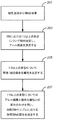
本開示の特定の局面は、妊娠個体の体液から得られたDNA中の胎児DNAの画分を測定する方法に関する。このような方法は、以下の操作によって特徴付けられ得る:(a)体液のサンプルを受け取ること;(b)母性ゲノムおよび胎児ゲノムの両方のDNA抽出物が体液中に存在する条件下で、サンプルからDNAを抽出すること;(c)1つまたは複数の多型を含むDNAセグメント配列を生成する条件下で、核酸シーケンサーによって抽出されたDNAを配列決定すること;(d)体液中のDNAの配列決定に由来するDNAセグメント配列を、参照配列における1つまたは複数の指定された多型についてマッピングすること;(e)マッピングされたDNAセグメント配列のアレル頻度を、指定された多型の少なくとも1つについて決定すること;(f)少なくとも1つの指定された多型を、妊娠個体の接合生殖性と胎児の接合生殖性との組み合わせに基づいて分類すること;および(g)妊娠個体から得られたDNAにおける胎児DNAの画分を、(e)において決定されたアレル頻度および(f)からの接合生殖性の組み合わせを用いて推定すること。 Certain aspects of the present disclosure relate to a method for measuring a fraction of fetal DNA in DNA obtained from body fluids of a pregnant individual. Such a method may be characterized by the following operations: (a) receiving a sample of body fluid; (b) under conditions where both maternal and fetal genome DNA extracts are present in the body fluid. Extracting the DNA from; (c) sequencing the DNA extracted by the nucleic acid sequencer under conditions that generate a DNA segment sequence comprising one or more polymorphisms; (d) the DNA in the body fluid Mapping a DNA segment sequence derived from sequencing to one or more specified polymorphisms in a reference sequence; (e) mapping the allelic frequency of the mapped DNA segment sequence to at least one of the specified polymorphisms (F) classifying at least one designated polymorphism based on the combination of the zygosity of the pregnant individual and the zygosity of the fetus ; and (g) obtained from the pregnant individual. DNA Fractions of kicking fetal DNA, be estimated using the combination of zygosity from allele frequencies were determined and (f) in (e).
マッピングは、1つまたは複数の指定された多型に対して核酸配列をマッピングするようにプログラムされたコンピューター装置を用いて行われてもよい。一般に、操作(d)〜(g)のいずれかは、プログラム指示の下で動く1つまたは複数のプロセッサーにおいて行われてもよい。 The mapping may be performed using a computer device programmed to map the nucleic acid sequence to one or more designated polymorphisms. In general, any of operations (d)-(g) may be performed on one or more processors running under program instructions.
特定の態様において、妊娠個体の体液から得られたDNAは、妊娠個体の血漿から得られた無細胞DNAである。典型的には、配列決定は、1つまたは複数の指定された多型のいずれかを選択的に増幅することなく行われる。 In certain embodiments, the DNA obtained from the body fluid of a pregnant individual is cell-free DNA obtained from the plasma of the pregnant individual. Typically, sequencing is performed without selectively amplifying any of one or more specified polymorphisms.
特定の態様において、胎児を孕む個体の血液から得られたDNAセグメントのマッピングは、多型のデータベースに対するセグメントのコンピューター的マッピングを含む。特定の態様において、(f)における分類は、少なくとも1つの指定された多型を、以下の組み合わせのうちの1つに分類する:(i)妊娠個体がホモ接合型であり、胎児がホモ接合型である、(ii)妊娠個体がホモ接合型であり、胎児がヘテロ接合型である、(iii)妊娠個体がヘテロ接合型であり、胎児がホモ接合型である、および(iv)妊娠個体がヘテロ接合型であり、胎児がヘテロ接合型である。 In certain embodiments, mapping of DNA segments obtained from the blood of an individual carrying a fetus includes computational mapping of the segments to a polymorphic database. In certain embodiments, the classification in (f) classifies at least one designated polymorphism into one of the following combinations: (i) the pregnant individual is homozygous and the fetus is homozygous (Ii) the pregnant individual is homozygous and the fetus is heterozygous; (iii) the pregnant individual is heterozygous and the fetus is homozygous; and (iv) the pregnant individual Is heterozygous and the fetus is heterozygous.
種々のフィルタリング操作が、使用され得る。これらは、例えば、組み合わせ(i)または組み合わせ(iv)に分類されるいかなる多型も検討しないことを含む。別の例において、この方法は、少なくとも1つの指定された多型をフィルタリングし、規定の閾値より高いマイナーアレル頻度を有する任意の多型を排除することを、さらに含む。なお別の例において、この方法は、少なくとも1つの指定された多型をフィルタリングし、規定の閾値より低いマイナーアレル頻度を有する任意の多型を排除する操作を含む。 Various filtering operations can be used. These include, for example, not considering any polymorphism classified as combination (i) or combination (iv). In another example, the method further includes filtering at least one designated polymorphism and excluding any polymorphism having a minor allele frequency that is above a defined threshold. In yet another example, the method includes an operation of filtering at least one designated polymorphism and excluding any polymorphism having a minor allele frequency lower than a defined threshold.
分類操作は、種々の方法において実施され得る。例えば、(e)において決定されたアレル頻度に対し、閾値を適用することを含んでもよい。別の例において、この分類操作は、複数の多型について得られた(e)からのアレル頻度データを、混合モデルに適用することを含む。1つの実施において、混合モデルは階乗モーメントを使用する。 The classification operation can be performed in various ways. For example, a threshold value may be applied to the allele frequency determined in (e). In another example, the classification operation includes applying allele frequency data from (e) obtained for multiple polymorphisms to a mixed model. In one implementation, the mixed model uses factorial moments.
本明細書において記載されるように決定された胎児画分は、種々の適用のために使用され得る。いくつかの例において、本明細書において記載される方法は、1つまたは複数のプロセッサーにおいてプログラム指示を実行し、(g)において決定された胎児DNAの画分を、妊娠個体についての患者医療記録に自動的に記録し、コンピューター読み取り可能媒体上に保存する操作を含む。患者医療記録は、研究室、臨床医のオフィス、病院、健康維持組織、保険会社または個体医療記録ウェブサイトによって保持され得る。別の適用において、胎児DNAの画分の推定は、母性試験サンプルが採取されたヒト対象の処置を処方するか、開始するか、および/または変更するために使用される。別の適用において、胎児DNAの画分の推定は、1つまたは複数のさらなる試験を指示するか、および/または行うために使用される。 The fetal fraction determined as described herein can be used for various applications. In some examples, the methods described herein execute program instructions in one or more processors, and the fraction of fetal DNA determined in (g) is stored in a patient medical record for a pregnant individual. Automatically recorded and stored on a computer readable medium. Patient medical records may be maintained by laboratories, clinician offices, hospitals, health maintenance organizations, insurance companies or individual medical records websites. In another application, the fractional fraction of fetal DNA is used to prescribe, initiate, and / or modify the treatment of a human subject from whom a maternal test sample has been taken. In another application, estimation of the fraction of fetal DNA is used to direct and / or perform one or more additional tests.
本開示の別の局面は、妊娠個体の体液から得られたDNA中の胎児DNA画分を推定するための装置に関する。このような装置は、以下の特徴によって特徴付けられ得る:(a)(i)母性ゲノムおよび胎児ゲノムの両方のDNAを含む体液のサンプルから抽出されたDNAを受け取り、そして(ii)1つまたは複数の指定された多型を含むDNAセグメント配列を生成する条件下で、抽出されたDNAを配列決定するように構成されたシーケンサー;ならびに(b)種々の操作(例えば、本明細書において記載された2つ以上の方法操作と共に記載された操作)を行うよう1つまたは複数のプロセッサーに命令するよう構成された(例えば、プログラムされた)コンピューター装置。いくつかの態様において、このコンピューター装置は、(i)参照配列における1つまたは複数の指定された多型に核酸配列をマッピングし、(ii)少なくとも1つの指定された多型についてマッピングされたDNAセグメント配列のアレル頻度を決定し、(iii)少なくとも1つの指定された多型を、妊娠個体の接合生殖性と胎児の接合生殖性との組み合わせに基づいて分類し、そして(iv)妊娠個体から得られたDNAにおける胎児DNAの画分を、アレル頻度および接合生殖性の組み合わせを用いて推定するように、構成される。 Another aspect of the present disclosure relates to an apparatus for estimating a fetal DNA fraction in DNA obtained from body fluid of a pregnant individual. Such a device may be characterized by the following features: (a) (i) receives DNA extracted from a sample of body fluid containing both maternal and fetal genomic DNA, and (ii) one or A sequencer configured to sequence the extracted DNA under conditions that produce a DNA segment sequence comprising a plurality of designated polymorphisms; and (b) various operations (e.g., as described herein) A computer device configured (eg, programmed) to instruct one or more processors to perform an operation described with two or more method operations). In some embodiments, the computing device comprises (i) mapping a nucleic acid sequence to one or more specified polymorphisms in a reference sequence, and (ii) DNA mapped for at least one specified polymorphism Determining the allele frequency of the segment sequence, (iii) classifying at least one designated polymorphism based on the combination of the zygote of the pregnant individual and the zygosity of the fetus , and (iv) from the pregnant individual The fraction of fetal DNA in the resulting DNA is configured to be estimated using a combination of allele frequency and zygosity.
特定の態様において、装置はまた、母性ゲノムと胎児ゲノムの両方のDNAを抽出する条件下で、サンプルからDNAを抽出するためのツールをも備える。いくつかの実施において、装置は、シーケンサーにおいて配列決定するために、妊娠個体の血漿から得られた無細胞DNAを抽出するように構成されたモジュールを備える。 In certain embodiments, the apparatus also comprises a tool for extracting DNA from a sample under conditions that extract both maternal and fetal genomic DNA. In some implementations, the apparatus comprises a module configured to extract cell-free DNA obtained from the plasma of a pregnant individual for sequencing in a sequencer.
いくつかの例において、装置は、多型のデータベースを備える。コンピューター装置は、胎児を孕む個体の血液から得られたDNAセグメントを、セグメントを多型のデータベースに対しコンピューター的にマッピングすることによりマッピングするよう、1つまたは複数のプロセッサーに命令するようにさらに構成され得る。データベースにおける配列は、参照配列の一例である。参照配列の他の例は、以下に提示される。 In some examples, the device comprises a polymorphic database. The computer device is further configured to instruct one or more processors to map the DNA segment obtained from the blood of the individual carrying the fetus by computerizing the segment to a polymorphic database. Can be done. The sequence in the database is an example of a reference sequence. Other examples of reference sequences are presented below.
特定の態様において、コンピューター装置は、少なくとも1つの指定された多型を、以下の組み合わせのうちの1つに分類するよう、1つまたは複数のプロセッサーに命令するようにさらに構成される:(i)妊娠個体がホモ接合型であり、胎児がホモ接合型である、(ii)妊娠個体がホモ接合型であり、胎児がヘテロ接合型である、(iii)妊娠個体がヘテロ接合型であり、胎児がホモ接合型である、および(iv)妊娠個体がヘテロ接合型であり、胎児がヘテロ接合型である。いくつかの態様において、コンピューター装置は、組み合わせ(i)または組み合わせ(iv)に分類されたいかなる多型をも検討から除くよう、1つまたは複数のプロセッサーに命令するようにさらに構成される。 In certain embodiments, the computing device is further configured to instruct one or more processors to classify at least one designated polymorphism into one of the following combinations: (i ) The pregnant individual is homozygous, the fetus is homozygous, (ii) the pregnant individual is homozygous, the fetus is heterozygous, (iii) the pregnant individual is heterozygous, The fetus is homozygous, and (iv) the pregnant individual is heterozygous and the fetus is heterozygous. In some embodiments, the computing device is further configured to instruct one or more processors to remove any polymorphism classified as combination (i) or combination (iv) from consideration.
特定の態様において、コンピューター装置は、規定の閾値より高いマイナーアレル頻度を有するいかなる多型をも検討から除くよう、1つまたは複数のプロセッサーに命令するようにさらに構成される。いくつかの態様において、コンピューター装置は、1つまたは複数の指定された多型をフィルタリングし、規定の閾値より低いマイナーアレル頻度を有するいかなる多型をも検討から除くよう1つまたは複数のプロセッサーに命令するように、さらに構成される。特定の態様において、コンピューター装置は、アレル頻度に対し閾値を適用することによって、少なくとも1つの指定された多型を分類するよう、1つまたは複数のプロセッサーに命令するようさらに構成される。 In certain embodiments, the computing device is further configured to instruct one or more processors to remove any polymorphism having a minor allele frequency above a defined threshold from consideration. In some embodiments, the computing device filters one or more specified polymorphisms to one or more processors so as to exclude any polymorphism having a minor allele frequency below a specified threshold from consideration. Further configured to command. In certain embodiments, the computing device is further configured to instruct one or more processors to classify at least one designated polymorphism by applying a threshold to the allele frequency.
特定の態様において、コンピューター装置は、複数の多型について得られたアレル頻度データを、混合モデルに対して適用することにより、少なくとも1つの指定された多型を分類するよう、1つまたは複数のプロセッサーに命令するようさらに構成される。混合モデルは、階乗モーメントを使用してもよい。 In certain embodiments, the computing device applies one or more of the allele frequency data obtained for a plurality of polymorphisms to a mixed model to classify at least one designated polymorphism. Further configured to instruct the processor. Mixed models may use factorial moments.
特定の態様において、コンピューター装置は、DNAの胎児の画分を、妊娠個体についての患者医療記録に自動的に記録し、コンピューター読み取り可能媒体に保存するよう、1つまたは複数のプロセッサーに命令するようさらに構成される。患者医療記録は、研究室、臨床医のオフィス、病院、健康維持組織、保険会社または個体医療記録ウェブサイトによって保持され得る。 In certain embodiments, the computing device instructs the one or more processors to automatically record the fetal fraction of DNA in a patient medical record for the pregnant individual and store it on a computer readable medium. Further configured. Patient medical records may be maintained by laboratories, clinician offices, hospitals, health maintenance organizations, insurance companies or individual medical records websites.
本開示の別の局面は、以下の操作にしたがって、妊娠個体の体液から得られたDNAにおける胎児DNAの画分を推定する方法に関する:(a)複数の多型配列に対し妊娠個体の体液から得られたDNAのセグメント(このDNAは、複数の多型配列を同定する条件下で配列決定されていた)をマッピングし;(b)複数の多型配列のそれぞれについてマッピングした核酸のアレル頻度を決定し;そして(c)アレル頻度を混合モデルに適用し、胎児を孕む個体の血液から得られたDNAにおける胎児DNA画分の推定を得る。操作(a)〜(c)の任意の1つまたは複数は、プログラム指示の下で動く1つまたは複数のプロセッサーにおいて行われ得る。特定の態様において、操作(c)は、1つまたは複数のプロセッサーにおいて指示を実行し、複数の多型配列のそれぞれについてのアレル頻度データの階乗モーメントについての一連の等式を解くことを含む。いくつかの態様において、混合モデルは、配列決定のエラーを説明する。 Another aspect of the present disclosure relates to a method for estimating a fraction of fetal DNA in DNA obtained from body fluid of a pregnant individual according to the following procedure: (a) Map the segment of DNA obtained (this DNA was sequenced under conditions that identify multiple polymorphic sequences); (b) map the nucleic acid allele frequencies mapped for each of the multiple polymorphic sequences And (c) applying the allelic frequency to the mixed model to obtain an estimate of the fetal DNA fraction in the DNA obtained from the blood of the individual holding the fetus . Any one or more of operations (a)-(c) may be performed on one or more processors running under program instructions. In certain embodiments, operation (c) comprises executing instructions in one or more processors and solving a set of equations for the factorial moments of allele frequency data for each of the plurality of polymorphic sequences. . In some embodiments, the mixed model accounts for sequencing errors.
特定の態様において、この方法は、胎児および妊娠個体の両方においてヘテロ接合型であると同定された多型についてのアレル頻度のコンピューター的除外を、さらに含む。いくつかの実施において、(c)の前に、この方法は、胎児および妊娠個体の両方においてホモ接合型であると同定された多型についてのアレル頻度のコンピューター的除外の操作を含む。いくつかの実施において、(c)の前に、この方法は、妊娠個体においてヘテロ接合型であると同定された多型についてのアレル頻度のコンピューター的除外の操作を含む。 In certain embodiments, the method further comprises computational exclusion of allele frequencies for polymorphisms identified as being heterozygous in both fetuses and pregnant individuals. In some implementations, prior to (c), the method involves the computational exclusion of allele frequencies for polymorphisms identified as homozygous in both fetuses and pregnant individuals. In some implementations, prior to (c), the method includes the computational exclusion of allele frequencies for polymorphisms identified as heterozygous in the pregnant individual.
妊娠個体の体液から得られたDNAは、妊娠個体の血漿から得られた無細胞DNAであり得る。体液から得られた核酸のマッピングは、多型のデータベースに対するセグメントのマッピングによって実施され得る。 The DNA obtained from the body fluid of the pregnant individual can be cell-free DNA obtained from the plasma of the pregnant individual. Mapping of nucleic acids obtained from body fluids can be performed by mapping segments to a polymorphic database.
本開示のこの局面の方法は、妊娠個体の体液由来のDNAを、多型配列を含むDNAセグメント配列を生成する条件下で、核酸シーケンサーによって配列決定することをさらに含み得る。 The method of this aspect of the present disclosure may further comprise sequencing DNA from the body fluid of a pregnant individual under conditions that produce a DNA segment sequence comprising a polymorphic sequence.
いくつかの実施において、(a)におけるマッピングは、複数の両アレル多型配列を同定することを含む。他の態様において、(a)におけるマッピングは、事前に規定された複数の多型配列に対しDNAのセグメントをマッピングすることを含む。 In some implementations, the mapping in (a) includes identifying a plurality of both allelic polymorphic sequences. In other embodiments, the mapping in (a) comprises mapping a segment of DNA to a plurality of pre-defined polymorphic sequences.
いくつかの態様において、この局面の方法は、1つまたは複数のプロセッサーにおいてプログラム指示を実行し、(c)において決定された胎児のDNAの画分を、妊娠個体の患者医療記録に自動的に記録し、コンピューター読み取り可能媒体に保存することをさらに含む。患者医療記録は、研究室、臨床医のオフィス、病院、健康維持組織、保険会社または個体医療記録ウェブサイトによって保持され得る。 In some embodiments, the method of this aspect executes program instructions in one or more processors and automatically applies the fraction of fetal DNA determined in (c) to the patient medical record of the pregnant individual. It further includes recording and storing on a computer readable medium. Patient medical records may be maintained by laboratories, clinician offices, hospitals, health maintenance organizations, insurance companies or individual medical records websites.
胎児DNAの画分の推定に基づき、この局面の方法は、母性試験サンプルが採取されたヒト対象の処置を処方すること、開始すること、および/または変更することをさらに含み得る。胎児DNAの画分の推定に基づき、この局面の方法は、1つまたは複数のさらなる試験を指示することおよび/または行うことをさらに含み得る。 Based on the estimation of the fraction of fetal DNA, the method of this aspect can further include prescribing, initiating, and / or modifying treatment of the human subject from whom the maternal test sample was taken. Based on the estimation of the fraction of fetal DNA, the method of this aspect may further include directing and / or conducting one or more additional tests.
本開示のなお別の局面にしたがい、以下の操作を用いて、妊娠個体の体液から得られたDNAにおける胎児DNAの画分を推定するための方法が、提供される:(a)体液のサンプルを受け取ること;(b)母性ゲノムおよび胎児ゲノムの両方のDNA抽出物が体液中に存在する条件下で、サンプルからDNAを抽出すること;(c)DNAセグメント配列を生成する条件下で、核酸シーケンサーによって抽出されたDNAを配列決定すること;(d)体液に由来するDNAセグメント配列を比較し、この比較から、1つまたは複数の両アレル多型を同定すること;(e)同定された多型の少なくとも1つについてのDNAセグメント配列のアレル頻度を決定すること;(f)少なくとも1つの同定された多型を、妊娠個体の接合生殖性と胎児の接合生殖性との組み合わせに基づいて分類すること;および(g)妊娠個体から得られたDNAにおける胎児DNAの画分を、(e)において決定されたアレル頻度および(f)からの接合生殖性の組み合わせを用いて推定すること。 In accordance with yet another aspect of the present disclosure, a method is provided for estimating the fraction of fetal DNA in DNA obtained from bodily fluid of a pregnant individual using the following procedure: (a) Sample of bodily fluid (B) extracting DNA from the sample under conditions in which both maternal and fetal genome DNA extracts are present in body fluids; (c) nucleic acid under conditions that produce a DNA segment sequence. Sequencing DNA extracted by a sequencer; (d) comparing DNA segment sequences from body fluids and identifying one or more allele polymorphisms from this comparison; (e) identified Determining the allelic frequency of the DNA segment sequence for at least one of the polymorphisms; (f) determining at least one identified polymorphism based on a combination of the zygosity of the pregnant individual and the zygosity of the fetus Classification; and (g) Pregnant Estimate the fraction of fetal DNA in the resulting DNA using the allele frequency determined in (e) and the zygote combination from (f).
マッピングは、1つまたは複数の指定された多型に対し核酸配列をマッピングするようプログラミングされたコンピューター装置を用いて、行われ得る。一般に、操作(d)〜(g)のいずれかが、プログラム指示の下で動く1つまたは複数のプロセッサーにおいて行われ得る。 The mapping can be performed using a computer device programmed to map the nucleic acid sequence to one or more designated polymorphisms. In general, any of operations (d)-(g) may be performed on one or more processors running under program instructions.
この局面の特定の実施において、DNAセグメント配列は、約20塩基対と約300塩基対との間の長さを有する。 In a particular implementation of this aspect, the DNA segment sequence has a length between about 20 base pairs and about 300 base pairs.
この局面の特定の態様において、(f)における分類は、少なくとも1つの同定された多型を、以下の組み合わせの1つに分類する:(i)妊娠個体がホモ接合型であり、胎児がホモ接合型である、(ii)妊娠個体がホモ接合型であり、胎児がヘテロ接合型である、(iii)妊娠個体がヘテロ接合型であり、胎児がホモ接合型である、および(iv)妊娠個体がヘテロ接合型であり、胎児がヘテロ接合型である。この方法はさらに、組み合わせ(i)または組み合わせ(iv)に分類されるいかなる多型をも検討から除くことを含み得る。 In a particular embodiment of this aspect, the classification in (f) classifies at least one identified polymorphism into one of the following combinations: (i) the pregnant individual is homozygous and the fetus is homozygous (Ii) the pregnant individual is homozygous and the fetus is heterozygous; (iii) the pregnant individual is heterozygous and the fetus is homozygous; and (iv) the pregnancy The individual is heterozygous and the fetus is heterozygous. The method may further comprise removing from the study any polymorphism classified as combination (i) or combination (iv).
種々の態様にしたがい、この局面の方法は、本明細書において記載されるフィルタリング操作および/または分類操作を、他の局面に関連して含み得る。例えば、この局面の方法は、1つまたは複数の同定された多型をフィルタリングすることにより、規定の閾値より高いマイナーアレル頻度を有するいかなる多型をも検討から除くことを含み得る。いくつかの場合、少なくとも1つの同定された多型を分類することは、(e)において決定されたアレル頻度に対し閾値を適用することを含む。本明細書において記載される混合モデルの用途は、同定された多型を分類するために使用され得る。 According to various embodiments, the method of this aspect may include the filtering and / or classification operations described herein in relation to other aspects. For example, the method of this aspect can include filtering any one or more identified polymorphisms from consideration to remove any polymorphism having a minor allele frequency above a defined threshold. In some cases, classifying at least one identified polymorphism includes applying a threshold to the allele frequency determined in (e). The mixed model application described herein can be used to classify the identified polymorphisms.
本開示の別の局面は、胎児DNAの画分を推定するための、以下の要素を備える装置に関する:(a)(i)母性ゲノムおよび胎児ゲノムの両方のDNAを含む体液のサンプルから抽出されたDNAを受け取り、そして(ii)抽出されたDNAを配列決定し、DNAの配列セグメントを生成するように構成されたシーケンサー;ならびに(b)1つまたは複数のプロセッサーに指示し、(i)妊娠個体の体液から得られたDNAの配列セグメントを複数の多型配列に対してマッピングし、(ii)マッピングされたDNAの配列セグメントからの複数の多型配列のそれぞれについてアレル頻度を決定し、そして(iii)このアレル頻度を、混合モデルに適用し、胎児を孕む個体の血液から得られたDNAにおける胎児DNAの画分の推定を得るよう構成されたコンピューター装置。 Another aspect of the present disclosure relates to an apparatus comprising the following elements for estimating a fraction of fetal DNA: (a) (i) extracted from a sample of bodily fluid containing both maternal and fetal genomic DNA And (ii) a sequencer configured to sequence the extracted DNA and generate a sequence segment of DNA; and (b) instruct one or more processors; Mapping a sequence segment of DNA obtained from an individual's body fluid to a plurality of polymorphic sequences; (ii) determining an allele frequency for each of the plurality of polymorphic sequences from the sequence segment of the mapped DNA; and (iii) A computer device configured to apply this allele frequency to a mixed model to obtain an estimation of a fraction of fetal DNA in DNA obtained from blood of an individual holding a fetus .
胎児DNAの画分を推定するためのなお別の装置は、以下の要素を備える:(a)(i)母性ゲノムおよび胎児ゲノムの両方のDNAを含む体液のサンプルから抽出されたDNAを受け取り、そして(ii)DNAセグメント配列を生成する条件下で、抽出されたDNAを配列決定するように構成されたシーケンサー;ならびに(b)1つまたは複数のプロセッサーに指示し、(i)体液に由来するDNAセグメント配列を比較し、この比較から、1つまたは複数の両アレル多型を同定し、(ii)同定された多型の少なくとも1つについてDNAセグメント配列のアレル頻度を決定し、(iii)少なくとも1つの同定された多型を、妊娠個体の接合生殖性と胎児の接合生殖性との組み合わせに基づいて分類し、そして(iii)妊娠個体から得られたDNAにおける胎児DNAの画分を、アレル頻度および接合生殖性の組み合わせを用いて推定するよう構成されたコンピューター装置。 Yet another device for estimating the fraction of fetal DNA comprises the following elements: (a) (i) receiving DNA extracted from a sample of body fluid containing both maternal and fetal genomic DNA; And (ii) a sequencer configured to sequence the extracted DNA under conditions that generate a DNA segment sequence; and (b) directs one or more processors and (i) is derived from a body fluid Comparing the DNA segment sequences, and from this comparison, identifying one or more both allelic polymorphisms, (ii) determining the allelic frequency of the DNA segment sequence for at least one of the identified polymorphisms, and (iii) Classifying at least one identified polymorphism based on the combination of zygote and fetal zygosity of the pregnant individual, and (iii) a fraction of fetal DNA in the DNA obtained from the pregnant individual, Combination of allele frequency and zygosity Configured the computer system to estimate with.
本明細書において記載される装置の局面において使用される命令および/またはハードウェアは、本明細書において開示される方法の局面の任意の1つまたは複数のコンピューター的操作またはアルゴリズム的操作の実行を、このような操作が明白に上で挙げられているか否かに関わらず、提供し得る。 The instructions and / or hardware used in the apparatus aspects described herein are those that perform any one or more of the computer or algorithmic operations of the method aspects disclosed herein. , Whether or not such an operation is explicitly listed above.
[本発明1001]
妊娠個体の体液から得られたDNA中の胎児DNAの画分を推定する方法であって、以下の段階:
(a)前記体液のサンプルを受け取る段階;
(b)前記サンプルから、前記体液中に存在する母性ゲノムと胎児ゲノムとの両方のDNAを抽出する条件下でDNAを抽出する段階;
(c)前記抽出したDNAを、1つまたは複数の多型を含むDNAセグメント配列を生成する条件下で、核酸シーケンサーによって配列決定する段階;
(d)前記体液中のDNAの配列決定に由来する前記DNAセグメント配列を、参照配列における1つまたは複数の指定された多型に対してマッピングする段階であって、ここで、前記マッピングは、1つまたは複数の指定された多型に対して核酸配列をマッピングするようプログラムされたコンピューター装置を用いて実施される、段階;
(e)前記マッピングされたDNAセグメント配列のアレル頻度を、前記指定された多型の少なくとも1つについて決定する段階;
(f)前記少なくとも1つの指定された多型を、前記妊娠個体の接合生殖性と前記胎児の接合生殖性との組み合わせに基づいて分類する段階;および
(g)(e)において決定された前記アレル頻度と(f)からの接合生殖性の組み合わせとを用いて、前記妊娠個体から得られたDNA中の胎児DNAの画分を推定する段階
を含み、ここで、(e)〜(g)は、前記決定する段階、分類する段階、および推定する段階のためのプログラム命令の下で動く1つまたは複数のプロセッサーにおいて行われる、方法。
[本発明1002]
(f)の分類する段階は、少なくとも1つの指定された多型を、以下の組み合わせのうちの1つに分類する、本発明1001の方法:(i)妊娠個体がホモ接合型であり、かつ胎児がホモ接合型である、(ii)妊娠個体がホモ接合型であり、かつ胎児がヘテロ接合型である、(iii)妊娠個体がヘテロ接合型であり、かつ胎児がホモ接合型である、および(iv)妊娠個体がヘテロ接合型であり、かつ胎児がヘテロ接合型である。
[本発明1003]
組み合わせ(i)または組み合わせ(iv)に分類されるいかなる多型も、検討から除くことをさらに含む、本発明1002の方法。
[本発明1004]
少なくとも1つの指定された多型をフィルタリングし、規定の閾値より高いマイナーアレル頻度を有するいかなる多型をも検討から除くことをさらに含む、本発明1001の方法。
[本発明1005]
少なくとも1つの指定された多型をフィルタリングし、規定の閾値より低いマイナーアレル頻度を有するいかなる多型をも検討から除くことをさらに含む、本発明1001の方法。
[本発明1006]
少なくとも1つの指定された多型を分類する段階は、(e)において決定されたアレル頻度に対し、閾値を適用することを含む、本発明1001の方法。
[本発明1007]
少なくとも1つの指定された多型を分類する段階は、複数の多型について得られた(e)からのアレル頻度データを、混合モデルに適用することを含む、本発明1001の方法。
[本発明1008]
混合モデルは、階乗モーメントを使用する、本発明1007の方法。
[本発明1009]
妊娠個体の体液から得られたDNAは、前記妊娠個体の血漿から得られた無細胞DNAである、本発明1001の方法。
[本発明1010]
胎児を孕む個体の血液から得られたDNAセグメントをマッピングする段階は、多型のデータベースに対して前記セグメントをコンピューター的にマッピングすることを含む、本発明1001の方法。
[本発明1011]
配列決定する段階は、1つまたは複数の指定された多型のいずれかを選択的に増幅させることなく実施される、本発明1001の方法。
[本発明1012]
1つまたは複数のプロセッサーにおいてプログラム命令を実行し、(g)において推定された胎児DNAの画分をコンピューター読み取り可能媒体に保存された妊娠個体についての患者医療記録に自動的に記録することをさらに含む、本発明1001の方法。
[本発明1013]
前記患者医療記録は、研究室、臨床医のオフィス、病院、健康維持組織、保険会社または個人医療記録ウェブサイトによって保持される、本発明1012の方法。
[本発明1014]
胎児DNAの画分の推定に基づき、母性試験サンプルが採取されたヒト対象の処置を処方すること、開始することおよび/または変更することをさらに含む、本発明1001の方法。
[本発明1015]
胎児DNAの画分の推定に基づき、1つまたは複数のさらなる試験を指示することおよび/または行うことをさらに含む、本発明1001の方法。
[本発明1016]
(a)(i)母性ゲノムおよび胎児ゲノムの両方のDNAを含む体液のサンプルから抽出されたDNAを受け取り、かつ(ii)1つまたは複数の指定された多型を含むDNAセグメント配列を生成する条件下で、前記抽出されたDNAを配列決定するように構成されたシーケンサーと;
(b)参照配列上の前記1つまたは複数の指定された多型に核酸配列をマッピングし、
前記指定された多型の少なくとも1つについてマッピングされたDNAセグメント配列のアレル頻度を決定し、
前記少なくとも1つの指定された多型を妊娠個体の接合生殖性と前記胎児の接合生殖性との組み合わせに基づいて分類し、かつ
前記妊娠個体から得られたDNA中の胎児DNAの画分を、前記アレル頻度および前記接合生殖性の組み合わせを用いて推定するよう、1つまたは複数のプロセッサーに命令するように構成されたコンピューター装置
とを備える、妊娠個体の体液から得られたDNA中の胎児DNAの画分を推定するための装置。
[本発明1017]
母性ゲノムと胎児ゲノムとの両方のDNAを抽出する条件下でサンプルからDNAを抽出するためのツールをさらに備える、本発明1016の装置。
[本発明1018]
コンピューター装置は、少なくとも1つの指定された多型を、以下の組み合わせの1つに分類するよう、1つまたは複数のプロセッサーに命令するようにさらに構成される、本発明1016の装置:(i)妊娠個体がホモ接合型であり、かつ胎児がホモ接合型である、(ii)妊娠個体がホモ接合型であり、かつ胎児がヘテロ接合型である、(iii)妊娠個体がヘテロ接合型であり、かつ胎児がホモ接合型である、および(iv)妊娠個体がヘテロ接合型であり、かつ胎児がヘテロ接合型である。
[本発明1019]
コンピューター装置は、組み合わせ(i)または組み合わせ(iv)に分類されるいかなる多型も、検討から除くよう1つまたは複数のプロセッサーに命令するようにさらに構成される、本発明1018の装置。
[本発明1020]
コンピューター装置は、規定の閾値より高いマイナーアレル頻度を有するいかなる多型をも検討から除くよう1つまたは複数のプロセッサーに命令するようにさらに構成される、本発明1016の装置。
[本発明1021]
コンピューター装置は、1つまたは複数の指定された多型をフィルタリングし、規定の閾値より低いマイナーアレル頻度を有するいかなる多型をも検討から除くよう1つまたは複数のプロセッサーに命令するようにさらに構成される、本発明1016の装置。
[本発明1022]
コンピューター装置は、アレル頻度に対し、閾値を適用することによって少なくとも1つの指定された多型を分類するよう1つまたは複数のプロセッサーに命令するようにさらに構成される、本発明1016の装置。
[本発明1023]
コンピューター装置は、複数の多型について得られたアレル頻度データを、混合モデルに適用することによって少なくとも1つの指定された多型を分類するよう1つまたは複数のプロセッサーに命令するようにさらに構成される、本発明1016の装置。
[本発明1024]
混合モデルは、階乗モーメントを使用する、本発明1023の装置。
[本発明1025]
シーケンサーにおける配列決定のために妊娠個体の血漿から得られた無細胞DNAを抽出するための装置をさらに備える、本発明1016の装置。
[本発明1026]
コンピューター装置は、多型のデータベースに対してセグメントをコンピューター的にマッピングすることによって胎児を孕む個体の血液から得られたDNAセグメントをマッピングするよう1つまたは複数のプロセッサーに命令するようにさらに構成された、多型のデータベースをさらに備える、本発明1016の装置。
[本発明1027]
コンピューター装置は、DNAの胎児画分を、コンピューター読み取り可能媒体に保存された妊娠個体についての患者医療記録に自動的に記録するよう1つまたは複数のプロセッサーに命令するようにさらに構成される、本発明1016の装置。
[本発明1028]
前記患者医療記録は、研究室、臨床医のオフィス、病院、健康維持組織、保険会社または個人医療記録ウェブサイトによって保持される、本発明1027の装置。
[本発明1029]
妊娠個体の体液から得られたDNA中の胎児DNAの画分を推定する方法であって、以下の段階:
(a)前記妊娠個体の体液から得られたDNAのセグメントを、複数の多型配列に対してマッピングする段階であって、ここで、前記複数の多型配列を同定する条件下で、前記DNAを配列決定する、段階;
(b)前記マッピングされた核酸のアレル頻度を、前記複数の多型配列のそれぞれについて決定する段階;および
(c)前記アレル頻度を混合モデルに適用する段階であって、前記胎児を孕む個体の血液から得られたDNA中の前記胎児DNAの画分の推定を得る、段階
を含み、ここで、(b)〜(c)は、前記決定する段階および適用する段階のためのプログラム命令の下で動く1つまたは複数のプロセッサーにおいて行われる、方法。
[本発明1030]
(c)は、複数の多型配列のそれぞれについてのアレル頻度データの階乗モーメントについての一連の等式を解くために、1つまたは複数のプロセッサーに対する命令を実行することを含む、本発明1029の方法。
[本発明1031]
(c)の前に、胎児および妊娠個体の両方においてヘテロ接合型であると同定されている多型についてのアレル頻度をコンピューター的に除くことをさらに含む、本発明1029の方法。
[本発明1032]
(c)の前に、胎児および妊娠個体の両方においてホモ接合型であると同定されている多型についてのアレル頻度をコンピューター的に除くことをさらに含む、本発明1029の方法。
[本発明1033]
(c)の前に、妊娠個体においてヘテロ接合型であると同定されている多型についてのアレル頻度をコンピューター的に除くことをさらに含む、本発明1029の方法。
[本発明1034]
混合モデルは、配列決定エラーを考慮する、本発明1029の方法。
[本発明1035]
妊娠個体の体液から得られるDNAは、前記妊娠個体の血漿から得られる無細胞DNAである、本発明1029の方法。
[本発明1036]
妊娠個体の体液から得られる核酸をマッピングする段階は、多型のデータベースに対しセグメントをマッピングすることを含む、本発明1029の方法。
[本発明1037]
妊娠個体の体液のサンプルを得ることをさらに含む、本発明1029の方法。
[本発明1038]
多型配列を含むDNAセグメント配列を生成する条件下で、核酸シーケンサーにより妊娠個体の体液由来のDNAを配列決定することをさらに含む、本発明1029の方法。
[本発明1039]
(a)のマッピングする段階は、複数の両アレル性多型配列を同定することを含む、本発明1029の方法。
[本発明1040]
(a)のマッピングする段階は、複数の事前指定された多型配列に対してDNAのセグメントをマッピングすることを含む、本発明1029の方法。
[本発明1041]
コンピューター読み取り可能媒体上に保存された妊娠個体についての患者医療記録において、(c)において推定されたDNAの胎児の画分を自動的に記録するためのプログラム命令を、1つまたは複数のプロセッサーにおいて実行することをさらに含む、本発明1029の方法。
[本発明1042]
前記患者医療記録は、研究室、臨床医のオフィス、病院、健康維持組織、保険会社または個人医療記録ウェブサイトによって保持される、本発明1041の方法。
[本発明1043]
胎児DNAの画分の推定に基づき、母性試験サンプルが採取されたヒト対象の処置を処方すること、開始することおよび/または変更することをさらに含む、本発明1029の方法。
[本発明1044]
胎児DNAの画分の推定に基づき、1つまたは複数のさらなる試験を指示することおよび/または行うことをさらに含む、本発明1029の方法。
[本発明1045]
妊娠個体の体液から得られたDNA中の胎児DNAの画分を推定する方法であって、以下の段階:
(a)前記体液のサンプルを受け取る段階;
(b)前記サンプルから、前記体液中に存在する母性ゲノムと胎児ゲノムとの両方のDNAを抽出する条件下でDNAを抽出する段階;
(c)前記抽出したDNAを、DNAセグメント配列を生成する条件下で、核酸シーケンサーによって配列決定する段階;
(d)前記体液に由来する前記DNAセグメント配列を比較し、比較から、1つまたは複数の両アレル性多型を同定する段階であって、ここで、前記比較することは、核酸配列を比較し多型を同定するようプログラムされたコンピューター装置を用いて行う、段階;
(e)前記DNAセグメント配列のアレル頻度を、前記同定された多型の少なくとも1つについて決定する段階;
(f)前記少なくとも1つの同定された多型を、前記妊娠個体の接合生殖性と前記胎児の接合生殖性との組み合わせに基づいて分類する段階;および
(g)(e)において決定された前記アレル頻度と(f)からの前記接合生殖性の組み合わせとを用いて、前記妊娠個体から得られたDNA中の胎児DNAの画分を推定する段階
を含み、ここで、(e)〜(g)は、前記決定する段階、分類する段階、および推定する段階のためのプログラム命令の下で動く1つまたは複数のプロセッサーにおいて行われる、方法。
[本発明1046]
(f)の分類する段階は、少なくとも1つの同定された多型を、以下の組み合わせのうちの1つに分類する、本発明1045の方法:(i)妊娠個体がホモ接合型であり、かつ胎児がホモ接合型である、(ii)妊娠個体がホモ接合型であり、かつ胎児がヘテロ接合型である、(iii)妊娠個体がヘテロ接合型であり、かつ胎児がホモ接合型である、および(iv)妊娠個体がヘテロ接合型であり、かつ胎児がヘテロ接合型である。
[本発明1047]
組み合わせ(i)または組み合わせ(iv)に分類されるいかなる多型も、検討から除くことをさらに含む、本発明1046の方法。
[本発明1048]
1つまたは複数の同定された多型をフィルタリングし、規定の閾値より高いマイナーアレル頻度を有するいかなる多型をも検討から除くことをさらに含む、本発明1045の方法。
[本発明1049]
DNAセグメント配列は、約20塩基対と約300塩基対との間の長さを有する、本発明1045の方法。
[本発明1050]
少なくとも1つの同定された多型を分類する段階は、(e)において決定されたアレル頻度に対し、閾値を適用することを含む、本発明1045の方法。
[本発明1051]
少なくとも1つの同定された多型を分類する段階は、複数の多型について得られた(e)からのアレル頻度データを、混合モデルに適用することを含む、本発明1045の方法。
[本発明1052]
混合モデルは、階乗モーメントを使用する、本発明1051の方法。
[本発明1053]
妊娠個体の体液から得られたDNAは、前記妊娠個体の血漿から得られた無細胞DNAである、本発明1045の方法。
[本発明1054]
(a)(i)母性ゲノムおよび胎児ゲノムの両方のDNAを含む体液のサンプルから抽出されたDNAを受け取り、かつ(ii)前記抽出されたDNAを配列決定して、DNAの配列セグメントを生成するように構成されたシーケンサーと;
(b)複数の多型配列に妊娠個体の体液から得られた前記DNAの配列セグメントをマッピングし、
前記複数の多型配列のそれぞれについて、前記DNAのマッピングされた配列セグメントからアレル頻度を決定し、かつ
前記アレル頻度を混合モデルに適用し、前記胎児を孕む個体の血液から得られたDNA中の胎児DNAの画分の推定を得るよう、1つまたは複数のプロセッサーに命令するように構成されたコンピューター装置
とを備える、妊娠個体の体液から得られたDNA中の胎児DNAの画分を推定するための装置。
[本発明1055]
(a)(i)母性ゲノムおよび胎児ゲノムの両方のDNAを含む体液のサンプルから抽出されたDNAを受け取り、かつ(ii)DNAセグメント配列を生成する条件下で、前記抽出されたDNAを配列決定するように構成されたシーケンサーと;
(b)前記体液に由来するDNAセグメント配列を比較し、比較から、1つまたは複数の両アレル性多型を同定し;
前記DNAセグメント配列のアレル頻度を、前記同定された多型の少なくとも1つについて決定し;
前記少なくとも1つの同定された多型を、妊娠個体の接合生殖性と前記胎児の接合生殖性との組み合わせに基づいて分類し;かつ
前記アレル頻度と前記接合生殖性の組み合わせとを用いて、前記妊娠個体から得られたDNA中の胎児DNAの画分を推定するよう、1つまたは複数のプロセッサーに命令するように構成されたコンピューター装置
とを備える、妊娠個体の体液から得られたDNA中の胎児DNAの画分を推定するための装置。
本開示の態様のこれらのおよび他の特徴および利点は、添付の図面を参照して、以下でより詳細に説明される。
[Invention 1001]
A method for estimating a fraction of fetal DNA in DNA obtained from body fluid of a pregnant individual, comprising the following steps:
(a) receiving a sample of said body fluid;
(b) extracting DNA from the sample under conditions for extracting both maternal and fetal genome DNA present in the body fluid;
(c) sequencing the extracted DNA with a nucleic acid sequencer under conditions that produce a DNA segment sequence comprising one or more polymorphisms;
(d) mapping the DNA segment sequence derived from the sequencing of DNA in the body fluid to one or more designated polymorphisms in a reference sequence, wherein the mapping comprises: Performed using a computer device programmed to map the nucleic acid sequence to one or more specified polymorphisms;
(e) determining the allele frequency of the mapped DNA segment sequence for at least one of the designated polymorphisms;
(f) classifying the at least one designated polymorphism based on a combination of the gestational fertility of the pregnant individual and the zygosity of the fetus; and
(g) estimating the fraction of fetal DNA in the DNA obtained from the pregnant individual using the allele frequency determined in (e) and the mating reproductive combination from (f)
Wherein (e)-(g) are performed in one or more processors running under program instructions for said determining, classifying, and estimating steps.
[Invention 1002]
The classifying step of (f) classifies at least one designated polymorphism into one of the following combinations: The method of the present invention 1001: (i) the pregnant individual is homozygous and The fetus is homozygous, (ii) the pregnant individual is homozygous and the fetus is heterozygous, (iii) the pregnant individual is heterozygous and the fetus is homozygous, And (iv) the pregnant individual is heterozygous and the fetus is heterozygous.
[Invention 1003]
The method of the present invention 1002, further comprising excluding any polymorphism classified as combination (i) or combination (iv) from consideration.
[Invention 1004]
The method of the present invention 1001, further comprising filtering at least one designated polymorphism and excluding any polymorphism having a minor allele frequency above a defined threshold from consideration.
[Invention 1005]
The method of the present invention 1001, further comprising filtering at least one designated polymorphism and excluding from the study any polymorphism having a minor allele frequency below a defined threshold.
[Invention 1006]
The method of the present invention 1001, wherein classifying at least one designated polymorphism comprises applying a threshold to the allele frequency determined in (e).
[Invention 1007]
The method of the present invention 1001, wherein the step of classifying at least one designated polymorphism comprises applying allelic frequency data from (e) obtained for a plurality of polymorphisms to a mixed model.
[Invention 1008]
The method of the present invention 1007, wherein the mixed model uses factorial moments.
[Invention 1009]
The method of the present invention 1001, wherein the DNA obtained from the body fluid of a pregnant individual is cell-free DNA obtained from the plasma of the pregnant individual.
[Invention 1010]
100. The method of the invention 1001, wherein the step of mapping a DNA segment obtained from the blood of an individual carrying a fetus comprises computationally mapping said segment to a polymorphism database.
[Invention 1011]
The method of the present invention 1001, wherein the sequencing step is performed without selectively amplifying any of the one or more designated polymorphisms.
[Invention 1012]
Further executing program instructions in one or more processors and automatically recording the fraction of fetal DNA estimated in (g) in a patient medical record for a pregnant individual stored in a computer readable medium. Including the method of the invention 1001.
[Invention 1013]
The method of the invention 1012 wherein the patient medical records are maintained by a laboratory, clinician office, hospital, health maintenance organization, insurance company or personal medical records website.
[Invention 1014]
The method of the present invention 1001, further comprising prescribing, initiating and / or altering treatment of a human subject from whom a maternal test sample was taken based on an estimate of a fraction of fetal DNA.
[Invention 1015]
The method of the present invention 1001, further comprising directing and / or conducting one or more further tests based on the estimation of a fraction of fetal DNA.
[Invention 1016]
(a) (i) accepts DNA extracted from a sample of body fluid containing both maternal and fetal genomic DNA, and (ii) generates a DNA segment sequence containing one or more specified polymorphisms A sequencer configured to sequence the extracted DNA under conditions;
(b) mapping a nucleic acid sequence to the one or more designated polymorphisms on a reference sequence;
Determining the allele frequency of the mapped DNA segment sequence for at least one of the designated polymorphisms;
Classifying the at least one designated polymorphism on the basis of a combination of the zygosity of the pregnant individual and the zygosity of the fetus; and
A computer device configured to instruct one or more processors to estimate a fraction of fetal DNA in DNA obtained from the pregnant individual using the combination of the allele frequency and the mating reproductive properties
A device for estimating a fraction of fetal DNA in DNA obtained from body fluid of a pregnant individual.
[Invention 1017]
The device of the present invention 1016 further comprising a tool for extracting DNA from a sample under conditions to extract both maternal and fetal genome DNA.
[Invention 1018]
The apparatus of invention 1016 is further configured to instruct one or more processors to classify at least one designated polymorphism into one of the following combinations: (i) The pregnant individual is homozygous and the fetus is homozygous, (ii) The pregnant individual is homozygous and the fetus is heterozygous, (iii) The pregnant individual is heterozygous And the fetus is homozygous, and (iv) the pregnant individual is heterozygous and the fetus is heterozygous.
[Invention 1019]
The apparatus of 1018 of this invention, wherein the computer apparatus is further configured to instruct one or more processors to remove any polymorphism classified as combination (i) or combination (iv) from consideration.
[Invention 1020]
The apparatus of the present invention 1016, wherein the computing device is further configured to instruct one or more processors to remove any polymorphism having a minor allele frequency above a defined threshold from consideration.
[Invention 1021]
The computing device is further configured to filter one or more specified polymorphisms and instruct one or more processors to remove any polymorphisms having a minor allele frequency below a prescribed threshold from consideration. The apparatus of the present invention 1016.
[Invention 1022]
The apparatus of invention 1016, wherein the computing device is further configured to instruct one or more processors to classify at least one specified polymorphism by applying a threshold to the allele frequency.
[Invention 1023]
The computing device is further configured to instruct one or more processors to classify at least one specified polymorphism by applying the allele frequency data obtained for the plurality of polymorphisms to the mixed model. The apparatus of the present invention 1016.
[Invention 1024]
The apparatus of invention 1023, wherein the mixed model uses factorial moments.
[Invention 1025]
The device of the invention 1016 further comprising a device for extracting cell-free DNA obtained from the plasma of pregnant individuals for sequencing in a sequencer.
[Invention 1026]
The computer device is further configured to instruct one or more processors to map a DNA segment obtained from the blood of an individual carrying a fetus by computationally mapping the segment to a polymorphic database. The apparatus of the present invention 1016 further comprising a polymorphic database.
[Invention 1027]
The computer device is further configured to instruct one or more processors to automatically record a fetal fraction of DNA in a patient medical record for a pregnant individual stored in a computer readable medium. The device of invention 1016.
[Invention 1028]
The apparatus of the present invention 1027, wherein the patient medical records are maintained by a laboratory, clinician office, hospital, health maintenance organization, insurance company or personal medical records website.
[Invention 1029]
A method for estimating a fraction of fetal DNA in DNA obtained from body fluid of a pregnant individual, comprising the following steps:
(a) mapping a segment of DNA obtained from the body fluid of the pregnant individual to a plurality of polymorphic sequences, wherein the DNA is subjected to conditions for identifying the plurality of polymorphic sequences. Sequencing, stage;
(b) determining an allele frequency of the mapped nucleic acid for each of the plurality of polymorphic sequences; and
(c) applying the allele frequency to a mixed model, obtaining an estimation of a fraction of the fetal DNA in DNA obtained from the blood of an individual holding the fetus
Wherein (b)-(c) are performed in one or more processors operating under program instructions for said determining and applying steps.
[Invention 1030]
(c) comprises executing instructions to one or more processors to solve a series of equations for the factorial moments of allele frequency data for each of a plurality of polymorphic sequences. the method of.
[Invention 1031]
Prior to (c), the method of the invention 1029 further comprising computationally removing allele frequencies for polymorphisms identified as being heterozygous in both fetuses and pregnant individuals.
[Invention 1032]
Prior to (c), the method of the invention 1029 further comprising computationally removing allele frequencies for polymorphisms identified as being homozygous in both fetuses and pregnant individuals.
[Invention 1033]
Prior to (c), the method of the present invention 1029 further comprising computationally removing allele frequencies for polymorphisms identified as being heterozygous in the pregnant individual.
[Invention 1034]
The method of the present invention 1029, wherein the mixed model takes into account sequencing errors.
[Invention 1035]
The method of the present invention 1029, wherein the DNA obtained from the body fluid of a pregnant individual is cell-free DNA obtained from the plasma of the pregnant individual.
[Invention 1036]
The method of the present invention 1029, wherein the step of mapping the nucleic acid obtained from the body fluid of the pregnant individual comprises mapping the segment to a polymorphism database.
[Invention 1037]
The method of the present invention 1029 further comprising obtaining a sample of bodily fluid of a pregnant individual.
[Invention 1038]
The method of the invention 1029 further comprising sequencing DNA from a bodily fluid of a pregnant individual with a nucleic acid sequencer under conditions that produce a DNA segment sequence comprising the polymorphic sequence.
[Invention 1039]
The method of 1029 of this invention, wherein the mapping step (a) comprises identifying a plurality of both allelic polymorphic sequences.
[Invention 1040]
The method of the present invention 1029, wherein the mapping step (a) comprises mapping a segment of DNA to a plurality of pre-designated polymorphic sequences.
[Invention 1041]
In patient medical records for pregnant individuals stored on a computer readable medium, program instructions for automatically recording the fetal fraction of DNA estimated in (c) in one or more processors The method of the invention 1029, further comprising performing.
[Invention 1042]
The method of the present invention 1041, wherein the patient medical record is maintained by a laboratory, clinician office, hospital, health maintenance organization, insurance company or personal medical records website.
[Invention 1043]
The method of the present invention 1029 further comprising prescribing, initiating and / or altering treatment of the human subject from whom the maternal test sample was taken based on the estimation of the fraction of fetal DNA.
[Invention 1044]
The method of the present invention 1029 further comprising directing and / or conducting one or more additional tests based on the estimation of the fraction of fetal DNA.
[Invention 1045]
A method for estimating a fraction of fetal DNA in DNA obtained from body fluid of a pregnant individual, comprising the following steps:
(a) receiving a sample of said body fluid;
(b) extracting DNA from the sample under conditions for extracting both maternal and fetal genome DNA present in the body fluid;
(c) sequencing the extracted DNA with a nucleic acid sequencer under conditions that produce a DNA segment sequence;
(d) comparing the DNA segment sequences derived from the body fluid and identifying one or more allelic polymorphisms from the comparison, wherein the comparing comprises comparing nucleic acid sequences Using a computer device programmed to identify the polymorphism;
(e) determining the allele frequency of the DNA segment sequence for at least one of the identified polymorphisms;
(f) classifying the at least one identified polymorphism based on a combination of the gestational fertility of the pregnant individual and the zygosity of the fetus; and
(g) using the allele frequency determined in (e) and the mating reproductive combination from (f) to estimate the fraction of fetal DNA in the DNA obtained from the pregnant individual
Wherein (e)-(g) are performed in one or more processors running under program instructions for said determining, classifying, and estimating steps.
[Invention 1046]
The classifying step of (f) classifies at least one identified polymorphism into one of the following combinations: The method of the present invention 1045: (i) the pregnant individual is homozygous and The fetus is homozygous, (ii) the pregnant individual is homozygous and the fetus is heterozygous, (iii) the pregnant individual is heterozygous and the fetus is homozygous, And (iv) the pregnant individual is heterozygous and the fetus is heterozygous.
[Invention 1047]
The method of the present invention 1046 further comprising excluding any polymorphism classified as combination (i) or combination (iv) from consideration.
[Invention 1048]
The method of the present invention 1045, further comprising filtering one or more identified polymorphisms and excluding any polymorphism having a minor allele frequency above a defined threshold from consideration.
[Invention 1049]
The method of the present invention 1045, wherein the DNA segment sequence has a length between about 20 base pairs and about 300 base pairs.
[Invention 1050]
The method of the present invention 1045, wherein classifying at least one identified polymorphism comprises applying a threshold to the allele frequency determined in (e).
[Invention 1051]
The method of the present invention 1045, wherein classifying at least one identified polymorphism comprises applying the allelic frequency data from (e) obtained for the plurality of polymorphisms to a mixed model.
[Invention 1052]
The method of the present invention 1051, wherein the mixed model uses factorial moments.
[Invention 1053]
The method of the present invention 1045, wherein the DNA obtained from the body fluid of the pregnant individual is cell-free DNA obtained from the plasma of the pregnant individual.
[Invention 1054]
(a) (i) receiving DNA extracted from a sample of body fluid containing both maternal and fetal genomic DNA, and (ii) sequencing the extracted DNA to generate a sequence segment of DNA With a sequencer configured to;
(b) mapping a sequence segment of the DNA obtained from the body fluid of a pregnant individual to a plurality of polymorphic sequences,
For each of the plurality of polymorphic sequences, determining an allele frequency from the mapped sequence segment of the DNA; and
A computer configured to instruct one or more processors to apply the allele frequency to a mixed model and to obtain an estimate of the fraction of fetal DNA in the DNA obtained from the blood of the individual holding the fetus apparatus
A device for estimating a fraction of fetal DNA in DNA obtained from body fluid of a pregnant individual.
[Invention 1055]
(a) (i) receiving DNA extracted from a sample of body fluid containing both maternal and fetal genomic DNA, and (ii) sequencing the extracted DNA under conditions that produce a DNA segment sequence With a sequencer configured to;
(b) comparing DNA segment sequences derived from the body fluid and identifying one or more allelic polymorphisms from the comparison;
Determining the allele frequency of the DNA segment sequence for at least one of the identified polymorphisms;
Classifying the at least one identified polymorphism based on a combination of the zygote of the pregnant individual and the zygote of the fetus; and
A computer configured to instruct one or more processors to estimate a fraction of fetal DNA in DNA obtained from the pregnant individual using the allele frequency and the mating reproductive combination apparatus
A device for estimating a fraction of fetal DNA in DNA obtained from body fluid of a pregnant individual.
These and other features and advantages of aspects of the present disclosure are described in more detail below with reference to the accompanying drawings.
詳細な説明
序論および概要
本開示の特定の態様は、妊娠雌性体の血液から得られたDNAを解析すること、およびこの分析を、胎児由来のDNAの画分を推定するために用いることに関する。次いで、DNAの胎児画分は、母性血液から得られたDNAの独立した解析に基づいて、胎児の別の測定基準または性質決定にあるレベルの信頼性を与えるために使用され得る。例えば、母性血液から得られた胎児DNAサンプルは、別個に解析されて、この妊娠雌性体が孕む胎児における異常を検出することができる。この別個の解析による異常判定は、母性血液から得られたDNAにおける胎児DNAの画分量に基づく信頼性レベルに統計学的に基づく。DNA全量において相対的に低い胎児DNAの画分は、胎児DNAに基づく任意の性質決定における低い信頼性を示唆する。
Detailed Description Introduction and Overview Certain embodiments of the present disclosure relate to analyzing DNA obtained from blood of pregnant female bodies and using this analysis to estimate fractions of fetal DNA. The fetal fraction of DNA can then be used to provide a level of confidence in another metric or characterization of the fetus based on independent analysis of DNA obtained from maternal blood. For example, fetal DNA samples obtained from maternal blood can be analyzed separately to detect abnormalities in the fetus that the pregnant female body crabs. The determination of abnormality by this separate analysis is statistically based on a confidence level based on the fraction of fetal DNA in DNA obtained from maternal blood. The fraction of fetal DNA that is relatively low in total DNA suggests low confidence in any characterization based on fetal DNA.
典型的に、必ずしもではないが、解析された母性血液中のDNAは、無細胞DNAであるが、ある態様において、細胞定着DNAであってもよい。無細胞DNAは、母性血漿から得られる。妊娠雌性体から得られた無細胞DNAカウント中の胎児DNAの量は、胎児の在胎月齢を含む種々の因子に依存して広く変動する。典型的なヒト妊婦女性について、現在、無細胞DNAの約5〜20%は、胎児DNAであると信じられている。しかし、胎児画分が非常に低い(例えば、約1%以下である)ことも稀ではない。このような場合、胎児DNAの任意の別個の性質決定が、本質的に疑われ得る。他方、ある研究者らは、母性無細胞DNAサンプルが、胎児DNA画分を40%または50%の高さで有すると報告している。 Typically, but not necessarily, the DNA in maternal blood analyzed is cell-free DNA, but in certain embodiments may be cell-fixed DNA. Cell-free DNA is obtained from maternal plasma. The amount of fetal DNA in a cell-free DNA count obtained from a pregnant female body varies widely depending on a variety of factors, including the gestational age of the fetus . For typical human pregnant women, it is currently believed that about 5-20% of cell-free DNA is fetal DNA. However, it is not uncommon for the fetal fraction to be very low (eg, less than about 1%). In such cases, any separate characterization of fetal DNA can be suspected in essence. On the other hand, some researchers report that maternal cell-free DNA samples have fetal DNA fractions as high as 40% or 50%.
本明細書において記載される特定の実施において、母性DNAの胎児画分の決定は、1つまたは複数の多型を有することが公知な配列部位に関する多数のDNA配列に基づく。典型的には、必ずしもではないが、このような多型は、1ヌクレオチド多型(SNP)である。好適な多型の他の型としては、欠失、STR(短いタンデムリピート)、挿入、インデル(微小インデルを含む)などが挙げられる。さらなる例は、以下に示される。特定の態様において、多型部位は、以下に記載される「参照配列」において見出される。ある態様において、多型部位は、配列タグを互いにおよび/または参照配列とアラインメントした際に発見される。 In the particular implementation described herein, the determination of the fetal fraction of maternal DNA is based on a number of DNA sequences for sequence sites known to have one or more polymorphisms. Typically, but not necessarily, such a polymorphism is a single nucleotide polymorphism (SNP). Other types of suitable polymorphisms include deletions, STR (short tandem repeats), insertions, indels (including micro indels) and the like. Further examples are given below. In certain embodiments, the polymorphic site is found in the “reference sequence” described below. In certain embodiments, polymorphic sites are discovered when sequence tags are aligned with each other and / or reference sequences.
本開示の特定の方法は、検討中の多型部位において、胎児のDNA配列は、その母親のDNA配列と一致しない場合があるという事実を使用する。例えば、特定のSNPの部位における母性DNAは、ホモ接合型であってもよく、他方、SNPの胎児版は、ヘテロ接合型であってもよい。したがって、問題のSNPについて得られた配列サンプルの収集は、メジャーアレルを含む配列の大部分と、マイナーアレルを含む残りの画分とによる異種性と考えられる。メジャーアレルおよびマイナーアレルの相対量は、サンプル中の胎児DNAの画分によって決定される。 Certain methods of the present disclosure use the fact that at the polymorphic site under consideration, the fetal DNA sequence may not match the maternal DNA sequence. For example, maternal DNA at a particular SNP site may be homozygous, while the fetal version of SNP may be heterozygous. Thus, the collection of sequence samples obtained for the SNP in question is considered to be heterogeneous with the majority of the sequence containing the major allele and the remaining fraction containing the minor allele. The relative amount of major and minor alleles is determined by the fraction of fetal DNA in the sample.
ホモ接合型サンプルにおいて、所定のSNPまたは他の多型の両コピーは、同じアレルを含み、他方、ヘテル接合型のSNPまたは他の多型は、1コピーのメジャーアレルと1コピーのマイナーアレルとを含むことが言及される。したがって、ヘテロ接合型の個体からのみ得られたDNAは、50%のメジャーアレルと50%のマイナーアレルとを含むことが分かる。この知見は、下で概説される胎児DNAの画分を説明する際に使用され得る。まとめると、下でより完全に説明されるように、本明細書において開示される種々の方法は、母性DNAおよび胎児DNAにおいて2つのアレルしか存在しない多型のみを検討する。 In a homozygous sample, both copies of a given SNP or other polymorphism contain the same allele, whereas a heterozygous SNP or other polymorphism contains one copy of a major allele and one copy of a minor allele. Is included. Therefore, it can be seen that DNA obtained only from heterozygous individuals contains 50% major alleles and 50% minor alleles. This finding can be used in describing the fraction of fetal DNA outlined below. In summary, as described more fully below, the various methods disclosed herein consider only polymorphisms in which there are only two alleles in maternal DNA and fetal DNA.
ある実施において、母性血液から得られたDNAは、多数回(多型の「適用範囲」と考えられる多型の特定部位に対してマッピングする読み取りの全回数、およびマイナーアレルカウントと考えられる多型についてのマイナーアレルに対してマッピングする読み取りの回数)読み取られる。マイナーアレルカウント対適用範囲の比は、種々の実施において重要である。 In one implementation, the DNA obtained from maternal blood can be generated multiple times (the total number of reads that map to a specific site of the polymorphism that is considered the “scope” of the polymorphism, and the polymorphism that is considered the minor allele count. The number of reads that map to the minor allele for The ratio of minor allele count to coverage is important in various implementations.
本明細書において開示される特定の方法は、母性および胎児の両方からのDNAを含むDNAサンプル中の多型の4つの事例を同定し性質決定する。下の図1は、これらの4つの事例を示す。具体的には、比較的興味のない第1の事例において、母親および胎児の両方が、検討される特定の多型においてホモ接合型である。このような事例においては、問題の多型を含むDNAサンプル中の全ての配列が同じアレルを含み、母親および胎児からのDNAの相対量に関して何の情報も得ることができないと考えられる。しかし、この事例は、研究者および技術者が、検討される配列データを作成するために使用されるDNA配列決定装置の相対的エラー率についての何らかの着想を得ることを可能にするという意味で、興味深いともいえることに留意されたい。 The particular method disclosed herein identifies and characterizes four instances of polymorphisms in a DNA sample containing DNA from both maternal and fetuses . Figure 1 below shows these four cases. Specifically, in the first case, which is relatively uninteresting, both the mother and the fetus are homozygous for the particular polymorphism being considered. In such cases, it is believed that all sequences in the DNA sample containing the polymorphism in question contain the same allele and no information is available regarding the relative amount of DNA from the mother and fetus . However, this case means that researchers and engineers can get some idea about the relative error rate of the DNA sequencing equipment used to create the sequence data being considered, Note that it can be interesting.
解析が遭遇する第2の事例は、妊娠雌性体がホモ接合型で胎児がヘテロ接合型である多型である。この事例において、比較的小さいが、それにも関わらず有為である、検出配列の画分が、マイナーアレルを含むと考えられる。具体的には、この第2の事例において、マイナーアレルの頻度は、名目上、母親の血流における胎児DNAの画分を2で割ることによって得られる。 The second case that the analysis encounters is a polymorphism in which the pregnant female body is homozygous and the fetus is heterozygous. In this case, the fraction of the detection sequence that is relatively small but nevertheless significant is considered to contain minor alleles. Specifically, in this second case, the frequency of minor alleles is nominally obtained by dividing the fraction of fetal DNA in the mother's bloodstream by 2.
第3の事例において、検討される多型は、母性DNAにおいてヘテロ接合型であり、胎児DNAにおいてホモ接合型である。この場合、マイナーアレルの頻度は、名目上、0.5からDNAサンプルにおける胎児DNAの画分の2分の1を引くことによって得られる。 In the third case, the polymorphism studied is heterozygous in maternal DNA and homozygous in fetal DNA. In this case, the frequency of minor alleles is obtained by nominally subtracting one half of the fetal DNA fraction in the DNA sample from 0.5.
最後に、第4の事例において、検討される多型は、母性および胎児の両方においてヘテロ接合型である。この事例において、メッジャーアレルおよびマイナーアレルの頻度は、両方とも0.5であると予測される。第1の事例のように、第4の事例は、DNAの胎児画分を決定するためには、比較的興味がない。 Finally, in the fourth case, the polymorphism studied is heterozygous in both maternal and fetal . In this case, the frequency of both the major and minor alleles is expected to be 0.5. Like the first case, the fourth case is relatively uninteresting for determining the fetal fraction of DNA.
研究者、技術者またはソフトウェアが、所定の多型(この多型が4つの事例のどれに属するかが分かっている)についてサンプルにおいて胎児DNAの画分を決定することが課されている場合、検討中の多型が事例2または事例3に入るとすると、胎児DNAの画分は、直接的に推定可能である。しかし、実際には、この知見を実験せずに有することはできない。したがって、コンピューター装置は、本明細書において記載される操作を行う必要がある。
If a researcher, technician or software is required to determine the fraction of fetal DNA in the sample for a given polymorphism (which knows which of the four cases this polymorph belongs to) If the polymorphism under consideration falls into
本明細書において他所で記載される特定の態様において、1つの多型を4つの事例のうちの1つに分類するために、閾値技術が使用される。多型がこのように分類され、事例2または事例3のどちらかに在ることが見出されると、胎児画分は、推定され得る。他の態様において、この技術は、ゲノムの全てまたは一部を通して分布される多くの多型を検討する。具体的な例において説明されるように、ゲノムに亘る多くの異なるSNPが、この目的のために使用され得る。
In certain embodiments described elsewhere herein, threshold techniques are used to classify a polymorphism into one of four cases. If the polymorphism is classified in this way and found to be in either
特定の態様において、アレル頻度は、母性血液サンプルから得られたDNAサンプルにおいて多くの異なる多型について決定される。この多くの多型について、ある画分は接合生殖性事例1に相当し、別の画分は事例2に相当し、第3の画分は事例3に相当し、最後の画分は事例4に相当すると考えられる。これらの画分は、合計すると1の値になる。混合モデルもしくは関連の技術は、これらの4つのカテゴリーのそれぞれにおいて多型の1つまたは複数の統計学的特性を探るために使用され得る。具体的には、混合モデルは、妊娠雌性体の血液から得られるDNAサンプルにおいて遭遇する4つの事例それぞれについての平均および任意で分散を決定するために使用され得る。具体的な態様において、これは、検討中の多型のカウントの総数(適用範囲)に対しマイナーアレルの頻度に関連する平均および分散である。本明細書において他所で詳述されたように、これらの4つのカテゴリー、または少なくとも第2および第3のカテゴリーのそれぞれについての平均値は、母性血液から得られたDNAにおける胎児画分に直接関係する。
In certain embodiments, allelic frequencies are determined for many different polymorphisms in a DNA sample obtained from a maternal blood sample. For this many polymorphisms, one fraction corresponds to mating reproductive case 1, another fraction corresponds to
混合モデルを使用する特定の実施において、多型が考慮される位置それぞれについて、1つまたは複数の階乗モーメントが計算される。例えば、階乗モーメント(または階乗モーメントの集合)は、DNA配列中で考慮される多数のSNP位置を用いて計算される。以下の等式4で示されるように、種々の階乗モーメントのそれぞれは、所定の位置についてのマイナーアレル頻度対適用範囲の比について考慮される種々のSNP位置の全てに亘る合計である。以下の等式5で示されるように、これらの階乗モーメントは、上で記載される4つの接合生殖性の事例のそれぞれに関連したパラメータにも関する。具体的には、これらは、事例それぞれについての可能性、ならびに考慮される多型の集合における4つの事例それぞれについての相対量に関する。説明されるように、この可能性は、母性血液中の無細胞DNAにおける胎児DNA画分に相関する。より完全に以下に説明されるように、充分な数のこれらの階乗モーメント(等式4において示される)を計算することにより、本方法は、全ての未知数を解くために充分な数の式を提供する。この場合の未知数は、考慮される多型の集団における4つの事例それぞれの相対量、ならびに4つの事例それぞれに関連する可能性(したがって、胎児DNA画分)であると考えられる。等式5を参照されたい。類似の結果は、以下の等式7〜12において表される他の種類の混合モデルを用いて得られ得る。これらの特定の種類は、事例1および2に当てはまる多型のみを使用し、事例3および4の多型は、閾値技術によってフィルタリングされる。
In a particular implementation using a mixed model, one or more factorial moments are calculated for each position where a polymorphism is considered. For example, the factorial moment (or set of factorial moments) is calculated using a number of SNP positions considered in the DNA sequence. As shown in Equation 4 below, each of the various factorial moments is a sum over all of the various SNP positions considered for the minor allele frequency to coverage ratio for a given position. These factorial moments also relate to the parameters associated with each of the four mating reproductive cases described above, as shown in
したがって、階乗モーメントは、接合生殖性の4つの事例の任意の組み合わせの可能性を同定する混合モデルの一部として使用され得る。そして、言及された通り、これらの可能性、または少なくとも第2および第3の事例についての可能性は、母性血液における全無細胞DNAにおける胎児DNAの画分に直接関係する。 Thus, the factorial moment can be used as part of a mixed model that identifies the possibility of any combination of the four cases of zygosity. And as mentioned, these possibilities, or at least the possibilities for the second and third cases, are directly related to the fraction of fetal DNA in total cell-free DNA in maternal blood.
配列決定エラーは、解かれるべき階乗モーメント等式のシステムの複雑性を減らすために使用され得ることもまた言及される。この点において、配列決定エラーは、4つの結果(任意の所定の多型位置における4つの可能性の有る塩基のそれぞれに対応する)のうちの任意の1つを実際に有し得ることが認識されるべきである。 It is also mentioned that sequencing errors can be used to reduce the complexity of the system of factorial moment equations to be solved. In this regard, it is recognized that sequencing errors can actually have any one of the four results (corresponding to each of the four possible bases at any given polymorphic position). It should be.
特定の態様において、タグが、参照染色体またはゲノムとアラインメントされ、そして両アレル多型が同定される。これらの多型は、事前に規定されているか、さもなければアラインメントの前に同定される。これらは、単に、アラインメントの間に同定され、その後、その本明細書において記載される接合生殖性およびマイナーアレルカウントに基づいて性質決定される。この情報は、本明細書において記載されるように、ゲノム画分を推定するために、使用される。 In certain embodiments, the tag is aligned with a reference chromosome or genome and both allelic polymorphisms are identified. These polymorphisms are either pre-defined or otherwise identified prior to alignment. These are simply identified during alignment and then characterized based on their mating reproductive and minor allele counts described herein. This information is used to estimate the genomic fraction as described herein.
本明細書において記載される態様において使用されるタグの長さは、一般に、タグを作るために使用される配列決定方法によって決定される。この方法は、広い範囲のタグの長さに亘って強固である。ある実施において、タグは、約20〜300塩基対長(または約30〜100塩基対長)の間である。 The length of the tag used in the embodiments described herein is generally determined by the sequencing method used to make the tag. This method is robust over a wide range of tag lengths. In certain implementations, the tag is between about 20-300 base pairs in length (or about 30-100 base pairs in length).
本開示の態様のいくつかを実施するための手順フロー例は、図2に示される。ここで図示されるように、この手順は201にてDNA(無細胞DNAまたは細胞定着DNA)の母性血液または他の体液からの収集によって始まる。このDNAの多数の配列は、参照配列における1つまたは複数の多型に対しマッピングされる。このマッピングは、多型のそれぞれについてのアレル頻度を提供する。ブロック203を参照されたい。
An example procedure flow for implementing some of the aspects of the present disclosure is shown in FIG. As illustrated here, this procedure begins at 201 with the collection of DNA (cell-free or cell-fixed DNA) from maternal blood or other body fluids. Multiple sequences of this DNA are mapped to one or more polymorphisms in the reference sequence. This mapping provides the allele frequency for each of the polymorphisms. See
より具体的には、ブロック203での手順は、収集したDNAの配列を多数の多型の位置において読み取ることを含み得る。いくつかの場合において、これらは、倍数性決定または胎児DNAに関して行われる他の決定についての手順の一部として作成され得る。したがって、ある態様において、別個の配列を作成する必要はない。読み取り配列は、参照配列に対してアラインメントされて、BLASTまたは類似のツールを用いてアラインメントが最大化される。
More specifically, the procedure at
参照配列は、多型のデータベースとして提供され得る。ある場合において、これは、全ての多型定義のコンビナトリアルから作成されるアレル探索参照セット(例えば、多型がSNPである場合、全てのSNP配列)である。例えば、添付書類を参照されたい。特定の例において、配列は、約100〜150塩基対長である。 The reference sequence can be provided as a polymorphic database. In some cases, this is an allelic search reference set created from all polymorphic combinatorial definitions (eg, all SNP sequences if the polymorphism is a SNP). For example, see the attached document. In certain instances, the sequence is about 100-150 base pairs in length.
図2に戻ると、本方法は、ブロック203の操作において考慮される1つまたは複数の多型についての母性/胎児接合生殖性組み合わせを決定する。ブロック205を参照されたい。混合モデルは、特定の態様において、この目的のために使用され得る。言及されたように、組み合わせは、以下の通りである:M&Fホモ接合型、Mホモ接合型およびFヘテロ接合型、Mヘテロ接合型およびFホモ接合型、およびM&Fヘテロ接合型。
Returning to FIG. 2, the method determines a maternal / fetal mating reproductive combination for one or more polymorphisms considered in the operation of
最後に、ブロック207で示されるように、本方法は、1つまたは複数の多型における接合生殖性事例アレル頻度の組み合わせを、母性サンプル由来のDNAにおける胎児成分の画分量を推定するために使用する。
Finally, as indicated by
定義
以下の議論は、特定の局面および本開示の態様の利益を理解することの助けとして提供される。
Definitions The following discussion is provided as an aid to understanding certain aspects and benefits of embodiments of the present disclosure.
用語「読み取り」は、核酸サンプルの一部からの配列読み取りをいう。典型的には、必ずしもではないが、読み取りは、サンプル中の連続する塩基対の短い配列を表す。読み取りは、サンプル部分の塩基対配列によって(ATCGで)記号的に表され得る。これは、メモリデバイスに保存され、適切に処理されて、参照配列に一致するかまたは他の基準をみたすか否かを決定し得る。読み取りは、配列決定装置から直接的に、またはサンプルに関する保存された配列情報から間接的に、得られ得る。 The term “read” refers to a sequence read from a portion of a nucleic acid sample. Typically, but not necessarily, the reading represents a short sequence of consecutive base pairs in the sample. The reading can be represented symbolically (in ATCG) by the base pair sequence of the sample portion. This can be stored in the memory device and processed appropriately to determine whether it matches the reference sequence or meets other criteria. Readings can be obtained directly from the sequencing device or indirectly from stored sequence information about the sample.
用語「タグ」もまた、核酸サンプルからの短い配列をいう。典型的には、タグは、ゲノム中の配列の位置などの関連する情報を含む。いくつかの目的のために、読み取りおよびタグの用語は、本明細書において相互交換可能である。しかし、典型的には、配列読み取りは、参照配列に対してアラインメントされ、参照ゲノム上の1部位のみにおいてマッピングする読み取りは、タグと呼ばれる。「セグメント配列」は、時々、本明細書において、「タグ」と相互交換可能に使用される。 The term “tag” also refers to a short sequence from a nucleic acid sample. Typically, a tag contains relevant information, such as the location of a sequence in the genome. For some purposes, the terms read and tag are interchangeable herein. Typically, however, sequence reads are aligned to a reference sequence, and reads that map at only one site on the reference genome are called tags. “Segment sequence” is sometimes used interchangeably with “tag” herein.
しばしば、本明細書において「読み取り」は、36塩基対長(36マー)である核酸の配列として記載される。無論、本開示の態様は、この大きさに限定されない。より小さい読み取りおよびより大きい読み取りが、多くの適用において好適である。ヒトゲノムに対する読み取りをアラインメントする適用について、30塩基対またはそれ以上の大きさの読み取りは、一般に、サンプルを1つの染色体に対してマッピングするために十分であると考えられる。より大きなタグ/読み取りが、いくつかの適用について好適である。全ゲノム配列決定について、1000塩基対またはそれより大きな桁の読み取りが、使用され得る。特定の態様において、読み取りは、約20塩基対と10,000塩基対との間、または約30塩基対と1000塩基対との間、または約30塩基対と50塩基対との間の長さを有し得る。 Often, “read” herein is described as a sequence of nucleic acids that is 36 base pairs long (36 mer). Of course, aspects of the present disclosure are not limited to this size. Smaller readings and larger readings are suitable in many applications. For applications that align reads to the human genome, readings as large as 30 base pairs are generally considered sufficient to map a sample to one chromosome. Larger tags / reads are suitable for some applications. For whole genome sequencing, 1000 base pair or more orders of reading can be used. In certain embodiments, the reading has a length between about 20 base pairs and 10,000 base pairs, or between about 30 base pairs and 1000 base pairs, or between about 30 base pairs and 50 base pairs. Can do.
「参照配列」は、生物学的分子、しばしば核酸(染色体すなわちゲノム)の配列である。典型的には、多数の読み取りは、所定の参照配列のメンバーである。特定の態様において、読み取りまたはタグは、参照配列と比較されて、この参照配列がこの読み取り配列を含むか否かを決定する。この手順は時々、アラインメントと呼ばれる。 A “reference sequence” is a sequence of a biological molecule, often a nucleic acid (chromosome or genome). Typically, multiple reads are members of a given reference sequence. In certain embodiments, the reading or tag is compared to a reference sequence to determine whether the reference sequence includes the reading sequence. This procedure is sometimes called alignment.
種々の態様において、参照配列は、これに対してアラインメントされた読み取りよりも有意に大きい。例えば、これは、少なくとも約100倍大きいか、または少なくとも約1000倍大きいか、または少なくとも約10,000倍大きいか、または少なくとも約105倍大きいか、または少なくとも約106倍大きいか、または少なくとも約107倍大きくてもよい。 In various embodiments, the reference sequence is significantly larger than the reading aligned to it. For example, it is at least about 100 times greater, or at least about 1000 times greater, or at least about 10,000 times greater, or at least about 10 5 times greater, or at least about 10 6 times greater, or at least about 10 It can be 7 times larger.
一例において、参照配列は、完全長のヒトゲノムの参照配列である。このような配列は、ゲノム参照配列と呼ばれ得る。別の例において、参照配列は、特定のヒト染色体、例えば第13番染色体に限定される。このような配列は、染色体参照配列と呼ばれ得る。参照配列の他の例としては、他の種のゲノムならびに任意の種の染色体、部分染色体領域(例えば、ストランド)などが挙げられる。
In one example, the reference sequence is a full-length human genome reference sequence. Such a sequence can be referred to as a genomic reference sequence. In another example, the reference sequence is limited to a specific human chromosome, such as
種々の態様において、参照配列は、多数の個体に由来する共通配列であるか、または他の組み合わせである。しかし、特定の適用において、参照配列は、特定の個体から得られてもよい。 In various embodiments, the reference sequence is a consensus sequence from multiple individuals, or other combinations. However, in certain applications, the reference sequence may be obtained from a particular individual.
用語「アラインメント」は、読み取りまたはタグを参照配列と比較し、それによって参照配列が読み取り配列を含むか否かを決定する手順をいう。参照配列が読み取りを含む場合、この読み取りは、参照配列に対してマッピングされてもよく、または、特定の態様において、参照配列における特定の位置にマッピングされてもよい。いくつかの場合において、アラインメントは、単に、読み取りが特定の参照配列のメンバーであるか否か(すなわち、この読み取りが参照配列中に存在するか否か)を告げる。例えば、ヒト第13番染色体についての参照配列に対する読み取りのアラインメントは、第13番染色体についての参照配列においてこの読み取りが存在するか否かを告げる。この情報を提供するツールは、セットメンバーシップテスターと呼ばれ得る。いくつかの場合、アラインメントは、さらに、参照配列において読み取りまたはタグがマッピングされる位置を示す。例えば、参照配列が全ヒトゲノム配列である場合、アラインメントは、読み取りが第13番染色体上に存在することを示し得、そしてこの読み取りが、第13番染色体の特定のストランド上にあることをさらに示し得る。
The term “alignment” refers to a procedure in which a reading or tag is compared to a reference sequence, thereby determining whether the reference sequence includes the reading sequence. If the reference sequence includes a read, this read may be mapped to the reference sequence or, in certain embodiments, mapped to a specific position in the reference sequence. In some cases, the alignment simply tells whether the read is a member of a particular reference sequence (ie, whether this read is present in the reference sequence). For example, the alignment of the reading to the reference sequence for
「部位」は、読み取りまたはタグに対応する、参照配列における特有の位置である。特定の態様において、これは、染色体のアイデンティティ(例えば、第13番染色体)、染色体のストランド、および染色体における正確な位置を特定する。 A “site” is a unique position in a reference sequence that corresponds to a reading or tag. In certain embodiments, this identifies the identity of the chromosome (eg, chromosome 13), the strand of the chromosome, and the exact location on the chromosome.
「多型部位」は、ヌクレオチド配列ダイバージェンスが起こる座である。座は、1塩基対の小ささであってもよい。例示的マーカーは、少なくとも2つのアレルを有し、各出現頻度は、1%より高く、より典型的には、選択された集団の10%または20%よりも高い。多型部位は、1塩基対の小ささであってもよい。用語「多型座」および「多型部位」は、本明細書において、相互交換可能に使用される。 A “polymorphic site” is a locus where nucleotide sequence divergence occurs. The locus may be as small as one base pair. Exemplary markers have at least two alleles, and the frequency of each occurrence is higher than 1%, more typically higher than 10% or 20% of the selected population. The polymorphic site may be as small as 1 base pair. The terms “polymorphic locus” and “polymorphic site” are used interchangeably herein.
本明細書において「多型配列」は、核酸配列、例えば、1つまたは複数の多型部位、例えば、1つのSNPまたはタンデムのSNPを含むDNA配列をいう。本技術にしたがう多型配列は、胎児核酸と母性核酸との混合物を含む母性サンプルにおいて、母性アレルと非母性アレルとの間で特異的に区別するために使用され得る。 As used herein, “polymorphic sequence” refers to a nucleic acid sequence, eg, a DNA sequence that includes one or more polymorphic sites, eg, one SNP or tandem SNP. A polymorphic sequence according to the present technology can be used to specifically differentiate between maternal and non-maternal alleles in a maternal sample containing a mixture of fetal and maternal nucleic acids.
詳細な態様
典型的には、本明細書において記載されるプロセスは、1つまたは複数の多型に亘り、サンプリングされたDNAに関連する参照配列を使用する。参照配列は、例えば、ヒトのゲノム、染色体または染色体の一領域であってもよい。多型の1つまたは複数は、胎児DNA画分を推定する目的で、指定され得る。胎児画分を決定する際の使用のために指定される多型は、既知である多型である。例えば、既知のSTRにおける参照、事実および配列情報の包括的列挙および関連の集団データは、STRBaseにおいて編纂され、これは、ibm4.carb.nist.gov:8800/dna/home.htmにてワールドワイドウェブを介してアクセスされ得る。よく使用されるSTR座についてのGenBank(登録商標)(http://www2.ncbi.nlm.nih.gov/cgi-bin/genbank)からの配列情報もまた、STRBaseを通してアクセス可能である。アクセスされ得る既知のSNPの情報は、公にアクセス可能なデータベース(ワールドワイドウェブアドレスwi.mit.eduにおけるヒトSNPデータベース、ワールドワイドウェブアドレスncbi.nlm.nih.govにおけるNCBI dbSNPホームページ、ワールドワイドウェブアドレスlifesciences.perkinelmer.com、ワールドワイドウェブアドレスappliedbiosystems.comにおけるLife Technologies(商標)によるApplied Biosystems(Carlsbad, CA)、ワールドワイドウェブアドレスcelera.comにおけるCelera Human SNPデータベース、ワールドワイドウェブアドレスgan.iarc.frにおけるGenome Analysis Group(GAN)のSNPデータベースを含むが、これらに限定されない)から入手可能である。1つの態様において、胎児画分の決定のために指定されたSNPは、Pakstisら(Pakstisら、Hum Genet 127: 315-324 [2010年])によって記載される92個体同定SNP(IISNP)(集団の間で頻度において非常に小さな変化(Fst<0.06)を有すること、および0.4以上の平均ヘテロ接合生殖性を有する世界において、非常に情報価値のあることが示されている)の群から選択される。本発明の方法に含まれるSNPとしては、連結SNPと非連結SNPとが挙げられる。好適なタンデムSNP配列を指定するために、International HapMap Consortiumデータベースが、探索され得る(The International HapMap Project、Nature 426: 789-796 [2003年])。このデータベースは、hapmap.orgにおいて、ワールドワイドウェブにおいて利用可能である。
Detailed Embodiments Typically, the processes described herein use reference sequences associated with sampled DNA across one or more polymorphisms. The reference sequence may be, for example, the human genome, a chromosome or a region of a chromosome. One or more of the polymorphisms can be designated for the purpose of estimating the fetal DNA fraction. A polymorphism designated for use in determining a fetal fraction is a polymorphism that is known. For example, a comprehensive listing of references, facts and sequence information in known STRs and associated population data is compiled in STRBase, which is available worldwide at ibm4.carb.nist.gov:8800/dna/home.htm. It can be accessed via the web. Sequence information from GenBank® (http://www2.ncbi.nlm.nih.gov/cgi-bin/genbank) for commonly used STR loci is also accessible through STRBase. Known SNP information that can be accessed includes publicly accessible databases (human SNP database at world wide web address wi.mit.edu, NCBI dbSNP homepage at world wide web address ncbi.nlm.nih.gov, world wide web Address lifesciences.perkinelmer.com, Applied Biosystems (Carlsbad, CA) by Life Technologies (TM) at worldwide web address appliedbiosystems.com, Celera Human SNP database at world wide web address celera.com, world wide web address gan.iarc. available from the Genome Analysis Group (GAN) SNP database at fr). In one embodiment, the SNP designated for determination of the fetal fraction is a 92 individual identification SNP (IISNP) (population) described by Pakstis et al. (Pakstis et al., Hum Genet 127: 315-324 [2010]). Selected from the group of having a very small change in frequency (F st <0.06) and highly informative in a world with an average heterozygous fertility of 0.4 or more) Is done. Examples of SNPs included in the method of the present invention include linked SNPs and unlinked SNPs. To specify a suitable tandem SNP sequence, the International HapMap Consortium database can be searched (The International HapMap Project, Nature 426: 789-796 [2003]). This database is available on the World Wide Web at hapmap.org.
このように使用される多型は、胎児DNA画分を決定するために指定された既知多型のパネルであってもよく、または、他の目的、例えば染色体に対するサンプルDNAタグのマッピングなどのための母性DNAの解析において偶然に見出されていてもよい。 The polymorphism used in this way may be a panel of known polymorphisms designated to determine the fetal DNA fraction, or for other purposes, such as mapping sample DNA tags to chromosomes, etc. May be found by chance in the analysis of maternal DNA.
特定の態様において、本方法は、ゲノム、例えば胎児および母性の無細胞DNAを含む母性サンプルの混合物を用いてサンプル中のDNAを配列決定し、参照ゲノム上の既知の多型部位を含む配列に対しマッピングする複数の配列タグを提供し、既知部位においてマッピングされたこのタグを用いて胎児画分を決定することを、含む(下で詳細に記載される)。あるいは、DNAの配列決定後、配列決定技術(例えば、NGS)によって得られた配列タグは、参照ゲノム、例えばhg19に対してマッピングされ、および偶然生じた(すなわち、既知ではない)多型の部位への配列タグマッピングが、胎児画分を決定するために使用される。 In certain embodiments, the method uses a mixture of maternal samples, including a genome, such as fetal and maternal cell-free DNA, to sequence the DNA in the sample to a sequence that includes a known polymorphic site on the reference genome. Providing a plurality of sequence tags to map to and determining the fetal fraction using this tag mapped at a known site (described in detail below). Alternatively, after DNA sequencing, the sequence tag obtained by a sequencing technique (eg, NGS) is mapped to a reference genome, eg, hg19, and the site of the polymorphism that occurred accidentally (ie, is not known) Sequence tag mapping to is used to determine the fetal fraction.
既知多型部位に対して配列タグがマッピングされる参照配列は、公開された参照ゲノムであってもよく、または人工的データベースもしくは検討中の多型についての他の予め規定された配列の集合であってもよい。データベース配列のそれぞれは、多型に関連した1つまたは複数のヌクレオチドに及ぶ。一例として、下で「添付書類1」において提示される多型配列のリストを参照されたい。 The reference sequence to which the sequence tag is mapped to a known polymorphic site may be a published reference genome, or an artificial database or other predefined collection of sequences for the polymorph under consideration There may be. Each database sequence spans one or more nucleotides associated with the polymorphism. As an example, see the list of polymorphic sequences presented below in “Appendix 1”.
種々の態様において、胎児DNA画分を推定するために使用される多型の数は、少なくとも2つの多型、より詳細には、それぞれ少なくとも約10の多型、より好ましくは、それぞれ少なくとも約100の多型である。 In various embodiments, the number of polymorphisms used to estimate the fetal DNA fraction is at least two polymorphisms, more particularly at least about 10 polymorphisms, more preferably at least about 100 each. Is a polymorphism of
1つの例において、SNP適用範囲およびアレル頻度は、SNP定義のコンビナトリアル展開から構築された参照ゲノムに対して作られた配列をアラインメントすることによって決定される。単位複製配列データベースは、例えば、少なくとも約50塩基の隣接配列によって囲まれた両アレル変動性情報を含む。例えば、変動性情報ストリング「[g/c]」(交替制のアレル「g」と「c」を表す)を有する単位複製配列は、以下のようであってもよい:
atcg.....accg[g/c]ccgt....
In one example, SNP coverage and allele frequency are determined by aligning sequences made against a reference genome constructed from a SNP-defined combinatorial expansion. An amplicon database includes, for example, both allelic variability information surrounded by adjacent sequences of at least about 50 bases. For example, an amplicon with the variability information string “[g / c]” (representing the alternating alleles “g” and “c”) may be as follows:
atcg ..... accg [g / c] ccgt ....
いくつかの場合において、単位複製配列データベースおよび作られた配列を入力する手順、およびSNP/アレルカウントの出力は、以下の通りである。
1.SNP定義のコンビナトリアル伸長からアレル探索参照セットを作成する。単位複製配列データベースにおける各配列について、変動性情報ストリングにおける各アレルについて、アレルによって置き換わる変動性情報ストリングによってアレル配列を作成する。
a.例えば、上記の単位複製配列の例を検討すると、2つの配列が作成される:1)atcg.....accgGccgt....および2)atcg.....accgCccgt....
b.全長アレル探索参照セットの例は、アレル探索データベース配列表において見出され得る。
2.アレル探索参照セットに対して配列をマッピングし、この探索セットにおいて1つの配列のみに一致するマッピングを取っておく。
3.アレルカウントを、そのアレル配列に一致する配列の数を計数することにより、決定する。
In some cases, the amplicon database and the procedure for entering the generated sequence, and the output of the SNP / allele count are as follows.
1. Create an allele search reference set from SNP-defined combinatorial extension. For each sequence in the amplicon database, for each allele in the variability information string, an allele sequence is created with the variability information string replaced by the allele.
For example, considering the above example of an amplicon, two sequences are created: 1) atcg ..... accgGccgt .... and 2) atcg ..... accgCccgt ....
b. Examples of full length allele search reference sets can be found in the allele search database sequence listing.
2. Map sequences against the allele search reference set and keep a mapping that matches only one sequence in this search set.
3. The allele count is determined by counting the number of sequences that match the allele sequence.
本明細書において開示される方法は、「正常」妊娠、すなわち、母親が1胎児のみを孕む妊娠を想定しており、双子、三つ子などは想定していない。当業者は、非正常妊娠を考慮する改変(特に胎児の数が分かっているもの)を、高く評価する。 The methods disclosed herein assume a “normal” pregnancy, ie, a pregnancy in which the mother has only one fetus , not twins, triplets, and the like. Those skilled in the art will appreciate modifications that take into account abnormal pregnancies, especially those with known fetal numbers.
示される通り、胎児画分を決定する際、この方法は、母性血液からのサンプル中のDNAを配列決定し、検討中の多型の各配列をマッピングする配列タグを数える。各多型について、方法は、マッピングされる読み取りの総数(適用範囲)と各アレルに伴う配列タグの数(アレルカウント)とを符号させる。単純な例において、5の適用範囲を有する多型は、アレルBの3つの読み取りおよびアレルAの2つの読み取りを有し得る。この例において、アレルAは、マイナーアレルとみなされ、アレルBは、メジャーアレルとみなされる。 As shown, in determining the fetal fraction, the method sequences DNA in a sample from maternal blood and counts sequence tags that map each sequence of the polymorphism under consideration. For each polymorphism, the method encodes the total number of reads that are mapped (scope) and the number of sequence tags that accompany each allele (allele count). In a simple example, a polymorphism with a coverage of 5 may have 3 reads for allele B and 2 reads for allele A. In this example, allele A is considered a minor allele and allele B is considered a major allele.
いくつかの態様において、この操作は、非常に迅速な配列決定ツール、例えば、大規模パラレルDNA配列決定ツールを使用する。このようなツールの例は、下でより詳細に記載される。いくつかの場合、数千数百万のタグ配列が、1つのサンプルについて読み取られる。好ましくは、配列決定は、配列決定されたDNAの、検討中の多型を有する特定の事前に規定された配列への、素早く且つ直接的な割り当てを可能にする様式で行われる。一般に、30塩基対またはそれより大きな大きさのタグにおいて、この目的のために、十分な情報がある。この大きさのタグは、対象となる配列に対して明確にマッピングされ得る。特定の態様において、プロセスにおいて使用されるタグ配列は、36塩基対長である。 In some embodiments, this operation uses a very rapid sequencing tool, such as a massively parallel DNA sequencing tool. Examples of such tools are described in more detail below. In some cases, millions of millions of tag sequences are read for a single sample. Preferably, sequencing is performed in a manner that allows for quick and direct assignment of the sequenced DNA to a specific predefined sequence having the polymorph under consideration. In general, there is sufficient information for this purpose in tags of 30 base pairs or larger. This size tag can be clearly mapped to the sequence of interest. In certain embodiments, the tag sequence used in the process is 36 base pairs long.
タグは、参照ゲノムまたはアレル配列データベース中の配列(例えば、以前に言及された添付書類1を参照されたい)に対してマッピングされ、マッピングされたタグの数が、決定される。これは、検討中の各多型についての適用範囲およびマイナーアレルカウントの両方を提供する。いくつかの場合、これは、各タグを23のヒト染色体のうちの1つにマッピングし、同時に染色体あたりのマッピングされたタグの数を決定することによって、なされ得る。 Tags are mapped to sequences in the reference genome or allelic sequence database (see, eg, previously mentioned Appendix 1), and the number of mapped tags is determined. This provides both the coverage and minor allele count for each polymorph under consideration. In some cases, this can be done by mapping each tag to one of 23 human chromosomes and simultaneously determining the number of mapped tags per chromosome.
言及されたように、適用範囲は、参照配列における所定の多型に対しマッピングされる読み取り配列の総数である。読み取り配列の総数におけるアレルカウントは、1つのアレルを有する多型に対しマッピングされる。全てのアレルカウントの合計は、適用範囲と同じでなければならない。最も高いカウントを有するアレルは、メジャーアレルであり、最も低いカウントを有するアレルは、マイナーアレルである。特定の態様において、胎児DNA画分を推定するために必要とされる唯一の情報は、それぞれ複数の多型についての適用範囲およびマイナーアレルカウントである。いくつかの態様において、DNA配列決定装置の塩基コーリングエラー率もまた、使用される。 As mentioned, the coverage is the total number of read sequences mapped to a given polymorphism in the reference sequence. The allele count in the total number of read sequences is mapped to the polymorphism with one allele. The sum of all allele counts must be the same as the scope. The allele with the highest count is the major allele and the allele with the lowest count is the minor allele. In certain embodiments, the only information needed to estimate the fetal DNA fraction is the coverage and minor allele count for each of the multiple polymorphisms. In some embodiments, the base calling error rate of the DNA sequencing device is also used.
本明細書において開示される特定の方法の数学的または記号的な支持を検討することが、有用である。言及された通り、種々の例において、母性血液から作成された配列は、参照ゲノムまたは他の核酸配列とアラインメントされる(同一の塩基が最大化されるように重ね合わせられる)。ゲノム位置jおよび参照に対してアラインメントされる配列のセットの場合、アラインメントされた配列の中の4つのDNA塩基(「a」、「t」、「g」、および「c」、「アレル」とも呼ばれる)のそれぞれの出現率の数は、それぞれ、w(j,1)、w(j,2)、w(j,3)およびw(j,4)である。この議論の目的のために、一般性を失わずに、全ての変動性は、両アレル性であるとみなし得る。したがって、以下の表記が使用され得る:
ゲノム位置jにおけるメジャーアレルカウントは、
![]()
位置jにおけるマイナーアレルカウントは、
![]()
位置jにおける適用範囲は、
![]()
配列決定機エラー率は、eと表される。
It is useful to consider the mathematical or symbolic support of a particular method disclosed herein. As mentioned, in various examples, sequences generated from maternal blood are aligned with a reference genome or other nucleic acid sequence (superposed so that identical bases are maximized). For a set of sequences that are aligned to genomic position j and reference, the four DNA bases in the aligned sequences (also referred to as `` a '', `` t '', `` g '', and `` c '', `` allele '') The number of occurrence rates for each of these is w (j, 1), w (j, 2), w (j, 3) and w (j, 4), respectively. For the purposes of this discussion, all variability can be considered bi-allelic without loss of generality. Thus, the following notation can be used:
The major allele count at genome position j is
![]()
The minor allele count at position j is
![]()
The application range at position j is
![]()
The sequencer error rate is denoted e.
文脈が明らかである場合、簡便にするために、表記は、相互交換可能に使用される;例えば、A、Aiまたは{ai}は、マイナーアレルもしくはマイナーアレルカウントについて相互交換可能に使用され得る。下付き文字は、複数のSNPが検討されるか否かに依存して、使用されてもされなくてもよい。(SNPは、例示の目的でのみ使用される。他の種類の多型が、本明細書において他所で議論されるように使用され得る。) Where the context is clear, the notation is used interchangeably for convenience; for example, A, Ai or {ai} may be used interchangeably for minor alleles or minor allele counts. The subscript may or may not be used depending on whether multiple SNPs are considered. (SNP is used for illustrative purposes only. Other types of polymorphisms can be used as discussed elsewhere herein.)
図1において、多型接合生殖性の4つの状態についての基礎が、記載される。説明されるように、母親は、所定の多型においてホモ接合型であってもまたはヘテロ接合型であってもよい。同様に、子は、同じ位置においてヘテロ接合型であってもホモ接合型であってもよい。説明されるように、事例1および2は、母親がホモ接合型である多型事例である。子および母親が両方ともホモ接合型である場合、この多型は、事例1の多型である。上で示されるように、この状況は、典型的に、とりたてて興味深くない。母親がホモ接合型であり、子がヘテロ接合型である場合、胎児画分fは、マイナーアレル対適用範囲の比を2倍することによって、名目上得られる。多型事例において、母親がヘテロ接合型であり、子がホモ接合型である場合(図1の事例3)、胎児画分は、名目上、マイナーアレル対適用範囲の比の2倍を1から減算したものである。最後に、母親および胎児の両方がヘテロ接合型である場合の事例において、マイナーアレル画分は、エラーを除いては、常に0.5である。胎児画分は、事例4に当てはまる多型について得られ得ない。
In FIG. 1, the basis for the four states of polymorphic zygosity is described. As described, the mother may be homozygous or heterozygous for a given polymorphism. Similarly, the child may be heterozygous or homozygous at the same location. As illustrated,
4つの事例は、ここで、さらに詳述される。 The four cases will be further detailed here.
事例1:母親および子がホモ接合型
● この場合、配列決定エラーまたは夾雑物混入を除いては、相違は観察されない。
● E(最小アレル頻度)=E(A)=0
● 実際に、A〜は低npについてポアソン分布によって十分に近似する二項分布(として分布する)。二項もしくはポアソンの分布率パラメータは、配列エラー率eおよび適用範囲Dに関連する。図3は、ヒト参照ゲノムに対してアラインメントされ作成された36マー配列の不一致頻度を示す。
● この事例は、胎児画分に関して情報を含まない。
Case 1: Mother and child are homozygous ● In this case, no difference is observed except for sequencing errors or contamination.
● E (minimum allele frequency) = E (A) = 0
● In fact, A ~ is a binomial distribution that is well approximated by Poisson distribution for low np. The binomial or Poisson distribution rate parameter is related to the sequence error rate e and coverage D. FIG. 3 shows the mismatch frequency of 36-mer sequences that are aligned and generated against the human reference genome.
● This case does not contain information about fetal fractions.
図3は、ヒトゲノムHG18に対してElandをデフォルトパラメータで用いてアラインメントされたIllumina GA2データの30レーンに亘る配列決定された塩基位置によるエラー推定を示す。 FIG. 3 shows error estimation by sequenced base positions over 30 lanes of Illumina GA2 data aligned with Eland as the default parameter for human genome HG18.
事例2:母親がホモ接合型および子がヘテロ接合型
● この事例において、小さな胎児画分(f)について、観察されるアレル頻度は、きわめて異なっている。メジャーアレルは、通常、マイナーアレルの数倍多い頻度で出現する。
● エラーを除き、1つのSNP位置(D,A), E(A)=Df/2が与えられ、fについての不変推定値は、2A/Dである
● エラーを除き、A〜二項式(f/2,D)。平均Df/2、分散(1-f/2)Df/2。
[D>15である場合、およそ正規分布]
Example 2: Mother homozygous and child heterozygous ● In this case, little fetal fraction for (f), allele frequency observed is quite different. Major alleles usually appear several times more frequently than minor alleles.
● Except for errors, one SNP position (D, A), E (A) = Df / 2 is given, and the invariant estimate for f is 2A / D ● Except for errors, A to binomial (f / 2, D). Average Df / 2, variance (1-f / 2) Df / 2.
[If D> 15, approximately normal distribution]
事例3:母親がヘテロ接合型および子がホモ接合型
● この事例において、メジャーアレルおよびマイナーアレルについて観察された頻度は、近接しており、A/Dは、0.5のすぐ下である。
● エラーを除き、E(A)=D(1-f)/2、およびE(1-(2A/D))=f
● エラーを除き、A〜二項式((1-f)/2,D)。平均D((1-f)/2)、分散D/4(1-f^2)。
Case 3: In mother homozygous is heterozygous and children ● this case, the frequency observed for the major allele and the minor allele, are close, A / D is just below 0.5.
● Excluding errors, E (A) = D (1-f) / 2 and E (1- (2A / D)) = f
● Except for errors, A to binomial ((1-f) / 2, D). Average D ((1-f) / 2), variance D / 4 (1-f ^ 2).
事例4:母親がヘテロ接合型および子がヘテロ接合型
エラーを除き、これには2つの下位事例があることに留意されたい。
事例4.1:父親由来のアレルが、母親由来のアレルとは異なる
これは、E(A)=Df/2のマイナーアレルである第3のアレルを導入する。これらの場合は、fについての推定における影響は有さない。なぜなら、配列の単位複製配列に対する割り当てのための手順は、参照SNPが両アレル性である場合に、これらの事例を閉め出すからである。
事例4.2;父親由来のアレルが、母親のアレルの1つと一致する
● この事例において、エラーを除き、2つのアレルは、1:1の比率で現れ、したがって、この事例は、胎児画分推定のために有用ではない。
● エラーを除き、E(A)=0.5、およびA〜二項式(0.5,D)、0.5で切り捨てる。
Case 4: mother except heterozygous error heterozygous and child, this is noted that there are two sub-cases.
Example 4.1: An allele from a father is different from an allele from a mother This introduces a third allele that is a minor allele of E (A) = Df / 2. In these cases, there is no effect on the estimation for f. This is because the procedure for assigning sequences to amplicons closes these cases when the reference SNP is biallelic.
Case 4.2: Father allele matches one of the mother alleles ● In this case, except for errors, the two alleles appear in a 1: 1 ratio, so this case is a fetal fraction estimation Not useful for.
● Excluding errors, round down with E (A) = 0.5 and A to Binary (0.5, D), 0.5.
図4は、ヘテロ接合生殖性事例1から4までについての、マイナーアレルカウントA対適用範囲D(エラーなしと仮定する)のプロットを示す。 FIG. 4 shows a plot of minor allele count A versus coverage D (assuming no errors) for heterozygous reproductive cases 1 through 4.
種々の態様において、本方法は、1つまたは複数のSNP(または他の多型)におけるアレル頻度の解析を広く考慮し、事例2および/または事例3のいずれかである多型を分類する。このアレル頻度を、この分類と組み合わせて使用し、本方法は、胎児画分を推定できる。
In various embodiments, the method broadly considers analysis of allelic frequencies in one or more SNPs (or other polymorphisms) and classifies polymorphisms that are either
いくつかの場合、個々のSNP位置についての所定のマイナーアレルカウントAおよび適用範囲D、言い換えると一点(D,A)は、一点推定を行う方法を可能にする。例えば、特定の方法は、アレルカウント(D,A)を有するSNPを、1つの事例に分類し、以下のような胎児画分推定を引き出す:
ES1.1 事例決定のための単純閾
個々の位置(SNP)について、
1. 2A/D< eのような決定関数または二項式(e,D)もしくはポアソン(De)の規定棄却値により、事例1に決定する。また、本発明の範囲内で、代替の分布をも使用し得る。胎児画分(f)は推定されない。
2. 2A/D>(0.5-e)であるか、または二項式の何らかの棄却値(0.5,D)(または他の好適な近似分布)である場合、事例4に決定する。fの推定のために、この位置を使用しない。
3. さもなければ、2A/D<0.25(または何らかの他の手動設定もしくは自動の推定閾)である場合、事例2に決定する。胎児画分fは、2A/Dと推定される。
4. さもなければ、事例3である。胎児画分推定f=(1-2A/D)を使用せよ。
In some cases, a given minor allele count A and coverage D, or in other words a single point (D, A), for each SNP position allows a way to make a single point estimate. For example, certain methods classify SNPs with allele counts (D, A) into one case and derive fetal fraction estimates as follows:
ES1.1 Simple threshold for case determination For each position (SNP)
1. The case 1 is determined by a decision function such as 2A / D <e or a specified rejection value of binomial (e, D) or Poisson (De). Alternative distributions may also be used within the scope of the present invention. The fetal fraction (f) is not estimated.
2. If 2A / D> (0.5-e), or any rejection value (0.5, D) (or other suitable approximate distribution) of the binomial, determine case 4. Do not use this position for estimation of f.
3. Otherwise, decide to
4. Otherwise, Case 3. Use the fetal fraction estimate f = (1-2A / D).
正確性は、いくつかのSNPからのアレルカウント情報を組み合わせて胎児画分を推定することによって、得られ得る。 Accuracy can be obtained by combining allele count information from several SNPs to estimate the fetal fraction.
方法EM1:多数のSNPを、平均化によって組み合わせる。
平均値、中央値、他の中央測定値(例えば、チューキーバイウェイト、M推定量など)を取る。加重平均もまた、使用され得る。重量がいかに規定されるかの例は、下のEM2.4を参照されたい。さらに、ロバスト中央測定値が、使用され得る。
Method EM1: Multiple SNPs are combined by averaging.
Take the mean, median, and other median measurements (eg, Tukey-by-weight, M estimator, etc.). A weighted average may also be used. See EM 2.4 below for an example of how weight is specified. In addition, a robust center measurement can be used.
方法EM2変換による事例2および事例3からの同時推定
fがX%よりも低い場合について、事例3の点(D,A)を、事例2の点と同時に起こるように変換し得る。この線から、共通の傾きが原点を通る回帰を介して計算され得る(図5を参照されたい)。
Simultaneous estimation from
For the case where f is lower than X%, the point (D, A) in case 3 can be transformed to occur simultaneously with the point in
変換に基づく方法の1つの理論上の欠点は、事例2および3の二項分布が、異なる形状を有することである。典型的な胎児画分レベル(<10%)における事例2のデータは、ポアソンに近い右に向かって傾く分布を有し、事例3は、正規に近い分布を有する。
One theoretical drawback of the transformation-based method is that the binomial distributions of
図5は、事例3データの事例2への変換を示す。ここで、単回帰は、両事例から同時にfを推定可能である。
FIG. 5 shows the conversion of case 3 data to
EM2.3を計算するための方法:
工程1:事例4データを捨てる
各データ点(D,A)について、A>(0.5D-T1)である場合、さらなる解析から(D,A)を除外する。T1(D,A)は、実数値関数である。
工程2:事例3データを変換する
図6を参照されたい。4ではない各データ点(D,A)について、A>T2*Dである場合、点を新たな座標(D1,A1)に変換する。T2(D,A)は、実数値関数である。
α=2A/D
A1=0.5D-A
D1=D
工程3:事例1データからの汚染を減らすため、閾値DTを確立する。
実数値関数であるT2(D,A)を下回る全てのデータ点は、無視する。
工程4:回帰は、残った変換済み事例2および3のデータについて推定する。
原点を通る回帰を、残った点に適用する。胎児画分推定は、回帰などの傾斜の倍である。
Method for calculating EM2.3:
Step 1: Discard case 4 data For each data point (D, A), if A> (0.5D-T1), exclude (D, A) from further analysis. T1 (D, A) is a real-valued function.
Step 2: Convert Case 3 data See FIG. For each data point (D, A) that is not 4, if A> T2 * D, the point is converted to a new coordinate (D1, A1). T2 (D, A) is a real value function.
α = 2A / D
A1 = 0.5DA
D1 = D
Step 3: Establish threshold DT to reduce contamination from case 1 data.
All data points below the real-valued function T2 (D, A) are ignored.
Step 4: Regression is estimated for the remaining transformed
Apply regression through the origin to the remaining points. Fetal fraction estimation is double the slope, such as regression.
事例2および3のデータの同じ一致を達成するために構築可能である変換の多くのクラスが存在することに、留意されたい。例としては、三角法、変換または回転行列の使用が挙げられる。これらの偏差は、本開示の範囲内に含まれることが企図される。さらに、多くのクラスの回帰(L2,L1, .... )または最適化が、使用され得る。最適化アルゴリズムの交換は、些細な変更であり、本開示の範囲内に包含される。
Note that there are many classes of transformations that can be constructed to achieve the same agreement of the data in
図6は、回転後のデータを示す。D1の選択により、事例1および事例2ならびに3は、重ならない。E1は、事例1データの99%信頼区間上限の上限を示す。
FIG. 6 shows the data after rotation.
方法EM3加重最小二乗法
EM2.3からの回帰方法は、換算されたデータ点の全てが等しい分散を有すると仮定する。異なるデータソース、およびなお、同じヘテロ接合生殖性パターンからの点の不等分散性を考慮すると、より正確である。
工程1〜3は、EM2.3と同じである。
工程4:回帰
EM2.3からの回帰において、事例2データからの点は、分散v2(f,D)=[0.5*Df-0.25*Df^2]を有し、事例3データからの点は、分散v3(f,D)=[0.25D(1-f^2)]を有する。EM2.3のように、各点に異なる加重wを与えると仮定し、本発明者らは最小化をもとめる。
![]()
一次導関数を0に設定し、sを解く:

この方法は、各点の分散の逆数(適切に、v2(2A/D,D)またはv3(2A/D,D)と推定される)に加重する。胎児画分推定は、2*sである。
Method EM3 weighted least squares method
The regression method from EM2.3 assumes that all of the reduced data points have equal variance. It is more accurate considering the disparity of points from different data sources and still the same heterozygous reproductive pattern.
Steps 1 to 3 are the same as EM2.3.
Process 4: regression
In the regression from EM2.3, the points from
![]()
Set the first derivative to 0 and solve for s:
This method weights the reciprocal of the variance at each point (suitably estimated as v2 (2A / D, D) or v3 (2A / D, D)). The fetal fraction estimate is 2 * s.
特定の態様において、混合モデルは、多型の集合を2以上の接合生殖性事例に分類し、同時に、これらの事例のそれぞれについての平均アレル頻度から胎児DNA画分を推定するために使用され得る。一般に、混合モデルは、データの特定の集合が、異なる種類のデータの混合(それぞれそれ自体の予測分布(例えば、正規分布)を有する)に作成されることを仮定する。この手順は、データのそれぞれの種類についての、平均値および他の特性の可能性を見出すことを企図する。本明細書において開示される態様において、検討中の多型についてのマイナーアレル頻度データを作成する、4つまでの異なるデータ種類(接合生殖性事例)が存在する。 In certain embodiments, a mixed model can be used to classify a set of polymorphisms into two or more mating reproductive cases and simultaneously estimate fetal DNA fractions from the average allele frequency for each of these cases . In general, a mixture model assumes that a particular set of data is created in a mixture of different types of data, each with its own predictive distribution (eg, normal distribution). This procedure is intended to find the mean and other characteristic possibilities for each type of data. In the embodiments disclosed herein, there are up to four different data types (zygote reproductive cases) that generate minor allele frequency data for the polymorphism under consideration.
混合モデルの1つの実施は、以下の節に提示される。この態様において、マイナーアレル頻度Aは、等式3で示される4つの項の合計である。項のそれぞれは、4つの接合生殖性事例のうちの1つと対応する。各項は、多型画分αとマイナーアレル頻度の二項分布との積である。αは、4つの事例の各々に当てはまる多型の画分である。それぞれの二項分布は、関連の可能性pおよび適用範囲dを有する。例えば、事例2についてのマイナーアレル可能性は、f/2で与えられる。
One implementation of the mixed model is presented in the following section. In this embodiment, the minor allele frequency A is the sum of the four terms shown in Equation 3. Each of the terms corresponds to one of the four mating reproductive cases. Each term is the product of the polymorphic fraction α and the binomial distribution of minor allele frequencies. α is the polymorphic fraction that applies to each of the four cases. Each binomial distribution has an associated probability p and coverage d. For example, the minor allele possibility for
本開示の態様は、検討中のアレル頻度データについて「階乗モーメント」を使用する。周知であるように、分布の平均は、一次モーメントである。これは、マイナーアレル頻度の予測値である。分散は、二次モーメントである。これは、二乗されたアレル頻度の予測値から計算される。 Aspects of the present disclosure use “factorial moment” for allele frequency data under consideration. As is well known, the average of the distribution is the first moment. This is a predicted value of the minor allele frequency. Dispersion is the second moment. This is calculated from the predicted value of the squared allele frequency.
全ての多型に亘るアレル頻度データは、等式4において示されるように、階乗モーメント(一次階乗モーメント、二次階乗モーメントなど)を計算するために使用され得る。これらの等式によって示されるように、階乗モーメントはi(データセット内の個々の多型)に被さる項の和であり、ここで、データセットにおいて、n個のこのような多型が存在する。合計される項は、マイナーアレルカウントの関数、aiおよび適用範囲diである。 Allele frequency data across all polymorphisms can be used to calculate factorial moments (primary factorial moment, secondary factorial moment, etc.), as shown in Equation 4. As shown by these equations, the factorial moment is the sum of the terms over i (individual polymorphisms in the dataset), where there are n such polymorphisms in the dataset. To do. The summed terms are the minor allele count function, a i and coverage d i .
通常、階乗モーメントは、等式5において説明されるようなαiおよびpiの値の関係を有する。可能性piからは、胎児画分fを決定することができる。例えば、p2=f/2であり、p3=1-f/2である。したがって、信頼性の有る論理が、検討中の複数の多型に亘り、マイナーアレル画分について、階乗モーメント式に対して未知のαおよびpに関係する等式系を解くことができる。無論、混合モデルを解くために他の技術が、本発明の範囲内で存在する。
Usually, the factorial moment has a relationship between the values of α i and p i as described in
本明細書において開示される混合モデル態様の数学的または記号的な支持をさらに検討することが、有用である。上に記載された4つのヘテロ接合生殖性事例は、点(ai,di)におけるaiの分布についての以下の二項式混合モデルを示唆する:
A={ai}〜α1Bin(pi,di)+α2Bin(p2,di)+α3Bin(p3,di)+α4Bin(p4,di)
式中、
1=α1+α2+α3+α4
m=4
等式3
It is useful to further examine the mathematical or symbolic support of the mixed model aspects disclosed herein. The four heterozygous fertility cases described above suggest the following binomial mixture model for the distribution of ai at point (ai, di):
A = {a i } to α 1 Bin (p i , d i ) + α 2 Bin (p 2 , d i ) + α 3 Bin (p 3 , d i ) + α 4 Bin (p 4 , d i )
Where
1 = α 1 + α 2 + α 3 + α 4
m = 4
Equation 3
pi対胎児画分および配列決定エラー率に関係する種々のモデルが、下に記載される。パラメータαiは、特定の集団パラメータに関係し、これらの値を「フロートする」ようにさせる能力は、これらの方法に、民族性および親の子孫などの因子に関してさらなる強固さを与える。 various models related to p i versus fetal fraction and sequencing error rate, is described below. The parameter α i is related to a particular population parameter, and the ability to “float” these values gives these methods additional robustness with respect to factors such as ethnicity and parent offspring.
種々のヘテロ接合生殖性事例について、上の等式は、胎児画分について解くことができる。恐らく、胎児画分について解くための最も簡単な方法は、混合パラメータが観察されたデータから容易に推定され得るモーメントに関して表され得る階乗モーメントの方法によるものである。 For the various heterozygous fertility cases, the above equation can be solved for the fetal fraction. Perhaps the simplest way to solve for the fetal fraction is by the factorial moment method, where the mixing parameters can be expressed in terms of moments that can be easily estimated from the observed data.
所定のn個のSNP位置について、階乗モーメントは、以下のように規定される:
解は、n>2*(推定されるパラメータの数)である場合の等式5の関連から引き出される等式系における{αi,pi}について解くことによって決定され得る。明らかに、この問題は、より多くの{αi,pi}の推定が必要とされるほどより高くなるgのために、より数学的に難しくなる。
The solution can be determined by solving for {α i , p i } in the system of equations derived from the relationship of
典型的に、低い胎児画分において、単純閾によって事例1と2との間(または事例3と4との間)のデータを正確に区別することはできない。幸運にも、還元事例モデルの使用について、点(2A/D)=Tの区別により、事例1/2のデータは、事例3/4のデータから容易に分けられる。T=0.5の使用は、十分に働くことが見出されている。 Typically, in the low fetal fraction, simple thresholds cannot accurately distinguish data between cases 1 and 2 (or between cases 3 and 4). Fortunately, for the use of the reduced case model, the data for case 1/2 is easily separated from the data for case 3/4 by distinguishing the point (2A / D) = T. The use of T = 0.5 has been found to work well.
等式4および5を使用する混合モデルの方法は、全ての多型についてのデータを使用するが、配列決定エラーについて別個に考慮しないことに留意されたい。第1および第2の事例についてのデータを第3および第4の事例についてのデータと分ける適切な方法は、配列決定エラーを考慮に入れることができる。
Note that the mixed model
さらなる例において、混合モデルに対して提供されるデータセットは、事例1および事例2の多型についてのデータのみを含む。これらは、母親がホモ接合型である多型である。閾値技術が、事例3および事例4の多型を除去するために使用され得る。例えば、特定の閾値を超えるマイナーアレル頻度を有する多型が、混合モデルを使用する前に除かれる。適切にフィルタリングされたデータおよび等式7および8に還元された階乗モーメントを用い、等式9において示されるように胎児画分fを計算することができる。等式7は、混合モデルの本実施についての等式3の書き換えであることに留意されたい。また、この特定の例において、機械読み取りに関連する配列決定エラーは、分からないことにも留意されたい。結果として、この等式は、エラーeについて別個に解かなければならない。
In a further example, the data set provided for the mixed model includes only data for case 1 and
図7は、この混合モデルおよび既知の胎児画分(x軸)および推定された胎児画分を使用する結果の比較を示す。混合モデルが完全に胎児画分を予測している場合、プロットされた結果は、破線を辿ると考えられる。それでも、推定された画分は、特に、データのほとんどは混合モデルを適用する前に除かれたことを考えると、非常に良好である。 FIG. 7 shows a comparison of results using this mixed model and the known fetal fraction (x-axis) and the estimated fetal fraction. If the mixed model fully predicts the fetal fraction, the plotted result will follow a dashed line. Nevertheless, the estimated fraction is very good, especially considering that most of the data was removed before applying the mixed model.
さらに詳述すると、いくつかの他の方法が、等式3からのモデルのパラメータ推定のために使用され得る。いくつかの場合において、導関数をχ二乗統計学の0に設定することによって、扱いやすい解が、見出され得る。直接微分によって容易な解が見出されない場合、二項式PDFのテイラーシリーズ拡大または他の近似多項式が、有効であり得る。最小χ二乗推定法が、有効であることが周知である。 More specifically, several other methods can be used for model parameter estimation from Equation 3. In some cases, a manageable solution can be found by setting the derivative to 0 in chi-square statistics. If an easy solution is not found by direct differentiation, a Taylor series extension of the binomial PDF or other approximate polynomial may be useful. It is well known that the least chi-square estimation method is effective.
別の適用の下で、近似β分布の混合において操作される期待値最大化方法を含む混合モデルを解く方法が、議論される。 Under another application, a method for solving a mixture model including an expectation maximization method operated on mixing of approximate β distributions is discussed.
モデル事例(1+2)、配列決定エラー未知
ヘテロ接合生殖性1および2のみを説明する還元モデルを考慮されたい。この事例において、混合分布は、以下のように記され得る:
A={ai}〜α1Bin(e,di)+α2Bin(f/2,di)
式中、
1=α1+α2
m=4
等式7
Model case (1 + 2), sequencing error unknown Consider a reduction model that accounts only for
A = {a i } to α 1 Bin (e, d i ) + α 2 Bin (f / 2, d i )
Where
1 = α 1 + α 2
m = 4
Equation 7
および以下の系:
F1=α1e+(1-α1)(f/2)
F2=α1e2+(1-α1)(f/2)2
F3=α1e3+(1-α1)(f/2)3
等式8
は、e(配列決定エラー率)、α(事例1点の比)、およびf(胎児画分)について解かれる。ここで、Fiは、上の等式4として規定される。胎児画分についての閉じた式の解は、以下の実数解であるように選択される。
F 1 = α 1 e + (1-α 1 ) (f / 2)
F 2 = α 1 e 2 + (1-α 1 ) (f / 2) 2
F 3 = α 1 e 3 + (1-α 1 ) (f / 2) 3
Equation 8
Is solved for e (sequencing error rate), α (ratio of 1 case), and f ( fetal fraction). Here, Fi is defined as Equation 4 above. The closed equation solution for the fetal fraction is chosen to be the following real solution:
エスティメータの性能を測定するために、ハーディ・ワインベルグ平衡点(ai,di)を、{1%、3%、5%、10%、15%、20%、および25%}に設計した胎児画分および1%で一定の配列決定エラー率によって構築した。1%のエラー率は、本発明者らが使用する配列決定機およびプロトコールについて現在認められている率であり、上の表3で示されたIllumina Genome analyzer IIデータのグラフと一致する。等式9はデータに適用され、上向きの4点バイアスを例外として、「既知の」胎児画分についての一般的な一致が見出される。興味深いことに、配列決定エラー率eは、1%のすぐ上であると推定される。 To measure the performance of the estimator, Hardy-Weinberg equilibrium point (ai, di), fetal designed to {1%, 3%, 5%, 10%, 15%, 20%, and 25%} The fractions and 1% were constructed with constant sequencing error rate. The 1% error rate is the rate currently accepted for the sequencer and protocol we use and is consistent with the Illumina Genome analyzer II data graph shown in Table 3 above. Equation 9 is applied to the data and a general agreement is found for the “known” fetal fraction with the exception of an upward four-point bias. Interestingly, the sequencing error rate e is estimated to be just above 1%.
次の混合モデル例において、閾値または他のフィルタリング技術が、事例3および4に当てはまる多型についてのデータを除くために再び使用される。しかし、この場合において、配列決定エラーは、既知である。これは、胎児DNA画分fについて得られた式を、等式10に示されるように単純化する。図8は、この型の混合モデルが、等式9によって使用されたアプローチと比較して、改善された結果を提供したことを示す。
In the following mixed model example, thresholds or other filtering techniques are again used to remove data for polymorphisms that apply to cases 3 and 4. However, in this case, sequencing errors are known. This simplifies the equation obtained for fetal DNA fraction f as shown in
類似のアプローチは、等式11および12において示される。このアプローチは、いくつかの配列エラーがマイナーアレルカウントに追加されるのみであることが、認識される。むしろ、4つの配列決定エラー毎に1つのみが、マイナーアレルカウントを増大する。図9は、本技術を使用して、実測の胎児画分と推定胎児画分との間の著しく良好な一致を示す。
A similar approach is shown in
モデル事例(1+2)、配列決定エラー既知
使用される機械の配列決定エラー率は、大部分が既知であり、計算のバイアスおよび複雑性は、解かれるべき変数であるeがなくなることによって低下し得る。したがって、本発明者らは、以下の等式系を得る:
F1=α1e+(1-α1)(f/2)
F2=α1e2+(1-α1)(f/2)2
等式10
胎児画分fについて解を得るため:
![]()
F 1 = α 1 e + (1-α 1 ) (f / 2)
F 2 = α 1 e 2 + (1-α 1 ) (f / 2) 2
To get a solution for the fetal fraction f:
![]()
図8は、既知のパラメータとして機械エラー率を用いることにより、一点における上方バイアスが低下することを示す。 FIG. 8 shows that using the machine error rate as a known parameter reduces the upward bias at one point.
モデル事例(1+2)、配列決定エラー既知、改善されたエラーモデル
モデルにおけるバイアスを改善するために、本発明者らは、上の等式のエラーモデルを、全ての配列決定エラー事象がマイナーアレルカウントに加えられるわけではない事実を考慮するように、拡張した。ヘテロ接合生殖性事例1において、A=aiである。さらに、本発明者らは、配列決定エラー事象がヘテロ接合生殖性事例2カウントに貢献し得るという事実を認める。それゆえに、本発明者らは、胎児画分Fを、以下の階乗モーメント関係式の系を解くことにより、決定する。
既知のパラメータとして機械エラー率を使用する模擬データを示す図9において、事例1および2エラーモデルの拡張は、上方バイアスを大いに下げ、胎児画分についての点を0.2未満にする。
In FIG. 9, which shows simulated data using machine error rate as a known parameter, the extension of
実施選択肢
サンプル
本明細書において開示される態様において使用されるサンプルは、細胞性または無細胞性のゲノムDNAを含む。細胞性DNAは、遺伝組成が同じまたは異なる細胞全体から手動でもしくは機械的にゲノムDNAを抽出することにより、細胞全体に由来する。細胞性DNAは、例えば、1対象に由来する同じ遺伝構成の細胞全体に由来してもよく、異なる対象の細胞全体の混合物に由来してもよく、または1対象に由来する遺伝的構成の異なる細胞全体の混合物に由来してもよい。細胞全体からゲノムDNAを抽出する方法は、当技術分野において公知であり、供給源の性質に依存して異なる。
Implementation Options Samples Samples used in the embodiments disclosed herein comprise cellular or acellular genomic DNA. Cellular DNA is derived from whole cells by manually or mechanically extracting genomic DNA from whole cells of the same or different genetic composition. Cellular DNA can be derived from, for example, whole cells of the same genetic makeup from one subject, from a mixture of whole cells of different subjects, or different genetic makeup from one subject. It may be derived from a mixture of whole cells. Methods for extracting genomic DNA from whole cells are known in the art and vary depending on the nature of the source.
いくつかの場合において、細胞性ゲノムDNAを断片化することが、有益であり得る。断片化は、ランダムであってもよく、または、例えば、制限エンドヌクレアーゼ消化を用いて達成されるように、特異的であってもよい。ランダム断片化のための方法は、当技術分野において周知であり、例えば、限定DNA消化、アルカリ処理および物理的剪断が挙げられる。特定の態様において、サンプル核酸は、約500以上の塩基対の断片への断片化に供され、これが、次世代配列決定(NGS)方法が、容易に適用され得る。1つの態様において、サンプル核酸は、cfDNAとして得られ、これは、断片化に供されない。 In some cases, it may be beneficial to fragment cellular genomic DNA. Fragmentation may be random or specific, for example, as achieved using restriction endonuclease digestion. Methods for random fragmentation are well known in the art and include, for example, limited DNA digestion, alkaline treatment and physical shearing. In certain embodiments, the sample nucleic acid is subjected to fragmentation into fragments of about 500 or more base pairs, which can be readily applied with next generation sequencing (NGS) methods. In one embodiment, the sample nucleic acid is obtained as cfDNA, which is not subjected to fragmentation.
無細胞DNAは、対象の生物体液、例えば血液において典型的に見出されるゲノム断片の混合物として、天然に存在するゲノムDNAである。ゲノム混合物は、生物学的プロセス(例えば、アポトーシス)により、天然で破裂してそのゲノム内容物を放出した細胞に由来し得る。cfDNAのサンプルは、同じ種の異なる対象の細胞の混合物に由来するか、異なるゲノム組成の1対象の細胞の混合物に由来するか、または異なる種、例えば対象からの細胞の混合物に由来するcfDNAを含んでもよい。 Cell-free DNA is naturally occurring genomic DNA as a mixture of genomic fragments typically found in a biological fluid of interest, such as blood. Genomic mixtures can be derived from cells that have naturally ruptured and released their genomic contents by biological processes (eg, apoptosis). Samples of cfDNA can be derived from a mixture of cells of different subjects of the same species, from a mixture of cells of one subject of different genomic composition, or from different species, e.g., a mixture of cells from a subject. May be included.
無細胞核酸(無細胞DNAを含む)は、生物学的サンプル(血漿、血清および尿を含むが、これらに限定されない)から、当技術分野において公知の種々の方法によって得られ得る(Fanら、ProcNatlAcadSci 105: 16266-16271 [2008年]; Koideら、Prenatal Diagnosis 25: 604-607 [2005年]; Chenら、Nature Med. 2: 1033-1035 [1996年]; Loら、Lancet 350: 485-487 [1997年]; Botezatuら、Clin Chem. 46: 1078-1084, 2000年;およびSuら、J Mol. Diagn. 6: 101-107 [2004年])。細胞からcfDNAを分離するために、分画法、遠心分離法(例えば、密度勾配遠心分離法)、DNA特異的沈殿法またはハイスループットセルソーティングおよび/もしくは分離方法が、使用され得る。cfDNAの手動または自動の分離のための市販のキットは、入手可能である(Roche Diagnostics, Indianapolis, IN, Qiagen, Valencia, CA, Macherey-Nagel, Duren, DE)。 Cell-free nucleic acids (including cell-free DNA) can be obtained from biological samples (including but not limited to plasma, serum and urine) by various methods known in the art (Fan et al., ProcNatlAcadSci 105: 16266-16271 [2008]; Koide et al., Prenatal Diagnosis 25: 604-607 [2005]; Chen et al., Nature Med. 2: 1033-1035 [1996]; Lo et al., Lancet 350: 485- 487 [1997]; Botezatu et al., Clin Chem. 46: 1078-1084, 2000; and Su et al., J Mol. Diagn. 6: 101-107 [2004]). Fractionation, centrifugation (eg, density gradient centrifugation), DNA-specific precipitation, or high-throughput cell sorting and / or separation methods can be used to separate cfDNA from cells. Commercial kits for manual or automated separation of cfDNA are available (Roche Diagnostics, Indianapolis, IN, Qiagen, Valencia, CA, Macherey-Nagel, Duren, DE).
本明細書において記載される方法が適用される核酸の混合物を含むサンプルは、生物学的サンプル、例えば組織サンプルであっても、生物体液サンプルであっても、または細胞サンプルであってもよい。いくつかの態様において、核酸の混合物は、公知の方法いずれか1つにより、生物学的サンプルから、精製されるか、または単離される。サンプルは、精製されたポリヌクレオチドまたは単離されたポリヌクレオチドであり得る。生物体液としては、非限定の例として、血液、血漿、血清、汗、涙、痰、尿、痰、耳液、リンパ液、唾液、脳脊髄液、破砕物、骨髄浮遊液、膣液、大腿骨頸部洗浄液、脳液、腹水、乳汁、呼吸管、腸管および非尿生殖器管の分泌液、羊水および白血球フェレーシス(leukophoresis)のサンプルが挙げられる。いくつかの態様において、サンプルは、非侵襲性手順によって容易に入手可能なサンプル、例えば、血液、血漿、血清、汗、涙、痰、尿、痰、耳液、唾液または糞便である。好ましくは、生物学的サンプルは、末梢血サンプル、または血漿および血清画分である。他の態様において、生物学的サンプルは、スワッブもしくはスメア、生検標本または細胞培養物である。別の態様において、サンプルは、2種以上の生物学的サンプルの混合物、例えば、生物体液サンプル、組織サンプルおよび細胞培養物サンプルの2種以上を含み得る生物学的サンプルである。本明細書において使用される用語「血液」、「血漿」および「血清」は、画分またはその処理部分を明確に含む。同様に、サンプルが生検、スワッブ、スメアなどから得られる場合、「サンプル」は、生検、スワッブ、スメアなどに由来する処理画分または部分を明白に含む。 The sample containing the mixture of nucleic acids to which the methods described herein are applied may be a biological sample, such as a tissue sample, a biological fluid sample, or a cell sample. In some embodiments, the mixture of nucleic acids is purified or isolated from the biological sample by any one of the known methods. The sample can be a purified polynucleotide or an isolated polynucleotide. Non-limiting examples of biological fluids include blood, plasma, serum, sweat, tears, sputum, urine, sputum, ear fluid, lymph fluid, saliva, cerebrospinal fluid, crushed material, bone marrow suspension, vaginal fluid, femur Examples include cervical lavage fluid, brain fluid, ascites, milk, respiratory tract, intestinal and non-genitourinary tract secretions, amniotic fluid, and leukophoresis. In some embodiments, the sample is a sample readily obtainable by non-invasive procedures, such as blood, plasma, serum, sweat, tears, sputum, urine, sputum, ear fluid, saliva or feces. Preferably, the biological sample is a peripheral blood sample, or plasma and serum fractions. In other embodiments, the biological sample is a swab or smear, a biopsy specimen or a cell culture. In another embodiment, the sample is a biological sample that can include a mixture of two or more biological samples, eg, two or more of a biological fluid sample, a tissue sample, and a cell culture sample. As used herein, the terms “blood”, “plasma” and “serum” explicitly include fractions or treated portions thereof. Similarly, if the sample is obtained from a biopsy, swab, smear, etc., the “sample” explicitly includes a processed fraction or portion derived from the biopsy, swab, smear, etc.
いくつかの態様において、サンプルとしては、異なる個体由来のサンプル、同じまたは異なる個体の異なる発生段階由来のサンプル、異なる疾患の個体(例えば、癌を有する個体または遺伝性障害を有することが疑われる個体)由来のサンプル、正常個体由来のサンプル、一個体における疾患の異なる段階において得られたサンプル、疾患についての異なる処置を受けた個体から得られたサンプル、異なる環境因子を受けた個体由来のサンプル、または病理の素因を有する個体もしくは感染性疾患因子(例えば、HIV)に曝露された個体由来のサンプルが挙げられるが、これらに限定されない。 In some embodiments, the samples include samples from different individuals, samples from different stages of development of the same or different individuals, individuals with different diseases (eg, individuals with cancer or suspected of having a genetic disorder) ) Derived samples, samples from normal individuals, samples obtained at different stages of the disease in one individual, samples obtained from individuals who received different treatments for the disease, samples from individuals who received different environmental factors, Or a sample from an individual having a predisposition to pathology or an individual exposed to an infectious disease factor (eg, HIV), but is not limited thereto.
1つの態様において、サンプルは、妊娠雌性体、例えば妊娠女性から得られる母性サンプルである。この場合において、サンプルは、本明細書において記載される方法を用いて分析されて、胎児における染色体異常の可能性の出生前診断を提供し得る。母性サンプルは、組織サンプル、生物体液サンプル、または細胞サンプルであってもよい。生物体液としては、非限定の例として、血液、血漿、血清、汗、涙、痰、尿、痰、耳液、リンパ液、唾液、脳脊髄液、破砕物、骨髄浮遊液、膣液、大腿骨頸部洗浄液、脳液、腹水、乳汁、呼吸管、腸管および非尿生殖器管の分泌液、および白血球フェレーシス(leukophoresis)のサンプルが挙げられる。別の態様において、母性サンプルは、2種以上の生物学的サンプルの混合物、例えば、2種以上の生物体液サンプル、組織サンプルおよび細胞培養物サンプルを含み得る生物学的サンプルである。いくつかの態様において、サンプルは、非侵襲性手順によって容易に入手可能なサンプル(例えば、血液、血漿、血清、汗、涙、痰、尿、痰、耳液、唾液および糞便)である。いくつかの態様において、生物学的サンプルは、末梢血サンプルであるか、または血漿および血清画分である。他の態様において、生物学的サンプルは、スワッブもしくはスメア、生検標本、または細胞培養物である。 In one embodiment, the sample is a maternal sample obtained from a pregnant female body, such as a pregnant woman. In this case, the sample can be analyzed using the methods described herein to provide a prenatal diagnosis of possible chromosomal abnormalities in the fetus . The maternal sample may be a tissue sample, a biological fluid sample, or a cell sample. Non-limiting examples of biological fluids include blood, plasma, serum, sweat, tears, sputum, urine, sputum, ear fluid, lymph fluid, saliva, cerebrospinal fluid, crushed material, bone marrow suspension, vaginal fluid, femur Examples include cervical lavage fluid, brain fluid, ascites, milk, respiratory tract, intestinal and non-genitourinary tract secretions, and leukophoresis samples. In another embodiment, the maternal sample is a biological sample that can include a mixture of two or more biological samples, eg, two or more biological fluid samples, tissue samples, and cell culture samples. In some embodiments, the sample is a sample that is readily available by non-invasive procedures (eg, blood, plasma, serum, sweat, tears, sputum, urine, sputum, ear fluid, saliva and feces). In some embodiments, the biological sample is a peripheral blood sample or a plasma and serum fraction. In other embodiments, the biological sample is a swab or smear, a biopsy specimen, or a cell culture.
サンプルはまた、インビトロ培養の組織、細胞または他のポリヌクレオチド含有供給源から得られ得る。培養サンプルとしては、異なる培地および条件(例えば、pH、圧力または温度)において維持された培養物(例えば、組織または細胞)、異なる期間の長さに亘って維持された培養物(例えば、組織または細胞)、異なる因子または試薬(例えば、候補薬物またはモジュレーター)によって処理された培養物(例えば、組織または細胞)、または異なる種類の組織もしくは細胞の培養物が挙げられるが、これらに限定されない。生物学的供給源から核酸を単離する方法は、周知であり、上で説明されたような供給源の性質に依存して異なる。 Samples can also be obtained from tissue, cells or other polynucleotide-containing sources in vitro culture. Culture samples include cultures (eg, tissues or cells) maintained in different media and conditions (eg, pH, pressure or temperature), cultures (eg, tissue or cells) maintained over different lengths of time. Cell), cultures (eg, tissues or cells) treated with different factors or reagents (eg, candidate drugs or modulators), or cultures of different types of tissues or cells. Methods of isolating nucleic acids from biological sources are well known and vary depending on the nature of the source as described above.
ゲノム画分の同定における使用のための多型
説明されるように、多型は、胎児画分を評価するために使用され得る。1つまたは複数の多型のアレル画分および接合生殖性は、この評価において使用される。有用な多型の例としては、非限定的に、一ヌクレオチド多型(SNP)、タンデムSNP、インデルと呼ばれる小規模複数塩基欠失もしくは挿入(欠失挿入多型すなわちDIPとも呼ばれる)、マルチヌクレオチド多型(MNP)、短タンデムリピート(STR)、制限断片長多型(RFLP)、欠失(微小欠失を含む)、挿入(微小挿入を含む)、二重重複、反転、転位、増殖、複雑多部位変異、コピー数変異(CNV)および染色体における配列の任意の他の変化を含む多型が挙げられる。
Polymorphisms for use in the identification of genomic fractions As described, polymorphisms can be used to assess fetal fractions. One or more polymorphic allelic fractions and zygosity are used in this assessment. Examples of useful polymorphisms include, but are not limited to, single nucleotide polymorphisms (SNPs), tandem SNPs, small multiple base deletions or insertions called indels (also called deletion insertion polymorphisms or DIPs), multinucleotides Polymorphism (MNP), short tandem repeat (STR), restriction fragment length polymorphism (RFLP), deletion (including microdeletion), insertion (including microinsertion), double duplication, inversion, translocation, proliferation, Polymorphisms that include complex multi-site mutations, copy number mutations (CNV) and any other changes in sequence in the chromosome.
いくつかの態様において、本開示の方法において使用される多型は、SNPおよび/またはSTRを含む。SNP多型は、単一SNP、タンデムSNPであり得る。単一SNPは、個々のSNPおよびタグSNP(すなわち、ハプロタイプおよび/またはハプロタイプブロックに存在するSNP)を含む。いくつかの態様において、多型の組み合わせが使用される。例えば、コピー数における相違は、1つまたは複数のSNPおよび1つまたは複数のSTRを含む多型配列の組み合わせの比較によって検出され得る。 In some embodiments, the polymorphism used in the methods of the present disclosure comprises SNP and / or STR. The SNP polymorphism can be a single SNP, a tandem SNP. A single SNP includes individual SNPs and tag SNPs (ie, SNPs present in haplotypes and / or haplotype blocks). In some embodiments, a combination of polymorphisms is used. For example, differences in copy number can be detected by comparison of combinations of polymorphic sequences comprising one or more SNPs and one or more STRs.
一般的に、本明細書において記載される配列決定方法により作成された読み取りに含まれ得る任意の多型部位は、異なるゲノムのDNAを含むサンプルにおいてゲノム画分を同定するために使用され得る。本発明の方法を実施するために有用な多型配列は、種々の公にアクセス可能なデータベース(継続的に拡張している)から入手可能である。例えば、有用なデータベースとしては、非限定的に、ワールドワイドウェブアドレスwi.mit.eduにおけるヒトSNPデータベース、ワールドワイドウェブアドレスncbi.nlm.nih.govにおけるNCBI dbSNPホームページ、ワールドワイドウェブアドレスlifesciences.perkinelmer.com、ワールドワイドウェブアドレスcelera.comにおけるCeleraヒトSNPデータベース、ワールドワイドウェブアドレスgan.iarc.frにおけるGenome Analysis Group (GAN)のSNPデータベース、ワールドワイドウェブアドレスatcc.orgにおけるATCC短タンデムリピート(STR)データベース、およびワールドワイドウェブアドレスhapmap.orgにおけるHapMapデータベースが挙げられる。 In general, any polymorphic site that can be included in the reads generated by the sequencing methods described herein can be used to identify genomic fractions in samples containing different genomic DNA. Polymorphic sequences useful for carrying out the methods of the present invention are available from a variety of publicly accessible databases (continuously expanding). For example, useful databases include, but are not limited to, the human SNP database at the world wide web address wi.mit.edu, the NCBI dbSNP home page at the world wide web address ncbi.nlm.nih.gov, the world wide web address lifesciences.perkinelmer Celera human SNP database at .com, worldwide web address celera.com, Genome Analysis Group (GAN) SNP database at world wide web address gan.iarc.fr, ATCC short tandem repeat (STR) at world wide web address atcc.org ) Databases and the HapMap database at the world wide web address hapmap.org.
胎児画分評価において使用され得る多型の数は、少なくとも、1、2、3、4、5、6、7、8、9、10、11、12、13、14、15、16、17、18、19、20、21、22、23、24、25、26、27、28、29、30、35、40、45、50、55、60、65、70、75、80、85、90、95、100、200、300、400、500、600、700、800、900、1000以上であり得る。例えば、ヒトゲノムは、少なくとも約1000万のSNPを含むことが推定される。したがって、ヒト対象由来のサンプルにおいてゲノタイピングされ得る利用可能な多型の数は、少なくとも約1000万のSNP、ならびに、任意の1人のヒトゲノム中に存在する多くの他の種の多型であり得る。いくつかの態様において、第1および第2のゲノムのDNA(例えば、cfDNA)の混合物を含むサンプルの第1のゲノムにおける1つまたは複数の多型の同定は、本明細書において記載されるNGS方法を用いる全ゲノム配列決定によって実施される。いくつかの態様において、全ゲノム配列決定方法は、クローン増幅核酸分子の大規模パラレル配列決定または1核酸分子の大規模パラレル配列決定(すなわち、1分子配列決定)により多型配列を同定するNGS方法である。 The number of polymorphisms that can be used in fetal fraction assessment is at least 1, 2, 3, 4, 5, 6, 7, 8, 9, 10, 11, 12, 13, 14, 15, 16, 17, 18, 19, 20, 21, 22, 23, 24, 25, 26, 27, 28, 29, 30, 35, 40, 45, 50, 55, 60, 65, 70, 75, 80, 85, 90, 95, 100, 200, 300, 400, 500, 600, 700, 800, 900, 1000 or more. For example, the human genome is estimated to contain at least about 10 million SNPs. Thus, the number of available polymorphisms that can be genotyped in a sample from a human subject is at least about 10 million SNPs, as well as many other species polymorphisms present in any one human genome obtain. In some embodiments, the identification of one or more polymorphisms in the first genome of a sample comprising a mixture of first and second genomic DNA (eg, cfDNA) is described herein. Performed by whole genome sequencing using the method. In some embodiments, the whole genome sequencing method is an NGS method of identifying a polymorphic sequence by massive parallel sequencing of clonal amplified nucleic acid molecules or massive parallel sequencing of one nucleic acid molecule (ie, single molecule sequencing) It is.
適用
サンプル中の2つの異なるゲノム供給源のそれぞれに由来する核酸の画分は、種々の目的のために使用され得る。本明細書において記載される種々の態様において、母性サンプル中の無細胞DNAにおける胎児DNAの画分は、出生前診断を容易にするため、および妊娠の処置を考慮した決定を行うことを助けるために、使用される。他の態様において、検討中のゲノムは、母性のものでも胎児のものでもない。微量ゲノムの存在を決定するためのゲノム供給源の種々の例は、以下に示される。
Applications The fraction of nucleic acids from each of the two different genomic sources in the sample can be used for various purposes. In various embodiments described herein, the fraction of fetal DNA in cell-free DNA in a maternal sample facilitates prenatal diagnosis and helps to make decisions that take into account the treatment of pregnancy. Used. In other embodiments, the genome under consideration is not maternal or fetal . Various examples of genomic sources for determining the presence of trace genomes are given below.
母性血液中を循環する無細胞胎児DNAおよびRNAは、妊娠管理のためおよび生殖決定の補助のための両方で、数が増大しつつある遺伝的疾患の初期非侵襲性出生前診断(NIPD)のために使用され得る。少量の循環する胎児DNAが、妊娠期間中、母性血流中に存在する(Loら、Lancet 350: 485-487 [1997年])。死にゆく胎盤細胞に起源すると想定すると、無細胞胎児DNAは、典型的に200bp長よりも短い短断片からなることが示されており(Chanら、ClinChem 50: 88-92 [2004年])、これは妊娠第4週の早さで認められ得(lllanesら、Early Human Dev 83: 563-566 [2007年])、送達の時間内に母性循環から取り除かれることが知られている(Loら、Am J Hum Genet 64: 218-224 [1999年])。cfDNAに加え、無細胞胎児RNA(cfRNA)の断片もまた、母性血流において認められ得、胎児もしくは胎盤において転写された遺伝子に起源する。母性血液サンプルからのこれらの胎児遺伝因子の抽出およびその後の解析は、NIPDのための新たな機会を提供する。 Cell-free fetal DNA and RNA circulating in maternal blood are used in early non-invasive prenatal diagnosis (NIPD) for genetic diseases that are increasing in number, both for pregnancy management and to aid in reproductive decisions. Can be used for. A small amount of circulating fetal DNA is present in the maternal bloodstream during pregnancy (Lo et al., Lancet 350: 485-487 [1997]). Assuming that it originates from dying placental cells, cell-free fetal DNA has been shown to consist of short fragments that are typically shorter than 200 bp in length (Chan et al., ClinChem 50: 88-92 [2004]) This can be observed as early as the fourth week of pregnancy (lllanes et al., Early Human Dev 83: 563-566 [2007]) and is known to be removed from the maternal circulation within the time of delivery (Lo et al. Am J Hum Genet 64: 218-224 [1999]). In addition to cfDNA, fragments of cell-free fetal RNA (cfRNA) can also be found in the maternal bloodstream and originate from genes transcribed in the fetus or placenta. Extraction and subsequent analysis of these fetal genetic factors from maternal blood samples provides new opportunities for NIPD.
説明されるように、本開示の方法は、生物学的サンプル中の第2のゲノムの画分を決定する。この方法は、第1および第2のゲノムのDNA(例えば、cfDNA)の混合物を含む血液サンプルにおいて、多くの障害の存在または非存在を決定してもよい。いくつかの態様において、胎児画分の決定は、(a)cfDNAの混合物の少なくとも一部をゲノム配列決定し、複数の配列タグを得ること;(b)複数の配列タグにおいて、多数の多型の存在または非存在を決定すること、および(c)多数の多型と混合物中の第1および/または第2のゲノムとを関連づけることを、含み得る。好ましい態様において、この混合物は、多数の多型について富化されていない。DNAの混合物における多数の多型の同定は、本明細書において記載されるように、全ゲノム配列決定方法によって得られたマッピングされたタグの配列を、多数の参照多型と比較することによって実施される。 As described, the methods of the present disclosure determine a fraction of a second genome in a biological sample. This method may determine the presence or absence of a number of disorders in a blood sample containing a mixture of first and second genomic DNA (eg, cfDNA). In some embodiments, determining the fetal fraction comprises (a) genomic sequencing at least a portion of the mixture of cfDNA to obtain a plurality of sequence tags; (b) multiple polymorphisms in the plurality of sequence tags. And (c) associating multiple polymorphisms with the first and / or second genome in the mixture. In a preferred embodiment, the mixture is not enriched for a large number of polymorphs. Identification of multiple polymorphisms in a mixture of DNA is performed by comparing the sequence of the mapped tag obtained by whole genome sequencing methods with multiple reference polymorphisms as described herein. Is done.
上で開示された態様において、第1のゲノムは、胎児ゲノムであり、そして第2のゲノムは、母性ゲノムである。別の態様において、第1のゲノムは、非罹患の細胞由来のゲノムであり、第2のゲノムは、罹患した細胞、例えば癌細胞に由来するゲノムである。いくつかの態様において、罹患した細胞および非罹患の細胞は、同じ対象に由来する。例えば、罹患した細胞は、障害によってゲノムが改変されている細胞であり得る。いくつかの態様において、障害は、単一遺伝子性障害である。他の態様において、障害は、多遺伝子性障害である。障害は、1つの多型、すなわちタグSNPによって、またはハプロタイプにおいて存在する多数の多型によって、同定され得る。いくつかの態様において、本方法にしたがって同定される多数の多型は、1つのハプロタイプブロックに存在する。 In the embodiments disclosed above, the first genome is a fetal genome and the second genome is a maternal genome. In another embodiment, the first genome is a genome from an unaffected cell and the second genome is a genome from a diseased cell, such as a cancer cell. In some embodiments, the affected and unaffected cells are from the same subject. For example, the affected cell can be a cell whose genome has been altered by a disorder. In some embodiments, the disorder is a single genetic disorder. In other embodiments, the disorder is a polygenic disorder. Disorders can be identified by one polymorphism, ie by tag SNP or by multiple polymorphisms present in the haplotype. In some embodiments, multiple polymorphisms identified according to the method are present in one haplotype block.
本方法の助けによって同定され得る障害は、少なくとも部分的に遺伝子もしくは染色体の異常によって疾病が起きる、遺伝的障害である。サンプル中の胎児画分の知見は、このような障害を出生前の状況において同定することを助け得る。本方法によって同定される障害としては、単一遺伝子性(すなわち1つの遺伝子)の障害および多遺伝子性の(すなわち、複合的)障害が挙げられる。単一遺伝子性障害としては、常染色体優性、常染色体劣性、X染色体優性、X染色体劣性およびY連鎖の障害が挙げられる。 Disorders that can be identified with the aid of this method are genetic disorders in which the disease is caused at least in part by genetic or chromosomal abnormalities. Knowledge of the fetal fraction in the sample can help to identify such disorders in prenatal situations. Disorders identified by this method include single genetic (ie, one gene) disorders and polygenic (ie, complex) disorders. Single genetic disorders include autosomal dominant, autosomal recessive, X chromosome dominant, X chromosome recessive and Y-linked disorders.
常染色体優性障害において、障害に罹患したヒトに必須であるのは、突然変異した1コピーの遺伝子のみである。典型的には、罹患した対象は、一方の罹患した親を持ち、子孫が突然変異した遺伝子を受け継ぐ50%の機会が存在する。常染色体優性が時々低下した浸透度を有する状態は、1つの突然変異コピーのみが必須であるが、この突然変異を受け継いだ個体の全てがこの疾患を発症するわけではないことを意味する。本方法によって同定され得る常染色体優性障害の例としては、非限定的に、家族性高コレステロール血症、遺伝性球状赤血球症、マルファン症候群、1型神経線維腫症、遺伝性非ポリポーシス結腸直腸癌および遺伝性多発性外骨腫症、ならびにハンチントン病が挙げられる。 In an autosomal dominant disorder, only one copy of the mutated gene is essential for the affected person. Typically, an affected subject has one affected parent and there is a 50% chance that the offspring will inherit the mutated gene. A state in which autosomal dominance has a reduced penetrance from time to time means that only one mutant copy is essential, but not all individuals who inherit the mutation will develop the disease. Examples of autosomal dominant disorders that can be identified by this method include, but are not limited to, familial hypercholesterolemia, hereditary spherocytosis, Marfan syndrome, type 1 neurofibromatosis, hereditary nonpolyposis colorectal Cancer and hereditary multiple exostosis, and Huntington's disease.
本方法を使用して検出される常染色体劣性障害としては、鎌状赤血球貧血、嚢胞性繊維症、テイ・サックス病、テイ・サックス病、ムコ多糖症、糖原病およびガラクトース血症が挙げられる。本方法によって検出されるX染色体連鎖障害としては、デュシェンヌ筋ジストロフィーおよび血友病が挙げられる。常染色体劣性障害において、常染色体劣性障害に罹患した対象には2コピーの遺伝子が突然変異している必要がある。罹患した対象は、通常、それぞれ1コピーの突然変異遺伝子を保有している非罹患の親(キャリアと呼ばれる)を有する。各々1コピーの突然変異遺伝子を保有する2人の非罹患のヒトは、各妊娠につき、この障害を罹患する子どもを得る25%の機会を有する。本方法によって同定され得るこの種の障害の例としては、嚢胞性繊維症、鎌状赤血球貧血、テイ・サックス病、ニーマン・ピック病、脊髄性筋萎縮症およびローベルト症候群が挙げられる。特定の他の表現型(湿性対乾性の耳あかなど)もまた、常染色体劣性様式で決定される。X染色体優性障害は、X染色体における遺伝子の突然変異によって起こる。ほんの少しの障害のみが、この遺伝パターンを有する(主要な例は、X連鎖型低リン血症性くる病である)。男性および女性は両方ともこれらの障害に罹患し、典型的には、雄性体は雌性体よりもより重度に罹患する。いくつかのX優性状態、例えばレット症候群、2型色素失調症およびアイカルディ症候群は、通常、雄性体において致命的であり、したがって、主として雌性体に見られる。クラインフェルター症候群(47,XXY)を有する男の子もまたX優性状態を受け継ぎ、疾患の重篤度の点でこの疾患を有する雌性体と類似した症状を示すという、極めて稀な例を除く。X優性障害を遺伝する機会は、男性と女性との間で異なる。X染色体優性障害を有する男性の息子は、全て非罹患であり(彼らは、父のY染色体を受け継ぐため)、娘は全てこの状態を受け継ぐ。X優性障害を有する女性は、各妊娠について罹患した胎児を有する50%の機会を有するが、色素失調症などの場合においては、雌性体子孫のみが遺伝的に生存可能であることに留意されたい。加えて、これらの状態は受精能力自体を変化させないが、レット症候群またはアルカルディ症候群を有する個体は、滅多に生殖しない。
Autosomal recessive disorders detected using this method include sickle cell anemia, cystic fibrosis, Tay-Sachs disease, Tay-Sachs disease, mucopolysaccharidosis, glycogenosis and galactosemia . X chromosome linkage disorders detected by this method include Duchenne muscular dystrophy and hemophilia. In autosomal recessive disorders, subjects with autosomal recessive disorders must have two copies of the gene mutated. Affected subjects usually have unaffected parents (called carriers), each carrying one copy of the mutated gene. Two unaffected humans, each carrying one copy of the mutated gene, have a 25% chance of getting a child with this disorder for each pregnancy. Examples of this type of disorder that can be identified by the present methods include cystic fibrosis, sickle cell anemia, Tay-Sachs disease, Niemann-Pick disease, spinal muscular atrophy and Robert syndrome. Certain other phenotypes (such as wet versus dry ear lobe) are also determined in an autosomal recessive manner. X chromosome dominance is caused by mutations in genes on the X chromosome. Only a few disorders have this genetic pattern (the main example is X-linked hypophosphatemic rickets). Both men and women suffer from these disorders, and typically the male body is more severely affected than the female body. Some X-dominant states, such as Rett syndrome,
本方法はまた、X連鎖障害に関連する多型の同定を容易にし得る。X連鎖劣性疾患もまた、X染色体上の遺伝子における突然変異によって引き起こされる。雄性体は、雌性体より頻繁に罹患し、この障害を遺伝する機会は、男性と女性との間で異なる。X連鎖劣性障害を有する男性の息子は罹患せず、娘は1コピーの突然変異遺伝子を保有する。X連鎖劣性障害のキャリアである女性(XRXr)は、罹患する息子を有する50%の機会と、1コピーの突然変異した遺伝子を保有する、したがってキャリアである娘を有する50%の機会を有する。X連鎖劣性状態としては、非限定的に、重症血友病A、デュシェンヌ筋ジストロフィー、およびレッシュ・ナイハン症候群ならびに一般的且つより深刻でない状態、例えば男性型脱毛症および赤緑色盲が挙げられる。X連鎖劣性状態は、時々、雌性体において、偏ったX不活化またはモノソミーX(ターナー症候群)により顕在化し得る。 The method can also facilitate the identification of polymorphisms associated with X-linked disorders. X-linked recessive diseases are also caused by mutations in genes on the X chromosome. The male body is affected more frequently than the female body and the opportunity to inherit this disorder varies between men and women. A male son with X-linked recessive disorder is unaffected, and a daughter carries a copy of the mutated gene. Women who are carriers of X-linked recessive disorder (X R X r ) have 50% chance to have an affected son and 50% chance to have a daughter who carries a copy of the mutated gene and is therefore a carrier Have X-linked recessive conditions include, but are not limited to, severe hemophilia A, Duchenne muscular dystrophy, and Lesch-Nyhan syndrome and common and less severe conditions such as androgenetic alopecia and red-green blindness. X-linked recessive conditions can sometimes manifest itself in the female body due to biased X inactivation or monosomy X (Turner syndrome).
Y連鎖障害は、Y染色体上の突然変異によって引き起こされる。雄性体は、Y染色体をその父から受け継ぐので、罹患した父の全ての息子は、罹患する。雌性体は、その父からX染色体を受け継ぐので、罹患した父の雌性体子孫は、決して罹患しない。Y染色体は比較的小さく、非常に少ない遺伝子を含むので、比較的少ないY連鎖障害しか存在しない。多くの場合、この症状は、何らかの生殖処置の助けによって回避し得る不妊を含む。例は、雄性不妊および耳介多毛症である。 Y-linked disorders are caused by mutations on the Y chromosome. Since the male body inherits the Y chromosome from its father, all sons of the affected father are affected. Since the female body inherits the X chromosome from its father, the female body offspring of the affected father will never be affected. Since the Y chromosome is relatively small and contains very few genes, there are relatively few Y-linkages. In many cases, this symptom includes infertility that can be avoided with the aid of some reproductive treatment. Examples are male infertility and auricular hirsutism.
説明されたように、本開示のサンプルにおける遺伝画分を検出するための方法は、母性サンプルからの異数性の検出を容易にするために使用され得る。いくつかの態様において、異数性は、完全染色体トリソミーもしくはモノソミー、または部分的トリソミーもしくはモノソミーである。部分的異数性は、染色体の部分の欠失もしくは獲得によって起こり、不均衡な転移、不均衡な反転、欠失および挿入に起因する染色体不均衡を包含する。飛び抜けて、最も一般的に知られる、生存と両立し得る異数性は、第21番トリソミー、すなわちダウン症候群(DS)であって、これは、第21番染色体の一部分または全ての存在によって引き起こされる。稀に、DSは、遺伝的に受け継いだかまたは散発性の欠損によって起こり得、それにより、第21番染色体の全てもしくは一部分の余剰なコピーが、別の染色体(通常、第14番染色体)に付着し、1つの異常な染色体を形成する。DSは、知能障害、重度の学習困難および超過死亡率(長期間の健康問題、例えば心疾患によって起こる)に関係する。公知の臨床的重要性を有する他の異数性としては、エドワード症候群(第18番トリソミー)およびパトー症候群(第13番トリソミー)が挙げられ、これらは、多くの場合、生まれて最初の数ヶ月間で死に至る。性染色体の数に関連する異常もまた公知であり、雌性出産におけるモノソミーX(例えば、ターナー症候群(XO))、三倍性X染色体症候群(XXX)および、雄性出産におけるクラインフェルター症候群(XXY)およびXYY症候群が挙げられ、これらは全て、不妊性および知的技能の低下を含む種々の表現型に関連する。モノソミーX[45,X]は、自然流産の約7%にのぼる妊娠初期死亡の一般的な原因である。45,X(ターナー症候群とも呼ばれる)の1〜2/10,000の出生頻度に基づき、45,X受胎産物の1%未満しか、出産まで生き延びられないと推定される。ターナー症候群患者の約30%が、45,X細胞系統と、46,XX細胞系統または再編成されたX染色体を含むもののどちらかとの両方による、モザイクである(HookおよびWarburton 1983年)。生産児における表現型は、高い胚性致死率を考えると、比較的穏やかであり、ターナー症候群を有する恐らく全ての雌性生産児は、2つの性染色体を含む細胞系統を有すると仮定された。モノソミーXは、雌性において45,Xまたは45,X/46XXとして、そして雄性において45,X/46XYとして起こり得る。ヒトにおける常染色体モノソミーは、一般的に、生存適合性でないと示唆される;しかし、生産児における1つの第21番染色体の完全モノソミーを記載する、極めて多くの細胞遺伝学的報告が存在する(Vosranovalら、Molecular Cytogen. 1: 13 [2008年]; Joostenら、Prenatal Diagn. 17: 271-5 [1997年])。本発明の方法は、これらおよび他の染色体異常を、出生前に診断するために使用され得る。
As described, the method for detecting a genetic fraction in a sample of the present disclosure can be used to facilitate detection of aneuploidy from a maternal sample. In some embodiments, the aneuploidy is full chromosomal trisomy or monosomy, or partial trisomy or monosomy. Partial aneuploidy occurs by deletion or gain of a portion of a chromosome and includes chromosomal imbalances resulting from unbalanced transpositions, unbalanced inversions, deletions and insertions. By far the most commonly known aneuploidy that is compatible with survival is
いくつかの態様にしたがい、胎児画分は、第1〜22番、XおよびY染色体のいずれか1つの染色体トリソミーの存在または非存在を決定する際に有用であり得る。本発明にしたがって検出され得る染色体トリソミーの例としては、非限定的に、第21番トリソミー(T21;ダウン症候群)、第18番トリソミー(T18;エドワード症候群)、第16番トリソミー(T16)、第20番トリソミー(T20)、第22番トリソミー(T22;ネコ眼症候群)、第15番トリソミー(T15;プラダー・ウィリー症候群)、第13番トリソミー(T13;パトー症候群)、第8番トリソミー(T8;ウォーカニー症候群)、第9番トリソミーおよびXXY(クラインフェルター症候群)、XYYまたはXXXトリソミーが挙げられる。非モザイク状態で存在する他の常染色体の完全トリソミーは、致死性であるが、モザイク状態で存在する場合は、生存適合性であり得る。モザイク状態で存在するかまたは非モザイク状態で存在するかいずれかである種々の完全トリソミー、および部分的トリソミーは、本発明の技術にしたがって胎児cfDNAにおいて決定され得ることが理解される。 According to some embodiments, the fetal fraction can be useful in determining the presence or absence of chromosome trisomy of any one of chromosomes 1-22, X and Y. Examples of chromosomal trisomy that can be detected according to the present invention include, but are not limited to, trisomy 21 (T21; Down syndrome), trisomy 18 (T18; Edward syndrome), trisomy 16 (T16), Trisomy 20 (T20), Trisomy 22 (T22; Cat Eye Syndrome), Trisomy 15 (T15; Prader-Willi Syndrome), Trisomy 13 (T13; Pateau Syndrome), Trisomy 8 (T8; (Walkany syndrome), No. 9 trisomy and XXY (Kleinfelter syndrome), XYY or XXX trisomy. Other autosomal complete trisomy present in a non-mosaic state is lethal, but may be viable if present in a mosaic state. It is understood that various complete and partial trisomy, either present in a mosaic or non-mosaic state, can be determined in fetal cfDNA according to the techniques of the present invention.
本方法によって決定可能な部分的トリソミーの非限定の例としては、部分的トリソミー1q32-44、トリソミー9p、トリソミー4モザイク型、トリソミー17p、部分的トリソミー4q26-qter、部分的2pトリソミー、部分的トリソミー1q、および/または部分的トリソミー6p/モノソミー6qが挙げられるが、これらに限定されない。 Non-limiting examples of partial trisomy that can be determined by this method include partial trisomy 1q32-44, trisomy 9p, trisomy 4 mosaic, trisomy 17p, partial trisomy 4q26-qter, partial 2p trisomy, partial trisomy 1q, and / or partial trisomy 6p / monosomy 6q.
本明細書において開示される方法はまた、染色体モノソミーX、染色体モノソミー21および部分的モノソミー(モノソミー13、モノソミー15、モノソミー16、モノソミー21およびモノソミー22など)(これらは、妊娠流産に関係することが公知である)を決定することを補助するために使用され得る。典型的には完全異数性に含まれる染色体の部分的モノソミーもまた、本発明の方法によって決定され得る。本方法にしたがって決定され得る欠失症候群の非限定の例としては、染色体の部分的欠失によって引き起こされる症候群が、挙げられる。本発明の方法にしたがって決定され得る部分的欠失の例としては、非限定的に、第1、第4、第5、第7、第11、第18、第15、第13、第17、第22および第10番染色体の部分的欠失が挙げられ、これらは、以下に記載される。
The methods disclosed herein also include chromosomal monosomy X,
1q21.1欠失症候群または1q21.1(再発性)微小欠失は、稀な第1番染色体の異常である。欠失症候群に次いで、1q21.1二重重複症候群も存在する。特定のスポット上の欠失症候群によるDNAの一部分の欠如が存在する一方、二重重複症候群により、同じスポット上のDNAの類似の部分の2コピーまたは3コピーが存在する。文献は、1q21.1コピー数変動(CNV)として欠失および二重重複の両方を言及する。1q21.1欠失は、TAR(橈骨欠損症を伴う血小板減少)症候群に関連し得る。 1q21.1 deletion syndrome or 1q21.1 (recurrent) microdeletion is a rare chromosome 1 abnormality. Following the deletion syndrome is also the 1q21.1 double-duplication syndrome. While there is a lack of a portion of DNA due to a deletion syndrome on a particular spot, double or three copies of a similar portion of DNA on the same spot are present due to double overlap syndrome. The literature refers to both deletions and double duplications as 1q21.1 copy number variation (CNV). 1q21.1 deletion may be associated with TAR (thrombocytopenia with rib deficiency) syndrome.
ウォルフ・ヒルシュホーン症候群(WHS)(OMIN#194190)は、染色体4p16.3のヘミ接合性欠失に関連する連続的遺伝子欠失症候群である。ウォルフ・ヒルシュホーン症候群は、出生前および出生後の成長欠損、種々の程度の発達障害、特徴的な頭蓋顔面容貌(鼻、高い額、張り出した眉間、隔離症、高い三日月眉、突出した眼、内眼角ぜい皮、短い人中、口角をへの字に曲げた独特の口および小顎の「ギリシャ兜」外観)、および発作性疾患によって特徴付けられる先天性奇形症候群である。 Wolf Hirschhorn Syndrome (WHS) (OMIN # 194190) is a continuous gene deletion syndrome associated with a hemizygous deletion of chromosome 4p16.3. Wolf-Hirschhorn syndrome has prenatal and postnatal growth deficits, various degrees of developmental disability, characteristic craniofacial features (nose, high forehead, overhanging eyebrows, isolation, high crescent eyebrows, protruding eyes, It is a congenital malformation syndrome characterized by an internal keratoplasty, a short person, a unique “Greek vaginal” appearance of the mouth and chin that is bent into a rounded corner, and a seizure disorder.
第5番染色体の部分的欠失(5p-もしくは5pマイナスとしても公知であり、クリードゥシャー症候群(OMIN#123450)と呼ばれる)は、第5染色体の短腕(p腕)の欠失(5p15.3-p15.2)によって起こる。この状態を有する胎児は、多くの場合、ネコの鳴き声のような甲高い泣き声を有する。この障害は、幼年時において、知的障害および発達遅延、小さな頭部サイズ(小頭症)、低出生体重および弱い筋緊張(低血圧)により特徴付けられ、明らかな顔の容貌および心疾患の可能性を有する。 Partial deletion of chromosome 5 (also known as 5p- or 5p minus, called Creedshire syndrome (OMIN # 123450)) is a deletion of the short arm (p-arm) of chromosome 5 (5p15 .3-p15.2). Fetuses with this condition often have a shrill cry like a cat cry. This disorder is characterized by intellectual disability and developmental delay, small head size (microcephaly), low birth weight and weak muscle tone (hypotension) in childhood, with obvious facial features and heart disease Have potential.
染色体7q11.23欠失症候群としても知られるウィリアムズ・ビューレン症候群(OMIN 194050)は、染色体7q11.23上の1.5〜1.8Mbのヘミ接合性欠失(約28の遺伝子を有する)によって起こる、多系統障害をもたらす連続的遺伝子欠失症候群である。 Williams Buren Syndrome (OMIN 194050), also known as chromosome 7q11.23 deletion syndrome, is a multisystem strain caused by a 1.5-1.8 Mb hemizygous deletion (having about 28 genes) on chromosome 7q11.23 It is a continuous gene deletion syndrome that causes disability.
11q欠失障害としても知られるヤコブセン症候群は、バンド11q24.1を含む第11番染色体の末端領域における欠失から生じる、稀な先天性障害である。これは、知的障害、明らかな顔の外観および種々の身体的問題(心臓欠陥および出血障害を含む)を引き起こし得る。
Jacobsen syndrome, also known as 11q deletion disorder, is a rare congenital disorder that results from a deletion in the terminal region of
第18番染色体の部分的モノソミー(モノソミー18pとして知られる)は、第18番染色体の短腕(p)の全体または一部分が欠失している(一染色体性である)稀な染色体障害である。この障害は、典型的に、低伸長、種々の程度の精神遅滞、言葉の遅れ、頭蓋および顔(頭蓋顔面)領域の奇形、および/またはさらなる身体的異常によって特徴付けられる。関連の頭蓋顔面欠陥は、場合によって、程度および重症度が大いに変動し得る。 Partial monosomy on chromosome 18 (known as monosomy 18p) is a rare chromosomal disorder in which all or part of the short arm (p) of chromosome 18 is deleted (monochromosomal) . This disorder is typically characterized by low elongation, varying degrees of mental retardation, verbal delay, malformations of the skull and face (craniofacial) area, and / or additional physical abnormalities. Associated craniofacial defects can vary greatly in magnitude and severity in some cases.
第15番染色体の構造もしくはコピー数の変化によって引き起こされる疾患としては、アンジェルマン症候群およびプラダー・ウィリー症候群が挙げられ、これは、第15番染色体の同じ部位(15p11-q13領域)の遺伝子活性の欠失に関する。いくつかの転位および微小欠失は、キャリア親において無症候性であり得るが、なお、子孫において主要な遺伝的疾患を引き起こし得ることが、理解される。例えば、15q11-q13微小欠失を保有する健康な母は、重症の神経変性障害であるアンジェルマン症候群を有する子どもを出産し得る。したがって、本発明は、胎児におけるこのような部分的欠失および他の欠失を同定するために使用され得る。
Diseases caused by changes in the structure or copy number of
部分的モノソミー13qは、第13番染色体の長腕(q)の一片が失われている(一染色体性である)際に生じる、稀な染色体障害である。部分的モノソミー13qを有して出生した乳児は、低出生体重、頭部および顔(頭蓋顔面領域)の奇形、骨格異常(特に手および足)、および他の身体的異常を示し得る。精神遅滞は、この状態の特徴である。幼児期の間の死亡率は、この障害を有する出生個体の間で高い。部分的モノソミー13qのほとんど全ての症例は、明らかな理由なしに、ランダムに生じる(散発性)。
Partial monosomy 13q is a rare chromosomal disorder that occurs when a piece of the long arm (q) of
スミス・マゲニス症候群(SMS、OMIM#182290)は、第17番染色体の1コピー上の遺伝物質の欠失または喪失によって引き起こされる。この周知の症候群は、発達遅延、精神遅滞、先天異常(例えば心臓および腎臓の欠陥)および神経行動学的異常(例えば、重度の睡眠障害および自傷行動)に関連する。スミス・マゲニス症候群(SMS)は、ほとんどの場合(90%)、染色体17p11.2の3.7Mbの中間部欠失によって引き起こされる。 Smith-Magenis syndrome (SMS, OMIM # 182290) is caused by the loss or loss of genetic material on one copy of chromosome 17. This well-known syndrome is associated with developmental delays, mental retardation, birth defects (eg, heart and kidney defects) and neurobehavioral abnormalities (eg, severe sleep disorders and self-injurious behavior). Smith-Magenis syndrome (SMS) is most often (90%) caused by a 3.7 Mb middle deletion on chromosome 17p11.2.
ディジョージ症候群としても知られる22q11.2欠失症候群は、第22番染色体の小片の欠失によって引き起こされる症候群である。欠失(22q11.2)は、一対の染色体のうちの1つの、長腕上の染色体中央付近で起こる。この症候群の特徴は、同じ家族のメンバーの間であっても広く変動していて、身体の多くの部分に影響を及ぼす。特徴的徴候および症候は、出生時欠損、例えば先天性心疾患、口蓋における欠損(最も一般的には、閉鎖についての神経筋問題に関連する)(口蓋帆咽頭不全)、学習障害、顔の容貌の軽度の相違、および感染の再発を含み得る。染色体領域22q11.2における微小欠失は、統合失調症の危険の20〜30倍の増大に関連する。 The 22q11.2 deletion syndrome, also known as DiGeorge syndrome, is a syndrome caused by the deletion of a small piece of chromosome 22. The deletion (22q11.2) occurs near the center of the chromosome on the long arm of one of the pair of chromosomes. The characteristics of this syndrome vary widely, even among members of the same family, and affect many parts of the body. Characteristic signs and symptoms include birth defects, such as congenital heart disease, defects in the palate (most commonly associated with neuromuscular problems with closure) (palatopharyngeal failure), learning disabilities, facial features Of minor differences, and recurrence of infection. A microdeletion in chromosomal region 22q11.2 is associated with a 20-30 fold increase in the risk of schizophrenia.
第10番染色体の短腕上の欠失は、ディジョージ症候群様の表現型に関連する。染色体10pの部分的モノソミーは、稀であるが、ディジョージ症候群の特徴を示す患者の一部に観察されてきている。
The deletion on the short arm of
1つの態様において、本発明の方法は、部分的モノソミー(第1、第4、第5、第7、第11、第18、第15、第13、第17、第22および第10番染色体の部分的モノソミーが挙げられるが、これらに限定されない)を決定するために使用され、例えば、部分的モノソミー1q21.11、部分的モノソミー4pl6.3、部分的モノソミー5pl5.3-pl5.2、部分的モノソミー7q11.23、部分的モノソミー11q24.1、部分的モノソミー18p、第15番染色体の部分的モノソミー(15q11-ql3)、部分的モノソミー13q、部分的モノソミー17p11.2、第22番染色体の部分的モノソミー(22q11.2)、および部分的モノソミー10pもまた、本方法を用いて決定され得る。 In one embodiment, the method of the present invention comprises partial monosomy (first, fourth, fifth, seventh, eleventh, eighteenth, fifteenth, thirteenth, seventeenth, twenty-second and twenty-first chromosomes). Partial monosomy, including but not limited to) partial monosomy 1q21.11, partial monosomy 4pl6.3, partial monosomy 5pl5.3-pl5.2, partial Monosomy 7q11.23, Partial monosomy 11q24.1, Partial monosomy 18p, Partial monosomy of chromosome 15 (15q11-ql3), Partial monosomy 13q, Partial monosomy 17p11.2, Partial of chromosome 22 Monosomy (22q11.2) and partial monosomy 10p can also be determined using this method.
本発明の方法にしたがって決定され得る他の部分的モノソミーとしては、不均衡な転位t(8;11)(p23.2;p15.5);11q23微小欠失;17p11.2欠失;22q13.3欠失;Xp22.3微小欠失;10p14欠失;20p微小欠失、[de1(22)(q11.2q11.23)]、7q11.23および7q36欠失;1p36欠失;2p微小欠失;1型神経線維腫症(17q11.2微小欠失)、Yq欠失;4p16.3微小欠失;1p36.2微小欠失;11q14欠失;19q13.2微小欠失;ルビンシュタイン・テイビ(16p13.3微小欠失);7p21微小欠失;ミラー・ディーカー症候群(17p13.3);および2q37微小欠失が挙げられる。部分的欠失は、染色体の一部分の小さな欠失であってもよく、またはこれらは、1遺伝子の欠失が起こり得る染色体の微小欠失であってもよい。 Other partial monosomy that can be determined according to the methods of the present invention include the unbalanced translocation t (8; 11) (p23.2; p15.5); 11q23 microdeletion; 17p11.2 deletion; 22q13. 3 deletion; Xp22.3 microdeletion; 10p14 deletion; 20p microdeletion, [de1 (22) (q11.2q11.23)], 7q11.23 and 7q36 deletion; 1p36 deletion; 2p microdeletion ; Type 1 neurofibromatosis (17q11.2 microdeletion), Yq deletion; 4p16.3 microdeletion; 1p36.2 microdeletion; 11q14 deletion; 19q13.2 microdeletion; Rubinstein Tei (16p13 .3 microdeletion); 7p21 microdeletion; Miller-Dieker syndrome (17p13.3); and 2q37 microdeletion. Partial deletions may be small deletions of a portion of a chromosome, or they may be chromosomal microdeletions in which a deletion of one gene can occur.
染色体腕の一部分の二重重複によって引き起こされるいくつかの二重重複症候群は、同定されている(OMIN [ncbi.nlm.nih.gov/omimにおいてオンラインで見られるOnline Mendelian Inheritance in Man]を参照されたい)。1つの態様において、本方法は、第1〜22番、XおよびY染色体のいずれか1つのセグメントの二重重複および/もしくは多重重複の存在または非存在を決定するために使用され得る。本方法にしたがって決定され得る二重重複症候群の非限定の例としては、第8、第15、第12および第17番染色体の一部分の二重重複が挙げられ、これは、下で説明される。
Several double-duplication syndromes caused by double-duplication of a portion of the chromosome arm have been identified (see Online Mendelian Inheritance in Man seen online at OMIN [ncbi.nlm.nih.gov/omim]. Wanna) In one embodiment, the method can be used to determine the presence or absence of double and / or multiple overlap of any one segment of chromosomes 1-22, X and Y. Non-limiting examples of double-duplication syndromes that can be determined according to this method include double-duplication of portions of
8p23.1二重重複症候群は、ヒト第8番染色体由来の領域の二重重複によって引き起こされる、稀な遺伝障害である。この二重重複症候群は、64,000件の出産に対し1件の有病率と推定されており、8p23.1欠失症候群と相反する。8p23.1二重重複は、言葉の遅れ、発達の遅れ、軽度の異形症、突出した額および三日月眉ならびに先天的心疾患(CHD)の1つまたは複数を含む種々の表現型に関連する。 8p23.1 double duplication syndrome is a rare genetic disorder caused by a double duplication of a region from human chromosome 8. This double-duplication syndrome is estimated to have a prevalence of 1 in 64,000 births, which is in conflict with the 8p23.1 deletion syndrome. 8p23.1 double duplication is associated with various phenotypes including one or more of language delay, developmental delay, mild dysmorphism, prominent forehead and crescent eyebrows and congenital heart disease (CHD).
染色体15q二重重複症候群(Dupl5q)は、染色体15q11-13.1の二重重複から生じる、臨床的に同定可能な症候群である。Dupl5qを有する子は、通常、筋緊張低下(筋緊張が乏しい)、成長遅滞を有する;彼らは、口唇裂および/もしくは口蓋裂、または心臓、腎臓もしくは他の器官の奇形を持って生まれてくる場合がある;彼らは、ある程度の認識の遅れ/障害(精神遅滞)、言葉および言語の遅れおよび感覚処理障害を示す。 Chromosome 15q double duplication syndrome (Dupl5q) is a clinically identifiable syndrome that results from a double duplication of chromosome 15q11-13.1. Child with Dupl5q is, usually, hypotonia (muscle tone is poor), with a growth retardation; they are, cleft lip and / or cleft palate or heart, born with a malformation of the kidney or other organ, They exhibit some degree of cognitive delay / disability (mental retardation), language and language delays and sensory processing disorders.
パリスター・キリアン症候群は、過剰な第12番染色体物質の結果である。通常、いくつかが過剰な第12番物質を有し、いくつかが正常(過剰な第12番物質を有さない46染色体)である細胞の混合物(モザイク型)が、存在する。この症候群を有する子は、重度の精神遅滞、乏しい筋緊張、「貧相な」顔の容貌および突出した額を含む、多くの問題を有する。彼らは、非常に薄い上唇および厚い下唇、ならびに短い鼻を有する傾向にある。他の健康問題としては、発作、摂食不良、固い関節、成人期の白内障、聴力喪失および心臓欠陥が挙げられる。パリスター・キリアンを有するヒトは、短い寿命を有する。 Parister-Killian syndrome is the result of excess chromosome 12 material. There is usually a mixture of cells (mosaic), some with excess # 12 and some normal (46 chromosomes without excess # 12). A child with this syndrome has many problems, including severe mental retardation, poor muscle tone, “poor” facial features and prominent forehead. They tend to have very thin upper and thick lower lips and a short nose. Other health problems include seizures, poor eating, hard joints, adult cataracts, hearing loss and heart defects. Humans with Paristar-Killian have a short lifespan.
dup(17)(p11.2p11.2)もしくはdup17pと呼ばれる遺伝状態を有する個体は、第17番染色体の短腕に過剰な遺伝情報を保有する(二重重複として知られる)。染色体17p11.2の二重重複は、ポトツキ-ルプスキ(Potocki-Lupski)症候群(PTLS)の根底にあり、この症候群は、医学的文献において数ダースの症例しか報告されていない遺伝状態であると、新たに認識されている。この二重重複を有する患者は、多くの場合、低い筋緊張、乏しい摂食および幼児期の生育不全を有し、そして運動および言語のマイルストーンの発達遅延をも示す。PTLSを有する多くの個体は、発音および言語処理に困難を有する。加えて、患者は、自閉症もしくは自閉症スペクトラム障害を有するヒトにおいて見られるのと同様の行動特性を有し得る。PTLSを有する個体は、心臓欠陥および睡眠時無呼吸を有し得る。染色体17pl2における、遺伝子PMP22を含む大きな領域の二重重複は、シャルコー・マリー・トゥース病を引き起こすことが公知である。 Individuals with a genetic state called dup (17) (p11.2p11.2) or dup17p carry excess genetic information in the short arm of chromosome 17 (known as double duplication). The double duplication of chromosome 17p11.2 underlies Potocki-Lupski syndrome (PTLS), which is a genetic condition that has been reported in only a few dozen cases in the medical literature. Newly recognized. Patients with this double overlap often have low muscle tone, poor feeding and early childhood growth failure, and also show delayed development of motor and language milestones. Many individuals with PTLS have difficulty in pronunciation and language processing. In addition, patients can have behavioral characteristics similar to those seen in humans with autism or autism spectrum disorders. Individuals with PTLS may have heart defects and sleep apnea. It is known that double overlap of a large region including the gene PMP22 on chromosome 17pl2 causes Charcot-Marie-Tooth disease.
CNVは、死産に関連している。しかし、従来の細胞遺伝学の固有の限界ゆえに、CNVの死産への寄与は、不十分であると考えられる(Harrisら、PrenatalDiagn 31: 932-944 [2011年])。本方法は、部分的異数性(例えば、染色体セグメントの欠失および多重重複)の存在の決定を助ける際に有用であり、同定を補助し死産に関連するCNVの存在または非存在を決定するために使用され得る。 CNV is associated with stillbirth. However, due to the inherent limitations of conventional cytogenetics, CNV's contribution to stillbirth appears to be inadequate (Harris et al., PrenatalDiagn 31: 932-944 [2011]). This method is useful in helping to determine the presence of partial aneuploidy (eg, chromosomal segment deletion and multiple duplication), assists in identification and determines the presence or absence of CNV associated with stillbirth Can be used for.
本方法はまた、複合的、多因子性または多遺伝子性の遺伝障害に関連する(生活様式因子および環境因子と組み合わせた、多数の遺伝子の効果に関連する可能性が高いことを意味する)多型を同定することを補助し得る。多因子性障害としては、例えば、心臓病および糖尿病が挙げられる。複合型障害は多くの場合、家族内で集まるが、これらは、遺伝の明確なパターンを有さない。ある家系において、多遺伝子性疾患は、「家族内で起こる」傾向があるが、遺伝は、メンデルの法則の疾患のように単純ではない。強い環境要素が、多くの複合型障害、例えば血圧と関連する。本方法は、多遺伝子性障害(喘息、多発性硬化症などの自己免疫疾患、癌、繊毛関連疾患、口唇裂、糖尿病、心疾患、高血圧、炎症性腸疾患、精神遅滞、気分障害、肥満、屈折異常および不妊症が挙げられるが、これらに限定されない)に関連する多型を同定するために使用され得る。いくつかの態様において、多型はSNPである。他の態様において、多型はSTRである。なお他の態様において、多型はSNPとSTRとの組み合わせである。 The method is also associated with multiple, multifactorial, or multigenic genetic disorders (meaning likely to be associated with the effects of multiple genes in combination with lifestyle and environmental factors). Can assist in identifying the type. Multifactorial disorders include, for example, heart disease and diabetes. Complex disorders often gather within families, but they do not have a clear pattern of inheritance. In some families, polygenic diseases tend to "occur within the family", but inheritance is not as simple as Mendel's Law disease. Strong environmental factors are associated with many complex disorders, such as blood pressure. This method is used for polygenic disorders (asthma, autoimmune diseases such as multiple sclerosis, cancer, cilia-related diseases, cleft lip, diabetes, heart disease, hypertension, inflammatory bowel disease, mental retardation, mood disorders, obesity, Can be used to identify polymorphisms associated with (including but not limited to, refractive errors and infertility). In some embodiments, the polymorphism is SNP. In other embodiments, the polymorphism is STR. In yet other embodiments, the polymorphism is a combination of SNP and STR.
1つの態様において、障害に関連する多型配列の同定は、cfDNAの混合物における第2のゲノムに相当する細胞性ゲノムの少なくとも1部分の配列決定を含む。第1ゲノムによって与えられた多型配列の同定は、本質的に第2ゲノムのみに由来するDNA分子を含む第1サンプルにおいて、多数の多型部位において配列を決定し、この配列を、第1ゲノムと第2ゲノムとに由来するDNA分子の混合物を含む第2サンプルにおいて、対応する多数の多型部位において決定し、そして両サンプルにおいて決定された多型配列を比較することにより、2つのゲノムの混合物を含むサンプルの第1ゲノムにおいて多数の多型を同定することによって行われる。例えば、胎児ゲノム(すなわち、第1ゲノム)によって与えられた多型配列の同定は、配列を、母性バフィーコートサンプル(すなわち、本質的に第2ゲノムのみに由来するDNA分子を含むサンプル)において、多数の多型部位において決定し、この配列を、精製血漿サンプル(すなわち、胎児ゲノムおよび母性ゲノムに由来するcfDNA分子の混合物を含む第2サンプル)において、対応する多数の多型部位において決定し、そして両サンプルにおいて決定された多型配列を比較することにより、多数の胎児多型を同定することによって行われる。1つの態様において、第1ゲノムは、胎児ゲノムであり、第2ゲノムは、母性ゲノムである。別の態様において、第1ゲノムは、非罹患細胞のゲノムであり、第2ゲノムは、罹患細胞由来のゲノムである。いくつかの態様において、罹患および非罹患の細胞は、同じ対象に由来する。例えば、罹患した細胞は、ゲノムが障害によって変わっている細胞であり得る。 In one embodiment, identifying a polymorphic sequence associated with a disorder comprises sequencing at least a portion of a cellular genome corresponding to a second genome in a mixture of cfDNA. Identification of the polymorphic sequence conferred by the first genome determines the sequence at a number of polymorphic sites in the first sample containing DNA molecules derived essentially only from the second genome, In a second sample containing a mixture of DNA molecules derived from a genome and a second genome, the two genomes are determined by comparing at the corresponding multiple polymorphic sites and comparing the polymorphic sequences determined in both samples By identifying a number of polymorphisms in the first genome of a sample containing a mixture of For example, identification of a polymorphic sequence given by the fetal genome (ie, the first genome) can be obtained by analyzing the sequence in a maternal buffy coat sample (ie, a sample containing DNA molecules that are essentially derived only from the second genome). Determined at multiple polymorphic sites and this sequence is determined at the corresponding multiple polymorphic sites in a purified plasma sample (ie, a second sample containing a mixture of cfDNA molecules derived from fetal and maternal genomes); This is done by identifying multiple fetal polymorphisms by comparing the polymorphic sequences determined in both samples. In one embodiment, the first genome is a fetal genome and the second genome is a maternal genome. In another embodiment, the first genome is the genome of an unaffected cell and the second genome is a genome from a diseased cell. In some embodiments, the affected and unaffected cells are from the same subject. For example, the affected cell can be a cell whose genome has been altered by a disorder.
1つの態様において、ゲノム画分を推定する本開示の方法は、患者において癌の検出を補助する。種々の例において、癌は、以下を含む方法によって検出される:正常(すなわち、非罹患の)細胞および癌性(すなわち、罹患の)細胞に由来するゲノムの混合物を含む患者由来のサンプルを提供すること;および、癌に関連する多数の多型を同定すること。いくつかの態様において、サンプルは、血液、血漿、血清および尿から選択される。いくつかの態様において、サンプルは、血漿サンプルである。他の態様において、サンプルは、尿サンプルである。 In one embodiment, the disclosed method of estimating a genomic fraction assists in detecting cancer in a patient. In various examples, cancer is detected by a method comprising: providing a patient-derived sample comprising a mixture of genomes derived from normal (ie, unaffected) cells and cancerous (ie, affected) cells. And identifying a number of polymorphisms associated with cancer. In some embodiments, the sample is selected from blood, plasma, serum and urine. In some embodiments, the sample is a plasma sample. In other embodiments, the sample is a urine sample.
1つの態様において、癌に関連する多数の多型の同定は、サンプル中のDNAを、多型標的配列について富化することを含む。他の態様において、サンプルの多型標的配列についての富化は、行われない。いくつかの態様において、癌に関連する多数の多型の同定は、多型配列のコピー数の定量を含む。 In one embodiment, identifying multiple polymorphisms associated with cancer comprises enriching the DNA in the sample for polymorphic target sequences. In other embodiments, enrichment of the sample for polymorphic target sequences is not performed. In some embodiments, identifying multiple polymorphisms associated with cancer comprises quantifying the copy number of the polymorphic sequence.
本発明の方法にしたがって同定され得、および/またはモニタリングされ得る癌としては、固形腫瘍、ならびに血液腫瘍および/または悪性腫瘍が挙げられる。処置される種々の癌としては、肉腫、癌腫、および腺癌が挙げられ、乳癌、肺癌、結腸直腸癌、膵臓癌、卵巣癌、前立腺癌、腎臓癌種、肝癌、脳腫瘍、黒色腫、多発性骨髄腫、リンパ腫、ホジキンリンパ腫、非ホジキンリンパ腫、小児リンパ種、ならびにリンパ球起源および皮膚起源のリンパ種、白血病、小児白血病、有毛細胞白血病、急性リンパ球白血病、急性骨髄細胞白血病、慢性リンパ球白血病、慢性骨髄細胞白血病、慢性骨髄性白血病および肥満細胞白血病、骨髄性新生物、肥満細胞新生物、血液腫瘍およびリンパ腫瘍に限定されず、原発性腫瘍部位から離れた他の組織または器官における転移病変を含む。 Cancers that can be identified and / or monitored according to the methods of the present invention include solid tumors and hematological and / or malignant tumors. Various cancers to be treated include sarcomas, carcinomas, and adenocarcinomas, breast cancer, lung cancer, colorectal cancer, pancreatic cancer, ovarian cancer, prostate cancer, kidney cancer type, liver cancer, brain tumor, melanoma, multiple Myeloma, Lymphoma, Hodgkin Lymphoma, Non-Hodgkin Lymphoma, Pediatric Lymphoma, and Lymphocytic and Skin Origin Lymphoma, Leukemia, Pediatric Leukemia, Hair Cell Leukemia, Acute Lymphocytic Leukemia, Acute Myeloid Leukemia, Chronic Lymphocytes Metastasis in other tissues or organs away from the primary tumor site, not limited to leukemia, chronic myeloid leukemia, chronic myeloid leukemia and mast cell leukemia, myeloid neoplasm, mast cell neoplasm, blood tumor and lymphoma Includes lesions.
本発明の方法は、例えば、特定のハプロタイプに関連することが公知である疾患状態における診断または予後の決定において、新規なハプロタイプを決定するために、および薬物に対する応答性に関係するハプロタイプを検出するために、有用である。多数の多型配列と多数の障害との関連は、多数の障害のそれぞれについての1つの多型配列の同一性から決定され得る。あるいは、多数の多型配列と多数の障害との関連は、多数の障害それぞれについての多数の多型配列の同一性から決定され得る。 The methods of the invention detect haplotypes that are associated with responsiveness to drugs, for example, in determining diagnosis or prognosis in disease states known to be associated with a particular haplotype, and in determining prognosis Is useful for. The association between multiple polymorphic sequences and multiple disorders can be determined from the identity of one polymorphic sequence for each of the multiple disorders. Alternatively, the association between multiple polymorphic sequences and multiple disorders can be determined from the identity of multiple polymorphic sequences for each of the multiple disorders.
従来のゲノタイピング技術は、多型を数キロ塩基の短いゲノム領域内に同定することに限定されており、ハプロタイプの同定は、コンピューターアルゴリズムを用いた家族データおよび統計学的推定に依存する。全ゲノム配列決定は、多型のゲノム上での直接同定により、ハプロタイプの同定を可能にする。種々の態様にしたがうハプロタイプの同定は、多型の間に介在する距離によって限定されない。いくつかの態様において、方法は、母性細胞性DNAを全ゲノム配列決定することを含む。母性細胞性DNAは、胎児ゲノムDNAを欠く生物学的サンプルから得られ得る。例えば、母性DNAは、母性血液のバフィーコート層から得られ得る。全染色体に亘る複数の多型配列を含むハプロタイプが、決定され得る。1つの態様において、胎児ハプロタイプは、公知の障害関連ハプロタイプと比較され、公知の障害関連ハプロタイプのいずれか1つとの胎児ハプロタイプの一致に基づき、胎児が障害を有するか、または胎児が障害に罹患し易いことを示す。胎児ハプロタイプはまた、特定の多型の処置応答性または非応答性と関連するハプロタイプと比較され得る。同定された胎児ハプロタイプと公知のハプロタイプデータベースとの比較は、障害の診断および/または予後診断を可能にする。胎児cfDNAおよび母性cfDNAの混合物を含む任意の生物学的サンプルは、胎児障害の存在または非存在を決定するために使用され得る。好ましくは、生物学的サンプルは、血液またはその画分(血漿を含む)または尿から選択される。1つの態様において、生物学的サンプルは、血液サンプルである。別の態様において、生物学的サンプルは、血漿サンプルである。なお別の態様において、生物学的サンプルは、尿サンプルである。 Traditional genotyping techniques are limited to identifying polymorphisms within short genomic regions of a few kilobases, and haplotype identification relies on family data and statistical estimation using computer algorithms. Whole genome sequencing allows for the identification of haplotypes by direct identification of polymorphisms on the genome. Identification of haplotypes according to various aspects is not limited by the distance intervening between polymorphisms. In some embodiments, the method comprises whole genome sequencing of maternal cellular DNA. Maternal cellular DNA can be obtained from a biological sample lacking fetal genomic DNA. For example, maternal DNA can be obtained from a buffy coat layer of maternal blood. Haplotypes containing multiple polymorphic sequences across all chromosomes can be determined. In one embodiment, the fetal haplotype is compared to a known disorder-related haplotype and based on a fetal haplotype match with any one of the known disorder-related haplotypes, the fetus has the disorder or the fetus has suffered the disorder. Indicates that it is easy. Fetal haplotypes can also be compared to haplotypes associated with treatment responsiveness or non-responsiveness of a particular polymorphism. Comparison of the identified fetal haplotypes with known haplotype databases allows for the diagnosis and / or prognosis of disorders. Any biological sample containing a mixture of fetal and maternal cfDNA can be used to determine the presence or absence of a fetal disorder. Preferably, the biological sample is selected from blood or a fraction thereof (including plasma) or urine. In one embodiment, the biological sample is a blood sample. In another embodiment, the biological sample is a plasma sample. In yet another embodiment, the biological sample is a urine sample.
1つの態様において、本発明は、多数の胎児障害の存在または非存在を決定するための方法を提供し、この方法は、(a)胎児DNAと母性DNAとの無細胞混合物を含む母性血液サンプルを得ること、(b)胎児DNAと母性DNAとの無細胞混合物の少なくとも1部分の全ゲノム配列決定をし、それによって、複数の配列タグを得ること;(c)この配列タグにおいて、多数の胎児多型を決定すること、および(d)多数の胎児障害の存在または非存在を決定することを、含む。本方法にしたがって同定され得る多数の胎児障害の例としては、本明細書において記載された単一遺伝子性障害および多遺伝子性障害が挙げられる。 In one embodiment, the present invention provides a method for determining the presence or absence of multiple fetal disorders, the method comprising: (a) a maternal blood sample comprising a cell-free mixture of fetal and maternal DNA (B) whole genome sequencing of at least a portion of a cell-free mixture of fetal and maternal DNA, thereby obtaining a plurality of sequence tags; (c) in this sequence tag, a number of Determining fetal polymorphism and (d) determining the presence or absence of a number of fetal disorders. Examples of a number of fetal disorders that can be identified according to this method include the single gene disorders and polygenic disorders described herein.
1つの態様において、本発明は、多数の胎児障害の存在または非存在を決定するための方法を提供し、この方法は、多数の障害関連ハプロタイプに関連する多数の胎児多型を同定することを含む。いくつかの態様において、ハプロタイプのそれぞれは、少なくとも、少なくとも2、少なくとも3、少なくとも4、少なくとも5、少なくとも10または少なくとも15の異なるタグ多型を含む。ハプロタイプにおいて存在するタグ多型は、同種の多型、例えば、全てのタグSNP多型であり得るか、または、多型の組み合わせ、例えば、タグSNPとタグ欠失との組み合わせであり得る。1つの態様において、多型は、タグSNPである。別の態様において、多型は、タグSTRである。なお別の態様において、多型は、タグSNPとタグSTRとの組み合わせである。タグ多型は、ゲノムのコード領域にあってもよく、および/または非コード領域にあってもよい。多型の同定は、本明細書において記載されるNGS技術を用いる全ゲノム配列決定によって行われる。 In one embodiment, the present invention provides a method for determining the presence or absence of a number of fetal disorders, the method comprising identifying a number of fetal polymorphisms associated with a number of disorder-related haplotypes. Including. In some embodiments, each of the haplotypes comprises at least at least 2, at least 3, at least 4, at least 5, at least 10 or at least 15 different tag polymorphisms. The tag polymorphism present in the haplotype can be a homologous polymorphism, eg, all tag SNP polymorphisms, or can be a combination of polymorphisms, eg, a combination of tag SNP and tag deletion. In one embodiment, the polymorphism is a tag SNP. In another embodiment, the polymorphism is a tag STR. In yet another embodiment, the polymorphism is a combination of tag SNP and tag STR. A tag polymorphism may be in the coding region of the genome and / or in a non-coding region. Polymorph identification is performed by whole genome sequencing using the NGS technique described herein.
本発明は、2つの異なるゲノム(1つまたは複数の対象となる配列の量において異なることが公知であるかまたは疑われる)に由来する核酸の混合物を含む試験サンプル中の対象となる配列の多型としてコピー数変動(CNV)を同定するための方法を提供する。本発明の方法によって決定されるコピー数変動性は、染色体全体の獲得または喪失、非常に大きな(顕微鏡的に視認可能な)染色体セグメントに関わる変更、およびキロ塩基(kb)からメガ塩基(Mb)までの大きさにおよぶDNAセグメントの超顕微鏡的コピー数変動の非存在が、挙げられる。 The present invention relates to a large number of sequences of interest in a test sample comprising a mixture of nucleic acids derived from two different genomes (known or suspected to differ in the amount of one or more sequences of interest). A method for identifying copy number variation (CNV) as a type is provided. Copy number variability determined by the methods of the present invention includes gain or loss of the entire chromosome, changes involving very large (microscopically visible) chromosomal segments, and kilobases (kb) to megabases (Mb) The absence of ultra-microscopic copy number variation of DNA segments ranging up to a size is mentioned.
ヒトゲノムにおけるCNVは、ヒト多様性および疾患の傾向に有意に影響する(Redonら、Nature 23: 444-454 [2006年]、Shaikhら、Genome Res 19: 1682-1690 [2009年])。CNVは、異なるメカニズムを通して、多くの場合、遺伝子量の不均衡または遺伝子破壊のいずれかによって、遺伝病に寄与することが公知である。遺伝障害とのその直接的相関性に加えて、CNVは、有害であり得る表現型変更を媒介することが公知である。近年、いくつかの研究が、稀なもしくはデノボのCNVの複合障害(自閉症、ADHDおよび統合失調症など)において、正常対照と比較した負担の増大を報告しており、稀もしくは独特なCNVの病原性の可能性を強調している(Sebatら、316: 445-449 [2007年]; Walshら、Science 320: 539-543 [2008年])。CNVは、主に欠失、二重重複、挿入および不均衡な転位事象による、ゲノム再編成から生じる。 CNV in the human genome significantly affects human diversity and disease trends (Redon et al., Nature 23: 444-454 [2006], Shaikh et al., Genome Res 19: 1682-1690 [2009]). CNV is known to contribute to genetic diseases through different mechanisms, often by either gene dosage imbalance or gene disruption. In addition to its direct correlation with genetic disorders, CNV is known to mediate phenotypic changes that can be harmful. In recent years, several studies have reported an increased burden compared to normal controls in rare or de novo CNV complex disorders such as autism, ADHD and schizophrenia, and rare or unique CNV (Sebat et al., 316: 445-449 [2007]; Walsh et al., Science 320: 539-543 [2008]). CNV arises from genomic rearrangements, mainly due to deletions, double duplications, insertions and unbalanced transposition events.
本発明の態様は、2種の異なるゲノム(対象となる1つまたは複数の配列の量において異なることが公知であるかまたは疑われる)に由来する核酸の混合物を含む試験サンプル中の、対象となる配列、例えば臨床的に関連する配列のコピー数変動を評価する方法を提供する。核酸の混合物は、2種以上の細胞に由来する。1つの態様において、核酸の混合物は、医学的状態、例えば癌に罹患している対象に由来する正常細胞および癌性細胞に由来する。 Embodiments of the invention include a subject in a test sample comprising a mixture of nucleic acids derived from two different genomes (known or suspected to differ in the amount of one or more sequences of interest). A method for assessing copy number variation of a sequence such as a clinically relevant sequence is provided. A mixture of nucleic acids is derived from two or more types of cells. In one embodiment, the mixture of nucleic acids is derived from normal and cancerous cells from a medical condition, eg, a subject suffering from cancer.
多くの固形腫瘍、例えば乳癌は、いくつかの遺伝子異常の蓄積を介した転移の開始から進行すると考えられる[Satoら、Cancer Res., 50: 7184-7189 [1990年]; Jongsmaら、J ClinPathol: Mol Path 55: 305-309 [2002年]]。このような蓄積する遺伝子異常は、増殖性利点、遺伝的不安定性および付き添い能力(薬物抵抗性を迅速に進化させる)および血管形成の増大、タンパク質分解および転移を付与し得る。遺伝子異常は、劣性「腫瘍抑制遺伝子」に影響してもよく、または優性的に作用する癌遺伝子に影響してもよい。ヘテロ接合生殖性の喪失(LOH)をもたらす欠失および組換えは、突然変異した腫瘍抑制アレルを明らかにすることにより、腫瘍進行において、主要な役割を果たすと考えられる。 Many solid tumors, such as breast cancer, are thought to progress from the onset of metastasis through the accumulation of several genetic abnormalities [Sato et al., Cancer Res., 50: 7184-7189 [1990]; Jongsma et al., J ClinPathol. : Mol Path 55: 305-309 [2002]]. Such accumulating genetic abnormalities can confer proliferative benefits, genetic instability and attendant capacity (rapid evolution of drug resistance) and increased angiogenesis, proteolysis and metastasis. A genetic abnormality may affect a recessive “tumor suppressor gene” or may affect a dominantly acting oncogene. Deletions and recombination leading to loss of heterozygosity (LOH) are thought to play a major role in tumor progression by revealing mutated tumor suppressor alleles.
cfDNAは、悪性腫瘍(肺癌(Pathakら、ClinChem 52: 1833-1842 [2006年])、前立腺癌(Schwartzenbachら、Clin Cancer Res 15: 1032-8 [2009年])および乳癌(Schwartzenbachら、breast-cancer-research.com/content/11/5/R71においてオンラインで入手可能[2009年])が挙げられるがこれらに限定されない)を診断されている患者の循環において見出されている。癌患者における循環するcfDNAにおいて決定され得る癌に関連する遺伝的不安定性の同定は、潜在的診断ツールおよび予後診断ツールである。1つの態様において、本発明の方法は、癌(例えば、癌腫、肉腫、リンパ腫、白血病、生殖細胞腫瘍および芽細胞腫)を有することが疑われるかまたは分かっている対象に由来する核酸の混合物を含むサンプルにおける対象となる配列のCNVを評価する。1つの態様において、サンプルは、末梢血に由来する(処理する)血漿サンプルであり、正常細胞および癌性細胞に由来するcfDNAの混合物を含む。別の態様において、CNVが存在するか否か決定する必要のある生物学的サンプルは、他の生体体液(血清、汗、涙、痰、尿、痰、耳液、リンパ液、唾液、脳脊髄液、破砕物、骨髄浮遊液、膣液、大腿骨頸部洗浄液、脳液、腹水、乳汁、気管、腸管および生殖器管の分泌物および白血球フェレーシス(leukophoresis)サンプルが挙げられるが、これらに限定されない)または組織生検、スワッブまたはスメアに由来する癌性細胞および非癌性細胞の混合物に由来する。 cfDNA is found in malignant tumors (lung cancer (Pathak et al., ClinChem 52: 1833-1842 [2006]), prostate cancer (Schwartzenbach et al., Clin Cancer Res 15: 1032-8 [2009])) and breast cancer (Schwartzenbach et al., breast- available online at cancer-research.com/content/11/5/R71 [2009]), but is found in the circulation of patients being diagnosed. The identification of cancer related genetic instabilities that can be determined in circulating cfDNA in cancer patients is a potential diagnostic and prognostic tool. In one embodiment, the method of the invention comprises a mixture of nucleic acids from a subject suspected or known to have cancer (eg, carcinoma, sarcoma, lymphoma, leukemia, germ cell tumor and blastoma). Evaluate the CNV of the sequence of interest in the containing sample. In one embodiment, the sample is a plasma sample derived (processed) from peripheral blood and comprises a mixture of cfDNA derived from normal and cancerous cells. In another embodiment, the biological sample that needs to be determined if CNV is present is another biological fluid (serum, sweat, tears, sputum, urine, sputum, ear fluid, lymph fluid, saliva, cerebrospinal fluid) , Debris, bone marrow suspension, vaginal fluid, femoral neck lavage fluid, brain fluid, ascites, milk, trachea, intestinal and genital tract secretions, and leukophoresis samples) Or from a mixture of cancerous and non-cancerous cells derived from a tissue biopsy, swab or smear.
対象となる配列は、癌の発生および/または進行において役割を果たすことが分かっているかまたは疑われている核酸配列である。対象となる配列の例としては、以下で説明されるように、癌性細胞において増幅されるかまたは欠失される核酸配列が挙げられる。 A sequence of interest is a nucleic acid sequence that is known or suspected to play a role in the development and / or progression of cancer. Examples of sequences of interest include nucleic acid sequences that are amplified or deleted in cancerous cells, as described below.
ヒト固形腫瘍に関連する優性的に作用する遺伝子は、典型的に、過剰発現または改変された発現によってその効果を発揮する。遺伝子増幅は、遺伝子発現の上方制御をもたらす一般的メカニズムである。細胞遺伝学研究の証拠は、ヒト乳癌の50%を超えて生じる有意な増幅を示す。最も留意すべきは、第17番染色体(17(17q21-q22))上に位置する癌原遺伝子ヒト上皮増殖因子レセプター2(HER2)の増幅は、細胞表面上のHER2レセプターの過剰発現をもたらし、乳癌および他の悪性腫瘍における過剰且つ調節不全のシグナル伝達に導く(Parkら、Clinical Breast Cancer 8: 392-401 [2008年])。種々の癌遺伝子が、他のヒト悪性腫瘍において増幅されることが見出されている。ヒト腫瘍における細胞性癌遺伝子の増幅の例としては、以下の増幅が挙げられる:前骨髄球性白血病細胞株HL60および小細胞肺癌細胞株におけるc-myc、原発性神経芽細胞腫(ステージIIIおよびIV)、神経芽細胞腫細胞株、網膜芽細胞腫細胞株および原発性腫瘍、ならびに小細胞肺癌細胞株および腫瘍におけるN-myc、小細胞肺癌細胞株および腫瘍におけるL-myc、急性骨髄性白血病および結腸癌腫細胞株におけるc-myb、類表皮癌細胞および原発性グリア腫におけるc-erbb、肺、結腸、膀胱および直腸の原発性癌腫におけるc-K-ras-2、乳癌腫細胞株におけるN-ras(Varmus H.、Ann Rev Genetics 18: 553-612 (1984年))[Watsonら、Molecular Biology of the Gene(第4版、Benjamin/Cummings Publishing Co. 1987年において引用される)]。 Dominantly acting genes associated with human solid tumors typically exert their effects by overexpression or altered expression. Gene amplification is a common mechanism that results in upregulation of gene expression. Evidence from cytogenetic studies indicates significant amplification that occurs in over 50% of human breast cancer. Most notably, amplification of the proto-oncogene human epidermal growth factor receptor 2 (HER2) located on chromosome 17 (17 (17q21-q22)) results in overexpression of the HER2 receptor on the cell surface, It leads to excessive and dysregulated signaling in breast cancer and other malignancies (Park et al., Clinical Breast Cancer 8: 392-401 [2008]). Various oncogenes have been found to be amplified in other human malignancies. Examples of amplification of cellular oncogenes in human tumors include the following amplification: promyelocytic leukemia cell line HL60 and c-myc in small cell lung cancer cell lines, primary neuroblastoma (stage III and IV), neuroblastoma cell lines, retinoblastoma cell lines and primary tumors, and N-myc in small cell lung cancer cell lines and tumors, L-myc in small cell lung cancer cell lines and tumors, acute myeloid leukemia And c-myb in colon carcinoma cell lines, c-erbb in epidermoid carcinoma cells and primary gliomas, cK-ras-2 in primary carcinomas of the lung, colon, bladder and rectum, N-ras in breast cancer cell lines (Varmus H., Ann Rev Genetics 18: 553-612 (1984)) [Watson et al., Molecular Biology of the Gene (4th edition, cited in Benjamin / Cummings Publishing Co. 1987)].
腫瘍抑制遺伝子に関わる染色体欠失は、固形腫瘍の発生および進行において、重要な役割を果たし得る。網膜芽細胞腫腫瘍抑制遺伝子(Rb-1)(染色体13q14に位置する)は、最も広範囲に亘って特徴付けられた腫瘍抑制遺伝子である。Rb-1遺伝子産物である105 kDaの核リンタンパク質は、細胞周期調節において明らかに重要な役割を果たす(Howeら、ProcNatlAcadSci (USA) 87: 5883-5887 [1990年])。Rbタンパク質の改変されたかまたは失われた発現は、点変異もしくは染色体欠失のいずれかを介した両遺伝子アレルの不活化によって起こる。Rb-i遺伝子改変は、網膜芽細胞腫においてのみでなく、他の悪性腫瘍(例えば、骨肉腫、小細胞肺癌(Rygaardら、Cancer Res 50: 5312-5317 [1990年])および乳癌においても存在することが見出されている。制限断片長多型(RFLP)研究は、このような腫瘍の種類は、13qにおけるヘテロ接合生殖性を頻繁に失っていることを示しており、このことは、Rb-1遺伝子アレルのうちの1つが、大規模染色体欠失に起因して失われていることを示唆する(Bowcockら、Am J Hum Genet, 46: 12 [1990年])。二重重複、欠失および不均衡な転位(第6番染色体および他のパートナー染色体に関わる)を含む第1番染色体異常は、第1番染色体の領域、特に1q21-1q32および1p11-13は、骨髄増殖性新生物の慢性段階および進行段階の両方に病原的に関連する癌遺伝子もしくは腫瘍抑制遺伝子を、有し得ることを示す(Caramazzaら、Eur J Hematol84: 191-200 [2010年])。骨髄増殖性新生物もまた、第5番染色体の欠失に関連する。第5番染色体の完全な喪失または中間部欠失は、脊髄形成異常症候群(MDS)の最も一般的な核型異常である。隔離されたdel(5q)/5q-MDS患者は、さらなる核型欠損を有するもの(骨髄増殖性新生物(MPN)および急性骨髄性白血病を発症する傾向がある)よりも好ましい予後診断を有する。不均衡な第5番染色体欠失の頻度は、5qが、1つまたは複数の腫瘍抑制遺伝子(造血幹細胞/前駆細胞(HSC/HPC)における増殖制御において基本的役割を有する)を有するという着想に導いた。一般的欠失領域(CDR)の細胞遺伝学的マッピングは、5q31および5q32上に、候補腫瘍抑制遺伝子(リボソーマルサブユニットRPS14、転写因子Egr1/Krox20および細胞骨格リモデリングタンパク質α-カテニンが挙げられる)を中央に配置する(Eisenmannら、Oncogene 28: 3429-3441 [2009年])。新鮮な腫瘍および腫瘍細胞株の細胞遺伝学および対立形質研究は、染色体3p上のいくつかの異なる領域からのアレル喪失(3p25、3p21-22、3p21.3、3p12-13および3p14を含む)は、広範囲(肺、乳房、腎臓、頭部および頸部、卵巣、子宮頸部、結腸、膵臓、食道、膀胱および他の気管)の主要な上皮癌に関与する、最も早く且つ最も頻繁な遺伝子異常であることを示している。いくつかの腫瘍抑制遺伝子は、染色体3p領域にマッピングされていて、癌腫の発生において、中間部欠失もしくはプロモーター過剰メチル化は、第3番染色体の3pもしくは全体の喪失の前に起こると考えられる(Angeloni D.、Briefings Functional Genomics 6: 19-39 [2007年])。
Chromosomal deletions involving tumor suppressor genes can play an important role in the development and progression of solid tumors. The retinoblastoma tumor suppressor gene (Rb-1) (located on chromosome 13q14) is the most extensively characterized tumor suppressor gene. The 105 kDa nuclear phosphoprotein, the Rb-1 gene product, clearly plays an important role in cell cycle regulation (Howe et al., ProcNatlAcadSci (USA) 87: 5883-5887 [1990]). Altered or lost expression of the Rb protein occurs by inactivation of both gene alleles through either point mutations or chromosomal deletions. Rb-i gene alterations exist not only in retinoblastoma but also in other malignancies (eg osteosarcoma, small cell lung cancer (Rygaard et al., Cancer Res 50: 5312-5317 [1990])) and breast cancer Restriction fragment length polymorphism (RFLP) studies have shown that such tumor types frequently lose heterozygosity at 13q, which means that This suggests that one of the Rb-1 gene alleles is lost due to a large chromosomal deletion (Bowcock et al., Am J Hum Genet, 46:12 [1990]). Chromosome 1 abnormalities, including deletions and unbalanced translocations (related to chromosome 6 and other partner chromosomes), are associated with regions of chromosome 1, especially 1q21-1q32 and 1p11-13 Having oncogenes or tumor suppressor genes that are pathologically associated with both the chronic and advanced stages of the organism (Caramazza et al., Eur J Hematol84: 191-200 [2010]) Myeloproliferative neoplasms are also associated with deletion of
ダウン症(DS)を有する新生児および児童は、多くの場合、先天的一過性白血病を示し、急性骨髄性白血病および急性リンパ芽球性白血病の増大した危険性を有する。約300の遺伝子を有する第21番染色体は、白血病、リンパ腫および固形腫瘍において、多くの構造的異常、例えば、転位、欠失および増幅に関与し得る。さらに、第21番染色体上に位置する遺伝子は、腫瘍形成において重要な役割を果たすことが同定されている。体細胞性の数的ならびに構造的な第21番染色体異常は、白血病に関連し、特異的遺伝子(21qに位置するRUNX1、TMPRSS2およびTFFを含む)は、腫瘍形成において役割を果たす(Fonatsch C Gene Chromosomes Cancer 49: 497-508 [2010年])。
Newborns and children with Down's syndrome (DS) often show congenital transient leukemia and have an increased risk of acute myeloid leukemia and acute lymphoblastic leukemia.
1つの態様において、本方法は、遺伝子増幅と腫瘍進化の程度との関連を評価する手段を提供する。増幅および/または欠失と癌のステージまたは等級との間の相関性は、予後的に重要であり得る。なぜなら、このような情報は、遺伝ベースの腫瘍等級の定義(最悪の予後を有するより進行した腫瘍を伴う疾患の将来の経過を予測することが好ましい)に寄与し得るからである。加えて、初期増幅および/または欠失事象に関する情報は、これらの事象をその後の疾患進行の予測として関連づける際に有用であり得る。本方法によって同定される遺伝子増幅および欠失は、他の公知のパラメータ、例えば腫瘍等級、組織学、Brd/Urdラベリング指標、ホルモン状態、リンパ節転移、腫瘍サイズ、生存期間および疫学的研究および生物統計学的研究から得られ得る他の腫瘍性質と関連し得る。例えば、本方法によって試験される腫瘍DNAとしては、異形増殖症、腺管癌(インサイチュー)、ステージI〜IIIの癌および転移リンパ節が挙げられ、増幅と欠失とステージの間の関連を同定することを可能にする。得られた関連性は、有効な治療的介入を可能にし得る。例えば、連続的に増幅される領域は、過剰発現遺伝子を含み得、その産物は、治療的に攻撃され得る(例えば、成長因子受容体チロシンキナーゼ、p185HER2)。 In one embodiment, the method provides a means of assessing the association between gene amplification and the extent of tumor evolution. The correlation between amplification and / or deletion and cancer stage or grade can be prognostically important. This is because such information can contribute to a genetic-based definition of tumor grade, preferably to predict the future course of the disease with a more advanced tumor with the worst prognosis. In addition, information regarding initial amplification and / or deletion events may be useful in correlating these events as predictions of subsequent disease progression. Gene amplifications and deletions identified by the present method may include other known parameters such as tumor grade, histology, Brd / Urd labeling index, hormone status, lymph node metastasis, tumor size, survival and epidemiological studies and organisms. It may be related to other tumor properties that can be obtained from statistical studies. For example, tumor DNA to be tested by this method includes dysplasia, ductal carcinoma (in situ), stage I-III cancer and metastatic lymph nodes, and the relationship between amplification and deletion and stage. Makes it possible to identify. The resulting association may allow for effective therapeutic intervention. For example, a continuously amplified region can contain overexpressed genes, and the product can be therapeutically attacked (eg, growth factor receptor tyrosine kinase, p185 HER2 ).
本方法は、原発性癌からの核酸対他の部位へ転位した細胞の核酸のコピー数変動を決定することにより、薬物抵抗性に関連する増幅および/または欠失事象を同定するために使用され得る。遺伝子増幅および/または欠失が、核型不安定性の顕在化である場合、これは、迅速に薬物抵抗性を発達させ、化学療法抵抗性の患者由来の原発性腫瘍において、化学療法感受性の患者における腫瘍よりも、多くの増幅および/または欠失が予測される。例えば、特定の遺伝子の増幅が、薬物抵抗性の発達に応答性である場合、この遺伝子をとりまく領域は、化学療法抵抗性の患者の肋膜滲出からの腫瘍細胞において継続的に増幅されているが、原発性腫瘍においては増幅されていないことが予測される。遺伝子増幅および/または欠失と薬物抵抗性の発現との間の関連性の発見は、アジュバント治療の利益を得られるかまたは得られない患者の同定を可能にし得る。 This method is used to identify amplification and / or deletion events associated with drug resistance by determining nucleic acid copy variation in nucleic acids from primary cancer versus cells translocated to other sites. obtain. If gene amplification and / or deletion is a manifestation of karyotypic instability, this rapidly develops drug resistance, in a primary tumor from a chemoresistant patient, a chemosensitive patient More amplifications and / or deletions are expected than tumors in For example, if the amplification of a particular gene is responsive to drug resistance development, the region surrounding this gene is continuously amplified in tumor cells from the capsular effusion of a chemoresistant patient It is predicted that the primary tumor is not amplified. The discovery of an association between gene amplification and / or deletion and the development of drug resistance may allow the identification of patients who may or may not benefit from adjuvant treatment.
他の態様において、本方法は、トリヌクレオチド反復配列障害(トリヌクレオチド反復配列伸長によって起こる1組の遺伝障害である)に関連する多型を同定するために使用され得る。トリヌクレオチド伸長は、全てのゲノム配列を通して存在する、不安定なミクロサテライト反復配列の部分集団である。健康な遺伝子において、この反復配列が存在する場合、動的突然変異は、反復配列カウントを増大し得、欠損遺伝子を生じ得る。1つの態様において、本方法は、脆弱X症候群に関連するトリヌクレオチド反復配列を同定するために使用され得る。脆弱X症候群に罹患する患者のX染色体の長腕は、230から4000までのCGGを含み得る(比較すると、キャリアにおいて60〜230の反復配列、非罹患の個体において5〜54の反復配列である)。このトリヌクレオチド伸長から生じる染色体不安定性は、精神遅滞、明らかな顔の容貌および雄性における巨睾丸症として臨床的に現れる。第2に、関連するDNAトリプレット反復疾患である脆弱X-E症候群もまた、X染色体上に同定されたが、伸長したCCG反復配列の結果であることが見出された。本方法は、他の反復配列伸長障害(カテゴリーI、IIおよびIIIを含む)に関連するトリヌクレオチド反復配列を同定し得る。カテゴリーI障害としては、特定の遺伝子のタンパク質コード部分におけるCAG反復配列伸長によって起こる、ハンチントン病(HD)および脊髄小脳失調症が挙げられる。カテゴリーII伸長は、不均一な伸長により、より表現型が分かれる傾向にあり、一般的に規模は小さいが、遺伝子のエキソンにおいても見られる。カテゴリーIIIとしては、脆弱X症候群、筋強直性ジストロフィー、2種の脊髄小脳失調症、若年性ミオクローヌス癲癇およびフリードリヒ失調症が挙げられる。これらの疾患は、典型的には、最初の2つの群よりも大きな反復配列伸長によって特徴付けられ、この反復配列は、遺伝子のタンパク質コード領域の外側に配置される。 In other embodiments, the method can be used to identify polymorphisms associated with trinucleotide repeat sequence disorders, which are a set of genetic disorders caused by trinucleotide repeat sequence extension. Trinucleotide extension is a subpopulation of unstable microsatellite repeats that exist throughout all genomic sequences. If this repetitive sequence is present in a healthy gene, dynamic mutation can increase the repetitive sequence count and result in a defective gene. In one embodiment, the method can be used to identify trinucleotide repeats associated with fragile X syndrome. The long arm of the X chromosome of patients suffering from fragile X syndrome can contain 230 to 4000 CGG (compared to 60-230 repeats in carriers, 5-54 repeats in unaffected individuals) ). Chromosome instability resulting from this trinucleotide extension manifests clinically as mental retardation, overt facial appearance, and testicular disease in males. Second, fragile X-E syndrome, a related DNA triplet repeat disease, was also identified on the X chromosome, but was found to be the result of an extended CCG repeat sequence. The method can identify trinucleotide repeat sequences associated with other repeat sequence elongation disorders (including categories I, II and III). Category I disorders include Huntington's disease (HD) and spinocerebellar ataxia caused by CAG repeat extension in the protein coding portion of certain genes. Category II stretches tend to be more phenotypically separated by heterogeneous stretches, which are generally smaller in scale, but are also found in gene exons. Category III includes fragile X syndrome, myotonic dystrophy, two spinocerebellar ataxias, juvenile myoclonic epilepsy, and Friedrich ataxia. These diseases are typically characterized by a repetitive sequence extension that is larger than the first two groups, and this repetitive sequence is located outside the protein coding region of the gene.
他の態様において、本方法は、典型的にはさもなければ関係しないタンパク質のコード領域におけるCAG反復配列の数の増大によって起こることが公知である、少なくとも10の神経障害に関連するCAGトリヌクレオチド反復配列を同定し得る。タンパク質合成の間、伸長されたCAG反復配列は、中断されない連なったグルタミン残基に翻訳され、ポリグルタミン鎖(「polyQ」)として公知のものを形成する。このようなポリグルタミン鎖は、凝集の増大を受け得る。これらの障害は、遺伝の常染色体優性様式(X連鎖遺伝を示す球脊髄性筋萎縮症を除く)、中年期発症、進行性の経過およびCAG反復配列の数と疾患の重症度および発症年齢との相間によって特徴付けられる。病因遺伝子は、公知のポリグルタミン疾患の全てにおいて、広範に発現する。PolyQ疾患の共通の症状は、通常は、人生のより後半で作用する、進行性の神経細胞の変性によって特徴付けられる。これらの疾患は、同じ反復コドン(CAG)および同じ症状を共有するが、異なるポリグルタミン疾患についての反復配列は、異なる染色体上に存在する。本方法によって同定され得るpolyQ障害の例としては、非限定的に、DRPLA(歯状核赤核淡蒼球ルイ体萎縮症)、HD(ハンチントン病)、SBMA(球脊髄性筋萎縮症またはケネディ病)、SCA1(脊髄小脳失調症1型)、SCA2(脊髄小脳失調症2型)、SCA3(脊髄小脳失調症3型もしくはマシャド・ジョセフ病)、SCA6(脊髄小脳失調症6型)、SCA7(脊髄小脳失調症7型)、SCA17(脊髄小脳失調症17型)が挙げられる。本方法によって同定され得る非polyQ障害の例としては、FRAXA(脆弱X症候群)、FXTAS(脆弱X関連振戦/失調症候群)、FRAXE(脆弱XE精神遅滞)、FRDA(フリードリヒ失調症)、DM(筋強直性ジストロフィー)、SCA8(脊髄小脳失調症8型)、SCA12(脊髄小脳失調症12型)が挙げられる。 In other embodiments, the method is typically known to occur by increasing the number of CAG repeats in the otherwise unrelated protein coding region, CAG trinucleotide repeats associated with at least 10 neuropathies The sequence can be identified. During protein synthesis, the extended CAG repeats are translated into uninterrupted linked glutamine residues, forming what is known as a polyglutamine chain ("polyQ"). Such polyglutamine chains can undergo increased aggregation. These disorders include autosomal dominant mode of inheritance (except for spinal and spinal muscular atrophy with X-linked inheritance), middle-aged onset, progressive course, number of CAG repeats and disease severity and age of onset Characterized by the relationship between and. Etiology genes are widely expressed in all known polyglutamine diseases. A common symptom of PolyQ disease is usually characterized by progressive neuronal degeneration that acts later in life. These diseases share the same repetitive codon (CAG) and the same symptoms, but the repetitive sequences for different polyglutamine diseases are present on different chromosomes. Examples of polyQ disorders that can be identified by this method include, but are not limited to, DRPLA (dentate nucleus red nucleus pallidal Louis atrophy), HD (Huntington's disease), SBMA (spinous muscular atrophy or Kennedy) Disease), SCA1 (spinal cerebellar ataxia type 1), SCA2 (spinal cerebellar ataxia type 2), SCA3 (spinal cerebellar ataxia type 3 or Machado Joseph disease), SCA6 (spinal cerebellar ataxia type 6), SCA7 ( Spinocerebellar ataxia type 7), SCA17 (spinal cerebellar ataxia type 17). Examples of non-polyQ disorders that can be identified by this method include FRAXA (fragile X syndrome), FXTAS (fragile X-related tremor / ataxia), FRAXE (fragile XE mental retardation), FRDA (Friedrich ataxia), DM ( Myotonic dystrophy), SCA8 (spinal cerebellar ataxia type 8), SCA12 (spinal cerebellar ataxia type 12).
癌におけるCNVの役割に加え、CNVは、通常複合疾患が増えていることに関連しており、これらとしては、ヒト免疫不全ウイルス(HIV)、自己免疫疾患およびある範囲の神経精神病学的障害が挙げられる。 In addition to the role of CNV in cancer, CNV is usually associated with an increasing number of complex diseases, including human immunodeficiency virus (HIV), autoimmune diseases and a range of neuropsychiatric disorders. Can be mentioned.
現在までに、多くの研究が、炎症に関与する遺伝子におけるCNVと免疫応答性ならびにHIV、喘息、クローン病および他の自己免疫障害との間の関連性を報告している(Fanciulliら、Clin Genet 77: 201-213 [2010年])。例えば、CCL3L1におけるCNVは、HIV/AIDS易罹患性(CCL3L1、17q11.2欠失)、リウマチ性関節炎(CCL3L1、17q11.2欠失)および川崎病(CCL3L1、17q11.2二重重複)において実施されている;HBD-2におけるCNVは、慢性クローン病(HDB-2、8p23.1欠失)および乾癬(HDB-2、8p23.1欠失)にかかり易くすることが報告されている;FCGR3BにおけるCNVは、全身性紅斑性エリトマトーデスにおいて糸球体腎炎にかかり易くすることが示されている(FCGR3B、1q23欠失、1q23二重重複)、抗好中球細胞質抗体(ANCA)関連血管炎(FCGR3B、1q23欠失)およびリウマチ性関節炎発症の危険性の増大。少なくとも2種の、異なる座においてCNVに関連することが示されている炎症性疾患もしくは自己免疫疾患が存在する。例えば、クローン疾患は、HDB-2において低コピー数に関連しているが、IGRM遺伝子の上流の高頻度の欠失多型(p47免疫関連GTPaseファミリーのメンバーをコードする)にも関連している。FCGR3Bコピー数との関連に加え、SLE易罹患性もまた、低コピー数の補体成分C4を有する対象の間で有意に増大することが報告されている。 To date, many studies have reported associations between CNV and immune responsiveness in genes involved in inflammation and HIV, asthma, Crohn's disease and other autoimmune disorders (Fanciulli et al., Clin Genet 77: 201-213 [2010]). For example, CNV in CCL3L1 is performed in HIV / AIDS susceptibility (CCL3L1, 17q11.2 deletion), rheumatoid arthritis (CCL3L1, 17q11.2 deletion) and Kawasaki disease (CCL3L1, 17q11.2 double overlap) CNV in HBD-2 has been reported to predispose to chronic Crohn's disease (HDB-2, 8p23.1 deletion) and psoriasis (HDB-2, 8p23.1 deletion); FCGR3B CNV has been shown to predispose to glomerulonephritis in systemic lupus erythematosus (FCGR3B, 1q23 deletion, 1q23 double duplication), anti-neutrophil cytoplasmic antibody (ANCA) -related vasculitis (FCGR3B) , 1q23 deletion) and increased risk of developing rheumatoid arthritis. There are at least two inflammatory or autoimmune diseases that have been shown to be associated with CNV at different loci. For example, clonal disease is associated with low copy number in HDB-2 but is also associated with a frequent deletion polymorphism upstream of the IGRM gene (encoding a member of the p47 immune-related GTPase family) . In addition to association with FCGR3B copy number, SLE susceptibility has also been reported to be significantly increased among subjects with low copy number complement component C4.
GSTM1(GSTM1、1q23欠失)およびGSTT1(GSTT1、22q11.2欠失)の座におけるゲノム欠失とアトピー性喘息の危険性の増大との間の関連は、多くの独立した研究において報告されている。いくつかの態様において、本方法は、炎症性疾患および/または自己免疫疾患に関連するCNVの存在もしくは非存在を決定するために使用され得る。例えば、本方法は、HIV、喘息またはクローン疾患を罹患すると疑われる患者におけるCNVの存在を決定するために使用され得る。このような疾患と関連しているCNVの例としては、非限定的に、17q11.2、8p23.1、1q23および22q11.2における欠失および17q11.2および1q23における二重重複が挙げられる。いくつかの態様において、本方法は、CCL3L1、HBD-2、FCGR3B、GSTM、GSTT1、C4およびIRGMを含むがこれらに限定されない遺伝子においてCNVの存在を決定するために、使用され得る。 An association between genomic deletions at the GSTM1 (GSTM1, 1q23 deletion) and GSTT1 (GSTT1, 22q11.2 deletion) loci and increased risk of atopic asthma has been reported in many independent studies Yes. In some embodiments, the method can be used to determine the presence or absence of CNV associated with inflammatory and / or autoimmune diseases. For example, the method can be used to determine the presence of CNV in a patient suspected of suffering from HIV, asthma or clonal disease. Examples of CNV associated with such diseases include, but are not limited to, deletions at 17q11.2, 8p23.1, 1q23 and 22q11.2 and double duplication at 17q11.2 and 1q23. In some embodiments, the method can be used to determine the presence of CNV in genes including but not limited to CCL3L1, HBD-2, FCGR3B, GSTM, GSTT1, C4 and IRGM.
デノボのおよび遺伝したCNVといくつかの通常の神経疾患および精神疾患との間の関連性は、自閉症、精神分裂症および癲癇、ならびに神経変性疾患のいくつかの症例(例えば、パーキンソン病、筋萎縮性側索硬化症(ALS)および常染色体優性アルツハイマー病)において報告されている(Fanciulliら、Clin Genet 77: 201-213 [2010年])。細胞遺伝学的異常は、15q11-q13における二重重複を有する自閉症および自閉症スペクトル障害(ASD)を有する患者において観察されている。自閉症ゲノムプロジェクトコンソーシアムにしたがい、いくつかの再発性CNVを含む154のCNVが、染色体15q11-q13上または新たな遺伝子位置(染色体2p16、1q21を含む)および17p12(ASDと重なるスミス・マゲニス症候群に関連する領域)のいずれかにおいて存在する。染色体16p11.2における再発性の微小欠失もしくは微小二重重複は、シナプスの分化を調節しそしてグルタミン作用性神経伝達物質放出を調節することが公知であるSHANK3(22q13.3欠失)、ニューレキシン1(NRXN1、2p16.3欠失)およびニューログリン(neuroglin)(NLGN4、Xp22.33欠失)のような遺伝子についての座において、デノボのCNVが検出される観察に、脚光を当てる。統合失調症はまた、多数のデノボのCNVに関連している。統合失調症に関連している微小欠失および微小二重重複は、神経発生経路およびグルタミン作動性経路に属する遺伝子の過剰出現を含み、このことは、これらの遺伝子に影響を及ぼす多数のCNVが、統合失調症の病理学に直接的に寄与し得ることが示唆される(例えば、ERBB4、2q34欠失、SLC1A3、5p 13.3欠失;RAPEGF4、2q31.1欠失;CIT、12.24欠失;およびデノボのCNVを有する多数の遺伝子)。CNVはまた、以下に挙げられる他の神経障害にも関連している:癲癇(CHRNA 7、15q13.3欠失)、パーキンソン病(SNCA 4q22二重重複)およびALS(SMN1、5q12.2.-q13.3欠失;およびSMN2欠失)。いくつかの態様において、本方法は、神経系の疾患に関連するCNVの存在または非存在を決定するために使用され得る。例えば、本方法は、自閉症、統合失調症、癲癇、神経変性疾患(例えばパーキンソン病、筋萎縮性側索硬化症(ALS)または常染色体優性のアルツハイマー病)に罹患していることが疑われる患者においてCNVの存在を決定するために使用され得る。本方法は、神経系の疾患(非限定的に、自閉症スペクトル障害(ASD)、統合失調症および癲癇のいずれかを含む)に関連している遺伝子のCNV、ならびに神経変性障害、例えばパーキンソン病に関連している遺伝子のCNVを決定するために使用され得る。このような疾患に関連しているCNVの例としては、非限定的に、15q11-ql3、2p16、1q21、17p12、16p11.2および4q22における二重重複、および22q13.3、2p16.3、Xp22.33、2q34、5p13.3、2q31.1、12.24、15q13.3および5q12.2における欠失が、挙げられる。いくつかの態様において、本方法は、SHANK3、NLGN4、NRXNl、ERBB4、SLC1A3、RAPGEF4、CIT、CHRNA7、SNCA、SMNlおよびSMN2を含むがこれらに限定されない遺伝子において、CNVの存在を決定するために使用され得る。 The association between de novo and inherited CNV and some common neurological and psychiatric disorders has been linked to some cases of autism, schizophrenia and epilepsy, and neurodegenerative diseases (eg Parkinson's disease, It has been reported in amyotrophic lateral sclerosis (ALS) and autosomal dominant Alzheimer's disease (Fanciulli et al., Clin Genet 77: 201-213 [2010]). Cytogenetic abnormalities have been observed in patients with autism and autism spectrum disorder (ASD) with double overlap in 15q11-q13. According to the Autism Genome Project Consortium, 154 CNVs, including several recurrent CNVs, are on chromosome 15q11-q13 or new gene positions (including chromosomes 2p16, 1q21) and 17p12 (Smith-Magenis syndrome overlapping with ASD) In any of the areas related to Recurrent microdeletion or microduplication in chromosome 16p11.2 is known to regulate synaptic differentiation and to regulate glutaminergic neurotransmitter release, SHANK3 (22q13.3 deletion), We focus on the observation that de novo CNV is detected at loci for genes such as Lexin 1 (NRXN1, 2p16.3 deletion) and neuroroglin (NLGN4, Xp22.33 deletion). Schizophrenia is also associated with numerous de novo CNVs. The microdeletions and microduplications associated with schizophrenia include the overexpression of genes belonging to the neurogenesis and glutamatergic pathways, which means that many CNVs affecting these genes Suggests that it may contribute directly to the pathology of schizophrenia (eg, ERBB4, 2q34 deletion, SLC1A3, 5p 13.3 deletion; RAPEGF4, 2q31.1 deletion; CIT, 12.24 deletion; and Numerous genes with de novo CNV). CNV is also associated with other neurological disorders, including: epilepsy (CHRNA 7, 15q13.3 deletion), Parkinson's disease (SNCA 4q22 double duplication) and ALS (SMN1, 5q12.2.- q13.3 deletion; and SMN2 deletion). In some embodiments, the method can be used to determine the presence or absence of CNV associated with a disease of the nervous system. For example, the method is suspected of suffering from autism, schizophrenia, epilepsy, neurodegenerative disease (eg Parkinson's disease, amyotrophic lateral sclerosis (ALS) or autosomal dominant Alzheimer's disease). Can be used to determine the presence of CNV in a patient. The method involves CNV of genes associated with diseases of the nervous system, including but not limited to any of autism spectrum disorder (ASD), schizophrenia and epilepsy, and neurodegenerative disorders such as Parkinson Can be used to determine CNV of genes associated with disease. Examples of CNV associated with such diseases include, but are not limited to, double overlap in 15q11-ql3, 2p16, 1q21, 17p12, 16p11.2 and 4q22, and 22q13.3, 2p16.3, Xp22 Deletions at .33, 2q34, 5p13.3, 2q31.1, 12.24, 15q13.3 and 5q12.2 are included. In some embodiments, the method is used to determine the presence of CNV in genes including but not limited to SHANK3, NLGN4, NRXNl, ERBB4, SLC1A3, RAPGEF4, CIT, CHRNA7, SNCA, SMNl and SMN2. Can be done.
代謝形質および心臓血管形質(例えば、家族性高コレステロール血症(FH)、アテローム性動脈硬化症および冠状動脈疾患)とCNVとの間の関連性は、多くの研究において報告されている(Fanciulliら、Clin Genet 77: 201-213 [2010年])。例えば、生殖細胞系再編成、主に欠失は、他のLDLR突然変異を有さない幾人かのFH患者において、LDLR遺伝子(LDLR、19pl3.2欠失/二重重複)にて観察されている。別の例は、アポリポタンパク質(a)(apo(a))をコードするLPA遺伝子であり、その血漿濃度は、冠状動脈疾患、心筋梗塞(MI)および脳卒中の危険性に関連する。リポタンパク質Lp(a)を含むapo(a)の血漿濃度は、個体間で1000倍以上変動し、この変動性の90%が、LPA座において、血漿濃度およびLp(a)イソ型の大きさ(「クリングル4」反復配列の非常に変動する数(5〜50の範囲)に対し比例する)により、遺伝的に決定される。これらのデータは、少なくとも2つの遺伝子におけるCNVが、心臓血管についての危険性に関連し得ることを示す。本方法は、CNVの心臓血管障害との関連性について特異的に探索する大型の研究において使用され得る。いくつかの態様において、本方法は、代謝疾患もしくは心臓血管疾患に関連するCNVの存在または非存在を決定するために使用され得る。例えば、本方法は、家族性高コレステロール血症を罹患すると疑われる患者においてCNVの存在を決定するために使用され得る。本方法は、代謝疾患または心臓血管疾患(例えば、高コレステロール血症)に関連する遺伝子のCNVを決定するために使用され得る。このような疾患に関連するCNVの例としては、非限定的に、LDLR遺伝子の19p13.2欠失/二重重複およびLPA遺伝子の多重重複が挙げられる。 Associations between metabolic and cardiovascular traits (eg, familial hypercholesterolemia (FH), atherosclerosis and coronary artery disease) and CNV have been reported in many studies (Fanciulli et al. Clin Genet 77: 201-213 [2010]). For example, germline rearrangements, mainly deletions, are observed in the LDLR gene (LDLR, 19pl3.2 deletion / double duplication) in some FH patients without other LDLR mutations ing. Another example is the LPA gene encoding apolipoprotein (a) (apo (a)), whose plasma concentration is associated with the risk of coronary artery disease, myocardial infarction (MI) and stroke. Plasma concentrations of apo (a), including lipoprotein Lp (a), fluctuate more than 1000 times between individuals, and 90% of this variability is at the LPA locus, with plasma concentration and Lp (a) isoform size Genetically determined by (proportional to a highly variable number of “Kringle 4” repeats (range 5-50)). These data indicate that CNV in at least two genes may be associated with cardiovascular risk. The method can be used in large studies that specifically explore the association of CNV with cardiovascular disorders. In some embodiments, the method can be used to determine the presence or absence of CNV associated with metabolic or cardiovascular disease. For example, the method can be used to determine the presence of CNV in a patient suspected of having familial hypercholesterolemia. The method can be used to determine the CNV of a gene associated with a metabolic or cardiovascular disease (eg, hypercholesterolemia). Examples of CNV associated with such diseases include, but are not limited to, 19p13.2 deletion / double duplication of LDLR gene and multiple duplication of LPA gene.
配列決定
種々の態様において、本明細書において記載される方法は、クローン増幅されたDNAテンプレートもしくは1つのDNA分子がフローセル内で大規模パラレル様式により配列決定される、次世代配列決定技術(NGS)を使用する(例えば、Volkerdingら、ClinChem 55: 641-658 [2009年];Metzker M Nature Rev 11: 31-46 [2010年]において記載される)。高処理配列情報に加え、NGSは、デジタル定量可能な情報を提供し、ここでは、各配列読み取りが、個々のクローンのDNAテンプレートまたは1DNA分子を表す、可算「配列タグ」である。NGSの配列決定技術としては、パイロシーケンス、リバーシブル染料ターミネーターを用いた合成による配列決定、オリゴヌクレオチドプローブ連結による配列決定ならびに実時間配列決定が挙げられる。
Sequencing In various embodiments, the methods described herein include a next generation sequencing technique (NGS) in which a clonal amplified DNA template or a single DNA molecule is sequenced in a massively parallel fashion within a flow cell. (For example, described in Volkerding et al., ClinChem 55: 641-658 [2009]; Metzker M Nature Rev 11: 31-46 [2010]). In addition to high-throughput sequence information, NGS provides digitally quantifiable information, where each sequence read is a countable “sequence tag” that represents an individual clone's DNA template or single DNA molecule. NGS sequencing techniques include pyrosequencing, sequencing by synthesis using reversible dye terminators, sequencing by ligation of oligonucleotide probes and real-time sequencing.
種々の態様において、増幅されていないか、または部分的にしか増幅されていない(標的増幅)サンプルを解析可能である。いくつかの場合、胎児画分を決定する方法は、いかなる種類の標的増幅も必要とせずに達成され得る。 In various embodiments, samples that are not amplified or only partially amplified (target amplification) can be analyzed. In some cases, the method of determining the fetal fraction can be accomplished without the need for any type of target amplification.
配列決定手順の一部分として起こる全ゲノム増幅は、ますます良い有効範囲を提供するために増大した配列決定サイクル数によってまかない得る十分なコピーを提供する。 Whole genome amplification that occurs as part of the sequencing procedure provides sufficient copies that can be surpassed by an increased number of sequencing cycles to provide an increasingly good coverage.
好ましい態様において、2つの異なるゲノムに由来するDNA分子の混合物を含むサンプルは、全ゲノム配列決定の前に、全ゲノム配列について非特異的に富化されている(すなわち、配列決定の前に、全ゲノム増幅が行われる)。 In a preferred embodiment, a sample comprising a mixture of DNA molecules from two different genomes is enriched non-specifically for the entire genome sequence prior to whole genome sequencing (i.e., prior to sequencing, Whole genome amplification is performed).
サンプルDNAの非特異的富化は、サンプルのゲノムDNA断片の全ゲノム増幅と呼ぶことができ、これは、配列決定によって多型を同定する前にサンプルDNAのレベルを増大するために使用され得る。非特異的富化は、サンプル中に存在する2つのゲノムのうちの1つの選択的富化であってもよい。例えば、非特異的富化は、母性サンプルにおける胎児ゲノムに選択的であってもよく、これは、サンプル中の胎児DNA対母性DNAの相対比を増大させるための公知の方法によって得られ得る。あるいは、非特異的富化は、サンプル中に存在する両ゲノムの非選択的増幅であってもよい。例えば、非特異的増幅は、胎児ゲノムおよび母性ゲノム由来のDNAの混合物を含むサンプル中の、胎児DNAおよび母性DNAのものであってもよい。全ゲノム増幅のための方法は、当技術分野において公知である。縮重オリゴヌクレオチドプライマーによるPCR(DOP)、プライマー伸長PCR技術(PEP)および多置換増幅(MDA)は、全ゲノム増幅方法の例である。いくつかの態様において、異なるゲノム由来のcfDNAの混合物を含むサンプルは、この混合物中に存在するゲノムのcfDNAについて富化されない。他の態様において、異なるゲノム由来のcfDNAの混合物を含むサンプルは、サンプル中に存在するゲノムの任意の1つについて非特異的に富化されている。 Non-specific enrichment of sample DNA can be referred to as whole-genome amplification of a sample's genomic DNA fragment, which can be used to increase the level of sample DNA before identifying polymorphisms by sequencing . Non-specific enrichment may be selective enrichment of one of the two genomes present in the sample. For example, non-specific enrichment may be selective for the fetal genome in the maternal sample, which can be obtained by known methods for increasing the relative ratio of fetal DNA to maternal DNA in the sample. Alternatively, non-specific enrichment may be non-selective amplification of both genomes present in the sample. For example, non-specific amplification in a sample comprising a mixture of DNA from the fetal genome and maternal genomes may be of fetal DNA and maternal DNA. Methods for whole genome amplification are known in the art. PCR with degenerate oligonucleotide primers (DOP), primer extension PCR technology (PEP) and multiple displacement amplification (MDA) are examples of whole genome amplification methods. In some embodiments, a sample comprising a mixture of cfDNA from different genomes is not enriched for genomic cfDNA present in the mixture. In other embodiments, a sample comprising a mixture of cfDNA from different genomes is non-specifically enriched for any one of the genomes present in the sample.
他の態様において、サンプル中のcfDNAは、特異的に富化される。特異的富化は、DNAサンプルを配列決定する前の増幅のために選択されている特異的配列、例えば多型標的配列についての、ゲノムサンプルの富化をいう。しかし、本開示の態様の利点は、標的増幅が必要ないことである。多型 In other embodiments, the cfDNA in the sample is specifically enriched. Specific enrichment refers to enrichment of a genomic sample for a specific sequence that has been selected for amplification prior to sequencing the DNA sample, eg, a polymorphic target sequence. However, an advantage of aspects of the present disclosure is that no target amplification is required. Polymorphism
配列決定技術のいくつかは、以下に記載されるように、Affymetrix Inc.(Sunnyvale、CA)製のハイブリダイゼーションによる配列決定プラットフォーム、ならびに454 Life Sciences(Bradford、CT)、Illumina/Solexa(Hayward、CA)およびHelicos Biosciences(Cambridge、MA)製の合成による配列決定プラットフォーム、ならびにApplied Biosystems(Foster City、CA)製の連結による配列決定プラットフォームなどのように、市販で入手可能である。Helicos Biosciencesの合成による配列決定を用いて行った1分子配列決定に加え、他の1分子配列決定技術が、本開示の方法によって包含され、これらとしては、Pacific BiosciencesのSMRT(商標)技術、Ion Torrent(商標)技術および例えばOxford Nanopore Technologiesによって開発されているナノポア配列決定が挙げられる。 Some of the sequencing techniques are described below by hybridization sequencing platforms from Affymetrix Inc. (Sunnyvale, CA), as well as 454 Life Sciences (Bradford, CT), Illumina / Solexa (Hayward, CA). ) And Helicos Biosciences (Cambridge, MA) and synthetic sequencing platforms from Applied Biosystems (Foster City, Calif.), And the like. In addition to single molecule sequencing performed using sequencing by synthesis of Helicos Biosciences, other single molecule sequencing techniques are encompassed by the methods of this disclosure, including Pacific Biosciences' SMRT ™ technology, Ion Mention may be made of Torrent ™ technology and for example nanopore sequencing developed by Oxford Nanopore Technologies.
自動化サンガー法は、「第1世代」の技術と考えられるが、自動化サンガー配列決定を含むサンガー配列決定もまた、本開示の方法によって使用され得る。核酸画像技術現像(例えば、原子間力顕微鏡法(AFM)または透過電子顕微鏡法(TEM))の使用を含むさらなる配列決定方法もまた、本開示の方法によって包含される。例示的な配列決定技術は、以下に記載される。 Although the automated Sanger method is considered a “first generation” technique, Sanger sequencing, including automated Sanger sequencing, can also be used with the disclosed methods. Additional sequencing methods including the use of nucleic acid image technology development (eg, atomic force microscopy (AFM) or transmission electron microscopy (TEM)) are also encompassed by the methods of the present disclosure. Exemplary sequencing techniques are described below.
1つの態様において、本開示の方法において使用されるDNA配列決定技術は、Helicos True Single Molecule Sequencing(tSMS)(例えば、Harris T.D.ら、Science 320: 106-109 [2008年]において記載される)である。tSMS技術において、DNAサンプルは、約100〜200ヌクレオチドの鎖に分裂され、各DNA鎖の3'末端に、ポリA配列が加えられる。各鎖は、蛍光標識アデノシンヌクレオチドの付加によって標識される。次いで、このDNA鎖は、フローセル表面上に固定化されている何百万ものオリゴT捕捉部位を含むフローセルに、ハイブリダイズされる。テンプレートは、約1億テンプレート/cm2の密度であり得る。次いで、このフローセルは、装置(例えば、HeliScope(商標)シーケンサー)内にローディングされ、レーザーがこのフローセルの表面を照射し、各テンプレートの位置を明らかにする。CCDカメラが、フローセル表面上にこのテンプレートの位置をマッピングし得る。次いで、テンプレート蛍光標識は、分裂させられて、洗われて除かれる。配列決定反応は、DNAポリメラーゼおよび蛍光標識ヌクレオチドの導入によって始まる。オリゴT核酸は、プライマーとして働く。ポリメラーゼは、標識ヌクレオチドをテンプレート内のプライマーに、直接様式で組み込む。ポリメラーゼおよび組み込まれなかったヌクレオチドは、除かれる。蛍光標識ヌクレオチドの直接組み込みを有するテンプレートは、フローセル表面の画像化によって認められる。画像化後、分裂工程が、蛍光標識を取り除き、この手順は、所望の読み取り長が達成されるまで、他の蛍光標識ヌクレオチドについて繰り返される。配列情報は、各ヌクレオチド付加工程にて収集される。1分子配列決定技術による全ゲノム配列決定は、配列決定ライブラリーの調製におけるPCRベースの増幅を排除し、サンプル調製の直接性は、サンプルのコピーを測定するよりも、直接的なサンプルの測定を可能にする。 In one embodiment, the DNA sequencing technique used in the disclosed method is Helicos True Single Molecule Sequencing (tSMS) (eg, as described in Harris TD et al., Science 320: 106-109 [2008]). is there. In tSMS technology, a DNA sample is split into strands of about 100-200 nucleotides, and a poly A sequence is added to the 3 ′ end of each DNA strand. Each strand is labeled by the addition of fluorescently labeled adenosine nucleotides. This DNA strand is then hybridized to a flow cell containing millions of oligo T capture sites immobilized on the flow cell surface. The template can be a density of about 100 million templates / cm 2 . The flow cell is then loaded into a device (eg, a HeliScope ™ sequencer) and a laser illuminates the surface of the flow cell to reveal the location of each template. A CCD camera may map the position of this template on the flow cell surface. The template fluorescent label is then split and washed away. The sequencing reaction begins with the introduction of DNA polymerase and fluorescently labeled nucleotides. Oligo T nucleic acid serves as a primer. The polymerase incorporates the labeled nucleotide directly into the primer in the template. Polymerase and unincorporated nucleotides are removed. Templates with direct incorporation of fluorescently labeled nucleotides are recognized by flow cell surface imaging. After imaging, the splitting process removes the fluorescent label and this procedure is repeated for other fluorescently labeled nucleotides until the desired read length is achieved. Sequence information is collected at each nucleotide addition step. Whole genome sequencing with single molecule sequencing technology eliminates PCR-based amplification in the preparation of sequencing libraries, and the directity of sample preparation makes direct sample measurements rather than measuring sample copies. to enable.
1つの態様において、本開示の方法において使用されるDNA配列決定技術は、454配列決定(Roche)(例えば、Margulies, M.ら、Nature 437: 376-380 (2005年)において記載される)である。454配列決定は、2つの工程を含む。第1の工程において、DNAは、約300〜800塩基対の断片に剪断され、この断片は、末端平滑化される。次いで、オリゴヌクレオチドアダプターが、断片の末端に連結される。アダプターは、断片の増幅および配列決定のためのプライマーとして働く。断片は、DNA捕捉ビーズ、例えばストレプトアビジン被膜ビーズに、例えば5'ビオチンタグを有するアダプターBを用いて付着し得る。ビーズに付着した断片は、油-水エマルジョンの液滴内でPCR増幅される。結果は、各ビーズ上のクローン増幅されたDNA断片の多数のコピーである。第2工程において、ビーズは、(ピコリットルサイズの)ウェル内に捕捉される。パイロシーケンシングは、各DNA断片で並行して行われる。1つまたは複数のヌクレオチドの付加が、光信号を発生させ、これは、配列決定機器におけるCCDカメラによって記録される。信号強度は、組み込まれたヌクレオチド数に比例する。パイロシーケンシングは、ヌクレオチド付加の際に放出されるピロリン酸(PPi)を使用する。PPiは、アデノシン5'リン酸の存在下でATPスルフリラーゼによってATPに変換される。ルシフェラーゼは、ATPを使用して、ルシフェリンをオキシルシフェリンに変換し、この反応は、光を発生させ、この光は、認識されて解析される。
In one embodiment, the DNA sequencing technique used in the methods of the present disclosure is 454 sequencing (Roche) (described in, eg, Margulies, M. et al., Nature 437: 376-380 (2005)). is there. 454 sequencing involves two steps. In the first step, the DNA is sheared into approximately 300-800 base pair fragments, which are end blunted. An oligonucleotide adapter is then ligated to the end of the fragment. The adapter serves as a primer for fragment amplification and sequencing. Fragments can be attached to DNA capture beads, such as streptavidin-coated beads, using, for example, adapter B with a 5 ′ biotin tag. Fragments attached to the beads are PCR amplified in oil-water emulsion droplets. The result is a large number of copies of the clonal amplified DNA fragment on each bead. In the second step, the beads are captured in wells (picoliter size). Pyrosequencing is performed in parallel on each DNA fragment. The addition of one or more nucleotides generates an optical signal that is recorded by a CCD camera in the sequencing instrument. The signal strength is proportional to the number of nucleotides incorporated. Pyrosequencing uses pyrophosphate (PPi) that is released upon nucleotide addition. PPi is converted to ATP by ATP sulfurylase in the presence of
1つの態様において、本開示の方法において使用されるDNA配列決定技術は、SOLiD(商標)技術(Applied Biosystems)である。SOLiD(商標)連結による配列決定において、ゲノムDNAは、断片に剪断され、この断片の5'末端および3'末端にアダプターが付着して、フラグメントライブラリーを作製する。あるいは、断片の5'末端および3'末端へのアダプターの連結によって断片を環状化させ、この環状化断片を消化して内部アダプターを作製することによって、内部アダプターが導入され得、得られた断片の5'末端および3'末端にアダプターを付着させて、得られた断片によりメイトペアライブラリーを作製し得る。次に、クローン化ビーズ集団が、ビーズ、プライマー、テンプレートおよびPCR成分を含むマイクロリアクターにおいて調製される。PCR後、テンプレートは変性され、ビーズは富化されて、伸ばされたテンプレートを有するビーズが分離される。選択されたビーズ上のテンプレートは、スライドガラスへの結合を可能にする3'修飾を施される。配列は、連続的ハイブリダイゼーションおよび中央決定塩基(または塩基の対)での部分的にランダムなオリゴヌクレオチドの連結によって決定され得、これは、特異的フルオロフォアによって同定される。色が記録された後、連結されたオリゴヌクレオチドは、分裂させられ、除かれて、手順は繰り返される。 In one embodiment, the DNA sequencing technology used in the methods of the present disclosure is SOLiD ™ technology (Applied Biosystems). In sequencing by SOLiD ™ ligation, genomic DNA is sheared into fragments and adapters are attached to the 5 ′ and 3 ′ ends of this fragment to create a fragment library. Alternatively, the internal adapter can be introduced by circularizing the fragment by linking the adapter to the 5 'end and 3' end of the fragment, and digesting the circularized fragment to create an internal adapter, and the resulting fragment An adapter can be attached to the 5 ′ end and 3 ′ end of the mate, and a mate pair library can be prepared from the resulting fragments. Next, a cloned bead population is prepared in a microreactor comprising beads, primers, templates and PCR components. After PCR, the template is denatured, the beads are enriched, and the beads with the stretched template are separated. The template on the selected bead is 3 ′ modified to allow binding to the glass slide. The sequence can be determined by sequential hybridization and ligation of partially random oligonucleotides with a centrally determined base (or base pair), which is identified by a specific fluorophore. After the color is recorded, the ligated oligonucleotide is split and removed and the procedure is repeated.
1つの態様において、本開示の方法において使用されるDNA配列決定技術は、Pacific Biosciencesの1分子実時間(SMRT(商標))配列決定技術である。SMRT配列決定において、染料標識ヌクレオチドの連続的組み込みが、DNA合成の間画像化される。1つのDNAポリメラーゼ分子が、個々の0モード波長アイデンティファイア(ZMWアイデンティファイア)の底表面に付着し、このZMWアイデンティファイアが、リン酸基連結されたヌクレオチドが延長中のプライマー鎖に組み込まれる間に、配列情報を得る。ZMWは、ZMWの外側に迅速に(マイクロ秒で)拡散する蛍光ヌクレオチドの背景に対して、DNAポリメラーゼによる1つのヌクレオチドの組み込みの観察を可能にする閉じ込め構造である。ヌクレオチドを延長する鎖に組み込むために、数ミリ秒かかる。この間に、蛍光標識は励起し、蛍光信号を発して、蛍光タグが分裂する。染料の対応する蛍光の同定は、どの塩基が組み込まれたかを示す。手順は繰り返される。 In one embodiment, the DNA sequencing technique used in the disclosed method is Pacific Biosciences' single molecule real-time (SMRT ™) sequencing technique. In SMRT sequencing, the continuous incorporation of dye-labeled nucleotides is imaged during DNA synthesis. One DNA polymerase molecule is attached to the bottom surface of each zero-mode wavelength identifier (ZMW identifier), which incorporates phosphate-linked nucleotides into the extending primer strand During the process, sequence information is obtained. ZMW is a confinement structure that allows observation of the incorporation of a single nucleotide by DNA polymerase against a background of fluorescent nucleotides that diffuse rapidly (in microseconds) outside of ZMW. It takes several milliseconds to incorporate the nucleotide into the extending strand. During this time, the fluorescent label is excited, emits a fluorescent signal, and the fluorescent tag is split. Identification of the corresponding fluorescence of the dye indicates which base has been incorporated. The procedure is repeated.
1つの態様において、本開示の方法において使用されるDNA配列決定技術は、ナノポア配列決定(例えばSoni GVおよびMeller A. ClinChem 53: 1996-2001 [2007年]において記載される)である。ナノポア配列決定DNA解析技術は、Oxford Nanopore Technologies(Oxford、United Kingdom)を含む多くの会社によって産業的に開発されている。ナノポア配列決定は、1分子配列決定技術であり、ここで、1分子のDNAが、ナノポアを通過する際に直接的に配列決定される。ナノポアは、直径1ナノメートルの桁の小さな穴である。ナノポアの導伝性流体内への浸漬およびこれを通した電位(電圧)の適用は、ナノポアを通るイオンの伝導に起因する僅かな電流を生じる。流れる電流の量は、ナノポアの大きさおよび形状に感受性である。DNA分子がナノポアを通る際、DNA分子上の各ヌクレオチドが、ナノポアを異なる程度で塞ぎ、ナノポアを通る電流の大きさを、異なる程度で変える。したがって、DNA分子がナノポアを通る際の電流におけるこの変化は、DNA配列の読み取りを表す。 In one embodiment, the DNA sequencing technique used in the methods of the present disclosure is nanopore sequencing (as described, for example, in Soni GV and Meller A. ClinChem 53: 1996-2001 [2007]). Nanopore sequencing DNA analysis technology has been industrially developed by many companies, including Oxford Nanopore Technologies (Oxford, United Kingdom). Nanopore sequencing is a single molecule sequencing technique in which a single molecule of DNA is sequenced directly as it passes through the nanopore. A nanopore is a small hole with a diameter of 1 nanometer. The immersion of the nanopore in a conducting fluid and the application of a potential (voltage) through it results in a small current due to the conduction of ions through the nanopore. The amount of current flowing is sensitive to the size and shape of the nanopore. As the DNA molecule passes through the nanopore, each nucleotide on the DNA molecule plugs the nanopore to a different extent and changes the magnitude of the current through the nanopore to a different extent. Thus, this change in current as a DNA molecule passes through the nanopore represents a reading of the DNA sequence.
1つの態様において、本開示の方法において使用されるDNA配列決定技術は、化学的感受性電界効果トランジスタ(chemFET)アレイである(例えば、2007年12月17日出願の米国特許出願第2009/0026082号に記載される)。この技術の1つの例において、DNA分子は、反応チャンバ内に置かれ得、そしてテンプレート分子は、ポリメラーゼに結合した配列決定プライマーにハイブリダイズし得る。配列決定プライマーの3'末端における新たな核酸鎖への1つまたは複数の三リン酸の組み込みは、chemFETにより、電流における変化によって認識され得る。1つのアレイは、多数のchemFETセンサを有し得る。別の例において、1つの核酸は、ビーズに付着し得、そしてこの核酸は、ビーズ上で増幅され得、そして個々のビーズは、chemFETアレイ上の個々の反応チャンバ(それぞれのチャンバがchemFETセンサを有する)に移送され得て、核酸は配列決定され得る。 In one embodiment, the DNA sequencing technique used in the disclosed methods is a chemically sensitive field effect transistor (chemFET) array (eg, US Patent Application No. 2009/0026082 filed December 17, 2007). As described in). In one example of this technique, a DNA molecule can be placed in a reaction chamber and a template molecule can hybridize to a sequencing primer bound to a polymerase. Incorporation of one or more triphosphates into a new nucleic acid strand at the 3 ′ end of the sequencing primer can be recognized by chemFET by a change in current. An array can have multiple chemFET sensors. In another example, one nucleic acid can be attached to a bead, and the nucleic acid can be amplified on the bead, and each bead can be separated into individual reaction chambers on the chemFET array (each chamber containing a chemFET sensor). The nucleic acid can be sequenced.
1つの態様において、本開示の方法において使用されるDNA配列決定技術は、透過電子顕微鏡法(TEM)を用いるHalcyon Molecularの方法である。この方法は、個別分子配置迅速ナノ輸送(Individual Molecule Placement Rapid Nano Transfer(IMPRNT))と呼ばれ、重原子マーカーを用いて選択的に標識された高分子量(150kb以上)DNAの1原子解析透過電子顕微鏡画像化を用いることおよびこれらの分子を超薄フィルム上に超高密度(鎖間3nm)平行アレイ内に、一定の塩基間隔で並べることを含む。電子顕微鏡は、フィルム上の分子を画像化し、重原子マーカーの位置を決定して、DNAから塩基配列情報を引き出すために使用される。この方法は、PCT特許公開WO 2009/046445においてさらに記載される。この方法は、10分間未満での完全なヒトゲノムの配列決定を可能にする。 In one embodiment, the DNA sequencing technique used in the disclosed methods is Halcyon Molecular's method using transmission electron microscopy (TEM). This method is called “Individual Molecule Placement Rapid Nano Transfer (IMPRNT)”. One atom analysis transmission electron of high molecular weight (150kb or more) DNA selectively labeled with heavy atom marker Using microscopic imaging and aligning these molecules on ultra-thin films in ultra-high density (3 nm interchain) parallel arrays with constant base spacing. Electron microscopy is used to image molecules on film, determine the position of heavy atom markers, and extract base sequence information from DNA. This method is further described in PCT patent publication WO 2009/046445. This method allows sequencing of the complete human genome in less than 10 minutes.
1つの態様において、DNA配列決定技術は、Ion Torrent1分子配列決定でり、これは、半導体技術を単純な配列決定化学と組み合わせて、コードされた情報(A、C、G、T)をデジタル情報(0、1)に半導体チップ上で化学的に直接翻訳する。天然において、ヌクレオチドがポリメラーゼによってDNAの鎖に組み込まれる場合、水素イオンが副産物として放出される。Ion Torrentは、微細加工ウェルの高密度アレイを使用して、大規模平行方法によって、この生化学的手順を行う。各ウェルは、異なるDNA分子を保持する。ウェルの下は、イオン感受性層であり、その下は、イオンセンサである。ヌクレオチド、例えばCが、DNAテンプレートに添加されて、DNAの鎖に組み込まれる場合、水素イオンが放出される。このイオンからの電荷は、溶液のpHを変化させ、これが、Ion Torrentのイオンセンサに同定され得る。シーケンサー(基本的に世界最小のソリッドステートpHメータ)は、塩基を読み上げ、化学的情報からデジタル情報へ直接的に変える。次いで、Ion personal Genome Machine (PGM(商標))シーケンサーは、順次、ヌクレオチドで次々にチップを覆う。チップを覆う次のヌクレオチドが、一致しない場合。電圧変化は記録されず、塩基は読み上げられない。DNA鎖上に2つの同一の塩基が存在する場合、電圧は倍加し、チップは、2つの同一の塩基が呼ばれたことを記録する。直接同定は、数秒でのヌクレオチド組み込みの記録を可能にする。 In one embodiment, the DNA sequencing technology is Ion Torrent1 molecular sequencing, which combines semiconductor technology with simple sequencing chemistry to convert the encoded information (A, C, G, T) into digital information. Translate directly to (0, 1) chemically on the semiconductor chip. In nature, when a nucleotide is incorporated into a strand of DNA by a polymerase, hydrogen ions are released as a byproduct. Ion Torrent performs this biochemical procedure in a massively parallel manner using a high-density array of microfabricated wells. Each well holds a different DNA molecule. Below the well is an ion sensitive layer and below it is an ion sensor. When a nucleotide, such as C, is added to the DNA template and incorporated into the DNA strand, hydrogen ions are released. The charge from this ion changes the pH of the solution, which can be identified in an Ion Torrent ion sensor. The sequencer (basically the world's smallest solid state pH meter) reads the base and changes it directly from chemical information to digital information. The Ion personal Genome Machine (PGM ™) sequencer then sequentially covers the chips with nucleotides. The next nucleotide covering the chip does not match. Voltage changes are not recorded and bases are not read out. If there are two identical bases on the DNA strand, the voltage doubles and the chip records that two identical bases have been called. Direct identification allows recording of nucleotide incorporation in seconds.
いくつかの態様において、本方法は、サンプルヌクレオチド配列を同定するかまたはマッピングする前にこれを増幅するために、PCRまたは関連の技術を使用する。しかし、本明細書において開示されるアルゴリズム技術は、一般的に、ゲノム画分を推定するために増幅を必要とせず、特に使用された多型の標的増幅を必要としない。 In some embodiments, the method uses PCR or related techniques to amplify the sample nucleotide sequence prior to identifying or mapping it. However, the algorithmic techniques disclosed herein generally do not require amplification to estimate the genomic fraction, and do not require the target amplification of the polymorphism used specifically.
特定の態様は、デジタルPCRおよびハイブリダイゼーションによる配列決定を使用する。デジタルポリメラーゼ連鎖反応(デジタルPCRもしくはdPCR)は、サンプル中の核酸を直接的に同定しそして定量するために使用され得る。デジタルPCRは、エマルジョン内で行われ得る。個々の核酸は、例えば、微少流体チャンバデバイス内に分けられており、各核酸は、PCRによって個々に増幅される。核酸は、平均約0.5個の核酸/ウェルが存在するように分けられてもよく、または1核酸/ウェルを超えないように分けられてもよい。胎児アレルと母性アレルとを識別するために、異なるプローブが、使用され得る。アレルはコピー数を決定するために列挙され得る。ハイブリダイゼーションによる配列決定において、ハイブリダイゼーションは、複数のポリヌクレオチド配列を複数のポリヌクレオチドプローブと接触させることを含み、ここで、複数のポリヌクレオチドプローブのそれぞれは、基板上に繋留されていてもよい。基板は、公知のヌクレオチド配列のアレイを含む平坦表面であってもよい。アレイに対するハイブリダイゼーションのパターンは、サンプル中に存在するポリヌクレオチド配列を決定するために使用され得る。他の態様において、各プローブは、ビーズ、例えば磁気ビーズなどに繋留されている。ビーズへのハイブリダイゼーションは、同定され得、そしてサンプル内の複数のポリヌクレオチド配列を同定するために使用され得る。 Particular embodiments use digital PCR and sequencing by hybridization. Digital polymerase chain reaction (digital PCR or dPCR) can be used to directly identify and quantify nucleic acids in a sample. Digital PCR can be performed in an emulsion. Individual nucleic acids are divided into, for example, microfluidic chamber devices, and each nucleic acid is individually amplified by PCR. Nucleic acids may be divided so that there is an average of about 0.5 nucleic acids / well, or may be divided so that no more than one nucleic acid / well. Different probes can be used to distinguish between fetal and maternal alleles. Alleles can be listed to determine copy number. In sequencing by hybridization, hybridization includes contacting a plurality of polynucleotide sequences with a plurality of polynucleotide probes, wherein each of the plurality of polynucleotide probes may be tethered on a substrate. . The substrate may be a flat surface comprising an array of known nucleotide sequences. The pattern of hybridization to the array can be used to determine the polynucleotide sequence present in the sample. In other embodiments, each probe is tethered to a bead, such as a magnetic bead. Hybridization to the beads can be identified and used to identify multiple polynucleotide sequences within the sample.
1つの態様において、本方法は、Illuminaの合成による配列決定およびリバーシブルターミネーターベースの配列決定化学(例えば、Bentleyら、Nature 6: 53-59 [2009年]に記載される)を用いて、何百万ものDNA断片の大規模平行配列決定を使用する。テンプレートDNAは、ゲノムDNA、例えばcfDNAであってもよい。いくつかの態様において、単離された細胞由来のゲノムDNAがテンプレートとして使用され、これは、数百塩基対の長さに断片化される。他の態様において、cfDNAがテンプレートとして使用され、cfDNAは短い断片で存在するため、断片化は必要ではない。例えば、胎児cfDNAは、300bp未満の断片として血流を循環し、母性cfDNAは、約0.5Kbと1Kbとの間の断片として循環すると推測されている(Liら、ClinChem, 50: 1002-1011 (2004年))。Illuminaの配列決定技術は、断片化ゲノムDNAの、平らな、任意で透明な表面(この上に、オリゴヌクレオチドアンカーが結合している)への付着に依存する。テンプレートDNAは、末端修復されて、5'リン酸化平滑末端を作っており、Klenow断片のポリメラーゼ活性が、1つのA塩基を平滑リン酸化DNA断片の3'末端に追加するために使用される。この追加は、連結効率を上げるためにその3'末端における1つのT塩基のぶら下がりを有するオリゴヌクレオチドアダプターへの連結のためのDNA断片を、調製する。アダプターオリゴヌクレオチドは、フローセルアンカーに対して相補的である。限界希釈条件下で、アダプター修飾された1本鎖テンプレートDNAが、フローセルに加えられ、アンカーへのハイブリダイゼーションによって固定化される。付着したDNA断片は、伸長されて架橋増幅され、それぞれ約1,000コピーの同じテンプレートを含む何億ものクラスターを有する超高密度配列決定フローセルを作る。1つの態様において、ランダムに断片化されたゲノムDNA、例えばcfDNAは、クラスター増幅を受ける前にPCRを用いて増幅される。あるいは、増幅していないゲノムライブラリー調製が使用され、ランダムに断片化されたゲノムDNA、例えばcfDNAは、クラスター増幅のみを用いて富化される(Kozarewaら、Nature Methods 6: 291-295 [2009年])。テンプレートは、除去可能な蛍光色素を有するリバーシブルターミネーターを使用する強固な4色DNA合成による配列決定技術を用いて配列決定される。高感度蛍光同定は、レーザー励起および全内部反射光学を用いて達成される。約20〜40bp、例えば36bpの短い配列読み取りは、反復配列をマスクした参照ゲノムに対してアラインメントされ、遺伝的相違は、特に開発したデータ分析パイプラインソフトウェアを用いて呼ばれる。第1の読み取りの完了後、テンプレートは、インサイチューで再編成されて、断片の反対の末端からの第2の読み取りを可能にする。したがって、DNA断片の1端または両端の配列決定が、本方法にしたがって使用される。サンプル中に存在するDNA断片の部分的配列決定が行われ、公知の参照ゲノムに対してマッピングされている予め決定されている長さ、例えば36bpの読み取りを含む配列タグが、カウントされる。 In one embodiment, the method uses hundreds of hundreds of hundreds of thousands of sequences by synthesis of Illumina and reversible terminator based sequencing chemistry (eg, as described in Bentley et al., Nature 6: 53-59 [2009]). Use massively parallel sequencing of thousands of DNA fragments. The template DNA may be genomic DNA, such as cfDNA. In some embodiments, genomic DNA from isolated cells is used as a template, which is fragmented to a length of several hundred base pairs. In other embodiments, fragmentation is not necessary because cfDNA is used as a template and cfDNA is present in short fragments. For example, fetal cfDNA circulates in the bloodstream as a fragment of less than 300 bp, and maternal cfDNA is presumed to circulate as a fragment between about 0.5 Kb and 1 Kb (Li et al., ClinChem, 50: 1002-1011 ( 2004)). Illumina sequencing technology relies on the attachment of fragmented genomic DNA to a flat, optionally transparent surface on which oligonucleotide anchors are bound. The template DNA has been end repaired to create a 5 ′ phosphorylated blunt end, and the polymerase activity of the Klenow fragment is used to add one A base to the 3 ′ end of the blunt phosphorylated DNA fragment. This addition prepares a DNA fragment for ligation to an oligonucleotide adapter with one T base dangling at its 3 ′ end to increase ligation efficiency. The adapter oligonucleotide is complementary to the flow cell anchor. Under limiting dilution conditions, adapter-modified single-stranded template DNA is added to the flow cell and immobilized by hybridization to the anchor. The attached DNA fragments are extended and cross-linked to create an ultra-dense sequencing flow cell with hundreds of millions of clusters each containing about 1,000 copies of the same template. In one embodiment, randomly fragmented genomic DNA, such as cfDNA, is amplified using PCR prior to undergoing cluster amplification. Alternatively, unamplified genomic library preparations are used, and randomly fragmented genomic DNA, such as cfDNA, is enriched using only cluster amplification (Kozarewa et al., Nature Methods 6: 291-295 [2009] Year]). The template is sequenced using a sequencing technique with robust four-color DNA synthesis using a reversible terminator with a removable fluorescent dye. High sensitivity fluorescence identification is achieved using laser excitation and total internal reflection optics. Short sequence reads of about 20-40 bp, for example 36 bp, are aligned against a reference genome masked repetitive sequences, and genetic differences are called using specially developed data analysis pipeline software. After completion of the first reading, the template is reorganized in situ to allow a second reading from the opposite end of the fragment. Therefore, sequencing at one or both ends of a DNA fragment is used according to the method. Partial sequencing of the DNA fragments present in the sample is performed and sequence tags containing a predetermined length, eg 36 bp reading, mapped to a known reference genome are counted.
配列読み取りの長さは、特定の配列決定技術と関連する。NGS方法は、数十塩基対から数百塩基対までサイズが変動する配列読み取りを提供する。本明細書において記載される方法のいくつかの態様において、配列読み取りは、約20bp、約25bp、約30bp、約35bp、約40bp、約45bp、約50bp、約55bp、約60bp、約65bp、約70bp、約75bp、約80bp、約85bp、約90bp、約95bp、約lOObp、約1lObp、約120bp、約130、約140bp、約150bp、約200bp、約250bp、約300bp、約350bp、約400bp、約450bp、または約500bpである。技術的進歩が、500bpを超える1末端読み取りを可能にし、両端読み取りが行われる場合には約1000bp以上の読み取りを可能にすることが、期待される。1つの態様において、配列読み取りは、36bpである。本開示の方法によって使用され得る他の配列決定方法としては、5000bpを超える核酸分子を配列決定可能である1分子配列決定方法が挙げられる。大容量の配列アウトプットが、シーケンサーからの一次画像アウトプットを塩基の鎖に変換する解析パイプラインによって、移送される。一組の統合化アルゴリズムは、中心的な一次データ変換工程を行う:画像解析、強度スコアリング、塩基呼び出しおよびアラインメント。 The length of the sequence read is related to the particular sequencing technique. The NGS method provides sequence reads that vary in size from tens to hundreds of base pairs. In some embodiments of the methods described herein, the sequence reads are about 20 bp, about 25 bp, about 30 bp, about 35 bp, about 40 bp, about 45 bp, about 50 bp, about 55 bp, about 60 bp, about 65 bp, about 70bp, about 75bp, about 80bp, about 85bp, about 90bp, about 95bp, about lOObp, about 1lObp, about 120bp, about 130, about 140bp, about 150bp, about 200bp, about 250bp, about 300bp, about 350bp, about 400bp, It is about 450 bp or about 500 bp. Technological advances are expected to allow one-end readings above 500 bp and allow readings of about 1000 bp or more when double-ended readings are taken. In one embodiment, the sequence read is 36 bp. Other sequencing methods that can be used by the methods of the present disclosure include single molecule sequencing methods that can sequence nucleic acid molecules greater than 5000 bp. A large volume of sequence output is transferred by an analysis pipeline that converts the primary image output from the sequencer into a base chain. A set of integrated algorithms performs a central primary data conversion process: image analysis, intensity scoring, base calling and alignment.
マッピング
例えば、特定の遺伝子、染色体、アレルまたは他の構造に対してマッピングされたサンプル中の全ての配列を同定することによって、それぞれ同定された配列を1つの箱にマッピングするために、種々のコンピューター方法が使用され得る。多くのコンピューターアルゴリズムが、配列をアラインメントするために存在し、非限定的に、BLAST(Altschulら、1990年)、BLITZ(MPsrch)(Sturrock&Collins、1993年)、FASTA(Person&Lipman、1988年)、BOWTIE(Langmeadら、Genome Biology 10: R25.1-R25.10 [2009年])、またはELAND(Illumina, Inc.、San Diego, CA, USA)が挙げられる。いくつかの態様において、箱の配列は、当技術分野において公知である核酸データベース(非限定的に、GenBank、dbEST、dbSTS、EMBL(欧州分子生物学研究室)およびDDBJ(日本のDNAデータバンク)が挙げられる)において見出される。BLASTまたは類似のツールが、配列データベースに対して同定された配列を探索するために使用され得、探索ヒットは、同定された配列を適切な箱に選別するために使用され得る。
Mapping For example, by identifying all sequences in a sample mapped to a particular gene, chromosome, allele or other structure, various computers can be used to map each identified sequence to one box. The method can be used. Many computer algorithms exist to align sequences, including but not limited to BLAST (Altschul et al., 1990), BLITZ (MPsrch) (Sturrock & Collins, 1993), FASTA (Person & Lipman, 1988), BOWTIE ( Langmead et al., Genome Biology 10: R25.1-R25.10 [2009]), or ELAND (Illumina, Inc., San Diego, CA, USA). In some embodiments, the box sequence is a nucleic acid database known in the art (but not limited to GenBank, dbEST, dbSTS, EMBL (European Molecular Biology Laboratory) and DDBJ (Japanese DNA Data Bank)). Are found). BLAST or similar tools can be used to search the identified sequences against the sequence database, and search hits can be used to screen the identified sequences into the appropriate boxes.
装置
配列決定データの解析およびそれに由来する診断は、典型的に、規定のアルゴリズムおよびプログラムにしたがって操作するコンピューターハードウェアを使用して行われる。したがって、特定の態様は、1つまたは複数のコンピューターシステムまたは他の処理システム内に保存されたかまたはそれらを介して移送されたデータを含める手順を使用する。本発明の態様はまた、これらの操作を行うための装置に関連する。この装置は、所望の目的のために特別に構築されていてもよく、または、コンピュータープログラムおよび/またはコンピューター内に保存されたデータ構造によって選択的に作動させたかもしくは再設計された一般目的のコンピューター(またはコンピューター群)であってもよい。いくつかの態様において、プロセッサー群が、いくつかのまたは全ての列挙された解析操作を協同的に(例えば、ネットワークもしくはクラウドコンピューティングを介して)および/または平行して行う。本明細書において記載された方法を行うためのプロセッサーまたはプロセッサー群は、種々の種類のものであってもよく、プログラム可能デバイスのようなマイクロコントローラーおよびマイクロプロセッサー(例えば、CPLDおよびFPGA)ならびにゲートアレイASIC、デジタル信号プロセッサー、および/または一般目的のマイクロプロセッサーのような、他のデバイスが挙げられる。
Apparatus Analysis of sequencing data and diagnostics derived therefrom are typically performed using computer hardware operating according to defined algorithms and programs. Thus, certain embodiments use procedures that include data stored in or transported through one or more computer systems or other processing systems. Aspects of the invention also relate to an apparatus for performing these operations. The apparatus may be specially constructed for the desired purpose, or a general purpose computer selectively activated or redesigned by a computer program and / or a data structure stored in the computer (Or a computer group). In some embodiments, the processors perform some or all of the listed analysis operations cooperatively (eg, via network or cloud computing) and / or in parallel. The processor or group of processors for performing the methods described herein may be of various types, such as programmable devices such as microcontrollers and microprocessors (eg, CPLDs and FPGAs) and gate arrays. Other devices may be mentioned such as ASICs, digital signal processors, and / or general purpose microprocessors.
加えて、特定の態様は、種々のコンピューターによって実施される操作を実行するためのプログラム命令および/またはデータ(データ構造を含む)を含む、実体的および/または非一時的コンピューター読み取り可能媒体もしくはコンピュータープログラム製品に関連する。コンピューター読み取り可能媒体としては、半導体メモリデバイス、ディスクドライブのような磁気媒体、磁気テープ、CDのような光学媒体、光磁気媒体およびプログラム命令を保存し実行するために特別に構築されたハードウェアデバイス(例えば読み取り専用メモリデバイス(ROM)およびランダムアクセスメモリ(RAM))が挙げられるが、これらに限定されない。コンピューター読み取り可能媒体は、エンドユーザーによって直接的に制御されてもよく、または媒体は、エンドユーザーによって間接的に制御されてもよい。直接的に制御された媒体の例としては、ユーザー機能に配置された媒体および/または他のエンティティと共有しない媒体が挙げられる。間接的に制御される媒体の例としては、外部ネットワークを介しておよび/またはサービス提供共有リソース(「クラウド」など)を介してユーザーに間接的にアクセス可能な媒体が挙げられる。プログラム命令の例としては、機械コード(コンパイラによって作成される)およびインタープリタを用いるコンピューターによって実行され得るより高いレベルのコードを含むファイルを含む。 In addition, certain embodiments are tangible and / or non-transitory computer readable media or computers that include program instructions and / or data (including data structures) for performing operations performed by various computers. Related to program products. Computer readable media include semiconductor memory devices, magnetic media such as disk drives, magnetic tape, optical media such as CD, magneto-optical media and hardware devices specially constructed to store and execute program instructions (For example, but not limited to read only memory device (ROM) and random access memory (RAM)). The computer readable medium may be directly controlled by the end user, or the medium may be indirectly controlled by the end user. Examples of directly controlled media include media located in user functionality and / or media that are not shared with other entities. Examples of media that are indirectly controlled include media that can be indirectly accessed by a user via an external network and / or via a service providing shared resource (such as a “cloud”). Examples of program instructions include machine code (created by a compiler) and files containing higher level code that can be executed by a computer using an interpreter.
1つの態様において、コンピュータープログラム製品は、規定のゲノム(例えば、胎児のゲノム)に由来する核酸の画分および任意で他の情報(試験サンプル中の胎児の異数性の存在または非存在など)を示すアウトプットを作成するために提供される。コンピューター製品は、特定の生物由来の核酸の画分を決定するための上述の方法の任意の1つまたは複数を行うための命令を含み得る。説明されたように、コンピューター製品は、プロセッサーがゲノム画分を決定することを可能にするためにその上に記録されたコンピューター実行可能な理論もしくはコンピューターコンパイル可能な理論(例えば、命令)、ならびに、いくつかの場合においては、ゲノムにおいて異数性もしくは他の状態が存在するか非存在であるかを有する、非一時的および/または有形コンピューター読み取り可能媒体を含み得る。1つの例において、コンピューター製品は、プロセッサーが胎児画分を決定し胎児異数性を診断することを可能にする、その上に記録されたコンピューター実行可能な理論またはコンピューターコンパイル可能な理論(例えば、命令)を有するコンピューター読み取り可能媒体を含み、この理論は、以下を含む:母性生体サンプル由来の核酸分子の少なくとも一部分からの配列決定データ(この配列決定データは、1つまたは複数の多型の座における配列を含む)を受け取るための受け取り手順;配列を解析し、1つまたは複数の多型についてアレルカウントを決定し、母性生体サンプルにおける核酸の胎児画分を決定するためのコンピューター補助理論;およびサンプル中の核酸の胎児画分を示すアウトプットを作成するためのアウトプット手順。 In one embodiment, the computer program product is a fraction of nucleic acids derived from a defined genome (eg, fetal genome) and optionally other information (such as the presence or absence of fetal aneuploidy in a test sample). Provided to produce output showing The computer product may include instructions for performing any one or more of the methods described above for determining a fraction of nucleic acids from a particular organism. As described, the computer product is a computer-executable theory or computer-compileable theory (eg, instructions) recorded thereon to allow the processor to determine the genomic fraction, and In some cases, non-transitory and / or tangible computer readable media having an aneuploidy or other state present or absent in the genome may be included. In one example, the computer product is a computer-executable theory or computer-compileable theory recorded thereon that allows the processor to determine fetal fractions and diagnose fetal aneuploidy (e.g., The theory includes the following: sequencing data from at least a portion of a nucleic acid molecule from a maternal biological sample (this sequencing data is one or more polymorphic loci) A computer-aided theory for analyzing sequences, determining allelic counts for one or more polymorphisms, and determining the fetal fraction of nucleic acids in a maternal biological sample; and An output procedure for generating an output showing the fetal fraction of nucleic acids in a sample.
検討中のサンプルからの配列情報は、記載されるように多型参照配列に対してマッピングされ得る。さらに、マッピングされた配列情報は、アレルカウントを作成するためおよび/または多型についての接合生殖性事例を決定するために使用され得る。このような情報は、胎児画分を決定するために使用され得る。種々の態様において、多型参照配列は、データベース(例えば、リレーショナルデータベースまたはオブジェクトデータベースなど)に保存される。ほとんどの場合、助けのないヒトが、これらのコンピューター操作のいずれか1つまたは全てを実施することは実際的でない、または不可能ですらあることが、理解されるべきである。例えば、サンプル由来の1つの30bpの読み取りを多型参照配列のデータベースにマッピングすることは、コンピューター機器の助けなしでは、もしかすると法外に長い期間がかかると考えられる。信頼できる呼び出しは、多くの場合、1つまたは複数の染色体に対し、数千(例えば、少なくとも約10,000)または何百万の読み取りを必要とするので、無論、問題は、いっそうひどくなる。 Sequence information from the sample under consideration can be mapped to a polymorphic reference sequence as described. Further, the mapped sequence information can be used to generate allelic counts and / or to determine zygote cases for polymorphisms. Such information can be used to determine the fetal fraction. In various aspects, the polymorphic reference sequence is stored in a database (eg, a relational database or an object database). It should be understood that in most cases it is impractical or even impossible for a person without help to perform any one or all of these computer operations. For example, mapping a single 30 bp read from a sample to a database of polymorphic reference sequences would probably be prohibitively long without the aid of computer equipment. Of course, the problem is exacerbated because reliable calls often require thousands (eg, at least about 10,000) or millions of reads on one or more chromosomes.
特定の態様において、本開示の方法は、解析した核酸配列を産生した生物についての参照多型に関する保存されたリストまたは他の編成されたデータの収集物を使用する。上で説明されたように、検討下のサンプル由来の配列は、保存された多型に対してアラインメントされ得るか、さもなければマッピングされ得る。個々の多型は、典型的に、核酸サンプルから同定された配列を明確にマッピングするために充分な長さの配列である。典型的に、多型は、各アレルについて1つの群に入る。種々の態様において、参照多型は、多型の配列に加えてその特徴を含むデータベースに保存される。多型に関するこの情報の収集物は、例えば、リレーショナルデータベースまたはオブジェクトデータベースに保存され得る。 In certain embodiments, the disclosed methods use a conserved list or other organized collection of data for reference polymorphisms for the organism that produced the analyzed nucleic acid sequence. As explained above, sequences from the sample under consideration can be aligned or otherwise mapped to a conserved polymorphism. Individual polymorphisms are typically sequences of sufficient length to unambiguously map sequences identified from nucleic acid samples. Typically, polymorphisms fall into one group for each allele. In various embodiments, the reference polymorphism is stored in a database that includes its characteristics in addition to the polymorphic sequence. This collection of information about polymorphisms can be stored, for example, in a relational database or an object database.
図10は、適切に構築されたかまたは設計された場合、本発明の解析機器として働くことが可能な、典型的なコンピューターシステムを説明する。コンピューターシステム200は、一次記憶装置206(典型的にランダムアクセスメモリもしくはRAMである)、一次記憶装置204(典型的には読み取り専用メモリもしくはROMである)を含む記憶装置デバイスと組み合わされた、任意の数のプロセッサー202(中央処理ユニットもしくはCPUとも呼ばれる)を備える。CPU 202は、マイクロコントローラーおよびマイクロプロセッサーを含むプログラム可能デバイス(例えば、CPLDおよびFPGA)ならびにプログラム不可能なデバイス(例えば、ゲートアレイASICまたは一般的目的のマイクロプロセッサー)などの種々の種類であり得る。当技術分野において周知であるように、一次記憶装置204は、データおよび命令をCPUに移すように働き、一次記憶装置206は、典型的には、双方向性の様式でデータおよび命令を移すために使用される。これらの一次記憶装置デバイスの両方は、上で記載されたような、任意の好適なコンピューター読み取り可能媒体を備え得る。大量記憶装置デバイス208もまた、双方向性的に、CPU 202と組み合わされて、さらなるデータ保存容量を提供し、上述のコンピューター読み取り可能媒体のいずれかを備え得る。大量記憶装置208は、プログラム、データなどを保存するために使用され得、典型的に、二次記憶装置媒体、例えばハードディスクである。大量記憶装置208内に保持される情報は、適切な場合、標準的様式で、仮想メモリである一次記憶装置206の一部分として、組み込まれ得ることが理解される。CD-ROM 214のような特定の大量記憶装置デバイスもまた、データを一方的にCPUに渡し得る。
FIG. 10 illustrates a typical computer system that, when properly constructed or designed, can serve as the analytical instrument of the present invention. The
CPU 202はまた、1つまたは複数のインプット/アウトプットデバイス(例えば、ビデオモニタ、マウス、キーボード、マイクロホン、接触感知式ディスプレイ、トランスデューサーカードリーダー、タブレット、スタイラス、音声または筆記認識機または他の周知のインプットデバイス、例えば、無論、他のコンピューター)に接続するインターフェース210と組み合わされる。最後に、CPU 202は、一般的に212において示される外部接続を用いて、外部デバイス、例えばデータベースまたはコンピューターもしくはテレコミュニケーションネットワークと組み合わせられてもよい。このような接続により、CPUは、本明細書において記載される方法工程を行う過程において、ネットワークから情報を受け取り得るか、またはネットワークに対し情報をアウトプットし得ることが企図される。
配列または他のデータが、直接的にまたは間接的に、ユーザーによってコンピューターにインプットされ得る。1つの態様において、コンピューターシステム200は、増幅された核酸の配列を読み取りおよび/または解析する配列決定ツールと直接的に組み合わされる。配列またはこのようなツールからの他の情報は、システム200による解析のために、インターフェース212を介して提供される。あるいは、システム200によって処理される配列は、配列記憶装置供給源、例えばデータベースまたは他のリポジトリから提供される。一旦処理装置200に入ると、一次記憶装置206または大型記憶装置208のようなメモリデバイスは、少なくとも一時的に、核酸の配列をバッファーに入れるかまたは保存する。加えて、メモリデバイスは、種々の染色体または遺伝子についてのタグ数、計算されたコピーカウントなどを保存し得る。メモリはまた、提示された配列またはマッピングされたデータを解析するための種々のルーチンおよび/またはプログラムを保存し得る。このようなプログラム/ルーチンとしては、統計学的解析を行うためのプログラムなどが挙げられ得る。
Sequences or other data can be input into the computer by the user, either directly or indirectly. In one embodiment, the
1つの例において、ユーザーは、サンプルを配列決定装置に提供する。データは、コンピューターに接続された配列決定装置によって収集されおよび/または解析される。コンピューター上のソフトウェアは、データ収集および/または解析を可能にする。データは、保存されても、(モニタまたは他の類似のデバイスを介して)表示されても、および/または別の場所に送られてもよい。示されるように、コンピューターは、インターネットに接続され得、インターネットは、データを、遠隔地のユーザー(例えば、臨床医、科学者または解析者)によって使用されるハンドヘルドデバイスに伝達するために使用される。データは、伝達の前に保存されても、および/または解析されてもよいことが、理解される。いくつかの態様において、生データは、収集されて、このデータを解析および/または保存する遠隔地のユーザー(または装置)に送られる。伝達は、インターネットを介して起こり得るが、衛星または他の接続を介しても起こり得る。あるいは、データは、コンピューター読み取り可能媒体(例えば、CDまたは半導体メモリ記憶装置デバイス)において保存され得る、この媒体は、(例えば、メールを介して)エンドユーザーに送られ得る。遠隔地のユーザーは、同じまたは異なる地理学的位置(限定されないが、ビル、町、州、国または大陸が挙げられる)にいてもよい。 In one example, the user provides a sample to the sequencing device. Data is collected and / or analyzed by a sequencing device connected to a computer. Software on the computer allows data collection and / or analysis. Data may be stored, displayed (via a monitor or other similar device), and / or sent to another location. As shown, the computer can be connected to the Internet, which is used to communicate data to a handheld device used by a remote user (eg, a clinician, scientist or analyst). . It will be appreciated that the data may be stored and / or analyzed prior to transmission. In some embodiments, raw data is collected and sent to a remote user (or device) that analyzes and / or stores this data. Transmission can occur via the Internet, but can also occur via satellite or other connections. Alternatively, the data can be stored on a computer readable medium (eg, a CD or semiconductor memory storage device), which can be sent to the end user (eg, via email). Remote users may be in the same or different geographic locations, including but not limited to buildings, towns, states, countries or continents.
いくつかの態様において、本発明の方法は、複数のポリヌクレオチド配列に関するデータを収集し、そしてこのデータをコンピューターに送信することをさらに含む。例えば、コンピューターは、研究室の設備、例えば、サンプル収集装置、ヌクレオチド増幅装置、ヌクレオチド配列決定装置、またはハイブリダイゼーション装置と接続され得る。次いで、コンピューターは、研究室デバイスによって集められた、適用可能なデータを収集してもよい。データは、任意の工程(例えば、実時間で収集されている間、送信の前、送信の間または送信と合わせて、または送信後)において、コンピューター上に保存され得る。データは、コンピューターから引き出され得るコンピューター読み取り可能媒体上に保存され得る。収集されたかまたは保存されたデータは、例えば、ローカルネットワークまたは広範囲ネットワーク(例えば、インターネット)を介して、コンピューターから遠隔地へ伝達され得る。 In some embodiments, the methods of the invention further comprise collecting data relating to a plurality of polynucleotide sequences and transmitting the data to a computer. For example, the computer can be connected to laboratory equipment such as a sample collection device, a nucleotide amplification device, a nucleotide sequencing device, or a hybridization device. The computer may then collect applicable data collected by the laboratory device. The data can be stored on the computer at any step (eg, while being collected in real time, before transmission, during transmission or in conjunction with or after transmission). The data can be stored on a computer readable medium that can be retrieved from the computer. Collected or stored data can be communicated from a computer to a remote location, for example, via a local network or a wide area network (eg, the Internet).
1つの局面において、本発明は、少なくとも60%、65%、70%、75%、80%、85%、90%、91%、92%、93%、94%、95%、96%、97%、98%、または少なくとも99%の正確さのヌクレオチド配列決定の定量的解析を実施可能なシステムを、さらに提供する。ヌクレオチド配列決定は、サンガー配列決定、大規模平行配列決定、ハイブリダイゼーションまたは本明細書において記載される他の技術を含み得る。このシステムは、種々の構成要素、例えば、研究室設備およびコンピューターシステムを備え得、本明細書において開示される本発明の方法を実施するように構成され得る。 In one aspect, the invention provides at least 60%, 65%, 70%, 75%, 80%, 85%, 90%, 91%, 92%, 93%, 94%, 95%, 96%, 97 Further provided is a system capable of performing quantitative analysis of nucleotide sequencing with%, 98%, or at least 99% accuracy. Nucleotide sequencing can include Sanger sequencing, massively parallel sequencing, hybridization, or other techniques described herein. The system may comprise various components, such as laboratory equipment and computer systems, and may be configured to perform the inventive methods disclosed herein.
いくつかの態様において、装置および/またはプログラミング命令は、本方法に関係する情報(例えば、母性試験サンプルを提供したヒト対象についての患者医療記録における胎児DNA画分および任意で胎児染色体異数性の存在または非存在)を自動的に記録するための命令を、さらに含み得る。患者医療記録は、例えば、研究室、臨床医のオフィス、病院、健康維持組織、保険会社またはウェブサイト上の個人的医療記録によって、維持され得る。さらに、プロセッサー実施解析の結果に基づき、本方法は、母性試験サンプルが採取されたヒト対象の処置を処方すること、開始すること、および/または変えることを、さらに含み得る。これは、対象から採取されたさらなるサンプルにおいて、1つまたは複数のさらなる試験または解析を実施することを含み得る。 In some embodiments, the apparatus and / or programming instructions provide information related to the method (e.g., fetal DNA fraction and optionally fetal chromosomal aneuploidy in a patient medical record for a human subject that provided a maternal test sample). Instructions for automatically recording presence or absence) may further be included. Patient medical records may be maintained, for example, by personal medical records on a laboratory, clinician office, hospital, health maintenance organization, insurance company or website. Further, based on the results of the processor performance analysis, the method may further include prescribing, initiating, and / or altering the treatment of the human subject from whom the maternal test sample was taken. This may include performing one or more additional tests or analyzes on additional samples taken from the subject.
配列決定された変動性から予測された胎児画分:事例2
母性サンプルにおける胎児画分を信頼性をもって推定するために本方法が使用され得ることを実証するために、人工的「母性」サンプルを作成し、塩基変動性を、第1および第7番染色体の全ての座において同定して、マイナー寄与ゲノムの画分を予測した。
Fetal fraction predicted from sequenced variability:
To demonstrate that the method can be used to reliably estimate the fetal fraction in a maternal sample, an artificial “maternal” sample is created and base variability is determined for chromosomes 1 and 7. Identified at all loci and predicted fractions of minor contributing genomes.
妊娠雌性体から単離されたcfDNAは、母性cfDNAと胎児cfDNAとの混合物であり、胎児cfDNAのレベルは、全cfDNAの約10%の中央値に相当する(Loら、2010年、「Maternal Plasma DNA seqeucning reveals the genome-wide genetic and mutational profile of the fetus」、Prenatal Diagnosis, 2, 1-12)。人工的母性サンプルを作製するため、母親およびその息子から得られたゲノムDNA(gDNA)(母親および息子DNA NA10924およびNA10925; The Coriell Institute for Medical Research, Camden, NJ)を使用し、混合ゲノムのサンプルを作製した。母親のgDNAおよび息子のgDNAのそれぞれの5μgを、約200bpの断片に剪断し、それぞれの濃度を決定した。息子からの10%のDNAおよび母親からの90%のDNAを含む人工的サンプルを作製し、母性血液サンプル(在胎月齢に依存して、典型的には、2〜40%の胎児cfDNAを含むと考えられる[Lunら、2008年、「Microfluidics digital PCR reveals a higher than expected fraction of fetal DNA in maternal plasma」、Clinical Chemistry, 54, 1664-1672])を偽装した。配列決定ライブラリーを、人工的サンプルのDNAから調製し、IlluminaHiSeq 2000を用いて、4レーンのフローセル上で50配列決定サイクルに供した。およそ8億個の49マー配列読み取りが、作製された。 The cfDNA isolated from the pregnant female body is a mixture of maternal and fetal cfDNA, with fetal cfDNA levels representing a median of approximately 10% of total cfDNA (Lo et al., 2010, “Maternal Plasma DNA seqeucning reveals the genome-wide genetic and mutational profile of the fetus ", Prenatal Diagnosis, 2, 1-12). Use mixed genomic samples to produce artificial maternal samples using genomic DNA (gDNA) from mother and son (mother and son DNA NA10924 and NA10925; The Coriell Institute for Medical Research, Camden, NJ) Was made. 5 μg each of maternal gDNA and son gDNA were sheared into approximately 200 bp fragments and the concentration of each was determined. An artificial sample containing 10% DNA from the son and 90% DNA from the mother is made and a maternal blood sample (typically containing 2-40% fetal cfDNA, depending on age of gestation) [Lun et al., 2008, “Microfluidics digital PCR reveals a higher than expected fraction of fetal DNA in maternal plasma”, Clinical Chemistry, 54, 1664-1672]). Sequencing libraries were prepared from artificial sample DNA and subjected to 50 sequencing cycles on a 4-lane flow cell using an IlluminaHiSeq 2000. Approximately 800 million 49-mer sequence reads were generated.
約8億の読み取りを、GSNAPアルゴリズム(http://research-pub.gene.com/gmap/)を用いて、反復配列をマスクしたヒト参照配列ゲノム(hgl9 build)に対してアラインメントし、1つの不一致および挿入なしおよび欠失なしを得た。ゲノム上の多数の位置に対してマッピングされた配列は、無視した。全ての他のマッピングされた読み取りを、配列タグとして数え、40〜100の配列タグがマッピングされた座のみを、さらなる解析について検討した。すなわち、40〜100のタグの適用範囲を有する塩基のみを、検討した。 Approximately 800 million reads were aligned using the GSNAP algorithm (http://research-pub.gene.com/gmap/) against the human reference sequence genome (hgl9 build) masked for repetitive sequences, Inconsistencies and no insertions and no deletions were obtained. Sequences mapped to multiple locations on the genome were ignored. All other mapped reads were counted as sequence tags and only loci mapped with 40-100 sequence tags were considered for further analysis. That is, only bases with a 40-100 tag tag range were considered.
各塩基座について、4つの塩基のそれぞれについてマッピングされた多くのタグを、数えた。2つより多くの潜在的塩基を有する座を排除し、1アレルおよび2アレル座に対してマッピングされたタグのみを使用して、人工的胎児画分を予測した。各塩基座においてマッピングされたタグの総数を、その座において、適用範囲(D)で表した。この模擬母性サンプルにおいて、母親のメジャーアレル(B)の寄与は、タグの90%部分を反映し、息子のマイナーアレル(A)の寄与は、タグの10%部分を反映すると予測される。 For each base locus, a number of tags mapped for each of the four bases were counted. Artificial fetal fractions were predicted using only tags mapped to the 1 and 2 allele loci, excluding loci with more than 2 potential bases. The total number of tags mapped at each base locus was expressed as coverage (D) at that locus. In this simulated maternal sample, the contribution of the mother's major allele (B) reflects the 90% portion of the tag, and the contribution of the son's minor allele (A) reflects the 10% portion of the tag.
図11AおよびBは、それぞれ第1および第7番染色体についてのマイナーアレル百分率(A/D)についての、第1および第7番染色体上の改変塩基の数の観察(頻度)のヒストグラムを示す。マイナーアレル百分率は、所定の座におけるアレルの総数の百分率である。例えば、所定の座(マイナーアレルAの8回の出現およびマイナーアレルBの56回の出現)について、マイナーアレル百分率は、8%である。このデータは、マイナーアレルについての最も大きな出現数(頻度)は、マイナーアレルが、5%(胎児画分の半分を表す)で存在する際に観察されたことを示す。したがって、このデータは、10%の胎児画分を含むサンプルを予測し、これは、人工的母性サンプルを作製するために使用されることと一致する。 FIGS. 11A and B show histograms of observation (frequency) of the number of modified bases on chromosomes 1 and 7 for the minor allele percentage (A / D) for chromosomes 1 and 7, respectively. The minor allele percentage is the percentage of the total number of alleles at a given locus. For example, for a given locus (8 occurrences of minor allele A and 56 occurrences of minor allele B), the minor allele percentage is 8%. This data indicates that the highest number of occurrences (frequency) for minor alleles was observed when minor alleles were present at 5% (representing half of the fetal fraction). This data therefore predicts a sample containing 10% fetal fraction, which is consistent with being used to create an artificial maternal sample.
図12AおよびBは、それぞれ第1および第7番染色体に沿ったアレル頻度の分布を示す。両プロットは、5%のマイナーアレル頻度および95%のメジャーアレル頻度で出現する、染色体に沿った改変アレルの最大数を示す。残りのデータ点のいくつかは、母性ゲノムにおいて存在する両アレル性座を示す。一方で、他は、配列決定方法論のノイズを表す。改変アレルが染色体のセントロメアと重なることを示さない各プロットの中央部分は、染色体の反復豊富領域と考えられ、1つより多くの座においてタグがマッピングされ、それゆえ、解析から除かれる。他の領域、例えばセントロメアに隣接する領域およびテロメアに対応する領域において、改変アレルは、過剰提示される。これらの領域の過剰提示は、配列方法論に帰し得、それにより、いくつかの領域を、他より高いレベルにおいて配列決定した。 12A and B show the distribution of allele frequencies along chromosomes 1 and 7, respectively. Both plots show the maximum number of modified alleles along the chromosome that appear at a minor allele frequency of 5% and a major allele frequency of 95%. Some of the remaining data points indicate both allelic loci present in the maternal genome. On the other hand, the other represents noise in sequencing methodology. The central portion of each plot that does not show that the modified allele overlaps the centromere of the chromosome is considered a repeat-rich region of the chromosome, and tags are mapped at more than one locus and are therefore excluded from the analysis. In other regions, such as regions adjacent to centromeres and regions corresponding to telomeres, modified alleles are over-presented. Overpresentation of these regions could be attributed to sequencing methodology, whereby some regions were sequenced at a higher level than others.
したがって、本方法は、胎児画分を予測するために使用され得る。標的配列(例えば、SNP)の同定を必要としないので、この方法は特に有用である。任意の染色体の任意の位置における任意の改変体が、胎児画分百分率を予測するために供され得る。 Thus, the method can be used to predict fetal fractions. This method is particularly useful because it does not require identification of the target sequence (eg, SNP). Any variant at any position on any chromosome can be provided to predict the fetal fraction percentage.
他の態様
上は、特定の手順および装置にしたがい、本発明を一般的に説明するが、本発明は、より広範囲な適用範囲を有する。特に、本発明は、妊娠個体から採取されたDNAサンプルにおける胎児DNAの画分を検出する見地から記載されているが、このように限定されず、本明細書において提示された概念および方法はまた、2種以上の異なるゲノムに起源するDNAを有するサンプルにおいてDNA型の相対量が検出される他の文脈においても、適用され得る。無論、当業者は、他の変更、修飾および改変を認識すると考えられる。
Other Embodiments Although the present invention is generally described according to specific procedures and apparatus, the present invention has a broader scope. In particular, the present invention has been described from the standpoint of detecting a fraction of fetal DNA in a DNA sample taken from a pregnant individual, but is not so limited, and the concepts and methods presented herein are also It can also be applied in other contexts where the relative amount of DNA type is detected in a sample having DNA originating from two or more different genomes. Of course, those skilled in the art will recognize other changes, modifications, and variations.
例えば、本明細書において記載される例および適用のほとんどは、胎児を孕む個体から採取されたDNAサンプルにおけるDNAの胎児画分の推定を考慮するが、本発明はそのように限定されない。より一般的には、種々の態様が、試験サンプル中の2つの異なるゲノム(対象となる1つまたは複数の配列の量において、相違することが公知であるかまたは疑われる)からの核酸の混合物を含む試験サンプル中の2つの異なるゲノムからの核酸の相対量を評価することを提供する。核酸の混合物は、2種以上の細胞に由来する。 For example, most of the examples and applications described herein consider the estimation of the fetal fraction of DNA in a DNA sample taken from an individual that harbors a fetus , but the invention is not so limited. More generally, a mixture of nucleic acids from two different genomes in a test sample (known or suspected to differ in the amount of one or more sequences of interest) in a test sample It is provided to evaluate the relative amount of nucleic acids from two different genomes in a test sample comprising: A mixture of nucleic acids is derived from two or more types of cells.
さらに、本明細書において提示される例のほとんどは、ヒト妊婦から採取されたサンプルを考慮するが、本開示はそのように限定されない。例えば、試験されるサンプルを提供する個体は、ポリヌクレオチド配列を含む生物、例えば植物、昆虫例えばハエ、または動物であってもよい。いくつかの態様において、対象は哺乳動物であり、例えば、マウス、ラット、犬、猿またはヒトである。示されるように、対象は、妊娠個体であってもよい。対象は、癌などの疾患を有する個体であってもよく、または、微生物、例えばウイルスのような異物に感染していてもよい。このサンプルは、対象からの体液(例えば、血液、血漿、血清、唾液、尿、排泄物、膿、リンパ液、粘液など)を含み得る。例えば、サンプルは、母性無細胞DNAおよび胎児無細胞DNAの混合物を含む、母性血漿サンプルであってもよい。一般的に、本開示の方法は、サンプルからのDNAの配列決定;配列読み取りの多型に対するマッピング;接合生殖性に基づく多型の分類;およびサンプルの2次供給源由来のDNAの画分を推定することを含み得る。 Furthermore, although most of the examples presented herein consider samples taken from human pregnant women, the present disclosure is not so limited. For example, the individual providing the sample to be tested may be an organism containing the polynucleotide sequence, such as a plant, an insect such as a fly, or an animal. In some embodiments, the subject is a mammal, eg, a mouse, rat, dog, monkey or human. As indicated, the subject may be a pregnant individual. The subject may be an individual having a disease such as cancer, or may be infected with a foreign substance such as a microorganism, eg, a virus. The sample can include body fluids from the subject (eg, blood, plasma, serum, saliva, urine, feces, pus, lymph, mucus, etc.). For example, the sample may be a maternal plasma sample comprising a mixture of maternal cell-free DNA and fetal cell-free DNA. In general, the disclosed methods include sequencing DNA from a sample; mapping sequence reads to polymorphisms; classifying polymorphisms based on mating reproductivity; and fractionating DNA from a secondary source of a sample. Estimating.
添付資料1 アレル探索データベース配列表
Claims (35)
(a)前記体液のサンプルを受け取る段階;
(b)前記サンプルから、前記体液中に存在する母性ゲノムと胎児ゲノムとの両方のDNAを抽出する条件下でDNAを抽出する段階;
(c)前記抽出したDNAを、複数の多型を含むDNA読み取り配列を生成する条件下で、核酸シーケンサーによって配列決定する段階;
(d)前記DNA読み取り配列を、前記複数の多型に対応する、参照配列上の複数の多型部位に対してマッピングする段階であって、
ここで、前記マッピングは、参照配列上の多型部位に対して核酸配列をマッピングするようプログラムされたコンピューター装置を用いて実施される、段階;
(e)前記マッピングされたDNA読み取り配列のアレル頻度を、前記複数の多型について決定する段階;
(f)以下の接合性の事例:
(i)妊娠個体がホモ接合型であり、かつ胎児がホモ接合型である事例、
(ii)妊娠個体がホモ接合型であり、かつ胎児がヘテロ接合型である事例、
(iii)妊娠個体がヘテロ接合型であり、かつ胎児がホモ接合型である事例、および
(iv)妊娠個体がヘテロ接合型であり、かつ胎児がヘテロ接合型である事例、
の2つ以上の事例における多型について段階(e)において決定された前記アレル頻度を用いて、前記抽出されたDNA中の胎児DNAの画分を計算する段階、
を含み、
ここで、段階(f)は、該妊娠個体におけるヘテロ接合型多型またはホモ接合型多型を同定するための細胞内DNAを用いることなく行われ、
段階(e)〜(f)は、前記決定する段階、および計算する段階のためのプログラム命令の下で動く1つまたは複数のプロセッサーにおいて行われる、方法。 A method for calculating a fraction of fetal DNA in DNA obtained from body fluid of a pregnant individual, comprising the following steps:
(a) receiving a sample of said body fluid;
(b) extracting DNA from the sample under conditions for extracting both maternal and fetal genome DNA present in the body fluid;
(c) sequencing the extracted DNA with a nucleic acid sequencer under conditions that generate a DNA reading sequence comprising a plurality of polymorphisms;
(d) mapping the DNA reading sequence to a plurality of polymorphic sites on a reference sequence corresponding to the plurality of polymorphisms,
Wherein the mapping is performed using a computer device programmed to map the nucleic acid sequence to a polymorphic site on a reference sequence;
(e) determining the allele frequency of the mapped DNA reading sequence for the plurality of polymorphisms;
(f) Examples of bondability:
(i) a case where the pregnant individual is homozygous and the fetus is homozygous,
(ii) a case where the pregnant individual is homozygous and the fetus is heterozygous,
(iii) a case where the pregnant individual is heterozygous and the fetus is homozygous, and
(iv) a case where the pregnant individual is heterozygous and the fetus is heterozygous,
Using the allele frequency determined in step (e) for a polymorphism in more than one case, to calculate the fraction of fetal DNA in the extracted DNA stage,
Including
Here, step (f) is performed without using intracellular DNA to identify a heterozygous polymorphism or a homozygous polymorphism in the pregnant individual,
A method wherein steps (e)-(f) are performed in one or more processors operating under program instructions for said determining and calculating steps.
A1=0.5D-A
D1=D
として、データ(D、A)を(D1、A1)に変換することを含む、請求項4記載の方法:
ここで、Dは、多型の適用範囲であり、およびAは、多型のマイナーアレルカウントである。 Converting the data of case (iii) into the data of case (ii)
A1 = 0.5DA
D1 = D
The method of claim 4, comprising converting the data (D, A) to (D1, A1).
Where D is the polymorphic scope and A is the polymorphic minor allele count.
ここで、Aは、問題の多型についてのマイナーアレル頻度であり、およびDは、問題の多型についての適用範囲である。 Method according to claim 3, wherein the fraction of fetal DNA is calculated as 2A / D for case (ii) and 1-2A / D for case (iii):
Where A is the minor allele frequency for the polymorph in question, and D is the coverage for the polymorph in question.
ここで、Fjは、j番目の階乗モーメントであり、α iは、i番目の多型のマイナーアレル頻度であり、diは、i番目の多型の適用範囲であり、およびnは、多型の数である。 The factorial moment is
Where F j is the j th factorial moment, α i is the minor allele frequency of the i th polymorph, d i is the coverage of the i th polymorph, and n is , The number of polymorphisms.
ここで、α iは、接合性の事例(i)における複数の多型の部分であり、piは、接合性の事例(i)におけるマイナーアレルの二項確率であり、mは、接合性の事例の数であり、gは、計算された階乗モーメントの数である。 The following relationship between factorial moment and the possibility of minor alleles in the case of connectivity:
Where α i is the portion of multiple polymorphisms in the zygosity case (i), p i is the binomial probability of the minor allele in the zygosity case (i), and m is the zygosity G is the number of calculated factorial moments.
ここで、F1、F2およびF3は、最初の3つの階乗モーメントであり、α iは、接合性の事例(i)における複数の多型の部分であり、eは、未知の配列決定のエラー率であり、およびfは、胎児DNAの画分である。 The plurality of polymorphs are composed of polymorphs in case (i) and case (ii) and have the following relationship:
Where F 1 , F 2 and F 3 are the first three factorial moments, α i is the multiple polymorphic part in the zygosity case (i), and e is the unknown sequence Error rate of decision, and f is the fraction of fetal DNA.
ここで、F1およびF2は、最初の2つの階乗モーメントであり、α iは、接合性の事例(i)における複数の多型の部分であり、eは、既知の配列決定のエラー率であり、およびfは、胎児DNAの画分である。 The plurality of polymorphs are composed of polymorphs in case (i) and case (ii) and have the following relationship:
Where F 1 and F 2 are the first two factorial moments, α i are multiple polymorphic parts in the zygosity case (i), and e is a known sequencing error Rate, and f is the fraction of fetal DNA.
ここで、F1およびF2は、最初の2つの階乗モーメントであり、α iは、接合性の事例(i)における複数の多型の部分であり、eは、事例(i)および事例(ii)に別個に適用される既知の配列決定のエラー率であり、およびfは、胎児DNAの画分である。 The plurality of polymorphs are composed of polymorphs in case (i) and case (ii) and have the following relationship:
Where F 1 and F 2 are the first two factorial moments, α i is the multiple polymorphic part in the connectivity case (i), and e is the case (i) and case (ii) is the known sequencing error rate applied separately, and f is the fraction of fetal DNA.
(a)前記体液のサンプルを受け取る段階;
(b)前記サンプルから、前記体液中に存在する母性ゲノムと胎児ゲノムとの両方のDNAを抽出する条件下でDNAを抽出する段階;
(c)前記抽出したDNAを、DNA読み取り配列を生成する条件下で、核酸シーケンサーによって配列決定する段階;
(d)前記DNA読み取り配列について、複数の両アレル性多型を同定する段階であって、
ここで、前記同定は、核酸配列を比較し多型を同定するようプログラムされたコンピューター装置を用いて、前記DNA読み取り配列を参照配列と比較すること、または互いを比較することにより行う、段階;
(e)前記DNA読み取り配列のアレル頻度を、前記複数の両アレル性多型について決定する段階;
(f) 以下の接合性の事例:
(i)妊娠個体がホモ接合型であり、かつ胎児がホモ接合型である事例、
(ii)妊娠個体がホモ接合型であり、かつ胎児がヘテロ接合型である事例、
(iii)妊娠個体がヘテロ接合型であり、かつ胎児がホモ接合型である事例、および
(iv)妊娠個体がヘテロ接合型であり、かつ胎児がヘテロ接合型である事例、
の2つ以上の事例における多型について段階(e)において決定された前記アレル頻度とを用いて、前記抽出されたDNA中の胎児DNAの画分を計算する段階、
を含み、
ここで、段階(f)は、該妊娠個体におけるヘテロ接合型多型またはホモ接合型多型を同定するための細胞内DNAを用いることなく行われ、
段階(e)〜(f)は、前記決定する段階、および計算する段階のためのプログラム命令の下で動く1つまたは複数のプロセッサーにおいて行われる、方法。 A method for calculating a fraction of fetal DNA in DNA obtained from body fluid of a pregnant individual, comprising the following steps:
(a) receiving a sample of said body fluid;
(b) extracting DNA from the sample under conditions for extracting both maternal and fetal genome DNA present in the body fluid;
(c) sequencing the extracted DNA with a nucleic acid sequencer under conditions that produce a DNA reading sequence;
(d) identifying a plurality of both allelic polymorphisms for the DNA reading sequence,
Wherein the identification is performed by comparing the DNA reading sequence to a reference sequence or by comparing each other using a computer device programmed to compare nucleic acid sequences and identify polymorphisms;
(e) determining the allele frequency of the DNA reading sequence for the plurality of both allelic polymorphisms;
(f) Examples of bondability:
(i) a case where the pregnant individual is homozygous and the fetus is homozygous,
(ii) a case where the pregnant individual is homozygous and the fetus is heterozygous,
(iii) a case where the pregnant individual is heterozygous and the fetus is homozygous, and
(iv) a case where the pregnant individual is heterozygous and the fetus is heterozygous,
Using said allele frequencies determined in step (e) for a polymorphism in more than one case, to calculate the fraction of fetal DNA in the extracted DNA stage,
Including
Here, step (f) is performed without using intracellular DNA to identify a heterozygous polymorphism or a homozygous polymorphism in the pregnant individual,
A method wherein steps (e)-(f) are performed in one or more processors operating under program instructions for said determining and calculating steps.
Applications Claiming Priority (3)
| Application Number | Priority Date | Filing Date | Title |
|---|---|---|---|
| US201161474362P | 2011-04-12 | 2011-04-12 | |
| US61/474,362 | 2011-04-12 | ||
| PCT/US2012/033391 WO2012142334A2 (en) | 2011-04-12 | 2012-04-12 | Resolving genome fractions using polymorphism counts |
Related Child Applications (1)
| Application Number | Title | Priority Date | Filing Date |
|---|---|---|---|
| JP2015249239A Division JP6268153B2 (en) | 2011-04-12 | 2015-12-22 | Analysis of genomic fractions using polymorphic counts |
Publications (3)
| Publication Number | Publication Date |
|---|---|
| JP2014512817A JP2014512817A (en) | 2014-05-29 |
| JP2014512817A5 JP2014512817A5 (en) | 2015-05-07 |
| JP5863946B2 true JP5863946B2 (en) | 2016-02-17 |
Family
ID=46001809
Family Applications (3)
| Application Number | Title | Priority Date | Filing Date |
|---|---|---|---|
| JP2014505313A Active JP5863946B2 (en) | 2011-04-12 | 2012-04-12 | Analysis of genomic fractions using polymorphic counts |
| JP2015249239A Active JP6268153B2 (en) | 2011-04-12 | 2015-12-22 | Analysis of genomic fractions using polymorphic counts |
| JP2017247208A Active JP6760917B2 (en) | 2011-04-12 | 2017-12-25 | Analysis of genomic fractions using polymorphic counts |
Family Applications After (2)
| Application Number | Title | Priority Date | Filing Date |
|---|---|---|---|
| JP2015249239A Active JP6268153B2 (en) | 2011-04-12 | 2015-12-22 | Analysis of genomic fractions using polymorphic counts |
| JP2017247208A Active JP6760917B2 (en) | 2011-04-12 | 2017-12-25 | Analysis of genomic fractions using polymorphic counts |
Country Status (20)
| Country | Link |
|---|---|
| US (3) | US9447453B2 (en) |
| EP (5) | EP3567124B1 (en) |
| JP (3) | JP5863946B2 (en) |
| CN (2) | CN103797129B (en) |
| AU (1) | AU2012242698C1 (en) |
| CA (1) | CA2832468C (en) |
| CY (3) | CY1117574T1 (en) |
| DK (4) | DK3567124T3 (en) |
| ES (4) | ES2806728T3 (en) |
| HK (1) | HK1195103A1 (en) |
| HR (3) | HRP20220296T1 (en) |
| HU (3) | HUE041411T2 (en) |
| IL (1) | IL228843A (en) |
| LT (3) | LT3078752T (en) |
| PL (4) | PL3078752T3 (en) |
| PT (3) | PT3078752T (en) |
| RS (3) | RS57837B1 (en) |
| SI (3) | SI3567124T1 (en) |
| TR (1) | TR201816062T4 (en) |
| WO (1) | WO2012142334A2 (en) |
Families Citing this family (100)
| Publication number | Priority date | Publication date | Assignee | Title |
|---|---|---|---|---|
| US11111544B2 (en) | 2005-07-29 | 2021-09-07 | Natera, Inc. | System and method for cleaning noisy genetic data and determining chromosome copy number |
| US11111543B2 (en) | 2005-07-29 | 2021-09-07 | Natera, Inc. | System and method for cleaning noisy genetic data and determining chromosome copy number |
| US10081839B2 (en) | 2005-07-29 | 2018-09-25 | Natera, Inc | System and method for cleaning noisy genetic data and determining chromosome copy number |
| US9424392B2 (en) | 2005-11-26 | 2016-08-23 | Natera, Inc. | System and method for cleaning noisy genetic data from target individuals using genetic data from genetically related individuals |
| US10083273B2 (en) | 2005-07-29 | 2018-09-25 | Natera, Inc. | System and method for cleaning noisy genetic data and determining chromosome copy number |
| CA3116156C (en) | 2008-08-04 | 2023-08-08 | Natera, Inc. | Methods for allele calling and ploidy calling |
| EP2854056A3 (en) | 2009-09-30 | 2015-06-03 | Natera, Inc. | Methods for non-invasive pre-natal ploidy calling |
| WO2011091063A1 (en) | 2010-01-19 | 2011-07-28 | Verinata Health, Inc. | Partition defined detection methods |
| US9260745B2 (en) | 2010-01-19 | 2016-02-16 | Verinata Health, Inc. | Detecting and classifying copy number variation |
| US20120100548A1 (en) | 2010-10-26 | 2012-04-26 | Verinata Health, Inc. | Method for determining copy number variations |
| US9323888B2 (en) | 2010-01-19 | 2016-04-26 | Verinata Health, Inc. | Detecting and classifying copy number variation |
| US10388403B2 (en) | 2010-01-19 | 2019-08-20 | Verinata Health, Inc. | Analyzing copy number variation in the detection of cancer |
| EP2848703A1 (en) | 2010-01-19 | 2015-03-18 | Verinata Health, Inc | Simultaneous determination of aneuploidy and fetal fraction |
| US11326208B2 (en) | 2010-05-18 | 2022-05-10 | Natera, Inc. | Methods for nested PCR amplification of cell-free DNA |
| US20190010543A1 (en) | 2010-05-18 | 2019-01-10 | Natera, Inc. | Methods for simultaneous amplification of target loci |
| US11332785B2 (en) | 2010-05-18 | 2022-05-17 | Natera, Inc. | Methods for non-invasive prenatal ploidy calling |
| EP2572003A4 (en) | 2010-05-18 | 2016-01-13 | Natera Inc | Methods for non-invasive prenatal ploidy calling |
| US11408031B2 (en) | 2010-05-18 | 2022-08-09 | Natera, Inc. | Methods for non-invasive prenatal paternity testing |
| US11322224B2 (en) | 2010-05-18 | 2022-05-03 | Natera, Inc. | Methods for non-invasive prenatal ploidy calling |
| US11332793B2 (en) | 2010-05-18 | 2022-05-17 | Natera, Inc. | Methods for simultaneous amplification of target loci |
| US10316362B2 (en) | 2010-05-18 | 2019-06-11 | Natera, Inc. | Methods for simultaneous amplification of target loci |
| US11939634B2 (en) | 2010-05-18 | 2024-03-26 | Natera, Inc. | Methods for simultaneous amplification of target loci |
| US11339429B2 (en) | 2010-05-18 | 2022-05-24 | Natera, Inc. | Methods for non-invasive prenatal ploidy calling |
| US9677118B2 (en) | 2014-04-21 | 2017-06-13 | Natera, Inc. | Methods for simultaneous amplification of target loci |
| US20130040375A1 (en) | 2011-08-08 | 2013-02-14 | Tandem Diagnotics, Inc. | Assay systems for genetic analysis |
| US11031095B2 (en) | 2010-08-06 | 2021-06-08 | Ariosa Diagnostics, Inc. | Assay systems for determination of fetal copy number variation |
| US10533223B2 (en) | 2010-08-06 | 2020-01-14 | Ariosa Diagnostics, Inc. | Detection of target nucleic acids using hybridization |
| US8700338B2 (en) | 2011-01-25 | 2014-04-15 | Ariosa Diagnosis, Inc. | Risk calculation for evaluation of fetal aneuploidy |
| US10167508B2 (en) | 2010-08-06 | 2019-01-01 | Ariosa Diagnostics, Inc. | Detection of genetic abnormalities |
| US20130261003A1 (en) | 2010-08-06 | 2013-10-03 | Ariosa Diagnostics, In. | Ligation-based detection of genetic variants |
| US20140342940A1 (en) | 2011-01-25 | 2014-11-20 | Ariosa Diagnostics, Inc. | Detection of Target Nucleic Acids using Hybridization |
| US20120034603A1 (en) | 2010-08-06 | 2012-02-09 | Tandem Diagnostics, Inc. | Ligation-based detection of genetic variants |
| US11203786B2 (en) | 2010-08-06 | 2021-12-21 | Ariosa Diagnostics, Inc. | Detection of target nucleic acids using hybridization |
| BR112013016193B1 (en) | 2010-12-22 | 2019-10-22 | Natera Inc | ex vivo method to determine if an alleged father is the biological father of a unborn baby in a pregnant woman and report |
| US9994897B2 (en) | 2013-03-08 | 2018-06-12 | Ariosa Diagnostics, Inc. | Non-invasive fetal sex determination |
| US11270781B2 (en) | 2011-01-25 | 2022-03-08 | Ariosa Diagnostics, Inc. | Statistical analysis for non-invasive sex chromosome aneuploidy determination |
| US10131947B2 (en) | 2011-01-25 | 2018-11-20 | Ariosa Diagnostics, Inc. | Noninvasive detection of fetal aneuploidy in egg donor pregnancies |
| US8756020B2 (en) | 2011-01-25 | 2014-06-17 | Ariosa Diagnostics, Inc. | Enhanced risk probabilities using biomolecule estimations |
| JP6153874B2 (en) | 2011-02-09 | 2017-06-28 | ナテラ, インコーポレイテッド | Method for non-invasive prenatal ploidy calls |
| EP2678451B1 (en) | 2011-02-24 | 2017-04-26 | The Chinese University Of Hong Kong | Molecular testing of multiple pregnancies |
| JP5863946B2 (en) | 2011-04-12 | 2016-02-17 | ベリナタ ヘルス インコーポレイテッド | Analysis of genomic fractions using polymorphic counts |
| US9411937B2 (en) | 2011-04-15 | 2016-08-09 | Verinata Health, Inc. | Detecting and classifying copy number variation |
| US8712697B2 (en) | 2011-09-07 | 2014-04-29 | Ariosa Diagnostics, Inc. | Determination of copy number variations using binomial probability calculations |
| DK2768978T3 (en) | 2011-10-18 | 2017-12-18 | Multiplicom Nv | Fetal CHROMOSOMAL ANEUPLOIDID DIAGNOSIS |
| US10289800B2 (en) | 2012-05-21 | 2019-05-14 | Ariosa Diagnostics, Inc. | Processes for calculating phased fetal genomic sequences |
| EP2875156A4 (en) | 2012-07-19 | 2016-02-24 | Ariosa Diagnostics Inc | Multiplexed sequential ligation-based detection of genetic variants |
| AU2013204615A1 (en) | 2012-07-20 | 2014-02-06 | Verinata Health, Inc. | Detecting and classifying copy number variation in a fetal genome |
| WO2014151117A1 (en) | 2013-03-15 | 2014-09-25 | The Board Of Trustees Of The Leland Stanford Junior University | Identification and use of circulating nucleic acid tumor markers |
| GB201304810D0 (en) * | 2013-03-15 | 2013-05-01 | Isis Innovation | Assay |
| CN105555968B (en) | 2013-05-24 | 2020-10-23 | 塞昆纳姆股份有限公司 | Methods and processes for non-invasive assessment of genetic variation |
| WO2015026967A1 (en) * | 2013-08-20 | 2015-02-26 | Natera, Inc. | Methods of using low fetal fraction detection |
| US20170132364A1 (en) * | 2013-09-03 | 2017-05-11 | Welgene Biotech Co., Ltd. | Non-invasive prenatal testing method based on genome-wide normalized score |
| US9499870B2 (en) | 2013-09-27 | 2016-11-22 | Natera, Inc. | Cell free DNA diagnostic testing standards |
| US10577655B2 (en) | 2013-09-27 | 2020-03-03 | Natera, Inc. | Cell free DNA diagnostic testing standards |
| US10262755B2 (en) | 2014-04-21 | 2019-04-16 | Natera, Inc. | Detecting cancer mutations and aneuploidy in chromosomal segments |
| WO2015042649A1 (en) * | 2013-09-27 | 2015-04-02 | Murdoch Children's Research Institute | A quantitative assay for target dna in a mixed sample comprising target and non-target dna |
| RU2543155C1 (en) * | 2014-02-03 | 2015-02-27 | Закрытое акционерное общество "Геноаналитика" | Non-invasive diagnostic technique for foetal aneuploidy by sequence analysis |
| WO2015148776A1 (en) * | 2014-03-27 | 2015-10-01 | The Scripps Research Institute | Systems and methods for genomic annotation and distributed variant interpretation |
| EP3561075A1 (en) | 2014-04-21 | 2019-10-30 | Natera, Inc. | Detecting mutations in tumour biopsies and cell-free samples |
| EP3760739A1 (en) | 2014-07-30 | 2021-01-06 | Sequenom, Inc. | Methods and processes for non-invasive assessment of genetic variations |
| JP7441584B6 (en) * | 2014-08-22 | 2024-03-15 | レゾリューション バイオサイエンス, インコーポレイテッド | Method for quantitative genetic analysis of cell-free DNA (cfDNA) |
| EP3235010A4 (en) | 2014-12-18 | 2018-08-29 | Agilome, Inc. | Chemically-sensitive field effect transistor |
| US10020300B2 (en) | 2014-12-18 | 2018-07-10 | Agilome, Inc. | Graphene FET devices, systems, and methods of using the same for sequencing nucleic acids |
| US9859394B2 (en) | 2014-12-18 | 2018-01-02 | Agilome, Inc. | Graphene FET devices, systems, and methods of using the same for sequencing nucleic acids |
| US9618474B2 (en) | 2014-12-18 | 2017-04-11 | Edico Genome, Inc. | Graphene FET devices, systems, and methods of using the same for sequencing nucleic acids |
| US9857328B2 (en) | 2014-12-18 | 2018-01-02 | Agilome, Inc. | Chemically-sensitive field effect transistors, systems and methods for manufacturing and using the same |
| US10006910B2 (en) | 2014-12-18 | 2018-06-26 | Agilome, Inc. | Chemically-sensitive field effect transistors, systems, and methods for manufacturing and using the same |
| EP3294906A1 (en) | 2015-05-11 | 2018-03-21 | Natera, Inc. | Methods and compositions for determining ploidy |
| CN107960107A (en) * | 2015-06-15 | 2018-04-24 | 默多克儿童研究所 | The method for measuring chimerism |
| BE1023274A9 (en) * | 2015-07-17 | 2017-03-17 | Multiplicom Nv | Estimation method and system for estimating a fetal fraction |
| SG10202107693TA (en) * | 2015-09-22 | 2021-09-29 | Univ Hong Kong Chinese | Accurate quantification of fetal dna fraction by shallow-depth sequencing of maternal plasma dna |
| BE1022771B1 (en) * | 2015-10-14 | 2016-08-31 | Multiplicom Nv | Method and system for determining whether a woman is pregnant based on a blood sample |
| JP6913089B2 (en) | 2015-11-11 | 2021-08-04 | レゾリューション バイオサイエンス, インコーポレイテッド | Highly efficient construction of DNA library |
| CA3016077A1 (en) | 2016-03-22 | 2017-09-28 | Counsyl, Inc. | Combinatorial dna screening |
| US10811539B2 (en) | 2016-05-16 | 2020-10-20 | Nanomedical Diagnostics, Inc. | Graphene FET devices, systems, and methods of using the same for sequencing nucleic acids |
| EP3504347A4 (en) | 2016-08-25 | 2020-04-29 | Resolution Bioscience, Inc. | Methods for the detection of genomic copy changes in dna samples |
| EP3518974A4 (en) | 2016-09-29 | 2020-05-27 | Myriad Women's Health, Inc. | Noninvasive prenatal screening using dynamic iterative depth optimization |
| US11485996B2 (en) | 2016-10-04 | 2022-11-01 | Natera, Inc. | Methods for characterizing copy number variation using proximity-litigation sequencing |
| CN109996894A (en) * | 2016-11-18 | 2019-07-09 | 香港中文大学 | The antenatal test of the Noninvasive based on general haplotype for single-gene disorder |
| US10011870B2 (en) | 2016-12-07 | 2018-07-03 | Natera, Inc. | Compositions and methods for identifying nucleic acid molecules |
| US11694768B2 (en) | 2017-01-24 | 2023-07-04 | Sequenom, Inc. | Methods and processes for assessment of genetic variations |
| RU2636618C1 (en) * | 2017-02-14 | 2017-11-24 | Федеральное государственное бюджетное учреждение "Национальный медицинский исследовательский центр акушерства, гинекологии и перинатологии имени академика В.И. Кулакова" Министерства здравоохранения Российской Федерации | Method and test system for determination of share of fetal dna in plasma of pregnant woman using high-productive sequencing methods |
| CA3049139A1 (en) | 2017-02-21 | 2018-08-30 | Natera, Inc. | Compositions, methods, and kits for isolating nucleic acids |
| EP3998350A1 (en) * | 2017-03-17 | 2022-05-18 | Sequenom, Inc. | Methods and processes for assessment of genetic mosaicism |
| KR102145417B1 (en) * | 2017-05-24 | 2020-08-19 | 지니너스 주식회사 | Method for generating distribution of background allele frequency for sequencing data obtained from cell-free nucleic acid and method for detecting mutation from cell-free nucleic acid using the same |
| KR102487135B1 (en) * | 2017-06-20 | 2023-01-10 | 일루미나, 인코포레이티드 | Methods and systems for digesting and quantifying DNA mixtures from multiple contributors of known or unknown genotype |
| CA3067418C (en) * | 2017-06-20 | 2022-08-16 | Illumina, Inc. | Methods for accurate computational decomposition of dna mixtures from contributors of unknown genotypes |
| CN108220451B (en) * | 2017-12-08 | 2020-10-27 | 北京科迅生物技术有限公司 | Detection method and kit for concentration of fetal free nucleic acid |
| GB201810571D0 (en) | 2018-06-27 | 2018-08-15 | Cs Genetics Ltd | Reagents and methods for the analysis of circulating microparticles |
| US11525159B2 (en) | 2018-07-03 | 2022-12-13 | Natera, Inc. | Methods for detection of donor-derived cell-free DNA |
| ES2738176B2 (en) * | 2018-07-20 | 2021-01-11 | Bioarray S L | METHOD FOR THE STUDY OF MUTATIONS IN EMBRYOS IN IN VITRO REPRODUCTION PROCESSES |
| CN109378037B (en) * | 2018-10-31 | 2023-04-14 | 中国石油大学(华东) | Accurate allele inference method based on genetic rule |
| US20200381079A1 (en) * | 2019-06-03 | 2020-12-03 | Illumina, Inc. | Methods for determining sub-genic copy numbers of a target gene with close homologs using beadarray |
| GB201909325D0 (en) | 2019-06-28 | 2019-08-14 | Cs Genetics Ltd | Reagents and methods for analysis for microparticles |
| CN110438220A (en) * | 2019-08-16 | 2019-11-12 | 深圳市人民医院 | The motionless syndrome gene panel kit of cilium and its application |
| US11946104B2 (en) | 2020-07-07 | 2024-04-02 | Billiontoone, Inc. | Non-invasive prenatal testing at early stage of pregnancy |
| CN113345515A (en) * | 2021-06-17 | 2021-09-03 | 苏州贝康医疗器械有限公司 | Method and device for detecting embryo inheritance in new hair balance translocation family |
| CN113667734B (en) * | 2021-07-16 | 2022-05-24 | 四川大学华西医院 | Application of SHANK3 fragment sequence methylation detection reagent in preparation of schizophrenia diagnostic kit |
| AU2022323972A1 (en) | 2021-08-02 | 2024-01-25 | Natera, Inc. | Methods for detecting neoplasm in pregnant women |
| AU2023220947A1 (en) | 2022-02-16 | 2024-01-18 | Illumina, Inc. | Minimizing fetal fraction bias in maternal polygenic risk score estimation |
Family Cites Families (88)
| Publication number | Priority date | Publication date | Assignee | Title |
|---|---|---|---|---|
| US6270961B1 (en) | 1987-04-01 | 2001-08-07 | Hyseq, Inc. | Methods and apparatus for DNA sequencing and DNA identification |
| US5976790A (en) | 1992-03-04 | 1999-11-02 | The Regents Of The University Of California | Comparative Genomic Hybridization (CGH) |
| WO1994003638A1 (en) | 1992-07-30 | 1994-02-17 | Applied Biosystems, Inc. | Method of detecting aneuploidy by amplified short tandem repeats |
| US5776737A (en) | 1994-12-22 | 1998-07-07 | Visible Genetics Inc. | Method and composition for internal identification of samples |
| US6057103A (en) | 1995-07-18 | 2000-05-02 | Diversa Corporation | Screening for novel bioactivities |
| CA2267795C (en) | 1996-10-04 | 2011-04-05 | Intronn Llc | Sample collection devices and methods using markers and the use of such markers as controls in sample validation, laboratory evaluation and/or accreditation |
| GB9704444D0 (en) | 1997-03-04 | 1997-04-23 | Isis Innovation | Non-invasive prenatal diagnosis |
| ATE545710T1 (en) | 1997-04-01 | 2012-03-15 | Illumina Cambridge Ltd | METHOD FOR THE DUPLICATION OF NUCLEIC ACIDS |
| AR021833A1 (en) | 1998-09-30 | 2002-08-07 | Applied Research Systems | METHODS OF AMPLIFICATION AND SEQUENCING OF NUCLEIC ACID |
| US6440706B1 (en) | 1999-08-02 | 2002-08-27 | Johns Hopkins University | Digital amplification |
| US20020142324A1 (en) | 2000-09-22 | 2002-10-03 | Xun Wang | Fungal target genes and methods to identify those genes |
| US6691042B2 (en) | 2001-07-02 | 2004-02-10 | Rosetta Inpharmatics Llc | Methods for generating differential profiles by combining data obtained in separate measurements |
| US7226732B2 (en) | 2001-07-16 | 2007-06-05 | Cepheid | Methods, apparatus, and computer programs for verifying the integrity of a probe |
| US6927028B2 (en) * | 2001-08-31 | 2005-08-09 | Chinese University Of Hong Kong | Non-invasive methods for detecting non-host DNA in a host using epigenetic differences between the host and non-host DNA |
| US7893248B2 (en) | 2002-02-20 | 2011-02-22 | Sirna Therapeutics, Inc. | RNA interference mediated inhibition of Myc and/or Myb gene expression using short interfering nucleic acid (siNA) |
| NZ535045A (en) | 2002-03-01 | 2008-04-30 | Ravgen Inc | Rapid analysis of variations in a genome |
| US6977162B2 (en) * | 2002-03-01 | 2005-12-20 | Ravgen, Inc. | Rapid analysis of variations in a genome |
| US20030194704A1 (en) | 2002-04-03 | 2003-10-16 | Penn Sharron Gaynor | Human genome-derived single exon nucleic acid probes useful for gene expression analysis two |
| US7727720B2 (en) * | 2002-05-08 | 2010-06-01 | Ravgen, Inc. | Methods for detection of genetic disorders |
| ATE531257T1 (en) | 2002-09-27 | 2011-11-15 | Gen Hospital Corp | MICROFLUIDIC DEVICE FOR CELL SEPARATION AND USES THEREOF |
| US10229244B2 (en) | 2002-11-11 | 2019-03-12 | Affymetrix, Inc. | Methods for identifying DNA copy number changes using hidden markov model based estimations |
| CA2513302C (en) | 2003-01-17 | 2013-03-26 | The Trustees Of Boston University | Haplotype analysis |
| WO2004078999A1 (en) * | 2003-03-05 | 2004-09-16 | Genetic Technologies Limited | Identification of fetal dna and fetal cell markers in maternal plasma or serum |
| EP1606417A2 (en) | 2003-03-07 | 2005-12-21 | Rubicon Genomics Inc. | In vitro dna immortalization and whole genome amplification using libraries generated from randomly fragmented dna |
| AU2004270220B2 (en) | 2003-09-05 | 2009-03-05 | The Chinese University Of Hong Kong | Method for non-invasive prenatal diagnosis |
| WO2005039389A2 (en) | 2003-10-22 | 2005-05-06 | 454 Corporation | Sequence-based karyotyping |
| US20050114205A1 (en) * | 2003-11-21 | 2005-05-26 | Kenneth Nelson | Multi-media digital cartridge storage and playback units |
| US7252946B2 (en) | 2004-01-27 | 2007-08-07 | Zoragen, Inc. | Nucleic acid detection |
| US20100216151A1 (en) | 2004-02-27 | 2010-08-26 | Helicos Biosciences Corporation | Methods for detecting fetal nucleic acids and diagnosing fetal abnormalities |
| US20060046258A1 (en) | 2004-02-27 | 2006-03-02 | Lapidus Stanley N | Applications of single molecule sequencing |
| US20100216153A1 (en) * | 2004-02-27 | 2010-08-26 | Helicos Biosciences Corporation | Methods for detecting fetal nucleic acids and diagnosing fetal abnormalities |
| US20090117542A1 (en) | 2004-05-17 | 2009-05-07 | The Ohio State University Research Foundation | Unique short tandem repeats and methods of their use |
| DE102004036285A1 (en) | 2004-07-27 | 2006-02-16 | Advalytix Ag | Method for determining the frequency of sequences of a sample |
| JP2007327743A (en) | 2004-09-07 | 2007-12-20 | Univ Of Tokyo | Method and system for analyzing gene copy |
| TW200624106A (en) | 2004-09-07 | 2006-07-16 | Uni Charm Corp | Warming article |
| US20060286566A1 (en) * | 2005-02-03 | 2006-12-21 | Helicos Biosciences Corporation | Detecting apparent mutations in nucleic acid sequences |
| US20060178835A1 (en) | 2005-02-10 | 2006-08-10 | Applera Corporation | Normalization methods for genotyping analysis |
| EP1859050B1 (en) | 2005-03-18 | 2012-10-24 | The Chinese University Of Hong Kong | A method for the detection of chromosomal aneuploidies |
| EP1891218A2 (en) | 2005-06-08 | 2008-02-27 | Compugen Ltd. | Novel nucleotide and amino acid sequences, and assays and methods of use thereof for diagnosis |
| US20060286558A1 (en) | 2005-06-15 | 2006-12-21 | Natalia Novoradovskaya | Normalization of samples for amplification reactions |
| US8532930B2 (en) | 2005-11-26 | 2013-09-10 | Natera, Inc. | Method for determining the number of copies of a chromosome in the genome of a target individual using genetic data from genetically related individuals |
| DE102005057988A1 (en) | 2005-08-04 | 2007-02-08 | Bosch Rexroth Ag | axial piston |
| DK2385143T3 (en) | 2006-02-02 | 2016-09-19 | Univ Leland Stanford Junior | Non-invasive fetal genetic screening by digital analysis |
| EP1996728B1 (en) | 2006-02-28 | 2011-05-04 | University of Louisville Research Foundation | Detecting fetal chromosomal abnormalities using tandem single nucleotide polymorphisms |
| US20100184043A1 (en) | 2006-02-28 | 2010-07-22 | University Of Louisville Research Foundation | Detecting Genetic Abnormalities |
| WO2007146106A2 (en) | 2006-06-05 | 2007-12-21 | Cryo- Cell International, Inc. | Procurement, isolation and cryopreservation of maternal placental cells |
| US8137912B2 (en) | 2006-06-14 | 2012-03-20 | The General Hospital Corporation | Methods for the diagnosis of fetal abnormalities |
| US20080070792A1 (en) | 2006-06-14 | 2008-03-20 | Roland Stoughton | Use of highly parallel snp genotyping for fetal diagnosis |
| US20080050739A1 (en) | 2006-06-14 | 2008-02-28 | Roland Stoughton | Diagnosis of fetal abnormalities using polymorphisms including short tandem repeats |
| EP3424598B1 (en) | 2006-06-14 | 2022-06-08 | Verinata Health, Inc. | Rare cell analysis using sample splitting and dna tags |
| EP2589668A1 (en) | 2006-06-14 | 2013-05-08 | Verinata Health, Inc | Rare cell analysis using sample splitting and DNA tags |
| US9328378B2 (en) | 2006-07-31 | 2016-05-03 | Illumina Cambridge Limited | Method of library preparation avoiding the formation of adaptor dimers |
| US8262900B2 (en) | 2006-12-14 | 2012-09-11 | Life Technologies Corporation | Methods and apparatus for measuring analytes using large scale FET arrays |
| WO2008098142A2 (en) | 2007-02-08 | 2008-08-14 | Sequenom, Inc. | Nucleic acid-based tests for rhd typing, gender determination and nucleic acid quantification |
| CN106834474B (en) | 2007-07-23 | 2019-09-24 | 香港中文大学 | Utilize gene order-checking diagnosing fetal chromosomal aneuploidy |
| US20100112590A1 (en) | 2007-07-23 | 2010-05-06 | The Chinese University Of Hong Kong | Diagnosing Fetal Chromosomal Aneuploidy Using Genomic Sequencing With Enrichment |
| US20100331195A1 (en) | 2007-10-04 | 2010-12-30 | William Andregg | Sequencing Nucleic Acid Polymers with Electron Microscopy |
| US8709726B2 (en) * | 2008-03-11 | 2014-04-29 | Sequenom, Inc. | Nucleic acid-based tests for prenatal gender determination |
| AU2009226083A1 (en) | 2008-03-19 | 2009-09-24 | Existence Genetics Llc | Genetic analysis |
| CA2718137A1 (en) | 2008-03-26 | 2009-10-01 | Sequenom, Inc. | Restriction endonuclease enhanced polymorphic sequence detection |
| US20090270601A1 (en) | 2008-04-21 | 2009-10-29 | Steven Albert Benner | Differential detection of single nucleotide polymorphisms |
| EP2853601B1 (en) | 2008-07-18 | 2016-09-21 | TrovaGene, Inc. | Methods for PCR-based detection of "ultra short" nucleic acid sequences |
| CA3069077C (en) | 2008-09-20 | 2022-03-22 | The Board Of Trustees Of The Leland Stanford Junior University | Noninvasive diagnosis of fetal aneuploidy by sequencing |
| WO2010115044A2 (en) | 2009-04-02 | 2010-10-07 | Fluidigm Corporation | Selective tagging of short nucleic acid fragments and selective protection of target sequences from degradation |
| CA2778926C (en) | 2009-10-26 | 2018-03-27 | Lifecodexx Ag | Means and methods for non-invasive diagnosis of chromosomal aneuploidy |
| DK3783110T3 (en) | 2009-11-05 | 2023-02-06 | Univ Hong Kong Chinese | Fetal genomic analysis from a maternal biological sample |
| WO2011060240A1 (en) | 2009-11-12 | 2011-05-19 | Genzyme Corporation | Copy number analysis of genetic locus |
| EP2848703A1 (en) | 2010-01-19 | 2015-03-18 | Verinata Health, Inc | Simultaneous determination of aneuploidy and fetal fraction |
| US20120237928A1 (en) | 2010-10-26 | 2012-09-20 | Verinata Health, Inc. | Method for determining copy number variations |
| US20120100548A1 (en) | 2010-10-26 | 2012-04-26 | Verinata Health, Inc. | Method for determining copy number variations |
| US10388403B2 (en) | 2010-01-19 | 2019-08-20 | Verinata Health, Inc. | Analyzing copy number variation in the detection of cancer |
| US9260745B2 (en) | 2010-01-19 | 2016-02-16 | Verinata Health, Inc. | Detecting and classifying copy number variation |
| US20120010085A1 (en) | 2010-01-19 | 2012-01-12 | Rava Richard P | Methods for determining fraction of fetal nucleic acids in maternal samples |
| WO2011091063A1 (en) | 2010-01-19 | 2011-07-28 | Verinata Health, Inc. | Partition defined detection methods |
| US10662474B2 (en) | 2010-01-19 | 2020-05-26 | Verinata Health, Inc. | Identification of polymorphic sequences in mixtures of genomic DNA by whole genome sequencing |
| US9323888B2 (en) | 2010-01-19 | 2016-04-26 | Verinata Health, Inc. | Detecting and classifying copy number variation |
| US20110312503A1 (en) | 2010-01-23 | 2011-12-22 | Artemis Health, Inc. | Methods of fetal abnormality detection |
| WO2012012037A1 (en) | 2010-07-19 | 2012-01-26 | New England Biolabs, Inc. | Oligonucleotide adaptors: compositions and methods of use |
| US20120034603A1 (en) | 2010-08-06 | 2012-02-09 | Tandem Diagnostics, Inc. | Ligation-based detection of genetic variants |
| TWI719339B (en) | 2010-11-30 | 2021-02-21 | 香港中文大學 | Detection of genetic or molecular aberrations associated with cancer |
| CN103620055A (en) | 2010-12-07 | 2014-03-05 | 利兰·斯坦福青年大学托管委员会 | Non-invasive determination of fetal inheritance of parental haplotypes at the genome-wide scale |
| WO2012088348A2 (en) | 2010-12-23 | 2012-06-28 | Sequenom, Inc. | Fetal genetic variation detection |
| US20120190020A1 (en) * | 2011-01-25 | 2012-07-26 | Aria Diagnostics, Inc. | Detection of genetic abnormalities |
| JP6153874B2 (en) * | 2011-02-09 | 2017-06-28 | ナテラ, インコーポレイテッド | Method for non-invasive prenatal ploidy calls |
| JP5863946B2 (en) | 2011-04-12 | 2016-02-17 | ベリナタ ヘルス インコーポレイテッド | Analysis of genomic fractions using polymorphic counts |
| GB2484764B (en) | 2011-04-14 | 2012-09-05 | Verinata Health Inc | Normalizing chromosomes for the determination and verification of common and rare chromosomal aneuploidies |
| US9411937B2 (en) | 2011-04-15 | 2016-08-09 | Verinata Health, Inc. | Detecting and classifying copy number variation |
| CA2840418C (en) | 2011-07-26 | 2019-10-29 | Verinata Health, Inc. | Method for determining the presence or absence of different aneuploidies in a sample |
-
2012
- 2012-04-12 JP JP2014505313A patent/JP5863946B2/en active Active
- 2012-04-12 HU HUE16158103A patent/HUE041411T2/en unknown
- 2012-04-12 RS RS20181302A patent/RS57837B1/en unknown
- 2012-04-12 PT PT16158103T patent/PT3078752T/en unknown
- 2012-04-12 DK DK19178858.7T patent/DK3567124T3/en active
- 2012-04-12 CA CA2832468A patent/CA2832468C/en active Active
- 2012-04-12 PL PL16158103T patent/PL3078752T3/en unknown
- 2012-04-12 AU AU2012242698A patent/AU2012242698C1/en active Active
- 2012-04-12 PT PT191788587T patent/PT3567124T/en unknown
- 2012-04-12 SI SI201231986T patent/SI3567124T1/en unknown
- 2012-04-12 SI SI201231813T patent/SI3456844T1/en unknown
- 2012-04-12 PL PL18186518T patent/PL3456844T3/en unknown
- 2012-04-12 EP EP19178858.7A patent/EP3567124B1/en active Active
- 2012-04-12 SI SI201231416T patent/SI3078752T1/en unknown
- 2012-04-12 PT PT181865189T patent/PT3456844T/en unknown
- 2012-04-12 ES ES18186518T patent/ES2806728T3/en active Active
- 2012-04-12 HU HUE18186518A patent/HUE050032T2/en unknown
- 2012-04-12 DK DK16158103.8T patent/DK3078752T3/en active
- 2012-04-12 US US13/445,778 patent/US9447453B2/en active Active
- 2012-04-12 ES ES19178858T patent/ES2907069T3/en active Active
- 2012-04-12 LT LTEP16158103.8T patent/LT3078752T/en unknown
- 2012-04-12 RS RS20200985A patent/RS60710B1/en unknown
- 2012-04-12 HU HUE19178858A patent/HUE057424T2/en unknown
- 2012-04-12 HR HRP20220296TT patent/HRP20220296T1/en unknown
- 2012-04-12 RS RS20220224A patent/RS63008B1/en unknown
- 2012-04-12 CN CN201280028976.9A patent/CN103797129B/en active Active
- 2012-04-12 ES ES12716939.9T patent/ES2572912T3/en active Active
- 2012-04-12 EP EP18186518.9A patent/EP3456844B1/en active Active
- 2012-04-12 ES ES16158103.8T patent/ES2692333T3/en active Active
- 2012-04-12 LT LTEP19178858.7T patent/LT3567124T/en unknown
- 2012-04-12 DK DK12716939.9T patent/DK2697392T3/en active
- 2012-04-12 EP EP12716939.9A patent/EP2697392B1/en active Active
- 2012-04-12 EP EP16158103.8A patent/EP3078752B1/en not_active Revoked
- 2012-04-12 PL PL12716939T patent/PL2697392T3/en unknown
- 2012-04-12 CN CN201610697158.8A patent/CN106319047B/en active Active
- 2012-04-12 PL PL19178858T patent/PL3567124T3/en unknown
- 2012-04-12 TR TR2018/16062T patent/TR201816062T4/en unknown
- 2012-04-12 EP EP21213624.6A patent/EP4039820A1/en active Pending
- 2012-04-12 DK DK18186518.9T patent/DK3456844T3/en active
- 2012-04-12 WO PCT/US2012/033391 patent/WO2012142334A2/en active Application Filing
- 2012-04-12 LT LTEP18186518.9T patent/LT3456844T/en unknown
-
2013
- 2013-10-13 IL IL228843A patent/IL228843A/en active IP Right Grant
-
2014
- 2014-08-18 HK HK14108407.4A patent/HK1195103A1/en unknown
-
2015
- 2015-12-22 JP JP2015249239A patent/JP6268153B2/en active Active
-
2016
- 2016-05-30 CY CY20161100473T patent/CY1117574T1/en unknown
- 2016-08-11 US US15/234,966 patent/US10658070B2/en active Active
-
2017
- 2017-12-25 JP JP2017247208A patent/JP6760917B2/en active Active
-
2018
- 2018-10-26 HR HRP20181770TT patent/HRP20181770T1/en unknown
- 2018-10-30 CY CY181101123T patent/CY1120851T1/en unknown
-
2020
- 2020-04-20 US US16/853,347 patent/US20200251180A1/en active Pending
- 2020-08-10 HR HRP20201249TT patent/HRP20201249T1/en unknown
- 2020-08-27 CY CY20201100805T patent/CY1123287T1/en unknown
Also Published As
Similar Documents
| Publication | Publication Date | Title |
|---|---|---|
| JP6760917B2 (en) | Analysis of genomic fractions using polymorphic counts | |
| US20230044849A1 (en) | Using cell-free dna fragment size to determine copy number variations | |
| US11072814B2 (en) | Using cell-free DNA fragment size to determine copy number variations | |
| US20120237928A1 (en) | Method for determining copy number variations |
Legal Events
| Date | Code | Title | Description |
|---|---|---|---|
| A521 | Request for written amendment filed |
Free format text: JAPANESE INTERMEDIATE CODE: A523 Effective date: 20150310 |
|
| A521 | Request for written amendment filed |
Free format text: JAPANESE INTERMEDIATE CODE: A523 Effective date: 20150311 |
|
| A621 | Written request for application examination |
Free format text: JAPANESE INTERMEDIATE CODE: A621 Effective date: 20150311 |
|
| A871 | Explanation of circumstances concerning accelerated examination |
Free format text: JAPANESE INTERMEDIATE CODE: A871 Effective date: 20150311 |
|
| A975 | Report on accelerated examination |
Free format text: JAPANESE INTERMEDIATE CODE: A971005 Effective date: 20150415 |
|
| A131 | Notification of reasons for refusal |
Free format text: JAPANESE INTERMEDIATE CODE: A131 Effective date: 20150422 |
|
| RD04 | Notification of resignation of power of attorney |
Free format text: JAPANESE INTERMEDIATE CODE: A7424 Effective date: 20150619 |
|
| A601 | Written request for extension of time |
Free format text: JAPANESE INTERMEDIATE CODE: A601 Effective date: 20150709 |
|
| A521 | Request for written amendment filed |
Free format text: JAPANESE INTERMEDIATE CODE: A523 Effective date: 20150821 |
|
| TRDD | Decision of grant or rejection written | ||
| A01 | Written decision to grant a patent or to grant a registration (utility model) |
Free format text: JAPANESE INTERMEDIATE CODE: A01 Effective date: 20151125 |
|
| A61 | First payment of annual fees (during grant procedure) |
Free format text: JAPANESE INTERMEDIATE CODE: A61 Effective date: 20151222 |
|
| R150 | Certificate of patent or registration of utility model |
Ref document number: 5863946 Country of ref document: JP Free format text: JAPANESE INTERMEDIATE CODE: R150 |
|
| R250 | Receipt of annual fees |
Free format text: JAPANESE INTERMEDIATE CODE: R250 |
|
| R250 | Receipt of annual fees |
Free format text: JAPANESE INTERMEDIATE CODE: R250 |
|
| R250 | Receipt of annual fees |
Free format text: JAPANESE INTERMEDIATE CODE: R250 |
|
| R250 | Receipt of annual fees |
Free format text: JAPANESE INTERMEDIATE CODE: R250 |
|
| R250 | Receipt of annual fees |
Free format text: JAPANESE INTERMEDIATE CODE: R250 |
|
| R250 | Receipt of annual fees |
Free format text: JAPANESE INTERMEDIATE CODE: R250 |