JP5684145B2 - Measurement of G protein-coupled receptor activation - Google Patents
Measurement of G protein-coupled receptor activation Download PDFInfo
- Publication number
- JP5684145B2 JP5684145B2 JP2011546532A JP2011546532A JP5684145B2 JP 5684145 B2 JP5684145 B2 JP 5684145B2 JP 2011546532 A JP2011546532 A JP 2011546532A JP 2011546532 A JP2011546532 A JP 2011546532A JP 5684145 B2 JP5684145 B2 JP 5684145B2
- Authority
- JP
- Japan
- Prior art keywords
- protein
- subunit
- coupled receptor
- cell
- receptor
- Prior art date
- Legal status (The legal status is an assumption and is not a legal conclusion. Google has not performed a legal analysis and makes no representation as to the accuracy of the status listed.)
- Active
Links
- 102000003688 G-Protein-Coupled Receptors Human genes 0.000 title claims description 183
- 108090000045 G-Protein-Coupled Receptors Proteins 0.000 title claims description 183
- 238000005259 measurement Methods 0.000 title description 12
- 230000004913 activation Effects 0.000 title description 6
- 210000004027 cell Anatomy 0.000 claims description 181
- 102000006830 Luminescent Proteins Human genes 0.000 claims description 172
- 108010047357 Luminescent Proteins Proteins 0.000 claims description 172
- 102000005962 receptors Human genes 0.000 claims description 135
- 108020003175 receptors Proteins 0.000 claims description 135
- 108090000765 processed proteins & peptides Proteins 0.000 claims description 134
- 102000004196 processed proteins & peptides Human genes 0.000 claims description 130
- 229920001184 polypeptide Polymers 0.000 claims description 128
- 238000000034 method Methods 0.000 claims description 79
- 239000005090 green fluorescent protein Substances 0.000 claims description 70
- 150000001875 compounds Chemical class 0.000 claims description 59
- 108010043121 Green Fluorescent Proteins Proteins 0.000 claims description 57
- 102000004144 Green Fluorescent Proteins Human genes 0.000 claims description 57
- 102000040430 polynucleotide Human genes 0.000 claims description 53
- 108091033319 polynucleotide Proteins 0.000 claims description 53
- 239000002157 polynucleotide Substances 0.000 claims description 53
- 102000012547 Olfactory receptors Human genes 0.000 claims description 50
- 108050002069 Olfactory receptors Proteins 0.000 claims description 50
- 239000000758 substrate Substances 0.000 claims description 44
- 239000000203 mixture Substances 0.000 claims description 40
- YHIPILPTUVMWQT-UHFFFAOYSA-N Oplophorus luciferin Chemical group C1=CC(O)=CC=C1CC(C(N1C=C(N2)C=3C=CC(O)=CC=3)=O)=NC1=C2CC1=CC=CC=C1 YHIPILPTUVMWQT-UHFFFAOYSA-N 0.000 claims description 35
- 108010052090 Renilla Luciferases Proteins 0.000 claims description 32
- 239000012528 membrane Substances 0.000 claims description 29
- 230000008859 change Effects 0.000 claims description 22
- 239000012634 fragment Substances 0.000 claims description 22
- 239000013598 vector Substances 0.000 claims description 22
- 210000005253 yeast cell Anatomy 0.000 claims description 22
- 239000000232 Lipid Bilayer Substances 0.000 claims description 20
- 241000244206 Nematoda Species 0.000 claims description 19
- 238000000225 bioluminescence resonance energy transfer Methods 0.000 claims description 17
- 239000002502 liposome Substances 0.000 claims description 14
- 238000004519 manufacturing process Methods 0.000 claims description 7
- 210000004899 c-terminal region Anatomy 0.000 claims description 5
- 238000012216 screening Methods 0.000 claims description 5
- 210000003458 notochord Anatomy 0.000 claims description 4
- 239000000370 acceptor Substances 0.000 description 153
- 108090000623 proteins and genes Proteins 0.000 description 58
- 235000018102 proteins Nutrition 0.000 description 53
- 102000004169 proteins and genes Human genes 0.000 description 53
- 101150075330 odr-10 gene Proteins 0.000 description 46
- QSJXEFYPDANLFS-UHFFFAOYSA-N Diacetyl Chemical group CC(=O)C(C)=O QSJXEFYPDANLFS-UHFFFAOYSA-N 0.000 description 37
- 239000000523 sample Substances 0.000 description 34
- 108060001084 Luciferase Proteins 0.000 description 33
- 239000003446 ligand Substances 0.000 description 33
- 230000004044 response Effects 0.000 description 31
- 238000002866 fluorescence resonance energy transfer Methods 0.000 description 30
- XLYOFNOQVPJJNP-UHFFFAOYSA-N water Substances O XLYOFNOQVPJJNP-UHFFFAOYSA-N 0.000 description 29
- 235000001014 amino acid Nutrition 0.000 description 28
- 150000001413 amino acids Chemical class 0.000 description 28
- 230000014509 gene expression Effects 0.000 description 27
- 108091006027 G proteins Proteins 0.000 description 26
- 102000030782 GTP binding Human genes 0.000 description 26
- 108091000058 GTP-Binding Proteins 0.000 description 26
- 239000005089 Luciferase Substances 0.000 description 26
- 238000003556 assay Methods 0.000 description 26
- 240000004808 Saccharomyces cerevisiae Species 0.000 description 24
- 235000014680 Saccharomyces cerevisiae Nutrition 0.000 description 24
- 238000001514 detection method Methods 0.000 description 23
- 238000012546 transfer Methods 0.000 description 22
- 230000000694 effects Effects 0.000 description 21
- 239000003205 fragrance Substances 0.000 description 21
- 230000003834 intracellular effect Effects 0.000 description 18
- 108091005957 yellow fluorescent proteins Proteins 0.000 description 18
- 125000003275 alpha amino acid group Chemical group 0.000 description 17
- 238000002165 resonance energy transfer Methods 0.000 description 17
- 108010082025 cyan fluorescent protein Proteins 0.000 description 15
- 108020001507 fusion proteins Proteins 0.000 description 15
- 102000037865 fusion proteins Human genes 0.000 description 15
- -1 lactamase Proteins 0.000 description 15
- 239000002953 phosphate buffered saline Substances 0.000 description 15
- 210000000170 cell membrane Anatomy 0.000 description 14
- 230000001105 regulatory effect Effects 0.000 description 14
- 230000001580 bacterial effect Effects 0.000 description 13
- 239000006228 supernatant Substances 0.000 description 13
- IGXWBGJHJZYPQS-SSDOTTSWSA-N D-Luciferin Chemical compound OC(=O)[C@H]1CSC(C=2SC3=CC=C(O)C=C3N=2)=N1 IGXWBGJHJZYPQS-SSDOTTSWSA-N 0.000 description 12
- BJGNCJDXODQBOB-UHFFFAOYSA-N Fivefly Luciferin Natural products OC(=O)C1CSC(C=2SC3=CC(O)=CC=C3N=2)=N1 BJGNCJDXODQBOB-UHFFFAOYSA-N 0.000 description 12
- LOKCTEFSRHRXRJ-UHFFFAOYSA-I dipotassium trisodium dihydrogen phosphate hydrogen phosphate dichloride Chemical compound P(=O)(O)(O)[O-].[K+].P(=O)(O)([O-])[O-].[Na+].[Na+].[Cl-].[K+].[Cl-].[Na+] LOKCTEFSRHRXRJ-UHFFFAOYSA-I 0.000 description 12
- 230000003595 spectral effect Effects 0.000 description 12
- CYCGRDQQIOGCKX-UHFFFAOYSA-N Dehydro-luciferin Natural products OC(=O)C1=CSC(C=2SC3=CC(O)=CC=C3N=2)=N1 CYCGRDQQIOGCKX-UHFFFAOYSA-N 0.000 description 11
- DDWFXDSYGUXRAY-UHFFFAOYSA-N Luciferin Natural products CCc1c(C)c(CC2NC(=O)C(=C2C=C)C)[nH]c1Cc3[nH]c4C(=C5/NC(CC(=O)O)C(C)C5CC(=O)O)CC(=O)c4c3C DDWFXDSYGUXRAY-UHFFFAOYSA-N 0.000 description 11
- 239000000872 buffer Substances 0.000 description 11
- 239000004005 microsphere Substances 0.000 description 11
- 230000003287 optical effect Effects 0.000 description 11
- 239000000047 product Substances 0.000 description 11
- 102000003916 Arrestin Human genes 0.000 description 10
- 108090000328 Arrestin Proteins 0.000 description 10
- 108020004414 DNA Proteins 0.000 description 10
- 231100000673 dose–response relationship Toxicity 0.000 description 10
- 238000004020 luminiscence type Methods 0.000 description 10
- 238000002360 preparation method Methods 0.000 description 10
- 241000251468 Actinopterygii Species 0.000 description 9
- 108060003345 Adrenergic Receptor Proteins 0.000 description 9
- 102000017910 Adrenergic receptor Human genes 0.000 description 9
- KRKNYBCHXYNGOX-UHFFFAOYSA-N citric acid Chemical compound OC(=O)CC(O)(C(O)=O)CC(O)=O KRKNYBCHXYNGOX-UHFFFAOYSA-N 0.000 description 9
- 239000008367 deionised water Substances 0.000 description 9
- 229910021641 deionized water Inorganic materials 0.000 description 9
- 235000019688 fish Nutrition 0.000 description 9
- 230000006870 function Effects 0.000 description 9
- 230000003993 interaction Effects 0.000 description 9
- 238000012544 monitoring process Methods 0.000 description 9
- 239000002096 quantum dot Substances 0.000 description 9
- WQZGKKKJIJFFOK-GASJEMHNSA-N Glucose Chemical compound OC[C@H]1OC(O)[C@H](O)[C@@H](O)[C@@H]1O WQZGKKKJIJFFOK-GASJEMHNSA-N 0.000 description 8
- 238000004458 analytical method Methods 0.000 description 8
- 239000011324 bead Substances 0.000 description 8
- 239000008103 glucose Substances 0.000 description 8
- 230000006698 induction Effects 0.000 description 8
- 239000002609 medium Substances 0.000 description 8
- 239000002159 nanocrystal Substances 0.000 description 8
- 239000013612 plasmid Substances 0.000 description 8
- 239000000126 substance Substances 0.000 description 8
- 238000006467 substitution reaction Methods 0.000 description 8
- 230000009466 transformation Effects 0.000 description 8
- 241000894006 Bacteria Species 0.000 description 7
- 102000004190 Enzymes Human genes 0.000 description 7
- 108090000790 Enzymes Proteins 0.000 description 7
- LFQSCWFLJHTTHZ-UHFFFAOYSA-N Ethanol Chemical compound CCO LFQSCWFLJHTTHZ-UHFFFAOYSA-N 0.000 description 7
- 108700026244 Open Reading Frames Proteins 0.000 description 7
- ISAKRJDGNUQOIC-UHFFFAOYSA-N Uracil Chemical compound O=C1C=CNC(=O)N1 ISAKRJDGNUQOIC-UHFFFAOYSA-N 0.000 description 7
- 230000015572 biosynthetic process Effects 0.000 description 7
- 238000006243 chemical reaction Methods 0.000 description 7
- 238000000295 emission spectrum Methods 0.000 description 7
- 238000005516 engineering process Methods 0.000 description 7
- 108010048367 enhanced green fluorescent protein Proteins 0.000 description 7
- 229940088598 enzyme Drugs 0.000 description 7
- 239000013604 expression vector Substances 0.000 description 7
- 239000000463 material Substances 0.000 description 7
- 230000004048 modification Effects 0.000 description 7
- 238000012986 modification Methods 0.000 description 7
- 230000035772 mutation Effects 0.000 description 7
- 125000003729 nucleotide group Chemical group 0.000 description 7
- 239000013615 primer Substances 0.000 description 7
- 230000035945 sensitivity Effects 0.000 description 7
- 239000000243 solution Substances 0.000 description 7
- 230000002103 transcriptional effect Effects 0.000 description 7
- ROWKJAVDOGWPAT-UHFFFAOYSA-N Acetoin Chemical compound CC(O)C(C)=O ROWKJAVDOGWPAT-UHFFFAOYSA-N 0.000 description 6
- IJGRMHOSHXDMSA-UHFFFAOYSA-N Atomic nitrogen Chemical compound N#N IJGRMHOSHXDMSA-UHFFFAOYSA-N 0.000 description 6
- 238000005119 centrifugation Methods 0.000 description 6
- 230000005284 excitation Effects 0.000 description 6
- 238000003780 insertion Methods 0.000 description 6
- 230000037431 insertion Effects 0.000 description 6
- 239000002773 nucleotide Substances 0.000 description 6
- 239000008188 pellet Substances 0.000 description 6
- 230000019491 signal transduction Effects 0.000 description 6
- 239000002904 solvent Substances 0.000 description 6
- 238000013518 transcription Methods 0.000 description 6
- 230000035897 transcription Effects 0.000 description 6
- MGTUVUVRFJVHAL-UHFFFAOYSA-N 2,8-dibenzyl-6-(4-hydroxyphenyl)imidazo[1,2-a]pyrazin-3-ol Chemical compound Oc1c(Cc2ccccc2)nc2c(Cc3ccccc3)nc(cn12)-c1ccc(O)cc1 MGTUVUVRFJVHAL-UHFFFAOYSA-N 0.000 description 5
- 108090000331 Firefly luciferases Proteins 0.000 description 5
- 238000012357 Gap analysis Methods 0.000 description 5
- 241000963438 Gaussia <copepod> Species 0.000 description 5
- 241001465754 Metazoa Species 0.000 description 5
- FAPWRFPIFSIZLT-UHFFFAOYSA-M Sodium chloride Chemical compound [Na+].[Cl-] FAPWRFPIFSIZLT-UHFFFAOYSA-M 0.000 description 5
- 239000011575 calcium Substances 0.000 description 5
- 239000003153 chemical reaction reagent Substances 0.000 description 5
- 230000000723 chemosensory effect Effects 0.000 description 5
- 238000012217 deletion Methods 0.000 description 5
- 230000037430 deletion Effects 0.000 description 5
- 230000009977 dual effect Effects 0.000 description 5
- 239000000975 dye Substances 0.000 description 5
- 230000004927 fusion Effects 0.000 description 5
- 238000002703 mutagenesis Methods 0.000 description 5
- 231100000350 mutagenesis Toxicity 0.000 description 5
- 210000002569 neuron Anatomy 0.000 description 5
- 239000003016 pheromone Substances 0.000 description 5
- 229920000642 polymer Polymers 0.000 description 5
- 238000011002 quantification Methods 0.000 description 5
- 230000011664 signaling Effects 0.000 description 5
- 241000894007 species Species 0.000 description 5
- 238000001228 spectrum Methods 0.000 description 5
- 239000004094 surface-active agent Substances 0.000 description 5
- 238000003786 synthesis reaction Methods 0.000 description 5
- SFLSHLFXELFNJZ-QMMMGPOBSA-N (-)-norepinephrine Chemical compound NC[C@H](O)C1=CC=C(O)C(O)=C1 SFLSHLFXELFNJZ-QMMMGPOBSA-N 0.000 description 4
- 229920001817 Agar Polymers 0.000 description 4
- 102000008081 Arrestins Human genes 0.000 description 4
- 108010074613 Arrestins Proteins 0.000 description 4
- OYPRJOBELJOOCE-UHFFFAOYSA-N Calcium Chemical compound [Ca] OYPRJOBELJOOCE-UHFFFAOYSA-N 0.000 description 4
- 108091026890 Coding region Proteins 0.000 description 4
- 241000254173 Coleoptera Species 0.000 description 4
- 241000196324 Embryophyta Species 0.000 description 4
- 241000233866 Fungi Species 0.000 description 4
- 108010052285 Membrane Proteins Proteins 0.000 description 4
- 102000006461 Parathyroid Hormone Receptors Human genes 0.000 description 4
- 108010058828 Parathyroid Hormone Receptors Proteins 0.000 description 4
- 108090000445 Parathyroid hormone Proteins 0.000 description 4
- 241000242739 Renilla Species 0.000 description 4
- VYPSYNLAJGMNEJ-UHFFFAOYSA-N Silicium dioxide Chemical compound O=[Si]=O VYPSYNLAJGMNEJ-UHFFFAOYSA-N 0.000 description 4
- 241000700605 Viruses Species 0.000 description 4
- 239000008272 agar Substances 0.000 description 4
- UCMIRNVEIXFBKS-UHFFFAOYSA-N beta-alanine Chemical compound NCCC(O)=O UCMIRNVEIXFBKS-UHFFFAOYSA-N 0.000 description 4
- 230000005540 biological transmission Effects 0.000 description 4
- 229910052791 calcium Inorganic materials 0.000 description 4
- 230000007423 decrease Effects 0.000 description 4
- 239000000539 dimer Substances 0.000 description 4
- 238000002474 experimental method Methods 0.000 description 4
- GNBHRKFJIUUOQI-UHFFFAOYSA-N fluorescein Chemical compound O1C(=O)C2=CC=CC=C2C21C1=CC=C(O)C=C1OC1=CC(O)=CC=C21 GNBHRKFJIUUOQI-UHFFFAOYSA-N 0.000 description 4
- 102000034345 heterotrimeric G proteins Human genes 0.000 description 4
- 108091006093 heterotrimeric G proteins Proteins 0.000 description 4
- 229940088597 hormone Drugs 0.000 description 4
- 239000005556 hormone Substances 0.000 description 4
- JVTAAEKCZFNVCJ-UHFFFAOYSA-N lactic acid Chemical compound CC(O)C(O)=O JVTAAEKCZFNVCJ-UHFFFAOYSA-N 0.000 description 4
- 150000002632 lipids Chemical class 0.000 description 4
- 229960002748 norepinephrine Drugs 0.000 description 4
- SFLSHLFXELFNJZ-UHFFFAOYSA-N norepinephrine Natural products NCC(O)C1=CC=C(O)C(O)=C1 SFLSHLFXELFNJZ-UHFFFAOYSA-N 0.000 description 4
- 102000039446 nucleic acids Human genes 0.000 description 4
- 108020004707 nucleic acids Proteins 0.000 description 4
- 150000007523 nucleic acids Chemical class 0.000 description 4
- 235000019645 odor Nutrition 0.000 description 4
- TZMFJUDUGYTVRY-UHFFFAOYSA-N pentane-2,3-dione Chemical compound CCC(=O)C(C)=O TZMFJUDUGYTVRY-UHFFFAOYSA-N 0.000 description 4
- 150000003904 phospholipids Chemical class 0.000 description 4
- 108060006184 phycobiliprotein Proteins 0.000 description 4
- 239000011550 stock solution Substances 0.000 description 4
- 108091032973 (ribonucleotides)n+m Proteins 0.000 description 3
- WKBPZYKAUNRMKP-UHFFFAOYSA-N 1-[2-(2,4-dichlorophenyl)pentyl]1,2,4-triazole Chemical compound C=1C=C(Cl)C=C(Cl)C=1C(CCC)CN1C=NC=N1 WKBPZYKAUNRMKP-UHFFFAOYSA-N 0.000 description 3
- NCYCYZXNIZJOKI-IOUUIBBYSA-N 11-cis-retinal Chemical compound O=C/C=C(\C)/C=C\C=C(/C)\C=C\C1=C(C)CCCC1(C)C NCYCYZXNIZJOKI-IOUUIBBYSA-N 0.000 description 3
- FUOOLUPWFVMBKG-UHFFFAOYSA-N 2-Aminoisobutyric acid Chemical compound CC(C)(N)C(O)=O FUOOLUPWFVMBKG-UHFFFAOYSA-N 0.000 description 3
- FPQQSJJWHUJYPU-UHFFFAOYSA-N 3-(dimethylamino)propyliminomethylidene-ethylazanium;chloride Chemical compound Cl.CCN=C=NCCCN(C)C FPQQSJJWHUJYPU-UHFFFAOYSA-N 0.000 description 3
- 108010000239 Aequorin Proteins 0.000 description 3
- 229920000936 Agarose Polymers 0.000 description 3
- 241000238421 Arthropoda Species 0.000 description 3
- 241000238366 Cephalopoda Species 0.000 description 3
- 241000579895 Chlorostilbon Species 0.000 description 3
- 108091005960 Citrine Proteins 0.000 description 3
- 238000000018 DNA microarray Methods 0.000 description 3
- 241000238557 Decapoda Species 0.000 description 3
- QRLVDLBMBULFAL-UHFFFAOYSA-N Digitonin Natural products CC1CCC2(OC1)OC3C(O)C4C5CCC6CC(OC7OC(CO)C(OC8OC(CO)C(O)C(OC9OCC(O)C(O)C9OC%10OC(CO)C(O)C(OC%11OC(CO)C(O)C(O)C%11O)C%10O)C8O)C(O)C7O)C(O)CC6(C)C5CCC4(C)C3C2C QRLVDLBMBULFAL-UHFFFAOYSA-N 0.000 description 3
- IAZDPXIOMUYVGZ-UHFFFAOYSA-N Dimethylsulphoxide Chemical compound CS(C)=O IAZDPXIOMUYVGZ-UHFFFAOYSA-N 0.000 description 3
- 241000588724 Escherichia coli Species 0.000 description 3
- XEKOWRVHYACXOJ-UHFFFAOYSA-N Ethyl acetate Chemical compound CCOC(C)=O XEKOWRVHYACXOJ-UHFFFAOYSA-N 0.000 description 3
- 102000006575 G-Protein-Coupled Receptor Kinases Human genes 0.000 description 3
- 108010008959 G-Protein-Coupled Receptor Kinases Proteins 0.000 description 3
- QGWNDRXFNXRZMB-UUOKFMHZSA-N GDP Chemical compound C1=2NC(N)=NC(=O)C=2N=CN1[C@@H]1O[C@H](COP(O)(=O)OP(O)(O)=O)[C@@H](O)[C@H]1O QGWNDRXFNXRZMB-UUOKFMHZSA-N 0.000 description 3
- XKMLYUALXHKNFT-UUOKFMHZSA-N Guanosine-5'-triphosphate Chemical compound C1=2NC(N)=NC(=O)C=2N=CN1[C@@H]1O[C@H](COP(O)(=O)OP(O)(=O)OP(O)(O)=O)[C@@H](O)[C@H]1O XKMLYUALXHKNFT-UUOKFMHZSA-N 0.000 description 3
- 241000238631 Hexapoda Species 0.000 description 3
- 102000003982 Parathyroid hormone Human genes 0.000 description 3
- 241000235648 Pichia Species 0.000 description 3
- LCTONWCANYUPML-UHFFFAOYSA-M Pyruvate Chemical compound CC(=O)C([O-])=O LCTONWCANYUPML-UHFFFAOYSA-M 0.000 description 3
- 102100040756 Rhodopsin Human genes 0.000 description 3
- 108090000820 Rhodopsin Proteins 0.000 description 3
- 102100037505 Secretin Human genes 0.000 description 3
- 108010086019 Secretin Proteins 0.000 description 3
- 239000002253 acid Substances 0.000 description 3
- 239000000556 agonist Substances 0.000 description 3
- 238000013459 approach Methods 0.000 description 3
- QVGXLLKOCUKJST-UHFFFAOYSA-N atomic oxygen Chemical compound [O] QVGXLLKOCUKJST-UHFFFAOYSA-N 0.000 description 3
- 238000005415 bioluminescence Methods 0.000 description 3
- 230000029918 bioluminescence Effects 0.000 description 3
- 238000004364 calculation method Methods 0.000 description 3
- 125000002091 cationic group Chemical group 0.000 description 3
- 239000011035 citrine Substances 0.000 description 3
- 238000000749 co-immunoprecipitation Methods 0.000 description 3
- 238000012258 culturing Methods 0.000 description 3
- 230000001419 dependent effect Effects 0.000 description 3
- UVYVLBIGDKGWPX-KUAJCENISA-N digitonin Chemical compound O([C@@H]1[C@@H]([C@]2(CC[C@@H]3[C@@]4(C)C[C@@H](O)[C@H](O[C@H]5[C@@H]([C@@H](O)[C@@H](O[C@H]6[C@@H]([C@@H](O[C@H]7[C@@H]([C@@H](O)[C@H](O)CO7)O)[C@H](O)[C@@H](CO)O6)O[C@H]6[C@@H]([C@@H](O[C@H]7[C@@H]([C@@H](O)[C@H](O)[C@@H](CO)O7)O)[C@@H](O)[C@@H](CO)O6)O)[C@@H](CO)O5)O)C[C@@H]4CC[C@H]3[C@@H]2[C@@H]1O)C)[C@@H]1C)[C@]11CC[C@@H](C)CO1 UVYVLBIGDKGWPX-KUAJCENISA-N 0.000 description 3
- UVYVLBIGDKGWPX-UHFFFAOYSA-N digitonine Natural products CC1C(C2(CCC3C4(C)CC(O)C(OC5C(C(O)C(OC6C(C(OC7C(C(O)C(O)CO7)O)C(O)C(CO)O6)OC6C(C(OC7C(C(O)C(O)C(CO)O7)O)C(O)C(CO)O6)O)C(CO)O5)O)CC4CCC3C2C2O)C)C2OC11CCC(C)CO1 UVYVLBIGDKGWPX-UHFFFAOYSA-N 0.000 description 3
- 239000010976 emerald Substances 0.000 description 3
- 229910052876 emerald Inorganic materials 0.000 description 3
- 239000003623 enhancer Substances 0.000 description 3
- 238000002189 fluorescence spectrum Methods 0.000 description 3
- 229930182830 galactose Natural products 0.000 description 3
- WHUUTDBJXJRKMK-VKHMYHEASA-L glutamate group Chemical group N[C@@H](CCC(=O)[O-])C(=O)[O-] WHUUTDBJXJRKMK-VKHMYHEASA-L 0.000 description 3
- QGWNDRXFNXRZMB-UHFFFAOYSA-N guanidine diphosphate Natural products C1=2NC(N)=NC(=O)C=2N=CN1C1OC(COP(O)(=O)OP(O)(O)=O)C(O)C1O QGWNDRXFNXRZMB-UHFFFAOYSA-N 0.000 description 3
- HNDVDQJCIGZPNO-UHFFFAOYSA-N histidine Natural products OC(=O)C(N)CC1=CN=CN1 HNDVDQJCIGZPNO-UHFFFAOYSA-N 0.000 description 3
- GFAZHVHNLUBROE-UHFFFAOYSA-N hydroxymethyl propionaldehyde Natural products CCC(=O)CO GFAZHVHNLUBROE-UHFFFAOYSA-N 0.000 description 3
- RAXXELZNTBOGNW-UHFFFAOYSA-N imidazole Natural products C1=CNC=N1 RAXXELZNTBOGNW-UHFFFAOYSA-N 0.000 description 3
- 238000001114 immunoprecipitation Methods 0.000 description 3
- 238000000338 in vitro Methods 0.000 description 3
- 238000011534 incubation Methods 0.000 description 3
- 238000002955 isolation Methods 0.000 description 3
- 239000013642 negative control Substances 0.000 description 3
- 229910052757 nitrogen Inorganic materials 0.000 description 3
- 239000001301 oxygen Substances 0.000 description 3
- 229910052760 oxygen Inorganic materials 0.000 description 3
- 239000000199 parathyroid hormone Substances 0.000 description 3
- 229960001319 parathyroid hormone Drugs 0.000 description 3
- 229940076788 pyruvate Drugs 0.000 description 3
- 230000009467 reduction Effects 0.000 description 3
- 230000022532 regulation of transcription, DNA-dependent Effects 0.000 description 3
- 229960002101 secretin Drugs 0.000 description 3
- OWMZNFCDEHGFEP-NFBCVYDUSA-N secretin human Chemical compound C([C@@H](C(=O)N[C@H](C(=O)N[C@@H](CO)C(=O)N[C@@H](CCC(O)=O)C(=O)N[C@@H](CC(C)C)C(=O)N[C@@H](CO)C(=O)N[C@@H](CCCNC(N)=N)C(=O)N[C@@H](CC(C)C)C(=O)N[C@@H](CCCNC(N)=N)C(=O)N[C@@H](CCC(O)=O)C(=O)NCC(=O)N[C@@H](C)C(=O)N[C@@H](CCCNC(N)=N)C(=O)N[C@@H](CC(C)C)C(=O)N[C@@H](CCC(N)=O)C(=O)N[C@@H](CCCNC(N)=N)C(=O)N[C@@H](CC(C)C)C(=O)N[C@@H](CC(C)C)C(=O)N[C@@H](CCC(N)=O)C(=O)NCC(=O)N[C@@H](CC(C)C)C(=O)N[C@@H](C(C)C)C(N)=O)[C@@H](C)O)NC(=O)[C@@H](NC(=O)CNC(=O)[C@H](CC(O)=O)NC(=O)[C@H](CO)NC(=O)[C@@H](N)CC=1NC=NC=1)[C@@H](C)O)C1=CC=CC=C1 OWMZNFCDEHGFEP-NFBCVYDUSA-N 0.000 description 3
- 230000001953 sensory effect Effects 0.000 description 3
- 238000002741 site-directed mutagenesis Methods 0.000 description 3
- 239000011780 sodium chloride Substances 0.000 description 3
- 238000000527 sonication Methods 0.000 description 3
- 238000010561 standard procedure Methods 0.000 description 3
- 239000011031 topaz Substances 0.000 description 3
- 229910052853 topaz Inorganic materials 0.000 description 3
- 102000035160 transmembrane proteins Human genes 0.000 description 3
- 108091005703 transmembrane proteins Proteins 0.000 description 3
- OUYCCCASQSFEME-UHFFFAOYSA-N tyrosine Natural products OC(=O)C(N)CC1=CC=C(O)C=C1 OUYCCCASQSFEME-UHFFFAOYSA-N 0.000 description 3
- 229940035893 uracil Drugs 0.000 description 3
- 238000005406 washing Methods 0.000 description 3
- IZFHEQBZOYJLPK-SSDOTTSWSA-N (R)-dihydrolipoic acid Chemical compound OC(=O)CCCC[C@@H](S)CCS IZFHEQBZOYJLPK-SSDOTTSWSA-N 0.000 description 2
- LMDZBCPBFSXMTL-UHFFFAOYSA-N 1-Ethyl-3-(3-dimethylaminopropyl)carbodiimide Substances CCN=C=NCCCN(C)C LMDZBCPBFSXMTL-UHFFFAOYSA-N 0.000 description 2
- VGIRNWJSIRVFRT-UHFFFAOYSA-N 2',7'-difluorofluorescein Chemical compound OC(=O)C1=CC=CC=C1C1=C2C=C(F)C(=O)C=C2OC2=CC(O)=C(F)C=C21 VGIRNWJSIRVFRT-UHFFFAOYSA-N 0.000 description 2
- ZWEHNKRNPOVVGH-UHFFFAOYSA-N 2-Butanone Chemical compound CCC(C)=O ZWEHNKRNPOVVGH-UHFFFAOYSA-N 0.000 description 2
- GOJUJUVQIVIZAV-UHFFFAOYSA-N 2-amino-4,6-dichloropyrimidine-5-carbaldehyde Chemical group NC1=NC(Cl)=C(C=O)C(Cl)=N1 GOJUJUVQIVIZAV-UHFFFAOYSA-N 0.000 description 2
- 108091032151 5-hydroxytryptamine receptor family Proteins 0.000 description 2
- CJIJXIFQYOPWTF-UHFFFAOYSA-N 7-hydroxycoumarin Natural products O1C(=O)C=CC2=CC(O)=CC=C21 CJIJXIFQYOPWTF-UHFFFAOYSA-N 0.000 description 2
- 239000012099 Alexa Fluor family Substances 0.000 description 2
- 102000002260 Alkaline Phosphatase Human genes 0.000 description 2
- 108020004774 Alkaline Phosphatase Proteins 0.000 description 2
- 241000271566 Aves Species 0.000 description 2
- 108090000363 Bacterial Luciferases Proteins 0.000 description 2
- 241000244203 Caenorhabditis elegans Species 0.000 description 2
- 102000009410 Chemokine receptor Human genes 0.000 description 2
- 108050000299 Chemokine receptor Proteins 0.000 description 2
- 102000019034 Chemokines Human genes 0.000 description 2
- 108010012236 Chemokines Proteins 0.000 description 2
- 241000243321 Cnidaria Species 0.000 description 2
- 241000239250 Copepoda Species 0.000 description 2
- 241000700108 Ctenophora <comb jellyfish phylum> Species 0.000 description 2
- 102100028630 Cytoskeleton-associated protein 2 Human genes 0.000 description 2
- 102000053602 DNA Human genes 0.000 description 2
- 241000255925 Diptera Species 0.000 description 2
- 241000701832 Enterobacteria phage T3 Species 0.000 description 2
- 241000206602 Eukaryota Species 0.000 description 2
- YTNIXZGTHTVJBW-SCRDCRAPSA-N FMNH2 Chemical compound OP(=O)(O)OC[C@@H](O)[C@@H](O)[C@@H](O)CN1C=2C=C(C)C(C)=CC=2NC2=C1NC(=O)NC2=O YTNIXZGTHTVJBW-SCRDCRAPSA-N 0.000 description 2
- 102000034353 G alpha subunit Human genes 0.000 description 2
- 108091006099 G alpha subunit Proteins 0.000 description 2
- 102000038630 GPCRs class A Human genes 0.000 description 2
- 108091007907 GPCRs class A Proteins 0.000 description 2
- 102000027582 GPCRs class B Human genes 0.000 description 2
- 108091008883 GPCRs class B Proteins 0.000 description 2
- 102000053187 Glucuronidase Human genes 0.000 description 2
- 108010060309 Glucuronidase Proteins 0.000 description 2
- JZNWSCPGTDBMEW-UHFFFAOYSA-N Glycerophosphorylethanolamin Natural products NCCOP(O)(=O)OCC(O)CO JZNWSCPGTDBMEW-UHFFFAOYSA-N 0.000 description 2
- 101001023784 Heteractis crispa GFP-like non-fluorescent chromoprotein Proteins 0.000 description 2
- NTYJJOPFIAHURM-UHFFFAOYSA-N Histamine Chemical compound NCCC1=CN=CN1 NTYJJOPFIAHURM-UHFFFAOYSA-N 0.000 description 2
- 101000766848 Homo sapiens Cytoskeleton-associated protein 2 Proteins 0.000 description 2
- 108010001336 Horseradish Peroxidase Proteins 0.000 description 2
- WHUUTDBJXJRKMK-VKHMYHEASA-N L-glutamic acid Chemical compound OC(=O)[C@@H](N)CCC(O)=O WHUUTDBJXJRKMK-VKHMYHEASA-N 0.000 description 2
- XNSAINXGIQZQOO-UHFFFAOYSA-N L-pyroglutamyl-L-histidyl-L-proline amide Natural products NC(=O)C1CCCN1C(=O)C(NC(=O)C1NC(=O)CC1)CC1=CN=CN1 XNSAINXGIQZQOO-UHFFFAOYSA-N 0.000 description 2
- OUYCCCASQSFEME-QMMMGPOBSA-N L-tyrosine Chemical compound OC(=O)[C@@H](N)CC1=CC=C(O)C=C1 OUYCCCASQSFEME-QMMMGPOBSA-N 0.000 description 2
- 102100033374 Leukotriene B4 receptor 1 Human genes 0.000 description 2
- FYYHWMGAXLPEAU-UHFFFAOYSA-N Magnesium Chemical compound [Mg] FYYHWMGAXLPEAU-UHFFFAOYSA-N 0.000 description 2
- TWRXJAOTZQYOKJ-UHFFFAOYSA-L Magnesium chloride Chemical compound [Mg+2].[Cl-].[Cl-] TWRXJAOTZQYOKJ-UHFFFAOYSA-L 0.000 description 2
- 241001529936 Murinae Species 0.000 description 2
- NHXYSAFTNPANFK-HDMCBQFHSA-N Neurokinin B Chemical compound C([C@@H](C(=O)N[C@H](C(=O)NCC(=O)N[C@@H](CC(C)C)C(=O)N[C@@H](CCSC)C(N)=O)C(C)C)NC(=O)[C@H](CC=1C=CC=CC=1)NC(=O)[C@H](CC(O)=O)NC(=O)[C@H](CC=1N=CNC=1)NC(=O)[C@H](CCSC)NC(=O)[C@@H](N)CC(O)=O)C1=CC=CC=C1 NHXYSAFTNPANFK-HDMCBQFHSA-N 0.000 description 2
- 101800002813 Neurokinin-B Proteins 0.000 description 2
- 229910019142 PO4 Inorganic materials 0.000 description 2
- 239000001888 Peptone Substances 0.000 description 2
- 108010080698 Peptones Proteins 0.000 description 2
- ISWSIDIOOBJBQZ-UHFFFAOYSA-N Phenol Chemical compound OC1=CC=CC=C1 ISWSIDIOOBJBQZ-UHFFFAOYSA-N 0.000 description 2
- 108010002724 Pheromone Receptors Proteins 0.000 description 2
- 241000254064 Photinus pyralis Species 0.000 description 2
- LCTONWCANYUPML-UHFFFAOYSA-N Pyruvic acid Chemical compound CC(=O)C(O)=O LCTONWCANYUPML-UHFFFAOYSA-N 0.000 description 2
- MUPFEKGTMRGPLJ-RMMQSMQOSA-N Raffinose Natural products O(C[C@H]1[C@@H](O)[C@H](O)[C@@H](O)[C@@H](O[C@@]2(CO)[C@H](O)[C@@H](O)[C@@H](CO)O2)O1)[C@@H]1[C@H](O)[C@@H](O)[C@@H](O)[C@@H](CO)O1 MUPFEKGTMRGPLJ-RMMQSMQOSA-N 0.000 description 2
- 102000007056 Recombinant Fusion Proteins Human genes 0.000 description 2
- 108010008281 Recombinant Fusion Proteins Proteins 0.000 description 2
- 241000235070 Saccharomyces Species 0.000 description 2
- 102100028927 Secretin receptor Human genes 0.000 description 2
- 108091027544 Subgenomic mRNA Proteins 0.000 description 2
- QAOWNCQODCNURD-UHFFFAOYSA-L Sulfate Chemical compound [O-]S([O-])(=O)=O QAOWNCQODCNURD-UHFFFAOYSA-L 0.000 description 2
- 102100033009 Tachykinin-3 Human genes 0.000 description 2
- 239000000627 Thyrotropin-Releasing Hormone Substances 0.000 description 2
- 102400000336 Thyrotropin-releasing hormone Human genes 0.000 description 2
- 101800004623 Thyrotropin-releasing hormone Proteins 0.000 description 2
- MUPFEKGTMRGPLJ-UHFFFAOYSA-N UNPD196149 Natural products OC1C(O)C(CO)OC1(CO)OC1C(O)C(O)C(O)C(COC2C(C(O)C(O)C(CO)O2)O)O1 MUPFEKGTMRGPLJ-UHFFFAOYSA-N 0.000 description 2
- 241000251539 Vertebrata <Metazoa> Species 0.000 description 2
- 102100038344 Vomeronasal type-1 receptor 2 Human genes 0.000 description 2
- 238000000862 absorption spectrum Methods 0.000 description 2
- OIRDTQYFTABQOQ-KQYNXXCUSA-N adenosine Chemical compound C1=NC=2C(N)=NC=NC=2N1[C@@H]1O[C@H](CO)[C@@H](O)[C@H]1O OIRDTQYFTABQOQ-KQYNXXCUSA-N 0.000 description 2
- 150000001299 aldehydes Chemical class 0.000 description 2
- QWCKQJZIFLGMSD-UHFFFAOYSA-N alpha-aminobutyric acid Chemical compound CCC(N)C(O)=O QWCKQJZIFLGMSD-UHFFFAOYSA-N 0.000 description 2
- 150000001412 amines Chemical class 0.000 description 2
- 125000000539 amino acid group Chemical group 0.000 description 2
- 239000007864 aqueous solution Substances 0.000 description 2
- HUMNYLRZRPPJDN-UHFFFAOYSA-N benzaldehyde Chemical compound O=CC1=CC=CC=C1 HUMNYLRZRPPJDN-UHFFFAOYSA-N 0.000 description 2
- 108010005774 beta-Galactosidase Proteins 0.000 description 2
- 102000005936 beta-Galactosidase Human genes 0.000 description 2
- 102000006995 beta-Glucosidase Human genes 0.000 description 2
- 108010047754 beta-Glucosidase Proteins 0.000 description 2
- 230000004071 biological effect Effects 0.000 description 2
- OWBTYPJTUOEWEK-UHFFFAOYSA-N butane-2,3-diol Chemical compound CC(O)C(C)O OWBTYPJTUOEWEK-UHFFFAOYSA-N 0.000 description 2
- UHYPYGJEEGLRJD-UHFFFAOYSA-N cadmium(2+);selenium(2-) Chemical compound [Se-2].[Cd+2] UHYPYGJEEGLRJD-UHFFFAOYSA-N 0.000 description 2
- 229940041514 candida albicans extract Drugs 0.000 description 2
- 239000000969 carrier Substances 0.000 description 2
- 238000004113 cell culture Methods 0.000 description 2
- 210000004671 cell-free system Anatomy 0.000 description 2
- 230000036755 cellular response Effects 0.000 description 2
- 239000003795 chemical substances by application Substances 0.000 description 2
- 230000035605 chemotaxis Effects 0.000 description 2
- 108700010039 chimeric receptor Proteins 0.000 description 2
- HVYWMOMLDIMFJA-DPAQBDIFSA-N cholesterol Chemical compound C1C=C2C[C@@H](O)CC[C@]2(C)[C@@H]2[C@@H]1[C@@H]1CC[C@H]([C@H](C)CCCC(C)C)[C@@]1(C)CC2 HVYWMOMLDIMFJA-DPAQBDIFSA-N 0.000 description 2
- 210000000349 chromosome Anatomy 0.000 description 2
- 238000010367 cloning Methods 0.000 description 2
- 210000001520 comb Anatomy 0.000 description 2
- 239000002299 complementary DNA Substances 0.000 description 2
- 238000004624 confocal microscopy Methods 0.000 description 2
- 230000021615 conjugation Effects 0.000 description 2
- 238000010276 construction Methods 0.000 description 2
- XVOYSCVBGLVSOL-UHFFFAOYSA-N cysteic acid Chemical compound OC(=O)C(N)CS(O)(=O)=O XVOYSCVBGLVSOL-UHFFFAOYSA-N 0.000 description 2
- 125000001295 dansyl group Chemical group [H]C1=C([H])C(N(C([H])([H])[H])C([H])([H])[H])=C2C([H])=C([H])C([H])=C(C2=C1[H])S(*)(=O)=O 0.000 description 2
- 238000001085 differential centrifugation Methods 0.000 description 2
- WOERBKLLTSWFBY-UHFFFAOYSA-M dihydrogen phosphate;tetramethylazanium Chemical compound C[N+](C)(C)C.OP(O)([O-])=O WOERBKLLTSWFBY-UHFFFAOYSA-M 0.000 description 2
- 238000010790 dilution Methods 0.000 description 2
- 239000012895 dilution Substances 0.000 description 2
- QUHVVVWAQMRCSJ-IXXPHHLHSA-N dinoflagellate luciferin Chemical group N1C(CC2=C(C=3C(=O)CC(/C=3N2)=C/2[C@H]([C@H](C)[C@H](N\2)C(O)=O)CCC(O)=O)C)=C(CC)C(C)=C1CC1NC(=O)C(C)=C1C=C QUHVVVWAQMRCSJ-IXXPHHLHSA-N 0.000 description 2
- MWRBNPKJOOWZPW-CLFAGFIQSA-N dioleoyl phosphatidylethanolamine Chemical compound CCCCCCCC\C=C/CCCCCCCC(=O)OCC(COP(O)(=O)OCCN)OC(=O)CCCCCCC\C=C/CCCCCCCC MWRBNPKJOOWZPW-CLFAGFIQSA-N 0.000 description 2
- 201000010099 disease Diseases 0.000 description 2
- 208000037265 diseases, disorders, signs and symptoms Diseases 0.000 description 2
- VYFYYTLLBUKUHU-UHFFFAOYSA-N dopamine Chemical compound NCCC1=CC=C(O)C(O)=C1 VYFYYTLLBUKUHU-UHFFFAOYSA-N 0.000 description 2
- 239000003814 drug Substances 0.000 description 2
- 239000000284 extract Substances 0.000 description 2
- 230000004438 eyesight Effects 0.000 description 2
- 108010021843 fluorescent protein 583 Proteins 0.000 description 2
- 235000013305 food Nutrition 0.000 description 2
- BTCSSZJGUNDROE-UHFFFAOYSA-N gamma-aminobutyric acid Chemical compound NCCCC(O)=O BTCSSZJGUNDROE-UHFFFAOYSA-N 0.000 description 2
- 229930195712 glutamate Natural products 0.000 description 2
- 101150100166 gpa-12 gene Proteins 0.000 description 2
- 230000012010 growth Effects 0.000 description 2
- 238000013537 high throughput screening Methods 0.000 description 2
- 230000002209 hydrophobic effect Effects 0.000 description 2
- 238000005286 illumination Methods 0.000 description 2
- 238000003384 imaging method Methods 0.000 description 2
- 230000000977 initiatory effect Effects 0.000 description 2
- 230000010354 integration Effects 0.000 description 2
- 210000005061 intracellular organelle Anatomy 0.000 description 2
- 239000004310 lactic acid Substances 0.000 description 2
- 235000014655 lactic acid Nutrition 0.000 description 2
- 108020001756 ligand binding domains Proteins 0.000 description 2
- 239000011777 magnesium Substances 0.000 description 2
- 229910052749 magnesium Inorganic materials 0.000 description 2
- 239000011159 matrix material Substances 0.000 description 2
- 239000012913 medium supplement Substances 0.000 description 2
- 238000010369 molecular cloning Methods 0.000 description 2
- 210000000653 nervous system Anatomy 0.000 description 2
- 239000002858 neurotransmitter agent Substances 0.000 description 2
- 230000007935 neutral effect Effects 0.000 description 2
- 235000015097 nutrients Nutrition 0.000 description 2
- NUJGJRNETVAIRJ-UHFFFAOYSA-N octanal Chemical compound CCCCCCCC=O NUJGJRNETVAIRJ-UHFFFAOYSA-N 0.000 description 2
- 238000006384 oligomerization reaction Methods 0.000 description 2
- 230000002018 overexpression Effects 0.000 description 2
- VYNDHICBIRRPFP-UHFFFAOYSA-N pacific blue Chemical compound FC1=C(O)C(F)=C2OC(=O)C(C(=O)O)=CC2=C1 VYNDHICBIRRPFP-UHFFFAOYSA-N 0.000 description 2
- 239000002245 particle Substances 0.000 description 2
- 230000037361 pathway Effects 0.000 description 2
- 235000019319 peptone Nutrition 0.000 description 2
- 229940031826 phenolate Drugs 0.000 description 2
- 239000002427 pheromone receptor Substances 0.000 description 2
- NBIIXXVUZAFLBC-UHFFFAOYSA-K phosphate Chemical compound [O-]P([O-])([O-])=O NBIIXXVUZAFLBC-UHFFFAOYSA-K 0.000 description 2
- 239000010452 phosphate Substances 0.000 description 2
- 150000008104 phosphatidylethanolamines Chemical class 0.000 description 2
- BASFCYQUMIYNBI-UHFFFAOYSA-N platinum Chemical class [Pt] BASFCYQUMIYNBI-UHFFFAOYSA-N 0.000 description 2
- 239000002244 precipitate Substances 0.000 description 2
- 238000012545 processing Methods 0.000 description 2
- XNSAINXGIQZQOO-SRVKXCTJSA-N protirelin Chemical compound NC(=O)[C@@H]1CCCN1C(=O)[C@@H](NC(=O)[C@H]1NC(=O)CC1)CC1=CN=CN1 XNSAINXGIQZQOO-SRVKXCTJSA-N 0.000 description 2
- MUPFEKGTMRGPLJ-ZQSKZDJDSA-N raffinose Chemical compound O[C@H]1[C@H](O)[C@@H](CO)O[C@@]1(CO)O[C@@H]1[C@H](O)[C@@H](O)[C@H](O)[C@@H](CO[C@@H]2[C@@H]([C@@H](O)[C@@H](O)[C@@H](CO)O2)O)O1 MUPFEKGTMRGPLJ-ZQSKZDJDSA-N 0.000 description 2
- 229910052761 rare earth metal Inorganic materials 0.000 description 2
- 150000002910 rare earth metals Chemical class 0.000 description 2
- 230000033300 receptor internalization Effects 0.000 description 2
- 230000002829 reductive effect Effects 0.000 description 2
- 230000025508 response to water Effects 0.000 description 2
- PYWVYCXTNDRMGF-UHFFFAOYSA-N rhodamine B Chemical compound [Cl-].C=12C=CC(=[N+](CC)CC)C=C2OC2=CC(N(CC)CC)=CC=C2C=1C1=CC=CC=C1C(O)=O PYWVYCXTNDRMGF-UHFFFAOYSA-N 0.000 description 2
- FSYKKLYZXJSNPZ-UHFFFAOYSA-N sarcosine Chemical compound C[NH2+]CC([O-])=O FSYKKLYZXJSNPZ-UHFFFAOYSA-N 0.000 description 2
- 108700027603 secretin receptor Proteins 0.000 description 2
- 230000008786 sensory perception of smell Effects 0.000 description 2
- 238000000926 separation method Methods 0.000 description 2
- QZAYGJVTTNCVMB-UHFFFAOYSA-N serotonin Chemical compound C1=C(O)C=C2C(CCN)=CNC2=C1 QZAYGJVTTNCVMB-UHFFFAOYSA-N 0.000 description 2
- 230000035939 shock Effects 0.000 description 2
- 239000000377 silicon dioxide Substances 0.000 description 2
- 150000003384 small molecules Chemical class 0.000 description 2
- DAEPDZWVDSPTHF-UHFFFAOYSA-M sodium pyruvate Chemical compound [Na+].CC(=O)C([O-])=O DAEPDZWVDSPTHF-UHFFFAOYSA-M 0.000 description 2
- 239000008223 sterile water Substances 0.000 description 2
- 239000000725 suspension Substances 0.000 description 2
- 150000003505 terpenes Chemical class 0.000 description 2
- 235000007586 terpenes Nutrition 0.000 description 2
- WGTODYJZXSJIAG-UHFFFAOYSA-N tetramethylrhodamine chloride Chemical compound [Cl-].C=12C=CC(N(C)C)=CC2=[O+]C2=CC(N(C)C)=CC=C2C=1C1=CC=CC=C1C(O)=O WGTODYJZXSJIAG-UHFFFAOYSA-N 0.000 description 2
- MPLHNVLQVRSVEE-UHFFFAOYSA-N texas red Chemical compound [O-]S(=O)(=O)C1=CC(S(Cl)(=O)=O)=CC=C1C(C1=CC=2CCCN3CCCC(C=23)=C1O1)=C2C1=C(CCC1)C3=[N+]1CCCC3=C2 MPLHNVLQVRSVEE-UHFFFAOYSA-N 0.000 description 2
- 229940126585 therapeutic drug Drugs 0.000 description 2
- 229940034199 thyrotropin-releasing hormone Drugs 0.000 description 2
- 102000027257 transmembrane receptors Human genes 0.000 description 2
- 108091008578 transmembrane receptors Proteins 0.000 description 2
- 238000005199 ultracentrifugation Methods 0.000 description 2
- ORHBXUUXSCNDEV-UHFFFAOYSA-N umbelliferone Chemical compound C1=CC(=O)OC2=CC(O)=CC=C21 ORHBXUUXSCNDEV-UHFFFAOYSA-N 0.000 description 2
- HFTAFOQKODTIJY-UHFFFAOYSA-N umbelliferone Natural products Cc1cc2C=CC(=O)Oc2cc1OCC=CC(C)(C)O HFTAFOQKODTIJY-UHFFFAOYSA-N 0.000 description 2
- 241001430294 unidentified retrovirus Species 0.000 description 2
- 230000000007 visual effect Effects 0.000 description 2
- 239000012138 yeast extract Substances 0.000 description 2
- DIGQNXIGRZPYDK-WKSCXVIASA-N (2R)-6-amino-2-[[2-[[(2S)-2-[[2-[[(2R)-2-[[(2S)-2-[[(2R,3S)-2-[[2-[[(2S)-2-[[2-[[(2S)-2-[[(2S)-2-[[(2R)-2-[[(2S,3S)-2-[[(2R)-2-[[(2S)-2-[[(2S)-2-[[(2S)-2-[[2-[[(2S)-2-[[(2R)-2-[[2-[[2-[[2-[(2-amino-1-hydroxyethylidene)amino]-3-carboxy-1-hydroxypropylidene]amino]-1-hydroxy-3-sulfanylpropylidene]amino]-1-hydroxyethylidene]amino]-1-hydroxy-3-sulfanylpropylidene]amino]-1,3-dihydroxypropylidene]amino]-1-hydroxyethylidene]amino]-1-hydroxypropylidene]amino]-1,3-dihydroxypropylidene]amino]-1,3-dihydroxypropylidene]amino]-1-hydroxy-3-sulfanylpropylidene]amino]-1,3-dihydroxybutylidene]amino]-1-hydroxy-3-sulfanylpropylidene]amino]-1-hydroxypropylidene]amino]-1,3-dihydroxypropylidene]amino]-1-hydroxyethylidene]amino]-1,5-dihydroxy-5-iminopentylidene]amino]-1-hydroxy-3-sulfanylpropylidene]amino]-1,3-dihydroxybutylidene]amino]-1-hydroxy-3-sulfanylpropylidene]amino]-1,3-dihydroxypropylidene]amino]-1-hydroxyethylidene]amino]-1-hydroxy-3-sulfanylpropylidene]amino]-1-hydroxyethylidene]amino]hexanoic acid Chemical compound C[C@@H]([C@@H](C(=N[C@@H](CS)C(=N[C@@H](C)C(=N[C@@H](CO)C(=NCC(=N[C@@H](CCC(=N)O)C(=NC(CS)C(=N[C@H]([C@H](C)O)C(=N[C@H](CS)C(=N[C@H](CO)C(=NCC(=N[C@H](CS)C(=NCC(=N[C@H](CCCCN)C(=O)O)O)O)O)O)O)O)O)O)O)O)O)O)O)N=C([C@H](CS)N=C([C@H](CO)N=C([C@H](CO)N=C([C@H](C)N=C(CN=C([C@H](CO)N=C([C@H](CS)N=C(CN=C(C(CS)N=C(C(CC(=O)O)N=C(CN)O)O)O)O)O)O)O)O)O)O)O)O DIGQNXIGRZPYDK-WKSCXVIASA-N 0.000 description 1
- QDZOEBFLNHCSSF-PFFBOGFISA-N (2S)-2-[[(2R)-2-[[(2S)-1-[(2S)-6-amino-2-[[(2S)-1-[(2R)-2-amino-5-carbamimidamidopentanoyl]pyrrolidine-2-carbonyl]amino]hexanoyl]pyrrolidine-2-carbonyl]amino]-3-(1H-indol-3-yl)propanoyl]amino]-N-[(2R)-1-[[(2S)-1-[[(2R)-1-[[(2S)-1-[[(2S)-1-amino-4-methyl-1-oxopentan-2-yl]amino]-4-methyl-1-oxopentan-2-yl]amino]-3-(1H-indol-3-yl)-1-oxopropan-2-yl]amino]-1-oxo-3-phenylpropan-2-yl]amino]-3-(1H-indol-3-yl)-1-oxopropan-2-yl]pentanediamide Chemical compound C([C@@H](C(=O)N[C@H](CC=1C2=CC=CC=C2NC=1)C(=O)N[C@@H](CC(C)C)C(=O)N[C@@H](CC(C)C)C(N)=O)NC(=O)[C@@H](CC=1C2=CC=CC=C2NC=1)NC(=O)[C@H](CCC(N)=O)NC(=O)[C@@H](CC=1C2=CC=CC=C2NC=1)NC(=O)[C@H]1N(CCC1)C(=O)[C@H](CCCCN)NC(=O)[C@H]1N(CCC1)C(=O)[C@H](N)CCCNC(N)=N)C1=CC=CC=C1 QDZOEBFLNHCSSF-PFFBOGFISA-N 0.000 description 1
- SBKVPJHMSUXZTA-MEJXFZFPSA-N (2S)-2-[[(2S)-2-[[(2S)-1-[(2S)-5-amino-2-[[2-[[(2S)-1-[(2S)-6-amino-2-[[(2S)-2-[[(2S)-5-amino-2-[[(2S)-2-[[(2S)-2-[[(2S)-2-[[(2S)-2-amino-3-(1H-indol-3-yl)propanoyl]amino]-3-(1H-imidazol-4-yl)propanoyl]amino]-3-(1H-indol-3-yl)propanoyl]amino]-4-methylpentanoyl]amino]-5-oxopentanoyl]amino]-4-methylpentanoyl]amino]hexanoyl]pyrrolidine-2-carbonyl]amino]acetyl]amino]-5-oxopentanoyl]pyrrolidine-2-carbonyl]amino]-4-methylsulfanylbutanoyl]amino]-3-(4-hydroxyphenyl)propanoic acid Chemical compound C([C@@H](C(=O)N[C@@H](CC=1C2=CC=CC=C2NC=1)C(=O)N[C@@H](CC(C)C)C(=O)N[C@@H](CCC(N)=O)C(=O)N[C@@H](CC(C)C)C(=O)N[C@@H](CCCCN)C(=O)N1CCC[C@H]1C(=O)NCC(=O)N[C@@H](CCC(N)=O)C(=O)N1CCC[C@H]1C(=O)N[C@@H](CCSC)C(=O)N[C@@H](CC=1C=CC(O)=CC=1)C(O)=O)NC(=O)[C@@H](N)CC=1C2=CC=CC=C2NC=1)C1=CNC=N1 SBKVPJHMSUXZTA-MEJXFZFPSA-N 0.000 description 1
- BVAUMRCGVHUWOZ-ZETCQYMHSA-N (2s)-2-(cyclohexylazaniumyl)propanoate Chemical compound OC(=O)[C@H](C)NC1CCCCC1 BVAUMRCGVHUWOZ-ZETCQYMHSA-N 0.000 description 1
- MRTPISKDZDHEQI-YFKPBYRVSA-N (2s)-2-(tert-butylamino)propanoic acid Chemical compound OC(=O)[C@H](C)NC(C)(C)C MRTPISKDZDHEQI-YFKPBYRVSA-N 0.000 description 1
- HWYCFZUSOBOBIN-AQJXLSMYSA-N (2s)-2-[[(2s)-1-[(2s)-5-amino-2-[[(2s)-2-[[(2s)-2-[[(2s)-2-amino-3-phenylpropanoyl]amino]-4-methylpentanoyl]amino]-3-phenylpropanoyl]amino]-5-oxopentanoyl]pyrrolidine-2-carbonyl]amino]-n-[(2s)-1-[[(2s)-1-amino-1-oxo-3-phenylpropan-2-yl]amino]-5-(diaminome Chemical compound C([C@H](N)C(=O)N[C@@H](CC(C)C)C(=O)N[C@@H](CC=1C=CC=CC=1)C(=O)N[C@@H](CCC(N)=O)C(=O)N1[C@@H](CCC1)C(=O)N[C@@H](CCC(N)=O)C(=O)N[C@@H](CCCN=C(N)N)C(=O)N[C@@H](CC=1C=CC=CC=1)C(N)=O)C1=CC=CC=C1 HWYCFZUSOBOBIN-AQJXLSMYSA-N 0.000 description 1
- NPDBDJFLKKQMCM-SCSAIBSYSA-N (2s)-2-amino-3,3-dimethylbutanoic acid Chemical compound CC(C)(C)[C@H](N)C(O)=O NPDBDJFLKKQMCM-SCSAIBSYSA-N 0.000 description 1
- SDAFHXYVWUEZIJ-LRHNFOCQSA-N (2s)-n-[(2s)-1-[[(2s)-1-[[(2s)-1-[[2-[[(2s)-1-[[2-[[(2s)-1-amino-4-methyl-1-oxopentan-2-yl]amino]-2-oxoethyl]amino]-1-oxo-3-phenylpropan-2-yl]amino]-2-oxoethyl]amino]-3-(4-hydroxyphenyl)-1-oxopropan-2-yl]amino]-4-methyl-1-oxopentan-2-yl]amino]-5-(diaminom Chemical compound C([C@@H](C(=O)NCC(=O)N[C@@H](CC(C)C)C(N)=O)NC(=O)CNC(=O)[C@H](CC=1C=CC(O)=CC=1)NC(=O)[C@H](CC(C)C)NC(=O)[C@H](CCCN=C(N)N)NC(=O)[C@H](CCC(N)=O)NC(=O)[C@H](C)NC(=O)CNC(=O)[C@H](CO)NC(=O)[C@H]1N(CCC1)C(=O)[C@H](C)N)C1=CC=CC=C1 SDAFHXYVWUEZIJ-LRHNFOCQSA-N 0.000 description 1
- HBZBAMXERPYTFS-SECBINFHSA-N (4S)-2-(6,7-dihydro-5H-pyrrolo[3,2-f][1,3]benzothiazol-2-yl)-4,5-dihydro-1,3-thiazole-4-carboxylic acid Chemical compound OC(=O)[C@H]1CSC(=N1)c1nc2cc3CCNc3cc2s1 HBZBAMXERPYTFS-SECBINFHSA-N 0.000 description 1
- QHGUCRYDKWKLMG-QMMMGPOBSA-N (R)-octopamine Chemical compound NC[C@H](O)C1=CC=C(O)C=C1 QHGUCRYDKWKLMG-QMMMGPOBSA-N 0.000 description 1
- SGSVWAYHEWEQET-SCRDCRAPSA-N 1,5-dihydroriboflavin Chemical compound OC[C@@H](O)[C@@H](O)[C@@H](O)CN1C=2C=C(C)C(C)=CC=2NC2=C1NC(=O)NC2=O SGSVWAYHEWEQET-SCRDCRAPSA-N 0.000 description 1
- ZKAMEFMDQNTDFK-UHFFFAOYSA-N 1h-imidazo[4,5-b]pyrazine Chemical compound C1=CN=C2NC=NC2=N1 ZKAMEFMDQNTDFK-UHFFFAOYSA-N 0.000 description 1
- JUUZQMUWOVDZSD-UHFFFAOYSA-N 1h-imidazole;pyrazine Chemical compound C1=CNC=N1.C1=CN=CC=N1 JUUZQMUWOVDZSD-UHFFFAOYSA-N 0.000 description 1
- OGNSCSPNOLGXSM-UHFFFAOYSA-N 2,4-diaminobutyric acid Chemical compound NCCC(N)C(O)=O OGNSCSPNOLGXSM-UHFFFAOYSA-N 0.000 description 1
- AVNJFDTZJJNPKF-ZDUSSCGKSA-N 2-[3-[2-[(2S)-butan-2-yl]-3-hydroxy-6-(1H-indol-3-yl)imidazo[1,2-a]pyrazin-8-yl]propyl]guanidine Chemical compound CC[C@H](C)c1nc2c(CCCNC(N)=[NH2+])nc(cn2c1[O-])-c1c[nH]c2ccccc12 AVNJFDTZJJNPKF-ZDUSSCGKSA-N 0.000 description 1
- 101710112984 20 kDa protein Proteins 0.000 description 1
- WUPHOULIZUERAE-UHFFFAOYSA-N 3-(oxolan-2-yl)propanoic acid Chemical compound OC(=O)CCC1CCCO1 WUPHOULIZUERAE-UHFFFAOYSA-N 0.000 description 1
- QXZBMSIDSOZZHK-DOPDSADYSA-N 31362-50-2 Chemical compound C([C@@H](C(=O)N[C@@H](CC(C)C)C(=O)N[C@@H](CCSC)C(N)=O)NC(=O)CNC(=O)[C@@H](NC(=O)[C@H](C)NC(=O)[C@H](CC=1C2=CC=CC=C2NC=1)NC(=O)[C@H](CCC(N)=O)NC(=O)[C@H](CC(N)=O)NC(=O)CNC(=O)[C@H](CC(C)C)NC(=O)[C@H](CCCNC(N)=N)NC(=O)[C@H](CCC(N)=O)NC(=O)[C@H]1NC(=O)CC1)C(C)C)C1=CNC=N1 QXZBMSIDSOZZHK-DOPDSADYSA-N 0.000 description 1
- 102000040125 5-hydroxytryptamine receptor family Human genes 0.000 description 1
- SLXKOJJOQWFEFD-UHFFFAOYSA-N 6-aminohexanoic acid Chemical compound NCCCCCC(O)=O SLXKOJJOQWFEFD-UHFFFAOYSA-N 0.000 description 1
- 241000208140 Acer Species 0.000 description 1
- 102000007469 Actins Human genes 0.000 description 1
- 108010085238 Actins Proteins 0.000 description 1
- 102000006462 Adaptor Protein Complex beta Subunits Human genes 0.000 description 1
- 108010083807 Adaptor Protein Complex beta Subunits Proteins 0.000 description 1
- 102000009346 Adenosine receptors Human genes 0.000 description 1
- 108050000203 Adenosine receptors Proteins 0.000 description 1
- ZKHQWZAMYRWXGA-KQYNXXCUSA-N Adenosine triphosphate Chemical compound C1=NC=2C(N)=NC=NC=2N1[C@@H]1O[C@H](COP(O)(=O)OP(O)(=O)OP(O)(O)=O)[C@@H](O)[C@H]1O ZKHQWZAMYRWXGA-KQYNXXCUSA-N 0.000 description 1
- 102100026439 Adhesion G protein-coupled receptor E1 Human genes 0.000 description 1
- 102400001318 Adrenomedullin Human genes 0.000 description 1
- 101800004616 Adrenomedullin Proteins 0.000 description 1
- 241000242764 Aequorea victoria Species 0.000 description 1
- 101100001475 Aeromonas hydrophila subsp. hydrophila (strain ATCC 7966 / DSM 30187 / BCRC 13018 / CCUG 14551 / JCM 1027 / KCTC 2358 / NCIMB 9240 / NCTC 8049) alr-1 gene Proteins 0.000 description 1
- 240000000146 Agaricus augustus Species 0.000 description 1
- 241000059559 Agriotes sordidus Species 0.000 description 1
- 108010025188 Alcohol oxidase Proteins 0.000 description 1
- 241000710929 Alphavirus Species 0.000 description 1
- 102000015427 Angiotensins Human genes 0.000 description 1
- 108010064733 Angiotensins Proteins 0.000 description 1
- 241001156002 Anthonomus pomorum Species 0.000 description 1
- 241001244729 Apalis Species 0.000 description 1
- 102100033367 Appetite-regulating hormone Human genes 0.000 description 1
- 101710111255 Appetite-regulating hormone Proteins 0.000 description 1
- 101001074560 Arabidopsis thaliana Aquaporin PIP1-2 Proteins 0.000 description 1
- 102100022716 Atypical chemokine receptor 3 Human genes 0.000 description 1
- 241000972773 Aulopiformes Species 0.000 description 1
- 241000193830 Bacillus <bacterium> Species 0.000 description 1
- 241000702194 Bacillus virus SPO1 Species 0.000 description 1
- 102000013585 Bombesin Human genes 0.000 description 1
- 108010051479 Bombesin Proteins 0.000 description 1
- 101800004538 Bradykinin Proteins 0.000 description 1
- 102400000967 Bradykinin Human genes 0.000 description 1
- 108091005539 Brain-specific angiogenesis inhibitors Proteins 0.000 description 1
- 239000002126 C01EB10 - Adenosine Substances 0.000 description 1
- 102400000140 C5a anaphylatoxin Human genes 0.000 description 1
- 101800001654 C5a anaphylatoxin Proteins 0.000 description 1
- 102000001902 CC Chemokines Human genes 0.000 description 1
- 108010040471 CC Chemokines Proteins 0.000 description 1
- 108050006947 CXC Chemokine Proteins 0.000 description 1
- 102000019388 CXC chemokine Human genes 0.000 description 1
- 102000011968 CXCR6 Receptors Human genes 0.000 description 1
- 108010061304 CXCR6 Receptors Proteins 0.000 description 1
- 101100324465 Caenorhabditis elegans arr-1 gene Proteins 0.000 description 1
- 101100069049 Caenorhabditis elegans goa-1 gene Proteins 0.000 description 1
- 101100504103 Caenorhabditis elegans gpb-2 gene Proteins 0.000 description 1
- 102000055006 Calcitonin Human genes 0.000 description 1
- 108060001064 Calcitonin Proteins 0.000 description 1
- BHPQYMZQTOCNFJ-UHFFFAOYSA-N Calcium cation Chemical compound [Ca+2] BHPQYMZQTOCNFJ-UHFFFAOYSA-N 0.000 description 1
- 241000222120 Candida <Saccharomycetales> Species 0.000 description 1
- OKTJSMMVPCPJKN-UHFFFAOYSA-N Carbon Chemical compound [C] OKTJSMMVPCPJKN-UHFFFAOYSA-N 0.000 description 1
- 101710172503 Chemokine-binding protein Proteins 0.000 description 1
- 102000004859 Cholecystokinin Receptors Human genes 0.000 description 1
- 108090001085 Cholecystokinin Receptors Proteins 0.000 description 1
- 102000005853 Clathrin Human genes 0.000 description 1
- 108010019874 Clathrin Proteins 0.000 description 1
- 108020004705 Codon Proteins 0.000 description 1
- 108700010070 Codon Usage Proteins 0.000 description 1
- 108010022152 Corticotropin-Releasing Hormone Proteins 0.000 description 1
- 239000000055 Corticotropin-Releasing Hormone Substances 0.000 description 1
- 102000012289 Corticotropin-Releasing Hormone Human genes 0.000 description 1
- 241000699800 Cricetinae Species 0.000 description 1
- 241000238424 Crustacea Species 0.000 description 1
- 241000195493 Cryptophyta Species 0.000 description 1
- 241000701022 Cytomegalovirus Species 0.000 description 1
- 239000003155 DNA primer Substances 0.000 description 1
- 108030000598 Dinoflagellate luciferases Proteins 0.000 description 1
- 241000199914 Dinophyceae Species 0.000 description 1
- 241000258963 Diplopoda Species 0.000 description 1
- 101710158332 Diuretic hormone Proteins 0.000 description 1
- 102000015554 Dopamine receptor Human genes 0.000 description 1
- 108050004812 Dopamine receptor Proteins 0.000 description 1
- 108010035533 Drosophila Proteins Proteins 0.000 description 1
- KCXVZYZYPLLWCC-UHFFFAOYSA-N EDTA Chemical compound OC(=O)CN(CC(O)=O)CCN(CC(O)=O)CC(O)=O KCXVZYZYPLLWCC-UHFFFAOYSA-N 0.000 description 1
- LVGKNOAMLMIIKO-UHFFFAOYSA-N Elaidinsaeure-aethylester Natural products CCCCCCCCC=CCCCCCCCC(=O)OCC LVGKNOAMLMIIKO-UHFFFAOYSA-N 0.000 description 1
- 102000002045 Endothelin Human genes 0.000 description 1
- 108050009340 Endothelin Proteins 0.000 description 1
- 102000010180 Endothelin receptor Human genes 0.000 description 1
- 108050001739 Endothelin receptor Proteins 0.000 description 1
- 241000160765 Erebia ligea Species 0.000 description 1
- 241000588722 Escherichia Species 0.000 description 1
- 241001167795 Escherichia coli OP50 Species 0.000 description 1
- 241000701959 Escherichia virus Lambda Species 0.000 description 1
- 229910052693 Europium Inorganic materials 0.000 description 1
- 102000012673 Follicle Stimulating Hormone Human genes 0.000 description 1
- 108010079345 Follicle Stimulating Hormone Proteins 0.000 description 1
- 238000001327 Förster resonance energy transfer Methods 0.000 description 1
- 102100021243 G-protein coupled receptor 182 Human genes 0.000 description 1
- 101710101415 G-protein coupled receptor 182 Proteins 0.000 description 1
- 102000005915 GABA Receptors Human genes 0.000 description 1
- 108010005551 GABA Receptors Proteins 0.000 description 1
- 102000017934 GABA-B receptor Human genes 0.000 description 1
- 108060003377 GABA-B receptor Proteins 0.000 description 1
- 101150010789 GPB1 gene Proteins 0.000 description 1
- 101150030514 GPC1 gene Proteins 0.000 description 1
- 108091008885 GPCRs class E Proteins 0.000 description 1
- 102000013446 GTP Phosphohydrolases Human genes 0.000 description 1
- 108091006109 GTPases Proteins 0.000 description 1
- 102400001370 Galanin Human genes 0.000 description 1
- 101800002068 Galanin Proteins 0.000 description 1
- 108010081952 Galanin-Like Peptide Proteins 0.000 description 1
- 108060003199 Glucagon Proteins 0.000 description 1
- 102000051325 Glucagon Human genes 0.000 description 1
- 102400000322 Glucagon-like peptide 1 Human genes 0.000 description 1
- DTHNMHAUYICORS-KTKZVXAJSA-N Glucagon-like peptide 1 Chemical compound C([C@@H](C(=O)N[C@@H]([C@@H](C)CC)C(=O)N[C@@H](C)C(=O)N[C@@H](CC=1C2=CC=CC=C2NC=1)C(=O)N[C@@H](CC(C)C)C(=O)N[C@@H](C(C)C)C(=O)N[C@@H](CCCCN)C(=O)NCC(=O)N[C@@H](CCCNC(N)=N)C(N)=O)NC(=O)[C@H](CCC(O)=O)NC(=O)[C@H](CCCCN)NC(=O)[C@H](C)NC(=O)[C@H](C)NC(=O)[C@H](CCC(N)=O)NC(=O)CNC(=O)[C@H](CCC(O)=O)NC(=O)[C@H](CC(C)C)NC(=O)[C@H](CC=1C=CC(O)=CC=1)NC(=O)[C@H](CO)NC(=O)[C@H](CO)NC(=O)[C@@H](NC(=O)[C@H](CC(O)=O)NC(=O)[C@H](CO)NC(=O)[C@@H](NC(=O)[C@H](CC=1C=CC=CC=1)NC(=O)[C@@H](NC(=O)CNC(=O)[C@H](CCC(O)=O)NC(=O)[C@H](C)NC(=O)[C@@H](N)CC=1N=CNC=1)[C@@H](C)O)[C@@H](C)O)C(C)C)C1=CC=CC=C1 DTHNMHAUYICORS-KTKZVXAJSA-N 0.000 description 1
- 101800000224 Glucagon-like peptide 1 Proteins 0.000 description 1
- 102000018899 Glutamate Receptors Human genes 0.000 description 1
- 108010027915 Glutamate Receptors Proteins 0.000 description 1
- 101710141680 Glutamate-1-semialdehyde 2,1-aminomutase 1 Proteins 0.000 description 1
- 102100033851 Gonadotropin-releasing hormone receptor Human genes 0.000 description 1
- 108010086677 Gonadotropins Proteins 0.000 description 1
- 102000006771 Gonadotropins Human genes 0.000 description 1
- 108010051696 Growth Hormone Proteins 0.000 description 1
- 239000000095 Growth Hormone-Releasing Hormone Substances 0.000 description 1
- 108010078321 Guanylate Cyclase Proteins 0.000 description 1
- 102000014469 Guanylate cyclase Human genes 0.000 description 1
- 240000005625 Gypsophila elegans Species 0.000 description 1
- QXZGBUJJYSLZLT-UHFFFAOYSA-N H-Arg-Pro-Pro-Gly-Phe-Ser-Pro-Phe-Arg-OH Natural products NC(N)=NCCCC(N)C(=O)N1CCCC1C(=O)N1C(C(=O)NCC(=O)NC(CC=2C=CC=CC=2)C(=O)NC(CO)C(=O)N2C(CCC2)C(=O)NC(CC=2C=CC=CC=2)C(=O)NC(CCCN=C(N)N)C(O)=O)CCC1 QXZGBUJJYSLZLT-UHFFFAOYSA-N 0.000 description 1
- 239000012981 Hank's balanced salt solution Substances 0.000 description 1
- 241000255967 Helicoverpa zea Species 0.000 description 1
- 108010002059 Histamine Receptors Proteins 0.000 description 1
- 241000282412 Homo Species 0.000 description 1
- 101000718225 Homo sapiens Adhesion G protein-coupled receptor E1 Proteins 0.000 description 1
- 101000678890 Homo sapiens Atypical chemokine receptor 3 Proteins 0.000 description 1
- 101100236208 Homo sapiens LTB4R gene Proteins 0.000 description 1
- 101001009547 Homo sapiens Prosaposin receptor GPR37 Proteins 0.000 description 1
- 101000666856 Homo sapiens Vasoactive intestinal polypeptide receptor 1 Proteins 0.000 description 1
- PMMYEEVYMWASQN-DMTCNVIQSA-N Hydroxyproline Chemical compound O[C@H]1CN[C@H](C(O)=O)C1 PMMYEEVYMWASQN-DMTCNVIQSA-N 0.000 description 1
- 229910000673 Indium arsenide Inorganic materials 0.000 description 1
- GPXJNWSHGFTCBW-UHFFFAOYSA-N Indium phosphide Chemical compound [In]#P GPXJNWSHGFTCBW-UHFFFAOYSA-N 0.000 description 1
- 206010061218 Inflammation Diseases 0.000 description 1
- 102400001355 Interleukin-8 Human genes 0.000 description 1
- 108090001007 Interleukin-8 Proteins 0.000 description 1
- SNDPXSYFESPGGJ-BYPYZUCNSA-N L-2-aminopentanoic acid Chemical compound CCC[C@H](N)C(O)=O SNDPXSYFESPGGJ-BYPYZUCNSA-N 0.000 description 1
- AHLPHDHHMVZTML-BYPYZUCNSA-N L-Ornithine Chemical compound NCCC[C@H](N)C(O)=O AHLPHDHHMVZTML-BYPYZUCNSA-N 0.000 description 1
- ZGUNAGUHMKGQNY-ZETCQYMHSA-N L-alpha-phenylglycine zwitterion Chemical compound OC(=O)[C@@H](N)C1=CC=CC=C1 ZGUNAGUHMKGQNY-ZETCQYMHSA-N 0.000 description 1
- RHGKLRLOHDJJDR-BYPYZUCNSA-N L-citrulline Chemical compound NC(=O)NCCC[C@H]([NH3+])C([O-])=O RHGKLRLOHDJJDR-BYPYZUCNSA-N 0.000 description 1
- XIGSAGMEBXLVJJ-YFKPBYRVSA-N L-homocitrulline Chemical compound NC(=O)NCCCC[C@H]([NH3+])C([O-])=O XIGSAGMEBXLVJJ-YFKPBYRVSA-N 0.000 description 1
- ROHFNLRQFUQHCH-YFKPBYRVSA-N L-leucine Chemical compound CC(C)C[C@H](N)C(O)=O ROHFNLRQFUQHCH-YFKPBYRVSA-N 0.000 description 1
- SNDPXSYFESPGGJ-UHFFFAOYSA-N L-norVal-OH Natural products CCCC(N)C(O)=O SNDPXSYFESPGGJ-UHFFFAOYSA-N 0.000 description 1
- LRQKBLKVPFOOQJ-YFKPBYRVSA-N L-norleucine Chemical compound CCCC[C@H]([NH3+])C([O-])=O LRQKBLKVPFOOQJ-YFKPBYRVSA-N 0.000 description 1
- QIVBCDIJIAJPQS-VIFPVBQESA-N L-tryptophane Chemical compound C1=CC=C2C(C[C@H](N)C(O)=O)=CNC2=C1 QIVBCDIJIAJPQS-VIFPVBQESA-N 0.000 description 1
- 108010021290 LHRH Receptors Proteins 0.000 description 1
- 241000131894 Lampyris noctiluca Species 0.000 description 1
- ROHFNLRQFUQHCH-UHFFFAOYSA-N Leucine Natural products CC(C)CC(N)C(O)=O ROHFNLRQFUQHCH-UHFFFAOYSA-N 0.000 description 1
- 102100033375 Leukotriene B4 receptor 2 Human genes 0.000 description 1
- 101710127900 Leukotriene B4 receptor 2 Proteins 0.000 description 1
- 108090000093 Leukotriene B4 receptors Proteins 0.000 description 1
- 241000186781 Listeria Species 0.000 description 1
- 241000254056 Luciola Species 0.000 description 1
- 102000043136 MAP kinase family Human genes 0.000 description 1
- 108091054455 MAP kinase family Proteins 0.000 description 1
- 241000829100 Macaca mulatta polyomavirus 1 Species 0.000 description 1
- 108010038049 Mating Factor Proteins 0.000 description 1
- 102000004378 Melanocortin Receptors Human genes 0.000 description 1
- 108090000950 Melanocortin Receptors Proteins 0.000 description 1
- 102000001419 Melatonin receptor Human genes 0.000 description 1
- 108050009605 Melatonin receptor Proteins 0.000 description 1
- 102000018697 Membrane Proteins Human genes 0.000 description 1
- 102000003792 Metallothionein Human genes 0.000 description 1
- 108090000157 Metallothionein Proteins 0.000 description 1
- 241000237852 Mollusca Species 0.000 description 1
- 101710159910 Movement protein Proteins 0.000 description 1
- 241000186359 Mycobacterium Species 0.000 description 1
- 229910002651 NO3 Inorganic materials 0.000 description 1
- RHGKLRLOHDJJDR-UHFFFAOYSA-N Ndelta-carbamoyl-DL-ornithine Natural products OC(=O)C(N)CCCNC(N)=O RHGKLRLOHDJJDR-UHFFFAOYSA-N 0.000 description 1
- 102000030937 Neuromedin U receptor Human genes 0.000 description 1
- 108010002741 Neuromedin U receptor Proteins 0.000 description 1
- 102400001095 Neuropeptide FF Human genes 0.000 description 1
- 108050002826 Neuropeptide Y Receptor Proteins 0.000 description 1
- 102000012301 Neuropeptide Y receptor Human genes 0.000 description 1
- 102000017922 Neurotensin receptor Human genes 0.000 description 1
- 108060003370 Neurotensin receptor Proteins 0.000 description 1
- NHNBFGGVMKEFGY-UHFFFAOYSA-N Nitrate Chemical compound [O-][N+]([O-])=O NHNBFGGVMKEFGY-UHFFFAOYSA-N 0.000 description 1
- QHGUCRYDKWKLMG-MRVPVSSYSA-N Octopamine Natural products NC[C@@H](O)C1=CC=C(O)C=C1 QHGUCRYDKWKLMG-MRVPVSSYSA-N 0.000 description 1
- 102000003840 Opioid Receptors Human genes 0.000 description 1
- 108090000137 Opioid Receptors Proteins 0.000 description 1
- 102000010175 Opsin Human genes 0.000 description 1
- 108050001704 Opsin Proteins 0.000 description 1
- 102000002512 Orexin Human genes 0.000 description 1
- AHLPHDHHMVZTML-UHFFFAOYSA-N Orn-delta-NH2 Natural products NCCCC(N)C(O)=O AHLPHDHHMVZTML-UHFFFAOYSA-N 0.000 description 1
- UTJLXEIPEHZYQJ-UHFFFAOYSA-N Ornithine Natural products OC(=O)C(C)CCCN UTJLXEIPEHZYQJ-UHFFFAOYSA-N 0.000 description 1
- 102400000050 Oxytocin Human genes 0.000 description 1
- 101800000989 Oxytocin Proteins 0.000 description 1
- XNOPRXBHLZRZKH-UHFFFAOYSA-N Oxytocin Natural products N1C(=O)C(N)CSSCC(C(=O)N2C(CCC2)C(=O)NC(CC(C)C)C(=O)NCC(N)=O)NC(=O)C(CC(N)=O)NC(=O)C(CCC(N)=O)NC(=O)C(C(C)CC)NC(=O)C1CC1=CC=C(O)C=C1 XNOPRXBHLZRZKH-UHFFFAOYSA-N 0.000 description 1
- 238000012408 PCR amplification Methods 0.000 description 1
- 102100036893 Parathyroid hormone Human genes 0.000 description 1
- 108091005804 Peptidases Proteins 0.000 description 1
- 102000035195 Peptidases Human genes 0.000 description 1
- 102000015731 Peptide Hormones Human genes 0.000 description 1
- 108010038988 Peptide Hormones Proteins 0.000 description 1
- 108700020962 Peroxidase Proteins 0.000 description 1
- 102000003992 Peroxidases Human genes 0.000 description 1
- OAICVXFJPJFONN-UHFFFAOYSA-N Phosphorus Chemical compound [P] OAICVXFJPJFONN-UHFFFAOYSA-N 0.000 description 1
- 241000254058 Photinus Species 0.000 description 1
- 241000133753 Photuris Species 0.000 description 1
- 108010004684 Pituitary adenylate cyclase-activating polypeptide Proteins 0.000 description 1
- 102000002808 Pituitary adenylate cyclase-activating polypeptide Human genes 0.000 description 1
- 108700023400 Platelet-activating factor receptors Proteins 0.000 description 1
- 241000191235 Porichthys Species 0.000 description 1
- 102100030284 Prosaposin receptor GPR37 Human genes 0.000 description 1
- 229940124158 Protease/peptidase inhibitor Drugs 0.000 description 1
- 108010029485 Protein Isoforms Proteins 0.000 description 1
- 102000001708 Protein Isoforms Human genes 0.000 description 1
- 101710138270 PspA protein Proteins 0.000 description 1
- 108010080192 Purinergic Receptors Proteins 0.000 description 1
- 102000000033 Purinergic Receptors Human genes 0.000 description 1
- 241001427617 Pyrophorus Species 0.000 description 1
- 102000008944 RGS Proteins Human genes 0.000 description 1
- 108010074020 RGS Proteins Proteins 0.000 description 1
- 241000700638 Raccoonpox virus Species 0.000 description 1
- 108020004511 Recombinant DNA Proteins 0.000 description 1
- 241000242743 Renilla reniformis Species 0.000 description 1
- 241001136903 Rhagoletis pomonella Species 0.000 description 1
- 241000714474 Rous sarcoma virus Species 0.000 description 1
- 241000607142 Salmonella Species 0.000 description 1
- 241000293871 Salmonella enterica subsp. enterica serovar Typhi Species 0.000 description 1
- 241000293869 Salmonella enterica subsp. enterica serovar Typhimurium Species 0.000 description 1
- 241001468001 Salmonella virus SP6 Species 0.000 description 1
- 108010077895 Sarcosine Proteins 0.000 description 1
- 241000242583 Scyphozoa Species 0.000 description 1
- 108020004682 Single-Stranded DNA Proteins 0.000 description 1
- 229920002125 Sokalan® Polymers 0.000 description 1
- 101710142969 Somatoliberin Proteins 0.000 description 1
- 102100022831 Somatoliberin Human genes 0.000 description 1
- 108050001286 Somatostatin Receptor Proteins 0.000 description 1
- 102000011096 Somatostatin receptor Human genes 0.000 description 1
- 102100038803 Somatotropin Human genes 0.000 description 1
- 241000256248 Spodoptera Species 0.000 description 1
- 241000256251 Spodoptera frugiperda Species 0.000 description 1
- 108010090804 Streptavidin Proteins 0.000 description 1
- 102400000096 Substance P Human genes 0.000 description 1
- 101800003906 Substance P Proteins 0.000 description 1
- 108010072901 Tachykinin Receptors Proteins 0.000 description 1
- 102000007124 Tachykinin Receptors Human genes 0.000 description 1
- 108090000190 Thrombin Proteins 0.000 description 1
- 102000011923 Thyrotropin Human genes 0.000 description 1
- 108010061174 Thyrotropin Proteins 0.000 description 1
- 241000255985 Trichoplusia Species 0.000 description 1
- 241000255993 Trichoplusia ni Species 0.000 description 1
- 239000007983 Tris buffer Substances 0.000 description 1
- QIVBCDIJIAJPQS-UHFFFAOYSA-N Tryptophan Natural products C1=CC=C2C(CC(N)C(O)=O)=CNC2=C1 QIVBCDIJIAJPQS-UHFFFAOYSA-N 0.000 description 1
- 102000014384 Type C Phospholipases Human genes 0.000 description 1
- 108010079194 Type C Phospholipases Proteins 0.000 description 1
- 206010053614 Type III immune complex mediated reaction Diseases 0.000 description 1
- 102000012327 Urotensin II receptors Human genes 0.000 description 1
- 108050002984 Urotensin II receptors Proteins 0.000 description 1
- 241000700618 Vaccinia virus Species 0.000 description 1
- 108010003205 Vasoactive Intestinal Peptide Proteins 0.000 description 1
- 102400000015 Vasoactive intestinal peptide Human genes 0.000 description 1
- GXBMIBRIOWHPDT-UHFFFAOYSA-N Vasopressin Natural products N1C(=O)C(CC=2C=C(O)C=CC=2)NC(=O)C(N)CSSCC(C(=O)N2C(CCC2)C(=O)NC(CCCN=C(N)N)C(=O)NCC(N)=O)NC(=O)C(CC(N)=O)NC(=O)C(CCC(N)=O)NC(=O)C1CC1=CC=CC=C1 GXBMIBRIOWHPDT-UHFFFAOYSA-N 0.000 description 1
- 108010004977 Vasopressins Proteins 0.000 description 1
- 102000002852 Vasopressins Human genes 0.000 description 1
- 101710090398 Viral interleukin-10 homolog Proteins 0.000 description 1
- 108010066342 Virus Receptors Proteins 0.000 description 1
- 102000018265 Virus Receptors Human genes 0.000 description 1
- 108091005722 Vomeronasal receptors Proteins 0.000 description 1
- 230000021736 acetylation Effects 0.000 description 1
- 238000006640 acetylation reaction Methods 0.000 description 1
- OIPILFWXSMYKGL-UHFFFAOYSA-N acetylcholine Chemical compound CC(=O)OCC[N+](C)(C)C OIPILFWXSMYKGL-UHFFFAOYSA-N 0.000 description 1
- 229960004373 acetylcholine Drugs 0.000 description 1
- 150000007513 acids Chemical class 0.000 description 1
- 230000003213 activating effect Effects 0.000 description 1
- 229960005305 adenosine Drugs 0.000 description 1
- ULCUCJFASIJEOE-NPECTJMMSA-N adrenomedullin Chemical compound C([C@@H](C(=O)N[C@@H](CCC(N)=O)C(=O)NCC(=O)N[C@@H]([C@@H](C)CC)C(=O)N[C@@H](CCCNC(N)=N)C(=O)N[C@@H](CO)C(=O)N[C@@H](CC=1C=CC=CC=1)C(=O)NCC(=O)N[C@@H]1C(N[C@@H](CCCNC(N)=N)C(=O)N[C@@H](CC=2C=CC=CC=2)C(=O)NCC(=O)N[C@H](C(=O)N[C@@H](CSSC1)C(=O)N[C@@H]([C@@H](C)O)C(=O)N[C@@H](C(C)C)C(=O)N[C@@H](CCC(N)=O)C(=O)N[C@@H](CCCCN)C(=O)N[C@@H](CC(C)C)C(=O)N[C@@H](C)C(=O)N[C@@H](CC=1NC=NC=1)C(=O)N[C@@H](CCC(N)=O)C(=O)N[C@@H]([C@@H](C)CC)C(=O)N[C@@H](CC=1C=CC(O)=CC=1)C(=O)N[C@@H](CCC(N)=O)C(=O)N[C@@H](CC=1C=CC=CC=1)C(=O)N[C@@H]([C@@H](C)O)C(=O)N[C@@H](CC(O)=O)C(=O)N[C@@H](CCCCN)C(=O)N[C@@H](CC(O)=O)C(=O)N[C@@H](CCCCN)C(=O)N[C@@H](CC(O)=O)C(=O)N[C@@H](CC(N)=O)C(=O)N[C@@H](C(C)C)C(=O)N[C@@H](C)C(=O)N1[C@@H](CCC1)C(=O)N[C@@H](CCCNC(N)=N)C(=O)N[C@@H](CO)C(=O)N[C@@H](CCCCN)C(=O)N[C@@H]([C@@H](C)CC)C(=O)N[C@@H](CO)C(=O)N1[C@@H](CCC1)C(=O)N[C@@H](CCC(N)=O)C(=O)NCC(=O)N[C@@H](CC=1C=CC(O)=CC=1)C(N)=O)[C@@H](C)O)=O)NC(=O)[C@H](CC(N)=O)NC(=O)[C@H](CC(N)=O)NC(=O)[C@H](CCSC)NC(=O)[C@H](CO)NC(=O)[C@H](CCC(N)=O)NC(=O)[C@H](CCCNC(N)=N)NC(=O)[C@@H](N)CC=1C=CC(O)=CC=1)C1=CC=CC=C1 ULCUCJFASIJEOE-NPECTJMMSA-N 0.000 description 1
- 238000001042 affinity chromatography Methods 0.000 description 1
- 230000002776 aggregation Effects 0.000 description 1
- 238000004220 aggregation Methods 0.000 description 1
- 238000013019 agitation Methods 0.000 description 1
- 239000003570 air Substances 0.000 description 1
- 150000001298 alcohols Chemical class 0.000 description 1
- 125000003172 aldehyde group Chemical group 0.000 description 1
- 150000001335 aliphatic alkanes Chemical class 0.000 description 1
- 150000001336 alkenes Chemical class 0.000 description 1
- 108010055455 allatostatin Proteins 0.000 description 1
- 108090000861 alpha Adrenergic Receptors Proteins 0.000 description 1
- 102000004305 alpha Adrenergic Receptors Human genes 0.000 description 1
- 102000030484 alpha-2 Adrenergic Receptor Human genes 0.000 description 1
- 108020004101 alpha-2 Adrenergic Receptor Proteins 0.000 description 1
- 230000009435 amidation Effects 0.000 description 1
- 238000007112 amidation reaction Methods 0.000 description 1
- 229960002684 aminocaproic acid Drugs 0.000 description 1
- 239000012491 analyte Substances 0.000 description 1
- 210000004102 animal cell Anatomy 0.000 description 1
- 238000000137 annealing Methods 0.000 description 1
- 230000000692 anti-sense effect Effects 0.000 description 1
- 239000012736 aqueous medium Substances 0.000 description 1
- KBZOIRJILGZLEJ-LGYYRGKSSA-N argipressin Chemical compound C([C@H]1C(=O)N[C@@H](CCC(N)=O)C(=O)N[C@@H](CC(N)=O)C(=O)N[C@@H](CSSC[C@@H](C(N[C@@H](CC=2C=CC(O)=CC=2)C(=O)N1)=O)N)C(=O)N1[C@@H](CCC1)C(=O)N[C@@H](CCCN=C(N)N)C(=O)NCC(N)=O)C1=CC=CC=C1 KBZOIRJILGZLEJ-LGYYRGKSSA-N 0.000 description 1
- 229910052785 arsenic Inorganic materials 0.000 description 1
- RQNWIZPPADIBDY-UHFFFAOYSA-N arsenic atom Chemical compound [As] RQNWIZPPADIBDY-UHFFFAOYSA-N 0.000 description 1
- 230000002238 attenuated effect Effects 0.000 description 1
- 210000003403 autonomic nervous system Anatomy 0.000 description 1
- 230000006399 behavior Effects 0.000 description 1
- 230000008901 benefit Effects 0.000 description 1
- 238000005574 benzylation reaction Methods 0.000 description 1
- 102000012740 beta Adrenergic Receptors Human genes 0.000 description 1
- 108010079452 beta Adrenergic Receptors Proteins 0.000 description 1
- 102000007378 beta-Arrestin 1 Human genes 0.000 description 1
- 108010032969 beta-Arrestin 1 Proteins 0.000 description 1
- 229940000635 beta-alanine Drugs 0.000 description 1
- 235000013361 beverage Nutrition 0.000 description 1
- 238000010256 biochemical assay Methods 0.000 description 1
- 230000003115 biocidal effect Effects 0.000 description 1
- 239000012620 biological material Substances 0.000 description 1
- 238000007413 biotinylation Methods 0.000 description 1
- 230000006287 biotinylation Effects 0.000 description 1
- 230000000903 blocking effect Effects 0.000 description 1
- QXZGBUJJYSLZLT-FDISYFBBSA-N bradykinin Chemical compound NC(=N)NCCC[C@H](N)C(=O)N1CCC[C@H]1C(=O)N1[C@H](C(=O)NCC(=O)N[C@@H](CC=2C=CC=CC=2)C(=O)N[C@@H](CO)C(=O)N2[C@@H](CCC2)C(=O)N[C@@H](CC=2C=CC=CC=2)C(=O)N[C@@H](CCCNC(N)=N)C(O)=O)CCC1 QXZGBUJJYSLZLT-FDISYFBBSA-N 0.000 description 1
- 239000007975 buffered saline Substances 0.000 description 1
- 229910052980 cadmium sulfide Inorganic materials 0.000 description 1
- BBBFJLBPOGFECG-VJVYQDLKSA-N calcitonin Chemical compound N([C@H](C(=O)N[C@@H](CC(C)C)C(=O)NCC(=O)N[C@@H](CCCCN)C(=O)N[C@@H](CC(C)C)C(=O)N[C@@H](CO)C(=O)N[C@@H](CCC(N)=O)C(=O)N[C@@H](CCC(O)=O)C(=O)N[C@@H](CC(C)C)C(=O)N[C@@H](CC=1NC=NC=1)C(=O)N[C@@H](CCCCN)C(=O)N[C@@H](CC(C)C)C(=O)N[C@@H](CCC(N)=O)C(=O)N[C@@H]([C@@H](C)O)C(=O)N[C@@H](CC=1C=CC(O)=CC=1)C(=O)N1[C@@H](CCC1)C(=O)N[C@@H](CCCNC(N)=N)C(=O)N[C@@H]([C@@H](C)O)C(=O)N[C@@H](CC(N)=O)C(=O)N[C@@H]([C@@H](C)O)C(=O)NCC(=O)N[C@@H](CO)C(=O)NCC(=O)N[C@@H]([C@@H](C)O)C(=O)N1[C@@H](CCC1)C(N)=O)C(C)C)C(=O)[C@@H]1CSSC[C@H](N)C(=O)N[C@@H](CO)C(=O)N[C@@H](CC(N)=O)C(=O)N[C@@H](CC(C)C)C(=O)N[C@@H](CO)C(=O)N[C@@H]([C@@H](C)O)C(=O)N1 BBBFJLBPOGFECG-VJVYQDLKSA-N 0.000 description 1
- 229960004015 calcitonin Drugs 0.000 description 1
- 229910001424 calcium ion Inorganic materials 0.000 description 1
- 150000001718 carbodiimides Chemical class 0.000 description 1
- 229910052799 carbon Inorganic materials 0.000 description 1
- 150000007942 carboxylates Chemical group 0.000 description 1
- 150000001735 carboxylic acids Chemical class 0.000 description 1
- 230000003197 catalytic effect Effects 0.000 description 1
- 238000006555 catalytic reaction Methods 0.000 description 1
- 238000000423 cell based assay Methods 0.000 description 1
- 239000006285 cell suspension Substances 0.000 description 1
- 238000003261 cell-free assay format Methods 0.000 description 1
- 230000001413 cellular effect Effects 0.000 description 1
- 230000033077 cellular process Effects 0.000 description 1
- ATNHDLDRLWWWCB-AENOIHSZSA-M chlorophyll a Chemical class C1([C@@H](C(=O)OC)C(=O)C2=C3C)=C2N2C3=CC(C(CC)=C3C)=[N+]4C3=CC3=C(C=C)C(C)=C5N3[Mg-2]42[N+]2=C1[C@@H](CCC(=O)OC\C=C(/C)CCC[C@H](C)CCC[C@H](C)CCCC(C)C)[C@H](C)C2=C5 ATNHDLDRLWWWCB-AENOIHSZSA-M 0.000 description 1
- 235000012000 cholesterol Nutrition 0.000 description 1
- 229940015047 chorionic gonadotropin Drugs 0.000 description 1
- 229960002173 citrulline Drugs 0.000 description 1
- 235000013477 citrulline Nutrition 0.000 description 1
- 229930193282 clathrin Natural products 0.000 description 1
- 210000002806 clathrin-coated vesicle Anatomy 0.000 description 1
- 230000004186 co-expression Effects 0.000 description 1
- 229910017052 cobalt Inorganic materials 0.000 description 1
- 239000010941 cobalt Substances 0.000 description 1
- GUTLYIVDDKVIGB-UHFFFAOYSA-N cobalt atom Chemical compound [Co] GUTLYIVDDKVIGB-UHFFFAOYSA-N 0.000 description 1
- 239000003086 colorant Substances 0.000 description 1
- 239000000470 constituent Substances 0.000 description 1
- 239000013068 control sample Substances 0.000 description 1
- 239000011258 core-shell material Substances 0.000 description 1
- 238000012937 correction Methods 0.000 description 1
- 239000002537 cosmetic Substances 0.000 description 1
- 238000012864 cross contamination Methods 0.000 description 1
- 238000012926 crystallographic analysis Methods 0.000 description 1
- 125000000151 cysteine group Chemical group N[C@@H](CS)C(=O)* 0.000 description 1
- 230000009089 cytolysis Effects 0.000 description 1
- 210000000172 cytosol Anatomy 0.000 description 1
- 230000001086 cytosolic effect Effects 0.000 description 1
- 230000003247 decreasing effect Effects 0.000 description 1
- 238000001212 derivatisation Methods 0.000 description 1
- 238000000586 desensitisation Methods 0.000 description 1
- 238000013461 design Methods 0.000 description 1
- 239000003599 detergent Substances 0.000 description 1
- 238000010586 diagram Methods 0.000 description 1
- PGUYAANYCROBRT-UHFFFAOYSA-N dihydroxy-selanyl-selanylidene-lambda5-phosphane Chemical compound OP(O)([SeH])=[Se] PGUYAANYCROBRT-UHFFFAOYSA-N 0.000 description 1
- NAGJZTKCGNOGPW-UHFFFAOYSA-K dioxido-sulfanylidene-sulfido-$l^{5}-phosphane Chemical compound [O-]P([O-])([S-])=S NAGJZTKCGNOGPW-UHFFFAOYSA-K 0.000 description 1
- BNIILDVGGAEEIG-UHFFFAOYSA-L disodium hydrogen phosphate Chemical compound [Na+].[Na+].OP([O-])([O-])=O BNIILDVGGAEEIG-UHFFFAOYSA-L 0.000 description 1
- PMMYEEVYMWASQN-UHFFFAOYSA-N dl-hydroxyproline Natural products OC1C[NH2+]C(C([O-])=O)C1 PMMYEEVYMWASQN-UHFFFAOYSA-N 0.000 description 1
- 229960003638 dopamine Drugs 0.000 description 1
- 229940079593 drug Drugs 0.000 description 1
- 238000009509 drug development Methods 0.000 description 1
- 238000007876 drug discovery Methods 0.000 description 1
- 239000003596 drug target Substances 0.000 description 1
- 238000001493 electron microscopy Methods 0.000 description 1
- 238000004520 electroporation Methods 0.000 description 1
- 230000002121 endocytic effect Effects 0.000 description 1
- 230000012202 endocytosis Effects 0.000 description 1
- 244000079386 endoparasite Species 0.000 description 1
- ZUBDGKVDJUIMQQ-UBFCDGJISA-N endothelin-1 Chemical compound C([C@@H](C(=O)N[C@@H](CC(C)C)C(=O)N[C@@H](CC(O)=O)C(=O)N[C@@H]([C@@H](C)CC)C(=O)N[C@@H]([C@@H](C)CC)C(=O)N[C@@H](CC=1C2=CC=CC=C2NC=1)C(O)=O)NC(=O)[C@H]1NC(=O)[C@H](CC=2C=CC=CC=2)NC(=O)[C@@H](CC=2C=CC(O)=CC=2)NC(=O)[C@H](C(C)C)NC(=O)[C@H]2CSSC[C@@H](C(N[C@H](CO)C(=O)N[C@@H](CO)C(=O)N[C@H](CC(C)C)C(=O)N[C@@H](CCSC)C(=O)N[C@H](CC(O)=O)C(=O)N[C@@H](CCCCN)C(=O)N[C@@H](CCC(O)=O)C(=O)N2)=O)NC(=O)[C@@H](CO)NC(=O)[C@H](N)CSSC1)C1=CNC=N1 ZUBDGKVDJUIMQQ-UBFCDGJISA-N 0.000 description 1
- 238000003912 environmental pollution Methods 0.000 description 1
- 230000002255 enzymatic effect Effects 0.000 description 1
- 150000002148 esters Chemical class 0.000 description 1
- 150000002170 ethers Chemical class 0.000 description 1
- LVGKNOAMLMIIKO-QXMHVHEDSA-N ethyl oleate Chemical compound CCCCCCCC\C=C/CCCCCCCC(=O)OCC LVGKNOAMLMIIKO-QXMHVHEDSA-N 0.000 description 1
- 229940093471 ethyl oleate Drugs 0.000 description 1
- DEFVIWRASFVYLL-UHFFFAOYSA-N ethylene glycol bis(2-aminoethyl)tetraacetic acid Chemical compound OC(=O)CN(CC(O)=O)CCOCCOCCN(CC(O)=O)CC(O)=O DEFVIWRASFVYLL-UHFFFAOYSA-N 0.000 description 1
- 210000003527 eukaryotic cell Anatomy 0.000 description 1
- OGPBJKLSAFTDLK-UHFFFAOYSA-N europium atom Chemical class [Eu] OGPBJKLSAFTDLK-UHFFFAOYSA-N 0.000 description 1
- 230000005281 excited state Effects 0.000 description 1
- 238000000855 fermentation Methods 0.000 description 1
- 230000004151 fermentation Effects 0.000 description 1
- LIYGYAHYXQDGEP-UHFFFAOYSA-N firefly oxyluciferin Natural products Oc1csc(n1)-c1nc2ccc(O)cc2s1 LIYGYAHYXQDGEP-UHFFFAOYSA-N 0.000 description 1
- 238000001917 fluorescence detection Methods 0.000 description 1
- 229940028334 follicle stimulating hormone Drugs 0.000 description 1
- 230000002538 fungal effect Effects 0.000 description 1
- 229960003692 gamma aminobutyric acid Drugs 0.000 description 1
- 238000004817 gas chromatography Methods 0.000 description 1
- 230000002496 gastric effect Effects 0.000 description 1
- 239000011521 glass Substances 0.000 description 1
- MASNOZXLGMXCHN-ZLPAWPGGSA-N glucagon Chemical compound C([C@@H](C(=O)N[C@H](C(=O)N[C@@H](CCC(N)=O)C(=O)N[C@@H](CC=1C2=CC=CC=C2NC=1)C(=O)N[C@@H](CC(C)C)C(=O)N[C@@H](CCSC)C(=O)N[C@@H](CC(N)=O)C(=O)N[C@@H]([C@@H](C)O)C(O)=O)C(C)C)NC(=O)[C@H](CC(O)=O)NC(=O)[C@H](CCC(N)=O)NC(=O)[C@H](C)NC(=O)[C@H](CCCNC(N)=N)NC(=O)[C@H](CCCNC(N)=N)NC(=O)[C@H](CO)NC(=O)[C@H](CC(O)=O)NC(=O)[C@H](CC(C)C)NC(=O)[C@H](CC=1C=CC(O)=CC=1)NC(=O)[C@H](CCCCN)NC(=O)[C@H](CO)NC(=O)[C@H](CC=1C=CC(O)=CC=1)NC(=O)[C@H](CC(O)=O)NC(=O)[C@H](CO)NC(=O)[C@@H](NC(=O)[C@H](CC=1C=CC=CC=1)NC(=O)[C@@H](NC(=O)CNC(=O)[C@H](CCC(N)=O)NC(=O)[C@H](CO)NC(=O)[C@@H](N)CC=1NC=NC=1)[C@@H](C)O)[C@@H](C)O)C1=CC=CC=C1 MASNOZXLGMXCHN-ZLPAWPGGSA-N 0.000 description 1
- 229960004666 glucagon Drugs 0.000 description 1
- 150000002306 glutamic acid derivatives Chemical class 0.000 description 1
- 230000013595 glycosylation Effects 0.000 description 1
- 238000006206 glycosylation reaction Methods 0.000 description 1
- 239000002622 gonadotropin Substances 0.000 description 1
- 238000000227 grinding Methods 0.000 description 1
- 230000005283 ground state Effects 0.000 description 1
- 239000000122 growth hormone Substances 0.000 description 1
- 239000003324 growth hormone secretagogue Substances 0.000 description 1
- 239000001963 growth medium Substances 0.000 description 1
- 108091005708 gustatory receptors Proteins 0.000 description 1
- 230000036541 health Effects 0.000 description 1
- 244000000013 helminth Species 0.000 description 1
- 239000000833 heterodimer Substances 0.000 description 1
- 229960001340 histamine Drugs 0.000 description 1
- 238000000265 homogenisation Methods 0.000 description 1
- 230000003054 hormonal effect Effects 0.000 description 1
- 108091008039 hormone receptors Proteins 0.000 description 1
- 210000005260 human cell Anatomy 0.000 description 1
- 230000005660 hydrophilic surface Effects 0.000 description 1
- 229960002591 hydroxyproline Drugs 0.000 description 1
- 230000005965 immune activity Effects 0.000 description 1
- 230000016178 immune complex formation Effects 0.000 description 1
- 230000001900 immune effect Effects 0.000 description 1
- 238000003364 immunohistochemistry Methods 0.000 description 1
- 230000006872 improvement Effects 0.000 description 1
- RPQDHPTXJYYUPQ-UHFFFAOYSA-N indium arsenide Chemical compound [In]#[As] RPQDHPTXJYYUPQ-UHFFFAOYSA-N 0.000 description 1
- 150000002475 indoles Chemical class 0.000 description 1
- 230000004054 inflammatory process Effects 0.000 description 1
- 230000002401 inhibitory effect Effects 0.000 description 1
- 230000005764 inhibitory process Effects 0.000 description 1
- 238000002347 injection Methods 0.000 description 1
- 239000007924 injection Substances 0.000 description 1
- 150000002484 inorganic compounds Chemical class 0.000 description 1
- 229910001872 inorganic gas Inorganic materials 0.000 description 1
- 229910010272 inorganic material Inorganic materials 0.000 description 1
- 229910052500 inorganic mineral Inorganic materials 0.000 description 1
- 230000008611 intercellular interaction Effects 0.000 description 1
- XKTZWUACRZHVAN-VADRZIEHSA-N interleukin-8 Chemical compound C([C@H](NC(=O)[C@H](CC(O)=O)NC(=O)[C@H](CC=1C2=CC=CC=C2NC=1)NC(=O)[C@@H](NC(C)=O)CCSC)C(=O)N[C@@H](CC(O)=O)C(=O)N[C@@H](CC(O)=O)C(=O)N[C@@H](CC(C)C)C(=O)N[C@@H](CC(N)=O)C(=O)N[C@@H](CC=1C=CC=CC=1)C(=O)N[C@@H]([C@@H](C)O)C(=O)NCC(=O)N[C@@H](CCSC)C(=O)N1[C@H](CCC1)C(=O)N1[C@H](CCC1)C(=O)N[C@@H](C)C(=O)N[C@H](CC(O)=O)C(=O)N[C@H](CCC(O)=O)C(=O)N[C@H](CC(O)=O)C(=O)N[C@H](CC=1C=CC(O)=CC=1)C(=O)N[C@H](CO)C(=O)N1[C@H](CCC1)C(N)=O)C1=CC=CC=C1 XKTZWUACRZHVAN-VADRZIEHSA-N 0.000 description 1
- 229940096397 interleukin-8 Drugs 0.000 description 1
- 230000004068 intracellular signaling Effects 0.000 description 1
- 210000003734 kidney Anatomy 0.000 description 1
- 238000002372 labelling Methods 0.000 description 1
- 108091005543 latrophilin Proteins 0.000 description 1
- 102000035110 latrophilin Human genes 0.000 description 1
- 230000031700 light absorption Effects 0.000 description 1
- 230000000670 limiting effect Effects 0.000 description 1
- 238000012417 linear regression Methods 0.000 description 1
- 238000001638 lipofection Methods 0.000 description 1
- 239000007788 liquid Substances 0.000 description 1
- 230000004807 localization Effects 0.000 description 1
- 239000006166 lysate Substances 0.000 description 1
- 239000012139 lysis buffer Substances 0.000 description 1
- 229910001629 magnesium chloride Inorganic materials 0.000 description 1
- 240000004308 marijuana Species 0.000 description 1
- 239000012577 media supplement Substances 0.000 description 1
- 230000007927 membrane protein solubilization Effects 0.000 description 1
- 239000002207 metabolite Substances 0.000 description 1
- 102000006239 metabotropic receptors Human genes 0.000 description 1
- 108020004083 metabotropic receptors Proteins 0.000 description 1
- 229910052751 metal Inorganic materials 0.000 description 1
- 239000002184 metal Substances 0.000 description 1
- 150000002739 metals Chemical class 0.000 description 1
- 239000011325 microbead Substances 0.000 description 1
- 238000000520 microinjection Methods 0.000 description 1
- 238000000386 microscopy Methods 0.000 description 1
- 210000001589 microsome Anatomy 0.000 description 1
- 238000013508 migration Methods 0.000 description 1
- 230000005012 migration Effects 0.000 description 1
- 239000011707 mineral Substances 0.000 description 1
- SLZIZIJTGAYEKK-CIJSCKBQSA-N molport-023-220-247 Chemical compound C([C@@H](C(=O)N[C@@H](C)C(=O)N[C@@H]([C@@H](C)CC)C(=O)N[C@@H](CC(O)=O)C(=O)N[C@@H](CC(N)=O)C(=O)N[C@@H](CC=1N=CNC=1)C(=O)N[C@@H](CCCNC(N)=N)C(=O)N[C@@H](CO)C(=O)N[C@@H](CC=1C=CC=CC=1)C(=O)N[C@@H](CC=1N=CNC=1)C(=O)N[C@@H](CC(O)=O)C(=O)N[C@@H](CCCCN)C(=O)N[C@@H](CC=1C=CC(O)=CC=1)C(=O)NCC(=O)N[C@@H](CC(C)C)C(=O)N[C@@H](C)C(N)=O)NC(=O)[C@H]1N(CCC1)C(=O)CNC(=O)[C@H](CC(C)C)NC(=O)[C@H](CC(C)C)NC(=O)[C@H](CC=1C=CC(O)=CC=1)NC(=O)CNC(=O)[C@H](C)NC(=O)[C@H](CO)NC(=O)[C@H](CC(N)=O)NC(=O)[C@H](CC(C)C)NC(=O)[C@@H](NC(=O)[C@H](CC=1C2=CC=CC=C2NC=1)NC(=O)CN)[C@@H](C)O)C1=CNC=N1 SLZIZIJTGAYEKK-CIJSCKBQSA-N 0.000 description 1
- 239000000178 monomer Substances 0.000 description 1
- 108091006026 monomeric small GTPases Proteins 0.000 description 1
- 230000036651 mood Effects 0.000 description 1
- 230000001114 myogenic effect Effects 0.000 description 1
- 102000037979 non-receptor tyrosine kinases Human genes 0.000 description 1
- 108091008046 non-receptor tyrosine kinases Proteins 0.000 description 1
- 239000012457 nonaqueous media Substances 0.000 description 1
- 231100000956 nontoxicity Toxicity 0.000 description 1
- 231100001221 nontumorigenic Toxicity 0.000 description 1
- 239000000346 nonvolatile oil Substances 0.000 description 1
- 108010003516 norsynephrine receptor Proteins 0.000 description 1
- 210000004940 nucleus Anatomy 0.000 description 1
- 235000016709 nutrition Nutrition 0.000 description 1
- 230000035764 nutrition Effects 0.000 description 1
- 229960001576 octopamine Drugs 0.000 description 1
- 101150041055 odr-3 gene Proteins 0.000 description 1
- 238000000399 optical microscopy Methods 0.000 description 1
- 108060005714 orexin Proteins 0.000 description 1
- 210000000056 organ Anatomy 0.000 description 1
- 210000003463 organelle Anatomy 0.000 description 1
- 150000002894 organic compounds Chemical class 0.000 description 1
- 229960003104 ornithine Drugs 0.000 description 1
- 230000003647 oxidation Effects 0.000 description 1
- 238000007254 oxidation reaction Methods 0.000 description 1
- JJVOROULKOMTKG-UHFFFAOYSA-N oxidized Photinus luciferin Chemical compound S1C2=CC(O)=CC=C2N=C1C1=NC(=O)CS1 JJVOROULKOMTKG-UHFFFAOYSA-N 0.000 description 1
- XNOPRXBHLZRZKH-DSZYJQQASA-N oxytocin Chemical compound C([C@H]1C(=O)N[C@H](C(N[C@@H](CCC(N)=O)C(=O)N[C@@H](CC(N)=O)C(=O)N[C@@H](CSSC[C@H](N)C(=O)N1)C(=O)N1[C@@H](CCC1)C(=O)N[C@@H](CC(C)C)C(=O)NCC(N)=O)=O)[C@@H](C)CC)C1=CC=C(O)C=C1 XNOPRXBHLZRZKH-DSZYJQQASA-N 0.000 description 1
- 229960001723 oxytocin Drugs 0.000 description 1
- QNGNSVIICDLXHT-UHFFFAOYSA-N para-ethylbenzaldehyde Natural products CCC1=CC=C(C=O)C=C1 QNGNSVIICDLXHT-UHFFFAOYSA-N 0.000 description 1
- 244000045947 parasite Species 0.000 description 1
- 244000052769 pathogen Species 0.000 description 1
- 239000000813 peptide hormone Substances 0.000 description 1
- 239000000137 peptide hydrolase inhibitor Substances 0.000 description 1
- 102000014187 peptide receptors Human genes 0.000 description 1
- 108010011903 peptide receptors Proteins 0.000 description 1
- 150000002989 phenols Chemical class 0.000 description 1
- 125000001997 phenyl group Chemical group [H]C1=C([H])C([H])=C(*)C([H])=C1[H] 0.000 description 1
- 108010055752 phenylalanyl-leucyl-phenylalanyl-glutaminyl-prolyl-glutaminyl-arginyl-phenylalaninamide Proteins 0.000 description 1
- 150000004713 phosphodiesters Chemical class 0.000 description 1
- PTMHPRAIXMAOOB-UHFFFAOYSA-L phosphoramidate Chemical compound NP([O-])([O-])=O PTMHPRAIXMAOOB-UHFFFAOYSA-L 0.000 description 1
- 230000026731 phosphorylation Effects 0.000 description 1
- 238000006366 phosphorylation reaction Methods 0.000 description 1
- 238000006303 photolysis reaction Methods 0.000 description 1
- 108091008695 photoreceptors Proteins 0.000 description 1
- 230000015843 photosynthesis, light reaction Effects 0.000 description 1
- 230000035790 physiological processes and functions Effects 0.000 description 1
- 230000006461 physiological response Effects 0.000 description 1
- 102000030769 platelet activating factor receptor Human genes 0.000 description 1
- 229910052697 platinum Chemical class 0.000 description 1
- 229920000058 polyacrylate Polymers 0.000 description 1
- 239000004584 polyacrylic acid Substances 0.000 description 1
- 239000004417 polycarbonate Substances 0.000 description 1
- 229920000515 polycarbonate Polymers 0.000 description 1
- 230000004481 post-translational protein modification Effects 0.000 description 1
- 244000062645 predators Species 0.000 description 1
- 230000008569 process Effects 0.000 description 1
- 210000001236 prokaryotic cell Anatomy 0.000 description 1
- 150000003180 prostaglandins Chemical class 0.000 description 1
- 102000017953 prostanoid receptors Human genes 0.000 description 1
- 108050007059 prostanoid receptors Proteins 0.000 description 1
- 235000019833 protease Nutrition 0.000 description 1
- 230000004853 protein function Effects 0.000 description 1
- 238000000730 protein immunoprecipitation Methods 0.000 description 1
- 230000006916 protein interaction Effects 0.000 description 1
- 230000012743 protein tagging Effects 0.000 description 1
- 230000006337 proteolytic cleavage Effects 0.000 description 1
- 210000001938 protoplast Anatomy 0.000 description 1
- 238000000746 purification Methods 0.000 description 1
- 229940107700 pyruvic acid Drugs 0.000 description 1
- 238000006862 quantum yield reaction Methods 0.000 description 1
- 230000002285 radioactive effect Effects 0.000 description 1
- 238000010188 recombinant method Methods 0.000 description 1
- 238000011084 recovery Methods 0.000 description 1
- 230000010076 replication Effects 0.000 description 1
- 238000011160 research Methods 0.000 description 1
- 230000003938 response to stress Effects 0.000 description 1
- 229950001574 riboflavin phosphate Drugs 0.000 description 1
- 235000019515 salmon Nutrition 0.000 description 1
- 239000012266 salt solution Substances 0.000 description 1
- 150000003839 salts Chemical class 0.000 description 1
- 229910052594 sapphire Inorganic materials 0.000 description 1
- 239000010980 sapphire Substances 0.000 description 1
- 229940043230 sarcosine Drugs 0.000 description 1
- 230000000580 secretagogue effect Effects 0.000 description 1
- JRPHGDYSKGJTKZ-UHFFFAOYSA-K selenophosphate Chemical compound [O-]P([O-])([O-])=[Se] JRPHGDYSKGJTKZ-UHFFFAOYSA-K 0.000 description 1
- 230000013627 sensory perception of chemical stimulus Effects 0.000 description 1
- 238000012163 sequencing technique Methods 0.000 description 1
- 229940076279 serotonin Drugs 0.000 description 1
- 239000008159 sesame oil Substances 0.000 description 1
- 235000011803 sesame oil Nutrition 0.000 description 1
- 235000015170 shellfish Nutrition 0.000 description 1
- 102000030938 small GTPase Human genes 0.000 description 1
- 239000012064 sodium phosphate buffer Substances 0.000 description 1
- 229940054269 sodium pyruvate Drugs 0.000 description 1
- 239000002689 soil Substances 0.000 description 1
- 238000005063 solubilization Methods 0.000 description 1
- 230000007928 solubilization Effects 0.000 description 1
- 238000000638 solvent extraction Methods 0.000 description 1
- 238000001179 sorption measurement Methods 0.000 description 1
- 230000004936 stimulating effect Effects 0.000 description 1
- 230000000638 stimulation Effects 0.000 description 1
- 238000003756 stirring Methods 0.000 description 1
- 238000003860 storage Methods 0.000 description 1
- 230000001629 suppression Effects 0.000 description 1
- 238000012360 testing method Methods 0.000 description 1
- 210000001550 testis Anatomy 0.000 description 1
- 125000003396 thiol group Chemical group [H]S* 0.000 description 1
- RYYWUUFWQRZTIU-UHFFFAOYSA-K thiophosphate Chemical compound [O-]P([O-])([O-])=S RYYWUUFWQRZTIU-UHFFFAOYSA-K 0.000 description 1
- 229960004072 thrombin Drugs 0.000 description 1
- RZWIIPASKMUIAC-VQTJNVASSA-N thromboxane Chemical compound CCCCCCCC[C@H]1OCCC[C@@H]1CCCCCCC RZWIIPASKMUIAC-VQTJNVASSA-N 0.000 description 1
- 229960000874 thyrotropin Drugs 0.000 description 1
- 230000001748 thyrotropin Effects 0.000 description 1
- 210000001519 tissue Anatomy 0.000 description 1
- FGMPLJWBKKVCDB-UHFFFAOYSA-N trans-L-hydroxy-proline Natural products ON1CCCC1C(O)=O FGMPLJWBKKVCDB-UHFFFAOYSA-N 0.000 description 1
- 230000005026 transcription initiation Effects 0.000 description 1
- 238000010361 transduction Methods 0.000 description 1
- 230000026683 transduction Effects 0.000 description 1
- 238000001890 transfection Methods 0.000 description 1
- 230000001131 transforming effect Effects 0.000 description 1
- 230000009261 transgenic effect Effects 0.000 description 1
- 230000001052 transient effect Effects 0.000 description 1
- 230000032258 transport Effects 0.000 description 1
- 210000003956 transport vesicle Anatomy 0.000 description 1
- 150000003626 triacylglycerols Chemical class 0.000 description 1
- LENZDBCJOHFCAS-UHFFFAOYSA-N tris Chemical compound OCC(N)(CO)CO LENZDBCJOHFCAS-UHFFFAOYSA-N 0.000 description 1
- 125000001493 tyrosinyl group Chemical group [H]OC1=C([H])C([H])=C(C([H])=C1[H])C([H])([H])C([H])(N([H])[H])C(*)=O 0.000 description 1
- 238000002604 ultrasonography Methods 0.000 description 1
- 241000701161 unidentified adenovirus Species 0.000 description 1
- 241000701447 unidentified baculovirus Species 0.000 description 1
- 241001529453 unidentified herpesvirus Species 0.000 description 1
- 229960003726 vasopressin Drugs 0.000 description 1
- 210000003501 vero cell Anatomy 0.000 description 1
- 238000012800 visualization Methods 0.000 description 1
- 239000011782 vitamin Substances 0.000 description 1
- 235000013343 vitamin Nutrition 0.000 description 1
- 229940088594 vitamin Drugs 0.000 description 1
- 229930003231 vitamin Natural products 0.000 description 1
- 239000003039 volatile agent Substances 0.000 description 1
- 238000002424 x-ray crystallography Methods 0.000 description 1
Images


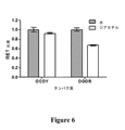


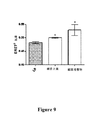


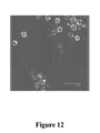

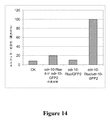

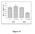
Classifications
-
- G—PHYSICS
- G01—MEASURING; TESTING
- G01N—INVESTIGATING OR ANALYSING MATERIALS BY DETERMINING THEIR CHEMICAL OR PHYSICAL PROPERTIES
- G01N33/00—Investigating or analysing materials by specific methods not covered by groups G01N1/00 - G01N31/00
- G01N33/48—Biological material, e.g. blood, urine; Haemocytometers
- G01N33/50—Chemical analysis of biological material, e.g. blood, urine; Testing involving biospecific ligand binding methods; Immunological testing
- G01N33/53—Immunoassay; Biospecific binding assay; Materials therefor
- G01N33/536—Immunoassay; Biospecific binding assay; Materials therefor with immune complex formed in liquid phase
- G01N33/542—Immunoassay; Biospecific binding assay; Materials therefor with immune complex formed in liquid phase with steric inhibition or signal modification, e.g. fluorescent quenching
-
- C—CHEMISTRY; METALLURGY
- C12—BIOCHEMISTRY; BEER; SPIRITS; WINE; VINEGAR; MICROBIOLOGY; ENZYMOLOGY; MUTATION OR GENETIC ENGINEERING
- C12N—MICROORGANISMS OR ENZYMES; COMPOSITIONS THEREOF; PROPAGATING, PRESERVING, OR MAINTAINING MICROORGANISMS; MUTATION OR GENETIC ENGINEERING; CULTURE MEDIA
- C12N15/00—Mutation or genetic engineering; DNA or RNA concerning genetic engineering, vectors, e.g. plasmids, or their isolation, preparation or purification; Use of hosts therefor
- C12N15/09—Recombinant DNA-technology
- C12N15/63—Introduction of foreign genetic material using vectors; Vectors; Use of hosts therefor; Regulation of expression
- C12N15/79—Vectors or expression systems specially adapted for eukaryotic hosts
- C12N15/80—Vectors or expression systems specially adapted for eukaryotic hosts for fungi
- C12N15/81—Vectors or expression systems specially adapted for eukaryotic hosts for fungi for yeasts
-
- G—PHYSICS
- G01—MEASURING; TESTING
- G01N—INVESTIGATING OR ANALYSING MATERIALS BY DETERMINING THEIR CHEMICAL OR PHYSICAL PROPERTIES
- G01N33/00—Investigating or analysing materials by specific methods not covered by groups G01N1/00 - G01N31/00
- G01N33/48—Biological material, e.g. blood, urine; Haemocytometers
- G01N33/50—Chemical analysis of biological material, e.g. blood, urine; Testing involving biospecific ligand binding methods; Immunological testing
- G01N33/68—Chemical analysis of biological material, e.g. blood, urine; Testing involving biospecific ligand binding methods; Immunological testing involving proteins, peptides or amino acids
-
- G—PHYSICS
- G01—MEASURING; TESTING
- G01N—INVESTIGATING OR ANALYSING MATERIALS BY DETERMINING THEIR CHEMICAL OR PHYSICAL PROPERTIES
- G01N2333/00—Assays involving biological materials from specific organisms or of a specific nature
- G01N2333/435—Assays involving biological materials from specific organisms or of a specific nature from animals; from humans
- G01N2333/705—Assays involving receptors, cell surface antigens or cell surface determinants
- G01N2333/72—Assays involving receptors, cell surface antigens or cell surface determinants for hormones
- G01N2333/726—G protein coupled receptor, e.g. TSHR-thyrotropin-receptor, LH/hCG receptor, FSH
Description
本発明は、試料中の化合物を検出するための方法およびポリペプチドに関する。詳細には、本発明は、細胞中で発現したときにGタンパク質共役受容体またはそのサブユニットのN末端が細胞の外側にあり、C末端が細胞の内側にある、脂質二重層中に埋め込まれ、化合物と結合することができる少なくとも1つのGタンパク質共役受容体を含む無細胞組成物の使用に関する。任意選択で、組成物は、Gタンパク質共役受容体の細胞内ループおよび/またはC末端と直接または間接的に結合する少なくとも1つのアクセサリー分子も含む。Gタンパク質共役受容体および/または存在する場合アクセサリー分子は、生物発光タンパク質およびアクセプター分子を一緒に含み、生物発光タンパク質およびアクセプター分子によって、生物発光共鳴エネルギー移動(BRET)を使用して受容体に結合する化合物を検出することができる。 The present invention relates to methods and polypeptides for detecting compounds in a sample. In particular, the present invention is embedded in a lipid bilayer when expressed in a cell where the N-terminus of the G protein coupled receptor or its subunit is outside the cell and the C-terminus is inside the cell. The use of a cell-free composition comprising at least one G protein coupled receptor capable of binding to a compound. Optionally, the composition also includes at least one accessory molecule that binds directly or indirectly to the intracellular loop and / or C-terminus of the G protein coupled receptor. G protein-coupled receptors and / or accessory molecules, if present, together comprise a bioluminescent protein and an acceptor molecule that binds to the receptor using bioluminescent resonance energy transfer (BRET) by the bioluminescent protein and acceptor molecule The compound to be detected can be detected.
Gタンパク質共役受容体(GPCR)は、細胞の外側で分子を感知し、内側でシグナル伝達経路を、最終的に細胞応答を活性化する膜貫通受容体のファミリーである。GPCRは、酵母および動物を含む真核生物でしか発見されていない。GPCRと結合しそれを活性化するリガンドには、光感受性化合物、匂い、フェロモン、ホルモン、および神経伝達物質があり、小分子からペプチド、大分子タンパク質までサイズが様々である。Gタンパク質共役受容体は多数の疾患に関与し、治療用薬物の標的でもある。 G protein-coupled receptors (GPCRs) are a family of transmembrane receptors that sense molecules outside the cell, activate signaling pathways inside, and ultimately cellular responses. GPCRs have only been found in eukaryotes including yeast and animals. Ligands that bind to and activate GPCRs include photosensitive compounds, odors, pheromones, hormones, and neurotransmitters, ranging in size from small molecules to peptides and large proteins. G protein-coupled receptors are involved in a number of diseases and are also targets for therapeutic drugs.
脊椎動物およびC.elegansの嗅覚受容体(OR)は、Gタンパク質共役受容体(GPCR)ファミリーのメンバーである(Buckら、1991;Troemelら、1995)。GPCRは、その7つの膜貫通ドメインを特徴とし、リガンド結合ドメインが膜の細胞外側にあり、Gタンパク質結合ドメインが細胞内側にあると推測される。受容体がリガンドと結合したとき、高次構造変化が受容体中に起こり、ヘテロ三量体Gタンパク質を活性化することを可能にする(Kobilkaら、2007)。次いで、活性化したGタンパク質は、グアニルシクラーゼやホスホリパーゼC経路などのシグナル伝達経路を活性化することができ、シグナルをより高位の処理中枢に伝達する(Gaillardら、2004)。 Vertebrates and C.I. The elegans olfactory receptor (OR) is a member of the G protein coupled receptor (GPCR) family (Buck et al., 1991; Troemel et al., 1995). A GPCR is characterized by its seven transmembrane domains, with the ligand binding domain presumed to be outside the membrane and the G protein binding domain inside the cell. When the receptor binds to the ligand, conformational changes occur in the receptor, allowing the heterotrimeric G protein to be activated (Kobilka et al., 2007). The activated G protein can then activate signal transduction pathways such as the guanyl cyclase and phospholipase C pathways and transmit signals to higher processing centers (Gaillard et al., 2004).
Forster共鳴エネルギー移動、または単に共鳴エネルギー移動(RET)とは、励起状態のドナー分子から基底状態のアクセプター分子へのエネルギーの非放射性移動である(Ghanouniら、2001)。エネルギー移動効率は、ドナーとアクセプターとの距離、スペクトルの重複の程度およびアクセプターとドナーの双極子の相対的配向に依存的である。分子内RETがGPCR活性化のモニタリングに使用されていた以前の場合では、蛍光ドナーおよびアクセプターといった、蛍光共鳴エネルギー移動(FRET)と呼ばれる方法を使用している。ほとんどの場合、蛍光ドナーとアクセプターはどちらも、Aequoria victoria由来の緑色蛍光タンパク質(GFP)の工学的変異体である(Tsien、1998)。最も広く使用されるFRETの対は、ドナーであるシアン蛍光タンパク質(CFP)とアクセプターである黄色蛍光タンパク質(YFP)であり(PistonおよびKremers、2007)、このFRET系は、いくつかのGPCRによる直接的なリガンド結合を定量するのに以前から使用されている(Lohseら、2003および2007;Vilardagaら、2003;Rochaisら、2007;Lisenbeeら、2007)。 Forster resonance energy transfer, or simply resonance energy transfer (RET), is a non-radiative transfer of energy from an excited state donor molecule to a ground state acceptor molecule (Ghanoni et al., 2001). The energy transfer efficiency depends on the distance between the donor and the acceptor, the degree of spectral overlap, and the relative orientation of the acceptor and donor dipoles. In previous cases where intramolecular RET was used to monitor GPCR activation, a method called fluorescence resonance energy transfer (FRET), such as fluorescent donors and acceptors, is used. In most cases, both fluorescent donors and acceptors are engineered variants of green fluorescent protein (GFP) from Aequoria victoria (Tsien, 1998). The most widely used pair of FRET is the donor cyan fluorescent protein (CFP) and the acceptor yellow fluorescent protein (YFP) (Piston and Kremers, 2007), and this FRET system is directly linked by several GPCRs Have been used previously to quantify specific ligand binding (Lohse et al., 2003 and 2007; Villadaga et al., 2003; Rochais et al., 2007; Lisenbee et al., 2007).
受容体の活性化をモニタリングする1つの方法では、単一のGPCRを、それぞれ3番目の細胞内ループおよびC末端内の挿入部位において、CFPおよびYFPで二重標識する。436nmの光でCFPを励起すると、480nmでのCFPの放出、および535nmで放出するYFPのFRETを引き起こす。FRETの効率は、ドナーとアクセプターとの距離の6乗によって変化し、このことから、GPCRにおける高次構造変化が極めて感度よく示される。このことは、α2AR、副甲状腺ホルモン受容体(PTHR)、β1−ARおよびセクレチン受容体を用いて、インタクトな細胞において実証された。アゴニストであるノルアドレナリンとα2AR(Lohseら、2003)、副甲状腺ホルモンとPTHR(Vilardagaら、2003)、ノルエピネフリンとβ1−AR(Rochaisら、2007)、およびセクレチンとセクレチン受容体(Lisenbeeら、2007)の相互作用はCFPとYFPとの距離を変化させ、それによってFRETシグナルの変化が生じた。 In one method of monitoring receptor activation, a single GPCR is double labeled with CFP and YFP at the third intracellular loop and insertion site in the C-terminus, respectively. Excitation of CFP with 436 nm light causes CFP emission at 480 nm and YFP FRET emission at 535 nm. The efficiency of FRET varies depending on the sixth power of the distance between the donor and the acceptor, and this indicates a highly conformational change in GPCR with extremely high sensitivity. This was demonstrated in intact cells using α2AR, parathyroid hormone receptor (PTHR), β1-AR and secretin receptor. The agonists noradrenaline and α2AR (Lohse et al., 2003), parathyroid hormone and PTHR (Vilardaga et al., 2003), norepinephrine and β1-AR (Rochais et al., 2007), and secretin and secretin receptor (Lisenbee et al., 2007). The interaction changed the distance between CFP and YFP, which caused a change in the FRET signal.
FRET系においてYFPアクセプターをフルオレセインヒ素ヘアピン結合体であるFlAsHと交換すると(Hoffmanら、2005;Nakanishiら、2006)、α2アドレナリン作動性受容体活性化のモニタリングに使用したとき、アゴニスト誘導FRETシグナルがCFP/YFP系と比較して5倍増大した(Nakanishiら、2006)。しかし、FlAsHは、困難な標識化および洗浄手順を伴い、広い調査集団での使用が限定されている。CFP/YFP系は、依然として、分子内の高次構造変化をモニタリングするのに最も頻繁に報告されているFRET系である。FRETに付随する主な不利点は、ドナー蛍光体にエネルギーを与えるのに光源が必要となることである(PistonおよびKremers、2007)。これは、ドナーの励起波長でアクセプターの望ましくない直接励起を引き起こす(「クロストーク」と呼ばれる問題)。 Replacing the YFP acceptor with FlAsH, a fluorescein arsenic hairpin conjugate in the FRET system (Hoffman et al., 2005; Nakanishi et al., 2006), when used to monitor α2 adrenergic receptor activation, the agonist-induced FRET signal is CFP There was a 5-fold increase compared to the / YFP system (Nakanishi et al., 2006). However, FlAsH is associated with difficult labeling and washing procedures and has limited use in a large research population. The CFP / YFP system is still the most frequently reported FRET system for monitoring intramolecular conformational changes. The main disadvantage associated with FRET is that a light source is required to energize the donor phosphor (Piston and Kremers, 2007). This causes undesired direct excitation of the acceptor at the donor excitation wavelength (a problem called “crosstalk”).
生物発光共鳴エネルギー移動(BRET)では、FRETのドナー蛍光体をルシフェラーゼと交換し、アクセプターは任意の適切な蛍光体でよい。基質を添加すると生物発光放出、したがってRETが開始されるので、ルシフェラーゼを使用すると、照明の必要が回避される。BRETの2つの一般的な実行形態は、GFP突然変異体のYFP(BRET1;λem=約530nm)またはGFP2(BRET2;λem=約510nm)のどちらかと結合した、ドナー系であるセレンテラジンh(BRET1;λem=約475nm)またはセレンテラジン400a(Clz400a)基質(BRET2;λem=約395nm)を有するウミシイタケ(Renilla)ルシフェラーゼ(RLuc)を含む。BRET2系は、量子収量を消費して、BRET1系の約55nmと比較して約115nmという、ドナーとアクセプターの放出ピーク間の優れたスペクトル分離をもたらす(PflegerおよびEidneら、2006)。 In bioluminescence resonance energy transfer (BRET), the FRET donor fluorophore is exchanged for luciferase and the acceptor can be any suitable fluorophore. The use of luciferase avoids the need for illumination, since the addition of a substrate initiates bioluminescence emission and thus RET. Two common implementations of BRET are donor systems bound to either the GFP mutant YFP (BRET 1 ; λ em = about 530 nm) or GFP 2 (BRET 2 ; λ em = about 510 nm) Renilla luciferase (RLuc) with coelenterazine h (BRET 1 ; λ em = about 475 nm) or coelenterazine 400a (Clz400a) substrate (BRET 2 ; λ em = about 395 nm). The BRET 2 system consumes quantum yields and results in excellent spectral separation between the donor and acceptor emission peaks of about 115 nm compared to about 55 nm of the BRET 1 system (Pfleger and Eidne et al., 2006).
嗅覚受容体でのFRETは、クラスA(a2−アドレナリン作動性および副甲状腺ホルモン、(Vilardagaら、2003))およびクラスB(セクレチン、(Lisenbeeら、2007))GPCRでしか以前には実証されていない。GPCRクラスAに属する哺乳動物ORと異なり、線虫OR(Robertson、1998および2001)は、これらのクラスのどちらにも属さず、進化的および構造的に異なる。さらに、GPCRスーパーファミリーのクラスA内にある、哺乳動物ORを含めた全てのORは、その発現の点では非定型的である。一般に、これらのタンパク質は、化学感覚系列に由来するニューロン以外では機能的に発現できない。哺乳動物および線虫ORの適切な発現にその存在が必要となるいくつかのアクセサリータンパク質が同定されている。 FRET at olfactory receptors has only been previously demonstrated in class A (a 2 -adrenergic and parathyroid hormone, (Vilardaga et al., 2003)) and class B (secretin, (Lisenbee et al., 2007)) GPCRs. Not. Unlike mammalian OR belonging to GPCR class A, nematode OR (Robertson, 1998 and 2001) does not belong to either of these classes and is evolutionary and structurally different. Furthermore, all OR, including mammalian OR, within class A of the GPCR superfamily are atypical in terms of their expression. In general, these proteins cannot be expressed functionally except in neurons derived from the chemosensory lineage. Several accessory proteins have been identified that are required for their proper expression in mammalian and nematode OR.
したがって、例えば、バイオセンサーで使用するGタンパク質共役受容体と結合する化合物の検出を可能にする適切に感度のよい方法および分子が必要である。 Thus, there is a need for appropriately sensitive methods and molecules that allow, for example, detection of compounds that bind to G protein coupled receptors for use in biosensors.
本発明者らは、驚くべきことに、嗅覚受容体(OR)など、キメラのGタンパク質共役受容体(GPCR)を使用して標的化合物を検出する無細胞の生物発光共鳴エネルギー移動(BRET)が、全細胞を使用したBRETやFRETなど他の検出方法より感度がよいことを見出した。 The inventors have surprisingly found cell-free bioluminescence resonance energy transfer (BRET) to detect target compounds using chimeric G protein-coupled receptors (GPCR), such as olfactory receptors (OR). The present inventors have found that the sensitivity is better than other detection methods such as BRET and FRET using whole cells.
したがって、第1の態様では、本発明は、化合物を検出する方法であって、前記方法が、
i)試料を、
a)脂質二重層中に埋め込まれ、化合物と結合することができる少なくとも1つのGタンパク質共役受容体と、
b)任意選択で、Gタンパク質共役受容体の細胞内ループおよび/またはC末端と直接または間接的に結合する少なくとも1つのアクセサリー分子と
を含む無細胞組成物と接触させるステップであって、
Gタンパク質共役受容体が、同じまたは異なる1つまたは複数のサブユニットを含み、Gタンパク質共役受容体および/または存在する場合アクセサリー分子が、生物発光タンパク質およびアクセプター分子を一緒に含むステップと、
ii)ステップi)と同時にまたはそれに連続して、生物発光タンパク質の基質を提供し、生物発光タンパク質が基質を修飾することを可能にするステップと、
iii)ステップii)が生物発光タンパク質とアクセプター分子の間の生物発光共鳴エネルギー移動(BRET)を変動(modulate)させるかどうかを決定するステップと
を含み、化合物がGタンパク質共役受容体と結合したときにアクセプター分子に対する生物発光タンパク質の空間的位置および/または双極子配向が変化し、細胞中で発現したときにGタンパク質共役受容体またはそのサブユニットのN末端が細胞の外側にあり、C末端が細胞の内側にある方法を提供する。
Accordingly, in a first aspect, the present invention provides a method for detecting a compound comprising:
i) The sample
a) at least one G protein-coupled receptor embedded in the lipid bilayer and capable of binding to the compound;
b) optionally contacting with a cell-free composition comprising at least one accessory molecule that binds directly or indirectly to the intracellular loop and / or C-terminus of the G protein coupled receptor,
The G protein coupled receptor comprises one or more subunits, the same or different, and the G protein coupled receptor and / or accessory molecule, if present, comprises the bioluminescent protein and the acceptor molecule together;
ii) providing a substrate for the bioluminescent protein simultaneously or sequentially with step i), allowing the bioluminescent protein to modify the substrate;
and iii) determining whether to modulate bioluminescence resonance energy transfer (BRET) between the bioluminescent protein and the acceptor molecule, wherein the compound binds to a G protein coupled receptor The spatial position and / or dipole orientation of the bioluminescent protein relative to the acceptor molecule changes such that when expressed in the cell, the N-terminus of the G protein-coupled receptor or its subunit is outside the cell and the C-terminus is Provide a method that is inside a cell.
一実施形態では、化合物と結合することができるGタンパク質共役受容体のサブユニットは、
i)生物発光タンパク質と、
ii)アクセプター分子と
を含み、化合物がサブユニットと結合したときにアクセプター分子に対する生物発光タンパク質の空間的位置および/または双極子配向が変化する。例えば、サブユニットは、
i)配列番号13、15、52もしくは54として示されるアミノ酸配列、または
ii)配列番号13、15、52もしくは54のいずれか1つもしくは複数と少なくとも40%同一であるアミノ酸配列
を含んでよい。
In one embodiment, the subunit of the G protein coupled receptor that can bind to the compound is:
i) a bioluminescent protein;
ii) the acceptor molecule and the spatial position and / or dipole orientation of the bioluminescent protein relative to the acceptor molecule changes when the compound is bound to the subunit. For example, the subunit is
It may comprise i) an amino acid sequence shown as SEQ ID NO: 13, 15, 52 or 54, or ii) an amino acid sequence that is at least 40% identical to any one or more of
代替の実施形態では、Gタンパク質共役受容体は、
i)a)生物発光タンパク質と、
b)アクセプター分子と
を含む第1のサブユニットと、
ii)化合物と結合することができる第2のサブユニットと
を含み、化合物が第2のサブユニットと結合したときにアクセプター分子に対する生物発光タンパク質の空間的位置および/または双極子配向が変化する。
In an alternative embodiment, the G protein coupled receptor is
i) a) a bioluminescent protein;
b) a first subunit comprising an acceptor molecule;
ii) comprising a second subunit capable of binding to the compound, wherein the spatial location and / or dipole orientation of the bioluminescent protein relative to the acceptor molecule changes when the compound is bound to the second subunit.
さらなる実施形態では、
i)生物発光タンパク質がサブユニットの3番目の非膜貫通ループの一部を形成し、アクセプター分子が5番目の非膜貫通ループの一部を形成し、または
ii)アクセプター分子がサブユニットの3番目の非膜貫通ループの一部を形成し、生物発光タンパク質が5番目の非膜貫通ループの一部を形成し、または
iii)生物発光タンパク質がサブユニットの1番目の非膜貫通ループの一部を形成し、アクセプター分子が3番目の非膜貫通ループの一部を形成し、または
iv)アクセプター分子がサブユニットの1番目の非膜貫通ループの一部を形成し、生物発光タンパク質が3番目の非膜貫通ループの一部を形成し、または
v)生物発光タンパク質がサブユニットの5番目の非膜貫通ループの一部を形成し、アクセプター分子がC末端の一部を形成し、または
vi)アクセプター分子がサブユニットの5番目の非膜貫通ループの一部を形成し、生物発光タンパク質がC末端の一部を形成する。
In a further embodiment,
i) the bioluminescent protein forms part of the third non-transmembrane loop of the subunit, and the acceptor molecule forms part of the fifth non-transmembrane loop, or ii) the acceptor molecule forms part of the subunit 3 Forming part of the first non-transmembrane loop, the bioluminescent protein forming part of the fifth non-transmembrane loop, or iii) one of the first non-transmembrane loops of the subunit The acceptor molecule forms part of the third non-transmembrane loop, or iv) the acceptor molecule forms part of the first non-transmembrane loop of the subunit, and the bioluminescent protein is 3 Form part of the first non-transmembrane loop, or v) the bioluminescent protein forms part of the fifth non-transmembrane loop of the subunit, and the acceptor molecule is part of the C-terminus Formed, or vi) an acceptor molecule forms part of a fifth non-transmembrane loop of subunits, bioluminescent protein forms part of a C-terminal.
さらに別の代替の実施形態では、Gタンパク質共役受容体は、
i)生物発光タンパク質を含む第1のサブユニットと、
ii)アクセプター分子を含む第2のサブユニットと
を含む。例えば、第1または第2のサブユニットは、
i)配列番号17もしくは19として示されるアミノ酸配列、または
ii)配列番号17および/もしくは19と少なくとも40%同一であるアミノ酸配列
を含んでよい。
In yet another alternative embodiment, the G protein coupled receptor is
i) a first subunit comprising a bioluminescent protein;
ii) a second subunit comprising an acceptor molecule. For example, the first or second subunit is
It may comprise i) the amino acid sequence shown as SEQ ID NO: 17 or 19, or ii) an amino acid sequence that is at least 40% identical to SEQ ID NO: 17 and / or 19.
一実施形態では、
i)生物発光タンパク質が第1のサブユニットの3番目の非膜貫通ループの一部を形成し、アクセプター分子が第2のサブユニットの5番目の非膜貫通ループの一部を形成し、または
ii)アクセプター分子が第1のサブユニットの3番目の非膜貫通ループの一部を形成し、生物発光タンパク質が第2のサブユニットの5番目の非膜貫通ループの一部を形成し、または
iii)生物発光タンパク質が第1のサブユニットの1番目の非膜貫通ループの一部を形成し、アクセプター分子が第2のサブユニットの3番目の非膜貫通ループの一部を形成し、または
iv)アクセプター分子が第1のサブユニットの1番目の非膜貫通ループの一部を形成し、生物発光タンパク質が第2のサブユニットの3番目の非膜貫通ループの一部を形成し、または
v)生物発光タンパク質が第1のサブユニットの5番目の非膜貫通ループの一部を形成し、アクセプター分子が第2のサブユニットのC末端の一部を形成し、または
vi)アクセプター分子が第1のサブユニットの5番目の非膜貫通ループの一部を形成し、生物発光タンパク質が第2のサブユニットのC末端の一部を形成する。
In one embodiment,
i) the bioluminescent protein forms part of the third non-transmembrane loop of the first subunit, and the acceptor molecule forms part of the fifth non-transmembrane loop of the second subunit, or ii) the acceptor molecule forms part of the third non-transmembrane loop of the first subunit and the bioluminescent protein forms part of the fifth non-transmembrane loop of the second subunit, or iii) the bioluminescent protein forms part of the first non-transmembrane loop of the first subunit and the acceptor molecule forms part of the third non-transmembrane loop of the second subunit, or iv) the acceptor molecule forms part of the first non-transmembrane loop of the first subunit and the bioluminescent protein forms part of the third non-transmembrane loop of the second subunit, or v The bioluminescent protein forms part of the fifth non-transmembrane loop of the first subunit and the acceptor molecule forms part of the C-terminus of the second subunit, or vi) the acceptor molecule is first Forming a part of the fifth non-transmembrane loop of the subunit, and the bioluminescent protein forming part of the C-terminus of the second subunit.
さらなる代替の実施形態では、
i)Gタンパク質共役受容体が生物発光タンパク質を含むサブユニットを含み、アクセサリー分子がアクセプター分子を含み、または
ii)Gタンパク質共役受容体がアクセプター分子を含むサブユニットを含み、アクセサリー分子が生物発光タンパク質を含む。
In a further alternative embodiment,
i) the G protein coupled receptor comprises a subunit comprising a bioluminescent protein and the accessory molecule comprises an acceptor molecule, or ii) the G protein coupled receptor comprises a subunit comprising an acceptor molecule, and the accessory molecule comprises a bioluminescent protein including.
別の代替の実施形態では、アクセサリー分子は、
i)生物発光タンパク質と、
ii)アクセプター分子と
を含む。
In another alternative embodiment, the accessory molecule is
i) a bioluminescent protein;
ii) an acceptor molecule.
さらなる代替の実施形態では、組成物は、少なくとも2つのアクセサリー分子を含み、第1のアクセサリー分子は生物発光タンパク質を含み、第2のアクセサリー分子はアクセプター分子を含む。 In a further alternative embodiment, the composition includes at least two accessory molecules, the first accessory molecule includes a bioluminescent protein, and the second accessory molecule includes an acceptor molecule.
本発明者らは、FRET−無細胞およびBRET−全細胞検出系と比較したときに本発明がいかに優れているかを発見して特に驚いた。したがって、好ましい実施形態では、本発明の方法は、非生物発光タンパク質をドナー分子として使用し、蛍光共鳴エネルギー移動(FRET)の変動を決定する場合より少なくとも2倍、3倍または4倍感度がよい。さらなる好ましい実施形態では、本発明の方法は、この方法で無細胞組成物中にあるものの代わりにインタクトな細胞中に存在する同じGタンパク質共役受容体を使用する場合より少なくとも10倍、20倍、30倍または40倍強いBRETシグナルをもたらす。 The inventors were particularly surprised to discover how superior the present invention is when compared to FRET-cell-free and BRET-whole cell detection systems. Thus, in a preferred embodiment, the method of the invention uses a non-bioluminescent protein as a donor molecule and is at least 2-fold, 3-fold or 4-fold more sensitive than when determining fluorescence resonance energy transfer (FRET) variations. . In a further preferred embodiment, the method of the invention is at least 10-fold, 20-fold, when using the same G protein coupled receptor present in intact cells instead of in the cell-free composition in this method, It produces a BRET signal that is 30 or 40 times stronger.
クラスAのGPCR、一般にOR、特に線虫ORの非定型的性質を考慮すると、BRETドナーおよびアクセプタータグとともにクラスAのGPCRを含むキメラが、a)構造的完全性を保持し、b)匂い物質による受容体活性化を光学的シグナルに変換することができることは驚くべきことであった。したがって、好ましい実施形態では、Gタンパク質共役受容体はクラスAのGPCRである。さらなる好ましい実施形態では、クラスAのGPCRは嗅覚受容体である。細胞中で発現したときに受容体のN末端が細胞の外側にあり、C末端が細胞の内側にある限り、嗅覚受容体はどんな供給源由来のものでもよい。例として、それだけに限らないが、脊索動物受容体、線虫受容体、またはそのいずれか1つの生物学的に活性な変異体もしくは断片がある。脊索動物受容体の例として、それだけに限らないが、哺乳動物受容体、鳥類受容体および魚類受容体がある。好ましい実施形態では、嗅覚受容体は、線虫受容体、またはその生物学的に活性な変異体もしくは断片である。一実施形態では、線虫受容体は、Caenorhabditis elegans受容体、またはその生物学的に活性な変異体もしくは断片である。 Considering the atypical nature of class A GPCRs, generally OR, especially nematode OR, chimeras containing class A GPCRs with BRET donor and acceptor tags a) retain structural integrity and b) odor It was surprising that receptor activation by substances can be converted into an optical signal. Thus, in a preferred embodiment, the G protein coupled receptor is a class A GPCR. In a further preferred embodiment, the class A GPCR is an olfactory receptor. The olfactory receptor can be from any source as long as the N-terminus of the receptor is outside the cell and the C-terminus is inside the cell when expressed in the cell. Examples include, but are not limited to, a notochord receptor, a nematode receptor, or a biologically active variant or fragment of any one thereof. Examples of chordal receptors include, but are not limited to, mammalian receptors, avian receptors and fish receptors. In a preferred embodiment, the olfactory receptor is a nematode receptor, or a biologically active variant or fragment thereof. In one embodiment, the nematode receptor is a Caenorhabditis elegans receptor, or a biologically active variant or fragment thereof.
一実施形態では、嗅覚受容体は、
i)配列番号1〜6のいずれか1つで示されるアミノ酸配列と、
ii)配列番号1〜6のいずれか1つまたは複数と少なくとも40%同一であるアミノ酸配列と
を含む。
In one embodiment, the olfactory receptor is
i) the amino acid sequence represented by any one of SEQ ID NOS: 1-6;
ii) comprising an amino acid sequence that is at least 40% identical to any one or more of SEQ ID NOs: 1-6.
別の実施形態では、サブユニットは、2つ以上の異なるGタンパク質共役受容体サブユニットの部分のキメラである。 In another embodiment, the subunit is a chimera of portions of two or more different G protein coupled receptor subunits.
アクセサリー分子は、Gタンパク質共役受容体と直接または間接的に結合する任意の分子でよい。例として、それだけに限らないが、Gタンパク質およびアレスチンがある。 The accessory molecule can be any molecule that binds directly or indirectly to the G protein coupled receptor. Examples include, but are not limited to, G protein and arrestin.
BRETの変化の幅に対して化合物の濃度を変化させる効果は、その応答が用量依存的であることを最初に示した。したがって、好ましい実施形態では、BRETのレベルは、試料中の化合物の相対量を示す。 The effect of changing the concentration of the compound on the breadth of change in BRET initially showed that the response was dose dependent. Thus, in a preferred embodiment, the level of BRET indicates the relative amount of compound in the sample.
生物発光タンパク質の例として、それだけに限らないが、ルシフェラーゼ、β−ガラクトシダーゼ、ラクタマーゼ(lactamase)、西洋ワサビペルオキシダーゼ、アルカリホスファターゼ、β−グルクロニダーゼまたはβ−グルコシダーゼがある。 Examples of bioluminescent proteins include, but are not limited to, luciferase, β-galactosidase, lactamase, horseradish peroxidase, alkaline phosphatase, β-glucuronidase or β-glucosidase.
ルシフェラーゼの例として、それだけに限らないが、ウミシイタケ(Renilla)ルシフェラーゼ、ホタル(Firefly)ルシフェラーゼ、腔腸動物(Coelenterate)ルシフェラーゼ、北アメリカツチボタル(North America glow worm)ルシフェラーゼ、コメツキム(click beetle)シルシフェラーゼ、鉄道虫(railroad worm)ルシフェラーゼ、細菌(bacterial)ルシフェラーゼ、ガウシア(Gaussia)ルシフェラーゼ、エクオリン、ヒカリキノコバエ(Arachnocampa)ルシフェラーゼ、またはそのいずれか1つの生物学的に活性な変異体もしくは断片、またはその2つ以上のキメラがある。 Examples of luciferases include, but are not limited to, Renilla luciferase, Firefly luciferase, Coelenterate luciferase, North America glow worm luciferase, click beetle luciferase, railroad Railroad worm luciferase, bacterial luciferase, Gaussia luciferase, aequorin, Archnocampa luciferase, or any one biologically active variant or fragment thereof, or two or more thereof There is a chimera.
基質の例として、それだけに限らないが、甲虫(beetle)ルシフェリン、他のルシフェリン、セレンテラジン、またはセレンテラジンの誘導体がある。また、エクオリンなどの一部の発光タンパク質の場合、基質は、カルシウムイオンなどの補助因子でもよい。 Examples of substrates include, but are not limited to, beetle luciferin, other luciferins, coelenterazine, or derivatives of coelenterazine. In the case of some photoproteins such as aequorin, the substrate may be a cofactor such as calcium ion.
一実施形態では、アクセプター分子はタンパク質であり、その例として、それだけに限らないが、緑色蛍光タンパク質(GFP)、GFPの青色蛍光変異体(BFP)、GFPのシアン蛍光変異体(CFP)、GFPの黄色蛍光変異体(YFP)、増強型GFP(EGFP)、増強型CFP(ECFP)、増強型YFP(EYFP)、GFPS65T、エメラルド(Emerald)、トパーズ(Topaz)、GFPuv、不安定化EGFP(dEGFP)、不安定化ECFP(dECFP)、不安定化EYFP(dEYFP)、HcRed、t−HcRed、DsRed、DsRed2、t−ダイマー2、t−ダイマー2(12)、mRFP1、ポシロポリン(pocilloporin)、ウミシイタケ(Renilla)GFP、モンスター(Monster) GFP、paGFP、カエデ(Kaede)タンパク質もしくはフィコビリタンパク質(Phycobiliprotein)、またはそのいずれか1つの生物学的に活性な変異体もしくは断片がある。 In one embodiment, the acceptor molecule is a protein, such as, but not limited to, green fluorescent protein (GFP), blue fluorescent variant of GFP (BFP), cyan fluorescent variant of GFP (CFP), GFP Yellow fluorescent mutant (YFP), enhanced GFP (EGFP), enhanced CFP (ECFP), enhanced YFP (EYFP), GFPS65T, emerald (Emerald), topaz (GFP), destabilized EGFP (dEGFP) , Destabilized ECFP (dECFP), destabilized EYFP (dEYFP), HcRed, t-HcRed, DsRed, DsRed2, t-dimer2, t-dimer2 (12), mRFP1, pocilloporin, Renilla ) GFP, Monster (Mon ter) GFP, paGFP, there is maple (Kaede) protein or phycobiliprotein (Phycobiliprotein), or any one of a biologically active variant or fragment thereof.
代替の実施形態では、アクセプター分子は非タンパク質であり、その例として、それだけに限らないが、Alexa Fluor色素、Bodipy色素、Cy色素、フルオレセイン、ダンシル、ウンベリフェロン、蛍光マイクロスフェア、発光マイクロスフェア、蛍光ナノ結晶、マリナブルー、カスケードブルー、カスケードイエロー、パシフィックブルー、オレゴングリーン、テトラメチルローダミン、ローダミン、テキサスレッド、希土類元素キレート、またはその任意の組合せもしくは誘導体がある。 In alternative embodiments, the acceptor molecule is a non-protein, such as, but not limited to, Alexa Fluor dye, Bodipy dye, Cy dye, fluorescein, dansyl, umbelliferone, fluorescent microsphere, luminescent microsphere, fluorescent There are nanocrystals, Marina Blue, Cascade Blue, Cascade Yellow, Pacific Blue, Oregon Green, Tetramethylrhodamine, Rhodamine, Texas Red, rare earth chelates, or any combination or derivative thereof.
別の実施形態では、前記方法は、ステップi)もしくはステップii)と同時にまたはそれに連続して、生物発光タンパク質の補助因子を提供するステップをさらに含む。例として、それだけに限らないが、ATP、マグネシウム、酸素、FMNH2、カルシウム、またはそのいずれか2つ以上の組合せがある。 In another embodiment, the method further comprises providing a bioluminescent protein cofactor simultaneously with or sequentially with step i) or step ii). Examples include, but are not limited to, ATP, magnesium, oxygen, FMNH 2 , calcium, or any combination of two or more thereof.
一実施形態では、無細胞組成物は、組換え細胞中でGタンパク質共役受容体を産生し、細胞の膜を破壊することによって得られた。 In one embodiment, the cell-free composition was obtained by producing a G protein coupled receptor in a recombinant cell and disrupting the membrane of the cell.
一実施形態では、組換え細胞は、Gタンパク質共役受容体と結合する非内因性タンパク質を産生しない。 In one embodiment, the recombinant cell does not produce a non-endogenous protein that binds to a G protein coupled receptor.
無細胞組成物を得ることができる細胞は、Gタンパク質共役受容体を発現し、この受容体をその細胞膜に取り込むことができる任意の細胞型である。特に好ましい実施形態では、組換え細胞は酵母細胞である。 The cell from which the cell-free composition can be obtained is any cell type that expresses a G protein-coupled receptor and can incorporate this receptor into its cell membrane. In a particularly preferred embodiment, the recombinant cell is a yeast cell.
代替の実施形態では、Gタンパク質共役受容体は、リポソームの脂質二重層中に埋め込まれる。 In an alternative embodiment, the G protein coupled receptor is embedded in the lipid bilayer of the liposome.
さらなる実施形態では、前記方法は、マイクロ流体工学を使用して行われる。 In a further embodiment, the method is performed using microfluidics.
別の態様では、本発明は、化合物を検出するための精製および/または組換えポリペプチドであって、前記ポリペプチドが、
i)Gタンパク質共役受容体のサブユニットと、
ii)生物発光タンパク質および/またはアクセプター分子と
を含み、細胞中で発現したときにサブユニットのN末端が細胞の外側にあり、C末端が細胞の内側にある、ポリペプチドを提供する。
In another aspect, the invention provides a purified and / or recombinant polypeptide for detecting a compound, wherein the polypeptide comprises:
i) a subunit of a G protein coupled receptor;
ii) providing a polypeptide comprising a bioluminescent protein and / or acceptor molecule, wherein when expressed in a cell, the subunit N-terminus is outside the cell and the C-terminus is inside the cell.
一実施形態では、ポリペプチドは、1つまたは複数の異なるGタンパク質共役受容体サブユニット、および任意選択で1つまたは複数の異なるアクセサリー分子を含むGタンパク質共役受容体であるタンパク質複合体の一部を形成する。 In one embodiment, the polypeptide is part of a protein complex that is a G protein coupled receptor comprising one or more different G protein coupled receptor subunits, and optionally one or more different accessory molecules. Form.
一実施形態では、
i)生物発光タンパク質がサブユニットの3番目の非膜貫通ループの一部を形成し、アクセプター分子が5番目の非膜貫通ループの一部を形成し、または
ii)アクセプター分子がサブユニットの3番目の非膜貫通ループの一部を形成し、生物発光タンパク質が5番目の非膜貫通ループの一部を形成し、または
iii)生物発光タンパク質がサブユニットの1番目の非膜貫通ループの一部を形成し、アクセプター分子が3番目の非膜貫通ループの一部を形成し、または
iv)アクセプター分子がサブユニットの1番目の非膜貫通ループの一部を形成し、生物発光タンパク質が3番目の非膜貫通ループの一部を形成し、または
v)生物発光タンパク質がサブユニットの5番目の非膜貫通ループの一部を形成し、アクセプター分子がC末端の一部を形成し、または
vi)アクセプター分子がサブユニットの5番目の非膜貫通ループの一部を形成し、生物発光タンパク質がC末端の一部を形成し、または
vii)生物発光タンパク質もしくはアクセプター分子がサブユニットの1番目の非膜貫通ループの一部を形成し、または
viii)生物発光タンパク質もしくはアクセプター分子がサブユニットの3番目の非膜貫通ループの一部を形成し、または
ix)生物発光タンパク質もしくはアクセプター分子がサブユニットの5番目の非膜貫通ループの一部を形成し、または
x)生物発光タンパク質もしくはアクセプター分子がサブユニットのC末端の一部を形成する。
In one embodiment,
i) the bioluminescent protein forms part of the third non-transmembrane loop of the subunit, and the acceptor molecule forms part of the fifth non-transmembrane loop, or ii) the acceptor molecule forms part of the subunit 3 Forming part of the first non-transmembrane loop, the bioluminescent protein forming part of the fifth non-transmembrane loop, or iii) one of the first non-transmembrane loops of the subunit The acceptor molecule forms part of the third non-transmembrane loop, or iv) the acceptor molecule forms part of the first non-transmembrane loop of the subunit, and the bioluminescent protein is 3 Form part of the first non-transmembrane loop, or v) the bioluminescent protein forms part of the fifth non-transmembrane loop of the subunit, and the acceptor molecule is part of the C-terminus Or vi) the acceptor molecule forms part of the fifth non-transmembrane loop of the subunit, the bioluminescent protein forms part of the C-terminus, or vii) the bioluminescent protein or acceptor molecule Forms part of the first non-transmembrane loop of the unit, or viii) the bioluminescent protein or acceptor molecule forms part of the third non-transmembrane loop of the subunit, or ix) the bioluminescent protein or The acceptor molecule forms part of the subunit's fifth non-transmembrane loop, or x) the bioluminescent protein or acceptor molecule forms part of the subunit's C-terminus.
さらなる実施形態では、ポリペプチドは、生物発光タンパク質およびアクセプター分子を含み、化合物がポリペプチドと結合したときにアクセプター分子に対する生物発光タンパク質の空間的位置および/または双極子配向が変化する。 In further embodiments, the polypeptide comprises a bioluminescent protein and an acceptor molecule, wherein the spatial location and / or dipole orientation of the bioluminescent protein relative to the acceptor molecule changes when the compound is bound to the polypeptide.
代替の実施形態では、ポリペプチドはサブユニットおよび生物発光タンパク質を含み、ポリペプチドはアクセプター分子を含む第2のポリペプチドと直接または間接的に結合し、化合物がポリペプチドおよび/または第2のポリペプチドと結合したときにアクセプター分子に対する生物発光タンパク質の空間的位置および/または双極子配向が変化する。 In an alternative embodiment, the polypeptide comprises a subunit and a bioluminescent protein, the polypeptide binds directly or indirectly to a second polypeptide comprising an acceptor molecule, and the compound is a polypeptide and / or a second polypeptide. The spatial position and / or dipole orientation of the bioluminescent protein relative to the acceptor molecule changes when bound to the peptide.
さらなる代替の実施形態では、ポリペプチドはサブユニットおよびアクセプター分子を含み、ポリペプチドは生物発光タンパク質を含む第2のポリペプチドと直接または間接的に結合し、化合物がポリペプチドおよび/または第2のポリペプチドと結合したときにアクセプター分子に対する生物発光タンパク質の空間的位置および/または双極子配向が変化する。 In a further alternative embodiment, the polypeptide comprises a subunit and an acceptor molecule, the polypeptide binds directly or indirectly to a second polypeptide comprising a bioluminescent protein, and the compound is a polypeptide and / or a second The spatial position and / or dipole orientation of the bioluminescent protein relative to the acceptor molecule changes when bound to the polypeptide.
上記2つの実施形態では、第2のポリペプチドは、(本明細書で定義されるホモもしくはヘテロ二量体または高次の多量体を形成する)Gタンパク質共役受容体サブユニットまたはアクセサリー分子のいずれかを含む。 In the above two embodiments, the second polypeptide is either a G protein-coupled receptor subunit or accessory molecule (forming a homo- or hetero-dimer or higher-order multimer as defined herein). Including
さらなる態様では、本発明は、化合物を検出するための精製および/または組換えポリペプチドであって、前記ポリペプチドが、
i)Gタンパク質共役受容体の細胞内ループおよび/またはC末端と直接または間接的に結合するアクセサリー分子と、
ii)生物発光タンパク質および/またはアクセプター分子と
を含み、細胞中で発現したときにGタンパク質共役受容体またはそのサブユニットのN末端が細胞の外側にあり、C末端が細胞の内側にある、ポリペプチドを提供する。
In a further aspect, the invention provides a purified and / or recombinant polypeptide for detecting a compound, wherein the polypeptide comprises:
i) an accessory molecule that binds directly or indirectly to the intracellular loop and / or C-terminus of the G protein coupled receptor;
ii) a polyluminescent protein comprising a bioluminescent protein and / or acceptor molecule, wherein when expressed in a cell, the N-terminus of the G protein-coupled receptor or its subunit is outside the cell and the C-terminus is inside the cell. A peptide is provided.
熟練した読者なら理解するであろうが、検出の方法に関する上記の実施形態の多くは、本発明のポリペプチドの態様にも当てはまる。 As the skilled reader will appreciate, many of the above embodiments relating to the method of detection also apply to the polypeptide aspect of the invention.
本発明のポリペプチドをコードする単離および/または外因性ポリヌクレオチドも提供される。 Also provided are isolated and / or exogenous polynucleotides that encode the polypeptides of the invention.
一実施形態では、ポリヌクレオチドは、
i)配列番号7〜12、14、16、18、20、51もしくは53のいずれか1つで示されるヌクレオチド配列、または
ii)配列番号7〜12、14、16、18、20、51もしくは53のいずれか1つまたは複数と少なくとも40%同一であるヌクレオチド配列
を含む。
In one embodiment, the polynucleotide is
i) a nucleotide sequence represented by any one of SEQ ID NOs: 7-12, 14, 16, 18, 20, 51 or 53, or ii) SEQ ID NOs: 7-12, 14, 16, 18, 20, 51 or 53 A nucleotide sequence that is at least 40% identical to any one or more of:
別の態様では、本発明のポリヌクレオチドを含むベクターが提供される。好ましい実施形態では、ポリヌクレオチドは、プロモーターと作動可能に連結する。 In another aspect, a vector comprising the polynucleotide of the present invention is provided. In preferred embodiments, the polynucleotide is operably linked to a promoter.
さらなる態様では、本発明のポリヌクレオチドおよび/または本発明のベクターを含む宿主細胞が提供される。 In a further aspect, a host cell comprising the polynucleotide of the invention and / or the vector of the invention is provided.
好ましくは、宿主細胞は本発明のポリペプチドを産生し、したがってそれを含む。 Preferably, the host cell produces and therefore comprises the polypeptide of the invention.
宿主細胞は任意の細胞型でよい。 The host cell can be any cell type.
本発明者らは、酵母の膜における、哺乳動物の嗅覚受容体と進化的に異なる線虫の嗅覚受容体の機能的発現も最初に示す。したがって、好ましい実施形態では、宿主細胞は酵母細胞である。 We also show for the first time functional expression of nematode olfactory receptors that are evolutionarily different from mammalian olfactory receptors in yeast membranes. Thus, in a preferred embodiment, the host cell is a yeast cell.
さらなる態様では、本発明は、本発明のポリペプチド、本発明のポリヌクレオチド、本発明のベクター、および/または本発明の宿主細胞を含む組成物を提供する。 In a further aspect, the present invention provides a composition comprising a polypeptide of the invention, a polynucleotide of the invention, a vector of the invention, and / or a host cell of the invention.
好ましくは、ポリペプチドは脂質二重層中に埋め込まれている。 Preferably, the polypeptide is embedded in the lipid bilayer.
i)Gタンパク質共役受容体サブユニットを含む本発明のポリペプチドであって、前記ポリペプチドが脂質二重層中に埋め込まれ、ならびに/または
ii)Gタンパク質共役受容体の細胞内ループおよび/もしくはC末端と直接もしくは間接的に結合したアクセサリー分子を含む本発明のポリペプチドであって、Gタンパク質共役受容体が脂質二重層中に埋め込まれ、細胞中で発現したときにGタンパク質共役受容体またはそのサブユニットのN末端が細胞の外側にあり、C末端が細胞の内側にある、ポリペプチド
を含む無細胞組成物も提供される。
i) a polypeptide of the invention comprising a G protein-coupled receptor subunit, wherein said polypeptide is embedded in a lipid bilayer, and / or ii) an intracellular loop and / or C of a G protein-coupled receptor A polypeptide of the invention comprising an accessory molecule linked directly or indirectly to a terminus, wherein the G protein-coupled receptor is embedded in a lipid bilayer and expressed in a cell or a G protein-coupled receptor Also provided is a cell-free composition comprising a polypeptide in which the N-terminus of the subunit is outside the cell and the C-terminus is inside the cell.
好ましくは、脂質二重層は、酵母脂質二重層である。 Preferably, the lipid bilayer is a yeast lipid bilayer.
一実施形態では、無細胞組成物は、生物発光タンパク質の基質および/または生物発光タンパク質の補助因子をさらに含む。 In one embodiment, the cell-free composition further comprises a bioluminescent protein substrate and / or a bioluminescent protein cofactor.
熟練した読者なら理解するであろうが、検出の方法に関する上記の実施形態の多くは、本発明の無細胞の態様にも当てはまる。 As one skilled in the art will appreciate, many of the above embodiments relating to the method of detection also apply to the cell-free aspect of the present invention.
さらなる態様では、本発明は、本発明の無細胞組成物を作製する方法であって、本発明の細胞を得るステップと、細胞の膜を破壊するステップとを含む方法を提供する。 In a further aspect, the present invention provides a method of making a cell-free composition of the present invention, comprising the steps of obtaining a cell of the present invention and disrupting a membrane of the cell.
一実施形態では、細胞を浸透化または溶解する。 In one embodiment, the cells are permeabilized or lysed.
一実施形態では、細胞をフレンチプレスで溶解する。 In one embodiment, the cells are lysed with a French press.
別の態様では、本発明は、本発明のポリペプチド、本発明の宿主細胞、本発明の組成物および/または本発明の無細胞組成物を含むバイオセンサーを提供する。 In another aspect, the invention provides a biosensor comprising a polypeptide of the invention, a host cell of the invention, a composition of the invention and / or a cell-free composition of the invention.
さらなる態様では、本発明は、Gタンパク質共役受容体と結合する化合物をスクリーニングする方法であって、
i)候補化合物を、
a)脂質二重層中に埋め込まれ、化合物と結合することができる少なくとも1つのGタンパク質共役受容体と、
b)任意選択で、Gタンパク質共役受容体の細胞内ループおよび/またはC末端と直接または間接的に結合する少なくとも1つのアクセサリー分子と
を含む無細胞組成物と接触させるステップであって、
Gタンパク質共役受容体が、同じまたは異なる1つまたは複数のサブユニットを含み、Gタンパク質共役受容体および/または存在する場合アクセサリー分子が、生物発光タンパク質およびアクセプター分子を一緒に含むステップと、
ii)ステップi)と同時にまたはそれに連続して、生物発光タンパク質の基質を提供し、生物発光タンパク質が基質を修飾することを可能にするステップと、
iii)ステップii)が生物発光タンパク質とアクセプター分子の間の生物発光共鳴エネルギー移動(BRET)を変動させるかどうかを決定するステップと
を含み、BRETの変動が、化合物がGタンパク質共役受容体と結合することを示し、細胞中で発現したときにGタンパク質共役受容体またはそのサブユニットのN末端が細胞の外側にあり、C末端が細胞の内側にある、前記方法を提供する。
In a further aspect, the present invention is a method of screening for a compound that binds to a G protein coupled receptor comprising:
i) candidate compounds
a) at least one G protein-coupled receptor embedded in the lipid bilayer and capable of binding to the compound;
b) optionally contacting with a cell-free composition comprising at least one accessory molecule that binds directly or indirectly to the intracellular loop and / or C-terminus of the G protein coupled receptor,
The G protein coupled receptor comprises one or more subunits, the same or different, and the G protein coupled receptor and / or accessory molecule, if present, comprises the bioluminescent protein and the acceptor molecule together;
ii) providing a substrate for the bioluminescent protein simultaneously or sequentially with step i), allowing the bioluminescent protein to modify the substrate;
iii) determining whether or not step ii) varies bioluminescence resonance energy transfer (BRET) between the bioluminescent protein and the acceptor molecule, wherein the variation in BRET binds the G protein coupled receptor Wherein the N-terminus of the G protein coupled receptor or subunit thereof is outside the cell and the C-terminus is inside the cell when expressed in the cell.
熟練した読者なら理解するであろうが、検出の方法に関する上記の実施形態の多くは、本発明のスクリーニングの方法の態様にも当てはまる。 As one skilled in the art will appreciate, many of the above embodiments relating to detection methods also apply to aspects of the screening methods of the present invention.
さらなる態様では、本発明は、本発明のポリペプチド、本発明のポリヌクレオチド、本発明のベクター、本発明の宿主細胞、本発明の組成物、本発明の無細胞組成物、および/または本発明のバイオセンサーを含むキットを提供する。 In a further aspect, the invention provides a polypeptide of the invention, a polynucleotide of the invention, a vector of the invention, a host cell of the invention, a composition of the invention, a cell-free composition of the invention, and / or the invention. A kit comprising a biosensor is provided.
明らかとなるように、本発明の1つの態様の好ましい性質および特徴は、本発明の多くの他の態様に適用可能である。 As will be apparent, the preferred properties and characteristics of one aspect of the invention are applicable to many other aspects of the invention.
本明細書全体にわたって、「含む(comprise)」という語、または「含む(comprises)」や「含んでいる(comprising)」などの変化形は、述べられた要素、整数もしくはステップ、または一群の要素、整数もしくはステップを包含することを意味するが、任意の他の要素、整数もしくはステップ、または一群の要素、整数もしくはステップを除外することを意味しないことが理解されるであろう。 Throughout this specification, the word “comprise” or variations such as “comprises” or “comprising” may be referred to as stated elements, integers or steps, or a group of elements. Will be understood to include integers or steps, but not to exclude any other element, integer or step, or group of elements, integers or steps.
以後、下記の非限定的な実施例により、添付図面を参照しながら本発明を説明する。 The invention will now be described by the following non-limiting examples with reference to the accompanying drawings.
発明の詳細な説明
一般的技術および定義
別段具体的に定義されていない限り、本明細書で使用される全ての科学技術用語は、(例えば、細胞培養、分子遺伝学、バイオセンサー、G共役型タンパク質受容体の生物学、免疫学、免疫組織化学、タンパク質化学、および生化学における)当業者によって一般に理解されているものと同じ意味を有するとみなすものとする。
Detailed Description of the Invention General Techniques and Definitions Unless otherwise specifically defined, all scientific and technical terms used herein include (eg, cell culture, molecular genetics, biosensors, G-coupled types) It shall be deemed to have the same meaning as commonly understood by those skilled in the art (in protein receptor biology, immunology, immunohistochemistry, protein chemistry, and biochemistry).
別段示されていない限り、本発明において利用される組換えタンパク質、細胞培養、および免疫学的技術は標準的な手順であり、当業者に周知である。そのような技術は、J.Perbal、A Practical Guide to Molecular Cloning、John Wiley and Sons(1984)、J.Sambrookら、Molecular Cloning:A Laboratory Manual、Cold Spring Harbour Laboratory Press(1989)、T.A.Brown(編)、Essential Molecular Biology:A Practical Approach、第1および2巻、IRL Press(1991)、D.M.GloverおよびB.D.Hames(編)、DNA Cloning:A Practical Approach、第1〜4巻、IRL Press(1995および1996)、ならびにF.M.Ausubelら(編)、Current Protocols in Molecular Biology、Greene Pub.Associates and Wiley−Interscience(1988、現在までの全ての最新版を含む)、Ed HarlowおよびDavid Lane(編)Antibodies:A Laboratory Manual、Cold Spring Harbour Laboratory、(1988)、ならびにJ.E.Coliganら(編)Current Protocols in Immunology、John Wiley & Sons(現在までの全ての最新版を含む)などの出典における文献全体にわたって記載され、説明されている。
Unless otherwise indicated, the recombinant proteins, cell cultures, and immunological techniques utilized in the present invention are standard procedures and well known to those skilled in the art. Such techniques are described in J. Org. Perbal, A Practical Guide to Molecular Cloning, John Wiley and Sons (1984), J. Am. Sambrook et al., Molecular Cloning: A Laboratory Manual, Cold Spring Harbor Laboratory Press (1989), T. et al. A. Brown (eds.), Essential Molecular Biology: A Practical Approach,
本明細書において、別段指定されていない限り、「Gタンパク質共役受容体」という用語は、Gタンパク質を介してシグナル伝達する7回膜貫通受容体を指す。この受容体は、単一のサブユニットでもよく、または2つ以上の受容体サブユニットでもよい。2つ以上の受容体サブユニットが存在するとき、それらは同じでもよく、異なってもよく、またはその組合せ(例えば、1つのサブユニットが2つ、別のサブユニットが1つだけ)でもよい。さらに、別段の指定または含意がない限り、「Gタンパク質共役受容体」および「Gタンパク質共役受容体のサブユニット」という用語、またはその変化形は互換的に使用される。 As used herein, unless otherwise specified, the term “G protein coupled receptor” refers to a seven-transmembrane receptor that signals through the G protein. The receptor may be a single subunit or two or more receptor subunits. When two or more receptor subunits are present, they may be the same, different, or a combination thereof (eg, two subunits and only one other subunit). Further, unless otherwise specified or implied, the terms “G protein-coupled receptor” and “subunit of G protein-coupled receptor”, or variations thereof, are used interchangeably.
本明細書において、「嗅覚受容体」、「嗅受容体」、「OR」という用語またはその変化形は、生物の細胞中に存在するとき、化学感覚的知覚に関与するポリペプチドを指す。一実施形態では、細胞はニューロンである。さらに、「嗅覚受容体」または「嗅受容体」という用語は、匂い物質リガンドと結合し、または匂い物質リガンドと結合するタンパク質複合体の一部を形成し、その結果生理的応答を起こすポリペプチドを指す。 As used herein, the terms “olfactory receptor”, “olfactory receptor”, “OR” or variations thereof refer to polypeptides that are involved in chemosensory perception when present in cells of an organism. In one embodiment, the cell is a neuron. Furthermore, the term “olfactory receptor” or “olfactory receptor” refers to a polypeptide that binds to or forms part of a protein complex that binds to or binds to an odorant ligand, resulting in a physiological response. Point to.
本明細書において、「生物発光タンパク質」という用語は、適切な基質に作用し、発光を生じさせることができる任意のタンパク質を指す。 As used herein, the term “bioluminescent protein” refers to any protein capable of acting on a suitable substrate and producing luminescence.
本明細書において、「基質」という用語は、生物発光タンパク質とともに、発光を生じさせまたは吸収することができる任意の分子を指す。 As used herein, the term “substrate” refers to any molecule capable of producing or absorbing luminescence with a bioluminescent protein.
本明細書において、「生物発光タンパク質が基質を修飾することを可能にする」という語句は、エネルギーを産生する、基質に対する生物発光タンパク質の任意の酵素活性を指す。 As used herein, the phrase “allows a bioluminescent protein to modify a substrate” refers to any enzymatic activity of the bioluminescent protein relative to the substrate that produces energy.
本明細書において、「アクセプター分子」という用語は、生物発光タンパク質の活性の結果放出されたエネルギーを受容し、光エネルギーとして再び放出することができる任意の化合物を指す。 As used herein, the term “acceptor molecule” refers to any compound that can accept energy released as a result of the activity of a bioluminescent protein and release it again as light energy.
本明細書において、生物発光共鳴エネルギー移動(BRET)とは、生物発光タンパク質ドナーとアクセプター分子の間のエネルギーの非放射性移動に基づく近接アッセイである。 As used herein, bioluminescence resonance energy transfer (BRET) is a proximity assay based on non-radioactive transfer of energy between a bioluminescent protein donor and acceptor molecule.
本明細書において、「変動させる」もしくは「変動」という用語またはその変化形は、生物発光タンパク質および/またはアクセプター分子の強度および/または放出スペクトルの変化を指す。 As used herein, the terms “variate” or “variation” or variations thereof refer to changes in the intensity and / or emission spectra of bioluminescent proteins and / or acceptor molecules.
本明細書において、「空間的位置」という用語は、Gタンパク質共役受容体を含む本明細書で定義されるポリペプチドと化合物が結合した結果変化する、アクセプター分子に対する生物発光タンパク質の三次元での位置付けを指す。 As used herein, the term “spatial position” refers to the three-dimensional bioluminescent protein relative to an acceptor molecule that changes as a result of the compound binding with a polypeptide as defined herein, including a G protein coupled receptor. Refers to positioning.
本明細書において、「双極子配向」という用語は、三次元空間におけるその配向に対する、生物発光タンパク質および/またはアクセプター分子に関連する双極子モーメントの三次元空間における方向を指す。双極子モーメントは、分子に対して電荷が変化する結果生じるものである。 As used herein, the term “dipole orientation” refers to the direction in three-dimensional space of the dipole moment associated with a bioluminescent protein and / or acceptor molecule relative to that orientation in three-dimensional space. A dipole moment is the result of a change in charge on a molecule.
本明細書において、「感度がよい」(more sensitive)という用語は、1つのレポーター系(例えば、BRET)で別のレポーター系(例えば、FRET)に対してリガンドの非結合形態とリガンドの結合形態の間の共鳴エネルギー移動の比率が大きく変化することを指す。Gタンパク質共役受容体サブユニットを含む本発明の単離および/または組換えポリペプチドを含有していること。好ましくは、無細胞組成物は、本発明のポリペプチドを含んでも含まなくても、生細胞を含まない。 As used herein, the term “more sensitive” refers to an unbound form of a ligand and a bound form of a ligand in one reporter system (eg, BRET) relative to another reporter system (eg, FRET). It means that the ratio of resonance energy transfer between the two changes greatly. Containing an isolated and / or recombinant polypeptide of the invention comprising a G protein coupled receptor subunit. Preferably, the cell-free composition does not include living cells, whether or not the polypeptide of the present invention is included.
本明細書において、「接触させる」という用語は、Gタンパク質共役受容体と結合することができるという形で検出される化合物を含む、または含む可能性がある試料を添加することを指す。 As used herein, the term “contacting” refers to adding a sample that contains or may contain a compound that is detected in the form of being capable of binding to a G protein coupled receptor.
本明細書において、「脂質二重層」という用語は、内部に共通する疎水性の二重層および2つの親水性の表面を有する、互いに反対に整列した分子の並びの、典型的には両親媒性の2つの層を指す。脂質二重層は、天然に存在するものでもよく、または人工のものでもよい。最も好ましくは、脂質二重層は、Gタンパク質共役受容体を含む本明細書で定義されるポリペプチドが中に挿入される細胞または生体膜、例えば、哺乳動物、昆虫、植物または酵母細胞膜であり、最も好ましくは、酵母細胞膜である。 As used herein, the term “lipid bilayer” refers to an array of oppositely aligned molecules, typically amphiphilic, having a hydrophobic bilayer common to the interior and two hydrophilic surfaces. Refers to the two layers. The lipid bilayer may be naturally occurring or artificial. Most preferably, the lipid bilayer is a cell or biological membrane into which a polypeptide as defined herein comprising a G protein coupled receptor is inserted, eg, a mammalian, insect, plant or yeast cell membrane, Most preferred is a yeast cell membrane.
本明細書において、「酵母脂質二重層」という用語は、Gタンパク質共役受容体を含む本明細書で定義されるポリペプチドを発現する酵母細胞に由来する脂質二重層を指す。当業者は、それだけに限らないが、Fus1pタンパク質(TrueheartおよびFink、1989)、Sso1/2p、SncipおよびSec9pタンパク質を含むSNARE複合体(Stropら、2007)、フェロモン受容体Ste2p(Celicら、2003)、および/またはAlr1(Graschopfら、2001)など、膜中に埋め込まれた、酵母中に天然に存在するタンパク質を検出することによって、脂質二重層が酵母に由来するかどうかを容易に決定することができる。 As used herein, the term “yeast lipid bilayer” refers to a lipid bilayer derived from a yeast cell that expresses a polypeptide as defined herein comprising a G protein coupled receptor. Those skilled in the art will recognize, but are not limited to, the Fus1p protein (Trueheart and Fink, 1989), the SNARE complex including Sso1 / 2p, Sncip and Sec9p proteins (Strop et al., 2007), the pheromone receptor Ste2p (Celic et al., 2003), And / or easily determining whether the lipid bilayer is derived from yeast by detecting proteins naturally occurring in yeast, such as Alr1 (Graschopf et al., 2001), embedded in the membrane. it can.
本明細書において、「同時にまたは連続して」という用語は、試料を本明細書で定義されるポリペプチドと接触させる前、その間、またはその後に基質を提供できることを意味する。好ましくは、試料と同時またはその後に基質を提供する。 As used herein, the term “simultaneously or sequentially” means that the substrate can be provided before, during, or after contacting the sample with the polypeptide as defined herein. Preferably, the substrate is provided simultaneously with or after the sample.
「試料」は、検出する化合物を含むことが疑われる任意の物質または組成物でよい。試料の例として、空気、液体、生体材料および土壌がある。試料は、環境もしくは供給源から直接得ることもでき、または本発明の方法を行う前に適切な手順によって少なくとも部分的に精製することもできる。 A “sample” may be any substance or composition suspected of containing a compound to be detected. Examples of samples are air, liquid, biomaterial and soil. Samples can be obtained directly from the environment or source, or can be at least partially purified by appropriate procedures prior to performing the methods of the invention.
本明細書において、「Gタンパク質共役受容体および/または存在する場合アクセサリー分子が、生物発光タンパク質およびアクセプター分子を一緒に含む」という用語は、生物発光タンパク質が、Gタンパク質共役受容体のサブユニットまたはアクセサリー分子と結合する、好ましくは共有結合する、より好ましくはそれとの融合タンパク質として産生され、アクセプター分子が、Gタンパク質共役受容体のサブユニットまたはアクセサリー分子と結合する、好ましくは共有結合する、より好ましくはそれとの融合タンパク質として産生されることを意味する。生物発光タンパク質およびアクセサリー分子は、同じもしくは異なるGタンパク質共役受容体のサブユニットまたは同じもしくは異なるアクセサリー分子と結合する、好ましくは共有結合する、より好ましくはそれとの融合タンパク質として産生することができる。さらに、生物発光タンパク質およびアクセサリー分子の一方は、Gタンパク質共役受容体のサブユニットと結合する、好ましくは共有結合する、より好ましくはそれとの融合タンパク質として産生することができ、生物発光タンパク質およびアクセサリー分子の他方は、アクセサリー分子と結合することができる。さらに、複数の生物発光タンパク質とアクセサリー分子の対は、多重化して異なるリガンドを検出し、または同じリガンドの検出の感度を増大させる組合せとして存在し得る。 As used herein, the term “G protein-coupled receptor and / or accessory molecule, if present, includes bioluminescent protein and acceptor molecule together” means that the bioluminescent protein is a subunit of a G protein-coupled receptor or Produced as a fusion protein that binds, preferably covalently binds to an accessory molecule, more preferably, the acceptor molecule binds, preferably covalently binds, preferably a subunit or accessory molecule of a G protein coupled receptor. Means that it is produced as a fusion protein with it. The bioluminescent protein and the accessory molecule can be produced as a fusion protein that binds, preferably covalently binds, more preferably binds to the same or different subunits of G protein coupled receptors or the same or different accessory molecules. Further, one of the bioluminescent protein and the accessory molecule can be produced as a fusion protein that binds, preferably covalently binds, more preferably, a subunit of the G protein coupled receptor, and the bioluminescent protein and accessory molecule The other of can bind to an accessory molecule. In addition, multiple bioluminescent protein and accessory molecule pairs may exist as a combination to multiplex to detect different ligands or to increase the sensitivity of detection of the same ligand.
本明細書において、「の一部を形成する(forms part of)」という用語は、Gタンパク質共役受容体またはそのサブユニットの特定の領域内に位置する生物発光タンパク質またはアクセプター分子を指す。この用語は、生物発光タンパク質および/またはアクセプター分子が、Gタンパク質共役受容体に付着または結合するが、アミノ酸の連続した鎖を形成しない可能性も含む。一実施形態では、生物発光タンパク質またはアクセプター分子は、Gタンパク質共役受容体の特定の領域と完全に入れ替わる。別の実施形態では、Gタンパク質共役受容体の特定の領域の全部ではないが一部が入れ替わる。さらに別の実施形態では、Gタンパク質共役受容体の特定の領域が入れ替わらない。熟練した受け手なら理解するであろうが、生物発光タンパク質またはアクセプター分子は、本発明のポリペプチドのGタンパク質共役受容体部分が標的化合物と結合してアクセプター分子に対する生物発光タンパク質の位置および/または双極子配向の空間的変化を起こすことができないようには挿入されない。 As used herein, the term “forms part of” refers to a bioluminescent protein or acceptor molecule located within a particular region of a G protein coupled receptor or subunit thereof. The term also includes the possibility that a bioluminescent protein and / or acceptor molecule attaches or binds to a G protein coupled receptor but does not form a continuous chain of amino acids. In one embodiment, the bioluminescent protein or acceptor molecule completely replaces a particular region of the G protein coupled receptor. In another embodiment, some but not all of the specific regions of the G protein coupled receptor are interchanged. In yet another embodiment, certain regions of the G protein coupled receptor are not interchanged. As will be appreciated by a skilled recipient, the bioluminescent protein or acceptor molecule can be a bioluminescent protein position and / or bipolar with respect to the acceptor molecule when the G protein coupled receptor portion of the polypeptide of the invention binds to the target compound. It is not inserted so that it cannot cause a spatial change in the orientation of the child.
本明細書において、Gタンパク質共役受容体の特定の部分(ドメイン)の「少なくとも大部分」という用語は、特定の領域の少なくとも51%、より好ましくは少なくとも75%、さらにより好ましくは少なくとも90%を指す。 As used herein, the term “at least the majority” of a particular portion (domain) of a G protein coupled receptor refers to at least 51%, more preferably at least 75%, even more preferably at least 90% of a particular region. Point to.
「実質的に精製された」または「精製された」とは、その天然の状態で結合する1つまたは複数の脂質、核酸、他のポリペプチド、または他の混入分子から分離しているポリペプチドを意味する。実質的に精製されたポリペプチドは、それと天然に結合する他の構成成分を少なくとも60%、より好ましくは少なくとも75%、より好ましくは少なくとも90%含まないことが好ましい。しかし、現在のところ、本発明のポリペプチドが天然に存在する証拠はない。 “Substantially purified” or “purified” refers to a polypeptide that is separated from one or more lipids, nucleic acids, other polypeptides, or other contaminating molecules that bind in its native state. Means. It is preferred that the substantially purified polypeptide is free of at least 60%, more preferably at least 75%, more preferably at least 90% of other components that naturally bind to it. However, at present, there is no evidence that the polypeptide of the present invention exists in nature.
ポリペプチドとの関連で「組換え」という用語は、その天然の状態と比較して変化した量または変化した速度で、細胞によって、または無細胞発現系で産生されたときのポリペプチドを指す。一実施形態では、細胞は、そのポリペプチドを天然に産生しない細胞である。しかし、細胞は、産生するポリペプチドの量を変化させる、好ましくは増大させる非内因性遺伝子を含む細胞でもよい。本発明の組換えポリペプチドは、それが産生されたトランスジェニック(組換え)細胞、または無細胞発現系の他の構成成分から分離していないポリペプチド、および少なくとも一部の他の構成成分から後で精製して除かれる、そのような細胞または無細胞系で産生されたポリペプチドを含む。 The term “recombinant” in the context of a polypeptide refers to a polypeptide when produced by a cell or in a cell-free expression system at an altered amount or altered rate compared to its native state. In one embodiment, the cell is a cell that does not naturally produce the polypeptide. However, the cell may be a cell containing a non-endogenous gene that alters, preferably increases, the amount of polypeptide produced. The recombinant polypeptide of the present invention is derived from the polypeptide from which it was produced, or from a polypeptide that has not been separated from other components of the cell-free expression system, and at least some other components. It includes polypeptides produced in such cells or cell-free systems that are later purified away.
「ポリペプチド」および「タンパク質」という用語は一般に互換的に使用され、1本のポリペプチド鎖を指し、非アミノ酸基の付加によって修飾されてもよく、またはされなくてもよい。そのようなポリペプチド鎖は、他のポリペプチドもしくはタンパク質、または補助因子などの他の分子と結合し得ることが理解されるであろう。本明細書において「タンパク質」および「ポリペプチド」という用語は、本明細書に記載のポリペプチドの変異体、突然変異体、生物学的に活性な断片、修飾物、類似体および/または誘導体も含む。 The terms “polypeptide” and “protein” are generally used interchangeably and refer to a single polypeptide chain, which may or may not be modified by the addition of non-amino acid groups. It will be appreciated that such polypeptide chains may bind to other polypeptides or proteins, or other molecules such as cofactors. As used herein, the terms “protein” and “polypeptide” also refer to variants, mutants, biologically active fragments, modifications, analogs and / or derivatives of the polypeptides described herein. Including.
ポリペプチドの%同一性は、GAP(NeedlemanおよびWunsch、1970)分析(GCGプログラム)により、ギャップ生成ペナルティー=5、ギャップ伸長ペナルティー=0.3を用いて決定する。質問配列は長さが少なくとも25アミノ酸であり、GAP分析は少なくとも25アミノ酸の領域にわたって2つの配列を整列させる。より好ましくは、質問配列は長さが少なくとも50アミノ酸であり、GAP分析は少なくとも50アミノ酸の領域にわたって2つの配列を整列させる。より好ましくは、質問配列は長さが少なくとも100アミノ酸であり、GAP分析は少なくとも100アミノ酸の領域にわたって2つの配列を整列させる。さらにより好ましくは、質問配列は長さが少なくとも250アミノ酸であり、GAP分析は少なくとも250アミノ酸の領域にわたって2つの配列を整列させる。さらにより好ましくは、GAP分析はその全長にわたって2つの配列を整列させる。 The% identity of a polypeptide is determined by GAP (Needleman and Wunsch, 1970) analysis (GCG program) using a gap creation penalty = 5 and a gap extension penalty = 0.3. The query sequence is at least 25 amino acids in length, and GAP analysis aligns the two sequences over a region of at least 25 amino acids. More preferably, the query sequence is at least 50 amino acids in length and the GAP analysis aligns the two sequences over a region of at least 50 amino acids. More preferably, the query sequence is at least 100 amino acids in length and the GAP analysis aligns the two sequences over a region of at least 100 amino acids. Even more preferably, the query sequence is at least 250 amino acids in length and the GAP analysis aligns the two sequences over a region of at least 250 amino acids. Even more preferably, the GAP analysis aligns the two sequences over their entire length.
本明細書において、「生物学的に活性な断片」とは、完全長ポリペプチドの定められた活性を維持する、本明細書に記載のポリペプチドの部分である。例えば、Gタンパク質共役受容体の生物学的に活性な断片は、標的化合物と結合して、高次構造変化を起こすことができなければならない。生物学的に活性な断片は、定められた活性を維持する限りどんなサイズでもよい。好ましくは、生物学的に活性な断片は、長さが少なくとも150アミノ酸、より好ましくは少なくとも250アミノ酸である。 As used herein, a “biologically active fragment” is a portion of a polypeptide described herein that maintains the defined activity of the full-length polypeptide. For example, biologically active fragments of G protein coupled receptors must be able to bind to the target compound and cause conformational changes. Biologically active fragments can be any size as long as they maintain the defined activity. Preferably, the biologically active fragment is at least 150 amino acids in length, more preferably at least 250 amino acids.
本明細書において、「生物学的に活性な変異体」とは、1つまたは複数のアミノ酸により天然に存在する分子および/または定められた分子と異なるが、生物学的に活性な断片について上記で定められたような定められた活性を維持する分子である。生物学的に活性な変異体は、天然に存在する分子および/または定められた分子と、典型的には少なくとも50%、より好ましくは少なくとも80%、より好ましくは少なくとも90%、より好ましくは少なくとも95%、より好ましくは少なくとも97%、さらにより好ましくは少なくとも99%同一である。 As used herein, a “biologically active variant” differs from a naturally occurring molecule and / or defined molecule by one or more amino acids, but the biologically active fragment described above. A molecule that maintains a defined activity as defined in. Biologically active variants are typically at least 50%, more preferably at least 80%, more preferably at least 90%, more preferably at least 90% with naturally occurring and / or defined molecules. 95%, more preferably at least 97%, even more preferably at least 99% identical.
定められたポリペプチドまたはポリヌクレオチドに関して、上記で示したものより高い%同一性の数字が好ましい実施形態を包含することが理解されるであろう。したがって、適用可能な場合、最小の%同一性の数字に照らして、ポリペプチドまたはポリヌクレオチドが、関連する指定された配列番号と、少なくとも50%、より好ましくは少なくとも60%、より好ましくは少なくとも70%、より好ましくは少なくとも80%、より好ましくは少なくとも85%、より好ましくは少なくとも90%、より好ましくは少なくとも91%、より好ましくは少なくとも92%、より好ましくは少なくとも93%、より好ましくは少なくとも94%、より好ましくは少なくとも95%、より好ましくは少なくとも96%、より好ましくは少なくとも97%、より好ましくは少なくとも98%、より好ましくは少なくとも99%、より好ましくは少なくとも99.1%、より好ましくは少なくとも99.2%、より好ましくは少なくとも99.3%、より好ましくは少なくとも99.4%、より好ましくは少なくとも99.5%、より好ましくは少なくとも99.6%、より好ましくは少なくとも99.7%、より好ましくは少なくとも99.8%、さらにより好ましくは少なくとも99.9%同一であるアミノ酸配列を含むことが好ましい。 It will be appreciated that for a given polypeptide or polynucleotide, higher percent identity numbers than those indicated above encompass preferred embodiments. Thus, where applicable, a polypeptide or polynucleotide will have at least 50%, more preferably at least 60%, more preferably at least 70 with the associated designated SEQ ID NO, in light of the minimum% identity number. %, More preferably at least 80%, more preferably at least 85%, more preferably at least 90%, more preferably at least 91%, more preferably at least 92%, more preferably at least 93%, more preferably at least 94% More preferably at least 95%, more preferably at least 96%, more preferably at least 97%, more preferably at least 98%, more preferably at least 99%, more preferably at least 99.1%, more preferably at least 99. 2%, More preferably at least 99.3%, more preferably at least 99.4%, more preferably at least 99.5%, more preferably at least 99.6%, more preferably at least 99.7%, more preferably at least 99. It is preferred to include amino acid sequences that are .8% identical, even more preferably at least 99.9% identical.
センスもしくはアンチセンス方向または両方の組合せにおける、一本鎖または二本鎖のDNA、RNA、またはこれらの組合せを含めて、「単離ポリヌクレオチド」とは、その天然の状態で結合または連結するポリヌクレオチド配列から少なくとも部分的に分離しているポリヌクレオチドを意味する。好ましくは、単離ポリヌクレオチドは、それと天然に結合する他の構成成分を少なくとも60%、好ましくは少なくとも75%、最も好ましくは少なくとも90%含まない。 An “isolated polynucleotide”, including single-stranded or double-stranded DNA, RNA, or combinations thereof, in sense or antisense orientation or a combination of both, is a polynucleotide that binds or links in its native state. A polynucleotide that is at least partially separated from a nucleotide sequence. Preferably, an isolated polynucleotide is at least 60% free, preferably at least 75% free, and most preferably at least 90% free from other components that naturally associate with it.
ポリヌクレオチドとの関連で、「外因性」という用語は、その天然の状態と比較して変化した量で、細胞または無細胞発現系中に存在するときのポリヌクレオチドを指す。一実施形態では、細胞は、そのポリヌクレオチドを天然に含まない細胞である。しかし、細胞は、コードされたポリペプチドの産生量を変化させる、好ましくは増大させる非内因性ポリヌクレオチドを含む細胞でもよい。本発明の外因性ポリヌクレオチドは、それが存在するトランスジェニック(組換え)細胞、または無細胞発現系の他の構成成分から分離していないポリヌクレオチド、および少なくとも一部の他の構成成分から後で精製して除かれる、そのような細胞または無細胞系で産生されたポリヌクレオチドを含む。 In the context of a polynucleotide, the term “exogenous” refers to a polynucleotide when present in a cell or cell-free expression system in an amount that is altered as compared to its native state. In one embodiment, the cell is a cell that does not naturally contain the polynucleotide. However, the cell may be a cell comprising a non-endogenous polynucleotide that alters, preferably increases, the production of the encoded polypeptide. The exogenous polynucleotide of the present invention is a polynucleotide that is not separated from the transgenic (recombinant) cell in which it is present, or from other components of the cell-free expression system, and at least some other components. Polynucleotides produced in such cells or cell-free systems that are purified away in
さらに、「ポリヌクレオチド」という用語は、本明細書において、「核酸」という用語と互換的に使用される。本発明のポリヌクレオチドは、これとともに提供される分子と比較したときに、ヌクレオチド残基または修飾された残基の欠失、挿入、または置換である1つまたは複数の突然変異を有してよい。突然変異体は、天然に存在するもの(すなわち、天然供給源から単離されたもの)でもよく、または合成のもの(例えば、核酸上で部位特異的突然変異導入を行うことによるもの)でもよい。通常、ポリヌクレオチドの単量体は、ホスホジエステル結合またはその類似物によって連結する。ホスホジエステル結合の類似物には、ホスホロチオエート、ホスホロジチオエート、ホスホロセレノエート、ホスホロジセレノエート、ホスホロアニロチオエート、ホスホラニリデート(phosphoranilidate)およびホスホラミデートがある。 Furthermore, the term “polynucleotide” is used herein interchangeably with the term “nucleic acid”. The polynucleotides of the invention may have one or more mutations that are deletions, insertions or substitutions of nucleotide residues or modified residues when compared to the molecules provided therewith. . Mutants can be naturally occurring (ie, isolated from natural sources) or synthetic (eg, by performing site-directed mutagenesis on a nucleic acid). . Usually, the polynucleotide monomers are linked by phosphodiester bonds or the like. Analogs of phosphodiester linkage include phosphorothioate, phosphorodithioate, phosphoroselenoate, phosphorodiselenoate, phosphoroanilothioate, phosphoranilide and phosphoramidate.
本発明の組成物は、「許容される担体」を含んでよい。そのような許容される担体の例として、水、食塩水、リンガー溶液、デキストロース溶液、ハンクス溶液、および他の生理的に均衡のとれた水性の塩溶液がある。固定油、ゴマ油、オレイン酸エチルやトリグリセリドなどの非水性媒体を使用することもできる。「許容される担体」の正確な性質は、組成物の用途に依存する。本明細書に記載の用途、および組成物における本発明の構成成分の性質を考慮すると、当業者は、特定の用途について適切な「(複数の)許容される担体」を容易に決定することができる。 The composition of the present invention may comprise an “acceptable carrier”. Examples of such acceptable carriers are water, saline, Ringer's solution, dextrose solution, Hank's solution, and other physiologically balanced aqueous salt solutions. Non-aqueous media such as fixed oil, sesame oil, ethyl oleate and triglycerides can also be used. The exact nature of the “acceptable carrier” depends on the use of the composition. Given the applications described herein and the nature of the components of the present invention in the composition, one of ordinary skill in the art can readily determine the appropriate “multiple acceptable carriers” for a particular application. it can.
生物発光タンパク質
生物発光タンパク質は、基質を活性化した産物に転換する酵素であり、次いでその産物は緩和するにつれてエネルギーを放出することが当技術分野で理解されている。(基質に対する生物発光タンパク質の活性によって生じた)その活性化した産物は、アクセプター分子に移る、生物発光タンパク質によって生じた発光の供給源である。
Bioluminescent protein A bioluminescent protein is an enzyme that converts a substrate into an activated product, which is then understood in the art to release energy as it relaxes. The activated product (generated by the activity of the bioluminescent protein on the substrate) is the source of luminescence generated by the bioluminescent protein that is transferred to the acceptor molecule.
本発明で使用できるいくつかの異なる生物発光タンパク質がある(例えば、表1を参照)。生物発光タンパク質のサイズを考慮すると、本発明に有用な機能的ポリペプチドを産生できたことは驚きであった。光放出系が知られており、細菌、原生動物、腔腸動物、軟体動物、魚類、ヤスデ類、ハエ、菌類、蠕虫、甲殻類、および甲虫、特にPyrophorus属のコメツキムシ、ならびにPhotinus、Photuris、およびLuciola属のホタルを含めた多数の発光生物から単離されている。生物発光を示すさらなる生物は、WO00/024878、WO99/049019およびViviani(2002)に列挙されている。 There are a number of different bioluminescent proteins that can be used in the present invention (see, eg, Table 1). Given the size of the bioluminescent protein, it was surprising that a functional polypeptide useful in the present invention could be produced. Light-emitting systems are known and include bacteria, protozoa, coelenterates, molluscs, fish, millipedes, flies, fungi, worms, crustaceans, and beetles, especially the beetles of the genus Pyrophorus, and Photinus, Photuris, and It has been isolated from a number of luminescent organisms, including the Luciola firefly. Additional organisms that exhibit bioluminescence are listed in WO 00/024878, WO 99/049019 and Viviani (2002).
1つの非常によく知られている例は、特異的な生化学物質であるルシフェリン(天然に存在する蛍光体)がルシフェラーゼ活性を有する酵素によって酸化される、エネルギーを産生する化学反応を触媒するルシフェラーゼとして知られるタンパク質のクラスである(Hastings、1996)。原核生物も真核生物も、細菌、藻類、菌類、昆虫、魚類および他の海洋形態の種を含めて、非常に多様な生物がこのように光エネルギーを放出することができ、それぞれが、他の生物のものと化学的に異なる特異的なルシフェラーゼ活性およびルシフェリンを有する。ルシフェリン/ルシフェラーゼ系は、形態、化学的性質および機能において非常に多様である。したがって、ルシフェラーゼ活性を有する生物発光タンパク質は、様々な供給源からまたは様々な手段によって入手可能である。ルシフェラーゼ活性を有する生物発光タンパク質の例は、米国特許第5,229,285号、第5,219,737号、第5,843,746号、第5,196,524号および第5,670,356号で見つけることができる。最も広く使用されている2つのルシフェラーゼは、(i)ウミシイタケ(Renilla)ルシフェラーゼ(R.reniformis由来)、35kDaのタンパク質、基質としてセレンテラジンを使用し、480nmで光を放出する(Lorenzら、1991);および(ii)ホタルルシフェラーゼ(Photinus pyralis由来)、61kDaのタンパク質、基質としてルシフェリンを使用し、560nmで光を放出する(de Wetら、1987)である。 One very well known example is a luciferase that catalyzes an energy-producing chemical reaction in which a specific biochemical luciferin (a naturally occurring fluorophore) is oxidized by an enzyme having luciferase activity. A class of proteins known as (Hastings, 1996). Both prokaryotes and eukaryotes, such as bacteria, algae, fungi, insects, fish and other marine forms of species, can release light energy in this way, each It has specific luciferase activity and luciferin that are chemically different from those of other organisms. The luciferin / luciferase system is very diverse in morphology, chemistry and function. Accordingly, bioluminescent proteins having luciferase activity are available from various sources or by various means. Examples of bioluminescent proteins having luciferase activity are US Pat. Nos. 5,229,285, 5,219,737, 5,843,746, 5,196,524 and 5,670, You can find it in 356. The two most widely used luciferases are (i) Renilla luciferase (from R. reniformis), a 35 kDa protein, using coelenterazine as a substrate and emitting light at 480 nm (Lorenz et al., 1991); And (ii) firefly luciferase (from Photinus pyralis), a 61 kDa protein, using luciferin as a substrate and emitting light at 560 nm (de Wet et al., 1987).

ガウシア(Gaussia)ルシフェラーゼ(Gaussia princeps由来)は、生化学的アッセイで使用されている(Verhaegenら、2002)。ガウシア(Gaussia)ルシフェラーゼは20kDaのタンパク質であり、セレンテラジンを迅速な反応で酸化し、その結果470nmで明るい光が放出される。 Gaussia luciferase (from Gaussia Princes) has been used in biochemical assays (Verhagen et al., 2002). Gaussia luciferase is a 20 kDa protein that oxidizes coelenterazine in a rapid reaction, resulting in the emission of bright light at 470 nm.
本発明に有用なルシフェラーゼは、Anachnocampa種(WO2007/019634)からも特徴付けられている。これらの酵素は、サイズが59kDaであり、放出スペクトルがスペクトルの青色部分に入る発光反応を触媒するATP依存性ルシフェラーゼである。 Luciferases useful in the present invention have also been characterized from the Anachnocampa species (WO2007 / 019634). These enzymes are ATP-dependent luciferases that are 59 kDa in size and catalyze a luminescent reaction whose emission spectrum falls into the blue portion of the spectrum.
本発明で使用できる代替の非ルシフェラーゼ生物発光タンパク質は、適切な基質に作用して発光シグナルを生じさせる任意の酵素である。そのような酵素の具体例は、β−ガラクトシダーゼ、ラクタマーゼ、西洋ワサビペルオキシダーゼ、アルカリホスファターゼ、β−グルクロニダーゼおよびβ−グルコシダーゼである。これらの酵素の合成発光基質は当技術分野で周知であり、Tropix Inc.(Bedford、MA、USA)などの企業から市販されている。 An alternative non-luciferase bioluminescent protein that can be used in the present invention is any enzyme that acts on a suitable substrate to produce a luminescent signal. Specific examples of such enzymes are β-galactosidase, lactamase, horseradish peroxidase, alkaline phosphatase, β-glucuronidase and β-glucosidase. Synthetic luminescent substrates for these enzymes are well known in the art and are described in Tropix Inc. (Bedford, MA, USA).
本発明に有用なペルオキシダーゼの例は、Hushpulianら(2007)によって記載されている。 Examples of peroxidases useful in the present invention are described by Hushpulian et al. (2007).
好ましい実施形態では、立体障害による相互作用の阻害を防止するために、低分子量の生物発光タンパク質が使用される。生物発光タンパク質は、好ましくは1本のポリペプチド鎖からなる。また、生物発光タンパク質は、好ましくはオリゴマーまたは凝集物を形成しない。生物発光タンパク質のウミシイタケ(Renilla)ルシフェラーゼ、ガウシア(Gaussia)ルシフェラーゼおよびホタルルシフェラーゼはこれらの基準の全てまたはほとんどを満たす。 In preferred embodiments, low molecular weight bioluminescent proteins are used to prevent inhibition of interaction due to steric hindrance. The bioluminescent protein preferably consists of a single polypeptide chain. Also, the bioluminescent protein preferably does not form oligomers or aggregates. The bioluminescent proteins Renilla luciferase, Gaussia luciferase and firefly luciferase meet all or most of these criteria.
基質
基質の選択は、生物発光タンパク質によって生じた光の波長および強度に影響を及ぼし得る。
Substrate The choice of substrate can affect the wavelength and intensity of light produced by the bioluminescent protein.
広く知られている基質は、刺胞動物、カイアシ類、毛顎動物(chaetgnaths)、有櫛動物、十脚類シュリンプ、アミ類シュリンプ、放散虫類および一部の魚類の分類群に存在するセレンテラジンである(GreerおよびSzalay、2002)。例えばウミシイタケ(Renilla)ルシフェラーゼでは、418〜512nmの間で光を放出するセレンテラジン類似体/誘導体が利用可能である(Inouyeら、1997)。セレンテラジン類似体/誘導体(400A、DeepBlueC)は、ウミシイタケ(Renilla)ルシフェラーゼで光を400nmで放出することが記載されている(WO01/46691)。セレンテラジン類似体/誘導体の他の例は、EnduRenおよびViviRenである。 Well-known substrates are coelenterazines found in taxonomic groups of cnidarians, copepods, chaetgnaths, combs, decapod shrimp, shrimp shrimp, radiolarians and some fish (Greer and Szalay, 2002). For example, Renilla luciferase has available coelenterazine analogues / derivatives that emit light between 418 and 512 nm (Inouye et al., 1997). Coelenterazine analogues / derivatives (400A, DeepBlueC) have been described to emit light at 400 nm with Renilla luciferase (WO01 / 46691). Other examples of coelenterazine analogs / derivatives are EnduRen and ViviRen.
本明細書において、「ルシフェリン」という用語は、生物発光が可能な生物で認められる、光を放出する生物学的色素のクラスを指し、それは酵素ルシフェラーゼの存在下で酸化されて、オキシルシフェリンおよび光の形でのエネルギーを産生する。ルシフェリン、または2−(6−ヒドロキシベンゾチアゾール−2−イル)−2−チアゾリン−4−カルボン酸は、ホタルPhotinus pyralisから初めて単離された。それ以来、様々な異なる生物から、主に海洋、例えば魚類およびイカから様々な形態のルシフェリンが発見され研究されてきたが、多くは陸上生物、例えば、蠕虫、甲虫および様々な他の昆虫において同定されている(Dayら、2004;Viviani、2002)。 As used herein, the term “luciferin” refers to a class of biological dyes that emit light, found in bioluminescent capable organisms, which are oxidized in the presence of the enzyme luciferase to produce oxyluciferin and light. Produces energy in the form of Luciferin, or 2- (6-hydroxybenzothiazol-2-yl) -2-thiazoline-4-carboxylic acid, was first isolated from the firefly Photinus pyralis. Since then, various forms of luciferin have been discovered and studied from a variety of different organisms, primarily from the oceans such as fish and squid, but many have been identified in terrestrial organisms such as helminths, beetles and various other insects (Day et al., 2004; Viviani, 2002).
ルシフェリンには少なくとも5つの一般的な型があり、それぞれが化学的に異なり、広範囲の異なる補助因子を使用する化学的構造的に異なるルシフェラーゼによって触媒される。第1はホタルルシフェリンというホタルルシフェラーゼの基質であり、触媒作用にATPを必要とする(EC1.13.12.7)。第2は細菌ルシフェリンであり、一部のイカおよび魚類でも認められ、長鎖アルデヒドおよび還元型リン酸リボフラビンからなる。細菌ルシフェラーゼはFMNH依存性である。第3は渦鞭毛藻類(dinoflagellate)ルシフェリンであり、夜間の海洋のリン光に関与する生物である渦鞭毛藻類(海洋プランクトン)で認められるテトラピロールクロロフィル(tetrapyrrolic chlorophyll)誘導体である。渦鞭毛藻類ルシフェラーゼは渦鞭毛藻類ルシフェリンの酸化を触媒し、触媒活性のある3つの同一のドメインからなる。第4はイミダゾロピラジンのヴァルグリン(vargulin)であり、特定の貝虫類および深海魚、例えばPorichthysで認められる。最後はセレンテラジン(イミダゾールピラジン)であり、放散虫類、有櫛動物、刺胞動物、イカ、カイアシ類、毛顎動物、魚類およびシュリンプで認められるタンパク質エクオリンの光放出体である。 There are at least five general types of luciferin, each of which is chemically different and catalyzed by chemically structurally different luciferases that use a wide range of different cofactors. The first is a firefly luciferase substrate called firefly luciferin, which requires ATP for catalysis (EC 1.13.12.7). The second is bacterial luciferin, which is also found in some squid and fish and consists of a long chain aldehyde and reduced riboflavin phosphate. Bacterial luciferase is FMNH-dependent. The third is dinoflagellate luciferin, a tetrapyrrolic chlorophyll derivative found in dinoflagellates (marine plankton), an organism involved in nighttime ocean phosphorescence. The dinoflagellate luciferase catalyzes the oxidation of dinoflagellate luciferin and consists of three identical domains with catalytic activity. The fourth is the imidazolopyrazine vargulin, which is found in certain shellfish and deep-sea fish, such as Porichthys. The last is coelenterazine (imidazole pyrazine), a light emitter of the protein aequorin found in radiolarians, combs, cnidarians, squids, copepods, hairy animals, fish and shrimp.
アクセプター分子
本発明で使用できるいくつかの異なるアクセプター分子がある。アクセプター分子は、タンパク質でもよく、または非タンパク質性のものでもよい。タンパク質であるアクセプター分子の例として、それだけに限らないが、緑色蛍光タンパク質(GFP)、GFPの青色蛍光変異体(BFP)、GFPのシアン蛍光変異体(CFP)、GFPの黄色蛍光変異体(YFP)、増強型GFP(EGFP)、増強型CFP(ECFP)、増強型YFP(EYFP)、GFPS65T、エメラルド(Emerald)、トパーズ(Topaz)、GFPuv、不安定化EGFP(dEGFP)、不安定化ECFP(dECFP)、不安定化EYFP(dEYFP)、HcRed、t−HcRed、DsRed、DsRed2、t−ダイマー2、t−ダイマー2(12)、mRFP1、ポシロポリン、ウミシイタケ(Renilla) GFP、モンスター(Monster) GFP、paGFP、カエデ(Kaede)タンパク質もしくはフィコビリタンパク質(Phycobiliprotein)、またはそのいずれか1つの生物学的に活性な変異体もしくは断片がある。タンパク質でないアクセプター分子の例として、それだけに限らないが、Alexa Fluor色素、Bodipy色素、Cy色素、フルオレセイン、ダンシル、ウンベリフェロン、蛍光マイクロスフェア、発光マイクロスフェア、蛍光ナノ結晶、マリナブルー、カスケードブルー、カスケードイエロー、パシフィックブルー、オレゴングリーン、テトラメチルローダミン、ローダミン、テキサスレッド、希土類元素キレート、またはその任意の組合せもしくは誘導体がある。
Acceptor molecules There are several different acceptor molecules that can be used in the present invention. The acceptor molecule may be a protein or non-proteinaceous. Examples of acceptor molecules that are proteins include, but are not limited to, green fluorescent protein (GFP), GFP blue fluorescent mutant (BFP), GFP cyan fluorescent mutant (CFP), GFP yellow fluorescent mutant (YFP) , Enhanced GFP (EGFP), enhanced CFP (ECFP), enhanced YFP (EYFP), GFPS65T, emerald, topaz, GFPuv, destabilized EGFP (dEGFP), destabilized ECFP (dECFP) ), Destabilized EYFP (dEYFP), HcRed, t-HcRed, DsRed, DsRed2, t-dimer 2, t-dimer 2 (12), mRFP1, posiloporin, Renilla GFP, Monster GFP, paGFP , Mosquito De (Kaede) protein or phycobiliprotein (Phycobiliprotein), or is at any one biologically active variant or fragment. Examples of non-protein acceptor molecules include, but are not limited to, Alexa Fluor dyes, Bodipy dyes, Cy dyes, fluorescein, dansyl, umbelliferone, fluorescent microspheres, luminescent microspheres, fluorescent nanocrystals, Marina Blue, Cascade Blue, Cascade There are yellow, pacific blue, oregon green, tetramethylrhodamine, rhodamine, Texas red, rare earth chelates, or any combination or derivative thereof.
1つの非常によく知られている例は、クラゲAequorea victoria由来の緑色蛍光タンパク質および分子生物学、例えば突然変異導入およびキメラタンパク質技術の適用から生じた多数の他の変異体(GFP)を含む蛍光体の群である(Tsien、1998)。GFPは、その発色団の独特の構成成分に基づいて分類され、各クラスは異なる励起および放出波長を有する:クラス1、中性フェノールとアニオン性フェノレートの野生型混合物:クラス2、フェノレートアニオン:クラス3、中性フェノール:クラス4、積み重なったs電子系を有するフェノレートアニオン:クラス5、インドール:クラス6、イミダゾール:およびクラス7、フェニル。
One very well-known example is a green fluorescent protein from the jellyfish Aequorea victoria and a fluorescence that includes a number of other variants (GFP) resulting from molecular biology, eg, mutagenesis and application of chimeric protein technology A group of bodies (Tsien, 1998). GFP is classified based on the unique components of its chromophore, each class having different excitation and emission wavelengths:
突然変異した、天然に存在するアクセプター分子(変異体)も本発明に有用であり得る。BRETに適する工学的に作製された系の1つの例は、ウミシイタケ(Renilla)ルシフェラーゼとGFPの増強型黄色突然変異体(EYFP)の対であり、1または複数の媒介タンパク質(この場合、Gタンパク質共役受容体)が存在しないと単独では互いにそれほど直接相互作用しない(Xuら、1999)。 Mutated, naturally occurring acceptor molecules (variants) may also be useful in the present invention. One example of an engineered system suitable for BRET is a pair of Renilla luciferase and an enhanced yellow mutant (EYFP) of GFP, in which one or more mediator proteins, in this case G protein In the absence of conjugated receptors, they do not interact so directly with each other (Xu et al., 1999).
別の実施形態では、アクセプター分子は蛍光ナノ結晶である。ナノ結晶、または「量子ドット」は、光分解に対する耐性、明るさの向上、非毒性、およびいくつかのプロセスを同時にモニタリングすることを可能にするサイズ依存性の狭い放出スペクトルを含めて、蛍光標識として有機分子を上回るいくつかの利点を有する。さらに、ナノ結晶の吸収スペクトルは最初のピークの上で連続し、全てのサイズ、したがって全ての色を、単一の励起波長を用いて励起することを可能にする。 In another embodiment, the acceptor molecule is a fluorescent nanocrystal. Nanocrystals, or “quantum dots,” are fluorescent labels, including resistance to photolysis, increased brightness, non-toxicity, and a size-dependent narrow emission spectrum that allows several processes to be monitored simultaneously As having several advantages over organic molecules. Furthermore, the absorption spectrum of the nanocrystal is continuous over the first peak, allowing all sizes and thus all colors to be excited using a single excitation wavelength.
蛍光ナノ結晶は、様々な形でタンパク質に付着させ、または「バイオコンジュゲート」させることができる。例えば、ジヒドロリポ酸(DHLA)または両親媒性ポリマー由来のカルボキシレート基を用いて、「量子ドット」の表面キャップを負に帯電させることができる。直接、または組換えタンパク質と融合した正に帯電したロイシンジッパーペプチドからなる架橋を介して、タンパク質をDHLA−ナノ結晶と静電的にコンジュゲートさせることができる。後者は、意図された標的との特異性によって一次抗体と結合する。あるいは、従来のカルボジイミド化学を用いて、抗体、ストレプトアビジン、または他のタンパク質をナノ結晶のポリアクリレートキャップと共有結合させる。 Fluorescent nanocrystals can be attached to proteins or “bioconjugated” in a variety of ways. For example, dihydrolipoic acid (DHLA) or a carboxylate group derived from an amphiphilic polymer can be used to negatively charge the surface cap of the “quantum dot”. Proteins can be electrostatically conjugated with DHLA-nanocrystals either directly or through a cross-link consisting of a positively charged leucine zipper peptide fused to a recombinant protein. The latter binds to the primary antibody with specificity for the intended target. Alternatively, antibodies, streptavidin, or other proteins are covalently attached to nanocrystalline polyacrylate caps using conventional carbodiimide chemistry.
セレン化カドミウム、硫化カドミウム、ヒ化インジウムおよびリン化インジウムを含めたナノ結晶を生成するコロイド法がある。これらの量子ドットは、その量子ドットの体積内にわずか100〜100,000個の原子を含有することができ、直径は原子10〜50個である。一部の量子ドットは、一方の物質が他方の物質に埋まり、大きなバンドギャップを有する小さな領域である。これらはいわゆるコアシェル構造といえ、例えば、コアにCdSe、シェルにZnSを有するもの、またはオルモシル(ormosil)と呼ばれるシリカの特殊な形態に由来するものがある。ドットが大きいと、その蛍光スペクトルは赤く(低エネルギーと)なる。逆に、小さいドットは、青い(高エネルギー)の光を放出する。色合いは、量子ドットのエネルギーレベルに直接関連する。定量的にいうと、蛍光の光のエネルギー(したがって色)を決定するバンドギャップエネルギーは、量子ドットのサイズの2乗に反比例する。大きい量子ドットは、大きいエネルギーレベルを有し、より密集している。このことは、量子ドットが低いエネルギー、すなわちスペクトルの赤い方に近いものを含有する光子を吸収することを可能にしている。 There are colloidal methods that produce nanocrystals including cadmium selenide, cadmium sulfide, indium arsenide, and indium phosphide. These quantum dots can contain only 100-100,000 atoms within the volume of the quantum dot and have a diameter of 10-50 atoms. Some quantum dots are small regions where one material is buried in the other and has a large band gap. These are so-called core-shell structures, for example, those having CdSe in the core and ZnS in the shell, or those derived from a special form of silica called ormosil. If the dot is large, its fluorescence spectrum becomes red (low energy). Conversely, small dots emit blue (high energy) light. Tint is directly related to the energy level of the quantum dot. Quantitatively speaking, the bandgap energy that determines the energy (and hence the color) of fluorescent light is inversely proportional to the square of the size of the quantum dot. Large quantum dots have large energy levels and are more dense. This allows the quantum dot to absorb photons containing low energy, i.e. those near the red side of the spectrum.
代替の実施形態では、アクセプター分子は蛍光マイクロスフェアである。これらは典型的にはポリマーでできており、ポリマーマトリックス中に取り込まれた蛍光分子(例えば、フルオレセインGFPまたはYFP)を含有し、様々な試薬とコンジュゲートさせることができる。蛍光マイクロスフェアは内部標識することもでき、または表面上で標識することもできる。内部標識で、典型的には狭い蛍光放出スペクトルを有する非常に明るく安定した粒子が生成される。内部標識すると、表面の基は、リガンド(例えばタンパク質)とビーズの表面とのコンジュゲートに利用可能なままとなる。内部標識したビーズは、光退色に対して高い耐性を示すので、画像化の用途で広く使用される。 In an alternative embodiment, the acceptor molecule is a fluorescent microsphere. These are typically made of polymers that contain fluorescent molecules (eg, fluorescein GFP or YFP) incorporated into the polymer matrix and can be conjugated with various reagents. The fluorescent microspheres can be labeled internally or on the surface. Internal labels produce very bright and stable particles, typically with a narrow fluorescence emission spectrum. When internally labeled, the surface groups remain available for conjugation of the ligand (eg, protein) to the surface of the bead. Internally labeled beads are widely used in imaging applications because they are highly resistant to photobleaching.
カルボキシレート修飾蛍光マイクロスフェアは、1−エチル−3−(3−ジメチルアミノプロピル)カルボジイミド塩酸塩(EDAC)などの水溶性カルボジイミド試薬を使用したタンパク質の共有結合に適している。硫酸蛍光マイクロスフェアは比較的疎水性であり、ほとんどどんなタンパク質も受動的にかつほとんど不可逆的に吸着する。アルデヒド−硫酸蛍光マイクロスフェアは、表面アルデヒド基を付加し、タンパク質と反応するように修飾された硫酸マイクロスフェアである。 Carboxylate-modified fluorescent microspheres are suitable for covalent binding of proteins using water soluble carbodiimide reagents such as 1-ethyl-3- (3-dimethylaminopropyl) carbodiimide hydrochloride (EDAC). Sulfate fluorescent microspheres are relatively hydrophobic and adsorb almost any protein passively and almost irreversibly. Aldehyde-sulfate fluorescent microspheres are sulfate microspheres that have been modified to add surface aldehyde groups and react with proteins.
別の実施形態では、アクセプター分子は発光マイクロスフェアである。これらは典型的にはポリマーでできており、ポリマーマトリックス中に取り込まれた発光分子(例えば、ユーロピウムまたは白金の複合体)を含有し、様々な試薬とコンジュゲートさせることができる。 In another embodiment, the acceptor molecule is a luminescent microsphere. These are typically made of polymers and contain luminescent molecules (eg, europium or platinum complexes) incorporated into a polymer matrix and can be conjugated with various reagents.
生物発光タンパク質とアクセプター分子の対
BRETに適した対を決定する際に考慮すべき基準は、生物発光タンパク質と比較した、アクセプター分子の相対的な放出/蛍光スペクトルである。生物発光タンパク質の放出スペクトルは、生物発光タンパク質の発光の放出からの光エネルギーが、2つの分子が適切に近接し互いに対して適切な配向にあるときにアクセプター分子を励起することができ、それによってアクセプター分子の蛍光を促進する波長であるように、アクセプター分子の吸光度スペクトルと重複すべきである。例えば、観察可能な放出スペクトルのピークに基づくと、ウミシイタケ(Renilla)ルシフェラーゼ/EGFPの対はウミシイタケ(Renilla)ルシフェラーゼ/EYEFの対ほど良好でないことが実証されている(Xu、1999;Wangら、(1997)、Bioluminescence and Chemiluminescence:Molecular Reporting with Photons、Hastingsら編(Wiley、New York)、pp.419〜422)。潜在的な対を研究するために、選択した生物発光タンパク質およびアクセプター分子を含有するタンパク質融合物を調製し、適当な基質の存在下で試験する。
Bioluminescent protein and acceptor molecule pair The criteria to consider when determining a suitable pair for BRET is the relative emission / fluorescence spectrum of the acceptor molecule compared to the bioluminescent protein. The emission spectrum of a bioluminescent protein is such that the light energy from the emission of luminescence of the bioluminescent protein can excite the acceptor molecule when the two molecules are in close proximity and in the proper orientation relative to each other, thereby It should overlap the absorbance spectrum of the acceptor molecule so that it is at a wavelength that promotes the fluorescence of the acceptor molecule. For example, based on the peak of the observable emission spectrum, the Renilla luciferase / EGFP pair has been demonstrated not as good as the Renilla luciferase / EYEF pair (Xu, 1999; Wang et al., ( 1997), Bioluminescence and Chemiluminescence: Molecular Reporting with Photons, edited by Hastings et al. (Wiley, New York), pp. 419-422). To study potential pairs, a protein fusion containing the selected bioluminescent protein and acceptor molecule is prepared and tested in the presence of an appropriate substrate.
生物発光タンパク質とアクセプター分子が誤って互いに結合しないことも確認すべきである。これは、同じ細胞中で生物発光タンパク質およびアクセプター分子を別々に同時発現させ、次いで発光スペクトルをモニタリングしてBRETが起こるかどうかを決定することにより行うことができる。これは、例えば、Xuら、(1999)の方法を使用して実現することができる。BRETがほとんどまたは全く観察されない場合、選択した生物発光タンパク質とアクセプター分子は適切なBRETの対を形成する。 It should also be confirmed that the bioluminescent protein and acceptor molecule do not accidentally bind to each other. This can be done by co-expressing the bioluminescent protein and acceptor molecule separately in the same cell and then monitoring the emission spectrum to determine if BRET occurs. This can be achieved, for example, using the method of Xu et al. (1999). If little or no BRET is observed, the selected bioluminescent protein and acceptor molecule form an appropriate BRET pair.
生物発光タンパク質放出は、基質の修飾によって操作することができる。ルシフェラーゼの場合、基質はセレンテラジンである。生物発光タンパク質放出を変化させることの背後にある論理的根拠は、ドナー放出とアクセプター放出の間の分解能を向上させることにある。元のBRET系は、ウミシイタケ(Renilla)ルシフェラーゼをドナーとして、EYFP(またはTopaz)をアクセプターとして、セレンテラジンh誘導体を基質として使用する。BRETアッセイで組み合わせたとき、これらの構成成分は、生物発光タンパク質については475〜480nmで、アクセプター分子については525〜530nmで光を発し、スペクトル分解能は45〜55nmとなる。 Bioluminescent protein release can be manipulated by modification of the substrate. In the case of luciferase, the substrate is coelenterazine. The rationale behind changing bioluminescent protein release is to improve the resolution between donor release and acceptor release. The original BRET system uses Renilla luciferase as a donor, EYFP (or Topaz) as an acceptor, and coelenterazine h derivative as a substrate. When combined in a BRET assay, these components emit light at 475-480 nm for bioluminescent proteins, 525-530 nm for acceptor molecules, and a spectral resolution of 45-55 nm.
残念ながら、ウミシイタケ(Renilla)ルシフェラーゼでは実質的にGFP放出と重複する広い放出ピークが生じ、これは系のシグナル対ノイズを低下させる一因となる。ウミシイタケ(Renilla)ルシフェラーゼの基質としてcoel400aを使用する本発明の1つのBRET系は、ドナーとアクセプターの放出波長間の広いスペクトル分解能をもたらす(−105nm)。ウミシイタケ(Renilla)ルシフェラーゼとcoel400aは、390〜400nmで光を発し、この範囲で光を吸収し、505〜508nmで光を再び放出するGFPを調製した。ウミシイタケ(Renilla)ルシフェラーゼとGFPの放出間のスペクトル分解能のこの増大のため、このBRET系は、本発明のポリペプチドの高次構造の小さな変化をモニタリングする優れた生物学的ツールをもたらす。これは、ドナーとアクセプターの波長の差がおよそ51nmである、セレンテラジンh誘導体およびEYFPを使用する以前に記載されている系を上回る著しい向上である。 Unfortunately, Renilla luciferase produces a broad emission peak that substantially overlaps with GFP release, which contributes to reducing the signal to noise of the system. One BRET system of the present invention using coel400a as a substrate for Renilla luciferase results in a broad spectral resolution between the emission wavelengths of the donor and acceptor (-105 nm). Renilla luciferase and coel400a prepared GFP that emitted light at 390-400 nm, absorbed light in this range, and re-emitted light at 505-508 nm. Because of this increase in spectral resolution between Renilla luciferase and GFP release, this BRET system provides an excellent biological tool to monitor small changes in the conformation of the polypeptides of the invention. This is a significant improvement over previously described systems using coelenterazine h derivatives and EYFP, where the wavelength difference between the donor and acceptor is approximately 51 nm.
ウミシイタケ(Renilla)ルシフェラーゼ活性の結果(野生型セレンテラジンにより発せられるものとは異なる)様々な波長で光を発するcoel400aを含めて、様々なセレンテラジン誘導体が当技術分野で知られている。当業者は、ドナーの光放出ピークが変化したため、この波長で光を吸収し、それによって効率のよいエネルギー移動を可能にするアクセプター分子を選択する必要があることを理解するであろう。これは、例えばGFPクラス4を、クラス3または1のGFPとなるように変更することによって行うことができる。ドナーの光放出とアクセプターの光吸収ピークとの間のスペクトルの重複は、とりわけ、効率のよいエネルギー移動の1つの条件である。クラス3および1のGFPは、400nmで光を吸収し、505〜511nmで再び放出することが知られている。この結果、およそ111nmのドナーとアクセプターの放出間の波長の差となる。
Various coelenterazine derivatives are known in the art, including coel400a, which emits light at various wavelengths as a result of Renilla luciferase activity (different from that emitted by wild-type coelenterazine). One skilled in the art will appreciate that because the donor's light emission peak has changed, it is necessary to select an acceptor molecule that absorbs light at this wavelength, thereby allowing efficient energy transfer. This can be done, for example, by changing GFP class 4 to be
さらなる生物発光タンパク質とアクセプター分子の対の例を表2に示す。 Examples of additional bioluminescent protein and acceptor molecule pairs are shown in Table 2.
Gタンパク質共役受容体
Gタンパク質共役受容体(GPCR)は、7回膜貫通受容体、7TM受容体、7回らせん(heptahelical)受容体、およびGタンパク質連結受容体(GPLR)としても知られている。GPCRは、細胞の外側で分子を感知し、内側でシグナル伝達経路を、最終的に細胞応答を活性化する膜貫通受容体の大型タンパク質のファミリーである。これらの受容体と結合しそれを活性化するリガンドには、光感受性化合物、匂い、フェロモン、ホルモン、および神経伝達物質があり、小分子からペプチド、大型タンパク質までサイズが様々である。GPCRは多数の疾患に関与するが、現代の全医療用薬物のほぼ半分の標的でもある。GPCRは、配列相同性および機能類似性に基づいて、少なくとも5つのクラスに分類することができる:
クラスAのロドプシン様、
クラスBのセクレチン様、
クラスCの代謝型/フェロモン、
クラスDの菌類フェロモン、および
クラスEのcAMP受容体。
G protein-coupled receptor G protein-coupled receptor (GPCR) is also known as 7-transmembrane receptor, 7TM receptor, 7-heptahelical receptor, and G-protein coupled receptor (GPLR) . GPCRs are a large protein family of transmembrane receptors that sense molecules outside the cell, activate signaling pathways inside, and ultimately activate cellular responses. Ligands that bind to and activate these receptors include photosensitive compounds, odors, pheromones, hormones, and neurotransmitters, ranging in size from small molecules to peptides and large proteins. GPCRs are involved in many diseases but are also almost half the target of all modern medical drugs. GPCRs can be classified into at least five classes based on sequence homology and functional similarity:
Class A rhodopsin,
Class B secretin-like,
Class C metabolites / pheromones,
Class D fungal pheromones and class E cAMP receptors.
クラスAのロドプシン様受容体には、アミン受容体:アセチルコリン、アルファアドレナリン受容体、ベータアドレナリン受容体、ドーパミン、ヒスタミン、セロトニン、オクトパミン、および微量アミン;ペプチド受容体:アンギオテンシン、ボンベシン、ブラジキニン、C5aアナフィラトキシン、Fmet−leu−phe、APJ様、インターロイキン−8、ケモカイン受容体(C−Cケモカイン、C−X−Cケモカイン、BONZO受容体(CXC6R)、C−X3−Cケモカイン、およびXCケモカイン)、CCK受容体、エンドセリン受容体、メラノコルチン受容体、神経ペプチドY受容体、ニューロテンシン受容体、オピオイド受容体、ソマトスタチン受容体、タキキニン受容体(サブスタンスP(NK1)、サブスタンスK(NK2)、ニューロメジンK(NK3)、タキキニン様1、およびタキキニン様2)、バソプレッシン様受容体(バソプレッシン、オキシトシン、およびコノプレッシン)、ガラニン様受容体(ガラニン、アラトスタチン、およびGPCR54)、プロテイナーゼ活性化様受容体(例えば、トロンビン)、オレキシン&神経ペプチドFF、ウロテンシンII受容体、アドレノメデュリン(G10D)受容体、GPR37/エンドセリンB様受容体、ケモカイン受容体様受容体、およびニューロメジンU受容体;ホルモンタンパク質受容体:卵胞刺激ホルモン、ルトロピン−絨毛性ゴナドトロピンホルモン、チロトロピン、およびゴナドトロピン;オプシン(ロドプシン)受容体;嗅受容体;プロスタノイド受容体:プロスタグランジン、プロスタサイクリン、およびトロンボキサン;ヌクレオチド様受容体:アデノシンおよびプリン受容体;カンナビス受容体;血小板活性化因子受容体;ゴナドトロピン放出ホルモン受容体;チロトロピン放出ホルモン&分泌促進物質受容体:チロトロピン放出ホルモン、成長ホルモン分泌促進物質、および成長ホルモン分泌促進物質様;メラトニン受容体;ウイルス受容体;リゾスフィンゴ脂質&LPA(EDG)受容体;ロイコトリエンB4受容体:ロイコトリエンB4受容体BLT1およびロイコトリエンB4受容体BLT2;ならびにクラスAオーファン/他の受容体:血小板ADP&KI01受容体、SREB、Mas原癌遺伝子、RDC1、ORPH、LGR様(ホルモン受容体)、GPR、GPR45様、システイニルロイコトリエン、Mas関連受容体(MRG)、およびGP40様受容体がある。 Class A rhodopsin-like receptors include amine receptors: acetylcholine, alpha-adrenergic receptor, beta-adrenergic receptor, dopamine, histamine, serotonin, octopamine, and trace amines; peptide receptors: angiotensin, bombesin, bradykinin, C5a anaphyla Toxin, Fmet-leu-phe, APJ-like, interleukin-8, chemokine receptor (C-C chemokine, C-X-C chemokine, BONZO receptor (CXC6R), C-X3-C chemokine, and XC chemokine) , CCK receptor, endothelin receptor, melanocortin receptor, neuropeptide Y receptor, neurotensin receptor, opioid receptor, somatostatin receptor, tachykinin receptor (substance P (NK1), substance (NK2), neuromedin K (NK3), tachykinin-like 1 and tachykinin-like 2), vasopressin-like receptors (vasopressin, oxytocin, and conopressin), galanin-like receptors (galanin, allatostatin, and GPCR54), proteinase activity Hormone-like receptors (eg thrombin), orexin & neuropeptide FF, urotensin II receptor, adrenomedullin (G10D) receptor, GPR37 / endothelin B-like receptor, chemokine receptor-like receptor, and neuromedin U receptor; hormone Protein receptors: follicle stimulating hormone, lutropin-chorionic gonadotropin hormone, thyrotropin, and gonadotropin; opsin (rhodopsin) receptor; olfactory receptor; prostanoid receptor: prostaglandin, Rostacycline and thromboxane; nucleotide-like receptor: adenosine and purine receptor; cannabis receptor; platelet activating factor receptor; gonadotropin releasing hormone receptor; thyrotropin releasing hormone & secretagogue receptor: thyrotropin releasing hormone, growth Hormone secretagogue and growth hormone secretagogue-like; melatonin receptor; virus receptor; lysosphingolipid & LPA (EDG) receptor; leukotriene B4 receptor: leukotriene B4 receptor BLT1 and leukotriene B4 receptor BLT2; A orphan / other receptors: platelet ADP & KI01 receptor, SREB, Mas proto-oncogene, RDC1, ORPH, LGR-like (hormone receptor), GPR, GPR45-like, cysteinyl leukotri There are EN, Mas-related receptor (MRG), and GP40-like receptor.
GPCRのクラスB(セクレチン受容体ファミリー)には、ポリペプチドホルモンの受容体(カルシトニン、コルチコトロピン放出因子、胃抑制ペプチド、グルカゴン、グルカゴン様ペプチド−1、−2、成長ホルモン放出ホルモン、副甲状腺ホルモン、PACAP、セクレチン、血管作用性小腸ポリペプチド、利尿ホルモン、EMR1、ラトロフィリン)、形質膜での細胞間相互作用を媒介すると考えられる分子(脳特異的血管新生阻害因子(BAI))ならびにストレス応答および寿命を制御する一群のDrosophilaタンパク質(Methuselah様タンパク質)がある。 GPCR class B (secretin receptor family) includes polypeptide hormone receptors (calcitonin, corticotropin releasing factor, gastric inhibitory peptide, glucagon, glucagon-like peptide-1, -2, growth hormone releasing hormone, parathyroid hormone, PACAP, secretin, vasoactive intestinal polypeptide, diuretic hormone, EMR1, latrophilin), molecules thought to mediate cell-cell interactions at the plasma membrane (brain-specific angiogenesis inhibitor (BAI)) and stress responses and longevity There is a group of Drosophila proteins (Methusellah-like proteins) that regulate
クラスCの代謝型グルタミン酸/フェロモン受容体には、代謝型グルタミン酸、代謝型グルタミン酸グループI、代謝型グルタミン酸グループII、代謝型グルタミン酸グループIII、他の代謝型グルタミン酸、細胞外カルシウム感知、推定上のフェロモン受容体、GABA−B、GABA−Bサブタイプ1、GABA−Bサブタイプ2、およびオーファンGPRC5受容体がある。
Class C metabotropic glutamate / pheromone receptors include metabotropic glutamate, metabotropic glutamate group I, metabotropic glutamate group II, metabotropic glutamate group III, other metabotropic glutamates, extracellular calcium sensing, putative pheromones There are receptors, GABA-B, GABA-
GPCRは、視覚、嗅覚、行動および気分の制御、免疫系の活性および炎症の制御、自律神経系の伝達、細胞密度の感知、ならびにその他多くを含めて、多種多様な生理的過程に関与する。不活性なGタンパク質は、その不活性状態で受容体と結合することが知られている。リガンドを認識した後、受容体またはそのサブユニットは高次構造を変化させ、それによってGタンパク質を機械的に活性化し、Gタンパク質は受容体から解離する。受容体はここで別のGタンパク質を活性化することもでき、切り替わって不活性状態に戻ることもできる。受容体分子は、活性と不活性の生物物理的状態の間の高次構造の平衡状態にあると考えられている。リガンドが受容体に結合すると、活性な受容体の状態の方に平衡状態を変化させることができる。本発明のポリペプチドはGタンパク質共役受容体を含み、細胞中で発現したときにその受容体のN末端が細胞の外側にあり、C末端が細胞の内側にある。当業者は、膜貫通タンパク質の配向を検出するのに適した技術を承知している。そのような技術は、それだけに限らないが、結晶解析、NMR研究、モデリング研究ならびに光学または電子顕微鏡標本の界面活性剤浸透化対照と組み合わせた免疫標識のような顕微鏡技術、2つのポリペプチドの断片相補性タギングなどを含む。 GPCRs are involved in a wide variety of physiological processes, including vision, olfaction, behavior and mood control, immune system activity and inflammation control, autonomic nervous system transmission, cell density sensing, and many others. Inactive G proteins are known to bind to receptors in their inactive state. After recognizing the ligand, the receptor or its subunits change conformation, thereby mechanically activating the G protein, which dissociates from the receptor. The receptor can now activate another G protein or switch back to the inactive state. Receptor molecules are believed to be in a conformational equilibrium between active and inactive biophysical states. When the ligand binds to the receptor, the equilibrium state can be changed towards the active receptor state. The polypeptides of the present invention comprise a G protein coupled receptor, and when expressed in a cell, the N-terminus of the receptor is outside the cell and the C-terminus is inside the cell. Those skilled in the art are aware of techniques suitable for detecting the orientation of transmembrane proteins. Such techniques include, but are not limited to, crystallographic analysis, NMR studies, modeling studies and microscopy techniques such as immunolabeling combined with surfactant-permeabilized controls on optical or electron microscopy specimens, fragment complementation of two polypeptides Including sex tagging.
好ましい実施形態では、Gタンパク質共役受容体はクラスAのGPCRである。さらなる好ましい実施形態では、クラスA(ロドプシン様)GPCRは嗅覚受容体またはアドレナリン作動性受容体であり、より好ましくは嗅覚受容体である。細胞中で発現したときに受容体のN末端が細胞の外側にあり、C末端が細胞の内側にある限り、嗅覚受容体はどんな供給源由来のものでもよい。例として、それだけに限らないが、脊索動物受容体、線虫受容体、またはそのいずれか1つの生物学的に活性な変異体もしくは断片がある。脊索動物受容体の例として、それだけに限らないが、哺乳動物受容体、鳥類受容体および魚類受容体がある。好ましい実施形態では、嗅覚受容体は、線虫受容体、またはその生物学的に活性な変異体もしくは断片である。一実施形態では、線虫受容体は、Caenorhabditis elegans受容体、またはその生物学的に活性な変異体もしくは断片である。本発明のポリペプチドの生成に使用し、かつ/または本発明の方法で使用することができる嗅覚受容体の例は、BuckおよびAxel(1991)、Robertson(1998および2001)、Aloniら(2006)、Feldmesser(2006)、Olenderら(2004aおよびb)、Glusmanら(2000a、2000bおよび2001)、Fuchsら(2001)、PilpelおよびLancet(1999)、Sharonら(1998)、Zozulyaら(2001)、NiimuraおよびNei(2003)、Landerら(2001)、ZhangおよびFirestein(2002)、Youngら(2002)に記載されている。さらに、嗅覚受容体の包括的な一覧がSenseLabのウェブサイトから入手可能である(http://senselab.med.yale.edu)。 In a preferred embodiment, the G protein coupled receptor is a class A GPCR. In a further preferred embodiment, the class A (rhodopsin-like) GPCR is an olfactory receptor or adrenergic receptor, more preferably an olfactory receptor. The olfactory receptor can be from any source as long as the N-terminus of the receptor is outside the cell and the C-terminus is inside the cell when expressed in the cell. Examples include, but are not limited to, a notochord receptor, a nematode receptor, or a biologically active variant or fragment of any one thereof. Examples of chordal receptors include, but are not limited to, mammalian receptors, avian receptors and fish receptors. In a preferred embodiment, the olfactory receptor is a nematode receptor, or a biologically active variant or fragment thereof. In one embodiment, the nematode receptor is a Caenorhabditis elegans receptor, or a biologically active variant or fragment thereof. Examples of olfactory receptors that can be used to produce the polypeptides of the invention and / or be used in the methods of the invention are Buck and Axel (1991), Robertson (1998 and 2001), Aloni et al. (2006). Feldmesser (2006), Older et al. (2004a and b), Glusman et al. (2000a, 2000b and 2001), Fuchs et al. (2001), Pilpel and Lancet (1999), Sharon et al. (1998), Zozulya et al. (2001), Niimura. And Nei (2003), Lander et al. (2001), Zhang and Firestein (2002), Young et al. (2002). In addition, a comprehensive list of olfactory receptors is available from the SenseLab website (http://senselab.med.yale.edu).
他の実施形態では、GPCRはクラスBまたはクラスCの受容体であり、これら2つの実施形態のうちクラスCがより好ましい。 In other embodiments, the GPCR is a class B or class C receptor, with class C being more preferred of these two embodiments.
特に好ましい実施形態では、Gタンパク質共役受容体は7回膜貫通ドメインを含む。 In a particularly preferred embodiment, the G protein coupled receptor comprises a seven transmembrane domain.
生物発光タンパク質は、Gタンパク質共役受容体(または本発明のポリペプチド)の1番目、3番目、5番目の非膜貫通ループ(ドメイン)またはC末端の一部を形成することができる。アクセプター分子も、Gタンパク質共役受容体(または本発明のポリペプチド)の1番目、3番目、5番目の非膜貫通ループ(ドメイン)またはC末端の一部を形成することができる。Gタンパク質共役受容体が発現し、細胞中に存在するときに、これらの領域はそれぞれ細胞内にある。 The bioluminescent protein can form part of the first, third, fifth non-transmembrane loop (domain) or C-terminus of the G protein coupled receptor (or polypeptide of the invention). The acceptor molecule can also form part of the first, third, fifth non-transmembrane loop (domain) or C-terminus of the G protein coupled receptor (or polypeptide of the invention). Each of these regions is intracellular when the G protein-coupled receptor is expressed and present in the cell.
アクセプター分子は、同じ分子の一部(すなわち、同じ1本のポリペプチド鎖)のとき、生物発光タンパク質と同じ領域にある可能性はないが、Gタンパク質共役受容体が二量体または高次の多量体として存在するとき、アクセプター分子は生物発光タンパク質と等しい領域にある可能性がある。例えば、生物発光タンパク質は、受容体の1つのサブユニットのC末端の一部を形成することができ、アクセプター分子は、受容体の別のサブユニットのC末端の一部を形成することができる。この例では、標識が結合したサブユニットは同じでもよく、または異なっていてもよく、例えば2つのサブユニットが同一であり、生物発光タンパク質で標識された一方とアクセプター分子で標識された他方が離れていてもよい。一実施形態では、生物発光タンパク質はGPCRサブユニットの3番目の非膜貫通ループの一部を形成し、アクセプター分子は5番目の非膜貫通ループの一部を形成する。代替の実施形態では、アクセプター分子はGPCRサブユニットの3番目の非膜貫通ループの一部を形成し、生物発光タンパク質は5番目の非膜貫通ループの一部を形成する。 The acceptor molecule may not be in the same region as the bioluminescent protein when part of the same molecule (ie, the same single polypeptide chain), but the G protein coupled receptor is a dimer or higher order. When present as a multimer, the acceptor molecule may be in the same region as the bioluminescent protein. For example, a bioluminescent protein can form part of the C-terminus of one subunit of the receptor and an acceptor molecule can form part of the C-terminus of another subunit of the receptor. . In this example, the subunits to which the label is attached may be the same or different, for example, the two subunits are the same, one labeled with the bioluminescent protein and the other labeled with the acceptor molecule separated. It may be. In one embodiment, the bioluminescent protein forms part of the third non-transmembrane loop of the GPCR subunit and the acceptor molecule forms part of the fifth non-transmembrane loop. In an alternative embodiment, the acceptor molecule forms part of the third non-transmembrane loop of the GPCR subunit and the bioluminescent protein forms part of the fifth non-transmembrane loop.
別の実施形態では、生物発光タンパク質はGPCRサブユニットの1番目の非膜貫通ループの一部を形成し、アクセプター分子は3番目の非膜貫通ループの一部を形成する。別の実施形態では、アクセプター分子はGPCRサブユニットの1番目の非膜貫通ループの一部を形成し、生物発光タンパク質は3番目の非膜貫通ループの一部を形成する。 In another embodiment, the bioluminescent protein forms part of the first non-transmembrane loop of the GPCR subunit and the acceptor molecule forms part of the third non-transmembrane loop. In another embodiment, the acceptor molecule forms part of the first non-transmembrane loop of the GPCR subunit and the bioluminescent protein forms part of the third non-transmembrane loop.
好ましい実施形態では、生物発光タンパク質はGPCRサブユニットの5番目の非膜貫通ループの一部を形成し、アクセプター分子はC末端の一部を形成する。代替の実施形態では、アクセプター分子はGPCRサブユニットの5番目の非膜貫通ループの一部を形成し、生物発光タンパク質はC末端の一部を形成する。 In a preferred embodiment, the bioluminescent protein forms part of the fifth non-transmembrane loop of the GPCR subunit and the acceptor molecule forms part of the C-terminus. In an alternative embodiment, the acceptor molecule forms part of the fifth non-transmembrane loop of the GPCR subunit and the bioluminescent protein forms part of the C-terminus.
別の実施形態では、Gタンパク質共役受容体は少なくとも2つのサブユニットを含み、生物発光タンパク質が第1のサブユニットの3番目の非膜貫通ループの一部を形成し、アクセプター分子が第2のサブユニットの5番目の非膜貫通ループの一部を形成する。代替の実施形態では、アクセプター分子は第1のサブユニットの3番目の非膜貫通ループの一部を形成し、生物発光タンパク質は第2のサブユニットの5番目の非膜貫通ループの一部を形成する。 In another embodiment, the G protein-coupled receptor comprises at least two subunits, the bioluminescent protein forms part of the third non-transmembrane loop of the first subunit, and the acceptor molecule is the second Form part of the fifth non-transmembrane loop of the subunit. In an alternative embodiment, the acceptor molecule forms part of the third non-transmembrane loop of the first subunit and the bioluminescent protein forms part of the fifth non-transmembrane loop of the second subunit. Form.
別の実施形態では、Gタンパク質共役受容体は少なくとも2つのサブユニットを含み、生物発光タンパク質が第1のサブユニットの1番目の非膜貫通ループの一部を形成し、アクセプター分子が第2のサブユニットの3番目の非膜貫通ループの一部を形成する。別の実施形態では、アクセプター分子は第1のサブユニットの1番目の非膜貫通ループの一部を形成し、生物発光タンパク質は第2のサブユニットの3番目の非膜貫通ループの一部を形成する。 In another embodiment, the G protein-coupled receptor comprises at least two subunits, the bioluminescent protein forms part of the first non-transmembrane loop of the first subunit, and the acceptor molecule is the second Form part of the third non-transmembrane loop of the subunit. In another embodiment, the acceptor molecule forms part of the first non-transmembrane loop of the first subunit and the bioluminescent protein forms part of the third non-transmembrane loop of the second subunit. Form.
別の実施形態では、Gタンパク質共役受容体は少なくとも2つのサブユニットを含み、生物発光タンパク質が第1のサブユニットの5番目の非膜貫通ループの一部を形成し、アクセプター分子が第2のサブユニットのC末端の一部を形成する。代替の実施形態では、アクセプター分子は第1のサブユニットの5番目の非膜貫通ループの一部を形成し、生物発光タンパク質は第2のサブユニットのC末端の一部を形成する。 In another embodiment, the G protein coupled receptor comprises at least two subunits, the bioluminescent protein forms part of the fifth non-transmembrane loop of the first subunit, and the acceptor molecule is the second It forms part of the C-terminus of the subunit. In an alternative embodiment, the acceptor molecule forms part of the fifth non-transmembrane loop of the first subunit and the bioluminescent protein forms part of the C-terminus of the second subunit.
別の実施形態では、Gタンパク質共役受容体は少なくとも2つのサブユニットを含み、ドナーおよびアクセプター分子はそれぞれ第1および第2のサブユニットの同じ部位にある。 In another embodiment, the G protein coupled receptor comprises at least two subunits and the donor and acceptor molecules are at the same site in the first and second subunits, respectively.
一実施形態では、生物発光タンパク質またはアクセプター分子は、5番目の膜貫通ドメインの2番目のアミノ酸の後、6番目の膜貫通ドメインの開始の前から2番目のアミノ酸の前に位置する。別の実施形態では、生物発光タンパク質またはアクセプター分子は、5番目の膜貫通ドメインの後のアミノ酸8付近の後、または5番目の膜貫通ドメインの後のアミノ酸22付近の後に位置する。さらなる実施形態では、生物発光タンパク質またはアクセプター分子は、6番目の膜貫通ドメインから約10または12アミノ酸前に挿入される。最も好ましくは、生物発光タンパク質またはアクセプター分子は、3番目の非膜貫通ループ(ドメイン)の中央に位置する。 In one embodiment, the bioluminescent protein or acceptor molecule is positioned after the second amino acid of the fifth transmembrane domain and before the second amino acid before the start of the sixth transmembrane domain. In another embodiment, the bioluminescent protein or acceptor molecule is located after amino acid 8 after the fifth transmembrane domain or after amino acid 22 after the fifth transmembrane domain. In a further embodiment, the bioluminescent protein or acceptor molecule is inserted about 10 or 12 amino acids before the sixth transmembrane domain. Most preferably, the bioluminescent protein or acceptor molecule is located in the middle of the third non-transmembrane loop (domain).
C末端に関しては、天然のC末端の約5〜25アミノ酸が7番目の膜貫通ドメインの末端に残存することが好ましい。好ましくは、生物発光タンパク質またはアクセプター分子は、7番目の膜貫通部の後から約16または20アミノ酸後に挿入される。 Regarding the C-terminus, it is preferred that approximately 5-25 amino acids of the natural C-terminus remain at the end of the seventh transmembrane domain. Preferably, the bioluminescent protein or acceptor molecule is inserted about 16 or 20 amino acids after the seventh transmembrane moiety.
1番目の非膜貫通ループ(ドメイン)における生物発光タンパク質またはアクセプター分子の位置については、前記標識が1番目の膜貫通ドメインの末端から約2アミノ酸後、および2番目の膜貫通ドメインの開始から約2アミノ酸前に挿入されることが好ましい。最も好ましくは、生物発光タンパク質またはアクセプター分子は、1番目の非膜貫通ループ(ドメイン)の中央に位置する。 For the position of the bioluminescent protein or acceptor molecule in the first non-transmembrane loop (domain), the label is about 2 amino acids after the end of the first transmembrane domain and about the beginning of the second transmembrane domain. It is preferably inserted 2 amino acids before. Most preferably, the bioluminescent protein or acceptor molecule is located in the middle of the first non-transmembrane loop (domain).
さらなる実施形態では、生物発光タンパク質は、Gタンパク質共役受容体(または本発明のポリペプチド)のN末端、2番目、4番目または6番目の非膜貫通ループ(ドメイン)の一部を形成することができる。アクセプター分子も、Gタンパク質共役受容体(または本発明のポリペプチド)のN末端、2番目、4番目または6番目の非膜貫通ループ(ドメイン)の一部を形成することができるが、同じ分子の一部のとき、生物発光タンパク質と同じ領域にある可能性はない。Gタンパク質共役受容体が発現し、細胞中に存在するときに、これらの領域はそれぞれ細胞外にある。 In a further embodiment, the bioluminescent protein forms part of the N-terminus, second, fourth or sixth non-transmembrane loop (domain) of the G protein coupled receptor (or polypeptide of the invention). Can do. The acceptor molecule can also form part of the N-terminal, second, fourth or sixth non-transmembrane loop (domain) of the G protein coupled receptor (or polypeptide of the invention), but the same molecule In some cases, it may not be in the same region as the bioluminescent protein. Each of these regions is extracellular when a G protein-coupled receptor is expressed and present in the cell.
GPCRは、2つ以上の異なるGPCRの天然に存在しないキメラでもよい。特に、このことは、1つの受容体の部分が、検出する化合物に応じて多種多様なGPCRの他の部分が挿入されたキメラ中に常に存在する伝達カセットを生成することを可能にする。 A GPCR may be a non-naturally occurring chimera of two or more different GPCRs. In particular, this allows a portion of one receptor to generate a transfer cassette that is always present in a chimera into which other portions of a wide variety of GPCRs are inserted depending on the compound to be detected.
一実施形態では、サブユニットは、第1のGタンパク質共役受容体サブユニットのN末端および1番目の膜貫通ドメインの少なくとも大部分、第2のGタンパク質共役受容体サブユニットの1番目の非膜貫通ループの少なくとも大部分から5番目の膜貫通ドメインの少なくとも大部分まで、ならびに第1のGタンパク質共役受容体サブユニットの5番目の非膜貫通ループの少なくとも大部分からC末端までを含む。 In one embodiment, the subunit is an N-terminus of the first G protein coupled receptor subunit and at least a majority of the first transmembrane domain, the first non-membrane of the second G protein coupled receptor subunit. From at least the majority of the transmembrane loop to at least the most of the fifth transmembrane domain, as well as from the at least most of the fifth non-transmembrane loop of the first G protein coupled receptor subunit to the C-terminus.
別の実施形態では、サブユニットは、第1のGタンパク質共役受容体サブユニットのN末端から5番目の膜貫通ドメインの少なくとも大部分まで、および第2のGタンパク質共役受容体サブユニットの5番目の非膜貫通ループの少なくとも大部分からC末端までを含む。 In another embodiment, the subunit is from the N-terminus of the first G protein-coupled receptor subunit to at least the majority of the fifth transmembrane domain and the fifth of the second G protein-coupled receptor subunit. From at least the majority of the non-transmembrane loop to the C-terminus.
当業者は、Gタンパク質共役物のN末端、膜貫通ドメイン、非膜貫通ループ(ドメイン)およびC末端を容易に決定することができる。例えば、様々なバイオインフォマティクスの手法を使用して、そのアミノ酸配列およびGタンパク質共役受容体の既知の膜貫通ドメインとの類似性に基づいて、タンパク質における膜貫通ドメインの位置およびトポロジーを決定することができる。アラインメントおよびアミノ酸配列比較は、例えば、BLASTプログラムまたはCLUSTAL Wプログラムを使用することにより、当技術分野で日常的に行われる。既知の膜貫通ドメイン含有タンパク質とのアラインメントに基づいて、当業者は膜貫通ドメインの位置を予測することができる。さらに、一部の膜貫通タンパク質の三次元構造が知られており、例えば、7回膜貫通Gタンパク質共役ロドプシン光受容体構造がX線結晶解析によって解析されている。そのような3D構造の分析およびそれとの比較に基づいて、他の膜タンパク質における膜貫通ドメインの位置およびトポロジーを予測することができる。タンパク質における膜貫通ドメインの位置およびトポロジーの予測に利用可能な多数のプログラムも存在する。例えば、膜貫通タンパク質のセグメントを予測するTMpred(HofmannおよびStoffel、1993);膜タンパク質のトポロジーを予測するTopPred(von Heijneら、1992);単一および複数の配列から二次構造を予測するPREDATOR(FrishmanおよびArgos、1997);複数整列させた配列からタンパク質の膜貫通領域を予測するTMAP(PerssonおよびArgos、1994);ならびに単一の配列から膜貫通領域を予測するALOM2(Klienら、1984)の1つまたは組合せを使用することができる。 One skilled in the art can readily determine the N-terminus, transmembrane domain, non-transmembrane loop (domain) and C-terminus of the G protein conjugate. For example, various bioinformatics techniques can be used to determine the location and topology of a transmembrane domain in a protein based on its amino acid sequence and similarity to known transmembrane domains of G protein coupled receptors. it can. Alignment and amino acid sequence comparisons are routinely performed in the art, for example, using the BLAST program or the CLUSTAL W program. Based on alignments with known transmembrane domain-containing proteins, one skilled in the art can predict the location of the transmembrane domain. Furthermore, the three-dimensional structure of some transmembrane proteins is known. For example, the 7-transmembrane G protein-coupled rhodopsin photoreceptor structure has been analyzed by X-ray crystallography. Based on such 3D structure analysis and comparison thereto, the location and topology of transmembrane domains in other membrane proteins can be predicted. There are also a number of programs that can be used to predict the location and topology of transmembrane domains in proteins. For example, TMpred that predicts segments of transmembrane proteins (Hofmann and Stoffel, 1993); TopPred that predicts membrane protein topology (von Heijne et al., 1992); PREDATOR that predicts secondary structure from single and multiple sequences ( Frisman and Argos, 1997); TMAP predicting the transmembrane region of a protein from multiple aligned sequences (Persson and Argos, 1994); and ALOM2 (Klien et al., 1984) predicting the transmembrane region from a single sequence. One or a combination can be used.
標準的な命名法に従って、膜貫通ドメインおよび非膜貫通ループ(ドメイン)の番号付けは、ポリペプチドのN末端に応じて行う。 According to standard nomenclature, transmembrane domains and non-transmembrane loops (domains) are numbered according to the N-terminus of the polypeptide.
天然に存在するGタンパク質共役受容体のアミノ酸配列突然変異体/変異体は、コードするポリヌクレオチド中に適当なヌクレオチド変化を導入することにより、または所望のポリペプチドのin vitro合成により調製することができる。そのような突然変異体には、例えば、アミノ酸配列内の残基の欠失、挿入または置換物がある。最終的なポリペプチド産物が所望の特徴を有することを条件として、欠失、挿入および置換の組合せを行って最終的な構築物に到達することができる。 Amino acid sequence mutants / variants of naturally occurring G protein-coupled receptors can be prepared by introducing appropriate nucleotide changes into the encoding polynucleotide, or by in vitro synthesis of the desired polypeptide. it can. Such mutants include, for example, deletions, insertions or substitutions of residues within the amino acid sequence. A combination of deletions, insertions and substitutions can be made to arrive at the final construct, provided that the final polypeptide product has the desired characteristics.
突然変異(変異)ポリペプチドは、当技術分野で知られている任意の技術を使用して調製することができる。例えば、本明細書に記載のポリヌクレオチドをin vitro突然変異導入にかけることができる。そのようなin vitro突然変異導入技術には、適切なベクターへのポリヌクレオチドのサブクローニング、E.coli XL−1red(Stratagene)などの「突然変異誘発」株へのベクターの形質転換、および適切な数の世代にわたる形質転換細菌の増殖が含まれ得る。別の例では、Harayama(1998)によって大枠が記載されている通りに、Gタンパク質共役受容体をコードするポリヌクレオチドをDNAシャッフリング技術にかける。本明細書に記載の技術を使用して突然変異/変異DNAに由来する産物を容易にスクリーニングして、それが本発明の方法に有用であるかを決定することができる。 Mutant (mutant) polypeptides can be prepared using any technique known in the art. For example, the polynucleotides described herein can be subjected to in vitro mutagenesis. Such in vitro mutagenesis techniques include the subcloning of polynucleotides into appropriate vectors, E. coli. Transformation of the vector into a “mutagenesis” strain, such as E. coli XL-1 red (Stratagene), and propagation of transformed bacteria over an appropriate number of generations may be included. In another example, a polynucleotide encoding a G protein-coupled receptor is subjected to DNA shuffling techniques, as outlined by Harayama (1998). Using the techniques described herein, products derived from mutant / variant DNA can be easily screened to determine if it is useful in the methods of the invention.
アミノ酸配列突然変異体の設計において、突然変異部位の位置および突然変異の性質は、改変する(複数の)特徴に依存する。突然変異の部位は、例えば、(1)最初に保存的アミノ酸を選択して置換し、次いで得られた結果に応じて選択を急進的にして置換すること、(2)標的残基を欠失させること、または(3)位置する部位の隣に他の残基を挿入することによって、個々にまたは連続して改変することができる。 In designing amino acid sequence mutants, the location of the mutation site and the nature of the mutation depend on the feature (s) to be modified. For example, (1) a conservative amino acid is first selected and replaced, and then the selection is radically replaced according to the results obtained. (2) the target residue is deleted. Or (3) can be modified individually or sequentially by inserting other residues next to the site located.
アミノ酸配列の欠失は、一般に約1〜15個の残基であり、より好ましくは約1〜10個の残基であり、典型的には約1〜5個の連続した残基に及ぶ。 Amino acid sequence deletions are generally about 1-15 residues, more preferably about 1-10 residues, and typically span about 1-5 contiguous residues.
置換突然変異体は、Gタンパク質共役受容体における少なくとも1つのアミノ酸残基が除去され、その場所に異なる残基が挿入される。置換突然変異導入で最も興味深い部位は、機能にとって重要なものとして同定された部位を含む。他の興味深い部位は、様々な株または種から得られた特定の残基が同一であるものである。これらの位置は、生物活性にとって重要である可能性がある。これらの部位、特に少なくとも3つの他の同一に保存されている部位の配列に入るものは、比較的保存的な形で置換することが好ましい。そのような保存的置換を表3に示す。 Substitution mutants have at least one amino acid residue in the G protein coupled receptor removed and a different residue inserted in its place. The most interesting sites for substitution mutagenesis include those identified as important for function. Other interesting sites are those where certain residues from different strains or species are identical. These positions may be important for biological activity. These sites, particularly those that fall into the sequence of at least three other identically conserved sites, are preferably substituted in a relatively conservative manner. Such conservative substitutions are shown in Table 3.
さらに、所望される場合、置換物または付加物として非天然アミノ酸または化学的アミノ酸類似体を本明細書に記載のポリペプチドに導入することができる。そのようなアミノ酸には、それだけに限らないが、一般的なアミノ酸のD−異性体、2,4−ジアミノ酪酸、α−アミノイソ酪酸、4−アミノ酪酸、2−アミノ酪酸、6−アミノヘキサン酸、2−アミノイソ酪酸、3−アミノプロピオン酸、オルニチン、ノルロイシン、ノルバリン、ヒドロキシプロリン、サルコシン、シトルリン、ホモシトルリン、システイン酸、t−ブチルグリシン、t−ブチルアラニン、フェニルグリシン、シクロヘキシルアラニン、β−アラニン、フルオロアミノ酸、β−メチルアミノ酸、Cα−メチルアミノ酸、Nα−メチルアミノ酸などのデザイナーアミノ酸、および一般のアミノ酸類似体がある。 In addition, if desired, unnatural amino acids or chemical amino acid analogs can be introduced as a substitute or adduct into the polypeptides described herein. Such amino acids include, but are not limited to, the D-isomer of common amino acids, 2,4-diaminobutyric acid, α-aminoisobutyric acid, 4-aminobutyric acid, 2-aminobutyric acid, 6-aminohexanoic acid, 2-aminoisobutyric acid, 3-aminopropionic acid, ornithine, norleucine, norvaline, hydroxyproline, sarcosine, citrulline, homocitrulline, cysteic acid, t-butylglycine, t-butylalanine, phenylglycine, cyclohexylalanine, β-alanine, There are designer amino acids such as fluoroamino acids, β-methyl amino acids, Cα-methyl amino acids, Nα-methyl amino acids, and general amino acid analogs.
例えば、ビオチン化、ベンジル化、グリコシル化、アセチル化、リン酸化、アミド化、既知の保護/遮断基による誘導体化、タンパク質分解性切断、抗体分子または他の細胞性リガンドとの連結などにより、合成の間またはその後に異なった形で修飾されたポリペプチドも、本発明の範囲に含まれる。これらの修飾物は、ポリペプチドの安定性および/または生物活性を増大させる働きをし得る。 Synthesis, for example, by biotinylation, benzylation, glycosylation, acetylation, phosphorylation, amidation, derivatization with known protecting / blocking groups, proteolytic cleavage, conjugation with antibody molecules or other cellular ligands, etc. Polypeptides modified differently during or after are also within the scope of the present invention. These modifications can serve to increase the stability and / or biological activity of the polypeptide.
本明細書に記載のポリペプチドは、組換えポリペプチドの産生および回収、ならびにポリペプチドの化学合成を含めた様々な形で生成することができる。一実施形態では、ポリペプチドを発現することができる細胞をポリペプチドの産生に有効な条件下で培養し、そのポリペプチドを回収することにより、本発明の単離ポリペプチドを生成する。培養するのに好ましい細胞は、本発明の組換え細胞である。有効な培養条件には、それだけに限らないが、有効な培地、バイオリアクター、ポリペプチド産生を可能にする温度、pHおよび酸素条件がある。有効な培地とは、細胞を培養して本発明のポリペプチドを産生する任意の培地を指す。典型的には、そのような培地は、同化できる炭素、窒素およびリン酸源、ならびに適当な塩、ミネラル、金属およびビタミンなどの他の栄養素を有する水性培地を含む。従来の発酵バイオリアクター、振盪フラスコ、試験管、マイクロタイター皿、およびペトリ皿の中で本発明の細胞を培養することができる。組換え細胞に適した温度、pHおよび酸素含量で培養を実施することができる。そのような培養条件は、当業者の専門知識の範囲内にある。 The polypeptides described herein can be produced in a variety of forms, including recombinant polypeptide production and recovery, and chemical synthesis of the polypeptides. In one embodiment, an isolated polypeptide of the invention is produced by culturing cells capable of expressing the polypeptide under conditions effective for the production of the polypeptide and recovering the polypeptide. Preferred cells for culturing are the recombinant cells of the present invention. Effective culture conditions include, but are not limited to, effective media, bioreactors, temperature, pH and oxygen conditions that allow polypeptide production. An effective medium refers to any medium in which cells are cultured to produce a polypeptide of the invention. Typically, such media include aqueous media with assimilable carbon, nitrogen and phosphate sources, and other nutrients such as appropriate salts, minerals, metals and vitamins. The cells of the invention can be cultured in conventional fermentation bioreactors, shake flasks, test tubes, microtiter dishes, and petri dishes. Culturing can be carried out at a temperature, pH and oxygen content appropriate for a recombinant cell. Such culture conditions are within the expertise of one skilled in the art.
アクセサリー分子
アクセサリー分子は、G共役型タンパク質受容体のシグナル伝達に関与するタンパク質である。例として、Gタンパク質およびアレスチンがある。任意の標識アクセサリー分子は、Gタンパク質共役受容体(および/または本明細書で定義されるポリペプチド)と直接または間接的に結合し、化合物とGタンパク質共役受容体が結合した後、2つの標識の相対的位置が空間的に変化して、BRETの検出を可能にする限り、本発明の方法で使用することができる。
Accessory molecules Accessory molecules are proteins involved in signal transduction of G-coupled protein receptors. Examples are G protein and arrestin. The optional label accessory molecule binds directly or indirectly to the G protein coupled receptor (and / or a polypeptide as defined herein), and after the compound and the G protein coupled receptor are bound, the two labels Can be used in the method of the present invention, as long as the relative positions of are spatially varied to allow detection of BRET.
当業者は、当技術分野で標準的な技術を使用して、特定のGタンパク質共役受容体と結合するアクセサリー分子を容易に同定することができる。 One skilled in the art can readily identify accessory molecules that bind to a particular G protein-coupled receptor using standard techniques in the art.
Gタンパク質共役受容体について上記で記載した突然変異体(変異体)の詳細は、アクセサリー分子にも当てはまる。 The details of the mutants (variants) described above for G protein coupled receptors also apply to accessory molecules.
生物発光タンパク質またはアクセプター分子は、アクセサリー分子が依然として受容体と直接または間接的に結合することができ、対象とする化合物と結合した後、BRETの変化を検出することができる限り、アクセサリー分子中のどこにでも位置することができる。一実施形態では、生物発光タンパク質またはアクセプター分子は、アクセサリー分子のC末端またはN末端に位置する。GPCRとの複合体の形成に少なくとも1つの他のアクセサリー分子を必要とする場合、アクセサリー分子は受容体と間接的に結合し、例えば、第1のアクセサリー分子は受容体と結合し、第2のアクセサリー分子は、第1のアクセサリー分子と直接結合することによって受容体と間接的に結合する。 A bioluminescent protein or acceptor molecule is present in the accessory molecule as long as the accessory molecule can still bind directly or indirectly to the receptor and can detect a change in BRET after binding to the compound of interest. Can be located anywhere. In one embodiment, the bioluminescent protein or acceptor molecule is located at the C-terminus or N-terminus of the accessory molecule. If at least one other accessory molecule is required to form a complex with the GPCR, the accessory molecule indirectly binds to the receptor, eg, the first accessory molecule binds to the receptor, and the second The accessory molecule binds indirectly to the receptor by directly binding to the first accessory molecule.
Gタンパク質
Gタンパク質は、細胞内シグナル伝達のセカンドメッセンジャーカスケードに関与するタンパク質のファミリーである。Gタンパク質は、不活性なグアノシン二リン酸(GDP)結合状態と活性なグアノシン三リン酸(GTP)結合状態とを入れ替える「分子スイッチ」として機能する。最終的に、Gタンパク質はシグナル伝達ネットワークのカスケードを開始することにより下流の細胞過程を制御するHofmannら、2009;OldhamおよびHamm、2008)。
G proteins G proteins are a family of proteins involved in the second messenger cascade of intracellular signaling. The G protein functions as a “molecular switch” that switches between an inactive guanosine diphosphate (GDP) binding state and an active guanosine triphosphate (GTP) binding state. Finally, the G protein controls downstream cellular processes by initiating a cascade of signaling networks Hofmann et al., 2009; Oldham and Hamm, 2008).
2つの異なるGタンパク質のファミリー:Gタンパク質共役受容体によって活性化され、アルファ(α)、ベータ(β)およびガンマ(γ)サブユニットで構成される、時折「大型」Gタンパク質と称されるヘテロ三量体Gタンパク質;ならびに低分子量GTPアーゼのRasスーパーファミリーに属する「小型」Gタンパク質(20〜25kDa)がある。これらのタンパク質は、ヘテロ三量体Gタンパク質で認められるアルファ(α)サブユニットと相同であり、やはりGTPおよびGDPと結合し、シグナル伝達に関与する。形質膜と結合するために、多数のGタンパク質が共有結合により脂質で修飾され、例えば、ヘテロ三量体Gタンパク質サブユニットはミリストイル化、パルミトイル化、またはプレニル化されていることがあり、一方で小型Gタンパク質はプレニル化されていることがある。 Two different families of G proteins: heterozygous, sometimes referred to as “large” G proteins, activated by G protein-coupled receptors and composed of alpha (α), beta (β) and gamma (γ) subunits There is a trimeric G protein; as well as a “small” G protein (20-25 kDa) belonging to the Ras superfamily of low molecular weight GTPases. These proteins are homologous to the alpha (α) subunit found in heterotrimeric G proteins and also bind GTP and GDP and are involved in signal transduction. Many G proteins are covalently modified with lipids to bind to the plasma membrane, for example, heterotrimeric G protein subunits may be myristoylated, palmitoylated, or prenylated, while Small G proteins may be prenylated.
熟練した受け手なら気づくであろうが、既知のGタンパク質が多数ある。線虫においていくつかのヘテロ三量体Gタンパク質が同定されており、例えばC.elegansは21個のGα、2個のGβおよび2個のGγ遺伝子を有する(Jansenら、1999;Cuppenら、2003)。配列類似性に基づいて、哺乳動物Gαサブユニットは、4つのファミリー:Gs、Gi/o、GqおよびG12に分けられている(Nevesら、2002)。C.elegansは各哺乳動物ファミリーの1つのオルソログを発現する:GSA−1(Gs)、GOA−1(Gi/o)、EGL−30(Gq)およびGPA−12(G12)。残りのC.elegansαサブユニット(GPA−1〜11、GPA−13〜17およびODR−3)は、分類を可能にするほど十分な相同性を有さない。保存されるGαサブユニットは、GPA−12を除いて幅広く発現されるが、新たなGα遺伝子のうち14個が化学感覚ニューロンのサブセット中で発現される。 There will be many known G proteins, as one skilled in the art will notice. Several heterotrimeric G proteins have been identified in C. elegans, e.g. elegans has 21 Gα, 2 Gβ and 2 Gγ genes (Jansen et al., 1999; Cuppen et al., 2003). Based on sequence similarity, mammalian Gα subunits are divided into four families: Gs, Gi / o, Gq and G12 (Neves et al., 2002). C. elegans express one ortholog of each mammalian family: GSA-1 (Gs), GOA-1 (Gi / o), EGL-30 (Gq) and GPA-12 (G12). The remaining C.I. The elegans alpha subunits (GPA-1-11, GPA-13-17 and ODR-3) do not have sufficient homology to allow classification. The conserved Gα subunit is widely expressed except for GPA-12, but 14 of the new Gα genes are expressed in a subset of chemosensory neurons.
GβサブユニットGPB−1ならびにGγサブユニットGPC−2は、古典的なGタンパク質ヘテロ三量体中でαサブユニットと一緒に機能すると思われる。残りのGβサブユニットGPB−2は特定のRGSタンパク質の機能を制御すると考えられるが、残りのGγサブユニットGPC−1は、化学感覚において制限された役割を有する。したがって、C.elegansにおけるほとんどのGタンパク質経路の機能的差異はαサブユニットにある。 The Gβ subunit GPB-1 as well as the Gγ subunit GPC-2 appear to function together with the α subunit in the classical G protein heterotrimer. While the remaining Gβ subunit GPB-2 is thought to regulate the function of certain RGS proteins, the remaining Gγ subunit GPC-1 has a limited role in chemosensory. Therefore, C.I. The functional difference of most G protein pathways in elegans is in the α subunit.
アレスチン
アレスチンは、Gタンパク質シグナル伝達の終結に重要なタンパク質の1つである。活性化したGPCRはGタンパク質共役受容体キナーゼ(GRK)によって特異的にリン酸化され、次いでアレスチンと結合して受容体/Gタンパク質相互作用が妨げられ、その結果後のシグナル伝達が抑えられる。アレスチンは、アゴニストで活性化されているものを含むGPCRと結合し、GRKによってリン酸化されていないものよりされているものと堅く結合する。
Arrestin Arrestin is one of the proteins important for the termination of G protein signaling. Activated GPCRs are specifically phosphorylated by G protein-coupled receptor kinase (GRK) and then bind to arrestin to prevent receptor / G protein interaction, thereby suppressing subsequent signal transduction. Arrestin binds to GPCRs, including those activated with agonists, and binds more tightly to those that are not phosphorylated by GRK.
GPCRの分類をもたらす、細胞内のアレスチン輸送の2つの異なるパターンが以下のように描写されている:アレスチンが細胞表面で受容体と相互作用するが、エンドサイトーシスで小胞中に取り込まれず、したがって受容体との一過性の相互作用を示すクラスA、ならびにR−アレスチンおよび受容体が細胞膜からエンドサイトーシス小胞へと一緒に輸送されるクラスB。これら2つのクラスの受容体は、異なるアレスチンアイソフォームに対する親和性に関しても異なる。さらに、クラスA受容体は、R−アレスチン2と優先的に結合するが、クラスB受容体は、等しい親和性でβ−アレスチン1およびH−アレスチン2と結合する。
Two different patterns of intracellular arrestin transport leading to the classification of GPCRs are depicted as follows: arrestin interacts with receptors on the cell surface but is not taken up into vesicles by endocytosis, Thus, class A, which exhibits a transient interaction with the receptor, and class B, where R-arrestin and the receptor are transported together from the cell membrane into the endocytic vesicle. These two classes of receptors also differ with respect to their affinity for different arrestin isoforms. Furthermore, class A receptors bind preferentially to R-arrestin 2, whereas class B receptors bind to β-
R−アレスチンの結合により受容体がその同族Gタンパク質から脱共役し、下流のセカンドメッセンジャー分子を介したGPCRシグナル伝達の抑制または脱感作を引き起こす。 The binding of R-arrestin uncouples the receptor from its cognate G protein, causing suppression or desensitization of GPCR signaling through downstream second messenger molecules.
Gタンパク質シグナルを終結する一方で、アレスチンの結合は、GPCRからの新たなシグナル伝達を開始することができる。例えば、R−アレスチンは、Srcなどの非受容体チロシンキナーゼに、内在化している受容体とのシグナル伝達複合体を形成させるアダプターとして働く。H−アレスチンは、ASK−MKK4−JNK3やRAF−MEK−ERK1/2などのMAPKモジュールの、GPCRによって制御された足場として機能する。さらに、アレスチンはクラスリン、AP2複合体のβ−アダプチンサブユニット2やArf−6などのエンドサイトーシス機構のタンパク質と相互作用し、したがってクラスリン被覆小胞を介して受容体の内在化を促進する。 While terminating the G protein signal, arrestin binding can initiate new signaling from the GPCR. For example, R-arrestin serves as an adapter that allows a non-receptor tyrosine kinase such as Src to form a signaling complex with an internalized receptor. H-arrestin functions as a scaffold controlled by GPCRs of MAPK modules such as ASK-MKK4-JNK3 and RAF-MEK-ERK1 / 2. In addition, arrestin interacts with proteins of endocytic machinery such as clathrin, β-adaptin subunit 2 of the AP2 complex and Arf-6, and thus receptor internalization via clathrin-coated vesicles. Facilitate.
線虫アレスチンの例は、C.elegans由来のARR−1であり、これはHSNニューロンおよび様々な化学感覚ニューロンを含めた神経系で主として発現し、可溶性および揮発性匂い物質の検出に関与する。20を超える推定上のアレスチンが線虫C.elegansにおいて現在同定されている。そのようなアレスチンは神経系全体を通じて発現し、脊椎動物非視覚性アレスチンと同様に受容体内在化を支援し、視覚/感覚性サブタイプと同様に嗅覚および視覚にも関与する。 Examples of nematode arrestins include C.I. ARR-1 from elegans, which is mainly expressed in the nervous system, including HSN neurons and various chemosensory neurons, and is involved in the detection of soluble and volatile odorants. More than 20 putative arrestins are found in C. elegans. currently identified in elegans. Such arrestins are expressed throughout the nervous system, support receptor internalization like vertebrate non-visual arrestins, and are involved in olfaction and vision as well as visual / sensory subtypes.
組換えベクター
本発明の一実施形態は、宿主細胞中にポリヌクレオチド分子を送達することができる任意のベクター中に挿入された、本明細書に記載のポリペプチドをコードする少なくとも1つの単離/外因性ポリヌクレオチドを含む組換えベクターを含む。そのようなベクターは、異種のポリヌクレオチド配列を含有し、この配列は本発明のポリヌクレオチド分子の隣に天然には認められないポリヌクレオチド配列であり、好ましくは、その(複数の)ポリヌクレオチド分子が由来する種以外の種に由来する。ベクターはRNAでもまたはDNAでもよく、原核生物性でもまたは真核生物性でもよく、典型的には(米国特許第5,792,294号に記載のような)トランスポゾン、ウイルスまたはプラスミドである。
Recombinant Vectors One embodiment of the present invention comprises at least one isolated / encoding polypeptide described herein inserted into any vector capable of delivering a polynucleotide molecule into a host cell. A recombinant vector containing an exogenous polynucleotide is included. Such vectors contain a heterologous polynucleotide sequence, which is a non-naturally occurring polynucleotide sequence next to the polynucleotide molecule of the present invention, preferably the polynucleotide molecule (s). Derived from species other than the species from which The vector may be RNA or DNA, may be prokaryotic or eukaryotic, and is typically a transposon, virus or plasmid (as described in US Pat. No. 5,792,294).
組換えベクターの1つの型は、発現ベクターと作動可能に連結する(複数の)ポリヌクレオチドを含む。作動可能に連結するという語句は、分子が宿主細胞に形質転換されたときに発現することができるような形で発現ベクター中にポリヌクレオチド分子を挿入することを指す。本明細書において、発現ベクターは、宿主細胞を形質転換し、特定のポリヌクレオチド分子の発現を行うことができるDNAまたはRNAベクターである。好ましくは、発現ベクターは、宿主細胞内で複製することもできる。発現ベクターは原核生物性でもまたは真核生物性でもよく、典型的にはウイルスまたはプラスミドである。発現ベクターは、細菌、菌類、内部寄生生物、節足動物、動物、および植物細胞を含めた組換え細胞中で機能する(すなわち、遺伝子発現を誘導する)任意のベクターを含む。本発明のベクターを使用して無細胞発現系でポリペプチドを産生することもでき、そのような系は当技術分野で周知である。 One type of recombinant vector comprises a polynucleotide (s) that is operably linked to an expression vector. The phrase operably linked refers to inserting a polynucleotide molecule into an expression vector in such a way that it can be expressed when the molecule is transformed into a host cell. In the present specification, an expression vector is a DNA or RNA vector capable of transforming a host cell and expressing a specific polynucleotide molecule. Preferably, the expression vector can also replicate in the host cell. Expression vectors may be prokaryotic or eukaryotic and are typically viruses or plasmids. Expression vectors include any vector that functions (ie, induces gene expression) in recombinant cells, including bacteria, fungi, endoparasites, arthropods, animals, and plant cells. The vectors of the invention can also be used to produce polypeptides in a cell-free expression system, and such systems are well known in the art.
本明細書において「作動可能に連結する」とは、2つ以上の核酸(例えばDNA)セグメント間の機能的関係を指す。典型的には、それは、転写される配列に対する転写制御エレメントの機能的関係を指す。例えば、プロモーターは、適当な宿主細胞および/または無細胞発現系中でコード配列の転写を刺激または調節する場合、本明細書で定義されるポリヌクレオチドなどのコード配列と作動可能に連結する。一般に、転写される配列と作動可能に連結しているプロモーター転写制御エレメントは、転写される配列と物理的に連続し、すなわちそれはシス作用性である。しかし、エンハンサーなど一部の転写制御エレメントは、転写を亢進するコード配列と物理的に連続し、または近接して位置する必要はない。 As used herein, “operably linked” refers to a functional relationship between two or more nucleic acid (eg, DNA) segments. Typically it refers to the functional relationship of a transcriptional control element to the transcribed sequence. For example, a promoter is operably linked to a coding sequence, such as a polynucleotide as defined herein, when it stimulates or regulates transcription of the coding sequence in a suitable host cell and / or cell-free expression system. In general, a promoter transcriptional control element that is operably linked to a transcribed sequence is physically contiguous with the transcribed sequence, ie it is cis-acting. However, some transcription control elements such as enhancers need not be physically contiguous or in close proximity to coding sequences that enhance transcription.
具体的には、本発明の発現ベクターは、転写調節配列、翻訳調節配列、複製起点や、組換え細胞に適合し本発明のポリヌクレオチド分子の発現を調節する他の制御配列などの制御配列を含有する。具体的には、本発明の組換え分子は、転写調節配列を含む。転写調節配列は、転写の開始、延長、および終結を調節する配列である。特に重要な転写調節配列は、プロモーター、エンハンサー、オペレーターやレプレッサー配列など、転写開始を調節するものである。適切な転写調節配列は、本発明の組換え細胞の少なくとも1つの中で機能することができる任意の転写調節配列を含む。様々なそのような転写調節配列が当業者に知られている。好ましい転写調節配列には、それだけに限らないが、tac、lac、trp、trc、oxy−pro、omp/lpp、rrnB、バクテリオファージラムダ、バクテリオファージT7、T7lac、バクテリオファージT3、バクテリオファージSP6、バクテリオファージSP01、メタロチオネイン、α接合因子、Pichiaアルコールオキシダーゼ、アルファウイルスサブゲノムプロモーター(シンドビズウイルスサブゲノムプロモーターなど)、抗生物質耐性遺伝子、バキュロウイルス、Heliothis zea昆虫ウイルス、ワクシニアウイルス、ヘルペスウイルス、アライグマポックスウイルス、他のポックスウイルス、アデノウイルス、サイトメガロウイルス(中間初期プロモーターなど)、シミアンウイルス40、レトロウイルス、アクチン、レトロウイルス長末端反復配列、ラウス肉腫ウイルス、熱ショック、リン酸および硝酸転写調節配列、ならびに原核または真核細胞中で遺伝子発現を調節することができる他の配列など、細菌、酵母、節足動物、線虫、植物または動物細胞中で機能するものがある。
Specifically, the expression vector of the present invention comprises a regulatory sequence such as a transcriptional regulatory sequence, a translational regulatory sequence, an origin of replication, and other regulatory sequences that are compatible with recombinant cells and regulate the expression of the polynucleotide molecule of the present invention. contains. Specifically, the recombinant molecule of the present invention includes a transcriptional regulatory sequence. A transcriptional regulatory sequence is a sequence that regulates the initiation, extension, and termination of transcription. Particularly important transcriptional regulatory sequences are those that regulate transcription initiation, such as promoters, enhancers, operators and repressor sequences. Suitable transcription regulatory sequences include any transcription regulatory sequence that can function in at least one of the recombinant cells of the invention. A variety of such transcription regulatory sequences are known to those skilled in the art. Preferred transcriptional regulatory sequences include, but are not limited to, tac, lac, trp, trc, oxy-pro, omp / lpp, rrnB, bacteriophage lambda, bacteriophage T7, T7lac, bacteriophage T3, bacteriophage SP6, bacteriophage SP01, metallothionein, α mating factor, Pichia alcohol oxidase, alphavirus subgenomic promoter (such as Sindbiz virus subgenomic promoter), antibiotic resistance gene, baculovirus, Heliothis zea insect virus, vaccinia virus, herpes virus, raccoon poxvirus, Other poxviruses, adenoviruses, cytomegaloviruses (intermediate early promoters, etc.),
宿主細胞
本発明の別の実施形態は、本明細書に記載の1つもしくは複数の組換え分子で形質転換された宿主細胞またはその子孫細胞を含む。細胞へのポリヌクレオチド分子の形質転換は、ポリヌクレオチド分子を細胞中に挿入できる任意の方法で行うことができる。形質転換の技術には、それだけに限らないが、トランスフェクション、エレクトロポレーション、マイクロインジェクション、リポフェクション、吸着、およびプロトプラスト融合がある。組換え細胞は、単細胞のままでもよく、または成長して組織、臓器もしくは多細胞生物になってもよい。本発明の形質転換されたポリヌクレオチド分子は、染色体外にあるままでもよく、または発現する能力が保持される形で、形質転換される(すなわち組換え)細胞の染色体内の1つまたは複数の部位に組み込むこともできる。
Host cells Another embodiment of the present invention includes a host cell or its progeny cells transformed with one or more recombinant molecules described herein. Transformation of a polynucleotide molecule into a cell can be performed by any method that allows insertion of the polynucleotide molecule into a cell. Transformation techniques include, but are not limited to, transfection, electroporation, microinjection, lipofection, adsorption, and protoplast fusion. Recombinant cells can remain unicellular or can grow into tissues, organs or multicellular organisms. The transformed polynucleotide molecule of the present invention may remain extrachromosomal or one or more in the chromosome of the transformed (ie, recombinant) cell in a manner that retains the ability to express. It can also be incorporated into the site.
形質転換するのに適した宿主細胞は、本発明のポリヌクレオチドで形質転換することができる任意の細胞を含む。本発明の宿主細胞は、本明細書に記載のポリペプチドを内因的に(すなわち天然に)産生することもでき、または本明細書に記載の少なくとも1つのポリヌクレオチド分子で形質転換した後にそのようなポリペプチドを産生することもできる。本発明の宿主細胞は、本明細書で定義される少なくとも1つのタンパク質を産生することができる任意の細胞でよく、細菌、菌類(酵母を含む)、寄生生物、線虫、節足動物、動物および植物細胞を含む。宿主細胞の例として、Salmonella、Escherichia、Bacillus、Listeria、Saccharomyces、Spodoptera、Mycobacteria、Trichoplusia、BHK(仔ハムスター腎)細胞、MDCK細胞、CRFK細胞、CV−1細胞、COS(例えばCOS−7)細胞、およびVero細胞がある。宿主細胞のさらなる例は、E.coli K−12誘導体を含むE.coli;Salmonella typhi;弱毒化株を含むSalmonella typhimurium;Spodoptera frugiperda;Trichoplusia ni;および非腫瘍原性マウス筋原細胞のG8細胞(例えば、ATCC CRL 1246)である。特に好ましい宿主細胞は、それだけに限らないが、Saccharomyces種、特にSaccharomyces cerevisiae、Candida種、Hansenula種やPichia種などの酵母細胞である。 Suitable host cells to transform include any cell that can be transformed with a polynucleotide of the present invention. A host cell of the invention can also produce a polypeptide described herein endogenously (ie, naturally) or so after transformation with at least one polynucleotide molecule described herein. Polypeptide can also be produced. The host cell of the present invention may be any cell capable of producing at least one protein as defined herein, bacteria, fungi (including yeast), parasites, nematodes, arthropods, animals And plant cells. Examples of host cells include Salmonella, Escherichia, Bacillus, Listeria, Saccharomyces, Spodoptera, Mycobacterium, Trichoplusia, BHK (child hamster kidney) cells, MDCK cells, CRFK cells, CV-1 cells, CV-1 cells, CV-1 cells And Vero cells. Additional examples of host cells include E. E. coli containing a K-12 derivative. Salmonella typhi; Salmonella typhimurium including attenuated strains; Spodoptera frugiperda; Trichoplusia ni; and G8 cells of non-tumorigenic mouse myogenic cells (eg, ATCC CRL 1246). Particularly preferred host cells are, but not limited to, yeast cells such as Saccharomyces species, particularly Saccharomyces cerevisiae, Candida species, Hansenula species and Pichia species.
組換えDNA技術を使用して、例えば、宿主細胞内のポリヌクレオチド分子のコピー数、そのポリヌクレオチド分子が転写される効率、得られた転写物が翻訳される効率、および翻訳後修飾の効率を操作することにより、形質転換されたポリヌクレオチド分子の発現を向上させることができる。本発明のポリヌクレオチド分子の発現の増大に有用な組換え技術には、それだけに限らないが、高コピー数のプラスミドへのポリヌクレオチド分子の作動的連結、1つまたは複数の宿主細胞染色体へのポリヌクレオチド分子の組み込み、プラスミドへのベクター安定性配列の付加、転写調節シグナル(例えば、プロモーター、オペレーター、エンハンサー)の置換または改変、翻訳調節シグナル(例えば、リボソーム結合部位、シャイン−ダルガルノ配列)の置換または改変、宿主細胞のコドン使用に対応するような本発明のポリヌクレオチド分子の改変、および転写物を不安定にする配列の欠失がある。 Using recombinant DNA technology, for example, the copy number of a polynucleotide molecule in a host cell, the efficiency with which the polynucleotide molecule is transcribed, the efficiency with which the resulting transcript is translated, and the efficiency of post-translational modification. By manipulation, the expression of the transformed polynucleotide molecule can be improved. Recombinant techniques useful for increasing expression of a polynucleotide molecule of the present invention include, but are not limited to, operative ligation of the polynucleotide molecule to a high copy number plasmid, poly to one or more host cell chromosomes. Integration of nucleotide molecules, addition of vector stability sequences to plasmids, substitution or modification of transcriptional regulatory signals (eg promoters, operators, enhancers), substitution of translational regulatory signals (eg ribosome binding sites, Shine-Dalgarno sequences) or There are modifications, modifications of the polynucleotide molecules of the invention to accommodate host cell codon usage, and deletions of sequences that render the transcript unstable.
無細胞組成物
細胞から無細胞組成物を調製する方法は当技術分野で周知であり、それには、凍結融解サイクルの反復、粉砕、超音波処理デバイスにおける超音波での細胞の処理、ホモジナイズ処理、フレンチプレスの使用、界面活性剤および/または酵素の添加、ガラスビーズ溶解、分画遠心、様々な勾配媒体を使用するいくつかの密度勾配の手順がある。これらの技術は当業者によく知られており、例えば「Current Protocols in Protein Science」;John E.Caligan;Ben M.Dunn;Hidde L.Ploegh;David W.Speicher;Paul T.Wingfield;Wiley and Sons)で詳細に論じられている。
Cell-free compositions Methods for preparing cell-free compositions from cells are well known in the art, including repeated freeze-thaw cycles, grinding, treating cells with ultrasound in a sonication device, homogenizing, There are several density gradient procedures using French press, addition of surfactants and / or enzymes, glass bead lysis, differential centrifugation, and various gradient media. These techniques are well known to those skilled in the art and are described, for example, in “Current Protocols in Protein Science”; Caligan; Ben M .; Dunn; Hidde L. Ploeh; David W. Speicher; Paul T .; Wingfield; Wiley and Sons).
細胞膜抽出物の単離または調製では、これらの方法の組合せが通常使用される(RiederおよびEmr、2001)。一般に、機械的手段により、または界面活性剤を使用して細胞を溶解し、例えば、米国特許第7,393,934号に概略が述べられているように、分画遠心により膜分画を単離する。Moore(1994)に概略が述べられている方法が一般的に使用される。そのような方法の例は、米国特許第7,320,875号で見つけることができる。他の方法、例えば水性二相アフィニティー分配も膜分画の調製に利用可能である(PerssonおよびJergil、1994)。 A combination of these methods is commonly used in the isolation or preparation of cell membrane extracts (Rieder and Emr, 2001). In general, cells are lysed by mechanical means or using a surfactant and the membrane fraction is isolated by, for example, differential centrifugation as outlined in US Pat. No. 7,393,934. Release. The method outlined in Moore (1994) is commonly used. An example of such a method can be found in US Pat. No. 7,320,875. Other methods such as aqueous two-phase affinity partitioning are also available for the preparation of membrane fractions (Persson and Jergil, 1994).
細胞膜分画の精製に利用可能な多数の市販のキット、例えば、QiagenのQproteome形質膜タンパク質キット(Qproteome Plasma Membrane Protein Kit)もある。細胞を低張性緩衝液中でインキュベートし、それを膨張させる。弱い界面活性剤を添加し、得られた細胞懸濁液を機械的破壊によってホモジナイズする。インタクトな細胞、細胞片、核および主要な細胞内小器官を遠心分離によって除去する。得られた上清は、細胞質タンパク質およびミクロソーム、ゴルジ小胞、ならびに形質膜を含有する。細胞膜上の分子に特異的なリガンドを上清に添加する。リガンドは細胞膜小胞と結合し、リガンドと結合する磁性ビーズを使用してリガンド−小胞複合体を沈降させる。洗浄後、天然の条件下で形質膜小胞を溶出させ、リガンドはビーズと結合したままである。 There are also a number of commercially available kits available for purification of cell membrane fractions, for example Qiagen's Qproteome Plasma Membrane Protein Kit. Cells are incubated in hypotonic buffer and allowed to swell. Weak detergent is added and the resulting cell suspension is homogenized by mechanical disruption. Intact cells, cell debris, nuclei and major organelles are removed by centrifugation. The resulting supernatant contains cytoplasmic proteins and microsomes, Golgi vesicles, and plasma membranes. A ligand specific for a molecule on the cell membrane is added to the supernatant. The ligand binds to the plasma membrane vesicle and magnetic ligands that bind the ligand are used to precipitate the ligand-vesicle complex. After washing, the plasma membrane vesicles are eluted under native conditions and the ligand remains bound to the beads.
酵母細胞では、細胞膜の調製に一般的に使用される方法は、細胞をスフェロプラストにすることである。これは高収量をもたらし、形質膜の大規模単離に申し分なく適している。一般に、そのような方法では、スフェロプラストの負に帯電した表面を高密度のカチオン性シリカビーズで被覆し、その膜を細胞の他のどんな膜の細胞内小器官よりも高密度にする必要がある。洗浄手順で、過剰なカチオン性ビーズを除去し、その後ポリアクリル酸を添加して、ビーズ上の遊離カチオン性基を遮断する。その後、EGTA含有溶解緩衝液中で、手作業でホモジナイズすることにより、被覆したスフェロプラストを溶解して膜構成成分の凝集を防止する。スフェロプラスト溶解物の遠心分離により、重い形質膜−マイクロビーズ集合物をペレットにし、細胞内にある膜の細胞内小器官を上清中に残す。 In yeast cells, a commonly used method for cell membrane preparation is to make the cells spheroplasts. This results in high yields and is well suited for large scale isolation of plasma membranes. In general, such methods require that the negatively charged surface of spheroplasts be coated with a high density of cationic silica beads to make the membrane denser than the intracellular organelles of any other membrane in the cell. There is. The washing procedure removes excess cationic beads and then polyacrylic acid is added to block free cationic groups on the beads. Thereafter, the coated spheroplasts are dissolved by manual homogenization in an EGTA-containing lysis buffer to prevent aggregation of membrane constituents. Centrifugation of the spheroplast lysate pellets the heavy plasma membrane-microbead assembly, leaving the intracellular organelles of the membrane in the cell in the supernatant.
本発明に使用することができるGタンパク質共役受容体関連ポリペプチドを含む無細胞組成物の生成のさらなる例は、Kaiserら(2008)に記載されている。 Further examples of the generation of cell-free compositions comprising G protein coupled receptor related polypeptides that can be used in the present invention are described in Kaiser et al. (2008).
特定の実施形態では、GPCRを含む本発明のポリペプチドは、リポソーム調製物の脂質二重層中に埋め込まれる。本明細書において、「リポソーム」という用語は、両親媒性のリン脂質、例えばホスファチジルエタノールアミンおよびコレステロールの二重層を含む閉じた小胞を指す。リポソームは、(ホスファチジルエタノールアミンのような)脂質鎖が混合した天然由来のリン脂質、またはDOPE(ジオレオイルホスファチジルエタノールアミン)のような純粋な表面活性物質成分から構成され得る。 In certain embodiments, a polypeptide of the invention comprising a GPCR is embedded in a lipid bilayer of a liposome preparation. As used herein, the term “liposome” refers to a closed vesicle comprising a bilayer of amphiphilic phospholipids such as phosphatidylethanolamine and cholesterol. Liposomes can be composed of naturally occurring phospholipids mixed with lipid chains (such as phosphatidylethanolamine) or pure surfactant components such as DOPE (dioleoylphosphatidylethanolamine).
本明細書に記載のポリペプチドを含有するリポソームは、水中で、例えば超音波処理により、タンパク質を発現する細胞のリン脂質膜を破壊することによって作製することができる。リン脂質は再び集合して、水溶液の中心部を含有するリポソームの球体になる。剪断速度が低いと多数の層を有する多重膜リポソームが生じる。高速剪断超音波処理を続けると、より小さい単層のリポソームが形成され、これは本発明の適用により適している。配向および到達性が最適である最適なリポソームサイズにする実験は、当業者の日常的な能力の範囲内である。 Liposomes containing the polypeptides described herein can be made by disrupting the phospholipid membrane of a protein-expressing cell in water, for example, by sonication. The phospholipids reassemble and become liposome spheres containing the central part of the aqueous solution. Low shear rates produce multilamellar liposomes with multiple layers. Continued high speed shear sonication results in the formation of smaller unilamellar liposomes, which are more suitable for application in the present invention. Experiments to achieve optimal liposome size with optimal orientation and reach are within the routine capabilities of one of ordinary skill in the art.
本明細書に記載のポリペプチドを生成し、界面活性剤中で解離させ、その後脂質の存在下で膜を再構成し、界面活性剤を除去する膜再構成技術によってそのポリペプチドをリポソーム中に取り込むこともできる。結合相手(化合物)との、リポソームに入っているポリペプチドの配向および到達性が確実に最適になるように、リポソーム組成物を調整する。最適なリポソーム組成物にするそのような調整は、当技術分野で日常的である。 The polypeptide described herein is produced, dissociated in a surfactant, and then the membrane is reconstituted in the presence of lipids, and the polypeptide is incorporated into liposomes by membrane reconstitution techniques that remove the surfactant. It can also be imported. The liposome composition is adjusted to ensure that the orientation and accessibility of the polypeptide in the liposome with the binding partner (compound) is optimal. Such adjustments to obtain an optimal liposome composition are routine in the art.
BRETのアッセイ
好ましい実施形態では、生物発光タンパク質とアクセプター分子の間で起こるエネルギー移動は、特定の波長を選択する光学フィルターを使用して測定された放出(1つはアクセプター分子の放出、もう1つは生物発光タンパク質の放出)から計算された比率として示される(方程式1を参照)。
Ea/Ed=BRET比率 (1)
上式で、Eaはアクセプター分子の放出強度として定義され(放出光はアクセプターの放出に適合した特異的なフィルターを使用して選択される)、Edは生物発光タンパク質の放出強度として定義される(放出光は生物発光タンパク質の放出に適合した特異的なフィルターを使用して選択される)。
BRET Assay In a preferred embodiment, the energy transfer that occurs between the bioluminescent protein and the acceptor molecule is measured using an optical filter that selects a specific wavelength (one is the acceptor molecule release, the other is the release of the acceptor molecule). Is expressed as a ratio calculated from the release of bioluminescent protein (see Equation 1).
Ea / Ed = BRET ratio (1)
Where Ea is defined as the emission intensity of the acceptor molecule (the emitted light is selected using a specific filter adapted for the emission of the acceptor) and Ed is defined as the emission intensity of the bioluminescent protein ( The emitted light is selected using a specific filter adapted for the release of the bioluminescent protein).
光学フィルターは、BRETに適した波長識別を可能にする任意の型のフィルターでよいことが、当業者には容易に理解されるはずである。例えば、本発明に従って使用される光学フィルターは、干渉フィルター、ロングパスフィルター、ショートパスフィルターなどであり得る。フィルターを通過する波長の強度(通常はカウント毎秒(CPS)または相対発光単位(RLU))は、光電子増倍管(PMT)またはCCDカメラを使用して定量することができる。その後、定量したシグナルを使用してBRET比率を計算し、エネルギー移動効率を表す。アクセプター放出の強度が増大するにつれて、BRET比率は増大する。 It should be readily appreciated by those skilled in the art that the optical filter may be any type of filter that allows wavelength discrimination suitable for BRET. For example, the optical filter used in accordance with the present invention can be an interference filter, a long pass filter, a short pass filter, and the like. The intensity of the wavelength passing through the filter (usually counts per second (CPS) or relative emission units (RLU)) can be quantified using a photomultiplier tube (PMT) or a CCD camera. The quantified signal is then used to calculate the BRET ratio and represent the energy transfer efficiency. As the intensity of acceptor emission increases, the BRET ratio increases.
一般に、ドナー放出強度に対するアクセプター放出強度の比率が決定され(方程式1を参照)、これは、エネルギー移動効率を反映する任意の単位で表された数である。この比率は、エネルギー移動効率の増大につれて増大する(Xuら、1999を参照)。 In general, the ratio of acceptor emission intensity to donor emission intensity is determined (see Equation 1), which is a number expressed in arbitrary units that reflect energy transfer efficiency. This ratio increases with increasing energy transfer efficiency (see Xu et al., 1999).
エネルギー移動効率は、アクセプター放出強度に対するドナー放出強度の逆比率を使用して表すこともできる(方程式2を参照)。この場合、エネルギー移動効率が増大するにつれて比率は低下する。この計算を行う前に、バックグラウンドの光および基質の自己発光の存在について放出強度を補正する。この補正は、基質を含有するが、生物発光タンパク質、アクセプター分子または本発明のポリペプチドを含有しない対照試料から適当な波長で測定された放出強度を減算することにより一般に行われる。
Ed/Ea=BRET比率 (2)
上式で、EaおよびEdは上記で定義した通りである。
Energy transfer efficiency can also be expressed using the inverse ratio of donor emission intensity to acceptor emission intensity (see Equation 2). In this case, the ratio decreases as the energy transfer efficiency increases. Prior to making this calculation, the emission intensity is corrected for the presence of background light and substrate self-luminescence. This correction is generally done by subtracting the emission intensity measured at the appropriate wavelength from a control sample containing the substrate but not containing the bioluminescent protein, acceptor molecule or polypeptide of the invention.
Ed / Ea = BRET ratio (2)
Where Ea and Ed are as defined above.
生物発光タンパク質およびアクセプター分子の放出の光強度は、分光蛍光光度計など単色光分光器ベースの機器、電荷結合素子(CCD)カメラまたはダイオードアレイ検出器を使用して定量することもできる。分光蛍光光度計を使用して、基質の添加後に生物発光タンパク質とアクセプター分子の放出ピークがどちらも検出されるように放出スキャンを行う。ピーク下の面積は相対的な光強度を表し、上記で概略を述べた通り、これを比率の計算に使用する。同じ試料から生物発光タンパク質およびアクセプター分子の光を測定することができる任意の機器を使用して、本発明のBRET系をモニタリングすることができる。 The light intensity of the bioluminescent protein and acceptor molecule emission can also be quantified using a monochromator based instrument such as a spectrofluorometer, a charge coupled device (CCD) camera or a diode array detector. Using a spectrofluorometer, an emission scan is performed so that both the bioluminescent protein and acceptor molecule emission peaks are detected after substrate addition. The area under the peak represents the relative light intensity and is used in the ratio calculation as outlined above. Any instrument that can measure the light of the bioluminescent protein and acceptor molecule from the same sample can be used to monitor the BRET system of the present invention.
代替的な実施形態では、単独のアクセプター分子の放出がBRETの有効な検出および/または定量に適している。この場合、エネルギー移動効率は、アクセプター放出強度のみを使用して表される。エネルギー移動を測定するために、比率の計算を行わずにアクセプター放出強度を使用できることが、当業者には容易に明らかとなるであろう。これは、理想的には、アクセプター分子が、生物発光タンパク質から移動した光を吸収する場合にのみ光を放出することに起因する。この場合、光フィルターが1つだけ必要である。 In an alternative embodiment, the release of a single acceptor molecule is suitable for effective detection and / or quantification of BRET. In this case, energy transfer efficiency is expressed using only the acceptor emission intensity. It will be readily apparent to those skilled in the art that the acceptor emission intensity can be used to measure energy transfer without performing a ratio calculation. This is ideally due to the fact that the acceptor molecule emits light only when it absorbs light that has migrated from the bioluminescent protein. In this case, only one light filter is required.
関連する実施形態では、単独の生物発光タンパク質の放出がBRETの有効な検出および/または定量に適している。この場合、エネルギー移動効率は、生物発光タンパク質放出強度のみを使用して計算される。エネルギー移動を測定するために、比率の計算を行わずにドナー放出強度を使用できることが、当業者には容易に明らかとなるであろう。これは、アクセプター分子が、生物発光タンパク質から移動した光を吸収するので、生物発光タンパク質から検出可能な放出が対応して低下することに起因する。この場合、光フィルターが1つだけ必要である。 In related embodiments, the release of a single bioluminescent protein is suitable for effective detection and / or quantification of BRET. In this case, the energy transfer efficiency is calculated using only the bioluminescent protein emission intensity. It will be readily apparent to those skilled in the art that the donor emission intensity can be used without measuring the ratio to measure energy transfer. This is due to the corresponding decrease in the detectable release from the bioluminescent protein, since the acceptor molecule absorbs the light transferred from the bioluminescent protein. In this case, only one light filter is required.
代替的な実施形態では、エネルギー移動効率は、測定に1つの光学フィルターしか必要としない比率的測定を使用して表される。この場合、適当な光学フィルターを使用してドナーまたはアクセプターの光強度を決定し、フィルターを使用せずに試料の別の測定を行う(オープンスペクトルの強度)。この後者の測定では、(全ての波長について)光の総放出量を定量する。次いで、方程式3または4を使用して比率の計算を行う。方程式3では、アクセプターの光学フィルターのみを必要とする。方程式4では、ドナーの光学フィルターのみを必要とする。
Ea/Eo−Ea=BRET比率または=Eo−Ea/Ea (3)
Eo−Ed/Ed=BRET比率または=Ed/Eo−Ed (4)
上式で、EaおよびEdは上記で定義した通りであり、Eoは、全ての波長を組み合わせた(オープンスペクトル)放出強度として定義される。
In an alternative embodiment, energy transfer efficiency is expressed using a proportional measurement that requires only one optical filter for the measurement. In this case, the light intensity of the donor or acceptor is determined using an appropriate optical filter, and another measurement of the sample is performed without using the filter (open spectrum intensity). In this latter measurement, the total amount of light emitted (for all wavelengths) is quantified. The ratio is then calculated using Equation 3 or 4. In Equation 3, only the acceptor optical filter is required. In Equation 4, only the donor optical filter is required.
Ea / Eo-Ea = BRET ratio or = Eo-Ea / Ea (3)
Eo-Ed / Ed = BRET ratio or = Ed / Eo-Ed (4)
Where Ea and Ed are as defined above, and Eo is defined as the combined emission intensity (open spectrum) of all wavelengths.
方程式1〜4からさらなる方程式を導き出せることが、当業者には容易に明らかであるはずである。例えば、そのように導き出された式の1つでは、生物発光タンパク質および/またはアクセプター分子の放出波長で存在するバックグラウンドの光について補正する。 It should be readily apparent to those skilled in the art that additional equations can be derived from equations 1-4. For example, one such derived formula corrects for background light present at the emission wavelength of the bioluminescent protein and / or acceptor molecule.
BRETアッセイを行う際に、BRETCountを使用して、各ウェルから光放出を決定することができる。BRETCount機器は改変型のTopCountであり、TopCountは、Packard Instrument(Meriden、CT)から販売されているマイクロタイタープレートシンチレーションおよび発光計数器である。バックグラウンドノイズを除去するために同時に2つの光電子増倍管(PMT)を利用する古典的な計数器と異なり、TopCountは、ノイズ低減に単一のPMT技術および時間分解パルス計数を使用して、標準の不透明なマイクロタイタープレートでの計数を可能にする。不透明なマイクロタイタープレートの使用は、光学的なクロストークをごくわずかなレベルまで低減することができる。TopCountには、1、2、6または12個の試料の同時読み取りを可能にするそれぞれ1、2、6および12個の検出器(PMT)を含めて、様々なフォーマットがある。BRETCountの他に、他の市販の機器:Victor2(Wallac、Finland(Perkin Elmer Life Sciences))およびFusion(Packard Instrument、Meriden)でBRETを行うことが可能である。少なくともアクセプター分子の放出、および好ましくは2つの波長(アクセプター分子および生物発光タンパク質)またはそれ以上を検出できる読取り器を使用して、BRETを行うことができる。 When performing a BRET assay, BRETCount can be used to determine light emission from each well. The BRETCount instrument is a modified TopCount, which is a microtiter plate scintillation and luminescence counter sold by Packard Instrument (Meriden, CT). Unlike classic counters that utilize two photomultiplier tubes (PMT) at the same time to remove background noise, TopCount uses a single PMT technique and time-resolved pulse counting for noise reduction, Allows counting on standard opaque microtiter plates. The use of opaque microtiter plates can reduce optical crosstalk to negligible levels. TopCount comes in a variety of formats, including 1, 2, 6, and 12 detectors (PMT), respectively, that allow 1, 2, 6, or 12 samples to be read simultaneously. In addition to BRETCount, BRET can be performed with other commercially available devices: Victor2 (Wallac, Finland (Perkin Elmer Life Sciences)) and Fusion (Packard Instrument, Meriden). BRET can be performed using a reader that can detect at least the release of the acceptor molecule, and preferably two wavelengths (acceptor molecule and bioluminescent protein) or more.
本発明の一実施形態では、マイクロ流体工学デバイスを使用してBRETを検出する。好都合なことに、マイクロ流体工学デバイスは、マイクロ流体工学デバイスの試料貯蔵所に移すのに、一般に約50μL以下の試料のアリコートしか必要としない。これは、手作業でまたはシリンジ、キャピラリーなどを介した圧気注入によって行われる。 In one embodiment of the present invention, BRET is detected using a microfluidic device. Conveniently, the microfluidic device generally requires no more than about 50 μL of sample aliquot to be transferred to the sample reservoir of the microfluidic device. This is done manually or by pressurized air injection via syringes, capillaries and the like.
マイクロ流体工学を使用した自動化発光バイオチップデバイスを使用して、全ての必要なBRET反応ステップを行うことができる。複数の手作業での操作ステップを回避し、実験を行う際の人間の時間および労力を低減するため、マイクロ流体バイオチッププラットホームでBRET反応を自動化することは望ましい。マイクロ流体工学デバイスは、パターン化されたマイクロチャネルを有する内蔵型の使い捨てバイオチップ、ならびに複数の試料、試薬、および基質を貯蔵するための貯蔵手段を有する区画を含有し得る。区画からマイクロチャネルを介して反応部位へと試料、または試薬の少なくとも1つ、次いで発光基質を連続して移すステップを自動化することができる。次いで発光基質がドナー分子と反応して発光し、それが光学検出器によって検出される。発光を検出するマイクロ流体工学デバイスの例が米国特許第6,949,377号に記載されている。 An automated luminescent biochip device using microfluidics can be used to perform all necessary BRET reaction steps. It is desirable to automate the BRET reaction on a microfluidic biochip platform in order to avoid multiple manual manipulation steps and reduce human time and effort when performing experiments. The microfluidic device may contain a self-contained disposable biochip having patterned microchannels and a compartment having storage means for storing a plurality of samples, reagents, and substrates. The step of sequentially transferring at least one of the sample or reagent and then the luminescent substrate from the compartment to the reaction site via the microchannel can be automated. The luminescent substrate then reacts with the donor molecule to emit light, which is detected by an optical detector. An example of a microfluidic device that detects luminescence is described in US Pat. No. 6,949,377.
使用
本発明を使用して、多種多様な化合物、特に匂い物質を検出することができる。典型的には、匂い物質は、少なくとも1つの生物の化学感覚的嗅覚受容体によって検出することができる揮発性の有機もしくは無機化合物または無機ガスとなる。これらは、アミンおよび/またはスルフヒドリル含有化合物、カルボン酸、アルコール、アルデヒド、アルカン、アルケン、芳香族化合物、エステル、テルペンまたはテルペン誘導体、エーテル、CO2など、ならびにこれらの性質の組合せを有する化合物を含み得る。
Use The present invention can be used to detect a wide variety of compounds, particularly odorants. Typically, the odorant will be a volatile organic or inorganic compound or inorganic gas that can be detected by the chemosensory olfactory receptors of at least one organism. These include amine and / or sulfhydryl-containing compounds, carboxylic acids, alcohols, aldehydes, alkanes, alkenes, aromatics, esters, terpenes or terpene derivatives, ethers, such as CO 2, as well as compounds having a combination of these properties obtain.
匂い物質は、ヒトにとって価値のあるまたは興味深いいくつかの生物学的または化学的状態を示し得る。そのように示される状態には、以下が含まれ得る:
・食料および飲料、医薬品または関連材料の感覚的訴え、品質または安全性。
・ヒトまたは動物の健康、栄養または運動状態。
・病原体を含めた危険な物質の存在の有無。
・産業プロセスの進行または状態。
・環境汚染または状態。
・香料、芳香剤または他の化粧品の感覚的訴え、品質または安全性。
Odor substances can exhibit several biological or chemical states that are of value or interest to humans. Conditions so indicated may include the following:
• Sensory appeal, quality or safety of food and beverages, pharmaceuticals or related materials.
• Human or animal health, nutrition or exercise status.
-Presence of dangerous substances including pathogens.
• The progress or state of the industrial process.
-Environmental pollution or condition.
• Sensory appeal, quality or safety of fragrances, fragrances or other cosmetics.
特に好ましい実施形態では、化合物は、本明細書に記載のポリペプチドの受容体部分とのみ結合する。 In particularly preferred embodiments, the compound binds only to the receptor portion of the polypeptides described herein.
特に好ましい実施形態では、嗅覚受容体を含む本明細書で定義されるポリペプチドをバイオセンサーで使用して、対象とする(複数の)化合物を検出する。バイオセンサーとは、生物学的な構成成分を物理化学的な検出器構成成分と組み合わせた、分析対象を検出するためのデバイスである。それは、典型的には3つの部分からなり、第1はGタンパク質共役受容体を含む本明細書で定義される少なくとも1つのポリペプチドである。第2に、変換器または検出器エレメントを必要とし、これは物理化学的に(例えば、光学的、電気化学的に)働き、化合物とポリペプチドの相互作用から生じるシグナルを、容易に測定および定量できる別のシグナルに変換する(すなわち、変換器)。第3に、関連の電子またはシグナル処理装置を必要とし、ユーザーフレンドリーに相互作用の結果を提示する。BRETを含むバイオセンサーの例は、Charestら(2005)に記載されている。 In particularly preferred embodiments, a polypeptide as defined herein comprising an olfactory receptor is used in a biosensor to detect the compound (s) of interest. A biosensor is a device for detecting an analyte that combines a biological component with a physicochemical detector component. It is typically composed of three parts, the first being at least one polypeptide as defined herein comprising a G protein coupled receptor. Second, a transducer or detector element is required, which works physicochemically (eg, optically, electrochemically) and easily measures and quantifies the signal resulting from the interaction of the compound with the polypeptide. Convert to another signal that is possible (ie, a transducer). Third, it requires an associated electronic or signal processing device and presents the interaction results in a user-friendly manner. An example of a biosensor comprising BRET is described in Charest et al. (2005).
別の実施形態では、スクリーニング方法を薬物の発見および/または開発に使用する。より具体的には、受容体は、潜在的な治療薬の標的である。したがって、この実施形態では、受容体が、それだけに限らないが、アドレナリン作動性受容体、セロトニン受容体、ドーパミン受容体、代謝型/グルタミン酸受容体、GABA受容体、鋤鼻受容体(vomeronasal receptor)、味覚受容体やセクレチン様受容体などの臨床的に重要な分子であることが好ましい。 In another embodiment, screening methods are used for drug discovery and / or development. More specifically, receptors are potential therapeutic drug targets. Thus, in this embodiment, the receptor includes, but is not limited to, adrenergic receptor, serotonin receptor, dopamine receptor, metabotropic / glutamate receptor, GABA receptor, vomeronasal receptor, It is preferably a clinically important molecule such as a taste receptor or a secretin-like receptor.
Gタンパク質共役受容体と相互作用することが可能な分子を同定し、特徴付け、スクリーニングする方法も本発明の範囲内にあり、96ウェル、384ウェル、1536ウェル(および他)の市販のプレートを使用して実施される、当技術分野で知られているいわゆるハイスループットスクリーニング方法および類似した手法(Spencer、1998;Oldenburg、1998;Milligan、1999)を含む。本発明に従って使用されるさらなる方法は、それだけに限らないが、(とりわけPope、1999に記載の)ハイスループットスクリーニングにおける均等蛍光読取り(homogenous fluorescence readout)を含む。 Methods for identifying, characterizing and screening molecules capable of interacting with G protein coupled receptors are also within the scope of the present invention, including 96-well, 384-well, 1536-well (and other) commercial plates. Includes so-called high-throughput screening methods and similar techniques known in the art to be performed using (Spencer, 1998; Oldenburg, 1998; Milligan, 1999). Additional methods used in accordance with the present invention include, but are not limited to, homogeneous fluorescence readout in high-throughput screening (notably described in Pope, 1999).
当業者なら気づくであろうが、本発明を多重化することもできる。この系では、異なる化合物と結合する、異なるGタンパク質共役受容体を含む2つ以上のポリペプチドを提供する。異なる波長で放出して異なる標的化合物の検出および定量を可能にするように、異なる各Gタンパク質共役受容体を異なる生物発光タンパク質および/またはアクセプター分子で標識する。 One skilled in the art will recognize that the present invention can be multiplexed. This system provides two or more polypeptides comprising different G protein coupled receptors that bind different compounds. Each different G protein coupled receptor is labeled with a different bioluminescent protein and / or acceptor molecule so that it is emitted at different wavelengths to allow detection and quantification of different target compounds.
[実施例1]
二重標識BRET ODR−10(OGPR)構築物
材料および方法
二重標識BRET OR構築物の設計および構築
3番目の細胞内ループ(IC3)およびC末端に挿入されたBRET2構成成分を用いてBRET2受容体構築物を作製した。(1)IC3の中央にあるGFP2、およびC末端にあるRLuc(h);(2)IC3の中央にあるRLuc(h)およびC末端にあるGFP2の付いたORを用いて2つのBRET構築物を作製した。The Biology WorkbenchにあるTMAPという、膜貫通セグメントを予測するウェブベースのツール(http://seqtool.sdsc.edu)を使用して、3番目の細胞内ループ(IC3)の位置を決定した。ODR−10では、IC3の中央はアミノ酸240〜241である。OGORと称されるこの構築物のアミノ酸配列は、配列番号15として示される(コードするヌクレオチド配列は配列番号16として示される)。
[Example 1]
Dual Labeled BRET ODR-10 (OGPR) Construct Materials and Methods Design and Construction of a Dual Labeled BRET OR Construct BRET 2 Receptor Using a Third Intracellular Loop (IC3) and a BRET 2 Component Inserted at the C-terminus A body construct was made. (1) GFP 2 in the middle of IC3, and the C-terminal end RLuc (h); (2) the center of IC3 RLuc (h) and two BRET using OR marked with a certain GFP 2 to C-terminal A construct was made. The position of the third intracellular loop (IC3) was determined using a web-based tool (http://seqtool.sdsc.edu) that predicts the transmembrane segment, TMAP, located at The Biology Workshop. In ODR-10, the center of IC3 is amino acids 240-241. The amino acid sequence of this construct, designated OGOR, is shown as SEQ ID NO: 15 (the encoding nucleotide sequence is shown as SEQ ID NO: 16).
ODR−10は、superscriptII(Invitrogen)を使用して、標準的な技術を用いて調製したC.elegans cDNAから増幅した。PCR条件は、94℃で2分を行い、94℃で15秒、59℃で30秒、68℃で70秒を30サイクル、最後の伸長ステップが68℃で5分であった。標準のPfx50 PCR成分を、プライマーODR−10Xba1F 5’−AGTCTAGAATGTCGGGAGAATTGTGGATTA−3’(配列番号21)およびODR−10−attb1−R 5’−GGGGACAAGTTTGTACAAAAAAGCAGGCTTCATCACGTCGGAACTTGAG−3’(配列番号22)とともに使用した。増幅したcDNAをpGEM−T(Promega)にクローニングし、配列を決定した。 ODR-10 was prepared using standard techniques using superscript II (Invitrogen). Amplified from elegans cDNA. PCR conditions were 94 ° C. for 2 minutes, 94 ° C. for 15 seconds, 59 ° C. for 30 seconds, 68 ° C. for 70 seconds for 30 cycles, and the final extension step at 68 ° C. for 5 minutes. Standard Pfx50 PCR components were used with primers ODR-10Xba1F 5'-AGTCTAGAATGTCGGGGAGAATTGTGGATATA-3 '(SEQ ID NO: 21) and ODR-10-attb1-R 5'-GGGGAACAAGTTTGTACAAAAAGAGCGCTAG-3CCATGATCTGC The amplified cDNA was cloned into pGEM-T (Promega) and sequenced.
BRET構成成分のGFP2およびウミシイタケ(Renilla)ルシフェラーゼRLuc(h)は、市販のプラスミドpGFP2−MCS−Rluc(h)(PerkinElmer;Lot#6310051−6D1)から入手した。 The BRET components GFP 2 and Renilla luciferase RLuc (h) were obtained from the commercially available plasmid pGFP 2 -MCS-Rluc (h) (PerkinElmer; Lot # 6310051-6D1).
構築物の個々の断片をPCR増幅することによってBRET構築物を構築した(プライマーおよびアニーリング温度については表4を参照)。Pfx50(商標)標準成分(Invitrogen)を用いてPCRを行い、条件は、94℃で2分を行い、94℃で15秒、59℃で30秒、68℃で70秒を30サイクル、最後の伸長ステップが68℃で5分であった。構築物の別々の構成成分を、重複するプライマーを使用して増幅した。 The BRET construct was constructed by PCR amplification of individual fragments of the construct (see Table 4 for primers and annealing temperatures). PCR was performed using Pfx50 ™ standard component (Invitrogen) under the following conditions: 94 ° C for 2 minutes, 94 ° C for 15 seconds, 59 ° C for 30 seconds, 68 ° C for 70 seconds, 30 cycles The extension step was 68 minutes at 68 ° C. Separate components of the construct were amplified using overlapping primers.
次いで、構成成分を精製し、対の反応に入れて94℃で変性し、52℃で(重複するプライマー部位で)アニールし、68℃で5分伸長して必要な対にした。次いでこれを、その対のほとんど5’および3’のプライマーを使用するPCRの鋳型として使用した。完全長の構築物が作製されるまでこれを反復した。次いでこれらをpGEM−Tにクローニングし、配列を決定した。ApaLIを使用してエラーのないクローンをpDONR201(Invitrogen)にサブクローニングした。次いでGateway(登録商標)技術を使用してこれらをpYES−DEST52(Invitrogen)にサブクローニングして(attB部位は元の5’および3’プライマーに含まれていた)、その後S.cerevisiaeで発現させた。 The components were then purified, denatured at 94 ° C. in a paired reaction, annealed at 52 ° C. (with overlapping primer sites), and extended to the required pair at 68 ° C. for 5 minutes. This was then used as a template for PCR using almost the 5 'and 3' primers of the pair. This was repeated until a full length construct was made. These were then cloned into pGEM-T and sequenced. An error free clone was subcloned into pDONR201 (Invitrogen) using ApaLI. These were then subcloned into pYES-DEST52 (Invitrogen) using Gateway® technology (the attB site was included in the original 5 'and 3' primers) and then S. cerevisiae. It was expressed in C. cerevisiae.
OGOR突然変異体(H110Y)の構築
二本鎖鋳型としてpDONR201ベクター中のOGOR(上記参照)を用いて部位特異的突然変異導入を実施した。標準的なプロトコールに従ってStratagene Quickchange部位特異的突然変異導入キットを使用して、以下のプライマー:5’−CACCAGTTTTGTTGTCTCAGGAGTTTATTTTGTTTATCGATATTTTGCAACTT−3’(WT)(配列番号43)および5’−AAGTTGCAAAATATCGATAAACAAAATAAACTCCTGAGACAACAAAACTGGTG−3’(H110Yアンチセンス)(配列番号44)を使用してヒスチジン110からチロシンへの(H110Y)突然変異を行った。
Construction of OGOR mutant (H110Y) Site-directed mutagenesis was performed using OGOR (see above) in the pDONR201 vector as a double-stranded template. Using the Stratagene Quickchange site-directed mutagenesis kit according to standard protocols, the following primers were used: ) (SEQ ID NO: 44) was used to make a (H110Y) mutation from histidine 110 to tyrosine.
Pfx50(商標)標準成分(Invitrogen)を用いてPCRを行い、条件は、94℃で2分を行い、94℃で30秒、55℃で1分、68℃で6.8分を16サイクルであった。次いで、配列決定の後、Gateway(登録商標)技術を使用してOGOR突然変異体をpYES−DEST52(Invitrogen)にサブクローニングして(5’および3’のattB部位は元のOGOR配列に含まれていた)、その後S.cerevisiaeで発現させた。 PCR was performed using Pfx50 ™ standard component (Invitrogen) under conditions of 94 ° C for 2 minutes, 94 ° C for 30 seconds, 55 ° C for 1 minute, 68 ° C for 6.8 minutes in 16 cycles. there were. Then, after sequencing, the OGOR mutant was subcloned into pYES-DEST52 (Invitrogen) using Gateway® technology (the 5 ′ and 3 ′ attB sites were included in the original OGOR sequence). And then S. It was expressed in C. cerevisiae.
溶液調製
20%(w/v)グルコース − グルコース20gを脱イオン水100mLに添加し、溶解するまでホットプレート(50℃)上で加熱し、溶液をフィルター精製(0.2μmフィルター)する。
20%(w/v)ラフィノース − グルコース20gを脱イオン水100mLに添加し、溶解するまでホットプレート(50℃)上で加熱し、溶液をフィルター精製(0.2μmフィルター)する。 20 g of 20% (w / v) raffinose-glucose is added to 100 mL of deionized water and heated on a hot plate (50 ° C.) until dissolved, and the solution is filter purified (0.2 μm filter).
20%(w/v)ガラクトース − ガラクトース20gを脱イオン水100mLに添加し、溶解するまでホットプレート(50℃)上で加熱し、溶液をフィルター精製(0.2μmフィルター)する。 20% (w / v) galactose—Add 20 g of galactose to 100 mL of deionized water and heat on a hot plate (50 ° C.) until dissolved and filter purify the solution (0.2 μm filter).
YPD培地 − 細菌用ペプトン20g、酵母エキス10gを脱イオン水1Lに添加する。15分間オートクレーブをかける。 YPD medium—Add 20 g of bacterial peptone and 10 g of yeast extract to 1 L of deionized water. Autoclave for 15 minutes.
YPDプレート − 細菌用ペプトン20g、酵母エキス10gおよび寒天20gを脱イオン水1Lに添加する。15分間オートクレーブをかける。オートクレーブした後、20%グルコースを20mL添加し、寒天プレートに注ぐ。 YPD plate—Add 20 g of bacterial peptone, 10 g of yeast extract and 20 g of agar to 1 L of deionized water. Autoclave for 15 minutes. After autoclaving, add 20 mL of 20% glucose and pour onto an agar plate.
Yeast Synthetic Drop−Out Media without Uracil(SCMM−U)培地− アミノ酸不含酵母窒素塩基(yeast nitrogen base without amino acids)(製品番号Y 0626、Sigma)6.7gおよびウラシル不含酵母合成離脱培地補充物(yeast synthetic drop-out media supplement without uracil)(製品番号Y 1501)1.92gを脱イオン水1Lに添加する。15分間オートクレーブをかける。 Yeast Synthetic Drop-Out Media with Uracil (SCMM-U) Medium-Yeast Nitrogen Base Without Amino Acids (Product Number Y0626, Sigma) 6.7 g and Uracil-Free Yeast Synthetic Release Medium Supplement 1.92 g of (yeast synthetic drop-out media supplement without uracil) (Product No. Y 1501) is added to 1 L of deionized water. Autoclave for 15 minutes.
SCMM−Uプレート − アミノ酸不含酵母窒素塩基(製品番号Y 0626、Sigma)1.34g、ウラシル不含酵母合成離脱培地補充物(製品番号Y 1501、Sigma)0.384gおよび寒天4gを脱イオン水180mLに添加する。15分間オートクレーブをかける。オートクレーブした後、20%グルコースを20mL添加し、寒天プレートに注ぐ。 SCMM-U plate-amino acid-free yeast nitrogen base (Product No. Y 0626, Sigma) 1.34 g, uracil-free yeast synthesis withdrawal medium supplement (Product No. Y 1501, Sigma) 0.384 g and agar 4 g with deionized water Add to 180 mL. Autoclave for 15 minutes. After autoclaving, add 20 mL of 20% glucose and pour onto an agar plate.
1×リン酸ナトリウム緩衝液(1×PBS、pH7.4) − リン酸水素二ナトリウム82.33g(0.58M)、リン酸ナトリウム一塩基酸23.45g(0.17M)、および塩化ナトリウム(NaCl)40gを脱イオン水中で溶解することにより10×ストック溶液を調製した。ストック溶液を脱イオン水で1×PBSに希釈した。 1 × sodium phosphate buffer (1 × PBS, pH 7.4) —82.33 g (0.58 M) of disodium hydrogen phosphate, 23.45 g (0.17 M) of sodium phosphate monobasic acid, and sodium chloride ( A 10 × stock solution was prepared by dissolving 40 g of NaCl) in deionized water. The stock solution was diluted to 1 × PBS with deionized water.
セレンテラジン400a − 純エタノール10.20mL中でセレンテラジン400a(Clz400a、Biosynth)1mgを溶解して250μMストック溶液を作製する。40または400μLのセレンテラジン400aのアリコートをピペットで微量遠心管に入れ、次いでSpeedVac(登録商標)Plus SC110A(Savant)を用いてドライダウンする。−80℃でアリコートを貯蔵する。試料を100または1000μLの無水エタノール(それぞれ40または400μLのアリコート)で再構成して、50μMの濃度を得る。 Coelenterazine 400a-Dissolve 1 mg of coelenterazine 400a (Clz400a, Biosynth) in 10.20 mL of pure ethanol to make a 250 μM stock solution. An aliquot of 40 or 400 μL of coelenterazine 400a is pipetted into a microfuge tube and then dried down using a SpeedVac® Plus SC110A (Savant). Store aliquots at -80 ° C. Samples are reconstituted with 100 or 1000 μL absolute ethanol (40 or 400 μL aliquots, respectively) to obtain a concentration of 50 μM.
匂い物質調製物 − 匂い物質のジアセチル、ベンズアルデヒドおよびオクタナールの1%ストック溶液をそれぞれの溶媒である水、エタノールおよびDMSOで作製した。匂い物質を水で段階希釈して、所望の終濃度を得た。匂い物質を添加していない溶媒の同じ希釈物も調製した。 Odorant preparation-1% stock solutions of the odorants diacetyl, benzaldehyde and octanal were made with the respective solvents water, ethanol and DMSO. The odorant was serially diluted with water to obtain the desired final concentration. The same dilution of the solvent with no odorant added was also prepared.
プラスミド形質転換プロトコール
酵母形質転換キット(YEAST−1、Sigma)を使用して、酵母形質転換を実施した。YPDプレートをINVSC1(Invitrogen)S.cervesiae株で画線し、28℃で2〜3日間インキュベートした。YPDプレートからInVSC1細胞1ループ分を掻き取って微量遠心管に入れる。形質転換緩衝液(製品コードT 0809)0.5mL中で細胞を再懸濁し、5秒間遠心する。上清を除去し、緩衝液50〜100μLを管に残す。10mg/mLサケ精巣DNA(製品コードD 9156)10μLを管に添加する。研究するpYES−DEST52−ORプラスミドDNA1μgを添加し、10秒間ボルテックスをかける。PLATE緩衝液(製品コード8966)600μLを添加し、ボルテックスをかける。室温で4時間インキュベートする。42℃熱ブロックで試料に15分間熱ショックをかける。微量遠心機で3秒間遠心し、上清を除去する。滅菌水500μL中で細胞を再懸濁する。SCMM−Uプレートに100μLを播く。コロニーが現れるまで28℃で2〜3日間インキュベートする。
Plasmid Transformation Protocol Yeast transformation was performed using a yeast transformation kit (YEAST-1, Sigma). The YPD plate was transferred to INVSC1 (Invitrogen) Streaked with cervesiae strain and incubated at 28 ° C. for 2-3 days. Scrape one loop of InVSC1 cells from the YPD plate into a microfuge tube. Resuspend cells in 0.5 mL of transformation buffer (Product Code T 0809) and centrifuge for 5 seconds. Remove the supernatant and leave 50-100 μL of buffer in the tube. Add 10 μL of 10 mg / mL salmon testis DNA (Product Code D 9156) to the tube. Add 1 μg of the pYES-DEST52-OR plasmid DNA to be studied and vortex for 10 seconds. Add 600 μL of PLATE buffer (product code 8966) and vortex. Incubate for 4 hours at room temperature. Heat shock the sample for 15 minutes in a 42 ° C heat block. Centrifuge for 3 seconds in a microcentrifuge and remove the supernatant. Resuspend cells in 500 μL of sterile water.
発現プロトコール
pYES−DEST52−OR構築物を含有する単一コロニーを、2%グルコースを含有するSCMM−U培地15mLに接種する。振盪(200rpm)しながら28℃で一晩増殖させる。
Expression Protocol A single colony containing the pYES-DEST52-OR construct is inoculated into 15 mL of SCMM-U medium containing 2% glucose. Grow overnight at 28 ° C. with shaking (200 rpm).
一晩培養物のOD600を決定した。誘導培地(SCMM−U、2%ガラクトース、2%ラフィノース)30mL中で0.4のOD600を得るのに必要な一晩培養物の量を取り出し、1500×g、5分間で細胞をペレットにした。誘導培地1mL中で細胞を再懸濁し、それを誘導培地29mLに接種した。振盪(200rpm)しながら15℃で72時間増殖させる。フラスコから培養物を取り出し、1500×gで5分間、4℃で細胞を遠心分離する。上清をデカントし、滅菌水1mL中で細胞を再懸濁する。滅菌した微量遠心管に細胞を移し、試料を10,000×gで1分間遠心分離する。上清を除去し、膜単離を実施する。 The OD 600 of the overnight culture was determined. Remove the amount of overnight culture required to obtain an OD 600 of 0.4 in 30 mL of induction medium (SCMM-U, 2% galactose, 2% raffinose) and pellet the cells at 1500 × g for 5 minutes. did. Cells were resuspended in 1 mL induction medium and inoculated into 29 mL induction medium. Grow for 72 hours at 15 ° C. with shaking (200 rpm). Remove the culture from the flask and centrifuge the cells at 1500 xg for 5 minutes at 4 ° C. Decant the supernatant and resuspend the cells in 1 mL of sterile water. Transfer the cells to a sterile microfuge tube and centrifuge the sample for 1 minute at 10,000 × g. The supernatant is removed and membrane isolation is performed.
膜調製
1500×g(4℃)で5分間の遠心分離によって細胞を回収し、水で洗浄し、1000mg/Lグルコース、36mg/Lピルビン酸ナトリウム、カルシウムおよびマグネシウム(Invitrogen)を含有するダルベッコリン酸緩衝食塩水(D−PBS)または1×PBS4mL中で再懸濁した。フレンチプレス(約18000psi)によって細胞を溶解し、9300×g(4℃)で15分間の遠心分離によって可溶性タンパク質分画を単離した。この後、13.15mLポリカーボネート厚肉遠心管に可溶性タンパク質分画を移し、4℃で1時間、40,000rpm(Beckman Coulter L−80超遠心分離機)で超遠心分離をかけた。上清をデカントし、D−PBDまたは1×PBS1mL中で膜ペレットを再懸濁し、可溶化するまで4℃で一晩放置した。
Membrane preparation Dulbeccolic acid containing cells by centrifugation at 1500 × g (4 ° C.) for 5 minutes, washed with water and containing 1000 mg / L glucose, 36 mg / L sodium pyruvate, calcium and magnesium (Invitrogen) Resuspended in 4 mL of buffered saline (D-PBS) or 1 × PBS. Cells were lysed with a French press (approximately 18000 psi) and the soluble protein fraction was isolated by centrifugation at 9300 × g (4 ° C.) for 15 minutes. Thereafter, the soluble protein fraction was transferred to a 13.15 mL polycarbonate thick-walled centrifuge tube and subjected to ultracentrifugation at 40,000 rpm (Beckman Coulter L-80 ultracentrifuge) at 4 ° C. for 1 hour. The supernatant was decanted and the membrane pellet was resuspended in 1 mL of D-PBD or 1 × PBS and left overnight at 4 ° C. until solubilized.
スペクトル測定
プレートを読み取るSpectraMax M2分光蛍光光度計(Molecular Devices)を用いて、全てのスペクトルスキャンを記録した。96ウェルプレート(Perkin−Elmer)を使用して、100μLの膜調製物試料のアリコートをスキャンした。蛍光スペクトルスキャンは、励起波長420nmで、455nm放出カットオフフィルターを使用して、450から600nmまで記録した。
Spectral measurements All spectral scans were recorded using a SpectraMax M2 spectrofluorometer reading the plate (Molecular Devices). A 96-well plate (Perkin-Elmer) was used to scan an aliquot of a 100 μL membrane preparation sample. Fluorescence spectral scans were recorded from 450 to 600 nm using an excitation cutoff of 420 nm and a 455 nm emission cutoff filter.
同時二重放出検出
POLARstar OPTIMAマイクロプレート読取り器(BMG LabTech)を用いて、同時二重放出RET測定を実施した。同時放出測定ではBRET2フィルターセット(410〜80/515〜30)を使用し、各チャネルについてセットのそれぞれのゲインを用いた(3300/4095)。
Simultaneous double emission detection Simultaneous double emission RET measurements were performed using a POLARstar OPTIMA microplate reader (BMG LabTech). For simultaneous emission measurements, a BRET 2 filter set (410-80 / 515-30) was used and the respective gain of the set was used for each channel (3300/4095).
RET分析プロトコール
試料濃度の標準化
BRET2タグ付き受容体試料の相対的濃度を評価するために、スペクトルスキャンから試料の蛍光強度を決定し、OGORの蛍光強度によって試料の量を標準化した。GFP2の強度を濃度に転換するために、精製したGFP2タンパク質を様々な濃度でアッセイして、蛍光強度に対する濃度に関して較正グラフを作成した。コバルトアフィニティークロマトグラフィーを使用して、供給された使用説明書に従って(BD Talon(BD Biosciences、Clontech))GFP2タンパク質を精製した。最良適合の方程式の線を再編成すると(y=271x+233.31)、蛍光強度が濃度に転換される。最終体積100μLで5または10nMの概算濃度をアッセイに使用した。
RET Analysis Protocol Standardization of Sample Concentration To assess the relative concentration of BRET 2 tagged receptor samples, the fluorescence intensity of the samples was determined from a spectral scan and the amount of sample was normalized by the fluorescence intensity of OGOR. To convert GFP 2 intensity to concentration, purified GFP 2 protein was assayed at various concentrations and a calibration graph was generated for concentration against fluorescence intensity. Cobalt affinity chromatography was used to purify GFP 2 protein according to the supplied instructions (BD Talon (BD Biosciences, Clontech)). Rearranging the line of best-fit equations (y = 271x + 233.31) converts the fluorescence intensity into concentration. Estimated concentrations of 5 or 10 nM were used for the assay in a final volume of 100 μL.
選択性
96ウェルプレート(Perkin−Elmer)でRET分析を実施し、特定の二重タグ付き受容体タンパク質(OGOR)を1μMの特定の各匂い物質とともに45分間インキュベートした。最終体積は100μLであり、10μLの匂い物質、受容体タンパク質試料(「試料濃度の標準化」の節を参照)、D−PBS(または1×PBS)および5μLのClz400a(5μM)を含んでいた。D−PBS(または1×PBS)の試料をアッセイすることによって、バックグラウンドシグナルを記録した。インキュベーション時間の間、96ウェルプレートウェルをTopseal(商標)−A(Packard)で密封し、各試料の領域を個々に密封した。非匂い物質試料(水、ピルビン酸、クエン酸、乳酸)を調製し、最初にアッセイした。この後、ドラフト中で匂い物質溶液を調製し、以下の順序でアッセイした:2−ブタノン、2,3−ペンタンジオン、2,3−ブタンジオールおよびジアセチル。
Selectivity RET analysis was performed in 96-well plates (Perkin-Elmer) and specific double-tagged receptor proteins (OGOR) were incubated with 1 μM of each specific odorant for 45 minutes. The final volume was 100 μL and contained 10 μL odorant, receptor protein sample (see “Standardization of sample concentration” section), D-PBS (or 1 × PBS) and 5 μL of Clz400a (5 μM). Background signal was recorded by assaying a sample of D-PBS (or 1 × PBS). During the incubation period, 96-well plate wells were sealed with Topseal ™ -A (Packard), and each sample area was sealed individually. Non-odorant samples (water, pyruvate, citric acid, lactic acid) were prepared and assayed first. After this, an odorant solution was prepared in the draft and assayed in the following order: 2-butanone, 2,3-pentanedione, 2,3-butanediol and diacetyl.
BRET2測定では、45分のインキュベーション時間後にClz400a基質(5μM)を添加し、5秒間に対して0.50sの積分時間(integration time)を使用してシグナルを記録した。Clz400aを添加した後、シグナルを記録する間ウェルを再度密封して、試料の交差汚染を低減した。試料をウェルに添加した順にアッセイを連続して実施した。 For BRET 2 measurements, Clz400a substrate (5 μM) was added after 45 minutes incubation time and the signal was recorded using an integration time of 0.50 s for 5 seconds. After addition of Clz400a, the wells were resealed while recording the signal to reduce sample cross-contamination. The assay was performed sequentially in the order that samples were added to the wells.
BRET2比率は、((受容体試料の515nmでの放出)−(D−PBD(または1×PBS)の515nmでの放出)/(受容体試料の410nmでの放出)−(D−PBDの410nmでの放出)(または1×PBS)のように測定された放出比率として計算する。 BRET 2 ratio is ((Release sample release at 515 nm) − (D-PBD (or 1 × PBS) release at 515 nm) / (Receptor sample release at 410 nm) − (D-PBD release) Calculate as the release ratio measured as (release at 410 nm) (or 1 × PBS).
用量反応曲線
GFP2の強度によって、OGORの濃度を10nMに標準化した。本文中に示したように、濃度範囲にわたって匂い物質の濃度を変化させることにより、用量反応曲線を構築した。常に最低の濃度を最初に、最高の濃度を最後にアッセイした。
The intensity of the dose response curve GFP 2, were normalized concentration of OGOR to 10 nM. A dose-response curve was constructed by varying the concentration of odorants over a range of concentrations as indicated in the text. The lowest concentration was always assayed first and the highest concentration last.
Windows(登録商標) XP用GraphPad Prismを使用した非線形回帰分析により、S字状の用量反応曲線を適合させ、EC50を計算した。 A sigmoidal dose response curve was fitted and EC 50 was calculated by non-linear regression analysis using GraphPad Prism for Windows® XP.
結果
リガンド応答および選択性
生成した構築物は、嗅覚受容体の3番目の細胞内ループ内およびC末端に挿入されたBRET2ドナーおよびアクセプターを有する(図1)。ODR−10によってジアセチルが結合すると、ドナーおよびアクセプター構成成分が離れた場合にBRET2比率が増大し、またはこれらの構成成分が互いに近づいた場合にシグナルが増大する。
Results Ligand response and selectivity The resulting construct has a BRET 2 donor and acceptor inserted in the third intracellular loop and C-terminus of the olfactory receptor (FIG. 1). Binding of diacetyl by ODR-10 increases the BRET 2 ratio when the donor and acceptor components are separated, or increases the signal when these components are close to each other.
OGORを10nMまたは1μMの以下のリガンド:水、ピルビン酸、クエン酸、乳酸、2−ブタノン、2,3−ペンタンジオン、2,3−ブタンジオールおよびジアセチルとともにインキュベートした後、BRET2シグナルを記録した(図2aおよびb)。BRET2シグナルの最大の変化が、OGORをμM濃度のジアセチルとともにインキュベートした時に起こり、溶媒(水)の応答と比較してシグナルの37.5%の差が生じた(図3a)。これは、無細胞アッセイのフォーマットで二重標識BRET2タグ付き受容体を使用してリガンド結合をモニタリングした最初の例である。 After incubating OGOR with 10 nM or 1 μM of the following ligands: water, pyruvate, citric acid, lactic acid, 2-butanone, 2,3-pentanedione, 2,3-butanediol and diacetyl, BRET 2 signal was recorded (FIGS. 2a and b). The largest change in BRET 2 signal occurred when OGOR was incubated with μM concentration of diacetyl, resulting in a 37.5% difference in signal compared to the solvent (water) response (FIG. 3a). This is the first example of monitoring ligand binding using a dual-labeled BRET 2- tagged receptor in a cell-free assay format.
nMとμMどちらの濃度のジアセチルをOGORに添加した時でも、BRET2比率の変化は、対照応答(水)に対して有意差があった(P>0.05)(図3aおよびb)。ジアセチル刺激に対するOGORの応答は、nMとμM両方の濃度で試験した他の全てのリガンドと比較して、対照応答に対して有意差があった唯一の応答であった。これらの結果から、ODR−10がジアセチルと選択的に結合することが確認される。ジアセチルは、ODR−10を発現するHEK293細胞においてCa2+上昇応答を生じさせる唯一の揮発性化合物であることが以前に確認されていた(Zhangら、1997)。 When either nM or μM concentrations of diacetyl were added to OGOR, the change in BRET 2 ratio was significantly different from the control response (water) (P> 0.05) (FIGS. 3a and b). The response of OGOR to diacetyl stimulation was the only response that was significantly different from the control response compared to all other ligands tested at both nM and μM concentrations. These results confirm that ODR-10 selectively binds to diacetyl. Diacetyl has previously been identified as the only volatile compound that produces a Ca 2+ elevated response in HEK293 cells expressing ODR-10 (Zhang et al., 1997).
37.5%という、リガンド結合に対するBRET2応答の最大の変化は、α2A受容体による10μMのノルエピネフリンの結合時(約5%)(Lohseら、2003)およびPTHRによる1μMのPTHの結合時(約20%)(Vilardagaら、2003)にFRETシグナルで観察された変化より実質的に大きい。ジアセチルの添加時のBRET2シグナルの低下から、BRET2構成成分が匂い物質の結合時に離れることが示唆される。これは、分子内変化のモニタリングについて、このBRET2伝達系が、FRET伝達系と比較して優れた感度を示す最初の実証である。 The greatest change in BRET 2 response to ligand binding of 37.5% was seen when 10 μM norepinephrine was bound by the α 2A receptor (approximately 5%) (Lohse et al., 2003) and when 1 μM PTH was bound by PTHR ( About 20%) (Vilardaga et al., 2003) is substantially greater than the change observed in the FRET signal. A decrease in the BRET 2 signal upon addition of diacetyl suggests that the BRET 2 component is released upon binding of the odorant. This is the first demonstration that this BRET 2 transmission system exhibits superior sensitivity compared to the FRET transmission system for monitoring intramolecular changes.
用量応答
OGORのBRET2シグナルの変化の幅に対する匂い物質の濃度変化の効果(図4)は、用量依存的応答の最初の実証をもたらす。EC50値は3.55fMジアセチルと計算され、これは0.31千兆分率(ppq)に等しい。ヒト細胞で発現され、カルシウム画像化技術によってモニタリングされたとき、ジアセチルに対するODR−10の見かけの親和性は、2.3μM(約0.2百万分率(ppm))と計算され、EC50値は走化性の結果と一致する(Zhangら、1997)。
Dose Response The effect of odorant concentration changes on the breadth of OGOR's BRET 2 signal (FIG. 4) provides the first demonstration of a dose-dependent response. The EC 50 value is calculated to be 3.55 fM diacetyl, which is equal to 0.31 trillion fraction (ppq). When expressed in human cells and monitored by calcium imaging techniques, the apparent affinity of ODR-10 for diacetyl was calculated to be 2.3 μM (approximately 0.2 million parts per million (ppm)) and the EC 50 Values are consistent with chemotaxis results (Zhang et al., 1997).
ここで示した無細胞アッセイは、全細胞アッセイ、および蛍光検出(Liら、2009)や炎イオン化検出を伴うガスクロマトグラフィー技術(GC−FID)(Macciolaら、2008)など既存の化学的検出系と比較して、ジアセチル定量について9桁を超えて感度が高い。 The cell-free assays shown here include whole cell assays and existing chemical detection systems such as fluorescence detection (Li et al., 2009) and gas chromatography technology with flame ionization detection (GC-FID) (Macciola et al., 2008). Compared with, the sensitivity of diacetyl quantification is higher than 9 digits.
陰性対照−OGOR突然変異(H110Y)
ジアセチルに対するOGOR応答の理想的な陰性対照は、OGOR受容体の場合と同じ距離離れてBRET2構成成分を置くが、その受容体はジアセチル自体に応答しない。ODR−10の3番目の膜貫通残基にある110位でヒスチジンをチロシンに交換すると、ジアセチルに対する走化性応答を強く欠損する(Troemelら、1995;Senguptaら、1996)。
Negative control-OGOR mutation (H110Y)
An ideal negative control for the OGOR response to diacetyl places the BRET 2 component at the same distance as the OGOR receptor, but the receptor does not respond to diacetyl itself. Replacing histidine with tyrosine at position 110 in the third transmembrane residue of ODR-10 strongly lacks the chemotactic response to diacetyl (Tromel et al., 1995; Sengupta et al., 1996).
OGOR構築物にヒスチジン110からチロシンへの(H110Y)突然変異を導入すると、μM濃度のジアセチルに対する応答が、OGORについて32.4%から4.1%に低減した(図5)。ジアセチルに対する応答のこの低下は、溶媒(水)の応答とは有意差がなく(P<0.05)、この突然変異から機能喪失が生じたことが示唆された。このことから、ジアセチルに対するOGORの応答が、ODR−10受容体とジアセチルの相互作用に起因し、BRET2構成成分自体との非特異的な相互作用に起因しないことが推測される。 Introducing a histidine 110 to tyrosine (H110Y) mutation in the OGOR construct reduced the response to μM concentrations of diacetyl from 32.4% to 4.1% for OGOR (FIG. 5). This reduction in response to diacetyl was not significantly different from the solvent (water) response (P <0.05), suggesting that this mutation resulted in loss of function. From this, it is speculated that the response of OGOR to diacetyl is due to the interaction between the ODR-10 receptor and diacetyl, and not due to non-specific interaction with the BRET 2 component itself.
[実施例2]
二重標識FRET ODR−10(OCOY)と二重標識BRET2 ODR−10(OGOR)の比較
GPCR活性化のモニタリングについてBRET2系とFRET系の感度を比較するため、ODR−10受容体にタグを付けるのに使用したBRET2構成成分をFRET構成成分と交換し、その結果3番目の細胞内ループにCFPが、C末端にYFPが挿入された(OCOY)。CFP(mCFP)(配列番号45)およびYFP(mシトリン)(配列番号46)誘導体はどちらも単量体であり、酵母発現についてコドンを最適化した(EUROSCARF)(それぞれ配列番号47および48)。
[Example 2]
Comparison of double-labeled FRET ODR-10 (OCOY) and double-labeled BRET 2 ODR-10 (OGOR) To compare the sensitivity of BRET 2 and FRET systems for monitoring GPCR activation, tag the ODR-10 receptor The BRET 2 component used to attach was replaced with the FRET component, resulting in the insertion of CFP in the third intracellular loop and YFP at the C-terminus (OCOY). Both the CFP (mCFP) (SEQ ID NO: 45) and YFP (m citrine) (SEQ ID NO: 46) derivatives were monomeric and optimized codons for yeast expression (EUROSCARF) (SEQ ID NOs: 47 and 48, respectively).
材料および方法
Genscriptにより、KpnIおよびXhoI制限部位に挟まれたOCOY配列を合成した。そのOCOY配列を、pENTR11ベクターのKpnIおよびXhoI部位に挿入した。次いで、Gateway(登録商標)技術を使用してこれらをpYES−DEST52(Invitrogen)に組み換えて、その後S.cerevisiaeで発現させた。OCOYコード配列は配列番号49として示されるが、そのアミノ酸配列は配列番号50として示される。
Materials and Methods The OCOY sequence flanked by KpnI and XhoI restriction sites was synthesized by Genscript. The OCOY sequence was inserted into the KpnI and XhoI sites of the pENTR11 vector. These were then recombined into pYES-DEST52 (Invitrogen) using Gateway® technology, followed by S. It was expressed in C. cerevisiae. The OCOY coding sequence is shown as SEQ ID NO: 49, but its amino acid sequence is shown as SEQ ID NO: 50.
OCOY発現
OGOR発現と同じ発現プロトコールを、InVSC1におけるOCOYの発現に使用した。
OCOY expression The same expression protocol as OGOR expression was used for the expression of OCOY in InVSC1.
濃度の標準化
二重タグ付き受容体試料の相対的濃度を評価するために、スペクトルスキャンから試料の蛍光強度を決定し、OCOYの蛍光強度によって試料の量を標準化した。YFPの強度(530nm)は、OCOYについて721.32a.u.に標準化された。50μLのOCOYをピペットで適当なウェルに入れ、1×PBSで体積を100μLにした。
Concentration standardization To assess the relative concentration of the double-tagged receptor sample, the fluorescence intensity of the sample was determined from a spectral scan, and the amount of sample was normalized by the fluorescence intensity of OCOY. The intensity of YFP (530 nm) is 721.32 a. u. Standardized. 50 μL of OCOY was pipetted into appropriate wells and the volume was made up to 100 μL with 1 × PBS.
結果
リガンド応答
μM濃度のジアセチルに応答して、OCOYのFRET比率(520nmでの強度/480nmでの強度)は、OGORでの32.4%の低減と比較して、7.6%低減した(図6)。OCOYの応答は、溶媒(水)の応答に対して有意差がある(P<0.05)ことが示された。OGORは、ジアセチル結合のモニタリングについて、OCOYより4倍を超えて感度が高いことが示された。
Results Ligand Response In response to μM concentrations of diacetyl, OCOY's FRET ratio (intensity at 520 nm / intensity at 480 nm) was reduced by 7.6% compared to a 32.4% reduction at OGOR ( FIG. 6). The OCOY response was shown to be significantly different (P <0.05) from the solvent (water) response. OGOR has been shown to be more than 4 times more sensitive than OCOY for monitoring diacetyl bonds.
BRET2系の、最大のRET効率の50%に対応するRETプローブ分離であるForster距離(R0)が7.5nmであると最近決定され、遺伝的にコードされた任意のRET対について最大のR0値が決定された(Dacresら、2009)。FRET系のR0値は4.8nmであると以前に決定され(Eversら、2006)、BRET2系が、FRET系(2.4〜7.2nm)より大きな距離範囲(3.8〜11.3nm)でプローブを置くことができることが示唆される。OGORで測定された移動効率は、ジアセチル(1μM)の添加時64.3%から47%に変化し、6.8nmから7.6nmへの距離の変化が示唆される。FRET系など、Forster距離が4.8nmの系は、ジアセチルの添加時にこの距離の変化で11.3%から6.0%への移動効率の変化しか示さない。このことから、BRET2系が、検出感度の点から、この距離範囲におけるこの距離の変化の測定により適していることが確認される。 The Forster distance (R 0 ), which is the RET probe separation corresponding to 50% of the maximum RET efficiency of the BRET 2 system, was recently determined to be 7.5 nm, and the maximum for any genetically encoded RET pair The R 0 value was determined (Dacres et al., 2009). The R 0 value of the FRET system was previously determined to be 4.8 nm (Evers et al., 2006), and the BRET 2 system has a larger distance range (3.8-11) than the FRET system (2.4-7.2 nm). .3 nm) suggests that the probe can be placed. The migration efficiency measured by OGOR changes from 64.3% to 47% upon addition of diacetyl (1 μM), suggesting a change in distance from 6.8 nm to 7.6 nm. Systems with a Forster distance of 4.8 nm, such as the FRET system, show only a change in transfer efficiency from 11.3% to 6.0% with the change in distance when diacetyl is added. This confirms that the BRET 2 system is more suitable for measuring the change in this distance in this distance range in terms of detection sensitivity.
OCOY用量反応曲線
ジアセチルに対するOCOYの応答が溶媒(水)の応答に対して有意差があったことを確認した後、用量反応をプロットした。このデータを得るために、様々な濃度のジアセチルを添加してスペクトルスキャンを記録した(図7)。ジアセチルの濃度が1×10⁻²¹Mから1×10⁻¹⁰Mに増大すると、YFPの放出強度は低下し、CFPの放出強度は付随して増大した(図7)。OCOYの応答を、標準化されたOGORの反応曲線に対してスケーリングすると(図8)、BRET2検出が、ODR−10によるジアセチル結合のモニタリングについてFRET検出より感度が高いことが確認された。FRET検出系で計算されたEC50対数値は−16.43±0.9741M(平均±SEM、n=5)であったが、これは、BRET2系で計算されたEC50対数値−14.43±0.5468(平均±SEM、n=12)に対して有意差がない(P<0.05)。このことから、ジアセチルに対するODR−10の親和性がどちらの検出系でも統計的に類似していることが実証され、ジアセチルがODR−10受容体と特異的に相互作用し、RET構成成分自体とは相互作用しないことが推測される。
OCOY Dose-Response Curve After confirming that the OCOY response to diacetyl was significantly different from the solvent (water) response, the dose response was plotted. To obtain this data, spectral scans were recorded with various concentrations of diacetyl added (FIG. 7). As the concentration of diacetyl increased from 1 × 10 −21 M to 1 × 10 −10 M, the release intensity of YFP decreased and the emission intensity of CFP increased concomitantly (FIG. 7). Scaling the OCOY response to a standardized OGOR response curve (FIG. 8) confirmed that BRET 2 detection was more sensitive than FRET detection for monitoring diacetyl binding by ODR-10. The EC50 logarithmic value calculated with the FRET detection system was -16.43 ± 0.9741 M (mean ± SEM, n = 5), which is the EC50 logarithmic value calculated with the BRET 2 system minus 14.43. There is no significant difference with respect to ± 0.5468 (mean ± SEM, n = 12) (P <0.05). This demonstrates that the affinity of ODR-10 for diacetyl is statistically similar in both detection systems, with diacetyl interacting specifically with the ODR-10 receptor and the RET component itself. Are not expected to interact.
[実施例3]
二重タグ付きStr112(TGTR)
材料および方法
Genscriptにより、NcoIおよびNotI制限部位に挟まれたTGTR配列を合成した。そのTGTR配列を、pENTR11ベクターのNcoIおよびNotI部位に挿入した。次いで、Gateway(登録商標)技術を使用してこれらをpYES−DEST52(Invitrogen)に組み換えて、その後S.cerevisiaeで発現させた。
[Example 3]
Str112 with double tag (TGTR)
Materials and Methods TGTR sequences flanked by NcoI and NotI restriction sites were synthesized by Genscript. The TGTR sequence was inserted into the NcoI and NotI sites of the pENTR11 vector. These were then recombined into pYES-DEST52 (Invitrogen) using Gateway® technology, followed by S. It was expressed in C. cerevisiae.
TGTR発現
OGOR発現と同じ発現プロトコールを、InVSC1におけるTGTRの発現に使用した。TGTRコード配列は配列番号51として示されるが、そのアミノ酸配列は配列番号52として示される。
TGTR expression The same expression protocol as OGOR expression was used for expression of TGTR in InVSC1. The TGTR coding sequence is shown as SEQ ID NO: 51, but its amino acid sequence is shown as SEQ ID NO: 52.
RET分析プロトコール
OGOR選択性研究で記載したのと同じRET分析プロトコール。10nMのTGTRで、1μMのリガンドのピルビン酸、クエン酸、酢酸エチル、アセトインおよびジアセチルをアッセイした。
RET analysis protocol Same RET analysis protocol as described in the OGOR selectivity study. The 1 μM ligands pyruvic acid, citric acid, ethyl acetate, acetoin and diacetyl were assayed with 10 nM TGTR.
細菌抽出物アッセイの調製
LB中で一晩、37℃で振盪(200rpm)しながら細菌株OP50を増殖させた。E.coli株OP50は、実験室において線虫成長用食物源として通常使用される(Brenner、1974)。OP50細菌培養物を使用して2つのアッセイを行った。第1のアッセイでは、10μLの細菌培養物または10μLのLBを10nMのTGTRに添加した。第2のアッセイは、1mLの細菌培養物を微量遠心管に入れ、10000×gで1分間遠心分離することを必要とした。新鮮な微量遠心管にデカントで入れた後の10μLの上清、または10μLのLBを10nMのTGTRに添加した。細菌試料を添加した後、OGORのRET分析と同じプロトコールに従った。
Preparation of Bacterial Extract Assay Bacterial strain OP50 was grown in LB overnight at 37 ° C. with shaking (200 rpm). E. E. coli strain OP50 is commonly used in the laboratory as a food source for nematode growth (Brenner, 1974). Two assays were performed using OP50 bacterial cultures. In the first assay, 10 μL bacterial culture or 10 μL LB was added to 10 nM TGTR. The second assay required 1 mL of bacterial culture to be placed in a microfuge tube and centrifuged at 10,000 × g for 1 minute. Ten μL of supernatant after decanting into a fresh microfuge tube, or 10 μL of LB was added to 10 nM TGTR. After adding the bacterial sample, the same protocol was followed as OGOR RET analysis.
結果
OP50細菌上清またはLB中の細菌培養物10μLの添加に対するTGTRの応答は、LB単独に対して有意差があった(P≦0.05)(図9)。増殖の間にOP50から放出された複雑な匂い物質混合物に対してStr112受容体を刺激すると、BRET2比率が増大し、このことから、BRET2構成成分が匂い物質の結合時に互いに近づくことが示唆される。
Results The response of TGTR to addition of 10 μL of bacterial culture in OP50 bacterial supernatant or LB was significantly different from LB alone (P ≦ 0.05) (FIG. 9). Stimulating the Str112 receptor against a complex odorant mixture released from OP50 during growth increases the BRET 2 ratio, suggesting that the BRET 2 components approach each other upon odorant binding. Is done.
いくつかのリガンドを個々に試験し(図10)、ピルビン酸とアセトインでどちらも水に対するTGTRの応答と比較して有意に高いBRET2シグナルが生じた(P≦0.05)。このことから、リガンド結合時にBRET2構成成分が互いに近づくことが確認される。アセトインに応答したBRET2シグナルの20.8%の増大は、ノルエピネフリンに対するFRETタグ付きα2Aアドレナリン作動性受容体の応答(Lohseら、2003)より4倍大きい。これは、Str112線虫受容体による匂い物質結合の最初の実証であり、BRET2タグ付きStr112による匂い物質結合をモニタリングした最初の例でもある。 Several ligands were tested individually (FIG. 10), and both pyruvate and acetoin produced a significantly higher BRET 2 signal compared to the TGTR response to water (P ≦ 0.05). This confirms that the BRET 2 components are close to each other upon ligand binding. The 20.8% increase in BRET 2 signal in response to acetoin is 4 times greater than the response of FRET-tagged α 2A adrenergic receptors to norepinephrine (Lohse et al., 2003). This is the first demonstration of odorant binding by the Str112 nematode receptor and the first example of monitoring odorant binding by BRET 2- tagged Str112.
[実施例4]
受容体キメラ
BRETタグの付いたODR−10のN末端(aa1〜32)およびIC3からC末端(aa225〜aa339)がマルチクローニング部位を挟むように、ODR−10伝達カセットを工学的に作製する(図11)。キメラ受容体を酵母で発現させる。ORのリガンド結合領域をシャッフルして「インフレーム」でこのカセットに入れ、これらのキメラ受容体を使用して、BRETによるリガンド結合を評価することができる。
[Example 4]
Receptor Chimera An ODR-10 transduction cassette is engineered so that the N-terminus (aa 1-32) of ODR-10 with BRET tag and the IC3 to C-terminus (aa 225-aa 339) sandwich the multiple cloning site ( FIG. 11). The chimeric receptor is expressed in yeast. The ligand binding region of the OR can be shuffled into the cassette “in frame” and these chimeric receptors can be used to assess ligand binding by BRET.
[実施例5]
嗅受容体のオリゴマー形成
C末端においてRlucで標識されたOdr10(配列番号17)およびC末端においてGFP2で標識されたOdr10(配列番号19)を酵母で同時発現させて、受容体のオリゴマー形成およびBRETの変動を示した。
[Example 5]
Oligoreceptor Oligomerization Odr10 (SEQ ID NO: 17) labeled with Rluc at the C-terminus and Odr10 (SEQ ID NO: 19) labeled with GFP at the C-terminus were co-expressed in yeast to form receptor oligomerization and BRET Showed fluctuations.
材料および方法
形質転換体の培養およびタグ付き遺伝子の誘導条件
形質転換された酵母細胞のコロニーを、適当な栄養素(ウラシル(U)、トリプトファン(T)など)を欠く酵母合成離脱培地(SCMM、Sigma)中で増殖させて、2%グルコースを用いて一晩、28℃、190rpmで振盪しながらプラスミド含有細胞について選択した;SCMM−U−Tは、Invsc1に対してpYesDest52−Odr10−RlucおよびpDestpESC−Odr10−GFP2を同時発現させ(Invsc1/OR/OG)、SCMM−Uは、Invsc1に対してそれぞれ単独のpVV214−odr10−Rluc(Invsc1/OR)および単独のpYesDest52 odr10−GFP2(Invsc1/OG)を発現させる。
Materials and Methods Transformant Culture and Tagged Gene Induction Conditions Colonies of transformed yeast cells are transformed into yeast synthesis withdrawal media (SCMM, Sigma) lacking appropriate nutrients (uracil (U), tryptophan (T), etc.) ) And selected for plasmid-containing cells with 2% glucose overnight at 28 ° C., 190 rpm shaking; SCMM-UT was tested against Invsc1 with pYesDest52-Odr10-Rluc and pDestpESC- Odr10-GFP2 was co-expressed (Invsc1 / OR / OG) and SCMM-U was single pVV214-odr10-Rluc (Invsc1 / OR) and single pYesDest52 odr10-GFP2 (Invsc1 / OG) for Invsc1, respectively. To be expressed.
一晩培養後、OD600を決定し、細胞を誘導培地(培養培地のようなグルコースの代わりに2%ガラクトースおよび1%ラフィノースを有する)中で懸濁して0.4の最終的なOD600を得た。この誘導培地を15℃で振盪しながら24時間インキュベートして融合受容体発現を誘導した。次いで培養物をペレットにし、冷やしたリン酸緩衝食塩水(PBS)でそれを2回洗浄した。細胞ペレットは−80℃で貯蔵することができ、BRET2アッセイの直前にPBS緩衝液中で懸濁した。 After overnight culture, the OD600 was determined and the cells were suspended in induction medium (with 2% galactose and 1% raffinose instead of glucose like culture medium) to give a final OD600 of 0.4. . The induction medium was incubated for 24 hours with shaking at 15 ° C. to induce fusion receptor expression. The culture was then pelleted and washed twice with chilled phosphate buffered saline (PBS). Cell pellets could be stored at -80 ° C and suspended in PBS buffer just prior to the BRET 2 assay.
BRET2アッセイ用酵母細胞の定量
最初に、SpectraMax M2分光蛍光光度計(Molecular Devices)を使用して、白色96ウェルマイクロプレート(Perkin Elmer)中で直接Ex420nmでGFP2を励起することによるGFP強度(1ウェル当たり100μl中約3000RLUまたはc.p.s)によってInvsc1/OR/OGを定量した。ルシフェリン天然セレンテラジン(終濃度5μM)を96ウェル白色マイクロプレート中の総体積100μlの細胞に添加し、直ちに発光シグナルを測定する(Polarstar Optima、BMGLABTECH)ことによって、定量したInvsc1/OR/OG細胞のウミシイタケ(Renilla)ルシフェラーゼ活性を決定した。
Quantification of yeast cells for BRET 2 assay First, using a SpectraMax M2 spectrofluorometer (Molecular Devices), the GFP intensity by directly exciting GFP 2 at Ex420 nm in a white 96 well microplate (Perkin Elmer) ( Invsc1 / OR / OG was quantified by approximately 3000 RLU or cps in 100 μl per well). Luciferin native coelenterazine (final concentration 5 μM) is added to cells in a total volume of 100 μl in a 96-well white microplate and the luminescence signal is immediately measured (Polarstar Optima, BMGLABTECH) to quantify the Invsc1 / OR / OG cell renilla (Renilla) luciferase activity was determined.
Invsc1/OR/OGと類似したGFP強度を有するように、Invsc1/OGを標準化した。Invsc1/OR/OGと類似したRluc活性を有するように、Invsc1/ORを標準化した。Invsc1/OR/OGと同じ細胞密度を有するように、陰性対照(宿主株)Invsc1を定量した。 Invsc1 / OG was normalized to have a GFP intensity similar to Invsc1 / OR / OG. Invsc1 / OR was normalized to have Rluc activity similar to Invsc1 / OR / OG. The negative control (host strain) Invsc1 was quantified to have the same cell density as Invsc1 / OR / OG.
マイクロプレートBRET2細胞ベースアッセイ
IssadおよびJockers(2006)からアッセイ手順を改変し、96ウェル白色マイクロプレート中で、総体積100μlで、各試料について2回反復してそれを行った。3つの生物学的に独立したアッセイを行い、データをプールする。終濃度10μMでセレンテラジンh DeepBlueC基質(Biosynth AG)を添加し、二重波長発光/蛍光測定マイクロプレート読取り器(BMGLabtech)を用いて読み取りを行った。以下の最適化されたフィルター設定を使用して、Rlucの光放出(410±80nm)およびGFP2の光放出(515±30nm)を測定した。同時発現したRlucおよびGFP2融合タンパク質の515nm/410nmでの放出、ならびにRluc融合タンパク質単独の515nm/410nmでの放出の差として、BRET比率を定義した。結果をミリBRET単位で表し、1ミリBRETは1000を乗じたBRET比率の値に対応する(Ayoub、2002)。同じ設定での、RlucまたはGFP2融合物を発現しない対照細胞(宿主株Invsc1)の照明の結果、細胞の自己蛍光が検出される。BRET比率の値を計算する前に、試験した全ての試料から自己蛍光を減算した。
Microplate BRET 2 cell-based assay The assay procedure was modified from Issad and Jockers (2006) and was performed in 96 well white microplates in a total volume of 100 μl in duplicate for each sample. Three biologically independent assays are performed and the data are pooled. Coelenterazine h DeepBlueC substrate (Biosynth AG) was added at a final concentration of 10 μM, and reading was performed using a dual wavelength luminescence / fluorescence measurement microplate reader (BMGLabtech). The following optimized filter settings were used to measure Rluc light emission (410 ± 80 nm) and GFP 2 light emission (515 ± 30 nm). The BRET ratio was defined as the difference between the release of co-expressed Rluc and GFP2 fusion proteins at 515 nm / 410 nm and the release of Rluc fusion protein alone at 515 nm / 410 nm. The results are expressed in milli-BRET units, where 1 milli-BRET corresponds to a BRET ratio value multiplied by 1000 (Ayoub, 2002). Illumination of control cells that do not express Rluc or GFP2 fusions (host strain Invsc1) in the same setting results in detection of cell autofluorescence. Prior to calculating the BRET ratio value, autofluorescence was subtracted from all samples tested.
粗製膜調製、可溶化、および免疫沈降
適切に誘導された(GFP2レベルが宿主株Invsc1細胞のバックグラウンドに対して2倍以下であった)酵母細胞を、氷冷した緩衝液A(75mM トリス−HCl、pH7.4、12mM MgCl2および2mM EDTA)で3回洗浄し、緩衝液B(緩衝液A+プロテアーゼ阻害剤カクテル、コンプリートMini EDTA−フリー(Roche Applied Science))中で最終的なペレットを懸濁した。次いでフレンチプレスを使用して酵母細胞を破壊し、それを10,000×g、4℃で15分間遠心分離する。130000×gで上清を1時間超遠心分離し、冷やした緩衝液B中でペレットを懸濁した。
Crude membrane preparation, solubilization, and immunoprecipitation Yeast cells that were properly induced (GFP 2 levels were less than twice the background of the host strain Invsc1 cells) were washed with ice-cold buffer A (75 mM Tris -Wash 3 times with HCl, pH 7.4, 12 mM MgCl2 and 2 mM EDTA) and suspend the final pellet in Buffer B (Buffer A + Protease Inhibitor Cocktail, Complete Mini EDTA-Free) It became cloudy. The yeast cells are then broken using a French press and centrifuged at 10,000 × g for 15 minutes at 4 ° C. The supernatant was ultracentrifuged at 130,000 × g for 1 hour and the pellet was suspended in chilled buffer B.
Ayoubら(2002)に記載の方法に従って、膜タンパク質の可溶化および免疫沈降の手順を改変した。粗製膜を0.5%ジギトニン(Sigma)で可溶化し、4℃でこの混合物を3時間撹拌した。18,000×g、4℃で30分間遠心分離することにより、可溶化されていない膜タンパク質を除去した。冷やした緩衝液Bを使用してジギトニン濃度を0.2%に調整し、ポリクローナル抗GFP抗体(Sapphire Bioscience、カタログ番号120−29000)を添加して最終希釈率を1:1000にした。穏やかにかき混ぜながら、免疫複合体形成を4℃で12時間進行させ、プロテインA−アガロース懸濁物(Sigma)を添加して最終体積を1:10(アガロース懸濁物:試料)にし、穏やかにかき混ぜながら4℃でさらに6時間インキュベートした。5000×gで1分間遠心分離した後、上清をデカントし、冷やした緩衝液C(緩衝液B+0.2%ジギトニン)0.5mlでアガロースビーズを5回洗浄した。基質としてセレンテラジンを使用して(Biosynth AG、Switzerland)、発光測定器(BMGLabtech)でルシフェラーゼ活性を評価することにより、沈降物を分析した。 The membrane protein solubilization and immunoprecipitation procedures were modified according to the method described by Ayoub et al. (2002). The crude membrane was solubilized with 0.5% digitonin (Sigma) and the mixture was stirred at 4 ° C. for 3 hours. Non-solubilized membrane proteins were removed by centrifugation at 18,000 × g for 30 minutes at 4 ° C. The digitonin concentration was adjusted to 0.2% using chilled buffer B, and a polyclonal anti-GFP antibody (Sapphire Bioscience, catalog number 120-29000) was added to a final dilution of 1: 1000. Immune complex formation is allowed to proceed for 12 hours at 4 ° C. with gentle agitation and protein A-agarose suspension (Sigma) is added to a final volume of 1:10 (agarose suspension: sample). The mixture was further incubated at 4 ° C. for 6 hours with stirring. After centrifugation at 5000 × g for 1 minute, the supernatant was decanted and the agarose beads were washed 5 times with 0.5 ml of chilled buffer C (buffer B + 0.2% digitonin). Precipitates were analyzed by assessing luciferase activity with a luminescence meter (BMGLLabtech) using coelenterazine as a substrate (Biosynth AG, Switzerland).
結果
酵母細胞における線虫odr−10の視覚化 − 共焦点顕微鏡法
24時間の誘導後にodr−10−Rlucおよびodr−10−GFP2を同時発現した酵母株Invsc1の共焦点画像から、GFP2融合タンパク質がほとんど細胞の膜上でのみ発現したことが明らかとなり、タグ付きodr−10の過剰発現は観察されなかった。細胞の約32%がタグ付きodr−10タンパク質を発現した。誘導時間を48時間まで延長すると、共焦点画像は、細胞中でのGFP2の過剰発現を示し、凝集したGFP2粒子が細胞の細胞質ゾル中で観察された(図12)。
Results Visualization of nematode odr-10 in yeast cells-confocal microscopy From a confocal image of yeast strain Invsc1 co-expressing odr-10-Rluc and odr-10-GFP 2 after 24 hours induction, GFP2 fusion protein Was almost exclusively expressed on the cell membrane, and overexpression of tagged odr-10 was not observed. Approximately 32% of the cells expressed tagged odr-10 protein. When the induction time was extended to 48 hours, the confocal image showed overexpression of GFP 2 in the cells and aggregated GFP2 particles were observed in the cytosol of the cells (FIG. 12).
BRET2による生存酵母細胞における構成的odr−10オリゴマーの検出
BRET2実験の結果から、odr−10−Rlucおよびodr−10−GFP2を同時発現する酵母細胞で著しいエネルギー移動が観察されたことが示され(図13)、odr−10がホモオリゴマーを形成することが示唆される。半分の酵母細胞がodr−10−Rlucを発現した試料およびあと半分の酵母細胞がodr−10−GFP2を発現した試料では、著しいエネルギー移動は得られなかった。
From a constitutive odr-10 results of the oligomer detection BRET 2 experiments in living yeast cells by BRET 2, is that a significant energy transfer in yeast cells co-expressing odr-10-Rluc and odr-10-GFP 2 was observed As shown (FIG. 13), it is suggested that odr-10 forms a homo-oligomer. Half of the yeast cells in the samples in which the sample and after a half of the yeast cells expressed the odr-10-Rluc expressed the odr-10-GFP 2, the significant energy transfer was not obtained.
免疫沈降による生存酵母細胞における構成的Odr−10オリゴマーの検出
受容体−Rlucおよび−GFP2融合物の対の発現、その後の抗緑色蛍光タンパク質抗体を使用した沈降、および免疫共沈降したルシフェラーゼ活性の測定によって、生存酵母細胞におけるOdr−10のオリゴマー形成が確認された。図14で示された結果から、Odr−10が細胞中でオリゴマーを形成することが示唆される。免疫共沈降が受容体に非依存的な、GFP2とRlucとの相互作用から生じ得る可能性を除外するために、(1)odr−10−Rluc/GFP2、(2)受容体−GFP2または受容体−Rlucのどちらかを発現した可溶化膜の混合物を使用した免疫共沈降も実施した。同じ細胞中でのodr−10−GFP2およびodr−10−Rlucの同時発現と比較してルシフェラーゼ活性の20%未満が回復したこれらの試料の結果から、同時発現するodr−10−GFP2およびodr−10−Rlucの融合タンパク質間の相互作用が特異的であったことが示唆される。
Versus expression of detecting receptor -Rluc and -GFP 2 fusions constitutive odr-10 oligomers in living yeast cells by immunoprecipitation, subsequent anti green fluorescent protein antibody precipitated using, and co-immunoprecipitation luciferase activity Measurements confirmed the formation of Odr-10 oligomers in viable yeast cells. The results shown in FIG. 14 suggest that Odr-10 forms an oligomer in the cell. To exclude the possibility that co-immunoprecipitation may arise from the receptor-independent interaction of GFP 2 and Rluc, (1) odr-10-Rluc / GFP 2 , (2) receptor-GFP Co-immunoprecipitation using a mixture of solubilized membranes expressing either 2 or receptor-Rluc was also performed. From the results of these samples that restored less than 20% of luciferase activity compared to co-expression of odr-10-GFP 2 and odr-10-Rluc in the same cell, the co-expressed odr-10-GFP 2 and It is suggested that the interaction between the odr-10-Rluc fusion protein was specific.
[実施例6]
BRET2タグ付きマウスα2Aアドレナリン作動性受容体
そのN末端がAla250の後にあり、そのC末端がArg372の隣にある3番目の細胞内ループ中にGFP2が挿入されるように、マウスα2Aアドレナリン作動性受容体(配列番号56)を工学的に作製した。受容体のC末端中のVal461にRlucを融合させた(アミノ酸配列は配列番号54として、コードするオープンリーディングフレームは配列番号53として示される)。
[Example 6]
BRET 2- tagged mouse α 2A adrenergic receptor mouse α 2A so that GFP 2 is inserted into the third intracellular loop whose N-terminus is after Ala250 and whose C-terminus is next to Arg372 An adrenergic receptor (SEQ ID NO: 56) was engineered. Rluc was fused to Val461 in the C-terminus of the receptor (amino acid sequence is shown as SEQ ID NO: 54 and the encoded open reading frame is shown as SEQ ID NO: 53).
二重タグ付きアドレナリン作動性受容体を酵母で発現させる。これらの受容体を使用して、BRET2比率の変化を介してマウスα2Aアドレナリン作動性受容体によるリガンド結合をモニタリングすることができる。 Double-tagged adrenergic receptors are expressed in yeast. These receptors can be used to monitor ligand binding by mouse α 2A adrenergic receptors via changes in the BRET 2 ratio.
[実施例7]
全細胞対無細胞BRETアッセイ
本発明の無細胞アッセイ系を全細胞アッセイ系と比較すると、RLucチャネルで測定したときの光放出量の点から、全細胞アッセイと比較して無細胞アッセイで、BRET2シグナルの強度が40倍を超えて大きかったことが実証される(図15)。1μMのジアセチルのアッセイの結果、全細胞アッセイを使用するとBRET2シグナルの有意な変化はなかったが(P≦0.05)、無細胞アッセイ系を使用すると52%の有意な低下(P≦0.05)が観察された(図16)。
[Example 7]
Whole cell vs. cell-free BRET assay When the cell-free assay system of the present invention is compared to the whole-cell assay system, the BRET assay is compared to the whole-cell assay in terms of the amount of light emitted when measured with the RLuc channel. It is demonstrated that the intensity of the two signals was greater than 40 times (Figure 15). As a result of the 1 μM diacetyl assay, there was no significant change in the BRET 2 signal using the whole cell assay (P ≦ 0.05), but a 52% significant decrease using the cell-free assay system (P ≦ 0). .05) was observed (FIG. 16).
広く記載されている本発明の趣旨または範囲を逸脱せずに、具体的な実施形態に示されている本発明に多数の変更および/または改変をなし得ることが当業者に理解されるであろう。したがって、本実施形態は、全ての点において例示的であり制限的ではないとみなされるべきである。 It will be understood by those skilled in the art that many changes and / or modifications can be made to the invention shown in the specific embodiments without departing from the spirit or scope of the invention as broadly described. Let's go. Therefore, this embodiment should be regarded as illustrative in all points and not restrictive.
本出願は、その内容全体が参照により本明細書に組み込まれる、2009年1月29日に出願されたUS61/148,271の優先権を主張するものである。 This application claims the priority of US 61 / 148,271, filed Jan. 29, 2009, the entire contents of which are incorporated herein by reference.
本明細書で論じ、かつ/または言及した全ての刊行物は、その全体が本明細書に組み込まれる。 All publications discussed and / or mentioned herein are incorporated herein in their entirety.
本明細書に含まれている、文書、行為、材料、デバイス、物品などのどんな論述も、本発明の場面を提供する目的のものに過ぎない。それは、これらの事項のいずれかまたは全てが従来技術の基礎の一部を形成し、または本出願の各請求項の優先日前から存在する、本発明に関連する分野における共通の一般知識であったことを承認するものとみなすべきでない。 Any discussion of documents, acts, materials, devices, articles or the like which has been included in the present specification is solely for the purpose of providing a scene for the present invention. It was a common general knowledge in the field relevant to the present invention where any or all of these matters form part of the prior art basis or exist before the priority date of each claim of this application It should not be regarded as an approval.
(参考文献)
配列番号1−C.elegans Odr10受容体。
配列番号2−C.elegans Str112受容体。
配列番号3−C.elegans Str113受容体。
配列番号4−C.elegans Str114受容体。
配列番号5−C.elegans Str115受容体。
配列番号6−C.elegans Str116受容体。
配列番号7−C.elegans Odr10受容体をコードするORF。
配列番号8−C.elegans Str112受容体をコードするORF。
配列番号9−C.elegans Str113受容体をコードするORF。
配列番号10−C.elegans Str114受容体をコードするORF。
配列番号11−C.elegans Str115受容体をコードするORF。
配列番号12−C.elegans Str116受容体をコードするORF。
配列番号13−C.elegans Odr10受容体、3番目の細胞内ループ(5番目の非膜貫通ループ)中に挿入されたRLucおよびC末端に挿入されたGFPを含む本発明のポリペプチド。
配列番号14−C.elegans Odr10受容体、3番目の細胞内ループ(5番目の非膜貫通ループ)中に挿入されたRLucおよびC末端に挿入されたGFPを含む本発明のポリペプチドをコードするオープンリーディングフレーム。
配列番号15−C.elegans Odr10受容体、3番目の細胞内ループ(5番目の非膜貫通ループ)中に挿入されたGFPおよびC末端に挿入されたRLucを含む本発明のポリペプチド。
配列番号16−C.elegans Odr10受容体、3番目の細胞内ループ(5番目の非膜貫通ループ)中に挿入されたGFPおよびC末端に挿入されたRLucを含む本発明のポリペプチドをコードするオープンリーディングフレーム。
配列番号17−C.elegans Odr10受容体およびC末端に挿入されたRLucを含む本発明のポリペプチド。
配列番号18−C.elegans Odr10受容体、C末端に挿入されたRLucを含む本発明のポリペプチドをコードするオープンリーディングフレーム。
配列番号19−C.elegans Odr10受容体およびC末端に挿入されたGFP2を含む本発明のポリペプチド。
配列番号20−C.elegans Odr10受容体、C末端に挿入されたGFP2を含む本発明のポリペプチドをコードするオープンリーディングフレーム。
配列番号21〜44−オリゴヌクレオチドプライマー。
配列番号45−mシトリン誘導体。
配列番号46−mCFP誘導体。
配列番号47−mシトリン誘導体をコードするオープンリーディングフレーム。
配列番号48−mCFP誘導体をコードするオープンリーディングフレーム。
配列番号49−Odr10FRET二重標識融合タンパク質をコードするオープンリーディングフレーム。
配列番号50−Odr10FRET二重標識融合タンパク質。
配列番号51−Str112BRET二重標識融合タンパク質をコードするオープンリーディングフレーム。
配列番号52−Str112BRET二重標識融合タンパク質。
配列番号53−マウスα2Aアドレナリン作動性受容体BRET二重標識融合タンパク質をコードするオープンリーディングフレーム。
配列番号54−マウスα2Aアドレナリン作動性受容体BRET二重標識融合タンパク質。
配列番号55−マウスα2Aアドレナリン作動性受容体をコードするオープンリーディングフレーム。
配列番号56−マウスα2Aアドレナリン作動性受容体。
SEQ ID NO: 1-C. elegans Odr10 receptor.
SEQ ID NO: 2-C. elegans Str112 receptor.
SEQ ID NO: 3-C. elegans Str113 receptor.
SEQ ID NO: 4-C. elegans Str114 receptor.
SEQ ID NO: 5-C. elegans Str115 receptor.
SEQ ID NO: 6-C. elegans Str116 receptor.
SEQ ID NO: 7-C. ORF encoding the elegans Odr10 receptor.
SEQ ID NO: 8-C. ORF encoding the elegans Str112 receptor.
SEQ ID NO: 9-C. ORF encoding the elegans Str113 receptor.
SEQ ID NO: 10-C. ORF encoding the elegans Str114 receptor.
SEQ ID NO: 11-C. ORF encoding the elegans Str115 receptor.
SEQ ID NO: 12-C. ORF encoding the elegans Str116 receptor.
SEQ ID NO: 13-C. A polypeptide of the invention comprising an elegans Odr10 receptor, RLuc inserted in a third intracellular loop (fifth non-transmembrane loop) and GFP inserted in the C-terminus.
SEQ ID NO: 14-C. An open reading frame encoding a polypeptide of the invention comprising the elegans Odr10 receptor, RLuc inserted in the third intracellular loop (fifth non-transmembrane loop) and GFP inserted at the C-terminus.
SEQ ID NO: 15-C. A polypeptide of the invention comprising an elegans Odr10 receptor, GFP inserted in a third intracellular loop (fifth non-transmembrane loop) and RLuc inserted at the C-terminus.
SEQ ID NO: 16-C. An open reading frame encoding a polypeptide of the invention comprising an elegans Odr10 receptor, GFP inserted in a third intracellular loop (fifth non-transmembrane loop) and RLuc inserted at the C-terminus.
SEQ ID NO: 17-C. A polypeptide of the invention comprising an elegans Odr10 receptor and RLuc inserted at the C-terminus.
SEQ ID NO: 18-C. An open reading frame encoding a polypeptide of the invention comprising the elegans Odr10 receptor, RLuc inserted at the C-terminus.
SEQ ID NO: 19-C. A polypeptide of the invention comprising an elegans Odr10 receptor and GFP2 inserted at the C-terminus.
SEQ ID NO: 20-C. An open reading frame encoding a polypeptide of the invention comprising the elegans Odr10 receptor, GFP2 inserted at the C-terminus.
SEQ ID NOs: 21-44-oligonucleotide primers.
SEQ ID NO: 45-m Citrine derivative.
SEQ ID NO: 46-mCFP derivative.
SEQ ID NO: 47-m Open reading frame encoding citrine derivative.
SEQ ID NO: 48—Open reading frame encoding mCFP derivative.
SEQ ID NO: 49—Open reading frame encoding Odr10FRET double-labeled fusion protein.
SEQ ID NO: 50—Odr10 FRET double labeled fusion protein.
SEQ ID NO: 51—Open reading frame encoding Str112BRET double-labeled fusion protein.
SEQ ID NO: 52—Str112BRET double labeled fusion protein.
Open reading frame encoding SEQ ID NO: 53- murine alpha 2A-adrenergic receptor BRET double labeled fusion protein.
SEQ ID NO: 54—Mouse α 2A adrenergic receptor BRET double-labeled fusion protein.
SEQ ID NO: 55—Open reading frame encoding mouse α2A adrenergic receptor.
SEQ ID NO: 56 - murine alpha 2A-adrenergic receptors.
Claims (25)
i)試料を、脂質二重層中に埋め込まれ、化合物と結合することができる少なくとも1つのGタンパク質共役受容体を含む無細胞組成物と接触させるステップであって、
Gタンパク質共役受容体が、同じまたは異なる1つまたは複数のサブユニットを含み、
それぞれのサブユニットは、Gタンパク質共役受容体であり、Gタンパク質共役受容体が、生物発光タンパク質およびアクセプター分子を含み、生物発光タンパク質が、ウミシイタケ(Renilla)ルシフェラーゼまたはその生物学的に活性な変異体もしくは断片であり、アクセプター分子が緑色蛍光タンパク質2(GFP2)であるステップと、
ii)ステップi)と同時にまたはそれに連続して、生物発光タンパク質の基質を提供し、かつ基質がセレンテラジン400aであり、生物発光タンパク質が基質を修飾することを可能にするステップと、
iii)ステップii)が生物発光タンパク質とアクセプター分子の間の生物発光共鳴エネルギー移動(BRET)を変動させるかどうかを決定するステップと
を含み、化合物がGタンパク質共役受容体と結合したときにアクセプター分子に対する生物発光タンパク質の空間的位置および/または双極子配向が変化し、細胞中で発現したときにGタンパク質共役受容体またはそのサブユニットのN末端が細胞の外側にあり、C末端が細胞の内側にあり、かつ、BRET比率の変化の幅が試料中の化合物の相対量を示し、かつ、
1)Gタンパク質共役受容体のサブユニットが、生物発光タンパク質およびアクセプター分子を含む化合物と結合することができ、化合物がサブユニットに結合したときにアクセプター分子に対する生物発光タンパク質の空間的位置および/または双極子配向が変化し、
2)Gタンパク質共役受容体は、生物発光タンパク質およびアクセプター分子とを含む第1のサブユニットと、化合物と結合することができる第2のサブユニットとを含み、化合物が第2のサブユニットと結合したときにアクセプター分子に対する生物発光タンパク質の空間的位置および/または双極子配向が変化し、
かつ、1)および2)において、
a)生物発光タンパク質がサブユニットの5番目の非膜貫通ループの一部を形成し、アクセプター分子がC末端の一部を形成し、または
b)アクセプター分子がサブユニットの5番目の非膜貫通ループの一部を形成し、生物発光タンパク質がC末端の一部を形成する、前記方法。 A method for detecting a compound comprising:
i) contacting the sample with a cell-free composition comprising at least one G protein-coupled receptor embedded in the lipid bilayer and capable of binding to the compound comprising:
The G protein coupled receptor comprises one or more subunits, the same or different,
Each subunit is a G protein coupled receptor, where the G protein coupled receptor includes a bioluminescent protein and an acceptor molecule, wherein the bioluminescent protein is Renilla luciferase or a biologically active variant thereof. Or a fragment, wherein the acceptor molecule is green fluorescent protein 2 (GFP 2 );
ii) providing a substrate for the bioluminescent protein simultaneously or sequentially with step i), and the substrate is coelenterazine 400a, allowing the bioluminescent protein to modify the substrate;
and iii) determining whether the bioluminescence resonance energy transfer (BRET) between the bioluminescent protein and the acceptor molecule is varied, wherein the acceptor molecule when bound to the G protein coupled receptor The spatial position and / or dipole orientation of the bioluminescent protein relative to the G. is expressed in the cell, the N-terminus of the G protein-coupled receptor or its subunit is outside the cell and the C-terminus is inside the cell And the width of the change in the BRET ratio indicates the relative amount of the compound in the sample, and
1) A subunit of a G protein-coupled receptor can bind to a compound comprising a bioluminescent protein and an acceptor molecule, and the spatial location of the bioluminescent protein relative to the acceptor molecule and / or when the compound is bound to the subunit and / or The dipole orientation changes,
2) The G protein coupled receptor includes a first subunit that includes a bioluminescent protein and an acceptor molecule and a second subunit that can bind to the compound, wherein the compound binds to the second subunit. The spatial position and / or dipole orientation of the bioluminescent protein relative to the acceptor molecule changes,
And in 1) and 2)
a) the bioluminescent protein forms part of the fifth non-transmembrane loop of the subunit, the acceptor molecule forms part of the C-terminus, or b) the acceptor molecule forms the fifth non-transmembrane of the subunit Said method wherein part of the loop is formed and the bioluminescent protein forms part of the C-terminus.
ii)Gタンパク質共役受容体が、リポソームの脂質二重層中に埋め込まれる、請求項1から3のいずれか一項に記載の方法。 i) a cell-free composition was obtained by producing a G protein-coupled receptor in a recombinant cell and disrupting the membrane of the cell, or ii) the G protein-coupled receptor is in the lipid bilayer of the liposome 4. The method according to any one of claims 1 to 3, wherein the method is embedded in.
i)Gタンパク質共役受容体のサブユニットと、
ii)生物発光タンパク質が、ウミシイタケ(Renilla)ルシフェラーゼまたはその生物学的に活性な変異体もしくは断片であり、アクセプター分子が緑色蛍光タンパク質2(GFP2)である、生物発光タンパク質およびアクセプター分子と
を含み、かつ、細胞中で発現したときにサブユニットのN末端が細胞の外側にあり、C末端が細胞の内側にあり、かつ化合物がポリペプチドと結合したときにアクセプター分子に対する生物発光タンパク質の空間的位置および/または双極子配向が変化し、かつ、BRET比率の変化の幅が試料中の化合物の相対量を示し、かつ
a)生物発光タンパク質がサブユニットの5番目の非膜貫通ループの一部を形成し、アクセプター分子がC末端の一部を形成し、または
b)アクセプター分子がサブユニットの5番目の非膜貫通ループの一部を形成し、生物発光タンパク質がC末端の一部を形成する、前記ポリペプチド。 A purified and / or recombinant polypeptide for detecting a compound, wherein the polypeptide
i) a subunit of a G protein coupled receptor;
ii) the bioluminescent protein is Renilla luciferase or a biologically active variant or fragment thereof, the bioluminescent protein and acceptor molecule, wherein the acceptor molecule is green fluorescent protein 2 (GFP 2 ) And when expressed in a cell, the N-terminus of the subunit is outside the cell, the C-terminus is inside the cell, and the spatial of the bioluminescent protein relative to the acceptor molecule when the compound is bound to the polypeptide. The position and / or dipole orientation changes, and the width of the BRET ratio change indicates the relative amount of the compound in the sample, and a) the bioluminescent protein is part of the fifth non-transmembrane loop of the subunit And the acceptor molecule forms part of the C-terminus, or b) the acceptor molecule Forms part of the fifth non-transmembrane loop of the unit, a bioluminescent protein forms part of a C-terminal, the polypeptide.
ii)サブユニットが、第1のGタンパク質共役受容体サブユニットのN末端から5番目の膜貫通ドメインの少なくとも大部分まで、および第2のGタンパク質共役受容体サブユニットの5番目の非膜貫通ループの少なくとも大部分からC末端までを含む、
請求項1〜7のいずれか1項に記載の方法。 i) the subunit is at least the majority of the N-terminal and first transmembrane domain of the first G protein coupled receptor subunit, the first non-transmembrane loop of the second G protein coupled receptor subunit; Contains at least most to the most of the fifth transmembrane domain and at least most of the fifth non-transmembrane loop of the first G protein coupled receptor subunit to the C-terminus, or ii) Subunits from the N-terminus of the first G protein-coupled receptor subunit to at least the majority of the fifth transmembrane domain and of the fifth non-transmembrane loop of the second G protein-coupled receptor subunit. Including at least most to the C-terminus,
The method according to claim 1 .
ii)サブユニットが、第1のGタンパク質共役受容体サブユニットのN末端から5番目の膜貫通ドメインの少なくとも大部分まで、および第2のGタンパク質共役受容体サブユニットの5番目の非膜貫通ループの少なくとも大部分からC末端までを含む、
請求項8に記載のポリペプチド。 i) the subunit is at least the majority of the N-terminal and first transmembrane domain of the first G protein coupled receptor subunit, the first non-transmembrane loop of the second G protein coupled receptor subunit; Contains at least most to the most of the fifth transmembrane domain and at least most of the fifth non-transmembrane loop of the first G protein coupled receptor subunit to the C-terminus, or ii) Subunits from the N-terminus of the first G protein-coupled receptor subunit to at least the majority of the fifth transmembrane domain and of the fifth non-transmembrane loop of the second G protein-coupled receptor subunit. Including at least most to the C-terminus ,
The polypeptide according to claim 8.
i)候補化合物を、脂質二重層中に埋め込まれ、化合物と結合することができる少なくとも1つのGタンパク質共役受容体を含む無細胞組成物と接触させるステップであって、
Gタンパク質共役受容体が、同じまたは異なる1つまたは複数のサブユニットを含み、それぞれのサブユニットは、Gタンパク質共役受容体であり、Gタンパク質共役受容体が、生物発光タンパク質およびアクセプター分子を含み、生物発光タンパク質が、ウミシイタケ(Renilla)ルシフェラーゼまたはその生物学的に活性な変異体もしくは断片であり、アクセプター分子が緑色蛍光タンパク質2(GFP2)であるステップと、
ii)ステップi)と同時にまたはそれに連続して、生物発光タンパク質の基質を提供し、かつ基質がセレンテラジン400aであり、生物発光タンパク質が基質を修飾することを可能にするステップと、
iii)ステップii)が生物発光タンパク質とアクセプター分子の間の生物発光共鳴エネルギー移動(BRET)を変動させるかどうかを決定するステップと
を含み、BRETの変動が、化合物がGタンパク質共役受容体と結合することを示し、細胞中で発現したときにGタンパク質共役受容体またはそのサブユニットのN末端が細胞の外側にあり、C末端が細胞の内側にあり、かつ、BRET比率の変化の幅が試料中の化合物の相対量を示し、かつ
1)Gタンパク質共役受容体のサブユニットが、生物発光タンパク質およびアクセプター分子を含む化合物と結合することができ、化合物がサブユニットに結合したときにアクセプター分子に対する生物発光タンパク質の空間的位置および/または双極子配向が変化し、
2)Gタンパク質共役受容体は、生物発光タンパク質およびアクセプター分子とを含む第1のサブユニットと、化合物と結合することができる第2のサブユニットとを含み、化合物が第2のサブユニットと結合したときにアクセプター分子に対する生物発光タンパク質の空間的位置および/または双極子配向が変化し、
かつ、1)および2)において、
a)生物発光タンパク質がサブユニットの5番目の非膜貫通ループの一部を形成し、アクセプター分子がC末端の一部を形成し、または
b)アクセプター分子がサブユニットの5番目の非膜貫通ループの一部を形成し、生物発光タンパク質がC末端の一部を形成する、前記方法。 A method of screening for a compound that binds to a G protein coupled receptor, the method comprising:
i) contacting the candidate compound with a cell-free composition comprising at least one G protein-coupled receptor embedded in the lipid bilayer and capable of binding to the compound comprising:
The G protein-coupled receptor comprises one or more subunits, the same or different, each subunit being a G protein-coupled receptor, wherein the G protein-coupled receptor comprises a bioluminescent protein and an acceptor molecule; The bioluminescent protein is Renilla luciferase or biologically active variant or fragment thereof and the acceptor molecule is green fluorescent protein 2 (GFP 2 );
ii) providing a substrate for the bioluminescent protein simultaneously or sequentially with step i), and the substrate is coelenterazine 400a, allowing the bioluminescent protein to modify the substrate;
iii) determining whether or not step ii) varies bioluminescence resonance energy transfer (BRET) between the bioluminescent protein and the acceptor molecule, wherein the variation in BRET binds the G protein coupled receptor The G protein-coupled receptor or its subunit is N-terminal outside the cell, the C-terminal is inside the cell, and the range of change in the BRET ratio is 1) the subunit of the G protein-coupled receptor is capable of binding to a compound comprising a bioluminescent protein and an acceptor molecule, and relative to the acceptor molecule when the compound is bound to the subunit The spatial location and / or dipole orientation of the bioluminescent protein changes,
2) The G protein coupled receptor includes a first subunit that includes a bioluminescent protein and an acceptor molecule and a second subunit that can bind to the compound, wherein the compound binds to the second subunit. The spatial position and / or dipole orientation of the bioluminescent protein relative to the acceptor molecule changes,
And in 1) and 2)
a) the bioluminescent protein forms part of the fifth non-transmembrane loop of the subunit, the acceptor molecule forms part of the C-terminus, or b) the acceptor molecule forms the fifth non-transmembrane of the subunit Said method wherein part of the loop is formed and the bioluminescent protein forms part of the C-terminus.
Applications Claiming Priority (3)
| Application Number | Priority Date | Filing Date | Title |
|---|---|---|---|
| US14827109P | 2009-01-29 | 2009-01-29 | |
| US61/148,271 | 2009-01-29 | ||
| PCT/AU2010/000080 WO2010085844A1 (en) | 2009-01-29 | 2010-01-28 | Measuring g protein coupled receptor activation |
Publications (3)
| Publication Number | Publication Date |
|---|---|
| JP2012516429A JP2012516429A (en) | 2012-07-19 |
| JP2012516429A5 JP2012516429A5 (en) | 2013-03-14 |
| JP5684145B2 true JP5684145B2 (en) | 2015-03-11 |
Family
ID=42395049
Family Applications (1)
| Application Number | Title | Priority Date | Filing Date |
|---|---|---|---|
| JP2011546532A Active JP5684145B2 (en) | 2009-01-29 | 2010-01-28 | Measurement of G protein-coupled receptor activation |
Country Status (12)
| Country | Link |
|---|---|
| US (1) | US8647887B2 (en) |
| EP (1) | EP2382470B1 (en) |
| JP (1) | JP5684145B2 (en) |
| KR (1) | KR101834574B1 (en) |
| CN (1) | CN102439444B (en) |
| AU (1) | AU2010207880B2 (en) |
| CA (1) | CA2748500C (en) |
| HK (1) | HK1163254A1 (en) |
| IL (1) | IL213771A0 (en) |
| NZ (1) | NZ593294A (en) |
| SG (1) | SG172321A1 (en) |
| WO (1) | WO2010085844A1 (en) |
Families Citing this family (29)
| Publication number | Priority date | Publication date | Assignee | Title |
|---|---|---|---|---|
| CA2782090C (en) * | 2009-12-02 | 2019-03-12 | Unilever Plc | Improved method for screening a potential modulator compound of a taste receptor |
| KR20140023370A (en) * | 2011-04-22 | 2014-02-26 | 쓰리엠 이노베이티브 프로퍼티즈 컴파니 | Luminescence detection method |
| ITMI20112038A1 (en) * | 2011-11-10 | 2013-05-11 | Arterra Bioscience S R L | "NEW CHEMICAL GPCR RECEPTORS, THEIR USES FOR SCREENING OF PHYTOPHARMANS, AND RELATIVE SCREENING METHODS" |
| AU2013204332B2 (en) | 2012-04-16 | 2015-07-16 | Commonwealth Scientific And Industrial Research Organisation | Methods and systems for detecting an analyte or classifying a sample |
| CN102839193A (en) * | 2012-08-22 | 2012-12-26 | 海狸(广州)生物科技有限公司 | Detection method for compound specificity through G protein-coupled receptor (GPCR) |
| CN103149111A (en) * | 2013-02-26 | 2013-06-12 | 浙江大学 | Method for detecting odor substance butanedione based on olfactory receptor sensor |
| US20160060642A1 (en) * | 2013-04-16 | 2016-03-03 | Commonwealth Scientific And Industrial Research Organisation | G Protein Coupled Receptor Libraries |
| WO2014176700A1 (en) * | 2013-05-01 | 2014-11-06 | Université de Montréal | Biosensors for ras-dependent signaling pathways and use thereof |
| BR112016012160A2 (en) | 2013-11-28 | 2017-09-26 | Commw Scient Ind Res Org | Methods for Detecting Plasmodium Infection |
| WO2016007886A1 (en) * | 2014-07-11 | 2016-01-14 | Northwestern University | Yeast-based biosensor |
| US10584159B2 (en) | 2014-09-19 | 2020-03-10 | The Royal Institution For The Advancement Of Learning/Mcgill University | Renilla based biosensors for monitoring biomolecule localization and trafficking in cells |
| ES2832331T3 (en) * | 2014-10-14 | 2021-06-10 | Univ Montreal | Protein-based biosensor that interacts with gbetagamma to monitor G protein activation |
| US10273463B2 (en) * | 2014-10-16 | 2019-04-30 | International Paper Company | Chemiluminescent wetness indicator for absorbent products |
| CN105018564B (en) * | 2015-07-22 | 2017-03-08 | 华东师范大学 | A kind of g protein coupled receptor vivo tracking method and application |
| CN109071626A (en) | 2016-02-24 | 2018-12-21 | 阿罗姆斯公司 | For detecting smell, fragrance and the biosensor of taste |
| CN105671119A (en) * | 2016-03-02 | 2016-06-15 | 昆明理工大学 | Drug activity ingredient screening method based on histamine receptor 3 molecular probe |
| EP3559666A1 (en) * | 2016-12-21 | 2019-10-30 | H. Hoffnabb-La Roche Ag | Assay for determining antibody or ligand binding and function |
| WO2018122402A1 (en) * | 2017-01-02 | 2018-07-05 | Koninklijke Kpn N.V. | Selectively updating a dynamic manifest file |
| CN106872435B (en) * | 2017-04-14 | 2019-07-16 | 重庆大学 | Protein bio luminescence imaging sensor based on acid imide multiarm polymers |
| EP3665481B1 (en) | 2017-08-08 | 2023-10-11 | PPB Technology Pty Ltd | Carbohydrate sensors |
| US10209239B1 (en) | 2017-08-16 | 2019-02-19 | Aromyx Corporation | Method of making an aromagraph comprising ectopic olfactory receptors |
| CN109553687B (en) * | 2017-09-27 | 2021-07-06 | 北京大学 | Fluorescent probe constructed based on G protein coupled receptor |
| US20190194573A1 (en) * | 2017-12-21 | 2019-06-27 | Aromyx Corporation | Scent, Smell and Odor Creation from an Aromagraph |
| CN108586620B (en) * | 2018-05-10 | 2021-11-23 | 山东大学 | Sensor molecule for screening ligands for insect sex pheromone receptors and screening method |
| US11078200B2 (en) | 2018-06-29 | 2021-08-03 | International Paper Company | Synthesis of coelenterazine |
| EP3830088A1 (en) | 2018-06-29 | 2021-06-09 | International Paper Company | Synthesis of coelenterazine |
| CN110252079B (en) * | 2019-05-08 | 2022-03-15 | 天津大学 | Remote transmission method for smell |
| KR102264116B1 (en) * | 2019-06-19 | 2021-06-14 | 한국과학기술연구원 | Method and Computer Program for Analyzing Real-Time Activity of GPCR in Cellular Endoplasmic Reticulum |
| CN110577593B (en) * | 2019-07-01 | 2021-09-14 | 华中农业大学 | Small-molecule near-infrared light fluorescent protein and fusion protein thereof |
Family Cites Families (82)
| Publication number | Priority date | Publication date | Assignee | Title |
|---|---|---|---|---|
| US5196524A (en) | 1989-01-06 | 1993-03-23 | Eli Lilly And Company | Fusion reporter gene for bacterial luciferase |
| US5219737A (en) | 1990-03-27 | 1993-06-15 | Kikkoman Corporation | Mutant luciferase of a firefly, mutant luciferase genes, recombinant dnas containing the genes and a method of producing mutant luciferase |
| US5229285A (en) | 1991-06-27 | 1993-07-20 | Kikkoman Corporation | Thermostable luciferase of firefly, thermostable luciferase gene of firefly, novel recombinant dna, and process for the preparation of thermostable luciferase of firefly |
| JP3466765B2 (en) | 1994-07-27 | 2003-11-17 | キッコーマン株式会社 | Biotinylated firefly luciferase, biotinylated firefly luciferase gene, novel recombinant DNA, method for producing biotinylated firefly luciferase and bioluminescence analysis method |
| US5670356A (en) | 1994-12-12 | 1997-09-23 | Promega Corporation | Modified luciferase |
| US6008373A (en) | 1995-06-07 | 1999-12-28 | Carnegie Mellon University | Fluorescent labeling complexes with large stokes shift formed by coupling together cyanine and other fluorochromes capable of resonance energy transfer |
| GB9517926D0 (en) | 1995-09-01 | 1995-11-01 | Biocine Spa | Binding protein |
| US5792294A (en) | 1995-11-16 | 1998-08-11 | Otis Elevator Company | Method of replacing sheave liner |
| US6593135B2 (en) | 1996-08-16 | 2003-07-15 | The State Of Oregon, Acting By And Through The State Board Of Higher Education On Behalf Of The University Of Oregon | Long wavelength engineered fluorescent proteins |
| WO1998040477A1 (en) | 1997-03-14 | 1998-09-17 | The Regents Of The University Of California | Fluorescent protein sensors for detection of analytes |
| US6207392B1 (en) | 1997-11-25 | 2001-03-27 | The Regents Of The University Of California | Semiconductor nanocrystal probes for biological applications and process for making and using such probes |
| EP1064360B1 (en) | 1998-03-27 | 2008-03-05 | Prolume, Ltd. | Luciferases, gfp fluorescent proteins, their nucleic acids and the use thereof in diagnostics |
| GB9812596D0 (en) | 1998-06-11 | 1998-08-12 | Amersham Pharm Biotech Uk Ltd | Energy transfer assay method |
| WO2000008443A1 (en) | 1998-08-08 | 2000-02-17 | Imperial Cancer Research Technology Limited | Multiple frequency fluorescence lifetime imaging |
| CA2341314A1 (en) | 1998-09-03 | 2000-03-16 | Loma Linda University | Method for studying protein interactions in vivo |
| EP1271154A3 (en) | 1998-09-18 | 2005-08-17 | Massachusetts Institute Of Technology | Biological applications of semiconductor nanocrystals |
| GB9823468D0 (en) | 1998-10-28 | 1998-12-23 | Secr Defence | Novel enzyme |
| US6207831B1 (en) | 1998-12-21 | 2001-03-27 | Novartis Ag | Fluorescent dyes (AIDA) for solid phase and solution phase screening |
| AU3488700A (en) | 1999-02-12 | 2000-08-29 | Ljl Biosystems, Inc. | Luminescent metal-ligand complexes |
| US6824990B1 (en) | 1999-10-05 | 2004-11-30 | Washington University | Methods of detecting and modulating oligomerization of G protein-coupled receptors |
| AU2334801A (en) | 1999-12-22 | 2001-07-03 | Biosignal Packard Inc. | A bioluminescence resonance energy transfer (bret) system with broad spectral resolution between donor and acceptor emission wavelengths and its use |
| US20040214227A1 (en) | 1999-12-22 | 2004-10-28 | Erik Joly | Bioluminescence resonance energy transfer (bret) fusion molecule and method of use |
| ATE439441T1 (en) | 2000-02-23 | 2009-08-15 | Life Technologies Corp | MODIFIED FLUORESCENT PROTEINS |
| US20010053848A1 (en) | 2000-03-10 | 2001-12-20 | Patel Yogesh C. | Hetero-oligomeric G protein-coupled receptors as novel drug targets |
| DE60117761T2 (en) | 2000-03-15 | 2006-08-31 | Prolume, Ltd. | Luciferase, Fluorescent Proteins, for Luciferase and Fluorescent Proteins Encoding Nucleic Acid and Their Use in Diagnosis, High-Throughput Screening Procedures |
| WO2002008245A2 (en) | 2000-07-13 | 2002-01-31 | The Scripps Research Institute | Labeled peptides, proteins and antibodies and processes and intermediates useful for their preparation |
| DE10046215B4 (en) | 2000-09-19 | 2004-04-15 | Institut für Chemo- und Biosensorik Münster e.V. i.Ins. | Fluorochromes and their use |
| US20020048811A1 (en) | 2000-10-16 | 2002-04-25 | Devreotes Peter N. | Receptor mediated activation of heterotrimeric G-proteins |
| AU2002229561A1 (en) | 2000-11-22 | 2002-06-03 | Devgen N.V. | Fluorescent proteins |
| US6596499B2 (en) | 2000-11-30 | 2003-07-22 | The Netherlands Cancer Institute | Membrane molecule indicator compositions and methods |
| US6949377B2 (en) | 2001-03-05 | 2005-09-27 | Ho Winston Z | Chemiluminescence-based microfluidic biochip |
| US20020168641A1 (en) | 2001-03-09 | 2002-11-14 | Bruce Mortensen | Fluorescein-cyanine 5 as a fluorescence resonance energy transfer pair |
| US20030044847A1 (en) | 2001-05-15 | 2003-03-06 | Sidney Pestka | Methods for anlyzing interactions between proteins in live and intact cells |
| JP3829252B2 (en) | 2001-06-08 | 2006-10-04 | 独立行政法人理化学研究所 | Fluorescent protein |
| CN1592625A (en) * | 2001-08-10 | 2005-03-09 | 惠氏公司 | G protein-coupled receptor assay |
| US20030082547A1 (en) | 2001-08-27 | 2003-05-01 | Ewing Gregory J. | Non-fluorescent quencher compounds and biomolecular assays |
| AU2002335790A1 (en) | 2001-10-15 | 2003-04-28 | Stratagene | Recombinant renilla reniformis system for bioluminescence resonance energy transfer |
| DE10153829A1 (en) | 2001-11-05 | 2003-05-28 | Bayer Ag | Assay based on doped nanoparticles |
| US7393934B2 (en) | 2001-12-21 | 2008-07-01 | Human Genome Sciences, Inc. | Human G-protein chemokine receptor (CCR5) HDGNR10 |
| FR2836475B1 (en) * | 2002-02-26 | 2007-02-23 | Aventis Pharma Sa | METHOD FOR DETECTING LEPTIN RECEPTOR LIGANDS |
| EP1512745A4 (en) | 2002-06-10 | 2006-05-17 | Riken | Pigment protein |
| EP1512744B1 (en) | 2002-06-10 | 2008-12-31 | Riken | Pigment protein |
| JP2004053416A (en) | 2002-07-19 | 2004-02-19 | Zoegene Corp | Method for analyzing protein-intermolecular interaction using c-terminus labeled protein |
| WO2004009788A2 (en) | 2002-07-19 | 2004-01-29 | Diversa Corporation | Fluorescent proteins, nucleic acids encoding them and methods for making and using them |
| EP2314683B1 (en) | 2002-08-23 | 2012-10-24 | Riken | Fluorescent protein and chromoprotein |
| WO2004034054A2 (en) | 2002-10-11 | 2004-04-22 | 7Tm Pharma A/S | Method for enhancing and prolonging the bioluminescence resonance energy transfer (bret) signal in a bret assay and a substrate solution for use in a bret assay |
| DK1413584T3 (en) * | 2002-10-21 | 2005-07-04 | Axxam S R L | Photoprotein with enhanced bioluminescence |
| DE10258150A1 (en) | 2002-12-10 | 2004-07-08 | Dyomics Gmbh | Hydrophilic markers based on benzopyrylo-polymethines |
| ATE412177T1 (en) | 2002-12-20 | 2008-11-15 | Univ Wuerzburg J Maximilians | MILLISECOND ACTIVATION SWITCH FOR 7- TRANSMEMBRANE PROTEINS |
| US8580517B2 (en) | 2003-02-05 | 2013-11-12 | Washington University In St. Louis | Biosensor and use thereof to identify therapeutic drug molecules and molecules binding orphan receptors |
| WO2005015158A2 (en) * | 2003-08-06 | 2005-02-17 | Senomyx Inc. | T1r hetero-oligomeric taste receptors, cell lines that express said receptors, and taste compounds |
| GB0320236D0 (en) | 2003-08-29 | 2003-10-01 | Molecular Light Tech Res Ltd | Chemiluminescent compounds |
| CA2538797A1 (en) | 2003-09-12 | 2005-03-24 | Invitrogen Corporation | Multiplex binding and activity assays |
| US20050118619A1 (en) | 2003-09-22 | 2005-06-02 | Wensheng Xia | Dark quenchers for fluorescence resonance energy transfer (FRET) in bioassays |
| WO2005030930A2 (en) | 2003-09-23 | 2005-04-07 | Vanderbilt University | Variants of cyan fluorescent protein with improved fluorescent properties |
| EP1687615A1 (en) | 2003-11-19 | 2006-08-09 | Dimerix Biosciences Pty Ltd | Resonance energy transfer assay system for multi-component detection |
| US20050211776A1 (en) | 2003-12-01 | 2005-09-29 | Morgan Ray H | Method and system for providing a mail stamp unit assembly with tracking code |
| JP4709653B2 (en) | 2003-12-03 | 2011-06-22 | 独立行政法人理化学研究所 | Fluorescent protein |
| JP2007529747A (en) | 2004-03-17 | 2007-10-25 | ユニバーシティ オブ ハワイ | Sensor structure and detection method |
| CA2607015C (en) * | 2004-05-04 | 2016-07-05 | Universite De Montreal | Double brilliance beta-arrestin: a biosensor for monitoring the activity of receptors and signalling molecules, and method of using same |
| JP2008507995A (en) | 2004-08-02 | 2008-03-21 | セルーメン、インコーポレイテッド | Method for detecting molecular interactions in cells |
| WO2006024041A2 (en) | 2004-08-27 | 2006-03-02 | The Regents Of The University Of California | Monomeric and dimeric fluorescent protein variants and methods for making same |
| GB0421693D0 (en) | 2004-09-30 | 2004-11-03 | Amersham Biosciences Uk Ltd | Method for measuring binding of a test compound to a G-protein coupled receptor |
| WO2006037226A1 (en) | 2004-10-05 | 2006-04-13 | Mcgill University | Use of quantum dots for biological labels and sensors |
| WO2006042907A1 (en) | 2004-10-19 | 2006-04-27 | Wallac Oy | A novel probe and its use in bioaffinity assays |
| WO2006042912A1 (en) | 2004-10-22 | 2006-04-27 | Wallac Oy | Highly sensitive homogeneous assay based on anti-stokes' shift fret measurement |
| JP4557685B2 (en) | 2004-11-15 | 2010-10-06 | 独立行政法人理化学研究所 | Fluorescent protein |
| US8031338B2 (en) | 2004-12-02 | 2011-10-04 | Vanderbilt University | Measuring Forster resonance energy transfer with polarized and depolarized light |
| US20060205015A1 (en) | 2005-02-14 | 2006-09-14 | Herve Ansanay | Method for detecting the interactions between a G protein-coupled receptor (GPCR) and one of the Galpha or Gbetagamma subunits |
| WO2006086883A1 (en) | 2005-02-16 | 2006-08-24 | Universite De Montreal | Biosensors for monitoring receptor-mediated g-protein activation |
| KR100704011B1 (en) | 2005-02-16 | 2007-04-04 | 한국과학기술원 | Detection method for specific biomolecular interactions using FRET between metal nanoparticle and quantum dot |
| WO2006087163A2 (en) | 2005-02-17 | 2006-08-24 | Chemcom S.A. | Novel in vitro methods for studying receptors recognizing volatile compounds |
| US7507565B2 (en) | 2005-03-11 | 2009-03-24 | Stanford University | Bioluminescence resonance energy transfer (BRET) systems and methods of use thereof |
| WO2007019382A1 (en) | 2005-08-03 | 2007-02-15 | University Of Sydney | Photoactivatable chromo/fluorescent gfp-like proteins and imaging applications |
| JP4918489B2 (en) | 2005-08-09 | 2012-04-18 | 独立行政法人科学技術振興機構 | Protein probes for detecting protein interactions with other molecules |
| NZ565938A (en) | 2005-08-19 | 2011-10-28 | Commw Scient Ind Res Org | Arachnocampa (Diptera) ATP-dependent luciferases |
| FR2890446B1 (en) | 2005-09-05 | 2008-04-18 | Cis Bio Internat Sa | METHOD FOR DETECTING INTRACELLULAR INTERACTION BETWEEN BIO-MOLECULES |
| DE102006029454A1 (en) | 2005-12-05 | 2007-06-06 | Dyomics Gmbh | Hydrophilic markers based on diastereomeric |
| CN1991364A (en) * | 2005-12-26 | 2007-07-04 | 中国科学院上海生命科学研究院 | G-albumen coupling acceptor antagonist preventing and treating Alzheimer's disease |
| JP2008058104A (en) * | 2006-08-30 | 2008-03-13 | Sekisui Chem Co Ltd | Composition for evaluating bondability to g protein conjugate-type receptor and bondability evaluation method |
| KR20090115133A (en) * | 2007-02-02 | 2009-11-04 | 노파르티스 아게 | Modulators of sclerostin binding partners for treating bone-related disorders |
| CA2687908A1 (en) * | 2007-05-23 | 2008-12-04 | Pharmacome Llc | G-protein-conjugated receptor having altered ligand affinity, and use thereof |
-
2010
- 2010-01-28 US US13/141,661 patent/US8647887B2/en active Active
- 2010-01-28 KR KR1020117020119A patent/KR101834574B1/en active IP Right Grant
- 2010-01-28 AU AU2010207880A patent/AU2010207880B2/en active Active
- 2010-01-28 EP EP10735426.8A patent/EP2382470B1/en active Active
- 2010-01-28 NZ NZ593294A patent/NZ593294A/en unknown
- 2010-01-28 SG SG2011045754A patent/SG172321A1/en unknown
- 2010-01-28 CA CA2748500A patent/CA2748500C/en active Active
- 2010-01-28 JP JP2011546532A patent/JP5684145B2/en active Active
- 2010-01-28 CN CN201080014630.4A patent/CN102439444B/en active Active
- 2010-01-28 WO PCT/AU2010/000080 patent/WO2010085844A1/en active Application Filing
-
2011
- 2011-06-23 IL IL213771A patent/IL213771A0/en active IP Right Grant
-
2012
- 2012-04-19 HK HK12103871.4A patent/HK1163254A1/en unknown
Also Published As
| Publication number | Publication date |
|---|---|
| EP2382470A1 (en) | 2011-11-02 |
| CA2748500A1 (en) | 2010-08-05 |
| KR101834574B1 (en) | 2018-03-05 |
| EP2382470B1 (en) | 2014-12-10 |
| CN102439444A (en) | 2012-05-02 |
| EP2382470A4 (en) | 2012-08-15 |
| NZ593294A (en) | 2013-04-26 |
| US20120077210A1 (en) | 2012-03-29 |
| HK1163254A1 (en) | 2012-09-07 |
| AU2010207880A1 (en) | 2010-08-05 |
| IL213771A0 (en) | 2011-07-31 |
| AU2010207880B2 (en) | 2012-03-29 |
| SG172321A1 (en) | 2011-07-28 |
| US8647887B2 (en) | 2014-02-11 |
| KR20110125228A (en) | 2011-11-18 |
| CN102439444B (en) | 2014-10-22 |
| WO2010085844A1 (en) | 2010-08-05 |
| JP2012516429A (en) | 2012-07-19 |
| CA2748500C (en) | 2017-10-17 |
Similar Documents
| Publication | Publication Date | Title |
|---|---|---|
| JP5684145B2 (en) | Measurement of G protein-coupled receptor activation | |
| EP1088233B1 (en) | A bioluminescence resonance energy transfer (bret) system and its use | |
| US10533231B2 (en) | Artificial bioluminescent enzyme | |
| Rivero-Müller et al. | Di/oligomerization of GPCRs—mechanisms and functional significance | |
| JPWO2010119721A1 (en) | Ultra-bright and stable artificial bioluminescent enzyme | |
| Arun et al. | Green fluorescent proteins in receptor research: an emerging tool for drug discovery | |
| WO2007149807A1 (en) | Methods for identifying modifiers of gpr1 activity | |
| WO2002068473A1 (en) | Biosensor for detecting chemical agents | |
| US8084575B2 (en) | Millisecond activation switch for seven-transmembrane proteins | |
| JP2015519569A (en) | Assay | |
| US20160060642A1 (en) | G Protein Coupled Receptor Libraries | |
| US20110081661A1 (en) | Modified photoproteins with increased affinity for calcium and enhanced bioluminescence and uses thereof | |
| KR102176538B1 (en) | Biosensor based on fluorescence resonance energy transfer for detecting of antipsychotic medicine and detection method for using same | |
| JP6785008B2 (en) | New artificial luminescent enzyme group | |
| EP2802600B1 (en) | Method to identify ligands for sigma-1 receptors | |
| Höring | Split-Luciferase Complementation and Molecular Dynamics Studies for the Mini-G Protein-Based Functional Characterization of GPCRs | |
| Kocan et al. | Enhanced BRET Technology for the Monitoring of Agonist-Induced and Agonist | |
| German | Insights into the Structure and Function of an Insect Olfactory Receptor Using FRET |
Legal Events
| Date | Code | Title | Description |
|---|---|---|---|
| A521 | Request for written amendment filed |
Free format text: JAPANESE INTERMEDIATE CODE: A523 Effective date: 20130125 |
|
| A621 | Written request for application examination |
Free format text: JAPANESE INTERMEDIATE CODE: A621 Effective date: 20130125 |
|
| A977 | Report on retrieval |
Free format text: JAPANESE INTERMEDIATE CODE: A971007 Effective date: 20131030 |
|
| A131 | Notification of reasons for refusal |
Free format text: JAPANESE INTERMEDIATE CODE: A131 Effective date: 20140513 |
|
| A521 | Request for written amendment filed |
Free format text: JAPANESE INTERMEDIATE CODE: A523 Effective date: 20140813 |
|
| A131 | Notification of reasons for refusal |
Free format text: JAPANESE INTERMEDIATE CODE: A131 Effective date: 20140902 |
|
| A521 | Request for written amendment filed |
Free format text: JAPANESE INTERMEDIATE CODE: A523 Effective date: 20141113 |
|
| TRDD | Decision of grant or rejection written | ||
| A01 | Written decision to grant a patent or to grant a registration (utility model) |
Free format text: JAPANESE INTERMEDIATE CODE: A01 Effective date: 20141216 |
|
| A61 | First payment of annual fees (during grant procedure) |
Free format text: JAPANESE INTERMEDIATE CODE: A61 Effective date: 20150114 |
|
| R150 | Certificate of patent or registration of utility model |
Ref document number: 5684145 Country of ref document: JP Free format text: JAPANESE INTERMEDIATE CODE: R150 |
|
| R250 | Receipt of annual fees |
Free format text: JAPANESE INTERMEDIATE CODE: R250 |
|
| R250 | Receipt of annual fees |
Free format text: JAPANESE INTERMEDIATE CODE: R250 |
|
| R250 | Receipt of annual fees |
Free format text: JAPANESE INTERMEDIATE CODE: R250 |
|
| R250 | Receipt of annual fees |
Free format text: JAPANESE INTERMEDIATE CODE: R250 |
|
| R250 | Receipt of annual fees |
Free format text: JAPANESE INTERMEDIATE CODE: R250 |
|
| R250 | Receipt of annual fees |
Free format text: JAPANESE INTERMEDIATE CODE: R250 |
|
| R250 | Receipt of annual fees |
Free format text: JAPANESE INTERMEDIATE CODE: R250 |