JP5655091B2 - Portable blood coagulation monitoring device and evaluation method of blood coagulation reaction - Google Patents
Portable blood coagulation monitoring device and evaluation method of blood coagulation reaction Download PDFInfo
- Publication number
- JP5655091B2 JP5655091B2 JP2012544860A JP2012544860A JP5655091B2 JP 5655091 B2 JP5655091 B2 JP 5655091B2 JP 2012544860 A JP2012544860 A JP 2012544860A JP 2012544860 A JP2012544860 A JP 2012544860A JP 5655091 B2 JP5655091 B2 JP 5655091B2
- Authority
- JP
- Japan
- Prior art keywords
- blood
- plate
- blood sample
- reaction
- blood coagulation
- Prior art date
- Legal status (The legal status is an assumption and is not a legal conclusion. Google has not performed a legal analysis and makes no representation as to the accuracy of the status listed.)
- Active
Links
- 230000023555 blood coagulation Effects 0.000 title claims description 84
- 238000006243 chemical reaction Methods 0.000 title claims description 37
- 238000011156 evaluation Methods 0.000 title claims 2
- 238000012806 monitoring device Methods 0.000 title description 2
- 210000004369 blood Anatomy 0.000 claims description 113
- 239000008280 blood Substances 0.000 claims description 113
- 238000000034 method Methods 0.000 claims description 46
- 210000001772 blood platelet Anatomy 0.000 claims description 45
- 230000033001 locomotion Effects 0.000 claims description 26
- 230000003287 optical effect Effects 0.000 claims description 24
- 230000007246 mechanism Effects 0.000 claims description 21
- 238000012360 testing method Methods 0.000 claims description 19
- BWGVNKXGVNDBDI-UHFFFAOYSA-N Fibrin monomer Chemical compound CNC(=O)CNC(=O)CN BWGVNKXGVNDBDI-UHFFFAOYSA-N 0.000 claims description 18
- 238000011282 treatment Methods 0.000 claims description 18
- 108010073385 Fibrin Proteins 0.000 claims description 17
- 102000009123 Fibrin Human genes 0.000 claims description 17
- 229950003499 fibrin Drugs 0.000 claims description 17
- 230000004044 response Effects 0.000 claims description 14
- 238000001514 detection method Methods 0.000 claims description 13
- 239000012530 fluid Substances 0.000 claims description 12
- 230000035602 clotting Effects 0.000 claims description 10
- 238000004458 analytical method Methods 0.000 claims description 9
- 238000006073 displacement reaction Methods 0.000 claims description 9
- 230000008569 process Effects 0.000 claims description 9
- 238000006116 polymerization reaction Methods 0.000 claims description 8
- 239000012491 analyte Substances 0.000 claims description 7
- 230000027455 binding Effects 0.000 claims description 7
- 238000005755 formation reaction Methods 0.000 claims description 7
- 230000002439 hemostatic effect Effects 0.000 claims description 7
- 230000004913 activation Effects 0.000 claims description 6
- 230000008859 change Effects 0.000 claims description 6
- 230000003993 interaction Effects 0.000 claims description 6
- 238000001179 sorption measurement Methods 0.000 claims description 6
- 208000007536 Thrombosis Diseases 0.000 claims description 5
- 230000015572 biosynthetic process Effects 0.000 claims description 5
- 238000003860 storage Methods 0.000 claims description 5
- 102000004506 Blood Proteins Human genes 0.000 claims description 4
- 108010017384 Blood Proteins Proteins 0.000 claims description 4
- 230000005540 biological transmission Effects 0.000 claims description 4
- 230000001066 destructive effect Effects 0.000 claims description 4
- 230000009257 reactivity Effects 0.000 claims description 4
- 238000000518 rheometry Methods 0.000 claims description 4
- 241001465754 Metazoa Species 0.000 claims description 3
- 239000003146 anticoagulant agent Substances 0.000 claims description 3
- 230000008901 benefit Effects 0.000 claims description 3
- 210000004027 cell Anatomy 0.000 claims description 3
- 230000015271 coagulation Effects 0.000 claims description 3
- 238000005345 coagulation Methods 0.000 claims description 3
- 230000006378 damage Effects 0.000 claims description 3
- 230000003278 mimic effect Effects 0.000 claims description 3
- 238000001228 spectrum Methods 0.000 claims description 3
- 238000010521 absorption reaction Methods 0.000 claims description 2
- 230000002785 anti-thrombosis Effects 0.000 claims description 2
- 230000001413 cellular effect Effects 0.000 claims description 2
- 239000003153 chemical reaction reagent Substances 0.000 claims description 2
- 230000008878 coupling Effects 0.000 claims 3
- 238000010168 coupling process Methods 0.000 claims 3
- 238000005859 coupling reaction Methods 0.000 claims 3
- 230000001112 coagulating effect Effects 0.000 claims 1
- 230000003111 delayed effect Effects 0.000 claims 1
- 230000001747 exhibiting effect Effects 0.000 claims 1
- 238000012544 monitoring process Methods 0.000 claims 1
- 238000010200 validation analysis Methods 0.000 claims 1
- 239000000523 sample Substances 0.000 description 51
- 238000000576 coating method Methods 0.000 description 26
- 230000006870 function Effects 0.000 description 16
- 239000011248 coating agent Substances 0.000 description 14
- 239000000306 component Substances 0.000 description 13
- 239000000126 substance Substances 0.000 description 11
- 239000000463 material Substances 0.000 description 8
- 239000011521 glass Substances 0.000 description 7
- 208000014674 injury Diseases 0.000 description 7
- 210000004204 blood vessel Anatomy 0.000 description 6
- 230000008733 trauma Effects 0.000 description 6
- 150000001875 compounds Chemical class 0.000 description 5
- 208000032843 Hemorrhage Diseases 0.000 description 4
- 108010000499 Thromboplastin Proteins 0.000 description 4
- 102000002262 Thromboplastin Human genes 0.000 description 4
- 230000005856 abnormality Effects 0.000 description 4
- 230000000740 bleeding effect Effects 0.000 description 4
- 239000012503 blood component Substances 0.000 description 4
- 239000003795 chemical substances by application Substances 0.000 description 4
- 201000010099 disease Diseases 0.000 description 4
- 208000037265 diseases, disorders, signs and symptoms Diseases 0.000 description 4
- 210000003743 erythrocyte Anatomy 0.000 description 4
- 208000027418 Wounds and injury Diseases 0.000 description 3
- 230000009471 action Effects 0.000 description 3
- 239000000872 buffer Substances 0.000 description 3
- 238000003745 diagnosis Methods 0.000 description 3
- 238000010586 diagram Methods 0.000 description 3
- 230000000694 effects Effects 0.000 description 3
- 239000000203 mixture Substances 0.000 description 3
- 239000002245 particle Substances 0.000 description 3
- 230000000472 traumatic effect Effects 0.000 description 3
- 238000012795 verification Methods 0.000 description 3
- PGOHTUIFYSHAQG-LJSDBVFPSA-N (2S)-6-amino-2-[[(2S)-5-amino-2-[[(2S)-2-[[(2S)-2-[[(2S)-2-[[(2S)-4-amino-2-[[(2S)-2-[[(2S)-2-[[(2S)-2-[[(2S)-2-[[(2S)-5-amino-2-[[(2S)-5-amino-2-[[(2S)-2-[[(2S)-2-[[(2S)-2-[[(2S,3R)-2-[[(2S)-5-amino-2-[[(2S)-2-[[(2S)-2-[[(2S,3R)-2-[[(2S)-2-[[(2S)-2-[[(2S)-2-[[(2S)-2-[[(2S)-5-amino-2-[[(2S)-1-[(2S,3R)-2-[[(2S)-2-[[(2S)-2-[[(2R)-2-[[(2S)-2-[[(2S)-2-[[2-[[(2S)-2-[[(2S)-2-[[(2S)-2-[[(2S)-1-[(2S)-2-[[(2S)-2-[[(2S)-2-[[(2S)-2-amino-4-methylsulfanylbutanoyl]amino]-3-(1H-indol-3-yl)propanoyl]amino]-5-carbamimidamidopentanoyl]amino]propanoyl]pyrrolidine-2-carbonyl]amino]-3-methylbutanoyl]amino]-4-methylpentanoyl]amino]-4-methylpentanoyl]amino]acetyl]amino]-3-hydroxypropanoyl]amino]-4-methylpentanoyl]amino]-3-sulfanylpropanoyl]amino]-4-methylsulfanylbutanoyl]amino]-5-carbamimidamidopentanoyl]amino]-3-hydroxybutanoyl]pyrrolidine-2-carbonyl]amino]-5-oxopentanoyl]amino]-3-hydroxypropanoyl]amino]-3-hydroxypropanoyl]amino]-3-(1H-imidazol-5-yl)propanoyl]amino]-4-methylpentanoyl]amino]-3-hydroxybutanoyl]amino]-3-(1H-indol-3-yl)propanoyl]amino]-5-carbamimidamidopentanoyl]amino]-5-oxopentanoyl]amino]-3-hydroxybutanoyl]amino]-3-hydroxypropanoyl]amino]-3-carboxypropanoyl]amino]-3-hydroxypropanoyl]amino]-5-oxopentanoyl]amino]-5-oxopentanoyl]amino]-3-phenylpropanoyl]amino]-5-carbamimidamidopentanoyl]amino]-3-methylbutanoyl]amino]-4-methylpentanoyl]amino]-4-oxobutanoyl]amino]-5-carbamimidamidopentanoyl]amino]-3-(1H-indol-3-yl)propanoyl]amino]-4-carboxybutanoyl]amino]-5-oxopentanoyl]amino]hexanoic acid Chemical compound CSCC[C@H](N)C(=O)N[C@@H](Cc1c[nH]c2ccccc12)C(=O)N[C@@H](CCCNC(N)=N)C(=O)N[C@@H](C)C(=O)N1CCC[C@H]1C(=O)N[C@@H](C(C)C)C(=O)N[C@@H](CC(C)C)C(=O)N[C@@H](CC(C)C)C(=O)NCC(=O)N[C@@H](CO)C(=O)N[C@@H](CC(C)C)C(=O)N[C@@H](CS)C(=O)N[C@@H](CCSC)C(=O)N[C@@H](CCCNC(N)=N)C(=O)N[C@@H]([C@@H](C)O)C(=O)N1CCC[C@H]1C(=O)N[C@@H](CCC(N)=O)C(=O)N[C@@H](CO)C(=O)N[C@@H](CO)C(=O)N[C@@H](Cc1cnc[nH]1)C(=O)N[C@@H](CC(C)C)C(=O)N[C@@H]([C@@H](C)O)C(=O)N[C@@H](Cc1c[nH]c2ccccc12)C(=O)N[C@@H](CCCNC(N)=N)C(=O)N[C@@H](CCC(N)=O)C(=O)N[C@@H]([C@@H](C)O)C(=O)N[C@@H](CO)C(=O)N[C@@H](CC(O)=O)C(=O)N[C@@H](CO)C(=O)N[C@@H](CCC(N)=O)C(=O)N[C@@H](CCC(N)=O)C(=O)N[C@@H](Cc1ccccc1)C(=O)N[C@@H](CCCNC(N)=N)C(=O)N[C@@H](C(C)C)C(=O)N[C@@H](CC(C)C)C(=O)N[C@@H](CC(N)=O)C(=O)N[C@@H](CCCNC(N)=N)C(=O)N[C@@H](Cc1c[nH]c2ccccc12)C(=O)N[C@@H](CCC(O)=O)C(=O)N[C@@H](CCC(N)=O)C(=O)N[C@@H](CCCCN)C(O)=O PGOHTUIFYSHAQG-LJSDBVFPSA-N 0.000 description 2
- SLXKOJJOQWFEFD-UHFFFAOYSA-N 6-aminohexanoic acid Chemical compound NCCCCCC(O)=O SLXKOJJOQWFEFD-UHFFFAOYSA-N 0.000 description 2
- 239000005995 Aluminium silicate Substances 0.000 description 2
- 108010039209 Blood Coagulation Factors Proteins 0.000 description 2
- 102000015081 Blood Coagulation Factors Human genes 0.000 description 2
- KRKNYBCHXYNGOX-UHFFFAOYSA-K Citrate Chemical compound [O-]C(=O)CC(O)(CC([O-])=O)C([O-])=O KRKNYBCHXYNGOX-UHFFFAOYSA-K 0.000 description 2
- 206010053567 Coagulopathies Diseases 0.000 description 2
- 108010035532 Collagen Proteins 0.000 description 2
- 102000008186 Collagen Human genes 0.000 description 2
- 108010037362 Extracellular Matrix Proteins Proteins 0.000 description 2
- 102000010834 Extracellular Matrix Proteins Human genes 0.000 description 2
- VYPSYNLAJGMNEJ-UHFFFAOYSA-N Silicium dioxide Chemical compound O=[Si]=O VYPSYNLAJGMNEJ-UHFFFAOYSA-N 0.000 description 2
- 208000027276 Von Willebrand disease Diseases 0.000 description 2
- 230000002378 acidificating effect Effects 0.000 description 2
- 235000012211 aluminium silicate Nutrition 0.000 description 2
- 229960002684 aminocaproic acid Drugs 0.000 description 2
- 239000010836 blood and blood product Substances 0.000 description 2
- 208000015294 blood coagulation disease Diseases 0.000 description 2
- 239000003114 blood coagulation factor Substances 0.000 description 2
- 229940125691 blood product Drugs 0.000 description 2
- 239000003633 blood substitute Substances 0.000 description 2
- 229920001436 collagen Polymers 0.000 description 2
- 238000007405 data analysis Methods 0.000 description 2
- 210000002744 extracellular matrix Anatomy 0.000 description 2
- 230000020764 fibrinolysis Effects 0.000 description 2
- 239000003999 initiator Substances 0.000 description 2
- NLYAJNPCOHFWQQ-UHFFFAOYSA-N kaolin Chemical compound O.O.O=[Al]O[Si](=O)O[Si](=O)O[Al]=O NLYAJNPCOHFWQQ-UHFFFAOYSA-N 0.000 description 2
- 238000012423 maintenance Methods 0.000 description 2
- 239000011159 matrix material Substances 0.000 description 2
- 238000012986 modification Methods 0.000 description 2
- 230000004048 modification Effects 0.000 description 2
- 150000003904 phospholipids Chemical class 0.000 description 2
- 229940012957 plasmin Drugs 0.000 description 2
- 230000010118 platelet activation Effects 0.000 description 2
- 102000004169 proteins and genes Human genes 0.000 description 2
- 108090000623 proteins and genes Proteins 0.000 description 2
- 238000011084 recovery Methods 0.000 description 2
- 230000009467 reduction Effects 0.000 description 2
- 230000035939 shock Effects 0.000 description 2
- 208000010110 spontaneous platelet aggregation Diseases 0.000 description 2
- 238000001356 surgical procedure Methods 0.000 description 2
- 210000001519 tissue Anatomy 0.000 description 2
- 238000012546 transfer Methods 0.000 description 2
- 108010047303 von Willebrand Factor Proteins 0.000 description 2
- 208000012137 von Willebrand disease (hereditary or acquired) Diseases 0.000 description 2
- 102100036537 von Willebrand factor Human genes 0.000 description 2
- 229960001134 von willebrand factor Drugs 0.000 description 2
- UCTWMZQNUQWSLP-VIFPVBQESA-N (R)-adrenaline Chemical compound CNC[C@H](O)C1=CC=C(O)C(O)=C1 UCTWMZQNUQWSLP-VIFPVBQESA-N 0.000 description 1
- 229930182837 (R)-adrenaline Natural products 0.000 description 1
- XTWYTFMLZFPYCI-KQYNXXCUSA-N 5'-adenylphosphoric acid Chemical compound C1=NC=2C(N)=NC=NC=2N1[C@@H]1O[C@H](COP(O)(=O)OP(O)(O)=O)[C@@H](O)[C@H]1O XTWYTFMLZFPYCI-KQYNXXCUSA-N 0.000 description 1
- XTWYTFMLZFPYCI-UHFFFAOYSA-N Adenosine diphosphate Natural products C1=NC=2C(N)=NC=NC=2N1C1OC(COP(O)(=O)OP(O)(O)=O)C(O)C1O XTWYTFMLZFPYCI-UHFFFAOYSA-N 0.000 description 1
- 108010039627 Aprotinin Proteins 0.000 description 1
- 241000283690 Bos taurus Species 0.000 description 1
- OYPRJOBELJOOCE-UHFFFAOYSA-N Calcium Chemical compound [Ca] OYPRJOBELJOOCE-UHFFFAOYSA-N 0.000 description 1
- 102000004266 Collagen Type IV Human genes 0.000 description 1
- 108010042086 Collagen Type IV Proteins 0.000 description 1
- 206010010356 Congenital anomaly Diseases 0.000 description 1
- 201000003883 Cystic fibrosis Diseases 0.000 description 1
- 108090000790 Enzymes Proteins 0.000 description 1
- 102000004190 Enzymes Human genes 0.000 description 1
- 206010016590 Fibrin deposition on lens postoperative Diseases 0.000 description 1
- 102100037362 Fibronectin Human genes 0.000 description 1
- 108010067306 Fibronectins Proteins 0.000 description 1
- 206010059484 Haemodilution Diseases 0.000 description 1
- 208000031220 Hemophilia Diseases 0.000 description 1
- 208000009292 Hemophilia A Diseases 0.000 description 1
- 208000024659 Hemostatic disease Diseases 0.000 description 1
- HTTJABKRGRZYRN-UHFFFAOYSA-N Heparin Chemical compound OC1C(NC(=O)C)C(O)OC(COS(O)(=O)=O)C1OC1C(OS(O)(=O)=O)C(O)C(OC2C(C(OS(O)(=O)=O)C(OC3C(C(O)C(O)C(O3)C(O)=O)OS(O)(=O)=O)C(CO)O2)NS(O)(=O)=O)C(C(O)=O)O1 HTTJABKRGRZYRN-UHFFFAOYSA-N 0.000 description 1
- 108010022901 Heparin Lyase Proteins 0.000 description 1
- 206010020608 Hypercoagulation Diseases 0.000 description 1
- 239000004952 Polyamide Substances 0.000 description 1
- 101800004937 Protein C Proteins 0.000 description 1
- 102000017975 Protein C Human genes 0.000 description 1
- 229940096437 Protein S Drugs 0.000 description 1
- 108010066124 Protein S Proteins 0.000 description 1
- 102000029301 Protein S Human genes 0.000 description 1
- 108010094028 Prothrombin Proteins 0.000 description 1
- 102100027378 Prothrombin Human genes 0.000 description 1
- 101800001700 Saposin-D Proteins 0.000 description 1
- 206010053476 Traumatic haemorrhage Diseases 0.000 description 1
- 108010031318 Vitronectin Proteins 0.000 description 1
- 102100035140 Vitronectin Human genes 0.000 description 1
- 230000002159 abnormal effect Effects 0.000 description 1
- 230000001133 acceleration Effects 0.000 description 1
- DHKHKXVYLBGOIT-UHFFFAOYSA-N acetaldehyde Diethyl Acetal Natural products CCOC(C)OCC DHKHKXVYLBGOIT-UHFFFAOYSA-N 0.000 description 1
- 150000001241 acetals Chemical class 0.000 description 1
- 239000012190 activator Substances 0.000 description 1
- 239000013543 active substance Substances 0.000 description 1
- 239000000654 additive Substances 0.000 description 1
- 230000004520 agglutination Effects 0.000 description 1
- 230000002776 aggregation Effects 0.000 description 1
- 238000004220 aggregation Methods 0.000 description 1
- 230000004075 alteration Effects 0.000 description 1
- XAGFODPZIPBFFR-UHFFFAOYSA-N aluminium Chemical compound [Al] XAGFODPZIPBFFR-UHFFFAOYSA-N 0.000 description 1
- 229910052782 aluminium Inorganic materials 0.000 description 1
- 150000001413 amino acids Chemical class 0.000 description 1
- 229940127219 anticoagulant drug Drugs 0.000 description 1
- 230000010100 anticoagulation Effects 0.000 description 1
- 229960004405 aprotinin Drugs 0.000 description 1
- 238000003556 assay Methods 0.000 description 1
- 230000002238 attenuated effect Effects 0.000 description 1
- 229940019700 blood coagulation factors Drugs 0.000 description 1
- 229910052791 calcium Inorganic materials 0.000 description 1
- 239000011575 calcium Substances 0.000 description 1
- 238000013480 data collection Methods 0.000 description 1
- 230000007123 defense Effects 0.000 description 1
- 230000008021 deposition Effects 0.000 description 1
- 238000013461 design Methods 0.000 description 1
- 230000001627 detrimental effect Effects 0.000 description 1
- 238000011161 development Methods 0.000 description 1
- 230000009977 dual effect Effects 0.000 description 1
- 239000000839 emulsion Substances 0.000 description 1
- 210000003038 endothelium Anatomy 0.000 description 1
- 229940088598 enzyme Drugs 0.000 description 1
- 229960005139 epinephrine Drugs 0.000 description 1
- KAQKFAOMNZTLHT-VVUHWYTRSA-N epoprostenol Chemical compound O1C(=CCCCC(O)=O)C[C@@H]2[C@@H](/C=C/[C@@H](O)CCCCC)[C@H](O)C[C@@H]21 KAQKFAOMNZTLHT-VVUHWYTRSA-N 0.000 description 1
- 229960001123 epoprostenol Drugs 0.000 description 1
- 238000000605 extraction Methods 0.000 description 1
- 239000000835 fiber Substances 0.000 description 1
- 239000003527 fibrinolytic agent Substances 0.000 description 1
- 230000003480 fibrinolytic effect Effects 0.000 description 1
- 210000002950 fibroblast Anatomy 0.000 description 1
- 238000004108 freeze drying Methods 0.000 description 1
- 238000007710 freezing Methods 0.000 description 1
- 230000008014 freezing Effects 0.000 description 1
- 230000002068 genetic effect Effects 0.000 description 1
- 230000002008 hemorrhagic effect Effects 0.000 description 1
- 229960002897 heparin Drugs 0.000 description 1
- 229920000669 heparin Polymers 0.000 description 1
- 239000000017 hydrogel Substances 0.000 description 1
- 208000013403 hyperactivity Diseases 0.000 description 1
- 230000001976 improved effect Effects 0.000 description 1
- 238000000338 in vitro Methods 0.000 description 1
- 230000001939 inductive effect Effects 0.000 description 1
- 238000002329 infrared spectrum Methods 0.000 description 1
- 239000003112 inhibitor Substances 0.000 description 1
- ZPNFWUPYTFPOJU-LPYSRVMUSA-N iniprol Chemical compound C([C@H]1C(=O)NCC(=O)NCC(=O)N[C@H]2CSSC[C@H]3C(=O)N[C@@H](CCCCN)C(=O)N[C@@H](C)C(=O)N[C@@H](CCCNC(N)=N)C(=O)N[C@H](C(N[C@H](C(=O)N[C@@H](CCCNC(N)=N)C(=O)N[C@@H](CC=4C=CC(O)=CC=4)C(=O)N[C@@H](CC=4C=CC=CC=4)C(=O)N[C@@H](CC=4C=CC(O)=CC=4)C(=O)N[C@@H](CC(N)=O)C(=O)N[C@@H](C)C(=O)N[C@@H](CCCCN)C(=O)N[C@@H](C)C(=O)NCC(=O)N[C@@H](CC(C)C)C(=O)N[C@@H](CSSC[C@H](NC(=O)[C@H](CC(O)=O)NC(=O)[C@H](CCC(O)=O)NC(=O)[C@H](C)NC(=O)[C@H](CO)NC(=O)[C@H](CCCCN)NC(=O)[C@H](CC=4C=CC=CC=4)NC(=O)[C@H](CC(N)=O)NC(=O)[C@H](CC(N)=O)NC(=O)[C@H](CCCNC(N)=N)NC(=O)[C@H](CCCCN)NC(=O)[C@H](C)NC(=O)[C@H](CCCNC(N)=N)NC2=O)C(=O)N[C@@H](CCSC)C(=O)N[C@@H](CCCNC(N)=N)C(=O)N[C@@H]([C@@H](C)O)C(=O)N[C@@H](CSSC[C@H](NC(=O)[C@H](CC=2C=CC=CC=2)NC(=O)[C@H](CC(O)=O)NC(=O)[C@H]2N(CCC2)C(=O)[C@@H](N)CCCNC(N)=N)C(=O)N[C@@H](CC(C)C)C(=O)N[C@@H](CCC(O)=O)C(=O)N2[C@@H](CCC2)C(=O)N2[C@@H](CCC2)C(=O)N[C@@H](CC=2C=CC(O)=CC=2)C(=O)N[C@@H]([C@@H](C)O)C(=O)NCC(=O)N2[C@@H](CCC2)C(=O)N3)C(=O)NCC(=O)NCC(=O)N[C@@H](C)C(O)=O)C(=O)N[C@@H](CCC(N)=O)C(=O)N[C@H](C(=O)N[C@@H](CC=2C=CC=CC=2)C(=O)N[C@H](C(=O)N1)C(C)C)[C@@H](C)O)[C@@H](C)CC)=O)[C@@H](C)CC)C1=CC=C(O)C=C1 ZPNFWUPYTFPOJU-LPYSRVMUSA-N 0.000 description 1
- 230000000977 initiatory effect Effects 0.000 description 1
- 102000006495 integrins Human genes 0.000 description 1
- 108010044426 integrins Proteins 0.000 description 1
- 238000010030 laminating Methods 0.000 description 1
- 230000007774 longterm Effects 0.000 description 1
- 238000004519 manufacturing process Methods 0.000 description 1
- 230000005055 memory storage Effects 0.000 description 1
- 230000002503 metabolic effect Effects 0.000 description 1
- 239000002207 metabolite Substances 0.000 description 1
- 210000004877 mucosa Anatomy 0.000 description 1
- 230000005693 optoelectronics Effects 0.000 description 1
- 210000000056 organ Anatomy 0.000 description 1
- 230000008520 organization Effects 0.000 description 1
- 238000006213 oxygenation reaction Methods 0.000 description 1
- 230000000737 periodic effect Effects 0.000 description 1
- 230000002093 peripheral effect Effects 0.000 description 1
- 238000000053 physical method Methods 0.000 description 1
- 229920002647 polyamide Polymers 0.000 description 1
- 229920000642 polymer Polymers 0.000 description 1
- 239000003755 preservative agent Substances 0.000 description 1
- 230000002035 prolonged effect Effects 0.000 description 1
- 230000001737 promoting effect Effects 0.000 description 1
- 229960000856 protein c Drugs 0.000 description 1
- 229940039716 prothrombin Drugs 0.000 description 1
- 230000002685 pulmonary effect Effects 0.000 description 1
- 238000011002 quantification Methods 0.000 description 1
- 238000011160 research Methods 0.000 description 1
- 150000003839 salts Chemical class 0.000 description 1
- 238000012216 screening Methods 0.000 description 1
- 239000000565 sealant Substances 0.000 description 1
- 239000000377 silicon dioxide Substances 0.000 description 1
- 239000007787 solid Substances 0.000 description 1
- 230000003319 supportive effect Effects 0.000 description 1
- 238000011477 surgical intervention Methods 0.000 description 1
- 238000002560 therapeutic procedure Methods 0.000 description 1
- 201000005665 thrombophilia Diseases 0.000 description 1
- 239000003053 toxin Substances 0.000 description 1
- 231100000765 toxin Toxicity 0.000 description 1
- 108700012359 toxins Proteins 0.000 description 1
- 238000012549 training Methods 0.000 description 1
- GYDJEQRTZSCIOI-LJGSYFOKSA-N tranexamic acid Chemical compound NC[C@H]1CC[C@H](C(O)=O)CC1 GYDJEQRTZSCIOI-LJGSYFOKSA-N 0.000 description 1
- 229960000401 tranexamic acid Drugs 0.000 description 1
- 230000002792 vascular Effects 0.000 description 1
Images


Classifications
-
- G—PHYSICS
- G01—MEASURING; TESTING
- G01N—INVESTIGATING OR ANALYSING MATERIALS BY DETERMINING THEIR CHEMICAL OR PHYSICAL PROPERTIES
- G01N33/00—Investigating or analysing materials by specific methods not covered by groups G01N1/00 - G01N31/00
- G01N33/48—Biological material, e.g. blood, urine; Haemocytometers
- G01N33/50—Chemical analysis of biological material, e.g. blood, urine; Testing involving biospecific ligand binding methods; Immunological testing
- G01N33/86—Chemical analysis of biological material, e.g. blood, urine; Testing involving biospecific ligand binding methods; Immunological testing involving blood coagulating time or factors, or their receptors
-
- G—PHYSICS
- G01—MEASURING; TESTING
- G01N—INVESTIGATING OR ANALYSING MATERIALS BY DETERMINING THEIR CHEMICAL OR PHYSICAL PROPERTIES
- G01N21/00—Investigating or analysing materials by the use of optical means, i.e. using sub-millimetre waves, infrared, visible or ultraviolet light
- G01N21/17—Systems in which incident light is modified in accordance with the properties of the material investigated
-
- G—PHYSICS
- G01—MEASURING; TESTING
- G01N—INVESTIGATING OR ANALYSING MATERIALS BY DETERMINING THEIR CHEMICAL OR PHYSICAL PROPERTIES
- G01N33/00—Investigating or analysing materials by specific methods not covered by groups G01N1/00 - G01N31/00
- G01N33/48—Biological material, e.g. blood, urine; Haemocytometers
- G01N33/483—Physical analysis of biological material
- G01N33/487—Physical analysis of biological material of liquid biological material
- G01N33/49—Blood
- G01N33/4905—Determining clotting time of blood
-
- G—PHYSICS
- G01—MEASURING; TESTING
- G01N—INVESTIGATING OR ANALYSING MATERIALS BY DETERMINING THEIR CHEMICAL OR PHYSICAL PROPERTIES
- G01N11/00—Investigating flow properties of materials, e.g. viscosity, plasticity; Analysing materials by determining flow properties
- G01N2011/006—Determining flow properties indirectly by measuring other parameters of the system
- G01N2011/008—Determining flow properties indirectly by measuring other parameters of the system optical properties
Description
関連出願の相互参照
本願は、2009年12月18日出願の米国仮出願第61/287,780号に関連し、その優先権を主張するものであり、同仮出願の開示全体を本願に明確に援用する。
This application is related to and claims priority to US Provisional Application No. 61 / 287,780, filed on Dec. 18, 2009, and the entire disclosure of the provisional application is clarified herein. Incorporated into.
本発明は、血液凝固反応の迅速な評価を可能にする機器および方法に関する。特に、本発明は血液凝固反応に関する、たとえば血小板機能やフィブリン重合を含む大量の複雑な情報を提供して、特に外傷性血液凝固異常だけでなく、血液凝固の遺伝的または後天的異常、たとえばフォンヴィレブランド病または血友病の診断に対する適切な治療プロトコルを選択できるようにする機器および方法に関する。 The present invention relates to devices and methods that allow for rapid assessment of blood clotting reactions. In particular, the present invention provides a large amount of complex information regarding blood clotting reactions, including, for example, platelet function and fibrin polymerization, especially not only traumatic blood clotting abnormalities but also genetic or acquired abnormalities of blood clotting such as von The invention relates to devices and methods that allow the selection of appropriate treatment protocols for the diagnosis of Villebrand disease or hemophilia.
失血に対する体の防御工程は、血液凝固と呼ばれる。血液凝固には、凝血塊(血栓)の形成が含まれ、破綻した組織、血管または器官からのそれ以上の失血を阻止する。これは、血液中で循環し、破綻した血管上に血小板血栓を形成する役割を果たす血小板と呼ばれる細胞からなる細胞システムと、協働してフィブリン塊を生成するように機能する複数のタンパク質(血液凝固因子と呼ばれる)の作用に基づく第二のシステムが関わる複雑な工程である。これら2つのシステムは協働して凝血塊を形成し、いずれかのシステムに障害が生じると、凝血塊の形成が過剰または過少となりうる。 The body's defense process against blood loss is called blood clotting. Blood clotting involves the formation of a clot (thrombus), which prevents further blood loss from broken tissue, blood vessels or organs. It consists of a cell system consisting of cells called platelets that circulate in the blood and form platelet thrombi on broken blood vessels, and multiple proteins that function to work together to produce fibrin clots (blood This is a complex process involving a second system based on the action of a coagulation factor). These two systems work together to form a clot, and if any system fails, clot formation can be excessive or insufficient.
血小板には3つの基本的な機能がある。すなわち、(1)破綻した血管に吸着する(血小板粘着と呼ばれる現象)、(2)他の血小板に吸着し、形成血栓を肥大させる(血小板凝集と呼ばれる現象)、および(3)血液凝固系の工程を支援する(血小板表面上の分子はいくつかの重要な反応を大幅に加速させる)機能である。 Platelets have three basic functions. (1) adsorb to broken blood vessels (a phenomenon called platelet adhesion), (2) adsorb to other platelets and enlarge the formed thrombus (a phenomenon called platelet aggregation), and (3) the blood coagulation system It is a function that supports the process (molecules on the platelet surface greatly accelerate some important reactions).
血管が破綻すると、通常は血流と直接接触していない物質が露出する。これらの物質(主としてコラーゲンとこれに吸着する多重体フォンヴィレブランド因子)により、血小板が破綻表面に吸着できる。血小板は、いったん表面に吸着すると、破綻領域にさらに多くの血小板を引き付ける血小板凝集と呼ばれる現象を引き起こす化学物質を放出する。このような2つの工程が一次止血反応である。タンパク質に基づくシステム(血液凝固系)は、形成された血栓を安定化させ、さらに傷を密閉する役割を果たす。 When a blood vessel breaks down, substances that are not normally in direct contact with the bloodstream are exposed. Platelets can be adsorbed on the fracture surface by these substances (mainly collagen and the multi-body von Willebrand factor adsorbed thereto). Once adsorbed on the surface, platelets release chemicals that cause a phenomenon called platelet aggregation that attracts more platelets to the ruptured area. These two steps are primary hemostatic reactions. Protein-based systems (blood clotting systems) serve to stabilize the formed thrombus and further seal the wound.
血液凝固系に対する血小板の支援的役割は、一部で、血小板外側の要素の1つの、リン脂質と呼ばれるものによって提供され、これは凝血塊形成系の中の反応の多くに必要である。この系の目標はフィブリンの形成であり、これが血小板凝集体の内部に網を形成して、凝血塊を安定化させる。すべての因子に不活性形態と活性形態がある。因子は、いったん活性化されると次の因子を活性化させ、これが連続して最終的にフィブリンが形成される。血液凝固系は、血小板が凝集した血管破綻部位において起こる。フィブリンは網を形成し、この網は血小板と協働して血管壁の傷をふさぐ。フィブリン網はその後、凝血塊を架橋結合するその他の因子によってさらに安定化される(補強されたフィブリン繊維の複雑な網状構造の形成とよく似ている)。 The supportive role of platelets for the blood clotting system is provided in part by one of the elements outside the platelets, called phospholipids, which is necessary for many of the reactions in the clot formation system. The goal of this system is the formation of fibrin, which forms a network inside the platelet aggregate and stabilizes the clot. All factors have an inactive form and an active form. Once activated, a factor activates the next factor, which in succession eventually forms fibrin. The blood clotting system occurs at the site of vascular failure where platelets have aggregated. Fibrin forms a network that works with platelets to block the vessel wall wound. The fibrin network is then further stabilized by other factors that crosslink the clot (similar to the formation of a complex network of reinforced fibrin fibers).
外傷性出血の場合、出血の治療に適切な療法を施し、外傷を適切に処置するためには、特定の個人の凝血反応を非常に素早く理解することが重要である。一次(吸着、フォンヴィレブランド因子の相互作用)および二次(フィブリンポリマの組織化と重合、インテグリン機能)の両方の血小板機能の不良が、長時間に及ぶ非圧縮性出血の特に重要な誘因として認められている。外傷患者の止血障害の発現と、これに関連する出血性およびその他のショック状態の進行は、異なる因子による可能性があるため、異なる治療が必要である。 In the case of traumatic bleeding, it is important to understand the clotting response of a particular individual very quickly in order to provide appropriate therapy for the treatment of the bleeding and to properly treat the trauma. Poor platelet function, both primary (adsorption, von Willebrand factor interaction) and secondary (fibrin polymer organization and polymerization, integrin function) is a particularly important trigger for prolonged incompressible bleeding It recognized. Different treatments are needed because the development of hemostatic disorders and the progression of hemorrhagic and other shock conditions associated with trauma patients may be due to different factors.
現在、トロンボエラストグラフィ(TEG)が全血凝固効率試験の臨床標準として受け入れられている。本願の解釈において、留意すべき点として、「全血」とは全血と1つまたはそれ以上の物質との混合物、全血の1つまたはそれ以上の成分を含む微量の全血、1つまたはそれ以上の非血液物質と混合された微量の全血、または血小板または血漿等の精製血液成分、再構成された血液製剤、改質された血液検体、または代用血液を意味する。 Currently, thromboelastography (TEG) is accepted as a clinical standard for whole blood clotting efficiency studies. In interpreting the present application, it should be noted that “whole blood” refers to a mixture of whole blood and one or more substances, a trace amount of whole blood containing one or more components of whole blood, one It means a small amount of whole blood mixed with or more non-blood substances, or purified blood components such as platelets or plasma, reconstituted blood products, modified blood specimens, or blood substitutes.
TEGシステムは1948年にドイツで最初に開発され、それ以降、改良が重ねられてきた。しかしながら、その動作原理は同じままである。 The TEG system was first developed in Germany in 1948 and has been improved since then. However, its operating principle remains the same.
従来のTEGには、比較的大きな量の血液検体、すなわち小量カップに約0.36mlが必要となる。ピンが血液中に挿入されて、低周波数で小さな角度だけ正弦波振動で回転される。この機器は、ある時間にわたって回転させることによって生じる連成運動を測定する。これは血小板の吸着を測定せず、フィブリンの重合のみを測定し、せん断力による血液凝固反応の機械的活性化は行わない。それゆえ、TEG分析から得られる情報は、今日、血液凝固反応について理解されているものよりはるかに乏しく、非常に長い時間を要するため、外傷に対して不適切な治療が行われる可能性があり、それが患者にとって不利な結果、時には死亡につながることさえある。 A conventional TEG requires a relatively large amount of blood sample, ie about 0.36 ml in a small cup. A pin is inserted into the blood and rotated with a sinusoidal vibration by a small angle at a low frequency. This instrument measures the coupled motion produced by rotating over a period of time. This does not measure platelet adsorption, only fibrin polymerization, and does not mechanically activate blood clotting reactions by shear forces. Therefore, the information obtained from TEG analysis is much less than what is understood today for blood clotting reactions and takes a very long time, which may result in inappropriate treatment for trauma. , It can be a detrimental result for the patient and sometimes even death.
PFA−100として一般に知られている別の機器は、開口部を有するフィルタへと至る狭い流路の中に血液を強制的に流すことによって、血管を模倣しようとするものである。この機器は開口部が閉塞する時間を測定し、基本的に、閉塞の基因となる血小板の機能反応を示す。開口部の閉塞時間は、血小板反応による凝血塊形成の間接的な指標となる。TEGやPFA−100等の機器を使用するには、徹底した実験室での訓練と技能の維持が必要であり、現場で容易に使用されることはない。 Another device commonly known as PFA-100 attempts to mimic blood vessels by forcing blood to flow through a narrow flow path to a filter with an opening. This instrument measures the time that the opening is occluded and basically shows the functional response of the platelets that is the cause of the occlusion. The opening occlusion time is an indirect indicator of clot formation due to platelet reaction. Using equipment such as TEG and PFA-100 requires thorough laboratory training and skill maintenance, and is not easily used in the field.
したがって、現場において外傷性血液凝固障害を診断するための、少量の血液検体から血液凝固に関わる複雑な機序に関する大量の情報を含む結果を迅速に提供する携帯式の血液凝固モニタリング機器を提供することが望ましい。より具体的には、救急救命措置を施す上で重要な初期時間と考えられるもののうち45分またはそれ以上の遅れを生じさせ、実際の血液凝固工程に代わるものとして活性剤とイニシエータで処理する必要のない、抗凝固処理の行われていない血液検体を使用する先行技術によるシステムや機器と比較して、現場で遭遇する状況下で最初に対応する者が使用でき、出血事象に対して即刻治療が行えるようなリアルタイムの情報を提供する機器を提供することが重要である。 Therefore, a portable blood coagulation monitoring device that quickly provides a result including a large amount of information on a complicated mechanism related to blood coagulation from a small amount of blood sample for diagnosing traumatic blood coagulation disorder in the field is provided. It is desirable. More specifically, a delay of 45 minutes or more of what is considered an important initial time for taking emergency life-saving measures must be generated and treated with an active agent and initiator as an alternative to the actual blood clotting process. Compared to prior art systems and equipment that use non-anticoagulated blood samples without any treatment, it can be used by the first responder in situations encountered in the field and provides immediate treatment for bleeding events It is important to provide equipment that provides real-time information that can be used.
1つの態様において、本発明は、天然の、抗凝固処理が行われていない血液検体における血液凝固反応を測定する機器に関する。この機器は、各々が相互に対面する表面を有し、比較的少量の検体が、それらの間に空間ができないように同時に両表面と接触できるような量だけ離間されている2つの部材、すなわちプレートを含む。 In one aspect, the present invention relates to a device for measuring a blood clotting reaction in a natural, non-anticoagulated blood sample. The instrument has two members, each having surfaces facing each other, separated by an amount such that a relatively small amount of analyte can simultaneously contact both surfaces so that there is no space between them, i.e. Including plates.
プレートは、平行かつ線形方向に相互に関して移動可能であり、血液の成分が各表面への凝集または吸着を開始できるような間隔で離間されている。駆動機構が両部材のいずれか一方に接続されて、血液検体がその表面に接触すると、一方または他方を相互に関して直線的に移動させる。光検出センサシステムが設置され、2つの部材間に配置された血液検体との光の相互作用を検出し、光の相互作用とその検出結果は血液検体の血液凝固反応の指標となる。より具体的には、光源と検出器を適正に位置付けて、ある時間にわたり、特定のせん断速度を発生させるための部材の移動の変化にしたがって、血液凝固中の両血小板反応、フィブリン反応および血液成分のその他の反応に関する情報を得ることができる。 The plates are movable relative to each other in parallel and linear directions and are spaced apart such that blood components can initiate agglutination or adsorption on each surface. When the drive mechanism is connected to one of the two members and the blood sample contacts the surface, one or the other is linearly moved with respect to each other. A light detection sensor system is installed to detect light interaction with a blood sample disposed between two members, and the light interaction and the detection result serve as an indicator of blood coagulation reaction of the blood sample. More specifically, both platelet reaction, fibrin reaction and blood components during blood clotting according to changes in the movement of members to properly position the light source and detector and generate a specific shear rate over time. Information on other reactions can be obtained.
より具体的には、この機器により、血液の生物物理学的反応が一部で血液とそれが接触する表面間の相対的せん断速度に依存するとの知識に基づいて、血液凝固反応の測定を行うことができる。より具体的には、せん断速度が速いほど血小板反応が大きく、それによって、血小板がプレートの表面に吸着し、これがフィブリン重合の引き金となり、一方のプレートだけがモータにより駆動された時に、2枚のプレートの運動を連成する。より具体的には、出血事象において血小板は素早く反応する必要があるため、短時間の速いせん断速度を使用することにより、これらの状況での血小板反応を正確に評価できることが認識されている。その後、相互に関するプレートまたは部材の相対的運動に関して、より遅いせん断速度を使用してフィブリン反応を正確に評価するか、または、中程度のせん断速度でフィブリンおよび血小板の両反応を正確に評価できる。 More specifically, this instrument measures blood clotting reactions based on the knowledge that the biophysical reaction of blood depends in part on the relative shear rate between the blood and the surface it contacts. be able to. More specifically, the higher the shear rate, the greater the platelet response, whereby platelets adsorb to the surface of the plate, which triggers fibrin polymerization, and when only one plate is driven by a motor, Couple plate motion. More specifically, it is recognized that platelet response in these situations can be accurately assessed by using a short, fast shear rate because platelets need to react quickly in a bleeding event. Subsequently, a slower shear rate can be used to accurately assess the fibrin response with respect to the relative movement of the plates or members relative to each other, or both fibrin and platelet responses can be accurately assessed at moderate shear rates.
ここで「せん断」とは、静止固体(ガラス板の表面)との界面における流体(血液)の移動中の総体流の中の粒子にかかる加速力と定義される。せん断「速度」は、その粒子の断面領域の異なる地点にかかる速度の差であり、その粒子の、静止面からの距離に依存する。 Here, “shear” is defined as the acceleration force applied to the particles in the total flow during the movement of the fluid (blood) at the interface with the stationary solid (surface of the glass plate). The shear “velocity” is the difference in velocity at different points in the cross-sectional area of the particle and depends on the distance of the particle from the stationary surface.
好ましい態様において、第一の部材と第二の部材は、機器から取り外し可能な血液検体収集カートリッジを構成するプレートである。一方または両方のプレートを相互に関して移動する場合、この機器は、せん断に関して上で述べたように、プレートを相互に関して異なる速度で移動させて、血液検体の血液凝固反応に関わる異なる機序を検出するようにプログラムされる。 In a preferred embodiment, the first member and the second member are plates that constitute a blood sample collection cartridge that is removable from the instrument. When moving one or both plates relative to each other, the instrument moves the plates at different speeds relative to each other as described above for shear to detect different mechanisms involved in the blood clotting reaction of the blood sample. To be programmed.
光検出システムは、血液検体が表面と結合して、プレートの相互に関する運動を連成することを、血液凝固における血小板の反応の指標として検出するようになされる。これに加えて、このシステムはまた、フィブリン反応をプローブで調べることもできる。 The light detection system is adapted to detect that the blood sample binds to the surface and couples the movement of the plates relative to each other as an indicator of the platelet response in blood clotting. In addition, the system can also probe the fibrin response with a probe.
この機器のその他の特徴と詳細は、以下の詳しい説明、本明細書の付属書類および付属の特許請求の範囲に記載されており、これらの中では本発明が非限定的な方法で説明される。 Other features and details of this instrument are set forth in the following detailed description, the appendices hereof and the appended claims, in which the invention is described in a non-limiting manner. .
他の態様において、血液検体における血液凝固反応を測定する方法が提供される。血液検体の小滴を、対向して配置されたプレートの対面する表面間に設置し、これらと接触させる。プレートの少なくとも一方または両方を、所定の速度で相互に関して移動させる。小滴の血液凝固反応を光学的手段により検出する。 In another aspect, a method for measuring a blood clotting reaction in a blood sample is provided. A small drop of blood sample is placed between the facing surfaces of the opposing plates and brought into contact with them. At least one or both of the plates are moved relative to each other at a predetermined speed. The blood clotting reaction of the droplet is detected by optical means.
2種類の血液凝固反応を検出するために、プレートを第一の速度で相互に関して移動させ、反応を光学的手段により検出し、その後、第一の速度より遅い第二の速度で移動させ、第二の応答、一般的にフィブリン重合を光学的手段により検出することができる。これに加えて、1枚のプレートだけを移動させる場合、当然のことながら、両プレートの表面上の血液検体の粘弾性反応が、第一のプレートの運動を起こさせ、これが第二のプレートの運動を誘発させる可能性があり(「連成運動」という)、これは血液の粘弾性反応の指標として測定でき、最終的に、血液凝固反応に関して推論されうる結論が得られる。さらに、プレートをある時間にわたり異なる速度で移動させることによって、凝血塊が形成される際の血液検体の粘弾性状態の変化を測定してもよく、これもまた、血液凝固反応の指標となる。 To detect two types of blood clotting reactions, the plates are moved relative to each other at a first speed, the reaction is detected by optical means, and then moved at a second speed that is slower than the first speed, Two responses, generally fibrin polymerization, can be detected by optical means. In addition to this, when moving only one plate, of course, the viscoelastic reaction of the blood samples on the surfaces of both plates causes the movement of the first plate, which There is a possibility of inducing movement (referred to as “coupled movement”), which can be measured as an indicator of the viscoelastic response of the blood and ultimately a conclusion that can be inferred regarding the blood clotting response. In addition, the plate may be moved at different speeds over time to measure changes in the viscoelastic state of the blood sample as the clot is formed, which is also an indicator of the blood clotting reaction.
光学的検出は、光を検体小滴に透過させ、それぞれの光検出器において、検体小滴を通じた光の透過、反射および回折の少なくとも1つを検出することによって行ってもよい。検出結果から、検体小滴における血液の血液凝固特性を表すアナログ信号を生成してもよい。プレートは好ましくはガラス、より具体的には透明ガラスで作製して、入射光の強度の90%またはそれ以上の光透過を可能にする。 Optical detection may be performed by transmitting light through the analyte droplet and detecting at each photodetector at least one of light transmission, reflection and diffraction through the analyte droplet. From the detection result, an analog signal representing the blood coagulation characteristics of the blood in the sample droplet may be generated. The plate is preferably made of glass, more specifically transparent glass, allowing light transmission of 90% or more of the intensity of incident light.
本発明を特徴付ける上記およびその他の利点と特性は、本明細書に付属し、本明細書の別の部分を構成する特許請求の範囲に記載されている。しかしながら、本発明およびその使用を通じて実現される利益と目的をよりよく理解するために、図面と、引用によってその全体が特に本明細書に組み込まれる付属書類I、II、IIIおよびIVを含む添付の説明事項を参照するべきであり、その中には本発明の例示的実施形態が記載されている。 These and other advantages and features that characterize the present invention are set forth in the claims that accompany this specification and form a separate part of this specification. However, for a better understanding of the benefits and objectives realized through the present invention and its use, the accompanying drawings, including the accompanying drawings I, II, III and IV, which are specifically incorporated herein in their entirety by reference. Reference should be made to the description, in which exemplary embodiments of the invention are described.
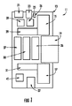
図1は、たとえば、長期的な衰弱事象または死亡さえ引き起こすことを防止するという点において、最初の1時間がきわめて重要であるような重篤な現場状況において、治療を最適化するために血液凝固反応を評価することが重要な外傷またはその他の関連する血液凝固障害を診断するための例示的な携帯式血液凝固モニタまたはアッセイ機器11を概略的に描いたものである。
FIG. 1 illustrates, for example, blood clotting to optimize treatment in severe field situations where the first hour is critical in preventing long-term debilitating events or even death. 1 schematically depicts an exemplary portable blood coagulation monitor or
この機器は耐衝撃性筐体13の中に格納されており、この筐体にはすべての構成要素が従来の方法で格納されている。 This device is housed in an impact resistant housing 13, in which all components are stored in a conventional manner.
少なくとも1つ、および好ましくは1対のリニアボイスコイルアクチュエータモータ15と17が、図2により詳細に描かれている、取り外し可能な血液検体カセットの中のサンプルプレートの運動を駆動するために設けられている。モータ15と17および取り外し可能な血液検体カセット19については簡単に説明したが、残りの構成要素については、概して図を参照しながら、機器の左上角のデータカードポート21から時計回りに説明する。
At least one and preferably a pair of linear voice
データカードポート21は、FDカードまたはあらゆるメモリモジュールを受けるのに有益であり、このモジュールには、機器11を較正するため、および/またはデータを機器11から取り出して、そのデータを分析可能な他のシステムに入力するためのデータが含まれる。UBSポート23により、たとえば医師がより複雑な分析を行うために制御するコンピュータと直接インタフェースさせることができる。
The
マイクロコントローラ25はEEPROM 26と動作し、USBポートおよびデータカードポート21からのデータを扱い、LCDディスプレイ等のユーザインタフェース(図示せず)のための制御を提供することができ、LCDディスプレイは機器を使用している使用者に対し、最初に取得したデータとその分析結果を提供する。リニアボイスコイルアクチュエータモータ15と17および、取り外し可能な血液検体カセット19については簡単に説明しており、後でより詳しく述べる。
たとえばRFまたはIRスペクトルのデータを送信するための無線リンク27もまた設置され、データとプログラミングを機器へ、または機器から通信するためのまた別の手段となる。光学的変位センサ29と31は、リニアボイスコイルアクチュエータモータ15と17がどこにあるかを検出し、その運動の限界を制御する。デジタルシンセサイザモジュール33は、ボイスコイルアクチュエータモータ15と17を駆動するために必要な波長を生成することによって、モータドライバ35を通じてモータ15と17の動作を制御する役割を果たす。
A
ADCモジュール37はアナログ−デジタル変換器であり、取り外し可能な血液カセット19の中の検体について行われた光学的または物理的測定からのデータを取得し、このデータをデジタル形式に変換し、これをマイクロコントローラ39に供給し、マイクロコントローラ39は得られたデータを管理して、機器の使用者に有益な結果を提供する。
The
3軸加速度計41は従来のデバイスであり、機器11に対する外的影響と振動を考慮し、検体の分析結果として収集されたあらゆるデータに対するこれらの振動の影響を打ち消す役割を果たす。
The three-
取り外し可能な血液検体カセット19が図2に概略的に示されており、これは上側サンプルプレート101と下側サンプルプレート103を含み、それらの間に血液の検体小滴を設置することができる。機器のボイスコイルアクチュエータモータとの連結部105が設置され、これは上側線形柔軟機構107と下側線形柔軟機構109に接続され、これらはそれぞれ上側および下側プレートの運動を駆動する役割を果たす。光センサ111が、サンプルプレートに関して、たとえばレーザまたはその他の光源(図示せず)からプレート間の検体を通り、また検体の中に投射される光を検出する位置に設置される。光はその後、検体を透過した、反射された、回折した、または検体を通る光路内でその他変調された光として検出することができ、光センサ111によって検出されて、血液検体の血液凝固特性に関する情報が提供される。
A removable
電源リンク113は、たとえば、バッテリまたはその他の形態の電源等の電力供給源に接続する役割を果たす。データリンク115もまた、検体から収集され、EEPROM 117に保存されたデータを機器からダウンロードできるようにするために設置できる。この点において、当業者にとっては当然のことながら、データリンク115とEEPROM 117は、カセット19が機器11に受けられると、機器11の中の各種の電子構成要素と接続されて、データをそこにアップロードおよびダウンロードすることができる。
The
これもまた当業者にとって当然のことかもしれないが、図3はさらに、本発明による機器であって、平行なプレートを有し、一般にこれらの平行なプレートの間に約50から約250μmの小さなギャップを有する機器の動作を描いている。これらは、制御された速度で相互に行き違いになるように摺動して、プレートの間にせん断応力を発生させ、これはT=μV/Dで示され、式中、Tはせん断応力であり、μ=粘度、V=V1+V2(Vはプレートの相対的線形速度である)、D=プレート間のギャップである。 Although this may also be natural for those skilled in the art, FIG. 3 is further an apparatus according to the present invention having parallel plates, typically between about 50 and about 250 μm small between these parallel plates. It depicts the operation of a device with a gap. They slide back and forth at a controlled rate to generate a shear stress between the plates, expressed as T = μV / D, where T is the shear stress , Μ = viscosity, V = V1 + V2 (V is the relative linear velocity of the plates), D = gap between plates.
図4は、本発明による機器11を組み立てた状態を描いたものであり、その各種の構成要素を示している。図4からわかるように、機器はポケットサイズで、好ましくは全体の寸法はiPhoneと同程度であり、約12cm×約6cm×約2cmである。機器11は、衝撃と振動を補償する内部の加速度計により耐久性が強化されている。設計上、これは多目的であり、幅広いせん断ダイナミックレンジにわたって、血小板とフィブリン凝血塊形成を測定することができる。またさらに、機器11は周辺機器としてUSBハブ電源で動作でき、その構成要素は容易に製造され、組み立てられる。一般に、生物物理学的分解能は約2μ未満の変位であり、機器11では、広い範囲のせん断応力の利用と完全な光アクセスが可能となる。
FIG. 4 depicts the assembled state of the
図5の分解図にさらに描かれているように、機器11は好ましくは、Acetal,6A1−4V Ti,2024A1等の材料でCNC機械加工された一体の筐体で構成される。下側サンプルウィンドウ203は上側サンプルウィンドウ205と協働し、これらは変位センサアーム207によって移動される。一般にアルミニウムで作製される一体の柔軟な4バー機構209が変位センサアーム207と関連付けられる。モータアセンブリ211、一般にVCAモータコイルアセンブリは、各種の構成要素を移動させる役割を果たし、一般に希土類で構成される磁石アセンブリ213に関連付けられる。光センサアレイ215は、凝血塊形成反応を調べ、測定する役割を果たす。センサアレイ215は一般に、赤外線差動変位センサアレイである。
As further depicted in the exploded view of FIG. 5, the
好ましくは、機器において、プレート101と103の第一と第二の面を、少なくとも、テクスチャ、物質またはその他の材料でコーティングし、血液凝固工程を誘発し、遅らせ、またはその他変調させて、診断またはその他の目的のために、検体の血液凝固の特定の面にとって有利に、または不利になることを選択できるようにしてある。表面の改質には、血小板または血中タンパク質の結合反応性または活性化を促進するものを含めることができる。同様に、このような改質は、血小板または血中タンパク質の結合反応性または活性化を低減させることができ、これは、かかる処理またはコーティングに関する以下の詳細な説明からより明らかとなるであろう。
Preferably, in the instrument, the first and second surfaces of the
機器は、新鮮全血または微量の新鮮全血の血液レオロジと血液凝固を、組織因子、カオリン、イニシエータ、クエン酸塩およびその他のような外的試薬を添加することなく、分析することができる。このような物質およびその他は、詳細な分析上の理由によって選択的に添加してもよいが、機器の最も好ましい実施形態においては、これらは不要である。 The instrument can analyze the blood rheology and clotting of fresh whole blood or trace amounts of fresh whole blood without the addition of external reagents such as tissue factor, kaolin, initiator, citrate and others. Such materials and others may be selectively added for detailed analytical reasons, but they are not necessary in the most preferred embodiment of the instrument.
アセンブリによれば、機器11は、同一の人または動物からの連続的な検体によって、血栓形成促進処理が行われた止血状態と抗血栓処理が行われた止血状態間の動的釣り合いをリアルタイムで、またはわずかな遅延で測定するように構成される。より好ましい態様において、モータ15と17はリニアボイスコイルアクチュエータモータであるが、線形運動を駆動できる、他のどのようなタイプの機器であってもよい。
According to the assembly, the
1つの実施形態において、血液凝固の検出は、検体流体の粘度の変化とプレート表面との結合から生じるプレート101と103の第一と第二の表面間の機械的相互作用を測定することにより、光学的に行われる。好ましい態様において、2枚のプレート101と103間の相対的運動は、任意に選択された波形を生成するように制御されて、選択された振幅、周波数、持続時間およびシーケンスの所望の流体せん断速度を発生させ、それによって機器はDC(せん断ゼロ)から、流体キャビテーションおよびその後の検体の細胞構成要素の破壊の原因となるようなせん断速度までの非常に広い範囲にわたり、所望の流体せん断を模倣でき、この範囲にはこれら2つの地点間のせん断速度スペクトルにおけるすべての地点が連続的に含まれる。
In one embodiment, the detection of blood clotting is accomplished by measuring the mechanical interaction between the first and second surfaces of
より具体的には、せん断速度は一連の数値において制御されて、標的とされる診断または分析の目標のための具体的なプロトコルまたはプレート移動パラダイムが生成され、この目標とはたとえば、一次凝固を急速に開始させること、初期、中期または後期の凝血塊形成の破壊的または非破壊的粘弾性評価を行うこと、他の商用または実験用機器と比較するための、臨床的に受け入れられている、またはその他認定されているせん断速度プロトコルを模倣すること、または既知の標準に照らした検証試験を行うことである。光を光学的検出に使用できるが、電磁波スペクトル全体を使用して、一次および二次両方の血液凝固機序のための検体小滴の血液凝固特性を表すアナログ信号を生成できることは明らかである。 More specifically, the shear rate is controlled in a series of numerical values to generate a specific protocol or plate movement paradigm for the targeted diagnostic or analytical goal, which includes, for example, primary coagulation. Clinically accepted for rapid initiation, destructive or non-destructive viscoelastic assessment of early, intermediate or late clot formation, compared to other commercial or laboratory equipment, Or imitating other recognized shear rate protocols, or performing verification tests against known standards. Although light can be used for optical detection, it is clear that the entire electromagnetic spectrum can be used to generate an analog signal that represents the blood clotting properties of the analyte droplet for both primary and secondary blood clotting mechanisms.
留意すべき点として、「血液検体」という用語が本明細書で使用される時、これは、全血、全血と1つまたはそれ以上の物質との混合物、全血の1つまたはそれ以上の成分を含む微量の全血、1つまたはそれ以上の非血液物質と混合された微量の全血、または血小板または血漿等の精製血液成分、再構成された血液製剤、改質された血液検体または代用血液等を意味することが意図される。 It should be noted that when the term “blood sample” is used herein, this means whole blood, a mixture of whole blood and one or more substances, one or more of whole blood. Of whole blood containing one or more components, one or more whole blood mixed with one or more non-blood substances, or purified blood components such as platelets or plasma, reconstituted blood products, modified blood samples Or it is intended to mean blood substitutes.
具体的な実施形態において、相互に交換可能なサンプルカセット19を使用することができ、各々が異なる分析および保守または較正機能を果たす。1つのカセット19を、機器11の定期的な較正および検証用とすることができる。あるいは、このような使い捨ての交換可能なカセット19は、機器11を較正し、血液検体を受け、血液検体を保持し、異なる試験プロトコルのために検体格納室の形状を変えることができ、試験中に検体を利用可能な状態に維持する役割を果たし、たとえば凍結、凍結乾燥等、保存のために検体を安全に取り出し、または血液検体に曝されずに検体を処分するのに役立つ。1つの実施形態において、カセット19は、単純な毛管現象作用によって血液検体を収集できるため、検体抽出および測定器の使用が不要である。
In a specific embodiment,
当業者には当然のことながら、カセット19は、サンプルプレートの間隔が異なり、表面の化学的および光学的特性が異なり、分析と検証試験のためのバリエーションが同様である異なる試験や用途に合わせて製造してもよい。
As will be appreciated by those skilled in the art, the
カセット19には、光学的手段により検出可能な血液状態、血液型、pH、酸素化、代謝産物、毒素またはその他の測定対象の検出を可能にする光学的操作観察用電子部品を含めてもよい。機器11は、その電子部品を通じて、顕微鏡等、その他の実験システムと接続することができるというものである。プレート101と103は、好ましくは光学的に透明であり、光信号が血液検体を通過し、平面間の血液検体の一部または全部を直接、光学的に視覚化することができる。これによって、レーザビームまたはその他の光表面の透過、反射、内部反射、選択的吸収、偏向または旋光、減衰内部反射(部分または全部)および伝導が可能となる。
前述のように、カセット19は、EEPROM 117のような不揮発性永久メモリストレージデバイスを含んでいてもよく、これはカセット、ロット、製造日、および構成の詳細を識別する初期データを含むほか、ユーザが設定する検体識別情報、試験開始時刻、試験継続時間、および明記された試験出力データと結果等の重要データを各検体とともに、破壊されるまで永久的に保管する。
As mentioned above,
カセット19は、添加剤、保存剤、シール剤または緩衝剤およびその他、試験前、試験中または試験後に血液検体に添加し、またその上に重ねることのできる同様の化学物質等、別の流体またはその他の材料を含んでいてもよい。図に具体的に示されていない1つの実施形態において、線形運動の代わりに、前述の線形変位に追加して、またはその代わりに、回転運動を使用してせん断を発生させることができる。
その構成により、機器11は、前述のデジタルシンセサイザ33、一般にはデュアルチャネルシンセサイザを使用して、0.0001Hzから1000Hzを含む、広いダイナミックレンジの機械的振動で血液検体に機械的せん断をかけることができ、このシンセサイザは、異なるデューティサイクル、周波数および振幅の正弦波、三角波、方形波等、一定周期の波形を生成する能力と、スルーレート、振幅等のすべてのパラメータが急速に変化する任意の波形を生成する能力を有するか、平面のいずれか一方または両方の一定した(DC)機械的変位を保持してもよい。モータ15と17は、ドライバ35によって、いずれか一方または両方の駆動機構がモータ15と17に連結されて、遷移運動を起こさせるために同時に利用できるような方法、またはいずれかを単独で、または機械的変位のための精密センサの役割を果たす他の機構と一緒に使用できるような方法で駆動してもよい。このようなシステムにおいては、たとえば4バーまたはマルチバーの一体型柔軟機構のような機械的特徴を組み込み、血液検体そのものに伴うもの以外の、摺動または回転する軸受面の使用から生じる機械的ヒステリシスを除去する。
By virtue of that configuration, the
データ分析およびデータ整理ソフトウェアを機器の電子部品に組み込んで、測定結果である血液凝固のパラメータを後で読み出すために保管し、コンピュータまたは、表示、保存または分析用のその他の機器に転送し、グラフィックで表示し、物理単位の数値で表示し、または具体的な診断、臨床指標または臨床的意義のパラメータの変化を示すアイコンや記号の形態で表示できるようにしてもよい。このソフトウェアを用いれば、結果として得られたデータを、使用者がその結果を同様の機能を有する他の機器から得られると予想されるような同様の、または類似の結果と直接比較できるように再構成し、または血液凝固パラメータに関する、容認されている標準的な数値または範囲と比較できるような方法で表示することが可能となるように表現することができるであろう。この点において、機器にLCDユーザインタフェース等のユーザインタフェース(図示せず)が実装されることがわかる。 Incorporate data analysis and data reduction software into the instrument's electronic components and store the resulting blood clotting parameters for later retrieval and transfer to a computer or other instrument for display, storage or analysis, and graphics May be displayed in numerical form of physical units, or may be displayed in the form of icons or symbols indicating changes in parameters of specific diagnosis, clinical indicators or clinical significance. With this software, the resulting data can be directly compared with similar or similar results that the user is expected to obtain from other devices with similar functions. It could be expressed in such a way that it can be reconstructed or displayed in such a way that it can be compared with accepted standard values or ranges for blood clotting parameters. In this respect, it can be seen that a user interface (not shown) such as an LCD user interface is mounted on the device.
機器11は、データ分析およびデータ整理ソフトウェアと連結された、プレート101と103の運動プロトコルを利用して、たとえば血小板の機能、血液凝固系の機能、赤血球(RBC)のレオロジ、RBC凝集、血液凝固促進剤または抗凝固剤の影響、線溶および、血液凝固のその他の特徴等を直接評価することが可能である。適当な使用者の要求事項とユーザインタフェースを備える機器11は、家庭内および非熟練者による使用が可能な、「単純」(CLIAの分類で「waived」)に分類してもよい。使い捨てカセットは、流体検体の単純で簡単な収集を通じて血液検体を収集し、保持するように構成されており、測定用ピペット、シリンジまたはその他、血液検体採取のための測定器を使用する必要がない。カセット19により、収集された血液検体を安全に取り扱い、保管し、取り出し、処分することができ、また、カセット19は、プレートの表面積とプレート間間隔を調節して、ごく微量の血液、一般に約1mL未満の血液を使用できるように製造されてもよい。
機器11とカセット19によれば、標準型および倒立型の両方の顕微鏡構成を利用して、血液凝固分析前、分析中または分析後の検体体積の大部分または全部を光学顕微鏡によって検査できる。機器11は、疾患および血液凝固異常の診断と定量化に利用でき、その疾患等の例としては、これらに限定されないが、誘発性、後天的および先天的状態、たとえば外傷性血液凝固異常(TIC)、フォンヴィレブランド病(vWD)、血液凝固因子消費、血小板消費、血小板無力症、代謝による血小板の枯渇、血液希釈、プロテインC、Sおよび線溶系の活性異常亢進、RBCレオロジの変化および不適正に行われた血液凝固調節のための治療、ならびに血液凝固に関わるその他の疾患や状態がある。
According to the
またさらに、機器11は、傷害または外傷の現場、搬送中、または救助、初期対応、治療、外科的介入または回復中の他のいずれの時点においても、15分未満、好ましくは4分未満の時間内に、その場で血液凝固異常を迅速に評価できる。フィードバックおよびフィードフォワード方式は、血液検体を安定化させ、外的な機械的ノイズ、振動および衝突による衝撃に抵抗できるようにする役割を果たす。外傷および失血に対する医療的対処の早期段階における一次および二次の両血液凝固の経時変化の試験に影響を与えることができる。同様に、一次および二次血液凝固の手術中の試験および手術中のこれらの機序の変更を実現できる。
Still further, the
機器11は、一次または二次血液凝固機序に影響を与えようと意図される生物活性化合物の迅速な体外スクリーニングに使用できる。これはまた、一次または二次血液凝固機序における疾患の臨床治療の指針にも使用できる。
同様に、機器11はとカセット19は、臨床および研究上、関心のあるその他の身体物質、たとえば嚢胞性線維症の治療の研究と臨床的指針のための、たとえば肺粘膜の流動学的特性を測定するような、専用に設計されたカセット(使い捨て)と専用の試験プロトコルおよびファームウェアを必要とする用途に、またはレオロジの異なる新たな新規流体の研究で使用するように改変してもよい。
Similarly, the
機器11は、光電子および無線手段を利用して、個々に試験開始時刻が異なり、試験持続時間が異なり、または検体格納室の試験プロトコルが異なる複数の機器を同時に、または複数の検体で使用し、中央コンピュータまたはデータ収集表示機器に血液凝固に関する豊富なデータ群を提供する。これによって、多くの検体を同時にモニタし、手術または回復中、または搬送または現場での治療中に、ある個人から血液の凝固状態の動的変化を追跡することが可能となる。
The
前述のように、本発明の特定の実施形態は、プレート101と105、一般にガラスプレートの、血液検体と接触する表面上にコーティングを追加した形態をとってもよい。これらのコーティングは、血小板の付着と活性化を促進する特性を有するものであってもよく、たとえば、人または牛由来のコラーゲン、より具体的にはIV型コラーゲン等である。同様に、コーティングは、培養された繊維芽細胞の細胞外マトリクス(ECM)から、または培養された内皮から得ても、または人または動物由来の生体血管の天然の皮内下層組織から得てもよい。マトリクスの特定の分子成分、たとえばビトロネクチンまたはフィブロネクチンはコーティングを構成しても、または、吸着特性を増強するためのマトリクスの強化された特徴であってもよい。コーティングはまた、血小板の吸着および/または活性化を促進するための合成による性質を有していてもよく、たとえばポリアミドやポリグルコサミン、より具体的には天然または合成のβ−N−アセチルポリグルコサミン等である。その他の実施形態は、プレート101と105の上に、血小板機能を変調させるコーティング、たとえば血小板機能の活性化因子、たとえばアデノシン二リン酸またはエピネフリン、あるいは血小板機能の阻害因子、たとえばプロスタサイクリンまたはプロスタグランディンE−1を放出できる材料を組み込んでもよい。
As mentioned above, certain embodiments of the present invention may take the form of
本発明の他の実施形態は、プレート101と105の上に、血液凝固およびフィブリン重合の工程を促進、開始または変調するコーティングを追加した形態をとってもよい。これらのコーティングは、微粉シリカ、またはカオリン、または組織因子(天然型または組み換え型)、または血液凝固系の中のステップを促進することが知られているその他の作用因子を放出することのできる材料の形態をとってもよい。コーティングはまた、血液検体中に存在するかもしれない抗凝固因子を逆行させる性質のもの、たとえばヘパリンを除去するためのヘパリン分解酵素、またはクエン酸塩を除去するための石化カルシウムであってもよい。このようなコーティングは、せん断によって誘発される血小板活性化機序により血液凝固を開始するのに十分な血小板の数または機能を持たない患者の血液を試験する場合に好ましいかもしれない。同じ理由で、コーティングには、天然または合成のリン脂質の放出源を取り入れるか、あるいはプロトロンビン時間(PT)または部分トロンボプラスチン時間(PTT)等の標準的な臨床血液凝固試験において用いられる作用因子を模倣するために、部分または全トロンボプラスチンを含んでいてもよい。
Other embodiments of the present invention may take the form of adding a coating on
本発明のまた別の実施形態において、プレートに、血液検体中の線溶亢進の影響を阻害または変調または逆行させるコーティング、たとえばイプシロンアミノカプロン酸(EACA)、またはトラネキサム酸、またはアプロチニン、またはプラスミンという酵素の作用に影響を与えるその他の抗プラスミン化合物または化学物質を追加してもよい。これらのコーティングは、試験系におけるプラスミンの即効を除去しなければ、検体(および、それゆえ患者)の血液凝固/止血システムに関するその他の有益な情報を抽出する能力を損なうような深刻な線溶亢進を来している患者の血液を試験する際に望ましかもしれない。同様に、コーティングは、血液検体のpHをpH7.2から7.4の最適な所望のレベル、または検体が患者の重篤な状態によって過剰に酸性状態または塩基性である場合に情報の損失を回避するためのその他の特定のレベルに調節するために、酸性または塩基性の緩衝化合物を含んでいてもよい。これらの緩衝化合物は、放出された塩またはアミノ酸、あるいは所望のpHを生成し、これを維持する目的のために血液と生体適合性のあるその他の両性イオン基を有する高分子可溶性化合物の形態をとってもよい。 In yet another embodiment of the invention, the plate is coated with a coating that inhibits or modulates or reverses the effects of hyperfibrinolysis in a blood sample, such as the enzyme epsilon aminocaproic acid (EACA), or tranexamic acid, or aprotinin, or plasmin Other antiplasmin compounds or chemicals that affect the action of can be added. These coatings can cause severe fibrinolysis that can destroy the ability of the specimen (and hence the patient) to extract other useful information about the blood clotting / hemostatic system if the immediate effect of plasmin in the test system is not removed. It may be desirable when testing the blood of a patient who is coming. Similarly, the coating reduces the loss of information when the pH of the blood sample is at an optimum desired level of pH 7.2 to 7.4, or if the sample is excessively acidic or basic depending on the patient's severe condition. Acidic or basic buffer compounds may be included to adjust to other specific levels to avoid. These buffer compounds are in the form of released salts or amino acids, or polymeric soluble compounds having other zwitterionic groups that are biocompatible with blood for the purpose of generating and maintaining the desired pH. It may be taken.
本発明のプレート101と105に上記およびその他のコーティングを追加しても、一般に、プレート101と105間の血液検体用空間の形状または、モータを用いた機構によるプレート101と105の運動と制御は、システムを必要なギャップが規定内に維持されるように調製できなくなるまで変化しない。その理由により、ガラスプレート上のこのようなコーティングを製造する好ましい方法は、所望の材料を帯電および堆積によってガラス部材と直接接触させること、または血液検体と接触した時に直ちに活性化するのに適した、放出可能な形態で所望の作用因子を含む、薄い分子ヒドロゲルおよびキャリアエマルジョンを積層することを含んでいてもよい。
Even if the above-mentioned and other coatings are added to the
上記のコーティングは、1つの目的のための単独の作用因子を含んでいても、あるいは複数の支援目的のためのいくつかの、またはそれ以上の上記コーティングを組み合わせた混合体であってもよい。1つの実施形態は、2つの面の上に異なるコーティングを設けることを含んでいてもよく、一方は1つのタイプでもう一方は異なるタイプまたは異なる密度であるか、あるいは対向する部材の上の同様のコーティングが改質されてもよい。他の実施形態において、1つまたはそれ以上のコーティングを一方または両方の表面の異なる領域において異なる密度または濃度で、あるいは一方または両方の表面上に線形、放射状またはその他の密度勾配のコーティングを設けてもよい。各タイプのコーティングのパターンまたは勾配は、同じまたは対向する表面上の他の表面コーティング処理のそれとは異なっていてもよい。ある例示的実施形態において表面はガラスであるが、表面は、その上にコーティングを受け、また所期の方法で機能できるその他のどの材料であってもよく、これは当業者にとって明らかであろう。 The above coatings may contain a single agent for one purpose, or may be a mixture of several or more of the above coatings for multiple support purposes. One embodiment may include providing different coatings on the two surfaces, one being one type and the other being a different type or density, or similar on opposing members. The coating may be modified. In other embodiments, one or more coatings may be provided with different densities or concentrations in different regions of one or both surfaces, or with linear, radial or other density gradient coatings on one or both surfaces. Also good. The pattern or gradient of each type of coating may be different from that of other surface coating processes on the same or opposing surfaces. In certain exemplary embodiments, the surface is glass, but the surface can be any other material that has received a coating thereon and can function in the intended manner, as will be apparent to those skilled in the art. .
本発明を各種の実施形態の説明によって解説し、これらの実施形態をごく詳細に説明したが、これが付属の特許請求の範囲をこのような詳細に制限する、または何らかの方法で限定することは出願人の意図ではない。本発明はしたがって、より広いその態様において、具体的な詳細、代表的な機器と方法、および図と説明文に示された例示のための事例に限定されない。したがって、せん断により誘発される血小板の活性化と血液凝固反応を全面的に利用する携帯式機器において止血機能全体をモニタするという出願人の包括的な発明性のある着想の真の主旨と範囲から逸脱することなく、かかる詳細に変更を加えることができる。 The present invention has been described in terms of various embodiments, and these embodiments have been described in great detail, but it is not intended that this limit the appended claims to such details or be limited in any way. It is not human intention. The present invention, therefore, in its broader aspects is not limited to the specific details, representative equipment and methods, and illustrative examples shown in the figures and legends. Therefore, from the true spirit and scope of the applicant's comprehensive inventive idea to monitor the overall hemostatic function in portable devices that fully utilize shear-induced platelet activation and blood clotting reactions Changes can be made to such details without departing.
Claims (31)
第一の表面を有する第一の部材と第二の表面を有する第二の部材であり、前記第一の部材は前記第一の表面が前記第二の部材の前記第二の表面と対面するように位置付けられ、血液検体小滴が前記第一の表面と前記第二の表面と接触して、血液凝固を開始するのに十分な量だけ離間され、前記第一の部材と第二の部材が相互に関して線形に移動可能であるような第一の部材と第二の部材と、
前記第一の部材と前記第二の部材の少なくとも一方に接続され、血液検体が前記第一の表面と前記第二の表面に接触すると、前記第一の部材と前記第二の部材を相互に関して平行に線形に移動させる駆動機構と、
前記第一の部材と第二の部材間に配置された血液検体と光の相互作用を、前記血液検体の血液凝固反応の指標として検出する光検出センサシステムと、
を備えることを特徴とする機器。 In a device for measuring blood coagulation reaction in a blood sample,
A first member having a first surface and a second member having a second surface, wherein the first member faces the second surface of the second member. Positioned so that the blood sample droplet is in contact with the first surface and the second surface and spaced apart by an amount sufficient to initiate blood clotting, the first member and the second member A first member and a second member that are linearly movable with respect to each other;
When connected to at least one of the first member and the second member and the blood sample contacts the first surface and the second surface, the first member and the second member are related to each other. A drive mechanism that moves linearly in parallel;
A light detection sensor system for detecting an interaction between light and a blood sample disposed between the first member and the second member as an indicator of a blood coagulation reaction of the blood sample;
A device characterized by comprising:
前記第一の部材と第二の部材が、前記機器から取り外し可能な血液検体収集カートリッジを構成することを特徴とする機器。 The device of claim 1,
The device wherein the first member and the second member constitute a blood sample collection cartridge that is removable from the device.
前記血液検体収集カートリッジは、試験対象の血液検体に関するデータを保存するためのメモリデバイスを前記カートリッジ上にさらに備えることを特徴とする機器。 The device according to claim 2,
The blood sample collection cartridge further comprises a memory device on the cartridge for storing data relating to a blood sample to be tested.
前記駆動機構が、血液検体の血液凝固反応に関わる異なる機序を検出するために、前記第一の部材と第二の部材を相互に関して異なる速度で移動させるようにプログラムされることを特徴とする機器。 The device of claim 1,
The drive mechanism is programmed to move the first member and the second member at different speeds relative to each other to detect different mechanisms involved in the blood clotting reaction of the blood sample. machine.
前記光検出センサシステムが、前記第一の表面と前記第二の表面への前記血液検体の結合を、血液凝固における血小板反応の指標として検出するようになされていることを特徴とする機器。 The device of claim 1,
The apparatus, wherein the photodetection sensor system is configured to detect binding of the blood sample to the first surface and the second surface as an indicator of a platelet reaction in blood coagulation.
前記第一と第二の表面の少なくとも一方が、前記検体他の目的の血液凝固の特定の局面にとって有利に、または不利になることを選択するように、血液凝固工程を誘発し、遅らせ、または変調させるように処理されていることを特徴とする機器。 The device of claim 1,
At least one of the first and second front surface, the advantage for certain aspects of the blood coagulation analytes other purposes, or to choose to become disadvantageous to induce blood clotting process, delayed, Or a device that is processed to be modulated.
前記表面の前記処理が血小板または血中タンパク質の結合、反応性または活性化を促進することを特徴とする機器。 The device according to claim 6, wherein
A device characterized in that the treatment of the surface promotes binding, reactivity or activation of platelets or blood proteins.
前記表面の前記処理が血小板または血中タンパク質の結合、反応性または活性化を低減させることを特徴とする機器。 The device according to claim 6, wherein
The instrument wherein the treatment of the surface reduces binding, reactivity or activation of platelets or blood proteins.
外部試薬を添加することなく、新鮮な全血または微量の新鮮な全血の血液レオロジと血液凝固を分析するように構成されていることを特徴とする機器。 The device of claim 1,
An instrument configured to analyze blood rheology and blood clotting of fresh whole blood or a small amount of fresh whole blood without adding external reagents.
同一の人または動物からの連続的な検体によって、血栓形成促進処理が行われた止血状態と抗血栓処理が行われた止血状態間の動的釣り合いを、機能的遅延を発生させずに測定するように構成されていることを特徴とする機器。 The device of claim 1,
Measures the dynamic balance between a hemostatic state that has undergone thrombus formation treatment and a hemostatic state that has undergone anti-thrombotic treatment, with continuous specimens from the same person or animal, without causing a functional delay A device characterized by being configured as follows.
前記駆動機構と光検出センサシステムの動作を制御する、少なくとも1つのマイクロコントローラをさらに備えることを特徴とする機器。 The device of claim 1,
The apparatus further comprising at least one microcontroller for controlling operations of the driving mechanism and the light detection sensor system.
前記第一の部材と前記第二の部材間の相対的運動の量を検出し、制御する、少なくとも1つの変位センサをさらに備えることを特徴とする機器。 The device of claim 1,
The apparatus further comprising at least one displacement sensor that detects and controls the amount of relative motion between the first member and the second member.
前記機器と外部システム間を接続し、通信させるための接続インタフェースをさらに備えることを特徴とする機器。 The device of claim 1,
A device further comprising a connection interface for connecting and communicating between the device and an external system.
前記駆動機構が線形運動を駆動できるデバイスを備えることを特徴とする機器。 The device of claim 1,
An apparatus, wherein the drive mechanism comprises a device capable of driving linear motion.
光検出センサシステムに接続され、血液検体の血液凝固反応を示すアナログ信号を、それを保存するためにデジタル信号に変換するためのアナログ−デジタル変換器をさらに備えることを特徴とする機器。 The device of claim 1,
An apparatus further comprising an analog-to-digital converter connected to the light detection sensor system for converting an analog signal indicative of a blood coagulation reaction of a blood sample into a digital signal for storage.
血液検体小滴を対向して配置された実質的に平行な2枚のプレートの第一と第二の対面する表面の間に、これらと接触するように設置するステップと、
少なくとも一方のプレートを所定の速度でもう一方のプレートに関して線形に移動させるステップと、
前記検体流体の粘度の変化と前記プレートの表面への結合から生じる、前記第一と第二の表面間の機械的相互作用を測定することによって、前記血液小滴の血液凝固反応を光学的手段により検出するステップと、
を含むことを特徴とする方法。 In a method for measuring a blood coagulation reaction of a blood sample,
Between the first and second facing surfaces of two substantially parallel plates which are arranged opposite the blood sample droplet, the steps of placed in contact with these,
Moving at least one plate linearly with respect to the other plate at a predetermined speed;
Optical means for coagulating the blood droplet by measuring the mechanical interaction between the first and second surfaces resulting from a change in viscosity of the analyte fluid and binding to the surface of the plate Detecting with
A method comprising the steps of:
少なくとも一方のプレートを第一の速度で移動させるステップと、前記血液検体小滴の前記プレートの前記表面への吸着を光学的手段により検出し、血液凝固における血小板反応を判断するステップと、をさらに含むことを特徴とする方法。 The method of claim 16, wherein
Moving at least one of the plates at a first speed; and detecting the adsorption of the blood sample droplets to the surface of the plate by optical means to determine a platelet response in blood coagulation. A method characterized by comprising.
続いて少なくとも一方のプレートを、前記第一の速度より遅い第二の速度で移動させるステップと、前記血液検体の血液凝固レベルを、フィブリン重合反応の指標として光学的手段により検出するステップと、をさらに含むことを特徴とする方法。 The method of claim 17, wherein
Subsequently, moving at least one of the plates at a second speed slower than the first speed, and detecting a blood coagulation level of the blood sample by an optical means as an index of a fibrin polymerization reaction. A method further comprising:
前記2枚のプレート間の相対的運動が、選択された振幅、周波数、持続時間およびシーケンスの所望の流体せん断速度を発生させ、前記機器がDC(せん断ゼロ)から、流体キャビテーションおよびその後の前記検体の細胞構成要素の破壊の原因となるようなせん断速度までであり、これら2つの地点間のせん断速度スペクトルにおけるすべての地点を連続的に含むような非常に広い広範囲にわたって所望の流体せん断を模倣できるように、任意に選択された波形を生成するように制御されることを特徴とする方法。 The method of claim 16, wherein
The relative motion between the two plates generates the desired fluid shear rate of the selected amplitude, frequency, duration and sequence, and the instrument moves from DC (zero shear) to fluid cavitation and subsequent the specimen Up to the shear rate that causes the destruction of the cellular components of the cell, and can mimic the desired fluid shear over a very wide and wide range including all points in the shear rate spectrum between these two points in succession. A method characterized in that it is controlled to generate an arbitrarily selected waveform.
前記せん断速度が一連の数値において制御され、一次凝固の急速な開始、初期、中期または後期の凝血塊形成の破壊的または非破壊的粘弾性評価、他の商用または実験用機器と比較するための、臨床的に受け入れられている、またはその他認定されているせん断速度プロトコルの模倣、または既知の標準に照らした検証試験等、標的とされる診断または分析の目標のための具体的なプロトコルまたはプレート移動パラダイムが生成されることを特徴とする方法。 The method of claim 19, wherein
The shear rate is controlled in a series of numerical values for rapid onset of primary coagulation, destructive or non-destructive viscoelastic evaluation of clot formation in early, mid or late phases, for comparison with other commercial or laboratory equipment Specific protocols or plates for targeted diagnostic or analytical goals, such as imitation of clinically accepted or other certified shear rate protocols, or validation tests against known standards A method characterized in that a movement paradigm is generated.
前記プレートが、血液凝固を測定するための機器に挿入可能なカセットの一部であり、前記プレートが透明であることを特徴とする方法。 The method of claim 16, wherein
The method wherein the plate is part of a cassette that can be inserted into an instrument for measuring blood clotting, and the plate is transparent.
前記カセットがメモリをさらに備え、その上に、前記光検出により得られた血液凝固反応に関する情報を保存するステップをさらに備えることを特徴とする方法。 The method of claim 21, wherein
The cassette further comprises a memory, further comprising the step of storing information on the blood coagulation reaction obtained by the light detection.
前記光検出が、前記検体小滴に電磁波を透過させ、それぞれの光検出器において前記検体小型滴を通じた前記電磁波の透過、吸収、反射および回折の少なくとも1つを検出して、一次および二次血液凝固機序のための前記検体小滴における前記血液の血液凝固特性を表すアナログ信号を生成することによって行われることを特徴とする方法。 The method of claim 16, wherein
The light detection transmits an electromagnetic wave to the specimen droplet, and detects at least one of transmission, absorption, reflection, and diffraction of the electromagnetic wave through the specimen small droplet in each photodetector to detect primary and secondary A method comprising: generating an analog signal representative of blood clotting characteristics of the blood in the specimen droplet for a blood clotting mechanism.
前記信号デジタル信号に変換するステップと、前記デジタル信号を保存するステップと、前記保存されたデジタル信号を所定の方法で分析して、前記検体小滴における前記血液の前記血液凝固反応に関する選択された情報を取得するステップと、をさらに含むことを特徴とする方法。 24. The method of claim 23, wherein
Converting the signal into a digital signal; storing the digital signal; and analyzing the stored digital signal in a predetermined manner to select the blood coagulation reaction of the blood in the specimen droplet. Obtaining information. The method further comprising:
少なくとも一方のプレートに接続可能であり、前記少なくとも一方のプレートを移動させる、少なくとも1つのボイスコイルアクチュエータモータと、前記少なくとも一方のプレートを前記モータにより移動させるステップと、をさらに備えることを特徴とする方法。 The method of claim 16, wherein
And at least one voice coil actuator motor that is connectable to at least one plate and moves the at least one plate, and further comprising the step of moving the at least one plate by the motor. Method.
前記モータに接続されたプロセッサと、その動作を前記プロセッサにより所定の方法で制御するステップと、をさらに備えることを特徴とする方法。 26. The method of claim 25, wherein
A method further comprising: a processor connected to the motor; and controlling the operation of the processor by a predetermined method.
一方のプレートを他方のプレートに関して、前記他方のプレートを前記血液と前記他方のプレート間の粘弾性による連成によって移動させるような方法で移動させるステップと、前記他方のプレートの前記運動から前記血液の粘弾性特性を判断するステップと、をさらに含むことを特徴とする方法。 The method of claim 16, wherein
Moving one plate with respect to the other plate in such a manner that the other plate is moved by viscoelastic coupling between the blood and the other plate, and the blood from the movement of the other plate Determining the viscoelastic characteristics of the method.
前記血液検体により引き起こされた、前記一方のプレートと前記他方のプレート間の粘弾性による連成によって引き起こされた前記一方のプレートと前記他方のプレートの運動によって発生した歪み速度を検出するステップと、前記2枚のプレート間の機械的連成とその結果得られた前記歪み速度から判断された前記血液検体の粘弾性に基づく推測分析により、前記血液の血液凝固状態を判断するステップと、をさらに含むことを特徴とする方法。 The method of claim 16, wherein
Detecting a strain rate caused by the movement of the one plate and the other plate caused by the viscoelastic coupling between the one plate and the other plate caused by the blood sample; Determining the blood coagulation state of the blood by speculative analysis based on the viscoelasticity of the blood sample determined from the mechanical coupling between the two plates and the resulting strain rate. A method characterized by comprising.
ある時間にわたり前記血液の粘弾性を継続的に測定して、前記血液の血液凝固反応の経時変化をモニタするステップをさらに含むことを特徴とする方法。 The method of claim 16, wherein
The method further comprising the step of continuously measuring the viscoelasticity of the blood over a period of time to monitor the change over time in the blood clotting reaction of the blood.
血液検体小滴を対向して配置された2枚のプレートの対面する表面の間に、これらに接触させて設置するステップと、
少なくとも一方のプレートを他方のプレートに関して、第一の速度で線形に移動させるステップと、
前記血液中の血小板反応を示す前記血液の一次血液凝固反応を光学的手段により検出するステップと、
少なくとも一方のプレートを他方のプレートに関して、第二の速度で線形に移動させるステップと、
フィブリン重合を示す前記血液の二次血液凝固反応を光学的手段により検出するステップと、
を含むことを特徴とする方法。 In a method for measuring a blood coagulation reaction in a blood sample,
Placing a blood sample droplet in contact between the facing surfaces of two plates disposed opposite each other;
Moving at least one plate linearly with respect to the other plate at a first speed;
Detecting, by optical means, a primary blood coagulation reaction indicative of the blood platelet reaction in the blood;
Moving at least one plate linearly with respect to the other plate at a second speed;
Detecting a secondary blood coagulation reaction of the blood exhibiting fibrin polymerization by optical means;
A method comprising the steps of:
ある時間にわたって前記血液の粘弾性を継続的に測定し、前記血液の血液凝固反応の経時変化をモニタするステップをさらに含むことを特徴とする方法。 The method of claim 30, wherein
The method further comprises the step of continuously measuring the viscoelasticity of the blood over a period of time and monitoring the time course of the blood clotting reaction of the blood.
Applications Claiming Priority (3)
| Application Number | Priority Date | Filing Date | Title |
|---|---|---|---|
| US28778009P | 2009-12-18 | 2009-12-18 | |
| US61/287,780 | 2009-12-18 | ||
| PCT/US2010/060911 WO2011075614A2 (en) | 2009-12-18 | 2010-12-17 | Portable coagulation monitoring device and method of assessing coagulation response |
Publications (2)
| Publication Number | Publication Date |
|---|---|
| JP2013515238A JP2013515238A (en) | 2013-05-02 |
| JP5655091B2 true JP5655091B2 (en) | 2015-01-14 |
Family
ID=44151642
Family Applications (1)
| Application Number | Title | Priority Date | Filing Date |
|---|---|---|---|
| JP2012544860A Active JP5655091B2 (en) | 2009-12-18 | 2010-12-17 | Portable blood coagulation monitoring device and evaluation method of blood coagulation reaction |
Country Status (17)
| Country | Link |
|---|---|
| US (2) | US8450078B2 (en) |
| EP (1) | EP2513647B1 (en) |
| JP (1) | JP5655091B2 (en) |
| CN (1) | CN102687009B (en) |
| AU (1) | AU2010330861B2 (en) |
| BR (1) | BR112012014421B8 (en) |
| CA (1) | CA2780492C (en) |
| CO (1) | CO6551732A2 (en) |
| CR (1) | CR20120326A (en) |
| EC (1) | ECSP12011981A (en) |
| ES (1) | ES2543099T3 (en) |
| HK (1) | HK1173497A1 (en) |
| HN (1) | HN2012001269A (en) |
| MX (1) | MX2012007084A (en) |
| NI (1) | NI201200109A (en) |
| PE (1) | PE20121723A1 (en) |
| WO (1) | WO2011075614A2 (en) |
Cited By (2)
| Publication number | Priority date | Publication date | Assignee | Title |
|---|---|---|---|---|
| US9726647B2 (en) | 2015-03-17 | 2017-08-08 | Hemosonics, Llc | Determining mechanical properties via ultrasound-induced resonance |
| US10962524B2 (en) | 2011-02-15 | 2021-03-30 | HomoSonics LLC | Characterization of blood hemostasis and oxygen transport parameters |
Families Citing this family (34)
| Publication number | Priority date | Publication date | Assignee | Title |
|---|---|---|---|---|
| US7892188B2 (en) | 2003-10-22 | 2011-02-22 | Hemosonics, Llc | Method and apparatus for characterization of clot formation |
| US8448499B2 (en) | 2008-12-23 | 2013-05-28 | C A Casyso Ag | Cartridge device for a measuring system for measuring viscoelastic characteristics of a sample liquid, a corresponding measuring system, and a corresponding method |
| US10739358B2 (en) | 2009-12-18 | 2020-08-11 | Entegrion, Inc. | Portable coagulation monitoring devices, systems, and methods |
| EP2555704B1 (en) | 2010-04-08 | 2019-05-29 | Hemosonics, Llc | Hemostatic parameter display |
| US20140083628A1 (en) | 2012-09-27 | 2014-03-27 | Velico Medical, Inc. | Spray drier assembly for automated spray drying |
| EP2676136B1 (en) | 2011-02-15 | 2020-12-23 | Hemosonics, Llc | Devices, systems and methods for evaluation of hemostasis |
| US20120294767A1 (en) | 2011-05-19 | 2012-11-22 | Hemosonics Llc | Portable hemostasis analyzer |
| CN104024840B (en) * | 2011-12-26 | 2018-04-06 | 松下健康医疗控股株式会社 | Liquid sample measurement system and measure device |
| CN104303052B (en) | 2012-01-16 | 2018-11-16 | 仪宝科技公司 | For measuring the method, apparatus and system of fluid physics performance |
| WO2014085804A1 (en) | 2012-11-30 | 2014-06-05 | The University Of North Carolina At Chapel Hill | Methods, systems, and computer readable media for determining physical properties of a specimen in a portable point of care diagnostic device |
| WO2014099420A1 (en) | 2012-12-17 | 2014-06-26 | Abbott Point Of Care Inc | A portable clinical analysis system for immunometric measurement |
| US9949674B2 (en) | 2012-12-17 | 2018-04-24 | Abbott Point Of Care Inc. | Portable clinical analysis system for hematocrit measurement |
| CN104956212B (en) * | 2012-12-17 | 2017-10-27 | 雅培医护站股份有限公司 | The dimensional orientation of Portable Clinical test device and the self-correcting of motion |
| US9952194B2 (en) | 2012-12-17 | 2018-04-24 | Abbott Point Of Care Inc. | Operation and verification of a portable clinical analysis system |
| EP2932259B1 (en) | 2012-12-17 | 2019-11-13 | Abbott Point Of Care, Inc. | Spatial orientation determination in portable clinical analysis systems |
| KR101481240B1 (en) * | 2012-12-27 | 2015-01-19 | 고려대학교 산학협력단 | Apparatus and method for monitoring platelet function and drug response in a microfluidic-chip |
| CN103487377B (en) * | 2013-10-11 | 2016-03-09 | 江苏英诺华医疗技术有限公司 | A kind of detector for coagulation function of whole blood and detection method |
| US10823743B1 (en) * | 2013-10-28 | 2020-11-03 | Ifirst Medical Technologies, Inc. | Methods of measuring coagulation of a biological sample |
| AU2014348333B2 (en) * | 2013-11-15 | 2020-08-20 | Entegrion, Inc. | Portable coagulation monitoring devices, systems, and methods |
| US9452199B2 (en) | 2014-01-17 | 2016-09-27 | General Electric Company | Platelet activation and growth factor release using electric pulses |
| US9354243B2 (en) | 2014-05-05 | 2016-05-31 | Haemonetics Corporation | Methodologies and reagents for detecting fibrinolysis and hyperfibrinolysis in a blood sample using viscoelastic analysis |
| EP4109095A1 (en) * | 2014-09-09 | 2022-12-28 | Perosphere Technologies Inc. | Microfluid chip-based, universal coagulation assay |
| US9561184B2 (en) | 2014-09-19 | 2017-02-07 | Velico Medical, Inc. | Methods and systems for multi-stage drying of plasma |
| US10175225B2 (en) | 2014-09-29 | 2019-01-08 | C A Casyso Ag | Blood testing system and method |
| JP6833703B2 (en) * | 2014-11-14 | 2021-02-24 | コラ ヘルスケア インコーポレイテッド | Stroke subtype determination device and method |
| CN105403691B (en) * | 2016-01-01 | 2017-08-18 | 赛奥生物科技(青岛)有限公司 | A kind of Blood Kit analysis system |
| CN106198943B (en) * | 2016-08-16 | 2019-11-12 | 中国科学院苏州生物医学工程技术研究所 | A kind of Studies on Hemodynamic Changes device |
| GB2555870A (en) | 2016-11-15 | 2018-05-16 | Haemair Ltd | Rheometer and method for the use thereof |
| EP3541443B1 (en) * | 2016-11-17 | 2024-04-10 | enicor GmbH | Coated articles for blood coagulation testing and methods of preparing the same |
| AU2018254584A1 (en) | 2017-04-20 | 2019-10-24 | Hemosonics, Llc | Disposable system for analysis of hemostatic function |
| WO2020027741A1 (en) * | 2018-07-29 | 2020-02-06 | Koc Universitesi | Microfluidic thromboelastometry instrument |
| US20210260589A1 (en) * | 2020-02-26 | 2021-08-26 | Emmanuel Mpock | Method and Apparatus for Measuring Blood Coagulation |
| KR102414376B1 (en) * | 2020-05-26 | 2022-06-29 | 고려대학교 산학협력단 | Blood test apparatus |
| US11841189B1 (en) | 2022-09-15 | 2023-12-12 | Velico Medical, Inc. | Disposable for a spray drying system |
Family Cites Families (15)
| Publication number | Priority date | Publication date | Assignee | Title |
|---|---|---|---|---|
| US5023A (en) * | 1847-03-20 | Stocking-loom | ||
| JPH0743388B2 (en) * | 1990-05-29 | 1995-05-15 | 日立化成工業株式会社 | Method for measuring gelation reaction associated with blood coagulation, measuring device and measuring element |
| FR2664982B1 (en) * | 1990-07-20 | 1994-04-29 | Serbio | APPARATUS FOR DETECTING CHANGE IN VISCOSITY, BY MEASURING RELATIVE SLIDING, PARTICULARLY FOR DETECTING BLOOD COAGULATION TIME. |
| US5838515A (en) * | 1996-04-30 | 1998-11-17 | Quantum Corporation | PWM/linear driver for disk drive voice coil actuator |
| US6046051A (en) * | 1997-06-27 | 2000-04-04 | Hemosense, Inc. | Method and device for measuring blood coagulation or lysis by viscosity changes |
| US6750053B1 (en) * | 1999-11-15 | 2004-06-15 | I-Stat Corporation | Apparatus and method for assaying coagulation in fluid samples |
| JP3792156B2 (en) * | 1999-11-15 | 2006-07-05 | アイ−スタット コーポレイション | Apparatus and method for analyzing clots in a liquid sample |
| US6743596B1 (en) * | 2000-10-27 | 2004-06-01 | Biomerieux, Inc. | Method of determining global coagulability hemostatic potential |
| GB0030929D0 (en) * | 2000-12-19 | 2001-01-31 | Inverness Medical Ltd | Analyte measurement |
| ES2234349B1 (en) * | 2002-04-18 | 2006-11-01 | Servicio De Instrumentacion Hospitalaria, S.L. | DEVICE FOR MEASUREMENT OF COAGULATION TIME AND PLAQUETARY ACTIVITY AND, PROCEDURE OF THE SAME. |
| EP1619994A4 (en) * | 2003-04-16 | 2009-03-11 | Univ Drexel | Acoustic blood analyzer for assessing blood properties |
| US20040228766A1 (en) * | 2003-05-14 | 2004-11-18 | Witty Thomas R. | Point of care diagnostic platform |
| US7399637B2 (en) * | 2004-04-19 | 2008-07-15 | Medtronic, Inc. | Blood coagulation test cartridge, system, and method |
| CN2916635Y (en) * | 2006-06-09 | 2007-06-27 | 姚冬 | Blood coagulation analysis tester |
| JP4675946B2 (en) * | 2007-09-07 | 2011-04-27 | 独立行政法人産業技術総合研究所 | Viscosity measuring device |
-
2010
- 2010-12-17 AU AU2010330861A patent/AU2010330861B2/en active Active
- 2010-12-17 JP JP2012544860A patent/JP5655091B2/en active Active
- 2010-12-17 CA CA2780492A patent/CA2780492C/en active Active
- 2010-12-17 PE PE2012000844A patent/PE20121723A1/en active IP Right Grant
- 2010-12-17 ES ES10838275.5T patent/ES2543099T3/en active Active
- 2010-12-17 CN CN201080055503.9A patent/CN102687009B/en active Active
- 2010-12-17 BR BR112012014421A patent/BR112012014421B8/en active IP Right Grant
- 2010-12-17 EP EP20100838275 patent/EP2513647B1/en active Active
- 2010-12-17 US US12/971,013 patent/US8450078B2/en active Active
- 2010-12-17 MX MX2012007084A patent/MX2012007084A/en active IP Right Grant
- 2010-12-17 WO PCT/US2010/060911 patent/WO2011075614A2/en active Application Filing
-
2012
- 2012-06-15 HN HN2012001269A patent/HN2012001269A/en unknown
- 2012-06-15 CR CR20120326A patent/CR20120326A/en unknown
- 2012-06-15 NI NI201200109A patent/NI201200109A/en unknown
- 2012-06-18 EC ECSP12011981 patent/ECSP12011981A/en unknown
- 2012-06-20 CO CO12103321A patent/CO6551732A2/en active IP Right Grant
-
2013
- 2013-01-21 HK HK13100873.7A patent/HK1173497A1/en unknown
- 2013-05-20 US US13/897,712 patent/US9063161B2/en active Active
Cited By (6)
| Publication number | Priority date | Publication date | Assignee | Title |
|---|---|---|---|---|
| US10962524B2 (en) | 2011-02-15 | 2021-03-30 | HomoSonics LLC | Characterization of blood hemostasis and oxygen transport parameters |
| US11680940B2 (en) | 2011-02-15 | 2023-06-20 | Hemosonics Llc | Characterization of blood hemostasis and oxygen transport parameters |
| US9726647B2 (en) | 2015-03-17 | 2017-08-08 | Hemosonics, Llc | Determining mechanical properties via ultrasound-induced resonance |
| US10495613B2 (en) | 2015-03-17 | 2019-12-03 | Hemosonics, Llc | Determining mechanical properties via ultrasound-induced resonance |
| US11002712B2 (en) | 2015-03-17 | 2021-05-11 | Hemosonics Llc | Determining mechanical properties via ultrasound-induced resonance |
| US11656206B2 (en) | 2015-03-17 | 2023-05-23 | Hemosonics Llc | Determining mechanical properties via ultrasound-induced resonance |
Also Published As
| Publication number | Publication date |
|---|---|
| WO2011075614A3 (en) | 2011-10-13 |
| AU2010330861A1 (en) | 2012-05-31 |
| CN102687009A (en) | 2012-09-19 |
| CA2780492A1 (en) | 2011-06-23 |
| CA2780492C (en) | 2018-07-17 |
| CR20120326A (en) | 2012-09-24 |
| EP2513647A4 (en) | 2013-09-04 |
| US8450078B2 (en) | 2013-05-28 |
| MX2012007084A (en) | 2012-10-15 |
| NI201200109A (en) | 2013-02-15 |
| BR112012014421B8 (en) | 2021-07-27 |
| CN102687009B (en) | 2014-07-16 |
| HN2012001269A (en) | 2014-12-01 |
| US20130267017A1 (en) | 2013-10-10 |
| WO2011075614A2 (en) | 2011-06-23 |
| EP2513647B1 (en) | 2015-04-22 |
| ES2543099T3 (en) | 2015-08-14 |
| US20110151491A1 (en) | 2011-06-23 |
| BR112012014421A2 (en) | 2017-12-19 |
| AU2010330861B2 (en) | 2013-09-26 |
| PE20121723A1 (en) | 2012-12-11 |
| CO6551732A2 (en) | 2012-10-31 |
| BR112012014421B1 (en) | 2021-02-02 |
| JP2013515238A (en) | 2013-05-02 |
| US9063161B2 (en) | 2015-06-23 |
| ECSP12011981A (en) | 2012-08-31 |
| EP2513647A2 (en) | 2012-10-24 |
| HK1173497A1 (en) | 2013-05-16 |
Similar Documents
| Publication | Publication Date | Title |
|---|---|---|
| JP5655091B2 (en) | Portable blood coagulation monitoring device and evaluation method of blood coagulation reaction | |
| US7857761B2 (en) | Acoustic blood analyzer for assessing blood properties | |
| KR100237946B1 (en) | Apparatus for assaying viscosity changes in fluid samples and method of conducting the same | |
| US20200363394A1 (en) | Optical thromboelastography systems and methods | |
| US9546981B2 (en) | Device and method for performing blood thromboelastographic assays by magnetic sensing | |
| US5792660A (en) | Comparative determinants of viscosity in body fluids obtained with probes providing increased sensitivity | |
| WO2014031253A1 (en) | Device and method for performing blood thromboelastographic assays by magnetic sensing | |
| Meyer dos Santos et al. | A novel μ-fluidic whole blood coagulation assay based on Rayleigh surface-acoustic waves as a point-of-care method to detect anticoagulants | |
| US8772040B2 (en) | Apparatus and method of platelet multi-function analysis, and micro stirring chip | |
| US10739358B2 (en) | Portable coagulation monitoring devices, systems, and methods | |
| US20210268497A1 (en) | Microfluidic thromboelastometry instrument | |
| JP6503605B2 (en) | Portable device, system and method for blood coagulation monitoring | |
| AU2013234363B2 (en) | Portable coagulation monitoring device | |
| US20240069043A1 (en) | Portable coagulation monitoring devices, systems, and methods | |
| Leavitt | Sarah Leavitt, Shairko Missouri, Divya Patel, and Corey S. Scher | |
| Leavitt et al. | Point-of-Care Tests in for Blood Coagulation in the Perioperative Period |
Legal Events
| Date | Code | Title | Description |
|---|---|---|---|
| A621 | Written request for application examination |
Free format text: JAPANESE INTERMEDIATE CODE: A621 Effective date: 20131107 |
|
| A977 | Report on retrieval |
Free format text: JAPANESE INTERMEDIATE CODE: A971007 Effective date: 20140616 |
|
| A131 | Notification of reasons for refusal |
Free format text: JAPANESE INTERMEDIATE CODE: A131 Effective date: 20140701 |
|
| A521 | Request for written amendment filed |
Free format text: JAPANESE INTERMEDIATE CODE: A523 Effective date: 20140930 |
|
| TRDD | Decision of grant or rejection written | ||
| A01 | Written decision to grant a patent or to grant a registration (utility model) |
Free format text: JAPANESE INTERMEDIATE CODE: A01 Effective date: 20141028 |
|
| A61 | First payment of annual fees (during grant procedure) |
Free format text: JAPANESE INTERMEDIATE CODE: A61 Effective date: 20141121 |
|
| R150 | Certificate of patent or registration of utility model |
Ref document number: 5655091 Country of ref document: JP Free format text: JAPANESE INTERMEDIATE CODE: R150 |
|
| R250 | Receipt of annual fees |
Free format text: JAPANESE INTERMEDIATE CODE: R250 |
|
| R250 | Receipt of annual fees |
Free format text: JAPANESE INTERMEDIATE CODE: R250 |
|
| R250 | Receipt of annual fees |
Free format text: JAPANESE INTERMEDIATE CODE: R250 |
|
| R250 | Receipt of annual fees |
Free format text: JAPANESE INTERMEDIATE CODE: R250 |
|
| R250 | Receipt of annual fees |
Free format text: JAPANESE INTERMEDIATE CODE: R250 |
|
| R250 | Receipt of annual fees |
Free format text: JAPANESE INTERMEDIATE CODE: R250 |
|
| R250 | Receipt of annual fees |
Free format text: JAPANESE INTERMEDIATE CODE: R250 |