JP5231401B2 - Method of guided cell ingrowth and controlled tissue regeneration in spinal surgery - Google Patents
Method of guided cell ingrowth and controlled tissue regeneration in spinal surgery Download PDFInfo
- Publication number
- JP5231401B2 JP5231401B2 JP2009512489A JP2009512489A JP5231401B2 JP 5231401 B2 JP5231401 B2 JP 5231401B2 JP 2009512489 A JP2009512489 A JP 2009512489A JP 2009512489 A JP2009512489 A JP 2009512489A JP 5231401 B2 JP5231401 B2 JP 5231401B2
- Authority
- JP
- Japan
- Prior art keywords
- collagen
- collagen foil
- foil biomatrix
- biomatrix
- tissue
- Prior art date
- Legal status (The legal status is an assumption and is not a legal conclusion. Google has not performed a legal analysis and makes no representation as to the accuracy of the status listed.)
- Active
Links
- 238000001356 surgical procedure Methods 0.000 title claims description 56
- 238000000034 method Methods 0.000 title description 76
- 230000017423 tissue regeneration Effects 0.000 title description 17
- 102000008186 Collagen Human genes 0.000 claims description 400
- 108010035532 Collagen Proteins 0.000 claims description 400
- 229920001436 collagen Polymers 0.000 claims description 400
- 239000011888 foil Substances 0.000 claims description 299
- 210000001519 tissue Anatomy 0.000 claims description 118
- 239000000203 mixture Substances 0.000 claims description 36
- 210000001951 dura mater Anatomy 0.000 claims description 29
- 241000283073 Equus caballus Species 0.000 claims description 25
- 230000004761 fibrosis Effects 0.000 claims description 24
- 206010016654 Fibrosis Diseases 0.000 claims description 23
- 241000282414 Homo sapiens Species 0.000 claims description 18
- 230000010261 cell growth Effects 0.000 claims description 18
- 241000124008 Mammalia Species 0.000 claims description 13
- 238000000926 separation method Methods 0.000 claims description 13
- 210000001032 spinal nerve Anatomy 0.000 claims description 12
- 241000283690 Bos taurus Species 0.000 claims description 11
- 230000002265 prevention Effects 0.000 claims description 11
- 239000000758 substrate Substances 0.000 claims description 2
- 210000004027 cell Anatomy 0.000 description 79
- 239000010410 layer Substances 0.000 description 50
- 208000027418 Wounds and injury Diseases 0.000 description 25
- 230000015572 biosynthetic process Effects 0.000 description 24
- 230000007547 defect Effects 0.000 description 22
- 206010052428 Wound Diseases 0.000 description 21
- 241000283973 Oryctolagus cuniculus Species 0.000 description 17
- 239000000463 material Substances 0.000 description 17
- 230000008439 repair process Effects 0.000 description 15
- 108010080379 Fibrin Tissue Adhesive Proteins 0.000 description 14
- 210000004369 blood Anatomy 0.000 description 14
- 239000008280 blood Substances 0.000 description 14
- 238000002684 laminectomy Methods 0.000 description 13
- 230000001172 regenerating effect Effects 0.000 description 13
- 239000000126 substance Substances 0.000 description 13
- 239000000047 product Substances 0.000 description 12
- 241001269524 Dura Species 0.000 description 11
- FAPWRFPIFSIZLT-UHFFFAOYSA-M Sodium chloride Chemical compound [Na+].[Cl-] FAPWRFPIFSIZLT-UHFFFAOYSA-M 0.000 description 11
- 210000003484 anatomy Anatomy 0.000 description 11
- 230000008569 process Effects 0.000 description 11
- 241001465754 Metazoa Species 0.000 description 10
- 230000004888 barrier function Effects 0.000 description 10
- 238000004132 cross linking Methods 0.000 description 10
- 238000002513 implantation Methods 0.000 description 10
- 208000014674 injury Diseases 0.000 description 10
- 239000012528 membrane Substances 0.000 description 10
- 108010073385 Fibrin Proteins 0.000 description 9
- 102000009123 Fibrin Human genes 0.000 description 9
- BWGVNKXGVNDBDI-UHFFFAOYSA-N Fibrin monomer Chemical compound CNC(=O)CNC(=O)CN BWGVNKXGVNDBDI-UHFFFAOYSA-N 0.000 description 9
- 229950003499 fibrin Drugs 0.000 description 9
- 238000007490 hematoxylin and eosin (H&E) staining Methods 0.000 description 9
- 230000008595 infiltration Effects 0.000 description 9
- 238000001764 infiltration Methods 0.000 description 9
- 230000008929 regeneration Effects 0.000 description 9
- 238000011069 regeneration method Methods 0.000 description 9
- 239000011780 sodium chloride Substances 0.000 description 9
- 230000004663 cell proliferation Effects 0.000 description 8
- 239000000499 gel Substances 0.000 description 8
- 238000004519 manufacturing process Methods 0.000 description 8
- 238000007634 remodeling Methods 0.000 description 8
- 210000001789 adipocyte Anatomy 0.000 description 7
- 230000000975 bioactive effect Effects 0.000 description 7
- 210000000988 bone and bone Anatomy 0.000 description 7
- 208000037265 diseases, disorders, signs and symptoms Diseases 0.000 description 7
- 239000012530 fluid Substances 0.000 description 7
- 230000006870 function Effects 0.000 description 7
- HEMHJVSKTPXQMS-UHFFFAOYSA-M Sodium hydroxide Chemical compound [OH-].[Na+] HEMHJVSKTPXQMS-UHFFFAOYSA-M 0.000 description 6
- 230000000694 effects Effects 0.000 description 6
- 239000000835 fiber Substances 0.000 description 6
- 239000007943 implant Substances 0.000 description 6
- 230000002980 postoperative effect Effects 0.000 description 6
- 231100000241 scar Toxicity 0.000 description 6
- 230000036573 scar formation Effects 0.000 description 6
- 239000003106 tissue adhesive Substances 0.000 description 6
- 238000009826 distribution Methods 0.000 description 5
- 239000003814 drug Substances 0.000 description 5
- 230000001965 increasing effect Effects 0.000 description 5
- 239000007788 liquid Substances 0.000 description 5
- 239000011148 porous material Substances 0.000 description 5
- 230000005855 radiation Effects 0.000 description 5
- 239000000565 sealant Substances 0.000 description 5
- 210000000278 spinal cord Anatomy 0.000 description 5
- 230000008733 trauma Effects 0.000 description 5
- IAYPIBMASNFSPL-UHFFFAOYSA-N Ethylene oxide Chemical compound C1CO1 IAYPIBMASNFSPL-UHFFFAOYSA-N 0.000 description 4
- 108010049003 Fibrinogen Proteins 0.000 description 4
- 102000008946 Fibrinogen Human genes 0.000 description 4
- VEXZGXHMUGYJMC-UHFFFAOYSA-N Hydrochloric acid Chemical compound Cl VEXZGXHMUGYJMC-UHFFFAOYSA-N 0.000 description 4
- 208000002193 Pain Diseases 0.000 description 4
- 239000002262 Schiff base Substances 0.000 description 4
- 208000002847 Surgical Wound Diseases 0.000 description 4
- 238000010521 absorption reaction Methods 0.000 description 4
- 239000012620 biological material Substances 0.000 description 4
- 210000002808 connective tissue Anatomy 0.000 description 4
- 230000006378 damage Effects 0.000 description 4
- 208000035475 disorder Diseases 0.000 description 4
- 238000011156 evaluation Methods 0.000 description 4
- 229940012952 fibrinogen Drugs 0.000 description 4
- 238000002695 general anesthesia Methods 0.000 description 4
- 230000028709 inflammatory response Effects 0.000 description 4
- 230000003993 interaction Effects 0.000 description 4
- 239000011159 matrix material Substances 0.000 description 4
- 230000001338 necrotic effect Effects 0.000 description 4
- 230000036407 pain Effects 0.000 description 4
- 238000004626 scanning electron microscopy Methods 0.000 description 4
- 239000000243 solution Substances 0.000 description 4
- 238000007920 subcutaneous administration Methods 0.000 description 4
- 230000009772 tissue formation Effects 0.000 description 4
- 238000002054 transplantation Methods 0.000 description 4
- 210000005166 vasculature Anatomy 0.000 description 4
- QTBSBXVTEAMEQO-UHFFFAOYSA-N Acetic acid Chemical compound CC(O)=O QTBSBXVTEAMEQO-UHFFFAOYSA-N 0.000 description 3
- 102000012422 Collagen Type I Human genes 0.000 description 3
- 108010022452 Collagen Type I Proteins 0.000 description 3
- 241001494479 Pecora Species 0.000 description 3
- 150000004753 Schiff bases Chemical class 0.000 description 3
- IUJDSEJGGMCXSG-UHFFFAOYSA-N Thiopental Chemical compound CCCC(C)C1(CC)C(=O)NC(=S)NC1=O IUJDSEJGGMCXSG-UHFFFAOYSA-N 0.000 description 3
- 208000031737 Tissue Adhesions Diseases 0.000 description 3
- 210000001361 achilles tendon Anatomy 0.000 description 3
- 239000000853 adhesive Substances 0.000 description 3
- 230000001070 adhesive effect Effects 0.000 description 3
- 239000000515 collagen sponge Substances 0.000 description 3
- 201000010099 disease Diseases 0.000 description 3
- 210000002950 fibroblast Anatomy 0.000 description 3
- 230000012010 growth Effects 0.000 description 3
- 230000001939 inductive effect Effects 0.000 description 3
- 230000010354 integration Effects 0.000 description 3
- 210000004698 lymphocyte Anatomy 0.000 description 3
- 210000003205 muscle Anatomy 0.000 description 3
- 239000005445 natural material Substances 0.000 description 3
- 229940124583 pain medication Drugs 0.000 description 3
- 102000004169 proteins and genes Human genes 0.000 description 3
- 108090000623 proteins and genes Proteins 0.000 description 3
- 150000003254 radicals Chemical class 0.000 description 3
- 230000009467 reduction Effects 0.000 description 3
- 210000003491 skin Anatomy 0.000 description 3
- 210000000273 spinal nerve root Anatomy 0.000 description 3
- 239000007858 starting material Substances 0.000 description 3
- 210000002435 tendon Anatomy 0.000 description 3
- 229960003279 thiopental Drugs 0.000 description 3
- HSJKGGMUJITCBW-UHFFFAOYSA-N 3-hydroxybutanal Chemical compound CC(O)CC=O HSJKGGMUJITCBW-UHFFFAOYSA-N 0.000 description 2
- 206010008164 Cerebrospinal fluid leakage Diseases 0.000 description 2
- 206010015548 Euthanasia Diseases 0.000 description 2
- WSFSSNUMVMOOMR-UHFFFAOYSA-N Formaldehyde Chemical compound O=C WSFSSNUMVMOOMR-UHFFFAOYSA-N 0.000 description 2
- SXRSQZLOMIGNAQ-UHFFFAOYSA-N Glutaraldehyde Chemical compound O=CCCCC=O SXRSQZLOMIGNAQ-UHFFFAOYSA-N 0.000 description 2
- 206010048724 Pericardial fibrosis Diseases 0.000 description 2
- 206010060932 Postoperative adhesion Diseases 0.000 description 2
- 102000029797 Prion Human genes 0.000 description 2
- 108091000054 Prion Proteins 0.000 description 2
- 208000010513 Stupor Diseases 0.000 description 2
- 241000282898 Sus scrofa Species 0.000 description 2
- 208000018756 Variant Creutzfeldt-Jakob disease Diseases 0.000 description 2
- 241000700605 Viruses Species 0.000 description 2
- 239000002253 acid Substances 0.000 description 2
- 229940125717 barbiturate Drugs 0.000 description 2
- HNYOPLTXPVRDBG-UHFFFAOYSA-N barbituric acid Chemical compound O=C1CC(=O)NC(=O)N1 HNYOPLTXPVRDBG-UHFFFAOYSA-N 0.000 description 2
- 230000009286 beneficial effect Effects 0.000 description 2
- 230000033228 biological regulation Effects 0.000 description 2
- 208000005881 bovine spongiform encephalopathy Diseases 0.000 description 2
- 210000001175 cerebrospinal fluid Anatomy 0.000 description 2
- 238000006243 chemical reaction Methods 0.000 description 2
- 239000003153 chemical reaction reagent Substances 0.000 description 2
- 229940079593 drug Drugs 0.000 description 2
- 238000005538 encapsulation Methods 0.000 description 2
- 206010015037 epilepsy Diseases 0.000 description 2
- 210000003743 erythrocyte Anatomy 0.000 description 2
- 238000002474 experimental method Methods 0.000 description 2
- 210000003195 fascia Anatomy 0.000 description 2
- 210000003714 granulocyte Anatomy 0.000 description 2
- 239000003102 growth factor Substances 0.000 description 2
- 230000002439 hemostatic effect Effects 0.000 description 2
- 230000002757 inflammatory effect Effects 0.000 description 2
- 238000001990 intravenous administration Methods 0.000 description 2
- 238000010253 intravenous injection Methods 0.000 description 2
- 210000004705 lumbosacral region Anatomy 0.000 description 2
- 238000001000 micrograph Methods 0.000 description 2
- 244000052769 pathogen Species 0.000 description 2
- 230000008506 pathogenesis Effects 0.000 description 2
- 230000035515 penetration Effects 0.000 description 2
- 239000000546 pharmaceutical excipient Substances 0.000 description 2
- 229920001223 polyethylene glycol Polymers 0.000 description 2
- 238000001556 precipitation Methods 0.000 description 2
- 238000012545 processing Methods 0.000 description 2
- 230000001737 promoting effect Effects 0.000 description 2
- 230000029058 respiratory gaseous exchange Effects 0.000 description 2
- 229940069575 rompun Drugs 0.000 description 2
- 238000010186 staining Methods 0.000 description 2
- 210000000701 subdural space Anatomy 0.000 description 2
- 238000012360 testing method Methods 0.000 description 2
- 229940075469 tissue adhesives Drugs 0.000 description 2
- 238000002627 tracheal intubation Methods 0.000 description 2
- 210000003462 vein Anatomy 0.000 description 2
- 230000029663 wound healing Effects 0.000 description 2
- QYEFBJRXKKSABU-UHFFFAOYSA-N xylazine hydrochloride Chemical compound Cl.CC1=CC=CC(C)=C1NC1=NCCCS1 QYEFBJRXKKSABU-UHFFFAOYSA-N 0.000 description 2
- HRPVXLWXLXDGHG-UHFFFAOYSA-N Acrylamide Chemical compound NC(=O)C=C HRPVXLWXLXDGHG-UHFFFAOYSA-N 0.000 description 1
- JDLKFOPOAOFWQN-VIFPVBQESA-N Allicin Natural products C=CCS[S@](=O)CC=C JDLKFOPOAOFWQN-VIFPVBQESA-N 0.000 description 1
- 241000234282 Allium Species 0.000 description 1
- 235000002732 Allium cepa var. cepa Nutrition 0.000 description 1
- 238000003691 Amadori rearrangement reaction Methods 0.000 description 1
- 241000894006 Bacteria Species 0.000 description 1
- 241000283707 Capra Species 0.000 description 1
- 229920002134 Carboxymethyl cellulose Polymers 0.000 description 1
- 239000004971 Cross linker Substances 0.000 description 1
- XZMCDFZZKTWFGF-UHFFFAOYSA-N Cyanamide Chemical compound NC#N XZMCDFZZKTWFGF-UHFFFAOYSA-N 0.000 description 1
- 241001050985 Disco Species 0.000 description 1
- 102000004190 Enzymes Human genes 0.000 description 1
- 108090000790 Enzymes Proteins 0.000 description 1
- 102000010834 Extracellular Matrix Proteins Human genes 0.000 description 1
- 108010037362 Extracellular Matrix Proteins Proteins 0.000 description 1
- 241000282326 Felis catus Species 0.000 description 1
- 102000016359 Fibronectins Human genes 0.000 description 1
- 108010067306 Fibronectins Proteins 0.000 description 1
- 208000034826 Genetic Predisposition to Disease Diseases 0.000 description 1
- 206010018852 Haematoma Diseases 0.000 description 1
- 208000032843 Hemorrhage Diseases 0.000 description 1
- 241000282412 Homo Species 0.000 description 1
- 208000032984 Intraoperative Complications Diseases 0.000 description 1
- KDXKERNSBIXSRK-UHFFFAOYSA-N Lysine Natural products NCCCCC(N)C(O)=O KDXKERNSBIXSRK-UHFFFAOYSA-N 0.000 description 1
- 239000004472 Lysine Substances 0.000 description 1
- 206010033425 Pain in extremity Diseases 0.000 description 1
- 206010034486 Pericarditis adhesive Diseases 0.000 description 1
- 102000001938 Plasminogen Activators Human genes 0.000 description 1
- 108010001014 Plasminogen Activators Proteins 0.000 description 1
- 229920003171 Poly (ethylene oxide) Polymers 0.000 description 1
- 208000004550 Postoperative Pain Diseases 0.000 description 1
- 208000024777 Prion disease Diseases 0.000 description 1
- 206010059599 Pseudomeningocele Diseases 0.000 description 1
- 241000282849 Ruminantia Species 0.000 description 1
- 230000003187 abdominal effect Effects 0.000 description 1
- 230000009471 action Effects 0.000 description 1
- 239000000654 additive Substances 0.000 description 1
- 210000000577 adipose tissue Anatomy 0.000 description 1
- 125000003172 aldehyde group Chemical group 0.000 description 1
- 150000001299 aldehydes Chemical class 0.000 description 1
- 150000004705 aldimines Chemical class 0.000 description 1
- JDLKFOPOAOFWQN-UHFFFAOYSA-N allicin Chemical compound C=CCSS(=O)CC=C JDLKFOPOAOFWQN-UHFFFAOYSA-N 0.000 description 1
- 235000010081 allicin Nutrition 0.000 description 1
- VREFGVBLTWBCJP-UHFFFAOYSA-N alprazolam Chemical compound C12=CC(Cl)=CC=C2N2C(C)=NN=C2CN=C1C1=CC=CC=C1 VREFGVBLTWBCJP-UHFFFAOYSA-N 0.000 description 1
- 150000001413 amino acids Chemical group 0.000 description 1
- 125000003277 amino group Chemical group 0.000 description 1
- 210000004102 animal cell Anatomy 0.000 description 1
- 238000010171 animal model Methods 0.000 description 1
- -1 antibiotic Substances 0.000 description 1
- 229940030225 antihemorrhagics Drugs 0.000 description 1
- 239000012237 artificial material Substances 0.000 description 1
- 239000002585 base Substances 0.000 description 1
- 230000008901 benefit Effects 0.000 description 1
- 230000002146 bilateral effect Effects 0.000 description 1
- 239000012867 bioactive agent Substances 0.000 description 1
- 230000003115 biocidal effect Effects 0.000 description 1
- 230000005540 biological transmission Effects 0.000 description 1
- 230000000740 bleeding effect Effects 0.000 description 1
- 210000000601 blood cell Anatomy 0.000 description 1
- 150000001718 carbodiimides Chemical class 0.000 description 1
- 239000001768 carboxy methyl cellulose Substances 0.000 description 1
- 235000010948 carboxy methyl cellulose Nutrition 0.000 description 1
- 239000008112 carboxymethyl-cellulose Substances 0.000 description 1
- 230000021164 cell adhesion Effects 0.000 description 1
- 230000001413 cellular effect Effects 0.000 description 1
- 230000008614 cellular interaction Effects 0.000 description 1
- 230000008859 change Effects 0.000 description 1
- 239000007795 chemical reaction product Substances 0.000 description 1
- 231100000045 chemical toxicity Toxicity 0.000 description 1
- 230000001684 chronic effect Effects 0.000 description 1
- 210000001608 connective tissue cell Anatomy 0.000 description 1
- 230000008602 contraction Effects 0.000 description 1
- 239000000824 cytostatic agent Substances 0.000 description 1
- 230000007812 deficiency Effects 0.000 description 1
- 210000004207 dermis Anatomy 0.000 description 1
- 238000013461 design Methods 0.000 description 1
- 235000005911 diet Nutrition 0.000 description 1
- 230000037213 diet Effects 0.000 description 1
- 230000004064 dysfunction Effects 0.000 description 1
- 206010014599 encephalitis Diseases 0.000 description 1
- 230000002255 enzymatic effect Effects 0.000 description 1
- 210000000981 epithelium Anatomy 0.000 description 1
- YSMODUONRAFBET-UHNVWZDZSA-N erythro-5-hydroxy-L-lysine Chemical group NC[C@H](O)CC[C@H](N)C(O)=O YSMODUONRAFBET-UHNVWZDZSA-N 0.000 description 1
- 210000002744 extracellular matrix Anatomy 0.000 description 1
- 239000012467 final product Substances 0.000 description 1
- 230000004927 fusion Effects 0.000 description 1
- 230000002068 genetic effect Effects 0.000 description 1
- 230000035876 healing Effects 0.000 description 1
- 230000023597 hemostasis Effects 0.000 description 1
- 239000002874 hemostatic agent Substances 0.000 description 1
- 208000010544 human prion disease Diseases 0.000 description 1
- 230000036571 hydration Effects 0.000 description 1
- 238000006703 hydration reaction Methods 0.000 description 1
- DAAGYSXTLYIDHZ-UHFFFAOYSA-N hydroxyallysine Chemical compound OC(=O)C(N)CCC(O)C=O DAAGYSXTLYIDHZ-UHFFFAOYSA-N 0.000 description 1
- 230000028993 immune response Effects 0.000 description 1
- 230000000951 immunodiffusion Effects 0.000 description 1
- 230000001976 improved effect Effects 0.000 description 1
- 238000001727 in vivo Methods 0.000 description 1
- 238000010348 incorporation Methods 0.000 description 1
- 230000006698 induction Effects 0.000 description 1
- 208000015181 infectious disease Diseases 0.000 description 1
- 210000004969 inflammatory cell Anatomy 0.000 description 1
- 238000001802 infusion Methods 0.000 description 1
- 230000005764 inhibitory process Effects 0.000 description 1
- 239000011229 interlayer Substances 0.000 description 1
- 230000003834 intracellular effect Effects 0.000 description 1
- 238000011835 investigation Methods 0.000 description 1
- 210000004749 ligamentum flavum Anatomy 0.000 description 1
- 210000004072 lung Anatomy 0.000 description 1
- 230000005541 medical transmission Effects 0.000 description 1
- WSFSSNUMVMOOMR-NJFSPNSNSA-N methanone Chemical compound O=[14CH2] WSFSSNUMVMOOMR-NJFSPNSNSA-N 0.000 description 1
- ZIUHHBKFKCYYJD-UHFFFAOYSA-N n,n'-methylenebisacrylamide Chemical compound C=CC(=O)NCNC(=O)C=C ZIUHHBKFKCYYJD-UHFFFAOYSA-N 0.000 description 1
- 230000001537 neural effect Effects 0.000 description 1
- 238000011587 new zealand white rabbit Methods 0.000 description 1
- 238000005457 optimization Methods 0.000 description 1
- 230000008520 organization Effects 0.000 description 1
- 230000002611 ovarian Effects 0.000 description 1
- 239000012188 paraffin wax Substances 0.000 description 1
- 238000009931 pascalization Methods 0.000 description 1
- 230000001575 pathological effect Effects 0.000 description 1
- 230000002093 peripheral effect Effects 0.000 description 1
- 230000000704 physical effect Effects 0.000 description 1
- 229940127126 plasminogen activator Drugs 0.000 description 1
- 239000002244 precipitate Substances 0.000 description 1
- 239000003755 preservative agent Substances 0.000 description 1
- 238000011084 recovery Methods 0.000 description 1
- 230000001105 regulatory effect Effects 0.000 description 1
- 238000011160 research Methods 0.000 description 1
- 230000002441 reversible effect Effects 0.000 description 1
- 238000012552 review Methods 0.000 description 1
- 208000008864 scrapie Diseases 0.000 description 1
- 230000002269 spontaneous effect Effects 0.000 description 1
- 230000000087 stabilizing effect Effects 0.000 description 1
- 230000001954 sterilising effect Effects 0.000 description 1
- 230000008093 supporting effect Effects 0.000 description 1
- 239000003894 surgical glue Substances 0.000 description 1
- 238000011477 surgical intervention Methods 0.000 description 1
- 208000011580 syndromic disease Diseases 0.000 description 1
- 230000001225 therapeutic effect Effects 0.000 description 1
- 230000008467 tissue growth Effects 0.000 description 1
- 231100000419 toxicity Toxicity 0.000 description 1
- 230000001988 toxicity Effects 0.000 description 1
- 230000008736 traumatic injury Effects 0.000 description 1
- 210000000689 upper leg Anatomy 0.000 description 1
- 210000002517 zygapophyseal joint Anatomy 0.000 description 1
Images


Classifications
-
- A—HUMAN NECESSITIES
- A61—MEDICAL OR VETERINARY SCIENCE; HYGIENE
- A61L—METHODS OR APPARATUS FOR STERILISING MATERIALS OR OBJECTS IN GENERAL; DISINFECTION, STERILISATION OR DEODORISATION OF AIR; CHEMICAL ASPECTS OF BANDAGES, DRESSINGS, ABSORBENT PADS OR SURGICAL ARTICLES; MATERIALS FOR BANDAGES, DRESSINGS, ABSORBENT PADS OR SURGICAL ARTICLES
- A61L31/00—Materials for other surgical articles, e.g. stents, stent-grafts, shunts, surgical drapes, guide wires, materials for adhesion prevention, occluding devices, surgical gloves, tissue fixation devices
- A61L31/04—Macromolecular materials
- A61L31/043—Proteins; Polypeptides; Degradation products thereof
- A61L31/044—Collagen
-
- A—HUMAN NECESSITIES
- A61—MEDICAL OR VETERINARY SCIENCE; HYGIENE
- A61F—FILTERS IMPLANTABLE INTO BLOOD VESSELS; PROSTHESES; DEVICES PROVIDING PATENCY TO, OR PREVENTING COLLAPSING OF, TUBULAR STRUCTURES OF THE BODY, e.g. STENTS; ORTHOPAEDIC, NURSING OR CONTRACEPTIVE DEVICES; FOMENTATION; TREATMENT OR PROTECTION OF EYES OR EARS; BANDAGES, DRESSINGS OR ABSORBENT PADS; FIRST-AID KITS
- A61F2/00—Filters implantable into blood vessels; Prostheses, i.e. artificial substitutes or replacements for parts of the body; Appliances for connecting them with the body; Devices providing patency to, or preventing collapsing of, tubular structures of the body, e.g. stents
- A61F2/02—Prostheses implantable into the body
- A61F2/30—Joints
- A61F2/30767—Special external or bone-contacting surface, e.g. coating for improving bone ingrowth
-
- A—HUMAN NECESSITIES
- A61—MEDICAL OR VETERINARY SCIENCE; HYGIENE
- A61P—SPECIFIC THERAPEUTIC ACTIVITY OF CHEMICAL COMPOUNDS OR MEDICINAL PREPARATIONS
- A61P41/00—Drugs used in surgical methods, e.g. surgery adjuvants for preventing adhesion or for vitreum substitution
-
- A—HUMAN NECESSITIES
- A61—MEDICAL OR VETERINARY SCIENCE; HYGIENE
- A61F—FILTERS IMPLANTABLE INTO BLOOD VESSELS; PROSTHESES; DEVICES PROVIDING PATENCY TO, OR PREVENTING COLLAPSING OF, TUBULAR STRUCTURES OF THE BODY, e.g. STENTS; ORTHOPAEDIC, NURSING OR CONTRACEPTIVE DEVICES; FOMENTATION; TREATMENT OR PROTECTION OF EYES OR EARS; BANDAGES, DRESSINGS OR ABSORBENT PADS; FIRST-AID KITS
- A61F2/00—Filters implantable into blood vessels; Prostheses, i.e. artificial substitutes or replacements for parts of the body; Appliances for connecting them with the body; Devices providing patency to, or preventing collapsing of, tubular structures of the body, e.g. stents
- A61F2/0077—Special surfaces of prostheses, e.g. for improving ingrowth
-
- A—HUMAN NECESSITIES
- A61—MEDICAL OR VETERINARY SCIENCE; HYGIENE
- A61F—FILTERS IMPLANTABLE INTO BLOOD VESSELS; PROSTHESES; DEVICES PROVIDING PATENCY TO, OR PREVENTING COLLAPSING OF, TUBULAR STRUCTURES OF THE BODY, e.g. STENTS; ORTHOPAEDIC, NURSING OR CONTRACEPTIVE DEVICES; FOMENTATION; TREATMENT OR PROTECTION OF EYES OR EARS; BANDAGES, DRESSINGS OR ABSORBENT PADS; FIRST-AID KITS
- A61F2/00—Filters implantable into blood vessels; Prostheses, i.e. artificial substitutes or replacements for parts of the body; Appliances for connecting them with the body; Devices providing patency to, or preventing collapsing of, tubular structures of the body, e.g. stents
- A61F2/02—Prostheses implantable into the body
- A61F2/30—Joints
- A61F2/30756—Cartilage endoprostheses
- A61F2002/30757—Cartilage endoprostheses made of a sheet covering the natural articular surface, e.g. cap
-
- A—HUMAN NECESSITIES
- A61—MEDICAL OR VETERINARY SCIENCE; HYGIENE
- A61F—FILTERS IMPLANTABLE INTO BLOOD VESSELS; PROSTHESES; DEVICES PROVIDING PATENCY TO, OR PREVENTING COLLAPSING OF, TUBULAR STRUCTURES OF THE BODY, e.g. STENTS; ORTHOPAEDIC, NURSING OR CONTRACEPTIVE DEVICES; FOMENTATION; TREATMENT OR PROTECTION OF EYES OR EARS; BANDAGES, DRESSINGS OR ABSORBENT PADS; FIRST-AID KITS
- A61F2/00—Filters implantable into blood vessels; Prostheses, i.e. artificial substitutes or replacements for parts of the body; Appliances for connecting them with the body; Devices providing patency to, or preventing collapsing of, tubular structures of the body, e.g. stents
- A61F2/02—Prostheses implantable into the body
- A61F2/30—Joints
- A61F2/44—Joints for the spine, e.g. vertebrae, spinal discs
- A61F2/442—Intervertebral or spinal discs, e.g. resilient
- A61F2002/4445—Means for culturing intervertebral disc tissue
-
- A—HUMAN NECESSITIES
- A61—MEDICAL OR VETERINARY SCIENCE; HYGIENE
- A61F—FILTERS IMPLANTABLE INTO BLOOD VESSELS; PROSTHESES; DEVICES PROVIDING PATENCY TO, OR PREVENTING COLLAPSING OF, TUBULAR STRUCTURES OF THE BODY, e.g. STENTS; ORTHOPAEDIC, NURSING OR CONTRACEPTIVE DEVICES; FOMENTATION; TREATMENT OR PROTECTION OF EYES OR EARS; BANDAGES, DRESSINGS OR ABSORBENT PADS; FIRST-AID KITS
- A61F2310/00—Prostheses classified in A61F2/28 or A61F2/30 - A61F2/44 being constructed from or coated with a particular material
- A61F2310/00005—The prosthesis being constructed from a particular material
- A61F2310/00365—Proteins; Polypeptides; Degradation products thereof
Description
(発明の分野)
本発明は、脊柱組織、硬膜及び脊髄神経からなる群より選択される組織の表面における術後又は外傷後細胞癒着を防止する方法であって、組織を、多層生体活性及び生体機能性コラーゲン生物基質ホイルで覆い、分離する工程を含む方法、並びに、細胞増殖及び組織修復を導く及び哺乳動物において障害を処置する方法であって、多層コラーゲンホイル生物基質により前記組織を覆い、分離する工程を含む方法に関する。本発明の方法は、導かれた細胞の内殖及び制御された組織再生のために生体機能性基質を提供することによって、硬膜周囲及び神経周囲癒着、並びに瘢痕組織形成を防止する。
(Field of Invention)
The present invention relates to a method for preventing post-surgical or post-traumatic cell adhesion on the surface of a tissue selected from the group consisting of spinal column tissue, dura mater and spinal nerve, the tissue comprising a multilayer bioactive and biofunctional collagen organism A method comprising the steps of covering and separating with a matrix foil, and a method for directing cell proliferation and tissue repair and treating a disorder in a mammal comprising covering and separating said tissue with a multilayered collagen foil biomatrix. Regarding the method. The method of the present invention prevents peridural and perineural adhesions and scar tissue formation by providing a biofunctional matrix for directed cell ingrowth and controlled tissue regeneration.
(発明の背景)
脊髄手術後の内部瘢痕、硬膜周囲及び神経周囲の線維化及び癒着は、よく知られており、前記手術の望ましくない副作用である。これらの状態は、高い率で疼痛、運動の困難さをもたらし、多くの場合に追加の手術を必要とする。脊髄手術後に硬膜外付着を低減するために、カルボキシメチルセルロース及びポリエチレンオキシドを含むゲルの使用が、非特許文献1;非特許文献2;及び非特許文献3において開示されている。これらの例では、ゲルは、適用領域において非制御的方法で分布され、いったん適用されると、ゲル分布は、容易に操作又は修正することができない。更に、これらの癒着防止ゲルは、バリアとして限られた成功しか収めておらず、非画定の層厚を有する。これらの癒着防止ゲルは、あったとしても低い止血特性を有し、創傷治癒支持機能を提供せず、細胞増殖及び組織再生を導かない。さらに、ADCON−Lの使用に伴って生じるCSF(脳脊髄液)漏れの速度が増加したことが、報告されている(非特許文献4;非特許文献5;非特許文献6)。
(Background of the Invention)
Internal scar, spinal and perineural fibrosis and adhesions after spinal surgery are well known and are undesirable side effects of the surgery. These conditions result in a high rate of pain, difficulty in movement, and often require additional surgery. The use of gels containing carboxymethyl cellulose and polyethylene oxide to reduce epidural adhesion after spinal surgery is disclosed in Non-Patent Document 1; Non-Patent Document 2; and Non-Patent Document 3. In these examples, the gel is distributed in an uncontrolled manner in the application area, and once applied, the gel distribution cannot be easily manipulated or modified. Furthermore, these anti-adhesion gels have only limited success as barriers and have undefined layer thicknesses. These anti-adhesion gels have low hemostatic properties, if any, do not provide a wound healing support function, and do not lead to cell proliferation and tissue regeneration. Furthermore, it has been reported that the rate of CSF (cerebrospinal fluid) leakage that occurs with the use of ADCON-L has increased (Non-Patent Document 4; Non-Patent
市販されている一つの癒着防止製品は、DURAGEN PLUSである。ウシ由来のDURAGEN PLUSバリアは、あまり形状安定性がなく、これは、その形状及び位置を適用後に修正することが困難であることを意味する。更に、DURAGEN PLUSバリアは、高い引っ張り強さ及び弾性を有さない。DURAGEN PLUSバリアが多孔性であることによって、結果的に流体密封性(流体に対して不浸透性)がなく、したがって、バリア機能として限られた成功しか収めていない。また、DURAGEN PLUSは血液を吸収し、それは、癒着形成の病因において主な役割を果たすフィブリン帯をもたらす可能性があり、その多孔質構造は、制御されない線維性組織の形成及び癒着にも寄与しうる導かれない細胞の内殖を促進する。 One anti-adhesion product that is commercially available is DURAGEN PLUS. Bovine-derived DURAGEN PLUS barriers are not very shape stable, which means that their shape and position are difficult to modify after application. Furthermore, DURAGEN PLUS barriers do not have high tensile strength and elasticity. The porous nature of the DURAGEN PLUS barrier results in a lack of fluid tightness (impermeable to fluids) and thus has limited success as a barrier function. DURAGEN PLUS also absorbs blood, which can lead to fibrin bands that play a major role in the pathogenesis of adhesion formation, and its porous structure also contributes to the formation and adhesion of uncontrolled fibrous tissue. Promotes unguided cell ingrowth.
したがって、血液を吸収せず、再構築、再生及び創傷治癒プロセスを支持し、細胞の増殖及び内殖を導き、そして生体機能性分離層として効果的に作用する、外科的及び外傷性障害後の組織治癒及び再生プロセスにおける術後又は外傷後の硬膜周囲及び神経周囲癒着形成を防止するための、導かれ、制御された組織再生の新たな系の必要性が強く存在する。
したがって、脊柱組織を覆い、分離する生体機能性コラーゲンホイル基質を使用して、術後又は外傷後の硬膜周囲又は神経周囲癒着及び線維化を防止し、細胞の内殖を導き、組織再生を制御する新たな方法を提供することが、本発明の目的である。 Therefore, a biofunctional collagen foil matrix that covers and separates the spinal column tissue is used to prevent post-surgical or post-traumatic pericardial or perineural adhesions and fibrosis, leading to cell ingrowth and tissue regeneration. It is an object of the present invention to provide a new way to control.
(発明の概要)
本発明は、脊柱組織のような組織を覆い、分離する多層コラーゲンホイル生基質を使用して、術後又は外傷後の硬膜周囲又は神経周囲癒着及び線維形成を防止し、細胞増殖及び細胞内殖を導き、手術又は外傷後の細胞再生を制御する組成物及び方法を対象とする。脊柱組織には、脊柱管組織、硬膜及び脊髄神経のようなものが含まれる。本発明の方法は、例えば、哺乳動物、例えばヒトにおける脊髄の手術の際に使用することができ、組織を微視的な多層コラーゲンホイル生物基質で覆い、分離する工程を含む。上記の方法の一つの例において、多層コラーゲンホイル生物基質は、修復細胞及び再生細胞からなる群より選択される細胞を誘引する。上記の方法の別の例において、生体機能性コラーゲンホイル生物基質の多層構造は、表面において細胞増殖を導き、修復細胞及び再生細胞のような細胞の内殖を導き、前記内殖の後で天然の組織に再構築され、再吸収される。更に、本発明は、脊柱組織の欠損により特徴付けられる哺乳動物の障害を処置する方法であって、制御されない組織形成を抑制するために、前記組織及び/又は周囲組織を、多層コラーゲンホイル生基質で覆い、分離する工程を含む方法に関する。
(Summary of Invention)
The present invention uses a multi-layer collagen foil biomatrix that covers and separates tissue such as spinal column tissue to prevent post-surgical or post-traumatic pericardial or perineural adhesions and fibrosis, cell proliferation and intracellular It is directed to compositions and methods that guide growth and control cell regeneration after surgery or trauma. Spine tissue includes such things as spinal canal tissue, dura mater and spinal nerve. The methods of the invention can be used, for example, during spinal surgery in a mammal, such as a human, and include the steps of covering and separating the tissue with a microscopic multilayered collagen foil biomatrix. In one example of the above method, the multilayered collagen foil biomatrix attracts cells selected from the group consisting of repair cells and regenerative cells. In another example of the above method, the multi-layer structure of the biofunctional collagen foil biomatrix leads to cell growth on the surface, leading to ingrowth of cells such as repair and regenerative cells, and natural after the ingrowth. Restructured and reabsorbed. Furthermore, the present invention provides a method of treating a mammalian disorder characterized by a loss of spinal tissue, wherein said tissue and / or surrounding tissue is treated with a multilayer collagen foil biomatrix to inhibit uncontrolled tissue formation. And covering and separating.
コラーゲンに基づいた組成物を治療的に使用するには、コラーゲンに基づいた組成物は、通常、宿主により異物として認識され、多くの場合に被包される。したがって、対応する解剖学的組織への再細胞形成及び再構築は生じないか又は不可能であり、導かれる細胞内殖がなく、そして再生プロセスの制御がなく、単に「生適合性」移植片として許容されている。対照的に、本発明の多層コラーゲンホイル生物基質は、多層コラーゲンホイル生物基質内及びコラーゲンホイル生物基質の表面に細胞増殖を導く、一時的な生体活性分離層として機能する膜(例えば、脊髄膜)として作用する。細胞増殖に対して単独でバリアとして作用するのではなく、大部分の癒着防止組成物がそうであるように、本発明の多層コラーゲンホイル生物基質は、著しく生体活性があり、組織の再構築を支持する。例えば、移植の2週間後、本発明の多層コラーゲンホイル生物基質は、硬膜周囲組織の回復した解剖学的構造と既に十分に一体化している。更に、手術の間及び後、本発明のコラーゲン膜の非多孔性流体密封(例えば、血液)多層構造は、手術後の初期期間における癒着形成の状態を支持することに関与する、硬膜周囲創傷領域からの血液(例えば、フィブリノーゲン/フィブリン)及び壊死物質の制御されない分布を(多孔性組成物と対照的に)防止することができる。本発明のコラーゲン生物基質は、また、硬膜と、瘢痕形成及び線維化の主要な領域である硬膜周囲創傷領域との直接的な接触を防止する。このことは、制御されない癒着及び瘢痕形成、並びに硬膜周囲線維化の防止及び最小化を伴う、解剖学的構造への制御された再構築にも寄与する。 For therapeutic use of collagen-based compositions, collagen-based compositions are usually recognized as foreign by the host and are often encapsulated. Thus, recellular formation and remodeling into the corresponding anatomy does not occur or is impossible, there is no guided cell ingrowth, and there is no control of the regeneration process, just a “biocompatible” graft Is acceptable. In contrast, the multilayer collagen foil biomatrix of the present invention is a membrane that functions as a temporary bioactive separation layer (eg, spinal cord membrane) that directs cell growth within and to the surface of the collagen foil biomatrix. Acts as As with most anti-adhesion compositions, rather than acting alone as a barrier to cell growth, the multilayered collagen foil biomatrix of the present invention is remarkably bioactive and helps remodel tissues. To support. For example, after 2 weeks of implantation, the multilayered collagen foil biomatrix of the present invention is already fully integrated with the recovered anatomy of pericardial tissue. Further, during and after surgery, the non-porous fluid-sealed (eg, blood) multilayer structure of the collagen membrane of the present invention is involved in supporting the state of adhesion formation in the initial period after surgery, a pericardial wound Uncontrolled distribution of blood (eg, fibrinogen / fibrin) and necrotic material from the region (as opposed to a porous composition) can be prevented. The collagen biomatrix of the present invention also prevents direct contact between the dura mater and the perdural wound area, which is the main area of scar formation and fibrosis. This also contributes to controlled remodeling into the anatomy with uncontrolled adhesions and scar formation, and prevention and minimization of pericardial fibrosis.
本発明は部分的には以下を対象とする:
1.哺乳動物の組織の表面において、細胞増殖及び制御された組織再生を導き、術後又は外傷後癒着及び線維形成を防止する方法であって、組織に対して、微視的な非多孔性多層コラーゲンホイル生物基質を提供し、それで覆い、それで分離する工程を含む方法。
The present invention is directed, in part, to:
1. A method of leading to cell proliferation and controlled tissue regeneration on the surface of a mammalian tissue and preventing post-surgical or post-traumatic adhesion and fibrosis, wherein the tissue is microscopic non-porous multilayer collagen Providing a foil biomatrix, covering with it and separating with it.
2.組織が脊柱組織、硬膜及び脊髄神経からなる群より選択される、段落1の方法。 2. The method of paragraph 1, wherein the tissue is selected from the group consisting of spinal column tissue, dura mater and spinal nerve.
3.細胞増殖が多層コラーゲンホイル生物基質の層間の隙間において導かれる、段落1又は2の方法。 3. The method of paragraph 1 or 2, wherein cell growth is directed in the interstices of the multilayer collagen foil biomatrix.
4.哺乳動物がヒトである、段落1、2又は3の方法。 4). The method of paragraphs 1, 2, or 3, wherein the mammal is a human.
5.組織を非多孔性多層コラーゲンホイル生物基質で覆い、分離する工程が、脊髄の手術の際に実施される、段落2記載の方法。 5. The method of paragraph 2, wherein the step of covering and separating the tissue with a non-porous multilayer collagen foil biomatrix is performed during spinal surgery.
6.多層コラーゲンホイル生物基質が、修復細胞及び再生細胞からなる群より選択される細胞を誘引する、段落1、2、3、4又は5記載の方法。
6). 6. The method of
7.多層コラーゲンホイル生物基質の多層構造が、多層コラーゲンホイル生物基質の表面内及び表面上、並びに多層コラーゲンホイル生物基質内で修復細胞及び再生細胞の内殖を導く、段落1、2、3、4又は5記載の方法。
7).
8.多層コラーゲンホイル生物基質が、手術後2〜約12週間以内で細胞内殖の際に天然の組織に再吸収され、再構築される、段落5、6又は7記載の方法。
8). 8. The method of
9.コラーゲンホイルが、次のウシ、ブタ、ウマ又はヒトコラーゲン及びこれらの混合物からなる群より選択される供給源のうちの1つから誘導され、フィブリンシーラントを使用して哺乳動物の組織と結合する、段落1〜8記載の方法。 9. The collagen foil is derived from one of the following sources selected from the group consisting of bovine, porcine, equine or human collagen and mixtures thereof and binds to mammalian tissue using a fibrin sealant; The method according to paragraphs 1-8.
10.組織に対して、生体機能性非多孔性多層コラーゲンホイル生物基質を提供し、それで覆い、それで分離する工程を含み、多層コラーゲンホイルが、多層コラーゲンホイル生物基質内及び上において細胞増殖を導く層間の隙間を有する、哺乳動物において癒着を防止する方法。 10. Providing the tissue with a biofunctional non-porous multilayer collagen foil biomatrix, covering with it and separating, the multilayer collagen foil between the layers leading to cell growth in and on the multilayer collagen foil biomatrix A method for preventing adhesions in mammals having a gap.
11.方法が、術後癒着又は外傷により引き起こされる癒着を防止する、段落10の方法。 11. The method of paragraph 10, wherein the method prevents post-surgical adhesions or adhesions caused by trauma.
12.方法が、組織の表面における線維形成を防止する、段落10又は11の方法。 12 The method of paragraph 10 or 11, wherein the method prevents fibrosis at the surface of the tissue.
13.組織が脊柱組織、硬膜及び脊髄神経からなる群より選択される、段落10、11又は12の方法。 13. The method of paragraphs 10, 11 or 12, wherein the tissue is selected from the group consisting of spinal column tissue, dura mater and spinal nerve.
14.コラーゲンホイルの個々の層が平滑であり、実質的に非多孔性である、段落10の方法。 14 The method of paragraph 10, wherein the individual layers of collagen foil are smooth and substantially non-porous.
15.コラーゲンホイルの個々の層が完全に非多孔性である、段落14の方法。 15. The method of paragraph 14, wherein the individual layers of collagen foil are completely non-porous.
16.多層コラーゲンホイル生物基質が、多層コラーゲンホイルの層間の隙間で細胞増殖を導き、コラーゲンホイルの層が、宿主により天然組織に再吸収され、再構築される、段落10、11又は12の方法。 16. The method of paragraphs 10, 11 or 12, wherein the multilayered collagen foil biomatrix directs cell growth in the interstices between the multilayered collagen foils, and the layer of collagen foil is reabsorbed and reconstituted by the host into native tissue.
17.多層コラーゲンホイルが、新たな細胞の細胞増殖を導く一時的な脊髄膜として作用し、同時に、手術又は外傷後の癒着及び線維化を防止する、段落10、11又は12の方法。 17. The method of paragraphs 10, 11 or 12, wherein the multilayered collagen foil acts as a temporary spinal cord membrane that leads to cell growth of new cells while simultaneously preventing adhesion and fibrosis after surgery or trauma.
18.コラーゲンホイルが、次のウシ、ブタ、ウマ又はヒトコラーゲン及びこれらの混合物からなる群より選択される供給源のうちの1つから誘導される、段落10〜17の方法。 18. The method of paragraphs 10-17, wherein the collagen foil is derived from one of the following sources selected from the group consisting of bovine, porcine, equine or human collagen and mixtures thereof.
19.コラーゲンホイルが組み換え的に生成される、段落18の方法。 19. The method of paragraph 18, wherein the collagen foil is produced recombinantly.
20.組織が、腹部、卵巣、肺、筋肉及び腱組織からなる群より選択される、段落1、10、11、12、17又は18の方法。 20. The method of paragraphs 1, 10, 11, 12, 17 or 18 wherein the tissue is selected from the group consisting of abdominal, ovarian, lung, muscle and tendon tissue.
21.生体活性作用物質(例えば、プラスミノーゲンアクティベーター)、成長因子、抗生物質、細胞増殖抑制薬及びこれらの組み合わせを加える工程を更に含む、段落1、9、10又は18の方法。 21. The method of paragraphs 1, 9, 10, or 18 further comprising the step of adding a bioactive agent (eg, plasminogen activator), growth factor, antibiotic, cytostatic agent, and combinations thereof.
22.方法が、骨又は腱の表面間の癒着の成長を防止する、段落1、9、10又は18の方法。 22. The method of paragraphs 1, 9, 10, or 18 wherein the method prevents adhesion growth between bone or tendon surfaces.
23.微視的な多層コラーゲンホイル生物基質を含み、多層コラーゲンホイルが、多層コラーゲンホイル生物基質のコラーゲン層間の隙間において及び外面において細胞増殖を導く、癒着の防止及び線維形成の防止に使用される組成物。 23. Composition used for preventing adhesions and preventing fibrosis, comprising a microscopic multilayered collagen foil biomatrix, wherein the multilayered collagen foil leads to cell proliferation in the interstices between and between the collagen layers of the multilayered collagen foil biomatrix .
24.多層コラーゲンホイル生物基質が、ウシ、ブタ、ウマ又はヒトコラーゲンからなる群より選択される物質から誘導される、段落23の組成物。 24. The composition of paragraph 23, wherein the multilayered collagen foil biomatrix is derived from a material selected from the group consisting of bovine, porcine, equine or human collagen.
25.多層コラーゲンホイルがウマコラーゲンから誘導される、段落23の組成物。 25. The composition of paragraph 23, wherein the multilayered collagen foil is derived from equine collagen.
26.多層コラーゲンホイル生物基質の個々の層が平滑であり、非多孔性である、段落23又は25の組成物。 26. The composition of paragraph 23 or 25, wherein the individual layers of the multilayer collagen foil biomatrix are smooth and non-porous.
27.多層コラーゲンホイル生物基質の個々の層が平滑であり、実質的に非多孔性である、段落23又は25の組成物。 27. The composition of paragraph 23 or 25, wherein the individual layers of the multilayer collagen foil biomatrix are smooth and substantially non-porous.
28.多層コラーゲンホイル生物基質の層が多孔性であり、場合により孔が相互連結している、段落23又は25の組成物。 28. The composition of paragraph 23 or 25, wherein the layers of the multi-layer collagen foil biomatrix are porous and optionally the pores interconnected.
29.組成物が微視的な多層コラーゲンホイル生物基質からなり、多層コラーゲンホイルがコラーゲン層間の隙間において修復及び/又は再生細胞の増殖を導き、コラーゲンがウシ、ブタ、ウマ又はヒトコラーゲン及びこれらの混合物よりなる群のうちの1つから選択される、哺乳動物における癒着の防止及び線維形成の防止のための薬剤の製造における組成物の使用。 29. The composition comprises a microscopic multilayer collagen foil biomatrix, the multilayer collagen foil leads to the growth of repair and / or regenerative cells in the gaps between the collagen layers, the collagen is from bovine, porcine, equine or human collagen and mixtures thereof Use of the composition in the manufacture of a medicament for the prevention of adhesions and the prevention of fibrosis in a mammal selected from one of the group consisting of:
30.癒着が、硬膜周囲又は神経周囲の癒着のような、術後癒着又は外傷により引き起こされる癒着である、段落29の使用。 30. The use of paragraph 29, wherein the adhesion is an adhesion caused by post-surgical adhesions or trauma, such as pericardial or perineural adhesions.
31.組成物が、層間の隙間で細胞増殖を導く、段落29の使用。 31. The use of paragraph 29, wherein the composition directs cell growth in the interlayer gap.
32.コラーゲンホイルが、一次液密及び細胞密封性シール又はバリアを作り出す、段落29の使用。
33.コラーゲンホイルが、外科用シーラントの使用により又は縫合により組織に結合している、段落29、30、31又は32の使用。
34.組成物がキットの形態で利用可能である、段落29〜33の使用。
本発明は、例えば以下の項目を提供する。
(項目1)
哺乳動物の脊柱組織、硬膜及び脊髄神経からなる群より選択される組織の表面における術後又は外傷後癒着及び線維形成を防止するための導かれた細胞の内殖及び制御された組織再生の方法であって、組織に対して、微視的な非多孔性多層コラーゲンホイル生物基質を提供し、それで覆い、そしてそれで分離する工程を含む方法。
(項目2)
前記哺乳動物がヒトである、項目1記載の方法。
(項目3)
組織を、非多孔性多層コラーゲンホイル生物基質で覆う工程及び分離する工程が、脊髄の手術の際に実施される、項目1記載の方法。
(項目4)
前記多層コラーゲンホイル生物基質が、修復細胞及び再生細胞からなる群より選択される細胞を誘引し、それによって新たな組織増殖を誘発する、項目1記載の方法。
(項目5)
前記多層コラーゲンホイル生物基質の多層構造が、多層コラーゲンホイル生物基質の表面において細胞の増殖を、そして隙間において細胞の内殖を導き、ここで該細胞が修復細胞及び再生細胞からなる群より選択される、項目1記載の方法。
(項目6)
前記多層コラーゲンホイル生物基質が、修復細胞及び再生細胞からなる群より選択される細胞の内殖の際に天然の組織に再吸収され、再構築される、項目5記載の方法。
(項目7)
脊髄及び硬膜、並びに周囲の組織からなる群より選択される組織の欠損により特徴付けられる哺乳動物における障害を処置する方法であって、脊髄及び硬膜からなる群より選択される組織に対して、多層コラーゲンホイル生物基質の隙間内で細胞増殖を導く生体機能性非多孔性多層コラーゲンホイル生物基質を提供し、それで覆い、そしてそれで分離する工程を含む方法。
(項目8)
前記多層コラーゲンホイル生物基質が、次のウシ、ブタ、ウマ、ヒトコラーゲン及びこれらの混合物からなる群より選択される供給源のうちの1つから誘導される、項目7記載の方法。
(項目9)
前記多層コラーゲンホイル生物基質が、フィブリンシーラントを使用して哺乳動物の組織と結合する、項目7記載の方法。
(項目10)
細胞の内殖が、前記多層コラーゲンホイル生物基質の層間の隙間において、及び該多層コラーゲンホイル生物基質の外面において導かれる、項目7記載の方法。
(項目11)
哺乳動物における癒着の防止及び線維形成の防止のための薬剤の製造における組成物の使用であって、ここで該組成物が微視的な多層コラーゲンホイル生物基質からなり、該多層コラーゲンホイル生物基質がコラーゲン層間の隙間において細胞の増殖を導き、ここでコラーゲンがウシ、ブタ、ウマ又はヒトコラーゲン及びこれらの混合物よりなる群のうちの1つから選択される。
(項目12)
前記癒着が、硬膜周囲又は神経周囲癒着のような、術後癒着又は外傷により引き起こされる癒着である、項目11記載の使用。
(項目13)
前記多層コラーゲンホイル生物基質が、該多層コラーゲンホイル生物基質の層の隙間間において及び表面において細胞増殖を導く、項目11記載の使用。
(項目14)
前記多層コラーゲンホイル生物基質が、一次液密シール及び分離層を作り出す、項目11記載の使用。
(項目15)
前記多層コラーゲンホイル生物基質が平滑で実質的に非多孔性である、項目14記載の使用。
(項目16)
前記多層コラーゲンホイル生物基質が平滑で非多孔性である、項目14記載の使用。
(項目17)
前記多層コラーゲンホイル生物基質が天然の組織に再吸収され、再構築される、項目11記載の使用。
(項目18)
多層ホイル生物基質が14日間で天然の組織に再吸収され、再構築される、項目17記載の使用。
(項目19)
コラーゲンホイルがウマコラーゲンから誘導される、項目11記載の使用。
(項目20)
前記組成物がキットの形態で利用可能である、項目11記載の使用。
32. The use of paragraph 29, wherein the collagen foil creates a primary liquid tight and cell tight seal or barrier.
33. The use of paragraphs 29, 30, 31 or 32 wherein the collagen foil is attached to the tissue by use of a surgical sealant or by suturing.
34. The use of paragraphs 29-33, wherein the composition is available in kit form.
For example, the present invention provides the following items.
(Item 1)
Guided cell in-growth and controlled tissue regeneration to prevent post-surgical or post-traumatic adhesions and fibrosis on the surface of tissue selected from the group consisting of mammalian spinal column tissue, dura mater and spinal nerve A method comprising providing to a tissue a microscopic non-porous multi-layer collagen foil biomatrix, covering with it and separating with it.
(Item 2)
Item 2. The method according to Item 1, wherein the mammal is a human.
(Item 3)
The method of item 1, wherein the steps of covering and separating the tissue with a non-porous multilayer collagen foil biomatrix are performed during spinal surgery.
(Item 4)
The method of item 1, wherein the multilayered collagen foil biomatrix attracts cells selected from the group consisting of repair cells and regenerative cells, thereby inducing new tissue growth.
(Item 5)
The multilayer structure of the multilayer collagen foil biomatrix leads to cell proliferation on the surface of the multilayer collagen foil biomatrix and cell ingrowth in the interstices, wherein the cells are selected from the group consisting of repair cells and regenerative cells The method according to item 1.
(Item 6)
6. The method of
(Item 7)
A method of treating a disorder in a mammal characterized by a deficiency in a tissue selected from the group consisting of the spinal cord and dura mater and surrounding tissue, wherein the method is for a tissue selected from the group consisting of the spinal cord and dura mater Providing a biofunctional non-porous multi-layer collagen foil biomatrix that directs cell growth within the interstices of the multi-layer collagen foil biomatrix, covering with it, and separating with it.
(Item 8)
8. The method of item 7, wherein the multilayered collagen foil biomatrix is derived from one of the following sources selected from the group consisting of bovine, porcine, equine, human collagen and mixtures thereof.
(Item 9)
8. The method of item 7, wherein the multilayered collagen foil biomatrix is combined with mammalian tissue using a fibrin sealant.
(Item 10)
8. The method of item 7, wherein cell ingrowth is directed in a gap between the layers of the multilayer collagen foil biomatrix and on the outer surface of the multilayer collagen foil biomatrix.
(Item 11)
Use of a composition in the manufacture of a medicament for preventing adhesions and preventing fibrosis in a mammal, wherein the composition comprises a microscopic multilayered collagen foil biomatrix, the multilayered collagen foil biomatrix Leads to cell growth in the interstices between the collagen layers, where the collagen is selected from one of the group consisting of bovine, porcine, equine or human collagen and mixtures thereof.
(Item 12)
12. Use according to item 11, wherein the adhesion is an adhesion caused by a post-operative adhesion or trauma, such as a pericardial or perineural adhesion.
(Item 13)
12. Use according to item 11, wherein the multilayered collagen foil biomatrix leads to cell growth between and at the interstices of the layers of the multilayered collagen foil biomatrix.
(Item 14)
12. Use according to item 11, wherein the multilayered collagen foil biomatrix creates a primary liquid tight seal and a separation layer.
(Item 15)
15. Use according to item 14, wherein the multilayered collagen foil biomatrix is smooth and substantially non-porous.
(Item 16)
15. Use according to item 14, wherein the multilayered collagen foil biomatrix is smooth and non-porous.
(Item 17)
12. Use according to item 11, wherein the multilayered collagen foil biomatrix is resorbed and reconstituted in natural tissue.
(Item 18)
18. Use according to item 17, wherein the multilayer foil biomatrix is resorbed and reconstituted in natural tissue in 14 days.
(Item 19)
12. Use according to item 11, wherein the collagen foil is derived from equine collagen.
(Item 20)
12. Use according to item 11, wherein the composition is available in the form of a kit.
(発明の詳細な説明)
本発明の一つの態様は、哺乳動物の脊柱管組織、硬膜及び脊髄神経からなる群より選択される組織を含む脊柱組織の表面における術後又は外傷後の硬質周辺又は神経周辺癒着及び線維形成を防止する方法であって、微視的な多層コラーゲンホイル生物基質で組織を覆い、組織を他の周囲組織から分離する工程を含む方法に関する。
(Detailed description of the invention)
One aspect of the present invention provides post-surgical or post-traumatic hard or perineural adhesions and fibrosis on the surface of spinal tissue, including tissue selected from the group consisting of mammalian spinal canal tissue, dura mater and spinal nerve And a method comprising the steps of covering the tissue with a microscopic multilayered collagen foil biomatrix and separating the tissue from other surrounding tissues.
本発明の多層コラーゲンホイル生物基質は、例えば国際特許出願WO04/108179(この開示は、その全体が参照として本明細書に組み込まれる)に記載されている非天然に生じる生物基質中のコラーゲン原線維から構成される実質的に非多孔性のホイルの複数層からなる、コラーゲン性未変性架橋微視的多層生物基質である。本発明で使用されるコラーゲンホイルは、生体機能性、生体活性、機械的安定性、弾性、非多孔性及び流体密封性、特に血液及び細胞密封性のある、血液、フィブリノーゲン、壊死性物質及び損傷を受けた組織の制御されない分布に対する一時的なバリアである。したがって、脊柱組織間の画定された生体活性分離層は、最初に、その一方が又は両方とも擦過しているか、そうでなければ損傷を受けている場合がある脊柱組織及び周囲の組織を遮蔽する。多層コラーゲンホイル生物基質は、止血剤として作用し、制御されないフィブリン帯の形成及び分布を抑制し、並びに硬膜又は脊髄神経に隣接又は近接して位置する解剖学的領域における線維及び癒着形成の主な原因の一つである血腫を抑制する。 The multilayered collagen foil biomatrix of the present invention is a collagen fibril in a non-naturally occurring biomatrix described, for example, in international patent application WO 04/108179, the disclosure of which is hereby incorporated by reference in its entirety. A collagenous native cross-linked microscopic multilayer biomatrix consisting of multiple layers of substantially non-porous foil composed of The collagen foil used in the present invention is biofunctional, bioactive, mechanically stable, elastic, non-porous and fluid tight, especially blood and fibrinogen, necrotic and damaged Is a temporary barrier to uncontrolled distribution of affected tissues. Thus, the defined bioactive separation layer between the spinal tissues initially shields the spinal tissue and surrounding tissues, one or both of which may be scratched or otherwise damaged. . The multi-layer collagen foil biomatrix acts as a hemostatic agent, suppresses the formation and distribution of uncontrolled fibrin bands, and is primarily responsible for fiber and adhesion formation in anatomical regions located adjacent to or close to the dura mater or spinal nerve. Suppresses hematoma, one of the major causes.
本発明の一つの例において、脊髄神経及び/又は硬膜への癒着が本発明により防止されている細胞は、連結組織細胞から選択される。哺乳動物は、ヒト、マウス、ラット、ネコ、イヌなどのような任意の哺乳動物であることができる。 In one example of the present invention, the cell in which adhesion to the spinal nerve and / or dura is prevented according to the present invention is selected from connective tissue cells. The mammal can be any mammal such as a human, mouse, rat, cat, dog and the like.
組織を多層生体機能性コラーゲンホイル生物基質で覆い、分離する工程は、脊髄硬膜又は脊柱のあらゆる傷害又は欠損の処置の際に実施することができる。本発明の一つの例において、組織を多層コラーゲンホイル生物基質で覆い、分離することは、脊髄手術の際に実施することができる。別の例において、多層コラーゲンホイル生物基質は、修復細胞及び再生細胞のような細胞を誘引し、ホイル生物基質を通して及びその上でそれらの内殖を導く。多層コラーゲンホイル生物基質は、細胞の内殖により天然の組織に再吸収され、再構築される。コラーゲンホイル生物基質は、インビボでの細胞内殖のための生体活性及び生体機能性骨格として作用し、再生及び回復の際に哺乳動物の組織に交換される。コラーゲンホイル生物基質は、それが移植された哺乳動物により再吸収される。この特性は、図5〜6で示されているように、未変性架橋コラーゲン線維及びコラーゲンホイル生物基質の多層構造の生体機能性により強化することができる。 The process of covering and separating the tissue with a multilayer biofunctional collagen foil biomatrix can be performed in the treatment of any injury or defect of the spinal dura or spinal column. In one example of the present invention, covering and separating the tissue with a multilayered collagen foil biomatrix can be performed during spinal surgery. In another example, the multilayered collagen foil biomatrix attracts cells such as repair cells and regenerative cells and directs their ingrowth through and on the foil biomatrix. The multilayered collagen foil biomatrix is resorbed and reconstituted by natural tissue by cell ingrowth. The collagen foil biomatrix acts as a bioactive and biofunctional scaffold for cell ingrowth in vivo and is exchanged for mammalian tissue during regeneration and recovery. The collagen foil biomatrix is resorbed by the mammal into which it is implanted. This property can be enhanced by the biofunctionality of the multilayer structure of native cross-linked collagen fibers and collagen foil biomatrix, as shown in FIGS.
本発明で使用されるコラーゲンホイル生物基質を生成するのに利用されるプロセスは、コラーゲン原線維の積層を形成する。それぞれの層の間には、患者の細胞及び血管系が移動し、新たなコラーゲン構造及び非変性高次構造組織を形成することができる隙間がある。コラーゲンホイル生物基質の生体機能性未変性コラーゲン線維及び非多孔性層状構造が、コラーゲンホイル生物基質の全体にわたり、そしてその複数の層の間に存在する隙間において、細胞、血管系の内殖及び新たなコラーゲン構造の形成を促進することは、本発明の方法の有益な特性である。創傷又は欠損における無作為で非誘導的な制御されない細胞内殖と比較して、本発明の方法により導かれた内殖及び再生は、癒着及び線維の形成を防止し、脊柱の解剖学的構造において組織の分離を維持する。したがって、硬膜周囲又は神経周囲の癒着及び線維化に関連する疼痛及び合併症が回避される。 The process utilized to produce the collagen foil biomatrix used in the present invention forms a laminate of collagen fibrils. Between each layer is a gap that allows the patient's cells and vasculature to migrate and form new collagen structures and non-denatured conformational tissue. The biofunctional native collagen fibers and non-porous lamellar structure of the collagen foil biomatrix are in contact with the cells, vasculature ingrowth and renewal in the gaps that exist throughout and between the collagen foil biomatrix. Promoting the formation of a unique collagen structure is a beneficial property of the method of the present invention. Compared to random, non-inducible uncontrolled cell ingrowth in wounds or defects, ingrowth and regeneration guided by the method of the present invention prevents adhesions and fibrosis, and spinal anatomy Maintain tissue separation in Thus, pain and complications associated with peridural or perineural adhesions and fibrosis are avoided.
語句「組織を多層コラーゲンホイル生物基質で覆う」とは、一般に組織を多層コラーゲンホイル生物基質と物理的に接触させることを意味する。本発明の一つの例において、組織と多層コラーゲンホイル生物基質との接触は、前記ホイルの移植をもたらす。多層コラーゲンホイル生物基質の位置決めの例は、図7〜8に例示されている。 The phrase “covering the tissue with a multilayered collagen foil biomatrix” generally means that the tissue is in physical contact with the multilayered collagen foil biomatrix. In one example of the present invention, contacting the tissue with a multilayered collagen foil biomatrix results in implantation of the foil. Examples of multi-layer collagen foil biomatrix positioning are illustrated in FIGS.
本明細書において使用されるとき、語句「多層コラーゲンホイル生物基質」又は「コラーゲン生物基質」又は「コラーゲンホイル」とは、非コラーゲン性成分を除去し、顕微鏡レベルで多層の層状構造を有するコラーゲン原線維のシートを形成するように処理されている、非変性コラーゲン原線維の生物基質(すなわち、生体適合性及び生体機能性物質の基質)を意味する。多層コラーゲンホイルは、非コラーゲン性成分を除去し、同じ物理的特性を有するコラーゲン原線維のシートを形成するように処理されている、ウシ、ヒツジ、ブタ、ウマ又はヒト由来のようなあらゆる供給源からであることができる。本発明のコラーゲンホイル生物基質は、走査電子顕微鏡法により決定されるように、実質的に非多孔性である。 As used herein, the phrase “multilayer collagen foil biomatrix” or “collagen biomatrix” or “collagen foil” refers to a collagen source that removes non-collagenous components and has a multilayered layered structure at the microscopic level. By non-denatured collagen fibril biomatrix (ie, a substrate of biocompatible and biofunctional materials) that has been treated to form a sheet of fibers. A multi-layer collagen foil is any source such as from bovine, sheep, pig, horse or human that has been processed to remove non-collagenous components and form a sheet of collagen fibrils having the same physical properties Can be from. The collagen foil biomatrix of the present invention is substantially non-porous as determined by scanning electron microscopy.
生体機能性多層ホイル生物基質の文脈において本明細書で使用されるとき、用語「生体機能性」は、生体基質が、動物における非変性コラーゲン原線維と同様の方法で動物の細胞により認識され、利用される非変性コラーゲン原線維からなることを意味する。例えば、限定することなく、そのような機能には、修復及び再生細胞を、生体機能性コラーゲン原線維及び多層構造に沿って移動すること、並びに生体機能性コラーゲン原線維を含む又は交換する細胞により新たな細胞外基質を付着することが含まれる。 As used herein in the context of a biofunctional multilayer foil biomatrix, the term “biofunctionality” means that the biomatrix is recognized by animal cells in a manner similar to non-denatured collagen fibrils in animals, It means consisting of unmodified collagen fibrils utilized. For example, without limitation, such functions include repair and regenerative cells by migrating along biofunctional collagen fibrils and multilayer structures, and by cells that contain or exchange biofunctional collagen fibrils. It involves attaching a new extracellular matrix.
本明細書で使用されるとき、語句「非天然に生じる生体基質」とは、(i)天然の物質に含有されているコラーゲン原線維が、天然の物質のコラーゲン構造内の天然に生じる配置から移動又は再配置されているような方法で処理又は加工されている天然に存在している物質(すなわち、天然の物質)から、又は(ii)コラーゲン原線維の配置を操作するように処理又は加工されている天然に存在しない物質(すなわち、組み換え物質のような非天然の人工の物質)から形成される非変性コラーゲン原線維を含む製造された基質又は骨組みを意味する。例えば、非天然に生じる生体基質は、機械的又は化学的に加工されている(例えば、粉砕されている、細断されている、など)コラーゲンを含む出発物質から形成することができる。天然に生じるコラーゲン骨組みの構造を保存するような方法で出発物質を処理又は加工することにより形成されるコラーゲン生物基質は、非天然に生じる生物基質ではない(例えば、細胞成分を除去するが、天然に生じるコラーゲン構造を保存するように処理された上皮組織)。 As used herein, the phrase “non-naturally occurring biomatrix” refers to (i) from the naturally occurring arrangement in which the collagen fibrils contained in the natural substance are within the collagen structure of the natural substance. From a naturally occurring substance that has been treated or processed in such a way that it has been moved or relocated (ie, a natural substance) or (ii) treated or processed to manipulate the placement of collagen fibrils Means a manufactured matrix or skeleton comprising non-denatured collagen fibrils formed from a non-naturally occurring material that has been produced (ie, a non-natural artificial material such as a recombinant material). For example, a non-naturally occurring biomatrix can be formed from a starting material that includes collagen that has been mechanically or chemically processed (eg, ground, shredded, etc.). A collagen biomatrix formed by processing or processing the starting material in such a way as to preserve the structure of the naturally occurring collagen framework is not a non-naturally occurring biomatrix (eg, removes cellular components but does not Epithelial tissue treated to preserve the collagen structure that occurs in the
本発明の一つの実施態様において、コラーゲンホイル生物基質は、コラーゲン原線維からなる結合組織タンパク質から構成される。例えば、コラーゲンホイル生物基質は、I型コラーゲン原線維からなる結合組織タンパク質から構成されることができる。コラーゲン原線維から構成されることに加えて、コラーゲンホイル生物基質は、更に、賦形剤、防腐剤、成長因子、又は最終生成物の柔軟性及び弾性に役立つ添加剤を含むことができる。 In one embodiment of the invention, the collagen foil biomatrix is comprised of connective tissue proteins consisting of collagen fibrils. For example, a collagen foil biomatrix can be composed of connective tissue proteins consisting of type I collagen fibrils. In addition to being composed of collagen fibrils, the collagen foil biomatrix can further include excipients, preservatives, growth factors, or additives that aid in the flexibility and elasticity of the final product.
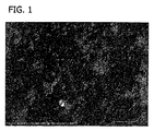
コラーゲン原線維のそれぞれの層は、実質的に非多孔性である。本明細書で使用されるとき、語句「実質的に非多孔性」とは、コラーゲンシートを形成するコラーゲン原線維の沈殿によりもたらされるコラーゲンホイル生物基質に存在する任意の孔が、大部分、互いに隔離していることを意味する。孔は、コラーゲンホイルの厚さを横断するような方法で相互連結していない。コラーゲンホイル生物基質に穴を作り出す機械的穿孔は、孔ではない。本発明の一つの例において、物質は、1500×倍率の走査電子顕微鏡を使用して目に見える孔を実質的に含まないように見える。図1〜4のように、走査電子顕微鏡画像は、コラーゲンホイル生物基質の非多孔性の性質を例示する。 Each layer of collagen fibrils is substantially non-porous. As used herein, the phrase “substantially non-porous” means that any pores present in the collagen foil biomatrix resulting from the precipitation of collagen fibrils that form the collagen sheet are largely composed of one another. Means isolated. The pores are not interconnected in such a way as to traverse the thickness of the collagen foil. Mechanical perforations that create holes in the collagen foil biomatrix are not holes. In one example of the present invention, the material appears to be substantially free of pores visible using a 1500 × magnification scanning electron microscope. As in FIGS. 1-4, scanning electron microscope images illustrate the non-porous nature of the collagen foil biomatrix.
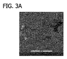
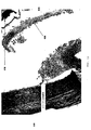
本発明の一つの実施態様において、本発明で利用されるコラーゲンホイル生物基質は、多数の多方向でからみ合うコラーゲン原線維の層からなる非天然に生じる多層コラーゲン膜である。したがって、コラーゲン原線維は、多方向の様式で平面に配置され、これらの面はシートを形成し、多層構造を作り出す。乾性コラーゲンホイル生物基質の例示を、顕微鏡写真(SEM)で見ることができ、コラーゲン原線維が埋め込まれているコラーゲンホイル生物基質の表面を例示する(図1)。コラーゲン原線維を、僅かに湿気のある環境が天然に近い条件を提供する、ESEM(環境制御型走査電子顕微鏡法)条件下での、コラーゲンホイル生物基質の上面の写真の表面で見ることができる。表面は平滑であり、実質的に非多孔性であるように見える(図2)。コラーゲンホイル生物基質の下面の写真(ESEM)は、図3においてコラーゲンホイル生物基質の実質的な非多孔性を例示する。 In one embodiment of the present invention, the collagen foil biomatrix utilized in the present invention is a non-naturally occurring multilayered collagen membrane consisting of a number of multi-directionally entangled collagen fibril layers. Thus, collagen fibrils are arranged in a plane in a multidirectional manner, and these planes form a sheet, creating a multilayer structure. An illustration of a dry collagen foil biomatrix can be seen in a micrograph (SEM), illustrating the surface of a collagen foil biomatrix in which collagen fibrils are embedded (FIG. 1). Collagen fibrils can be seen on the surface of the top photo of the collagen foil biomatrix under ESEM (Environmentally Controlled Scanning Electron Microscopy) conditions where a slightly moist environment provides near-natural conditions . The surface is smooth and appears to be substantially non-porous (FIG. 2). A photograph of the underside of the collagen foil biomatrix (ESEM) illustrates the substantial non-porosity of the collagen foil biomatrix in FIG.
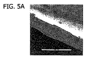
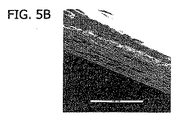
複数層における二次元方向へのコラーゲン線維の特有の配向は、主に、高静水圧下であっても液密であることに関与し、高弾性を有する強い力を提供する。コラーゲンホイル生物基質の多数の平行配向薄コラーゲン原線維層のために、この物質は、欠損を覆った後に閉鎖し、分離して、体の自己の組織を一時的に交換するのに適しており、新たな組織を形成する細胞内殖のために生体機能性骨格を提供する。本発明の複数層構造は、コラーゲンホイル生物基質の液密特性を増強する。
The unique orientation of the collagen fibers in the two-dimensional direction in the multiple layers is mainly involved in being liquid-tight even under high hydrostatic pressure, and provides a strong force with high elasticity. Due to the multiple parallel-oriented thin collagen fibril layers of the collagen foil biomatrix, this material is suitable for closing and separating after covering the defect and temporarily replacing the body's own tissue Provide a biofunctional scaffold for cell ingrowth to form new tissues. The multi-layer structure of the present invention enhances the liquid tight properties of the collagen foil biomatrix.
コラーゲンホイル生物基質は、実質的に非多孔性であるが、コラーゲン原線維の層間には隙間が存在する。コラーゲンホイル生物基質は、それぞれの頁が実質的に平滑であり、非多孔性であり、それぞれの頁の間に空間がある、頁の積み重ねと類似している。その乾燥形態である場合、隙間はより顕著である。隙間は、コラーゲンホイル生物基質が僅かに湿気のある環境で天然に近い条件で観察されたとき、低減する。コラーゲンホイル生物基質の隙間の低減は、図5において湿気のある環境下でのコラーゲンホイル生物基質の断面の画像で例示されている。液密特性の促進に加えて、コラーゲンホイル生物基質の多数の平行配向薄コラーゲン原線維層は、同時に、体の自己組織の新規構造における細胞内殖のための生物学的に同等な生体機能性骨格として機能する。 Collagen foil biomatrix is substantially non-porous, but there are gaps between layers of collagen fibrils. A collagen foil biomatrix is similar to a stack of pages where each page is substantially smooth and non-porous, with a space between each page. In the dry form, the gap is more prominent. The gap is reduced when the collagen foil biomatrix is observed in near-natural conditions in a slightly humid environment. The reduction of the collagen foil biomatrix gap is illustrated in FIG. 5 by a cross-sectional image of the collagen foil biomatrix in a humid environment. In addition to promoting fluid-tight properties, multiple parallel-oriented thin collagen fibril layers of the collagen foil biomatrix are simultaneously bio-equivalent biofunctionality for cell ingrowth in new structures of the body's self-organization Functions as a skeleton.
水和された場合、本発明で使用されるコラーゲンホイル生物基質の容量の変化は、小さいか無視できる。多孔性の代替生成物と対照的に、コラーゲンホイル生物基質は、水和されたときに実質的にその大きさ及び形状を保持し、水和された後でも優れた安定性を有し、組織と接触した後で膨張又は収縮の問題を起こさない。水和され、移植されると、コラーゲンホイル生物基質は、コラーゲンホイル生物基質を患者の組織に保持する縫合糸を引き裂くか又はフィブリン若しくは他の生体適合性接着剤シールを分裂させる程度に、領域又は厚さにおいて著しく拡大又は縮小しない。 When hydrated, the change in the volume of the collagen foil biomatrix used in the present invention is small or negligible. In contrast to porous alternative products, collagen foil biomatrix substantially retains its size and shape when hydrated, has excellent stability even after hydration, and tissue Does not cause expansion or contraction problems after contact with. When hydrated and implanted, the collagen foil biomatrix is sufficient to tear the suture holding the collagen foil biomatrix into the patient's tissue or to split the fibrin or other biocompatible adhesive seal. Does not significantly increase or decrease in thickness.
本発明の一つの例において、乾燥コラーゲンホイル生物基質の領域の収縮又は膨張は、完全に水和された場合、約−5%〜約20%に変わることができる。別の例において、乾燥コラーゲンホイル生物基質の領域は、完全に水和された場合、約−5%〜約10%に変わることができる。更なる例において、乾燥コラーゲンホイル生物基質の領域は、完全に水和された場合、約−5%〜約5%に変わる。例えば、乾燥コラーゲンホイル生物基質の領域は、完全に水和された場合、約4%以下で増加する。 In one example of the present invention, the shrinkage or expansion of the region of dry collagen foil biomatrix can vary from about −5% to about 20% when fully hydrated. In another example, the area of dry collagen foil biomatrix can vary from about −5% to about 10% when fully hydrated. In a further example, the area of dry collagen foil biomatrix varies from about −5% to about 5% when fully hydrated. For example, the area of dry collagen foil biomatrix increases below about 4% when fully hydrated.
本発明の一つの例において、コラーゲンホイル生物基質は、完全に水和された場合、乾燥厚の約6倍まで増加する。別の例において、コラーゲンホイル生物基質は、完全に水和された場合、乾燥厚の約3倍まで増加する。別の例において、コラーゲンホイル生物基質は、完全に水和された場合、乾燥厚の約2倍に増加する。 In one example of the present invention, the collagen foil biomatrix increases to about 6 times its dry thickness when fully hydrated. In another example, the collagen foil biomatrix increases to about 3 times its dry thickness when fully hydrated. In another example, the collagen foil biomatrix increases to about twice the dry thickness when fully hydrated.
本発明で使用されるコラーゲンホイル生物基質の厚さは、特定の用途により必要とされるように変わることができる。特定の大きさのコラーゲンホイル生物基質を生成するために、利用される出発物質の量を変えることによって、コラーゲンホイル生物基質の厚さを制御することができる。本発明の一つの例において、本発明で使用されるコラーゲンホイル生物基質は、乾燥形態の場合、約0.01mm〜約3.0mmの厚さを有する。別の例において、コラーゲンホイル生物基質は、約0.02mm〜約2.0mmの厚さを有する。更なる例において、コラーゲンホイル生物基質は、約0.03mm〜約1.5mmの厚さを有する。別の例において、コラーゲンホイル生物基質は、約0.05mm〜約1mmの厚さを有する。なお別の例において、コラーゲンホイル生物基質は、約1.0mm以下の厚さを有する。 The thickness of the collagen foil biomatrix used in the present invention can vary as required by the particular application. To produce a specific size collagen foil biomatrix, the thickness of the collagen foil biomatrix can be controlled by varying the amount of starting material utilized. In one example of the present invention, the collagen foil biomatrix used in the present invention has a thickness of about 0.01 mm to about 3.0 mm when in dry form. In another example, the collagen foil biomatrix has a thickness of about 0.02 mm to about 2.0 mm. In a further example, the collagen foil biomatrix has a thickness of about 0.03 mm to about 1.5 mm. In another example, the collagen foil biomatrix has a thickness of about 0.05 mm to about 1 mm. In yet another example, the collagen foil biomatrix has a thickness of about 1.0 mm or less.
コラーゲンホイル生物基質の乾燥重量は、所望の厚さによって決まる。一つの例において、コラーゲンホイル生物基質の乾燥重量は、約1mg/cm2〜約50mg/cm2である。別の例において、コラーゲンホイル生物基質の乾燥重量は、約1.5mg/cm2〜約30mg/cm2である。なお別の例において、コラーゲンホイル生物基質の乾燥重量は、約2mg/cm2〜約20mg/cm2である。更なる例において、コラーゲンホイル生物基質の乾燥重量は、約2.5mg/cm2〜約15mg/cm2である。例えば、コラーゲンホイル生物基質の乾燥重量は、約3mg/cm2〜約10mg/cm2である。 The dry weight of the collagen foil biomatrix depends on the desired thickness. In one example, the dry weight of the collagen foil biomatrix is from about 1 mg / cm 2 to about 50 mg / cm 2 . In another example, the dry weight of the collagen foil biomatrix is from about 1.5 mg / cm 2 to about 30 mg / cm 2 . In yet another example, the dry weight of the collagen foil biomatrix is from about 2 mg / cm 2 to about 20 mg / cm 2 . In a further example, the dry weight of the collagen foil biomatrix is from about 2.5 mg / cm 2 to about 15 mg / cm 2 . For example, the dry weight of the collagen foil biomatrix is about 3 mg / cm 2 to about 10 mg / cm 2 .
本発明の一つの例において、コラーゲンホイル生物基質の重量は、水和されたとき、乾燥重量の約15倍まで増加する。別の例において、コラーゲンホイル生物基質の重量は、水和されたとき、乾燥重量の約10倍まで増加する。別の例において、コラーゲンホイル生物基質の重量は、水和されたとき、乾燥重量の約7倍まで増加する。なお別の例において、コラーゲンホイル生物基質の重量は、水和されたとき、乾燥状態の約5倍まで増加する。 In one example of the present invention, the weight of the collagen foil biomatrix increases to about 15 times the dry weight when hydrated. In another example, the weight of the collagen foil biomatrix increases to about 10 times its dry weight when hydrated. In another example, the weight of the collagen foil biomatrix increases to about 7 times its dry weight when hydrated. In yet another example, the weight of the collagen foil biomatrix increases to about 5 times the dry state when hydrated.
本発明で使用されるコラーゲンホイル生物基質は、有益なことに、高い引っ張り強さを有し、このことは、例えば手術適用の際にコラーゲンホイル生物基質の取り扱いを改善及び支持し、例えば移植後に機械的安定性の増加をもたらす。加えて、コラーゲンホイル生物基質の厚さを増加することによって、引っ張り強さを有意に増加することができる。 The collagen foil biomatrix used in the present invention beneficially has a high tensile strength, which improves and supports the handling of the collagen foil biomatrix, eg during surgical applications, eg after implantation. Increases mechanical stability. In addition, the tensile strength can be significantly increased by increasing the thickness of the collagen foil biomatrix.
コラーゲンホイル生物基質を加圧下で引き裂く傾向は、「極限引張荷重」又は「極限引張力」として測定することができる(本明細書以降では、「極限引張力」と呼ぶ)。コラーゲンホイル生物基質の極限引張力は、特定の幅を有するコラーゲンホイル生物基質のストリップに圧力を受けさせ、コラーゲンホイル生物基質の破損(例えば、裂け又は破断)をもたらした付加された圧力の量を決定することによって、決定することができる。極限引張力は、次の方程式:「極限引張力」=付加圧力/コラーゲンホイル生物基質ストリップの幅=ニュートン/ストリップcmを使用して定量化することができる。 The tendency to tear a collagen foil biomatrix under pressure can be measured as “ultimate tensile load” or “ultimate tensile force” (hereinafter referred to as “ultimate tensile force”). The ultimate tensile force of a collagen foil biomatrix causes the strip of collagen foil biomatrix with a specific width to be subjected to pressure and the amount of applied pressure that causes the collagen foil biomatrix to break (eg, tear or break). It can be determined by determining. The ultimate tensile force can be quantified using the following equation: “ultimate tensile force” = applied pressure / collagen foil biomatrix strip width = Newton / strip cm.
本発明の一つの例において、コラーゲンホイル生物基質は、約1〜約30ニュートン/ストリップcm、例えば、約1.5〜約15ニュートン/ストリップcm、例えば約2〜約10ニュートン/ストリップcm、例えば、約3〜約6ニュートン/ストリップcmの極限引張力を有する。 In one example of the invention, the collagen foil biomatrix is about 1 to about 30 Newtons / strip cm, such as about 1.5 to about 15 Newtons / strip cm, such as about 2 to about 10 Newtons / strip cm, such as And an ultimate tensile force of about 3 to about 6 Newtons / cm of strip.
本発明で使用されるコラーゲンホイル生物基質は、高い引っ張り強さを有し、水和された場合、弾性及び弾力性を維持する。この特徴は、コラーゲンホイル生物基質が、接触部位に存在する解剖学的状態(例えば、曲線)に最適に適合することを可能にする。 The collagen foil biomatrix used in the present invention has high tensile strength and maintains elasticity and elasticity when hydrated. This feature allows the collagen foil biomatrix to optimally match the anatomical condition (eg, curve) present at the contact site.
水和状態の場合、コラーゲンホイル生物基質を、例えば手術部位において容易に移動することができ、例えばそれが移植される欠損の形状及び位置に最適に成形し、適合することができる。いったん移植されると、コラーゲンホイル生物基質移植片は、平滑を維持し、必要であれば位置を変えることができる。時間をかけて、細胞及び血管系が、多層コラーゲンホイル生物基質の複数層の全体を通して導かれるように移動し、最終的に、多層コラーゲンホイル生物基質を新たな組織に交換する。コラーゲンホイル生物基質の層内に細胞が移動し、血管系が形成されると、組織は導かれた方向でコラーゲンホイル生物基質の形態を取る。新たに形成された結合組織を有するコラーゲンホイル生物基質の細胞が組織された後では、脊髄硬膜組織又は脊髄神経組織への癒着形成は最小化される。 In the hydrated state, the collagen foil biomatrix can be easily moved, eg, at the surgical site, and can be optimally shaped and adapted to the shape and location of the defect in which it is implanted, for example. Once implanted, the collagen foil biomatrix graft remains smooth and can be repositioned if necessary. Over time, the cells and vasculature migrate so that they are directed through multiple layers of the multilayer collagen foil biomatrix, eventually replacing the multilayer collagen foil biomatrix with new tissue. As cells migrate into the collagen foil biomatrix layer and the vasculature is formed, the tissue takes the form of a collagen foil biomatrix in the guided direction. After cells of the collagen foil biomatrix with newly formed connective tissue are organized, adhesion formation to spinal dura mater tissue or spinal nerve tissue is minimized.
コラーゲンホイル生物基質を製造するのに使用されるコラーゲンは、あらゆる適切な供給源から得ることができる。例えば、限定することなく、コラーゲンはウシ、ヒツジ、ブタ、ウマ又はヒト由来のものであることができる。コラーゲンは、腱、真皮若しくは別のコラーゲンリッチ組織のような天然に生じる組織から採取することができるか、又は組み換え遺伝子的な方法により生成することができる。下記に記載するように、本発明の一つの例示的な実施態様は、アキレス腱から誘導されるウマコラーゲンを利用する。 The collagen used to produce the collagen foil biomatrix can be obtained from any suitable source. For example, without limitation, the collagen can be of bovine, sheep, pig, horse or human origin. Collagen can be taken from a naturally occurring tissue such as a tendon, dermis or another collagen rich tissue, or can be produced by recombinant genetic methods. As described below, one exemplary embodiment of the present invention utilizes equine collagen derived from an Achilles tendon.
例えば、WO04/108179に記載されている製造プロセスの際に、コラーゲン原線維は、原線維が溶液に沈殿してコラーゲンホイルを形成するように自然に架橋する。化学薬品又は放射線(例えば、イオン化又は紫外線放射)によりコラーゲン原線維を架橋するのではなく、コラーゲン原線維の自然な架橋を可能にすることによって、その生体機能性を確実にし、加速された再生を促進し、コラーゲンホイル生物基質が組織と接触したときに吸収時間の低減を促進する。化学薬品又は放射線によるコラーゲン原線維の架橋は、吸収時間の増加をもたらすか、さらには非吸収、被包及び瘢痕形成をもたらす可能性がある。本発明で利用されるコラーゲンホイル生物基質における原線維の自然の架橋は、自然の、生理学的様手段により生じる。主に、この自然の架橋は、非共有相互作用(例えば、ファンデルワールス若しくは双極子−双極子相互作用)によって、又はコラーゲン分子のアミノ酸側鎖間の容易に分離可能なシッフ塩基結合の形成によって終了する。コラーゲンの分子間架橋が、物理的及び化学的安定性に関与している。コラーゲン架橋の形成における主要な工程は、リシン又はヒドロキシリシン残基の酵素的変換に依存し、アルデヒド、アリシン及びヒドロキシアリシンを生じる。こられのアルデヒド基は、反応性アミノ基と自発的に反応して、不安定なアルジミン結合(例えば、−CH=N−)を有する不安定なアルドール縮合物を含有するシッフ塩基成分の形成をもたらす。したがって、本発明の生成物の原線維は、例えば弱酸を用いる処理によって分離することができる。化学架橋剤の使用により生じる架橋は、安定した共有的に架橋した架橋部分の存在により検出することができる。一般的に、このことは、シッフ塩基試薬(例えば、グルタルアルデヒド)を使用して、シッフ塩基反応生成物を形成し、次に、アマドリ転位又は還元条件のいずれかによって結合を安定化することで達成される。加えて、コラーゲンを、多様な生体機能性カルボジイミド試薬で架橋することができる。放射線の使用により生じる架橋は、照射の際に生じた遊離ラジカル部分の反応により引き起こされる、コラーゲン原線維間の安定した共有結合の存在により検出することができる。一方、本発明の生成物における原線維は、あらゆる安定した共有結合と実質的に非架橋しており、化学的又は放射線的手段で処理されていない。したがって、本発明の生成物における原線維間のあらゆる関連性は、実質的に非共有的であるか又は容易に可逆的であり、安定して架橋していない。シアンアミド、グルタルアルデヒド、ホルムアルデヒド、アクリルアミド、カルボジイミドジオン、ジイミデート、ビスアクリルアミドなどのような化学薬品が、過去に、コラーゲン原線維を化学的に架橋するために利用されてきた。しかしそのような化学薬品の使用は、神経組織と、コラーゲンホイル生物基質における残留化学薬品との不注意な接触に関連する毒性の危険性をもたらしうる。したがって、沈殿プロセスは、架橋化学薬品の毒性の危険性を回避し、化学薬品又は放射線によるコラーゲン原線維の架橋に関連するより長い吸収時間を回避する。 For example, during the manufacturing process described in WO 04/108179, collagen fibrils naturally crosslink so that the fibrils precipitate in solution to form a collagen foil. Rather than cross-linking collagen fibrils with chemicals or radiation (eg, ionization or ultraviolet radiation), by ensuring the natural cross-linking of collagen fibrils, their biofunctionality is ensured and accelerated regeneration is achieved. Promotes and reduces the absorption time when the collagen foil biomatrix comes in contact with the tissue. Cross-linking of collagen fibrils with chemicals or radiation can result in increased absorption time or even non-absorption, encapsulation and scar formation. The natural cross-linking of fibrils in the collagen foil biomatrix utilized in the present invention occurs by natural, physiological-like means. Primarily, this natural cross-linking is due to non-covalent interactions (eg van der Waals or dipole-dipole interactions) or by the formation of easily separable Schiff base bonds between the amino acid side chains of the collagen molecule. finish. Collagen intermolecular crosslinking is involved in physical and chemical stability. The main step in the formation of collagen cross-links relies on enzymatic conversion of lysine or hydroxylysine residues, resulting in aldehydes, allicin and hydroxyallysine. These aldehyde groups react spontaneously with reactive amino groups to form Schiff base components containing labile aldol condensates with labile aldimine linkages (eg, —CH═N—). Bring. Thus, the fibrils of the product of the invention can be separated, for example, by treatment with a weak acid. Crosslinking resulting from the use of a chemical crosslinker can be detected by the presence of a stable, covalently crosslinked moiety. In general, this involves using a Schiff base reagent (eg, glutaraldehyde) to form a Schiff base reaction product and then stabilizing the binding either by Amadori rearrangement or by reducing conditions. Achieved. In addition, collagen can be cross-linked with a variety of biofunctional carbodiimide reagents. Cross-linking resulting from the use of radiation can be detected by the presence of stable covalent bonds between collagen fibrils caused by the reaction of free radical moieties generated upon irradiation. On the other hand, the fibrils in the products of the present invention are substantially non-crosslinked with any stable covalent bonds and have not been treated by chemical or radiological means. Thus, any association between fibrils in the products of the present invention is substantially non-covalent or easily reversible and is not stably cross-linked. Chemicals such as cyanamide, glutaraldehyde, formaldehyde, acrylamide, carbodiimidedione, diimidate, bisacrylamide and the like have been used in the past to chemically crosslink collagen fibrils. However, the use of such chemicals can pose a risk of toxicity associated with inadvertent contact of neural tissue with residual chemicals in the collagen foil biomatrix. Thus, the precipitation process avoids the risk of cross-linking chemical toxicity and avoids longer absorption times associated with cross-linking of collagen fibrils with chemicals or radiation.
誘導された乾燥沈殿コラーゲン組成物は、二次元の多方向で天然にからみ合うコラーゲン原線維の多数の層からなる高分子量多層コラーゲン膜から構成されるコラーゲンホイル生物基質を形成する。コラーゲンホイル生物基質は、主に、間質I型コラーゲンを含有する。コラーゲンホイル生物基質は、実質的に孔を有さず、主に液密である。免疫拡散試験を生成物において実施して、外来性タンパク質の不在を確実にすることができる。コラーゲンホイル生物基質は、エチレンオキシド(ETO)若しくは同様の滅菌ガスによりガス滅菌するか、又は照射により滅菌することができる。 The derived dry precipitated collagen composition forms a collagen foil biomatrix composed of a high molecular weight multilayered collagen membrane consisting of multiple layers of collagen fibrils that are naturally entangled in two dimensions. The collagen foil biomatrix mainly contains interstitial type I collagen. The collagen foil biomatrix is substantially free of pores and is primarily liquid tight. An immunodiffusion test can be performed on the product to ensure the absence of foreign proteins. The collagen foil biomatrix can be gas sterilized with ethylene oxide (ETO) or similar sterilizing gas or sterilized by irradiation.
本発明のウマコラーゲンホイル生物基質を使用する有意な利点は、前記ホイルと接触する患者に疾患を伝染する危険性が実質的に低いことである。コラーゲン原線維を、酸(例えば、塩酸、酢酸など)又は水酸化ナトリウムのような塩基により処理してウマコラーゲンホイルを生成する製造プロセスは、有益には、存在しうる細菌、ウイルス及びプリオンを不活性化するか、又はそれらの感染レベルを低減するように作用する。塩酸、水酸化ナトリウム、エチレンオキシド(ETO)などによる生体材料の処理は、従来技術において、プリオン及びウイルスを不活性化する薬剤及び生体材料の規制内の承認された方法として認可されている。そのような処理は、一部の規制下において、バッチ毎でウマコラーゲンホイルを試験する規制要件を軽減することができる。したがって、製造プロセスでのコラーゲン原線維の処理は、製品の安定性を増強し、患者への疾患の伝染の危険性を低減する。 A significant advantage of using the equine collagen foil biomatrix of the present invention is that the risk of transmitting a disease to a patient in contact with the foil is substantially low. A manufacturing process in which collagen fibrils are treated with an acid (eg, hydrochloric acid, acetic acid, etc.) or a base such as sodium hydroxide to produce equine collagen foil beneficially eliminates bacteria, viruses and prions that may be present. Acts to activate or reduce their level of infection. Treatment of biomaterials with hydrochloric acid, sodium hydroxide, ethylene oxide (ETO), etc. has been approved in the prior art as an approved method within the regulation of drugs and biomaterials that inactivate prions and viruses. Such treatment can alleviate regulatory requirements for testing equine collagen foils on a batch-by-batch basis under some regulations. Thus, the treatment of collagen fibrils in the manufacturing process enhances product stability and reduces the risk of disease transmission to the patient.
上記の製造プロセスに付されたウマ物質は、あらゆる病原体を患者に伝染しないことが知られている。したがって、製造プロセスに加えて、ウマに基づくコラーゲンの利用は、以前はヒト死体物質と関連していた海綿状脳炎を伝染する危険性を更に回避する。ウマアキレス腱から得られるコラーゲンのようなウマ由来物から誘導されるコラーゲンの使用は、ウシ海綿状脳症(BSE)又はスクレイピーとしても知られている、伝染性海綿状脳症(TSE)を伝染する危険性を回避する。この疾患の伝染は、反芻動物供給源から得た生物学的物質(例えば、ウシ、ヤギ、ヒツジなどからの生物学的物質)の使用に関連している。 It is known that equine material subjected to the above manufacturing process does not transmit any pathogens to the patient. Thus, in addition to the manufacturing process, the use of equine-based collagen further avoids the risk of transmitting spongiform encephalitis previously associated with human cadaveric material. The use of collagen derived from equine-derived materials, such as collagen obtained from equine Achilles tendon, is a risk of infecting contagious spongiform encephalopathy (TSE), also known as bovine spongiform encephalopathy (BSE) or scrapie To avoid. Transmission of this disease is associated with the use of biological material obtained from ruminant sources (eg, biological material from cattle, goats, sheep, etc.).
コラーゲンがウマ由来物から誘導され、(例えば、酵素により)処理されている、本発明で使用されるコラーゲンホイル生物基質は、追加的に、免疫反応を誘発する危険性を低減する。 The collagen foil biomatrix used in the present invention, where the collagen is derived from horse-derived material and processed (eg, with an enzyme) additionally reduces the risk of eliciting an immune response.
ウマ誘導コラーゲンホイル生物基質は、また、炎症性反応の低減をもたらす。ヒト大腿筋膜のような供給源から誘導されるコラーゲンを含有するホイルと比較すると、ウマコラーゲンホイル生物基質との接触によりもたらされる炎症性細胞の数は、有意に少ない。 The equine derived collagen foil biomatrix also results in a reduction of the inflammatory response. Compared to foils containing collagen derived from sources such as human thigh fascia, the number of inflammatory cells brought about by contact with the equine collagen foil biomatrix is significantly lower.
使用する前に、乾燥コラーゲンホイル生物基質を、例えば生理食塩水で水和することができる。一つの例において、生理食塩水は、0.9%塩化ナトリウム溶液を含む。別の例において、コラーゲンホイル生物基質は、賦形剤又は薬剤含有溶液で水和される。コラーゲンホイル生物基質を水和するのに必要な時間の長さは、ホイルの厚さに関係する。コラーゲンホイル生物基質は、全領域にわたって厚さが一定になるまで水和される。一つの例において、コラーゲンホイル生物基質は、生理食塩水で約0.5秒から約1時間水和される。別の例において、コラーゲンホイル生物基質は、生理食塩水で約0.5秒から約30分間水和される。別の例において、コラーゲンホイル生物基質は、生理食塩水で約0.5秒から約20分間水和される。別の例において、コラーゲンホイル生物基質は、生理食塩水で約0.5秒から約10分間水和される。なお別の例において、コラーゲンホイル生物基質は、生理食塩水で約0.5秒から約2分間水和される。別の例において、コラーゲンホイル生物基質は、例えばコラーゲンホイル生物基質を生理食塩水に浸けることにより、生理食塩水で約0.5秒から約10秒間水和される。別の例において、コラーゲンホイル生物基質は、組織と接触する前に水和されない。 Prior to use, the dried collagen foil biomatrix can be hydrated, eg, with saline. In one example, the saline solution includes a 0.9% sodium chloride solution. In another example, the collagen foil biomatrix is hydrated with an excipient or drug-containing solution. The length of time required to hydrate the collagen foil biomatrix is related to the thickness of the foil. The collagen foil biomatrix is hydrated until the thickness is constant over the entire area. In one example, the collagen foil biomatrix is hydrated with saline for about 0.5 seconds to about 1 hour. In another example, the collagen foil biomatrix is hydrated with saline for about 0.5 seconds to about 30 minutes. In another example, the collagen foil biomatrix is hydrated with saline for about 0.5 seconds to about 20 minutes. In another example, the collagen foil biomatrix is hydrated with saline for about 0.5 seconds to about 10 minutes. In yet another example, the collagen foil biomatrix is hydrated with saline for about 0.5 seconds to about 2 minutes. In another example, the collagen foil biomatrix is hydrated with saline for about 0.5 seconds to about 10 seconds, eg, by immersing the collagen foil biomatrix in saline. In another example, the collagen foil biomatrix is not hydrated prior to contacting the tissue.
コラーゲンホイル生物基質は、外科的技術を確立することにより、例えばフィブリンシーラント、組織接着剤、縫合糸により、又は圧力適合外科技術により、患者の組織に結合することができる。あるいは、コラーゲンホイル生物基質と、組織又は組織の表面の血液との自然な誘引を使用して、コラーゲンホイル生物基質を、あらゆるシーラント、接着剤、縫合又は圧力適合技術を使用することなく組織と結合することができる。いったん水和されると、コラーゲンホイル生物基質を、例えば患者の組織における外科的開口部よりも僅かに大きく切断することができる。それにより、コラーゲンホイル生物基質が、それが結合する患者の組織から僅かにはみ出す。一つの例において、水和コラーゲンホイル生物基質を、組織からおよそ0.5cm〜約1cmはみ出すような大きさにする。はみ出し量は、外科医の好み及び技術に応じて変わることができる。 The collagen foil biomatrix can be bound to the patient's tissue by establishing surgical techniques, for example, by fibrin sealants, tissue adhesives, sutures, or by pressure compatible surgical techniques. Alternatively, the natural attraction of the collagen foil biomatrix and the blood on the tissue or the surface of the tissue is used to bind the collagen foil biomatrix to the tissue without using any sealant, adhesive, suture or pressure matching techniques. can do. Once hydrated, the collagen foil biomatrix can be cut slightly larger than, for example, a surgical opening in a patient's tissue. Thereby, the collagen foil biomatrix protrudes slightly from the patient's tissue to which it binds. In one example, the hydrated collagen foil biomatrix is sized to protrude from about 0.5 cm to about 1 cm from the tissue. The amount of protrusion can vary depending on the surgeon's preference and skill.
一つの例において、コラーゲンとフィブリノーゲン及びフィブロネクチンとの周知の相互作用によって、コラーゲンホイル生物基質を、フィブリンシーラントを用いて所定の場所に固定することができる。外科的使用に承認されているフィブリンシーラントの例には、TISSUCOL及びTISSEELフィブリンシーラント(Baxter AG, Vienna, Austria)が挙げられる。あるいは、広範囲な炎症反応を誘発せず、脊髄の手術における使用が承認されている組織接着剤を利用することもできる。フィブリンシーラント又は組織接着剤を、液密シールを形成するために、組織からはみ出しているコラーゲンホイル生物基質の周囲部分に連続線で起用することができる。液密シール固定は、隣接組織と出血との接触に関連する合併症、例えば、フィブリンによる癒着形成の誘発を回避するので、有益である。別の例において、コラーゲンホイル生物基質は、フィブリンシーラント又は組織接着剤の連続線により組織と結合する場合、液密シールを生じる。更なる例において、組織からはみ出しているコラーゲンホイル生物基質に、組織に結合するために、フィブリンシーラント又は組織接着剤を点在させることができる。なお別の例において、コラーゲンホイル生物基質は、いったん所望の接触部位に位置決めされると、それを組織に外科的に縫合することによって結合される。コラーゲンホイル生物基質が縫合される場合、ホイルを引き裂くのを防止するために無緊張縫合技術を使用しなければならない。縫合線を例えばフィブリンシーラントで封鎖することが推奨される。別の例において、コラーゲンホイル生物基質は、当該技術で既知の圧力適合技術により位置決めされ、移植される。この技術において、コラーゲンホイル生物基質は、所望の移植部位に位置決めされ、周囲組織のより所定の場所に保持される。したがって、移植片は、縫合糸、フィブリンシーラント又は組織接着剤を使用することなく、所定の場所にとどまる。別の例において、コラーゲンホイル生物基質は、あらゆるシーラント、接着剤、縫合又は圧力適合技術を使用することなく、位置決めされ、移植される。この技術において、コラーゲンホイル生物基質は、所望の移植部位に位置決めされ、コラーゲンホイル生物基質と哺乳動物の組織との間に生じる自然な誘引又は接着により所定の場所に保持される。別の例において、コラーゲンホイル生物基質を、組織に適用し、上記の方法のいずれかにより固定することができ、次に、別のコラーゲンホイル生物基質を、隣接組織に適用し、上記の方法のいずれかにより適用することができ、したがって、コラーゲンホイル生物基質の隣接シートをもたらすことができる。 In one example, the collagen foil biomatrix can be immobilized in place using a fibrin sealant by the well known interaction of collagen with fibrinogen and fibronectin. Examples of fibrin sealants that are approved for surgical use include TISSUCOL and TISSEEL fibrin sealants (Baxter AG, Vienna, Austria). Alternatively, tissue adhesives that do not elicit a widespread inflammatory response and are approved for use in spinal surgery can be utilized. Fibrin sealant or tissue adhesive can be applied in a continuous line to the surrounding portion of the collagen foil biomatrix that protrudes from the tissue to form a fluid tight seal. Liquid tight seal fixation is beneficial because it avoids complications associated with contact between adjacent tissues and bleeding, such as induction of adhesion formation by fibrin. In another example, a collagen foil biomatrix produces a fluid tight seal when bonded to tissue by a continuous line of fibrin sealant or tissue adhesive. In a further example, the collagen foil biomatrix that protrudes from the tissue can be interspersed with fibrin sealant or tissue adhesive to bind the tissue. In yet another example, the collagen foil biomatrix is joined by surgically suturing it to tissue once it is positioned at the desired contact site. When a collagen foil biomatrix is sutured, a tensionless suture technique must be used to prevent tearing the foil. It is recommended that the suture be sealed with, for example, a fibrin sealant. In another example, the collagen foil biomatrix is positioned and implanted by pressure fitting techniques known in the art. In this technique, the collagen foil biomatrix is positioned at the desired implantation site and held in place in the surrounding tissue. Thus, the implant remains in place without the use of sutures, fibrin sealant or tissue adhesive. In another example, the collagen foil biomatrix is positioned and implanted without the use of any sealant, adhesive, suture or pressure matching techniques. In this technique, a collagen foil biomatrix is positioned at a desired implantation site and held in place by natural attraction or adhesion that occurs between the collagen foil biomatrix and mammalian tissue. In another example, a collagen foil biomatrix can be applied to the tissue and fixed by any of the methods described above, then another collagen foil biomatrix is applied to the adjacent tissue and the method described above. Can be applied by either, thus resulting in an adjacent sheet of collagen foil biomatrix.
場合により、本発明のコラーゲンホイル生物基質を、他の生成物と共に利用することができる。例えば、コラーゲンホイル生物基質を組織に適用し、上記に記載された方法のいずれかにより固定した後、癒着防止生成物をコラーゲンホイル生物基質又は隣接組織の上面又は下面に適用することができる。一つの実施態様において、CoSeal(登録商標)(Baxter Healthcare corporationから入手可能)のようなPEGに基づいた生成物を、コラーゲンホイル生物基質又は隣接組織の上面又は下面に適用することができる。本発明のコラーゲンホイル生物基質は、「滑りやすい」表面を作り出すのではなく、組織を分離することにより及び組織再生を導くことにより癒着を防止するので、その作用は、細胞が癒着しない「滑りやすい」表面を一時的に作り出す生成物を利用することにより補足されることができる。別の実施態様において、PEGに基づく生成物で一面又は両面が既に被覆されている、直ぐに使用できるコラーゲンホイル生物基質を使用することができる。 Optionally, the collagen foil biomatrix of the present invention can be utilized with other products. For example, after the collagen foil biomatrix is applied to the tissue and fixed by any of the methods described above, the anti-adhesion product can be applied to the upper or lower surface of the collagen foil biomatrix or adjacent tissue. In one embodiment, a PEG-based product such as CoSeal® (available from Baxter Healthcare corporation) can be applied to the upper or lower surface of the collagen foil biomatrix or adjacent tissue. The collagen foil biomatrix of the present invention does not create a “slippery” surface, but prevents adhesion by separating tissue and leading to tissue regeneration, so its action is “slippery” where cells do not adhere. It can be supplemented by utilizing a product that temporarily creates a surface. In another embodiment, a ready-to-use collagen foil biomatrix that is already coated on one or both sides with a PEG-based product can be used.
加えて、本発明は、脊柱及び硬膜、並びに周囲組織からなる群より選択される組織の連絡切断により特徴付けられる、哺乳動物における、例えば傷害、手術又は病原体に基づく疾患のような障害を処置する薬剤、すなわち医学的に適用可能な物質の製造における多層コラーゲンホイル生物基質の使用に関する。 In addition, the present invention treats disorders in mammals, such as diseases based on injury, surgery or pathogens, characterized by the disconnection of tissues selected from the group consisting of the spinal column and dura mater, and surrounding tissues. To the use of multilayered collagen foil biomatrix in the manufacture of pharmaceuticals, i.e. medically applicable substances.
実施例:癒着及び線維形成、基本及び研究目標
過剰な瘢痕組織形成及び癒着は、頻繁に根性痛及び身体的機能障害を引き起こす、脊髄の手術における重篤な問題であり、例えば腰椎椎間板の手術の失敗又は椎間板切除後症候群である。修正手術の場合の硬膜損傷の症例が、文献において5〜10%報告されている。
Examples: Adhesion and fibrosis, basic and research goals
Excessive scar tissue formation and adhesions are serious problems in spinal surgery that frequently cause radical pain and physical dysfunction, such as lumbar disc surgery failure or post-discectomy syndrome. Cases of dural injury in the case of corrective surgery have been reported in the literature 5-10%.
癒着形成の術後発生及び程度についての最も重要な臨界パラメーターは、以下である:
−手術領域の術前状態(患者の遺伝的素因を含む癒着/線維化の存在);
−手術の術中活動(創傷の程度、組織及び特定の解剖学的構造(例えば、膜)の傷害の回避、止血の注意、組織の乾燥の回避など);
−術後癒着の防止及び最小化の術中処置、例えば、スポンジ、ホイル又はゲルのような癒着防止移植物の移植。
The most important critical parameters for the postoperative occurrence and extent of adhesion formation are the following:
-Preoperative status in the surgical area (presence of adhesion / fibrosis including genetic predisposition of the patient);
-Surgical intraoperative activity (degree of wound, avoidance of injury to tissue and specific anatomical structures (eg membrane), attention to hemostasis, avoidance of tissue dryness, etc.)
-Intraoperative treatment of prevention and minimization of post-surgical adhesions, for example implantation of anti-adhesion implants such as sponges, foils or gels.
癒着には複数の原因があるので、外科的処置は、全ての患者において100%の癒着防止を達成することはあり得ないが、上記のパラメーターの前提条件に影響を与えることはできる。外科的目標は、術後期間の間及び初期における外科的創傷状態の最適化である。この期間における創傷状態は、主に、あらゆる術後癒着の形成及び線維形成に関与することが知られている。 Because there are multiple causes of adhesions, surgical procedures may not achieve 100% adhesion prevention in all patients, but can affect the prerequisites for the above parameters. The surgical goal is the optimization of the surgical wound status during and early after the postoperative period. It is known that the wound condition during this period is mainly involved in the formation of all post-operative adhesions and fibrosis.
癒着/硬膜周囲線維化の病因について考えると、硬膜周囲背側創傷領域における生理学的瘢痕形成の際の、潜在的な、硬膜を保護し、癒着を防止し、最小化する方法及び生成物の調査行為は、最初の術後期間(およそ、手術後1〜2週間)に集中している。これは、特定の組成物が、背側の外科的欠損領域における瘢痕形成から硬膜を保護し、分離する機能、並びに、あらゆる制御されない瘢痕/癒着/線維形成の主な要因及び基本的な原因である、血液、フィブリノーゲン、フィブリン及び壊死物質の制御されない術後分布を回避する機能を果たすかを決定するためには、最も重要な期間である。 Given the pathogenesis of adhesions / peridural fibrosis, methods and generations that protect the dura mater, prevent adhesions, and minimize the potential for physiological scar formation in the peridural dorsal wound area Object investigation activities are concentrated in the first postoperative period (approximately 1-2 weeks after surgery). This is because the particular composition protects and isolates the dura from scar formation in the dorsal surgical defect area, as well as the main and fundamental causes of any uncontrolled scar / adhesion / fibrosis Is the most important period to determine whether it serves the function of avoiding uncontrolled post-operative distribution of blood, fibrinogen, fibrin and necrotic material.
術後2週間目の状態は、移植物の一体化についての評価、並びに移植物の種類及び生体活性、生体機能性及び組織適合性についての潜在性における結論を可能にする。例えば、2週間後、移植物は、背側の解剖学的構造において十分に許容され、細胞化し、一体化しているか、又は対照的に、長期間異物として作用し、被包及び瘢痕形成を誘発するか。以下の実施例の目的は、術後期間における生物学的コラーゲンホイル生物基質の一時的な分離層として及び細胞増殖及び組織再生のための生物基質としての生体機能性を評価すること、並びに臨床的に関連性のある癒着及び制御されない瘢痕組織形成の防止及び最小化の値を評価することである。 The condition at 2 weeks post-surgery allows assessment of implant integrity and conclusion on the potential for implant type and bioactivity, biofunctionality and histocompatibility. For example, after 2 weeks, the implant is well tolerated in the dorsal anatomy, cellized, integrated, or in contrast, acts as a foreign body for a long time, inducing encapsulation and scar formation Or. The purpose of the following examples is to evaluate biofunctionality as a temporary separation layer of biological collagen foil biomatrix in the postoperative period and as a biomatrix for cell growth and tissue regeneration, and clinical To evaluate the value of prevention and minimization of adhesions and uncontrolled scar tissue formation related to
実施例I:物質及び方法:生体機能性コラーゲンホイル生物基質を使用する脊髄手術において癒着の低減を示した動物実験
A)図7〜9に示されている物質及び方法:
動物:ニュージーランド白色ウサギ、両方の性別、体重4kg。由来:Charles River, Sulzfeld, Deutschland or Harlan Winkelmann, D−33178 Borchen。
Example I: Materials and Methods: Animal experiments that showed reduced adhesions in spinal surgery using biofunctional collagen foil biomatrix
A) Materials and methods shown in FIGS. 7-9:
Animal: New Zealand white rabbit, both sexes, weight 4 kg. Origin: Charles River, Sulzfeld, Deutschland or Harlan Winkelmann, D-33178 Borchen.
モデル及び群:
生体機能性コラーゲンホイル生物基質の使用:椎弓板切除、脊髄神経根の露出及びコラーゲンホイル生物基質の適用。各群は20匹の動物を含む。
Models and groups:
Use of biofunctional collagen foil biomatrix: laminectomy, spinal nerve root exposure and application of collagen foil biomatrix. Each group contains 20 animals.
群1:椎弓板切除、皮膚閉鎖(対照)。 Group 1: laminectomy, skin closure (control).
群2:椎弓板切除、コラーゲンホイル生物基質で覆う。 Group 2: laminectomy, covered with collagen foil biomatrix.
手術:
動物を腹臥位で位置決めし、剃毛した手術領域(脊柱腰椎部)を固定した。滅菌条件下で、皮膚の正中切開後、脊椎傍筋組織を棘状突起から分離し、腰椎脊柱の椎弓板を露出した。第3及び第4腰椎の半側椎弓板切除を実施した。対応する2つの脊髄神経根及び根チャンネルの露出。異なる群における手術の継続も記載される。
Surgery:
The animal was positioned in the prone position and the shaved surgical area (vertebral lumbar region) was fixed. Under sterile conditions, after a midline skin incision, paravertebral muscle tissue was separated from the spinous processes and the lumbar spine lamina was exposed. Half and laminectomy of the third and fourth lumbar vertebrae was performed. Exposure of the corresponding two spinal nerve roots and root channels. The continuation of surgery in different groups is also described.
一般的な麻酔:Ketavetを60mg/kg
Rompunを16mg/kg(皮下)
チオペンタールを効果に基づいて耳静脈から静注
Narkosis、挿管法、人工呼吸
疼痛投薬:2×日。Temgesicを0.05mg/kg(皮下)で手術後3〜4日間。
General anesthesia: Kevetet 60 mg / kg
Rompun 16mg / kg (subcutaneous)
Intravenous injection of thiopental from the ear vein based on the effect
Narcosis, intubation, artificial respiration
Pain medication: 2 x days. 3-4 days after surgery with Tegmesic 0.05 mg / kg (subcutaneous).
安楽死:静注でのバルビツール過剰投与による全身麻酔。 Euthanasia: General anesthesia with intravenous barbiturate overdose.
評価及び病理学的試験:
各群の動物を次のように安楽死させた:10日後に動物10匹、4週間後に動物10匹、及び3か月後に動物10匹。評価パラメーター:癒着及び線維化の程度。その後、広範囲な組織学的処置を行った。実験は、生体機能性コラーゲンホイル生物基質を利用した群において癒着形成の低減を示した。
Evaluation and pathological examination:
Animals in each group were euthanized as follows: 10 animals after 10 days, 10 animals after 4 weeks, and 10 animals after 3 months. Evaluation parameters: degree of adhesion and fibrosis. Thereafter, extensive histological treatment was performed. Experiments have shown reduced adhesion formation in groups utilizing biofunctional collagen foil biomatrix.
実施例II:ヒト患者における移植後1週間のコラーゲンホイル生物基質
ヒト患者(年齢:18歳、性別:女性)を、日常的な外科的処置(てんかん)の枠内で7日間の間隔を置いて手術した。コラーゲンホイル生物基質を、1回目の手術の際に硬膜外に移植した(CSFの漏れの防止)。2回目の手術の際に、硬膜の再開口の前に、コラーゲンホイル生物基質を日常的に移動した。硬膜外移植の1週間後、コラーゲンホイル生物基質は、図10に示されているように、依然として機械的に安定しており、移動可能であった。
Example II: Collagen foil biomatrix one week after transplantation in human patients
Human patients (age: 18 years, gender: female) were operated at intervals of 7 days within routine surgical procedures (epilepsy). A collagen foil biomatrix was transplanted epidurally during the first surgery (prevention of CSF leakage). During the second surgery, the collagen foil biomatrix was routinely transferred prior to reopening of the dura mater. One week after epidural implantation, the collagen foil biomatrix was still mechanically stable and movable, as shown in FIG.
図11は、移植後1週間の図10のコラーゲンホイル生物基質の断面図である。見て分かるように、線維芽細胞は、多層構造に導かれてコラーゲンホイル生物基質の中へ増殖する。縦方向の貫入は、約220〜320μmである。 FIG. 11 is a cross-sectional view of the collagen foil biomatrix of FIG. 10 one week after transplantation. As can be seen, the fibroblasts are guided into a multi-layered structure and grow into the collagen foil biomatrix. The longitudinal penetration is about 220-320 μm.
多層構造に沿って導かれた修復細胞の内殖の速度は、横断方向(「図10」)と比較して縦方向では約10〜15倍速かった。最小限の炎症性浸潤が示され、継続中の再生プロセスを表している。 The rate of ingrowth of repair cells guided along the multilayer structure was about 10-15 times faster in the longitudinal direction compared to the transverse direction (“FIG. 10”). Minimal inflammatory infiltration is shown, representing an ongoing regeneration process.
実施例III:脊髄手術におけるコラーゲンホイル生物基質の移植後の細胞増殖、組織再生、並びに硬膜周囲癒着及び線維化の防止
物質:
精製し、細かく刻んだ、ウマのアキレス腱から生成し、沈殿して原線維にした非変性ウマコラーゲン原線維(主にI型コラーゲン)。柔軟な形状安定性及び弾性の生物基質を特別に設計し、非多孔性の流体密封性多層構造を有する。乾燥多層コラーゲンホイル膜の厚さは、約0.1mmであった。湿潤条件では、膜厚は、0.3mmに成長した。
Example III: Prevention of cell proliferation, tissue regeneration, and pericardial adhesion and fibrosis after transplantation of collagen foil biomatrix in spinal surgery
Non-denatured equine collagen fibrils (mainly type I collagen) produced from horse achilles tendon that has been purified and minced and precipitated into fibrils. Specially designed for flexible shape-stable and elastic biomatrix and has a non-porous, fluid-tight multilayer structure. The thickness of the dried multilayer collagen foil film was about 0.1 mm. Under wet conditions, the film thickness grew to 0.3 mm.
一般的な麻酔:Ketavetを60mg/kg
Rompunを16mg/kg(皮下)
チオペンタールを効果に基づいて耳静脈から静注
Narkosis、挿管法、人工呼吸
疼痛投薬:2×日。Temgesicを0.05mg/kg(皮下)で手術後3〜4日間。
General anesthesia: Kevetet 60 mg / kg
Rompun 16mg / kg (subcutaneous)
Intravenous injection of thiopental from the ear vein based on the effect
Narcosis, intubation, artificial respiration
Pain medication: 2 x days. 3-4 days after surgery with Tegmesic 0.05 mg / kg (subcutaneous).
安楽死:静注でのバルビツール過剰投与による全身麻酔。 Euthanasia: General anesthesia with intravenous barbiturate overdose.
動物モデル:
本発明の研究は、体重が手術時で平均3kgであり、平均年齢が4か月であるニュージーランド白色(「NZW」)ウサギにより実施した。全てのウサギは雌であり、動物研究は、オーストリア、ウイーン市の正式な承認の後で実施した。このウサギの動物実験モデルにおける手術の設計は、ヒトにおける最も一般的な手術介入と同様であった。椎弓板切除及び摘除は、第4から第5脊柱腰椎部(L4/5)の椎間関節で実施した。Posterior Lumbar Interbody Fusion(PLIF)手術のような実際の手術を完全に実施するために、黄色靱帯を摘除した。脊髄の上にある硬膜はそのまま残しておいた。椎弓板切除領域を、本発明の生物学的コラーゲンホイル生物基質で覆った。脊椎傍筋肉を所定の場所に戻し、筋膜を吸収性縫合糸で閉鎖した。皮膚をsyntofil縫合糸で閉鎖した。
Animal model:
The study of the present invention was performed with New Zealand white ("NZW") rabbits, who weighed 3 kg on average at the time of surgery and had an average age of 4 months. All rabbits were female and animal studies were conducted after formal approval from the city of Vienna, Austria. The design of surgery in this rabbit animal experimental model was similar to the most common surgical intervention in humans. Laminectomy and excision were performed at the facet joints of the 4th to 5th lumbar vertebrae (L4 / 5). The ligamentum flavum was removed in order to fully perform the actual operation, such as the Posterial Lumbar Interbody Fusion (PLIF) operation. The dura mater above the spinal cord was left intact. The laminectomy area was covered with the biological collagen foil biomatrix of the present invention. The paravertebral muscle was returned to place and the fascia was closed with absorbable sutures. The skin was closed with syntofil suture.
ウサギは、最初の3日間は術後疼痛投薬及び流体注入を受け、混合食餌を摂取した。ウサギをチオペンタールの過剰投与により安楽死させ、脊椎の手術領域を取り出し、組織学的評価のために単離した。 Rabbits received postoperative pain medication and fluid infusion for the first 3 days and received a mixed diet. Rabbits were euthanized by overdose of thiopental, the surgical area of the spine was removed and isolated for histological evaluation.
組織学的方法:
脊柱を一括切除し、固定のために10%ホルマリン溶液に浸漬した。脱灰した後、各腰椎を薄片に切断し、脱水し、パラフィンの中に埋め込んだ。7個の3μm厚の切片を各試料から取った。
Histological method:
The spinal column was excised in a lump and immersed in a 10% formalin solution for fixation. After decalcification, each lumbar vertebra was cut into slices, dehydrated and embedded in paraffin. Seven 3 μm thick sections were taken from each sample.
切片を、全細胞及び骨成分を染色する全体的な組織学のために、Haematoxilin−Eosinにより染色した。スライドを、硬膜と硬膜周囲背側外科的創傷領域との関係、コラーゲンホイル生物基質への細胞内殖と癒着形成及び硬膜周囲線維創傷形成の不在/存在及び程度との関係のような解剖学的構造を評価することによって検査した。 Sections were stained with Haematoxilin-Eosin for overall histology staining of whole cells and bone components. Slides, such as the relationship between the dura mater and peridural dorsal surgical wound area, the relationship between cell in-growth and adhesion formation on collagen foil biomatrix and absence / presence and extent of pericardial fiber wound formation The anatomy was examined by evaluating.
結果/組織学的所見:
切片を、全細胞及び骨成分を染色する全体的な組織学のために、Haematoxilin−Eosinにより染色した。スライドを、硬膜と硬膜周囲背側外科的創傷領域との関係のような解剖学的構造を評価することによって検査した。コラーゲンホイル生物基質の中への及び上での細胞増殖、並びに生物基質及び周囲組織における炎症性反応の量及び質を分析した。一体化/組み込みプロセス、並びに癒着形成及び硬膜周辺結合組織の組織化の不在/存在及び程度も分析した。
Results / histological findings:
Sections were stained with Haematoxilin-Eosin for overall histology staining of whole cells and bone components. Slides were examined by evaluating anatomy such as the relationship between the dura mater and the perdural dorsal surgical wound area. Cell growth into and on the collagen foil biomatrix and the quantity and quality of inflammatory responses in the biomatrix and surrounding tissues were analyzed. The integration / incorporation process, as well as the absence / presence and extent of adhesion formation and peridural connective tissue organization were also analyzed.
研究は以下の組織学的所見を示す:
術後直後:コラーゲンホイル生物基質を、硬膜と背側欠損領域との間に置き、硬膜と背側創傷領域との間に生物学的分離層を形成する。背側欠損の組織に固定しない。血液(赤血球)は、コラーゲンホイル生物基質の表面の薄層に付着し、止血物質としての機能を証明する。非多孔性であり、血液に対して流体密封性であるコラーゲンホイル生物基質の微視的な平行多層構造が、はっきりと目に見える。コラーゲンホイル生物基質の厚さは、約0.3mmである(図12)。
The study shows the following histological findings:
Immediately after surgery: A collagen foil biomatrix is placed between the dura mater and the dorsal defect area to form a biological separation layer between the dura mater and the dorsal wound area. Do not fix to dorsal defect tissue. Blood (red blood cells) adheres to a thin layer on the surface of the collagen foil biomatrix and proves its function as a hemostatic substance. A microscopic parallel multilayer structure of a collagen foil biomatrix that is non-porous and fluid-tight to blood is clearly visible. The thickness of the collagen foil biomatrix is about 0.3 mm (FIG. 12).
術後1週間:膜は、周囲組織とほぼ一体化している。血液細胞、特にリンパ球の浸潤がある(図13A)。硬膜下腔には、細胞浸潤又は癒着組織がない。背側表面において、細胞は、表面に沿って導かれ、コラーゲンホイル生物基質の表面にほとんど貫入していない。欠損の端部では、コラーゲン生物基質は、骨に適用されている。接触領域(a)は、生体活性(生物基質/細胞相互作用)の優先領域であり、多層構造に沿って導かれた細胞浸潤の開始地点である。多層コラーゲンホイル生物基質と硬膜との間の領域は、図13Aにおいて示されているように、脂肪細胞を含有する疎性組織(b)からなり、解剖学的構造が手術により影響を受けなかった領域における硬膜外腔の組織(c)に匹敵する。 One week after surgery: The membrane is almost integrated with the surrounding tissue. There is infiltration of blood cells, especially lymphocytes (FIG. 13A). The subdural space is free of cell infiltration or adhesions. On the dorsal surface, the cells are guided along the surface and hardly penetrate the surface of the collagen foil biomatrix. At the end of the defect, a collagen biomatrix is applied to the bone. The contact area (a) is a preferential area of bioactivity (biomatrix / cell interaction) and is a starting point of cell infiltration guided along the multilayer structure. The area between the multilayered collagen foil biomatrix and the dura mater consists of loose tissue (b) containing adipocytes, as shown in FIG. 13A, and the anatomy is not affected by surgery. Comparable to tissue (c) in the epidural space in the region.
図13Aは、欠損の端部における多層コラーゲンホイル生物基質と骨との接触領域(a)を示す。細胞と多層コラーゲンホイル生物基質との強い交互作用。多層コラーゲンホイル生物基質の再構築は、多層コラーゲンホイル生物基質の平行多層構造への細胞(線維芽細胞、顆粒球)の導かれた浸潤によって開始する。 FIG. 13A shows the contact area (a) of the multilayer collagen foil biomatrix and bone at the end of the defect. Strong interaction between cells and multilayer collagen foil biomatrix. Reconstruction of the multilayered collagen foil biomatrix is initiated by guided infiltration of cells (fibroblasts, granulocytes) into the parallel multilayered structure of the multilayered collagen foil biomatrix.
図13Bは、欠損の端部における多層コラーゲンホイル生物基質と骨との接触領域(a)を示す。 FIG. 13B shows the contact area (a) of the multilayered collagen foil biomatrix and bone at the end of the defect.
図13Cは、椎弓板切除欠損の中央における多層コラーゲンホイル生物基質を示す。コラーゲン生物基質は一体化しており、腹側硬膜外腔を背側創傷形成から分離している。細胞は、背側表面に沿って導かれており、コラーゲン生物基質の表面に貫入していない。コラーゲン生物基質と硬質との間の領域(a)は、脂肪細胞を含有する疎性組織からなる。 FIG. 13C shows a multilayered collagen foil biomatrix in the middle of a laminectomy defect. The collagen biomatrix is integrated and separates the ventral epidural space from the dorsal wound formation. The cells are guided along the dorsal surface and do not penetrate the surface of the collagen biomatrix. The area (a) between the collagen biomatrix and hard consists of loose tissue containing adipocytes.
図13Dは、術後1週間のNZWウサギを示す。多層コラーゲンホイル生物基質は、椎弓板切除欠損を閉鎖し、硬膜を、細胞リッチ背側創傷形成の開始地点から分離している。細胞は、コラーゲン生物基質の表面に貫入していない。コラーゲン生物基質の端部では、多層構造(a)への修復細胞の導かれた浸潤の始まりが見られる。
FIG. 13D shows a NZW rabbit 1 week after surgery. The multilayered collagen foil biomatrix closes the laminectomy defect and separates the dura from the beginning of cell rich dorsal wound formation. Cells do not penetrate the surface of the collagen biomatrix. At the end of the collagen biomatrix, the onset of guided infiltration of repair cells into the multilayer structure (a) is seen.
図13Eは、術後1週間のNZWウサギを示す。コラーゲンスポンジ(DURAGEN)を使用して椎弓板切除欠損を覆った。本発明と対照的に、硬膜外腔と背側創傷領域との間に明確な非多孔性分離層がない。コラーゲンスポンジは、血液で浸されている。 FIG. 13E shows a NZW rabbit 1 week after surgery. A laminectomy defect was covered using a collagen sponge (DURAGEN). In contrast to the present invention, there is no clear non-porous separation layer between the epidural space and the dorsal wound area. The collagen sponge is immersed in blood.
術後2週間:膜と周囲組織との一体化は、毛細血管構造の内殖により改善されている。分葉核顆粒球であるリンパ球の量が増加している。コラーゲンホイル生物基質の構造は、周囲組織と区別することができる。膜の上方においてリンパ球及び顆粒級の滲出による炎症性反応がある。硬膜下腔には、細胞浸潤又は癒着組織がない。 2 weeks after surgery: integration of membrane and surrounding tissue is improved by ingrowth of capillary structures. The amount of lymphocytes that are segmented nucleus granulocytes is increasing. The structure of the collagen foil biomatrix can be distinguished from the surrounding tissue. Above the membrane is an inflammatory response due to lymphocyte and granular grade exudation. The subdural space is free of cell infiltration or adhesions.
図14A〜Cは、術後2週間のNZWウサギを示す。多層コラーゲンホイル生物基質は完全に一体化している。組織修復細胞は、コラーゲン生物基質に浸潤し、多層構造に沿って導かれている。硬膜は、脂肪細胞を有する疎性組織によって、創傷形成及び再構築コラーゲン生物基質から分離されている。 14A-C show NZW rabbits 2 weeks after surgery. The multilayer collagen foil biomatrix is fully integrated. Tissue repair cells infiltrate the collagen biomatrix and are guided along the multilayer structure. The dura mater is separated from the wounding and remodeling collagen biomatrix by loose tissue with adipocytes.
結論:
本発明の非多孔性多層コラーゲンホイル生物基質は、硬膜と背側手術欠損との間に即効性の優れた分離及び保護機能を有し、硬膜周囲領域への血液、フィブリン及び壊死性物質の制御されない分布を回避し、背側表面上方の創傷の始まりにおいて細胞増殖を導き、分離することを実証した。生体機能性コラーゲンホイルは、また、多層構造の中へ細胞内殖を導く。多層構造に沿った内殖の速度は、背側表面から生物基質の中への細胞の内殖よりも速い。第1周目ではコラーゲンホイル生物基質の腹側表面への細胞の内殖はない。多層構造に沿って又はそれを横断する細胞内殖の異なる速度は、術後1週間における多層コラーゲンホイル生物基質の硬膜周囲移植後の臨床観察(実施例II)と一致している。
Conclusion:
The non-porous multi-layer collagen foil biomatrix of the present invention has an excellent immediate separation and protection function between the dura mater and the dorsal surgical defect, and blood, fibrin and necrotic substances to the peridural region Was demonstrated to lead to and separate cell proliferation at the beginning of the wound above the dorsal surface. Biofunctional collagen foils also lead to cell ingrowth into the multilayer structure. The rate of ingrowth along the multilayer structure is faster than the ingrowth of cells from the dorsal surface into the biomatrix. In the first round, there is no ingrowth of cells on the ventral surface of the collagen foil biomatrix. The different rates of cell ingrowth along or across the multilayer structure are consistent with clinical observations after peridural implantation of the multilayer collagen foil biomatrix one week after surgery (Example II).
2週間後、多層生物基質は修復細胞により浸潤されている。生物基質と背側手術創傷領域の生理学的創傷形成との完全な一体化が存在する。硬膜は、硬膜を脊柱管から生理学的に分離する、脂肪細胞と同様に見える疎性脂肪組織により分離されている。細胞リッチ生物基質の徐々に消滅する多層構造に沿って導かれる修復細胞の高い生体活性が存在する。生物基質は、正常な解剖学的構造に再構築され、一体化される。 Two weeks later, the multilayer biomatrix is infiltrated with repair cells. There is a complete integration between the biomatrix and the physiological wound formation in the dorsal surgical wound area. The dura is separated by loose adipose tissue that looks like adipocytes, which physiologically separates the dura from the spinal canal. There is a high bioactivity of repaired cells that is guided along the multi-layered structure of cell-rich biomatrix. The biomatrix is reconstructed and integrated into a normal anatomy.
本発明の多層コラーゲンホイル生物基質は、多層構造内で細胞の内殖及び背側表面の上方で細胞増殖を導く、硬膜と背側欠損領域との間の有効な生体適合性及び生体機能性の迅速な保護及び分離層であることを実証した。細胞増殖を導くことによって、コラーゲンホイル生物基質は、再構築及び組織再生プロセスを有効に制御し、臨床的に関連する癒着の防止及び最小化のために最適な条件を提供する。 The multilayered collagen foil biomatrix of the present invention provides effective biocompatibility and biofunctionality between the dura mater and the dorsal defect region that leads to cell in-growth and cell growth above the dorsal surface within the multilayer structure. It proved to be a quick protection and separation layer. By directing cell growth, the collagen foil biomatrix effectively controls the remodeling and tissue regeneration processes and provides optimal conditions for the prevention and minimization of clinically relevant adhesions.
Claims (9)
Applications Claiming Priority (3)
| Application Number | Priority Date | Filing Date | Title |
|---|---|---|---|
| US80959106P | 2006-05-31 | 2006-05-31 | |
| US60/809,591 | 2006-05-31 | ||
| PCT/EP2007/004791 WO2007137839A2 (en) | 2006-05-31 | 2007-05-30 | Method for directed cell in-growth and controlled tissue regeneration in spinal surgery |
Publications (3)
| Publication Number | Publication Date |
|---|---|
| JP2009538649A JP2009538649A (en) | 2009-11-12 |
| JP2009538649A5 JP2009538649A5 (en) | 2011-06-02 |
| JP5231401B2 true JP5231401B2 (en) | 2013-07-10 |
Family
ID=38457907
Family Applications (1)
| Application Number | Title | Priority Date | Filing Date |
|---|---|---|---|
| JP2009512489A Active JP5231401B2 (en) | 2006-05-31 | 2007-05-30 | Method of guided cell ingrowth and controlled tissue regeneration in spinal surgery |
Country Status (14)
| Country | Link |
|---|---|
| US (1) | US8703122B2 (en) |
| EP (1) | EP2021045B1 (en) |
| JP (1) | JP5231401B2 (en) |
| KR (1) | KR20090017654A (en) |
| CN (1) | CN101454030B (en) |
| AU (1) | AU2007267338B2 (en) |
| BR (1) | BRPI0712088B8 (en) |
| CA (1) | CA2651941C (en) |
| CO (1) | CO6140041A2 (en) |
| ES (1) | ES2575933T3 (en) |
| HK (1) | HK1128638A1 (en) |
| MX (1) | MX2008014847A (en) |
| NO (1) | NO20085419L (en) |
| WO (1) | WO2007137839A2 (en) |
Cited By (1)
| Publication number | Priority date | Publication date | Assignee | Title |
|---|---|---|---|---|
| RU2644278C1 (en) * | 2016-12-21 | 2018-02-08 | Федеральное государственное автономное учреждение "Национальный медицинский исследовательский центр нейрохирургии имени академика Н.Н.Бурденко" Министерства здравоохранения Российской Федерации (ФГАУ "НМИЦ нейрохирургии им. акад. Н.Н.Бурденко" Минздрава России | Method for microscurgeric reconstruction of the spinal cord on an animal model by using polyvinyl alcohol biodegradated hydrogel |
Families Citing this family (34)
| Publication number | Priority date | Publication date | Assignee | Title |
|---|---|---|---|---|
| US6066325A (en) | 1996-08-27 | 2000-05-23 | Fusion Medical Technologies, Inc. | Fragmented polymeric compositions and methods for their use |
| US8603511B2 (en) | 1996-08-27 | 2013-12-10 | Baxter International, Inc. | Fragmented polymeric compositions and methods for their use |
| US8303981B2 (en) | 1996-08-27 | 2012-11-06 | Baxter International Inc. | Fragmented polymeric compositions and methods for their use |
| US7435425B2 (en) * | 2001-07-17 | 2008-10-14 | Baxter International, Inc. | Dry hemostatic compositions and methods for their preparation |
| US8834864B2 (en) * | 2003-06-05 | 2014-09-16 | Baxter International Inc. | Methods for repairing and regenerating human dura mater |
| US7927626B2 (en) | 2003-08-07 | 2011-04-19 | Ethicon, Inc. | Process of making flowable hemostatic compositions and devices containing such compositions |
| CN101454030B (en) | 2006-05-31 | 2013-06-26 | 巴克斯特国际公司 | Method for directed cell in-growth and controlled tissue regeneration in spinal surgery |
| TWI436793B (en) | 2006-08-02 | 2014-05-11 | Baxter Int | Rapidly acting dry sealant and methods for use and manufacture |
| US9056151B2 (en) * | 2007-02-12 | 2015-06-16 | Warsaw Orthopedic, Inc. | Methods for collagen processing and products using processed collagen |
| US20080260794A1 (en) * | 2007-02-12 | 2008-10-23 | Lauritzen Nels J | Collagen products and methods for producing collagen products |
| US8092541B2 (en) * | 2007-08-03 | 2012-01-10 | Warsaw Orthopedic, Inc. | Method of using an anti-growth matrix as a barrier for cell attachment and osteo-inductive factors |
| JP2011500237A (en) | 2007-10-30 | 2011-01-06 | バクスター・インターナショナル・インコーポレイテッド | Use of regenerative biofunctional collagen biomatrix to treat visceral or cavity wall defects |
| WO2009109194A2 (en) | 2008-02-29 | 2009-09-11 | Ferrosan A/S | Device for promotion of hemostasis and/or wound healing |
| US9039783B2 (en) | 2009-05-18 | 2015-05-26 | Baxter International, Inc. | Method for the improvement of mesh implant biocompatibility |
| PT2442835E (en) * | 2009-06-16 | 2015-03-23 | Baxter Healthcare Sa | Hemostatic sponge |
| JP2013514093A (en) | 2009-12-16 | 2013-04-25 | バクスター・インターナショナル・インコーポレイテッド | Hemostatic sponge |
| SA111320355B1 (en) | 2010-04-07 | 2015-01-08 | Baxter Heathcare S A | Hemostatic sponge |
| US8460691B2 (en) | 2010-04-23 | 2013-06-11 | Warsaw Orthopedic, Inc. | Fenestrated wound repair scaffold |
| US8790699B2 (en) | 2010-04-23 | 2014-07-29 | Warsaw Orthpedic, Inc. | Foam-formed collagen strand |
| JP6289096B2 (en) | 2010-06-01 | 2018-03-07 | バクスター・インターナショナル・インコーポレイテッドBaxter International Incorp0Rated | Process for making a dry and stable hemostatic composition |
| US9084728B2 (en) | 2010-06-01 | 2015-07-21 | Baxter International Inc. | Process for making dry and stable hemostatic compositions |
| CN103037845B (en) | 2010-06-01 | 2015-11-25 | 巴克斯特国际公司 | For the preparation of the method for dry, stable hemostatic composition |
| US20130090291A1 (en) | 2011-10-11 | 2013-04-11 | Baxter Healthcare S.A. | Hemostatic compositions |
| AU2012318257B2 (en) | 2011-10-11 | 2015-10-01 | Baxter Healthcare S.A. | Hemostatic compositions |
| US20130108671A1 (en) | 2011-10-27 | 2013-05-02 | Baxter Healthcare S.A. | Hemostatic compositions |
| JP6241624B2 (en) | 2012-03-06 | 2017-12-06 | フェロサン メディカル デバイシーズ エイ/エス | Pressurized container containing hemostatic paste |
| JP6394916B2 (en) | 2012-06-12 | 2018-09-26 | フェロサン メディカル デバイシーズ エイ/エス | Dry hemostatic composition |
| US9114190B2 (en) | 2013-02-08 | 2015-08-25 | Laser Spine Institute, Llc | Regeneration of spinal discs |
| AU2014283170B2 (en) | 2013-06-21 | 2017-11-02 | Ferrosan Medical Devices A/S | Vacuum expanded dry composition and syringe for retaining same |
| CA2928963C (en) | 2013-12-11 | 2020-10-27 | Ferrosan Medical Devices A/S | Dry composition comprising an extrusion enhancer |
| JP6726852B2 (en) | 2014-10-13 | 2020-07-22 | フェッローサン メディカル ディバイス エー/エス | Dry composition for use in hemostasis and wound healing |
| JP6747650B2 (en) | 2014-12-24 | 2020-08-26 | フェロサン メディカル デバイシーズ エイ/エス | Syringe for holding and mixing the first substance and the second substance |
| CA2986981A1 (en) | 2015-07-03 | 2017-01-12 | Ferrosan Medical Devices A/S | Syringe for mixing two components and for retaining a vacuum in a storage condition |
| JP7395113B2 (en) | 2018-05-09 | 2023-12-11 | フェロサン メディカル デバイシーズ エイ/エス | Method of preparing a hemostatic composition |
Family Cites Families (133)
| Publication number | Priority date | Publication date | Assignee | Title |
|---|---|---|---|---|
| US2507244A (en) * | 1947-04-14 | 1950-05-09 | Upjohn Co | Surgical gelatin dusting powder and process for preparing same |
| CH264752A (en) * | 1947-06-03 | 1949-10-31 | Hoffmann La Roche | Process for the manufacture of carriers for pharmaceuticals. |
| SE420565B (en) | 1974-06-06 | 1981-10-19 | Pharmacia Ab | AID FOR INTRAVASCULAR ADMINISTRATION FOR USE IN CONNECTION WITH INTRAVASCULAR ADMINISTRATION OF A SOLUTION OR SUSPENSION OF A DIAGNOSTIC AGENT |
| US4013078A (en) * | 1974-11-25 | 1977-03-22 | Feild James Rodney | Intervertebral protector means |
| US4164559A (en) * | 1977-09-21 | 1979-08-14 | Cornell Research Foundation, Inc. | Collagen drug delivery device |
| DE2843963A1 (en) * | 1978-10-09 | 1980-04-24 | Merck Patent Gmbh | BODY-RESORBABLE SHAPED MATERIAL BASED ON COLLAGEN AND THEIR USE IN MEDICINE |
| US4265233A (en) * | 1978-04-12 | 1981-05-05 | Unitika Ltd. | Material for wound healing |
| US4179400A (en) | 1978-05-09 | 1979-12-18 | W. R. Grace & Co. | Process for preparing catalytic solutions of sulfonium salts |
| AT359652B (en) | 1979-02-15 | 1980-11-25 | Immuno Ag | METHOD FOR PRODUCING A TISSUE ADHESIVE |
| AT359653B (en) | 1979-02-15 | 1980-11-25 | Immuno Ag | METHOD FOR PRODUCING A TISSUE ADHESIVE |
| DE3036033A1 (en) | 1980-09-24 | 1982-05-06 | Max-Planck-Gesellschaft zur Förderung der Wissenschaften e.V., 3400 Göttingen | POWDERED WOUND TREATMENT AND METHOD FOR THE PRODUCTION THEREOF |
| US4300494A (en) | 1979-09-26 | 1981-11-17 | Shell Oil Company | Thermal insulated intake ports |
| US4292972A (en) | 1980-07-09 | 1981-10-06 | E. R. Squibb & Sons, Inc. | Lyophilized hydrocolloio foam |
| DE3105624A1 (en) * | 1981-02-16 | 1982-09-02 | Hormon-Chemie München GmbH, 8000 München | MATERIAL FOR SEALING AND HEALING Wounds |
| US4424208A (en) * | 1982-01-11 | 1984-01-03 | Collagen Corporation | Collagen implant material and method for augmenting soft tissue |
| DE3360633D1 (en) * | 1982-02-12 | 1985-10-03 | Unitika Ltd | Anti-cancer device |
| US4482386A (en) | 1982-03-26 | 1984-11-13 | Warner-Lambert Company | Method of conditioning a water swellable hydrocolloid |
| US4543332A (en) * | 1982-03-29 | 1985-09-24 | Miles Laboratories, Inc. | Method for the preparation of spherical microorganism cell aggregates |
| US4540410A (en) * | 1982-11-16 | 1985-09-10 | Serono Pharmaceutical Partners | Lyophilized compositions, preparation and use thereof |
| EP0132983B2 (en) | 1983-07-14 | 1991-06-12 | Hitachi Chemical Co., Ltd. | Production of gelatin spherical gels and their use |
| JPS60100516A (en) | 1983-11-04 | 1985-06-04 | Takeda Chem Ind Ltd | Preparation of sustained release microcapsule |
| US4515637A (en) * | 1983-11-16 | 1985-05-07 | Seton Company | Collagen-thrombin compositions |
| AT389815B (en) * | 1984-03-09 | 1990-02-12 | Immuno Ag | METHOD FOR INACTIVATING VARIABLE FILTERABLE DISEASERS IN BLOOD PRODUCTS |
| US4600574A (en) * | 1984-03-21 | 1986-07-15 | Immuno Aktiengesellschaft Fur Chemisch-Medizinische Produkte | Method of producing a tissue adhesive |
| US4837285A (en) * | 1984-03-27 | 1989-06-06 | Medimatrix | Collagen matrix beads for soft tissue repair |
| JPS6144825A (en) * | 1984-08-09 | 1986-03-04 | Unitika Ltd | Hemostatic agent |
| GB8422950D0 (en) * | 1984-09-11 | 1984-10-17 | Warne K J | Hydrogel |
| JPS61122222A (en) * | 1984-11-19 | 1986-06-10 | Koken:Kk | Hemostatic agent composed of collagen or gelatin and protamine |
| US5178883A (en) * | 1984-11-29 | 1993-01-12 | Regents Of The University Of Minnesota | Method for promoting hair growth |
| US5165938A (en) | 1984-11-29 | 1992-11-24 | Regents Of The University Of Minnesota | Wound healing agents derived from platelets |
| US4600533A (en) | 1984-12-24 | 1986-07-15 | Collagen Corporation | Collagen membranes for medical use |
| US5007916A (en) * | 1985-08-22 | 1991-04-16 | Johnson & Johnson Medical, Inc. | Method and material for prevention of surgical adhesions |
| IE59361B1 (en) * | 1986-01-24 | 1994-02-09 | Akzo Nv | Pharmaceutical preparation for obtaining a highly viscous hydrogel or suspension |
| IL78826A (en) * | 1986-05-19 | 1991-05-12 | Yissum Res Dev Co | Precursor composition for the preparation of a biodegradable implant for the sustained release of an active material and such implants prepared therefrom |
| US5300494A (en) * | 1986-06-06 | 1994-04-05 | Union Carbide Chemicals & Plastics Technology Corporation | Delivery systems for quaternary and related compounds |
| US4946870A (en) * | 1986-06-06 | 1990-08-07 | Union Carbide Chemicals And Plastics Company Inc. | Delivery systems for pharmaceutical or therapeutic actives |
| US4832686A (en) * | 1986-06-24 | 1989-05-23 | Anderson Mark E | Method for administering interleukin-2 |
| US4803075A (en) * | 1986-06-25 | 1989-02-07 | Collagen Corporation | Injectable implant composition having improved intrudability |
| US5080893A (en) * | 1988-05-31 | 1992-01-14 | University Of Florida | Method for preventing surgical adhesions using a dilute solution of polymer |
| US5017229A (en) * | 1990-06-25 | 1991-05-21 | Genzyme Corporation | Water insoluble derivatives of hyaluronic acid |
| US5140016A (en) * | 1988-05-31 | 1992-08-18 | University Of Florida | Method and composition for preventing surgical adhesions using a dilute solution of polymer |
| US5350573A (en) * | 1988-05-31 | 1994-09-27 | University Of Florida Research Foundation, Inc. | Method and composition for preventing surgical adhesions |
| US5447966A (en) * | 1988-07-19 | 1995-09-05 | United States Surgical Corporation | Treating bioabsorbable surgical articles by coating with glycerine, polalkyleneoxide block copolymer and gelatin |
| US4925677A (en) * | 1988-08-31 | 1990-05-15 | Theratech, Inc. | Biodegradable hydrogel matrices for the controlled release of pharmacologically active agents |
| US5041292A (en) * | 1988-08-31 | 1991-08-20 | Theratech, Inc. | Biodegradable hydrogel matrices for the controlled release of pharmacologically active agents |
| US5135751A (en) * | 1988-11-16 | 1992-08-04 | Mediventures Incorporated | Composition for reducing postsurgical adhesions |
| US5126141A (en) * | 1988-11-16 | 1992-06-30 | Mediventures Incorporated | Composition and method for post-surgical adhesion reduction with thermo-irreversible gels of polyoxyalkylene polymers and ionic polysaccharides |
| US5162430A (en) * | 1988-11-21 | 1992-11-10 | Collagen Corporation | Collagen-polymer conjugates |
| US5510418A (en) * | 1988-11-21 | 1996-04-23 | Collagen Corporation | Glycosaminoglycan-synthetic polymer conjugates |
| US5614587A (en) * | 1988-11-21 | 1997-03-25 | Collagen Corporation | Collagen-based bioadhesive compositions |
| US4891359A (en) * | 1988-12-08 | 1990-01-02 | Johnson & Johnson Patient Care, Inc. | Hemostatic collagen paste composition |
| DE3903672C1 (en) * | 1989-02-08 | 1990-02-01 | Lohmann Gmbh & Co Kg | |
| EP0493387B1 (en) | 1989-08-10 | 1993-10-20 | W.L. Gore & Associates, Inc. | A medical dispensing system for tissue adhesive components |
| US5196185A (en) * | 1989-09-11 | 1993-03-23 | Micro-Collagen Pharmaceutics, Ltd. | Collagen-based wound dressing and method for applying same |
| US5061274A (en) | 1989-12-04 | 1991-10-29 | Kensey Nash Corporation | Plug device for sealing openings and method of use |
| US5219328A (en) * | 1990-01-03 | 1993-06-15 | Cryolife, Inc. | Fibrin sealant delivery method |
| US5134229A (en) * | 1990-01-12 | 1992-07-28 | Johnson & Johnson Medical, Inc. | Process for preparing a neutralized oxidized cellulose product and its method of use |
| JPH0813750B2 (en) * | 1990-03-01 | 1996-02-14 | 持田製薬株式会社 | Oral thrombin formulation |
| US5306501A (en) * | 1990-05-01 | 1994-04-26 | Mediventures, Inc. | Drug delivery by injection with thermoreversible gels containing polyoxyalkylene copolymers |
| US5595735A (en) * | 1990-05-23 | 1997-01-21 | Johnson & Johnson Medical, Inc. | Hemostatic thrombin paste composition |
| US5292362A (en) * | 1990-07-27 | 1994-03-08 | The Trustees Of Columbia University In The City Of New York | Tissue bonding and sealing composition and method of using the same |
| US5209776A (en) * | 1990-07-27 | 1993-05-11 | The Trustees Of Columbia University In The City Of New York | Tissue bonding and sealing composition and method of using the same |
| US5192300A (en) * | 1990-10-01 | 1993-03-09 | Quinton Instrument Company | Insertion assembly and method of inserting a vessel plug into the body of a patient |
| US5108421A (en) * | 1990-10-01 | 1992-04-28 | Quinton Instrument Company | Insertion assembly and method of inserting a vessel plug into the body of a patient |
| NO302481B1 (en) * | 1990-10-16 | 1998-03-09 | Takeda Chemical Industries Ltd | Polymer for an extended release preparation, as well as an extended release preparation |
| US5129882A (en) * | 1990-12-27 | 1992-07-14 | Novoste Corporation | Wound clotting device and method of using same |
| US5690675A (en) | 1991-02-13 | 1997-11-25 | Fusion Medical Technologies, Inc. | Methods for sealing of staples and other fasteners in tissue |
| AU654574B2 (en) * | 1991-06-14 | 1994-11-10 | Amgen, Inc. | Collagen film drug delivery for proteins |
| AT398079B (en) * | 1991-11-04 | 1994-09-26 | Immuno Ag | PREPARATION WITH THROMBINE ACTIVITY AND METHOD FOR THEIR PRODUCTION |
| DK0632820T3 (en) * | 1992-02-28 | 2000-10-02 | Collagen Corp | Highly concentrated, homogenized collagen compositions |
| ATE269371T1 (en) | 1992-02-28 | 2004-07-15 | Cohesion Tech Inc | INJECTABLE CERAMIC COMPOUNDS AND METHOD FOR THEIR PRODUCTION AND USE |
| US5204382A (en) * | 1992-02-28 | 1993-04-20 | Collagen Corporation | Injectable ceramic compositions and methods for their preparation and use |
| US5384333A (en) * | 1992-03-17 | 1995-01-24 | University Of Miami | Biodegradable injectable drug delivery polymer |
| CA2134071C (en) * | 1992-04-23 | 1999-04-27 | Sew Wah Tay | Apparatus and method for sealing vascular punctures |
| IL105529A0 (en) * | 1992-05-01 | 1993-08-18 | Amgen Inc | Collagen-containing sponges as drug delivery for proteins |
| AU4406793A (en) * | 1992-06-04 | 1993-12-30 | Clover Consolidated, Limited | Water-soluble polymeric carriers for drug delivery |
| US5385606A (en) * | 1992-07-06 | 1995-01-31 | Kowanko; Nicholas | Adhesive composition and method |
| US5413571A (en) * | 1992-07-16 | 1995-05-09 | Sherwood Medical Company | Device for sealing hemostatic incisions |
| US5428022A (en) * | 1992-07-29 | 1995-06-27 | Collagen Corporation | Composition of low type III content human placental collagen |
| US5514379A (en) * | 1992-08-07 | 1996-05-07 | The General Hospital Corporation | Hydrogel compositions and methods of use |
| DE4227681C2 (en) * | 1992-08-21 | 1995-05-18 | Becker & Co Naturinwerk | Wound covering material based on collagen fibers and process for its production |
| CA2149221C (en) * | 1992-11-12 | 2005-02-08 | Neville Alleyne | Cardiac protection device |
| US5667839A (en) * | 1993-01-28 | 1997-09-16 | Collagen Corporation | Human recombinant collagen in the milk of transgenic animals |
| EP0702959B1 (en) | 1993-05-31 | 2001-08-08 | Kaken Pharmaceutical Co., Ltd. | Cross-linked gelatin gel preparation containing basic fibroblast growth factor |
| FR2715309B1 (en) * | 1994-01-24 | 1996-08-02 | Imedex | Adhesive composition, for surgical use, based on collagen modified by oxidative cutting and not crosslinked. |
| US5674275A (en) | 1994-04-06 | 1997-10-07 | Graphic Controls Corporation | Polyacrylate and polymethacrylate ester based hydrogel adhesives |
| US5531759A (en) * | 1994-04-29 | 1996-07-02 | Kensey Nash Corporation | System for closing a percutaneous puncture formed by a trocar to prevent tissue at the puncture from herniating |
| GB9415739D0 (en) | 1994-07-30 | 1994-09-21 | Scimat Ltd | Gel wound dressing |
| US5516532A (en) * | 1994-08-05 | 1996-05-14 | Children's Medical Center Corporation | Injectable non-immunogenic cartilage and bone preparation |
| US5931165A (en) * | 1994-09-06 | 1999-08-03 | Fusion Medical Technologies, Inc. | Films having improved characteristics and methods for their preparation and use |
| US5698213A (en) | 1995-03-06 | 1997-12-16 | Ethicon, Inc. | Hydrogels of absorbable polyoxaesters |
| US5580923A (en) | 1995-03-14 | 1996-12-03 | Collagen Corporation | Anti-adhesion films and compositions for medical use |
| US6129761A (en) | 1995-06-07 | 2000-10-10 | Reprogenesis, Inc. | Injectable hydrogel compositions |
| PT876165E (en) | 1995-12-18 | 2006-10-31 | Angiotech Biomaterials Corp | COMPOSITIONS OF RETICULATED POLYMERS AND PROCESSES FOR THEIR USE |
| US6458889B1 (en) | 1995-12-18 | 2002-10-01 | Cohesion Technologies, Inc. | Compositions and systems for forming crosslinked biomaterials and associated methods of preparation and use |
| JP3476631B2 (en) | 1995-12-21 | 2003-12-10 | 株式会社アムニオテック | Medical material composed of human-derived natural collagen membrane |
| US5748318A (en) * | 1996-01-23 | 1998-05-05 | Brown University Research Foundation | Optical stress generator and detector |
| CA2251475C (en) | 1996-04-04 | 2006-09-05 | Immuno Aktiengesellschaft | Hemostatic sponge based on collagen |
| US5902832A (en) * | 1996-08-20 | 1999-05-11 | Menlo Care, Inc. | Method of synthesizing swollen hydrogel for sphincter augmentation |
| US7435425B2 (en) | 2001-07-17 | 2008-10-14 | Baxter International, Inc. | Dry hemostatic compositions and methods for their preparation |
| US6706690B2 (en) * | 1999-06-10 | 2004-03-16 | Baxter Healthcare Corporation | Hemoactive compositions and methods for their manufacture and use |
| US6066325A (en) | 1996-08-27 | 2000-05-23 | Fusion Medical Technologies, Inc. | Fragmented polymeric compositions and methods for their use |
| US6063061A (en) * | 1996-08-27 | 2000-05-16 | Fusion Medical Technologies, Inc. | Fragmented polymeric compositions and methods for their use |
| US7320962B2 (en) * | 1996-08-27 | 2008-01-22 | Baxter International Inc. | Hemoactive compositions and methods for their manufacture and use |
| US7871637B2 (en) * | 1996-08-27 | 2011-01-18 | Baxter International Inc. | Dry hemostatic compositions and methods for their preparation |
| TW501934B (en) * | 1996-11-20 | 2002-09-11 | Tapic Int Co Ltd | Collagen material and process for making the same |
| EP0986408B1 (en) | 1997-06-03 | 2005-05-11 | Innogenetics N.V. | New medicaments based on polymers composed of methacrylamide-modified gelatin |
| US5908054A (en) * | 1997-06-16 | 1999-06-01 | Fusion Medical Technologies, Inc. | Fluid dispersion and delivery assembly and method |
| US5997895A (en) | 1997-09-16 | 1999-12-07 | Integra Lifesciences Corporation | Dural/meningeal repair product using collagen matrix |
| DE69831964T2 (en) | 1997-09-16 | 2006-07-27 | Integra Lifesciences Corp. | COMPOSITION FOR PROMOTING THE GROWTH OF DURAL OR MENINGEAL TISSUE CONTAINING COLLAGEN |
| US6179872B1 (en) * | 1998-03-17 | 2001-01-30 | Tissue Engineering | Biopolymer matt for use in tissue repair and reconstruction |
| US6227394B1 (en) * | 1998-06-09 | 2001-05-08 | Asahi Glass Company Ltd. | Glass bulb for a cathode ray tube and a method for producing a cathode ray tube |
| US6110484A (en) * | 1998-11-24 | 2000-08-29 | Cohesion Technologies, Inc. | Collagen-polymer matrices with differential biodegradability |
| US6328229B1 (en) | 1998-12-18 | 2001-12-11 | Cohesion Technologies, Inc. | Low volume mixing spray head for mixing and dispensing of two reactive fluid components |
| US6312725B1 (en) | 1999-04-16 | 2001-11-06 | Cohesion Technologies, Inc. | Rapid gelling biocompatible polymer composition |
| US6312474B1 (en) | 1999-09-15 | 2001-11-06 | Bio-Vascular, Inc. | Resorbable implant materials |
| US6221109B1 (en) | 1999-09-15 | 2001-04-24 | Ed. Geistlich Söhne AG fur Chemische Industrie | Method of protecting spinal area |
| CA2422852C (en) | 2000-09-18 | 2012-06-26 | Organogenesis Inc. | Methods for treating a patient using a bioengineered flat sheet graft prostheses |
| EP1359947A2 (en) | 2001-01-25 | 2003-11-12 | Nycomed Pharma AS | A suspension comprising fibrinogen, thrombin and alcohol and a method of coating a carrier with the same |
| JP2003160506A (en) * | 2001-08-10 | 2003-06-03 | Ed Geistlich Soehne Ag Fuer Chemische Industrie | Collagen carrier of therapeutic genetic material and method |
| AU2002300450B2 (en) * | 2001-08-10 | 2007-04-05 | Ed. Geistlich Soehne Ag Fuer Chemische Industrie | Collagen Carrier of Therapeutic Genetic Material, and Method |
| US8834864B2 (en) * | 2003-06-05 | 2014-09-16 | Baxter International Inc. | Methods for repairing and regenerating human dura mater |
| AU2004245086B2 (en) * | 2003-06-05 | 2008-06-26 | Baxter Healthcare S.A. | Compositions for repairing and regenerating human dura mater |
| EP1682196A2 (en) * | 2003-11-10 | 2006-07-26 | Angiotech International Ag | Medical implants and anti-scarring agents |
| AU2004293463A1 (en) * | 2003-11-20 | 2005-06-09 | Angiotech International Ag | Implantable sensors and implantable pumps and anti-scarring agents |
| US20080091277A1 (en) * | 2004-08-13 | 2008-04-17 | Kai Deusch | Surgical prosthesis having biodegradable and nonbiodegradable regions |
| ES2367583T3 (en) | 2005-05-04 | 2011-11-10 | Suprapolix B.V. | HYDROGELS WITH HYDROGEN LINKS. |
| WO2007001926A2 (en) | 2005-06-24 | 2007-01-04 | Hyperbranch Medical Technology, Inc. | Low-swelling hydrogel sealants for wound repair |
| CN101454030B (en) | 2006-05-31 | 2013-06-26 | 巴克斯特国际公司 | Method for directed cell in-growth and controlled tissue regeneration in spinal surgery |
| TWI436793B (en) | 2006-08-02 | 2014-05-11 | Baxter Int | Rapidly acting dry sealant and methods for use and manufacture |
| JP2011500237A (en) * | 2007-10-30 | 2011-01-06 | バクスター・インターナショナル・インコーポレイテッド | Use of regenerative biofunctional collagen biomatrix to treat visceral or cavity wall defects |
| US9039783B2 (en) | 2009-05-18 | 2015-05-26 | Baxter International, Inc. | Method for the improvement of mesh implant biocompatibility |
| PT2442835E (en) | 2009-06-16 | 2015-03-23 | Baxter Healthcare Sa | Hemostatic sponge |
-
2007
- 2007-05-30 CN CN200780019553XA patent/CN101454030B/en active Active
- 2007-05-30 KR KR1020087031934A patent/KR20090017654A/en not_active Application Discontinuation
- 2007-05-30 JP JP2009512489A patent/JP5231401B2/en active Active
- 2007-05-30 ES ES07725680.8T patent/ES2575933T3/en active Active
- 2007-05-30 CA CA2651941A patent/CA2651941C/en active Active
- 2007-05-30 BR BRPI0712088A patent/BRPI0712088B8/en not_active IP Right Cessation
- 2007-05-30 EP EP07725680.8A patent/EP2021045B1/en active Active
- 2007-05-30 US US12/302,716 patent/US8703122B2/en active Active
- 2007-05-30 MX MX2008014847A patent/MX2008014847A/en active IP Right Grant
- 2007-05-30 AU AU2007267338A patent/AU2007267338B2/en not_active Ceased
- 2007-05-30 WO PCT/EP2007/004791 patent/WO2007137839A2/en active Application Filing
-
2008
- 2008-12-29 CO CO08137571A patent/CO6140041A2/en unknown
- 2008-12-30 NO NO20085419A patent/NO20085419L/en not_active Application Discontinuation
-
2009
- 2009-07-28 HK HK09106947.2A patent/HK1128638A1/en not_active IP Right Cessation
Cited By (1)
| Publication number | Priority date | Publication date | Assignee | Title |
|---|---|---|---|---|
| RU2644278C1 (en) * | 2016-12-21 | 2018-02-08 | Федеральное государственное автономное учреждение "Национальный медицинский исследовательский центр нейрохирургии имени академика Н.Н.Бурденко" Министерства здравоохранения Российской Федерации (ФГАУ "НМИЦ нейрохирургии им. акад. Н.Н.Бурденко" Минздрава России | Method for microscurgeric reconstruction of the spinal cord on an animal model by using polyvinyl alcohol biodegradated hydrogel |
Also Published As
| Publication number | Publication date |
|---|---|
| AU2007267338A1 (en) | 2007-12-06 |
| CN101454030B (en) | 2013-06-26 |
| CA2651941A1 (en) | 2007-12-06 |
| BRPI0712088B8 (en) | 2021-06-22 |
| EP2021045A2 (en) | 2009-02-11 |
| CO6140041A2 (en) | 2010-03-19 |
| CN101454030A (en) | 2009-06-10 |
| US20100028309A1 (en) | 2010-02-04 |
| WO2007137839A8 (en) | 2008-02-28 |
| AU2007267338B2 (en) | 2013-04-04 |
| WO2007137839A3 (en) | 2008-11-27 |
| MX2008014847A (en) | 2009-04-30 |
| WO2007137839A2 (en) | 2007-12-06 |
| NO20085419L (en) | 2008-12-30 |
| ES2575933T3 (en) | 2016-07-04 |
| BRPI0712088B1 (en) | 2018-05-29 |
| EP2021045B1 (en) | 2016-03-16 |
| CA2651941C (en) | 2015-02-17 |
| HK1128638A1 (en) | 2009-11-06 |
| JP2009538649A (en) | 2009-11-12 |
| BRPI0712088A2 (en) | 2012-03-06 |
| KR20090017654A (en) | 2009-02-18 |
| US8703122B2 (en) | 2014-04-22 |
Similar Documents
| Publication | Publication Date | Title |
|---|---|---|
| JP5231401B2 (en) | Method of guided cell ingrowth and controlled tissue regeneration in spinal surgery | |
| US8790698B2 (en) | Use of a regenerative biofunctional collagen biomatrix for treating visceral or parietal defects | |
| US8834864B2 (en) | Methods for repairing and regenerating human dura mater | |
| JP4537998B2 (en) | Composition for repairing and regenerating human dura mater | |
| JP2003510101A (en) | Collagen-containing products for promoting dural or meningeal tissue growth | |
| US10723783B2 (en) | Polypeptide compositions and methods of using the same | |
| CA3144487C (en) | Tissue fibrosis inhibitor in which biocompatible polymer is used | |
| Meddings | Evaluation of collagen vicryl mesh as a dural graft substance |
Legal Events
| Date | Code | Title | Description |
|---|---|---|---|
| A521 | Request for written amendment filed |
Free format text: JAPANESE INTERMEDIATE CODE: A523 Effective date: 20100528 |
|
| A621 | Written request for application examination |
Free format text: JAPANESE INTERMEDIATE CODE: A621 Effective date: 20100528 |
|
| A521 | Request for written amendment filed |
Free format text: JAPANESE INTERMEDIATE CODE: A523 Effective date: 20110407 |
|
| A131 | Notification of reasons for refusal |
Free format text: JAPANESE INTERMEDIATE CODE: A131 Effective date: 20121023 |
|
| A601 | Written request for extension of time |
Free format text: JAPANESE INTERMEDIATE CODE: A601 Effective date: 20130122 |
|
| A602 | Written permission of extension of time |
Free format text: JAPANESE INTERMEDIATE CODE: A602 Effective date: 20130129 |
|
| A521 | Request for written amendment filed |
Free format text: JAPANESE INTERMEDIATE CODE: A523 Effective date: 20130222 |
|
| TRDD | Decision of grant or rejection written | ||
| A01 | Written decision to grant a patent or to grant a registration (utility model) |
Free format text: JAPANESE INTERMEDIATE CODE: A01 Effective date: 20130315 |
|
| A61 | First payment of annual fees (during grant procedure) |
Free format text: JAPANESE INTERMEDIATE CODE: A61 Effective date: 20130321 |
|
| FPAY | Renewal fee payment (event date is renewal date of database) |
Free format text: PAYMENT UNTIL: 20160329 Year of fee payment: 3 |
|
| R150 | Certificate of patent or registration of utility model |
Ref document number: 5231401 Country of ref document: JP Free format text: JAPANESE INTERMEDIATE CODE: R150 Free format text: JAPANESE INTERMEDIATE CODE: R150 |
|
| R250 | Receipt of annual fees |
Free format text: JAPANESE INTERMEDIATE CODE: R250 |
|
| R250 | Receipt of annual fees |
Free format text: JAPANESE INTERMEDIATE CODE: R250 |
|
| R250 | Receipt of annual fees |
Free format text: JAPANESE INTERMEDIATE CODE: R250 |
|
| R250 | Receipt of annual fees |
Free format text: JAPANESE INTERMEDIATE CODE: R250 |
|
| R250 | Receipt of annual fees |
Free format text: JAPANESE INTERMEDIATE CODE: R250 |
|
| R250 | Receipt of annual fees |
Free format text: JAPANESE INTERMEDIATE CODE: R250 |
|
| R250 | Receipt of annual fees |
Free format text: JAPANESE INTERMEDIATE CODE: R250 |
|
| R250 | Receipt of annual fees |
Free format text: JAPANESE INTERMEDIATE CODE: R250 |
|
| R250 | Receipt of annual fees |
Free format text: JAPANESE INTERMEDIATE CODE: R250 |