JP4917599B2 - Correction of presbyopia due to negative higher-order spherical aberration - Google Patents
Correction of presbyopia due to negative higher-order spherical aberration Download PDFInfo
- Publication number
- JP4917599B2 JP4917599B2 JP2008519372A JP2008519372A JP4917599B2 JP 4917599 B2 JP4917599 B2 JP 4917599B2 JP 2008519372 A JP2008519372 A JP 2008519372A JP 2008519372 A JP2008519372 A JP 2008519372A JP 4917599 B2 JP4917599 B2 JP 4917599B2
- Authority
- JP
- Japan
- Prior art keywords
- eye
- spherical aberration
- presbyopia
- laser
- zernike
- Prior art date
- Legal status (The legal status is an assumption and is not a legal conclusion. Google has not performed a legal analysis and makes no representation as to the accuracy of the status listed.)
- Expired - Fee Related
Links
Images


Classifications
-
- A—HUMAN NECESSITIES
- A61—MEDICAL OR VETERINARY SCIENCE; HYGIENE
- A61B—DIAGNOSIS; SURGERY; IDENTIFICATION
- A61B3/00—Apparatus for testing the eyes; Instruments for examining the eyes
- A61B3/10—Objective types, i.e. instruments for examining the eyes independent of the patients' perceptions or reactions
- A61B3/103—Objective types, i.e. instruments for examining the eyes independent of the patients' perceptions or reactions for determining refraction, e.g. refractometers, skiascopes
-
- A—HUMAN NECESSITIES
- A61—MEDICAL OR VETERINARY SCIENCE; HYGIENE
- A61B—DIAGNOSIS; SURGERY; IDENTIFICATION
- A61B18/00—Surgical instruments, devices or methods for transferring non-mechanical forms of energy to or from the body
- A61B18/18—Surgical instruments, devices or methods for transferring non-mechanical forms of energy to or from the body by applying electromagnetic radiation, e.g. microwaves
-
- A—HUMAN NECESSITIES
- A61—MEDICAL OR VETERINARY SCIENCE; HYGIENE
- A61B—DIAGNOSIS; SURGERY; IDENTIFICATION
- A61B3/00—Apparatus for testing the eyes; Instruments for examining the eyes
- A61B3/10—Objective types, i.e. instruments for examining the eyes independent of the patients' perceptions or reactions
- A61B3/1015—Objective types, i.e. instruments for examining the eyes independent of the patients' perceptions or reactions for wavefront analysis
-
- A—HUMAN NECESSITIES
- A61—MEDICAL OR VETERINARY SCIENCE; HYGIENE
- A61B—DIAGNOSIS; SURGERY; IDENTIFICATION
- A61B8/00—Diagnosis using ultrasonic, sonic or infrasonic waves
- A61B8/10—Eye inspection
-
- A—HUMAN NECESSITIES
- A61—MEDICAL OR VETERINARY SCIENCE; HYGIENE
- A61F—FILTERS IMPLANTABLE INTO BLOOD VESSELS; PROSTHESES; DEVICES PROVIDING PATENCY TO, OR PREVENTING COLLAPSING OF, TUBULAR STRUCTURES OF THE BODY, e.g. STENTS; ORTHOPAEDIC, NURSING OR CONTRACEPTIVE DEVICES; FOMENTATION; TREATMENT OR PROTECTION OF EYES OR EARS; BANDAGES, DRESSINGS OR ABSORBENT PADS; FIRST-AID KITS
- A61F2/00—Filters implantable into blood vessels; Prostheses, i.e. artificial substitutes or replacements for parts of the body; Appliances for connecting them with the body; Devices providing patency to, or preventing collapsing of, tubular structures of the body, e.g. stents
- A61F2/02—Prostheses implantable into the body
- A61F2/14—Eye parts, e.g. lenses, corneal implants; Implanting instruments specially adapted therefor; Artificial eyes
- A61F2/16—Intraocular lenses
- A61F2/1613—Intraocular lenses having special lens configurations, e.g. multipart lenses; having particular optical properties, e.g. pseudo-accommodative lenses, lenses having aberration corrections, diffractive lenses, lenses for variably absorbing electromagnetic radiation, lenses having variable focus
- A61F2/1637—Correcting aberrations caused by inhomogeneities; correcting intrinsic aberrations, e.g. of the cornea, of the surface of the natural lens, aspheric, cylindrical, toric lenses
- A61F2/164—Aspheric lenses
-
- A—HUMAN NECESSITIES
- A61—MEDICAL OR VETERINARY SCIENCE; HYGIENE
- A61F—FILTERS IMPLANTABLE INTO BLOOD VESSELS; PROSTHESES; DEVICES PROVIDING PATENCY TO, OR PREVENTING COLLAPSING OF, TUBULAR STRUCTURES OF THE BODY, e.g. STENTS; ORTHOPAEDIC, NURSING OR CONTRACEPTIVE DEVICES; FOMENTATION; TREATMENT OR PROTECTION OF EYES OR EARS; BANDAGES, DRESSINGS OR ABSORBENT PADS; FIRST-AID KITS
- A61F9/00—Methods or devices for treatment of the eyes; Devices for putting-in contact lenses; Devices to correct squinting; Apparatus to guide the blind; Protective devices for the eyes, carried on the body or in the hand
- A61F9/007—Methods or devices for eye surgery
- A61F9/008—Methods or devices for eye surgery using laser
- A61F9/00802—Methods or devices for eye surgery using laser for photoablation
- A61F9/00804—Refractive treatments
- A61F9/00808—Inducing higher orders, e.g. for correction of presbyopia
-
- G—PHYSICS
- G02—OPTICS
- G02C—SPECTACLES; SUNGLASSES OR GOGGLES INSOFAR AS THEY HAVE THE SAME FEATURES AS SPECTACLES; CONTACT LENSES
- G02C7/00—Optical parts
- G02C7/02—Lenses; Lens systems ; Methods of designing lenses
- G02C7/04—Contact lenses for the eyes
- G02C7/041—Contact lenses for the eyes bifocal; multifocal
- G02C7/042—Simultaneous type
-
- A—HUMAN NECESSITIES
- A61—MEDICAL OR VETERINARY SCIENCE; HYGIENE
- A61F—FILTERS IMPLANTABLE INTO BLOOD VESSELS; PROSTHESES; DEVICES PROVIDING PATENCY TO, OR PREVENTING COLLAPSING OF, TUBULAR STRUCTURES OF THE BODY, e.g. STENTS; ORTHOPAEDIC, NURSING OR CONTRACEPTIVE DEVICES; FOMENTATION; TREATMENT OR PROTECTION OF EYES OR EARS; BANDAGES, DRESSINGS OR ABSORBENT PADS; FIRST-AID KITS
- A61F9/00—Methods or devices for treatment of the eyes; Devices for putting-in contact lenses; Devices to correct squinting; Apparatus to guide the blind; Protective devices for the eyes, carried on the body or in the hand
- A61F9/007—Methods or devices for eye surgery
- A61F9/008—Methods or devices for eye surgery using laser
- A61F2009/00844—Feedback systems
-
- A—HUMAN NECESSITIES
- A61—MEDICAL OR VETERINARY SCIENCE; HYGIENE
- A61F—FILTERS IMPLANTABLE INTO BLOOD VESSELS; PROSTHESES; DEVICES PROVIDING PATENCY TO, OR PREVENTING COLLAPSING OF, TUBULAR STRUCTURES OF THE BODY, e.g. STENTS; ORTHOPAEDIC, NURSING OR CONTRACEPTIVE DEVICES; FOMENTATION; TREATMENT OR PROTECTION OF EYES OR EARS; BANDAGES, DRESSINGS OR ABSORBENT PADS; FIRST-AID KITS
- A61F9/00—Methods or devices for treatment of the eyes; Devices for putting-in contact lenses; Devices to correct squinting; Apparatus to guide the blind; Protective devices for the eyes, carried on the body or in the hand
- A61F9/007—Methods or devices for eye surgery
- A61F9/008—Methods or devices for eye surgery using laser
- A61F2009/00861—Methods or devices for eye surgery using laser adapted for treatment at a particular location
- A61F2009/00872—Cornea
-
- A—HUMAN NECESSITIES
- A61—MEDICAL OR VETERINARY SCIENCE; HYGIENE
- A61F—FILTERS IMPLANTABLE INTO BLOOD VESSELS; PROSTHESES; DEVICES PROVIDING PATENCY TO, OR PREVENTING COLLAPSING OF, TUBULAR STRUCTURES OF THE BODY, e.g. STENTS; ORTHOPAEDIC, NURSING OR CONTRACEPTIVE DEVICES; FOMENTATION; TREATMENT OR PROTECTION OF EYES OR EARS; BANDAGES, DRESSINGS OR ABSORBENT PADS; FIRST-AID KITS
- A61F9/00—Methods or devices for treatment of the eyes; Devices for putting-in contact lenses; Devices to correct squinting; Apparatus to guide the blind; Protective devices for the eyes, carried on the body or in the hand
- A61F9/007—Methods or devices for eye surgery
- A61F9/008—Methods or devices for eye surgery using laser
- A61F2009/00878—Planning
- A61F2009/0088—Planning based on wavefront
-
- A—HUMAN NECESSITIES
- A61—MEDICAL OR VETERINARY SCIENCE; HYGIENE
- A61F—FILTERS IMPLANTABLE INTO BLOOD VESSELS; PROSTHESES; DEVICES PROVIDING PATENCY TO, OR PREVENTING COLLAPSING OF, TUBULAR STRUCTURES OF THE BODY, e.g. STENTS; ORTHOPAEDIC, NURSING OR CONTRACEPTIVE DEVICES; FOMENTATION; TREATMENT OR PROTECTION OF EYES OR EARS; BANDAGES, DRESSINGS OR ABSORBENT PADS; FIRST-AID KITS
- A61F9/00—Methods or devices for treatment of the eyes; Devices for putting-in contact lenses; Devices to correct squinting; Apparatus to guide the blind; Protective devices for the eyes, carried on the body or in the hand
- A61F9/007—Methods or devices for eye surgery
- A61F9/008—Methods or devices for eye surgery using laser
- A61F9/00802—Methods or devices for eye surgery using laser for photoablation
- A61F9/00804—Refractive treatments
- A61F9/00806—Correction of higher orders
-
- G—PHYSICS
- G02—OPTICS
- G02C—SPECTACLES; SUNGLASSES OR GOGGLES INSOFAR AS THEY HAVE THE SAME FEATURES AS SPECTACLES; CONTACT LENSES
- G02C2202/00—Generic optical aspects applicable to one or more of the subgroups of G02C7/00
- G02C2202/22—Correction of higher order and chromatic aberrations, wave front measurement and calculation
Description
本発明は、光学的矯正に関し、かつ特には、老視及び他の視覚状態を治療し、老視及び他の視覚状態の治療の処方を開発する等のための方法、装置及びシステムを提供する。 The present invention relates to optical correction, and in particular, provides methods, apparatus and systems for treating presbyopia and other visual conditions, developing prescriptions for presbyopia and other visual conditions, and the like. .
老視は、目の調節特性に影響を及ぼす状態である。物体が、若く、適切に機能する目に近付くと、毛様体筋収縮及び小帯弛緩によって、目のレンズ(水晶体)が丸く、又は凸状になることが可能になり、かつそれ故に近距離で焦点を合わせるその光学的力及び能力を増加できる。調節によって、目が、近い物体と、遠い物体の間で焦点を合わせ、かつ再度焦点を合わせることが可能になる。 Presbyopia is a condition that affects the eye's accommodation characteristics. When an object approaches a young, properly functioning eye, ciliary muscle contraction and zonule relaxation allows the lens (lens) of the eye to become rounded or convex and hence close. Can increase its optical power and ability to focus on. Adjustment allows the eye to focus and refocus between near and far objects.
老視は、人が年を取ると通常発現し、かつ自然の進行性調節喪失と関連する。老眼は、種々の距離で物体に急速かつ容易に再度焦点を合わせる能力を失い得る。近距離で物体に焦点を合わせる能力の喪失もあり得る。症状は、個人の生涯を通じて進行するが、老視の影響は、通常45歳後に目立つようになる。65歳までに、水晶体は、多くの場合ほぼ全ての弾性を失い、かつ形状を変える限られた能力のみを有する。 Presbyopia usually develops as a person ages and is associated with natural progressive loss of regulation. Presbyopia can lose its ability to refocus quickly and easily on objects at various distances. There may also be a loss of ability to focus on objects at close range. Symptoms progress throughout the life of the individual, but the effects of presbyopia usually become noticeable after 45 years. By the age of 65, the lens often loses almost all elasticity and has only a limited ability to change shape.
老視に関連する視力問題の対処として、これまでは読書用眼鏡を個人が使用することにより、目が近い物体に焦点を合わせることと、明瞭な像を維持することを可能にした。このアプローチは、遠視(hyperopia)又は遠視(farsightedness)を治療するそれと類似する。 To deal with vision problems related to presbyopia, individual reading glasses have been used so far to focus on objects with close eyes and to maintain a clear image. This approach is similar to that of treating hyperopia or farsightedness.
老視は、多数の代替的アプローチによっても治療された。多くの老視者は、レンズの一部が遠方視力のために補正され、かつレンズの他の部分が近方視力のために補正された、二重焦点眼鏡を処方される。遠近両用眼鏡を通して下方を凝視する時、個人は、近方視力のために補正されたレンズ部分を通して見る。遠くの物体を眺める時、個人は、遠方視力のために補正された遠近両用眼鏡部分を通して高い所を見る。コンタクトレンズ及び眼内レンズ(IOL)は、例えば(片目が遠方視力のために矯正され、他方の目が近方視力のために矯正される)単眼視力、又は二重焦点か、多焦点レンズによる両側矯正に頼ることによって、老視を治療するために同様に使用された。屈折矯正手術の分野において、切除プロファイルが、多くの場合、目の焦点範囲を受動的に増加させることを目標として、老視を治療するために提案された。 Presbyopia was also treated by a number of alternative approaches. Many presbyopia are prescribed bifocal spectacles in which part of the lens is corrected for distance vision and the other part of the lens is corrected for near vision. When staring down through the bifocal glasses, the individual looks through the lens portion corrected for near vision. When looking at a distant object, an individual sees a high place through a bifocal portion corrected for distance vision. Contact lenses and intraocular lenses (IOLs), for example, with monocular vision (one eye is corrected for distance vision and the other eye is corrected for near vision), or bifocal or multifocal lenses It was similarly used to treat presbyopia by relying on bilateral correction. In the field of refractive surgery, ablation profiles have been proposed to treat presbyopia, often with the goal of passively increasing the focal range of the eye.
公知の、及び提案された老視治療方法は、種々の程度の成功を収めたが、いずれも、全ての患者にとって理想的であると証明されなかった。特に、患者の視力(及び彼らの視覚能力への満足感)を低下させることなく視距離範囲を拡大するための処方を発生させることは、難しいはずである。 The known and proposed presbyopia treatment methods have achieved varying degrees of success, but none has proven to be ideal for all patients. In particular, it should be difficult to generate a prescription for extending the viewing distance range without reducing the patient's visual acuity (and satisfaction with their visual ability).
以上に照らして、老視治療のための改良された方法、装置及びシステムを有することが望ましいであろう。一般的に、これらの改良された技術が、目の屈折異常を治療する公知の方法に適合することが、望ましいであろう。理想的には、かかる改良された目の治療アプローチは、老視治効を増加させながら、患者の治療のための複雑さ又は費用を著しく増加させずに、実行することが、比較的容易かもしれない。 In light of the above, it would be desirable to have an improved method, apparatus and system for presbyopia treatment. In general, it would be desirable for these improved techniques to be compatible with known methods of treating refractive errors in the eye. Ideally, such an improved eye treatment approach may be relatively easy to implement without significantly increasing the complexity or cost of treating the patient, while increasing presbyopia efficacy. unknown.
本発明は、一般的に患者の片目又は両目を治療し、かつ/又はその適切な処方を決定する改良された装置、システム、及び方法を提供する。本発明の技術は、多くの場合、他の視力異常の同時の治療と組み合わせて、老視を扱うことに特に適している。多くの実施態様において、本発明の技術は、患者の片目又は両目の中に高次球面収差を意図的に与える。この球面収差は、多くの場合、瞳孔全体に広がる、制御された量の負の球面収差を含む。好都合には、所望の老視緩和量の高次球面収差は、1つ以上の球面ゼルニケ係数によって定義できる。球面ゼルニケ係数は、目の望ましくない収差を矯正もし、かつ老視の少なくとも部分的な軽減を提供もする、患者の処方を発生させるために、波面収差計から発生するゼルニケ係数と、容易に組み合わせられる。処方は、レーザ眼科手術のような屈折矯正手術の技術を使用して、眼内レンズ及び他の移植構造を使用して、コンタクトレンズを使用して、一時的又は永続的な角膜整形技術を使用するなどして目に与えられる。 The present invention generally provides an improved apparatus, system, and method for treating one or both eyes of a patient and / or determining an appropriate prescription thereof. The technique of the present invention is particularly suitable for treating presbyopia, often in combination with simultaneous treatment of other vision abnormalities. In many embodiments, the techniques of the present invention intentionally provide higher order spherical aberration in one or both eyes of a patient. This spherical aberration often includes a controlled amount of negative spherical aberration that extends across the pupil. Conveniently, the desired amount of presbyopia relaxation high order spherical aberration can be defined by one or more spherical Zernike coefficients. Spherical Zernike coefficients are easily combined with Zernike coefficients generated from wavefront aberrometers to generate patient prescriptions that also correct unwanted aberrations in the eye and also provide at least partial relief of presbyopia It is done. Prescriptions use temporary or permanent corneal shaping techniques using contact lenses, using intraocular lenses and other implant structures, using refractive surgery techniques such as laser ophthalmic surgery It is given to eyes by doing.
第1の側面において、本発明は、目を有する患者の老視を治療する方法を提供する。方法は、目の中で老視緩和量の高次球面収差を引き起こすことを含む。 In a first aspect, the present invention provides a method for treating presbyopia in a patient having an eye. The method includes causing a presbyopia relaxation amount of higher order spherical aberration in the eye.
任意に、引き起こされた球面収差は、目の瞳孔全体に広がり得る。老視緩和量は、瞳孔全体にわたる負の球面収差の約0.05から約0.4μmの範囲であっても良く、多くの場合約0.1から約0.3μmの範囲であり、かつ理想的には約0.15から約0.25μmの範囲である。概して、球面収差は、放射対称性非球性を含む。球面収差は、少なくとも1つの有意の放射対称性の高次ゼルニケ多項式係数によって表すことができ、かつ複数の有意の高次ゼルニケ多項式係数に概して対応する。 Optionally, the induced spherical aberration can spread throughout the pupil of the eye. The amount of presbyopia mitigation may be in the range of about 0.05 to about 0.4 μm of negative spherical aberration across the pupil, often in the range of about 0.1 to about 0.3 μm , And ideally it is in the range of about 0.15 to about 0.25 μm . In general, spherical aberration includes radial symmetry asphericity. Spherical aberration can be represented by at least one significant radial symmetric higher order Zernike polynomial coefficient and generally corresponds to a plurality of significant higher order Zernike polynomial coefficients.
目の屈折矯正のために、球面収差は、目の波面に対応する複数のゼルニケ係数と組み合わせることができる。老視緩和量の球面収差及び屈折異常矯正処方は組み合わせることができ、その組み合わせは、角膜を整形すること、目にレンズを挿入すること、角膜の正面にレンズを位置決めすること、レーザ眼科手術、LASEK、LASIK、光屈折式角膜整形手術、コンタクトレンズ、強膜レンズ、眼内レンズ、有水晶体(phacik)眼内レンズ等によって目の中で誘導される。 For eye refractive correction, spherical aberration can be combined with multiple Zernike coefficients corresponding to the wavefront of the eye. Presbyopia mitigation amount of spherical aberration and refractive error correction prescription can be combined, such as shaping the cornea, inserting a lens in the eye, positioning the lens in front of the cornea, laser ophthalmic surgery, It is induced in the eye by LASEK, LASIK, photorefractive corneal plastic surgery, contact lens, sclera lens, intraocular lens, phakic intraocular lens, and the like.
幾つかの実施態様において、目の屈折収差は、波面収差計によって測定できる。測定された屈折収差の波面ゼルニケ係数が決定でき、かつ処方は、波面ゼルニケ係数を、老視緩和量の球面収差に対応する少なくとも1つの高次ゼルニケ係数と組み合わせることによって患者に対して定義できる。 In some embodiments, the refractive aberration of the eye can be measured by a wavefront aberrometer. A wavefront Zernike coefficient of the measured refractive aberration can be determined, and a prescription can be defined for the patient by combining the wavefront Zernike coefficient with at least one higher-order Zernike coefficient corresponding to a presbyopia relaxation amount of spherical aberration.
目の正面に設置される点源の像は、概してぼやけた点である。点源が、角膜に向かって(任意に数学的無限と定義される)非常に遠い距離から移動すると、像は網膜後方で焦点を合わせることがある。結果として、網膜上に形成される像は、更にぼやけることがある。多くの実施態様において、治療された目にとって、高強度のリングを有する低強度の中心点でなく、低強度の領域によって取り囲まれた比較的強い中心点を有するような点源を結像させることが好ましい。この構成において、網膜上に形成された大きな物体の像は、それがより明確な縁部を有するので、より鮮明になり得る。その目的のために、治療された目は、遠い点源が網膜に焦点の合った点をもたらし、かつより強いコアと、薄暗い周縁部タイプの像が、目のレンズ及び網膜の間に距離を置いて形成するように構成できる。治療された目は、調節を用いることなく、網膜上に焦点の合った遠距離の物体と、近距離の物体が、比較的鮮明な像を網膜上に形成することが分かる。かかる目の網膜上のかかる物体の像は、目の残余調節を利用することによって、更に焦点を合わせることができる。 The image of the point source placed in front of the eyes is generally a blurred point. If the point source moves from a very far distance (optionally defined as mathematical infinite) towards the cornea, the image may be focused behind the retina. As a result, the image formed on the retina may become more blurred. In many embodiments, imaging a point source with a relatively strong center point surrounded by a low intensity region, rather than a low intensity center point with a high intensity ring, for the treated eye Is preferred. In this configuration, the image of a large object formed on the retina can be sharper because it has a clearer edge. To that end, the treated eye provides a point where a distant point source is in focus on the retina, and a stronger core and a dim peripheral type image provides a distance between the eye lens and the retina. It can be configured to be placed and formed. It can be seen that the treated eye, without using accommodation, a distant object focused on the retina and a close object form a relatively clear image on the retina. The image of such an object on the retina of the eye can be further focused by utilizing the eye residual adjustment.
多くの実施態様において、老視緩和量の球面収差は、識別され、かつ老視処方は、識別された量の球面収差を提供するように決定される。老視緩和量の球面収差は、決定された老視処方を目に重ねることによって誘発できる。 In many embodiments, a presbyopia relaxation amount of spherical aberration is identified, and the presbyopia prescription is determined to provide an identified amount of spherical aberration. Presbyopia mitigation amount of spherical aberration can be induced by overlaying the determined presbyopia prescription on the eye.
もう1つの側面において、本発明は、瞳孔を有する目を有する患者における老視を治療する方法を提供する。方法は、目の屈折を変化させ、変化させた目が、瞳孔全体にわたる約0.1から約0.3μmの範囲で高次の負の球面収差を有し、老視の影響が緩和されるようにすることを含む。 In another aspect, the present invention provides a method of treating presbyopia in a patient having an eye with a pupil. The method changes the refraction of the eye, and the changed eye has high order negative spherical aberration in the range of about 0.1 to about 0.3 μm across the pupil, mitigating the effects of presbyopia. To include.
もう1つの側面において、本発明は、瞳孔を有する患者の目のための老視治療を計画する方法を提供する。複数のゼルニケ多項式係数は、目の測定された収差に対応する。方法は、ゼルニケ多項式係数を、老視緩和の負の球面収差の少なくとも1つの高次ゼルニケ多項式係数と組み合わせることによって目の処方を導くことを含む。 In another aspect, the present invention provides a method for planning presbyopia treatment for the eyes of a patient having a pupil. The plurality of Zernike polynomial coefficients correspond to the measured aberrations of the eye. The method includes deriving an eye prescription by combining a Zernike polynomial coefficient with at least one higher order Zernike polynomial coefficient of a negative spherical aberration of presbyopia relaxation.
更にもう1つの側面において、本発明は、老視を治療するシステムを提供する。システムは、出力に連結された処方発生モジュールを含む。処方発生モジュールは、目の老視緩和量の高次球面収差を定義する。出力は、レンズ製造又は修正アセンブリへの通信のために構成される。 In yet another aspect, the present invention provides a system for treating presbyopia. The system includes a prescription generation module coupled to the output. The prescription generation module defines higher order spherical aberration of the amount of presbyopia relaxation of the eye. The output is configured for communication to the lens manufacturing or correction assembly.
概して、処方発生器は、波面システムに連結された入力を有する。波面システムは、目の測定された収差に対応する複数の屈折ゼルニケ係数を発生させる。レンズ製造アセンブリは、システムの一部を同様に含むことができ、代表的なレンズ製造アセンブリは、目の角膜にレーザエネルギーのパターンを向けることによって、目に処方を与える切除レーザを含む。処方発生器は、屈折波面矯正を、老視緩和の波面修正と組み合わせることができる。幾つかの実施態様において、処方発生器は、屈折ゼルニケ係数を、少なくとも1つの老視緩和のゼルニケ係数と組み合わせることができる。少なくとも1つの老視緩和のゼルニケ係数は、老視緩和量の球面収差に対応する少なくとも1つの高次ゼルニケ係数を含むことができる。このことは、目全体にわたる約0.1〜約0.3μmの負の球面収差を目に提供し得る。 Generally, a prescription generator has an input coupled to a wavefront system. The wavefront system generates a plurality of refractive Zernike coefficients that correspond to the measured aberrations of the eye. The lens manufacturing assembly can also include a portion of the system, and a typical lens manufacturing assembly includes an ablation laser that prescribes the eye by directing a pattern of laser energy onto the cornea of the eye. The prescription generator can combine refractive wavefront correction with presbyopia relaxation wavefront correction. In some embodiments, the prescription generator can combine the refractive Zernike coefficient with at least one Zernike coefficient of presbyopia mitigation. The at least one presbyopia relaxation Zernike coefficient may include at least one higher order Zernike coefficient corresponding to a presbyopia relaxation amount of spherical aberration. This can provide the eye with a negative spherical aberration of about 0.1 to about 0.3 μm over the entire eye.
本発明は、一般的に患者の片目又は両目を治療する(及び/又は治療を計画する)装置、システム、及び方法を提供する。本発明は、カスタマイズされた又は一般的な老視緩和形状を提供し、かつ本発明の実施態様は、既存のレーザシステム、波面測定システム、並びにその他の光学測定及び屈折矯正装置、システム及び技術との使用に容易に適合できる。本発明のシステム、ソフトウェア及び方法は、主としてレーザ眼科手術システムとの関連で記載されるが、本発明が、眼鏡レンズ、眼内レンズ、コンタクトレンズ、角膜輪インプラント、膠原角膜組織熱リモデリング等のような代替的な眼科治療装置、処置及びシステムにおける使用に適し得ることが理解されるべきである。 The present invention generally provides devices, systems, and methods for treating (and / or planning treatment) one or both eyes of a patient. The present invention provides customized or general presbyopia mitigation shapes, and embodiments of the present invention include existing laser systems, wavefront measurement systems, and other optical measurement and refraction correction devices, systems and techniques. Can be easily adapted for use. Although the system, software and method of the present invention will be described primarily in the context of a laser ophthalmic surgical system, the present invention is not limited to spectacle lenses, intraocular lenses, contact lenses, corneal ring implants, collagen corneal tissue thermal remodeling, etc. It should be understood that it may be suitable for use in such alternative ophthalmic therapy devices, procedures and systems.
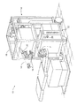
ここで、図1を参照すると、レーザ眼科手術システム10は、レーザビーム14を生成するレーザ12を含む。レーザ12は、患者Pの目Eにレーザビーム14を向けるレーザ送達光学部品16に光学的に連結される。送達光学部品支持構造(明確にするため、ここでは図示せず)は、レーザ12を支持するフレーム18から伸長する。顕微鏡20は、送達光学部品支持構造に取り付けられ、顕微鏡は、多くの場合、目の角膜を結像させるために使用される。
Referring now to FIG. 1, the laser
レーザ12は、一般的にエキシマレーザを含み、理想的には、約193nmの波長を有するレーザ光パルスを生成するアルゴン−フッ素レーザを含む。レーザ12は、好ましくは、レーザ送達光学部品16を介して送達されるフィードバック安定化フルエンスを患者の目に提供するように設計される。本発明の実施態様はまた、代替的紫外又は赤外放射源、特に目の隣接組織及び/又は下層組織に著しい損傷を引き起こすことなく、制御可能に角膜組織を切除するように構成される放射源により有用であり得る。幾つかの代替的な実施態様において、レーザビーム源は、開示全体が参考として本明細書に組み込まれる、特許文献1および2(Linの米国特許第5520679号及び第5144630号)、特許文献3(並びにMeadの第5742626号)に記載されたように、193〜215nmの波長を有する固体レーザ源を用いることがある。他の実施態様において、レーザ源は、開示全体が参考として本明細書に組み込まれる、Telfairの特許文献4および5(米国特許第5782822号及び第6090102号)に記載されたような赤外レーザを含む。それ故にエキシマレーザは、実例となる切除ビーム源であるが、他のレーザが本発明において使用されても良い。
レーザ12及びレーザ送達光学部品16は、一般的にコンピュータシステム22の指示下で、患者Pの目にレーザビーム14を向ける。コンピュータシステム22は、多くの場合、角膜の所定の彫刻を実施して目の屈折特徴を変えるように、レーザビーム14を選択的に調整してレーザエネルギーのパルスに角膜の部分を曝露する。多くの実施態様において、レーザ12とレーザ送達光学系16の両方は、所望のレーザ形成プロセスを実施するために、コンピュータシステム22の制御下にあり、コンピュータシステムは、レーザパルスのパターンを実施(及び任意に修正)する。パルスのパターンは、治療表の形状で有形の媒体29の機械読み取り可能なデータ内に要約され得る。
当業者に当然理解されるように、追加の部品及びサブシステムが、レーザシステム10に含まれても良い。例えば、開示全体が参考として本明細書に組み込まれる、米国特許第5646791号に記載されたように、レーザビーム内のエネルギー分布を制御するために、空間及び/又は時間積分器が含まれても良い。レーザ手術システムの切除流出物排出器/フィルタ、吸引器、及び他の補助部品は、当該技術分野において公知である。レーザ・切除処置を実行するための適切なシステムの更なる詳細は、開示全体が参考として本明細書に組み込まれる、同一出願人による特許文献6ないし15(米国特許第4665913号、第4669466号、第4732148号、第4770172号、第4773414号、第5207668号、第5108388号、第5219343号、第5646791号、第5163934号)に見られる。適切なシステムには、Alcon、Bausch & Lomb、Nidek、WaveLight(商標)、LaserSight(商標)、Schwind、Zeiss Meditec等により製造及び/又は販売されるような、市販の屈折レーザーシステムを含む。
図2は、本発明のレーザ手術システム10によって使用できる代表的なコンピュータシステム22の簡略化したブロック図である。コンピュータシステム22は、概してバスサブシステム54を介して多数の周辺装置と通信し得る少なくとも1つのプロセッサ52を含む。これらの周辺装置は、メモリサブシステム58及びファイル記憶サブシステム60を含む記憶サブシステム56と、ユーザインタフェース入力装置62と、ユーザインタフェース出力装置64と、ネットワークインタフェースサブシステム66とを含み得る。ネットワークインタフェースサブシステム66は、外部ネットワーク68及び/又は波面測定システム30のような他の装置へのインタフェースを提供する。
FIG. 2 is a simplified block diagram of an
ユーザインタフェース入力装置62は、キーボードと、マウス、トラックボール、タッチパッド、又はグラフィックスタブレットのようなポインティング装置と、スキャナと、フットペダルと、ジョイスティックと、ディスプレイに組み込まれたタッチスクリーンと、音声認識システムのような音声入力装置と、マイクロホンと、他のタイプの入力装置とを含み得る。ユーザ入力装置62は、多くの場合、本明細書に記載された方法の何れかを実現する有形記憶媒体29から、コンピュータ実行可能なコードをダウンロードするために使用される。一般的に、用語「入力装置」の使用は、コンピュータ・システム22に情報を入力するための種々の従来の、及び独自の装置及び方法を含むことが意図される。
The user
ユーザインターフェース出力装置64は、ディスプレイサブシステム、プリンタ、ファックス機、又は音声出力装置のような非視覚的ディスプレイを含み得る。ディスプレイサブシステムは、陰極線管(CRT)、液晶ディスプレイ(LCD)のようなフラットパネル装置、投影装置等であっても良い。ディスプレイサブシステムはまた、音声出力装置を介するなどの非視覚的ディスプレイを提供できる。一般的に、用語「出力装置」の使用は、コンピュータシステム22からの情報をユーザに出力するための種々の従来の、及び独自の装置及び方法を含むことが意図される。
User
記憶サブシステム56は、本発明の種々の実施態様の機能性を提供する基本プログラミング及びデータ構造を記憶する。例えば、本明細書に記載するように、本発明の方法の機能性を実施するデータベース及びモジュールは、記憶サブシステム56内に記憶できる。これらのソフトウェアモジュールは一般的に、プロセッサ52により実行される。分散環境において、ソフトウェアモジュールは、複数のコンピュータシステムに記憶でき、かつ複数のコンピュータシステムのプロセッサにより実行できる。記憶サブシステム56は、概してメモリサブシステム58と、ファイル記憶サブシステム60とを含む。
メモリサブシステム58は通常、プログラム実行中に命令及びデータを記憶するための主ランダムアクセスメモリ(RAM)70、及び固定命令が記憶される読み取り専用メモリ(ROM)72を含む多数のメモリを含む。ファイル記憶サブシステム60は、プログラム及びデータファイルのための永続的(不揮発性)記憶を提供し、かつ波面センサデータ、波面勾配、波面高度マップ、治療マップ及び/又は切除表を任意に実現できる有形記憶媒体29(図1)を含むことができる。ファイル記憶サブシステム60は、ハードディスクドライブ、フロッピー(登録商標)ディスクドライブと付随するリムーバブル媒体、コンパクトデジタル読み取り専用メモリ(CD−ROM)ドライブ、光学ドライブ、DVD、CD−R、CD−RW、固体リムーバブルメモリ、及び/又は他のリムーバブル媒体カートリッジ若しくはディスクを含むことができる。ドライブの1つ以上は、コンピュータシステム22に連結された他の場所で、他の接続されたコンピュータ上の遠隔位置に位置できる。本発明の機能性を実施するモジュールは、ファイル記憶サブシステム60により記憶できる。
The
バスサブシステム54は、コンピュータシステム22の種々の部品及びサブシステムを、意図した通りに互いに通信させるメカニズムを提供する。コンピュータシステム22の種々のサブシステム及び部品は、同じ物理的位置にある必要は無く、分散ネットワーク内の種々の位置に分布し得る。バスサブシステム54は、単一バスとして概略的に示されているが、バスサブシステムの代替的実施態様は、複数のバスを利用することがある。
コンピュータシステム22自体は、パーソナルコンピュータ、ポータブルコンピュータ、ワークステーション、コンピュータ端末、ネットワークコンピュータ、波面測定システム又はレーザー手術システムの制御システム、メインフレーム、又は何らかの他のデータ処理システムを含む種々のタイプのものであっても良い。コンピュータ及びネットワークの、常に変化する性質のために、図2に示すコンピュータシステム22の記載は、本発明の一実施態様を例示するための例のみを表している。図2に示すコンピュータシステムとおよそ同数の部品を有する、コンピュータシステム22の多くの他の構成が可能である。
The
ここで図3を参照すると、波面測定システム30の一実施態様が、簡略化した形状で概略的に示される。ごく一般的に言うと、波面測定システム30は、患者の眼を出る波面の局所傾斜を感知するように構成される。Hartmann−Shackの原理に基づく装置は、一般的に目の瞳孔全体にわたる傾斜をサンプリングするレンズレットアレイを含む。その後、局所傾斜は、多くの場合ゼルニケ多項式展開法を使用して、波面表面又はマップを再構築するために分析される。
Referring now to FIG. 3, one embodiment of a
より具体的には、1つの波面測定システム30は、レーザーのような光源32を含み、それが網膜Rの表面上に像44を形成するために、目Eの屈折組織34を通して源の像を投影する。網膜Rからの像は、目の屈折システム(例えば屈折組織34)により伝送され、かつシステム光学部品37により波面センサ36上に結像する。波面センサ36は、光学組織34の光学誤差の測定及び/又は光学組織切除治療プログラムの決定のためにコンピュータシステム22’に信号を伝達する。コンピュータ22’は、図1及び図2に示すコンピュータシステム22と同じ又は類似するハードウェアを含み得る。コンピュータシステム22’は、レーザー手術システム10に命令するコンピュータシステム22と通信し得るか、又は波面測定システム30及びレーザー手術システム10のコンピュータシステム部品の幾つか又は全部が、組み合わされるか、又は分離されても良い。所望であれば、波面センサ36からのデータは、有形媒体29を介して、I/Oポートを介して、イントラネット又はインターネットのようなネットワーク接続66を介して等によりレーザーコンピュータシステム22に伝送できる。
More specifically, one
波面センサ36は、一般的にレンズレットアレイ38と、像センサ40とを含む。網膜Rからの反射光は、光学組織34を通って伝送され、かつ像センサ40の表面に結像し、かつ目の瞳孔Pは、レンズレットアレイ38の表面に同様に結像する。レンズレットアレイは、伝送された光ビームをビームレット42のアレイに分離し、かつ(システムの他の光学部品と組み合わせて)分離されたビームレットをセンサ40の表面に結像させる。センサ40は、概して電荷結合素子又は「CCD」を含み、かつ光学組織34の関連領域の特性を決定するために使用できる、これらの個別のビームレットの特性を感知する。特に、像44が光の点又は小スポットを含む場合、ビームレットにより結像するような伝送スポットの位置は、光学組織の関連領域の局所勾配を直接、示すことができる。
The wavefront sensor 36 generally includes a
目Eは、一般的に前方向ANTと後方向POSを定義する。光源32は、一般的に図3に示すように、光学組織34を通して網膜R上に後方向に光を送る。光学組織34は、再度、網膜から前方向に波面センサ36に向けて反射された光を伝送する。網膜R上に実際に形成される像44は、像源が最初に光学組織34によって伝送される時、目の光学系のいかなる欠陥によっても歪み得る。任意に、像投射光学部品46は、像44のいかなる歪みも減少させるように構成されるか、又は減少させることに適している。
Eye E generally defines a forward ANT and a backward POS. The
幾つかの実施態様において、投射光学部品46は、光学組織34の球面誤差及び/又は円柱誤差を補正することによって、低次光学誤差を減少させ得る。光学組織の高次光学誤差はまた、変形可能鏡のような適応光学系の使用によって、補正できる。像44で点又は小スポットを網膜R上に画定するように選択される光源32の使用によって、波面センサ36により提供されるデータの分析が容易になり得る。特定の光源構造にかかわらず、網膜R上に、明確、かつ正確に形成された像44を有することが一般的に有益である。
In some embodiments, the
波面データは、Hartmann−Shackセンサ像の像スポット分析から得られるx及びy波面勾配値と、瞳孔カメラ51(図3)の像により測定されるHartmann−Shackレンズレットアレイの公称中心からのx及びy瞳孔中心オフセットとを含む、2つの別個のアレイにおいて、コンピュータ読み取り可能媒体29又は波面センサシステム30のメモリ内に記憶できる。かかる情報は、目の波面誤差に関する全ての入手可能な情報を含むことができ、かつ波面又はその所望の部分を再構築するために概して十分である。かかる実施態様において、Hartmann−Shack像を2回以上再処理する必要はないであろうし、かつ勾配アレイを記憶するために必要なデータスペースは大きくはない。例えば、直径8mmの瞳孔の像を調節するために、20×20サイズ(即ち400素子)のアレイで多くの場合十分である。認識されるように、別の実施態様において、波面データは、単一のアレイ又は複数のアレイとして、波面センサシステムのメモリに記憶できる。
The wavefront data includes the x and y wavefront gradient values obtained from the image spot analysis of the Hartmann-Shack sensor image and the x and y from the nominal center of the Hartmann-Shack lenslet array measured by the image of the pupil camera 51 (FIG. 3). can be stored in the memory of the computer readable medium 29 or the
本発明の実施態様は、一般的に、像44の感知に関して記載されるが、一連の波面センサデータ読み取りができることを、理解すべきである。例えば、時系列の波面データ読み取りは、眼球組織収差のより正確な全体的決定を提供するために役立つことがある。眼球組織は、短期間で形状が変化し得るので、複数の時間的に分離された波面センサ測定は、屈折矯正処置のベースとして光学特性の単一のスナップショットに依存することを回避できる。種々の構成、位置、及び/又は方向での目に対する目の波面センサデータを得ることを含む、なおも更なる代替案がまた利用可能である。例えば、開示全体が参考として本明細書に組み込まれる、特許文献16(米国特許第6004313号)に記載されたように、患者は、多くの場合、固定標的に焦点を合せることにより、波面測定システム30との目の位置合わせを維持することに役立つ。その参考文献に記載されたように固定標的の位置を変化させることにより、目の光学特性は、目が変動する距離及び/又は角度で視野を結像するために調節又は適合する間に決定できる。
ここで図4を参照すると、老視を治療する方法は、図3に概略的に示すもののような、波面センサを使用する、目の波面測定を一般的に含む。多くの実施態様において、波面のゼルニケ係数は決定される(106)。波面及びゼルニケ係数は、目の光学収差を矯正するように、屈折処方を直接決定するために使用できる。公知の波面に基づく光学矯正は、多くの場合、目が治療後に正視であるように、目の全ての光学収差を矯正又は補償しようと努める。 Referring now to FIG. 4, a method for treating presbyopia generally includes a wavefront measurement of the eye using a wavefront sensor, such as that schematically illustrated in FIG. In many embodiments, the Zernike coefficient of the wavefront is determined (106). The wavefront and Zernike coefficients can be used to directly determine the refractive prescription so as to correct the optical aberrations of the eye. Known wavefront-based optical corrections often seek to correct or compensate for all optical aberrations of the eye so that the eye is normal after treatment.
代表的な方法(102)においては、正視眼を提供するように処方を開発するよりも、老視緩和の高次球面収差が決定される(108)。この所望の球面収差は、波面からのゼルニケ係数と共に、処方を決定するために使用され(110)、処方は、一般的に他の高次及び標準屈折異常を矯正する一方で、治療された目を所望の球面収差を有するようにする。次に処方は、図1に示したようなレーザ眼科手術システムを任意に使用して、目に与えられる(112)。 In an exemplary method (102), higher order spherical aberrations for presbyopia relief are determined (108) rather than developing a prescription to provide a normal eye. This desired spherical aberration, along with Zernike coefficients from the wavefront, is used to determine the prescription (110), which generally corrects other higher-order and standard refractive errors while treating the treated eye. To have the desired spherical aberration. The prescription is then given to the eye ( 112 ) , optionally using a laser eye surgery system such as that shown in FIG.
波面測定は、種々の市販のシステムを使用して行うことができ、代表的な波面測定システムには、カリフォルニア州サンタクララのVISX,Incorporatedから入手可能なVISX WaveScan(商標)システムを含む。代替的な波面測定システムには、例えば特許文献17(米国特許第6271915号)に記載されるものを含む。なおも更なる代替的な波面測定システムを用いることができ、好ましいシステムは、上記のように、網膜から目の屈折組織を通して伝送された光を使用して波面を測定する。
代表的な老視緩和方法(102)は、波面のゼルニケ係数を決定する(106)が、代替的な方法は、波面を定義するために、種々の代替的な数学的枠組みのいずれも使用できる。例えば、直接的な波面に基づく角膜切除治療処方は、開示全体が参考として本明細書に組み込まれる、米国特許出願第10/006992号に記載されたような方法及びシステムを使用して導くことができる。開示全体が参考として本明細書に同様に組み込まれる、米国特許出願第10/872107号に記載された技術を含む、フーリエ変換及び直接積分を使用する波面再構築もまた用いることができる。ともかく、波面再構築は、一般的に少なくとも若干量の目の不規則収差に対応する。処方の基本を、少なくとも部分的にかかる不規則収差に置くことによって、本明細書に記載された治療は、少なくとも20/20か、それ以上の視力を提供でき、場合により20/20を超える視力を、多くの場合老視緩和と共に提供する。 The exemplary presbyopia mitigation method (102) determines the Zernike coefficient of the wavefront (106), but alternative methods can use any of a variety of alternative mathematical frameworks to define the wavefront. . For example, a direct wavefront-based keratotomy treatment regimen can be derived using methods and systems such as those described in US patent application Ser. No. 10/006992, the entire disclosure of which is incorporated herein by reference. it can. Wavefront reconstruction using Fourier transforms and direct integration can also be used, including techniques described in US patent application Ser. No. 10 / 872,107, the entire disclosure of which is also incorporated herein by reference. In any case, wavefront reconstruction generally corresponds to at least some amount of irregular eye aberrations. By placing the basis of the prescription at least in part on such irregular aberrations, the treatments described herein can provide visual acuity of at least 20/20 or more, and in some cases greater than 20/20 Often provided with presbyopia relief.
多数のアプローチが、所望の老視緩和の高次球面収差を決定する(108)ために用いることができる。本明細書において使用されるように、高次球面収差は、標準近視及び遠視以外の球面収差を包含する。所望の球面収差は、実験によるデータ、屈折組織の単純又は複雑なモデル等に基づき決定できる。波面再構築が、ゼルニケ係数を含む時、所望の老視緩和の球面収差は、Z(2,0)、Z(4,0)、Z(6,0)項等のような放射対称性ゼルニケ多項式展開係数として好適にモデル化できる。他の実施態様において、所望の球面収差の異なる数学的定式化を用いることができる。 A number of approaches can be used to determine 108 the desired presbyopia mitigation higher order spherical aberration. As used herein, higher order spherical aberration encompasses spherical aberrations other than standard myopia and hyperopia. The desired spherical aberration can be determined based on experimental data, simple or complex models of refractive tissue, and the like. When the wavefront reconstruction includes a Zernike coefficient, the desired presbyopia-reducing spherical aberration is a radiation-symmetric Zernike such as Z (2,0), Z (4,0), Z (6,0) terms, etc. It can be suitably modeled as a polynomial expansion coefficient. In other embodiments, a different mathematical formulation of the desired spherical aberration can be used.
波面再構築106及び老視緩和の球面収差(108)が、ゼルニケ多項式を使用して定義された時、組み合わされた処方(110)は、波面誤差を別の方法で矯正しながら、適切な非球性を目に重ねることによって直接計算できる。このことは、適切な多項式の項を測定された波面に足す又は引くことと同じように簡単であり得る。他の再構築技術が用いられる場合、又は所望の老視緩和の球面収差が、波面再構築のそれと異なる数学の項において定義される場合、組み合わされた処方を決定するための更に複雑な分析アプローチを用いることができる。
When the
一旦所望の処方が、決定されると(110)、その処方は、多種多様な代替的な屈折変更アプローチのいずれかを使用して、目に与えられる(112)。例えば、カスタムコンタクトレンズが、レーザ切除されるか、又は他の方法で形成でき、眼内又は有水晶体レンズが、レーザ又は他の彫刻技術によって成形でき、レーザ又は他の加熱方法を使用する選択的角膜膠原収縮を用いることができる等である。ともかく、多くの実施態様において、所望の球面収差は、治療が完了した後、かつ多くの場合何らかの関連する治癒がなされた後、患者の瞳孔全体に広がる。 Once the desired prescription is determined (110), the prescription is given to the eye (112) using any of a wide variety of alternative refractive modification approaches. For example, custom contact lenses can be laser ablated or otherwise formed, and intraocular or phakic lenses can be molded by laser or other engraving techniques, selectively using laser or other heating methods For example, corneal collagen contraction can be used. Regardless, in many embodiments, the desired spherical aberration spreads across the patient's pupil after treatment is complete and often after some associated healing has taken place.
老視を緩和するために、所望の高次球面収差を決定する(108)方法を検討すると、ヒトの目は、一般的に角膜、水晶体及び硝子体液を含む複数の屈折要素を有する。図5で概略的に示すように、物体は、その物体の鮮明な像が網膜R上に形成される時に、焦点が合って見られる。物体の焦点が合って見える距離範囲は、少なくとも部分的に水晶体の調節によって決まる。レンズ(水晶体)の調節が小さいか、又は無視できる(このことは、人が年を取ると起こり得る)とすると、眺められた物体は、最高の視距離から離れると、ぼんやりと見える。視距離の変化による像特性の変化は、網膜の前及び/又は後の焦点距離又は像焦点面の変化による像特性の変化に同様に関連し得る(かつ対応し得る)。物体がぼんやりするようになる率は、目及び瞳孔直径の光学的性質次第によって決まる。焦点距離の変化によって像がぼやけることは、目に適切な光学的矯正を適用することによって減少できる。所望の矯正は、一般的に本明細書において、レーザ切除を使用して角膜に適用されるとして記載されるが、他の屈折治療様式が(例えば、コンタクトレンズ又は強膜レンズを使用して)角膜の前、又は(眼内レンズ等を使用して)角膜の後で屈折を変更できる。 Considering a method for determining a desired higher order spherical aberration (108) to alleviate presbyopia, the human eye typically has multiple refractive elements including cornea, lens and vitreous humor. As schematically shown in FIG. 5, the object is viewed in focus when a sharp image of the object is formed on the retina R. The distance range in which the object appears to be in focus is determined at least in part by adjustment of the lens. Given that the lens (lens) adjustment is small or negligible (this can happen when a person gets older), the viewed object will appear blurry when away from the highest viewing distance. Changes in image characteristics due to changes in viewing distance may be similarly related (and may correspond) to changes in image characteristics due to changes in focal length or image focal plane before and / or after the retina. The rate at which the object becomes blurred depends on the optical properties of the eye and pupil diameter. The blurring of the image due to changes in focal length can be reduced by applying an appropriate optical correction to the eye. Although the desired correction is generally described herein as being applied to the cornea using laser ablation, other refractive treatment modalities (eg, using contact lenses or scleral lenses) Refraction can be changed before the cornea or after the cornea (using intraocular lenses or the like).
球面収差は、光学系の放射対称性収差であり、瞳孔の異なる部分を通過する光を角膜からの異なる距離で焦点合わせさせ得る。従って、点光源の像は、ぼやけたスポット等になり得る。光軸に沿った(それぞれ瞳孔の中心領域及び瞳孔の周縁領域内で結像するような)近軸及び周縁像の相対位置は、球面収差の符号を決定できる。光学系の近軸焦点長が、周縁焦点距離より短い時、球面収差は負の符号を有し、かつ光学系の近軸焦点長が、周縁焦点距離より長い時、球面収差は正符号を有する。 Spherical aberration is a radial symmetry aberration of the optical system that can focus light passing through different parts of the pupil at different distances from the cornea. Therefore, the image of the point light source can be a blurred spot or the like. The relative positions of the paraxial and peripheral images along the optical axis (which respectively image in the central region of the pupil and the peripheral region of the pupil) can determine the sign of the spherical aberration. When the paraxial focal length of the optical system is shorter than the peripheral focal length, the spherical aberration has a negative sign, and when the paraxial focal length of the optical system is longer than the peripheral focal length, the spherical aberration has a positive sign. .
ヒトの目は、目の波面測定(104)において一般的に識別されるように、概して少量の球面収差を有する。術後の目及び/又は病的な目は、有意な量の球面収差を有することがある。網膜像の質は、球面収差の大きさ又は量と、球面収差の符号との両方によって決まり得る。以下に記載されるように、負の球面収差はより良好な焦点深度を提供でき、かつそれ故により望ましい老視緩和能力を提供できる。 The human eye generally has a small amount of spherical aberration, as generally identified in eye wavefront measurements (104). Post-operative eyes and / or morbid eyes may have a significant amount of spherical aberration. The quality of the retinal image can depend on both the magnitude or amount of spherical aberration and the sign of spherical aberration. As described below, negative spherical aberration can provide better depth of focus, and therefore can provide desirable presbyopia mitigation capabilities.
ここで図5を参照すると、目の簡略化された分析モデル120は、老視緩和の高次球面収差を決定する際に有用であり得る。図5に示したモデルは、カリフォルニア州サンディエゴのZEMAX Development Corporationから市販されているソフトウェアを使用して開発されたZEMAX(商標)光学設計ソフトウェアモデルを含む。目のモデル120は、単一の円錐定数によって平坦な非球面としてモデル化できる前方角膜122を含む。換言すると、モデルの角膜122は、非球面にするために、加えられる偶数次の放射項(radial terms)を有する楕円体を含む。使用される放射項は、前方角膜に関して8次までを含むことができる。放射非球面項の中央曲率、円錐定数及び係数は、目の球面収差がこれらのパラメータを調整することによって変化できるように、モデル120において可変であっても良い。 Referring now to FIG. 5, a simplified analytical model 120 of the eye can be useful in determining higher order spherical aberrations for presbyopia relaxation. The model shown in FIG. 5 includes a ZEMAX ™ optical design software model developed using software available from ZEMAX Development Corporation of San Diego, California. The eye model 120 includes an anterior cornea 122 that can be modeled as a flat aspheric surface with a single conic constant. In other words, the model cornea 122 includes an ellipsoid with an even order radial term applied to make it aspheric. The radiation terms used can include up to the 8th order with respect to the anterior cornea. The central curvature, conic constant, and coefficient of the radiating aspheric term may be variable in the model 120 so that the spherical aberration of the eye can be changed by adjusting these parameters.
目のモデル120を使用して、円内エネルギー、選択された周波数での変調伝達関数(MTF)、及び幾何学的点広がり関数(PSF)は、様々な球面収差及び物体の距離に対して像の質を評価するために計算できる。多染性計算は、光の複数の波長を使用して、多くの場合、0.45μm、0.55μm及び0.65μm光のような、光の3つ以上の波長を使用して実行できる。0.04、1及び0.35の加重係数がそれぞれ、以上に特定された波長に適用できる。瞳孔の大きさは、若干の分析において変化し得るが、少なくとも幾つかの下記の計算は、単一の6mmの入射瞳孔の大きさによってなされ、結像した物体は、モデルの目の光軸に位置決めされる。 Using the eye model 120, the energy in a circle, the modulation transfer function (MTF) at a selected frequency, and the geometric point spread function (PSF) are imaged for various spherical aberrations and object distances. Can be calculated to evaluate the quality of the. Multichromatic calculations use multiple wavelengths of light, often using more than two wavelengths of light, such as 0.45 μm , 0.55 μm and 0.65 μm light. Can be executed. Weighting factors of 0.04, 1 and 0.35 can each be applied to the wavelengths specified above. The pupil size may vary in some analyses, but at least some of the following calculations are made by a single 6 mm entrance pupil size, and the imaged object is aligned with the optical axis of the model eye. Positioned.
ここで図6を参照すると、幾何学的点広がり関数は、図5のモデルの目に関して、種々の球面収差SAで計算された。物体、及び具体的には光点源は、目からの無限距離(V=0D)及び2メートル(V=0.5D)でモデル化された(式中、Vは両眼転導である)。図6は、約ゼロ、−0.2μm、及び+0.21μmの球面収差SAを有するモデルの目の幾何学的点広がり関数の断面をグラフにより示す。図6のグラフで、水平又はx軸は、網膜上の位置をμmで表し、距離は、目の光軸から測定される。網膜上の1μmの長さは、角膜レンズの中心で、約0.2分角の角度の範囲のものである。 Referring now to FIG. 6, the geometric point spread function was calculated with various spherical aberrations SA for the model eye of FIG. The object, and specifically the light point source, was modeled at infinite distance from the eye (V = 0D) and 2 meters (V = 0.5D), where V is binocular transduction. . FIG. 6 graphically illustrates a cross-section of the geometric point spread function of a model eye having spherical aberrations SA of approximately zero, −0.2 μm , and +0.21 μm . In the graph of FIG. 6, the horizontal or x-axis represents the position on the retina in μm , and the distance is measured from the optical axis of the eye. The 1 μm length on the retina is in the range of approximately 0.2 arc minutes at the center of the corneal lens.
幾何学的点広がり関数(PSF)ピークは、球面収差がほとんど存在しない、又は球面収差が存在せず、かつ物体が無限距離にある場合に、最も高い。しかしながら、このモデルの目の点広がり関数は、物体が目に近付くにつれて急速に広がり、0.5Dの両眼転導での実質的にゼロ球面収差モデルが、遙かに低いピーク高さを有する。対照的に、両眼転導の変化によるピーク高さの変化は、負の球面収差(SA−0.2、V0D/0.5D)を有するモデルの目に関して、著しく小さい。ピーク高さ変化の減少と共に、負の球面収差を有するモデルの目は、物体が無限距離から2メートルに移動すると(換言すれば、両眼転導Vが0から0.5Dに変化すると)、より局所化された状態に留まる点広がり関数を有する。 The geometric point spread function (PSF) peak is highest when there is little or no spherical aberration and when the object is at infinity. However, the eye point spread function of this model spreads rapidly as the object approaches the eye, and the substantially zero spherical aberration model with 0.5D binocular transduction has a much lower peak height. . In contrast, the change in peak height due to changes in binocular transduction is significantly less for model eyes with negative spherical aberration (SA-0.2, V0D / 0.5D). With decreasing peak height change, the eyes of a model with negative spherical aberration, when the object moves from infinite distance to 2 meters (in other words, when binocular transduction V changes from 0 to 0.5D) It has a point spread function that stays more localized.
ここで図7を参照すると、通過焦点スポット図130のアレイは、目が種々の量の球面収差を有し、かつ像平面が角膜からの種々の距離にある時の点源の像をグラフにより示す。通過焦点スポット図は、球面収差を有さない又はほとんど有さない目、SA=0、負の球面収差を有する目、SA=−0.2、及び正の球面収差を有する目、SA=+0.2に提供される。アレイ130の底部に沿って現れる数は、関連する通過焦点スポット図が分析される光軸に沿った距離を示し、「0」位置は、(無限132に関する最高焦点面によって示されるような)概して網膜Rに位置する、最高焦点位置にある。アレイ右側の正の数は、最高焦点位置から測定された角膜からより遠い距離を示し、他方でアレイ左側の負の数は、目のモデルの角膜レンズにより近い位置を表す。点源が目に無限に近付くにつれて、(調節しない目の)網膜での像は、矢印方向134で変化する。それ故に、網膜で形成される点源の像は、中心スポットのより左側でのスポット図である。測定は、μmで示す。
Referring now to FIG. 7, the array of through-focus spot diagrams 130 shows graphically the image of the point source when the eye has various amounts of spherical aberration and the image plane is at various distances from the cornea. Show. The passing focal spot diagram shows eyes with little or no spherical aberration, SA = 0, eyes with negative spherical aberration, SA = −0.2, and eyes with positive spherical aberration, SA = + 0. .2 is provided. The number appearing along the bottom of the
アレイの頂部に沿って示すように、最高焦点スポットの大きさは、SA=0の場合に最小である。負の球面収差に関して、スポット図が、(最高近軸焦点位置よりも角膜に近い結像距離で)アレイ左側で比較的高強度の中心領域を含み、他方でこの同じモデルが、(最高近軸焦点位置からのより大きな距離で)アレイ右側により均等に広がるスポット図を有することに注意。この特徴は、球面収差が正の場合に逆転し、スポットは、(より大きな距離で)アレイ右側に強い中心領域を有し、他方で正の球面収差は、アレイ左側に向かってより近い焦点距離でスポットの均等な広がりをもたらす。 As shown along the top of the array, the size of the highest focal spot is minimal when SA = 0. For negative spherical aberration, the spot diagram includes a relatively high intensity central region on the left side of the array (at an imaging distance closer to the cornea than the highest paraxial focal position), while this same model (highest paraxial) Note that it has a spot diagram that spreads more evenly on the right side of the array (at a greater distance from the focal position). This feature is reversed when the spherical aberration is positive, the spot has a strong central area on the right side of the array (at a greater distance), while the positive spherical aberration is closer to the left side of the array. To bring about even spread of spots.
薄暗い周縁尾部とともに比較的強い中心領域を有することは、スポット周縁部で均等な分布又は高強度を有するスポット図と比較してより鮮明な像を作り得る。その上、(角膜により近い、かつ/又は図7のアレイ左側に沿った)網膜正面の焦点距離で強い中心スポットを有することは、有用であり得る。更に具体的には、物体がかかる目に接近する時、網膜上の像は、より鮮明である。その上、目は、網膜上で均等に鮮明な像に焦点を合わせるように(いずれかの残余調節を使用して)調節できることがある。点源が、網膜の後方に位置する強い中心スポットを発生させるならば、近い物体の像は、より不鮮明である。従って、目に残るいかなる調節能力も、患者が強い中心領域を焦点に運ぶことを補助できるので、アレイ130の中間列を横切る負の球面収差の通過焦点スポット図は、長い焦点深度を提供することによって利点を提供するように見える。更に、かかる負の球面収差を有する(種々の距離による)網膜スポットの大きさの変化は、正の球面収差の場合に関するよりも、かつ/又はゼロ球面収差の場合の、スポットの大きさの対応する実質的な変化よりも小さくなり得る。
It has a relatively strong central region with a dim peripheral tail may make a clearer image as compared to the spot diagram with a uniform distribution or high intensity spot periphery. Moreover, it may be useful to have a strong central spot at the focal length of the front of the retina (closer to the cornea and / or along the left side of the array in FIG. 7). More specifically, when an object approaches such an eye, the image on the retina is clearer. In addition, the eye may be adjustable (using any residual adjustment) to focus on an evenly sharp image on the retina. If the point source generates a strong central spot located behind the retina, the near object image will be more smeared. Thus, any spherical adjustment ability that can remain in the eye can help the patient to focus on a strong central region, so a negative spherical aberration passing focal spot diagram across the middle row of the
ここで図8を参照すると、50サイクル/mmでの変調伝達関数(MTF)が、焦点からの距離の関数として示される。図7の通過焦点スポット図がそうであったように、変調伝達関数は、ゼロ球面収差の場合の焦点周囲で最高であり得る。しかしながら、焦点よりも小さい距離に関して(かつここでもまたx軸に沿って負の値を有する、また角膜により近い距離に関して)、負の球面収差を有するモデルの目のMTFは、増加し、かつ最高焦点からのシフトが50μmよりも大きい時に、ゼロ球面収差の場合に関して変調伝達関数値を実際に超える。 Referring now to FIG. 8, the modulation transfer function (MTF) at 50 cycles / mm is shown as a function of distance from the focus. As was the case of the passing focal spot diagram of FIG. 7, the modulation transfer function may be highest around the focus for zero spherical aberration. However, for distances smaller than the focus (and again for negative distances along the x-axis and for distances closer to the cornea), the MTF of the model eye with negative spherical aberration increases and is highest. When the shift from focus is greater than 50 μm , the modulation transfer function value is actually exceeded for the case of zero spherical aberration.
目が調節を有さない(事実上固定レンズを有する)とすると、物体が無限に設置されるか、又は目に近付くならば、像の位置又は焦点は、角膜から及び/又は網膜から離れる。このことは、網膜の位置が、最高の像位置よりも角膜に近くなり得ることを意味する。換言すれば、網膜は、物体が目の近くに設置されると、焦点からの負のシフト距離で置かれ得る。かかる状況において、負の球面収差の目のMTFは、物体が目に近付く時、変調伝達関数が、より遅く変化し、かつ適度な(及び多くの場合許容可能な)レベルに留まるので、実質的にゼロ球面収差の目、又は正の球面収差の目よりも良くなり得る。 Assuming that the eye has no accommodation ( effectively with a fixed lens), the position or focus of the image is away from the cornea and / or from the retina if the object is placed infinitely or approaches the eye. This means that the position of the retina can be closer to the cornea than the highest image position. In other words, the retina can be placed at a negative shift distance from the focus when the object is placed near the eye. In such a situation, the MTF of a negative spherical aberration eye is substantially because the modulation transfer function changes more slowly and remains at a reasonable (and often acceptable) level when the object approaches the eye. It can be better than zero spherical aberration eyes or positive spherical aberration eyes.
ここで図9を参照すると、(SAはまたμmで特定される)種々の球面収差に対する円内エネルギー、及び(また両眼転導で特定され、V=0が無限での物体であり、かつV+0.5Dが目から2メートルでの物体である)物体位置が示される。この円内エネルギーのグラフは、適切な質の負の球面収差が、PSF及びMTFに関して以上に記載されたものと類似した利点を提供し得ることを示す。更に具体的には、図9のグラフィックデータは、示された両眼転導距離で、光軸に設置される多染性点源のために計算される幾何学的な円内エネルギーの図表を示す。比較のために、回折限界光学系のグラフも示される。図9によれば、物体が無限から2メートルまで移動すると、全エネルギーの所与の割合を包含する半径は増加する。半径の変化は、球面収差が負の時に小さく、かつ球面収差が正の時に大きい。 Referring now to FIG. 9, the in-circle energy for various spherical aberrations (SA is also specified in μm ), and (also specified in binocular transduction, V = 0 is an infinite object, and The object position is shown (V + 0.5D is the object 2 meters from the eye). This in-circle energy graph shows that an appropriate quality negative spherical aberration can provide similar benefits to those described above for PSF and MTF. More specifically, the graphic data of FIG. 9 shows a diagram of the geometric in-circle energy calculated for a polychromatic point source placed on the optical axis at the indicated binocular transduction distance. Show. For comparison, a graph of a diffraction limited optical system is also shown. According to FIG. 9, as the object moves from infinity to 2 meters, the radius encompassing a given percentage of the total energy increases. The change in radius is small when the spherical aberration is negative and large when the spherical aberration is positive.
以上は、適切な負の球面収差を有する目が、大きな焦点深度を提供できることを示す。従って、かかる負の球面収差は、老視の緩和を達成し、かつ視距離の変化にもかかわらずより良い視力を提供するために、目に有益であり得る。点広がり関数、変調伝達関数、及び円内エネルギーに関する上記シミュレーションデータは、老視の限定された視深度を軽減するための負の球面収差の能力を裏付ける。物体位置として小さい変動を示したこれらの像の質的特性の各々は、目が、正の球面収差、又は実質的に球面収差がない場合と対照的に適切な負の球面収差を有する時に、無限から目に近いどこかにシフトされる。目のいかなる残りの残余調節も、負の球面収差を有する目のための像質因子を更に強化できる。 The above shows that an eye with an appropriate negative spherical aberration can provide a large depth of focus. Thus, such negative spherical aberration can be beneficial to the eye to achieve presbyopia relief and provide better vision despite changes in viewing distance. The above simulation data on the point spread function, modulation transfer function, and in-circle energy supports the ability of negative spherical aberration to reduce the limited visual depth of presbyopia. Each of the qualitative characteristics of these images that showed a small variation in object position is that when the eye has an appropriate negative spherical aberration, as opposed to a positive spherical aberration or substantially no spherical aberration, Shifted from infinity to somewhere close to the eyes. Any remaining residual adjustment of the eye can further enhance the image quality factor for eyes with negative spherical aberration.
図10に示した表は、(図5に類似した光学的モデルを使用して両方ともモデル化された)最初の又は基本角膜と、矯正又は治療された角膜との形状パラメータ例を示す。ここでもまた、図10に特定された負の球面収差は、レーザ眼科手術、コンタクトレンズ等によって与えられ得るような、角膜の負の非球性を介して導入されると考えられる。図10の表に示す係数は、標準ゼルニケ多項式係数であり、最適化された係数値が、上記光学マトリックスにおける負の球面収差の利点を強化するように、特定された。負の球面収差量は、上記光学マトリックスに所望の特徴を提供した形状を導くために変更された。上記のようなMTF及びPSFは、この最適化のために利用された。 The table shown in FIG. 10 shows example shape parameters of the initial or basic cornea (both modeled using an optical model similar to FIG. 5) and the cornea corrected or treated. Again, it is believed that the negative spherical aberration identified in FIG. 10 is introduced through the negative asphericity of the cornea, which can be provided by laser eye surgery, contact lenses, and the like. The coefficients shown in the table of FIG. 10 are standard Zernike polynomial coefficients, and optimized coefficient values were identified to enhance the advantages of negative spherical aberration in the optical matrix. The amount of negative spherical aberration was changed to derive a shape that provided the desired features in the optical matrix. MTF and PSF as described above were utilized for this optimization.
ここで図11を参照すると、最適化の結果は、種々の物体距離に対する50サイクル/mmでの変調伝達関数値を示す。変調伝達関数は、幾何学的に計算され、かつ値は、4mmの瞳孔の大きさを有する多色光に関する。 Referring now to FIG. 11, the optimization results show the modulation transfer function values at 50 cycles / mm for various object distances. The modulation transfer function is calculated geometrically and the value relates to polychromatic light having a pupil size of 4 mm.
図11に示した値は、物体が最高焦点位置から離れる時の変調伝達関数の下落が、標準角膜よりも最適化された角膜に関して遙かに遅いことを示す。変調伝達関数の遅い変化は、図10の最適化された形状によって治療された目の大きな被写界深度を意味する。1.0D読書用眼鏡の影響も示す。 The values shown in FIG. 11 indicate that the drop in modulation transfer function when the object moves away from the highest focus position is much slower for the optimized cornea than for the standard cornea. A slow change in the modulation transfer function means a large depth of field of the eye treated with the optimized shape of FIG. The impact of 1.0D reading glasses is also shown.
代表的な実施態様は、例として、かつ明確な理解のために若干詳細に記載されたが、当業者は、種々の修正、改変、及び変更が用いられ得ることを認識するであろう。従って、本発明の範囲は、添付の請求項によって専ら限定されるべきである。 While exemplary embodiments have been described in some detail by way of example and for purposes of clarity of understanding, those skilled in the art will recognize that various modifications, changes and changes may be used. Accordingly, the scope of the invention should be limited solely by the appended claims.
44 像
102 代表的な方法
122 角膜
R 網膜
44
Claims (9)
出力に接続された処方生成器モジュールを含み、前記目が前記瞳孔を横切って誘導された負の球面収差を有するように、前記処方生成器モジュールが前記目の高次の負の球面収差の老視緩和量を画定し、前記出力はレンズ製造または目矯正アセンブリと通信するように構成される、
ことを特徴とする装置。A device for treating presbyopia of an eye having a pupil,
Contain formulatory generator module connected to the output, so as to have a negative spherical aberration in which the eye is induced across the pupil, the formulation generator module old negative spherical aberration of higher order of the eye defining a visual relaxation amount, the output is configured to communicate with the lens manufacturing or eye correcting assembly,
A device characterized by that.
目の感知された収差に対応する複数の屈折ゼルニケ係数を生成する、前記処方生成器の前記入力に接続された波面システムと、
前記目の角膜の方向へレーザエネルギーのパターンを向けることにより、前記目に処方を施すための切除レーザビームを含む、前記レンズ製造または目矯正アセンブリと、
を含み、
前記処方生成器は、前記屈折ゼルニケ係数を少なくともひとつの老視緩和ゼルニケ係数と組み合わせ、前記少なくともひとつの老視緩和ゼルニケ係数は前記負の球面収差の老視緩和量に対応する少なくともひとつの高次ゼルニケ係数を含み、前記瞳孔を横切って0.1から0.3マイクロメートルの負の球面収差を前記目に与えるようにする、
ことを特徴とする請求項1記載の装置。The prescription generator has an input, and
A wavefront system connected to the input of the prescription generator that generates a plurality of refractive Zernike coefficients corresponding to the perceived aberration of the eye;
By directing the laser energy pattern in the direction of the eye cornea, including ablation laser beam for performing prescribed the eye, and the lens manufacturing or eye correcting assembly,
Including
The prescription generator combines the refractive Zernike coefficient with at least one presbyopia-reducing Zernike coefficient, and the at least one presbyopia-reducing Zernike coefficient is at least one higher order corresponding to a presbyopia mitigation amount of the negative spherical aberration. wherein Zernike coefficients, the negative spherical aberration of 0.1 to 0.3 micrometers across the pupil so as to provide the eye,
The apparatus according to claim 1.
ことを特徴とする請求項1記載の装置。The formulation generator module, the eye of the identifying adaptive intraocular lens to change the optical, negative spherical altered eyes induced across the pupil when the lens is inserted into the eye has aberrations, the output is configured to communicate with intraocular lens source to provide the intraocular lens which is identified on the eye,
The apparatus according to claim 1.
目の感知された収差に対応する複数の屈折ゼルニケ係数を生成する、前記処方生成器の前記入力に接続された波面システムを含み、
前記処方生成器は前記屈折ゼルニケ係数を少なくともひとつの老視緩和ゼルニケ係数と組み合わせ、前記少なくともひとつの老視緩和ゼルニケ係数は前記負の球面収差の老視緩和量に対応する少なくともひとつの高次ゼルニケ係数を含み、前記瞳孔を横切って0.1から0.3マイクロメートルの負の球面収差を前記目に与えるようにする、
ことを特徴とする請求項3記載の装置。 The prescription generator has an input, and
A wavefront system connected to the input of the prescription generator that generates a plurality of refractive Zernike coefficients corresponding to a sensed aberration of the eye;
The prescription generator combines the refractive Zernike coefficient with at least one presbyopia-relieving Zernike coefficient, and the at least one presbyopia-reducing Zernike coefficient is at least one higher-order Zernike coefficient corresponding to the presbyopia mitigation amount of the negative spherical aberration. comprises coefficients and the negative spherical aberration of 0.1 to 0.3 micrometers across the pupil so as to provide the eye,
The apparatus of claim 3.
ことを特徴とする請求項3記載の装置。Further comprising an intraocular lens manufacturing apparatus for manufacturing the intraocular lens,
The apparatus of claim 3.
ことを特徴とする請求項1記載の装置。 The eye is to have negative spherical aberration induced across the pupil, the formulation generator module identifies a laser induced changes of the eye, configured so that the output communicates with the laser eye surgery assembly Being
The apparatus according to claim 1.
ことを特徴とする請求項6記載の装置。The laser ophthalmic surgical assembly includes at least one of an infrared laser, an ultraviolet laser, a femtosecond laser, and a wavelength multiplexed solid-state laser.
The apparatus according to claim 6.
目の感知された収差に対応する複数の屈折ゼルニケ係数を生成する、前記処方生成器の前記入力に接続された波面システムを含み、
前記処方生成器は前記屈折ゼルニケ係数を少なくともひとつの老視緩和ゼルニケ係数と組み合わせ、前記少なくともひとつの老視緩和ゼルニケ係数は前記負の球面収差の老視緩和量に対応する少なくともひとつの高次ゼルニケ係数を含み、前記瞳孔を横切って0.1から0.3マイクロメートルの負の球面収差を前記目に与えるようにする、
ことを特徴とする請求項7記載の装置。 The prescription generator has an input, and
A wavefront system connected to the input of the prescription generator that generates a plurality of refractive Zernike coefficients corresponding to a sensed aberration of the eye;
The presbyopia-mitigating Zernike coefficients and a combination of at least one prescription generator the refractive Zernike coefficients, wherein at least one of the presbyopia-mitigating Zernike coefficients corresponding to presbyopia-mitigating amount of the negative spherical aberration at least one higher order Zernike comprises coefficients and the negative spherical aberration of 0.1 to 0.3 micrometers across the pupil so as to provide the eye,
The apparatus of claim 7.
ことを特徴とする請求項6記載の装置。The laser-induced modification of the eye includes a corneal incision,
The apparatus according to claim 6.
Applications Claiming Priority (3)
| Application Number | Priority Date | Filing Date | Title |
|---|---|---|---|
| US11/173,904 US7261412B2 (en) | 2005-06-30 | 2005-06-30 | Presbyopia correction through negative high-order spherical aberration |
| US11/173,904 | 2005-06-30 | ||
| PCT/US2006/023820 WO2007005261A2 (en) | 2005-06-30 | 2006-06-19 | Presbyopia correction through negative high-order spherical aberration |
Publications (3)
| Publication Number | Publication Date |
|---|---|
| JP2009500072A JP2009500072A (en) | 2009-01-08 |
| JP2009500072A5 JP2009500072A5 (en) | 2009-08-06 |
| JP4917599B2 true JP4917599B2 (en) | 2012-04-18 |
Family
ID=37589047
Family Applications (1)
| Application Number | Title | Priority Date | Filing Date |
|---|---|---|---|
| JP2008519372A Expired - Fee Related JP4917599B2 (en) | 2005-06-30 | 2006-06-19 | Correction of presbyopia due to negative higher-order spherical aberration |
Country Status (8)
| Country | Link |
|---|---|
| US (5) | US7261412B2 (en) |
| EP (1) | EP1895928B1 (en) |
| JP (1) | JP4917599B2 (en) |
| KR (1) | KR101274321B1 (en) |
| AU (1) | AU2006266300B2 (en) |
| CA (1) | CA2612248C (en) |
| MX (1) | MX2007016344A (en) |
| WO (1) | WO2007005261A2 (en) |
Families Citing this family (74)
| Publication number | Priority date | Publication date | Assignee | Title |
|---|---|---|---|---|
| US8342686B2 (en) | 2002-12-06 | 2013-01-01 | Amo Manufacturing Usa, Llc. | Compound modulation transfer function for laser surgery and other optical applications |
| US8911086B2 (en) | 2002-12-06 | 2014-12-16 | Amo Manufacturing Usa, Llc | Compound modulation transfer function for laser surgery and other optical applications |
| US7434936B2 (en) * | 2002-12-06 | 2008-10-14 | Amo Manufacturing Usa, Llc | Residual accommodation threshold for correction of presbyopia and other presbyopia correction using patient data |
| US7320517B2 (en) | 2002-12-06 | 2008-01-22 | Visx, Incorporated | Compound modulation transfer function for laser surgery and other optical applications |
| US7293873B2 (en) * | 2002-12-06 | 2007-11-13 | Visx, Incorporated | Presbyopia correction using patient data |
| DE102005013558A1 (en) * | 2005-03-23 | 2006-09-28 | Carl Zeiss Meditec Ag | Method and device for increasing the depth of focus of an optical system |
| US7413566B2 (en) * | 2005-05-19 | 2008-08-19 | Amo Manufacturing Usa, Llc | Training enhanced pseudo accommodation methods, systems and devices for mitigation of presbyopia |
| US7261412B2 (en) * | 2005-06-30 | 2007-08-28 | Visx, Incorporated | Presbyopia correction through negative high-order spherical aberration |
| ATE555410T1 (en) * | 2006-03-08 | 2012-05-15 | Scient Optics Inc | METHOD AND DEVICE FOR UNIVERSAL IMPROVEMENT OF VISION |
| US7879089B2 (en) * | 2006-05-17 | 2011-02-01 | Alcon, Inc. | Correction of higher order aberrations in intraocular lenses |
| US8016420B2 (en) * | 2007-05-17 | 2011-09-13 | Amo Development Llc. | System and method for illumination and fixation with ophthalmic diagnostic instruments |
| DE102007032001B4 (en) * | 2007-07-09 | 2009-02-19 | Carl Zeiss Vision Gmbh | Device and method for determining the required correction of the refractive error of an eye |
| US20090059163A1 (en) * | 2007-08-30 | 2009-03-05 | Pinto Candido D | Ophthalmic Lens Having Selected Spherochromatic Control and Methods |
| CN101909539B (en) * | 2007-10-29 | 2014-04-16 | 梁俊忠 | Methods for improving lens focus range and multi-focus lens |
| US20090157179A1 (en) * | 2007-12-11 | 2009-06-18 | Pinto Candido D | Ophthalmic Lenses Providing an Extended Depth of Field |
| US9724190B2 (en) * | 2007-12-13 | 2017-08-08 | Amo Groningen B.V. | Customized multifocal ophthalmic lens |
| US7802883B2 (en) * | 2007-12-20 | 2010-09-28 | Johnson & Johnson Vision Care, Inc. | Cosmetic contact lenses having a sparkle effect |
| US7753521B2 (en) * | 2008-03-31 | 2010-07-13 | Johnson & Johnson Vision Care, Inc. | Lenses for the correction of presbyopia and methods of designing the lenses |
| WO2009123700A2 (en) * | 2008-04-02 | 2009-10-08 | Junzhong Liang | Methods and devices for refractive corrections of presbyopia |
| CA2721743A1 (en) | 2008-04-22 | 2009-10-29 | Amo Development Llc | High-order optical correction during corneal laser surgery |
| EP2307923A1 (en) * | 2008-07-15 | 2011-04-13 | Alcon, Inc. | Extended depth of focus (edof) lens to increase pseudo-accommodation by utilizing pupil dynamics |
| US8646916B2 (en) * | 2009-03-04 | 2014-02-11 | Perfect Ip, Llc | System for characterizing a cornea and obtaining an opthalmic lens |
| BRPI1006732B8 (en) * | 2009-03-04 | 2021-06-22 | Aaren Scientific Inc | lens sized for use on a human eye |
| US8292952B2 (en) * | 2009-03-04 | 2012-10-23 | Aaren Scientific Inc. | System for forming and modifying lenses and lenses formed thereby |
| IN2012DN00468A (en) * | 2009-07-14 | 2015-06-05 | Elenza Inc | |
| US8342683B2 (en) * | 2009-08-27 | 2013-01-01 | Novartis Ag | Optimizing optical aberrations in ophthalmic lenses |
| WO2011028659A1 (en) | 2009-09-01 | 2011-03-10 | Arthur Bradley | Multifocal correction providing improved quality of vision |
| US8882264B2 (en) * | 2009-09-16 | 2014-11-11 | Indiana University Research And Technology Corporation | Simultaneous vision lenses, design strategies, apparatuses, methods, and systems |
| US8409181B2 (en) * | 2009-11-05 | 2013-04-02 | Amo Development, Llc. | Methods and systems for treating presbyopia |
| US20110144629A1 (en) * | 2009-12-10 | 2011-06-16 | Rupert Veith | Method for Complementing Conventional Vision Correction with Laser Correction of the Cornea |
| US8331048B1 (en) | 2009-12-18 | 2012-12-11 | Bausch & Lomb Incorporated | Methods of designing lenses having selected depths of field |
| US9504376B2 (en) | 2009-12-22 | 2016-11-29 | Amo Wavefront Sciences, Llc | Optical diagnosis using measurement sequence |
| US20120123534A1 (en) * | 2010-11-11 | 2012-05-17 | University Of Rochester | Modified monovision by extending depth of focus |
| WO2012074742A1 (en) * | 2010-11-30 | 2012-06-07 | Amo Groningen Bv | Method for designing, evaluating and optimizing ophthalmic lenses and laser vision correction |
| US10874505B2 (en) | 2011-09-16 | 2020-12-29 | Rxsight, Inc. | Using the light adjustable lens (LAL) to increase the depth of focus by inducing targeted amounts of asphericity |
| US11191637B2 (en) | 2011-09-16 | 2021-12-07 | Rxsight, Inc. | Blended extended depth of focus light adjustable lens with laterally offset axes |
| US11135052B2 (en) | 2011-09-16 | 2021-10-05 | Rxsight, Inc. | Method of adjusting a blended extended depth of focus light adjustable lens with laterally offset axes |
| FR2980095B1 (en) * | 2011-09-19 | 2013-10-18 | Frederic Hehn | DEVICE AND METHOD FOR AIDING THE TREATMENT OF THE CORNEA |
| DE102012000390A1 (en) * | 2012-01-11 | 2013-07-11 | Rodenstock Gmbh | Spectacle lens optimization with individual eye model |
| US10613347B2 (en) | 2012-01-11 | 2020-04-07 | Rodenstock Gmbh | Population of an eye model for optimizing spectacle lenses with measurement data |
| US9829715B2 (en) | 2012-01-23 | 2017-11-28 | Nvidia Corporation | Eyewear device for transmitting signal and communication method thereof |
| TWI588560B (en) | 2012-04-05 | 2017-06-21 | 布萊恩荷登視覺協會 | Lenses, devices, methods and systems for refractive error |
| US9557565B2 (en) * | 2012-07-02 | 2017-01-31 | Nvidia Corporation | Near-eye optical deconvolution displays |
| US9494797B2 (en) | 2012-07-02 | 2016-11-15 | Nvidia Corporation | Near-eye parallax barrier displays |
| US9841537B2 (en) | 2012-07-02 | 2017-12-12 | Nvidia Corporation | Near-eye microlens array displays |
| USRE47984E1 (en) * | 2012-07-02 | 2020-05-12 | Nvidia Corporation | Near-eye optical deconvolution displays |
| US9201250B2 (en) | 2012-10-17 | 2015-12-01 | Brien Holden Vision Institute | Lenses, devices, methods and systems for refractive error |
| JP2015533430A (en) | 2012-10-17 | 2015-11-24 | ブリエン ホールデン ビジョン インスティテュートBrien Holden Vision Institute | Refractive anomaly lens, device, method and system |
| US10238534B2 (en) * | 2013-03-07 | 2019-03-26 | Novartis Ag | Systems and processes for eye moisturizing during ocular surgery |
| ES2644612T3 (en) * | 2013-07-08 | 2017-11-29 | Wavelight Gmbh | Technique to treat presbyopia |
| US9582075B2 (en) | 2013-07-19 | 2017-02-28 | Nvidia Corporation | Gaze-tracking eye illumination from display |
| US9880325B2 (en) | 2013-08-14 | 2018-01-30 | Nvidia Corporation | Hybrid optics for near-eye displays |
| US9207466B2 (en) * | 2013-11-26 | 2015-12-08 | Johnson & Johnson Vision Care, Inc. | Determining lens alignment on an eye using optical wavefronts |
| CA2960503C (en) | 2014-09-09 | 2022-01-25 | Staar Surgical Company | Ophthalmic implants with extended depth of field and enhanced distance visual acuity |
| EP3426476B1 (en) | 2016-03-09 | 2022-03-23 | Staar Surgical Company | Ophthalmic implants with extended depth of field and enhanced distance visual acuity |
| US20180104099A1 (en) * | 2016-10-13 | 2018-04-19 | Ronald Michael Kurtz | Digitally controlled optical system for nonpharmacologic constriction of a pupil |
| US20180104098A1 (en) * | 2016-10-13 | 2018-04-19 | Ronald Michael Kurtz | System for nonpharmacologic long-term constriction of a pupil |
| US10406352B2 (en) * | 2016-10-13 | 2019-09-10 | Ronald Michael Kurtz | System for temporary nonpharmacologic constriction of the pupil |
| US20180104508A1 (en) * | 2016-10-13 | 2018-04-19 | Ronald Michael Kurtz | Optical system for nonpharmacologic constriction of a pupil |
| US20180104506A1 (en) * | 2016-10-13 | 2018-04-19 | Ronald Michael Kurtz | Mobile platform for nonpharmacologic constriction of a pupil |
| US10406380B2 (en) * | 2016-10-13 | 2019-09-10 | Ronald Michael Kurtz | Method for nonpharmacologic temporary constriction of a pupil |
| US20220087522A1 (en) * | 2016-10-17 | 2022-03-24 | EyeQue Inc. | Methods and Apparatus for Addressing Presbyopia |
| AU2017352030B2 (en) | 2016-10-25 | 2023-03-23 | Amo Groningen B.V. | Realistic eye models to design and evaluate intraocular lenses for a large field of view |
| DE102017007975B4 (en) | 2017-01-27 | 2018-12-27 | Rodenstock Gmbh | Computer-implemented method, apparatus and computer program product for occupying an eye model for optimizing spectacle lenses with measurement data |
| AU2017397835B2 (en) * | 2017-02-10 | 2023-05-18 | Alcon Inc. | Optimization of spherical aberration parameters for corneal laser treatment |
| CN110267629B (en) * | 2017-02-10 | 2022-03-08 | 爱尔康公司 | Calculation of actual astigmatism correction and nomogram for corneal laser treatment |
| US10739227B2 (en) | 2017-03-23 | 2020-08-11 | Johnson & Johnson Surgical Vision, Inc. | Methods and systems for measuring image quality |
| US11000363B2 (en) * | 2017-05-02 | 2021-05-11 | Alcon Inc. | Accommodating intraocular lens devices, systems, and methods using an opaque frame |
| EP3687447A1 (en) | 2017-11-30 | 2020-08-05 | AMO Groningen B.V. | Intraocular lenses that improve post-surgical spectacle independent and methods of manufacturing thereof |
| AU2019219296A1 (en) | 2018-02-08 | 2020-08-20 | Amo Groningen B.V. | Multi-wavelength wavefront system and method for measuring diffractive lenses |
| US10876924B2 (en) | 2018-02-08 | 2020-12-29 | Amo Groningen B.V. | Wavefront based characterization of lens surfaces based on reflections |
| ES2956033T3 (en) | 2018-08-17 | 2023-12-12 | Staar Surgical Co | Polymeric composition exhibiting refractive index nanogradient |
| WO2022130169A1 (en) * | 2020-12-19 | 2022-06-23 | Alcon Inc. | Ablation systems and methods for treating presbyopia |
| CN114748242B (en) * | 2022-04-13 | 2023-01-10 | 南开大学 | Corneal ablation design method and device for wavefront-guided refractive surgery |
Citations (3)
| Publication number | Priority date | Publication date | Assignee | Title |
|---|---|---|---|---|
| JP2003526446A (en) * | 2000-03-13 | 2003-09-09 | メンフィス アイ アンド カタラクト アソシエーツ アンビュラトリー サージェリー センター(ディー.ビー.エー.)メカ レーザー アンド サージェリー センター | Laser eye surgery system for controlling mirror pattern of digital micromirror device (DMD) using wavefront sensor analysis |
| JP2004524072A (en) * | 2000-12-22 | 2004-08-12 | フアルマシア・フローニンゲン・ベー・ベー | How to get an ophthalmic lens that reduces eye aberrations |
| JP2006506203A (en) * | 2002-11-19 | 2006-02-23 | カール ツアイス メディテック アクチエンゲゼルシャフト | Excimer laser device and related control method for corneal ablation to reduce presbyopia |
Family Cites Families (66)
| Publication number | Priority date | Publication date | Assignee | Title |
|---|---|---|---|---|
| US5207668A (en) | 1983-11-17 | 1993-05-04 | Visx Incorporated | Method for opthalmological surgery |
| US4665913A (en) | 1983-11-17 | 1987-05-19 | Lri L.P. | Method for ophthalmological surgery |
| US4732148A (en) | 1983-11-17 | 1988-03-22 | Lri L.P. | Method for performing ophthalmic laser surgery |
| US4773414A (en) | 1983-11-17 | 1988-09-27 | Lri L.P. | Method of laser-sculpture of the optically used portion of the cornea |
| US5219343A (en) | 1983-11-17 | 1993-06-15 | Visx Incorporated | Apparatus for performing ophthalmogolical surgery |
| US4770172A (en) | 1983-11-17 | 1988-09-13 | Lri L.P. | Method of laser-sculpture of the optically used portion of the cornea |
| US5108388B1 (en) | 1983-12-15 | 2000-09-19 | Visx Inc | Laser surgery method |
| US4669466A (en) | 1985-01-16 | 1987-06-02 | Lri L.P. | Method and apparatus for analysis and correction of abnormal refractive errors of the eye |
| EP0201231A3 (en) | 1985-05-03 | 1989-07-12 | THE COOPER COMPANIES, INC. (formerly called CooperVision, Inc.) | Method of treating presbyopia with concentric bifocal contact lenses |
| US5163934A (en) | 1987-08-05 | 1992-11-17 | Visx, Incorporated | Photorefractive keratectomy |
| US5204702A (en) * | 1991-04-15 | 1993-04-20 | Ramb, Inc. | Apparatus and process for relieving eye strain from video display terminals |
| US5144630A (en) | 1991-07-29 | 1992-09-01 | Jtt International, Inc. | Multiwavelength solid state laser using frequency conversion techniques |
| GB9220433D0 (en) | 1992-09-28 | 1992-11-11 | St George S Enterprises Ltd | Pupillometer |
| US5520679A (en) | 1992-12-03 | 1996-05-28 | Lasersight, Inc. | Ophthalmic surgery method using non-contact scanning laser |
| US6302877B1 (en) * | 1994-06-29 | 2001-10-16 | Luis Antonio Ruiz | Apparatus and method for performing presbyopia corrective surgery |
| JP3169779B2 (en) * | 1994-12-19 | 2001-05-28 | 日本電気株式会社 | Multi-thread processor |
| US5646791A (en) | 1995-01-04 | 1997-07-08 | Visx Incorporated | Method and apparatus for temporal and spatial beam integration |
| US5782822A (en) | 1995-10-27 | 1998-07-21 | Ir Vision, Inc. | Method and apparatus for removing corneal tissue with infrared laser radiation |
| US6045578A (en) * | 1995-11-28 | 2000-04-04 | Queensland University Of Technology | Optical treatment method |
| US5742626A (en) | 1996-08-14 | 1998-04-21 | Aculight Corporation | Ultraviolet solid state laser, method of using same and laser surgery apparatus |
| US20010041884A1 (en) * | 1996-11-25 | 2001-11-15 | Frey Rudolph W. | Method for determining and correcting vision |
| US6271914B1 (en) | 1996-11-25 | 2001-08-07 | Autonomous Technologies Corporation | Objective measurement and correction of optical systems using wavefront analysis |
| US5777719A (en) * | 1996-12-23 | 1998-07-07 | University Of Rochester | Method and apparatus for improving vision and the resolution of retinal images |
| US6090102A (en) | 1997-05-12 | 2000-07-18 | Irvision, Inc. | Short pulse mid-infrared laser source for surgery |
| GB9716793D0 (en) * | 1997-08-07 | 1997-10-15 | Vista Optics Limited | Contact lens |
| EP1059873B1 (en) | 1998-03-04 | 2006-08-16 | Visx Incorporated | System for laser treatment of presbyopia |
| US6004313A (en) | 1998-06-26 | 1999-12-21 | Visx, Inc. | Patient fixation system and method for laser eye surgery |
| US7281795B2 (en) * | 1999-01-12 | 2007-10-16 | Calhoun Vision, Inc. | Light adjustable multifocal lenses |
| US6129722A (en) * | 1999-03-10 | 2000-10-10 | Ruiz; Luis Antonio | Interactive corrective eye surgery system with topography and laser system interface |
| US6050687A (en) * | 1999-06-11 | 2000-04-18 | 20/10 Perfect Vision Optische Geraete Gmbh | Method and apparatus for measurement of the refractive properties of the human eye |
| AU4026501A (en) * | 1999-10-05 | 2001-05-10 | Lasersight Technologies, Inc. | Prolate shaped corneal reshaping |
| US20010031959A1 (en) * | 1999-12-29 | 2001-10-18 | Rozakis George W. | Method and system for treating presbyopia |
| US6547822B1 (en) * | 2000-05-03 | 2003-04-15 | Advanced Medical Optics, Inc. | Opthalmic lens systems |
| US6609793B2 (en) * | 2000-05-23 | 2003-08-26 | Pharmacia Groningen Bv | Methods of obtaining ophthalmic lenses providing the eye with reduced aberrations |
| GR20000100291A (en) * | 2000-08-24 | 2002-05-24 | Σ. Χαριλαος Γκινης | Compressible implant for |
| US6474814B1 (en) * | 2000-09-08 | 2002-11-05 | Florida Optical Engineering, Inc | Multifocal ophthalmic lens with induced aperture |
| AU2002239515A1 (en) * | 2000-12-08 | 2002-06-18 | Visx Incorporated | Direct wavefront-based corneal ablation treatment program |
| WO2002088830A1 (en) | 2001-04-27 | 2002-11-07 | Novartis Ag | Automatic lens design and manufacturing system |
| DE60230173D1 (en) * | 2001-10-19 | 2009-01-15 | Bausch & Lomb | presbyopia |
| US6802605B2 (en) * | 2001-12-11 | 2004-10-12 | Bausch And Lomb, Inc. | Contact lens and method for fitting and design |
| US7130835B2 (en) * | 2002-03-28 | 2006-10-31 | Bausch & Lomb Incorporated | System and method for predictive ophthalmic correction |
| WO2003102498A1 (en) * | 2002-05-30 | 2003-12-11 | Visx, Inc. | “tracking torsional eye orientation and position” |
| US7163292B2 (en) * | 2002-09-06 | 2007-01-16 | Synergeyes, Inc. | Hybrid contact lens system and method |
| CA2498717A1 (en) * | 2002-09-13 | 2004-03-25 | Ocular Sciences, Inc. | Devices and methods for improving vision |
| US7381221B2 (en) * | 2002-11-08 | 2008-06-03 | Advanced Medical Optics, Inc. | Multi-zonal monofocal intraocular lens for correcting optical aberrations |
| US7896916B2 (en) * | 2002-11-29 | 2011-03-01 | Amo Groningen B.V. | Multifocal ophthalmic lens |
| US7293873B2 (en) | 2002-12-06 | 2007-11-13 | Visx, Incorporated | Presbyopia correction using patient data |
| US7036931B2 (en) * | 2003-01-29 | 2006-05-02 | Novartis Ag | Ophthalmic lenses |
| GB0303193D0 (en) * | 2003-02-12 | 2003-03-19 | Guillon Michael | Methods & lens |
| US7905917B2 (en) * | 2003-03-31 | 2011-03-15 | Bausch & Lomb Incorporated | Aspheric lenses and lens family |
| WO2004090611A2 (en) * | 2003-03-31 | 2004-10-21 | Bausch & Lomb Incorporated | Intraocular lens and method for reducing aberrations in an ocular system |
| US20040237971A1 (en) | 2003-06-02 | 2004-12-02 | Hema Radhakrishnan | Methods and apparatuses for controlling optical aberrations to alter modulation transfer functions |
| DE10325841A1 (en) * | 2003-06-06 | 2004-12-30 | Acritec Gmbh | intraocular lens |
| US7168807B2 (en) * | 2003-06-20 | 2007-01-30 | Visx, Incorporated | Iterative fourier reconstruction for laser surgery and other optical applications |
| US20050041203A1 (en) * | 2003-08-20 | 2005-02-24 | Lindacher Joseph Michael | Ophthalmic lens with optimal power profile |
| US20050261752A1 (en) * | 2004-05-18 | 2005-11-24 | Visx, Incorporated | Binocular optical treatment for presbyopia |
| JP2007537829A (en) * | 2004-05-20 | 2007-12-27 | クーパーヴィジョン インコーポレイテッド | Corneal onlay and wavefront aberration correction for visual enhancement |
| US7387387B2 (en) * | 2004-06-17 | 2008-06-17 | Amo Manufacturing Usa, Llc | Correction of presbyopia using adaptive optics and associated methods |
| EP1791499A4 (en) * | 2004-08-13 | 2011-08-17 | Ottawa Health Research Inst | Vision enhancing ophthalmic devices and related methods and compositions |
| ITTO20040825A1 (en) * | 2004-11-23 | 2005-02-23 | Cogliati Alvaro | ARTIFICIAL LENSES IN PARTICULAR CONTACT LENSES OR INTRA-OCULAR LENSES FOR THE CORRECTION OF THE PRESBYOPIA EVENTUALLY ASSOCIATED WITH OTHER VISUAL DEFECTS, AND THEIR MANUFACTURING METHOD |
| US7401922B2 (en) * | 2005-04-13 | 2008-07-22 | Synergeyes, Inc. | Method and apparatus for reducing or eliminating the progression of myopia |
| US8273077B2 (en) * | 2005-04-14 | 2012-09-25 | University Of Rochester | System and method for treating vision refractive errors |
| US7413566B2 (en) * | 2005-05-19 | 2008-08-19 | Amo Manufacturing Usa, Llc | Training enhanced pseudo accommodation methods, systems and devices for mitigation of presbyopia |
| US7261412B2 (en) * | 2005-06-30 | 2007-08-28 | Visx, Incorporated | Presbyopia correction through negative high-order spherical aberration |
| US8182471B2 (en) * | 2006-03-17 | 2012-05-22 | Amo Manufacturing Usa, Llc. | Intrastromal refractive correction systems and methods |
| US8016420B2 (en) * | 2007-05-17 | 2011-09-13 | Amo Development Llc. | System and method for illumination and fixation with ophthalmic diagnostic instruments |
-
2005
- 2005-06-30 US US11/173,904 patent/US7261412B2/en active Active
-
2006
- 2006-06-19 CA CA2612248A patent/CA2612248C/en not_active Expired - Fee Related
- 2006-06-19 KR KR1020087002519A patent/KR101274321B1/en active IP Right Grant
- 2006-06-19 MX MX2007016344A patent/MX2007016344A/en active IP Right Grant
- 2006-06-19 EP EP06799951.6A patent/EP1895928B1/en not_active Not-in-force
- 2006-06-19 AU AU2006266300A patent/AU2006266300B2/en not_active Ceased
- 2006-06-19 JP JP2008519372A patent/JP4917599B2/en not_active Expired - Fee Related
- 2006-06-19 WO PCT/US2006/023820 patent/WO2007005261A2/en active Application Filing
-
2007
- 2007-07-19 US US11/780,147 patent/US7478907B2/en not_active Expired - Fee Related
-
2008
- 2008-09-09 US US12/207,444 patent/US8142499B2/en active Active
-
2009
- 2009-05-06 US US12/436,743 patent/US9358154B2/en not_active Expired - Fee Related
-
2016
- 2016-05-03 US US15/145,278 patent/US10213102B2/en active Active
Patent Citations (3)
| Publication number | Priority date | Publication date | Assignee | Title |
|---|---|---|---|---|
| JP2003526446A (en) * | 2000-03-13 | 2003-09-09 | メンフィス アイ アンド カタラクト アソシエーツ アンビュラトリー サージェリー センター(ディー.ビー.エー.)メカ レーザー アンド サージェリー センター | Laser eye surgery system for controlling mirror pattern of digital micromirror device (DMD) using wavefront sensor analysis |
| JP2004524072A (en) * | 2000-12-22 | 2004-08-12 | フアルマシア・フローニンゲン・ベー・ベー | How to get an ophthalmic lens that reduces eye aberrations |
| JP2006506203A (en) * | 2002-11-19 | 2006-02-23 | カール ツアイス メディテック アクチエンゲゼルシャフト | Excimer laser device and related control method for corneal ablation to reduce presbyopia |
Also Published As
| Publication number | Publication date |
|---|---|
| CA2612248C (en) | 2012-09-11 |
| WO2007005261A2 (en) | 2007-01-11 |
| CA2612248A1 (en) | 2007-01-11 |
| JP2009500072A (en) | 2009-01-08 |
| US20090216218A1 (en) | 2009-08-27 |
| EP1895928A4 (en) | 2011-02-02 |
| US8142499B2 (en) | 2012-03-27 |
| KR20080037667A (en) | 2008-04-30 |
| US7261412B2 (en) | 2007-08-28 |
| AU2006266300B2 (en) | 2011-06-02 |
| US20160256048A1 (en) | 2016-09-08 |
| AU2006266300A1 (en) | 2007-01-11 |
| US7478907B2 (en) | 2009-01-20 |
| WO2007005261A3 (en) | 2007-06-21 |
| US20070002274A1 (en) | 2007-01-04 |
| MX2007016344A (en) | 2008-03-05 |
| EP1895928A2 (en) | 2008-03-12 |
| US20090000628A1 (en) | 2009-01-01 |
| US10213102B2 (en) | 2019-02-26 |
| US9358154B2 (en) | 2016-06-07 |
| EP1895928B1 (en) | 2017-08-23 |
| US20080015461A1 (en) | 2008-01-17 |
| KR101274321B1 (en) | 2013-06-13 |
Similar Documents
| Publication | Publication Date | Title |
|---|---|---|
| JP4917599B2 (en) | Correction of presbyopia due to negative higher-order spherical aberration | |
| US7748847B2 (en) | Correction of presbyopia using adaptive optics and associated methods | |
| CA2608748C (en) | Training enhanced pseudo accommodation methods, systems and devices for mitigation of presbyopia |
Legal Events
| Date | Code | Title | Description |
|---|---|---|---|
| A521 | Written amendment |
Free format text: JAPANESE INTERMEDIATE CODE: A523 Effective date: 20090618 |
|
| A621 | Written request for application examination |
Free format text: JAPANESE INTERMEDIATE CODE: A621 Effective date: 20090618 |
|
| A977 | Report on retrieval |
Free format text: JAPANESE INTERMEDIATE CODE: A971007 Effective date: 20110407 |
|
| A131 | Notification of reasons for refusal |
Free format text: JAPANESE INTERMEDIATE CODE: A131 Effective date: 20110411 |
|
| A521 | Written amendment |
Free format text: JAPANESE INTERMEDIATE CODE: A523 Effective date: 20110708 |
|
| TRDD | Decision of grant or rejection written | ||
| A01 | Written decision to grant a patent or to grant a registration (utility model) |
Free format text: JAPANESE INTERMEDIATE CODE: A01 Effective date: 20120116 |
|
| A01 | Written decision to grant a patent or to grant a registration (utility model) |
Free format text: JAPANESE INTERMEDIATE CODE: A01 |
|
| A61 | First payment of annual fees (during grant procedure) |
Free format text: JAPANESE INTERMEDIATE CODE: A61 Effective date: 20120126 |
|
| FPAY | Renewal fee payment (event date is renewal date of database) |
Free format text: PAYMENT UNTIL: 20150203 Year of fee payment: 3 |
|
| R150 | Certificate of patent or registration of utility model |
Ref document number: 4917599 Country of ref document: JP Free format text: JAPANESE INTERMEDIATE CODE: R150 Free format text: JAPANESE INTERMEDIATE CODE: R150 |
|
| R250 | Receipt of annual fees |
Free format text: JAPANESE INTERMEDIATE CODE: R250 |
|
| R250 | Receipt of annual fees |
Free format text: JAPANESE INTERMEDIATE CODE: R250 |
|
| R250 | Receipt of annual fees |
Free format text: JAPANESE INTERMEDIATE CODE: R250 |
|
| R250 | Receipt of annual fees |
Free format text: JAPANESE INTERMEDIATE CODE: R250 |
|
| R250 | Receipt of annual fees |
Free format text: JAPANESE INTERMEDIATE CODE: R250 |
|
| LAPS | Cancellation because of no payment of annual fees |