JP4854912B2 - Antibodies against cancer - Google Patents
Antibodies against cancer Download PDFInfo
- Publication number
- JP4854912B2 JP4854912B2 JP2002576291A JP2002576291A JP4854912B2 JP 4854912 B2 JP4854912 B2 JP 4854912B2 JP 2002576291 A JP2002576291 A JP 2002576291A JP 2002576291 A JP2002576291 A JP 2002576291A JP 4854912 B2 JP4854912 B2 JP 4854912B2
- Authority
- JP
- Japan
- Prior art keywords
- cancer
- antibody
- use according
- fragment
- cripto
- Prior art date
- Legal status (The legal status is an assumption and is not a legal conclusion. Google has not performed a legal analysis and makes no representation as to the accuracy of the status listed.)
- Expired - Fee Related
Links
Images


Classifications
-
- C—CHEMISTRY; METALLURGY
- C07—ORGANIC CHEMISTRY
- C07K—PEPTIDES
- C07K16/00—Immunoglobulins [IGs], e.g. monoclonal or polyclonal antibodies
- C07K16/18—Immunoglobulins [IGs], e.g. monoclonal or polyclonal antibodies against material from animals or humans
- C07K16/28—Immunoglobulins [IGs], e.g. monoclonal or polyclonal antibodies against material from animals or humans against receptors, cell surface antigens or cell surface determinants
- C07K16/30—Immunoglobulins [IGs], e.g. monoclonal or polyclonal antibodies against material from animals or humans against receptors, cell surface antigens or cell surface determinants from tumour cells
- C07K16/3046—Stomach, Intestines
-
- A—HUMAN NECESSITIES
- A61—MEDICAL OR VETERINARY SCIENCE; HYGIENE
- A61P—SPECIFIC THERAPEUTIC ACTIVITY OF CHEMICAL COMPOUNDS OR MEDICINAL PREPARATIONS
- A61P35/00—Antineoplastic agents
-
- A—HUMAN NECESSITIES
- A61—MEDICAL OR VETERINARY SCIENCE; HYGIENE
- A61P—SPECIFIC THERAPEUTIC ACTIVITY OF CHEMICAL COMPOUNDS OR MEDICINAL PREPARATIONS
- A61P35/00—Antineoplastic agents
- A61P35/02—Antineoplastic agents specific for leukemia
-
- A—HUMAN NECESSITIES
- A61—MEDICAL OR VETERINARY SCIENCE; HYGIENE
- A61K—PREPARATIONS FOR MEDICAL, DENTAL OR TOILETRY PURPOSES
- A61K39/00—Medicinal preparations containing antigens or antibodies
- A61K2039/505—Medicinal preparations containing antigens or antibodies comprising antibodies
-
- A—HUMAN NECESSITIES
- A61—MEDICAL OR VETERINARY SCIENCE; HYGIENE
- A61K—PREPARATIONS FOR MEDICAL, DENTAL OR TOILETRY PURPOSES
- A61K38/00—Medicinal preparations containing peptides
-
- C—CHEMISTRY; METALLURGY
- C07—ORGANIC CHEMISTRY
- C07K—PEPTIDES
- C07K2317/00—Immunoglobulins specific features
- C07K2317/70—Immunoglobulins specific features characterized by effect upon binding to a cell or to an antigen
- C07K2317/73—Inducing cell death, e.g. apoptosis, necrosis or inhibition of cell proliferation
Description
本発明は、抗癌剤、特にヒト大腸癌、前立腺癌および乳癌の細胞のin vitroおよびin vivo増殖を抑制する薬剤に関する。本発明はまた、癌ワクチンにも関する。 The present invention relates to an anticancer agent, particularly a drug that suppresses in vitro and in vivo growth of human colon cancer, prostate cancer and breast cancer cells. The present invention also relates to cancer vaccines.
1980年代初期には、抗癌剤として使用するためのモノクローナル抗体(Mab)の開発に大きな関心が寄せられていた。ある場合には、これらは種々の細胞傷害性化合物(例えば、毒素)または他の物質(例えば、アイソトープ、薬物)を、コンジュゲーションによって、癌細胞に送達する「魔法の弾丸」(magic bullets)を形成するように設計された。しかし、低い特異性、低い浸透性(すなわち、固形腫瘍の場合)、HAMA(すなわち、ヒト抗マウス抗体)応答の惹起を含めて、いくつかの理由のため、こうしたMabに基づく抗癌剤は成功を収めず、大方は断念された。 In the early 1980s, there was great interest in developing monoclonal antibodies (Mabs) for use as anticancer agents. In some cases, these are “magic bullets” that deliver various cytotoxic compounds (eg, toxins) or other substances (eg, isotopes, drugs) to cancer cells by conjugation. Designed to form. However, these Mab-based anticancer agents have been successful for several reasons, including low specificity, low permeability (ie in the case of solid tumors), and the induction of a HAMA (ie human anti-mouse antibody) response. The majority were abandoned.
最近になって、Mabに基づく抗癌剤が再び脚光を浴びるようになり、以前に経験した問題点の多くが遺伝子工学的技術により解決されるようになった (Hudson PJ, "Recombinant antibody constructs in cancer therapy" (癌治療における組換え抗体構築物), Curr Opin Immunol, 11, pp 548-557 (1999); この文献の開示内容を参照により本明細書中に含めるものとする)。実際、現在では、3種類のモノクローナル抗体、すなわち、HER2/neu陽性乳癌治療用のTranstuzumabという名称で販売されているヒト化HER2/neuモノクローナル抗体、非ホジキンリンパ腫治療用のRituxanとして知られるヒト化抗CD20モノクローナル抗体、および抗EGFRモノクローナル抗体であるC225が存在し、これらはすでに使用されているか、または臨床試験中である。これらの抗体は主に細胞傷害性抗体として作用するものでも、Fc媒介炎症反応により作用するものでもなく、抗原と結合して細胞のシグナル伝達を妨害したり、アポトーシスへと至らせるものである。例えば、HER2/neuモノクローナル抗体の場合には、この抗体が増殖因子の結合を妨害し、つまり「ブロック」して、結果的にHER2/neu陽性乳癌細胞の死をもたらす。 Recently, Mab-based anticancer drugs have come into the spotlight again, and many of the previously experienced problems have been solved by genetic engineering techniques (Hudson PJ, "Recombinant antibody constructs in cancer therapy "(Recombinant antibody constructs in cancer therapy), Curr Opin Immunol, 11, pp 548-557 (1999); the disclosure of this document is hereby incorporated by reference). In fact, there are currently three types of monoclonal antibodies: a humanized HER2 / neu monoclonal antibody sold under the name Transtuzumab for the treatment of HER2 / neu positive breast cancer, and a humanized anti-antigen known as Rituxan for the treatment of non-Hodgkin lymphoma. There is a CD20 monoclonal antibody and an anti-EGFR monoclonal antibody, C225, which is already in use or in clinical trials. These antibodies do not mainly act as cytotoxic antibodies or act by Fc-mediated inflammatory reactions, but bind to antigens to interfere with cell signal transduction or lead to apoptosis. For example, in the case of a HER2 / neu monoclonal antibody, this antibody interferes with, or “blocks” growth factor binding, resulting in the death of HER2 / neu positive breast cancer cells.
既存の癌治療を補足するために、さらに多くの抗癌剤が明らかに必要とされている。いくつかの癌細胞で発現することが知られているタンパク質であるCripto-1タンパク質またはCripto-1タンパク質の抗原部分 (Montuori N.ら, "isolation and characterisation of the CRIPTO autosomal gene and its X-linked related sequence" (CRIPTO常染色体遺伝子およびそのX連鎖性関連配列の単離および特徴づけ), Am J Hum Genet, 49 (3), pp 555-565 (1991))、またはPim-1タンパク質の融合タンパク質(Friedmann M.ら, "Characterisation of the proto-oncogene pim-1: kinase activity and substrate recognition sequence" (プロトオンコジーンpim-1の特徴づけ: キナーゼ活性および基質認識配列), Arch Biochem Biophys, 298 (2), pp 594-601 (1992))、または大腸癌細胞溶解物を用いてラットを免疫することにより、本発明者らは、驚いたことに、種々の癌細胞系の増殖を抑制し、ある場合にはそれらの癌細胞系のアポトーシスを引き起こすことが見出されたモノクローナル抗体を製造することができた。 There is a clear need for more anticancer agents to supplement existing cancer treatments. Cripto-1 protein, which is a protein known to be expressed in some cancer cells, or the antigenic part of Cripto-1 protein (Montuori N. et al., "Isolation and characterization of the CRIPTO autosomal gene and its X-linked related sequence "(isolation and characterization of the CRIPTO autosomal gene and its X-linked sequence), Am J Hum Genet, 49 (3), pp 555-565 (1991)), or a fusion protein of the Pim-1 protein ( Friedmann M. et al., "Characterisation of the proto-oncogene pim-1: kinase activity and substrate recognition sequence", Arch Biochem Biophys, 298 (2), pp 594-601 (1992)), or by immunizing rats with colorectal cancer cell lysates, we surprisingly suppressed the growth of various cancer cell lines , in some cases It is to cause apoptosis of these cancer cell lines It was able to produce a found a monoclonal antibody.
第1の態様において、本発明は、癌細胞をCripto-1タンパク質の結合パートナー(例えば、モノクローナル抗体もしくはそのフラグメント)で処理することを含んでなる、癌細胞におけるアポトーシスの誘導方法を提供する。
好ましくは、前記結合パートナーはCripto-1タンパク質と特異的に結合し、より好ましくは、実質的に下記アミノ酸配列:
CPPSFYGRNCEHDVRKE (配列番号1)、または
ELNRTCCLNGGTCMLGSFCACPPSFYGRNCEHDVRKE (配列番号2)
に相当するCripto-1アミノ酸配列と特異的に結合する。
In a first aspect, the present invention provides a method for inducing apoptosis in a cancer cell comprising treating the cancer cell with a Cripto-1 protein binding partner (eg, a monoclonal antibody or fragment thereof).
Preferably, said binding partner specifically binds to Cripto-1 protein, more preferably substantially the following amino acid sequence:
CPPSFYGRNCEHDVRKE (SEQ ID NO: 1), or
ELNRTCCLNGGTCMLGSFCACPPSFYG R NCEHDVRKE (SEQ ID NO: 2)
It specifically binds to the Cripto-1 amino acid sequence corresponding to.
第2の態様において、本発明は、Cripto-1タンパク質と特異的に結合し、癌細胞においてアポトーシスを誘導することができるモノクローナル抗体またはそのフラグメントを提供する。 In a second aspect, the present invention provides a monoclonal antibody or fragment thereof that can specifically bind to Cripto-1 protein and induce apoptosis in cancer cells .
第3の態様において、本発明は、第2の態様に従うモノクローナル抗体またはそのフラグメントを、場合により製薬上許容される担体または希釈剤と組み合わせて、含有する抗癌剤を提供する。 In a third aspect, the present invention provides an anticancer agent comprising a monoclonal antibody or fragment thereof according to the second aspect, optionally in combination with a pharmaceutically acceptable carrier or diluent .
第4の態様において、本発明は、有効量の第3の態様に従う抗癌剤を被験者に投与することを含んでなる、被験者における癌の治療方法を提供する。 In a fourth aspect, the present invention provides a method of treating cancer in a subject comprising administering to the subject an effective amount of an anticancer agent according to the third aspect .
第5の態様において、本発明は、Cripto-1タンパク質もしくはその抗原フラグメント、またはCripto-1タンパク質もしくはその抗原フラグメントをコードする発現可能なDNA分子を、場合により製薬上許容される賦形剤またはアジュバントと組み合わせて、含有する癌ワクチンを提供する。 In a fifth aspect, the present invention provides a Cripto-1 protein or antigen fragment thereof, or an expressible DNA molecule encoding the Cripto-1 protein or antigen fragment thereof, optionally a pharmaceutically acceptable excipient or adjuvant. In combination with a cancer vaccine containing the same .
第6の態様において、本発明は、有効量の第5の態様に従う癌ワクチンを被験者に投与することを含んでなる、被験者における癌の治療方法を提供する。 In a sixth aspect, the present invention provides a method for treating cancer in a subject, comprising administering to the subject an effective amount of a cancer vaccine according to the fifth aspect .
第7の態様において、本発明は、Pim-1タンパク質と特異的に結合しかつ1種以上の癌細胞型の増殖を抑制する、Pim-1タンパク質の単離された結合パートナーを提供する。
第8の態様において、本発明は、第7の態様に従う結合パートナーを、場合により製薬上許容される担体または希釈剤と組み合わせて、含有する抗癌剤を提供する。
第9の態様において、本発明は、有効量の第8の態様に従う抗癌剤を被験者に投与することを含んでなる、被験者における癌の治療方法を提供する。
第10の態様において、本発明は、大腸癌細胞溶解物中に存在する抗原(SDS-PAGEで推定して16kdまたは30kdの分子量を有する)の単離された結合パートナーを提供し、該結合パートナーは該抗原と特異的に結合し、かつ1種以上の癌細胞型の増殖を抑制する。
第11の態様において、本発明は、第10の態様に従う結合パートナーを、場合により製薬上許容される担体または希釈剤と組み合わせて、含有する抗癌剤を提供する。
更なる態様において、本発明は、有効量の第11の態様に従う抗癌剤を被験者に投与することを含んでなる、被験者における癌の治療方法を提供する。
In a seventh aspect, the present invention provides an isolated binding partner of Pim-1 protein that specifically binds to Pim-1 protein and inhibits the growth of one or more cancer cell types .
In an eighth aspect, the present invention provides an anticancer agent comprising a binding partner according to the seventh aspect, optionally in combination with a pharmaceutically acceptable carrier or diluent .
In a ninth aspect, the present invention provides a method of treating cancer in a subject comprising administering to the subject an effective amount of an anticancer agent according to the eighth aspect .
In a tenth aspect, the present invention provides an isolated binding partner of an antigen (having a molecular weight of 16 kd or 30 kd as estimated by SDS-PAGE) present in a colon cancer cell lysate, the binding partner Specifically binds to the antigen and inhibits the growth of one or more cancer cell types .
In an eleventh aspect, the present invention provides an anticancer agent comprising a binding partner according to the tenth aspect, optionally in combination with a pharmaceutically acceptable carrier or diluent .
In a further aspect, the present invention provides a method of treating cancer in a subject comprising administering to the subject an effective amount of an anticancer agent according to the eleventh aspect .
本発明の結合パートナーは、好ましくは、大腸癌細胞、乳癌細胞、前立腺癌細胞、白血病細胞および肺癌細胞の1種以上の増殖を抑制し、かつCripto-1タンパク質、Pim-1タンパク質、または大腸癌細胞溶解物中に存在する抗原と結合することを特徴とする。 The binding partner of the present invention preferably inhibits the growth of one or more of colon cancer cells, breast cancer cells, prostate cancer cells, leukemia cells and lung cancer cells, and Cripto-1 protein, Pim-1 protein, or colon cancer It is characterized by binding to an antigen present in the cell lysate.
望ましくは、上記結合パートナーは抗体またはそのフラグメントであるが、それらはCripto-1タンパク質の受容体タンパク質 (Bianco C.ら, "Cripto-1 indirectly stimulates the tyrosine phosphorylation of erb B-4 through a novel receptor" (Cripto-1は新規受容体を介してerb B-4のチロシンリン酸化を間接的に促進する), J Biol Chem, 274 (13), pp 8624-8629 (1999))、Pim-1タンパク質の受容体タンパク質、または大腸癌細胞溶解物抗原の受容体タンパク質、あるいは、Cripto-1タンパク質、Pim-1タンパク質、または大腸癌細胞溶解物抗原と特異的に結合する他のいずれかのペプチド、ポリペプチドまたはタンパク質から選択することも可能である。これに関連して「特異的に結合する」という用語は、もっぱらCripto-1タンパク質、Pim-1タンパク質、または大腸癌細胞溶解物抗原と結合し、他の哺乳動物タンパク質とは無視できる程度にしか交差反応しないペプチド、ポリペプチドまたはタンパク質の結合特性を意味すると理解すべきである。 Preferably, the binding partner is an antibody or a fragment thereof , which is a receptor protein of Cripto-1 protein (Bianco C. et al., "Cripto-1 indirectly stimulated the tyrosine phosphorylation of erb B-4 through a novel receptor" (Cripto-1 indirectly promotes tyrosine phosphorylation of erb B-4 via a novel receptor), J Biol Chem, 274 (13), pp 8624-8629 (1999)), Pim-1 protein Receptor protein or receptor protein of colon cancer cell lysate antigen, or Cripto-1 protein, Pim-1 protein, or any other peptide or polypeptide that specifically binds to colon cancer cell lysate antigen It is also possible to select from proteins. In this context, the term “specifically binds” refers exclusively to Cripto-1, Pim-1 or colorectal cancer cell lysate antigens and is negligible for other mammalian proteins. It should be understood as meaning the binding properties of peptides, polypeptides or proteins that do not cross-react.
より望ましくは、本発明の結合パートナーは抗体またはそのフラグメントから選択され、特に、実質的に下記アミノ酸配列:
CPPSFYGRNCEHDVRKE (配列番号1)
に相当するアミノ酸配列を含んでなるCripto-1タンパク質の抗原決定基、Pim-1タンパク質、または大腸癌細胞溶解物中に存在する分子量16 Kdまたは30 Kd (SDS-PAGEにより推定)の抗原と結合するモノクローナル抗体またはそのフラグメントから選択される。この16 Kdまたは30 Kd抗原は大腸癌細胞および/または乳癌細胞の増殖に必要とされる増殖因子でありうる。
More desirably, the binding partner of the present invention is selected from an antibody or fragment thereof, in particular substantially the following amino acid sequence:
CPPSFYGRNCEHDVRKE (SEQ ID NO: 1)
Binds to an antigenic determinant of Cripto-1 protein comprising the amino acid sequence corresponding to, Pim-1 protein, or an antigen with a molecular weight of 16 Kd or 30 Kd (estimated by SDS-PAGE) present in colon cancer cell lysate Selected from monoclonal antibodies or fragments thereof. This 16 Kd or 30 Kd antigen may be a growth factor required for the growth of colon cancer cells and / or breast cancer cells.
本発明によるモノクローナル抗体は当技術分野で公知の標準技法により生産することができる。モノクローナル抗体のF(ab')2、FabおよびFcのようなフラグメントは、例えば、当技術分野で標準的なペプシンやパパインによる切断、あるいは、ハイブリドーマ細胞系または抗体産生動物細胞から単離した抗体遺伝子の発現を含む組換えDNA法により得ることができる。特に好ましい抗体フラグメントは一本鎖Fv(scFv)抗体フラグメントである。scFvを作製するための方法は、Pluckthun A, Bio/Technology, 9, pp 545-551 (1991)および米国特許第4,946,778号に記載されている。これら2つの文献に含まれる開示内容は参照により本明細書中に含めるものとする。 Monoclonal antibodies according to the invention can be produced by standard techniques known in the art. Fragments of monoclonal antibodies such as F (ab ′) 2 , Fab and Fc can be obtained, for example, by cleavage with standard pepsin or papain in the art, or antibody genes isolated from hybridoma cell lines or antibody-producing animal cells. Can be obtained by recombinant DNA methods involving the expression of Particularly preferred antibody fragments are single chain Fv (scFv) antibody fragments. Methods for making scFv are described in Pluckthun A, Bio / Technology, 9, pp 545-551 (1991) and US Pat. No. 4,946,778. The disclosures contained in these two documents are hereby incorporated by reference.
本発明による抗体フラグメントは、モノクローナル抗体や他の「大きな」結合パートナーのタイプに比して、利点を提供すると考えられる。なぜなら、抗体フラグメントは固形腫瘍(特に、大きい腫瘍)への浸透に改善が見られるからである。 Antibody fragments according to the present invention are believed to provide advantages over monoclonal antibodies and other “large” binding partner types. This is because antibody fragments show improved penetration into solid tumors, particularly large tumors.
本発明によるモノクローナル抗体および抗体フラグメントは、米国特許第5,225,539号(その開示内容を参照により本明細書中に含めるものとする)に記載される技法にしたがってヒト化することができる。 Monoclonal antibodies and antibody fragments according to the present invention can be humanized according to the techniques described in US Pat. No. 5,225,539, the disclosure of which is incorporated herein by reference.
モノクローナル抗体および抗体フラグメントはまた、ヒトミエローマ細胞系(例えば、Karpas 707Hヒトミエローマ細胞系;Karpas A.ら、"A human myeloma cell line suitable for the generation of human monoclonal antibodies" (ヒトモノクローナル抗体の作製に適するヒトミエローマ細胞系), Proc Natl Acad Sci USA, 98, pp 1799-1804 (2001))に融合された免疫動物(例えば、マウスまたはラット)由来の脾細胞を用いてヒト抗体または抗体フラグメントを作製することができる。キメラなマウス/ヒトモノクローナル抗体は、Mount PF.ら、"Chimeric (mouse/human) anti-colon cancer antibody c30.6 inhibits the growth of human colorectal cancer xenografts in scid/scid mice" (キメラ(マウス/ヒト)抗大腸癌抗体c30.6は、scid/scidマウスにおけるヒト大腸癌異種移植片の増殖を抑制する), Cancer Research, 54, pp 6160-6166 (1994)に従って作製することができ、この文献の開示内容も参照により本明細書に含めるものとする。 Monoclonal antibodies and antibody fragments can also be used in human myeloma cell lines (eg, Karpas 707H human myeloma cell line; Karpas A. et al., “A human myeloma cell line suitable for the generation of human monoclonal antibodies”). Human antibodies or antibody fragments are produced using spleen cells derived from immunized animals (eg, mice or rats) fused to human myeloma cell line), Proc Natl Acad Sci USA, 98, pp 1799-1804 (2001)) be able to. Chimeric mouse / human monoclonal antibodies are described in Mount PF. Et al., “Chimeric (mouse / human) anti-colon cancer antibody c30.6 inhibits the growth of human colorectal cancer xenografts in scid / scid mice”. The anti-colon cancer antibody c30.6 suppresses the growth of human colorectal cancer xenografts in scid / scid mice), Cancer Research, 54, pp 6160-6166 (1994). The contents are also incorporated herein by reference.
モノクローナル抗体および抗体フラグメントは、標準的な技法により(例えば、発酵用の容器を用いて組織培養または血清フリーのいずれかで)大量に製造し、プロテインA(例えば、マウスMab用)、プロテインG(例えば、ラットMab用)、またはMEP HYPERCEL(例えば、IgMおよびIgG Mab用)のようなアフィニティーカラムを使って精製することができる。 Monoclonal antibodies and antibody fragments are produced in large quantities by standard techniques (eg, either in tissue culture or serum free using a vessel for fermentation), protein A (eg, for mouse Mabs), protein G ( For example, it can be purified using an affinity column such as for rat Mabs) or MEP HYPERCEL (for example for IgM and IgG Mabs).
本発明の結合パートナーは細胞傷害性化合物または上記のような他の物質とコンジュゲートさせることができる。好適な細胞傷害性化合物として、第一線の化学療法剤、例えば、アントラサイクリン系(例:イダルビシン、ドキソルビシン、ダウノルビシン、およびエピルビシン)、5FU、トポイソメラーゼ阻害剤(例:イリノテカン)、シスプラチン、カルボプラチン、およびタキソールが挙げられる。 The binding partners of the invention can be conjugated with cytotoxic compounds or other substances as described above. Suitable cytotoxic compounds include first-line chemotherapeutic agents such as anthracyclines (eg, idarubicin, doxorubicin, daunorubicin, and epirubicin), 5FU, topoisomerase inhibitors (eg, irinotecan), cisplatin, carboplatin, and Taxol is mentioned.
本発明の結合パートナーはまた、第1の結合タンパク質(例えば、ビオチン)にコンジュゲートさせてもよく、これにより第1の結合タンパク質と結合する第2の結合タンパク質(例えば、アビジン)を与えることによって結合パートナー間の架橋を可能とすることができる。以下の実施例11に記載するin vitro実験では、第2抗体との架橋が乳癌細胞増殖の抑制を増大させる。さらに、予備的な実験において、大腸癌細胞の場合にも同様の結果が得られることが示された。 A binding partner of the invention may also be conjugated to a first binding protein (eg, biotin), thereby providing a second binding protein (eg, avidin) that binds to the first binding protein. Cross-linking between binding partners may be possible. In the in vitro experiments described in Example 11 below, cross-linking with the second antibody increases the suppression of breast cancer cell proliferation. Furthermore, preliminary experiments showed that similar results were obtained with colon cancer cells.
さらに、本発明の結合パートナーはパノレックス(Panorex;Centacor, Glaxo)、リツキシン(Rituxin;Genentech, Roche)またはハーセプチン(Herceptin;Genentech, Roche)のような抗体に架橋させることもできる。これらの第2抗体はそれぞれ大腸癌、リンパ腫および乳癌に対して有効であることが分かっている。 Furthermore, the binding partners of the invention can also be crosslinked to antibodies such as Panorex (Centacor, Glaxo), Rituxin (Genentech, Roche) or Herceptin (Herceptin; Genentech, Roche). These second antibodies have been shown to be effective against colon cancer, lymphoma and breast cancer, respectively.
好ましくは、本発明の結合パートナーは抗癌剤(ヒトまたは動物用でありうる)を形成するのに適した製薬上許容される担体または希釈剤と組み合わされる。適当な担体または希釈剤として、等張食塩溶液、例えば、リン酸緩衝溶液が挙げられる。本組成物は非経口、筋肉内、静脈内、皮下、眼内、経口または経皮投与用に製剤化することができる。典型的には、結合パートナー(例えば、抗体または抗体フラグメント)は約0.01〜約30 mg/kg(体重)、好ましくは0.1〜10 mg/kg(体重)の用量で投与しうる。しかしながら、上記投与経路および投与量は単に指針としてのみ役に立つものであって、当業者であれば、特定の被験者および癌の症状のための最適な投与経路および投与量を容易に決定できることを理解すべきである。 Preferably, the binding partner of the present invention is combined with a pharmaceutically acceptable carrier or diluent suitable for forming an anti-cancer agent (which can be human or animal). Suitable carriers or diluents include isotonic saline solutions such as phosphate buffered solutions. The composition can be formulated for parenteral, intramuscular, intravenous, subcutaneous, intraocular, oral or transdermal administration. Typically, the binding partner (eg, antibody or antibody fragment) may be administered at a dose of about 0.01 to about 30 mg / kg (body weight), preferably 0.1 to 10 mg / kg (body weight). However, it is understood that the above routes of administration and dosages serve only as a guide and one of ordinary skill in the art can readily determine the optimal route of administration and dosage for a particular subject and cancer condition. Should.
抗癌剤は被験者の癌の治療方法において使用される。かかる方法は癌の大きさの縮小をもたらすか、少なくとも、癌のさらなる増殖および/または広がりを阻止することができる。この方法はまた、伝統的な癌治療法、例えば、放射線療法、化学療法(例えば、アントラサイクリン系、5FU、トポイソメラーゼ阻害材、シスプラチンおよびカルボプラチン)、またはホルモン療法もしくはホルモン改変剤(例えば、カタモキシフェン(Catamoxifen))を利用する療法と組み合わせて使用してもよい。 Anti-cancer agents are used in methods for treating cancer in a subject. Such methods can result in a reduction in the size of the cancer or at least prevent further growth and / or spread of the cancer. This method also includes traditional cancer treatments such as radiation therapy, chemotherapy (eg, anthracyclines, 5FU, topoisomerase inhibitors, cisplatin and carboplatin), or hormone therapy or hormone modifying agents (eg, catamoxifen ( Catamoxifen)) may be used in combination with therapies.
本発明はまた、癌用のワクチンならびに被験者の癌の治療方法におけるその使用にも係わる。この種のワクチンはCripto-1タンパク質(もしくはその抗原フラグメント)、Pim-1タンパク質(もしくはその抗原フラグメント)、または大腸癌細胞溶解物中に存在する抗原を含有するか、あるいは、Cripto-1タンパク質(もしくはその抗原フラグメント)、Pim-1タンパク質(もしくはその抗原フラグメント)、または大腸癌細胞溶解物中に存在する抗原をコードする発現可能なDNA分子を含有しうる。 The present invention also relates to a vaccine for cancer and its use in a method of treating cancer in a subject. This type of vaccine contains a Cripto-1 protein (or antigen fragment thereof), a Pim-1 protein (or antigen fragment thereof), or an antigen present in a colon cancer cell lysate, or a Cripto-1 protein ( Or an antigen fragment thereof), Pim-1 protein (or antigen fragment thereof), or an expressible DNA molecule encoding an antigen present in a colon cancer cell lysate.
典型的には、かかるワクチンは注射可能な剤形(溶液または懸濁液のいずれか)として調製する。注射に先立って液体中に溶解または懸濁させるのに適した固体剤形としても調製することができる。また、その製剤を乳化してもよく、該タンパク質またはDNAをリポソーム内に封入してもよい。さらに、該タンパク質またはDNAを製薬上許容される賦形剤もしくはアジュバントと混合してもよい。適当な賦形剤は、例えば、水、食塩水、デキストロース、グリセロール、エタノールなど、またはそれらの組合せである。適当なアジュバントとしては、水酸化アルミニウム、リン酸アルミニウム、および硫酸アルミニウムカリウム(ミョウバン)が挙げられる。 Typically, such vaccines are prepared as injectable dosage forms (either solutions or suspensions). It can also be prepared as a solid dosage form suitable for dissolution or suspension in a liquid prior to injection. The preparation may be emulsified, and the protein or DNA may be encapsulated in liposomes. Furthermore, the protein or DNA may be mixed with a pharmaceutically acceptable excipient or adjuvant. Suitable excipients are, for example, water, saline, dextrose, glycerol, ethanol, etc., or combinations thereof. Suitable adjuvants include aluminum hydroxide, aluminum phosphate, and potassium aluminum sulfate (alum).
本発明はさらに、癌細胞を、Cripto-1タンパク質、Pim-1タンパク質、または大腸癌細胞溶解物中に存在する抗原の結合パートナーで処理することを含んでなる、癌細胞のアポトーシスを誘導する方法に関する。癌細胞を処理するために用いる結合パートナーの量は、個々の結合パートナーの性質および素性だけでなく癌細胞の環境(例えば、in vitro細胞培養、または腫瘍モデルや癌患者などのin vivo環境)によっても変化するだろう。しかし、アポトーシスを誘導するのに有効な結合パートナーの量を決定することは十分に当業者の技量の範囲内である。 The invention further comprises treating cancer cells with Cripto-1 protein, Pim-1 protein, or a binding partner of an antigen present in a colon cancer cell lysate, to induce apoptosis of cancer cells About. The amount of binding partner used to treat a cancer cell depends not only on the nature and identity of the individual binding partner, but also on the cancer cell environment (eg, in vitro cell culture or in vivo environments such as tumor models or cancer patients). Will also change. However, it is well within the skill of the artisan to determine the amount of binding partner effective to induce apoptosis.
本発明はさらに、癌細胞を、Cripto-1タンパク質、Pim-1タンパク質、または大腸癌細胞溶解物中に存在する抗原の結合パートナーで処理することを含んでなる、細胞傷害性化合物に対して癌細胞を感受性の状態にする方法にも係わる。癌細胞を感受性の状態にするために用いる結合パートナーの量は、個々の結合パートナーの性質および素性だけでなく、癌細胞の環境(例えば、in vitro細胞培養、または腫瘍モデルや癌患者などのin vivo環境)ならびに癌細胞を感受性にしようとする細胞傷害性化合物の性質および素性によっても変化するだろう。しかし、癌細胞を感受性の状態にするのに有効な結合パートナーの量を決定することは十分に当業者の技量の範囲内である。 The present invention further comprises treating cancer cells against cytotoxic compounds comprising treating cancer cells with a Cripto-1 protein, Pim-1 protein, or antigen binding partner present in a colon cancer cell lysate. It also relates to a method of making cells sensitive. The amount of binding partner used to render a cancer cell susceptible is determined not only by the nature and identity of the individual binding partner, but also by the environment of the cancer cell (eg, in vitro cell culture, or It will also vary depending on the environment in vivo) and the nature and identity of the cytotoxic compounds that attempt to sensitize the cancer cells. However, it is well within the skill of the artisan to determine the amount of binding partner effective to render a cancer cell susceptible.
最後に、本発明は、有効量の、実質的に下記アミノ酸配列:
ELNRTCCLNGGTCMLGSFCACPPSFYGRNCEHDVRKE (配列番号2)
に相当するアミノ酸配列を含むペプチドまたはその抗原フラグメントを被験者に投与することを含んでなる、被験者において癌細胞に対するCTL応答を引き出す方法に関する。
Finally, the present invention provides an effective amount of substantially the following amino acid sequence:
ELNRTCCLNGGTCMLGSFCACPPSFYG R NCEHDVRKE (SEQ ID NO: 2)
To a method for eliciting a CTL response against cancer cells in a subject, comprising administering to the subject a peptide comprising an amino acid sequence corresponding to
本明細書全体を通して、「含む」なる用語、または「含んでなる」もしくは「含んでいる」などのその変形語は、記載した要素、整数もしくは工程、または複数の該要素、整数もしくは工程のグループを含めることを意味し、他のどのような要素、整数もしくは工程、または複数の該要素、整数もしくは工程のグループも含まれないと理解すべきである。 Throughout this specification, the term “comprising” or variations thereof, such as “comprising” or “comprising”, refer to the stated element, integer or step, or a group of a plurality of such elements, integers or steps It should be understood that any other element, integer or process, or a group of a plurality of such elements, integers or processes is not included.
アミノ酸配列に関して使用される「実質的に・・・相当する」という用語は、特定されたアミノ酸配列を包含するだけでなく、その特定アミノ酸配列の生物学的活性を実質的に改変しない1以上のアミノ酸置換、挿入または付加を含むことによってのみ相違する、関連したアミノ酸配列をも包含するものである。特に、この用語は1以上の保存的アミノ酸置換を含むことによってのみ相違する関連アミノ酸配列を包含するものである。保存的アミノ酸置換とは、次の組合せが意図される: G, A; V, I, L, M; D, E; N, Q; S, T; K, R, H; F, Y, W, H; およびP, Nα-アルキルアミノ酸。 The term “substantially ... corresponding” as used with respect to an amino acid sequence includes not only the specified amino acid sequence, but also one or more that does not substantially alter the biological activity of the specific amino acid sequence. It also encompasses related amino acid sequences that differ only by including amino acid substitutions, insertions or additions. In particular, the term is intended to encompass related amino acid sequences that differ only by the inclusion of one or more conservative amino acid substitutions. Conservative amino acid substitutions are intended to be combinations of: G, A; V, I, L, M; D, E; N, Q; S, T; K, R, H; F, Y, W , H; and P, Nα-alkyl amino acids.
本明細書に含まれている文献、行為、材料、装置、物品などのいかなる解説も、単に本発明のある状況を提供することを目的としたものにすぎない。これらの事項のいずれかまたは全部が先行技術の一部を構成すること、あるいは、本出願の優先日以前に存在したという理由で本発明の関連分野における技術常識であったことを容認するものとして解釈されるべきでない。 Any discussion of documents, acts, materials, devices, articles or the like which has been included in the present specification is solely for the purpose of providing a context for the present invention. As an admission that any or all of these matters form part of the prior art or that they were common general knowledge in the relevant field of the present invention because they existed prior to the priority date of this application. Should not be interpreted.
以下、本発明について非限定的な実施例および添付の図面を用いてさらに説明することにする。 The invention will now be further described with reference to non-limiting examples and the accompanying drawings.
序言
大腸癌では、放射線療法に対する応答が皆無であり、また、5FUDR、レバマソール(levamasole)などの薬物に対する応答もほとんど認められないが、最近、トポイソメラーゼ阻害剤であるイリノテカン(Irinotecan)を用いて若干の改善が見られた。大腸癌患者の進行した疾病状態(すなわち、Dukes B、C、D)(リンパ節への局部的広がりがある状態から遠隔転移があるDukes Dまで)での予後はよくない。Dukes Dでは、ほとんどの患者が診断後1年間生存できない。
Introduction In colorectal cancer, there is no response to radiation therapy, and there is almost no response to drugs such as 5FUDR and levamasole, but recently Irinotecan, a topoisomerase inhibitor, has been An improvement was seen. The prognosis of advanced colorectal cancer patients in advanced disease states (ie, Dukes B, C, D) (from local spread to lymph nodes to Dukes D with distant metastasis) is poor. With Dukes D, most patients cannot survive for one year after diagnosis.
乳癌の場合、予後は、原発性の疾患をもつ乳癌患者を除いて、かなり良好であり、多くの患者は細胞傷害/ホルモン療法および放射線療法による処置で経過が良好である。乳癌がHER-2/neu陽性(それは患者のおよそ30%に当たる)である場合は、一部の患者が上記のHER-2/neuモノクローナル抗体によく応答する。 In the case of breast cancer, the prognosis is fairly good, except for breast cancer patients with primary disease, and many patients have a good course of treatment with cytotoxic / hormonal therapy and radiation therapy. If breast cancer is HER-2 / neu positive (which is about 30% of patients), some patients respond well to the HER-2 / neu monoclonal antibodies described above.
大腸癌と乳癌のための新しい治療薬を同定し開発することが引き続き必要とされている。 There is a continuing need to identify and develop new therapeutics for colorectal and breast cancer.
抗体の製造
(1) ルイスラットを当技術分野の標準的な技法に従って免疫するにあたって、配列:CPPSFYGRNCEHDVRKE (配列番号1)を有するCripto-1タンパク質由来の17アミノ酸ペプチド(KLHに結合させたもの)を用いた。この配列はヒトおよびマウスCripto-1タンパク質の残基97-113に対応しており、Cripto-1タンパク質をEGFファミリーの他のメンバーから区別する改変型EGF様モチーフの部分を構成している (Brandt R.ら、"Identification and biological characterization of an epidermal growth factor-related protein: cripto-1" (上皮増殖因子関連タンパク質cripto-1の同定および生物学的特徴づけ), J Biol Chem, 269, pp 17320-17328 (1994); Salomon DS. "Cripto: a novel epidermal growth factor (EGF)-related peptide in mammary gland development and neoplasia"(Cripto:乳腺発達および新生物形成における新規上皮増殖因子(EGF)関連ペプチド), Bioassays, 21, pp 61-70 (1999))。
Antibody production
(1) In immunizing Lewis rats according to standard techniques in the art, a 17 amino acid peptide derived from Cripto-1 protein having a sequence of CPSSFYGRNCEHDVRKE (SEQ ID NO: 1) (conjugated to KLH) was used. This sequence corresponds to residues 97-113 of the human and mouse Cripto-1 protein and forms part of a modified EGF-like motif that distinguishes the Cripto-1 protein from other members of the EGF family (Brandt R. et al., “Identification and biological characterization of an epidermal growth factor-related protein: cripto-1”, J Biol Chem, 269, pp 17320- 17328 (1994); Salomon DS. "Cripto: a novel epidermal growth factor (EGF) -related peptide in mammary gland development and neoplasia" (Cripto: a novel epidermal growth factor (EGF) -related peptide in breast development and neoplasia), Bioassays, 21, pp 61-70 (1999)).
(2) Balb Cマウスを標準的な技法に従って免疫するにあたって、腫瘍組織の凍結乾燥とその後の融解を3回繰り返すことにより調製した大腸癌細胞溶解物を使用した。次いで、凍結/融解サンプルをプロテアーゼ阻害剤含有リン酸緩衝溶液中で各回1分にて3回ホモジナイズした。 (2) When immunizing Balb C mice according to a standard technique, a colon cancer cell lysate prepared by repeating freeze-drying of tumor tissue and subsequent thawing three times was used. The frozen / thawed samples were then homogenized three times at 1 minute each in a phosphate buffer solution containing protease inhibitors.
(3) Balb Cマウスを標準的な技法に従って免疫するにあたって、Pim-1とグルタチオン-S-トランスフェラーゼ(GST)との59 kDの融合タンパク質を用いた(この融合タンパク質は、米国ワシントン州立大学、微生物学部、Nancy S Magnuson博士から提供されたものである)。 (3) A 59 kD fusion protein of Pim-1 and glutathione-S-transferase (GST) was used to immunize Balb C mice according to standard techniques. Provided by the Faculty, Dr. Nancy S Magnuson).
免疫ラットから脾細胞を単離し、これをミエローマNS1(Xing PX.ら、"Monoclonal antibodies to mucin VNTR peptides" (ムチンVNTRペプチドに対するモノクローナル抗体), Methods Mol Biol, 125, pp 369-381 (2000))細胞と融合させて、抗体分泌ハイブリドーマを作製した。ハイブリドーマは、初めに、癌細胞系(すなわち、大腸癌細胞系LS174TおよびHT29、ならびに乳癌細胞系MCF7)の増殖をin vitroで抑制する抗体含有上清の能力を評価することによりスクリーニングしたが、その際、25 cm2 フラスコ (培地10 mlを含む) に入れたLS174TおよびMCF7細胞 (1x10⁵個)を50μg/mlの抗Cripto-1モノクローナル抗体(C3およびC13)の存在下または不在下で増殖させることを含む簡便なアッセイを使用した。培養後6日目に位相差顕微鏡を使って生存細胞をカウントした。 Spleen cells were isolated from immunized rats, and myeloma NS1 (Xing PX. Et al., “Monoclonal antibodies to mucin VNTR peptides”, Methods Mol Biol, 125, pp 369-381 (2000)) Antibody-secreting hybridomas were prepared by fusing with cells. Hybridomas were initially screened by assessing the ability of antibody-containing supernatants to inhibit the growth of cancer cell lines (ie, colon cancer cell lines LS174T and HT29, and breast cancer cell line MCF7) in vitro. LS174T and MCF7 cells (1x10 5 cells) in a 25 cm 2 flask (containing 10 ml of medium) are grown in the presence or absence of 50 μg / ml anti-Cripto-1 monoclonal antibodies (C3 and C13) A simple assay was used. On the 6th day after the culture, viable cells were counted using a phase contrast microscope.
癌細胞増殖抑制のアッセイ
増殖抑制は、トリチウム標識チミジンの取込みの抑制を測定し、トリパンブルー排除アッセイにより、または細胞の細胞性タンパク質含有量を測定するための迅速で感度のよい方法である比色細胞傷害性アッセイSRB(スルホローダミンB)(Skehan P, "New calorimetric cytotoxicity assay for anticancer-drug screening" (抗癌剤スクリーニングのための新しい比色細胞傷害性アッセイ), J Natl Cancer Inst. 82, pp 1107-1112 (1990))を用いることにより、測定することで評価することができる。
Cancer Cell Growth Inhibition Assay Growth inhibition is a rapid and sensitive method for measuring the inhibition of tritium-labeled thymidine incorporation, by trypan blue exclusion assay, or for measuring cellular protein content of cells. Cytotoxicity assay SRB (sulforhodamine B) (Skehan P, "New calorimetric cytotoxicity assay for anticancer-drug screening", J Natl Cancer Inst. 82, pp 1107- 1112 (1990)) can be used for evaluation.
実施例1: 抗Cripto-1抗体の単離および実験結果の要約
単離した2種類のMab(すなわち、C3およびC13)は、Cripto-1(ヒトではCR1、マウスではtdgflによりコードされるEGFファミリーのメンバー)と結合する。このCripto-1は増殖因子であると考えられる可溶性またはおそらく細胞表面(分子量36Kd)のGPI結合タンパク質であり、細胞の生存および増殖を促進しかつ胚発生および癌(Brandt R, 前掲)において重要である。この増殖因子については、いくつかの動物種(例えば、ツメガエル、ゼブラフィッシュ、マウスおよびヒト)での存在がすでに記載されている。本発明との関連において重要なことは、このCripto-1の発現がヒトの大腸癌、胃癌、膵臓癌、乳癌および肺癌において数倍増加しており、この増加が前癌状態の病変で検出され得ることである (Brandt R, 前掲; Saeki T.ら、"Differential immunohistochemical detection of amphiregulin and cripto in human normal colon and colorectal tumours"(ヒトの正常大腸および大腸腫瘍におけるアンフィレギュリンおよびCRIPTOの示差的免疫組織化学的検出), Cancer Res, 52, pp 3467-3473 (1992); Salomon DS, 前掲; Panico L.ら、"Differential immunohistochemical detection of transforming growth factor alpha, amphireguliln and CRIPTO in human normal and malignant tissues"(ヒトの正常および悪性組織におけるトランスフォーミング増殖因子α、アンフィレギュリンおよびCRIPTOの示差的免疫組織化学的検出), Int J Cancer, 65, pp 51-56 (1996))。例えば、大腸および乳房の正常細胞はCripto-1を含んでいないが、大腸癌や乳癌の約85%にはCripto-1が見出される。
Example 1: Isolation of anti-Cripto-1 antibodies and summary of experimental results The two isolated Mabs (ie, C3 and C13) are the EGF family encoded by Cripto-1 (CR1 in humans and tdgfl in mice) Member). Cripto-1 is a soluble or possibly cell surface (molecular weight 36 Kd) GPI-binding protein thought to be a growth factor that promotes cell survival and proliferation and is important in embryogenesis and cancer (Brandt R, supra). is there. This growth factor has already been described in several animal species (eg, Xenopus, zebrafish, mouse and human). Importantly in the context of the present invention, the expression of this Cripto-1 is increased several fold in human colon cancer, gastric cancer, pancreatic cancer, breast cancer and lung cancer, and this increase is detected in precancerous lesions. (Brandt R, supra; Saeki T. et al., “Differential immunohistochemical detection of amphiregulin and cripto in human normal colon and colorectal tumours” (different immune system of amphiregulin and CRIPTO in human normal colon and colorectal tumors) Chemical Detection), Cancer Res, 52, pp 3467-3473 (1992); Salomon DS, supra; Panico L. et al., "Differential immunohistochemical detection of transforming growth factor alpha, amphireguliln and CRIPTO in human normal and malignant tissues" Differential immunohistochemical detection of transforming growth factor alpha, amphiregulin and CRIPTO in normal and malignant tissues of Int J Cancer, 65, pp 51-56 (1996)). For example, normal cells of the large intestine and breast do not contain Cripto-1, but Cripto-1 is found in about 85% of colon and breast cancer.
これらの抗Cripto-1 Mabは、それらが結合する組織の分布(特に、ヒト乳腺の発達、乳汁分泌、および妊娠中の分布)についてまだ十分に特性決定されねばならないが、新鮮なまたはホルマリン固定したヒト組織の免疫ペルオキシダーゼ染色を用いて、これらのMabは癌特異的で、大腸癌 (60%) および乳癌 (70%) に存在するが正常な大腸組織には存在しない抗原と結合することが示された。さらに、本発明者らは、抗Cripto-1 Mabがマウス腫瘍とも反応することを観察している。さらに重要なことは、これらの抗体が組織培養において大腸癌細胞系LS174Tおよび乳癌細胞系MCF7の増殖の顕著な抑制を示したことである。さらに、これらのMabはまた、白血病、肺癌細胞および前立腺癌細胞の抑制も示した。 These anti-Cripto-1 Mabs must still be well characterized for the distribution of tissues to which they bind (especially human mammary gland development, lactation, and pregnancy distribution), but fresh or formalin fixed Using immunoperoxidase staining of human tissues, these Mabs are cancer-specific and have been shown to bind antigens that are present in colon cancer (60%) and breast cancer (70%) but not in normal colon tissue. It was done. Furthermore, the inventors have observed that anti-Cripto-1 Mab also reacts with mouse tumors. More importantly, these antibodies showed significant suppression of proliferation of colon cancer cell line LS174T and breast cancer cell line MCF7 in tissue culture. In addition, these Mabs also showed suppression of leukemia, lung cancer cells and prostate cancer cells.
その他の実験では、該抗体を第2抗ラット抗体とin vitroで架橋結合させることによって、アポトーシスの増加を達成しうることが見出された。また、5FU、シスプラチン、カルボプラチンなどの細胞傷害性化合物を用いて用量応答試験をin vitroで実施したところ、癌の細胞分裂と増殖を抑制するレベルが、Mabを細胞傷害性化合物と組み合わせて用いたとき大幅に増加したばかりでなく、細胞数の真の減少(このことは、Mabが癌細胞のアポトーシスを誘導したことを示す)も見られることがわかった。 In other experiments, it was found that increased apoptosis could be achieved by cross-linking the antibody with a second anti-rat antibody in vitro. In addition, dose response studies were conducted in vitro using cytotoxic compounds such as 5FU, cisplatin, carboplatin, etc., and the level of inhibiting cell division and proliferation of cancer was determined using Mab in combination with cytotoxic compounds. Sometimes it was found that not only was there a significant increase, but there was also a real decrease in cell number, indicating that Mab induced apoptosis of cancer cells.
実施例2: Cripto-1に対するモノクローナル抗体C4
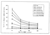
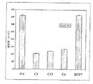
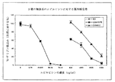
別の抗Cripto-1モノクローナル抗体であるMab C4を、Mab C3およびC13を誘導する際に用いたものと同じ方法を用いて取得した。それぞれのCripto-1 Mabは次の方法で選択した。すなわち、a) 標的組織への該抗体の結合を測定するための免疫ペルオキシダーゼ染色の検出、b)所定の細胞系での細胞増殖抑制アッセイ、例えば、3H-チミジンアッセイ(抗体はチミジン取込みにより60%を上回る抑制を示す)、およびc)トリパンブルー排除で測定して、細胞数が1/2に減少することを検出。図1の一番上の線は、72時間にわたり共培養した後のMab C4によるLS174T大腸癌細胞系の抑制を示し、一方、図2は、30μg/mlの各Mabを含む培地10mlを入れた25cm2フラスコ中で1×104個の該細胞を7日間培養した後のMab C4、C3およびC13による細胞カウント数の減少を示す。また、シスプラチンで処理される細胞にMabを添加すると、0.0938〜0.75μg/mlの薬物単独とのインキュベーションと比べて、3H-チミジン取込みがさらに減少したという点で、抗体はシスプラチンに対するLS174Tの感受性をも増強させた。
Example 2: Monoclonal antibody C4 against Cripto-1
Another anti-Cripto-1 monoclonal antibody, Mab C4, was obtained using the same method used to induce Mab C3 and C13. Each Cripto-1 Mab was selected by the following method. A) detection of immunoperoxidase staining to measure binding of the antibody to the target tissue, b) cytostatic assay in a given cell line, eg, 3 H-thymidine assay (antibodies are obtained by thymidine incorporation). ), And c) Detected a reduction in cell number by half as measured by trypan blue exclusion. The top line in FIG. 1 shows the suppression of the LS174T colon cancer cell line by Mab C4 after co-culture for 72 hours, while FIG. 2 contains 10 ml of medium containing 30 μg / ml of each Mab. FIG. 5 shows a decrease in cell count by Mabs C4, C3 and C13 after culturing 1 × 10 4 cells in a 25 cm 2 flask for 7 days. In addition, the addition of Mab to cells treated with cisplatin further reduced the sensitivity of LS174T to cisplatin in that 3 H-thymidine incorporation was further reduced compared to incubation with 0.0938-0.75 μg / ml drug alone. Was also enhanced.
同様の結果がエピルビシンおよび5FUの場合にも得られた。0、10、20および30μg/mlのMab C4との72時間のインキュベーション後に、LS174T細胞によるトリチウム標識チミジンの取込みは、0.04、0.08、0.1625および0.125μg/mlのエピルビシンの存在下で50〜90%も抑制された。5FUについては、1.5、1.9、2.1および2.4μg/mlの該薬物の存在下でチミジン取込みが50〜90%抑制された。5FUは大腸癌治療の主戦力であり、代謝拮抗薬である。Mab C4と5FU、シスプラチンまたはエピルビシンとの併用による相乗効果が臨床的に有効であるだろう。 Similar results were obtained with epirubicin and 5FU. After 72 hours incubation with 0, 10, 20 and 30 μg / ml Mab C4, tritium labeled thymidine incorporation by LS174T cells was 50-90% in the presence of 0.04, 0.08, 0.1625 and 0.125 μg / ml epirubicin. Was also suppressed. For 5FU, thymidine incorporation was inhibited by 50-90% in the presence of 1.5, 1.9, 2.1 and 2.4 μg / ml of the drug. 5FU is the main force for colorectal cancer treatment and an antimetabolite. The synergistic effect of combining Mab C4 with 5FU, cisplatin or epirubicin would be clinically effective.
実施例3: 抗Cripto-1抗体と癌および正常組織との結合試験
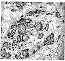
抗Cripto-1 Mabは、FACSおよび免疫ペルオキシダーゼ染色で試験したとき、LS174T、HT29 (大腸癌)、MCF7、T47D (乳癌)、DU145およびPC3 (前立腺癌)、BenおよびColo 235 (肺癌)など、多くの癌細胞系と反応したが、胚性腎細胞系293とは反応しなかった。また、3種類のMabは、免疫ペルオキシダーゼ染色により、ホルマリン固定組織、例えば、大腸癌(7/9)、乳癌(5/7)、あらゆるタイプの肺癌(18/20)、胃癌(3/4)、膵臓癌(1/2)と反応したが、正常な乳房(0/4)、大腸(0/8)、肺(0/4)、胃(0/2)、膵臓(0/2)、肝臓(0/3)、およびリンパ球(0/3)とは反応しなかった。染色の強度および比率は陰性から非常に強い陽性までさまざまであり、このことはCripto-1発現が癌ごとに異なっていることを示している。図3Aは、Mab C4による乳癌組織の免疫ペルオキシダーゼ染色を示し、該抗体による正常乳房組織の染色がまったく認められない図3Bと対照的である。
Example 3: Binding test between anti-Cripto-1 antibody and cancer and normal tissue Anti-Cripto-1 Mab was tested for LS174T, HT29 (colon cancer), MCF7, T47D (breast cancer), when tested with FACS and immunoperoxidase staining. Reacted with many cancer cell lines, such as DU145 and PC3 (prostate cancer), Ben and Colo 235 (lung cancer), but not with embryonic kidney cell line 293. In addition, three types of Mabs are stained with formalin-fixed tissues, such as colon cancer (7/9), breast cancer (5/7), all types of lung cancer (18/20), gastric cancer (3/4) by immunoperoxidase staining. , Reacted with pancreatic cancer (1/2), but normal breast (0/4), large intestine (0/8), lung (0/4), stomach (0/2), pancreas (0/2), It did not react with the liver (0/3) and lymphocytes (0/3). The intensity and ratio of staining varies from negative to very strong positive, indicating that Cripto-1 expression varies from cancer to cancer. FIG. 3A shows immunoperoxidase staining of breast cancer tissue with Mab C4, in contrast to FIG. 3B where no staining of normal breast tissue with the antibody is observed.
実施例4: マウスにおける大腸癌および前立腺癌細胞に対する抗Cripto-1抗体のin vivo抑制効果
SCIDマウス(6〜8週齢)に、0日目に2×10⁶個の前立腺癌細胞系DU145を皮下接種し、6時間後、マウスに500μgのMab C4を腹腔内投与し、続いて2、4、7、9および10日目に250μgを、そして14および17日目に125μgを投与した。対照としてリン酸緩衝溶液(PBS) (0.5ml)を使用した。24日目に腫瘍を取り出して測定した。腫瘍の大きさおよび重さがMab C4の投与により顕著に減少していた(図4Aおよび4B)。
Example 4: In vivo inhibitory effect of anti-Cripto-1 antibody on colon cancer and prostate cancer cells in mice
SCID mice (6-8 weeks old) were inoculated subcutaneously with 2 × 10 6 prostate cancer cell lines DU145 on
大腸癌モデルにおいても同様の結果が実証された(図5Aおよび5B)。この場合は、LS174T細胞を接種して16時間後にMab C13を500μg、その後2、7、9、11および13日目に250μg使用した。重さを測定するために25日目に腫瘍を切除した。
Similar results were demonstrated in the colorectal cancer model (FIGS. 5A and 5B). In this case, 500 μg of Mab C13 was used 16 hours after inoculation with LS174T cells, and then 250 μg was used on
実施例5: 抗Cripto-1抗体により誘導されるアポトーシス
抗Cripto-1モノクローナル抗体は、3H-チミジン取込みの低下により測定されるように細胞分裂を停止させ、細胞数を減少させた(それぞれ図1および2)。このことは、このMabが癌細胞のアポトーシスを誘導することを示している。これはさらにDNAの断片化およびFACSアッセイによっても実証された(図6および7)。
Example 5: Apoptotic anti-Cripto-1 monoclonal antibody induced by anti-Cripto-1 antibody stopped cell division and decreased cell number as measured by a decrease in 3 H-thymidine incorporation (respectively figures). 1 and 2). This indicates that this Mab induces apoptosis of cancer cells. This was further demonstrated by DNA fragmentation and FACS assay (FIGS. 6 and 7).
図6においては、50μg/mlのMab C3で72時間処理したLS174T細胞から可溶性DNAを抽出し、2%アガロースゲル上で電気泳動にかけた。細胞培地で処理した細胞からのものを対照サンプルとした。 In FIG. 6, soluble DNA was extracted from LS174T cells treated with 50 μg / ml Mab C3 for 72 hours and electrophoresed on a 2% agarose gel. Control samples were from cells treated with cell culture medium.
図7においては、LS174T細胞を30μg/mlのMab C4または対照抗体Mab BCP7で72時間処理し、その後フローサイトメトリーアッセイで分析して、アポトーシスの指示薬であるヨウ化プロピジウム(PI)染色を測定した。これらの結果から、試験Mabで処理した細胞ではPI染色が増加していることがわかった。 In FIG. 7, LS174T cells were treated with 30 μg / ml Mab C4 or the control antibody Mab BCP7 for 72 hours and then analyzed by flow cytometry assay to measure propidium iodide (PI) staining, an indicator of apoptosis. . From these results, it was found that PI staining was increased in cells treated with the test Mab.
実施例6: 抗Cripto-1抗体により媒介されるシグナル伝達
(i) 抗Cripto-1 Mabにより誘導されるJNK活性化
プロテインキナーゼモジュールにより制御されるシグナル伝達経路は、細胞増殖、分化、アポトーシスをはじめとする極めて重要な細胞機能を調節している。マイトジェンにより活性化されるプロテインキナーゼの3つの異なるセット(すなわち、細胞外シグナル調節キナーゼ(ERK)、JNK/SAPK、およびp38)の活性化に帰するアポトーシスの制御においては、3つの主要なキナーゼカスケードが同定されている。ERKはマイトジェンと生存因子により活性化され、一方JNK/SAPKおよびp38はストレスシグナルにより刺激される。ストレスにより活性化されるキナーゼカスケード(JNK/SAPKおよびp38経路を含む)は様々なアポトーシス刺激に応答して活性化され、アポトーシス過程で決定的な役割を果たしているようである。
Example 6: Signal transduction mediated by anti-Cripto-1 antibody
(i) JNK activation induced by anti-Cripto-1 Mab Signal transduction pathways controlled by protein kinase modules regulate vital cell functions such as cell proliferation, differentiation and apoptosis. Three major kinase cascades in the control of apoptosis resulting from activation of three different sets of protein kinases activated by mitogens (ie, extracellular signal-regulated kinases (ERK), JNK / SAPK, and p38) Has been identified. ERK is activated by mitogens and survival factors, while JNK / SAPK and p38 are stimulated by stress signals. Stress-activated kinase cascades (including the JNK / SAPK and p38 pathways) are activated in response to various apoptotic stimuli and appear to play a crucial role in the apoptotic process.
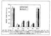
抗Cripto-1媒介アポトーシスにおけるJNKおよびp38の役割は、異なる濃度のモノクローナル抗体といろいろなインキュベーション時間を用いて、大腸癌LS174T細胞系において検討した。特に、抗Cripto-1 Mabと共に3時間インキュベーションした後のLS174T細胞においてはJNK/SPAKが用量依存的に活性化された(図8A)。JNK活性化は24時間の曝露後に最高レベルに達し(図8Bおよび8D)、48時間以内で減衰し、72時間のインキュベーションで基底レベルに戻った(図8D)。このことは、抗Cripto-1 MabによるJNK活性化が時間依存的であることを示している。 The role of JNK and p38 in anti-Cripto-1-mediated apoptosis was investigated in the colon cancer LS174T cell line using different concentrations of monoclonal antibodies and various incubation times. In particular, JNK / SPAK was activated in a dose-dependent manner in LS174T cells after incubation with anti-Cripto-1 Mab for 3 hours (FIG. 8A). JNK activation reached the highest level after 24 hours of exposure (Figures 8B and 8D), decayed within 48 hours, and returned to basal levels after 72 hours of incubation (Figure 8D). This indicates that JNK activation by anti-Cripto-1 Mab is time-dependent.
(ii) 抗Cripto-1抗体によるJNK活性化はp38活性化に先行する
ストレスに関係したp38経路についても、抗Cripto-1 Mabで処理した後のLS174T細胞で検討した。p38活性化はMab曝露の48時間後に起こり、この時点で活性化JNKのレベルが低下しはじめた。上昇JNKが基底レベルに戻った72時間後にはp38がさらに活性化された(図8D)。したがって、JNK活性化はMabにより誘導されるアポトーシスに先立って起こるが、p38はアポトーシスが起こる時間に活性化され、このことは、双方のシグナルがMab誘導アポトーシスに関与している可能性があることを示唆している。対照的に、シスプラチンはJNKおよびp38 MAPK活性化を両方とも誘導し(図8Aおよび8C)、このことは、Mabがシスプラチンとは異なるやり方でJNKとp38を活性化したことを示している。抗Cripto-1 Mabによるシスプラチン細胞傷害性の増強は、JNKリン酸化(図8A)およびp38 MAPK(図8B)の増加と同時に起こる。
(ii) JNK activation by anti-Cripto-1 antibody was also examined in the LS174T cells after treatment with anti-Cripto-1 Mab for the p38 pathway related to stress preceding p38 activation. p38 activation occurred 48 hours after Mab exposure, at which point the level of activated JNK began to decline. P38 was further activated 72 hours after elevated JNK returned to basal levels (FIG. 8D). Thus, JNK activation occurs prior to Mab-induced apoptosis, but p38 is activated at the time apoptosis occurs, indicating that both signals may be involved in Mab-induced apoptosis It suggests. In contrast, cisplatin induced both JNK and p38 MAPK activation (FIGS. 8A and 8C), indicating that Mab activated JNK and p38 differently than cisplatin. Enhancement of cisplatin cytotoxicity by anti-Cripto-1 Mab coincides with an increase in JNK phosphorylation (FIG. 8A) and p38 MAPK (FIG. 8B).
したがって、抗Cripto-1 MabはJNKとp38の双方の活性化を介して腫瘍細胞のアポトーシスを誘導する。 Thus, anti-Cripto-1 Mab induces tumor cell apoptosis through activation of both JNK and p38.
(iii) ERKおよびAktリン酸化ならびにCripto-1発現
ERKおよびAkt生存経路の阻害に対するMabの効果(図8Bおよび8D)は実証されなかった。Cripto-1発現レベルの変化はMab処理後に一切認められなかった(図8Bおよび8D)。これらの予備的なシグナル伝達研究からは、抗Cripto-1 MabがJNK活性化経路を介してアポトーシスを引き起こすことが明確に示される。
(iii) ERK and Akt phosphorylation and Cripto-1 expression
The effect of Mab on inhibition of ERK and Akt survival pathways (FIGS. 8B and 8D) was not demonstrated. No change in Cripto-1 expression level was observed after Mab treatment (FIGS. 8B and 8D). These preliminary signaling studies clearly show that anti-Cripto-1 Mabs cause apoptosis via the JNK activation pathway.
実施例7: 抗Cripto-1抗体による白血病細胞の抑制
図9は、Mab C3およびC13がT細胞リンパ芽球性白血病細胞系CCRF-CEMの増殖を抑制することを示す結果を提供する。これらの抗体はまた、上記細胞系の薬剤耐性変異体であるCEM/A7Rの増殖をも抑制した。変異体CEM/A7RはP-グリコプロテインの過剰発現によりこの性質を獲得する。こうして、この細胞系はさまざまな天然由来の化学療法剤に対して通常は耐性である。
Example 7: Inhibition of leukemia cells by anti-Cripto-1 antibody FIG. 9 provides results showing that Mab C3 and C13 inhibit the proliferation of the T cell lymphoblastic leukemia cell line CCRF-CEM. These antibodies also suppressed the growth of CEM / A7R, a drug-resistant mutant of the above cell line. Mutant CEM / A7R acquires this property by overexpression of P-glycoprotein. Thus, this cell line is usually resistant to various naturally derived chemotherapeutic agents.
Mab C4は、薬剤耐性細胞系CEM/A7およびCEM/A7Rならびに薬剤感受性マウス胸腺腫細胞系(すなわち、E3)に対して同様の抑制効果を示した。E3と比較して、CEM/A7およびCEM/A7Rはエピルビシンに対してそれぞれ約80倍および40倍の耐性を示す(図10A)。上記抗体は薬剤耐性細胞(図10B)と薬剤感受性細胞(図10C)をエピルビシン感受性の状態にするようである。したがって、C4は急性白血病に共通した薬剤耐性の問題を克服することができる。 Mab C4 showed similar inhibitory effects on the drug resistant cell lines CEM / A7 and CEM / A7R and the drug sensitive mouse thymoma cell line (ie, E3). Compared to E3, CEM / A7 and CEM / A7R are about 80 and 40 times more resistant to epirubicin, respectively (FIG. 10A). The antibody appears to render drug resistant cells (FIG. 10B) and drug sensitive cells (FIG. 10C) sensitive to epirubicin. Therefore, C4 can overcome the drug resistance problem common to acute leukemia.
実施例8: 前立腺癌に対する抗Cripto-1抗体の効果
前立腺癌細胞系PC3由来の細胞を30μg/mlのMab C3と共に6日間培養した。2、3および6日目に細胞数をカウントした。図11は、抗体の存在下において細胞数が著しく減少したことを示している。同様の効果が図12に示すように薬剤耐性DU 145細胞においても観察された。
Example 8: Effect of anti-Cripto-1 antibody on prostate cancer Cells derived from prostate cancer cell line PC3 were cultured with 30 μg / ml Mab C3 for 6 days. Cell numbers were counted on
さらにMab C3は、PC3細胞を薬剤エピルビシンに対して、また、DU 145細胞を薬剤シスプラチンに対して感受性の状態にすることができた(それぞれ、図13および14に示す)。 In addition, Mab C3 was able to render PC3 cells sensitive to the drug epirubicin and DU 145 cells to the drug cisplatin (shown in FIGS. 13 and 14, respectively).
実施例9: 抗Cripto-1抗体および抗癌剤の効果
大腸癌細胞LS174Tにおいてシスプラチンのような細胞傷害性薬剤の抑制効果を増強させるMab C4の能力は、上記実施例2に示される。同様の効果がMab C3およびC13の場合にもエピルビシンおよび5FUに関して認められた(それぞれ、図15および16に示す)。
Example 9: Effect of anti-Cripto-1 antibody and anticancer agent The ability of Mab C4 to enhance the inhibitory effect of cytotoxic drugs such as cisplatin in colon cancer cells LS174T is shown in Example 2 above. Similar effects were observed with Marub C3 and C13 for epirubicin and 5FU (shown in FIGS. 15 and 16, respectively).
実施例10: 抗Cripto-1抗体および乳癌
図17に示すように、Mab C3は乳癌細胞MCF7の増殖を抑制し、さらに該細胞をシスプラチン、カルボプラチンおよび5FUに対して感受性にした。同様の結果がMab C13とエピルビシンの場合にも観察された(図18)。
Example 10: Anti-Cripto-1 Antibody and Breast Cancer As shown in FIG. 17, Mab C3 suppressed the growth of breast cancer cells MCF7 and further sensitized the cells to cisplatin, carboplatin and 5FU. Similar results were observed with Mab C13 and epirubicin (Figure 18).
実施例11: 抗Cripto-1抗体の架橋
Mab C3を抗ラット抗体で架橋させた。このMabを架橋することの効果を乳癌細胞系MCF7において検討したが、その際、該細胞をMab C3と共に2時間インキュベートし、次いでウサギ抗ラット抗体と4時間インキュベートした後でPI染色を行った。架橋させなかったBCP7およびMab C3を対照として使用した。図19は、PI染色を用いたフローサイトメトリーにより測定して、Mabを架橋することがより顕著な細胞死をもたらしたことを示している。
Example 11: Cross-linking of anti-Cripto-1 antibody
Mab C3 was cross-linked with anti-rat antibody. The effect of cross-linking this Mab was examined in the breast cancer cell line MCF7, in which the cells were incubated with Mab C3 for 2 hours and then incubated with rabbit anti-rat antibody for 4 hours before PI staining. Uncrosslinked BCP7 and Mab C3 were used as controls. FIG. 19 shows that cross-linking the Mab resulted in more pronounced cell death as measured by flow cytometry using PI staining.
実施例12: 抗Pim-1抗体の単離
2つの単離されたMab(すなわち、P4およびP9)はPim-1オンコジーンの産物に対して誘導されたものである。この遺伝子はセリン-トレオニンプロテインキナーゼのクラスに属するタンパク質をコードしている。抗Pim-1抗体はマウス胸腺腫E3細胞の増殖を抑制し(図20)、また、試験した大腸癌(図21および22)および乳癌細胞系のほかに、これらの抗体は白血病および前立腺癌細胞系の抑制も示した(データは示してない)。
Example 12: Isolation of anti-Pim-1 antibody
Two isolated Mabs (ie, P4 and P9) have been induced against the product of the Pim-1 oncogene. This gene encodes a protein belonging to the class of serine-threonine protein kinases. Anti-Pim-1 antibodies inhibit the proliferation of mouse thymoma E3 cells (Figure 20), and in addition to the colon cancer (Figures 21 and 22) and breast cancer cell lines tested, these antibodies are also used in leukemia and prostate cancer cells. System suppression was also shown (data not shown).
実施例13: 大腸癌細胞溶解物抗原に対する抗体の単離
ラットを新鮮な大腸癌組織の溶解物で免疫することにより未知の抗原に対して5種類のMab (すなわち、1.14、1.68、2.20、3.60および4.57)を誘導し、単離した。これらの抗体も組織培養において大腸癌細胞系LS174Tおよび乳癌細胞系MCF7の増殖を抑制することが見出された(図23)。さらに、これらの抗体は、前立腺癌細胞系DU 145の抑制を示したが、特にシスプラチンと組み合わせて使用したときに認められた(図24)。
Example 13: Isolation of antibodies against colon cancer cell lysate antigen Rats were immunized with fresh colon cancer tissue lysate to immunize 5 antigens against unknown antigens (ie 1.14, 1.68, 2.20, 3.60). And 4.57) were derived and isolated. These antibodies were also found to inhibit the growth of colon cancer cell line LS174T and breast cancer cell line MCF7 in tissue culture (FIG. 23). In addition, these antibodies showed suppression of the prostate cancer cell line DU145, but were particularly observed when used in combination with cisplatin (FIG. 24).
実施例14: 抗体のヒト化
完全にヒトの抗Cripto-1抗体を作製するにあたって、免疫用抗原としてKLHに結合させた2つのペプチドを使用する。すなわち、Mab C3、C4およびC13を作製するために17-mer (97-113)ペプチド(配列番号1)、ならびに、(2)該17-merペプチドと、Cripto-1およびそのレセプターの推定上の結合部位と、を含む37-merペプチドp47 (77-113) ELNRTCCLNGGTCMLGSFCACPPSFYGRNCEHDVRKE(配列番号2)を使用し、ラット抗Cripto-1 Mabの作製について先に記載したものと同じ方法でこれらをin vivoおよびin vitroで試験する。
Example 14: Humanization of antibodies In producing a fully human anti-Cripto-1 antibody, two peptides conjugated to KLH are used as immunizing antigens. A 17-mer (97-113) peptide (SEQ ID NO: 1) to generate Mabs C3, C4 and C13, and (2) the putative 17-mer peptide and Cripto-1 and its receptor The 37-mer peptide p47 (77-113) ELNRTCCLNGGTCMLGSFCACPPSFYG R NCEHDVRKE (SEQ ID NO: 2) containing the binding site and these in vivo in the same manner as described above for the production of rat anti-Cripto-1 Mab And test in vitro.
これらの抗原を用いてマウスを免疫し、次いで非分泌ミエローマ細胞系NSO-bcl 2(免疫グロブリン遺伝子を一切もたない)と細胞融合させてスクリーニングするか、あるいは、これらの抗原を用いて、ヒト抗体のみを産生するようにマウス免疫グロブリン遺伝子が「ノックアウト」されてヒト遺伝子で置換されているヒトIgマウス(例えば、ゼノマウス(XenoMouse))(多重免疫感作を行って、高親和性抗体の存在についてマウスをスクリーニングすることができる)を免疫し、次いでマイクロプレートを用いた細胞増殖阻止アッセイにより阻止機能特性を示す抗体を産生するB細胞を同定する。その後、阻止抗体を産生する個々のB細胞の抗体コード化遺伝子を回収し、1群の適当な組換え候補抗体産物(それぞれ、いつでもスケールアップ製造が可能)を生成するために使用する。 Mice are immunized with these antigens and then screened by cell fusion with non-secreting myeloma cell line NSO-bcl 2 (without any immunoglobulin gene) or humans with these antigens Human Ig mice (eg, XenoMouse) in which mouse immunoglobulin genes are “knocked out” and replaced with human genes so that only antibodies are produced (eg, XenoMouse) (multiple immunization is performed, and high-affinity antibodies exist) Mice can be screened for) and then B cells producing antibodies exhibiting inhibitory functional properties are identified by a cell growth inhibition assay using microplates. The individual B cell antibody-encoding genes that produce blocking antibodies are then recovered and used to generate a group of appropriate recombinant candidate antibody products, each of which can be scaled up at any time.
実施例15: 抗体の臨床用途
実施例14に記載した手順に従って製造したヒトMabは、0.5 mg〜10 mg/kg(体重)の用量で静脈内注射により患者に投与する。患者には適当な抗癌剤を投与してもよい。
Example 15: Clinical Use of Antibodies Human Mabs produced according to the procedure described in Example 14 are administered to patients by intravenous injection at a dose of 0.5 mg to 10 mg / kg (body weight). The patient may be administered an appropriate anticancer agent.
実施例16: Cripto-1免疫感作の効果
レシピエントに「受動的」に投与される抗体とは対照的に、Criptoタンパク質またはその抗原フラグメントは「能動的」に免疫するために使用され、ワクチンをもたらす。そのような手法においては、Cripto抗原を担体(例えば、ミョウバン、マンナン、ビーズまたは他のアジュバント)と組み合わせて、癌の予防薬として癌患者を免疫するために使用する。その結果として生じる免疫応答は以下のものでありうる:
a) 上記の抗体を含むがこれらに限らない抗体の生成;
b) MHCクラスIまたはII分子により提示されたCripto抗原を認識するT細胞の生産(その結果として生じるT細胞応答は、細胞傷害性T細胞、サイトカイン(例えば、インターフェロン)産生細胞(例えば、ELISPOTまたは他の手段による)のようなエフェクター細胞、T細胞増殖、および/またはin vivo遅延型過敏反応のいずれかとして測定することができる);および/または
c) 抗体と細胞性免疫の両方の組合せ。
Example 16: Effect of Cripto-1 Immunization In contrast to antibodies that are "passively" administered to recipients, Cripto protein or antigenic fragments thereof are used to "actively" immunize vaccines Bring. In such an approach, the Cripto antigen is used in combination with a carrier (eg, alum, mannan, beads or other adjuvant) to immunize cancer patients as a prophylactic agent for cancer. The resulting immune response can be:
a) generation of antibodies, including but not limited to the above antibodies;
b) Production of T cells that recognize the Cripto antigen presented by MHC class I or II molecules (the resulting T cell response can be cytotoxic T cells, cytokines (eg interferon) producing cells (eg ELISPOT or Such as by effector cells, T cell proliferation, and / or in vivo delayed type hypersensitivity reactions)); and / or
c) A combination of both antibody and cellular immunity.
したがって、Cripto-1を用いて、レシピエントに投与する抗体を製造することができ、また、Cripto-1を用いて、抗体、T細胞またはその両方を産生する患者に「ワクチン接種」をすることができる。 Thus, Cripto-1 can be used to produce antibodies for administration to recipients, and Cripto-1 can be used to “vaccinate” patients who produce antibodies, T cells, or both Can do.
実施例14に記載した、KLHとコンジュゲートさせたCripto-1 37-merペプチド(CFAで乳化したもの)を用いてマウスを免疫した。免疫応答をELISAおよびELISPOT IFNγアッセイで調べた。マウスは抗体とIFNγ生産の両方に応答した(図25および26に示す)。 Mice were immunized with Cripto-1 37-mer peptide (emulsified with CFA) conjugated with KLH as described in Example 14. Immune responses were examined with ELISA and ELISPOT IFNγ assays. Mice responded to both antibody and IFNγ production (shown in FIGS. 25 and 26).
実施例17: 抗Cripto-1抗体および肺癌
Mab C4はまた、肺癌細胞BenおよびColo 38においても3H-チミジンの取込みを用量依存的に抑制した。Ben細胞では、対照細胞と比較して、Mabとの72時間のインキュベーション後に取込みが90%抑制された。Colo 38細胞では、抑制率が60%であった(図27)。
Example 17: Anti-Cripto-1 antibody and lung cancer
Mab C4 also inhibited 3 H-thymidine incorporation in a dose-dependent manner in lung cancer cells Ben and Colo 38. In Ben cells, uptake was suppressed by 90% after 72 hours incubation with Mab compared to control cells. In Colo 38 cells, the inhibition rate was 60% (FIG. 27).
肺癌細胞系Benまたは肺癌組織の免疫ペルオキシダーゼ染色もMab C3を示し、肺癌細胞の細胞表面と細胞質の両方に染色が認められた。一方、正常肺組織には染色がまったく認められなかった。 Immunoperoxidase staining of lung cancer cell line Ben or lung cancer tissue also showed Mab C3, and staining was observed on both the cell surface and cytoplasm of lung cancer cells. On the other hand, no staining was observed in normal lung tissue.
当業者であれば、特定の実施形態で示した本発明に対して、本発明の精神または範囲を逸脱することなしに、さまざまな変更および/または修飾が可能であることを理解するであろう。したがって、ここに記載した実施形態はすべての点で限定としてではなく例示として見なされるべきである。 Those skilled in the art will appreciate that various changes and / or modifications can be made to the invention shown in the specific embodiments without departing from the spirit or scope of the invention. . Accordingly, the embodiments described herein are to be considered in all respects only as illustrative and not restrictive.
Claims (18)
Applications Claiming Priority (3)
| Application Number | Priority Date | Filing Date | Title |
|---|---|---|---|
| AUPR3958A AUPR395801A0 (en) | 2001-03-26 | 2001-03-26 | Antibodies against cancer |
| AUPR3958 | 2001-03-26 | ||
| PCT/AU2002/000362 WO2002077033A1 (en) | 2001-03-26 | 2002-03-26 | Antibodies against cancer |
Related Child Applications (1)
| Application Number | Title | Priority Date | Filing Date |
|---|---|---|---|
| JP2008188842A Division JP2009024014A (en) | 2001-03-26 | 2008-07-22 | Antibody against cancer |
Publications (3)
| Publication Number | Publication Date |
|---|---|
| JP2004534741A JP2004534741A (en) | 2004-11-18 |
| JP2004534741A5 JP2004534741A5 (en) | 2005-12-22 |
| JP4854912B2 true JP4854912B2 (en) | 2012-01-18 |
Family
ID=3827964
Family Applications (2)
| Application Number | Title | Priority Date | Filing Date |
|---|---|---|---|
| JP2002576291A Expired - Fee Related JP4854912B2 (en) | 2001-03-26 | 2002-03-26 | Antibodies against cancer |
| JP2008188842A Pending JP2009024014A (en) | 2001-03-26 | 2008-07-22 | Antibody against cancer |
Family Applications After (1)
| Application Number | Title | Priority Date | Filing Date |
|---|---|---|---|
| JP2008188842A Pending JP2009024014A (en) | 2001-03-26 | 2008-07-22 | Antibody against cancer |
Country Status (9)
| Country | Link |
|---|---|
| US (1) | US7318924B2 (en) |
| EP (1) | EP1383801A4 (en) |
| JP (2) | JP4854912B2 (en) |
| KR (1) | KR100909290B1 (en) |
| CN (2) | CN100457781C (en) |
| AU (3) | AUPR395801A0 (en) |
| CA (1) | CA2442318A1 (en) |
| NZ (1) | NZ528624A (en) |
| WO (1) | WO2002077033A1 (en) |
Families Citing this family (28)
| Publication number | Priority date | Publication date | Assignee | Title |
|---|---|---|---|---|
| GB0020953D0 (en) | 2000-08-24 | 2000-10-11 | Smithkline Beecham Biolog | Vaccine |
| JP4307845B2 (en) | 2001-04-26 | 2009-08-05 | バイオジェン・アイデック・エムエイ・インコーポレイテッド | Antibodies that block Cripto and uses thereof |
| US7582299B2 (en) | 2001-04-26 | 2009-09-01 | Biogen Idec Ma Inc. | Cripto-specific antibodies |
| US20080213247A1 (en) * | 2002-10-23 | 2008-09-04 | Plowman Gregory D | Mbms as Modifiers of Branching Morphogenesis and Methods of Use |
| EP1624073A4 (en) * | 2003-04-03 | 2006-05-17 | Oncorex Inc | Drug |
| CA2529945A1 (en) * | 2003-06-27 | 2005-01-06 | Biogen Idec Ma Inc. | Use of hydrophobic-interaction-chromatography or hinge-region modifications for the production of homogeneous antibody-solutions |
| CA2539116C (en) * | 2003-09-15 | 2014-11-18 | Research Development Foundation | Cripto antagonism of activin and tgf-b signaling |
| EP1682159A4 (en) | 2003-10-16 | 2010-07-21 | Stephen John Ralph | Immunomodulating compositions and uses therefor |
| AU2006203889A1 (en) * | 2005-01-05 | 2006-07-13 | Biogen Idec Ma Inc. | CRIPTO binding molecules |
| US7939056B2 (en) * | 2005-11-14 | 2011-05-10 | The Brigham And Women's Hospital, Inc. | Interleukin-10 compositions for the treatment of adenocarcinomas |
| WO2008150530A2 (en) * | 2007-06-01 | 2008-12-11 | Biogen Idec Ma Inc. | Cripto binding molecules |
| US8425898B2 (en) | 2008-06-20 | 2013-04-23 | Duke University | Compositions, methods, and kits for eliciting an immune response |
| KR100973029B1 (en) * | 2008-10-07 | 2010-08-11 | 박지운 | Aluminum Alloy Street Lighting Pole |
| CN102272157B (en) * | 2008-11-07 | 2015-11-25 | 研究发展基金会 | For suppressing composition and the method for the formation of CRIPTO/GRP78 mixture and signal |
| JP5990752B2 (en) * | 2011-01-31 | 2016-09-14 | オリンパス株式会社 | Antibody therapy effect enhancer |
| BR112013027119A8 (en) | 2011-04-21 | 2018-03-06 | Seattle Genetics Inc | new ligand-drug conjugates (adcs) and their use |
| NZ720736A (en) | 2013-12-23 | 2020-08-28 | Bayer Pharma AG | Antibody drug conjugates (adcs) with kinesin spindel protein (ksp) |
| CN107847593B (en) * | 2015-04-20 | 2021-12-14 | 美侬米克国际有限公司 | Therapeutic antibodies and uses thereof |
| CA2990076A1 (en) | 2015-06-22 | 2016-12-29 | Bayer Pharma Aktiengesellschaft | Antibody drug conjugates (adcs) and antibody prodrug conjugates (apdcs) with enzymatically cleavable groups |
| EP3313523A2 (en) | 2015-06-23 | 2018-05-02 | Bayer Pharma Aktiengesellschaft | Targeted conjugates of ksp inhibitors |
| WO2017060322A2 (en) | 2015-10-10 | 2017-04-13 | Bayer Pharma Aktiengesellschaft | Ptefb-inhibitor-adc |
| CN116059390A (en) | 2016-03-24 | 2023-05-05 | 拜耳制药股份公司 | Prodrugs of cytotoxic actives with enzymatically cleavable groups |
| JP7022707B2 (en) | 2016-06-15 | 2022-02-18 | バイエル・ファルマ・アクティエンゲゼルシャフト | Specific antibody-drug-conjugate (ADC) including KSP inhibitor and anti-CD123 antibody |
| KR20190099250A (en) | 2016-12-21 | 2019-08-26 | 바이엘 악티엔게젤샤프트 | Prodrugs of Cytotoxic Active Agents with Enzymatically Cleavable Groups |
| BR112019012883A2 (en) | 2016-12-21 | 2019-11-26 | Bayer Ag | ligand-drug conjugates (adcs) which have enzymatically cleavable groups |
| US11433140B2 (en) | 2016-12-21 | 2022-09-06 | Bayer Pharma Aktiengesellschaft | Specific antibody drug conjugates (ADCs) having KSP inhibitors |
| CN109744199A (en) * | 2017-11-08 | 2019-05-14 | 南京艾莫瑞生物科技有限公司 | A kind of tumour cell heterograft zebra fish model, its construction method and application |
| JP6761889B1 (en) * | 2019-11-11 | 2020-09-30 | 株式会社Gspエンタープライズ | Anti-human Krypto-1 antibody |
Family Cites Families (14)
| Publication number | Priority date | Publication date | Assignee | Title |
|---|---|---|---|---|
| US5225539A (en) | 1986-03-27 | 1993-07-06 | Medical Research Council | Recombinant altered antibodies and methods of making altered antibodies |
| US4946778A (en) | 1987-09-21 | 1990-08-07 | Genex Corporation | Single polypeptide chain binding molecules |
| US5171665A (en) | 1989-04-17 | 1992-12-15 | Oncogen | Monoclonal antibody to novel antigen associated with human tumors |
| US5256643A (en) | 1990-05-29 | 1993-10-26 | The Government Of The United States | Human cripto protein |
| US5264557A (en) * | 1991-08-23 | 1993-11-23 | The United States Of America As Represented By The Department Of Health And Human Services | Polypeptide of a human cripto-related gene, CR-3 |
| US5981215A (en) | 1995-06-06 | 1999-11-09 | Human Genome Sciences, Inc. | Human criptin growth factor |
| US6207153B1 (en) | 1996-05-22 | 2001-03-27 | Viventia Biotech, Inc. | Antigen binding fragments that specifically detect cancer cells, nucleotides encoding the fragments, and use thereof for the prophylaxis and detection of cancers |
| IL125608A0 (en) | 1998-07-30 | 1999-03-12 | Yeda Res & Dev | Tumor associated antigen peptides and use of same as anti-tumor vaccines |
| ATE332013T1 (en) | 1999-03-11 | 2006-07-15 | Ardenia Investments Ltd | COMPOUNDS FOR CANCER THERAPY |
| US6333410B1 (en) | 2000-08-18 | 2001-12-25 | Immunogen, Inc. | Process for the preparation and purification of thiol-containing maytansinoids |
| GB0020953D0 (en) * | 2000-08-24 | 2000-10-11 | Smithkline Beecham Biolog | Vaccine |
| NZ525380A (en) | 2000-09-18 | 2008-06-30 | Biogen Idec Inc | Non-fucosylated forms of Cripto and their use as tumor blocking agents |
| JP4307845B2 (en) | 2001-04-26 | 2009-08-05 | バイオジェン・アイデック・エムエイ・インコーポレイテッド | Antibodies that block Cripto and uses thereof |
| US7582299B2 (en) | 2001-04-26 | 2009-09-01 | Biogen Idec Ma Inc. | Cripto-specific antibodies |
-
2001
- 2001-03-26 AU AUPR3958A patent/AUPR395801A0/en not_active Abandoned
-
2002
- 2002-03-26 CN CNB028073177A patent/CN100457781C/en not_active Expired - Fee Related
- 2002-03-26 NZ NZ528624A patent/NZ528624A/en not_active IP Right Cessation
- 2002-03-26 CN CNA2008101103851A patent/CN101310770A/en active Pending
- 2002-03-26 AU AU2002240719A patent/AU2002240719B2/en not_active Ceased
- 2002-03-26 CA CA002442318A patent/CA2442318A1/en not_active Abandoned
- 2002-03-26 JP JP2002576291A patent/JP4854912B2/en not_active Expired - Fee Related
- 2002-03-26 EP EP02706533A patent/EP1383801A4/en not_active Withdrawn
- 2002-03-26 WO PCT/AU2002/000362 patent/WO2002077033A1/en active IP Right Grant
- 2002-03-26 US US10/470,013 patent/US7318924B2/en not_active Expired - Fee Related
-
2003
- 2003-09-26 KR KR1020037012528A patent/KR100909290B1/en not_active IP Right Cessation
-
2007
- 2007-04-11 AU AU2007201587A patent/AU2007201587A1/en not_active Abandoned
-
2008
- 2008-07-22 JP JP2008188842A patent/JP2009024014A/en active Pending
Also Published As
| Publication number | Publication date |
|---|---|
| JP2004534741A (en) | 2004-11-18 |
| KR100909290B1 (en) | 2009-07-24 |
| KR20040010613A (en) | 2004-01-31 |
| CN101310770A (en) | 2008-11-26 |
| EP1383801A4 (en) | 2005-06-01 |
| CN100457781C (en) | 2009-02-04 |
| WO2002077033A1 (en) | 2002-10-03 |
| NZ528624A (en) | 2006-03-31 |
| AUPR395801A0 (en) | 2001-04-26 |
| AU2007201587A1 (en) | 2007-05-03 |
| AU2002240719B2 (en) | 2007-01-11 |
| JP2009024014A (en) | 2009-02-05 |
| CA2442318A1 (en) | 2002-10-03 |
| CN1639194A (en) | 2005-07-13 |
| US7318924B2 (en) | 2008-01-15 |
| US20040176576A1 (en) | 2004-09-09 |
| EP1383801A1 (en) | 2004-01-28 |
Similar Documents
| Publication | Publication Date | Title |
|---|---|---|
| JP4854912B2 (en) | Antibodies against cancer | |
| JP7138735B2 (en) | Antibodies and vaccines for use in treating ROR1 cancer and inhibiting metastasis | |
| US11498972B2 (en) | Anti-OX40 antibody and use thereof | |
| JP5631733B2 (en) | Anti-EpCAM antibodies and uses thereof | |
| AU2002240719A1 (en) | Antibodies against cancer | |
| JP6170926B2 (en) | Antibody against carcinoembryonic antigen-related cell adhesion molecule (CEACAM) | |
| JP2021531826A (en) | Specific antibody against trophoblast cell surface antigen 2 (TROP2) | |
| JP6326137B2 (en) | Anti-HER2 antibody and conjugate thereof | |
| JP2017518958A (en) | Antibodies against immunogenic glycopeptides, compositions containing them, and uses thereof | |
| CN110382532B (en) | anti-G-CSF antibodies and uses thereof | |
| JP2022514693A (en) | MUC18-specific antibody | |
| JP2022514786A (en) | MUC18-specific antibody | |
| ES2348161T3 (en) | METHODS TO DAMAGE CELLS USING ANTI-GFRA ANTIBODY EFECTOR FUNCTIONS1. | |
| US20100119514A1 (en) | Antibodies Against Cancer |
Legal Events
| Date | Code | Title | Description |
|---|---|---|---|
| A521 | Request for written amendment filed |
Free format text: JAPANESE INTERMEDIATE CODE: A523 Effective date: 20050316 |
|
| A621 | Written request for application examination |
Free format text: JAPANESE INTERMEDIATE CODE: A621 Effective date: 20050316 |
|
| A131 | Notification of reasons for refusal |
Free format text: JAPANESE INTERMEDIATE CODE: A131 Effective date: 20080122 |
|
| A601 | Written request for extension of time |
Free format text: JAPANESE INTERMEDIATE CODE: A601 Effective date: 20080415 |
|
| A602 | Written permission of extension of time |
Free format text: JAPANESE INTERMEDIATE CODE: A602 Effective date: 20080422 |
|
| A521 | Request for written amendment filed |
Free format text: JAPANESE INTERMEDIATE CODE: A523 Effective date: 20080613 |
|
| A521 | Request for written amendment filed |
Free format text: JAPANESE INTERMEDIATE CODE: A821 Effective date: 20080613 |
|
| A521 | Request for written amendment filed |
Free format text: JAPANESE INTERMEDIATE CODE: A523 Effective date: 20080722 |
|
| A02 | Decision of refusal |
Free format text: JAPANESE INTERMEDIATE CODE: A02 Effective date: 20080819 |
|
| A521 | Request for written amendment filed |
Free format text: JAPANESE INTERMEDIATE CODE: A523 Effective date: 20110926 |
|
| A01 | Written decision to grant a patent or to grant a registration (utility model) |
Free format text: JAPANESE INTERMEDIATE CODE: A01 |
|
| A61 | First payment of annual fees (during grant procedure) |
Free format text: JAPANESE INTERMEDIATE CODE: A61 Effective date: 20111026 |
|
| FPAY | Renewal fee payment (event date is renewal date of database) |
Free format text: PAYMENT UNTIL: 20141104 Year of fee payment: 3 |
|
| R150 | Certificate of patent or registration of utility model |
Free format text: JAPANESE INTERMEDIATE CODE: R150 |
|
| S111 | Request for change of ownership or part of ownership |
Free format text: JAPANESE INTERMEDIATE CODE: R313111 |
|
| FPAY | Renewal fee payment (event date is renewal date of database) |
Free format text: PAYMENT UNTIL: 20141104 Year of fee payment: 3 |
|
| R350 | Written notification of registration of transfer |
Free format text: JAPANESE INTERMEDIATE CODE: R350 |
|
| LAPS | Cancellation because of no payment of annual fees |