JP4632262B2 - Sample processing - Google Patents
Sample processing Download PDFInfo
- Publication number
- JP4632262B2 JP4632262B2 JP2006508686A JP2006508686A JP4632262B2 JP 4632262 B2 JP4632262 B2 JP 4632262B2 JP 2006508686 A JP2006508686 A JP 2006508686A JP 2006508686 A JP2006508686 A JP 2006508686A JP 4632262 B2 JP4632262 B2 JP 4632262B2
- Authority
- JP
- Japan
- Prior art keywords
- tubule
- segment
- reagent
- sample
- nucleic acid
- Prior art date
- Legal status (The legal status is an assumption and is not a legal conclusion. Google has not performed a legal analysis and makes no representation as to the accuracy of the status listed.)
- Expired - Lifetime
Links
- 238000012545 processing Methods 0.000 title claims description 49
- 210000005239 tubule Anatomy 0.000 claims description 252
- 239000000523 sample Substances 0.000 claims description 192
- 239000003153 chemical reaction reagent Substances 0.000 claims description 143
- 239000011324 bead Substances 0.000 claims description 86
- 238000000034 method Methods 0.000 claims description 85
- 150000007523 nucleic acids Chemical group 0.000 claims description 82
- 108020004707 nucleic acids Proteins 0.000 claims description 80
- 102000039446 nucleic acids Human genes 0.000 claims description 80
- 239000000758 substrate Substances 0.000 claims description 59
- 230000005291 magnetic effect Effects 0.000 claims description 54
- VYPSYNLAJGMNEJ-UHFFFAOYSA-N Silicium dioxide Chemical compound O=[Si]=O VYPSYNLAJGMNEJ-UHFFFAOYSA-N 0.000 claims description 50
- 239000002699 waste material Substances 0.000 claims description 48
- 239000007788 liquid Substances 0.000 claims description 42
- 239000000243 solution Substances 0.000 claims description 33
- 230000003321 amplification Effects 0.000 claims description 30
- 238000003199 nucleic acid amplification method Methods 0.000 claims description 30
- 238000003860 storage Methods 0.000 claims description 28
- 239000000126 substance Substances 0.000 claims description 28
- 239000011534 wash buffer Substances 0.000 claims description 27
- 239000000872 buffer Substances 0.000 claims description 25
- 239000000377 silicon dioxide Substances 0.000 claims description 25
- 239000000463 material Substances 0.000 claims description 23
- 239000012530 fluid Substances 0.000 claims description 22
- 230000009089 cytolysis Effects 0.000 claims description 19
- 238000010828 elution Methods 0.000 claims description 19
- 210000004027 cell Anatomy 0.000 claims description 18
- 239000007787 solid Substances 0.000 claims description 18
- 238000005406 washing Methods 0.000 claims description 18
- 230000027455 binding Effects 0.000 claims description 17
- -1 filter Substances 0.000 claims description 15
- 230000035784 germination Effects 0.000 claims description 15
- 239000000725 suspension Substances 0.000 claims description 15
- 238000004140 cleaning Methods 0.000 claims description 13
- 239000002853 nucleic acid probe Substances 0.000 claims description 13
- 238000010790 dilution Methods 0.000 claims description 11
- 239000012149 elution buffer Substances 0.000 claims description 11
- 210000003743 erythrocyte Anatomy 0.000 claims description 11
- 108020004711 Nucleic Acid Probes Proteins 0.000 claims description 10
- 108091093037 Peptide nucleic acid Proteins 0.000 claims description 10
- 230000003196 chaotropic effect Effects 0.000 claims description 10
- 239000012895 dilution Substances 0.000 claims description 10
- 239000013024 dilution buffer Substances 0.000 claims description 10
- 150000003839 salts Chemical class 0.000 claims description 10
- 108010067770 Endopeptidase K Proteins 0.000 claims description 9
- 241000700605 Viruses Species 0.000 claims description 9
- 238000002156 mixing Methods 0.000 claims description 7
- 239000003112 inhibitor Substances 0.000 claims description 6
- 230000007246 mechanism Effects 0.000 claims description 6
- 239000002253 acid Substances 0.000 claims description 5
- 239000000427 antigen Substances 0.000 claims description 5
- 108091007433 antigens Proteins 0.000 claims description 5
- 102000036639 antigens Human genes 0.000 claims description 5
- 238000003825 pressing Methods 0.000 claims description 5
- 230000004763 spore germination Effects 0.000 claims description 5
- 241000193738 Bacillus anthracis Species 0.000 claims description 4
- 108010053770 Deoxyribonucleases Proteins 0.000 claims description 4
- 102000016911 Deoxyribonucleases Human genes 0.000 claims description 4
- 239000000020 Nitrocellulose Substances 0.000 claims description 4
- 239000000084 colloidal system Substances 0.000 claims description 4
- 239000011521 glass Substances 0.000 claims description 4
- 239000003446 ligand Substances 0.000 claims description 4
- 229920001220 nitrocellulos Polymers 0.000 claims description 4
- 241000894006 Bacteria Species 0.000 claims description 3
- 102000004190 Enzymes Human genes 0.000 claims description 3
- 108090000790 Enzymes Proteins 0.000 claims description 3
- UGQMRVRMYYASKQ-KQYNXXCUSA-N Inosine Chemical compound O[C@@H]1[C@H](O)[C@@H](CO)O[C@H]1N1C2=NC=NC(O)=C2N=C1 UGQMRVRMYYASKQ-KQYNXXCUSA-N 0.000 claims description 3
- 229930010555 Inosine Natural products 0.000 claims description 3
- 108091005804 Peptidases Proteins 0.000 claims description 3
- 239000002250 absorbent Substances 0.000 claims description 3
- 230000002745 absorbent Effects 0.000 claims description 3
- 239000011248 coating agent Substances 0.000 claims description 3
- 238000000576 coating method Methods 0.000 claims description 3
- 239000000835 fiber Substances 0.000 claims description 3
- 208000015181 infectious disease Diseases 0.000 claims description 3
- 230000002458 infectious effect Effects 0.000 claims description 3
- 229960003786 inosine Drugs 0.000 claims description 3
- 239000011159 matrix material Substances 0.000 claims description 3
- 238000006386 neutralization reaction Methods 0.000 claims description 3
- 239000003161 ribonuclease inhibitor Substances 0.000 claims description 3
- XLYOFNOQVPJJNP-UHFFFAOYSA-N water Substances O XLYOFNOQVPJJNP-UHFFFAOYSA-N 0.000 claims description 3
- QDGAVODICPCDMU-UHFFFAOYSA-N 2-amino-3-[3-[bis(2-chloroethyl)amino]phenyl]propanoic acid Chemical compound OC(=O)C(N)CC1=CC=CC(N(CCCl)CCCl)=C1 QDGAVODICPCDMU-UHFFFAOYSA-N 0.000 claims description 2
- QNAYBMKLOCPYGJ-UHFFFAOYSA-N D-alpha-Ala Natural products CC([NH3+])C([O-])=O QNAYBMKLOCPYGJ-UHFFFAOYSA-N 0.000 claims description 2
- 241000588724 Escherichia coli Species 0.000 claims description 2
- 241000700721 Hepatitis B virus Species 0.000 claims description 2
- QNAYBMKLOCPYGJ-UWTATZPHSA-N L-Alanine Natural products C[C@@H](N)C(O)=O QNAYBMKLOCPYGJ-UWTATZPHSA-N 0.000 claims description 2
- ONIBWKKTOPOVIA-BYPYZUCNSA-N L-Proline Chemical compound OC(=O)[C@@H]1CCCN1 ONIBWKKTOPOVIA-BYPYZUCNSA-N 0.000 claims description 2
- QNAYBMKLOCPYGJ-REOHCLBHSA-N L-alanine Chemical compound C[C@H](N)C(O)=O QNAYBMKLOCPYGJ-REOHCLBHSA-N 0.000 claims description 2
- 229930182821 L-proline Natural products 0.000 claims description 2
- 108010083644 Ribonucleases Proteins 0.000 claims description 2
- 102000006382 Ribonucleases Human genes 0.000 claims description 2
- 229960003767 alanine Drugs 0.000 claims description 2
- 239000003146 anticoagulant agent Substances 0.000 claims description 2
- 229940127219 anticoagulant drug Drugs 0.000 claims description 2
- 210000004556 brain Anatomy 0.000 claims description 2
- 239000002738 chelating agent Substances 0.000 claims description 2
- 239000000701 coagulant Substances 0.000 claims description 2
- 239000003085 diluting agent Substances 0.000 claims description 2
- 238000001802 infusion Methods 0.000 claims description 2
- 230000003472 neutralizing effect Effects 0.000 claims description 2
- 238000003752 polymerase chain reaction Methods 0.000 claims description 2
- 229960002429 proline Drugs 0.000 claims description 2
- 239000004094 surface-active agent Substances 0.000 claims description 2
- 241001515965 unidentified phage Species 0.000 claims description 2
- 241000222722 Leishmania <genus> Species 0.000 claims 7
- 241000223960 Plasmodium falciparum Species 0.000 claims 3
- HEMHJVSKTPXQMS-UHFFFAOYSA-M Sodium hydroxide Chemical compound [OH-].[Na+] HEMHJVSKTPXQMS-UHFFFAOYSA-M 0.000 claims 3
- MTCFGRXMJLQNBG-REOHCLBHSA-N (2S)-2-Amino-3-hydroxypropansäure Chemical compound OC[C@H](N)C(O)=O MTCFGRXMJLQNBG-REOHCLBHSA-N 0.000 claims 2
- 238000004891 communication Methods 0.000 claims 2
- 239000006260 foam Substances 0.000 claims 2
- 230000003100 immobilizing effect Effects 0.000 claims 2
- 241000589875 Campylobacter jejuni Species 0.000 claims 1
- 241000193468 Clostridium perfringens Species 0.000 claims 1
- 241000701022 Cytomegalovirus Species 0.000 claims 1
- 241000725619 Dengue virus Species 0.000 claims 1
- ZRALSGWEFCBTJO-UHFFFAOYSA-N Guanidine Chemical group NC(N)=N ZRALSGWEFCBTJO-UHFFFAOYSA-N 0.000 claims 1
- 241000606768 Haemophilus influenzae Species 0.000 claims 1
- 241000590002 Helicobacter pylori Species 0.000 claims 1
- 241000711549 Hepacivirus C Species 0.000 claims 1
- 241000701044 Human gammaherpesvirus 4 Species 0.000 claims 1
- 241000713772 Human immunodeficiency virus 1 Species 0.000 claims 1
- 241000713340 Human immunodeficiency virus 2 Species 0.000 claims 1
- 241000186779 Listeria monocytogenes Species 0.000 claims 1
- 206010026749 Mania Diseases 0.000 claims 1
- 241000187479 Mycobacterium tuberculosis Species 0.000 claims 1
- 241000588650 Neisseria meningitidis Species 0.000 claims 1
- 241000150452 Orthohantavirus Species 0.000 claims 1
- 206010035148 Plague Diseases 0.000 claims 1
- 239000004365 Protease Substances 0.000 claims 1
- 102100037486 Reverse transcriptase/ribonuclease H Human genes 0.000 claims 1
- 241001354013 Salmonella enterica subsp. enterica serovar Enteritidis Species 0.000 claims 1
- 244000082988 Secale cereale Species 0.000 claims 1
- 241000191967 Staphylococcus aureus Species 0.000 claims 1
- 241000193998 Streptococcus pneumoniae Species 0.000 claims 1
- 239000007983 Tris buffer Substances 0.000 claims 1
- 241000700647 Variola virus Species 0.000 claims 1
- 241000607626 Vibrio cholerae Species 0.000 claims 1
- 241000710886 West Nile virus Species 0.000 claims 1
- 241000710772 Yellow fever virus Species 0.000 claims 1
- 241000607479 Yersinia pestis Species 0.000 claims 1
- 229940024606 amino acid Drugs 0.000 claims 1
- 150000001413 amino acids Chemical group 0.000 claims 1
- 229940065181 bacillus anthracis Drugs 0.000 claims 1
- 229940047650 haemophilus influenzae Drugs 0.000 claims 1
- 244000045947 parasite Species 0.000 claims 1
- 125000005642 phosphothioate group Chemical group 0.000 claims 1
- 235000019419 proteases Nutrition 0.000 claims 1
- 239000012629 purifying agent Substances 0.000 claims 1
- 229960001153 serine Drugs 0.000 claims 1
- 229940031000 streptococcus pneumoniae Drugs 0.000 claims 1
- LENZDBCJOHFCAS-UHFFFAOYSA-N tris Chemical compound OCC(N)(CO)CO LENZDBCJOHFCAS-UHFFFAOYSA-N 0.000 claims 1
- 241000712461 unidentified influenza virus Species 0.000 claims 1
- 229940118696 vibrio cholerae Drugs 0.000 claims 1
- 229910052724 xenon Inorganic materials 0.000 claims 1
- FHNFHKCVQCLJFQ-UHFFFAOYSA-N xenon atom Chemical compound [Xe] FHNFHKCVQCLJFQ-UHFFFAOYSA-N 0.000 claims 1
- 229940051021 yellow-fever virus Drugs 0.000 claims 1
- 238000001514 detection method Methods 0.000 description 56
- 210000004369 blood Anatomy 0.000 description 43
- 239000008280 blood Substances 0.000 description 43
- 230000008569 process Effects 0.000 description 40
- 238000003556 assay Methods 0.000 description 20
- 108091006146 Channels Proteins 0.000 description 19
- 108020004414 DNA Proteins 0.000 description 19
- 239000000203 mixture Substances 0.000 description 19
- KFZMGEQAYNKOFK-UHFFFAOYSA-N Isopropanol Chemical compound CC(C)O KFZMGEQAYNKOFK-UHFFFAOYSA-N 0.000 description 18
- LOKCTEFSRHRXRJ-UHFFFAOYSA-I dipotassium trisodium dihydrogen phosphate hydrogen phosphate dichloride Chemical compound P(=O)(O)(O)[O-].[K+].P(=O)(O)([O-])[O-].[Na+].[Na+].[Cl-].[K+].[Cl-].[Na+] LOKCTEFSRHRXRJ-UHFFFAOYSA-I 0.000 description 18
- 238000011534 incubation Methods 0.000 description 18
- 239000002953 phosphate buffered saline Substances 0.000 description 18
- LFQSCWFLJHTTHZ-UHFFFAOYSA-N Ethanol Chemical compound CCO LFQSCWFLJHTTHZ-UHFFFAOYSA-N 0.000 description 17
- 239000012472 biological sample Substances 0.000 description 13
- 210000004215 spore Anatomy 0.000 description 13
- 102000006943 Uracil-DNA Glycosidase Human genes 0.000 description 12
- 108010072685 Uracil-DNA Glycosidase Proteins 0.000 description 12
- 239000012139 lysis buffer Substances 0.000 description 12
- 238000012360 testing method Methods 0.000 description 12
- 238000007399 DNA isolation Methods 0.000 description 11
- 238000002955 isolation Methods 0.000 description 11
- AHCYMLUZIRLXAA-SHYZEUOFSA-N Deoxyuridine 5'-triphosphate Chemical compound O1[C@H](COP(O)(=O)OP(O)(=O)OP(O)(O)=O)[C@@H](O)C[C@@H]1N1C(=O)NC(=O)C=C1 AHCYMLUZIRLXAA-SHYZEUOFSA-N 0.000 description 10
- 108091028043 Nucleic acid sequence Proteins 0.000 description 10
- 108010006785 Taq Polymerase Proteins 0.000 description 10
- 238000000227 grinding Methods 0.000 description 10
- 239000000047 product Substances 0.000 description 10
- 238000003757 reverse transcription PCR Methods 0.000 description 10
- QKNYBSVHEMOAJP-UHFFFAOYSA-N 2-amino-2-(hydroxymethyl)propane-1,3-diol;hydron;chloride Chemical compound Cl.OCC(N)(CO)CO QKNYBSVHEMOAJP-UHFFFAOYSA-N 0.000 description 9
- 239000012807 PCR reagent Substances 0.000 description 9
- 238000013019 agitation Methods 0.000 description 9
- 238000006243 chemical reaction Methods 0.000 description 9
- 229920002477 rna polymer Polymers 0.000 description 9
- PCDQPRRSZKQHHS-XVFCMESISA-N CTP Chemical compound O=C1N=C(N)C=CN1[C@H]1[C@H](O)[C@H](O)[C@@H](COP(O)(=O)OP(O)(=O)OP(O)(O)=O)O1 PCDQPRRSZKQHHS-XVFCMESISA-N 0.000 description 8
- 108091034117 Oligonucleotide Proteins 0.000 description 8
- SUYVUBYJARFZHO-RRKCRQDMSA-N dATP Chemical compound C1=NC=2C(N)=NC=NC=2N1[C@H]1C[C@H](O)[C@@H](COP(O)(=O)OP(O)(=O)OP(O)(O)=O)O1 SUYVUBYJARFZHO-RRKCRQDMSA-N 0.000 description 8
- 238000003205 genotyping method Methods 0.000 description 8
- 239000011148 porous material Substances 0.000 description 8
- 108090000623 proteins and genes Proteins 0.000 description 8
- 239000002683 reaction inhibitor Substances 0.000 description 8
- 239000011541 reaction mixture Substances 0.000 description 8
- 229920000742 Cotton Polymers 0.000 description 7
- 238000007906 compression Methods 0.000 description 7
- 230000006835 compression Effects 0.000 description 7
- 238000002844 melting Methods 0.000 description 7
- 230000008018 melting Effects 0.000 description 7
- 238000010839 reverse transcription Methods 0.000 description 7
- 239000004743 Polypropylene Substances 0.000 description 6
- FAPWRFPIFSIZLT-UHFFFAOYSA-M Sodium chloride Chemical compound [Na+].[Cl-] FAPWRFPIFSIZLT-UHFFFAOYSA-M 0.000 description 6
- 238000001574 biopsy Methods 0.000 description 6
- 238000001914 filtration Methods 0.000 description 6
- 229920001155 polypropylene Polymers 0.000 description 6
- 239000013615 primer Substances 0.000 description 6
- 238000002123 RNA extraction Methods 0.000 description 5
- 108020000999 Viral RNA Proteins 0.000 description 5
- 230000001580 bacterial effect Effects 0.000 description 5
- 230000008859 change Effects 0.000 description 5
- 230000000295 complement effect Effects 0.000 description 5
- PJJJBBJSCAKJQF-UHFFFAOYSA-N guanidinium chloride Chemical compound [Cl-].NC(N)=[NH2+] PJJJBBJSCAKJQF-UHFFFAOYSA-N 0.000 description 5
- 230000033001 locomotion Effects 0.000 description 5
- 238000004519 manufacturing process Methods 0.000 description 5
- 230000035772 mutation Effects 0.000 description 5
- 239000004033 plastic Substances 0.000 description 5
- 229920003023 plastic Polymers 0.000 description 5
- HNXGGWNCFXZSAI-UHFFFAOYSA-N 2-morpholin-2-ylethanesulfonic acid Chemical compound OS(=O)(=O)CCC1CNCCO1 HNXGGWNCFXZSAI-UHFFFAOYSA-N 0.000 description 4
- 108700028369 Alleles Proteins 0.000 description 4
- 108020000946 Bacterial DNA Proteins 0.000 description 4
- 102000012410 DNA Ligases Human genes 0.000 description 4
- 108010061982 DNA Ligases Proteins 0.000 description 4
- KCXVZYZYPLLWCC-UHFFFAOYSA-N EDTA Chemical compound OC(=O)CN(CC(O)=O)CCN(CC(O)=O)CC(O)=O KCXVZYZYPLLWCC-UHFFFAOYSA-N 0.000 description 4
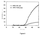
- 101150065637 Hfe gene Proteins 0.000 description 4
- 239000013504 Triton X-100 Substances 0.000 description 4
- 229920004890 Triton X-100 Polymers 0.000 description 4
- XSQUKJJJFZCRTK-UHFFFAOYSA-N Urea Chemical compound NC(N)=O XSQUKJJJFZCRTK-UHFFFAOYSA-N 0.000 description 4
- 230000015572 biosynthetic process Effects 0.000 description 4
- 239000004202 carbamide Substances 0.000 description 4
- 239000003795 chemical substances by application Substances 0.000 description 4
- 238000010438 heat treatment Methods 0.000 description 4
- 239000006166 lysate Substances 0.000 description 4
- 239000012528 membrane Substances 0.000 description 4
- 244000005700 microbiome Species 0.000 description 4
- 238000002360 preparation method Methods 0.000 description 4
- 238000000926 separation method Methods 0.000 description 4
- 108010014303 DNA-directed DNA polymerase Proteins 0.000 description 3
- 102000016928 DNA-directed DNA polymerase Human genes 0.000 description 3
- 108010007577 Exodeoxyribonuclease I Proteins 0.000 description 3
- 102100029075 Exonuclease 1 Human genes 0.000 description 3
- 239000007987 MES buffer Substances 0.000 description 3
- 239000000654 additive Substances 0.000 description 3
- 230000008901 benefit Effects 0.000 description 3
- 210000000601 blood cell Anatomy 0.000 description 3
- 108010052305 exodeoxyribonuclease III Proteins 0.000 description 3
- 230000002068 genetic effect Effects 0.000 description 3
- 238000009396 hybridization Methods 0.000 description 3
- 238000002347 injection Methods 0.000 description 3
- 239000007924 injection Substances 0.000 description 3
- 238000001746 injection moulding Methods 0.000 description 3
- 210000000265 leukocyte Anatomy 0.000 description 3
- 238000007834 ligase chain reaction Methods 0.000 description 3
- 239000002245 particle Substances 0.000 description 3
- 239000012266 salt solution Substances 0.000 description 3
- 239000011780 sodium chloride Substances 0.000 description 3
- 239000007790 solid phase Substances 0.000 description 3
- 238000012546 transfer Methods 0.000 description 3
- 230000003612 virological effect Effects 0.000 description 3
- 108010017384 Blood Proteins Proteins 0.000 description 2
- 102000004506 Blood Proteins Human genes 0.000 description 2
- 230000004568 DNA-binding Effects 0.000 description 2
- 241001646719 Escherichia coli O157:H7 Species 0.000 description 2
- 102000048988 Hemochromatosis Human genes 0.000 description 2
- 241000725303 Human immunodeficiency virus Species 0.000 description 2
- 102000003960 Ligases Human genes 0.000 description 2
- 108090000364 Ligases Proteins 0.000 description 2
- TWRXJAOTZQYOKJ-UHFFFAOYSA-L Magnesium chloride Chemical compound [Mg+2].[Cl-].[Cl-] TWRXJAOTZQYOKJ-UHFFFAOYSA-L 0.000 description 2
- 238000012408 PCR amplification Methods 0.000 description 2
- 102000035195 Peptidases Human genes 0.000 description 2
- 230000000996 additive effect Effects 0.000 description 2
- 239000000853 adhesive Substances 0.000 description 2
- 230000001070 adhesive effect Effects 0.000 description 2
- 230000004520 agglutination Effects 0.000 description 2
- 238000004458 analytical method Methods 0.000 description 2
- 238000013459 approach Methods 0.000 description 2
- 210000004666 bacterial spore Anatomy 0.000 description 2
- 230000004888 barrier function Effects 0.000 description 2
- 239000011230 binding agent Substances 0.000 description 2
- 238000005266 casting Methods 0.000 description 2
- 230000008878 coupling Effects 0.000 description 2
- 238000010168 coupling process Methods 0.000 description 2
- 238000005859 coupling reaction Methods 0.000 description 2
- 239000012470 diluted sample Substances 0.000 description 2
- 238000001035 drying Methods 0.000 description 2
- 230000007613 environmental effect Effects 0.000 description 2
- 229940088598 enzyme Drugs 0.000 description 2
- 238000001125 extrusion Methods 0.000 description 2
- 238000002866 fluorescence resonance energy transfer Methods 0.000 description 2
- 210000003128 head Anatomy 0.000 description 2
- 238000007849 hot-start PCR Methods 0.000 description 2
- 238000000338 in vitro Methods 0.000 description 2
- 239000006193 liquid solution Substances 0.000 description 2
- 238000011880 melting curve analysis Methods 0.000 description 2
- 230000000813 microbial effect Effects 0.000 description 2
- 238000012544 monitoring process Methods 0.000 description 2
- 238000000465 moulding Methods 0.000 description 2
- 239000013642 negative control Substances 0.000 description 2
- 230000005298 paramagnetic effect Effects 0.000 description 2
- 230000035699 permeability Effects 0.000 description 2
- 239000013612 plasmid Substances 0.000 description 2
- 229920000515 polycarbonate Polymers 0.000 description 2
- 239000004417 polycarbonate Substances 0.000 description 2
- 229920000098 polyolefin Polymers 0.000 description 2
- 235000019833 protease Nutrition 0.000 description 2
- 238000010791 quenching Methods 0.000 description 2
- 230000000171 quenching effect Effects 0.000 description 2
- 239000000376 reactant Substances 0.000 description 2
- 239000012488 sample solution Substances 0.000 description 2
- 230000003068 static effect Effects 0.000 description 2
- 238000003756 stirring Methods 0.000 description 2
- 238000003786 synthesis reaction Methods 0.000 description 2
- 238000013518 transcription Methods 0.000 description 2
- 230000035897 transcription Effects 0.000 description 2
- 235000011178 triphosphate Nutrition 0.000 description 2
- 239000001226 triphosphate Substances 0.000 description 2
- 210000002845 virion Anatomy 0.000 description 2
- NWUYHJFMYQTDRP-UHFFFAOYSA-N 1,2-bis(ethenyl)benzene;1-ethenyl-2-ethylbenzene;styrene Chemical compound C=CC1=CC=CC=C1.CCC1=CC=CC=C1C=C.C=CC1=CC=CC=C1C=C NWUYHJFMYQTDRP-UHFFFAOYSA-N 0.000 description 1
- 102100031126 6-phosphogluconolactonase Human genes 0.000 description 1
- 108010029731 6-phosphogluconolactonase Proteins 0.000 description 1
- 206010069754 Acquired gene mutation Diseases 0.000 description 1
- 101150029409 CFTR gene Proteins 0.000 description 1
- 101100315624 Caenorhabditis elegans tyr-1 gene Proteins 0.000 description 1
- 206010053567 Coagulopathies Diseases 0.000 description 1
- 108091026890 Coding region Proteins 0.000 description 1
- 108020001738 DNA Glycosylase Proteins 0.000 description 1
- 238000007400 DNA extraction Methods 0.000 description 1
- 102000028381 DNA glycosylase Human genes 0.000 description 1
- 108050009160 DNA polymerase 1 Proteins 0.000 description 1
- 239000003155 DNA primer Substances 0.000 description 1
- 238000001712 DNA sequencing Methods 0.000 description 1
- 241000450599 DNA viruses Species 0.000 description 1
- 108090000626 DNA-directed RNA polymerases Proteins 0.000 description 1
- 102000004163 DNA-directed RNA polymerases Human genes 0.000 description 1
- 101100009781 Danio rerio dmbx1a gene Proteins 0.000 description 1
- 239000004593 Epoxy Substances 0.000 description 1
- 108060002716 Exonuclease Proteins 0.000 description 1
- 108010018962 Glucosephosphate Dehydrogenase Proteins 0.000 description 1
- 208000018565 Hemochromatosis Diseases 0.000 description 1
- 239000004831 Hot glue Substances 0.000 description 1
- 206010028980 Neoplasm Diseases 0.000 description 1
- 241000714209 Norwalk virus Species 0.000 description 1
- 206010030113 Oedema Diseases 0.000 description 1
- 101150105372 POX1 gene Proteins 0.000 description 1
- 239000004698 Polyethylene Substances 0.000 description 1
- 101710194807 Protective antigen Proteins 0.000 description 1
- 102000052575 Proto-Oncogene Human genes 0.000 description 1
- 108700020978 Proto-Oncogene Proteins 0.000 description 1
- 108020004518 RNA Probes Proteins 0.000 description 1
- 239000003391 RNA probe Substances 0.000 description 1
- 101001091368 Rattus norvegicus Glandular kallikrein-7, submandibular/renal Proteins 0.000 description 1
- 229920000297 Rayon Polymers 0.000 description 1
- 101000898773 Saccharomyces cerevisiae (strain ATCC 204508 / S288c) Saccharopepsin Proteins 0.000 description 1
- 241000239226 Scorpiones Species 0.000 description 1
- 108010017898 Shiga Toxins Proteins 0.000 description 1
- 108090000190 Thrombin Proteins 0.000 description 1
- 108010020713 Tth polymerase Proteins 0.000 description 1
- 102000044209 Tumor Suppressor Genes Human genes 0.000 description 1
- 108700025716 Tumor Suppressor Genes Proteins 0.000 description 1
- 206010047249 Venous thrombosis Diseases 0.000 description 1
- 108020005202 Viral DNA Proteins 0.000 description 1
- 101100194320 Zea mays PER1 gene Proteins 0.000 description 1
- 230000002159 abnormal effect Effects 0.000 description 1
- 230000000078 anti-malarial effect Effects 0.000 description 1
- 229940019748 antifibrinolytic proteinase inhibitors Drugs 0.000 description 1
- 239000003430 antimalarial agent Substances 0.000 description 1
- 238000011225 antiretroviral therapy Methods 0.000 description 1
- 239000012062 aqueous buffer Substances 0.000 description 1
- 239000012298 atmosphere Substances 0.000 description 1
- 235000015278 beef Nutrition 0.000 description 1
- 238000005452 bending Methods 0.000 description 1
- 239000012148 binding buffer Substances 0.000 description 1
- 238000000071 blow moulding Methods 0.000 description 1
- DQXBYHZEEUGOBF-UHFFFAOYSA-N but-3-enoic acid;ethene Chemical compound C=C.OC(=O)CC=C DQXBYHZEEUGOBF-UHFFFAOYSA-N 0.000 description 1
- 238000005251 capillar electrophoresis Methods 0.000 description 1
- 230000006037 cell lysis Effects 0.000 description 1
- 210000000170 cell membrane Anatomy 0.000 description 1
- 210000002421 cell wall Anatomy 0.000 description 1
- 238000003776 cleavage reaction Methods 0.000 description 1
- 238000010367 cloning Methods 0.000 description 1
- 230000035602 clotting Effects 0.000 description 1
- 238000005345 coagulation Methods 0.000 description 1
- 230000015271 coagulation Effects 0.000 description 1
- 239000002299 complementary DNA Substances 0.000 description 1
- 238000010276 construction Methods 0.000 description 1
- 238000011109 contamination Methods 0.000 description 1
- 230000006837 decompression Effects 0.000 description 1
- 230000001934 delay Effects 0.000 description 1
- 230000014670 detection of bacterium Effects 0.000 description 1
- 239000003599 detergent Substances 0.000 description 1
- 238000003113 dilution method Methods 0.000 description 1
- 238000006073 displacement reaction Methods 0.000 description 1
- 239000003814 drug Substances 0.000 description 1
- 241001493065 dsRNA viruses Species 0.000 description 1
- 210000002889 endothelial cell Anatomy 0.000 description 1
- 238000005516 engineering process Methods 0.000 description 1
- UAUDZVJPLUQNMU-KTKRTIGZSA-N erucamide Chemical compound CCCCCCCC\C=C/CCCCCCCCCCCC(N)=O UAUDZVJPLUQNMU-KTKRTIGZSA-N 0.000 description 1
- 239000005038 ethylene vinyl acetate Substances 0.000 description 1
- 230000005284 excitation Effects 0.000 description 1
- 102000013165 exonuclease Human genes 0.000 description 1
- 238000002474 experimental method Methods 0.000 description 1
- 238000000605 extraction Methods 0.000 description 1
- 108010091897 factor V Leiden Proteins 0.000 description 1
- 239000010419 fine particle Substances 0.000 description 1
- 239000012634 fragment Substances 0.000 description 1
- 238000001415 gene therapy Methods 0.000 description 1
- 210000004602 germ cell Anatomy 0.000 description 1
- 239000003365 glass fiber Substances 0.000 description 1
- 239000001963 growth medium Substances 0.000 description 1
- WPIULSIZRNJJDL-UHFFFAOYSA-N guanidine;isocyanic acid Chemical group N=C=O.NC(N)=N WPIULSIZRNJJDL-UHFFFAOYSA-N 0.000 description 1
- 230000003067 hemagglutinative effect Effects 0.000 description 1
- 230000002209 hydrophobic effect Effects 0.000 description 1
- 239000004615 ingredient Substances 0.000 description 1
- 230000005764 inhibitory process Effects 0.000 description 1
- 238000010102 injection blow moulding Methods 0.000 description 1
- 238000011081 inoculation Methods 0.000 description 1
- 238000009830 intercalation Methods 0.000 description 1
- 239000003456 ion exchange resin Substances 0.000 description 1
- 229920003303 ion-exchange polymer Polymers 0.000 description 1
- 238000003475 lamination Methods 0.000 description 1
- 231100000518 lethal Toxicity 0.000 description 1
- 230000001665 lethal effect Effects 0.000 description 1
- 239000010808 liquid waste Substances 0.000 description 1
- 238000011068 loading method Methods 0.000 description 1
- 230000002934 lysing effect Effects 0.000 description 1
- 229910001629 magnesium chloride Inorganic materials 0.000 description 1
- 210000004962 mammalian cell Anatomy 0.000 description 1
- 230000001404 mediated effect Effects 0.000 description 1
- 239000002609 medium Substances 0.000 description 1
- 238000002493 microarray Methods 0.000 description 1
- 238000000520 microinjection Methods 0.000 description 1
- 239000002991 molded plastic Substances 0.000 description 1
- 210000000214 mouth Anatomy 0.000 description 1
- 238000010899 nucleation Methods 0.000 description 1
- 239000002773 nucleotide Substances 0.000 description 1
- 125000003729 nucleotide group Chemical group 0.000 description 1
- FATBGEAMYMYZAF-KTKRTIGZSA-N oleamide Chemical compound CCCCCCCC\C=C/CCCCCCCC(N)=O FATBGEAMYMYZAF-KTKRTIGZSA-N 0.000 description 1
- 230000003287 optical effect Effects 0.000 description 1
- 238000004806 packaging method and process Methods 0.000 description 1
- 230000036961 partial effect Effects 0.000 description 1
- 230000007918 pathogenicity Effects 0.000 description 1
- 239000000137 peptide hydrolase inhibitor Substances 0.000 description 1
- 239000012466 permeate Substances 0.000 description 1
- 238000009832 plasma treatment Methods 0.000 description 1
- 229920001200 poly(ethylene-vinyl acetate) Polymers 0.000 description 1
- 229920001515 polyalkylene glycol Polymers 0.000 description 1
- 229920000573 polyethylene Polymers 0.000 description 1
- 229920000139 polyethylene terephthalate Polymers 0.000 description 1
- 239000005020 polyethylene terephthalate Substances 0.000 description 1
- 229920000642 polymer Polymers 0.000 description 1
- 229920001296 polysiloxane Polymers 0.000 description 1
- 229920002635 polyurethane Polymers 0.000 description 1
- 239000004814 polyurethane Substances 0.000 description 1
- 238000003793 prenatal diagnosis Methods 0.000 description 1
- INDBQLZJXZLFIT-UHFFFAOYSA-N primaquine Chemical compound N1=CC=CC2=CC(OC)=CC(NC(C)CCCN)=C21 INDBQLZJXZLFIT-UHFFFAOYSA-N 0.000 description 1
- 229960005179 primaquine Drugs 0.000 description 1
- 230000001737 promoting effect Effects 0.000 description 1
- 235000004252 protein component Nutrition 0.000 description 1
- 230000005180 public health Effects 0.000 description 1
- 238000000746 purification Methods 0.000 description 1
- 238000011002 quantification Methods 0.000 description 1
- 239000002096 quantum dot Substances 0.000 description 1
- 239000002964 rayon Substances 0.000 description 1
- 238000003753 real-time PCR Methods 0.000 description 1
- 238000011897 real-time detection Methods 0.000 description 1
- 230000002829 reductive effect Effects 0.000 description 1
- 230000002787 reinforcement Effects 0.000 description 1
- 230000002441 reversible effect Effects 0.000 description 1
- 238000006798 ring closing metathesis reaction Methods 0.000 description 1
- 238000005096 rolling process Methods 0.000 description 1
- 210000003296 saliva Anatomy 0.000 description 1
- 230000007017 scission Effects 0.000 description 1
- 238000007789 sealing Methods 0.000 description 1
- 238000012163 sequencing technique Methods 0.000 description 1
- 210000002966 serum Anatomy 0.000 description 1
- 239000012748 slip agent Substances 0.000 description 1
- 239000002689 soil Substances 0.000 description 1
- 244000000000 soil microbiome Species 0.000 description 1
- 230000037439 somatic mutation Effects 0.000 description 1
- 238000001179 sorption measurement Methods 0.000 description 1
- 241000894007 species Species 0.000 description 1
- 238000001228 spectrum Methods 0.000 description 1
- 230000001502 supplementing effect Effects 0.000 description 1
- 229940124597 therapeutic agent Drugs 0.000 description 1
- 239000012815 thermoplastic material Substances 0.000 description 1
- 229960004072 thrombin Drugs 0.000 description 1
- 238000001890 transfection Methods 0.000 description 1
- 125000002264 triphosphate group Chemical class [H]OP(=O)(O[H])OP(=O)(O[H])OP(=O)(O[H])O* 0.000 description 1
- UNXRWKVEANCORM-UHFFFAOYSA-N triphosphoric acid Chemical compound OP(O)(=O)OP(O)(=O)OP(O)(O)=O UNXRWKVEANCORM-UHFFFAOYSA-N 0.000 description 1
- 238000003466 welding Methods 0.000 description 1
Images


Classifications
-
- C—CHEMISTRY; METALLURGY
- C12—BIOCHEMISTRY; BEER; SPIRITS; WINE; VINEGAR; MICROBIOLOGY; ENZYMOLOGY; MUTATION OR GENETIC ENGINEERING
- C12N—MICROORGANISMS OR ENZYMES; COMPOSITIONS THEREOF; PROPAGATING, PRESERVING, OR MAINTAINING MICROORGANISMS; MUTATION OR GENETIC ENGINEERING; CULTURE MEDIA
- C12N15/00—Mutation or genetic engineering; DNA or RNA concerning genetic engineering, vectors, e.g. plasmids, or their isolation, preparation or purification; Use of hosts therefor
- C12N15/09—Recombinant DNA-technology
- C12N15/10—Processes for the isolation, preparation or purification of DNA or RNA
- C12N15/1003—Extracting or separating nucleic acids from biological samples, e.g. pure separation or isolation methods; Conditions, buffers or apparatuses therefor
-
- B—PERFORMING OPERATIONS; TRANSPORTING
- B01—PHYSICAL OR CHEMICAL PROCESSES OR APPARATUS IN GENERAL
- B01F—MIXING, e.g. DISSOLVING, EMULSIFYING OR DISPERSING
- B01F31/00—Mixers with shaking, oscillating, or vibrating mechanisms
- B01F31/55—Mixers with shaking, oscillating, or vibrating mechanisms the materials to be mixed being contained in a flexible bag submitted to periodical deformation
-
- B—PERFORMING OPERATIONS; TRANSPORTING
- B01—PHYSICAL OR CHEMICAL PROCESSES OR APPARATUS IN GENERAL
- B01L—CHEMICAL OR PHYSICAL LABORATORY APPARATUS FOR GENERAL USE
- B01L3/00—Containers or dishes for laboratory use, e.g. laboratory glassware; Droppers
- B01L3/50—Containers for the purpose of retaining a material to be analysed, e.g. test tubes
- B01L3/502—Containers for the purpose of retaining a material to be analysed, e.g. test tubes with fluid transport, e.g. in multi-compartment structures
-
- G—PHYSICS
- G01—MEASURING; TESTING
- G01N—INVESTIGATING OR ANALYSING MATERIALS BY DETERMINING THEIR CHEMICAL OR PHYSICAL PROPERTIES
- G01N1/00—Sampling; Preparing specimens for investigation
- G01N1/02—Devices for withdrawing samples
- G01N1/10—Devices for withdrawing samples in the liquid or fluent state
-
- G—PHYSICS
- G01—MEASURING; TESTING
- G01N—INVESTIGATING OR ANALYSING MATERIALS BY DETERMINING THEIR CHEMICAL OR PHYSICAL PROPERTIES
- G01N33/00—Investigating or analysing materials by specific methods not covered by groups G01N1/00 - G01N31/00
- G01N33/48—Biological material, e.g. blood, urine; Haemocytometers
- G01N33/50—Chemical analysis of biological material, e.g. blood, urine; Testing involving biospecific ligand binding methods; Immunological testing
- G01N33/53—Immunoassay; Biospecific binding assay; Materials therefor
- G01N33/543—Immunoassay; Biospecific binding assay; Materials therefor with an insoluble carrier for immobilising immunochemicals
-
- B—PERFORMING OPERATIONS; TRANSPORTING
- B01—PHYSICAL OR CHEMICAL PROCESSES OR APPARATUS IN GENERAL
- B01L—CHEMICAL OR PHYSICAL LABORATORY APPARATUS FOR GENERAL USE
- B01L2200/00—Solutions for specific problems relating to chemical or physical laboratory apparatus
- B01L2200/06—Fluid handling related problems
- B01L2200/0621—Control of the sequence of chambers filled or emptied
-
- B—PERFORMING OPERATIONS; TRANSPORTING
- B01—PHYSICAL OR CHEMICAL PROCESSES OR APPARATUS IN GENERAL
- B01L—CHEMICAL OR PHYSICAL LABORATORY APPARATUS FOR GENERAL USE
- B01L2200/00—Solutions for specific problems relating to chemical or physical laboratory apparatus
- B01L2200/10—Integrating sample preparation and analysis in single entity, e.g. lab-on-a-chip concept
-
- B—PERFORMING OPERATIONS; TRANSPORTING
- B01—PHYSICAL OR CHEMICAL PROCESSES OR APPARATUS IN GENERAL
- B01L—CHEMICAL OR PHYSICAL LABORATORY APPARATUS FOR GENERAL USE
- B01L2300/00—Additional constructional details
- B01L2300/04—Closures and closing means
- B01L2300/041—Connecting closures to device or container
- B01L2300/042—Caps; Plugs
-
- B—PERFORMING OPERATIONS; TRANSPORTING
- B01—PHYSICAL OR CHEMICAL PROCESSES OR APPARATUS IN GENERAL
- B01L—CHEMICAL OR PHYSICAL LABORATORY APPARATUS FOR GENERAL USE
- B01L2300/00—Additional constructional details
- B01L2300/04—Closures and closing means
- B01L2300/046—Function or devices integrated in the closure
- B01L2300/047—Additional chamber, reservoir
-
- B—PERFORMING OPERATIONS; TRANSPORTING
- B01—PHYSICAL OR CHEMICAL PROCESSES OR APPARATUS IN GENERAL
- B01L—CHEMICAL OR PHYSICAL LABORATORY APPARATUS FOR GENERAL USE
- B01L2300/00—Additional constructional details
- B01L2300/06—Auxiliary integrated devices, integrated components
- B01L2300/0681—Filter
-
- B—PERFORMING OPERATIONS; TRANSPORTING
- B01—PHYSICAL OR CHEMICAL PROCESSES OR APPARATUS IN GENERAL
- B01L—CHEMICAL OR PHYSICAL LABORATORY APPARATUS FOR GENERAL USE
- B01L2300/00—Additional constructional details
- B01L2300/06—Auxiliary integrated devices, integrated components
- B01L2300/069—Absorbents; Gels to retain a fluid
-
- B—PERFORMING OPERATIONS; TRANSPORTING
- B01—PHYSICAL OR CHEMICAL PROCESSES OR APPARATUS IN GENERAL
- B01L—CHEMICAL OR PHYSICAL LABORATORY APPARATUS FOR GENERAL USE
- B01L2300/00—Additional constructional details
- B01L2300/08—Geometry, shape and general structure
- B01L2300/0861—Configuration of multiple channels and/or chambers in a single devices
- B01L2300/087—Multiple sequential chambers
-
- B—PERFORMING OPERATIONS; TRANSPORTING
- B01—PHYSICAL OR CHEMICAL PROCESSES OR APPARATUS IN GENERAL
- B01L—CHEMICAL OR PHYSICAL LABORATORY APPARATUS FOR GENERAL USE
- B01L2300/00—Additional constructional details
- B01L2300/10—Means to control humidity and/or other gases
-
- B—PERFORMING OPERATIONS; TRANSPORTING
- B01—PHYSICAL OR CHEMICAL PROCESSES OR APPARATUS IN GENERAL
- B01L—CHEMICAL OR PHYSICAL LABORATORY APPARATUS FOR GENERAL USE
- B01L2400/00—Moving or stopping fluids
- B01L2400/04—Moving fluids with specific forces or mechanical means
- B01L2400/0475—Moving fluids with specific forces or mechanical means specific mechanical means and fluid pressure
- B01L2400/0481—Moving fluids with specific forces or mechanical means specific mechanical means and fluid pressure squeezing of channels or chambers
-
- B—PERFORMING OPERATIONS; TRANSPORTING
- B01—PHYSICAL OR CHEMICAL PROCESSES OR APPARATUS IN GENERAL
- B01L—CHEMICAL OR PHYSICAL LABORATORY APPARATUS FOR GENERAL USE
- B01L2400/00—Moving or stopping fluids
- B01L2400/06—Valves, specific forms thereof
- B01L2400/0677—Valves, specific forms thereof phase change valves; Meltable, freezing, dissolvable plugs; Destructible barriers
-
- B—PERFORMING OPERATIONS; TRANSPORTING
- B01—PHYSICAL OR CHEMICAL PROCESSES OR APPARATUS IN GENERAL
- B01L—CHEMICAL OR PHYSICAL LABORATORY APPARATUS FOR GENERAL USE
- B01L3/00—Containers or dishes for laboratory use, e.g. laboratory glassware; Droppers
- B01L3/50—Containers for the purpose of retaining a material to be analysed, e.g. test tubes
- B01L3/505—Containers for the purpose of retaining a material to be analysed, e.g. test tubes flexible containers not provided for above
-
- Y—GENERAL TAGGING OF NEW TECHNOLOGICAL DEVELOPMENTS; GENERAL TAGGING OF CROSS-SECTIONAL TECHNOLOGIES SPANNING OVER SEVERAL SECTIONS OF THE IPC; TECHNICAL SUBJECTS COVERED BY FORMER USPC CROSS-REFERENCE ART COLLECTIONS [XRACs] AND DIGESTS
- Y02—TECHNOLOGIES OR APPLICATIONS FOR MITIGATION OR ADAPTATION AGAINST CLIMATE CHANGE
- Y02A—TECHNOLOGIES FOR ADAPTATION TO CLIMATE CHANGE
- Y02A50/00—TECHNOLOGIES FOR ADAPTATION TO CLIMATE CHANGE in human health protection, e.g. against extreme weather
- Y02A50/30—Against vector-borne diseases, e.g. mosquito-borne, fly-borne, tick-borne or waterborne diseases whose impact is exacerbated by climate change
-
- Y—GENERAL TAGGING OF NEW TECHNOLOGICAL DEVELOPMENTS; GENERAL TAGGING OF CROSS-SECTIONAL TECHNOLOGIES SPANNING OVER SEVERAL SECTIONS OF THE IPC; TECHNICAL SUBJECTS COVERED BY FORMER USPC CROSS-REFERENCE ART COLLECTIONS [XRACs] AND DIGESTS
- Y10—TECHNICAL SUBJECTS COVERED BY FORMER USPC
- Y10S—TECHNICAL SUBJECTS COVERED BY FORMER USPC CROSS-REFERENCE ART COLLECTIONS [XRACs] AND DIGESTS
- Y10S435/00—Chemistry: molecular biology and microbiology
- Y10S435/81—Packaged device or kit
-
- Y—GENERAL TAGGING OF NEW TECHNOLOGICAL DEVELOPMENTS; GENERAL TAGGING OF CROSS-SECTIONAL TECHNOLOGIES SPANNING OVER SEVERAL SECTIONS OF THE IPC; TECHNICAL SUBJECTS COVERED BY FORMER USPC CROSS-REFERENCE ART COLLECTIONS [XRACs] AND DIGESTS
- Y10—TECHNICAL SUBJECTS COVERED BY FORMER USPC
- Y10T—TECHNICAL SUBJECTS COVERED BY FORMER US CLASSIFICATION
- Y10T436/00—Chemistry: analytical and immunological testing
- Y10T436/25—Chemistry: analytical and immunological testing including sample preparation
Description
関連出願の相互参照
本願明細書は、引用をもってその内容全体を本願明細書に援用する2003年2月5日に提出された米国特許仮出願第60/445,304号の優先権を主張する。以下の米国特許出願第09/910,233号、09/782,732号、および10/241,816号も、引用をもってその内容全体を本願明細書に援用する。
CROSS REFERENCE TO RELATED APPLICATIONS This application claims priority to US Provisional Application No. 60 / 445,304, filed Feb. 5, 2003, the entire contents of which are incorporated herein by reference. The following US patent applications 09 / 910,233, 09 / 782,732, and 10 / 241,816 are also incorporated herein by reference in their entirety.
発明の背景
診断用アッセイを行う際、特に生物学的サンプルの処理において、サンプルの調製を必要とすることが多い。たとえば、生物学的サンプルは、典型的には、アッセイに好適な条件下におく前に集中的で厳密な処理を施す。適切なサンプル調製には、たとえば特定の温度、濃度、試薬の容量、および望ましいアッセイを妨害する可能性のある物質の除去などの厳密な条件を要することが多い。未処理のサンプルに、厳密に管理された研究施設で高度に習熟した人員による適切な処理を施すために、遠く離れた場所に運ばなければならないことが多い。従来の処理装置および方法は、大規模で非常に複雑で洗練された設備を必要とすることが多い。従来のサンプル処理のこれらの要因によって、結果が出るまでの時間の遅れ、高コスト、サンプルの完全性の欠損、および診断用アッセイを多くの場合に使用する実用性への制限が、常に生じる。
BACKGROUND OF THE INVENTION When performing a diagnostic assay, sample preparation is often required, particularly in the processing of biological samples. For example, biological samples are typically subjected to intensive and rigorous processing prior to placing them under conditions suitable for the assay. Proper sample preparation often requires stringent conditions such as specific temperatures, concentrations, reagent volumes, and removal of substances that may interfere with the desired assay. Untreated samples often have to be transported to a remote location in order to be properly processed by highly trained personnel in a strictly controlled laboratory. Conventional processing equipment and methods often require large, very complex and sophisticated equipment. These factors in conventional sample processing always result in delays in time to results, high costs, lack of sample integrity, and limitations on the practical use of diagnostic assays in many cases.
発明の概要
本願開示はサンプルを処理する装置および方法を提供する。開示された装置および方法は複数の処理ステップによってサンプルの調製を容易にすることができる。
SUMMARY OF THE INVENTION The present disclosure provides an apparatus and method for processing a sample. The disclosed apparatus and method can facilitate sample preparation through multiple processing steps.
ある局面では、サンプルを処理する小管には第1のセグメント、第2のセグメント、および第3のセグメントが含まれてよい。各セグメントは前記小管によって範囲が決定され、少なくとも部分的に破壊可能なシールで流動的に隔離され、別のセグメントから排出されたある容量の流体を受けることができるように膨張可能で、圧縮された場合には実質的に流体が入らないように圧縮可能であってよい。各セグメントには少なくとも1種類の試薬を入れることができる。 In certain aspects, the tubule for processing the sample may include a first segment, a second segment, and a third segment. Each segment is delimited by the tubule, is fluidly isolated with an at least partially destructible seal, and is inflatable and compressed to receive a volume of fluid discharged from another segment. In this case, it may be compressible so that substantially no fluid enters. Each segment can contain at least one reagent.
別の局面では、サンプルを処理する方法には、破壊可能なシールで流動的に隔離された複数のセグメントに分離された小管にサンプルを導入するステップであって、当該ステップにおいて前記小管が廃棄物を受ける近位端とアッセイを実施する遠位端を有するステップと、前記小管のセグメントに入ったサンプルを予め選択された前記サンプルの成分に特異的に結合することができる物質でインキュベートするステップと、前記の予め選択された成分を含有するセグメントの遠位の小管を締めてそのセグメントを圧縮することによって予め選択された成分から出る廃棄物を除去するステップと、試薬を放出して、前記の予め選択した成分を含有するセグメントの少なくとも1つとそのセグメントの遠位にある試薬を含有するセグメントを圧縮し、破壊可能なシールを開けて前記試薬を前記の予め選択された成分を含有するセグメントに押し出すか、または前記の予め選択された成分を前記試薬を含有するセグメントへ押し出すことによって、流動的に隔離された隣接する遠位セグメントから出た前記の予め選択された成分と混合するステップと、を含んでよい。 In another aspect, a method of processing a sample includes introducing a sample into a tubule separated into a plurality of segments fluidly isolated by a breakable seal, wherein the tubule is a waste product. Receiving a proximal end for receiving and a distal end for performing an assay; and incubating a sample contained in the segment of the tubule with a substance that can specifically bind to a preselected component of the sample; Removing waste from the preselected component by tightening a distal tubule of the segment containing the preselected component and compressing the segment; Compress at least one segment containing a preselected component and a segment containing reagents distal to that segment Fluidly sequestered by opening the breakable seal and extruding the reagent into the segment containing the preselected component or extruding the preselected component into the segment containing the reagent Mixing with said preselected ingredients exiting from the adjacent adjacent distal segment.
本願明細書に開示された装置および方法は、既存の技術よりも優位に優れた利点を提供することができる。ある実施態様では、望ましいサンプル処理プロトコル用の小管と試薬を予めパックし、アッセイ全体用の物質を1つの便利なパッケージとして提供することもできる。ある実施態様では、処理の初期段階で廃棄産生物を目的の標的から分離し、処理されたサンプルが未処理のサンプルと接触した表面と接触しないようにする。したがって、小管壁をコーティングするかもしれない未処理サンプル中に存在する微量の反応阻害物質が、前記の処理されたサンプルを汚染する可能性は小さい。 The apparatus and methods disclosed herein can provide advantages that are superior to existing technologies. In some embodiments, tubules and reagents for the desired sample processing protocol can be pre-packed and the material for the entire assay provided in one convenient package. In certain embodiments, the waste product is separated from the target of interest at an early stage of processing so that the treated sample does not come into contact with the surface in contact with the untreated sample. Thus, it is unlikely that trace amounts of reaction inhibitors present in an untreated sample that may coat the tubule wall will contaminate the treated sample.
発明の詳細な説明
本願開示はサンプルを処理するデバイスおよび方法を記述する。いくつかの実施態様では、セグメントに分けられた小管は生物学的サンプルの受け取り、保存、処理および/または分析するために有用な容器を提供する。ある実施態様では、セグメントに分けられた小管は複数の処理ステップが関与するサンプル処理のプロトコルを容易にする。ある実施態様では、サンプルをサンプル小管に収集し、その後その小管を分析器の中に配置することができ、前記分析器が前記小管およびその内容物を操作して前記サンプルを処理することができる。
DETAILED DESCRIPTION OF THE INVENTION The present disclosure describes devices and methods for processing samples. In some embodiments, the segmented tubule provides a useful container for receiving, storing, processing and / or analyzing a biological sample. In some embodiments, segmented tubules facilitate sample processing protocols that involve multiple processing steps. In one embodiment, a sample can be collected in a sample tube, which can then be placed in an analyzer, and the analyzer can manipulate the tube and its contents to process the sample. .
好ましい実施態様には、破壊可能なシールでコンパートメントに分割されたフレキシブル小管が含まれる。各々のセグメントにはサンプルを処理するための様々な試薬およびバッファを入れてよい。クランプおよびアクチュエータをさまざまな組み合わせおよびさまざまなタイミングで前記小管に適用して、流体の動きを方向付け、破壊可能なシールを破ることもできる。この破壊可能なシールが破れると、実質的に障害のない内部小管壁に流体を流すことができる。好ましい実施態様では、生物学的サンプルの流れは処理が進展するにつれて前記小管の遠位端の方向に向けられ、その一方で廃棄物の流れは反対側の前記小管の開口部、つまり前記サンプルが最初に注入された開口部へ強制的に動かされてよい。このサンプル注入口は、施錠メカニズムを有するキャップで可能で永久に密封することができ、そのキャップの中に廃棄物チャンバがおかれていて廃棄物を受け取って保存することができる。このアプローチの重要な利点は、処理されたサンプルは、未処理のサンプルが接触した表面と接触しない点である。したがって、小管壁をコーティングするかもしれない未処理サンプル中に存在する微量の反応阻害物質が、前記の処理されたサンプルを汚染する可能性は小さい。 A preferred embodiment includes a flexible tubule that is divided into compartments with a breakable seal. Each segment may contain various reagents and buffers for processing the sample. Clamps and actuators can be applied to the tubules in various combinations and at various timings to direct fluid movement and break a breakable seal. When this breakable seal is broken, fluid can flow through the inner tubule wall, which is substantially unobstructed. In a preferred embodiment, the flow of biological sample is directed toward the distal end of the tubule as processing progresses, while the waste stream is directed to the opposite tubule opening, i.e., the sample. It may be forced to move to the originally injected opening. The sample inlet can be permanently sealed with a cap with a locking mechanism, and a waste chamber can be placed in the cap to receive and store the waste. An important advantage of this approach is that the treated sample does not contact the surface that the untreated sample contacts. Thus, it is unlikely that trace amounts of reaction inhibitors present in an untreated sample that may coat the tubule wall will contaminate the treated sample.
ある実施態様では、前記小管は1つのセグメントの中の複数に分かれたセグメントのそれぞれから出るある容量の流体を受け取ることができるように膨張可能であってよい。これによって、1つのセグメントの中で特定の処理ステップをサンプルと試薬に実施することができるので、アッセイを行うための機械的構造を単純にすることができる。膨張可能であってよい小管を用いた実施態様の別の利点は、同一の小管構造を用いて、セグメントの中で異なる容積の試薬をパッケージすることができ、同一の小管を実施すべきアッセイに応じて異なる仕様にパッケージすることができる点である。 In certain embodiments, the tubule may be inflatable to receive a volume of fluid exiting from each of the plurality of segments within a segment. This simplifies the mechanical structure for performing the assay because certain processing steps can be performed on the samples and reagents within a segment. Another advantage of embodiments using tubules that may be inflatable is that the same tubule structure can be used to package different volumes of reagents within a segment, to the same tubule being assayed. It can be packaged in different specifications accordingly.
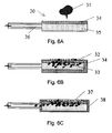
前記装置には、透明なフレキシブル小管10(図1AおよびB、図2AおよびB、ならびに図3AおよびB)であって、16、110、120、130、140、150、160、170、180、および/または190などの複数のセグメントを構成することができ、圧縮によって実質的に平らになるフレキシブル小管10が含まれてよい。ある実施態様では、小管は少なくとも2つのセグメントを有してよい。ある実施態様では、小管は少なくとも3つのセグメントを有してよい。前記フレキシブル小管は約2℃乃至105℃における操作上の機能性、サンプル、標的、および試薬との適合性、気体の低浸透性、最小の蛍光特性、および/または圧縮および屈曲サイクルの繰り返しにおける弾性を提供することができる。前記小管はたとえば、ポリプロピレンまたはポリエチレンなどのポリオレフィン、ポリウレタン、ポリオレフィンコポリマー、および/または好適な特徴を提供するその他の物質などをそれらに限定せずに含む、さまざまな物質でつくられてよい。透明性、湿潤性、表面の滑らかさ、表面電荷および熱弾性、などの小管の特性が、前記小管の性能に影響するかもしれない。これらの特性は、シード添加、プラズマ処理、添加物の添加、および照射などの例示的なプロセスによって向上することがある。ある実施態様では、前記プラスチックに添加物質を添加して、特定の特徴を向上させることもできる。たとえば、エルカ酸アミドおよび/またはオレイン酸アミドなどのスリップ剤を加えてもよく、ある実施態様ではいわゆる「粘着防止剤」を加えてもよい。添加物の前記プラスチック中の濃度は、約0.01%乃至約5.0%の範囲内であってよい。
The apparatus includes a transparent flexible tubule 10 (FIGS. 1A and B, FIGS. 2A and B, and FIGS. 3A and B), 16, 110, 120, 130, 140, 150, 160, 170, 180, and A plurality of segments, such as 190, may be configured and may include a
前記小管は、押し出し成形、射出成形、およびブロー成形などの、多様な好適な方法によって製造することができる。好ましい実施態様では、前記小管は連続的に押し出される。前記小管の製造のための代替的な技術には、二次的な処理操作によって好適な小管をつくることができる、たとえば鋳造、押出、またはブローフィルムなどが含まれる。前記小管壁の材料には、共押し出しまたはフィルムラミネーションによる多層が含まれる。たとえば、内層は高い生体適合性で選択し、外層は低い気体透過性で選択してよい。さらなる例として、前記内層は剥がすことができるシールなどの破壊可能なシール14(図2AおよびB、ならびに図3AおよびB)に容易に成形することができるが、前記外層は弾力的で不浸透性が高いものであってよい。本願明細書の開示に用いるために、前記小管は約0.03mm乃至約0.8mm、好ましくは0.03mm乃至約0.5mmの壁の厚さを有し、約1気圧の圧力を外部からかけると実質的に平らになることができることが望ましい。 The tubule can be manufactured by a variety of suitable methods such as extrusion, injection molding, and blow molding. In a preferred embodiment, the tubule is extruded continuously. Alternative techniques for the manufacture of the tubule include, for example, casting, extrusion, or blown film, where suitable tubules can be made by secondary processing operations. The tubule wall material includes multiple layers by coextrusion or film lamination. For example, the inner layer may be selected with high biocompatibility and the outer layer may be selected with low gas permeability. As a further example, the inner layer can be easily formed into a breakable seal 14 (FIGS. 2A and B, and FIGS. 3A and B), such as a peelable seal, while the outer layer is elastic and impervious. May be high. For use in disclosing the present specification, the tubule has a wall thickness of about 0.03 mm to about 0.8 mm, preferably 0.03 mm to about 0.5 mm, and is substantially subject to a pressure of about 1 atm from the outside. It is desirable to be able to flatten.
ある実施態様では、前記装置は、粉砕運動を用いて生検サンプルまたは固体状の環境サンプルなどの固体サンプルから得た細胞の塊を崩壊させることができるように、少なくとも1つのセグメントの壁が強化されていてよい。これらの強化された壁の特徴の例は、図7Aに描写されているとおり、前記小管の壁の反対側の表面にある、微細な歯状の内部表面109であってよく、前記小管を圧縮すると前記小管の軸に沿った滑り運動を生じるように補正されている。これらの粉砕表面109の近傍にある小管の壁は、ポリカーボネートまたはポリエチレンテレフタラートなどの弾力のある好適なプラスチックでできている補強パッチを用いて補強されていてよい。前記の歯状の内部表面は同様に好適な材料でできていてよい。別の実施態様では、図5AおよびBに描写されている214などの粉砕表面の形状を有するパッドを小管の内部壁に付けてよい。前記パッドは強化された材料でつくることができ、前記表面形状は従来の機械的、電気化学的、または微小電気機械的な方法を用いて、前記パッドが圧縮に耐えられるようにつくることができる。
In one embodiment, the device has a wall of at least one segment that is reinforced so that a mass of cells obtained from a solid sample, such as a biopsy sample or a solid environmental sample, can be disrupted using a crushing motion. May have been. An example of these enhanced wall features may be a fine toothed
前記サンプル小管10は1つ以上のセグメント16、110、120、130、140、150、160、170、180、および/または190、ならびに/またはサブセグメント18、121、122に分割することができる。好ましい実施態様では、前記セグメントは破壊可能なシール14で範囲を決定されており、隣接するセグメントと流動的に隔離されている。このシールは、たとえば、あるアッセイを行うために乾燥試薬と液体試薬を再構築するまでこの2つの試薬を分離する場合や、反応が望ましい時点まで化学反応種を分離するために有用なことがある。図3AおよびBに示されるとおり、破壊可能なシール14は小管10の領域であって、その領域において、相対する壁が実質的に連結されているが、あとで前記小管または予めシールされた表面を著しく損傷せずに前記の壁を剥がすことができないほど強く連結しないように、小管10の領域に形成してよい。そのようなシールは「剥離可能な」シールと命名してよい。好ましい実施態様では、前記の剥離可能なシールの領域は前記小管の軸に直角なバンドであってよい。そのバンドは、約0.5mm乃至5mm、好ましくは約1mm乃至約3mm、最も好ましくは約1mmの範囲内において小管の長さをカバーしてよい。、前記シールは好ましくは、前記セグメントをシールできるように前記小管の幅全体をカバーする。ある実施態様では、前記シールバンドの長さまたは形状は変化してよく、および/または前記シールバンドの面は前記小管の軸に直角に配置してよい。そのような変化によって、剥離特性を変化することができる。
The
破壊可能なシール14は、前記小管の相対する壁の間に、剥離可能なシールが望ましい位置にある前記小管に制御された量のエネルギーをかけることによってつくることができる。たとえば、温度制御したシール形成ヘッドで、固定された金敷に対して一定の圧力で一定の時間間隔をおいて圧力をかけることができる。望ましいサイズと剥離力のシールを形成するために、温度、圧力、および時間の様々な組み合わせを選択してよい。エネルギーは、たとえばポリプロピレン管材料を加熱するために105℃乃至140℃の一定温度に保たれた温度制御シール形成ヘッド、望ましいシール領域に3乃至100気圧の圧力を正確にかけることができるアクチュエータ、および1乃至30秒の一定のサイクル時間で前記アクチュエータの配列を動かすための制御システムによって、送達してよい。この方法を用いて、約1気圧の内部圧力をかけると剥がれて開くように、ポリプロピレン小管に十分なシールを形成した。シール形成のエネルギーを前記小管に送達する代替的な技術には、RFおよび超音波溶接が含まれる。
The
他の実施態様では、代替的な小管の材料および材料の配合を用いて剥離可能なシールの性能を最適化することができる。たとえば、剥離可能なシールの形成のために、異なる融点の2種類のポリプロピレンポリマを、組成と溶融特性が最適化されるような割合で混合してよい。破壊可能なシール14に加えて、またはその代わりに、前記フレキシブル小管は、さらに1つ以上の圧力ゲート194を有することができる。この圧力ゲートは、前記フレキシブル小管のセグメントに制御された力をかけることによって、テストの操作中に、可逆的に開閉できる。
In other embodiments, alternative tubule materials and material blends can be used to optimize peelable seal performance. For example, to form a peelable seal, two polypropylene polymers with different melting points may be mixed in proportions that optimize composition and melt properties. In addition to or instead of the
フィルターを小管のセグメントに組み込むことができるフィルターの例206および216を、それぞれ図4A、ならびに図5AおよびBに示す。好ましい実施態様では、フレキシブルフィルタ材料を多層に重ねてフィルタをつくることができる。サンプルに直接接触する前記フィルタの最上層は、ろ過のために選択された孔サイズを有してよく、ろ過中に圧力がかかるときに最上層を支持するための構造を提供するために、前記フィルタの下層にははるかに大きな孔サイズを有する材料を含んでよい。この好ましい実施態様では、前記フィルタはバッグ状に折り畳まれてよく、その開口端が前記小管壁にしっかり付けられているエッジを有してよい。前記フィルタバッグを有するセグメントは、前記小管の外部を圧縮することによって実質的に平らにすることができてよい。
Examples of
例示的な実施態様では、小管のセグメントの中に、1種類以上の試薬を乾燥物質として、または液体状溶液として保存することができる。試薬を乾燥状態で保存してもよい実施態様では、液体状溶液を隣接するセグメントに保存して、前記試薬溶液の再構築を促進にすることができる。典型的な試薬の例には、溶解試薬、溶出バッファ、洗浄バッファ、DNase阻害剤、RNase阻害剤、プロテイナーゼ阻害剤、キレート剤、中和試薬、カオトロピック塩溶液、洗浄剤、界面活性剤、抗凝血剤、発芽溶液、イソプロパノール、エタノール溶液、抗体、核酸プローブ、ペプチド核酸プローブ、およびホスホチオ酸核酸プローブが含まれる。前記試薬の1つがカオトロピック塩溶液の場合の実施態様では、好ましい成分はイソシアン酸グアニジニウムもしくは塩酸グアニジニウム、またはそれらの組み合わせである。ある実施態様では、サンプルを注入する開口部に対して、試薬を前記小管に保存してよい順序は、前記管を用いる方法において前記試薬を使用することができる順序を反映する。好ましい実施態様では、試薬には、予め選択されたサンプルの成分に特異的に結合することができる物質が含まれる。たとえば、物質は核酸に特異的に結合することができ、または核酸プローブは特定の塩基配列を有する核酸に特異的に結合することができる。 In an exemplary embodiment, one or more reagents can be stored as a dry substance or as a liquid solution in the segment of the tubule. In embodiments where the reagent may be stored in a dry state, the liquid solution may be stored in adjacent segments to facilitate reconstitution of the reagent solution. Examples of typical reagents include lysis reagents, elution buffers, wash buffers, DNase inhibitors, RNase inhibitors, proteinase inhibitors, chelating agents, neutralizing reagents, chaotropic salt solutions, detergents, surfactants, anticoagulants. Blood agents, germination solutions, isopropanol, ethanol solutions, antibodies, nucleic acid probes, peptide nucleic acid probes, and phosphothioic acid nucleic acid probes are included. In embodiments where one of the reagents is a chaotropic salt solution, the preferred component is guanidinium isocyanate or guanidinium hydrochloride, or a combination thereof. In one embodiment, the order in which reagents may be stored in the tubule for the opening through which the sample is injected reflects the order in which the reagents can be used in the method using the tube. In a preferred embodiment, the reagent includes a substance that can specifically bind to a pre-selected sample component. For example, the substance can specifically bind to a nucleic acid, or the nucleic acid probe can specifically bind to a nucleic acid having a specific base sequence.
他の例示的な実施態様では、固相基質は小管のセグメントに入れて、標的微生物または核酸などの、1つ以上の選択されたサンプルの成分を捕捉するために(そのような成分がサンプル中に存在する場合)用いることができる。捕捉によって、前記標的成分の濃縮、およびサンプルから得た反応阻害物質の除去を促進することができる。基質は、特定の化学的条件および温度条件下において選択された標的細胞、ビリオン、核酸、またはその他の成分を捕捉することができ、異なる化学的条件および温度条件下において前記成分を放出することができる固相物質であってよい。 In another exemplary embodiment, the solid phase substrate is placed in a segment of a tubule to capture one or more selected sample components, such as target microorganisms or nucleic acids (such components in the sample). Can be used). By capturing, the concentration of the target component and the removal of the reaction inhibitor obtained from the sample can be promoted. The substrate can capture selected target cells, virions, nucleic acids, or other components under specific chemical and temperature conditions and can release the components under different chemical and temperature conditions. It can be a solid phase material.
ある実施態様では、試薬を前記基質上でコーティングしてよい。コーティング可能な試薬の例は、レセプタ、リガンド、抗体、抗原、核酸プローブ、ペプチド核酸プローブ、ホスホチオ酸核酸プローブ、バクテリオファージ、シリカ、カオトロピック塩、プロテイナーゼ、DNase、RNase、DNase阻害剤、RNase阻害剤、および発芽溶液である。ある実施態様では、前記基質は前記小管の乾燥セグメントに保存することができ、その一方で他の実施態様では、液体に浸して保存することができる。ある実施態様では、前記基質およびサンプルを注入する開口部に対して、試薬を前記小管に保存してよい順序は、前記装置を用いる方法において前記試薬および前記試薬を使用することができる順序を反映する。 In certain embodiments, a reagent may be coated on the substrate. Examples of reagents that can be coated include receptors, ligands, antibodies, antigens, nucleic acid probes, peptide nucleic acid probes, phosphothioic acid nucleic acid probes, bacteriophages, silica, chaotropic salts, proteinases, DNase, RNase, DNase inhibitors, RNase inhibitors, And germination solution. In certain embodiments, the substrate can be stored in the dry segment of the tubule, while in other embodiments, it can be stored immersed in a liquid. In one embodiment, the order in which reagents may be stored in the tubules relative to the opening for injecting the substrate and sample reflects the order in which the reagents and reagents can be used in the method using the device. To do.
前記基質は、ビーズ、パッド、フィルタ、シート、および/または小管壁表面もしくは収集ツールの一部分であってよい。前記基質が複数のビーズである実施態様では、前記ビーズはシリカビーズ、磁気ビーズ、シリカ磁気ビーズ、ガラスビーズ、ニトロセルロースコロイドビーズ、および磁化ニトロセルロースコロイドビーズであってよい。前記ビーズが常磁性であってよい実施態様では、前記ビーズを磁場によって捕捉することができる。核酸分子の官能基コートされた表面への選択的吸着が可能であってよい試薬の例は、たとえば引用をもって本願明細書に援用される米国特許第5,705,628号、5,898,071号、および6,534,262号に記載されている。分離は、前記溶液のイオン強度およびポリアルキレングリコール濃度を調節して前記核酸を固相表面に選択的に浸透させ、および可逆的に吸着させることによって行うことができる。 The substrate may be a bead, pad, filter, sheet, and / or a tubule wall surface or part of a collection tool. In embodiments where the substrate is a plurality of beads, the beads may be silica beads, magnetic beads, silica magnetic beads, glass beads, nitrocellulose colloid beads, and magnetized nitrocellulose colloid beads. In embodiments where the beads may be paramagnetic, the beads can be captured by a magnetic field. Examples of reagents that may be capable of selective adsorption of a nucleic acid molecule to a functionally coated surface are described, for example, in U.S. Pat. ing. Separation can be performed by adjusting the ionic strength and polyalkylene glycol concentration of the solution to selectively permeate the nucleic acid onto the solid surface and adsorb reversibly.
これらの固相表面が常磁性微粒子の場合、標的核酸分子に吸着された前記磁気ビーズを、前記核酸だけを保持してそれ以外の分子は保持しないような条件下で洗浄することができる。このプロセスによって単離された核酸分子は、キャピラリ電気泳動、ヌクレオチドシーケンシング、逆転写、クローニング、トランスフェクション、トラン集くション、哺乳類細胞のマイクロインジェクション、遺伝子療法プロトコル、RNAプローブのin vitro合成、cDNAライブラリの構築、およびポリヌクレアーゼ連鎖反応(PCR)増幅に好適である。数社が磁気ベースの精製システムを提供しており、たとえばキアゲン社製MagAttractTM、コーテックスバイオケム社製MagaZorTM、ロシュアプライドサイエンス社製MagNA Pure LCTM、およびメルク社製MagPrep シリカなどがある。これらのキットはすべてマイナス電荷を帯びた粒子を使用しており、様々な核酸が前記ビーズに選択的に結合するようにバッファ条件を操作して、前記ビーズを洗浄し、前記ビーズを水性バッファ中で溶出する。これらの企業が用いている製品の多くには、前記磁気ビーズ上に核酸を沈着させるのを補助するためにカオトロピック塩が用いられている。引用によりその内容を本願明細書に援用する、米国特許第4,483,920号および5,234,809号に例が記載されている。 When these solid-phase surfaces are paramagnetic fine particles, the magnetic beads adsorbed on the target nucleic acid molecule can be washed under conditions that retain only the nucleic acid and not other molecules. Nucleic acid molecules isolated by this process can be used for capillary electrophoresis, nucleotide sequencing, reverse transcription, cloning, transfection, transcription, mammalian cell microinjection, gene therapy protocols, in vitro synthesis of RNA probes, cDNA Suitable for library construction and polynuclease chain reaction (PCR) amplification. Several companies provides a magnetic-based purification systems, for example, and the like Qiagen MagAttract TM, Coatex Bio Chem Co. MagaZor TM, Roche Applied Science, Inc. MagNA Pure LC TM, and Merck MagPrep silica. All of these kits use negatively charged particles, the buffer conditions are manipulated so that various nucleic acids selectively bind to the beads, the beads are washed, and the beads are placed in an aqueous buffer. Elute with. Many of the products used by these companies use chaotropic salts to help deposit nucleic acids on the magnetic beads. Examples are described in US Pat. Nos. 4,483,920 and 5,234,809, the contents of which are incorporated herein by reference.
ある実施態様では、前記基質はパッド214または30(図5AおよびB、図6A乃至C)であってよい。さらなる実施態様では、前記基質パッドには、紙35、異なる疎水性の特性を有する紙34が交互に重なっている層、グラスファイバーフィルタ、または特定の孔サイズを有するポリカーボネートフィルタが含まれてよい。ある実施態様では、前記パッドは、前記パッドの表面の特定部分をカバーするためのフィルタまたは不浸透性シート38であってよく、当該フィルタは予め決められた孔サイズを有する。そのようなろ過装置は、白血球32と赤血球33(またはウイルスまたは微生物などのその他の粒子)を全血31および/またはその他のサンプルから分離するために用いることができる。前記パッド214は、前記小管の壁(図5AおよびB)および/またはサンプル収集ツール26に装着することができる。ある実施態様では、前記パッドは試薬溶液に浸すことができ、一方でその他の実施態様では乾燥試薬でコーティングしてもよい。
In some embodiments, the substrate may be a
好ましい例示的な実施態様には、小管セグメント110、120、130、140、150、160、170、180、および/または 190を2つ以上直線状に並べたものが含まれる(図1B)。直線状に並べることによって、制御された状態で、前記サンプルと残りの廃棄物を標的の管の中で移動させることが容易になる。未処理の生物学的サンプルは、前記小管の第1のセグメント110(図1B)にある第1の開口部12(図2B)から注入することができる。それから、処理されたサンプルから出る廃棄物を第1の開口部に戻し、その一方で前記標的を反対側の末端に向けて押し進め、前記小管の壁に付着いているかもしれない反応阻害物質による前記標的の汚染を最小限にして、前記標的のさらなる操作に好適な試薬を含むことができる前記小管のきれいなセグメントに前記標的を閉じこめる。ある実施態様は、少なくとも3つの、複数のセグメントを用いて、各セグメントには少なくとも1種類の試薬を入れてよい。ある実施態様では、これらのセグメントは以下の順序で試薬を含有していてよい。第2のセグメントに入っている試薬は溶解試薬、希釈もしくは洗浄バッファ、または基質のいずれかであってよく、第3のセグメントに入っている試薬は基質、溶解試薬、洗浄バッファ、または中和試薬であってよく、第4のセグメントの試薬は洗浄バッファ、懸濁バッファ、溶出試薬、または核酸増幅および検出試薬であってよい。ある実施態様では、前記の3つのセグメントは連続的に配置されてよく、その一方で他の実施態様では、これらの3つのセグメントは別のセグメントまたは中間のセグメントによって隔離されていてよい。
Preferred exemplary embodiments include two or
ある実施態様では、圧力ゲート194は第2の開口部を選択的に開閉するために組み込まれており、前記小管の遠位端に位置しており、前記小管からテスト中につくられた生成物を前記小管の外で行われるさらなる処理のために収集することができる。ある実施態様では、この第2の開口部は2つの圧力ゲート194および196で決定されるセグメント198に配置され、サンプル処理セグメントから出る生成物を保存することもできる。ある実施態様では、破壊可能なシールおよび圧力ゲートの組み合わせは、前記小管の内容物を第2の開口部に移すために提供されている。
In one embodiment, a
ある実施態様では、サンプル注入後に管を閉じる管の閉鎖装置には、キャップ20(図1B)および/またはクランプ310が含まれてよい。前記キャップと前記フレキシブル小管の第1の開口部の間のインターフェイスまたはアダプタ52を用いて、しっかりした密封状態を確保することができる。例示的な実施態様では、このインターフェイスにはネジ山を付けることができ、キャップ上には先の細くなった形状62および/または好ましい固さの管フレーム50が含まれており、締め合わせると前記ネジ山64が前記管フレームとキャップとの間の先の細くなった形状62としっかり合わさって、好適なロック状態を提供することができる。この例示的な実施態様では、前記キャップは、管のホルダーへの完全な着脱のために、1/2乃至1周の完全な回転を要することがある。連結部におけるネジ山の間隔と先細りの角度の組み合わせは、製造が容易で、しかも有効に密封できたことを使用者に知らせるためのフィードバック抵抗を提供するように選択することができる。他の実施態様では、前記のキャップロック装置には、前記キャップと管ホルダーの間にスナップ式、プレス式および/またはその他のタイプの「ねじってロック」メカニズム、ならびにキャップに蝶番かひもを付けるなどして、前記キャップを前記小管に予め取り付けるような同様の処置が含まれてよい。
In certain embodiments, a tube closure device that closes the tube after sample injection may include a cap 20 (FIG. 1B) and / or a
キャップ20およびチューブフレーム50は両方とも、ポリプロピレンなどの好適な射出形成プラスチックでつくることができる。前記管フレーム50は、永久的な密封シールで前記フレキシブル管に固定することができる。前記キャップの外部は、扱いが容易になるように隆線またはフィンガーグリップで覆われていてよい。さらに、前記キャップ20にはサンプル同定マークまたはラベルを取り付ける領域が含まれてよい。さらなる代替物として、前記キャップは、フレキシブル管開口部を前記キャップの突出部に対して圧縮して密封シールを形成するためのプレス嵌めまたはカラーで第1の開口フレキシブル管に直接取り付けられてよい。前記管キャップと管ホルダーの間のロックは、収集ツール36または前記キャップに組み込まれた特色が前記管がサンプル処理および前記フレキシブル小管の扁平化が容易になるように明確に配向できるように、鍵をかけるか、またはガイドされていてよい。さらに、前記キャップは、前記キャップが前記フレキシブル管の開口部に取り付けられたあとにはずれることを防ぐためのラチェットまたは同様の安全メカニズムなどの特色が組み込まれていてもよい。
Both
ある実施態様において前記小管を閉じるために用いた前記キャップ20は、前記キャップの本体を実質的に中空にすることによって、そのキャップ内にキャビティ22を含んでもよい。ある実施態様では、前記中空部分は前記キャップ本体の上部から前記キャップ本体の底にある開口部までのびている。チャンバを形成するために、前記キャビティの上部は、前記キャップ本体にカバーを固定することによって閉じることができる。前記カバーは前記キャップ本体と同じ構成要素でつくられていてよい。前記カバーには、通気口26を組み込んでもよく、またはさらに、微生物バリア、フィルタ、または液体もしくは特定の温度に曝露された場合に前記通気口を閉じるように拡張する物質を貼り付けて組み込んでもよい。前記チャンバの底は開いたままになっているか、または破壊可能な隔膜または弁で閉じられていてよい。前記の中空チャンバには、さらにフレキシブル膜または隔膜24を組み込んでもよい。このフレキシブル隔膜は、ディップ成形、液体注入シリコーン成形、ブロー成形、および/または薄いエラストマー構造を形成するのに好適なその他の方法を用いて作製することができる。前記フレキシブル隔膜は、前記キャップ本体のキャビティ22集合体に、前記キャップが前記管にはめられたあと、前記管の内部を外部環境から効果的に隔離するように挿入することができる。前記フレキシブル隔膜は、外部からかかる圧力がない場合、その固有の剛性によって、前記隔膜が確実に好ましい既知の変形状態になるようにデザインすることができる。さらなる実施態様として、前記フレキシブル隔膜をプランジャに置き換えてもよい。例示的な実施態様では、高さが約30mmで直径が14mmのキャップ本体は、好適な熱可塑性材料を射出成形してつくることができ、少なくとも500μLの容量を有する内部キャビティを含んでよい。前記キャップ本体のチャンバは、試薬の保持または分配、廃液を保持する容器としての働き、組み込まれた収集ツールのための吸い戻し空間としての働きなど、有用な目的に適合させることができる。
In one embodiment, the
前記キャップ20は、スワブ、キャピラリ管、スポイト、接種ループ、注射器、吸収パッド、鉗子、液体および固体サンプルの収集および前記小管への挿入が容易なスコップまたは棒などの、組み込まれた収集ツール30(図2B)を有してよい。前記収集ツールは、前記管に予め決められた量の物質を収集する、および置くためにデザインされていてよい。試薬は前記収集ツール自体に保存することができる。たとえば、前記収集ツールには、乾燥塩が含浸されたスワブであって、前記スワブを水和した時、前記スワブからとれた塩が溶液中に懸濁するようなスワブが含まれてよい。さらに、前記収集ツールおよびキャップは、小管セグメントが実質的に妨げにならないように前記サンプルを前記小管へ置いたあと、前記収集ツール部分を前記キャップに引っ込められるようにデザインされてよい。
The
前記キャップの前記チャンバ22は、試薬が保存できるようにつくられてもよい。これを実現するために、前記キャップをねじって閉めると前記隔膜が破れて前記試薬を放出するように、前記チャンバの底を破壊可能な隔膜または弁(非表示)で閉めてよい。そのような特徴は、たとえば前記キャップがスワブまたは棒などの収集ツールを組み込んでつくられている場合、有用である。この場合、前記キャップチャンバから放出された試薬を用いて、収集ツールから管セグメントにサンプルを洗い落とすか、または前記収集ツールに含まれる前記サンプルを溶解することができる。また、フレキシブル管セグメントを圧縮して生じた圧力を用いて前記管から前記キャップチャンバへ液体を押し上げることによって前記破壊可能な隔膜を開くことによって、前記キャップチャンバから試薬を放出することもできる。前記キャップの中のチャンバは、前記小管の中での処理から生じる廃液を貯蔵するようにつくることができる。好ましい実施態様では、前記チャンバの底は、前記フレキシブル小管の第1の開口部に連結されると前記小管と前記チャンバの間に流体の流通を形成するように、開放されていてよい。流体が前記キャップチャンバに移動すると、内部に入っている前記フレキシブル隔膜24は新しい流体の流入を収容できるように初期位置から上側に移動することができる。この隔膜の動きは、前記キャップカバーにある通気口26を組み込むことによって容易にすることができる。
The
流体が前記キャップチャンバに移動したあと、クランプ310またはアクチュエータ312が作動して前記小管を圧縮し、前記小管セグメントから前記キャップチャンバの容積を効果的に密封することができる。代替的な実施態様として、前記キャップチャンバは、圧力ゲートまたはチェック弁(非表示)を組み込んで、前記キャップチャンバから前記管セグメントに流体が流れ戻らないようにすることもできる。さらに代替的には、微生物バリアを含む前記キャップチャンバカバーで前記フレキシブル隔膜を取り除き、中に入っている気体は自由に外に出られるが、すべての液体と感染性物質は前記管の中に保持されるようにすることができる。さらに代替的には、前記フレキシブル隔膜を、軸方向の上方に移動して前記管セグメントから前記キャップチャンバに移したさらなる流体を収容するプランジャに替えることもできる。前記キャップチャンバの中に流体状の廃棄物を収容するその他の方法は、本願明細書の開示の範囲から逸脱することなく容易に想像することができる。
After fluid has moved into the cap chamber, a
実質的に硬いフレーム50には、前記小管の少なくとも2つの遠位端を束縛することによって好適にピンと張られたフレキシブル小管10を保持させてよい。ある例示的な実施態様では、第1の束縛を提供して、前記管の第1の開口部のまわりのフレームに前記小管を永久に取り付けてシールすることができる。このシールは、熱源および/または超音波源を用いて前記フレキシブル小管を前記フレームに溶融することによってつくることができる。代替的には、前記シールは、エチレン酢酸ビニルを用いた熱溶解接着ジョイントを使用するか、または紫外線硬化エポキシまたはその他の接着剤を用いたジョイントをつくることによって形成してもよい。さらなる実施態様では、前記小管は前記フレームで機械的に密封するかまたは挿入成形してもよい。第2の束縛は、前記フレームの底に前記小管を取り付けてシールして提供してよい。この第2の束縛の例示的な実施態様では、前記小管のこの末端を平らにシールして、熱溶融および/または超音波溶融技術によって硬いフレームに取り付けることもできる。代替的には、このジョイントおよびシールは、接着的または機械的アプローチを用いて形成することもできる。代替的な実施態様では、前記第2のシールは前記第1のシールと同様に、前記第2の開口部から前記フレキシブル小管の内容物へアクセスできるように実質的に開放状態にすることもできる。前記小管およびフレームの材料は、ジョイント製造用に最適化することができる。たとえば、確実に1つ以上の溶融領域全体により均一に溶解できるように、前記フレームは薄い小管よりも低い融点を有するポリプロピレンでつくることができる。前記小管と前記フレームの間の溶融を容易にするために、前記ジョイント領域をエネルギー導管、またはその他の一般的に用いられる溶融性能を向上させる特徴を含めるように先細にまたはその他の形に形成されてよい。ある例示的な実施態様では、前記の硬いフレームは、射出成形によって任意の好適なプラスチックで高さ約150mm、幅25mmの大きさを有するものをつくることができる
A substantially
前記の硬いフレーム50には、前記フレキシブル小管の圧縮および平坦化を容易にするためのいくつかの特徴が組み込まれている。たとえば、ある例示的な実施態様では、前記フレキシブル小管10は軸上の2つの末端だけが束縛されていて、半径方向の自由度が最大になっていて、圧縮したときに前記小管の半径方向の動きを妨げないようになっていてよい。別の実施態様では、フレームの前記管の第1の開口部付近に緩衝領域を含むことによって、圧縮を容易にすることができる。この緩衝領域を用いて、前記フレキシブル小管が、前記小管セグメントの実質的に圧縮された形状から、第1の開口部が実質的に開放された形状への変化を容易にすることもできる。フレキシブル小管の圧縮を容易にできる前記の硬いフレームのその他の有用な特徴には、内部小管のテンションメカニズムを含むことができる。ある例示的な実施態様では、このテンションメカニズムは、カンチレバーまたはリーフ型のスプリングなどの特徴を直接硬いフレームに鋳込んで、前記フレームで接点の一つでピンと張られた小管を引っ張って製造することができる。
The
前記の硬いフレーム50は、管の同定、取り扱い、サンプルの装填、および前記管のキャップとの連結を容易にすることができる。たとえば、前記管に付けたレベルまたは書き込み80によって前記管を同定するためのさらなる領域を、前記フレームに設けることもできる。前記フレームの前記プラスチック材料は、前記装置およびその機能を同定するのを助けるために、キャップ材料で色コードされてよい。前記フレームには、厚さもしくはその配向を受け取り装置へガイドするための鍵の変化、または製造中の変化などの特別な特徴を組み込むことができる。前記フレームは、前記のフレキシブル小管を偶発的な取り扱い事故、光への曝露および/または熱への曝露からカバーするまたは保護する、スリーブ90またはパッケージングに接触させてよい。前記の硬いフレームの本体は、前記管を保持する簡便な構造も提供することができる。前記フレームは、前記装置へのサンプル収集を容易にするためのそらせ板またはスコップなどの一体化した収集ツール32を有することができる。前記フレームのサンプル受け取り末端は、収集されたサンプルを前記フレキシブル小管に導く先細りのまたは漏斗状の内部表面が組み込まれていてもよい。
The
ある実施態様では、前の段落に記載された装置を用いた生物学的サンプルからの核酸の抽出方法が意図される。ある実施態様では、そのようなテストにおける一連のイベントには、1)収集ツールを用いて収集された生物学的サンプル、2)前記テスト中に必要な試薬を含有することもできる複数のセグメントが含まれてもよく、および前記の収集されたサンプルを前記小管の第1の開口部を用いて配置することができる、フレキシブル小管、3)設定されたインキュベーション時間の間、標的生物または核酸を補足するための、制御された温度および/またはその他の条件に設定することができる少なくとも1つの基質、4)前記基質に結合できず、したがって液体を廃棄物保存容器に移動させることによって除去することができる、未処理サンプル中の生物または分子、5)前記小管を圧縮するクランプおよび/またはアクチュエータによって前記標的から分離することができる廃棄物の、廃棄物保存容器中での保存、6)反応阻害物質を除去することができる、前記小管の別のセグメントから放出された洗浄バッファ、7)制御された温度におけるインキュベーション後に、前記基質に結合された前記標的を放出することができる、別のセグメントからの溶出試薬、8)当業に精通する者に公知の技術によって検出され、前記小管の第2の開口部を通して収集できる、核酸、が含まれる。例示的な実施態様では、前記サンプルの流れは、テストが進行するにつれて前記第1の開口部から前記小管の遠位端の方向であってよく、その一方で廃棄物の流れは反対側の前記小管の閉鎖されたサンプル流入開口部であって、前記小管のキャップの中にある廃棄物チャンバが前記廃棄物を受け取って保存する、サンプル流入開口部の方向であってよい。したがって、処理されたサンプルと前記の未処理サンプルが接触した反応容器の表面の間の望ましくない接触は避けられ、前記の未処理サンプル中に存在し前記反応容器の壁をコーティングしている可能性がある微量の反応阻害物質による反応の阻害が避けられる。 In certain embodiments, a method of extracting nucleic acid from a biological sample using the apparatus described in the previous paragraph is contemplated. In one embodiment, a series of events in such a test includes 1) a biological sample collected using a collection tool, and 2) a plurality of segments that can also contain reagents required during the test. A flexible tubule, which may be included and the collected sample can be placed using the first opening of the tubule, 3) supplementing the target organism or nucleic acid for a set incubation time At least one substrate that can be set to a controlled temperature and / or other conditions, 4) unable to bind to the substrate and therefore removed by moving the liquid to a waste storage container Possible organisms or molecules in the untreated sample, 5) separated from the target by clamps and / or actuators that compress the tubules Storage in a waste storage container, 6) a wash buffer released from another segment of the tubule that can remove reaction inhibitors, 7) after incubation at a controlled temperature Elution reagent from another segment capable of releasing the target bound to the substrate, 8) detected by techniques known to those skilled in the art and collected through a second opening in the tubule Possible nucleic acids. In an exemplary embodiment, the sample flow may be in the direction from the first opening to the distal end of the tubule as the test proceeds, while the waste flow is on the opposite side. A closed sample inflow opening in the tubule, which may be in the direction of the sample inflow opening in which a waste chamber in the cap of the tubule receives and stores the waste. Thus, unwanted contact between the treated sample and the surface of the reaction vessel in contact with the untreated sample is avoided and may be present in the untreated sample and coating the walls of the reaction vessel. Reaction inhibition by a small amount of reaction inhibitor is avoided.
ある実施態様には、試験管1であって、前記小管の縦軸方向に対して直角方向のセグメント16、110、120、130、140、150、160、170、180、および/または190などの複数のセグメントであって、210、221、222、230、240、250、260、270、280、および/または290などの試薬を含んでもよい、複数のセグメントを有するフレキシブル小管10、ならびに分析器であって、アクチュエータ312、322、332、342、352、362、372、382、および/または392などの複数のアクチュエータ、クランプ310、320、330、340、350、360、370、380、および/または390などのクランプ、および前記アクチュエータおよびクランプに対するようにたとえば314、344、および/または394などのブロック(簡便化のために他には番号を付けていない)を有してサンプルを処理することができる分析器の使用が組み込まれてよい。これらのアクチュエータ、クランプおよび/またはブロックの様々な組み合わせを用いて、前記小管を締め付けて閉じ、液体を分離することもできる。例示的な実施態様では、前記アクチュエータまたはブロックの少なくとも1つは、サンプル処理のための小管セグメントの温度を調節する熱制御要素を有してもよい。前記サンプル処理装置は、さらに、セグメントに磁場をかけることができる、少なくとも1つの磁場供給源430を有することができる。前記サンプル処理装置はさらに、光度計またはCCDなどの検出装置492を有して、前記小管で生じているまたは完了した反応を監視することができる。
In one embodiment,
前記管および前記分析器の併用によって、数多くのサンプル処理操作を可能にする。血液、唾液、血清、土壌、組織生検、便、またはその他の固体または液体サンプルなどのサンプルの収集は、前記キャップ20、または前記管フレーム50の特徴32に組み込んでもよいサンプル収集ツール30を用いて実行することができる。好適な量の前記サンプルを収集したあと、前記キャップを前記管の第1の開口部に置き、前記管を閉じて前記サンプルを第1のセグメントに沈着させることができる。このステップの後、前記収集ツールに入れた前記サンプルを、前記キャップの一部分を圧縮することによって前記キャップの中の分離されたチャンバに入れられた試薬で洗い落とすか再懸濁させてもよい。それから、さらなる処理のために、前記管を前記分析器に装填することができる。バーコードまたはRFタグなどの識別のための特徴を、前記分析器および/または使用者が読める形式で、前記サンプルのアイデンティティを指定するために、前記管に提供することができる。
The combined use of the tube and the analyzer allows a number of sample processing operations. Collection of samples, such as blood, saliva, serum, soil, tissue biopsy, stool, or other solid or liquid samples, uses a
小管セグメントの破壊可能なシールの開封は、前記フレキシブル小管に圧力をかけて前記小管壁の結合表面を不可逆的に分離することによって実行することができる。アクチュエータを用いて、流体を入れている小管セグメントを圧縮して破壊可能なシールを開封するために必要な圧力をかけることができる。セグメントがAおよびBの2つの破壊可能のシールによって区切られている実施態様では、シールAを破壊するために前記セグメントに圧力をかけながらその一方でシールBを破壊から保護するために、前記分析器がアクチュエータまたはクランプで前記シールBを物理的に保護することによって選択的にシールAを破壊することができる。代替的には、以下のような正確な方法でシールAに隣接するセグメントに圧力をかけることによって、シールAを選択的に開封することができる。その方法とは、最初にシールAを、隣接するセグメントに生じた圧力によって開封し、シールAを破壊したあと、2つのセグメント間の圧力をさらなる組み合わせのセグメント容積まで実質的に低下させると、組み合わせたセグメントの低下した圧力はシールBを破壊するのに不十分になる。この方法を用いて、保護されているアクチュエータおよび/またはクランプを用いずに、1回につき1つずつ破壊可能なシールを開封することができる。さらなる代替法として、シールAの接着をシールBの接着よりも弱くして、シールAがシールBよりも低い圧力で破壊されるようにすることもできる。 Opening the breakable seal of the tubule segment can be performed by applying pressure to the flexible tubule and irreversibly separating the coupling surface of the tubule wall. The actuator can be used to apply the pressure required to compress the tubule segment containing the fluid and open the breakable seal. In an embodiment in which the segment is delimited by two destructible seals A and B, the analysis is performed to protect seal B from breakage while applying pressure to the segment to break seal A. The device can selectively break the seal A by physically protecting the seal B with an actuator or clamp. Alternatively, the seal A can be selectively opened by applying pressure to the segment adjacent to the seal A in an accurate manner as follows. The method involves first opening seal A with the pressure generated in the adjacent segment, breaking the seal A, and then reducing the pressure between the two segments substantially to a further combined segment volume. The reduced pressure on the segment becomes insufficient to break seal B. Using this method, one breakable seal can be opened at a time without using protected actuators and / or clamps. As a further alternative, the seal A can be weaker than the seal B so that the seal A is broken at a lower pressure than the seal B.
1つのセグメントから別のセグメントへ流体を移動させるプロセスには、たとえば、第1のセグメントの1末端にあるクランプを開放するステップと、前記の第1のセグメントのもう1つの末端にあるクランプを圧縮するステップと、第2のセグメントにあるアクチュエータを開放するステップと、前記第1のセグメントから前記第2のセグメントへ液体を移動させるために前記第1のセグメントにあるアクチュエータを圧縮するステップと、が含まれてよい。代替的には、前記クランプを取り除くか、または前記第2のセグメントにあるアクチュエータを開放してから開いてもよい。 The process of moving fluid from one segment to another includes, for example, releasing the clamp at one end of the first segment and compressing the clamp at the other end of the first segment. And releasing the actuator in the second segment; and compressing the actuator in the first segment to move liquid from the first segment to the second segment. May be included. Alternatively, the clamp may be removed or the actuator in the second segment opened and then opened.
隣接したセグメントに配置された少なくとも1つが液体である2つの物質を混合するプロセスは、前記の2つのセグメントの間のクランプを開放するステップと、第1のセグメントに入った前記液体を、開封された破壊可能なシールを通って前記第2のセグメントへ移動するステップと、前記第2のセグメントおよび前記第1のセグメントを選択的に圧縮して前記液体をセグメント間に流すステップと、によって実行することができる。 The process of mixing two substances, at least one of which is disposed in an adjacent segment, is a step of opening a clamp between the two segments and unsealing the liquid that has entered the first segment. Moving through the breakable seal to the second segment and selectively compressing the second segment and the first segment to flow the liquid between the segments. be able to.
撹拌は、前記アクチュエータの両側についているクランプが前記小管を圧縮しながら、アクチュエータで小管セグメントを交互に圧縮および除圧することによって行うことができる。別の実施態様では、撹拌は、少なくとも2つのセグメントの間で液体を交互に移動させることによって実行することができる。 Agitation can be performed by alternately compressing and depressurizing the small tube segments with the actuator while the clamps on both sides of the actuator compress the small tubes. In another embodiment, agitation can be performed by moving the liquid alternately between at least two segments.
小管セグメントがプロトコルに必要な容積を超える容積を有する液体を含有することができる場合の実施態様では、前記セグメントにおける前記液体の容積を調節するプロセスは、前記小管壁の間のすきまを減らして前記セグメントの容積を望ましいレベルに設定するために前記小管セグメントを圧縮して、前記セグメントまたは隣接するアクチュエータの末端にあるクランプを越えて隣接するセグメントに超過した液体を流すステップと、前記小管セグメントをクランプまたはアクチュエータで閉じて前記セグメントに残存する液体の調節された容積を得るステップによって実行することができる。 In embodiments where the tubule segment can contain a liquid having a volume that exceeds the volume required for the protocol, the process of adjusting the volume of the liquid in the segment reduces the clearance between the tubule walls. Compressing the tubule segment to set the volume of the segment to a desired level, allowing excess liquid to flow over the segment or a clamp at the end of an adjacent actuator to the adjacent segment; and It can be carried out by closing with a clamp or actuator to obtain an adjusted volume of liquid remaining in the segment.
気泡を除去するプロセスには、気泡を含んだ液体を入れたセグメントを撹拌するステップが含まれていてよい。気泡を除去する別のプロセスには、液体を入れた第1のセグメントを撹拌する一方で第2のセグメントを閉じるステップと、前記第2のセグメントを開いて前記液体を前記第1のセグメントから第2のセグメントへ移すステップと、前記第2のセグメントを撹拌して前記第2のアクチュエータの位置を調節して前記第2のセグメントの上端の近傍またはまわりの気液界面を移動させて、前記第2のセグメントの上端をクランプで閉じて完全に液体を注入した気泡を含まないセグメントを形成するステップが含まれてよい。 The process of removing bubbles may include agitating the segment containing the liquid containing bubbles. Another process of removing bubbles includes stirring the first segment containing liquid while closing the second segment, and opening the second segment to remove the liquid from the first segment. Moving to a second segment, and moving the gas-liquid interface near or around the upper end of the second segment by agitating the second segment and adjusting the position of the second actuator. The top of the two segments may be closed with a clamp to form a bubble-free segment that is completely filled with liquid.
希釈プロセスは、前記セグメントの片方に希釈液が含まれてもう一方に希釈される物質が含まれる、液体移動プロセスを用いて行うことができる。 The dilution process can be performed using a liquid transfer process where one of the segments contains a diluent and the other contains a substance to be diluted.
異なる小管セグメントまたはサブセグメントに別々に保存した乾燥および液体成分から試薬を再構築するプロセスには、前液体成分を入れた小管のセグメントまたはサブセグメントを圧縮して前記乾燥試薬のセグメントにつながる破壊可能なシールを開封するステップと、前記乾燥試薬のセグメントまたはサブセグメントに前記液体を移すステップと、混合プロセスを用いて乾燥試薬と液体成分を混合するステップが含まれてよい。 For the process of rebuilding reagents from dry and liquid components stored separately in different tubule segments or subsegments, the tubule segment or subsegment containing the pre-liquid component can be compressed to break into the dry reagent segment Unsealing a seal, transferring the liquid to the dry reagent segment or sub-segment, and mixing the dry reagent and liquid components using a mixing process.
ろ過は、2つのセグメント間または2つのサブセグメント間に配置されたフィルタ206(図4A)を用いて行うことができる。たとえば、フィルタバッグを有する第1のセグメントに全血サンプルを置くことができる。前記フィルタの孔サイズは血液細胞ろ過用に選択することができる。その後、クランプ300は前記フィルタバッグの反対側のセグメントの末端を閉じることができ、アクチュエータ302は第1のセグメントを圧縮して、血漿流を前記フィルタを通して第2のセグメントへ動かす圧力を生じさせることができる。別の実施態様では、赤血球凝集剤の赤血球202表面抗原に対する抗体204などの凝固、凝集または凝集試薬を用いて、赤血球-赤血球結合を誘導し、前記ろ過の前にクラスターを形成させることができる。前記フィルタの孔サイズは、前記クラスタをブロックする一方で凝集しない細胞を流すように選択することができる。赤血球クラスタおよび血液を入れた第1のセグメントに圧力をかけると、前記第2のセグメントの中の白血球208を濃縮することができる。
Filtration can be performed using a filter 206 (FIG. 4A) placed between two segments or between two sub-segments. For example, a whole blood sample can be placed in a first segment having a filter bag. The pore size of the filter can be selected for blood cell filtration. The
ある実施態様では、粉砕プロセスは、微細波状の内部表面109(図7)を有する硬化した壁を有する小管セグメントを交互に圧縮および減圧し、生検組織サンプルなどの固体サンプルをを前記小管セグメントの中で破壊するアクチュエータを用いることによって行うことができる。別の実施態様では、小さいガラスビーズを固体サンプルとともに用いて、粉砕の性能を向上させることができる。さらなる実施態様では、モータ452で動かされる粉砕輪450を用いて、前記小管セグメント中のサンプル上に回転粉砕を形成し、ガラスビーズおよび生物学的サンプル200を動かして粉砕性能を向上させることができる。前記セグメント中の液体状反応物質の温度は、粉砕の結果が向上するように選択することができる。
In one embodiment, the grinding process alternately compresses and depressurizes tubule segments with hardened walls having a micro-wavy interior surface 109 (FIG. 7), and applies a solid sample, such as a biopsy tissue sample, to the tubule segments. This can be done by using an actuator that breaks inside. In another embodiment, small glass beads can be used with a solid sample to improve grinding performance. In a further embodiment, a
セグメント中での前記内容物のインキュベーションは、対応するアクチュエータおよび/またはブロックの温度を設定し、前記セグメントに圧力をかけて前記セグメントの小管壁と前記アクチュエータおよび前記ブロックの間に十分な表面接触を確保し、前記小管セグメントの内容物の温度を周囲のアクチュエータおよび/またはブロックの温度と実質的に同一にすることによって行うことができる。前記インキュベーションは、関与するすべてのセグメントの温度を要求された通りに設定している限り、すべての処理条件において行うことができる。 Incubation of the contents in a segment sets the temperature of the corresponding actuator and / or block and applies pressure to the segment to provide sufficient surface contact between the small tube wall of the segment and the actuator and the block And the temperature of the contents of the tubule segment can be made substantially the same as the temperature of the surrounding actuators and / or blocks. The incubation can be performed at all processing conditions as long as the temperature of all segments involved is set as required.
インキュベーションのための急激な温度勾配は、第1の温度で第1のセグメントにおいて液体をインキュベーションし、前記第1のセグメントに隣接する第2のセグメントのための第2の温度を設定し、前記の第1の温度でのインキュベーションが終了したあと、液体を急速に前記第1のセグメントから前記第2のセグメントに移動させて前記第2の温度でインキュベーションすることによって、達成することができる。 The steep temperature gradient for incubation is to incubate the liquid in the first segment at the first temperature, set a second temperature for the second segment adjacent to the first segment, and After the incubation at the first temperature is complete, this can be accomplished by rapidly moving liquid from the first segment to the second segment and incubating at the second temperature.
フローチャンネルプロセスに流れを通すには、アクチュエータを中央に配置した小管、およびある場合にはそれに隣接するクランプを圧縮し、セグメントを通して約1乃至約500μm、好ましくは約5乃至約500μmのすきまを有する薄層フローチャンネルを形成することによって、行うことができる。前記の隣接するアクチュエータは、液体のつながりのあるフローチャンネルを有する隣接するセグメント上で穏やかに収縮し、オフセットの内部圧力をつくり出して前記薄層フローチャンネルの実質的に均一なすきまを確保する。その後、2つの隣り合うアクチュエータによって、それぞれのセグメントの小管にかかる圧力を代替的に圧縮し開放して、制御された流速の流れを作り出すことが出来る。任意の流れ、圧力、および/または力のセンサを組み込んで、流れのふるまいの閉環制御が可能になることもある。前記フローチャンネルのプロセスは、洗浄、基質の結合の有効性の促進、および検出に用いることができる。 To pass the flow through the flow channel process, compress the tubule centered in the actuator, and in some cases the adjacent clamp, and have a clearance of about 1 to about 500 μm, preferably about 5 to about 500 μm, through the segment. This can be done by forming a thin layer flow channel. The adjacent actuators gently contract on adjacent segments with flow channels with liquid connections to create an offset internal pressure to ensure a substantially uniform clearance of the thin layer flow channels. Thereafter, two adjacent actuators can alternatively compress and release the pressure on the tubules of each segment to create a controlled flow rate flow. Optional flow, pressure, and / or force sensors may be incorporated to allow for ring closure control of flow behavior. The flow channel process can be used for washing, promoting the effectiveness of substrate binding, and detection.
磁気ビーズの固定および再懸濁プロセスを用いて、前記ビーズを前記サンプル液から分離することができる。磁気供給源430(図1B)によって生成された前記の磁場を、磁気ビーズ懸濁液230を含有するセグメント130にかけて、前記管壁の前記ビーズを捕捉して固定することができる場合もある。撹拌プロセスを、前記捕捉プロセス中に用いることもできる。別の実施態様では、フローチャンネルを磁場をかけたセグメント上に形成することができ、磁気ビーズをフロー下において捕捉して捕捉の効率を増すことができる。固定されたビーズを再懸濁するために、前記磁場をオフにして取り除き、撹拌またはフローチャンネルプロセスを再懸濁に用いることができる。
The beads can be separated from the sample solution using a magnetic bead fixation and resuspension process. In some cases, the magnetic field generated by a magnetic source 430 (FIG. 1B) can be applied to a
残留片および反応阻害物を基質から取り除く洗浄プロセスは、基本的な3ステップを用いて行うこともできる。第1に、アクチュエータで、固定されたビーズまたはシートなどの基質を入れたセグメントを圧縮し、このセグメントから液体を実質的に除去することができる。第2に、乾燥および液体成分から試薬を再構築させるプロセスと同様のプロセスを用いて、洗浄バッファを前記セグメントに移動させてよい。ビーズを用いた基質の場合、ビーズ再懸濁プロセスを用いてから前記小管壁にビーズを再捕捉させることができる。第3に、混合または撹拌プロセス後、前記アクチュエータで前記セグメントを圧縮し、使用した洗浄液を前記セグメントから除去することができる。別の実施態様では、固定されたビーズまたはシートのいずれかであってよい基質を入れたセグメントに、フローチャンネルを形成することもできる。層流の特性を有する一方向性のフロー洗浄を、前記基質を用いたフローチャンネルを通してつくる。最後に、アクチュエータおよび、もしあればクランプをすべて閉じて、すべての液体を前記セグメントから実質的に除去することができる。さらなる実施態様では、希釈による洗浄および層流による洗浄の併用によって、洗浄効率をさらに向上させることができる。 The washing process to remove residual debris and reaction inhibitors from the substrate can also be performed using three basic steps. First, the actuator can compress a segment containing a substrate, such as a fixed bead or sheet, and substantially remove liquid from the segment. Second, the wash buffer may be moved to the segment using a process similar to the process of drying and reconstituting reagents from liquid components. In the case of a substrate using beads, the beads can be recaptured on the tubule wall after a bead resuspension process. Third, after the mixing or agitation process, the actuator can compress the segment and remove used cleaning fluid from the segment. In another embodiment, a flow channel can be formed in a segment containing a substrate, which can be either an immobilized bead or a sheet. A unidirectional flow wash with laminar flow characteristics is created through the flow channel using the substrate. Finally, all actuators and clamps, if any, can be closed to remove substantially all liquid from the segment. In a further embodiment, the cleaning efficiency can be further improved by a combination of cleaning by dilution and cleaning by laminar flow.
溶解は、設定された温度でサンプルを加熱するか、または熱と化学物質の併用によって、細胞膜、細胞壁、または被膜されていないウイルス粒子を破壊して開くことによって行うことができる。別の実施態様では、溶解は、プロテイナーゼKおよびカオトロピック塩溶液などの化学試薬を用いて行うこともできる。前記化学試薬は、1つ以上の小管セグメントに保存することができ、上述のプロセスを用いて前記サンプルと混合することができる。ある実施態様では、化学的細胞溶解、機械的粉砕および加熱などの複数のプロセスを組み合わせて、たとえば生検で採取した組織などの固体サンプルを粉砕して、性能を最大にすることができる。 Lysis can be accomplished by heating the sample at a set temperature or by destroying and opening cell membranes, cell walls, or uncoated viral particles by a combination of heat and chemicals. In another embodiment, lysis can also be performed using chemical reagents such as proteinase K and chaotropic salt solutions. The chemical reagent can be stored in one or more tubule segments and can be mixed with the sample using the process described above. In certain embodiments, multiple processes such as chemical cell lysis, mechanical grinding and heating can be combined to grind solid samples, such as tissue taken from a biopsy, to maximize performance.
標的微生物の捕捉は、基質を用いることによって行うことができる。ある実施態様では、前記基質の表面は、抗原に対する抗体、リガンドまたはレセプタ、標的生物の表面上にあるレセプタまたはリガンド(ASA)、核酸(NA)、ペプチド核酸(PNA)、およびプローブまたは標的生物に相補的な特異的な核酸標的配列を捕捉するためのホスホチオ酸(PT)核酸プローブなどの、少なくとも1つの結合試薬でコーティングされていてよい。別の実施態様では、前記表面は、シリカまたはイオン交換樹脂コーティング表面などの静電的に帯電した(EC)表面を有するか、またはそれを形成するようにコーティングされ、実質的に核酸だけを可逆的に捕捉できてよい。ある実施態様では、前記基質は、乾燥状態で小管セグメントまたはサブセグメントに予めパックされていてよく、液体結合バッファは別のセグメントにパックされていてよい。前記基質および前記バッファは上述のプロセスを用いて再構築することができる。 The target microorganism can be captured by using a substrate. In one embodiment, the surface of the substrate is attached to an antibody against an antigen, a ligand or receptor, a receptor or ligand (ASA) on the surface of a target organism, a nucleic acid (NA), a peptide nucleic acid (PNA), and a probe or target organism. It may be coated with at least one binding reagent, such as a phosphothioic acid (PT) nucleic acid probe for capturing complementary specific nucleic acid target sequences. In another embodiment, the surface has or is coated to form an electrostatically charged (EC) surface, such as a silica or ion exchange resin coated surface, and is substantially reversible only for nucleic acids. May be captured. In one embodiment, the substrate may be pre-packed in a tubule segment or sub-segment in the dry state and the liquid binding buffer may be packed in another segment. The substrate and the buffer can be reconstructed using the process described above.
ある実施態様では、隣接するセグメントからの試薬を用いて、前記サンプルを希釈してから前記基質でインキュベーションすることができる。ある実施態様では、前記標的生物は前記微生物を溶解する前に前記基質で捕捉することができるが、他の実施態様では、溶解ステップは前記標的捕捉ステップの前に行うことができる。好ましい実施態様では、前記基質の撹拌しながらのインキュベーションは、たとえば生細菌の捕捉には4℃、またはウイルスの捕捉のためには室温など、望ましい温度で行うことができる。捕捉の後、前記小管セグメントからのサンプルの残留物および不必要な成分を除去するための洗浄プロセスを行うことができる。 In certain embodiments, reagents from adjacent segments can be used to dilute the sample prior to incubation with the substrate. In some embodiments, the target organism can be captured with the substrate prior to lysing the microorganism, while in other embodiments, a lysis step can be performed prior to the target capture step. In a preferred embodiment, incubation of the substrate with agitation can be performed at a desired temperature, for example, 4 ° C. for live bacteria capture or room temperature for virus capture. After capture, a washing process can be performed to remove sample residues and unwanted components from the tubule segment.
ある実施態様では、標的を捕捉するための基質として磁気ビーズを用いることができ、磁気ビーズ固定化および再懸濁プロセスを用いて、前記ビーズを前記サンプル液から分離してもよい。前記基質がパッド30またはシート214(図5AおよびB)であってよい他の実施態様では、前記基質30および214は収集ツール36に組み込まれていてよく、および/またはセグメントの小管壁に接着されてもよい。
In certain embodiments, magnetic beads can be used as a substrate to capture the target, and the beads may be separated from the sample solution using a magnetic bead immobilization and resuspension process. In other embodiments, where the substrate may be a
溶出は、高温で小管セグメントの中の溶液中の前記基質を加熱および/またはインキュベートすることによって行うことができる。溶出に好ましい温度は、50℃乃至95℃である。別の実施態様では、溶出は、前記基質が懸濁または埋め込まれている溶液のpHを変化させることによって行ってもよい。たとえば、例示的な実施態様では、洗浄溶液のpHは4乃至5.5であってよいが、溶出バッファのpHは8乃至9であってよい。 Elution can be performed by heating and / or incubating the substrate in solution in the tubule segment at an elevated temperature. The preferred temperature for elution is 50 ° C to 95 ° C. In another embodiment, elution may be performed by changing the pH of the solution in which the substrate is suspended or embedded. For example, in an exemplary embodiment, the pH of the wash solution can be 4 to 5.5, while the pH of the elution buffer can be 8 to 9.
胞子発芽のプロセスは、細菌の胞子を含有するサンプルを発芽溶液と混合し、前記混合物を好適な条件下においてインキュベートすることによって行うことができる。前記発芽溶液は、L-アラニン、イノシン、L-フェニルアラニン、および/またはL-プロリンの少なくとも1つとともに前記胞子から放出される前栄養細胞の部分的な成長を可能にするリッチな成長培地を含有してよい。発芽に好ましいインキュベーション温度は、20℃乃至37℃の範囲内である。抗胞子抗体を有する基質でコーティングすることによって、生胞子および/または死滅胞子の両方を含有するサンプルから栄養細胞を選択的に濃縮することができる。前記生胞子は、前記基質から複数の栄養細胞を放出することができ、その栄養細胞をさらに処理して前記細菌種に特徴的な核酸配列を検出することができる。ある実施態様では、前記発芽溶液をパッドに吸収させることができる。 The spore germination process can be performed by mixing a sample containing bacterial spores with a germination solution and incubating the mixture under suitable conditions. The germination solution contains a rich growth medium that allows partial growth of prevegetative cells released from the spores with at least one of L-alanine, inosine, L-phenylalanine, and / or L-proline You can do it. The preferred incubation temperature for germination is in the range of 20 ° C to 37 ° C. By coating with a substrate having anti-spore antibodies, vegetative cells can be selectively enriched from samples containing both live and / or dead spores. The live spores can release a plurality of vegetative cells from the substrate, and the vegetative cells can be further processed to detect nucleic acid sequences characteristic of the bacterial species. In one embodiment, the germination solution can be absorbed by a pad.
ある実施態様では、前記生物学的サンプルから抽出された核酸は、PCR(ポリメラーゼ連鎖反応)法、RCA(rolling circle amplification)法、LCR(リガーゼ連鎖反応)法、TMA(転写媒介増幅)法、NASBA(核酸配列ベース増幅)法、およびSDAR(strand
displacement amplification reaction)法からなるグループから選択される少なくとも1つの方法を用いて、前記核酸を増幅することによってさらに処理されてよい。ある実施態様では、前記生物から抽出された前記核酸は、リボ核酸(RNA)であってよく、その処理には、TthポリメラーゼおよびTaqポリメラーゼ、もしくはトランスクリプターゼおよびTaqポリメラーゼなどの酵素の組み合わせを用いた逆転写ポリメラーゼ連鎖反応(RT-PCR)が含まれる。ある実施態様では、ニックを入れた環状核酸プローブを、T4 DNAリガーゼまたはAmpligaseTMを用いて環状化し、DNAまたはRNA標的などの核酸を誘導してから、in vitro選択プロセス後に閉じた環状プローブの形成を検出することができる。そのような検出は、PCR、TMA、RCA、LCR、NASBAまたはSDARによって、当業に公知の酵素を用いて行うことができる。例示的な実施態様では、前記核酸の増幅は、蛍光標識された核酸プローブまたはDNAインターカレート色素、および核酸の増幅中の蛍光の増加を検出するための分子分析器の光度計または電荷結合装置を用いて、リアルタイムに検出することができる。これらの蛍光標識プローブには、当業に公知の検出スキーム(つまり、TaqManTM、molecular beaconsTM、蛍光共鳴エネルギー転移(FRET)、scorpionTM プローブ)を用い、一般的には蛍光消光とレポータ間の消光または蛍光エネルギーの放出を用いて、特異的な核酸の合成または存在を検出する。
In one embodiment, the nucleic acid extracted from the biological sample is a PCR (polymerase chain reaction) method, an RCA (rolling circle amplification) method, an LCR (ligase chain reaction) method, a TMA (transcription-mediated amplification) method, NASBA, (Nucleic acid sequence-based amplification) method and SDAR (strand
It may be further processed by amplifying the nucleic acid using at least one method selected from the group consisting of displacement amplification reaction). In one embodiment, the nucleic acid extracted from the organism may be ribonucleic acid (RNA), and the treatment uses a combination of enzymes such as Tth polymerase and Taq polymerase, or transcriptase and Taq polymerase. Reverse transcription polymerase chain reaction (RT-PCR). In certain embodiments, a nicked circular nucleic acid probe is circularized using T4 DNA ligase or Amprigase ™ to induce nucleic acids such as DNA or RNA targets, and then closed circular probe formation after an in vitro selection process Can be detected. Such detection can be performed by PCR, TMA, RCA, LCR, NASBA or SDAR using enzymes known in the art. In an exemplary embodiment, the amplification of the nucleic acid comprises a fluorescently labeled nucleic acid probe or DNA intercalating dye, and a molecular analyzer photometer or charge-coupled device for detecting an increase in fluorescence during nucleic acid amplification. Can be detected in real time. These fluorescently labeled probes use detection schemes known in the art (ie, TaqMan ™ , molecular beacons ™ , Fluorescence Resonance Energy Transfer (FRET), scorpion ™ probes), typically between fluorescence quenching and reporter Quenching or emission of fluorescent energy is used to detect the synthesis or presence of specific nucleic acids.
小管セグメントから出るシグナルのリアルタイム検出は、ブロック490などのブロックにつながれた光度計、分光器、CCDなどのセンサ492(図1B)を用いて行うことができる。例示的な実施態様では、前記小管セグメント190にアクチュエータ392で圧力をかけて、前記小管セグメントの形状を好適に決定することができる。シグナルの形式は、たとえば蛍光、スペクトルおよび/または細胞の画像または量子ドットなどの人工的な要素などの画像などの特定波長の光の強度であってよい。蛍光の検出には、光学システムからの光の励起を用いて反応物を照射することができ、放出された光は光度計によって検出することができる。特定の波長を有する複数のシグナルを検出するために、異なる波長のシグナルを検出専用チャンネルまたは分光器によって連続的にまたは平行に検出することができる。
Real-time detection of the signal exiting the tubule segment can be performed using a sensor 492 (FIG. 1B) such as a photometer, spectrograph, CCD, etc. connected to a block such as block 490. In an exemplary embodiment, pressure can be applied to the
本願明細書に開示された装置および方法は、医学、農学、および環境の監視、ならびにその他の数多くの生物学的サンプルテストの用途の実施に広く適用することができる。外科的に取り出された腫瘍のまわりの組織生検サンプルから単離された核酸を用いて、前癌組織を検出することができる。このような用途では、腫瘍抑制遺伝子のホットスポット突然変異およびプロトオンコジーンを、当業に公知の遺伝子型同定技術を用いて検出することができる。前癌組織は体性突然変異を有する場合が多く、その突然変異は、核酸の供給源として全血を用いて、生検サンプルの遺伝子型同定テストの結果を患者の遺伝子型と比較することによって容易に同定することができる。白血球から単離された核酸を用いて、当業に公知の遺伝子型同定技術を用いて遺伝子変異体、および生殖系列の突然変異を検出することができる。そのような突然変異の例は、米国遺伝医学学会および米国産婦人科医学会が出産前診断用に推奨したCFTR遺伝子の約25種類の既知の突然変異体である。遺伝子変異体の例は、抗マラリア薬プリマキンのような治療薬への感受性に影響するグルコース-6-リン酸デヒドロゲナーゼの高頻度対立遺伝子である。 The devices and methods disclosed herein can be widely applied in the implementation of medical, agricultural, and environmental monitoring, as well as numerous other biological sample test applications. Pre-cancerous tissue can be detected using nucleic acids isolated from tissue biopsy samples around a surgically removed tumor. In such applications, tumor suppressor gene hot spot mutations and proto-oncogenes can be detected using genotyping techniques known in the art. Precancerous tissues often have somatic mutations, which are determined by comparing the results of genotyping tests on biopsy samples with the patient's genotype using whole blood as the source of nucleic acids. Can be easily identified. Nucleic acids isolated from white blood cells can be used to detect genetic variants and germline mutations using genotyping techniques known in the art. Examples of such mutations are about 25 known mutants of the CFTR gene recommended by the American College of Genetics and the American College of Obstetrics and Gynecology for prenatal diagnosis. An example of a genetic variant is a frequent allele of glucose-6-phosphate dehydrogenase that affects susceptibility to therapeutic agents such as the antimalarial primaquine.
臨床的に関連のある遺伝子変異体の別の例は、第V因子ライデン異常症対立遺伝子などの病的状態の高リスク、および静脈血栓症の高リスクに関連する対立遺伝子である。細菌から単離された核酸を用いて、遺伝子コード配列を検出し、細菌株の病原性を評価することもできる。そのような遺伝子の例は、炭疽菌のPOX1プラスミドにある致死因子、防御抗原A、および浮腫因子遺伝子、ならびに炭疽菌のPXO2プラスミドにある莢膜抗原A、BおよびCである。これらの配列が存在すると、炭疽菌と無害な土壌細菌を区別することができる。RNAウイルスから単離された核酸を用いると、ウイルスの有無の検出、または感染患者の治療を導くためのウイルスの定量化のための遺伝子コード配列を検出することができる。 Another example of a clinically relevant genetic variant is an allele associated with a high risk of morbidity such as a Factor V Leiden Abnormal Allele and a high risk of venous thrombosis. Nucleic acids isolated from bacteria can also be used to detect gene coding sequences and assess the pathogenicity of bacterial strains. Examples of such genes are the lethal factor, protective antigen A, and edema factor genes in the anthrax POX1 plasmid, and the capsular antigens A, B, and C in the anthrax PXO2 plasmid. The presence of these sequences can distinguish anthrax from harmless soil bacteria. Nucleic acids isolated from RNA viruses can be used to detect gene coding sequences for detection of the presence or absence of virus or for quantification of the virus to guide treatment of infected patients.
そのようなアッセイの特に重要な有用性は、抗レトロウイルス治療の指針となるヒト免疫不全ウイルス(HIV)の検出である。DNAウイルスから単離された核酸を用いると、血液に由来する製剤の製造における使用の前の血中ウイルスの有無を検出するための遺伝子コード配列を検出することができる。血液サンプルのプール中でのB型肝炎ウイルスの検出は、当業に公知のこの有用性の例である。牛ひき肉中の大腸菌ベロ毒素の存在は、当該装置のを農業用に用いる可能性を示すよい例である。表面上におけるノーウォークウイルスの検出は、公衆衛生環境の監視用の用途の例である。 A particularly important utility of such an assay is the detection of human immunodeficiency virus (HIV), which guides antiretroviral therapy. Using a nucleic acid isolated from a DNA virus, it is possible to detect a gene coding sequence for detecting the presence or absence of a virus in the blood before use in the production of a blood-derived preparation. Detection of hepatitis B virus in a pool of blood samples is an example of this utility known in the art. The presence of E. coli verotoxin in ground beef is a good example of the potential use of the device for agriculture. Detection of Norwalk virus on the surface is an example of an application for monitoring the public health environment.
例1 全血からのゲノムDNA単離および検出
DNA単離およびDNA配列の検出は、剥離可能なシールで分割された9つのセグメントを有し、予めパックされた試薬、およびキャップ20とそこに収納された廃棄物貯蔵容器22を入れたフレキシブル小管10を含む管1(図1B)で行うことができる。前記小管の前記第1のセグメント110は前記の全血サンプルを受け止めることができる。前記第2のセグメントは、40μlのリン酸緩衝食塩水(PBS)221(137 mM NaCl、2.7 mM KCl、4.3 mM Na2HPO4、1.4 mM KH2PO4、pH 7.3)、および250μgの乾燥プロテイナーゼK 222を有する希釈バッファであって、サブセグメント1(121)および2(122)にそれぞれ収容でき、剥離可能なシール125で分割されている、希釈バッファを含有してよい。第3のセグメント130は、4.7 Mの塩酸グアニジニウム、10 mM尿素、10 mM Tris HCl、pH 5.7および 2% トリトン X-100を含有してよいカオトロピック塩を含有してよいの溶解バッファ230 50μlを含有してよい。第4のセグメント140は、500μgのMagPrepTM(メルク社)などの磁気シリカビーズ240を80μlのイソプロパノールに懸濁させた懸濁液を含有してよい。これらのビーズはカオトロピック塩およびアルコールの存在下においてDNAと結合することができる。第5のセグメント150は、80μlの洗浄バッファ250(50% エタノール、20mM NaCl、10 mM Tris HCl、pH7.5)を含んでよい。第6のセグメント160は、80μlの20mM 2-モルホリンエタンスルホン酸(MES)バッファ260、pH5.3を含有してよい。前記MESバッファのpHは、前記ビーズからのDNA溶出を十分避けられるほどのpHに調節されてよい。第7のセグメント170は、80μlの溶出バッファ270(10 mM Tris HCl、pH 8.5、PCRに好適なバッファの例)を含有してよい。前記溶出バッファのpHは、前記ビーズの表面から前記バッファへDNAを十分溶出できるほど高いpHに調節されてよい。第8のセグメント180は、乾燥ウラシル-N-グリコシラーゼ(UNG)280を含有してよい。第9のセグメント190は、乾燥PCR試薬290(デオキシアデノシン三リン酸(dATP)、デオキシシトシン三リン酸(dCTP)、およびデオキシグニノシン三リン酸(dGTP)の3種類の10nmolデオキシヌクレオチド三リン酸(dNTP)をそれぞれ1種類ずつ、20nmolのデオキシウリジン三リン酸(dUTP)、2.5μmolのKCl、200nmolのMgCl2、Taq DNAポリメラーゼ1乃至5単位、およびオリゴヌクレオチドプライマのそれぞれを20乃至100pmol、ならびにTaqManプローブ6乃至25pmolを含有してよい)を含有してよい。セグメント190の末端194は、永久に密封するか、または増幅反応の生成物を収集するための圧力ゲートを入れて、DNAシーケンシング、または当業に知られるその他のテストによって遺伝子型決定テストの結果を確認することができる。
Example 1 Genomic DNA isolation and detection from whole blood
DNA isolation and DNA sequence detection is a flexible tubule with nine segments separated by a peelable seal, containing pre-packed reagents, and a
遺伝子型決定のために、10μlを超える全血を第1のセグメント110に装填することができる。その後、前記小管をキャップ20で閉じて、分析器に挿入することができる。サンプル処理には以下のステップが含まれてよい。
More than 10 μl of whole blood can be loaded into the
1. サンプル溶解 前記小管についている第1のクランプ310以外のすべてのクランプを閉じてよい。第1のアクチュエータ312で第1のセグメント110を圧縮して、血液210の容積を調節し、前記セグメントの中に約10μlを保持してから、前記第1のクランプ310で前記小管を圧縮して前記セグメントを閉じてよい。その後、第2のアクチュエータ322で第2のセグメント120(サブセグメント121および122)を圧縮して、剥離可能なシール125を破壊してPBS 221をプロテイナーゼK 222と混合することができる。それから、第2のクランプ320を開けて、前記第2のアクチュエータで前記第2のセグメントを圧縮し、前記の剥離可能なシール開けることができる。さらに、第1および第2のアクチュエータで前記セグメントを交互に圧縮し、前記希釈バッファを前記血液サンプルを混合してよい。前記分析器で第1のアクチュエータ312および第2のクランプ320を閉じて、前記の希釈サンプルを第2のセグメント120に移し、第3のクランプ330を移動して開け、アクチュエータ322および332で小管セグメント130および120を交互に圧縮してセグメント間の剥離可能なシールを開き、熔解バッファ230を希釈サンプルと混合して、その混合物を50℃で5分間、インキュベーションすることができる。前記インキュベーション温度は、前記アクチュエータおよび/または前記アクチュエータの反対側にあるブロックに組み込まれた小管と温度要素のあいだを接触させて維持することができる。
1. Sample lysis All clamps except the
2. 核酸の捕捉 インキュベーション後、第4のクランプ340を開き、第4のアクチュエータ342で第4のセグメント140を圧縮して前記の隔離可能なシールを開き、イソプロパノール240中に懸濁させた磁気シリカビーズをセグメント130および/または120のライセートと混合することができる。隣接するアクチュエータ312または342を有するアクチュエータ322および332でそれぞれのセグメントを交互に圧縮し、室温で5分間前記混合物を撹拌してインキュベートして、前記磁気シリカビーズへのDNAの結合を促進することができる。その後、セグメント130の近傍にある磁気供給源430で磁場を生成し、懸濁液の中のビーズを捕捉することができる。アクチュエータ322および332でセグメント120および130を交互に圧縮し、ビーズを捕捉することができる。代替的には、アクチュエータ332でセグメント130を圧縮して、フローチャンネルを形成し、2つの隣接するアクチュエータ322および342でそれぞれのセグメントを交互に圧縮して前記捕捉効率を上げることができる。実質的にすべてのビーズをセグメント130の壁に固定してから、アクチュエータ342乃至クランプ310までのアクチュエータおよびクランプを連続して開閉し、固定されていないサンプルおよび廃棄物を廃棄物貯蔵容器22へ移動させることができる。
2. Nucleic acid capture After incubation, open the
3. 洗浄 残留片および反応阻害物質を、さらなるサンプル処理に使用する前記ビーズおよび前記セグメントから取り除くために、前記捕捉プロセスの次に洗浄プロセスを行うことができる。この実施態様において、希釈による洗浄にはエタノール洗浄バッファを用いることができ、および薄層流による洗浄にはMES洗浄バッファを用いることができる。クランプ350およびアクチュエータ342を最初に開き、次いでアクチュエータ352を閉じてエタノールバッファ250をセグメント240に移動させ、その後クランプ350を閉じる。同一のプロセスをセグメント140および130に用いて、前記エタノールバッファをセグメント130に移動させることができる。前記磁場を除去し、前記アクチュエータ332および少なくとも1つの隣接するアクチュエータでそれぞれのセグメントを交互に圧縮して流れをつくり、前記ビーズを再懸濁させることができる。それから、前記磁場をオンにして実質的にすべての前記ビーズを捕捉し、上述のプロセスを用いて前記液体を廃液貯蔵容器に移動させることができる。第1の洗浄の完了後、MES洗浄バッファをセグメント160から140に移動させることができる。アクチュエータ332ならびにクランプ340および330を緩やかに開放し、セグメント130を通る薄層流チャンネルを形成することができる。アクチュエータ342でセグメント140を緩やかに圧縮し、ある内部圧力をつくり出して前記薄層流チャンネルの実質的に均一なすきまを確保することができる。その後、アクチュエータ342で前記小管を緩やかに圧縮し、アクチュエータ322で前記小管を開放して前記フローチャンネルを通る洗浄バッファの基本的な層流を確保することができる。その洗浄が完了したら、前記アクチュエータおよびクランプを閉じることができ、実質的にすべての廃棄物を前記廃棄物貯蔵容器22に移動させてもよい。
3. Washing The capture process can be followed by a wash process to remove residual debris and reaction inhibitors from the beads and the segments used for further sample processing. In this embodiment, an ethanol wash buffer can be used for dilution washing, and a MES wash buffer can be used for laminar flow washing.
4. 核酸の溶出 その後、上述のプロセスと類似のプロセスを用いて、溶出バッファ270をセグメント170から130に移動させてよい。前記磁場を取り除き、前記ビーズをセグメント130および140の間のフロー下で溶出バッファに再懸濁することができる。前記磁場をオンにして、実質的にすべてのビーズを固定化し、前記アクチュエータおよびクランプを連続して開閉させて、溶出された核酸溶液をセグメント170に移動させることができる。アクチュエータ372でセグメント170を圧縮し、溶出された核酸溶液の容積を50μlに調節してから、クランプ370で前記小管を閉じてDNA溶出プロセスを終了させることができる。
4. Elution of nucleic acids The elution buffer 270 may then be moved from
5. 核酸増幅および検出 次いで、前記核酸溶液をセグメント180に移し、37℃で5分間、UNG280と混合してインキュベートして、前記生物学的サンプル中に存在する可能性のある夾雑PCR産生物を分解することができる。インキュベーション後、前記温度を95℃まで上昇させて、DNAおよびUNGを2分間変性させてもよい。それから、前記核酸溶液をセグメント190に移し、PCR試薬290と60℃で混合してホットスタートPCRを開始することができる。95℃、2秒間、および60℃、15秒間を50サイクルする典型的な2温度増幅アッセイを、セグメント180を95℃に、およびセグメント190を60℃に設定し、アクチュエータ382および392を交互に開閉して、前記反応混合物を前記セグメント間を移動させることによって行うことができる。95℃、2秒間、60℃、10秒間、および72℃、10秒間を50サイクルする典型的な3温度増幅アッセイを、セグメント170を95℃に、セグメント180を72℃に、およびセグメント190を60℃に設定し、アクチュエータ372、382および392を交互に開閉して、前記反応混合物を前記セグメント間を移動させることによって行うことができる。光度計などの検出センサ492をブロック394に搭載して、前記小管壁の一部分を通して前記レポータ色素からのリアルタイム蛍光放出を監視することができる。アッセイ終了後、テストの結果を報告して、さらなる処理のために、セグメント190を圧縮して、前記サンプルを圧力ゲート194を通してセグメント198に移動させることができる。
5. Nucleic Acid Amplification and Detection The nucleic acid solution is then transferred to
エチレンジアミンテトラ酢酸(EDTA)で処理した新鮮な全血10μlを予めパックされたサンプル管に装填し、本願明細書に記載の分析器で処理した。検出は、野生種血色素症(HFE)に遺伝子に相補的なVICTM標識TaqMan Minor Groove Binder プローブ、およびC282Y突然変異体に相補的なFAM-labeled TaqMan Minor Groove プローブを用いて行われた。図8は、3回の独立した実験の結果、およびテンプレートDNAをのぞいた陰性対照を示す。野生種HFE対立遺伝子のみを含有するこれらのサンプルとして、VIC蛍光トレースのみを示す。 Ten μl of fresh whole blood treated with ethylenediaminetetraacetic acid (EDTA) was loaded into a pre-packed sample tube and processed with the analyzer described herein. Detection was performed using a VIC ™ labeled TaqMan Minor Groove Binder probe complementary to the gene for wild type hemochromatosis (HFE) and a FAM-labeled TaqMan Minor Groove probe complementary to the C282Y mutant. FIG. 8 shows the results of three independent experiments and a negative control excluding template DNA. Only VIC fluorescence traces are shown for these samples containing only the wild type HFE allele.
例2 スワブサンプルからのゲノムDNA単離および検出
DNA単離およびDNA配列の検出は、剥離可能なシールで分割された9つのセグメントを有し、予めパックされた試薬、およびキャップ20とそこに収納された廃棄物貯蔵容器22、ならびにさらに前記キャップの開口部から突き出たスワブを入れたフレキシブル小管10を含む管1で行うことができる。予めパックされた試薬はすべて例1に記載のものと同一であってよいが、第2のセグメント120のサブセグメント(121)にはPBS希釈バッファ50μlが入っていてもよい。
Example 2 Isolation and detection of genomic DNA from swab samples
DNA isolation and DNA sequence detection has nine segments divided by peelable seals, pre-packed reagents, and
キャップ20についているスワブを用いて、口腔内、表面のサンプル、または当業者に知られる拭うことができるその他のサンプルを収集することができる。収集後、前記キャップを前記小管にはめて、前記スワブサンプルを第1のセグメント110に導入することができる。それから前記小管を分析器に挿入することができる。 前記小管についている第1のクランプ310以外のすべてのクランプを閉じてよい。第2のアクチュエータ322で第2のセグメント120(サブセグメント121および122)を圧縮して、剥離可能なシール125を破壊してPBS 221をプロテイナーゼK 222と混合することができる。それから、第2のクランプ320を開けて、前記第2のアクチュエータで前記第2のセグメントを圧縮し、前記の剥離可能なシール開けて、前記PBSおよびプロテイナーゼK試薬を第1のセグメント110に移動させることができる。クランプ320を閉じて前記第1のアクチュエータ312で交互に圧縮と減圧をし、前記スワブの先端から前記スワブサンプルを溶出する。前記サンプルを溶出後、第1のアクチュエータ312で第1のセグメント110を圧縮し、クランプ320および第2のアクチュエータ322を開いて、前記の溶出したサンプルを前記第2のセグメントに移動させることができる。それから、第2のアクチュエータ322で第2のセグメント120を圧縮して、溶出サンプルの容積を約50μlに調節してから、前記第2のクランプ320で前記小管を圧縮して前記セグメントを閉じることができる。その後のすべての処理ステップは、例1に記載のものと類似する。
The swab attached to the
レーヨンを先端に付けた滅菌スワブ(コパン社、伊)でドナーの頬の内側をこすり、口腔内内皮細胞を収集した。スワブを20μlのPBSに浸け、活発に撹拌して細胞を懸濁させた。懸濁させた細胞10μlを予めパックされたサンプル小管に装填し、本願明細書に記載の分析器で処理した。検出は、野生型HFE遺伝子に相補的なVIC標識TaqMan Minor Groove Binder プローブ、および前記HFE遺伝子の282Y突然変異に相補的なFAM標識プローブを用いて行った(図9)。 Oral endothelial cells were collected by rubbing the inside of the cheek of the donor with a sterile swab (Copan, Italy) with a rayon tip. The swab was immersed in 20 μl of PBS and vigorously stirred to suspend the cells. 10 μl of suspended cells were loaded into pre-packed sample tubes and processed with the analyzer described herein. Detection was performed using a VIC-labeled TaqMan Minor Groove Binder probe complementary to the wild-type HFE gene and a FAM-labeled probe complementary to the 282Y mutation of the HFE gene (FIG. 9).
例3 血漿からの細菌DNA単離
血漿からのDNA単離およびDNA配列の検出は、剥離可能なシールで分割された9つのセグメントを有し、予めパックされた試薬、およびキャップ20とそこに収納された廃棄物貯蔵容器22を入れたフレキシブル小管10を含む管1(図1B)で行うことができる。すべての予めパックされた試薬は、例1に記載のものと同一であってよいが、第2のセグメント120のサブセグメント1(121)は50μlのPBS希釈バッファを含有していてよく、第3のセグメント130は100μlの溶解バッファ230を含有してよく、第4のセグメント140はイソプロパノール130μlに懸濁させたシリカ磁気ビーズ500μgを含んでよい。細菌DNAの検出には、10μlを超える血漿を第1のセグメント110に充填してよい。次いで、前記サンプルを、前記の予めパックされた試薬を用いて、例1に記載のサンプル処理ステップで処理してよい。
Example 3 Isolation of Bacterial DNA from Plasma DNA isolation from plasma and detection of DNA sequence has nine segments separated by a peelable seal, pre-packed reagents, and
大腸菌O157:H7細胞約105個を、アッセイに用いるヒト血漿中で容積10μlになるように希釈した。DNA抽出および検出は、本願明細書に記載の分析器で行った。O157:H7のStxl遺伝子を認識するFAM標識プローブを検出用に用いた。図10は大腸菌O157:H7 DNAを除いた陰性対照の結果を示す。 Approximately 10 5 E. coli O157: H7 cells were diluted to a volume of 10 μl in human plasma used for the assay. DNA extraction and detection was performed with the analyzer described herein. A FAM-labeled probe that recognizes the O157: H7 Stxl gene was used for detection. FIG. 10 shows the results of a negative control excluding E. coli O157: H7 DNA.
例4 血漿からのウイルスDNA単離および検出
血漿からのRNA単離およびRNA配列の検出は、剥離可能なシールで分割された9つのセグメントを有し、予めパックされた試薬、およびキャップ20とそこに収納された廃棄物貯蔵容器22を入れたフレキシブル小管10を含む管1で行うことができる。すべての予めパックされた試薬は、例3に記載のものと同一であってよいが、第4のセグメント140は前記セグメントに完全にフィットする大きさのシリカ膜、シリカシート、またはシリカファイバーメッシュのいずれか、およびイソプロパノール130μlを含有することができ、第9のセグメント190は、それぞれ10nmolのdATP、dCTPおよびdGTP、20 nmolのdUTP、2.5 μmolのKCl、200 nmolのMgCl2、1-5単位の Tth DNAポリメラーゼ、およびそれぞれ20乃至100 pmolのオリゴヌクレオチドプライマ、Taq DNAポリメラーゼ1乃至5単位を含むまたは含まない6乃至25 pmolのTaqManプローブを含んでよい乾燥RT-PCR試薬290を含有することができる。
Example 4 Isolation and detection of viral DNA from plasma RNA isolation and detection of RNA sequences from plasma have nine segments separated by a peelable seal, pre-packed reagents, and
ウイルス核酸の単離および検出のために、50μlを超える血漿を第1のセグメント110に装填することができる。それから、前記サンプルを、前記の予めパックされた試薬を用いて、例1に記載のサンプル処理ステップだが、修飾核酸捕捉ステップおよび追加の逆転写ステップは除くステップで処理することができる。前記の核酸捕捉ステップでは、第4のクランプ340を開き、第4のアクチュエータ342で第4のセグメント140を圧縮して前記の隔離可能なシールを開き、前記ライセート230をセグメント130のイソプロパノール240中のシリカ膜と接触させてもよい。アクチュエータ332または342でそれぞれのセグメントを交互に圧縮し、室温で5分間前記混合物を撹拌およびインキュベートして、前記シリカ膜への核酸の結合を促進することができる。核酸の完了後、前記アクチュエータ342で前記140を収縮させて、前記廃液を前記廃棄物貯蔵容器に移動させることができる。クランプ330を閉じて、アクチュエータ332、343、および352でセグメント130、140、および150にフローチャンネルを形成し、エタノール洗浄バッファで前記基質を洗浄することができる。その後のすべてのサンプル処理ステップは、例3に記載されたものと同一であってよい。追加の逆転写ステップをPCR増幅の前に行ってよく、そのステップには、抽出されたRNAを、第9のセグメント190のRT-PCR試薬で10分間65℃でインキュベートするステップが含まれる。
More than 50 μl of plasma can be loaded into the
例5 全血からの細菌DNA単離および検出
全血からのDNA単離およびDNA配列の検出は、剥離可能なシールで分割された9つのセグメントを有し、予めパックされた試薬、およびキャップ20とそこに収納された廃棄物貯蔵容器22を入れたフレキシブル小管10を含む管1で行うことができる。第2のセグメント120のサブセグメント1(121)はPBS希釈バッファ50μlを含有してよく、第3のセグメント130は溶解バッファ230 100μlを含有してよく、第4のセグメント140はペプチド核酸(PNA)プローブ104乃至107コピーに結合させたDynabeadsTM (ダイナルバイオテック社)などの磁気ビーズ10μgをハイブリダイゼーションバッファ(2XSSC / 0.1 M EDTAを100μl)に懸濁させたものを含有してよい。すべてのその他の予めパックされた試薬は、例1に記載のものと同一であってよい。
Example 5 Bacterial DNA isolation and detection from whole blood DNA isolation and detection of DNA sequence from whole blood has nine segments divided by a peelable seal, pre-packed reagents, and cap 20 And the
細菌核酸の単離および検出のために、50μlを超える全血を第1のセグメント110に充填してよい。次いで、前記サンプルを、前記の予めパックされた試薬を用いて、修飾核酸捕捉ステップを除く例1に記載のサンプル処理ステップで処理してよい。核酸捕捉ステップでは、第4のクランプ340を開き、第4のアクチュエータで第4のセグメント140を圧縮して前記の隔離可能なシールを開き、ハイブリダイゼーションバッファ240中に懸濁させたPNA結合磁気ビーズをセグメント130のライセートと混合してよい。隣接するアクチュエータ312または342を有するアクチュエータ322および332でそれぞれのセグメントを交互に圧縮し、室温で15分間前記混合物を撹拌してインキュベートして、磁気ビーズへ結合させたPNAプローブへのDNAのハイブリダイゼーションを促進させてよい。前記サンプルは、すべてのその他の予めパックされた試薬を用いて、例1に記載のサンプル処理ステップで処理してよい。
More than 50 μl of whole blood may be loaded into the
例6 全血からのウイルスRNA単離および検出
血漿からのウイルスRNA単離およびRNA配列の検出は、剥離可能なシールで分割された9つのセグメントを有し、予めパックされた試薬、およびキャップ20とそこに収納された廃棄物貯蔵容器22を入れたフレキシブル小管10を含む管1で行うことができる。すべての予めパックされた試薬は、例5に記載のものと同一であってよいが、第9のセグメント190は、dATP、dCTP、およびdGTPをそれぞれ10nmolずつ、20nmolのdUTP、2.5μmolのKCl、200nmolのMgCl2、Taq DNAポリメラーゼを1乃至5単位、Tth DNAポリメラーゼを1乃至5単位、 オリゴヌクレオチドプライマをそれぞれ20乃至100pmol、ならびに6乃至25pmolのTaqManプローブを含んでよいRT-PCR試薬290を含んでよい。ウイルスRNAの単離および検出には、50μlを超える全血を第1のセグメント110に装填する。その後、前記サンプルを、予めパックされた試薬を用いて、例1に記載のサンプル処理ステップで処理することができるが、増幅前の追加の逆転写ステップは除く。当該ステップでは、抽出されたRNAを第9のセグメントで10分間、65℃でRT-PCR試薬とともにインキュベートする。
Example 6 Viral RNA Isolation and Detection from Whole Blood Viral RNA isolation and detection of RNA sequences from plasma has 9 segments divided by a peelable seal, pre-packed reagents, and cap 20 And the
例7 免疫磁性濃縮を用いた全血からの細菌単離
全血からの細菌DNA単離およびDNA配列の検出は、剥離可能なシールで分割された9つのセグメントを有し、予めパックされた試薬、およびキャップ20とそこに収納された廃棄物貯蔵容器22を入れたフレキシブル小管10を含む管1で行うことができる。第2のセグメント120は、細菌のエピトープに特異的な捕捉抗体でコーティングされたDynabeadsなどの乾燥磁気ビーズを含んでよい。第3のセグメント130は、サンプルのpHを調節し、赤血球濃度を希釈して前記捕捉抗体による結合の有効性を確保するために用いられるPBSバッファ230 100μlを含有してよい。第4のセグメント140は、剥離可能なシールで分割された2つのサブセグメントに収容された乾燥塩(1μmol KHCO3、15μmol NH4Cl)および0.1 mM EDTA、pH 8.0 100μlを含む赤血球溶解バッファを含有してよい。第5のセグメント150および第6のセグメント160は、それぞれPBS洗浄バッファ80μlを含有してよい。すべてのその他の予めパックされた試薬は、例1に記載のものと同一であってよい。
Example 7 Bacterial isolation from whole blood using immunomagnetic enrichment Bacterial DNA isolation and detection of DNA sequence from whole blood has nine segments separated by a peelable seal and is a pre-packed reagent And the
全血中の細菌の検出には、50μlを超える全血を第1のセグメント110に装填することができる。それから、前記小管をキャップ20で閉じて、分析器に挿入する。サンプル処理には以下のステップが含まれている。
For detection of bacteria in whole blood, more than 50 μl of whole blood can be loaded into the
1. 核酸の捕捉 前記小管についている第1のクランプ310以外のすべてのクランプを閉じてよい。第1のアクチュエータ312で第1のセグメント110を圧縮して、血液210の容積を調節し、前記セグメントの中に約50μlを残してから、前記第1のクランプ310で前記小管を圧縮して前記セグメントを閉じてよい。それから、第3のアクチュエータ332で第3のセグメント130を圧縮してセグメント130とセグメント120の間の剥離可能なシールを破壊し、PBSバッファを抗体と結合させた磁気ビーズと混合して細く溶液を再構築することができる。それから、第2のクランプ320を開けて、第1のアクチュエータ312で第1のセグメント110を圧縮し、前記血液サンプルを第2のセグメント120および第3のセグメント130に移動させることができる。次いで、第2のアクチュエータ322および第3のアクチュエータ332で前記セグメントを交互に圧縮し、前記捕捉溶液と全血を混合する一方で、前記混合物を4℃で15乃至30分間インキュベートして抗体と標的細胞の結合を促進させることができる。その後、磁気供給源430で生成された磁場をセグメント130にかけて、懸濁液の中のビーズを捕捉することができる。アクチュエータ322および332でセグメント120および130を交互に圧縮し、ビーズを捕捉することができる。実質的にすべてのビーズをセグメント130の壁に固定してから、アクチュエータ332からクランプ310までのアクチュエータおよびクランプを連続して開閉し、固定されていないサンプルおよび廃棄物を廃棄物貯蔵容器22へ移動させることができる。
1. Nucleic acid capture All clamps except the
2. 赤血球溶解 標的の捕捉後、第4のクランプ340を開けて、第4のアクチュエータで第4のセグメント140を圧縮し、赤血球溶解バッファを再構築して、前記バッファをセグメント230に移動させることができる。磁気供給源430によってつくり出された磁場を消して、ビーズを再懸濁させることができる。アクチュエータ322および332でそれぞれのセグメントを交互に圧縮し、室温で5分間前記混合物を撹拌およびインキュベートして、前記サンプル中に残存する赤血球の溶解を促進することができる。その後、前記磁場をセグメント130にかけて、懸濁液の中のビーズを捕捉することができる。実質的にすべての前記ビーズをセグメント130の壁に固定化したあと、固定されていないサンプルおよび廃棄物を廃棄物貯蔵容器22に移動させることができる。
2. Erythrocyte lysis After capturing the target, open the
3. 洗浄 前記結合ステップのあとで2つの洗浄処理を行うことができる。どちらもセグメント150および160に予めパックされたPBS洗浄バッファを用いてよい。洗浄は、上述のプロセスを用いて希釈による洗浄によって行ってよい。
3. Washing Two washing processes can be performed after the combining step. Both may use PBS wash buffer pre-packed in
4. 核酸の溶出 溶出は例1に記載のプロセスによって行うことができる。前記ビーズ懸濁液は、捕捉した標的細胞を溶解してDNAを放出させるために、95℃で2乃至5分間、静置、フローまたは撹拌条件下でインキュベートすることができる。 4. Elution of nucleic acid Elution can be performed by the process described in Example 1. The bead suspension can be incubated at 95 ° C. for 2-5 minutes under static, flow or agitation conditions to lyse the captured target cells and release the DNA.
5. 核酸増幅および検出 リアルタイムPCR検出は、例1に記載のものと同一のプロセスで行ってよい。 5. Nucleic acid amplification and detection Real-time PCR detection may be performed in the same process as described in Example 1.
例8 免疫磁性濃縮を用いた全血からのウイルス単離
全血からのウイルスRNA単離および配列の検出は、剥離可能なシールで分割された9つのセグメントを有し、予めパックされた試薬、およびキャップ20とそこに収納された廃棄物貯蔵容器22を入れたフレキシブル小管10を含む管1で行うことができる。すべての予めパックされた試薬は、例5に記載のものと同一であってよいが、第2のセグメント120はウイルスエピトープに特異的な捕捉抗体でコーティングされたDynabeadsなどの乾燥磁気ビーズを含有してよく、第9のセグメント190は、dATP、dCTP、およびdGTPをそれぞれ10nmolずつ、20nmolのdUTP、2.5μmolのKCl、200nmolのMgCl2、Taq DNAポリメラーゼを1乃至5単位、Tth DNAポリメラーゼを1乃至5単位、 オリゴヌクレオチドプライマをそれぞれ20乃至100pmol、ならびに6乃至25pmolのTaqManプローブを含んでよい乾燥RT-PCR試薬290を含有してよい。 ウイルスRNAの単離および配列の検出のために、50μlを超える全血を第1のセグメント110に装填することができるそれから、前記サンプルを、前記の予めパックされた試薬を用いて、例7に記載のサンプル処理ステップだが、修飾標的捕捉ステップおよび追加の逆転写ステップは除くステップで処理することができる。前記標的捕捉ステップでは、抗体が結合した磁気ビーズによるビリオン捕捉はセグメント120および130において室温で5分間行うことができる。前記逆転写ステップを増幅の前に行ってよく、そのステップには、抽出されたRNAを、第9のセグメント190のRT-PCR試薬で10分間65℃でインキュベートするステップが含まれる。
Example 8 Virus Isolation from Whole Blood Using Immunomagnetic Concentration Viral RNA isolation from whole blood and sequence detection consisted of nine segments divided by a peelable seal, pre-packed reagents, And a
例9 パドロックプローブおよび融解曲線分析を用いたヒトDNAの多重遺伝子型解析
全血からのDNA単離およびDNA配列の検出は、剥離可能なシールで分割された9つのセグメントを有し、予めパックされた試薬、およびキャップ20とそこに収納された廃棄物貯蔵容器22を入れたフレキシブル小管10を含む管1で行ってよい。すべての予めパックされた試薬は例1に列挙されたものと同一であってよいが、第8のセグメント180および第9のセグメント190は除く。第8のセグメント180は剥離可能なシールによって隔てられた2つのサブセグメントを含んでいてよく、第1のサブセグメントには乾燥パッドロックプローブおよびT4 DNAリガーゼ280を含んでいてよく、第2のサブセグメントは乾燥エキソヌクレアーゼIおよびエキソヌクレアーゼIIIを含んでいてよい。第9のセグメント190は、乾燥UNGおよびPCR試薬290(それぞれ200μmolの3種類のdNTP、PCRで用いるそれぞれ100pmolのオリゴヌクレオチド、400μmolのdUTP、1nmolのKCl、0.1nmolのMgCl2、Taq DNAポリメラーゼ5単位、および任意で12.5pmolのTaqManプローブまたは分子ビーコンが含まれてよい)が入っていてよい。
Example 9 Multiple genotyping of human DNA using padlock probe and melting curve analysis DNA isolation from whole blood and detection of DNA sequence has 9 segments separated by a peelable seal and is pre-packed And the
遺伝子型解析には、10μlを超える全血を第1のセグメント110に装填することができる。それから、前記サンプルを、前記の予めパックされた試薬を用いて、例1に記載のサンプル処理ステップだが、核酸増幅および検出ステップは除くステップで処理することができる。第7のセグメント170の中で核酸抽出が終了したあと、アクチュエータ372でセグメント170の核酸溶液の容積を約5乃至15μlに調節してよく、その一方で、残りの核酸溶液をクランプ370でセグメント170から分離したセグメント160に保持しておく。それから、アクチュエータ372でセグメント170を圧縮してセグメント170と180の間の剥離可能なシールを破り、その一方でセグメント180の第1と第2のサブセグメントの間の剥離可能なシールを維持してよい。前記の抽出した核酸をセグメント180の第1のサブセグメント中でT4 DNAリガーゼおよびパドロックプローブと混合してよく、前記混合物をセグメント170に移してよい。セグメント160に入れた残りの核酸溶液もセグメント170に移してよい。前記核酸溶液、パドロックプローブ、およびT4リガーゼをセグメント170の中で15分間37℃でインキュベートしてよい。その後、前記混合物を第8のセグメント180に移し、セグメント180の第2のサブセグメントの剥離可能なシールを破り、前記核酸をエキソヌクレアーゼIおよびエキソヌクレアーゼIIIで5分間37℃でインキュベートし、すべての直線状DNAフラグメントを分解してよい。インキュベーション後、前記溶液を第8のセグメント180で95℃に加熱し、エキソヌクレアーゼおよびT4リガーゼを不活化してよい。その後、前記溶液を第9のセグメント190に移し、乾燥UNGおよびPCR試薬と混合することができる。UNGは、前記サンプルが導入された時に存在する可能性がある夾雑PCR産生物をいずれも分解し、前記環状パドロックプローブを直線化して前記レポータ配列の増幅を促進する。PCR増幅は例1に記載の通り行ってよい。ブロック394に搭載した検出センサ492で、前記小管壁の一部分を通して前記レポータ色素からのリアルタイム蛍光放出を監視することができる。前記標的を同定するために、融解曲線解析を行うことができる。代替的には、核酸マイクロアレイ上でのさらなる検出、または当業に知られるその他の検出技術のために、前記サンプルを前記圧力ゲート194を通してセグメント198に移すこともできる。
For genotyping, more than 10 μl of whole blood can be loaded into the
例10 生細菌胞子の単離と発芽
表面のスワブ胞子サンプルからのDNA単離およびDNA配列の検出は、剥離可能なシールで分割された9つのセグメントを有し、予めパックされた試薬、およびキャップ20とそこに収納された廃棄物貯蔵容器22、ならびにさらに前記キャップの開口部から突き出たスワブを入れたフレキシブル小管10を含む管1で行うことができる。前記小管の第1のセグメント110には、剥離可能なシールで分離された2つのサブセグメントが含まれてよく、第1のサブセグメントはスワブサンプルを収容できるように作られてよく、第2のサブセグメントは、前記捕捉抗体による前記胞子の結合を効率よくするために適切なpHを有するPBS洗浄バッファ80μlを含有してよい。第2のセグメント120は、抗胞子抗体で表面をコーティングした固体基質であって、当該固体基質において、前記抗体は前記胞子上のエピトープには高親和性を有し、発芽細胞上のエピトープには低親和性を有する、固体基質を含有してよい。前記第2のセグメントはさらに、セグメント120および110の間の剥離可能なシールの破壊を促進するための、ある容積の気体を予めパックしていてよい。第3のセグメント130は、50μlの胞子発芽試薬230であって、ブレインハートインヒュージョン培地(ディフコ社)、His 50 mM、Tyr 1 mM、イノシン2 mM、Ala 200 mM、およびSer
200 mMを含んでよい、50μlの胞子発芽試薬230を含んでよい。第4のセグメント140は、4.7M塩酸グアニジニウム、10mM尿素、10 mM Tris HCl, pH 5.7、および2%トリトンX-100を含むカオトロピック塩を含有する溶解バッファ50μlを含有してよい。第5のセグメント150は、MagPrepTMビーズ(メルク社)などの磁気シリカビーズ500μgをイソプロパノール80μlに懸濁させて含有してよい。第6のセグメント160は、80μlの洗浄バッファ(50% エタノール、20mM NaCl、10 mM Tris HCl、pH7.5)を含んでよい。第7のセグメント170は、80μlの20mM MESバッファ270、pH5.3を含有してよい。第8のセグメント180は、80μlの溶出バッファ280(10 mM Tris HCl、pH
8.5)を含有してよい。第9のセグメント190は、乾燥UNGおよび乾燥PCR試薬290(それぞれ10nmolのdATP、dCTP、およびdGTP、20μmolのdUTP、2.5μmolのKCl、200nmolのMgCl2、Taq DNAポリメラーゼ1乃至5単位、およびそれぞれ20乃至100pmolのオリゴヌクレオチドプライマ、6乃至25pmolのTaqManプローブが含まれてよい)が入っていてよい。
Example 10 Isolation and germination of live bacterial spores DNA isolation and DNA sequence detection from surface swab spore samples has 9 segments separated by peelable seals, pre-packed reagents, and caps 20 and a
50 μl of
8.5) may be included. The
生胞子の検出では、キャップ20に組み込まれたスワブを用いてサンプルを収集できる。収集後、前記キャップを前記小管にはめて、前記スワブサンプルを第1のセグメント110に導入することができる。それから前記小管を分析器に挿入することができる。サンプル処理には以下のステップが含まれてよい。
For live spore detection, a sample can be collected using a swab incorporated into the
1. 胞子発芽 前記小管についている第1のクランプ310以外のすべてのクランプを閉じてよい。第1のアクチュエータ312で第1のセグメント110を圧縮し、セグメント110の第1および第2のサブセグメント間の剥離可能なシールを破壊してPBS洗浄バッファを放出する。その後、第1のアクチュエータ310でセグメント110を交互に圧縮および減圧して、PBSバッファでスワブの先端から胞子を洗ってよい。前記胞子をPBSに懸濁させたあと、アクチュエータ322でセグメント120を圧縮し、セグメント110および120の間の剥離可能なシールを破り、前記胞子懸濁液をセグメント120へ移動させてよい。クランプ320を閉じ、アクチュエータ322でセグメント120を交互に圧縮して、前記胞子の前記抗体への結合を促進することができる。インキュベーション後、前記液体廃棄物を前記廃棄物貯蔵容器へ移動することができる。それから、アクチュエータ332でセグメント130を圧縮して、セグメント120および130の間の剥離可能なシールを破り、前記発芽溶液をセグメント120中で、撹拌しながら37℃で13分間、前記捕捉胞子とともにインキュベートすることができる。発芽細胞は、前記胞子特異的抗体と結合することなく、溶液中に懸濁されるだろう。
1. Spore germination All clamps except the
2. 核酸の捕捉 発芽後、第4のクランプ340を開き、第4のアクチュエータ342で第4のセグメント140を圧縮して前記の剥離可能なシールを開き、前記溶解バッファを前記発芽細胞と混合することができる。それから、第5のクランプ350を開き、第5のアクチュエータ352でセグメント150を圧縮してイソプロパノール240に懸濁させた磁気シリカビーズをセグメント130へ移動させて前記ライセートと混合することができる。アクチュエータ332または342でそれぞれのセグメントを交互に圧縮し、室温で5分間前記混合物を撹拌しインキュベートして、前記磁気シリカビーズへの核酸の結合を促進することができる。その後、磁気供給源430で生成された磁場をセグメント130にかけて、懸濁液の中のビーズを捕捉することができる。アクチュエータ332および342でセグメント130および140を交互に圧縮し、ビーズを捕捉することができる。実質的にすべての前記ビーズをセグメント130の壁に固定化したあと、固定されていないサンプルおよび廃棄物を廃棄物貯蔵容器22に移動させることができる。
2. Nucleic acid capture After germination, open the
3. 洗浄 セグメント160のエタノール洗浄バッファ、およびセグメント170のMESバッファを用いて、固定されたビーズを洗浄することができる。希釈を用いた洗浄は、例1に記載の通り、セグメント120および130の中でアクチュエータ322および332によって行うことができる。代替的には、薄層流を用いた洗浄は、例1に記載の通り、セグメント120、130、および140の中でアクチュエータ322、332、および342によって行うことができる。
3. Washing The immobilized beads can be washed using the ethanol wash buffer of
4. 核酸の溶出 溶出バッファ270を、例1に記載の通り、DNA溶出のためにセグメント180から130へ移動させることができる。
4. Elution of nucleic acid The elution buffer 270 can be moved from
5. 核酸増幅および検出 それから、前記核酸溶液をセグメント190に移し、UNGおよび乾燥PCR試薬と混合することができる。反応混合物を37℃で5分間インキュベーションすると、UNGがいずれの夾雑PCR産生物も分解する。インキュベーション後、前記反応混合物をセグメント180に移し、95℃で2分間変性させることができる。それから、前記核酸溶液をセグメント190に移し、60℃でインキュベーションしてホットスタートPCRを開始することができる。95℃、2秒間、および60℃、15秒間を50サイクルする典型的な2温度増幅アッセイを、セグメント180を95℃に、およびセグメント190を60℃に設定し、アクチュエータ382および392を交互に開閉して、前記反応混合物を前記セグメント間を移動させることによって行うことができる。95℃、2秒間、60℃、10秒間、および72℃、10秒間を50サイクルする典型的な3温度増幅アッセイを、セグメント170を95℃に、セグメント180を72℃に、およびセグメント190を60℃に設定し、アクチュエータ372、382および392を交互に開閉して、前記反応混合物を前記セグメント間を移動させることによって行うことができる。光度計などの検出センサ492をブロック394に搭載して、前記小管壁を通して前記レポータからのリアルタイム蛍光放出を監視することができる。アッセイ終了後、テストの結果を報告して、さらなる処理のために、セグメント190を圧縮して、前記サンプルを圧力ゲート194を通してセグメント198に移動させることができる。
5. Nucleic acid amplification and detection The nucleic acid solution can then be transferred to
例11 固体組織サンプルからのヒトDNAの多重遺伝子解析
11番目の実施態様では、固体組織サンプルからのDNA単離およびDNA配列の検出は、剥離可能なシールで分割された9つのセグメントを有し、予めパックされた試薬、およびキャップ20とそこに収納された廃棄物貯蔵容器22を入れたフレキシブル小管10を含む管1で行うことができる。前記小管の第1のセグメント110は、固体組織サンプルを受け止めるように作られており、組織の粉砕を容易にするための微細波状内部表面を有する固い壁を有することができる。第2のセグメント120は、250μgの乾燥プロテイナーゼK 222を入れることができる。第3のセグメント130は、4.7M塩酸グアニジニウム、10mM尿素、10 mM Tris HCl, pH
5.7、および2%トリトンX-100を含むカオトロピック塩を含有する溶解バッファ100μlを含有することができる。第4のセグメント140、第5のセグメント150、第6のセグメント160、および第7のセグメント170は、例1に記載のものと同一の試薬を入れてよい。第8のセグメント180は剥離可能なシールによって隔てられた2つのサブセグメントを含んでいてよく、第1のサブセグメントには乾燥パッドロックプローブおよびT4 DNAリガーゼ280を含んでいてよく、第2のサブセグメントは乾燥エキソヌクレアーゼIおよびエキソヌクレアーゼIIIを含むことができる。第9のセグメント190は、乾燥UNGおよびPCR試薬290(それぞれ200μmolの3種類のdNTP、PCRで用いるそれぞれ100pmolのオリゴヌクレオチド、400μmolのdUTP、1nmolのKCl、0.1nmolのMgCl2、Taq DNAポリメラーゼ5単位、および任意で12.5pmolのTaqManプローブ)を含有してよい。
Example 11 Multiple gene analysis of human DNA from solid tissue samples
In an eleventh embodiment, DNA isolation and DNA sequence detection from a solid tissue sample has nine segments divided by a peelable seal and contains pre-packed reagents and
5.7 and 100 μl of lysis buffer containing chaotropic salts with 2% Triton X-100 can be included.
突然変異の検出アッセイでは、1mg乃至50mgの固体組織サンプルを、前記第1のセグメントに装填することができる。それから、前記小管をキャップ20で閉じて、分析器に挿入することができる。次いで、前記小管のすべてのクランプを閉じることができる。クランプ330を開けて、第3のアクチュエータ332で第3のセグメント130を圧縮して、セグメント120および130の間の剥離可能なシールを破り、溶解バッファ230をプロテイナーゼKを混合することができる。それから、第2のクランプ320を開けて、前記第2のアクチュエータで第2のセグメントを圧縮し、前記の剥離可能なシール開けて、前記溶解溶液をセグメント110の固体組織サンプルに導入することができる。第2のクランプ320を閉じて、第1のアクチュエータ312でセグメント110を圧縮および減圧して、前記固体組織サンプルを前記小管壁の表面にある微細歯でホモナイズするのを促進することができる。熱素子を含有するセグメント110を50乃至68℃に設定し、プロテイナーゼの切断の効率を上げることもできる。前記組織サンプルを十分にホモナイズしたあと、そのホモジネートをセグメント120に移して、セグメント140のイソプロパノールに懸濁させた磁気シリカビーズをセグメント130に移すことができる。アクチュエータ322または332でそれぞれのセグメントを交互に圧縮し、前記ホモジネートを前記ビーズ懸濁液と混合して、前記磁気シリカビーズへのDNAの結合を促進することができる。その後、磁気供給源430で生成された磁場をセグメント130にかけて、懸濁液の中のビーズを捕捉することができる。アクチュエータ322および332でセグメント120および130を交互に圧縮し、磁場にあるビーズを捕捉することができる。代替的には、アクチュエータ332でセグメント130を圧縮して、フローチャンネルを形成し、2つの隣接するアクチュエータ322および342でそれぞれのセグメントを交互に圧縮して前記捕捉効率を上げることができる。実質的にすべてのビーズをセグメント130の壁に固定してから、アクチュエータ342からクランプ310までのアクチュエータおよびクランプを連続して開閉し、固定されていないサンプルを廃棄物貯蔵容器22へ移動させることができる。それに続く洗浄および核酸溶出ステップは例1に記載のプロセスによって行うことができる。核酸増幅および検出は、例9に記載のものと同一のパドロックプローブアッセイで行ってよい。
For mutation detection assays, 1 to 50 mg of solid tissue sample can be loaded into the first segment. The tubule can then be closed with a
例12 全血からの血漿分離およびウイルス検出
12番目の実施態様では、全血からのRNA単離および配列の検出は、剥離可能なシールで分割された9つのセグメントを有し、予めパックされた試薬、およびキャップ20とそこに収納された廃棄物貯蔵容器22を入れたフレキシブル小管10を含む管1で行うことができる。前記小管の第1のセグメント110は、剥離可能なシールで分離された2つのサブセグメントを含むことができ、第1のサブセグメントは全血サンプルを受け止められるように作られてよく、第2のサブセグメントは、トロンビンなどの凝血剤の1種、または乾燥多価抗赤血球抗体を含むことができる。第1のセグメントはさらに、第2のサブセグメントの底に、好ましくは1μm乃至10μmの孔サイズの埋込式フィルタバッグを1つ以上含有することができる。フィルタの孔サイズは、実質的に血液細胞は全く通過せず、血漿だけが通過できるようにすることができる。第2のセグメント120には80μlのPBS希釈バッファが入っていてよい。第3のセグメント130は、250μgの乾燥プロテイナーゼKおよび60μlの溶解バッファ(4.7M塩酸グアニジニウム、10mM尿素、10 mM Tris HCl, pH 5.7、および2%トリトンX-100)を、剥離可能なシールで隔てた2つのセグメントに収容して含有することができる。第4のセグメント140、第5のセグメント150、第6のセグメント160、第7のセグメント170、および第8のセグメント180は、例1に記載のものと同一の試薬を入れてよい。第9のセグメント190は、それぞれ10nmolのdATP、dCTP、およびdGTP、20nmolのdUTP、2.5μmolのKCl、200nmolのMgCl2、Taq DNAポリメラーゼ1乃至5単位、Tth DNAポリメラーゼ1乃至5単位、およびそれぞれ20乃至100pmolのオリゴヌクレオチドプライマ、ならびに6乃至25pmolのTaqManプローブが含まれてよい乾燥RT-PCR試薬290を含有してよい。
Example 12 Plasma separation from whole blood and virus detection
In a twelfth embodiment, RNA isolation and sequence detection from whole blood has nine segments divided by a peelable seal, stored in a pre-packed reagent, and cap 20 This can be done with a
前記小管の中での血漿の分離のために、300μlを超える全血を第1のセグメント110に装填することができる。すべてのクランプを閉じて、アクチュエータ312でセグメント110を圧縮し、前記サブセグメント間の剥離可能なシールを破って前記血液サンプルを乾燥多価抗赤血球抗体または凝血剤と混合させることができる。アクチュエータ312で前記セグメント110を交互に圧縮および減圧し、抗体の赤血球への結合および細胞クラスタの形成を促進することができる。アクチュエータ322でセグメント120を圧縮し、セグメント120および110の間の剥離可能なシールを破って、前記希釈バッファをセグメント110に移動させ、血液サンプルと混合することができる。十分な量の赤血球が凝固したあと、アクチュエータ312でセグメント110を穏やかに圧縮して前記血液サンプルを前記の埋込式フィルタを通し、その一方で、アクチュエータ322でセグメント120をゆっくり減圧して前記フィルタのもう一方側から吸引することができる。血漿の分離後、クランプ320を閉じて、アクチュエータ332でセグメント130を圧縮して、前記溶解バッファの乾燥プロテイナーゼKを再構築することができる。それから、クランプ330を開いて、アクチュエータ322でセグメント120を圧縮し、前記血漿サンプルを前記溶解バッファと混合して、セグメント130の中で前記混合物を50℃で5分間インキュベートすることができる。DNAウイルスでは、それに続く核酸捕捉、洗浄、溶出および増幅、ならびに検出ステップは、例1に記載のものと同一であってよい。増幅の前に逆転写ステップを加えてもよく、そのステップにおいて、抽出されたRNAを第9のセグメント190のRT-PCR試薬で10分間65℃でインキュベートする。
More than 300 μl of whole blood can be loaded into the
例13 綿ベースの基質上で採取した全血からのゲノムDNA単離および検出
13番目の実施態様では、DNA単離およびDNA配列の検出は、剥離可能なシールで分割された4つのセグメントを有し、予めパックされた試薬、および収納された廃棄物貯蔵容器22有するキャップ20を含むフレキシブル小管10を含む管1で行うことができる。前記小管の第1のセグメント110は、Whatman BFC 180 and FTAペーパ、Schleicher and Schuell 903TMおよびIsoCodeペーパなどの綿ベース基質上に収集された全血サンプルを受け止めることができる。第2のセグメント120には40μlの希釈水220を含む洗浄バッファが入っていてよい。第8のセグメント130は、80μlの溶出バッファ280(10 mM Tris HCl、pH 8.5)または希釈水230を含有してよい。第4のセグメント140は、乾燥UNGおよび乾燥PCR試薬240(それぞれ10nmolのdATP、dCTP、およびdGTP、20μmolのdUTP、2.5μmolのKCl、200nmolのMgCl2、Taq DNAポリメラーゼ1乃至5単位、およびそれぞれ20乃至100pmolのオリゴヌクレオチドプライマ、6乃至25pmolのTaqManプローブを含有してよい)が入っていてよい。セグメント140の末端は永久に密封することができる。
Example 13 Isolation and detection of genomic DNA from whole blood collected on a cotton-based substrate
In a thirteenth embodiment, DNA isolation and DNA sequence detection comprises four segments divided by a peelable seal, a pre-packed reagent, and a
遺伝子型解析のために、指穿刺またはその他の手段によって収集されるなどした全血を、コネクタ36によってサンプル小管キャップ20に取り付けられた綿ベースのマトリックス30に吸収させてよい。サンプル処理には以下のステップが含まれてよい。
For genotyping, whole blood, such as collected by finger puncture or other means, may be absorbed into a cotton-based
1. サンプル溶解 前記小管についている第1のクランプ310以外のすべてのクランプを閉じてよい。第1のアクチュエータ312で第1のセグメント110を圧縮して、前記アクチュエータ312から綿ベースの基質30までの距離を調節してから、前記第1のクランプ310で前記小管を圧縮して前記セグメントを閉じてよい。前記第1のセグメントを95℃で5分間インキュベートし、前記血液サンプルを乾燥させることができる。それから、前記セグメントの温度を室温まで冷却してよい。前記の乾燥プロセスで全血細胞を溶解し、血漿タンパク質およびPCR阻害剤の前記綿基質への結合を促進することができる。前記インキュベーション温度は、前記アクチュエータおよび/または前記アクチュエータの反対側にあるブロックに組み込まれた小管と温度要素の間を接触させて維持することができる。
1. Sample lysis All clamps except the
2. 洗浄 洗浄可能な残留物およびPCR阻害物質を、さらなるサンプル処理に使用する前記基質および前記セグメントから取り除くために、前記加熱プロセスの次に洗浄プロセスを行うことができる。この実施態様では、希釈による洗浄または薄層流による洗浄を用いることができる。希釈による洗浄の場合、クランプ320を最初に開き、次いでアクチュエータ322を閉じて洗浄バッファ220をセグメント210に移動させ、その後クランプ320を閉じることができる。第1のアクチュエータ312で圧縮および開放を繰り返して綿ベースの基質を3分間室温で撹拌し、未結合の血漿タンパク質成分およびPCR阻害物質を放出させることができる。洗浄の完了後、洗浄バッファをセグメント110から前記キャップ20に収容された廃棄物貯蔵容器22に移動させることができる。アクチュエータ312、クランプ310および320を緩やかに開放し、セグメント110を通る薄層流チャンネルを形成することができる。アクチュエータ322でセグメント120を緩やかに圧縮し、ある内部圧力をつくり出して前記薄層流チャンネルの実質的に均一なすきまを確保することができる。その後、アクチュエータ322で前記小管を緩やかに圧縮し、前記フローチャンネルを通る洗浄バッファの基本的な層流を生じさせることができる。その洗浄が完了したら、前記アクチュエータおよびクランプで前記セグメントを圧縮することができ、実質的にすべての廃棄物を前記廃棄物貯蔵容器22に移動させてもよい。
2. Washing The heating process can be followed by a washing process to remove washable residues and PCR inhibitors from the substrate and the segments used for further sample processing. In this embodiment, washing by dilution or laminar flow can be used. For cleaning by dilution, the
3. 核酸の溶出 その後、上述のプロセスと類似のプロセスを用いて、溶出バッファ230をセグメント130から110に移動させてよい。前記綿ベースの基質を95℃で、静置、フロー、または撹拌条件下において2分間インキュベートすることができる。それから、前記溶出物をセグメント130に移動することができる。アクチュエータ332でセグメント130を圧縮し、溶出された核酸溶液の容積を50μlに調節してから、クランプ330で前記小管を閉じてDNA溶出プロセスを終了させることができる。
3. Elution of nucleic acids The
4. 核酸増幅および検出 次いで、前記核酸溶液をセグメント140に移し、37℃で5分間、UNGおよびPCR試薬240と混合してインキュベートして、前記サンプルが導入された時に前記生物学的サンプル中に存在した可能性のある夾雑PCR産生物を分解することができる。インキュベーション後、前記温度を95℃まで上昇させて、DNAを2分間変性させてからPCR反応を行ってもよい。95℃、2秒間、および60℃、9乃至15秒間を50サイクルする典型的な2温度増幅アッセイを、セグメント180を95℃に、およびセグメント190を60℃に設定し、アクチュエータ332および342を交互に開閉して、前記反応混合物を前記セグメント間を移動させることによって行うことができる。95℃、2秒間、60℃、8乃至10秒間、および72℃、8乃至12秒間を50サイクルする典型的な3温度増幅アッセイを、セグメント120を95℃に、セグメント130を72℃に、およびセグメント140を60℃に設定し、アクチュエータ322、332および342を交互に開閉して、前記反応混合物を前記セグメント間を移動させることによって行うことができる。光度計492などの検出センサをブロック344に搭載して、前記小管壁を通して前記レポータからのリアルタイム蛍光放出を監視することができる。
4. Nucleic acid amplification and detection The nucleic acid solution is then transferred to
本願明細書に引用されるすべての特許および公報は、引用によりここに援用する。 All patents and publications cited herein are hereby incorporated by reference.
Claims (45)
少なくとも3つのセグメントであって、
当該セグメントのそれぞれが前記小管によって画定され、
少なくとも部分的には破壊可能なシールによって流体的に隔離され、その結果、破壊可能なシールが破裂すると、流体流にとって閉塞のない内側小管表面が残り、
別のセグメントから排出されたある容積の流体を受け止められるように膨張可能であって、
圧縮された場合には流体が入らないように圧縮可能である、セグメントであって、
当該小管において、少なくとも3つのセグメントがそれぞれ少なくとも1種類の試薬を含有し、
当該小管において、少なくとも1つの破壊可能なシールが剥離可能なシールである、
少なくとも3つのセグメントを含む、サンプルを処理する小管。A small tube for processing a sample,
At least three segments,
Each of the segments is defined by the tubule;
At least partially fluidly isolated by a breakable seal, so that when the breakable seal ruptures, the inner tubule surface remains unoccluded for fluid flow;
Inflatable to receive a volume of fluid drained from another segment;
A segment that is compressible to prevent fluids from entering when compressed,
In the tubule, at least three segments each contain at least one reagent,
In the tubule, at least one breakable seal is a peelable seal,
A tubule for processing a sample, comprising at least three segments.
前記物質がヒト免疫不全ウィルス1型、ヒト免疫不全ウィルス2型、インフルエンザウィルス、黄熱病ウィルス、デング熱ウィルス、B型肝炎ウィルス、C型肝炎ウィルス、サイトメガロウィルス、エプスタイン・バーウィルス、ウエストナイルウィルス、ハンタウィルス、天然痘、熱帯マラリア原虫、三日熱マラリア原虫、四日熱マラリア原虫、熱帯リーシュマニア、ドノバン・リーシュマニア、幼児リーシュマニア、大形リーシュマニア、メキシコリーシュマニア、シャーガスリーシュマニア、ブラジルリーシュマニア、およびアマゾンリーシュマニアのうちの少なくとも1つの予め選択された成分に特異的に結合することができる、小管。The tubule according to claim 8, wherein the substance is plague, wild gonorrhoeae, Listeria monocytogenes, Bacillus anthracis, Escherichia coli, Salmonella enteritidis, Campylobacter pylori, Campylobacter jejuni, Clostridium perfringens, Can specifically bind to at least one preselected component of Staphylococcus aureus, Haemophilus influenzae, Streptococcus pneumoniae, Neisseria meningitidis, Vibrio cholerae, Mycobacterium tuberculosis, and Rye,
The substance is human immunodeficiency virus type 1, human immunodeficiency virus type 2, influenza virus, yellow fever virus, dengue virus, hepatitis B virus, hepatitis C virus, cytomegalovirus, Epstein-Barr virus, West Nile virus, Hantavirus, smallpox, Plasmodium falciparum, Plasmodium falciparum, Plasmodium falciparum, Tropical leishmania, Donovan leishmania, Infant leishmania, Large leishmania, Mexican leishmania, Chagas leishmania, Brazil leash A tubule capable of specifically binding to a preselected component of mania, and at least one of Amazon Leishmania.
サンプルを請求項1に記載の小管に導入するステップであって、当該小管には近位端と遠位端があり、当該サンプルが当該小管の近位端に導入されるステップと、
当該小管中のサンプルを、当該サンプルのあらかじめ選択した成分に特異的に結合することができる物質とともにインキュベートするステップと、
そのインキュベートしたサンプルの成分を、そのインキュベートしたサンプルから遠位の小管の部分を固定してそのインキュベートしたサンプルがある小管の部分を圧縮することによって、あらかじめ選択した成分以外を当該小管の近位端の方に押すステップと、
あらかじめ選択した成分を、そのあらかじめ選択した成分から近位の小管の部分を固定して、そのあらかじめ選択した成分がある小管の部分を圧縮することによって、当該小管の遠位部分の方に押すステップと、
あらかじめ選択した成分と当該小管の遠位部分にある試薬を混合するステップと、
を含む、方法。A method of processing a sample,
Introducing the sample into the tubule of claim 1, wherein the tubule has a proximal end and a distal end, and the sample is introduced into the proximal end of the tubule;
Incubating the sample in the tubule with a substance capable of specifically binding to a preselected component of the sample;
Components of the incubated sample are fixed to a portion of the tubule distal from the incubated sample and the portion of the tubule containing the incubated sample is compressed, thereby removing the preselected components from the proximal end of the tubule. The step to push towards
Pushing the preselected component toward the distal portion of the tubule by securing the portion of the tubule proximal to the preselected component and compressing the portion of the tubule containing the preselected component When,
Mixing preselected components with reagents in the distal portion of the tubule;
Including a method.
Applications Claiming Priority (2)
| Application Number | Priority Date | Filing Date | Title |
|---|---|---|---|
| US44530403P | 2003-02-05 | 2003-02-05 | |
| PCT/US2004/003541 WO2004080597A2 (en) | 2003-02-05 | 2004-02-05 | Sample processing tubule |
Publications (3)
| Publication Number | Publication Date |
|---|---|
| JP2006518221A JP2006518221A (en) | 2006-08-10 |
| JP2006518221A5 JP2006518221A5 (en) | 2010-07-29 |
| JP4632262B2 true JP4632262B2 (en) | 2011-02-16 |
Family
ID=32990611
Family Applications (1)
| Application Number | Title | Priority Date | Filing Date |
|---|---|---|---|
| JP2006508686A Expired - Lifetime JP4632262B2 (en) | 2003-02-05 | 2004-02-05 | Sample processing |
Country Status (8)
| Country | Link |
|---|---|
| US (4) | US7718421B2 (en) |
| EP (2) | EP1603674B1 (en) |
| JP (1) | JP4632262B2 (en) |
| CN (1) | CN1767897B (en) |
| AU (1) | AU2004220626B2 (en) |
| CA (1) | CA2515075C (en) |
| ES (2) | ES2566509T3 (en) |
| WO (1) | WO2004080597A2 (en) |
Families Citing this family (225)
| Publication number | Priority date | Publication date | Assignee | Title |
|---|---|---|---|---|
| US6048734A (en) | 1995-09-15 | 2000-04-11 | The Regents Of The University Of Michigan | Thermal microvalves in a fluid flow method |
| US6780617B2 (en) | 2000-12-29 | 2004-08-24 | Chen & Chen, Llc | Sample processing device and method |
| US7799521B2 (en) * | 1998-06-24 | 2010-09-21 | Chen & Chen, Llc | Thermal cycling |
| US6692700B2 (en) | 2001-02-14 | 2004-02-17 | Handylab, Inc. | Heat-reduction methods and systems related to microfluidic devices |
| US6852287B2 (en) | 2001-09-12 | 2005-02-08 | Handylab, Inc. | Microfluidic devices having a reduced number of input and output connections |
| US7010391B2 (en) | 2001-03-28 | 2006-03-07 | Handylab, Inc. | Methods and systems for control of microfluidic devices |
| US7829025B2 (en) | 2001-03-28 | 2010-11-09 | Venture Lending & Leasing Iv, Inc. | Systems and methods for thermal actuation of microfluidic devices |
| US7323140B2 (en) | 2001-03-28 | 2008-01-29 | Handylab, Inc. | Moving microdroplets in a microfluidic device |
| US8895311B1 (en) | 2001-03-28 | 2014-11-25 | Handylab, Inc. | Methods and systems for control of general purpose microfluidic devices |
| EP1427531B1 (en) | 2001-09-11 | 2016-10-19 | Iquum, Inc. | Sample vessels |
| GB0128350D0 (en) | 2001-11-27 | 2002-01-16 | Lab901 Ltd | Non-rigid apparatus for microfluidic applications |
| US7482116B2 (en) | 2002-06-07 | 2009-01-27 | Dna Genotek Inc. | Compositions and methods for obtaining nucleic acids from sputum |
| CA2515075C (en) | 2003-02-05 | 2012-10-02 | Iquum, Inc. | Sample processing |
| CA2526409A1 (en) * | 2003-05-19 | 2004-12-02 | Brandeis University | Nucleic acid processing methods, kits and devices |
| US20040267181A1 (en) * | 2003-06-26 | 2004-12-30 | Asd | Swab sample collection and recovery device |
| JP4472634B2 (en) * | 2003-07-10 | 2010-06-02 | ユニヴェルシテ リブル ドゥ ブリュッセル | Apparatus, kit and method for pulsing a biological sample with a reagent and thus stabilizing the pulsed sample |
| US7947450B2 (en) * | 2003-07-10 | 2011-05-24 | Universite Libre De Bruxelles | Device, kit and method for pulsing biological samples with an agent and stabilising the sample so pulsed |
| EP2402089A1 (en) | 2003-07-31 | 2012-01-04 | Handylab, Inc. | Processing particle-containing samples |
| US7337907B2 (en) * | 2003-08-01 | 2008-03-04 | Polyzen, Inc. | Press-flat centrifuge tube and specimen collection assembly comprising same |
| US8426126B2 (en) * | 2004-03-18 | 2013-04-23 | Applied Biosystems, Llc | Modified surfaces as solid supports for nucleic acid purification |
| US8852862B2 (en) | 2004-05-03 | 2014-10-07 | Handylab, Inc. | Method for processing polynucleotide-containing samples |
| WO2005108620A2 (en) * | 2004-05-03 | 2005-11-17 | Handylab, Inc. | Processing polynucleotide-containing samples |
| JP4429069B2 (en) * | 2004-05-06 | 2010-03-10 | 三洋電機株式会社 | Microorganism detection apparatus and method |
| AU2005253151B2 (en) * | 2004-06-07 | 2010-08-19 | Iquum, Inc. | Sample multiprocessing |
| US20060188892A1 (en) * | 2005-02-18 | 2006-08-24 | Ambion, Inc. | Enzymatic digestion of tissue |
| US7745204B1 (en) | 2005-04-29 | 2010-06-29 | Georgia Tech Research Corporation | Automation of biological sample aliquoting |
| RU2432205C2 (en) * | 2005-06-23 | 2011-10-27 | Биокартис С.А. | Cartridge, system and method of automated medical diagnostics |
| US7727473B2 (en) | 2005-10-19 | 2010-06-01 | Progentech Limited | Cassette for sample preparation |
| US7754148B2 (en) | 2006-12-27 | 2010-07-13 | Progentech Limited | Instrument for cassette for sample preparation |
| DE102005054924B4 (en) | 2005-11-17 | 2012-06-14 | Siemens Ag | Apparatus and method for extracting a swab sample |
| KR101157174B1 (en) * | 2005-11-24 | 2012-06-20 | 삼성전자주식회사 | Method and apparatus for rapidly lysing cells or virus |
| EP1979079A4 (en) | 2006-02-03 | 2012-11-28 | Integenx Inc | Microfluidic devices |
| US20080003564A1 (en) * | 2006-02-14 | 2008-01-03 | Iquum, Inc. | Sample processing |
| US11806718B2 (en) | 2006-03-24 | 2023-11-07 | Handylab, Inc. | Fluorescence detector for microfluidic diagnostic system |
| US8088616B2 (en) | 2006-03-24 | 2012-01-03 | Handylab, Inc. | Heater unit for microfluidic diagnostic system |
| US7998708B2 (en) | 2006-03-24 | 2011-08-16 | Handylab, Inc. | Microfluidic system for amplifying and detecting polynucleotides in parallel |
| EP2001990B1 (en) | 2006-03-24 | 2016-06-29 | Handylab, Inc. | Integrated system for processing microfluidic samples, and method of using same |
| US10900066B2 (en) | 2006-03-24 | 2021-01-26 | Handylab, Inc. | Microfluidic system for amplifying and detecting polynucleotides in parallel |
| JP5069871B2 (en) * | 2006-05-30 | 2012-11-07 | シスメックス株式会社 | Novel cancer cell detection sample preparation kit and cancer cell detection kit using the same |
| BRPI0712571B1 (en) * | 2006-06-02 | 2017-03-28 | Luminex Corp | systems and methods for making measurements of one or more materials |
| US8296088B2 (en) | 2006-06-02 | 2012-10-23 | Luminex Corporation | Systems and methods for performing measurements of one or more materials |
| ES2688281T3 (en) * | 2006-07-28 | 2018-10-31 | Diagnostics For The Real World, Ltd | Device, system and method to process a sample |
| US20080069732A1 (en) * | 2006-09-20 | 2008-03-20 | Robert Yi | Diagnostic test system |
| EP3604553A3 (en) * | 2006-11-03 | 2020-04-29 | Siemens Healthcare Diagnostics Inc. | Random access system for polymerase chain reaction testing |
| WO2008061165A2 (en) | 2006-11-14 | 2008-05-22 | Handylab, Inc. | Microfluidic cartridge and method of making same |
| WO2008060604A2 (en) | 2006-11-14 | 2008-05-22 | Handylab, Inc. | Microfluidic system for amplifying and detecting polynucleotides in parallel |
| JP4884180B2 (en) * | 2006-11-21 | 2012-02-29 | 東京エレクトロン株式会社 | Substrate processing apparatus and substrate processing method |
| EP1938756A1 (en) * | 2006-12-29 | 2008-07-02 | Qiagen GmbH | Method and materials for triggered release of a biological sample |
| JP2008167722A (en) * | 2007-01-15 | 2008-07-24 | Konica Minolta Medical & Graphic Inc | Nucleic acid isolation method by heating on magnetic support |
| GB2445738A (en) * | 2007-01-16 | 2008-07-23 | Lab901 Ltd | Microfluidic device |
| GB2445739A (en) | 2007-01-16 | 2008-07-23 | Lab901 Ltd | Polymeric laminates containing heat seals |
| EP1970711A1 (en) * | 2007-03-16 | 2008-09-17 | Radiometer Medical ApS | Reagent cup device |
| US20100190240A1 (en) * | 2007-03-21 | 2010-07-29 | Ibis Biosciences, Inc. | Reagents for nucleic acid purification |
| CN101678356B (en) | 2007-04-06 | 2014-04-16 | 加利福尼亚技术学院 | Microfluidic device |
| US9458451B2 (en) | 2007-06-21 | 2016-10-04 | Gen-Probe Incorporated | Multi-channel optical measurement instrument |
| JP4969650B2 (en) * | 2007-06-29 | 2012-07-04 | 凸版印刷株式会社 | Gene detection determination apparatus, gene detection determination method, and gene reaction apparatus |
| CA2693779A1 (en) * | 2007-07-12 | 2009-01-15 | Smiths Detection Inc. | Sample preparation apparatus |
| US9186677B2 (en) | 2007-07-13 | 2015-11-17 | Handylab, Inc. | Integrated apparatus for performing nucleic acid extraction and diagnostic testing on multiple biological samples |
| US8182763B2 (en) | 2007-07-13 | 2012-05-22 | Handylab, Inc. | Rack for sample tubes and reagent holders |
| US20090136385A1 (en) | 2007-07-13 | 2009-05-28 | Handylab, Inc. | Reagent Tube |
| US8133671B2 (en) | 2007-07-13 | 2012-03-13 | Handylab, Inc. | Integrated apparatus for performing nucleic acid extraction and diagnostic testing on multiple biological samples |
| EP3741869A1 (en) | 2007-07-13 | 2020-11-25 | Handylab, Inc. | Polynucleotide capture materials and methods of using same |
| USD621060S1 (en) | 2008-07-14 | 2010-08-03 | Handylab, Inc. | Microfluidic cartridge |
| US9618139B2 (en) | 2007-07-13 | 2017-04-11 | Handylab, Inc. | Integrated heater and magnetic separator |
| US8105783B2 (en) | 2007-07-13 | 2012-01-31 | Handylab, Inc. | Microfluidic cartridge |
| US8287820B2 (en) | 2007-07-13 | 2012-10-16 | Handylab, Inc. | Automated pipetting apparatus having a combined liquid pump and pipette head system |
| JP4999588B2 (en) * | 2007-07-24 | 2012-08-15 | キヤノン株式会社 | Container device, DNA testing device including the same, and method for penetrating the same |
| US9707556B2 (en) | 2007-08-17 | 2017-07-18 | Diagnostics For The Real World, Ltd. | Device, system and method for processing a sample |
| FR2921490B1 (en) * | 2007-09-21 | 2010-09-10 | Metagenex | METHOD AND DEVICE FOR COLLECTING CELLULAR EQUIPMENT FROM FILTER-INSULATED CELLS |
| US8460528B2 (en) * | 2007-10-17 | 2013-06-11 | Advanced Liquid Logic Inc. | Reagent storage and reconstitution for a droplet actuator |
| US20090104076A1 (en) * | 2007-10-19 | 2009-04-23 | Los Alamos National Security Llc | Sample preparation cartridge and system |
| US8187808B2 (en) * | 2008-02-29 | 2012-05-29 | Northwestern University | Barriers for facilitating biological reactions |
| USD618820S1 (en) | 2008-07-11 | 2010-06-29 | Handylab, Inc. | Reagent holder |
| USD787087S1 (en) | 2008-07-14 | 2017-05-16 | Handylab, Inc. | Housing |
| GB0814570D0 (en) | 2008-08-08 | 2008-09-17 | Diagnostics For The Real World | Isolation of nucleic acid |
| DE112009003237A5 (en) * | 2008-09-08 | 2012-07-05 | Ogeno Gmbh | BIOPSY INSTRUMENT FOR ENRICHING SAMPLE MATERIAL |
| EP2206780B1 (en) * | 2008-12-23 | 2012-02-29 | Qiagen GmbH | Method and device for carrying out a nucleic acid preparation and/or amplification |
| US20110269138A1 (en) * | 2008-12-30 | 2011-11-03 | Qiagen Hamburg Gmbh | Method for detecting methicillin-resistant staphylococcus aureus (mrsa) strains |
| CN102325596B (en) | 2008-12-31 | 2015-04-29 | 3M创新有限公司 | Live bioload detection using microparticles |
| BRPI0918693A2 (en) | 2008-12-31 | 2016-07-26 | 3M Innovative Properties Co | sampling devices and methods for the concentration of microorganisms |
| US8368882B2 (en) * | 2009-01-30 | 2013-02-05 | Gen-Probe Incorporated | Systems and methods for detecting a signal and applying thermal energy to a signal transmission element |
| WO2010124325A1 (en) * | 2009-04-27 | 2010-11-04 | Axxin Pty Ltd | A capillary flow test assembly |
| US8236254B2 (en) * | 2009-05-14 | 2012-08-07 | Abbott Diabetes Care Inc. | Cap-linked test strip carrier for vial augmentation |
| WO2010141921A1 (en) | 2009-06-05 | 2010-12-09 | Integenx Inc. | Universal sample preparation system and use in an integrated analysis system |
| EP2483657A4 (en) * | 2009-10-02 | 2017-12-13 | Life Technologies Corporation | Sample preparation devices and methods |
| KR20120101065A (en) * | 2009-11-13 | 2012-09-12 | 베크만 컬터, 인코포레이티드 | Systems and methods for detecting the presence of a biological status using clustering |
| JP5972174B2 (en) | 2009-12-30 | 2016-08-17 | スリーエム イノベイティブ プロパティズ カンパニー | Detection method of living bioburden using microparticles |
| ES2652335T3 (en) * | 2010-01-07 | 2018-02-01 | Bigtec Private Limited | A method to isolate nucleic acids and a kit thereof |
| CN105004596B (en) | 2010-02-23 | 2018-12-21 | 卢米尼克斯公司 | Instrument and method for the sample preparation of integration, reaction and detection |
| GB201005885D0 (en) * | 2010-04-08 | 2010-05-26 | Avacta Ltd | Apparatus and method |
| US20130109030A1 (en) * | 2010-07-05 | 2013-05-02 | Koninklijke Philips Electronics N.V. | Examination system with sample incubation |
| WO2012007504A1 (en) * | 2010-07-14 | 2012-01-19 | Qiagen Gmbh | New liquid processing device |
| WO2012007503A1 (en) * | 2010-07-14 | 2012-01-19 | Qiagen Gmbh | New storage, collection or isolation device |
| WO2012024658A2 (en) | 2010-08-20 | 2012-02-23 | IntegenX, Inc. | Integrated analysis system |
| AU2011293832B2 (en) | 2010-08-26 | 2014-07-17 | Spot Bioscience, Llc | Biological fluid sampling and storage apparatus for remote use |
| KR101420094B1 (en) | 2010-10-27 | 2014-07-17 | (주)바이오니아 | Automatic realtime PCR system for the various analysis of biological sample, method for Automatic nucleic acid purification and realtime quantification of gene amplification, method for automatic viable cell count of pathogenic bacteria using realtime quantitative PCR, method for automatically getting antigene density using quantitative immunity PCR |
| IT1403618B1 (en) * | 2011-01-05 | 2013-10-31 | Copan Italia Spa | PROCEDURE FOR REALIZING A DEVICE FOR THE COLLECTION AND TRANSFER OF SAMPLES FOR MOLECULAR BIOLOGY |
| US20120263771A1 (en) * | 2011-03-25 | 2012-10-18 | Receptors Llc | Fiber with microbial removal, micro-biocidal, or static growth capability |
| CA2833262C (en) | 2011-04-15 | 2020-08-18 | Becton, Dickinson And Company | Scanning real-time microfluidic thermocycler and methods for synchronized thermocycling and scanning optical detection |
| AU2012250619B2 (en) | 2011-05-04 | 2015-11-26 | Luminex Corporation | Apparatus and methods for integrated sample preparation, reaction and detection |
| EP3928715A1 (en) | 2011-06-19 | 2021-12-29 | DNA Genotek, Inc. | Devices, solutions and methods for sample collection |
| CA2840949A1 (en) | 2011-07-06 | 2013-01-10 | Advanced Liquid Logic Inc | Reagent storage on a droplet actuator |
| US20140162941A1 (en) * | 2011-07-27 | 2014-06-12 | Baylor College Of Medicine | Process for preparing biological samples |
| USD692162S1 (en) | 2011-09-30 | 2013-10-22 | Becton, Dickinson And Company | Single piece reagent holder |
| KR102121853B1 (en) | 2011-09-30 | 2020-06-12 | 벡톤 디킨슨 앤드 컴퍼니 | Unitized reagent strip |
| US20130092690A1 (en) * | 2011-10-18 | 2013-04-18 | Reflex Medical Corp. | Seal cap with pre-filled agent for a specimen container |
| US20150136604A1 (en) | 2011-10-21 | 2015-05-21 | Integenx Inc. | Sample preparation, processing and analysis systems |
| US10865440B2 (en) | 2011-10-21 | 2020-12-15 | IntegenX, Inc. | Sample preparation, processing and analysis systems |
| CN104040238B (en) | 2011-11-04 | 2017-06-27 | 汉迪拉布公司 | Polynucleotides sample preparation apparatus |
| BR112014013756A8 (en) * | 2011-12-07 | 2017-06-13 | Cytovera Inc | Method and device for sample processing |
| JP5807542B2 (en) | 2011-12-22 | 2015-11-10 | 株式会社島津製作所 | Chip device for manipulating target components and method using the same |
| CA2863121A1 (en) | 2011-12-30 | 2013-07-04 | Abbott Molecular Inc. | Microorganism nucleic acid purification from host samples |
| CA2863637C (en) | 2012-02-03 | 2021-10-26 | Becton, Dickinson And Company | External files for distribution of molecular diagnostic tests and determination of compatibility between tests |
| AU2013203281A1 (en) | 2012-02-03 | 2013-08-22 | Axxin Pty Ltd | Nucleic acid amplification and detection apparatus and method |
| RU2643937C2 (en) | 2012-06-28 | 2018-02-06 | Флюоресентрик, Инк. | Device for detecting chemical indicator |
| WO2014022653A1 (en) * | 2012-08-02 | 2014-02-06 | Siemens Healthcare Diagnostics Inc. | Biological liquid collection vessels, systems, and methods |
| US9347056B2 (en) * | 2012-10-26 | 2016-05-24 | Seiko Epson Corporation | Nucleic acid extraction device, and nucleic acid extraction method, nucleic acid extraction kit, and nucleic acid extraction apparatus, each using the same |
| CN103849548A (en) | 2012-12-03 | 2014-06-11 | 三星电子株式会社 | Reagent container for amplifying nucleic acid, method of preparing the reagent container, method of storing the reagent, and microfluidic system for nucleic acid analysis |
| US10610861B2 (en) | 2012-12-17 | 2020-04-07 | Accellix Ltd. | Systems, compositions and methods for detecting a biological condition |
| EP2932266A4 (en) | 2012-12-17 | 2016-11-30 | Leukodx Ltd | Systems and methods for determining a chemical state |
| US20140170678A1 (en) | 2012-12-17 | 2014-06-19 | Leukodx Ltd. | Kits, compositions and methods for detecting a biological condition |
| US9272277B2 (en) * | 2013-02-15 | 2016-03-01 | Honeywell International Inc. | Capillary groove for isobaric waste entry |
| JP2014176304A (en) * | 2013-03-13 | 2014-09-25 | Seiko Epson Corp | Cartridge for nucleic acid amplification reaction |
| EP3003560B1 (en) * | 2013-05-31 | 2020-03-04 | Pixcell Medical Technologies Ltd. | Cartridge system for preparing a sample fluid containing cells for analysis |
| JP2015073513A (en) * | 2013-10-11 | 2015-04-20 | セイコーエプソン株式会社 | Nucleic acid extraction device, nucleic acid extraction kit, and nucleic acid extraction apparatus |
| JP2015084657A (en) * | 2013-10-28 | 2015-05-07 | セイコーエプソン株式会社 | Device for nucleic acid extraction |
| EP3071333A4 (en) | 2013-11-18 | 2017-11-15 | IntegenX Inc. | Cartridges and instruments for sample analysis |
| EP3080294B1 (en) | 2013-12-12 | 2018-06-13 | Altratech Limited | A capacitive sensor and method of use |
| EP3080267B1 (en) | 2013-12-12 | 2020-02-19 | Altratech Limited | A sample preparation method and apparatus |
| AU2014368940B2 (en) | 2013-12-20 | 2019-10-31 | Spot Bioscience, Llc | Whole blood separation sampling apparatus |
| JP2015159786A (en) * | 2014-02-28 | 2015-09-07 | セイコーエプソン株式会社 | Purification apparatus and purification method of target substance |
| US20150264967A1 (en) * | 2014-03-20 | 2015-09-24 | Rex Adams | Retro-fit flavor dispensing systems |
| WO2015179098A1 (en) | 2014-05-21 | 2015-11-26 | Integenx Inc. | Fluidic cartridge with valve mechanism |
| US10710082B2 (en) * | 2014-05-30 | 2020-07-14 | Pressure Biosciences, Inc. | Sample preparation devices and methods |
| US10316347B2 (en) | 2014-06-26 | 2019-06-11 | Ecolab Usa Inc. | Endospore detection using hydrophobic collection material |
| US9145581B1 (en) * | 2014-10-17 | 2015-09-29 | Daniel Lai | Rapid nucleic acid extraction method and apparatus |
| EP3552690A1 (en) | 2014-10-22 | 2019-10-16 | IntegenX Inc. | Systems and methods for sample preparation, processing and analysis |
| US10463290B2 (en) | 2014-11-14 | 2019-11-05 | Axxin Pty Ltd. | Biological sample collection and storage assembly |
| US9464969B2 (en) | 2014-11-20 | 2016-10-11 | Monolythix, Inc. | Monoliths |
| US20170356905A1 (en) * | 2015-01-22 | 2017-12-14 | Nec Solution Innovators, Ltd. | Target analysis tool and target analysis method |
| US10697000B2 (en) * | 2015-02-24 | 2020-06-30 | 10X Genomics, Inc. | Partition processing methods and systems |
| US11260386B2 (en) | 2015-06-05 | 2022-03-01 | The Emerther Company | Component of a device, a device, and a method for purifying and testing biomolecules from biological samples |
| WO2016197122A1 (en) * | 2015-06-05 | 2016-12-08 | Orion Genomics Llc | Sample collection device |
| EP3304082B1 (en) | 2015-06-08 | 2020-05-13 | Arquer Diagnostics Limited | Methods for analysing a urine sample |
| EP4060344A1 (en) | 2015-06-08 | 2022-09-21 | Arquer Diagnostics Limited | Methods and kits |
| CN105188010A (en) * | 2015-06-30 | 2015-12-23 | 瓮安县创新电子厂 | Earphone speaker PC board mounting rod |
| AU2016295422B2 (en) | 2015-07-17 | 2022-01-06 | Axxin Pty Ltd | Diagnostic test assembly, apparatus, method |
| CN209155878U (en) * | 2015-08-10 | 2019-07-26 | 生命科技公司 | Sample carriers and the system for utilizing it |
| US20170051274A1 (en) | 2015-08-18 | 2017-02-23 | Monolythix, Inc. | Sample concentration devices |
| WO2017031369A1 (en) * | 2015-08-18 | 2017-02-23 | Monolythix, Inc. | Sample concentration devices |
| PL3344392T3 (en) * | 2015-09-02 | 2021-10-25 | Polymer Technology Systems, Inc. | Systems and methods for controlling excessive fluid flow in a cassette designed to receive a fluid sample |
| KR101810942B1 (en) * | 2015-09-04 | 2018-01-25 | (주)나노엔텍 | sample pretreatment system and controlling method of the same |
| CN108350448A (en) * | 2015-09-10 | 2018-07-31 | 株式会社钟化 | The method and device for the method for nucleic acid are detached from the sample containing nucleic acid |
| CN105349530B (en) * | 2015-12-11 | 2018-02-27 | 杭州优思达生物技术有限公司 | A kind of nucleic acid detection method and detection pipe |
| EP3387107B1 (en) * | 2015-12-11 | 2020-08-12 | Spartan Bioscience Inc. | Tube sealing system and methods for nucleic acid amplification |
| CN105483219B (en) * | 2015-12-11 | 2019-01-15 | 杭州优思达生物技术有限公司 | Mycobacterium tuberculosis complex nucleic acid detection method and kit |
| WO2017123622A1 (en) * | 2016-01-11 | 2017-07-20 | Fluoresentric, Inc. | Systems, apparatus, and methods for inline sample preparation |
| US10569939B1 (en) | 2016-03-21 | 2020-02-25 | United States Of America As Represented By The Administrator Of The National Aeronautics And Space Administration | Axially sealing plug |
| BR112018070380A2 (en) * | 2016-04-05 | 2019-02-05 | Nch Corp | bacterial spore germination method; and nutrient germination composition. |
| US9933445B1 (en) | 2016-05-16 | 2018-04-03 | Hound Labs, Inc. | System and method for target substance identification |
| US10774393B2 (en) * | 2016-05-20 | 2020-09-15 | Roche Molecular Systems, Inc. | Cell surface marker depletion in a sample processing device |
| CN105886387B (en) * | 2016-05-30 | 2018-08-07 | 北京安洁优科技有限公司 | One pipe packing method of diagnostic reagent and a pipeization pack diagnostic reagent bottle |
| CN106222236A (en) * | 2016-07-27 | 2016-12-14 | 郑州点石生物技术有限公司 | Microorganism detection reagent and preparation method thereof in blood |
| US20180051273A1 (en) * | 2016-08-17 | 2018-02-22 | Syracuse University | System and method for dna extraction |
| CN106318859B (en) * | 2016-08-22 | 2018-08-31 | 中国人民解放军南京军区南京总医院 | A kind of closed continuous bacterium solution configuration device |
| US11478791B2 (en) | 2016-09-12 | 2022-10-25 | Delta Electronics Int'l (Singapore) Pte Ltd | Flow control and processing cartridge |
| US11376581B2 (en) | 2016-09-12 | 2022-07-05 | Delta Electronics Int'l (Singapore) Pte Ltd | Flow control and processing cartridge |
| US10590469B2 (en) * | 2016-09-15 | 2020-03-17 | Roche Molecular Svstems, Inc. | Methods for performing multiplexed real-time PCR in a self-contained nucleic acid analysis pouch |
| US10422012B2 (en) | 2016-10-10 | 2019-09-24 | Roche Molecular Systems, Inc. | Devices comprising bacteriophage PHI6 internal control compositions |
| CN110461473B (en) * | 2016-11-16 | 2021-11-05 | 奎多公司 | Whole blood separation device |
| US11231347B2 (en) * | 2016-11-29 | 2022-01-25 | S2 Genomics, Inc. | Method and apparatus for processing tissue samples |
| CA3045457A1 (en) * | 2016-12-01 | 2018-06-07 | Novel Microdevices, Llc | Automated point-of-care devices for complex sample processing and methods of use thereof |
| JP6858540B2 (en) | 2016-12-08 | 2021-04-14 | 株式会社ミズホメディー | Device for nucleic acid amplification reaction |
| TWI611171B (en) | 2016-12-14 | 2018-01-11 | 財團法人工業技術研究院 | Biological sample processing device |
| RU2750979C2 (en) * | 2016-12-22 | 2021-07-07 | Басф Се | Mating and opening the container device with probe |
| GB201703383D0 (en) | 2017-03-02 | 2017-04-19 | Gargle Tech Ltd | Testing for particulates |
| WO2018170557A1 (en) * | 2017-03-24 | 2018-09-27 | Universal Biosensors Pty Ltd | Sample pre-treatment devices and methods |
| US20190093155A1 (en) | 2017-05-25 | 2019-03-28 | Roche Molecular Systems, Inc. | Multiplex Nucleic Acid Amplification Assay |
| US10906035B2 (en) * | 2017-05-30 | 2021-02-02 | Roche Molecular Systems, Inc. | Modified sample processing device |
| US11602750B2 (en) * | 2017-05-30 | 2023-03-14 | Roche Molecular Systems, Inc. | Customizable sample processing device |
| CN107091923B (en) * | 2017-06-02 | 2018-01-30 | 成都普利泰生物科技有限公司 | A kind of capillary chemistry luminescence detection apparatus and its detection method |
| CN107151700B (en) * | 2017-06-08 | 2023-11-07 | 杭州遂真生物技术有限公司 | Gene detection method, gene detection kit and gene detection equipment |
| EP3460077B1 (en) | 2017-09-20 | 2020-06-17 | Altratech Limited | Diagnostic device and system |
| US11459601B2 (en) | 2017-09-20 | 2022-10-04 | Altratech Limited | Diagnostic device and system |
| CN111386465B (en) | 2017-09-27 | 2024-03-22 | 艾可辛私人有限公司 | Diagnostic test system and method |
| WO2019086517A1 (en) | 2017-10-31 | 2019-05-09 | Roche Diagnostics Gmbh | Improved magnetic particles and uses thereof |
| JP7052044B2 (en) * | 2017-12-19 | 2022-04-11 | エフ.ホフマン-ラ ロシュ アーゲー | How to make a brittle seal in a sample processing device |
| WO2019181400A1 (en) * | 2018-03-22 | 2019-09-26 | 富士フイルム株式会社 | Specimen processing method and vessel for specimen processing |
| CN108753591B (en) * | 2018-05-30 | 2022-03-22 | 天津达伟医疗科技有限公司 | Integrated anal detection enriched bacteria sampling tube, method and application |
| WO2019232504A2 (en) | 2018-06-01 | 2019-12-05 | S2 Genomics, Inc. | Method and apparatus for processing tissue samples |
| CN108949499A (en) * | 2018-06-12 | 2018-12-07 | 广州和实生物技术有限公司 | A kind of rotary reagent extraction amplification device |
| CN108949498B (en) * | 2018-06-12 | 2024-01-30 | 广州和实生物技术有限公司 | Totally-enclosed integrated reagent extraction and amplification device |
| GB201813334D0 (en) * | 2018-08-15 | 2018-09-26 | Wang Tiesheng | Nanocomposite materials and methods of manufacture thereof |
| CN115430471A (en) | 2018-09-05 | 2022-12-06 | 英雄科学有限公司 | Apparatus and method for particle testing |
| AU2019384801A1 (en) | 2018-11-20 | 2021-06-10 | Spectrum Solutions, Llc | Sample collection system including sealing cap and valve |
| US20200173995A1 (en) * | 2018-12-02 | 2020-06-04 | Polymer Technology Systems, Inc. | Systems and methods for an integrated consumable for analyte testing, including a premix apparatus |
| KR102041333B1 (en) * | 2018-12-24 | 2019-11-06 | 주식회사 덴오믹스 | Cell lysis composition and method of extracting nucleic acid using the same |
| US20200245899A1 (en) | 2019-01-31 | 2020-08-06 | Hound Labs, Inc. | Mechanical Breath Collection Device |
| CN110373309A (en) * | 2019-06-12 | 2019-10-25 | 广州知芯科技有限公司 | A kind of nucleic acid extraction amplification system and molecular detector arrangement |
| US11701094B2 (en) | 2019-06-20 | 2023-07-18 | Spectrum Solutions L.L.C. | Sample collection system including valve and plug assemblies |
| WO2021046504A1 (en) * | 2019-09-06 | 2021-03-11 | Teleflex Medical Incorporated | Infection detection systems and methods including a sample processor having integrated sample filter and meter |
| CN110596066A (en) * | 2019-09-28 | 2019-12-20 | 佛山克情日用品有限公司 | Tableware cleanliness degree detection equipment |
| US11857981B2 (en) | 2019-12-23 | 2024-01-02 | 10X Genomics, Inc. | Magnetic separator for an automated single cell sequencing system |
| CN110964842B (en) * | 2019-12-31 | 2023-11-03 | 广州金域医学检验集团股份有限公司 | Sample processing reagent for detecting mycobacterium tuberculosis nucleic acid, kit and mycobacterium tuberculosis nucleic acid amplification method |
| US11701668B1 (en) | 2020-05-08 | 2023-07-18 | 10X Genomics, Inc. | Methods and devices for magnetic separation |
| US11933731B1 (en) | 2020-05-13 | 2024-03-19 | Hound Labs, Inc. | Systems and methods using Surface-Enhanced Raman Spectroscopy for detecting tetrahydrocannabinol |
| US11946038B1 (en) | 2020-05-29 | 2024-04-02 | 10X Genomics, Inc. | Methods and systems including flow and magnetic modules |
| CN115279498A (en) * | 2020-07-17 | 2022-11-01 | 深圳华大智造科技股份有限公司 | Biochemical reaction device and application thereof |
| EP4192982A2 (en) | 2020-08-06 | 2023-06-14 | F. Hoffmann-La Roche AG | Compositions and methods for the detection of severe acute respiratory syndrome coronavirus 2 (sars-2), influenza a, and influenza b |
| TWI750907B (en) * | 2020-11-19 | 2021-12-21 | 廣普生物科技股份有限公司 | Microtube assembly and microtube operating system thereof |
| WO2022113112A1 (en) * | 2020-11-27 | 2022-06-02 | Reliance Industries Limited | Device for rna extraction for sars-cov-2 detection |
| CA3202405A1 (en) | 2021-01-06 | 2022-07-14 | Zvi Feldman | Filtration sampling devices |
| US11806711B1 (en) | 2021-01-12 | 2023-11-07 | Hound Labs, Inc. | Systems, devices, and methods for fluidic processing of biological or chemical samples using flexible fluidic circuits |
| CN112899151A (en) * | 2021-02-01 | 2021-06-04 | 青岛迪诺瓦基因科技有限公司 | Flowing liquid temperature changing device and using method thereof |
| CN112575090B (en) * | 2021-02-05 | 2022-05-20 | 济南千麦医学检验有限公司 | PCR reaction kit for detecting BRCA gene mutation |
| CN113243938B (en) * | 2021-05-13 | 2022-05-17 | 高美龄 | Plunger valve type filtering medical suction nozzle head |
| US20220387004A1 (en) * | 2021-05-24 | 2022-12-08 | Graphene-Dx, Inc. | Device for collection of biological samples |
| WO2023275831A1 (en) * | 2021-07-02 | 2023-01-05 | Creganna Unlimited Company | Disposable flow through diagnostic device and method of construction thereof |
| CN113528333B (en) * | 2021-07-20 | 2022-03-08 | 北京擎科生物科技有限公司 | Nucleic acid synthesis reaction apparatus and nucleic acid synthesis method |
| CN113604346B (en) * | 2021-09-02 | 2023-11-17 | 浙江仅一医疗科技有限公司 | Detection device containing reagent and method for detecting nucleic acid using detection device |
| CN113846014A (en) * | 2021-10-29 | 2021-12-28 | 青岛全诊生物技术有限公司 | Nucleic acid extraction and amplification device |
| CN114045216B (en) * | 2022-01-11 | 2022-04-15 | 至美时代生物智能科技(北京)有限公司 | Air sampling bottle, air sampling system and air sampling method |
| CN115078606B (en) * | 2022-06-14 | 2023-06-06 | 浙江工商大学 | Method for identifying varieties of minced shrimp products based on lipidomic |
| CN115099206B (en) * | 2022-08-23 | 2022-12-06 | 佰墨思(成都)数字技术有限公司 | Method and system for generating dynamic form in full-period process of biopharmaceutical production |
| CN117025379B (en) * | 2023-10-09 | 2023-12-15 | 迪飞医学科技(南京)有限公司 | RAPID isothermal amplification nucleic acid detection device and detection method |
Family Cites Families (163)
| Publication number | Priority date | Publication date | Assignee | Title |
|---|---|---|---|---|
| US2895475A (en) * | 1956-12-03 | 1959-07-21 | Everett L Cole | Container for collecting, storing and dispensing biological fluids |
| US3036894A (en) * | 1958-10-22 | 1962-05-29 | Jasper A Forestiere | Method of using testing containers |
| US3260413A (en) * | 1964-08-31 | 1966-07-12 | Scientific Industries | Automatic chemical analyzer |
| USRE29725E (en) * | 1966-04-26 | 1978-08-08 | E. I. Du Pont De Nemours And Company | Analytical test pack and process for analysis |
| US3697227A (en) | 1966-05-13 | 1972-10-10 | Westinghouse Electric Corp | Chemical constituent sampler |
| FR1513306A (en) | 1966-09-08 | 1968-02-16 | Automatisme Cie Gle | Improvements to analysis facilities |
| BE702970A (en) | 1966-09-08 | 1968-02-23 | ||
| US3441205A (en) * | 1966-10-10 | 1969-04-29 | Marvin Kendall Young Jr | Method for separating sediment from supernatant fluid |
| US3607097A (en) | 1967-08-09 | 1971-09-21 | Philips Corp | Analyzer for liquid samples |
| US3579303A (en) * | 1968-03-06 | 1971-05-18 | Donald E Pickering | System for handling specimens and other substances in medicine and physical sciences |
| FR2035058A1 (en) | 1969-03-19 | 1970-12-18 | American Optical Corp | |
| US3736933A (en) * | 1970-12-02 | 1973-06-05 | B Szabo | Burstable seamed hypodermic applicators |
| IT1006557B (en) * | 1971-09-08 | 1976-10-20 | Bagshawe Kenneth Dawson | REACTION CELL PARTICULARLY USEFUL IN RADIOIMMUNITY TESTS |
| US3819158A (en) * | 1972-08-17 | 1974-06-25 | Lever Brothers Ltd | Devices for blending materials |
| US3918913A (en) | 1974-12-02 | 1975-11-11 | Lilly Co Eli | Sampler-injector for liquid chromatography |
| US4038030A (en) * | 1975-04-10 | 1977-07-26 | American Hospital Supply Corporation | Profile analysis pack and method |
| US4065263A (en) * | 1976-04-02 | 1977-12-27 | Woodbridge Iii Richard G | Analytical test strip apparatus |
| US4073693A (en) * | 1976-06-08 | 1978-02-14 | American Home Products Corporation | Apparatus and method for conducting a plurality of biological tests |
| US4166457A (en) | 1976-08-16 | 1979-09-04 | University Of Utah Research Institute | Fluid self-sealing bioelectrode |
| DE2753865A1 (en) | 1977-12-02 | 1979-06-07 | Ultrakust Geraetebau | Milk sampling system - with strand of sample bags formed from two foils and marked |
| DE2804881C3 (en) * | 1978-02-04 | 1980-08-07 | Fa. Carl Zeiss, 7920 Heidenheim | Device for the automatic analysis of liquid samples |
| US4187861A (en) * | 1978-02-21 | 1980-02-12 | Heffernan Bart T | Blood sample handling apparatus and method |
| US4381208A (en) * | 1978-08-15 | 1983-04-26 | Lucas Industries Limited | Method of making a ribbon cable |
| US4266559A (en) | 1979-04-02 | 1981-05-12 | American Hospital Supply Corporation | Blood sampler |
| US4596271A (en) * | 1980-10-02 | 1986-06-24 | Brundage Robert W | Fluid pressure device |
| US4329698A (en) * | 1980-12-19 | 1982-05-11 | International Business Machines Corporation | Disposable cartridge for ink drop printer |
| US4426451A (en) * | 1981-01-28 | 1984-01-17 | Eastman Kodak Company | Multi-zoned reaction vessel having pressure-actuatable control means between zones |
| JPS5818167A (en) | 1981-07-24 | 1983-02-02 | Fuji Photo Film Co Ltd | Analyzing film and analysis using said film |
| US4446232A (en) * | 1981-10-13 | 1984-05-01 | Liotta Lance A | Enzyme immunoassay with two-zoned device having bound antigens |
| US5073484A (en) | 1982-03-09 | 1991-12-17 | Bio-Metric Systems, Inc. | Quantitative analysis apparatus and method |
| US4483920A (en) * | 1982-05-17 | 1984-11-20 | Hahnemann University | Immobilization of message RNA directly from cells onto filter material |
| US4427580A (en) * | 1982-09-01 | 1984-01-24 | Cornell Research Foundation, Inc. | Method for separation and recovery of proteins and nucleic acids from nucleoproteins using water destructuring salts |
| US4752449A (en) * | 1982-10-15 | 1988-06-21 | Hemotec, Inc. | Apparatus for coagulation detection by gas flow or plunger sensing techniques |
| US4608275A (en) * | 1983-07-01 | 1986-08-26 | Macdermid, Incorporated | Oxidizing accelerator |
| EP0139373A1 (en) | 1983-08-26 | 1985-05-02 | The Regents Of The University Of California | Multiple immunoassay system |
| JPS61247965A (en) * | 1985-04-25 | 1986-11-05 | Susumu Kogyo Kk | Enzyme immunological measurement method |
| US4695430A (en) | 1985-10-31 | 1987-09-22 | Bio/Data Corporation | Analytical apparatus |
| FR2590673B1 (en) | 1985-11-05 | 1988-09-16 | Bally Philippe | SELF-CONTAINED MINIATURIZED TEST DEVICE FOR SINGLE USE |
| JPS62226057A (en) * | 1986-03-28 | 1987-10-05 | Minoru Tomita | Method and apparatus for measuring agglutination of red blood cell for whole blood |
| US5057438A (en) | 1986-09-24 | 1991-10-15 | Agency Of Industrial Science And Technology | Electrophoretic antigen-antibody determination with laminate of multiple membranes |
| US5714380A (en) * | 1986-10-23 | 1998-02-03 | Amoco Corporation | Closed vessel for isolating target molecules and for performing amplification |
| US4900321A (en) * | 1986-12-12 | 1990-02-13 | Baxter International Inc. | Set with integrally formed sample cell |
| US4820297A (en) * | 1986-12-12 | 1989-04-11 | Baxter International Inc. | Fluid delivery system with integrally formed sample cell |
| US4846005A (en) * | 1986-12-12 | 1989-07-11 | Baxter International Inc. | Set with attachable sample cell |
| AT391213B (en) * | 1987-05-12 | 1990-09-10 | Avl Verbrennungskraft Messtech | DEVICE FOR SELECTIVE FEEDING OF AN ANALYZER |
| US5178832A (en) * | 1987-09-28 | 1993-01-12 | The Texas A&M University System | Selective immobilization and detection of mycotoxins in solution |
| US4874691A (en) | 1987-10-16 | 1989-10-17 | Quadra Logic Technologies Inc. | Membrane-supported immunoassays |
| DE3737604A1 (en) * | 1987-11-05 | 1989-05-24 | Biotechnolog Forschung Gmbh | DEVICE FOR FLOW INJECTION ANALYSIS |
| CA1339723C (en) | 1988-01-19 | 1998-03-17 | Philip Mcmahon | Immunoassay with multiple test spots |
| US5137804A (en) * | 1988-05-10 | 1992-08-11 | E. I. Du Pont De Nemours And Company | Assay device and immunoassay |
| US5019348A (en) * | 1988-05-26 | 1991-05-28 | Beckman Instruments, Inc. | Automated chemical conversion unit in a peptide/protein sequenator |
| US5061445A (en) | 1988-11-03 | 1991-10-29 | Igen, Inc. | Apparatus for conducting measurements of electrochemiluminescent phenomena |
| US5354440A (en) * | 1988-11-29 | 1994-10-11 | Isco, Inc. | Capillary electrophoresis technique |
| US5229297A (en) * | 1989-02-03 | 1993-07-20 | Eastman Kodak Company | Containment cuvette for PCR and method of use |
| CA1338505C (en) * | 1989-02-03 | 1996-08-06 | John Bruce Findlay | Containment cuvette for pcr and method of use |
| US5234809A (en) * | 1989-03-23 | 1993-08-10 | Akzo N.V. | Process for isolating nucleic acid |
| CA1328359C (en) * | 1989-03-27 | 1994-04-12 | Michael D. Mintz | Fluid sample collection and delivery system and methods particularly adapted for body fluid sampling |
| US5120662A (en) * | 1989-05-09 | 1992-06-09 | Abbott Laboratories | Multilayer solid phase immunoassay support and method of use |
| US5504007A (en) * | 1989-05-19 | 1996-04-02 | Becton, Dickinson And Company | Rapid thermal cycle apparatus |
| CA1329698C (en) | 1989-06-12 | 1994-05-24 | Mark Joseph Devaney, Jr. | Temperature control device |
| US5089233A (en) * | 1989-06-12 | 1992-02-18 | Eastman Kodak Company | Processing apparatus for a chemical reaction pack |
| GB8917963D0 (en) * | 1989-08-05 | 1989-09-20 | Scras | Apparatus for repeated automatic execution of a thermal cycle for treatment of biological samples |
| US5185127A (en) * | 1989-09-21 | 1993-02-09 | Becton, Dickinson And Company | Test device including flow control means |
| CA2031108A1 (en) | 1989-12-26 | 1991-06-27 | Thomas William Glanville | Chemical reaction pack and method of using same |
| US5098660A (en) * | 1990-01-08 | 1992-03-24 | Eastman Kodak Company | Transfer apparatus for chemical reaction pack |
| US5143084A (en) | 1990-05-24 | 1992-09-01 | Spacelabs, Inc. | Disposable cartridge for sampling and analyzing body fluids |
| US5455175A (en) | 1990-06-04 | 1995-10-03 | University Of Utah Research Foundation | Rapid thermal cycling device |
| US5187084A (en) * | 1990-06-22 | 1993-02-16 | The Dow Chemical Company | Automatic air temperature cycler and method of use in polymerose chain reaction |
| DE59103769D1 (en) * | 1990-08-16 | 1995-01-19 | Draegerwerk Ag | Colorimetric detection device with a reagent reservoir. |
| DE4029004C1 (en) * | 1990-09-13 | 1992-04-02 | Max-Planck-Gesellschaft Zur Foerderung Der Wissenschaften Ev, 3400 Goettingen, De | |
| US5066463A (en) * | 1990-10-01 | 1991-11-19 | Chang Maw Guay | Multiple-purpose fecal examination apparatus |
| KR100236506B1 (en) | 1990-11-29 | 2000-01-15 | 퍼킨-엘머시터스인스트루먼츠 | Apparatus for polymerase chain reaction |
| EP0519053B1 (en) * | 1990-12-31 | 1997-10-29 | Promega Corporation | Nucleic acid amplification with dna-dependent rna polymerase activity of rna replicases |
| US5709668A (en) * | 1991-01-16 | 1998-01-20 | Senetek Plc | Automatic medicament injector employing non-coring needle |
| US5244813A (en) | 1991-01-25 | 1993-09-14 | Trustees Of Tufts College | Fiber optic sensor, apparatus, and methods for detecting an organic analyte in a fluid or vapor sample |
| FR2672231A1 (en) | 1991-02-01 | 1992-08-07 | Eibet | Apparatus for repeated automatic execution of a heat cycle, especially for the amplification of the number of a defined nucleic acid sequence |
| US5270183A (en) | 1991-02-08 | 1993-12-14 | Beckman Research Institute Of The City Of Hope | Device and method for the automated cycling of solutions between two or more temperatures |
| US5258314A (en) | 1991-03-18 | 1993-11-02 | Paradigm Biotechnologies Partnership | Microprocessor-based biomedical monitoring apparatus and method |
| EP0504772A3 (en) | 1991-03-18 | 1993-01-27 | Paradigm Biotechnologies Partnership | Analytical apparatus |
| DE4202850A1 (en) * | 1992-01-31 | 1993-08-05 | Boehringer Mannheim Gmbh | ANALYSIS ELEMENT FOR IMMUNOASSAYS |
| US5422271A (en) * | 1992-11-20 | 1995-06-06 | Eastman Kodak Company | Nucleic acid material amplification and detection without washing |
| EP0688426A1 (en) | 1993-03-08 | 1995-12-27 | WAINWRIGHT, Norman | Aligned fiber diagnostic chromatography |
| SE9301495D0 (en) * | 1993-04-30 | 1993-04-30 | Kabi Pharmacia Ab | A DEVICE FOR MEASURING AND ADMINISTRATING A LIQUID PREPARATION |
| WO1994026414A1 (en) * | 1993-05-17 | 1994-11-24 | Syntex (U.S.A.) Inc. | Reaction container for specific binding assays and method for its use |
| SE9302121D0 (en) | 1993-06-18 | 1993-06-18 | Kabi Pharmacia Ab | APPARATUS FOR CONTROLLED DELIVERY OF LIQUIDS |
| US5491067A (en) * | 1993-07-15 | 1996-02-13 | Ortho Diagnostic Systems Inc. | Agglutination reaction and separation vessel |
| EP0636413B1 (en) | 1993-07-28 | 2001-11-14 | PE Corporation (NY) | Nucleic acid amplification reaction apparatus and method |
| IL106662A (en) * | 1993-08-11 | 1996-10-31 | Yissum Res Dev Co | Flow cell device for monitoring blood or any other cell suspension under flow |
| CA2130013C (en) * | 1993-09-10 | 1999-03-30 | Rolf Moser | Apparatus for automatic performance of temperature cycles |
| US5374395A (en) | 1993-10-14 | 1994-12-20 | Amoco Corporation | Diagnostics instrument |
| JP3346064B2 (en) * | 1993-12-28 | 2002-11-18 | セイコーエプソン株式会社 | Inkjet cartridge |
| EP0662345B1 (en) | 1994-01-06 | 2000-05-31 | Johnson & Johnson Clinical Diagnostics, Inc. | Apparatus for heating a fluid-carrying compartment of a reaction cuvette |
| ATE169521T1 (en) | 1994-01-11 | 1998-08-15 | Abbott Lab | DEVICE AND METHOD FOR PERFORMING NUCLEIC ACID TESTS USING TEMPERATURE CYCLES |
| US6143263A (en) | 1994-04-29 | 2000-11-07 | The Babcock & Wilcox Company | Method and system for SO2 and SO3 control by dry sorbent/reagent injection and wet scrubbing |
| US5508197A (en) * | 1994-07-25 | 1996-04-16 | The Regents, University Of California | High-speed thermal cycling system and method of use |
| US5705628A (en) | 1994-09-20 | 1998-01-06 | Whitehead Institute For Biomedical Research | DNA purification and isolation using magnetic particles |
| US5571410A (en) | 1994-10-19 | 1996-11-05 | Hewlett Packard Company | Fully integrated miniaturized planar liquid sample handling and analysis device |
| US5658413A (en) * | 1994-10-19 | 1997-08-19 | Hewlett-Packard Company | Miniaturized planar columns in novel support media for liquid phase analysis |
| NZ337543A (en) | 1994-10-24 | 2002-06-28 | Allergan Inc | Composition comprising avian antitoxin against clostridial neurotoxin protein and method for producing recombinant antitoxin proteins are provided for |
| JPH08196299A (en) * | 1995-01-26 | 1996-08-06 | Tosoh Corp | Thermal cycling reaction apparatus and reaction vessel therefor |
| US5591573A (en) * | 1995-04-10 | 1997-01-07 | Alpha Therapeutic Corporation | Method and system for testing blood samples |
| US5780222A (en) * | 1995-04-10 | 1998-07-14 | Alpha Therapeutic Corporation | Method of PCR testing of pooled blood samples |
| GB2303916B (en) | 1995-07-29 | 1999-03-03 | Siemens Plc | Improvements in or relating to aqueous sample testing apparatus |
| GB2303799B (en) | 1995-07-29 | 1999-03-10 | Siemens Plc | Improvements in or relating to aqueous sample testing apparatus |
| KR100469207B1 (en) | 1995-10-20 | 2005-03-16 | 화이자 헬스 에이비 | Arrangement in electronically controlled injection devices |
| US5847734A (en) | 1995-12-04 | 1998-12-08 | Pawlowski, Jr.; Norman E. | Air purge system for an ink-jet printer |
| US6068751A (en) * | 1995-12-18 | 2000-05-30 | Neukermans; Armand P. | Microfluidic valve and integrated microfluidic system |
| US5830657A (en) * | 1996-05-01 | 1998-11-03 | Visible Genetics Inc. | Method for single-tube sequencing of nucleic acid polymers |
| US5863502A (en) * | 1996-01-24 | 1999-01-26 | Sarnoff Corporation | Parallel reaction cassette and associated devices |
| ES2115521B1 (en) | 1996-02-26 | 1999-02-16 | Grifols Grupo Sa | DEVICE FOR THE CONDUCT OF ERITROCITAR REACTIONS. |
| FR2746001B1 (en) * | 1996-03-12 | 1998-06-12 | ORTHOPEDIC SURGERY ASSEMBLY FOR HIP PROSTHESIS WITH REMOVABLE NECK | |
| AU2378097A (en) | 1996-05-01 | 1997-11-19 | Visible Genetics Inc. | Method and apparatus for thermal cycling and for automated sample preparation with thermal cycling |
| US5913232A (en) * | 1996-05-20 | 1999-06-15 | Sendx Medical, Inc. | reference solution container for blood gas/electrolyte measuring system |
| DK0912766T4 (en) * | 1996-06-04 | 2012-04-02 | Univ Utah Res Found | Hybridization monitoring during PCR |
| WO1997048818A1 (en) | 1996-06-17 | 1997-12-24 | The Board Of Trustees Of The Leland Stanford Junior University | Thermocycling apparatus and method |
| GB9618595D0 (en) | 1996-09-06 | 1996-10-16 | Central Research Lab Ltd | Reaction cell |
| US6210036B1 (en) * | 1996-09-06 | 2001-04-03 | Gerald P. Eberle | Connector thermal sensor |
| GB9621357D0 (en) | 1996-10-12 | 1996-12-04 | Central Research Lab Ltd | Heating apparatus |
| DE19646116A1 (en) * | 1996-11-08 | 1998-05-14 | Eppendorf Geraetebau Netheler | Temperature control block with recordings |
| US5811296A (en) * | 1996-12-20 | 1998-09-22 | Johnson & Johnson Clinical Diagnostics, Inc. | Blocked compartments in a PCR reaction vessel |
| US6180698B1 (en) * | 1997-02-28 | 2001-01-30 | Candescent Technologies Corporation | Polycarbonate-containing liquid chemical formulation and method for making polycarbonate film |
| JP3428426B2 (en) * | 1997-03-26 | 2003-07-22 | 株式会社日立製作所 | Sample analysis system |
| DE69818869T2 (en) | 1997-03-28 | 2004-09-09 | Applera Corp., Foster City | Device for thermal cyclers for PCR |
| US6186982B1 (en) * | 1998-05-05 | 2001-02-13 | Elan Corporation, Plc | Subcutaneous drug delivery device with improved filling system |
| WO1998050147A1 (en) | 1997-05-09 | 1998-11-12 | The Regents Of The University Of California | Peltier-assisted microfabricated reaction chambers for thermal cycling |
| US6194160B1 (en) * | 1998-03-19 | 2001-02-27 | Immunetics, Inc. | Systems and methods for rapid blot screening |
| US5866366A (en) * | 1997-07-01 | 1999-02-02 | Smithkline Beecham Corporation | gidB |
| US6300138B1 (en) | 1997-08-01 | 2001-10-09 | Qualigen, Inc. | Methods for conducting tests |
| US6426230B1 (en) * | 1997-08-01 | 2002-07-30 | Qualigen, Inc. | Disposable diagnostic device and method |
| US6066296A (en) * | 1997-09-23 | 2000-05-23 | Array Medical, Inc. | Sample addition, reagent application, and testing chamber |
| US5942432A (en) * | 1997-10-07 | 1999-08-24 | The Perkin-Elmer Corporation | Apparatus for a fluid impingement thermal cycler |
| US6111096A (en) * | 1997-10-31 | 2000-08-29 | Bbi Bioseq, Inc. | Nucleic acid isolation and purification |
| WO1999025475A1 (en) * | 1997-11-19 | 1999-05-27 | Peter Miethe | Device for sequential discharge of flowable reagents |
| US6251660B1 (en) * | 1997-11-25 | 2001-06-26 | Mosaic Technologies, Inc. | Devices and methods for detecting target molecules in biological samples |
| CA2315146C (en) * | 1997-12-16 | 2008-11-18 | Meridian Medical Technologies, Inc. | Automatic injector for administrating a medicament |
| DE69839709D1 (en) * | 1997-12-24 | 2008-08-21 | Cepheid | Apparatus and method for lysis |
| US6413874B1 (en) * | 1997-12-26 | 2002-07-02 | Canon Kabushiki Kaisha | Method and apparatus for etching a semiconductor article and method of preparing a semiconductor article by using the same |
| US6300308B1 (en) | 1997-12-31 | 2001-10-09 | Board Of Regents, The University Of Texas System | Methods and compositions for inducing autoimmunity in the treatment of cancers |
| EP0955097B1 (en) | 1998-05-04 | 2004-10-06 | F. Hoffmann-La Roche Ag | Thermal cycler having an automatically positionable cover |
| US6534262B1 (en) * | 1998-05-14 | 2003-03-18 | Whitehead Institute For Biomedical Research | Solid phase technique for selectively isolating nucleic acids |
| CA2632856C (en) | 1998-06-24 | 2010-10-12 | Chen & Chen, Llc | Fluid sample testing system |
| WO1999067647A1 (en) | 1998-06-24 | 1999-12-29 | Chen & Chen, Llc | Multi-layer testing column |
| US6780617B2 (en) * | 2000-12-29 | 2004-08-24 | Chen & Chen, Llc | Sample processing device and method |
| US7799521B2 (en) * | 1998-06-24 | 2010-09-21 | Chen & Chen, Llc | Thermal cycling |
| GB9814506D0 (en) | 1998-07-03 | 1998-09-02 | Stanley Christopher J | Optical sensor for insitu measurement of analytes |
| GB9819897D0 (en) * | 1998-09-11 | 1998-11-04 | Seward Limited | Devices for blending materials |
| AU6515899A (en) | 1998-10-16 | 2000-05-08 | Larry S. Millstein | Methods of making patterned arrays of analyte-binding molecules |
| EP1000661A1 (en) | 1998-10-29 | 2000-05-17 | Hans-Knöll-Institut für Naturstoff-Forschung e.v. | Ultrathin-walled multiwell plate for heat block thermocycling |
| US6250166B1 (en) * | 1999-06-04 | 2001-06-26 | General Electric Company | Simulated dovetail testing |
| US6159727A (en) | 1999-06-04 | 2000-12-12 | Clontech Laboratories, Inc. | Hybridization chamber |
| WO2001007892A1 (en) | 1999-07-27 | 2001-02-01 | Esperion Therapeutics, Inc. | Method and device for measurement of cholesterol efflux |
| US6719715B2 (en) | 1999-09-16 | 2004-04-13 | Vasogen Ireland Limited | Apparatus and process for conditioning organic fluid |
| US6471069B2 (en) * | 1999-12-03 | 2002-10-29 | Becton Dickinson And Company | Device for separating components of a fluid sample |
| IL134035A0 (en) * | 2000-01-13 | 2001-04-30 | Ronen Daniel | A device, system and method for remote push-publishing of content onto display screens of mobile devices including a screen saver application |
| US6440072B1 (en) * | 2000-03-30 | 2002-08-27 | Acuson Corporation | Medical diagnostic ultrasound imaging system and method for transferring ultrasound examination data to a portable computing device |
| WO2002078847A1 (en) * | 2001-03-28 | 2002-10-10 | Hitachi, Ltd. | Instrument and method for recovering nucleic acid |
| US20040209331A1 (en) | 2001-07-16 | 2004-10-21 | Kirk Ririe | Thermal cycling system and method of use |
| ES2334338T3 (en) * | 2001-11-05 | 2010-03-09 | Zymogenetics, Inc. | IL-21 ANTAGONISTS. |
| US7198759B2 (en) * | 2002-07-26 | 2007-04-03 | Applera Corporation | Microfluidic devices, methods, and systems |
| CN102620959B (en) * | 2002-12-26 | 2015-12-16 | 梅索磅秤技术有限公司 | Assay cartridges and using method thereof |
| CA2515075C (en) * | 2003-02-05 | 2012-10-02 | Iquum, Inc. | Sample processing |
| AU2005253151B2 (en) | 2004-06-07 | 2010-08-19 | Iquum, Inc. | Sample multiprocessing |
| US20080003564A1 (en) * | 2006-02-14 | 2008-01-03 | Iquum, Inc. | Sample processing |
-
2004
- 2004-02-05 CA CA2515075A patent/CA2515075C/en not_active Expired - Lifetime
- 2004-02-05 WO PCT/US2004/003541 patent/WO2004080597A2/en active Application Filing
- 2004-02-05 JP JP2006508686A patent/JP4632262B2/en not_active Expired - Lifetime
- 2004-02-05 ES ES04737303.0T patent/ES2566509T3/en not_active Expired - Lifetime
- 2004-02-05 CN CN200480008443XA patent/CN1767897B/en not_active Expired - Lifetime
- 2004-02-05 EP EP04737303.0A patent/EP1603674B1/en not_active Expired - Lifetime
- 2004-02-05 US US10/773,775 patent/US7718421B2/en active Active
- 2004-02-05 AU AU2004220626A patent/AU2004220626B2/en not_active Expired
- 2004-02-05 EP EP12194999.4A patent/EP2574400B1/en not_active Expired - Lifetime
- 2004-02-05 ES ES12194999.4T patent/ES2604352T3/en not_active Expired - Lifetime
-
2010
- 2010-05-18 US US12/782,354 patent/US8936933B2/en active Active
-
2014
- 2014-12-17 US US14/574,030 patent/US9708599B2/en active Active
-
2017
- 2017-06-15 US US15/624,535 patent/US10443050B2/en active Active
Also Published As
| Publication number | Publication date |
|---|---|
| WO2004080597A2 (en) | 2004-09-23 |
| US20040161788A1 (en) | 2004-08-19 |
| EP2574400A1 (en) | 2013-04-03 |
| WO2004080597A3 (en) | 2005-03-31 |
| US8936933B2 (en) | 2015-01-20 |
| JP2006518221A (en) | 2006-08-10 |
| CA2515075A1 (en) | 2004-09-23 |
| US20100218621A1 (en) | 2010-09-02 |
| EP2574400B1 (en) | 2016-09-28 |
| US9708599B2 (en) | 2017-07-18 |
| EP1603674A2 (en) | 2005-12-14 |
| AU2004220626A1 (en) | 2004-09-23 |
| EP1603674B1 (en) | 2016-01-06 |
| CN1767897B (en) | 2011-03-02 |
| ES2566509T3 (en) | 2016-04-13 |
| AU2004220626B2 (en) | 2010-07-29 |
| US10443050B2 (en) | 2019-10-15 |
| CA2515075C (en) | 2012-10-02 |
| ES2604352T3 (en) | 2017-03-06 |
| CN1767897A (en) | 2006-05-03 |
| US20150105300A1 (en) | 2015-04-16 |
| US20170283790A1 (en) | 2017-10-05 |
| US7718421B2 (en) | 2010-05-18 |
Similar Documents
| Publication | Publication Date | Title |
|---|---|---|
| JP4632262B2 (en) | Sample processing | |
| US20080003564A1 (en) | Sample processing | |
| US7785535B2 (en) | Sample multiprocessing | |
| JP5254949B2 (en) | Integrated nucleic acid assay | |
| KR101786506B1 (en) | Nucleic acid purification | |
| JP2023085281A (en) | Sample preparation for difficult sample type | |
| JP5859424B2 (en) | Method for detection and characterization of toxin-producing Clostridium difficile | |
| US20170209844A1 (en) | Microspotting device | |
| CN108603221A (en) | Comprehensive sample processing system |
Legal Events
| Date | Code | Title | Description |
|---|---|---|---|
| A521 | Request for written amendment filed |
Free format text: JAPANESE INTERMEDIATE CODE: A523 Effective date: 20070205 |
|
| A621 | Written request for application examination |
Free format text: JAPANESE INTERMEDIATE CODE: A621 Effective date: 20070205 |
|
| A131 | Notification of reasons for refusal |
Free format text: JAPANESE INTERMEDIATE CODE: A131 Effective date: 20091201 |
|
| A601 | Written request for extension of time |
Free format text: JAPANESE INTERMEDIATE CODE: A601 Effective date: 20100301 |
|
| A602 | Written permission of extension of time |
Free format text: JAPANESE INTERMEDIATE CODE: A602 Effective date: 20100308 |
|
| A601 | Written request for extension of time |
Free format text: JAPANESE INTERMEDIATE CODE: A601 Effective date: 20100330 |
|
| A602 | Written permission of extension of time |
Free format text: JAPANESE INTERMEDIATE CODE: A602 Effective date: 20100406 |
|
| A521 | Request for written amendment filed |
Free format text: JAPANESE INTERMEDIATE CODE: A523 Effective date: 20100601 |
|
| A524 | Written submission of copy of amendment under article 19 pct |
Free format text: JAPANESE INTERMEDIATE CODE: A524 Effective date: 20100601 |
|
| A131 | Notification of reasons for refusal |
Free format text: JAPANESE INTERMEDIATE CODE: A131 Effective date: 20100622 |
|
| A521 | Request for written amendment filed |
Free format text: JAPANESE INTERMEDIATE CODE: A523 Effective date: 20100916 |
|
| TRDD | Decision of grant or rejection written | ||
| A01 | Written decision to grant a patent or to grant a registration (utility model) |
Free format text: JAPANESE INTERMEDIATE CODE: A01 Effective date: 20101012 |
|
| A01 | Written decision to grant a patent or to grant a registration (utility model) |
Free format text: JAPANESE INTERMEDIATE CODE: A01 |
|
| A61 | First payment of annual fees (during grant procedure) |
Free format text: JAPANESE INTERMEDIATE CODE: A61 Effective date: 20101110 |
|
| R150 | Certificate of patent or registration of utility model |
Free format text: JAPANESE INTERMEDIATE CODE: R150 Ref document number: 4632262 Country of ref document: JP Free format text: JAPANESE INTERMEDIATE CODE: R150 |
|
| FPAY | Renewal fee payment (event date is renewal date of database) |
Free format text: PAYMENT UNTIL: 20131126 Year of fee payment: 3 |
|
| R250 | Receipt of annual fees |
Free format text: JAPANESE INTERMEDIATE CODE: R250 |
|
| R250 | Receipt of annual fees |
Free format text: JAPANESE INTERMEDIATE CODE: R250 |
|
| R250 | Receipt of annual fees |
Free format text: JAPANESE INTERMEDIATE CODE: R250 |
|
| R250 | Receipt of annual fees |
Free format text: JAPANESE INTERMEDIATE CODE: R250 |
|
| R250 | Receipt of annual fees |
Free format text: JAPANESE INTERMEDIATE CODE: R250 |
|
| R250 | Receipt of annual fees |
Free format text: JAPANESE INTERMEDIATE CODE: R250 |
|
| R250 | Receipt of annual fees |
Free format text: JAPANESE INTERMEDIATE CODE: R250 |
|
| R250 | Receipt of annual fees |
Free format text: JAPANESE INTERMEDIATE CODE: R250 |
|
| R250 | Receipt of annual fees |
Free format text: JAPANESE INTERMEDIATE CODE: R250 |
|
| R250 | Receipt of annual fees |
Free format text: JAPANESE INTERMEDIATE CODE: R250 |
|
| R250 | Receipt of annual fees |
Free format text: JAPANESE INTERMEDIATE CODE: R250 |