JP4628625B2 - Amplification method for detecting target nucleic acids involving fluorescence energy transfer - Google Patents
Amplification method for detecting target nucleic acids involving fluorescence energy transfer Download PDFInfo
- Publication number
- JP4628625B2 JP4628625B2 JP2001515326A JP2001515326A JP4628625B2 JP 4628625 B2 JP4628625 B2 JP 4628625B2 JP 2001515326 A JP2001515326 A JP 2001515326A JP 2001515326 A JP2001515326 A JP 2001515326A JP 4628625 B2 JP4628625 B2 JP 4628625B2
- Authority
- JP
- Japan
- Prior art keywords
- label
- probe
- sequence
- amplification
- acceptor
- Prior art date
- Legal status (The legal status is an assumption and is not a legal conclusion. Google has not performed a legal analysis and makes no representation as to the accuracy of the status listed.)
- Expired - Fee Related
Links
Images


Classifications
-
- C—CHEMISTRY; METALLURGY
- C12—BIOCHEMISTRY; BEER; SPIRITS; WINE; VINEGAR; MICROBIOLOGY; ENZYMOLOGY; MUTATION OR GENETIC ENGINEERING
- C12Q—MEASURING OR TESTING PROCESSES INVOLVING ENZYMES, NUCLEIC ACIDS OR MICROORGANISMS; COMPOSITIONS OR TEST PAPERS THEREFOR; PROCESSES OF PREPARING SUCH COMPOSITIONS; CONDITION-RESPONSIVE CONTROL IN MICROBIOLOGICAL OR ENZYMOLOGICAL PROCESSES
- C12Q1/00—Measuring or testing processes involving enzymes, nucleic acids or microorganisms; Compositions therefor; Processes of preparing such compositions
- C12Q1/68—Measuring or testing processes involving enzymes, nucleic acids or microorganisms; Compositions therefor; Processes of preparing such compositions involving nucleic acids
- C12Q1/6844—Nucleic acid amplification reactions
- C12Q1/6853—Nucleic acid amplification reactions using modified primers or templates
-
- C—CHEMISTRY; METALLURGY
- C12—BIOCHEMISTRY; BEER; SPIRITS; WINE; VINEGAR; MICROBIOLOGY; ENZYMOLOGY; MUTATION OR GENETIC ENGINEERING
- C12Q—MEASURING OR TESTING PROCESSES INVOLVING ENZYMES, NUCLEIC ACIDS OR MICROORGANISMS; COMPOSITIONS OR TEST PAPERS THEREFOR; PROCESSES OF PREPARING SUCH COMPOSITIONS; CONDITION-RESPONSIVE CONTROL IN MICROBIOLOGICAL OR ENZYMOLOGICAL PROCESSES
- C12Q1/00—Measuring or testing processes involving enzymes, nucleic acids or microorganisms; Compositions therefor; Processes of preparing such compositions
- C12Q1/68—Measuring or testing processes involving enzymes, nucleic acids or microorganisms; Compositions therefor; Processes of preparing such compositions involving nucleic acids
- C12Q1/6813—Hybridisation assays
- C12Q1/6816—Hybridisation assays characterised by the detection means
- C12Q1/6818—Hybridisation assays characterised by the detection means involving interaction of two or more labels, e.g. resonant energy transfer
Abstract
Description
【0001】
本発明は、試料中の標的ポリヌクレオチドを、例えば、増幅反応を、好ましくは定量的方法でモニターすることにより検出する方法、及びその方法に用いられるプローブ及びキットに関する。該方法は、多型性又は対立遺伝子変化のような配列の特徴の検出に適しているので、診断法に用いることができる。
既知の蛍光ポリメラーゼ連鎖反応(PCR)モニター法には、わずかな第二世代のPCR熱サイクル装置により使用し得る鎖特異的DNA挿入法と包括的DNA挿入法の双方が含まれている。
包括的蛍光PCR法は、二本鎖DNA化学種に結合した場合に蛍光が増加するDNA挿入色素を利用している。増幅中のDNAの容積濃度の上昇による蛍光増加は、反応の進行を測定するために、また、最初の標的分子コピー数を求めるために使用し得る。更に、温度変化が制御された蛍光をモニターすることにより、例えば、PCR熱サイクルの終わりにDNA融解曲線が作成され得る。
これらの包括的蛍光PCR法は、時間を遅延させずに核酸の容積濃度の上昇をモニターする方法である。単一の蛍光読取りは、反応毎に同じ点で読み取ることができる。終点融解曲線分析は、アンプリコンからの人為産物を判別するために、また、アンプリコンを判別するために使用し得る。産物のピークは、アガロースゲル電気泳動によって可視化し得る濃度で見ることができない。
【0002】
高分解能融解データを得るために、既存のハードウエアにより5分までかけて徐々に融解実験を行わなければならない。しかしながら、蛍光増幅を連続してモニターすることにより、融解とハイブリダイゼーションのヒステリシスの3D画像が作成され得る。この3D画像は、アンプリコン依存性であり、産物の判別に十分な情報を与えることができる。
一般的にDNA融解曲線分析は、PCR熱サイクルを最適化するのに強力な手段であることがわかった。アンプリコンの融解温度を求めることにより、後のPCRサイクルの変性温度をこの温度まで下げることが可能である。ゲノムDNAよりむしろ第一世代の反応産物からの増幅の最適化により、後のサイクルで生じる人為産物の生成が減少する。プライマーオリゴヌクレオチドとその補体の融解温度は、アニーリング温度を求めるために用いることができ、経験的最適化の要求が減少する。
しかしながら、包括的挿入法は、擬似鎖特異的にすぎないので、鎖特異的検出が必要とされる場合にはほとんど役に立たない。
【0003】
蛍光PCR鎖特異的方法は、更に、核酸反応成分を用いて増幅反応の進行をモニターする方法である。その方法は、検出の根拠として蛍光エネルギー移動(FET)を用いることができる。1種以上の核酸プローブを蛍光分子で標識し、その一方がエネルギー供与分子として、もう一方がエネルギー受容分子として作用し得る。これらは、それぞれリポーター分子及び消光分子としてしばしば知られている。供与分子は、通常は蛍光発光波長を示す光の特定の波長で励起する。受容分子は、この発光波長で励起し、様々な距離依存性エネルギー移動メカニズムによって供与分子の発光エネルギーを受容し得る。起こり得る蛍光エネルギー移動の個々の例は、蛍光共鳴エネルギー移動又は『FRET』である。一般に、受容分子は、密接に近接している(例えば、同じ分子、又は隣接分子)場合に供与分子の発光エネルギーを受容する。FET又はFRET検出の根拠は、供与体発光波長での変化をモニターすることである。受容体が蛍光分子である場合、受容体発光波長をモニターすることもできる。
2種類の一般に用いられるFETプローブ又はFRETプローブ、供与体を受容体から分けるために核酸プローブの加水分解を用いるプローブ、及び供与分子と受容分子の空間的関係を変えるためにハイブリダイゼーションを用いるプローブがある。
【0004】
加水分解プローブは、TaqMan(登録商標)プローブとして市販されている。これらは、供与分子と受容分子で標識されているDNAオリゴヌクレオチドからなっている。プローブは、PCR産物の1本の鎖の特定領域に結合するように設計されている。PCRプライマーとこの鎖をアニーリングした後、Taq酵素が5' →3' ポリメラーゼ活性によりDNAを伸長させる。Taq酵素は、5' →3' エキソヌクレアーゼ活性も示す。TaqMan(登録商標)プローブは、Taq伸長を開始するのを防止するためにリン酸化によって3' 端で保護される。TaqMan(登録商標)プローブが産物鎖に対してハイブリッド形成する場合には、伸長しているTaq分子もプローブを加水分解することができ、検出の根拠として供与体を受容体から遊離させる。この場合のシグナルは累積し、遊離供与分子と受容分子の濃度は増幅反応の各サイクルと共に増大する。
シグナル生成がプローブ加水分解反応の発生に左右されるという事実は、この方法に伴う時間の遅延があることを意味する。更に、プローブの存在がPCR法の順調な操作を中断することがある。
更に、加水分解が非特異的になることがあり、特に、例えば、50サイクルを超える多数の増幅が必要であることがわかった。その場合、プローブの非特異的加水分解によって、過度に高いシグナルが生じる。
【0005】
このことは、そのような手法がアイダホテクノロジー社(Idaho Technologies Inc.)のRapidCycler(登録商標)やLightCycler(登録商標)のような急速熱風サーマルサイクラーの開発とともに更に優れてきている急速PCR法と全く適合しないことを意味している。他の急速PCR装置は、例えば、同時係属英国特許出願第9625442.0号及び同第9716052.7号に記載されている。従来の熱サイクルを超える急速サイクルの長所は、別の方法で報告されている。そのような手法は、例えば、生物学的闘争の検出システムに特に有効であり、生命の喪失又は重大な損傷が避けられる場合には結果の速度が重要である。
更に、シグナル生成が、一般的には、アンプリコン又はプローブの融解温度よりむしろプローブの加水分解に左右されるので、加水分解プローブは融解のヒステリシスについての情報をほとんど与えない。
ハイブリダイゼーションプローブは、多くの外観で入手できる。分子ビーコンは、相補的5' 配列と3' 配列をもつオリゴヌクレオチドであり、その結果、ヘアピンループを形成する。末端蛍光標識は、ヘアピン構造が形成されるときにFRETが生じるように密接に近接している。分子ビーコンを相補的配列に対してハイブリッド形成した後、蛍光標識が分離されるので、FRETは起こらず、これにより検出の根拠が生じる。
【0006】
一対の標識オリゴヌクレオチドを用いることができる。これらは、FRETが起こり得るように供与分子と受容分子を共にもたらすPCR産物鎖により密接に近接してハイブリッド形成する。増強されたFRETが検出の根拠である。隣接の単一プローブを含む標識増幅プライマーを用いたこのタイプの変形も含まれている。
2つのプローブ、又は2つの標識分子を含んでいる分子ビーコン型のプローブの使用によって、プロセスに関係するコストがかかる。更に、この方法には、特に相互に密接に近接して結合するのに十分長い2つのプローブが既知であるようにかなり長い既知の配列の存在が必要である。これは、HIVウイルスのような有効なプローブを設計するために使用し得る生物体の保存配列の長さが相対的に短かくてもよい一部の診断用途においては問題となり得る。
更に、一対のプローブの使用は、複雑な実験設計を必要とする。例えば、プローブの融解物によって得られるシグナルは、双方のプローブの融解の関数である。小さな不適合の実験又はプローブの一方がスプライス領域を横切って結合することを必要とする実験(例えば、イントロンの両側の配列がプローブ部位として利用し得る試料においてDNAに比べてRNAを検出する)は、もう一方が最初に融解する場合には不正確な結果を生じることがある。
【0007】
同時係属国際特許出願第PCT/GB990504号には、試料中の標的配列の量を定量するように適合することができる具体的な核酸配列の存在を検出する分析が記載されている。この分析においては、少なくとも1つが蛍光で標識されている一組のヌクレオチドを用いて増幅反応が行われる。従って、増幅産物は、蛍光標識がその中に取込まれている。反応は、増幅産物に対してハイブリッド形成することができかつ前記蛍光標識ヌクレオチドから蛍光を吸収し得る又は蛍光エネルギーをそれに供与し得る反応性分子を含んでいるプローブの存在下に行われる。次に、プローブに対してハイブリッド形成しかつそれらの間でFET又はFRET相互作用を生じる反応の過程で変わる前記試料の蛍光を測定することにより、反応がモニターされ得る。
ここで、出願人は、前記分析の改良された方法を見出した。
本発明は、試料中の標的核酸配列の存在を検出する方法であって、少なくとも1つが第1標識で標識されている一組のヌクレオチド、及び一本鎖の形にあるときの前記標的核酸配列に対してハイブリッド形成することができかつ第2標識を有しているプローブに化学結合基によって5' 端で結合している増幅プライマーを含んでいる試薬を用いて前記試料を増幅反応に供し、標識した前記プローブが、増幅産物の相補的領域に対してハイブリッド形成することができるように前記標的配列と同様の配列を有し、該第1標識又は該第2標識の一方が蛍光エネルギーを該第1標識又は該第2標識のもう一方に供与し得る蛍光分子を含んでいる段階; 及び前記試料の蛍光をモニターする段階を含む、前記方法を提供する。
【0008】
本分析においては、第1段階で標的配列が一本鎖にされるので、試薬のプライマー領域がハイブリッド形成し得る。従って、これにより、標識ヌクレオチドを含みかつ産物の下流に相補的な5' 端の下流に標識したプローブ領域をもつ相補鎖を生成する鎖の伸長が開始し得る。
伸長相が完了するとすぐに、融解相中に産物がその鋳型鎖から分けられるので、一本鎖になる。この形で、標識したプローブ領域がねじれるとともに産物鎖の相補的領域に対してハイブリッド形成することができ、そのときに蛍光エネルギー(供与体)をもう一方の標識にFET又はFRETによって供与し得る標識がそのようにされるので、試料からの蛍光シグナルが変化する。このシグナルの変化は、増幅反応の過程をモニターするために反応全体でモニターし得る。
増幅の第2段階と後続段階においては、産物鎖は、それ自体伸長の鋳型鎖として作用することができる。しかしながら、化学結合が伸長反応を停止させた後、前記プローブに相補的な配列が生じる。従って、プローブ領域は一本鎖のままである。
必要な場合には、増幅反応中に標識したプローブ領域に結合しない対応する増幅プライマーが存在してもよい。このプライマーにより、用いられている検出装置の動的範囲へシグナルを仲介する働きをすることができる従来の非標識増幅産物の作製がもたらされる。複雑なプローブ/プライマー構造の存在によって悪影響を及ぼすことがある反応効率を改善することができる。
【0009】
供与標識(受容体)から蛍光を吸収し得る標識がこの機能を行う場合、供与体からの蛍光が減少する。この減少は、検出することができ、これにより、プローブ領域の結合が示される。
最も好ましくは、蛍光を吸収し得る標識(受容体)は、それ自体固有の波長で蛍光を放射する蛍光分子である。この場合、供与標識と異なる波長を有している受容分子からの蛍光の増加によって、プローブの結合が示される。
適切には、供与標識はヌクレオチドについて標識され、受容標識はプローブについて標識される。この場合、このように標識した増幅産物の存在は、プローブについて受容分子からの蛍光をモニターすることによって検出することができ、同じ産物鎖の下流領域に主として結合する。この場合、増幅産物からのシグナルは、蛍光標識のバックグラウンドシグナルからも非特異的増幅産物からも区別し得る。
シグナルが標識した増幅産物によってのみ生じるという事実は、多量のバックグラウンドDNAを含む反応混合物中の特定の標的配列を検出することによって系が非常に特異的であることを意味する。これは、非特異的増幅産物がプローブ領域に対してハイブリッド形成しないので、測定したシグナルに寄与しないためである。
【0010】
この種類の分析は、安価な試薬を用いて行うことができる。標識した単一プローブは、受容分子と供与分子双方を含むものより経済的である。
試薬の中の標識したプローブは、一本鎖であってもよく、上記の分子ビーコンの形を取るように5' 及び3' 相補的配列を含んでいてもよい。しかしながら、この場合、プローブには、プローブについての標識とアンプリコン鎖内の標識ヌクレオチド間の相互作用の結果として検出の根拠となるFET又はFRETシグナルが生じる1つの標識分子だけが必要である。
本明細書に用いられる『一組のヌクレオチド』という表現は、DNAやRNAや他の類縁体のような核酸を形成するのに十分なヌクレオチドのグループを意味する。従って、これらは、アデノシン、シトシン、グアニン及びチミン又はウラシル含有ヌクレオチドを含んでいる。これらの1つ以上は、蛍光で標識されている。標識ウラシルは、ベーリンガーマンハイム(Boehringer Mannhaeim)から入手できる。適切な蛍光標識としては、フルオレセインが含まれる。
標識したウラシルの使用は、その使用が反応容器内で行われる増幅反応から次の反応への汚染又は持ち越し汚染を防止する戦略に組入れることができる点で特に好ましいものである。ウラシル含有核酸を消化する酵素、例えば、ウラシル-N-グリコシラーゼは、プレサイクルインキュベーション段階に用いることができ、次の適用の熱サイクルが始まる前に残留アンプリコンを消化することを確実にする。
【0011】
増幅産物からのシグナルレベル、よってFET又はFRETシグナルを緩和するように適切には1を超えるヌクレオチド、おそらくすべてのヌクレオチドを標識する。
増幅は、適切には、ポリメラーゼ連鎖反応(PCR)、鎖置換分析(SDA)又はNASBAのような既知の増幅反応、好ましくはPCRを用いて行われる。
好ましくは、第1標識と第2標識双方(即ち、供与標識と受容標識の双方)の蛍光をモニターし、発光間の関係を計算する。
適切な受容標識は、蛍光色素、例えば、ローダミン色素又はCy5のような他の色素である。また、受容標識は、DABCYL又はメチルレッドのような『暗受容体』としてしばしば知られる非蛍光受容分子を含むことができる。これらは、慣用的方法でプローブに結合することができる。プローブに沿った受容標識の位置は重要ではないが、たいてい、プローブの末端領域に位置する。
FET、例えば、FRETが第1標識と第2標識間に生じるように、供与標識の蛍光放射は、受容標識より短い波長でなければならない。
従って、適切な組合わせを次の表に示す。
【0012】

【0013】
好ましくは、供与体及び/又は受容体として用いられる分子は、尖った発光ピークを生じ、発光の波長には重なりがほとんど又は全くない。これらの状況下で、増幅産物によって生じるシグナルからの『鎖特異的ピーク』を分割することは必要なくてよい。鎖特異的シグナルのみの単純な測定(即ち、受容標識によって示される)は、標的反応によるFET又はFRETの程度に関する情報を与える。
しかしながら、供与標識と受容標識からの蛍光シグナルにスペクトルの重なりがある場合には、例えば、スペクトル間の関係を経験的に求めるとともにこの関係を用いて2つのシグナルからシグナルを規格化することにより結局は説明し得る。
標識プローブをプローブから分ける化学結合は、適切には、ヌクレオチド配列を結合し得るがDNAポリメラーゼによって認識されない分子である。この要求を満たす広範囲の化学リンカーが利用できる。
リンカーの形成に用いることができる化学のタイプと反応の例は、例えば、国際出願第95/08642号に記載されている。特に、化学リンカーは、2つのポリヌクレオチド配列(プライマーとプローブ)を共に結合する原子のグループを含んでいる。リンカーは、慣用的方法のいずれによってもそれぞれのポリヌクレオチドに結合し得る。
【0014】
一般的に言えば、リンカーは、第1官能基と第2官能基を有し、それによってそれぞれプローブ配列とプライマー配列に又は引き続きプローブ配列又はプライマー配列が生成される個々のヌクレオチドに結合し得る有機化学から誘導される。
リンカーの結合は、例えば、炭素-炭素単結合、炭素-炭素二重結合、炭素-炭素三重結合、炭素-窒素単結合、炭素-窒素二重結合、炭素-酸素単結合、炭素-イオウ単結合、炭素-ケイ素単結合、イオウ-窒素結合、イオウ-酸素結合、リン-酸素結合、又はリン-窒素結合により、リンカーを形成するのに用いられる有機化学と反応の種類に左右される。
適切な官能基としては、ヒドロキシル基、アミノ基、チオ基、アルキル硫酸基、ハロゲン基が含まれるがこれらに限定されない。
適切には、リンカーは段階で導入することができる。
例えば、第1官能基を有するが第2官能基(例えば、アルコール基、エステル基、チオアルキル基等)が遮断された有機化学を、プローブ配列か又はプライマー配列とその末端に結合するために反応させ得る。慣用的な方法を用いて遮断基を除去した後、化学リンカーを形成するために第2官能基をプローブ又はプライマーのもう一方と反応させることができる。
また、化学リンカーは、例えば、次の反応スキームを用いて2つの部分からその場で生成させることができる。
【0015】
【化1】
(式中、P1及びP2はプライマーポリヌクレオチド配列とプローブポリヌクレオチド配列であり; Xはポリヌクレオチド配列の3' 端と反応させて-X' -部分をつくる官能基であり; Zはポリヌクレオチド配列の5' 端と反応させて-Z' -部分をつくる官能基であり; Y及びY' はポリヌクレオチドと反応せず相互に反応してY''基をつくる官能基であり; R1及びR2はリンカーの一部である。)
行われる反応の種類によっては、X'、Y" 又はX' は直接結合することができる。適切な基R1及び/又はR2の例としては、次のものが含まれる。
-NH-CH2-CH=CH-、SH-CH2-CH2-CH=CH-、-NH-CH2-CH2-O-CH2-CH=CH-、-(CH2)n-O-(式中、nは1〜20の整数である。)
リンカーをポリヌクレオチドの5' 又は3' 末端に結合する場合、便利な結合としては、リン酸結合、カルボキシ結合又はエーテル結合、特にリン酸結合が含まれる。適切なリン酸リンカーとしては、アミノアルキルホスホリル基、特にC1-12アルキル鎖、特にC6アルキル鎖を含むものが含まれる。これらのリンカーは、固相合成で合成オリゴヌクレオチドに容易に結合することができる。例えば、S. Agrawalら, Nucleic Acids Research, 1986, 14, 6227及び国際出願第88/02004号(アプライドバイオシステムズ)を参照されたい。
【0017】
リンカーをポリヌクレオチドベースに結合する他の一般法は、J. L. Ruth & D. E. Bergstrom, J. Org. Chem., 1978, 43, 2870; D. E. Bergstrom & M. K. Ogawa, J. Amer. Chem. Soc., 1978, 10, 8106; C. F. Bigge, P. Kalaritis, J. R. Deck & M. P. Mertes, J. Amer. Chem. Soc., 1980, 102, 2033に記載されている。好ましい方法は、欧州特許出願第063,879号に開示されているものである。該方法は、αビニル基を有するリンカー又はリンカー基と水銀化塩基とをK2PdCl4の存在下に反応させ、その水銀がHg+ としてリンカーと反応させる塩基の位置に結合している段階を含んでいる。
リンカーのサイズ又は含量は、プローブ領域と標的配列間でハイブリダイゼーションを可能にしつつ伸長に対する遮断として作用し得るのであればかなり変動させ得る。リンカーは、約2個の炭素から任意数の炭素まで、例えば、20原子まで有することができる。リンカーは、ヘテロ原子と不飽和を含むことができる。脂肪族基、脂環式基、芳香族基又は複素環基が化学リンカー内に有してもよい。便利には、-(CH2)n-を含んでいる。しかしながら、水溶解性の維持を援助する-O-、-CHOH-、-COO-、又は-CH2CH2-O-のような他の基を含んでもよい。
【0018】
リンカーのポリヌクレオチドの糖基への結合は、シッフ塩基によって予め選ばれた塩基の脱プリン又は脱ピリミジン後に1' アルデヒドに結合することができ、糖がリボースの場合には2' ヒドロキシに結合することができる。リン酸部分へのリンカーアームの結合は、例えば、米国特許第4,469,863号に記載されていようにリン酸基のアルキル化によることができる。
化学リンカーをポリヌクレオチドの塩基の基に結合するために、ポリヌクレオチドを生成する前に塩基に結合してもよいことは好ましいことである。これは、リンカーを塩基に結合するのに必要とされてもよい反応条件がポリヌクレオチド内で望ましくない副反応を引き起こしてしまうからである。更に、ポリヌクレオチドレベルでの結合が一様でない再生不可能な収量を与えることがある。ヌクレオチド又はヌクレオチドレベルでの結合は、修飾ヌクレオチド又はヌクレオチドを、まず精製し、次にポリヌクレオチドに組込ませる。組込みは、クローニング、例えば、M13ベクター中でのクローニングによるか又はポリヌクレオチドシンセサイザー装置における常法での合成によることができる。
【0019】
リンカーを糖の1' アルデヒドに結合する場合、ポリヌクレオチドプローブのポリヌクレオチド部分の形成後にリンカーを適切に結合する。これは、糖の結合には糖の1' 位に遊離アルデヒドを必要とするためである。遊離アルデヒドは、脱プリン又は脱ピリミジンによって生成される。塩基を含まずに糖とリン酸を含んでいる基は、ポリメラーゼ酵素の基質ではない。従って、まず、所望のポリヌクレオチド配列を選択的に脱プリン又は脱ピリミジンさせ、次に、リンカーを糖にアルデヒドによって結合させることにより、リンカーを結合させなければならない。リンカーをリボース糖の2' ヒドロキシ基に結合する場合、リンカーは、ヌクレオシド、ヌクレオチド又はポリヌクレオチドのレベルで結合し得る。
これは、リンカーで修飾したヌクレオチドがポリヌクレオチドシンセサイザー装置によってポリヌクレオチドに組込み得るからである。リンカーアームがリン酸基に結合する場合、リンカーアームは、好ましくは、ヌクレオシド又はヌクレオチドのレベルで結合するので、結合はリン酸基以外の位置ではない。リンカーをポリヌクレオチドの5' 又は3' 端に組込むために核酸シンセサイザーにおいてホスホルアミダイト技術を用いることができる。
【0020】
特に、リンカーは、エチレングリコール、例えば、Hexエチレングリコールの多重形を含んでいる。そのようなリンカーは、構造-(CHOH-CHOH)n-(nは1を超える整数、例えば、1〜10、適切には6である。)であってもよい。
リンカー基を含んでいるプローブは、オズウェル社(Oswell Ltd)、英国から入手し得る。
本発明の方法は、その適用に非常に反応しやすい。本方法は、後に詳述されるように、試料中の標的核酸配列に関して定量的データと定性的データ双方をつくるために使用し得る。特に、本発明は、定量的増幅を与えるだけでなく、更に又は代わりに、二重らせんの不安定化温度又は融点のような確認するデータを得るために使用し得る。
本発明の方法においては、標識プローブは、増幅プローブとともに欠くことのできないものであるので、増幅反応の過程全体に存在する。プロセスは、すべての試薬が最初に添加された単一容器内で増幅とモニターが行われ得るという点で、均質な方法で検出を行うことができる。後続の試薬添加段階は必要としない。固体支持体(これは後述されるように選択であるが)の存在下に本方法を行うことも求められていない。
【0021】
プローブが増幅反応全体に存在するので、蛍光シグナルによって増幅反応の進行をモニターすることができる。これにより、試料中に存在する標的配列の量を定量する手段を与えることができる。
増幅反応の各サイクル中、標的配列とプローブ領域を有するアンプリコン鎖によって受容体シグナルが生じる。その試料中のアンプリコンの量が増加するにつれて、受容体シグナルが増加する。サイクルについて増加率をプロットすることにより、増加の開始点を求めることができる。
標識プローブは、DNA又はRNAのような核酸分子を含むことができ、一本鎖の形にあるときの標的核酸配列に対してハイブリッド形成する。この場合、段階(b)は、標的核酸を一本鎖にする条件の使用を必要とする。また、プローブは、ぺプチド核酸又は二本鎖の形でも標的配列を結合する他の核酸類縁体のような分子を含むこともできる。
特に、用いられる増幅は、PCR又はLCRのような、試料中に存在する標的核酸配列のいずれもが一本鎖になる条件に試料を供する段階を必要とする。次に、プローブ領域は、適切なハイブリダイゼーション条件が起こるのであれば増幅反応の過程でそれを含むアンプリコン鎖の下流領域に対してハイブリッド形成することが可能である。
【0022】
好適実施態様においては、増幅反応の各サイクルでこれらの条件を満たすようにプローブを設計することができる。従って、増幅反応の各サイクル中の点で、プローブは標的配列に対してハイブリッド形成し、FET又はFRETの結果としてシグナルを生じる。増幅が進行するのにつれて、プローブ領域が下流配列から分離又は融解するので、受容標識によって生じるシグナルは、供与分子を含むか受容分子を含むかによって減少又は増加する。例えば、受容体である場合、増幅の各サイクルにおいて受容標識からの蛍光ピークが生じる。ピークの強さは、プローブを含むアンプリコン鎖が利用できることから増幅が進行するにつれて増加する。
各サイクルで試料からの受容標識の蛍光をモニターすることにより、増幅反応の進行がさまざまな方法でモニターし得る。例えば、融解ピークによって得られるデータは、例えば、その融解ピーク下の面積を計算することにより分析することができ、このデータはサイクル数に対してプロットされ得る。
蛍光は、既知の蛍光光度計を用いて適切にモニターされる。これらからのシグナルは、例えば、光電子倍増電圧の形で、データプロセッサボードに送られ、各試料管と関連があるスペクトルに変換される。多重管、例えば、96管を同時に評価し得る。データは、この方法で反応全体で頻繁な間隔で、例えば、10回おきに収集し得る。
【0023】
この方法で生じたスペクトルを、例えば、色素のような予め選ばれた蛍光部分の『一致』を用いて分割することができ、各シグナル部分(即ち、ヌクレオチド標識及び/又はプローブ標識)の代表的なピークを生じる。ピーク下の面積を求めて各シグナルに対する強度値を示すことができ、必要な場合には、相互に商として表すことができる。シグナル強度及び/又は比率の差によって、FET又はFRETの変化を反応によって又は温度のような異なる反応条件で記録することができる。微分ピーク下の面積の積分によって、FET又はFRET作用の強度値を計算することができる。
このデータによって、試料中に存在する標的核酸の量を定量する手段が示される。
プライマー/標識プローブ試薬は、溶液中に遊離していてもよく、固体支持体、例えば、産物を分離するのに有効な磁気ビーズのようなビーズの表面、又は表面プラスモン共鳴検出器の導波管のような検出装置の表面上に固定化されてもよい。その選択は、調べる具体的な分析の種類や用いられる具体的な検出手段に左右される。
プローブは、増幅反応に用いられるDNAポリメラーゼによって加水分解され、よって受容分子を遊離するように設計することができる。これにより、累積したシグナルが得られ、遊離プローブ標識量がシステム内に存在し、各サイクルとともに増加する。しかしながら、この分析において、シグナルがプローブの加水分解に左右されないこの方法においてはプローブが消費されることは必要でない。
【0024】
適切には、プローブは、標的配列から無傷で遊離され、適切なハイブリダイゼーション条件が増幅反応中に満たされる場合に再び結合し得るように設計される。これは、例えば、増幅反応の伸長相であってもよい。しかしながら、シグナルがプローブ加水分解に左右されないので、プローブは、増幅サイクル中の段階でハイブリッド形成するとともに標的配列から融解するように設計されてもよい。特に、プローブは、増幅反応による妨害ができるだけ少なくすることを確実にする反応の伸長温度より高い温度でハイブリッド形成するように設計される。
これにより、反応の各段階に有する増幅産物の量に直接関係する十分に可逆的なシグナルが得られる。更に、反応速度が非常に重要である場合、例えば、急速PCRにおいてには、検出されるアンプリコン鎖とともに欠くことのできないプローブがそれに対して急速にハイブリッド形成し得るので有利である。
この方法で作成されるデータは、様々な方法で説明し得る。その最も単純な形においては、増幅反応の過程で又は終わりに受容分子の蛍光の増加は、増幅反応が進行したので標的配列が実際に試料中に存在するという事実を示す、標的配列の存在量の増加を示している。しかしながら、上記のように、増幅反応全体にモニターすることにより定量が可能である。更に、後述されるように配列についての情報を得るために、終点測定としてか又は全体で確認データや特に融点分析を得ることが可能である。
【0025】
従って、本発明の好適実施態様は、核酸増幅を検出する方法であって、(a)核酸ポリメラーゼ、(b)少なくとも1つが第1標識で標識されている一組のヌクレオチド及び(c)一本鎖の形にあるときの標的配列に対してハイブリッド形成することができかつ第2標識を有しているプローブに化学結合基によって5' 端で結合している増幅プライマーを含んでいる試薬の存在下に標的ポリヌクレオチドについて核酸増幅を行い、標識した前記プローブが、増幅産物の相補的領域に対してハイブリッド形成することができるように前記標的配列と同様の配列を有し、該第1標識又は該第2標識の一方が、供与分子からの蛍光エネルギーを吸収しうる受容標識を含んでいる該第1標識又は該第2標識のもう一方に供与し得る供与標識を含み、前記プライマーが前記標的ポリヌクレオチドに対してハイブリッド形成し得る段階; 及び増幅反応中の蛍光の変化をモニターする段階を含む、前記方法を含んでいる。適切には、受容標識は、それ自体蛍光であり、固有の波長において蛍光エネルギーを放射する。
増幅は、適切には、当該技術において十分に理解されるようにDNA鎖内の標的ヌクレオチド配列のみが増幅されるように設計されている一対のプライマーを用いて行われる。核酸ポリメラーゼは、適切には、Taqポリメラーゼのような熱安定ポリメラーゼである。
【0026】
増幅反応を行うことができる適切な条件は、当該技術において周知である。最適条件は、それぞれの場合において可変であってもよく、必要とされる具体的なアンプリコン、用いられるプライマーの種類又は用いられる酵素に左右される。最適条件は、それぞれの場合において当業者が決定することができる。典型的な変性温度は95℃程度であり、典型的なアニーリング温度は55℃程度であり、伸長温度は72℃程度である。
本発明の具体的な実施態様においては、標識したプローブは、薬剤発見に用いることができるRNA転写物を定量するために、例えば、発現実験において用いることができる。特に、本実施態様は、真核生物体からの組織における発現実験に適している。真核細胞におけるタンパク質をコードしているDNAは、イントロン、DNA配列の非コード領域、及びタンパク質配列をコードしているエキソンを含んでいてもよい。非コードイントロン配列は、細胞の『スプライシング』プロセス中にDNA配列から誘導されるRNA配列から除去される。PCRプライマーは、通常は、コード領域に標的があり、逆転写酵素PCRが全核酸抽出物について用いられる場合、産物はDNA依存性増幅とRNA依存性増幅双方から得られる。従って、発現実験に用いられる場合、PCRのみがゲノムDNAと発現したRNAから得られる増幅を有する。
【0027】
コードエキソンの隣接末端領域についてイントロンを横切って結合するように設計されている標識したプローブは、イントロン領域のために相互作用が制限される。スプライスRNAはこれらの領域が除去されているので、コードエキソンの隣接末端領域は、連続配列をなし、プローブ領域の効率のよい結合を可能にする。
逆に、プローブ領域がイントロン領域を結合するように設計されている場合には、ゲノムDNAの増幅産物だけを検出することができる。そのようなプローブから生じるシグナルは、DNA濃度にのみ関係し、試料のRNA濃度に関係しない。
従って、実施態様においては、更に、プローブ領域は、RNAのスプライス領域か又はDNAのイントロンに特異的であるので、増幅RNA又は増幅DNAの一方のみが検出及び/又は定量される。
代わりに又は更に、本発明の方法は、配列の特徴を求めるハイブリダイゼーション分析に使用し得る。従って、態様においては、更に、本発明は、配列の特徴を決定する方法であって、(a)少なくとも一方が第1標識で標識されている一組のヌクレオチド、及び標的配列の領域と同様の配列を含みかつ第2標識を更に含んでいるプローブに化学結合によって5' 端で結合している増幅プライマーを含んでいる試薬を用いて前記配列を増幅し、前記第1標識又は前記第2標識の一方が供与標識であり、もう一方が受容標識であり、該供与標識が蛍光エネルギーを該受容標識に供与することができ、その結果、プローブ領域を組込んでいる増幅産物を形成する段階、(b)増幅産物を、該プローブ領域が該増幅産物の相補的領域に対してハイブリッド形成する条件に供する段階、及び(c)前記試料の蛍光をモニターするとともに、該試料に対する該プローブ領域のハイブリダイゼーション又は該プローブ領域と標的核酸配列間に形成された二重らせんの不安定化の結果として蛍光が変化する、前記配列に特有の具体的な反応条件を求める段階を含む、前記方法を提供する。
【0028】
適切な反応条件としては、温度、電気化学、又は具体的な酵素又は化学薬品の存在に対する応答が含まれる。蛍光の変化をこれらの特性が変化するにつれてモニターすることにより、配列の正確な種類に特有な情報を得ることができる。例えば、温度の場合には、プローブが加熱の結果として試料中の配列から分離する温度を求めることができる。このことは、遺伝子診断において多型性及び/又は対立遺伝子の変化を検出及び所望される場合には定量するのに非常に有効になり得る。『多型性』とは、配列に起こることができる、特に天然に起こることができる転移、トランスバージョン、挿入、逆位の欠失が含まれる。
融解のヒステリシスは、標的配列が唯一の塩基対によって変化する場合には異なる。従って、例えば、試料が単一の対立遺伝子変異体のみを有する場合、プローブ領域の融解温度は、他の対立遺伝子変異体のみを有する試料中に見出されるものと異なる特定値となる。両方の対立遺伝子変異体を有する試料は、対立遺伝子変異体のそれぞれに対応する2つの融点を示す。同様の考察が電気化学的性質について、又はある種の酵素又は化学薬品の存在に当てはまる。標識プローブは、電気化学ポテンシャルを加えることができる固体表面上に固定化することができる。下流の標的配列は、配列の正確な種類によって特定の電気化学値でプローブに結合し、プローブから反発もする。
【0029】
更に、プローブハイブリダイゼーションの速度論は、絶対項で標的配列濃度の定量を可能にする。試料からの蛍光の変化により、試料に対するプローブ領域のハイブリダイゼーション速度を計算することができる。ハイブリダイゼーション速度の増加は、試料中に有する標的配列の量に関係する。増幅反応が進行するにつれて標的配列の濃度が高くなるので、プローブ領域のハイブリダイゼーションは急速に起こる。従って、このパラメータは、定量化の根拠として用いることができる。このデータ処理方式は、シグナル強度に直接依存せずに情報を得る点で有効である。
本発明の態様は、更に、本発明の方法に用いられるキットを含んでいる。そのキットは、増幅プライマーの5' 端に結合した標識、特に受容標識を含む標的ヌクレオチド配列に特異的なプローブを有する。所望される場合には、プローブは、ビーズ、例えば、磁気ビーズのような支持体、又は消失性波形検出装置の導波管のような検出器に用いられる支持体上に固定化し得る。
【0030】
更に、キットは、1つ以上の標識ヌクレオチド、特に前記受容標識と適合する蛍光標識ヌクレオチドを含むことができる。キットの他の潜在的成分としては、DNAポリメラーゼのような増殖反応に用いられる試薬が含まれる。
非蛍光受容分子の使用は、同時係属国際特許出願第PCT/GB990504号に記載される分析に用いることができる。
従って、態様においては、本発明は、更に、試料中の標的核酸配列の存在を検出する方法であって、(a)少なくとも1つが第1標識で標識されている一組のヌクレオチドを用いて前記試料を増幅反応に供する段階、(b)増幅産物とプローブとを、第2標識分子を含んでいる該プローブが前記標的配列に対してハイブリッド形成する条件下で接触させ、前記第1標識又は前記第2標識の一方が蛍光分子であり、もう一方が蛍光エネルギーを受容し得る非蛍光分子である段階及び(c)前記試料の蛍光をモニターする段階を含んでいる、前記方法を提供する。
適切には、非蛍光レベルは、DABCYL、マルリック(メチル)レッド又はQSY-7ジアリールローダミン色素である。非蛍光標識は、好ましくは第2標識であり、プローブについて標識される。
【0031】
ここで、本発明を、添付の図面によって一例として特に記載する。
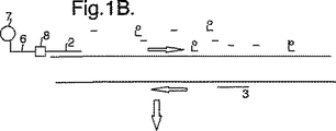
図1は、本発明の方法で行われる相互作用を示す概略図である。
図示した増幅反応においては、DNA分子(1)を一組の増幅プライマー(2)、(3)及び一部が蛍光標識(5)で標識している一組のヌクレオチド(4)と接触させることにより増幅用に調製する。プライマー(2)の一方を受容標識(7)を含むプローブ(6)に化学結合によって結合する。
DNA分子(1)を一本鎖にし(図1B)、そのときプライマー(2、3)は、周知である増幅反応において前進プライマーと逆プライマーとして結合する。
続いての増幅反応の過程で、アンプリコン産物(9)が作製される(図1C)。標識及び非標識双方のヌクレオチドが形成される産物に組込まれている。アンプリコン鎖のかなりの割合が標識プローブ領域(6、7)を含んでいる。
この産物を続いての増幅相で融解したときに、受容分子(7)を含むプローブ領域(6)はアンプリコン鎖内で標的配列を結合する。蛍光ヌクレオチドと受容分子(7)間のFRET相互作用によって、受容体に固有の波長でシグナルが生成する。
次に、受容分子(7)からのシグナルを、慣用の蛍光検出装置を用いてモニターし得る。
【図面の簡単な説明】
【図1A】 本発明の方法で行われる相互作用を示す概略図である。
【図1B】 本発明の方法で行われる相互作用を示す概略図である。
【図1C】 本発明の方法で行われる相互作用を示す概略図である。
【図1D】 本発明の方法で行われる相互作用を示す概略図である。[0001]
The present invention relates to a method for detecting a target polynucleotide in a sample, for example, by monitoring an amplification reaction, preferably by a quantitative method, and a probe and kit used in the method. The method is suitable for detection of sequence features such as polymorphisms or allelic changes and can therefore be used in diagnostic methods.
Known fluorescent polymerase chain reaction (PCR) monitoring methods include both strand-specific and global DNA insertion methods that can be used with a few second generation PCR thermocyclers.
Comprehensive fluorescent PCR utilizes DNA intercalating dyes that increase fluorescence when bound to double-stranded DNA species. Increased fluorescence with increasing volume concentration of DNA during amplification can be used to measure the progress of the reaction and to determine the initial target molecule copy number. Furthermore, by monitoring fluorescence with controlled temperature changes, a DNA melting curve can be generated, for example, at the end of a PCR thermal cycle.
These comprehensive fluorescent PCR methods are methods for monitoring an increase in the nucleic acid volume concentration without delaying the time. A single fluorescence reading can be read at the same point for each reaction. End point melting curve analysis can be used to discriminate artifacts from amplicons and to discriminate amplicons. Product peaks cannot be seen at concentrations that can be visualized by agarose gel electrophoresis.
[0002]
In order to obtain high-resolution melting data, melting experiments must be performed gradually over 5 minutes with existing hardware. However, by continuously monitoring fluorescence amplification, 3D images of melting and hybridization hysteresis can be generated. This 3D image is amplicon-dependent and can provide sufficient information for product discrimination.
In general, DNA melting curve analysis has been found to be a powerful tool for optimizing PCR thermal cycling. By determining the melting temperature of the amplicon, it is possible to reduce the denaturation temperature of subsequent PCR cycles to this temperature. Optimization of amplification from first generation reaction products rather than genomic DNA reduces the production of artifacts that occur in later cycles. The melting temperature of the primer oligonucleotide and its complement can be used to determine the annealing temperature, reducing the requirement for empirical optimization.
However, the global insertion method is only quasi-strand specific and is of little use when strand-specific detection is required.
[0003]
The fluorescent PCR strand-specific method is a method of further monitoring the progress of the amplification reaction using a nucleic acid reaction component. The method can use fluorescence energy transfer (FET) as a basis for detection. One or more nucleic acid probes can be labeled with a fluorescent molecule, one of which can act as an energy donor molecule and the other as an energy acceptor molecule. These are often known as reporter molecules and quencher molecules, respectively. The donor molecule is excited at a specific wavelength of light that usually exhibits a fluorescence emission wavelength. The acceptor molecule is excited at this emission wavelength and can accept the emission energy of the donor molecule by various distance dependent energy transfer mechanisms. A specific example of fluorescence energy transfer that can occur is fluorescence resonance energy transfer or “FRET”. Generally, the acceptor molecule accepts the emission energy of the donor molecule when it is in close proximity (eg, the same molecule or an adjacent molecule). The basis for FET or FRET detection is to monitor changes in donor emission wavelength. When the acceptor is a fluorescent molecule, the acceptor emission wavelength can also be monitored.
Two commonly used FET or FRET probes, probes that use hydrolysis of nucleic acid probes to separate donors from acceptors, and probes that use hybridization to change the spatial relationship between donor and acceptor molecules is there.
[0004]
Hydrolysis probes are commercially available as TaqMan® probes. These consist of DNA oligonucleotides labeled with donor and acceptor molecules. The probe is designed to bind to a specific region of one strand of the PCR product. After annealing this strand with the PCR primer, the Taq enzyme extends the DNA by 5 '→ 3' polymerase activity. Taq enzyme also exhibits 5 ′ → 3 ′ exonuclease activity. The TaqMan® probe is protected at the 3 ′ end by phosphorylation to prevent initiation of Taq extension. If the TaqMan® probe hybridizes to the product strand, the extending Taq molecule can also hydrolyze the probe, releasing the donor from the acceptor as a basis for detection. The signal in this case is cumulative and the concentration of free donor and acceptor molecules increases with each cycle of the amplification reaction.
The fact that signal generation depends on the occurrence of the probe hydrolysis reaction means that there is a time delay associated with this method. Furthermore, the presence of the probe may interrupt the smooth operation of the PCR method.
Furthermore, it has been found that hydrolysis can be non-specific, in particular a large number of amplifications, for example over 50 cycles, are required. In that case, non-specific hydrolysis of the probe results in an excessively high signal.
[0005]
This is quite similar to the rapid PCR method, which has become even better with the development of rapid hot air thermal cyclers such as Idaho Technologies Inc.'s RapidCycler (R) and LightCycler (R). It means that it does not fit. Other rapid PCR devices are described, for example, in co-pending UK patent applications 9625442.0 and 9716052.7. The advantages of rapid cycling over conventional thermal cycles have been reported in other ways. Such an approach is particularly effective, for example, in a biological struggle detection system, where the speed of the results is important if loss of life or serious damage is avoided.
In addition, hydrolyzed probes provide little information about melting hysteresis because signal generation is generally dependent on probe hydrolysis rather than amplicon or probe melting temperature.
Hybridization probes are available in many appearances. Molecular beacons are oligonucleotides with complementary 5 'and 3' sequences, resulting in a hairpin loop. The terminal fluorescent labels are in close proximity so that FRET occurs when the hairpin structure is formed. After the molecular beacon is hybridized to the complementary sequence, FRET does not occur because the fluorescent label is separated, thereby providing the basis for detection.
[0006]
A pair of labeled oligonucleotides can be used. These hybridize in closer proximity to the PCR product strand that brings both donor and acceptor molecules together so that FRET can occur. Enhanced FRET is the basis for detection. This type of variation using a labeled amplification primer containing a single adjacent probe is also included.
The use of two probes, or molecular beacon type probes that contain two labeled molecules, costs associated with the process. Furthermore, this method requires the presence of a known sequence that is fairly long, in particular so that two probes are known that are long enough to bind in close proximity to each other. This can be problematic in some diagnostic applications where the conserved sequence length of an organism that can be used to design effective probes such as HIV viruses may be relatively short.
Furthermore, the use of a pair of probes requires a complex experimental design. For example, the signal obtained by the probe melt is a function of the melting of both probes. Small mismatch experiments or experiments that require one of the probes to bind across the splice region (e.g., detect RNA relative to DNA in a sample where the sequences on both sides of the intron can be used as probe sites) Inaccurate results may occur if the other melts first.
[0007]
Co-pending International Patent Application No. PCT / GB990504 describes an analysis that detects the presence of a specific nucleic acid sequence that can be adapted to quantify the amount of target sequence in a sample. In this analysis, an amplification reaction is performed using a set of nucleotides, at least one of which is fluorescently labeled. Thus, the amplified product has a fluorescent label incorporated therein. The reaction is carried out in the presence of a probe containing a reactive molecule that can hybridize to the amplification product and can absorb fluorescence from or donate fluorescence energy to the fluorescently labeled nucleotide. The reaction can then be monitored by measuring the fluorescence of the sample that hybridizes to the probes and changes in the course of the reaction between them causing FET or FRET interactions.
Here, Applicants have found an improved method of the analysis.
The present invention is a method for detecting the presence of a target nucleic acid sequence in a sample, wherein at least one is labeled with a first label, a set of nucleotides, and said target nucleic acid sequence when in single-stranded form Subjecting the sample to an amplification reaction using a reagent comprising an amplification primer that is capable of hybridizing to and having a second label attached to the probe at the 5 ′ end by a chemical binding group; The labeled probe has a sequence similar to the target sequence so that it can hybridize to the complementary region of the amplification product, and either the first label or the second label emits fluorescence energy. Providing the method, comprising: a fluorescent molecule capable of donating to the first label or the other of the second label; and monitoring the fluorescence of the sample.
[0008]
In this analysis, the target sequence is made single-stranded in the first step, so that the primer region of the reagent can hybridize. Thus, this can initiate strand extension that produces a complementary strand containing labeled nucleotides and having a labeled probe region downstream of the 5 ′ end that is complementary downstream of the product.
As soon as the extension phase is complete, the product is separated from its template strand during the melt phase and becomes single stranded. In this way, the labeled probe region can be twisted and hybridized to the complementary region of the product strand, at which time fluorescence energy (donor) can be donated to the other label by FET or FRET So that the fluorescence signal from the sample changes. This change in signal can be monitored throughout the reaction to monitor the course of the amplification reaction.
In the second and subsequent stages of amplification, the product strand can itself act as an extension template strand. However, after a chemical bond stops the extension reaction, a sequence complementary to the probe results. Thus, the probe region remains single stranded.
If necessary, there may be a corresponding amplification primer that does not bind to the labeled probe region during the amplification reaction. This primer results in the creation of a conventional unlabeled amplification product that can serve to mediate the signal to the dynamic range of the detection device being used. Reaction efficiency, which can be adversely affected by the presence of complex probe / primer structures, can be improved.
[0009]
When a label capable of absorbing fluorescence from a donor label (acceptor) performs this function, the fluorescence from the donor is reduced. This decrease can be detected, indicating binding of the probe region.
Most preferably, the label capable of absorbing fluorescence (acceptor) is a fluorescent molecule that emits fluorescence at its own wavelength. In this case, the binding of the probe is indicated by an increase in fluorescence from an acceptor molecule having a different wavelength than the donor label.
Suitably the donor label is labeled for the nucleotide and the acceptor label is labeled for the probe. In this case, the presence of the amplification product labeled in this way can be detected by monitoring the fluorescence from the acceptor molecule for the probe and binds mainly to the downstream region of the same product strand. In this case, the signal from the amplification product can be distinguished from the background signal of the fluorescent label as well as from the non-specific amplification product.
The fact that the signal is generated only by the labeled amplification product means that the system is very specific by detecting a specific target sequence in a reaction mixture containing a large amount of background DNA. This is because non-specific amplification products do not hybridize to the probe region and thus do not contribute to the measured signal.
[0010]
This type of analysis can be performed using inexpensive reagents. A labeled single probe is more economical than one containing both acceptor and donor molecules.
The labeled probe in the reagent may be single stranded and may include 5 'and 3' complementary sequences to take the form of the molecular beacons described above. In this case, however, the probe only needs one labeled molecule that produces a FET or FRET signal that is the basis for detection as a result of the interaction between the label on the probe and the labeled nucleotide in the amplicon strand.
As used herein, the expression “a set of nucleotides” refers to a group of nucleotides sufficient to form a nucleic acid, such as DNA, RNA, or other analog. They therefore contain adenosine, cytosine, guanine and thymine or uracil containing nucleotides. One or more of these are labeled with fluorescence. Labeled uracil is available from Boehringer Mannhaeim. Suitable fluorescent labels include fluorescein.
The use of labeled uracil is particularly preferred in that its use can be incorporated into a strategy to prevent contamination or carry-over contamination from the amplification reaction performed in the reaction vessel to the next reaction. Enzymes that digest uracil-containing nucleic acids, such as uracil-N-glycosylase, can be used in the pre-cycle incubation step to ensure that residual amplicons are digested before the next application thermal cycle begins.
[0011]
Appropriately label more than one nucleotide, possibly all nucleotides, to mitigate the signal level from the amplification product and thus the FET or FRET signal.
Amplification is suitably performed using known amplification reactions such as polymerase chain reaction (PCR), strand displacement analysis (SDA) or NASBA, preferably PCR.
Preferably, the fluorescence of both the first label and the second label (ie both donor and acceptor labels) is monitored and the relationship between the emissions is calculated.
Suitable acceptor labels are fluorescent dyes such as rhodamine dyes or other dyes such as Cy5. The acceptor label can also include a non-fluorescent acceptor molecule often known as a “dark acceptor” such as DABCYL or methyl red. These can be bound to the probe in a conventional manner. The position of the receiving label along the probe is not critical, but is usually located in the terminal region of the probe.
The fluorescence emission of the donor label must be at a shorter wavelength than the acceptor label so that an FET, eg, FRET, occurs between the first label and the second label.
Therefore, the appropriate combinations are shown in the following table.
[0012]
[0013]
Preferably, molecules used as donors and / or acceptors produce sharp emission peaks and little or no overlap in emission wavelengths. Under these circumstances, it may not be necessary to split the “strand specific peak” from the signal produced by the amplification product. A simple measurement of only the strand specific signal (ie indicated by the acceptor label) gives information on the extent of FET or FRET by the target reaction.
However, if there are spectral overlaps in the fluorescence signals from the donor and acceptor labels, for example, the relationship between the spectra is empirically determined and the signal is normalized from the two signals using this relationship. Can explain.
The chemical bond that separates the labeled probe from the probe is suitably a molecule that can bind the nucleotide sequence but is not recognized by the DNA polymerase. A wide range of chemical linkers are available that meet this requirement.
Examples of chemistry types and reactions that can be used to form the linker are described, for example, in International Application No. 95/08642. In particular, a chemical linker contains a group of atoms that join together two polynucleotide sequences (primer and probe). The linker can be attached to the respective polynucleotide by any conventional method.
[0014]
Generally speaking, a linker has a first functional group and a second functional group so that it can be attached to a probe sequence and a primer sequence, respectively, or to an individual nucleotide from which the probe or primer sequence is subsequently generated. Derived from chemistry.
Linker bond is, for example, carbon-carbon single bond, carbon-carbon double bond, carbon-carbon triple bond, carbon-nitrogen single bond, carbon-nitrogen double bond, carbon-oxygen single bond, carbon-sulfur single bond Depending on the organic chemistry and type of reaction used to form the linker, a carbon-silicon single bond, a sulfur-nitrogen bond, a sulfur-oxygen bond, a phosphorus-oxygen bond, or a phosphorus-nitrogen bond.
Suitable functional groups include, but are not limited to, hydroxyl groups, amino groups, thio groups, alkyl sulfate groups, and halogen groups.
Suitably, the linker can be introduced in stages.
For example, an organic chemistry that has a first functional group but is blocked from a second functional group (e.g., an alcohol group, an ester group, a thioalkyl group, etc.) is allowed to react to bind the probe sequence or primer sequence to its end. obtain. After removal of the blocking group using conventional methods, the second functional group can be reacted with the other of the probe or primer to form a chemical linker.
Alternatively, chemical linkers can be generated in situ from the two moieties using, for example, the following reaction scheme.
[0015]
[Chemical 1]
(Where P 1 And P 2 Is a primer polynucleotide sequence and a probe polynucleotide sequence; X is a functional group that reacts with the 3 'end of the polynucleotide sequence to form an -X'- moiety; Z is reacted with the 5' end of the polynucleotide sequence -Z'-functional group that forms a moiety; Y and Y 'are functional groups that react with each other and do not react with the polynucleotide to form a Y''group; R 1 And R 2 Is part of the linker. )
Depending on the type of reaction to be performed, X ′, Y ″ or X ′ can be directly bonded. 1 And / or R 2 Examples of include:
-NH-CH 2 -CH = CH-, SH-CH 2 -CH 2 -CH = CH-, -NH-CH 2 -CH 2 -O-CH 2 -CH = CH-,-(CH 2 ) n -O- (wherein n is an integer of 1 to 20)
When the linker is attached to the 5 ′ or 3 ′ end of the polynucleotide, convenient linkages include phosphate bonds, carboxy bonds or ether bonds, especially phosphate bonds. Suitable phosphate linkers include aminoalkyl phosphoryl groups, especially C 1-12 Alkyl chain, especially C 6 Those containing alkyl chains are included. These linkers can be easily attached to synthetic oligonucleotides by solid phase synthesis. See, for example, S. Agrawal et al., Nucleic Acids Research, 1986, 14, 6227 and International Application No. 88/02004 (Applied Biosystems).
[0017]
Other general methods for attaching linkers to polynucleotide bases are described in JL Ruth & DE Bergstrom, J. Org. Chem., 1978, 43, 2870; DE Bergstrom & MK Ogawa, J. Amer. Chem. Soc., 1978, 10, 8106; CF Bigge, P. Kalaritis, JR Deck & MP Mertes, J. Amer. Chem. Soc., 1980, 102, 2033. A preferred method is that disclosed in European Patent Application 063,879. The method comprises a linker having an α vinyl group or a linker group and a mercuric base. 2 PdCl Four The mercury in the presence of Hg + As a step of binding to the position of the base to be reacted with the linker.
The size or content of the linker can vary considerably if it can act as a block against extension while allowing hybridization between the probe region and the target sequence. The linker can have from about 2 carbons to any number of carbons, for example up to 20 atoms. The linker can contain heteroatoms and unsaturation. An aliphatic group, alicyclic group, aromatic group or heterocyclic group may be present in the chemical linker. Conveniently,-(CH 2 ) n -Contains. However, -O-, -CHOH-, -COO-, or -CH helps maintain water solubility 2 CH 2 Other groups such as -O- may be included.
[0018]
The linkage of the linker to the sugar group of the polynucleotide can be linked to the 1 'aldehyde after depurination or depyrimidine of a base preselected by a Schiff base, or to the 2' hydroxy when the sugar is ribose. be able to. Linkage of the linker arm to the phosphate moiety can be, for example, by alkylation of the phosphate group as described in US Pat. No. 4,469,863.
In order to attach a chemical linker to a base group of a polynucleotide, it may be preferable to attach to a base prior to producing the polynucleotide. This is because the reaction conditions that may be required to attach the linker to the base cause undesirable side reactions within the polynucleotide. In addition, binding at the polynucleotide level may give non-renewable yields that are not uniform. For binding at the nucleotide or nucleotide level, the modified nucleotide or nucleotide is first purified and then incorporated into the polynucleotide. Integration can be by cloning, for example by cloning in an M13 vector or by routine synthesis in a polynucleotide synthesizer apparatus.
[0019]
If the linker is attached to the 1 ′ aldehyde of the sugar, the linker is appropriately attached after formation of the polynucleotide portion of the polynucleotide probe. This is because a free aldehyde is required at the 1 ′ position of the sugar for sugar binding. Free aldehydes are produced by depurination or depyrimidine. Groups containing sugar and phosphate without a base are not substrates for the polymerase enzyme. Thus, the linker must be attached first by selectively depurinating or depyrimidine the desired polynucleotide sequence and then attaching the linker to the sugar by an aldehyde. If the linker is attached to the 2 ′ hydroxy group of the ribose sugar, the linker may be attached at the nucleoside, nucleotide or polynucleotide level.
This is because a linker-modified nucleotide can be incorporated into a polynucleotide by a polynucleotide synthesizer device. If the linker arm is attached to a phosphate group, the linker arm is preferably attached at the nucleoside or nucleotide level, so the attachment is not at a position other than the phosphate group. Phosphoramidite technology can be used in the nucleic acid synthesizer to incorporate a linker at the 5 ′ or 3 ′ end of the polynucleotide.
[0020]
In particular, the linker includes multiple forms of ethylene glycol, eg, Hex ethylene glycol. Such linkers have the structure-(CHOH-CHOH) n -(N is an integer greater than 1, for example, 1 to 10, suitably 6.).
Probes containing linker groups are available from Oswell Ltd, UK.
The method of the present invention is very responsive to its application. The method can be used to generate both quantitative and qualitative data regarding target nucleic acid sequences in a sample, as will be detailed later. In particular, the present invention not only provides quantitative amplification, but can also or alternatively be used to obtain confirming data such as the destabilization temperature or melting point of a double helix.
In the method of the present invention, since the labeled probe is indispensable together with the amplification probe, it exists throughout the amplification reaction process. The process can be detected in a homogeneous manner in that amplification and monitoring can be performed in a single container where all reagents are initially added. Subsequent reagent addition steps are not required. Neither is it required to perform the process in the presence of a solid support (although this is an option as described below).
[0021]
Since the probe is present throughout the amplification reaction, the progress of the amplification reaction can be monitored by the fluorescence signal. This can provide a means for quantifying the amount of target sequence present in the sample.
During each cycle of the amplification reaction, a receptor signal is generated by an amplicon chain having a target sequence and a probe region. As the amount of amplicon in the sample increases, the receptor signal increases. By plotting the rate of increase for the cycle, the starting point of the increase can be determined.
A labeled probe can comprise a nucleic acid molecule such as DNA or RNA and hybridizes to a target nucleic acid sequence when in single-stranded form. In this case, step (b) requires the use of conditions that make the target nucleic acid single stranded. Probes can also include molecules such as peptide nucleic acids or other nucleic acid analogs that bind the target sequence in double-stranded form.
In particular, the amplification used requires the step of subjecting the sample to conditions such that any of the target nucleic acid sequences present in the sample are single stranded, such as PCR or LCR. The probe region can then be hybridized to the downstream region of the amplicon chain containing it during the amplification reaction if appropriate hybridization conditions occur.
[0022]
In a preferred embodiment, the probe can be designed to meet these conditions in each cycle of the amplification reaction. Thus, at each point in the cycle of the amplification reaction, the probe hybridizes to the target sequence and produces a signal as a result of FET or FRET. As amplification proceeds, the signal generated by the acceptor label decreases or increases depending on whether it contains a donor molecule or acceptor molecule, as the probe region separates or melts from the downstream sequence. For example, in the case of a receptor, a fluorescent peak from the acceptor label occurs at each cycle of amplification. The intensity of the peak increases as amplification proceeds due to the availability of the amplicon chain containing the probe.
By monitoring the fluorescence of the receiving label from the sample at each cycle, the progress of the amplification reaction can be monitored in various ways. For example, the data obtained by a melting peak can be analyzed, for example, by calculating the area under the melting peak, and this data can be plotted against the number of cycles.
Fluorescence is suitably monitored using a known fluorimeter. The signals from these are sent to the data processor board, for example, in the form of a photomultiplier voltage, and converted to a spectrum associated with each sample tube. Multiple tubes, for example 96 tubes, can be evaluated simultaneously. Data can be collected in this manner at frequent intervals throughout the reaction, for example, every tenth time.
[0023]
The spectrum generated by this method can be resolved, for example, using a “match” of preselected fluorescent moieties such as dyes, representative of each signal moiety (ie, nucleotide label and / or probe label). Produces a strong peak. The area under the peak can be determined to indicate the intensity value for each signal and, if necessary, can be expressed as a quotient. Due to differences in signal intensity and / or ratio, changes in FET or FRET can be recorded by reaction or at different reaction conditions such as temperature. By integrating the area under the differential peak, the intensity value of the FET or FRET action can be calculated.
This data provides a means to quantify the amount of target nucleic acid present in the sample.
The primer / labeled probe reagent may be free in solution and may be a solid support, eg, the surface of a bead, such as a magnetic bead effective to separate products, or the surface of a surface plasmon resonance detector. It may be immobilized on the surface of a detection device such as a tube. The selection depends on the specific analysis type to be examined and the specific detection means used.
The probe can be designed to be hydrolyzed by the DNA polymerase used in the amplification reaction, thus releasing the acceptor molecule. This gives a cumulative signal and the amount of free probe label is present in the system and increases with each cycle. However, in this analysis, it is not necessary for the probe to be consumed in this method where the signal does not depend on the hydrolysis of the probe.
[0024]
Suitably, the probe is designed such that it is released intact from the target sequence and can re-bind if appropriate hybridization conditions are met during the amplification reaction. This may be, for example, the extension phase of the amplification reaction. However, since the signal is independent of probe hydrolysis, the probe may be designed to hybridize and melt from the target sequence at a stage during the amplification cycle. In particular, the probe is designed to hybridize at a temperature above the extension temperature of the reaction, which ensures that interference from the amplification reaction is minimized.
This provides a sufficiently reversible signal that is directly related to the amount of amplification product at each stage of the reaction. Furthermore, it is advantageous if the reaction rate is very important, for example in rapid PCR, since an indispensable probe with the amplicon strand to be detected can rapidly hybridize to it.
Data created in this way can be described in various ways. In its simplest form, an increase in acceptor fluorescence during or at the end of the amplification reaction indicates the fact that the target sequence is actually present in the sample as the amplification reaction has progressed. Shows an increase. However, as described above, quantification is possible by monitoring the entire amplification reaction. Furthermore, confirmation data and in particular melting point analysis can be obtained as end-point measurements or as a whole to obtain information about the sequence as described below.
[0025]
Accordingly, a preferred embodiment of the present invention is a method for detecting nucleic acid amplification comprising: (a) a nucleic acid polymerase; (b) a set of nucleotides labeled with at least one first label; and (c) a single Presence of a reagent containing an amplification primer capable of hybridizing to a target sequence when in the form of a strand and bound to the probe having a second label by a chemical binding group at the 5 'end The target polynucleotide is subjected to nucleic acid amplification below, and the labeled probe has a sequence similar to the target sequence so that it can hybridize to a complementary region of the amplified product, and the first label or One of the second labels comprises a donor label capable of donating to the first label or the other of the second label comprising an acceptor label capable of absorbing fluorescence energy from a donor molecule, wherein the primer is the target Po And said method comprising the steps of: hybridizing to a renucleotide; and monitoring changes in fluorescence during the amplification reaction. Suitably, the receiving label is itself fluorescent and emits fluorescent energy at a characteristic wavelength.
Amplification is suitably performed using a pair of primers that are designed to amplify only the target nucleotide sequence in the DNA strand as is well understood in the art. The nucleic acid polymerase is suitably a thermostable polymerase such as Taq polymerase.
[0026]
Appropriate conditions under which the amplification reaction can be performed are well known in the art. Optimal conditions may be variable in each case and depend on the specific amplicon required, the type of primer used or the enzyme used. Optimal conditions can be determined by the person skilled in the art in each case. Typical denaturing temperatures are on the order of 95 ° C, typical annealing temperatures are on the order of 55 ° C, and elongation temperatures are on the order of 72 ° C.
In a specific embodiment of the invention, labeled probes can be used, for example, in expression experiments to quantify RNA transcripts that can be used for drug discovery. In particular, this embodiment is suitable for expression experiments in tissues from eukaryotic organisms. DNA encoding proteins in eukaryotic cells may include introns, non-coding regions of DNA sequences, and exons encoding protein sequences. Non-coding intron sequences are removed from RNA sequences that are derived from DNA sequences during the cellular “splicing” process. PCR primers are usually targeted in the coding region and if reverse transcriptase PCR is used for the total nucleic acid extract, the product is obtained from both DNA-dependent amplification and RNA-dependent amplification. Thus, when used in expression experiments, only PCR has amplification obtained from genomic DNA and expressed RNA.
[0027]
Labeled probes designed to bind across introns for adjacent end regions of code exons are limited in interaction due to intron regions. Since these regions are removed from the splice RNA, the flanking end regions of the coding exon form a contiguous sequence, allowing efficient binding of the probe regions.
Conversely, if the probe region is designed to bind the intron region, only the amplification product of genomic DNA can be detected. The signal resulting from such a probe is only related to the DNA concentration, not the sample RNA concentration.
Thus, in an embodiment, the probe region is further specific to either the RNA splice region or the DNA intron, so that only one of amplified RNA or amplified DNA is detected and / or quantified.
Alternatively or additionally, the methods of the invention can be used for hybridization analysis to determine sequence characteristics. Thus, in an embodiment, the present invention further provides a method for determining sequence characteristics, comprising: (a) a set of nucleotides at least one of which is labeled with a first label; and a region of the target sequence Amplifying the sequence using a reagent comprising an amplification primer bound at the 5 ′ end by chemical linkage to a probe comprising a sequence and further comprising a second label, wherein the first label or the second label One is a donor label and the other is an acceptor label, the donor label being able to donate fluorescence energy to the acceptor label, thereby forming an amplification product incorporating the probe region; (b) subjecting the amplification product to conditions under which the probe region hybridizes to a complementary region of the amplification product; and (c) monitoring the fluorescence of the sample and suspending the probe region relative to the sample. Providing said method comprising determining specific reaction conditions specific to said sequence, wherein the fluorescence changes as a result of hybridization or destabilization of a double helix formed between said probe region and a target nucleic acid sequence To do.
[0028]
Suitable reaction conditions include temperature, electrochemistry, or response to the presence of a specific enzyme or chemical. By monitoring the changes in fluorescence as these properties change, information specific to the exact type of sequence can be obtained. For example, in the case of temperature, the temperature at which the probe separates from the array in the sample as a result of heating can be determined. This can be very effective in detecting and quantifying polymorphisms and / or allelic changes if desired. “Polymorphism” includes transpositions, transversions, insertions, inversion deletions that can occur in a sequence, especially in nature.
Melting hysteresis is different when the target sequence is changed by only one base pair. Thus, for example, if a sample has only a single allelic variant, the melting temperature of the probe region will be a specific value that is different from that found in a sample having only other allelic variants. Samples with both allelic variants show two melting points corresponding to each of the allelic variants. Similar considerations apply to electrochemical properties or to the presence of certain enzymes or chemicals. The labeled probe can be immobilized on a solid surface to which an electrochemical potential can be applied. The downstream target sequence binds to and repels the probe with a specific electrochemical value depending on the exact type of sequence.
[0029]
Furthermore, the kinetics of probe hybridization allows quantification of the target sequence concentration in absolute terms. From the change in fluorescence from the sample, the hybridization rate of the probe region to the sample can be calculated. The increase in hybridization rate is related to the amount of target sequence that is present in the sample. As the amplification reaction proceeds, the concentration of the target sequence increases so that probe region hybridization occurs rapidly. Therefore, this parameter can be used as a basis for quantification. This data processing method is effective in that information is obtained without directly depending on the signal intensity.
Embodiments of the present invention further include kits used in the methods of the present invention. The kit has a probe specific for the target nucleotide sequence containing a label, particularly a receiving label, attached to the 5 'end of the amplification primer. If desired, the probe can be immobilized on a support used in a detector, such as a support, such as a bead, eg, a magnetic bead, or a waveguide of a vanishing waveform detector.
[0030]
In addition, the kit can include one or more labeled nucleotides, particularly fluorescently labeled nucleotides that are compatible with the receiving label. Other potential components of the kit include reagents used in growth reactions such as DNA polymerase.
The use of non-fluorescent acceptor molecules can be used for the analysis described in co-pending international patent application No. PCT / GB990504.
Thus, in an embodiment, the present invention further relates to a method for detecting the presence of a target nucleic acid sequence in a sample, comprising: (a) using a set of nucleotides, at least one of which is labeled with a first label. Subjecting the sample to an amplification reaction; (b) contacting the amplification product and the probe under conditions such that the probe containing a second labeled molecule hybridizes to the target sequence, and the first label or the The method is provided comprising the steps of one of the second labels being a fluorescent molecule and the other being a non-fluorescent molecule capable of accepting fluorescent energy and (c) monitoring the fluorescence of the sample.
Suitably the non-fluorescent level is DABCYL, malric (methyl) red or QSY-7 diarylrhodamine dye. The non-fluorescent label is preferably the second label and is labeled for the probe.
[0031]
The invention will now be particularly described by way of example with reference to the accompanying drawings.
FIG. 1 is a schematic diagram showing the interaction performed in the method of the present invention.
In the amplification reaction shown, the DNA molecule (1) is contacted with a set of amplification primers (2), (3) and a set of nucleotides (4) partially labeled with a fluorescent label (5). Prepare for amplification. One of the primers (2) is bound to the probe (6) containing the receiving label (7) by chemical bonding.
The DNA molecule (1) is made single-stranded (FIG. 1B), and then the primers (2, 3) bind as a forward primer and a reverse primer in a well-known amplification reaction.
During the subsequent amplification reaction, an amplicon product (9) is produced (FIG. 1C). Both labeled and unlabeled nucleotides are incorporated into the product formed. A significant proportion of amplicon chains contain labeled probe regions (6, 7).
When this product is melted in a subsequent amplification phase, the probe region (6) containing the acceptor molecule (7) binds the target sequence within the amplicon chain. The FRET interaction between the fluorescent nucleotide and the acceptor molecule (7) generates a signal at the wavelength characteristic of the acceptor.
The signal from the acceptor molecule (7) can then be monitored using a conventional fluorescence detector.
[Brief description of the drawings]
FIG. 1A is a schematic diagram showing the interaction performed in the method of the present invention.
FIG. 1B is a schematic diagram showing the interaction performed in the method of the present invention.
FIG. 1C is a schematic diagram showing the interaction performed in the method of the present invention.
FIG. 1D is a schematic diagram illustrating the interaction performed in the method of the present invention.
Claims (16)
少なくとも1つが第1標識で標識されている一組のヌクレオチド、及び一本鎖の形にあるときの前記標的核酸配列に対してハイブリッド形成することができかつ第2標識を有しているプローブに、DNAポリメラーゼにより認識されない化学結合基によって5' 端で結合している増幅プライマーを含んでいる試薬を用いて
前記試料を、前記標的核酸が増幅される増幅反応に供し、
標識した前記プローブが、増幅産物の相補的領域に対してハイブリッド形成することができ、蛍光エネルギーが該第1標識と該第2標識の間で移動することができるように前記標的配列と同様の配列を有し、
該第1標識又は該第2標識の一方が供与標識を含み、もう一方が受容標識を含み、
該供与標識が蛍光エネルギーを該受容標識に供与し得る蛍光分子を含んでいる段階; 及び
前記試料の蛍光をモニターする段階
を含む、前記方法。A method for detecting the presence of a target nucleic acid sequence in a sample, comprising:
A set of nucleotides, at least one of which is labeled with a first label, and a probe capable of hybridizing to said target nucleic acid sequence when in single-stranded form and having a second label , Subjecting the sample to an amplification reaction in which the target nucleic acid is amplified using a reagent comprising an amplification primer bound at the 5 ′ end by a chemical binding group not recognized by DNA polymerase;
Similar to the target sequence so that the labeled probe can hybridize to the complementary region of the amplification product and the fluorescence energy can be transferred between the first label and the second label Having an array,
One of the first label or the second label comprises a donor label and the other comprises an acceptor label;
The method comprising the steps of: the donor label comprising a fluorescent molecule capable of donating fluorescent energy to the acceptor label; and monitoring the fluorescence of the sample.
(a)少なくとも一方が第1標識で標識されている一組のヌクレオチド、及び標的配列の領域と同様の配列を含みかつ第2標識を更に含んでいるプローブに、DNAポリメラーゼにより認識されない化学結合基によって5' 端で結合している増幅プライマーを含んでいる試薬を用いて前記配列を増幅し、
前記第1標識又は前記第2標識の一方が供与標識であり、もう一方が受容標識であり、
該供与標識が蛍光エネルギーを該受容標識に供与することができ、その結果、プローブ領域に、DNAポリメラーゼにより認識されない化学結合基によって5' 端で結合している増幅産物を形成する段階、
(b)増幅産物を、該プローブ領域が該増幅産物の相補的領域に対して、蛍光エネルギーが該第1標識と該第2標識の間で移動することができるようにハイブリッド形成する条件に供する段階、及び
(c)前記試料の蛍光をモニターするとともに、該試料に対する該プローブ領域のハイブリダイゼーション又は該プローブ領域と標的核酸配列間に形成された二重らせんの不安定化の結果として蛍光が変化する、前記配列に特有の具体的な反応条件を求める段階
を含む、前記方法。A method for determining sequence characteristics, comprising:
(a) a set of nucleotides, at least one of which is labeled with a first label, and a chemical binding group that is not recognized by a DNA polymerase on a probe comprising a sequence similar to the region of the target sequence and further including a second label Amplifying said sequence using a reagent comprising an amplification primer bound at the 5 ′ end by
One of the first label or the second label is a donor label and the other is an acceptor label;
The donor label can donate fluorescent energy to the acceptor label, thereby forming an amplification product bound to the probe region at the 5 'end by a chemical linking group not recognized by DNA polymerase ;
(b) subjecting the amplification product to conditions that allow the probe region to hybridize to a complementary region of the amplification product such that fluorescence energy can be transferred between the first label and the second label. Stages, and
(c) monitoring the fluorescence of the sample and changing the fluorescence as a result of hybridization of the probe region to the sample or destabilization of a double helix formed between the probe region and the target nucleic acid sequence, Determining the specific reaction conditions specific to the sequence.
請求項1記載の方法を用いて前記多型性又は変化を含むことが疑われる配列を増幅する段階、
生成した蛍光シグナルを用いて増幅産物内の相補的配列からプローブ領域が融解する温度を測定する段階、及び
これを多型性及び/又は対立遺伝子変化の存在に関係させる段階
を含む、前記方法。 The method of claim 14, wherein the characteristic determined is polymorphism and / or the presence of allelic changes ,
Amplifying the sequence suspected of containing the polymorphism or change using the method of claim 1;
Measuring the temperature at which the probe region melts from the complementary sequence in the amplification product using the generated fluorescent signal, and relating this to the presence of polymorphisms and / or allelic changes.
Applications Claiming Priority (3)
| Application Number | Priority Date | Filing Date | Title |
|---|---|---|---|
| GB9918237.0 | 1999-08-04 | ||
| GBGB9918237.0A GB9918237D0 (en) | 1999-08-04 | 1999-08-04 | Detection system |
| PCT/GB2000/003016 WO2001011078A1 (en) | 1999-08-04 | 2000-08-03 | Amplification method for detection of target nucleic acids involving fluorescence energy transfer |
Publications (3)
| Publication Number | Publication Date |
|---|---|
| JP2003506068A JP2003506068A (en) | 2003-02-18 |
| JP2003506068A5 JP2003506068A5 (en) | 2007-09-27 |
| JP4628625B2 true JP4628625B2 (en) | 2011-02-09 |
Family
ID=10858467
Family Applications (1)
| Application Number | Title | Priority Date | Filing Date |
|---|---|---|---|
| JP2001515326A Expired - Fee Related JP4628625B2 (en) | 1999-08-04 | 2000-08-03 | Amplification method for detecting target nucleic acids involving fluorescence energy transfer |
Country Status (10)
| Country | Link |
|---|---|
| US (1) | US7015018B1 (en) |
| EP (1) | EP1198593B1 (en) |
| JP (1) | JP4628625B2 (en) |
| AT (1) | ATE287969T1 (en) |
| AU (1) | AU774766B2 (en) |
| CA (1) | CA2381037C (en) |
| DE (1) | DE60017750T2 (en) |
| ES (1) | ES2245315T3 (en) |
| GB (1) | GB9918237D0 (en) |
| WO (1) | WO2001011078A1 (en) |
Families Citing this family (7)
| Publication number | Priority date | Publication date | Assignee | Title |
|---|---|---|---|---|
| EP1377683A4 (en) * | 2001-04-11 | 2004-09-08 | Us Gov Health & Human Serv | Modified random primers for probe labeling |
| GB0112868D0 (en) * | 2001-05-25 | 2001-07-18 | Secr Defence | Detection system |
| JP4457001B2 (en) * | 2002-05-31 | 2010-04-28 | セクレタリー・デパートメント・オブ・アトミック・エナジー | MET / FRET based method for target nucleic acid detection in which donor / acceptor moieties are on complementary strands |
| US8658366B2 (en) * | 2008-09-18 | 2014-02-25 | Roche Molecular Systems, Inc. | Detection of target variants using a fluorescent label and a soluble quencher |
| CN103328956B (en) * | 2011-01-26 | 2015-08-12 | 奥林巴斯株式会社 | Differentiate the method for polymorphic nucleic acid molecule |
| WO2012102326A1 (en) | 2011-01-26 | 2012-08-02 | オリンパス株式会社 | Method for identifying polymorphism of nucleic acid molecule |
| CN112266950A (en) * | 2020-10-23 | 2021-01-26 | 深圳澳东检验检测科技有限公司 | Probe primer combination and detection kit thereof |
Family Cites Families (6)
| Publication number | Priority date | Publication date | Assignee | Title |
|---|---|---|---|---|
| DK0566751T3 (en) * | 1992-03-23 | 1996-03-04 | Hoffmann La Roche | DNA detection method |
| US6117635A (en) * | 1996-07-16 | 2000-09-12 | Intergen Company | Nucleic acid amplification oligonucleotides with molecular energy transfer labels and methods based thereon |
| GB9725237D0 (en) | 1997-11-29 | 1998-01-28 | Secr Defence | Amplification system |
| GB9803382D0 (en) | 1998-02-19 | 1998-04-15 | Secr Defence | Detection system |
| GB9812768D0 (en) * | 1998-06-13 | 1998-08-12 | Zeneca Ltd | Methods |
| US6440675B1 (en) * | 1999-09-17 | 2002-08-27 | Whitehead Institute For Biomedical Research | Methods for selecting primers |
-
1999
- 1999-08-04 GB GBGB9918237.0A patent/GB9918237D0/en not_active Ceased
-
2000
- 2000-08-03 DE DE60017750T patent/DE60017750T2/en not_active Expired - Lifetime
- 2000-08-03 JP JP2001515326A patent/JP4628625B2/en not_active Expired - Fee Related
- 2000-08-03 AU AU64558/00A patent/AU774766B2/en not_active Ceased
- 2000-08-03 EP EP00951700A patent/EP1198593B1/en not_active Expired - Lifetime
- 2000-08-03 WO PCT/GB2000/003016 patent/WO2001011078A1/en active IP Right Grant
- 2000-08-03 ES ES00951700T patent/ES2245315T3/en not_active Expired - Lifetime
- 2000-08-03 AT AT00951700T patent/ATE287969T1/en not_active IP Right Cessation
- 2000-08-03 CA CA2381037A patent/CA2381037C/en not_active Expired - Fee Related
- 2000-08-03 US US10/048,752 patent/US7015018B1/en not_active Expired - Fee Related
Also Published As
| Publication number | Publication date |
|---|---|
| AU6455800A (en) | 2001-03-05 |
| WO2001011078A1 (en) | 2001-02-15 |
| EP1198593A1 (en) | 2002-04-24 |
| JP2003506068A (en) | 2003-02-18 |
| DE60017750D1 (en) | 2005-03-03 |
| DE60017750T2 (en) | 2005-12-29 |
| ES2245315T3 (en) | 2006-01-01 |
| CA2381037C (en) | 2011-04-12 |
| CA2381037A1 (en) | 2001-02-15 |
| ATE287969T1 (en) | 2005-02-15 |
| US7015018B1 (en) | 2006-03-21 |
| WO2001011078A8 (en) | 2001-06-14 |
| EP1198593B1 (en) | 2005-01-26 |
| AU774766B2 (en) | 2004-07-08 |
| GB9918237D0 (en) | 1999-10-06 |
Similar Documents
| Publication | Publication Date | Title |
|---|---|---|
| US6287781B1 (en) | Method for detection of target nucleic acids using PCR | |
| JP4457001B2 (en) | MET / FRET based method for target nucleic acid detection in which donor / acceptor moieties are on complementary strands | |
| JP5685561B2 (en) | Nucleic acid detection substance and detection method | |
| JP4540844B2 (en) | Fluorescence quantitative detection system for nucleic acids | |
| EP2316975A1 (en) | Asynchronous primed PCR | |
| US20100227326A1 (en) | Detection System | |
| AU2002311414A1 (en) | Nucleic acid detection method | |
| JP2004532044A5 (en) | ||
| JP4628625B2 (en) | Amplification method for detecting target nucleic acids involving fluorescence energy transfer | |
| US20050176014A1 (en) | Fluorescent hybridization probes with reduced background | |
| JP2003508064A (en) | Methods for exogenous and internal control during nucleic acid amplification | |
| JP3647109B2 (en) | DNA detection method |
Legal Events
| Date | Code | Title | Description |
|---|---|---|---|
| A521 | Request for written amendment filed |
Free format text: JAPANESE INTERMEDIATE CODE: A523 Effective date: 20070803 |
|
| A621 | Written request for application examination |
Free format text: JAPANESE INTERMEDIATE CODE: A621 Effective date: 20070803 |
|
| A131 | Notification of reasons for refusal |
Free format text: JAPANESE INTERMEDIATE CODE: A131 Effective date: 20100513 |
|
| A521 | Request for written amendment filed |
Free format text: JAPANESE INTERMEDIATE CODE: A523 Effective date: 20100813 |
|
| TRDD | Decision of grant or rejection written | ||
| A01 | Written decision to grant a patent or to grant a registration (utility model) |
Free format text: JAPANESE INTERMEDIATE CODE: A01 Effective date: 20101021 |
|
| A01 | Written decision to grant a patent or to grant a registration (utility model) |
Free format text: JAPANESE INTERMEDIATE CODE: A01 |
|
| A61 | First payment of annual fees (during grant procedure) |
Free format text: JAPANESE INTERMEDIATE CODE: A61 Effective date: 20101110 |
|
| FPAY | Renewal fee payment (event date is renewal date of database) |
Free format text: PAYMENT UNTIL: 20131119 Year of fee payment: 3 |
|
| R150 | Certificate of patent or registration of utility model |
Free format text: JAPANESE INTERMEDIATE CODE: R150 |
|
| LAPS | Cancellation because of no payment of annual fees |