JP4493845B2 - How to select antibodies to internalize - Google Patents
How to select antibodies to internalize Download PDFInfo
- Publication number
- JP4493845B2 JP4493845B2 JP2000546239A JP2000546239A JP4493845B2 JP 4493845 B2 JP4493845 B2 JP 4493845B2 JP 2000546239 A JP2000546239 A JP 2000546239A JP 2000546239 A JP2000546239 A JP 2000546239A JP 4493845 B2 JP4493845 B2 JP 4493845B2
- Authority
- JP
- Japan
- Prior art keywords
- phage
- cells
- cell
- internalized
- antibody
- Prior art date
- Legal status (The legal status is an assumption and is not a legal conclusion. Google has not performed a legal analysis and makes no representation as to the accuracy of the status listed.)
- Expired - Fee Related
Links
Images


Classifications
-
- C—CHEMISTRY; METALLURGY
- C12—BIOCHEMISTRY; BEER; SPIRITS; WINE; VINEGAR; MICROBIOLOGY; ENZYMOLOGY; MUTATION OR GENETIC ENGINEERING
- C12N—MICROORGANISMS OR ENZYMES; COMPOSITIONS THEREOF; PROPAGATING, PRESERVING, OR MAINTAINING MICROORGANISMS; MUTATION OR GENETIC ENGINEERING; CULTURE MEDIA
- C12N15/00—Mutation or genetic engineering; DNA or RNA concerning genetic engineering, vectors, e.g. plasmids, or their isolation, preparation or purification; Use of hosts therefor
- C12N15/09—Recombinant DNA-technology
- C12N15/10—Processes for the isolation, preparation or purification of DNA or RNA
- C12N15/1034—Isolating an individual clone by screening libraries
- C12N15/1037—Screening libraries presented on the surface of microorganisms, e.g. phage display, E. coli display
-
- A—HUMAN NECESSITIES
- A61—MEDICAL OR VETERINARY SCIENCE; HYGIENE
- A61K—PREPARATIONS FOR MEDICAL, DENTAL OR TOILETRY PURPOSES
- A61K47/00—Medicinal preparations characterised by the non-active ingredients used, e.g. carriers or inert additives; Targeting or modifying agents chemically bound to the active ingredient
- A61K47/50—Medicinal preparations characterised by the non-active ingredients used, e.g. carriers or inert additives; Targeting or modifying agents chemically bound to the active ingredient the non-active ingredient being chemically bound to the active ingredient, e.g. polymer-drug conjugates
- A61K47/51—Medicinal preparations characterised by the non-active ingredients used, e.g. carriers or inert additives; Targeting or modifying agents chemically bound to the active ingredient the non-active ingredient being chemically bound to the active ingredient, e.g. polymer-drug conjugates the non-active ingredient being a modifying agent
- A61K47/68—Medicinal preparations characterised by the non-active ingredients used, e.g. carriers or inert additives; Targeting or modifying agents chemically bound to the active ingredient the non-active ingredient being chemically bound to the active ingredient, e.g. polymer-drug conjugates the non-active ingredient being a modifying agent the modifying agent being an antibody, an immunoglobulin or a fragment thereof, e.g. an Fc-fragment
- A61K47/6835—Medicinal preparations characterised by the non-active ingredients used, e.g. carriers or inert additives; Targeting or modifying agents chemically bound to the active ingredient the non-active ingredient being chemically bound to the active ingredient, e.g. polymer-drug conjugates the non-active ingredient being a modifying agent the modifying agent being an antibody, an immunoglobulin or a fragment thereof, e.g. an Fc-fragment the modifying agent being an antibody or an immunoglobulin bearing at least one antigen-binding site
- A61K47/6843—Medicinal preparations characterised by the non-active ingredients used, e.g. carriers or inert additives; Targeting or modifying agents chemically bound to the active ingredient the non-active ingredient being chemically bound to the active ingredient, e.g. polymer-drug conjugates the non-active ingredient being a modifying agent the modifying agent being an antibody, an immunoglobulin or a fragment thereof, e.g. an Fc-fragment the modifying agent being an antibody or an immunoglobulin bearing at least one antigen-binding site the antibody targeting a material from animals or humans
-
- A—HUMAN NECESSITIES
- A61—MEDICAL OR VETERINARY SCIENCE; HYGIENE
- A61P—SPECIFIC THERAPEUTIC ACTIVITY OF CHEMICAL COMPOUNDS OR MEDICINAL PREPARATIONS
- A61P37/00—Drugs for immunological or allergic disorders
- A61P37/02—Immunomodulators
-
- C—CHEMISTRY; METALLURGY
- C07—ORGANIC CHEMISTRY
- C07K—PEPTIDES
- C07K14/00—Peptides having more than 20 amino acids; Gastrins; Somatostatins; Melanotropins; Derivatives thereof
- C07K14/005—Peptides having more than 20 amino acids; Gastrins; Somatostatins; Melanotropins; Derivatives thereof from viruses
-
- C—CHEMISTRY; METALLURGY
- C07—ORGANIC CHEMISTRY
- C07K—PEPTIDES
- C07K16/00—Immunoglobulins [IGs], e.g. monoclonal or polyclonal antibodies
- C07K16/18—Immunoglobulins [IGs], e.g. monoclonal or polyclonal antibodies against material from animals or humans
- C07K16/28—Immunoglobulins [IGs], e.g. monoclonal or polyclonal antibodies against material from animals or humans against receptors, cell surface antigens or cell surface determinants
- C07K16/2863—Immunoglobulins [IGs], e.g. monoclonal or polyclonal antibodies against material from animals or humans against receptors, cell surface antigens or cell surface determinants against receptors for growth factors, growth regulators
-
- C—CHEMISTRY; METALLURGY
- C07—ORGANIC CHEMISTRY
- C07K—PEPTIDES
- C07K16/00—Immunoglobulins [IGs], e.g. monoclonal or polyclonal antibodies
- C07K16/18—Immunoglobulins [IGs], e.g. monoclonal or polyclonal antibodies against material from animals or humans
- C07K16/28—Immunoglobulins [IGs], e.g. monoclonal or polyclonal antibodies against material from animals or humans against receptors, cell surface antigens or cell surface determinants
- C07K16/2881—Immunoglobulins [IGs], e.g. monoclonal or polyclonal antibodies against material from animals or humans against receptors, cell surface antigens or cell surface determinants against CD71
-
- C—CHEMISTRY; METALLURGY
- C07—ORGANIC CHEMISTRY
- C07K—PEPTIDES
- C07K16/00—Immunoglobulins [IGs], e.g. monoclonal or polyclonal antibodies
- C07K16/18—Immunoglobulins [IGs], e.g. monoclonal or polyclonal antibodies against material from animals or humans
- C07K16/32—Immunoglobulins [IGs], e.g. monoclonal or polyclonal antibodies against material from animals or humans against translation products of oncogenes
-
- C—CHEMISTRY; METALLURGY
- C12—BIOCHEMISTRY; BEER; SPIRITS; WINE; VINEGAR; MICROBIOLOGY; ENZYMOLOGY; MUTATION OR GENETIC ENGINEERING
- C12N—MICROORGANISMS OR ENZYMES; COMPOSITIONS THEREOF; PROPAGATING, PRESERVING, OR MAINTAINING MICROORGANISMS; MUTATION OR GENETIC ENGINEERING; CULTURE MEDIA
- C12N15/00—Mutation or genetic engineering; DNA or RNA concerning genetic engineering, vectors, e.g. plasmids, or their isolation, preparation or purification; Use of hosts therefor
- C12N15/09—Recombinant DNA-technology
- C12N15/63—Introduction of foreign genetic material using vectors; Vectors; Use of hosts therefor; Regulation of expression
- C12N15/79—Vectors or expression systems specially adapted for eukaryotic hosts
- C12N15/85—Vectors or expression systems specially adapted for eukaryotic hosts for animal cells
- C12N15/86—Viral vectors
-
- A—HUMAN NECESSITIES
- A61—MEDICAL OR VETERINARY SCIENCE; HYGIENE
- A61K—PREPARATIONS FOR MEDICAL, DENTAL OR TOILETRY PURPOSES
- A61K38/00—Medicinal preparations containing peptides
-
- C—CHEMISTRY; METALLURGY
- C07—ORGANIC CHEMISTRY
- C07K—PEPTIDES
- C07K2317/00—Immunoglobulins specific features
- C07K2317/20—Immunoglobulins specific features characterized by taxonomic origin
- C07K2317/21—Immunoglobulins specific features characterized by taxonomic origin from primates, e.g. man
-
- C—CHEMISTRY; METALLURGY
- C07—ORGANIC CHEMISTRY
- C07K—PEPTIDES
- C07K2317/00—Immunoglobulins specific features
- C07K2317/60—Immunoglobulins specific features characterized by non-natural combinations of immunoglobulin fragments
- C07K2317/62—Immunoglobulins specific features characterized by non-natural combinations of immunoglobulin fragments comprising only variable region components
- C07K2317/622—Single chain antibody (scFv)
-
- C—CHEMISTRY; METALLURGY
- C07—ORGANIC CHEMISTRY
- C07K—PEPTIDES
- C07K2317/00—Immunoglobulins specific features
- C07K2317/60—Immunoglobulins specific features characterized by non-natural combinations of immunoglobulin fragments
- C07K2317/62—Immunoglobulins specific features characterized by non-natural combinations of immunoglobulin fragments comprising only variable region components
- C07K2317/626—Diabody or triabody
-
- C—CHEMISTRY; METALLURGY
- C07—ORGANIC CHEMISTRY
- C07K—PEPTIDES
- C07K2317/00—Immunoglobulins specific features
- C07K2317/70—Immunoglobulins specific features characterized by effect upon binding to a cell or to an antigen
- C07K2317/77—Internalization into the cell
-
- C—CHEMISTRY; METALLURGY
- C07—ORGANIC CHEMISTRY
- C07K—PEPTIDES
- C07K2319/00—Fusion polypeptide
- C07K2319/01—Fusion polypeptide containing a localisation/targetting motif
- C07K2319/02—Fusion polypeptide containing a localisation/targetting motif containing a signal sequence
-
- C—CHEMISTRY; METALLURGY
- C12—BIOCHEMISTRY; BEER; SPIRITS; WINE; VINEGAR; MICROBIOLOGY; ENZYMOLOGY; MUTATION OR GENETIC ENGINEERING
- C12N—MICROORGANISMS OR ENZYMES; COMPOSITIONS THEREOF; PROPAGATING, PRESERVING, OR MAINTAINING MICROORGANISMS; MUTATION OR GENETIC ENGINEERING; CULTURE MEDIA
- C12N2795/00—Bacteriophages
- C12N2795/00011—Details
- C12N2795/10011—Details dsDNA Bacteriophages
- C12N2795/10022—New viral proteins or individual genes, new structural or functional aspects of known viral proteins or genes
Abstract
Description
【0001】
(関連出願の相互参照)
本願は、米国特許法第119条(e)に基づく仮出願米国特許仮出願番号60/082,953(1998年4月24日出願)(これは、本明細書において参考としてその全体がその全ての目的について援用される)に対する利益を主張する。
【0002】
(連邦政府が支援した研究および開発の下でなされた発明に対する権利についての陳述)
本研究は、部分的に、Department of Defense Grants DAMD17−96−1−6244およびDAMD17−94−4433によって支援された。米合衆国政府は、本発明においていくらかの権利を有し得る。
【0003】
(発明の分野)
本発明は、免疫診断および免疫治療の分野に関する。本発明は、インターナライズする抗体およびインターナライズするレセプターリガンドを同定する方法、ならびに結合したインターナライズされたレセプター結合を提供する。
【0004】
(発明の背景)
増殖(成長)因子レセプターおよび他のシグナル伝達レセプターは、しばしば、ヒトのガンおよび他の疾患において過剰発現され、従って、標的化された治療の開発について利用されている。例えば、HER2/neu遺伝子は、ヒトアデノガン腫のいくつかの型(特に、乳房および卵巣の腫瘍)において増幅し(Slamonら(1989)Science 244:707−712)、これは、対応する増殖因子ErbB2の過剰発現をもたらす。ErbB2過剰発現細胞の標的化は、主に、異なる形式における抗ErbB2抗体を用いて達成されている。この形式としては、化学療法剤を含むリポソームとの結合体(Kirpotinら(1997)Biochem.36:66−75)、毒素遺伝子を送達するDNAキャリアタンパク質との融合物(ForminayaおよびWels(1996)J.Biol.Chem.271:10560−10568)、および毒素との直接融合物(Altenschmidtら(1997)Int.J.Cancer 73:117−124)が挙げられる。
【0005】
これらの標的化されたアプローチの多くについて、エフェクター分子を細胞膜を横切って細胞質ゾルへと送達することが必要である。いくつかの場合において、これは、レセプター媒介性のエンドサイトーシスの利点を利用することによって容易にされ得る(UllrichおよびSchlessinger(1990)Cell 61:203−212)。レセプター媒介性のエンドサイトーシスは、しばしば、リガンドとの結合が、ホモ二量化またはヘテロ二量化を介するレセプター活性化を引き起こす場合に、二価リガンドについて直接的にか、または一価のリガンドについてレセプターにおけるコンホメーション変化を引き起こすことのいずれかによって起こる。抗体は、このプロセスを模倣し得、エンドサイトーシスを刺激し得、インターナライズされ得、そしてその負荷物を細胞質ゾルへと送達する。さらに、抗体がインターナリゼーションを媒介する効率は、その抗体の型(例えば、抗体全体、フラグメント、単鎖抗体、モノマー、ダイマーなど)に依存し、そして認識されるエピトープに依存して有意に異なる(Yarden(1990)Proc.Natl.Acad.Sci.USA 87:2569−2573;Hurwitzら(1995)Proc.Natl.Acad.Sci.USA 92:3353−3357)。従って、いくつかの適用(例えば、リポソーム標的化)について、特異的なエピトープに結合する抗体のみが迅速にインターナライズされ、そして機能的な標的化ビヒクルを生成する。
【0006】
インターナライズする抗体はまた、認識されるエピトープに依存して、細胞増殖阻害または細胞増殖の増強を引き起こすことが示されている。従って、インターナリゼーションについての選択は、増殖阻害性または増殖刺激性(アゴニスト性)の抗体の単離をもたらすはずである。そのような阻害性抗体は、ガン処置として使用され得るか、または細胞の過剰増殖によって特徴付けられる他の状態の処置のため、および炎症の処置(抗炎症)のために使用され得る。アゴニスト性抗体は、関連する細胞(例えば、幹細胞)の増殖を刺激するために使用され得る。遺伝子送達のためにガン以外の細胞を標的化することはまた、多くの適用を有する。
【0007】
現在、インターナリゼーションを媒介する抗体は、ハイブリドーマをスクリーニングすることによって同定される。しかし、ハイブリドーマが産生する抗体のスクリーニングは骨が折れ、時間がかかり、そして高価である。
【0008】
(発明の要旨)
本発明は、部分的に、レセプター媒介性のエンドサイトーシス後に細胞内から感染性のファージ粒子を回収することによって大きな非免疫ファージライブラリーからインターナライズする抗体を直接選択することが可能であるとの発見に基づく。
【0009】
従って、1つの実施態様において、本発明は、標的細胞中にインターナライズされるポリペプチドまたは抗体結合部分を選択する方法を提供する。この方法は、好ましくは、以下の工程を包含する:i)1つ以上のこの標的細胞を、ファージディスプレイライブラリーの1以上のメンバーと接触させる工程;iv)この標的細胞を、このディスプレイライブラリーのメンバーが、インターナライズするマーカーに結合する場合にインターナライズされ得る条件下で培養する工程;およびv)このファージディスプレイライブラリーのメンバーが1以上の標的細胞中にインターナライズされる場合にこのファージディスプレイライブラリーのインターナライズされたメンバーを同定する工程。この方法はまた、必要に応じて、および好ましくは、さらに以下の工程を包含する:該ファージディスプレイライブラリーのメンバーを、サブトラクティブ細胞株の細胞と接触させる工程;および次いで該標的細胞を洗浄して、サブトラクティブ細胞株の該細胞を取り除き、そして該標的細胞に非特異的に結合するかまたは弱く結合している該ファージディスプレイライブラリーのメンバーを除去する工程。好ましい実施態様において、このファージディスプレイは、抗体ファージディスプレイライブラリーであり、より好ましくは、単鎖抗体(例えば、scFv、scFabなど)を提示する抗体ファージディスプレイライブラリーである。
【0010】
好ましい実施態様において、「同定する」工程は、インターナライズされたファージを回収すること、および上記プロセスの工程を再び反復して、インターナライズする結合部分についてさらに選択することを包含する。1つの実施態様において、「回収する」工程は、その標的細胞を溶解してインターナライズされたファージを放出させる工程;および細菌宿主にそのインターナライズされたファージを感染させて、選択の次の回のためのファージを産生する工程を包含する。この回収する工程は、感染性ファージを回収する工程、および/またはファージに提示された抗体をコードする核酸を回収する工程、および/または選択マーカー(例えば、抗生物質耐性遺伝子またはcDNA)を発現するファージの選択を包含する。この同定する工程は、レポーター遺伝子の発現を検出する工程、特定の核酸の存在/非存在または量を検出する工程、または選択マーカーを介したファージの選択を包含し得る。好ましい方法において、サブトラクティブ細胞株の細胞は、その標的細胞よりも少なくとも2倍過剰で存在する。好ましい方法において、標的細胞は、接着層を形成する。好ましい方法において、この標的細胞株は、組織培養プレートに接着して増殖され、そして単一の細胞培養フラスコにおける懸濁物中でサブトラクトする細胞株とともに同時インキュベートされる。特に好ましい方法において、サブトラクティブ細胞株との接触は、インターナリゼーション培養条件(例えば、37℃)よりも低い温度(例えば、4℃)で行われる。
【0011】
特に好ましい実施態様において、ファージは、選択マーカーおよび/またはレポーター遺伝子を発現する。好ましい選択マーカーとしては、以下が挙げられるがそれらに限定されない:蛍光タンパク質、抗生物質耐性の遺伝子もしくはcDNAをコードする遺伝子(またはcDNA)および色素形成性の遺伝子またはcDNA(例えば、西洋ワサビペルオキシダーゼ、βラクタマーゼ、ルシフェラーゼおよびβガラクトシダーゼ)。特定の実施態様において、その標的細胞は、固形腫瘍細胞、cDNA発現ライブラリーのメンバー、サイトカインレセプターを過剰発現する細胞、増殖因子レセプターを過剰発現する細胞、転移性細胞、形質転換された細胞株の細胞、特定の表面標的レセプターをコードする遺伝子またはcDNAを用いて形質転換された細胞、および固形腫瘍の外側に由来する新生物細胞を含み得る。1つの特に好ましい実施態様において、サブトラクティブ細胞株のこの細胞は、その標的細胞と同じ組織型から選択される。適切なサブトラクティブ細胞株の細胞としては、線維芽細胞、単球、幹細胞、およびリンパ球が挙げられるがそれらに限定されない。
【0012】
本発明の方法はまた、インターナライズするレセプターおよび/またはインターナライズするレセプターエピトープ(結合した場合にその結合部分のインターナリゼーションを誘発する、レセプターの領域)を同定し得る。この方法は、一般に、本明細書において記載されるインターナライズする抗体またはポリペプチドを同定する方法のいずれかを、さらなる工程(これによって、同定されたインターナライズする抗体またはポリペプチドがもとの標的細胞または異なる細胞をプローブするために使用される)とともに包含する。インターナライズする抗体またはポリペプチドがそのように結合する場合、それらは、インターナライズするレセプターを有する細胞の単離を可能にし、そしてそのレセプターおよび/またはレセプターエピトープ自体の単離を可能にする。従って、1つの実施態様において、この方法は以下を包含する:i)1つ以上のその標的細胞を、ファージディスプレーライブラリーの1つ以上のメンバーとを接触させる工程;ii)必要に応じて、しかし、好ましくは、そのファージディスプレイライブラリーのメンバーを、サブトラクティブ細胞株の細胞と接触させる工程;iii)必要に応じて、しかし好ましくは、iii)この標的細胞を洗浄して、このサブトラクティブ細胞株の細胞を除去し、そして、この標的細胞に非特異的に結合しているか、または弱く結合しているこのファージディスプレイライブラリーのメンバーを除去する工程;iv)このファージディスプレイライブラリーのメンバーが、インターナライズするマーカーに結合したときにインターナライズされ得る条件下で、この細胞を培養する工程;v)このファージディスプレイライブラリーのメンバーが1以上のこの標的細胞にインターナライズされる場合、このファージディスプレイのインターナライズされるメンバーを同定する工程;vi)同一のまたは異なる標的細胞を、この工程(v)のこの同定されたインターナライズされるメンバー、またはそれから増殖されたメンバーと接触させ、それによって、このメンバーがこの同一または異なる標的細胞の表面に結合する工程。この方法は、そのメンバーが結合する、同じかまたは異なる標的細胞の成分を単離する工程をさらに包含し得る。いくつかの方法において、この「同定する」工程は、インターナライズするファージを回収する工程、および工程(i)〜(v)までの工程を反復して、インターナライズする結合部分についてさらに選択する工程を包含する。
【0013】
この接触工程、洗浄工程、培養工程および同定工程は、好ましくは、本明細書において記載されるように行われ、そしてその標的細胞およびサブトラクティブ細胞は、本明細書において記載される細胞を含む。
【0014】
なお別の実施態様において、本発明は、多価抗体ファージディスプレイライブラリーを提供する。このライブラリーは、好ましくは、複数のファージを含み、ここで、このファージは、平均して、少なくとも2コピーの単鎖抗体を提示し、そしてこのライブラリーは、複数の種の単鎖抗体を含む。好ましい実施態様において、このファージは、平均して、ファージ粒子1つあたり少なくとも3つ、少なくとも4つまたは少なくとも5つのコピーの単鎖抗体を、提示する。特に好ましいライブラリーは、平均して、少なくとも105、好ましくは少なくとも106、より好ましくは少なくとも107、および最も好ましくは少なくとも108の異なる種の単鎖抗体を含む。最も好ましい実施態様において、この抗体は、ファージ(ファージミドではない)ベクターである核酸によってコードされる。
【0015】
特定の実施態様において、このライブラリーは、インターナライズする細胞表面レセプターに特異的に結合するメンバー(例えば、erbB2、EGFレセプター、PDGFレセプター、VEGFレセプター、トランスフェリンレセプターなど)について選択される。この単鎖抗体は、好ましくは、単鎖抗体Fv(scFv)または単鎖Fab(scFab)抗体である。糸状ファージが、好ましくは、本発明のライブラリーにおいて使用され、そしてこの抗体は、好ましくは、PIIIのマイナー外被タンパク質との融合物として発現される。このファージはまた、選択マーカー(例えば、抗生物質耐性遺伝子またはcDNA)および/またはレポーター遺伝子もしくはcDNA(例えば、グリーン蛍光タンパク質(GFP)、Fflux、β−gal、β−ラクタマーゼなど)を発現し得る。
【0016】
さらになお別の実施態様において、本発明は、本明細書において記載されるファージディスプレイ抗体ライブラリーの1つをコードする核酸ライブラリーを提供する。この核酸ライブラリーは、少なくとも105、より好ましくは少なくとも106、および最も好ましくは少なくとも107の異なるファージまたはファージミドベクターを包含する。
【0017】
本発明はまた、本明細書において記載される方法を実施するためのキットを提供する。このキットは、好ましくは、本明細書に記載されるファージディスプレイライブラリー(またはその一部)を含む1つ以上の容器を備える(このキットは、そのライブラリーをコードする核酸および/または単鎖抗体(好ましくは多価ライブラリー)を発現するファージ粒子、および/またはそのファージからのインタクトなファージまたは核酸を含む細胞を備え得る。
【0018】
(定義)
本明細書において使用される場合、「抗体」とは、イムノグロブリン遺伝子またはイムノグロブリン遺伝子のフラグメントによって実質的にコードされる1つ以上のポリペプチドからなるタンパク質をいう。認識されるイムノグロブリン遺伝子は、κ、λ、α、γ、δ、εおよびμの定常領域遺伝子、ならびに多数のイムノグロブリンの可変領域遺伝子を含む。軽鎖は、κまたはλのいずれかと分類される。重鎖は、γ、μ、α、δまたはεと分類される。次いで、これらは、イムノグロブリンクラスである、それぞれIgG、IgM、IgA、IgDおよびIgEを決定する。
【0019】
代表的なイムノグロブリン(抗体)の構造単位は、テトラマーを含むことが知られる。各テトラマーは、ポリペプチド鎖の2つの同一の対から構成され、ここで、各対は1つの「軽」鎖(約25kD)および1つの「重」鎖(約50〜70kD)を有する。各鎖のN末端は、約100〜110以上のアミノ酸の可変領域を規定し、これらは主に、抗原認識を担う。用語可変軽鎖(VL)および可変重鎖(VH)とは、それぞれ、これらの軽鎖および重鎖をいう。
【0020】
抗体は、インタクトなイムノグロブリンまたは種々のペプチダーゼを用いた消化により生成された多数の充分に特徴付けられたフラグメントとして存在する。従って、例えば、ペプシンは、ヒンジ領域におけるジスルフィド結合の下の抗体を消化して、FabのダイマーであるF(ab)’2(これは、それ自体がジスルフィド結合によってVH−CH1に結合した軽鎖である)を生成する。F(ab)’2は、緩和な条件下で還元されて、ヒンジ領域におけるジスルフィドを結合を破壊され、それによって、(Fab’)2ダイマーをFab’モノマーへと変換し得る。Fab’モノマーは、本質的にヒンジ領域の部分を有するFabである(他の抗体フラグメントのより詳細な説明について、Fundamental Immunology、W.E.Paul編、Raven Press、N.Y.(1993)を参照のこと)。種々の抗体フラグメントは、インタクトな抗体の消化に関して規定されるが、当業者は、そのようなFab’フラグメントがデノボで、化学的または組換えDNA方法論を利用することによるかのいずれかによって合成され得ることを理解する。従って、用語「抗体」はまた、本明細書において使用される場合、抗体全体の改変によって産生されたか、または組換えDNA方法論を用いてデノボ合成されたのいずれかの抗体フラグメントを包含する。好ましい抗体としては、単鎖抗体、(単鎖ポリペプチドとして存在する抗体)、より好ましくは単鎖Fv抗体(scFvまたはscFv)が挙げられ、ここで、可変重鎖および可変軽鎖は一緒に結合されて(直接またはペプチドリンカーを介して)、連続的なポリペプチドを形成する。単鎖Fv抗体は、共有結合されたVH−VLヘテロダイマーであり、これは、直接結合されているか、またはペプチドコードリンカーによって結合されるかのいずれかであるVHコード配列およびVLコード配列を含む核酸から発現され得る。Huston,ら(1988)Proc.Nat.Acad.Sci.USA、85:5879−5883。VHおよびVLが各々単鎖ポリペプチドとして結合されている間、このVHドメインおよびVLドメインは、非共有結合的に会合する。糸状ファージの表面に発現されるべき第一の機能的な抗体分子は、単鎖Fv’(scFv)であるが、別の発現戦略もまた、首尾よく行われている。例えば、Fab分子は、鎖(重鎖または軽鎖)のうちの1つがg3キャプシドタンパク質に融合され、そして相補鎖が可溶性分子としてペリプラズムへと輸送される場合にファージにおいて提示され得る。この2つの鎖は、同じまたは異なるレプリコン上にコードされ得る;重要な点は、各々Fab分子におけるこの2つの抗体鎖が、翻訳後にアセンブルし、そしてこのダイマーが、g3pへのその鎖の一方の連結を介して、そのファージ粒子へと取り込まれることである(例えば、米国特許第5733743号を参照のこと).scFv抗体および多数の他の構造(これは、天然に凝集するが、抗体V領域からの化学的に分離された軽鎖ポリペプチドおよび重鎖ポリペプチドを、抗体結合部位の構造に実質的に類似する三次元構造へと変換する)は、当業者に公知である(例えば、米国特許第5,091,513号、同第5,132,405号、および同第4,956,778号を参照のこと)。特に好ましい抗体としては、ファージI上に提示された抗体すべてが挙げられるが、好ましい抗体は、ファージ上に提示された抗体をすべて含むべきである(例えば、scFv、Fv、Fabおよびジスルフィド結合Fv(Reiterら(1995)Protein Eng.8:1323−1331)。
【0021】
「抗体結合部位」または「抗体部分」とは、抗原結合に関与するイムノグロブリン分子の部分をいう。抗原結合部位は、重鎖(「H」)および軽鎖(「L」)のN末端可変(「V」)領域のアミノ酸残基によって形成される。重鎖および軽鎖のV領域内の3つの高度に多岐にわたるストレッチは、「超可変領域」いわれる。この領域は、より保存的な隣接するストレッチ(「フレームワーク領域」または「FR」として知られる)の間に挿入されている。従って、用語「FR」とは、イムノグロブリンにおける超可変領域の間および近位に天然に見出されるアミノ酸配列をいう。抗体分子では、軽鎖の3つの超可変領域および重鎖の3つの超可変領域が、互いに三次元空間上で抗原結合「表面」を形成するように配置される。この表面は、標的抗原の認識および結合を媒介する。重鎖および軽鎖の各々の3つの超可変領域は、「相補性決定領域」または「CDR」と呼ばれ、そして例えば、Kabatら、Sequences of proteins of immunological interest、第4版.U.S.Dept.Health and Human Services、Public Health Services、Bethesda、MD(1987)によって特徴付けられている。
【0022】
本明細書において使用される場合、用語「免疫学的結合」および「免疫学的結合特性」とは、イムノグロブリン分子とそのイムノグロブリンが特異的である抗原との間に生じる型の非共有結合性の相互作用をいう。免疫学的な結合相互作用の強度または親和性は、相互作用の解離定数(Kd)で表され得、ここで、より小さなKdは、より大きな親和性を表す。選択されたポリペプチドの免疫学的結合特性は、当該分野で周知の方法を用いて定量され得る。1つのそのような方法は、抗原結合部位/抗原複合体形成および解離の速度を測定する工程を包含し、ここで、これらの速度は、複合体パートナーの濃度、相互作用の親和性および両方の方向における速度に等量に影響を与える幾何パラメータに依存する。従って、「オン速度定数」(kon)および「オフ速度定数」(koff)の両方は、会合および解離の濃度および実際の速度の計算によって決定され得る。koff/konの比は、親和性に関連しないすべてのパラメータの除去を可能にし、従って、解離定数Kdと等価である(一般には、Daviesら(1990)Ann.Rev.Biochem.、59:439−473を参照のこと)。
【0023】
句「タンパク質に特異的に結合する」または「〜と特異的に免疫反応性である」とは、抗体に対して言及する場合、タンパク質および他の生物学的物質の異種集団の存在下で、タンパク質の存在の決定因子である結合反応をいう。従って、指定されたイムノアッセイ条件下で、特定された抗体は、特定のタンパク質に結合し、そしてそのサンプルに存在する他のタンパク質には有意な量で結合しない。そのような条件下でのタンパク質に対する特異的な結合は、特定のタンパク質の特異性について選択された抗体を必要とし得る。例えば、F5抗体またはC1抗体は、c−erbB−2タンパク質に対して惹起され得る。これらの抗体は、c−erbB−2に結合し、そして、組織サンプルに存在する他のタンパク質には結合しない。種々のイムノアッセイ形式を使用して、特定のタンパク質と特異的に免疫反応性の抗体を選択し得る。例えば、固相ELISAイムノアッセイは、あるタンパク質と特異的に免疫反応性であるモノクローナル抗体を選択するために慣用的に使用される。特異的な免疫反応性を決定するために使用され得るイムノアッセイの形式および条件の記載については、HarlowおよびLane(1988)Antibodies、A Laboratory Manual、Cold Spring Harbor Publications、New Yorkを参照のこと。
【0024】
用語「ポリペプチド」、「ペプチド」または「タンパク質」は、本明細書において交換可能に使用されて、αアミノ基と隣接する残基のカルボキシ基との間のペプチド結合によって、一方から他方へと結合されたアミノ酸残基の直鎖状のシリーズをいう。このアミノ酸残基は、好ましくは、天然の「L」イソ型形態で存在する。しかし、「D」イソ型形態における残基は、所望の機能的特性がそのポリペプチドによって維持される限り、任意のLアミノ酸残基に置き換わり得る。さらに、このアミノ酸は、20の「標準的な」アミノ酸に加えて、改変されたアミノ酸および異常なアミノ酸を含み、これには、米国特許法規則1.822条(b)(4)に列挙されるものが含まれるがそれらに限定されない。さらに、アミノ酸残基配列の開始部または末端のダッシュは、1以上のアミノ酸残基のさらなる配列へのペプチド結合を示すか、またはカルボキシル末端基もしくはヒドロキシル末端基への共有結合を示す。
【0025】
用語「結合するポリペプチド」とは、抗体が抗原に対して結合するものと類似の様式で、標的分子(例えば、細胞レセプター)に特異的に結合するポリペプチドをいう。結合するポリペプチドは、結合するポリペプチドがイムノグロブリン遺伝子またはイムノグロブリン遺伝子のフラグメントには最終的に由来しないという点において抗体とは区別される。
【0026】
用語「保存的置換」は、タンパク質またはペプチドに関して使用されて、その分子の活性(特異性または結合親和性)を実質的に変更しないアミノ酸置換を反映する。代表的には保存的アミノ酸置換は、類似の化学的特性(例えば、電荷または疎水性)を有する別のアミノ酸と1つのアミノ酸の置換を包含する。以下の6つのグループは、互いについて代表的な保存的置換であるアミノ酸を各々含む:
1)アラニン(A)、セリン(S)、トレオニン(T);
2)アスパラギン酸(D)、グルタミン酸(E);
3)アスパラギン(N)、グルタミン(Q);
4)アルギニン(R)、リジン(K);
5)イソロイシン(I)、ロイシン(L)、メチオニン(M)、バリン(V);および
6)フェニルアラニン(F)、チロシン(Y)、トリプトファン(W)。
【0027】
用語「核酸」とは、一本鎖または二本鎖の形態のいずれかの、デオキシリボヌクレオチドまたはリボヌクレオチドおよびそれらのポリマーをいう。特に限定しない限り、この用語は、参照核酸と類似の結合特性を有し、そして天然に存在するヌクレオチドと類似の様式で代謝される天然のヌクレオチドの公知のアナログを含む形態を包含する。他に言及しない限り、特定の核酸配列はまた、暗示的に、その保存的に改変された改変体(例えば、縮重コドン置換)および相補配列、ならびに明示的に示された配列を包含する。
【0028】
詳細には、縮重コドン置換は、1つ以上の選択される(またはすべての)コドンの第三位が混合塩基および/またはデオキシイノシン残基に置換された配列を生成することによって達成され得る(Batzerら(1991)Nucleic Acid Res.19:5081;Ohtsukaら(1985)J.Biol.Chem.260:2605−2608;およびCassolら(1992);Rossoliniら、(1994)Mol.Cell.Probes 8:91−98)。用語核酸は、遺伝子、遺伝子にコードされるcDNAおよびmRNAと交換可能に使用される。
【0029】
用語「単離された」または「生物学的に純粋な」とは、そのネイティブな状態において見出される場合に通常付随する成分が実質的または本質的にない物質をいう。しかし、用語「単離された」とは、電気泳動ゲルまたは他の分離媒体に存在する成分をいうことは意図しない。単離された成分は、そのような分離媒体を含まず、そして別の適用における使用について用意された形態であるか、または新たな適用/環境においてすでに使用されている状態にある。
【0030】
キメラ分子は、それらのネイティブ状態では別個に存在する2以上の分子が一緒になって結合されて、その成分分子のすべての所望の機能性を有する単一の分子を形成する分子をいう。キメラ分子は、各々別個に合成された2つの分子を共有結合することによって調製され得るが、当業者は、キメラ分子が融合タンパク質である場合、そのキメラは、デノボで単一の「結合された」分子として調製され得ることを認識する。
【0031】
融合タンパク質は、成分分子がすべてポリペプチドであり、そして互いに末端のペプチド結合を介して接着(融合)し、その結果そのキメラ分子が連続した単鎖ポリペプチドであるキメラ分子である。種々の成分が、直接互いに接着され得るか、または1以上のペプチドリンカーを通じて結合され得る。
【0032】
エフェクター部分または分子は、代表的に、標的細胞(例えばc−erbBを過剰発現する腫瘍)に送達されることが所望される特徴的な活性を有する分子または分子である。エフェクター分子としては、細胞毒素、標識、放射性核種、リガンド、抗体、薬物、リポソームおよびウイルス外被タンパク質(これらは、そのウイルスを、c−erbB2発現細胞に感染し得るようにさせる)が挙げられる。
【0033】
「標的」細胞とは、本発明のファージディスプレイライブラリーのメンバーまたはキメラ分子によって特異的に結合される細胞または細胞型をいう。好ましい標的細胞は、インターナライズする抗体または結合ポリペプチドが探索される細胞である。この標的細胞は、代表的に、その細胞型に特徴的な標的分子の発現または過剰発現によって特徴付けられる。従って、例えば、標的細胞は、c−erbB−2のようなマーカーを過剰発現する、腫瘍細胞のような細胞であり得る。
【0034】
「標的化する部分」とは、標的分子に特異的に結合する部分(例えば、分子)をいう。標的分子が細胞表面上の分子であり、そしてその標的分子がキメラ分子の成分である場合、その標的化部分は、その標的を有する細胞に対するキメラ分子を特異的に結合する。その標的化部分がポリペプチドである場合、その部分は、「標的化ポリペプチド」といわれ得る。
【0035】
用語「インターナライズする」または「インターナライズ(される)(された)」とは、細胞について言及する場合、ある部分(例えば、ファージ)の細胞の外側から内側への輸送をいう。インターナライズされる部分は、細胞内画分(例えば、その細胞自体の液胞、リソソーム、小胞体、ゴルジ装置または細胞質ゾル)に配置され得る。
【0036】
インターナライズするレセプターまたはマーカーは、抗体または結合タンパク質によって特異的に結合される場合にその抗体または結合タンパク質の細胞内へのインターナリゼーションをもたらす、外部の細胞表面に存在する分子である。インターナライズするレセプターまたはマーカーとしては、レセプター(例えば、ホルモン、サイトカインまたは増殖(成長)因子レセプター)、リガンドおよびそれとの結合がインターナリゼーションをもたらす他の細胞表面マーカーが挙げられる。
【0037】
用語「異種核酸」とは、その核酸が見出される細胞にとってネイティブではないか、またはその最終的な起源が、その「異種核酸」が現在見出される細胞もしくは細胞株にはない核酸をいう。
【0038】
イディオタイプは、抗体の高度に可変性の抗原結合部位を表し、そしてそれ自体が、免疫原性である。抗体媒介性の免疫応答の生成の間、個体は、その抗原、ならびに抗イディオタイプ抗体(この免疫原性結合部位(イディオタイプ)は、その抗原を模倣する)に対する抗原を発達させる。抗イディオタイプ抗体はまた、抗体またはそのフラグメントを用いる免疫化によって生成され得る。
【0039】
「ファージディスプレイライブラリー」とは、ファージ(例えば、糸状ファージ)の収集物をいい、ここで、そのファージは、外部(代表的には異種)タンパク質を発現する。この外部タンパク質は、そのファージが接触する他の部分との相互作用(結合)はしない。外部タンパク質を提示する各々のファージは、このファージディスプレイライブラリーの「メンバー」である。
【0040】
「抗体ライブラリー」とは、抗体(1つ以上の抗体遺伝子またはcDNAによってコードされる結合するタンパク質)を提示するファージディスプレイライブラリーをいう.この抗体ライブラリーは、ファージの集団またはそのようなファージの集団をコードするベクターの集団、あるいはファージまたはベクターのそのような集団を有する細胞を含む。このライブラリーは、一価であり得、1ファージ粒子あたり平均して1つの単鎖抗体を提示するか、または1つのウイルス粒子あたり平均して2つ以上の単鎖抗体を提示する。好ましい抗体ライブラリーは、平均して106を超える。好ましくは107を超える、より好ましくは108を超える、そして最も好ましくは109の異なる種のメンバー(すなわち、その多くの異なる抗体をコードする)を含む。
【0041】
用語「糸状ファージ」とは、その表面上に異種ポリペプチドを提示し得るウイルス粒子をいう。当業者は、種々のバクテリオファージが本発明において使用され得ることを理解するが、好ましい実施態様において、そのベクターは、糸状バクテリオファージ(例えば、f1、fd、Pf1、M13などのような)であるか、またはそれに由来する。糸状ファージは、テトラサイクリンのような選択マーカーを含み得る(例えば、「fd−tet」)。種々の糸状ファージディスプレイ系は、当業者に周知である(例えば、Zacherら、(1980)Gene9:127−140、Smithら、(1985)Science 228:1315−1317(1985);ならびにParmleyおよびSmith(1988)Gene 73:305−318を参照のこと)。
【0042】
「ウイルスパッケージングシグナル」は、ウイルスキャプシドへの核酸の組み込みを指向するに必要かつ十分な核酸配列である。
【0043】
アセンブリ細胞は、核酸がウイルスコートタンパク質(キャプシド)にパッケージングされ得る細胞である。アセンブリ細胞は、1つ以上の異なるウイルス粒子(例えば、通常のファージまたは衰弱化ファージおよびヘルパーファージ)を感染させ得これらのウイルス粒子は、個々に、または核酸のウイルスキャプシドへのパッケージングと合わせて指向する。
【0044】
用語「検出可能な標識」は、検出可能な物理的特性または化学的特性を有する任意の物質をいう。このような検出可能な標識は、イムノアッセイの分野で十分に開発されてきており、一般に、このような方法において有用な任意の標識は、本発明に適用され得る。従って、標識は、分光学的手段、光化学的手段、生物化学的手段、免疫化学的手段、電子的手段、光学的手段または化学的手段によって検出可能ないずれかの組成物である。本発明において有用な標識としては、磁性ビーズ(例えば、DynabeadsTM)、蛍光染料(例えば、フルオレセインイソチオシアネート、テキサスレッド、ローダミンなど)、放射性標識(例えば、3H、125I、35S、14C、または32P)、酵素(例えば、LacZ、CAT、西洋わさびペルオキシダーゼ、アルカリホスファターゼなど、検出可能な酵素として、マーカー遺伝子産物として、またはELISAにおいてのいずれかとして、一般に使用される)、および比色標識(例えば、コロイド金または着色ガラスもしくはプラスティック(例えば、ポリスチレン、ポリプロピレン、ラテックスなど)のビーズ)が挙げられる。核酸によって発現され得るこれらの検出可能な標識は、「レポーター遺伝子」または「レポーター遺伝子産物」といわれる。
【0045】
蛍光標識は、単一種の有機分子(しかし、無機分子も含む)、有機分子および/または無機分子の複数分子混合物、結晶、ヘテロポリマーなどに限定されないことが認識される。従って、例えば、シリカ殻に囲まれるCdSe−CdSコア殻ナノ結晶は、生物学的分子とカップリングするために容易に誘導体化され得る(Bruchezら(1998)Science,281:2013−2016)。同様に、高度に蛍光性の量子ドット(quantum dot)(硫化亜鉛キャップ化セレン化カドミウム)を、超高感度生物学的検出における使用のための生体分子に共有結合した(WarrenおよびNie(1998)Science,281:2016−2018)。
【0046】
以下の略語が本明細書中で使用される:AMP、アンピシリン;c−erbB−2 ECD、c−erbB−2の細胞外ドメイン;CDR、相補性決定領域;ELISA、酵素結合免疫吸着アッセイ;FACS、蛍光活性化セルソーター;FR、フレームワーク領域;Glu、グルコース;HBS、hepes緩衝化生理食塩水(10mM hepes、150mM NaCl、pH7.4);IMAC、固定化金属アフィニティークロマトグラフィー;kon、結合速度定数;koff、解離速度定数;MPBS、PBS中スキムミルク粉末;MTPBS、TPBS中のスキムミルク粉末;PBS、リン酸緩衝化生理食塩水(25mM NaH2PO4、125mM NaCl、pH7.0);PCR、ポリメラーゼ連鎖反応;RU、共鳴単位;scFv、単鎖Fvフラグメント;TPBS、PBS中の0.05% v/v Tween20;SPR、表面プラズモン共鳴;Vκ、免疫グロブリンκ軽鎖可変領域;Vλ、免疫グロブリンλ軽鎖可変領域;VL、免疫グロブリン軽鎖可変領域;VH、免疫グロブリン重鎖可変領域;wt、野生型。
【0047】
(詳細な説明)
(I.緒言)
本発明は、特定の標的細胞によりインターナライズされる特異的結合ポリペプチドおよび/または抗体についてスクリーニングする新たな方法を提供する。細胞に対する外部標的(例えば、レセプター)への結合を単に検出する先行技術のアッセイ法とは異なり、本発明のアッセイは、結合し、そして細胞へ(すなわち、小胞および/もしくは小胞体へ、ならびに/または細胞質ゾル自体へ)輸送される分子を明らかに同定する。
【0048】
多くの特異的抗体の利用性は、インターナライズするレセプター(例えば、c−erbB−2)に結合することが一般に公知である抗体ですら、結合した抗体のインターナリゼーションがしばしば欠如すること、または受容しがたい緩慢なインターナリゼーション速度により制限されてきた。このような抗体は、細胞表面に対して一部分を送達するために有用である一方、一般には、エフェクター分子(活性については、細胞への侵入を得なければならない)の送達が満足いかないことが証明された。
【0049】
対照的に、本発明の方法により同定された結合ポリペプチドおよび/または抗体は、細胞へ迅速にインターナライズされる。従って、それらは、標的細胞へエフェクター部分を送達するためにきわめて有用である。さらに、一旦インターナライズする抗体またはポリペプチドが同定されると、これを使用して1つ以上の細胞または細胞株を再プローブし、これまで未知だったインターナライズする細胞標的(例えば、レセプター)を同定し得る。
【0050】
さらに、インターナリゼーションについて選択することはまた、生物学的機能について選択する。多くのレセプター(例えば、増殖因子レセプター)は、リガンドの効果を調整し、そして調節する方法としてインターナリゼーションを利用する。例えば、リガンド結合は、シグナル伝達およびレセプターインターナリゼーションを生じ得る。次いで、レセプターの数の減少は、さらなるリガンドの効果のダウンレギュレーションを引き起こす。同様のことが、増殖因子レセプターを結合する抗体を用いて生じる(Hurwitzら(1995)Proc.Natl.Acad.Sci.USA 92:3353−3357)。例えば、「増殖因子は、それらの細胞表面レセプターに結合し、そしてそれらの固有触媒活性を活性化することによって作用し、このことによって、細胞応答を導くシグナル伝達カスケードを開始する。増殖因子/レセプター複合体は、細胞表面膜に静的に存在しないが、エンドサイトーシス的な輸送プロセスのインターナリゼーションならびに再使用および分解の選別を受ける。結論として、増殖因子は、細胞外媒体から枯渇され、そしてそれらのレセプターは、ダウンレギュレーションを受ける。これらの輸送プロセスは、増殖因子/レセプター複合体のシグナル伝達の速度論に対するそれらの影響によって、細胞挙動応答の重要なモジュレーターである」(Reddyら(1996)Nature Biotech.14:1696−1699)。
【0051】
ErbB2系においては、ErbB2結合抗体が増殖を阻害する1つの機構は、レセプターインターナリゼーションを引き起こし、そしてダウンレギュレートすることである(Hurwitzら(1995)Proc.Natl.Acad.Sci.USA 92:3353−3357)。増殖因子レセプターを結合し、そして増殖阻害を引き起こすインターナライズする抗体を増殖刺激抗体へ向ける(turn)こともまた可能である。例えば、EGFのマイトジェン特性は、EGFのEGFレセプターに対する親和性を低下させることによって増加される。親和性が低くなるにつれて、EGFは、レセプターシグナル伝達を引き起こすが、(より低い親和性から推測すると)野生型EGFより低減されたインターナリゼーションおよびダウンレギュレーションを引き起こす(Reddyら(1996)Nature Biotech.14:1696−1699)。従って、増殖を阻害する、インターナライズする抗体の親和性の低下は、増殖因子へそれらを向け得る。従って、インターナライズする抗体の同定は、増殖阻害および増殖刺激の両方についてのリード化合物/薬物を提供し得る。
【0052】
(II.インターナライズする抗体および/またはレセプターを同定する方法)
(A)インターナリゼーションポリペプチド/抗体の同定)
1つの実施態様において、本発明は、インターナライズする抗体またはポリペプチドを同定する方法を提供する。この方法は、「標的」細胞と、抗体もしくは結合ポリペプチドを提示するファージディスプレイライブラリーの1つ以上のメンバーとを接触させる工程を包含する。このファージディスプレイライブラリーは、好ましくは、多価ファージディスプレイライブラリーであり、そして本発明は、多価抗体ファージディスプレイライブラリーの始めての記載を提供すると考えられる。
【0053】
適切なインキュベーション期間の後、細胞を洗浄して、外部に結合したファージ(ライブラリーメンバー)を除去する。次いで、インターナライズしたファージを細胞から遊離する(例えば、細胞溶解により)。インターナライズしたファージが依然として生存している(感染性)ことが本発明の発見であった。従って、細胞溶解においてインターナライズしたファージが回収され、そしてインターナライズしたファージを含む溶解物を使用して細菌宿主に感染させることにより増殖され得る。感染細菌の増殖は、選択のさらなるラウンドのために使用され得るファージの増殖を導く。選択の各ラウンドによって、さらに効率的にインターナライズするか、標的細胞に対してより特異的であるか、または改善した結合特性を有するファージを富化する。
【0054】
ファージディスプレイライブラリーは、好ましくは、サブトラクティブ細胞株と接触されて(すなわち、サブトラクティブ細胞株は、標的細胞および培養培地に添加される)、「標的」細胞に対して特異的でないファージディスプレイライブラリーのメンバーを除去する。サブトラクティブ細胞株は、好ましくは、ファージディスプレイライブラリーの非特異的結合メンバーが、サブトラクティブ細胞株によって差し引きされ得る前にインターナライズされない(封鎖されない(sequestered))ように、このライブラリーのメンバーがインターナライズされない条件下で(例えば、約4℃〜20℃の温度で、より好ましくは、約4℃の温度で)添加される。
【0055】
非特異的結合抗体を差し引きした後に、「標的」細胞を洗浄して、サブトラクティブ細胞株を除去し、そして非特異的に結合したか、または弱く結合したファージを除去する。
【0056】
次いで、標的細胞をインターナリゼーションが生じ得る条件下で(例えば、約35℃〜約39℃の温度で、より好ましくは、約37℃の温度で)培養する。インターナリゼーション培養期間は、抗体(ファージディスプレイメンバー)のインターナリゼーション速度を決定する。このために選択が生じる。インターナリゼーション期間がより短くなるにつれて、より迅速なインターナライズする抗体が選択されるが、その一方で、インターナリゼーション期間が長くなるにつれて、より緩慢な、インターナライズする抗体が選択される。インターナリゼーション期間は、好ましくは、約120分未満であり、より好ましくは、約60分未満であり、そして最も好ましくは、約30分未満、または約20分未満ですらある。
【0057】
インターナリゼーション期間の間に、インターナリゼーションが生じ得る条件下で標的細胞が増殖されることに注意のこと。多くの細胞株について、このことは、培養プレート上に接着して細胞株を培養することを包含する。
【0058】
インターナリゼーションが生じるようにした後、標的細胞を洗浄して、インターナライズしていないファージ(例えば、表面結合ファージ)を除去する。
【0059】
次いで、細胞をきれいな培地に移し得る。好ましい実施態様において、細胞が接着性である場合、それらは、トリプシン処理されて、細胞外マトリクス(これは、細胞外マトリクスと結合するファージ抗体を含み得る)から遊離される。洗浄を介して細胞を溶液へ遊離することが、さらに可能になり、そして細胞を新たな培養フラスコへ移すことによって、組織培養ディッシュへ固着してい得る任意のファージを組織培養ディッシュへ残す。
【0060】
次いで、細胞を大容量のPBSで洗浄し、そしてインターナライズしたファージを遊離するために溶解し得る。次いで、これを増殖し得る(例えば、E.coliを感染するために使用して、次のラウンドの選択のためにファージを生成し得る)。インターナライズしたファージを実際に可視化する必要はないことに注意のこと。単純な細胞溶解および正式にインターナライズしたファージの増殖は、インターナライズするファージディスプレイメンバーを回収するに十分である。
【0061】
(B)インターナライズするレセプターの同定)
一旦、細胞へインターナライズされる抗体またはポリペプチドが同定されると、同定された抗体またはポリペプチドを用いて1つ以上の細胞型をプローブして、抗体によって認識され、そして結合される標的を同定することが可能である。この抗体はインターナライズする抗体であるため、このような標的は、それ自体インターナライズする標的であるようである(例えば、インターナライズするレセプターのメンバーまたはその一部)。
【0062】
1つの実施態様において、この抗体は、以下に記載のように標識され得る。次いで、細胞は、抗体と接触され(すなわち、インビボまたはインビトロ)、そして抗体が結合する細胞または細胞領域を単離し得る。
【0063】
あるいは、この抗体を、例えば、アフィニティーマトリクス(例えば、アフィニティーカラム)において使用して、それらが結合する標的(例えば、レセプターまたはレセプターサブユニット)を単離し得る。簡潔には、1つの実施態様において、アフィニティークロマトグラフィーは、本発明の方法に従って同定された、インターナライズする抗体の1つ以上の種を(例えば、個体支持体上に)固定化する工程を包含する。次いで、細胞、細胞溶解物、または細胞ホモジネートを、この固定化した抗体と接触させ、次いで、これを、そのコグネイトリガンドに結合させる。次いで、残りの物質を洗い流し、次いで結合した/単離されたコグネイトリガンドをさらなる使用のために、抗体から遊離させ得る。アフィニティークロマトグラフィーを行う方法は、当業者に周知である(例えば、米国特許第5,710,254号、同第5,491,096号、同第5,278,061号、同第5,110,907号、同第4,985,144号、同第4,385,991号、3,938,001号などを参照のこと)。
【0064】
別の実施態様において、この抗体を使用して、細胞溶解物から標的を免疫沈降し得る。次いで、沈降物をSDS−PAGEゲル上で泳動し、これをニトロセルロース上にウェスタンブロットする。このブロットを沈降させた抗体でプローブして、標的の位置を同定する。次いで、標的を含むブロットの一部分は、N末端タンパク質配列決定に供され得る。次いで、N末端配列を使用して、標準データベースから標的を同定し得るか、またはDNAプローブを合成して、ゲノムもしくはcDNAライブラリーをプローブし得る。このアプローチを使用してファージ抗体に結合した抗原を同定する。ファージ抗体ライブラリーの選択は、インタクトなChlamydia trachomatis(クラミジア症を引き起こす細菌様生物)上で行われた。次いで、選択した抗体を上記のように使用して、結合した抗原を同定した。
【0065】
(C)機能的ゲノム)
別の実施態様において、本発明のアッセイを使用して、ライブラリーをスクリーニングし、以前は未知だった結合因子を同定し得る。このタンパク質分析(proteomic)または機能的ゲノム分析に対する2つの好ましいアプローチが存在する。最初にファージディスプレイされたcDNAライブラリーを作製する。mRNA(おそらく差し引きされた)を細胞株または目的の組織から作製する。第1鎖DNAを合成し、そしてDNAseで処理するか、またはいくつかの他の方法でフラグメント化する。これは、5’および3’UTRならびに3’停止コドンを除去する。次いで、ファージライブラリーを作製し、そして細胞に対するインターナライズについて選択する。
【0066】
細胞表面レセプターに結合し、そしてインターナライズするリガンド(またはリガンドのドメイン)が同定される。このアプローチを使用して、例えば、インターナライズする増殖因子レセプターを結合するオーファン増殖因子を同定し得る。レセプターが公知であるが、リガンドは公知でない場合、レプセター遺伝子を細胞株へトランスフェクトし得、そしてトランスフェクトした細胞株を選択のために使用し得る。選択はまた、レポーター遺伝子の送達と組み合わせられ得る。この場合、ファージライブラリーを作製するために使用されるファージベクターは、レポーター遺伝子を含む。選択後に、例えば、レポーター遺伝子GFPを発現する緑色細胞を、(細胞を全て溶解して、インターナライズしたファージを回収するよりむしろ)FACSによって単離し得る。このことは、選択の特異性を改善するために予測される。
【0067】
第2のアプローチについては、第2段落:このファージライブラリーを、上記のように、標的細胞株に対するインターナリゼーションについて選択する。次いで、選択したポリクローナルまたはモノクローナルファージを使用して、cDNAライブラリーでトランスフェクトした細胞(例えば、COS細胞)をフロー選別する。cDNAライブラリープラスミドを選別した細胞から回収し、そして標準的技術を使用して細菌において増幅する。増幅したプラスミドcDNAライブラリーを使用して細胞(例えば、COS細胞)をトランスフェクトし、再びファージを使用して選別する。数ラウンドの選択後に、選別した細胞は、インターナライズしたファージに結合した細胞表面レセプターをコードするプラスミドのみを含むはずである。これらは、DNA配列決定によりそして各プラスミドDNAをファージの結合について試験することにより、このプラスミドDNAを使用して、COS細胞をトランスフェクトした後に、同定され得る。
【0068】
(III.アッセイ成分)
(A)ファージディスプレイライブラリー)
(1)一価抗体ライブラリーおよびポリペプチドライブラリー)
細菌に感染するウイルス(バクテリオファージまたはファージ)の表面上でポリペプチドおよび抗体フラグメントを発現する能力は、1010の非結合クローンを越えるライブラリーから単一の結合ポリペプチドまたは抗体フラグメントを単離することを可能にする。ファージ表面上でポリペプチドまたは抗体フラグメントを発現させる(ファージディスプレイ)ために、ポリペプチドまたは抗体フラグメント遺伝子をファージ表面タンパク質(pIII)をコードする遺伝子に挿入し、そして抗体フラグメント−pIII融合タンパク質をファージ表面上に提示する(McCaffertyら(1990)Nature,348:552−554;Hoogenboomら(1991)Nucleic Acids Res.19:4133−4137)。ファージ表面上の抗体フラグメントは機能的であるので、抗原結合ポリペプチドまたは抗体フラグメントを有するファージは、抗原アフィニティークロマトグラフィーにより非結合ファージから分離され得る(McCaffertyら(1990)Nature,348:552−554)。抗体フラグメントの親和性に依存して、20倍から1,000,000倍の富化因子が、1回の親和性選択について得られる。しかし、溶出したファージで細菌を感染させることによって、より多くのファージが増殖し得、そして別の回の選択に供され得る。このようにして、1回において1,000倍の富化により、2回の選択で1,000,000倍になる(McCaffertyら(1990)Nature,348:552−554)。従って、富化が少ない場合ですら(Marksら(1991)J.Mol.Biol.222:581−597)、複数ラウンドのアフィニティー選択によりまれなファージの単離が導かれ得る。ファージ抗体ライブラリーの抗原に対する選択は、富化を生じるので、クローンの大部分は、4回の選択後に抗原を結合する。従って、比較的少数のクローン(数百)は、抗原に対する結合について分析する必要がある。
【0069】
好ましい実施態様において、結合についての分析は、抗体フラグメント遺伝子と遺伝子IIIとの間にアンバーコドンを含めることによって簡単される。このアンバーコドンによって、宿主細菌株を単に変更することによって、提示された抗体フラグメントと可溶性(ネイティブな)抗体フラグメントとの間を簡単にスイッチすることができるようになる(Hoogenboomら(1991)Nucleic Acids Res.19:4133−4137)。
【0070】
非常に大きく、そして多様なV遺伝子レパートリーをファージ上で提示することによって、予備免疫を行うことなく、ヒト抗体が生成され得る(Marksら(1991)J.Mol.Biol.222:581−597)。第1の実施例において、ヒト末梢血リンパ球に存在する天然のVHおよびVLレパートリーは、免疫していないドナーからPCRにより単離された。V遺伝子レパートリーは、PCRを使用してランダムに、ともにスプライスされて、scFv遺伝子レパートリーを作製する。これは、ファージベクターにクローニングされて、3000万のファージ抗体(同定)のライブラリーを作製する。この単一の「未処理」ファージ抗体ライブラリーから、結合抗体フラグメントを、17を越える異なる抗原(ハプテン、ポリサッカライドおよびタンパク質を含む)に対して単離した(Marksら(1991)J.Mol.Biol.222:581−597;Marksら(1993)Bio/Technology.10:779−783;Griffithsら(1993)EMBO J.12:725−734;Clacksonら(1991)Nature.352:624−628)。抗体は、自己タンパク質(ヒトサイログロブリン、免疫グロブリン、腫瘍壊死因子およびCEAを含む)に対して生成された(Griffithsら(1993)EMBO J.12:725−734)。インタクトな細胞上で直接選択することによって細胞表面抗原に対する抗体を単離することもまた可能である。例えば、4つの異なる赤血球細胞表面抗原に対する抗体フラグメントを、赤血球上で直接選択することによって生成した(Marksら(1993)Bio/Technology.10:779−783)。抗体を、血液型抗原(表面密度が5,000部位/細胞ほどの低さ)に対して生成した。抗体フラグメントは、選択に使用した抗原に対して高度に特異的であり、そして凝集アッセイおよび免疫蛍光アッセイにおいて機能的であった。より低密度の抗体に対する抗体を、目的の抗原を欠失した高度に関連する細胞型上でファージ抗体ライブラリーを最初に選択することによって生成した。このネガティブ選択によってより高密度の抗原に対する結合物が除去され、そして枯渇したファージ抗体ライブラリーを、目的の抗原を発現する細胞上で引き続き選択することによって、その抗原に対する抗体の単離を生じた。ライブラリーのこの大きさと多様性があれば、いくつかの結合物に対して少なくとも1つは、そのときのタンパク質抗原の70%に対して単離され得る。抗体フラグメントは、選択に使用された抗原に対して高度に特異的であり、そして1:M〜100nM範囲の親和性を有する(Marksら(1991)J.Mol.Biol.222:581−597;Griffithsら(1993)EMBO J.12:725−734)。より大きなファージ抗体ライブラリーは、抗原のより大きな割合に対してより高い結合親和性のより多くの抗体の単離を生じた。
【0071】
適切な大きなファージディスプレイ抗体ライブラリーの作製は、実施例1に詳細に記載される。
【0072】
(2)多価抗体ファージディスプレイライブラリー)
ファージディスプレイ抗体ライブラリーから、インターナライズする抗体を選択する可能性は、ディスプレイされた抗体の価数を増加させることによって増加される。このアプローチは、正常な細胞表面レセプター生物学を利用する。しばしば、細胞表面レセプター(例えば、増殖因子レセプター)は、ホモまたはヘテロの二量体化(または三量体化、または四量体化など)のプロセスを通じてそれらのコグネイトリガンドを結合させることに際して活性化する。このプロセスにおけるレセプターサブユニットの会合は、直接的に(例えば、二価リガンドにより結合される場合)か、またはレセプター中のコンフォメーション変化を引き起こすことにより間接的に媒介され得る。
【0073】
ディスプレイライブラリー(例えば、ファージディスプレイライブラリー)中の多価抗体が、このプロセスを模倣し得、エンドサイトーシスを刺激し得、インターナライズされるようになり得、そしてそれらの有効荷重(payload)を細胞質ゾルに送達し得ることが、本発明の発見であった。従って、インターナライズする抗体を同定するかまたはインターナライズするエプトープを認識する可能性を増大させるために、本発明の好ましい実施態様は、多価ファージディスプレイ抗体ライブラリーを利用する。多価ファージディスプレイ抗体ライブラリーは、本発明以前にはまだ作製されていなかったと考えられる。多価提示ペプチドファージライブラリーとは異なり、ファージ抗体ライブラリーは、代表的には、ファージミド系を使用するファージ表面上の単コピーとして、pIIIに融合されたモノマー単鎖Fv(scFv)またはFab抗体フラグメントを提示する(Marksら、(1991)J.Mol.Biol.222:581−597;Sheetsら(1998)Proc.Natl.Acad.Sci.USA 95:6157−6162)。
【0074】
本明細書中で使用されるように、多価ファージディスプレイ抗体ライブラリーは、各メンバー(例えば、ファージ粒子)が平均して2以上の結合ドメインを提示するライブラリーを意味し、ここで、各結合ドメインは、可変の重鎖領域および可変の軽鎖領域を含む。より一般的には、多価ファージディスプレイライブラリーは、平均して、ページ(page)粒子当たり2以上のpIII融合物を提示する。多価ファージディスプレイは、ダイアボディー(diabody)(すなわち、2つの単鎖抗体の融合または結合体化によって形成されたタンパク質(例えば、scFv))を発現させることによるか、または各ファージ粒子上における平均して2つ以上の抗体の提示によって達成され得る。対して、単価ライブラリーは、平均して、ウイルス粒子当たり1つの単鎖抗体を提示する。
【0075】
(a)ダイアボディー発現)
ダイアボディーは、各鎖が、同じ鎖上のドメイン間の対合を可能にするには短すぎるリンカー(例えば、ペプチドリンカー)を用いて連結された重鎖(VH)および軽鎖(VL)可変ドメインからなるscFvダイマーである。結果的に、対合は、2つの異なる鎖の相補的ドメイン間で生じ、2つの結合部位を有する安定な非共有結合ダイマーを作製する(Holligerら(1993)Proc.Natl.Acad.Sci.90:6444−6448)。C6.5ダイアボディーを、Ig VHおよびVLドメイン間のペプチドリンカーを15アミノ酸から5アミノ酸に短縮することにより構築した。そしてこれは、C6.5(4.0×10⁻¹⁰M)(Adamsら(1998)Brit.J.Cancer.77:1405−1412、1998)よりも約40倍低いKdで、SKBR3細胞上でErbB2に二価的に結合する。
【0076】
本明細書中に記載の実施例5において、C6.5ダイアボディー遺伝子を、ファージミドpHEN−1におけるpIII融合物としての発現のためにサブクローニングした(Hoogenboomら(1991)Nucleic Acids Res.19:4133−4137)。このことは、ヘルパーファージでのレスキュー後に主に単一のscFvまたはダイアボディー−pIII融合物を発現するファージミドを生じた(Marksら(1992)J.Biol.Chem.267:16007−16010)。ダイアボディーファージミドは、1つのscFv−pIII融合分子および1つのネイティブのscFv分子の分子間対合から生じるダイアボディーフラグメントを提示する。本明細書中で提供された教示を使用して、当業者は、他のダイアボディーを慣用的に生成し得る。
【0077】
scFvの二価ダイアボディーまたは多重コピーを提示するファージは、モノマーscFvを提示するファージより効率的にエンドサイトーシスされ、そして感染性ファージの回収を、細胞をクロロキンとプレインキュベートすることにより増大させた。
【0078】
この結果は、109メンバーのライブラリーにおいて、単一のファージ抗体メンバーについて存在する低い濃度でさえも、エンドサイトーシス可能な抗体について選択することが可能であることを示す。
【0079】
(b)単鎖抗体の多価ディスプレイ)
ダイアボディーの使用に対する代替として、各ウイルス粒子が、平均して、少なくとも2コピー、好ましくは少なくとも3コピー、より好ましくは少なくとも4コピー、そして最も好ましくは少なくとも5コピーの単鎖抗体を発現する抗体ファージディスプレイライブラリーが、作製される。
【0080】
原則的には、ページ上のpIIIの各コピー(そして、1ファージ当たりに3コピーまたは5コピーのpIIIが存在するかに関して論争がある)は、抗体を発現しているはずである。しかし、タンパク質溶解が起こり、そして実際に提示される数は代表的には低い。従って、好ましい多価抗体ライブラリーは、ファージベクター中で構築されるが、ファージミド(phage mid)ベクター中では構築されない。このことは、ヘルパーファージがファージを作製するために添加される必要がないことを意味する。ヘルパーファージは、scFv−pIII融合物と競合するE.coli野生型pIIIになる。従って、ファージミドベクターにおいて、この競合により、平均して、ファージ当たりたった1つ(またはそれ未満)の抗体に至る。
【0081】
多価抗体ライブラリーを生成するために、単鎖抗体(代表的にはファージミドで発現された)は、ファージミドベクターからファージベクターにサブクローン化される。ヘルパーファージは必要とされず、そして野生型pIIIと融合scFv pIII融合物との間には競合は存在せず、従って、平均して、ファージは、2つ以上のpIII融合物を提示する。従って、例示のために、実施例5は、C6.5 scFv遺伝子のファージベクターfd−Sfi/Notへのサブクローン化を記載する。これは、3〜5コピーの各scFv−pIII融合タンパク質を有するファージを生じる。
【0082】
(B)標的細胞)
本発明の標的細胞は、インターナライズするポリペプチドまたは抗体を同定することが所望されるか、またはインターナライズするマーカー(例えば、レセプター)を同定することが所望される任意の細胞を包含する。この細胞は、多細胞真核生物、単細胞真核生物(植物および真菌を含む)の細胞、および原核生物細胞でさえも包含し得る。好ましい標的細胞は、真核生物、より好ましくは、脊椎動物細胞、および最も好ましくは哺乳動物細胞(例えば、マウス、ウシ、霊長類(ヒトを含む)、大型動物(largomorph)、イヌ、ネコなどに由来する細胞)である。細胞は、正常な健常細胞であり得るか、または特定の病状によって特徴付けられる細胞(例えば、腫瘍細胞)であり得る。
【0083】
標的細胞は、以下に有用である場合の任意の細胞型を包含し得る:1)細胞型または関連した細胞型を特異的に認識する抗体を有する(例えば、細胞選別、細胞染色、または他の診断手順のために);2)細胞型または関連した細胞型に特異的にインターナライズされるリガンドを有する(例えば、毒性または治療用の遺伝子もしくはタンパク質を送達するために)。さらなる標的細胞は、分化細胞(すなわち、組織(例えば、前立腺、乳房)になるように分化された細胞)を包含するが、これらに限定されない。従って、前立腺細胞を認識し、そして殺傷した抗体は、たとえそれが正常な前立腺細胞を殺傷しようと、前立腺癌に良好である(前立腺は、必須の器官ではない)。標的細胞は、組織特異的細胞、および所定の発生段階の細胞を包含し得る。標的細胞はまた、前駆細胞(例えば、骨髄幹細胞)を含み得、これは、単離、おそらく分化について刺激することについて有用である。
【0084】
標的細胞はまた、公知のレセプター(例えば、ErbB2)の遺伝子でトランスフェクトされた細胞株を包含し得る。このレセプターは、インターナライズする抗体を有するために有用である。
【0085】
多くのErbB2抗体は、インターナライズしていない。組換えタンパク質で免疫するか、または組換えタンパク質についてファージライブラリーを選択するよりむしろ、インターナリゼーションについてのErbB2トランスフェクト細胞の選択は、所望の特徴(インターナリゼーション)を有する抗体を正確に生じるはずである。最後に、cDNAライブラリーは、所望の標的細胞株または組織由来の細胞株(例えば、COS)にトランスフェクトされ得、そしてファージ抗体は、インターナリゼーションについて選択され得る。数回の選択後、ファージは、トランスフェクト細胞を(例えば、FACSによって)染色および選別するために使用され得る。DNAは、細胞から回収され得る。これは、インターナライズするレセプターの配列、ならびにそれらに結合するファージ抗体を生じる。
【0086】
(C)サブトラクティブ細胞株の細胞)
本発明のアッセイの好ましい実施態様では、ファージディスプレイライブラリーを、「サブトラクティブ」細胞株由来の細胞と接触させる。この工程は、細胞に非特異的に結合するか、または標的に対して結合するポリペプチドもしくは抗体を得るために所望される標的以外の標的に結合するかのいずれかであるファージディスプレイライブラリーのメンバーを枯渇または除去することが意図される。「サブトラクティブ」細胞株由来の細胞との接触は、標的細胞が、ファージディスプレイライブラリーのメンバーと接触される前、間、もしくは後に生じ得る。しかし、好ましい実施態様では、サブトラクティブ細胞株の細胞との接触は、標的細胞の接触と同時である。従って、例えば、好ましい実施態様では、標的細胞株(組織培養プレートに付着して増殖)を、単一細胞培養フラスコ中のサブトラクティング細胞株(懸濁中)と同時インキュベートさせる。
【0087】
事実上任意の細胞が、サブトラクティブ細胞として作用し得る。しかし、好ましい実施態様では、サブトラクティブ細胞は、標的細胞上に全てのマーカーを提示する。但し、所望の結合抗体または結合ポリペプチドの選択のために標的として作用するマーカー(例えば、レセプター)を除く。従って、特に好ましい細胞は、共通のインターナライズする細胞表面レセプター(例えば、トランスフェリン)を有することによって標的細胞に密接に関連する(例えば、線維芽細胞)。腫瘍細胞株において選択する場合(例えば、胸部腫瘍細胞株)、正常な胸部細胞株に対してネガティブに選択し得る。しかし、このことは、過剰発現した抗原に結合する抗体について枯渇し得、そのため再度、並行した経路は線維芽細胞においてネガティブ選択することである。トランスフェクト細胞を使用しようとした場合、非トランスフェクト細胞がサブトラクティブ細胞株として使用され得る。腫瘍が上皮起源である場合、好ましいサブトラクティブ細胞もまた上皮であり、なおより好ましくは同じ組織または器官由来である。
【0088】
特に好ましいサブトラクティブ細胞は、未分化細胞株、非トランスフェクト細胞、未分化細胞および非トランスフェクト細胞の混合物を包含するが、これらに限定されない。腫瘍細胞上でのインターナリゼーションについて選択する場合、好ましいサブトラクティブ細胞株は、好ましくは、同じ組織の非腫瘍細胞である(例えば、胸部腫瘍細胞、対、正常胸部上皮細胞)。また、cDNA発現ライブラリーのために、サブトラクティブ細胞株は、ライブラリー構築のために使用される非形質転換細胞株である(例えば、COS、CHOなど)。
【0089】
1つの特に好ましい実施態様では、「標的」細胞は、特異的標的レセプターの遺伝子またはcDNAで形質転換された細胞である。この場合、サブトラクティブ細胞株は、好ましくは、非トランスフェクト細胞株である。従って、例えば、CHO細胞がEGFレセプターの遺伝子を含むベクターで形質転換された場合、EGF発現細胞は、標的細胞株として使用され、そしてサブトラクティブ細胞株は、形質転換されていないCHO細胞である。このアプローチを使用して、インターナライズする抗EGFレセプター抗体が得られた。
【0090】
サブトラクティブ細胞は、標的細胞に対して過剰に提供された場合、より効果的である。過剰は、好ましくは少なくとも約2倍から約1000倍過剰であり、より好ましくは、約3倍から約100倍過剰であり、そして最も好ましくは約5倍から50倍過剰である。1つの実施態様では、5倍過剰で十分である。
【0091】
(D)洗浄工程)
上記のように種々の洗浄工程が、本発明の方法において使用される。特に、「弱い」洗浄工程が使用されて、サブトラクティブ細胞、およびファージディスプレイライブラリーの弱く結合するメンバーまたは非特異的結合メンバーを除去し得る。第2の強い洗浄工程は、好ましくは、ファージディスプレイライブラリーのメンバーのインターナリゼーション後に使用される。「強い」洗浄工程は、強固に結合した表面ファージおよび弱く結合した表面ファージを取り除くことを意図する。
【0092】
弱いおよび強い洗浄工程を行なうための緩衝液および方法は、当業者に周知である。例えば、弱い洗浄は、標準的な緩衝液または培養培地(例えば、リン酸緩衝化生理食塩水(緩衝液)DMEM(培養培地)など)を用いて行われ得る。
【0093】
(E)インターナライズする条件下での培養)
上記で説明したように、細胞は、好ましくは、「インターナライズする」条件下で培養される。インターナライズする培養条件とは、細胞が、適切な(例えば、インターナライズする)部位またはレセプターでファージディスプレイライブラリーのメンバーにより結合される場合、細胞中へ結合メンバーを輸送する条件である。これは、小胞へ、小胞体へ、ゴルジ複合体へ、または細胞自身の細胞質中への輸送を含み得る。
【0094】
インターナライズする条件は、細胞が、そのネイティブな状態の細胞の状態を模倣する条件下で培養される場合、最も容易に達成される。したがって、多くの細胞(例えば、上皮細胞)は、好ましくは、基底膜に結合された接着層として増殖する。そのような細胞は、それらが接着性の単層として培養される場合、結合ポリペプチドおよび結合抗体をより有効にインターナライズする。クロロキンおよび血清を含まない培地の両方は、非特異的インターナリゼーションを回避し、そして特異的インターナリゼーション(目的のレセプターのインターナリゼーションを誘導し、そしてその隣にある非特異的ファージを取りこむ血清中のリガンド)を増強する。さらに、インターナリゼーションを生じるために、細胞は、インターナリゼーションを可能にする温度およびpHで培養されるべきである。適切な温度およびpHは、ぞれぞれ約35℃〜約39℃およびpH6〜約pH8、より好ましくは、約pH6.5〜約pH7.5、好ましい温度とpHは、約37℃およびpH7.5である。好ましい実施態様において、細胞は、ファージおよび競合(サブトラクション)細胞を添加する約2時間前に血清培養培地中で、予めインキュベートされる。
【0095】
(F)インターナライズしたファージの同定)
インターナライズしたファージディスプレイライブラリーのメンバーは、直接的または間接的に同定され得る。直接的同定は、単に細胞内のファージを可視化することにより(例えば、免疫蛍光または共焦点顕微鏡を介して)達成され得る。ファージのインターナリゼーションは、細胞中で発現されるレポーター遺伝子を送達するそれらの能力により同定され得る。レポーター遺伝子は、検出可能なシグナル(例えば、蛍光シグナル(例えば、lux、緑色蛍光タンパク質など)または比色シグナル(例えば、HRP、β−ガラクトシダーゼ)あるいは、それ自身が選択マーカーであり得る(例えば、抗生物質耐性遺伝子))を産生する遺伝子であり得る。そのようなファージにおけるβ−ガラクトシダーゼおよびGFPの両方のレポーター遺伝子としての使用は、本明細書中に記載される。
【0096】
あるいは、ファージディスプレイのメンバーは、マーカー(例えば、標識)を有し得、そして単に標識の検出(例えば、フローサイトメトリー)により検出され得るインターナライズされたファージを含有する細胞を検出し得る。直接的な方法は、好ましくは、選択が行なわれた後に結合しているレセプターまたは細胞の同定のために使用される。セルソーティングアプローチ(FACS)は、表面結合またはインターナライズされたファージのいずれかの同定に働くことが注目される。しかし、特異性のさらなるレベルは、細胞が溶解前にインターナライズされたファージの存在についてまず選別される場合、達成され得る。直接的な方法はまた、選択されたファージが実際にインターナライズされることを実証するための分析期の間に使用される。
【0097】
あるいは、インターナライズされたファージディスプレイライブラリーのメンバーが、間接的に同定され得る。間接的検出方法において、ファージディスプレイライブラリーのメンバーは、それらが細胞内に存在する間に検出される必要はない。それらは単に、インターナライズされるだけで十分である。
【0098】
間接的同定は、例えば、以下に記載されるような細胞中にインターナライズされたファージを、単離および拡大することにより達成される。間接的同定は、同定されたファージディスプレイライブラリーのメンバーが、引き続く選択工程において使用されるか、または続くモノクローナルファージの特徴付け(選択の結果についての分析のため)をするためのモノクローナルファージゲノムを有する細菌を単離する場合、特によく適している。
【0099】
(G)インターナライズしたファージの同定および拡大)
本発明の発見は、標的細胞(例えば、哺乳動物腫瘍細胞)中にインターナライズされたファージディスプレイライブラリーのメンバーが生存し続け、そして回収され得、そして続く選択および/または単離の工程、ならびに特定のメンバーの特徴づけに適切な「選択された」ライブラリー中へ拡大され得るということである。
【0100】
本明細書中で使用する場合、用語「回収」は、感染ファージの回収ならびに/あるいはファージ抗体遺伝子の回収および/または抗体遺伝子を伴う異種核酸の回収を含むことを意図する。
【0101】
インターナライズされたファージは、標準的な方法を用いて、単離および拡大され得る。代表的には、それらは、細胞の溶解(例えば、100mMのトリエチルアミン(高pH)を用いて)および適切な細菌宿主(例えば、E.coli TG1)に感染する溶解物を使用することを含む。次いで、ファージ含有細菌は、標準的な方法に従って、培養される(例えば、Sambrookら(前出)、Marksら、(1991)J.Mol.Biol.222:581−597を参照のこと)
(IV.インターナライズする抗体の調製および改変)
以下に記載するように、一旦インターナライズする抗体が同定されると、抗体のさらなるコピーが、化学合成手段または組換え発現系のいずれかを使用することにより調製され得る。さらに、他の「関連した」インターナライズする抗体は、同じエピトープに結合する抗体についてスクリーニングすることにより、および/または改変された抗体のライブラリーを産生する同定されたインターナライズする抗体の改変により同定され得、次いでライブラリーのインターナリゼーションにおいて抗体を再スクリーニングする。
【0102】
(A)抗体合成)
(1)化学合成)
インターナライズする抗体は、本発明の方法により一旦同定されると、ペプチド合成の周知の方法を使用して化学的に合成され得る。配列のC末端アミノ酸が不溶性支持体に結合され、その後に配列中の残るアミノ酸の連続的な付加をする、固相合成は、単鎖抗体の化学合成のための1つの好ましい方法である。固相合成のための技術は、以下により記載される:BaranyおよびMerrifield、Solid Phase Peptide Synthesis;3−284頁、The Peptides:Analysis、Synthesis、Biology.第2巻:Special Methods in Peptide Synthesis、Part A.、Merrifieldら(1963)、J.Am.Chem.Soc.、85:2149−2156、およびStewartら、(1984)Solid Phase Peptide Synthesis、第2版、Pierce Chem.Co.、Rpckford、III。
【0103】
(2)インターナライズする抗体の組換え発現)
好ましい実施態様において、インターナライズする抗体は、本発明の方法により一旦同定されると、当業者に周知の標準的な技術により調製される。インターナライズする抗体をコードする核酸配列が決定される(例えば、Sanger配列決定を介して)。配列情報を使用して、核酸は、当業者に公知の多くの標準的な方法により化学的に合成され得る。オリゴヌクレオチド合成は、好ましくは、市販の固相オリゴヌクレオチド合成機(Needham−VanDevanterら(1984)Nucleic Acids Res.12:6159−6168)を実行するか、またはBeaucageら(Beaucageら(1981)Tetrahedron Letts.22(20):1859−1862)により記載される固相ホスホロアミダイトトリエステル法を用いて手動で合成される。あるいは、抗体をコードする核酸は、標準的な方法により増幅および/またはクローニングされ得る。
【0104】
これらの目的を達成するための分子クローニング技術は、当該分野で公知である。広範な種々のクローニング方法およびインビトロ増幅方法が、組換え核酸の構築に適切である。多くのクローニング実施を通して、当業者を指示するのに十分なこれらの技術および説明の例は、BergerおよひKimmel、Guide to Molecular Cloning Techniques、Methods in Enzymology 第152巻 Academic Press、Inc.、San Diego、CA(Berger);Sambrookら(1989)Molecular Cloning−A Laboratory Manual(第2版)1−3巻、Cold Spring Harbor Laboratory、Cold Spring Harbor Press、NY(Sambrook);およびCurrent Protocols in Molecular Biology、F.M.Ausubelら編、Current Protocols、Greene Publishing Associates,Inc.およびJohn Wiley&Sons,Inc.との間の合弁会社(1994遺補)(Ausubel)、に見出される。組換え免疫グロブリンの産生の方法もまた、当該分野において公知である。Cabilly、米国特許第4,816,567号;およびQueenら(1989)Proc.Natl.Acad.Sci.USA 86:10029−10033を参照のこと。さらに、抗体の単離およびクローニングの詳細なプロトコルは、本明細書中の実施例およびSchierら(1996)J.Mol.Biol.、263:551−567において提供される。
【0105】
(B)同じ「インターナライズする」エピトープに結合する他の抗体の同定) 一旦、1つ以上のインターナライズする抗体が、本発明のスクリーニング方法により同定されると、他の「関連する」インターナライズする抗体は、同定されたインターナライズする抗体と、抗体により結合されるエピトープにおいて、またはインターナライズする抗体に対して惹起されるイディオタイプ抗体と結合するエピトープのいずれかで交叉反応する抗体をスクリーニングすることにより、同定され得る。
【0106】
(1)抗イディオタイプ抗体との交差反応性)
イディオタイプは、抗体の高度に可変性である抗原結合部位を表し、そしてこれ自身免疫原性である。抗体媒介性免疫応答の生成の間に、個体は、抗原に対する抗体、ならびに抗イディオタイプ抗体(この免疫原性結合部位(イディオタイプ)は、抗原を模擬する)を発達させる。
【0107】
抗イディオタイプ抗体は、当業者に周知の標準的な方法を使用する本発明のスクリーニングシステムにおいて同定されたインターナライズする抗体の可変領域に対して惹起され得る。簡潔には、抗イディオタイプ抗体は、本発明のインターナライズする抗体、またはそのフラグメント(例えば、CDR)を動物に注射し、それによって抗体上の種々の抗原決定基(イディオタイプ領域における決定基を含む)に対する抗血清を誘発することによって作製され得る。
【0108】
抗被分析物抗体の産生のための方法は、当該分野において周知である。大きな分子量の抗原(約5000ダルトンより大きい)は、動物に直接的に注射され得るが、小さな分子量の化合物(約5000ダルトン未満)は、それらを免疫原性にするために、好ましくは、高分子量の免疫原性キャリア(通常は、タンパク質)と結合される。免疫化に応答して産生される抗体は、血清、腹水、免疫グロブリン(Ig)画分、IgG画分として、またはアフィニティ精製された単一の特異性の物質として利用され得る。
【0109】
ポリクローナル抗イディオタイプ抗体は、上記のように調製される本発明の抗体を用いて動物を免疫することによって調製され得る。一般的には、この抗体(例えば、ファージディスプレイライブラリー)が由来する動物の種であり、かつアロタイプがその動物と適合した動物を免疫化することが所望され得る。このことは、非イディオタイプ決定基に対して指向された抗体の産生を最小化する。次いで、上記のように得られた抗血清は、通常、ファージディスプレイライブラリーが由来するのと同じ種由来の正常な血清に対して大量に吸収され、それによって、非イディオタイプ決定基に対して指向された抗体を排除する。吸収は、正常な(非免疫)血清タンパク質をグルタルアルデヒドと架橋することによって形成されるゲルに対して抗血清を通過させることによって達成され得る。抗イディオタイプ特異性を有する抗体は、直接ゲルを通過するが、非イディオタイプ決定基についての特異性を有する抗体は、ゲルと結合する。不溶性ポリサッカリド支持体(例えば、セファロース)上での非免疫血清タンパク質の固定化もまた、吸収のための適切なマトリックスを提供する。
【0110】
モノクローナル抗イディオタイプ抗体は、Kohlerら(1975)Nature 256:495の方法を用いて生成され得る。特に、モノクローナル抗イディオタイプ抗体は、ハイブリドーマ技術を使用して調製され得る。この技術は、(1)目的の抗原またはハプテンキャリア結合体で免疫されたマウス由来の脾臓細胞(すなわち、本発明の抗体またはその部分配列)、(2)薬剤(例えば、8−アザグアニン)に対する耐性について選択されたマウス骨髄腫細胞株との融合を含む。一般に、免疫グロブリンを分泌しない骨髄腫細胞株の使用が所望される。いくつかのそのような株が、当該分野で公知である。好ましい細胞株は、P3X63Ag8.653である。この細胞株は、CRL−1580のようにアメリカンタイプカルチャーコレクションに寄託される。
【0111】
融合は、確立された方法(例えば、Monoclonal Antibodies、R.Kennett、J.McKearnおよびK.Bechtol編、N.Y.、Plenum Press、1980、およびCurrent Topics in Microbiology & Immunology、第81巻、F.Melchers,M.PotterおよびN.L.Warner編、N.Y.、Springer−Verlag、1978を参照のこと)に従って、ポリエチレングリコールの存在下で実施され得る。細胞と融合されたおよび融合されなかった、得られた混合物をヒポキサンチンアミノプテリン−チミジン(HAT)選択培地にプレートする。これらの条件下で、ハイブリッド細胞のみが増殖する。
【0112】
十分な細胞増殖が生じた時に(代表的には、融合後10〜14日)、培養培地を採取し、そして固相RIAおよび酵素結合イムノソルベント検定法を含む多くの方法のいずれか1つにより、モノクローナルイディオタイプ抗被分析物抗体の存在についてスクリーニングする。次いで、所望の特異性の抗体を含む培養ウェルからの細胞は、拡大されそして再クローニングされる。次いで、目的に抗体に対して陽性を維持する培養物由来の細胞は、通常、感受性であり、組織適合性であり、プリスタンで感作されたマウスにおいて腹水腫瘍として継代される。
【0113】
腹水は、腹腔の穿刺により採取され、抗体について再検査され、そして上記のように精製される。非分泌骨髄腫株が融合に使用される場合、モノクローナル抗体のアフィニティー精製は、通常必要がない。なぜなら、抗体は、すでにその抗原結合特性に対して均質だからである。腹水の夾雑タンパク質からそれを単離すること(すなわち、免疫グロブリン画分を産生すること)が必要なことの全てである。
【0114】
あるいは、目的のハイブリッド細胞株は、血清を含まない組織培養物中で増殖され得、そして抗体は、培養培地から採取される。一般に、これは、大量の抗体を得るのには、あまり望ましくない方法である。なぜなら、収量が低いからである。細胞をマウスの静脈に通過させることおよび血清から抗体を採取することがまた、可能である。この方法は、一般的に好ましくない。なぜなら、採血あたりの得られ得る血清の量が少なく、そして他の血清成分からの非常に広範な精製が必要だからである。しかし、いくつかのハイブリドーマは、腹水腫瘍として増殖しない。従って、抗体を得る代替的なこれらの方法の1つを使用しなければならない。
【0115】
(2)F5またはC1エピトープとの交差反応性)
抗イディオタイプ抗体の代わりに、他のインターナライズする抗体は、元々の選択において使用したエピトープに対する同定された「プロトタイプの」抗体との交差反応性によって同定され得る。エピトープマッピング形式における「プロトタイプの」インターナライズする抗体と新たな候補との間の競合は、その抗体が同じエピトープに対して競合していることを確立する。
【0116】
(C)他の「関連する」インターナライズする抗体を選択するためのファージ)ディスプレイ法
(1)鎖シャッフリング法)
より高親和性の抗体を作製するために、同定されたインターナライズする抗体の結合の配列に基づく変異体scFv遺伝子レパートリーは、ファージの表面上で作製され、そして発現される。より高い親和性scFvは、上記および実施例で記載されるように、抗原上で選択される。
【0117】
改変された単鎖抗体(scFv)遺伝子レパートリーを作製する1つのアプローチは、新たなパートナーを作製するために、元々のVHまたはVL遺伝子をV遺伝子のレパートリーで置換することであった(鎖シャッフリング)(Clacksonら(1991)Nature.352:624−628)。鎖シャッフリングおよびファージディスプレイを使用して、ハプテンフェニルオキサゾロン(phOx)を結合したヒトscFv抗体フラグメントの親和性は、300nMから1nMまで(300倍)増大した(Marksら(1992)Bio/Technology 10:779−783)。
【0118】
従って、例えば、インターナライズする抗体の親和性を変更するために、変異体scFv遺伝子レパートリーは、インターナライズする抗体のVH遺伝子およびヒトVL遺伝子レパートリーを含有して作製され得る(軽鎖シャッフリング)。scFv遺伝子レパートリーは、ファージディスプレイベクターpHEN−1中にクローニングされ得(Hoogenboomら(1991)Nucleic Acids.,19:4133−4137)、そして形質転換の後、形質転換体のライブラリーが得られる。
【0119】
同様に、重鎖シャッフリングのために、インターナライズする抗体VH CDR1および/またはCDR2および/またはCDR3ならびに軽鎖は、ファージ抗体ライブラリー形質転換体を作製するために、ヒトVH遺伝子レパートリーを含有するベクター中にクローニングされる。抗体親和性を作製するための鎖シャッフリングの詳細な説明については、Schierら(1996)J.Mol.Biol.,255:28−43,1996を参照のこと。
【0120】
(2)結合親和性を改善するための部位特異的変異誘発)
アミノ酸側鎖に接触する抗原の大部分は、相補性決定領域(CDR)に位置し、3つはVH(CDR1、CDR2、およびCDR3)に、そして3つはVL(CDR1、CDR2、およびCDR3)に位置している(Chothiaら(1987)J.Mol.Biol.,196:901−917;Chothiaら(1986)Science,233:755−8;Nhanら(1991)J.Mol.Biol.,217:133−151)。これらの残基は、抗原に対する抗体親和性を担う結合エネルギー論の大部分に寄与する。他の分子において、リガンドに接触するアミノ酸を変異させることは、その結合パートナーに対する1つのタンパク質分子の親和性を増大させる有効な手段であることが示されている(Lowmanら(1993)J.Mol.Biol.,234:564−578;Wells(1990)Biochemistry、29:8509−8516)。CDRの部位特異的変異誘発およびc−erbB−2に対するスクリーニングは、改善された結合親和性および/または公知のインターナライズする抗体のインターナリゼーションを有するC6抗体を作製するために使用され得る。
【0121】
(3)より高い親和性ヒトscFvを産生するためのCDR無作為化)
単純な部位特異的変異誘発の延長において、部分CDRまたはCDR全体が無作為化される変異体抗体ライブラリーが、作製され得る(VL CDR1およびCDR2、ならびにVH CDR1、CDR2、およびCDR3)。1つの実施態様において、各CDRは、公知のインターナライズする抗体を鋳型として使用して、別々のライブラリーにおいて無作為化される。各CDRライブラリーからの最も親和性の高いアフィニティー変異体のCDR配列が、相加的な親和性の増大を得るために組み合わせられる。同様のアプローチが、3.4×10⁻¹⁰から9.0×10⁻¹³Mまで1500倍にわたって、成長ホルモンレセプターに対するヒト成長ホルモン(hGH)の親和性を増大するために使用されてきた(Lowmanら(1993)J.Mol.Biol.,234:564−578)。
【0122】
VH CDR3は、しばしば、結合ポケットの中心を占有し、従って、この領域における変異は、親和性の増大を生じるようである(Clacksonら(1995)Science,267:383−386)。1つの実施態様において、4つのVH CDR3残基は、ヌクレオチドNNSを使用して一度に無作為化される(例えば、Schierら(1996)Gene,169:147−155;SchierおよびMarks(1996)Human Antibodies and Hybridomas.7:97−105,1996;およびSchierら(1996)J.Mol.Biol.263:551−567,1996)。
【0123】
(4)ホモダイマーの作製)
(scFv’)2抗体を作製するために、2つのインターナライズするscFvが、リンカー(例えば、炭素リンカー、ペプチド、など)か、または例えば、2つのシステイン間のジスルフィド結合を介してのいずれかで連結される。従って、例えば、ジスルフィド結合したscFvを作製するためには、システイン残基が、mycタグと本明細書に記載される抗体のカルボキシ末端のヘキサヒスチジンタグとの間での部位特異的変異誘発によって導入される。正確な配列の導入は、DNA配列決定によって確認され得る。その構築物が、pUC119中にある場合、pelBリーダーは、発現されたscFvをペリプラズムに指向し、そしてクローニング部位(Nco1およびNot1)は、F5またはC1変異体scFvを導入するために存在する。発現されたscFvは、mycタグをC末端に有し、続いて2つのグリシン、システイン、および次いで6個のヒスチジンが続き、IMACによる精製を容易にする。2つのシステイン残基間のジスルフィド結合形成後、その2つのscFvを、互いに約26アミノ酸(2つの11アミノ酸mycタグおよび4つのグリシン)だけ分離する。
【0124】
scFvは、この構築物から発現され得、IMACによって精製され得、そしてゲル濾過によって分析され得る。(scFv’)2ダイマーを作製するために、システインは、1mM βメルカプトエタノールとともにインキュベートすることによって還元され、そしてscFvの半分は、DTNBの添加によって遮断される。遮断されたscFvおよび遮断されていないscFvは、(scFv’)2を形成するために互いにインキュベートされ、そして得られる物質が、ゲル濾過によって分析され得る。F5およびC1 scFv’モノマー、ならびにF5およびC1(svFv’)2ダイマーの親和性は、BIAcoreによって決定される。
【0125】
特に好ましい実施態様において、(scFv’)2ダイマーは、scFv’フラグメントをリンカーを介して、より好ましくは、ペプチドリンカーを介して結合することによって作製される。これは、広範な種々の当業者に周知の手段によって達成され得る。例えば、1つの好ましいアプローチは、Holligerら(1993)Proc.Natl.Acad.Sci.USA、90:6444−6448(WO94/13804もまた参照のこと)に記載される。
【0126】
(5)抗体/ポリペプチド結合親和性の測定)
上記に説明されるように、増大したアビディティーについての選択は、標的抗原(例えば、c−erbB−2)についての抗体の親和性を測定することを含む。このような測定を行う方法は、同時係属中の出願USSN08/665,202に詳細に記載される。簡単には、例えば、F5、C1の、またはF5由来またはC1由来の抗体のKd、c−erbB−2への結合の速度反応論は、BIAcore(表面プラズモン共鳴に基づくバイオセンサ)において決定される。この技術のために、抗原は、質量の変化を検出し得る誘導体化センサチップに結合される。抗体がセンサチップを通過するとき、抗体はその抗原に結合し、定量可能な質量の増大を生じる。抗体濃度の関数としての会合速度の測定は、会合速度定数(kon)を計算するために使用され得る。会合相の後、緩衝液を、そのチップ上に流し、そして抗体の乖離の速度(koff)を決定する。Konは、代表的には、1.0×102〜5.0×106の範囲において測定され、そしてkoffは、1.0×10-1〜1.0×10-6の範囲である。平衡定数Kdは、しばしばkoff/konとして計算され、従って、代表的には、10-5〜10-12の範囲で測定される。このようにして測定された親和性は、蛍光消光滴定によって溶液中で測定された親和性と良好に相関する。
【0127】
(V.ライブラリーおよびベクター)
別の実施態様において、本発明は、本明細書に記載される方法の実施のためのライブラリーおよびベクターを提供する。このライブラリーは、好ましくは、多価のライブラリーであり、ダイアボディー(diabody)ライブラリーを含み、そしてより好ましくは、多価単鎖抗体ライブラリー(例えば、scFv)(例えば、ファージにより発現された)を含む。
【0128】
このライブラリーは、多くの形態をとり得る。従って、1つの実施態様において、このライブラリーは、ファージディスプレイライブラリーのメンバーを含む細胞の集団であるが、一方、別の実施態様において、そのライブラリーは、単離されたファージの集団からなり、そしてなおライブラリーは、多価ファージディスプレイライブラリーをコードする核酸のライブラリーからなる。核酸は、抗体をコードするファージミドベクターであり得、そしてファージベクター中へのサブクローニングの準備ができているか、または核酸は、サブクローン化された抗体をコードする核酸をすでに保有しているファージミドの集団であり得る。
【0129】
(VI.インターナライズする抗体を選択するためのキット)
別の実施態様において、本発明は、本明細書に記載される方法の実施のためのキットを提供する。そのキットは、好ましくは、ファージディスプレイライブラリーのメンバーを含む(例えば、ファージ粒子として、ベクターとして、またはファージを含む細胞として)。このアッセイキットは、さらに、本発明のアッセイの実施のために本明細書に記載される任意の他の成分を含み得る。このような材料は、好ましくは、ヘルパーファージ、1つ以上の細菌または哺乳動物細胞株、緩衝液、抗生物質、標識などを含むが、これらに限定されない。
【0130】
さらに、キットは、必要に応じて、本明細書に記載される選択方法を開示する指示(すなわち、プロトコル)を含む説明資料を含み得る。説明資料は、代表的には、記述されたか、または印刷された資料を含むが、これらはこのようなものに限定されない。このような説明書を保存し得る、およびそれらとエンドユーザーとをコミュニケーションさせ得る任意の媒体が、本発明によって意図される。このような媒体には、電子的保存媒体(例えば、磁気ディスク、テープ、カートリッジ、チップ)、光学媒体(例えば、CD ROM)などが含まれるが、これらに限定されない。このような媒体は、このような説明資料を提供するインターネットサイトへのアドレスを含み得る。
【0131】
(実施例)
以下の実施例は、本願発明を例証するために提供するが、限定するためではない。
【0132】
(実施例1:109〜1011メンバーを含む非免疫ヒトFabファージ抗体ライブラリーの作製)
以前の107メンバーのファージディスプレーライブラリーの操作は、主に2つの制限を示した:1)Fabの発現レベルは、低すぎて特徴付けに適切な物質を生成しなかった、および2)このライブラリーは、比較的不安定であった。これらの制限は、ファージベクターにおけるライブラリの作製、およびcre−lox組換え系の使用の結果である。従って、本発明者らは、この計画に最適なアプローチは、ファージミドベクターを用いて、非常に大きなscFVライブラリーを作製することであると判断した。この目的は、本発明者らの以前の3.0×10⁷メンバーscFVライブラリーよりも少なくとも100倍大きいライブラリーを作製することであった。取られたアプローチは、別々のレプリコン上のVHおよびVLライブラリーをクローン化し、オーバーラップエクステンションによるスプライシングによって、これらをscFv遺伝子レパートリーへと合わせ、そしてこのscFv遺伝子レパートリーをファージディスプレーベクターpHEN1中にクローン化することであった。ヒト末梢血リンパ球RNAおよび脾臓RNAを、IgM重鎖定常領域プライマー、ならびにκ軽鎖定常領域プライマーおよびλ軽鎖定常領域プライマーを用いてプライミングし、そして第1鎖cDNAを合成した。第1鎖cDNAを、VH Vκk遺伝子およびVλ遺伝子のレパートリーのPCR増幅のためのテンプレートとして用いた。
【0133】
VH遺伝子レパートリーを、Nco1−NotIフラグメントとしてベクターpCU119Sfi−Not中にクローン化して、8.0×10⁸メンバーのライブラリーを作製した。このライブラリーは、PCRフィンガープリント法によると多様であった。一本鎖リンカーDNAを、PCRを用いてVL遺伝子レパートリー上に重ね継ぎ、そしてこのレパートリーをXhoI−NotIフラグメントとしてベクターpHENIXscFv中にクローン化して、7.2×10⁶メンバーのライブラリーを作製した。このVHおよびVL遺伝子レパートリーをこれらのそれぞれのベクターから増幅し、そしてPCRを用いて共に重ね継いで、scFv遺伝子レパートリーを作製した。このscFv遺伝子レパートリーをNcoI−NotIフラグメントとしてこのベクター中にクローン化して、7.0×10⁹メンバーのscFvファージ抗体ライブラリーを作製した。このライブラリーは、BstN1フィンガープリント法によって決定した場合に多様であった。
【0134】
このライブラリーの質を検証するために、ファージを調製し、そして14の異なるタンパク質抗原上で選択した。この結果を表1に示す。scFv抗体を、選択に用いた全ての抗原(ここで、1抗原あたり3〜15の特有のscFvが単離された(平均8.7)を有する)に対して得た(表1)。
【0135】
【表1】

【0136】
【表2】
【0137】
【表3】
【0138】
(実施例2:レセプターを媒介したエンドサイトーシスによる、細胞へのscFvの取り込み、そして引き続く回収。)
上記の7.0×10⁹メンバーscFvファージディスプレーライブラリーを、悪性乳房腫瘍細胞株MB231およびZR−75−1において、正常な乳房細胞株HBL100におけるネガティブ選択の有り無しの両方で選択した。上節に記載したのと同様の結果を得た。非悪性細胞株と悪性細胞株とを区別し得ない、scFvを単離した。
【0139】
選択の特異性を増加させるために、細胞表面レセプターに結合するファージを、スレセプターを媒介したエンドサイトーシによって細胞へ取り込み得、次いでその細胞を溶解することによって細胞から回収し得ることを仮定した。これは、以下のことを仮定した:1)ファージは、レセプターを媒介したエンドサイトーシスによってインターナライズされ得、そして2)このファージはリソソーム分解の前に細胞内から感染状態において回収され得る。インターナライズされたファージ抗体について選択する能力は、2つの利点を有する:1)インターナライズし得るレセプターへ結合する抗体の同定、および2)選択プロセスにおける、さらなるレベルの特異性。インターナライズされる抗体の同定は、インターナリゼーションが必須である場合に、(例えば、免疫毒素、標的化されたリポソーム、標的化された遺伝子治療ベクターなど)多くの標的とした治療的アプローチに非常に有用である。
【0140】
(A)F5またはC1ファージのレセプターを媒介したインターナリゼーション)
原理の証拠を決定するために、本発明者らは、C6.5ファージおよびC6.5ダイアボディーファージを利用した(同時係属中の出願の米国特許出願第08/665,202号を参照のこと)。本発明者らは、以前にscFvはインターナライズされるが、低速であること、そしてC6.5ダイアボディーは、幾分良くインターナライズされる(おそらく、このダイアボディーがレセプター二量体化を引き起こすため)ことを示した。C6.5ファージ、C6.5ダイアボディーファージ、または無関係な抗ボツリヌス菌ファージをSKBR3細胞(ErbB2発現乳房腫瘍細胞株)と共に37℃または4℃のいずれかでインキュベートし、そしてインターナライズされているファージを、PBSおよび低pHグリシン緩衝液を用いて連続的に洗浄することによって除去した。次いで、細胞を透過処理し、そしてビオチン化抗M13抗体を添加し、次いでストレプトアビジンTexas Redを添加した。次いで、細胞を共焦点顕微鏡によって検査した。C6.5ファージおよびC6.5ダイアボディーファージの両方を、細胞質内で観測した。約1%の細胞が、C6.5ファージをインターナライズし、そして20%の細胞が、C6.5ダイアボディーファージをインターナライズしていた。抗ボツリヌス菌ファージのインターナリゼーションは存在しなかった。
【0141】
感染性ファージが、特異的に取り込まれ得、そして細胞内から回収され得たか否かを決定するために、C6.5ファージまたはC6.5ダイアボディーファージをSKBR3細胞とともに37℃でインキュベートした。非結合ファージを、PBSを用いて取り除き、そして細胞表面に結合したファージを、低pHグリシンを用いて2度洗浄することによって溶出した。次いで、この細胞を溶解し、そして各画分(初回および2回目のグリシン洗浄液、ならびに細胞質画分)を用いてE.coli TG1に感染させた。抗ボツリヌス菌ファージ(グリシンで1回洗浄)の、20倍(C6.5)または30倍(C6.5ダイアボディー)のファージが細胞表面に結合していた(表4)。2回目のグリシン洗浄の後、細胞表面由来の感染性ファージの力価は低下していた。このことは、洗浄が、表面結合ファージを除去することに効果的であったことを示した(表4)。細胞溶解の後、力価は、2回目のグリシン洗浄液の10倍(C6.5ファージ)または50倍(C6.5ダイアボディーファージ)を超えて増加した。本発明者らは、この力価は、細胞内から回収したファージを示すと考える。この細胞内からのファージの回収は、抗ボツリヌス菌ファージについてよりも、ErbB2結合C6.5について100倍高く、C6.5ダイアボディーファージについて200倍高かった(表4)。
【0142】
【表4】
【0143】
(B)ファージ抗体ライブラリーからのインターナライズ抗体の選択および特徴付け)
上記の結果は、本発明者らを、上記のファージ抗体ライブラリーの選択を試み、インターナライズされた新規なファージ抗体を同定するよう駆り立てた。ファージ抗体をライブラリーからレスキューし、そしてSKBR3細胞上で選択した。選択のために、ファージを37℃で細胞と共にインキュベートし、非結合ファージを、PBSで細胞を洗浄することによって除去し、そして細胞表面抗原に結合したファージを、低pHグリシンでの連続的な洗浄によって除去した。次いで、細胞を、インターナライズされたファージを放出するために溶解し、そして溶解物を使用して、E.coli TG1を感染させ、次回の選択のためのファージを調製した。3回の選択を行った。各回の選択由来の100のクローンを、SKBR3細胞への結合について、およびErbB2細胞外ドメインへの結合について、ELISAによって分析した。本発明者らは、SKBR3細胞がErbB2を高レベルで発現することが公知であり、そしてErbB2が、インターナライズされることが公知であるレセプターであることから、ErbB2に対するバインダーを獲得するようであると仮説した。各回の選択後、細胞質から回収したファージの力価は増加した(表5)。3回目後、クローンの45%が、SKBR3細胞結合に陽性であり、そして17%が、ErbB2に結合した(表5)。
【0144】
【表5】
【0145】
ファージ抗体がSKBR3細胞に特異的であることを確証するために、ファージを、各独特なクローンから調製し、そしてSKBR3細胞(高ErbB2発現)および2つの他の上皮腫瘍細胞株(SK−OV−3(中程度のErbB2発現)およびMCF7(低ErbB2発現))ならびに正常乳房細胞株(HS578B)への結合について分析した。各独特なクローンは、腫瘍細胞株を特異的に染色したが、正常乳房細胞株は染色しなかった。
【0146】
SKBR3細胞およびMCF7細胞を、ファージ抗体C6.5(陽性コントロール)、3TF5および3GH7と共にインキュベートした。後者2つのクローンをライブラリーから単離した(3TF5はErbB2および3GH7に結合する抗原(未知)に結合する)。3つ全てのファージ抗体は、SKBR3細胞(選択細胞株でかつ高ErbB2発現因子)を強く染色する。C6.5ファージは、MCF7細胞(低ErbB2発現因子)を弱く染色する。ライブラリー由来の抗ErbB2クローン3TF5は、3GH7同様、C6.5よりMCF7細胞をかなりより強く染色する。
【0147】
SKBR3、SK−OV−3、MCF7およびHST578細胞を、ネイティブの精製scFv 3TF5および3GH7を使用して研究した。これらの研究のために、scFv遺伝子を、scFvのC末端にヘキサヒスチジンタグを融合するベクター内へ、サブクローニングした。次いで、scFvを発現し、細菌ペリプラズムから収集し、固定化金属アフィニティークロマトグラフィーによって精製した。2つのscFvは、SKBR3細胞を強く染色し、そして正常乳房細胞株HST578を染色しない。低いErbB2発現細胞株MCF7についてはわずかな染色、およびSK−OV−3細胞(中程度のErbB2発現因子)については中程度の染色である。一般に、染色の強度は、ファージによって確認されるよりも低い。このことは予測されることである。なぜなら、ファージ染色のための二次抗体は、相当なシグナル増幅を生じる主要コートタンパク質(2500コピー/ファージ)を認識するからである。
【0148】
抗ErbB2ファージ抗体3TF5をさらに研究して、それが実際にインターナライズされるか否かを決定した。この抗体を、そのインターナライズがErbB2結合C6.5と比較され得るので、最初の研究のために選択した。5.0×10¹¹の3TF5またはC6.5ファージを、SKBR3細胞と共に、37℃または4℃でインキュベートした。PBSでの洗浄後、3TF5ファージは、C6.5ファージよりも強く細胞を染色した。低pHグリシンでの洗浄後、共焦点顕微鏡によって、3TF5ファージは、95%よりも多くの細胞によってインターナライズされる一方、C6.5は、ほんの数パーセントの細胞にしかインターナライズされないことが明らかになった。いずれかの抗体の4℃でのインキュベーションは、インターナライズを導かなかった。
【0149】
ネイティブの精製3TF5 scFvを、同様に分析し、そしてこれもまた、SKBR3細胞によって効率的にインターナライズされた。ネイティブ3TF5 scFvは、ゲル濾過によって決定されるように、明らかな二量体化または凝集化を有さないモノマーとしてのみ存在した。
【0150】
これらの実験は、ファージ抗体が細胞によってインターナライズされ得、そして細胞質から回収され得ることを実証する。インターナライズする細胞表面レセプターに結合するファージは、非結合ファージより100倍を超えて富化され得る。この富化のレベルは、細胞表面上で選択することによって達成される富化よりも多い。本発明者らは、このアプローチをライブラリー選択に適用し、そしてSKBR−3細胞に結合し、インターナライズされるファージ抗体を単離した。これらの抗体のいくつかは、ErbB2に結合するが、純粋な抗原上で選択することによって生成される抗体(例えば、C6.5)よりも、より効率的にインターナライズされる。SKBR−3および他の乳房腫瘍細胞株に特異的に結合し、そして効率的にインターナライズされる、他の多くの抗体を単離した。これらの抗体は、腫瘍の標的化、および潜在的な、新規なインターナライジング腫瘍細胞レセプターを同定するために有用であることが明らかであるはずである。
【0151】
(実施例3:変異ファージ抗体ライブラリーの作製および適切な乳房腫瘍細胞株についての選択による、所望の結合特性を有する抗体フラグメントの親和性の増加)
ファージディスプレイは、従来のハイブリドーマ技術を使用して産生され得ない親和性を有する抗体を産生するための可能性を有する。超高親和性ヒト抗体フラグメントは、優れた腫瘍貫入、長期の腫瘍滞留および循環からの迅速なクリアランスを生じ得、高い特異性を導く。従って、本発明者らは、超高親和性ヒト抗体フラグメントを生成するための方法論を発展させるために一連の実験を行った。実験を、以下の疑問に答えるために行った:1)より低い親和性バインダーのバックグラウンド中で、希少なより高い親和性のファージ抗体について選択およびスクリーニングするための最も効果的な方法は何であるか;2)最も高い親和性のファージ抗体の選択を確実にするために、抗原から結合ファージを除去するための最も効果的な手段は何であるか;3)変異ファージ抗体ライブラリーを作製するための最も効果的な技術(ランダム変異誘発または部位特異的変異誘発)は何であるか;4)抗体分子のどの領域が、抗体フラグメントの親和性を最も効果的に増加するための変異誘発について選択されるべきか。
【0152】
これらの疑問に答えるために、本発明者らは、ヒトscFv C6.5を研究した。このヒトscFv C6.5は、腫瘍抗原ErbB−2(32)の細胞外ドメイン(ECD)に、1.6×10⁻⁸MのKdおよび6.3×10⁻³S-1のKoffで、結合する(Schierら(1995)Immunotechnology,1:63−71)。C6.5の単離および特徴付けは、以下に間単に記載され、同時継続出願である米国特許出願第08/665,202号に詳細に記載される。
【0153】
インビボにおける優れた腫瘍:正常組織の比にもかかわらず、C6.5の定量的送達は、放射免疫療法を使用して動物において腫瘍を治癒するのに適切ではなかった(Schierら(1995)Immunotechnology,1:63−71)。腫瘍への抗体の定量的送達を改善するために、C6.5の親和性を増加した。最初に、差示的なE.coli内での増殖または宿主株の毒性よりむしろ、親和性に基づいてファージ抗体の選択を可能にする技術を開発した(Schierら(1996)J.Mol.Biol.255:28−43;Schierら(1996)Gene 169:147−155;Schierら(1996)Human antibodies and hybridomas 7:97−105)。次いで、本発明者らは、親和性の最大の増大を達成するためにsvFc遺伝子のどの位置を変異させるかを決定した(Schierら(1996)J.Mol.Biol.255:28−43;Schierら(1996)Gene;Schierら(1996)J.Mol.Biol.263:551−567)。ランダム変異誘発は、抗原が接触するアミノ酸を含む相補性決定領域(CDR)の部位特異的変異誘発と同様大きさの親和性の増大を生じなかった。CDRの多様化から得られた結果は、以下のことを示した:1)親和性の最大の増大は、結合ポケット(VLおよびVH CDR3)の中心に位置するCDRを変異させることにより達成された;2)CDR残基の半分は、scFvにおける構造的役割を有し、そして変異した場合、野生型のように復帰する;および3)これらの構造的残基は、相同な原子結晶構造上でのモデリングによって、ライブラリーの構築の前に同定され得る。これらの観察は、相同モデリングによって構造的役割を有すると仮定された残基を保存して、変異が、ランダムにVLおよびVH CDR3内に連続的に導入される、抗体の親和性を増加するための一般的なストラテジーの開発を導いた(Schierら(1996)J.Mol.Biol.263:551−567)。このアプローチを使用して、C6.5の親和性は、1.3×10⁻¹¹MのKdへ、1200倍に増加した(同書)。
【0154】
体内分布研究によって、親和性と24時間での腫瘍1グラムあたりのscFvの注入用量パーセント(%ID/g)との間の密接な相関が明らかになった(Adamsら(1998)Cancer Res.58:485−490)。最大程度の腫瘍滞留が、125I−C6ML3−9(1.42%ID/g、Kd=1.0×10-9M)で観察された。有意により少ない腫瘍滞留が、125I−C6.5(0.80%ID/g、Kd=1.6×10-8)およびC6G98A(0.19%ID/g、Kd=3.2×10-7M)で達成された。腫瘍:正常器官の比もまた、親和性の差異を反映した(例えば、24時間でのC6ML3−9、C6.5およびC6G98Aについて、それぞれ、腫瘍:血液の比が17.2、13.3、3.5および2.6、ならびに腫瘍:肝臓の比が26.2、19.8、4.0および3.1)。より高い親和性のscFvの研究は、継続中である。この結果は、ハイブリドーマ技術からは達成可能でない値へ、抗体の親和性を増加する本研究者らの能力を実証し、腫瘍標的化における親和性の重要性を確証した。
【0155】
(実施例4:C6.5ベースの乳癌治療の前臨床開発)
C6.5ベースの乳癌治療を開発するために、2つのアプローチを共同的に追求した。C6.5ベースの分子を、放射免疫療法のために操作している。定量的腫瘍送達および抗体フラグメントの滞留を増加するために、二量体scFv「ダイアボディー」を、VHおよびVLドメインの間のリンカーを15アミノ酸から5アミノ酸に短縮することによって作製した。結果として、対形成が、2つの異なる鎖の相補的ドメイン間で生じ、2つの結合部位を有する、安定な非共有結合的に結合した二量体を作製する。インビトロで、C6.5のV遺伝子から生成されるダイアボディーは、C6.5 scFvに比べて、SK−OV−3細胞の表面上での有意により高い見かけの親和性およびより長い滞留を有する(T1/2>5時間 対 5分)(Adamsら(1998)Brit.J.Cancer.)。C6.5ダイアボディーの体内分布研究により、24時間で6.5%ID/g(対して、C6.5 scFvについては、たった1%ID/g)が明らかになった。ダイアボディーの滞留を72時間にわたって試験して、そして累積曲線下面積(AUC)値を測定した場合、得られた腫瘍:器官のAUC比は、他の一価または二価のscFv分子について報告された比より大きかった。これらの分子の治療的可能性を、ヌードマウスにおける放射免疫療法研究において試験している。C6.5ベースの分子のインビボでの特徴付けは、正式には技術目的の1つではないので、本発明者らは、C6.5およびC6.5ベースのダイアボディーの親和性変異体を使用し続け、抗体親和性、大きさおよび結合価の間の関係、ならびにNIH R01 1 CA65559−01A1の一部としての特異的腫瘍標的化を研究した。
【0156】
別の共同研究において、C6.5ベースの分子を、充填リポソームを含有するドキソルビシンをErbB2発現乳癌に対して標的するために使用している(Kirpotinら(1997)Biochemistry.36:66−75)。scFvのリポソームへの化学的カップリングを促進するために、C6.5遺伝子を、scFvのC末端で遊離のシステイン残基の付加を生じるE.coli発現ベクター内へサブクローニングした。精製C6.5cys scFvを、リポソームに結合体化し、インビトロでの取り込みを、SKBR3細胞を使用して決定した。取り込みの全量は、6時間で3.4mmolリン脂質/106細胞であり、取り込みの70%がインターナライズされた。この取り込みは、Genentechの4D5抗HER2 Fab’を使用して達成された取り込みに匹敵する。非結合体化リポソームの取り込みは、存在しなかった。結果は、C6.5がErbB2エピトープに結合し、この結合が、ハイブリドーマ(4D5)から産生される抗体の最良のインターナリゼーションと匹敵する割合でインターナリゼーションを生じることを示す。SCIDマウスにおけるインビボ治療研究は、C6.5標的化リポソームが、非標的リポソーム、または非標的化リポソームおよび全身性4D5抗体との組み合わせよりも、より大きな程度の腫瘍の緩解、およびより高い治癒比率を生じることを示した。
【0157】
(結論)
本明細書中に記載された実験は、マウスの免疫化によって産生される抗体の親和性に匹敵する親和性を有する精製された抗体に対するヒト抗体のパネルを提供し得る、大量の(7.0×109メンバー)ファージ抗体ライブラリーが作製されたことを実証する。細胞表面レセプターに結合するファージ抗体は、細胞によってインターナライズされ、そして細胞内から感染性状態において回復され得る。非インターナライズ抗体を100倍より超えるインターナライズファージ抗体の富化を可能にする方法論が開発された。次いで、これらの方法論を適用して、SKBR−3細胞上のインターナライズレセプターに結合する新規なscFv抗体を選択した。これらの抗体のいくつかは、ErbB2に結合するが、C6.5ベースのscFvよりも効率的にインターナライズされる。より多くの抗体は、未知のインターナライズレセプターに結合する。これらscFvの全ては、SKBR−3細胞または関連する腫瘍細胞株に特異的に結合する。これらの結果は、この選択アプローチが、1つの細胞型(悪性)と別の細胞型(非悪性)を区別し得る抗体を生成するための強力なアプローチであることを示す。さらに、本発明者らは、結合について選択することが可能なだけでなく、機能(インターナリゼーション)について選択することも可能であることを実証した。近々、本発明者らはさらに、単離した抗体を特異性に関して特徴付けし、そしてErbB2結合scFvの場合においては、親和性に関して特徴付けする。より長期的には、本発明者らは、以下のためにこれらの試薬を使用する:1)インターナリゼーションの割合に対する親和性および結合価の効果を研究する;そして2)免疫沈降を使用して結合された抗原を同定する。これらの結果は、有用な治療標的である、新規なインターナライズする腫瘍細胞表面レセプターの同定を導くであろう。このアプローチは有用であると証明された場合、本発明者らは、このアプローチを原発性腫瘍細胞およびDCISに対して適用する予定である。本発明者らはまた、リポソーム標的化について3TF5(ErbB2結合scFvがC6.5より速くインターナライズされる)を評価するつもりである。3TF5は、C6.5より効果的である可能性がある。
【0158】
さらに、これらの実験は、インビトロでの抗体親和性を、インビボで以前に達成されなかった値に増加するための方法論の実証する。本発明者らは、これらの方法論を適用し、新規なErbB2結合scFvを生成した。
【0159】
(実施例5:ファージライブラリーよりのインターナライズ抗体の選択)
この実施例において、本発明者らはErbB2に結合するヒトscFv(C6.5)を研究し、ファージライブラリーからインターナライズ抗体を直接選択する可能性を決定し、最も効果的なディスプレイ型式を同定した。ファージミド上に一価で提示された野生型C6.5scFvを使用して、本発明者らは、抗ErbB2ファージ抗体がレセプター媒介性エンドサイトーシスを行い得ることを実証する。単一のコピーとしてファージミド上に提示されるか、またはファージ上に複数のコピーとして提示されるかのいずれかである、C6.5の親和性変異体および二価のダイアボディー(diabodiies)を使用して、本発明者らは、ファージエンドサイトーシスにおける親和性、価、および提示型式の役割を規定し、そしてインターナリゼーションについて最も大きな富化をもたらす因子を同定する。二価のダイアボディーまたはscFvの複数のコピーを提示するファージは、一価のscFvを提示するファージよりも効率的にエンドサイトーシスし、そして感染性のファージの回収率は、クロロキンと細胞とのプレインキュベーションによって増加した。アプライしたファージ力価の関数としての細胞質内からのファージの回収の測定は、109のライブラリー中に単一のファージ抗体のメンバーについて存在する低濃度であっても、エンドサイトーシスし得る抗体を選択することが可能である。
【0160】
(A)材料および方法)
(1)細胞)
SKBR3胸部腫瘍細胞株を、ATCCより得て、10%FCS(Hyclone)を補充したRPMI培地中で、5% CO2中で37℃で増殖した。
【0161】
(2)抗体および抗体ファージ調製物)
C6.5scFvファージベクターを、scFv C6.5 pHEN1(Schierら、(1995)Immunotechnology 1:63〜71)由来のSfiI/NotIフラグメントとしてのC6.5遺伝子を、ファージベクターfd/SfiI/NotI(Andrew Griffiths、MRC Cambridge、UKより贈呈)中にサブクローニングすることにより構築した。C6.5ダイアボディーファージミドベクターを、NcoI/NcoIフラグメントとしてのC6.5ダイアボディー遺伝子(Adamsら、(1998)Brit.J.Cancer.77:1405〜1412、1998)を、pHEN1(Hoogenboomら、(1991)Nucleic Acids Res.19:4133〜4137)中にサブクローニングすることにより構築した。pHEN1中の抗ボツリヌスscFvファージミド(クローン3D12)(Amersdorferら、(1997)Infection and Immunity 65:3743〜3752)C6.5 scFvファージミド(Schierら、(1995)Immunotechnology 1:63〜71)およびscFv C6ML3−9 scFvファージミド(Schierら、(1996)J.Mol.Biol.263:551〜567)は以前に記載されている。ファージを、適切なベクターから調製し(Sambrookら、(1990)Molecular Clonig−A Laboratory Manual、New York:Cold Spring Harbor Laboratory)、pHEN1中の構築物の滴定についてアンピシリン耐性(100μg/ml)を使用し、そしてfd中の構築物の滴定についてテトラサイクリン(50μg/ml)を使用し、以前に記載されたように(Marksら、(1991)J.Mol.Biol.222:581〜597)、E.coli TG1において滴定した。可溶性C6.5 scFv、C6.5ダイアボディーおよび抗ボツリヌスscFvを、ベクターpUC119mycHis(Schierら(1995)Immunotechnology 1:63〜71)より発現し、そして他に記載される(同書)固定化金属アフィニティークロマトグラフィーによって精製した。
【0162】
(3)インターナライズされたネイティブ抗体フラグメントおよびファージ抗体の検出)
SKBR3細胞を6ウェル培養プレート(Falcon)中のカバーガラス上で、50%コンフルエンスまで増殖させた。培養培地を、5.1011cfu/mlのファージ調製物(最大1/10の培養培地容量を表すファージ調製物)、またはリン酸緩衝化生理食塩水(pH7.4(PBS))中の20μg/mlの精製scFvもしくはダイアボディーの添加の2時間前に新しくした。37℃でのインキュベーションの2時間後、ウェルを氷冷PBSを用いて6回迅速に洗浄し、そして4mLのストリッピング(stripping)緩衝液(50mM グリシン pH2.8、0.5M NaCl、2M 尿素、2% ポリビニルピロリドン)を用いて室温で、各10分間3回洗浄した。2回のさらなるPBS洗浄の後、細胞を4%パラホルムアルデヒド(室温で10分)中で固定化し、PBSで洗浄し、−20℃でアセトンを用いて透過化し(30秒)、そしてPBS中で再度洗浄した。カバーガラスをPBS−1% BSA(室温、20分)で飽和した。ファージ粒子をビオチン化抗−M13免疫グロブリン(5Prime−3Prime、Inc.、300倍希釈)(室温、45分)、およびTexas red結合体化ストレプトアビジン(Amersham、300倍希釈)(室温、20分)で検出した。C末端mycペプチドタグを含有する可溶性scFvおよびダイアボディーを、マウスmAb9E10(Santa Cruz Biotech、100倍希釈)(室温、45分)、抗マウスビオチン化免疫グロブリン(Amersham、100倍希釈)およびTexas red結合体化ステレプトアビジンを用いて検出した。光学的共焦点切片を、Bio−Rad MRC 1024走査型レーザー共焦点顕微鏡を使用して得た。あるいは、スライドを、Zeiss Axioskop UV 蛍光顕微鏡を用いて分析した。
【0163】
(4)細胞表面結合またはインターナライズされたファージの回収および滴定)
コンフルエント未満のSKRB3細胞を6ウェルプレート上で増殖した。培養培地を実験の2時間前に再び新しくした。細胞を、37℃で、異なる濃度のファージ調製物を用いて、異なる時間、インキュベートした。PBSおよびストリッピング緩衝液の洗浄後、インターナライズされたネイティブの抗体フラグメントおよびファージ抗体の検出について、正確に上記のように行い、細胞を再度2回PBSで洗浄し、そして1mL〜100mL トリエチルアミン(TEA)を用いて溶解した。ストリッピング緩衝液を洗浄し、TEA溶解物を1/2容量のTris−HCl 1M、pH7.4を用いて中和した。いくつかの実験について、3回のストリッピング緩衝液洗浄の後に、細胞をトリプシン処理し、15ml Falconチューブに回収し、PBSで2回洗浄し、次にTEAで溶解した。クロロキンの存在下で行った実験において、SKBR3細胞を、ファージの添加前に、50μMのクロロキンを含有する完全培地の存在下で2時間プレインキュベートした。クロロキンの非存在下で、対応するコントロールサンプルを同時に調製した。全ての実験について、ファージを、上記のように、E.coli TG1上で滴定した。
【0164】
(B)結果)
(1)ファージ抗体インターナリゼーションを研究するために利用したモデル系)
ヒト抗ErbB2 scFv C6.5を、組換えErbB2細胞外ドメイン(13)上のヒトscFvファージ抗体ライブラリーを選択することにより、得た。C6.5 scFvは、Kd=1.6×10⁻⁸MでErbB2と結合し、そして自発的に二量体化または凝集する傾向をともなわずに、溶液中で、安定な単量体scFvである(Schierら、(1995)Immunotechnology 1:63〜71)。インターナリゼーションに対する親和性の影響を決定するために、本発明者らは、C6.5と3アミノ酸異なるscFv(C6ML3−9)を研究した(Schierら、(1996)J.Mol.Biol.263:551〜567)。C6ML3−9 scFvはまた、溶液中で安定な単量体であり、そしてC6.5 scFVと同一のエピトープに結合するが、16倍低いKdである(1.0×10⁻⁹M)(Schierら、(1996)J.Mol.Biol.263:551〜567;Adamsら、(1998)Cancer Res.58:485〜490)。レセプターのホモ二量体化は、代表的に抗体のインターナリゼーションを必要とするようなので、本発明者らはまた、二量体C6.5ダイアボディーを研究した(Adamsら、(1998)Brit.J.Cancer.77:1405〜1412、1998)。ダイアボディーは、各鎖が、同一鎖のドメイン間での対形成をさせない程度に短いペプチドリンカーを使用して連結される、重鎖(VH)可変ドメインおよび軽鎖(VL)可変ドメインからなるscFv二量体である。結果として、対形成は、2つの異なる鎖の相補ドメイン間に生じ、2つの結合部位を有する安定な非共有結合二量体を生じる(Holligerら(1993)Proc.Natl.Acad.Sci.、 90:6444〜6448)。C6.5ダイアボディーをIgのVHおよびVLドメイン間のペプチドリンカーを15から5アミノ酸に短縮し、そしてSKBR3細胞上のErbB2に対して、二価で、C6.5の約40分の1のKdで結合する(4.0×10⁻¹⁰M)(Adamsら(1998)Brit.J.Cancer.77:1405〜1412、1998)。
【0165】
ネイティブのC6.5 scFvおよびC6.5ダイアボディーを発現し、そしてE.coliから精製し、そして免疫蛍光共焦点顕微鏡によってErbB2を発現するSKBR3胸部腫瘍細胞中へのエンドサイトーシスについて分析した。予測されたように、単量体のC6.5 scFvは、有意にインターナライズされないが、二量体C6.5ダイアボディーは、全ての可視化された細胞の細胞質中において検出され得る。
【0166】
引き続いての実験のために、C6.5およびC6ML3−9 scFvおよびC6.5ダイアボディー遺伝子を、ファージミドpHEN−1中にpIII融合物としの発現のために、サブクローニングした(Hoogenboomら、(1991)Nucleic Acids Res.19:4133〜4137)。このことは、ヘルパーファージを用いたレスキューの後に、単一のscFvまたはダイアボディーpIII融合物をもっぱら発現するファージミドを生じるはずである(Marksら、(1992)J.Biol.Chem.267:16007〜16010)(図2Aおよび図2B)。ダイアボディーファージミドは、1つのscFv−pIII融合分子と1つのネイティブscFv分子との分子間対形成より生じる二価の抗体フラグメントを提示する(図2B)。C6.5 scFv遺伝子もまた、ファージベクターfd−Sfi/Not中にサブクローニングされた。このことは、scFv−pIII融合タンパク質の各々の3〜5コピーを有するファージを生じる(図2C)。ヒト乳癌細胞株SKBR3をエンドサイトーシスの標的細胞として使用した。その表面ErbB2密度は、1細胞当たり約1.0×106である(Hynesら、(1989)J.Cell.Biochem.39:167〜173)。
【0167】
(2)C6.5ファージミドは、ヒト細胞によってエンドサイトーシスされる)
C6.5 scFvファージミドを37℃でカバーガラス上でSKBR3細胞とともに2時間インキュベートし、インターナリゼーションを活性化させた。細胞をPBSを用いて広範に洗浄し、非特異的結合を除去し、そして高塩−低pH(ストリッピング)緩衝液を用いてさらに3回洗浄し、細胞表面レセプターに特異的に結合したファージを除去した。インターナライズされたファージミドを、主要コートファージタンパク質pVIIIを認識するビオチン化M13抗血清を用いて検出した。抗ボツリヌス毒素ファージミドをネガティブコントロールとして使用した。染色を免疫蛍光顕微鏡を使用することにより分析した。C6.5 scFvファージミドとインキュベートした約1%の細胞は、エンドソームの局在と一致する強力な細胞内染色を示したが、一方、抗ボツリヌスファージミドについては染色が観察されなかった。さらに、インキュベーションを37℃の代わりに4℃で2時間おこなった場合、染色は見られなかった(データ示さず)。PBS洗浄の後であるが、ストリッピング緩衝液での洗浄前に行った染色は、全ての細胞の膜染色を示した。このことは、ストリッピング緩衝液を用いる複数回の洗浄が、表面に結合したファージミドの除去に必要であることを示した。結果はまた、細胞に結合したファージの画分のみがエンドサイトーシスされることを示す。
【0168】
(3)増加した親和性および二価性は、増加したファージエンドサイトーシスをもたらす)
本発明者らは、免疫蛍光を用いて、C6.5 scFv、C6ML3−9 scFvおよびC6.5ダイアボディーファージミドおよびC6.5 scFvファージのインターナリゼーションを比較した。C6ML3−9 scFvおよびC6.5ダイアボディーファージミドの両方、ならびにC6.5 scFvファージは、C6.5 scFvファージミドと比較して、細胞表面において観察された、増加された免疫蛍光の強度を生じた。C6ML3−9 scFvファージミドについて、約10%の細胞が、2時間のインキュベーションの後に、細胞内蛍光を示した。この値は、二量体のC6.5ダイアボディーファージミドについて細胞の約30%まで増加し、多価のC6.5 scFvファージについて細胞の100%まで増加した。
【0169】
(3)感染性ファージは、細胞内から回収し得、そしてその力価は、免疫蛍光を使用して観察された取り込みレベルと相関する)
感染性ファージ抗体粒子が、細胞内から回収され得るか否か決定するために、本発明者らは約5.0×10⁵SKBR3細胞を、異なるファージミドまたはファージの3.0×10¹¹cfuとともに、37℃で2時間インキュベートした。6回のPBS洗浄を使用して、非特異的結合したファージを除去し、そして特異的に結合したファージを、ストリッピング緩衝液を用いる3回の継続する洗浄によって細胞表面から取り除いた(それぞれ、洗浄I、IIおよびIII、表6)。次に細胞を1mLの100mM トリエチルアミン溶液(TEA)を用いて溶解した(細胞内ファージを示す)。3回のストリッピングで洗浄し、そして細胞溶解液を中和し、そしてそのファージ力価をE.coli TG1での感染によって決定した。ファージ回収の力価を、表6に報告する。
【0170】
【表6】
【0171】
【表7】
【0172】
(4)特異的にエンドサイトーシスされたファージの富化比の増加)
上記の結果は、ファージ抗体が、レセプター媒介性エンドサイトーシスを受け得ること、および細胞溶解物中で感染性を保持し得ることを示す。ファージライブラリー由来のインターナライズされたファージの選択は、非特異的ファージに対する特異的にインターナライズされるファージの富化を増加する方法の最適化を必要とする。2つのパラメーターを改善し得る:(1)非特異的またはインターナライズされないファージの回収の減少、および(2)インターナライズされたファージの感染性の保持。これらのパラメーターを試験するために、本発明者らは、野生型C6.5 scFvファージミドを研究した。本発明者らは、この分子を選択した。なぜなら、この分子が最少の程度の特異的エンドサイトーシスを受けるにもかかわらず、それは共焦点顕微鏡に基づいて、明らかにエンドサイトーシスされるからである。C6.5 scFvファージミドもまた、非免疫ファージ抗体ライブラリーのディスプレーのために最も一般に利用される型式を表し(ファージミドベクター中の1コピーのpIII)、そして親和性で成熟したC6ML3−9 scFvよりもそのようなライブラリーに由来するscFvのKdのより代表的な親和性(16nM)を有する(Sheetsら、(1998)Proc.Natl.Acad.Sci.USA、 95:6157〜6162;Vaughanら、(1996)Nature Biotech.14:309〜314)。
【0173】
(a)インターナライズされないファージのバックグラウンドの減少)
非特異的ファージ回収のバックグラウンドを減少するために、本発明者らは、TEA溶解前の細胞のトリプシン処理の効果を研究した。このことは、細胞外マトリックスに捕獲されたファージを除去するはずである。トリプシン処理もまた、細胞培養フラスコから細胞を解離させ、新しい容器へ移すこと、および細胞培養フラスコに結合した任意のファージの除去を可能にする。これらの実験のために、C6.5 scFvファージミド(5.0×10⁸アンピシリン耐性cfu)を、1000倍過剰の野生型fdファージ(5.0×10¹¹テトラサイクリン耐性cfu)と混合した。ファージミドとSKBR−3細胞との2時間の37℃でのインキュベーションの後、細胞をPBSで洗浄し、3回ストリッピング緩衝液で洗浄した。次に細胞を、直接TEAで溶解するか、またはトリプシン処理し、2回PBSで洗浄し、次にTEAで溶解した。最初のストリッピング洗浄液中のファージミドおよび細胞溶解液を、E.coli TG1の感染によって力価測定し、そしてアンピシリンおよびテトラサイクリンプレートにプレートした。細胞表面から回収したfdファージおよびC6.5 scFvファージミドの力価は、2つの実験群について同様であった(図3)。細胞表面画分中のfdファージ/C6.5 scFvファージミドの比(160/1および250/1)は、最初の1000倍の比からの、特異的細胞表面結合によって達成される4〜6倍の富化を生じる。トリプシン処理を行わない場合、細胞溶解物中のfdファージ/C6.5 scFvファージミドの比は、6.1倍にのみ増加し、対照的に、トリプシン処理した場合、209倍に増加する(図3)。このことは、特異的ファージ回収の量におけるほんのわずかの減少をともなう、非特異的結合の60分の1の減少より生じる(図3)。
【0174】
(b)感染性インターナライズファージの回収の改善)
感染性インターナライズファージの回収を増加するために、本発明者らは、クロロキンの使用を介するリソゾームの酸性化の阻止がエンドサイトーシスされるファージをエンドソーム分解から保護するのか否か、研究した(Barryら、(1996)Nat.Med.2:299〜305)。SKBR3細胞を、クロロキンおよびC6.5 scFvファージミドまたは抗ボツリヌスファージミドのいずれかととともにインキュベートした。細胞溶解物を、種々の時点で力価測定し、細胞内ファージミドの数を測定した。C6.5 scFvファージミドは、20分の時点で存在し、ファージミドの量は、クロロキンの添加の有無において、類似していた。より後の時点で、約2倍の感染性ファージミドがクロロキンの使用により回収された。対照的に、抗ボツリヌスファージのかなり少ない量が存在し、そしてクロロキンは、力価に対する効果を有さなかった。このことは、このファージミドが、エンドソームへの非特異的エンドサイトーシスよりもむしろ、非特異的表面結合より生じることを示唆する。全体として、これらの結果は、リソソームの酸性化の阻止が、20分より長いインキュベーションについて回収される感染性ファージの量を増加することを示す。
【0175】
(5)低ファージ濃度でのインターナライズされたファージの回収)
5.0×10⁹より多いメンバーを含む極めて大きなファージ抗体ライブラリーのみが、全ての抗原に対する抗体の高い親和性のパネルを生成し得る(10、23、24)。ファージは、ただ約1013cfu/mlに濃縮され得るので、大きなライブラリーからの代表的なファージ調製物は、各メンバーにつき104のコピーのみ含む。従って、エンドサイトーシスのためのライブラリーの選択は、ファージが104と同じくらい低い力価で細胞に適用する時にファージが回収され得る場合のみ、行い得る。従って、本発明者らは、適用したファージの力価の関数として、SKBR3細胞内からの感染性ファージの回収率を決定した。SKBR3細胞を、C6.5scFv、C6ML3−9scFvもしくはC6.5ダイアボディー(diabody)ファージミドまたはC6.5 scFvファージとともに、37℃で2時間インキュベートした。細胞を回収用緩衝液で3回洗浄し、トリプシン処理し、そしてPBSで2回洗浄した。細胞を溶解し、そして細胞内ファージをE.coli TG1において滴定した。研究した全てのファージについてのファージ力価が増大するにつれてファージの回収率が増大した(図5)。一価を示した抗体について、ファージミドは、3.0×10⁵(C6.5 scFv)から3.0×10⁶(C6ML3−9 scFv)未満の投入力価では、細胞内から回収され得なかった。この閾値は、二価および多価ディスプレイ(C6.5ダイアボディーファージミドおよびC6.5 scFvファージについては3.0×10⁴)について減少した。
【0176】
(C)考察)
本発明者らは、抗レセプター抗体を提示するファージは、レセプター発現細胞により特異的にエンドサイトーシスされ得、そして感染形態において細胞質ゾルから回収され得ることを、初めて実証する。この結果は、大きな非免疫ファージライブラリーからインターナライズする抗体を直接的に選択する可能性を実証し、そして首尾良い選択をもたらす因子を同定する。抗ErbB2抗体フラグメントを提示するファージは、ErbB2を発現するSKBR3細胞によって特異的にエンドサイトーシスされ、細胞質ゾル内で可視化され得、そしてその細胞内から感染形態で回収され得る。一価のscFv抗体フラグメントがファージミド系において一価的に提示された場合、インターナライズされたファージの回収は、バックグラウンドのたった3.5〜18倍上回ったのみであった。二価のダイアボディーのディスプレイ、またはファージベクター中のscFvの多価ディスプレイは、インターナライズされたファージの回収が、バックグラウンドより30〜146倍上回って増大した。この結果は、天然のモノマーC6.5 scFvおよびダイマーのC6.5ダイアボディーの本発明者らの研究、ならびに他のモノクローナル抗ErbB2抗体の研究(モノマーFabではなくダイマーIgGが二量体化し、かつレセプターを活性化させ、そしてエンドサイトーシスを受ける(Yarden(1990)Proc.Natl.Acad.Sci.USA 87:2569〜2573;Hurwitzら(1995)Proc.Natl.Acad.Sci.USA92:3353〜2257))と一致する。実際、C6.5およびC6ML3−9 scFvファージミドのエンドサイトーシスは、2つ以上のscFvを提示するファージの小さな割合を反映するようである(Marksら(1992)J.Biol.Chem.267:16007〜16010)。高い結合活性の結合またはレセプター架橋のいずれか、および続くエンドサイトーシスを媒介する際の価数の重要性は、特異的なファージエンドサイトーシスを示す唯一の他の報告によって確認される。pVIII上での高い親和性Arg−Gly−Aspインテグリン結合ペプチドの約300コピーを提示するファージは、哺乳動物細胞によって効率良くエンドサイトーシスされた(Hartら(1994)J.Biol.Chem.269:12468〜12474)。エンドサイトーシス後のファージの回収はまた、細胞表面からのファージの回収と比較して、細胞選択の特異性を増大させる。従って、非特異的表面結合に対する特異的表面結合の濃縮率は、2〜20倍の範囲である。これらの値は、一回の細胞表面選択について、他に報告されたものの約10倍濃縮に匹敵する(Pereiraら(1997)J.Immunol.Meth.203:11〜24;Wattersら(1997)Immunotechnology3:21〜29)。対照的に、本発明者らの、非特異的エンドサイトーシスに対する特異的エンドサイトーシスの濃縮割合は、3.5〜146倍の範囲である。
【0177】
これらの結果に基づくと、ファージ抗体ライブラリーからのインターナライズする抗体の選択は、ファージベクターを用いて、ファージミドベクターまたは多価scFvのいずれかのホモダイマーダイアボディーで最も成功するはずである。このようなライブラリーは公開されていないが、一方それらの構築を妨げる技術的障壁は存在しない。多価ライブラリーは、レセプターを架橋させ、そしてエンドサイトーシスをもたらすと思われる形態で抗体フラグメントを提示する。このようなライブラリーからの抗体は、エンドサイトーシスを媒介するために二価である必要がある。あるいは、モノマーレセプターリガンドは、ダイマー形態を好むレセプターにおけるコンホメーションの変化を引き起こすことによってか、または2つのレセプターを同時に結合することによってのいずれかでレセプターを活性化し、そしてエンドサイトーシスをもたらし得る。同様の様式でレセプターを結合したモノマーscFvもまた、エンドサイトーシスされ得た。従って、ファージミドベクター中の一価scFvのライブラリーの選択により、レセプターを活性化し、かつモノマーとしてエンドサイトーシスされるリガンド模倣物の選択を生じ得た。このようなscFvは、薬物、毒素またはDNAの細胞質への送達のための融合分子の構築に、特に有用であり得た。レセプターのインターナライゼーションを媒介する抗体は、レセプターのダウンレギュレーションおよび増殖阻害をもたらし得るので(Hurwitzら(1995)Proc.Natl.Acad.Sci.USA92:3353〜3357;Hudziakら(1989)Mol.Cel..Biol.9:1165〜1172;Stancovskiら(1991)Proc.Natl.Acad.Sci.USA 88:8691〜8698);Lewisら(1993)Cancer Immunol.Immunother、37:255〜263)、エンドサイトーシスし得る抗体についての選択もまた、細胞増殖を直接的に阻害または調節する抗体を想定し得る。
【0178】
(実施例5:細胞のトランスフェクション)
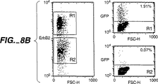
制限酵素SfiIおよびNotIを用いてファージミドDNAを消化することにより、pHEN1−F5からF5 scFv遺伝子を取り出した。fdDOG1(前述の参考文献を参照のこと)に基づくが、StiI部位を遺伝子IIIリーダー配列に挿入するように改変したファージベクターを、SfiIおよびNotIで消化し、そして消化したF5遺伝子を消化したファージFdベクターDNAに連結させた。組換え形質転換体を同定した。F5組換えファージを含むE.coliを、培養物中で増殖させ、F5−Fdファージを産生した(ファージ調製についてManiatisを参照のこと)。次いで、F5ファージを用いて、哺乳動物プロモーター(CMV)、続いて∃−ガラクトシダーゼについての遺伝子(pcDNA3.1/HisB/LacZ、In Vitrogen)または増強させたグリーン蛍光タンパク質についての遺伝子(pN2EGFP、Clonetchプラスミド)のいずれか、および真核生物のポリアデニル化配列を含むファージミドを保有するE.coliに感染させた。細菌を一晩、テトラサイクリン15μg/mL、およびアンピシリン100μg/mL(pcDNA3.1/HisB/LacZを含む細菌)かまたはカナマイシン30μg/mL(pN2EGFPを含む細菌)のいずれかの存在下で増殖させた。F5−Fdコートの混合物の上清から調製されたファージは、単鎖形式でのレポーター遺伝子(ファージの約50%)か、またはF5−Fdファージゲノム(ファージの約50%)のいずれかを含む。107pfuファージ混合物とのErbB2陽性細胞5.105 SKBR3のインキュベーション(滅菌するまで0.45μmフィルターを2回通して濾過する)により、細胞の1%においてレポーター遺伝子が発現し得る。10倍より陰性のコントロールファージ(すなわち、野生型Fd中のレポーター遺伝子パッケージング)とともにインキュベートされた細胞は、レポーター遺伝子の発現を示さなかった。ErbB2高細胞(SKBR3)とErbB2低細胞(MCF7)の混合集団(Lewisら(1993)Cancer Immunol Immunother 37:255〜263)をF5−Fd−EGFPファージとともに2日間インキュベートした実験において、本発明者らは、erbB2陽性細胞(FACSによるそれらのErbB2レベルより分化した細胞)においてのみ、レポーター遺伝子の発現を得た。
【0179】
(実施例6:糸状バクテリオファージによる哺乳動物細胞への標的化遺伝子送達)
本実施例において、本発明者らは、原核動物ウイルスが、真核生物細胞に感染するように再操作され得、バクテリオファージゲノムの一部分の発現を生じることを示す。増殖因子レセプターErbB2を発現する哺乳動物細胞を結合し得、そしてレセプター媒介エンドサイトーシスを引き起こし得るファージを、乳腫瘍細胞上でのファージ抗体ライブラリーを選択することおよびその細胞内から感染性ファージを回収することにより単離した。免疫蛍光により決定した場合、F5ファージは、ErbB2を発現するSKBR3細胞の100%に効率的にエンドサイトーシスされた。このファージゲノムの一部分の発現を達成するために、F5ファージを操作して、CMVプロモーターにより駆動されるグリーン蛍光タンパク質(GFP)レポーター遺伝子をパッケージングした。これらのファージは、細胞に適用される場合、ErbB2媒介エンドサイトーシスを引き起こし、GFP発現を導いた。ErbB2を過剰発現する細胞においてのみ生じるGFP発現は、用量依存性であり、60時間後、細胞の4%に達成し、そして2.0×10⁷cfu/ml程の低さのファージ力価で検出された(500ファージ/細胞)。この結果は、適切な抗体を提示する細菌性ウイルスが、哺乳動物レセプターに結合し得、そして嚢胞内膜性経路を利用して真核生物細胞に感染し、そしてウイルス遺伝子発現を生じ得る。このことは、ファージ抗体を直接的にスクリーニングすることによって遺伝子発現のための正確な輸送経路に、細胞内で遺伝子を送達し得る、標的化された分子を発見するための新規の方法を示す。このことは、細胞質ゾルへの送達が必要である遺伝子治療または他の適用のための適切な標的物および標的化分子の同定を有意に容易にすべきである。このアプローチはまた、標的化された遺伝子発現のためのファージ抗体をスクリーニングするよりもむしろ直接選択するために適用され得る。この結果はまた、ファージ抗体の、インビトロまたはインビボでの標的化された遺伝子送達ビヒクルとしての可能性を実証する。
【0180】
(A)導入)
遺伝子治療の広範な適用は、高い効率で、治療遺伝子を適切な細胞もしくは組織型に標的化する能力を必要とする(MichealおよびCuriel(1994)Gene Ther.1:223〜232)。レトロウイルスベクターの標的化は、レセプターリガンドまたは単鎖Fv(scFv)抗体フラグメントをウイルスエンベロープタンパク質に挿入することにより報告されている(Kasaharaら(1994)Science 266:1373〜1376)。アデノウイルスベクターの標的化は、アデノウイルスノブを結合する抗体フラグメント、およびレセプターリガンドまたは抗体のような細胞標的化分子からなる「アダプター」融合分子の使用により達成された(Douglasら(1996)Nat.Biotechnol.14:1574〜1578;Watkinsら1997)Gene Ther.4(10):1004〜1012)。細胞表面レセプターリガンドまたは抗体を用いた非ウイルスベクターの標的化もまた、報告されている(FominayaおよびWels(1996)J.Biol.Chem.271(18):10560〜10568;MichealおよびCuriel(1994)Gene Ther.1:223〜232)。これらのアプローチの全ては、細胞表面レセプターに結合し、続いてDNAを核へ送達する遺伝子送達ビヒクルのインターナリゼーションをもたらす標的化分子の使用に依存する。適切な標的化分子の同定は、主に、レセプターリガンドまたは抗体を個々にスクリーニングすることによって実施されている。scFv抗体フラグメントの場合、これは、代表的には、ハイブリドーマのV遺伝子からのscFvの構築、標的化された遺伝子送達ビヒクルの構築、およびインビトロでの標的化能力の評価を必要とする。
【0181】
より最近では、糸状ファージライブラリーから、細胞表面レセプターに結合するペプチドおよび抗体フラグメントを直接選択することが可能であることが証明された(Andersenら(1996)Proc.Natl.Acad.Sci.USA 93(5):1820〜1824;Barryら(1996)Nat.Med.2:299〜305;CaiおよびGaren(1995)Proc.Natl.Acad.Sci.USA 92(24):6537〜6541;de Kruifら(1995)Proc.Natl.Acad.Sci.USA 92(6):3938〜3942;Marksら(1993)Bio/Technology 11(10):1145〜1149)。このことは、潜在的な標的化分子の数の目立った増加を導いた。バクテリオファージがレセプター媒介エンドサイトーシスをもたらす能力(Barryら(1996)Nat.Med.2:299〜305;Hartら(1994)J.Biol.Cehm.269(17):12468〜12474)は、ファージライブラリーが細胞結合のためにだけでなく、哺乳動物細胞中にインターナリゼーションするためにも選択され得ることを示す。ファージ単鎖ファージゲノムが転写され、そして翻訳され得る場合、エンドサイトーシスおよび発現を導く正確な輸送経路へのファージゲノムの送達を導く様式でレセプターを結合するファージについてスクリーニングおよび選択することが、可能であることが分かるはずである。ファージが、細胞膜の化学的変化後、哺乳動物細胞中に侵入し、レポーター遺伝子の発現を導き得ることが以前に示されている(OkayamaおよびBerg(1985)Mol.Cell.Biol.5(5):1136〜1142;Yokoyama−KobayashiおよびKato(1993)Biochem.Biophys.Res.Commun.193(2):935〜939)。より最近、Laroccaらは、間接的なバクテリオファージ媒介遺伝子送達は、ビオチン化ファージを、ストレプトアビジンおよびビオチン化線維芽細胞増殖因子(FGF)を介してFGFレセプターを発現する哺乳動物細胞に対して標的化することによって起こり得ることを示した(Laroccaら(1998)Hum.Gene Ther.9:2393〜2399)。
【0182】
この報告において、本発明者らは、ファージマイナータンパク質pIIIとの遺伝子融合物として抗ErbB2 scFv F5を提示する糸状ファージが、ErbB2を発現する哺乳動物細胞に直接感染し、ファージゲノム中に含まれるレポーター遺伝子の発現を導き得ることを示す。これは、細胞内薬物送達または遺伝子治療のための標的化分子を、ファージ抗体を直接スクリーニングして、エンドサイトーシスを引き起こし、そして遺伝子を遺伝子発現のための正確な輸送経路に細胞内送達し得る分子を同定することによって発見する、新規の方法を与える。これは、細胞質ゾルへの送達が必要とされる遺伝子治療または他の適用のための適切な標的および標的化分子の同定を有意に容易にするはずである。本発明者らはまた、このアプローチを用いて標的化された遺伝子発現のためにファージ抗体を直接選択する方法を議論する。最終的には、本発明者らは、インビトロまたはインビボでの標的化遺伝子送達ベクターのためのファージ抗体それ自体の使用についての可能性を議論する。
【0183】
(B)材料および方法)
(1)抗ErbB2 F5 scFv)
ベクターpHEN−1中の抗ErbB2 scFv(F5)(Hoogenboomら(1991)Nucleic Acids Res.19(15):4133〜4137)(pHEN−F5)を、ErbB2を発現するSKBR3細胞上の非免疫ファージ抗体ライブラリー(Sheetsら(1998)Proc.Natl.Acad.Sci.USA 95(11):6157〜6162)を選択し、続いて組換えErbB2細胞外ドメイン(ECD)上での結合をスクリーニングすることによって得られた。天然のF5 scFvは、ErbB2 ECDと、Kd=1.6×10⁻⁷M(先に記載されるような(Schierら(1996)J.Mol.Biol.255(1):28〜43)BIAコアにおける表面プラズモン共鳴により決定した)で結合する。
【0184】
(2)ファージおよびファージミドベクター)
pN2EGFP(4.7Kbp)(Clontech)のHindIII/NotIフラグメントを、pcDNA3−HisB/LacZ(Invitrogen)HindIII/NotI骨格中にサブクローニングすることによって、pcDNA3−GFP(6.1Kbp)を得た。pHEN−1からのSfiI/NotI scFv−F5挿入物を、fd−Sfi/NotのSfiI/NotI部位(これは、遺伝子IIIリーダー中のApaL1クローニング部位をSfiIに変化させることによってfd−tet−DOG(Clacksonら(1991)Nature 352(6336):624〜628)から構築された)にサブクローニングすることによって、fd−F5−ファージベクターを構築した。1.6KbpのpN2EGFPの平滑末端化したAse I/Afl IIフラグメントを、pHEN−F5の平滑末端化したEcoR I部位にサブクローニングすることによって、pHEN−F5−GFPファージミドベクター(6.8Kbp)を得た。挿入物の配向をNotI制限消化により分析した。
【0185】
(3)細胞株の培養およびトランスフェクション)
SKBR3およびMCF7を、10%ウシ胎仔血清(FBS)(Hyclone)を補充したRPMI中で増殖させた。6ウェルプレート中で増殖させた50%コンフルエントなSKBR3細胞を、製造業者により推奨されるように、リポフェクタミン(GIBCO BRL)を用いて1ウェルあたり1μgのDNAでトランスフェクトした。Maxiprep Qiagen Kit(Qiagen Inc.)を用いたアルカリ溶解により、pN2EGFP dsDNAを調製した。500μlのファージミド調製物(以下を参照のこと)から、2回のフェノール抽出続いてエタノール沈殿により、ssDNAを抽出した。ssDNAについて40μg/mlまたはdsDNAについて50μg/mlに等しい、1.0A260nmでの吸光度の測定により、DNAを定量した。GFPの検出について、トリプシンEDTAmix(GIBCO BRL)を用いて細胞を剥離し、そしてFACScan(Becton Dickinson)で分析した。
【0186】
(4)ファージミドおよびファージの調製)
pHEN−F5、pHEN−F5−GFP、pcDNA3−GFPまたはpN2EGFPファージミドを、以前に記載されるように(Marksら(1991)J.Mol.Biol.222(3):581〜597)、VCS−M13ヘルパーファージ(Stratagene)を用いた重感染により、E.coli TG1より調製した。Fd−F5−ファージを、以前に記載されるように(McCaffertyら(1990)Nature 348(6301):552〜554)、E.coli TG1から調製した。pcDNA3−GFPを含むE.coli TG1をfd−F5−ファージで重感染させることによって、F5−GFP−ファージおよびF5−LacZ−ファージを調製した。培養物上清から、2回のポリエチレングリコール沈殿(Sambrookら(1990)、Molecular cloning−a laboratory manual、Cold Spring Harbor Laboratory、New York)によりウイルス粒子を精製し、リン酸緩衝化生理食塩水(pH7.4)(PBS)中に再懸濁し、0.45μmのフィルターで濾過し、そして4℃で保存した。あるいは、この調製物は、さらなるCsCl超遠心分離工程に供した(SmithおよびScott(1993)Meth.Enzymol.217:228〜257)。ファージミドDNAに対するパッケージングされたヘルパーファージDNAの比を、アンピシリンおよびカナマイシン耐性(ヘルパーファージレスキューpHEN−F5について)、またはアンピシリンおよびテトラサイクリン耐性(fd−F5ファージレスキューpcDNA3−GFPについて)についてファージを滴定することによって(Sambrookら、前出)決定した。
【0187】
(5)ファージFACS)
細胞をトリプシン処理し、1%FBS含有PBS(FACS緩衝液)で洗浄し、そして同じ緩衝液に106細胞/mlで再懸濁した。染色手順をFACS緩衝液で希釈された試薬を用いて氷上で実行した。細胞の100μlのアリコートを円錐形の96ウェルプレート(Nunc)に分配し、300gで遠心分離し、そして細胞ペレットを100μlのファージまたはファージミド調製物の階段希釈物に再懸濁し、そして1時間インキュベートした。細胞を2回、遠心分離し、そして洗浄し、細胞ペレットを100μlの抗M13抗体(5Prime,3Prime Inc.)(1/400希釈)に再懸濁し、そして45分間インキュベートした。細胞を上記のように洗浄し、100μlのストレプトアビジン−フィコエリトリン(Jackson Inc.)(1/400希釈)に再懸濁し、そして20分間インキュベートした。最後の洗浄後、細胞をFACSで分析した。
【0188】
(6)免疫蛍光検査)
細胞を6ウェルプレートで50%コンスルエンスまでカバーガラスにおいて増殖した。ファージ調製物(培養培地の10%未満)を添加し、そして細胞を16時間インキュベートした。カバーガラスを6回PBSで、グリシン緩衝液(50mMグリシン、pH2.8、NaCl 500mM)で10分間3回洗浄し、PBSで中和し、そしてPBS−4%パラホルムアルデヒドで、室温で5分間固定した。細胞を30秒間、冷アセトンで透過性にし、PBS−1% BSAで飽和し、そして抗M13抗体(飽和溶液において1/300希釈)と、その後ストレプトアビジン−テキサスレッド(Amersham)(飽和溶液において1/300希釈)とともにインキュベートした。カバーガラスをAxioskop蛍光顕微鏡(Zeiss)で分析した。
【0189】
(7)バクテリオファージ媒介細胞感染)
CsClファージ調製物を培養培地で少なくとも10倍に希釈し、0.45μmフィルターを通して濾過し、そして30%〜80%コンフルエントな細胞に添加した。インキュベーション後、細胞をトリプシン処理し、FACS緩衝液で洗浄し、そしてFACSによりGFP発現について分析した。MCF7およびSKBR3が同時培養される実験において、ErbB2発現を、ErbB2 ECD(10μg/ml)に結合する抗ErbB2マウスmAb 4D5(1時間)、ビオチン化ヒツジ抗マウス免疫グロブリン(Amersham)およびストレプトアビジン−フィコエリトリンを用いてFACSにより定量した。
【0190】
C)結果
(1)ErbB2発現細胞による一価F5ファージ粒子および多価F5ファージ粒子に結合するErbB2のインターナリゼーション)
本発明者らは、ErbB2発現SKBR3乳房腫瘍細胞での選択、および細胞内での感染性ファージの回収(M.Poulら、manuscript in preparation)により、pIIIへの融合物として、バクテリオファージの表面で提示されたscFvのライブラリーから抗ErbB2 scFv−F5を単離した(Sheetsら(1998)Proc.Natl.Acad.Sci.USA 95(11):6157〜6162)。この選択ストラテジーを、レセプター結合の際に、エンドサイトーシスを被り得るscFvを選択するために使用した。pHEN−F5ファージミドベクターがVCS−M13ヘルパーファージでレスキューされる場合、得られたウイルス粒子(F5−ファージミド)は、平均1コピーのscFv−pIII融合タンパク質、およびヘルパーファージからの3〜4コピーの野生型pIII型の非主要コートタンパク質を提示する(Marksら(1992)J.Biol.Chem.267(23):16007〜16010)。結果として、ファージミドは一価で結合する。ErbB2発現細胞へのウイルス粒子の結合を改善するため、多価ファージ抗体は、F5 scFvDNAを、pIIIタンパク質との融合のためファージベクターfd−Sfi/Notにサブクローニングすることにより作製した。fd−F5ファージといわれるウイルス粒子は、4〜5コピー数のscFv−pIII融合タンパク質(Id.)を提示する。
【0191】
F5ファージ抗体が哺乳動物細胞によりインターナライズされ得るか否かを決定するために、ErbB2を過剰発現するSKBR3細胞をfd−F5ファージ(109コロニー形成単位/ml、cfu/ml)、F5ファージミド(1011 cfu/ml)、または陰性のコントロールとして、無関係の抗ボツリヌス菌scFv−pIII融合タンパク質(1012 cfu/ml)(Amersdorferら、1997)を提示するファージミドとともに16時間インキュベートした。細胞表面を低いpHのグリシン緩衝液を用いてファージ抗体を剥がし、細胞を透過性にし、そして固定し、そして細胞内ファージを抗M13抗体を用いて検出した。著しいことに、全ての細胞は、fd−F5ファージ、またはF5ファージミドでインキュベートされた場合、強力な細胞内染色を示したが、抗−ボツリヌス菌ファージミドとインキュベートされた場合は示さなかった。これは、pIIIに融合したscFvの特異性に対するファージ侵入の依存を実証する。
【0192】
(2)真核生物細胞における発現のためのレポーター遺伝子をパッケージングするErbB2結合ファージおよびファージミドの調製)
F5ファージが発現を誘導する哺乳動物細胞にレポーター遺伝子を送達し得るか否かを研究するために2つのストラテジーを用いた。レポーター遺伝子を含有する一価ファージを作製するため、本発明者らは、CMVプロモーターにより駆動されたグリーン蛍光タンパク質(GFP)の遺伝子を、ベクターpHEN−F5−GFPを生成するファージミドベクターpHEN−F5へクローニングした(図6、左側)。pHEN−F5−GFP(アンピシリン耐性)を含有するEschrichia coli TG1をヘルパーファージ(カナマイシン耐性)で感染し、そして高い力価の一価F5−GFPファージミドを得た(培養上清の5.0×10¹⁰アンピシリン耐性cfu/ml)。パッケージングされたファージミドDNA対ヘルパーファージDNAの比(アンピリシン耐性cfu対カナマイシン耐性cfu)は100:1と決定された。レポーター遺伝子を含有する多価ファージを作製するため、pcDNA3−GFPファージミド(アンピシリン耐性)を保有するE.coli TG1を、fd−F5−ファージ(テトラサイクリン耐性)で感染することにより(従ってヘルパーファージとしてfd−F5ファージを用いて)、fd−F5−GFPファージを生成した。fd−F5−GFPファージの力価は、約5.0×10⁸アンピシリン耐性cfu/ml 培養上清であった。fdをヘルパーファージとして用いた場合、より低いファージ力価が生じるのは、それがファージミドf1起点から干渉を生じるプラスミドの複製起点を欠失するからである(ClearyおよびRay(1980)Proc.Natl.Acad.Sci.USA 77(8):4638〜4642)。パッケージングされたレポーター遺伝子DNA対ファージDNAの比(アンピシリン耐性cfu対テトラサイクリン耐性cfu)は1:1であった。fdをヘルパーファージとして用いる場合のレポーター遺伝子/ヘルパーゲノムのより低い比は、VCS−M13における変異したパッケージングシグナルと比較して、fdゲノム上の完全に機能的なパッケージングシグナルの存在に起因する(VieiraおよびMessing(1987)Meth.Enzymol.153:3〜11)。ファージ調製物およびファージミド調製物の両方は、SKBR3細胞結合について評価された(図7)。両方の調製物がSKBR3細胞に結合したが、結合は、1010 cfu/mlのF5−GFPファージミド(15ピコモル)に対して、fd−F5−GFPファージでは108 cfu/ml(160フェムトモル)程度と決定され得た。従って、多価結合は、ウイルス粒子の結合定数の明白な上昇を導く。
【0193】
(3)ErbB2発現乳癌細胞への標的化されたファージミドおよびファージ媒介型の遺伝子トランスファー)
ErbB2結合ファージミドが遺伝子送達を標的され得るか否かを決定するため、2.0×105のSKBR3細胞(高いレベルのErbB2を発現する乳房腫瘍細胞株)または2.0×105のMCF7細胞(低いErbB2を発現する乳房腫瘍細胞株)を、37℃で、5.0×1011cfu/mlのF5−GFPファージミドとインキュベートした。細胞を48時間後FACSにより、GFP発現について分析した(図8A)。SKBR3細胞の1.37%は、F5−GFPファージミドでのインキュベーション後、GFPを発現した(図8A6)。GFP発現は、f1パッケージングシグナルの方向にかかわらず同一であった(データ示さず)。これは、転写/翻訳が相補的DNA鎖の合成を介して進行していたことを示す。GFP発現は、ファージなしで、またはレポーター遺伝子をパッケージングするヘルパーファージとともにインキュベートされたSKBR3細胞において検出されなかった(図8A4および8A5)。発現はまた、ファージ、ヘルパーファージまたはpHEN−F5−GFPなしにインキュベートされたMCF7細胞においても見られなかった。これは、標的された遺伝子送達のためのErbB2発現の必要性を示す(図8A1、8A2および4A3)。遺伝子トランスファー適用が異種の細胞集団における特定の細胞の標的化に関するようなので、本発明者らは、SKBR3細胞およびMCF7細胞の同時培養において同じ実験を実行した(図8B)。細胞をSKBR3細胞からMCF7細胞を識別するためにErbB2発現について染色した。そしてそれぞれの小集団のGFP発現をFACSにより分析した。SKBR3細胞のみ(1.91%)がGFPを発現した。同様の結果がF5−GFPファージミドの代わりにF5−GFPファージを用いて見出された(データ示さず)。これらのデータは、fd−F5−GFPファージおよびF5−GFPファージミド媒介型の遺伝子送達が、ErbB2過剰発現細胞に制限されること、そして非発現細胞の存在下でこのような細胞に標的され得ることを確認する。
【0194】
(4)ファージ媒介型の遺伝子トランスファーの特徴づけ)
ファージ媒介型の遺伝子トランスファーの用量応答性の特徴を決定するため、SKBR3細胞を、漸増量のfd−F5−GFPファージまたはF5−GFPファージミドとともに60時間インキュベートし、そしてGFP陽性細胞のパーセントを決定した(図9Aおよび9B)。有意な数のGFP陽性細胞(図9A)の検出のために必要な最小のファージ濃度(図9A)は、fd−F5−GFPファージについては約4.0×107cfu/ml(0.13%)、そしてF5−GFPファージミドについては1.0×1010cfu/ml(0.12%)であった。この価は、結合曲線と密接に関係し(図7)、そして遺伝子発現について、多価ファージは、ファージミドよりも100〜1000倍より有効であることを示す。有意な数の陽性細胞は、レポーター遺伝子をパッケージングするヘルパーファージの4.0×1013cfu/mlまででは観察されなかった。ファージおよびファージミドの両方について、GFP陽性細胞のパーセントは、ファージ濃度にともない増加した(プラトーの証拠はない)。得られた最大の値はfd−F5−GFPファージについては細胞の2%であり、そしてF5−GFPファージミドについては4%であり、そして適用されたファージの力価(それぞれ、1.5×109cfu/mlおよび4.0×1012cfu/ml)により限定されるようである。1細胞あたりの発現されるGFPの量(平均蛍光強度(MFI)により評価される、図9B)はまた、ファージ濃度にともない増加し、少数の細胞が2.0×107cfu/ml(fd−F5−GFPファージ)〜1.0×1010cfu/ml(F5−GFPファージミド)の低いファージミド力価で発現を示す。
【0195】
ファージで得られた遺伝子発現の収量と伝統的なトランスフェクション方法を比較するため、一本鎖(ssDNA)または二本鎖(dsDNA)をリポフェクタミンを用いてSKBR3にトランスフェクトした。ssDNAの1μgあたり、ファージミド媒介型の遺伝子送達の効率(約1%)は、ssDNA(0.98%)およびdsDNA(1.27%)のリポフェクタミントランスフェクションに匹敵した(表8)。効率は、ファージ媒介型のトランスフェクションについて約500倍高く、2.25ngのssDNAで、細胞の0.87%のトランスフェクションを生じた。
【0196】
【表8】
【0197】
(C)考察)
本発明者らは、非主要コートタンパク質pIIIとの遺伝的融合物として抗ErbB2 scFv抗体フラグメントを提示する糸状ファージが、scFvの特異性を発現する哺乳動物細胞に直接標的され得ることを実証する。このようなファージは、レセプター媒介型のエンドサイトーシスを被り、そして最終的にレポーター遺伝子発現を導く細胞内輸送経路に侵入する。これは、原核生物のウイルスをが再操作して、真核生物細胞を感染させ、その結果、バクテリオファージゲノムの一部の発現を生じ得ることを実証する顕著な知見である。遺伝子発現は、2.0×10⁷cfu程度のファージで検出され、そして細胞の4%までファージの力価を増大するにつれて増大された。多価提示は、一価提示と比べて、検出可能な遺伝子発現についての閾値を500倍低下した。このことは、機能性の親和性における上昇およびレセプター架橋からのレセプター媒介型エンドサイトーシスの上昇した速度にもっとも起因するようである。しかし、トランスフェクトされた細胞の最大パーセントは、産生された有意により高いファージの力価に起因して、一価提示(ファージミド)について、より高かった。多価ファージのより低い力価は、レポーターファージミドに対するf1複製起点のfdファージ抗体複製起点での妨害に起因する(ClearyおよびRay(1980)Proc.Natl.Acad.Sci.USA 77(8):4638〜4642)。
【0198】
エンドサイトーシス可能レポーターに結合するファージを用いる哺乳動物細胞の標的された感染は、一般的な現象のようである。例えば、pHEN−GFPの遺伝子IIIへの抗トレンスフェリンレセプターScFvの融合は、MCF7細胞の10%、SKBR3細胞の4%、LNCaP細胞の1%、および初代黒色腫細胞の1%においてGFP発現を生じる。同様に、ビオチン化ファージおよびストレプトアビジンFGF融合分子を用いるFGFレセプター発現細胞への標的されたGFP遺伝子の送達は、現在報告されている(Laroccaら(1998)Hum.Gene.Ther.9:2393〜2399)。しかし、遺伝子IIIを介する標的分子の直接の遺伝的融合は、アダプター分子を用いるよりもより有効であり得る。従って、FGF−アダプター分子を用いてトランスフェクトされた細胞の最大パーセントは、報告されていないが、本発明者らは、それが感染された細胞の数、感染後の遺伝子発現の測定までの時間、およびGFPを発現する細胞の数に基づいてFGF発現L6ラット筋原細胞のわずか0.03%であることを評価する。発現のより大きい頻度(0.5%)がCOS−1細胞で見られたが、これは、ラージT抗原およびSV40媒介型DNA複製の存在から生じ、従って、ほとんどの細胞には一般化されない。
【0199】
本発明者らの記載するアプローチは、標的化された細胞内薬物または遺伝子送達のためのリガンドを発見するための新規な方法を示す。ファージ抗体またはペプチドのライブラリーは、まず哺乳動物細胞によるエンドサイトーシスについて(Barryら、(1996)Nat.Med.2:299〜305)、または精製された抗原、細胞、組織もしくは器官への結合について、選択される。選択されたscFv遺伝子のpHEN−GFPベクターへのサブクローニング後、個々のコロニーから産生されたファージは、遺伝子発現について直接スクリーニングされ得る。発現が1.0×10¹⁰cfu程度のプラスミドの少なさで検出され得るので、これは、可能である。これは、エンドサイトーシスされたscFvの直接の同定を可能にするだけでなく、遺伝子発現のための適切な輸送を被るレセプター抗体のサブセットも可能にする。多価提示が効率的なエンドサイトーシスに必要である場合、scFv遺伝子はfd−Sfi−Notにサブクローニングされ得、次いでこれはレポーターファージミドのレスキューに用いられる。scFv−fdファージの使用はまた種々の発現ベクターにおける多数の異なるレポーター遺伝子の標的化を可能にする。なぜなら、多数の市販の哺乳動物ベクターは、f1複製起点を含むからである。この場合、抗体標的ファージは、特に標準的技術によりトランスフェクトすることが困難な細胞について、有用なトランスフェクション試薬であると証明され得る。
【0200】
このアプローチを、標的化遺伝子送達のための抗体の、スクリーニングよりも直接選択に用いることが可能であることも証明されている。例えば、哺乳動物の細胞はGFP遺伝子を含むファージ抗体ライブラリーとともにインキュベートされ、次いでFACSを用いてGFPの発現に基づいてソートされる。ファージ抗体DNAは、細胞溶解により哺乳動物の細胞質から回収され、そしてE.coliをトランスフェクトするために用いられ、そして選択の別の回のためのさらなるファージを調製する。回収可能なファージDNAの量が不十分である場合、pHEN−GFPベクター中のネオマイシン遺伝子の包含がG418を用いるGFP発現哺乳動物細胞の選択を可能にする(Laroccaら、前出)。
【0201】
最後に、この系は、標的可能なインビトロまたはインビボにおける遺伝子治療ビヒクルとして見込まれる。主な制限は感染の効率、薬物動態および免疫原性である。感染効率に関しては、この報告において標的されたファージにより得られた値(ファージ調製物の8.0×10⁴ml)は、標的化レトロウイルスについて報告された値(103〜105/mlのウイルス)(Kasaharaら(1994)Science 266:1373〜1376;Somiaら(1995)Proc.Natl.Acad.Sci.USA 92(16):7570〜7574)と異ならないが、アデノウイルス標的化ストラテジーについて報告された(Douglasら(1996)Nat.Biotechnol.14:1574〜1578;Watkinsら、1997)Gene Ther.4(10)1004〜1012)よりは低い。しかし、より高い感染効率を制限する因子は、系の間で異なるようである。従って、レトロウイルスにより感染された細胞のパーセンテージは、バクテリオファージについて観察されたものよりも有意に高いが、感染は、細胞に侵入し得る多数のウイルス産生に遭遇する問題により制限される。すべての細胞は、標的化ファージを取り込むので、遺伝子発現は1つまたはいくつかの取りこみ後事象(例えば、DNAを放出するためのファージの分解、エンドソームのエスケープ、核標的化または転写)により制限される。ファージおよびそのDNAの運命のより詳細な研究は、感染効率を増大するためのファージの操作を可能にするブロックがどこに存在するかを示唆するようである。例えば、エンドソームのエスケープは、複製欠陥アデノウイルスの同時投与(Curielら(1991)Proc.Natl.Acad.SciUSA 88(19):8850〜8854)または、ファージの主要コートタンパク質pVIIIへの、エンドソームエスケープペプチド(Waguerら(1992)Proc.Natl.Acad.Sci.USA 89(17):7934〜7938)もしくはエンドソームエスケープタンパク質(FominayaおよびWels(1996)J.Biol.Chem.271(18):10560〜10568)の組み込みにより増強され得る。あるいは、感染効率は、pVIII変異体のscFv標的ライブラリーを作製すること、および増加した遺伝子発現について選択することにより複合的に上昇され得る。薬物動態に関しては、徹底的に研究されてはいないが、ファージの生体分布は血管内の腔に制限されるようである。これは、インビトロのファージ遺伝子治療に影響しないが、血管系を標的するものへのインビボ使用を制限し得る。これはなお、癌のような血管新生が役割を果たすものを含む多くの適用を残す。免疫原性に関しては、ファージは免疫原性であるらしく、従ってこれは、ファージがインビボで投与され得る回数を制限する。あるいは、例えば、免疫血清でのpVIIIライブラリーのネガティブ選択により、免疫原性を軽減または排除するために主要コートタンパク質pVIIIを進化することが可能であることが証明され得る(Jenneら(1998)J.Immunol.161(6):3161〜3168)。
【0202】
本明細書で記載される実施例および実施態様は、例証的な目的のみのためであり、そしてそれらの観点から種々の改変および変化が当業者に示唆され、そして本出願の思想および本文ならびに添付の特許請求の範囲に含まれることが理解される。本明細書に引用される全ての刊行物、特許および特許出願は、すべての目的のためにその全体が参考として本明細書に援用される。
【図面の簡単な説明】
【図1】図1は、大きなヒトscFvファージ抗体ライブラリーの構築の方法を示す。ライブラリー構築のストラテジーは、ライブラリー構築の個々の工程を最適化して、scFv遺伝子アセンブリの効率およびアセンブリしたscFv遺伝子のクローニングの効率の両方を増大させる工程を包含した。(A)最初に、リンパ球由来のmRNAを使用して、VHおよびVL遺伝子レパートリーをRTPCRにより作製し、これは、異なるベクターにクローニングされ、それぞれ、8.0×10⁸および7.2×10⁶のメンバーのVHおよびVL遺伝子ライブラリーが作製される。クローニングされたV遺伝子ライブラリーは、scFvアセンブリのためのVHおよびVL遺伝子の安定かつ制限のない供給源を提供した。ペプチド(G4S)3をコードするDNAを、VLライブラリーの5’末端に組み込んだ。このことによって、scFv遺伝子の作製が、2つのDNAフラグメントをPCRスプライシングすることにより可能になった。以前に、scFv遺伝子レパートリーは、VH、VL、およびリンカーDNAからなる3つの別個のDNAフラグメントからアセンブリされた。(B)VHおよびVL遺伝子レパートリーを別個のライブラリーから増幅し、そして重複伸長PCR(overlap extension PCR)を使用してscFv遺伝子レパートリーにアセンブリした。プライマーを使用して、VH遺伝子の5’末端の200bp上流およびVL遺伝子の200bp下流にアニールしたVHおよびVL遺伝子レパートリーを再増幅した。これらの長い突出部は、効率的な制限酵素消化を確実にした。(C)scFv遺伝子レパートリーを、NcoIおよびNotIで消化し、そしてプラスミドpHEN1に、M13遺伝子IIIコートタンパク質遺伝子との融合物として、ファージディスプレイのためにクローニングした。
【図2】図2A、2B、および2Cは、抗体ファージディスプレイを示す模式図を示す:(2A)単一のscFv、(2B)単一のダイアボディー、または(2C)多重scFvを提示するファージのカーツーン。scFv=単鎖Fv抗体フラグメント;VH=Ig重鎖可変領域ドメイン;VL=Ig軽鎖可変領域ドメイン;pIII=ファージマイナーコートタンパク質pIII;Ag=scFvに結合される抗原。
【図3】図3は、抗原特異的ファージの富化に対するトリプシン処理の効果を示す。fdファージ(5.0×10¹¹cfu)およびC6.5 scFvファージミド(5.0×10⁸fu)の混合物をSKBR3細胞とともに37℃で2時間インキュベートした。表6に記載されたように洗浄した(−)か、または細胞を細胞溶解前にトリプシン処理した(+)。第1のストリッピング緩衝液洗浄物中に存在するファージ(細胞表面ファージ)および細胞溶解物(細胞内ファージ)を、アンピシリン(C6.5ファージミド)またはテトラサイクリン(fdファージ)の存在下で力価測定した。
【図4】図4は、抗原特異的ファージの回収に対するインキュベーション時間およびクロロキンの効果を示す。SKBR3細胞を、抗ボツリヌスファージミド(白四角、黒四角)またはC6.5 scFvファージミド(白丸、黒丸)(1.5×10⁹cfu/ml)を添加する前に、クロロキン(50μM)の存在下(黒四角、黒丸)または非存在下(白四角、白丸)で2時間インキュベートした。細胞サンプルを、ファージ添加の0分後、20分後、1時間後、または3時間後に採取し、図4に記載されるように洗浄し(トリプシン処理工程を包含する)、そして細胞内ファージを力価測定した。
【図5】図5は、細胞内ファージの回収に対するファージ濃度の効果を示す。種々の濃度のC6.5 scFvファージミド、C6ML3−9 scFvファージミド、C6.5ダイアボディーファージミドまたはC6.5 scFvファージ(投入ファージ力価)を、6ウェルプレート中2時間37℃で増殖させたコンフルエント未満のSKBR3細胞とともにインキュベートした。細胞を図4に記載にように処理(トリプシン処理を包含する)し、そして細胞内ファージを力価測定する(産生ファージ力価)。
【図6】図6は、抗ErbB−2ファージミドおよび真核生物レポーター遺伝子をパッケージングするファージを生成するためのストラテジーを示す。左列:ヘルパーファージを使用して、TG1含有pHEN−F5−GFP、fl複製起点(fl ori)、遺伝子IIIに融合した抗ErbB−2 F5 scFv遺伝子、およびCMVプロモーターにより駆動される真核生物GFPレポーター遺伝子から構成されるファージミドを感染させる。培養上清から回収したファージは、平均1つのscFv−pIII融合タンパク質を提示し、そしてそれらの99%は、GFPレポーター遺伝子をパッケージングする。右列:抗ErbB2 F5 scFv遺伝子をfdファージゲノムに、scFv−pIII融合物として発現のためにクローニングする。fd−F5ファージを使用して、TG1含有GFPレポーターファージミドベクター(pcDNA3−GFP)を感染させる。培養上清から精製したファージは、多重scFv−pIII融合タンパク質を提示し、そして約50%がGFPレポーター遺伝子をパッケージする。
【図7】図7は、細胞に対する抗ErbB2ファージミドおよびファージの結合の比較を示す。105のErbB2発現SKBR3細胞を、漸増濃度のF5−ファージミド(白丸)またはfd−F5ファージ(白四角)とともに4℃で1時間インキュベートした。ファージを結合した細胞表面を、ビオチン化抗M13およびストレプトアビジン−PEを用いて検出した。結合をFACSにより検出し、そして結果を平均蛍光強度(MFI)として表した。
【図8】図8Aおよび8Bは、乳癌細胞株におけるファージミド媒介性遺伝子移入を示す。(図8A)6ウェルプレート中で増殖させた(1,2,3)2×10⁵MCF7細胞(低ErbB2発現)または(4,5,6)2.0×10⁵SKBR3細胞(高ErbB2発現)を、(1,4)ファージなし、(2,5)5.0×10¹²cfu/mlのGFPをパッケージングするヘルパーファージ、または(3,6)5.0×1011cfu/mlのF5−GFP−ファージミドのいずれかと、48時間インキュベートした。細胞をトリプシン処理し、そしてGFPをFACSにより検出した。(図8B)等量のMCF7およびSKBR3細胞(1×105)を、ともに増殖させ、そして5.0×1011cfu/mlのF5−GFP−ファージミドととともに48時間インキュベートした。細胞をトリプシン処理し、そして4D5抗体およびローダミン結合体化ヒツジ抗マウスIgを使用してErbB2発現について染色し、SKBR3細胞(領域R1)とMCF7細胞(領域R2)を区別した。各亜集団のGFP含量を、FACS により決定した。
【図9】図9A、9B、9C、および9Dは、SKBR3細胞におけるファージミド媒介性GFP発現の濃度依存性および時間経過を示す。図9Aおよび9Bは、ファージミドの濃度依存性およびSKBR3細胞におけるファージ媒介GFP発現を示す。5.0×10⁴細胞を24ウェルプレート中で増殖させ、そして漸増濃度のF5−GFPファージミド(四角)、fg−F5−GFP−ファージ(菱形)またはGFP−ヘルパーファージ(丸)とともにインキュベートした。60時間後、細胞をトリプシン処理し、そしてGFP発現についてFACSで分析した。図9Cおよび9Dは、SKRB3細胞におけるファージミド媒介性GFP発現の時間依存性を示す。5.0×10⁴細胞を、5×10¹¹cfu/mlのF5−GFP−ファージミドとともにインキュベートし、そしてGFP発現についてFACSで分析した。48時間を超えるインキュベーション時間にわたり、ファージを2.5×104細胞に添加し、そして培養培地を48時間のインキュベーション後に新鮮培地と置き換えた。結果を、(9Aおよび9C)GFP陽性細胞の%ならびに(9Bおよび9D)GFP陽性細胞のMFIとして表す。
【配列表】
(Cross-reference of related applications)
This application is a provisional application based on US Patent Act 119 (e), US provisional patent application No. 60 / 082,953 (filed April 24, 1998) (this is incorporated herein by reference in its entirety) Insist on the benefit of
[0002]
(Statement of rights to inventions made under federal-supported research and development)
This study was supported in part by Department of Defence Grants DAMD17-96-1-6244 and DAMD17-94-4433. The US government may have some rights in the invention.
[0003]
(Field of Invention)
The present invention relates to the fields of immunodiagnosis and immunotherapy. The present invention provides a method for identifying an internalizing antibody and an internalizing receptor ligand, as well as bound internalized receptor binding.
[0004]
(Background of the Invention)
Growth factor receptors and other signaling receptors are often overexpressed in human cancer and other diseases and are therefore utilized for the development of targeted therapies. For example, the HER2 / neu gene is amplified in several types of human adenocarcinoma, particularly breast and ovarian tumors (Slamon et al. (1989) Science 244: 707-712), which is associated with the corresponding growth factor ErbB2. Leads to overexpression. Targeting ErbB2 overexpressing cells has been achieved primarily with anti-ErbB2 antibodies in different formats. This format includes conjugates with liposomes containing chemotherapeutic agents (Kirpotin et al. (1997) Biochem. 36: 66-75), fusions with DNA carrier proteins that deliver toxin genes (Forminaya and Wels (1996) J Biol. Chem. 271: 10560-10568) and direct fusions with toxins (Altenschmidt et al. (1997) Int. J. Cancer 73: 117-124).
[0005]
For many of these targeted approaches, it is necessary to deliver effector molecules across the cell membrane into the cytosol. In some cases this can be facilitated by taking advantage of receptor-mediated endocytosis (Ullrich and Schlessinger (1990) Cell 61: 203-212). Receptor-mediated endocytosis often involves receptors directly for bivalent ligands or for monovalent ligands where binding to the ligand causes receptor activation via homodimerization or heterodimerization. Either by causing a conformational change in Antibodies can mimic this process, can stimulate endocytosis, can be internalized, and deliver the load to the cytosol. Furthermore, the efficiency with which an antibody mediates internalization depends on the type of antibody (eg, whole antibody, fragment, single chain antibody, monomer, dimer, etc.) and varies significantly depending on the recognized epitope. (Yarden (1990) Proc. Natl. Acad. Sci. USA 87: 2569-2573; Hurwitz et al. (1995) Proc. Natl. Acad. Sci. USA 92: 3353-3357). Thus, for some applications (eg, liposome targeting), only antibodies that bind to specific epitopes are rapidly internalized and generate a functional targeting vehicle.
[0006]
Internalizing antibodies have also been shown to cause cell growth inhibition or enhanced cell growth, depending on the recognized epitope. Thus, selection for internalization should result in the isolation of growth-inhibiting or growth-stimulating (agonistic) antibodies. Such inhibitory antibodies can be used as a cancer treatment or for the treatment of other conditions characterized by cellular hyperproliferation and for the treatment of inflammation (anti-inflammatory). Agonistic antibodies can be used to stimulate the proliferation of related cells (eg, stem cells). Targeting cells other than cancer for gene delivery also has many applications.
[0007]
Currently, antibodies that mediate internalization are identified by screening hybridomas. However, screening for antibodies produced by hybridomas is laborious, time consuming and expensive.
[0008]
(Summary of the Invention)
The present invention, in part, allows direct selection of antibodies that internalize from large non-immune phage libraries by recovering infectious phage particles from within the cell after receptor-mediated endocytosis. Based on the discovery.
[0009]
Accordingly, in one embodiment, the present invention provides a method of selecting a polypeptide or antibody binding moiety that is internalized in a target cell. The method preferably includes the following steps: i) contacting one or more of the target cells with one or more members of a phage display library; iv) the target cells of the display library Culturing under conditions that can be internalized when a member of the phage binds to an internalizing marker; and v) the phage when the members of the phage display library are internalized into one or more target cells. Identifying an internalized member of the display library. This method also optionally and preferably further comprises the following steps: contacting the phage display library members with cells of a subtractive cell line; and then washing the target cells. Removing the cells of the subtractive cell line and removing members of the phage display library that are non-specifically or weakly bound to the target cells. In a preferred embodiment, the phage display is an antibody phage display library, more preferably an antibody phage display library displaying single chain antibodies (eg, scFv, scFab, etc.).
[0010]
In a preferred embodiment, the “identifying” step includes recovering the internalized phage and repeating the above process steps again to further select for binding moieties to be internalized. In one embodiment, “recovering” comprises lysing the target cells to release the internalized phage; and infecting the bacterial host with the internalized phage, the next round of selection. Producing a phage for. This recovering step may include recovering infectious phage and / or recovering nucleic acid encoding the antibody displayed on the phage and / or expressing a selectable marker (eg, antibiotic resistance gene or cDNA). Includes selection of phage. This identifying step can include detecting the expression of a reporter gene, detecting the presence / absence or amount of a particular nucleic acid, or selecting a phage via a selectable marker. In a preferred method, the cells of the subtractive cell line are present in at least a 2-fold excess over their target cells. In a preferred method, the target cells form an adhesion layer. In a preferred method, the target cell line is grown adhering to a tissue culture plate and co-incubated with a cell line that subtracts in suspension in a single cell culture flask. In a particularly preferred method, the contact with the subtractive cell line is performed at a temperature (eg 4 ° C.) lower than the internalization culture conditions (eg 37 ° C.).
[0011]
In particularly preferred embodiments, the phage expresses a selectable marker and / or reporter gene. Preferred selectable markers include, but are not limited to: fluorescent proteins, genes that encode antibiotic resistance genes or cDNAs (or cDNAs) and chromogenic genes or cDNAs (eg, horseradish peroxidase, β Lactamase, luciferase and β-galactosidase). In certain embodiments, the target cell is a solid tumor cell, a member of a cDNA expression library, a cell that overexpresses a cytokine receptor, a cell that overexpresses a growth factor receptor, a metastatic cell, or a transformed cell line. It may include cells, cells transformed with a gene or cDNA encoding a particular surface target receptor, and neoplastic cells derived from outside a solid tumor. In one particularly preferred embodiment, the cells of the subtractive cell line are selected from the same tissue type as the target cells. Suitable subtractive cell line cells include, but are not limited to, fibroblasts, monocytes, stem cells, and lymphocytes.
[0012]
The methods of the invention can also identify internalizing receptors and / or internalizing receptor epitopes (regions of the receptor that, when bound, induce internalization of the binding moiety). This method generally involves any of the methods for identifying an internalizing antibody or polypeptide described herein in a further step (where the identified internalizing antibody or polypeptide is the original target. Used to probe cells or different cells). When the internalizing antibody or polypeptide so binds, they allow the isolation of cells having the internalizing receptor and the isolation of the receptor and / or receptor epitope itself. Accordingly, in one embodiment, the method includes: i) contacting one or more of the target cells with one or more members of a phage display library; ii) optionally, Preferably, however, the member of the phage display library is contacted with a cell of a subtractive cell line; iii) optionally, but preferably, iii) the target cell is washed and the subtractive cell Removing cells of the strain and removing members of the phage display library that are non-specifically or weakly bound to the target cell; iv) members of the phage display library are Can be internalized when combined with an internalizing marker Culturing the cells under the circumstances; v) identifying the internalized member of the phage display if the phage display library member is internalized into one or more of the target cells; vi) Contacting the same or different target cells with this identified internalized member of this step (v), or a member grown therefrom, so that this member binds to the surface of this same or different target cell Process. The method can further include isolating the same or different target cell components to which the member binds. In some methods, the “identifying” step comprises recovering the phage to be internalized and repeating steps (i)-(v) to further select for the binding moiety to be internalized. Is included.
[0013]
This contacting step, washing step, culturing step and identification step are preferably performed as described herein, and the target cells and subtractive cells comprise the cells described herein.
[0014]
In yet another embodiment, the present invention provides a multivalent antibody phage display library. The library preferably comprises a plurality of phage, wherein the phage on average displays at least 2 copies of a single chain antibody, and the library contains a plurality of species of single chain antibodies. Including. In a preferred embodiment, the phage on average displays at least 3, at least 4 or at least 5 copies of a single chain antibody per phage particle. Particularly preferred libraries on average are at least 10 Five , Preferably at least 10 6 , More preferably at least 10 7 , And most preferably at least 10 8 Of different species of single chain antibodies. In the most preferred embodiment, the antibody is encoded by a nucleic acid that is a phage (not phagemid) vector.
[0015]
In certain embodiments, the library is selected for members that specifically bind to an internalizing cell surface receptor (eg, erbB2, EGF receptor, PDGF receptor, VEGF receptor, transferrin receptor, etc.). This single chain antibody is preferably a single chain antibody Fv (scFv) or a single chain Fab (scFab) antibody. Filamentous phage are preferably used in the libraries of the invention, and the antibody is preferably expressed as a fusion with the minor coat protein of PIII. The phage can also express a selectable marker (eg, antibiotic resistance gene or cDNA) and / or a reporter gene or cDNA (eg, green fluorescent protein (GFP), Fflux, β-gal, β-lactamase, etc.).
[0016]
In still yet another embodiment, the present invention provides a nucleic acid library encoding one of the phage display antibody libraries described herein. This nucleic acid library has at least 10 Five , More preferably at least 10 6 , And most preferably at least 10 7 Of different phage or phagemid vectors.
[0017]
The present invention also provides kits for performing the methods described herein. The kit preferably comprises one or more containers containing a phage display library (or part thereof) as described herein (the kit comprises nucleic acids and / or single strands encoding the library). Phage particles expressing antibodies (preferably multivalent libraries) and / or cells containing intact phage or nucleic acids from the phage may be provided.
[0018]
(Definition)
As used herein, “antibody” refers to a protein consisting of one or more polypeptides substantially encoded by immunoglobulin genes or fragments of immunoglobulin genes. The recognized immunoglobulin genes include the kappa, lambda, alpha, gamma, delta, epsilon and mu constant region genes, as well as a number of immunoglobulin variable region genes. Light chains are classified as either kappa or lambda. Heavy chains are classified as γ, μ, α, δ, or ε. These then determine the immunoglobulin classes, IgG, IgM, IgA, IgD and IgE, respectively.
[0019]
A typical immunoglobulin (antibody) structural unit is known to contain a tetramer. Each tetramer is composed of two identical pairs of polypeptide chains, where each pair has one “light” chain (about 25 kD) and one “heavy” chain (about 50-70 kD). The N-terminus of each chain defines a variable region of about 100-110 or more amino acids, which are primarily responsible for antigen recognition. The term variable light chain (V L ) And variable heavy chain (V H ) Refers to these light and heavy chains respectively.
[0020]
Antibodies exist as a number of well-characterized fragments produced by digestion with intact immunoglobulins or various peptidases. Thus, for example, pepsin digests the antibody under the disulfide bond in the hinge region to produce Fab dimer F (ab) ′ 2 (which itself is a V disulfide bond by V H -
[0021]
“Antibody binding site” or “antibody portion” refers to the portion of an immunoglobulin molecule that participates in antigen binding. The antigen binding site is formed by amino acid residues in the N-terminal variable (“V”) region of the heavy chain (“H”) and light chain (“L”). Three highly diverse stretches within the heavy and light chain V regions are referred to as “hypervariable regions”. This region is inserted between more conservative adjacent stretches (known as “framework regions” or “FR”). Thus, the term “FR” refers to an amino acid sequence that is naturally found between and proximal to hypervariable regions in immunoglobulins. In an antibody molecule, the three hypervariable regions of the light chain and the three hypervariable regions of the heavy chain are arranged to form an antigen binding “surface” in three-dimensional space with each other. This surface mediates recognition and binding of the target antigen. The three hypervariable regions of each of the heavy and light chains are referred to as “complementarity determining regions” or “CDRs” and are described, for example, in Kabat et al. U. S. Dept. Characterized by Health and Human Services, Public Health Services, Bethesda, MD (1987).
[0022]
As used herein, the terms “immunological binding” and “immunological binding property” refer to the type of non-covalent binding that occurs between an immunoglobulin molecule and an antigen to which the immunoglobulin is specific. Sexual interaction. The strength or affinity of an immunological binding interaction can be expressed as the dissociation constant (Kd) of the interaction, where a smaller Kd represents a greater affinity. The immunological binding properties of the selected polypeptide can be quantified using methods well known in the art. One such method involves measuring the rate of antigen binding site / antigen complex formation and dissociation, where these rates are determined by the concentration of the complex partner, the affinity of the interaction and both. Depends on geometric parameters that affect the velocity in the direction equally. Therefore, the “on-speed constant” (k on ) And “off-rate constant” (k off ) Both can be determined by calculating the concentration and actual rate of association and dissociation. k off / K on Ratio allows the removal of all parameters not related to affinity, and thus the dissociation constant K d (See generally Davies et al. (1990) Ann. Rev. Biochem., 59: 439-473).
[0023]
The phrase “specifically binds to a protein” or “specifically immunoreactive with” when referring to an antibody, in the presence of a heterogeneous population of proteins and other biological materials, A binding reaction that is a determinant of the presence of a protein. Thus, under designated immunoassay conditions, a specified antibody binds to a specific protein and does not bind in a significant amount to other proteins present in the sample. Specific binding to a protein under such conditions may require an antibody that is selected for the specificity of the particular protein. For example, F5 or C1 antibodies can be raised against c-erbB-2 protein. These antibodies bind to c-erbB-2 and do not bind to other proteins present in the tissue sample. A variety of immunoassay formats can be used to select antibodies that are specifically immunoreactive with a particular protein. For example, solid phase ELISA immunoassays are routinely used to select monoclonal antibodies that are specifically immunoreactive with a protein. For a description of immunoassay formats and conditions that can be used to determine specific immunoreactivity, see Harlow and Lane (1988) Antibodies, A Laboratory Manual, Cold Spring Harbor Publications, New York.
[0024]
The terms “polypeptide”, “peptide” or “protein” are used interchangeably herein and from one to the other by a peptide bond between the α-amino group and the carboxy group of the adjacent residue. A linear series of linked amino acid residues. This amino acid residue is preferably present in the natural “L” isoform form. However, residues in the “D” isoform form can be replaced with any L amino acid residue as long as the desired functional properties are maintained by the polypeptide. In addition, the amino acids include, in addition to the 20 “standard” amino acids, modified and unusual amino acids, which are listed in 37 CFR 1.822 (b) (4). Including, but not limited to. In addition, a dash at the beginning or end of the amino acid residue sequence indicates a peptide bond to an additional sequence of one or more amino acid residues, or a covalent bond to a carboxyl or hydroxyl end group.
[0025]
The term “binding polypeptide” refers to a polypeptide that specifically binds to a target molecule (eg, a cell receptor) in a manner similar to that in which an antibody binds to an antigen. A binding polypeptide is distinguished from an antibody in that the binding polypeptide is not ultimately derived from an immunoglobulin gene or a fragment of an immunoglobulin gene.
[0026]
The term “conservative substitution” is used in reference to a protein or peptide to reflect an amino acid substitution that does not substantially alter the activity (specificity or binding affinity) of the molecule. Typically, a conservative amino acid substitution includes the replacement of one amino acid with another amino acid that has similar chemical properties (eg, charge or hydrophobicity). The following six groups each contain amino acids that are representative conservative substitutions for each other:
1) Alanine (A), Serine (S), Threonine (T);
2) Aspartic acid (D), glutamic acid (E);
3) Asparagine (N), glutamine (Q);
4) Arginine (R), Lysine (K);
5) Isoleucine (I), leucine (L), methionine (M), valine (V); and
6) Phenylalanine (F), tyrosine (Y), tryptophan (W).
[0027]
The term “nucleic acid” refers to deoxyribonucleotides or ribonucleotides and polymers thereof in either single- or double-stranded form. Unless specifically limited, the term encompasses forms comprising known analogs of natural nucleotides that have similar binding properties as the reference nucleic acid and are metabolized in a manner similar to naturally occurring nucleotides. Unless otherwise stated, a particular nucleic acid sequence also implicitly encompasses conservatively modified variants thereof (eg, degenerate codon substitutions) and complementary sequences, as well as explicitly indicated sequences.
[0028]
Specifically, degenerate codon substitution can be accomplished by generating a sequence in which the third position of one or more selected (or all) codons is replaced with a mixed base and / or deoxyinosine residue. (Batzer et al. (1991) Nucleic Acid Res. 19: 5081; Ohtsuka et al. (1985) J. Biol. Chem. 260: 2605-2608; and Cassol et al. (1992); Rossolini et al. (1994) Mol. Cell.
[0029]
The term “isolated” or “biologically pure” refers to material that is substantially or essentially free from components that normally accompany it when found in its native state. However, the term “isolated” is not intended to refer to components present in an electrophoretic gel or other separation medium. Isolated components do not contain such separation media and are in a form ready for use in another application or are already in use in a new application / environment.
[0030]
A chimeric molecule refers to a molecule in which two or more molecules that exist separately in their native state are joined together to form a single molecule having all the desired functionality of its component molecules. Chimeric molecules can be prepared by covalently linking two molecules, each synthesized separately, but one skilled in the art will recognize that when the chimeric molecule is a fusion protein, the chimera is de novo single “linked” It is recognized that it can be prepared as a molecule.
[0031]
A fusion protein is a chimeric molecule in which the component molecules are all polypeptides and are attached (fused) to each other via terminal peptide bonds so that the chimeric molecule is a continuous single chain polypeptide. The various components can be directly attached to each other or can be linked through one or more peptide linkers.
[0032]
An effector moiety or molecule is typically a molecule or molecule having a characteristic activity that is desired to be delivered to a target cell (eg, a tumor that overexpresses c-erbB). Effector molecules include cytotoxins, labels, radionuclides, ligands, antibodies, drugs, liposomes, and viral coat proteins that allow the virus to infect c-erbB2-expressing cells.
[0033]
A “target” cell refers to a cell or cell type that is specifically bound by a member of a phage display library or chimeric molecule of the present invention. Preferred target cells are those in which antibodies or binding polypeptides that are internalized are sought. This target cell is typically characterized by the expression or overexpression of a target molecule characteristic of the cell type. Thus, for example, the target cell can be a cell, such as a tumor cell, that overexpresses a marker such as c-erbB-2.
[0034]
“Targeting moiety” refers to a moiety (eg, a molecule) that specifically binds to a target molecule. If the target molecule is a molecule on the cell surface and the target molecule is a component of a chimeric molecule, the targeting moiety specifically binds the chimeric molecule to a cell with that target. If the targeting moiety is a polypeptide, the moiety can be referred to as a “targeting polypeptide”.
[0035]
The term “internalize” or “internalized” (referred to) refers to the transport of a portion (eg, phage) from outside to inside of a cell when referring to the cell. The portion to be internalized can be placed in an intracellular fraction (eg, the cell's own vacuole, lysosome, endoplasmic reticulum, Golgi apparatus or cytosol).
[0036]
An internalizing receptor or marker is a molecule present on the surface of an external cell that, when specifically bound by the antibody or binding protein, results in internalization of the antibody or binding protein into the cell. Receptors or markers that internalize include receptors (eg, hormones, cytokines or growth factor receptors), ligands and other cell surface markers whose binding results in internalization.
[0037]
The term “heterologous nucleic acid” refers to a nucleic acid that is not native to the cell in which the nucleic acid is found or whose ultimate origin is not in the cell or cell line in which the “heterologous nucleic acid” is currently found.
[0038]
An idiotype represents a highly variable antigen-binding site of an antibody and is itself immunogenic. During the generation of an antibody-mediated immune response, the individual develops its antigen, as well as an antigen to the anti-idiotype antibody, whose immunogenic binding site (idiotype) mimics that antigen). Anti-idiotype antibodies can also be generated by immunization with antibodies or fragments thereof.
[0039]
A “phage display library” refers to a collection of phage (eg, filamentous phage), where the phage expresses an external (typically heterologous) protein. This external protein does not interact (bind) with other parts that the phage contacts. Each phage displaying an external protein is a “member” of this phage display library.
[0040]
“Antibody library” refers to a phage display library that displays antibodies (binding proteins encoded by one or more antibody genes or cDNAs). The antibody library includes a population of phage or a population of vectors encoding such a population of phage, or cells having such a population of phage or vectors. The library can be monovalent and displays an average of one single chain antibody per phage particle, or an average of two or more single chain antibodies per virus particle. Preferred antibody libraries average 10 6 Over. Preferably 10 7 More preferably 10 8 And most preferably 10 9 Of different species (ie, encoding many different antibodies thereof).
[0041]
The term “filamentous phage” refers to a viral particle capable of displaying a heterologous polypeptide on its surface. One skilled in the art will appreciate that a variety of bacteriophages can be used in the present invention, but in a preferred embodiment, the vector is a filamentous bacteriophage (such as, for example, f1, fd, Pf1, M13, etc.). Or derived from it. Filamentous phage can include a selectable marker such as tetracycline (eg, “fd-tet”). Various filamentous phage display systems are well known to those of skill in the art (eg, Zacher et al. (1980) Gene 9: 127-140, Smith et al. (1985) Science 228: 1315-1317 (1985); and Parmley and Smith (1985). 1988) Gene 73: 305-318).
[0042]
A “viral packaging signal” is a nucleic acid sequence that is necessary and sufficient to direct the incorporation of a nucleic acid into a viral capsid.
[0043]
An assembly cell is a cell in which nucleic acids can be packaged into a viral coat protein (capsid). The assembly cells can be infected with one or more different viral particles (eg, normal phage or attenuated phage and helper phage) that are individually or in conjunction with packaging of nucleic acids into viral capsids. Orient.
[0044]
The term “detectable label” refers to any substance having a detectable physical or chemical property. Such detectable labels have been well developed in the field of immunoassays, and generally any label useful in such methods can be applied to the present invention. Thus, a label is any composition detectable by spectroscopic means, photochemical means, biochemical means, immunochemical means, electronic means, optical means or chemical means. Labels useful in the present invention include magnetic beads (eg, Dynabeads ™), fluorescent dyes (eg, fluorescein isothiocyanate, Texas red, rhodamine, etc.), radioactive labels (eg, Three H, 125 I, 35 S, 14 C, or 32 P), enzymes (such as LacZ, CAT, horseradish peroxidase, alkaline phosphatase, etc., commonly used as detectable enzymes, either as marker gene products or as in ELISA), and colorimetric labels (such as , Colloidal gold or colored glass or plastic (eg, beads of polystyrene, polypropylene, latex, etc.). These detectable labels that can be expressed by the nucleic acid are referred to as “reporter genes” or “reporter gene products”.
[0045]
It will be appreciated that fluorescent labels are not limited to a single type of organic molecule (but also including inorganic molecules), organic molecules and / or multi-molecular mixtures of inorganic molecules, crystals, heteropolymers, and the like. Thus, for example, CdSe-CdS core-shell nanocrystals surrounded by a silica shell can be easily derivatized for coupling with biological molecules (Bruchez et al. (1998) Science, 281: 2013-2016). Similarly, highly fluorescent quantum dots (zinc sulfide capped cadmium selenide) were covalently attached to biomolecules for use in ultrasensitive biological detection (Warren and Nie (1998)). Science, 281: 2016-2018).
[0046]
The following abbreviations are used herein: AMP, ampicillin; c-erbB-2 ECD, extracellular domain of c-erbB-2; CDR, complementarity determining region; ELISA, enzyme-linked immunosorbent assay; FACS FR, framework region; Glu, glucose; HBS, hepes buffered saline (10 mM hepes, 150 mM NaCl, pH 7.4); IMAC, immobilized metal affinity chromatography; k on , Binding rate constant; k off Dissociation rate constant; MPBS, skim milk powder in PBS; MTPBS, skim milk powder in TPBS; PBS, phosphate buffered saline (25 mM NaH) 2 PO Four 125 mM NaCl, pH 7.0); PCR, polymerase chain reaction; RU, resonance unit; scFv, single chain Fv fragment; TPBS, 0.05% v /
[0047]
(Detailed explanation)
(I. Introduction)
The present invention provides a new method of screening for specific binding polypeptides and / or antibodies that are internalized by specific target cells. Unlike prior art assays that simply detect binding to an external target (eg, a receptor) to a cell, the assay of the present invention binds to the cell (ie, to the vesicle and / or the endoplasmic reticulum, and). Identify molecules that are transported (and / or to the cytosol itself).
[0048]
The availability of many specific antibodies is that even antibodies generally known to bind to an internalizing receptor (eg, c-erbB-2) often lack internalization of the bound antibody, or Limited by unacceptably slow internalization rates. While such antibodies are useful for delivering a portion to the cell surface, in general, delivery of effector molecules (for activity that must gain entry into the cell) may not be satisfactory. Proven.
[0049]
In contrast, binding polypeptides and / or antibodies identified by the methods of the invention are rapidly internalized into cells. They are therefore very useful for delivering effector moieties to target cells. In addition, once an internalizing antibody or polypeptide is identified, it can be used to reprobe one or more cells or cell lines to identify previously unknown internalizing cell targets (eg, receptors). Can be identified.
[0050]
Furthermore, selecting for internalization also selects for biological function. Many receptors (eg, growth factor receptors) utilize internalization as a way to modulate and modulate the effects of ligands. For example, ligand binding can result in signal transduction and receptor internalization. A decrease in the number of receptors then causes a further down regulation of the effect of the ligand. The same occurs with antibodies that bind growth factor receptors (Hurwitz et al. (1995) Proc. Natl. Acad. Sci. USA 92: 3353-3357). For example, “Growth factors act by binding to their cell surface receptors and activating their intrinsic catalytic activity, thereby initiating a signaling cascade leading to cellular responses. Growth factors / receptors. The complex is not statically present in the cell surface membrane, but undergoes internalization of the endocytic transport process and screening for reuse and degradation.In conclusion, growth factors are depleted from the extracellular medium, And their receptors are down-regulated.The transport processes are important modulators of cellular behavioral responses due to their influence on the kinetics of growth factor / receptor complex signaling ”(Reddy et al. (1996). ) Nature Biotech.14: 1696-1 699).
[0051]
In the ErbB2 system, one mechanism by which ErbB2 binding antibodies inhibit proliferation is to cause and down-regulate receptor internalization (Hurwitz et al. (1995) Proc. Natl. Acad. Sci. USA 92: 3353-3357). It is also possible to turn the internalizing antibody that binds the growth factor receptor and causes growth inhibition to the growth stimulating antibody. For example, the mitogenic properties of EGF are increased by reducing the affinity of EGF for the EGF receptor. As affinity decreases, EGF causes receptor signaling, but (presumed from lower affinity) causes reduced internalization and downregulation than wild-type EGF (Reddy et al. (1996) Nature Biotech. 14: 1696-1699). Thus, the reduced affinity of internalizing antibodies that inhibit growth can direct them to growth factors. Thus, identification of internalizing antibodies can provide lead compounds / drugs for both growth inhibition and growth stimulation.
[0052]
(II. Methods for identifying internalizing antibodies and / or receptors)
(A) Identification of internalization polypeptide / antibody)
In one embodiment, the present invention provides a method of identifying an antibody or polypeptide that internalizes. The method includes contacting a “target” cell with one or more members of a phage display library displaying an antibody or binding polypeptide. This phage display library is preferably a multivalent phage display library, and the present invention is believed to provide the first description of a multivalent antibody phage display library.
[0053]
After an appropriate incubation period, the cells are washed to remove externally bound phage (library members). The internalized phage is then released from the cell (eg, by cell lysis). It was a discovery of the present invention that the internalized phage still survived (infectivity). Thus, internalized phage in cell lysis can be recovered and propagated by infecting a bacterial host using a lysate containing the internalized phage. Growth of infecting bacteria leads to growth of phage that can be used for further rounds of selection. Each round of selection enriches phage that internalize more efficiently, are more specific for target cells, or have improved binding properties.
[0054]
The phage display library is preferably contacted with a subtractive cell line (ie, the subtractive cell line is added to the target cells and culture medium) and is not specific for “target” cells. Remove rally members. The subtractive cell line preferably has members of this library such that non-specific binding members of the phage display library are not internalized (sequestered) before they can be subtracted by the subtractive cell line. It is added under non-internalized conditions (eg, at a temperature of about 4 ° C. to 20 ° C., more preferably at a temperature of about 4 ° C.).
[0055]
After subtracting non-specifically bound antibodies, the “target” cells are washed to remove subtractive cell lines and to remove non-specifically or weakly bound phage.
[0056]
The target cells are then cultured under conditions that can cause internalization (eg, at a temperature of about 35 ° C. to about 39 ° C., more preferably at a temperature of about 37 ° C.). The internalization culture period determines the internalization rate of the antibody (phage display member). This makes a choice. The shorter the internalization period, the faster the internalizing antibody is selected, while the longer the internalization period, the slower the internalizing antibody is selected. The internalization period is preferably less than about 120 minutes, more preferably less than about 60 minutes, and most preferably less than about 30 minutes, or even less than about 20 minutes.
[0057]
Note that during the internalization period, the target cells are grown under conditions where internalization can occur. For many cell lines, this involves culturing the cell line adherent on a culture plate.
[0058]
After allowing internalization to occur, the target cells are washed to remove non-internalized phage (eg, surface bound phage).
[0059]
The cells can then be transferred to a clean medium. In preferred embodiments, if the cells are adherent, they are trypsinized and released from the extracellular matrix, which may include phage antibodies that bind to the extracellular matrix. It is further possible to release the cells into solution via washing, and leaving the cells in the tissue culture dish any phage that may have become stuck to the tissue culture dish by transferring the cells to a new culture flask.
[0060]
The cells can then be washed with a large volume of PBS and lysed to release the internalized phage. This can then be propagated (eg, used to infect E. coli to generate phage for the next round of selection). Note that there is no need to actually visualize the internalized phage. Simple cell lysis and growth of formally internalized phage is sufficient to recover phage display members that internalize.
[0061]
(B) Identification of receptor to be internalized)
Once an antibody or polypeptide that is internalized into a cell is identified, the identified antibody or polypeptide is used to probe one or more cell types to identify targets that are recognized and bound by the antibody. It is possible to identify. Since this antibody is an internalizing antibody, it appears that such a target is itself an internalizing target (eg, a member of the internalizing receptor or part thereof).
[0062]
In one embodiment, the antibody can be labeled as described below. The cells can then be contacted with the antibody (ie, in vivo or in vitro) and the cells or cell regions to which the antibody binds can be isolated.
[0063]
Alternatively, the antibodies can be used, for example, in an affinity matrix (eg, an affinity column) to isolate the target (eg, receptor or receptor subunit) to which they bind. Briefly, in one embodiment, affinity chromatography includes immobilizing (eg, on an individual support) one or more species of an antibody that is identified according to the methods of the invention. To do. A cell, cell lysate, or cell homogenate is then contacted with the immobilized antibody, which is then bound to the cognate ligand. The remaining material can then be washed away and the bound / isolated cognate ligand can then be released from the antibody for further use. Methods for performing affinity chromatography are well known to those skilled in the art (eg, US Pat. Nos. 5,710,254, 5,491,096, 5,278,061, 5,110). No. 907, No. 4,985,144, No. 4,385,991, No. 3,938,001).
[0064]
In another embodiment, the antibody can be used to immunoprecipitate targets from cell lysates. The precipitate is then run on an SDS-PAGE gel, which is Western blotted onto nitrocellulose. The blot is probed with precipitated antibody to identify the target location. The portion of the blot containing the target can then be subjected to N-terminal protein sequencing. The N-terminal sequence can then be used to identify targets from standard databases, or DNA probes can be synthesized to probe genomic or cDNA libraries. This approach is used to identify the antigen bound to the phage antibody. Selection of the phage antibody library was performed on intact Chlamydia trachomatis (bacterial organism causing chlamydiasis). The selected antibodies were then used as described above to identify the bound antigen.
[0065]
(C) Functional genome)
In another embodiment, the assay of the present invention can be used to screen a library and identify previously unknown binding agents. There are two preferred approaches to this protein analysis or functional genomic analysis. First, a phage-displayed cDNA library is prepared. mRNA (possibly subtracted) is made from a cell line or tissue of interest. First strand DNA is synthesized and treated with DNAse or fragmented in several other ways. This removes the 5 ′ and 3 ′ UTRs and the 3 ′ stop codon. A phage library is then created and selected for internalization on the cells.
[0066]
Ligands (or ligand domains) that bind to and internalize cell surface receptors are identified. This approach can be used, for example, to identify orphan growth factors that bind to an internalizing growth factor receptor. If the receptor is known but the ligand is not known, the repressor gene can be transfected into the cell line and the transfected cell line can be used for selection. Selection can also be combined with delivery of a reporter gene. In this case, the phage vector used to create the phage library contains a reporter gene. After selection, for example, green cells expressing the reporter gene GFP can be isolated by FACS (rather than lysing all the cells and recovering the internalized phage). This is expected to improve the specificity of selection.
[0067]
For the second approach, the second paragraph: This phage library is selected for internalization against the target cell line as described above. The selected polyclonal or monoclonal phage is then used to flow sort cells (eg, COS cells) transfected with the cDNA library. The cDNA library plasmid is recovered from the sorted cells and amplified in bacteria using standard techniques. Cells (eg, COS cells) are transfected using the amplified plasmid cDNA library and again sorted using phage. After several rounds of selection, the sorted cells should contain only the plasmid encoding the cell surface receptor bound to the internalized phage. These can be identified after transfection of COS cells using this plasmid DNA by DNA sequencing and by testing each plasmid DNA for phage binding.
[0068]
(III. Assay components)
(A) Phage display library)
(1) Monovalent antibody library and polypeptide library)
The ability to express polypeptides and antibody fragments on the surface of a virus (bacteriophage or phage) that infects bacteria is 10 Ten It makes it possible to isolate a single binding polypeptide or antibody fragment from a library over a number of non-binding clones. To express a polypeptide or antibody fragment on the phage surface (phage display), the polypeptide or antibody fragment gene is inserted into a gene encoding a phage surface protein (pIII) and the antibody fragment-pIII fusion protein is inserted into the phage surface. Presented above (McCafferty et al. (1990) Nature, 348: 552-554; Hoogenboom et al. (1991) Nucleic Acids Res. 19: 4133-4137). Since antibody fragments on the phage surface are functional, phage with antigen-binding polypeptides or antibody fragments can be separated from unbound phage by antigen affinity chromatography (McCafferty et al. (1990) Nature, 348: 552-554). ). Depending on the affinity of the antibody fragment, 20- to 1,000,000-fold enrichment factors are obtained for a single affinity selection. However, by infecting bacteria with eluted phage, more phage can grow and be subjected to another round of selection. Thus, a 1,000-fold enrichment at a time results in a 1,000,000-fold increase at two selections (McCafferty et al. (1990) Nature, 348: 552-554). Thus, even when enrichment is low (Marks et al. (1991) J. Mol. Biol. 222: 581-597), multiple rounds of affinity selection can lead to rare phage isolation. Since selection against antigens in a phage antibody library results in enrichment, the majority of clones bind antigen after four rounds of selection. Therefore, a relatively small number of clones (hundreds) need to be analyzed for binding to the antigen.
[0069]
In a preferred embodiment, analysis for binding is simplified by including an amber codon between the antibody fragment gene and gene III. This amber codon makes it possible to easily switch between the displayed and soluble (native) antibody fragments simply by changing the host bacterial strain (Hoogenboom et al. (1991) Nucleic Acids). Res.19: 4133-4137).
[0070]
By displaying a very large and diverse V gene repertoire on phage, human antibodies can be generated without preliminary immunization (Marks et al. (1991) J. Mol. Biol. 222: 581-597). . In the first example, natural V present in human peripheral blood lymphocytes. H And V L The repertoire was isolated by PCR from an unimmunized donor. The V gene repertoire is spliced together randomly using PCR to create the scFv gene repertoire. This is cloned into a phage vector to create a library of 30 million phage antibodies (identification). From this single “naive” phage antibody library, bound antibody fragments were isolated against over 17 different antigens, including haptens, polysaccharides and proteins (Marks et al. (1991) J. Mol. Biol. 222: 581-597; Marks et al. (1993) Bio / Technology.10: 779-783; Griffiths et al. (1993) EMBO J. 12: 725-734; Clackson et al. (1991) Nature.352: 624-628). . Antibodies were raised against self-proteins (including human thyroglobulin, immunoglobulin, tumor necrosis factor and CEA) (Griffiths et al. (1993) EMBO J. 12: 725-734). It is also possible to isolate antibodies against cell surface antigens by selecting directly on intact cells. For example, antibody fragments against four different red blood cell surface antigens were generated by direct selection on red blood cells (Marks et al. (1993) Bio / Technology. 10: 779-783). Antibodies were raised against blood group antigens (surface density as low as 5,000 sites / cell). The antibody fragment was highly specific for the antigen used for selection and was functional in aggregation and immunofluorescence assays. Antibodies against lower density antibodies were generated by first selecting a phage antibody library on highly related cell types lacking the antigen of interest. This negative selection removed binding to a higher density of antigen and resulted in isolation of antibodies to that antigen by subsequent selection of a depleted phage antibody library on cells expressing the antigen of interest. . Given this size and diversity of the library, at least one for several conjugates can be isolated for 70% of the current protein antigen. Antibody fragments are highly specific for the antigen used for selection and have affinities ranging from 1: M to 100 nM (Marks et al. (1991) J. Mol. Biol. 222: 581-597; Griffiths et al. (1993) EMBO J. 12: 725-734). A larger phage antibody library resulted in the isolation of more antibodies with higher binding affinity for a larger proportion of antigen.
[0071]
The generation of a suitable large phage display antibody library is described in detail in Example 1.
[0072]
(2) Multivalent antibody phage display library)
The possibility of selecting an antibody to internalize from a phage display antibody library is increased by increasing the valency of the displayed antibody. This approach takes advantage of normal cell surface receptor biology. Often cell surface receptors (eg, growth factor receptors) are active in binding their cognate ligands through homo- or hetero-dimerization (or trimerization, or tetramerization, etc.) processes. Turn into. The association of receptor subunits in this process can be mediated directly (eg, when bound by a divalent ligand) or indirectly by causing a conformational change in the receptor.
[0073]
Multivalent antibodies in a display library (eg, a phage display library) can mimic this process, can stimulate endocytosis, become internalized, and their payload It was a discovery of the present invention that can be delivered to the cytosol. Accordingly, to increase the likelihood of identifying an internalizing antibody or recognizing an internalizing eptop, a preferred embodiment of the present invention utilizes a multivalent phage display antibody library. It is believed that the multivalent phage display antibody library has not yet been prepared prior to the present invention. Unlike multivalent display peptide phage libraries, phage antibody libraries are typically monomeric single chain Fv (scFv) or Fab antibodies fused to pIII as a single copy on the phage surface using the phagemid system. Fragments are presented (Marks et al., (1991) J. Mol. Biol. 222: 581-597; Sheets et al. (1998) Proc. Natl. Acad. Sci. USA 95: 6157-6162).
[0074]
As used herein, a multivalent phage display antibody library refers to a library in which each member (eg, phage particle) on average displays two or more binding domains, where each The binding domain includes a variable heavy chain region and a variable light chain region. More generally, multivalent phage display libraries display, on average, two or more pIII fusions per page particle. Multivalent phage display can be achieved by expressing a diabody (ie, a protein formed by the fusion or conjugation of two single chain antibodies (eg, scFv)) or the average on each phage particle And can be achieved by presentation of more than one antibody. In contrast, the unit price library presents, on average, one single chain antibody per virus particle.
[0075]
(A) Diabody expression)
Diabodies are heavy chains (V) in which each chain is linked using a linker (eg, a peptide linker) that is too short to allow pairing between domains on the same chain. H ) And light chain (V L ) ScFv dimer consisting of variable domains. As a result, pairing occurs between the complementary domains of two different strands, creating a stable noncovalent dimer with two binding sites (Holliger et al. (1993) Proc. Natl. Acad. Sci. 90. : 6444-6448). C6.5 Diabody with Ig V H And V L The peptide linker between domains was constructed by shortening from 15 amino acids to 5 amino acids. And this is C6.5 (4.0 × 10 -Ten M) about 40 times lower than (Adams et al. (1998) Brit. J. Cancer. 77: 1405-1412, 1998). d Thus, it binds bivalently to ErbB2 on SKBR3 cells.
[0076]
In Example 5 described herein, the C6.5 diabody gene was subcloned for expression as a pIII fusion in phagemid pHEN-1 (Hoogenboom et al. (1991) Nucleic Acids Res. 19: 4133- 4137). This resulted in phagemids that mainly expressed a single scFv or diabody-pIII fusion after rescue with helper phage (Marks et al. (1992) J. Biol. Chem. 267: 16007-16010). Diabody phagemids display diabody fragments resulting from intermolecular pairings of one scFv-pIII fusion molecule and one native scFv molecule. Using the teaching provided herein, one of ordinary skill in the art can routinely generate other diabodies.
[0077]
Phages displaying bivalent diabodies or multiple copies of scFv were more efficiently endocytosed than phage displaying monomeric scFv, and the recovery of infectious phage was increased by preincubating the cells with chloroquine. .
[0078]
This result is 10 9 In the library of members, we show that even the low concentrations present for a single phage antibody member can be selected for antibodies capable of endocytosis.
[0079]
(B) Multivalent display of single chain antibody)
As an alternative to the use of diabodies, antibody phage in which each virus particle expresses on average at least 2 copies, preferably at least 3 copies, more preferably at least 4 copies, and most preferably at least 5 copies of a single chain antibody A display library is created.
[0080]
In principle, each copy of pIII on the page (and there is controversy as to whether there are 3 or 5 copies of pIII per phage) should be expressing the antibody. However, protein lysis occurs and the actual number presented is typically low. Thus, preferred multivalent antibody libraries are constructed in phage vectors, but not in phage mid vectors. This means that helper phage need not be added to make the phage. Helper phage competes with the scFv-pIII fusion. E. coli wild type pIII. Thus, in phagemid vectors, this competition, on average, leads to only one (or less) antibody per phage.
[0081]
To generate a multivalent antibody library, single chain antibodies (typically expressed in a phagemid) are subcloned from a phagemid vector to a phage vector. Helper phage is not required and there is no competition between the wild type pIII and the fusion scFv pIII fusion, so on average, the phage displays more than one pIII fusion. Thus, for illustration, Example 5 describes the subcloning of the C6.5 scFv gene into the phage vector fd-Sfi / Not. This yields a phage with 3-5 copies of each scFv-pIII fusion protein.
[0082]
(B) Target cell)
Target cells of the invention include any cell in which it is desired to identify a polypeptide or antibody to internalize or to identify a marker (eg, receptor) to internalize. The cells can include multicellular eukaryotes, unicellular eukaryotic cells (including plants and fungi), and even prokaryotic cells. Preferred target cells are eukaryotes, more preferably vertebrate cells, and most preferably mammalian cells (eg, mice, cows, primates (including humans), large animals, dogs, cats, etc. Cell). The cell can be a normal healthy cell or can be a cell characterized by a particular disease state (eg, a tumor cell).
[0083]
Target cells can include any cell type that is useful for: 1) having antibodies that specifically recognize the cell type or related cell types (eg, cell sorting, cell staining, or other) For diagnostic procedures); 2) having a ligand that is specifically internalized to the cell type or related cell type (eg, to deliver a toxic or therapeutic gene or protein). Additional target cells include, but are not limited to, differentiated cells (ie, cells that have been differentiated to become tissues (eg, prostate, breast)). Thus, an antibody that recognizes and kills prostate cells is good for prostate cancer, even if it tries to kill normal prostate cells (the prostate is not an essential organ). Target cells can include tissue-specific cells and cells at a given developmental stage. Target cells can also include progenitor cells (eg, bone marrow stem cells), which are useful for stimulating isolation, possibly differentiation.
[0084]
Target cells can also include cell lines transfected with a gene for a known receptor (eg, ErbB2). This receptor is useful because it has an internalizing antibody.
[0085]
Many ErbB2 antibodies are not internalized. Rather than immunizing with a recombinant protein or selecting a phage library for the recombinant protein, selection of ErbB2-transfected cells for internalization accurately yields antibodies with the desired characteristics (internalization). It should be. Finally, the cDNA library can be transfected into a desired target cell line or tissue-derived cell line (eg, COS) and phage antibodies can be selected for internalization. After several selections, the phage can be used to stain and sort transfected cells (eg, by FACS). DNA can be recovered from the cells. This produces a sequence of receptors that internalize, as well as phage antibodies that bind to them.
[0086]
(C) Cells of subtractive cell line)
In a preferred embodiment of the assay of the present invention, the phage display library is contacted with cells from a “subtractive” cell line. This step involves either a non-specific binding to the cell or a phage display library that binds to a target other than the desired target to obtain a polypeptide or antibody that binds to the target. It is intended to deplete or remove members. Contact with cells from a “subtractive” cell line can occur before, during, or after the target cell is contacted with a member of the phage display library. However, in a preferred embodiment, contacting the subtractive cell line with cells is concurrent with contacting the target cells. Thus, for example, in a preferred embodiment, a target cell line (grown on a tissue culture plate) is co-incubated with a subtracting cell line (in suspension) in a single cell culture flask.
[0087]
Virtually any cell can act as a subtractive cell. However, in a preferred embodiment, the subtractive cell presents all markers on the target cell. However, markers (eg, receptors) that act as targets for selection of the desired binding antibody or polypeptide are excluded. Thus, particularly preferred cells are closely related to the target cell (eg, fibroblasts) by having a common internalizing cell surface receptor (eg, transferrin). When selecting in a tumor cell line (eg, a breast tumor cell line), one can select negatively relative to a normal breast cell line. However, this can be depleted for antibodies that bind to the overexpressed antigen, so again the parallel pathway is a negative selection in fibroblasts. If it is intended to use transfected cells, non-transfected cells can be used as a subtractive cell line. Where the tumor is of epithelial origin, the preferred subtractive cells are also epithelial, and even more preferably from the same tissue or organ.
[0088]
Particularly preferred subtractive cells include, but are not limited to, undifferentiated cell lines, non-transfected cells, a mixture of undifferentiated cells and non-transfected cells. When selecting for internalization on tumor cells, preferred subtractive cell lines are preferably non-tumor cells of the same tissue (eg, breast tumor cells versus normal breast epithelial cells). Also, for cDNA expression libraries, subtractive cell lines are non-transformed cell lines used for library construction (eg, COS, CHO, etc.).
[0089]
In one particularly preferred embodiment, the “target” cell is a cell transformed with a specific target receptor gene or cDNA. In this case, the subtractive cell line is preferably an untransfected cell line. Thus, for example, when CHO cells are transformed with a vector containing the gene for the EGF receptor, EGF-expressing cells are used as the target cell line, and the subtractive cell line is an untransformed CHO cell. Using this approach, internalizing anti-EGF receptor antibodies were obtained.
[0090]
Subtractive cells are more effective when provided in excess over target cells. The excess is preferably at least about 2-fold to about 1000-fold excess, more preferably about 3-fold to about 100-fold excess, and most preferably about 5-fold to 50-fold excess. In one embodiment, a 5-fold excess is sufficient.
[0091]
(D) Cleaning process)
As described above, various washing steps are used in the method of the present invention. In particular, a “weak” wash step can be used to remove subtractive cells and weakly binding or non-specific binding members of the phage display library. The second strong wash step is preferably used after internalization of the members of the phage display library. The “strong” washing step is intended to remove tightly bound surface phage and weakly bound surface phage.
[0092]
Buffers and methods for performing weak and strong washing steps are well known to those skilled in the art. For example, a weak wash can be performed using a standard buffer or culture medium (eg, phosphate buffered saline (buffer) DMEM (culture medium), etc.).
[0093]
(E) Cultivation under internalizing conditions)
As explained above, the cells are preferably cultured under “internalizing” conditions. Internalizing culture conditions are conditions that transport a binding member into a cell when the cell is bound by a member of a phage display library at an appropriate (eg, internalizing) site or receptor. This may include transport into the vesicle, into the endoplasmic reticulum, into the Golgi complex, or into the cell's own cytoplasm.
[0094]
Internalizing conditions are most easily achieved when cells are cultured under conditions that mimic the state of the cell in its native state. Thus, many cells (eg, epithelial cells) preferably grow as an adhesive layer bonded to the basement membrane. Such cells more effectively internalize the binding polypeptide and binding antibody when they are cultured as an adherent monolayer. Both chloroquine and serum-free media avoid non-specific internalization and induce specific internalization (inducing internalization of the receptor of interest and taking in non-specific phage next to it Enhance ligands in serum). Furthermore, to produce internalization, the cells should be cultured at a temperature and pH that allows internalization. Suitable temperatures and pH are about 35 ° C. to about 39 ° C. and pH 6 to about
[0095]
(F) Identification of internalized phage)
Members of the internalized phage display library can be identified directly or indirectly. Direct identification can be accomplished simply by visualizing the intracellular phage (eg, via immunofluorescence or confocal microscopy). Phage internalization can be identified by their ability to deliver reporter genes expressed in cells. The reporter gene can be a detectable signal (eg, a fluorescent signal (eg, lux, green fluorescent protein, etc.) or a colorimetric signal (eg, HRP, β-galactosidase) or itself a selectable marker (eg, an antibiotic It can be a gene that produces a substance resistance gene)). The use of both β-galactosidase and GFP as reporter genes in such phage is described herein.
[0096]
Alternatively, members of the phage display can have markers (eg, labels) and can detect cells that contain internalized phage that can be detected simply by detection of the label (eg, flow cytometry). The direct method is preferably used for identification of receptors or cells that are bound after selection has been performed. It is noted that the cell sorting approach (FACS) serves to identify either surface-bound or internalized phage. However, an additional level of specificity can be achieved if the cells are first sorted for the presence of internalized phage prior to lysis. A direct method is also used during the analysis phase to demonstrate that the selected phage is actually internalized.
[0097]
Alternatively, members of the internalized phage display library can be indirectly identified. In the indirect detection method, members of the phage display library need not be detected while they are present in the cell. It is sufficient that they are simply internalized.
[0098]
Indirect identification is achieved, for example, by isolating and expanding phage that have been internalized into cells as described below. Indirect identification refers to the identification of a monoclonal phage genome for the identified phage display library members to be used in a subsequent selection step or for subsequent monoclonal phage characterization (for analysis of the results of the selection). It is particularly well-suited when isolating bacteria having it.
[0099]
(G) Identification and expansion of internalized phage)
The discovery of the present invention is that the members of the phage display library internalized in the target cells (eg, mammalian tumor cells) can remain alive and recovered, and the subsequent selection and / or isolation steps, and It can be expanded into a “selected” library appropriate for the characterization of a particular member.
[0100]
As used herein, the term “recovery” is intended to include recovery of infectious phage and / or recovery of phage antibody genes and / or recovery of heterologous nucleic acids with antibody genes.
[0101]
Internalized phage can be isolated and expanded using standard methods. Typically they involve lysis of cells (eg, using 100 mM triethylamine (high pH)) and using lysates that infect appropriate bacterial hosts (eg, E. coli TG1). The phage-containing bacteria are then cultured according to standard methods (see, eg, Sambrook et al. (Supra), Marks et al. (1991) J. Mol. Biol. 222: 581-597).
(IV. Preparation and modification of antibodies to internalize)
As described below, once an internalizing antibody has been identified, additional copies of the antibody can be prepared by using either chemical synthesis means or recombinant expression systems. In addition, other “related” internalizing antibodies are identified by screening for antibodies that bind to the same epitope and / or by modifying the identified internalizing antibodies that produce a library of modified antibodies. The antibody can then be rescreened in library internalization.
[0102]
(A) Antibody synthesis)
(1) Chemical synthesis
Internalizing antibodies, once identified by the methods of the present invention, can be chemically synthesized using well-known methods of peptide synthesis. Solid phase synthesis, where the C-terminal amino acid of the sequence is attached to an insoluble support, followed by sequential addition of the remaining amino acids in the sequence, is one preferred method for chemical synthesis of single chain antibodies. Techniques for solid phase synthesis are described by: Barany and Merrifield, Solid Phase Peptide Synthesis; page 3-284, The Peptides: Analysis, Synthesis, Biology. Volume 2: Special Methods in Peptide Synthesis, Part A. Merrifield et al. (1963), J. Am. Am. Chem. Soc. 85: 2149-2156, and Stewart et al. (1984) Solid Phase Peptide Synthesis, 2nd Edition, Pierce Chem. Co. , Rpckford, III.
[0103]
(2) Recombinant expression of internalizing antibody)
In a preferred embodiment, internalizing antibodies are prepared by standard techniques well known to those skilled in the art once identified by the methods of the invention. The nucleic acid sequence encoding the antibody to be internalized is determined (eg, via Sanger sequencing). Using sequence information, nucleic acids can be chemically synthesized by a number of standard methods known to those skilled in the art. Oligonucleotide synthesis is preferably performed by a commercially available solid phase oligonucleotide synthesizer (Needham-Van Devanta et al. (1984) Nucleic Acids Res. 12: 6159-6168) or Beaucage et al. (Beaucage et al. (1981) Tetrahedron Letts. .22 (20): 1859-1862), synthesized manually using the solid phase phosphoramidite triester method. Alternatively, the nucleic acid encoding the antibody can be amplified and / or cloned by standard methods.
[0104]
Molecular cloning techniques to achieve these objectives are known in the art. A wide variety of cloning and in vitro amplification methods are suitable for the construction of recombinant nucleic acids. Examples of these techniques and descriptions sufficient to direct one of ordinary skill in the art through many cloning practices include: Berger and Kimmel, Guide to Molecular Cloning Techniques, Methods in Enzymology, Volume 152 Academic Press, Inc. , San Diego, CA (Berger); Sambrook et al. (1989) Molecular Cloning-A Laboratory Manual (2nd edition) 1-3, Cold Spring Harbor Laboratory, Cold Spring Harbor, Cold Spring Harbor, Cold Spring Harbor, Cold Spring Harbor, Cold Spring Harbor, Cold Spring Harbor, Cold Spring Harbor. Biology, F.M. M.M. Edited by Ausubel et al., Current Protocols, Greene Publishing Associates, Inc. And John Wiley & Sons, Inc. Found in a joint venture between 1994 (Supplement 1994) (Ausubel). Methods for producing recombinant immunoglobulins are also known in the art. Cabilly, US Pat. No. 4,816,567; and Queen et al. (1989) Proc. Natl. Acad. Sci. USA 86: 1000029-10033. In addition, detailed protocols for antibody isolation and cloning are described in the Examples herein and by Schier et al. (1996) J. MoI. Mol. Biol. 263: 551-567.
[0105]
(B) Identification of other antibodies that bind to the same “internalizing” epitope) Once one or more internalizing antibodies are identified by the screening method of the present invention, other “related” internalizations are identified. Antibodies that cross-react with identified internalizing antibodies, either at the epitope bound by the antibody, or at the epitope binding to the idiotype antibody raised against the internalizing antibody. Can be identified.
[0106]
(1) Cross-reactivity with anti-idiotype antibodies)
An idiotype represents an antigen binding site that is highly variable in an antibody and is itself immunogenic. During the generation of an antibody-mediated immune response, the individual develops an antibody against the antigen, as well as an anti-idiotype antibody (this immunogenic binding site (idiotype) mimics the antigen).
[0107]
Anti-idiotypic antibodies can be raised against the variable region of the internalizing antibody identified in the screening system of the invention using standard methods well known to those skilled in the art. Briefly, anti-idiotypic antibodies are injected into an animal with an internalizing antibody of the invention, or a fragment thereof (eg, CDR), whereby various antigenic determinants (determinants in the idiotype region) on the antibody. Can be generated by inducing antisera against (including).
[0108]
Methods for the production of anti-analyte antibodies are well known in the art. Large molecular weight antigens (greater than about 5000 Daltons) can be injected directly into animals, while small molecular weight compounds (less than about 5000 Daltons) are preferably high molecular weight to make them immunogenic. Of an immunogenic carrier (usually a protein). Antibodies produced in response to immunization can be utilized as serum, ascites, immunoglobulin (Ig) fraction, IgG fraction, or affinity-purified single specificity substance.
[0109]
Polyclonal anti-idiotype antibodies can be prepared by immunizing animals with the antibodies of the present invention prepared as described above. In general, it may be desirable to immunize an animal from which the antibody (eg, phage display library) is derived and whose allotype is compatible with that animal. This minimizes the production of antibodies directed against non-idiotypic determinants. The antiserum obtained as described above is then usually absorbed in large amounts relative to normal serum from the same species from which the phage display library is derived, thereby against non-idiotypic determinants. Eliminate directed antibodies. Absorption can be achieved by passing the antiserum through a gel formed by cross-linking normal (non-immune) serum protein with glutaraldehyde. Antibodies with anti-idiotype specificity pass directly through the gel, whereas antibodies with specificity for non-idiotype determinants bind to the gel. Immobilization of non-immune serum proteins on an insoluble polysaccharide support (eg, sepharose) also provides a suitable matrix for absorption.
[0110]
Monoclonal anti-idiotype antibodies can be generated using the method of Kohler et al. (1975) Nature 256: 495. In particular, monoclonal anti-idiotype antibodies can be prepared using hybridoma technology. This technique consists of (1) spleen cells derived from mice immunized with the antigen of interest or hapten carrier conjugate (ie, the antibody of the present invention or a partial sequence thereof), (2) resistance to drugs (eg, 8-azaguanine). Fusion with a mouse myeloma cell line selected for. In general, it is desirable to use a myeloma cell line that does not secrete immunoglobulins. Several such strains are known in the art. A preferred cell line is P3X63Ag8.653. This cell line is deposited with the American Type Culture Collection like CRL-1580.
[0111]
Fusion is based on established methods (see, eg, Monoclonal Antibodies, R. Kennett, J. McKern and K. Beckol, NY, Plenum Press, 1980, and Current Topics in Microbiology 81, Vol. 81). Melchers, M. Potter and NL Warner, NY, Springer-Verlag, 1978) and can be performed in the presence of polyethylene glycol. The resulting mixture, fused and unfused with cells, is plated onto hypoxanthine aminopterin-thymidine (HAT) selection medium. Under these conditions, only hybrid cells grow.
[0112]
When sufficient cell growth occurs (typically 10-14 days after fusion), the culture medium is harvested and by any one of a number of methods including solid phase RIA and enzyme linked immunosorbent assays. Screen for the presence of monoclonal idiotype anti-analyte antibodies. Cells from culture wells containing antibodies of the desired specificity are then expanded and recloned. Cells from cultures that remain positive for the antibody in question are then usually sensitive, histocompatible, and passaged as ascites tumors in mice sensitized with pristane.
[0113]
Ascites is collected by abdominal puncture, retested for antibodies, and purified as described above. When non-secretory myeloma lines are used for fusion, affinity purification of the monoclonal antibody is usually not necessary. This is because the antibody is already homogeneous for its antigen binding properties. All that is necessary to isolate it from contaminating proteins of ascites (ie to produce an immunoglobulin fraction).
[0114]
Alternatively, the hybrid cell line of interest can be grown in serum-free tissue culture and the antibody is harvested from the culture medium. In general, this is a less desirable method for obtaining large quantities of antibodies. This is because the yield is low. It is also possible to pass the cells through the veins of mice and to collect antibodies from the serum. This method is generally not preferred. This is because the amount of serum that can be obtained per blood collection is low and very extensive purification from other serum components is required. However, some hybridomas do not grow as ascites tumors. Therefore, one of these alternative methods of obtaining antibodies must be used.
[0115]
(2) Cross-reactivity with F5 or C1 epitope)
Instead of anti-idiotype antibodies, other internalizing antibodies can be identified by cross-reactivity with the identified “prototype” antibodies against the epitope used in the original selection. Competition between a “prototype” internalizing antibody and a new candidate in an epitope mapping format establishes that the antibody is competing for the same epitope.
[0116]
(C) Phage) display method for selecting other “related” internalizing antibodies
(1) Chain shuffling method)
To generate higher affinity antibodies, a mutant scFv gene repertoire based on the identified internalizing antibody binding sequence is generated and expressed on the surface of the phage. Higher affinity scFvs are selected on the antigen as described above and in the examples.
[0117]
One approach to creating a modified single chain antibody (scFv) gene repertoire is to create an original V H Or V L It was to replace the gene with a repertoire of V genes (chain shuffling) (Clackson et al. (1991) Nature. 352: 624-628). Using chain shuffling and phage display, the affinity of hapten phenyloxazolone (phOx) -conjugated human scFv antibody fragments increased from 300 nM to 1 nM (300-fold) (Marks et al. (1992) Bio / Technology 10: 779). -783).
[0118]
Thus, for example, to alter the affinity of an internalizing antibody, the mutant scFv gene repertoire may contain the V of the internalizing antibody. H Gene and human V L Can be made containing a gene repertoire (light chain shuffling). The scFv gene repertoire can be cloned into the phage display vector pHEN-1 (Hoogenboom et al. (1991) Nucleic Acids., 19: 4133-4137) and after transformation a library of transformants is obtained.
[0119]
Similarly, antibody V internalizing for heavy chain shuffling H CDR1 and / or CDR2 and / or CDR3 and the light chain are used to generate human antibody library transformants. H Cloned into a vector containing the gene repertoire. For a detailed description of chain shuffling to create antibody affinity, see Schier et al. (1996) J. MoI. Mol. Biol. , 255: 28-43, 1996.
[0120]
(2) Site-directed mutagenesis to improve binding affinity)
The majority of antigens that contact amino acid side chains are located in complementarity determining regions (CDRs), three of which are V H (CDR1, CDR2, and CDR3) and three are V L (CDR1, CDR2, and CDR3) (Chotia et al. (1987) J. Mol. Biol., 196: 901-917; Chothia et al. (1986) Science, 233: 755-8; Nhan et al. (1991) J. Mol. Biol., 217: 133-151). These residues contribute to most of the binding energetics responsible for antibody affinity for the antigen. In other molecules, mutating an amino acid that contacts a ligand has been shown to be an effective means of increasing the affinity of one protein molecule for its binding partner (Lowman et al. (1993) J. Mol. Biol., 234: 564-578; Wells (1990) Biochemistry, 29: 8509-8516). Site-directed mutagenesis of CDRs and screening for c-erbB-2 can be used to generate C6 antibodies with improved binding affinity and / or internalization of known internalizing antibodies.
[0121]
(3) CDR randomization to produce higher affinity human scFv)
In an extension of simple site-directed mutagenesis, a variant antibody library can be generated in which partial CDRs or entire CDRs are randomized (V L CDR1 and CDR2, and V H CDR1, CDR2, and CDR3). In one embodiment, each CDR is randomized in a separate library using a known internalizing antibody as a template. The CDR sequences of the highest affinity variants from each CDR library are combined to obtain an additive affinity increase. A similar approach is 3.4 x 10 -Ten To 9.0 × 10 -13 It has been used to increase the affinity of human growth hormone (hGH) for growth hormone receptors up to 1500-fold up to M (Lowman et al. (1993) J. Mol. Biol., 234: 564-578).
[0122]
V H CDR3 often occupies the center of the binding pocket and thus mutations in this region appear to result in increased affinity (Clackson et al. (1995) Science, 267: 383-386). In one embodiment, four V H CDR3 residues are randomized at once using the nucleotide NNS (see, eg, Schier et al. (1996) Gene, 169: 147-155; Schier and Marks (1996) Human Antibodies and Hybridomas. 7: 97-105. , 1996; and Schier et al. (1996) J. Mol. Biol. 263: 551-567, 1996).
[0123]
(4) Production of homodimer)
(ScFv ′) 2 To make the antibody, two internalizing scFvs are linked either by a linker (eg, carbon linker, peptide, etc.) or, for example, via a disulfide bond between two cysteines. Thus, for example, to create a disulfide-linked scFv, a cysteine residue is introduced by site-directed mutagenesis between the myc tag and the carboxy-terminal hexahistidine tag of an antibody described herein. Is done. The introduction of the correct sequence can be confirmed by DNA sequencing. When the construct is in pUC119, the pelB leader directs the expressed scFv to the periplasm and cloning sites (Nco1 and Not1) are present to introduce F5 or C1 mutant scFv. The expressed scFv has a myc tag at the C-terminus, followed by 2 glycines, cysteine, and then 6 histidines to facilitate purification by IMAC. After disulfide bond formation between two cysteine residues, the two scFvs are separated from each other by approximately 26 amino acids (two 11 amino acid myc tags and four glycines).
[0124]
The scFv can be expressed from this construct, purified by IMAC, and analyzed by gel filtration. (ScFv ′) 2 To make the dimer, cysteine is reduced by incubating with 1 mM β-mercaptoethanol and half of the scFv is blocked by the addition of DTNB. Blocked and unblocked scFv is (scFv ′) 2 Incubated with each other to form and the resulting material can be analyzed by gel filtration. F5 and C1 scFv ′ monomers, and F5 and C1 (svFv ′) 2 Dimer affinity is determined by BIAcore.
[0125]
In a particularly preferred embodiment (scFv ′) 2 Dimers are made by linking scFv ′ fragments via a linker, more preferably via a peptide linker. This can be accomplished by a wide variety of means well known to those skilled in the art. For example, one preferred approach is described by Holliger et al. (1993) Proc. Natl. Acad. Sci. USA, 90: 6444-6448 (see also WO94 / 13804).
[0126]
(5) Measurement of antibody / polypeptide binding affinity)
As explained above, selection for increased avidity involves measuring the affinity of the antibody for the target antigen (eg, c-erbB-2). Methods for making such measurements are described in detail in co-pending application USSN 08 / 665,202. Briefly, for example, K of F5, C1, or F5-derived or C1-derived antibodies d , The kinetics of binding to c-erbB-2 are determined in BIAcore (a biosensor based on surface plasmon resonance). For this technique, the antigen is bound to a derivatized sensor chip that can detect a change in mass. As the antibody passes through the sensor chip, it binds to its antigen, resulting in an increase in quantifiable mass. The measurement of association rate as a function of antibody concentration is determined by the association rate constant (k on ) Can be used to calculate. After the association phase, buffer is flowed over the chip and the rate of antibody dissociation (k off ). K on Is typically 1.0 × 10 2 ~ 5.0 × 10 6 And is measured in the range of k off Is 1.0 × 10 -1 ~ 1.0 × 10 -6 Range. Equilibrium constant K d Is often k off / K on Therefore, typically 10 -Five -10 -12 Measured in the range of. The affinity measured in this way correlates well with the affinity measured in solution by fluorescence quenching titration.
[0127]
(V. Libraries and vectors)
In another embodiment, the present invention provides libraries and vectors for the performance of the methods described herein. This library is preferably a multivalent library, including a diabody library, and more preferably a multivalent single chain antibody library (eg, scFv) (eg, expressed by a phage). Included).
[0128]
This library can take many forms. Thus, in one embodiment, the library is a population of cells comprising members of a phage display library, while in another embodiment, the library consists of a population of isolated phage. And still the library consists of a library of nucleic acids encoding a multivalent phage display library. The nucleic acid can be a phagemid vector encoding an antibody and is ready for subcloning into the phage vector, or the nucleic acid already possesses a nucleic acid encoding the subcloned antibody It can be.
[0129]
(VI. Kit for selecting antibodies to be internalized)
In another embodiment, the present invention provides a kit for the performance of the methods described herein. The kit preferably includes a member of a phage display library (eg, as a phage particle, as a vector, or as a cell containing a phage). The assay kit can further include any other components described herein for performing the assays of the invention. Such materials preferably include, but are not limited to, helper phage, one or more bacterial or mammalian cell lines, buffers, antibiotics, labels, and the like.
[0130]
In addition, the kit may optionally include instructional material including instructions (ie, protocols) that disclose the selection methods described herein. The instructional materials typically include written or printed materials, but are not limited to such. Any medium capable of storing such instructions and allowing them to communicate with the end user is contemplated by the present invention. Such media include, but are not limited to, electronic storage media (eg, magnetic disks, tapes, cartridges, chips), optical media (eg, CD ROM), and the like. Such media may include addresses to internet sites that provide such instructional materials.
[0131]
(Example)
The following examples are provided to illustrate the invention, but not to limit it.
[0132]
(Example 1: 10 9 -10 11 Preparation of non-immune human Fab phage antibody library containing members)
Previous 10 7 The manipulation of the member phage display library showed two main limitations: 1) the level of Fab expression was too low to produce material suitable for characterization, and 2) the library was It was relatively unstable. These limitations are the result of the creation of libraries in phage vectors and the use of the cre-lox recombination system. We therefore determined that the optimal approach for this project was to create a very large scFV library using a phagemid vector. The purpose of this is our previous 3.0 × 10 7 It was to create a library that was at least 100 times larger than the member scFV library. The approach taken is V on separate replicons H And V L The library was cloned, combined by splicing with overlap extension, into the scFv gene repertoire, and this scFv gene repertoire was cloned into the phage display vector pHEN1. Human peripheral blood lymphocyte RNA and spleen RNA were primed with IgM heavy chain constant region primers and kappa light chain constant region primers and lambda light chain constant region primers, and first strand cDNA was synthesized. First strand cDNA was used as a template for PCR amplification of the repertoire of VH Vκk and Vλ genes.
[0133]
V H The gene repertoire was cloned into the vector pCU119Sfi-Not as an Nco1-NotI fragment and 8.0 × 10 8 A library of members was created. This library was diverse according to PCR fingerprinting. Single-stranded linker DNA is converted to V using PCR L Overlaying on the gene repertoire and cloning this repertoire into the vector pHENIX scFv as a XhoI-NotI fragment, 7.2 × 10 6 A library of members was created. This V H And V L The gene repertoire was amplified from each of these vectors and was spliced together using PCR to create the scFv gene repertoire. This scFv gene repertoire was cloned into this vector as an NcoI-NotI fragment to give 7.0 × 10 9 Member scFv phage antibody libraries were generated. This library was diverse as determined by the BstN1 fingerprint method.
[0134]
To verify the quality of this library, phage were prepared and selected on 14 different protein antigens. The results are shown in Table 1. scFv antibodies were obtained against all antigens used for selection, where 3-15 unique scFvs per antigen were isolated (average 8.7) (Table 1).
[0135]
[Table 1]
[0136]
[Table 2]
[0137]
[Table 3]
[0138]
(Example 2: Uptake of scFv into cells by receptor-mediated endocytosis and subsequent recovery.)
7.0 × 10 above 9 Member scFv phage display libraries were selected in the malignant breast tumor cell lines MB231 and ZR-75-1 both with and without negative selection in the normal breast cell line HBL100. Similar results were obtained as described in the above section. An scFv was isolated that could not distinguish between non-malignant and malignant cell lines.
[0139]
To increase the specificity of selection, it was hypothesized that phage that bind to cell surface receptors could be taken up into cells by receptor-mediated endocytosis and then recovered from the cells by lysing the cells. . This assumed the following: 1) The phage can be internalized by receptor-mediated endocytosis, and 2) the phage can be recovered in an infectious state from within the cell prior to lysosomal degradation. The ability to select for internalized phage antibodies has two advantages: 1) identification of antibodies that bind to an internalizable receptor, and 2) an additional level of specificity in the selection process. Identification of antibodies to be internalized is very useful for many targeted therapeutic approaches (eg, immunotoxins, targeted liposomes, targeted gene therapy vectors, etc.) where internalization is essential. Useful for.
[0140]
(A) F5 or C1 phage receptor-mediated internalization)
To determine proof of principle, we utilized C6.5 phage and C6.5 diabody phage (see co-pending application US patent application Ser. No. 08 / 665,202). ). We have previously seen that scFv is internalized but is slow, and the C6.5 diabody is somewhat better internalized (probably this diabody causes receptor dimerization. Because) Phage that has been incubated with C6.5 phage, C6.5 diabody phage, or an irrelevant anti-Botulinum phage with SKBR3 cells (ErbB2-expressing breast tumor cell line) at either 37 ° C. or 4 ° C. and internalized Were removed by sequential washing with PBS and low pH glycine buffer. Cells were then permeabilized and biotinylated anti-M13 antibody was added followed by streptavidin Texas Red. The cells were then examined by confocal microscopy. Both C6.5 phage and C6.5 diabody phage were observed in the cytoplasm. Approximately 1% of the cells internalized C6.5 phage and 20% of the cells internalized C6.5 diabody phage. There was no internalization of anti-botulinum phage.
[0141]
To determine whether infectious phage could be specifically taken up and recovered from within the cells, C6.5 phage or C6.5 diabody phage were incubated with SKBR3 cells at 37 ° C. Unbound phage was removed using PBS, and phage bound to the cell surface was eluted by washing twice with low pH glycine. The cells were then lysed and each fraction (first and second glycine wash and cytoplasmic fraction) was used to Infected with E. coli TG1. 20 times (C6.5) or 30 times (C6.5 diabody) of anti-Botulinum phage (washed once with glycine) was bound to the cell surface (Table 4). After the second glycine wash, the titer of infectious phage from the cell surface was reduced. This indicated that washing was effective in removing surface bound phage (Table 4). After cell lysis, the titer increased more than 10 times (C6.5 phage) or 50 times (C6.5 diabody phage) of the second glycine wash. We believe that this titer indicates phage recovered from within the cell. Phage recovery from this cell was 100-fold higher for ErbB2-bound C6.5 and 200-fold higher for C6.5 diabody phage than for anti-Botulinum phage (Table 4).
[0142]
[Table 4]
[0143]
(B) Selection and characterization of internalized antibody from phage antibody library)
The above results prompted the inventors to try to select the phage antibody library described above and to identify new internalized phage antibodies. Phage antibodies were rescued from the library and selected on SKBR3 cells. For selection, phages are incubated with cells at 37 ° C., unbound phages are removed by washing the cells with PBS, and phage bound to cell surface antigens are washed successively with low pH glycine. Removed by. The cells are then lysed to release the internalized phage, and the lysate is used to E. coli TG1 was infected and phages for the next selection were prepared. Three selections were made. 100 clones from each round of selection were analyzed by ELISA for binding to SKBR3 cells and to ErbB2 extracellular domain. We seem to acquire a binder for ErbB2 because SKBR3 cells are known to express ErbB2 at high levels and ErbB2 is a receptor known to be internalized. Hypothesized. After each round of selection, the titer of phage recovered from the cytoplasm increased (Table 5). After the third round, 45% of the clones were positive for SKBR3 cell binding and 17% bound to ErbB2 (Table 5).
[0144]
[Table 5]
[0145]
To confirm that the phage antibody is specific for SKBR3 cells, phage were prepared from each unique clone and SKBR3 cells (high ErbB2 expression) and two other epithelial tumor cell lines (SK-OV- 3 (moderate ErbB2 expression) and MCF7 (low ErbB2 expression)) and normal breast cell line (HS578B) were analyzed for binding. Each unique clone specifically stained a tumor cell line, but not a normal breast cell line.
[0146]
SKBR3 and MCF7 cells were incubated with phage antibodies C6.5 (positive control), 3TF5 and 3GH7. The latter two clones were isolated from the library (3TF5 binds to an antigen (unknown) that binds ErbB2 and 3GH7). All three phage antibodies strongly stain SKBR3 cells (selected cell line and high ErbB2 expression factor). C6.5 phage weakly stains MCF7 cells (low ErbB2 expression factor). The library-derived anti-ErbB2 clone 3TF5, like 3GH7, stains MCF7 cells much more strongly than C6.5.
[0147]
SKBR3, SK-OV-3, MCF7 and HST578 cells were studied using native purified scFv 3TF5 and 3GH7. For these studies, the scFv gene was subcloned into a vector that fused a hexahistidine tag to the C-terminus of the scFv. The scFv was then expressed and collected from the bacterial periplasm and purified by immobilized metal affinity chromatography. The two scFvs strongly stain SKBR3 cells and do not stain the normal breast cell line HST578. Slight staining for the low ErbB2-expressing cell line MCF7 and moderate staining for SK-OV-3 cells (medium ErbB2 expression factor). In general, the intensity of staining is lower than confirmed by phage. This is to be expected. This is because secondary antibodies for phage staining recognize the major coat protein (2500 copies / phage) that results in substantial signal amplification.
[0148]
The anti-ErbB2 phage antibody 3TF5 was further studied to determine if it was actually internalized. This antibody was selected for the initial study because its internalization can be compared to ErbB2 binding C6.5. 5.0 × 10 11 Of 3TF5 or C6.5 phage were incubated with SKBR3 cells at 37 ° C or 4 ° C. After washing with PBS, 3TF5 phage stained cells more strongly than C6.5 phage. After washing with low pH glycine, confocal microscopy reveals that 3TF5 phage is internalized by more than 95% of cells, while C6.5 is internalized by only a few percent of cells. became. Incubation of either antibody at 4 ° C did not lead to internalization.
[0149]
Native purified 3TF5 scFv was analyzed in the same manner and was also efficiently internalized by SKBR3 cells. Native 3TF5 scFv was present only as a monomer with no apparent dimerization or aggregation as determined by gel filtration.
[0150]
These experiments demonstrate that phage antibodies can be internalized by cells and recovered from the cytoplasm. Phages that bind to the internalizing cell surface receptor can be enriched more than 100-fold over unbound phage. This level of enrichment is greater than the enrichment achieved by selecting on the cell surface. We applied this approach to library selection and isolated phage antibodies that bind to and internalize SKBR-3 cells. Some of these antibodies bind to ErbB2, but are more efficiently internalized than antibodies produced by selecting on pure antigen (eg, C6.5). Many other antibodies have been isolated that specifically bind to and efficiently internalize SKBR-3 and other breast tumor cell lines. It should be clear that these antibodies are useful for tumor targeting and to identify potential novel internalizing tumor cell receptors.
[0151]
Example 3: Increasing affinity of antibody fragments with desired binding properties by making mutant phage antibody libraries and selecting for appropriate breast tumor cell lines
Phage display has the potential to produce antibodies with an affinity that cannot be produced using conventional hybridoma technology. Ultra-high affinity human antibody fragments can result in excellent tumor penetration, prolonged tumor residence and rapid clearance from circulation, leading to high specificity. Accordingly, the inventors have conducted a series of experiments to develop a methodology for generating ultra-high affinity human antibody fragments. Experiments were performed to answer the following questions: 1) What is the most effective method for selecting and screening for rare higher affinity phage antibodies in the background of lower affinity binders? 2) What is the most effective means to remove bound phage from antigens to ensure selection of the highest affinity phage antibodies; 3) to create mutant phage antibody libraries Is the most effective technique (random mutagenesis or site-directed mutagenesis); 4) which region of the antibody molecule is selected for mutagenesis to most effectively increase the affinity of the antibody fragment Should it be?
[0152]
To answer these questions, the inventors studied human scFv C6.5. This human scFv C6.5 is 1.6 × 10 6 in the extracellular domain (ECD) of the tumor antigen ErbB-2 (32). -8 K of M d And 6.3 × 10 -3 S -1 K off (Schier et al. (1995) Immunotechnology, 1: 63-71). The isolation and characterization of C6.5 is briefly described below and is described in detail in co-pending US patent application Ser. No. 08 / 665,202.
[0153]
Despite the superior tumor: normal tissue ratio in vivo, quantitative delivery of C6.5 was not appropriate for curing tumors in animals using radioimmunotherapy (Schier et al. (1995) Immunotechnology). , 1: 63-71). In order to improve the quantitative delivery of antibodies to the tumor, the affinity of C6.5 was increased. First, the differential E.D. A technique has been developed that allows selection of phage antibodies based on affinity rather than growth in E. coli or host strain toxicity (Schier et al. (1996) J. Mol. Biol. 255: 28-43; Schier et al. (1996) Gene 169: 147-155; Schier et al. (1996) Human antibodies and hybridomas 7: 97-105). The inventors then determined which position of the svFc gene to mutate to achieve the greatest increase in affinity (Schier et al. (1996) J. Mol. Biol. 255: 28-43; Schier (1996) Gene; Schier et al. (1996) J. Mol. Biol. 263: 551-567). Random mutagenesis did not result in an affinity increase as large as site-directed mutagenesis of the complementarity determining region (CDR) containing the amino acids that the antigen contacts. The results obtained from the diversification of the CDRs showed that: 1) The greatest increase in affinity is due to the binding pocket (V L And V H Achieved by mutating the CDR located in the center of CDR3); 2) half of the CDR residues have a structural role in scFv and when mutated, revert to wild-type; and 3 These structural residues can be identified prior to library construction by modeling on homologous atomic crystal structures. These observations preserve the residues hypothesized to have a structural role by homologous modeling, and mutations randomly L And V H It led to the development of a general strategy to increase the affinity of antibodies that are continuously introduced into CDR3 (Schier et al. (1996) J. Mol. Biol. 263: 551-567). Using this approach, the affinity of C6.5 is 1.3 × 10 -11 K of M d Increased 1200 times (ibid.).
[0154]
Biodistribution studies revealed a close correlation between affinity and percent injected dose of scFv per gram of tumor (% ID / g) at 24 hours (Adams et al. (1998) Cancer Res. 58 : 485-490). Maximum tumor retention 125 I-C6ML3-9 (1.42% ID / g, K d = 1.0 × 10 -9 M). Significantly less tumor retention, 125 I-C6.5 (0.80% ID / g, K d = 1.6 × 10 -8 ) And C6G98A (0.19% ID / g, K d = 3.2 × 10 -7 M). Tumor: normal organ ratios also reflected affinity differences (eg, for C6ML3-9, C6.5 and C6G98A at 24 hours, tumor: blood ratios of 17.2, 13.3, respectively) 3.5 and 2.6, and tumor: liver ratios of 26.2, 19.8, 4.0 and 3.1). Studies of higher affinity scFv are ongoing. This result demonstrated our ability to increase antibody affinity to a value not achievable from hybridoma technology and confirmed the importance of affinity in tumor targeting.
[0155]
Example 4: Preclinical development of C6.5-based breast cancer treatment
Two approaches were jointly pursued to develop a C6.5-based breast cancer treatment. A C6.5-based molecule is engineered for radioimmunotherapy. In order to increase quantitative tumor delivery and antibody fragment retention, dimer scFv “diabodies” H And V L It was made by shortening the linker between domains from 15 amino acids to 5 amino acids. As a result, pairing occurs between the complementary domains of two different strands, creating a stable non-covalently linked dimer with two binding sites. In vitro, diabodies generated from the C6.5 V gene have a significantly higher apparent affinity and longer retention on the surface of SK-OV-3 cells compared to C6.5 scFv ( T 1/2 > 5 hours vs. 5 minutes) (Adams et al. (1998) Brit. J. Cancer.). A biodistribution study of the C6.5 diabody revealed 6.5% ID / g at 24 hours (versus only 1% ID / g for C6.5 scFv). When diabody retention was tested for 72 hours and the area under the cumulative curve (AUC) value was measured, the resulting tumor: organ AUC ratio was reported for other monovalent or divalent scFv molecules. It was bigger than the ratio. The therapeutic potential of these molecules is being tested in radioimmunotherapy studies in nude mice. Since the in vivo characterization of C6.5-based molecules is not officially one of the technical objectives, we use affinity variants of C6.5 and C6.5-based diabodies Continuing, we investigated the relationship between antibody affinity, size and valency, and specific tumor targeting as part of
[0156]
In another collaborative study, C6.5-based molecules have been used to target doxorubicin containing packed liposomes against ErbB2-expressing breast cancer (Kirpotin et al. (1997) Biochemistry. 36: 66-75). In order to facilitate chemical coupling of scFv to liposomes, the C6.5 gene is transformed into an E. coli that results in the addition of a free cysteine residue at the C-terminus of the scFv. Subcloned into the E. coli expression vector. Purified C6.5 cys scFv was conjugated to liposomes and in vitro uptake was determined using SKBR3 cells. The total amount of uptake was 3.4 mmol phospholipid / 10 in 6 hours 6 Cells, 70% of the uptake was internalized. This uptake is comparable to that achieved using Genentech's 4D5 anti-HER2 Fab ′. There was no uptake of unconjugated liposomes. The results show that C6.5 binds to the ErbB2 epitope and this binding results in internalization at a rate comparable to the best internalization of the antibody produced from the hybridoma (4D5). In vivo treatment studies in SCID mice have shown that C6.5 targeted liposomes have a greater degree of tumor remission and higher healing rate than non-targeted liposomes or a combination of non-targeted liposomes and systemic 4D5 antibody. It has been shown to occur.
[0157]
(Conclusion)
The experiments described herein can provide a large amount (7.0) of human antibodies to purified antibodies with an affinity comparable to that of antibodies produced by immunization of mice. × 10 9 Member) Demonstrate that a phage antibody library was created. Phage antibodies that bind to cell surface receptors can be internalized by the cell and recovered from within the cell in an infectious state. A methodology has been developed that allows enrichment of internalized phage antibodies more than 100-fold over non-internalized antibodies. These methodologies were then applied to select novel scFv antibodies that bind to internalized receptors on SKBR-3 cells. Some of these antibodies bind to ErbB2, but are internalized more efficiently than C6.5-based scFv. More antibodies bind to unknown internalized receptors. All of these scFvs specifically bind to SKBR-3 cells or related tumor cell lines. These results indicate that this selection approach is a powerful approach to generate antibodies that can distinguish one cell type (malignant) from another (non-malignant). Furthermore, the inventors have demonstrated that it is possible not only to select for binding but also to select for function (internalization). In the near future, we will further characterize the isolated antibody for specificity, and in the case of ErbB2 binding scFv, for affinity. In the longer term, we will use these reagents for: 1) study the effect of affinity and valency on the rate of internalization; and 2) use immunoprecipitation. To identify the bound antigen. These results will lead to the identification of novel internalizing tumor cell surface receptors that are useful therapeutic targets. If this approach proves useful, we intend to apply this approach to primary tumor cells and DCIS. We will also evaluate 3TF5 (ErbB2 binding scFv is internalized faster than C6.5) for liposome targeting. 3TF5 may be more effective than C6.5.
[0158]
Furthermore, these experiments demonstrate a methodology for increasing antibody affinity in vitro to values not previously achieved in vivo. We applied these methodologies to generate a new ErbB2 binding scFv.
[0159]
(Example 5: Selection of internalized antibody from phage library)
In this example, we study human scFv (C6.5) that binds ErbB2, determine the possibility of directly selecting internalized antibodies from a phage library, and identify the most effective display format did. Using wild type C6.5 scFv displayed monovalently on the phagemid, we demonstrate that anti-ErbB2 phage antibodies are capable of receptor-mediated endocytosis. Use affinity mutants of C6.5 and divalent diabodies, either displayed on phagemid as a single copy or multiple copies on phage Thus, we define the role of affinity, titer, and display type in phage endocytosis and identify the factors that provide the greatest enrichment for internalization. Phage displaying divalent diabodies or multiple copies of scFv are more efficiently endocytosed than phage displaying monovalent scFv, and the recovery of infectious phage is determined by Increased by preincubation. Measurement of phage recovery from the cytoplasm as a function of applied phage titer is 10 9 It is possible to select antibodies that are capable of endocytosis, even at the low concentrations present for members of a single phage antibody in the library.
[0160]
(A) Material and method)
(1) Cell)
The SKBR3 breast tumor cell line was obtained from ATCC and grown at 37 ° C. in 5
[0161]
(2) Antibody and antibody phage preparation)
The C6.5 scFv phage vector was transformed into the phage vector fd / SfiI / NotI (Andrew Griffith) as an SfiI / NotI fragment from scFv C6.5 pHEN1 (Schier et al. (1995) Immunotechnology 1: 63-71). , MRC Cambridge, presented by UK). The C6.5 diabody phagemid vector was prepared by converting the C6.5 diabody gene (Adams et al., (1998) Brit. J. Cancer. 77: 1405-1412, 1998) as an NcoI / NcoI fragment with pHEN1 (Hoogenboom et al., ( 1991) Nucleic Acids Res. 19: 4133-4137). Anti-Botulinum scFv Phagemid (clone 3D12) in pHEN1 (Amersdorfer et al. (1997) Infection and Immunity 65: 3743-3752) C6.5 scFv Phagemid (Schier et al. (1995) Immunotechnology 3-F6 C71-71M6 The 9 scFv phagemid (Schier et al. (1996) J. Mol. Biol. 263: 551-567) has been previously described. Phages were prepared from an appropriate vector (Sambrook et al. (1990) Molecular Clonig-A Laboratory Manual, New York: Cold Spring Harbor Laboratory) using ampicillin resistance (100 μg / ml for construct titration in pHEN1). Tetracycline (50 μg / ml) was then used for the titration of the construct in fd, as previously described (Marks et al. (1991) J. Mol. Biol. 222: 581-597). Titrated in E. coli TG1. Soluble C6.5 scFv, C6.5 diabody and anti-botulinum scFv are expressed from the vector pUC119mycHis (Schier et al. (1995) Immunotechnology 1: 63-71) and described elsewhere (Id.). Purified by chromatography.
[0162]
(3) Detection of internalized native antibody fragments and phage antibodies)
SKBR3 cells were grown to 50% confluence on coverslips in 6-well culture plates (Falcon). The culture medium is 5.10 11 cfu / ml phage preparation (phage preparation representing a culture medium volume of up to 1/10), or 20 μg / ml purified scFv or diabody in phosphate buffered saline (pH 7.4 (PBS)) Renewed 2 hours before addition. After 2 hours of incubation at 37 ° C., the wells were rapidly washed 6 times with ice-cold PBS and 4 mL of stripping buffer (50 mM glycine pH 2.8, 0.5 M NaCl, 2 M urea, 2% polyvinylpyrrolidone) at
[0163]
(4) Recovery and titration of cell surface bound or internalized phage)
Sub-confluent SKRB3 cells were grown on 6-well plates. The culture medium was renewed 2 hours before the experiment. Cells were incubated at 37 ° C. with different concentrations of phage preparation for different times. After washing with PBS and stripping buffer, detection of internalized native antibody fragments and phage antibodies was performed exactly as described above, cells were washed twice more with PBS, and 1 mL-100 mL triethylamine (TEA ). The stripping buffer was washed and the TEA lysate was neutralized with 1/2 volume of Tris-HCl 1M, pH 7.4. For some experiments, after 3 stripping buffer washes, the cells were trypsinized, collected in a 15 ml Falcon tube, washed twice with PBS, and then lysed with TEA. In experiments performed in the presence of chloroquine, SKBR3 cells were preincubated for 2 hours in the presence of complete medium containing 50 μM chloroquine before addition of phage. Corresponding control samples were prepared simultaneously in the absence of chloroquine. For all experiments, the phage were isolated from E. coli as described above. Titrated on E. coli TG1.
[0164]
(B) Result)
(1) Model system used to study phage antibody internalization)
Human anti-ErbB2 scFv C6.5 was obtained by selecting a human scFv phage antibody library on the recombinant ErbB2 extracellular domain (13). C6.5 scFv is K d = 1.6 × 10 -8 It is a stable monomeric scFv in solution without binding to ErbB2 at M and with a tendency to spontaneously dimerize or aggregate (Schier et al. (1995) Immunotechnology 1: 63-71). . To determine the influence of affinity on internalization, we studied a scFv (C6ML3-9) that differs from C6.5 by 3 amino acids (Schier et al. (1996) J. Mol. Biol. 263). : 551-567). C6ML3-9 scFv is also a stable monomer in solution and binds to the same epitope as C6.5 scFV but with a 16-fold lower K d (1.0 × 10 -9 M) (Schier et al. (1996) J. Mol. Biol. 263: 551-567; Adams et al. (1998) Cancer Res. 58: 485-490). Since receptor homodimerization typically requires internalization of the antibody, we also studied the dimeric C6.5 diabody (Adams et al. (1998) Brit. J. Cancer. 77: 1405-1412, 1998). Diabodies are heavy chains (V) in which each chain is linked using a peptide linker that is short enough to prevent pairing between domains of the same chain. H ) Variable domain and light chain (V L ) ScFv dimer consisting of variable domains. As a result, pairing occurs between the complementary domains of two different strands, resulting in a stable non-covalent dimer with two binding sites (Holliger et al. (1993) Proc. Natl. Acad. Sci., 90 : 6444-6448). C6.5 Diabody with Ig V H And V L The peptide linker between domains is shortened from 15 to 5 amino acids, and is bivalent, about 40 times the K6.5 of C6.5 to ErbB2 on SKBR3 cells. d (4.0 × 10 -Ten M) (Adams et al. (1998) Brit. J. Cancer. 77: 1405-1412, 1998).
[0165]
Expresses native C6.5 scFv and C6.5 diabodies; purified from E. coli and analyzed for endocytosis into SKBR3 breast tumor cells expressing ErbB2 by immunofluorescence confocal microscopy. As expected, monomeric C6.5 scFv is not significantly internalized, but the dimeric C6.5 diabody can be detected in the cytoplasm of all visualized cells.
[0166]
For subsequent experiments, the C6.5 and C6ML3-9 scFv and C6.5 diabody genes were subcloned for expression as a pIII fusion in phagemid pHEN-1 (Hoogenboom et al., (1991). Nucleic Acids Res. 19: 4133-4137). This should result in a phagemid that exclusively expresses a single scFv or diabody pIII fusion after rescue with helper phage (Marks et al. (1992) J. Biol. Chem. 267: 16007- 16010) (FIGS. 2A and 2B). Diabody phagemid displays a bivalent antibody fragment resulting from intermolecular pairing of one scFv-pIII fusion molecule and one native scFv molecule (FIG. 2B). The C6.5 scFv gene was also subcloned into the phage vector fd-Sfi / Not. This results in a phage with 3-5 copies of each of the scFv-pIII fusion proteins (FIG. 2C). The human breast cancer cell line SKBR3 was used as a target cell for endocytosis. Its surface ErbB2 density is about 1.0 × 10 6 per cell. 6 (Hynes et al., (1989) J. Cell. Biochem. 39: 167-173).
[0167]
(2) C6.5 phagemid is endocytosed by human cells)
C6.5 scFv phagemid was incubated with SKBR3 cells for 2 hours on a glass coverslip at 37 ° C. to activate internalization. Cells were washed extensively with PBS to remove non-specific binding and washed 3 more times with high salt-low pH (stripping) buffer to specifically bind to cell surface receptors. Was removed. Internalized phagemid was detected using a biotinylated M13 antiserum that recognizes the major coat phage protein pVIII. Anti-botulinum toxin phagemid was used as a negative control. Staining was analyzed by using an immunofluorescence microscope. Approximately 1% of cells incubated with C6.5 scFv phagemid showed strong intracellular staining consistent with endosomal localization, whereas no staining was observed for anti-botulinum phagemid. Furthermore, no staining was seen when incubation was carried out for 2 hours at 4 ° C instead of 37 ° C (data not shown). Staining after washing with PBS but before washing with stripping buffer showed membrane staining of all cells. This indicated that multiple washes with stripping buffer were necessary to remove surface bound phagemids. The results also show that only the fraction of phage bound to the cells is endocytosed.
[0168]
(3) Increased affinity and bivalency results in increased phage endocytosis)
We compared the internalization of C6.5 scFv, C6ML3-9 scFv and C6.5 diabody phagemid and C6.5 scFv phage using immunofluorescence. Both C6ML3-9 scFv and C6.5 diabody phagemids, and C6.5 scFv phage produced increased intensity of immunofluorescence observed at the cell surface compared to C6.5 scFv phagemid. For C6ML3-9 scFv phagemid, approximately 10% of cells showed intracellular fluorescence after 2 hours of incubation. This value increased to about 30% of the cells for the dimeric C6.5 diabody phagemid and to 100% of the cells for the multivalent C6.5 scFv phage.
[0169]
(3) Infectious phage can be recovered from within the cell and its titre correlates with the level of uptake observed using immunofluorescence)
To determine whether infectious phage antibody particles can be recovered from within the cell, we have approximately 5.0 × 10 × 10. Five SKBR3 cells were isolated from different phagemids or phages at 3.0 × 10 11 Incubated with cfu at 37 ° C. for 2 hours. Six PBS washes were used to remove non-specifically bound phage, and specifically bound phage were removed from the cell surface by three successive washes with stripping buffer (respectively, Washes I, II and III, Table 6). The cells were then lysed with 1 mL of 100 mM triethylamine solution (TEA) (indicating intracellular phage). Wash with 3 strips and neutralize the cell lysate and determine the phage titer by E. coli. Determined by infection with E. coli TG1. The titer of phage recovery is reported in Table 6.
[0170]
[Table 6]
[0171]
[Table 7]
[0172]
(4) Increase in enrichment ratio of specifically endocytosed phage)
The above results indicate that phage antibodies can undergo receptor-mediated endocytosis and can remain infectious in cell lysates. Selection of internalized phage from a phage library requires optimization of the method to increase enrichment of specifically internalized phage relative to non-specific phage. Two parameters can be improved: (1) reduced recovery of non-specific or non-internalized phage, and (2) retention of infectivity of the internalized phage. To test these parameters, we studied wild type C6.5 scFv phagemid. We selected this molecule. Because this molecule undergoes a minimal degree of specific endocytosis, it is clearly endocytosed based on confocal microscopy. C6.5 scFv phagemid also represents the most commonly used format for display of non-immune phage antibody libraries (one copy of pIII in the phagemid vector), and more than affinity matured C6ML3-9 scFv ScFv Kd from such a library has a more typical affinity (16 nM) (Sheets et al., (1998) Proc. Natl. Acad. Sci. USA, 95: 6157-6162; Vauhan et al., ( 1996) Nature Biotech.14: 309-314).
[0173]
(A) Reduced background of uninternalized phage)
In order to reduce the background of non-specific phage recovery, we studied the effect of trypsinization of cells prior to TEA lysis. This should remove the phage trapped in the extracellular matrix. Trypsinization also allows cells to be dissociated from the cell culture flask, transferred to a new container, and removal of any phage bound to the cell culture flask. For these experiments, C6.5 scFv phagemid (5.0 × 10 5 8 Ampicillin resistant cfu) was added to a 1000-fold excess of wild type fd phage (5.0 × 10 11 Tetracycline resistant cfu). After 2 hours incubation at 37 ° C. with phagemid and SKBR-3 cells, the cells were washed with PBS and washed 3 times with stripping buffer. Cells were then lysed directly with TEA or trypsinized, washed twice with PBS and then lysed with TEA. The phagemid and cell lysate in the first stripping wash was Titered by infection with E. coli TG1 and plated on ampicillin and tetracycline plates. The titers of fd phage and C6.5 scFv phagemid recovered from the cell surface were similar for the two experimental groups (FIG. 3). The ratio of fd phage / C6.5 scFv phagemid in the cell surface fraction (160/1 and 250/1) is 4-6 fold achieved by specific cell surface binding from the initial 1000 fold ratio. Cause enrichment. Without trypsinization, the ratio of fd phage / C6.5 scFv phagemid in the cell lysate increases only 6.1-fold, in contrast, it increases 209-fold when trypsinized (FIG. 3). ). This results from a 1 / 60th reduction in non-specific binding with only a slight reduction in the amount of specific phage recovery (Figure 3).
[0174]
(B) Improved recovery of infectious internalized phage)
In order to increase the recovery of infectious internalized phage, we investigated whether blocking lysosomal acidification through the use of chloroquine protects endocytosed phage from endosomal degradation ( Barry et al. (1996) Nat. Med. 2: 299-305). SKBR3 cells were incubated with chloroquine and either C6.5 scFv phagemid or anti-botulinum phagemid. Cell lysates were titrated at various time points to determine the number of intracellular phagemids. C6.5 scFv phagemid was present at 20 minutes and the amount of phagemid was similar with and without the addition of chloroquine. At later time points, approximately twice as many infectious phagemids were recovered using chloroquine. In contrast, there was a fairly small amount of anti-botulinum phage and chloroquine had no effect on titer. This suggests that this phagemid results from non-specific surface binding rather than non-specific endocytosis to endosomes. Overall, these results indicate that inhibition of lysosomal acidification increases the amount of infectious phage recovered for incubations longer than 20 minutes.
[0175]
(5) Recovery of internalized phage at low phage concentration)
5.0 × 10 9 Only very large phage antibody libraries containing more members can produce a high affinity panel of antibodies against all antigens (10, 23, 24). The phage is only about 10 13 Representative phage preparations from large libraries are 10 per member, since they can be concentrated to cfu / ml. Four Includes only a copy of Thus, the selection of a library for endocytosis results in 10 phages. Four This can only be done if the phage can be recovered when applied to the cells at a titer as low as. Therefore, we determined the recovery of infectious phage from within SKBR3 cells as a function of the applied phage titer. SKBR3 cells were incubated with C6.5 scFv, C6ML3-9 scFv or C6.5 diabody phagemid or C6.5 scFv phage for 2 hours at 37 ° C. Cells were washed 3 times with collection buffer, trypsinized, and washed twice with PBS. Cells are lysed and intracellular phages are transformed into E. coli. Titrated in E. coli TG1. Phage recovery increased as the phage titer for all the phages studied increased (FIG. 5). For antibodies that showed monovalence, the phagemid was 3.0 × 10 Five (C6.5 scFv) to 3.0 × 10 6 At an input value less than (C6ML3-9 scFv), it could not be recovered from the cell. This threshold is divalent and multivalent display (3.0 x 10 for C6.5 diabody phagemid and C6.5 scFv phage). Four ).
[0176]
(C) Consideration)
The inventors demonstrate for the first time that phage displaying anti-receptor antibodies can be specifically endocytosed by receptor-expressing cells and recovered from the cytosol in an infectious form. This result demonstrates the possibility of directly selecting antibodies to internalize from large non-immune phage libraries and identifies factors that lead to successful selection. Phage displaying anti-ErbB2 antibody fragments can be specifically endocytosed by SKBR3 cells expressing ErbB2, visualized in the cytosol, and recovered from the cells in infectious form. When the monovalent scFv antibody fragment was monovalently displayed in the phagemid system, recovery of the internalized phage was only 3.5-18 times above background. Bivalent diabody display, or multivalent display of scFv in phage vectors, increased the recovery of internalized phage 30-146 fold above background. This result shows that our study of the natural monomer C6.5 scFv and the dimeric C6.5 diabody, as well as other monoclonal anti-ErbB2 antibodies (dimer IgG but not monomeric Fab dimerized, and Activates the receptor and undergoes endocytosis (Yarden (1990) Proc. Natl. Acad. Sci. USA 87: 2569-2573; Hurwitz et al. (1995) Proc. Natl. Acad. Sci. USA 92: 3353-2257 )). Indeed, endocytosis of C6.5 and C6ML3-9 scFv phagemids appears to reflect a small percentage of phage displaying more than one scFv (Marks et al. (1992) J. Biol. Chem. 267: 16007). ~ 16010). The importance of either high binding activity binding or receptor cross-linking and subsequent valency in mediating endocytosis is confirmed by the only other report showing specific phage endocytosis. Phages displaying about 300 copies of the high affinity Arg-Gly-Asp integrin-binding peptide on pVIII were efficiently endocytosed by mammalian cells (Hart et al. (1994) J. Biol. Chem. 269: 12468-12474). Phage recovery after endocytosis also increases the specificity of cell selection compared to phage recovery from the cell surface. Therefore, the concentration ratio of specific surface binding to non-specific surface binding is in the range of 2 to 20 times. These values are comparable to those reported elsewhere for a single cell surface selection, approximately 10-fold enrichment (Pereira et al. (1997) J. Immunol. Meth. 203: 11-24; Waters et al. (1997) Immunotechnology 3 : 21-29). In contrast, our enrichment ratio of specific endocytosis to non-specific endocytosis ranges from 3.5 to 146 times.
[0177]
Based on these results, selection of antibodies to internalize from the phage antibody library should be most successful with phage vectors, either with homodimer diabodies of phagemid vectors or multivalent scFv. While such libraries are not publicly available, there are no technical barriers that prevent their construction. Multivalent libraries display antibody fragments in a form that would crosslink the receptor and result in endocytosis. Antibodies from such libraries need to be bivalent to mediate endocytosis. Alternatively, monomeric receptor ligands can activate the receptor and result in endocytosis, either by causing a conformational change in the receptor that prefers a dimeric form or by binding two receptors simultaneously. . Monomeric scFv bound receptors in a similar manner could also be endocytosed. Thus, selection of a library of monovalent scFv in phagemid vectors could result in selection of ligand mimetics that activate the receptor and are endocytosed as monomers. Such scFvs could be particularly useful in the construction of fusion molecules for delivery of drugs, toxins or DNA into the cytoplasm. Antibodies that mediate receptor internalization can lead to receptor down-regulation and growth inhibition (Hurwitz et al. (1995) Proc. Natl. Acad. Sci. USA 92: 3353-3357; Hudziak et al. (1989) Mol. Cel. Biol.9: 1165-1172; Stankovski et al. (1991) Proc. Natl.Acad.Sci.USA 88: 8691-8698); Lewis et al. (1993) Cancer Immunol. Immunother, 37: 255-263), selection for antibodies capable of endocytosis may also envisage antibodies that directly inhibit or modulate cell proliferation.
[0178]
(Example 5: Transfection of cells)
The F5 scFv gene was removed from pHEN1-F5 by digesting phagemid DNA with restriction enzymes SfiI and NotI. Phage Fd based on fdDOG1 (see reference above) but modified to insert a StiI site into the gene III leader sequence was digested with SfiI and NotI and digested with the F5 gene. Ligated into vector DNA. Recombinant transformants were identified. E. containing F5 recombinant phage. E. coli was grown in culture to produce F5-Fd phage (see Maniatis for phage preparation). The F5 phage was then used to construct a mammalian promoter (CMV) followed by a gene for ∃-galactosidase (pcDNA3.1 / HisB / LacZ, In Vitrogen) or an enhanced green fluorescent protein (pN2EGFP, Clonech plasmid And a phagemid comprising a eukaryotic polyadenylation sequence. Infected with E. coli. Bacteria were grown overnight in the presence of either
[0179]
Example 6: Targeted gene delivery to mammalian cells by filamentous bacteriophage
In this example, we show that prokaryotic viruses can be re-engineered to infect eukaryotic cells, resulting in expression of a portion of the bacteriophage genome. Phage capable of binding mammalian cells expressing the growth factor receptor ErbB2 and causing receptor-mediated endocytosis were selected for phage antibody libraries on breast tumor cells and infectious phages from within the cells. Isolated by recovery. As determined by immunofluorescence, F5 phage was efficiently endocytosed in 100% of SKBR3 cells expressing ErbB2. To achieve expression of a portion of this phage genome, F5 phage was engineered to package a green fluorescent protein (GFP) reporter gene driven by the CMV promoter. These phage, when applied to cells, caused ErbB2-mediated endocytosis and led to GFP expression. The GFP expression that occurs only in cells that overexpress ErbB2 is dose dependent, reaching 4% of the cells after 60 hours, and 2.0 × 10 7 Detected with phage titers as low as cfu / ml (500 phage / cell). The result is that a bacterial virus presenting the appropriate antibody can bind to the mammalian receptor and utilize the endocystic pathway to infect eukaryotic cells and produce viral gene expression. This represents a novel method for discovering targeted molecules that can deliver genes intracellularly to the correct transport pathway for gene expression by directly screening phage antibodies. This should significantly facilitate identification of appropriate targets and targeting molecules for gene therapy or other applications that require delivery to the cytosol. This approach can also be applied to directly select rather than screen phage antibodies for targeted gene expression. This result also demonstrates the potential of phage antibodies as targeted gene delivery vehicles in vitro or in vivo.
[0180]
(A) Introduction)
Widespread application of gene therapy requires the ability to target therapeutic genes to the appropriate cell or tissue type with high efficiency (Micheal and Curiel (1994) Gene Ther. 1: 223-232). Retroviral vector targeting has been reported by inserting a receptor ligand or a single chain Fv (scFv) antibody fragment into the viral envelope protein (Kasahara et al. (1994) Science 266: 1373-1376). Targeting of adenoviral vectors has been achieved by the use of “adapter” fusion molecules consisting of antibody fragments that bind adenoviral knobs and cell targeting molecules such as receptor ligands or antibodies (Douglas et al. (1996) Nat. Biotechnol.14: 1544-1578; Watkins et al. 1997) Gene Ther. 4 (10): 1004-1012). Targeting non-viral vectors with cell surface receptor ligands or antibodies has also been reported (Fominya and Wels (1996) J. Biol. Chem. 271 (18): 10560-10568; Michael and Curiel (1994). Gene Ther. 1: 223-232). All of these approaches rely on the use of targeting molecules that result in the internalization of gene delivery vehicles that bind to cell surface receptors and subsequently deliver DNA to the nucleus. Identification of appropriate targeting molecules has been performed primarily by screening receptor ligands or antibodies individually. In the case of scFv antibody fragments, this typically requires the construction of an scFv from the hybridoma V gene, the construction of a targeted gene delivery vehicle, and the assessment of targeting ability in vitro.
[0181]
More recently, it has been demonstrated that it is possible to directly select peptides and antibody fragments that bind to cell surface receptors from filamentous phage libraries (Andersen et al. (1996) Proc. Natl. Acad. Sci. USA 93. (5): 1820-1824; Barry et al. (1996) Nat. Med. 2: 299-305; Cai and Garen (1995) Proc. Natl. Acad. Sci. USA 92 (24): 6537-6541; (1995) Proc. Natl. Acad. Sci. USA 92 (6): 3938-3942; Marks et al. (1993) Bio / Technology 11 (10): 1145-1149). This led to a marked increase in the number of potential targeting molecules. The ability of bacteriophages to effect receptor-mediated endocytosis (Barry et al. (1996) Nat. Med. 2: 299-305; Hart et al. (1994) J. Biol. Chem. 269 (17): 12468-12474) FIG. 5 shows that the library can be selected not only for cell binding, but also for internalization into mammalian cells. If the phage single-chain phage genome can be transcribed and translated, it is possible to screen and select for phage that bind the receptor in a manner that leads to delivery of the phage genome to the correct transport pathway that leads to endocytosis and expression You should see that. It has previously been shown that phage can enter mammalian cells after chemical changes in the cell membrane and lead to expression of reporter genes (Okayama and Berg (1985) Mol. Cell. Biol. 5 (5). Yokoyama-Kobayashi and Kato (1993) Biochem. Biophys. Res. Commun. 193 (2): 935-939). More recently, Larocca et al., Indirect bacteriophage-mediated gene delivery targets biotinylated phage to mammalian cells expressing FGF receptors via streptavidin and biotinylated fibroblast growth factor (FGF). (Lalocca et al. (1998) Hum. Gene Ther. 9: 2393-2399).
[0182]
In this report, we report that a filamentous phage displaying anti-ErbB2 scFv F5 as a gene fusion with the phage minor protein pIII directly infects a mammalian cell expressing ErbB2 and is included in the phage genome. It shows that the expression of the gene can be led. This allows targeted molecules for intracellular drug delivery or gene therapy to directly screen phage antibodies, cause endocytosis, and deliver the gene intracellularly to the correct transport pathway for gene expression It provides a new way to discover by identifying molecules. This should significantly facilitate the identification of suitable targets and targeting molecules for gene therapy or other applications that require delivery to the cytosol. We also discuss how to select phage antibodies directly for targeted gene expression using this approach. Finally, we discuss the possibility of using the phage antibody itself for targeted gene delivery vectors in vitro or in vivo.
[0183]
(B) Material and method)
(1) Anti-ErbB2 F5 scFv)
Anti-ErbB2 scFv (F5) in vector pHEN-1 (Hoogenboom et al. (1991) Nucleic Acids Res. 19 (15): 4133-4137) (pHEN-F5), a nonimmune phage antibody on SKBR3 cells expressing ErbB2 By selecting a library (Sheets et al. (1998) Proc. Natl. Acad. Sci. USA 95 (11): 6157-6162) followed by screening for binding on the recombinant ErbB2 extracellular domain (ECD) Obtained. Natural F5 scFv consists of ErbB2 ECD and K d = 1.6 × 10 -7 M (as previously described (Schier et al. (1996) J. Mol. Biol. 255 (1): 28-43) determined by surface plasmon resonance in the BIA core).
[0184]
(2) Phage and phagemid vectors)
pN 2 The pcDNA3-GFP (6.1 Kbp) was obtained by subcloning the HindIII / NotI fragment of EGFP (4.7 Kbp) (Clontech) into the pcDNA3-HisB / LacZ (Invitrogen) HindIII / NotI backbone. The SfiI / NotI scFv-F5 insert from pHEN-1 was replaced with the SfiI / NotI site of fd-Sfi / Not (this was done by changing the ApaL1 cloning site in the gene III leader to SfiI fd-tet-DOG The fd-F5-phage vector was constructed by subcloning into Clackson et al. (1991) Nature 352 (6336): 624-628). 1.6 Kbp pN 2 The pHEN-F5-GFP phagemid vector (6.8 Kbp) was obtained by subcloning the blunt-ended Ase I / Afl II fragment of EGFP into the blunt-ended EcoR I site of pHEN-F5. The orientation of the insert was analyzed by NotI restriction digestion.
[0185]
(3) Cell line culture and transfection)
SKBR3 and MCF7 were grown in RPMI supplemented with 10% fetal bovine serum (FBS) (Hyclone). 50% confluent SKBR3 cells grown in 6-well plates were transfected with 1 μg of DNA per well using Lipofectamine (GIBCO BRL) as recommended by the manufacturer. By alkaline lysis using Maxiprep Qiagen Kit (Qiagen Inc.), pN 2 EGFP dsDNA was prepared. SsDNA was extracted from 500 μl of phagemid preparation (see below) by two phenol extractions followed by ethanol precipitation. 1.0 A, equal to 40 μg / ml for ssDNA or 50 μg / ml for dsDNA 260 DNA was quantified by measuring absorbance at nm. For detection of GFP, cells were detached using trypsin EDTAmix (GIBCO BRL) and analyzed with FACScan (Becton Dickinson).
[0186]
(4) Preparation of phagemid and phage)
pHEN-F5, pHEN-F5-GFP, pcDNA3-GFP or pN 2 EGFP phagemid was obtained by superinfection with VCS-M13 helper phage (Stratagene) as described previously (Marks et al. (1991) J. Mol. Biol. 222 (3): 581-597). It was prepared from E. coli TG1. Fd-F5-phages were isolated as previously described (McCafferty et al. (1990) Nature 348 (6301): 552-554). Prepared from E. coli TG1. E. coli containing pcDNA3-GFP F5-GFP-phage and F5-LacZ-phage were prepared by superinfecting E. coli TG1 with fd-F5-phage. Virus particles were purified from the culture supernatant by two polyethylene glycol precipitations (Sambrook et al. (1990), Molecular cloning-a laboratory manual, Cold Spring Harbor Laboratory, New York), and phosphate buffered saline (pH 7). .4) Resuspended in (PBS), filtered through a 0.45 μm filter and stored at 4 ° C. Alternatively, this preparation was subjected to a further CsCl ultracentrifugation step (Smith and Scott (1993) Meth. Enzymol. 217: 228-257). Titrating the phage for ampicillin and kanamycin resistance (for helper phage rescue pHEN-F5) or ampicillin and tetracycline resistance (for fd-F5 phage rescue pcDNA3-GFP) to the ratio of packaged helper phage DNA to phagemid DNA (Sambrook et al., Supra).
[0187]
(5) Phage FACS)
Cells are trypsinized, washed with 1% FBS in PBS (FACS buffer) and 10% in the same buffer. 6 Resuspended at cells / ml. The staining procedure was performed on ice with reagents diluted in FACS buffer. A 100 μl aliquot of cells was dispensed into a conical 96-well plate (Nunc), centrifuged at 300 g, and the cell pellet was resuspended in a serial dilution of 100 μl phage or phagemid preparation and incubated for 1 hour. . Cells were centrifuged twice and washed, and the cell pellet was resuspended in 100 μl of anti-M13 antibody (5 Prime, 3 Prime Inc.) (1/400 dilution) and incubated for 45 minutes. Cells were washed as above, resuspended in 100 μl streptavidin-phycoerythrin (Jackson Inc.) (1/400 dilution) and incubated for 20 minutes. After the last wash, the cells were analyzed by FACS.
[0188]
(6) Immunofluorescence test)
Cells were grown in coverslips to 6% confluence in 6 well plates. Phage preparation (less than 10% of culture medium) was added and cells were incubated for 16 hours. The coverslips are washed 6 times with PBS, 3 times for 10 minutes with glycine buffer (50 mM glycine, pH 2.8,
[0189]
(7) Bacteriophage-mediated cell infection)
The CsCl phage preparation was diluted at least 10-fold with culture medium, filtered through a 0.45 μm filter, and added to 30% -80% confluent cells. Following incubation, cells were trypsinized, washed with FACS buffer, and analyzed for GFP expression by FACS. In experiments where MCF7 and SKBR3 were co-cultured, ErbB2 expression was measured by binding to ErbB2 ECD (10 μg / ml) anti-ErbB2 mouse mAb 4D5 (1 hour), biotinylated sheep anti-mouse immunoglobulin (Amersham) and streptavidin-phycoerythrin. Was quantified by FACS.
[0190]
C) Results
(1) Internalization of ErbB2 binding to monovalent F5 phage particles and multivalent F5 phage particles by ErbB2 expressing cells)
We have selected on ErbB2-expressing SKBR3 breast tumor cells and recovered infectious phage in the cells (M. Poul et al., Manscript in preparation) as a fusion to pIII on the surface of bacteriophage. Anti-ErbB2 scFv-F5 was isolated from the presented library of scFv (Sheets et al. (1998) Proc. Natl. Acad. Sci. USA 95 (11): 6157-6162). This selection strategy was used to select scFv that could undergo endocytosis upon receptor binding. When the pHEN-F5 phagemid vector is rescued with VCS-M13 helper phage, the resulting viral particles (F5-phagemid) average 1 copy of scFv-pIII fusion protein and 3-4 copies of wild-type from helper phage. A non-major coat protein of type pIII is presented (Marks et al. (1992) J. Biol. Chem. 267 (23): 16007-16010). As a result, the phagemid binds monovalently. To improve viral particle binding to ErbB2-expressing cells, multivalent phage antibodies were generated by subcloning F5 scFv DNA into the phage vector fd-Sfi / Not for fusion with the pIII protein. Viral particles referred to as fd-F5 phage display 4-5 copies of scFv-pIII fusion protein (Id.).
[0191]
To determine whether F5 phage antibodies can be internalized by mammalian cells, SKBR3 cells overexpressing ErbB2 were treated with fd-F5 phage (10 9 Colony forming unit / ml, cfu / ml), F5 phagemid (10 11 cfu / ml), or an irrelevant anti-Botulinum scFv-pIII fusion protein (10 12 cfu / ml) (Amersdorfer et al., 1997) and incubated with phagemids for 16 hours. The cell surface was stripped of phage antibody with low pH glycine buffer, the cells were permeabilized and fixed, and intracellular phage was detected with anti-M13 antibody. Significantly, all cells showed strong intracellular staining when incubated with fd-F5 phage, or F5 phagemid, but not when incubated with anti-Botulinum phagemid. This demonstrates the dependence of phage entry on the specificity of scFv fused to pIII.
[0192]
(2) Preparation of ErbB2-binding phage and phagemid packaging reporter gene for expression in eukaryotic cells)
Two strategies were used to study whether F5 phage could deliver a reporter gene to mammalian cells that induce expression. To create a monovalent phage containing a reporter gene, we transferred the gene for green fluorescent protein (GFP) driven by the CMV promoter to the phagemid vector pHEN-F5, which produces the vector pHEN-F5-GFP. Cloned (Figure 6, left side). Escherichia coli TG1 containing pHEN-F5-GFP (ampicillin resistant) was infected with helper phage (kanamycin resistant) and high titer monovalent F5-GFP phagemid was obtained (5.0 × 10 × of culture supernatant). Ten Ampicillin resistant cfu / ml). The ratio of packaged phagemid DNA to helper phage DNA (ampyricin resistant cfu vs kanamycin resistant cfu) was determined to be 100: 1. In order to produce multivalent phage containing a reporter gene, E. coli harboring pcDNA3-GFP phagemid (ampicillin resistance). fd-F5-GFP phage was generated by infecting E. coli TG1 with fd-F5-phage (tetracycline resistant) (thus using fd-F5 phage as a helper phage). The titer of fd-F5-GFP phage is about 5.0 × 10 8 Ampicillin resistant cfu / ml culture supernatant. When fd is used as a helper phage, a lower phage titer results because it lacks the plasmid origin of replication that interferes with the phagemid f1 origin (Cleary and Ray (1980) Proc. Natl. Acad.Sci.USA 77 (8): 4638-4642). The ratio of packaged reporter gene DNA to phage DNA (ampicillin resistant cfu to tetracycline resistant cfu) was 1: 1. The lower reporter gene / helper genome ratio when using fd as a helper phage is due to the presence of a fully functional packaging signal on the fd genome compared to the mutated packaging signal in VCS-M13. (Vieira and Messing (1987) Meth. Enzymol. 153: 3-11). Both phage and phagemid preparations were evaluated for SKBR3 cell binding (FIG. 7). Both preparations bound to SKBR3 cells, but the binding was 10 Ten cfu / ml F5-GFP phagemid (15 pmol) compared to 10 for fd-F5-GFP phage 8 It could be determined as cfu / ml (160 femtomole). Thus, multivalent binding leads to a clear increase in the binding constant of the viral particle.
[0193]
(3) Targeted phagemid and phage-mediated gene transfer to ErbB2-expressing breast cancer cells)
To determine whether ErbB2-binding phagemids can be targeted for gene delivery, 2.0 × 10 Five SKBR3 cells (a breast tumor cell line expressing high levels of ErbB2) or 2.0 × 10 Five Of MCF7 cells (a breast tumor cell line expressing low ErbB2) at 37 ° C. at 5.0 × 10 11 Incubated with cfu / ml F5-GFP phagemid. Cells were analyzed for GFP expression by FACS after 48 hours (FIG. 8A). 1.37% of SKBR3 cells expressed GFP after incubation with F5-GFP phagemid (FIG. 8A6). GFP expression was the same regardless of the direction of the f1 packaging signal (data not shown). This indicates that transcription / translation proceeded through the synthesis of complementary DNA strands. GFP expression was not detected in SKBR3 cells incubated without phage or with helper phage packaging reporter genes (FIGS. 8A4 and 8A5). Expression was also not seen in MCF7 cells incubated without phage, helper phage or pHEN-F5-GFP. This indicates the need for ErbB2 expression for targeted gene delivery (FIGS. 8A1, 8A2 and 4A3). Since gene transfer applications are related to targeting specific cells in heterogeneous cell populations, we performed the same experiment in co-culture of SKBR3 and MCF7 cells (FIG. 8B). Cells were stained for ErbB2 expression to distinguish MCF7 cells from SKBR3 cells. And GFP expression of each small population was analyzed by FACS. Only SKBR3 cells (1.91%) expressed GFP. Similar results were found using F5-GFP phage instead of F5-GFP phagemid (data not shown). These data indicate that fd-F5-GFP phage and F5-GFP phagemid-mediated gene delivery is restricted to ErbB2-overexpressing cells and can be targeted to such cells in the presence of non-expressing cells Confirm.
[0194]
(4) Characterization of phage-mediated gene transfer)
To determine the dose response characteristics of phage-mediated gene transfer, SKBR3 cells were incubated with increasing amounts of fd-F5-GFP phage or F5-GFP phagemid for 60 hours and the percentage of GFP positive cells was determined (FIGS. 9A and 9B). The minimum phage concentration (FIG. 9A) required for detection of a significant number of GFP positive cells (FIG. 9A) is about 4.0 × 10 6 for fd-F5-GFP phage. 7 cfu / ml (0.13%) and 1.0 × 10 5 for F5-GFP phagemid Ten cfu / ml (0.12%). This value is closely related to the binding curve (FIG. 7) and indicates that multivalent phage are 100-1000 times more effective than phagemid for gene expression. A significant number of positive cells is 4.0 x 10 of helper phage packaging the reporter gene. 13 Not observed up to cfu / ml. For both phage and phagemid, the percentage of GFP positive cells increased with phage concentration (no evidence of plateau). The maximum value obtained was 2% of the cells for fd-F5-GFP phage and 4% for F5-GFP phagemid, and the titer of applied phage (1.5 × 10 5 each, 9 cfu / ml and 4.0 × 10 12 cfu / ml). The amount of GFP expressed per cell (as assessed by mean fluorescence intensity (MFI), FIG. 9B) also increases with phage concentration, with a small number of cells being 2.0 × 10 6. 7 cfu / ml (fd-F5-GFP phage) to 1.0 × 10 Ten Expression is shown with a low phagemid titer of cfu / ml (F5-GFP phagemid).
[0195]
To compare the yield of gene expression obtained with phage and traditional transfection methods, single-stranded (ssDNA) or double-stranded (dsDNA) were transfected into SKBR3 using lipofectamine. The efficiency of phagemid-mediated gene delivery per microgram of ssDNA (approximately 1%) was comparable to lipofectamine transfection of ssDNA (0.98%) and dsDNA (1.27%) (Table 8). The efficiency was about 500 times higher for phage-mediated transfection, yielding 0.87% transfection of cells with 2.25 ng ssDNA.
[0196]
[Table 8]
[0197]
(C) Consideration)
We demonstrate that filamentous phage displaying anti-ErbB2 scFv antibody fragments as genetic fusions with the non-major coat protein pIII can be directly targeted to mammalian cells expressing the specificity of scFv. Such phages undergo receptor-mediated endocytosis and ultimately enter an intracellular transport pathway that leads to reporter gene expression. This is a prominent finding that demonstrates that prokaryotic viruses can be re-engineered to infect eukaryotic cells, resulting in the expression of a portion of the bacteriophage genome. Gene expression is 2.0 × 10 7 Detected with cfu grade phage and increased with increasing phage titer to 4% of cells. Multivalent presentation reduced the threshold for detectable gene expression by a factor of 500 compared to monovalent presentation. This is most likely due to an increase in functional affinity and an increased rate of receptor-mediated endocytosis from receptor cross-linking. However, the maximum percentage of transfected cells was higher for monovalent display (phagemid) due to the significantly higher phage titers produced. The lower titer of the multivalent phage is due to interference at the fd phage antibody origin of replication with the f1 origin of replication against the reporter phagemid (Cleary and Ray (1980) Proc. Natl. Acad. Sci. USA 77 (8): 4638: ~ 4642).
[0198]
Targeted infection of mammalian cells using phage that bind to an endocytosable reporter appears to be a common phenomenon. For example, fusion of anti-trensferin receptor ScFv to pHEN-GFP gene III results in GFP expression in 10% of MCF7 cells, 4% of SKBR3 cells, 1% of LNCaP cells, and 1% of primary melanoma cells . Similarly, delivery of targeted GFP genes to FGF receptor-expressing cells using biotinylated phage and streptavidin FGF fusion molecules is currently reported (Lalocca et al. (1998) Hum. Gene. Ther. 9: 2393). 2399). However, direct genetic fusion of the target molecule via gene III may be more effective than using an adapter molecule. Thus, although the maximum percentage of cells transfected with an FGF-adaptor molecule has not been reported, we have determined that the number of cells in which it was infected, the time to measurement of gene expression after infection And only 0.03% of FGF-expressing L6 rat myoblasts based on the number of cells expressing GFP. A greater frequency of expression (0.5%) was seen in COS-1 cells, but this resulted from the presence of large T antigen and SV40 mediated DNA replication and is therefore not generalized to most cells.
[0199]
Our described approach represents a novel method for discovering targeted intracellular drugs or ligands for gene delivery. A library of phage antibodies or peptides is first bound for endocytosis by mammalian cells (Barry et al. (1996) Nat. Med. 2: 299-305) or binding to purified antigens, cells, tissues or organs. Selected. After subcloning of the selected scFv gene into the pHEN-GFP vector, phage produced from individual colonies can be directly screened for gene expression. Expression is 1.0 × 10 Ten This is possible because it can be detected with as little as cfu plasmid. This not only allows direct identification of endocytosed scFv, but also allows a subset of receptor antibodies that undergo appropriate transport for gene expression. If multivalent display is required for efficient endocytosis, the scFv gene can be subcloned into fd-Sfi-Not, which is then used for rescue of the reporter phagemid. The use of scFv-fd phage also allows the targeting of many different reporter genes in various expression vectors. This is because many commercially available mammalian vectors contain the f1 origin of replication. In this case, antibody-targeted phage can prove to be a useful transfection reagent, especially for cells that are difficult to transfect by standard techniques.
[0200]
It has also been demonstrated that this approach can be used for direct selection rather than screening for antibodies for targeted gene delivery. For example, mammalian cells are incubated with a phage antibody library containing the GFP gene and then sorted based on GFP expression using FACS. Phage antibody DNA is recovered from the mammalian cytoplasm by cell lysis and E. coli. Additional phage are prepared for another round of selection, used to transfect E. coli. If the amount of recoverable phage DNA is insufficient, the inclusion of the neomycin gene in the pHEN-GFP vector allows selection of GFP expressing mammalian cells using G418 (Lalocca et al., Supra).
[0201]
Finally, this system is expected as a targetable in vitro or in vivo gene therapy vehicle. The main limitations are infection efficiency, pharmacokinetics and immunogenicity. Regarding infection efficiency, the value obtained with the targeted phage in this report (8.0 × 10 × of the phage preparation). Four ml) is the value reported for the targeted retrovirus (10 Three -10 Five / Ml virus) (Kasahara et al. (1994) Science 266: 1373-1376; Somia et al. (1995) Proc. Natl. Acad. Sci. USA 92 (16): 7570-7574) but not adenoviral targeting Strategy has been reported (Douglas et al. (1996) Nat. Biotechnol. 14: 1574-1578; Watkins et al. 1997) Gene Ther. 4 (10) 1004 to 1012). However, the factors that limit higher infection efficiency appear to differ between systems. Thus, although the percentage of cells infected with retroviruses is significantly higher than that observed for bacteriophages, infection is limited by problems encountered with the production of numerous viruses that can enter the cell. Since all cells take up targeted phage, gene expression is limited by one or several post-incorporation events (eg, phage degradation to release DNA, endosomal escape, nuclear targeting or transcription). The A more detailed study of the fate of the phage and its DNA appears to suggest where there are blocks that allow manipulation of the phage to increase infection efficiency. For example, endosomal escape can be achieved by co-administration of replication-deficient adenovirus (Curiel et al. (1991) Proc. Natl. Acad. SciUSA 88 (19): 8850-8854) or the endosomal escape peptide to the phage major coat protein pVIII. (Waguer et al. (1992) Proc. Natl. Acad. Sci. USA 89 (17): 7934-7938) or the endosomal escape protein (Fomaya and Wels (1996) J. Biol. Chem. 271 (18): 10560-10568). Can be enhanced. Alternatively, infection efficiency can be increased in combination by creating a scFv target library of pVIII mutants and selecting for increased gene expression. Although pharmacokinetics have not been thoroughly studied, the biodistribution of phage appears to be restricted to intravascular spaces. This does not affect in vitro phage gene therapy, but may limit in vivo use to those that target the vasculature. This still leaves many applications, including those where angiogenesis plays a role, such as cancer. With regard to immunogenicity, the phage appears to be immunogenic and thus this limits the number of times the phage can be administered in vivo. Alternatively, for example, negative selection of a pVIII library with immune sera can prove that it is possible to evolve the major coat protein pVIII to reduce or eliminate immunogenicity (Jenne et al. (1998) J Immunol.161 (6): 3161-3168).
[0202]
The examples and embodiments described herein are for illustrative purposes only, and various modifications and changes in these respects will be suggested to those skilled in the art, and the spirit and text of this application and the accompanying text It is understood that this is within the scope of the following claims. All publications, patents and patent applications cited herein are hereby incorporated by reference in their entirety for all purposes.
[Brief description of the drawings]
FIG. 1 shows the method of construction of a large human scFv phage antibody library. The library construction strategy involved optimizing the individual steps of library construction to increase both the efficiency of scFv gene assembly and the efficiency of cloning of the assembled scFv gene. (A) First, using lymphocyte-derived mRNA, H And V L Gene repertoires were generated by RTPCR, which were cloned into different vectors, 8.0 × 10 respectively 8 And 7.2 × 10 6 Member V H And V L A gene library is created. The cloned V gene library is a V gene for scFv assembly. H And V L Provided a stable and unrestricted source of genes. Peptide (G Four S) Three DNA encoding V L It was incorporated at the 5 'end of the library. This allowed the creation of the scFv gene by PCR splicing of the two DNA fragments. Previously, the scFv gene repertoire was V H , V L And three separate DNA fragments consisting of linker DNA. (B) V H And V L The gene repertoire was amplified from a separate library and assembled into an scFv gene repertoire using overlap extension PCR. Using primer,
Figures 2A, 2B, and 2C show schematic diagrams showing antibody phage display: (2A) single scFv, (2B) single diabody, or (2C) phage displaying multiple scFv Carton. scFv = single chain Fv antibody fragment; V H = Ig heavy chain variable region domain; V L = Ig light chain variable region domain; pIII = phage minor coat protein pIII; Ag = antigen bound to scFv.
FIG. 3 shows the effect of trypsin treatment on enrichment of antigen-specific phage. fd phage (5.0 × 10 11 cfu) and C6.5 scFv phagemid (5.0 × 10 5 8 The mixture of fu) was incubated with SKBR3 cells at 37 ° C. for 2 hours. Washed as described in Table 6 (-) or cells were trypsinized (+) prior to cell lysis. Titer phage (cell surface phage) and cell lysate (intracellular phage) present in the first stripping buffer wash in the presence of ampicillin (C6.5 phagemid) or tetracycline (fd phage). did.
FIG. 4 shows the effect of incubation time and chloroquine on recovery of antigen-specific phage. SKBR3 cells were treated with anti-botulinum phagemid (white squares, black squares) or C6.5 scFv phagemid (white circles, black circles) (1.5 × 10 6 9 Before adding (cfu / ml), the cells were incubated for 2 hours in the presence (black squares, black circles) or absence (white squares, white circles) of chloroquine (50 μM). Cell samples are taken at 0 minutes, 20 minutes, 1 hour, or 3 hours after phage addition, washed as described in FIG. 4 (including a trypsinization step), and intracellular phages are The titer was measured.
FIG. 5 shows the effect of phage concentration on intracellular phage recovery. Less than confluent when various concentrations of C6.5 scFv phagemid, C6ML3-9 scFv phagemid, C6.5 diabody phagemid or C6.5 scFv phage (input phage titer) were grown for 2 hours at 37 ° C. in 6-well plates Of SKBR3 cells. Cells are treated (including trypsin treatment) as described in FIG. 4 and intracellular phage titered (production phage titer).
FIG. 6 shows a strategy for generating phage that packages an anti-ErbB-2 phagemid and a eukaryotic reporter gene. Left column: eukaryotic GFP driven by TG1-containing pHEN-F5-GFP, fl origin of replication (fl ori), anti-ErbB-2 F5 scFv gene fused to gene III, and CMV promoter using helper phage Infect a phagemid composed of a reporter gene. Phages recovered from the culture supernatant display an average of one scFv-pIII fusion protein and 99% of them package the GFP reporter gene. Right column: The anti-ErbB2 F5 scFv gene is cloned into the fd phage genome for expression as a scFv-pIII fusion. The fd-F5 phage is used to infect a TG1-containing GFP reporter phagemid vector (pcDNA3-GFP). Phages purified from the culture supernatant display multiple scFv-pIII fusion proteins and about 50% package the GFP reporter gene.
FIG. 7 shows a comparison of anti-ErbB2 phagemid and phage binding to cells. 10 Five Of ErbB2 expressing SKBR3 cells were incubated with increasing concentrations of F5-phagemid (open circles) or fd-F5 phage (open squares) at 4 ° C. for 1 hour. The cell surface bound to the phage was detected using biotinylated anti-M13 and streptavidin-PE. Binding was detected by FACS and results were expressed as mean fluorescence intensity (MFI).
FIGS. 8A and 8B show phagemid-mediated gene transfer in breast cancer cell lines. (FIG. 8A) (1,2,3) 2 × 10 grown in 6-well plates Five MCF7 cells (low ErbB2 expression) or (4,5,6) 2.0 × 10 Five SKBR3 cells (high ErbB2 expression), (1,4) no phage, (2,5) 5.0 × 10 12 helper phage packaging cfu / ml GFP, or (3,6) 5.0 × 10 11 Incubated with either cfu / ml F5-GFP-phagemid for 48 hours. Cells were trypsinized and GFP was detected by FACS. (FIG. 8B) Equal amounts of MCF7 and SKBR3 cells (1 × 10 Five ) Are grown together and 5.0 × 10 11 Incubated with cfu / ml F5-GFP-phagemid for 48 hours. Cells were trypsinized and stained for ErbB2 expression using 4D5 antibody and rhodamine-conjugated sheep anti-mouse Ig to differentiate between SKBR3 cells (region R1) and MCF7 cells (region R2). The GFP content of each subpopulation was determined by FACS.
FIGS. 9A, 9B, 9C, and 9D show the concentration dependence and time course of phagemid-mediated GFP expression in SKBR3 cells. Figures 9A and 9B show the concentration dependence of phagemid and phage-mediated GFP expression in SKBR3 cells. 5.0 × 10 Four Cells were grown in 24-well plates and incubated with increasing concentrations of F5-GFP phagemid (squares), fg-F5-GFP-phage (diamonds) or GFP-helper phage (circles). After 60 hours, cells were trypsinized and analyzed by FACS for GFP expression. Figures 9C and 9D show the time dependence of phagemid-mediated GFP expression in SKRB3 cells. 5.0 × 10 Four Cells are 5 × 10 11 Incubated with cfu / ml F5-GFP-phagemid and analyzed by FACS for GFP expression. Over an incubation time of more than 48 hours, the phages are removed 2.5 × 10 Four Cells were added and the culture medium was replaced with fresh medium after 48 hours of incubation. Results are expressed as% of (9A and 9C) GFP positive cells and MFI of (9B and 9D) GFP positive cells.
[Sequence Listing]
Claims (38)
i)1以上の該標的細胞を、1以上のポリペプチドを提示するファージディスプレイライブラリーの1以上のメンバーと接触させる工程;
ii)前記ファージディスプレイライブラリーのうちインターナライズされるライブラリーのメンバーが、インターナライズされないライブラリーのメンバーに比べて少なくとも30倍富化される条件下で前記1以上の標的細胞を培養し、前記インターナライズされるライブラリーのメンバーを富化させる工程であって、前記少なくとも30倍の富化が工程i)及びii)の1回の繰り返しで達成される前記工程;および
iii)前記ファージディスプレイライブラリーのインターナライズされるライブラリーのメンバーを同定することで、前記標的細胞にインターナライズされるポリペプチドを選択する工程、
を包含する、方法。A method of selecting a polypeptide de is internalized into target cells, the method comprising the steps of:
i) contacting one or more of the target cells with one or more members of a phage display library displaying one or more polypeptides ;
ii) culturing the one or more target cells under conditions wherein members of the phage display library to be internalized are enriched by at least 30 times compared to members of the non-internalized library; Enriching the members of the library to be internalized, wherein said at least 30-fold enrichment is achieved in one iteration of steps i) and ii); and
iii) selecting a polypeptide that is internalized to the target cell by identifying a member of the internalized library of the phage display library;
Including the method.
インターナライズされたファージを回収する工程、および
前記(i)から(iii)までの工程を反復して、インターナライズする結合部分についてさらに選択する工程、
を包含する、
方法。The method of claim 1, wherein the identifying step is:
Recovering the internalized phage, and repeating the steps (i) to ( iii ) to further select the binding moiety to be internalized,
Including
Method.
(a)前記標的細胞を溶解して前記インターナライズされたファージを放出させる工程;および
(b)細菌宿主に、該インターナライズされたファージを感染させて、選択の次の回のためのファージを産生する工程、
を包含する、方法。5. The method of claim 4, wherein the recovering step is:
(A) lysing the target cells to release the internalized phage; and (b) infecting a bacterial host with the internalized phage to obtain a phage for the next round of selection. Producing process,
Including the method.
i)1以上の標的細胞を、1以上のポリペプチドを提示するファージディスプレイライブラリーの1以上のメンバーと接触させる工程;
ii)インターナライズするマーカーに結合した該ファージディスプレイライブラリーのメンバーが、インターナライズされる条件下で、該1以上の標的細胞を培養する工程;
iii)細胞外マトリクス中に捕獲されたファージを取り除くことにより、前記ファージディスプレイライブラリーのうちインターナライズされないメンバーを除去する工程;及び
iv)1以上の該標的細胞にインターナライズされる該ファージディスプレイライブラリーのメンバーを同定する工程であって、前記ファージディスプレイライブラリーのうちインターナライズされるライブラリーメンバーは各々、標的細胞にインターナライズされるポリペプチドを提示する、前記同定する工程;
を包含する、方法。 A method of selecting a polypeptide that is internalized in a target cell, the method comprising the following steps:
i) 1 or more target cell, contacting with one or more members of a phage display library presenting one or more polypeptides;
Step ii) members of internalizing the phage display library bound to marker, under the conditions b Ntanaraizu, culturing the one or more target cells;
iii) removing non-internalized members of the phage display library by removing phage captured in the extracellular matrix; and
i v) a step of identifying one or more target cells internalized Ru said phage display library members, library members are internalized of the phage display library are each in a target cell Presenting the internalized polypeptide, said identifying step ;
Including the method.
Applications Claiming Priority (5)
| Application Number | Priority Date | Filing Date | Title |
|---|---|---|---|
| US8295398P | 1998-04-24 | 1998-04-24 | |
| US60/082,953 | 1998-04-24 | ||
| US09/249,529 US6794128B2 (en) | 1998-04-24 | 1999-02-12 | Methods of selecting internalizing antibodies |
| US09/249,529 | 1999-02-12 | ||
| PCT/US1999/008468 WO1999056129A1 (en) | 1998-04-24 | 1999-04-22 | Methods of selecting internalizing antibodies |
Related Child Applications (1)
| Application Number | Title | Priority Date | Filing Date |
|---|---|---|---|
| JP2009170482A Division JP2009278993A (en) | 1998-04-24 | 2009-07-21 | Method for selecting internalizing antibody |
Publications (3)
| Publication Number | Publication Date |
|---|---|
| JP2002513156A JP2002513156A (en) | 2002-05-08 |
| JP2002513156A5 JP2002513156A5 (en) | 2006-05-25 |
| JP4493845B2 true JP4493845B2 (en) | 2010-06-30 |
Family
ID=26768026
Family Applications (2)
| Application Number | Title | Priority Date | Filing Date |
|---|---|---|---|
| JP2000546239A Expired - Fee Related JP4493845B2 (en) | 1998-04-24 | 1999-04-22 | How to select antibodies to internalize |
| JP2009170482A Pending JP2009278993A (en) | 1998-04-24 | 2009-07-21 | Method for selecting internalizing antibody |
Family Applications After (1)
| Application Number | Title | Priority Date | Filing Date |
|---|---|---|---|
| JP2009170482A Pending JP2009278993A (en) | 1998-04-24 | 2009-07-21 | Method for selecting internalizing antibody |
Country Status (8)
| Country | Link |
|---|---|
| US (3) | US6794128B2 (en) |
| EP (1) | EP1073905B1 (en) |
| JP (2) | JP4493845B2 (en) |
| AT (1) | ATE525385T1 (en) |
| AU (1) | AU768784B2 (en) |
| CA (1) | CA2326499C (en) |
| DK (1) | DK1073905T3 (en) |
| WO (1) | WO1999056129A1 (en) |
Families Citing this family (46)
| Publication number | Priority date | Publication date | Assignee | Title |
|---|---|---|---|---|
| US7853411B2 (en) * | 1997-02-27 | 2010-12-14 | Cellomics, Inc. | System for cell-based screening |
| IL140918A0 (en) | 1998-07-27 | 2002-02-10 | Genentech Inc | Improved transformation efficiency in phage display through modification of a coat protein |
| US7563874B2 (en) * | 1998-08-31 | 2009-07-21 | The Regents Of The University Of California | Therapeutic monoclonal antibodies that neutralize botulinum neurotoxins |
| EP1811036B8 (en) * | 1999-02-22 | 2011-09-21 | Georgetown University | Antibody fragment-targeted immunoliposomes for systemic gene delivery |
| US9034329B2 (en) * | 1999-02-22 | 2015-05-19 | Georgetown University | Preparation of antibody or an antibody fragment-targeted immunoliposomes for systemic administration of therapeutic or diagnostic agents and uses thereof |
| US7780882B2 (en) * | 1999-02-22 | 2010-08-24 | Georgetown University | Simplified and improved method for preparing an antibody or an antibody fragment targeted immunoliposome for systemic administration of a therapeutic or diagnostic agent |
| EP1157041B1 (en) | 1999-03-01 | 2005-06-01 | Genentech, Inc. | Antibodies for cancer therapy and diagnosis |
| US7045283B2 (en) * | 2000-10-18 | 2006-05-16 | The Regents Of The University Of California | Methods of high-throughput screening for internalizing antibodies |
| WO2002048403A2 (en) * | 2000-12-11 | 2002-06-20 | Hk Pharmaceuticals, Inc. | Multiplexed protein expression and activity assay |
| WO2002061071A2 (en) | 2000-12-18 | 2002-08-08 | Dyax Corp. | Focused libraries of genetic packages |
| US20030104402A1 (en) * | 2001-01-23 | 2003-06-05 | University Of Rochester | Methods of producing or identifying intrabodies in eukaryotic cells |
| US7090994B2 (en) * | 2001-03-07 | 2006-08-15 | Xenogen Corporation | Methods of screening for introduction of DNA into a target cell |
| US20030194798A1 (en) * | 2001-05-24 | 2003-10-16 | Surber Mark W. | Minicell compositions and methods |
| ITRM20010408A1 (en) | 2001-07-10 | 2003-01-10 | Univ Napoli Federico Ii | CYTOTOXIC HUMAN MINI-ANTIBODY FOR CANCER CELLS THAT EXPRESS THE ERBB2 RECEPTOR. |
| US20030073104A1 (en) * | 2001-10-02 | 2003-04-17 | Belcher Angela M. | Nanoscaling ordering of hybrid materials using genetically engineered mesoscale virus |
| US7452539B2 (en) * | 2001-12-19 | 2008-11-18 | Genentech, Inc. | Stabilizing polypeptides which have been exposed to urea |
| US7332580B2 (en) * | 2002-04-05 | 2008-02-19 | The Regents Of The University Of California | Bispecific single chain Fv antibody molecules and methods of use thereof |
| US7332585B2 (en) * | 2002-04-05 | 2008-02-19 | The Regents Of The California University | Bispecific single chain Fv antibody molecules and methods of use thereof |
| GB2392723B (en) | 2002-09-04 | 2005-01-12 | Bioinvent Int Ab | Screening methods |
| US20050064508A1 (en) | 2003-09-22 | 2005-03-24 | Semzyme | Peptide mediated synthesis of metallic and magnetic materials |
| US9701754B1 (en) * | 2002-10-23 | 2017-07-11 | City Of Hope | Covalent disulfide-linked diabodies and uses thereof |
| WO2005082096A2 (en) * | 2004-02-24 | 2005-09-09 | Allergan, Inc. | Botulinum toxin screening assays |
| EP2153848A3 (en) * | 2005-01-27 | 2010-07-21 | The Regents of the University of California | Therapeutic monoclonal antibodies that neutralize botulinium neurotoxins |
| EP1871424A2 (en) * | 2005-04-22 | 2008-01-02 | Alza Corporation | Immunoliposome composition for targeting to a her2 cell receptor |
| WO2007142781A1 (en) * | 2006-05-15 | 2007-12-13 | Georgetown University | Preparation of antibody or an antibody fragment-targeted immunoliposomes for systemic administration of therapeutic or diagnostic agents and uses thereof |
| WO2008040348A2 (en) * | 2006-10-03 | 2008-04-10 | Genmab A/S | High througput methods for characterization of antibodies |
| US20100104572A1 (en) * | 2006-11-02 | 2010-04-29 | Sylvie Luria | Methods for screening for therapeutic molecules and use of the molecules therefrom |
| US8580263B2 (en) * | 2006-11-21 | 2013-11-12 | The Regents Of The University Of California | Anti-EGFR family antibodies, bispecific anti-EGFR family antibodies and methods of use thereof |
| CA2680741A1 (en) | 2007-03-22 | 2009-01-15 | The Regents Of The University Of California | Therapeutic monoclonal antibodies that neutralize botulinum neurotoxins |
| US20090175784A1 (en) | 2007-05-11 | 2009-07-09 | Joshua Goldstein | Anti-Alpha V Immunoliposome Composition, Methods, and Uses |
| KR101570886B1 (en) * | 2007-07-09 | 2015-11-23 | 조지타운 유니버시티 | Methods for generating immune response using cationic-liposome-mediated nucleic acid delivery |
| CA2732003A1 (en) | 2008-07-31 | 2010-02-04 | James D. Marks | Antibodies that neutralize botulinum neurotoxins |
| WO2010074192A1 (en) * | 2008-12-26 | 2010-07-01 | 国立大学法人東京大学 | Diagnosis and treatment of cancer using anti-lgr7 antibody |
| TW201141519A (en) * | 2010-03-04 | 2011-12-01 | Symphogen As | Anti-HER2 antibodies and compositions |
| US8609095B2 (en) | 2010-03-04 | 2013-12-17 | Symphogen A/S | Anti-HER2 antibodies and compositions |
| WO2012047427A2 (en) | 2010-08-31 | 2012-04-12 | The Regents Of The University Of California | Antibodies for botulinum neurotoxins |
| WO2012112730A2 (en) | 2011-02-15 | 2012-08-23 | Merrimack Pharmaceuticals, Inc. | Compositions and methods for delivering nucleic acid to a cell |
| GB201116364D0 (en) | 2011-09-22 | 2011-11-02 | Bioinvent Int Ab | Screening methods and uses thereof |
| US9097721B2 (en) | 2012-02-29 | 2015-08-04 | The Board Of Trustees Of The University Of Illinois | Compositions comprising engineered phosphothreonine affinity reagents, methods of making, and methods of use |
| RU2678421C2 (en) | 2013-06-28 | 2019-01-28 | Экс-Боди, Инк. | Target antigen discovery, phenotypic screens and use thereof for identification of target cell specific target epitopes |
| EP3049811B1 (en) | 2013-09-23 | 2020-09-09 | X-Body, Inc. | Methods and compositions for generation of binding agents against cell surface antigens |
| CN104894652A (en) * | 2015-06-25 | 2015-09-09 | 黄薇 | Construction and application of humanized single-chain antibody libraries of cTnI (cardiac troponin I) |
| CN105273084B (en) * | 2015-09-25 | 2018-10-09 | 中国人民解放军第四军医大学 | Target the humanization modified non-internalization single-chain antibody P2h2 and application of HER2 |
| CA3026236C (en) * | 2016-06-03 | 2021-01-26 | Samsung Life Public Welfare Foundation | Method for screening antibody using patient-derived tumor spheroids |
| US11512410B2 (en) | 2016-06-26 | 2022-11-29 | Gennova Biopharmaceuticals Limited | Antibody phage display library |
| US11725306B2 (en) | 2017-11-30 | 2023-08-15 | University Of Delhi South Campus | Antibody fragment library, and uses thereof |
Family Cites Families (11)
| Publication number | Priority date | Publication date | Assignee | Title |
|---|---|---|---|---|
| US4892827A (en) | 1986-09-24 | 1990-01-09 | The United States Of America As Represented By The Department Of Health And Human Services | Recombinant pseudomonas exotoxins: construction of an active immunotoxin with low side effects |
| US5242824A (en) * | 1988-12-22 | 1993-09-07 | Oncogen | Monoclonal antibody to human carcinomas |
| PT1696031E (en) | 1991-12-02 | 2010-06-25 | Medical Res Council | Production of anti-self antibodies from antibody segment repertoires and displayed on phage |
| WO1994026787A1 (en) * | 1993-05-07 | 1994-11-24 | The Board Of Trustees Of The Leland Stanford Junior University | Method for generating cell type specific phage antibody libraries |
| US5733782A (en) | 1993-10-25 | 1998-03-31 | Creative Biomolecules, Inc. | Methods and compositions for high protein production from non-native DNA |
| US5736388A (en) * | 1994-12-30 | 1998-04-07 | Chada; Sunil | Bacteriophage-mediated gene transfer systems capable of transfecting eukaryotic cells |
| SE9503151D0 (en) * | 1995-09-13 | 1995-09-13 | Bioinvent Internatioal Ab | Combined ligand and receptor display |
| WO1997038723A1 (en) | 1996-04-16 | 1997-10-23 | Immusol Incorporated | Targeted viral vectors |
| AU3737297A (en) * | 1996-08-05 | 1998-02-25 | Brigham And Women's Hospital | Bacteriophage-mediated gene therapy |
| US6057098A (en) * | 1997-04-04 | 2000-05-02 | Biosite Diagnostics, Inc. | Polyvalent display libraries |
| US6054312A (en) * | 1997-08-29 | 2000-04-25 | Selective Genetics, Inc. | Receptor-mediated gene delivery using bacteriophage vectors |
-
1999
- 1999-02-12 US US09/249,529 patent/US6794128B2/en not_active Expired - Fee Related
- 1999-04-22 EP EP99921396A patent/EP1073905B1/en not_active Expired - Lifetime
- 1999-04-22 CA CA2326499A patent/CA2326499C/en not_active Expired - Fee Related
- 1999-04-22 AT AT99921396T patent/ATE525385T1/en not_active IP Right Cessation
- 1999-04-22 AU AU38622/99A patent/AU768784B2/en not_active Ceased
- 1999-04-22 JP JP2000546239A patent/JP4493845B2/en not_active Expired - Fee Related
- 1999-04-22 WO PCT/US1999/008468 patent/WO1999056129A1/en active IP Right Grant
- 1999-04-22 DK DK99921396.0T patent/DK1073905T3/en active
-
2004
- 2004-05-26 US US10/855,755 patent/US20050037339A1/en not_active Abandoned
-
2008
- 2008-05-23 US US12/154,688 patent/US20090105086A1/en not_active Abandoned
-
2009
- 2009-07-21 JP JP2009170482A patent/JP2009278993A/en active Pending
Also Published As
| Publication number | Publication date |
|---|---|
| EP1073905A1 (en) | 2001-02-07 |
| US20010008759A1 (en) | 2001-07-19 |
| AU768784B2 (en) | 2004-01-08 |
| CA2326499C (en) | 2012-07-10 |
| WO1999056129A1 (en) | 1999-11-04 |
| DK1073905T3 (en) | 2012-01-16 |
| EP1073905A4 (en) | 2004-12-08 |
| ATE525385T1 (en) | 2011-10-15 |
| US20090105086A1 (en) | 2009-04-23 |
| US20050037339A1 (en) | 2005-02-17 |
| JP2009278993A (en) | 2009-12-03 |
| US6794128B2 (en) | 2004-09-21 |
| AU3862299A (en) | 1999-11-16 |
| JP2002513156A (en) | 2002-05-08 |
| EP1073905B1 (en) | 2011-09-21 |
| CA2326499A1 (en) | 1999-11-04 |
Similar Documents
| Publication | Publication Date | Title |
|---|---|---|
| JP4493845B2 (en) | How to select antibodies to internalize | |
| US9388244B2 (en) | Internalizing ErbB2 antibodies | |
| Poul et al. | Selection of tumor-specific internalizing human antibodies from phage libraries | |
| Heitner et al. | Selection of cell binding and internalizing epidermal growth factor receptor antibodies from a phage display library | |
| AU746040B2 (en) | Targeted gene delivery to cells by filamentous bacteriophage | |
| AU2004201332B2 (en) | Methods of selecting internalizing antibodies | |
| MXPA00010434A (en) | Methods of selecting internalizing antibodies | |
| AU2004201055B2 (en) | Internalizing ErbB2 antibodies | |
| JP3923534B6 (en) | Molecular labeling and selection | |
| AU2007234594A1 (en) | Internalizing ErbB2 antibodies | |
| MXPA00010440A (en) | INTERNALIZING ErbB2 ANTIBODIES |
Legal Events
| Date | Code | Title | Description |
|---|---|---|---|
| RD02 | Notification of acceptance of power of attorney |
Free format text: JAPANESE INTERMEDIATE CODE: A7422 Effective date: 20011220 |
|
| A521 | Request for written amendment filed |
Free format text: JAPANESE INTERMEDIATE CODE: A821 Effective date: 20011220 |
|
| RD04 | Notification of resignation of power of attorney |
Free format text: JAPANESE INTERMEDIATE CODE: A7424 Effective date: 20020328 |
|
| A521 | Request for written amendment filed |
Free format text: JAPANESE INTERMEDIATE CODE: A523 Effective date: 20060328 |
|
| A621 | Written request for application examination |
Free format text: JAPANESE INTERMEDIATE CODE: A621 Effective date: 20060328 |
|
| A131 | Notification of reasons for refusal |
Free format text: JAPANESE INTERMEDIATE CODE: A131 Effective date: 20090122 |
|
| A601 | Written request for extension of time |
Free format text: JAPANESE INTERMEDIATE CODE: A601 Effective date: 20090421 |
|
| A602 | Written permission of extension of time |
Free format text: JAPANESE INTERMEDIATE CODE: A602 Effective date: 20090428 |
|
| A601 | Written request for extension of time |
Free format text: JAPANESE INTERMEDIATE CODE: A601 Effective date: 20090518 |
|
| A602 | Written permission of extension of time |
Free format text: JAPANESE INTERMEDIATE CODE: A602 Effective date: 20090525 |
|
| RD13 | Notification of appointment of power of sub attorney |
Free format text: JAPANESE INTERMEDIATE CODE: A7433 Effective date: 20090626 |
|
| A521 | Request for written amendment filed |
Free format text: JAPANESE INTERMEDIATE CODE: A523 Effective date: 20090721 |
|
| RD02 | Notification of acceptance of power of attorney |
Free format text: JAPANESE INTERMEDIATE CODE: A7422 Effective date: 20090827 |
|
| TRDD | Decision of grant or rejection written | ||
| A01 | Written decision to grant a patent or to grant a registration (utility model) |
Free format text: JAPANESE INTERMEDIATE CODE: A01 Effective date: 20100310 |
|
| A01 | Written decision to grant a patent or to grant a registration (utility model) |
Free format text: JAPANESE INTERMEDIATE CODE: A01 |
|
| A61 | First payment of annual fees (during grant procedure) |
Free format text: JAPANESE INTERMEDIATE CODE: A61 Effective date: 20100407 |
|
| R150 | Certificate of patent or registration of utility model |
Free format text: JAPANESE INTERMEDIATE CODE: R150 |
|
| FPAY | Renewal fee payment (event date is renewal date of database) |
Free format text: PAYMENT UNTIL: 20130416 Year of fee payment: 3 |
|
| FPAY | Renewal fee payment (event date is renewal date of database) |
Free format text: PAYMENT UNTIL: 20130416 Year of fee payment: 3 |
|
| RD02 | Notification of acceptance of power of attorney |
Free format text: JAPANESE INTERMEDIATE CODE: R3D02 |
|
| FPAY | Renewal fee payment (event date is renewal date of database) |
Free format text: PAYMENT UNTIL: 20130416 Year of fee payment: 3 |
|
| FPAY | Renewal fee payment (event date is renewal date of database) |
Free format text: PAYMENT UNTIL: 20140416 Year of fee payment: 4 |
|
| LAPS | Cancellation because of no payment of annual fees |