JP4124486B2 - Antigen binding fragment that specifically detects cancer cells, nucleotides encoding this fragment, and use thereof for cancer prevention and detection - Google Patents
Antigen binding fragment that specifically detects cancer cells, nucleotides encoding this fragment, and use thereof for cancer prevention and detection Download PDFInfo
- Publication number
- JP4124486B2 JP4124486B2 JP54285397A JP54285397A JP4124486B2 JP 4124486 B2 JP4124486 B2 JP 4124486B2 JP 54285397 A JP54285397 A JP 54285397A JP 54285397 A JP54285397 A JP 54285397A JP 4124486 B2 JP4124486 B2 JP 4124486B2
- Authority
- JP
- Japan
- Prior art keywords
- antigen
- cells
- chain
- antibody
- region
- Prior art date
- Legal status (The legal status is an assumption and is not a legal conclusion. Google has not performed a legal analysis and makes no representation as to the accuracy of the status listed.)
- Expired - Fee Related
Links
Images


Classifications
-
- C—CHEMISTRY; METALLURGY
- C12—BIOCHEMISTRY; BEER; SPIRITS; WINE; VINEGAR; MICROBIOLOGY; ENZYMOLOGY; MUTATION OR GENETIC ENGINEERING
- C12N—MICROORGANISMS OR ENZYMES; COMPOSITIONS THEREOF; PROPAGATING, PRESERVING, OR MAINTAINING MICROORGANISMS; MUTATION OR GENETIC ENGINEERING; CULTURE MEDIA
- C12N15/00—Mutation or genetic engineering; DNA or RNA concerning genetic engineering, vectors, e.g. plasmids, or their isolation, preparation or purification; Use of hosts therefor
- C12N15/09—Recombinant DNA-technology
- C12N15/11—DNA or RNA fragments; Modified forms thereof; Non-coding nucleic acids having a biological activity
-
- A—HUMAN NECESSITIES
- A61—MEDICAL OR VETERINARY SCIENCE; HYGIENE
- A61K—PREPARATIONS FOR MEDICAL, DENTAL OR TOILETRY PURPOSES
- A61K39/00—Medicinal preparations containing antigens or antibodies
-
- A—HUMAN NECESSITIES
- A61—MEDICAL OR VETERINARY SCIENCE; HYGIENE
- A61K—PREPARATIONS FOR MEDICAL, DENTAL OR TOILETRY PURPOSES
- A61K39/00—Medicinal preparations containing antigens or antibodies
- A61K39/395—Antibodies; Immunoglobulins; Immune serum, e.g. antilymphocytic serum
-
- A—HUMAN NECESSITIES
- A61—MEDICAL OR VETERINARY SCIENCE; HYGIENE
- A61K—PREPARATIONS FOR MEDICAL, DENTAL OR TOILETRY PURPOSES
- A61K47/00—Medicinal preparations characterised by the non-active ingredients used, e.g. carriers or inert additives; Targeting or modifying agents chemically bound to the active ingredient
- A61K47/50—Medicinal preparations characterised by the non-active ingredients used, e.g. carriers or inert additives; Targeting or modifying agents chemically bound to the active ingredient the non-active ingredient being chemically bound to the active ingredient, e.g. polymer-drug conjugates
-
- A—HUMAN NECESSITIES
- A61—MEDICAL OR VETERINARY SCIENCE; HYGIENE
- A61K—PREPARATIONS FOR MEDICAL, DENTAL OR TOILETRY PURPOSES
- A61K51/00—Preparations containing radioactive substances for use in therapy or testing in vivo
- A61K51/02—Preparations containing radioactive substances for use in therapy or testing in vivo characterised by the carrier, i.e. characterised by the agent or material covalently linked or complexing the radioactive nucleus
- A61K51/04—Organic compounds
- A61K51/08—Peptides, e.g. proteins, carriers being peptides, polyamino acids, proteins
- A61K51/10—Antibodies or immunoglobulins; Fragments thereof, the carrier being an antibody, an immunoglobulin or a fragment thereof, e.g. a camelised human single domain antibody or the Fc fragment of an antibody
- A61K51/1045—Antibodies or immunoglobulins; Fragments thereof, the carrier being an antibody, an immunoglobulin or a fragment thereof, e.g. a camelised human single domain antibody or the Fc fragment of an antibody against animal or human tumor cells or tumor cell determinants
-
- A—HUMAN NECESSITIES
- A61—MEDICAL OR VETERINARY SCIENCE; HYGIENE
- A61K—PREPARATIONS FOR MEDICAL, DENTAL OR TOILETRY PURPOSES
- A61K51/00—Preparations containing radioactive substances for use in therapy or testing in vivo
- A61K51/02—Preparations containing radioactive substances for use in therapy or testing in vivo characterised by the carrier, i.e. characterised by the agent or material covalently linked or complexing the radioactive nucleus
- A61K51/04—Organic compounds
- A61K51/08—Peptides, e.g. proteins, carriers being peptides, polyamino acids, proteins
- A61K51/10—Antibodies or immunoglobulins; Fragments thereof, the carrier being an antibody, an immunoglobulin or a fragment thereof, e.g. a camelised human single domain antibody or the Fc fragment of an antibody
- A61K51/1093—Antibodies or immunoglobulins; Fragments thereof, the carrier being an antibody, an immunoglobulin or a fragment thereof, e.g. a camelised human single domain antibody or the Fc fragment of an antibody conjugates with carriers being antibodies
-
- A—HUMAN NECESSITIES
- A61—MEDICAL OR VETERINARY SCIENCE; HYGIENE
- A61P—SPECIFIC THERAPEUTIC ACTIVITY OF CHEMICAL COMPOUNDS OR MEDICINAL PREPARATIONS
- A61P35/00—Antineoplastic agents
-
- A—HUMAN NECESSITIES
- A61—MEDICAL OR VETERINARY SCIENCE; HYGIENE
- A61P—SPECIFIC THERAPEUTIC ACTIVITY OF CHEMICAL COMPOUNDS OR MEDICINAL PREPARATIONS
- A61P37/00—Drugs for immunological or allergic disorders
-
- C—CHEMISTRY; METALLURGY
- C07—ORGANIC CHEMISTRY
- C07K—PEPTIDES
- C07K16/00—Immunoglobulins [IGs], e.g. monoclonal or polyclonal antibodies
- C07K16/18—Immunoglobulins [IGs], e.g. monoclonal or polyclonal antibodies against material from animals or humans
- C07K16/28—Immunoglobulins [IGs], e.g. monoclonal or polyclonal antibodies against material from animals or humans against receptors, cell surface antigens or cell surface determinants
- C07K16/30—Immunoglobulins [IGs], e.g. monoclonal or polyclonal antibodies against material from animals or humans against receptors, cell surface antigens or cell surface determinants from tumour cells
-
- C—CHEMISTRY; METALLURGY
- C12—BIOCHEMISTRY; BEER; SPIRITS; WINE; VINEGAR; MICROBIOLOGY; ENZYMOLOGY; MUTATION OR GENETIC ENGINEERING
- C12N—MICROORGANISMS OR ENZYMES; COMPOSITIONS THEREOF; PROPAGATING, PRESERVING, OR MAINTAINING MICROORGANISMS; MUTATION OR GENETIC ENGINEERING; CULTURE MEDIA
- C12N15/00—Mutation or genetic engineering; DNA or RNA concerning genetic engineering, vectors, e.g. plasmids, or their isolation, preparation or purification; Use of hosts therefor
- C12N15/09—Recombinant DNA-technology
- C12N15/63—Introduction of foreign genetic material using vectors; Vectors; Use of hosts therefor; Regulation of expression
- C12N15/79—Vectors or expression systems specially adapted for eukaryotic hosts
- C12N15/85—Vectors or expression systems specially adapted for eukaryotic hosts for animal cells
- C12N15/86—Viral vectors
-
- C—CHEMISTRY; METALLURGY
- C12—BIOCHEMISTRY; BEER; SPIRITS; WINE; VINEGAR; MICROBIOLOGY; ENZYMOLOGY; MUTATION OR GENETIC ENGINEERING
- C12N—MICROORGANISMS OR ENZYMES; COMPOSITIONS THEREOF; PROPAGATING, PRESERVING, OR MAINTAINING MICROORGANISMS; MUTATION OR GENETIC ENGINEERING; CULTURE MEDIA
- C12N5/00—Undifferentiated human, animal or plant cells, e.g. cell lines; Tissues; Cultivation or maintenance thereof; Culture media therefor
- C12N5/10—Cells modified by introduction of foreign genetic material
-
- A—HUMAN NECESSITIES
- A61—MEDICAL OR VETERINARY SCIENCE; HYGIENE
- A61K—PREPARATIONS FOR MEDICAL, DENTAL OR TOILETRY PURPOSES
- A61K38/00—Medicinal preparations containing peptides
Abstract
Description
技術分野
本発明は、新生物細胞で検出されるが正常細胞では検出されない抗原に特異的な抗体に関する。この抗原を、本明細書中では「C抗原」と呼ぶ。C抗原は、「H11」と呼ばれるヒトモノクローナル抗体(Mab)によって認識される。本発明は、H11の免疫学的特異性を保持しそして本明細書中で「αC」と呼ばれる広範な種々の抗体およびその機能的誘導体を包含する。例示的抗体のH11、H11を含む組成物、およびH11を産生するハイブリドーマは、本明細書中に含まれる。H11V領域ポリヌクレオチドおよびそれによってコードされるポリペプチドおよびこれらのポリヌクレオチドを含む組換え分子もまた、本発明に包含される。αC抗体の治療および診断を含む使用方法もまた、本発明に包含される。
背景技術
医学的調査における多くの進歩にもかかわらず、ガンは、米国における死亡の2番目に主要な原因のままである。先進国においては、ほぼ5人に1人がガンで死亡する。外科的切除、放射線療法、および化学療法のような伝統的態様の臨床的ケアは、特に固形腫瘍に対して顕著な失敗率を有する。失敗は、最初の腫瘍が非応答性であるため、または元の部位および/または転移での再増殖による再発のためのいずれかで生じる。死亡率が減少している乳ガンのようなガンでさえ、好結果の介入は、ガン性細胞の早期検出に頼る。ガンの病因、診断、剥離は、医学的調査および開発の中心的焦点のままである。
良性腫瘍を生じる新形成は、通常、外科的に塊を除去することによって完全に治療され得る。腫瘍が、周辺組織の侵襲によって表されるように、悪性になる場合、根絶することが非常に困難になる。一旦悪性腫瘍が転移すると、ましてや根絶されることはないようである。
罹病率および死亡率の見地から、3つの主要なガンは、結腸ガン、乳ガン、および肺ガンである。新しい外科的手順は、結腸ガンに対して増加した生存率を与える。改善されたスクリーニング方法は、乳ガンの検出を増加させ、より初期でより攻撃的でない治療を可能にする。多くの研究は、早期検出が生存および処置の選択を増加させることを示している。肺ガンは、処置に対してたいてい不応性のままである。
基底細胞ガンを除いて、米国だけでも1年当たり100万を越えるガンの新症例があり、そしてガンがこの国での1年当たり50万を越える死亡の原因である。世界全体では、5つの最も普通のガンは、肺ガン、胃ガン、乳ガン、結腸/直腸ガン、および子宮頚ガンであり、そして1年当たりの新症例の合計数は600万を越える。ガンを発症する人々の約半数のみが、ガンで死亡する。
黒色腫は、新しい治療様式の緊急の必要性があるヒトの疾患の1つである。これは、特に攻撃的な形態の皮膚ガンであり、そして通常の無防備の日光曝露を受けた個体では頻度の増加を生じる。初期の疾患段階では、黒色腫は、真皮−上皮連結部での増殖によって特徴づけられ、これは隣接組織をすぐに侵襲しそして広く転移する。一旦これが転移すると、これは根絶することがしばしば不可能であり、そして結果的に死に至る。世界中では、70,000名の患者が黒色腫と診断され、そして各年の25,000名の報告された死亡の原因である。アメリカガン学会は、2000年までに、75名のアメリカ人あたり1名が黒色腫と診断されると見積もる。
神経芽細胞腫は、乳児期および初期幼児期に生じる非常に悪性の腫瘍である。ウィルムス腫瘍を除いて、これは子供において最も普通の腹膜後腫瘍である。この腫瘍は初期に転移し、リンパ節、肝臓、骨、肺、および骨髄の広い併発を伴う。原発腫瘍は切除によって解決可能であるが、再発率は高い。
肺ガンの推定178,100の新しい症例が1997年に診断され、これはガンの診断の13%を占める。肺ガンによる推定160,400の死亡が1997年に生じ、すべてのガンでの死亡の29%を占める。肺ガンの1年生存率は、1973年の32%から1993年の41%に増加し、これは主として外科的技術の改善による。組み合わせたすべての段階についての5年生存率は、わずかに14%である。疾患がまだ局在しているときに検出された症例についての生存率は48%であるが、肺ガンの15%のみがその初期に発見される。
小細胞肺ガンは、最も悪性でかつ最も速く増殖する形態の肺ガンであり、そして肺ガンの新しい症例の20〜25%を占める。60,000症例が、1996年に米国で診断される。原発腫瘍は、一般的に、化学療法に応答性であるが、広範に広がった転移が続く。診断での生存時間中央値は、約1年であり、5年生存率は5〜10%である。
乳ガンは、最も普通のガンの1つであり、そして米国におけるガンによる死亡の3番目に主要な原因であり、1997年の間の米国における女性のなかで約180,200の新症例の1年出現率である。乳ガンの約1,400の新症例は、1997年に男性において診断される。先進国においては、8名の女性中約1名は、乳ガンを発現すると予測され得る。乳がんについての全体の死亡率は、1930年以外変わらないままである。これは、1年当たり平均0.2%増加しているが、65歳未満の年齢の女性では1年当たり平均0.3%減少している。予備データは、おそらく、インサイチュで局在化したガンおよびガン腫の診断が増加した結果として、乳ガン死亡率が減少し始め得ることを示唆する。例えば、Marchant(1994)Contemporary Management of Breast Disease II:Breast Cancer,Obstetricsand Gynecology Clinics of North America 21:555-560;およびColditz(1993)CancerSuppl.71:1480-1489を参照のこと。1997年に推定44,190名の死亡(43,900名の女性、290名の男性)が乳ガンによって生じる。女性では、肺ガンに次ぐ、ガン死亡の2番目の主要な原因である。局在化した乳ガンの5年生存率は、1940年代の72%から今日の97%まで増加している。しかし、ガンが局部的に広がった場合、生存率は76%であり、そして遠位に転移を有する女性については生存率は20%である。乳ガンの診断後の生存は、5年を越えて低下し続ける。乳ガンと診断された女性の65%が10年間生存し、そして56%が15年間生存する。
非ホジキンB細胞リンパ腫は、1996年に米国で約225,000名の患者を苦しめることが予測される、免疫系のガンである。これらのガンは、予後および処置に関して異なり、そして一般的に3つの等級の1つに分類される。最も低い等級の生存中央値は6.6年であり、より高い等級のガンは、ずっと低い平均余命を有する。事実上、すべての非ホジキンB細胞リンパ腫は不治である。非ホジキンリンパ腫の新しい診断は、過去10年にわたり毎年約7%増加しており、1996年には52,700名の新しい診断が見積もられる。この増加は、一部は、AIDS患者集団におけるリンパ腫の増加している流行による。
結腸ガンおよび直腸ガンは、1997年には推定131,200症例を数え、94,100の結腸ガンおよび37,100の直腸ガンを含む。直腸結腸ガンは、新しいガン診断の約9%を占める。直腸結腸ガンによる推定54,900名の死亡が1997年に生じ、ガンの死亡の約10%を占める。直腸結腸ガンについての死亡率は、過去20年の間に女性では32%および男性では14%下がり、このことは、発生率の減少および生存率の増加を反映する。しかし、アフリカ系アメリカ人男性における死亡率は上昇し続けている。結腸ガンおよび直腸ガンの患者についての1年間および5年間の相対生存率は、それぞれ、82%および61%である。直腸結腸ガンが、初期の局在化した段階で検出される場合、5年生存率は91%である;しかし、直腸結腸ガンの37%のみがその段階で発見される。隣接する器官またはリンパ節を含むようにガンが局部的に広がった後では、生存率は63%に下がる。遠位転移を有する人の生存率は7%である。生存は、5年を越えて低下し続け、そして50%が10年間生存する。
困難にもかかわらず、(単独または他の処置との組合せで)抗ガン薬を使用する有効な治療法は、いくつかの以前は高度に致死的であったガンについて考え出されている。これらの中で最も著名なものは、ホジキンリンパ腫、精巣ガン、絨毛ガン、ならびにいくつかの白血病および他の子供のガンである。より普通のガンのいくつかについては、早期診断、適切な外科手術、または局所放射線治療が、大部分の患者の回復を可能にする。
ガン処置の現在の方法は、比較的非選択的である。外科手術は、患部組織を除去し、放射線治療は固形腫瘍を縮小させ、そして化学療法は速く分裂する細胞を殺傷する。化学療法は、特に、多くの副作用を生じ、いくつかの場合では、非常に重篤なので、潜在的に有効な薬物の使用を不可能にする。さらに、ガンは、しばしば、化学療法薬に対する耐性を発現する。
多くの努力が、ガン治療の特異性を増強するために行われている。総説については、kohnおよびLiotta(1995)Cancer Res.55:1856-1862を参照のこと。特に、ある腫瘍で排他的にまたは優先的に発現される細胞表面抗原の同定は、より選択的な処置ストラテジーの処方を可能にする。これらの抗原に対する抗体は、いくつかのタイプのガンの免疫療法で使用されている。
脊椎動物系における基本的な免疫グロブリン(Ig)構造単位は、2つの同一の軽(「L」)ポリペプチド鎖(約23kDa)、および2つの同一の重(「H」)鎖(約53〜70kDa)から構成される。4つの鎖は、「Y」の立体配置にジスルフィド結合によって連結される。Yの基部では、2つのH鎖は、共有ジスルフィド結合によって結合される。
図1は、抗体難の概略図を示す。L鎖およびH鎖は、各々、N末端の可変(V)領域、およびC末端の定常(C)領域から構成される。L鎖では、V領域(「VLJL」と呼ぶ)は、連結(JL)領域を通してC領域(CL)に連結されたV(VL)領域から構成される。H鎖では、V領域(VHDHJH)は、多様性(DH)領域および連結(JH)領域の組合せを通してC領域(CH)へ連結された可変(VH)領域から構成される。L鎖およびH鎖のVLJL領域およびVHDHJH領域は、それぞれ、Yの先端で会合して抗原結合部分を形成しそして抗原結合特異性を決定する。
(CH)領域は、イソタイプ、すなわち、抗体のクラスまたはサブクラスを規定する。異なるイソタイプの抗体は、補体を活性化し、広範な種々の細胞タイプに存在する特異的レセプター(例えば、Fcレセプター)に結合し、粘膜および胎盤バリアを横断し、そして基本的な4鎖IgG分子のポリマーを形成する能力のようなそのエフェクター機能が顕著に異なる。
抗体は、免疫グロブリン分子(IgM、IgG、IgD、IgE、またはIgA)で利用されるCHタイプに従って「クラス」に分類される。少なくとも5つのタイプのCH遺伝子(Cμ、Cγ、Cδ、Cε、およびCα)があり、そしていくつかの種は、複数のCHサブタイプ(例えば、ヒトではCγ1、Cγ2、Cγ3、およびCγ4)を有する。ヒトの半数体ゲノムには合計9つのCH遺伝子があり、マウスおよびラットでは8つ、および多くの他の種ではいくつか少ない。対照的に、正常には、ほんの2つのタイプのL鎖C領域(CL)(カッパ(κ)およびラムダ(λ))しかなく、そしてこれらのC領域の1つしか単一のL鎖タンパク質に存在しない(すなわち、産生されるあらゆるVLJLについて可能なL鎖C領域が1つしかない)。各H鎖クラスは、L鎖クラスのいずれかと会合し得る(例えば、CHγ領域は、κまたはλのL鎖のいずれかと同じ抗体に存在し得る)が、特定のクラス内のH鎖およびL鎖のC領域は、抗原特異性によって変化しない(例えば、IgG抗体は、抗原特性にかかわらず、いつもCγH鎖C領域を有する)。
H鎖およびL鎖のV領域、D領域、J領域、およびC領域の各々は、別々のゲノム配列によってコードされる。抗体多様性は、H鎖における異なるVH、DH、およびJH遺伝子のセグメントと、L鎖におけるVLおよびJL遺伝子のセグメントとの間の組換えによって生成される。異なるVH、DH、およびJH遺伝子の組換えは、B細胞の分化の間のDNA組換えによって達成される。簡単には、H鎖配列は、最初に組換わってDHJH複合体を生成し、次いで、第2の組換え事象はVHDHJH複合体を産生する。機能的H鎖は、転写、それに続くRNA転写産物のスプライシングの際に産生される。機能的H鎖の産生は、L鎖配列における組換えを誘発して、再配列したVLJL領域を産生し、これが次には機能的VLJLCL領域、すなわち、機能的L鎖を形成する。
診断試薬および治療試薬としての抗体の価値および可能性は、当該技術分野で長く認識されてきた。不運にも、この分野は、所望の特異性の大量の抗体を産生するために必要とされる遅い退屈なプロセスによって妨げられてきた。古典的細胞融合技術は、抗体を産生するB細胞を不死化細胞株と融合することによって、Mabの効率的産生を可能にした。得られる細胞株は、ハイブリドーマ細胞株である。
抗体およびその機能的誘導体は、種々の臨床環境で使用されてきた。例えば、ジゴキシン特異的Fab抗体フラグメントを使用して、生命を脅かすジギタリス中毒を処置した。抗体は、結腸ガンの放射免疫診断のような診断技術で、より日常的に有用になっている。Kodaら(1995)Am.J.Gastroenterol.90:1644。これまで受け入れ難いと考えられていたMabの多くの使用は、最近、実行されている。例えば、hall(1995)Science 279:915-916を参照のこと。
多くの自己抗体(自己抗原を認識しそしてこれに結合する抗体)がヒトで見られる。これらの多くは、慢性関節リウマチ、全身性エリテマトーデス、重症筋無力症、原発性胆汁性肝硬変、多発性筋炎、全身性脈管炎、特発性壊死性および半月形糸球体腎炎、ならびに筋萎縮性側索硬化症のような特定の疾患に関連する。総説については、Shattner(1986/1987)Immunol.Lett.14:143-153を参照のこと。他の自己抗体は天然に存在する。LutzおよびWipp(1982)J.Immunol.128:1965;およびGuilbertら(1982)J.Immunol.128:2779-2787。最近、特定のガン抗原に対するヒト自己抗体が検出されており、そしていくつかの場合、ハイブリドーマ技術によって産生される。これらの抗体はまた、能動免疫によって産生されている。米国特許第5,474,755号。初めは、ヒトB細胞が、エプスタイン−バーウイルスまたはマウス骨髄腫を使用して不死化された。総説については、Buckら(1984)「Monoclonal Antibodies」NY,Plenum Pressを参照のこと。さらに最近の技術は、この潜在的に有害なウイルスを使用せずに不死化を可能にした。例えば、米国特許第4,618,477号;およびGlassy(1987)Cancer Res.47:5181-5188を参照のこと。ほとんどの場合、抗体は、1つに対して、またはいくつかの場合には、少数のガンのタイプに対して特異的である。例えば、Mabは、神経膠腫細胞を特異的に認識するが、他の腫瘍または正常細胞を認識しないことが記載されている。これらの抗体を使用して、患者の脳における神経膠腫を画像化した。Fischerら(1991)Immunobiol.Prot.Pep.VI(M.Atassi編)Plenum Press,NY,263-270頁。広範囲の種々の腫瘍を認識し得るが正常な非ガン性細胞を認識できないかまたはほんのわずかに認識する抗体は、記載されていない。
組換え遺伝子技術は、抗体、その機能的フラグメント、および認識される抗原のクローニングおよび発現を可能にした。これらの操作された抗体は、新規な産生方法および処置様式を提供する。例えば、機能的免疫グロブリンフラグメントは、細菌ならびにトランスジェニックタバコ種子および植物で発現されている。Skerra(1993)Curr.Opin.Immunol.5:256-262;FiedlerおよびConrad(1995)Bio/Technology 13:1090-1093;Zhangら(1993)Cancer Res.55:3384-3591;Maら(1995)Science 268:916;および合成抗体の総説については、Barbas(1995)Nature Med.1:836-839を参照のこと。
腫瘍関連抗原に対するいくつかのヒトMabが、産生されそして特徴付けされている。ヒトMabによって認識される腫瘍関連抗原には、細胞表面、細胞質、および核の抗原が含まれる。Yoshikawaら(1989)Jpn,J.Cancer Res.(Gann)80:546-553;Yamaguchiら(1987)Proc.Natl.Acad.Sci.USA 84:2416-2420;Haspelら(1985)Cancer Res.45:3951-3961;Coteら(1986)Proc.Natl.Acad.Sci.USA 83:2959-2963;Glassy(1987)Cancer Res.47:5181-5188;Borup-Christensenら(1987)Cancer Detect.Prevent.Suppl.1:207-215;Haspelら(1985)Cancer Res.45:3951-3961;Kan-Mitchellら(1989)Cancer Res.49:4536-4541;Yoshikawaら(1986)Jpn.J.Cancer Res.77:1122-1133;およびMcKnightら(1990)Human Antibod.Hybridomas1:125-129。
ヒトMabは、ガン画像化、診断、および治療に使用されている。Olsson(1985)J.Nat.Cancer Inst.75:397-404;LarrickおよびBourla(1986)J.Biol.Resp.Mod.5:379-393;McCabeら(1988)Cancer Res.48:4348-4353;Research News(1993)Science 262:841;Ditzelら(1994)Cancer 73:858-863;およびAlonso(1991)Am.J.Clin.Oncol.4:463-471。高い腫瘍細胞毒性を有する組換え単鎖二特異性抗体が報告されている。この分子は、ヒトT細蜘CD3抗原とEpCAMとの両方を認識し、これは、最小の残留した直腸結腸ガンを有する患者における散在性の腫瘍細胞に関連する。Mackら(1995)Proc.Natl.Acad.Sci.USA 92:7021-7025。
いくつかのマウスモノクローナル抗GD2抗体は、無胸腺症(nu/nu)マウスにおいて神経外胚葉起源の腫瘍の増殖を抑制するか、または転移性黒色腫の患者において寛解を生じることが報告された。ヒト−マウスキメラ抗GD2抗体は、転移性神経芽細胞腫の患者での寛解を生じた。抗体の作用機序は、抗体依存性細胞傷害(ADCC)または補体媒介性細胞傷害(CMC)を含むと考えられる。臨床的応答は、GM2、GD2、およびGD3に対するMabで黒色腫を処置することによって得られている。Chereshら(1985)Proc.Natl.Acad.Sci.USA 82:5155-5159。GM2を含むガングリオシドワクチンでの能動免疫は、50/58の患者で抗GM2抗体を産生し、これらの患者は、検出可能な抗GM2抗体を有さない患者よりも平均して長く生存した。
GD2に対するMabはまた、小細胞肺ガンと特異的に反応することが見いだされている。Chereshら(1986)Cancer Res.46:5112-5118。肺ガン、黒色腫、胃ガン、扁平上皮ガン、頚ガン、および乳ガンを含む他のガンに特異的なヒトMabもまた産生されている。Murakami(1985)in Vitro Cell.Dev.Biol.21:593;Schadendorf(1989)J.Immunol.142:1621-1625;Yoshikawaら(1986)Jpn.J.Cancer Res.77:1122-1133;PickeringおよびMisra(1984)Clin.Immunol.Immunopathol.32:253-260;HagiwaraおよびSato(1983)Mol.Biol.Med.1:245-252;およびSchlomら(1980)Proc.Natl.Acad.Sci.USA 77:6841-6845。ヒト抗ガンMabおよびこれらが認識する抗原もまた、ワクチンでの使用について示唆されている。例えば、Finnら(1995)Immunol.Rev.145:61-89を参照のこと。悪性脳腫瘍に対するヒトMabを、有害な副作用なく第I相臨床試験で使用した。Matsumotoら(1994)The Clinical Report 28:118-126。第II相臨床試験結果は、転移性胃腸ガンにおいてマウスMabおよびコロニー刺激因子での組合せた処置について報告されている。Salehら(1995)Cancer Res.55:4339-4346。単鎖イムノトキシンはまた、ラットモデルにおいてガン性髄膜炎を治療することが見いだされている。Pastanら(1995)Proc.Natl.Acad.Sci.USA 92:2765-2769。卵巣ガン細胞を特異的に認識するヒトMabは、このガンを効果的に画像化することが示されている。Chaudhuriら(1994)Cancer 73:1098-1104。
ガン特異的免疫ではなくこれらのガンに普通の抗原に対する免疫反応性を提供するための単純でかつ信頼できるストラテジーがある場合、ガンの臨床的見通しが一般に改善される。本明細書中に引用されるすべての参考文献は、これによって、その全体が参考として援用される。
発明の開示
本発明は、抗体の抗原結合フラグメントを含む組成物を包含し、ここでこの抗体は、配列番号2のアミノ酸配列を有するH鎖V領域および配列番号4のアミノ酸配列を有するL鎖V領域を含む抗体によって認識される抗原を、特異的に認識する。好ましくは、抗体はH11である。本発明は、さらに、H11のH鎖V領域およびL鎖V領域(それぞれ、配列番号2および4)を含む抗体を包含する。H11は、広範な種々のガン由来のガン細胞を特異的に認識するが、正常な非ガン性細胞を認識しない。「認識しない」とは、非ガン細胞が、H11によって特異的に結合されないか、またはこの抗体によってほんのわずかにしか認識されないかのいずれかであることを意味する。抗体は、αCと呼ばれ、そしてH11およびH11の「免疫学的特異性」(すなわち、H11によって認識される抗原を認識し、そして少なくとも1つのタイプのガン細胞に対して特異的であるが正常細胞を認識しない)を有する任意の抗体を含む。これらの抗原結合フラグメントには、H11によって例示される、ネイティブな抗体全体;二特異性抗体;キメラ抗体;Fab、Fab’、単鎖V領域フラグメント(scFv)、および融合ポリペプチドが含まれるが、これらに限定されない。
本発明は、さらに、H11抗体融合分子(H鎖C領域、単鎖VH−VLまたはVL−VHV領域に結合した抗原性分子、治療分子、毒性分子、または標識分子を有するポリペプチド領域)、およびこのようなポリペプチドをコードするポリヌクレオチドを包含する。
本発明はまた、H11の免疫学的特異性を有するポリペプチドを包含し、ここで、このポリペプチドは、αC抗体のV領域からの少なくとも5つの連続するアミノ酸を含む。V領域は、L鎖またはH鎖由来であり得る。5つの連続するアミノ酸は、好ましくは、免疫学的特異性での役割を果たし、そしてCDR(抗体の相補性決定領域)由来であり得る。インタクトなH11、H11の機能的に活性なフラグメント、融合タンパク質、キメラ抗体、複数の抗原タンパク質、およびαC抗体の他のポリペプチド誘導体が含まれる。単鎖V領域および融合タンパク質が特に興味深い。
本発明の化合物および組成物は、特に、このようなガンの治療、および予防的ケアを含むガンを検出または処置するために、特に、再発の危険性を減少させるために使用され得る。
本発明は、さらに、αC抗原結合フラグメントを産生する細胞および細胞株を包含する。
本発明の別々の実施態様は、H11の免疫学的特異性を有するポリペプチドをコードする配列を含むポリヌクレオチドであり、ここで、コードされたポリペプチドは、H11のV領域からの少なくとも5つの連続するアミノ酸を含む。V領域は、H11のL鎖またはH鎖のいずれか由来であり得る。5つの連続するアミノ酸は、好ましくは、H11免疫学的反応性において役割を果たし、そしてCDR由来であり得る。H11のV領域は、A6と命名された抗体への相同性の小領域を有することが見いだされている。この相同性領域のみを含みそして他のH11特異的アミノ酸残基を含まないペプチドは、特に本発明から排除される。A6は、WO953574に記載される。
本発明はまた、H11のL鎖コード配列またはH鎖コード配列と安定な二重鎖を形成し得るが、他の既述の免疫グロブリン分子についての配列とは形成しない少なくとも20の連続するヌクレオチドの単離されたポリヌクレオチドを包含する。これらのポリヌクレオチドのいずれかは、クローニングベクター、発現ベクターの形態であり得、または宿主細胞にトランスフェクトされ得る。
本発明のさらなる実施態様は、少なくとも1つのαC抗原結合フラグメントでのガン患者の予防的処置を包含する。好ましくは、αCは、治療分子のガン細胞への送達をもたらすために治療分子に融合される。個体は、臨床的に検出可能な腫瘍を有し得、または腫瘍は、予め処置されそして検出不可能にされていてもよい。この方法は、疾患を緩和するため、または再発の危険性を減少させるためであり得る。
本発明のさらなる実施態様は、試料中のαCによって認識される抗原(本明細書では以下、「C抗原」)の検出または定量のためのキットであり、適切な包装の中に本発明のH11またはポリペプチドを含む。本発明に包含される試薬またはキットを用いることによるC抗原またはC抗原を発現する細胞を検出するための方法もまた、本発明に包含される。
発明を実施するための最良の形態
本発明は、ガン性細胞を特異的に認識する新たに同定されたヒトMabによって例示される抗原結合フラグメントを包含する。この特異性は、ガン細胞にのみ及び、そして抗体は非ガン性細胞を認識しない。例示的抗体は、H11と命名され、そして可変領域は配列番号1および4(配列番号3および6は、それぞれ1および4の相補鎖である)によってコードされ、そして「C抗原」と命名された抗原を認識する。H11の特異性には、神経膠芽細胞腫、神経芽細胞腫、悪性黒色腫、乳房腺ガン、肺腺ガン、小細胞肺ガン、結腸腺ガン、および前立腺腺ガンが含まれるが、これらに限定されない。
本明細書中の実施例に示すように、H11およびH11-scFvは、試験したすべての正常組織由来の非ガン性細胞を認識しない。それゆえ、H11およびαC抗原結合フラグメントは、広範な種々のガンに関連する臨床状態を緩和することに有用である。本発明は、H11が特異的である抗原(C抗原と命名された)を認識する抗原結合フラグメントを包含する。本発明は、さらに、H11のポリペプチド誘導体、ならびに新規試薬の診断、処置、および製造においてこれらの組成物を使用するための方法を包含する。本発明は、さらに、αC,H11、およびその誘導体をコードするポリヌクレオチドを包含する。その使用方法もまた、本発明に包含される。
本発明は、さらに、C抗原に対して免疫学的特異性を有するαC誘導体を包含する。これらの誘導体は、H11 VDJ連結の一部を含むポリペプチド配列の領域を含む。少なくとも1、好ましくは2、およびより好ましくは3以上のH11 CDRアミノ酸配列にわたる領域も包含される。
H11のL鎖C領域およびH鎖C領域の全長配列は決定されていないが、他のヒト免疫グロブリン分子の配列と同一またはほぼ同一であると予測される。さらに、V領域アミノ酸配列の知見は、どのC領域とのサブクローニングをも可能にする。このようなサブクローニング技術は、当該分野で周知である。これらのクローニング技術によって産生されるキメラ分子もまた、本発明に包含される。
H11 IgMクローンおよびscFV抗体クローンで市販のヘプタペプチドファージライブラリーをスクリーニングすることにより、以下のアミノ酸配列を有するN末端で非常に強いコンセンサス配列が示されている:Phe-His-Arg-Tyr-Ser/Thr。結果を表1に示す。
【表1】

【表2】
本出願で記載される特定の化合物、組成物、および方法は、一般的に、免疫化学の古典的技術によって日常的に生成されるαCおよびその誘導体に関する。これには、化学結合によって、または賦形剤もしくはアジュバントと混合することによって、別の化合物に結合されているαCが含まれる。用語、抗原結合フラグメントは、ガン細胞に特異的な様式でC抗原に結合する任意のペプチドを含む。代表的には、これらの誘導体は、Fab、F(ab’)2、Fab’、scFv(モノマーおよびポリマーの両方の形態)、および単離されたH鎖およびL鎖のような、免疫グロブリンフラグメントを含む。抗原結合フラグメントは、H11の特異性を保持するが、アビディティーおよび/または親和性は変更され得る。本明細書中に記載のH11-scFvが特に好ましい。
抗原結合フラグメント(本明細書中では「誘導体」とも呼ばれる)は、代表的には、遺伝子操作によって生成されるが、これらは他の方法および方法の組合せによって代わりに得られ得る。この分類は、操作されたペプチドフラグメントおよび融合タンパク質を含むが、これらに限定されない。好ましい化合物には、H11 CDRのポリペプチドフラグメント、サイトカインエフェクター成分を含む抗体融合タンパク質、アジュバントまたは薬物を含む抗体融合タンパク質、および単鎖V領域タンパク質が含まれる。
本発明は、さらに、H11抗体V領域およびその誘導体をコードするポリヌクレオチドを含む。これらには、単離されたポリヌクレオチドフラグメント、組換えポリヌクレオチド、ならびにこのポリヌクレオチドを含む治療プラスミドおよびベクター(例えば、ワクシニアベクター)が含まれる。これらのポリヌクレオチドは、配列番号1、3、4、6、13、15、16、および18によって例示される。
本発明の薬学的組成物および処置様式は、新生物に対する免疫応答を誘起するために適切である。神経膠芽細胞腫、黒色腫、神経芽細胞腫、腺ガン、神経膠腫、軟組織肉腫、および種々のガン(小細胞肺ガンを含む)を含むがこれらに限定されないヒトガンの患者は、特に適切な被験体である。
H11は種々のガンを特異的に認識することが示されているので、H11はガンの診断、画像化、および処置に特に有用である。適切なガンには、腫瘍学の分野で公知の任意のガンが含まれ、これには、星状細胞腫、乏突起神経膠腫、脳室上衣腫、髄芽細胞腫、原始神経外胚葉腫瘍(PNET)、膵管腺ガン、小細胞肺腺ガンおよび大細胞肺腺ガン、扁平上皮ガン、気管支肺胞ガン、上皮腺ガン、およびそれらの肝臓転移、肝ガン、胆管ガン、腺管腺ガンおよび小葉腺ガンのような乳ガン、子宮頚部の扁平上皮および腺ガン、子宮上皮ガンおよび卵巣上皮ガン、前立腺腺ガン、膀胱の移行性扁平上皮細胞ガン、B細胞リンパ腫およびT細胞リンパ腫、(結節性およびびまん性)形質細胞腫、急性および慢性の白血病、悪性黒色腫、軟組織肉腫、ならびに平滑筋肉腫を含むが、これらに限定されない。
被験体は、進行した形態の疾患を有し得、この場合、処置目的は、疾患の進行の緩和または逆転、および副作用の改善を含み得る。被験体は、既に処置されている状態についての病歴を有し得、この場合、目的は、代表的には、再発の危険性の低下または遅延を含む。
さらに、本発明の抗原結合フラグメントは、診断試薬および画像化試薬として使用され得る。これらの適用は、以下の節でさらに詳細に記載される。
「H11」は、低い等級の神経膠腫を有する64歳の男性の末梢血リンパ球(ヒト骨髄腫細胞株に融合されてNBGM1/H11と命名されたハイブリドーマを産生する)の融合体から得られる抗体である。H11の生成および特徴付けは、実施例1に記載される。「αC」は、C抗原を特異的に認識しそしてガンと非ガン細胞との間を識別する、モノクローナル、ポリクローナル、またはその誘導体のいずれかの、任意の抗体、またはその抗原結合フラグメントを示す。αCにはH11が含まれる。
αCの「免疫学的活性」とは、C抗原を特異的に結合する能力をいう。このような結合は、免疫応答を惹起してもしなくてもよい。特異的免疫応答は、抗体、B細胞、T細胞、およびそれらの任意の組合せ、ならびにそれらから生じるエフェクター機能を含み得る。抗体媒介機能ADCCおよび補体媒介細胞溶解(CDC)が含まれる。T細胞応答には、Tヘルパー細胞機能、細胞傷害性T細胞機能、炎症/インデューサーT細胞機能、およびT細胞媒介抑制が含まれる。これらの基準のいずれかに従って特異的免疫応答を惹起し得る化合物は、「免疫原性」といわれる。
αC「活性」またはαC「機能」とは、αCの任意の免疫学的活性、または本開示においてH11に起因するとされる任意の他の生物学的活性をいい、ガンの検出、改善、または軽減におけるH11の役割を含む。
H11の「V領域」とは、H11L鎖のV領域またはH11 H鎖のV領域の、単独または組合せのいずれかをいう。これらのV領域は、配列番号2および5に示され;これらの領域をコードするDNAは、それぞれ配列番号1および4に示される。
本明細書中で言及されるGM-CSF、IL-2、および他の生物学的活性分子とは、同じ生物学的機能または生理学的機能を有するそれぞれの親分子に基づくフラグメントおよび誘導体を含むことを意味する。
用語「ポリペプチド」、「ペプチド」、および「タンパク質」は、任意の長さのアミノ酸残基のポリマーを言及するために本明細書中では交換可能に使用される。ポリマーは、直線状または分枝状であり得、改変されたアミノ酸またはアミノ酸アナログを含み得、そしてアミノ酸以外の化学部分によって中断され得る。これらの用語はまた、自然にまたは介在によって改変されているアミノ酸ポリマーを包含する;例えば、ジスルフィド結合形成、グリコシル化、脂質化、アセチル化、リン酸化、あるいは、標識または生物活性成分との結合のような任意の他の操作または改変。他の記述または暗示がない限り、用語αCまたはH11は、H11免疫学的特異性を有する任意のポリペプチドモノマーまたはポリペプチドポリマーを含み、インタクトなαC抗体、ならびにより小さいおよびより大きい機能的に等価なポリペプチドを含む。
「融合ポリペプチド」は、天然に存在するものとは配列の異なる位置に領域を含むポリペプチドである。この領域は、正常には別々のタンパク質に存在し得、そして融合ポリペプチド中に一緒にもたらされる;これらは正常には同じタンパク質に存在し得るが、融合ポリペプチド中では新しい配置で置かれる;またはこれらは合成によって配置され得る。例えば、以下に記載のように、本発明は、αCの機能的部分およびトキシンから構成される組換えタンパク質(およびこのタンパク質をコードするポリヌクレオチド)を包含する。これらの融合タンパク質を製造する方法は、当該分野で公知であり、そして例えばWO93/07286に記載される。
αCポリペプチドの「機能的に等価なフラグメント」は、付加、欠失、または置換の任意の組合せによってネイティブな配列とは異なるが、それが使用されている状況に関してフラグメントの少なくとも1つの機能的特性を維持している。αCポリヌクレオチドの機能的に等価なフラグメントは、発現系によって産生される場合はH11に機能的に等価であるポリペプチドをコードするか、またはハイブリダイゼーションアッセイで使用される場合はH11ポリヌクレオチドと類似のハイブリダイゼーション特異性を有するかのいずれかである。αCポリペプチドの機能的に等価なフラグメントは、代表的には、1つ以上の以下の特性を有する:C抗原を結合する能力;特異的様式で少なくとも1つのタイプのガン細胞を結合する能力;およびH11によって惹起されるのと類似の抗原特異性で免疫応答を惹起する能力。
「ポリヌクレオチド」は、任意の長さのヌクレオチドのポリマー形態であり、これはデオキシリボヌクレオチド、リボヌクレオチド、およびアナログを任意の組合せアナログで含む。ポリヌクレオチドは、任意の3次元構造を有し得、そして公知または未知の任意の機能を果たし得る。用語「ポリヌクレオチド」は、二本鎖、一本鎖、および三重らせんの分子を含む。他に明記または要求されない限り、本明細書中に記載の発明の、ポリヌクレオチドである任意の実施態様は、二本鎖形態と、DNA、RNA、またはハイブリッド分子のいずれかの二本鎖形態を作り上げることが公知かまたは予測される2つの相補的な一本鎖形態の各々との両方を包含する。
以下は、ポリヌクレオチドの非限定的な例である:遺伝子または遺伝子フラグメント、エキソン、イントロン、mRNA、tRNA、rRNA、リボザイム、cDNA、組換えポリヌクレオチド、分枝状ポリヌクレオチド、プラスミド、ベクター、任意の配列の単離されたDNA、任意の配列の単離されたRNA、核酸プローブ、およびプライマー。ポリヌクレオチドは、改変されたヌクレオチド(例えば、メチル化ヌクレオチドおよびヌクレオチドアナログ)、ウラシル、他の糖および連結基(例えば、フルオロリボースおよびチオエート)、ならびにヌクレオチド分枝を含み得る。ヌクレオチドの配列は、非ヌクレオチド成分によって中断され得る。ポリヌクレオチドは、重合の後、例えば、標識成分との結合によって、さらに改変され得る。この定義に含まれる他のタイプの改変は、キャップ、1つ以上の天然に存在するヌクレオチドのアナログでの置換、およびタンパク質、金属イオン、標識成分、他のポリヌクレオチド、または固体支持体にポリヌクレオチドを接着させるための手段の導入である。
用語「組換え」ポリヌクレオチドは、天然に存在しないかまたは天然にない配置で別のポリヌクレオチドに連結されているかのいずれかの、ゲノム、cDNA、半合成、または合成起源のポリヌクレオチドを意味する。
「ベクター」とは、インビトロまたはインビボのいずれかで、標的細胞に伝達される異種ポリヌクレオチドを含む、組換えのDNAまたはRNAのプラスミドまたはウイルスをいう。異種ポリヌクレオチドは、治療の目的のための目的の配列を含み得、そして必要に応じて発現カセットの形態であり得る。本明細書中で使用される場合、ベクターは、最終的な標的細胞または被験体において複製し得る必要はない。この用語は、ポリヌクレオチドの複製のためのクローニングベクター、およびポリヌクレオチドがコードする配列の翻訳のための発現ベクターを含む。ウイルス粒子にキャプシド化またはエンベロープ化されたポリヌクレオチドを含むウイルスベクターもまた含まれる。
「細胞株」または「細胞培養物」は、インビトロで増殖または維持された細菌、植物、昆虫、または高等真核生物細胞を示す。細胞の子孫は、(形態学的に、遺伝子型、表現型でのいずれかで)親細胞と完全に同一でなくてもよい。Mabは、ハイブリドーマまたは他の細胞によって産生され得る。マウスおよびヒトの両方のハイブリドーマを作製する方法は、当該分野で公知である。ヒトハイブリドーマを産生する特定の方法は、明細書全体を通して記載されそして参照される。
「宿主細胞」は、遺伝的に変更されている。または組換えプラスミドもしくは組換えベクターのような外因性ポリヌクレオチドの投与によって遺伝的に変更され得る、原核生物または真核生物の細胞を示す。遺伝的に変更された細胞をいう場合、この用語は、元の変更された細胞およびその子孫の両方をいう。
「異種」とは、比較されている実在物の残りの部分とは遺伝子型が異なる実体物に由来することを意味する。例えば、ポリヌクレオチドは、異なる供給源に由来するプラスミドまたはベクターに遺伝子操作法によって配置され得、そして異種ポリヌクレオチドである。そのネイティブなコード配列型から除去され、そしてネイティブな配列以外のコード配列に作動可能に連結されたプロモーターは、異種プロモーターである。
「シグナルペプチド」または「リーダー配列」は、新しく合成されたタンパク質を、細胞膜、通常は真核生物細胞の小胞体、および細菌の内膜または内膜と外膜の両方のいずれか、を通るように指示する短いアミノ酸配列である。シグナルペプチドは、代表的にはポリペプチドのN末端部分にあり、そして代表的にはポリペプチドの生合成と分泌との間に細胞から酵素的に除去される。シグナルペプチドは、タンパク質産生中にのみ、分泌されたタンパク質に存在しない。
「単離された」ポリヌクレオチドまたはポリペプチドは、ネイティブな環境に関連する物質を実質的に含まないものである。実質的に含まないとは、これらの物質の少なくとも50%、好ましくは少なくとも70%、より好ましくは少なくとも80%、およびさらにより好ましくは少なくとも90%含まないことを意味する。
ポリヌクレオチドの「安定な二重鎖」、または生化学反応で任意の2つ以上の成分間で形成される「安定な複合体」とは、二重鎖または複合体の形成とその後の検出(あらゆる任意の洗浄工程またはその間に起こり得る他の操作を含む)との間に持続するために十分に長く持続している二重鎖または複合体をいう。
「生物学的試料」は、血液および他の生物学的起源の液体試料、生検標本または組織培養物のような固体組織試料、あるいはそれに由来する細胞およびその子孫を含む、種々の試料タイプを包含する。この定義はまた、試薬での処置、溶解、またはある成分(例えば、タンパク質またはポリヌクレオチド)の富化によるような、その獲得後のあらゆる方法で操作されている試料を包含する。この用語は、任意の種から得られる種々の臨床試料を包含し、そしてまた、培養物中の細胞、細胞上清、および細胞溶解物も包含する。特に、本明細書に記載の目的のために、生物学的試料は、腫瘍組織または腫瘍性であると考えられる組織を含み、そして例えば、外科手術的切除、生検、吸引、または当該分野で公知の任意の方法によって得られる。
「免疫原」とは、ヒトまたは動物使用のための組成物をいい、これは、特定の抗原に対するある程度の特異的免疫学的反応性をレシピエントに与える強度で投与される。免疫学的反応性は、抗体、または標的に対して免疫学的に反応性である細胞(特に、B細胞、形質細胞、Tヘルパー細胞、および細胞傷害性Tリンパ球、およびそれらの前駆体)、あるいはその任意の組合せによって行われ得る。本発明の目的のために、標的は、主として腫瘍随伴C抗原またはその腫瘍特異的部分である。免疫学的反応性は、実験的目的のために、特定の症状の処置のために、特定の物質の排除のために、または予防のために所望され得る。活性な免疫原は、ワクチン成分の不在下で持続する免疫応答を誘起することを意図する。
本明細書で使用される場合、「アジュバント」は、いくつかの意味を有し、そのすべては用語が使用される状況に依存して明らかである。薬学的調製物の場合では、アジュバントは、抗原の免疫原性を増強するために抗原と組合せて提供されるまたは抗原に組換えで融合された化学的または生物学的因子である。ガン診断または処置の場合では、アジュバントとは、臨床的には腫瘍塊を検出できないが、再発の危険性の疑いがあるガン患者のクラスをいう。
正常には固形腫瘍として現れるガンのタイプをいう場合、「臨床的に検出可能な」腫瘍は、腫瘍塊に基づいて、すなわち、CATスキャン、X線、または触診のような手順によって、検出可能である腫瘍である。生化学的、組織学的、または免疫学的所見単独では、この定義に適合するためには不十分であり得る。
本明細書で使用される場合、「処置」とは、処置される個体または細胞の天然の経過を変化させるための試みにおける臨床的媒介をいい、そして予防のためかまたは臨床的病理の経過中のいずれかで行われ得る。処置の所望の効果には、病気の発生または再発を抑制すること、徴候の緩和、病気の何らかの直接的または間接的な病理学的結果の縮小、転移を妨害すること、病気の進行速度を減少させること、病気の症状の緩和または軽減、および鎮静または改善した予後が含まれる。
病気の状態に関連した「病状」は、病気に冒された個体の健康、正常な生理、またはQOLを損なう任意の状態である。これには、冒された組織のまだ侵されていない領域への破壊的侵襲、正常組織機能を犠牲にした増殖、不規則なまたは抑制された生物学的活性、炎症性応答または免疫学的応答の悪化または抑制、他の病原性生物または病原体への感受性の増加、ならびに疼痛、発熱、悪心、疲労、気分変化、担当医によって決定され得るような他の特徴のような望ましくない臨床的徴候が含まれるが、これらに限定されない。
「有効量」は、有益な結果または所望の臨床的結果をもたらすために十分な量である。有効量は、1回以上の用量で投与され得る。処置に関して、有効量は、病気の進行を緩和、軽減、安定化、逆転、または遅延させるために、または他に病気の病理学的結果を減少させるために十分な量である。アジュバントに関しては、有効量は、免疫原に対する免疫応答を増強するために十分な量である。有効量は、一般的には、症例毎に基づいて医師によって決定され、そして当業者の技術範囲内である。代表的には、いくつかの要因が、適切な用量を決定する場合に考慮される。これらの要因には、患者の年齢、性、および体重、処置される症状、症状の重篤度、および投与される抗体の形態が挙げられる。例えば、scFvの濃度は、治療に効果的であるためにはネイティブな抗体の濃度と同じくらい高い必要はない。
「個体」、「患者」、または「被験体」は、脊椎動物、好ましくは哺乳動物、より好ましくはヒトである。哺乳動物には、ヒト、家畜、競技動物、およびペットが含まれるが、これらに限定されない。
本発明の実施は、他に示されない限り、分子生物学(組換え技術を含む)、微生物学、細胞生物学、生化学、および免疫学の従来の技術を使用し、これらは当該分野の範囲内である。このような技術は、「Molecular Cloning:A Laboratory Manual」第2版(Sambrookら,1989);「OligonucleotideSynthesis」(M.J.Gait編,1984);「Animal Cell Culture」(R.I.Freshney編,1987);「Methodsin Enzymology」(Academic Press,Inc.);「Handbook of Experimental Immunology」(D.M.WeiおよびC.C.Blackwell編」;「Gene Transfer Vectors for Mammalian Cells」(J.M.MillerおよびM.P.Calos編,1987);「Curent Protocols in Molecular Biology」(F.M.Ausubelら編,1987);「PCR:The Polymerase Chain Reaction」(Mullisら編,1994);「Current Protocols in Immunology」(J.E.Coliganら編,1991)のような文献に十分に説明されている。これらの技術は、本発明のポリヌクレオチドおよびポリペプチドの産生に適用可能であり、そして例えば、本発明を製造し実施することにおいて考慮され得る。特定の実施態様に特に有用な技術は、以下の節に議論する。
本発明はまた、化学的機能部分に結合したαCを包含する。代表的には、この部分は、検出可能なシグナルを産生し得る標識である。これらの結合したαCは、例えば、腫瘍負荷の定量のような検出システム、ならびに転移病巣の画像化および腫瘍画像化に有用である。このような標識は当該分野で公知であり、そして放射性同位元素、酵素、蛍光化合物、化学発光化合物、生体発光化合物基質補因子およびインヒビターが含まれるが、これらに限定されない。このような標識の使用を教示する特許の例としては、米国特許第3,817,837号;第3,850,752号;第3,939,350号;第3,996,345号;第4,277,437号;第4,275,149号;および第4,366,241号を参照のこと。この部分は、αCに共有結合され、組換え連結され、あるいは第2の抗体、プロテインA、またはビオチン−アビジン複合体のような第2の試薬を介してαCに結合され得る。
他の機能的部分には、シグナルペプチド、免疫学的反応性を増強する因子、固体支持体へのカップリングを容易にする因子、ワクチンキャリア、生体応答調節剤、常磁性標識、および薬物が挙げられる。シグナルペプチドは、以上に記載され、そして原核生物および真核生物の形態を含む。免疫学的反応性を増強する薬剤は、細菌スーパー抗原を含むが、これに限定されない。固体支持体へのカップリングを容易にする薬剤には、ビオチンまたはアビジンが含まれるが、これらに限定されない。免疫原キャリアには、任意の生理学的に受容可能な緩衝剤が含まれるが、これに限定されない。生体応答調節因子には、サイトカイン、特に腫瘍壊死因子(TNF)、インターロイキン2、インターロイキン4、顆粒球マクロファージコロニー刺激因子、およびγインターフェロンが挙げられる。
適切な薬物部分は、抗腫瘍剤を含む。これらには、放射性同位元素、ビンカアルカロイド(例えば、硫酸ビンプラスチン、硫酸ピンクリスチン、および硫酸ビンデシン)、アドリアマイシン、硫酸ブレオマイシン、カルボプラチン、シスプラチン、シクロホスファミド、シタラビン、ダカルバジン、ダクチノマイシン、塩酸デュアノルビシン(duanorubicin)、塩酸ドキソルビシン、エトポシド、フルオロウラシル、ロムスチン、塩酸メクロレタミン(mechlororethamine)、メルファラン、メルカプトプリン、メトトレキサート、マイトマイシン、ミトーテン、ペントスタチン、ピポブロマン、塩酸プロカルバジン(procarbaze)、ストレプトゾトシン、タキソール、チオグアニン、およびウラシルマスタードが挙げられるが、これらに限定されない。
単鎖分子を含むイムノトキシンは、組換え手段によって産生され得る。種々のイムノトキシンの産生は当該分野で周知であり、そして方法は、例えば、「Monoclonal Antibody-toxin Conjugates:Aiming the Magic Bullet.」Thorpeら(1982)Monoclonal Antibodies in Clinical Medicine,Academic Press,168-190頁;Vitatta(1987)Science 238:1098-1104;およびWinterおよびMilstein(1991)Nature 349:293-299に見いだされ得る。適切なトキシンには、リシン、放射性核種、ヨウシュヤマゴボウ抗ウイルスタンパク質、シュードモナスエキソトキシンA、ジフテリアトキシン、リシンA鎖、レストリクトシン(restorictocin)およびホスホリパーゼ酵素のような菌類トキシンが含まれるが、これらに限定されない。一般的には、「ChimericToxins」OlsnesおよびPihl,Pharmac.Ther.15:355-381(1981);および「MonoclonalAntibodies for Cancer Detection and Therapy」BaldwinおよびByers編,159-179頁,224-266頁,Academic Press(1985)を参照のこと。
化学的に機能的な部分は、例えば、他の遺伝子(例えば、酵素)からの抗原結合フラグメントおよび機能的領域をコードする融合遺伝子を生成することによって、組換えによって作製され得る。遺伝子融合物の場合、2つの成分が同じポリペプチド遺伝子内に存在する。あるいは、αC抗原結合フラグメントは、種々の周知の化学手順のいずれかによってその部分に化学的に結合され得る。例えば、その部分がタンパク質である場合、連結は、異種二官能性交差リンカー(例えば、SPDP、カルボジイミドグルタルアルデヒドなど)によってである得る。この部分は、共有結合され、あるいは第2の抗体、プロテインA、またはビオチン−アビジン複合体のような第2の試薬を介して結合され得る。常磁性部分およびその抗体への結合は、当該分野で周知である。例えば、Miltenyiら(1990)Cytometry 11:231-238を参照のこと。
本発明のαC抗体は、いくつかの方法で調製され得る。これは、配列番号1および5またはαC結合フラグメントをコードする他のポリヌクレオチドを含む抗原結合フラグメントを発現するように操作された細胞から最も都合よく得られる。例えば、細胞は、適切な培地中で培養され得、そして使用した培地は、抗体供給源として使用され得る。必要に応じて、マトリクスコートしたチャンネルまたはビーズおよび細胞同時培養物は、抗体産生細胞の増殖を増強するために含まれ得る。大量の抗体の産生のためには、一般的には、腹水を得ることがより好都合である。腹水産生方法(method of raising ascites)は、一般的には、免疫学的に純粋な組織適合性または免疫耐性の哺乳動物、特にマウスに、ハイブリドーマ細胞を注入する工程を包含する。哺乳動物は、適切な組成物(例えば、Pristane)の前投与によって腹水産生のために初回刺激され得る。
あるいは、αCは、タンパク質合成の標準的方法とともに、配列データおよびこの開示で提供される他の情報を使用して化学的に合成され得る。適切な方法は、固相メリフィールド技術である。自動化ペプチド合成機は、例えば、Applied Biosystems,Inc.(Foster City,CA)によって製造される合成機のように、市販されている。
αCはまた、Sambrookら(1989)に記載されるような日常的な組換え方法を用いて得られ得る。例えば、アミノ酸およびポリヌクレオチド(配列番号1〜6、および13〜18)配列ならびに本明細書で提供される情報を使用して、αCのHまたはLのいずれかの鎖をコードするポリヌクレオチドは、適切な発現ベクター(これは、プロモーターのような、転写のための制御配列を含む)にクローニングされ得る。次に、発現ベクターは、宿主細胞に導入される。宿主細胞は、ポリヌクレオチドが転写されそしてタンパク質に翻訳されるような適切な条件下で増殖される。αCのHおよびL鎖は、別々に産生され得、次いでジスルフィド結合再生列によって組み合わされ得る。あるいは、αCの各鎖をコードする別々のポリヌクレオチドを有するベクター、または別々の転写物としての両方の鎖をコードする一本のポリヌクレオチドを有するベクターは、単一の宿主細胞にトランスフェクトされ得、次いで分子全体を産生し、そして会合し得る。好ましくは、宿主細胞は、分子の正常な炭水化物補体を提供し得るより高等な真核生物に由来する。したがって、産生されたαCは、当該分野での標準的な技術を使用して精製され得る。次に、αCの産生における使用のためのαCをコードするポリヌクレオチドは、αC抗体を産生するハイブリドーマから得られ得るか、あるいは本明細書中で提供されるDNA配列から合成によってまたは組換えによって産生され得る。
αCを得る他の方法は、C抗原で適切な動物を免疫することそしてポリクローナルまたはMab産生のための標準的な手順に従うことである。したがって、産生したMabは、当該分野で公知の方法によって「ヒト化」され得る。ヒト化抗体の例は、例えば、米国特許第5,530,101号および第5,585,089号に提供される。
「ヒト化」抗体は、配列の少なくとも一部分が、最初の形態からヒト免疫グロブリンにより類似するように変更されている抗体である。1つの型では、H鎖およびL鎖C領域が、ヒト配列に置換される。これは、H11 V領域および異種免疫グロブリンC領域を含む融合ポリペプチドである。他の型では、CDR領域が、H11アミノ酸配列を含むが、Vフレームワーク領域はまた、変換されたヒト配列である。例えば、EP0329400を参照のこと。第3の型では、V領域は、ヒトおよびマウスV領域のコンセンサス配列を設計すること、およびコンセンサス配列間で異なるCDRの外側の残基を変換することによってヒト化される。本発明は、ヒト化Mabを包含する。
ヒト化抗体を作製する際に、フレームワーク残基の選択は、高い結合親和性を保持するために重要であり得る。理論上は、任意のHuAbからのフレームワーク配列は、CDR移植のためのテンプレートとして利用し得る;しかし、このようなフレームワークへの直接的なCDR置換は、抗原への結合親和性の著しい喪失を導き得る。Glaserら(1992)J.Immunol.149:2606;Tempestら(1992)Biotechnology 9:266;およびShalabyら(1992)J.Exp.Med.17:217。HuAbが元来のmuAbにより類似するほど、ヒトフレームワークは、親和性を減少し得る、マウスCDRへの歪曲を導入しないようになる。抗体配列データベースに対する配列相同性検索に基づくと、HuAbIC4もまた、muM4TS.22への良好なフレームワーク相同性を提供するが、他の高い相同性のHuAbは、特にヒト亜群IからのκL鎖またはヒト亜群IIIからのH鎖に、さらに適切である。Kabatら(1987)。ENCAD(Levittら(1983)J.Mol.Biol.168:595)のような種々のコンピュータプログラムが、V領域についての理想的配列を予測するために利用可能である。したがって、本発明は、種々のV領域を有するHuAbを包含する。適切なV領域配列を決定することおよびこれらの配列を最適化することは、当業者の範囲内である。減少した免疫原性を有する抗体を得るための方法もまた、米国特許第5,270,202号及びEP699,755に記載される。
抗体産生および単離方法は、当該分野で周知である。例えば、HarlowおよびLane(1988)Antibodies:A Laboratory Manual,Cold SpringHarbor Laboratory,New Yorkを参照のこと。H11抗体は、IgMサブクラスのヒト免疫グロブリンであり、そしてこのイソタイプの免疫グロブリンに適切な任意の技術によって単離され得る。精製法法には、塩沈降法(例えば、硫酸アンモニウムを用いる)、イオン交換クロマトグラフィー(例えば、カチオンまたはアニオン交換カラムを用いて、中性pHで流され、そして漸増イオン強度の段階グラジエントで溶出される)、ゲル濾過クロマトグラフィー(ゲル濾過HPLCを含む)、およびプロテインA、プロテインG、ヒドロキシアパタイト、および抗免疫グロブリンのようなアフィニティー樹脂でのクロマトグラフィーが含まれ得る。H11はまた、例えば、精製されたAb1またはAb3の形態で、C抗原を含むアフィニティーカラムで精製され得る。好ましくは、H11は、プロテインA-CL-SepharoseTM4Bクロマトグラフィー、次いでDEAE-SepharoseTM4Bイオン交換カラムでのクロマトグラフィーを使用して精製される。
本発明はまた、ハイブリッド抗体を包含し、ここで、HおよびL鎖の1つの対は、第1の抗体から得られるが、HおよびL鎖の他方の対は、異なる第2の抗体から得られる。本発明の目的のためには、LおよびH鎖の1つの対は、αCからである。1つの実施態様では、各L-H鎖対は、C抗原の異なるエピトープを結合する。このようなハイブリッドはまた、ヒト化HまたはL鎖を使用して形成され得る。
本発明に包含される別のαCは、HまたはL鎖がさらなる特性を提供するように改変されている抗体である。例えば、アミノ酸配列の変化は、得られるポリペプチドの免疫原性の現象を生じ得る。この変化は、1つ以上のアミノ酸の変化からC領域ドメインのような領域の完全な再設計までに及ぶ。代表的な変化には、補体結合反応、膜レセプターとの相互作用、および他のエフェクター機能に関連する変化が含まれるが、これらに限定されない。組換え抗体はまた、物質(例えば、サイトカイン)の腫瘍細胞への特異的送達を援助するように設計され得る。種々の免疫グロブリンドメインが、天然に存在するもの以外の順で配置されているペプチドもまた、本発明に包含される。
αCが個体に投与されるならば、好ましくは少なくとも80%純粋であり、より好ましくは少なくとも90%純粋であり、さらにより好ましくは少なくとも95%純粋でありそして発熱物質および他の夾雑物を含まない。この状況では、パーセント純度は、調製物の総タンパク質含量の重量パーセントとして算出され、そしてαCが精製された後に組成物に計画的に添加される構成成分を含まない。
αC抗体は、多くの目的について使用され得る。これらは、次にαCまたはC抗原に対するT細胞応答を誘起するために使用され得るααCを産生するために抗体応答を誘起することおよび種々のタイプのガンを処置することを包含する。これらの使用は、後の節でさらに十分に詳述される。
本発明は、αCのV領域の少なくとも一部を含むαCのポリペプチドフラグメントを包含する。好ましいフラグメントは、H11の免疫学的活性を有するものである。他の免疫グロブリンとは実質的に異なるアミノ酸配列を含むフラグメント、およびCDRを含むフラグメントもまた好ましい。1つの実施態様では、本発明は、αCH鎖V領域のポリペプチドフラグメントを包含し、配列番号2の少なくとも25の連続したアミノ酸、より好ましくは30の連続したアミノ酸、またはそのCDR1の5つの連続したアミノ酸、またはそのCDR2またはCDR3の少なくとも7つの連続したアミノ酸、好ましくは少なくとも9つの連続したアミノ酸を含む。本発明はまた、αCのL鎖V領域のポリペプチドフラグメントを包含し、配列番号5の少なくとも25の連続したアミノ酸、より好ましくは30の連続したアミノ酸、またはそのCDR2の7つの連続したアミノ酸、またはそのCDR1またはCDR3の少なくとも8つの連続したアミノ酸、好ましくは10の連続したアミノ酸を含む。
αCポリペプチドのサイズは、所望の機能を提供するために必要な最小のサイズのみであり得る。ポリペプチドは、所望であれば、必要に応じて、αCに固有の、または異種供給源からのいずれかのさらなる配列を含み得る。αCペプチドは、A6の相同領域と同じではないH11のV領域配列からのほんの5つの連続したアミノ酸を含み得る。αCのL鎖V領域またはαC H鎖V領域からの7アミノ酸、より好ましくは約10アミノ酸、より好ましくは約15アミノ酸、より好ましくは約25アミノ酸、より好ましくは約50アミノ酸、より好ましくは約75アミノ酸を含むポリペプチドも包含される。全体のαCのL鎖V領域またはαCのH鎖V領域を含むポリペプチドは、さらにより好ましい。好ましくは、ポリペプチドはH11由来である。好ましくは、ポリペプチドは、配列番号14および配列番号17に示されるscFvである。
本発明は、H11と機能的に等価である、または変更されているが測定可能なH11免疫学的活性を有する、改変されたαCポリペプチドを包含する。改良されたH11免疫学的活性を有する改変されたポリペプチドが好ましい。改変されたポリペプチドの例には、アミノ酸残基の保存的置換、および免疫学的活性を著しく有害に変更しないアミノ酸の1つ以上の欠失または付加を有するペプチドが含まれる。
この1つの例は、プロトタイプH11配列と比較して、1つ以上のアミノ酸置換を含むH11ポリペプチドである。置換は、1つ以上のアミノ酸残基を変化または改変することから、V領域のような領域の完全な再設計までの範囲であり得る。アミノ酸置換は、存在するならば、好ましくは、ペプチドのフォールディングまたは機能特性に有害な影響を及ぼさない保存的置換である。保存的置換が行われ得る機能的に関連するアミノ酸の群は、グリシン/アラニン;バリン/イソロイシン/ロイシン:アスパラギン/グルタミン;アスパラギン酸/グルタミン酸;セリン/トレオニン/メチオニン;リジン/アルギニン;およびフェニルアラニン/チロシン/トリプトファンである。本発明のポリペプチドは、グリコシル化または非グリコシル化形態であり得、翻訳後改変(例えば、アセチル化、およびリン酸化)され得、または合成(例えば、標識基の付加)によって改変され得る。
H11 L鎖およびH11H鎖の両方を含むH11ポリペプチド誘導体は、別々のLおよびH鎖として形成され得、次いで会合さられ、または両方の鎖についての発現系によってインサイチュで会合され得る。このような発現系は、LおよびH鎖について別々の転写可能な領域を含むプラスミドで適切な細胞をトランスフェクトすることによって、または各鎖についてのプラスミドで同じ細胞を同時トランスフェクトすることによって、生成され得る。第3の方法では、H鎖コード領域を有する適切なプラスミドがH鎖のない変異体にトランスフェクトされる。
H鎖のない変異体は、供給者の指示に従って蛍光標識したウサギ抗マウスIgG(H鎖特異的、DAKO Corporation,Carpinteria,CA)で約2×10⁷のH11産生細胞を処理することによって得られ得る。染色したおよび染色していない細胞群を、蛍光活性化セルソーターで分析する。染色していない細胞を、滅菌したチューブに集め、そして限界希釈によって1細胞/ウェルで96ウェルプレートにプレーティングする。次いで、培養上清を、ヤギ抗マウスIgG(H鎖特異的)およびヤギ抗マウスκを使用するELISAによってアッセイする。κ陽性かつIgG陰性表現型のクローンを、少なくとも3回サブクローニングして、安定なH11(-H)変異体を得る。推定のH鎖のない変異体H11(-H)クローンからのmRNAが単離され得、そしてL鎖V領域cDNAの配列が決定され得る。H11VHについてのmRNAの逆転PCRを、H11(-H)cDNAのクローニングのために使用される、2セットの5’-および3’-プライマーで行う(実施例7)。H鎖のない変異体は、検出可能なDNAバンドを得ない。細胞のトランスフェクションは、適切なH鎖プラスミドで行う。
本発明により包含される別のαC誘導体は、αC HまたはL鎖がさらなる特性を提供するように改変されている抗体である。例えば、アミノ酸配列の変化は、得られるポリペプチドのより大きな免疫原性を生じ得る。この変化は、1つ以上のアミノ酸の変化からC領域ドメインのような領域の完全な再設計までに及ぶ。意図された変化は補体結合反応、膜レセプターとの相互作用、および他のエフェクター機能に影響を及ぼす。組換えH11抗体はまた、物質(例えば、リンホカイン)のエフェクター細胞への特異的伝達を援助するように設計され得る。種々の免疫グロブリンドメインが天然に存在する以外の順で配置されているタンパク質がまた、本発明に包含される。
本発明はまた、H11の単鎖V領域フラグメント(「scFv」)を包含する。単鎖V領域フラグメントは、短い連結ペプチドを使用することによってLおよび/またはH鎖V領域を連結することによって作製される。Birdら(1988)Science 242:423-426。十分な可撓性および長さを有する任意のペプチドが、scFvにおいてリンカーとして使用され得る。通常、リンカーは、免疫原性がほとんどない〜全くないように選択される。連結ペプチドの例は、(GGGGS)3であり、これは、一方のV領域のカルボキシ末端ともう一方のV領域のアミノ末端との間の約3.5nmを架橋する。他のリンカー配列もまた使用され得、そして薬物または固体支持体を接着するための手段のようなさらなる機能を提供し得る。
H鎖またはL鎖のすべてまたは任意の部分は、任意の組合せで使用され得る。代表的には、全体のV領域はscFvに含まれる、例えば、L領域およびV領域は、H鎖V領域に連結され得る。あるいは、L鎖V領域の一部は、H鎖V領域またはその一部に連結され得る。H鎖V領域がH11由来であり、そしてL鎖V領域が他の免疫グロブリン由来であるscFvもまた、意図される。1つの成分がH11ポリペプチドであり、そしてもう1つの成分がT細胞エピトープのような異なるポリペプチドである、2相性のscFvを構築することもまた可能である。
scFvは、例えば、VH−(リンカー)−VLまたはVL−(リンカー)−VHの、任意の順で会合され得る。例えば、配列番号13および16は、形態VL−(リンカー)−VHを有するH11-scFv2構築物を示す。しかし、配列番号13に示される構築物はモノマーを形成する一方、配列番号16に示される構築物はダイマーを形成する。特定の発現系におけるこれらの2つの形状の発現レベルに差があり得、この場合、これらの形態の1つが好ましくあり得る。タンデムscFvもまた、(X)−(リンカー)−(X)−(リンカー)−(X)のように作製され得、ここで、XはαCポリペプチド、またはαCポリペプチドと他のポリペプチドとの組合せである。他の実施態様では、単鎖抗体ポリペプチドは、全くリンカーを有さないか、またはほんの短い非可撓性リンカーを有する。例示的な立体配置には、VL−VHおよびVH−VLが含まれる。この連結は、鎖内のVLとVHとの間の相互作用を可能にするには短すぎ、そしてこの鎖は各末端のVL/VH抗原結合部位とのホモダイマーを形成する。このような分子は、当該分野では「ダイアボディ(diabody)」と呼ばれる。
scFvは、組換え的かまたは合成的かのいずれかで産生され得る。scFvの合成産生については、自動合成機が使用され得る。scFvの組換え産生については、scFvをコードするポリヌクレオチドを含む適切なプラスミドが、酵母細胞、植物細胞、昆虫細胞、または哺乳動物細胞のような真核生物、あるいはEscherichiacoliのような原核生物のいずれかの適切な宿主細胞に導入され得、そしてポリヌクレオチドによって発現されたタンパク質は、標準的なタンパク質精製技術を使用して単離され得る。
scFvの産生に特に有用な系は、E.coli中のプラスミドpET-22b(+)(Novagen,Madison,WI)である。pET-22b(+)は、6つの連続するヒスチジン残基からなるニッケルイオン結合ドメインを含み、これは発現したタンパク質の適切なアフィニティー樹脂での精製を可能にする。適切なベクターの他の例は、上記のpcDNA3(Invitrogen,San Diego,CA)である。
発現条件は、scFvが、機能的、および好ましくは最適な三次元構造をとることを確実にすべきである。使用されるプラスミド(特にプロモーターの活性)および宿主細胞によっては、産生の速度を調節するために必要であり得る。例えば、より弱いプロモーターの使用、またはより低い温度での発現は、原核生物系において適切に折り畳まれたscFvの産生を最適にすることが必要であり得、または、真核生物細胞においてはscFvを発現することが好ましくあり得る。
好ましいscFvは、配列番号2の少なくとも10の連続したアミノ酸および配列番号5の少なくとも10の連続したアミノ酸を含み、特に配列番号2のアミノ酸および配列番号5のアミノ酸は、5〜20アミノ酸のリンカーポリペプチドによって連結され、またはH11のL鎖V領域およびH鎖V領域を含む。
本発明はまた、複数のαCポリペプチドを含む、αCポリペプチドのポリマー形態を包含する。1つの実施態様は、必要に応じてキャリアに結合したαCポリペプチドの直線状ポリマーである。これらの直線状ポリマーは、単一のαCポリペプチドの複数のコピー、または異なるαCポリペプチドの組合せを含み得、そしてタンデムαCポリペプチド、または他のアミノ酸配列によって分離されたαCポリペプチドを有し得る。別の実施態様は、αC複数抗原ペプチド(MAP)である。MAPは、放射状に分枝しているリジン樹状突起を有する小さな免疫学的不活性コアを有し、そこでは多くのαCポリペプチドが共有結合される。例えば、Posnettら(1988)J.Biol.Chem.263:1719-1725;およびTam(1989)Meth.Enz.168:7-15を参照のこと。この結果は、コアに対するαCポリペプチドの高いモル比を有する大きな巨大分子である。MAPは、有効な免疫原であり、そしてイムノアッセイに有用な抗原である。αCMAPを生成するためのコアは、標準的なペプチド合成技術によって作製され得るか、または、例えば、Quality Controlled Biochemicals,Inc.,Hopkinton,MAから市販で得られ得る。代表的なコアマトリクスは、3つのレベルのリジンおよび8つのアミノ酸から作製される。
αCポリペプチドを免疫原として使用する場合、好ましくは、ポリペプチドは、キャリアとともに送達される。宿主に対して有害ではない任意のキャリアが使用され得る。適切なキャリアは、代表的には、タンパク質;多糖類(例えば、ラテックス機能化Sepharose、アガロース、セルロース、セルロースビーズなど);ポリマーアミノ酸(例えば、ポリグルタミン酸、ポリリジンなど);アミノ酸コポリマー;および不活性ウイルス粒子または弱毒化細菌(例えば、Salmonella)のような、大きな、緩徐に代謝される巨大分子である。特に有用なキャリアタンパク質は、血清アルブミン、キーホールリンペットヘモシアニン(KLH)、特定のIg分子、チログロブリン、オボアルブミン、および破傷風トキソイドである。KLHが特に好ましい。
本発明のαCポリペプチドは、多くの方法で同定され得る。例えば、LおよびH鎖のV領域は、全体のV領域アミノ酸配列に一緒にわたる一連の短いポリペプチドを調製することによって、スクリーニングされ得る。一連の20または50アミノ酸長のポリペプチドを使用して、各αC V領域は、有用な機能的特性について調査され得る。D2の形状を有するかまたはイディオタイプ−抗イディオタイプ接触に関連するような、潜在的に興味深いポリペプチドを同定するためにタンパク質配列のコンピュータ分析を行うこともまた可能である。
本発明は、さらに、所望の特性を有する他のαCポリペプチドを得るための種々の様式に組み合わせた、この項に記載のαCの種々の適応を包含する。例えば、改変されたアミノ酸残基を有するαCポリペプチドは、MAPに含まれ得る。他の実施態様では、αC scFvは、IL-2のようなサイトカインに融合される。すべてのこのような組合せは、本発明に包含される。
本発明のポリペプチドは、αC抗体のタンパク質分解を含む任意の適切な手順によるか、組換え方法によるか、または化学合成によって作製され得る。これらの方法は当該分野で公知であり、そして本明細書に詳細に記載する必要はない。タンパク質分解酵素の例には、トリプシン、キモトリプシン、ペプシン、パパイン、V8プロテアーゼ、サブチリシン、プラスミン、およびトロンビンが挙げられるが、これらに限定されない。インタクトなαCは、1つ以上のプロテイナーゼと同時にまたは連続してインキュベートされ得る。あるいは、またはさらに、インタクトな抗体は、ジスルフィド還元剤で処理され得る。次いで、ペプチドは、当該分野で公知の技術によって互いに分離され得、これには、ゲル濾過クロマトグラフィー、ゲル電気泳動、および逆相HPLCが挙げられるが、これらに限定されない。
αCポリペプチドはまた、適切な発現系で、本明細書の他で提供される情報に従ったペプチドをコードするポリヌクレオチドからの発現によって作製され得る。代表的には、αCポリペプチドをコードするポリヌクレオチドは、適切なプロモーターの制御下で発現ベクターに連結され、そして所定の宿主細胞を遺伝子変化させるために使用され得る。真核生物および原核生物の両方の宿主系が使用され得る。次いで、ポリペプチドは、溶解した細胞からかまたは培養培地から単離され、そして所定の使用に必要な程度に精製される。本発明での使用に適切な原核生物宿主細胞の例には、E.coliが挙げられる。真核生物宿主細胞の例には、鳥類の細胞、昆虫細胞、植物細胞、および動物細胞が挙げられ、動物細胞としては、COS7、HeLa、およびCHO細胞が挙げられるが、これらに限定されない。
H11ポリペプチドが植物種子のような適切な貯蔵媒体で発現される場合のような一定の適用では、H11ポリペプチドは、精製せずに使用され得る。Fiedlerら(1995)Biotechnology 13:1090-1093。ほとんどの適用については、一般的に、ポリペプチドが、他の細胞構成成分から少なくとも部分的に精製されることが好ましい。好ましくは、ポリペプチドは、総タンパク質の重量パーセントとして少なくとも約50%純粋である。より好ましくは、タンパク質は、少なくとも約50〜75%純粋である。臨床的使用については、ポリペプチドは、好ましくは、少なくとも約80%純粋である。
本発明はまた、生物学的試料においてC抗原を検出する方法を包含する。この方法は、生物学的試料を得る工程、抗体を抗原結合させる条件下でこの試料をαCと接触させる工程、および、もしあるなら、抗体の抗原への結合を検出する工程を包含する。
本発明はまた、生物学的試料において抗H11または抗αCを検出する方法を包含する。抗αCは、H11と交差反応する場合は検出可能である。この活性を有する抗αCは、腫瘍随伴疾患の経過中に自然に生じ得る。この活性を有する抗αCは、特に、αCでの治療の過程を受けている個体にありそうである。これらの方法は、例えば、個体における抗体レベルをモニターするための、臨床的環境に、ならびに抗H11または抗αCの商業的産生におけるような、工業的環境に、適用可能である。
アッセイ方法は、標的とH11との間の安定な複合体の形成を可能にするために適切な条件下で、試料中の任意の抗H11または抗αC標的抗体をH11抗体またはポリペプチドと接触させる工程、および形成された任意の安定な複合体を検出する工程を伴う。試料は、必要に応じて、抗体濃度を富化することによって、アッセイを行う前に適切に調製される。インタクトなマウスαCを使用する場合、一般的に、存在し得る任意の抗マウス免疫グロブリン活性の試料を枯渇させることが好ましい。抗マウス免疫グロブリン抗体は、例えば、正常マウスIgGとの沈降またはマウスIg吸着剤との吸着によって、試料から除去され得る。抗マウス免疫グロブリン抗体、特にFc領域に特異的な抗体の結合は、アッセイの試薬の賢明な選択によって最少にされ得る。マウスαCのF(ab’)2またはFabフラグメントおよびヒト化αCまたはH11のようなマウス抗原決定基を有する他の試薬が、適切である。
試料が適切に調製された後、試料を、αCと存在し得る何らかの標的抗体との複合体の形成を可能にする条件下で、過剰のαCと混合する。次いで、複合体の量を決定し、そして予測される範囲の公知の量の標的抗体を含む標準試料と形成された複合体と比較する。複合体形成は、免疫沈降法または比濁分析のような当該分野で公知の任意の方法によって観察され得るが、一般的に、放射性同位元素(例えば、125I)、酵素(例えば、ペルオキシダーゼおよびβ-ガラクトシダーゼ)、または蛍光色素(例えば、フルオレセイン)のような標識で標識された試薬を用いることは、より感受性が良好である。
本発明は、本明細書で提供されるポリヌクレオチド配列(配列番号1および4)に基づいて、抗体H11またはH11のフラグメントをコードする種々のポリヌクレオチドを提供する。種々の実施態様がこの項に記載され、H11 H鎖V領域配列またはH11L鎖V領域配列の多くの異なる組合せを含む。一般的に、本発明のH11ポリヌクレオチドは、H11分子に独特である(他の免疫グロブリンおよび特にA6抗体と比較して)少なくとも1つの特徴をコードする。好ましくは、この特徴は、H11の免疫学的反応性に何らかの点で関連する。
本発明は、H11L鎖V領域の一部をコードするポリヌクレオチドを包含し、配列番号4の、少なくとも約70の連続したヌクレオチド、好ましくは少なくとも約80の連続したヌクレオチド、より好ましくは少なくとも約100の連続したヌクレオチド、さらにより好ましくは少なくとも約150ヌクレオチドを含む。本発明はまた、H11L鎖V領域の一部をコードするポリヌクレオチドを包含し、そのCDR1をコードする配列の少なくとも約25の連続したヌクレオチド、好ましくは少なくとも約30の連続したヌクレオチド、およびさらにより好ましくは少なくとも約35の連続したヌクレオチドを含む。本発明はまた、H11L鎖V領域の一部をコードするポリヌクレオチドを包含し、そのCDR2またはCDR3をコードする配列の少なくとも約20の連続したヌクレオチド、好ましくは少なくとも約25の連続したヌクレオチド、およびさらにより好ましくは少なくとも約35の連続したヌクレオチドを含む。
本発明はまた、H11H鎖V領域の一部をコードするポリヌクレオチドを包含し、配列番号1の、少なくとも約70の連続したヌクレオチド、好ましくは少なくとも約80の連続したヌクレオチド、より好ましくは少なくとも約100の連続したヌクレオチド、およびさらにより好ましくは少なくとも約150の連続したヌクレオチドを含む。本発明はまた、H11L鎖V領域の一部をコードするポリヌクレオチドを包含し、そのCDR1をコードする配列の15の連続したヌクレオチドを含む。本発明はまた、H11 L鎖V領域の一部をコードするポリヌクレオチドを包含し、そのCDR2またはCDR3をコードする配列の少なくとも約20の連続したヌクレオチド、好ましくは少なくとも約25の連続したヌクレオチド、およびさらにより好ましくは少なくとも約35の連続したヌクレオチドを含む。
本発明は、H11の免疫学的活性を有するポリペプチドをコードする単離されたH11ポリヌクレオチドを包含し、ここで、このポリペプチドは、配列番号5に示すようなH11のVL鎖の少なくとも5アミノ酸をコードする。本発明はまた、H11の免疫学的活性を有するポリペプチドをコードする単離されたH11ポリヌクレオチドを包含し、ここで、このポリペプチドは、配列番号2に示すようなH11のVH鎖の少なくとも5アミノ酸をコードする。ポリヌクレオチド配列は、コドン使用頻度、安定性、促進クローニング、または任意の他の目的を最適にするように設計された変化を有する配列番号1または配列番号4に示される配列と類似し得る。このようなポリヌクレオチドを設計するために、配列番号2または配列番号5でアミノ酸配列を与えることは、当業者の技術の範囲内である。好ましいポリヌクレオチドは、H11CDRの少なくとも5つのアミノ酸をコードする。
本発明はまた、機能的に等価なH11の改変体および誘導体ならびにその機能的に等価なフラグメントをコードするポリヌクレオチドを包含する。これは、これによってコードされるポリペプチドの特性を増強させ得るか、減少させ得るか、または有意には影響し得ない。これらの機能的に等価な改変体、誘導体、およびフラグメントは、C抗原を特異的に認識する能力を提示する。例えば、コードされたアミノ酸配列が変化しないDNA配列の変化、ならびにアミノ酸残基の保存的置換、1または2、3のアミノ酸欠失または付加、およびアミノ酸アナログによるアミノ酸残基の置換を生じる変化は、コードされたポリペプチドの特性に著しい影響を及ぼさない変化である。保存的アミノ酸置換は、グリシン/アラニン;バリン/イソロイシン/ロイシン;アスパラギン/グルタミン;アスパラギン酸/グルタミン酸:セリン/トレオニン/メチオニン;リジン/アルギニン;およびフェニルアラニン/チロシン/トリプトファンである。
本発明のポリヌクレオチドは、同じ転写ユニット内のさらなるコード配列、制御エレメント(例えば、プロモーター、リボソーム結合部位、およびポリアデニル化部位)、同じまたは異なるプロモーターの制御下のさらなる転写ユニット、クローニング、発現、および宿主細胞の形質転換を可能にする配列、ならびに本発明の実施態様を提供することが所望され得るような任意のこのような構築物のような、さらなる配列を含み得る。
本発明は、H11のL鎖またはH鎖V領域をコードするポリヌクレオチドと安定なハイブリッドを形成するが、本発明の出願時に公知の他の免疫グロブリンをコードする領域とは形成しない、少なくとも約15の連続したヌクレオチド、好ましくは少なくとも約20ヌクレオチド、より好ましくは少なくとも約25の連続したヌクレオチド、より好ましくは少なくとも約35の連続したヌクレオチド、より好ましくは少なくとも約50の連続したヌクレオチド、さらにより好ましくは少なくとも約75ヌクレオチド、なおより好ましくは少なくとも約100ヌクレオチド、さらにより好ましくは約200ヌクレオチド、およびさらにより好ましくは少なくとも約300ヌクレオチドのポリヌクレオチドを包含する。少なくとも1セットの条件が存在する限り、任意のセットの条件がこの試験に使用され得、ここで、試験ポリヌクレオチドは必要とされる特異性を証明する。好ましくは、試験ポリヌクレオチドが結合するH11コード配列は、配列番号1および4に示される配列である。公知の免疫グロブリン配列は、H11の分類段階と類似の分類段階に分かれるので、試験は、試験ポリヌクレオチドのH11配列とのハイブリダイゼーションを、最も密接に関連する配列とのハイブリダイゼーションと比較することによって行われ得る。約10の最も密接に関連する配列の配列番号1、3、4、または5に対するパネルが好ましい。
ハイブリダイゼーション反応は、異なる「ストリンジェンシー」の条件下で行われ得る。ハイブリダイゼーション反応のストリンジェンシーを増加させる条件は周知である。例えば、SambrookおよびManiatisを参照のこと。関連した条件の例には、(増加するストリンジェンシーの順で)25℃、37℃、50℃、および68℃のインキュベーション温度;10×SSC、6×SSC、1×SSC、0.1×SSC(ここでSSCは0.15MNaClおよび15mMクエン酸緩衝液)の緩衝液濃度および他の緩衝液系を使用するその等価物;0%、25%、50%、および75%のホルムアルデヒド濃度;5分から24時間までの洗浄インキュベーション時間;1、2、またはそれより多い洗浄工程;1、2、または15分の洗浄インキュベーション時間;ならびに6×SSC、1×SSC、0.1×SSC、または脱イオン化水の洗浄溶液が挙げられる。
H11のフラグメントをコードする有用なH11ポリヌクレオチドは、ポリヌクレオチドフラグメント(例えば、配列番号1または配列番号4に基づく)を生成する工程、およびそれによってコードされるポリヌクレオチドを目的の機能について試験する工程によって、同定され得る。あるいは、特定のポリペプチドによってコードされるポリペプチドフラグメントが調製され得、そして目的の機能について試験され得る。あるいは、所望の特性を有するαCポリペプチドが与えられると、このポリペプチドをコードするポリヌクレオチドが設計され得る。
H11ポリマー、融合タンパク質、ヒト化免疫グロブリン、単鎖V領域、および目的の他の特定のポリペプチドについてのコード領域を有するポリヌクレオチドは、すべてのこれらの実施態様に包含される。これらのポリペプチドは以上に記載される。
本発明はまた、検出可能な標識と共有結合したポリヌクレオチドを提供する。このようなポリヌクレオチドは、例えば、関連のヌクレオチド配列の検出のためのプローブとして有用である。
本発明のポリヌクレオチドは、化学合成法、組換えクローニング法、PCR、またはこれらの任意の組合せを使用して得られ得る。化学的ポリヌクレオチド合成方法は当該分野で周知であり、そして本明細書に詳細に記載される必要はない。当業者は、本明細書で提供される配列データを使用して、DNA合成機を用いることによってまたは商業サービスから注文することによって所望のポリヌクレオチドを得ることができる。
あるいは、αCポリヌクレオチド配列は、αC抗体産生細胞株、αCクローニングベクター、またはαC発現ベクターから得られ得る。所望の配列をコードするRNAまたはDNAは、標準的組換え技術によって単離され得、増幅され得、そしてプロセシングされ得る。このような技術には、制限ヌクレアーゼでの切断、およびポリメラーゼ連鎖反応(PCR)による増幅、またはこれらの適切な組合せが含まれる。PCR技術は、米国特許第4,683,195号、第4,800,159号、第4,754,065号および第4,683,202号、ならびにPCR:The Polymerase Chain Reaction,Mullisら編,Birkauswer Press,Boston(1994)に記載される。
所望の配列を含むポリヌクレオチドは、適切なベクターに挿入され得、そしてベクターは、順に、複製および増幅に適切な宿主細胞に導入され得る。ポリヌクレオチドは、当該分野で公知の任意の手段によって宿主細胞に導入され得る。細胞は、直接取込、エンドサイトーシス、トランスフェクション、f接合、またはエレクトロポレーションにより外因性ポリヌクレオチドを導入することによって形質転換される。一旦導入されると、外因性ポリヌクレオチドは、非組込みベクター(例えば、プラスミド)として細胞内に維持され得るかまたは宿主細胞ゲノムに組み込まれ得る。増幅されたDNAは、標準的方法によって宿主細胞から単離され得る。例えば、Sambrookら(1989)を参照のこと。RNAはまた、形質転換された宿主細胞から得られ得るか、またはDNA依存性RNAポリメラーゼを使用することによってDNAから直接的に得られ得る。
本発明はさらに、H11ポリヌクレオチドを含む種々のベクターを包含する。これらのベクターは、H11ポリヌクレオチドの供給源でもある組換えポリペプチドの発現に使用され得る。クローニングベクターは、それらが含むH11ポリヌクレオチドの複製コピーを得るために、または将来の回収のための貯蔵庫でポリヌクレオチドを保存する手段として使用され得る。発現ベクター(およびこれらの発現ベクターを含む宿主細胞)は、それらが含むポリヌクレオチドから産生されるポリペプチドを得るために使用され得る。それらはまた、個体でH11を発現し、したがって遺伝子治療におけるようなポリペプチドを合成し得るインタクトな細胞を有することが所望される場合に使用され得る。適切なクローニングおよび発現ベクターには、当該分野で公知のもの、例えば、細菌、哺乳動物、酵母、および昆虫発現系での使用のためのベクターが含まれる。特異的ベクターおよび適切な宿主細胞は当該分野で公知であり、そして本明細書で詳細に記載されない。例えば、GacesaおよびRamji,Vectors,John Wiley & Sons(1994)を参照のこと。
クローニングおよび発現ベクターは、代表的には、選択可能なマーカー(例えば、ベクターで形質転換された宿主細胞の生存または増殖に必要なタンパク質をコードする遺伝子)を含むが、このようなマーカー遺伝子は、宿主細胞に同時に導入された他のポリヌクレオチド配列に有され得る。選択可能な遺伝子が導入されている宿主細胞のみが、選択的条件下で増殖する。代表的な選択遺伝子は、(a)抗生物質または他のトキシン、例えば、アンピシリン、ネオマイシン、メトトレキサートに対する耐性を与えるか;(b)栄養要求性欠失を補うか;または(c)複合培地から利用可能でない重要な栄養を供給するかのいずれかである。適切なマーカー遺伝子の選択は、宿主細胞に依存し、そして種々の宿主に適切な遺伝子は当該分野で公知である。ベクターはまた、代表的には、宿主によって認識される複製系を含む。
適切なクローニングベクターは、標準的な技術に従って構築され得るか、または当該分野で利用可能な多数のクローニングベクターから選択され得る。選択されたクローニングベクターは、使用されることが意図される宿主細胞に従って変化し得るが、有用なクローニングベクターは、一般的に、自己複製する能力を有し、特定の制限エンドヌクレアーゼに対する単一の標的を有し得、またはマーカー遺伝子を有し得る。適切な例には、プラスミドおよび細菌ウイルス、例えば、pUC18、mp18、mp19、pBR322、pMB9、ColE1、pCR1、RP4、ファージDNA、およびシャトルベクター(例えば、pSA3およびpAT28)が挙げられる。これらおよび他のクローニングベクターは、BioRad、Stratagen、およびInvitrogenのような商業供給者から入手可能である。
発現ベクターは、一般的には、目的のαCポリペプチドをコードするポリヌクレオチドを含む複製可能なポリヌクレオチド構築物である。αCポリペプチドをコードするポリヌクレオチドは、プロモーター、エンハンサー、およびターミネーターのような適切な転写制御エレメントに作動可能に連結される。発現(すなわち、翻訳)については、リボソーム結合部位、翻訳開始部位、および停止コドンのような1つ以上の翻訳制御エレメントもまた、通常必要とされる。これらの制御エレメント(転写および翻訳)は、H11遺伝子、または異種遺伝子(すなわち、他の遺伝子または他の生物に由来する)に由来し得る。シグナルペプチドをコードするポリヌクレオチド配列はまた、αCポリペプチドが細胞膜を通過または細胞膜に埋まることを可能にするため、または細胞から分泌されるために含まれ得る。酵母細胞、鳥類細胞、および哺乳動物含む真核生物の細胞での発現に適切な多くの発現ベクターは、当該分野で公知である。発現ベクターの1つの例は、pcDNA3(Invitrogen,San Diego,CA)であり、そこで転写はサイトメガロウイルス(CMV)初期プロモーター/エンハンサーによって駆動する。このベクターはまた、目的のαCポリヌクレオチドの挿入のための複数の制限酵素についての認識部位を含む。発現ベクター(系)の別の例は、バキュロウイルス/昆虫系である。
αCポリヌクレオチドを含む抗体標的遺伝子治療での使用に適切な発現系もまた、本発明に包含される。適切な発現系は、例えば、Brownら(1994)Virol.198:477-488;およびMiyamuraら(1994)Proc.Natl.Acad.Sci.USA 91:8507-8511に記載される。
目的のポリヌクレオチドを含むベクターは、エレクトロポレーション;塩化カルシウム、塩化ルビジウム、リン酸カルシウム、DEAE-デキストラン、または他の物質を用いるトランスフェクション;マイクロプロジェクタイルボンバートメント;リポフェクション;および感染(ここで、ベクターは、ワクシニアウイルスのような感染因子であり、これは以下に記載する)を含む、多くの適切な手段のいずれかによって宿主細胞に導入され得る。導入するベクターまたはαCポリヌクレオチドの選択は、しばしば、宿主細胞の特徴に依存する。
一旦適切な宿主細胞に導入されると、αCポリペプチドの発現は、当該分野で公知の任意のアッセイを使用して決定され得る。例えば、αCポリペプチドの存在は、培養上清(H11ポリペプチドが分泌される場合)または細胞溶解物のRIAまたはELISAによって検出され得る。
H11ポリヌクレオチドに特に有用な発現ベクターは、H11ポリヌクレオチド配列から構成されるワクシニアウイルスであり、これはまた、ワクチン調製物で使用され得る。Moss(1991)Science 252:1662-1667。H11ポリペプチドフラグメントを含むH11ポリペプチドをコードするポリヌクレオチド配列をワクシニアに導入するために、目的のポリヌクレオチド配列は、最初に、複製に必要とされないワクシニアDNAに相同なフランキング配列とともにワクシニアウイルスプロモーターを含むプラスミドに挿入される。次いで、プラスミド含有細胞は、ワクシニアで感染され、これは、プラスミドとウイルスとの間の低レベルの相同組換えを導き、ワクシニアウイルスゲノム中へのワクシニアプロモーターおよびH11ポリペプチドをコードするポリヌクレオチドのトランスファーを結果として生じる。代表的には、H11ポリヌクレオチド配列は、ウイルスTK(チミジンキナーゼ)遺伝子に挿入される。TK部位への挿入は、野生型と比較して10,000倍以上ウイルスを弱毒化する。Flexnerら(1980)Vaccine 88(Cold Spring Harbor Laboratory),179-184頁。組換えウイルスは、TK-表現型によって同定される。好ましくは、H11ポリヌクレオチドの発現は、ワクシニア初期/後期プロモーター(7.5K)の制御下であり、これによって、得られるH11ポリペプチドは、ウイルスの生活環を通して感染した細胞で発現され得る。しかし、pH6または合成プロモーターのような、当該分野で公知の他のプロモーターが使用され得る。H11ポリペプチドの発現は、組換えワクシニアで感染した細胞または生組換えワクシニアウイルスで免疫した個体で生じる。WR、ALVAC、NYVACを含むいくつかの株のワクシニアのいずれか1つが使用され得るが、これらに限定されない。
本発明のベクターは、αCポリペプチドをコードする1つ以上のポリヌクレオチドを含み得る。これはまた、リンホカイン(IL-2、IL-4、GM-CSF、TNF-α、およびIFN-γを含むが、これらに限定されない)のような所望の産物を増強、促進、または調節する他のポリペプチドをコードするポリヌクレオチド配列も含み得る。好ましいリンホカインは、GM-CSFである。好ましいGM-CSF構築物は、3’非翻訳領域からのAUリッチエレメント、およびヘアピンループを形成し得る5’非翻訳領域の配列が欠失されている構築物である。scFv、キメラ、およびポリマーのような組換えαC改変体をコードするワクシニアベクターも、本発明に包含される。
本発明の他の実施態様は、上記のように、αCポリヌクレオチド、またはαCポリヌクレオチド配列を含むベクターで形質転換された宿主細胞である。原核生物および真核生物宿主細胞の両方が使用され得る。原核生物宿主には、細菌細胞、例えば、E.coliおよびMycobacteriaが挙げられる。真核生物宿主の中には、酵母細胞、昆虫細胞、鳥類細胞、植物細胞、および哺乳動物細胞がある。宿主系は当該分野で公知であり、そして本明細書に詳細に記載する必要はない。哺乳動物宿主細胞の例には、CHOおよびNSOが挙げられ、EuropeanCollection of Cell Cultures(England)から入手可能である。例えば、CMVプロモーターで、次いでグルタミンシンセターゼを使用するこのプラスミドの増幅によって駆動される、NSO細胞のプラスミドでのトランスフェクションは、タンパク質産生に有用な系を提供する。Cockettら(1990)Bio/Technology 8:662-667。
本発明の宿主細胞は、特に、αCポリヌクレオチドの貯蔵所として、またはαCポリヌクレオチドおよびポリペプチドの産生のためのビヒクルとして、使用され得る。これらはまた、αCポリペプチドのインビボベクターとして使用され得る。本発明のH11ポリヌクレオチドは、以下に記載のような、H11ポリペプチド、未処理H11、またはH11の組換え形態を産生するために発現系で使用され得る。
本発明のポリヌクレオチドは、いくつかの用途を有する。これらは、例えば、αCの産生のための発現系で有用である。これらはまた、当該分野で周知の方法を使用して試料中のαCポリヌクレオチドまたは関連する配列の存在についてアッセイするためのハイブリダイゼーションプローブとして有用である。さらに、ポリヌクレオチドはまた、所望のポリヌクレオチドの増幅をもたらすためのプライマーとして有用である。本発明のポリヌクレオチドはまた、ワクチンを含む薬学的組成物においておよび遺伝子治療のために有用である。
ポリヌクレオチドはまた、αCコード配列の検出のためのハイブリダイゼーションプローブとして使用され得る。適切な試料には、遺伝子治療での使用のためにエクスビボで形質転換された細胞が含まれる。1つの例では、DNAまたはRNAは、試料から抽出され、そして必要に応じて、ゲル上で流されおよび/または制限エンドヌクレアーゼで切断される。プロセシングされた試料ポリヌクレオチドは、代表的には、洗浄に適切な培地に移される。次いで、試料ポリヌクレオチドは、試料が適合するαC配列を含む場合、安定な二重鎖が形成されることを可能にする条件下でH11ポリヌクレオチドプローブと接触させる。形成された任意の安定な二重鎖が、任意の適切な手段によって検出される。例えば、αCポリヌクレオチドプローブは、標識された形態で供給され得、そして洗浄後の試料に残っている標識は、形成された安定な二重鎖の量を直接的に反映する。第2の例では、ハイブリダイゼーションはインサイチュで行われる。適切に調製された組織試料は、αCコード配列の位置を示すために標識されたプローブに重ねる。
短いαCポリヌクレオチドはまた、特にプライマーとハイブリダイズする領域を含むより長い配列を増幅するために、PCR反応のためのプライマーとして使用され得る。これは、さらなる遺伝子操作のためのポリヌクレオチドを産生するために、調製的に行われ得る。これはまた、αCコードポリヌクレオチドが、例えば、診断目的の試料中に存在するかどうかを決定するために、分析的に行われ得る。
ポリヌクレオチドの別の用途は、ワクチンおよび遺伝子治療においてである。一般的原理は、そこにコードされたポリペプチドの発現を促進または減弱するようにポリペプチドを投与することである。したがって、本発明は、免疫応答を惹起する方法、および有効量のαCポリヌクレオチドの個体への投与を含む処置の方法を包含する。これらの方法では、αCポリペプチドをコードするαCポリヌクレオチドは、直接的にまたはαCポリヌクレオチドでトランスフェクトされた細胞によってのいずれかで、個体に投与される。好ましくは、αCポリヌクレオチドは、環状プラスミドの形態で、好ましくはスーパーコイル立体配置である。好ましくは、αCポリヌクレオチドは、細胞内部で複製される。したがって、αCポリヌクレオチドは、標識組織タイプの細胞で本来活性である異種プロモーターのような、適切なプロモーターに作動可能に連結される。好ましくは、一旦細胞核にあると、プラスミドは、環状非複製エピソーム分子として存続する。インビトロ変異は、例えば、より大きな親和性および/またはアビディティーを有する分子をコードするためのプラスミド構築物で行われ得る。
αCポリヌクレオチドを含むプラスミドが、真核生物細胞でαC発現をし得るかどうかを決定するために、COS-7、CHO、またはHeLaのような細胞は、プラスミドでトランスフェクトされ得る。次いで、αCの発現は、イムノアッセイによって、例えば、ウェスタンブロットによって決定される。より小さなαCポリペプチドは、例えば、プラスミドを構築することによって検出され得、その結果得られるαCポリペプチドは、標的エピトープまたは酵素標識のようなタグと融合される。発現されたαCポリペプチドのさらなる特徴付けは、ペプチドを精製すること、次いで、本明細書に記載の機能的アッセイの1つを行うことによって、達成され得る。
遺伝子治療の1つの態様では、本発明のポリペプチドは、エクスビボで細胞を遺伝的に変更するために使用される。この方策では、ドナーから取り出されたまたは細胞株から得られた細胞は、αCポリペプチドをコードするベクターでトランスフェクトまたは形質導入され、次いでレシピエントに投与される。トランスフェクションに適切な細胞には、末梢血単核細胞が挙げられる。
遺伝子治療の他の態様では、本発明のポリヌクレオチドは、インビボで細胞を遺伝的に変更するために使用される。この目的には、種々のタイプのガンを処置することが含まれるが、これらに限定されない。
αCポリペプチドは、いくつかの方法で特徴付けされ得る。例えば、αCポリペプチドは、ガン細胞に特異的に結合するその能力について、ガン細胞とインタクトなH11との間の結合を特異的に阻害するその能力について、試験され得る。αCポリペプチドはまた、抗CDR3ポリペプチドと反応し得る。αCポリペプチドはまた、ガンのような新生物疾患を緩和するまたは改善するその能力について試験され得る。これらの特性の1つのみが、本発明の範囲内になるようにポリペプチドについての順で存在する必要があるが、好ましくは1つより多くのこれらの特性が存在することが、理解される。
αCポリペプチドのガン細胞またはその抗原性画分への結合能力は、イムノアッセイによって試験され得る。任意の形態の直接結合アッセイが適切である。このようなアッセイの1つにおいて、ガン細胞または推定αCポリペプチドが標識化される。適切な標識には、125Iのような放射性同位元素、ペルオキシダーゼのような酵素、フルオレセインのような蛍光標識、および化学発光標識が挙げられる。代表的に、他の結合パートナーは、洗浄を容易にするために不溶化される(例えば、マイクロタイタープレート上にコーティングすることによる)。標識化成分を不溶化成分と組み合わせた後、固相は洗浄され、そして結合した標識の量が測定される。別のこのようなアッセイはサンドイッチアッセイであり、ここで、推定αCポリペプチドは固相上の1次抗免疫グロブリンにより捕獲され、そしてαC抗体で発色させられる。これらの例のいずれにおいても、αCの結合程度は、固相に結合した標識の量に正比例する。
阻害アッセイを行うために、推定αCポリペプチドは、ガン細胞へのH11の結合を減少させる能力について力価測定される。阻害される反応における結合ペアーのいずれかが標識化される一方、他方は洗浄を容易にするために代表的には不溶化される。推定αCポリペプチドは、代表的には標識化成分と混合され、次いで混合物は固相と合わされる。H11の特性を有するポリペプチドは、コントロールポリペプチドと比較して、固相に付着した標識の量を比例して減少させる。この試験は直接結合を測定するよりも高感度であり得る。なぜなら、十分な濃度で存在する場合、αCとC抗原との間のより低い親和性相互作用が、安定な結合を形成するには弱すぎるが、別のリガンド-レセプター対の結合を妨げるには適切であり得るからである。
本発明は、αCを単独または組み合わせのいずれかで含む薬学的組成物および免疫原性組成物を包含する。このような薬学的組成物およびワクチンは、単独または他の形態の治療(例えば、化学治療または放射線治療)との組合せのいずれかで、免疫応答を惹起するためおよび新生物疾患を処置するために有用である。
活性成分としてαC抗体またはそのポリヌクレオチドもしくはポリペプチド誘導体を含む薬学的組成物の調製または、薬学的調製物の調製のための一般的に受け入れられた手順に従って行われる。例えば、Remington’sPharmaceutical Sciences 第18版(1990),E.W.Martin編,Mack Publishing Co.,PAを参照のこと。意図される使用および投与方法に依存して、薬学的組成物の調製において活性成分をさらに加工することが所望され得る。適切な加工には、滅菌、適切な非毒性および非干渉成分との混合、用量単位への分割、および送達デバイスへの封入が挙げられ得る。
薬学的受容可能な液体組成物は、例えば、本明細書中に具現化されるポリペプチドを液体賦形剤(例えば、水、生理食塩水、水性デキストロース、グリセロール、またはエタノール)中に溶解または分散させることにより調整され得る。組成物はまた、他の医用薬剤(medicinal agent)、製薬剤(pharmaceutical agent)、アジュバント、キャリア、および補助基質(例えば、湿潤剤または乳化剤、およびpH緩衝化剤)を含み得る。
本発明の薬学的組成物は、組成物を形態に適切な方法により投与される。代表的な経路は、皮下、筋肉内、腹腔内、皮内、経口、鼻腔内、および肺内(すなわち、エアロゾルによる)を含む。ヒトへの使用のための本発明の薬学的組成物は、代表的には非経口経路、最も代表的には皮内経路、皮下経路、または筋肉内経路により投与される。
経口投与、鼻腔内投与、または局所投与のための薬学的組成物は、固体形態、半固体形態、または液体形態(錠剤、カプセル、パウダー、液体、および懸濁液を含む)で供給され得る。注射のための組成物は、液体溶液または懸濁液として、エマルジョンとして、または注射の前に液体に溶解もしくは懸濁するに適切な固体形態として、供給され得る。気道を介する投与のために好ましい組成物は、固体、パウダー、または液体エアロゾル(適切なエアロゾル化デバイスと共に使用する場合)を提供する組成物である。必須ではないが、薬学的組成物は、好ましくは正確な量の投与のために適切な単位投薬形態で供給される。また、本発明により意図される形態は、徐放形態または持続性放出形態であり、それにより、比較的一定レベルの活性化合物が長期間にわたって提供される。
本発明に具体化される組成物は、新生物形成を特異的に認識するその能力について評価され得る。従って、試験化合物は適切な薬学的組成物として調製され、そして試験被験体に投与される。初期の研究は、好ましくは、マウスまたはウサギのような小動物において、次いで必要に応じて非ヒト霊長類、次いで最終的にヒトにおいて行われる。免疫原性は、好ましくは、事前の抗体応答のない、個体において試験される。適切な用量の試験組成物が、適切な処置計画において投与される。予想範囲内の異なる用量および計画を比較することが適切であり得る。このような試験は当業者の技術の範囲内にある。
本発明の組成物は、新生物疾患を有するヒトへの投与のために特に適切である。特に関連するのは、メラノーマ、神経芽細胞腫、神経膠腫、肉腫、リンパ腫、および小細胞肺ガンである。
また、本発明はガンを処置するための方法を含む。この方法は、所望の効果を達成するために有効な量のαCを含む薬学的組成物を投与する工程を含む。所望の効果とは、存在する腫瘍塊の緩和または再発の予防である。ガンの処置のために投与される薬学的組成物の量は、所望の効果を生じるに有効な量である。有効な量は、単回投与または一連の投与で提供され得る。
投与されるαC抗原結合性フラグメントの有効な量は、投与経路、個体の状態、および所望の目的のような幾つかの要素に依存する。用語「治療的有効」は、使用される抗原結合性フラグメントの量が、ガンを改善するために十分な量であることを意味する。「改善する」は、個体におけるガンの有害な影響を減少させることを意味する。代表的には、直接的に投与される場合、1回の投与量は、約10μg〜20mg、好ましくは250μg〜10mg、より好ましくは300μg〜5mg、さらにより好ましくは500μg〜2.5mgである。投与は、代表的には、1週間ごとにまたは2週間ごとに、所望の測定可能なパラメーター(例えば、疾患症状の減少)が検出されるまで行われる。次いで投与は、適宜に頻度を下げて(例えば、2週間ごとに、または1ヶ月ごとに)継続され得る。
本発明の種々の組成物は、単独でもしくは所望の目的を促進する他の活性な薬剤と組合わせて使用され得るか、または所望の補助治療を提供し得る。適切な活性薬剤には、抗腫瘍薬、および上記の生体応答調節因子、ならびにDouillardら(1986)Hybridomas(Supp.1:139)により記載される細胞のようなエフェクター細胞が挙げられる。
免疫療法のために使用される場合、αCは、上記の治療薬剤で標識され得るか、または標識され得ない。これらの薬剤は、本発明のポリペプチドと直接または間接的のいずれかで結合され得る。間接的結合の1つの例は、スペーサー部分の使用による。これらのスペーサー部分は、不溶性または可溶性で有り得(Dienerら(1986)Science 231:148)、そして標的部位でのαCからの薬物放出を可能にするように選択され得る。あるいは、αCおよび治療薬剤は、翻訳され得、合成され得、連結され得、または他にαCおよび治療薬剤の両方の機能を有する単一分子として作製され得る。免疫療法のためにαCに結合され得る治療薬剤の例には、生体応答調節因子、薬物、放射性同位元素、レクチン、およびトキシンが挙げられるが、これらに限定されない。生体応答調節因子には、リンホカイン(腫瘍壊死因子、インターロイキン1、2、および3、リンホトキシン、マクロファージ活性化因子、遊走阻害因子、コロニー刺激因子、およびインターフェロンを含むが、これらに限定されない)が挙げられる。αCを標識化し得るインターフェロンには、α-インターフェロン、β-インターフェロン、およびγ-インターフェロン(IFN-γ)およびそのサブタイプが挙げられる。
免疫療法のために放射性同位元素を結合したαCを使用することにおいて、特定のインタイプが、インタイプ安定性および放出と同様に、白血球分布のような因子に依存して、他のものより好ましくあり得る。所望であれば、悪性細胞の分布は、下記のインビボ診断技術により評価され得る。悪性度に依存して、特定のエミッターが他のエミッターより好ましくあり得る。一般に、αおよびβ粒子放出放射性同位元素が免疫療法において好ましい。例えば、ガンのように、動物が固形腫瘍病巣を有する場合、数ミリメートルの組織を透過することが可能な高エネルギーβエミッター(例えば、90Y)が好ましくあり得る。他方で、白血病の場合のように、悪性疾患が単一の標的細胞からなる場合、短射程の高エネルギーαエミッター(例えば、212Bi)が好ましくあり得る。治療目的のために本発明の抗原結合フラグメントに結合され得る放射性同位元素には、125I、131I、90Y、67Cu、212Bi、211At、212Pb、47Sc、109Pb、および188Reが挙げられるが、これらに限定されない。
レクチンは、通常、植物材料から単離されるタンパク質であり、これは特定の糖部分に結合する。多くのレクチンがまた細胞を凝集させ得、そしてリンパ球を刺激し得る。しかし、リシンは免疫療法に使用されている毒性レクチンである。これは、好ましくは、リシンのα-ペプチド鎖(毒性を担う)を抗体分子に結合させて、毒性効果の部位特異的な送達を可能にすることにより達成される。
トキシンは、植物、動物、または微生物により産生される有毒物質であり、十分な用量でしばしば致死的である。ジフテリアトキシンは、治療的に使用され得る、Corynebacterium diphtheriaにより産生される物質である。このトキシンは、αおよびβサブユニットからなり、適切な条件下で分離され得る。毒性A鎖成分は、抗体に結合され、そして新生物細胞への部位特異的送達のために使用され得る。
従って、例えば、αCは、α-インターフェロンとの組合せで使用され得る。この治療様式は、メラノーマ細胞によるMab反応性抗原の発現を増加させることによりMabのメラノーマ標的化を増強する。Greinerら(1987)Science 235:895。あるいは、αCは、例えば、IFN-γとの組合せで使用され得、このことによってエフェクター細胞によるFcレセプターの発現を活性化および増加し、これは、次いで、抗原結合フラグメントのエフェクター細胞への結合および標的悪性細胞の殺滅の増強を生じる。当業者は、種々の生物学的応答調節因子から選択して、αCの効力を増強する所望のエフェクター機能を産生し得る。
αCが、本明細書中に記載されるような種々の治療薬剤との組合せで使用される場合、両薬剤の投与は、通常、実質的に同時に生じる。用語「実質的に同時」とは、それらが時間に関して合理的に近して一緒に投与されることを意味する。通常は、αCの前に治療薬剤を投与することが好ましい。例えば、治療薬剤は、αCの1〜6日前に投与され得る。治療薬剤の投与は、毎日であるか、またはその他の適切な間隔で、例えば、悪性疾患の性質、患者の状態、および薬剤の半減期のような因子に依存し得る。
αCを使用して、組合せ療法の設計が可能である。エフェクター細胞および同一もしくは異なる治療薬剤との組合せでのαCの投与の前に、治療薬剤を投与することが望ましくあり得る。例えば、患者は、最初にIFN-γおよびインターロイキン-2(Il-2)を毎日、3日〜5日間投与し、そして5日目にエフェクター細胞、IFN-γ、およびIl-2との組合せでαCを投与することにより処置され得る。
本発明はまた、C抗原を発現する腫瘍または新生物細胞の場所に、リポソームを特異的に送達するために、膜結合αCを有するリポソームの使用を包含する。これらのリポソームは、それらが、αCに加えて上記のような、疾患の悪性部位で放出される免疫療法薬剤を含むように産生され得る。Wolffら(1984)Biochem.Biophys.Acta 802:259。別のこのような送達系は、Brownら(1994)Virology 198:477-488;およびMiyamuraら(1994)Proc.Natl.Acad.Sci.USA 91:8507-8511に記載されており、キメラパルボウイルスB19キャプシドを、抗原結合性フラグメントの提示のために利用する。このようなキメラ系は、本願方法における使用のために包含される。
αCの投与のための投与量範囲は、所望の効果を生成するために十分大きく、ここで、悪性疾患の症状は、不都合な副作用(例えば、所望でない交差反応、アナフィラキシー反応、など)を起こすことなく改善される。一般に、投与量は、患者の年齢、状態、性別、および疾患の程度により変化し、そして当業者により決定され得る。投与量は、合併症の場合には、各々の医師によって調製され得る。投与量は、1日または数日間、毎日1用量以上の投与で、約0.1mg/kg〜約2000mg/kg、好ましくは約0.1mg〜約500mg/kgで変化し得る。一般に、αCが治療薬剤と組合わされて投与される場合、インビボでの免疫診断画像化のために使用される投与量に匹敵する低投与量が使用され得る。
αCの治療的組成物は、注射または漸進的灌流により投与され得る。αC抗原結合性フラグメントは、静脈内、腹腔内、筋内、皮下、腔内、硬膜下腔内、または経皮的に、単独またはエフェクター細胞との組合せで投与され得る。
投与の別の方法は病巣内投与であり、例えば、腫瘍への直接注射による。ガン患者への種々の形態の免疫療法の病巣内投与は、免疫学的薬剤の全身投与で見られる毒性を引き起こさない。Fletcherら(1987)Lymphokine Res.6:45;Rabinowichら(1987)Cancer Res.47:173;Rosenbergら(1989)Science 233:1318;およびPizzら(1984)Int.J.Cancer 34:359。
αCは、脳ガンを処置および画像化することにおける使用に特に適切である。送達の部位が脳である場合、治療薬剤は、脳に送達され得なければならない。血液-脳関門は、全身循環から脳および脊髄への多くの治療薬剤の取り込みを制限する。血液-脳関門を通過する分子は、2つの主要なメカニズム自由拡散および促進された輸送を使用する。血液-脳関門の存在のために、CNSにおける所定の治療薬剤の有益な濃度の達成は、特定の薬物送達ストラテジーの使用を必要とし得る。CNSへの治療薬剤の送達は、幾つかの方法により達成され得る。
1つの方法は神経外科的技術に依存する。重大な病気の患者の場合、外科的介入は、それに付随する危険性にもかかわらず正当化される。例えば、治療薬剤は、CNSへの直接的な物理的導入(例えば、脳室内、病巣内、または硬膜下腔内注射)により送達され得る。脳室内注射は、例えば、オマヤレザバーのようなレザバーに装着した脳室内カテーテルにより容易にされ得る。導入の方法はまた、再装填可能デバイスまたは生分解性デバイスにより提供される。別のアプローチは、血液-脳関門の透過性を増大させる物質による血液-脳関門の崩壊である。例として、マンニトールのような拡散性の乏しい薬剤の、エトポシドのような脳血管透過性を増大させる製剤、またはロイコトリエンのような血管作用性薬剤の動脈内注入が挙げられる。NeuweltおよびRappoport(1984)Fed.Proc.43:214-219;Babaら(1991)J.Cereb.Blood Flow Metab.11:638-643;およびGennusoら(1993)Cancer Invest.11:638-643。
さらに、処置の必要な場所に局所的に組成物を投与することが望ましくあり得;これは、例えば、手術の間の局所注入、注射、カテーテル、または移植物(この移植物は多孔性、非多孔性、またはゼラチン性物質(例えば、シラスティック膜のような膜またはファイバーを含む))により達成され得る。適切なこのような膜は、Guilford Pharmaceuticals Inc.により提供されるGliadel(登録商標)である。
別の方法は、血液-脳関門を通過するアナログを提供するためのαCの修飾または選択のような薬理学的技術を含む。例として、例えば、通常に血液-脳関門を通って輸送される分子に似せるために、分子の疎水性の増加、分子の正味電荷もしくは分子量の減少、または分子の修飾が挙げられる。Levin(1980)J.Med.Chem.23:682-684;Pardridge(1991):Peptide Drug Delivery to theBrain;およびKostisら(1994)J.Clin.Pharmacol.34:989-996。
αCの、リポソームのような疎水性環境へのカプセル化もまた、CNSへの薬物送達において有効である。例えば、WO91/04014は、リポソーム送達系を記載する。ここで、薬物は、通常に血液-脳関門を通って輸送される分子が付加されたリポソーム内にカプセル化される。
血液-脳関門を通過するようにαCを処方する別の方法は、シクロデキストリンへのカプセル化である。血液-脳関門を通過する任意の適切なシクロデキストリンが使用され得、これには、β-シクロデキストリン、γ-シクロデキストリン、およびそれらの誘導体が含まれるが、これらに限定されない。一般には、米国特許第5,017,566号、同第5,002,935号および同第4,983,586号を参照のこと。このような組成物はまた、米国特許第5,153,179号により記載されるようなグリセロール誘導体を含み得る。
なお別の方法は、新規な輸送可能なキメラαCを産生するためのαCと輸送可能な薬剤との結合のような生理学的技術を利用する。例えば、血管作用性腸ペプチドアナログ(VIPa)は、特異的なキャリア分子であるトランスフェリンレセプターに対するMabへの結合後にのみその血管作用性効果を発揮する。トランスフェリンレセプターは、血液-脳関門を通してのVIPa-Mab結合体の取り込みを容易にする。Pardridge(1991);およびBickelら(1993)Proc.Natl.Acad.Sci.USA 90:2618-2622。幾つかの他の特異的な輸送系が同定されており、これらには、インスリンまたはインスリン様増殖因子IおよびIIを移入するための系が挙げられるが、これらに限定されない。他の適切な非特異的キャリアには、ピリジニウム、脂肪酸、イノシトール、コレステロール、およびグルコース誘導体が挙げられるが、これらに限定されない。特定のプロドラッグが米国特許第5,017,566号に記載されており、それにより、中枢神経系に侵入する際に、薬物はキャリアから切断され、活性薬物を放出する。
適切な被験体は、本発明の薬学的組成物での処置に適切である任意の新生物形成(特に、ガン)の病理学的効果の危険にあると疑われる被験体を含む。ガンの病歴を有する被験体は、特に適切である。治療に適切なヒト被験体は、臨床的基準により区別され得る2つのグループを含む。「進行した疾患」または「高い腫瘍荷重」を有する患者は、臨床的に測定可能な腫瘍を有する患者である。臨床的に測定可能な腫瘍は、腫瘍塊に基づいて検出され得る腫瘍である(例えば、触診、CATスキャン、またはX-線により;それら自体のポジティブな生化学的または組織病理学的マーカーは、この集団を同定するために不十分であり得る)。本発明に具現化される薬学的組成物は、患者の状態を緩和する目的で、これらの患者に投与され、抗腫瘍応答を惹起する。理想的には、腫瘍塊の減少が結果として起こるが、いかなる臨床的改善も有益である。臨床的改善は、腫瘍の病理学的結果の進展の危険もしくは速度の減少または腫瘍の減少を含む。
適切な被験体の第2のグループは、「アジュバントグループ」として当該分野において公知である。これらは、ガンの病歴を有したが他の治療方法に応答性であった個体である。以前の治療には、外科的切除、放射線治療、および従来の化学療法が挙げられるが、これらに限定されない。結果的に、これらの個体は、臨床的に測定可能な腫瘍を全く有さない。しかし、それらは、元の腫瘍部位の近傍でまたは転移により、疾患の進展について危険であると疑われる。
このグループは、さらに、高いリスクの個体および低いリスクの個体に再分割され得る。再分割は、最初の処置の前または後で観察される特徴に基づいて行われる。これらの特徴は臨床分野において公知であり、そして異なるガンそれぞれについて適切に定義される。高いリスクのサブグループに典型な特徴は、腫瘍が隣接組織を侵襲していることか、またはリンパ節の関与を示していることである。
被験体の別の適切なグループは、ガンに対する遺伝的素因を有するがガンの臨床的徴候をまだ示さないグループである。例えば、乳ガンに関連した遺伝的変異について陽性とされたがまだ出産可能な年齢である女性は、予防的手術を行うに適するまでは、ガンの発症を防ぐために予防的にαC処置を受けることを望むかもしれない。
本発明に具現化される薬学的組成物は、抗ガン応答を惹起するために、アジュバントグループのまたはこれらのサブグループのいずれかの患者に投与される。理想的には、組成物はガンの再発を遅延するか、またはより良好には、再発の危険性を減少する(すなわち、治癒速度を改善する)。このようなパラメーターは、他の患者集団および他の治療方法との比較において決定され得る。
もちろん、これらの2つの患者グループ間のクロスオーバーが起こり、そして本発明の薬学的組成物は、適切な任意の時期に投与され得る。例えば、αC治療は、高い腫瘍荷重を有する患者の従来の治療の前または間に行われ得、そして腫瘍が臨床的に検出不可能になった後まで継続され得る。αC治療は、最初はアジュバントグループに分類されたが、再発の徴候を示している患者において継続され得る。担当医は、本発明の組成物が使用される方法または時期を決定する裁量を有する。
本発明の種々の化合物および組成物は、他の臨床的適応を有し、以下のセクションはこの適応の概観のみを提供する。
1つの適応は、エキソビボでの細胞の処置である。これは、または新生物形成疾患を有する個体の実験目的または処置のために望ましくあり得る。1つの例において、αCは、細胞(例えば、ドナーから得られた末梢血細胞、または適切な細胞株)の培養物に投与される。約0.5〜2μg/mLのH11が、この目的のための有効用量である。第2の例において、ドナー細胞は、本発明の発現ベクターを用いて遺伝的に改変され、レシピエントへの細胞の投与後、αCの進行中の分泌を提供する。
本発明はさらに、ガン細胞のインビボでの検出のための方法を包含する。診断的に有効な量の検出可能に標識化されたαCは、腫瘍画像化を必要とする被験体に与えられる。用語「診断的に有効」とは、所定量の検出可能に標識化されたαCが新生物形成の検出を可能にするために十分な量で投与されることを意味する。
投与される検出可能に標識化されたαCの濃度は、C抗原を有する細胞への結合が、バックグランドに比較して検出可能であるように十分であるべきである。さらに、検出可能に標識化されたαCは、最良の標的対バックグランドシグナル比を与えるために、循環系から迅速に除去されることが望ましい。
原則として、インビボ診断のための検出可能標識化αCの投与量はいくらか患者特異的であり、そして年齢、性別、および疾患の程度のような因子に依存する。このαCの投与量は、約0.01mg/m2〜約500mg/m2、好ましくは0.1mg/m2〜約200mg/m2、最も好ましくは約0.1mg/m2〜約10mg/m2で変化し得る。このような投与量は、例えば、投与される注入の数、腫瘍負荷、および当業者に公知の他の要素に依存して変化し得る。例えば、腫瘍はインビボでシアニン結合Mabを用いて標識されている。Ballouら(1995)Cancer Immunol.Immunother.41:257-263。
インビボ診断画像化のために、所定の放射性同位体を選択することにおいて、利用可能な検出機の型が主要な要素である。選択された放射性同位体は、所定の型の機器で検出可能な型の崩壊を有さなければならない。インビボ診断のための放射性同位体を選択することにおいてなお別の重要な要素は、標的による最大取り込みの時点で放射性同位体の半減期がなお検出可能であるように十分に長く、しかし個体に関して有害な放射が最小限になるように十分に短いことである。理想的には、インビボ画像化に使用される放射性同位体は粒子放出を欠き、しかし140〜250keVの範囲で多数の光子を産生し、従来のガンマカメラによって容易に検出される。画像化のためには、投与される範囲の111In-H11-scFv(例えば、5mCiの111インジウムで標識された2mgのscFv)の用量は、1人の患者当たり約0.01mg〜20mg、より好ましくは約0.1〜10mg、そしてなおより好ましくは約1〜5mgである。
インビボ診断のために、放射性同位体は、中間官能基を用いることによって、直接的または間接的かのいずれかでαCに結合され得る。金属イオン放射性同位体を免疫グロブリンに結合するためにしばしば使用される中間官能基は、二機能性キレート剤(例えば、ジエチレントリアミンペンタ酢酸(DTPA)、およびエチレンジアミンテトラ酢酸(EDTA)および類似の分子)である。αCに結合され得る金属イオンの代表的な例は、111In、97Ru、67Ga、68Ga、72As、89Zr、90Y、および201Tlである。
αCはまた、例えば磁気共鳴画像法(MRI)または電子スピン共鳴(ESR)などでのインビボ診断の目的のために、常磁性同位体で標識され得る。一般に、診断画像を可視化するための任意の従来の方法が利用され得る。通常、ガンマおよびポシジトロン放射放射性同位体がカメラ画像化のために、そして常磁性同位体がMRIのために使用される。このような技術に特に有用な元素は、157Gd、55Mn、162DY、52Cr、および56Feを含む。αCはまた、インビボ診断の目的のために蛍光染料で標識され得る。
αCはまた、インビトロアッセイを用いて新生物を検出するために使用され得る。試料は患者から収集され、そしてC抗原の存在を検出するためにαCを用いる任意の適切なイムノアッセイに供される。これは、C抗原を保有する腫瘍細胞が患者の血流中を循環しているリンパ腫および白血病を検出することにおいて、特に有用である。
αCはまた、個体における悪性度の緩解の過程をモニターするために使用され得る。従って、C抗原を発現する細胞の数の増大もしくは減少、または種々の体液に存在するC抗原の濃度における変化を測定することによって、悪性度を緩解することが意図された特定の治療レジメが効果的であるかどうかを決定し得る。
本発明は、αCを含むキットを含む。αCを用いる診断手順は、診断研究室、実験研究室、実施者、または個人によって実施され得る。臨床試料は、必要に応じて試験されるべき標的の富化のために事前に処理される。次いで、使用者は、診断成分における変化したレベルまたは改変を検出するために、キットに含まれる試薬を適用する。
各キットは、試料中のC抗原を検出するために使用されるαCを含む。必要に応じて、試料中の標的と形成された任意の複合体の検出を可能にするために、試薬は標識と結合され得る。別の選択において、標的を見出した後の第1の試薬に結合し得、それによって検出可能標識を供給する2次試薬が提供される。例えば、インタクトなαCとの使用のための標識化抗マウスIgGが、第2の試薬として提供され得る。1次試薬がビオチンに結合されている場合に、標識化アビジンが2次試薬として提供され得る。
キットは、種々の生物学的試料(液体試料、細胞懸濁液、および組織試料の両方を含む)を試験するために使用され得る。キット形態で供給され得るαCを用いる適切なアッセイは、本明細書中に記載されるアッセイを含む。各試薬は固形形態、または在庫貯蔵のため、および後に試験が実施されるときの反応培地の交換もしくは反応培地への添加のために適切な液体緩衝液中に溶解/懸濁されて供給される。適切な包装が提供される。キットは、必要に応じて、手順において有用であるさらなる成分を提供し得る。これらの任意の成分は、緩衝液、捕捉試薬、発色試薬、標識、反応表面、検出のための手段、コントロール試料、指導書、および説明情報を含むが、これらに制限されない。
先述の記載は、とりわけ、H11ならびにH11コードポリヌクレオチド、H11ポリペプチドフラグメント、および他の誘導体を調製するための詳細な方法を提供する。当該分野の実施者は、本明細書中に提供されるH11の配列データを参照することによって、本発明の実施態様を実施し得る。以下の実施例は本願発明を例示するために提供され、制限するためではない。
【実施例】
実施例1
Mab H11を得る方法
MabNBGM1/H11(「H11」)は、以下のヒト腫瘍組織および対応する腫瘍細胞株に対して反応性のヒトモノクローナルIgM抗体である:神経膠腫、悪性黒色腫、結腸腺ガンおよび乳房腺ガン。MabNBGM1/H11のインビトロ特徴付けを実施例2に示す。
H11の融合を、低い等級の神経膠腫を有する64歳男性から得た8×10⁶末梢血リンパ球をTM-H2-SP2ヒト骨髄腫細胞株と融合することによって達成した。TM-H2-SP2細胞株は、6-チオグアニン(6μg/mL)への耐性について0.8%メチルセルロース中で選択された未知のヒト骨髄腫様株のヒポキサンチングアニンホスホリボシルトランスフェラーゼ(EC2.4.2.8)欠損誘導体であるIgG(κ)親細胞株TM-H2の、免疫グロブリン非分泌亜株である。そしてこれはヒポキサンチン-アミノプテリン-チミジン培地中では増殖し得ない。TM-H2-SP2の核型は、46±2、XXである。
生じた生存ハイブリドーマ細胞を40個のマイクロウェルに2×10⁵細胞/mLおよび0.2mL/ウェルの密度で分割した。融合H11からの増殖の頻度は、40個の潜在的なハイブリドーマ含有ウェルの内12個(30%)であった。維持増殖から生じた増殖を、3カ月より長い期間の培養拡大を伴う長期の増殖として定義する;融合後3カ月よりも後にハイブリドーマ増殖の失敗が生じる例は、観察されなかった。
ハイブリドーマクローンのスクリーニングを、マイクロタイタープレートにおいてポリクローナル抗ヒトIgMまたはIgGをコート抗原として用いて、抗原捕捉酵素結合免疫吸着アッセイ(ELISA)によって行った。ハイブリドーマ培養上清は、測定した光学密度(O.D.)値がコントロール培養上清の平均バックグラウンドレベルを2標準偏差より大きく超える場合に陽性であった。
ハイブリドーマクローンNBGM1/H11の選択を細胞固定ELISAによって行った。6つのマイクロタイターウェル(それらは、IgMまたはIgG分泌について高い試験結果であった)からの培養上清を、事前に結合および固定したヒト腫瘍細胞株に対してスクリーニングした:神経膠芽細胞腫(SKMG-1およびD-54MG);黒色腫(A-375);および結腸腺ガン(SK-CO-1)。測定したO.D.値がコントロール培養上清の平均バックグラウンドレベルを2標準偏差より大きく超える場合に、ハイブリドーマ上清を陽性であると見なした。ハイブリドーマNBGM1/H11により産生されたMabは、これらの腫瘍細胞株に対して反応性であり続ける。「H11」抗体は、IgM(κ)である。
ハイブリドーマNBGM1/H11シードバンクの特徴付けを、MicrobiologicalAssociates(Rockville,MD)によって行った。この細胞の、(1)細菌および真菌汚染、(2)マイコプラズマ汚染、(3)HIV-1およびHIV-2抗原ならびに(4)HTLV-1およびHTLV-2抗原についての試験結果は陰性であった。
MabNBGM1/H11の特徴付けに使用した方法は、以下を含む:抗原捕捉ELISA、抗原ELISA、細胞固定ELISA、フローサイトメトリー、ヒト腫瘍細胞株の免疫ペルオキシダーゼ染色ならびにヒト腫瘍および正常組織の免疫組織化学(以下の実施例を参照のこと)。
フローサイトメトリー、免疫ペルオキシダーゼ染色、細胞固定ELISA、および抗原ELISA(すなわち、腫瘍細胞凍結融解抽出物)によって決定したこのヒトMabのヒト腫瘍細胞株への結合特徴は、以下に示す。
実施例2
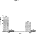
フローサイトメトリー分析によるヒト神経膠芽細胞腫(SKMG-1)細胞株および黒色腫(A375)細胞株へのMab H11の結合
腫瘍細胞へのMabH11の結合を決定するために、Tフラスコ中で増殖する腫瘍細胞を、PBS-EDTAとのインキュベーションにより剥離させた。細胞を低速度遠心分離により収集し、氷冷PBS-1%FBSで洗浄し、遠心分離し、そして上清を吸引した。以下のうちの1つでスパイクした培養培地に細胞ペレットを再懸濁し、そして氷上で30分間インキュベートした:コントロールヒト黒色腫IgM;ハイブリドーマNBGM1/H11培養上清;または精製MabH11を含むPBS。インキュベーションの後、細胞を遠心分離によって収集し、PBS-FBS中での再懸濁によって洗浄し、そして遠心分離した。次いで、細胞ペレットをFITC結合ヤギ抗ヒトIgMと30分間インキュベートした。インキュベーションの後、細胞をPBS-FBSで洗浄した。最後に、細胞をヨウ化プロピジウム(PI)を添加したPBS-FBSに再懸濁し、そして細胞を洗浄した。PI陽性細胞およびFITC陽性細胞を、フローサイトメトリーによって分析した。
フローサイトメトリー分析の結果を、図2、3、および4に示す。これらの結果は、粗精製および精製形態のMab H11が、生ヒト腫瘍細胞株(神経膠芽細胞腫、黒色腫、乳房腺ガンおよび結腸腺ガンを含む)上に発現される細胞表面結合抗原に結合することを示す。
実施例3
ELISA分析による、ヒト腫瘍細胞株の凍結融解抽出物へのMabH11の結合
ヒト腫瘍抗原に特異的に結合するH11の能力を決定するために、神経膠芽細胞腫(SKMG-1)細胞、乳房腺ガン(BT-20、MB-468、およびMB-453)細胞、結腸腺ガン(SK-CO-1およびHT-29)細胞の反復した凍結および融解によって調製したヒト腫瘍細胞抽出物でELISAプレートをコートした。
コートELISAプレートを、2〜8℃で16〜18時間インキュベートした。プレートを、PBS-3%BSAで1時間室温でブロックした。次いで、プレートをPBS中のMabH11またはPBS中のコントロールIgMまたは培養培地のいずれかと、室温で2時間インキュベートした。プレートを洗浄し、そしてビオチン化抗ヒトIgMとインキュベートし、続いてビオチン化抗ヒトIgMとインキュベートし、次いでストレプトアビジン結合アルカリホスファターゼと1時間インキュベートした。洗浄後、p-ニトロフェニルリン酸基質を各プレートに添加し、そして、インキュベーションの後に、プレートをELISAプレートリーダーにおいて405nmで読んだ。
腫瘍細胞抽出物へのMab H11の結合を、図5および6に示す。これらの結果は、Mab H11が、神経膠芽細胞腫細胞、乳房腺ガン細胞、および結腸腺ガン細胞から調製された腫瘍細胞抽出物に、用量依存様式において結合することを示す。
実施例4
免疫ペルオキシダーゼ染色によって決定したヒト腫瘍細胞へのMab H11の結合
H11の免疫反応性を決定するために、以下の実験を行った。腫瘍細胞を、24ウェルプレートにおいて、カバーガラス上で48〜96時間増殖させた。細胞をPBSで洗浄し、ホルムアルデヒドで固定し、そしてPBS中の5%正常ヤギ血清と30分間インキュベートした。洗浄後、細胞をハイブリドーマNBGM1/H11培養上清、またはPBS中の精製MabH11(10μg/mL)、またはコントロールヒト骨髄腫IgM(10μg/mL)でスパイクした培養培地のいずれかと、2時間インキュベートした。次いで、細胞を洗浄し、そしてHRPに結合した抗ヒトIgMとインキュベートした。最後に、細胞を洗浄し、MabH11結合を可視化するためにDAB基質とインキュベートし、ヘマトキシリンで対比染色し、そしてGVAにおいてマウントした。
Mab H11の免疫反応性の結果を、表3に示す。ここで、反応性を陰性(--)、弱い陽性(+)、陽性(++)、強い陽性(+++)として示す。これらの結果は、免疫ペルオキシダーゼ染色によって決定されたように、MabH11によって認識されるエピトープが多数の異なる型のヒト腫瘍細胞および細胞株によって発現されることを示す。
【表3】
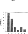
細胞固定ELISAによって決定したヒト腫瘍細胞株へのMabH11の結合
ヒト腫瘍細胞および細胞株へのH11の結合もまた、細胞固定ELISAによって決定した。EDTA-PBSとのインキュベートによって、増殖腫瘍細胞をTフラスコ表面から剥離させた。細胞を遠心分離によって収集し、PBSで洗浄し、培養培地に再懸濁し、計数し、そして5,000〜10,000細胞を含む50μlの細胞懸濁液を、96ウェルELISAプレートの各ウェルにおいた。細胞をプレートに接着させた後に、培養上清を除去し、そしてプレートをPBS-BSAでブロックした。次いで、細胞を異なる濃度(1〜20μl/mL)のMabH11またはコントロールヒト骨髄腫IgMのいずれかと2時間インキュベートした。インキュベーションの後、プレートを洗浄し、ビオチン結合ヤギ抗ヒトIgMとインキュベートし、再び洗浄し、そしてストレプトアビジン結合アルカリホスファターゼとインキュベートした。最後に、プレートを洗浄し、p-ニトロフェニルリン酸基質とインキュベートし、そしてプレートをELISAプレートリーダーにおいて405nmで読んだ。
Mab H11のヒト腫瘍細胞株への反応性の細胞固定ELISAによる結果を、表4および図7に示す。表4において、反応性を試験するためにコントロールIgM10μg/mLおよびH11 10μg/mLを使用した。そして値を405nmでの吸光度±標準偏差として示す。これらの結果は、1)Mab H11は1μg/mLの低濃度でも神経膠芽細胞腫細胞(SKMG-1)と強度に反応し、一方20μg/mLでのコントロールIgMはSKMG-1細胞と反応しない:および2)MabH11は多数の腫瘍細胞株(乳房腺ガン、結腸腺ガン、悪性骨髄腫、神経芽細胞腫、神経膠芽細胞腫、肺腺ガン、小細胞肺ガン、および前立腺腺ガン)上に存在する腫瘍抗原を認識することを示す。Mab反応性の程度は、ガンおよび腫瘍細胞株の両方で異なる。ガンの型および腫瘍細胞についてのMabH11の反応性は、コントロールIgMの反応性よりも3〜10倍高かった。
【表4】
H11の免疫解剖学的分布および免疫病理学的分析
微小解剖学的詳細についてのH11特異性ならびに組織および腫瘍における不均一性を決定するために、免疫組織化学を使用した。この技術の制限は、研究している分子の低レベルの発現による擬陰性結果の可能性、ならびに類似のエピトープまたは他の抗原によって共有されるエピトープへの抗体結合による擬陽性結果(交差反応性)の可能性を含む。これらの制限にとりくむために、非特異的結合を示さなかった最も高い抗体の濃度で本研究を行った。これは、異なる組織における全てのレベルの交差反応性の検出を可能にした。また、固定分析は、抗原染色強度と形態学的保存との最良の組合せを確立した。この実施例は、IMPATHInc.,New Yorkから得られた、H11の細胞特異性および抗原発現を研究するために維持された、正常組織および腫瘍組織のクリオスタット切断凍結切片の選択されたパネル上の結果を提示する。この研究では、間接的な免疫ペルオキシダーゼ技術を使用した。
組織学的に正常なヒト組織を、外科試料および部検試料から得た。これらの新鮮な組織を、クリオモールド中でOCT(Miles Laboratories,Inc.,Naperville,IL)に包埋し、液体窒素で冷却したイソペンタン中で瞬間凍結した。IMPATHの凍結組織バンクからの組織を5ミクロンに切断し、ポリ-L-リジンコートスライド上におき、風乾させ、そして-70℃で保存した。
濡れた氷上におかれ、そして2〜8℃で保存した非ビオチン化H11を、200μg/mLの濃度で、3.0mLの総容量で供給した。ヒト骨髄腫IgM(Pierceカタログ番号31146)(これもNovopharmから供給された)を、陰性コントロールとして使用した。両方の抗体を、抗体H11の力価測定分析によって予測されたのと同じ作用濃度まで、リン酸緩衝化生理食塩水に希釈した。ペルオキシダーゼ標識2次抗体は、PBSに500:1に希釈したヤギ抗ヒトIgM(AmericanQualex,San Clemente,CA,ロット番号A112PN)であった。
免疫ペルオキシダーゼ技術:間接的免疫ペルオキシダーゼ法を用いて、免疫組織化学的研究を行った。クリオスタット切断切片を-70℃冷凍庫から取り出し、風乾し、そして固定プロトコル(固定の詳細は、以下に提供する)に従って固定した。組織切片をPBSに希釈した5%正常ヤギ血清で10分間ブロックし、次いで1次抗体と4℃で一晩インキュベートした。スライドをPBSで洗浄し、続いて0.5%Tween/PBS溶液、次いでPBSで再度洗浄した。内因性ペルオキシダーゼ活性を、30分間の3%過酸化水素/メタノールインキュベーションでブロックし、続いてPBSで3回洗浄した。次いで、切片を、ヤギ抗ヒトIgM(ペルオキシダーゼ標識)2次抗体で15分間室温にてインキュベートし、そして上記のようにPBSで洗浄した。
ペルオキシダーゼ反応を、組織切片を3,3-ジアミノベンジジン-テトラヒドロクロライド(DAB)(Sigma Chemical Co.,St.Louis,MO)と2〜5分間インキュベートすることによって可視化した。組織切片をよく洗浄し、改変Harrisヘマトキシリン(FisherScientific,Fairlawn,NJ)で対比染色し、段階的アルコールで脱水し、キシレンで清澄化し、そしてカバーガラスをかけた。陰性コントロール抗体で高レベルのバックグラウンド染色を示す組織には、さらに徹底的な洗浄を繰り返した。IMPATHによって供給されたヒト乳ガン(F95-036)は、H11の陽性コントロールであった。陰性コントロールでは、1次試験抗体を精製ヒト骨髄腫IgMに交換した。
固定分析の目的は、抗原染色強度と形態学的保存との最適な組合せを提供する条件を確立することであった。陽性コントロール組織を、固定なしを含む5つの固定プロトコルで試験した。試験した固定プロトコルは、10%中性緩衝化ホルマリン(23〜25℃)、アセトン(2〜8℃)メチル/アセトン(1:1V/V、2〜8℃)、および95%エタノール(23〜25℃)であった。この研究において、H11について10%中性緩衝化ホルマリン(NBF)が最適な結果を生じた。
固定剤として10%NBFを用いて、連続抗体希釈物(20.0μg/mL〜0.1μg/mL)を、陽性コントロールのヒト乳ガンにおいて試験した。抗体H11の10.0μg/mLの濃度が最適な結果−陰性コントロールの有意なバックグラウンド染色を伴わない最高染色強度を生じた。
得られた結果を表5および表6に示す。表5は正常組織におけるH11反応性を示し、表6はヒト腫瘍におけるH11反応性を示す。
【表5】
陽性な免疫反応性が、試験した腫瘍標本(乳房、結腸、神経膠、胃、肺(腺、扁平上皮、および小細胞)、リンパ腫、黒色腫、卵巣、および前立腺を含む)のほとんど全てにおいて観察された。反応性は、これらの標本に存在する腫瘍細胞の10%〜95%以上で観察された;染色強度は、弱い(1+)から強い(3+)の範囲であった。しかし、抗体H11は、試験した3つの肉腫全ての標本において非反応性であった。腫瘍細胞の正常対応物の全てではないがいくつかは、標本中に存在する場合、H11に反応性であった。乳ガン、胃ガンおよび前立腺ガンに存在するいくつかの正常細胞が、抗体H11に反応性であった。抗体H11に反応性であった巨大顆粒細胞は、好酸球-マスト細胞系の炎症細胞であると考えられる。
要約すると、抗体H11は、腫瘍に存在するいくつかの正常組織を除いて、正常ヒト組織には主として非反応性である。H11抗体は、試験したほとんど全ての腫瘍において発現される抗原を検出する。
実施例7
H11クローニング、発現、および免疫学的反応性
H11-scFv抗体フラグメントがガン細胞に特異的に結合する能力を決定するために、以下の実験を行った。
単鎖抗体構築物を、以下の手順によって作製した。H11のκ V領域およμ V領域の5’末端および3’末端に特異的なプライマーをApplied Biosystems DNA合成機で合成した。全てのプライマーは、クローニングのための制限エンドヌクレアーゼ部位を含んでいた。プライマー5および6はまた、(SGGGG)3リンカーをコードするさらなるヌクレオチドを含んでいた。使用したプライマーを表7に列記し、ここで、導入した制限エンドヌクレアーゼ部位に下線を付す。
【表7】
リボゾーム結合部位、OmpAシグナルペプチド配列、c-myc(9E10)検出タグおよびヒスチジンテイルを含有する発現ベクターpSJF1(図8を参照のこと)を、Bbs1およびBamH1で切断することにより調製した。モノマーおよびダイマーの構築物を、それぞれのκおよびμフラグメントをpSJF1中に連結し、そしてこれらをコンピテントなTG1E.coliに形質転換することにより組み立てた。得られたコロニーを、コロニーPCRおよび制限エンドヌクレアーゼ消化によりスクリーニングして適正な大きさのインサートを確認し、そしてジデオキシ蛍光配列決定により配列を確認した。
H11のモノマーまたはダイマーの発現プラスミドのいずれかを含有する形質転換されたTG1を、26℃にて24時間振盪し、続いて0.1μMの最終濃度にIPTGを添加した。細胞をさらに16時間インキュベートし、次いで遠心分離により収集した。H11抗体を含有する周辺質タンパク質を、スクロース緩衝液(25%スクロース、1mMEDTA、10mM Tris pH8.0)、それに続く氷冷ショック緩衝液(10mM Tris 8.0、0.5mM MgCl2)での処理により放出させた。発現を、ポリアクリルアミドゲル電気泳動およびウェスタンブロッティングにより確認した。抗体を、ニッケルチャージカラム(PharmaciaHiTrapキレーティングカラム)を用いて精製し、そして結合した抗体を漸増勾配のイミダゾールを用いて溶出した。精製抗体を、PBS/0.02%アジ化ナトリウムに対して透析し、そして0.5mg/mLまで濃縮した。
Mab H11とH11-scFvとの間の抗原性の類似性もまた、細胞固定ELISAにより決定した。A375細胞を用いてコートしたELISAプレートを、MabH11、コントロールIgM、H11-scFv、またはコントロールBGA-scFvとともにインキュベートし、続いてウサギ抗ヒトIgM抗体またはウサギ抗scFv抗体とともに適切なようにインキュベートした。検出を、ヤギ抗ウサギIgG-西洋ワサビペルオキシダーゼ、それに続く基質により行なった。図9に示す結果は、H11IgMおよびH11-scFvの両方の高親和性、ならびにコントロールIgMおよびBGA-SL-6の両方の低親和性を実証する。
ビオチン化コントロールscFvと比較してのビオチン化H11-scFvの特異性を決定するために、以下の実験を実施した。ヒト腫瘍細胞をELISAプレートに固定し、そしてビオチン化H11-scFvまたはビオチン化BGAscFv(コントロール)のいずれかとともに上記のようにインキュベートした。
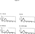
ビオチン化H11-scFvはまた、腫瘍細胞株に対してコントロールよりもずっと大きな親和性(8倍〜50倍)を細胞固定ELISAにおいて実証した。2.5μg/mLのH11-scFvまたはBGAscFvの濃度に対応するデータを、表8および図10に示す。
図11は、リンパ腫細胞Daudi、Ramos、CA-46、およびCCRF-CEM細胞への結合についてのビオチン化H11-scFvの反応性の力価測定に関連する、表8の部分を例示する。試験した全ての濃度(1.25〜10μg/mL)で、H11-scFvはリンパ腫細胞に対する高親和性を実証したが、BGAscFvは実証しなかった。
【表8】
H11-scFvを用いて、腫瘍組織および正常組織の切片を染色した。結果を、正常組織について表8に、そして腫瘍組織について表9に示す。
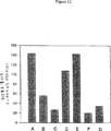
図12は、腫瘍細胞株に対するH11-scFvおよびコントロールscFvの相対蛍光強度を示す。
表9のデータは、ビオチン化H11-scFvが、一般的に正常組織と反応しないことを実証する。試験した正常組織のほぼ全ては、正常な膵臓および末梢神経組織により生じた弱い陽性シグナルのみを有して、測定可能な反応性が存在しないことを実証した。表9では、-veは測定可能な活性がないことを示し、そして+/-は弱い陽性活性を示す。
【表9】
【表11】
フローサイトメトリーにより決定した場合の生存腫瘍細胞に対するH11-scFvの反応性生存腫瘍細胞に対するH11-scFvの反応性を試験するために、腫瘍細胞株由来の細胞を、実施例2で上記したように、フローサイトメトリーのために調製した。腫瘍細胞を、ビオチン化H11-scFvまたはコントロールのビオチン化scFvのいずれかとともに、上記のように、100μg/mLまたは200μg/mLのタンパク質濃度でインキュベートした。反応性を、平均蛍光および陽性細胞の%として決定した。ビオチン化H11-scFvを、上記のように、そして100μg/mLまたは200μg/mLのいずれかのタンパク質濃度で調製した。平均蛍光および陽性細胞の%を、表12に示す。ここで、#はコントロールscFvとしてのビオチン化3B1であり;*はコントロールscFvとしてのビオチン化BGASL-6であり;**はコントロールとしてのPBS 5% FCSであり;そして***はコントロールscFvとしてのBiotin-5B1である。
【表12】
これらの結果は、精製形態のMab H11が、乳房腺ガン細胞株(SK-BR-3)、神経膠芽細胞腫細胞株(SKMG-1)、および黒色腫細胞株(A-375)、リンパ腫細胞株上に発現される細胞表面結合抗原(単数または複数)に結合することを示す。
生存リンパ腫細胞へのビオチン化H11-scFvの反応性をさらに試験するために、腫瘍細胞株由来の細胞を調製し、そしてビオチン化H11-scFvとともに100μg/mLまたは200μg/mLのいずれかのタンパク質濃度でインキュベートし、そして実施例2で上記のようにフローサイトメトリーにより分析した。平均蛍光および陽性細胞の%を、フローサイトメトリーにより測定した。scFv結合のコントロールは、ビオチン化BGAscFvであった。結果を表13に示す。
【表13】
免疫ペルオキシダーゼ染色により決定した、ヒト腫瘍細胞へのビオチン化H11-scFvの結合
H11-scFvの免疫反応性を決定するために、以下の実験を実施した。腫瘍細胞を、T型フラスコにおいて増殖させ、そして細胞遠心物(cytospin)を調製し、そしてビオチン化H11-scFvまたはPBSとともにインキュベートして結合を決定した。
H11-scFvの免疫反応性の結果を表14に示す。ここで、反応性を、陰性(--)、弱陽性(±)、陽性(+または++)として示す。これらの結果は、免疫ペルオキシダーゼ染色により決定した場合、MabH11により認識されるエピトープは多くの異なる型のヒト腫瘍細胞および細胞株により発現されることを示す。
【表14】
組換えにより産生されたH11 IgG1の反応性
H11 IgG1を、チャイニーズハムスター卵巣(CHO)細胞において、以下の通りに産生した。H11の軽鎖および重鎖の配列をコードするcDNAを含むいくつかのベクターを調製した。これらの構築物の方向、DNAインサート、および抗生物質選択基準を表15に示す。ここで、CMVはサイトメガロウイルスであり;DHFRはジヒドロ葉酸リダクターゼであり;HCは重鎖であり、そしてLCは軽鎖である。
【表15】
プラスミドはまた、ジヒドロ葉酸レダクターゼ(DHFR)をコードする遺伝子を含み、従ってチャイニーズハムスター卵巣(CHO)DHFR欠損細胞において増殖し得る。メトトレキサート(葉酸アナログおよび強力なDHFRインヒビター)を用いる増幅は、DHFR遺伝子およびその隣接配列(すなわち、構築物中の抗体の軽鎖および重鎖)の増幅をもたらす。メトトレキサート濃度(約0.01nM〜約800nM)の段階的増加は、標的遺伝子(単数または複数)から非常に高レベルのタンパク質を産生し得る。構築物はまた、選択マーカーとして抗生物質耐性を与える遺伝子(ネオマイシンまたはゼオマイシン(zeomycin)のいずれかが用いられる)を含む。ベクターを図15および図16に示す。
組換えにより産生されたH11 IgG1のフローサイトメトリー分析の結果を表16に示し、そしてSK-BR-3乳ガン細胞上で抗原に結合するH11 IgG1がCHO細胞において産生され得ることを例示する。
【表16】
ガン細胞株に対するH11結合
種々のヒトガン細胞株に対するH11 IgMおよびH11-scFvの結合親和性を、H11抗体を放射性ヨウ素または放射性インジウムのいずれかで標識することにより、決定した。125I-H11-scFvを7、20、または150μCi/μgの比活性で調製し、そして0.6μCi/μgを有する125I-H11IgMを得た。さらに、13および38μCi/μgの比活性を有する111In-H11-scFvを、実施例12に記載のように調製した。scFv3B1(これは、C抗原を認識しない)をコントロールとして用いて非特異的結合を示し、そして150μCi/μgで標識した。
125I-H11-scFvを、P-2ミニカラムを用いて精製し、そして図17に示すように、85%メタノールにおけるペーパークロマトグラフィーにより分析した。125I-H11IgMを、Sephadex G-50ミニカラム(Pharmacia)を用いて精製し、そして図18に示すように85%メタノールにおけるペーパークロマトグラフィーにより分析した。SephadexG-50カラムを用いて111In-H11-scFv精製し、これを次いで図19に示すように0.1Mクエン酸塩中のITLC-SGにより分析した。
H11結合の結果を図13および図14に示す。図13は、LS174Tヒト結腸ガン細胞に対する125I-H11-scFvの特異的結合を示す。図14は、A375細胞に対する111In-H11-scFvの全結合を示す。
得られた結果は、H11が、LT174Tおよびヒト黒色腫細胞の両方に特異的に結合することを示す。H11はまた、低い親和性でだが、乳ガン細胞株に結合する。
実施例12
111インジウム-DTPA-H11-scFvでの腫瘍画像化
H11-scFvを、ジエチレントリアミンペンタ酢酸(DTPA)の二環式(bycyclic)無水物と、10:1のモル比(DTPA:H11-scFv)で結合させ、H11-scFvの1モルあたりDTPAの2モルの置換レベルとなった。DTPA-H11-scFvを、SephadexG-25(Pharmacia)ミニカラムで過剰なDTPAから精製し、そしてCentricon-30マイクロコンセントレーター(Amicon)を用いて10mg/mLまで再濃縮した。DTPA-H11-scFvを、酢酸111インジウムで25mCi/mgの比活性に放射標識した。取り込まれなかった111Inを、SephadexG-25ミニカラムを用いて除去した。酢酸111インジウムを、塩化111インジウム(Nordion)およびpH6.0の1M酢酸緩衝液から調製した。最終111In-DTAP-H11-scFvの放射化学的純度は、100mMクエン酸ナトリウム(pH5.0)において薄層シリカゲルクロマトグラフィーにより測定した場合、99%より高かった。図19は、精製およびTLCを示す。
右側面に存在する皮下A375黒色腫異種移植片および中央腹部領域に皮下HT-29ヒト結腸ガン異種移植片を有する雌性ヌードマウスに、尾静脈中に100μCiの111In-DTAP-H11-scFvを静脈内注射した。マウスを、GEStar 4000iコンピューターに接続したガンマ線カメラ(Siemans ZL3700)の下に直ちに配置し、そして動的な捕捉を、120分間、各15秒間の計480フレームで得た。次いで、フレームを、12×10分間の画像に組み合わせた。A375腫瘍は、早くも注射後30分間でマウスの右側面で可視であった。
2つの腫瘍の目的領域分析は、A375腫瘍は120分間の研究を通して放射能を蓄積したが、一方、HT-29腫瘍は、最初の1時間に放射能を蓄積し、次いで放射能濃度は比較的一定なままであることを示した。図20は、12フレームを示し、そして120分目に撮った右側下フレームの2つの矢印は、2つの腫瘍における放射能の蓄積を示す。細い矢印はA375腫瘍を指し、そして太い矢印はHT-29腫瘍を指す。画像上で可視の正常組織は、心臓、肝臓、腎臓、および膀胱を含む。心臓は循環量の放射能に起因して可視であり、そして腎臓および膀胱は111In-DTAP-H11-scFvの腎臓での排泄に起因して可視である。少量の肝臓への取込みは、肝臓への血流または肝臓への111In-DTAP-H11-scFvの部分的結合に起因し得る。
先述の発明は、明瞭さおよび理解の目的で図および実施例によりいくらか詳細に記載してきたが、当業者には、特定の変更および改変が実施され得ることが明らかである。それゆえ、説明および実施例は、本発明の範囲を限定すると解釈されるべきではなく、本発明の範囲は、添付の請求の範囲により描写される。
【図面の簡単な説明】
【図1】図1は、一般的な抗体構造の概略図を示す。
【図2】図2は、H11によって認識される細胞のフローサイトメトリー分析を示す。
【図3】図3は、H11によって認識される細胞のフローサイトメトリー分析を示す。
【図4】図4は、H11によって認識される細胞のフローサイトメトリー分析を示す。AはA-375(黒色腫)であり、BはSKMG-1(神経膠腫)であり、CはSK-BR-3(乳房腺ガン)であり、DはHT-29(結腸腺ガン)であり、EはMB-468(乳ガン)であり、そしてFはT47D(乳ガン)である。
【図5】図5は、腫瘍細胞抽出物へのH11の結合を示す。明るい色のバーはH11を示し、そして暗い色のバーはコントロール抗体を示す。
【図6】図6は、腫瘍細胞抽出物へのH11の結合を示す。
【図7】図7は、細胞固定ELISAによるヒト腫瘍細胞株へのH11の結合を示す。明るい色のバーはH11 IgMを示し、そして暗い色のバーはコントロールIgMを示す。
【図8】図8は、発現ベクターpSJF1の概略図を示す。
【図9】図9は、細胞固定ELISAによるMab H11とH11-scFvとの間の抗原性類似性の決定を示す。反応性を、H11 scFvに対するウサギ抗体によって決定した。
【図10】図10は、ビオチン化H11-scFv(太線)およびBGAscFv(細線)のリンパ腫細胞に対する相対蛍光強度を示す。
【図11】図11は、細胞固定ELISAによって決定された場合の、リンパ腫細胞、RAMOS、Daudi、CA-46、およびCCRF-CEM細胞への結合についての、ビオチン化H11-scFvの反応性の滴定を示す。抗体濃度は、最初の10μg/mL(白抜きのバー)から5μg/mL、次いで2.5μg/mL、最後に1.25μg/mL(二重斜交平行線模様)まで減少する。
【図12】図12は、H11-scFvおよびコントロールscFv結合の腫瘍細胞株に対する相対蛍光強度を示す。AはA-375(黒色腫)であり、BはSK-BR-3(乳房腺ガン)であり、CはHT-29(結腸腺ガン)であり、DはCA-46(バーキットリンパ腫)であり、EはRAMOS(バーキットリンパ腫)であり、FはH9(T細胞リンパ腫)であり、そしてGはCCRF-CEM(急性リンパ芽球リンパ腫)である。
【図13】図13は、LS174T細胞への125I-H11-scFvの結合を示す。
【図14】図14は、A375への111In-H11-scFvの結合を示す。
【図15】図15は、組換えH11-IgGをトランスフェクトしそして発現するために使用される哺乳動物発現ベクターpNB2を示す。
【図16】図16は、組換えH11-IgGをトランスフェクトしそして発現するために使用される哺乳動物発現ベクターpNB3を示す。
【図17】図17は、P-2ミニカラムでの125I-H11-scFvの精製(A)および85%メタノールにけるペーパークロマトグラフィーによる125I-H11-scFvの分析(B)を示す。
【図18】図18は、SephadexG-25ミニカラムでの125I-H11 IgMの精製(A)および85%メタノールにおけるペーパークロマトグラフィーによる125I-H11IgMの分析(B)を示す。
【図19】図19は、SephadexG-50ミニカラムでの111In-DTPA-H11-scFvの精製(A)およびITLC-SG/0.1Mクエン酸による111In-DTPA-H11-scFvの分析(B)を示す。
【図20】図20は、ヌードマウスにおけるA375異種移植腫瘍への111In-H11-scFvのインビボ結合を示す。
(配列表)
【化1】
配列表
(1)一般的情報:
(i)出願人:ダン,マイケル ディー.
マイティ,プラディップ ケイ.
カプラン,ハーワード エー.
(ii)発明の名称:ガン細胞を特異的に検出する抗原結合フラグメントH11、このフラグメントをコードするヌクレオチド、ならびにガンの予防および検出のためのその使用
(iii)配列数:18
(iv)通信住所:
(A)名称:モリソン アンド フォレスター
(B)番地:ページ ミル ロード755
(C)市:パロ アルト
(D)州:カリファルニア
(E)国:アメリカ合衆国
(F)郵便番号:94304-1018
(v)コンピューター読み出し形態:
(A)媒体型:フロッピー ディスク
(B)コンピューター:IBM PC互換用
(C)OS:PC-DOS/MS-DOS
(D)ソフトウェア:パテントイン リリース #1.0,バージョン #1.30
(vi)現在の出願データ:
(A)出願番号:
(B)出願日:
(C)分類:
(viii)代理人/事務所情報:
(A)名称:レーンハルト,スーザン ケイ.
(B)登録番号:33,943
(C)照会/記録番号:31608-20001.20
(ix)通信情報
(A)電話:(415)813-5600
(B)テレファックス:(415)494-0792
(C)テレックス:706141
(2)配列番号1の情報:
(i)配列の特徴:
(A)長さ:543塩基対
(B)型:核酸
(C)鎖の数:二本鎖
(D)トポロジー:直鎖状
(ix)配列の特徴:
(A)特徴を表す記号:CDS
(B)存在位置:1..543
(xi)配列:配列番号1:
(i)配列の特徴:
(A)長さ:179アミノ酸
(B)型:アミノ酸
(C)鎖の数:一本鎖
(D)トポロジー:直鎖状
(xi)配列:配列番号2:
(i)配列の特徴:
(A)長さ:543塩基対
(B)型:核酸
(C)鎖の数:二本鎖
(D)トポロジー:直鎖状
(xi)配列:配列番号3:
(i)配列の特徴:
(A)長さ:450塩基対
(B)型:核酸
(C)鎖の数:二本鎖
(D)トポロジー:直鎖状
(ix)配列の特徴:
(A)特徴を表す記号:CDS
(B)存在位置:1..450
(xi)配列:配列番号4:
(i)配列の特徴:
(A)長さ:150アミノ酸
(B)型:アミノ酸
(C)鎖の数:一本鎖
(D)トポロジー:直鎖状
(xi)配列:配列番号5:
(i)配列の特徴:
(A)長さ:450塩基対
(B)型:核酸
(C)鎖の数:二本鎖
(D)トポロジー:直鎖状
(xi)配列:配列番号6:
(i)配列の特徴:
(A)長さ:34塩基対
(B)型:核酸
(C)鎖の数:一本鎖
(D)トポロジー:直鎖状
(xi)配列:配列番号7:
![]()
(i)配列の特徴:
(A)長さ:26塩基対
(B)型:核酸
(C)鎖の数:一本鎖
(D)トポロジー:直鎖状
(xi)配列:配列番号8:
![]()
(i)配列の特徴:
(A)長さ:26塩基対
(B)型:核酸
(C)鎖の数:一本鎖
(D)トポロジー:直鎖状
(xi)配列:配列番号9:
![]()
(i)配列の特徴:
(A)長さ:27塩基対
(B)型:核酸
(C)鎖の数:一本鎖
(D)トポロジー:直鎖状
(xi)配列:配列番号10:
![]()
(i)配列の特徴:
(A)長さ:60塩基対
(B)型:核酸
(C)鎖の数:一本鎖
(D)トポロジー:直鎖状
(xi)配列:配列番号11:
![]()
(i)配列の特徴:
(A)長さ:46塩基対
(B)型:核酸
(C)鎖の数:一本鎖
(D)トポロジー:直鎖状
(xi)配列:配列番号12:
![]()
(i)配列の特徴:
(A)長さ:918塩基対
(B)型:核酸
(C)鎖の数:一本鎖
(D)トポロジー:直鎖状
(ix)配列の特徴:
(A)特徴を表す記号:CDS
(B)存在位置:join(1..906,913..918)
(xi)配列:配列番号13:
(i)配列の特徴:
(A)長さ:304アミノ酸
(B)型:アミノ酸
(D)トポロジー:直鎖状
(ii)配列の種類:タンパク質
(xi)配列:配列番号14:
(i)配列の特徴:
(A)長さ:918塩基対
(B)型:核酸
(C)鎖の数:二本鎖
(D)トポロジー:直鎖状
(xi)配列:配列番号15:
(i)配列の特徴:
(A)長さ:867塩基対
(B)型:核酸
(C)鎖の数:一本鎖
(D)トポロジー:直鎖状
(ix)配列の特徴:
(A)特徴を表す記号:CDS
(B)存在位置:join(1..855,862..867)
(xi)配列:配列番号16:
(i)配列の特徴:
(A)長さ:287アミノ酸
(B)型:アミノ酸
(D)トポロジー:直鎖状
(ii)配列の種類:タンパク質
(xi)配列:配列番号17:
(i)配列の特徴:
(A)長さ:867塩基対
(B)型:核酸
(C)鎖の数:一本鎖
(D)トポロジー:直鎖状
(xi)配列:配列番号18:
The present invention relates to antibodies specific for antigens that are detected in neoplastic cells but not in normal cells. This antigen is referred to herein as “C antigen”. C antigen is recognized by a human monoclonal antibody (Mab) called “H11”. The present invention encompasses a wide variety of antibodies and functional derivatives thereof that retain the immunological specificity of H11 and are referred to herein as “αC”. Exemplary antibodies H11, compositions comprising H11, and hybridomas producing H11 are included herein. H11V region polynucleotides and polypeptides encoded thereby and recombinant molecules comprising these polynucleotides are also encompassed by the present invention. Methods of use including treatment and diagnosis of αC antibodies are also encompassed by the present invention.
Background art
Despite many advances in medical research, cancer remains the second leading cause of death in the United States. In developed countries, almost 1 in 5 people die from cancer. Traditional aspects of clinical care such as surgical excision, radiation therapy, and chemotherapy have a significant failure rate, especially for solid tumors. Failure occurs either because the initial tumor is unresponsive or due to recurrence due to regrowth at the original site and / or metastasis. Even in cancers such as breast cancer with reduced mortality, successful intervention relies on early detection of cancerous cells. Cancer etiology, diagnosis, and ablation remain a central focus of medical research and development.
Neoplasia that results in a benign tumor can usually be completely treated by surgically removing the mass. When a tumor becomes malignant, as represented by the invasion of surrounding tissue, it becomes very difficult to eradicate it. Once a malignant tumor has metastasized, it seems unlikely to be eradicated.
From the perspective of morbidity and mortality, the three main cancers are colon cancer, breast cancer, and lung cancer. New surgical procedures give increased survival to colon cancer. Improved screening methods increase the detection of breast cancer and allow for earlier and less aggressive treatment. Many studies have shown that early detection increases survival and treatment options. Lung cancer remains largely refractory to treatment.
Except for basal cell cancer, there are more than 1 million new cases of cancer per year in the United States alone, and cancer is responsible for over 500,000 deaths per year in this country. Worldwide, the five most common cancers are lung cancer, stomach cancer, breast cancer, colon / rectal cancer, and cervical cancer, and the total number of new cases per year exceeds 6 million. Only about half of those who develop cancer die of it.
Melanoma is one of the human diseases that has an urgent need for new treatment modalities. This is a particularly aggressive form of skin cancer and results in increased frequency in individuals who have received normal unprotected sun exposure. In the early stage of disease, melanoma is characterized by proliferation at the dermis-epithelial junction, which immediately invades adjacent tissues and spreads widely. Once it has metastasized, it is often impossible to eradicate and results in death. Worldwide, 70,000 patients are diagnosed with melanoma and are responsible for 25,000 reported deaths each year. The American Cancer Society estimates that by 2000, 1 in 75 Americans will be diagnosed with melanoma.
Neuroblastoma is a very malignant tumor that occurs in infancy and early childhood. With the exception of Wilms tumor, this is the most common retroperitoneal tumor in children. This tumor metastasizes early, with a wide involvement of lymph nodes, liver, bone, lung, and bone marrow. Although the primary tumor can be resolved by resection, the recurrence rate is high.
An estimated 178,100 new cases of lung cancer were diagnosed in 1997, accounting for 13% of cancer diagnoses. An estimated 160,400 deaths from lung cancer occurred in 1997, accounting for 29% of all cancer deaths. The one-year survival rate for lung cancer has increased from 32% in 1973 to 41% in 1993, mainly due to improved surgical techniques. The 5-year survival rate for all stages combined is only 14%. The survival rate for cases detected when the disease is still localized is 48%, but only 15% of lung cancers are found in its early stages.
Small cell lung cancer is the most malignant and fastest growing form of lung cancer and accounts for 20-25% of new cases of lung cancer. 60,000 cases are diagnosed in the US in 1996. Primary tumors are generally responsive to chemotherapy but are followed by wide spread metastases. The median survival time at diagnosis is about 1 year and the 5-year survival rate is 5-10%.
Breast cancer is one of the most common cancers and the third leading cause of cancer death in the United States, with an annual incidence of about 180,200 new cases among women in the United States during 1997 It is. About 1,400 new cases of breast cancer are diagnosed in men in 1997. In developed countries, about 1 in 8 women can be expected to develop breast cancer. The overall mortality rate for breast cancer remains unchanged except 1930. This has increased by an average of 0.2% per year, but for women aged under 65, it has decreased by an average of 0.3% per year. Preliminary data suggests that breast cancer mortality may begin to decrease, possibly as a result of increased in situ localized cancer and carcinoma diagnoses. See, for example, Marchant (1994) Contemporary Management of Breast Disease II: Breast Cancer, Obstetricsand Gynecology Clinics of North America 21: 555-560; and Colditz (1993) CancerSuppl. 71: 1480-1489. In 1997, an estimated 44,190 deaths (43,900 women, 290 men) were caused by breast cancer. In women, it is the second leading cause of cancer death after lung cancer. The 5-year survival rate for localized breast cancer has increased from 72% in the 1940s to 97% today. However, if the cancer has spread locally, the survival rate is 76%, and for women with distant metastases, the survival rate is 20%. Survival after diagnosis of breast cancer continues to decline beyond 5 years. 65% of women diagnosed with breast cancer survive 10 years and 56% survive 15 years.
Non-Hodgkin B-cell lymphoma is a cancer of the immune system that is predicted to afflict approximately 225,000 patients in the United States in 1996. These cancers differ in terms of prognosis and treatment and are generally classified into one of three grades. The lowest grade median survival is 6.6 years, and higher grade cancers have a much lower life expectancy. Virtually all non-Hodgkin B cell lymphomas are incurable. New diagnoses of non-Hodgkin lymphoma have increased approximately 7% annually over the past decade, with 52,700 new diagnoses estimated in 1996. This increase is partly due to the increasing prevalence of lymphoma in the AIDS patient population.
Colon cancer and rectal cancer counted an estimated 131,200 cases in 1997, including 94,100 colon cancer and 37,100 rectal cancer. Colorectal cancer accounts for about 9% of new cancer diagnoses. An estimated 54,900 deaths from colorectal cancer occurred in 1997, accounting for approximately 10% of cancer deaths. Mortality for colorectal cancer has fallen by 32% for women and 14% for men over the past 20 years, reflecting a reduced incidence and increased survival. However, mortality among African American men continues to rise. The 1-year and 5-year relative survival rates for patients with colon cancer and rectal cancer are 82% and 61%, respectively. If colorectal cancer is detected at an early localized stage, the 5-year survival rate is 91%; however, only 37% of colorectal cancer is found at that stage. After cancer has spread locally to include adjacent organs or lymph nodes, the survival rate drops to 63%. The survival rate for people with distant metastases is 7%. Survival continues to decline beyond 5 years and 50% survive for 10 years.
Despite the difficulties, effective therapies using anticancer drugs (alone or in combination with other treatments) have been devised for some previously highly fatal cancers. The most prominent of these are Hodgkin lymphoma, testicular cancer, choriocarcinoma, and some leukemias and other childhood cancers. For some of the more common cancers, early diagnosis, appropriate surgery, or local radiation therapy will allow most patients to recover.
Current methods of cancer treatment are relatively non-selective. Surgery removes affected tissue, radiation therapy shrinks solid tumors, and chemotherapy kills rapidly dividing cells. Chemotherapy, in particular, produces many side effects and in some cases is so severe that it makes it impossible to use potentially effective drugs. In addition, cancer often develops resistance to chemotherapeutic drugs.
Many efforts have been made to enhance the specificity of cancer treatment. For a review, see kohn and Liotta (1995) Cancer Res. 55: 1856-1862. In particular, the identification of cell surface antigens that are expressed exclusively or preferentially in certain tumors allows for the formulation of more selective treatment strategies. Antibodies against these antigens are used in the immunotherapy of several types of cancer.
The basic immunoglobulin (Ig) structural unit in vertebrate systems consists of two identical light (“L”) polypeptide chains (about 23 kDa) and two identical heavy (“H”) chains (about 53- 70 kDa). The four chains are linked to the “Y” configuration by a disulfide bond. At the base of Y, the two H chains are joined by a covalent disulfide bond.
FIG. 1 shows a schematic diagram of antibody challenge. The L and H chains are each composed of an N-terminal variable (V) region and a C-terminal constant (C) region. In the L chain, the V region ("V L J L )) Is the connection (J L ) Region C through region (C L ) Connected to V (V L ) Consists of areas. In the H chain, the V region (V H D H J H ) Is diversity (D H ) Region and connection (J H ) C region (C H ) Variable (V) connected to H ) Consists of areas. L chain and H chain V L J L Region and V H D H J H Each region associates at the tip of Y to form an antigen binding moiety and determines antigen binding specificity.
(C H ) Region defines the isotype, ie the class or subclass of the antibody. Different isotypes of antibodies activate complement, bind to specific receptors (eg, Fc receptors) present in a wide variety of cell types, cross mucosal and placental barriers, and basic 4-chain IgG molecules Its effector function, such as its ability to form polymers, is significantly different.
Antibodies are available on immunoglobulin molecules (IgM, IgG, IgD, IgE, or IgA). H Classified into “classes” according to type. At least five types of C H There are genes (Cμ, Cγ, Cδ, Cε, and Cα), and some species have multiple C H Subtype (eg Cγ in humans) 1 , Cγ 2 , Cγ Three , And Cγ Four ). The human haploid genome has a total of 9 C H There are genes, eight in mice and rats, and few in many other species. In contrast, normally only two types of L chain C region (C L ) (Kappa (κ) and lambda (λ)) and only one of these C regions is present in a single light chain protein (ie, any V produced) L J L There is only one possible L chain C region). Each heavy chain class may be associated with any of the light chain classes (eg, C H The gamma region may be present in the same antibody as either the kappa or lambda light chain), but the heavy and light chain C regions within a particular class are not altered by antigen specificity (eg, Regardless of antigenic properties, it always has a Cγ heavy chain C region).
Each of the V, D, J, and C regions of the H and L chains is encoded by a separate genomic sequence. Antibody diversity is due to the different V in the H chain. H , D H , And J H Gene segments and V in the light chain L And J L Produced by recombination between gene segments. Different V H , D H , And J H Genetic recombination is achieved by DNA recombination during B cell differentiation. Briefly, the heavy chain sequence is first recombined into D H J H A complex is formed, and then the second recombination event is V H D H J H Producing a complex. A functional heavy chain is produced during transcription followed by splicing of the RNA transcript. Production of functional heavy chain induces recombination in the light chain sequence and rearranges V L J L Region, which in turn is functional V L J L C L A region, ie, a functional light chain is formed.
The value and potential of antibodies as diagnostic and therapeutic reagents has long been recognized in the art. Unfortunately, this field has been hampered by the slow and tedious process required to produce large quantities of antibodies of the desired specificity. Classical cell fusion technology has enabled efficient production of Mabs by fusing antibody-producing B cells with immortalized cell lines. The resulting cell line is a hybridoma cell line.
Antibodies and functional derivatives thereof have been used in various clinical settings. For example, digoxin-specific Fab antibody fragments were used to treat life-threatening digitalis poisoning. Antibodies have become more routinely useful in diagnostic techniques such as radioimmunodiagnosis of colon cancer. Koda et al. (1995) Am. J. Gastroenterol. 90: 1644. Many uses of Mabs that were previously considered unacceptable have been implemented recently. See, for example, hall (1995) Science 279: 915-916.
Many autoantibodies (antibodies that recognize and bind to self antigens) are found in humans. Many of these include rheumatoid arthritis, systemic lupus erythematosus, myasthenia gravis, primary biliary cirrhosis, polymyositis, systemic vasculitis, idiopathic necrotizing and half-moon glomerulonephritis, and muscle atrophic Associated with certain diseases such as cord sclerosis. For a review, see Shattner (1986/1987) Immunol. Lett. 14: 143-153. Other autoantibodies are naturally occurring. Lutz and Wipp (1982) J. Immunol. 128: 1965; and Guilbert et al. (1982) J. Immunol. 128: 2779-2787. Recently, human autoantibodies against specific cancer antigens have been detected and in some cases produced by hybridoma technology. These antibodies are also produced by active immunization. US Pat. No. 5,474,755. Initially, human B cells were immortalized using Epstein-Barr virus or mouse myeloma. For a review, see Buck et al. (1984) “Monoclonal Antibodies” NY, Plenum Press. More recent technology has allowed immortalization without the use of this potentially harmful virus. See, e.g., U.S. Patent No. 4,618,477; and Glassy (1987) Cancer Res. 47: 5181-5188. In most cases, antibodies are specific for one or, in some cases, for a small number of cancer types. For example, Mabs have been described to specifically recognize glioma cells but not other tumor or normal cells. These antibodies were used to image gliomas in the patient's brain. Fischer et al. (1991) Immunobiol. Prot. Pep. VI (edited by M. Atassi), Plenum Press, NY, pages 263-270. Antibodies that can recognize a wide variety of tumors, but cannot recognize normal non-cancerous cells or only a few, have not been described.
Recombinant gene technology has allowed the cloning and expression of antibodies, functional fragments thereof, and recognized antigens. These engineered antibodies provide new production methods and treatment modalities. For example, functional immunoglobulin fragments are expressed in bacteria and transgenic tobacco seeds and plants. Skerra (1993) Curr. Opin. Immunol. 5: 256-262; Fiedler and Conrad (1995) Bio / Technology 13: 1090-1093; Zhang et al. (1993) Cancer Res. 55: 3384-3591; Ma et al. (1995) For a review of synthetic antibodies, see Science 268: 916; and Barbas (1995) Nature Med. 1: 836-839.
Several human Mabs against tumor-associated antigens have been produced and characterized. Tumor-associated antigens recognized by human Mabs include cell surface, cytoplasmic, and nuclear antigens. Yoshikawa et al. (1989) Jpn, J. Cancer Res. (Gann) 80: 546-553; Yamaguchi et al. (1987) Proc. Natl. Acad. Sci. USA 84: 2416-2420; Haspel et al. (1985) Cancer Res. 45 : 3951-3961; Cote et al. (1986) Proc. Natl. Acad. Sci. USA 83: 2959-2963; Glassy (1987) Cancer Res. 47: 5181-5188; Borup-Christensen et al. (1987) Cancer Detect. Prevent. Suppl. 1: 207-215; Haspel et al. (1985) Cancer Res. 45: 3951-3961; Kan-Mitchell et al. (1989) Cancer Res. 49: 4536-4541; Yoshikawa et al. (1986) Jpn. J. Cancer Res. 77: 1122-1133; and McKnight et al. (1990) Human Antibod. Hybridomas 1: 125-129.
Human Mabs have been used for cancer imaging, diagnosis, and therapy. Olsson (1985) J. Nat. Cancer Inst. 75: 397-404; Larrick and Bourla (1986) J. Biol. Resp. Mod. 5: 379-393; McCabe et al. (1988) Cancer Res. 48: 4348-4353 Research News (1993) Science 262: 841; Ditzel et al. (1994) Cancer 73: 858-863; and Alonso (1991) Am. J. Clin. Oncol. 4: 463-471. Recombinant single chain bispecific antibodies with high tumor cytotoxicity have been reported. This molecule recognizes both human T cell CD3 antigen and EpCAM, which is associated with scattered tumor cells in patients with minimal residual colorectal cancer. Mack et al. (1995) Proc. Natl. Acad. Sci. USA 92: 7021-7025.
Several mouse monoclonal anti-GD2 antibodies have been reported to inhibit the growth of tumors of neuroectodermal origin in athymic (nu / nu) mice or to produce remission in patients with metastatic melanoma. The human-mouse chimeric anti-GD2 antibody produced a remission in patients with metastatic neuroblastoma. The mechanism of action of antibodies is thought to include antibody-dependent cytotoxicity (ADCC) or complement-mediated cytotoxicity (CMC). Clinical responses have been obtained by treating melanoma with Mabs against GM2, GD2, and GD3. Cheresh et al. (1985) Proc. Natl. Acad. Sci. USA 82: 5155-5159. Active immunization with ganglioside vaccines containing GM2 produced anti-GM2 antibodies in 50/58 patients, and these patients survived on average longer than patients without detectable anti-GM2 antibodies.
Mabs against GD2 have also been found to react specifically with small cell lung cancer. Cheresh et al. (1986) Cancer Res. 46: 5112-5118. Human Mabs specific for other cancers, including lung cancer, melanoma, gastric cancer, squamous cancer, cervical cancer, and breast cancer, have also been produced. Murakami (1985) in Vitro Cell. Dev. Biol. 21: 593; Schadendorf (1989) J. Immunol. 142: 1621-1625; Yoshikawa et al. (1986) Jpn. J. Cancer Res. 77: 1122-1133; Misra (1984) Clin. Immunol. Immunopathol. 32: 253-260; Hagiwara and Sato (1983) Mol. Biol. Med. 1: 245-252; and Schlom et al. (1980) Proc. Natl. Acad. Sci. USA 77 : 6841-6845. Human anti-cancer Mabs and the antigens they recognize have also been suggested for use in vaccines. See, for example, Finn et al. (1995) Immunol. Rev. 145: 61-89. Human Mabs for malignant brain tumors were used in phase I clinical trials without adverse side effects. Matsumoto et al. (1994) The Clinical Report 28: 118-126. Phase II clinical trial results have been reported for combined treatment with mouse Mab and colony stimulating factor in metastatic gastrointestinal cancer. Saleh et al. (1995) Cancer Res. 55: 4339-4346. Single chain immunotoxins have also been found to treat cancerous meningitis in a rat model. Pastan et al. (1995) Proc. Natl. Acad. Sci. USA 92: 2765-2769. Human Mabs that specifically recognize ovarian cancer cells have been shown to effectively image this cancer. Chaudhuri et al. (1994) Cancer 73: 1098-1104.
If there is a simple and reliable strategy for providing immunoreactivity to common antigens in these cancers rather than cancer-specific immunity, the clinical prospect of cancer is generally improved. All references cited herein are hereby incorporated by reference in their entirety.
Disclosure of the invention
The present invention includes a composition comprising an antigen-binding fragment of an antibody, wherein the antibody comprises an H chain V region having the amino acid sequence of SEQ ID NO: 2 and an L chain V region having the amino acid sequence of SEQ ID NO: 4 It specifically recognizes the antigen recognized by the antibody. Preferably the antibody is H11. The present invention further encompasses an antibody comprising the H chain V region and L chain V region of H11 (SEQ ID NOs: 2 and 4, respectively). H11 specifically recognizes a wide variety of cancer-derived cancer cells but does not recognize normal non-cancerous cells. “Unrecognized” means that non-cancerous cells are either not specifically bound by H11 or are only slightly recognized by this antibody. The antibody is referred to as αC and “immunological specificity” of H11 and H11 (ie recognizes the antigen recognized by H11 and is specific for at least one type of cancer cell but normal Any antibody that does not recognize cells). These antigen-binding fragments include whole native antibodies exemplified by H11; bispecific antibodies; chimeric antibodies; Fab, Fab ′, single chain V region fragments (scFv), and fusion polypeptides, It is not limited to these.
The present invention further provides an H11 antibody fusion molecule (H chain C region, single chain V H -V L Or V L -V H Polypeptide regions having antigenic, therapeutic, toxic, or labeled molecules bound to the V region), and polynucleotides encoding such polypeptides.
The invention also encompasses a polypeptide having the immunological specificity of H11, wherein the polypeptide comprises at least 5 consecutive amino acids from the V region of the αC antibody. The V region can be derived from the L chain or the H chain. Five consecutive amino acids preferably play a role in immunological specificity and can be derived from the CDR (antibody complementarity determining region). Intact H11, functionally active fragments of H11, fusion proteins, chimeric antibodies, multiple antigen proteins, and other polypeptide derivatives of αC antibodies are included. Of particular interest are single chain V regions and fusion proteins.
The compounds and compositions of the present invention can be used to detect or treat cancers, particularly including the treatment of such cancers, and prophylactic care, particularly to reduce the risk of recurrence.
The invention further encompasses cells and cell lines that produce αC antigen binding fragments.
A separate embodiment of the invention is a polynucleotide comprising a sequence encoding a polypeptide having immunological specificity of H11, wherein the encoded polypeptide comprises at least 5 from the V region of H11. Contains consecutive amino acids. The V region can be derived from either the H11 L chain or the H chain. Five consecutive amino acids preferably play a role in H11 immunological reactivity and can be derived from CDRs. The V region of H11 has been found to have a small region of homology to the antibody designated A6. Peptides that contain only this homology region and no other H11-specific amino acid residues are specifically excluded from the present invention. A6 is described in WO953574.
The present invention also provides for at least 20 contiguous nucleotides that can form a stable duplex with the H11 L chain coding sequence or H chain coding sequence, but not with sequences for other previously described immunoglobulin molecules. Includes isolated polynucleotides. Any of these polynucleotides can be in the form of a cloning vector, an expression vector, or can be transfected into a host cell.
Further embodiments of the invention include prophylactic treatment of cancer patients with at least one αC antigen binding fragment. Preferably, αC is fused to the therapeutic molecule to effect delivery of the therapeutic molecule to the cancer cell. The individual can have a clinically detectable tumor, or the tumor may have been previously treated and rendered undetectable. This method may be to alleviate the disease or reduce the risk of recurrence.
A further embodiment of the present invention is a kit for the detection or quantification of an antigen recognized by αC in a sample (hereinafter referred to as “C antigen”), and the H11 of the present invention in an appropriate package Or a polypeptide. Also encompassed by the present invention are methods for detecting C antigens or cells expressing C antigens by using the reagents or kits encompassed by the present invention.
BEST MODE FOR CARRYING OUT THE INVENTION
The present invention encompasses antigen-binding fragments exemplified by newly identified human Mabs that specifically recognize cancerous cells. This specificity extends only to cancer cells, and antibodies do not recognize non-cancerous cells. An exemplary antibody is designated H11, and the variable regions are encoded by SEQ ID NOs: 1 and 4 (SEQ ID NOs: 3 and 6 are the complementary strands of 1 and 4, respectively) and are designated “C antigen”. Recognize antigen. Specificities of H11 include glioblastoma, neuroblastoma, malignant melanoma, breast adenocarcinoma, lung adenocarcinoma, small cell lung cancer, colon adenocarcinoma, and prostate adenocarcinoma. It is not limited.
As shown in the examples herein, H11 and H11-scFv do not recognize non-cancerous cells from all normal tissues tested. Therefore, H11 and αC antigen binding fragments are useful in alleviating clinical conditions associated with a wide variety of cancers. The present invention encompasses antigen-binding fragments that recognize an antigen (designated C antigen) for which H11 is specific. The present invention further encompasses polypeptide derivatives of H11 and methods for using these compositions in the diagnosis, treatment, and manufacture of novel reagents. The present invention further includes polynucleotides encoding αC, H11, and derivatives thereof. The method of use is also encompassed by the present invention.
The invention further encompasses αC derivatives having immunological specificity for the C antigen. These derivatives contain regions of the polypeptide sequence that contain part of the H11 VDJ linkage. Also encompassed are regions spanning at least 1, preferably 2, and more preferably 3 or more H11 CDR amino acid sequences.
The full length sequences of the H11 L chain C region and H chain C region have not been determined, but are predicted to be identical or nearly identical to the sequences of other human immunoglobulin molecules. Furthermore, knowledge of the V region amino acid sequence allows subcloning with any C region. Such subcloning techniques are well known in the art. Chimeric molecules produced by these cloning techniques are also encompassed by the present invention.
Screening a commercially available heptapeptide phage library with H11 IgM clones and scFV antibody clones shows a very strong consensus sequence at the N-terminus with the following amino acid sequence: Phe-His-Arg-Tyr-Ser / Thr. The results are shown in Table 1.
[Table 1]
[Table 2]
The particular compounds, compositions, and methods described in this application generally relate to αC and its derivatives that are routinely produced by classical techniques of immunochemistry. This includes αC bound to another compound by chemical bonding or by mixing with excipients or adjuvants. The term antigen-binding fragment includes any peptide that binds to C antigen in a manner specific to cancer cells. Typically, these derivatives are Fab, F (ab ′) 2 , Fab ′, scFv (both monomeric and polymeric forms), and immunoglobulin fragments such as isolated heavy and light chains. Antigen-binding fragments retain the specificity of H11, but avidity and / or affinity can be altered. H11-scFv described herein is particularly preferred.
Antigen-binding fragments (also referred to herein as “derivatives”) are typically generated by genetic engineering, but these can alternatively be obtained by other methods and combinations of methods. This classification includes, but is not limited to, engineered peptide fragments and fusion proteins. Preferred compounds include H11 CDR polypeptide fragments, antibody fusion proteins comprising cytokine effector components, antibody fusion proteins comprising adjuvants or drugs, and single chain V region proteins.
The present invention further includes polynucleotides encoding the H11 antibody V region and derivatives thereof. These include isolated polynucleotide fragments, recombinant polynucleotides, and therapeutic plasmids and vectors (eg, vaccinia vectors) containing the polynucleotides. These polynucleotides are exemplified by SEQ ID NOs: 1, 3, 4, 6, 13, 15, 16, and 18.
The pharmaceutical compositions and treatment modalities of the present invention are suitable for inducing an immune response against a neoplasm. Particularly suitable for patients with human cancer, including but not limited to glioblastoma, melanoma, neuroblastoma, adenocarcinoma, glioma, soft tissue sarcoma, and various cancers (including small cell lung cancer) Subject.
Since H11 has been shown to specifically recognize various cancers, H11 is particularly useful for cancer diagnosis, imaging, and treatment. Suitable cancers include any cancer known in the field of oncology, including astrocytoma, oligodendroglioma, ventricular ependymoma, medulloblastoma, primitive neuroectodermal tumor (PNET), pancreatic ductal adenocarcinoma, small cell lung adenocarcinoma and large cell lung adenocarcinoma, squamous cell carcinoma, bronchoalveolar carcinoma, epithelial adenocarcinoma, and their liver metastases, liver cancer, bile duct cancer, ductal adenocarcinoma and Breast cancer such as lobular adenocarcinoma, squamous and adenocarcinoma of the cervix, uterine and ovarian epithelial cancer, prostate adenocarcinoma, transitional squamous cell carcinoma of the bladder, B cell lymphoma and T cell lymphoma, (nodular and Including, but not limited to, diffuse) plasmacytoma, acute and chronic leukemia, malignant melanoma, soft tissue sarcoma, and leiomyosarcoma.
A subject may have an advanced form of disease, in which case the treatment objective may include mitigation or reversal of disease progression and improvement of side effects. A subject may have a history of a condition that has already been treated, in which case the objective typically includes a reduction or delay in the risk of recurrence.
Furthermore, the antigen-binding fragments of the present invention can be used as diagnostic and imaging reagents. These applications are described in further detail in the following sections.
"H11" is derived from a fusion of peripheral blood lymphocytes of a 64-year-old male with low grade glioma, fused to a human myeloma cell line to produce a hybridoma designated NBGM1 / H11 It is an antibody. The generation and characterization of H11 is described in Example 1. “ΑC” refers to any antibody, or antigen-binding fragment thereof, either monoclonal, polyclonal, or a derivative thereof that specifically recognizes C antigen and distinguishes between cancer and non-cancerous cells. αC includes H11.
The “immunological activity” of αC refers to the ability to specifically bind C antigen. Such binding may or may not elicit an immune response. A specific immune response can include antibodies, B cells, T cells, and any combination thereof, and effector functions resulting therefrom. Antibody-mediated functions ADCC and complement-mediated cell lysis (CDC) are included. T cell responses include T helper cell function, cytotoxic T cell function, inflammation / inducer T cell function, and T cell mediated suppression. A compound that can elicit a specific immune response according to any of these criteria is said to be "immunogenic".
αC “activity” or αC “function” refers to any immunological activity of αC, or any other biological activity attributed to H11 in this disclosure, to detect, ameliorate, or reduce cancer. Including the role of H11 in
The “V region” of H11 refers to either the V region of the H11 L chain or the V region of the H11 H chain, either alone or in combination. These V regions are shown in SEQ ID NOs: 2 and 5; the DNA encoding these regions is shown in SEQ ID NOs: 1 and 4, respectively.
GM-CSF, IL-2, and other biologically active molecules referred to herein include fragments and derivatives based on the respective parent molecule having the same biological or physiological function Means.
The terms “polypeptide”, “peptide”, and “protein” are used interchangeably herein to refer to polymers of amino acid residues of any length. The polymer can be linear or branched, can contain modified amino acids or amino acid analogs, and can be interrupted by chemical moieties other than amino acids. These terms also encompass amino acid polymers that have been modified naturally or by intervention; for example, disulfide bond formation, glycosylation, lipidation, acetylation, phosphorylation, or labeling or attachment to a biologically active component. Any other manipulation or modification. Unless otherwise stated or implied, the term αC or H11 includes any polypeptide monomer or polypeptide polymer having H11 immunological specificity, including intact αC antibodies, and smaller and larger functionally equivalents. A polypeptide.
A “fusion polypeptide” is a polypeptide that contains regions at positions that differ in sequence from those that occur in nature. This region can be normally present in separate proteins and brought together in the fusion polypeptide; they can normally be present in the same protein but are placed in a new configuration in the fusion polypeptide; Or they can be placed synthetically. For example, as described below, the present invention encompasses a recombinant protein (and a polynucleotide encoding this protein) composed of a functional portion of αC and a toxin. Methods for producing these fusion proteins are known in the art and are described, for example, in WO93 / 07286.
A “functionally equivalent fragment” of an αC polypeptide differs from the native sequence by any combination of additions, deletions, or substitutions, but at least one functional property of the fragment with respect to the context in which it is used. Is maintained. A functionally equivalent fragment of an αC polynucleotide encodes a polypeptide that is functionally equivalent to H11 when produced by an expression system or is similar to an H11 polynucleotide when used in a hybridization assay. Having any hybridization specificity. A functionally equivalent fragment of an αC polypeptide typically has one or more of the following properties: the ability to bind C antigen; the ability to bind at least one type of cancer cell in a specific manner; And the ability to elicit an immune response with antigenic specificity similar to that elicited by H11.
A “polynucleotide” is a polymeric form of nucleotides of any length, including deoxyribonucleotides, ribonucleotides, and analogs in any combination analog. A polynucleotide can have any three-dimensional structure and can perform any function known or unknown. The term “polynucleotide” includes double-stranded, single-stranded, and triple-helical molecules. Unless otherwise specified or required, any embodiment of the invention described herein that is a polynucleotide includes a double-stranded form and any double-stranded form of a DNA, RNA, or hybrid molecule. Includes both each of the two complementary single-stranded forms known or predicted to make up.
The following are non-limiting examples of polynucleotides: genes or gene fragments, exons, introns, mRNA, tRNA, rRNA, ribozymes, cDNA, recombinant polynucleotides, branched polynucleotides, plasmids, vectors, any Isolated DNA of sequences, isolated RNA of any sequence, nucleic acid probes, and primers. A polynucleotide may comprise modified nucleotides (eg, methylated nucleotides and nucleotide analogs), uracil, other sugars and linking groups (eg, fluororibose and thioate), and nucleotide branches. The sequence of nucleotides can be interrupted by non-nucleotide components. A polynucleotide can be further modified after polymerization, such as by conjugation with a labeling component. Other types of modifications included in this definition include caps, substitution of one or more naturally occurring nucleotides with analogs, and polynucleotides on proteins, metal ions, labeling components, other polynucleotides, or solid supports. Introduction of means for adhering.
The term “recombinant” polynucleotide means a polynucleotide of genomic, cDNA, semi-synthetic, or synthetic origin, either non-naturally occurring or linked to another polynucleotide in a non-natural configuration. .
“Vector” refers to a recombinant DNA or RNA plasmid or virus containing a heterologous polynucleotide that is transmitted to a target cell, either in vitro or in vivo. The heterologous polynucleotide can contain the sequence of interest for therapeutic purposes and can optionally be in the form of an expression cassette. As used herein, a vector need not be able to replicate in the final target cell or subject. The term includes cloning vectors for polynucleotide replication and expression vectors for translation of the sequences encoded by the polynucleotides. Also included are viral vectors comprising a polynucleotide encapsidated or enveloped in a viral particle.
“Cell line” or “cell culture” refers to bacteria, plants, insects, or higher eukaryotic cells grown or maintained in vitro. The progeny of the cell may not be completely identical to the parent cell (either morphologically, genotyped or phenotyped). Mabs can be produced by hybridomas or other cells. Methods for producing both mouse and human hybridomas are known in the art. Specific methods for producing human hybridomas are described and referenced throughout the specification.
A “host cell” has been genetically altered. Or a prokaryotic or eukaryotic cell that can be genetically altered by administration of an exogenous polynucleotide, such as a recombinant plasmid or a recombinant vector. When referring to a genetically altered cell, the term refers to both the original altered cell and its progeny.
“Heterologous” means derived from an entity whose genotype differs from the rest of the entity being compared. For example, the polynucleotide can be genetically engineered into a plasmid or vector derived from a different source and is a heterologous polynucleotide. A promoter that is removed from its native coding sequence type and operably linked to a coding sequence other than the native sequence is a heterologous promoter.
A “signal peptide” or “leader sequence” allows a newly synthesized protein to pass through the cell membrane, usually the endoplasmic reticulum of a eukaryotic cell, and either the bacterial inner membrane or both the inner and outer membranes. The short amino acid sequence indicated in The signal peptide is typically at the N-terminal portion of the polypeptide and is typically enzymatically removed from the cell during polypeptide biosynthesis and secretion. The signal peptide is not present in the secreted protein only during protein production.
An “isolated” polynucleotide or polypeptide is one that is substantially free of materials related to the native environment. Substantially free means that at least 50%, preferably at least 70%, more preferably at least 80%, and even more preferably at least 90% of these materials are free.
A “stable duplex” of a polynucleotide, or “stable complex” formed between any two or more components in a biochemical reaction, refers to the formation and subsequent detection of a duplex or complex ( Any duplex or complex that lasts long enough to last between any optional washing steps or other operations that may occur during that time.
“Biological samples” refers to various sample types, including liquid samples of blood and other biological sources, solid tissue samples such as biopsy specimens or tissue cultures, or cells derived therefrom and their progeny. Include. This definition also encompasses samples that have been manipulated in any way after their acquisition, such as by treatment with reagents, lysis, or enrichment of certain components (eg, proteins or polynucleotides). The term encompasses a variety of clinical samples obtained from any species, and also includes cells, cell supernatants, and cell lysates in culture. In particular, for the purposes described herein, a biological sample includes tumor tissue or tissue that is considered to be neoplastic and, for example, surgical resection, biopsy, aspiration, or in the art It can be obtained by any known method.
“Immunogen” refers to a composition for human or animal use, which is administered at a strength that provides the recipient with some degree of specific immunological reactivity to a particular antigen. Immunological reactivity refers to antibodies or cells that are immunologically reactive to the target (particularly B cells, plasma cells, T helper cells, and cytotoxic T lymphocytes, and their precursors). Or any combination thereof. For purposes of the present invention, the target is primarily tumor-associated C antigen or a tumor-specific portion thereof. Immunological reactivity may be desired for experimental purposes, for the treatment of certain symptoms, for the elimination of certain substances, or for prevention. An active immunogen is intended to elicit an immune response that persists in the absence of vaccine components.
As used herein, “adjuvant” has several meanings, all of which are apparent depending on the context in which the term is used. In the case of pharmaceutical preparations, an adjuvant is a chemical or biological agent provided in combination with an antigen or recombinantly fused to the antigen to enhance the immunogenicity of the antigen. In the case of cancer diagnosis or treatment, an adjuvant refers to a class of cancer patients who are clinically unable to detect a tumor mass but are suspected of having a recurrence risk.
When referring to a type of cancer that normally appears as a solid tumor, a “clinically detectable” tumor can be detected based on the tumor mass, ie, by procedures such as CAT scans, x-rays, or palpation. A tumor. Biochemical, histological, or immunological findings alone may not be sufficient to meet this definition.
As used herein, “treatment” refers to clinical mediation in an attempt to alter the natural course of the individual or cell being treated, and for prevention or during the course of clinical pathology. Can be done either. The desired effects of treatment include suppressing the onset or recurrence of the disease, mitigating symptoms, reducing any direct or indirect pathological consequences of the disease, preventing metastasis, and reducing the rate of disease progression Treatment, alleviation or reduction of disease symptoms, and sedation or improved prognosis.
A “medical condition” associated with a disease state is any condition that impairs the health, normal physiology, or quality of life of the affected individual. This includes destructive invasion of the affected tissue to an unaffected area, proliferation at the expense of normal tissue function, irregular or suppressed biological activity, inflammatory response or immunological response Undesirable clinical signs such as exacerbation or suppression of the disease, increased susceptibility to other pathogenic organisms or pathogens, and other features such as pain, fever, nausea, fatigue, mood changes, and other characteristics that may be determined by the attending physician Including, but not limited to.
An “effective amount” is an amount sufficient to produce a beneficial or desired clinical result. An effective amount can be administered in one or more doses. With respect to treatment, an effective amount is an amount sufficient to alleviate, reduce, stabilize, reverse, or delay disease progression or otherwise reduce the pathological consequences of the disease. For an adjuvant, an effective amount is an amount sufficient to enhance the immune response to the immunogen. Effective amounts are generally determined by the physician on a case-by-case basis and are within the skill of the art. Typically, several factors are considered when determining the appropriate dose. These factors include the age, sex, and weight of the patient, the condition being treated, the severity of the condition, and the form of antibody being administered. For example, the concentration of scFv need not be as high as the concentration of native antibody in order to be therapeutically effective.
An “individual”, “patient” or “subject” is a vertebrate, preferably a mammal, more preferably a human. Mammals include, but are not limited to, humans, farm animals, sport animals, and pets.
The practice of the present invention uses conventional techniques of molecular biology (including recombinant techniques), microbiology, cell biology, biochemistry, and immunology, unless otherwise indicated, and these are within the skill of the art. Is within. Such techniques are described in “Molecular Cloning: A Laboratory Manual”, 2nd edition (Sambrook et al., 1989); “Oligonucleotide Synthesis” (MJGait, 1984); “Animal Cell Culture” (RIFreshney, 1987); “Methodsin Enzymology” (Academic Press, Inc.); “Handbook of Experimental Immunology” (DMWei and CC Blackwell); “Gene Transfer Vectors for Mammalian Cells” (JMMiller and MPCalos, 1987); “Curent Protocols in Molecular Biology” (FMAusubel et al., 1987); “PCR: The Polymerase Chain Reaction” (Mullis et al., 1994); “Current Protocols in Immunology” (JEColigan et al., 1991). Techniques that are applicable to the production of the polynucleotides and polypeptides of the invention, and can be considered, for example, in making and practicing the invention, are discussed in the following sections, which are particularly useful for particular embodiments.
The invention also includes αC bound to a chemical functional moiety. Typically, this moiety is a label that can produce a detectable signal. These bound αCs are useful, for example, in detection systems such as tumor burden quantification, as well as metastatic lesion imaging and tumor imaging. Such labels are known in the art and include, but are not limited to, radioisotopes, enzymes, fluorescent compounds, chemiluminescent compounds, bioluminescent compound substrate cofactors and inhibitors. See US Pat. Nos. 3,817,837; 3,850,752; 3,939,350; 3,996,345; 4,277,437; 4,275,149; and 4,366,241 for examples of patents that teach the use of such labels. This moiety can be covalently linked to αC, recombinantly linked, or linked to αC via a second reagent such as a second antibody, protein A, or biotin-avidin complex.
Other functional moieties include signal peptides, factors that enhance immunological reactivity, factors that facilitate coupling to a solid support, vaccine carriers, biological response modifiers, paramagnetic labels, and drugs. It is done. Signal peptides are described above and include prokaryotic and eukaryotic forms. Agents that enhance immunological reactivity include, but are not limited to, bacterial superantigens. Agents that facilitate coupling to a solid support include, but are not limited to, biotin or avidin. An immunogenic carrier includes, but is not limited to, any physiologically acceptable buffer. Biological response modifiers include cytokines, particularly tumor necrosis factor (TNF), interleukin 2,
Suitable drug moieties include antitumor agents. These include radioisotopes, vinca alkaloids (eg, bin plastin sulfate, pink listin sulfate, and vindesine sulfate), adriamycin, bleomycin sulfate, carboplatin, cisplatin, cyclophosphamide, cytarabine, dacarbazine, dactinomycin, hydrochloric acid Duanorubicin, doxorubicin hydrochloride, etoposide, fluorouracil, lomustine, mechlororethamine hydrochloride, melphalan, mercaptopurine, methotrexate, mitomycin, mitoten, pentostatin, pipobromin, procarbazine, procarbazine, streptozotocin Examples include, but are not limited to, uracil mustard.
Immunotoxins containing single chain molecules can be produced by recombinant means. The production of various immunotoxins is well known in the art, and methods are described, for example, in “Monoclonal Antibody-toxin Conjugates: Aiming the Magic Bullet.” Thorpe et al. (1982) Monoclonal Antibodies in Clinical Medicine, Academic Press, 168-190. Pages; Vitatta (1987) Science 238: 1098-1104; and Winter and Milstein (1991) Nature 349: 293-299. Suitable toxins include fungal toxins such as ricin, radionuclide, pokeweed antiviral protein, Pseudomonas exotoxin A, diphtheria toxin, ricin A chain, restorictocin and phospholipase enzymes. It is not limited to. See generally, “ChimericToxins” Olsnes and Pihl, Pharmac. Ther. 15: 355-381 (1981); and “Monoclonal Antibodies for Cancer Detection and Therapy” edited by Baldwin and Byers, pages 159-179, 224-266, Academic. See Press (1985).
Chemically functional moieties can be made recombinantly, for example, by generating fusion genes encoding antigen binding fragments and functional regions from other genes (eg, enzymes). In the case of a gene fusion, the two components are present in the same polypeptide gene. Alternatively, the αC antigen binding fragment can be chemically conjugated to the moiety by any of a variety of well-known chemical procedures. For example, if the moiety is a protein, the linkage can be by a heterobifunctional crosslinker (eg, SPDP, carbodiimide glutaraldehyde, etc.). This moiety can be covalently linked or coupled via a second reagent such as a second antibody, protein A, or biotin-avidin complex. Paramagnetic moieties and their binding to antibodies are well known in the art. See, for example, Miltenyi et al. (1990) Cytometry 11: 231-238.
The αC antibodies of the invention can be prepared in several ways. This is most conveniently obtained from cells engineered to express an antigen binding fragment comprising SEQ ID NOs: 1 and 5 or other polynucleotides encoding an αC binding fragment. For example, the cells can be cultured in a suitable medium and the medium used can be used as an antibody source. If desired, matrix-coated channels or beads and cell co-cultures can be included to enhance the growth of antibody producing cells. For the production of large amounts of antibodies, it is generally more convenient to obtain ascites. Ascites production methods generally involve injecting hybridoma cells into an immunologically pure histocompatibility or immune resistant mammal, particularly a mouse. Mammals can be primed for ascites production by prior administration of a suitable composition (eg, Pristine).
Alternatively, αC can be chemically synthesized using sequence data and other information provided in this disclosure, along with standard methods of protein synthesis. A suitable method is the solid phase Merrifield technique. Automated peptide synthesizers are commercially available, for example, synthesizers manufactured by Applied Biosystems, Inc. (Foster City, Calif.).
αC can also be obtained using routine recombination methods such as those described in Sambrook et al. (1989). For example, using the amino acid and polynucleotide (SEQ ID NO: 1-6, and 13-18) sequences and the information provided herein, a polynucleotide encoding either the H or L chain of αC can be expressed as: It can be cloned into an appropriate expression vector, which contains control sequences for transcription, such as a promoter. The expression vector is then introduced into the host cell. The host cell is grown under appropriate conditions such that the polynucleotide is transcribed and translated into a protein. The αC heavy and light chains can be produced separately and then combined by a disulfide bond regeneration sequence. Alternatively, a vector with separate polynucleotides encoding each strand of αC, or a vector with a single polynucleotide encoding both strands as separate transcripts can be transfected into a single host cell. The whole molecule can then be produced and associated. Preferably, the host cell is derived from a higher eukaryote that can provide the normal carbohydrate complement of the molecule. Thus, the produced αC can be purified using standard techniques in the art. Next, a polynucleotide encoding αC for use in the production of αC can be obtained from a hybridoma that produces an αC antibody, or produced synthetically or recombinantly from the DNA sequences provided herein. Can be done.
Another way to obtain αC is to immunize appropriate animals with C antigen and follow standard procedures for polyclonal or Mab production. Thus, the produced Mab can be “humanized” by methods known in the art. Examples of humanized antibodies are provided, for example, in US Pat. Nos. 5,530,101 and 5,585,089.
A “humanized” antibody is an antibody in which at least a portion of the sequence has been altered from its original form to be more similar to human immunoglobulin. In one type, the H and L chain C regions are replaced with human sequences. This is a fusion polypeptide comprising an H11 V region and a heterologous immunoglobulin C region. In other types, the CDR regions contain H11 amino acid sequences, but the V framework regions are also converted human sequences. For example, see EP0329400. In the third type, the V region is humanized by designing human and mouse V region consensus sequences and converting residues outside the CDRs that differ between the consensus sequences. The present invention encompasses humanized Mabs.
In making humanized antibodies, the choice of framework residues can be important to retain high binding affinity. Theoretically, framework sequences from any HuAb can be used as a template for CDR grafting; however, direct CDR substitution into such a framework results in a significant loss of binding affinity for the antigen. Can lead to. Glaser et al. (1992) J. Immunol. 149: 2606; Tempest et al. (1992) Biotechnology 9: 266; and Shalaby et al. (1992) J. Exp. Med. 17: 217. The more similar a HuAb is to the original muAb, the less likely the human framework will introduce distortion to the mouse CDR that may reduce affinity. Based on sequence homology searches against antibody sequence databases, HuAbIC4 also provides good framework homology to muM4TS.22, while other highly homologous HuAbs are specifically κ light chains from human subgroup I Or more suitable for heavy chains from human subgroup III. Kabat et al. (1987). Various computer programs such as ENCAD (Levitt et al. (1983) J. Mol. Biol. 168: 595) are available to predict the ideal sequence for the V region. Accordingly, the present invention encompasses HuAbs having various V regions. It is within the purview of those skilled in the art to determine appropriate V region sequences and to optimize these sequences. Methods for obtaining antibodies with reduced immunogenicity are also described in US Pat. No. 5,270,202 and EP 699,755.
Antibody production and isolation methods are well known in the art. See, for example, Harlow and Lane (1988) Antibodies: A Laboratory Manual, Cold Spring Harbor Laboratory, New York. The H11 antibody is an IgM subclass of human immunoglobulin and can be isolated by any technique appropriate for this isotype of immunoglobulin. Purification methods include salt precipitation (eg, using ammonium sulfate), ion exchange chromatography (eg, using a cation or anion exchange column, run at neutral pH, and eluted with a step gradient of increasing ionic strength. ), Gel filtration chromatography (including gel filtration HPLC), and chromatography on affinity resins such as protein A, protein G, hydroxyapatite, and anti-immunoglobulin. H11 can also be purified on an affinity column containing C antigen, for example, in the form of purified Ab1 or Ab3. Preferably, H11 is protein A-CL-Sepharose TM 4B chromatography followed by DEAE-Sepharose TM Purified using chromatography on a 4B ion exchange column.
The invention also encompasses hybrid antibodies, wherein one pair of heavy and light chains is obtained from a first antibody, while the other pair of heavy and light chains is obtained from a different second antibody. It is done. For the purposes of the present invention, one pair of light and heavy chains is from αC. In one embodiment, each L-H chain pair binds a different epitope of the C antigen. Such hybrids can also be formed using humanized heavy or light chains.
Another αC encompassed by the present invention is an antibody in which the heavy or light chain has been modified to provide additional properties. For example, changes in the amino acid sequence can result in an immunogenic phenomenon of the resulting polypeptide. This change ranges from changing one or more amino acids to complete redesign of a region such as the C region domain. Exemplary changes include, but are not limited to, changes associated with complement binding reactions, interactions with membrane receptors, and other effector functions. Recombinant antibodies can also be designed to assist in the specific delivery of substances (eg, cytokines) to tumor cells. Peptides in which the various immunoglobulin domains are arranged in an order other than that naturally occurring are also encompassed by the invention.
If αC is administered to an individual, it is preferably at least 80% pure, more preferably at least 90% pure, even more preferably at least 95% pure and free of pyrogens and other contaminants. . In this situation, the percent purity is calculated as a weight percent of the total protein content of the preparation and does not include components that are deliberately added to the composition after the αC has been purified.
αC antibodies can be used for a number of purposes. These include inducing an antibody response and treating various types of cancer to produce an ααC that can then be used to induce a T cell response to αC or C antigen. Their use is more fully detailed in a later section.
The present invention encompasses αC polypeptide fragments that comprise at least a portion of the αC V region. Preferred fragments are those that have H11 immunological activity. Also preferred are fragments comprising an amino acid sequence substantially different from other immunoglobulins, and fragments comprising a CDR. In one embodiment, the invention encompasses a polypeptide fragment of the αCH chain V region, wherein at least 25 consecutive amino acids of SEQ ID NO: 2, more preferably 30 consecutive amino acids, or 5 consecutive of its CDR1 Amino acids, or at least 7 consecutive amino acids thereof, preferably at least 9 consecutive amino acids of CDR2 or CDR3 thereof. The invention also encompasses polypeptide fragments of the αC light chain V region, wherein at least 25 consecutive amino acids of SEQ ID NO: 5, more preferably 30 consecutive amino acids, or 7 consecutive amino acids of CDR2 thereof, or It comprises at least 8 consecutive amino acids, preferably 10 consecutive amino acids, of its CDR1 or CDR3.
The size of the αC polypeptide can be only the minimum size necessary to provide the desired function. Polypeptides can include additional sequences, either specific for αC or from a heterologous source, if desired. The αC peptide may contain only 5 consecutive amino acids from the V11 sequence of H11 that is not the same as the homologous region of A6. 7 amino acids from the αC light chain V region or αC H chain V region, more preferably about 10 amino acids, more preferably about 15 amino acids, more preferably about 25 amino acids, more preferably about 50 amino acids, more preferably about 75 amino acids. Polypeptides containing amino acids are also encompassed. Even more preferred are polypeptides comprising the entire αC L chain V region or αC H chain V region. Preferably the polypeptide is derived from H11. Preferably, the polypeptide is the scFv shown in SEQ ID NO: 14 and SEQ ID NO: 17.
The invention encompasses modified αC polypeptides that are functionally equivalent to H11 or altered but have measurable H11 immunological activity. Modified polypeptides with improved H11 immunological activity are preferred. Examples of modified polypeptides include peptides having conservative substitutions of amino acid residues and one or more deletions or additions of amino acids that do not significantly deleteriously alter immunological activity.
One example of this is an H11 polypeptide that contains one or more amino acid substitutions compared to a prototype H11 sequence. Substitutions can range from changing or modifying one or more amino acid residues to complete redesign of a region such as the V region. Amino acid substitutions, if present, are preferably conservative substitutions that do not adversely affect peptide folding or functional properties. The group of functionally related amino acids for which conservative substitutions can be made are glycine / alanine; valine / isoleucine / leucine: asparagine / glutamine; aspartic acid / glutamic acid; serine / threonine / methionine; lysine / arginine; and phenylalanine / tyrosine / Tryptophan. The polypeptides of the invention can be in glycosylated or non-glycosylated form, can be post-translationally modified (eg, acetylated, and phosphorylated), or can be modified by synthesis (eg, the addition of a labeling group).
H11 polypeptide derivatives comprising both the H11 L chain and the H11 H chain can be formed as separate L and H chains and then associated or in situ by expression systems for both chains. Such expression systems are generated by transfecting appropriate cells with plasmids containing separate transcribable regions for the light and heavy chains, or by co-transfecting the same cells with plasmids for each chain. Can be done. In the third method, an appropriate plasmid with the heavy chain coding region is transfected into a mutant without the heavy chain.
A variant without heavy chain is approximately 2 × 10 6 with rabbit anti-mouse IgG (H chain specific, DAKO Corporation, Carpinteria, Calif.) Fluorescently labeled according to the supplier's instructions. 7 Of H11 producing cells. Stained and unstained cell populations are analyzed with a fluorescence activated cell sorter. Unstained cells are collected in sterile tubes and plated into 96-well plates at 1 cell / well by limiting dilution. The culture supernatant is then assayed by ELISA using goat anti-mouse IgG (H chain specific) and goat anti-mouse κ. Subcloning κ positive and IgG negative phenotype clones at least 3 times to generate stable H11 (-H) Get a mutant. Putative heavy chainless mutant H11 (-H) MRNA from the clone can be isolated and the sequence of the L chain V region cDNA can be determined. H11V H About mRNA reverse PCR, H11 (-H) This is done with two sets of 5'- and 3'-primers used for cDNA cloning (Example 7). Mutants without heavy chains do not yield detectable DNA bands. Transfection of cells is performed with an appropriate heavy chain plasmid.
Another αC derivative encompassed by the present invention is an antibody in which the αC H or L chain has been modified to provide additional properties. For example, changes in amino acid sequence can result in greater immunogenicity of the resulting polypeptide. This change ranges from changing one or more amino acids to complete redesign of a region such as the C region domain. Intended changes affect complement binding reactions, interactions with membrane receptors, and other effector functions. Recombinant H11 antibodies can also be designed to assist in the specific transmission of substances (eg, lymphokines) to effector cells. Proteins in which the various immunoglobulin domains are arranged in an order other than naturally occurring are also encompassed by the present invention.
The invention also encompasses single chain V region fragments of H11 (“scFv”). Single chain V region fragments are made by linking L and / or H chain V regions by using a short linking peptide. Bird et al. (1988) Science 242: 423-426. Any peptide with sufficient flexibility and length can be used as a linker in scFv. Usually, the linker is selected so that there is little to no immunogenicity. An example of a linking peptide is (GGGGS) Three Which bridges about 3.5 nm between the carboxy terminus of one V region and the amino terminus of the other V region. Other linker sequences can also be used and provide additional functions such as means for attaching drugs or solid supports.
All or any part of the H or L chain can be used in any combination. Typically, the entire V region is included in the scFv, for example, the L region and the V region can be linked to the H chain V region. Alternatively, a part of the L chain V region can be linked to the H chain V region or a part thereof. Also contemplated are scFvs in which the heavy chain V region is derived from H11 and the light chain V region is derived from other immunoglobulins. It is also possible to construct a biphasic scFv where one component is an H11 polypeptide and the other is a different polypeptide such as a T cell epitope.
scFv is, for example, V H -(Linker) -V L Or V L -(Linker) -V H Can be met in any order. For example, SEQ ID NOs: 13 and 16 are forms V L -(Linker) -V H The H11-scFv2 construct with is shown. However, the construct shown in SEQ ID NO: 13 forms a monomer, while the construct shown in SEQ ID NO: 16 forms a dimer. There may be differences in the expression levels of these two forms in a particular expression system, in which case one of these forms may be preferred. Tandem scFvs can also be made as (X)-(linker)-(X)-(linker)-(X), where X is an αC polypeptide, or an αC polypeptide and other polypeptides. It is a combination. In other embodiments, the single chain antibody polypeptide has no linker or only a short inflexible linker. An exemplary configuration includes V L -V H And V H -V L Is included. This linkage is the V in the chain. L And V H Is too short to allow interaction between and the chain is V at each end L / V H Forms a homodimer with the antigen binding site. Such molecules are referred to in the art as “diabodies”.
scFvs can be produced either recombinantly or synthetically. For synthetic production of scFv, an automated synthesizer can be used. For recombinant production of scFv, an appropriate plasmid containing a polynucleotide encoding scFv can be either a eukaryote such as a yeast cell, plant cell, insect cell or mammalian cell, or a prokaryote such as Escherichiacoli. Proteins that can be introduced into any suitable host cell and expressed by the polynucleotide can be isolated using standard protein purification techniques.
A particularly useful system for the production of scFv is the plasmid pET-22b (+) (Novagen, Madison, WI) in E. coli. pET-22b (+) contains a nickel ion binding domain consisting of 6 consecutive histidine residues, which allows purification of the expressed protein with an appropriate affinity resin. Another example of a suitable vector is pcDNA3 (Invitrogen, San Diego, Calif.) As described above.
Expression conditions should ensure that the scFv takes a functional and preferably optimal three-dimensional structure. Depending on the plasmid used (particularly the activity of the promoter) and the host cell, it may be necessary to control the rate of production. For example, the use of weaker promoters, or expression at lower temperatures may be necessary to optimize the production of appropriately folded scFv in prokaryotic systems, or scFv in eukaryotic cells. It may be preferred to express.
A preferred scFv comprises at least 10 consecutive amino acids of SEQ ID NO: 2 and at least 10 consecutive amino acids of SEQ ID NO: 5, in particular the amino acids of SEQ ID NO: 2 and SEQ ID NO: 5 are 5-20 amino acid linker polypeptides Or includes the L chain V region and the H chain V region of H11.
The invention also encompasses polymeric forms of αC polypeptides, including a plurality of αC polypeptides. One embodiment is a linear polymer of αC polypeptide optionally attached to a carrier. These linear polymers may contain multiple copies of a single αC polypeptide, or a combination of different αC polypeptides, and have αC polypeptides separated by tandem αC polypeptides, or other amino acid sequences. obtain. Another embodiment is an αC multiple antigen peptide (MAP). MAP has a small immunologically inactive core with radially branched lysine dendrites, where many αC polypeptides are covalently linked. See, for example, Posnett et al. (1988) J. Biol. Chem. 263: 1719-1725; and Tam (1989) Meth. Enz. 168: 7-15. The result is a large macromolecule with a high molar ratio of αC polypeptide to core. MAP is an effective immunogen and an antigen useful for immunoassays. The core for generating αCMAP can be made by standard peptide synthesis techniques or can be obtained commercially, eg, from Quality Controlled Biochemicals, Inc., Hopkinton, MA. A typical core matrix is made up of three levels of lysine and eight amino acids.
When an αC polypeptide is used as an immunogen, preferably the polypeptide is delivered with a carrier. Any carrier that is not harmful to the host may be used. Suitable carriers are typically proteins; polysaccharides (eg, latex functionalized Sepharose, agarose, cellulose, cellulose beads, etc.); polymeric amino acids (eg, polyglutamic acid, polylysine, etc.); amino acid copolymers; and inactive viruses Large, slowly metabolized macromolecules such as particles or attenuated bacteria (eg, Salmonella). Particularly useful carrier proteins are serum albumin, keyhole limpet hemocyanin (KLH), certain Ig molecules, thyroglobulin, ovalbumin, and tetanus Soid It is. KLH is particularly preferred.
The αC polypeptides of the present invention can be identified in a number of ways. For example, the V region of the light and heavy chains can be screened by preparing a series of short polypeptides that span the entire V region amino acid sequence. Using a series of 20 or 50 amino acid long polypeptides, each αC V region can be investigated for useful functional properties. It is also possible to perform computer analysis of the protein sequence to identify potentially interesting polypeptides having the shape of D2 or associated with idiotype-anti-idiotype contacts.
The invention further encompasses various indications of αC as described in this section, combined in various ways to obtain other αC polypeptides having the desired properties. For example, an αC polypeptide having an altered amino acid residue can be included in a MAP. In other embodiments, αC scFv is fused to a cytokine such as IL-2. All such combinations are encompassed by the present invention.
The polypeptides of the invention can be made by any suitable procedure, including proteolysis of αC antibodies, by recombinant methods, or by chemical synthesis. These methods are known in the art and need not be described in detail herein. Examples of proteolytic enzymes include, but are not limited to, trypsin, chymotrypsin, pepsin, papain, V8 protease, subtilisin, plasmin, and thrombin. Intact αC can be incubated simultaneously or sequentially with one or more proteinases. Alternatively, or in addition, the intact antibody can be treated with a disulfide reducing agent. The peptides can then be separated from each other by techniques known in the art, including but not limited to gel filtration chromatography, gel electrophoresis, and reverse phase HPLC.
αC polypeptides can also be made by expression from a polynucleotide encoding a peptide in an appropriate expression system according to the information provided elsewhere herein. Typically, a polynucleotide encoding an αC polypeptide is linked to an expression vector under the control of a suitable promoter and can be used to genetically alter a given host cell. Both eukaryotic and prokaryotic host systems can be used. The polypeptide is then isolated from lysed cells or from culture medium and purified to the extent necessary for a given use. An example of a prokaryotic host cell suitable for use in the present invention includes E. coli. Examples of eukaryotic host cells include avian cells, insect cells, plant cells, and animal cells, and animal cells include, but are not limited to, COS7, HeLa, and CHO cells.
In certain applications, such as when the H11 polypeptide is expressed in a suitable storage medium such as plant seed, the H11 polypeptide can be used without purification. Fiedler et al. (1995) Biotechnology 13: 1090-1093. For most applications, it is generally preferred that the polypeptide be at least partially purified from other cellular components. Preferably, the polypeptide is at least about 50% pure as a weight percent of the total protein. More preferably, the protein is at least about 50-75% pure. For clinical use, the polypeptide is preferably at least about 80% pure.
The present invention also includes a method for detecting C antigen in a biological sample. The method includes the steps of obtaining a biological sample, contacting the sample with αC under conditions that allow the antibody to bind antigen, and detecting binding of the antibody, if any, to the antigen.
The invention also encompasses a method of detecting anti-H11 or anti-αC in a biological sample. Anti-αC is detectable when it cross-reacts with H11. Anti-αC with this activity can occur naturally during the course of paraneoplastic diseases. Anti-αC with this activity is particularly likely in individuals undergoing a course of treatment with αC. These methods are applicable, for example, in a clinical environment to monitor antibody levels in an individual, as well as in an industrial environment, such as in the commercial production of anti-H11 or anti-αC.
The assay method contacts any anti-H11 or anti-αC target antibody in the sample with the H11 antibody or polypeptide under conditions suitable to allow the formation of a stable complex between the target and H11. And a step of detecting any stable complexes formed. Samples are prepared appropriately prior to performing the assay, if necessary, by enriching the antibody concentration. When using intact mouse αC, it is generally preferred to deplete any sample of anti-mouse immunoglobulin activity that may be present. The anti-mouse immunoglobulin antibody can be removed from the sample, for example, by precipitation with normal mouse IgG or adsorption with a mouse Ig adsorbent. Binding of anti-mouse immunoglobulin antibodies, particularly antibodies specific for the Fc region, can be minimized by judicious selection of assay reagents. F (ab ') of mouse αC 2 Or other reagents with mouse antigenic determinants such as Fab fragments and humanized αC or H11 are suitable.
After the sample is properly prepared, the sample is mixed with excess αC under conditions that allow formation of a complex of αC with any target antibody that may be present. The amount of complex is then determined and compared to the complex formed with a standard sample containing a known amount of target antibody in the expected range. Complex formation can be observed by any method known in the art, such as immunoprecipitation or turbidimetry, but generally radioisotopes (eg, 125 It is more sensitive to use reagents labeled with labels such as I), enzymes (eg, peroxidase and β-galactosidase), or fluorescent dyes (eg, fluorescein).
The present invention provides various polynucleotides that encode antibody H11 or a fragment of H11 based on the polynucleotide sequences provided herein (SEQ ID NOs: 1 and 4). Various embodiments are described in this section and include many different combinations of H11 H chain V region sequences or H11 L chain V region sequences. In general, the H11 polynucleotides of the present invention encode at least one feature that is unique to the H11 molecule (as compared to other immunoglobulins and particularly the A6 antibody). Preferably, this feature is in some way related to the immunological reactivity of H11.
The present invention encompasses a polynucleotide that encodes a portion of the H11 L chain V region and comprises at least about 70 contiguous nucleotides of SEQ ID NO: 4, preferably at least about 80 contiguous nucleotides, more preferably at least about 100 contiguous nucleotides. It comprises consecutive nucleotides, even more preferably at least about 150 nucleotides. The invention also encompasses a polynucleotide that encodes a portion of the H11 L chain V region, wherein at least about 25 contiguous nucleotides of the sequence encoding CDR1, preferably at least about 30 contiguous nucleotides, and even more preferably Contains at least about 35 contiguous nucleotides. The invention also encompasses a polynucleotide that encodes a portion of the H11 L chain V region, at least about 20 contiguous nucleotides of the sequence encoding CDR2 or CDR3, preferably at least about 25 contiguous nucleotides, and further More preferably it comprises at least about 35 consecutive nucleotides.
The present invention also encompasses a polynucleotide that encodes a portion of the H11 H chain V region, comprising at least about 70 contiguous nucleotides of SEQ ID NO: 1, preferably at least about 80 contiguous nucleotides, more preferably at least about 100. Of contiguous nucleotides, and even more preferably at least about 150 contiguous nucleotides. The invention also encompasses a polynucleotide that encodes a portion of the H11 L chain V region and includes 15 contiguous nucleotides of the sequence encoding CDR1. The invention also encompasses a polynucleotide that encodes a portion of the H11 L chain V region, wherein the CDR2 or CDR3 encoding sequence is at least about 20 contiguous nucleotides, preferably at least about 25 contiguous nucleotides, and Even more preferably, it comprises at least about 35 contiguous nucleotides.
The present invention includes an isolated H11 polynucleotide encoding a polypeptide having immunological activity of H11, wherein the polypeptide comprises at least 5 of the VL chain of H11 as shown in SEQ ID NO: 5. Encodes amino acids. The present invention also encompasses an isolated H11 polynucleotide encoding a polypeptide having immunological activity of H11, wherein the polypeptide comprises at least a VH chain of H11 as shown in SEQ ID NO: 2.
The invention also encompasses polynucleotides that encode functionally equivalent variants and derivatives of H11 and functionally equivalent fragments thereof. This can enhance, decrease or not significantly affect the properties of the polypeptide encoded thereby. These functionally equivalent variants, derivatives, and fragments display the ability to specifically recognize C antigen. For example, changes in the DNA sequence that do not change the encoded amino acid sequence, as well as conservative substitutions of amino acid residues, one or a few amino acid deletions or additions, and substitutions of amino acid residues by amino acid analogs are: A change that does not significantly affect the properties of the encoded polypeptide. Conservative amino acid substitutions are glycine / alanine; valine / isoleucine / leucine; asparagine / glutamine; aspartic acid / glutamic acid: serine / threonine / methionine; lysine / arginine; and phenylalanine / tyrosine / tryptophan.
The polynucleotides of the invention may comprise additional coding sequences, control elements (eg, promoters, ribosome binding sites, and polyadenylation sites) within the same transcription unit, additional transcription units under the control of the same or different promoters, cloning, expression, and Additional sequences may be included, such as sequences that allow transformation of the host cell, as well as any such constructs as may be desirable to provide embodiments of the invention.
The present invention forms stable hybrids with polynucleotides encoding H11 L chain or H chain V regions, but does not form other immunoglobulin-coding regions known at the time of filing of the present invention, at least about 15 Of consecutive nucleotides, preferably at least about 20 nucleotides, more preferably at least about 25 consecutive nucleotides, more preferably at least about 35 consecutive nucleotides, more preferably at least about 50 consecutive nucleotides, even more preferably at least It includes a polynucleotide of about 75 nucleotides, even more preferably at least about 100 nucleotides, even more preferably about 200 nucleotides, and even more preferably at least about 300 nucleotides. As long as there is at least one set of conditions, any set of conditions can be used for this test, where the test polynucleotide demonstrates the required specificity. Preferably, the H11 coding sequence to which the test polynucleotide binds is the sequence shown in SEQ ID NOs: 1 and 4. Since known immunoglobulin sequences are divided into a classification phase similar to that of H11, the test is performed by comparing the hybridization of the test polynucleotide with the H11 sequence to the hybridization with the most closely related sequence. Can be done. A panel against about 10 of the most closely related sequences SEQ ID NO: 1, 3, 4, or 5 is preferred.
Hybridization reactions can be performed under conditions of different “stringency”. Conditions that increase the stringency of a hybridization reaction are well known. See, for example, Sambrook and Maniatis. Examples of relevant conditions include (in order of increasing stringency) incubation temperatures of 25 ° C., 37 ° C., 50 ° C., and 68 ° C .; 10 × SSC, 6 × SSC, 1 × SSC, 0.1 × SSC (here SSC is 0.15 M NaCl and 15 mM citrate buffer) and its equivalents using other buffer systems; 0%, 25%, 50%, and 75% formaldehyde concentrations; from 5 minutes to 24
Useful H11 polynucleotides that encode fragments of H11 include generating polynucleotide fragments (eg, based on SEQ ID NO: 1 or SEQ ID NO: 4), and testing the polynucleotide encoded thereby for the function of interest. Can be identified. Alternatively, polypeptide fragments encoded by a particular polypeptide can be prepared and tested for the function of interest. Alternatively, given an αC polypeptide having the desired properties, a polynucleotide encoding this polypeptide can be designed.
Polynucleotides having coding regions for H11 polymers, fusion proteins, humanized immunoglobulins, single chain V regions, and other specific polypeptides of interest are encompassed in all these embodiments. These polypeptides are described above.
The invention also provides a polynucleotide covalently linked to a detectable label. Such polynucleotides are useful, for example, as probes for the detection of related nucleotide sequences.
The polynucleotides of the present invention can be obtained using chemical synthesis methods, recombinant cloning methods, PCR, or any combination thereof. Chemical polynucleotide synthesis methods are well known in the art and need not be described in detail herein. One skilled in the art can use the sequence data provided herein to obtain the desired polynucleotide by using a DNA synthesizer or by ordering from commercial services.
Alternatively, the αC polynucleotide sequence can be obtained from an αC antibody producing cell line, an αC cloning vector, or an αC expression vector. RNA or DNA encoding the desired sequence can be isolated, amplified, and processed by standard recombinant techniques. Such techniques include restriction nuclease cleavage and polymerase chain reaction (PCR) amplification, or a suitable combination thereof. PCR techniques are described in US Pat. Nos. 4,683,195, 4,800,159, 4,754,065 and 4,683,202, and PCR: The Polymerase Chain Reaction, edited by Mullis et al., Birkauswer Press, Boston (1994).
A polynucleotide containing the desired sequence can be inserted into a suitable vector, and the vector can in turn be introduced into a suitable host cell for replication and amplification. The polynucleotide can be introduced into the host cell by any means known in the art. Cells are transformed by introducing exogenous polynucleotides by direct uptake, endocytosis, transfection, f-mating, or electroporation. Once introduced, the exogenous polynucleotide can be maintained within the cell as a non-integrating vector (eg, a plasmid) or can be integrated into the host cell genome. Amplified DNA can be isolated from the host cell by standard methods. See, for example, Sambrook et al. (1989). RNA can also be obtained from transformed host cells or can be obtained directly from DNA by using a DNA-dependent RNA polymerase.
The invention further includes various vectors comprising H11 polynucleotides. These vectors can be used for expression of recombinant polypeptides that are also a source of H11 polynucleotides. Cloning vectors can be used to obtain duplicate copies of the H11 polynucleotides they contain or as a means of storing the polynucleotides in a repository for future recovery. Expression vectors (and host cells containing these expression vectors) can be used to obtain polypeptides produced from the polynucleotides they contain. They can also be used when it is desired to have intact cells that express H11 in an individual and can therefore synthesize polypeptides as in gene therapy. Suitable cloning and expression vectors include those known in the art, such as vectors for use in bacterial, mammalian, yeast, and insect expression systems. Specific vectors and suitable host cells are known in the art and are not described in detail herein. See, for example, Gacesa and Ramji, Vectors, John Wiley & Sons (1994).
Cloning and expression vectors typically contain a selectable marker (eg, a gene that encodes a protein necessary for the survival or growth of a host cell transformed with the vector), but such marker genes include: It can be present in other polynucleotide sequences introduced simultaneously into the host cell. Only host cells into which a selectable gene has been introduced will grow under selective conditions. Representative selection genes are (a) conferring resistance to antibiotics or other toxins such as ampicillin, neomycin, methotrexate; (b) supplement auxotrophic deficits; or (c) utilized from complex media One of the important nutrients that is not possible. The selection of the appropriate marker gene depends on the host cell, and appropriate genes for various hosts are known in the art. Vectors also typically include a replication system that is recognized by the host.
Suitable cloning vectors can be constructed according to standard techniques, or can be selected from a number of cloning vectors available in the art. Although the selected cloning vector may vary according to the host cell intended to be used, useful cloning vectors generally have the ability to self-replicate and are single-stranded against a particular restriction endonuclease. It can have a target or it can have a marker gene. Suitable examples include plasmids and bacterial viruses such as pUC18, mp18, mp19, pBR322, pMB9, ColE1, pCR1, RP4, phage DNA, and shuttle vectors (eg, pSA3 and pAT28). These and other cloning vectors are available from commercial suppliers such as BioRad, Stratagen, and Invitrogen.
An expression vector is generally a replicable polynucleotide construct comprising a polynucleotide that encodes the αC polypeptide of interest. The polynucleotide encoding the αC polypeptide is operably linked to suitable transcription control elements such as promoters, enhancers, and terminators. For expression (ie, translation), one or more translation control elements such as a ribosome binding site, a translation initiation site, and a stop codon are also usually required. These regulatory elements (transcription and translation) can be derived from the H11 gene, or a heterologous gene (ie, from other genes or other organisms). A polynucleotide sequence encoding a signal peptide may also be included to allow the αC polypeptide to cross or be embedded in the cell membrane or secreted from the cell. Many expression vectors suitable for expression in yeast cells, avian cells, and eukaryotic cells, including mammals, are known in the art. One example of an expression vector is pcDNA3 (Invitrogen, San Diego, Calif.), Where transcription is driven by the cytomegalovirus (CMV) early promoter / enhancer. This vector also contains recognition sites for multiple restriction enzymes for insertion of the αC polynucleotide of interest. Another example of an expression vector (system) is the baculovirus / insect system.
Expression systems suitable for use in antibody targeted gene therapy comprising αC polynucleotides are also encompassed by the present invention. Suitable expression systems are described, for example, in Brown et al. (1994) Virol. 198: 477-488; and Miyamura et al. (1994) Proc. Natl. Acad. Sci. USA 91: 8507-8511.
Vectors containing the polynucleotide of interest are electroporation; transfection with calcium chloride, rubidium chloride, calcium phosphate, DEAE-dextran, or other substances; microprojectile bombardment; lipofection; and infection (where the vector is An infectious agent such as vaccinia virus, which is described below) and can be introduced into the host cell by any of a number of suitable means. The choice of vector or αC polynucleotide to be introduced often depends on the characteristics of the host cell.
Once introduced into a suitable host cell, expression of the αC polypeptide can be determined using any assay known in the art. For example, the presence of αC polypeptide can be detected by RIA or ELISA of the culture supernatant (if the H11 polypeptide is secreted) or cell lysate.
Particularly useful expression vectors for H11 polynucleotides are vaccinia viruses composed of H11 polynucleotide sequences, which can also be used in vaccine preparations. Moss (1991) Science 252: 1662-1667. In order to introduce a polynucleotide sequence encoding an H11 polypeptide, including an H11 polypeptide fragment, into a vaccinia, the polynucleotide sequence of interest is first vaccinia virus promoter with a flanking sequence homologous to vaccinia DNA that is not required for replication. Is inserted into a plasmid containing The plasmid-containing cells are then infected with vaccinia, which leads to a low level of homologous recombination between the plasmid and the virus, transfer of the polynucleotide encoding the vaccinia promoter and H11 polypeptide into the vaccinia virus genome. As a result. Typically, the H11 polynucleotide sequence is inserted into the viral TK (Thymidine Kinase) gene. Insertion at the TK site attenuates the virus more than 10,000 times compared to the wild type. Flexner et al. (1980) Vaccine 88 (Cold Spring Harbor Laboratory), 179-184. Recombinant virus is TK - Identified by phenotype. Preferably, the expression of the H11 polynucleotide is under the control of a vaccinia early / late promoter (7.5K), whereby the resulting H11 polypeptide can be expressed in infected cells throughout the viral life cycle. However, other promoters known in the art such as pH 6 or synthetic promoters can be used. Expression of the H11 polypeptide occurs in cells infected with recombinant vaccinia or individuals immunized with live recombinant vaccinia virus. Any one of several strains of vaccinia may be used including but not limited to WR, ALVAC, NYVAC.
The vectors of the present invention can include one or more polynucleotides encoding an αC polypeptide. This also enhances, promotes, or regulates desired products such as lymphokines (including but not limited to IL-2, IL-4, GM-CSF, TNF-α, and IFN-γ) Polynucleotide sequences encoding the polypeptides of can also be included. A preferred lymphokine is GM-CSF. Preferred GM-CSF constructs are those in which the AU-rich element from the 3 ′ untranslated region and the sequence of the 5 ′ untranslated region capable of forming a hairpin loop are deleted. Vaccinia vectors encoding recombinant αC variants such as scFv, chimeras, and polymers are also encompassed by the present invention.
Another embodiment of the invention is a host cell transformed with an αC polynucleotide, or a vector comprising an αC polynucleotide sequence, as described above. Both prokaryotic and eukaryotic host cells can be used. Prokaryotic hosts include bacterial cells such as E. coli and Mycobacteria. Among eukaryotic hosts are yeast cells, insect cells, avian cells, plant cells, and mammalian cells. Host systems are known in the art and need not be described in detail herein. Examples of mammalian host cells include CHO and NSO, which are available from the European Collection of Cell Cultures (England). For example, transfection with plasmids of NSO cells, driven by amplification of this plasmid using a CMV promoter and then glutamine synthetase, provides a useful system for protein production. Cockett et al. (1990) Bio / Technology 8: 662-667.
The host cells of the invention can be used in particular as reservoirs for αC polynucleotides or as vehicles for the production of αC polynucleotides and polypeptides. They can also be used as in vivo vectors for αC polypeptides. The H11 polynucleotides of the invention can be used in expression systems to produce H11 polypeptides, untreated H11, or recombinant forms of H11, as described below.
The polynucleotides of the present invention have several uses. These are useful, for example, in expression systems for the production of αC. They are also useful as hybridization probes for assaying for the presence of αC polynucleotides or related sequences in a sample using methods well known in the art. In addition, the polynucleotide is also useful as a primer to effect amplification of the desired polynucleotide. The polynucleotides of the invention are also useful in pharmaceutical compositions including vaccines and for gene therapy.
Polynucleotides can also be used as hybridization probes for the detection of αC coding sequences. Suitable samples include cells that have been transformed ex vivo for use in gene therapy. In one example, DNA or RNA is extracted from the sample and optionally run on a gel and / or cleaved with a restriction endonuclease. Processed sample polynucleotides are typically transferred to a medium suitable for washing. The sample polynucleotide is then contacted with an H11 polynucleotide probe under conditions that allow a stable duplex to be formed if the sample contains a compatible αC sequence. Any stable duplex formed is detected by any suitable means. For example, the αC polynucleotide probe can be provided in a labeled form, and the label remaining in the sample after washing directly reflects the amount of stable duplex formed. In the second example, hybridization is performed in situ. A properly prepared tissue sample is overlaid on a labeled probe to indicate the location of the αC coding sequence.
Short αC polynucleotides can also be used as primers for PCR reactions, particularly to amplify longer sequences that include regions that hybridize with primers. This can be done preparatively to produce a polynucleotide for further genetic manipulation. This can also be done analytically to determine whether an αC-encoding polynucleotide is present, for example, in a sample for diagnostic purposes.
Another use for polynucleotides is in vaccines and gene therapy. The general principle is to administer a polypeptide so as to promote or attenuate expression of the polypeptide encoded therein. Accordingly, the present invention encompasses methods of eliciting an immune response and methods of treatment comprising administering to an individual an effective amount of an αC polynucleotide. In these methods, an αC polynucleotide encoding an αC polypeptide is administered to an individual either directly or by cells transfected with the αC polynucleotide. Preferably, the αC polynucleotide is in the form of a circular plasmid, preferably in supercoil configuration. Preferably, the αC polynucleotide is replicated inside the cell. Thus, the αC polynucleotide is operably linked to a suitable promoter, such as a heterologous promoter that is inherently active in labeled tissue type cells. Preferably, once in the nucleus, the plasmid persists as a circular non-replicating episomal molecule. In vitro mutations can be made, for example, in plasmid constructs to encode molecules with greater affinity and / or avidity.
To determine whether a plasmid containing an αC polynucleotide can produce αC expression in a eukaryotic cell, cells such as COS-7, CHO, or HeLa can be transfected with the plasmid. ΑC expression is then determined by immunoassay, eg, by Western blot. Smaller αC polypeptides can be detected, for example, by constructing a plasmid, and the resulting αC polypeptide is fused to a tag such as a target epitope or enzyme label. Further characterization of the expressed αC polypeptide can be achieved by purifying the peptide and then performing one of the functional assays described herein.
In one aspect of gene therapy, the polypeptides of the invention are used to genetically alter cells ex vivo. In this strategy, cells removed from a donor or obtained from a cell line are transfected or transduced with a vector encoding an αC polypeptide and then administered to a recipient. Suitable cells for transfection include peripheral blood mononuclear cells.
In other aspects of gene therapy, the polynucleotides of the invention are used to genetically alter cells in vivo. This purpose includes, but is not limited to, treating various types of cancer.
αC polypeptides can be characterized in several ways. For example, an αC polypeptide can be tested for its ability to specifically bind to cancer cells and its ability to specifically inhibit binding between cancer cells and intact H11. An αC polypeptide can also react with an anti-CDR3 polypeptide. αC polypeptides can also be tested for their ability to alleviate or ameliorate neoplastic diseases such as cancer. It is understood that only one of these properties needs to be in order for the polypeptide to be within the scope of the invention, but preferably more than one of these properties are present. .
The ability of an αC polypeptide to bind to cancer cells or antigenic fractions thereof can be tested by immunoassay. Any form of direct binding assay is suitable. In one such assay, cancer cells or putative αC polypeptides are labeled. Suitable signs include 125 Radioisotopes such as I, enzymes such as peroxidase, fluorescent labels such as fluorescein, and chemiluminescent labels. Typically, other binding partners are insolubilized (eg, by coating on a microtiter plate) to facilitate washing. After combining the labeling component with the insolubilizing component, the solid phase is washed and the amount of bound label is measured. Another such assay is a sandwich assay, where a putative αC polypeptide is captured by primary antiimmunoglobulin on a solid phase and developed with an αC antibody. In any of these examples, the extent of αC binding is directly proportional to the amount of label bound to the solid phase.
To perform an inhibition assay, the putative αC polypeptide is titrated for its ability to reduce H11 binding to cancer cells. Either of the binding pairs in the reaction to be inhibited is labeled while the other is typically insolubilized to facilitate washing. The putative αC polypeptide is typically mixed with a labeling component and the mixture is then combined with a solid phase. A polypeptide having the properties of H11 will proportionally reduce the amount of label attached to the solid phase compared to the control polypeptide. This test can be more sensitive than measuring direct binding. Because, when present in sufficient concentration, the lower affinity interaction between αC and C antigen is too weak to form a stable bond, but to prevent the binding of another ligand-receptor pair It may be appropriate.
The invention encompasses pharmaceutical and immunogenic compositions comprising αC, either alone or in combination. Such pharmaceutical compositions and vaccines are used either to elicit an immune response and to treat neoplastic diseases, either alone or in combination with other forms of therapy (eg, chemotherapy or radiation therapy). Useful.
It is carried out according to generally accepted procedures for the preparation of pharmaceutical compositions containing αC antibodies or polynucleotides or polypeptide derivatives thereof as active ingredients or for the preparation of pharmaceutical preparations. See, for example, Remington's Pharmaceutical Sciences 18th edition (1990), edited by EW Martin, Mack Publishing Co., PA. Depending on the intended use and method of administration, it may be desirable to further process the active ingredient in the preparation of a pharmaceutical composition. Appropriate processing may include sterilization, mixing with appropriate non-toxic and non-interfering components, dividing into dose units, and encapsulating in a delivery device.
Pharmaceutically acceptable liquid compositions, for example, dissolve or disperse a polypeptide embodied herein in a liquid excipient (eg, water, saline, aqueous dextrose, glycerol, or ethanol). Can be adjusted. The composition may also include other medicinal agents, pharmaceutical agents, adjuvants, carriers, and auxiliary substrates such as wetting or emulsifying agents and pH buffering agents.
The pharmaceutical compositions of the invention are administered by a method appropriate to the form of the composition. Exemplary routes include subcutaneous, intramuscular, intraperitoneal, intradermal, oral, intranasal, and intrapulmonary (ie, by aerosol). The pharmaceutical compositions of the invention for human use are typically administered by a parenteral route, most typically by an intradermal, subcutaneous or intramuscular route.
Pharmaceutical compositions for oral, intranasal, or topical administration can be supplied in solid form, semi-solid form, or liquid form (including tablets, capsules, powders, liquids, and suspensions). The composition for injection can be supplied as a liquid solution or suspension, as an emulsion, or as a solid form suitable for dissolving or suspending in a liquid prior to injection. Preferred compositions for administration via the respiratory tract are compositions that provide a solid, powder, or liquid aerosol (when used with a suitable aerosolization device). Although not required, the pharmaceutical composition is preferably supplied in a unit dosage form suitable for precise dosage administration. Also contemplated by the present invention are sustained release or sustained release forms, which provide a relatively constant level of active compound over an extended period of time.
A composition embodied in the present invention can be evaluated for its ability to specifically recognize neoplasia. Thus, the test compound is prepared as a suitable pharmaceutical composition and administered to the test subject. Initial studies are preferably performed in small animals such as mice or rabbits, then optionally in non-human primates, and finally in humans. Immunogenicity is preferably tested in individuals without a prior antibody response. An appropriate dose of the test composition is administered in an appropriate treatment regime. It may be appropriate to compare different doses and schedules within the expected range. Such tests are within the skill of one of ordinary skill in the art.
The compositions of the invention are particularly suitable for administration to humans with neoplastic diseases. Of particular relevance are melanoma, neuroblastoma, glioma, sarcoma, lymphoma, and small cell lung cancer.
The present invention also includes a method for treating cancer. The method includes the step of administering a pharmaceutical composition comprising an amount of αC effective to achieve the desired effect. The desired effect is mitigation of existing tumor mass or prevention of recurrence. The amount of pharmaceutical composition administered for the treatment of cancer is an amount effective to produce the desired effect. An effective amount can be provided in a single dose or a series of doses.
The effective amount of αC antigen-binding fragment to be administered depends on several factors such as the route of administration, the condition of the individual, and the desired purpose. The term “therapeutically effective” means that the amount of antigen-binding fragment used is sufficient to ameliorate cancer. “Improving” means reducing the harmful effects of cancer in an individual. Typically, when administered directly, a single dose is about 10 μg to 20 mg, preferably 250 μg to 10 mg, more preferably 300 μg to 5 mg, and even more preferably 500 μg to 2.5 mg. Administration is typically performed every week or every two weeks until the desired measurable parameter (eg, reduction in disease symptoms) is detected. Administration can then be continued at an appropriate frequency (eg, every 2 weeks or every month).
The various compositions of the present invention may be used alone or in combination with other active agents that promote the desired purpose, or may provide the desired adjuvant treatment. Suitable active agents include anti-tumor agents, and biological response modifiers as described above, and effector cells such as those described by Douillard et al. (1986) Hybridomas (Supp. 1: 139).
When used for immunotherapy, αC may or may not be labeled with the therapeutic agent described above. These agents can be coupled either directly or indirectly to the polypeptides of the present invention. One example of indirect coupling is by use of a spacer moiety. These spacer moieties can be insoluble or soluble (Diener et al. (1986) Science 231: 148) and can be selected to allow drug release from αC at the target site. Alternatively, αC and the therapeutic agent can be translated, synthesized, linked, or otherwise made as a single molecule with both αC and therapeutic agent functions. Examples of therapeutic agents that can be coupled to αC for immunotherapy include, but are not limited to, biological response modifiers, drugs, radioisotopes, lectins, and toxins. Biological response modifiers include lymphokines (including but not limited to tumor necrosis factor,
In using αC conjugated radioisotopes for immunotherapy, certain intypes are preferred over others, depending on factors such as leukocyte distribution as well as intype stability and release. possible. If desired, the distribution of malignant cells can be assessed by the following in vivo diagnostic techniques. Depending on the grade, certain emitters may be preferred over other emitters. In general, alpha and beta particle emitting radioisotopes are preferred in immunotherapy. For example, if an animal has a solid tumor lesion, such as cancer, a high energy β emitter that can penetrate several millimeters of tissue (eg, 90 Y) may be preferred. On the other hand, when the malignant disease consists of a single target cell, as in leukemia, a short range high energy alpha emitter (eg, 212 Bi) may be preferred. Radioisotopes that can be bound to the antigen-binding fragments of the invention for therapeutic purposes include: 125 I, 131 I, 90 Y, 67 Cu, 212 Bi, 211 At, 212 Pb, 47 Sc, 109 Pb, and 188 Although Re is mentioned, it is not limited to these.
Lectins are proteins that are usually isolated from plant material that bind to specific sugar moieties. Many lectins can also aggregate cells and stimulate lymphocytes. However, ricin is a toxic lectin used for immunotherapy. This is preferably accomplished by attaching the α-peptide chain of ricin (which is responsible for toxicity) to the antibody molecule to allow site-specific delivery of toxic effects.
Toxins are toxic substances produced by plants, animals, or microorganisms and are often lethal at sufficient doses. Diphtheria toxin is a substance produced by Corynebacterium diphtheria that can be used therapeutically. This toxin consists of α and β subunits and can be separated under appropriate conditions. Toxic A chain components can be conjugated to antibodies and used for site-specific delivery to neoplastic cells.
Thus, for example, αC can be used in combination with α-interferon. This treatment modality enhances the melanoma targeting of the Mab by increasing the expression of the Mab reactive antigen by the melanoma cells. Greiner et al. (1987) Science 235: 895. Alternatively, αC can be used, for example, in combination with IFN-γ, thereby activating and increasing the expression of Fc receptors by effector cells, which in turn binds antigen-binding fragments to effector cells and This results in enhanced killing of target malignant cells. One skilled in the art can select from a variety of biological response modulators to produce the desired effector function that enhances the potency of αC.
When αC is used in combination with various therapeutic agents as described herein, administration of both agents typically occurs substantially simultaneously. The term “substantially simultaneous” means that they are administered together reasonably close in time. Usually, it is preferable to administer a therapeutic agent before αC. For example, the therapeutic agent can be administered 1-6 days prior to αC. Administration of therapeutic agents can be daily or at other suitable intervals, depending on factors such as, for example, the nature of the malignancy, the condition of the patient, and the half-life of the agent.
αC can be used to design combination therapies. It may be desirable to administer the therapeutic agent prior to administration of αC in combination with the effector cells and the same or different therapeutic agent. For example, a patient initially administers IFN-γ and interleukin-2 (Il-2) daily for 3-5 days and on day 5 a combination of effector cells, IFN-γ, and Il-2 Can be treated by administering αC.
The present invention also encompasses the use of liposomes with membrane-bound αC to specifically deliver liposomes to tumor or neoplastic cell locations that express C antigen. These liposomes can be produced so that they contain an immunotherapeutic agent that is released at the malignant site of the disease, as described above, in addition to αC. Wolff et al. (1984) Biochem. Biophys. Acta 802: 259. Another such delivery system is described in Brown et al. (1994) Virology 198: 477-488; and Miyamura et al. (1994) Proc. Natl. Acad. Sci. USA 91: 8507-8511. The B19 capsid is utilized for presentation of antigen binding fragments. Such chimeric systems are included for use in the present methods.
The dosage range for administration of αC is large enough to produce the desired effect, where the symptoms of the malignancy cause adverse side effects (eg, unwanted cross-reactions, anaphylactic reactions, etc.) Will be improved. In general, dosage will vary according to the age, condition, sex, and extent of the disease of the patient and can be determined by one skilled in the art. The dosage can be adjusted by the respective physician in case of complications. The dosage can vary from about 0.1 mg / kg to about 2000 mg / kg, preferably from about 0.1 mg to about 500 mg / kg, with one or more doses administered daily for several days or days. In general, when αC is administered in combination with a therapeutic agent, a lower dosage comparable to that used for in vivo immunodiagnostic imaging can be used.
The therapeutic composition of αC can be administered by injection or gradual perfusion. The αC antigen-binding fragment can be administered intravenously, intraperitoneally, intramuscularly, subcutaneously, intracavity, intradurally, or transdermally, alone or in combination with effector cells.
Another method of administration is intralesional, for example by direct injection into the tumor. Intralesional administration of various forms of immunotherapy to cancer patients does not cause the toxicity seen with systemic administration of immunological agents. Fletcher et al. (1987) Lymphokine Res. 6:45; Rabinowich et al. (1987) Cancer Res. 47: 173; Rosenberg et al. (1989) Science 233: 1318; and Pizz et al. (1984) Int. J. Cancer 34: 359.
αC is particularly suitable for use in treating and imaging brain cancer. If the site of delivery is the brain, the therapeutic agent must be able to be delivered to the brain. The blood-brain barrier limits the uptake of many therapeutic agents from the systemic circulation into the brain and spinal cord. Molecules that cross the blood-brain barrier use two main mechanisms: free diffusion and facilitated transport. Because of the presence of the blood-brain barrier, achieving beneficial concentrations of a given therapeutic agent in the CNS may require the use of specific drug delivery strategies. Delivery of therapeutic agents to the CNS can be achieved by several methods.
One method relies on neurosurgical techniques. For seriously ill patients, surgical intervention is justified despite the attendant risks. For example, the therapeutic agent can be delivered by direct physical introduction into the CNS (eg, intraventricular, intralesional, or intradural injection). Intraventricular injection can be facilitated by an intraventricular catheter, for example, attached to a reservoir, such as an Ommaya reservoir. The method of introduction is also provided by a reloadable device or a biodegradable device. Another approach is the disruption of the blood-brain barrier by substances that increase the permeability of the blood-brain barrier. Examples include formulations of poorly diffusible drugs such as mannitol, formulations that increase cerebral vascular permeability such as etoposide, or intraarterial infusion of vasoactive drugs such as leukotrienes. Neuwelt and Rappoport (1984) Fed. Proc. 43: 214-219; Baba et al. (1991) J. Cereb. Blood Flow Metab. 11: 638-643; and Gennuso et al. (1993) Cancer Invest. 11: 638-643.
In addition, it may be desirable to administer the composition locally where needed for treatment; this may be, for example, local injection during surgery, injection, catheter, or implant (the implant is porous, non- It can be achieved with porous or gelatinous materials (eg including membranes or fibers such as silastic membranes). A suitable such membrane is Gliadel® provided by Guilford Pharmaceuticals Inc.
Another method involves pharmacological techniques such as modification or selection of αC to provide analogs that cross the blood-brain barrier. Examples include, for example, increasing the hydrophobicity of a molecule, decreasing the net charge or molecular weight of a molecule, or modifying a molecule to resemble a molecule that is normally transported through the blood-brain barrier. Levin (1980) J. Med. Chem. 23: 682-684; Pardridge (1991): Peptide Drug Delivery to the Brain; and Kostis et al. (1994) J. Clin. Pharmacol. 34: 989-996.
Encapsulation of αC in a hydrophobic environment such as liposomes is also effective in drug delivery to the CNS. For example, WO91 / 04014 describes a liposome delivery system. Here, the drug is encapsulated in liposomes to which molecules that are normally transported through the blood-brain barrier have been added.
Another method of formulating αC to cross the blood-brain barrier is encapsulation in cyclodextrin. Any suitable cyclodextrin that crosses the blood-brain barrier can be used, including but not limited to β-cyclodextrin, γ-cyclodextrin, and derivatives thereof. See generally US Pat. Nos. 5,017,566, 5,002,935 and 4,983,586. Such compositions can also include glycerol derivatives as described by US Pat. No. 5,153,179.
Yet another method utilizes physiological techniques such as conjugation of αC with a transportable agent to produce a novel transportable chimeric αC. For example, the vasoactive intestinal peptide analog (VIPa) exerts its vasoactive effect only after binding to Mab to a specific carrier molecule, transferrin receptor. The transferrin receptor facilitates uptake of the VIPa-Mab conjugate through the blood-brain barrier. Pardridge (1991); and Bickel et al. (1993) Proc. Natl. Acad. Sci. USA 90: 2618-2622. Several other specific transport systems have been identified, including but not limited to systems for transferring insulin or insulin-like growth factors I and II. Other suitable non-specific carriers include, but are not limited to, pyridinium, fatty acids, inositol, cholesterol, and glucose derivatives. Certain prodrugs are described in US Pat. No. 5,017,566, whereby upon entry into the central nervous system, the drug is cleaved from the carrier and releases the active drug.
Suitable subjects include subjects suspected of being at risk for pathological effects of any neoplasia (especially cancer) that are suitable for treatment with the pharmaceutical compositions of the present invention. Subjects with a history of cancer are particularly suitable. Suitable human subjects for treatment include two groups that can be distinguished by clinical criteria. A patient with “advanced disease” or “high tumor burden” is a patient with a clinically measurable tumor. A clinically measurable tumor is a tumor that can be detected based on the tumor mass (eg, by palpation, CAT scan, or X-ray; their own positive biochemical or histopathological markers are May be insufficient to identify this population). The pharmaceutical composition embodied in the present invention is administered to these patients to elicit an anti-tumor response for the purpose of alleviating the patient's condition. Ideally, tumor mass reduction will result, but any clinical improvement is beneficial. Clinical improvement includes a reduction in the risk or rate of progression of the pathological outcome of the tumor or a reduction in the tumor.
A second group of suitable subjects is known in the art as an “adjuvant group”. These are individuals who had a history of cancer but were responsive to other treatment methods. Previous treatment includes, but is not limited to, surgical excision, radiation therapy, and conventional chemotherapy. Consequently, these individuals do not have any clinically measurable tumors. However, they are suspected to be dangerous for disease progression in the vicinity of the original tumor site or by metastasis.
This group can be further subdivided into high-risk individuals and low-risk individuals. Subdivision is based on features observed before or after the first treatment. These features are known in the clinical field and are appropriately defined for each different cancer. A typical feature of high-risk subgroups is that the tumor is invading adjacent tissue or indicates involvement of lymph nodes.
Another suitable group of subjects is a group that has a genetic predisposition to cancer but does not yet show clinical signs of cancer. For example, a woman who is positive for a genetic mutation associated with breast cancer but is still of childbearing age should receive prophylactic αC treatment to prevent the development of cancer until it is suitable for preventive surgery. Maybe you want.
The pharmaceutical compositions embodied in the present invention are administered to patients in the adjuvant group or any of these subgroups to elicit an anti-cancer response. Ideally, the composition delays the recurrence of the cancer or, better, reduces the risk of recurrence (ie improves the healing rate). Such parameters can be determined in comparison to other patient populations and other treatment methods.
Of course, a crossover between these two patient groups occurs and the pharmaceutical composition of the invention can be administered at any suitable time. For example, αC treatment can be performed before or during conventional treatment of patients with high tumor burden and can be continued until after the tumor becomes clinically undetectable. αC treatment was initially classified into the adjuvant group, but can be continued in patients showing signs of recurrence. The attending physician has the discretion to determine how or when the composition of the invention will be used.
The various compounds and compositions of the present invention have other clinical indications and the following section provides only an overview of this indication.
One indication is the treatment of cells ex vivo. This may be desirable for experimental purposes or treatment of individuals with neoplastic diseases. In one example, αC is administered to a culture of cells (eg, peripheral blood cells obtained from a donor, or an appropriate cell line). About 0.5-2 μg / mL H11 is an effective dose for this purpose. In a second example, donor cells are genetically modified with the expression vectors of the present invention to provide ongoing secretion of αC after administration of the cells to the recipient.
The invention further encompasses a method for in vivo detection of cancer cells. A diagnostically effective amount of detectably labeled αC is provided to a subject in need of tumor imaging. The term “diagnostically effective” means that a predetermined amount of detectably labeled αC is administered in an amount sufficient to allow detection of neoplasia.
The concentration of detectably labeled αC administered should be sufficient so that binding to cells with C antigen is detectable compared to background. In addition, detectably labeled αC should be rapidly removed from the circulatory system to provide the best target to background signal ratio.
In principle, the dosage of detectable labeled αC for in vivo diagnosis is somewhat patient specific and depends on factors such as age, sex, and degree of disease. The dose of αC is about 0.01 mg / m 2 ~ 500mg / m 2 , Preferably 0.1 mg / m 2 ~ 200mg / m 2 Most preferably about 0.1 mg / m 2 ~ About 10mg / m 2 Can change. Such dosage may vary depending on, for example, the number of infusions administered, tumor burden, and other factors known to those skilled in the art. For example, tumors are labeled with cyanine-binding Mabs in vivo. Ballou et al. (1995) Cancer Immunol. Immunother. 41: 257-263.
The available detector type is a key factor in selecting a given radioisotope for in vivo diagnostic imaging. The selected radioisotope must have a type of decay detectable with a given type of instrument. Yet another important factor in selecting radioisotopes for in vivo diagnostics is long enough that the half-life of the radioisotope is still detectable at the point of maximum uptake by the target, but detrimental to the individual It should be short enough to minimize radiated emissions. Ideally, the radioisotope used for in vivo imaging lacks particle emission, but produces a large number of photons in the 140-250 keV range and is easily detected by a conventional gamma camera. For imaging purposes, 111 In-H11-scFv (eg 5mCi 111 The dose of indium labeled 2 mg scFv) is about 0.01 mg to 20 mg, more preferably about 0.1 to 10 mg, and even more preferably about 1 to 5 mg per patient.
For in vivo diagnostics, the radioisotope can be attached to αC either directly or indirectly by using an intermediate functional group. Intermediate functional groups often used to attach metal ion radioisotopes to immunoglobulins are bifunctional chelating agents such as diethylenetriaminepentaacetic acid (DTPA), and ethylenediaminetetraacetic acid (EDTA) and similar molecules is there. Representative examples of metal ions that can be bound to αC are: 111 In, 97 Ru, 67 Ga, 68 Ga, 72 As, 89 Zr, 90 Y, and 201 Tl.
αC can also be labeled with a paramagnetic isotope for in vivo diagnostic purposes such as magnetic resonance imaging (MRI) or electron spin resonance (ESR). In general, any conventional method for visualizing diagnostic images can be utilized. Usually, gamma and posiditron radioisotopes are used for camera imaging and paramagnetic isotopes for MRI. Elements that are particularly useful for such technologies are: 157 Gd, 55 Mn, 162 DY, 52 Cr, and 56 Contains Fe. αC can also be labeled with a fluorescent dye for in vivo diagnostic purposes.
αC can also be used to detect neoplasms using in vitro assays. Samples are collected from the patient and subjected to any suitable immunoassay that uses αC to detect the presence of C antigen. This is particularly useful in detecting lymphomas and leukemias in which tumor cells carrying the C antigen are circulating in the patient's bloodstream.
αC can also be used to monitor the course of malignant remission in an individual. Thus, a specific treatment regime intended to ameliorate the malignancy by measuring an increase or decrease in the number of cells expressing C antigen or a change in the concentration of C antigen present in various body fluids is effective Can be determined.
The present invention includes a kit comprising αC. Diagnostic procedures using αC can be performed by diagnostic laboratories, experimental laboratories, practitioners, or individuals. Clinical samples are pre-treated for enrichment of the target to be tested as needed. The user then applies the reagents included in the kit to detect altered levels or alterations in the diagnostic components.
Each kit contains αC used to detect C antigen in the sample. If desired, the reagent can be coupled with a label to allow detection of any complexes formed with the target in the sample. In another selection, a secondary reagent is provided that can bind to the first reagent after finding the target, thereby providing a detectable label. For example, labeled anti-mouse IgG for use with intact αC can be provided as a second reagent. Labeled avidin can be provided as a secondary reagent when the primary reagent is conjugated to biotin.
The kit can be used to test a variety of biological samples, including both liquid samples, cell suspensions, and tissue samples. Suitable assays using αC that can be supplied in kit form include the assays described herein. Each reagent is supplied in solid form or dissolved / suspended in an appropriate liquid buffer for storage in stock and for later replacement or addition to the reaction medium when testing is performed . Appropriate packaging is provided. The kit can optionally provide additional components that are useful in the procedure. These optional components include, but are not limited to, buffers, capture reagents, chromogenic reagents, labels, reaction surfaces, means for detection, control samples, instructions, and explanatory information.
The foregoing description provides, inter alia, detailed methods for preparing H11 and H11-encoding polynucleotides, H11 polypeptide fragments, and other derivatives. One skilled in the art can practice embodiments of the present invention by reference to the sequence data for H11 provided herein. The following examples are provided to illustrate the invention, not to limit it.
【Example】
Example 1
How to get Mab H11
MabNBGM1 / H11 (“H11”) is a human monoclonal IgM antibody reactive against the following human tumor tissues and corresponding tumor cell lines: glioma, malignant melanoma, colon adenocarcinoma and breast adenocarcinoma. In vitro characterization of MabNBGM1 / H11 is shown in Example 2.
8 x 10 H11 fusion obtained from a 64 year old male with low grade glioma 6 Peripheral blood lymphocytes were achieved by fusing with TM-H2-SP2 human myeloma cell line. The TM-H2-SP2 cell line is a hypoxanthine guanine phosphoribosyltransferase (EC 2.4.2.8) of an unknown human myeloma-like line selected in 0.8% methylcellulose for resistance to 6-thioguanine (6 μg / mL). Non-immunoglobulin sub-strain of IgG (κ) parental cell line TM-H2, which is a defective derivative. And it cannot grow in hypoxanthine-aminopterin-thymidine medium. The karyotype of TM-H2-SP2 is 46 ± 2, XX.
The resulting viable hybridoma cells are 2 x 10 in 40 microwells. Five Split at densities of cells / mL and 0.2 mL / well. The frequency of growth from fusion H11 was 12 out of 40 potential hybridoma containing wells (30%). Growth resulting from maintenance growth is defined as long-term growth with culture expansion for a period longer than 3 months; no example of hybridoma growth failure occurring after 3 months after fusion was observed.
Hybridoma clones were screened by antigen capture enzyme-linked immunosorbent assay (ELISA) using polyclonal anti-human IgM or IgG as a coat antigen in microtiter plates. The hybridoma culture supernatant was positive when the measured optical density (OD) value exceeded the average background level of the control culture supernatant by more than 2 standard deviations.
Selection of hybridoma clone NBGM1 / H11 was performed by cell-fixed ELISA. Culture supernatants from 6 microtiter wells, which were highly tested for IgM or IgG secretion, were screened against a pre-bound and fixed human tumor cell line: glioblastoma ( SKMG-1 and D-54MG); melanoma (A-375); and colon adenocarcinoma (SK-CO-1). A hybridoma supernatant was considered positive if the measured OD value exceeded the average background level of the control culture supernatant by more than 2 standard deviations. Mabs produced by the hybridoma NBGM1 / H11 remain reactive against these tumor cell lines. "H11" antibody is IgM (κ) It is.
Characterization of the hybridoma NBGM1 / H11 seed bank was performed by MicrobiologicalAssociates (Rockville, MD). The cells were tested negative for (1) bacterial and fungal contamination, (2) mycoplasma contamination, (3) HIV-1 and HIV-2 antigens, and (4) HTLV-1 and HTLV-2 antigens .
Methods used to characterize MabNBGM1 / H11 include: antigen capture ELISA, antigen ELISA, cell fixation ELISA, flow cytometry, immunoperoxidase staining of human tumor cell lines and immunohistochemistry of human tumors and normal tissues ( See the examples below).
The binding characteristics of this human Mab to human tumor cell lines as determined by flow cytometry, immunoperoxidase staining, cell fixation ELISA, and antigen ELISA (ie, tumor cell freeze thaw extract) are shown below.
Example 2
Mab H11 binding to human glioblastoma (SKMG-1) and melanoma (A375) cell lines by flow cytometric analysis
To determine MabH11 binding to tumor cells, tumor cells growing in T-flasks were detached by incubation with PBS-EDTA. Cells were collected by low speed centrifugation, washed with ice cold PBS-1% FBS, centrifuged, and the supernatant was aspirated. Cell pellets were resuspended in culture medium spiked with one of the following and incubated for 30 minutes on ice: control human melanoma IgM; hybridoma NBGM1 / H11 culture supernatant; or PBS containing purified MabH11. After incubation, the cells were collected by centrifugation, washed by resuspension in PBS-FBS, and centrifuged. The cell pellet was then incubated with FITC-conjugated goat anti-human IgM for 30 minutes. After incubation, the cells were washed with PBS-FBS. Finally, the cells were resuspended in PBS-FBS supplemented with propidium iodide (PI) and the cells were washed. PI positive cells and FITC positive cells were analyzed by flow cytometry.
The results of flow cytometry analysis are shown in FIGS. These results show that crude and purified forms of Mab H11 are expressed on cell surface-bound antigens expressed on live human tumor cell lines, including glioblastoma, melanoma, breast adenocarcinoma and colon adenocarcinoma. Indicates to combine.
Example 3
Binding of MabH11 to freeze-thawed extracts of human tumor cell lines by ELISA analysis
To determine the ability of H11 to specifically bind to human tumor antigens, glioblastoma (SKMG-1) cells, breast adenocarcinoma (BT-20, MB-468, and MB-453) cells, colon ELISA plates were coated with human tumor cell extracts prepared by repeated freezing and thawing of adenocarcinoma (SK-CO-1 and HT-29) cells.
Coated ELISA plates were incubated at 2-8 ° C. for 16-18 hours. Plates were blocked with PBS-3% BSA for 1 hour at room temperature. The plates were then incubated for 2 hours at room temperature with either MabH11 in PBS or control IgM in PBS or culture medium. Plates were washed and incubated with biotinylated anti-human IgM followed by incubation with biotinylated anti-human IgM and then with streptavidin-conjugated alkaline phosphatase for 1 hour. After washing, p-nitrophenyl phosphate substrate was added to each plate, and after incubation, the plates were read at 405 nm in an ELISA plate reader.
Binding of Mab H11 to the tumor cell extract is shown in FIGS. These results indicate that Mab H11 binds to tumor cell extracts prepared from glioblastoma cells, breast adenocarcinoma cells, and colon adenocarcinoma cells in a dose-dependent manner.
Example 4
Binding of Mab H11 to human tumor cells determined by immunoperoxidase staining
In order to determine the immunoreactivity of H11, the following experiment was performed. Tumor cells were grown for 48-96 hours on coverslips in 24-well plates. The cells were washed with PBS, fixed with formaldehyde, and incubated with 5% normal goat serum in PBS for 30 minutes. After washing, the cells were incubated for 2 hours with either the hybridoma NBGM1 / H11 culture supernatant or the culture medium spiked with purified MabH11 (10 μg / mL) in PBS or control human myeloma IgM (10 μg / mL). Cells were then washed and incubated with anti-human IgM conjugated to HRP. Finally, cells were washed and incubated with DAB substrate to visualize MabH11 binding, counterstained with hematoxylin, and mounted in GVA.
The results of Mab H11 immunoreactivity are shown in Table 3. Here, reactivity is shown as negative (-), weak positive (+), positive (++), strong positive (+++). These results indicate that the epitope recognized by MabH11 is expressed by many different types of human tumor cells and cell lines, as determined by immunoperoxidase staining.
[Table 3]
Binding of MabH11 to human tumor cell lines determined by cell fixation ELISA
Binding of H11 to human tumor cells and cell lines was also determined by cell fixation ELISA. Growing tumor cells were detached from the T-flask surface by incubation with EDTA-PBS. Cells were collected by centrifugation, washed with PBS, resuspended in culture medium, counted, and 50 μl of cell suspension containing 5,000-10,000 cells was placed in each well of a 96-well ELISA plate. After the cells were allowed to adhere to the plate, the culture supernatant was removed and the plate was blocked with PBS-BSA. The cells were then incubated for 2 hours with either different concentrations (1-20 μl / mL) of MabH11 or control human myeloma IgM. Following incubation, the plates were washed, incubated with biotin-conjugated goat anti-human IgM, washed again and incubated with streptavidin-conjugated alkaline phosphatase. Finally, the plate was washed, incubated with p-nitrophenyl phosphate substrate, and the plate was read at 405 nm in an ELISA plate reader.
Table 4 and FIG. 7 show the results of Mab H11-reactive cell fixation ELISA for human tumor cell lines. In Table 4, control
[Table 4]
Immunoanatomical distribution and immunopathological analysis of H11
Immunohistochemistry was used to determine H11 specificity for microanatomical details and heterogeneity in tissues and tumors. The limitations of this technique are the possibility of false negative results due to low level expression of the molecule under study, as well as false positive results due to antibody binding to similar epitopes or epitopes shared by other antigens (cross-reactivity). Including possibilities. To address these limitations, the study was conducted at the highest antibody concentration that did not show non-specific binding. This allowed detection of all levels of cross-reactivity in different tissues. Fixed analysis also established the best combination of antigen staining intensity and morphological preservation. This example is on a selected panel of cryostat-cut cryosections of normal and tumor tissues obtained from IMPATH Inc., New York, maintained to study H11 cell specificity and antigen expression. Present the results. In this study, an indirect immunoperoxidase technique was used.
Histologically normal human tissue was obtained from surgical and part specimens. These fresh tissues were embedded in OCT (Miles Laboratories, Inc., Naperville, IL) in a cryomold and snap frozen in isopentane cooled with liquid nitrogen. Tissue from IMPATH's frozen tissue bank was cut to 5 microns, placed on poly-L-lysine-coated slides, air dried, and stored at -70 ° C.
Non-biotinylated H11 placed on wet ice and stored at 2-8 ° C. was fed at a concentration of 200 μg / mL in a total volume of 3.0 mL. Human myeloma IgM (Pierce catalog number 31146) (also supplied by Novopharm) was used as a negative control. Both antibodies were diluted in phosphate buffered saline to the same working concentration predicted by titration analysis of antibody H11. The peroxidase labeled secondary antibody was goat anti-human IgM (American Qualex, San Clemente, CA, lot number A112PN) diluted 500: 1 in PBS.
Immunoperoxidase technology: Immunohistochemical studies were performed using the indirect immunoperoxidase method. Cryostat cut sections were removed from the -70 ° C freezer, air dried, and fixed according to the fixation protocol (details of fixation are provided below). Tissue sections were blocked with 5% normal goat serum diluted in PBS for 10 minutes and then incubated with primary antibody at 4 ° C. overnight. Slides were washed with PBS, followed by 0.5% Tween / PBS solution and then again with PBS. Endogenous peroxidase activity was blocked with a 30% 3% hydrogen peroxide / methanol incubation followed by 3 washes with PBS. The sections were then incubated with goat anti-human IgM (peroxidase labeled) secondary antibody for 15 minutes at room temperature and washed with PBS as described above.
The peroxidase reaction was visualized by incubating tissue sections with 3,3-diaminobenzidine-tetrahydrochloride (DAB) (Sigma Chemical Co., St. Louis, Mo.) for 2-5 minutes. Tissue sections were washed thoroughly, counterstained with modified Harris hematoxylin (Fisher Scientific, Fairlawn, NJ), dehydrated with graded alcohol, clarified with xylene, and covered with a coverslip. More thorough washing was repeated for tissues showing high levels of background staining with the negative control antibody. Human breast cancer (F95-036) supplied by IMPATH was a positive control for H11. In the negative control, the primary test antibody was replaced with purified human myeloma IgM.
The purpose of the fixed analysis was to establish conditions that provide an optimal combination of antigen staining intensity and morphological preservation. Positive control tissues were tested with 5 fixation protocols including no fixation. The fixation protocol tested was 10% neutral buffered formalin (23-25 ° C), acetone (2-8 ° C) methyl / acetone (1: 1 V / V, 2-8 ° C), and 95% ethanol (23- 25 ° C). In this study, 10% neutral buffered formalin (NBF) produced optimal results for H11.
Serial antibody dilutions (20.0 μg / mL to 0.1 μg / mL) were tested in positive control human breast cancer using 10% NBF as a fixative. The concentration of antibody H11 at 10.0 μg / mL yielded the optimal result—the highest staining intensity without significant background staining of the negative control.
The results obtained are shown in Tables 5 and 6. Table 5 shows H11 reactivity in normal tissues and Table 6 shows H11 reactivity in human tumors.
[Table 5]
Positive immunoreactivity is observed in almost all tumor specimens tested (including breast, colon, glial, stomach, lung (glands, squamous and small cells), lymphoma, melanoma, ovary, and prostate) It was done. Reactivity was observed in more than 10% to 95% of the tumor cells present in these specimens; staining intensity ranged from weak (1+) to strong (3+). However, antibody H11 was non-reactive in all three sarcoma specimens tested. Some but not all of the normal counterparts of tumor cells were reactive to H11 when present in the specimen. Several normal cells present in breast cancer, gastric cancer and prostate cancer were reactive with antibody H11. Giant granule cells reactive to antibody H11 are considered to be inflammatory cells of the eosinophil-mast cell lineage.
In summary, antibody H11 is largely non-reactive with normal human tissues, with the exception of some normal tissues present in tumors. The H11 antibody detects an antigen that is expressed in almost all tumors tested.
Example 7
H11 cloning, expression, and immunological reactivity
In order to determine the ability of the H11-scFv antibody fragment to specifically bind to cancer cells, the following experiment was performed.
Single chain antibody constructs were made by the following procedure. Primers specific for the 5 ′ and 3 ′ ends of the H11 κ and μV regions were synthesized on an Applied Biosystems DNA synthesizer. All primers contained a restriction endonuclease site for cloning.
[Table 7]
An expression vector pSJF1 (see FIG. 8) containing a ribosome binding site, OmpA signal peptide sequence, c-myc (9E10) detection tag and histidine tail was prepared by cutting with Bbs1 and BamH1. Monomer and dimer constructs were assembled by ligating the respective kappa and mu fragments into pSJF1 and transforming them into competent TG1E. The resulting colonies were screened by colony PCR and restriction endonuclease digestion to confirm the correct size insert, and the sequence was confirmed by dideoxy fluorescent sequencing.
Transformed TG1 containing either H11 monomer or dimer expression plasmid was shaken for 24 hours at 26 ° C., followed by the addition of IPTG to a final concentration of 0.1 μM. The cells were further incubated for 16 hours and then collected by centrifugation. Periplasmic protein containing H11 antibody is sucrose buffer (25% sucrose, 1 mM EDTA, 10 mM Tris pH 8.0) followed by ice-cold shock buffer (10 mM Tris 8.0, 0.5 mM MgCl 2 ). Expression was confirmed by polyacrylamide gel electrophoresis and Western blotting. The antibody was purified using a nickel charge column (Pharmacia HiTrap chelating column) and the bound antibody was eluted using an increasing gradient of imidazole. The purified antibody was dialyzed against PBS / 0.02% sodium azide and concentrated to 0.5 mg / mL.
Antigenic similarity between Mab H11 and H11-scFv was also determined by cell fixation ELISA. ELISA plates coated with A375 cells were incubated with MabH11, control IgM, H11-scFv, or control BGA-scFv, followed by incubation with rabbit anti-human IgM antibody or rabbit anti-scFv antibody as appropriate. Detection was performed with goat anti-rabbit IgG-horseradish peroxidase followed by substrate. The results shown in FIG. 9 demonstrate the high affinity of both H11IgM and H11-scFv, and the low affinity of both control IgM and BGA-SL-6.
To determine the specificity of biotinylated H11-scFv compared to biotinylated control scFv, the following experiment was performed. Human tumor cells were fixed on ELISA plates and incubated as above with either biotinylated H11-scFv or biotinylated BGAscFv (control).
Biotinylated H11-scFv also demonstrated much greater affinity (8-fold to 50-fold) than control for tumor cell lines in cell-fixed ELISA. Data corresponding to 2.5 μg / mL concentration of H11-scFv or BGAscFv is shown in Table 8 and FIG.
FIG. 11 illustrates the portion of Table 8 related to titration of biotinylated H11-scFv reactivity for binding to lymphoma cells Daudi, Ramos, CA-46, and CCRF-CEM cells. At all concentrations tested (1.25-10 μg / mL), H11-scFv demonstrated high affinity for lymphoma cells, but BGAscFv did not.
[Table 8]
H11-scFv was used to stain tumor and normal tissue sections. The results are shown in Table 8 for normal tissue and Table 9 for tumor tissue.
FIG. 12 shows the relative fluorescence intensity of H11-scFv and control scFv against tumor cell lines.
The data in Table 9 demonstrates that biotinylated H11-scFv generally does not react with normal tissues. Nearly all of the normal tissues tested had only weak positive signals produced by normal pancreas and peripheral nerve tissue, demonstrating that there was no measurable reactivity. In Table 9, -ve indicates no measurable activity and +/- indicates weak positive activity.
[Table 9]
[Table 11]
Reactivity of H11-scFv to viable tumor cells as determined by flow cytometry To test the reactivity of H11-scFv to viable tumor cells, cells from tumor cell lines were treated as described above in Example 2. Prepared for flow cytometry. Tumor cells were incubated with either biotinylated H11-scFv or control biotinylated scFv at a protein concentration of 100 μg / mL or 200 μg / mL as described above. Reactivity was determined as mean fluorescence and% positive cells. Biotinylated H11-scFv was prepared as described above and at either 100 μg / mL or 200 μg / mL protein concentration. Average fluorescence and% positive cells are shown in Table 12. Where # is biotinylated 3B1 as control scFv; * Is biotinylated BGASL-6 as control scFv; ** Is
[Table 12]
These results show that the purified form of Mab H11 is a mammary adenocarcinoma cell line (SK-BR-3), a glioblastoma cell line (SKMG-1), and a melanoma cell line (A-375), lymphoma Shows binding to cell surface-bound antigen (s) expressed on a cell line.
To further test the reactivity of biotinylated H11-scFv to viable lymphoma cells, cells from tumor cell lines were prepared, and either 100 μg / mL or 200 μg / mL protein concentration with biotinylated H11-scFv And analyzed by flow cytometry as described above in Example 2. Average fluorescence and% positive cells were measured by flow cytometry. The control for scFv binding was biotinylated BGAscFv. The results are shown in Table 13.
[Table 13]
Biotinylated H11-scFv binding to human tumor cells as determined by immunoperoxidase staining
In order to determine the immunoreactivity of H11-scFv, the following experiment was performed. Tumor cells were grown in T-flasks and cell spins were prepared and incubated with biotinylated H11-scFv or PBS to determine binding.
The results of immunoreactivity of H11-scFv are shown in Table 14. Here, reactivity is shown as negative (-), weak positive (±), positive (+ or ++). These results indicate that the epitope recognized by MabH11 is expressed by many different types of human tumor cells and cell lines as determined by immunoperoxidase staining.
[Table 14]
Reactivity of recombinantly produced H11 IgG1
H11 IgG1 was produced in Chinese hamster ovary (CHO) cells as follows. Several vectors containing cDNA encoding the light and heavy chain sequences of H11 were prepared. The orientation of these constructs, DNA inserts, and antibiotic selection criteria are shown in Table 15. Where CMV is cytomegalovirus; DHFR is dihydrofolate reductase; HC is heavy chain and LC is light chain.
[Table 15]
The plasmid also contains a gene encoding dihydrofolate reductase (DHFR) and can therefore be grown in Chinese hamster ovary (CHO) DHFR deficient cells. Amplification with methotrexate (folate analog and potent DHFR inhibitor) results in amplification of the DHFR gene and its flanking sequences (ie, the light and heavy chains of the antibody in the construct). A gradual increase in methotrexate concentration (about 0.01 nM to about 800 nM) can produce very high levels of protein from the target gene (s). The construct also contains a gene conferring antibiotic resistance as a selectable marker (either neomycin or zeomycin is used). The vectors are shown in FIGS. 15 and 16.
The results of flow cytometric analysis of recombinantly produced H11 IgG1 are shown in Table 16 and illustrate that H11 IgG1 binding to antigen on SK-BR-3 breast cancer cells can be produced in CHO cells.
[Table 16]
H11 binding to cancer cell lines
The binding affinity of H11 IgM and H11-scFv to various human cancer cell lines was determined by labeling the H11 antibody with either radioactive iodine or radioactive indium. 125 I-H11-scFv is prepared with a specific activity of 7, 20, or 150 μCi / μg and has 0.6 μCi / μg 125 I-H11IgM was obtained. In addition, it has a specific activity of 13 and 38 μCi / μg 111 In-H11-scFv was prepared as described in Example 12. scFv3B1 (which does not recognize C antigen) was used as a control to show non-specific binding and was labeled with 150 μCi / μg.
125 I-H11-scFv was purified using a P-2 minicolumn and analyzed by paper chromatography in 85% methanol as shown in FIG. 125 I-H11 IgM was purified using a Sephadex G-50 minicolumn (Pharmacia) and analyzed by paper chromatography in 85% methanol as shown in FIG. With Sephadex G-50 column 111 In-H11-scFv was purified and then analyzed by ITLC-SG in 0.1 M citrate as shown in FIG.
The results of H11 binding are shown in FIG. 13 and FIG. FIG. 13 shows against LS174T human colon cancer cells 125 Shows specific binding of I-H11-scFv. FIG. 14 shows against A375 cells 111 All bonds of In-H11-scFv are shown.
The results obtained indicate that H11 specifically binds to both LT174T and human melanoma cells. H11 also binds to breast cancer cell lines with low affinity.
Example 12
111 Tumor imaging with indium-DTPA-H11-scFv
H11-scFv is combined with a bicyclic anhydride of diethylenetriaminepentaacetic acid (DTPA) in a 10: 1 molar ratio (DTPA: H11-scFv) and 2 moles of DTPA per mole of H11-scFv Became the replacement level. DTPA-H11-scFv was purified from excess DTPA on a Sephadex G-25 (Pharmacia) minicolumn and reconcentrated to 10 mg / mL using a Centricon-30 microconcentrator (Amicon). DTPA-H11-scFv, acetic acid 111 Radiolabeled with indium to a specific activity of 25 mCi / mg. Not taken in 111 In was removed using a Sephadex G-25 mini column. Acetic acid 111 Indium chloride 111 Prepared from 1M acetate buffer with indium and pH 6.0. Final 111 The radiochemical purity of In-DTAP-H11-scFv was higher than 99% as measured by thin layer silica gel chromatography in 100 mM sodium citrate (pH 5.0). FIG. 19 shows purification and TLC.
Female nude mice with a subcutaneous A375 melanoma xenograft present on the right side and a subcutaneous HT-29 human colon cancer xenograft in the mid-abdominal region were treated with 100 μCi in the tail vein. 111 In-DTAP-H11-scFv was injected intravenously. The mouse was immediately placed under a gamma camera (Siemans ZL3700) connected to a GEStar 4000i computer, and dynamic capture was obtained for 120 minutes, 15 seconds each for a total of 480 frames. The frames were then combined into a 12 × 10 minute image. A375 tumor was visible on the right side of the mouse as early as 30 minutes after injection.
Area of interest analysis of the two tumors showed that A375 tumors accumulated radioactivity throughout the 120 minute study, while HT-29 tumors accumulated radioactivity in the first hour, followed by relatively low radioactivity concentrations. Shown to remain constant. FIG. 20 shows 12 frames, and the two arrows in the lower right frame taken at 120 minutes indicate the accumulation of radioactivity in the two tumors. The thin arrow points to A375 tumor and the thick arrow points to HT-29 tumor. Normal tissue visible on the image includes the heart, liver, kidney, and bladder. The heart is visible due to circulating radioactivity, and the kidneys and bladder are 111 Visible due to renal excretion of In-DTAP-H11-scFv. A small amount of uptake into the liver can cause blood flow into the liver or into the liver. 111 This may be due to partial binding of In-DTAP-H11-scFv.
Although the foregoing invention has been described in some detail by way of illustration and example for purposes of clarity and understanding, it will be apparent to those skilled in the art that certain changes and modifications may be practiced. Therefore, the description and examples should not be construed as limiting the scope of the invention, which is delineated by the appended claims.
[Brief description of the drawings]
FIG. 1 shows a schematic diagram of a general antibody structure.
FIG. 2 shows flow cytometric analysis of cells recognized by H11.
FIG. 3 shows a flow cytometric analysis of cells recognized by H11.
FIG. 4 shows a flow cytometric analysis of cells recognized by H11. A is A-375 (melanoma), B is SKMG-1 (glioma), C is SK-BR-3 (breast adenocarcinoma), D is HT-29 (colon adenocarcinoma) E is MB-468 (breast cancer) and F is T47D (breast cancer).
FIG. 5 shows H11 binding to tumor cell extracts. Light bar indicates H11 and dark bar indicates control antibody.
FIG. 6 shows H11 binding to tumor cell extracts.
FIG. 7 shows H11 binding to human tumor cell lines by cell fixation ELISA. Light bars indicate H11 IgM and dark bars indicate control IgM.
FIG. 8 shows a schematic diagram of the expression vector pSJF1.
FIG. 9 shows the determination of antigenic similarity between Mab H11 and H11-scFv by cell fixation ELISA. Reactivity was determined by a rabbit antibody against H11 scFv.
FIG. 10 shows the relative fluorescence intensity of biotinylated H11-scFv (thick line) and BGAscFv (thin line) for lymphoma cells.
FIG. 11: Reactivity titration of biotinylated H11-scFv for binding to lymphoma cells, RAMOS, Daudi, CA-46, and CCRF-CEM cells as determined by cell fixation ELISA. Indicates. The antibody concentration decreases from the first 10 μg / mL (open bar) to 5 μg / mL, then 2.5 μg / mL, and finally 1.25 μg / mL (double oblique parallel line pattern).
FIG. 12 shows relative fluorescence intensity for tumor cell lines with H11-scFv and control scFv binding. A is A-375 (melanoma), B is SK-BR-3 (breast adenocarcinoma), C is HT-29 (colon adenocarcinoma), D is CA-46 (Burkitt lymphoma) E is RAMOS (Burkitt lymphoma), F is H9 (T cell lymphoma), and G is CCRF-CEM (acute lymphoblastic lymphoma).
FIG. 13 shows LS174T cells 125 The binding of I-H11-scFv is shown.
FIG. 14 shows how to A375 111 The binding of In-H11-scFv is shown.
FIG. 15 shows the mammalian expression vector pNB2 used to transfect and express recombinant H11-IgG.
FIG. 16 shows the mammalian expression vector pNB3 used to transfect and express recombinant H11-IgG.
[Fig. 17] Fig. 17 shows the P-2 mini-column. 125 By purification of I-H11-scFv (A) and paper chromatography in 85% methanol 125 The analysis (B) of I-H11-scFv is shown.
FIG. 18 shows a Sephadex G-25 mini column. 125 By purification of I-H11 IgM (A) and paper chromatography in 85% methanol 125 The analysis (B) of I-H11IgM is shown.
FIG. 19 shows a Sephadex G-50 mini column. 111 Purification of In-DTPA-H11-scFv (A) and with ITLC-SG / 0.1M citric acid 111 The analysis (B) of In-DTPA-H11-scFv is shown.
FIG. 20 shows A375 xenograft tumors in nude mice. 111 In vivo binding of In-H11-scFv is shown.
(Sequence Listing)
[Chemical 1]
Sequence listing
(1) General information:
(I) Applicant: Dan, Michael Dee.
Mighty, Pradip Kay.
Kaplan, Howard A.
(Ii) Title of invention: Antigen-binding fragment H11 that specifically detects cancer cells, nucleotides encoding this fragment, and their use for prevention and detection of cancer
(Iii) Number of sequences: 18
(Iv) Communication address:
(A) Name: Morrison and Forester
(B) Address: Page Mill Road 755
(C) City: Palo Alto
(D) State: California
(E) Country: United States
(F) Zip code: 94304-1018
(V) Computer readout form:
(A) Media type: floppy disk
(B) Computer: IBM PC compatible
(C) OS: PC-DOS / MS-DOS
(D) Software: Patent in release # 1.0, version # 1.30
(Vi) Current application data:
(A) Application number:
(B) Application date:
(C) Classification:
(Viii) Agent / office information:
(A) Name: Lanehardt, Susan Kay.
(B) Registration number: 33,943
(C) Inquiry / Record Number: 31608-20001.20
(Ix) Communication information
(A) Phone: (415) 813-5600
(B) Telefax: (415) 494-0792
(C) Telex: 706141
(2) Information of SEQ ID NO: 1:
(I) Sequence features:
(A) Length: 543 base pairs
(B) Type: Nucleic acid
(C) Number of strands: double strand
(D) Topology: Linear
(Ix) Sequence features:
(A) Symbols representing features: CDS
(B) Location: 1..543
(Xi) Sequence: SEQ ID NO: 1:
(I) Sequence features:
(A) Length: 179 amino acids
(B) Type: Amino acid
(C) Number of chains: single chain
(D) Topology: Linear
(Xi) Sequence: SEQ ID NO: 2:
(I) Sequence features:
(A) Length: 543 base pairs
(B) Type: Nucleic acid
(C) Number of strands: double strand
(D) Topology: Linear
(Xi) Sequence: SEQ ID NO: 3:
(I) Sequence features:
(A) Length: 450 base pairs
(B) Type: Nucleic acid
(C) Number of strands: double strand
(D) Topology: Linear
(Ix) Sequence features:
(A) Symbols representing features: CDS
(B) Location: 1..450
(Xi) Sequence: SEQ ID NO: 4:
(I) Sequence features:
(A) Length: 150 amino acids
(B) Type: Amino acid
(C) Number of chains: single chain
(D) Topology: Linear
(Xi) Sequence: SEQ ID NO: 5
(I) Sequence features:
(A) Length: 450 base pairs
(B) Type: Nucleic acid
(C) Number of strands: double strand
(D) Topology: Linear
(Xi) Sequence: SEQ ID NO: 6
(I) Sequence features:
(A) Length: 34 base pairs
(B) Type: Nucleic acid
(C) Number of chains: single chain
(D) Topology: Linear
(Xi) Sequence: SEQ ID NO: 7
![]()
(I) Sequence features:
(A) Length: 26 base pairs
(B) Type: Nucleic acid
(C) Number of chains: single chain
(D) Topology: Linear
(Xi) Sequence: SEQ ID NO: 8
![]()
(I) Sequence features:
(A) Length: 26 base pairs
(B) Type: Nucleic acid
(C) Number of chains: single chain
(D) Topology: Linear
(Xi) Sequence: SEQ ID NO: 9:
![]()
(I) Sequence features:
(A) Length: 27 base pairs
(B) Type: Nucleic acid
(C) Number of chains: single chain
(D) Topology: Linear
(Xi) Sequence: SEQ ID NO: 10
![]()
(I) Sequence features:
(A) Length: 60 base pairs
(B) Type: Nucleic acid
(C) Number of chains: single chain
(D) Topology: Linear
(Xi) Sequence: SEQ ID NO: 11:
![]()
(I) Sequence features:
(A) Length: 46 base pairs
(B) Type: Nucleic acid
(C) Number of chains: single chain
(D) Topology: Linear
(Xi) Sequence: SEQ ID NO: 12
![]()
(I) Sequence features:
(A) Length: 918 base pairs
(B) Type: Nucleic acid
(C) Number of chains: single chain
(D) Topology: Linear
(Ix) Sequence features:
(A) Symbols representing features: CDS
(B) Location: join (1..906,913..918)
(Xi) Sequence: SEQ ID NO: 13:
(I) Sequence features:
(A) Length: 304 amino acids
(B) Type: Amino acid
(D) Topology: Linear
(Ii) Sequence type: protein
(Xi) Sequence: SEQ ID NO: 14
(I) Sequence features:
(A) Length: 918 base pairs
(B) Type: Nucleic acid
(C) Number of strands: double strand
(D) Topology: Linear
(Xi) Sequence: SEQ ID NO: 15
(I) Sequence features:
(A) Length: 867 base pairs
(B) Type: Nucleic acid
(C) Number of chains: single chain
(D) Topology: Linear
(Ix) Sequence features:
(A) Symbols representing features: CDS
(B) Location: join (1..855,862..867)
(Xi) Sequence: SEQ ID NO: 16
(I) Sequence features:
(A) Length: 287 amino acids
(B) Type: Amino acid
(D) Topology: Linear
(Ii) Sequence type: protein
(Xi) Sequence: SEQ ID NO: 17:
(I) Sequence features:
(A) Length: 867 base pairs
(B) Type: Nucleic acid
(C) Number of chains: single chain
(D) Topology: Linear
(Xi) Sequence: SEQ ID NO: 18
Claims (28)
該抗体は、
配列番号2に示されるH11抗体のH鎖V領域のアミノ酸配列と、配列番号5において示されるH11抗体のL鎖V領域のアミノ酸配列とを含むか;
配列番号14に示されるH11−scFvのH鎖V領域およびL鎖V領域のアミノ酸配列を含むか;あるいは
配列番号17に示されるH11−scFvのH鎖V領域およびL鎖V領域のアミノ酸配列を含む、組成物。A composition containing an antigen-binding fragment of an antibody comprising
The antibody is
The amino acid sequence of the H chain V region of the H11 antibody shown in SEQ ID NO: 2 and the amino acid sequence of the L chain V region of the H11 antibody shown in SEQ ID NO: 5;
The amino acid sequences of the H chain V region and L chain V region of H11-scFv shown in SEQ ID NO: 14; or the amino acid sequences of the H chain V region and L chain V region of H11-scFv shown in SEQ ID NO: 17 A composition comprising.
該抗体が、配列番号2に示されるH11抗体のH鎖V領域と、配列番号5において示されるH11抗体のL鎖V領域とを含むか;配列番号14に示されるH11−scFvのH鎖V領域およびL鎖V領域を含むか;あるいは配列番号17に示されるH11−scFvのH鎖V領域およびL鎖V領域を含み;
該抗原結合フラグメントは、少なくとも神経膠腫細胞、黒色腫細胞、乳ガン細胞、肺ガン細胞、卵巣ガン細胞、リンパ腫細胞、胃ガン細胞、結腸ガン細胞または前立腺ガン細胞のいずれか1つ以上に存在する細胞表面抗原への抗体の特異的な結合を阻害し得;
該抗原結合フラグメントが、該抗体の結合を減少するように該抗原に特異的に結合し、そして
該抗原結合フラグメントが、少なくとも脳組織、皮膚組織、胸部組織、肺組織、卵巣組織、リンパ節組織、大腸組織および前立腺組織の正常な非ガン性細胞に特異的には結合しない、
組成物。A composition comprising an antigen binding fragment of antibody,
Whether the antibody comprises the H chain V region of the H11 antibody shown in SEQ ID NO: 2 and the L chain V region of the H11 antibody shown in SEQ ID NO: 5; the H chain V of the H11-scFv shown in SEQ ID NO: 14 A region and an L chain V region; or an H11-scFv H chain V region and an L chain V region shown in SEQ ID NO: 17;
The antigen-binding fragment is present in at least one or more of glioma cells, melanoma cells, breast cancer cells, lung cancer cells, ovarian cancer cells, lymphoma cells, gastric cancer cells, colon cancer cells or prostate cancer cells. Can inhibit the specific binding of antibodies to cell surface antigens;
The antigen-binding fragment specifically binds to the antigen so as to reduce binding of the antibody, and the antigen-binding fragment comprises at least brain tissue, skin tissue, breast tissue, lung tissue, ovarian tissue, lymph node tissue Does not specifically bind to normal non-cancerous cells of colon tissue and prostate tissue,
Composition.
a)安定な抗体−抗原複合体の形成を可能にする条件下で、請求項1〜18のいずれかに規定される抗原結合フラグメントと該試料とを接触させる工程;および
b)工程a)で形成されたあらゆる安定な複合体を検出する工程を包含し、ここで該抗原は、配列番号2、5、14、もしくは17に記載されるH鎖V領域およびL鎖V領域を含むH11抗体もしくはH11−scFv抗体に、特異的に結合する抗原である、
方法。A method for detecting an antigen in a sample comprising the following steps:
in step a) contacting the sample with an antigen-binding fragment as defined in any of claims 1-18 under conditions allowing the formation of a stable antibody-antigen complex; and b) in step a) Detecting any stable complex formed, wherein the antigen comprises an H11 antibody comprising an H chain V region and an L chain V region set forth in SEQ ID NO: 2, 5, 14, or 17, or An antigen that specifically binds to an H11-scFv antibody;
Method.
Applications Claiming Priority (3)
| Application Number | Priority Date | Filing Date | Title |
|---|---|---|---|
| US65744996A | 1996-05-22 | 1996-05-22 | |
| US08/657,449 | 1996-05-22 | ||
| PCT/US1997/008962 WO1997044461A2 (en) | 1996-05-22 | 1997-05-22 | Antigen binding fragments that specifically detect cancer cells, nucleotides encoding the fragments, and use thereof for the prophylaxis and detection of cancers |
Related Child Applications (1)
| Application Number | Title | Priority Date | Filing Date |
|---|---|---|---|
| JP2007248260A Division JP2008099684A (en) | 1996-05-22 | 2007-09-25 | Antigen binding fragment specifically detecting cancer cell, nucleotide encoding the fragment, and use thereof for prophylaxis and detection of cancer |
Publications (3)
| Publication Number | Publication Date |
|---|---|
| JP2000511421A JP2000511421A (en) | 2000-09-05 |
| JP2000511421A5 JP2000511421A5 (en) | 2007-12-06 |
| JP4124486B2 true JP4124486B2 (en) | 2008-07-23 |
Family
ID=24637247
Family Applications (2)
| Application Number | Title | Priority Date | Filing Date |
|---|---|---|---|
| JP54285397A Expired - Fee Related JP4124486B2 (en) | 1996-05-22 | 1997-05-22 | Antigen binding fragment that specifically detects cancer cells, nucleotides encoding this fragment, and use thereof for cancer prevention and detection |
| JP2007248260A Withdrawn JP2008099684A (en) | 1996-05-22 | 2007-09-25 | Antigen binding fragment specifically detecting cancer cell, nucleotide encoding the fragment, and use thereof for prophylaxis and detection of cancer |
Family Applications After (1)
| Application Number | Title | Priority Date | Filing Date |
|---|---|---|---|
| JP2007248260A Withdrawn JP2008099684A (en) | 1996-05-22 | 2007-09-25 | Antigen binding fragment specifically detecting cancer cell, nucleotide encoding the fragment, and use thereof for prophylaxis and detection of cancer |
Country Status (16)
| Country | Link |
|---|---|
| US (3) | US6207153B1 (en) |
| EP (1) | EP0912738B1 (en) |
| JP (2) | JP4124486B2 (en) |
| KR (1) | KR20000015893A (en) |
| CN (1) | CN1201005C (en) |
| AT (1) | ATE403001T1 (en) |
| AU (1) | AU725238B2 (en) |
| BR (1) | BR9710811A (en) |
| DE (1) | DE69738868D1 (en) |
| DK (1) | DK0912738T3 (en) |
| ES (1) | ES2312177T3 (en) |
| HK (1) | HK1022174A1 (en) |
| IL (1) | IL127193A (en) |
| NO (1) | NO985150L (en) |
| NZ (1) | NZ332566A (en) |
| WO (1) | WO1997044461A2 (en) |
Cited By (1)
| Publication number | Priority date | Publication date | Assignee | Title |
|---|---|---|---|---|
| US8456805B2 (en) | 2010-01-23 | 2013-06-04 | Dr. Ing. H.C. F. Porsche Aktiengesellschaft | Support part |
Families Citing this family (51)
| Publication number | Priority date | Publication date | Assignee | Title |
|---|---|---|---|---|
| KR20000015893A (en) | 1996-05-22 | 2000-03-15 | 댄 마이클 | Antigen binding fragments that specifically detect cancer cells, nucleotides encoding the fragments, and use thereof for the prophylaxis and detection of cancers |
| US20020122807A1 (en) * | 1998-07-07 | 2002-09-05 | Dan Michael D. | Antigen binding fragments, designated 4B5, that specifically detect cancer cells, nucleotides encoding the fragments, and use thereof for the prophylaxis and detection of cancers |
| EP1047457A2 (en) * | 1998-01-16 | 2000-11-02 | MCA Development B.V. | USE OF RADIOLABELED MONOCLONAL IgM IN THERAPY FOR CANCER AND AUTOIMMUNE DISEASE |
| US20040009535A1 (en) | 1998-11-27 | 2004-01-15 | Celltech R&D, Inc. | Compositions and methods for increasing bone mineralization |
| WO2001040292A1 (en) * | 1998-11-27 | 2001-06-07 | Novopharm Biotech Inc. | Antigen-binding fragments specific for tumor associated antigens |
| ATE338120T2 (en) * | 1998-11-27 | 2006-09-15 | Ucb Sa | COMPOSITIONS AND METHODS FOR INCREASE BONE MINERALIZATION |
| AUPR395801A0 (en) * | 2001-03-26 | 2001-04-26 | Austin Research Institute, The | Antibodies against cancer |
| US20090123470A1 (en) * | 2001-03-26 | 2009-05-14 | The Macfarlane Burnet Istitute For Medical Research And Public Health Ltd. | Antibodies Against Cancer |
| US7582299B2 (en) * | 2001-04-26 | 2009-09-01 | Biogen Idec Ma Inc. | Cripto-specific antibodies |
| EP1390389B1 (en) * | 2001-04-26 | 2009-01-14 | Biogen Idec MA Inc. | Cripto blocking antibodies and uses thereof |
| US6821504B2 (en) * | 2001-05-23 | 2004-11-23 | New York University | Detection of alzheimer's amyloid by magnetic resonance imaging |
| US7115716B2 (en) * | 2001-11-19 | 2006-10-03 | Eli Lilly And Company | Tumor specific monoclonal antibodies |
| MXPA04004658A (en) * | 2001-11-19 | 2004-08-13 | Applied Molecular Evolution | Tumor specific monoclonal antibodies. |
| AU2003221841A1 (en) | 2002-04-03 | 2003-10-27 | Celltech R And D, Inc. | Association of polymorphisms in the sost gene region with bone mineral density |
| US7041309B2 (en) * | 2002-06-13 | 2006-05-09 | Neuropro Technologies, Inc. | Spinal fusion using an HMG-CoA reductase inhibitor |
| US8029803B2 (en) * | 2002-06-20 | 2011-10-04 | Paladin Labs, Inc. | Chimeric antigens for eliciting an immune response |
| US8025873B2 (en) | 2002-06-20 | 2011-09-27 | Paladin Labs, Inc. | Chimeric antigens for eliciting an immune response |
| US20040176577A1 (en) * | 2002-09-10 | 2004-09-09 | Eva Rojer | Immunoassays for specific determination of SCCA isoforms |
| WO2004050683A2 (en) * | 2002-12-02 | 2004-06-17 | Abgenix, Inc. | Antibodies directed to tumor necrosis factor and uses thereof |
| CA2529623A1 (en) | 2003-06-16 | 2005-02-17 | Celltech R & D, Inc. | Antibodies specific for sclerostin and methods for increasing bone mineralization |
| US8007805B2 (en) * | 2003-08-08 | 2011-08-30 | Paladin Labs, Inc. | Chimeric antigens for breaking host tolerance to foreign antigens |
| US20050220796A1 (en) * | 2004-03-31 | 2005-10-06 | Dynan William S | Compositions and methods for modulating DNA repair |
| IL161673A0 (en) * | 2004-04-29 | 2004-09-27 | Applied Research Systems | Compositions and methods for therapy of chemotherapy-induced neuropathy |
| CN101137673B (en) | 2005-01-05 | 2013-12-04 | 比奥根艾迪克Ma公司 | Cripto binding molecules |
| ITMI20050739A1 (en) * | 2005-04-22 | 2006-10-23 | Effebi Spa | VALVE-ACTUATOR CONNECTION PLATE |
| US7592429B2 (en) | 2005-05-03 | 2009-09-22 | Ucb Sa | Sclerostin-binding antibody |
| US8003108B2 (en) | 2005-05-03 | 2011-08-23 | Amgen Inc. | Sclerostin epitopes |
| CA2623329A1 (en) * | 2005-10-13 | 2007-04-19 | Virexx Medical Corp. | Chimeric antigen containing hepatitis c virus polypeptide and fc fragment for eliciting an immune response |
| TW200722436A (en) * | 2005-10-21 | 2007-06-16 | Hoffmann La Roche | A peptide-immunoglobulin-conjugate |
| WO2007071051A1 (en) * | 2005-12-21 | 2007-06-28 | Viventia Biotech Inc. | Novel cancer-associated antigen |
| EP2423226A3 (en) * | 2006-11-10 | 2012-05-30 | Amgen Inc. | Antibody-based diagnostics and therapeutics |
| EP2094731A2 (en) * | 2006-11-10 | 2009-09-02 | UCB Pharma S.A. | Anti human sclerostin antibodies |
| CL2008002775A1 (en) | 2007-09-17 | 2008-11-07 | Amgen Inc | Use of a sclerostin binding agent to inhibit bone resorption. |
| BRPI0819688A2 (en) * | 2007-12-14 | 2015-06-16 | Amgen Inc | Process for treating bone fracture with anti-sclerostin antibodies. |
| US20110110772A1 (en) * | 2009-11-11 | 2011-05-12 | Arrell Douglas J | Turbine Engine Components with Near Surface Cooling Channels and Methods of Making the Same |
| AU2011250920B2 (en) | 2010-05-14 | 2015-05-21 | Amgen Inc. | High concentration antibody formulations |
| AU2012223358A1 (en) | 2011-03-01 | 2013-09-05 | Amgen Inc. | Sclerostin and DKK-1 bispecific binding agents |
| MX338078B (en) | 2011-03-25 | 2016-04-01 | Amgen Inc | Anti - sclerostin antibody crystals and formulations thereof. |
| MX355816B (en) | 2011-08-04 | 2018-05-02 | Amgen Inc | Method for treating bone gap defects. |
| IN2014CN04634A (en) | 2011-12-28 | 2015-09-18 | Amgen Inc | |
| EP3626267A1 (en) | 2012-07-05 | 2020-03-25 | UCB Pharma, S.A. | Treatment for bone diseases |
| UY35148A (en) | 2012-11-21 | 2014-05-30 | Amgen Inc | HETERODIMERIC IMMUNOGLOBULINS |
| JP2016519081A (en) | 2013-03-14 | 2016-06-30 | バイエル・ヘルスケア・エルエルシーBayer HealthCare LLC | Monoclonal antibody against antithrombin β conjugated with heparin |
| AU2015213437B2 (en) * | 2014-02-07 | 2018-04-05 | Mcmaster University | Trifunctional T cell-antigen coupler and methods and uses thereof |
| CN107073297B (en) | 2014-07-08 | 2021-09-14 | 纽约大学 | Tau imaging ligands and their use in diagnosis and treatment of tauopathies |
| MA41142A (en) | 2014-12-12 | 2017-10-17 | Amgen Inc | ANTI-SCLEROSTINE ANTIBODIES AND THE USE OF THEM TO TREAT BONE CONDITIONS AS PART OF THE TREATMENT PROTOCOL |
| EP3334453A4 (en) | 2015-08-13 | 2019-02-06 | New York University | Antibody-based molecules specific for the truncated asp421 epitope of tau and their uses in the diagnosis and treatment of tauopathy |
| GB201604124D0 (en) | 2016-03-10 | 2016-04-27 | Ucb Biopharma Sprl | Pharmaceutical formulation |
| SG11202008206TA (en) | 2018-03-30 | 2020-09-29 | Amgen Inc | C-terminal antibody variants |
| US11110123B2 (en) | 2018-07-17 | 2021-09-07 | Triumvira Immunologics Usa, Inc. | T cell-antigen coupler with various construct optimizations |
| CN113621617B (en) * | 2020-04-21 | 2023-06-20 | 北京仁诚神经肿瘤生物技术工程研究中心有限公司 | Kit and system for evaluating prognosis of glioma by using cDNA, mRNA, protein and protein |
Family Cites Families (26)
| Publication number | Priority date | Publication date | Assignee | Title |
|---|---|---|---|---|
| NL154598B (en) | 1970-11-10 | 1977-09-15 | Organon Nv | PROCEDURE FOR DETERMINING AND DETERMINING LOW MOLECULAR COMPOUNDS AND PROTEINS THAT CAN SPECIFICALLY BIND THESE COMPOUNDS AND TEST PACKAGING. |
| US3817837A (en) | 1971-05-14 | 1974-06-18 | Syva Corp | Enzyme amplification assay |
| US3939350A (en) | 1974-04-29 | 1976-02-17 | Board Of Trustees Of The Leland Stanford Junior University | Fluorescent immunoassay employing total reflection for activation |
| US3996345A (en) | 1974-08-12 | 1976-12-07 | Syva Company | Fluorescence quenching with immunological pairs in immunoassays |
| US4275149A (en) | 1978-11-24 | 1981-06-23 | Syva Company | Macromolecular environment control in specific receptor assays |
| US4277437A (en) | 1978-04-05 | 1981-07-07 | Syva Company | Kit for carrying out chemically induced fluorescence immunoassay |
| US4366241A (en) | 1980-08-07 | 1982-12-28 | Syva Company | Concentrating zone method in heterogeneous immunoassays |
| DE3343530A1 (en) | 1983-12-01 | 1985-06-13 | Max Planck Gesellschaft | MEDICINE WITH IMPROVED PENETRATION OF THE TISSUE MEMBRANE |
| US5474755A (en) | 1984-01-31 | 1995-12-12 | Akzo Nobel N.V. | Tumor associated monoclonal antibodies |
| US4754065A (en) | 1984-12-18 | 1988-06-28 | Cetus Corporation | Precursor to nucleic acid probe |
| US4618477A (en) | 1985-01-17 | 1986-10-21 | International Business Machines Corporation | Uniform plasma for drill smear removal reactor |
| US4683202A (en) | 1985-03-28 | 1987-07-28 | Cetus Corporation | Process for amplifying nucleic acid sequences |
| US4683195A (en) | 1986-01-30 | 1987-07-28 | Cetus Corporation | Process for amplifying, detecting, and/or-cloning nucleic acid sequences |
| US4800159A (en) | 1986-02-07 | 1989-01-24 | Cetus Corporation | Process for amplifying, detecting, and/or cloning nucleic acid sequences |
| US5002935A (en) | 1987-12-30 | 1991-03-26 | University Of Florida | Improvements in redox systems for brain-targeted drug delivery |
| US5017566A (en) | 1987-12-30 | 1991-05-21 | University Of Florida | Redox systems for brain-targeted drug delivery |
| JPH01207920A (en) | 1988-02-16 | 1989-08-21 | Oki Electric Ind Co Ltd | Manufacture of inp semiconductor thin film |
| US5530101A (en) | 1988-12-28 | 1996-06-25 | Protein Design Labs, Inc. | Humanized immunoglobulins |
| AU6501390A (en) | 1989-09-21 | 1991-04-18 | Synergen, Inc. | Method for transporting compositions across the blood brain barrier |
| US5270202A (en) | 1989-11-03 | 1993-12-14 | Syamal Raychaudhuri | Anti-idiotypic antibodies to human melanoma-associated proteoglycan antigen |
| JPH04346792A (en) | 1991-05-22 | 1992-12-02 | Hagiwara Yoshihide | Amino acid sequence of anti-cancer human monoclonal antibody and dna base sequence coding the same antibody |
| WO1993007286A1 (en) | 1991-09-30 | 1993-04-15 | The United States Of America, Represented By The Secretary, Department Of Health And Human Services | Recombinant immunotoxins |
| US5621083A (en) * | 1991-11-04 | 1997-04-15 | Xoma Corporation | Immunotoxins comprising ribosome-inactivating proteins |
| US5639863A (en) | 1994-06-21 | 1997-06-17 | Dan; Michael D. | Human monoclonal antibodies specific to cell cycle independent glioma surface antigen |
| CU22615A1 (en) | 1994-06-30 | 2000-02-10 | Centro Inmunologia Molecular | PROCEDURE FOR OBTAINING LESS IMMUNOGENIC MONOCLONAL ANTIBODIES. MONOCLONAL ANTIBODIES OBTAINED |
| KR20000015893A (en) | 1996-05-22 | 2000-03-15 | 댄 마이클 | Antigen binding fragments that specifically detect cancer cells, nucleotides encoding the fragments, and use thereof for the prophylaxis and detection of cancers |
-
1997
- 1997-05-22 KR KR1019980709444A patent/KR20000015893A/en not_active Application Discontinuation
- 1997-05-22 CN CNB971948151A patent/CN1201005C/en not_active Expired - Fee Related
- 1997-05-22 ES ES97929703T patent/ES2312177T3/en not_active Expired - Lifetime
- 1997-05-22 WO PCT/US1997/008962 patent/WO1997044461A2/en not_active Application Discontinuation
- 1997-05-22 US US08/862,124 patent/US6207153B1/en not_active Expired - Lifetime
- 1997-05-22 DE DE69738868T patent/DE69738868D1/en not_active Expired - Lifetime
- 1997-05-22 JP JP54285397A patent/JP4124486B2/en not_active Expired - Fee Related
- 1997-05-22 BR BR9710811A patent/BR9710811A/en not_active Application Discontinuation
- 1997-05-22 DK DK97929703T patent/DK0912738T3/en active
- 1997-05-22 AU AU33696/97A patent/AU725238B2/en not_active Revoked
- 1997-05-22 EP EP97929703A patent/EP0912738B1/en not_active Expired - Lifetime
- 1997-05-22 NZ NZ332566A patent/NZ332566A/en unknown
- 1997-05-22 IL IL127193A patent/IL127193A/en not_active IP Right Cessation
- 1997-05-22 AT AT97929703T patent/ATE403001T1/en not_active IP Right Cessation
-
1998
- 1998-11-04 NO NO985150A patent/NO985150L/en not_active Application Discontinuation
-
2000
- 2000-02-24 HK HK00101107A patent/HK1022174A1/en not_active IP Right Cessation
-
2001
- 2001-02-13 US US09/782,397 patent/US20030021779A1/en not_active Abandoned
-
2003
- 2003-08-29 US US10/651,453 patent/US7166286B2/en not_active Expired - Fee Related
-
2007
- 2007-09-25 JP JP2007248260A patent/JP2008099684A/en not_active Withdrawn
Cited By (1)
| Publication number | Priority date | Publication date | Assignee | Title |
|---|---|---|---|---|
| US8456805B2 (en) | 2010-01-23 | 2013-06-04 | Dr. Ing. H.C. F. Porsche Aktiengesellschaft | Support part |
Also Published As
| Publication number | Publication date |
|---|---|
| US6207153B1 (en) | 2001-03-27 |
| IL127193A (en) | 2006-10-31 |
| DE69738868D1 (en) | 2008-09-11 |
| NZ332566A (en) | 2000-08-25 |
| ATE403001T1 (en) | 2008-08-15 |
| EP0912738A2 (en) | 1999-05-06 |
| IL127193A0 (en) | 1999-09-22 |
| JP2000511421A (en) | 2000-09-05 |
| AU3369697A (en) | 1997-12-09 |
| CN1201005C (en) | 2005-05-11 |
| WO1997044461A3 (en) | 1998-05-07 |
| NO985150D0 (en) | 1998-11-04 |
| EP0912738B1 (en) | 2008-07-30 |
| US7166286B2 (en) | 2007-01-23 |
| AU725238B2 (en) | 2000-10-12 |
| JP2008099684A (en) | 2008-05-01 |
| CN1229436A (en) | 1999-09-22 |
| HK1022174A1 (en) | 2000-07-28 |
| KR20000015893A (en) | 2000-03-15 |
| DK0912738T3 (en) | 2008-11-17 |
| WO1997044461A2 (en) | 1997-11-27 |
| NO985150L (en) | 1999-01-20 |
| ES2312177T3 (en) | 2009-02-16 |
| BR9710811A (en) | 1999-08-17 |
| US20040091484A1 (en) | 2004-05-13 |
| US20030021779A1 (en) | 2003-01-30 |
Similar Documents
| Publication | Publication Date | Title |
|---|---|---|
| JP4124486B2 (en) | Antigen binding fragment that specifically detects cancer cells, nucleotides encoding this fragment, and use thereof for cancer prevention and detection | |
| JP2000511421A5 (en) | ||
| JP4824025B2 (en) | Transferrin receptor antibody | |
| US7183384B2 (en) | Monoclonal antibody 7H11 reactive with human cancer | |
| JP3492373B2 (en) | Monoclonal antibody | |
| EP0521842B1 (en) | Tumor antigen specific antibody | |
| JPH05504330A (en) | New antibody reacts with human cancer | |
| CA2928895C (en) | Antibodies against ccr9 and applications thereof | |
| JPH04502702A (en) | Novel monoclonal antibodies against human cancer | |
| US20030082188A1 (en) | Treatment of prostate cancer by inhibitors of NCAM2 | |
| JP2001509369A (en) | Antigen binding fragments designated 4B5 that specifically detect cancer cells, nucleotides encoding the fragments, and their use for cancer prevention and detection | |
| US20080031873A1 (en) | Humanized monoclonal antibody 31.1 as an anticancer agent | |
| US7560095B2 (en) | Cancer specific monoclonal antibodies | |
| CA2255540C (en) | Antigen binding fragments that specifically detect cancer cells, nucleotides encoding the fragments, and use thereof for the prophylaxis and detection of cancers | |
| US7115722B1 (en) | Antigen binding fragments that specifically detect cancer cells, nucleotides encoding the fragments, and use thereof for the prophylaxis and detection of cancers | |
| AU775448B2 (en) | Antigen binding fragments that specifically detect cancer cells, nucleotides encoding the fragments, and use thereof for the prophylaxis and detection of cancers | |
| NZ505305A (en) | Antigen binding fragments that inhibit an antibody comprising amino acid sequences of H and L chain V regions of a scFv from binding to the surface of a cancer cell | |
| WO2001040292A1 (en) | Antigen-binding fragments specific for tumor associated antigens |
Legal Events
| Date | Code | Title | Description |
|---|---|---|---|
| A521 | Request for written amendment filed |
Free format text: JAPANESE INTERMEDIATE CODE: A523 Effective date: 20040520 |
|
| A621 | Written request for application examination |
Free format text: JAPANESE INTERMEDIATE CODE: A621 Effective date: 20040520 |
|
| A131 | Notification of reasons for refusal |
Free format text: JAPANESE INTERMEDIATE CODE: A131 Effective date: 20070213 |
|
| A521 | Request for written amendment filed |
Free format text: JAPANESE INTERMEDIATE CODE: A523 Effective date: 20070514 |
|
| A02 | Decision of refusal |
Free format text: JAPANESE INTERMEDIATE CODE: A02 Effective date: 20070703 |
|
| A524 | Written submission of copy of amendment under article 19 pct |
Free format text: JAPANESE INTERMEDIATE CODE: A524 Effective date: 20070925 |
|
| A911 | Transfer to examiner for re-examination before appeal (zenchi) |
Free format text: JAPANESE INTERMEDIATE CODE: A911 Effective date: 20071206 |
|
| A131 | Notification of reasons for refusal |
Free format text: JAPANESE INTERMEDIATE CODE: A131 Effective date: 20071218 |
|
| A521 | Request for written amendment filed |
Free format text: JAPANESE INTERMEDIATE CODE: A523 Effective date: 20080314 |
|
| TRDD | Decision of grant or rejection written | ||
| A01 | Written decision to grant a patent or to grant a registration (utility model) |
Free format text: JAPANESE INTERMEDIATE CODE: A01 Effective date: 20080407 |
|
| A01 | Written decision to grant a patent or to grant a registration (utility model) |
Free format text: JAPANESE INTERMEDIATE CODE: A01 |
|
| A61 | First payment of annual fees (during grant procedure) |
Free format text: JAPANESE INTERMEDIATE CODE: A61 Effective date: 20080502 |
|
| R150 | Certificate of patent or registration of utility model |
Free format text: JAPANESE INTERMEDIATE CODE: R150 |
|
| FPAY | Renewal fee payment (event date is renewal date of database) |
Free format text: PAYMENT UNTIL: 20110516 Year of fee payment: 3 |
|
| FPAY | Renewal fee payment (event date is renewal date of database) |
Free format text: PAYMENT UNTIL: 20110516 Year of fee payment: 3 |
|
| FPAY | Renewal fee payment (event date is renewal date of database) |
Free format text: PAYMENT UNTIL: 20120516 Year of fee payment: 4 |
|
| FPAY | Renewal fee payment (event date is renewal date of database) |
Free format text: PAYMENT UNTIL: 20130516 Year of fee payment: 5 |
|
| LAPS | Cancellation because of no payment of annual fees |