JP4122294B2 - Implantable prosthesis - Google Patents
Implantable prosthesis Download PDFInfo
- Publication number
- JP4122294B2 JP4122294B2 JP2003559367A JP2003559367A JP4122294B2 JP 4122294 B2 JP4122294 B2 JP 4122294B2 JP 2003559367 A JP2003559367 A JP 2003559367A JP 2003559367 A JP2003559367 A JP 2003559367A JP 4122294 B2 JP4122294 B2 JP 4122294B2
- Authority
- JP
- Japan
- Prior art keywords
- layer
- prosthesis
- tissue
- reinforcing member
- Prior art date
- Legal status (The legal status is an assumption and is not a legal conclusion. Google has not performed a legal analysis and makes no representation as to the accuracy of the status listed.)
- Expired - Fee Related
Links
Images


Classifications
-
- A—HUMAN NECESSITIES
- A61—MEDICAL OR VETERINARY SCIENCE; HYGIENE
- A61F—FILTERS IMPLANTABLE INTO BLOOD VESSELS; PROSTHESES; DEVICES PROVIDING PATENCY TO, OR PREVENTING COLLAPSING OF, TUBULAR STRUCTURES OF THE BODY, e.g. STENTS; ORTHOPAEDIC, NURSING OR CONTRACEPTIVE DEVICES; FOMENTATION; TREATMENT OR PROTECTION OF EYES OR EARS; BANDAGES, DRESSINGS OR ABSORBENT PADS; FIRST-AID KITS
- A61F2/00—Filters implantable into blood vessels; Prostheses, i.e. artificial substitutes or replacements for parts of the body; Appliances for connecting them with the body; Devices providing patency to, or preventing collapsing of, tubular structures of the body, e.g. stents
- A61F2/0063—Implantable repair or support meshes, e.g. hernia meshes
Abstract
Description
発明の分野
本発明は、植込み型プロテーゼに関し、より具体的には、軟組織または筋肉の欠損用のプロテーゼに関する。
The present invention relates to implantable prostheses, and more particularly to soft tissue or muscle defect prostheses.
関連技術の考察
組織および筋肉壁ヘルニアなどの解剖学的欠損を修復および/または補強するために種々のプロテーゼ材料が用いられている。例えば、腹壁ヘルニアおよび鼠径ヘルニアは一般的に、編成ポリプロピレンメッシュ(バードメッシュ(BARD MESH))などの生体適合性布のシートを用いて修復されている。布の中への組織成長によるなど布との組織の一体化により、最終的に修復が達成される。
Related Art Discussion Various prosthetic materials have been used to repair and / or reinforce anatomical defects such as tissue and muscle wall hernias. For example, abdominal wall inguinal hernias and inguinal hernias are typically repaired using a sheet of biocompatible fabric such as knitted polypropylene mesh (BARD MESH). Finally, repair is achieved by integration of the tissue with the fabric, such as by tissue growth into the fabric.
一部の処置では、プロテーゼ布は組織または臓器と接触し、望ましくない術後癒着や、望ましくないメッシュと組織または臓器との間の組織付着をもたらす可能性がある。かかる癒着を回避するために、癒着耐性バリアで被覆されているプロテーゼが使用され得る。腹壁ヘルニアの修復では、プロテーゼは、腹部臓器などの潜在的な癒着部位に面したバリアとともに配置される。胸壁再建の場合には、バリアは胸部の内臓(すなわち、心臓および肺)に面している。かかるプロテーゼの一例は、C.R.バード(Bard)社(Inc.)に譲渡された米国特許第5,593,441号に開示されている。接着耐性バリアを含むプロテーゼ材料の別の例は、これもC.R.バード社に譲渡された米国特許第6,120,539号に開示されている。 In some procedures, the prosthetic fabric may come into contact with the tissue or organ, leading to undesirable post-operative adhesions and undesirable tissue adhesion between the mesh and the tissue or organ. To avoid such adhesions, a prosthesis that is coated with an adhesion resistant barrier can be used. In repairing an abdominal wall hernia, the prosthesis is placed with a barrier facing a potential adhesion site, such as an abdominal organ. In the case of chest wall reconstruction, the barrier faces the internal organs of the chest (ie the heart and lungs). An example of such a prosthesis is C.I. R. U.S. Pat. No. 5,593,441 assigned to Bard (Inc.). Another example of a prosthetic material that includes an adhesion resistant barrier is also C.I. R. U.S. Pat. No. 6,120,539 assigned to Bird.
いったん患者に挿入されると、プロテーゼは通常、縫合され、ステープル止めされ、または欠損の上部、下部または内部の適所に仮固定される。これもC.R.バード社に譲渡された米国特許第6,267,772号に記載されたものなど一部のプロテーゼでは、反移動バーが植込み型材料から延在し、植込み後のプロテーゼの移動を阻止する。 Once inserted into the patient, the prosthesis is typically sutured, stapled, or temporarily secured in place at the top, bottom or interior of the defect. This is also C.I. R. In some prostheses, such as those described in US Pat. No. 6,267,772 assigned to Bird, an anti-moving bar extends from the implantable material to prevent movement of the prosthesis after implantation.
懸念される別の問題は、以前の手術もしくは肥満患者の組織または筋肉の大きな欠損によって弱体化した組織または筋肉に生じる欠損の矯正である。肥満、慢性肺疾患、前記手術、創傷感染、および血清腫または血腫形成などの因子は、創傷治癒に対する有害な影響を与え続け、再発ヘルニアの機会を増大させる。しばしば、これらの患者の腹壁は、重篤に障害され弱体化しており、初期の矯正を支持せず、かかる閉鎖が大幅な再発率と関連しうる。プロテーゼの他の構成は、やはりそれぞれC.R.バード社に譲渡された米国特許第5,695,525号、同第5,725,577号、同第5,743,917号、および同第6,267,772号において確認することができる。 Another issue of concern is the correction of defects that occur in tissues or muscles that have been weakened by previous surgery or large tissue or muscle defects in obese patients. Factors such as obesity, chronic lung disease, said surgery, wound infection, and seroma or hematoma formation continue to have a detrimental effect on wound healing and increase the chance of recurrent hernias. Often, the abdominal wall of these patients is severely impaired and weakened, does not support initial correction, and such closure can be associated with a significant recurrence rate. Other configurations of the prosthesis are also C.I. R. U.S. Pat. Nos. 5,695,525, 5,725,577, 5,743,917, and 6,267,772 assigned to Bird.
発明の概要
本発明は、組織または筋肉の欠損などの解剖学的欠損用の植込み型プロテーゼに関し、このプロテーゼは、プロテーゼ内への組織成長を促進し、その後に欠損領域を補強する。このプロテーゼは、操作が容易であり、プロテーゼの一部分と周囲の組織または臓器との間の術後癒着の発生を最小限に抑えるように設計されている。また、このプロテーゼは、所望の範囲の領域における操作および配置を補助するために十分に柔軟性であることと、外科医と患者の両者にとって許容できるように十分に順応性であることのバランスをもたらす。さらに、このプロテーゼは、十分な組織の内部成長が起こるまで所望の位置で適切に仮保持されうるように構成されている。このプロテーゼの実施形態は、肥満患者において起こりうるものなど大きな欠損の矯正または修復に特に適している。このプロテーゼは、それぞれ独立してまたは組み合わせて、かかる条件に貢献する1つもしくはそれ以上の特徴を含みうる。
SUMMARY OF THE INVENTION The present invention relates to an implantable prosthesis for an anatomical defect, such as a tissue or muscle defect, which promotes tissue growth into the prosthesis and then reinforces the defect area. This prosthesis is easy to operate and is designed to minimize the occurrence of post-surgical adhesions between a portion of the prosthesis and the surrounding tissue or organ. The prosthesis also provides a balance between being sufficiently flexible to assist in manipulation and placement in the desired area of the area and sufficiently flexible to be acceptable to both the surgeon and the patient. . In addition, the prosthesis is configured to be properly provisionally held in the desired location until sufficient tissue ingrowth occurs. This prosthesis embodiment is particularly suitable for the correction or repair of large defects such as those that may occur in obese patients. The prosthesis may include one or more features that contribute to such conditions, each independently or in combination.
一実施形態では、組織または筋肉の欠損用の植込み型プロテーゼは、組織または筋肉との接着の形成を可能にする材料の第1の層と第2の層とを含む。第2の層は、第1の層に付着している。少なくとも1つのポケットが、第1の層と第2の層との間に形成されている。組織または筋肉との接着の形成に対して耐性のあるバリア材料の層が、少なくとも第2の層に付着している。 In one embodiment, an implantable prosthesis for a tissue or muscle defect includes a first layer and a second layer of material that allow the formation of an adhesion with the tissue or muscle. The second layer is attached to the first layer. At least one pocket is formed between the first layer and the second layer. A layer of barrier material that is resistant to the formation of adhesions with tissue or muscle is attached to at least the second layer.
この実施形態の1つの態様では、第2の層の実質的な領域は、バリア材料の層に付着していない。別の態様では、プロテーゼは、組織または筋肉に仮付着されるように構成および配置されている。他の態様では、バリア材料の層はePTFEを含み、第1の層と第2の層のそれぞれはポリプロピレンメッシュを含む。 In one aspect of this embodiment, a substantial region of the second layer is not attached to the layer of barrier material. In another aspect, the prosthesis is constructed and arranged to be temporarily attached to tissue or muscle. In another aspect, the layer of barrier material comprises ePTFE and each of the first layer and the second layer comprises polypropylene mesh.
別の実施形態では、組織または筋肉の欠損用の植込み型プロテーゼは、材料の少なくとも1つの層を含み、その少なくとも一部分は組織または筋肉との接着の形成を可能にする。その少なくとも1つの層は、周縁部と、この周縁部の内部に配置された外側領域と、この外側領域の内部に配置された内側領域と、を含む。その少なくとも1つの層には、ポケットが形成されている。第1の補強部材がその少なくとも1つの層に結合されており、前記外側領域を取り囲み、少なくともその外側領域を補強するように構成および配置されている。第2の補強部材は、第1の補強部材から内部に距離を存して配置され、その少なくとも1つの層に結合されている。 In another embodiment, an implantable prosthesis for tissue or muscle defect includes at least one layer of material, at least a portion of which allows for the formation of adhesions with tissue or muscle. The at least one layer includes a peripheral portion, an outer region disposed within the peripheral portion, and an inner region disposed within the outer region. Pockets are formed in the at least one layer. A first reinforcing member is coupled to the at least one layer and is configured and arranged to surround the outer region and to reinforce at least the outer region. The second reinforcing member is disposed at a distance from the first reinforcing member and is coupled to at least one layer thereof.
この実施形態の1つの態様では、第1の補強部材と第2の補強部材のそれぞれは環状の構成で形成されており、別の態様では、第1の補強部材と第2の補強部材は互いに略同軸である。さらに別の態様では、第1の補強部材および第2の補強部材は、材料の第1の層と第2の層との間に挟まれており、さらに別の態様では、材料の第1の層と第2の層は縫合され、第1のチャネルと第2のチャネルを形成し、第1の補強部材は第1のチャネル内に配置されており、第2の補強部材は第2のチャネルに配置される。さらに別の態様では、外側領域の少なくとも一部分は、少なくとも約3cmの欠損を超えて延在し、さらに別の態様では、プロテーゼは50平方cmより大きい面積を有する表面を含む。 In one aspect of this embodiment, each of the first reinforcing member and the second reinforcing member is formed in an annular configuration, and in another aspect, the first reinforcing member and the second reinforcing member are mutually connected. It is substantially coaxial. In yet another aspect, the first reinforcement member and the second reinforcement member are sandwiched between a first layer and a second layer of material, and in yet another aspect, the first reinforcement member The layer and the second layer are stitched to form a first channel and a second channel, the first reinforcing member is disposed within the first channel, and the second reinforcing member is the second channel Placed in. In yet another aspect, at least a portion of the outer region extends beyond a defect of at least about 3 cm, and in yet another aspect, the prosthesis includes a surface having an area greater than 50 square centimeters.
さらに別の実施形態では、組織または筋肉の欠損用の植込み型プロテーゼは、材料の少なくとも1つの層を含み、その少なくとも一部分は組織または筋肉との接着の形成を可能にする。その少なくとも1つの層は、周縁部と、この周縁部の内部に配置された外側領域と、この外側領域の内部に配置された内側領域と、を含む。少なくとも1つの第1のポケットが内側領域に形成され、少なくとも1つの第2のポケットが、前記少なくとも1つの第1のポケットから分離して外側領域に形成される。その少なくとも1つの第2のポケットは、少なくとも1つの第2のポケットの内部へのアクセスを得るための少なくとも1つのアクセス開口部を含む。 In yet another embodiment, an implantable prosthesis for tissue or muscle defect includes at least one layer of material, at least a portion of which allows for the formation of adhesions with tissue or muscle. The at least one layer includes a peripheral portion, an outer region disposed within the peripheral portion, and an inner region disposed within the outer region. At least one first pocket is formed in the inner region and at least one second pocket is formed in the outer region separately from the at least one first pocket. The at least one second pocket includes at least one access opening for gaining access to the interior of the at least one second pocket.
この実施形態の1つの態様では、プロテーゼは、第1のポケットの端部を閉鎖し、かつ少なくとも1つの第1のポケットと少なくとも1つの第2のポケットとの間の境界を画定する隔壁を含む。別の態様では、隔壁は、第1のポケットから第2のポケットへのアクセスを阻止するように構成および配置されている。さらに別の態様では、少なくとも1つのアクセス開口部は、複数の間隔を存して配置された開口部を含む。さらに別の態様では、間隔を存して配置された開口部は、材料の第1の層に形成されており、さらに別の態様では、複数の開口部間も材料の第1の層の一部分が内側領域へのブリッジを形成する。別の態様では、少なくとも1つの第1のポケットは、プロテーゼを植込む人の4本の指を受入れるように構成および配置されている。 In one aspect of this embodiment, the prosthesis includes a septum that closes the end of the first pocket and defines a boundary between the at least one first pocket and the at least one second pocket. . In another aspect, the septum is constructed and arranged to prevent access from the first pocket to the second pocket. In yet another aspect, the at least one access opening includes a plurality of spaced apart openings. In yet another aspect, the spaced apart openings are formed in the first layer of material, and in yet another aspect, a portion of the first layer of material is also between the plurality of openings. Form a bridge to the inner region. In another aspect, the at least one first pocket is configured and arranged to receive four fingers of a person to implant a prosthesis.
さらに別の実施形態では、組織または筋肉の欠損用の植込み型プロテーゼは、材料の少なくとも1つの層を含み、その少なくとも一部分は組織または筋肉との接着の形成の影響を受けやすい。その材料の少なくとも1つの層は、メッシュ材料の第1の層と、メッシュ材料の第1の層に付着したメッシュ材料の第2の層と、を含む。少なくとも1つの層は、周縁部と、この周縁部の内部に配置された外側領域と、この外側領域の内部に配置された内側領域と、を含む。その内側領域に形成された少なくとも1つの第1のポケットは、メッシュ材料の第1の層と第2の層との付着によって画定されている。その外側領域に形成された少なくとも1つの第2のポケットは、メッシュ材料の第1の層と第2の層との付着によって画定されている。少なくとも1つの第2のポケットは、少なくとも1つの第1のポケットから分離している。少なくとも1つの第1のポケットと第2のポケットのそれぞれは、それぞれ少なくとも1つのポケットの内部へのアクセスを得るためのアクセス開口部を含む。第1の補強部材は、少なくとも1つの層に結合されて、実質的に外側領域を取り囲んでおり、少なくとも外側領域を補強するように構成および配置されている。第2の補強部材は、第1の補強部材から内部に間隔を存して配置されており、少なくとも1つの層に結合されている。 In yet another embodiment, an implantable prosthesis for a tissue or muscle defect includes at least one layer of material, at least a portion of which is susceptible to the formation of adhesions with tissue or muscle. The at least one layer of material includes a first layer of mesh material and a second layer of mesh material attached to the first layer of mesh material. The at least one layer includes a peripheral portion, an outer region disposed within the peripheral portion, and an inner region disposed within the outer region. At least one first pocket formed in the inner region is defined by the adhesion of the first and second layers of mesh material. At least one second pocket formed in the outer region is defined by the adhesion of the first and second layers of mesh material. The at least one second pocket is separated from the at least one first pocket. Each of the at least one first pocket and the second pocket each includes an access opening for gaining access to the interior of the at least one pocket. The first reinforcing member is coupled to at least one layer and substantially surrounds the outer region and is configured and arranged to reinforce at least the outer region. The second reinforcing member is spaced from the first reinforcing member and is coupled to at least one layer.
別の実施形態では、組織または筋肉の欠損用の植込み型プロテーゼは、材料の少なくとも1つの層を含み、その少なくとも一部分は組織または筋肉との接着の形成の影響を受けやすい。材料の少なくとも1つの層は、メッシュ材料の第1の層と、メッシュ材料の第1の層に付着したメッシュ材料の第2の層と、を含む。少なくとも1つの層は、周縁部と、この周縁部の内部に配置された外側領域と、この外側領域の内部に配置された内側領域と、を含む。組織との接着の形成を実質的に抑制するバリア層が、少なくともメッシュ材料の第2の層に付着している。内側領域に形成された少なくとも1つの第1のポケットは、メッシュ材料の第1の層と第2の層との付着によって画定されている。外側領域に形成された少なくとも1つの第2のポケットは、メッシュ材料の第1の層と第2の層との付着によって画定されている。少なくとも1つの第2のポケットは、少なくとも1つの第1のポケットから分離している。少なくとも1つの第1のポケットと第2のポケットのそれぞれは、それぞれ少なくとも1つのポケットの内部へのアクセスを得るためのアクセス開口部を含む。第1の補強部材は、少なくとも1つの層に結合されて、実質的に外側領域を取り囲んでおり、少なくとも外側領域を補強するように構成および配置されている。第2の補強部材は、第1の補強部材から内部に間隔を存して配置されており、少なくとも1つの層に結合されている。 In another embodiment, an implantable prosthesis for tissue or muscle defect includes at least one layer of material, at least a portion of which is susceptible to the formation of adhesions with tissue or muscle. The at least one layer of material includes a first layer of mesh material and a second layer of mesh material attached to the first layer of mesh material. The at least one layer includes a peripheral portion, an outer region disposed within the peripheral portion, and an inner region disposed within the outer region. A barrier layer that substantially inhibits the formation of adhesion with tissue is attached to at least the second layer of mesh material. At least one first pocket formed in the inner region is defined by the adhesion of the first and second layers of mesh material. At least one second pocket formed in the outer region is defined by the adhesion of the first and second layers of mesh material. The at least one second pocket is separated from the at least one first pocket. Each of the at least one first pocket and the second pocket each includes an access opening for gaining access to the interior of the at least one pocket. The first reinforcing member is coupled to at least one layer and substantially surrounds the outer region and is configured and arranged to reinforce at least the outer region. The second reinforcing member is spaced from the first reinforcing member and is coupled to at least one layer.
別の実施形態では、組織または筋肉の欠損用の植込み型プロテーゼは、本体とその本体に形成されたポケットとを有し、本体の少なくとも一部分は、組織または筋肉との接着の形成を許容する。 In another embodiment, an implantable prosthesis for a tissue or muscle defect has a body and a pocket formed in the body, wherein at least a portion of the body allows formation of an adhesion with the tissue or muscle.
本発明の種々の実施形態は、一部の利点を提供し、先行技術のプロテーゼの一部の欠点を克服する。本発明の実施形態は、同じ利点を共有しなくてもよく、どんな場合でもそれらを共有しなくてもよい。すなわち、本発明は、植込みの容易さ、および周囲の組織または臓器に影響を及ぼすことなく所望の組織または筋肉の成長を促進するという言及された利点を含めて多数の利点を提供する。 Various embodiments of the present invention provide some advantages and overcome some disadvantages of prior art prostheses. Embodiments of the present invention need not share the same advantages and in any case do not have to share them. That is, the present invention provides a number of advantages, including ease of implantation and the mentioned benefits of promoting the growth of the desired tissue or muscle without affecting the surrounding tissue or organ.
本発明の別の特徴および利点のほか、種々の実施形態の構造が、添付の図面を参考にして以下で詳述される。 In addition to other features and advantages of the present invention, the structure of various embodiments is described in detail below with reference to the accompanying drawings.
本発明の種々の実施形態が、一例として、添付の図面を参考にして以下に説明される。 Various embodiments of the present invention will now be described, by way of example, with reference to the accompanying drawings.
例示的実施形態の説明
プロテーゼ内への組織成長を促進し、その後に欠損領域を補強する組織または筋肉の欠損などの解剖学的欠損用の植込み型プロテーゼが提供される。プロテーゼの一部分と周囲の組織または臓器との間の術後癒着の発生を最小限に抑えることができる。このプロテーゼは、操作が容易であり、所望の範囲の領域(例えば、欠損の上部、下部、または内部)に適切に配置される。また、このプロテーゼは、プロテーゼの一体性または強度に影響を及ぼすことなく、十分な組織または筋肉の内部成長が起こるまで適切に仮保持され得るように構成されている。これらおよび他の条件を達成するために、このプロテーゼは種々の特徴を含むが、単独または適切な組合せで使用されるそのそれぞれを以下できわめて詳細に説明する。
Description of Exemplary Embodiments An implantable prosthesis is provided for anatomical defects such as tissue or muscle defects that promote tissue growth into the prosthesis and subsequently reinforce the defect area. The occurrence of post-surgical adhesions between a portion of the prosthesis and the surrounding tissue or organ can be minimized. The prosthesis is easy to operate and is properly placed in the desired area of the region (eg, the top, bottom, or interior of the defect). The prosthesis is also configured so that it can be properly provisionally held until sufficient tissue or muscle ingrowth occurs without affecting the integrity or strength of the prosthesis. In order to achieve these and other conditions, the prosthesis includes various features, each of which is used alone or in appropriate combination, will be described in greater detail below.
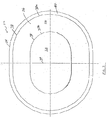
図1〜7は、軟組織または筋肉の欠損を矯正するためのかかる植込み型プロテーゼの実施形態を示す。プロテーゼ20は、組織侵入可能な材料の内部成長層22を含む。内部成長層22は、周縁部24と、周縁部24の内部に配置された外側周縁領域26と、外側周縁領域26の内部に配置され、これによって取り囲まれた内側中心領域28と、を含む。内部成長層22は、組織または筋肉の接着を可能にし、またはその影響を受けやすい材料22aの少なくとも1つの層を含む。一実施形態では、内部成長層22は、結合された第1の層22aおよび第2の層22bを含む。各層22a、22bは、十分な組織または筋肉の内部成長が植込み後の宿主組織または筋肉へプロテーゼを固定することを可能にする複数の隙間または開口部を含む生体適合性、柔軟性材料で形成されている。
1-7 show an embodiment of such an implantable prosthesis for correcting soft tissue or muscle defects. The
一実施形態では、各層22a、22bは、C.R.バード社から入手可能なバードメッシュ(BARD MESH)などの編成ポリプロピレンモノフィラメントメッシュ生地のシートで形成されている。植込まれると、ポリプロピレンメッシュは、メッシュ構造内および周囲での組織または筋肉の迅速な内部成長を促す。あるいは、組織または筋肉の補強および欠損の矯正に適切である他の外科用材料が、ソフトティッシュパッチ(SOFT TISSUE PATCH)(W.L.ゴア(Gore)アンド(&)アソシエーツ(Associates)社(Inc.))から入手可能な微孔性のePTFE)、サージプロ(SURGIPRO)(USサージカル(Surgical)社(Inc.)から入手可能)、トレレックス(TRELEX)(メドックスメディカル(Medox Medical)から入手可能、プロレン(PROLENE)とメルシレン(MERSILENE)(エチコン(Ethicon)社(Inc.)から入手可能)、および他のメッシュ材料(例えば、アトリウムメディカルコーポレーション(Atrium Medical Corporation)から入手可能)を含めて利用されうる。ポリグラクチン(VICRYL−−エチコン(Ethicon)社(Inc.)から入手可能)およびポリグリコール酸(デクソン(DEXON)−−USサージカル(Surgical)社(Inc.)から入手可能)を含む吸収性材料が、組織および筋肉の欠損の一時的矯正を含む用途に適切でありうる。クックバイオメディカル(Cook Biomedical)社(Inc.)から入手可能なクックサージシス(COOK SURGISIS)などコラーゲン材料も使用されうる。メッシュ生地は、マルチフィラメント糸で形成されうること、また編成、製織、編組、成形など適当な方法を使用してプロテーゼメッシュ材料を形成しうることも考えられている。
In one embodiment, each
確実に十分な組織または筋肉の内部成長が起こるように、材料の2つの層は、層22bの孔内への組織の成長を可能にし、周囲の筋肉または組織と層22bとの間の強い結合を提供するやり方でのみ付着される。層22aおよび層22bは、層22bの表面全体に亘って互いに積層または結合されていないことが好ましい。また、層22aおよび層22bは、層22aおよび層22bの多数の孔を塞ぐ接着剤や、融解によるなど層の有孔性に影響を及ぼすような方法で結合されていないことが好ましい。一実施形態では、第1の層22aと第2の層22bとは、ステッチ30で連結されている。
To ensure sufficient tissue or muscle ingrowth, the two layers of material allow tissue growth into the pores of
一実施形態では、層22aおよび層22bは、別々の位置でのみ付着されている(図4参照)。このようにして、組織または筋肉は、第1の層22aを通じて、第2の層22b内へ成長することができる。単一の縫合線30が内部成長層を十分に固定し得るが、内部成長層22aと22bのふくらみの量を制限する追加の縫合線を使用することが望ましい場合がある。また、付着は同心性パターンを含むことが示されているが、層の分離を最小限に抑えるために任意の適当なパターンが使用されてもよい。
In one embodiment,
当然のことながら、本発明は、第1の層と第2の層が他の適当の技術を用いて付着されてもよいため、特定の付着法に限定されるものではない。例えば、これらの層は、特定の位置または特定のパターンで層を融解したり、層を音波、誘導、振動、または赤外/レーザー溶接したり、または適当な結合剤を用いたりすることで、結合されてもよい。付着点は、渦巻きパターン、蛇行パターン、またはドットやビードの格子状パターンなど、組織または筋肉の侵入のための開放または非含浸間隙の十分な量を維持する適当なパターンを構成してもよい。 Of course, the present invention is not limited to a particular deposition method because the first and second layers may be deposited using other suitable techniques. For example, these layers can be melted at specific locations or in specific patterns, sonic, induction, vibration, or infrared / laser welded, or using suitable binders. May be combined. The attachment points may constitute a suitable pattern that maintains a sufficient amount of open or unimpregnated gaps for tissue or muscle penetration, such as a spiral pattern, a serpentine pattern, or a lattice pattern of dots or beads.
プロテーゼの位置決めおよび/または仮付着に役立つために、プロテーゼはポケット32を含んでいる。このように、外科医は、ポケットを用いてプロテーゼを所望の領域に位置決めすることができる。その後、外科医は、周囲の内部成長組織、筋肉、または腹膜層に材料の層のうちの1つを縫合またはステープル止めすることができる。例えば、外科医は、ポケットに侵入して、ポケットの上部層を組織、筋肉、または腹膜層に縫合、またはステープル止めすることができる。このように、プロテーゼは、少なくとも十分な組織または筋肉の内部成長が起こるまで適所に仮保持され得る。一実施形態では、第1の層および第2の層は、それらの間にポケット32を形成するようにして付着されている。しかし当然のことながら、本発明はこの点において限定されず、ポケット32を使用する必要はないし、他の適当な方法で形成される他の適当なポケットを使用してもよい。例えば、ポケットは、材料の追加の層や、第1の層22aに付着されたその一部分で形成されてもよい。
To assist in positioning and / or temporary attachment of the prosthesis, the prosthesis includes a
ポケットの内部へのアクセスを得るために、ポケット32は開口部34を含んでいる。一実施形態では、開口部は、第1の層22aに形成された横切断部またはスリットである。プロテーゼを位置決めするために、外科医は、1本もしくはそれ以上の指(または適当な医療器具)をポケットの中へ挿入し、プロテーゼを所定の位置に動かす。一実施形態では、ポケット32は、外科医の手の少なくとも4本の指を受入れる大きさに作られているが、本発明はこの点において限定されていないため、他の適当な大きさに作られたポケットを使用してもよい。さらに、ポケット32は、複数のポケットで形成されうるため、1本もしくはそれ以上の指を個別の指部分に挿入することができる。示された実施形態では、ポケット32は、第1のサイドポケット32aと第2のサイドポケット32bとを含む(図1と図7を参照)。しかし当然のことながら、本発明は、この点において限定されず、単一のサイドポケットのみを使用してもよい。
In order to gain access to the interior of the pocket, the
腹壁ヘルニアの矯正、または胸壁や腹壁の再建におけるなど一部の処置では、内部成長層は、組織、筋肉または臓器と接触しうるところ、これらは、内部成長層の中へ成長することを意図されていない。かかる接触は、潜在的に内部成長層と周囲の組織、筋肉または臓器との間の望ましくない術後癒着をもたらしうる。プロテーゼの選択部分への術後癒着の発生を最小限に抑え或いは除去するために、プロテーゼは、内部成長層22の片側の少なくとも一部分、かつ好ましくは全部を覆う組織、筋肉または臓器の癒着耐性バリア層36を含みうる。一実施形態では、バリア層36は、第2の層22bに隣接した側のプロテーゼに付着されている。プロテーゼ20は、腹部内臓(例えば、腸)または胸部内臓(例えば、心臓または肺)など潜在的に望ましくない癒着の部位にバリア層36が直面するように、患者内に位置決めされる。以下で詳述するように、バリア層36は材料および/または構造で形成され、その材料および/または構造は、植込まれた場合に、組織、筋肉または臓器の内部成長および接着形成を実質的に刺激することなく実際に耐え、これにより、内部成長層と隣接した組織、筋肉または臓器との間の望ましくない術後癒着の発生を制限し、または完全に除去する。
In some procedures, such as in correction of abdominal wall herniation, or reconstruction of the chest wall or abdominal wall, the ingrowth layer is intended to grow into the ingrowth layer where it can come into contact with tissue, muscle or organ. Not. Such contact can potentially lead to unwanted post-operative adhesions between the ingrowth layer and the surrounding tissue, muscle or organ. In order to minimize or eliminate the occurrence of post-surgical adhesions to selected portions of the prosthesis, the prosthesis is an adhesion resistant barrier for tissue, muscle or organ that covers at least a portion and preferably all of one side of the ingrowth layer 22.
一実施形態では、バリア層36は、大幅な組織内部成長を可能にしないフィブリル長−孔サイズまたは節間距離とも呼ばれる−を有する延伸ポリテトラフルオロエチレン(expanded polytetrafluoroethylene)(ePTFE)のシートで形成されている。一実施形態では、ePTFEのフィブリル長は5ミクロン未満である。別の実施形態では、ePTFEのフィブリル長は1ミクロン未満であり、さらに別の実施形態では、フィブリル長は0.5ミクロン未満である。バリア層36を形成するための他の適当な材料の例としては、C.R.バードから入手可能なフルオロ−テックス(FLUORO−TEX)心膜(Pericardial)および腹膜(Peritoneum)サージカルメンブレン(Surgical Membrane)およびフルオロ−テックス(FLUORO−TEX)硬膜代用品(Dura Substitute)、およびW.L.ゴア(Gore)アンド(&)アソシエーツ(Associates)社(Inc.)から入手可能なプリクルード(PRECLUDE)心膜メンブレン(Pericardial Membrane)、プリクルード(PRECLUDE)腹膜メンブレン(Peritoneal Membrane)、およびプリクルード(PRECLUDE)硬膜代用メンブレン(Dura Substitute membrane)が挙げられる。他の適当な微小ないし非多孔性材料の代表的かつ非限定的なサンプリングとしては、ダウコーニング社(Dow Corning Corporation)によって配給されたシラスティックアールエックスメディカルグレードシート(SILASTIC Rx Medical Grade Sheeting)(硬化白金(Platinum Cured))、および微孔性ポリプロピレンシート(セルガード(Celgard)社(Inc.)から入手可能)およびフィルムなどのシリコーンエラストマーが挙げられる。内生、外生、異種組織も、例えば、心膜および小腸粘膜下組織を含めて意図されている。ゲンザイム社(Genzyme Corporation)から入手可能なセプラフィルム(SEPRAFILM)などの吸収性材料、および酸化、再生セルロース(Intercede(TC7))が一部の用途に使用されうる。当然のことながら、他の適当な生体適合性接着耐性材料も使用されうる。
In one embodiment, the
プロテーゼ20は、従来の組織接近が実行不可能である場合の組織欠損の矯正、例えば、大きな切開創ヘルニア、特に、以前の手術によって弱体化した組織または筋肉、もしくは肥満患者における組織または筋肉において生じるものなど大きな欠損の矯正において特に有用である。このために、プロテーゼ20は欠損を埋め、組織または筋肉が内部成長層の中へ成長するとともに、かかる内部成長が起こったのちに、その周囲の組織または筋肉を支持する。一実施形態では、患者によって(例えば、患者の動きによって)誘発される応力を支え、再発性欠損を制限するために、組織または筋肉は、かかる応力を支えるために最もよく適している内部成長材料の層の中へ成長しうることが望ましい。層22aは、少なくとも1つの開口部34を含むため、必要な応力を支える可能性は比較的低い。一方、第2の層22bは、相当の大きさの開口部を含まず、または他の大きな不連続性を含み、かつ一般に均一であり、それゆえ、必要な負荷を支えることがより可能である。したがって、本明細書中で記載された実施形態では、負荷を保有する層は層22bである。
当然のことながら、本発明はこの点において限定されず、プロテーゼ20は、かかる開口部または不連続性が耐え得る量を超えて第2の層22の負荷保有能力を削減することがないという条件で、適当なサイズおよび形状の開口部で形成され得る。例えば、比較的小さなプロテーゼでは、かかる開口部または不連続性を使用してもよい。これらの開口部または不連続性を用いて、プロテーゼの少なくとも仮固定に役立て、組織の内部成長を促進させることができる。かかる開口部および不連続性を使用するプロテーゼの例は、本発明の譲渡人に譲渡され、本明細書中にその全体が参考によって援用されている米国特許第6,290,708号および同第6,224,616号に記載されている。
Of course, the present invention is not limited in this regard, and the
第2の層22bの中への組織または筋肉の成長を許容し且つ促進するために、バリア層36は、組織が層22bの孔の中へ成長することを許容し、周囲の筋肉または組織と層22bとの強い結合を提供するやり方でのみ第2の層22bに付着されていることが好ましい。層22bは、バリア層36に積層化されず、且つ層22bの全表面にわたってバリア層36に結合されていないことが好ましい。また、層22bは、層22bの多数の孔を塞ぐような接着剤によって、あるいはその他の点では層22bの多孔性に影響を及ぼすような融解などの方法によって、バリア層36に結合されていないことが好ましい。
In order to allow and promote tissue or muscle growth into the
一実施形態では、図1〜5に示されているように、第1の層22aと第2の層22bが、内部成長層、特に層22bへの十分な組織侵入を可能にすると同時に、層22aと層22bとの接続を提供するステッチを用いて別々の付着線で付着されている。また、これら同じステッチ(例えば、ステッチ38)を用いて、層22bをバリア層36に固定することができる。ステッチ線38は、バリア層36を内部成長層22bに十分に固定し得るが、ステッチ39などの追加のステッチ線を用いて、内部成長層から離隔するバリア層のふくらみの量を制限することが望ましい場合がある。付着は、同心性パターンを含むことが示されているが、内部成長層およびバリア層の分離を最小限に抑えるために、任意の適当なパターンを使用されてもよい。
In one embodiment, as shown in FIGS. 1-5, the
他方、層22bを層36に固定するために用いられるものとは異なる型のステッチを用いて、層22aと層22bを固定することができる。例えば、図4と図5に示されているように、すべてのステッチ線30がバリア層36を通過するわけではない。むしろ、ステッチ線38のみが、バリア層36を通過する。層36を層22bに固定するのに必要なだけの少ないステッチが使用され、プロテーゼのバリア層側の組織または筋肉の接着を最小限に抑えることが好ましい。また、示されている実施形態では、第1の層22aはその位置でアクセス開口部32を含むため、中央ステッチ線39のみが第2の層22bとバリア層36を通過する。
On the other hand, layers 22a and 22b can be secured using stitches of a different type than those used to secure
一実施形態では、バリア層36は、ステッチにより内部成長層22bに付着されているが、当然のことながら、バリア層は他の適当な技法を用いて付着されてもよいため、本発明はこの点において限定されていない。例えば、バリア層は、層を加熱し、層を溶接し、または適当な結合剤を用いることによって内部成長層に結合され得る。いずれにしても、組織または筋肉の侵入のために十分な量の開放または非含浸隙間が少なくとも層22bにおいて維持されているという条件で、隙渦巻きパターン、蛇行パターン、またはドットやビードの格子状パターンなど、適当なパターンが使用され得る。
In one embodiment, the
ステッチを使用して内部成長層22bをバリア層36に付着させ、さらに接着を最小限に抑える場合、ステッチは非多孔性、接着耐性材料で形成され得る。例えば、ステッチは適当なポリテトラフルオロエチレン(PTFE)で形成されうる。PTFEステッチは、ポリプロピレンモノフィラメントなど他のステッチ材料を用いるプロテーゼと比べ操作が容易であり、より柔軟でより順応性のプロテーゼを提供し得る。PTFEモノフィラメントは、材料の低い摩擦特性によって、製造工程も促進させる。しかし、ポリプロピレンモノフィラメントなどの適当な材料が、ステッチ用に使用され得ることを理解すべきである。例えば、ステッチ線の一部はバリア層を通過することがないため、またはバリア層が使用できない場合である場合には、接着耐性材料以外の材料を使用してもよい。もっとも、製造を容易にするために、通常、すべてのステッチは同じ材料で形成されているが、本発明はこの点において限定されていない。
If stitches are used to attach the
これらの層は、ボビンおよび縫糸を用いるミシンによって形成された通常の縫いステッチを用いて縫い合わされる。バリア層は、内部成長層に位置決めされ、縫い針に直面し、各ステッチのロッキング部分(すなわち、細い組みひも)が、バリア層側ではなくプロテーゼの内部成長側に形成され、組織、筋肉または臓器との限局性接着を削減することが好ましい。ステッチは、#10の先端に球が付いた針を用いて形成され、ステッチの穴を通じた内部成長の潜在的発生を削減することができる。バリア層の有無による内部成長材料のシートは、所望のステッチパターンでプログラムされたコンピュータ制御テーブル上での縫合処置中にフレームによって保持されうる。 These layers are stitched together using normal sewing stitches formed by a sewing machine using bobbins and threads. The barrier layer is positioned on the ingrowth layer and faces the sewing needle, and the locking portion of each stitch (ie, a thin braid) is formed on the ingrowth side of the prosthesis, not on the barrier layer side, and is on the tissue, muscle or organ It is preferable to reduce the localized adhesion. Stitches are formed using a needle with a sphere at the tip of # 10, which can reduce the potential for ingrowth through stitch holes. A sheet of ingrowth material with or without a barrier layer can be held by the frame during a stitching procedure on a computer control table programmed with the desired stitch pattern.
バリア層36は内部成長層22の片側の全表面を覆うことが好ましい一方で、バリア層36は、プロテーゼの片側の選択部分のみを覆うように構成され、バリア層がない部分の両側からの内部成長を強化するようにしてもよい。同様に、プロテーゼは、バリア層がプロテーゼの片側の全表面を覆い、かつプロテーゼのもう片側の1以上の部分を覆うように構成されてもよい。
While the
場合によっては、プロテーゼ20の外側周縁部を、隣接組織、筋肉または臓器から隔離することが望ましい場合がある。一実施形態では、周縁バリア40(図1、図3、および図5参照)は、プロテーゼ20の外側周縁部24の周囲に完全に延在し、それへの接着を阻止する。もっとも、周縁バリア40は、術後癒着の形成からの保護が望まれる場合、プロテーゼの外側周縁部の選択部分のみを覆うように構成されてもよい。
In some cases, it may be desirable to isolate the outer peripheral edge of the
周縁バリア40は、内部成長層22またはバリア層36のいずれかにより、一体的に形成されてもよい。あるいは、周縁バリア40は、プロテーゼの外側周縁部に付着され、または組込まれている別々の成分によって形成されてもよい。一実施形態では、周縁バリア40は、内部成長層22の一部分で形成されている。特に、内部成長層22は、その外側縁に沿った組織侵入可能な間隙または開口部を実質的に削減するために変化させ、それによって周縁バリア40を生成することができる。
The
一実施形態では、図3と図5に示されているように、層22の周縁部24は融解され、材料をシールし、外側周縁バリア40を形成する。バリア層36は、サブミクロンサイズの孔を有するなど、層22の融解材料の一部分がバリア層36に融合化するように構成されうる。周縁部24は、適当な方法を用いて融解され得る。一実施形態では、周縁部24は、層をヒートシールすることによって融解されてもよい。例示的な実施形態では、周縁バリア40は、ポリプロピレンメッシュ生地の環を融解することによって、プロテーゼの所望の構成に近い形状のePTFEバリア層36に形成される。これは、固定されたメッシュ生地とePTFE材料の特大のシートを覆い、かつプロテーゼの所望の形状で構成された加熱金型を用いることによって層をヒートシールすることによって達成され得る。融解環は、約3〜5秒間、約320°F〜400°Fの範囲の温度で加熱することによって、生地に形成され得る。通常選択される温度は、ePTFEバリア層の焼結温度以下である。本発明はこの点において限定されていないため、超音波、誘導、振動、赤外線/レーザー溶接等などの他のシール法を使用してもよい。融合されると、内部成長層は、上述したように、バリア層に縫合され、その後に環の一部分に沿って型抜きし、周縁バリアを有するプロテーゼを完成する。
In one embodiment, the
本発明はこの点において限定されていないため、周縁バリアを生成するための他の適当な技法を使用してもよい。かかる他の技法の例は、本発明の譲渡人に譲渡され、本明細書中でその全体が参考によって援用されている、同時係属中の米国特許出願第09/661,623号において記載されている。 Since the present invention is not limited in this respect, other suitable techniques for creating a peripheral barrier may be used. Examples of such other techniques are described in co-pending US patent application Ser. No. 09 / 661,623, assigned to the assignee of the present invention and incorporated herein by reference in its entirety. Yes.
上述された実施形態はバリア層を含むが、本発明はこの点において限定されていない。したがって、以下に記載されるものなどの他の実施形態は、バリア層または周縁バリアを含むことも含まないこともある。 Although the embodiments described above include a barrier layer, the present invention is not limited in this respect. Accordingly, other embodiments, such as those described below, may or may not include a barrier layer or a peripheral barrier.
場合によっては、(ただし限定されないが)比較的大きな欠損の矯正など、十分に硬質のプロテーゼを使用し、プロテーゼが容易かつ効率的に操作され、さらに十分に柔軟性に所望の領域に配置されて、プロテーゼがそのプロテーゼを植込む医師とそのプロテーゼを受入れる患者の両者によって十分に耐えられることが望ましい場合がある。プロテーゼは、覆われる領域の形状に適合すべきで、その端部が過度にカールしないように十分に硬質であるべきである。この特性は、肥満患者における大きな欠損による使用のために大きさの大きいプロテーゼでは特に有用である。したがって、本発明の別の態様によれば、剛性と柔軟性のバランスをとるために、プロテーゼ20は、第1の補強部材50と、第1の補強部材から内部に間隔を存して配置された別個の第2の補強部材52と、を含む。補強部材50、52は、以下に詳述するように、適当な方法で内部成長層22に結合され得る。
In some cases (but not limited to) using a sufficiently rigid prosthesis, such as correcting a relatively large defect, the prosthesis is easily and efficiently manipulated and is sufficiently flexible to be placed in the desired area It may be desirable for the prosthesis to be well tolerated by both the physician implanting the prosthesis and the patient receiving the prosthesis. The prosthesis should conform to the shape of the area to be covered and be sufficiently rigid so that its ends do not curl excessively. This property is particularly useful in large prostheses for use with large defects in obese patients. Thus, according to another aspect of the present invention, the
補強部材はプロテーゼの安定性に貢献し、所望の形状で維持することを可能にする。例えば、補強部材は、プロテーゼを実質的にフラットに維持することを可能にするのに役立つ。この安定性は、操作を容易にさせることによってプロテーゼの配置を容易にする。また、安定性は、プロテーゼがたわみ、褶曲し、曲がりまたは転位化する傾向を最小限に抑える。操作の難しさ、もしくは転位または屈曲は、植込み中に追加の手術方法および/または追加の固定を必要とし得る。以下で論じられるように、プロテーゼの植込み中、縫合は補強部材の周囲を通り、プロテーゼをおおむね所望の構成および位置に維持させることができる。 The reinforcing member contributes to the stability of the prosthesis and allows it to be maintained in the desired shape. For example, the reinforcement member helps to allow the prosthesis to remain substantially flat. This stability facilitates placement of the prosthesis by facilitating operation. Stability also minimizes the tendency of the prosthesis to bend, bend, bend or dislocate. Difficulty of operation, or dislocation or flexion, may require additional surgical methods and / or additional fixation during implantation. As discussed below, during implantation of the prosthesis, the suture can pass around the reinforcement member to maintain the prosthesis generally in the desired configuration and position.
一実施形態では、第1の補強部材50は実質的に連続的であり、かつ少なくとも外側領域26を補強するためにプロテーゼの外側領域26を取り囲む。図に示された実施形態では、補強部材50は、周縁部24には配置されていない。むしろ、補強部材50は、周縁部24の内方に間隔を存して配置されている。もっとも当然のことながら、補強部材50は、周縁部24に配置されてもよいため、本発明はこの点において限定されない。
In one embodiment, the first reinforcing
第2の補強部材52は、第1の補強部材の内方に配置され、プロテーゼの内側領域28を補強するために使用され得る。一実施形態では、第2の補強部材は、連続的であり、内側領域28の周りに延在し、ポケット32の外側周縁を画定する。示されている実施形態では、各補強部材50および52は、所望の厚さおよび断面形状のモノフィラメント糸であり、プロテーゼ上に環状に構成されている。当然のことながら、補強部材は、円形、正方形、長方形、三角形、楕円形等など任意の断面形状を有してもよい。部材50および52は、渦巻状パターン、正方形パターン、楕円形パターン、円形パターンなどの任意のパターンでプロテーゼ20上に構成されてもよい。
The second reinforcing
示されている実施形態では、第2の内側補強部材52は、第1の外側補強部材50と同軸または略同軸である。もっとも当然のことながら、他の適当な配置が使用されるため、補強部材はこの点において限定されない。
In the illustrated embodiment, the second
通常、第2の内側補強部材52は、プロテーゼが植込まれている場合に、欠損の端部とおおむね整列するようにプロテーゼ上に配置される。このように、植込み中、縫合は第2の内側補強部材52の周りまたは近くを通り、内側領域26を欠損の端部近くの組織または筋肉の仮付着させるために使用されうる。また、縫合糸は、第1の外側補強部材50の周りまたは近くも通り、欠損の端部からより離隔して、通常、欠損の端部から3cm〜5cm離隔して組織または筋肉に付着され、外側領域26も組織または筋肉に仮付着され得る。
Typically, the second inner reinforcing
補強部材50、52は、本発明はこの点において限定されていないため任意の適切な方法でプロテーゼ上に配置されればよい。一実施形態では、図3〜5に示されているように、第1の補強部材50および第2の補強部材52は、内部成長材料の第1の層22aと第2の層22bとの間に挟まれており、それらに物理的に付着されていても、いなくてもよい。これらの補強部材は、ステッチ線などの付着線30において第1の層22aと第2の層22bの付着によって形成されるチャネル54内にしっかりと、またはゆるく保持され得る。通常、縫糸によって形成される単一線30は、部材50および52の少なくとも外側または内側の端部に沿って縫合され、それらが層22aと層22bに対して移動しないようにする。部材50および52の剛性により、部材50および52の片側に沿った1本のステッチ線で十分であり得る。もっとも、各部材50および52の両側に2本のステッチ線があり、それらが存在するチャネル54を形成することによって、それらを適所に保持することが好ましい。これらのステッチは、層22aと層22bを通じて延在することが好ましいが、バリア層36を通じて延在することは、これが存在する場合には好ましくない。層36または層22aと層22bに縫合または結合された場合には、部材50が、層22a、22bおよび/または層36を、層22に対して層36のまたは層22aと22bに対してこれら互いのふくらみを阻止するやり方で一緒に保持することが、別の利点である。
The reinforcing
あるいは、補強部材50、52は、内部成長層22を覆い、または支持することができ、位置に関係なく、ステッチまたは結合剤で付着され、または超音波、誘導、振動、赤外線/レーザー溶接などによって融合され得る。あるいは、これらの補強部材は、層22a、22bの少なくとも1つにより織られ、または層22そのものが製造されているように層22と一体に形成され得る。バリア層が使用されている場合には、補強部材50、52はバリア層36の下に配置されていないこと、またはそれらを通じて突出することが望ましく、そうすることによって、結果として補強部材上に形成する望ましくない接着が生じる。
Alternatively, the reinforcing
補強部材50、52はモノフィラメント糸で形成されると上述したが、他の適当な構造を使用してもよい。例えば、補強部材は、その後にプロテーゼに付着され、プロテーゼ上に成形される成形要素であってもよい。例としては、本明細書中で参考によって援用されている米国特許第5,695,525号において示されている環が挙げられる。また、補強部材は、内部成長層で形成されてもよい。この点において、各補強部材50、52は、所望の形状で内部成長層22の一部分を融解することによって形成されてもよい。これらの補強部材は、約3〜5秒間、約320°F〜400°Fの範囲の温度で内部成長層に熱を加えることによって形成され得る。別の例では、補強部材50、52は、例えば、縫い取りした部分など、層22aと層22bの一方または両方を通過する複数のステッチで形成され得る。あるいは、補強部材50、52は、所望の補強の区域における織りパターンを変化させることによって形成され得る。このように、組織内部成長が望まれる内部成長層22の領域は、比較的ゆるいまたは糸目の粗い織りで形成されているが、補強の領域または区域は、比較的目の詰んだ織りで形成され、所望の剛性を提供する。本発明はこの点において限定されていないため、補強部材50、52を形成する他の適当な方法または構造を使用してもよい。
Although the reinforcing
一実施形態では、プロテーゼの部分は、プロテーゼを適所に仮保持するために使用される固定要素を含んでもよい。これらの固定要素は、縫合糸、ステープルまたはタックに加えられ、またはこれらの代わりとなり得る。一実施形態では、固定要素は、付着されまたはその他の点では補強部材上に形成され得る。かかる固定要素の例は、本発明の譲渡人に譲渡され、本明細書中でその全体が参考によって援用されている米国特許第6,267,772号に記載されている。 In one embodiment, the portion of the prosthesis may include a securing element that is used to temporarily hold the prosthesis in place. These anchoring elements can be added to or substituted for sutures, staples or tacks. In one embodiment, the securing element may be attached or otherwise formed on the reinforcing member. Examples of such anchoring elements are described in US Pat. No. 6,267,772, assigned to the assignee of the present invention and incorporated herein by reference in its entirety.
上述された一部の実施形態は補強部材を含むが、本発明はこの点において限定されていない。したがって、以下で述べられるものなど他の実施形態は、補強部材を含むことも、含まないこともあり得る。 Although some embodiments described above include a reinforcing member, the present invention is not limited in this respect. Accordingly, other embodiments, such as those described below, may or may not include a reinforcing member.
上述されたように、プロテーゼが適所に挿入された後または植込み中に、十分な組織または筋肉の内部成長が起こるのに優先して、プロテーゼの一部分が転位化する場合がある。例えば、プロテーゼの外側部分は、褶曲化しまたは移動し、望ましくない組織、筋肉または臓器の接着の影響を受けやすくなり得る。この転位または褶曲は、欠損、およびその後に領域26が相当に大きくなる肥満者における大きな欠損用など、比較的大きなプロテーゼには特に問題である。この可能性を減少させるために、プロテーゼの外側端部は、縫合糸、ステープル、らせん状タックまたは他の適当な固定装置を用いて適所に仮保持される。したがって、別の実施形態によれば、プロテーゼは、ポケット32の他に外側周縁領域26に形成されるポケット60を含む。ポケット60は、後述するように、プロテーゼを操作するために使用され、また外側領域26を仮固定するために使用され得る。ポケット60は、外科医の手の少なくとも一部分または適当な医療器具を受入れ、確実にプロテーゼが正確な向きに配置され、おおむね適切な位置に配置されるようにサイズが決められている。さらに、ポケットは、縫合糸、ステープル、らせん状タックまたは他の固定装置を配置する器具などの適切な器具を受入れるようにサイズが決められており、外科医は、周囲の内部成長組織、筋肉、腹膜層に外側領域26を適切に固定することができる。
As described above, a portion of the prosthesis may translocate prior to sufficient tissue or muscle ingrowth after the prosthesis is inserted in place or during implantation. For example, the outer portion of the prosthesis can be bent or moved and susceptible to unwanted tissue, muscle or organ adhesion. This dislocation or fold is particularly problematic for relatively large prostheses, such as for defects and subsequent large defects in obese individuals where
第1のポケット32と同様、一実施形態では、第2のポケット60は、第1の層22aと第2の層22bの付着によって画定される。特に、第1の層22aと第2の層22bは、ポケット60を形成するために別々の位置で結合される。もっとも、第2のポケットは適当な方法でプロテーゼ上に形成され得るため、本発明はこの点において限定されない。例えば、第2のポケット60は、プロテーゼの上面に付着された材料(図示せず)の追加の層で形成されてもよい。
Similar to the
一実施形態では、第2のポケット60は、周縁部24に実質的に延在するように構成されている。部材50が使用されている場合、第2のポケット60は、補強部材50に延在する。部材50がステッチによって適所に保持されている場合には、ポケット60は通常、付着線30aに延在し(図1、図3、および図5を参照)、付着線30aは第2のポケット60の外側周縁を画定するようになる。また、部材52は、第1のポケット32から第2のポケット60へのアクセスが防止されるようにポケット32と60との間の境界を画定する。
In one embodiment, the
第1のポケットと第2のポケットとの間のバリアまたは隔壁を提供する利点は、プロテーゼの植込みが容易になることである。この点において、プロテーゼが比較的大きい場合、外科医は、その手や器具をわざわざポケット60に挿入して、周縁部24を押す必要なくプロテーゼを適所に操作することができる。むしろ、外科医の手または器具はスリット34を通じて中央ポケット32に侵入し、隔壁またはバリア(例えば、部材52またはステッチ線30b)を押してプロテーゼを適所に移動させることができる。
An advantage of providing a barrier or septum between the first pocket and the second pocket is that the prosthesis is easier to implant. In this regard, if the prosthesis is relatively large, the surgeon can bother to insert the hand or instrument into the
2つのポケット32、60は別個であるため、第2のポケット60には、ポケット60の内部へのアクセスを可能にする独自の開口部62が備えられている。一実施形態では、アクセス開口部62は、外科医の手の少なくとも一部分、または1つもしくはそれ以上の外科器具を受入れるようにサイズおよび形状が決められ得る第1の層22aにおけるカットまたはスリットの形である。開口部は正確に形状が決められ得るが、一実施形態では、円弧にそって測定された約6.5cmの長さを有する。本発明はこの点において限定されていないため、他の適当なサイズと形状の開口部が使用され得る。
Since the two
第2のポケット60の内部へのアクセスを得ることは、プロテーゼを仮付着する場合にも有用であり得る。ステープル止めまたは縫合装置(図示せず)は、開口部62を介してポケット60の中へ挿入され得るため、縫合糸、ステープル、らせん状タックまたは他の固定装置は、第2のポケットを介して配置され、プロテーゼの少なくとも外側部分を周囲の組織または筋肉に仮付着させることができる。
Obtaining access to the interior of the
好ましくは、必ずしも必要ではないが、プロテーゼは欠損の端部の近くの位置、またプロテーゼの端部の近くの位置における組織または筋肉に付着される。本発明はこの点において限定されていないため、プロテーゼは、適当な方法で固定され得る。一実施形態では、縫合が使用され得る。例えば、図7に示されているように、縫合糸を使用する場合、プロテーゼを患者に導入する前に、バリア層36には通過させることなく、縫合糸100を内部成長層22aと22bの一方または両方を通過させることができる。あるいは、図7Aに示されているように、縫合糸100は、内部成長材料の層22a、22bの一方または両方を通過し、補強部材50、52を取り囲み得る。好ましくは、必ずしも必要ではないが、縫合糸はバリア層36を通過しないことである。縫合糸は、示されているように、ステッチ線30も取り囲み、またはそれぞれのチャネル54内の補強部材50、52の周りを通ることができるが、ステッチ線を含むことはない。針は通常、縫合糸に放置され、縫合糸は、止血鉗子などの適切な器具で固定され、邪魔にならないようにドレープされる。
Preferably, but not necessarily, the prosthesis is attached to tissue or muscle at a location near the end of the defect and at a location near the end of the prosthesis. Since the present invention is not limited in this respect, the prosthesis can be secured in any suitable manner. In one embodiment, sutures can be used. For example, as shown in FIG. 7, if a suture is used, the
別の実施形態では、縫合糸ではなく、ステープルが使用される。ステープルは、プロテーゼが患者の適所に配置された後にポケット32または60(開口部34または62を介して)に挿入され、その結果、プロテーゼはステープル止めされる。さらに別の実施形態では、オリジン(Origin)社(Inc.)から入手可能なものなどのらせん状タックが使用され得る。ステープルと同様、らせん状タックは、プロテーゼが患者の適所に配置された後にポケット32または60(開口部34または62を介して)に挿入され、その結果、プロテーゼは固定される。当然のことながら、本発明は、縫合糸、ステープル、タックまたは他の特定の固定機構もしくは技法の使用に限定されていないため、他の適当な固定装置が使用され得る。
In another embodiment, staples are used rather than sutures. Staples are inserted into
十分な数の縫合糸、ステープル、タックまたは他の固定装置を用いてプロテーゼを固定する。欠損のサイズおよび/または位置によって、4〜12個もしくはそれ以上の固定装置が使用され得るが、固定装置の一部は欠損の端部近くのプロテーゼ上に配置され、他の固定装置は周縁部34近くのプロテーゼ上に配置される。
The prosthesis is secured using a sufficient number of sutures, staples, tacks or other securing devices. Depending on the size and / or location of the defect, 4-12 or more anchoring devices may be used, but a part of the anchoring device is placed on the prosthesis near the edge of the defect and the other anchoring device is the
一実施形態では、第2のポケット60は、図1および図6に示されているように、領域26または部材52の周囲に間隔を置いて配置されている複数の開口部62を含む。開口部62は、第1の層の一部分64が間隔を存して配置された隣接した開口部62を結合するように第1の層22aを通じて形成されていることが好ましい。これらの部分64は、第2の層22bからの第1の層22aのふくらみの量を有効に制限する。例えば、部分64は、内側領域28へのブリッジとして作用し、その端部はステッチ線30bによって層22bに固定される。一実施形態では、開口部62の端部を補強し、層22aの引き裂きを防止するために、ステッチまたは他の補強部66は、部分64において形成される。補強部66は、示されているように弧状を含む任意の適当な形状を有し得ると共に、第1の層22aと第2の層22bを通じて、または第1の層22aだけを通じて延在し得る。
In one embodiment, the
上述された実施形態は内側ポケットおよび外側ポケットを含むが、本発明はこの点において限定されない。 Although the embodiments described above include an inner pocket and an outer pocket, the invention is not limited in this respect.
示されている実施形態では、プロテーゼ20は、比較的フラットで十分に柔軟性があり、外科医は、プロテーゼを操作してプロテーゼを挿入し、当該解剖学的部位にプロテーゼを適合させることができ、プロテーゼは、縫合、ステープル止めまたは固定されることができる。プロテーゼ20は、特定の欠損の矯正の促進につながる適当な形状またはサイズを有するように構成され得る。図面に示された実施形態では、プロテーゼ20は、略フラットの卵形を有する。他の形状の例としては、円形、正方形、長方形および不規則な形状が挙げられるが、これらに限定されない。
In the illustrated embodiment, the
プロテーゼ20を植込む方法の1つの例を以下に述べる。欠損を確認し、縁を裏返す。ボビー(bovie)などの医療器具を用いて、腹膜前または逆筋(retro−muscular)スペースのいずれかを得る。次いで、外科医の指を挿入し、周囲を掃除し、プロテーゼを配置するスペースをつくる。次いで、プロテーゼを転がし、作られたスペースに挿入することができる。上述されたように、プロテーゼ上のポケットを用いて挿入を補助することができる。ポケット上またはその内部のいずれかの周囲を指で再び掃除し、プロテーゼは、使用される場合には、補強部材の助けを借りて、広がって、その成形された形状となる。
One example of a method for implanting the
プロテーゼが適所に配置されると、縫合糸100を組織内部成長が望まれる層22a上から組織または筋肉を通過させる。次いで、縫合糸を結んでトリミングする。プロテーゼ上の転位の量を削減するために、縫合糸100は、相当な量の張力下にあってはならない。縫合の操作を促進するために、外科医は、適切の器具で開口部62を通じてポケット60に侵入し、ポケット60を通じて付着を行うことができる。例えば、ステープルが使用される場合、外科医は、ステープルをポケット32または60に挿入し、欠損の端部近く、好ましくはプロテーゼの周縁部において、内部成長が望まれる第1の層22aを組織または筋肉にステープル止めする。次いで、創傷は適当なやり方で閉鎖される。プロテーゼは、開腹処置、または低侵襲手術や腹腔鏡手術など低侵襲性処置においても使用できる。
When the prosthesis is in place, the
例示的な実施形態では、層22aと層22bはそれぞれ、直径約0.006インチのポリプロピレンモノフィラメントで編んだ厚さ約0.27インチのバードメッシュ(BARD MESH)で形成されている。バリア36は、厚さ約0.006〜0.008インチのePTFEシートで形成されている。バリア36は、直径0.008インチ〜0.012インチのPTFEモノフィラメントで形成された約3mm〜4mmの長さのステッチを用いて層22aと層22bに付着されている。プロテーゼ20は通常、任意の所望のサイズを有する略卵形を有する。例えば、プロテーゼは、一般に卵形の長軸と短軸に沿って測定されるように、およそ次のサイズ、5インチ×7インチ、7インチ×9インチ、8インチ×10インチ、または10インチ×13インチであればよい。プロテーゼは、50平方cm超の面積を覆うようなサイズでもあってもよい。一実施形態では、プロテーゼは、約68平方cm、別の実施形態では、約119平方cm、さらに別の実施形態では約152平方cm、またさらに別の実施形態では、(例えば、肥満患者の場合)約246平方cmの面積を覆う。もっとも、記載された材料および寸法は単に例示的であり、任意の適当なサイズおよび形状がプロテーゼ用に使用され得ることは、理解されるはずである。
In the exemplary embodiment, layers 22a and 22b are each formed from a BARD MESH having a thickness of about 0.27 inches knitted from polypropylene monofilaments having a diameter of about 0.006 inches. The
一実施形態では、プロテーゼは、プロテーゼが少なくとも3cm、また一部の実施形態では、少なくとも4cm、さらに他の実施形態では、少なくとも5cmの欠損の端部を覆うようにサイズが決められている。また、プロテーゼは、単一の欠損の矯正として上述されているが、適当なサイズと形状のプロテーゼを用いて、1つ以上の欠損が矯正され得ることも意図されている。 In one embodiment, the prosthesis is sized so as to cover the end of the defect, where the prosthesis is at least 3 cm, in some embodiments at least 4 cm, and in still other embodiments, at least 5 cm. Also, while a prosthesis has been described above as a single defect correction, it is also contemplated that one or more defects can be corrected using an appropriately sized and shaped prosthesis.
本発明の前述の説明は単にその例示であることが意図され、本発明の他の実施形態、変更および同等物が、添付されたクレームにおいて開示された発明の範囲内であることを理解すべきである。さらに、上述されたプロテーゼは、単独でまたは適当な組合せで使用され得る種々の特徴を含む。 It should be understood that the foregoing description of the invention is intended to be exemplary only, and that other embodiments, modifications, and equivalents of the invention are within the scope of the invention disclosed in the appended claims. It is. Further, the prostheses described above include various features that can be used alone or in appropriate combinations.
Claims (25)
材料の少なくとも2つの層(22a、22b、36)であって、当該少なくとも2つの層(22a、22b、36)の少なくとも1つの少なくとも一部分が、組織または筋肉との接着の形成を許容し、当該少なくとも2つの層(22a、22b、36)の少なくとも1つが、周縁部(24)と、当該周縁部(24)の内部に配置された外側領域(26)と、当該外側領域(26)の内部に配置された内側領域(28)とを含む少なくとも2つの層(22a、22b、36)と、
少なくとも1つのポケット(32、60)と、
前記少なくとも2つの層(22a、22b、36)の少なくとも1つに結合され且つ前記外側領域(26)内またはそれを取り囲み、または前記少なくとも2つの層(22a、22b、36)の2つの間に保持され且つ前記外側領域(26)内またはそれを取り囲む第1の補強部材(50)であって、少なくとも前記外側領域(26)を補強するように構成および配置されている第1の補強部材(50)と、
前記第1の補強部材(50)から内方へ間隔を存して配置されている第2の補強部材(52)であって、前記少なくとも2つの層(22a、22b、36)の少なくとも1つに結合され、または前記少なくとも2つの層(22a、22b、36)の2つの間に保持されている第2の補強部材(52)と、
を含む植込み型プロテーゼ(20)。An implantable prosthesis (20) for a tissue or muscle defect comprising:
At least two layers (22a, 22b, 36) of material, at least a portion of at least one of the at least two layers (22a, 22b, 36) allowing formation of adhesions with tissue or muscle, At least one of the at least two layers (22a, 22b, 36) includes a peripheral portion (24), an outer region (26) disposed inside the peripheral portion (24), and an inner portion of the outer region (26). At least two layers (22a, 22b, 36) including an inner region (28) disposed in
At least one pocket (32, 60);
Coupled to at least one of said at least two layers (22a, 22b, 36) and surrounding or surrounding said outer region (26), or between two of said at least two layers (22a, 22b, 36) A first reinforcing member (50) held and surrounding at or surrounding the outer region (26), the first reinforcing member configured and arranged to reinforce at least the outer region (26) ( 50),
A second reinforcing member (52) spaced inwardly from the first reinforcing member (50), wherein at least one of the at least two layers (22a, 22b, 36); A second reinforcing member (52) that is coupled to or held between two of said at least two layers (22a, 22b, 36);
Implantable prosthesis (20).
第1の層(22a)と、
前記第1の層(22a)に付着した少なくとも1つの第2の層(22b、36)と、
を含む請求項1に記載のプロテーゼ。The at least two layers (22a, 22b, 36)
A first layer (22a);
At least one second layer deposited prior Symbol first layer (22a) and (22b, 36),
The prosthesis of claim 1 including.
前記少なくとも1つの第2の層(36)の少なくとも1つは、組織、筋肉または臓器との接着の形成に対して耐性である材料を含む請求項2に記載のプロテーゼ。 Said first layer (22a) comprises a material allowing the formation of adhesions with tissue or muscle;
Wherein at least one of the at least one second layer (36), prosthesis of claim 2, including tissues, a material that is resistant to the formation of adhesion of the muscle or organs.
前記第1の補強部材(50)は、前記第1のチャネル(54)内に配置され、
前記第2の補強部材(52)は、前記第2のチャネル(54)内に配置されている請求項2に記載のプロテーゼ。Before SL first layer (22a) and second layers (22b, 36) are sewn together to form a first channel (54) and a second channel (54),
The first reinforcing member (50) is disposed in the first channel (54);
The prosthesis of claim 2 , wherein the second reinforcing member (52) is disposed within the second channel (54).
前記内側領域(28)内に形成された少なくとも1つのポケット(32)と、
前記外側領域(26)内に形成された少なくとも1つのポケット(60)と、
を含む請求項13に記載のプロテーゼ。The at least one pocket (32, 60)
At least one pocket (32) formed in the inner region (28);
At least one pocket (60) formed in the outer region (26);
The prosthesis according to claim 13 comprising:
材料の少なくとも1つの層(22)であって、その少なくとも一部分が組織または筋肉との接着の形成の影響を受けやすく、当該少なくとも1つの層(22)が、メッシュ材料の第1の層(22a)と、当該メッシュ材料の第1の層(22a)に付着したメッシュ材料の第2の層(22b)とを含み、当該少なくとも1つの層(22)が、周縁部(24)と、当該周縁部(24)の内部に配置された外側領域(26)と、当該外側領域(26)の内部に配置された内側領域(28)とを含む、材料の少なくとも1つの層(22)と、
前記内側領域(28)に形成され、前記メッシュ材料の第1の層(22a)と前記メッシュ材料の第2の層(22b)の接着によって画定された少なくとも1つの第1のポケット(32)と、
前記外側領域(26)に形成され、前記メッシュ材料の第1の層(22a)と前記メッシュ材料の第2の層(22b)の付着によって画定された少なくとも1つの第2のポケット(60)であって、当該少なくとも1つの第2のポケット(60)が前記少なくとも1つの第1のポケット(32)から離れており、当該少なくとも1つの第1のポケット(32)および第2のポケット(60)のそれぞれが、それぞれ少なくとも1つのポケット(32、60)の内部へのアクセスを得るためのアクセス開口部(34、62)を含む、少なくとも1つの第2のポケット(60)と、
前記少なくとも1つの層(22)に結合され、かつ前記外側領域(26)を実質的に取り囲む第1の補強部材(50)であって、少なくとも前記外側領域(26)を補強するように構成および配置されている第1の補強部材(50)と、
前記少なくとも1つの層(22)に結合され、前記第1の補強部材(50)から内方に間隔を存して配置された第2の補強部材(52)と、
を含む植込み型プロテーゼ(20)。An implantable prosthesis (20) for a tissue or muscle defect comprising:
And at least one layer of material (22), at least a portion susceptible to formation of adhesions with tissue or muscle, is even those said at one layer (22), a first mesh material A layer (22a) and a second layer (22b) of mesh material attached to the first layer (22a) of the mesh material, wherein the at least one layer (22) comprises a peripheral portion (24) and At least one layer (22) of material comprising an outer region (26) disposed within the perimeter (24) and an inner region (28) disposed within the outer region (26). When,
At least one first pocket (32) formed in the inner region (28) and defined by adhesion of the first layer (22a) of the mesh material and the second layer (22b) of the mesh material ; ,
At least one second pocket (60) formed in the outer region (26) and defined by the attachment of the first layer (22a) of the mesh material and the second layer ( 22b) of the mesh material . The at least one second pocket (60) is remote from the at least one first pocket (32), the at least one first pocket (32) and the second pocket (60). At least one second pocket (60), each including an access opening (34, 62) for gaining access to the interior of at least one pocket (32, 60), respectively.
A first reinforcing member (50) coupled to the at least one layer (22) and substantially surrounding the outer region (26), configured to reinforce at least the outer region (26); A first reinforcing member (50) disposed;
A second reinforcing member (52) coupled to the at least one layer (22) and spaced inwardly from the first reinforcing member (50);
Implantable prosthesis (20).
材料の少なくとも1つの層(22)であって、その少なくとも一部分が組織または筋肉との接着の形成の影響を受けやすく、当該少なくとも1つの層(22)が、メッシュ材料の第1の層(22a)と、当該メッシュ材料の第1の層(22a)に付着したメッシュ材料の第2の層(22b)とを含み、当該少なくとも1つの層(22)が、周縁部(24)と、当該周縁部(24)の内部に配置された外側領域(26)と、当該外側領域(26)の内部に配置された内側領域(28)とを含む、材料の少なくとも1つの層(22)と、
組織との接着の形成を実質的に阻止するバリア層(36)であって、少なくとも前記メッシュ材料の第2の層(22b)と付着しているバリア層(36)と、
前記内側領域(28)に形成され、前記メッシュ材料の第1の層(22a)と前記メッシュ材料の第2の層(22b)の付着によって画定された少なくとも1つの第1のポケット(32)と、
前記外側領域(26)に形成され、前記メッシュ材料の第1の層(22a)と前記メッシュ材料の第2の層(22b)の付着によって画定された少なくとも1つの第2のポケット(60)であって、当該少なくとも1つの第2のポケット(60)が前記少なくとも1つの第1のポケット(32)から離れており、当該少なくとも1つの第1のポケット(32)および第2のポケット(60)のそれぞれが、それぞれ少なくとも1つのポケット(32、60)の内部へのアクセスを得るためのアクセス開口部(34、62)を含む、少なくとも1つの第2のポケット(60)と、
前記少なくとも1つの層(22)に結合され、かつ前記外側領域(26)を実質的に取り囲む第1の補強部材(50)であって、少なくとも前記外側領域(26)を補強するように構成および配置されている第1の補強部材(50)と、
前記少なくとも1つの層(22)に結合され、前記第1の補強部材(50)から内方に間隔を存して配置された第2の補強部材(52)と、
を含む植込み型プロテーゼ(20)。An implantable prosthesis (20) for a tissue or muscle defect comprising:
And at least one layer of material (22), at least a portion susceptible to formation of adhesions with tissue or muscle, is even those said at one layer (22), a first mesh material A layer (22a) and a second layer (22b) of mesh material attached to the first layer (22a) of the mesh material, wherein the at least one layer (22) comprises a peripheral portion (24) and At least one layer (22) of material comprising an outer region (26) disposed within the perimeter (24) and an inner region (28) disposed within the outer region (26). When,
A barrier layer (36) that substantially prevents formation of adhesion to tissue, wherein the barrier layer (36) is attached to at least a second layer (22b) of the mesh material;
The formed inside region (28), a first layer (22a) and at least one first pocket defined by the attachment of the second layer (2 2b) of the mesh material of the mesh material (32) When,
Wherein formed on the outer region (26), a first layer (22a) and at least one second pocket is defined by attachment of the second layer of the mesh material (2 2b) of the mesh material (60) The at least one second pocket (60) is remote from the at least one first pocket (32), the at least one first pocket (32) and the second pocket (60). At least one second pocket (60), each including an access opening (34, 62) for gaining access to the interior of at least one pocket (32, 60), respectively
A first reinforcing member (50) coupled to the at least one layer (22) and substantially surrounding the outer region (26), configured to reinforce at least the outer region (26); A first reinforcing member (50) disposed;
A second reinforcing member (52) coupled to the at least one layer (22) and spaced inwardly from the first reinforcing member (50);
Implantable prosthesis (20).
Applications Claiming Priority (2)
| Application Number | Priority Date | Filing Date | Title |
|---|---|---|---|
| US10/040,936 US6790213B2 (en) | 2002-01-07 | 2002-01-07 | Implantable prosthesis |
| PCT/US2002/036906 WO2003059201A1 (en) | 2002-01-07 | 2002-11-18 | Implantable prosthesis |
Related Child Applications (2)
| Application Number | Title | Priority Date | Filing Date |
|---|---|---|---|
| JP2008027482A Division JP4598835B2 (en) | 2002-01-07 | 2008-02-07 | Implantable prosthesis |
| JP2008027480A Division JP2008173489A (en) | 2002-01-07 | 2008-02-07 | Implantable prosthesis |
Publications (2)
| Publication Number | Publication Date |
|---|---|
| JP2005514156A JP2005514156A (en) | 2005-05-19 |
| JP4122294B2 true JP4122294B2 (en) | 2008-07-23 |
Family
ID=21913803
Family Applications (3)
| Application Number | Title | Priority Date | Filing Date |
|---|---|---|---|
| JP2003559367A Expired - Fee Related JP4122294B2 (en) | 2002-01-07 | 2002-11-18 | Implantable prosthesis |
| JP2008027480A Pending JP2008173489A (en) | 2002-01-07 | 2008-02-07 | Implantable prosthesis |
| JP2008027482A Expired - Lifetime JP4598835B2 (en) | 2002-01-07 | 2008-02-07 | Implantable prosthesis |
Family Applications After (2)
| Application Number | Title | Priority Date | Filing Date |
|---|---|---|---|
| JP2008027480A Pending JP2008173489A (en) | 2002-01-07 | 2008-02-07 | Implantable prosthesis |
| JP2008027482A Expired - Lifetime JP4598835B2 (en) | 2002-01-07 | 2008-02-07 | Implantable prosthesis |
Country Status (9)
| Country | Link |
|---|---|
| US (2) | US6790213B2 (en) |
| EP (4) | EP2002801B1 (en) |
| JP (3) | JP4122294B2 (en) |
| AT (1) | ATE359038T1 (en) |
| AU (1) | AU2002343743A1 (en) |
| CA (1) | CA2471890C (en) |
| DE (3) | DE60219528T2 (en) |
| ES (4) | ES2282484T3 (en) |
| WO (1) | WO2003059201A1 (en) |
Families Citing this family (317)
| Publication number | Priority date | Publication date | Assignee | Title |
|---|---|---|---|---|
| US6790213B2 (en) * | 2002-01-07 | 2004-09-14 | C.R. Bard, Inc. | Implantable prosthesis |
| EP1482841B1 (en) | 2002-03-14 | 2005-12-07 | Yeung, Jeffery E. | Suture anchor and approximating device |
| EP2228018B1 (en) | 2002-06-17 | 2012-05-09 | Tyco Healthcare Group LP | Annular support structures |
| US7101381B2 (en) | 2002-08-02 | 2006-09-05 | C.R. Bard, Inc. | Implantable prosthesis |
| DE20306635U1 (en) * | 2003-04-28 | 2003-06-26 | Gfe Medizintechnik Gmbh | Surgical surface insert |
| EP1648340B1 (en) | 2003-05-19 | 2010-03-03 | SeptRx, Inc. | Tissue distention device and related methods for therapeutic intervention |
| AU2004266574B2 (en) | 2003-08-13 | 2010-11-04 | Board Of Supervisors Of Louisiana State University And Agricultural And Mechanical College | Compressive device for percutaneous treatment of obesity |
| US7296998B2 (en) * | 2003-09-22 | 2007-11-20 | Bartee Chaddick M | Hydrophilic high density PTFE medical barrier |
| US7056286B2 (en) | 2003-11-12 | 2006-06-06 | Adrian Ravenscroft | Medical device anchor and delivery system |
| US7395680B2 (en) * | 2004-07-20 | 2008-07-08 | Federal Mogul Worldwide, Inc. | Self-curling knitted sleeve and method of fabrication |
| KR20070039540A (en) * | 2004-07-20 | 2007-04-12 | 페더랄-모굴 월드 와이드, 인코포레이티드 | Self-curling sleeve |
| US8298290B2 (en) * | 2004-09-20 | 2012-10-30 | Davol, Inc. | Implantable prosthesis for soft tissue repair |
| US9012506B2 (en) | 2004-09-28 | 2015-04-21 | Atrium Medical Corporation | Cross-linked fatty acid-based biomaterials |
| US9801982B2 (en) | 2004-09-28 | 2017-10-31 | Atrium Medical Corporation | Implantable barrier device |
| WO2006036967A1 (en) | 2004-09-28 | 2006-04-06 | Atrium Medical Corporation | Solubilizing a drug for use in a coating |
| US8962023B2 (en) | 2004-09-28 | 2015-02-24 | Atrium Medical Corporation | UV cured gel and method of making |
| US8312836B2 (en) | 2004-09-28 | 2012-11-20 | Atrium Medical Corporation | Method and apparatus for application of a fresh coating on a medical device |
| US8367099B2 (en) | 2004-09-28 | 2013-02-05 | Atrium Medical Corporation | Perforated fatty acid films |
| US9000040B2 (en) | 2004-09-28 | 2015-04-07 | Atrium Medical Corporation | Cross-linked fatty acid-based biomaterials |
| US8372094B2 (en) | 2004-10-15 | 2013-02-12 | Covidien Lp | Seal element for anastomosis |
| WO2006044490A2 (en) | 2004-10-18 | 2006-04-27 | Tyco Healthcare Group, Lp | Annular adhesive structure |
| US7845536B2 (en) | 2004-10-18 | 2010-12-07 | Tyco Healthcare Group Lp | Annular adhesive structure |
| US7938307B2 (en) | 2004-10-18 | 2011-05-10 | Tyco Healthcare Group Lp | Support structures and methods of using the same |
| DE102005009356A1 (en) * | 2005-03-01 | 2006-09-07 | Ethicon Gmbh | Surgical implant |
| US9364229B2 (en) | 2005-03-15 | 2016-06-14 | Covidien Lp | Circular anastomosis structures |
| US7942890B2 (en) * | 2005-03-15 | 2011-05-17 | Tyco Healthcare Group Lp | Anastomosis composite gasket |
| US20060253203A1 (en) * | 2005-05-03 | 2006-11-09 | Alfredo Alvarado | Hernial prosthesis for intraprosthetic fixation |
| US7909836B2 (en) | 2005-05-20 | 2011-03-22 | Neotract, Inc. | Multi-actuating trigger anchor delivery system |
| US8628542B2 (en) | 2005-05-20 | 2014-01-14 | Neotract, Inc. | Median lobe destruction apparatus and method |
| US8394113B2 (en) | 2005-05-20 | 2013-03-12 | Neotract, Inc. | Coiled anchor device |
| US10195014B2 (en) | 2005-05-20 | 2019-02-05 | Neotract, Inc. | Devices, systems and methods for treating benign prostatic hyperplasia and other conditions |
| US8157815B2 (en) | 2005-05-20 | 2012-04-17 | Neotract, Inc. | Integrated handle assembly for anchor delivery system |
| US9504461B2 (en) | 2005-05-20 | 2016-11-29 | Neotract, Inc. | Anchor delivery system |
| US10925587B2 (en) | 2005-05-20 | 2021-02-23 | Neotract, Inc. | Anchor delivery system |
| US7896891B2 (en) | 2005-05-20 | 2011-03-01 | Neotract, Inc. | Apparatus and method for manipulating or retracting tissue and anatomical structure |
| US9149266B2 (en) | 2005-05-20 | 2015-10-06 | Neotract, Inc. | Deforming anchor device |
| US7645286B2 (en) | 2005-05-20 | 2010-01-12 | Neotract, Inc. | Devices, systems and methods for retracting, lifting, compressing, supporting or repositioning tissues or anatomical structures |
| US8491606B2 (en) | 2005-05-20 | 2013-07-23 | Neotract, Inc. | Median lobe retraction apparatus and method |
| US8529584B2 (en) | 2005-05-20 | 2013-09-10 | Neotract, Inc. | Median lobe band implant apparatus and method |
| US7758594B2 (en) | 2005-05-20 | 2010-07-20 | Neotract, Inc. | Devices, systems and methods for treating benign prostatic hyperplasia and other conditions |
| US9364212B2 (en) | 2005-05-20 | 2016-06-14 | Neotract, Inc. | Suture anchoring devices and methods for use |
| US8668705B2 (en) | 2005-05-20 | 2014-03-11 | Neotract, Inc. | Latching anchor device |
| US9549739B2 (en) | 2005-05-20 | 2017-01-24 | Neotract, Inc. | Devices, systems and methods for treating benign prostatic hyperplasia and other conditions |
| US8945152B2 (en) | 2005-05-20 | 2015-02-03 | Neotract, Inc. | Multi-actuating trigger anchor delivery system |
| US8834492B2 (en) | 2005-05-20 | 2014-09-16 | Neotract, Inc. | Continuous indentation lateral lobe apparatus and method |
| US8603106B2 (en) | 2005-05-20 | 2013-12-10 | Neotract, Inc. | Integrated handle assembly for anchor delivery system |
| US8333776B2 (en) | 2005-05-20 | 2012-12-18 | Neotract, Inc. | Anchor delivery system |
| US8425535B2 (en) | 2005-05-20 | 2013-04-23 | Neotract, Inc. | Multi-actuating trigger anchor delivery system |
| US9427423B2 (en) | 2009-03-10 | 2016-08-30 | Atrium Medical Corporation | Fatty-acid based particles |
| US8574627B2 (en) | 2006-11-06 | 2013-11-05 | Atrium Medical Corporation | Coated surgical mesh |
| US9278161B2 (en) | 2005-09-28 | 2016-03-08 | Atrium Medical Corporation | Tissue-separating fatty acid adhesion barrier |
| WO2007038934A1 (en) * | 2005-10-04 | 2007-04-12 | Bourhane Eddine Benelouezzane | Prosthesis for repairing digestive anastomosis dehiscences and abdominal wall deficiencies |
| EP1933991A4 (en) | 2005-10-15 | 2012-05-02 | Atrium Medical Corp | Hydrophobic cross-linked gels for bioabsorbable drug carrier coatings |
| US9629626B2 (en) | 2006-02-02 | 2017-04-25 | Covidien Lp | Mechanically tuned buttress material to assist with proper formation of surgical element in diseased tissue |
| US7793813B2 (en) | 2006-02-28 | 2010-09-14 | Tyco Healthcare Group Lp | Hub for positioning annular structure on a surgical device |
| US8398668B2 (en) * | 2006-04-19 | 2013-03-19 | Vibrynt, Inc. | Devices and methods for treatment of obesity |
| US8187297B2 (en) | 2006-04-19 | 2012-05-29 | Vibsynt, Inc. | Devices and methods for treatment of obesity |
| US7976554B2 (en) | 2006-04-19 | 2011-07-12 | Vibrynt, Inc. | Devices, tools and methods for performing minimally invasive abdominal surgical procedures |
| US8585733B2 (en) | 2006-04-19 | 2013-11-19 | Vibrynt, Inc | Devices, tools and methods for performing minimally invasive abdominal surgical procedures |
| US8342183B2 (en) * | 2006-04-19 | 2013-01-01 | Vibrynt, Inc. | Devices and methods for treatment of obesity |
| US8070768B2 (en) | 2006-04-19 | 2011-12-06 | Vibrynt, Inc. | Devices and methods for treatment of obesity |
| US8556925B2 (en) | 2007-10-11 | 2013-10-15 | Vibrynt, Inc. | Devices and methods for treatment of obesity |
| US20070265710A1 (en) * | 2006-05-10 | 2007-11-15 | Minnesota Medical Development | Method of making hernia patch and resulting product |
| US20070299538A1 (en) * | 2006-06-26 | 2007-12-27 | Roeber Peter J | Ease of use tissue repair patch |
| US7833284B2 (en) * | 2006-06-28 | 2010-11-16 | The Cleveland Clinic Foundation | Anti-adhesion membrane |
| US8870916B2 (en) * | 2006-07-07 | 2014-10-28 | USGI Medical, Inc | Low profile tissue anchors, tissue anchor systems, and methods for their delivery and use |
| FR2906131B1 (en) | 2006-09-21 | 2009-04-24 | Cl Medical Sarl | SURGICAL TREATMENT FOR TREATMENT OF URINARY INCONTINENCE IN MAN |
| US7614258B2 (en) | 2006-10-19 | 2009-11-10 | C.R. Bard, Inc. | Prosthetic repair fabric |
| WO2008057281A2 (en) | 2006-10-26 | 2008-05-15 | Tyco Healthcare Group Lp | Methods of using shape memory alloys for buttress attachment |
| US7845533B2 (en) | 2007-06-22 | 2010-12-07 | Tyco Healthcare Group Lp | Detachable buttress material retention systems for use with a surgical stapling device |
| US9492596B2 (en) * | 2006-11-06 | 2016-11-15 | Atrium Medical Corporation | Barrier layer with underlying medical device and one or more reinforcing support structures |
| US7998152B2 (en) * | 2006-12-21 | 2011-08-16 | Frank Robert E | Implantable prosthesis for periareolar mastopexy |
| EP2066272A2 (en) | 2006-12-28 | 2009-06-10 | Vibrynt, Inc. | Devices and methods for treatment of obesity |
| US8652154B2 (en) * | 2006-12-28 | 2014-02-18 | Orthovita, Inc. | Non-resorbable implantable guides |
| AU2008223389B2 (en) | 2007-03-06 | 2013-07-11 | Covidien Lp | Surgical stapling apparatus |
| US8011550B2 (en) | 2009-03-31 | 2011-09-06 | Tyco Healthcare Group Lp | Surgical stapling apparatus |
| US8011555B2 (en) | 2007-03-06 | 2011-09-06 | Tyco Healthcare Group Lp | Surgical stapling apparatus |
| AU2008224435B2 (en) | 2007-03-15 | 2014-01-09 | Ortho-Space Ltd. | Prosthetic devices and methods for using same |
| US20100137999A1 (en) * | 2007-03-15 | 2010-06-03 | Bioprotect Led. | Soft tissue fixation devices |
| US8038045B2 (en) | 2007-05-25 | 2011-10-18 | Tyco Healthcare Group Lp | Staple buttress retention system |
| US7950561B2 (en) | 2007-06-18 | 2011-05-31 | Tyco Healthcare Group Lp | Structure for attachment of buttress material to anvils and cartridges of surgical staplers |
| US7665646B2 (en) | 2007-06-18 | 2010-02-23 | Tyco Healthcare Group Lp | Interlocking buttress material retention system |
| US8062330B2 (en) | 2007-06-27 | 2011-11-22 | Tyco Healthcare Group Lp | Buttress and surgical stapling apparatus |
| US8758366B2 (en) | 2007-07-09 | 2014-06-24 | Neotract, Inc. | Multi-actuating trigger anchor delivery system |
| US20090018655A1 (en) * | 2007-07-13 | 2009-01-15 | John Brunelle | Composite Implant for Surgical Repair |
| US20090187197A1 (en) * | 2007-08-03 | 2009-07-23 | Roeber Peter J | Knit PTFE Articles and Mesh |
| US20090036996A1 (en) * | 2007-08-03 | 2009-02-05 | Roeber Peter J | Knit PTFE Articles and Mesh |
| FR2919996B1 (en) | 2007-08-17 | 2010-09-10 | Cl Medical | IMPLANT FOR SUPPORTING THE URETRE OF A MAN AND SURGICAL ASSEMBLY FOR TREATMENT OF URINARY INCONTINENCE IN A MAN COMPRISING SUCH AN IMPLANT |
| US8500759B2 (en) * | 2007-09-26 | 2013-08-06 | Ethicon, Inc. | Hernia mesh support device |
| US9308068B2 (en) | 2007-12-03 | 2016-04-12 | Sofradim Production | Implant for parastomal hernia |
| DE102007063214B4 (en) * | 2007-12-20 | 2019-06-27 | Aesculap Ag | Flat implant, especially for hernia care |
| US9398944B2 (en) | 2008-02-18 | 2016-07-26 | Covidien Lp | Lock bar spring and clip for implant deployment device |
| US9833240B2 (en) | 2008-02-18 | 2017-12-05 | Covidien Lp | Lock bar spring and clip for implant deployment device |
| AU2009215269B2 (en) | 2008-02-18 | 2013-01-31 | Covidien Lp | A device and method for deploying and attaching a patch to a biological tissue |
| US9393002B2 (en) | 2008-02-18 | 2016-07-19 | Covidien Lp | Clip for implant deployment device |
| US8758373B2 (en) | 2008-02-18 | 2014-06-24 | Covidien Lp | Means and method for reversibly connecting a patch to a patch deployment device |
| US9034002B2 (en) | 2008-02-18 | 2015-05-19 | Covidien Lp | Lock bar spring and clip for implant deployment device |
| US8317808B2 (en) | 2008-02-18 | 2012-11-27 | Covidien Lp | Device and method for rolling and inserting a prosthetic patch into a body cavity |
| US8808314B2 (en) | 2008-02-18 | 2014-08-19 | Covidien Lp | Device and method for deploying and attaching an implant to a biological tissue |
| US9044235B2 (en) | 2008-02-18 | 2015-06-02 | Covidien Lp | Magnetic clip for implant deployment device |
| US9301826B2 (en) | 2008-02-18 | 2016-04-05 | Covidien Lp | Lock bar spring and clip for implant deployment device |
| US9393093B2 (en) | 2008-02-18 | 2016-07-19 | Covidien Lp | Clip for implant deployment device |
| FR2929836B1 (en) * | 2008-04-11 | 2011-01-28 | Sofradim Production | DEVICE FOR THE POSITIONING, MAINTENANCE AND PERIPHERAL FASTENING OF A PROSTHESIS |
| FR2929835B1 (en) * | 2008-04-11 | 2010-06-11 | Sofradim Production | SURGICAL ATTACHMENT FOR ATTACHING A HERNIA PROSTHESIS |
| FR2929834B1 (en) * | 2008-04-11 | 2011-01-28 | Sofradim Production | SURGICAL CLIP FOR FIXING AND MAINTAINING A HERNIA PROSTHESIS |
| WO2009131682A1 (en) * | 2008-04-23 | 2009-10-29 | Mardil, Inc. | Implantable anti-adhesion three layer patch |
| US8709096B2 (en) * | 2008-04-29 | 2014-04-29 | Proxy Biomedical Limited | Tissue repair implant |
| US20090275945A1 (en) * | 2008-04-30 | 2009-11-05 | Exploramed Nc4, Inc. | Sheaths for extra-articular implantable systems |
| US20090276044A1 (en) * | 2008-04-30 | 2009-11-05 | Exploramed Nc4, Inc. | Sheaths for extra-articular implantable systems |
| ITTO20080329A1 (en) | 2008-05-02 | 2009-11-03 | Ermanno Trabucco | DOUBLE LAYER SURGICAL PROSTHESIS FOR THE REPAIR OF SOFT TISSUES |
| US20090287239A1 (en) * | 2008-05-16 | 2009-11-19 | Ams Research Corporation | Tissue Bulking Device and Method |
| US9242026B2 (en) | 2008-06-27 | 2016-01-26 | Sofradim Production | Biosynthetic implant for soft tissue repair |
| US8690900B2 (en) * | 2008-07-21 | 2014-04-08 | The Cleveland Clinic Foundation | Apparatus and method for connecting two elongate body tissues |
| CN102112064B (en) | 2008-07-30 | 2014-06-18 | 新域公司 | Anchor delivery system with replaceable cartridge |
| EP2344048B1 (en) | 2008-07-30 | 2016-09-07 | Neotract, Inc. | Slotted anchor device |
| CA2733573A1 (en) * | 2008-09-03 | 2010-03-11 | Cook Incorporated | Hernia patch with removable resilient element |
| US9387280B2 (en) | 2008-09-05 | 2016-07-12 | Synovis Orthopedic And Woundcare, Inc. | Device for soft tissue repair or replacement |
| US20100222881A1 (en) * | 2008-10-03 | 2010-09-02 | Ann Prewett | Vessel protection device |
| US20110270284A1 (en) * | 2008-10-03 | 2011-11-03 | Mayo Foundation For Medical Education And Research | Musculo-skeletal mesh and fixation system |
| EP2344049B1 (en) * | 2008-10-03 | 2021-01-27 | C.R.Bard, Inc. | Implantable prosthesis |
| EP2792307B1 (en) | 2008-10-20 | 2017-10-04 | Covidien LP | A device for attaching a patch to a biological tissue |
| AU2009316594B2 (en) * | 2008-11-20 | 2014-05-01 | Lifecell Corporation | Method for treatment and prevention of parastomal hernias |
| US20100147921A1 (en) | 2008-12-16 | 2010-06-17 | Lee Olson | Surgical Apparatus Including Surgical Buttress |
| US20120010636A1 (en) * | 2009-02-11 | 2012-01-12 | Nanyang Technological University | Multi-layered surgical prosthesis |
| US8556990B2 (en) * | 2009-02-23 | 2013-10-15 | Barry K. Bartee | Reinforced PTFE medical barriers |
| US20100247600A1 (en) * | 2009-03-24 | 2010-09-30 | Warsaw Orthopedic, Inc. | Therapeutic drug eluting implant cover and method of making the same |
| US7967179B2 (en) | 2009-03-31 | 2011-06-28 | Tyco Healthcare Group Lp | Center cinch and release of buttress material |
| US9486215B2 (en) | 2009-03-31 | 2016-11-08 | Covidien Lp | Surgical stapling apparatus |
| US8348126B2 (en) | 2009-03-31 | 2013-01-08 | Covidien Lp | Crimp and release of suture holding buttress material |
| US7988027B2 (en) | 2009-03-31 | 2011-08-02 | Tyco Healthcare Group Lp | Crimp and release of suture holding buttress material |
| US8016178B2 (en) | 2009-03-31 | 2011-09-13 | Tyco Healthcare Group Lp | Surgical stapling apparatus |
| US8365972B2 (en) | 2009-03-31 | 2013-02-05 | Covidien Lp | Surgical stapling apparatus |
| AU2010245983B2 (en) * | 2009-05-07 | 2015-07-02 | Covidien Lp | Surgical patch cover and method of use |
| JP5502194B2 (en) * | 2009-05-14 | 2014-05-28 | クック メディカル テクノロジーズ エルエルシー | Systems and methods for securing a graft member to tissue using one or more anchoring devices |
| US20110021869A1 (en) * | 2009-07-24 | 2011-01-27 | Hilary John Cholhan | Single-incision minimally-invasive surgical repair of pelvic organ/vaginal prolapse conditions |
| US20110038910A1 (en) | 2009-08-11 | 2011-02-17 | Atrium Medical Corporation | Anti-infective antimicrobial-containing biomaterials |
| EP3508144B1 (en) | 2009-08-17 | 2021-04-07 | Covidien LP | Patch deployment device |
| WO2011021083A1 (en) | 2009-08-17 | 2011-02-24 | PolyTouch Medical, Inc. | Articulating patch deployment device and method of use |
| FR2949688B1 (en) | 2009-09-04 | 2012-08-24 | Sofradim Production | FABRIC WITH PICOTS COATED WITH A BIORESORBABLE MICROPOROUS LAYER |
| EP2475309A4 (en) | 2009-09-08 | 2015-07-29 | Atrium Medical Corp | Hernia patch |
| US20150231409A1 (en) | 2009-10-15 | 2015-08-20 | Covidien Lp | Buttress brachytherapy and integrated staple line markers for margin identification |
| US8157151B2 (en) | 2009-10-15 | 2012-04-17 | Tyco Healthcare Group Lp | Staple line reinforcement for anvil and cartridge |
| US9693772B2 (en) | 2009-10-15 | 2017-07-04 | Covidien Lp | Staple line reinforcement for anvil and cartridge |
| US10293553B2 (en) | 2009-10-15 | 2019-05-21 | Covidien Lp | Buttress brachytherapy and integrated staple line markers for margin identification |
| US9610080B2 (en) | 2009-10-15 | 2017-04-04 | Covidien Lp | Staple line reinforcement for anvil and cartridge |
| US10842485B2 (en) | 2009-10-15 | 2020-11-24 | Covidien Lp | Brachytherapy buttress |
| US9649211B2 (en) | 2009-11-04 | 2017-05-16 | Confluent Medical Technologies, Inc. | Alternating circumferential bridge stent design and methods for use thereof |
| EP2496189A4 (en) | 2009-11-04 | 2016-05-11 | Nitinol Devices And Components Inc | Alternating circumferential bridge stent design and methods for use thereof |
| KR20090130277A (en) * | 2009-11-29 | 2009-12-22 | 이정삼 | The special mesh used in one port laparoscopic hernia operation |
| US8500776B2 (en) | 2010-02-08 | 2013-08-06 | Covidien Lp | Vacuum patch for rapid wound closure |
| CN102753105B (en) * | 2010-02-19 | 2015-09-30 | 生命细胞公司 | Stomach wall therapy equipment |
| US20110218559A1 (en) * | 2010-03-08 | 2011-09-08 | Tropsha Yelena G | Oxidized polypropylene mesh materials for tissue in growth |
| WO2012009707A2 (en) | 2010-07-16 | 2012-01-19 | Atrium Medical Corporation | Composition and methods for altering the rate of hydrolysis of cured oil-based materials |
| FR2962646B1 (en) | 2010-07-16 | 2012-06-22 | Sofradim Production | PROSTHETIC WITH RADIO OPAQUE ELEMENT |
| WO2012040193A1 (en) * | 2010-09-22 | 2012-03-29 | C.R. Bard, Inc. | Trimmable implantable prosthesis |
| US9861590B2 (en) | 2010-10-19 | 2018-01-09 | Covidien Lp | Self-supporting films for delivery of therapeutic agents |
| US8348130B2 (en) | 2010-12-10 | 2013-01-08 | Covidien Lp | Surgical apparatus including surgical buttress |
| US9084602B2 (en) | 2011-01-26 | 2015-07-21 | Covidien Lp | Buttress film with hemostatic action for surgical stapling apparatus |
| WO2012112752A2 (en) * | 2011-02-16 | 2012-08-23 | Tyco Healthcare Group Lp | Hernia repair system |
| US8479968B2 (en) | 2011-03-10 | 2013-07-09 | Covidien Lp | Surgical instrument buttress attachment |
| FR2972626B1 (en) | 2011-03-16 | 2014-04-11 | Sofradim Production | PROSTHETIC COMPRISING A THREE-DIMENSIONAL KNIT AND ADJUSTED |
| US9161749B2 (en) | 2011-04-14 | 2015-10-20 | Neotract, Inc. | Method and apparatus for treating sexual dysfunction |
| US8789737B2 (en) | 2011-04-27 | 2014-07-29 | Covidien Lp | Circular stapler and staple line reinforcement material |
| FR2977789B1 (en) | 2011-07-13 | 2013-07-19 | Sofradim Production | PROSTHETIC FOR UMBILIC HERNIA |
| FR2977790B1 (en) | 2011-07-13 | 2013-07-19 | Sofradim Production | PROSTHETIC FOR UMBILIC HERNIA |
| US8579924B2 (en) | 2011-07-26 | 2013-11-12 | Covidien Lp | Implantable devices including a mesh and a pivotable film |
| US9782957B2 (en) | 2011-08-24 | 2017-10-10 | Covidien Lp | Medical device films |
| EP2567682B2 (en) † | 2011-09-09 | 2017-12-27 | Paul Hartmann AG | Abdominal wound dressing with application aid |
| PT105907A (en) | 2011-09-26 | 2013-03-26 | Alrc Tecn E Materiais Cirurgicos Lda Zona Franca Da Madeira | IMPLANTABLE ROLL-OUT PROSTHETICS FOR REPAIR OF ABDOMINAL WALL DEFECTS OR WEAKNESSES |
| CA2849821C (en) * | 2011-09-30 | 2020-03-24 | Covidien Lp | Implantable prosthesis for repairing or reinforcing an anatomical defect |
| WO2013049787A1 (en) | 2011-09-30 | 2013-04-04 | Covidien Lp | Hernia repair device and method |
| WO2013046058A2 (en) | 2011-09-30 | 2013-04-04 | Sofradim Production | Reversible stiffening of light weight mesh |
| US8932621B2 (en) | 2011-10-25 | 2015-01-13 | Covidien Lp | Implantable film/mesh composite |
| US9179994B2 (en) * | 2011-10-25 | 2015-11-10 | Covidien Lp | Implantable film/mesh composite |
| US9005308B2 (en) | 2011-10-25 | 2015-04-14 | Covidien Lp | Implantable film/mesh composite for passage of tissue therebetween |
| US9675351B2 (en) | 2011-10-26 | 2017-06-13 | Covidien Lp | Buttress release from surgical stapler by knife pushing |
| US8584920B2 (en) | 2011-11-04 | 2013-11-19 | Covidien Lp | Surgical stapling apparatus including releasable buttress |
| US8967448B2 (en) | 2011-12-14 | 2015-03-03 | Covidien Lp | Surgical stapling apparatus including buttress attachment via tabs |
| US9113885B2 (en) | 2011-12-14 | 2015-08-25 | Covidien Lp | Buttress assembly for use with surgical stapling device |
| US9237892B2 (en) | 2011-12-14 | 2016-01-19 | Covidien Lp | Buttress attachment to the cartridge surface |
| US9010608B2 (en) | 2011-12-14 | 2015-04-21 | Covidien Lp | Releasable buttress retention on a surgical stapler |
| US9351732B2 (en) | 2011-12-14 | 2016-05-31 | Covidien Lp | Buttress attachment to degradable polymer zones |
| US9351731B2 (en) | 2011-12-14 | 2016-05-31 | Covidien Lp | Surgical stapling apparatus including releasable surgical buttress |
| ES2729712T3 (en) | 2011-12-20 | 2019-11-05 | Lifecell Corp | Sheet fabric products |
| BR112014014975B1 (en) | 2011-12-20 | 2019-06-25 | Lifecell Corporation | A method of producing a fabric composition |
| FR2985170B1 (en) | 2011-12-29 | 2014-01-24 | Sofradim Production | PROSTHESIS FOR INGUINAL HERNIA |
| FR2985271B1 (en) | 2011-12-29 | 2014-01-24 | Sofradim Production | KNITTED PICOTS |
| US8382775B1 (en) | 2012-01-08 | 2013-02-26 | Vibrynt, Inc. | Methods, instruments and devices for extragastric reduction of stomach volume |
| US9314362B2 (en) | 2012-01-08 | 2016-04-19 | Vibrynt, Inc. | Methods, instruments and devices for extragastric reduction of stomach volume |
| BR112014017472A8 (en) * | 2012-01-16 | 2017-07-04 | Gore & Ass | articles including serpentine fibril expanded polytetrafluoroethylene membranes and a layer of discontinuous fluoropolymer therein |
| ES2705823T3 (en) | 2012-01-24 | 2019-03-26 | Lifecell Corp | Matrices of elongated tissues |
| US9326773B2 (en) | 2012-01-26 | 2016-05-03 | Covidien Lp | Surgical device including buttress material |
| US9010609B2 (en) | 2012-01-26 | 2015-04-21 | Covidien Lp | Circular stapler including buttress |
| US9010612B2 (en) | 2012-01-26 | 2015-04-21 | Covidien Lp | Buttress support design for EEA anvil |
| US9931116B2 (en) | 2012-02-10 | 2018-04-03 | Covidien Lp | Buttress composition |
| US20130218125A1 (en) * | 2012-02-16 | 2013-08-22 | Covidien Lp | Implantable Devices Including A Mesh And A Perforated Film |
| US8820606B2 (en) | 2012-02-24 | 2014-09-02 | Covidien Lp | Buttress retention system for linear endostaplers |
| ES2878116T3 (en) | 2012-03-22 | 2021-11-18 | Bard Inc C R | Implantable prosthesis to repair soft tissue |
| US10292801B2 (en) | 2012-03-29 | 2019-05-21 | Neotract, Inc. | System for delivering anchors for treating incontinence |
| US10206769B2 (en) | 2012-03-30 | 2019-02-19 | Covidien Lp | Implantable devices including a film providing folding characteristics |
| US9820838B2 (en) | 2012-04-10 | 2017-11-21 | Ethicon, Inc. | Single plane tissue repair patch |
| US9820837B2 (en) | 2012-04-10 | 2017-11-21 | Ethicon, Inc. | Single plane tissue repair patch |
| US9820839B2 (en) | 2012-04-10 | 2017-11-21 | Ethicon, Inc. | Single plane tissue repair patch having a locating structure |
| EP3566727B1 (en) | 2012-04-24 | 2021-12-08 | LifeCell Corporation | Functionalized tissue matrices |
| US9867880B2 (en) | 2012-06-13 | 2018-01-16 | Atrium Medical Corporation | Cured oil-hydrogel biomaterial compositions for controlled drug delivery |
| FR2992662B1 (en) | 2012-06-28 | 2014-08-08 | Sofradim Production | KNIT WITH PICOTS |
| US10130353B2 (en) | 2012-06-29 | 2018-11-20 | Neotract, Inc. | Flexible system for delivering an anchor |
| FR2992547B1 (en) | 2012-06-29 | 2015-04-24 | Sofradim Production | PROSTHETIC FOR HERNIA |
| US9011550B2 (en) | 2012-07-09 | 2015-04-21 | Bard Shannon Limited | Method of mending a groin defect |
| US9572576B2 (en) | 2012-07-18 | 2017-02-21 | Covidien Lp | Surgical apparatus including surgical buttress |
| FR2994185B1 (en) | 2012-08-02 | 2015-07-31 | Sofradim Production | PROCESS FOR THE PREPARATION OF A POROUS CHITOSAN LAYER |
| US20140048580A1 (en) | 2012-08-20 | 2014-02-20 | Covidien Lp | Buttress attachment features for surgical stapling apparatus |
| US9173731B2 (en) * | 2012-08-27 | 2015-11-03 | Roderick B. Brown | Segmented hernia patch frame |
| FR2995778B1 (en) | 2012-09-25 | 2015-06-26 | Sofradim Production | ABDOMINAL WALL REINFORCING PROSTHESIS AND METHOD FOR MANUFACTURING THE SAME |
| FR2995788B1 (en) | 2012-09-25 | 2014-09-26 | Sofradim Production | HEMOSTATIC PATCH AND PREPARATION METHOD |
| FR2995779B1 (en) | 2012-09-25 | 2015-09-25 | Sofradim Production | PROSTHETIC COMPRISING A TREILLIS AND A MEANS OF CONSOLIDATION |
| AU2013322268B2 (en) * | 2012-09-28 | 2017-08-31 | Sofradim Production | Packaging for a hernia repair device |
| US9161753B2 (en) | 2012-10-10 | 2015-10-20 | Covidien Lp | Buttress fixation for a circular stapler |
| US9192384B2 (en) | 2012-11-09 | 2015-11-24 | Covidien Lp | Recessed groove for better suture retention |
| US20140131418A1 (en) | 2012-11-09 | 2014-05-15 | Covidien Lp | Surgical Stapling Apparatus Including Buttress Attachment |
| US9295466B2 (en) | 2012-11-30 | 2016-03-29 | Covidien Lp | Surgical apparatus including surgical buttress |
| US9681936B2 (en) | 2012-11-30 | 2017-06-20 | Covidien Lp | Multi-layer porous film material |
| US9402627B2 (en) | 2012-12-13 | 2016-08-02 | Covidien Lp | Folded buttress for use with a surgical apparatus |
| US9522002B2 (en) | 2012-12-13 | 2016-12-20 | Covidien Lp | Surgical instrument with pressure distribution device |
| US9204881B2 (en) | 2013-01-11 | 2015-12-08 | Covidien Lp | Buttress retainer for EEA anvil |
| US9433420B2 (en) | 2013-01-23 | 2016-09-06 | Covidien Lp | Surgical apparatus including surgical buttress |
| EP2950748B1 (en) | 2013-01-29 | 2020-07-01 | Bard Shannon Limited | Muscle wall defect prosthesis and deployment system |
| US9192383B2 (en) | 2013-02-04 | 2015-11-24 | Covidien Lp | Circular stapling device including buttress material |
| US9414839B2 (en) | 2013-02-04 | 2016-08-16 | Covidien Lp | Buttress attachment for circular stapling device |
| US9504470B2 (en) | 2013-02-25 | 2016-11-29 | Covidien Lp | Circular stapling device with buttress |
| US20140239047A1 (en) | 2013-02-28 | 2014-08-28 | Covidien Lp | Adherence concepts for non-woven absorbable felt buttresses |
| US9782173B2 (en) | 2013-03-07 | 2017-10-10 | Covidien Lp | Circular stapling device including buttress release mechanism |
| EP2967790B1 (en) * | 2013-03-11 | 2017-01-11 | Ethicon, Inc | Single plane tissue repair patch |
| US9700404B2 (en) * | 2013-03-14 | 2017-07-11 | Ethicon, Inc. | Tissue expander implant with self-sealing safety patch |
| DE102013004486A1 (en) * | 2013-03-14 | 2014-09-18 | Johnson & Johnson Medical Gmbh | Surgical implant |
| BR112015022938A2 (en) * | 2013-03-15 | 2017-07-18 | Ethicon Inc | single plan tissue repair plaster having a location structure |
| FR3006578B1 (en) | 2013-06-07 | 2015-05-29 | Sofradim Production | PROSTHESIS BASED ON TEXTILE FOR LAPAROSCOPIC PATHWAY |
| FR3006579B1 (en) * | 2013-06-07 | 2015-07-03 | Sofradim Production | PROSTHETIC FOR LAPAROSCOPIC PATH |
| FR3006580B1 (en) * | 2013-06-07 | 2015-07-17 | Sofradim Production | PROSTHETIC FOR HERNIA |
| FR3006581B1 (en) * | 2013-06-07 | 2016-07-22 | Sofradim Production | PROSTHESIS BASED ON TEXTILE FOR LAPAROSCOPIC PATHWAY |
| US10245135B2 (en) | 2013-07-08 | 2019-04-02 | Bg Medical, Llc | Segmented skirted surgical mesh |
| US9480546B2 (en) | 2013-08-05 | 2016-11-01 | Coloplast A/S | Hysteropexy mesh apparatuses and methods |
| USD772410S1 (en) | 2013-08-05 | 2016-11-22 | Bg Medical, Llc | Hernia mesh |
| USD734458S1 (en) | 2013-08-05 | 2015-07-14 | Bg Medical, Llc | Hernia mesh |
| US10342650B2 (en) * | 2013-09-27 | 2019-07-09 | Covidien Lp | Skirted hernia repair device |
| US9655620B2 (en) | 2013-10-28 | 2017-05-23 | Covidien Lp | Circular surgical stapling device including buttress material |
| WO2015134502A1 (en) | 2014-03-06 | 2015-09-11 | C.R. Bard, Inc. | Hernia repair patch |
| US9844378B2 (en) | 2014-04-29 | 2017-12-19 | Covidien Lp | Surgical stapling apparatus and methods of adhering a surgical buttress thereto |
| WO2015167807A1 (en) * | 2014-04-30 | 2015-11-05 | Tepha, Inc. | Three-dimensional resorbable implants for tissue reinforcement and hernia repair |
| EP3000433B1 (en) | 2014-09-29 | 2022-09-21 | Sofradim Production | Device for introducing a prosthesis for hernia treatment into an incision and flexible textile based prosthesis |
| EP3000432B1 (en) | 2014-09-29 | 2022-05-04 | Sofradim Production | Textile-based prosthesis for treatment of inguinal hernia |
| US10172700B2 (en) | 2014-12-01 | 2019-01-08 | C.R. Bard, Inc. | Prosthesis for repairing a hernia defect |
| US10105205B2 (en) | 2014-12-02 | 2018-10-23 | Bard Shannon Limited | Muscle wall defect prosthesis and deployment system |
| EP3029189B1 (en) | 2014-12-05 | 2021-08-11 | Sofradim Production | Prosthetic porous knit, method of making same and hernia prosthesis |
| US20160175082A1 (en) * | 2014-12-23 | 2016-06-23 | Novus Scientific Ab | Resorbable medical mesh implant for repair or prevention of parastomal hernia |
| US10835216B2 (en) | 2014-12-24 | 2020-11-17 | Covidien Lp | Spinneret for manufacture of melt blown nonwoven fabric |
| CA2972008C (en) | 2014-12-24 | 2020-08-11 | C.R. Bard, Inc. | Implantable prosthesis for soft tissue repair |
| US10470767B2 (en) | 2015-02-10 | 2019-11-12 | Covidien Lp | Surgical stapling instrument having ultrasonic energy delivery |
| EP3059255B1 (en) | 2015-02-17 | 2020-05-13 | Sofradim Production | Method for preparing a chitosan-based matrix comprising a fiber reinforcement member |
| US11020578B2 (en) | 2015-04-10 | 2021-06-01 | Covidien Lp | Surgical stapler with integrated bladder |
| EP3085337B1 (en) | 2015-04-24 | 2022-09-14 | Sofradim Production | Prosthesis for supporting a breast structure |
| ES2676072T3 (en) | 2015-06-19 | 2018-07-16 | Sofradim Production | Synthetic prosthesis comprising a knitted fabric and a non-porous film and method of forming it |
| US9713520B2 (en) * | 2015-06-29 | 2017-07-25 | Ethicon, Inc. | Skirted tissue repair implant having position indication feature |
| EP3317448B1 (en) | 2015-06-30 | 2021-09-08 | Tela Bio, Inc. | Corner-lock stitch patterns |
| EP3324882B1 (en) | 2015-07-21 | 2023-10-25 | Tela Bio, Inc. | Compliance control stitching in substrate materials |
| DE102015013989A1 (en) * | 2015-10-30 | 2017-05-04 | Johnson & Johnson Medical Gmbh | Surgical implant |
| ES2961200T3 (en) | 2015-12-28 | 2024-03-08 | Bard Inc C R | Prosthesis to repair a hernia defect |
| US10646322B2 (en) | 2015-12-28 | 2020-05-12 | C.R. Bard, Inc. | Deployment device for a soft tissue repair prosthesis |
| EP3195830B1 (en) | 2016-01-25 | 2020-11-18 | Sofradim Production | Prosthesis for hernia repair |
| WO2017189772A1 (en) | 2016-04-26 | 2017-11-02 | Tela Bio, Inc. | Hernia repair grafts having anti-adhesion barriers |
| US10959731B2 (en) | 2016-06-14 | 2021-03-30 | Covidien Lp | Buttress attachment for surgical stapling instrument |
| US10478279B2 (en) | 2016-09-07 | 2019-11-19 | C.R. Bard, Inc. | Implantable prosthetic device |
| EP3312325B1 (en) | 2016-10-21 | 2021-09-22 | Sofradim Production | Method for forming a mesh having a barbed suture attached thereto and the mesh thus obtained |
| US11026686B2 (en) | 2016-11-08 | 2021-06-08 | Covidien Lp | Structure for attaching buttress to anvil and/or cartridge of surgical stapling instrument |
| US10874768B2 (en) | 2017-01-20 | 2020-12-29 | Covidien Lp | Drug eluting medical device |
| US10925607B2 (en) | 2017-02-28 | 2021-02-23 | Covidien Lp | Surgical stapling apparatus with staple sheath |
| US10368868B2 (en) | 2017-03-09 | 2019-08-06 | Covidien Lp | Structure for attaching buttress material to anvil and cartridge of surgical stapling instrument |
| US11096610B2 (en) | 2017-03-28 | 2021-08-24 | Covidien Lp | Surgical implants including sensing fibers |
| EP3398554A1 (en) | 2017-05-02 | 2018-11-07 | Sofradim Production | Prosthesis for inguinal hernia repair |
| US10849625B2 (en) | 2017-08-07 | 2020-12-01 | Covidien Lp | Surgical buttress retention systems for surgical stapling apparatus |
| US10945733B2 (en) | 2017-08-23 | 2021-03-16 | Covidien Lp | Surgical buttress reload and tip attachment assemblies for surgical stapling apparatus |
| US10624729B2 (en) | 2017-10-12 | 2020-04-21 | C.R. Bard, Inc. | Repair prosthetic curl mitigation |
| CN111432749B (en) | 2017-10-19 | 2023-04-04 | C.R.巴德公司 | Self-gripping hernia prosthesis |
| US11141151B2 (en) | 2017-12-08 | 2021-10-12 | Covidien Lp | Surgical buttress for circular stapling |
| WO2019126718A1 (en) | 2017-12-23 | 2019-06-27 | Neotract, Inc. | Expandable tissue engagement apparatus and method |
| US11065000B2 (en) | 2018-02-22 | 2021-07-20 | Covidien Lp | Surgical buttresses for surgical stapling apparatus |
| US11590262B2 (en) | 2018-03-09 | 2023-02-28 | Tela Bio, Inc. | Surgical repair graft |
| US10758237B2 (en) | 2018-04-30 | 2020-09-01 | Covidien Lp | Circular stapling apparatus with pinned buttress |
| US11432818B2 (en) | 2018-05-09 | 2022-09-06 | Covidien Lp | Surgical buttress assemblies |
| US11284896B2 (en) | 2018-05-09 | 2022-03-29 | Covidien Lp | Surgical buttress loading and attaching/detaching assemblies |
| US11426163B2 (en) | 2018-05-09 | 2022-08-30 | Covidien Lp | Universal linear surgical stapling buttress |
| US11219460B2 (en) | 2018-07-02 | 2022-01-11 | Covidien Lp | Surgical stapling apparatus with anvil buttress |
| US10806459B2 (en) | 2018-09-14 | 2020-10-20 | Covidien Lp | Drug patterned reinforcement material for circular anastomosis |
| US10952729B2 (en) | 2018-10-03 | 2021-03-23 | Covidien Lp | Universal linear buttress retention/release assemblies and methods |
| EP3653171A1 (en) | 2018-11-16 | 2020-05-20 | Sofradim Production | Implants suitable for soft tissue repair |
| EP3934575A4 (en) | 2019-03-08 | 2022-12-28 | Tela Bio, Inc. | Textured medical textiles |
| US11819398B2 (en) * | 2019-03-19 | 2023-11-21 | Children's Hospital Medical Center | Self-expandable surgical implant for correction of congenital diaphragmatic hernia |
| US11730472B2 (en) | 2019-04-25 | 2023-08-22 | Covidien Lp | Surgical system and surgical loading units thereof |
| US11478245B2 (en) | 2019-05-08 | 2022-10-25 | Covidien Lp | Surgical stapling device |
| US11596403B2 (en) | 2019-05-08 | 2023-03-07 | Covidien Lp | Surgical stapling device |
| US11571208B2 (en) | 2019-10-11 | 2023-02-07 | Covidien Lp | Surgical buttress loading units |
| US11896472B2 (en) | 2019-10-28 | 2024-02-13 | Grant Technologies Llc | Surgical mesh having ingrowth-preventing coating on one side thereof, and method for making the same |
| US11523824B2 (en) | 2019-12-12 | 2022-12-13 | Covidien Lp | Anvil buttress loading for a surgical stapling apparatus |
| US11547407B2 (en) | 2020-03-19 | 2023-01-10 | Covidien Lp | Staple line reinforcement for surgical stapling apparatus |
| US11337699B2 (en) | 2020-04-28 | 2022-05-24 | Covidien Lp | Magnesium infused surgical buttress for surgical stapler |
| US20220031358A1 (en) | 2020-08-03 | 2022-02-03 | Neotract, Inc. | Handle and cartridge system for medical interventions |
| US11707276B2 (en) | 2020-09-08 | 2023-07-25 | Covidien Lp | Surgical buttress assemblies and techniques for surgical stapling |
| US11399833B2 (en) | 2020-10-19 | 2022-08-02 | Covidien Lp | Anvil buttress attachment for surgical stapling apparatus |
| US11534170B2 (en) | 2021-01-04 | 2022-12-27 | Covidien Lp | Anvil buttress attachment for surgical stapling apparatus |
| US11510670B1 (en) | 2021-06-23 | 2022-11-29 | Covidien Lp | Buttress attachment for surgical stapling apparatus |
| US11596399B2 (en) | 2021-06-23 | 2023-03-07 | Covidien Lp | Anvil buttress attachment for surgical stapling apparatus |
| US11672538B2 (en) | 2021-06-24 | 2023-06-13 | Covidien Lp | Surgical stapling device including a buttress retention assembly |
| US11678879B2 (en) | 2021-07-01 | 2023-06-20 | Covidien Lp | Buttress attachment for surgical stapling apparatus |
| US11684368B2 (en) | 2021-07-14 | 2023-06-27 | Covidien Lp | Surgical stapling device including a buttress retention assembly |
| US11801052B2 (en) | 2021-08-30 | 2023-10-31 | Covidien Lp | Assemblies for surgical stapling instruments |
| US11751875B2 (en) | 2021-10-13 | 2023-09-12 | Coviden Lp | Surgical buttress attachment assemblies for surgical stapling apparatus |
| US11806017B2 (en) | 2021-11-23 | 2023-11-07 | Covidien Lp | Anvil buttress loading system for surgical stapling apparatus |
Family Cites Families (224)
| Publication number | Priority date | Publication date | Assignee | Title |
|---|---|---|---|---|
| US334046A (en) * | 1886-01-12 | pinkham | ||
| US474887A (en) * | 1892-05-17 | Boiler | ||
| US560934A (en) * | 1896-05-26 | James robertson | ||
| US362113A (en) * | 1887-05-03 | Process of making car-wheels | ||
| US3124136A (en) | 1964-03-10 | Method of repairing body tissue | ||
| US358819A (en) * | 1887-03-01 | power | ||
| US719527A (en) * | 1902-04-19 | 1903-02-03 | John William Steel | Protector for bottles, jars, or the like. |
| US782814A (en) * | 1903-01-26 | 1905-02-21 | Frank H Ball | Steam-engine. |
| US1718857A (en) * | 1919-12-22 | 1929-06-25 | Elliott Otto Breedon | Automatic fire alarm |
| US1406271A (en) * | 1921-01-17 | 1922-02-14 | Henry C Michelsen | Internal-combustion engine |
| US2114282A (en) * | 1936-01-25 | 1938-04-19 | Dartmouth College | Testing ophthalmic lenses |
| US2621145A (en) | 1949-08-17 | 1952-12-09 | Machteld E Sano | Bone mat compositions |
| US2671444A (en) | 1951-12-08 | 1954-03-09 | Jr Benjamin F Pease | Nonmetallic mesh surgical insert for hernia repair |
| US2744906A (en) * | 1953-10-30 | 1956-05-08 | Schenley Ind Inc | Process for the production of the thiosemicarbazone isonicotinaldehyde |
| US3054406A (en) | 1958-10-17 | 1962-09-18 | Phillips Petroleum Co | Surgical mesh |
| US3272204A (en) | 1965-09-22 | 1966-09-13 | Ethicon Inc | Absorbable collagen prosthetic implant with non-absorbable reinforcing strands |
| US3625209A (en) | 1969-06-26 | 1971-12-07 | Medical Supply Co | Bandage with compress pad |
| SE392582B (en) | 1970-05-21 | 1977-04-04 | Gore & Ass | PROCEDURE FOR THE PREPARATION OF A POROST MATERIAL, BY EXPANDING AND STRETCHING A TETRAFLUORETENE POLYMER PREPARED IN AN PASTE-FORMING EXTENSION PROCEDURE |
| IT964474B (en) | 1971-07-09 | 1974-01-21 | Cutter Lab | PROSTHETIC SUPPORT FOR PARTS OF THE HUMAN BODY SLIDING BETWEEN THEM |
| US3810467A (en) | 1972-08-31 | 1974-05-14 | Dow Corning | Device for hernia correction |
| US3874388A (en) | 1973-02-12 | 1975-04-01 | Ochsner Med Found Alton | Shunt defect closure system |
| US4000348A (en) | 1974-10-15 | 1976-12-28 | Carlisle Corporation | Flat multiconductor cable and process for manufacture thereof |
| US3965703A (en) | 1975-04-18 | 1976-06-29 | Southern Webbing Mills | Warp knitted compression bandage fabric |
| US4007743A (en) | 1975-10-20 | 1977-02-15 | American Hospital Supply Corporation | Opening mechanism for umbrella-like intravascular shunt defect closure device |
| US4051848A (en) | 1976-03-01 | 1977-10-04 | Levine Norman S | Synthetic skin wound dressing |
| US4031569A (en) | 1976-03-15 | 1977-06-28 | Jacob H John | Nasal septum plug |
| US4147824A (en) | 1976-03-31 | 1979-04-03 | Burkert Gmbh | Multilayer seals and method for their production and joining to seal carriers |
| JPS603842B2 (en) | 1976-09-03 | 1985-01-31 | 住友電気工業株式会社 | Asymmetric pore diameter thin film material and its manufacturing method |
| SU782814A1 (en) | 1977-01-18 | 1980-11-30 | За витель | Prosthesis for closing defect in heart tissues |
| SU676285A1 (en) | 1978-02-08 | 1979-07-30 | Челябинский государственный медицинский институт | Prosthesis for closing ventricular septal defect |
| US4725279A (en) | 1979-01-22 | 1988-02-16 | Sterling Drug Inc. | Bio compatible and blood compatible materials and methods |
| US6080472A (en) | 1979-11-27 | 2000-06-27 | Yeu Ming Tai Chemical Ind. Co., Ltd. | Porous polytetrafluoroethylene molded article |
| US4344999A (en) | 1980-04-22 | 1982-08-17 | W. L. Gore & Associates, Inc. | Breathable laminate |
| US4452245A (en) | 1980-06-06 | 1984-06-05 | Usher Francis C | Surgical mesh and method |
| US4347847A (en) | 1980-06-06 | 1982-09-07 | Usher Francis C | Method of hernia repair |
| US4478665A (en) | 1980-11-06 | 1984-10-23 | W. L. Gore & Associates, Inc. | Method for manufacturing highly porous, high strength PTFE articles |
| US4576608A (en) | 1980-11-06 | 1986-03-18 | Homsy Charles A | Porous body-implantable polytetrafluoroethylene |
| US4385093A (en) | 1980-11-06 | 1983-05-24 | W. L. Gore & Associates, Inc. | Multi-component, highly porous, high strength PTFE article and method for manufacturing same |
| US4400833A (en) | 1981-06-10 | 1983-08-30 | Kurland Kenneth Z | Means and method of implanting bioprosthetics |
| US4713075A (en) | 1981-06-10 | 1987-12-15 | Kurland Kenneth Z | Method for the repair of connective tissue |
| US4585458A (en) | 1981-06-10 | 1986-04-29 | Kurland Kenneth Z | Means and method of implanting bioprosthetics |
| US4403604A (en) | 1982-05-13 | 1983-09-13 | Wilkinson Lawrence H | Gastric pouch |
| US4482516A (en) | 1982-09-10 | 1984-11-13 | W. L. Gore & Associates, Inc. | Process for producing a high strength porous polytetrafluoroethylene product having a coarse microstructure |
| US4598011A (en) | 1982-09-10 | 1986-07-01 | Bowman Jeffery B | High strength porous polytetrafluoroethylene product having a coarse microstructure |
| JPS59109534A (en) | 1982-12-14 | 1984-06-25 | Nitto Electric Ind Co Ltd | Porous polytetrafluoroethylene object |
| JPS59160506A (en) | 1983-02-28 | 1984-09-11 | Kuraray Co Ltd | Composite hollow yarn separating membrane and its production |
| US4561434A (en) | 1983-09-06 | 1985-12-31 | Standard Textile Co., Inc. | Launderable cloth-like product for surgical use and method of making the same |
| DE3412846A1 (en) | 1984-04-05 | 1985-10-17 | Hoechst Ag, 6230 Frankfurt | AREA SHAPED SANDWICH MOLDED BODY |
| US4633873A (en) | 1984-04-26 | 1987-01-06 | American Cyanamid Company | Surgical repair mesh |
| US5032445A (en) | 1984-07-06 | 1991-07-16 | W. L. Gore & Associates | Methods and articles for treating periodontal disease and bone defects |
| FR2577807B1 (en) | 1985-02-22 | 1993-12-03 | Ethnor | ABSORBABLE COMPOSITE SURGICAL MATERIAL, PREPARATION METHOD, RESORBABLE PROSTHESIS MADE FROM SUCH MATERIAL AND USE OF SUCH A PROSTHESIS |
| US4871365A (en) | 1985-04-25 | 1989-10-03 | American Cyanamid Company | Partially absorbable prosthetic tubular article having an external support |
| US4997440A (en) | 1985-04-25 | 1991-03-05 | American Cyanamid Company | Vascular graft with absorbable and nonabsorbable components |
| US4655221A (en) | 1985-05-06 | 1987-04-07 | American Cyanamid Company | Method of using a surgical repair mesh |
| US5007916A (en) | 1985-08-22 | 1991-04-16 | Johnson & Johnson Medical, Inc. | Method and material for prevention of surgical adhesions |
| US5002551A (en) | 1985-08-22 | 1991-03-26 | Johnson & Johnson Medical, Inc. | Method and material for prevention of surgical adhesions |
| US4923464A (en) | 1985-09-03 | 1990-05-08 | Becton, Dickinson And Company | Percutaneously deliverable intravascular reconstruction prosthesis |
| US4693720A (en) | 1985-09-23 | 1987-09-15 | Katecho, Incorporated | Device for surgically repairing soft tissues and method for making the same |
| US4710192A (en) | 1985-12-30 | 1987-12-01 | Liotta Domingo S | Diaphragm and method for occlusion of the descending thoracic aorta |
| US4792336A (en) | 1986-03-03 | 1988-12-20 | American Cyanamid Company | Flat braided ligament or tendon implant device having texturized yarns |
| JPS62216300A (en) | 1986-03-17 | 1987-09-22 | 株式会社イナックス | Manufacture of conductive nonwoven fabric composite molded board |
| US4769038A (en) | 1986-03-18 | 1988-09-06 | C. R. Bard, Inc. | Prostheses and techniques and repair of inguinal and femoral hernias |
| GB8613015D0 (en) | 1986-05-29 | 1986-07-02 | Thomas T R | Porous ptfe |
| DE3619197A1 (en) | 1986-06-07 | 1987-12-10 | Ethicon Gmbh | UPHOLSTERY IMPLANT |
| FR2604085B1 (en) * | 1986-09-19 | 1989-11-17 | Audion Michel | OSTEOSYNTHESIS PLATE FOR CONTAINING AND REDUCING COLLAPSE OF ORBITARY FLOORS |
| US4840626A (en) | 1986-09-29 | 1989-06-20 | Johnson & Johnson Patient Care, Inc. | Heparin-containing adhesion prevention barrier and process |
| US4854316A (en) | 1986-10-03 | 1989-08-08 | Davis Emsley A | Apparatus and method for repairing and preventing para-stomal hernias |
| US4816339A (en) | 1987-04-28 | 1989-03-28 | Baxter International Inc. | Multi-layered poly(tetrafluoroethylene)/elastomer materials useful for in vivo implantation |
| CA1322262C (en) | 1987-06-26 | 1993-09-21 | Yoshito Ikada | Artificial skin |
| US5282851A (en) | 1987-07-07 | 1994-02-01 | Jacob Labarre Jean | Intraocular prostheses |
| US5282856A (en) | 1987-12-22 | 1994-02-01 | Ledergerber Walter J | Implantable prosthetic device |
| US4955907A (en) | 1987-12-22 | 1990-09-11 | Ledergerber Walter J | Implantable prosthetic device |
| CA1302140C (en) | 1988-03-23 | 1992-06-02 | Melvin Bernard Herrin | Method for assembling composite carton blanks |
| US5092884A (en) | 1988-03-24 | 1992-03-03 | American Cyanamid Company | Surgical composite structure having absorbable and nonabsorbable components |
| US4917089A (en) | 1988-08-29 | 1990-04-17 | Sideris Eleftherios B | Buttoned device for the transvenous occlusion of intracardiac defects |
| US4900629A (en) | 1988-10-04 | 1990-02-13 | Garlock Inc. | High compressibility gasket material |
| US5217494A (en) | 1989-01-12 | 1993-06-08 | Coggins Peter R | Tissue supporting prosthesis |
| FR2641692A1 (en) | 1989-01-17 | 1990-07-20 | Nippon Zeon Co | Plug for closing an opening for a medical application, and device for the closure plug making use thereof |
| US4902423A (en) | 1989-02-02 | 1990-02-20 | W. L. Gore & Associates, Inc. | Highly air permeable expanded polytetrafluoroethylene membranes and process for making them |
| AU5154390A (en) | 1989-02-15 | 1990-09-05 | Microtek Medical, Inc. | Biocompatible material and prosthesis |
| US5222987A (en) | 1989-04-12 | 1993-06-29 | Imperial Chemical Industries Plc | Composite material for use in a prosthetic device |
| FR2646343B1 (en) | 1989-04-27 | 1991-12-20 | Gazielly Dominique | DEVICE FOR REINFORCING AND SUPPORTING THE HAIR OF THE ROTATORS OF AN INDIVIDUAL SHOULDER JOINT |
| US5104400A (en) | 1989-05-26 | 1992-04-14 | Impra, Inc. | Blood vessel patch |
| US5100422A (en) | 1989-05-26 | 1992-03-31 | Impra, Inc. | Blood vessel patch |
| JP2814574B2 (en) | 1989-06-15 | 1998-10-22 | 住友電気工業株式会社 | Polytetrafluoroethylene porous body and method for producing the same |
| US4994084A (en) | 1989-06-23 | 1991-02-19 | Brennan H George | Reconstructive surgery method and implant |
| NL8902237A (en) | 1989-09-06 | 1991-04-02 | Hc Implants Bv | ART SKIN. |
| US5234751A (en) | 1989-09-12 | 1993-08-10 | Sumitomo Electric Industries, Ltd. | Porous material of polytetrafluoroethylene and process for producing the same |
| SU1718857A1 (en) | 1989-11-25 | 1992-03-15 | Р.В.Сенютович, САПацей и В.П.Цым- балюк | Device for fixing an organ in the operation field |
| US5141522A (en) | 1990-02-06 | 1992-08-25 | American Cyanamid Company | Composite material having absorbable and non-absorbable components for use with mammalian tissue |
| US5677031A (en) | 1990-03-31 | 1997-10-14 | W. L. Gore & Associates, Inc. | Porous PTFE structures |
| DE69102515T2 (en) | 1990-04-02 | 1994-10-20 | Kanji Inoue | DEVICE FOR CLOSING A SHUTTER OPENING BY MEANS OF A NON-OPERATIONAL METHOD. |
| US5098779A (en) | 1990-06-25 | 1992-03-24 | W. L. Gore & Associates, Inc. | Carvable implant material |
| US5122155A (en) | 1990-10-11 | 1992-06-16 | Eberbach Mark A | Hernia repair apparatus and method of use |
| US5116357A (en) | 1990-10-11 | 1992-05-26 | Eberbach Mark A | Hernia plug and introducer apparatus |
| US5141515A (en) | 1990-10-11 | 1992-08-25 | Eberbach Mark A | Apparatus and methods for repairing hernias |
| CA2094908C (en) | 1990-12-06 | 2000-02-08 | Byron Kent Hayes | Implantable bioabsorbable article |
| US5116360A (en) | 1990-12-27 | 1992-05-26 | Corvita Corporation | Mesh composite graft |
| CS277367B6 (en) | 1990-12-29 | 1993-01-13 | Krajicek Milan | Three-layered vascular prosthesis |
| US5108420A (en) | 1991-02-01 | 1992-04-28 | Temple University | Aperture occlusion device |
| WO1992013500A1 (en) | 1991-02-08 | 1992-08-20 | Surgical Innovations, Inc. | Method and apparatus for repair of inguinal hernias |
| US5254133A (en) | 1991-04-24 | 1993-10-19 | Seid Arnold S | Surgical implantation device and related method of use |
| US5759204A (en) | 1991-05-16 | 1998-06-02 | Seare, Jr.; William J. | Methods and apparatus for establishing a stable body pocket |
| US5147384A (en) | 1991-06-20 | 1992-09-15 | Rocca Mechelle | Pacifier tether for use in enhancing an infant's developmental reflexes |
| US5358678A (en) | 1991-07-04 | 1994-10-25 | Mitsubishi Kasei Corporation | Polytetrafluoroethylene porous film and process for preparing the same |
| JP3269830B2 (en) | 1991-07-08 | 2002-04-02 | アルケア株式会社 | Substrate for holding curable resin composition for orthopedic surgery |
| CA2074349C (en) | 1991-07-23 | 2004-04-20 | Shinji Tamaru | Polytetrafluoroethylene porous film and preparation and use thereof |
| US5328946A (en) | 1991-08-29 | 1994-07-12 | E. I. Du Pont De Nemours And Company | Solvents for tetrafluoroethylene polymers |
| US5411550A (en) | 1991-09-16 | 1995-05-02 | Atrium Medical Corporation | Implantable prosthetic device for the delivery of a bioactive material |
| US5290217A (en) | 1991-10-10 | 1994-03-01 | Earl K. Sipes | Method and apparatus for hernia repair |
| US5292328A (en) | 1991-10-18 | 1994-03-08 | United States Surgical Corporation | Polypropylene multifilament warp knitted mesh and its use in surgery |
| CA2082090C (en) | 1991-11-05 | 2004-04-27 | Jack Fagan | Improved occluder for repair of cardiac and vascular defects |
| WO1993008777A1 (en) | 1991-11-06 | 1993-05-13 | Bioderm, Inc. | Occlusive wound dressing and applicator |
| DK168419B1 (en) | 1991-11-25 | 1994-03-28 | Cook Inc A Cook Group Company | Abdominal wall support device and apparatus for insertion thereof |
| US5258000A (en) | 1991-11-25 | 1993-11-02 | Cook Incorporated | Tissue aperture repair device |
| EP0876793B1 (en) | 1992-01-21 | 2007-12-26 | Regents Of The University Of Minnesota | Septal Defect Closure Device |
| US5217797A (en) | 1992-02-19 | 1993-06-08 | W. L. Gore & Associates, Inc. | Chemically resistant diaphragm |
| US5333624A (en) | 1992-02-24 | 1994-08-02 | United States Surgical Corporation | Surgical attaching apparatus |
| CA2090000A1 (en) | 1992-02-24 | 1993-08-25 | H. Jonathan Tovey | Articulating mesh deployment apparatus |
| WO1993017635A1 (en) | 1992-03-04 | 1993-09-16 | C.R. Bard, Inc. | Composite prosthesis and method for limiting the incidence of postoperative adhesions |
| CA2118520C (en) | 1992-04-24 | 1999-08-10 | Dirkjan Bakker | Devices for preventing tissue adhesion |
| US5766246A (en) | 1992-05-20 | 1998-06-16 | C. R. Bard, Inc. | Implantable prosthesis and method and apparatus for loading and delivering an implantable prothesis |
| US6312442B1 (en) | 1992-06-02 | 2001-11-06 | General Surgical Innovations, Inc. | Method for developing an anatomic space for laparoscopic hernia repair |
| WO1994002546A1 (en) | 1992-07-21 | 1994-02-03 | E.I. Du Pont De Nemours And Company | Impact resistant polytetrafluoroethylene and preparation thereof |
| US5688836A (en) | 1992-07-30 | 1997-11-18 | Daikin Industries, Ltd. | Polytetrafluoroethylene porous material and process for production of the same |
| US5743917A (en) | 1993-01-13 | 1998-04-28 | Saxon; Allen | Prosthesis for the repair of soft tissue defects |
| US5725577A (en) | 1993-01-13 | 1998-03-10 | Saxon; Allen | Prosthesis for the repair of soft tissue defects |
| CA2114290C (en) | 1993-01-27 | 2006-01-10 | Nagabushanam Totakura | Post-surgical anti-adhesion device |
| CA2114282A1 (en) | 1993-01-28 | 1994-07-29 | Lothar Schilder | Multi-layered implant |
| US5697390A (en) | 1993-01-29 | 1997-12-16 | Coltec Industries Inc | Process for producing filled polytetrafluoroethylene resin composite materials and products |
| US5453235A (en) | 1993-01-29 | 1995-09-26 | Impra, Inc. | Method of forming dual porosity FTFE tubes by extrusion of concentric preforms |
| DE4302709C1 (en) | 1993-02-01 | 1994-07-28 | Kirsch Axel | Cover device with cover membrane |
| US5356432B1 (en) | 1993-02-05 | 1997-02-04 | Bard Inc C R | Implantable mesh prosthesis and method for repairing muscle or tissue wall defects |
| US5368602A (en) | 1993-02-11 | 1994-11-29 | De La Torre; Roger A. | Surgical mesh with semi-rigid border members |
| US5433996A (en) | 1993-02-18 | 1995-07-18 | W. L. Gore & Associates, Inc. | Laminated patch tissue repair sheet material |
| JP3213828B2 (en) | 1993-05-24 | 2001-10-02 | ユニオン昭和株式会社 | Desiccant for HFC-32, HFC-152a |
| AT399507B (en) | 1993-07-30 | 1995-05-26 | Chemiefaser Lenzing Ag | BICOMPONENT MOLDED BODIES MADE OF POLYTETRAFLUORETHYLENE (PTFE) AND METHOD FOR THE PRODUCTION THEREOF |
| US6025044A (en) | 1993-08-18 | 2000-02-15 | W. L. Gore & Associates, Inc. | Thin-wall polytetrafluoroethylene tube |
| AU6943794A (en) | 1993-08-18 | 1995-03-14 | W.L. Gore & Associates, Inc. | A thin-wall, seamless, porous polytetrafluoroethylene tube |
| US5504170A (en) | 1993-08-27 | 1996-04-02 | W. L. Gore & Associates, Inc. | Aqueous microemulsion polymerization of tetrafluoroethylene |
| US5653760A (en) | 1993-08-30 | 1997-08-05 | Saffran; Bruce N. | Method and apparatus for managing macromolecular distribution |
| FR2709947B1 (en) | 1993-09-13 | 1995-11-10 | Bard Sa Laboratoires | Curved prosthetic mesh and its manufacturing process. |
| DE4331971A1 (en) | 1993-09-21 | 1995-03-23 | Hoechst Ag | Polytetrafluoroethylene micropowder, its manufacture and use |
| JP3185906B2 (en) | 1993-11-26 | 2001-07-11 | ニプロ株式会社 | Prosthesis for atrial septal defect |
| WO1995016735A1 (en) | 1993-12-17 | 1995-06-22 | E.I. Du Pont De Nemours And Company | Polyethylene therephthalate articles having desirable adhesion and non-blocking characteristics, and a preparative process therefor |
| US5505887A (en) | 1994-03-10 | 1996-04-09 | Meadox Medicals, Inc. | Extrusion process for manufacturing PTFE products |
| US5545178A (en) | 1994-04-29 | 1996-08-13 | Kensey Nash Corporation | System for closing a percutaneous puncture formed by a trocar to prevent tissue at the puncture from herniating |
| US5425740A (en) | 1994-05-17 | 1995-06-20 | Hutchinson, Jr.; William B. | Endoscopic hernia repair clip and method |
| US5527610A (en) | 1994-05-20 | 1996-06-18 | The Uab Research Foundation | Elastomeric polypeptide matrices for preventing adhesion of biological materials |
| FR2720266B1 (en) | 1994-05-27 | 1996-12-20 | Cogent Sarl | Prosthetic fabric. |
| JP3077534B2 (en) | 1994-05-31 | 2000-08-14 | 日立電線株式会社 | High strength fiber of polytetrafluoroethylene and method for producing the same |
| DE69529746T2 (en) | 1994-06-30 | 2003-09-04 | Daikin Ind Ltd | VOLUMINOUS LONG FIBER AND SPLIT YARN FROM POLYTETRAFLUORETHYTEN, METHOD FOR THE PRODUCTION AND PRODUCTION OF COTTON-LIKE MATERIAL USING THIS FIBER AND THIS YARN AND FABRIC FOR DUST FILTERS |
| AU1430795A (en) | 1994-07-27 | 1996-02-22 | W.L. Gore & Associates, Inc. | High strength porous ptfe sheet material |
| CN1145600A (en) | 1994-09-02 | 1997-03-19 | W·L·戈尔及同仁股份有限公司 | Porous polyetetrafluoroethylene compsn. |
| JP2987064B2 (en) | 1994-09-12 | 1999-12-06 | グンゼ株式会社 | Artificial dura |
| US6280453B1 (en) | 1994-09-29 | 2001-08-28 | Bard Asdi Inc. | Hernia mesh patch with stiffener line segment |
| US5769864A (en) | 1994-09-29 | 1998-06-23 | Surgical Sense, Inc. | Hernia mesh patch |
| US6176863B1 (en) | 1994-09-29 | 2001-01-23 | Bard Asdi Inc. | Hernia mesh patch with I-shaped filament |
| US5916225A (en) | 1994-09-29 | 1999-06-29 | Surgical Sense, Inc. | Hernia mesh patch |
| US6290708B1 (en) | 1994-09-29 | 2001-09-18 | Bard Asdi Inc. | Hernia mesh patch with seal stiffener |
| US6171318B1 (en) | 1994-09-29 | 2001-01-09 | Bard Asdi Inc. | Hernia mesh patch with stiffening layer |
| US6174320B1 (en) | 1994-09-29 | 2001-01-16 | Bard Asdi Inc. | Hernia mesh patch with slit |
| US5634931A (en) | 1994-09-29 | 1997-06-03 | Surgical Sense, Inc. | Hernia mesh patches and methods of their use |
| AU1400995A (en) | 1994-10-31 | 1996-05-23 | W.L. Gore & Associates, Inc. | Rigid sheet polytetrafluoroethylene material |
| IT1275080B (en) | 1994-11-09 | 1997-07-30 | Gabriele Valenti | DYNAMIC PROSTHESIS IN DOUBLE LAYER FOR SURGICAL TREATMENT OF INGUINAL HERNIA |
| US5879366A (en) | 1996-12-20 | 1999-03-09 | W.L. Gore & Associates, Inc. | Self-expanding defect closure device and method of making and using |
| US5601579A (en) | 1994-12-29 | 1997-02-11 | Medex, Inc. | Method for the treatment of bowel adhesions |
| FR2728776B1 (en) | 1994-12-30 | 1997-07-18 | Cogent Sarl | PROSTHETIC ELEMENT FOR THE TREATMENT OF HERNIA OF THE GROWTH, PARTICULARLY BY COELIOSCOPIC |
| US5634944A (en) | 1995-02-23 | 1997-06-03 | The Nemours Foundation | Body membrane prosthesis |
| US5733337A (en) | 1995-04-07 | 1998-03-31 | Organogenesis, Inc. | Tissue repair fabric |
| US5552100A (en) | 1995-05-02 | 1996-09-03 | Baxter International Inc. | Method for manufacturing porous fluoropolymer films |
| GB9510624D0 (en) | 1995-05-25 | 1995-07-19 | Ellis Dev Ltd | Textile surgical implants |
| DE19521642C2 (en) | 1995-06-14 | 2000-11-09 | Aesculap Ag & Co Kg | Implant, its use in surgery and process for its manufacture |
| US5569273A (en) | 1995-07-13 | 1996-10-29 | C. R. Bard, Inc. | Surgical mesh fabric |
| DE19544912A1 (en) | 1995-12-01 | 1997-06-05 | Gore W L & Ass Gmbh | PTFE body made of microporous polytetrafluoroethylene with filler and process for its production |
| US6235377B1 (en) | 1995-09-05 | 2001-05-22 | Bio Med Sciences, Inc. | Microporous membrane with a stratified pore structure created in situ and process |
| US5855613A (en) | 1995-10-13 | 1999-01-05 | Islet Sheet Medical, Inc. | Retrievable bioartificial implants having dimensions allowing rapid diffusion of oxygen and rapid biological response to physiological change |
| US5788626A (en) | 1995-11-21 | 1998-08-04 | Schneider (Usa) Inc | Method of making a stent-graft covered with expanded polytetrafluoroethylene |
| EP0955954B1 (en) | 1996-01-05 | 2005-03-16 | Medtronic, Inc. | Expansible endoluminal prostheses |
| GB9600450D0 (en) | 1996-01-10 | 1996-03-13 | Fra Mo Snc | Sheet material |
| US5800512A (en) | 1996-01-22 | 1998-09-01 | Meadox Medicals, Inc. | PTFE vascular graft |
| FR2744906B1 (en) | 1996-02-21 | 1998-04-24 | Cousin Biotech | HERNIA REPAIR PLATE |
| US6099791A (en) | 1996-03-08 | 2000-08-08 | Baxter International Inc. | Methods of manufacture of multiaxially oriented fluoropolymer films |
| US5607478A (en) | 1996-03-14 | 1997-03-04 | Meadox Medicals Inc. | Yarn wrapped PTFE tubular prosthesis |
| WO1997035533A1 (en) * | 1996-03-25 | 1997-10-02 | Enrico Nicolo | Surgical mesh prosthetic material and methods of use |
| DE19613730C2 (en) | 1996-03-26 | 2002-08-14 | Ethicon Gmbh | Flat implant for strengthening or closing body tissue |
| US5716408A (en) | 1996-05-31 | 1998-02-10 | C.R. Bard, Inc. | Prosthesis for hernia repair and soft tissue reconstruction |
| US5741297A (en) | 1996-08-28 | 1998-04-21 | Simon; Morris | Daisy occluder and method for septal defect repair |
| EP0827724A3 (en) | 1996-09-09 | 1998-05-06 | Herniamesh S.r.l. | Prosthesis for hernioplasty with preformed monofilament polypropylene mesh |
| US5871498A (en) | 1996-09-23 | 1999-02-16 | General Surgical Innovations, Inc. | Expansible apparatus with back-loaded cannula |
| US5716409A (en) | 1996-10-16 | 1998-02-10 | Debbas; Elie | Reinforcement sheet for use in surgical repair |
| US6162537A (en) | 1996-11-12 | 2000-12-19 | Solutia Inc. | Implantable fibers and medical articles |
| US5897587A (en) | 1996-12-03 | 1999-04-27 | Atrium Medical Corporation | Multi-stage prosthesis |
| US6315791B1 (en) | 1996-12-03 | 2001-11-13 | Atrium Medical Corporation | Self-expanding prothesis |
| US5961545A (en) | 1997-01-17 | 1999-10-05 | Meadox Medicals, Inc. | EPTFE graft-stent composite device |
| US5965074A (en) | 1997-02-17 | 1999-10-12 | E.I. Du Pont De Nemours And Company | Continuous paste extrusion method |
| KR100441808B1 (en) | 1997-03-19 | 2004-07-27 | 다이낑 고오교 가부시키가이샤 | Molded polytetrafluoroethylene article and method of production thereof |
| FR2762207B1 (en) | 1997-04-17 | 1999-07-30 | Ethnor | IMPROVEMENTS ON SUBCUTANEOUS PROSTHESES FOR BREAST PLASTY |
| US6120539A (en) | 1997-05-01 | 2000-09-19 | C. R. Bard Inc. | Prosthetic repair fabric |
| US5922026A (en) | 1997-05-01 | 1999-07-13 | Origin Medsystems, Inc. | Surgical method and prosthetic strip therefor |
| US5824082A (en) | 1997-07-14 | 1998-10-20 | Brown; Roderick B. | Patch for endoscopic repair of hernias |
| US6066776A (en) | 1997-07-16 | 2000-05-23 | Atrium Medical Corporation | Self-forming prosthesis for repair of soft tissue defects |
| US6241768B1 (en) | 1997-08-27 | 2001-06-05 | Ethicon, Inc. | Prosthetic device for the repair of a hernia |
| FR2767672B1 (en) | 1997-08-27 | 1999-11-26 | Ethnor | PROSTHESES FOR SEALING HERNIA CANALS |
| US6090116A (en) | 1997-10-03 | 2000-07-18 | D'aversa; Margaret M. | Knitted surgical mesh |
| US5990380A (en) | 1997-10-10 | 1999-11-23 | University Of Florida Research Foundation, Inc. | Percutaneous biofixed medical implants |
| US6004333A (en) | 1997-10-31 | 1999-12-21 | Ethicon Endo-Surgery, Inc. | Prosthetic with collagen for tissue repair |
| US5972007A (en) | 1997-10-31 | 1999-10-26 | Ethicon Endo-Surgery, Inc. | Energy-base method applied to prosthetics for repairing tissue defects |
| IT1298172B1 (en) | 1998-01-22 | 1999-12-20 | Ausimont Spa | PROCESS FOR THE PREPARATION OF MODIFIED POLYTHETRAL FLUOROETHYLENE (PTFE) |
| USD416327S (en) | 1998-03-25 | 1999-11-09 | Surgical Sense, Inc. | Hernia patch |
| US6103172A (en) | 1998-04-07 | 2000-08-15 | Pall Corporation | Method of preparaing a porous polytetrafluoroethylene membranne |
| US5972008A (en) | 1998-04-29 | 1999-10-26 | Kalinski; Robert J. | Method and apparatus for retaining a surgical mesh |
| US6669735B1 (en) | 1998-07-31 | 2003-12-30 | Davol, Inc. | Prosthesis for surgical treatment of hernia |
| US6159239A (en) | 1998-08-14 | 2000-12-12 | Prodesco, Inc. | Woven stent/graft structure |
| DE29817682U1 (en) | 1998-10-05 | 1999-03-04 | Medimex Holfeld Gmbh & Co | Network for surgical treatment of hernias |
| US6193731B1 (en) | 1998-10-27 | 2001-02-27 | Fziomed, Inc. | Laparoscopic insertion and deployment device |
| US6177533B1 (en) | 1998-11-13 | 2001-01-23 | E. I. Du Pont De Nemours And Company | Polytetrafluoroethylene resin |
| US6287316B1 (en) | 1999-03-26 | 2001-09-11 | Ethicon, Inc. | Knitted surgical mesh |
| US6258124B1 (en) | 1999-05-10 | 2001-07-10 | C. R. Bard, Inc. | Prosthetic repair fabric |
| US6214029B1 (en) | 2000-04-26 | 2001-04-10 | Microvena Corporation | Septal defect occluder |
| US7404819B1 (en) | 2000-09-14 | 2008-07-29 | C.R. Bard, Inc. | Implantable prosthesis |
| US6551356B2 (en) * | 2001-03-19 | 2003-04-22 | Ethicon, Inc. | Pocketed hernia repair |
| US6800082B2 (en) | 2001-10-19 | 2004-10-05 | Ethicon, Inc. | Absorbable mesh device |
| US6790213B2 (en) * | 2002-01-07 | 2004-09-14 | C.R. Bard, Inc. | Implantable prosthesis |
-
2002
- 2002-01-07 US US10/040,936 patent/US6790213B2/en not_active Expired - Lifetime
- 2002-11-18 WO PCT/US2002/036906 patent/WO2003059201A1/en active IP Right Grant
- 2002-11-18 EP EP08165736A patent/EP2002801B1/en not_active Expired - Lifetime
- 2002-11-18 DE DE60219528T patent/DE60219528T2/en not_active Expired - Lifetime
- 2002-11-18 ES ES02780704T patent/ES2282484T3/en not_active Expired - Lifetime
- 2002-11-18 ES ES07104139T patent/ES2318818T3/en not_active Expired - Lifetime
- 2002-11-18 DE DE60230205T patent/DE60230205D1/en not_active Expired - Lifetime
- 2002-11-18 CA CA002471890A patent/CA2471890C/en not_active Expired - Lifetime
- 2002-11-18 AU AU2002343743A patent/AU2002343743A1/en not_active Abandoned
- 2002-11-18 ES ES09179038T patent/ES2740950T3/en not_active Expired - Lifetime
- 2002-11-18 ES ES08165736T patent/ES2346488T3/en not_active Expired - Lifetime
- 2002-11-18 EP EP07104139A patent/EP1797842B1/en not_active Expired - Lifetime
- 2002-11-18 AT AT02780704T patent/ATE359038T1/en not_active IP Right Cessation
- 2002-11-18 EP EP02780704A patent/EP1467677B1/en not_active Expired - Lifetime
- 2002-11-18 DE DE60236702T patent/DE60236702D1/en not_active Expired - Lifetime
- 2002-11-18 EP EP09179038.6A patent/EP2161002B1/en not_active Expired - Lifetime
- 2002-11-18 JP JP2003559367A patent/JP4122294B2/en not_active Expired - Fee Related
-
2004
- 2004-05-26 US US10/853,823 patent/US7824420B2/en not_active Expired - Lifetime
-
2008
- 2008-02-07 JP JP2008027480A patent/JP2008173489A/en active Pending
- 2008-02-07 JP JP2008027482A patent/JP4598835B2/en not_active Expired - Lifetime
Also Published As
| Publication number | Publication date |
|---|---|
| ES2740950T3 (en) | 2020-02-07 |
| EP2002801B1 (en) | 2010-06-09 |
| EP2161002A1 (en) | 2010-03-10 |
| JP4598835B2 (en) | 2010-12-15 |
| US20030130745A1 (en) | 2003-07-10 |
| CA2471890C (en) | 2008-08-12 |
| DE60236702D1 (en) | 2010-07-22 |
| JP2008132345A (en) | 2008-06-12 |
| EP2161002B1 (en) | 2019-05-15 |
| ES2346488T3 (en) | 2010-10-15 |
| DE60230205D1 (en) | 2009-01-15 |
| EP2002801A1 (en) | 2008-12-17 |
| ATE359038T1 (en) | 2007-05-15 |
| JP2005514156A (en) | 2005-05-19 |
| WO2003059201A1 (en) | 2003-07-24 |
| AU2002343743A1 (en) | 2003-07-30 |
| CA2471890A1 (en) | 2003-07-24 |
| US7824420B2 (en) | 2010-11-02 |
| EP1797842B1 (en) | 2008-12-03 |
| EP1467677B1 (en) | 2007-04-11 |
| DE60219528T2 (en) | 2007-12-27 |
| DE60219528D1 (en) | 2007-05-24 |
| ES2318818T3 (en) | 2009-05-01 |
| US6790213B2 (en) | 2004-09-14 |
| ES2282484T3 (en) | 2007-10-16 |
| US20040215219A1 (en) | 2004-10-28 |
| JP2008173489A (en) | 2008-07-31 |
| EP1467677A1 (en) | 2004-10-20 |
| EP1797842A1 (en) | 2007-06-20 |
Similar Documents
| Publication | Publication Date | Title |
|---|---|---|
| JP4122294B2 (en) | Implantable prosthesis | |
| CA2739279C (en) | Implantable prosthesis | |
| JP5059278B2 (en) | Implantable prosthesis | |
| US8298290B2 (en) | Implantable prosthesis for soft tissue repair | |
| JP4425136B2 (en) | Implantable prosthesis | |
| US20190142564A1 (en) | Implantable prosthesis for soft tissue repair | |
| WO2012040193A1 (en) | Trimmable implantable prosthesis |
Legal Events
| Date | Code | Title | Description |
|---|---|---|---|
| A131 | Notification of reasons for refusal |
Free format text: JAPANESE INTERMEDIATE CODE: A131 Effective date: 20071107 |
|
| A521 | Request for written amendment filed |
Free format text: JAPANESE INTERMEDIATE CODE: A523 Effective date: 20080207 |
|
| TRDD | Decision of grant or rejection written | ||
| A01 | Written decision to grant a patent or to grant a registration (utility model) |
Free format text: JAPANESE INTERMEDIATE CODE: A01 Effective date: 20080411 |
|
| A61 | First payment of annual fees (during grant procedure) |
Free format text: JAPANESE INTERMEDIATE CODE: A61 Effective date: 20080501 |
|
| FPAY | Renewal fee payment (event date is renewal date of database) |
Free format text: PAYMENT UNTIL: 20110509 Year of fee payment: 3 |
|
| R150 | Certificate of patent or registration of utility model |
Free format text: JAPANESE INTERMEDIATE CODE: R150 |
|
| FPAY | Renewal fee payment (event date is renewal date of database) |
Free format text: PAYMENT UNTIL: 20110509 Year of fee payment: 3 |
|
| FPAY | Renewal fee payment (event date is renewal date of database) |
Free format text: PAYMENT UNTIL: 20120509 Year of fee payment: 4 |
|
| FPAY | Renewal fee payment (event date is renewal date of database) |
Free format text: PAYMENT UNTIL: 20130509 Year of fee payment: 5 |
|
| LAPS | Cancellation because of no payment of annual fees |