JP4094679B2 - Functional DNA clone for hepatitis C virus (HCV) and use thereof - Google Patents
Functional DNA clone for hepatitis C virus (HCV) and use thereof Download PDFInfo
- Publication number
- JP4094679B2 JP4094679B2 JP53885498A JP53885498A JP4094679B2 JP 4094679 B2 JP4094679 B2 JP 4094679B2 JP 53885498 A JP53885498 A JP 53885498A JP 53885498 A JP53885498 A JP 53885498A JP 4094679 B2 JP4094679 B2 JP 4094679B2
- Authority
- JP
- Japan
- Prior art keywords
- hcv
- rna
- sequence
- replication
- nucleic acid
- Prior art date
- Legal status (The legal status is an assumption and is not a legal conclusion. Google has not performed a legal analysis and makes no representation as to the accuracy of the status listed.)
- Expired - Lifetime
Links
- 241000711549 Hepacivirus C Species 0.000 title description 750
- 210000004027 cell Anatomy 0.000 claims description 206
- 230000010076 replication Effects 0.000 claims description 194
- 108090000623 proteins and genes Proteins 0.000 claims description 181
- 208000015181 infectious disease Diseases 0.000 claims description 171
- 238000000034 method Methods 0.000 claims description 124
- 108020004414 DNA Proteins 0.000 claims description 123
- 230000014509 gene expression Effects 0.000 claims description 107
- 102000039446 nucleic acids Human genes 0.000 claims description 97
- 108020004707 nucleic acids Proteins 0.000 claims description 97
- 239000002299 complementary DNA Substances 0.000 claims description 93
- 239000002245 particle Substances 0.000 claims description 90
- 102000004169 proteins and genes Human genes 0.000 claims description 87
- 241001465754 Metazoa Species 0.000 claims description 86
- 150000007523 nucleic acids Chemical class 0.000 claims description 86
- 241000700605 Viruses Species 0.000 claims description 70
- 230000002458 infectious effect Effects 0.000 claims description 70
- 230000000694 effects Effects 0.000 claims description 53
- 238000000338 in vitro Methods 0.000 claims description 52
- 230000003612 virological effect Effects 0.000 claims description 52
- 239000013612 plasmid Substances 0.000 claims description 49
- 238000013518 transcription Methods 0.000 claims description 40
- 230000035897 transcription Effects 0.000 claims description 39
- 230000035772 mutation Effects 0.000 claims description 38
- 210000004185 liver Anatomy 0.000 claims description 34
- 239000003550 marker Substances 0.000 claims description 34
- 108091032973 (ribonucleotides)n+m Proteins 0.000 claims description 33
- 239000002773 nucleotide Substances 0.000 claims description 33
- 125000003729 nucleotide group Chemical group 0.000 claims description 33
- 238000004113 cell culture Methods 0.000 claims description 32
- 108091035707 Consensus sequence Proteins 0.000 claims description 30
- 238000012216 screening Methods 0.000 claims description 27
- 239000003795 chemical substances by application Substances 0.000 claims description 24
- 238000012360 testing method Methods 0.000 claims description 24
- 102000004190 Enzymes Human genes 0.000 claims description 23
- 108090000790 Enzymes Proteins 0.000 claims description 23
- 108020004684 Internal Ribosome Entry Sites Proteins 0.000 claims description 23
- 108091028043 Nucleic acid sequence Proteins 0.000 claims description 21
- 230000001105 regulatory effect Effects 0.000 claims description 21
- 230000015572 biosynthetic process Effects 0.000 claims description 20
- 238000006467 substitution reaction Methods 0.000 claims description 20
- -1 HCV nucleic acid Chemical class 0.000 claims description 19
- 108700026244 Open Reading Frames Proteins 0.000 claims description 19
- 108700008625 Reporter Genes Proteins 0.000 claims description 14
- 230000000295 complement effect Effects 0.000 claims description 14
- 230000001965 increasing effect Effects 0.000 claims description 14
- 108091036078 conserved sequence Proteins 0.000 claims description 12
- 108010067390 Viral Proteins Proteins 0.000 claims description 11
- 230000003044 adaptive effect Effects 0.000 claims description 10
- 238000012217 deletion Methods 0.000 claims description 10
- 230000037430 deletion Effects 0.000 claims description 10
- 230000007423 decrease Effects 0.000 claims description 9
- 210000004962 mammalian cell Anatomy 0.000 claims description 9
- 229930193140 Neomycin Natural products 0.000 claims description 8
- 101710172711 Structural protein Proteins 0.000 claims description 8
- 108010026228 mRNA guanylyltransferase Proteins 0.000 claims description 8
- 229960004927 neomycin Drugs 0.000 claims description 8
- 238000012258 culturing Methods 0.000 claims description 7
- 102000053602 DNA Human genes 0.000 claims description 5
- 230000001580 bacterial effect Effects 0.000 claims description 5
- 230000003115 biocidal effect Effects 0.000 claims description 5
- 230000002829 reductive effect Effects 0.000 claims description 5
- 241000238631 Hexapoda Species 0.000 claims description 4
- 239000003242 anti bacterial agent Substances 0.000 claims description 4
- 230000003247 decreasing effect Effects 0.000 claims description 4
- 230000002401 inhibitory effect Effects 0.000 claims description 4
- 108091026898 Leader sequence (mRNA) Proteins 0.000 claims description 3
- 108091028664 Ribonucleotide Proteins 0.000 claims description 3
- 238000000099 in vitro assay Methods 0.000 claims description 3
- 239000002336 ribonucleotide Substances 0.000 claims description 3
- 125000002652 ribonucleotide group Chemical group 0.000 claims description 3
- 235000011178 triphosphate Nutrition 0.000 claims description 3
- 239000001226 triphosphate Substances 0.000 claims description 3
- UNXRWKVEANCORM-UHFFFAOYSA-N triphosphoric acid Chemical compound OP(O)(=O)OP(O)(=O)OP(O)(O)=O UNXRWKVEANCORM-UHFFFAOYSA-N 0.000 claims description 3
- 210000005253 yeast cell Anatomy 0.000 claims description 3
- 208000005176 Hepatitis C Diseases 0.000 claims description 2
- 108091036066 Three prime untranslated region Proteins 0.000 claims description 2
- 238000010998 test method Methods 0.000 claims description 2
- 238000003149 assay kit Methods 0.000 claims 2
- 210000005229 liver cell Anatomy 0.000 claims 1
- 230000000750 progressive effect Effects 0.000 claims 1
- 235000018102 proteins Nutrition 0.000 description 72
- 239000013598 vector Substances 0.000 description 58
- 241000282577 Pan troglodytes Species 0.000 description 46
- 108010076039 Polyproteins Proteins 0.000 description 45
- 241000711553 Hepatitis C virus (isolate H) Species 0.000 description 41
- 210000002845 virion Anatomy 0.000 description 41
- 238000004519 manufacturing process Methods 0.000 description 39
- 238000004458 analytical method Methods 0.000 description 35
- 239000000047 product Substances 0.000 description 34
- 238000001890 transfection Methods 0.000 description 33
- 229960005486 vaccine Drugs 0.000 description 33
- 238000010367 cloning Methods 0.000 description 32
- 239000012634 fragment Substances 0.000 description 32
- 108091026890 Coding region Proteins 0.000 description 28
- 238000003556 assay Methods 0.000 description 26
- 238000010276 construction Methods 0.000 description 26
- 238000011161 development Methods 0.000 description 26
- 230000018109 developmental process Effects 0.000 description 26
- 239000013604 expression vector Substances 0.000 description 26
- 239000002671 adjuvant Substances 0.000 description 25
- 108091007433 antigens Proteins 0.000 description 24
- 102000036639 antigens Human genes 0.000 description 24
- 235000001014 amino acid Nutrition 0.000 description 23
- 239000000427 antigen Substances 0.000 description 23
- 230000006870 function Effects 0.000 description 23
- 238000011081 inoculation Methods 0.000 description 23
- 229940088598 enzyme Drugs 0.000 description 22
- 238000013519 translation Methods 0.000 description 22
- 238000002474 experimental method Methods 0.000 description 21
- 238000001727 in vivo Methods 0.000 description 21
- 101800001758 RNA-directed RNA polymerase nsP4 Proteins 0.000 description 20
- 238000013459 approach Methods 0.000 description 20
- 210000001519 tissue Anatomy 0.000 description 20
- 241000710929 Alphavirus Species 0.000 description 19
- 241000282579 Pan Species 0.000 description 19
- 229940024606 amino acid Drugs 0.000 description 19
- 238000004806 packaging method and process Methods 0.000 description 19
- 150000001413 amino acids Chemical class 0.000 description 18
- 230000002950 deficient Effects 0.000 description 18
- 210000003494 hepatocyte Anatomy 0.000 description 18
- 210000002966 serum Anatomy 0.000 description 18
- 238000011282 treatment Methods 0.000 description 18
- 238000006243 chemical reaction Methods 0.000 description 17
- 238000003776 cleavage reaction Methods 0.000 description 17
- 230000000875 corresponding effect Effects 0.000 description 17
- 230000027455 binding Effects 0.000 description 16
- 238000005516 engineering process Methods 0.000 description 16
- 238000003199 nucleic acid amplification method Methods 0.000 description 16
- 238000012545 processing Methods 0.000 description 16
- 230000007017 scission Effects 0.000 description 16
- 239000000126 substance Substances 0.000 description 16
- 102000040650 (ribonucleotides)n+m Human genes 0.000 description 15
- 230000028993 immune response Effects 0.000 description 15
- 239000000523 sample Substances 0.000 description 15
- 230000003321 amplification Effects 0.000 description 14
- 230000000692 anti-sense effect Effects 0.000 description 14
- 230000002238 attenuated effect Effects 0.000 description 14
- 230000008901 benefit Effects 0.000 description 14
- 238000002560 therapeutic procedure Methods 0.000 description 14
- 241000209094 Oryza Species 0.000 description 13
- 235000007164 Oryza sativa Nutrition 0.000 description 13
- 125000003275 alpha amino acid group Chemical group 0.000 description 13
- 230000000840 anti-viral effect Effects 0.000 description 13
- 230000029087 digestion Effects 0.000 description 13
- 108020004999 messenger RNA Proteins 0.000 description 13
- 238000002360 preparation method Methods 0.000 description 13
- 108090000765 processed proteins & peptides Proteins 0.000 description 13
- 102000005962 receptors Human genes 0.000 description 13
- 108020003175 receptors Proteins 0.000 description 13
- 235000009566 rice Nutrition 0.000 description 13
- 102000053642 Catalytic RNA Human genes 0.000 description 12
- 108090000994 Catalytic RNA Proteins 0.000 description 12
- 230000004927 fusion Effects 0.000 description 12
- 230000001404 mediated effect Effects 0.000 description 12
- 230000001681 protective effect Effects 0.000 description 12
- 108091092562 ribozyme Proteins 0.000 description 12
- 230000001225 therapeutic effect Effects 0.000 description 12
- 108090000288 Glycoproteins Proteins 0.000 description 11
- 102000003886 Glycoproteins Human genes 0.000 description 11
- 108060004795 Methyltransferase Proteins 0.000 description 11
- 101800001554 RNA-directed RNA polymerase Proteins 0.000 description 11
- 240000004808 Saccharomyces cerevisiae Species 0.000 description 11
- 150000001875 compounds Chemical class 0.000 description 11
- 239000003112 inhibitor Substances 0.000 description 11
- 230000003993 interaction Effects 0.000 description 11
- 238000003786 synthesis reaction Methods 0.000 description 11
- 108010022394 Threonine synthase Proteins 0.000 description 10
- 206010046865 Vaccinia virus infection Diseases 0.000 description 10
- 238000010171 animal model Methods 0.000 description 10
- 239000012472 biological sample Substances 0.000 description 10
- 238000012790 confirmation Methods 0.000 description 10
- 238000001514 detection method Methods 0.000 description 10
- 102000004419 dihydrofolate reductase Human genes 0.000 description 10
- 241001493065 dsRNA viruses Species 0.000 description 10
- 230000002068 genetic effect Effects 0.000 description 10
- 238000003757 reverse transcription PCR Methods 0.000 description 10
- 239000007858 starting material Substances 0.000 description 10
- 238000002255 vaccination Methods 0.000 description 10
- 208000007089 vaccinia Diseases 0.000 description 10
- 101800001020 Non-structural protein 4A Proteins 0.000 description 9
- 238000012408 PCR amplification Methods 0.000 description 9
- 108700019146 Transgenes Proteins 0.000 description 9
- 210000004369 blood Anatomy 0.000 description 9
- 239000008280 blood Substances 0.000 description 9
- 230000001413 cellular effect Effects 0.000 description 9
- 238000011156 evaluation Methods 0.000 description 9
- 238000001415 gene therapy Methods 0.000 description 9
- 239000000203 mixture Substances 0.000 description 9
- 238000006386 neutralization reaction Methods 0.000 description 9
- 210000002381 plasma Anatomy 0.000 description 9
- 238000011160 research Methods 0.000 description 9
- 108091008146 restriction endonucleases Proteins 0.000 description 9
- 241000894007 species Species 0.000 description 9
- 230000029812 viral genome replication Effects 0.000 description 9
- FBOZXECLQNJBKD-ZDUSSCGKSA-N L-methotrexate Chemical compound C=1N=C2N=C(N)N=C(N)C2=NC=1CN(C)C1=CC=C(C(=O)N[C@@H](CCC(O)=O)C(O)=O)C=C1 FBOZXECLQNJBKD-ZDUSSCGKSA-N 0.000 description 8
- 102000035195 Peptidases Human genes 0.000 description 8
- 108091005804 Peptidases Proteins 0.000 description 8
- 108010022999 Serine Proteases Proteins 0.000 description 8
- 102000012479 Serine Proteases Human genes 0.000 description 8
- 241000710960 Sindbis virus Species 0.000 description 8
- 210000001744 T-lymphocyte Anatomy 0.000 description 8
- 206010058874 Viraemia Diseases 0.000 description 8
- 239000003814 drug Substances 0.000 description 8
- 239000002502 liposome Substances 0.000 description 8
- 229960000485 methotrexate Drugs 0.000 description 8
- 230000008569 process Effects 0.000 description 8
- 230000003362 replicative effect Effects 0.000 description 8
- 241000710831 Flavivirus Species 0.000 description 7
- 241000699670 Mus sp. Species 0.000 description 7
- 241000710778 Pestivirus Species 0.000 description 7
- 108091027544 Subgenomic mRNA Proteins 0.000 description 7
- HMNZFMSWFCAGGW-XPWSMXQVSA-N [3-[hydroxy(2-hydroxyethoxy)phosphoryl]oxy-2-[(e)-octadec-9-enoyl]oxypropyl] (e)-octadec-9-enoate Chemical compound CCCCCCCC\C=C\CCCCCCCC(=O)OCC(COP(O)(=O)OCCO)OC(=O)CCCCCCC\C=C\CCCCCCCC HMNZFMSWFCAGGW-XPWSMXQVSA-N 0.000 description 7
- 238000007845 assembly PCR Methods 0.000 description 7
- 229940079593 drug Drugs 0.000 description 7
- 208000006454 hepatitis Diseases 0.000 description 7
- 230000036039 immunity Effects 0.000 description 7
- 230000000977 initiatory effect Effects 0.000 description 7
- 230000003472 neutralizing effect Effects 0.000 description 7
- 102000004196 processed proteins & peptides Human genes 0.000 description 7
- 235000019833 protease Nutrition 0.000 description 7
- 230000004044 response Effects 0.000 description 7
- 238000012546 transfer Methods 0.000 description 7
- 241000701447 unidentified baculovirus Species 0.000 description 7
- 102100036475 Alanine aminotransferase 1 Human genes 0.000 description 6
- 108010082126 Alanine transaminase Proteins 0.000 description 6
- 241000709721 Hepatovirus A Species 0.000 description 6
- 241000282412 Homo Species 0.000 description 6
- 101000600434 Homo sapiens Putative uncharacterized protein encoded by MIR7-3HG Proteins 0.000 description 6
- 102100037401 Putative uncharacterized protein encoded by MIR7-3HG Human genes 0.000 description 6
- 230000006819 RNA synthesis Effects 0.000 description 6
- 238000012300 Sequence Analysis Methods 0.000 description 6
- 101710137500 T7 RNA polymerase Proteins 0.000 description 6
- 238000009825 accumulation Methods 0.000 description 6
- 238000010804 cDNA synthesis Methods 0.000 description 6
- 230000001684 chronic effect Effects 0.000 description 6
- 238000013461 design Methods 0.000 description 6
- 208000037265 diseases, disorders, signs and symptoms Diseases 0.000 description 6
- 108010030074 endodeoxyribonuclease MluI Proteins 0.000 description 6
- 238000003780 insertion Methods 0.000 description 6
- 230000037431 insertion Effects 0.000 description 6
- 239000000543 intermediate Substances 0.000 description 6
- 239000013642 negative control Substances 0.000 description 6
- 230000002797 proteolythic effect Effects 0.000 description 6
- 230000002103 transcriptional effect Effects 0.000 description 6
- 238000011830 transgenic mouse model Methods 0.000 description 6
- FWMNVWWHGCHHJJ-SKKKGAJSSA-N 4-amino-1-[(2r)-6-amino-2-[[(2r)-2-[[(2r)-2-[[(2r)-2-amino-3-phenylpropanoyl]amino]-3-phenylpropanoyl]amino]-4-methylpentanoyl]amino]hexanoyl]piperidine-4-carboxylic acid Chemical compound C([C@H](C(=O)N[C@H](CC(C)C)C(=O)N[C@H](CCCCN)C(=O)N1CCC(N)(CC1)C(O)=O)NC(=O)[C@H](N)CC=1C=CC=CC=1)C1=CC=CC=C1 FWMNVWWHGCHHJJ-SKKKGAJSSA-N 0.000 description 5
- 101710094648 Coat protein Proteins 0.000 description 5
- 108010041986 DNA Vaccines Proteins 0.000 description 5
- 229940021995 DNA vaccine Drugs 0.000 description 5
- YQYJSBFKSSDGFO-UHFFFAOYSA-N Epihygromycin Natural products OC1C(O)C(C(=O)C)OC1OC(C(=C1)O)=CC=C1C=C(C)C(=O)NC1C(O)C(O)C2OCOC2C1O YQYJSBFKSSDGFO-UHFFFAOYSA-N 0.000 description 5
- 241000588724 Escherichia coli Species 0.000 description 5
- LFQSCWFLJHTTHZ-UHFFFAOYSA-N Ethanol Chemical compound CCO LFQSCWFLJHTTHZ-UHFFFAOYSA-N 0.000 description 5
- 108010043121 Green Fluorescent Proteins Proteins 0.000 description 5
- 102000004144 Green Fluorescent Proteins Human genes 0.000 description 5
- 206010067125 Liver injury Diseases 0.000 description 5
- 241000699660 Mus musculus Species 0.000 description 5
- 108091007491 NSP3 Papain-like protease domains Proteins 0.000 description 5
- 206010028980 Neoplasm Diseases 0.000 description 5
- 101800001014 Non-structural protein 5A Proteins 0.000 description 5
- 208000037581 Persistent Infection Diseases 0.000 description 5
- 108091034057 RNA (poly(A)) Proteins 0.000 description 5
- 108020000999 Viral RNA Proteins 0.000 description 5
- 208000003152 Yellow Fever Diseases 0.000 description 5
- 210000003719 b-lymphocyte Anatomy 0.000 description 5
- 108010005774 beta-Galactosidase Proteins 0.000 description 5
- 239000013599 cloning vector Substances 0.000 description 5
- 230000001086 cytosolic effect Effects 0.000 description 5
- 238000003745 diagnosis Methods 0.000 description 5
- 201000010099 disease Diseases 0.000 description 5
- 239000005090 green fluorescent protein Substances 0.000 description 5
- 231100000234 hepatic damage Toxicity 0.000 description 5
- 231100000283 hepatitis Toxicity 0.000 description 5
- 229920001519 homopolymer Polymers 0.000 description 5
- 210000005260 human cell Anatomy 0.000 description 5
- 230000001939 inductive effect Effects 0.000 description 5
- 230000008818 liver damage Effects 0.000 description 5
- 239000002609 medium Substances 0.000 description 5
- 238000001466 metabolic labeling Methods 0.000 description 5
- 229920001184 polypeptide Polymers 0.000 description 5
- 230000009261 transgenic effect Effects 0.000 description 5
- 238000011144 upstream manufacturing Methods 0.000 description 5
- 241000710780 Bovine viral diarrhea virus 1 Species 0.000 description 4
- HEDRZPFGACZZDS-UHFFFAOYSA-N Chloroform Chemical compound ClC(Cl)Cl HEDRZPFGACZZDS-UHFFFAOYSA-N 0.000 description 4
- 241000196324 Embryophyta Species 0.000 description 4
- 241000710188 Encephalomyocarditis virus Species 0.000 description 4
- 102000014150 Interferons Human genes 0.000 description 4
- 108010050904 Interferons Proteins 0.000 description 4
- 108060001084 Luciferase Proteins 0.000 description 4
- 239000005089 Luciferase Substances 0.000 description 4
- 101800001292 Non-structural protein 2-3 Proteins 0.000 description 4
- ISWSIDIOOBJBQZ-UHFFFAOYSA-N Phenol Chemical compound OC1=CC=CC=C1 ISWSIDIOOBJBQZ-UHFFFAOYSA-N 0.000 description 4
- 108090000944 RNA Helicases Proteins 0.000 description 4
- 102000004409 RNA Helicases Human genes 0.000 description 4
- 102000006382 Ribonucleases Human genes 0.000 description 4
- 108010083644 Ribonucleases Proteins 0.000 description 4
- 125000000539 amino acid group Chemical group 0.000 description 4
- 239000003443 antiviral agent Substances 0.000 description 4
- 210000000170 cell membrane Anatomy 0.000 description 4
- 210000004671 cell-free system Anatomy 0.000 description 4
- 230000001419 dependent effect Effects 0.000 description 4
- 239000000032 diagnostic agent Substances 0.000 description 4
- 229940039227 diagnostic agent Drugs 0.000 description 4
- 238000009472 formulation Methods 0.000 description 4
- 150000004676 glycans Chemical class 0.000 description 4
- 238000009169 immunotherapy Methods 0.000 description 4
- 239000007924 injection Substances 0.000 description 4
- 238000002347 injection Methods 0.000 description 4
- 239000002054 inoculum Substances 0.000 description 4
- 229940079322 interferon Drugs 0.000 description 4
- 231100000518 lethal Toxicity 0.000 description 4
- 230000001665 lethal effect Effects 0.000 description 4
- 238000012317 liver biopsy Methods 0.000 description 4
- 210000004698 lymphocyte Anatomy 0.000 description 4
- 230000007246 mechanism Effects 0.000 description 4
- 239000003921 oil Substances 0.000 description 4
- 235000019198 oils Nutrition 0.000 description 4
- 210000000056 organ Anatomy 0.000 description 4
- 230000008506 pathogenesis Effects 0.000 description 4
- RXWNCPJZOCPEPQ-NVWDDTSBSA-N puromycin Chemical compound C1=CC(OC)=CC=C1C[C@H](N)C(=O)N[C@H]1[C@@H](O)[C@H](N2C3=NC=NC(=C3N=C2)N(C)C)O[C@@H]1CO RXWNCPJZOCPEPQ-NVWDDTSBSA-N 0.000 description 4
- 230000006798 recombination Effects 0.000 description 4
- 238000010839 reverse transcription Methods 0.000 description 4
- 230000008685 targeting Effects 0.000 description 4
- 230000010474 transient expression Effects 0.000 description 4
- 230000010415 tropism Effects 0.000 description 4
- 241001515965 unidentified phage Species 0.000 description 4
- MTCFGRXMJLQNBG-REOHCLBHSA-N (2S)-2-Amino-3-hydroxypropansäure Chemical compound OC[C@H](N)C(O)=O MTCFGRXMJLQNBG-REOHCLBHSA-N 0.000 description 3
- 241000894006 Bacteria Species 0.000 description 3
- 102100026189 Beta-galactosidase Human genes 0.000 description 3
- 108090000626 DNA-directed RNA polymerases Proteins 0.000 description 3
- 102000004163 DNA-directed RNA polymerases Human genes 0.000 description 3
- 241000991587 Enterovirus C Species 0.000 description 3
- 206010016654 Fibrosis Diseases 0.000 description 3
- 101000666856 Homo sapiens Vasoactive intestinal polypeptide receptor 1 Proteins 0.000 description 3
- 108060003951 Immunoglobulin Proteins 0.000 description 3
- 102000001706 Immunoglobulin Fab Fragments Human genes 0.000 description 3
- 108010054477 Immunoglobulin Fab Fragments Proteins 0.000 description 3
- 102000006992 Interferon-alpha Human genes 0.000 description 3
- 108010047761 Interferon-alpha Proteins 0.000 description 3
- COLNVLDHVKWLRT-QMMMGPOBSA-N L-phenylalanine Chemical compound OC(=O)[C@@H](N)CC1=CC=CC=C1 COLNVLDHVKWLRT-QMMMGPOBSA-N 0.000 description 3
- 241000699666 Mus <mouse, genus> Species 0.000 description 3
- 108091034117 Oligonucleotide Proteins 0.000 description 3
- 239000004365 Protease Substances 0.000 description 3
- 241000251131 Sphyrna Species 0.000 description 3
- 108091081024 Start codon Proteins 0.000 description 3
- 102000006601 Thymidine Kinase Human genes 0.000 description 3
- 108020004440 Thymidine kinase Proteins 0.000 description 3
- 102100038388 Vasoactive intestinal polypeptide receptor 1 Human genes 0.000 description 3
- 239000002253 acid Substances 0.000 description 3
- 230000001154 acute effect Effects 0.000 description 3
- 230000001464 adherent effect Effects 0.000 description 3
- 239000010836 blood and blood product Substances 0.000 description 3
- 229940125691 blood product Drugs 0.000 description 3
- 201000011510 cancer Diseases 0.000 description 3
- 125000002091 cationic group Chemical group 0.000 description 3
- 230000005101 cell tropism Effects 0.000 description 3
- 230000008859 change Effects 0.000 description 3
- 238000012512 characterization method Methods 0.000 description 3
- 230000007882 cirrhosis Effects 0.000 description 3
- 208000019425 cirrhosis of liver Diseases 0.000 description 3
- 230000002860 competitive effect Effects 0.000 description 3
- 210000000805 cytoplasm Anatomy 0.000 description 3
- 230000002939 deleterious effect Effects 0.000 description 3
- 230000001627 detrimental effect Effects 0.000 description 3
- 238000010790 dilution Methods 0.000 description 3
- 239000012895 dilution Substances 0.000 description 3
- 239000000839 emulsion Substances 0.000 description 3
- 230000005714 functional activity Effects 0.000 description 3
- 108020001507 fusion proteins Proteins 0.000 description 3
- 102000037865 fusion proteins Human genes 0.000 description 3
- 239000000499 gel Substances 0.000 description 3
- 238000001502 gel electrophoresis Methods 0.000 description 3
- 230000013595 glycosylation Effects 0.000 description 3
- 238000006206 glycosylation reaction Methods 0.000 description 3
- 206010073071 hepatocellular carcinoma Diseases 0.000 description 3
- 230000037451 immune surveillance Effects 0.000 description 3
- 230000002163 immunogen Effects 0.000 description 3
- 102000018358 immunoglobulin Human genes 0.000 description 3
- 238000007901 in situ hybridization Methods 0.000 description 3
- 206010022000 influenza Diseases 0.000 description 3
- 238000011835 investigation Methods 0.000 description 3
- 239000012528 membrane Substances 0.000 description 3
- MYWUZJCMWCOHBA-VIFPVBQESA-N methamphetamine Chemical compound CN[C@@H](C)CC1=CC=CC=C1 MYWUZJCMWCOHBA-VIFPVBQESA-N 0.000 description 3
- 230000005012 migration Effects 0.000 description 3
- 238000013508 migration Methods 0.000 description 3
- 238000010369 molecular cloning Methods 0.000 description 3
- 210000005087 mononuclear cell Anatomy 0.000 description 3
- 238000002703 mutagenesis Methods 0.000 description 3
- 231100000350 mutagenesis Toxicity 0.000 description 3
- OHDXDNUPVVYWOV-UHFFFAOYSA-N n-methyl-1-(2-naphthalen-1-ylsulfanylphenyl)methanamine Chemical compound CNCC1=CC=CC=C1SC1=CC=CC2=CC=CC=C12 OHDXDNUPVVYWOV-UHFFFAOYSA-N 0.000 description 3
- 230000036961 partial effect Effects 0.000 description 3
- 230000001717 pathogenic effect Effects 0.000 description 3
- COLNVLDHVKWLRT-UHFFFAOYSA-N phenylalanine Natural products OC(=O)C(N)CC1=CC=CC=C1 COLNVLDHVKWLRT-UHFFFAOYSA-N 0.000 description 3
- 239000013600 plasmid vector Substances 0.000 description 3
- 229920001282 polysaccharide Polymers 0.000 description 3
- 239000005017 polysaccharide Substances 0.000 description 3
- 239000013641 positive control Substances 0.000 description 3
- 230000001915 proofreading effect Effects 0.000 description 3
- 238000003259 recombinant expression Methods 0.000 description 3
- 238000005215 recombination Methods 0.000 description 3
- 238000011084 recovery Methods 0.000 description 3
- 238000012163 sequencing technique Methods 0.000 description 3
- 230000000405 serological effect Effects 0.000 description 3
- 229940031626 subunit vaccine Drugs 0.000 description 3
- 238000001356 surgical procedure Methods 0.000 description 3
- 230000001960 triggered effect Effects 0.000 description 3
- 241001430294 unidentified retrovirus Species 0.000 description 3
- 230000017613 viral reproduction Effects 0.000 description 3
- 210000000605 viral structure Anatomy 0.000 description 3
- 230000010464 virion assembly Effects 0.000 description 3
- 230000006394 virus-host interaction Effects 0.000 description 3
- 230000003442 weekly effect Effects 0.000 description 3
- UFBJCMHMOXMLKC-UHFFFAOYSA-N 2,4-dinitrophenol Chemical compound OC1=CC=C([N+]([O-])=O)C=C1[N+]([O-])=O UFBJCMHMOXMLKC-UHFFFAOYSA-N 0.000 description 2
- 101710169336 5'-deoxyadenosine deaminase Proteins 0.000 description 2
- 102000055025 Adenosine deaminases Human genes 0.000 description 2
- 102000007698 Alcohol dehydrogenase Human genes 0.000 description 2
- 108010021809 Alcohol dehydrogenase Proteins 0.000 description 2
- 208000024827 Alzheimer disease Diseases 0.000 description 2
- 108020000948 Antisense Oligonucleotides Proteins 0.000 description 2
- 101710117545 C protein Proteins 0.000 description 2
- 108090000565 Capsid Proteins Proteins 0.000 description 2
- 239000004215 Carbon black (E152) Substances 0.000 description 2
- 108010078791 Carrier Proteins Proteins 0.000 description 2
- 102100023321 Ceruloplasmin Human genes 0.000 description 2
- 108091092236 Chimeric RNA Proteins 0.000 description 2
- 241000710777 Classical swine fever virus Species 0.000 description 2
- 108020004705 Codon Proteins 0.000 description 2
- 206010010099 Combined immunodeficiency Diseases 0.000 description 2
- 241000557626 Corvus corax Species 0.000 description 2
- 241000186216 Corynebacterium Species 0.000 description 2
- 102000007260 Deoxyribonuclease I Human genes 0.000 description 2
- 108010008532 Deoxyribonuclease I Proteins 0.000 description 2
- 108010013369 Enteropeptidase Proteins 0.000 description 2
- 102100029727 Enteropeptidase Human genes 0.000 description 2
- 108020004206 Gamma-glutamyltransferase Proteins 0.000 description 2
- 102000053187 Glucuronidase Human genes 0.000 description 2
- 108010060309 Glucuronidase Proteins 0.000 description 2
- WHUUTDBJXJRKMK-UHFFFAOYSA-N Glutamic acid Natural products OC(=O)C(N)CCC(O)=O WHUUTDBJXJRKMK-UHFFFAOYSA-N 0.000 description 2
- PEDCQBHIVMGVHV-UHFFFAOYSA-N Glycerine Chemical compound OCC(O)CO PEDCQBHIVMGVHV-UHFFFAOYSA-N 0.000 description 2
- DHMQDGOQFOQNFH-UHFFFAOYSA-N Glycine Chemical compound NCC(O)=O DHMQDGOQFOQNFH-UHFFFAOYSA-N 0.000 description 2
- 108010051696 Growth Hormone Proteins 0.000 description 2
- 241000700721 Hepatitis B virus Species 0.000 description 2
- 102100025458 Inosine triphosphate pyrophosphatase Human genes 0.000 description 2
- 108090001061 Insulin Proteins 0.000 description 2
- 102100034349 Integrase Human genes 0.000 description 2
- CKLJMWTZIZZHCS-REOHCLBHSA-N L-aspartic acid Chemical compound OC(=O)[C@@H](N)CC(O)=O CKLJMWTZIZZHCS-REOHCLBHSA-N 0.000 description 2
- WHUUTDBJXJRKMK-VKHMYHEASA-N L-glutamic acid Chemical compound OC(=O)[C@@H](N)CCC(O)=O WHUUTDBJXJRKMK-VKHMYHEASA-N 0.000 description 2
- ZDXPYRJPNDTMRX-VKHMYHEASA-N L-glutamine Chemical compound OC(=O)[C@@H](N)CCC(N)=O ZDXPYRJPNDTMRX-VKHMYHEASA-N 0.000 description 2
- KDXKERNSBIXSRK-YFKPBYRVSA-N L-lysine Chemical compound NCCCC[C@H](N)C(O)=O KDXKERNSBIXSRK-YFKPBYRVSA-N 0.000 description 2
- AYFVYJQAPQTCCC-GBXIJSLDSA-N L-threonine Chemical compound C[C@@H](O)[C@H](N)C(O)=O AYFVYJQAPQTCCC-GBXIJSLDSA-N 0.000 description 2
- QIVBCDIJIAJPQS-VIFPVBQESA-N L-tryptophane Chemical compound C1=CC=C2C(C[C@H](N)C(O)=O)=CNC2=C1 QIVBCDIJIAJPQS-VIFPVBQESA-N 0.000 description 2
- OUYCCCASQSFEME-QMMMGPOBSA-N L-tyrosine Chemical compound OC(=O)[C@@H](N)CC1=CC=C(O)C=C1 OUYCCCASQSFEME-QMMMGPOBSA-N 0.000 description 2
- 102000007330 LDL Lipoproteins Human genes 0.000 description 2
- 108010007622 LDL Lipoproteins Proteins 0.000 description 2
- KDXKERNSBIXSRK-UHFFFAOYSA-N Lysine Natural products NCCCCC(N)C(O)=O KDXKERNSBIXSRK-UHFFFAOYSA-N 0.000 description 2
- 108010090054 Membrane Glycoproteins Proteins 0.000 description 2
- 102000012750 Membrane Glycoproteins Human genes 0.000 description 2
- 108090000157 Metallothionein Proteins 0.000 description 2
- 102100033986 Neurotensin receptor type 1 Human genes 0.000 description 2
- 238000000636 Northern blotting Methods 0.000 description 2
- 108700001237 Nucleic Acid-Based Vaccines Proteins 0.000 description 2
- 108090001074 Nucleocapsid Proteins Proteins 0.000 description 2
- 108010075285 Nucleoside-Triphosphatase Proteins 0.000 description 2
- 241000283973 Oryctolagus cuniculus Species 0.000 description 2
- 101001134286 Oryctolagus cuniculus Metallothionein-2C Proteins 0.000 description 2
- 238000002944 PCR assay Methods 0.000 description 2
- 108020002230 Pancreatic Ribonuclease Proteins 0.000 description 2
- 102000005891 Pancreatic ribonuclease Human genes 0.000 description 2
- 241000237988 Patellidae Species 0.000 description 2
- 230000004570 RNA-binding Effects 0.000 description 2
- 241000700159 Rattus Species 0.000 description 2
- 108020004511 Recombinant DNA Proteins 0.000 description 2
- 208000035415 Reinfection Diseases 0.000 description 2
- IWUCXVSUMQZMFG-AFCXAGJDSA-N Ribavirin Chemical compound N1=C(C(=O)N)N=CN1[C@H]1[C@H](O)[C@H](O)[C@@H](CO)O1 IWUCXVSUMQZMFG-AFCXAGJDSA-N 0.000 description 2
- MTCFGRXMJLQNBG-UHFFFAOYSA-N Serine Natural products OCC(N)C(O)=O MTCFGRXMJLQNBG-UHFFFAOYSA-N 0.000 description 2
- FAPWRFPIFSIZLT-UHFFFAOYSA-M Sodium chloride Chemical compound [Na+].[Cl-] FAPWRFPIFSIZLT-UHFFFAOYSA-M 0.000 description 2
- 102100038803 Somatotropin Human genes 0.000 description 2
- 108091023040 Transcription factor Proteins 0.000 description 2
- QIVBCDIJIAJPQS-UHFFFAOYSA-N Tryptophan Natural products C1=CC=C2C(CC(N)C(O)=O)=CNC2=C1 QIVBCDIJIAJPQS-UHFFFAOYSA-N 0.000 description 2
- 241000700618 Vaccinia virus Species 0.000 description 2
- 230000010530 Virus Neutralization Effects 0.000 description 2
- 102000018265 Virus Receptors Human genes 0.000 description 2
- 108010066342 Virus Receptors Proteins 0.000 description 2
- JLCPHMBAVCMARE-UHFFFAOYSA-N [3-[[3-[[3-[[3-[[3-[[3-[[3-[[3-[[3-[[3-[[3-[[5-(2-amino-6-oxo-1H-purin-9-yl)-3-[[3-[[3-[[3-[[3-[[3-[[5-(2-amino-6-oxo-1H-purin-9-yl)-3-[[5-(2-amino-6-oxo-1H-purin-9-yl)-3-hydroxyoxolan-2-yl]methoxy-hydroxyphosphoryl]oxyoxolan-2-yl]methoxy-hydroxyphosphoryl]oxy-5-(5-methyl-2,4-dioxopyrimidin-1-yl)oxolan-2-yl]methoxy-hydroxyphosphoryl]oxy-5-(6-aminopurin-9-yl)oxolan-2-yl]methoxy-hydroxyphosphoryl]oxy-5-(6-aminopurin-9-yl)oxolan-2-yl]methoxy-hydroxyphosphoryl]oxy-5-(6-aminopurin-9-yl)oxolan-2-yl]methoxy-hydroxyphosphoryl]oxy-5-(6-aminopurin-9-yl)oxolan-2-yl]methoxy-hydroxyphosphoryl]oxyoxolan-2-yl]methoxy-hydroxyphosphoryl]oxy-5-(5-methyl-2,4-dioxopyrimidin-1-yl)oxolan-2-yl]methoxy-hydroxyphosphoryl]oxy-5-(4-amino-2-oxopyrimidin-1-yl)oxolan-2-yl]methoxy-hydroxyphosphoryl]oxy-5-(5-methyl-2,4-dioxopyrimidin-1-yl)oxolan-2-yl]methoxy-hydroxyphosphoryl]oxy-5-(5-methyl-2,4-dioxopyrimidin-1-yl)oxolan-2-yl]methoxy-hydroxyphosphoryl]oxy-5-(6-aminopurin-9-yl)oxolan-2-yl]methoxy-hydroxyphosphoryl]oxy-5-(6-aminopurin-9-yl)oxolan-2-yl]methoxy-hydroxyphosphoryl]oxy-5-(4-amino-2-oxopyrimidin-1-yl)oxolan-2-yl]methoxy-hydroxyphosphoryl]oxy-5-(4-amino-2-oxopyrimidin-1-yl)oxolan-2-yl]methoxy-hydroxyphosphoryl]oxy-5-(4-amino-2-oxopyrimidin-1-yl)oxolan-2-yl]methoxy-hydroxyphosphoryl]oxy-5-(6-aminopurin-9-yl)oxolan-2-yl]methoxy-hydroxyphosphoryl]oxy-5-(4-amino-2-oxopyrimidin-1-yl)oxolan-2-yl]methyl [5-(6-aminopurin-9-yl)-2-(hydroxymethyl)oxolan-3-yl] hydrogen phosphate Polymers Cc1cn(C2CC(OP(O)(=O)OCC3OC(CC3OP(O)(=O)OCC3OC(CC3O)n3cnc4c3nc(N)[nH]c4=O)n3cnc4c3nc(N)[nH]c4=O)C(COP(O)(=O)OC3CC(OC3COP(O)(=O)OC3CC(OC3COP(O)(=O)OC3CC(OC3COP(O)(=O)OC3CC(OC3COP(O)(=O)OC3CC(OC3COP(O)(=O)OC3CC(OC3COP(O)(=O)OC3CC(OC3COP(O)(=O)OC3CC(OC3COP(O)(=O)OC3CC(OC3COP(O)(=O)OC3CC(OC3COP(O)(=O)OC3CC(OC3COP(O)(=O)OC3CC(OC3COP(O)(=O)OC3CC(OC3COP(O)(=O)OC3CC(OC3COP(O)(=O)OC3CC(OC3COP(O)(=O)OC3CC(OC3COP(O)(=O)OC3CC(OC3CO)n3cnc4c(N)ncnc34)n3ccc(N)nc3=O)n3cnc4c(N)ncnc34)n3ccc(N)nc3=O)n3ccc(N)nc3=O)n3ccc(N)nc3=O)n3cnc4c(N)ncnc34)n3cnc4c(N)ncnc34)n3cc(C)c(=O)[nH]c3=O)n3cc(C)c(=O)[nH]c3=O)n3ccc(N)nc3=O)n3cc(C)c(=O)[nH]c3=O)n3cnc4c3nc(N)[nH]c4=O)n3cnc4c(N)ncnc34)n3cnc4c(N)ncnc34)n3cnc4c(N)ncnc34)n3cnc4c(N)ncnc34)O2)c(=O)[nH]c1=O JLCPHMBAVCMARE-UHFFFAOYSA-N 0.000 description 2
- 239000012190 activator Substances 0.000 description 2
- 239000013543 active substance Substances 0.000 description 2
- 231100000354 acute hepatitis Toxicity 0.000 description 2
- 230000004075 alteration Effects 0.000 description 2
- WNROFYMDJYEPJX-UHFFFAOYSA-K aluminium hydroxide Chemical compound [OH-].[OH-].[OH-].[Al+3] WNROFYMDJYEPJX-UHFFFAOYSA-K 0.000 description 2
- 210000003484 anatomy Anatomy 0.000 description 2
- 239000000074 antisense oligonucleotide Substances 0.000 description 2
- 238000012230 antisense oligonucleotides Methods 0.000 description 2
- 230000006907 apoptotic process Effects 0.000 description 2
- WQZGKKKJIJFFOK-FPRJBGLDSA-N beta-D-galactose Chemical compound OC[C@H]1O[C@@H](O)[C@H](O)[C@@H](O)[C@H]1O WQZGKKKJIJFFOK-FPRJBGLDSA-N 0.000 description 2
- 229960000074 biopharmaceutical Drugs 0.000 description 2
- 210000004556 brain Anatomy 0.000 description 2
- 210000004899 c-terminal region Anatomy 0.000 description 2
- AIYUHDOJVYHVIT-UHFFFAOYSA-M caesium chloride Chemical compound [Cl-].[Cs+] AIYUHDOJVYHVIT-UHFFFAOYSA-M 0.000 description 2
- 230000015556 catabolic process Effects 0.000 description 2
- 230000005779 cell damage Effects 0.000 description 2
- 230000004186 co-expression Effects 0.000 description 2
- 238000004891 communication Methods 0.000 description 2
- 230000002596 correlated effect Effects 0.000 description 2
- 230000009089 cytolysis Effects 0.000 description 2
- 210000001151 cytotoxic T lymphocyte Anatomy 0.000 description 2
- 238000006731 degradation reaction Methods 0.000 description 2
- 238000002405 diagnostic procedure Methods 0.000 description 2
- 238000010586 diagram Methods 0.000 description 2
- 239000003937 drug carrier Substances 0.000 description 2
- 238000004520 electroporation Methods 0.000 description 2
- 210000002257 embryonic structure Anatomy 0.000 description 2
- 239000003623 enhancer Substances 0.000 description 2
- 210000003527 eukaryotic cell Anatomy 0.000 description 2
- 238000000605 extraction Methods 0.000 description 2
- 102000006640 gamma-Glutamyltransferase Human genes 0.000 description 2
- 238000001476 gene delivery Methods 0.000 description 2
- 238000010353 genetic engineering Methods 0.000 description 2
- 102000005396 glutamine synthetase Human genes 0.000 description 2
- 108020002326 glutamine synthetase Proteins 0.000 description 2
- 239000000122 growth hormone Substances 0.000 description 2
- 231100000844 hepatocellular carcinoma Toxicity 0.000 description 2
- 238000009396 hybridization Methods 0.000 description 2
- 210000004408 hybridoma Anatomy 0.000 description 2
- 229930195733 hydrocarbon Natural products 0.000 description 2
- 150000002430 hydrocarbons Chemical class 0.000 description 2
- 230000002209 hydrophobic effect Effects 0.000 description 2
- 210000002865 immune cell Anatomy 0.000 description 2
- 230000001900 immune effect Effects 0.000 description 2
- 230000005847 immunogenicity Effects 0.000 description 2
- 229940072221 immunoglobulins Drugs 0.000 description 2
- 230000004957 immunoregulator effect Effects 0.000 description 2
- 230000001976 improved effect Effects 0.000 description 2
- 229910052500 inorganic mineral Inorganic materials 0.000 description 2
- NOESYZHRGYRDHS-UHFFFAOYSA-N insulin Chemical compound N1C(=O)C(NC(=O)C(CCC(N)=O)NC(=O)C(CCC(O)=O)NC(=O)C(C(C)C)NC(=O)C(NC(=O)CN)C(C)CC)CSSCC(C(NC(CO)C(=O)NC(CC(C)C)C(=O)NC(CC=2C=CC(O)=CC=2)C(=O)NC(CCC(N)=O)C(=O)NC(CC(C)C)C(=O)NC(CCC(O)=O)C(=O)NC(CC(N)=O)C(=O)NC(CC=2C=CC(O)=CC=2)C(=O)NC(CSSCC(NC(=O)C(C(C)C)NC(=O)C(CC(C)C)NC(=O)C(CC=2C=CC(O)=CC=2)NC(=O)C(CC(C)C)NC(=O)C(C)NC(=O)C(CCC(O)=O)NC(=O)C(C(C)C)NC(=O)C(CC(C)C)NC(=O)C(CC=2NC=NC=2)NC(=O)C(CO)NC(=O)CNC2=O)C(=O)NCC(=O)NC(CCC(O)=O)C(=O)NC(CCCNC(N)=N)C(=O)NCC(=O)NC(CC=3C=CC=CC=3)C(=O)NC(CC=3C=CC=CC=3)C(=O)NC(CC=3C=CC(O)=CC=3)C(=O)NC(C(C)O)C(=O)N3C(CCC3)C(=O)NC(CCCCN)C(=O)NC(C)C(O)=O)C(=O)NC(CC(N)=O)C(O)=O)=O)NC(=O)C(C(C)CC)NC(=O)C(CO)NC(=O)C(C(C)O)NC(=O)C1CSSCC2NC(=O)C(CC(C)C)NC(=O)C(NC(=O)C(CCC(N)=O)NC(=O)C(CC(N)=O)NC(=O)C(NC(=O)C(N)CC=1C=CC=CC=1)C(C)C)CC1=CN=CN1 NOESYZHRGYRDHS-UHFFFAOYSA-N 0.000 description 2
- 230000003834 intracellular effect Effects 0.000 description 2
- 238000007852 inverse PCR Methods 0.000 description 2
- 238000001638 lipofection Methods 0.000 description 2
- 210000005228 liver tissue Anatomy 0.000 description 2
- 238000013507 mapping Methods 0.000 description 2
- 238000005259 measurement Methods 0.000 description 2
- 244000005700 microbiome Species 0.000 description 2
- 238000000520 microinjection Methods 0.000 description 2
- 239000011707 mineral Substances 0.000 description 2
- 238000007899 nucleic acid hybridization Methods 0.000 description 2
- 210000003819 peripheral blood mononuclear cell Anatomy 0.000 description 2
- 230000002688 persistence Effects 0.000 description 2
- 108010079892 phosphoglycerol kinase Proteins 0.000 description 2
- 230000026731 phosphorylation Effects 0.000 description 2
- 238000006366 phosphorylation reaction Methods 0.000 description 2
- 230000000704 physical effect Effects 0.000 description 2
- 229920001983 poloxamer Polymers 0.000 description 2
- 229920000447 polyanionic polymer Polymers 0.000 description 2
- 229920005862 polyol Polymers 0.000 description 2
- 150000003077 polyols Chemical class 0.000 description 2
- 235000004252 protein component Nutrition 0.000 description 2
- 230000004853 protein function Effects 0.000 description 2
- 238000000746 purification Methods 0.000 description 2
- 229950010131 puromycin Drugs 0.000 description 2
- 239000011347 resin Substances 0.000 description 2
- 229920005989 resin Polymers 0.000 description 2
- HZCAHMRRMINHDJ-DBRKOABJSA-N ribavirin Natural products O[C@@H]1[C@H](O)[C@@H](CO)O[C@H]1N1N=CN=C1 HZCAHMRRMINHDJ-DBRKOABJSA-N 0.000 description 2
- 229960000329 ribavirin Drugs 0.000 description 2
- 238000007363 ring formation reaction Methods 0.000 description 2
- 238000011808 rodent model Methods 0.000 description 2
- 238000007423 screening assay Methods 0.000 description 2
- 230000035945 sensitivity Effects 0.000 description 2
- 238000000926 separation method Methods 0.000 description 2
- 238000002864 sequence alignment Methods 0.000 description 2
- 235000004400 serine Nutrition 0.000 description 2
- 230000037432 silent mutation Effects 0.000 description 2
- 238000002741 site-directed mutagenesis Methods 0.000 description 2
- 238000001179 sorption measurement Methods 0.000 description 2
- 238000001228 spectrum Methods 0.000 description 2
- 210000000952 spleen Anatomy 0.000 description 2
- 239000006228 supernatant Substances 0.000 description 2
- 230000008093 supporting effect Effects 0.000 description 2
- 230000009897 systematic effect Effects 0.000 description 2
- 231100000331 toxic Toxicity 0.000 description 2
- 230000002588 toxic effect Effects 0.000 description 2
- 230000002463 transducing effect Effects 0.000 description 2
- 238000003151 transfection method Methods 0.000 description 2
- 230000009466 transformation Effects 0.000 description 2
- 238000011820 transgenic animal model Methods 0.000 description 2
- 230000001052 transient effect Effects 0.000 description 2
- 230000014621 translational initiation Effects 0.000 description 2
- 108010087967 type I signal peptidase Proteins 0.000 description 2
- OUYCCCASQSFEME-UHFFFAOYSA-N tyrosine Natural products OC(=O)C(N)CC1=CC=C(O)C=C1 OUYCCCASQSFEME-UHFFFAOYSA-N 0.000 description 2
- 235000002374 tyrosine Nutrition 0.000 description 2
- 241000701161 unidentified adenovirus Species 0.000 description 2
- 229940045145 uridine Drugs 0.000 description 2
- 229940125575 vaccine candidate Drugs 0.000 description 2
- 239000013603 viral vector Substances 0.000 description 2
- 230000010463 virion release Effects 0.000 description 2
- XLYOFNOQVPJJNP-UHFFFAOYSA-N water Substances O XLYOFNOQVPJJNP-UHFFFAOYSA-N 0.000 description 2
- BRZYSWJRSDMWLG-DJWUNRQOSA-N (2r,3r,4r,5r)-2-[(1s,2s,3r,4s,6r)-4,6-diamino-3-[(2s,3r,4r,5s,6r)-3-amino-4,5-dihydroxy-6-[(1r)-1-hydroxyethyl]oxan-2-yl]oxy-2-hydroxycyclohexyl]oxy-5-methyl-4-(methylamino)oxane-3,5-diol Chemical compound O1C[C@@](O)(C)[C@H](NC)[C@@H](O)[C@H]1O[C@@H]1[C@@H](O)[C@H](O[C@@H]2[C@@H]([C@@H](O)[C@H](O)[C@@H]([C@@H](C)O)O2)N)[C@@H](N)C[C@H]1N BRZYSWJRSDMWLG-DJWUNRQOSA-N 0.000 description 1
- ASWBNKHCZGQVJV-UHFFFAOYSA-N (3-hexadecanoyloxy-2-hydroxypropyl) 2-(trimethylazaniumyl)ethyl phosphate Chemical compound CCCCCCCCCCCCCCCC(=O)OCC(O)COP([O-])(=O)OCC[N+](C)(C)C ASWBNKHCZGQVJV-UHFFFAOYSA-N 0.000 description 1
- VOJUXHHACRXLTD-UHFFFAOYSA-M 1,4-dihydroxy-2-naphthoate Chemical compound C1=CC=C2C(O)=CC(C([O-])=O)=C(O)C2=C1 VOJUXHHACRXLTD-UHFFFAOYSA-M 0.000 description 1
- 108010065780 2-amino-4-hydroxy-6-hydroxymethyldihydropteridine pyrophosphokinase Proteins 0.000 description 1
- 229920000936 Agarose Polymers 0.000 description 1
- 108010088751 Albumins Proteins 0.000 description 1
- 102000002260 Alkaline Phosphatase Human genes 0.000 description 1
- 108020004774 Alkaline Phosphatase Proteins 0.000 description 1
- 108700028369 Alleles Proteins 0.000 description 1
- 108020005544 Antisense RNA Proteins 0.000 description 1
- 102000007592 Apolipoproteins Human genes 0.000 description 1
- 108010071619 Apolipoproteins Proteins 0.000 description 1
- 239000004475 Arginine Substances 0.000 description 1
- DCXYFEDJOCDNAF-UHFFFAOYSA-N Asparagine Natural products OC(=O)C(N)CC(N)=O DCXYFEDJOCDNAF-UHFFFAOYSA-N 0.000 description 1
- 210000002237 B-cell of pancreatic islet Anatomy 0.000 description 1
- 108090000363 Bacterial Luciferases Proteins 0.000 description 1
- 108010087504 Beta-Globulins Proteins 0.000 description 1
- 102000015081 Blood Coagulation Factors Human genes 0.000 description 1
- 108010039209 Blood Coagulation Factors Proteins 0.000 description 1
- 108010056891 Calnexin Proteins 0.000 description 1
- 102000034342 Calnexin Human genes 0.000 description 1
- 241000222120 Candida <Saccharomycetales> Species 0.000 description 1
- 102100033868 Cannabinoid receptor 1 Human genes 0.000 description 1
- 241000283707 Capra Species 0.000 description 1
- 241000282693 Cercopithecidae Species 0.000 description 1
- 108010035563 Chloramphenicol O-acetyltransferase Proteins 0.000 description 1
- 206010008909 Chronic Hepatitis Diseases 0.000 description 1
- 208000003322 Coinfection Diseases 0.000 description 1
- 108020004635 Complementary DNA Proteins 0.000 description 1
- 208000019707 Cryoglobulinemic vasculitis Diseases 0.000 description 1
- 102000004127 Cytokines Human genes 0.000 description 1
- 108090000695 Cytokines Proteins 0.000 description 1
- 241000701022 Cytomegalovirus Species 0.000 description 1
- 230000004544 DNA amplification Effects 0.000 description 1
- 230000007023 DNA restriction-modification system Effects 0.000 description 1
- 108010014303 DNA-directed DNA polymerase Proteins 0.000 description 1
- 102000016928 DNA-directed DNA polymerase Human genes 0.000 description 1
- 108010053770 Deoxyribonucleases Proteins 0.000 description 1
- 102000016911 Deoxyribonucleases Human genes 0.000 description 1
- 229920002307 Dextran Polymers 0.000 description 1
- 108090000204 Dipeptidase 1 Proteins 0.000 description 1
- 206010061818 Disease progression Diseases 0.000 description 1
- 241000255581 Drosophila <fruit fly, genus> Species 0.000 description 1
- 206010059866 Drug resistance Diseases 0.000 description 1
- 108010069091 Dystrophin Proteins 0.000 description 1
- 102000001039 Dystrophin Human genes 0.000 description 1
- 102100031780 Endonuclease Human genes 0.000 description 1
- 108010042407 Endonucleases Proteins 0.000 description 1
- 241000701867 Enterobacteria phage T7 Species 0.000 description 1
- 101710121417 Envelope glycoprotein Proteins 0.000 description 1
- 102000003951 Erythropoietin Human genes 0.000 description 1
- 108090000394 Erythropoietin Proteins 0.000 description 1
- 241000701959 Escherichia virus Lambda Species 0.000 description 1
- 108060002716 Exonuclease Proteins 0.000 description 1
- 102000001690 Factor VIII Human genes 0.000 description 1
- 108010054218 Factor VIII Proteins 0.000 description 1
- 108010003471 Fetal Proteins Proteins 0.000 description 1
- 108090000331 Firefly luciferases Proteins 0.000 description 1
- 108010001515 Galectin 4 Proteins 0.000 description 1
- 102100039556 Galectin-4 Human genes 0.000 description 1
- 108700039691 Genetic Promoter Regions Proteins 0.000 description 1
- 206010018364 Glomerulonephritis Diseases 0.000 description 1
- WQZGKKKJIJFFOK-GASJEMHNSA-N Glucose Natural products OC[C@H]1OC(O)[C@H](O)[C@@H](O)[C@@H]1O WQZGKKKJIJFFOK-GASJEMHNSA-N 0.000 description 1
- 239000004471 Glycine Substances 0.000 description 1
- 102000004457 Granulocyte-Macrophage Colony-Stimulating Factor Human genes 0.000 description 1
- 108010017213 Granulocyte-Macrophage Colony-Stimulating Factor Proteins 0.000 description 1
- 101150009006 HIS3 gene Proteins 0.000 description 1
- 102100021519 Hemoglobin subunit beta Human genes 0.000 description 1
- 108091005904 Hemoglobin subunit beta Proteins 0.000 description 1
- 208000031220 Hemophilia Diseases 0.000 description 1
- 208000009292 Hemophilia A Diseases 0.000 description 1
- 208000032843 Hemorrhage Diseases 0.000 description 1
- 208000005331 Hepatitis D Diseases 0.000 description 1
- 206010019755 Hepatitis chronic active Diseases 0.000 description 1
- 208000037262 Hepatitis delta Diseases 0.000 description 1
- 206010019799 Hepatitis viral Diseases 0.000 description 1
- 206010019851 Hepatotoxicity Diseases 0.000 description 1
- 241000228402 Histoplasma Species 0.000 description 1
- 101000823051 Homo sapiens Amyloid-beta precursor protein Proteins 0.000 description 1
- 241000714260 Human T-lymphotropic virus 1 Species 0.000 description 1
- 241000714259 Human T-lymphotropic virus 2 Species 0.000 description 1
- 241000725303 Human immunodeficiency virus Species 0.000 description 1
- 241000713772 Human immunodeficiency virus 1 Species 0.000 description 1
- XQFRJNBWHJMXHO-RRKCRQDMSA-N IDUR Chemical compound C1[C@H](O)[C@@H](CO)O[C@H]1N1C(=O)NC(=O)C(I)=C1 XQFRJNBWHJMXHO-RRKCRQDMSA-N 0.000 description 1
- 102000008394 Immunoglobulin Fragments Human genes 0.000 description 1
- 108010021625 Immunoglobulin Fragments Proteins 0.000 description 1
- 108700005091 Immunoglobulin Genes Proteins 0.000 description 1
- 102000009617 Inorganic Pyrophosphatase Human genes 0.000 description 1
- 108010009595 Inorganic Pyrophosphatase Proteins 0.000 description 1
- 102000004877 Insulin Human genes 0.000 description 1
- 108010002350 Interleukin-2 Proteins 0.000 description 1
- 108091092195 Intron Proteins 0.000 description 1
- XUJNEKJLAYXESH-REOHCLBHSA-N L-Cysteine Chemical compound SC[C@H](N)C(O)=O XUJNEKJLAYXESH-REOHCLBHSA-N 0.000 description 1
- ZQISRDCJNBUVMM-UHFFFAOYSA-N L-Histidinol Natural products OCC(N)CC1=CN=CN1 ZQISRDCJNBUVMM-UHFFFAOYSA-N 0.000 description 1
- ONIBWKKTOPOVIA-BYPYZUCNSA-N L-Proline Chemical compound OC(=O)[C@@H]1CCCN1 ONIBWKKTOPOVIA-BYPYZUCNSA-N 0.000 description 1
- QNAYBMKLOCPYGJ-REOHCLBHSA-N L-alanine Chemical compound C[C@H](N)C(O)=O QNAYBMKLOCPYGJ-REOHCLBHSA-N 0.000 description 1
- ODKSFYDXXFIFQN-BYPYZUCNSA-P L-argininium(2+) Chemical compound NC(=[NH2+])NCCC[C@H]([NH3+])C(O)=O ODKSFYDXXFIFQN-BYPYZUCNSA-P 0.000 description 1
- DCXYFEDJOCDNAF-REOHCLBHSA-N L-asparagine Chemical compound OC(=O)[C@@H](N)CC(N)=O DCXYFEDJOCDNAF-REOHCLBHSA-N 0.000 description 1
- HNDVDQJCIGZPNO-YFKPBYRVSA-N L-histidine Chemical compound OC(=O)[C@@H](N)CC1=CN=CN1 HNDVDQJCIGZPNO-YFKPBYRVSA-N 0.000 description 1
- ZQISRDCJNBUVMM-YFKPBYRVSA-N L-histidinol Chemical compound OC[C@@H](N)CC1=CNC=N1 ZQISRDCJNBUVMM-YFKPBYRVSA-N 0.000 description 1
- AGPKZVBTJJNPAG-WHFBIAKZSA-N L-isoleucine Chemical compound CC[C@H](C)[C@H](N)C(O)=O AGPKZVBTJJNPAG-WHFBIAKZSA-N 0.000 description 1
- ROHFNLRQFUQHCH-YFKPBYRVSA-N L-leucine Chemical compound CC(C)C[C@H](N)C(O)=O ROHFNLRQFUQHCH-YFKPBYRVSA-N 0.000 description 1
- FFEARJCKVFRZRR-BYPYZUCNSA-N L-methionine Chemical compound CSCC[C@H](N)C(O)=O FFEARJCKVFRZRR-BYPYZUCNSA-N 0.000 description 1
- KZSNJWFQEVHDMF-BYPYZUCNSA-N L-valine Chemical compound CC(C)[C@H](N)C(O)=O KZSNJWFQEVHDMF-BYPYZUCNSA-N 0.000 description 1
- 241000254158 Lampyridae Species 0.000 description 1
- 101710192606 Latent membrane protein 2 Proteins 0.000 description 1
- 241000222722 Leishmania <genus> Species 0.000 description 1
- ROHFNLRQFUQHCH-UHFFFAOYSA-N Leucine Natural products CC(C)CC(N)C(O)=O ROHFNLRQFUQHCH-UHFFFAOYSA-N 0.000 description 1
- 206010025323 Lymphomas Diseases 0.000 description 1
- 239000004472 Lysine Substances 0.000 description 1
- 101100322911 Methanocaldococcus jannaschii (strain ATCC 43067 / DSM 2661 / JAL-1 / JCM 10045 / NBRC 100440) aksF gene Proteins 0.000 description 1
- 102000029749 Microtubule Human genes 0.000 description 1
- 108091022875 Microtubule Proteins 0.000 description 1
- 102000005431 Molecular Chaperones Human genes 0.000 description 1
- 108010006519 Molecular Chaperones Proteins 0.000 description 1
- 241000713862 Moloney murine sarcoma virus Species 0.000 description 1
- 101710159910 Movement protein Proteins 0.000 description 1
- 241000714177 Murine leukemia virus Species 0.000 description 1
- 101710107068 Myelin basic protein Proteins 0.000 description 1
- 102000003505 Myosin Human genes 0.000 description 1
- 108060008487 Myosin Proteins 0.000 description 1
- 108010067385 Myosin Light Chains Proteins 0.000 description 1
- 101800000597 N-terminal peptide Proteins 0.000 description 1
- 102400000108 N-terminal peptide Human genes 0.000 description 1
- 101710144117 Non-structural protein 4 Proteins 0.000 description 1
- 101800000440 Non-structural protein NS3A Proteins 0.000 description 1
- 108020004711 Nucleic Acid Probes Proteins 0.000 description 1
- 102000011931 Nucleoproteins Human genes 0.000 description 1
- 108010061100 Nucleoproteins Proteins 0.000 description 1
- 102000007981 Ornithine carbamoyltransferase Human genes 0.000 description 1
- 101710198224 Ornithine carbamoyltransferase, mitochondrial Proteins 0.000 description 1
- 108010067372 Pancreatic elastase Proteins 0.000 description 1
- 108090000526 Papain Proteins 0.000 description 1
- 235000019483 Peanut oil Nutrition 0.000 description 1
- 241001494479 Pecora Species 0.000 description 1
- 102000057297 Pepsin A Human genes 0.000 description 1
- 108090000284 Pepsin A Proteins 0.000 description 1
- 108091000080 Phosphotransferase Proteins 0.000 description 1
- 241000709664 Picornaviridae Species 0.000 description 1
- 241000224016 Plasmodium Species 0.000 description 1
- 241000276498 Pollachius virens Species 0.000 description 1
- 101710182846 Polyhedrin Proteins 0.000 description 1
- 101710093543 Probable non-specific lipid-transfer protein Proteins 0.000 description 1
- ONIBWKKTOPOVIA-UHFFFAOYSA-N Proline Natural products OC(=O)C1CCCN1 ONIBWKKTOPOVIA-UHFFFAOYSA-N 0.000 description 1
- 101710086015 RNA ligase Proteins 0.000 description 1
- 239000013614 RNA sample Substances 0.000 description 1
- 108010092799 RNA-directed DNA polymerase Proteins 0.000 description 1
- 240000005608 Ranunculus bulbosus Species 0.000 description 1
- 235000000903 Ranunculus bulbosus Nutrition 0.000 description 1
- 101100394989 Rhodopseudomonas palustris (strain ATCC BAA-98 / CGA009) hisI gene Proteins 0.000 description 1
- 108020005148 Ribonucleic Acid Regulatory Sequences Proteins 0.000 description 1
- 241000283984 Rodentia Species 0.000 description 1
- 241000714474 Rous sarcoma virus Species 0.000 description 1
- 238000011579 SCID mouse model Methods 0.000 description 1
- 101000955420 Saccharomyces cerevisiae (strain ATCC 204508 / S288c) Xanthine phosphoribosyltransferase 1 Proteins 0.000 description 1
- 241001468001 Salmonella virus SP6 Species 0.000 description 1
- 241000710961 Semliki Forest virus Species 0.000 description 1
- 101800001838 Serine protease/helicase NS3 Proteins 0.000 description 1
- 238000002105 Southern blotting Methods 0.000 description 1
- 101000857870 Squalus acanthias Gonadoliberin Proteins 0.000 description 1
- 229930006000 Sucrose Natural products 0.000 description 1
- CZMRCDWAGMRECN-UGDNZRGBSA-N Sucrose Chemical compound O[C@H]1[C@H](O)[C@@H](CO)O[C@@]1(CO)O[C@@H]1[C@H](O)[C@@H](O)[C@H](O)[C@@H](CO)O1 CZMRCDWAGMRECN-UGDNZRGBSA-N 0.000 description 1
- 241000282887 Suidae Species 0.000 description 1
- 230000024932 T cell mediated immunity Effects 0.000 description 1
- 101710109576 Terminal protein Proteins 0.000 description 1
- 108020005038 Terminator Codon Proteins 0.000 description 1
- AYFVYJQAPQTCCC-UHFFFAOYSA-N Threonine Natural products CC(O)C(N)C(O)=O AYFVYJQAPQTCCC-UHFFFAOYSA-N 0.000 description 1
- 239000004473 Threonine Substances 0.000 description 1
- 241000223996 Toxoplasma Species 0.000 description 1
- 108700009124 Transcription Initiation Site Proteins 0.000 description 1
- 108060008683 Tumor Necrosis Factor Receptor Proteins 0.000 description 1
- 206010053614 Type III immune complex mediated reaction Diseases 0.000 description 1
- 108091000117 Tyrosine 3-Monooxygenase Proteins 0.000 description 1
- 102000048218 Tyrosine 3-monooxygenases Human genes 0.000 description 1
- 102000005918 Ubiquitin Thiolesterase Human genes 0.000 description 1
- 108010005656 Ubiquitin Thiolesterase Proteins 0.000 description 1
- 108091023045 Untranslated Region Proteins 0.000 description 1
- KZSNJWFQEVHDMF-UHFFFAOYSA-N Valine Natural products CC(C)C(N)C(O)=O KZSNJWFQEVHDMF-UHFFFAOYSA-N 0.000 description 1
- 108010015780 Viral Core Proteins Proteins 0.000 description 1
- 108010093857 Viral Hemagglutinins Proteins 0.000 description 1
- 108700010756 Viral Polyproteins Proteins 0.000 description 1
- 108700022715 Viral Proteases Proteins 0.000 description 1
- 108070000030 Viral receptors Proteins 0.000 description 1
- 108010084455 Zeocin Proteins 0.000 description 1
- 206010000059 abdominal discomfort Diseases 0.000 description 1
- 108020002494 acetyltransferase Proteins 0.000 description 1
- 102000005421 acetyltransferase Human genes 0.000 description 1
- 230000002378 acidificating effect Effects 0.000 description 1
- 150000007513 acids Chemical class 0.000 description 1
- 230000009471 action Effects 0.000 description 1
- 230000003213 activating effect Effects 0.000 description 1
- 230000006978 adaptation Effects 0.000 description 1
- 230000002411 adverse Effects 0.000 description 1
- 239000011543 agarose gel Substances 0.000 description 1
- 235000004279 alanine Nutrition 0.000 description 1
- 208000026935 allergic disease Diseases 0.000 description 1
- 230000000172 allergic effect Effects 0.000 description 1
- 229940037003 alum Drugs 0.000 description 1
- PNEYBMLMFCGWSK-UHFFFAOYSA-N aluminium oxide Inorganic materials [O-2].[O-2].[O-2].[Al+3].[Al+3] PNEYBMLMFCGWSK-UHFFFAOYSA-N 0.000 description 1
- 125000003277 amino group Chemical group 0.000 description 1
- 210000004102 animal cell Anatomy 0.000 description 1
- 238000000137 annealing Methods 0.000 description 1
- 239000005557 antagonist Substances 0.000 description 1
- 229940088710 antibiotic agent Drugs 0.000 description 1
- 230000000890 antigenic effect Effects 0.000 description 1
- 239000004599 antimicrobial Substances 0.000 description 1
- 239000007864 aqueous solution Substances 0.000 description 1
- ODKSFYDXXFIFQN-UHFFFAOYSA-N arginine Natural products OC(=O)C(N)CCCNC(N)=N ODKSFYDXXFIFQN-UHFFFAOYSA-N 0.000 description 1
- 125000003118 aryl group Chemical group 0.000 description 1
- 235000009582 asparagine Nutrition 0.000 description 1
- 229960001230 asparagine Drugs 0.000 description 1
- 235000003704 aspartic acid Nutrition 0.000 description 1
- 208000010668 atopic eczema Diseases 0.000 description 1
- 230000006472 autoimmune response Effects 0.000 description 1
- WQZGKKKJIJFFOK-VFUOTHLCSA-N beta-D-glucose Chemical compound OC[C@H]1O[C@@H](O)[C@H](O)[C@@H](O)[C@@H]1O WQZGKKKJIJFFOK-VFUOTHLCSA-N 0.000 description 1
- OQFSQFPPLPISGP-UHFFFAOYSA-N beta-carboxyaspartic acid Natural products OC(=O)C(N)C(C(O)=O)C(O)=O OQFSQFPPLPISGP-UHFFFAOYSA-N 0.000 description 1
- 102000006635 beta-lactamase Human genes 0.000 description 1
- 238000004166 bioassay Methods 0.000 description 1
- 238000010170 biological method Methods 0.000 description 1
- 238000001574 biopsy Methods 0.000 description 1
- 210000002459 blastocyst Anatomy 0.000 description 1
- 208000034158 bleeding Diseases 0.000 description 1
- 230000000740 bleeding effect Effects 0.000 description 1
- 239000003114 blood coagulation factor Substances 0.000 description 1
- 210000002798 bone marrow cell Anatomy 0.000 description 1
- 210000000481 breast Anatomy 0.000 description 1
- 201000008275 breast carcinoma Diseases 0.000 description 1
- 239000001506 calcium phosphate Substances 0.000 description 1
- 229960001714 calcium phosphate Drugs 0.000 description 1
- 229910000389 calcium phosphate Inorganic materials 0.000 description 1
- 235000011010 calcium phosphates Nutrition 0.000 description 1
- 210000000234 capsid Anatomy 0.000 description 1
- 150000001720 carbohydrates Chemical group 0.000 description 1
- 125000003178 carboxy group Chemical group [H]OC(*)=O 0.000 description 1
- 230000020411 cell activation Effects 0.000 description 1
- 230000034303 cell budding Effects 0.000 description 1
- 239000006143 cell culture medium Substances 0.000 description 1
- 230000003915 cell function Effects 0.000 description 1
- 230000007910 cell fusion Effects 0.000 description 1
- 208000037887 cell injury Diseases 0.000 description 1
- 230000010307 cell transformation Effects 0.000 description 1
- 239000003638 chemical reducing agent Substances 0.000 description 1
- YTRQFSDWAXHJCC-UHFFFAOYSA-N chloroform;phenol Chemical compound ClC(Cl)Cl.OC1=CC=CC=C1 YTRQFSDWAXHJCC-UHFFFAOYSA-N 0.000 description 1
- 230000002759 chromosomal effect Effects 0.000 description 1
- 210000000349 chromosome Anatomy 0.000 description 1
- 208000037516 chromosome inversion disease Diseases 0.000 description 1
- 231100000749 chronicity Toxicity 0.000 description 1
- 238000005352 clarification Methods 0.000 description 1
- 238000012411 cloning technique Methods 0.000 description 1
- 238000012761 co-transfection Methods 0.000 description 1
- 238000004581 coalescence Methods 0.000 description 1
- 238000002648 combination therapy Methods 0.000 description 1
- 239000003184 complementary RNA Substances 0.000 description 1
- 238000005094 computer simulation Methods 0.000 description 1
- 230000021615 conjugation Effects 0.000 description 1
- 238000011109 contamination Methods 0.000 description 1
- 238000004132 cross linking Methods 0.000 description 1
- 230000009260 cross reactivity Effects 0.000 description 1
- 201000003278 cryoglobulinemia Diseases 0.000 description 1
- 238000012136 culture method Methods 0.000 description 1
- 239000012228 culture supernatant Substances 0.000 description 1
- 210000004748 cultured cell Anatomy 0.000 description 1
- 238000005520 cutting process Methods 0.000 description 1
- 235000018417 cysteine Nutrition 0.000 description 1
- XUJNEKJLAYXESH-UHFFFAOYSA-N cysteine Natural products SCC(N)C(O)=O XUJNEKJLAYXESH-UHFFFAOYSA-N 0.000 description 1
- 230000000120 cytopathologic effect Effects 0.000 description 1
- 231100000433 cytotoxic Toxicity 0.000 description 1
- 231100000599 cytotoxic agent Toxicity 0.000 description 1
- 230000001472 cytotoxic effect Effects 0.000 description 1
- 239000002619 cytotoxin Substances 0.000 description 1
- 230000007547 defect Effects 0.000 description 1
- 230000007123 defense Effects 0.000 description 1
- 230000007812 deficiency Effects 0.000 description 1
- 238000001212 derivatisation Methods 0.000 description 1
- 239000008121 dextrose Substances 0.000 description 1
- 239000010432 diamond Substances 0.000 description 1
- 108010089355 dihydroneopterin aldolase Proteins 0.000 description 1
- 239000003085 diluting agent Substances 0.000 description 1
- 230000005750 disease progression Effects 0.000 description 1
- 208000037765 diseases and disorders Diseases 0.000 description 1
- 238000002224 dissection Methods 0.000 description 1
- 238000009826 distribution Methods 0.000 description 1
- 208000002173 dizziness Diseases 0.000 description 1
- 238000009509 drug development Methods 0.000 description 1
- 238000007877 drug screening Methods 0.000 description 1
- 238000001493 electron microscopy Methods 0.000 description 1
- 238000001962 electrophoresis Methods 0.000 description 1
- 230000008030 elimination Effects 0.000 description 1
- 238000003379 elimination reaction Methods 0.000 description 1
- 210000001671 embryonic stem cell Anatomy 0.000 description 1
- 238000005538 encapsulation Methods 0.000 description 1
- 230000030583 endoplasmic reticulum localization Effects 0.000 description 1
- 230000009144 enzymatic modification Effects 0.000 description 1
- 229940105423 erythropoietin Drugs 0.000 description 1
- 238000012869 ethanol precipitation Methods 0.000 description 1
- ZMMJGEGLRURXTF-UHFFFAOYSA-N ethidium bromide Chemical compound [Br-].C12=CC(N)=CC=C2C2=CC=C(N)C=C2[N+](CC)=C1C1=CC=CC=C1 ZMMJGEGLRURXTF-UHFFFAOYSA-N 0.000 description 1
- 229960005542 ethidium bromide Drugs 0.000 description 1
- 230000017188 evasion or tolerance of host immune response Effects 0.000 description 1
- 102000013165 exonuclease Human genes 0.000 description 1
- 239000000284 extract Substances 0.000 description 1
- 229960000301 factor viii Drugs 0.000 description 1
- 230000001605 fetal effect Effects 0.000 description 1
- 210000002950 fibroblast Anatomy 0.000 description 1
- 238000001914 filtration Methods 0.000 description 1
- 210000003953 foreskin Anatomy 0.000 description 1
- 238000002523 gelfiltration Methods 0.000 description 1
- 238000010363 gene targeting Methods 0.000 description 1
- 238000011239 genetic vaccination Methods 0.000 description 1
- 235000013922 glutamic acid Nutrition 0.000 description 1
- 239000004220 glutamic acid Substances 0.000 description 1
- ZDXPYRJPNDTMRX-UHFFFAOYSA-N glutamine Natural products OC(=O)C(N)CCC(N)=O ZDXPYRJPNDTMRX-UHFFFAOYSA-N 0.000 description 1
- 235000004554 glutamine Nutrition 0.000 description 1
- 235000011187 glycerol Nutrition 0.000 description 1
- 238000010438 heat treatment Methods 0.000 description 1
- 210000002443 helper t lymphocyte Anatomy 0.000 description 1
- 108060003552 hemocyanin Proteins 0.000 description 1
- 210000002767 hepatic artery Anatomy 0.000 description 1
- 230000002440 hepatic effect Effects 0.000 description 1
- 208000005252 hepatitis A Diseases 0.000 description 1
- 108700008776 hepatitis C virus NS-5 Proteins 0.000 description 1
- 208000029570 hepatitis D virus infection Diseases 0.000 description 1
- 231100000304 hepatotoxicity Toxicity 0.000 description 1
- 230000007686 hepatotoxicity Effects 0.000 description 1
- 210000003630 histaminocyte Anatomy 0.000 description 1
- HNDVDQJCIGZPNO-UHFFFAOYSA-N histidine Natural products OC(=O)C(N)CC1=CN=CN1 HNDVDQJCIGZPNO-UHFFFAOYSA-N 0.000 description 1
- 238000010562 histological examination Methods 0.000 description 1
- 239000005556 hormone Substances 0.000 description 1
- 229940088597 hormone Drugs 0.000 description 1
- 230000005745 host immune response Effects 0.000 description 1
- 230000007236 host immunity Effects 0.000 description 1
- 230000008348 humoral response Effects 0.000 description 1
- 125000002887 hydroxy group Chemical group [H]O* 0.000 description 1
- 210000003016 hypothalamus Anatomy 0.000 description 1
- 101150118781 icd gene Proteins 0.000 description 1
- 238000003384 imaging method Methods 0.000 description 1
- 210000001822 immobilized cell Anatomy 0.000 description 1
- 230000016178 immune complex formation Effects 0.000 description 1
- 208000026278 immune system disease Diseases 0.000 description 1
- 230000003053 immunization Effects 0.000 description 1
- 238000002649 immunization Methods 0.000 description 1
- 238000010820 immunofluorescence microscopy Methods 0.000 description 1
- 230000016784 immunoglobulin production Effects 0.000 description 1
- 238000003364 immunohistochemistry Methods 0.000 description 1
- 230000001771 impaired effect Effects 0.000 description 1
- 230000006872 improvement Effects 0.000 description 1
- 238000007850 in situ PCR Methods 0.000 description 1
- 238000005462 in vivo assay Methods 0.000 description 1
- 238000011534 incubation Methods 0.000 description 1
- 239000012678 infectious agent Substances 0.000 description 1
- 239000004615 ingredient Substances 0.000 description 1
- 230000005764 inhibitory process Effects 0.000 description 1
- 229940102223 injectable solution Drugs 0.000 description 1
- 229940125396 insulin Drugs 0.000 description 1
- 230000010354 integration Effects 0.000 description 1
- 230000002452 interceptive effect Effects 0.000 description 1
- 238000001361 intraarterial administration Methods 0.000 description 1
- 238000007918 intramuscular administration Methods 0.000 description 1
- 238000007912 intraperitoneal administration Methods 0.000 description 1
- 238000001990 intravenous administration Methods 0.000 description 1
- 238000001155 isoelectric focusing Methods 0.000 description 1
- AGPKZVBTJJNPAG-UHFFFAOYSA-N isoleucine Natural products CCC(C)C(N)C(O)=O AGPKZVBTJJNPAG-UHFFFAOYSA-N 0.000 description 1
- 229960000310 isoleucine Drugs 0.000 description 1
- 210000003734 kidney Anatomy 0.000 description 1
- 150000002605 large molecules Chemical class 0.000 description 1
- 210000000265 leukocyte Anatomy 0.000 description 1
- 230000000670 limiting effect Effects 0.000 description 1
- 150000002632 lipids Chemical class 0.000 description 1
- 208000019423 liver disease Diseases 0.000 description 1
- 238000011068 loading method Methods 0.000 description 1
- 230000004807 localization Effects 0.000 description 1
- 230000005923 long-lasting effect Effects 0.000 description 1
- 230000007774 longterm Effects 0.000 description 1
- 210000002751 lymph Anatomy 0.000 description 1
- 210000001165 lymph node Anatomy 0.000 description 1
- 230000001926 lymphatic effect Effects 0.000 description 1
- 229920002521 macromolecule Polymers 0.000 description 1
- TWRXJAOTZQYOKJ-UHFFFAOYSA-L magnesium chloride Substances [Mg+2].[Cl-].[Cl-] TWRXJAOTZQYOKJ-UHFFFAOYSA-L 0.000 description 1
- 229910001629 magnesium chloride Inorganic materials 0.000 description 1
- 230000000873 masking effect Effects 0.000 description 1
- 239000000463 material Substances 0.000 description 1
- 230000002503 metabolic effect Effects 0.000 description 1
- 229930182817 methionine Natural products 0.000 description 1
- SXTAYKAGBXMACB-UHFFFAOYSA-N methionine S-imide-S-oxide Natural products CS(=N)(=O)CCC(N)C(O)=O SXTAYKAGBXMACB-UHFFFAOYSA-N 0.000 description 1
- HPNSFSBZBAHARI-UHFFFAOYSA-N micophenolic acid Natural products OC1=C(CC=C(C)CCC(O)=O)C(OC)=C(C)C2=C1C(=O)OC2 HPNSFSBZBAHARI-UHFFFAOYSA-N 0.000 description 1
- 210000004688 microtubule Anatomy 0.000 description 1
- 230000003278 mimic effect Effects 0.000 description 1
- 235000010446 mineral oil Nutrition 0.000 description 1
- 239000002480 mineral oil Substances 0.000 description 1
- 230000004048 modification Effects 0.000 description 1
- 238000012986 modification Methods 0.000 description 1
- 238000009126 molecular therapy Methods 0.000 description 1
- 229960000951 mycophenolic acid Drugs 0.000 description 1
- HPNSFSBZBAHARI-RUDMXATFSA-N mycophenolic acid Chemical compound OC1=C(C\C=C(/C)CCC(O)=O)C(OC)=C(C)C2=C1C(=O)OC2 HPNSFSBZBAHARI-RUDMXATFSA-N 0.000 description 1
- 229930014626 natural product Natural products 0.000 description 1
- 238000007857 nested PCR Methods 0.000 description 1
- 239000002858 neurotransmitter agent Substances 0.000 description 1
- 230000007935 neutral effect Effects 0.000 description 1
- 239000002736 nonionic surfactant Substances 0.000 description 1
- 239000002853 nucleic acid probe Substances 0.000 description 1
- 229940023146 nucleic acid vaccine Drugs 0.000 description 1
- 210000004248 oligodendroglia Anatomy 0.000 description 1
- 238000006384 oligomerization reaction Methods 0.000 description 1
- 238000002515 oligonucleotide synthesis Methods 0.000 description 1
- 238000011275 oncology therapy Methods 0.000 description 1
- 230000008520 organization Effects 0.000 description 1
- 210000000496 pancreas Anatomy 0.000 description 1
- 235000019834 papain Nutrition 0.000 description 1
- 229940055729 papain Drugs 0.000 description 1
- 244000052769 pathogen Species 0.000 description 1
- 231100000255 pathogenic effect Toxicity 0.000 description 1
- 230000007918 pathogenicity Effects 0.000 description 1
- 230000001575 pathological effect Effects 0.000 description 1
- 230000037361 pathway Effects 0.000 description 1
- 239000013610 patient sample Substances 0.000 description 1
- 239000000312 peanut oil Substances 0.000 description 1
- 239000008188 pellet Substances 0.000 description 1
- 230000035515 penetration Effects 0.000 description 1
- 229940111202 pepsin Drugs 0.000 description 1
- 239000003208 petroleum Substances 0.000 description 1
- 239000000546 pharmaceutical excipient Substances 0.000 description 1
- CWCMIVBLVUHDHK-ZSNHEYEWSA-N phleomycin D1 Chemical compound N([C@H](C(=O)N[C@H](C)[C@@H](O)[C@H](C)C(=O)N[C@@H]([C@H](O)C)C(=O)NCCC=1SC[C@@H](N=1)C=1SC=C(N=1)C(=O)NCCCCNC(N)=N)[C@@H](O[C@H]1[C@H]([C@@H](O)[C@H](O)[C@H](CO)O1)O[C@@H]1[C@H]([C@@H](OC(N)=O)[C@H](O)[C@@H](CO)O1)O)C=1N=CNC=1)C(=O)C1=NC([C@H](CC(N)=O)NC[C@H](N)C(N)=O)=NC(N)=C1C CWCMIVBLVUHDHK-ZSNHEYEWSA-N 0.000 description 1
- 102000020233 phosphotransferase Human genes 0.000 description 1
- 239000002504 physiological saline solution Substances 0.000 description 1
- 230000008488 polyadenylation Effects 0.000 description 1
- 230000001323 posttranslational effect Effects 0.000 description 1
- OXCMYAYHXIHQOA-UHFFFAOYSA-N potassium;[2-butyl-5-chloro-3-[[4-[2-(1,2,4-triaza-3-azanidacyclopenta-1,4-dien-5-yl)phenyl]phenyl]methyl]imidazol-4-yl]methanol Chemical compound [K+].CCCCC1=NC(Cl)=C(CO)N1CC1=CC=C(C=2C(=CC=CC=2)C2=N[N-]N=N2)C=C1 OXCMYAYHXIHQOA-UHFFFAOYSA-N 0.000 description 1
- 239000002243 precursor Substances 0.000 description 1
- 230000002265 prevention Effects 0.000 description 1
- 125000002924 primary amino group Chemical group [H]N([H])* 0.000 description 1
- 230000009465 prokaryotic expression Effects 0.000 description 1
- 235000019419 proteases Nutrition 0.000 description 1
- 230000004952 protein activity Effects 0.000 description 1
- 230000012846 protein folding Effects 0.000 description 1
- 230000020978 protein processing Effects 0.000 description 1
- 230000012743 protein tagging Effects 0.000 description 1
- 230000006337 proteolytic cleavage Effects 0.000 description 1
- 230000005180 public health Effects 0.000 description 1
- 239000001397 quillaja saponaria molina bark Substances 0.000 description 1
- 239000002994 raw material Substances 0.000 description 1
- 230000008929 regeneration Effects 0.000 description 1
- 238000011069 regeneration method Methods 0.000 description 1
- 230000017610 release of virus from host Effects 0.000 description 1
- 238000012107 replication analysis Methods 0.000 description 1
- 238000002976 reverse transcriptase assay Methods 0.000 description 1
- 230000002441 reversible effect Effects 0.000 description 1
- 238000012552 review Methods 0.000 description 1
- 210000003705 ribosome Anatomy 0.000 description 1
- 229920002477 rna polymer Polymers 0.000 description 1
- 150000003839 salts Chemical class 0.000 description 1
- 229930182490 saponin Natural products 0.000 description 1
- 150000007949 saponins Chemical class 0.000 description 1
- 230000003248 secreting effect Effects 0.000 description 1
- 238000011896 sensitive detection Methods 0.000 description 1
- 230000009919 sequestration Effects 0.000 description 1
- 125000003607 serino group Chemical group [H]N([H])[C@]([H])(C(=O)[*])C(O[H])([H])[H] 0.000 description 1
- 239000008159 sesame oil Substances 0.000 description 1
- 235000011803 sesame oil Nutrition 0.000 description 1
- 208000002491 severe combined immunodeficiency Diseases 0.000 description 1
- 230000001568 sexual effect Effects 0.000 description 1
- 238000011125 single therapy Methods 0.000 description 1
- 238000009097 single-agent therapy Methods 0.000 description 1
- 210000002027 skeletal muscle Anatomy 0.000 description 1
- 150000003384 small molecules Chemical class 0.000 description 1
- 239000011780 sodium chloride Substances 0.000 description 1
- 239000000243 solution Substances 0.000 description 1
- 239000002904 solvent Substances 0.000 description 1
- 239000003549 soybean oil Substances 0.000 description 1
- 235000012424 soybean oil Nutrition 0.000 description 1
- 238000003892 spreading Methods 0.000 description 1
- 230000007480 spreading Effects 0.000 description 1
- 238000010561 standard procedure Methods 0.000 description 1
- 239000008174 sterile solution Substances 0.000 description 1
- 238000003860 storage Methods 0.000 description 1
- 238000012916 structural analysis Methods 0.000 description 1
- 230000004960 subcellular localization Effects 0.000 description 1
- 239000000758 substrate Substances 0.000 description 1
- 239000005720 sucrose Substances 0.000 description 1
- 230000001629 suppression Effects 0.000 description 1
- 230000004083 survival effect Effects 0.000 description 1
- 230000002459 sustained effect Effects 0.000 description 1
- 210000001550 testis Anatomy 0.000 description 1
- 238000010257 thawing Methods 0.000 description 1
- 229940124597 therapeutic agent Drugs 0.000 description 1
- 150000007970 thio esters Chemical class 0.000 description 1
- 238000010399 three-hybrid screening Methods 0.000 description 1
- 235000008521 threonine Nutrition 0.000 description 1
- 230000000451 tissue damage Effects 0.000 description 1
- 231100000827 tissue damage Toxicity 0.000 description 1
- 230000005100 tissue tropism Effects 0.000 description 1
- 239000012588 trypsin Substances 0.000 description 1
- 239000002753 trypsin inhibitor Substances 0.000 description 1
- 210000005239 tubule Anatomy 0.000 description 1
- 102000003298 tumor necrosis factor receptor Human genes 0.000 description 1
- 241000712461 unidentified influenza virus Species 0.000 description 1
- 239000004474 valine Substances 0.000 description 1
- 235000013311 vegetables Nutrition 0.000 description 1
- 230000007502 viral entry Effects 0.000 description 1
- 201000001862 viral hepatitis Diseases 0.000 description 1
- 244000000009 viral human pathogen Species 0.000 description 1
- 230000007484 viral process Effects 0.000 description 1
- 230000006490 viral transcription Effects 0.000 description 1
- 238000001262 western blot Methods 0.000 description 1
- 230000029663 wound healing Effects 0.000 description 1
Images


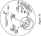
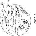


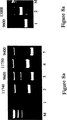


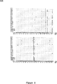


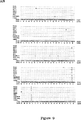

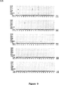


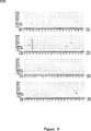

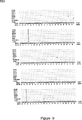


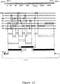

Classifications
-
- C—CHEMISTRY; METALLURGY
- C07—ORGANIC CHEMISTRY
- C07K—PEPTIDES
- C07K14/00—Peptides having more than 20 amino acids; Gastrins; Somatostatins; Melanotropins; Derivatives thereof
- C07K14/005—Peptides having more than 20 amino acids; Gastrins; Somatostatins; Melanotropins; Derivatives thereof from viruses
-
- A—HUMAN NECESSITIES
- A61—MEDICAL OR VETERINARY SCIENCE; HYGIENE
- A61K—PREPARATIONS FOR MEDICAL, DENTAL OR TOILETRY PURPOSES
- A61K39/00—Medicinal preparations containing antigens or antibodies
- A61K39/12—Viral antigens
-
- A—HUMAN NECESSITIES
- A61—MEDICAL OR VETERINARY SCIENCE; HYGIENE
- A61K—PREPARATIONS FOR MEDICAL, DENTAL OR TOILETRY PURPOSES
- A61K39/00—Medicinal preparations containing antigens or antibodies
- A61K39/12—Viral antigens
- A61K39/29—Hepatitis virus
-
- A—HUMAN NECESSITIES
- A61—MEDICAL OR VETERINARY SCIENCE; HYGIENE
- A61P—SPECIFIC THERAPEUTIC ACTIVITY OF CHEMICAL COMPOUNDS OR MEDICINAL PREPARATIONS
- A61P1/00—Drugs for disorders of the alimentary tract or the digestive system
-
- A—HUMAN NECESSITIES
- A61—MEDICAL OR VETERINARY SCIENCE; HYGIENE
- A61P—SPECIFIC THERAPEUTIC ACTIVITY OF CHEMICAL COMPOUNDS OR MEDICINAL PREPARATIONS
- A61P1/00—Drugs for disorders of the alimentary tract or the digestive system
- A61P1/16—Drugs for disorders of the alimentary tract or the digestive system for liver or gallbladder disorders, e.g. hepatoprotective agents, cholagogues, litholytics
-
- A—HUMAN NECESSITIES
- A61—MEDICAL OR VETERINARY SCIENCE; HYGIENE
- A61P—SPECIFIC THERAPEUTIC ACTIVITY OF CHEMICAL COMPOUNDS OR MEDICINAL PREPARATIONS
- A61P31/00—Antiinfectives, i.e. antibiotics, antiseptics, chemotherapeutics
-
- A—HUMAN NECESSITIES
- A61—MEDICAL OR VETERINARY SCIENCE; HYGIENE
- A61P—SPECIFIC THERAPEUTIC ACTIVITY OF CHEMICAL COMPOUNDS OR MEDICINAL PREPARATIONS
- A61P31/00—Antiinfectives, i.e. antibiotics, antiseptics, chemotherapeutics
- A61P31/12—Antivirals
-
- A—HUMAN NECESSITIES
- A61—MEDICAL OR VETERINARY SCIENCE; HYGIENE
- A61P—SPECIFIC THERAPEUTIC ACTIVITY OF CHEMICAL COMPOUNDS OR MEDICINAL PREPARATIONS
- A61P31/00—Antiinfectives, i.e. antibiotics, antiseptics, chemotherapeutics
- A61P31/12—Antivirals
- A61P31/14—Antivirals for RNA viruses
-
- A—HUMAN NECESSITIES
- A61—MEDICAL OR VETERINARY SCIENCE; HYGIENE
- A61P—SPECIFIC THERAPEUTIC ACTIVITY OF CHEMICAL COMPOUNDS OR MEDICINAL PREPARATIONS
- A61P31/00—Antiinfectives, i.e. antibiotics, antiseptics, chemotherapeutics
- A61P31/12—Antivirals
- A61P31/20—Antivirals for DNA viruses
-
- A—HUMAN NECESSITIES
- A61—MEDICAL OR VETERINARY SCIENCE; HYGIENE
- A61P—SPECIFIC THERAPEUTIC ACTIVITY OF CHEMICAL COMPOUNDS OR MEDICINAL PREPARATIONS
- A61P35/00—Antineoplastic agents
-
- C—CHEMISTRY; METALLURGY
- C12—BIOCHEMISTRY; BEER; SPIRITS; WINE; VINEGAR; MICROBIOLOGY; ENZYMOLOGY; MUTATION OR GENETIC ENGINEERING
- C12N—MICROORGANISMS OR ENZYMES; COMPOSITIONS THEREOF; PROPAGATING, PRESERVING, OR MAINTAINING MICROORGANISMS; MUTATION OR GENETIC ENGINEERING; CULTURE MEDIA
- C12N15/00—Mutation or genetic engineering; DNA or RNA concerning genetic engineering, vectors, e.g. plasmids, or their isolation, preparation or purification; Use of hosts therefor
- C12N15/09—Recombinant DNA-technology
- C12N15/63—Introduction of foreign genetic material using vectors; Vectors; Use of hosts therefor; Regulation of expression
- C12N15/79—Vectors or expression systems specially adapted for eukaryotic hosts
- C12N15/85—Vectors or expression systems specially adapted for eukaryotic hosts for animal cells
- C12N15/86—Viral vectors
-
- C—CHEMISTRY; METALLURGY
- C12—BIOCHEMISTRY; BEER; SPIRITS; WINE; VINEGAR; MICROBIOLOGY; ENZYMOLOGY; MUTATION OR GENETIC ENGINEERING
- C12N—MICROORGANISMS OR ENZYMES; COMPOSITIONS THEREOF; PROPAGATING, PRESERVING, OR MAINTAINING MICROORGANISMS; MUTATION OR GENETIC ENGINEERING; CULTURE MEDIA
- C12N7/00—Viruses; Bacteriophages; Compositions thereof; Preparation or purification thereof
-
- C—CHEMISTRY; METALLURGY
- C12—BIOCHEMISTRY; BEER; SPIRITS; WINE; VINEGAR; MICROBIOLOGY; ENZYMOLOGY; MUTATION OR GENETIC ENGINEERING
- C12Q—MEASURING OR TESTING PROCESSES INVOLVING ENZYMES, NUCLEIC ACIDS OR MICROORGANISMS; COMPOSITIONS OR TEST PAPERS THEREFOR; PROCESSES OF PREPARING SUCH COMPOSITIONS; CONDITION-RESPONSIVE CONTROL IN MICROBIOLOGICAL OR ENZYMOLOGICAL PROCESSES
- C12Q1/00—Measuring or testing processes involving enzymes, nucleic acids or microorganisms; Compositions therefor; Processes of preparing such compositions
- C12Q1/70—Measuring or testing processes involving enzymes, nucleic acids or microorganisms; Compositions therefor; Processes of preparing such compositions involving virus or bacteriophage
- C12Q1/701—Specific hybridization probes
- C12Q1/706—Specific hybridization probes for hepatitis
- C12Q1/707—Specific hybridization probes for hepatitis non-A, non-B Hepatitis, excluding hepatitis D
-
- A—HUMAN NECESSITIES
- A61—MEDICAL OR VETERINARY SCIENCE; HYGIENE
- A61K—PREPARATIONS FOR MEDICAL, DENTAL OR TOILETRY PURPOSES
- A61K39/00—Medicinal preparations containing antigens or antibodies
- A61K2039/51—Medicinal preparations containing antigens or antibodies comprising whole cells, viruses or DNA/RNA
- A61K2039/525—Virus
-
- A—HUMAN NECESSITIES
- A61—MEDICAL OR VETERINARY SCIENCE; HYGIENE
- A61K—PREPARATIONS FOR MEDICAL, DENTAL OR TOILETRY PURPOSES
- A61K38/00—Medicinal preparations containing peptides
-
- A—HUMAN NECESSITIES
- A61—MEDICAL OR VETERINARY SCIENCE; HYGIENE
- A61K—PREPARATIONS FOR MEDICAL, DENTAL OR TOILETRY PURPOSES
- A61K39/00—Medicinal preparations containing antigens or antibodies
-
- A—HUMAN NECESSITIES
- A61—MEDICAL OR VETERINARY SCIENCE; HYGIENE
- A61K—PREPARATIONS FOR MEDICAL, DENTAL OR TOILETRY PURPOSES
- A61K48/00—Medicinal preparations containing genetic material which is inserted into cells of the living body to treat genetic diseases; Gene therapy
-
- C—CHEMISTRY; METALLURGY
- C07—ORGANIC CHEMISTRY
- C07K—PEPTIDES
- C07K2319/00—Fusion polypeptide
-
- C—CHEMISTRY; METALLURGY
- C12—BIOCHEMISTRY; BEER; SPIRITS; WINE; VINEGAR; MICROBIOLOGY; ENZYMOLOGY; MUTATION OR GENETIC ENGINEERING
- C12N—MICROORGANISMS OR ENZYMES; COMPOSITIONS THEREOF; PROPAGATING, PRESERVING, OR MAINTAINING MICROORGANISMS; MUTATION OR GENETIC ENGINEERING; CULTURE MEDIA
- C12N2770/00—MICROORGANISMS OR ENZYMES; COMPOSITIONS THEREOF; PROPAGATING, PRESERVING, OR MAINTAINING MICROORGANISMS; MUTATION OR GENETIC ENGINEERING; CULTURE MEDIA ssRNA viruses positive-sense
- C12N2770/00011—Details
- C12N2770/24011—Flaviviridae
- C12N2770/24211—Hepacivirus, e.g. hepatitis C virus, hepatitis G virus
- C12N2770/24221—Viruses as such, e.g. new isolates, mutants or their genomic sequences
-
- C—CHEMISTRY; METALLURGY
- C12—BIOCHEMISTRY; BEER; SPIRITS; WINE; VINEGAR; MICROBIOLOGY; ENZYMOLOGY; MUTATION OR GENETIC ENGINEERING
- C12N—MICROORGANISMS OR ENZYMES; COMPOSITIONS THEREOF; PROPAGATING, PRESERVING, OR MAINTAINING MICROORGANISMS; MUTATION OR GENETIC ENGINEERING; CULTURE MEDIA
- C12N2770/00—MICROORGANISMS OR ENZYMES; COMPOSITIONS THEREOF; PROPAGATING, PRESERVING, OR MAINTAINING MICROORGANISMS; MUTATION OR GENETIC ENGINEERING; CULTURE MEDIA ssRNA viruses positive-sense
- C12N2770/00011—Details
- C12N2770/24011—Flaviviridae
- C12N2770/24211—Hepacivirus, e.g. hepatitis C virus, hepatitis G virus
- C12N2770/24222—New viral proteins or individual genes, new structural or functional aspects of known viral proteins or genes
-
- C—CHEMISTRY; METALLURGY
- C12—BIOCHEMISTRY; BEER; SPIRITS; WINE; VINEGAR; MICROBIOLOGY; ENZYMOLOGY; MUTATION OR GENETIC ENGINEERING
- C12N—MICROORGANISMS OR ENZYMES; COMPOSITIONS THEREOF; PROPAGATING, PRESERVING, OR MAINTAINING MICROORGANISMS; MUTATION OR GENETIC ENGINEERING; CULTURE MEDIA
- C12N2770/00—MICROORGANISMS OR ENZYMES; COMPOSITIONS THEREOF; PROPAGATING, PRESERVING, OR MAINTAINING MICROORGANISMS; MUTATION OR GENETIC ENGINEERING; CULTURE MEDIA ssRNA viruses positive-sense
- C12N2770/00011—Details
- C12N2770/24011—Flaviviridae
- C12N2770/24211—Hepacivirus, e.g. hepatitis C virus, hepatitis G virus
- C12N2770/24234—Use of virus or viral component as vaccine, e.g. live-attenuated or inactivated virus, VLP, viral protein
-
- C—CHEMISTRY; METALLURGY
- C12—BIOCHEMISTRY; BEER; SPIRITS; WINE; VINEGAR; MICROBIOLOGY; ENZYMOLOGY; MUTATION OR GENETIC ENGINEERING
- C12N—MICROORGANISMS OR ENZYMES; COMPOSITIONS THEREOF; PROPAGATING, PRESERVING, OR MAINTAINING MICROORGANISMS; MUTATION OR GENETIC ENGINEERING; CULTURE MEDIA
- C12N2840/00—Vectors comprising a special translation-regulating system
- C12N2840/20—Vectors comprising a special translation-regulating system translation of more than one cistron
- C12N2840/203—Vectors comprising a special translation-regulating system translation of more than one cistron having an IRES
- C12N2840/206—Vectors comprising a special translation-regulating system translation of more than one cistron having an IRES having multiple IRES
Description
政府援助
本発明に先行する研究は、少なくとも部分的に米国パブリックヘルスサービスの研究助成第CA57973号及びAI31501号からの助成金により援助された。したがって、政府は本発明にある種の権利を有する。
本発明の分野
本発明は機能性HCVウイルスゲノムRNA配列の決定に関し、感染HCV DNAクローンの構造に関し、治療、ワクチン及び診断応用におけるクローン、若しくはその誘導体の使用に関する。さらに本発明はHCVベクター、例えば遺伝子治療若しくは遺伝子ワクチンに関する。
発明の背景
C型肝炎ウイルスの簡潔な一般的概要
A型肝炎ウイルス及びB型肝炎ウイルス用の診断テストの開発の後、チンパンジーへ実験的に伝染させる更なる作用物質(AlterらによるLancet 1, 459-463(1978);HollingerらによるIntervirlogy 10, 60-68(1978); TaborらによるLancet 1, 463-466(1978))が、輸血による肝炎の主な原因として認識されるようになった。C型肝炎ウイルスと呼ばれる非A型非B型(NANB)肝炎の原因に相当するcDNAクローンは1989年に報告された(ChooらによるScience 244, 359-362(1989))。このブレークスルーはHCVの診断法、及び疫学、病原論及び分子ウイルス学の理解に急速な進展をもたらした(概観のためにはHoughtonらによるCurr Stud Hematol Blood Transfus 61, 1-11(1994)を参照)。HCV感染の証明は世界中で発見され、HCV特異抗体の普及率は、多くの国で0.4-2%から、エジプトでの14%までの範囲にある(HibbsらによるJ. Inf. Dis. 168, 789-790(1993))。血液若しくは血液生成物、又はあまり頻繁ではない性的及び先天性ルートを経由する伝染に加えて、周知の危険要因と関連しない散発性のケースは、HCVの場合の40%以上を占める(AlterらによるJ. Am. Med. Assoc. 264,2231−2235(1990); MastとAlterによるSemin. Virol. 4, 273-283(1993))。感染は通常慢性上の(AlterらによるN.Eng. J. Med. 327, 1899-1905(1992))、臨床上の不適切なキャリア状態から急性肝炎、慢性活性肝炎及び肝硬変にわたる結果であり、肝硬変は肝細胞癌の進展とかなり関連している。インターフェロン(IFN)-αは、慢性HCV感染(DavisらによるN. Engl. J. Med. 321, 1501-1506(1989); DiBisceglieらによるNew Engl. J. Med. 321, 1506-1510(1989))とチンパンジーモデル(ChooらによるProc. Natl. Acad. Sci. USA 91. 1294-1298(1994))においてある種の兆候を示すサブユニットワクチンのある少数の患者の治療には有用であることが示されており、より有効な治療及びワクチンを開発するために更なる効果が必要とされる。さまざまなHCV分離体の間に観測されたかなりの多様性(概観のために、BukhらによるSem. Liver DIs. 15, 41-63(1995)を参照)、慢性的に感染した個体における遺伝的変異体の発生(EnomotoらによるJ. Hepatol. 17, 415-416(1993); HijikataらによるBiochem. Biophys. Res Comm. 175, 220-228(1991); KatoらによるBiochem. Biophys. Res. Comm. 189, 119-127(1992); KatoらによるJ. Virol. 67, 3923-3930(1993); KurosakiらによるHepatology 18, 1293-1299(1993);LesniewskiらによるJ. Med. Virol. 40, 150-56(1993); OgataらによるProc. Natl. Acad. Sci. USA 88, 3392-3396(1991); WeinerらによるVirology 180, 842-848(1991); WeinerらによるProc. Natl. Acad. Sci. USA 89, 3468-3472(1992))、及びHCV感染後に惹起された保護免疫の欠如(FarciらによるScience 258, 135-140(1992); PrinceらによるJ. Infect. Dis. 165, 438-443(1992))は、上記目的に対する大きなチャレンジである。
HCVの分子生物学
分類。ゲノム構造及びビリオン性質に基づいて、HCVは、二つの他の属であるフラビウイルス(例えば黄熱(YF))と動物ペスチウイルス(例えばウシのウイルス下痢ウイルス(BVDV)と古典的ブタ熱ウイルス(CSFV)(FranckiらによるArch, Virol. Suppl.2, 223(1991))を含むフラビウイルス系統における分離属として分類されてきた。上記系統の全てのメンバーは、単一の長いオープンリーディングフレーム(ORF)の翻訳を経由して、全ての周知のウイルス特異タンパク質をコード化する(encoding)プラス鎖RNAゲノムを含むビリオンを包み込む。
ビリオンの構造及び物理的性質。HCVの構造と複製に関しては殆ど情報が入手できない。効率的なウイルス複製を支えることが可能な細胞培養の欠如と、一般的には血清に存在する感染性ウイルスの低いタイターにより研究は邪魔される。濾過実験に基づく感染性ウイルスのサイズは、30-80nmの間である(BradleyらによるGastroenterology 88, 773-779(1995); HeらによるJ. Infect. Dis. 156, 636-640(1987); YuasaらによるJ. Gen. Virol. 72, 2021-2024(1991))。スクロースにおける感染物質の浮遊密度の初期の測定により、1.1g/ml以下の低密度のものが、ある値の範囲で主に存在していることが分かった(BradleyらによるJ. Med. Virol. 34, 206-208(1991))。その後研究では、慢性的に感染したヒト若しくは実験的に感染させたチンパンジーからの血清に存在する潜在的感染性ウイルスの間接的測定として、HCV特異RNAを検出するためにRT/PCRが利用された。これらの研究から、さまざまな臨床サンプルの間にはかなりの不均一性が存在し、しかも多くの要因がHCV RNAを含む粒子の挙動に影響を与えることが、ますます明らかになってきた(HijikataらによるJ. Virol. 67, 1953-1958(1993); ThomssenらによるMed. Microbiol. Immunol. 81, 293-300(1992))。かかる要因は免疫グロブリン(Hijikataらによる(1993)の上記の文献)若しくは低密度リポタンパク質(Thomsennらによる1992の上記文献;ThomsennらによるMed. Microbil. Immunol. 182, 329-334(1993))と関連している。かなりの感染性急性期チンパンジーの血清において、HCV特異RNAは低浮遊密度(1.03-1.1g/ml)の感染で通常検出される(CarrickらによるJ. Virol. Meth. 39, 279-289(1992);Hijikataらによる(1993)の上記文献)。他のサンプルにおいて、HCV抗体の存在及び免疫複合体の形成は、高い密度と低い感染力の粒子と相関関係がある(Hijikataらによる(1993)の上記文献)。感染力を破壊するクロロホルムによる(BradleyらによるJ. Infect. Dis. 148, 245-265(1983); FeinstoneらによるInfect. Immun. 41, 816-821(1983))、若しくは非イオン性界面活性剤による粒子の処理は、HCVヌクレオキャプシドを表わすと信じられている高密度(1.17-1.25g/ml)の粒子を含むRNAを生じさせる(Hijikataらによる(1993)の上記文献;KantoらによるHepatology 19, 296-302(1994); MiyamotoらによるJ. Gen. Virol. 73, 715-718(1992))。
血清及び血漿にあるネガティブ-センスHCV特異RNAの報告がある(FongらによるJournal of Clinical Investigation 88: 1058-60(1991)を参照)。しかしながら、高感染力を有する血清は、ネガティブ-センスRNAの低レベル若しくは検出不可能なレベルであるので、かかるRNAは感染粒子の本質的成分であるとは思われない(SimizuらによるProc. Natl. Acad. USA 90: 6037-6041(1993))。
ビリオンタンパク質成分は活発には決定されていないが、推定されるHCV構造タンパク質は、塩基性Cタンパク質と二つの細胞膜糖タンパク質E1及びE2を含む。
HCV複製。HCV複製の初期の出来事は殆ど理解されていない。HCV糖タンパク質の細胞受容体は確認されていない。HCV粒子とベータリポタンパク質及び免疫グロブリンの関連性は、これらの宿主(host)分子がウイルス摂取と組織向性(tropism)を調整する可能性を高める。HCVの複製を調べる研究は、ヒト患者若しくは実験的に接種させたチンパンジーに主として限られていた。チンパンジーモデルにおいて、HCV RNAは接種後三日目に速くも血清に検出され、血清アラニンアミノトランスフェラーゼ(ALT)レベル(肝臓損傷の指標として)のピークから持続する(ShimizuらによるProc. Natl. Acad. Sci. USA 87: 6441-6444(1990))。ウイルス血症の兆候は、肝臓のHCV感染の間接的な特徴の発生を伴う。このことは細胞質抗原の発生(Shimizuらによる(1990)の上記文献)と、クロロホルムに敏感な“作用物質を形成する細管(tubule)”若しくは“TFA”(Bradleyらにより概観されているProg. Med. Virol. 37: 101-135(1990))と従来から呼ばれるHCVに対する微小管凝集物の形成のような肝細胞における超微構造の変化の発生を伴う。ウイルス抗原の発生(BlightらによるAmer. J. Path. 143: 1568-1573(1993); HiramatsuらによるHepatology 16: 306-311(1992);KrawczynskiらによるGastroenterology 103: 622-629(1992); YamadaらによるDigest. Dis. Sci. 38: 882-887(1993))及びポジティブネガティブセンスRNAの検出(Fongらによる(1991)の上記文献; GunjiらによるArch. Virol. 134: 293-302(1994); HarumaらによるJ. Hepatol. 18: 96-100(1993); LamasらによるJ. Hepatol. 16: 219-223(1992); Nouri AriaらによるJ. Clin. Inves. 91: 2226-34(1993); SherkerらによるJ. Med. Virol. 39: 91-96(1993); TakeharaらによるHepatology 15: 387-390(1992); TanakaらによるLiver 13: 203-208(1993))により示されるように、特に急性感染中では、肝細胞はHCV複製の主な部位であるらしい(NegroらによるProc. Natl. Acad. Sci. USA 89: 2247-2251(1992))。HCV感染の後段階では、HCV特異抗原の発生、ウイルス血症の持続若しくは分割、及び肝臓病の発病度は、チンパンジーとヒト患者の双方では大きく変化する。ある種の肝臓損傷は、HCV感染及び細胞変性の直接的な結果として発生するが、新たに出てきているコンセンサスは宿主免疫応答、特にウイルス特異細胞毒性Tリンパ球は細胞損傷の媒介においてより支配的な役割をする。
さらにHCVは肝臓外の貯蔵所においても複製することは推測されてきた。ある場合には、RT/PCR若しくは切片上ハイブリッドは、HCV RNAとT細胞、B細胞及び単核細胞を含む周辺の血液単核細胞との関連を示す(BlightとGowansによりViral Hepatitis Rev 1: 143-155(1995)に概観されている)。かかる細胞向性は慢性感染の確率に関連しており、HCV感染と混合寒冷グロブリン血症(FerriらによるEur. J. Clin. Invest. 23: 399-405(1993))糸球体腎炎、及び稀な非ホジキンBリンパ腫(Ferriらによる(1993)の上記文献;KagawaらによるLancet 341: 316-317(1993))のようなある種の免疫異常との間の関連性において役割を担っている。しかしながら、血清中の循環性ネガティブ(マイナス)鎖RNAの検出、本当の鎖特異RT/PCRを得る困難性(Gunjiらによる(1994)の上記文献)、及び見かけ上の感染細胞の数が少ないことは、生体外での上記組織における複製の明らかな証拠を得ることを難しくしている。
ゲノム構造。数多くのHCV分離体の完全長若しくは殆ど完全長に近いゲノム配列が、報告されている(LinらによるJ. Virol. 68: 5063-5073(1994a); OkamotoらによるJ. Gen. Virol, 75: 629-635(1994); SakamotoらによるJ. Gen. Virol, 75: 1761-1768(1994)及び上記文献の引用文献を参照)。分離体の間でかなりの遺伝的多様性があると仮定すると、幾つかの主なHCV遺伝子型が世界中に分布していることが明らかである。米国でかなり重要なタイプは、遺伝子型1、サブタイプ1a及び1bである(遺伝子型の有病率と分布は以下を参照し、Bukhらによる(1995)の上記文献を参照)。HCVゲノムRNAは長さが約9.6キロベース(kb、図1)である。5’NTRは341-344ベース(base)の長さであり、かなり保存されている。長いORFの長さは、3030、3031のポリタンパク質若しくは3033個のアミノ酸をコード化する分離体の間では僅かに変化している。報告された3’NTR構造は成分と長さ(28-42の塩基)の双方においてかなりの多様性を示し、3’末端ポリ(A)トラクト(tract)を含むある場合(HCV-1、タイプ1a)以外は、ポリ(U)で終結するようだ(ChenらによるVirology 188: 102-113(1992); OkamotoらによるGen. Virol. 72: 2697-2704(1994); TokitaらによるJ. Gen. Virol. 66: 1476-83(1994)を参照)。対照的に、我々の最近の分析により、H-菌株(タイプ1aも)のゲノムRNAは、内部ポリピリミジントラクトとそれに続く新規なRNA要素を含むことを示唆している(係属中の1995年8月29日に出願した米国特許出願08/520、678号及び1996年8月28日に出願した国際特許出願第PCT/US96/14033号)。係属中の米国特許出願第08/520、678号に示した結果からは、このタイプ1a分離体のゲノムRNAは、従来考えられていたようにホモポリマートラクトで終結せずに、むしろ約98の塩基の新規な配列で終結することが分かる。さらに、この3’NTR構造及び新規な3’末端要素は、これまでに調査された全てのHCV遺伝子型に共通の特徴である(KolykhalovらによるJ. Virol. 70: 3363-3371(1996); TanakaらによるBiochem. Biophys. Res. Comm. 215: 744-749(1996); TanakaらによるJ. Virol. 70: 3307-12(1996); YamadaらによるVirology 223: 255-261(1996))。
翻訳とタンパク質分解プロセシング(processing)。幾つかの研究は、翻訳開始における5’NTRの役割の調べるために、無細胞翻訳及び細胞培養での一過性発現を利用した(FukushiらによるBiochem. Biophys. Res. Comm. 199:425-432(1994); Tsukiyama-KoharaらによるJ. Virol. 66: 1476-1483(1992); WangらによるJ. Virol. 67: 3338-3344(1993); YooらによるVirology 1991:889-899(1992))。このかなり保存された配列は多重の短いAUG-開始PRFを含み、ペスチウイルス属の5’NTR領域と重要な相同性を示す(BuhkらによるProc. Natl. Acad. Sci. USA 89: 4942-4946(1992); Hanらによる(1991)の上記文献)。一連のステムループ構造は、コンピュータモデリングとさまざまなリボ核酸による消化に対する感度に基づいて提案されている(BrownらによるNucl. Acids Res. 20: 5041-5045(1992); Tsukiyama-Koharaらによる(1992)の上記文献)。幾つかのグループからの結果により、この要素は長いORFの第一のAUGで効率的な翻訳の開始を可能にする内部リボソームエントリ部位(IRES)として機能することが分かる(Fukushiらによる(1994)の上記文献; Tsukiyama-Koharaらによる(1992)の上記文献; Wangらによる(1993)の上記文献; Yooらによる(1992)の上記文献)。HCVとペスチウイルスIRES要素の幾つかの予想される特徴は、お互いに同じである(Browらによる(1992)の上記文献)。IRESとして機能するこの要素の能力から、HCVゲノムRNAは,5’キャップ構造が欠いていることを示唆している。
HCVポリタンパク質(図1)の組織とプロセシングは、ペスチウイルスのそれと殆ど同じである。少なくとも10個のポリペプチドは確認され、ポリタンパク質でのこれらの切断生成物の順序は、NH2-C-E1-p7-NS2-NS3-NS4A-NS4B-NS5A-NS5B-COOHである。図1に示すように、タンパク質分解プロセシングは、宿主単一ペプチダーゼ及び二つのHCVコード化(encoded)プロテイナーゼ、NS2-3オートプロテイナーゼ並びにNS3-4Aセリンプロテイナーゼにより媒介される(New YorkにあるRaven Pressにより発行された“Fields Virology”(B. N. Fields, D. M. KnipeとP. M. Howleyによる編集)のRiceによるpp.931-960;概観のためにはShimotohnoらによるJ. Hepatol. 22: 87-92(1995)を参照)。Cはウイルスコア若しくはキャプシドタンパク質であると信じられている塩基性タンパク質であり、E1及びE2は推定されるビリオンエンベロープ糖タンパク質であり、p7はE2糖タンパク質から非効率的に切断された未知の機能を有する疎水性タンパク質であり(Linらによる(1994a)の上記文献; MizushimaらによるJ. Virol. 68: 6215-6222(1994); SelbyらによるVirology 204: 114-122(1994))、同様にNS-2-NS5BはウイルスRNA複製複合体において機能する非構造(NS)タンパク質である。特にN末端セリンプロテイナーゼドメインのほかにも、NS3はRNAヘリカーゼの特徴的なモティーフを含み、RNA刺激NTPase活性を有することを示し(SuzichらによるJ. Virol. 67, 6152-6158(1993))、NS5Bは、プラス鎖RNAウイルスのRNA依存RNAポリメラーゼの特徴的なGDDモティーフを含む。
HCV RNA複製。フラビウイルスとの類似により、プラスセンスHCVビリオンRNAの複製はマイナス鎖中間体を経由して起こるものと考えられる。この戦略は以下のように簡潔に説明される:(i)進入するウイルス粒子の脱外被は、たぶん共翻訳と翻訳後に処理される単一の長いポリタンパク質を生じるように翻訳されるゲノムプラス鎖を放出し、個々の構造及び非構造タンパク質が生じる;(ii)非構造タンパク質はマイナス鎖の合成用のテンプレートとしてビリオンRNAを利用する複製複合体をたぶん形成する;(iii)上記のマイナス鎖はプラス鎖の合成のテンプレートとして働き、ウイルスタンパク質、マイナス鎖合成の更なる翻訳に利用される若しくは後代ビリオンにパッケージングされる。ウイルス増殖用の良好な実験システムがないので、HCV複製過程についての詳細はほとんど入手できない。本物のHCV複製とウイルス生命サイクルにおける他の段階の詳細な解析は、細胞培養でのHCV複製の効率的なシステムの開発により容易になるであろう。
多くの試みが、HCV感染個体から集められた血清のある培養細胞を感染させるために行われてきており、低レベルの複製はB細胞(BertoliniらによるRes. Virol. 144: 281-285(1993); NakajimaらによるJ. Virol. 70; 9925-9(1996); ValliらによるRes. Virol. 146: 285-288(1995))、T細胞(KatoらによるBiochem. Biophys. Res. Commun, 206: 863-9(1996); MizutaniらによるBiochem. Biophys. Res. Comm. 227: 822-826;MizutaniらによるJ. Virol. 70: 7219-7223(1996); Nakajimaらによる(1996)の上記文献; ShimizuとYoshikuraによるJ. Virol. 68: 8406-8408(1994); ShimizuらによるProc. Natl. Acad. Sci. USA 89:5477-5481(1992); ShimizuらによるProc. Natl. Acad. Sci. USA 90: 6037-6041(1993))と肝細胞(KatoらによるJpn. J. Cancer Res., 87: 787-92(1996); Tagawa, J. Gastoenterol. and Hepatol., 10: 523-527(1995))、細胞系統と同様に周辺の血液単核細胞(PBMCs)(CribierらによるJ. Gen. Virol., 76: 2485-2491(1996))とヒト致死肝細胞の一次培養(CarloniらによるArch. Virol. Suppl. 8: 31-39(1993); Cribierらによる(1995)の上記文献; IacovacciらによるRes. Virol., 144: 275-279(1993))若しくは大人のチンパンジーからの肝細胞(LanfordらによるVIrology 202: 606-14(1994))を含む方法により感染された多くの細胞タイプに報告されている。さらにHCV複製は、培養前に(ItoらによるJ. Gen. Virol. 77: 1043-1054(1996))生体内でウイルスに感染したヒトHCV患者から取出された一次肝細胞において、HCV-1cDNAクローンからの生体外で転写されたRNAでのトランスフェクションに続くヒト肝細胞系統Huh7において(YooらによるJ. Virol., 69: 32-38(1995))検出された。HCV-1クローンはホモポリマーの3’NTR配列下流を欠いているので、HCV-1クローンから誘導されたRNAでトランスフェクト(transfect)された細胞での複製の観察報告はわけがわかならい(以下を参照)。HCV複製用の最もよく特徴付けられる細胞培養は、レトロウイルス(HPB-Ma若しくはMT-2)で持続的に感染されるB細胞系統(Daudi)若しくはT細胞系統を利用する(Katoらによる(1995)の上記文献; MizutaniらによるBiochem. Bioohys. Res. Comm., 227: 822-826(1996a); Mizutaniらによる(1996)の上記文献; Nakajimaらによる(1996)の上記文献; ShimizuとYoshikuraによる(1994)の上記文献; ShimizuによるProc. Natl. Acad. Sci. USA, 90: 6037-6041(1993))。HPBMaはマウス肉腫ウイルスの両栄養性マウス白血病ウイルスで感染するのに対し、MT-2はヒトT細胞リンパ栄養性ウイルスタイプI(HTLV-I)で感染する。非クローン集団より効率的にHCV複製を助けるクローン(HPBMa10-2及びMT-2C)は、二つのT細胞系統HPBMa及びMT-2のために分離された(MizutaniらによるJ. Virol,(1996)の上記文献; Shimizuらによる(1993)の上記文献)。しかしながら、インプットRNAの分解後の上記細胞系統若しくはDaudi系統において得られたRNA複製の最大レベルは、依然として106細胞当たりたったの約5x10⁴RNA分子(Mizutaniらによる(1996)の上記文献; Mizutaniらによる(1996)の上記文献)若しくは培地のml当たり104RNA分子(Nakajimaらによる(1996)の上記文献)である。複製のレベルは低いが、一つのシステム(MizutaniらによるBiochem. Biophys. Res. Comm. 227: 822-826(1996a))において198日までの長期間の感染、別のシステム(Nakajimaらによる(1996)の上記文献)において一年以上の感染が報告されており、感染ウイルス生産は固有(naive)細胞へのウイルスの系列無細胞若しくは細胞媒介継代により実証された。
しかしながら、効率的なHCV複製は今までのところいずれの細胞培養でも観測されておらず、複製、マイナス鎖の誤った検出、及び複製されたRNAの一般的に低いタイターにより生成したプラス鎖からインプットRNAを識別する困難さを含む多くの問題に遭遇するかかるシステムを確立するように、全てのグループにより試されている。よって、以上の進展にもかかわらず、HCV増殖用のより効率的な細胞培養システムは、高濃度のウイルスストックの生産、ビリオン成分の構造解析、及びRNA複製を含む細胞内でのウイルス過程の改善された解析のために必要である。
ビリオン構築及び放出。この過程は直接調べられておらず、複合多糖の欠如、発現HCV糖タンパク質のER局在化(DubuissonらによるJ. Virol. 68: 6147-6160(1994); RalsonらによるJ. Virol. 67: 6753-6761(1993))及び細胞表面に上記タンパク質の不存在(Dubuissonらによる(1994)の上記文献; SpaeteらによるVirology188: 819-830(1992))により、初期のビリオンの形態形成は、細胞内のベシクルにバディング(budding)することにより起こることを示唆している。これまでのところ、効率的な粒子形成と放出は一過性発現検定において観測されておらず、本質的なウイルス若しくは宿主要因は存在しない又はブロックされていることを示唆している。ウイルスの実質的なフラクション(fraction)は、ペスチウイルスで発見されたように細胞に関連したままであるので、HCVビリオン形成と放出は非効率的である。最近の研究により、ヒト血漿から部分的に精製された細胞外HCV粒子は複合N結合多糖を含むが、これらの炭水化物部分はE1又はE2と特別に関連していないことを示す(SatoらによるVirology 196: 354-357(1993))。放出ビリオン上の糖タンパク質と関連する複合多糖は、トランスゴルジ体を通して移動し、宿主分泌経路を通してビリオンの移動を示唆している。このことが正しければ、HCV糖タンパク質の細胞内セクエストレーション(sequestration)及びビリオン形成は、免疫監視を最小化し、抗体及び補体(complement)によりウイルス感染細胞の溶菌(lysis)を防止することにより慢性感染の確立に重要な役割を担う。
遺伝的変異性。全てのプラス鎖RNAウイルスに関して、HCV(NS5B)のRNA依存RNAポリメラーゼ(RDRP)は、取込み違いの塩基の除去のための3’-5’エキソヌクレアーゼ校正活性が欠如していると信じられている。したがって、複製は誤りがちであり、多くの数の変異体から成る“準種(quasi-species)”ウイルス集合体へ導く(MartellらによるJ. Virol. 66: 3225-3229(1992); MartellらによるJ. Virol. 68: 3425-3436(1994))。この変異性はマルチレベルにおいて明らかである。第一に、慢性的に感染した個体において、ウイルス集合体における変化は時がたつにつれて起こり(Ogataらによる(1991)の上記文献; OkamotoらによるVirology 190: 894-899(1992))、上記変化は病気に重要な結果をもたらす。特定の興味ある例としては、E2糖タンパク質のN末端の30残基セグメントがあり、これはポリタンパク質の残りよりもかなり高い変異性を示す(例えば、HigashiらによるVirology 197, 659-668. 1993; Hijikataらによる(1992)の上記文献; Weinerらによる(1991)の上記文献)。たぶんHIV-1 gp120のV3ドメインに類似するこの超可変性領域は、HCV特異抗体を循環させることによる免疫選択下にあることを証明するものが蓄積されている(Katoらによる(1993)の上記文献; TaniguchiらによるVirology 195: 297-301(1993);Weinerらによる(1992)の上記文献)。このモデルにおいて、E2の上記部分に対して向けられた抗体は、ウイルス中和に寄与し、よって中和から逃げることを可能にする置換により変異体の選択を引き起こす。この柔軟性は、E2超可変性領域における特異アミノ酸配列はビリオン吸着(attachment)、浸透若しくは集合のようなタンパク質の他の機能に対して必須ではないことを示唆している。
さらに遺伝的変異性は、慢性感染患者のIFN-α治療後に観測されたさまざまな応答のスペクトルに寄与する。減少血清ALTレベルと、循環するHCV RNAのレベルの減少と通常相関関係にある改善肝組織学は、約40%の治療で確認される(Greiser-WilkeらによるJ. Gen. Virol. 72: 2105-2019(1991))。治療後、約70%の応答個体が再発する。ある場合には、循環ウイルスRNAの一過性損失後に、治療中若しくは治療後に新たなウイルス血症が観測される。このことは、IFN耐性HCV遺伝子型若しくは変種体の存在若しくは発生を示唆するが、更なる研究が、ウイルス遺伝子型の相対的寄与及び免疫応答における宿主特異的差異を決定するために必要である。
最終的に、世界中のさまざまなHCV分離体の配列比較により、莫大な数の遺伝的多様性を示している(Bukhらによる(1995)の上記文献を参照して概観される)。交差中和テストのような生物学的に関連する血清学的検定がないので、HCVタイプ(数により示される)、サブタイプ(文字により示される)、及び分離体は、ヌクレオチド若しくはアミノ酸配列類似性に基づいて、現在分類されている。多くの多様な遺伝子型間のアミノ酸配列類似性は、比較されたタンパク質に依存して、約50%ほどである。この多様性は、特に診断、ワクチン設計及び治療によっては重要な生物学的意味を有する。
cDNAからの感染性HCV転写物(transcript)を生じさせるための他人による試み
最近の論文(YooらによるJ. Virol. 69: 32-38(1995))には、Huh7細胞のトランスフェクション後の転写HCV-1 RNAの複製が報告されている。この論文では、HCV-1 cDNAクローンの種々の誘導体からのT7転写物は、ヒト肝癌細胞系統、Huh7の次に続くトランスフェクションを複製させる能力をテストした。起こり得るHCV複製は、鎖特異RT/PCR(5’NTRプライマーを利用して)と3H-ウリジンのあるHCV特異RNAの代謝ラベリングにより評価された。明らかに、ポリ(A)若しくはポリ(U)で終結している完全長転写物は、上記評価によりポジティブ(プラス)であったが、5’末端144塩基に欠失のある転写物は存在しなかった。ある培養では、HCV特異RNAは培地で検出され、この推定されるウイルスはフレッシュなHuh7細胞を再感染させるために利用された。
本発明者らは、上記結果を再現させることができなかった。上記報告は、複製とウイルス生産の本物のHCV感染よりもむしろ一過性複製を報告しているようである。例えば、上記論文で報告されているポジティブコントロール(positive control)は、高いタイターのチンパンジー血漿から抽出されたRNAのあるHuh7細胞の生産的トランスフェクションであった。この抽出サンプルは、潜在的感染性のある完全長のHCV RNAの最大107個の分子を含んでいたであろう。(顕微注射以外の)最適トランスフェクション条件下において、一般にはビリオンRNAの105個以上のRNA分子(少なくともポリオウイルス、シンドビスウイルス若しくはYFによっては)は、一つの感染イベントを開始させる必要がある。このことは100個よりも少なく報告されたHCV-1実験では、細胞は生産的にトランスフェクトされるであろうことを示唆している。さらに、トランスフェクションの16日後、ポジティブ及びネガティブ鎖RNAの双方は、代謝ラベリングの8時間後に繰り返して検出された。この方法によるマイナス鎖RNAの検出(トランスフェクトされたビリオンRNAと転写RNAの双方)は、HCVは効率的な複製とスプレッド(spread)の双方が可能であり、しかもHCV RNA合成のレベルは、YFのようなより強いフラビウイルス(高い多重感染のピークで)で予期されるレベルと同じである。しかしYooらは多様な抗血清を利用した上記細胞においてHCV抗原の検出は報告しておらず、さらにノーザン分析(3H-ウリジンによる代謝ラベリングよりもかなり高感度である)による完全長のポジティブ及びネガティブ鎖の検出も報告していない。最終的に、RNA若しくはHCV-1クローンから誘導されたウイルスが、チンパンジーモデルで感染されたことを示す重大な実験は報告されていない。
HCV研究用の感染クローン技術の重要性
ここ最近の数年における大きな進歩にもかかわらず、HCV複製、病原論及び免疫に関する数多くの疑問は未解決のままである。この分野は、我々にHCV RNAゲノム及びそのコード化されたタンパク質の機能のある面を理解させ、本物のウイルスの複製関係において構造/機能の疑問を実験的にテストさせる方法を全く有しないボトルネックに急速に近づいている。かかる分析は、保護ワクチン、有効な治療法及びHCV診断薬の設計を可能にするために、ウイルス生命サイクルにおける各段階を理解することは重要である。よって、この技術分野では感染HCV RNAの発現のための本物のHCV遺伝物質が必要である。
さらにこの技術分野では、固有(naive)HCVビリオン及びHCVビリオン構造の特徴付けを可能にするウイルス粒子タンパク質の発現のための本当の遺伝物質が必要である。
さらにこの技術分野には、HCVレセプター結合、細胞感染、複製、ビリオン集合体及び放出の分析を可能にする感染性HCVに対する生体外での培養方法が必要である。
この技術分野における上記及び他の必要なことは、本発明で扱われる。
本願での参考文献の引用は、かかる参考文献が緊急出願の“先行技術”として利用可能であることの承認として、解釈されるべきではない。
発明の要約
本発明は複製、機能的HCVタンパク質の発現及び坑ウイルス性治療及び診断薬の開発のための生体内と生体外での感染が可能な本物のC型肝炎ウイルス(HCV)DNAクローンを提供する。
広い態様において、本発明は、ポジティブ-センス核酸上の5’から3’と、一番端の(extreme)5’末端保存配列から成る機能性5’非翻訳領域(NTR)と、切断生成物がHCVウイルス粒子とRNA複製機械装置(replication machinery)の機能構成部品を形成するHCVポリタンパク質の少なくとも一部分をコード化するオープンリーディングフレーム(ORF)と、一番端の3’末端保存配列から成る3’非翻訳領域(NTR)と、若しくは適応ウイルス、弱毒化ウイルス、複製能のある非感染ウイルス、及び欠損ウイルスから成る群から選ばれる誘導体から成る遺伝子的に特別設計された(engineered)C型肝炎ウイルス(HCV)核酸クローンに関する。本発明者らにより、遺伝子工学技術を利用して実施される多くの操作は、本物のHCV核酸、例えば感染HCV RNAを生じるように転写されるcDNAを発生させることが必要であることが分かった。特別設計された本物のHCV核酸を提供することにより、本発明者らは、HCV複製機械装置とタンパク質活性の解剖、及び多様なHCV誘導体の調製を始めて可能にした。従来では、ある一定のHCVクローンが機能することが不可能になる誤り若しくは突出然変異を含んでいるかどうか不確実であったので、HCVの更なる分析用の出発物質が、人工物(artifact)により有用であるか又は簡単であるか確かではなかった。よって、本発明の大きな利点は、本物のHCVを提供し、よって変種が先行技術で提供されたクローンでの誤りによる人工物よりもむしろ本当の変化をもたらすことを保証することにある。
本発明の更なる利点は、感染HCVゲノム、特にポリタンパク質暗号づけ(coding)領域の特徴の認識にある。特定実施例において、HCV核酸は、HCV分離体若しくは遺伝子型の少なくと三つのクローンの大部分の配列から決定されたコンセンサス核酸配列を有する。HCV核酸は、本願で実証された本発明の特定実施例を表わすSEQ ID NO:1に示すような配列の少なくとも機能部分を有することが好ましい。SEQ ID NO:1はDNA配列であるが、本発明は対応するRNA配列、及びDNAとRNAの相補的配列のことも考えていることを指摘しておくべきである。更なる実施例において、HCV分離体からの領域は、例えばSEQ ID NO:1の配列を有するHCV核酸の相同領域と置換される。本願で実証された更に好ましい実施例において、HCV核酸は、複製能のあるHCV RNA複製用の発現上に遺伝暗号を指定する(code)DNAであり、又は自身は複製能のあるHCV RNAレプリコンである。以下の特定例において、本発明のHCV核酸はSEQ ID NO:1に示された若しくは対応する完全長配列を有する。さらに5’及び3’のさまざまな変異は、本発明により熟慮される。例えば5’末端配列は、GCCAGCC;GGCCAGCC;UGCCAGCC;AGCCAGCC;AAGCCAGCC;GAGCCAGCC;GUGCCAGCC;及びGCGCCAGCCから成る群から選ばれるRNA配列に相同若しくは相補的であり、ここで配列GCCAGCCはSEQ ID NO:3の5’末端である。本発明の更に別の利点は、感染HCVクローン用の完全3’-NTRの重要性のデモンストレーションである。3’-NTR、特にHCV遺伝子型の間でかなり保存されている約98の塩基の一番端の末端配列は、本願の全体の参考文献中に引用されている1995年8月29日に出願された米国特許出願番号08/520、678号と、これも本願の全体の参考文献中に引用されている1996年8月28日に出願されたPCT国際特許出願第PCT/US96/14033号の主題である。よって、好ましい態様において、機能性3’NTRは、HCV遺伝子型の中でかなり保存されている約98の塩基の3’末端配列から成る。特定実施例において、3’NTRの一番端にある末端は、配列5’-GGTGGCTCCATCTTAGCCCTAGTCACGGCTAGCTGTGAAAGGTCCGTGAGCCGCATGACTGCAGAGAGTGCTGATACTGGCCTCTCTGCTGATCATGT-3’(SEQ ID NO:4)を有するDNAに相同若しくは相補的である。SEQ ID NO:1で実証された特定実施例において、3’NTRは長いポリピリミジン領域(例えば約133の塩基)から成るが、代わりのポリピリミジン領域は、短い領域(約75の塩基)、若しくは短い又は長い領域を含んでさらに包み込まれる。当然、ポジティブ(プラス)鎖HCV DNA核酸において、ポリピリミジン領域はポリ(T/TC)領域であり、ポジティブ鎖HCV RNA核酸において、ポリピリミジン領域はポリ(U/UC)領域である。
本発明のさまざまな態様によれば、ポリタンパク質暗号づけ領域を含むHCV核酸は突然変異し、若しくは特別設計されて、例えばサイレント突然変異、保存突然変異などのある変異体若しくは誘導体が生じる。さらにかかるクローンは、例えば動物若しくは生体外で増殖のための選択により適応される。さらに本発明は、HCVキメラの生成を可能とし、他の遺伝子型若しくは分離体のゲノム部分は、SEQ ID NO:1又は以下の付着(deposited)実施例のようなHCVクローンの相同領域と置換される。更に別の実施例において、本発明はHCV-1、HCV-1a、HCV-1b、HCV-1c、HCV-2a、HCV-2b、HCV-2c、HCV-3a、及びそれらの“準種”変異体から成る群から選ばれるHCV遺伝子型からのポリタンパク質暗号づけ配列から成るクローンの調製方法を提供する。更なる好ましい実施例において、サイレントヌクレオチドは、特定HCVクローンのマーカーとして取込まれたポリタンパク質暗号づけ領域において変化する(つまり同じアミノ酸をコード化するコドンの3つの塩基の変異)。
本発明の更なる態様において、弱毒化および欠損変異体を含むHCV核酸は、発現制御配列と操作上関連した異種遺伝子から成り、ここで異種遺伝子及び発現制御配列はプラス鎖核酸分子上に配向される。特定の実施例において、異種遺伝子は、HCVポリタンパク質暗号づけ配列とのインフレーム融合と追加的シストロンの生成から成る群から選ばれる戦略により挿入される。異種遺伝子は抗生物質耐性遺伝子又はレポーター遺伝子である。あるいは、異種遺伝子は、治療遺伝子若しくはワクチン抗原をコード化する遺伝子、つまり夫々遺伝子治療若しくは遺伝子ワクチン応用のための遺伝子である。異種遺伝子が抗生物質耐性遺伝子である特定の実施例において、抗生物質耐性遺伝子は、3’NTRにおけるSfiL部位に挿入された内部リボソームエントリ部位(IRES)と操作上関連するネオマイシン耐性遺伝子である。
上記したように当然、本発明のHCV核酸は、二本鎖DNA、ポジチブ-センスcDNA、若しくはネガティブ-センスcDNA、又はポジチブ-センスRNA又はネガティブ-RNAから成る群から選ばれる。よって本発明の核酸の特定の配列が明らかにされると、DNA及び対応するRNA双方は、ポジティブ及びネガティブ鎖のDNA及びRNAを含むことが意図される。
HCV DNAは対応するHCV RNAの翻訳用のプラスミドベクターに挿入される。よって、HCV DNAはポジティブ−センスDNA上の5’-NTRのプロモーター5’から成り、ここでプロモーターからのテンプレートDNAの転写は、複製能のあるRNAを生じさせる。プロモーターは真核性プロモーター、イーストプロモーター、植物プロモーター、バクテリアプロモーター、若しくはウイルスプロモーターから成る群から選ばれる。以下の特定例において、ファージT7及びSP6プロモーターが利用される。特定実施例において、本発明はプラスミドクローン、p90/HCVFL(長いポリ(U))に関し、これは1997年2月13日に米国のメリーランド州ロックビル、パークローンドライブにあるAmerican Type Culture Collection(アメリカンタイプカルチャーコレクション)に寄託された、SEQ ID NO:5に示される配列を有する受託番号97879号と帰属される感染HCV RNA転写物を生じるように転写された完全長のHCV cDNAに宿る。当然、さらに本発明は、5’末端配列がGCCAGCC,GGCCAGCC、UGCCAGCC、AGCCAGCC,AAGCCAGCC、GAGCCAGCC、GUGCCAGCC,及びGCGCCAGCCから成る群から選ばれるRNA配列に相同若しくは相補的であるプラスミドの誘導体を含み、ここで配列GCCAGCCは、SEQ ID NO:3の5’末端と3’NTRが短いポリピリミジン領域から成る誘導体である(好ましい付着実施例では長いポリピリミジンを有するからである)。更なる実施例において、付着実施例の誘導体は、他のHCV分離体又は遺伝子型からの相同領域の置換により生じた誘導体、突然変異誘発により生じた誘導体、適応された、弱毒化された、複製能のある非感染性、及び欠損変異体から成る群から選ばれた誘導体、発現制御配列と操作上関連する異種遺伝子から成る誘導体、及び上述の誘導体のいずれかの機能フラグメントから成る誘導体から成る群から選ばれる。あるいは、5’NTR、ポリタンパク質暗号づけ領域、3’NTR若しくはHCVゲノムのより一般的な暗号づけ又は非翻訳領域のような付着DNAクローンの部分は、さまざまなHCV遺伝子型からの対応する領域で置換され、新しいキメラ感染クローンが生じる、若しくは他の分離体及び遺伝子型の拡張感染クローンにより生じる。例えば、HCV-1b若しくは-2aポリタンパク質暗号づけ領域(あるいは共通ポリタンパク質暗号づけ領域)は、付着クローンのHCV-H(1a菌株)ポリタンパク質に置換される。
当然、更に本発明は、HCV DNA若しくは上記に示したプラスミドクローンに宿る完全長のHCV cDNAから転写されたRNAを提供する。
よって、特異HCVゲノム自身はHCVの変種変異体を誘導するための優れた出発物質を提供する。なぜなら変種は変化及び誤りの蓄積からの結果から生じる人工物よりむしろ、本物のウイルスへの変化から生じるからである。本発明のHCV DNAクローン若しくはRNAは多くの方法、又は以下に明らかにするように本物のHCV成分を誘導するために利用される。
例えば、本発明は上記に再度引用されたプラスミドクローンから生産されるHCV RNAの感染量で組織培養中の細胞系統と接触させることから成るHCVとの感染を許容する細胞系統を確認する方法を提供し、細胞系統の細胞中のHCVの複製を検出する。当然、本発明は、例えば上記プラスミドにより生産されるHCV RNAの感染量を動物に導入することから成るHCVで感染を許容する動物を確認する方法にも拡張され、動物中でのHCVの複製を検出する。優性選択可能なマーカー(dominat selectable marker)から成ることが好ましい本物の感染性HCVを与えることにより、本発明は、培養中の細胞系統に接触させる、若しくはHCV RNAの感染量を動物に導入し、細胞系統又は動物におけるHCV RNAの進行的に増加するレベルを検出することから成る許容細胞系統又は動物での高いレベルのHCV複製を可能にする適応突然変異でHCVを選択する方法をさらに提供する。特定実施例において、適応突然変異はHCV向性の変形を可能にする。本発明の上記態様の直接的に意味することは、HCV感染に対する新しい有効な動物モデルの生産である。
本発明の核酸を利用して確認された許容細胞系統又は動物は、とりわけHCV感染の自然の歴史を研究する、HCVの機能成分を分離するため、及び本物のHCVウイルス若しくはそれらの成分を生産することに加えて、感度よく迅速に診断を行うために、非常に有用である。上述したように、本発明の特定の利点は、動物又は細胞系統で生産的感染を開始させることが可能なHCV DNAクローンを、始めてうまく調製できたことにある。
本発明のHCV DNA、例えばプラスミドベクターは本物のHCV成分をコード化するので、HCV DNAによりトランスフェクト、トランスフォーム、若しくはトランスデュースされた宿主細胞系統におけるかかるベクターの発現は実行される。例えば、バキュロウイルス又は植物発現システムは利用され、HCVウイルス粒子若しくはそれらの成分を発現する。よって、宿主細胞系統は細菌細胞、酵母細胞、植物細胞、昆虫細胞及び哺乳類細胞から成る群から選ばれる。
本発明は、とりわけ感染HCV RNAを提供するので、本発明は上述したプラスミドから転写されたHCV RNAのようなHCV DNAの感染供給量(dose)を動物に投与することから成るHCVで動物を感染させる方法を提供する。当然、本発明は本発明のHCVで感染された非ヒト動物を提供し、非ヒト動物は前述の方法により調製される。
本発明の更なる利点は、完全な機能性HCVゲノムを与えることにより、サブユニット発現システムにおいて不可能である方法による固有HCVタンパク質又はRNAで生産された本物のHCVウイルス粒子又はそれらの成分を調製することにある。加えて、本発明のHCVの各成分は機能的であり(よって本物のHCVが発生する)、ある特定成分は本物の成分である、つまり少なくとも一部は先行技術のクローンに影響を与える誤りが欠けている。実際に、本発明の更なる利点は、天然HCVビリオン若しくはタンパク質と構造的に同じ又はかなり関連しているHCVウイルス粒子又はウイルス粒子を発生させる能力にある。よって、更なる実施例において、本発明は、本発明のHCV RNAの感染量と、例えばHCV RNAの複製を可能とする条件下において上述したプラスミドから翻訳されたHCV RNAと接触させた細胞系統を培養させることから成る生体外でHCVを増殖させる方法を提供する。当然、本発明はHCVで感染された生体内の細胞系統にまで拡張され、ここでHCVは上述したようにゲノムRNA配列を有する。特定実施例において、細胞系統は肝細胞系統である。更に本発明は、本発明のHCV感染させた非ヒト動物からのHCVウイルス粒子を分離し、HCV複製及びウイルス粒子生成を可能にする条件下で本発明の細胞系統を培養させる、又はHCV粒子タンパク質の発現を可能にする条件下で、HCV DNAでトランスフェクトさせた宿主発現細胞系統を培養し、細胞培養からHCV粒子若しくは粒子タンパク質を分離することを含み、HCVウイルス粒子を生産させるさまざまな方法を提供する。本発明は、複製成分HCVゲノムRNA、若しくは本発明のHCV核酸に対応する複製欠損HCVゲノムRNAから成るHCVウイルス粒子に拡張される。
HCV核酸、例えばDNA若しくはRNAベクターにおける異種遺伝子を挿入させることにより、本発明は、例えばHCV RNAで動物を効果的に感染させるために、HCV RNAのある量を動物へ投与することによる遺伝子治療若しくは遺伝子予防接種のために、異種遺伝子でHCV感染しやすい動物をトランスデュースする方法を提供する。特定実施例において、かかるHCVベクターは上述したプラスミドに宿るHCVにおいて発生する。
例えば前述した本発明のプラスミドから転写されたように、本発明のHCVゲノムテンプレートRNAから成るHCV用の生体外無細胞システムは、機能性HCVレピリカーゼ成分及びリボヌクレオチドトリホスフェート塩基から成る等張性緩衝培地を提供する。上記要素は複製機械装置及び原材料(NTP)を提供する。
本物のHCVウイルス粒子及びウイルス粒子タンパク質は、HCV抗原として好ましい出発物質である。よって、更なる実施例において、本発明はHCVウイルス粒子の免疫原性量を動物に投与し、動物からアンチHCV抗体を分離させることから成るHCVへの抗体を生産させる方法を提供する。かかる抗体は、例えばHCVの存在を検出するために,診断的に利用される、若しくは例えば受身免疫治療において治療的に利用される。HCVへの抗体を生産させる更なる方法は、本発明のHCVウイルス粒子との反応性のヒト抗体ライブラリーをスクリーニングすることと、HCVウイルス粒子と反応性のある抗体を発現させるライブラリーからのクローンを選択することとから成る。当然、抗体を発生させることに加えて、本発明の本物のHCVウイルス粒子及びタンパク質はHCVワクチン用の好ましい出発物質である。好ましくは、本発明のワクチンは薬学的に許容なアジュバントを含む。
本願で提供された本物の物質は、生体外及び生体内でHCV複製を調節することが可能な作用物質用のスクリーニング法を提供する。かかる方法は、候補物質を本発明のHCV感染動物へ投与し、HCV感染若しくは活性レベルの増加又は減少を、候補物質投与前の動物におけるHCV感染若しくは活性レベルと比較してテストすることを含み、ここで候補物質の投与前の動物におけるHCV感染もしくは活性レベルと比較して、HCV感染若しくは活性レベルの減少は、HCV感染若しくは活性を阻害する能力があることを示している。HCV感染レベルをテストすることは、動物からの組織サンプル中のウイルスタイターを測定し、動物からの組織サンプル中のウイルスタンパク質を測定し、若しくは肝臓酵素を測定することにより実施される。あるいは、動物を感染させるのに利用されたHCVゲノムは、発現制御配列に操作上関連する異種遺伝子を含み、ここで異種遺伝子及び発現制御配列はプラス鎖核酸分子上に配向され、HCV活性レベルのテスト法は、動物からの組織サンプル中のマーカータンパク質のレベルを測定することから成る。
あるいは、かかる分析は、例えば候補物質で請求項32の細胞系統と接触させ、候補物質を投与する前のコントロール細胞系統若しくは細胞系統におけるHCV感染又は活性のレベルと比較して、HCV感染又は活性レベルの増加又は減少をテストすることにより生体外で行われ、ここで候補物質の投与前のコントロール細胞系統若しくは細胞系統におけるHCV感染又は活性レベルと比較して、HCV感染又は活性レベルが減少していることは、HCV感染又は活性を阻害させる能力があることを示している。生体外でHCV感染レベルをテストすることは、細胞、培地、若しくはその両方でウイルスタイターを測定することと、細胞、培地、若しくはその両方でウイルスタンパク質を測定することにより実施される。あるいは、細胞系統を感染させるために利用されたHCVゲノムが発現制御配列と操作上関連する異種遺伝子を含むとき、ここで異種遺伝子及び発現制御配列がポジティブ鎖核酸分子上に配向されているが、HCV活性レベルをテストする方法は、動物からの組織サンプル中のマーカータンパク質レベルを測定することから成る。
HCV複製の調節可能な作用物質をスクリーニングする更なる方法は、上述の無細胞システムを含む。この方法は、本発明の生体外でのシステムを接触され、候補物質の投与前のコントロール細胞システム若しくはシステム中のHCV複製レベルと比較して、HCV複製レベルの増加若しくは減少をテストすることから成り、ここで候補作用物質の投与前のコントロール細胞系統若しくは細胞系統中でのHCV複製レベルと比較して、HCV複製レベルが減少していることは、HCV感染又は活性を阻害する能力があることを示している。
本発明は、プラスセンスDNA上の5’から3を接合し、一番端の5’末端保存配列から成る機能性5’非翻訳領域(NTR)と、機能性HCVタンパク質の発現のために与えるHCVタンパク質をコード化するポリタンパク質暗号づけ領域と、一番端の3’末端保存配列からなる3’非翻訳領域(NTR)から成るHCV核酸を調製する方法を含む。更に上記方法は、5’-NTR、ポリタンパク質暗号づけ配列、及びHCV分離体又は遺伝子型の少なくとも三つのクローンの大部分の配列からの3’-NTRのコンセンサス配列を決定することから成る。特定実施例において、3’-NTRは、配列5’-GGTGGCTCCATCTTAGCCCTAGTCACGGCTAGCTGTGAAAGGTCCGTGAGCCGCATGACTGCAGAGAGTGCTGATACTGGCCTCTCTGCTGATCATGT-3’(SEQ ID NO:4)を有するDNAに相同である一番端の末端配列から成る。更なる特定実施例において、HCV核酸は、描かれた若しくは他のHCV分離体又は遺伝子型からの相同領域の置換から成るSEQ ID NO:1に対応するポジティブ鎖配列を有する。
さらに本発明は、重要な診断上の意味を有する。一実施例において、例えばHCVウイルス粒子とサンプル中のHCV特異抗体とを結合させることを可能にする条件下において、上述のように調製された本発明のHCVウイルス粒子で被験者からの生物学的サンプルを接触させ、HCVウイルス粒子とサンプル中の抗体との結合を検出することから成る、生物学的サンプルにおけるHCVへの抗体を生体外で検出する方法を提供し、ここでHCVウイルス粒子とサンプル中の抗体との結合を検出することは、サンプル中のHCVへの抗体の存在を示す。
被験者からの生物学的サンプル中のHCVの存在を検出する生体外での代わりの方法は、生産的HCV感染を許容する細胞系統と生物学的サンプルとを接触させることから成り、ここで細胞系統はHCVにより生じたトランス-作用要因(trans-acting factor)の制御下において発現されたレポーター遺伝子生成物を発現する導入遺伝子(transgene)を含むように変種され、レポーター遺伝子生成物の発現を検出し、さらにここでは、レポーター遺伝子生成物の発現を検出することは、被験者からの生物学的サンプル中にHCVが存在することを示している。関連した実施例において、本発明は、生物学的サンプルと生産的HCV感染を許容する細胞系統とを接触させ、レポーター遺伝子生成物の発現を検出することから成る被験者からの生物学的サンプル中のHCVの存在を生体外で検出する方法を提供し、ここで細胞系統はHCVにより生じるトランス-作用要因の制御下において高いレベルでレポータ遺伝子生成物を発現する欠陥ウイルス導入遺伝子を含むように変種され、レポーター遺伝子生成物の発現の検出は被験者からの生物学的サンプル中にHCVが存在していることを示す。よって、本発明の重要な利点は、上記の生体外での診断用の許容(感染しやすい)細胞系統を提供することにある。請求項64による方法では、欠損ウイルス導入遺伝子は特別に設計されたアルファウイルスを生じ、トランス-作用ヘルパー要因はアルファウイルスnsP4ポリメラーゼであり、ここでアルファウイルスnsP4ポリメラーゼは、HCV NS4Aとのキメラ融合タンパク質として発現され、アルファウイルスnsP4ポリメラーゼ-HCV NS4Aキメラ融合タンパク質はHCV NSプロテイナーゼにより切断され、機能性アルファウイルスnsP4ポリメラーゼを放出する。前述の方法において生物学的サンプルは血液、血清、血漿、血球、リンパ球、及び肝臓組織バイオプシーから成る群から選ばれる。
関連した態様において、さらに本発明は本物のHCVウイルス成分から成るHCV用テストキットと、本物のHCVウイルスから誘導される成分から成るHCV用診断テストキットを提供する。
よって本発明の主要目的は、感染HCVをコード化するDNAを提供しなければならない。
本発明の関連目的は、DNAクローンからの感染HCVゲノムRNAを提供することである。
本発明の更に別の目的は、細胞に侵入するが感染ウイルスを増殖させないワクチン開発に適する弱毒化されたHCV DNA若しくはゲノムRNAを提供することである。
本発明の別の目的は、アンチHCV(または坑ウイルス性)薬剤をテストし、薬剤耐性を評価し、弱毒化HCVウイルスワクチンをテストするためのHCV感染の生体外及び生体内モデルを提供することである。
本発明の更に別の目的は、中和検定においてHCVレポーター、アンタゴニストに結合するレポーターを確認するのに利用されたHCVビリオン若しくはウイルス粒子タンパク質の発現を提供することである。加えて、発現HCVビリオン若しくはウイルス粒子タンパク質は、固有HCVと構造的に同じ若しくはほとんど関連している抗原により、より効果的なHCVワクチンを開発するために利用される。
本発明の更なる目的は、特別設計されたレポーター細胞を用いて感染HCVを検出する能力に基づくHCV診断薬を提供することである。
さらに別の目的は、HCV特異抗体を分析し、HCV特異抗体を発生させるために、本物のウイルス抗原、特にウイルス粒子を提供することである。
本発明の上記及び他の目的は、図面と本発明の詳細な説明により詳述される。
【図面の簡単な説明】
図1(先行技術)。HCVゲノム構造、ポリタンパク質プロセシング、とタンパク質の特徴。上には構造及び非構造タンパク質暗号づけ領域と、3’及び5’NTRと、推定される3’の第二の構造のあるウイルスゲノムを示す。ゲノムの下の箱は、タンパク質分解プロセシングカスケードにより生じたタンパク質を示す。推定される構造タンパク質は陰のある箱(shaded box)で示され、非構造タンパク質はオープンな箱(open box)で示される。電荷のないアミノ酸の隣接した部分の広がりは黒のバーで示される。星印はN結合グリカンを示すが、位置若しくは利用した部位の数を必ずしも示さない。図示した切断部位は、宿主シグナラーゼ(◆)、NS2-3プロテイナーゼ(曲がった矢印)、NS3-4Aセリンプロテイナーゼ(下向き矢印)に切断させた。
図2。HCVベクターを利用する異種RNA及びタンパク質の発現の戦略。図2の上部は、単一の長いORF及びタンパク質分解プロセシングの翻訳により成熟ウイルスタンパク質を発現させるプラス極性RNAウイルスHCVの図である。構造タンパク質(STRUCTURAL)及び非構造タンパク質(REPLICASE)をコード化するポリタンパク質の領域は、夫々薄く陰のある、オープンな箱として示される。それ以下には、多くの提案された複製能のある“レプリコン”発現構成物(construct)を示す。第一の四つの構成物(A-D)は構造遺伝子が欠けており、したがってパッケージングを感染ウイルスへ入れることを可能とするヘルパーシステムは必要である。構成物E-Gは複製若しくはパッケージング用のヘルパー機能は必要ない。黒く陰のある箱は異種若しくは外来遺伝子配列(FG)を示す。翻訳開始(aug)と終結シグナル(trm)はオープン三角形と黒い菱形で夫々示される。内部リボソームエントリ部位(IRES)は垂直なストライプのある箱として示す。構成物A及びHはHCVポリタンパク質とインフレーム融合としての異種生成物の発現を示す。かかるタンパク質融合連結は、プロセシングが宿主若しくはウイルスプロテイナーゼ(矢印で示す)のいずれかにより媒介されるように特別に設計された。
図3。HCV感染を分析するための特別設計された細胞系統。パネルAは三つのサイレント導入遺伝子を発現させる細胞を示す。(i)シンドビスウイルス(Sin)非構造タンパク質4(nsP4)と融合したHCV NS4Aから成るポリタンパク質のタンパク質を発現するmRNAと、(ii)nsP4暗号づけ領域を欠く欠損シンドビスウイルスレプリコンであるが、レポーター遺伝子の発現を駆動させるサブゲノミックプロモーター(矢印)と、(iii)nsP4を欠くが、サブゲノミックRNAプロモーターの制御下においてユビキチン-nsP4融合遺伝子を含む欠損シンドビウイルスRNAは、核プロモータ要素により駆動される。シンドビスレプリコン及び欠損RNAは、シンドビスウイルス特異RNA複製に必要な全てのシグナルを含み、転写及びパッケージングシグナル(ステムループ構造)は活性nsP4が存在しないとサイレントである。パネルB。HCVにより感染しやすい細胞の生産的感染において、ウイルスは脱外被され、翻訳され、複製を始める(段階1)。このことにより、活性NS3セリンプロテイナーゼ(段階2)を生産し、HCV NS4A-シンドビスnsP4連結(段階3)で切断させ、活性nsP4が生じる。他の三つのシンドビスnsP4のあるnsP4集合は、活性シンドビス複製複合体を形成し(段階4)、両方のシンドビス特定RNAを複製し、シンドビスウイルスサブゲノミックプロモーターからの転写を導く(段階5)。欠損RNAのサブゲノミックRNAから発現されたUb-nsP4は切断され、nsP4ポリメラーゼのより活性な形を形成し、さらにシンドビス特異RNAsの複製および転写を増幅させる(段階6)。これらレポーターの高いレベルの遺伝子発現をもたらす(段階7)。
図4。チンパンジーモデルでテストされた構成物の初期の組(チンパンジー実験I)。正しいHCV5’及び3’末端がクローン化され決定される前に、チンパンジーモデルでクローンのテストを行った。図はT7若しくはSP6プロモーター要素を示し、HCV cDNA及びランオフ(run-off)部位は、ポリ(A)若しくはポリ(U)で終結する転写物の生産のために利用された。
図5(A及びB)。(A)コンビナトリアルライブラリー用に増幅されたHCV H77の領域。上部には、HCV H cDNAのダイアグラムを、示されたコンビナトリアルライブラリー(KpnIとNotI:オープンな箱)をクローン化させるために利用された制限部位と共に示す。領域はレシピエントベクター、pTET/HCVΔBgIII/5’+3’corrへクローン化された。このレシピエントベクターは黒で示す5’及び3’末端領域のHCV H77のコンセンサス配列を含む。おおよそのタンパク質境界も示される。以下には、HCV H77 RNAからのRT-PCRにより増幅されたフラグメントを、AからGとして示す。各セグメントの上にある数字は、ライブラリーにおける領域の最小の複雑度(complexity)を示す。プライマーペアと正確な位置を表2及び3に示す。(B)コンビナトリアルライブラリーの構築における中間体と最終フラグメント。表2及び3に示すように、とりわけ、アセンブリPCR過程での中間体とHCV cDNAにおけるおおよその位置を示す。
図6。アセンブリPCR法。アセンブリPCR法の一般的なスキームを示す。アセンブリに利用した特異HCVフラグメント及びプライマーを表3に示す。
図7。cDNA希釈のPCRによる複雑度決定の例。増幅領域A,D、及びGにおいて、第一の鎖cDNAのさまざまな希釈溶液は、PCRによるうまい増幅でチェックされた。生成物はアガロースゲル上にて分析された。この分析から、コンビナトリアルライブラリーにおける上記領域の最小の複雑度は、夫々cDNAの80、10及び10分子であった。
図8。長いポリ(U/UC)トラクトを通した効率的な転写の分析。生体外でのHCV RNAsの最適転写用の条件を利用して、幾つかのテンプレートDNAsからの転写生成物を示す。(A)レーン1、スーパーコイルpTET/HCVFL CMR/5’3’corr.DNA;レーン2、Xmm-消化pTET/HCVFL/CMR/5’3’corr.テンプレート(予想されるサイズは11740の塩基である);レーン3、HpaI-消化pTET/HCVFL CMR/5’3’corr.テンプレート(予想されるサイズは薬9600の塩基である);レーン4及び5、夫々11、750と9400の塩基の転写されたRNAサイズマーカー。転写反応は3mM UTP及び1mM A、G、とCTPを含む。(B)レーン1、BsmI-消化p92/HCVFL長いpU/5’GG DNA(予想されるサイズは約9600の塩基である);レーン2、XbaI-消化p92/HCVFL長いpU/5’GG DNA(予想されるサイズは約13000塩基である)。パネルBにおける転写反応は、3mMでの全て四つのNTPを含む。両パネルとも、ポリ(U/UC)トラクトで終結するHCV RNA転写物は長さ約9500の塩基であろう。両パネルにおけるレーンMはHindIII-消化ラムダDNAサイズマーカーである。
図9。HCV H77コンセンサス配列の決定用の配列アラインメント。決定されたHCV H配列のアラインメントを示す。各ブロックの下部でのヌクレオチド及びアミノ酸配列は、HCV H CMR基本形配列である。コンビナトリアルライブラリーからの配列クローンの数は左に示す(SEQ ID NO:19、20)。GenBankはInchaupeらによって決定されたHCV-H配列である(Proc. Natl. Acad. Sci. USA 88: 10292, 1991; Accession #M67463)。“cons.”はHCV H77コンセンサス配列(SEQ ID NO:1)を示す。HCV H CMR配列に同じ位置はドットで示し、ある配列領域のギャップは点線で示す。差が発見されたところでは、下のケースレターはサイレントヌクレオチド置換を示し、上のケースレターは特定ヌクレオチド置換により遺伝暗号指定の変化を招くことを示す。
図10。コンセンサスクローンの直接構築における段階。図はコンセンサスクローンの直接構築に利用された各配列クローンの領域を示す。各クローンからの一次フラグメントは斜め線が引かれた箱により示され、中間構築サブクローンはオープン箱として示され、完全長のコンセンサスクローンの構築に利用された最終のクローン及び領域は、陰のある箱で示される。表4はクローニング段階の詳細をまとめたものである。
図11。チンパンジーでテストされた10の完全長クローンの特徴/マーカー 実験III。上部には、HCV H77 cDNAコンセンサスRNAのスキームを示す。うまくチンパンジーに接種させた実験に利用された10のRNA転写物を以下に図解する。追加の5’ヌクレオチドと“短い”対“長い”ポリ(U/UC)トラクトを示す。全てのクローン/転写物は、マーカーとして2つのサイレントヌクレオチド置換を含む。位置899(Tに代わりC;星印で示す);位置5936(Aに代わりC;円の星印で示す)。追加の5’塩基のあるクローンは、位置514(三角形)のXhoIを不活性化させる突然変異を含む。“短い”対“長い”ポリ(U/UC)トラクトのあるクローンは、A(黒点)対位置8054でのGにより夫々区別される。
図12。接種動物からの血清サンプルには繰越(carryover)テンプレートDNAを含まない。図示するように、添加した競争者RNAのない(レーン1)、若しくは102(レーン2から7)又は103(レーン8から14)分子のある接種後の指摘した週間からの重複RNAサンプル(10μl血清)は、逆転写段階での(+)若しくは(-)酵素なしでRT-PCRにより増幅された(KolykhalovらによるJ. Virlo. 70: 3363(1996))。cDNA合成が存在しないと、特定PCR帯は検出されず、HCV特異核酸シグナルはRNAによるものであることを示す。図示した分析はチンパンジー#1535に対してであり、接種HCV RNAの高いレベルを受け取り、テンプレートDNAはDNaseIによる消化では分解されない。
図13。接種動物からの循環HCV RNAはRNAaseから保護される。レーン1において、10μlの血清は3x10⁵分子の競争者RNAと混合され、0.5μgのRNase Aで室温15分間消化させ、RNAzolで抽出され、(Kolykhalovによる1996の上記文献)に記載されている入れ子(nested)RT-PCRに利用された。レーン2に示すサンプルでは、競争者RNAはRNAzol(RNAse処理されていない)による溶菌後に添加された。レーン3において、競争者RNAなしの10μl血清は、レーン1に示すようなRNAzolによる抽出前にRNAse Aで予め消化させた。レーン4はRT-PCRのネガティブコントロール(negative control)である。実験から、トランスフェクトされたチンパンジーからの物質を含むHCV RNAは、過剰の競争者RNAが完全破壊された(destroyed)条件下においてRNase耐性があることを示している。分析されたサンプルは、RNAタイターが6x10⁶分子/mlであった6週間後のチンパンジー#1536からのものであった。
発明の詳細な説明
以上に指摘したように、好ましくは、本発明は本物のC型肝炎ウイルス(HCV)核酸、例えばDNA又はRNA、クローンを提供する。本発明の機能性HCV核酸は、感染しやすい動物及び細胞系統を提供することが好ましい。根気強い努力にもかかわらず、従来から感染性のあるHCVはうまくクローン化されておらず、よってウイルスの複製機構の系統的評価、レポーター結合及び細胞侵入、生体外及び生体内での分析システムを利用した坑ウイルス治療薬の開発、及び生体外での診断分析システムの開発を妨げてきた。加えて、本発明のクローンにより、適切なプロセシングを可能とする条件下において、HCV粒子及び粒子タンパク質の発現、よって固有HCVにかなり近い構造類似性を有するタンパク質の発現を可能とする。かかる粒子及びタンパク質は、アンチHCVワクチン開発にとって好ましい。加えて、感染に必要とされるHCVゲノムの要素を確認することにより、本発明者らは遺伝子治療ベクターの調製のために、慢性肝臓感染を導くHCVの性質を利用する。多くのタンパク質源であり、よって被験者の中で欠失を補うための溶解性要因の発現を好ましい器官で行うベクターは肝臓を目標とするので、かかるベクターは特に有用である。
本発明は、一部機能性遺伝子型1a cDNAクローンの発生を基礎としており、他のHCV遺伝子型(例えば同様な方法を利用して組立られ調べられる)のための調製の基礎として、利用可能である。上記生成物は、(i)より効率的なHCV治療;(ii)HCVワクチン;(iii)HCV診断薬と;(iv)HCVに基づく遺伝子発現ベクターの開発のためのさまざまな応用がある。上記応用例は以下に説明する。
本発明は、HCVコンセンサス配列の決定と複製能のある感染RNA転写物を産することが可能な完全長HCV cDNAクローンを作り上げるための情報の利用について説明する。5’及び3’末端でのかなり保存された配列を含む末端配列の決定、HCV cDNAクローンを増幅させ構築させるための誤りの少ない方法の利用、及びコンセンサス配列を反映させたクローンの構築の全ては、本発明の成功に寄与する。
用語“本物の”とは、本願ではHCV核酸のことを言い、DNA(つまりcDNA)若しくはRNAは、完全なゲノム複製及び機能性HCVタンパク質の生産、若しくはそれらの成分を与える。特定実施例において、本物のHCV核酸は、例えばチンパンジーモデル若しくは組織培養中で感染性があり、ウイルス粒子(つまりビリオン)がチンパンジーモデルと組織培養の双方で形成される。しかしながら、本発明の本物のHCV核酸は、幾つかの(全てではないが)機能性HCVタンパク質を生産するように、若しくはヘルパー細胞系統又はプラスミドなどの存在なしに、複製せずに細胞を生産的に感染させるように弱毒化される。本出願で例証された本物のHCVは、生体内での野生型ウイルスの複製に相当するHCV複製サイクルの開始に必要である、全てのウイルスをコード化させる(virus-encoded)情報をRNA要素若しくはコード化されたタンパク質のいずれかを有する。本願で説明した又は例証されたATCC及びその変異体で付着された実施例を含む、本願で説明した特異HCVクローンは、HCV治療、ワクチン、診断薬及び発現ベクターを開発するための好ましい出発物質である。特に、本発明のHCV核酸の利用は、本物のHCV成分が含まれていることを保証する。なぜなら、先行技術のクローン化されたHCVとは異なり、上記成分は共同して感染性タンパク質を与えるからである。本願で説明する特定出発物質、及び好ましくは本物のHCV cDNAに宿る付着プラスミドクローンは、本願で説明するように変種され、例えば部位特異的変異の導入(sited-directed mutagenesis)により変種され、欠損又は弱毒化された誘導体が生じる。あるいは、他の遺伝子型又は分離体からの配列は、本願で説明された特定実施例の相同配列にとって置換される。例えば、本発明の本物のHCV核酸は、本願で開示された、例えばレシピエントプラスミド上のコンセンサス5’及び3’配列から成り、他の分離体又は遺伝子型(コンセンサス領域若しくは非常に高い忠実度のクローニングにより得られた領域)からのポリタンパク質暗号づけ領域は、本願で例証されたHCVの相同性ポリタンパク質暗号づけ領域に置換される。加えて、一番端の5’若しくは3’配列、又は双方を含みことに限らず、特定実施例におけるHCVウイルス粒子及びRNA複製機械装置の機能性成分からの切断生成物のポリタンパク質をコード化するORFを含むことに限らず、本願で説明した本当のHCVの一般的特性は、本物のHCVクローンを得るために提供された特定分離体又は遺伝子型のコンセンサス配列を取込む。
特に、本発明は、例えば正真正銘の複製をすることができず、HCVタンパク質を生産することができず、ノーザン分析により検出されるHCV RNAを生産せず、動物若しくは生体外の細胞系統を感染させることができない、変種若しくは“補正(correcting)”非機能性HCVクローンを提供する。例えばSEQ ID NO:1のcDNA配列のような本発明の本物のHCV核酸配列と非機能性HCVクローンの配列とを比較することにより、非機能性クローンの欠損は確認され補正される。当業者に入手可能な核酸配列を修正させる全ての方法は、部位特異的変異の導入と、例えば非機能性クローンにおける相同配列のSEQ ID NO:1の本当のHCVクローンからの機能性配列の置換とに限られないものを含む非機能性HCVゲノムでの変種をもたらす。
用語“コンセンサス配列”とは、5’-NTR、ポリタンパク質暗号づけ配列機能又はその部分及びHCVの菌株又は遺伝子型のクローンに無関係に、三つ又はそれ以上、好ましくは六つ又はそれ以上からのコンセンサス残基を確認することにより決定された3’-NTRを含むHCVゲノム配列又はその部分のことを言及するために本願では利用する。以下の例において、5’-NTR(ポリタンパク質暗号づけ領域からの幾つかのキャプシドタンパク質を含む)及び3’-NTR(ポリタンパク質のC末端をコード化するゲノムの幾つかの部分を含む)コンセンサス配列は、レシピエントプラスミド(例3)において決定され、取込まれた。KpnI部位からNotI部位までのポリタンパク質暗号づけ領域の大部分のコンセンサス配列も、以下の図8及び例4にて示されるように決定されコンセンサス配列を与える。ポリタンパク質暗号づけ配列のKpnI及びNotI部分の挿入は、コンセンサス5’及び3’コンセンサス配列を含むレシピエントプラスミドにて行われ、本物のHCVゲノムDNAクローンを与える。
本発明の本物のHCV核酸は、HCV核酸の機能性活性に影響を与えずに保存配列の追加の塩基上流を有する、5’末端配列GCCAGCCから成る5’-NTRの一番端の保存配列を含むことが好ましい。好ましい実施例において、5’-GCCAGCCは0から10の追加の上流塩基、好ましくは0から5の上流塩基を、さらに好ましくは0、一つ、二つの上流塩基を含む。特定実施例において、一番端の5’末端配列はGCCAGCC;GGCCAGCC;UGCCAGCC;AGCCAGCC;AAGCCAGCC;GAGCCAGCC;GUGCCAGCC;若しくはGCGCAGCCであり、ここで配列GCCAGCCはSEQ ID NO:3の5’末端である。
本発明の本物のHCV核酸において、3’-NTRは長いポリピリミジン領域から成る。ポジティブ鎖HCV RNAにおいて、その領域はポリ(U)/ポリ(UC)トラクトに相当する。当然、ポジティブ鎖HCV DNAにおいて、これはポリ(T)/ポリ(TC)トラクトである。以下の例では、ポリピリミジントラクトは可変の長さである;長いポリ(U/UC)トラクトを含むHCVクローンがかなり感染力があると分かったが、短い(約75の塩基)と長い(133の塩基)ものは効果的である。長いトラクトは自然にHCV分離体が発生するときに発見された。よって、本発明の本当のHCV核酸可変長さのポリピリミジントラクトを有する。
本発明の特定実施例において、ファージプロモータの制御下において感染性HCV RNAをコード化するcDNAに宿るプラスミドp90/HCVFL(長いポリ(U))は、37 C.F.R.セクション1.801以下参照の規定にしたがい、特許保護を目的とした微生物の寄託の国際認識であるブダベスト条約を順守する目的で、ワシントン大学のメディカルスクールを代表して、1997年2月13日に米国メリーランド州のロックビル、パークローンドライブ 12301にあるAmerican Type Culture Collection(ATCC)に寄託した。
上記技術の利益はものすごく、簡単には到達できない。直接的な重要性は、生物化学、細胞培養、及び遺伝子導入動物手法を利用して、個々のHCVタンパク質及びRNA要素の機能の研究用の出発物質として、上記機能性クローンからのHCV cDNAの使用である。cDNA合成若しくはPCR増幅中に誤って導入されることがないので、ネガティブ若しくは誤解を招く結果を得る可能性は最小化されるであろう。さらにかかるクローンは、本物のウイルス複製に関係するHCV生物学の多くの面にわたる将来の分子遺伝子研究用の明確な出発物質を提供する。治療及びワクチン開発に関係する利用は、(i)ウイルス中和、吸着、浸透及びエントリの生体外及び生体内検定を開発するための明確なHCVウイルスストックの発生;(ii)HCVタンパク質、RNA要素及び新しい坑ウイルスターゲットの構造/機能の研究;(iii)HCV RNA複製及び粒子放出を支援するシステムや条件を確認するための細胞培養システム及び条件の系統的調査;(iv)細胞培養中でのより効率的な複製を可能とする適合HCV変種体の生産;(v)変質組織若しくは種向性(species tropism)のあるHCV変種体の生産;(vi)HCV複製を支援するモデルを含む阻害評価の代替の動物モデルの確立;(vii)無細胞HCV複製分析の開発;(viii)ワクチン用の免疫HCV粒子の生産;(ix)可能なワクチン候補としての弱毒化されたHCV誘導体のエンジニアリング;(x)遺伝子治療及びワクチン応用のための異種遺伝子生成物の発現用の弱毒化された若しくは欠損HCV誘導体;(xi)適切な受容体による肝臓若しくは他の細胞系統への治療剤の目標へのデリバリーのためのHCV糖タンパク質の利用を含む。
本願で利用されたさまざまな用語は以下に定義される。
フレーズ“薬学的に許容な”とは、ヒトに投与した際に、生理的に許容であり、胃の不調、めまいなどのアレルギー性若しくは同様な不適当な反応を一般には生じない分子構成要素及び成分のことを言う。本願で用いられるように、用語“薬学的に許容な”とは、連邦政府若しくは州政府、又は米国薬局に掲載された、又は動物、特にヒトに使用するための薬局で一般的に認められ、調整委員(regulatory agency)により承認されていることを意味している。用語“キャリア”とは、投与される化合物との希釈剤、アジュバント、賦形剤、又は溶剤のことである。かかる薬学的なキャリアは、水や、石油、ピーナッツオイル、大豆オイル、ミネラルオイル、ゴマオイルなどのような動物、野菜又は合成物であるオイルのような、無菌溶液である。水又は水溶液、生理食塩水や水溶性デキストローズやグリセリン溶液は、キャリアとして、特に注射可能な溶液として用いられることが好ましい。適切な薬学的キャリアはE. W. Martinによる“Reminton’s Pharmaceutical Sciences”に記載されている。
フレーズ“治療的に効果のある量”とは、活性、機能及び宿主の応答における臨床的に重要な欠損を、少なくとも約15パーセント、好ましくは少なくとも50パーセント、より好ましくは少なくとも90パーセント減少させ、最も好ましくは防止させるのに十分な量を意味する。あるいは、治療的に効果のある量は、宿主における臨床的に重要な状態の改善を生じさせるのに十分な量である。
用語“アジュバント”とは、抗原に対する免疫応答を増大させる化合物若しくは混合物のことである。アジュバントは、抗原をゆっくりと放出する組織デポー剤として、さらに免疫応答を特別にではなく増大させるリンパ系活性化剤として働く。(HoodらによるImmunology, Second Ed., 1984, Benjamin/Cummings: Menlo Park, California, p.384)。しばしば、アジュバントの存在しない場合での抗原とのみの主なチャレンジは、ヒト又は細胞免疫応答を引き出せない。アジュバントは、これらに限らないが、フロイント完全アジュバント、不完全なフロイントアジュバント、サポニン、水酸化アルミニウムのようなミネラルゲル、リゾレシチン、プルロニック(pluronic)ポリオール、ポリアニオン、ペプチド、オイル又は炭化水素エマルジョン、キーホールリンペット(keyhole limpet)ヘモシアン、ジニトロフェノールのような表面活性化物質とBCG(bacille Calmette-Guerin)及びコリネバクテリウムパルブムのような潜在的に有用なヒトアジュバントを含む。アジュバントは薬学的に許容であることが好ましい。
特定実施例において、用語“約”若しくは“おおよそ”とは、特定の値若しくは範囲の20%以内,好ましくは10%以内、より好ましくは5%以内であることを意味する。
前述に開示したものを更に詳述する本願の以下のサブセクションは、発明を限定させるものではなく、都合を考慮して提供される。
他のHCV分離体及び遺伝子型の機能性完全長クローン
本願で説明した手法を利用して、他のHCV遺伝子型の機能性完全長クローンが形成され、生物学的研究と坑ウイルススクリーニング及び評価に利用される。本発明の拡張において、ライブラリーは、高いRNAタイター(106以上)と周知の臨床歴史のある単一エクスポージャー患者からのRNAを利用して組立てられた。分離体のコンセンサス配列は、ライブラリーの個々のクローンの配列から生じる。プロモータと、5’及び3’末端コンセンサス配列(分離体から若しくは例えばHCV-H77のような異なる分離体のいずれかから決定された)とランオフ転写物の生産の3’制限部位を含む新しいレシピエントプラスミドが作られる。
コンビナトリアルライブラリーからの限定数のクローンのスクリーニングが、あまり誤りのない方法として出現し機能性クローンを与える。あるいは、本願に説明したように、多重クローンと方向性のある(directed)構築から誘導された配列は、機能性コンセンサスクローンを生産するのに利用される。
よって、本発明は他のHCVゲノム配列、若しくはコンセンサスゲノム配列の分離を考えている。本発明によれば、従来の分子生物学、微生物学、と現在の技術水準にある組換えDNA技術を利用する。かかる技術は文献に十分に説明されている。例えばSambrok, Fritsh&Maniatis, Molecular Cloning: A Laboratory Manual, Second Edition(1989)Cold Spring Harbor Laboratory Press, Cold SpringHarbor, New York(本願ではSambrookらによる1989の文献); DNA Cloning: A Practical Approach, Volumes IとII(D. N. Glove編集1985); Oligonucleotide Synthesis(M. J. Gait編集1984); Nucleic Acid Hybridixation(B. D. Hames&S. J. Higgins、編集(1985)); Transcription And Translation(B. D. Hames & S. J. Higgins編集(1984)9; Animal Cell Culture(R. I. Freshney,編集(1986)); Immobilized Cells And Enzymes(IRL Press,(1986); B. Perbal, A Practical Guide To Molecuar Cloning(1984); F. M. Ausubelらによる編集,Current Protocols in Molecular Biology, John Wiley & Sons, Inc.(1994)。
したがって、本願で現れるならば、以下の用語は次に示す定義を有する。“3’末端配列要素”、“3’末端”“3’配列要素”のような用語HCV配列は、以下の配列を全て含むことを意味する;(i)ポジティブ-センスゲノムRNAのRNA配列;(ii)例えばHCVネガティブ-センスRNAのような上記RNA配列の補体;(iii)RNA要素のポジティブ-センス配列に対応するDNA配列;(iv)RNA要素のネガティブ-センス配列に対応するDNA配列である。したがって、実質的に等価若しくは変質を示すヌクレオチド配列も同様に考えられる。上記変種体は、例えば部位特異的変異の導入を介して得られた変種体のように計画的であり、若しくは複合体又はその名のサブユニトの生産者である宿主における突然変異を介して得られた変種体のように偶発的である。
“ベクター”は、他のDNA(RNA)セグメントが参加し、付着セグメントの複製が起こるプラスミド、ファージ、又はコスミッドのようなレプリコンである。“カセット”とは、ベクターの特定制限部位に挿入されたDNA RNAのセグメントのことである。DNA又はRNAのセグメントは関心のあるポリペプチド若しくはRNAをコード化し、カセット及び制限部位は転写及び翻訳の適切なリーディングフレーム中のカセットに挿入されることを確実にするように設計される。
転写及び翻訳制御配列は、プロモーター、エンハンサー、ポリアデニレーションシグナル、ターミネーター、IRES要素などのようなDNA若しくはRNA調節配列であり、宿主細胞暗号づけ配列の発現が生じる。暗号づけ配列は、RNAポリメラーゼが暗号づけ配列をRNAへ転写するときに細胞内の転写及び翻訳制御配列の“制御下に”若しくは“操作上関連して”いる。さらにRNA配列は、翻訳、RNA安定性、RNA複製、及びRNA転写(RNAウイルスのために)を調節する能力により発現制御配列として働く。
“プロモーター配列”は、細胞中でRNAポリメラーゼと結合し、下流(3’方向)暗号づけ配列又は非暗号づけ配列の転写を開始することができるDNA又はRNA調節領域である。よって、プロモーター配列は、RNAウイルス複製と転写における類似のRNA配列又は同様な機能の構造のことを言うことにも利用される。本発明の感染性HCV DNAクローンの無細胞又はバクテリア発現のための好ましいプロモーターは、ファージプロモーターT7、T3とSP6である。あるいは、サイトメガロウイルスの直接早期の(immediate-early)プロモーターのような核プロモータが利用される。実際には、利用したシステムに依存して、発現は真核、原核又はウイルスプロモーター要素から引き起こされる。HCV RNA発現用のプロモーターはキャップのある(capped)又はキャップのない(uncapped)転写物を与える。
本願で利用されているように、全ての文法的形及びスペルの変形における用語“相同である(homologous)”とは、スーパーファミリー(例えば免疫グロブリンスーパーファミリー)からのタンパク質と、異なる種(例えばミオシンL鎖など)からの相同なタンパク質を含む“共通の進化起源”を有するタンパク質間の関係をさすことをいう(ReekらによるCell 50: 667(1987))。かかるタンパク質(及び遺伝子をコード化するもの)は、高度な配列類似性を有している。全ての文法的形における用語“配列類似性”とは、核酸配列間若しくは共通の進化起源を所有するか否かのタンパク質のアミノ酸配列間の同一又は一致の程度のことをいう(Reekらによる上記文献を参照)。しかしながら、共通の使用及び緊急の出願において、“実質的に”若しくは“かなり”のような副詞と共に修飾されたときには、用語“相同である”とは配列類似性のことをいい、共通の進化起源を有するものではない。
特定実施例において、二つのDNA又はRNA配列は、DNA配列の定義された長さ上のヌクレオチドの少なくとも約50%(好ましくは少なくとも約75%であり、さらに好ましくは少なくとも約90又は95%である)がマッチすれば、“相同であり”若しくは“実質的に類似”している。実質的に相同である配列は、配列データバンク入手可能な標準ソフトウエアを利用して、又は例えば特定のシステムで定義される緊縮条件下でのサザンハイブリダイゼーション実験から、配列を比較することにより確認される。適切なハイブリダイゼーションの条件を制限することは、当業者には可能である。Maniatisらによる上記文献; DNA Cloning, Vols. I & II,上記文献; Nucelic Acid Hydridization,上記文献。
同様に、特定実施例において、30%以上のアミノ酸が同じである、若しくは約60%以上が類似である(機能的に同一である)ときに、二つのアミノ酸配列は“相同である”若しくは”実質的に類似”である。好ましくは、類似若しくは相同である配列は、例えばGCG((Genetic Computer Group, Program Manual for the GCG Package, Version 7, Madison, Wisconsin)パイルアップを利用したアライメントにより確認される。
核酸又はアミノ酸に関して、用語“対応する”とは、本願では類似若しくは相同な配列を言うときに利用され、正確な位置が同一であるか、分子から類似性又は相同性が異なるかどうかが測定される。核酸若しくはアミノ酸配列アライメントはギャップを含む。よって、用語“対応する”とは配列類似性若しくは相同性の領域のことをいい、アミノ酸残基若しくはヌクレオチド塩基のナンバリングではない。
HCVゲノム核酸は感染性HCV源から、特に組織サンプル(血、血漿、血清、肝臓検体、白血球)、感染ヒト若しくはサル、又は許容動物種から分離される。ゲノムHCVクローン又はそれらの部分を得る方法は、上述したように(Sambrookらによる1989の上記文献を参照)本技術分野においては周知である。ポリタンパク質暗号づけ領域配列を含むHCV分離体は、例えばHoughtonらによる1989年6月1日発行の国際特許出願第WO89/04669号に;Houghtonらによる1990年10月4日発行の国際特許出願第WO90/11089号に;Houghtonらによる1994年9月27日発行の米国特許第5、350、671号に;Miyamotoらによる1994年12月13日発行の米国特許第5,372,9238号に;Choらによる1993年1月7日発行の欧州特許第EP0521318 A2号に;及び1992年10月28日発行の欧州特許第EP0510952 A1号に記載されており、各文献は本願の引用文献全体の中に取込まれる。さらに代表的な遺伝子型は、決してこれに制限されないが、他の分離体1b、1c、2a、2b、2c、3aなどを含む(Bukhらによる(1995)の上記文献;Simmonds, Hepatology 21: 570-83(1995); SimmondsらによるHepatology 19: 1321-1324(1994); SimmondsらによるJ. Gen. Virol. 77: 3013-3024(1996))。多くのサブタイプと遺伝子型にとって、十分な配列データはRT/PCRとPCRアセンブリのプライマーを設計するために利用可能である。
分子クローニングゲノムHCV RNA又はDNAにおいて、DNAフラグメントが、例えばcDNAへの逆転写とPCRにより発生する。上記フラグメントは集められ完全長の配列を形成する。多くのかかるフラグメントを調製することにより、HCVクローンのコンビナトリアルライブラリーが得られる。かかるライブラリーは感染性のあるクローンを生じさせ、同様にコンセンサス配列は、かかるライブラリーからの全ての若しくは重要な数のクローンの配列を比較することにより決定すべきである。十分なクローンが評価されるべきであり、その結果多様な位置にある大部分の塩基は同じである。よって、コンセンサスは少なくとも三つのクローン、好ましくは約五つのクローン、より好ましくは六つ又はそれ以上のクローンの配列を解析することにより決定される。当然、誤りがちなクローニング方法を用いれば用いるほど、配列されるべきクローンの数は増え、本物のHCVコンセンサス配列を与える。
それからコンセンサス配列は感染性HCV DNAクローンを調製するのに利用される。結果生じたクローンの忠実度は、配列決定により確立されることが好ましい。しかしながら、例えばクローンが感染性HCV RNAをコード化するならば、クローンの性質に基づいて選択が実行される。よって、感染性HCV DNAクローンをうまく調製することは、クローンでトランスフェクトされた又は感染された動物若しくは細胞培養の物理的、病理的又は免疫学的性質に基づく分析により検出される。例えば、cDNAクローンは、類似又は同一の物理-化学的、電気泳動移動、等電点電気泳動、又は非平衡pHゲル電気泳動挙動、タンパク質分解性消化マップ、又は固有HCV又はHCVウイルス粒子タンパク質として知られているような抗原性質を有するHCVビリオン又はウイルス粒子タンパク質を生産するように選択される。
機能性HCV cDNAクローンの成分。本発明で説明される機能性HCV cDNAの成分は、以下に説明するように、既知又は新しく確認されたHCV坑ウイルスターゲットのための無細胞、細胞培養、キメラウイルスと動物に基づくスクリーニング検定法を開発するために利用される。既知又は仮のターゲット及び分析法の例は、これに限定されないが以下のものを含む(Houghton, In“Field Virology”(概観のためにB. N. Fields, D. M. KnipeとP. M. Howley,編集),Vol.pp. 1035-1058. Raven Press, New York(1996); Rice(1996上記文献; RiceらによるAntiviral Therapy 1, Suppl. 4, 11-17(1997); Shimotohno, Hepatology 21; 887−8(1995)を参照)。
入ってくるHCVゲノムRNAの翻訳に必須な要素を含むかなり保存された5’NTRは、一つのターゲットである。この配列又はその補体は、RNA複製及び/又はパッケージングに重要なRNA要素を含むであろう。潜在的な治療戦略は、アンチセンスオリゴヌクレオチド(上記);トランス-作用リボザイム(上記);RNAデコイ;上記要素の機能を干渉する小さな分子化合物(これらはRNA要素自身と、又は同族体ウイルス若しくは活性に必要とされる細胞要因と結合することにより作用する)を含む。
別のターゲットは、かなり保存され以下の機能と関連するHCV C(キャプシド又はコア)タンパク質である:RNA結合とHCVゲノムRNAの特異包膜;細胞(ReyらによるVirus Res. 37: 209-220(1995))及び他のウイルス(ShihらによるJ. Virol. 69: 1160-1171(1995); ShihらによるJ. Virol. 67: 5823-5832(1993))遺伝子の転写モジュレーション;細胞形質転換(RayらによるJ. Virol. 70: 4438-4443(1996a));アポトーシスの防止(RayらによるVirol. 226: 176-182(1996b));TNF受容体スーパーファミリーのメンバーとの結合を介した宿主免疫応答のモジュレーション(MatsumotoらによるJ. Virol. 71: 1301-1309(1997))。
ビリオンエンベロープの成分を形成するE1、E2及びE2-p-7糖タンパク質は、潜在的な中和抗体のターゲットである。介入のキー段階は以下を含む。;シグナルペプチダーゼがポリタンパク質からのこれら前駆体の切断を媒介させる(Linらによる(1994a)の上記文献);E1E2糖タンパク質複合体のER集合と細胞シャペロンと上記タンパク質に関連し、機械装置を折重ねる。(Dubuissonらによる(1994)の上記文献; DubuissonとRice, J. Virol. 70: 778-786(1996));ウイルス粒子集合はヌクレオキャプシドとビリオンエンベロープとの間の相互作用を含む;ウイルス粒子の運搬と放出;免疫監視の回避若しくは結合及びLDL受容体を発現させる細胞のエントリにおいて役割を担うVLDLのような宿主成分とウイルス粒子の関連(Hijikataらによる(1993)上記文献; Thomssenらによる(1992)上記文献; ThomssenらによるMed. Microbil. Immunol. 182: 329-334(1993));ビリオン中の保存され可変な決定因子は、抗体による中和のため、又は抗体と結合し、同族体Fc受容体との相互作用により細胞の免疫増大感染を容易にするターゲットである;受容体結合及びエントリに重要なビリオン中の保存され可変な決定因子;ビリオン決定因子がエントリ、細胞膜との融合に参加し、入ってくるウイルスヌクレオキャプシドを脱外被させる。
2/3部位での切断に必要であるNS2-3オートプロテアーゼが、さらなるターゲットである。
複合体を形成し、HCVポリタンパク質(概観のためにRice,(1997)の上記文献参照)での四つの切断を媒介させるNS3セリンプロテアーゼとNS4A補助因子は、更に適当なターゲットである。ターゲットはセリンプロテアーゼ活性自身を含む;セリンプロテアーゼのC末端ドメインでの四面体Zn2+配位部位;NS3-NS4A補助因子相互作用;NS4Aの膜関連;NS4AによるNS3の安定性;NS3プロテアーゼ領域の形質変換能力(SakamuroらによるJ. Virol. 69: 3893-6(1995))である。
NS3RNA刺激NTRase(Suzichらによる(1993)の上記文献)、RNAヘリカーゼ(JinとPeterson, ArchBiochemBiophys 323: 47-53(1995); KimらによるBiochem. Biophys. Res. Commun. 215: 160-6(1995))、RNA結合(KanaiらによるFEBS Lett 376: 221-4(1995))活性;まだ明確でない機能のRNA複製複合体の成分としてのNS4Aタンパク質;他の推定される複製成分のNS5Aタンパク質は、セリン残基上で主にホスホリレイト(phosphorylate)され(TanjiらによるJ. Virol. 69: 3980-3986(1995))、薬開発の全てのターゲットである。治療用のターゲットであるNS5Aの可能な特性は、NS5Aホスホリレーション(phosphorylation)の責任を担うキナーゼと、NS5Aとの相互作用を含む;NS5Aとの相互作用とHCV複製複合体の他の成分である。
HCVポジティブ及びネガティブ鎖RNAの実際の合成の責任を担う酵素であるNS5B RDRPは別のターゲットである。その活性の特異な面はポリメラーゼ活性自身を含む(BehrensらによるEMBO J. 15: 12-22(1996));HCV RNAを含み、他のレプリカーゼ成分とNS5Bの相互作用;ネガティブ及びポジティブ鎖RNA合成の開始に関連する段階;NS5Bのホスホリレーション(HwangらによるVirology 227: 438(1997))である。
他のターゲットは宿主細胞機能のHCV RNA複製及び/又はモジュレーションに重要な構造又は非構造タンパク質機能を含む。ウイルスが入ること、出ること若しくは宿主細胞遺伝子発現のモジュレーションによって重要なチャンネルを形成することが可能な可能な疎水性タンパク質成分はターゲットである。
3’NTR、特別にかなり保存された要素(ポリ(U/UC)トラクト;98の塩基末端配列)はターゲットである。治療的手法は、ゲノムの上記部分がマイナス鎖合成の開始に重要な役割をするであろう以外は、5’NTRに説明されたものに平行である。翻訳、RNA安定性、若しくはパッケージングを含むHCV RNA複製の他の面にも関連する。
機能性HCV cDNAクローンは、全てのウイルスタンパク質及びRNAパッケージングに必要とされるRNA要素をコード化する。上記要素は坑ウイルス化合物の開発にとってターゲットである。電気泳動移動シフト、UV交差結合、フィルター結合、及び三つのハイブリッド(SenGuptaらによるProc. Natl. Acad. Sci. USA 93: 8496-8501(1996))検定法は、HCV RNAパッケージングに重要なタンパク質とRNA要素を定義し、上記過程の阻害剤のスクリーニングのための分析を確立させるために利用される。かかる阻害剤は生体外で生産された小さな分子又はRNAデコイを含む(Goldらによる(1995)の上記文献)。
複雑なHCVライブラリーはPCRシャーフリング(sherffling)を利用して、若しくは“ペプチドディスプレイ”ライブラリーに発生したようなランダム(randomized)配列を取込むことにより調製可能である。“ファージ法”を利用して(ScottとSmith, 1990, Science 249: 386-390(1990); CwirlaらによるProc. Natl. Acad. Sci., 87: 6378-6382(1990); DevlinらによるScience, 249: 404-406(1990))、かなり大規模なライブラリーは構築される(106-108の化学物質)。上述し以下に例証するように、かかるライブラリーからのクローンはコンセンサスゲノム配列を発生させるために利用される。
ヌクレオチド暗号づけ配列の変質により、HCVポリタンパク質暗号づけ領域として実質的に同じアミノ酸配列をコード化する他のDNA配列は、本発明で実際に利用される。このことは、以下のことに限らないが、他の種からの相同性遺伝子、配列内の同じアミノ酸残基をコード化するさまざまなコドンの置換により変質されたHCVポリタンパク質遺伝子の全て又は一部から成るヌクレオチド配列を含み、サイレント変化が生じる。かかるサイレント変化は、多重感染動物モデルでの特定の感染分離体を確認するために利用されるゲノムマーカーの生成を可能とする。同様に、本発明のHCVゲノム誘導体は、これらに限らないが、一次アミノ酸配列として保存アミノ酸置換を招く配列内の残基に置換される機能的に等価なアミノ酸残基での変質配列を含むHCVポリタンパク質のアミノ酸配列の全て又は一部を含む誘導体を包含する。例えば、配列内の一つ又はそれ以上のアミノ酸残基は類似の極性のアミノ酸により置換され、機能的に等価として振る舞い、サイレント変化をもたらす。配列内でのアミノ酸の置換は、アミノ酸の属する分類の他のメンバーから選択される。例えば、非極性(疎水性)アミノ酸には、アラニン、ロイシン、イソロイシン、バリン、プロリン、フェニルアラニン、トリプトファンとメチオニンがある。芳香族環構造を有するアミノ酸には、フェニルアラニン、トリプトファンとチロシンがある。極性のある中性アミノ酸には、グリシン、セリン、トレオニン、システイン、チロシン、アスパラギンとグルタミンがある。正に帯電した(塩基性)アミノ酸には、アルギニン、リジンとヒスチジンがある。負に帯電した(酸性)アミノ酸には、アスパラギン酸とグルタミン酸がある。
特に好ましい置換は
正の電荷が維持されるように、Lysに対してArg、又はその逆と;
負の電荷が維持されるように、Gluに対してAsp、又はその逆と;
フリーな水酸基が維持されるようにSerに対してThrと;
フリーなアミノ基が維持されうようにGlnに対してAsnとの置換が挙げられる。
別の実施例において、本物のHCVクローンが変種され、タンパク質機能が減少する若しくは排除されるアミノ酸置換が導入される。さらに本物のHCVクローンが変種され、ウイルス向性を変化させるアミノ酸置換が導入される。
さらに、HCVは校正活性がないので、ウイルス自身は容易に突然変異し、本発明内でさらに考えられたHCVの突然変異体“準種”が形成される。かかる突然変異体は、本願で説明したように被験者からの分離体の配列決定により容易に確認される。
HCV誘導体をコード化するクローンと本発明の類似体は、本技術分野で周知のさまざまな方法により生産される。そのような生産を招く操作は、遺伝子若しくはタンパク質レベルで起きる。例えば、クローン化されたHCVゲノム配列は、本技術分野では周知の数多くの戦略により変形される(Sambrookらによる1998の上記文献)。ゲノム配列は制限エンドヌクレアーゼで適当な部位で切断され、必要ならば続いて酵素変種され、生体外で分離され、ライゲート(ligate)される。あるいは、ゲノムフラグメントは、例えばPCRで接合され、HCVゲノムが生じる。ゲノム核酸誘導体若しくはHCVの類似体の生産において、変種ゲノムが固有HCVゲノムとして同じ翻訳リーディングフレーム内に残り、所望の活性がコード化される領域において翻訳停止シグナルにより中断されないことを保証するように注意しなければならない。
HCVポリタンパク質コード化核酸配列は、生体外若しくは生体内で突然変異し、翻訳、開始、及び/又は終結配列を生じさせ及び/又は破壊し、若しくは暗号づけ領域における変種体を生じさせる及び/又は新しい制限エンドヌクレアーゼ部位を形成する、若しくは予め存在したエンドヌクレアーゼ部位を破壊し、生体外での変種を容易にする。かかる突然変異は、HCVの機能活性の変質をもたらし、例えばウイルス活性の弱毒化、又は以下に示すような欠損ウイルスを発生させる。利用される本技術分野で周知の突然変異形成の技法は、これらに限らないが、生体外での部位特異的変異の導入(Hutchinsonやによる1978, J. Biol. Chem. 253: 6551; ZollerとSmith, 1984, DNA 3: 479-488; Oliphantらによる1986, Gene 44: 177; Hutchinsonらによる1986, Proc. Natl. Acad. Sci. USA 83: 710)、TABリンカー(Pharmacia)の使用がある。PCR技術は部位特異的変異の導入には好ましい(Higuchi, 1989,“Using PCR to Engineer DNA”in PCR Technology: Principles and Applications for DNA Amplification, H. Erlich編集., Stockton Press, Chapter 6, pp.61-70)。
細胞培養若しくは代わりの宿主におけるより効率的な複製のためのHCVの適合。以前に述べたように、細胞培養中でのHCV複製は非効率である。さらにHCV複製機械装置の制御下での優性選択可能なマーカーは、HCV複製機械装置における適応突然変異を選択するために利用される。かかる適応突然変異が現れるが、これらに限定されない、:(i)HCV RNA 複製の向性を変質させる;(ii)宿主細胞への有毒効果の原因であるウイルス生成物を変質させる;(iii)HCV RNA複製効率を増加させる又は減少させる;(iv)HCV RNA パッケージング効率及び/又はHCV粒子の構築及び放出を増加させる又は減少させる;(v)受容体結合とエントリのレベルでの細胞向性を変質させる。HCVオリジナルcDNAクローンの配列は、特定細胞タイプ中で複製を確立させることと適合性がたとえなくても、突然変異は生体外転写、HCV媒介RNA合成の初期段階中に起こる、又は上記化学的若しくは生物学的な多様な方法により、テンプレートDNAに取込まれ、特定細胞環境又は動物宿主において複製を可能にする。発現が生産的HCV RNA複製に依存する特別設計された優性選択可能なマーカーは、HCV複製機械装置若しくはトランスフェクトされた宿主細胞のいずれか、又はその双方において適応突然変異を選択するために利用される。
キメラHCVクローン。さらに上記機能性クローンの成分は、HCV遺伝子機能とその阻害剤の分析用のキメラウイルスを作り上げるために利用される(FilocamoらによるJ. Virol. 71: 1417-1427(1997); HahmらによるVirology 226:318-326(1996); LuとWimmer, Proc. Natl. Acad. Sci. USA 93: 1412-7(1996))。本発明の一つのそのような拡張において、5’IRES、プロテアーゼ、RNAヘリカーゼ、ポリメラーゼ又は3’NTRのような機能性HCV要素は、複製が一つ又はそれ以上の上記HCV要素に依存するBVDVのキメラ誘導体を生じさせるために利用される。かかるBVDV/HCVキメラは上記機能性成分に対する坑ウイルス戦略用のスクリーニング及び評価するために利用される。
加えて、優性選択可能なマーカーは、RNA複製若しくは粒子生成の高いレベルを可能にするHCV複製機械装置において突然変異を選択させるために利用される。一つの例において、DHFRの突然変異形を発現する特別設計されたHCV誘導体は、メトトレキセート(MTX)に対する抵抗性を比較するために利用される。優性選択可能なマーカーとして、殆ど化学量論的量がMTX抵抗性には必要であるので、突然変異体DHFRは非効率である。培地中にてMTXの濃度をうまく増加させることにより、DHFRの増加した量が、複製するHCV RNAが宿る細胞の継続生存に必要である。この選択スキーム、又はこの概念に基づく同様なスキームは、HCV RNA複製とRNA蓄積の高いレベルを可能にするHCV RNA複製機械装置において、突然変異の選択を招く。同様な選択はHCV粒子の高収量の生産を可能にする突然変異に、若しくは変質された細胞向性での突然変異HCV粒子に応用可能である。かかる選択スキームは、培養上澄からのHCV粒子を宿し、又は細胞破壊(cell disruption)後MTX抵抗性のために選択し、固有細胞の再感染による粒子をトランスデュースさせることを含む。
確認され分離されたゲノムRNAはcDNAへ逆転写される。さらにcDNAは、プロモーター及びランオフ部位を含むように“長い”PCRにより作られる、又は適当なレシピエントベクターにおける挿入用の3’末端コンセンサス配列特異プライマーを用いて作られる。上記cDNAは適当なクローニングベクター、例えば適当なプロモータと3’ランオフ配列に沿ってコンセンサス5’及び3’-NTRから成るベクターに挿入される。プライマーとランオフ配列を含むクローンは、機能性HCV RNAの生産に直接利用される。本技術分野において周知の数多くのベクター-宿主システムが利用される。ベクターの例としては、以下のものに限られないが、E. coli、ラムダ誘導体のようなバクテリオファージ、pBR322誘導体又はpUCプラスミド誘導体のようなプラスミド、例えばpGEXベクター、pmal-c、pFLAG、pTETなどがある。例えばクローニングベクターへの挿入は、DNAフラグメントを相補的粘着末端を有するクローニングベクターへライゲートさせることにより達成される。しかしながら、DNAをフラグメント化させるのに利用された相補的制限部位がクローニングベクターに存在しないのなら、DNA分子の端は酵素的に修飾される。あるいは、望ましい部位は、DNA末端上のヌクレオチド配列(リンカー)をライゲートすることにより作られる。上記ライゲートリンカーは、制限エンドヌクレレアーゼ認識配列をコード化する特別に化学的に合成されたオリゴヌクレオチドから成る。組換え分子は、遺伝子配列の多くのコピーが発生するように、トランスフォーメーション、トランスフェクション、感染、エレクトロポレーションなどにより宿主へ導入される。
HCV RNA及びポリタンパク質の発現
HCV RNAとHCVタンパク質、特にHCV RNAレプリカーゼ又はビリオンタンパク質の遺伝暗号を指定するHCV DNAは、適当な発現ベクター、つまり挿入されたタンパク質暗号づけ配列の転写及び翻訳用の必要な要素を含むベクターへ挿入される。かかる要素は本願では“プロモーター”という。よって、本発明のHCV DNAは本発明の発現ベクター内のプロモーターと操作上(又はoperably)関連する。さらに発現ベクターは複製起点を含ことが好ましい。必要な転写及び翻訳シグナルは、組換え発現ベクター上に与えられる。機能性RNAの生体外合成の好ましい実施例において、T7、T3若しくはSP6プロモーターが利用される。
潜在的宿主-ベクターシステムは、これらに限らないが、ウイルス組換え体(例えばワクチンウイルス、アデノウイルス、シンドビスウイルス、セムリキ森林熱ウイルスなど)で感染された哺乳類細胞システム;組換えウイルス(例えばバキュロウイルス)で感染された昆虫;酵母ベクターを含む酵母のような微生物;植物細胞;又はバクテリオファージでトランスフォームされたバクテリア、DNA、プラスミドDNA、若しくはコスミッドDNAを含む。ベクターの発現要素は抵抗性と特異性が変化する。利用した宿主-ベクターシステムに依存して、多くの適当な転写及び翻訳要素のいずれか一つが利用される。
HCV DNAクローンが導入された細胞から成る組換えベクターは、HCV RNAの発現又は細胞によるかかるHCVタンパク質を与える条件下で、適当な細胞培養地にて培養される。DNAフラグメントをクローニングベクターへ挿入させる前述したいずれの方法も、適当な転写/翻訳制御シグナルとタンパク質暗号づけ配列から成る遺伝子を含む発現ベクターを作り上げるために利用される。上記方法は、生体外組換えDNAと合成技術と生体内組換え(遺伝子的組換え)を含む。
HCV RNAの発現又はタンパク質は本技術分野で知られているプロモーター/エンハンサーにより制御されるが、上記調節要素は発現のために選択された宿主で機能的でなければならない。発現を制御するために利用されるプロモーターは、これらに限定されないが、SV40早期プロモーター領域(BenoistとChambon, 1981, Nature 290: 304-310)、ラウス肉腫ウイルスの3’の長い末端リピートに含まれるプロモーター(Yamamotoらによる1980, Cell 22: 787-797)、ヘルペスチミジンキナーゼプロモーター(Wangerらによる1981,Proc, Natl, Acad. Sci. USA. 78: 1441-1445)、メタロチオネイン遺伝子の調整配列(Brinsterらによる1982, Nature 296:39-42)、β-ラクタマーゼプロモーターのような原核発現ベクター(Villa-Kamariffらによる1978, Proc. Natl. Acad. Sci. USA, 75: 3727-3731)、又はtacプロモーター(DeBoerらによる1983, Proc. Natl. Acad. Sci. USA, 80: 21-25;“Useful proteins from recombinant baecteria”in Scientific American, 1980, 242: 74-94も参照)、Gal 4プロモーターのような酵母又はカビからのプロモーター要素、ADC(アルコールデヒドロゲナーゼ)プロモーター、PGK(ホスホグリセロールキナーゼ)プロモーター、アルカリ性ホスファターゼプロモーター、組織特異性を示し遺伝子導入動物において利用される動物転写制御領域、膵臓腺房細胞において活性であるエラスターゼI遺伝子制御領域(Swiftらによる1984, Cel 38: 639-646; OrnitzらによるCold Spring Harbor Symp. Quant. Biol. 50: 399-409;MacDonald, 1987, Hepatology 7: 425-515)、膵臓ベータ細胞において活性であるインシュリン遺伝子制御領域(Hanahan, 1985, Nature 351: 115-122)、リンパ系細胞において活性である免疫グロブリン遺伝子制御領域(Grosschedlらによる1984, Cell 38: 647-658; Adamesらによる1985, Nature 318: 533-538; Alexamderらによる1987, Mol. Cell. Biol. 7: 1436-1444)、精巣、胸部、リンパ及びマスト細胞において活性であるマウス乳腺癌ウイルス制御領域(Lederらによる1986, Cell 45: 485-495)、肝臓において活性であるアルブミン遺伝子制御領域(Pinkertらによる1987, Genes and Devel. 1: 268-276)、肝臓において活性であるアルファ胎児性タンパク質遺伝子制御領域(Krumlaufらによる1985, Mol. Cell. Biol. 5: 1639-1648; Hammerらによる1987, Science 235: 53-58)、肝臓において活性であるアルファ1-トリプシン遺伝子制御領域(Kelseyらによる1987, Genes and Devel. 1:161-171)、骨髄細胞において活性であるベータグロブリン遺伝子制御領域(Mogramらによる1985, Nature 315: 338-340; Kolliasらによる1986, Cell 46: 89-94)、脳の乏突起こう細胞において活性であるミエリン塩基性タンパク質遺伝子制御領域(Readheadらによる1987, Cell 48: 703-712)、骨格筋において活性であるミオシンL鎖遺伝子制御領域(Sani, 1985, Nature 314: 283-286)と視床下部において活性であるゴナドトロピン放出ホルモン遺伝子制御領域(Mansonらによる1986, Science 234: 1372-1378)を含む。
宿主/発現ベクターの幅広い組合せは、本発明のDNA配列を発現する時に利用される。例えば有用な発現ベクターは、染色体セグメント、非染色体と合成DNA配列を含む。適当なベクターはSV40n誘導体と周知のバクテリアプラスミド、例えばE.coliプラスミド、col E1、pCR1、pBR322、pMal-C2、pET、pGEX(Smithらによる1988, Gene 67: 31-40)、pMB9とその誘導体、RP4のようなプラスミド、例えばNM989のファージλの数多くの誘導体のようなファージDNASと他のファージDNA、例えばM13、糸状一本鎖ファージDNA、2μプラスミド又はその誘導体のようなイーストプラスミド、真核細胞において有用なベクター、昆虫又は哺乳類細胞において有用なベクター、プラスミドとファージDNAの組合せから誘導されるベクター、ファージDNA又は他の発現制御配列を利用して修飾されたプラスミド、及び本技術分野で周知なものから成る。
好ましい配列分析に加えて、本発明のHCV DNAクローンを含む発現ベクターは、以下の五つの手法により確認される:(a)所望のプラスミドDNA若しくは特異mRNAのPCR増幅、(b)核酸ハイブリダイゼーション、(c)選択マーカー遺伝子機能の存否、(d)適当な制限エンドヌクレアーゼによる分析、(e)挿入配列の発現である。第一の手法において、核酸はPCRにより増幅されて増幅生成物の検出を可能にする。第二の手法において、発現ベクターに挿入された外来遺伝子の存在を、HCV DNAと相同である配列から成るプローブを利用して、核酸ハイブリダイゼーションにより検出する。第三の手法において、組換えベクター/宿主システムはベクターにある外来遺伝子の挿入により引き起こされるある“選択マーカー”遺伝子機能(β-ガラクトシダーゼ、チミジンキナーゼ活性、抗生物質に対する抵抗、形質変換表現型、バキュロウイルスにおいてオクルージョンボディ(occlusion body)形成)の存否に基づいて確認され選択される。第四の手法において、組換え発現ベクターは適当な制限酵素で消化されることにより同じである。第五の手法において、組換え発現ベクターは、組換えにより発現された遺伝子生成物、例えばHCV RNA HCVビリオン若しくはHCVウイルスタンパク質の活性、生化学的、若しくは免疫特性を分析することにより確認される。
例えば、バキュロウイルス発現システムにおいて、非融合トランスファーベクターは、これらには限定されないが、pVL941(BamHIクローニング部位;Summers)、pVL1393(BamHI、SmaI、XbaI、EcoR1,NotI,XmaIII、BglIIとPstIクローニング部位;Invitrogen)、pVL1392(BglII,PstI,NotI,XmaIII,EcoRI、XbaI,SmaI,とBamHIクローニング部位;SumersとInvitrogen)とpBuleBacIII(BamHI、BglII、PstI,NcoI、とHindIIIクローニング部位、青(blue)/白(white)組換えスクリーニングが可能;Invitrogen)が利用され、融合トランスファーベクターは、これらには限定されないがpAc700(BamHIとKpnIクローニング部位、BamHI認識部位は開始コドンで始まる;Summers)、pAc701とpAc702(pAc700と同じで異なるリーディングフレームを有する)、pAc360(ポリヘドリン開始コドンのBamHIクローニング部位36ベースペア下流;Invitrogenn(195))、pBlueBacHisA,B,C(BamHI、BglII,PstI,NcoIとHindIIIクローニング部位で、三つの異なるリーディングフレーム、ProBond精製用のN末端ペプチド、とプラークの青/白組換えスクリーニング;Invitrogen)が利用される。
本発明において利用されることが考えられた哺乳類発現ベクターの例は、ジヒドロ葉酸レダクターゼ(DHFR)プロモーターのような誘発性プロモーター、例えばpED(PstI,SalI、SbaI,SmaIとEcoRIクローニング部位、クローン化された遺伝子とDHFRの双方を発現させるベクター)(Kaufman, Current Protocols in Molecular Biology, 16.12(1991))のようなDHFR発現ベクター又はDHFR/メトトレキセート共増幅ベクターのある発現ベクターを含む。あるいは、pEE14(HindIII、XbaI,SmaI,SbaI,EcoRIとBclIクローニング部位、ベクターはグルタミンシンセターゼとクローン化された遺伝子を発現する;Celltech)のようなグルタミンシンセターゼ/メチオニンスルフォキシイミン共増幅ベクターも含まれる。別の実施例において、pREP4(BamHI、SfiI、XhoI,NotI,NheI、HindIII,NheI、PvuIIとKpnlクローニング部位、PSV-LTRプロモーター、ハイグロマイシン選択可能マーカーを構成する;Invitrogen)、pCEP4(BamHI,SfiI,XhoI,NotI,NheI,HindIII,NheI,PvuIIとKpnIクローニング部位、hCMV直接早期遺伝子、ハイグロマイシン選択可能マーカーを構成する;Invitrogen)、pMEP4(KpnI,PvuI,NheI,HindIII,NotI,XhoI,SfiI、BamHIクローニング部位、誘発性メタロチオネインIIa遺伝子プロモーター、ハイグロマイシン選択可能マーカーを構成する;Invitrogen)、pREP8(BamHI、XhoI,NotI,HindIII,NheIとKpnIクローニング部位、PSV-LTRプロモーター、ヒスチジノール選択可能マーカーを構成する;Invitrogen)、pREP9(KpnI,NheI,HindIII,NotI,XhoI,SfiIとBamHIクローニング部位、PSV-LTRプロモーター、G418選択可能マーカーを構成する;Invitrogen)とpBVHis(PSV-LTRプロモーター、ハイグロマイシン選択可能マーカー、ProBond樹脂により精製可能で、エンテロキナーゼにより切断されたN末端ペプチド)のような、エプスタインバ−ウイルス(EBV)の制御下でエピソーム発現を導くベクターが利用される。TetとrTet(GossenとBujard, Proc. Natl. Acad. Sci. USA89: 5547-51(1992); GossenらによるScience 268: 1766-1769(1995))のような調節可能な哺乳類発現ベクターが利用される。本発明に利用される選択可能な哺乳類発現ベクターは、pRc/CMV(HindIII,BstXI、NotI,SbaIとApaIクローニング部位、G418選択;Invitrogen)、pRc/RSV(HindIII、SeI,BstXI、NotI,XbaIクローニング部位;G418選択;Invitrogen)と他を含む。本発明による利用されるワクチンウイルス哺乳類発現ベクター(Kaufman(1991)上記文献参照)は、これらに限らないが、pSC11(SmaIクローニング部位、TK-とβ-gal選択)、pMJ601(SalI、SmaI,AflI,NarI,BspMII,BamHI、ApaI,NheI、SacII、KpnI,とHindIIIクローニング部位;TK-とβ-gal選択)とpTKgptF1S(EoRI、PstI,SalI,AccI、HindII、SbaI,BamHIとHpaクローニング部位、TK又はXPRT選択)を含む。
本発明によるイースト発現システムの例は、非融合pYES2ベクター(XbaI,SphI、ShoI、NotI,GstXI、EcoRI、BstXI,BamHI、SacI,KpnI,とHindIIIクローニング部位;Invitrogen)又は融合pYESHisA,B,C(XbaI,SphI,ShoI,NoyI,BstXI,EcoRI、BamHI、SacI,KpnI,とHindIIIクローニング部位、ProBond樹脂で精製されエンテロキナーゼで切断されるN末端ペプチド)を含み、利用される。加えて、宿主細胞菌株は挿入された配列の発現を調節する、又は望ましい特定の方法で遺伝子生成物を変種され処理するように選択される。さまざまな宿主細胞は翻訳と翻訳後のタンパク質のプロセシング及び変種(例えば、糖鎖形成、切断(例えば配列のシグナル))の特徴的な特異機能を有する。イーストにおける発現は糖結合生成物が生成する。真核細胞における発現は“固有”糖鎖形成及びHCVタンパク質の折畳み(folding)の可能性を増大させる。さらに、哺乳類細胞における発現は、ウイルス粒子タンパク質の固有HCVビリオンを再組立若しくは組立てる手段を提供する。
さらに、さまざまなベクター/宿主発現システムは、タンパク質分解切断のようなプロセシング反応に、異なる程度で、影響を及ぼす。
他のRNAウイルス研究に有用である多くのトランスフェクション法は、本願では利用できる。例としては、微小注射、細胞融合、lipofectinのようなカルシウム-ホスフェイトカチオンリポソーム(RiceらによるNew Biol. 1: 285-296(1989);“HCV-based Gene Expression Vector”下記文献を参照)、DE-デキストラン(RiceらによるJ. Vriol. 61: 3809-3819(1987))とエレクトロポレーション(BredenbeekらによるJ. Virol. 67: 6439-6446(1993); LiljestronらによるJ. Virtol. 65: 4107-4113(1991))がある。上記他の方法によるトランスフェクションに対する細胞タイプ治療抵抗性のためにスクレイプローディング(scrape loading)(KumarらによるBiochem. Mo. Biol. Int. 32: 1059-1066(1994))及びバリスティック法(Ballistic method)(BurkholderらによるJ. Ommunol. Meth. 165: 149-156(1993))もさらに検討される。DNAベクタートランスポーターが検討される(Wuらによる1992, J. Biol. Chem. 267: 963-967; WuとWu, 1988, J. Biol. Chem. 263: 14621-14624; Hartmutらによる1990年3月15日に出願されたカナダ特許出願第2,012,311号)。
HCVによる生体外感染
HCV複製を支持する細胞系統の確認。本発明の重要な態様は、本物の標準化された生体外HCV複製システムを利用して、さまざまな治療的戦略の効力を評価する能力を比較することによる、新しくより効率的なアンチHCV治療を開発する方法を提供することである。かかる検定法はチンパンジーやHCV感染ヒト患者のような稀で価値のある実験動物を利用して試す前には、非常に貴重である。発明の背景で説明したように、せいぜい僅かな量のHCV複製が細胞培養中で観察されており、報告された多くのシステムはドラッグスクリーニング又は評価には適応できない。報告された中で最も見込みのあるシステムは、HTLV1感染MT-2CTリンパ球サブライン(subline)であり、約1000:1のSN比を有するHCV複製を支持することが分かった(MizutaniらによるJ. Virol.,70: 7219-23(1996))。しかしながら、上記システムにおける複製は患者接種での感染により開始されていることを指摘しておく。かかるシステムは有用ではあるが、細胞向性及び複製の検出に影響を与える接種源間の差により限定されてしまう。
HCV感染性クローン技術は、HCV複製とパッケージングの分析用の生体外及び生体内システムを確立するために利用される。上記技術は、これらに制限されるわけではないが、(i)許容細胞タイプの確認と選択(RNA複製、ビリオン構築及び放出のため);(ii)細胞培養パラメータ(例えば培養条件、細胞活性化を変化させる)若しくは細胞培養中でのHCV複製の効率を増加させる適合突然変異の選択;(iii)感染性HCV粒子の効率生産用の条件の明確(細胞上澄へ放出する若しくは細胞破壊(cell disruption)後に得るのいずれか)を含む。上記及び本発明の他に容易に拡張したものは、HCV治療、ワクチン、及び診断開発に幅広い有用性を有する。
許容細胞タイプを確認するための一般的手法を以下に概説する。RNAトランスフェクション(上記を参照)の最適方法は、細胞タイプにより変化し、RNAレポーター構成物を利用して決定される。上記方法は、例えばトランスフェクション条件を最適化し(CAT;クロラムフェニコールアセチルトランスフェラーゼ活性)、細胞タイプがHCV IRES媒介翻訳に許容かどうかを決定するのに(LUC;ルシフェラーゼ活性)、双方で利用された5’-CATHCV IRES-LUC-3’構造のあるビシストロニック(bicistronic)RNA(WangらによるJ. Virol. 67: 3338-44(1993))を含む。実際のHCV RNAトランスフェクション実験では、5’キャップルシフェラーゼレポーターRNA(Wangらによる(1993)の上記文献)のあるコトランスフェクションは、生産的トランスフェクションと翻訳の内部標準を提供する。HCV複製の潜在的に許容な細胞タイプの例には、これらに制限されないが、一次ヒト細胞(例えば肝細胞、T細胞、B細胞、包皮繊維芽細胞)だけでなく連続ヒト細胞系統(例えばHepG2,Huh7,HUT78、HPB-Ma,MT-2、MT-2Cと他のHTLV-1とHTLV-II感染T細胞系統、Namalawa, Daudi, EBV-トランスフォームされたLCL)がある。加えて、他の種、特にRNAで容易にトランスフェクトされ、フラビウイルス若しくはペスチウイルス(例えばSW-13、ベロ、BHK-21、COS,PK-15,MBCKなど)に許容である細胞系統もテストされた。細胞は上記に説明した方法を用いてトランスフェクトされる。
複製検定では、RNA転写物は機能性クローンと対応する非機能性である、例えばΔGDD(例参照)誘導体を利用して合成され、HCV RNAの存続のネガティブコントロールと生産的複製がない場合の抗原として利用される。テンプレートDNA(コンプリケイト(complicates)は後で分析される)はDNaseI処理の繰返しサイクルと酸フェノール抽出により取除かれ、その後に続くゲル電気泳動若しくはゲル濾過のいずれかにより精製された(転写RNAの109分子につき増幅可能なDNAの1分子以下に)。DNAフリーなRNA転写物はLUCレポーターRNAと混合され、上記で決定された最適条件を利用して細胞培養をトランスフェクトさせるのに利用した。細胞回収後に、RNaseAが培地に添加され過剰のインプットRNAが消化され、さまざまな時間インキュベートされた。早い段階で(トランスフェクト後約1日)収集され、LUC活性と(生産的トランスフェクションを確認するために)細胞と上澄液(ベースラインとして)中でのポジティブ鎖RNAレベルを分析した。サンプルは2-3週間ごとに定期的に集められ、QC-RT/PCR(Kolykhalovらによる(1996)の上記文献参照)によりプラス鎖RNAレベルを検定した。無傷の感染転写物と非機能性誘導体、例えばΔGDDの間の明らかで再現可能な差を示す細胞タイプ、欠失、制御は、本当の複製を確認するためにもっと徹底的な分析が行われた。かかる検定法は、QC-RT/PCR(Gunjiらによる(1994)の上記文献;LanfordらによるVirology 202: 606-14(1994))、ノーザンブロットハイブリダイゼーション、若しくは代謝ラベリング(Yooらによる(1995)の上記文献)及びin situハイブリダイゼーション(ISH;GowansらによるIn“Nucleic Acid Probes”(R. H. Symons,編集). Vol. pp.139-158. CRC Press, Boca Raton.(1989))、in situ PCR(ISHに続いてHCV特異増幅生成物のみを検出する;HaaseらによるProc. Natl. Acad. Sci. USA 87:4941-4975(1990))、と免疫組織化学によるネガティブ鎖HCV RNAの蓄積の測定を含む。
ウイルス-受容体相互作用の研究のためのHCV粒子。HCV感染と複製に許容である細胞系統の確認と組合せて、感染性クローン技術を用いて生産された明確なHCVストックは、細胞受容体とHCVの相互作用を評価するために利用された。検定は感染しやすい細胞へのウイルスの結合若しくは生産的感染を測定するように設けられ、上記過程の阻害剤のスクリーニングに利用される。
HCV受容体の特徴付けのための細胞系統の確認。RNAトランスフェクションにより検定されたHCV RNA複製に許容な細胞系統は、ウイルスにより感染される能力に対してスクリーニングされた。RNA複製に許容であるが相同なウイルスにより感染しない細胞系統は、HCV結合及びエントリに必要な一つ又はそれ以上の宿主受容体が欠けている。かかる細胞は、(i)HCV受容体と共受容体の機能性確認と分子クローニング;(ii)ウイルス-受容体相互作用の特徴付け;(iii)上記相互作用を阻害する化合物若しくは生物製剤(例えば抗体、SELEX RNAs(BartelとSzostak, In“RNA-protein interaction”(K. NagaiとI. W. Mattajによる編集)、Vol. pp. 82-102. IRL Press, Oxford(1995); GoldらによるAnnu. Rev. Biochem. 64: 763-797(1995),など)のスクリーニングするための検定法の開発のための価値ある道具を提供する。
このような方法で一旦明確になると、上記HCV受容体は治療のターゲットとしてだけでなく、HCVに感染しやすくされた遺伝子導入された動物において発現させる働きがある(KoikeらによるDev. Biol. Stand. 78: 101-7(1993); RenとRacaniello, J. Virol. 66: 296-304(1992))。HCV複製とスプレッドを支持するかかる遺伝子導入動物モデルは、アンチHCV薬を評価するのに重要な応用を有する。
許容細胞系統を確認するための代わりの手法。機能性クローンから誘導された未変種HCV RNA転写物を利用することに加えて、上記機能性HCVクローンはHCV複製用の選択可能なマーカーを与えるように特別に設計される。例えば、優性選択可能なマーカーをコード化する遺伝子は、HCVポリタンパク質の一部として、若しくはHCV RNAゲノムの許容領域に位置する分離シストロンとして発現される。かかる特別設計された誘導体(概観のために、BredenbeekとRice, Semin. Virol. 3: 297-310(1992)を参照)は、シンドビスウイルス(Frolovらによる,Proc. Natl. Acad. Sci. USA, 93: 11371-11377(1996))又はフラビウイルス(KhromykhとWestaway, J. Virol. 71: 1497-1505(1997))のような他のRNAウイルス用にうまく構築された。哺乳類細胞用の選択可能なマーカーの例には、これらに限定されないが、ジヒドロ葉酸レダクターゼ(DHFR;メトトレキセート耐性)、チミジンキナーゼ(tk:メトトレキセート耐性)、ピューロマイシンアセチルトランスフェラーゼ(pac:ピューロマイシン耐性)、ネオマイシン耐性(neo:ネオマイシン若しくはG418に対する耐性)、ミコフェノール酸耐性(gpt)、ハイグロマイシン耐性、及びゼオシン(zeocin)に対する耐性を含む。他の選択可能なマーカーは、イースト(ura3, his3, trp1)のようなさまざまな宿主に利用される。異種遺伝子の機能性発現の戦略は記述されている(概観のためにBredenbeekとRiceによる(1992)の上記文献参照)。例(図2)は、(i)細胞若しくはウイルスプロテアーゼにより媒介される選択可能なタンパク質を生じるように、切断によるウイルスポリタンパク質へのインフレーム挿入;(ii)特別設計された転写開始と停止シグナルを利用した分離シストロンの生産を含む。例は、これらに限定されないが、細胞若しくはウイルスmRNAsから誘導された内部リボソームエントリ部位(IRES)RNA要素の使用(JangらによるEnzyme 44: 292-309(1991); MacejakとSarnow, Nature 353: 90-94(1991); MollaらによるNature 356: 255-257(1992))を含む。特別な明示において、EMCV IRES要素とネオマイシン耐性遺伝子を含むカセットをHCV H773’NTR超可変領域に挿入される。転写されたRNAは、ヒト肝細胞若しくは他の細胞系統及び耐性細胞集団を選択するために利用された抗生物質G418をトランスフェクトさせるために利用される。この手法の一つの明示において、pHCVFL/3’EMCVIRESneo(以下に示す)からの転写物は、多種多様な細胞系統をトランスフェクトさせるのに利用される。
HCV cDNAの変種は、HCV複製の間接的な指標として便利な検定可能なマーカーを発現する系統を発生させるように作られる。かかる自己複製RNAは、全体のHCVゲノムRNA若しくはRNAレプリコンを含み、RNA複製に必須でない領域は削除される。検定可能な遺伝子は第二の選択可能なマーカー,又は便利な分析によりタンパク質をコード化する遺伝子を含む。例は、これらに限定されないが、β-ガラクトシダーゼ、β-グルクロニダーゼ、ホタル若しくはバクテリアルシフェラーゼ、グリーン蛍光タンパク質(GF)とそれらのヒトでの誘導体、細胞表面マーカーと分泌(secreted)マーカーを含む。かかる生成物は直接検定される、又は追加のレポーターの発現又は活性を活性化させる。
HCV感染及び複製用の動物モデル
チンパンジーに加えて、本発明はHCV複製を研究し、新規な治療を評価するための代わりの動物モデルの開発を可能にする。出発物質として本発明に記載された本物のHCV cDNAクローンを利用して、HCV複製の代わりの動物モデルを確立するために、多くの手法が検討された。一つの明示において、チンパンジーのトランスフェクションにより、又は細胞培養中での複製により生産されたよく定義されたHCVストックは、HCV複製を支えることが可能なヒト組織に宿る免疫不全マウスを接種させるために利用される。本技術の例は、SCID:Huマウスであり、厳しく組合された免疫不全のあるマウスは、これらに限定されないが、胎児肝臓、大人の肝臓、脾臓、又は周辺の血液単核細胞を含むさまざまなヒト(又はチンパンジー)組織と合体させる。SCIDマウスに加えて、正常な放射マウスはヒト又はチンパンジー組織の合体のレシピエントとして働く。上記キメラ動物は接種源を含む明確なウイルスで半ビボ(ex vivo)又はインビボ(in vivo)で感染された後に、HCV複製と置換されるであろう。
別の明示において、代わりの種にてHCV複製を可能にする適応突然変異は、上記動物における複製に対して許容である変種体を生じる。例えば、連続げっ歯類細胞系統又は一次組織(肝細胞のような)のいずれかにおける複製及びスプレッドに対する適応HCVは、小さなげっ歯類モデルにおけるウイルスの複製を可能にする、あるいは、化学若しくは生物学的(Stemmer, Proc. Natl. Acad. Sci. USA 91: 10747(1994))方法により発生したHCV変種体の複雑なライブラリーは、潜在的に感染しやすい動物の接種用に作られ、利用される。かかる動物は、前述したように免疫適格若しくは免疫不全のいずれかである。複製可能な変種体は分離され、分子的にクローン化され、選択された非ヒト種の複製に対して機能的である完全長クローンへ適応突然変異が取込まれる。
HCVの機能活性は遺伝子導入的に(transgenically)評価される。この点に関して、遺伝子導入されたマウスモデルが利用される(WilmutらによるExperoentia 47: 905(1991)を参照)。HCV RNA若しくはDNAは、ウイルスベクター又はコスミッドクローン(又はファージクローン)を含む遺伝子導入ベクターを調製するために利用される。コスミッドは公開された手順(Jaenisch, Science 240: 1468-1474(1988))を利用して、遺伝子導入マウスへ導入される。遺伝子導入マウスの調製において、胚幹細胞は胚盤胞胚(blastocyst embryos)(Joyner, In Gene Targeting: A Practical Approach. The Practical Approach Series, Rickwood, D.とHames, B. D.編集IRL Press: Oxford(1993)から得られ、HCV DNA若しくはRNAによりトランスフェクトされる。トランスフェクトされた細胞は、早期胚、例えば文献(HammerらによるにNature 315: 680(1985))に説明されたようなマウス胚へ注射される。遺伝子導入された動物の調製用のさまざまな技術が説明されている(1996年6月25日に発行された米国特許第5,530,177号;1996年12月31日に発行された米国特許第5,898,604号)。特に、関心は遺伝子導入動物モデルにあり、遺伝子導入の表現及び病原効果が研究される。例えば、ラットホスホエノールピルビン酸カルボキシキナーゼ-ウシ成長ホルモン融合遺伝子の効果はブタにおいて研究された(WieghartらによるJ. Reprod. Fert., Suppl. 41: 89-96(1996))。アルツハイマー病と関連するヒトアミロイド前駆体タンパク質をコード化する遺伝子を発現する遺伝子導入マウスは、アルツハイマー病と他の疾患を研究するために利用される(1996年3月7日に発行された国際特許出願第WO96/06927号;QuonらによるNature 352: 239(1991))。さらに遺伝子導入されたマウスは肝炎デルタ剤(PoloらによるJ. Virol. 69: 5203(1995))とB型肝炎ウイルス(Chisar, Curr. Top. Microbiol. Immunol. 206: 149(1996))を発生させ、上記特別設計された動物内で複製は起こる。
よって、本願で説明される機能性cDNAまたはその部分は、HCV複製及び病原に関連する遺伝子導入モデルを作るために利用される。一例にて、全体のHCVゲノムに宿る遺伝子導入動物が作られる。本発明の遺伝子導入マウスにおける全体のHCVゲノムの遺伝子導入発現の適当な構成物は、適当な5’末端、完全長HCV cDNA配列、シス-切断デルタリボザイム(Ball, J. Virol. 66: 2335-2345(1992); PattnaikらによるCell 69: 1011-1020(1992))のある転写物を生じるように特別設計された核プロモータを含み、本物の3’末端が、続いて正しい核プロセシングを促進し細胞質(HCV RNA複製が起こる場所)へ運搬させるシグナルが生じる。全体のHCVゲノムに加えて、動物が特別に設計されて、HCVタンパク質とRNA要素の個々の若しくはさまざまな組合せを発現する。例えば、HCV IRESの制御下で、HCV遺伝子生成物若しくはレポーター遺伝子を発現するように特別設計された動物は、特異RNAターゲットに対して向けられた治療を評価するのに利用される。同様の動物モデルにより、多くの周知HCVターゲットを検討される。
かかる代わりの動物モデルは、(i)全動物システムにおけるHCV複製へのさまざまな坑ウイルス剤の効果を研究するのに、(ii)基礎をなす関連する反応機構を明示し、治療に関与するための戦略を確認しテストして、肝細胞及び他の細胞タイプへのHCV遺伝子生成物の潜在的な直接細胞障害作用を調べ、(iii)HCV病原に関連する細胞及び組織損傷の免疫媒介機構を研究し、上記過程を干渉する戦略を確認しテストすることによって有用である。
薬剤耐性変種体の選択と分析
HCV複製を支える細胞系統及び動物モデルは、従来と新規な治療に抵抗性のあるHCV変種体の発生を調べるために利用される。全てのRNAウイルスと同様に、HCVレピリカーゼは校正活性が欠けていると推定され、したがってRNA複製は誤りがちであり、高いレベルの変種が生じる(Bukhらによる(1995)上記文献)。変異性は、時間の経過にわたって感染患者において及びさまざまな分離体の間で観測されたかなりの多様性において、それ自身が明らかになる。薬剤耐性変種体の発生は、HCVモノ及び組合せ治療の設計及び評価において、重要な考察であろう。本発明のHCV複製システムは、さまざまな治療配合物下において変種体の発生を研究するために利用される。このことは、単一の治療(monotherapy)若しくはさまざまな治療の組合せ(例えばIFN−α、リバビリン、及び新しい坑ウイルス化合物)をもたらす。耐性突然変異は耐性の分子的及び構造的基礎を明確にし、新しい治療配合物、若しくは効果的なアンチHCV薬剤(以下に)のスクリーニング検定法における配合物を評価するのに利用される。
アンチHCV剤のスクリーニング
HCV許容細胞系統又は動物モデル(好ましくは、げっ歯類モデル)は、新規な阻害剤のスクリーニングに,若しくはアンチHCV治療の候補を評価するのに利用される。かかる治療は、これらに限定されないが、(i)保存HCV DNAターゲットを目標とするアンチセンスオリゴヌクレオチド;(ii)HCV複製を阻害できる注射可能な化合物;(iii)HCV複製を阻害できる生物学的に利用可能な経口化合物を含む。かかる配合物のターゲットは、これらに制限されないが、(i)RNA複製及びRNAパッケージングによって重要な保存HCV RNA要素;(ii)HCV-コード化酵素;(iii)HCV RNA複製、ウイルス集合、ウイルス放出、ウイルス受容体結合、ウイルスエントリ、及びウイルスRNA複製の開始によって重要なタンパク質-タンパク質及びタンパク質-RNA相互作用;(iv)慢性感染を確立させるようにHCV能力を調節するウイルス-宿主相互作用;(v)肝臓発病度を調節し、アポトーシス及び肝毒性に影響を与える要因を含むウイルス-宿主相互作用;(vi)肝硬変及び肝細胞癌を含むより重症な臨床的結果の展開を導くウイルス-宿主相互作用と;(vii)他のあまり頻繁でなく、HCVと関連するヒト疾病を招くウイルス-宿主相互作用を含む。
アンチセンス及びリボザイム治療の評価。本発明はHCV複製を干渉する能力をテストされたアンチセンスヌクレオチド及びリボザイムの調製に拡張される。この手法は特異mRNAの翻訳を、アンチセンス核酸によるマスク若しくはリボゾームによる切断のいずれかのによりブロックするためのアンチセンス核酸及びリボザイムを利用する。
アンチセンス核酸は、特異mRNAの少なくともある部分と相補的であるDNA若しくはRNAである(Marcus-Sekura, Anal. Biochem. 172: 298(1988))。細胞内において、アンチセンス核酸はmRNAとハイブリダイゼーションして二本鎖DNA:RNA若しくはRNA:DNA分子を形成する。この二本鎖形のmRNAを翻訳しない。したがって、アンチセンス核酸はタンパク質へのmRNAの発現を干渉する。約15のヌクレオチドのオリゴマーとAUG開始コドンとハイブリダイゼーションする分子は特に効率的である。なぜなら上記ヌクレオチドと分子は合成が容易であり器官細胞に導入されたときには、大きな分子に問題を引き起こさないからである。アンチセンス法は、生体外の多くの遺伝子の発現を阻害するのに利用される(Marcus-Sekura, 1988,上記文献; HamborらによるJ. Exp. Med. 168: 1237(1988))。合成アンチセンスヌクレオチドは、天然ホスホエステル結合よりもむしろホスホロチオレート、又はチオエステルのようなホスホエステル類似体を含むことが好ましい。かかるホスホエステル結合類似体は分解に対して抵抗性があり、安定性が増加し、したがってアンチセンス核酸の効力が増す。
遺伝アンチセンス手法において、野生型対立遺伝子の発現は、アンチセンスRNAの発現にために抑制される。この技術は組織培養におけるTK合成を阻害し、ショウジョウバエにおけるクラッペル(Kruppel)変異と、マウスにおけるシバラー変異の表現型を発生させるように利用される(IzantらによるCell, 36: 1007-1015(1984); GreenらによるAnnu. Rev. Biochem., 55: 569-597(1986); KatsukiらによるScience, 241: 593-595(1988))。この手法の重要な利点は、極僅かな遺伝子の部分が、全mRNAの同族体の発現の効率的な抑制のために発現される必要がある。アンチセンス遺伝子導入は、正しい細胞タイプにおいて発現された自身のプロモータ若しくは他のプロモータの制御下に置かれ、SV40ポリA部位の上流に配置される。
リボザイムは、DNA制限エンドヌクレアーゼとある程度類似する方法で、他の一本鎖RNA分子を特別に切断させる能力を有するRNAである。リボザイムは、あるmRNAが自身のイントロンを切除する(excise)能力を有するという観察から発見された。上記RNAsのヌクレオチド配列を変質させることにより、研究者らはRNA分子内の認識特異ヌクレオチドを配列させ、切断する分子を設計できるようになる(Cech, J. Am. Med. Assoc. 260: 3030(1988))。配列特異であるので、特定配列のあるmRNAのみが不活性化される。
研究者らは二つのタイプのリボザイム、テトラヒメラタイプと“ハンマーヘッド”タイプを確認した。テトラヒメラタイプリボザイムは、四つの塩基配列を認識するが、“ハンマーヘッド”タイプは11から18の塩基配列を認識する。認識配列が長くなればなるほど、ターゲットMRNA種において専ら起こるであろう。したがって、“ハンマーヘッド”タイプリボザイムは、特異mRNA種を不活性化させるにはテトラヒメラタイプよりは好ましく、18の塩基認識配列は認識配列を短くするのに好ましい。
化合物ライブラリーのアンチHCV活性に対するスクリーニング。さまざまな天然生成物若しくは合成ライブラリーは、本発明により与えられる生体外若しくは生体内モデルにおいて、アンチHCV活性に対してスクリーニングされる。コンビナトリアルライブラリーを調製する一つの手法は、主にGeysen法(GeysenらによるMolecular Immunology 23: 709-715(1986); GeysenらによるJ. Immunologic. Method 102: 259-274(1987))の化学方法とFodorらの方法(FodorらによるScience 251: 767-773(1991))を利用する。例としてはFurkaらによるもの(14th International Congress of Biocchemistry, Volume 5 Abstract FR:013(1988); Int. J. Peptide Protein Res. 37: 487-493(1991))、Houghtonによるもの(1986年12月に発行された米国特許第4,631,211号)、Riceらによるもの(1991年4月23日に発行された米国特許第5,010、175号)があり、アンチHCV活性がテストされるペプチド混合物を生じさせる方法が記載されている。
別の態様には、合成ライブラリー(NeedelsらによるProc. Natl. Acad. Sci. USA 90: 10700-4(1993); OhlmeverらによるProc. Natl. Acad. Sci. USA 90: 10922-10926(1993); Lamらによる国際特許出願第WO92/00252号;Kocisらによる国際特許出願第WO9428028号)などが、本発明によるアンチHCV化合物のスクリーニング用に利用された。上記文献には生物学的検定法におけるライブラリースクリーニングへの適応が説明されている。
中和検定法用の明確な/特別設計されたHCVウイルス粒子。本願で説明した機能性クローンは、感染力と中和検定法用のHCV-H粒子の明確なストックを生産するように利用される。均質なストックは、チンパンジーモデル、細胞培養システムにおいて、さまざまな非相同発現システムを利用して生産される(例えばバキュロウイルス、イースト、哺乳類細胞、上記参照)。前述したように、HCV-Hの均質ウイルス調製に加えて、他の遺伝子型又は分離体のストックが生産される。上記のストックは細胞培養若しくは生体内分析に利用され、HCV粒子生産又は感染性を中和することができる分子若しくは遺伝子治療の手法を明確にする。かかる分子の例には、これらに制限されないが、ポリクローナル抗体、モノクローナル抗体、特別設計された/最適化された特異性のある人工抗体、単鎖抗体(以下の抗体のセクション参照)、核酸若しくは特異結合及び中和用に選択された核酸誘導体、少量の生物学的に利用可能な経口化合物などがある。保存ウイルス若しくは細胞ターゲットに向けられるかかる中和剤は、遺伝子型である、若しくは分離体特異である、又は広く交差反応性があるのいずれかである。中和剤は予防的に利用される、又はウイルス負荷を減少させ、他の坑ウイルス剤(IFN-α、リバビリン)と組合せてより効果的な治療の可能性を増加させる受身免疫治療が利用される。さらにHCV感染クローンの方向性のある(directed)操作は、糖タンパク質超可変領域における、又は抗体中和、CTL認識、免疫エスケープ及び免疫エンハンスメントの機構を研究するためのエピトープにおける明確な変化のあるHCVストックを生産するために利用される。上記研究は坑ウイルス治療の他のウイルス特異機能の確認をもたらす。
HCV複製の解剖
他のHCV複製検定法。本発明はHCV複製の方向性のある分子遺伝子解剖を始めて可能とする。かかる分析により、(i)現在追跡されている坑ウイルスターゲットを確認すること;(ii)治療介入の影響を受けやすいHCV複製の予期せぬ新しい面を発見することが期待される。突然変異誘発研究によるターゲットの直接確認には、以下の5’NTR、HCVポリタンパク質と切断生成物、及び3’NTRが含まれる。前述したように、感染性クローン技術と許容細胞培養を利用する分析は、感染性RNAで細胞培養をトランスフェクトさせた後の親と突然変異複製表現型を比較することに利用される。たとえPR-PCRがウイルスRNA蓄積の感度のよい検出を可能にしても、条件突然変異が回復しなけれえば、RNA複製の効率を減少させる突然変異を解析するのが難しい。第一サイクルへの補体として、トランス-相補性検定法(trans-complementation assays)は、HCV突然変異表現型の分析と阻害剤のスクリーニングを容易にするために利用される。非相同性システム(ワクシニア、シンドビス、又は非ウイルス)は、HCV RNAレプリカーゼ及び/又はパッケージング機械装置の発現を駆動させるために利用される(LemmとRice, J. Virol. 67: 1905-1915(1993a); LemmとRice, J. Virol. 67: 1916-1926(1993b); LemmらによるEmboJ. 13: 2925-2934(1994); LiらによるJ. Virol. 65: 6714-6723(1991))。仮にこれらの要素がトランスに機能するならば、適当なシス要素のあるRNAの共発現はRNA複製/パッケージングをもたらす。したがって、かかるシステムは本当のRNA複製とビリオン構築の段階を模倣するが、HCV複製からのウイルス成分のアンカップル(uncouple)生産は真似ない。HCV複製がなんとか自己制御的であるならば、非相同システムはかなり高いレベルのRNA複製若しくは粒子生産を引き起こし、突然変異表現型の解析と坑ウイルススクリーニングを容易にする。第三の手法は、HCVテンプレート依存RNA複製の無細胞システムを考案することである。結合(coupled)翻訳/複製と構築システムは、HeLa細胞中のポリオウイルスのために説明され(BartonとFlanegan, J. Virol. 67:822-831(1993); MollaらによるScience 254:1647-1651(1991))、ネガティブ鎖合成開始のテンプレート依存生体外検定法はシンドビスウイルスに対して確立される。HCV用の同様な生体外システムは、阻害剤スクリーニングと評価だけでなく、HCV複製の多くの面を研究することにとっても価値がない。それぞれの戦略例は以下のようである。
ウイルス又は非ウイルス発現システムを利用したHCV RNA複製及び/又はパッケージングのトランス相補性。非相同性システムはHCV複製を駆動させるために利用される。例えば、ワクシニア/T7細胞質発現システムは、RNAウイルスレプリカーゼとパッケージング機能のトランス相補性には非常に有用である(Ball,(1992)の上記文献;LemmとRice,(1993a)の上記文献;LemmとRice,(1993b)の上記文献; Lemmらによる(1994)の上記文献;Pattnaikらによる(1992)の上記文献;PattnaikらによるVirology 206: 760-4(1995); PorterらによるJ. Virol. 69:1548-1555(1995))。簡単には、ワクシニア組換え体(vTF7-3)は、関心のある細胞タイプ中でT7RNAポリメラーゼを発現させるために利用される。T7プロモーターから下流に位置するターゲットcDNAは、ワクシニア組換え体として、又はプラスミドトランスフェクションによるのいずれかで運搬される。このシステムは高いレベルのRNAとタンパク質発現を導く。ワクシニアの必要性がない(HCV RNAレピリカーゼ又はビリオン形成を干渉する)上記手法の変形はpT7T7システムであり、T7プロモーターはT7RNApolの発現を引き起こさせる(ChenらによるNucleic Acids Res. 22: 2114-2120(1994))。pT7T7はT7RNApol(タンパク質)と混合し、関心のあるT7駆動ターゲットプラスミドとコトランスフェクトされた。添加されたT7RNApolは転写を開始し、自身の生産と高いレベルのターゲット遺伝子の発現を導く。いずれかの手法を用いて、正確な5’及び3’末端を有するRNA転写物は、T7転写開始部位(5’)とシス切断HCVリボザイム(Rz)(3’)を利用して生産される(Ball,(1992)の上記文献;Pattnaikらによる(1992)の上記文献)。
上記又は同様な発現システムは、HCV RNA複製と粒子形成と上記過程を阻害する化合物の評価の検定法を確立するために利用される。HCV機能性クローン技術の別の拡張において、T7駆動タンパク質発現構成物と3’NTRに続くHCVリボザイムを取込んだ完全長HCVクローンが利用される。検定を確認する典型的な実験プランは、pT7T7で説明されるが、本質的に同様な検定はvTF7-3又はT7RNAポリメラーゼを発現する細胞系統を用いて検討される。HCV許容細胞はpT7T7+T7RNApol+p90/HCVFL長いpURz(又はΔGDDのようなネガティブコントロール)でコトランスフェクトされる。トランスフェクション後のさまざまな時間で、pT7T7システムにより駆動されたHCVタンパク質とRNAの蓄積は、それぞれウエスタン及びノーザンブロッティングにより追跡される。HCV特異レピリカーゼ機能を検定するためには、Act.DがDNA依存T7転写をブロックするため(LemmとRiceによる(1993a)の上記文献)に添加され、Act.D耐性RNA合成は代謝ラベリングにより監視される。放射能がp90/HCVFL長いpU/Rzの完全長HCV RNAに取込まれるが、p90/HCVFLΔGDD/Rzには取込まれない。この検定システム又は精巧な誘導体は、阻害剤をスクリーニングさせるためにと、HCV RNA複製への効果を研究するために利用される。
HCV複製とその阻害剤を検定するための無細胞システム。HCV RNA複製と阻害剤のスクリーニングを研究するための無細胞検定は、本発明に説明された機能性cDNAクローンを利用してさらに確立される。ビリオン若しくは転写RNAのいずれかが、基質RNAとして利用される。HCVによっては、生体外でトランスフェクトされた完全長HCV RNAは、ポリオウイルスで説明された生体外システム及び検定された複製を計画するように利用される(Bartonらによる(1995)の上記文献参照)。HCV RNA複製には肝細胞特異性又は他の要因が必要であり、システムは肝細胞又は他の細胞抽出物が補充され、あるいは比較可能なシステムは、HCV複製許容であると分かった細胞系統を利用して確立される。
上記手法の一つの心配事は、正しい無細胞合成とHCVポリタンパク質のプロセシングが発生しなければならないことである。正しくプロセシングされたレピリカーゼ成分の十分な量が生じることは難しい。この問題を克服するために、T7発現システムが適当な細胞中で高いレベルのHCVレプリカーゼ成分を発現させるために利用される(Lemmらによる(1997)の上記文献参照)。(緩衝液、Mg2+、ATP再生システムとNTRを添加した)上記細胞からのP15膜フラクションは、HCV特異テンプレートRNAを添加したときに、完全長ネガティブ鎖RNAを開始し合成されるべきである。
上記検定法のいずれか若しくは双方とも確立することにより、HCV RNA複製におけるさまざまな段階での複製と阻害剤が関係するHCV突然変異の効果、宿主要因の速く正確な分析を可能にする。さらに上記システムは、複製欠損HCVベクターを調製するヘルパーシステムの条件を確立する。
予防接種と防御免疫
効果的なHCVワクチンの開発に悪影響を与える多くの未知のパラメータがいまだに存在する。幾つかの個体は感染を克服することが、ヒト及びチンパンジーの双方で明らかである。さらに10-20%のIFNで処理した個体はHCV RNAの循環が行われないことの証明として、持続的応答を示すらしい。他の研究から、相同なタイプで再感染されるだけでなく、関連するHCVタイプよりもかなり離れているタイプでも感染することで証明されるように、防御免疫の欠如を示す(Farciらによる(1992)の上記文献; Princeらによる(1992)の上記文献)。にもかかわらず、E1E2オリゴマーと上記タンパク質を発現するワクシニア組換え体から成るサブユニットワクチンで免疫されたチンパンジーは、低投与対抗量(challenge)に対して部分的に保護される(ChooらによるProc. Natl. Acad. Sci. USA 91: 1294(1994))。本発明で説明される感染性クローン技術は、HVCに対する防御免疫応答の性質の理解を目的とする基礎研究としてだけでなく、新規なワクチン生産方法にとっても有用である。
HCVに対する活性免疫は、弱毒化された又は不活性化されたHCVビリオン、又はHCVウイルス粒子タンパク質の免疫原性量で、好ましくは免疫的に効果的なアジュバントで免疫化(予防接種)されることにより誘発される。“免疫的に効果的なアジュバント”は免疫応答を促進させる物質である。
アジュバントの選択はワクチン予防接種される被験者に依存する。好ましくは、薬学的に許容なアジュバントが利用される。例えば、ヒト用のワクチンは完全及び不完全なフロイントアジュバントを含むオイル若しくは炭化水素エマルジョンアジュバントは避けるべきである。ヒトへの使用に適するアジュバントの一例には、みょうばん(alum、アルミナゲル)がある。しかしながら、動物へのワクチンには、ヒトの使用に適さないアジュバントを含む。
抗原とアジュバントから成る伝統的ワクチンの代わりのものには、被験者組織の細胞による抗原の発現のために、抗原をコード化するDNA若しくはRNAの被験者への生体内での直接導入を含む。かかるワクチンは、本願では“DNAワクチン”、“遺伝子ワクチン”若しくは“核酸に基づくワクチン”と呼ぶ。DNAベクター若しくはベクタートランスポーターのような前述したトランスフェクションの方法は、DNAワクチンに利用される。
DNAワクチンは国際特許出願第WO95/20660号と、国際特許出願第WO93/19183号に説明されており、上記文献の開示内容は全体の引用文献に含まれる。防御免疫応答を顕在化させるようにウイルスタンパク質又はゲノムをコード化する、直接注射されたDNAの能力は、数多くの実験システムにおいて説明されてきた(ConryらによるCancer Res., 54: 1164-1168(1994); CoxらによるVirol, 67: 5664-5667(1993); DavisらによるHum. Mole. Genet., 2: 1847-1851(1993); SedegahらによるProc. Natl. Acad. Sci., 91: 9866-9870(1994); MontgomeryらによるDNA Cell Bio., 12: 777-783(1993); UlmerらによるScience, 259: 1745-1749(1993); WangらによるProc. Natl. Acad. Sci., 90:4156-4160(1993); XiangらによるVirology, 199: 132-140(1994))。インフルエンザウイルスの中和におけるこの戦略へアクセスするための研究は、抗体の生産を誘発するようにエンベロープ及び内部ウイルスタンパク質の双方を利用するが、特にウイルス赤血球凝集素タンパク質(HA)に集中していた(FynanらによるDNA Cell. Biol., 12: 785-789(1993A); FynanらによるProc. Natl. Acad. Sci., 90: 11478-11482(1993B); RobinsonらによるVaccine, 11: 957(1993); WebsterらによるVaccine, 12: 1495-1498(1994))。
防御免疫応答を顕在化させるようにタンパク質をコード化するDHNA若しくはRNAを直接注射する予防接種は、細胞媒介及び体液性応答の双方を生じさせる。このことは肝臓ウイルスで得られた結果と類似する(RazらによるProc. Natl. Acad. Sci., 91: 9519-9523(1994); Ulmer, 1993の上記文献;Wang, 1993の上記文献;Xiang, 1994の上記文献)。ケナガイタチでの研究により、インフルエンザの保存内部ウイルスタンパク質に対するDNAワクチンは、表面糖タンパク質と協力して、不活性化された又はサブビリオンワクチンよりインフルエンザの変種体に対してより効果的である(DonnellyらによるNat. Medicine, 6: 583-587(1995))。実際には、ヌクレオタンパク質をコード化するDNAへの再現可能な免疫応答は、動物の寿命を本質的に持続させるには必要であることが、マウスにおいて報告されている(YankauchasらによるDNA Cell Biol.,12: 771-776(1993))。
本発明のワクチンは、これらに限定されないが、筋肉内、腹腔内、静脈内、動脈内(例えば肝動脈)などを含む非経口ルートを経由して投与される。好ましくは予防接種の所望結果は、直接投与若しくはウイルスベクターのターゲティング又は選択、間接的にリンパ系細胞へ、例えばリンパ節や脾臓でのHCVの免疫応答を説明する。免疫細胞は連続して複製されるので、免疫細胞はレトロウイルスに基づく核酸ワクチンの理想的目標である。なぜならレトロウイルスは複製細胞を必要とするからである。
受動免疫は、坑血清を投与する、ポリクローナル抗体を中和する、若しくは患者へのHCVに対するモノクローナル抗体を中和することにより、HCVによる感染を患っていると疑われる動物検体へ与えられる。受動免疫は長期間の保護を与えないが、予防接種されていない被験者の急性感染の治療には価値ある手段である。受動免疫治療に投与された抗体は、自己由来の抗体であることが好ましい。例えば、被験者がヒトならば、抗体はヒト器官である、若しくは抗体に対する免疫応答の可能性を最小にするように“ヒト化”されていることが好ましい。加えて、抗体の中和をコード化する遺伝子は生体内、例えば肝細胞で発現のベクターに導入される。
受動免疫治療用の抗体。前述のように調製されたHCVビリオン若しくはビリオン粒子は、HCVを認識する抗体を発生させる免疫原として利用される。かかる抗体は、これらに限定されないが、ポリクローナル、モノクローナル、キメラ、単鎖(single chain)、Fabフラグメント、及びFab発現ライブラリーを含む。本技術分野で知られているさまざまな手順は、HCVへのポリクローナル抗体の生産に利用される。抗体に生産にとっては、さまざまな宿主動物は、例えば以下に説明されるように、これらに限定されないが、ウサギ、マウス、ラット、ヒツジ、ヤギなどを含むHCVビリオン又はポリタンパク質で注射されて免疫される。さなざまなアジュバントは、これらに限定されないが、フロイントアジュバント(完全及び不完全)、水酸化アルミニウムのようなミネラルゲル、リゾシチレンのような表面活性物質、プルロニックポリオール、ポリアニオン、ペプチド、オイルエマルジョン、キーホールリンペット、ヘモシアニン、ジニトロフェノール、及びBCG(bacille Calmette-Guerin)やコリネバクテリウムパルブムのような潜在的にヒトに有用なアジュバントを含む宿主種に依存して、免疫応答を促進させる。
前述したようにHCVに向かうモノクローナル抗体の調製には、培地における連続細胞系統による抗体分子の生産をもたらす技術が利用される。上記のことは、これらに限定されないが、KohlerとMilsteinにより独自に開発されたハイブリドーマ技術(Nature 256: 495-497(1975))だけでなくトリオマ(trioma)技術、ヒトB細胞ハイブリドーマ技術(KozborらによるImmunology Today 4: 72(1983); CoteらによるProc. Natl. Acad. Sci. USA 80: 2026-2030(1983))とヒトモノクローナル抗体を発生させるためのEBV-ハイブリドーマ技術(Coleらによるin Monoclonal Antibodies and Cancer Therapy, Alan R. Liss, Inc., pp.77-96(1985))を含む。本発明のさらなる実施例において、モノクローナル抗体は無菌動物においても生産される(1989年12月28に発行された国際特許出願第WO89/12690号)。実際に、本発明によれば、適当な生物活性のあるヒト抗体分子からの遺伝子と共に、HCV特異マウス抗体分子からの遺伝子をスプライシングすることによる“キメラ抗体”(MorrisonらによるJ. Bacteriol. 159 : 870(1984); NeubergerらによるNature 312: 604-608(194); TakedaらによるNasture 314: 452-454(1985))の生産用に開発された技術が利用できる。かかる抗体は本発明の範囲内である。ヒト又はヒト化された抗体は特にアレルギー応答において、外因性抗体よりはあまり免疫応答を誘発しないので、かかるヒト又はヒト化されたキメラ抗体は、ヒト疾病及び疾患の治療(以下に説明)において好ましく利用される。
本発明によれば、単鎖抗体(Hustonへの米国特許第5、476、786号と第5、132、405号;米国特許第4、946、778号)の生産に説明された技術は、HCV特異単鎖抗体を生産するように適応される。本発明の追加の実施例は、所望の特異性でモノクローナルFabフラグメントの迅速で容易な確認を可能にするFab発現ライブラリーの構築用に説明された技術を利用する(HuseらによるScience 246: 1275-1281(1989))。
抗体分子のイディオタイプを含む抗体フラグメントは、周知の技術で発生される。例えば、かかるフラグメントは、これらに限定されないが、抗体分子のペプシン消化により生じるF(ab’)2フラグメント;F(ab’)2フラグメントのジスルフィドブリッジを還元させることにより発生するFab’フラグメント;抗体分子をパパインと還元剤で処理することにより発生するFabフラグメントを含む。
サブユニット予防接種のためのHCV粒子。機能性HCV-HcDNAクローンと同様に構築され確認された他の遺伝子型のクローンは、予防接種用のHCV様(like)粒子を生産するために利用される。正しい糖鎖形成、折畳み、及びHCV粒子の構築は、適当な抗原及び防御サブユニットワクチンを発生させるためには重要である。幾つかの方法が粒子生産に利用される。上記方法には、HCV様粒子(バクテリア、イースト、哺乳類細胞を用いて)の誘発性又は構成性発現のための安定な細胞系統のエンジニアリング、若しくは組換えバキュロウイルス、ワクシニアウイルス(Moss, Proc. Natl. Acad. Sci. USA. 93: 11341-11348(1996))又はアルファウイルス(Frolovらによる(1996)の上記文献)のような高いレベルの真核異種発現システムの使用を含む。免疫化用のHCV粒子は局在化に依存して、培地若しくは破壊(disrupted)細胞のいずれかから精製される。かかる精製HCV粒子又はHCV遺伝子型のスペクトルを表わす粒子の混合物は、免疫抗原性を高めるためにさまざまなアジュバントと共に若しくはなしで注射される。
感染性非複製HCV粒子。別の明示において、受容体結合、エントリとゲノムRNAの翻訳が可能なHCV粒子が生産される。かかる粒子の異種発現手法は、これらに制限されないが、E coli、イースト、又は哺乳動物細胞系統、感染された若しくは組換えバキュロウイルスに宿る適当な宿主細胞、組換えワクシニナ、組換えアルファウイルス又はRNAレプリコン、若しくは組換えアデノウイルス、適当なHCV RNA及びタンパク質を発現するように特別設計されたものを含む。一例において、二つの組換えバキュロウイルスが特別設計される。あるバキュロウイルスは、HCV粒子の構築に必要とされるHCV構造タンパク質(例えばC-E1-E2-p7)を発現する。第二の組換え体は、ΔGDDのような欠失がHCV NS5B RDPRを不活性化させるように含まれている以外は、正確な5’と3’末端のある全HCVゲノムRNAを発現する。さらに生産的HCV複製を破壊する他の突然変異は、代わりに若しくは組合せて利用させる。二つの組換え体との適当な宿主細胞の共感染(Sf9,Sf21など)は、高いレベルのHCV構造タンパク質とHCV様粒子へのパッケージングのためのゲノムRNAが発生する。かかる粒子は高いレベルで発生し、精製され、予防接種に利用される。ワクシニアへ一旦導入されると、かかる粒子は正常な受容体結合とHCV感染しやすい細胞の感染を示す。エントリが起こり、ゲノムRNAは翻訳されて、ゲノムの更なる複製が不活性化された5Bポリメラーゼで完全にブロックされる以外は、全ての正常なHCV抗原が発生する。かかる粒子は、構造及び非構造HCVタンパク質抗原に対する効果的なCTL応答を顕在化させることが期待される。この予防接種戦略のみが、若しくは好ましくは前述したサブユニット戦略と共に、中和抗体とウイルスをクリアにさせるのを助けるCTL応答の双方の高いレベルを顕在化させるのに利用される。多種多様なHCVゲノム配列が、広い交差反応性及び防御免疫応答を確実にするために利用される。加えて、遺伝子工学,又は生体外での誘導化のいずれかによるHCV粒子の変種は、防御及び長く持続する免疫応答を顕在化させるときに、最も効果的に感染を細胞へターゲットさせるために利用される。
弱毒化HCV誘導体。HCVゲノムRNA配列を操作し、それによって変質病原性を有する突然変異体が発生する能力は、予防接種に適当な弱毒化されたHCV突然変異体を構築するための手段を提供する。かかるワクチン候補は防御抗原を発現するが、その能力を損なうと、病気を発症させ、慢性感染を確立させ、自己免疫応答を誘発し、細胞をトランスフォームさせる。当然、本発明の感染性HCVウイルスは弱毒化され、不活性化され、化学的又は熱処理により死滅される。
HCVに基づく遺伝子発現ベクター
ヒトの慢性肝臓感染を導くHCVの幾つかの同じ性質は、細胞培養システム、遺伝子予防接種、及び遺伝子治療における遺伝子発現ベクターを設計するのにかなり有用である。本願で説明した機能性クローンは、異種遺伝子生成物(RNAsとタンパク質)の発現のために設計されたキメラRNAを生産するように特別設計される。戦略は上記及び他の文献(BredenbeekとRice,(1992)の上記文献; Frolovらによる(1996)の上記文献)に説明され、これらに限定されないが、(i)HCVポリタンパク質のある異種暗号づけ配列のインフレーム融合;(ii)HCVゲノムRNAにおける追加のシストロンの生成と;(iii)一つ又はそれ以上の異種遺伝子(図2)を発現可能なマルチシストロン自己複製HCVベクターRNAを発生させるためのURES要素の封入を含む。かかるベクターに利用される機能性HCV RNAバックボーンは、これらに限定されないが、(i)複製及びスプレッド可能な弱毒化誘導体;(ii)ウイルススプレッドに必要な一つ又はそれ以上のウイルス成分(例えば構造タンパク質)を欠いているRNA複製能のある“デッドエンド(dead end)”誘導体;(iii)高い及び低いレベルのHCV特異RNA合成と蓄積が可能な突然変異誘導体;(iv)さまざまなヒト細胞タイプでの複製に適応された突然変異誘導体;(v)ヒト細胞中での長い非細胞変性複製が可能な特別設計された若しくは選択された突然変異誘導体を含む。RNA複製能はあるがパッケージング又はスプレッド能はないベクターは、裸のRNA,DNA若しくはパッケージRNA、DNAとしていずれかがウイルス様粒子へ導入される。かかるウイルス様粒子は前述されたように生産され、未変種若しくは肝細胞若しくは他のヒト細胞タイプのターゲット感染のために設計された変種HCVビリオン成分から成る。あるいは、HCV RNAベクターは異種ウイルスパッケージング機械装置を用いて包膜され運ばれる、又は効率的な遺伝子運搬用に変形されたリポソームへカルセル化される。上記パッケージング戦略とその変形は、HCVベクターRNAを特異細胞タイプへ効率よく狙わせことに利用される。前述の方法を利用して、他の種において複製及び発現能のある同様なHCV誘導ベクターシステムが導かれる。
例えば上記に示したように、細胞とDNAワクチンのトランスフェクションと関連したさまざまな方法は、本発明のHCVベクターを導入させるために利用される。主に関心があるのは、機能性HCV RNA又はビリオンを、例えば肝臓に直接注入することである。ターゲット遺伝子運搬は、1995年10月に発行された国際特許出願第WO95/28494号に記述されている。あるいは、ベクターはリポフェクションにより生体内に導入される。過去10年間の間に、生体外で核酸のカルセル化及びトランスフェクション用のリポソームの使用は増加している。トランスフェクションを媒介させるリポソームと遭遇する困難さと危険を制限するために設計させた合成カチオン脂質は、マーカーをコード化する遺伝子の生体内トランスフェクション用のリポソームを調製するために利用される(FelgnerらによるProc. Natl. Acad. Sci. USA 84:7413-7417(1987); MackeyらによるProc. Natl. Acad. Sci. USA 85: 8027-8031(1988)参照; UlmerらによるScience 259: 1745-1748(1993))。カチオン脂質の使用は負の電荷を帯びた核酸のカルセル化を促進し、さらに負に電荷を帯びた細胞膜との融合をも促進する(FelgerとRingold, Science 337: 387-388(1989))。外因性遺伝子を生体内で特定器官へ導入させるためにリポフェクションを使用することは、実用的には利点がある。特定細胞へリポソームを分子的に狙うことは、その方法の恩恵の一面である。特定細胞タイプへトランスフェクションを向かわせることは、膵臓、肝臓、腎臓及び脳のような細胞不均一性のある組織においては、特に利点がある。脂質はターゲティングのために他の分子と化学的に結合する(Mackeyらによる上記文献参照)。標的ペプチド、例えばホルモン或いは神経伝達物質、及び抗体のようなタンパク質又は非タンパク質は、リポソームと化学的に結合する。さらにレポーター媒介DNA運搬手法も利用される(CurieらによるHum. Gene. Ther. 3: 147-154(1992); WuとWu, J. Biol. Chem. 262: 4429-4432(1987))。
遺伝子治療の応用例は、これらに限定されないが、(i)生来の又は後天性代謝欠陥を正すために酵素又は他の分子の発現;(ii)創傷治癒を促進させるための分子の発現;(iii)ヒト癌の免疫媒介後退若しくは排除を促進させるための免疫調節分子の発現;(iv)腫瘍における細胞毒を活性化させることができる毒性分子又は酵素の標的発現;(v)病原感染細胞における坑ウイルス若しくは坑微生物剤の標的発現を含む。さまざまな治療異種遺伝子は本発明の遺伝子治療に組込まれ、以下に限定されないが、重症複合型免疫不全(SCID)を治療するためのアデノシンデアミナーゼ(ADA);腫瘍浸潤(TIL)T細胞へのマーカー遺伝子又はリンフォカイン遺伝子(KasisらによるProc. Natl. Acad. Sci. USA 87: 473(1990); Culverらによる同様雑誌.88:3155(1991));血友病を治療するためのFactor VIIIとFactor IXのような凝固因子用の遺伝子(DwarkiらによるProc. Natl. Acad. Sci. USA, 92: 1023-1027(19950); Thompson, Thromb and Haemostatis, 66: 119-122(1991))があり、以下に限定されないが、β-グロビン、ジストロフィン、インシュリン、エリスロポエチン、成長ホルモン、グルコセレブロシド、β-グルクロニダーゼ、α-アンチトリプシン、フェニルアラニン、ヒドロキシラーゼ、チロシンヒドロキシラーゼ、オルニチントランスカルバミラーゼ、アポリポタンパク質などのようなさまざまな他の既知治療遺伝子がある。一般にはAndersonらへの米国特許第5、399、346号を参照。
遺伝的予防接種(HCV以外の病原からの保護)の応用例には、これらには限定されないが、バクテリア(例えばプラスモジウム、リーシュマニア、トキソプラマ)、カビ(例えばカンジダ、ヒストプラズマ)、とウイルスヒト病原(例えばHIV,HSV,CMV,インフルエンザ)からの防御抗原の発現を含む。HCV-誘導RNA発現ベクターを利用して発現された防御抗原の免疫原性は、所望のTh1対Th2応答の展開を容易にするサイトカイン(例えばIL-2、GM-CSF)のような免疫調節分子の共発現を含み、アジュバントを利用して増大される。かかるアジュバントは、HCVベクター自身による取込みと共発現されるか、又は他の方法を用いて上記ベクターと組合せて投与される。
感染性HCVの診断方法
診断細胞系統。本願で説明する本発明は、患者サンプル内の感染性HCVの感度良好な診断のための細胞系統を引き出すためにも利用される。概念としては、機能性HCV成分は感染しやすい細胞系統をテストし、発生させるために利用され、容易に検定されたレポーターシステムはHCV感染により選択的に活性化される。これらに制限されないが、例には、(i)導入遺伝子として取込まれ、複製がアップレギュレートされる又はHCV感染により誘発される、レピリカーゼ成分を欠いた欠損HCV RNAs;(ii)HCV感染により活性化される敏感な異種増幅可能なレポーターシステムがある。第一の明示では、HCV RNA増幅に必要なシスRNAシグナルは、ルシフェラーゼ、グリーン蛍光タンパク質(GFP)、β-ガラクトシダーゼ、又は選択可能なマーカー(上記説明)のような便利なレポーター遺伝子に接する。かかるキメラRNAの発現は、適当な核プロモーターと適切な核プロセシングに必要な要素と細胞質への運搬により駆動される。HCVで特別設計された細胞系統が感染すると、導入遺伝子の細胞質複製と増幅が誘発され、生産的HCV感染の指標として、高いレベルのレポーター発現が引き起こされる。
第二の例において、細胞系統はよりしっかりと制御されるが、HCV感染の際にかなり誘発可能なレポーター遺伝子の増幅及び発現がされるように設計される。この増幅システムは特異成分に関係して説明されるが、他の等価な成分も利用される。一つのかかるシステムにおいて、図3に図解されるように、特別設計されたアルファウイルスレプリコン導入遺伝子は、アルファウイルスnsP4ポリメラーゼがなく生産され、アルファウイルスRNA増幅に絶対必要とされる酵素は、通常非構造タンパク質からの切断により生じる。この欠損アルファウイルスレプリコンの他の特徴は、サブゲノニックRNAプロモーターを含み、ルシフェラーゼ若しくはGFPレポーター遺伝子の発現を駆動させる。このプロモーター要素は、生産的細胞質アルファウイルスレプリコンがない場合に静止状態である。細胞系統は、HCV NS4Aタンパク質とアルファウイルスnsP4 RDRPから成る遺伝子融合の発現用の第二の導入遺伝子を含む。この融合遺伝子は発現され、細胞質膜区画に向かうが、nsP4活性には正確なN末端のある孤立nsP4タンパク質は必要であるので、nsP4の形はアルファウイルス複製複合体の機能成分としては不活性である(LemmらによるEMBO J. 13: 2925(1994))。任意の第三の導入遺伝子は、複製用のシスシグナル、ユビキチン-nsP4融合をコード化するサブゲノミックRNAの転写と、アルファウイルスパッケージングシグナルのある欠損アルファウイルスRNAを発現する。HCVによるかかる細胞系統の感染により、HCV NS3プロテイナーゼが生成し、NS4A-nsP4融合タンパク質のトランス切断を媒介し、nsP4ポリメラーゼを活性化させる。この活性ポリメラーゼはトランスに機能し、少量で効果的であり、欠損アルファウイルスレプリコンだけでなくユビキチン-nsP4をコード化する欠損アルファウイルスRNAの増幅を導く機能性アルファウイルス複製複合体を形成する。サブゲノミックRNAから発現されたユビキチン-nsP4は、細胞ユビキチンカルビキシ末端ヒドロラーゼにより効率よく切断され、追加のnsP4が生じるが、この場合酵素は限られる。一旦活性化されると、このシステムは非常に高レベルのレポータータンパク質を生じる。かかるHCV感染力検定の時間スケールはちょうど時間の単位が必要である(十分なレポーター遺伝子発現のためには)。
抗体診断薬。本願で説明した細胞系統に加えて、細胞系統をトランスフェクトされ若しくは感染され、又感染動物から分離されて発生したHCVウイルス粒子(ビリオン)は、抗原として、患者血液若しくは血液生成物におけるアンチHCV抗体を検出するために利用される。HCVウイルス粒子は本物のHCVゲノムから誘導されるので、粒子は天然HCVウイルスに非常に近い類似性又は同じ構造的特性を有するようである。上記反応剤は、患者が血清変換、つまりHCV特異抗体の集団発生を検出することにより、HCVに感染することを確立するために利用される。
あるいは、本願で説明したように調製された本物のHCV生成物を与える抗体は、被験者からの生物学的サンプルにおけるHCVの存在を検出するために利用される。
発明の典型として提供される、以下の非限定例を参照して、本発明はより理解される。
例
以下の例は実験の背景、最初に感染性HCV RNAをコード化するHCV DNAを調整することにうまくいかなかった試みや、最終的には機能性クローンの発生について報告する。
例1.HCV-Hゲノム構造及び発現の分析
HCV-H菌株、cDNAクローニング、配列分析と殆ど完全長のcDNAクローンの構築の原理。HCV-H菌株はPrucellとその同僚らによりチンパンジーにおいて広範囲にわたり特徴付けられ(Shimizuらによる(1990)の上記文献)、ごく最近ではShimizuとその共同研究者により生体外(Hijikataらによる(1993)の上記文献; ShimizuらによるJ. Virol. 68: 1494-1500(1994); ShimizuらによるProc. Natl. Acad. Aci. USA 89: 5477-5481(1992); ShimizuらによるProc. NAtl. Acad. Sci. USA 90, 6037-6041(1993))で研究されているので、最初の研究用に選択された。HCV-Hは輸血後のNANB肝炎のアメリカ人からの遺伝子型1aヒト分離体である(FeinstoneらによるJ. Infect. Dis. 144: 588-598(1981))。
HCV-Hの最初のcDNAクローニングと配列分析。オリジナルのHCV-H77分離体はチンパンジーに2回継代接種(passage)させ、高い血清ALTレベルと急性肝炎を発生させた。第二のチンパンジー継代接種からの肝臓組織は、cDNA合成と入れ子(nested)PCR増幅に適する粗RNAの調製に利用した。PCR増幅cDNAはプラスミド発現ベクターへクローン化され、幾つかの独立したクローンは分離され、配列分析、発現研究及び再構築させる長いcDNAクローンに利用された。部分配列データと制限酵素マッピングを利用して、pTET/T7HCVFLCMRと呼ばれる殆ど全体のHCV-HcDNAを含むクローンが集められ配列決定された(Daemerらによる未発表結果;GrakouiらによるJ. Virol. 67: 1385-1395(1993c))。このプラスミドに含まれるHCV配列はその後HCV-H CMR(SEQ ID NO:19)と呼ばれる。このクローンの配列は同一直線状であり、Inachauspeらにより(InachauspeらによるProc. Natl. Acad. Sci. USA 88: 10292-10296(1991))発表されたチンパンジー継代接種されたHCV-H77配列に98.5%相同であり、Ogataらにより(Ogataらによる(1991)の上記文献)発表された部分HCV-H90配列にかなりの類似性を示す。
プロトタイプHCV-Hクローンの特徴。HCV-H cDNAクローンと免疫試薬は、無細胞翻訳と細胞培養一過性発現検定に利用され、HCV-H遺伝子発現のかなり詳細な絵を提供する。一般的には、上記結果は異なるHCV遺伝子型の他の結果と同様である。この研究は、(i)HCV-Hポリタンパク質切断生成物の確認とマッピング(Grakouliらによる(1993c)の上記文献;Linらによる(1994a)の上記文献);(ii)タンパク質分解プロセシング部位の決定(GrakouliらによるJ. Virol. 67: 2832-2843(1993a); GrakouliらによるProc. Natl. Acad. Sci. USA 90: 10583-l0587(1993b); Linらによる(1994a)の上記文献);(iii)NS2-3オートプロテイナーゼ(Grakouliらによる(1993b)の上記文献; ReedらによるJ. Virol. 69: 4127-4136(1995))、NS3-4Aセリンプロテイナーゼ(Grakouliらによる(1993a)の上記文献; LinらによるJ. Virol. 68: 8147-8157(1994b); LinとRice, Proc. Natl. Acad. Sci. USA 92: 7622-7626(1995); LinらによるJ. Virol. 69: 4373-4380(1995))とそれらの切断条件(KolykhalovらによるJ. Virol. 68: 7525-7533(1994); Reedらによる(1995)の上記文献)の決定;(iv)NS4Aセリンプロテイナーゼ共同因子とNS3との関連の研究(Linらによる(1994b)の上記文献; LinとRice(1995)の上記文献; Linらによる(1995)の上記文献)と(v)折畳みとカルネキシン、オリゴマー形成及びサブセルラーローカリゼーション(subcellular localization)との関連を含むHCV糖タンパク質生合成の調査(Dubuissonらによる(1994)の上記文献; DubuissonとRice,(1996)の上記文献)を含んでいた。他の生物学的に重要な活性の検定法は、部分的に精製されたNS3(Suzichらによる(1993)の上記文献)と関連するRNA刺激NTPaseとRNAヘリカーゼ活性とRNA依存RNAポリメラーゼ活性を含むプロトタイプHCV-HcDNAクローンを用いて開発された。さらにクローン化されたcDNAから発現された抗原は、慢性HCV感染の患者からの血清(Grakouliらによる(1993c)の上記文献参照)と細胞毒性Tリンパ球(BattegayらによるJ. Virol. 69: 2462-2470(1995); KozielらによるJ. Clin. Invest. 96: 2311-21(1995))により認識される。
本発明にとっては、HCVポリタンパク質プロセシングの研究により、機能性IRES要素、無傷ORF、適切な膜トポロジーと全ての10のポリタンパク質切断生成物により証明された活性ウイルスプロテイナーゼの予めスクリーニングするための候補の完全長クローンの手段を提供した。
例2.cDNAからの機能性HCVの回収の第一の試み
プラスミド構築。高特異感染力のある潜在的に感染性のあるHCV RNA転写物(AhlquistらによるProc. Natl. Acad. Sci. USA 81: 7066-7070(1984); RiceらによるNew Biol. 1: 285-296(1989); Riceらによる(1987)の上記文献とその引用文献を参照)の生産の好ましい戦略は、ランオフRNA転写物(図4)の生産のHCV3’末端に続く制限部位のあるバクテリオファージプロモーター(SP6又はT7)からの候補の完全長HCV cDNA直後の下流のクローニングと関係していた。T7若しくはSP6転写システムは、幾つかの理由のために潜在的に感染性のあるRNAの生産に選択された。まず、T7又はSP6のいずれかが比較的高い特異感染性のキャップのある又はキャップのないRNA転写物の高収量を転写させるのに利用された他のRNAウイルスの数多くの例が存在する(BoyerとHaenni, J. Gen. Virol. 198: 415-426(1994))。加えて、T7システムはトランスフェクション用の明確なRNAの生体外での合成だけでなく、プラスミドDNAのトランスフェクションを用いた幾つかの生体内手法を可能にするので、T7システムは特に有用である。一例にはワクシニアT7システムがあり、T7 RNAポリメラーゼを発現するワクシニア組換え体はトランスフェクトされたプラスミドテンプレートの細胞質転写を可能にする(FuerstらによるProc. Natl. Acad. Sci. USA 83: 8122-8126(1986))。ワクシニアウイルスの必要性を回避する第二の生体内での手法には、T7 RNAポリメラーゼを発現するプラスミドのコトランスフェクションである(Chenらによる(1994)の上記文献)。明確な5’と3’末端のある転写物の生産用に設計されたHCVプラスミドDNAでのトランスフェクションは、トランスフェクション手順中に長いRNAの感染性が分解するならば、有利であろう(Ball(1992)の上記文献; Pattnaikらによる(1992)の上記文献)。しかしながら、生体内での上記方法は、転写RNAの構造上での正確な制御とHCV RNA複製が起こると信じられている細胞質への輸出を可能としない。よって、生体外転写法が通常の我々の研究に利用された。
プロセシング研究の大部分に利用された配列決定されたプロトタイプHCV-H cDNAクローンは、これらの構築用の出発物質であった。上記実験が開始したときにはHCV-HゲノムRNAの末端配列は未知であったので、他の分離体で報告された配列がPCRによる5’及び3’末端を設計するために利用された。テストされた第一組の構成物(図4)では、更なる5’末端配列はHCV-1分離体(Hanらによる(1991)の上記文献)から誘導された。3’NTRにとっては、二つの代わりの構造のあるプラスミドが構築された。3’NTRと末端ポリ(A)トラクトを含む一組(SP6若しくはT7)は、HanらによるHCV-1に報告された(Hanらによる(1991)の上記文献)。第二の組は、全ての他の分離体のコンセンサス3’NTR配列とそれに続く3’末端ポリ(U)トラクトを用いて構築された。
HCV RNAの感染力を検定する方法。潜在的に機能性のあるクローンの初期の確認用の所望の方法は、許容細胞培養のトランスフェクション後のRNA複製のスクリーニングであろう。幾つかの研究所からさまざまな細胞培養中で感染と複製が報告されているが(上記及び下記の発明の背景を参照)、上記システムは極端に非効率的であり、殆ど特徴付けられておらず、再現させることが難しい。生体外で効率的な複製を妨げている要因は知られていないが、ウイルス生命サイクル(吸着、エントリ、RNA複製、構築又は放出)における一つ又は多重の段階を含んでいる。さらに、細胞培養で発生したHCVが“本物である”、例えばチンパンジーモデルにおいて病気を誘発可能であることをだれも示していない。上記理由のために、RNAトランスフェクション後の複製を明らかに示すことに関連した技術的に困難さのために、チンパンジーモデルはライブラリーからの機能性クローンを確認するために利用された。外科的手順及び直接的肝臓内接種が利用された。なぜなら上記技術はラビット出血性疾患ウイルスビリオンRNA(OhlingerらによるJ. Virol. 64: 3331-3336(1990))の感染性をうまく示し、生体外転写により肝炎AウイルスRNA(EmersonらによるJ. Virol. 66: 6649-6654(1992))をうまく発生させるからである。
チンパンジー実験I
キャップのある若しくはキャップのない完全長RNA転写物は、各四つの直線化されたプラスミドテンプレートから合成され、経皮肝臓バイオプシー技術を利用して、チンパンジー肝臓の直接肝臓内接種により感染力を検定された。簡単に言えば、RNA転写後に反応はDNaseで消化され、フェノールで抽出され、RNAはエタノール沈殿により集められた。各転写RNAの収量と完全性は、変性条件下でアガロース電気泳動により求められた。転写物(SP6、T7、キャップのある、キャップのない)を含む夫々のポリ(U)若しくはポリ(A)の等量はプール(pool)され、二つの動物に別々に検定された。上記動物は以前はHCV若しくはプールされた血液生成物に晒されておらず、HCV抗体であり、RNAネガティブであった。各動物にとって、二つの注入部位が利用された。一つの部位において、1mlのRNaseのないPBS中の200μgのプールされたRNAが注入された。第二の部位において、0.8mlのRNaseのないPBSと200μlのLIPOFECTIN(BRL)とを混合させた200μgのプールされたRNAが注入された。接種前後の血漿と肝臓バイオプシーサンプルが週単位で集められた。血漿サンプルはALT及びGGTP(肝臓損傷の指標)を、利用可能な血清学的検定法を用いてHCV特異抗体を、及びRT/PCRによるHCV RNAを循環させる証拠を検定された。肝臓バイオプシー組織の組織学的調査に加えて、さらにサンプルは免疫蛍光検査及び電子顕微鏡による可能な分析のために保存された。6ヶ月間動物を追跡したにもかかわらず、上記検定法を利用して生産的HCV感染の証拠は得られなかった。
以下に十分に説明される方法を利用して、さらに上記クローンからの転写物は、幾つかの異なる細胞タイプにおける感染力が測定された。ある場合には、HCV抗原は数日間トランスフェクトされた細胞において検出された。しかしながら、同様な結果はNS5B RDRPにおける欠失を含むコントロールHCV転写物を利用して得られ、複製に対して不活性であるべきである。よって、複製の説得力のある証拠は、第一組の実験において得られなかった。
例3.cDNAからHCVを回収させる第二の試み
試みIの失敗の可能な理由。幾つかの可能な説明は、単独で若しくは組合せて、プロトタイプHCV-Hクローン(pTET/HCVFLCMR)からの感染性のあるHCV RNAを回収させるための以前のうまくいかなかった試みの原因に当てはまる。これらには、欠けている若しくは正しくない末端配列、HCV複製にとって内部の誤りのある有害若しくは致死、又は感染及び複製を検定する不十分な方法が含まれる。第一の心配事を扱うために、HCV-H5’及び3’末端配列は正確に決定された。有害な誤りのない完全長クローンを回収するための可能性を増加させるために、高忠実度RT/PCRとアセンブリPCRが、新しい末端配列を含む完全長HCV-Hの新しいライブラリーを構築するために利用された。ライブラリーからの多重クローンは、チンパンジーモデルにおける感染をテストされた。
HCV-H末端を正確に決定する原理。上述したように、HCV-Hの5’及び3’末端配列は未知であり、機能性転写物を発生させる以前の試みは(例2)、他のHCV分離体に対して決定された末端配列を有するcDNAクローンからのものであった。他のRNAウイルスシステムでの研究により、特異末端配列は機能性である複製能RNAの発生には重要であることが示された(BoyerとHaenni,(1994)の上記文献を参照)。かかる配列はマイナス及びプラス鎖RNA合成の開始に関係していると信じられている。ある場合には、2、3の追加塩基、若しくはさらに長い非ウイルス配列が、5’及び3’末端で許容される;典型的には上記配列はなくなる若しくは本物のウイルス複製中に選択される。他のRNAウイルスにとって、余分の塩基、特に5’末端での塩基は有害である。対照的に、本物の末端配列を欠いた転写物は、通常非機能的である。例えば、3’末端の第二の構造の欠失又はフラビウイルスゲノムRNAの3’NTRにおける保存配列要素は、YF又はTBE RNA複製には致命的である。他のウイルスにとって上記配列が重要であるならば、我々はHCV-H末端配列をより正確に決定するように試みた。
HCV-H5’NTRの構造。RNAの一番端の5’末端を増幅させクローン化させるのに利用された方法は、第一の鎖cDNA(5’RACE)へのホモポリマーのテーリング若しくは合成オリゴヌクレオチドの連結合成(ligation)(Schaefer, Anal. Biochem. 227: 255-273(1995))、第一の鎖cDNAの環化に続く逆PCR(ZeinerとGehring, Biotechniques 17: 1051-1053(1994))又はRNAリガーゼによるゲノムRNAの環化(必要ならば5’キャップ構造を取除くように処理した後)に続くcDNA合成と5’-3’接合にわたるPCR増幅(MandlらによるBiotechniques 10: 486(1991))を含む。上記手法の夫々は問題を有し、特に珍しいRNAには問題がある。にもかかわらず、5’末端配列は多くのHCV分離体に対して決定されており、一般的には一致している。HCV-Hにとっては、環化/逆PCR及び5’RACE法は、高いタイター血漿からのHCV-H RNAに対して5’末端コンセンサス配列を決定するために利用された(HCV-Hの新しいデータを強調文字で示す):5’-GCCAGCCCCCTGATGGG GGCGACACTCCACCATGAATC...-3’(SEQ ID NO:3)。この配列は他の分離体で決定されたものとかなり相同であるが、二つの位置(下線部)で我々のプロトタイプの完全長cDNA配列とは異なる。低い頻度で、さらに追加の5’残基(通常1つの追加のG)が回収される。表1には5’末端分析の結果を示す。

HCV-H3’NTRの構造。一番端の3’末端HCV配列の決定は、本願の全体の引用文献に含まれる同時係属中の、共同出願人による1995年8月29日に出願された米国特許出願第08/520、678号と、1996年8月28日に出願されたPCT国際特許出願第PCT/US96/14033号に説明されている。簡単に言えば、上記結果はHCV3’NTRは三つの要素(プラスセンス、5’から3’):(i)遺伝子型の間でかなりの変異性のある短い配列;(ii)ホモポリマーポリ(U)トラクトに続くC残基が散在し、主にUから成るポリピリミジンストレッチ;(iii)98の塩基の新規な配列から成ることを示す。上記新規な98の塩基配列はヒトゲノムDNAには存在せず、HCV遺伝子型の間でかなり保存されている。3’末端46の塩基は安定なステム-ループ構造を形成することが予想される。定量的競争RT/PCR検定法を利用して、高特異感染接種物からのHCVゲノムRNAのかなりのフラクションは、この3’末端配列要素を含むことが分かった。上記結果より、HCVゲノムRNAは、本物のHCV複製に、したがってcDNAから感染性のRNAの回収に必要とされるであろうかなり保存されたRNA要素で終結することを示す。上記結果は二つの他のグループにより確認された(Tanakaらによる(1995)の上記文献; Tanakaらによる(1996)の上記文献; Yamadaらによる(1996)の上記文献)。さらにかなりの数の臨床分離体は調査され、新規な保存3’末端要素を含むことが分かった(UmlauftらによるJ. Clin. Invest. 34: 2552-2558(1996))。
HCV H775’と3’コンセンサス配列を含むレシピエントベクター。HCV H末端配列の我々の分析を基にして、レシピエントベクターは決定されたコンセンサスH77配列5’のKpnI(580)と3’からNotI(9219)部位(上記末端HCV配列はp90/HCVF長いpUの配列と同じである、SEQID NO:5の以下を参照)を含むように構築された。このベクターはpTET/T7HCVΔBglII/5’3’corr.と称し、以下に説明するコンビナトリアル完全長ライブラリーの構築に利用された。
HCV-H菌株の完全長cDNAライブラリーの構築の更なる検討。先の試み(例2)に関して、第二回目のトライは、バクテリオファージDNA依存RNAポリメラーゼを用いて、生体外及び生体内で転写されるプラスミドベクターでの完全長cDNAテンプレートの構築を含む。正しい5’及び3’末端を有することに加えて、さらにRNA転写物は機能性HCVポリペプチドの完全な補体をコード化しなければならない。欠損HCVゲノムをクローン化させる可能性を最小にするために、高特異感染性HCV-H血漿(H77)は我々の新しいライブラリーのためのビリオンRNA源として利用される(初期に述べたように、先のクローンは感染チンパンジーの肝臓RNAから作られたcDNAから集められた)。しかしながら、逆転写とcDNAクローニング前の増幅の多重サイクルは、HCV cDNAテンプレートがウイルス複製に対して有害である一つ又はそれ以上の突然変異を含む可能性を高めた。上記理由のために、完全長クローンの複雑なライブラリーが、高忠実度アセンブリPCRを利用して、それから感染性RNAの生産用のプールにおいてスクリーニングされて構築された。
完全長HCV-H cDNAクローンの新しいライブラリーの構築。我々は41のHCVプライマーペアをスクリーニングして、ゲノムRNAのオーバーラップ1-4kb部分を増幅させるのに有用である11の組を発見した(図5と表2及び3)。
HCV-H77ゲノムの大部分は(ヌクレオチド39-9352から)集められ上記のように増幅され、レシピエントプラスミド(pTET/T7HCVΔBglII5’3’corr.)へのKpnI(580)-NotI(9219)フラグメントとしてクローン化され、完全長ライブラリーができる。前述したように、pTET/T7HCVΔBglII5’3’corr.はT7プロモーター、コンセンサスHCV-H5’と3’末端配列、KpnI部位への5’とNotI部位からの3’と、ランオフRNA転写物のテンプレート直線化及び生産用のHpaI部位とを含む。HpaIによる直線化は一つの余分な3’U残基を含むランオフ転写物を生じるように予測されることを指摘しておく。
ライブラリーからのクローンは二つの基準に基づく感染検定法のために選択された。まず一連の制限消化が実行され、明らかに欠失若しくはHCV cDNAへの挿入を有するクローンを排除した。233のクローンを分析し、このスクリーンを通過したクローンは、それから予想されるHCVポリタンパク質切断生成物の生産のためにワクシニアT7一過性発現システム(Grakouliらによる(1993a)の上記文献; Grakouliらによる(1993c)の上記文献を参照)を用いて分析された。BHK細胞における予備研究により、HCV IRESはHCVポリペプチドの発現のEMCV IRESと殆ど同じように効率的に機能することが分かったので、完全長クローンは上記技術を用いて直接分析された。129のクローンは慢性HCV(JHF: Grakouliらによる1993cの上記文献)の患者からのポリクローナル坑血清を利用してスクリーニングされた。49のクローンは、NS5B、HCV-H ORFにおけるC末端タンパク質の生産のために分析された(Grakouliらによる1993aの上記文献; Grakouliらによる1993cの上記文献)。上記テストを通過した34のクローン(予想制限パターン;無傷のORF及び正しいプロセシング;NS5B生産)は、潜在的な感染力のあるRNAの生体外での転写と感染分析のために選択された。
内部ポリ(U/UC)トラクトと98の塩基要素を含む完全長HCV RNAの転写の特別な条件。T7駆動転写では、生体外転写条件は最適化され、結果生じたRNAは一番端の3’末端配列を含むことを示した。T7 RNAポリメラーゼ終結シグナル(第二の構造とそれに続くポリU)は3’の新規な要素に先行するHCV配列と似ており、我々はこの部位で終結を観測したので、上記のことは特に関心があった。加えて、酵素はポリ(U/UC)トラクト内部の終結を早期に始める傾向があるようだ。図8Aに示すように、転写反応中のUTP濃度を3mMまで増大させることにより、高収量の完全長HCV RNA転写物を得た。この点に関して、T7ポリメラーゼはSP6ポリメラーゼよりも明らかに良く、UTPの比較的高濃度でさえも、ポリ(U)トラクトでのかなり早い終結を示した。
チンパンジー実験II
本質的には前述したように(例2)、外科的手順と直接肝臓内接種が転写RNAの感染力を検定するのに利用された。先のHCV研究には利用されなく、HCV血清学及びRNAではネガティブであった三つの動物が接種された。夫々二つの動物は17の独立のクローンからのRNA転写物で、肝臓の34の個々の部位に注入され接種された。各転写物調製に利用された二つの個々の接種は、ある部位に注入されたPBS中の50-100μgのRNAと、第二の部位への10μgのlipofectin(RNAトランスフェクションを促進させるカチオン性リポソーム(Riceらによる(1989)の上記文献参照))と混合させた1μgのRNAであった。この手順は各クローン/RNA調製用の生産的トランスフェクションの可能性を最大限にする目的であった。ネガティブコントロールとして、第三の動物(チンパンジー 1557)は、HCV RNA依存RNAポリメラーゼ(ΔGDDと呼ばれる)の活性部位を取り囲むNS5Bにおける21の残基のインフレーム欠失を含む転写物(約1500μg)で34の部位を同様に接種させた。接種に続き、血清サンプルを集めて(一週間間隔で)HCV RNA、肝臓トランスアミラーゼの上昇及びHCV特異抗体を分析した。実験動物又はネガティブコントロール動物(ΔGDD)のいずれも生産的感染の兆候を示さなかった(循環HCV RNA、上昇した肝臓酵素、組織病理学)。更なる実験では、接種後の一週間ぐらいの早い段階でさえも検出可能な循環HCV RNAを全く存在しなかったことは注目すべきことであった。
例4:cDNAから感染性のあるHCVの成功した回収
HCV-Hコンセンサス配列の決定。限定されたプールのスクリーニング手法はうまくいかなかったので、我々はHCV-H菌株の完全なコンセンサス配列を決定した。上記配列決定されたクローンのセグメントは、コンセンサス配列を有する完全長HCV-Hクローンの方向性のある集合に対して利用された。この手順は、cDNA合成若しくはPCR増幅中に起こるであろう、又はオリジナルHCV集団に存在する致死突然変異を排除するように期待される。したがって、コンセンサス方法は機能性HCVを発生させる大きな可能性を有していた。
コンセンサス配列の決定に関して、HCVゲノムの追加の領域はさらなるコメントをするのに値する。第一に、E2のN末端部分はかなり可変であり、免疫選択のターゲットであると信じられている(Houghton,(1996)の上記文献)。H77サンプルにおいて、かなりの変異性がHVR1に存在する(NakajimaらによるJ. Virol. 70: 3325-9(1996); Ogataらによる(1991)の上記文献)。この領域からの多重の独立クローンは配列決定され、各位置における主要なHVR1配列はコンセンサスクローンにおいて利用された。利用された主要な配列は他人により決定されたもの(Inchauspeらによる(1991)の上記文献; Nakajimaらによる(1996)の上記文献; Ogataらによる(1991)の上記文献)とは位置が異なる。しかしながら、上記配列は、H77接種チンパンジーから分離された肝臓RNAから誘導されたプロトタイプHCV-Hクローンのそれとかなり類似している。よって、上記配列はチンパンジーのHCV複製には許容であろう。以下に示すように、上記配列は機能的であるが、さらに多くの他のHVR配列変異も許容であろう。
HCV-H配列の第二の領域である3’NTRポリ(U/UC)トラクトの長さと成分ははっきりとは決定されていなかった。二本鎖cDNAの十分な量が、PCR増幅にたよらずに上記領域の直接クローニングに対しては得られなかった。PCR増幅は上記ホモポリマートラクトの長さを収縮及び拡張させることができる。よって、この手順から生じるクローンは固有HCVゲノムRNA構造を反映しない。PCR増幅により誘導された多重独立クローンにおいて、上記トラクトの長さは41から133のヌクレオチドへ変化した(Kolykhalovによる(1996)と特許出願番号第08/520、678号を参照)。よって、ポリ(U/UC)トラクトの二つの異なる長さがテストされた:“短い(75の塩基)”又は“長い(113の塩基)”。“短い”トラクトの長さは、実際には我々(Kolyhaiovらによる(1996)の上記文献)又は他の研究者(Tanakaらによる(1995)の上記文献; Tanakaらによる(1996)の上記文献; Yamadaらによる(1996)の上記文献)により報告された全ての配列(異なる遺伝子型)のほぼ中間の長さである。“長い”トラクトは一つのHCV-Hクローン(pGEM3Zf(-)HCV-H3’NTR#10)のみにおいて回収された;同様な長さのトラクトは、遺伝子型4分離体WD(Kolykhalovらによる(1996)の上記文献)の一つのクローンにおいて回収された。かかる長いポリ(U/UC)トラクトは他の研究者らにより報告されていない(Tanakaらによる(1995)の上記文献; Tanakaらによる(1996)の上記文献;Yamadaらによる(1996)の上記文献)。
5’末端配列、サイレントマーカー、3’NTRポリ(U/UC)トラクトの長さと3’ランオフ部位における変異。追加の塩基が幾つかのHCV cDNAクローンの5’末端において発見され、ポリ(U/UC)トラクトの長さについて不確実であるならば、幾つかの代わりのクローンが作られた。誘導体が後の分析において機能的であることを確認するためのマーカーとして働き、複製ウイルスが構築cDNAクローンから実際に回収されたことを示すために、サイレントヌクレオチド置換がORFに組込まれた。先に使用したHpaI部位を置き換えることは、BsmI部位がHCV cDNAの3’末端に続いて生じ、HCVゲノムRNAの正確な3’末端に対応するランオフ転写物の生産を可能にする。上記構築の詳細な説明は以下のようである。
5’末端での追加の塩基。pTET/T7HCVΔBglII+3’corr.と呼ばれる最も頻繁に出現する5’末端配列(5’-GCCA...-3’)を含むレシピエントクローンは、チンパンジーにおいてテストされたプロトタイプHCV-H cDNAクローンの一つであるpTET/HCV5’T7G3’AFLのBssHII(479)からKpnI(580)フラグメントをサブクローニングすることにより変種され、p67/HCVΔBglII/5’+3’/XhoI-が生じた。上記クローンは514位置におけるXohI部位の存在(pTET/T7HCVΔBglII/5’+3’corr.)又は(p67/HCVΔBglII/5’+3’/XhoI-)存在しないにことにより異なる。それからp67/HCVΔBglII/5’+3’/XhoI-は、異なる5’末端配列のある四つの誘導体を構築させるためのベクターとして利用された。
KpnI(580)とNotI(9219)の間のコンセンサス配列を含むクローンの構築。構築段階のスキームを図10に示す。7つの配列決定されたHCV-Hクローンが、プロトタイプコンセンサスクローンを構築するのに利用された。プラスミド源、HCV cDNAにおける位置、構築に利用された制限部位を表5にまとめる。
HCV cDNAとBsmIランオフ部位におけるBsmI-置換の導入。先に使用したHpaIランオフ部位は有害である追加の3’末端U残基のある転写物が生じたので、クローンは正確なHCV3’ヌクレオチドで終結する転写物が合成されるように再度特別に設計された。このことはBsmI部位をHCV3’末端からの適当な下流位置に位置決めすることにより達成された。BsmIによる切断により、HCV3’末端に相当する位置で終結するテンプレート鎖が生じる。H77コンセンサス配列は位置5934にBsmI部位を含むので、この部位は部位特異的変異の導入により特別設計された翻訳的サイレント置換により不活性化された。
構築の本シリーズの第一の段階は、HCV H77 cDNAにおけるBsmI部位を不活性化させることであった。p62/HCVFLcons/Bsm(-)と呼ばれるこのクローンは、以下に含まれる四つのフラグメント連結反応において発生した:(1)BsmI部位(位置5934でのAに代わりにC)を不活性化するサイレント置換を含むSacI(5923)とSau3AI(5942)の間のアニールされた合成オリゴ;(2)p61/HCVFLconsのNsiI(5282)からSacI(5923)のフラグメント;(3)p61/HCVFLconsのSau3AI(5942)からBsu36I(6209);(4)p61/HCVFLconsを消化させたBsu36I(6209)とNsiI(5282)。p62/HCVFLcons/Bsm(-)は、構築されたコンセンサスクローンの構造と、位置899(Tに代わりにC)でのサイレントマーカー突然変異、切断された(ablated)BsmI部位、及び位置8054(以下参照)でのサイレントマーカー突然変異の存在とを確認して、完全に配列決定された。
3’BsmIランオフ部位を含む中間体プラスミドp65/3’HCVBsm(+)/Not-Mluは、以下の三つのフラグメント連結反応により発生した:(1)BsmI部位(5’-tgTcgcattc-3’(SEQ ID NO:21)ここで強調文字で示したヌクレオチドはBsmI部位を指し、上の場合ヌクレオチドはHCVゲノムの3’末端塩基に相当する)を含むSau3AI(9639)とMluI(9656)の間のアニールされた合成オリゴ;(2)p62/HCVFLcons/Bsm(-)のNotI(9219)からSau3AI(9639)のフラグメント;(3)p61/HCVFLconsのMluI(9656)からNotI(9219)。上記クローンは内部BsmI部位(5934)と特別設計されたBsmIランオフ部位の双方を含むことを指摘しておく。
オリジナルコンセンサス完全長クローンであるP61/HCVFLcons.は、NS5B暗号づけ領域においてサイレント置換を含む(位置8054でのGに代わりA)。この置換は“短い”ポリ(U/UC)トラクト(三つのクローンは位置8054にAを含む)又は“長い”ポリ(U/UC)トラクト(位置8054にはGを有する)を含むクローン間を区別させるためのマーカーとして利用された。長いポリ(U/UC)と位置8054でのGを含むp90/HCVFL長いpU(SEQ ID NO:5)は、四つのフラグメントの連結反応により構築された:(1)p62/HCVFLcons/Bsm(-)のXbaI(-20)からHindIII(7861);(2)位置8054にG残基を含むライブラリークローンAAK#209(図9)のHindIII(7861)からEcoRI(8025);(3)p62/HCVFLcons/Bsm(-)のEcoRI(8205)からNotI(9219);(4)p65/3’HCVBsm(+)/Not-MluのNotI(9219)からXbaI(-20)。
“短い”ポリ(U/UC)トラクトと位置8054でサイレントマーカーAを含む誘導体であるp91/HCVFL短いpUは、以下のフラグメントの連結反応により発生した:(1)pGEM3Zf(-)HCV-H3’NTR#8のBglI(9398)からNheI(9520);(2)p65/3’HCVBsm(+)/Not-MluのNheI(9520)からMluI(9597);(3)p62/HCVFLcons/Bsm(-)のMluI(9597)からNotI(9219)。上記構築のナンバリングは最終のp91/HCVFL短いpU配列について言う。
長いポリ(U/UC)と5’末端での追加のヌクレオチドのある完全長構成物の最終セットを発生させるために、p90/HCVFL長いpUのKpnI(580)からMluI(9656)フラグメントが、p70/HCVΔBglII/5’+3’/XhoI-/GG、p71/HCVΔBglII/5’+3’/XhoI-/GAG、p72/HCVΔBglII/5’+3’/XhoI-/GUGと、p73/HCVΔBglII/5’+3’/XhoI-/GCGへクローン化され、それぞれp92/HCVFL長いpU/5’GG、p93/HCVFL長いpU/5’GAG、p94/HCVFL長いpU/5’GUG、p95/HCVFL長いpU/5’GCGGCGが生じた。
短いポリ(U/UC)のある完全長構成物の類似のセットを発生させるために、p91/HCVFL短いpUのKpnI(580)からMluI(9597)フラグメントが、p70/HCVΔBglII/5’+3’/XhoI-/GG、p71/HCVΔBglII/5’+3’/XhoI-/GAG、p72/HCVΔBglII/5’+3’/XhoI-/GUGと、p73/HCVΔBglII/5’+3’/XhoI-/GCGへクローン化され、それぞれp96/HCVFL短いpU/5’GG,p97/HCVFL短いpU/5’GAG、p98/HCVFL短いpU/5’GUG、p99/HCVFL短いpU/5’GCGが生じた。
上記10のクローンの目立った特徴を(5’塩基、サイレントマーカー、ポリ(U/UC)の長さ)、図11にまとめる。プラスミドはE.coli(tetsSURE菌株)中で増殖され、精製プラスミドDNAは、CsCl勾配での2回バンディング(banding)を含む標準的な方法により調製された(AusubelらによるCurrent protocols in molecular biology. eds. Greene Publishing Associates, New York(1993); SambrookらによるCold Spring Harbor laboratory, Cold Spring Harbor, NY(1989))。
完全長RNAの転写。上述したように、T7転写反応におけるUTP濃度が3mMまで増加することは、ポリ(U/UC)トラクトの読み過しを容易にすることにより、完全長HCV RNAの収量が増加する。UTP(3mM)に対する他のrNTR(1mM)斜傾(skewed)比はU残基の誤った取込みを導き、特に他のNTRが実質的に除去されたときに転写反応を遅くする。この心配事は、他の三つのNTRの濃度を3mMまで増加させることにより回避された。精製プラスミドDNAはBsmIにより消化され完成し、フェノール-クロロホルムにより一度抽出され、エタノールで析出させた(Ausubelらによる(1993)の上記文献; Sambrookらによる(1989)の上記文献)。DNAペレットをEtOHで洗浄し、塩を取除き、RNaseのないH2O中で再懸濁させた。転写反応(100μl)は以下の成分を含む:10μgのBsmI直線化されたテンプレートDNA,40mMのTris-Cl、pH7.8、16mMのMgCl2、5mMのDTT、10mMのNaCl、各3mMのrNTR、100ユニットT7RNAポリメラーゼと0.02U無機ピロホスファターゼ。37℃での一時間のインキュベーション後に、典型的収量は、ゲル電気泳動により見積られた80%以上の完全長RNAの約300μgであった。
チンパンジー実験III
10のコンセンサスクローンからの転写物は、前述した本質的には同じ外科的手順を利用して、二つの異なる動物へ接種させるのに利用した。実験手順はFDA及びNIH動物研究委員会により評価され認可された。動物は全ての肝炎ウイルスに対して血清陰性であり、入れ子RT-PCRによりHCV RNAに対して陰性であり、肝臓酵素の正常なベースラインを有していた。二つの異なる接種/トランスフェクション実験手順を利用した。チンパンジー#1535では、100μlの転写反応は400μlPBSで希釈され、接種に利用されるまで-80度で冷凍保存された。これらの保存条件がテストされ、HCV RNA転写物の完全性に対する観察可能な効果は何も示さなかった。接種前に、サンプルは解凍され、各サンプルは肝臓内の2箇所に注入された(1箇所につき約0.25ml)。10のクローンの注入箇所は肝臓の三つのローブ(lobe)に分布された。この手順のポジティブコントロールとして、チンパンジー#1557は二つの異なるA型肝炎ウイルスクローンからのRNA転写物により、同様に接種させた。この場合には、クローン当たり80-100μgの転写RNAが2箇所に接種された。第三の動物であるチンパンジー#1536は、lipofectinと混合させたRNAの少量で接種させた。この場合には、10の完全長HCV-H77クローンからの同じ転写RNAは、DNaseIで処理され、テンプレートDNAを取除き、0.15μg、0.5μgと1.5μg部分がPBSで50μlへ希釈され、接種に利用されるまで-80度で保存された。解凍後、9μgのlipofectin(Besthesda Research laboratory)を含む100μlのPBSが各サンプルに添加され、混合し単一の部位へ注入された。よって、異なるRNA/lipofectin比を有する各クローン/転写調製物が三つのさまざまな部位へ注入された。
血清サンプル及び肝臓バイオプシーは接種前に取られ、その後1週間間隔で取られた。接種後殆ど2ヶ月間、サンプルは肝臓酵素(ALT,ICD,GGTP)肝炎ウイルス血清学と、定量的競争RT-PCR(Kolykhalovらによる(1996)の上記文献)によるウイルス血症を検定された。
感染と複製がうまく開始されたことの証明。これまでの我々の結果を表6にまとめる。
チンパンジー#1536は、2週間後でのICDレベルに僅かなピークを示して初期に肝臓損傷のはっきりとした証拠は示さず、その後値は変動した。しかしながら、酵素のかなり増大したレベルが10及び11週間後に観察された。さらにこの動物は、10及び11週間後にHCV血清陽性になった。1週間では、HCV RNAタイターは104/mlであったが、6週間後には6x106/mlまで増加した。このことは1週間と比較して600倍増加したことを示す。
HAV転写物で接種させたポジティブコントロール(チンパンジー#1557)は、4週間で肝臓酵素に鋭いピークを示し、この時期に明らかに血清学的に変換した(seroconverted)ことを示した。HAV特異免疫反応は5週間で鋭く増加し、その後高レベルを持続させた。上記の結果はHAV感染の明らかな証拠を示し、チンパンジー#1535に利用した接種方法は有効である。
さらにHCV RNAに対して分析された全てのサンプルは、逆転写段階での酵素を無視することにより残留テンプレートDNAの存在を測定された。生成物は全く得られず、定量的競争PCR検定法において検出されたシグナルはRNAに由来するものであることを示す(図12)。加えて、上記サンプル物質を含むHCV RNAは、完全に分解された裸の競争者RNAが分析される血清と混合している同じ条件下でRNase消化に抵抗性があった(図13)。残留接種RNAとは反対に、RNAがエンベロープRNase耐性ウイルス粒子にパッケージされたなら、このことは予想された結果である。さらに、接種に利用された転写RNAの全量はチンパンジー#1535で3000μg、チンパンジー#1536でたったの約22μgであった。約150倍以下のRNA接種にもかかわらず、チンパンジー#1536はチンパンジー#1535よりも高いレベルのウイルス血症を示した。よって、ウイルス血症のレベルはインプットRNAとは相関関係にはなく、ウイルス増幅とスプレッドの暗示する。最終的には、非コンセンサスコンビナトリアルライブラリークローンとΔGDDネガティブコントロールを利用した先のネガティブな実験において(例3)、1000-2000μgのHCV特異RNAが、同様の手順を利用して動物当たりにつき接種された。1週間後又はその後でもHCV RNAは検出されておらず、この実験で観測されたシグナルは本物のウイルス複製と、血清に放出によるものであることを示唆している。
接種転写RNAに由来する上記動物で観測された感染の証拠は、制限酵素と特別設計されたマーカーの存在に対する回収ウイルスの配列分析により得られた。二つのサイレント突然変異は全てのトランスフェクトされたRNAをマークさせた。このことは位置899での置換(Tに代わりC)であり、内部BsmI部位(5934)を切断する位置5936での置換(Aに代わりC)であった。ヌクレオチド899マーカーにとって、466から950の間の領域は入れ子RT-PCRにより増幅され、直接配列決定され、位置899でのサイレントC(Tに代わり)マーカーを含む予想されたH77配列を有することを示した。さらに5801から6257の領域は入れ子RT-PCRにより増幅され、BsmIにより消化に対して耐性があることが分かった。しかしながら、H77 cDNA配列の上記領域(SstI(5923);BspHI(5944);Bsu36I(6209);RsaI(6244))において切断する他の四つの酵素に対して、予想される生成物が得られた。この分析はチンパンジー#1535(5週間)とチンパンジー#1536(6週間)の双方で行われた。
RNA接種動物の病原プロフィールは、H77物質若しくは他のHCVを含むサンプルで接種させたチンパンジーでの先の実験で得られたものを暗示させる。ヒトと同様に、チンパンジーでの病気の進行は、肝臓の損傷の程度、慢性への進行、ウイルス血症のレベル、血清変換(seroconversion)のタイミングに関して、かなり可変である。
感染動物から回収されたウイルスに存在するサイレントマーカーを評価することにより機能性“感染性のある”クローンの確認。前述のように、追加のサイレントマーカーは、5’末端配列と感染を開始させるに必要若しくは好ましいポリ(U/UC)トラクトの長さを確認するのを助けるために組込まれた。
単一のG(5’-GCCA...-3’)を含む転写物は、Cタンパク質暗号づけ領域でのXhoI(514)サイレントマーカーの存在により、追加の5’残基と区別された。上記マーカーを含む領域は、代表的数の独立cDNAが分析される(この場合には50以上)ことを保証する条件下でRT-PCRにより増幅された。結果生じた生成物は、XhoI若しくはコントロールとしてのAccI、全ての入力クローンのフラグメントを消化すべき酵素のいずれかによる消化を分析された。チンパンジー#1535(3週間後のサンプル)では、XhoIで消化された生成物のフラクションは入力接種物に平行であった:約20%はXhoI(4Uと30Uの双方)で消化され、80%は消化に対して抵抗があった(値はIC1000イメージングシステムにより臭化エチジウム染色消化を走査させることにより決定された)。完全な消化はAccIで観測された。チンパンジー#1536で分析された4週間後のサンプルにおいて、55%はXhoIで消化され、45%は消化に対して抵抗性があった。また、完全な消化はAccIで観測された。よって、第二の動物において、単一G(5’-GCCA...-3’)のみの転写物で利点が観測された。特異感染性での在り得る差異に関する上記データから確固たる定量結果を引き出すことは可能ではないが、明らかに結果は、追加のヌクレオチドのない転写物は感染力があることを示している(クローンp90/HCVFL長いpUとp91/HCVFL短いpU)。これまでの我々の分析はさまざまなクローンを区別すことはできないが、さらに追加のヌクレオチドのある転写物も感染を生じさせる。
“短い”若しくは“長い”ポリ(U/UC)トラクトを含む転写物は、NS5B暗号づけ領域の位置8054にあるサイレントマーカーにより区別される。7955と8088の間の領域は、100以上の分子的にクローン化された独立cDNA分子の増幅を保証する十分なcDNAを利用して、RT-PCRにより増幅された。10若しくは9の独立クローンの配列は、それぞれチンパンジー#1535(3週間後)とチンパンジー#1536(4週間後)で決定された。チンパンジー#1535の10のクローンのうち9つ(90%)は、位置8054でGを含み、“長い”ポリ(U/UC)トラクトを示唆している。チンパンジー#1536の9つのクローンのうち6つ(66%)は、位置8054でGを含み、“長い”ポリ(U/UC)トラクトを示唆している。結果は“短い”若しくは“長い”ポリ(U/UC)トラクトのいずれかを含む転写物は感染力があるが、“長い”ポリ(U/UC)トラクトが好ましいようだ。しかしながら、我々は上記効果が8054でのマーカー突然変異の有害な効果によるものである可能性を排除することはできない。上記の追加の分析は、これらの動物で観測されたウイルス血症は我々の完全長クローンから誘導された転写物により開始されたことをさらに確認するをこと与える。
本例で説明された機能性遺伝子型1a cDNAクローン、又は他のHCV遺伝子型の機能性クローン(同様な方法で構築され確認された)には、(i)より効果的なHCV治療;(ii)HCVワクチン;(iii)HCV診断薬と;(iv)HCVに基づく遺伝子発現ベクターの開発のさまざまな応用がある。
例5:肝細胞系統の生産的HCV感染
pCENからのEcoRI-BstBIフラグメントは、p90/HCVFL長いpUのユニークなSfiI部位へクローン化された。連結反応前に、突出した末端はdNTRの存在下でT4 DNAポリメラーゼを用いて、鈍く終結した(blunt end)。pCENからのEcoRI-BstBIフラグメントは、EMCV IRES要素とそれに続くネオマイシン耐性(NEO)暗号づけ領域を含む。このIRES NEOカセットはGhattasらにより説明されたものと本質的には同じである(Mol. Cell Biol. 11: 5848(1991))。正しい配向(HCVゲノムRNAに関してプラスセンス(positive-sense))の上記カセットを含むクローンは、適当な制限酵素での消化により確認された。
EMCV IRES NEOカセットはp90/HCVFL長いpUの3’NTRにおけるSfiI部位へ挿入された。この転写RNAはヒト肝細胞系統をトランスフェクトさせるのに利用され、それからG418を利用してネオマイシン耐性に対して選択された。多くの細胞は死滅したが、G418集団は数ヶ月以上の間成長した。驚くことに、HCV RNAは細胞当たり約1000RNA分子のコピー数で、上記細胞にいまだに存在しているらしい。ネオマイシン耐性はHCV RNAにより媒介されると信じられている。なぜなら上記細胞のゲノムには混入テンプレートDNAの組込み(integration)の証拠がないからである。
本発明は本願で説明した特定の実施例により範囲を限定されるものではない。実際に、本願で説明した例に加えて、本発明のさまざまな変形例は、前述の説明と添付図面から当業者には明らかである。かかる変形例は、添付した特許請求の範囲内にある。
核酸若しくはポリペプチドに対して与えられた全ての塩基サイズ若しくはアミノ酸サイズ、及び全ての分子量又は分子質量値はおおよそであり、説明のために提供された。
さまざまな刊行物が本願では引用され、それらの開示は全体の引用文献に組込まれる。
配列リスト
(1)一般情報:
(i)出願人: Rice, Charlesら
(ii)発明の名称:C型肝炎ウイルス(HCV)用の機能性DNAクローン及びその使用
(iii)配列数: 21
(iv)通信住所:
(A)受信人: David A. Jackson, Esq.
(B)ストリート: 411 Hackensack Ave, Continental Plaza, 4th Floor
(C)市 : Hackensack
(D)州 : New Jersey
(E)国 : USA
(F)郵便番号: 07601
(V)コンピュータ読取可能形態:
(A)媒体タイプ : フロッピーディスク
(B)コンピュータ: IBM PC互換機
(C)オペレーティングシステム: PC-DOS/MS-DOS
(D)ソフトウエア: パテントインリリース♯1.0,バージョン#1.30
(vi)現在の出願データ:
(A)出願番号: US
(B)出願日: 1997年3月3日
(C)分類:
(viii)弁護士/エージェント情報:
(A)名前: Jackson Esq., David A.
(B)登録番号: 26,742
(C)参照/整理番号: 1113-1006
(ix)通信情報:
(A)電話: 201-487-5800
(B)ファックス: 201-343-1684
(2)SEQ ID NO:1の情報:
(i)配列特徴:
(A)長さ: 9646ベースペア
(B)タイプ: 核酸
(C)ストランドネス: 二重鎖
(D)トポロジー: 直線
(ii)分子タイプ: cDNA
(iii)仮説: なし
(iv)アンチセンス: なし
(xi)配列記述: SEQ ID NO:1:
(i)配列特徴:
(A)長さ: 3012アミノ酸
(B)タイプ: アミノ酸
(C)ストランドネス: 一本鎖
(D)トポロジー: 直線
(ii)分子タイプ: タンパク質
(iii)仮説: なし
(v)フラグメントタイプ: N末端
(xi)配列記述: SEQ ID NO:2:
(i)配列特徴:
(A)長さ: 38ベースペア
(B)タイプ: 核酸
(C)ストランドネス: 二重鎖
(D)トポロジー: 直線
(ii)分子タイプ: DNA(ゲノム)
(iii)仮説: なし
(xi)配列記述: SEQ ID NO:3:
![]()
(i)配列特徴:
(A)長さ: 101ベースペア
(B)タイプ: 核酸
(C)ストランドネス: 二重鎖
(D)トポロジー: 直線
(ii)分子タイプ: DNA(ゲノム)
(iii)仮説: なし
(xi)配列記述: SEQ ID NO:4:
![]()
(i)配列特徴:
(A)長さ: 12980ベースペア
(B)タイプ: 核酸
(C)ストランドネス: 二重鎖
(D)トポロジー: 直線
(ii)分子タイプ: cDNA
(iii)仮説: なし
(iv)アンチセンス: なし
(xi)配列記述: SEQ ID NO:5:
(i)配列特徴:
(A)長さ: 21ベースペア
(B)タイプ: 核酸
(C)ストランドネス: 二重鎖
(D)トポロジー: 直線
(ii)分子タイプ: DNA(ゲノム)
(iii)仮説: なし
(xi)配列記述: SEQ ID NO:6:
![]()
(i)配列特徴:
(A)長さ: 20ベースペア
(B)タイプ: 核酸
(C)ストランドネス: 二重鎖
(D)トポロジー: 直線
(ii)分子タイプ: DNA(ゲノム)
(iii)仮説: なし
(xi)配列記述: SEQ ID NO:7:
![]()
(i)配列特徴:
(A)長さ: 27ベースペア
(B)タイプ: 核酸
(C)ストランドネス: 二重鎖
(D)トポロジー: 直線
(ii)分子タイプ: DNA(ゲノム)
(iii)仮説: なし
(xi)配列記述: SEQ ID NO:8:
![]()
(i)配列特徴:
(A)長さ: 18ベースペア
(B)タイプ: 核酸
(C)ストランドネス: 二重鎖
(D)トポロジー: 直線
(ii)分子タイプ: DNA(ゲノム)
(iii)仮説: なし
(xi)配列記述: SEQ ID NO:9:
![]()
(i)配列特徴:
(A)長さ: 16ベースペア
(B)タイプ: 核酸
(C)ストランドネス: 二重鎖
(D)トポロジー: 直線
(ii)分子タイプ: DNA(ゲノム)
(iii)仮説: なし
(xi)配列記述: SEQ ID NO:10:
![]()
(i)配列特徴:
(A)長さ: 17ベースペア
(B)タイプ: 核酸
(C)ストランドネス: 二重鎖
(D)トポロジー: 直線
(ii)分子タイプ: DNA(ゲノム)
(iii)仮説: なし
(xi)配列記述: SEQ ID NO:11:
![]()
(i)配列特徴:
(A)長さ: 16ベースペア
(B)タイプ: 核酸
(C)ストランドネス: 二重鎖
(D)トポロジー: 直線
(ii)分子タイプ: DNA(ゲノム)
(iii)仮説: なし
(xi)配列記述: SEQ ID NO:12:
![]()
(i)配列特徴:
(A)長さ: 18ベースペア
(B)タイプ: 核酸
(C)ストランドネス: 二重鎖
(D)トポロジー: 直線
(ii)分子タイプ: DNA(ゲノム)
(iii)仮説: なし
(xi)配列記述: SEQ ID NO:13:
![]()
(i)配列特徴:
(A)長さ: 16ベースペア
(B)タイプ: 核酸
(C)ストランドネス: 二重鎖
(D)トポロジー: 直線
(ii)分子タイプ: DNA(ゲノム)
(iii)仮説: なし
(xi)配列記述: SEQ ID NO:14:
![]()
(i)配列特徴:
(A)長さ: 17ベースペア
(B)タイプ: 核酸
(C)ストランドネス: 二重鎖
(D)トポロジー: 直線
(ii)分子タイプ: DNA(ゲノム)
(iii)仮説: なし
(xi)配列記述: SEQ ID NO:15:
![]()
(i)配列特徴:
(A)長さ: 18ベースペア
(B)タイプ: 核酸
(C)ストランドネス: 二重鎖
(D)トポロジー: 直線
(ii)分子タイプ: DNA(ゲノム)
(iii)仮説: なし
(xi)配列記述: SEQ ID NO:16:
![]()
(i)配列特徴:
(A)長さ: 16ベースペア
(B)タイプ: 核酸
(C)ストランドネス: 二重鎖
(D)トポロジー: 直線
(ii)分子タイプ: DNA(ゲノム)
(iii)仮説: なし
(xi)配列記述:SEQ ID NO:17:
![]()
(i)配列特徴:
(A)長さ: 30ベースペア
(B)タイプ: 核酸
(C)ストランドネス: 二重鎖
(D)トポロジー: 直線
(ii)分子タイプ: DNA(ゲノム)
(iii)仮説: なし
(xi)配列記述: SEQ ID NO:18:
![]()
(i)配列特徴:
(A)長さ: 9416ベースペア
(B)タイプ: 核酸
(C)ストランドネス: 二重鎖
(D)トポロジー: 直線
(ii)分子タイプ:cDNA
(iii)仮説: なし
(xi)配列記述:SEQ ID NO:19:
(i)配列特徴:
(A)長さ: 3011アミノ酸
(B)タイプ: アミノ酸
(C)ストランドネス: 一本鎖
(D)トポロジー: 直線
(ii)分子タイプ: タンパク質
(iii)仮説: なし
(v)フラグメントタイプ: N末端
(xi)配列記述: SEQ ID NO:20:
(i)配列特徴:
(A)長さ: 10ベースペア
(B)タイプ: 核酸
(C)ストランドネス: 二重鎖
(D)トポロジー: 直線
(ii)分子タイプ: DNA(ゲノム)
(iii)仮説: なし
(xi)配列記述:SEQ ID NO:21:
![]()
Research prior to the present invention was supported at least in part by grants from US Public Health Service Research Grants CA579731 and AI31501. Accordingly, the government has certain rights in the invention.
Field of the invention
The present invention relates to the determination of functional HCV viral genomic RNA sequences, to the structure of infectious HCV DNA clones, and to the use of clones or derivatives thereof in therapeutic, vaccine and diagnostic applications. The invention further relates to HCV vectors, such as gene therapy or gene vaccines.
Background of the Invention
A brief general overview of hepatitis C virus
Additional agents that can be experimentally transmitted to chimpanzees after development of diagnostic tests for hepatitis A and hepatitis B viruses (Lancet 1, 459-463 (1978) by Alter et al .;
Molecular biology of HCV
Classification. Based on genomic structure and virion properties, HCV is divided into two other genera, flaviviruses (eg yellow fever (YF)) and animal pestiviruses (eg bovine viral diarrhea virus (BVDV)) and classical swine fever virus (CSFV). (Arch, Virol. Suppl. 2, 223 (1991) by Francki et al.) Has been classified as a segregated genus in all flavivirus strains, all members of which are single long open reading frames (ORFs) Encapsulates virions containing a positive-strand RNA genome that encodes all known virus-specific proteins via translation of
The structure and physical properties of virions. Little information is available regarding the structure and replication of HCV. Research is hampered by the lack of cell culture capable of supporting efficient viral replication and the low titer of infectious viruses generally present in serum. The size of infectious virus based on filtration experiments is between 30-80 nm (Gradenterology 88, 773-779 (1995) by Bradley et al .; J. Infect. Dis. 156, 636-640 (1987) by He et al; Yuasa et al., J. Gen. Virol. 72, 2021-2024 (1991)). Initial measurements of the buoyant density of infectious agents in sucrose showed that low density of 1.1 g / ml or less was predominantly present in a range of values (J. Med. Virol by Bradley et al. 34, 206-208 (1991)). Later studies used RT / PCR to detect HCV-specific RNA as an indirect measure of potentially infectious virus present in sera from chronically infected humans or experimentally infected chimpanzees. . From these studies, it has become increasingly clear that there is considerable heterogeneity among various clinical samples and that many factors affect the behavior of particles containing HCV RNA (Hijikata J. Virol. 67, 1953-1958 (1993); Med. Microbiol. Immunol. 81, 293-300 (1992) by Thomssen et al.). Such factors include immunoglobulins (Hijikata et al. (1993) above) or low density lipoprotein (Thomsenn et al. 1992 above; Thomsenn et al. Med. Microbil. Immunol. 182, 329-334 (1993)). Related. In the sera of significant infectious acute chimpanzees, HCV-specific RNA is usually detected with low buoyant density (1.03-1.1 g / ml) infection (Carrick et al., J. Virol. Meth. 39, 279- 289 (1992); Hijikata et al. In other samples, the presence of HCV antibodies and immune complex formation correlate with high density and low infectivity particles (Hijikata et al. (1993) supra). With chloroform that destroys infectivity (Bradley et al., J. Infect. Dis. 148, 245-265 (1983); Feinstone et al., Infect. Immun. 41, 816-821 (1983)), or nonionic surfactants Treatment of particles with RNA yields RNA containing high density (1.17-1.25 g / ml) particles believed to represent HCV nucleocapsids (Hijikata et al. (1993) supra; Kanto et al. Hepatology 19, 296-302 (1994); Miyamoto et al., J. Gen. Virol. 73, 715-718 (1992)).
There are reports of negative-sense HCV-specific RNA in serum and plasma (see Journal of Clinical Investigation 88: 1058-60 (1991) by Fong et al.). However, because highly infectious sera are at low or undetectable levels of negative-sense RNA, such RNA does not appear to be an essential component of infectious particles (Proc. Natl by Simizu et al. Acad. USA 90: 6037-6041 (1993)).
Although the virion protein component has not been actively determined, the putative HCV structural proteins include basic C protein and two cell membrane glycoproteins E1 and E2.
HCV replication. The initial events of HCV replication are hardly understood. The cellular receptor for HCV glycoprotein has not been identified. The association of HCV particles with beta lipoproteins and immunoglobulins increases the likelihood that these host molecules regulate virus uptake and tissue tropism. Studies examining HCV replication were largely limited to human patients or experimentally inoculated chimpanzees. In the chimpanzee model, HCV RNA is detected in serum as early as 3 days after inoculation and persists from the peak of serum alanine aminotransferase (ALT) level (as an indicator of liver damage) (Proc. Natl. Acad. By Shimizu et al. Sci. USA 87: 6441-6444 (1990)). The signs of viremia are accompanied by the development of indirect features of HCV infection of the liver. This is due to the generation of cytoplasmic antigens (Shimizu et al. (1990), supra) and chloroform-sensitive “tubules that form active agents” or “TFAs” (Prog. Med reviewed by Bradley et al. Virol. 37: 101-135 (1990)), accompanied by the occurrence of ultrastructural changes in hepatocytes such as the formation of microtubule aggregates against HCV. Generation of viral antigens (Amer. J. Path. 143: 1568-1573 (1993) by Blight et al .; Hepatology 16: 306-311 (1992) by Hiramatsu et al .; Gastroenterology 103: 622-629 (1992) by Krawczynski et al .; Yamada Digest. Dis. Sci. 38: 882-887 (1993)) and detection of positive negative sense RNA (Fong et al. (1991), supra; Gunji et al. Arch. Virol. 134: 293-302 (1994) J. Hepatol. 18: 96-100 (1993) by Haruma et al. J. Hepatol. 16: 219-223 (1992) by Lamas et al. J. Clin. Inves. 91: 2226-34 (1993) by Nouri Aria et al. ); J. Med. Virol. Sherker et al. 39: 91-96 (1993); Hepatology 15: 387-390 (1992) by Takehara et al. Liver 13: 203-208 (1993) by Tanaka et al.) In particular, hepatocytes appear to be the main site of HCV replication, especially during acute infection (Negro et al., Proc. Natl. Acad. Sci. USA 89: 2247-2251 (1992)). In later stages of HCV infection, the generation of HCV-specific antigens, persistence or division of viremia, and the severity of liver disease vary greatly in both chimpanzees and human patients. Some liver damage occurs as a direct result of HCV infection and cytopathy, but emerging consensus is that the host immune response, especially virus-specific cytotoxic T lymphocytes, is more dominant in mediating cell damage To play a role.
In addition, it has been speculated that HCV replicates in reservoirs outside the liver. In some cases, RT / PCR or on-section hybrids show an association between HCV RNA and surrounding blood mononuclear cells including T cells, B cells and mononuclear cells (Viral Hepatitis Rev 1: 143 by Blight and Gowans). -155 (1995)). Such cellular tropism is associated with the probability of chronic infection, including HCV infection and mixed cryoglobulinemia (Eur. J. Clin. Invest. 23: 399-405 (1993)), glomerulonephritis, and rare It plays a role in the association with certain immune disorders, such as non-Hodgkin B lymphoma (supra by Ferri et al. (1993) above; Lanagawa 341: 316-317 (1993) by Kagawa et al.). However, detection of circulating negative (minus) strand RNA in serum, difficulty in obtaining true strand-specific RT / PCR (Gunji et al. (1994), supra), and apparently few infected cells Makes it difficult to obtain clear evidence of replication in these tissues in vitro.
Genomic structure. Genomic sequences near the full length or near full length of many HCV isolates have been reported (Lin et al., J. Virol. 68: 5063-5073 (1994a); Okamoto et al., J. Gen. Virol, 75: 629-635 (1994); see J. Gen. Virol, 75: 1761-1768 (1994) by Sakamoto et al. And references cited therein). Given the considerable genetic diversity between isolates, it is clear that several major HCV genotypes are distributed around the world. The most important types in the United States are
Translation and proteolytic processing. Some studies have used cell-free translation and transient expression in cell culture to investigate the role of the 5 ′ NTR in translation initiation (Fukushi et al., Biochem. Biophys. Res. Comm. 199: 425- 432 (1994); Tsukiyama-Kohara et al., J. Virol. 66: 1476-1483 (1992); Wang et al., J. Virol. 67: 3338-3344 (1993); Yoo et al., Virology 1991: 889-899 (1992) )). This highly conserved sequence contains multiple short AUG-initiating PRFs and shows significant homology to the 5′NTR region of the pestivirus genus (Buhk et al. Proc. Natl. Acad. Sci. USA 89: 4942-4946 ( (1992); Han et al. (1991), above). A series of stem-loop structures have been proposed based on computer modeling and sensitivity to digestion with various ribonucleic acids (Brown et al. Nucl. Acids Res. 20: 5041-5045 (1992); Tsukiyama-Kohara et al. (1992 ) Above document). Results from several groups show that this element functions as an internal ribosome entry site (IRES) that allows efficient translation initiation in the long ARF first AUG (Fukushi et al. (1994). The above literature by Tsukiyama-Kohara et al. (1992); the above literature by Wang et al. (1993); the above literature by Yoo et al. (1992)). Some expected features of HCV and pestivirus IRES elements are the same as each other (above by Brow et al. (1992)). The ability of this element to function as an IRES suggests that HCV genomic RNA lacks a 5 'cap structure.
The organization and processing of the HCV polyprotein (FIG. 1) is almost the same as that of pestiviruses. At least 10 polypeptides have been identified and the order of these cleavage products in the polyprotein is NH2-C-E1-p7-NS2-NS3-NS4A-NS4B-NS5A-NS5B-COOH. As shown in FIG. 1, proteolytic processing is mediated by host single peptidase and two HCV encoded proteinases, NS2-3 autoproteinase and NS3-4A serine proteinase (by Raven Press in New York). Pp.931-960 by Rice in the published “Fields Virology” (edited by BN Fields, DM Knipe and PM Howley); see J. Hepatol. 22: 87-92 (1995) by Shimotohno et al. For an overview. ). C is a basic protein believed to be a viral core or capsid protein, E1 and E2 are putative virion envelope glycoproteins, and p7 is an unknown function cleaved inefficiently from the E2 glycoprotein (Lin et al. (1994a), above literature; Mizushima et al., J. Virol. 68: 6215-6222 (1994); Selby et al., Virology 204: 114-122 (1994)), and so on. NS-2-NS5B is a nonstructural (NS) protein that functions in viral RNA replication complexes. In particular, in addition to the N-terminal serine proteinase domain, NS3 contains a characteristic motif of RNA helicase and has been shown to have RNA-stimulated NTPase activity (J. Virol. 67, 6152-6158 (1993) by Suzich et al.) NS5B contains the characteristic GDD motif of the RNA-dependent RNA polymerase of the positive strand RNA virus.
HCV RNA replication. By analogy with flaviviruses, replication of positive-sense HCV virion RNA is thought to occur via a negative-strand intermediate. This strategy is briefly described as follows: (i) The unwrapping of the invading viral particle is probably a genome plus that is translated to yield a single long polyprotein that is co-translated and processed post-translationally. Releasing strands, resulting in individual structural and nonstructural proteins; (ii) nonstructural proteins likely form a replication complex that utilizes virion RNA as a template for synthesis of the negative strand; (iii) the negative strand as described above Serves as a template for plus-strand synthesis and is used for further translation of viral proteins, minus-strand synthesis or packaged into progeny virions. Since there is no good experimental system for virus propagation, little details on the HCV replication process are available. Detailed analysis of authentic HCV replication and other stages in the viral life cycle will be facilitated by the development of an efficient system for HCV replication in cell culture.
Many attempts have been made to infect cultured cells with serum collected from HCV-infected individuals, and low-level replication has been observed in B cells (Bertolini et al. Res. Virol. 144: 281-285 (1993). ); Nakajima et al., J. Virol. 70; 9925-9 (1996); Valli et al., Res. Virol. 146: 285-288 (1995)), T cells (Kato et al., Biochem. Biophys. Res. Commun, 206 : 863-9 (1996); Biochem. Biophys. Res. Comm. 227: 822-826; Mizutani et al. J. Virol. 70: 7219-7223 (1996); ; Shimizu and Yoshikura by J. Virol. 68: 8406-8408 (1994); Shimizu et al. Proc. Natl. Acad. Sci. USA 89: 5477-5481 (1992); Shimizu et al. Proc. Natl. Acad. Sci. USA 90: 6037-6041 (1993)) and hepatocytes (Kato et al. Jpn. J. Cancer Res., 87: 787-92 (1996); Tagawa, J. Gastoenterol. And Hepatol., 10: 523-527 ( 1995)), as well as peripheral blood mononuclear cells (PB) Cs) (Cribier et al., J. Gen. Virol., 76: 2485-2491 (1996)) and primary cultures of human lethal hepatocytes (Carloni et al., Arch. Virol. Suppl. 8: 31-39 (1993); Cribier (1995) above; Res. Virol., 144: 275-279 (1993)) by Iacovacci et al. Or hepatocytes from adult chimpanzees (VIrology 202: 606-14 (1994) by Lanford et al.) It has been reported to many cell types infected by the method. In addition, HCV replication was performed in primary hepatocytes taken from human HCV patients infected with the virus in vivo prior to culture (J. Gen. Virol. 77: 1043-1054 (1996) by Ito et al.). In human hepatocyte cell line Huh7 following transfection with in vitro transcribed RNA (Yoo et al., J. Virol., 69: 32-38 (1995)). Since the HCV-1 clone lacks the 3'NTR sequence downstream of the homopolymer, observations of replication in cells transfected with RNA derived from the HCV-1 clone are not clear (see below) See). The best characterized cell cultures for HCV replication utilize B cell lines (Daudi) or T cell lines that are persistently infected with retroviruses (HPB-Ma or MT-2) (Kato et al. (1995). Biochem. Bioohys. Res. Comm., 227: 822-826 (1996a); Mizutani et al. (1996) above literature; Nakajima et al. (1996) above literature; Shimizu and Yoshikura (1994) above; Proc. Natl. Acad. Sci. USA, 90: 6037-6041 (1993) by Shimizu. HPBMa is infected with the murine sarcoma virus amphitrophic murine leukemia virus, while MT-2 is infected with human T cell lymphotrophic virus type I (HTLV-I). Clones that helped HCV replication more efficiently than non-clonal populations (HPBMa10-2 and MT-2C) were isolated for the two T cell lines HPBMa and MT-2 (J. Virol by Mizutani et al. (1996). The above literature; Shimizu et al. (1993) above). However, the maximum level of RNA replication obtained in the cell line or Daudi line after degradation of the input RNA is still 106Only about 5x10 per cellFourRNA molecules (above reference by Mizutani et al. (1996); supra reference by Mizutani et al. (1996)) or 10 per ml of mediumFourRNA molecules (Nakajima et al. (1996) above). Although the level of replication is low, in one system (Biochem. Biophys. Res. Comm. 227: 822-826 (1996a) by Mizutani et al.) Long-term infection up to 198 days, another system (Nakajima et al. (1996) In the above literature)), infection for over a year has been reported, and infectious virus production has been demonstrated by lineage-free or cell-mediated passage of the virus into naive cells.
However, efficient HCV replication has not been observed so far in any cell culture, and input from positive strands produced by replication, false detection of negative strands, and generally lower titers of replicated RNA. All groups have tried to establish such a system that encounters many problems, including the difficulty of identifying RNA. Thus, in spite of the above progress, a more efficient cell culture system for HCV propagation has improved the viral process in the cell, including the production of high concentrations of virus stock, structural analysis of virion components, and RNA replication. Is needed for the analyzed.
Virion construction and release. This process has not been investigated directly, lack of complex polysaccharides, ER localization of expressed HCV glycoproteins (Dubuisson et al. J. Virol. 68: 6147-6160 (1994); Ralson et al. J. Virol. 67: 6753-6761 (1993)) and the absence of the protein on the cell surface (Dubuisson et al. (1994) supra; Spaete et al. Virology 188: 819-830 (1992)) It suggests that it occurs by budding to the inner vesicle. So far, efficient particle formation and release has not been observed in transient expression assays, suggesting that no essential virus or host factors are present or blocked. HCV virion formation and release is inefficient because a substantial fraction of the virus remains associated with the cell as found with pestiviruses. Recent studies show that extracellular HCV particles partially purified from human plasma contain complex N-linked polysaccharides, but these carbohydrate moieties are not specifically associated with E1 or E2 (Virology by Sato et al. 196: 354-357 (1993)). Complex polysaccharides associated with glycoproteins on released virions migrate through the trans-Golgi apparatus, suggesting virion migration through the host secretory pathway. If this is correct, intracellular sequestration and virion formation of HCV glycoproteins minimize immune surveillance and prevent lysis of virus-infected cells by antibodies and complement. It plays an important role in establishing chronic infection.
Genetic variability. For all plus-strand RNA viruses, HCV (NS5B) RNA-dependent RNA polymerase (RDRP) is believed to lack 3'-5 'exonuclease proofreading activity for removal of misincorporated bases. . Thus, replication is error prone and leads to a “quasi-species” virus assembly consisting of a large number of variants (J. Virol. 66: 3225-3229 (1992) by Martell et al .; Martell et al. J. Virol. 68: 3425-3436 (1994)). This variability is evident at multiple levels. First, in chronically infected individuals, changes in the virus assembly occur over time (Ogata et al. (1991), above; V. Okamoto et al., Virology 190: 894-899 (1992)). Has important consequences for the disease. A particular interesting example is the N-terminal 30-residue segment of the E2 glycoprotein, which exhibits much higher variability than the rest of the polyprotein (eg, Virology 197, 659-668. 1993 by Higashi et al. Hijikata et al. (1992), above; Weiner et al. (1991), above). This hypervariable region, presumably similar to the V1 domain of HIV-1 gp120, has been accumulated to prove that it is under immune selection by circulating HCV specific antibodies (Kato et al. (1993) above). Literature; Taniguchi et al., Virology 195: 297-301 (1993); Weiner et al. (1992), above). In this model, antibodies directed against the above part of E2 contribute to virus neutralization, thus causing the selection of variants by substitutions that allow escape from neutralization. This flexibility suggests that specific amino acid sequences in the E2 hypervariable region are not essential for other functions of the protein such as virion attachment, permeation or assembly.
Furthermore, genetic variability contributes to the spectrum of different responses observed after IFN-α treatment in chronically infected patients. Improved liver histology, which is usually correlated with decreased serum ALT levels and decreased levels of circulating HCV RNA, is confirmed with approximately 40% treatment (J. Gen. Virol. 72: 2105 by Greiser-Wilke et al. -2019 (1991). After treatment, about 70% of responders will relapse. In some cases, new viremia is observed after transient loss of circulating viral RNA, during or after treatment. This suggests the presence or development of IFN-resistant HCV genotypes or variants, but further studies are needed to determine the relative contribution of viral genotypes and host-specific differences in immune responses.
Finally, sequence comparisons of various HCV isolates around the world show a vast number of genetic diversity (reviewed with reference to the above document by Bukh et al. (1995)). Since there are no biologically relevant serological assays such as cross-neutralization tests, HCV types (indicated by numbers), subtypes (indicated by letters), and isolates have nucleotide or amino acid sequence similarity Based on current classification. Amino acid sequence similarity between many diverse genotypes is on the order of about 50%, depending on the protein compared. This diversity has important biological implications, especially depending on diagnosis, vaccine design and therapy.
Other attempts to generate infectious HCV transcripts from cDNA
A recent paper (J. Virol. 69: 32-38 (1995) by Yoo et al.) Reports the replication of transcribed HCV-1 RNA after transfection of Huh7 cells. In this paper, T7 transcripts from various derivatives of the HCV-1 cDNA clone were tested for the ability to replicate the subsequent transfection of a human hepatoma cell line, Huh7. Possible HCV replication includes strand-specific RT / PCR (using 5'NTR primer) andThreeEvaluated by metabolic labeling of HCV-specific RNA with H-uridine. Apparently, the full-length transcript terminated with poly (A) or poly (U) was positive (plus) according to the above evaluation, but there was a transcript with a deletion at 144 bases at the 5 ′ end. There wasn't. In one culture, HCV-specific RNA was detected in the medium and this putative virus was used to re-infect fresh Huh7 cells.
The inventors have not been able to reproduce the above results. The report appears to report transient replication rather than genuine HCV infection of replication and virus production. For example, the positive control reported in the above paper was productive transfection of Huh7 cells with RNA extracted from high titer chimpanzee plasma. This extracted sample can contain up to 10 full-length HCV RNAs that are potentially infectious.7Would have included molecules. Under optimal transfection conditions (other than microinjection), generally 10FiveMore than one RNA molecule (at least depending on poliovirus, Sindbis virus or YF) needs to initiate one infection event. This suggests that in less than 100 reported HCV-1 experiments, the cells will be productively transfected. In addition, 16 days after transfection, both positive and negative strand RNA were repeatedly detected 8 hours after metabolic labeling. Detection of negative strand RNA (both transfected virion RNA and transcribed RNA) by this method allows both HCV to efficiently replicate and spread, and the level of HCV RNA synthesis is YF Is the same level as expected with stronger flaviviruses (with high multiple infection peaks). However, Yoo et al. Did not report the detection of HCV antigen in the above cells using various antisera, and further Northern analysis (ThreeNor does it report the detection of full-length positive and negative strands (which is much more sensitive than metabolic labeling with H-uridine). Finally, no serious experiments have been reported to show that viruses derived from RNA or HCV-1 clones were infected in a chimpanzee model.
Importance of infectious clone technology for HCV research
Despite significant progress in recent years, many questions regarding HCV replication, pathogenesis and immunity remain unresolved. This field is a bottleneck that has no way to understand the functional aspects of the HCV RNA genome and its encoded protein and to experimentally test structure / function questions in the context of real virus replication. Is approaching rapidly. Such an analysis is important to understand each stage in the viral life cycle in order to allow the design of protective vaccines, effective therapies and HCV diagnostics. Thus, there is a need in the art for genuine HCV genetic material for the expression of infectious HCV RNA.
In addition, there is a need in the art for real genetic material for the expression of naive HCV virions and virion proteins that allow characterization of HCV virion structures.
Further, the art requires an in vitro culture method for infectious HCV that allows analysis of HCV receptor binding, cell infection, replication, virion assembly and release.
These and other needs in the art are addressed by the present invention.
Citation of a reference herein shall not be construed as an admission that such reference is available as “prior art” to an emergency application.
Summary of invention
The present invention provides real hepatitis C virus (HCV) DNA clones capable of in vitro and in vitro infection for replication, functional HCV protein expression and antiviral treatment and diagnostic agent development.
In a broad aspect, the present invention provides a functional 5 ′ untranslated region (NTR) consisting of 5 ′ to 3 ′ on a positive-sense nucleic acid, an extreme 5 ′ terminal conserved sequence, and a cleavage product. Consists of an open reading frame (ORF) that encodes at least a portion of the HCV polyprotein that forms the functional component of the HCV virus particle and RNA replication machinery, and an extreme 3 'terminal conserved sequence 3 'Genetically engineered hepatitis C consisting of a non-translated region (NTR) or a derivative selected from the group consisting of adaptive virus, attenuated virus, replicating non-infected virus, and defective virus It relates to viral (HCV) nucleic acid clones. The inventors have found that many manipulations performed using genetic engineering techniques are required to generate transcribed cDNA to produce genuine HCV nucleic acids, such as infectious HCV RNA. . By providing a specially designed authentic HCV nucleic acid, we were able to enable anatomy of HCV replication machinery and protein activity and the preparation of various HCV derivatives for the first time. In the past, it was uncertain whether certain HCV clones contained errors or overhangs that made it impossible to function, so that the starting material for further analysis of HCV is an artifact. It was not sure if it was more useful or simple. Thus, a great advantage of the present invention is that it provides a genuine HCV, thus ensuring that the variant produces a real change rather than an artifact due to an error in the clone provided in the prior art.
A further advantage of the present invention resides in the recognition of the characteristics of the infected HCV genome, particularly the polyprotein coding region. In a particular embodiment, the HCV nucleic acid has a consensus nucleic acid sequence determined from the sequence of the majority of at least three clones of HCV isolates or genotypes. The HCV nucleic acid preferably has at least a functional portion of the sequence as shown in SEQ ID NO: 1, which represents the specific embodiment of the invention demonstrated herein. It should be pointed out that although SEQ ID NO: 1 is a DNA sequence, the present invention also contemplates corresponding RNA sequences and complementary sequences of DNA and RNA. In a further embodiment, the region from the HCV isolate is replaced with a homologous region of HCV nucleic acid having, for example, the sequence of SEQ ID NO: 1. In a further preferred embodiment demonstrated herein, the HCV nucleic acid is DNA that codes for the genetic code on expression for replication of HCV RNA, or is itself a replication-competent HCV RNA replicon. is there. In the specific examples below, the HCV nucleic acids of the invention have the full length sequence shown or corresponding to SEQ ID NO: 1. In addition, various 5 'and 3' mutations are contemplated by the present invention. For example, the 5 ′ terminal sequence is homologous or complementary to an RNA sequence selected from the group consisting of GCCAGCC; GGCCAGCC; UGCCAGCC; AGCCAGCC; AAGCCAGCC; GAGCCAGCC; GUGCCAGCC; 5 'end. Yet another advantage of the present invention is a demonstration of the importance of a complete 3'-NTR for infected HCV clones. The extreme end sequence of about 98 bases, which is fairly conserved among the 3'-NTR, especially the HCV genotype, was filed on Aug. 29, 1995, cited in the entire reference of this application. US Patent Application No. 08 / 520,678, and PCT International Patent Application No. PCT / US96 / 14033 filed on August 28, 1996, which is also cited in the entire references of this application. The subject. Thus, in a preferred embodiment, the functional 3'NTR consists of a 3 'terminal sequence of about 98 bases that is highly conserved among HCV genotypes. In a specific example, the extreme end of the 3 'NTR is the sequence 5'-GGTGGCTCCCCATCTTAGCCCTAGTCACGGCTAGCTGTGAAGGTCCCGTGAGCCGCCATGATCCAGAGAGTTGCTGATACTGGCCTCTCTGCTGCATCATGT-3' (SEQ ID NO: 4 complementary to DNA ID NO: 4). In the specific example demonstrated in SEQ ID NO: 1, the 3′NTR consists of a long polypyrimidine region (eg, about 133 bases), but an alternative polypyrimidine region is a short region (about 75 bases), or It is further encased including short or long regions. Of course, in a positive (plus) strand HCV DNA nucleic acid, the polypyrimidine region is a poly (T / TC) region, and in a positive strand HCV RNA nucleic acid, the polypyrimidine region is a poly (U / UC) region.
In accordance with various aspects of the invention, HCV nucleic acids comprising a polyprotein coding region are mutated or specifically designed to give rise to certain variants or derivatives, such as silent mutations, conservative mutations, and the like. Furthermore, such clones are adapted, for example, by selection for propagation in animals or in vitro. Furthermore, the present invention allows the generation of HCV chimeras, wherein the genomic portion of other genotypes or isolates is replaced with SEQ ID NO: 1 or homologous regions of HCV clones such as the deposited examples below. The In yet another embodiment, the present invention relates to HCV-1, HCV-1a, HCV-1b, HCV-1c, HCV-2a, HCV-2b, HCV-2c, HCV-3a, and “quasispecies” variants thereof A method for preparing a clone comprising a polyprotein coding sequence from an HCV genotype selected from the group consisting of bodies is provided. In a further preferred embodiment, the silent nucleotide is altered in the polyprotein coding region incorporated as a marker for a particular HCV clone (ie, a 3 base mutation in a codon encoding the same amino acid).
In a further aspect of the invention, the HCV nucleic acid comprising attenuated and defective variants consists of a heterologous gene that is operatively associated with an expression control sequence, wherein the heterologous gene and expression control sequence are oriented on the plus strand nucleic acid molecule. The In a particular embodiment, the heterologous gene is inserted by a strategy selected from the group consisting of in-frame fusion with an HCV polyprotein coding sequence and generation of an additional cistron. The heterologous gene is an antibiotic resistance gene or a reporter gene. Alternatively, the heterologous gene is a gene encoding a therapeutic gene or vaccine antigen, ie, a gene for gene therapy or gene vaccine application, respectively. In particular examples where the heterologous gene is an antibiotic resistance gene, the antibiotic resistance gene is a neomycin resistance gene that is operatively associated with an internal ribosome entry site (IRES) inserted into the SfiL site in the 3'NTR.
Of course, as described above, the HCV nucleic acid of the present invention is selected from the group consisting of double-stranded DNA, positive-sense cDNA, or negative-sense cDNA, or positive-sense RNA or negative-RNA. Thus, once the specific sequence of the nucleic acid of the invention is revealed, both DNA and corresponding RNA are intended to include positive and negative strands of DNA and RNA.
HCV DNA is inserted into a plasmid vector for translation of the corresponding HCV RNA. Thus, HCV DNA consists of a 5'-NTR promoter 5 'on positive-sense DNA, where transcription of template DNA from the promoter yields a replication competent RNA. The promoter is selected from the group consisting of a eukaryotic promoter, a yeast promoter, a plant promoter, a bacterial promoter, or a viral promoter. In the specific examples below, the phage T7 and SP6 promoters are utilized. In a specific embodiment, the present invention relates to a plasmid clone, p90 / HCVFL (long poly (U)), which was published on February 13, 1997 in the American Type Culture Collection (Parklone Drive, Rockville, Maryland, USA). It resides in the full-length HCV cDNA transcribed to yield an infectious HCV RNA transcript assigned to accession number 97879 having the sequence shown in SEQ ID NO: 5 deposited with the American Type Culture Collection. Of course, the present invention further includes a derivative of a plasmid whose 5 ′ end sequence is homologous or complementary to an RNA sequence selected from the group consisting of GCCAGCC, GGCCAGCC, UGCCAGCC, AGCCAGCC, AAGCCAGCC, GAGCCAGCC, GUGCCAGCC, and GCGCCAGCC, The sequence GCCAGCC is a derivative consisting of a polypyrimidine region with a short 5 ′ end and 3 ′ NTR of SEQ ID NO: 3 (since it has a long polypyrimidine in the preferred attachment example). In further embodiments, the derivatives of the attachment examples may include derivatives generated by substitution of homologous regions from other HCV isolates or genotypes, derivatives generated by mutagenesis, adapted, attenuated, replication A group selected from the group consisting of competent non-infectious and deletion mutants, a derivative consisting of a heterologous gene that is operatively associated with an expression control sequence, and a derivative consisting of a functional fragment of any of the aforementioned derivatives Chosen from. Alternatively, portions of attached DNA clones, such as the 5 ′ NTR, polyprotein coding region, 3 ′ NTR or more general coding or untranslated regions of the HCV genome, are in corresponding regions from various HCV genotypes. Replaced, resulting in a new chimeric infected clone, or caused by other isolates and genotyped extended infected clones. For example, the HCV-1b or -2a polyprotein coding region (or common polyprotein coding region) is replaced with the HCV-H (1a strain) polyprotein of the adherent clone.
Of course, the present invention further provides RNA transcribed from HCV DNA or full length HCV cDNA harboring the plasmid clones indicated above.
Thus, the specific HCV genome itself provides an excellent starting material for inducing variant variants of HCV. This is because variants arise from changes to real viruses rather than artifacts resulting from accumulation of changes and errors. The HCV DNA clones or RNAs of the present invention can be utilized in a number of ways or to induce a genuine HCV component as will be demonstrated below.
For example, the present invention provides a method for identifying a cell line that allows infection with HCV, comprising contacting the cell line in tissue culture with an infectious amount of HCV RNA produced from the above-cited plasmid clone. And detecting HCV replication in cells of the cell lineage. Of course, the present invention is also extended to a method for identifying animals that are permissive for infection with HCV comprising, for example, introducing into the animal an infectious dose of HCV RNA produced by the plasmid described above, to replicate HCV in the animal. To detect. By providing a genuine infectious HCV, preferably consisting of a dominant selectable marker, the present invention contacts the cell line in culture or introduces an infectious amount of HCV RNA into the animal, Further provided is a method of selecting HCV with an adaptive mutation that allows for high levels of HCV replication in permissive cell lines or animals, comprising detecting progressively increasing levels of HCV RNA in the cell line or animal. In certain embodiments, adaptive mutations allow for HCV tropic deformation. Directly implied by the above aspect of the invention is the production of a new effective animal model for HCV infection.
Permissive cell lines or animals identified using the nucleic acids of the present invention specifically study the natural history of HCV infection, isolate functional components of HCV, and produce real HCV viruses or components thereof In addition, it is very useful for making a quick diagnosis with high sensitivity. As noted above, a particular advantage of the present invention is that for the first time, HCV DNA clones capable of initiating productive infection in animals or cell lines have been successfully prepared.
Since the HCV DNA of the present invention, eg, a plasmid vector, encodes a genuine HCV component, expression of such a vector in a host cell line transfected, transformed, or transduced with HCV DNA is performed. For example, baculovirus or plant expression systems are utilized to express HCV viral particles or components thereof. Thus, the host cell line is selected from the group consisting of bacterial cells, yeast cells, plant cells, insect cells and mammalian cells.
Since the present invention provides, inter alia, infectious HCV RNA, the present invention infects animals with HCV comprising administering to the animal an infectious dose of HCV DNA, such as HCV RNA transcribed from the plasmids described above. Provide a way to make it happen. Of course, the present invention provides a non-human animal infected with the HCV of the present invention, which is prepared by the method described above.
A further advantage of the present invention is the preparation of authentic HCV viral particles or components thereof produced with native HCV proteins or RNA by methods that are not possible in the subunit expression system by providing a complete functional HCV genome. There is to do. In addition, each component of the HCV of the present invention is functional (thus generating real HCV), and certain components are genuine components, that is, there are errors that at least partially affect prior art clones. Missing. Indeed, a further advantage of the present invention resides in the ability to generate HCV viral particles or viral particles that are structurally the same or significantly related to native HCV virions or proteins. Thus, in a further embodiment, the present invention provides an infectious amount of the HCV RNA of the present invention and a cell line contacted with HCV RNA translated from the above-mentioned plasmid under conditions that allow replication of HCV RNA, for example. A method of growing HCV in vitro comprising culturing is provided. Of course, the present invention extends to in vivo cell lines infected with HCV, where HCV has a genomic RNA sequence as described above. In a particular embodiment, the cell line is a hepatic cell line. Furthermore, the present invention isolates HCV viral particles from HCV-infected non-human animals of the present invention and allows the cell line of the present invention to be cultured under conditions that allow HCV replication and viral particle production, or HCV particle protein Various methods for producing HCV viral particles, including culturing a host-expressing cell line transfected with HCV DNA under conditions that allow expression of HCV DNA and isolating HCV particles or particle proteins from the cell culture. provide. The present invention extends to HCV viral particles consisting of replication component HCV genomic RNA, or replication defective HCV genomic RNA corresponding to the HCV nucleic acid of the present invention.
By inserting a heterologous gene in an HCV nucleic acid, such as a DNA or RNA vector, the present invention provides gene therapy by administering an amount of HCV RNA to an animal, for example, to effectively infect the animal with HCV RNA. Methods for transducing animals susceptible to HCV infection with heterologous genes for gene vaccination are provided. In particular embodiments, such HCV vectors are generated in HCV harboring the plasmids described above.
For example, as transcribed from the aforementioned plasmid of the present invention, the in vitro cell-free system for HCV comprising the HCV genomic template RNA of the present invention is an isotonic buffer comprising a functional HCV replicase component and a ribonucleotide triphosphate base. Media is provided. The above elements provide replication machinery and raw materials (NTP).
Genuine HCV virus particles and virus particle proteins are preferred starting materials for HCV antigens. Thus, in a further embodiment, the present invention provides a method of producing antibodies to HCV comprising administering an immunogenic amount of HCV viral particles to an animal and separating the anti-HCV antibody from the animal. Such antibodies are used diagnostically, for example, to detect the presence of HCV, or therapeutically, eg, in passive immunotherapy. Further methods for producing antibodies to HCV include screening a human antibody library reactive with the HCV viral particles of the present invention and clones from libraries that express antibodies reactive with HCV viral particles. Selecting. Of course, in addition to generating antibodies, the genuine HCV viral particles and proteins of the present invention are preferred starting materials for HCV vaccines. Preferably, the vaccine of the present invention comprises a pharmaceutically acceptable adjuvant.
The real substances provided in this application provide screening methods for agents capable of regulating HCV replication in vitro and in vivo. Such a method comprises administering a candidate substance to an HCV-infected animal of the present invention and testing an increase or decrease in HCV infection or activity level relative to the HCV infection or activity level in the animal prior to administration of the candidate substance, Here, compared to the HCV infection or activity level in the animal prior to administration of the candidate substance, a decrease in the HCV infection or activity level indicates the ability to inhibit HCV infection or activity. Testing HCV infection levels is performed by measuring viral titers in tissue samples from animals, measuring viral proteins in tissue samples from animals, or measuring liver enzymes. Alternatively, the HCV genome utilized to infect the animal contains a heterologous gene that is operatively associated with an expression control sequence, wherein the heterologous gene and the expression control sequence are oriented on the plus-strand nucleic acid molecule and have a level of HCV activity. The test method consists of measuring the level of marker protein in a tissue sample from an animal.
Alternatively, such analysis may be performed by contacting the cell line of
Further methods of screening for agents capable of modulating HCV replication include the cell-free system described above. This method comprises contacting an in vitro system of the present invention and testing for an increase or decrease in the level of HCV replication compared to the level of HCV replication in a control cell system or system prior to administration of the candidate substance. Where the reduced HCV replication level compared to the HCV replication level in the control cell line or cell line prior to administration of the candidate agent is indicative of the ability to inhibit HCV infection or activity. Show.
The present invention joins 5 'to 3 on the positive-sense DNA and provides for the expression of a functional 5' untranslated region (NTR) consisting of the extreme 5 'terminal conserved sequence and a functional HCV protein. And a method of preparing an HCV nucleic acid comprising a polyprotein coding region encoding HCV protein and a 3 ′ untranslated region (NTR) consisting of the extreme 3 ′ terminal conserved sequence. The method further comprises determining a consensus sequence of 3'-NTR from the 5'-NTR, the polyprotein coding sequence, and the majority of sequences of at least three clones of HCV isolates or genotypes. In a specific example, the 3'-NTR has the sequence 5'-GGTGGCTCCATCTTAGCCCTAGTCACGGCTAGCTGTGAAAGGTCCCGTGAGCCCCATGCACTGCAGAGAGTGCTGATACTGGGCCTCCTGCTGGATCATGT-3 '(SEQ ID NO: 4) the most homologous to the end of the DNA sequence. In a further specific example, the HCV nucleic acid has a positive strand sequence corresponding to SEQ ID NO: 1 consisting of a replacement of a homologous region from a depicted or other HCV isolate or genotype.
Furthermore, the present invention has important diagnostic implications. In one embodiment, a biological sample from a subject with an HCV virus particle of the present invention prepared as described above, eg, under conditions that allow the HCV virus particle to bind to an HCV specific antibody in the sample. And detecting in vitro the antibody to HCV in the biological sample, comprising detecting the binding of the HCV virus particle to the antibody in the sample, wherein the antibody to the HCV virus particle and in the sample is detected in vitro. Detecting binding of to the antibody indicates the presence of the antibody to HCV in the sample.
An alternative in vitro method for detecting the presence of HCV in a biological sample from a subject comprises contacting the biological sample with a cell line that permits productive HCV infection, wherein the cell line Is mutated to contain a transgene that expresses a reporter gene product expressed under the control of a trans-acting factor produced by HCV to detect expression of the reporter gene product Also, here, detecting the expression of the reporter gene product indicates the presence of HCV in the biological sample from the subject. In a related embodiment, the present invention relates to a biological sample from a subject comprising contacting a biological sample with a cell line that permits productive HCV infection and detecting expression of a reporter gene product. A method for detecting the presence of HCV in vitro is provided, wherein the cell line is variably modified to contain a defective virus transgene that expresses a reporter gene product at a high level under the control of a trans-acting factor produced by HCV. Detection of reporter gene product expression indicates the presence of HCV in the biological sample from the subject. Thus, an important advantage of the present invention is to provide an acceptable (infectious) cell line for in vitro diagnosis as described above. In the method according to claim 64, the defective viral transgene gives rise to a specially designed alphavirus and the trans-acting helper factor is alphavirus nsP4 polymerase, wherein the alphavirus nsP4 polymerase is a chimeric fusion protein with HCV NS4A And the alphavirus nsP4 polymerase-HCV NS4A chimeric fusion protein is cleaved by the HCV NS proteinase to release a functional alphavirus nsP4 polymerase. In the foregoing method, the biological sample is selected from the group consisting of blood, serum, plasma, blood cells, lymphocytes, and liver tissue biopsy.
In a related aspect, the present invention further provides a test kit for HCV comprising a genuine HCV virus component and a diagnostic test kit for HCV comprising a component derived from a real HCV virus.
Thus, the main objective of the present invention must be to provide DNA encoding infectious HCV.
A related object of the present invention is to provide infectious HCV genomic RNA from DNA clones.
Yet another object of the present invention is to provide attenuated HCV DNA or genomic RNA suitable for vaccine development that invades cells but does not propagate infectious virus.
Another object of the present invention is to provide in vitro and in vivo models of HCV infection for testing anti-HCV (or antiviral) drugs, evaluating drug resistance and testing attenuated HCV virus vaccines. It is.
Yet another object of the present invention is to provide expression of HCV virion or viral particle proteins that have been utilized to identify HCV reporters, reporters that bind to antagonists in neutralization assays. In addition, expressed HCV virions or virion proteins are utilized to develop more effective HCV vaccines with antigens that are structurally the same or mostly related to native HCV.
A further object of the present invention is to provide HCV diagnostic agents based on the ability to detect infected HCV using specially designed reporter cells.
Yet another object is to provide genuine viral antigens, particularly viral particles, for analyzing HCV specific antibodies and generating HCV specific antibodies.
These and other objects of the present invention will be described in detail by the drawings and detailed description of the invention.
[Brief description of the drawings]
FIG. 1 (prior art). HCV genomic structure, polyprotein processing, and protein characteristics. Above is shown the viral genome with structural and nonstructural protein coding regions, 3 'and 5' NTR, and a putative 3 'second structure. The box below the genome shows the protein produced by the proteolytic processing cascade. The putative structural protein is shown as a shaded box, and the nonstructural protein is shown as an open box. The spread of adjacent portions of uncharged amino acids is indicated by black bars. The asterisk indicates an N-linked glycan but does not necessarily indicate the position or number of sites utilized. The cleavage sites shown were cleaved by host signalase (♦), NS2-3 proteinase (curved arrow), and NS3-4A serine proteinase (downward arrow).
FIG. Strategies for expression of heterologous RNA and proteins using HCV vectors. The top of FIG. 2 is a diagram of a positive polarity RNA virus HCV that expresses the mature viral protein by translation of a single long ORF and proteolytic processing. The regions of the polyprotein encoding the structural protein (STRUCTURAL) and the nonstructural protein (REPLICASE) are shown as thin, shaded, open boxes, respectively. Below that, a number of proposed replicative “replicon” expression constructs are shown. The first four constructs (AD) lack structural genes and therefore a helper system is needed that allows packaging to be put into the infecting virus. Component EG does not require a helper function for replication or packaging. Black and shaded boxes indicate heterologous or foreign gene sequences (FG). The translation start (aug) and termination signal (trm) are indicated by an open triangle and a black diamond, respectively. The internal ribosome entry site (IRES) is shown as a box with vertical stripes. Constructs A and H show the expression of the heterologous product as an in-frame fusion with the HCV polyprotein. Such protein fusion linkages were specifically designed so that processing is mediated either by the host or by viral proteinases (indicated by arrows).
FIG. Specially designed cell line for analyzing HCV infection. Panel A shows cells expressing three silent transgenes. (I) mRNA expressing a polyprotein protein consisting of HCV NS4A fused to Sindbis virus (Sin) nonstructural protein 4 (nsP4), and (ii) a defective Sindbis virus replicon lacking the nsP4 coding region. A subgenomic promoter (arrow) that drives the expression of the reporter gene, and (iii) a deficient Sindobivirus RNA that lacks nsP4 but contains the ubiquitin-nsP4 fusion gene under the control of the subgenomic RNA promoter Driven. Sindbis replicons and defective RNAs contain all the signals necessary for Sindbis virus-specific RNA replication, and transcription and packaging signals (stem loop structure) are silent in the absence of active nsP4. Panel B. In productive infection of cells susceptible to infection by HCV, the virus is unwrapped, translated, and begins to replicate (step 1). This produces active NS3 serine proteinase (stage 2), which is cleaved at the HCV NS4A-Sindbis nsP4 linkage (stage 3), resulting in active nsP4. The nsP4 assembly with the other three Sindbis nsP4 forms an active Sindbis replication complex (Step 4), replicates both Sindbis specific RNAs and directs transcription from the Sindbis virus subgenomic promoter (Step 5). Ub-nsP4 expressed from the subgenomic RNA of the defective RNA is cleaved to form a more active form of nsP4 polymerase, further amplifying replication and transcription of Sindbis-specific RNAs (step 6). This leads to a high level of gene expression of these reporters (step 7).
FIG. Initial set of constructs tested in chimpanzee model (chimpanzee experiment I). Clones were tested in a chimpanzee model before the correct HCV 5 'and 3' ends were cloned and determined. The figure shows T7 or SP6 promoter elements, and HCV cDNA and run-off sites were utilized for the production of transcripts terminating in poly (A) or poly (U).
FIG. 5 (A and B). (A) HCV H77 region amplified for combinatorial library. At the top, a diagram of the HCV H cDNA is shown with the restriction sites used to clone the indicated combinatorial libraries (KpnI and NotI: open box). The region was cloned into the recipient vector, pTET / HCVΔBgIII / 5 '+ 3'corr. This recipient vector contains HCV H77 consensus sequences in the 5 'and 3' end regions shown in black. Approximate protein boundaries are also shown. In the following, fragments amplified by RT-PCR from HCV H77 RNA are shown as A to G. The number above each segment indicates the minimum complexity of the region in the library. The primer pairs and exact positions are shown in Tables 2 and 3. (B) Intermediates and final fragments in the construction of a combinatorial library. As shown in Tables 2 and 3, the approximate positions in the intermediate and HCV cDNA are shown, inter alia, during the assembly PCR process.
FIG. Assembly PCR method. A general scheme of the assembly PCR method is shown. Specific HCV fragments and primers used in the assembly are shown in Table 3.
FIG. An example of complexity determination by PCR of cDNA dilution. In the amplification regions A, D, and G, various dilutions of the first strand cDNA were checked with a good amplification by PCR. The product was analyzed on an agarose gel. From this analysis, the minimum complexity of the region in the combinatorial library was 80, 10 and 10 molecules of cDNA, respectively.
FIG. Analysis of efficient transcription through long poly (U / UC) tracts. The transcription products from several template DNAs are shown using the conditions for optimal transcription of HCV RNAs in vitro. (A)
FIG. Sequence alignment for determination of HCV H77 consensus sequence. The alignment of the determined HCV H sequence is shown. The nucleotide and amino acid sequences at the bottom of each block are HCV H CMR basic sequences. The number of sequence clones from the combinatorial library is shown on the left (SEQ ID NO: 19, 20). GenBank is the HCV-H sequence determined by Inchaupe et al. (Proc. Natl. Acad. Sci. USA 88: 10292, 1991; Accession # M67463). “Cons.” Indicates the HCV H77 consensus sequence (SEQ ID NO: 1). The same position in the HCV H CMR array is indicated by dots, and gaps in certain arrangement regions are indicated by dotted lines. Where differences are found, the lower case letters indicate silent nucleotide substitutions, and the upper case letters indicate that specific nucleotide substitutions cause changes in the genetic code designation.
FIG. Stage in the direct construction of a consensus clone. The figure shows the region of each sequence clone used for direct construction of consensus clones. The primary fragment from each clone is indicated by the hatched box, the intermediate construction subclone is shown as an open box, and the final clone and region used to construct the full-length consensus clone is shaded Presented in a box. Table 4 summarizes the details of the cloning stage.
FIG. Characteristics / markers of 10 full-length clones tested in chimpanzees Experiment III. At the top, a scheme of HCV H77 cDNA consensus RNA is shown. The 10 RNA transcripts utilized in experiments that successfully inoculated chimpanzees are illustrated below. Shown are additional 5 'nucleotides and "short" versus "long" poly (U / UC) tracts. All clones / transcripts contain two silent nucleotide substitutions as markers. Position 899 (C instead of T; indicated with an asterisk); position 5936 (C instead of A; indicated with an asterisk in a circle). A clone with an additional 5 'base contains a mutation that inactivates XhoI at position 514 (triangle). Clones with “short” vs. “long” poly (U / UC) tracts are each distinguished by a G at A (black dot) vs. position 8054.
FIG. Serum samples from inoculated animals do not contain carryover template DNA. As shown, no competitor RNA added (lane 1) or 102(
FIG. Circulating HCV RNA from the inoculated animal is protected from RNAase. In
Detailed Description of the Invention
As indicated above, preferably, the present invention provides a genuine hepatitis C virus (HCV) nucleic acid, such as DNA or RNA, a clone. The functional HCV nucleic acids of the present invention preferably provide animals and cell lines that are susceptible to infection. Despite persevering efforts, traditionally infectious HCV has not been successfully cloned, thus systematic evaluation of viral replication mechanisms, reporter binding and cell entry, in vitro and in vivo analytical systems. This has hindered the development of antiviral anti-viral drugs and the development of in vitro diagnostic analysis systems. In addition, the clones of the present invention allow the expression of HCV particles and particle proteins and thus proteins with structural similarities that are very close to the native HCV under conditions that allow proper processing. Such particles and proteins are preferred for anti-HCV vaccine development. In addition, by identifying the elements of the HCV genome required for infection, we take advantage of the properties of HCV that lead to chronic liver infection for the preparation of gene therapy vectors. Such vectors are particularly useful because vectors that are a number of protein sources and therefore target the liver in a subject that expresses solubility factors to compensate for the deletion in the subject in the preferred organ.
The present invention is based on the generation of partially functional genotype 1a cDNA clones and can be used as a basis for preparation for other HCV genotypes (eg, assembled and examined using similar methods). is there. The products have various applications for the development of (i) more efficient HCV therapy; (ii) HCV vaccines; (iii) HCV diagnostic agents; and (iv) HCV-based gene expression vectors. The application example will be described below.
The present invention describes the use of information to determine the HCV consensus sequence and to create a full-length HCV cDNA clone capable of producing a replication competent infectious RNA transcript. The determination of terminal sequences, including highly conserved sequences at the 5 'and 3' ends, the use of error-free methods to amplify and construct HCV cDNA clones, and the construction of clones reflecting consensus sequences are all Contributes to the success of the present invention.
The term “genuine” refers herein to HCV nucleic acids, and DNA (ie cDNA) or RNA provides complete genomic replication and production of functional HCV proteins, or components thereof. In a specific example, a real HCV nucleic acid is infectious, for example, in a chimpanzee model or tissue culture, and viral particles (ie, virions) are formed in both the chimpanzee model and tissue culture. However, the real HCV nucleic acids of the present invention can produce cells without replication, either to produce some (but not all) functional HCV proteins, or without the presence of helper cell lines or plasmids, etc. Attenuated to infect. Genuine HCV exemplified in this application contains information about the virus-encoded information required to initiate the HCV replication cycle corresponding to replication of wild-type virus in vivo. Have any of the encoded proteins. The specific HCV clones described herein, including examples attached with ATCC and variants thereof described or exemplified herein, are preferred starting materials for developing HCV treatments, vaccines, diagnostics and expression vectors. is there. In particular, the use of the HCV nucleic acid of the present invention ensures that a genuine HCV component is included. This is because, unlike the prior art cloned HCV, the components together provide an infectious protein. The specific starting materials described herein, and preferably the adherent plasmid clones that reside in the authentic HCV cDNA, are mutated as described herein, eg, mutated by site-directed mutagenesis, deleted or An attenuated derivative is produced. Alternatively, sequences from other genotypes or isolates are substituted for the homologous sequences of the specific examples described herein. For example, an authentic HCV nucleic acid of the present invention consists of the
In particular, the present invention cannot, for example, authentically replicate, cannot produce HCV protein, does not produce HCV RNA detected by Northern analysis, and infects animal or in vitro cell lines. Variants or “correcting” non-functional HCV clones that cannot be provided are provided. By comparing the authentic HCV nucleic acid sequence of the present invention, eg, the SEQ ID NO: 1 cDNA sequence, with the sequence of a non-functional HCV clone, the loss of the non-functional clone is confirmed and corrected. All methods of modifying nucleic acid sequences available to those skilled in the art include the introduction of site-specific mutations and the replacement of functional sequences from real HCV clones of SEQ ID NO: 1, eg, homologous sequences in non-functional clones. Resulting in variants in non-functional HCV genomes including but not limited to.
The term “consensus sequence” refers to a consensus from three or more, preferably six or more, regardless of 5′-NTR, polyprotein coding sequence function or part thereof and HCV strain or genotype clone. It is used in this application to refer to the HCV genomic sequence or portion thereof containing the 3′-NTR determined by checking the residues. In the following example, a 5′-NTR (including several capsid proteins from the polyprotein coding region) and 3′-NTR (including several portions of the genome encoding the C-terminus of the polyprotein) consensus The sequence was determined and incorporated in the recipient plasmid (Example 3). The consensus sequence for most of the polyprotein coding region from the KpnI site to the NotI site is also determined as shown in FIG. 8 and Example 4 below to give a consensus sequence. Insertion of the KpnI and NotI parts of the polyprotein coding sequence is performed on a recipient plasmid containing consensus 5 'and 3' consensus sequences, giving an authentic HCV genomic DNA clone.
The genuine HCV nucleic acid of the present invention comprises the 5′-NTR extreme conserved sequence consisting of the 5 ′ terminal sequence GCCAGCC having an additional base upstream of the conserved sequence without affecting the functional activity of the HCV nucleic acid. It is preferable to include. In a preferred embodiment, the 5'-GCCCAGCC contains 0 to 10 additional upstream bases, preferably 0 to 5 upstream bases, more preferably 0, 1 or 2 upstream bases. In a specific example, the extreme 5 'terminal sequence is GCCAGCC; GGCCAGCC; UGCCAGCC; AGCCAGCC; AAGCCAGCC; GAGCCAGCC; GUGCCAGCC;
In the authentic HCV nucleic acid of the present invention, the 3'-NTR consists of a long polypyrimidine region. In the positive strand HCV RNA, the region corresponds to a poly (U) / poly (UC) tract. Of course, in the positive strand HCV DNA, this is a poly (T) / poly (TC) tract. In the following example, polypyrimidine tracts are variable length; HCV clones containing long poly (U / UC) tracts were found to be quite infectious, but short (about 75 bases) and long (133 The base) is effective. Long tracts were discovered when HCV isolates spontaneously occur. Thus, it has the true HCV nucleic acid variable length polypyrimidine tract of the present invention.
In a specific embodiment of the invention, the plasmid p90 / HCVFL (long poly (U)), which resides in the cDNA encoding infectious HCV RNA under the control of a phage promoter, is 37 C.I. F. R. On February 13, 1997, on behalf of the University of Washington Medical School for the purpose of complying with the Budabest Convention, an international recognition of deposits of microorganisms for the purpose of patent protection, in accordance with the provisions referred to in Section 1.801 and below. Deposited with the American Type Culture Collection (ATCC) in Park Lane Drive 12301, Rockville, Maryland.
The benefits of the above technology are tremendous and cannot be easily reached. Of direct importance is the use of HCV cDNA from the above functional clones as starting material for the study of the function of individual HCV proteins and RNA elements, utilizing biochemistry, cell culture, and transgenic animal techniques. It is. Since it is not mistakenly introduced during cDNA synthesis or PCR amplification, the possibility of obtaining negative or misleading results will be minimized. Furthermore, such clones provide a clear starting material for future molecular genetic studies across many aspects of HCV biology related to genuine viral replication. Uses related to therapy and vaccine development include (i) generation of virus neutralization, adsorption, penetration and defined HCV virus stocks to develop entry in vitro and in vivo assays; (ii) HCV proteins, RNA elements And (iii) systematic investigation of cell culture systems and conditions to confirm systems and conditions that support HCV RNA replication and particle release; and (iv) in cell culture Production of compatible HCV variants that allow more efficient replication; (v) production of HCV variants with altered tissue or species tropism; (vi) inhibition evaluation including models that support HCV replication (Vii) Development of cell-free HCV replication analysis; (viii) Production of immune HCV particles for vaccines; (ix) Yes Engineering of attenuated HCV derivatives as potential vaccine candidates; (x) attenuated or defective HCV derivatives for expression of heterologous gene products for gene therapy and vaccine applications; (xi) by appropriate receptors Including the use of HCV glycoproteins for delivery of therapeutic agents to the liver or other cell lines.
Various terms used in this application are defined below.
The phrase “pharmaceutically acceptable” refers to molecular components that are physiologically acceptable and generally do not produce allergic or similar inappropriate reactions such as stomach upset and dizziness when administered to humans. Say the ingredients. As used herein, the term “pharmaceutically acceptable” is generally recognized in pharmacies listed in the federal or state government, or the US pharmacy, or for use on animals, particularly humans, It means that it has been approved by the regulatory agency. The term “carrier” refers to a diluent, adjuvant, excipient, or solvent with which the compound is administered. Such pharmaceutical carriers are sterile solutions such as water and oils which are animals, vegetables or synthetics, such as petroleum, peanut oil, soybean oil, mineral oil, sesame oil and the like. Water or an aqueous solution, physiological saline, water-soluble dextrose or glycerin solution is preferably used as a carrier, particularly as an injectable solution. Suitable pharmaceutical carriers are described in “Reminton ’s Pharmaceutical Sciences” by E. W. Martin.
The phrase “therapeutically effective amount” means that the clinically significant deficiency in activity, function and host response is reduced by at least about 15 percent, preferably at least 50 percent, more preferably at least 90 percent. Preferably it means an amount sufficient to prevent. Alternatively, a therapeutically effective amount is an amount sufficient to cause an improvement in a clinically significant condition in the host.
The term “adjuvant” refers to a compound or mixture that increases the immune response to an antigen. Adjuvants act as tissue depots that release antigen slowly and as lymphatic activators that increase rather than specifically the immune response. (Immunology by Hood et al., Second Ed., 1984, Benjamin / Cummings: Menlo Park, California, p. 384). Often, a major challenge only with an antigen in the absence of an adjuvant cannot elicit a human or cellular immune response. Adjuvants include, but are not limited to, Freund's complete adjuvant, incomplete Freund's adjuvant, saponin, mineral gel such as aluminum hydroxide, lysolecithin, pluronic polyol, polyanion, peptide, oil or hydrocarbon emulsion, keyhole It includes surface activators such as keyhole limpet hemocyan, dinitrophenol and potentially useful human adjuvants such as BCG (bacille Calmette-Guerin) and Corynebacterium parvum. The adjuvant is preferably pharmaceutically acceptable.
In particular embodiments, the term “about” or “approximately” means within 20%, preferably within 10%, more preferably within 5% of a specified value or range.
The following subsections of the present application, which further elaborate on what has been disclosed above, are not intended to limit the invention but are provided for convenience.
Functional full-length clones of other HCV isolates and genotypes
Using the techniques described herein, functional full-length clones of other HCV genotypes are formed and used for biological research and antiviral screening and evaluation. In an extension of the invention, the library is a high RNA titer (106And so on) and RNA from single exposure patients with a well-known clinical history. The consensus sequence of the isolate is generated from the sequence of the individual clones in the library. A new recipient containing a promoter and 5 'and 3' terminal consensus sequences (determined from either the isolate or from different isolates such as HCV-H77) and a 3 'restriction site for the production of run-off transcripts A plasmid is made.
Screening a limited number of clones from a combinatorial library emerges as a less error-prone method and gives functional clones. Alternatively, as described herein, sequences derived from multiple clones and directed construction are utilized to produce functional consensus clones.
Thus, the present invention contemplates the separation of other HCV genomic sequences or consensus genomic sequences. The present invention utilizes conventional molecular biology, microbiology, and recombinant DNA technology present in the state of the art. Such techniques are explained fully in the literature. For example, Sambrok, Fritsh & Maniatis, Molecular Cloning: A Laboratory Manual, Second Edition (1989) Cold Spring Harbor Laboratory Press, Cold Spring Harbor, New York (in this application, Sambrook et al., 1989); DNA Cloning: A Practical Approach, Volumes I and II (DN Glove edit 1985); Oligonucleotide Synthesis (MJ Gait edit 1984); Nucleic Acid Hybridixation (BD Hames & S. J. Higgins, edit (1985)); Transcription And Translation (BD Hames & SJ Higgins edit (1984) 9; Animal Cell Culture (RI Freshney, edited (1986)); Immobilized Cells And Enzymes (IRL Press, (1986); B. Perbal, A Practical Guide To Molecuar Cloning (1984); Edited by FM Ausubel et al., Current Protocols in Molecular Biology, John Wiley & Sons, Inc. (1994).
Thus, if appearing in this application, the following terms will have the definitions set forth below. The terms HCV sequences such as “3 ′ end sequence element”, “3 ′ end” “3 ′ sequence element” are meant to include all of the following sequences: (i) RNA sequence of positive-sense genomic RNA; (Ii) the complement of the above RNA sequence, eg HCV negative-sense RNA; (iii) a DNA sequence corresponding to the positive-sense sequence of the RNA element; (iv) a DNA sequence corresponding to the negative-sense sequence of the RNA element; It is. Accordingly, nucleotide sequences that are substantially equivalent or altered are considered as well. Such variants may be deliberate, such as variants obtained through the introduction of site-specific mutations, or obtained through mutations in the host that is the producer of the complex or its named subunit. As incidental as a variant.
A “vector” is a replicon such as a plasmid, phage, or cosmid in which other DNA (RNA) segments participate and replication of the adherent segment occurs. A “cassette” is a segment of DNA RNA inserted into a specific restriction site of a vector. The DNA or RNA segment encodes the polypeptide or RNA of interest and is designed to ensure that the cassette and restriction sites are inserted into the cassette in the proper reading frame for transcription and translation.
Transcriptional and translational control sequences are DNA or RNA regulatory sequences such as promoters, enhancers, polyadenylation signals, terminators, IRES elements and the like, resulting in expression of host cell coding sequences. The coding sequence is “under control” or “operationally related” to the intracellular transcriptional and translational control sequences when RNA polymerase transcribes the coding sequence into RNA. In addition, RNA sequences serve as expression control sequences due to their ability to regulate translation, RNA stability, RNA replication, and RNA transcription (for RNA viruses).
A “promoter sequence” is a DNA or RNA regulatory region capable of binding RNA polymerase in a cell and initiating transcription of a downstream (3 ′ direction) coding or non-coding sequence. Thus, promoter sequences are also used to refer to similar RNA sequences or similar functional structures in RNA viral replication and transcription. Preferred promoters for cell-free or bacterial expression of the infectious HCV DNA clones of the present invention are the phage promoters T7, T3 and SP6. Alternatively, a nuclear promoter such as a cytomegalovirus immediate-early promoter is utilized. In practice, depending on the system utilized, expression is triggered from eukaryotic, prokaryotic or viral promoter elements. Promoters for HCV RNA expression provide capped or uncapped transcripts.
As used herein, the term “homologous” in all grammatical and spelling variants refers to a protein from a superfamily (eg, the immunoglobulin superfamily) and a different species (eg, myosin). Refers to the relationship between proteins having a “common evolutionary origin”, including homologous proteins from L chains etc. (Reek et al., Cell 50: 667 (1987)). Such proteins (and those that encode genes) have a high degree of sequence similarity. The term “sequence similarity” in all grammatical forms refers to the degree of identity or identity between nucleic acid sequences or between amino acid sequences of proteins whether or not they possess a common evolutionary origin (see above by Reek et al. See literature). However, in common applications and urgent applications, the term “homologous” refers to sequence similarity when modified with adverbs such as “substantially” or “substantially” and refers to a common evolutionary origin It does not have.
In particular embodiments, the two DNA or RNA sequences are at least about 50% (preferably at least about 75%, more preferably at least about 90 or 95%) of nucleotides over a defined length of the DNA sequence. ) Match, it is “homologous” or “substantially similar”. Sequences that are substantially homologous are confirmed by comparing the sequences using standard software available in the Sequence Data Bank or from, for example, Southern hybridization experiments under stringent conditions defined in a particular system. Is done. It is possible for those skilled in the art to limit appropriate hybridization conditions. Maniatis et al., Supra; DNA Cloning, Vols. I & II, supra; Nucelic Acid Hydridization, supra.
Similarly, in certain embodiments, two amino acid sequences are “homologous” or “when 30% or more amino acids are the same, or about 60% or more are similar (functionally identical)” “Substantially similar”. Preferably, similar or homologous sequences are confirmed by alignment using, for example, GCG (Genetic Computer Group, Program Manual for the GCG Package,
With respect to nucleic acids or amino acids, the term “corresponding” is used herein to refer to sequences that are similar or homologous, and measures whether the exact position is the same or whether the molecules differ in similarity or homology. The Nucleic acid or amino acid sequence alignments include gaps. Thus, the term “corresponding” refers to a region of sequence similarity or homology, not the numbering of amino acid residues or nucleotide bases.
HCV genomic nucleic acids are isolated from infectious HCV sources, particularly from tissue samples (blood, plasma, serum, liver specimens, leukocytes), infected humans or monkeys, or permissive animal species. Methods for obtaining genomic HCV clones or portions thereof are well known in the art as described above (see Sambrook et al., 1989, supra). HCV isolates containing polyprotein coding region sequences are described, for example, in International Patent Application No. WO 89/04669 issued Jun. 1, 1989 by Houghton et al .; International Patent Application No. WO 04/04669 issued by Houghton et al. In WO 90/11089; in US Pat. No. 5,350,671 issued September 27, 1994 by Houghton et al .; in US Pat. No. 5,372,9238 issued December 13, 1994 by Miyamoto et al .; Described in European Patent No. EP0521318 A2 issued on Jan. 7, 1993 by Cho et al .; and in European Patent No. EP0510952 A1 issued on Oct. 28, 1992. It is taken in. Further representative genotypes include, but are in no way limited to, other isolates 1b, 1c, 2a, 2b, 2c, 3a, etc. (Bukh et al. (1995) supra; Simmonds, Hepatology 21: 570 -83 (1995); Simats et al., Hepatology 19: 1321-1324 (1994); Simmonds et al., J. Gen. Virol. 77: 3013-3024 (1996)). For many subtypes and genotypes, sufficient sequence data is available to design RT / PCR and PCR assembly primers.
In molecular cloning genomic HCV RNA or DNA, DNA fragments are generated, for example, by reverse transcription to cDNA and PCR. The fragments are collected to form a full length sequence. By preparing many such fragments, a combinatorial library of HCV clones can be obtained. Such libraries give rise to infectious clones, and similarly consensus sequences should be determined by comparing the sequences of all or significant numbers of clones from such libraries. Sufficient clones should be evaluated so that most of the bases at various positions are the same. Thus, consensus is determined by analyzing the sequence of at least 3 clones, preferably about 5 clones, more preferably 6 or more clones. Of course, the more error-prone cloning methods are used, the greater the number of clones to be sequenced, giving a genuine HCV consensus sequence.
The consensus sequence is then utilized to prepare infectious HCV DNA clones. The fidelity of the resulting clone is preferably established by sequencing. However, if, for example, the clone encodes infectious HCV RNA, selection is performed based on the nature of the clone. Thus, the successful preparation of infectious HCV DNA clones is detected by analysis based on the physical, pathological or immunological properties of the animal or cell culture transfected or infected with the clone. For example, cDNA clones are known as similar or identical physico-chemical, electrophoretic migration, isoelectric focusing, or non-equilibrium pH gel electrophoresis behavior, proteolytic digestion maps, or native HCV or HCV viral particle proteins. Selected to produce HCV virions or viral particle proteins with antigenic properties as described.
Component of functional HCV cDNA clone. The functional HCV cDNA components described in the present invention include cell-free, cell culture, chimeric virus and animal based screening assays for known or newly identified HCV antiviral targets, as described below. Used for development. Examples of known or tentative targets and methods include, but are not limited to, the following (Houghton, In “Field Virology” (BN Fields, DM Knipe and PM Howley, for overview), Vol. 1035-1058. Raven Press, New York (1996); Rice (1996 supra;
A highly conserved 5'NTR that contains the elements essential for translation of incoming HCV genomic RNA is one target. This sequence or its complement will contain RNA elements important for RNA replication and / or packaging. Potential therapeutic strategies include antisense oligonucleotides (above); trans-acting ribozymes (above); RNA decoys; small molecular compounds that interfere with the function of the element (these are either the RNA element itself, or a homologous virus or activity Act by combining with the cellular factors required for).
Another target is the HCVC (capsid or core) protein, which is fairly conserved and associated with the following functions: RNA binding and specific envelopes of HCV genomic RNA; cells (Virus Res. 37: 209-220 by Rey et al. ( 1995)) and other viruses (J. Virol. 69: 1160-1171 (1995) by Shih et al .; J. Virol. 67: 5823-5832 (1993) by Shih et al.) Gene transcription; cell transformation (Ray J. Virol. 70: 4438-4443 (1996a)); prevention of apoptosis (Ray et al. Virol. 226: 176-182 (1996b)); host immunity through binding to members of the TNF receptor superfamily Modulation of response (J. Virol. 71: 1301-1309 (1997) by Matsumoto et al.).
The E1, E2 and E2-p-7 glycoproteins that form components of the virion envelope are potential neutralizing antibody targets. The key stages of intervention include: Signal peptidases mediate cleavage of these precursors from polyproteins (Lin et al. (1994a), supra); ER assembly of E1E2 glycoprotein complex, cellular chaperones, and proteins, Overlapping. (Dubuisson et al. (1994), above; Dubuisson and Rice, J. Virol. 70: 778-786 (1996)); the virion assembly involves an interaction between the nucleocapsid and the virion envelope; Association of viral particles with host components such as VLDL that play a role in the entry of cells that express or evade immune surveillance and bind to immune surveillance (Hijikata et al. (1993) supra; Thomssen et al. (1992) ) Above; Med. Microbil. Immunol. 182: 329-334 (1993)) by Thomssen et al .; conserved and variable determinants in virions are homologous to Fc for neutralization by antibodies or by binding to antibodies. It is a target that facilitates immune-enhanced infection of cells by interacting with receptors; conserved and variable determinants in virions important for receptor binding and entry; Li, participated in the fusion with the cell membrane, causing the uncoating the incoming virus nucleocapsid.
NS2-3 autoprotease, which is required for cleavage at the 2/3 site, is an additional target.
NS3 serine proteases and NS4A cofactors that form complexes and mediate four cleavages in the HCV polyprotein (see above, Rice, (1997) for an overview) are further suitable targets. Target contains serine protease activity itself; tetrahedral Zn in the C-terminal domain of serine protease2+NS3A cofactor interaction; NS4A membrane association; NS3 stability by NS4A; Transformability of NS3 protease region (J. Virol. 69: 3893-6 (1995) by Sakamuro et al.). .
NS3 RNA stimulated NTRase (supra literature by Suzich et al. (1993)), RNA helicase (Jin and Peterson, ArchBiochem Biophys 323: 47-53 (1995); Kim et al. Biochem. Biophys. Res. Commun. 215: 160-6 (1995) )), RNA binding (FEBS Lett 376: 221-4 (1995) by Kanai et al.) Activity; NS4A protein as a component of an RNA replication complex of unclear function; NS5A protein of other putative replication components It is mainly phosphorylated on serine residues (Tanji et al., J. Virol. 69: 3980-3986 (1995)) and is the target of all drug development. Possible properties of NS5A, a therapeutic target, include the interaction of NS5A with the kinase responsible for NS5A phosphorylation; interaction with NS5A and other components of the HCV replication complex .
NS5B RDRP, the enzyme responsible for the actual synthesis of HCV positive and negative strand RNA, is another target. Specific aspects of its activity include the polymerase activity itself (Behrens et al., EMBO J. 15: 12-22 (1996)); including HCV RNA, interaction of NS5B with other replicase components; negative and positive strand RNA synthesis NS5B phosphorylation (Virology 227: 438 (1997) by Hwang et al.).
Other targets include structural or nonstructural protein functions important for HCV RNA replication and / or modulation of host cell function. Hydrophobic protein components capable of forming critical channels by entering or exiting viruses or modulating host cell gene expression are targets.
The 3'NTR, a particularly highly conserved element (poly (U / UC) tract; 98 base terminal sequence) is the target. The therapeutic approach is parallel to that described for the 5'NTR, except that the above portion of the genome will play an important role in the initiation of minus strand synthesis. Related to other aspects of HCV RNA replication, including translation, RNA stability, or packaging.
A functional HCV cDNA clone encodes all viral proteins and RNA elements required for RNA packaging. The above elements are targets for the development of antiviral compounds. Electrophoretic migration shift, UV cross-linking, filter binding, and three hybrids (Proc. Natl. Acad. Sci. USA 93: 8496-8501 (1996) by SenGupta et al.) Are important proteins for HCV RNA packaging. And RNA elements are defined and used to establish an analysis for screening for inhibitors of the above process. Such inhibitors include small molecules or RNA decoys produced in vitro (Gold et al. (1995), supra).
Complex HCV libraries can be prepared using PCR sherffling or by incorporating randomized sequences such as those generated in “peptide display” libraries. Using the “phage method” (Scott and Smith, 1990, Science 249: 386-390 (1990); Proc. Natl. Acad. Sci., 87: 6378-6382 (1990) by Cwirla et al .; Science by Devlin et al. , 249: 404-406 (1990)), a fairly large library is built (106-108Chemicals). As described above and illustrated below, clones from such libraries are utilized to generate consensus genomic sequences.
Due to alterations in the nucleotide coding sequence, other DNA sequences that encode substantially the same amino acid sequence as the HCV polyprotein coding region are actually utilized in the present invention. This includes, but is not limited to, homologous genes from other species, all or part of an HCV polyprotein gene that has been altered by substitution of various codons that encode the same amino acid residue in the sequence. A silent change occurs. Such silent changes allow the generation of genomic markers that can be used to identify specific infectious isolates in a multiple infection animal model. Similarly, the HCV genomic derivative of the present invention includes, but is not limited to, an HCV comprising an altered sequence at a functionally equivalent amino acid residue that is substituted with a residue in the sequence that results in a conservative amino acid substitution as the primary amino acid sequence. It includes derivatives containing all or part of the amino acid sequence of the polyprotein. For example, one or more amino acid residues in the sequence are replaced by amino acids of similar polarity, behaving functionally equivalent, resulting in a silent change. Amino acid substitutions within the sequence are selected from other members of the class to which the amino acid belongs. For example, nonpolar (hydrophobic) amino acids include alanine, leucine, isoleucine, valine, proline, phenylalanine, tryptophan and methionine. Amino acids having an aromatic ring structure include phenylalanine, tryptophan and tyrosine. Polar neutral amino acids include glycine, serine, threonine, cysteine, tyrosine, asparagine and glutamine. Positively charged (basic) amino acids include arginine, lysine and histidine. Negatively charged (acidic) amino acids include aspartic acid and glutamic acid.
A particularly preferred substitution is
Arg relative to Lys, or vice versa, so that a positive charge is maintained;
Asp for Glu, or vice versa, so that a negative charge is maintained;
Thr against Ser so that a free hydroxyl group is maintained;
Substitution of Asn to Gln is included so that a free amino group is maintained.
In another example, a genuine HCV clone is mutated to introduce amino acid substitutions that reduce or eliminate protein function. In addition, genuine HCV clones are mutated to introduce amino acid substitutions that alter viral tropism.
In addition, because HCV has no proofreading activity, the virus itself is easily mutated to form the HCV mutant “quasispecies” further contemplated within the present invention. Such mutants are readily identified by sequencing isolates from the subject as described herein.
Clones encoding HCV derivatives and analogs of the invention are produced by a variety of methods well known in the art. The operations that lead to such production occur at the gene or protein level. For example, cloned HCV genomic sequences are modified by a number of strategies well known in the art (Sambrook et al. 1998, supra). Genomic sequences are cleaved at appropriate sites with restriction endonucleases, followed by enzymatic modification if necessary, isolated in vitro and ligated. Alternatively, the genomic fragments are joined, for example by PCR, to generate the HCV genome. Care is taken in the production of genomic nucleic acid derivatives or analogs of HCV to ensure that the variant genome remains in the same translational reading frame as the native HCV genome and is not interrupted by a translation stop signal in the region where the desired activity is encoded. Must.
The HCV polyprotein-encoding nucleic acid sequence is mutated in vitro or in vivo to produce and / or destroy a translation, initiation and / or termination sequence, or to produce a variant in the coding region and / or Form new restriction endonuclease sites or destroy preexisting endonuclease sites to facilitate in vitro variants. Such mutations result in alteration of the functional activity of HCV, for example attenuation of viral activity or generation of defective viruses as shown below. Well known mutagenesis techniques used in the art include, but are not limited to, the introduction of site-specific mutations in vitro (Hutchinson et al., 1978, J. Biol. Chem. 253: 6551; Zoller and Smith, 1984, DNA 3: 479-488; Oliphant et al. 1986, Gene 44: 177; Hutchinson et al. 1986, Proc. Natl. Acad. Sci. USA 83: 710), the use of a TAB linker (Pharmacia). PCR technology is preferred for site-directed mutagenesis (Higuchi, 1989, “Using PCR to Engineer DNA” in PCR Technology: Principles and Applications for DNA Amplification, edited by H. Erlich., Stockton Press,
Adaptation of HCV for more efficient replication in cell culture or alternative hosts. As previously mentioned, HCV replication in cell culture is inefficient. Furthermore, dominant selectable markers under the control of the HCV replication machinery are utilized to select adaptive mutations in the HCV replication machinery. Such adaptive mutations appear but are not limited to: (i) alter the tropism of HCV RNA replication; (ii) alter the viral product responsible for the toxic effect on the host cell; (iii) Increase or decrease HCV RNA replication efficiency; (iv) Increase or decrease HCV RNA packaging efficiency and / or HCV particle assembly and release; (v) Cell tropism at the level of receptor binding and entry To alter. Even though the sequence of the HCV original cDNA clone is not compatible with establishing replication in a particular cell type, mutations occur during in vitro transcription, the early stages of HCV-mediated RNA synthesis, or the chemical or It is incorporated into the template DNA by a variety of biological methods and allows replication in a specific cellular environment or animal host. A specially designed dominant selectable marker whose expression depends on productive HCV RNA replication is utilized to select for adaptive mutations in either the HCV replication machinery or the transfected host cell, or both. The
Chimeric HCV clone. In addition, the components of the above functional clones are used to create chimeric viruses for analysis of HCV gene function and its inhibitors (Filocamo et al. J. Virol. 71: 1417-1427 (1997); Hahm et al. Virology 226: 318-326 (1996); Lu and Wimmer, Proc. Natl. Acad. Sci. USA 93: 1412-7 (1996)). In one such extension of the invention, a functional HCV element, such as a 5′IRES, protease, RNA helicase, polymerase or 3′NTR, of BVDV whose replication depends on one or more of the above HCV elements. Used to produce chimeric derivatives. Such BVDV / HCV chimeras are used to screen and evaluate antiviral strategies against the functional components.
In addition, dominant selectable markers are utilized to select mutations in HCV replication machinery that allow high levels of RNA replication or particle production. In one example, a specially designed HCV derivative that expresses a mutant form of DHFR is utilized to compare resistance to methotrexate (MTX). As a dominant selectable marker, mutant DHFR is inefficient because almost stoichiometric amounts are required for MTX resistance. By successfully increasing the concentration of MTX in the medium, increased amounts of DHFR are required for continued survival of cells harboring replicating HCV RNA. This selection scheme, or a similar scheme based on this concept, leads to the selection of mutations in the HCV RNA replication machinery that allows for high levels of HCV RNA replication and RNA accumulation. Similar selections are applicable to mutations that allow high yield production of HCV particles, or to mutant HCV particles with altered cell tropism. Such selection schemes include harboring HCV particles from the culture supernatant or selecting for MTX resistance after cell disruption and transducing the particles by reinfection of native cells.
Confirmed and isolated genomic RNA is reverse transcribed into cDNA. In addition, cDNA is made by “long” PCR to include a promoter and runoff site, or using 3 ′ end consensus sequence specific primers for insertion in a suitable recipient vector. The cDNA is inserted into an appropriate cloning vector, eg, a vector consisting of consensus 5 'and 3'-NTR along with an appropriate promoter and 3' runoff sequence. Clones containing primers and run-off sequences are directly utilized for the production of functional HCV RNA. A number of vector-host systems well known in the art are utilized. Examples of vectors include, but are not limited to, E. coli, bacteriophages such as lambda derivatives, plasmids such as pBR322 derivatives or pUC plasmid derivatives, such as pGEX vectors, pmal-c, pFLAG, pTET, etc. There is. For example, insertion into a cloning vector is accomplished by ligating the DNA fragment to a cloning vector with complementary sticky ends. However, if the complementary restriction site utilized to fragment the DNA is not present in the cloning vector, the ends of the DNA molecule are enzymatically modified. Alternatively, the desired site is created by ligating a nucleotide sequence (linker) on the DNA terminus. The ligated linker consists of a specially chemically synthesized oligonucleotide that encodes a restriction endonuclease recognition sequence. The recombinant molecule is introduced into the host by transformation, transfection, infection, electroporation, etc. so that many copies of the gene sequence are generated.
Expression of HCV RNA and polyprotein
HCV DNA that specifies the genetic code of HCV RNA and HCV protein, particularly HCV RNA replicase or virion protein, is inserted into an appropriate expression vector, ie, a vector containing the necessary elements for transcription and translation of the inserted protein coding sequence. Is done. Such an element is referred to herein as a “promoter”. Thus, the HCV DNA of the present invention is operably associated with a promoter in the expression vector of the present invention. Furthermore, the expression vector preferably contains an origin of replication. The necessary transcriptional and translational signals are provided on the recombinant expression vector. In a preferred embodiment of in vitro synthesis of functional RNA, a T7, T3 or SP6 promoter is utilized.
Potential host-vector systems include, but are not limited to, mammalian cell systems infected with viral recombinants (eg, vaccine viruses, adenoviruses, Sindbis viruses, Semliki Forest virus, etc.); recombinant viruses (eg, baculo Insects infected with viruses); microorganisms such as yeast containing yeast vectors; plant cells; or bacteria, DNA, plasmid DNA, or cosmid DNA transformed with bacteriophages. The expression elements of vectors vary in resistance and specificity. Depending on the host-vector system utilized, any one of a number of suitable transcription and translation elements can be utilized.
Recombinant vectors consisting of cells into which HCV DNA clones have been introduced are cultured in a suitable cell culture medium under conditions that give expression of HCV RNA or such HCV proteins by the cells. Any of the methods described above for inserting a DNA fragment into a cloning vector can be used to construct an expression vector containing a gene consisting of appropriate transcriptional / translational control signals and protein coding sequences. Such methods include in vitro recombinant DNA, synthetic techniques and in vivo recombination (genetic recombination).
Although HCV RNA expression or protein is controlled by promoters / enhancers known in the art, the regulatory elements must be functional in the host selected for expression. Promoters utilized to control expression include, but are not limited to, the SV40 early promoter region (Benoist and Chambon, 1981, Nature 290: 304-310), the 3 ′ long terminal repeat of Rous sarcoma virus. Promoter (Yamamoto et al., 1980, Cell 22: 787-797), herpes thymidine kinase promoter (Wanger et al., 1981, Proc, Natl, Acad. Sci. USA. 78: 1441-1445), regulatory sequences of metallothionein genes (Brinster et al. 1982, Nature 296: 39-42), prokaryotic expression vectors such as the β-lactamase promoter (1978, Proc. Natl. Acad. Sci. USA, 75: 3727-3731 by Villa-Kamariff et al.), Or the tac promoter ( DeBoer et al., 1983, Proc. Natl. Acad. Sci. USA, 80: 21-25; see also “Useful proteins from recombinant baecteria” in Scientific American, 1980, 242: 74-94), yeast like the
A wide variety of host / expression vector combinations are utilized when expressing the DNA sequences of the present invention. For example, useful expression vectors include chromosomal segments, non-chromosomes, and synthetic DNA sequences. Suitable vectors include SV40n derivatives and well-known bacterial plasmids such as E. coli. E. coli plasmids, col E1, pCR1, pBR322, pMal-C2, pET, pGEX (Smith et al., 1988, Gene 67: 31-40), pMB9 and its derivatives, plasmids such as RP4, eg, NM989 phage λ Phage DNAS such as derivatives and other phage DNA, such as M13, filamentous single-stranded phage DNA, yeast plasmids such as 2μ plasmids or derivatives thereof, vectors useful in eukaryotic cells, vectors useful in insect or mammalian cells, It consists of vectors derived from combinations of plasmid and phage DNA, plasmids modified using phage DNA or other expression control sequences, and those well known in the art.
In addition to preferred sequence analysis, expression vectors containing the HCV DNA clones of the present invention are identified by the following five techniques: (a) PCR amplification of desired plasmid DNA or specific mRNA, (b) nucleic acid hybridization, (C) the presence or absence of a selectable marker gene function, (d) analysis with an appropriate restriction endonuclease, and (e) expression of the inserted sequence. In the first approach, the nucleic acid is amplified by PCR to allow detection of the amplified product. In the second method, the presence of a foreign gene inserted into an expression vector is detected by nucleic acid hybridization using a probe comprising a sequence homologous to HCV DNA. In the third approach, the recombinant vector / host system is responsible for certain “selection marker” gene functions (β-galactosidase, thymidine kinase activity, resistance to antibiotics, transformation phenotype, baculo, caused by insertion of foreign genes in the vector. It is confirmed and selected based on the presence or absence of an occlusion body in the virus. In the fourth approach, the recombinant expression vector is the same by digestion with an appropriate restriction enzyme. In the fifth approach, recombinant expression vectors are identified by analyzing the activity, biochemical, or immune properties of recombinantly expressed gene products, such as HCV RNA HCV virions or HCV viral proteins.
For example, in a baculovirus expression system, non-fusion transfer vectors include, but are not limited to, pVL941 (BamHI cloning site; Summers), pVL1393 (BamHI, SmaI, XbaI, EcoR1, NotI, XmaIII, BglII and PstI cloning sites; Invitrogen), pVL1392 (BglII, PstI, NotI, XmaIII, EcoRI, XbaI, SmaI, and BamHI cloning sites; Summers and Invitrogen) and pBuleBacIII (BamHI, BglII, PstI, NcoI, and HindIII cloning sites, blue (White) recombination screening is possible; Invitrogen) is used, and fusion transfer vectors include, but are not limited to, pAc700 ( AmHI and KpnI cloning sites, BamHI recognition site begins with start codon; Summers, pAc701 and pAc702 (same and different reading frames as pAc700), pAc360 (
Examples of mammalian expression vectors contemplated for use in the present invention include inducible promoters such as the dihydrofolate reductase (DHFR) promoter, such as pED (PstI, SalI, SbaI, SmaI and EcoRI cloning sites, cloned DHFR expression vectors or expression vectors with a DHFR / methotrexate co-amplification vector, such as vectors that express both genes and DHFR (Kaufman, Current Protocols in Molecular Biology, 16.12 (1991)). Alternatively, a glutamine synthetase / methionine sulphoximine co-amplification vector such as pEE14 (HindIII, XbaI, SmaI, SbaI, EcoRI and BclI cloning sites, the vector expresses a gene cloned with glutamine synthetase; Celltech) Is also included. In another example, pREP4 (forms BamHI, SfiI, XhoI, NotI, NheI, HindIII, NheI, PvuII and Kpnl cloning sites, PSV-LTR promoter, hygromycin selectable marker; Invitrogen), pCEP4 (BamHI, SfiI) , XhoI, NotI, NheI, HindIII, NheI, PvuII and KpnI cloning site, hCMV direct early gene, constitutes a hygromycin selectable marker; Invitrogen), pMEP4 (KpnI, PvuI, NheI, HindIII, NotI, XhoI, SfI Constitutes the BamHI cloning site, inducible metallothionein IIa gene promoter, hygromycin selectable marker; Invitrogen), pREP8 ( BamHI, XhoI, NotI, HindIII, NheI and KpnI cloning site, PSV-LTR promoter, histidinol selectable marker; Invitrogen), pREP9 (KpnI, NheI, HindIII, NotI, XhoI, SfiI and BamHI cloning sites Epstein-Barr, such as LTR promoter, G418 selectable marker; Invitrogen) and pBVHis (PSV-LTR promoter, hygromycin selectable marker, N-terminal peptide purified by ProBond resin and cleaved by enterokinase) -Vectors are used that direct episomal expression under the control of the virus (EBV). Regulated mammalian expression vectors such as Tet and rTet (Gossen and Bujard, Proc. Natl. Acad. Sci. USA 89: 5547-51 (1992); Gossen et al., Science 268: 1766-1769 (1995)) are used. The Selectable mammalian expression vectors utilized in the present invention include pRc / CMV (HindIII, BstXI, NotI, SbaI and ApaI cloning sites, G418 selection; Invitrogen), pRc / RSV (HindIII, SeI, BstXI, NotI, XbaI cloning). Site; G418 selection; Invitrogen) and others. Vaccine virus mammalian expression vectors (see Kaufman (1991), supra) utilized by the present invention include, but are not limited to, pSC11 (SmaI cloning site, TK- and β-gal selection), pMJ601 (SalI, SmaI, AflI). , NarI, BspMII, BamHI, ApaI, NheI, SacII, KpnI, and HindIII cloning sites; TK- and β-gal selection) and pTKgptF1S (EoRI, PstI, SalI, AccI, HindII, SbaI, BamHI, pamH, pamH, BamHI Or XPRT selection).
Examples of yeast expression systems according to the invention include non-fused pYES2 vectors (XbaI, SphI, ShoI, NotI, GstXI, EcoRI, BstXI, BamHI, SacI, KpnI, and HindIII cloning sites; Invitrogen) or fused pYESHisA, B, C ( XbaI, SphI, ShoI, NoyI, BstXI, EcoRI, BamHI, SacI, KpnI, and HindIII cloning sites, N-terminal peptides purified with ProBond resin and cleaved with enterokinase). In addition, host cell strains are selected to modulate the expression of the inserted sequences or to mutate and process the gene product in the specific fashion desired. Various host cells have characteristic specific functions of translation and post-translational protein processing and variants (eg, glycosylation, cleavage (eg, sequence signals)). Expression in yeast is produced by a sugar-bound product. Expression in eukaryotic cells increases the likelihood of “native” glycosylation and HCV protein folding. Furthermore, expression in mammalian cells provides a means to reassemble or assemble the native HCV virion of the virion protein.
Moreover, various vector / host expression systems affect processing reactions such as proteolytic cleavage to varying degrees.
Many transfection methods useful for other RNA virus studies are available in this application. Examples include microinjection, cell fusion, calcium-phosphate cation liposomes such as lipofectin (New Biol. 1: 285-296 (1989) by Rice et al .; see “HCV-based Gene Expression Vector” below), DE-dextran (Rice et al., J. Vriol. 61: 3809-3819 (1987)) and electroporation (Bredenbeek et al., J. Virol. 67: 6439-6446 (1993); Liljestron et al., J. Virtol. 65: 4107-4113 (1991)). Scrape loading (Kumar et al., Biochem. Mo. Biol. Int. 32: 1059-1066 (1994)) and Ballistic method (Ballistic method) for resistance to cell type therapy against transfection by other methods above. ) (J. Ommunol. Meth. 165: 149-156 (1993) by Burkholder et al.) Is further considered. DNA vector transporters are investigated (Wu et al. 1992, J. Biol. Chem. 267: 963-967; Wu and Wu, 1988, J. Biol. Chem. 263: 14621-14624; Hartmut et al. 1990 3 Canadian Patent Application No. 2,012,311 filed on May 15).
In vitro infection by HCV
Identification of cell lines that support HCV replication. An important aspect of the present invention is the development of new and more efficient anti-HCV therapies by comparing the ability to evaluate the efficacy of various therapeutic strategies utilizing a genuine standardized in vitro HCV replication system Is to provide a way to do. Such an assay is invaluable before testing with rare and valuable laboratory animals such as chimpanzees or HCV-infected human patients. As explained in the background of the invention, at most a small amount of HCV replication has been observed in cell culture, and many reported systems are not amenable to drug screening or evaluation. The most promising system reported was the HTLV1-infected MT-2CT lymphocyte subline, which was found to support HCV replication with a signal-to-noise ratio of about 1000: 1 (J. Mizutani et al. Virol., 70: 7219-23 (1996)). However, it should be pointed out that replication in the above system is initiated by infection with patient inoculation. While such systems are useful, they are limited by differences between inoculum sources that affect cell tropism and detection of replication.
HCV infectious clonal technology is utilized to establish in vitro and in vivo systems for HCV replication and packaging analysis. The above techniques include, but are not limited to, (i) confirmation and selection of permissive cell types (for RNA replication, virion assembly and release); (ii) cell culture parameters (eg, culture conditions, cell activation) Or selection of compatible mutations that increase the efficiency of HCV replication in cell culture; (iii) clarification of conditions for efficient production of infectious HCV particles (release into the cell supernatant or cell disruption (cell including disruption) The above and other easily expanded ones of the present invention have broad utility in HCV therapy, vaccines, and diagnostic development.
A general procedure for confirming permissive cell types is outlined below. The optimal method for RNA transfection (see above) varies with the cell type and is determined using the RNA reporter construct. The above methods are used both, for example to optimize transfection conditions (CAT; chloramphenicol acetyltransferase activity) and to determine whether a cell type is permissive for HCV IRES-mediated translation (LUC; luciferase activity). And bicistronic RNA with a 5′-CATHCV IRES-LUC-3 ′ structure (J. Virol. 67: 3338-44 (1993) by Wang et al.). In actual HCV RNA transfection experiments, cotransfection with a 5'cap luciferase reporter RNA (Wang et al. (1993) supra) provides an internal standard for productive transfection and translation. Examples of potentially permissive cell types of HCV replication include, but are not limited to, primary human cells (eg, hepatocytes, T cells, B cells, foreskin fibroblasts) as well as continuous human cell lines (eg, HepG2 , Huh7, HUT78, HPB-Ma, MT-2, MT-2C and other HTLV-1 and HTLV-II infected T cell lines, Namalawa, Daudi, EBV-transformed LCL). In addition, cell lines that are easily transfected with other species, especially RNA, and are permissive to flaviviruses or pestiviruses (eg SW-13, Vero, BHK-21, COS, PK-15, MBCK, etc.) are also tested. It was. Cells are transfected using the method described above.
In replication assays, RNA transcripts are non-functional corresponding to functional clones, eg, synthesized using ΔGDD (see example) derivatives, and antigens in the absence of HCV RNA persistence negative controls and productive replication. Used as Template DNA (complicates will be analyzed later) was removed by repeated cycles of DNase I treatment and acid phenol extraction, followed by purification by either gel electrophoresis or gel filtration (transcribed
HCV particles for virus-receptor interaction studies. A well-defined HCV stock produced using infectious clonal technology in combination with confirmation of cell lines that are permissive for HCV infection and replication was utilized to assess the interaction of cell receptors with HCV. Assays are provided to measure viral binding or productive infection of susceptible cells and are used to screen for inhibitors of the above process.
Confirmation of cell lineage for characterization of HCV receptors. Cell lines permissive for HCV RNA replication assayed by RNA transfection were screened for the ability to be infected by the virus. Cell lines that are permissive for RNA replication but not infected by homologous viruses lack one or more host receptors required for HCV binding and entry. Such cells include (i) functional confirmation and molecular cloning of HCV receptors and co-receptors; (ii) characterization of virus-receptor interactions; (iii) compounds or biologics that inhibit such interactions (eg, Antibodies, SELEX RNAs (Bartel and Szostak, In “RNA-protein interaction” (edited by K. Nagai and IW Mattaj), Vol. Pp. 82-102. IRL Press, Oxford (1995); Gold et al., Annu. Rev. Biochem. 64: 763-797 (1995), etc.) provides a valuable tool for the development of assays for screening.
Once clarified by such a method, the HCV receptor serves not only as a therapeutic target, but also in a transgenic animal that is susceptible to infection with HCV (Koike et al., Dev. Biol. Stand). 78: 101-7 (1993); Ren and Racaniello, J. Virol. 66: 296-304 (1992)). Such transgenic animal models that support HCV replication and spread have important applications in evaluating anti-HCV drugs.
An alternative technique for confirming permissive cell lines. In addition to utilizing native HCV RNA transcripts derived from functional clones, the functional HCV clones are specifically designed to provide selectable markers for HCV replication. For example, a gene encoding a dominant selectable marker is expressed as part of an HCV polyprotein or as a segregated cistron located in a permissive region of the HCV RNA genome. Such specially designed derivatives (for review, see Bredenbeek and Rice, Semin. Virol. 3: 297-310 (1992)) are Sindbis viruses (Frolov et al., Proc. Natl. Acad. Sci. USA). 93: 11371-11377 (1996)) or flaviviruses (Khromykh and Westaway, J. Virol. 71: 1497-1505 (1997)). Examples of selectable markers for mammalian cells include, but are not limited to, dihydrofolate reductase (DHFR; methotrexate resistant), thymidine kinase (tk: methotrexate resistant), puromycin acetyltransferase (pac: puromycin resistant), It includes neomycin resistance (neo: resistance to neomycin or G418), mycophenolic acid resistance (gpt), hygromycin resistance, and resistance to zeocin. Other selectable markers are available for various hosts such as yeast (ura3, his3, trp1). Strategies for functional expression of heterologous genes have been described (for an overview see Bredenbeek and Rice (1992), supra). Examples (Figure 2) are: (i) in-frame insertion into viral polyproteins by cleavage to yield selectable proteins mediated by cellular or viral proteases; (ii) specially designed transcription start and stop signals Production of separation cistrons using Examples include, but are not limited to, use of internal ribosome entry site (IRES) RNA elements derived from cellular or viral mRNAs (Enzyme 44: 292-309 (1991) by Jang et al .; Macejak and Sarnow, Nature 353: 90 -94 (1991); Nature 356: 255-257 (1992) by Molla et al. In a special manifestation, a cassette containing an EMCV IRES element and a neomycin resistance gene is inserted into the HCV H773'NTR hypervariable region. The transcribed RNA is utilized to transfect antibiotic G418, which was utilized to select human hepatocytes or other cell lines and resistant cell populations. In one manifestation of this approach, transcripts from pHCVFL / 3'EMCVIRESneo (shown below) are utilized to transfect a wide variety of cell lines.
Variants of HCV cDNA are made to generate lines that express an assayable marker that is useful as an indirect indicator of HCV replication. Such self-replicating RNA includes the entire HCV genomic RNA or RNA replicon, and regions that are not essential for RNA replication are deleted. Assayable genes include a second selectable marker, or a gene that encodes a protein by convenient analysis. Examples include, but are not limited to, β-galactosidase, β-glucuronidase, firefly or bacterial luciferase, green fluorescent protein (GF) and their human derivatives, cell surface markers and secreted markers. Such products are assayed directly or activate the expression or activity of additional reporters.
Animal model for HCV infection and replication
In addition to chimpanzees, the present invention allows the development of alternative animal models to study HCV replication and evaluate new therapies. A number of approaches have been investigated to establish an alternative animal model for HCV replication utilizing the authentic HCV cDNA clone described in the present invention as a starting material. In one manifestation, a well-defined HCV stock produced by chimpanzee transfection or by replication in cell culture is used to inoculate immunodeficient mice that reside in human tissue capable of supporting HCV replication. Used. An example of this technology is SCID: Hu mice, which are mice with severely combined immunodeficiency, including but not limited to various cells including fetal liver, adult liver, spleen, or surrounding blood mononuclear cells. Combine with human (or chimpanzee) tissue. In addition to SCID mice, normal radiating mice serve as recipients of human or chimpanzee tissue coalescence. The chimeric animals will be replaced with HCV replication after being infected in vivo with a defined virus, including the inoculum, ex vivo or in vivo.
In another manifestation, adaptive mutations that allow HCV replication in alternative species result in variants that are permissive for replication in the animal. For example, adaptive HCV for replication and spread in either a continuous rodent cell line or primary tissue (such as hepatocytes) allows virus replication in small rodent models, or chemical or biological (Stemmer, Proc. Natl. Acad. Sci. USA 91: 10747 (1994)), a complex library of HCV variants generated and used for inoculation of potentially susceptible animals. The Such animals are either immunocompetent or immunodeficient as described above. Replicable variants are isolated, molecularly cloned, and adaptive mutations are incorporated into full-length clones that are functional for replication of selected non-human species.
The functional activity of HCV is evaluated transgenically. In this regard, transgenic mouse models are used (see Experoentia 47: 905 (1991) by Wilmut et al.). HCV RNA or DNA is used to prepare a gene transfer vector containing a viral vector or a cosmid clone (or phage clone). Cosmids are introduced into transgenic mice using published procedures (Jaenisch, Science 240: 1468-1474 (1988)). In the preparation of transgenic mice, embryonic stem cells are derived from blastocyst embryos (Joyner, In Gene Targeting: A Practical Approach. The Practical Approach Series, Rickwood, D. and Hames, BD IRL Press: Oxford (1993) Obtained and transfected with HCV DNA or RNA, the transfected cells are injected into early embryos, for example mouse embryos as described in the literature (Hammer et al., Nature 315: 680 (1985)). Various techniques for the preparation of transgenic animals have been described (US Pat. No. 5,530,177 issued 25 June 1996; US issued 31 December 1996). No. 5,898,604) In particular, interest is in transgenic animal models, where expression of gene transfer and pathogenic effects are studied, eg rat phosphoenol pyruvin. The effect of the carboxykinase-bovine growth hormone fusion gene has been studied in pigs (Wieghart et al., J. Reprod. Fert., Suppl. 41: 89-96 (1996)), a human amyloid precursor protein associated with Alzheimer's disease. Transgenic mice expressing the gene to be encoded are used to study Alzheimer's disease and other diseases (International Patent Application No. WO 96/06927, issued March 7, 1996; Nature by Quon et al. 352: 239 (1991)) Further transgenic mice include hepatitis delta (Polo et al., J. Virol. 69: 5203 (1995)) and hepatitis B virus (Chisar, Curr. Top. Microbiol. Immunol. 206). : 149 (1996)), and replication occurs in the specially designed animals described above.
Thus, the functional cDNA or portion thereof described herein is utilized to create a gene transfer model associated with HCV replication and pathogenesis. In one example, transgenic animals are created that reside in the entire HCV genome. Appropriate constructs for gene transfer expression of the entire HCV genome in the transgenic mice of the present invention include the appropriate 5 ′ end, full length HCV cDNA sequence, cis-cleaved delta ribozyme (Ball, J. Virol. 66: 2335- 2345 (1992); contains a nuclear promoter specially designed to produce a transcript of Cell 69: 1011-1020 (1992) by Pattnaik et al., And the authentic 3 'end subsequently promotes correct nuclear processing. A signal is generated that is transported to the cytoplasm (where HCV RNA replication occurs). In addition to the entire HCV genome, animals are specifically designed to express individual or various combinations of HCV proteins and RNA elements. For example, an animal specifically designed to express an HCV gene product or reporter gene under the control of an HCV IRES can be used to evaluate a therapy directed against a specific RNA target. Many well-known HCV targets are considered by similar animal models.
Such alternative animal models are (i) to study the effects of various antiviral agents on HCV replication in whole animal systems, (ii) to identify the underlying relevant reaction mechanisms and to participate in therapy. To identify and test the potential direct cytotoxic effects of HCV gene products on hepatocytes and other cell types, and (iii) to investigate immune-mediated mechanisms of cell and tissue damage associated with HCV pathogenesis It is useful by researching and identifying and testing strategies that interfere with the above process.
Selection and analysis of drug resistant variants
Cell lines and animal models that support HCV replication are used to examine the development of HCV variants that are resistant to conventional and novel therapies. Like all RNA viruses, HCV replicase is presumed to lack proofreading activity, so RNA replication is prone to error and high levels of variants occur (Bukh et al. (1995) supra). Variability manifests itself in the considerable diversity observed in infected patients over time and between various isolates. The development of drug resistant variants may be an important consideration in the design and evaluation of HCV mono and combination therapies. The HCV replication system of the present invention is utilized to study variant development under a variety of therapeutic formulations. This results in a single therapy or a combination of different therapies (eg IFN-α, ribavirin, and new antiviral compounds). Resistance mutations define the molecular and structural basis of resistance and are used to evaluate new therapeutic formulations or formulations in screening assays for effective anti-HCV drugs (below).
Screening for anti-HCV agents
HCV-permissive cell lines or animal models (preferably rodent models) are used to screen for new inhibitors or to evaluate anti-HCV treatment candidates. Such treatments include, but are not limited to: (i) antisense oligonucleotides targeting conserved HCV DNA targets; (ii) injectable compounds capable of inhibiting HCV replication; (iii) biologicals capable of inhibiting HCV replication Contains available oral compounds. The targets of such formulations include, but are not limited to, (i) conserved HCV RNA elements important by RNA replication and RNA packaging; (ii) HCV-encoding enzymes; (iii) HCV RNA replication, virus assembly, virus Important protein-protein and protein-RNA interactions by release, viral receptor binding, viral entry, and initiation of viral RNA replication; (iv) virus-host interactions that modulate HCV ability to establish chronic infection; (V) virus-host interaction that regulates liver severity and includes factors that affect apoptosis and hepatotoxicity; (vi) virus-host that leads to the development of more severe clinical outcomes including cirrhosis and hepatocellular carcinoma. And (vii) other less frequent, virus-host interactions that lead to human disease associated with HCV Including action.
Evaluation of antisense and ribozyme treatments. The present invention extends to the preparation of antisense nucleotides and ribozymes that have been tested for their ability to interfere with HCV replication. This approach utilizes antisense nucleic acids and ribozymes to block the translation of specific mRNAs either by masking with antisense nucleic acids or by cleavage with ribosomes.
Antisense nucleic acids are DNA or RNA that is complementary to at least some portion of specific mRNA (Marcus-Sekura, Anal. Biochem. 172: 298 (1988)). In the cell, the antisense nucleic acid hybridizes with the mRNA to form a double stranded DNA: RNA or RNA: DNA molecule. This double-stranded mRNA is not translated. Thus, antisense nucleic acids interfere with mRNA expression on proteins. Molecules that hybridize to oligomers of about 15 nucleotides and the AUG start codon are particularly efficient. This is because the nucleotides and molecules are easy to synthesize and do not cause problems for large molecules when introduced into organ cells. Antisense methods are used to inhibit the expression of many genes in vitro (Marcus-Sekura, 1988, supra; Hambor et al., J. Exp. Med. 168: 1237 (1988)). Synthetic antisense nucleotides preferably include phosphorothiolates, or phosphoester analogs such as thioesters, rather than natural phosphoester linkages. Such phosphoester bond analogs are resistant to degradation and increase stability, thus increasing the efficacy of the antisense nucleic acid.
In the genetic antisense approach, the expression of the wild type allele is suppressed due to the expression of the antisense RNA. This technique inhibits TK synthesis in tissue culture and can be used to generate phenotypes of Kruppel mutations in Drosophila and Shiverer mutations in mice (Cell, 36: 1007-1015 (1984) by Izant et al.). Annu. Rev. Biochem., 55: 569-597 (1986) by Green et al., Science, 241: 593-595 (1988) by Katsuki et al.). An important advantage of this approach is that only a small portion of the gene needs to be expressed for efficient suppression of cognate expression of total mRNA. Antisense gene transfer is placed under the control of its own or other promoter expressed in the correct cell type and placed upstream of the SV40 polyA site.
Ribozymes are RNAs that have the ability to specifically cleave other single-stranded RNA molecules in a manner somewhat similar to DNA restriction endonucleases. Ribozymes were discovered from the observation that certain mRNAs have the ability to excise their introns. By altering the nucleotide sequence of the RNAs described above, researchers will be able to design molecules that sequence and cleave recognition-specific nucleotides in RNA molecules (Cech, J. Am. Med. Assoc. 260: 3030 ( 1988)). Since it is sequence specific, only mRNA with a specific sequence is inactivated.
Researchers have identified two types of ribozymes, the tetrahimera type and the “hammerhead” type. Tetrahimera type ribozymes recognize four base sequences, while the “hammerhead” type recognizes 11 to 18 base sequences. The longer the recognition sequence, the more likely it will occur in the target MRNA species. Therefore, “hammerhead” type ribozymes are preferred over tetrahymeric types to inactivate specific mRNA species, and 18 base recognition sequences are preferred to shorten the recognition sequence.
Screening compound library for anti-HCV activity. Various natural products or synthetic libraries are screened for anti-HCV activity in the in vitro or in vivo models provided by the present invention. One method for preparing combinatorial libraries is mainly the chemical method of the Geysen method (Meycular Immunology 23: 709-715 (1986) by Geysen et al .; J. Immunologic. Method 102: 259-274 (1987) by Geysen et al.). And Fodor et al. (Science 251: 767-773 (1991) by Fodor et al.). Examples are from Furka et al. (14th International Congress of Biocchemistry,
Another embodiment includes a synthetic library (Proc. Natl. Acad. Sci. USA 90: 10700-4 (1993) by Needels et al .; Proc. Natl. Acad. Sci. USA 90: 10922-10926 (1993) by Ohlmever et al. ); International Patent Application No. WO 92/00252 by Lam et al .; International Patent Application No. WO 9428028 by Kocis et al.) Were used for screening anti-HCV compounds according to the present invention. The above references describe the application of library screening in biological assays.
Clear / specially designed HCV virus particles for neutralization assays. The functional clones described in this application are utilized to produce a well-defined stock of HCV-H particles for infectivity and neutralization assays. Homogeneous stocks are produced using a variety of heterologous expression systems in a chimpanzee model, cell culture system (eg, baculovirus, yeast, mammalian cells, see above). As mentioned above, in addition to the homogeneous virus preparation of HCV-H, other genotype or isolate stocks are produced. The above stocks are used for cell culture or in vivo analysis to clarify molecular or gene therapy techniques that can neutralize HCV particle production or infectivity. Examples of such molecules include, but are not limited to, polyclonal antibodies, monoclonal antibodies, specially designed / optimized specific artificial antibodies, single chain antibodies (see the antibody section below), nucleic acids or specifics There are nucleic acid derivatives selected for binding and neutralization, small amounts of biologically available oral compounds, and the like. Such neutralizing agents directed to conserved viruses or cellular targets are either genotyped, isolate-specific, or widely cross-reactive. Neutralizing agents are used prophylactically or passive immunotherapy is used to reduce viral load and increase the potential for more effective treatment in combination with other antiviral agents (IFN-α, ribavirin). The In addition, directed manipulation of HCV-infected clones may result in HCV with distinct changes in glycoprotein hypervariable regions or in epitopes to study antibody neutralization, CTL recognition, immune escape and immune enhancement mechanisms. Used to produce stock. The above studies provide confirmation of other virus-specific functions of antiviral therapy.
Anatomy of HCV replication
Other HCV replication assays. The present invention enables for the first time a molecular gene dissection with the direction of HCV replication. Such analysis is expected to (i) identify currently tracked antiviral targets; (ii) discover unexpected new aspects of HCV replication that are susceptible to therapeutic intervention. Direct confirmation of targets by mutagenesis studies includes the following 5 'NTR, HCV polyprotein and cleavage products, and 3' NTR. As described above, analysis using infectious cloning techniques and permissive cell culture is used to compare the mutant replication phenotype with the parent after transfection of the cell culture with infectious RNA. Even if PR-PCR enables sensitive detection of viral RNA accumulation, it is difficult to analyze mutations that reduce the efficiency of RNA replication if conditional mutations are not recovered. As a complement to the first cycle, trans-complementation assays are utilized to facilitate HCV mutation phenotype analysis and inhibitor screening. A heterologous system (vaccinia, Sindbis, or non-viral) is utilized to drive expression of HCV RNA replicase and / or packaging machinery (Lemm and Rice, J. Virol. 67: 1905-1915 ( Lemm and Rice, J. Virol. 67: 1916-1926 (1993b); Lemm et al. Embo J. 13: 2925-2934 (1994); Li et al. J. Virol. 65: 6714-6723 (1991)) . If these elements function in trans, co-expression of RNA with the appropriate cis element results in RNA replication / packaging. Thus, such a system mimics the steps of true RNA replication and virion construction, but does not mimic the uncoupled production of viral components from HCV replication. If HCV replication is somehow self-regulating, a heterologous system will cause a fairly high level of RNA replication or particle production, facilitating mutation phenotype analysis and antiviral screening. The third approach is to devise a cell-free system for HCV template-dependent RNA replication. A coupled translation / replication and construction system has been described for poliovirus in HeLa cells (Barton and Flanegan, J. Virol. 67: 822-831 (1993); Science 254: 1647-1651 by Molla et al. (1991)), a template-dependent in vitro assay for initiating negative strand synthesis is established for Sindbis virus. Similar in vitro systems for HCV are not worth studying many aspects of HCV replication, as well as inhibitor screening and evaluation. Examples of each strategy are as follows.
Transcomplementation of HCV RNA replication and / or packaging using viral or non-viral expression systems. A heterologous system is utilized to drive HCV replication. For example, the vaccinia / T7 cytoplasmic expression system is very useful for trans complementation of RNA viral replicase and packaging functions (Ball, (1992), above; Lemm and Rice, (1993a), above; Lemm. And above, Rice, (1993b); Lemm et al. (1994) above; Pattnaik et al. (1992) above; Vattology 206: 760-4 (1995); Porter et al., J. Virol. 69: 1548-1555 (1995)). Briefly, vaccinia recombinant (vTF7-3) is utilized to express T7 RNA polymerase in the cell type of interest. The target cDNA located downstream from the T7 promoter is carried either as a vaccinia recombinant or by plasmid transfection. This system leads to high levels of RNA and protein expression. A variant of the above procedure without the need for vaccinia (interfering with HCV RNA replicase or virion formation) is the pT7T7 system, where the T7 promoter causes expression of T7 RNA pol (Nucleic Acids Res. 22: 2114-2120 by Chen et al. 1994)). pT7T7 was mixed with T7RNApol (protein) and cotransfected with the T7 driven target plasmid of interest. The added T7RNApol initiates transcription, leading to its production and high level target gene expression. Using either approach, RNA transcripts with the correct 5 'and 3' ends are produced utilizing the T7 transcription start site (5 ') and cis-cleaving HCV ribozyme (Rz) (3'). (Ball, (1992), above; Pattnaik et al., (1992), above).
Such or similar expression systems are utilized to establish assays for the evaluation of HCV RNA replication and particle formation and compounds that inhibit the process. In another extension of the HCV functional clone technology, a full length HCV clone incorporating a T7 drive protein expression construct and a 3'NTR followed by an HCV ribozyme is utilized. A typical experimental plan to confirm the assay is described in pT7T7, but essentially similar assays are considered using cell lines expressing vTF7-3 or T7 RNA polymerase. HCV-permissive cells are cotransfected with pT7T7 + T7RNApol + p90 / HCVFL long pURz (or a negative control such as ΔGDD). At various times after transfection, the accumulation of HCV protein and RNA driven by the pT7T7 system is followed by Western and Northern blotting, respectively. To assay for HCV specific repicase function, Act. D is added to block DNA-dependent T7 transcription (Lemm and Rice (1993a), above), Act. D-resistant RNA synthesis is monitored by metabolic labeling. Radioactivity is taken up by p90 / HCVFL long pU / Rz full-length HCV RNA, but not p90 / HCVFLΔGDD / Rz. This assay system or elaborate derivative is utilized to screen for inhibitors and to study effects on HCV RNA replication.
A cell-free system for assaying HCV replication and its inhibitors. A cell-free assay to study HCV RNA replication and inhibitor screening is further established utilizing the functional cDNA clone described in the present invention. Either virion or transcribed RNA is utilized as substrate RNA. For some HCV, in vitro transfected full-length HCV RNA is used to plan the in vitro system and assayed replication described in poliovirus (see above reference by Barton et al. (1995). ). HCV RNA replication requires hepatocyte specificity or other factors, and the system is supplemented with hepatocytes or other cell extracts, or a comparable system can be selected for cell lines that are found to be permissive for HCV replication. Established using.
One concern with the above approach is that correct cell-free synthesis and HCV polyprotein processing must occur. It is difficult to produce sufficient amounts of correctly processed repilicase components. To overcome this problem, the T7 expression system is utilized to express high levels of HCV replicase components in suitable cells (see Lemm et al. (1997), supra). (Buffer, Mg2+The P15 membrane fraction from the cells (with the addition of ATP regeneration system and NTR) should be synthesized starting with full length negative strand RNA when HCV specific template RNA is added.
Establishing either or both of the above assays allows for a fast and accurate analysis of the effects of HCV mutations involving various stages of replication and inhibitors, and host factors in HCV RNA replication. In addition, the system establishes the conditions for a helper system that prepares replication-deficient HCV vectors.
Vaccination and protective immunity
There are still many unknown parameters that adversely affect the development of effective HCV vaccines. It is clear that both individuals and chimpanzees overcome some infections. Furthermore, individuals treated with 10-20% IFN appear to show a sustained response as evidence of the lack of HCV RNA circulation. Other studies show a lack of protective immunity as evidenced by not only reinfection with homologous types but also infections that are far away from the associated HCV type (according to Farci et al. ( 1992) above; Prince et al. (1992) above). Nonetheless, chimpanzees immunized with subunit vaccines consisting of E1E2 oligomers and vaccinia recombinants expressing the above proteins are partially protected against low dose challenge (Proc by Choo et al. Natl. Acad. Sci. USA 91: 1294 (1994)). The infectious clonal technology described in the present invention is useful not only for basic research aimed at understanding the nature of protective immune responses against HVC, but also for new vaccine production methods.
Active immunity against HCV is immunized (immunized) with an immunogenic amount of attenuated or inactivated HCV virion, or HCV viral particle protein, preferably an immunologically effective adjuvant Triggered by An “immunologically effective adjuvant” is a substance that promotes an immune response.
The choice of adjuvant depends on the subject being vaccinated. Preferably, a pharmaceutically acceptable adjuvant is utilized. For example, human vaccines should avoid oil or hydrocarbon emulsion adjuvants containing complete and incomplete Freund's adjuvant. An example of an adjuvant suitable for human use is alum (alumina gel). However, vaccines for animals include adjuvants that are not suitable for human use.
Alternatives to traditional vaccines consisting of antigen and adjuvant include direct in vivo introduction of DNA or RNA encoding the antigen into the subject for expression of the antigen by cells of the subject tissue. Such vaccines are referred to herein as “DNA vaccines”, “gene vaccines” or “nucleic acid based vaccines”. The transfection methods described above, such as DNA vectors or vector transporters, are utilized for DNA vaccines.
DNA vaccines are described in International Patent Application No. WO95 / 20660 and International Patent Application No. WO93 / 19183, the disclosure of which is included in the entire cited references. The ability of directly injected DNA to encode viral proteins or genomes to elicit a protective immune response has been described in numerous experimental systems (Concer et al., Cancer Res., 54: 1164-1168 ( 1994); Cox et al., Virol, 67: 5664-5667 (1993); Davis et al., Hum. Mole. Genet., 2: 1847-1851 (1993); Sedegah et al., Proc. Natl. Acad. Sci., 91: 9866-9870 (1994); DNA Cell Bio. By Montgomery et al., 12: 777-783 (1993); Science by Ulmer et al., 259: 1745-1749 (1993); Proc. Natl. Acad. Sci., By Wang et al. 90: 4156-4160 (1993); Virology by Xiang et al., 199: 132-140 (1994)). Studies to access this strategy in neutralizing influenza viruses have utilized both envelope and internal viral proteins to induce antibody production, but have focused specifically on viral hemagglutinin protein (HA) (DNA Cell. Biol., 12: 785-789 (1993A) by Fynan et al .; Proc. Natl. Acad. Sci., 90: 11478-11482 (1993B) by Fynan et al., Vaccine, 11: 957 (1993) by Robinson et al. ); Vaccine, 12: 1495-1498 (1994) by Webster et al.).
Vaccination with direct injection of DHNA or RNA encoding proteins so as to elicit a protective immune response results in both cell-mediated and humoral responses. This is similar to the results obtained with liver virus (Raz et al. Proc. Natl. Acad. Sci., 91: 9519-9523 (1994); Ulmer, 1993, above; Wang, 1993, above; Xiang. , 1994, supra). According to studies in the field, DNA vaccines against conserved internal viral proteins of influenza are more effective against influenza variants than inactivated or subvirion vaccines in cooperation with surface glycoproteins (Donnelly et al. Nat. Medicine, 6: 583-587 (1995)). In fact, it has been reported in mice that a reproducible immune response to DNA encoding a nucleoprotein is necessary to essentially sustain the life of the animal (Yankauchas et al., DNA Cell Biol ., 12: 771-776 (1993)).
The vaccine of the present invention is administered via a parenteral route including, but not limited to, intramuscular, intraperitoneal, intravenous, intraarterial (eg, hepatic artery) and the like. Preferably, the desired outcome of vaccination accounts for direct administration or viral vector targeting or selection, indirectly to lymphoid cells, eg HCV immune responses in lymph nodes and spleen. Because immune cells replicate continuously, immune cells are an ideal target for retrovirus-based nucleic acid vaccines. This is because retroviruses require replicating cells.
Passive immunity is conferred on animal specimens suspected of suffering from infection with HCV by administering antiserum, neutralizing polyclonal antibodies, or neutralizing monoclonal antibodies against HCV in patients. Passive immunity does not provide long-term protection but is a valuable tool for the treatment of acute infections in non-vaccinated subjects. The antibody administered for passive immunotherapy is preferably an autologous antibody. For example, if the subject is a human, the antibody is preferably a human organ or “humanized” to minimize the possibility of an immune response to the antibody. In addition, the gene encoding antibody neutralization is introduced into a vector of expression in vivo, eg, in hepatocytes.
Antibodies for passive immunotherapy. The HCV virion or virion particle prepared as described above is used as an immunogen for generating an antibody that recognizes HCV. Such antibodies include, but are not limited to, polyclonal, monoclonal, chimeric, single chain, Fab fragments, and Fab expression libraries. Various procedures known in the art are utilized for the production of polyclonal antibodies to HCV. For production of antibodies, various host animals are immunized by injection with HCV virions or polyproteins including, but not limited to, rabbits, mice, rats, sheep, goats, etc., as described below. The Various adjuvants include, but are not limited to, Freund's adjuvant (complete and incomplete), mineral gels such as aluminum hydroxide, surface active substances such as lysochitylene, pluronic polyols, polyanions, peptides, oil emulsions, key Depending on the host species, including whole limpet, hemocyanin, dinitrophenol, and potentially human useful adjuvants such as BCG (bacille Calmette-Guerin) and Corynebacterium parvum, the immune response is enhanced.
As described above, preparation of monoclonal antibodies directed to HCV utilizes techniques that result in the production of antibody molecules by continuous cell lines in the medium. The above is not limited to these, but includes not only the hybridoma technology originally developed by Kohler and Milstein (Nature 256: 495-497 (1975)), but also the trioma technology, the human B cell hybridoma technology (Kozbor et al. Immunology Today 4: 72 (1983); Cote et al. Proc. Natl. Acad. Sci. USA 80: 2026-2030 (1983)) and EBV-hybridoma technology for generating human monoclonal antibodies (in Colo et al. In Monoclonal Antibodies and Cancer Therapy, Alan R. Liss, Inc., pp. 77-96 (1985)). In a further embodiment of the present invention, monoclonal antibodies are also produced in sterile animals (International Patent Application No. WO 89/12690, issued December 28, 1989). Indeed, according to the present invention, a “chimeric antibody” by splicing a gene from an HCV-specific mouse antibody molecule together with a gene from an appropriate biologically active human antibody molecule (Morrison et al., J. Bacteriol. 159: 870 (1984); Nature 312: 604-608 (194) by Neuberger et al .; Nasture 314: 452-454 (1985) by Takeda et al.). Such antibodies are within the scope of the present invention. Such human or humanized chimeric antibodies are preferred in the treatment of human diseases and disorders (described below), since human or humanized antibodies do not elicit an immune response much more than exogenous antibodies, particularly in allergic responses. Used.
According to the present invention, the technology described in the production of single chain antibodies (US Pat. Nos. 5,476,786 and 5,132,405 to Huston; US Pat. No. 4,946,778) Adapted to produce HCV specific single chain antibodies. Additional examples of the present invention utilize techniques described for the construction of Fab expression libraries that allow rapid and easy confirmation of monoclonal Fab fragments with the desired specificity (Science 246: 1275 by Huse et al. -1281 (1989)).
Antibody fragments that contain the idiotype of the antibody molecule are generated by well-known techniques. For example, such fragments include, but are not limited to, F (ab ') generated by pepsin digestion of antibody molecules.2Fragment; F (ab ')2Fab 'fragments generated by reducing the disulfide bridges of the fragments; including Fab fragments generated by treating antibody molecules with papain and a reducing agent.
HCV particles for subunit vaccination. Other genotype clones constructed and identified similar to functional HCV-H cDNA clones are utilized to produce HCV-like particles for vaccination. Correct glycosylation, folding, and construction of HCV particles are important for generating appropriate antigen and protective subunit vaccines. Several methods are used for particle production. Such methods include engineering of stable cell lines for inducible or constitutive expression of HCV-like particles (using bacteria, yeast, mammalian cells), or recombinant baculovirus, vaccinia virus (Moss, Proc. Natl). Acad. Sci. USA. 93: 11341-11348 (1996)) or the use of high level eukaryotic heterologous expression systems such as alphaviruses (above by Frolov et al. (1996)). HCV particles for immunization are purified from either media or disrupted cells, depending on localization. Such purified HCV particles or a mixture of particles representing a spectrum of HCV genotypes are injected with or without various adjuvants to enhance immunogenicity.
Infectious non-replicating HCV particles. In another manifestation, HCV particles capable of receptor binding, entry and translation of genomic RNA are produced. Methods for heterologous expression of such particles include, but are not limited to, E. coli, yeast, or mammalian cell lines, infected or recombinant baculovirus suitable host cells, recombinant vaccinina, recombinant alphavirus or RNA Includes replicons or recombinant adenoviruses, specially designed to express appropriate HCV RNA and proteins. In one example, two recombinant baculoviruses are specially designed. Some baculoviruses express HCV structural proteins (eg C-E1-E2-p7) that are required for the construction of HCV particles. The second recombinant expresses total HCV genomic RNA with the correct 5 'and 3' ends, except that deletions such as ΔGDD are included to inactivate HCV NS5B RDPR. In addition, other mutations that disrupt productive HCV replication are utilized instead or in combination. Co-infection of appropriate host cells with two recombinants (Sf9, Sf21, etc.) generates high levels of HCV structural proteins and genomic RNA for packaging into HCV-like particles. Such particles are generated at high levels, purified and used for vaccination. Once introduced into vaccinia, such particles exhibit normal receptor binding and infection of cells susceptible to HCV infection. All normal HCV antigens are generated, except that entry occurs and the genomic RNA is translated and completely blocked by further inactivated 5B polymerase. Such particles are expected to reveal an effective CTL response to structural and nonstructural HCV protein antigens. Only this vaccination strategy, or preferably with the subunit strategy described above, is used to reveal high levels of both neutralizing antibodies and CTL responses that help clear the virus. A wide variety of HCV genomic sequences are utilized to ensure broad cross-reactivity and protective immune responses. In addition, variants of HCV particles, either by genetic engineering or in vitro derivatization, are used to target infections to cells most effectively when revealing defense and long lasting immune responses Is done.
Attenuated HCV derivative. The ability to manipulate HCV genomic RNA sequences and thereby generate mutants with altered pathogenicity provides a means for constructing attenuated HCV mutants suitable for vaccination. Such vaccine candidates express protective antigens, but when impaired, develop disease, establish chronic infection, elicit an autoimmune response, and transform cells. Of course, the infectious HCV virus of the present invention is attenuated, inactivated and killed by chemical or heat treatment.
Gene expression vector based on HCV
Some of the same properties of HCV that lead to chronic human liver infection are quite useful for designing gene expression vectors in cell culture systems, gene vaccination, and gene therapy. The functional clones described herein are specifically designed to produce chimeric RNAs designed for the expression of heterologous gene products (RNAs and proteins). Strategies are described in the above and other references (Bredenbeek and Rice, (1992) above; Frolov et al. (1996) above), but are not limited to (i) Heterogeneous encryption with HCV polyproteins In-frame fusion of sequences; (ii) generation of additional cistrons in HCV genomic RNA; and (iii) generation of multicistron self-replicating HCV vector RNA capable of expressing one or more heterologous genes (Figure 2). Including encapsulation of URES elements. Functional HCV RNA backbones utilized in such vectors include, but are not limited to, (i) attenuated derivatives that can be replicated and spread; (ii) one or more viral components (eg, structures) required for viral spread RNA-capable “dead end” derivatives lacking protein); (iii) mutant derivatives capable of high and low levels of HCV-specific RNA synthesis and accumulation; (iv) various human cell types Mutant derivatives adapted for replication in humans; (v) specially designed or selected mutant derivatives capable of long non-cytopathic replication in human cells. A vector capable of replicating RNA but not capable of packaging or spreading is introduced into the virus-like particle either as naked RNA, DNA or packaged RNA, DNA. Such virus-like particles are produced as described above and consist of variant HCV virion components designed for targeted infection of native or hepatocytes or other human cell types. Alternatively, the HCV RNA vector is encapsulated and transported using a heterologous virus packaging machinery, or is encapsulated into liposomes that have been modified for efficient gene delivery. The packaging strategy and its variations are used to efficiently target HCV vector RNA to specific cell types. Using the methods described above, a similar HCV-inducible vector system is derived that is capable of replication and expression in other species.
For example, as indicated above, various methods associated with transfection of cells and DNA vaccines are utilized to introduce the HCV vectors of the present invention. Of primary interest is injecting functional HCV RNA or virions directly into the liver, for example. Target gene delivery is described in International Patent Application No. WO 95/28494, issued in October 1995. Alternatively, the vector is introduced into the living body by lipofection. During the past decade, the use of liposomes for nucleic acid carcelization and transfection in vitro has increased. Synthetic cationic lipids designed to limit the difficulties and dangers encountered with transfection-mediated liposomes can be used to prepare liposomes for in vivo transfection of genes encoding markers (Felgner et al. Natl. Acad. Sci. USA 84: 7413-7417 (1987); Proc. Natl. Acad. Sci. USA 85: 8027-8031 (1988) by Mackey et al; Science 259: 1745-1748 by Ulmer et al. (1993)). The use of cationic lipids promotes the carcelization of negatively charged nucleic acids and also promotes fusion with negatively charged cell membranes (Felger and Ringold, Science 337: 387-388 (1989)). There are practical advantages to using lipofection to introduce exogenous genes into specific organs in vivo. Molecular targeting of liposomes to specific cells is one aspect of the benefits of the method. Directing transfection to specific cell types is particularly advantageous in tissues with cell heterogeneity such as pancreas, liver, kidney and brain. Lipids chemically bind to other molecules for targeting (see above by Mackey et al.). Target peptides, such as hormones or neurotransmitters, and proteins or non-proteins such as antibodies chemically bind to liposomes. In addition, reporter-mediated DNA delivery techniques are also used (Hum. Gene. Ther. 3: 147-154 (1992) by Curie et al .; Wu and Wu, J. Biol. Chem. 262: 4429-4432 (1987)).
Examples of gene therapy applications include, but are not limited to, (i) expression of enzymes or other molecules to correct innate or acquired metabolic defects; (ii) expression of molecules to promote wound healing; iii) expression of immunoregulatory molecules to promote immune-mediated regression or elimination of human cancer; (iv) target expression of toxic molecules or enzymes capable of activating cytotoxins in tumors; (v) in pathogenic infected cells Includes targeted expression of antiviral or antimicrobial agents. A variety of therapeutic heterologous genes are incorporated into the gene therapy of the present invention, including but not limited to adenosine deaminase (ADA) for treating severe combined immunodeficiency (SCID); a marker for tumor infiltrating (TIL) T cells Gene or lymphokine gene (Proc. Natl. Acad. Sci. USA 87: 473 (1990) by Kasis et al .; similar magazine by Culver et al. 88: 3155 (1991)); Factor VIII and Factor for treating hemophilia Genes for clotting factors such as IX (Dwarki et al. Proc. Natl. Acad. Sci. USA, 92: 1023-1027 (19950); Thompson, Thromb and Haemostatis, 66: 119-122 (1991)) But not limited to: β-globin, dystrophin, insulin, erythropoietin, growth hormone, glucocerebrosid, β-glucuronidase, α-antitrypsin, phenylalanine, hydroxylase, tyrosine Hydroxylase, ornithine transcarbamylase, there are various other known therapeutic genes such as apolipoproteins. See generally US Pat. No. 5,399,346 to Anderson et al.
Examples of applications of genetic vaccination (protection against pathogens other than HCV) include, but are not limited to, bacteria (eg, Plasmodium, Leishmania, Toxoplasma), mold (eg, Candida, Histoplasma), and viral human pathogens Including expression of protective antigens from (eg, HIV, HSV, CMV, influenza). The immunogenicity of protective antigens expressed using HCV-derived RNA expression vectors is such that immunoregulatory molecules such as cytokines (eg IL-2, GM-CSF) that facilitate the development of the desired Th1 versus Th2 response Co-expression and increased using an adjuvant. Such adjuvants are co-expressed with uptake by the HCV vector itself, or are administered in combination with the vector using other methods.
Diagnosis method of infectious HCV
Diagnostic cell line. The invention described herein can also be used to derive cell lines for sensitive diagnosis of infectious HCV in patient samples. Conceptually, functional HCV components are utilized to test and generate susceptible cell lines, and easily assayed reporter systems are selectively activated by HCV infection. Examples include, but are not limited to, (i) defective HCV RNAs lacking a replicase component that are incorporated as a transgene and replication is upregulated or induced by HCV infection; (ii) by HCV infection There are sensitive heterologous amplifiable reporter systems that are activated. In the first manifestation, the cis RNA signal required for HCV RNA amplification contacts a convenient reporter gene such as luciferase, green fluorescent protein (GFP), β-galactosidase, or a selectable marker (described above). The expression of such chimeric RNA is driven by transport to the appropriate cytoplasm and the elements required for proper nuclear promoter and proper nuclear processing. Infection of cell lines specifically designed with HCV induces transgene cytoplasmic replication and amplification, leading to high levels of reporter expression as an indicator of productive HCV infection.
In the second example, the cell line is more tightly controlled, but is designed to amplify and express a reporter gene that can be triggered considerably during HCV infection. Although this amplification system will be described with respect to specific components, other equivalent components may also be utilized. In one such system, as illustrated in FIG. 3, a specially designed alphavirus replicon transgene is produced without alphavirus nsP4 polymerase, and the enzyme absolutely required for alphavirus RNA amplification is usually non- Caused by cleavage from structural proteins. Other features of this defective alphavirus replicon include a subgenomic RNA promoter that drives expression of a luciferase or GFP reporter gene. This promoter element is quiescent in the absence of a productive cytoplasmic alphavirus replicon. The cell line contains a second transgene for expression of a gene fusion consisting of HCV NS4A protein and alphavirus nsP4 RDRP. This fusion gene is expressed and goes to the cytoplasmic membrane compartment, but the nsP4 form is inactive as a functional component of the alphavirus replication complex, since the nsP4 activity requires an isolated nsP4 protein with the correct N-terminus. Yes (EMBO J. 13: 2925 (1994) by Lemm et al.). An optional third transgene expresses a defective alphavirus RNA with a cis signal for replication, transcription of a subgenomic RNA encoding a ubiquitin-nsP4 fusion, and an alphavirus packaging signal. Infection of such cell lines by HCV generates HCV NS3 proteinase, mediates trans-cleavage of the NS4A-nsP4 fusion protein and activates nsP4 polymerase. This active polymerase functions in trans and is effective in small amounts, forming a functional alphavirus replication complex that leads to the amplification of defective alphavirus RNA encoding ubiquitin-nsP4 as well as defective alphavirus replicons. Ubiquitin-nsP4 expressed from subgenomic RNA is efficiently cleaved by cellular ubiquitin carboxy terminal hydrolase, resulting in additional nsP4, but in this case the enzyme is limited. Once activated, this system produces very high levels of reporter protein. The time scale of such an HCV infectivity assay requires just units of time (for sufficient reporter gene expression).
Antibody diagnostics. In addition to the cell lines described in this application, HCV virus particles (virions) generated by being transfected or infected with cell lines and isolated from infected animals are used as antigens as anti-HCV antibodies in patient blood or blood products. Used to detect. Since HCV viral particles are derived from the authentic HCV genome, the particles appear to have very similar or the same structural characteristics as the native HCV virus. The reactive agent is utilized to establish that the patient is infected with HCV by detecting seroconversion, that is, the outbreak of HCV specific antibodies.
Alternatively, an antibody that provides a genuine HCV product prepared as described herein is utilized to detect the presence of HCV in a biological sample from a subject.
The invention will be better understood with reference to the following non-limiting examples provided as exemplary of the invention.
Example
The following examples report the background of the experiment, the first attempt at failure to prepare HCV DNA encoding infectious HCV RNA, and ultimately the generation of functional clones.
Example 1.Analysis of HCV-H genome structure and expression
Principles of HCV-H strain, cDNA cloning, sequence analysis and construction of almost full length cDNA clones. The HCV-H strain has been extensively characterized in chimpanzees by Prucell and colleagues (Shimizu et al. (1990) above), and most recently by Shimizu and co-workers in vitro (Hijikata et al. (1993) Reference above; J. Virol. 68: 1494-1500 (1994) by Shimizu et al. Proc. Natl. Acad. Aci. USA 89: 5477-5481 (1992) by Shimizu et al. Proc. NAtl. Acad. Sci by Shimizu et al.
Initial cDNA cloning and sequence analysis of HCV-H. The original HCV-H77 isolate was passaged twice in chimpanzees, resulting in high serum ALT levels and acute hepatitis. Liver tissue from the second chimpanzee passage was used to prepare crude RNA suitable for cDNA synthesis and nested PCR amplification. PCR amplified cDNA was cloned into a plasmid expression vector and several independent clones were isolated and utilized for long cDNA clones for sequence analysis, expression studies and reconstruction. Using partial sequence data and restriction enzyme mapping, a clone containing almost the entire HCV-H cDNA called pTET / T7HCVFLCMR was collected and sequenced (unpublished results by Damer et al .; J. Virol. 67 by Grakoui et al .: 1385-1395 (1993c)). The HCV sequence contained in this plasmid is then called HCV-H CMR (SEQ ID NO: 19). The sequence of this clone is collinear and is in line with the chimpanzee inoculated HCV-H77 sequence published by Inachauspe et al. (Proc. Natl. Acad. Sci. USA 88: 10292-10296 (1991) by Inachauspe et al.). It is 98.5% homologous and shows considerable similarity to the partial HCV-H90 sequence published by Ogata et al. (Above by Ogata et al. (1991)).
Characteristics of prototype HCV-H clone. HCV-H cDNA clones and immunoreagents are used for cell-free translation and cell culture transient expression assays, providing a fairly detailed picture of HCV-H gene expression. In general, the results are similar to other results of different HCV genotypes. This study consists of (i) identification and mapping of HCV-H polyprotein cleavage products (Grakouli et al. (1993c), above; Lin et al. (1994a), above); (ii) proteolytic processing site determination. (Grakouli et al., J. Virol. 67: 2832-2843 (1993a); Grakouli et al., Proc. Natl. Acad. Sci. USA 90: 10583-l0587 (1993b); Lin et al. (1994a), above); iii) NS2-3 autoproteinase (supra document by Grakouli et al. (1993b); Reed et al., J. Virol. 69: 4127-4136 (1995)), NS3-4A serine proteinase (supra literature by Grakouli et al. (1993a). J. Virol. 68: 8147-8157 (1994b); Lin and Rice, Proc. Natl. Acad. Sci. USA 92: 7622-7626 (1995); Lin et al., J. Virol. 69: 4373-; 4380 (1995)) and their cutting conditions (Kolykhalov et al., J. Virol. 68: 7525-7533 (1994); Reed et al. (1995), supra)). (Iv) A study of the association between NS4A serine proteinase cofactor and NS3 (Lin et al. (1994b), above; Lin and Rice (1995), above; Lin et al. (1995), above)) and ( v) Investigation of HCV glycoprotein biosynthesis including the association of folding with calnexin, oligomerization and subcellular localization (Dubuisson et al. (1994), above; Dubuisson and Rice, (1996), above) Was included. Other biologically important activity assays include RNA-stimulated NTPase, RNA helicase activity, and RNA-dependent RNA polymerase activity associated with partially purified NS3 (supra literature by Suzich et al. (1993)). Developed using a prototype HCV-H cDNA clone. In addition, antigens expressed from the cloned cDNA include sera from patients with chronic HCV infection (see Grakouli et al. (1993c) above) and cytotoxic T lymphocytes (Battegay et al. J. Virol. 69: 2462). -2470 (1995); J. Clin. Invest. 96: 2311-21 (1995) by Koziel et al.
For the present invention, HCV polyprotein processing studies have shown that candidate screening for the pre-screening of active viral proteinases proven by functional IRES elements, intact ORF, proper membrane topology and all 10 polyprotein cleavage products. A means for full-length clones was provided.
Example 2.First attempt to recover functional HCV from cDNA
Plasmid construction. A highly infectious and potentially infectious HCV RNA transcript (Proc. Natl. Acad. Sci. USA 81: 7066-7070 (1984) by Ahlquist et al .; New Biol. 1: 285-296 by Rice et al. (1989); Rice et al. (See above in 1987) and its cited references), the preferred strategy for production is a bacteriophage promoter with a restriction site following the HCV 3 'end of the production of run-off RNA transcripts (Figure 4). (SP6 or T7) was associated with downstream cloning immediately after the candidate full-length HCV cDNA. The T7 or SP6 transcription system was chosen for the production of potentially infectious RNA for several reasons. First, there are numerous examples of other RNA viruses in which either T7 or SP6 was utilized to transcribe high yields of relatively high infectious cap-capped or uncapped RNA transcripts (Boyer And Haenni, J. Gen. Virol. 198: 415-426 (1994)). In addition, the T7 system is particularly useful because it allows not only in vitro synthesis of unambiguous RNA for transfection, but also some in vivo techniques using plasmid DNA transfection. . An example is the vaccinia T7 system, where a vaccinia recombinant expressing T7 RNA polymerase allows cytoplasmic transcription of the transfected plasmid template (Proc. Natl. Acad. Sci. USA 83: 8122- 8126 (1986)). A second in vivo approach that avoids the need for vaccinia virus is co-transfection of a plasmid expressing T7 RNA polymerase (Chen et al. (1994), supra). Transfection with HCV plasmid DNA designed for the production of transcripts with distinct 5 'and 3' ends would be advantageous if long RNA infectivity was degraded during the transfection procedure (Ball (1992) supra; Pattnaik et al. (1992) supra). However, the above method in vivo does not allow precise control over the structure of the transcribed RNA and export to the cytoplasm where HCV RNA replication is believed to occur. Therefore, in vitro transcription was used for our usual research.
Sequenced prototype HCV-H cDNA clones utilized for most of the processing studies were the starting materials for these constructions. Since the terminal sequence of HCV-H genomic RNA was unknown when the experiment was started, the sequences reported in other isolates were used to design the 5 'and 3' ends by PCR. In the first set of constructs tested (FIG. 4), additional 5 'end sequences were derived from HCV-1 isolates (Han et al. (1991) supra). For the 3'NTR, a plasmid with two alternative structures was constructed. A pair (SP6 or T7) containing a 3'NTR and a terminal poly (A) tract was reported in HCV-1 by Han et al. (Supra (1991), above). The second set was constructed using the consensus 3'NTR sequence of all other isolates followed by a 3 'terminal poly (U) tract.
A method for testing the infectivity of HCV RNA. The desired method for the initial confirmation of potentially functional clones would be screening for RNA replication after transfection of permissive cell cultures. Although several laboratories have reported infection and replication in a variety of cell cultures (see above and the background of the invention below), the system is extremely inefficient and is almost uncharacterized. It is difficult to reproduce. Factors that prevent efficient replication in vitro are not known, but involve one or more stages in the viral life cycle (adsorption, entry, RNA replication, assembly or release). Furthermore, no one has shown that HCV generated in cell culture is “real”, eg, capable of inducing disease in a chimpanzee model. For the above reasons, the chimpanzee model was utilized to identify functional clones from the library due to technical difficulties associated with clearly showing replication after RNA transfection. Surgical procedures and direct intrahepatic inoculation were utilized. Because the above technique successfully demonstrated the infectivity of rabbit bleeding disease virus virion RNA (Ohlinger et al., J. Virol. 64: 3331-3336 (1990)), and in vitro transcription caused hepatitis A virus RNA (Emerson et al., J. Virol 66: 6649-6654 (1992)).
Chimpanzee Experiment I
Capped or uncapped full-length RNA transcripts were synthesized from each of the four linearized plasmid templates and tested for infectivity by direct intrahepatic inoculation of chimpanzee liver using transdermal liver biopsy technology. It was. Briefly, after RNA transcription, the reaction was digested with DNase, extracted with phenol, and RNA was collected by ethanol precipitation. The yield and integrity of each transcribed RNA was determined by agarose electrophoresis under denaturing conditions. Equal amounts of each poly (U) or poly (A) including transcripts (SP6, T7, capped, uncapped) were pooled and assayed separately in two animals. The animals were not previously exposed to HCV or pooled blood products, were HCV antibodies, and RNA negative. Two injection sites were utilized for each animal. At one site, 200 μg of pooled RNA in 1 ml RNase-free PBS was injected. In the second site, 200 μg of pooled RNA mixed with 0.8 ml RNase-free PBS and 200 μl LIPOFECTIN (BRL) was injected. Plasma and liver biopsy samples before and after inoculation were collected weekly. Plasma samples were assayed for ALT and GGTP (indicators of liver damage), HCV specific antibodies using available serological assays, and evidence of circulating HCV RNA by RT / PCR. In addition to histological examination of liver biopsy tissue, further samples were stored for possible analysis by immunofluorescence and electron microscopy. Despite following the animals for 6 months, no evidence of productive HCV infection was obtained using the above assay.
Using the methods fully described below, transcripts from the clones were further measured for infectivity in several different cell types. In some cases, HCV antigen was detected in cells transfected for several days. However, similar results are obtained using a control HCV transcript containing a deletion in NS5B RDRP and should be inactive against replication. Thus, convincing evidence of replication was not obtained in the first set of experiments.
Example 3Second attempt to recover HCV from cDNA
Possible reason for attempt I failure. Several possible explanations apply to the cause of previous unsuccessful attempts to recover infectious HCV RNA from a prototype HCV-H clone (pTET / HCVFLCMR) alone or in combination. These include missing or incorrect terminal sequences, harmful or lethal internal errors for HCV replication, or inadequate methods for assaying infection and replication. To address the first concern, the HCV-H5 'and 3' terminal sequences were accurately determined. To increase the possibility to recover full-length clones without deleterious errors, high fidelity RT / PCR and assembly PCR will build a new library of full-length HCV-H containing new terminal sequences It was used for. Multiple clones from the library were tested for infection in the chimpanzee model.
Principle of accurately determining the HCV-H terminus. As noted above, the 5 ′ and 3 ′ end sequences of HCV-H are unknown, and previous attempts to generate functional transcripts (Example 2) were determined for other HCV isolates. From a cDNA clone with Studies with other RNA virus systems have shown that specific terminal sequences are important for the generation of functional, replicative RNA (see the above references by Boyer and Haenni, (1994)). Such sequences are believed to be involved in the initiation of minus and plus strand RNA synthesis. In some cases, a few additional bases, or longer non-viral sequences are allowed at the 5 'and 3' ends; typically the sequences are eliminated or selected during authentic viral replication. For other RNA viruses, extra bases, particularly those at the 5 'end, are detrimental. In contrast, transcripts lacking genuine terminal sequences are usually non-functional. For example, deletion of the second structure at the 3 'end or a conserved sequence element in the 3' NTR of flavivirus genomic RNA is lethal to YF or TBE RNA replication. If the above sequences were important for other viruses, we tried to determine the HCV-H terminal sequence more accurately.
Structure of HCV-H5'NTR. The methods used to amplify and clone the 5 ′ end of the RNA at the very end include tailing homopolymers to the first strand cDNA (5′RACE) or ligation of synthetic oligonucleotides (ligation) ( Schaefer, Anal. Biochem. 227: 255-273 (1995)), circular PCR of the first strand cDNA followed by inverse PCR (Zeiner and Gehring, Biotechniques 17: 1051-1053 (1994)) or RNA ligase Includes cDNA synthesis following cyclization (if necessary after treatment to remove the 5 ′ cap structure) and PCR amplification over 5′-3 ′ conjugation (Biotechniques 10: 486 (1991) by Mandl et al.). Each of the above approaches has problems, especially with unusual RNA. Nevertheless, the 5 'terminal sequence has been determined for many HCV isolates and is generally consistent. For HCV-H, cyclization / inverse PCR and 5'RACE methods were used to determine the 5 'end consensus sequence for HCV-H RNA from high titer plasma (HCV-H new data 5'-GCCAGCCCCCCTGATGGG GGCGACACTCCACCATGAATC. . . -3 '(SEQ ID NO: 3). This sequence is quite homologous to that determined for other isolates, but differs from our prototype full-length cDNA sequence at two positions (underlined). Infrequently, additional 5 'residues (usually one additional G) are recovered. Table 1 shows the results of 5 'end analysis.
Structure of HCV-H3'NTR. The determination of the extreme 3 ′ terminal HCV sequence was determined by co-pending US patent application Ser. No. 08 / 520,678, filed Aug. 29, 1995, which is incorporated by reference herein in its entirety. And PCT International Patent Application No. PCT / US96 / 14033 filed Aug. 28, 1996. Briefly, the above results show that
A recipient vector comprising HCV H775 'and 3' consensus sequences. Based on our analysis of the HCV H-terminal sequence, the recipient vector was determined from the consensus H77
Further examination of construction of full-length cDNA library of HCV-H strain. With respect to the previous attempt (Example 2), the second round involves the construction of a full-length cDNA template with plasmid vectors that are transcribed in vitro and in vivo using bacteriophage DNA-dependent RNA polymerase. In addition to having the correct 5 'and 3' ends, the RNA transcript must also encode the complete complement of a functional HCV polypeptide. To minimize the possibility of cloning a defective HCV genome, highly specific infectious HCV-H plasma (H77) is used as a virion RNA source for our new library (as mentioned earlier) The previous clones were collected from cDNA made from liver chimpanzee liver RNA). However, multiple cycles of reverse transcription and amplification prior to cDNA cloning increased the likelihood that the HCV cDNA template contained one or more mutations that are detrimental to viral replication. For the above reasons, a complex library of full-length clones was constructed using high fidelity assembly PCR and then screened in a pool for the production of infectious RNA.
Construction of a new library of full-length HCV-H cDNA clones. We screened 41 HCV primer pairs and found 11 sets that were useful for amplifying the overlapping 1-4 kb portion of genomic RNA (Figure 5 and Tables 2 and 3).
The majority of the HCV-H77 genome (from nucleotides 39-9352) was collected and amplified as described above, as a KpnI (580) -NotI (9219) fragment into the recipient plasmid (pTET / T7HCVΔBglII5′3′corr.). Cloned to create a full-length library. As described above, pTET / T7HCVΔBglII5'3'corr. Contains the T7 promoter, consensus HCV-H5 'and 3' terminal sequences, 5 'to the KpnI site and 3' from the NotI site, and an HpaI site for template linearization and production of runoff RNA transcripts. It should be pointed out that linearization with HpaI is predicted to produce a run-off transcript containing one extra 3'U residue.
Clones from the library were selected for an infection assay based on two criteria. A series of restriction digests were first performed to eliminate clones that clearly had deletions or insertions into the HCV cDNA. 233 clones were analyzed and clones that passed this screen were then analyzed for the production of the expected HCV polyprotein cleavage product from the vaccinia T7 transient expression system (supra from Grakouli et al. (1993a); Grakouli et al. (See above in (1993c)). Preliminary studies in BHK cells found that HCV IRES functioned almost as efficiently as EMCV IRES for expression of HCV polypeptides, so full-length clones were analyzed directly using the technique described above. 129 clones were screened using polyclonal antisera from patients with chronic HCV (JHF: Grakouli et al., 1993c supra). Forty-nine clones were analyzed for production of C-terminal proteins in the NS5B, HCV-H ORF (Grakouli et al., 1993a supra; Grakouli et al., 1993c supra). 34 clones that passed the above test (expected restriction pattern; intact ORF and correct processing; NS5B production) were selected for in vitro transcription and infection analysis of potentially infectious RNA.
Special conditions for transcription of full-length HCV RNA containing an internal poly (U / UC) tract and 98 base elements. For T7 driven transcription, in vitro transcription conditions were optimized and the resulting RNA was shown to contain the extreme 3 'terminal sequence. The T7 RNA polymerase termination signal (second structure followed by poly U) is similar to the HCV sequence preceding the 3 'novel element, and we observed termination at this site, so the above is of particular interest was there. In addition, the enzyme appears to tend to initiate termination within the poly (U / UC) tract early. As shown in FIG. 8A, high yields of full-length HCV RNA transcripts were obtained by increasing the UTP concentration during the transcription reaction to 3 mM. In this regard, T7 polymerase was clearly better than SP6 polymerase, indicating a fairly early termination at the poly (U) tract, even at relatively high concentrations of UTP.
Chimpanzee Experiment II
Essentially as described above (Example 2), surgical procedures and direct intrahepatic inoculation were utilized to assay the infectivity of the transcribed RNA. Three animals that were not used in previous HCV studies and were negative for HCV serology and RNA were inoculated. Two animals each were RNA transcripts from 17 independent clones injected and inoculated into 34 individual sites of the liver. Two individual inoculations utilized for each transcript preparation consisted of 50-100 μg RNA in PBS injected at one site and 10 μg lipofectin (cationic liposomes to promote RNA transfection into the second site. (See the above-mentioned reference by Rice et al. (1989))). This procedure was aimed at maximizing the possibility of productive transfection for each clone / RNA preparation. As a negative control, a third animal (chimpanzee 1557) was a 34 (about 1500 μg) transcript containing an in-frame deletion of 21 residues in NS5B that surrounds the active site of HCV RNA-dependent RNA polymerase (referred to as ΔGDD). The same site was inoculated. Following inoculation, serum samples were collected (at weekly intervals) and analyzed for HCV RNA, elevated liver transamylase, and HCV specific antibodies. Neither experimental or negative control animals (ΔGDD) showed signs of productive infection (circulating HCV RNA, elevated liver enzymes, histopathology). In further experiments it was noteworthy that there was no detectable circulating HCV RNA even as early as a week after inoculation.
Example 4:Successful recovery of infectious HCV from cDNA
Determination of HCV-H consensus sequence. Since the limited pool screening procedure did not work, we determined a complete consensus sequence for the HCV-H strain. The sequenced clone segment was utilized for a directed collection of full-length HCV-H clones with consensus sequences. This procedure is expected to occur during cDNA synthesis or PCR amplification or to eliminate lethal mutations present in the original HCV population. Therefore, the consensus method had great potential to generate functional HCV.
Regarding the determination of the consensus sequence, the additional region of the HCV genome deserves further comment. First, the N-terminal part of E2 is quite variable and is believed to be a target for immune selection (Houghton, (1996), supra). There is considerable variability in HVR1 in the H77 sample (J. Virol. 70: 3325-9 (1996) by Nakajima et al .; the above reference by Ogata et al. (1991)). Multiple independent clones from this region were sequenced and the major HVR1 sequence at each position was utilized in the consensus clone. The major sequences utilized differed in position from those determined by others (the above reference by Inchauspe et al. (1991); the above reference by Nakajima et al. (1996); the above reference by Ogata et al. (1991)). However, the sequence is quite similar to that of the prototype HCV-H clone derived from liver RNA isolated from H77 inoculated chimpanzees. Thus, the sequence would be permissive for chimpanzee HCV replication. As shown below, the sequence is functional, but many other HVR sequence variations will be permissible.
The length and composition of the 3'NTR poly (U / UC) tract, the second region of the HCV-H sequence, was not clearly determined. A sufficient amount of double stranded cDNA was not obtained for direct cloning of the region without PCR amplification. PCR amplification can contract and expand the length of the homopolymer tract. Thus, the clones resulting from this procedure do not reflect the unique HCV genomic RNA structure. In multiple independent clones derived by PCR amplification, the length of the tract was changed from 41 to 133 nucleotides (see Kolykhalov (1996) and patent application No. 08 / 520,678). Thus, two different lengths of poly (U / UC) tracts were tested: “short (75 bases)” or “long (113 bases)”. The length of the “short” tract is actually measured by us (above by Kolyhaiov et al. (1996)) or other researchers (above by Tanaka et al. (1995); above by Tanaka et al. (1996); The length of all sequences (different genotypes) reported by Yamada et al. (1996) above). A “long” tract was recovered in only one HCV-H clone (pGEM3Zf (−) HCV-H3′NTR # 10); a similar length tract was obtained from
Mutation in 5 'terminal sequence, silent marker, 3' NTR poly (U / UC) tract length and 3 'runoff site. If additional bases were found at the 5 'end of several HCV cDNA clones and were uncertain about the length of the poly (U / UC) tract, several alternative clones were made. Silent nucleotide substitutions were incorporated into the ORF to serve as a marker to confirm that the derivative was functional in subsequent analysis and to show that the replicating virus was actually recovered from the constructed cDNA clone. Replacing the previously used HpaI site results in a BsmI site following the 3 'end of the HCV cDNA, allowing the production of runoff transcripts corresponding to the exact 3' end of the HCV genomic RNA. A detailed description of the construction is as follows.
Additional base at the 5 'end. pTET / T7HCVΔBglII + 3'corr. The recipient clone containing the most frequently occurring 5 ′ end sequence (5′-GCCA... -3 ′) referred to as pTET /
Construction of a clone containing a consensus sequence between KpnI (580) and NotI (9219). The construction stage scheme is shown in FIG. Seven sequenced HCV-H clones were utilized to construct a prototype consensus clone. Table 5 summarizes the plasmid source, position in the HCV cDNA, and restriction sites used for construction.
Introduction of BsmI-substitution at the HCV cDNA and BsmI runoff sites. Since the HpaI runoff site used previously resulted in a transcript with an additional 3 'terminal U residue that was detrimental, the clone was again specially designed to synthesize transcripts that terminate at the correct HCV 3' nucleotide. It was. This was achieved by positioning the BsmI site at an appropriate downstream position from the HCV 3 'end. Cleavage with BsmI generates a template strand that terminates at a position corresponding to the HCV 3 'end. Since the H77 consensus sequence contains a BsmI site at position 5934, this site was inactivated by a translational silent substitution specially designed by the introduction of site-specific mutations.
The first step in this series of construction was to inactivate the BsmI site in the HCV H77 cDNA. This clone, called p62 / HCVFLcons / Bsm (−), occurred in the four fragment ligation reactions included below: (1) Silent substitution that inactivates the BsmI site (C instead of A at position 5934) An annealed synthetic oligo between SacI (5923) and Sau3AI (5942) containing: (2) a fragment of NsiI (5282) to SacI (5923) of p61 / HCVFLcons; (3) Sau3AI (5942) of p61 / HCVFLcons; To Bsu36I (6209); (4) Bsu36I (6209) and NsiI (5282) digested with p61 / HCVFLcons. p62 / HCVFLcons / Bsm (−) is the structure of the constructed consensus clone, the silent marker mutation at position 899 (C instead of T), the ablated BsmI site, and position 8054 (see below) ) And was fully sequenced.
The intermediate plasmid p65 / 3′HCVBsm (+) / Not-Mlu containing the 3′BsmI runoff site was generated by the following three fragment ligation reactions: (1) BsmI site (5′-tgTcgcattc-3 ′ (SEQ ID NO: 21) Annealing between Sau3AI (9639) and MluI (9656) containing the nucleotide highlighted in this context refers to the BsmI site, in which case the nucleotide corresponds to the 3 ′ terminal base of the HCV genome) (2) NotI (9219) to Sau3AI (9639) fragment of p62 / HCVFLcons / Bsm (−); (3) MluI (9656) to NotI (9219) of p61 / HCVFLcons. Note that the clone contains both an internal BsmI site (5934) and a specially designed BsmI runoff site.
An original consensus full-length clone, P61 / HCVFLcons. Contains a silent substitution in the NS5B encryption region (A instead of G at position 8054). This replacement is performed between clones containing “short” poly (U / UC) tracts (three clones contain an A at position 8054) or “long” poly (U / UC) tracts (which have a G at position 8054). Used as a marker to distinguish. A p90 / HCVFL long pU (SEQ ID NO: 5) containing a long poly (U / UC) and a G at position 8054 was constructed by ligation of four fragments: (1) p62 / HCVFLcons / Bsm (− XbaI (−20) to HindIII (7861); (2) HindIII (7861) to EcoRI (8025) of library clone AAK # 209 (FIG. 9) containing a G residue at position 8054; (3) p62 / EcoRI (8205) to NotI (9219) of HCVFLcons / Bsm (−); (4) NotI (9219) to XbaI (−20) of p65 / 3′HCVBsm (+) / Not-Mlu.
The p91 / HCVFL short pU, a derivative containing the “short” poly (U / UC) tract and the silent marker A at position 8054, was generated by the ligation reaction of the following fragments: (1) pGEM3Zf (−) HCV-H3
In order to generate a final set of full length constructs with long poly (U / UC) and additional nucleotides at the 5 ′ end, the p90 / HCVFL long pU KpnI (580) to MluI (9666) fragment was / HCVΔBglII / 5 ′ + 3 ′ / XhoI− / GG, p71 / HCVΔBglII / 5 ′ + 3 ′ / XhoI− / GAG, p72 / HCVΔBglII / 5 ′ + 3 ′ / XhoI− / GUG, p73 / HCVΔBglII / 5 '+ 3' / XhoI- / GCG cloned into p92 / HCVFL long pU / 5'GG, p93 / HCVFL long pU / 5'GAG, p94 / HCVFL long pU / 5'GUG, p95 / HCVFL long pU, respectively / 5'GCGGCG was generated.
To generate a similar set of full-length constructs with short poly (U / UC), the KpnI (580) to MluI (9597) fragment of p91 / HCVFL short pU was expressed as p70 / HCVΔBglII / 5 ′ + 3 ′. / XhoI− / GG, p71 / HCVΔBglII / 5 ′ + 3 ′ / XhoI− / GAG, p72 / HCVΔBglII / 5 ′ + 3 ′ / XhoI− / GUG, p73 / HCVΔBglII / 5 ′ + 3 ′ / XhoI− / Cloned into GCG yielded p96 / HCVFL short pU / 5'GG, p97 / HCVFL short pU / 5'GAG, p98 / HCVFL short pU / 5'GUG, p99 / HCVFL short pU / 5'GCG, respectively.
The salient features of the 10 clones (5'base, silent marker, poly (U / UC) length) are summarized in FIG. The plasmid is E. coli. coli (tetsPurified plasmid DNA grown in SURE strain) was prepared by standard methods including double banding with a CsCl gradient (Current protocols in molecular biology. Eds. Greene Publishing Associates, New by Ausubel et al. York (1993); Cold Spring Harbor laboratory, Cold Spring Harbor, NY (1989) by Sambrook et al.
Transcription of full-length RNA. As described above, increasing the UTP concentration in the T7 transcription reaction to 3 mM increases the yield of full-length HCV RNA by facilitating poly (U / UC) tract read-through. Other rNTR (1 mM) skewed ratios to UTP (3 mM) lead to misincorporation of U residues and slow down the transcription reaction, especially when other NTRs are substantially removed. This concern was avoided by increasing the concentration of the other three NTRs to 3 mM. The purified plasmid DNA was digested and completed with BsmI, extracted once with phenol-chloroform and precipitated with ethanol (Ausubel et al. (1993) supra; Sambrook et al. (1989) supra). The DNA pellet is washed with EtOH, salt removed, RNase free H2Resuspended in O. The transcription reaction (100 μl) contains the following components: 10 μg BsmI linearized template DNA, 40 mM Tris-Cl, pH 7.8, 16
Chimpanzee Experiment III
Transcripts from 10 consensus clones were used to inoculate two different animals using essentially the same surgical procedure described above. The experimental procedure was evaluated and approved by the FDA and NIH Animal Research Committee. The animals were seronegative for all hepatitis viruses, negative for HCV RNA by nested RT-PCR, and had a normal baseline of liver enzymes. Two different inoculation / transfection experimental procedures were utilized. In chimpanzee # 1535, 100 μl of the transcription reaction was diluted with 400 μl PBS and stored frozen at −80 degrees until used for inoculation. These storage conditions were tested and showed no observable effect on the integrity of the HCV RNA transcript. Prior to inoculation, the samples were thawed and each sample was injected into two locations within the liver (approximately 0.25 ml per location). Ten clone injection sites were distributed in three lobes of the liver. As a positive control for this procedure, chimpanzee # 1557 was similarly inoculated with RNA transcripts from two different hepatitis A virus clones. In this case, 80-100 μg of transcribed RNA per clone was inoculated in two places. A third animal, chimpanzee # 1536, was inoculated with a small amount of RNA mixed with lipofectin. In this case, the same transcribed RNA from 10 full-length HCV-H77 clones was treated with DNase I, template DNA was removed, and 0.15 μg, 0.5 μg and 1.5 μg portions were diluted to 50 μl with PBS. Stored at -80 degrees until used for inoculation. After thawing, 100 μl PBS containing 9 μg lipofectin (Besthesda Research laboratory) was added to each sample, mixed and injected into a single site. Thus, each clone / transcription preparation with different RNA / lipofectin ratios was injected into three different sites.
Serum samples and liver biopsies were taken before inoculation and then at weekly intervals. Nearly two months after inoculation, samples were assayed for hepatic enzyme (ALT, ICD, GGTP) hepatitis virus serology and viremia by quantitative competitive RT-PCR (Kolykhalov et al. (1996) supra).
Proof of successful infection and replication. Our results so far are summarized in Table 6.
Chimpanzee # 1536 showed a slight peak in ICD levels after 2 weeks with no clear evidence of liver damage early, and values fluctuated thereafter. However, significantly increased levels of enzyme were observed after 10 and 11 weeks. Furthermore, this animal became HCV seropositive after 10 and 11 weeks. In a week, 10 HCV RNA titersFour/ ml, but 6x10 after 6 weeks6increased to / ml. This indicates an increase of 600 times compared to 1 week.
A positive control (chimpanzee # 1557) inoculated with the HAV transcript showed a sharp peak in liver enzymes at 4 weeks, clearly showing serological conversion at this time. The HAV specific immune response sharply increased at 5 weeks and then remained at a high level. The above results show clear evidence of HAV infection and the inoculation method utilized for chimpanzee # 1535 is effective.
In addition, all samples analyzed for HCV RNA were determined for the presence of residual template DNA by ignoring the enzyme in the reverse transcription step. No product was obtained, indicating that the signal detected in the quantitative competitive PCR assay is derived from RNA (Figure 12). In addition, HCV RNA containing the sample material was resistant to RNase digestion under the same conditions where fully degraded naked competitor RNA was mixed with serum to be analyzed (FIG. 13). This is an expected result if the RNA was packaged into envelope RNase resistant virus particles as opposed to residual inoculated RNA. Furthermore, the total amount of transcription RNA used for inoculation was 3000 μg for chimpanzee # 1535 and only about 22 μg for chimpanzee # 1536. Despite about 150-fold or less RNA inoculation, chimpanzee # 1536 showed higher levels of viremia than chimpanzee # 1535. Thus, the level of viremia is not correlated with the input RNA and implies viral amplification and spread. Finally, in a previous negative experiment using a non-consensus combinatorial library clone and a ΔGDD negative control (Example 3), 1000-2000 μg of HCV specific RNA was inoculated per animal using a similar procedure. It was. No HCV RNA was detected after or after one week, suggesting that the signal observed in this experiment was due to genuine viral replication and release into serum.
Evidence of infection observed in the animals derived from inoculated transcript RNA was obtained by sequence analysis of recovered viruses for the presence of restriction enzymes and specially designed markers. Two silent mutations marked all transfected RNAs. This was a substitution at position 899 (C instead of T) and a substitution at position 5936 (C instead of A) that cleaves the internal BsmI site (5934). For the nucleotide 899 marker, the region between 466 and 950 is amplified by nested RT-PCR and directly sequenced, indicating that it has the expected H77 sequence containing a silent C (instead of T) marker at position 899. It was. Furthermore, the region 5801 to 6257 was amplified by nested RT-PCR, and BsmI was found to be resistant to digestion. However, the expected product was obtained for the other four enzymes that cleave in the above regions of the H77 cDNA sequence (SstI (5923); BspHI (5944); Bsu36I (6209); RsaI (6244)). . This analysis was performed on both chimpanzee # 1535 (5 weeks) and chimpanzee # 1536 (6 weeks).
The pathogenic profile of RNA inoculated animals is indicative of that obtained in previous experiments in chimpanzees inoculated with samples containing H77 substance or other HCV. Like humans, disease progression in chimpanzees is highly variable with respect to the extent of liver damage, progression to chronicity, viremia levels, and timing of seroconversion.
Confirmation of functional “infectious” clones by assessing silent markers present in viruses recovered from infected animals. As previously mentioned, additional silent markers were incorporated to help identify the 5 'end sequences and the length of poly (U / UC) tracts necessary or preferred to initiate infection.
Transcripts containing a single G (5'-GCCA ...- 3 ') were distinguished from additional 5' residues by the presence of the XhoI (514) silent marker in the C protein coding region. The region containing the marker was amplified by RT-PCR under conditions that ensure that a representative number of independent cDNAs were analyzed (in this case 50 or more). The resulting product was analyzed for digestion with either XhoI or AccI as a control, the enzyme to digest all input clone fragments. In chimpanzee # 1535 (sample after 3 weeks), the fraction of the product digested with XhoI was parallel to the input inoculum: about 20% was digested with XhoI (both 4U and 30U) and 80% There was resistance to digestion (values were determined by scanning ethidium bromide stained digests with an IC1000 imaging system). Complete digestion was observed with AccI. In samples after 4 weeks analyzed with
Transcripts containing “short” or “long” poly (U / UC) tracts are distinguished by a silent marker at position 8054 in the NS5B coding region. The region between 7955 and 8088 was amplified by RT-PCR using sufficient cDNA to ensure amplification of over 100 molecularly cloned independent cDNA molecules. The sequences of 10 or 9 independent clones were determined in chimpanzee # 1535 (after 3 weeks) and chimpanzee # 1536 (after 4 weeks), respectively. Nine (90%) of 10 clones of chimpanzee # 1535 contain a G at position 8054, suggesting a “long” poly (U / UC) tract. Six out of nine clones of chimpanzee # 1536 (66%) contain a G at position 8054, suggesting a “long” poly (U / UC) tract. The result is that transcripts containing either "short" or "long" poly (U / UC) tracts are infectious, but "long" poly (U / UC) tracts appear to be preferred. However, we cannot rule out the possibility that the above effects are due to the deleterious effects of the marker mutation at 8054. The additional analysis described above provides further confirmation that the viremia observed in these animals was initiated by transcripts derived from our full-length clone.
The functional genotype 1a cDNA clones described in this example, or other HCV genotype functional clones (constructed and confirmed in a similar manner) include (i) a more effective HCV treatment; (ii There are various applications in the development of HCV vaccines; (iii) HCV diagnostic agents; and (iv) HCV-based gene expression vectors.
Example 5: Productive HCV infection of a hepatocyte lineage
The EcoRI-BstBI fragment from pCEN was cloned into the unique SfiI site of p90 / HCVFL long pU. Prior to the ligation reaction, the protruding ends were blunted using T4 DNA polymerase in the presence of dNTR. The EcoRI-BstBI fragment from pCEN contains an EMCV IRES element followed by a neomycin resistance (NEO) coding region. The IRES NEO cassette is essentially the same as described by Ghattas et al. (Mol. Cell Biol. 11: 5848 (1991)). Clones containing the above cassette in the correct orientation (positive-sense with respect to HCV genomic RNA) were confirmed by digestion with appropriate restriction enzymes.
The EMCV IRES NEO cassette was inserted into the SfiI site in the 3'NTR of p90 / HCVFL long pU. This transcribed RNA was used to transfect human hepatocyte cell lines and then selected for neomycin resistance using G418. Many cells died, but the G418 population grew for more than a few months. Surprisingly, it appears that HCV RNA is still present in the cells with a copy number of about 1000 RNA molecules per cell. Neomycin resistance is believed to be mediated by HCV RNA. This is because there is no evidence of contamination template DNA integration in the cell genome.
The present invention is not limited in scope by the specific embodiments described herein. Indeed, in addition to the examples described herein, various modifications of the present invention will be apparent to persons skilled in the art from the foregoing description and accompanying drawings. Such variations are within the scope of the appended claims.
All base sizes or amino acid sizes given for nucleic acids or polypeptides, and all molecular weights or molecular mass values are approximate and provided for illustrative purposes.
Various publications are cited in this application, the disclosures of which are incorporated into the entire cited reference.
Array list
(1) General information:
(I) Applicant: Rice, Charles et al.
(Ii) Title of invention: Functional DNA clone for hepatitis C virus (HCV) and use thereof
(Iii) Number of sequences: 21
(Iv) Communication address:
(A) Recipient: David A. Jackson, Esq.
(B) Street: 411 Hackensack Ave, Continental Plaza, 4th Floor
(C) City: Hackensack
(D) State: New Jersey
(E) Country: USA
(F) Zip code: 07601
(V) Computer-readable form:
(A) Media type: floppy disk
(B) Computer: IBM PC compatible
(C) Operating system: PC-DOS / MS-DOS
(D) Software: Patent In Release # 1.0, Version # 1.30
(Vi) Current application data:
(A) Application number: US
(B) Application date: March 3, 1997
(C) Classification:
(Viii) Lawyer / Agent Information:
(A) Name: Jackson Esq., David A.
(B) Registration number: 26,742
(C) Reference / reference number: 1113-1006
(Ix) Communication information:
(A) Telephone: 201-487-5800
(B) Fax: 201-343-1684
(2) Information of SEQ ID NO: 1:
(I) Sequence features:
(A) Length: 9646 base pair
(B) Type: Nucleic acid
(C) Strandness: Double chain
(D) Topology: straight line
(Ii) Molecular type: cDNA
(Iii) Hypothesis: None
(Iv) Antisense: None
(Xi) Sequence description: SEQ ID NO: 1:
(I) Sequence features:
(A) Length: 3012 amino acids
(B) Type: Amino acid
(C) Strandness: Single strand
(D) Topology: straight line
(Ii) Molecular type: protein
(Iii) Hypothesis: None
(V) Fragment type: N-terminal
(Xi) Sequence description: SEQ ID NO: 2:
(I) Sequence features:
(A) Length: 38 base pairs
(B) Type: Nucleic acid
(C) Strandness: Double chain
(D) Topology: straight line
(Ii) Molecular type: DNA (genome)
(Iii) Hypothesis: None
(Xi) Sequence description: SEQ ID NO: 3:
![]()
(I) Sequence features:
(A) Length: 101 base pairs
(B) Type: Nucleic acid
(C) Strandness: Double chain
(D) Topology: straight line
(Ii) Molecular type: DNA (genome)
(Iii) Hypothesis: None
(Xi) Sequence description: SEQ ID NO: 4:
![]()
(I) Sequence features:
(A) Length: 12980 base pair
(B) Type: Nucleic acid
(C) Strandness: Double chain
(D) Topology: straight line
(Ii) Molecular type: cDNA
(Iii) Hypothesis: None
(Iv) Antisense: None
(Xi) Sequence description: SEQ ID NO: 5:
(I) Sequence features:
(A) Length: 21 base pairs
(B) Type: Nucleic acid
(C) Strandness: Double chain
(D) Topology: straight line
(Ii) Molecular type: DNA (genome)
(Iii) Hypothesis: None
(Xi) Sequence description: SEQ ID NO: 6:
![]()
(I) Sequence features:
(A) Length: 20 base pairs
(B) Type: Nucleic acid
(C) Strandness: Double chain
(D) Topology: straight line
(Ii) Molecular type: DNA (genome)
(Iii) Hypothesis: None
(Xi) Sequence description: SEQ ID NO: 7:
![]()
(I) Sequence features:
(A) Length: 27 base pairs
(B) Type: Nucleic acid
(C) Strandness: Double chain
(D) Topology: straight line
(Ii) Molecular type: DNA (genome)
(Iii) Hypothesis: None
(Xi) Sequence description: SEQ ID NO: 8:
![]()
(I) Sequence features:
(A) Length: 18 base pairs
(B) Type: Nucleic acid
(C) Strandness: Double chain
(D) Topology: straight line
(Ii) Molecular type: DNA (genome)
(Iii) Hypothesis: None
(Xi) Sequence description: SEQ ID NO: 9:
![]()
(I) Sequence features:
(A) Length: 16 base pairs
(B) Type: Nucleic acid
(C) Strandness: Double chain
(D) Topology: straight line
(Ii) Molecular type: DNA (genome)
(Iii) Hypothesis: None
(Xi) Sequence description: SEQ ID NO: 10:
![]()
(I) Sequence features:
(A) Length: 17 base pairs
(B) Type: Nucleic acid
(C) Strandness: Double chain
(D) Topology: straight line
(Ii) Molecular type: DNA (genome)
(Iii) Hypothesis: None
(Xi) Sequence description: SEQ ID NO: 11:
![]()
(I) Sequence features:
(A) Length: 16 base pairs
(B) Type: Nucleic acid
(C) Strandness: Double chain
(D) Topology: straight line
(Ii) Molecular type: DNA (genome)
(Iii) Hypothesis: None
(Xi) Sequence description: SEQ ID NO: 12:
![]()
(I) Sequence features:
(A) Length: 18 base pairs
(B) Type: Nucleic acid
(C) Strandness: Double chain
(D) Topology: straight line
(Ii) Molecular type: DNA (genome)
(Iii) Hypothesis: None
(Xi) Sequence description: SEQ ID NO: 13:
![]()
(I) Sequence features:
(A) Length: 16 base pairs
(B) Type: Nucleic acid
(C) Strandness: Double chain
(D) Topology: straight line
(Ii) Molecular type: DNA (genome)
(Iii) Hypothesis: None
(Xi) Sequence description: SEQ ID NO: 14:
![]()
(I) Sequence features:
(A) Length: 17 base pairs
(B) Type: Nucleic acid
(C) Strandness: Double chain
(D) Topology: straight line
(Ii) Molecular type: DNA (genome)
(Iii) Hypothesis: None
(Xi) Sequence description: SEQ ID NO: 15:
![]()
(I) Sequence features:
(A) Length: 18 base pairs
(B) Type: Nucleic acid
(C) Strandness: Double chain
(D) Topology: straight line
(Ii) Molecular type: DNA (genome)
(Iii) Hypothesis: None
(Xi) Sequence description: SEQ ID NO: 16:
![]()
(I) Sequence features:
(A) Length: 16 base pairs
(B) Type: Nucleic acid
(C) Strandness: Double chain
(D) Topology: straight line
(Ii) Molecular type: DNA (genome)
(Iii) Hypothesis: None
(Xi) Sequence description: SEQ ID NO: 17:
![]()
(I) Sequence features:
(A) Length: 30 base pairs
(B) Type: Nucleic acid
(C) Strandness: Double chain
(D) Topology: straight line
(Ii) Molecular type: DNA (genome)
(Iii) Hypothesis: None
(Xi) Sequence description: SEQ ID NO: 18:
![]()
(I) Sequence features:
(A) Length: 9416 base pair
(B) Type: Nucleic acid
(C) Strandness: Double chain
(D) Topology: straight line
(Ii) Molecular type: cDNA
(Iii) Hypothesis: None
(Xi) Sequence description: SEQ ID NO: 19:
(I) Sequence features:
(A) Length: 3011 amino acids
(B) Type: Amino acid
(C) Strandness: Single strand
(D) Topology: straight line
(Ii) Molecular type: protein
(Iii) Hypothesis: None
(V) Fragment type: N-terminal
(Xi) Sequence description: SEQ ID NO: 20:
(I) Sequence features:
(A) Length: 10 base pairs
(B) Type: Nucleic acid
(C) Strandness: Double chain
(D) Topology: straight line
(Ii) Molecular type: DNA (genome)
(Iii) Hypothesis: None
(Xi) Sequence description: SEQ ID NO: 21:
![]()
Claims (40)
又は以下のa)、b)からなる群から選ばれる前記全長HCV_cDNAの誘導体を内部に有するプラスミドクローン、
a) 一番端の5’末端配列はNCCAGCCからなり、Nは1〜3の任意のヌクレオチドであって、配列GCCAGCCは配列番号3の5’末端である、配列番号5で表される全長HCV cDNAの誘導体、および
b) 配列番号5の3’−NTRが短いポリピリミジン領域を含む、配列番号5で表される完全長HCV cDNAの誘導体。A plasmid clone deposited with ATCC accession number 79879 and consisting of the sequence represented by SEQ ID NO: 5 and having a full-length HCV_cDNA inside,
Or a plasmid clone having a derivative of the full-length HCV_cDNA selected from the group consisting of a) and b) below:
a) The full-length HCV represented by SEQ ID NO: 5, wherein the 5 ′ terminal sequence at the extreme end consists of NCCAGCC, N is any nucleotide of 1 to 3, and the sequence GCCAGCC is the 5 ′ end of SEQ ID NO: 3 a derivative of cDNA, and b) a derivative of the full-length HCV cDNA represented by SEQ ID NO: 5, wherein the 3′-NTR of SEQ ID NO: 5 comprises a short polypyrimidine region.
b) 前記株化細胞培養物からHCVウイルス粒子を分離する
ことを含むHCVウイルス粒子を生産する方法。HCV virus particles comprising a) culturing the cell line of claim 17 under conditions that allow HCV replication and virus particle formation, and b) separating the HCV virus particle from said cell culture. How to produce.
b) 前記株化細胞培養物からHCVウイルス粒子を分離する
ことを含むHCVウイルス粒子を生産する方法。25. HCV viral particles comprising: a) culturing the cell line of claim 24 under conditions that allow HCV replication and viral particle formation; and b) isolating HCV viral particles from said cell culture. How to produce.
b) 前記細胞培養物からHCV粒子タンパク質を分離する
ことを含むHCV粒子タンパク質を生産する方法。culturing a host-expressing cell line transfected with the HCV nucleic acid of claim 1 under conditions that allow expression of the HCV particle protein; and b) separating the HCV particle protein from the cell culture. A method of producing HCV particle protein comprising:
b) 候補剤の投与前の動物におけるHCV感染又は活性レベルと比較してHCV感染又は活性レベルの増加又は減少をテストする、
ことを含むHCV複製の調節可能な剤をスクリーニングする方法であって、
ここで候補剤の投与前の動物におけるHCV感染又は活性レベルと比較してHCV感染又は活性レベルの減少はHCV感染又は活性を阻害させる能力がある剤であることを示すものであるスクリーニング方法。a) administering a candidate agent to an HCV-infected animal according to claim 21 , and b) testing for an increase or decrease in HCV infection or activity level compared to the HCV infection or activity level in the animal prior to administration of the candidate agent.
A method of screening for an agent capable of modulating HCV replication comprising:
A screening method wherein a decrease in HCV infection or activity level compared to HCV infection or activity level in an animal prior to administration of a candidate agent is an agent capable of inhibiting HCV infection or activity.
b) 動物からの組織サンプル中のウイルスタンパク質を測定すること、及び
c) 肝臓酵素を測定することから成る群から選ばれる、HCV感染レベルをテストする方法である請求項34に記載の方法。a) measuring virus titer in tissue samples from animals;
35. The method of claim 34 , wherein the method is a method of testing HCV infection levels selected from the group consisting of b) measuring viral proteins in tissue samples from animals, and c) measuring liver enzymes.
b) 候補剤の投与前のコントロール株化細胞又は株化細胞でのHCV感染レベル又はHCV活性レベルと比較してHCV感染又はHCV活性のレベルの増加又は減少をテストすること、
を含むHCV複製を調節可能な剤のスクリーニング方法であって、
ここで候補剤の投与前のコントロール株化細胞または株化細胞でのHCV感染レベル又はHCV活性レベルと比較してHCV感染又はHCV活性のレベルが減少していることは、HCV感染又はHCV活性を阻害する能力がある剤であることを示すものであるスクリーニング方法。a) contacting the cell line of claim 24 with a candidate agent; and b) HCV infection or HCV infection level or HCV activity level in a control cell line or cell line prior to administration of the candidate agent. Testing an increase or decrease in the level of HCV activity;
A method for screening for an agent capable of regulating HCV replication comprising:
Here, the reduced level of HCV infection or HCV activity compared to the level of HCV infection or HCV activity in the control cell line or cell line before administration of the candidate agent indicates that HCV infection or HCV activity is reduced. A screening method which indicates that the agent is capable of inhibiting.
a)細胞、培地、又はその両方におけるウイルスタイターを測定すること、及び
b)細胞、培地、又はその両方におけるウイルスタンパク質を測定すること
からなる群から選ばれてなる請求項37に記載の方法。Testing the level of HCV infection
38. The method of claim 37 , wherein the method is selected from the group consisting of a) measuring viral titers in cells, media, or both, and b) measuring viral proteins in cells, media, or both.
b) 候補剤を投与する前のコントロール細胞系又は系におけるHCV複製のレベルに比べ、HCV複製のレベルの増減をテストすること
を含むHCV複製を調節することができる剤をスクリーニングする方法であって、候補剤を投与する前のコントロール株化細胞又は株化細胞におけるHCV複製のレベルに比べ、HCV複製のレベルが減少することは、HCV感染又はHCV活性を阻害する能力を有する剤であることを示すものであるHCV複製を調節することができる剤をスクリーニングする方法。a) contacting the in vitro assay kit of claim 32 or 33 with a candidate agent; and b) increasing or decreasing the level of HCV replication relative to the level of HCV replication in a control cell line or system prior to administration of the candidate agent. A method of screening for an agent capable of modulating HCV replication comprising testing a level of HCV replication relative to the level of HCV replication in a control cell line or cell line prior to administration of the candidate agent There it decreases to a method of screening for agents capable of modulating HCV replication shows the Oh Rukoto the dosage having the ability to inhibit the HCV infection or HCV activity.
Applications Claiming Priority (3)
| Application Number | Priority Date | Filing Date | Title |
|---|---|---|---|
| US08/811,566 | 1997-03-04 | ||
| US08/811,566 US6127116A (en) | 1995-08-29 | 1997-03-04 | Functional DNA clone for hepatitis C virus (HCV) and uses thereof |
| PCT/US1998/004428 WO1998039031A1 (en) | 1997-03-04 | 1998-02-26 | Functional dna clone for hepatitis c virus (hcv) and uses thereof |
Related Child Applications (1)
| Application Number | Title | Priority Date | Filing Date |
|---|---|---|---|
| JP2007339488A Division JP4223068B2 (en) | 1997-03-04 | 2007-12-28 | Functional DNA clone for hepatitis C virus (HCV) and use thereof |
Publications (3)
| Publication Number | Publication Date |
|---|---|
| JP2001515353A JP2001515353A (en) | 2001-09-18 |
| JP2001515353A5 JP2001515353A5 (en) | 2007-05-10 |
| JP4094679B2 true JP4094679B2 (en) | 2008-06-04 |
Family
ID=25206905
Family Applications (2)
| Application Number | Title | Priority Date | Filing Date |
|---|---|---|---|
| JP53885498A Expired - Lifetime JP4094679B2 (en) | 1997-03-04 | 1998-02-26 | Functional DNA clone for hepatitis C virus (HCV) and use thereof |
| JP2007339488A Expired - Lifetime JP4223068B2 (en) | 1997-03-04 | 2007-12-28 | Functional DNA clone for hepatitis C virus (HCV) and use thereof |
Family Applications After (1)
| Application Number | Title | Priority Date | Filing Date |
|---|---|---|---|
| JP2007339488A Expired - Lifetime JP4223068B2 (en) | 1997-03-04 | 2007-12-28 | Functional DNA clone for hepatitis C virus (HCV) and use thereof |
Country Status (9)
| Country | Link |
|---|---|
| US (4) | US6127116A (en) |
| EP (1) | EP1005366A4 (en) |
| JP (2) | JP4094679B2 (en) |
| AU (1) | AU742175B2 (en) |
| BR (1) | BR9808147A (en) |
| CA (1) | CA2283464C (en) |
| IL (2) | IL131688A0 (en) |
| WO (1) | WO1998039031A1 (en) |
| ZA (1) | ZA981838B (en) |
Families Citing this family (63)
| Publication number | Priority date | Publication date | Assignee | Title |
|---|---|---|---|---|
| US7235394B1 (en) | 1995-08-29 | 2007-06-26 | Washington University | Functional DNA clone for hepatitis C virus (HCV) and uses thereof |
| US6242187B1 (en) * | 1996-01-29 | 2001-06-05 | Virologic, Inc. | Compositions and methods for determining anti-viral drug susceptibility and resistance and anti-viral drug screening |
| US7338759B1 (en) | 1997-03-04 | 2008-03-04 | Washington University | HCV variants |
| US6153421A (en) * | 1997-07-18 | 2000-11-28 | The United States Of America As Represented By The Secretary Of The Department Of Health And Human Services | Cloned genomes of infectious hepatitis C viruses and uses thereof |
| US20020034732A1 (en) * | 1997-07-30 | 2002-03-21 | Daniel J. Capon | Compositions and methods for determining anti-viral drug susceptibility and resistance and anti-viral drug screening |
| US20030028011A1 (en) * | 1997-07-30 | 2003-02-06 | Parkin Neil T. | Compositions and methods for determining susceptibility of hepatitis C virus to anti-viral drugs |
| US6893866B1 (en) * | 1997-11-28 | 2005-05-17 | The Crown In The Right Of The Queensland Department Of Health | Flavivirus expression and delivery system |
| DE19915178A1 (en) * | 1999-04-03 | 2000-10-05 | Univ Mainz Johannes Gutenberg | Hepatitis C virus cell culture system |
| EP1377554A1 (en) | 1999-06-16 | 2004-01-07 | University Of Iowa Research Foundation | Antagonism of immunostimulatory cpg-oligonucleotides by 4-aminoquinolines and other weak bases |
| AU1013601A (en) * | 1999-10-22 | 2001-05-08 | Aventis Pasteur Limited | Method of inducing and/or enhancing an immune response to tumor antigens |
| DE19953854C2 (en) * | 1999-11-09 | 2002-01-17 | Max Planck Gesellschaft | Process for the production of biopolymers with modified properties |
| AU1777401A (en) * | 1999-11-17 | 2001-05-30 | Xiuli Chen | Simultaneous detection of hbv, hcv and hiv in plasma samples using a multiplex capture assay |
| US6921634B2 (en) * | 1999-12-23 | 2005-07-26 | Board Of Regents, The University Of Texas System | Replication competent hepatitus C virus and methods of use |
| WO2001083736A2 (en) * | 2000-04-21 | 2001-11-08 | Boehringer Ingelheim (Canada) Ltd. | Internal de novo initiation sites of the hcv ns5b polymerase and use thereof |
| ATE346925T1 (en) * | 2000-05-10 | 2006-12-15 | Sanofi Pasteur Ltd | IMMUNOGENIC POLYPEPTIDES ENCODED BY MAGE MINIGENE AND USES THEREOF |
| US6447994B1 (en) * | 2000-06-20 | 2002-09-10 | The General Hospital Corporation | Production of replicative hepatitis C virus |
| AU2001282560A1 (en) * | 2000-10-03 | 2002-04-15 | Chugai Seiyaku Kabushiki Kaisha | Methods of infection with hepatitis c virus |
| WO2002038793A2 (en) * | 2000-11-07 | 2002-05-16 | Anadys Pharmaceuticals, Inc. | Hepatitis c virus constructs characterized by high efficiency replication |
| US20040005549A1 (en) * | 2000-11-07 | 2004-01-08 | Anadys Pharmaceuticals, Inc. | Hepatitis C virus constructs characterized by high efficiency replication |
| JP2004516039A (en) | 2000-12-22 | 2004-06-03 | ベーリンガー インゲルハイム (カナダ) リミテッド | Self-replicating RNA molecule from hepatitis C virus |
| ATE431396T1 (en) * | 2001-03-27 | 2009-05-15 | Vertex Pharma | COMPOSITIONS AND METHODS SUITABLE FOR HCV INFECTION |
| FR2824072B1 (en) * | 2001-04-27 | 2004-03-12 | Centre Nat Rech Scient | PROCESS FOR REPLICATION AND PRODUCTION OF HEPATITIS C VIRUS |
| US20030152942A1 (en) * | 2001-05-09 | 2003-08-14 | Lance Fors | Nucleic acid detection in pooled samples |
| WO2002089840A1 (en) * | 2001-05-10 | 2002-11-14 | The Regents Of The University Of California | Recombinant bicistronic flaviviruses and methods of use thereof |
| US20040209831A1 (en) * | 2002-02-20 | 2004-10-21 | Mcswiggen James | RNA interference mediated inhibition of hepatitis C virus (HCV) gene expression using short interfering nucleic acid (siNA) |
| US20050209180A1 (en) * | 2001-05-18 | 2005-09-22 | Sirna Therapeutics, Inc. | RNA interference mediated inhibition of hepatitis C virus (HCV) expression using short interfering nucleic acid (siNA) |
| US20060211642A1 (en) * | 2001-05-18 | 2006-09-21 | Sirna Therapeutics, Inc. | RNA inteference mediated inhibition of hepatitis C virus (HVC) gene expression using short interfering nucleic acid (siNA) |
| EP1424390A4 (en) * | 2001-08-06 | 2004-12-15 | Yangling Daiying Biolog Engine | The intact hepatitis c virus and the method for culturing it in a vitro cell culture |
| AU2002337840B2 (en) * | 2001-10-11 | 2007-08-09 | Msd Italia S.R.L. | Hepatitis C virus vaccine |
| US6689559B2 (en) * | 2001-11-29 | 2004-02-10 | The Research Foundation Of The State University Of New York | Efficient hepatitis C virus replicon and its use in identifying antiviral compounds |
| ATE556084T1 (en) * | 2002-03-11 | 2012-05-15 | Lab 21 Ltd | METHODS AND COMPOSITIONS FOR IDENTIFYING AND CHARACTERIZING HEPATITIS C |
| KR100471946B1 (en) * | 2002-05-08 | 2005-03-08 | (주)팬바이오넷 | Hepatitis c viral replicon, replicon-containing cell, and detecting method of hcv infection using replicon-containing cell |
| WO2004005524A1 (en) * | 2002-07-02 | 2004-01-15 | Merck & Co., Inc. | Hepatitis c virus particle formation |
| GB0222824D0 (en) | 2002-10-02 | 2002-11-06 | Moredun Res Inst | Bacteriophage-mediated immunisation II |
| JP2006502747A (en) * | 2002-10-16 | 2006-01-26 | ジェン−プロウブ インコーポレイテッド | Compositions and methods for detecting West Nile virus |
| US7927840B2 (en) * | 2006-09-11 | 2011-04-19 | Gen Probe Incorporated | Method for detecting West Nile Virus nucleic acids in the 3′ non-coding region |
| CA2510238A1 (en) * | 2002-10-22 | 2004-05-06 | Sanofi Pasteur Limited | Cancer treatment using vaccine and high-dose cytokine |
| US7455969B2 (en) * | 2002-11-13 | 2008-11-25 | Washington University | Highly permissive cell lines for hepatitis C virus RNA replication |
| DK2423216T3 (en) * | 2003-12-01 | 2015-07-20 | Univ Texas | Replication-competent hepatitis C virus and methods of using |
| EP1689857A1 (en) * | 2003-12-01 | 2006-08-16 | Vertex Pharmaceuticals Incorporated | Compositions comprising fetal liver cells and methods useful for hcv infection |
| ATE481507T1 (en) * | 2004-01-23 | 2010-10-15 | Bio Merieux Inc | PRIMER AND PROBE DESIGN FOR EFFICIENT AMPLIFICATION AND DETECTION OF THE 3' NON-TRANSLATED AREA OF HCV |
| EP1728186A2 (en) | 2004-03-02 | 2006-12-06 | Virco Bvba | Estimation of clinical cut-offs |
| KR101337579B1 (en) * | 2004-03-05 | 2013-12-06 | 베니텍 리미티드 | MULTIPLE PROMOTER EXPRESSION CASSETTES FOR SIMULTANEOUS DELIVERY OF RNAi AGENTS |
| US7833987B2 (en) * | 2004-03-12 | 2010-11-16 | Indian Institute Of Science | Small synthetic RNA, a method of preparing the same and uses thereof |
| JP5031560B2 (en) * | 2004-06-21 | 2012-09-19 | プロウジェンラ インコーポレーテッド | Diagnostic and screening methods and kits related to proteolytic activity |
| US7785875B2 (en) | 2004-07-03 | 2010-08-31 | Mogam Biotechnology Research Institute | Polynucleotide encoding HCV epitopes which can bind to various HLA supertypes, immunogenic composition comprising same and method of inducing an HCV-specific immune response using same |
| US7381705B2 (en) * | 2004-09-29 | 2008-06-03 | The Administrators Of The Tulane Educational Fund | Inhibitors of hepatitis C virus |
| WO2006086838A1 (en) * | 2005-02-16 | 2006-08-24 | The Council Of The Queensland Institute Of Medical Research | Flavivirus replicon constructs for tumour therapy |
| DK1863941T3 (en) * | 2005-03-04 | 2013-07-22 | Univ Rockefeller | INFECTIOUS ESTIMATES, CHEMICAL HEPATITIS C-VIRUS, METHODS OF PRODUCING THEREOF AND PROCEDURES FOR USING THEREOF |
| US20060234348A1 (en) * | 2005-04-13 | 2006-10-19 | Bujarski Jozef J | Method of production of RNA polymerase protein |
| JP2007312649A (en) * | 2006-05-24 | 2007-12-06 | Japan Health Science Foundation | Reconstructed infectious retrovirus genome clone dna, microorganism containing the same dna and method for producing the same dna |
| AU2015234338C1 (en) * | 2006-07-28 | 2017-07-20 | The Trustees Of The University Of Pennsylvania | Improved vaccines and methods for using the same |
| US7504255B2 (en) * | 2006-08-17 | 2009-03-17 | Saint Louis University | Compositions and methods for generation of infectious hepatitis C virus in immortalized human hepatocytes |
| US20080096274A1 (en) * | 2006-10-24 | 2008-04-24 | Guangpu Li | Recombinant alphavirus vectors and methods of using same |
| AU2008338803B2 (en) * | 2007-11-26 | 2015-02-05 | Glaxosmithkline Biologicals S.A. | Methods of generating alphavirus particles |
| AU2008363596B2 (en) * | 2008-10-29 | 2015-04-30 | Inovio Pharmaceuticals, Inc | Improved HCV vaccines and methods for using the same |
| US8921536B2 (en) | 2008-10-29 | 2014-12-30 | The Trustees Of The University Of Pennsylvania | HCV vaccines and methods for using the same |
| US20100129902A1 (en) | 2008-11-24 | 2010-05-27 | Erhard Ralf Schoenbrunner | Replication Stable and RNase Resistant Chimeras of Pestivirus with Insertion in 3' Nontranslated Region (3'NTR) |
| US8609403B2 (en) * | 2009-08-28 | 2013-12-17 | Tokyo Metropolitan Institute Of Medical Science | Polynucleotide derived from novel hepatitis C virus strain and use thereof |
| US20110070199A1 (en) * | 2009-09-21 | 2011-03-24 | Scheiber Ii Lane Bernard | Adaptable modified virus vector to deliver ribosomal ribonucleic acid combined with messenger ribonucleic acid as a medical treatment device to manage diabetes mellitus and other protein deficient dieases |
| CN103649316B (en) | 2011-05-02 | 2017-07-28 | 约翰霍普金斯大学 | The hepatitis C genotype group and method of preparation and use of synthesis |
| EP2744894A4 (en) * | 2011-08-18 | 2015-04-22 | Ycine Llc | Production of infectious rna viruses in yeast |
| US10595515B2 (en) | 2014-10-01 | 2020-03-24 | National Research Council Of Canada | Animal models, cell lines and methods for screening hepatitis C virus vaccines |
Family Cites Families (53)
| Publication number | Priority date | Publication date | Assignee | Title |
|---|---|---|---|---|
| US4631211A (en) * | 1985-03-25 | 1986-12-23 | Scripps Clinic & Research Foundation | Means for sequential solid phase organic synthesis and methods using the same |
| US5218099A (en) * | 1986-04-01 | 1993-06-08 | The United States Of America As Represented By The Department Of Health And Human Services | Post-transfusion, non-A, non-B hepatitis virus polynucleotides |
| ES2061500T5 (en) * | 1986-06-17 | 2003-05-16 | Chiron Corp | DIAGNOSIS AND VACCINES OF DELTA HEPATITIS, ITS PREPARATION AND USE. |
| US5656731A (en) * | 1987-10-15 | 1997-08-12 | Chiron Corporation | Nucleic acid-amplified immunoassay probes |
| US5714596A (en) * | 1987-11-18 | 1998-02-03 | Chiron Corporation | NANBV diagnostics: polynucleotides useful for screening for hepatitis C virus |
| US5350671A (en) * | 1987-11-18 | 1994-09-27 | Chiron Corporation | HCV immunoassays employing C domain antigens |
| US5698390A (en) * | 1987-11-18 | 1997-12-16 | Chiron Corporation | Hepatitis C immunoassays |
| CN1074422C (en) * | 1987-11-18 | 2001-11-07 | 希龙股份有限公司 | Nanbv diagnostics and vaccines |
| US5683864A (en) * | 1987-11-18 | 1997-11-04 | Chiron Corporation | Combinations of hepatitis C virus (HCV) antigens for use in immunoassays for anti-HCV antibodies |
| US5679342A (en) * | 1987-11-18 | 1997-10-21 | Chiron Corporation | Hepatitis C virus infected cell systems |
| GB2212511B (en) * | 1987-11-18 | 1992-01-22 | Chiron Corp | Hepatitis c virus |
| US5712088A (en) * | 1987-11-18 | 1998-01-27 | Chiron Corporation | Methods for detecting Hepatitis C virus using polynucleotides specific for same |
| US5312737A (en) * | 1988-03-11 | 1994-05-17 | Abbott Laboratories | CKS method of HCV protein synthesis |
| US5010175A (en) * | 1988-05-02 | 1991-04-23 | The Regents Of The University Of California | General method for producing and selecting peptides with specific properties |
| US5191064A (en) * | 1988-09-30 | 1993-03-02 | The Research Foundation For Microbial Diseases (Osaka University) | Non-a, non-b hepatitis virus antigen peptide |
| JPH02167081A (en) * | 1988-12-21 | 1990-06-27 | Tetsuo Nakamura | Genome rna of hepatitis virus of non-a, non-b type, cdna and viral antigen protein |
| US5176994A (en) * | 1988-12-21 | 1993-01-05 | Immuno Japan Inc. | Non-A, Non-B hepatitis virus genome RNA, cDNA and virus antigen protein |
| US5354844A (en) * | 1989-03-16 | 1994-10-11 | Boehringer Ingelheim International Gmbh | Protein-polycation conjugates |
| UA50829C2 (en) * | 1989-03-17 | 2002-11-15 | Чірон Корпорейшн | Polynucleotide, vector, cells, expressing system, polypeptides, monoclonal antibodies, preparation of polyclonal antibodies, nucleotide probe, analytic kits, method for detecting nucleic acids, methods for immunoassay, vaccine, method for production of antibodies |
| DE69031791T2 (en) * | 1989-08-25 | 1998-04-02 | Chiron Corp | Process for HCV cultivation in cell lines from B or T lymphocytes |
| US5372928A (en) * | 1989-09-15 | 1994-12-13 | Chiron Corporation | Hepatitis C virus isolates |
| KR940000755B1 (en) * | 1990-02-16 | 1994-01-29 | 유나이티드 바이오메디칼 인코오포레이티드 | Synthetic peptides specific for the detection of antibodies to hcv |
| US5106726A (en) * | 1990-02-16 | 1992-04-21 | United Biomedical, Inc. | Synthetic peptides specific for the detection of antibodies to HCV |
| PL172133B1 (en) * | 1990-04-04 | 1997-08-29 | Chiron Corp | Method of creating a combination of antigenes of viral hematitis c (hcv) and method of detecting antibodies against c (hcv) |
| US5371017A (en) * | 1990-04-04 | 1994-12-06 | Chiron Corporation | Hepatitis C virus protease |
| JPH05506230A (en) * | 1990-04-06 | 1993-09-16 | ジーンラブス テクノロジイズ インコーポレイテッド | Hepatitis C virus epitope |
| CA2072092A1 (en) * | 1990-06-30 | 1991-12-31 | Jan A. Hellings | Non-a non-b sequences |
| EP0468657A3 (en) * | 1990-07-09 | 1992-02-05 | Tonen Corporation | Non-a non b hepatitis-specific antigen and its use in hepatitus diagnosis |
| ATE188220T1 (en) * | 1990-11-08 | 2000-01-15 | Chiron Corp | ASILOGLYCOPROTEINS OF THE HEPATITIS C VIRUS |
| US5654179A (en) * | 1990-11-14 | 1997-08-05 | Hri Research, Inc. | Nucleic acid preparation methods |
| EP0510952A1 (en) * | 1991-04-26 | 1992-10-28 | Immuno Japan Inc. | Oligonucleotide primers and their application for high-fidelity detection of non-a, non-b hepatitis virus |
| BR9206131A (en) * | 1991-06-10 | 1995-05-02 | Lucky Ltd | Hepatitis C: diagnosis and vaccines |
| UA40572C2 (en) * | 1991-06-24 | 2001-08-15 | Чірон Корпорейшн | Polypeptide containing truncated sequence of hepatitis c virus, isolated epitope, reagent for immunoanalysis to hepatitis c virus, method for identification of availability of antibodies (variants) |
| EP0613502B1 (en) * | 1991-07-31 | 1998-10-21 | F. Hoffmann-La Roche Ag | Methods and reagents for detection of bacteria in cerebrospinal fluid |
| US5428145A (en) * | 1991-08-09 | 1995-06-27 | Immuno Japan, Inc. | Non-A, non-B, hepatitis virus genome, polynucleotides, polypeptides, antigen, antibody and detection systems |
| DE69223562T2 (en) * | 1991-08-27 | 1998-06-04 | Hoffmann La Roche | Methods and reagents for the detection of hepatitis C. |
| EP0532258A2 (en) * | 1991-09-09 | 1993-03-17 | Immuno Japan Inc. | Oligonucleotides and determination system of HCV genotypes |
| AU679429B2 (en) * | 1991-09-13 | 1997-07-03 | Novartis Vaccines And Diagnostics, Inc. | Immunoreactive hepatitis C virus polypeptide compositions |
| US5576302A (en) * | 1991-10-15 | 1996-11-19 | Isis Pharmaceuticals, Inc. | Oligonucleotides for modulating hepatitis C virus having phosphorothioate linkages of high chiral purity |
| ES2210235T3 (en) * | 1992-01-31 | 2004-07-01 | Abbott Laboratories | MAMPHERE EXPRESSION SYSTEMS FOR HCV PROTEINS. |
| JPH06105690A (en) * | 1992-03-10 | 1994-04-19 | Kunitada Shimotoono | Blood-transmissible non-a non-b hepatitis virus dna |
| US5643578A (en) * | 1992-03-23 | 1997-07-01 | University Of Massachusetts Medical Center | Immunization by inoculation of DNA transcription unit |
| US5610054A (en) * | 1992-05-14 | 1997-03-11 | Ribozyme Pharmaceuticals, Inc. | Enzymatic RNA molecule targeted against Hepatitis C virus |
| US5552310A (en) * | 1992-06-12 | 1996-09-03 | Hiroshi Yoshikura | Replication of hepatitis C virus genome and identification of virus having high infectivity |
| JP2778886B2 (en) * | 1992-10-16 | 1998-07-23 | エバーニュー バイオテック インコーポレイティド | Hepatitis C virus core antigen protein and diagnostic method and kit using the same |
| JPH06153961A (en) * | 1992-11-27 | 1994-06-03 | Imuno Japan:Kk | Partial gene of hepatitis c virus and method for detecting gene of hepatitis c virus in high sensitivity |
| AU6364094A (en) * | 1993-03-12 | 1994-09-26 | Board Of Regents, The University Of Texas System | Anthracyclines with unusually high activity against cells resistant to doxorubicin and its analogs |
| TW275068B (en) * | 1993-09-24 | 1996-05-01 | American Cyanamid Co | |
| EP1170368B1 (en) * | 1994-01-27 | 2010-07-28 | University Of Massachusetts Medical Center | Immunization by inoculation of dna transcription unit |
| US5861267A (en) * | 1995-05-01 | 1999-01-19 | Vertex Pharmaceuticals Incorporated | Methods, nucleotide sequences and host cells for assaying exogenous and endogenous protease activity |
| US5874565A (en) * | 1995-08-29 | 1999-02-23 | Washington University | Nucleic acids comprising a highly conserved novel 3 terminal sequence element of the hepatitis C virus |
| US5677124A (en) * | 1996-07-03 | 1997-10-14 | Ambion, Inc. | Ribonuclease resistant viral RNA standards |
| US6153421A (en) * | 1997-07-18 | 2000-11-28 | The United States Of America As Represented By The Secretary Of The Department Of Health And Human Services | Cloned genomes of infectious hepatitis C viruses and uses thereof |
-
1997
- 1997-03-04 US US08/811,566 patent/US6127116A/en not_active Expired - Lifetime
-
1998
- 1998-02-26 BR BR9808147-0A patent/BR9808147A/en not_active Application Discontinuation
- 1998-02-26 IL IL13168898A patent/IL131688A0/en not_active IP Right Cessation
- 1998-02-26 CA CA002283464A patent/CA2283464C/en not_active Expired - Lifetime
- 1998-02-26 EP EP98915130A patent/EP1005366A4/en not_active Ceased
- 1998-02-26 JP JP53885498A patent/JP4094679B2/en not_active Expired - Lifetime
- 1998-02-26 WO PCT/US1998/004428 patent/WO1998039031A1/en active IP Right Grant
- 1998-02-26 AU AU69386/98A patent/AU742175B2/en not_active Expired
- 1998-03-04 ZA ZA981838A patent/ZA981838B/en unknown
- 1998-03-04 US US09/034,756 patent/US6392028B1/en not_active Expired - Lifetime
-
1999
- 1999-01-26 US US09/238,076 patent/US20020102540A1/en not_active Abandoned
- 1999-09-01 IL IL131688A patent/IL131688A/en unknown
-
2001
- 2001-07-27 US US09/917,563 patent/US20030073080A1/en not_active Abandoned
-
2007
- 2007-12-28 JP JP2007339488A patent/JP4223068B2/en not_active Expired - Lifetime
Also Published As
| Publication number | Publication date |
|---|---|
| US20020102540A1 (en) | 2002-08-01 |
| JP2001515353A (en) | 2001-09-18 |
| US20030073080A1 (en) | 2003-04-17 |
| CA2283464A1 (en) | 1998-09-11 |
| ZA981838B (en) | 1999-02-19 |
| JP4223068B2 (en) | 2009-02-12 |
| BR9808147A (en) | 2000-03-28 |
| US6127116A (en) | 2000-10-03 |
| EP1005366A1 (en) | 2000-06-07 |
| AU6938698A (en) | 1998-09-22 |
| AU742175B2 (en) | 2001-12-20 |
| WO1998039031A1 (en) | 1998-09-11 |
| IL131688A (en) | 2007-09-20 |
| CA2283464C (en) | 2006-09-26 |
| EP1005366A4 (en) | 2004-05-19 |
| JP2008167755A (en) | 2008-07-24 |
| US6392028B1 (en) | 2002-05-21 |
| IL131688A0 (en) | 2001-03-19 |
Similar Documents
| Publication | Publication Date | Title |
|---|---|---|
| JP4094679B2 (en) | Functional DNA clone for hepatitis C virus (HCV) and use thereof | |
| US7407758B2 (en) | HCV variants | |
| JP2007143553A (en) | Hcv variants | |
| EP0856051B1 (en) | Novel 3' terminal sequence of hepatitis c virus genome and diagnostic and therapeutic uses thereof | |
| WO1998039031A9 (en) | Functional dna clone for hepatitis c virus (hcv) and uses thereof | |
| US7935676B2 (en) | Nucleic acid construct containing a nucleic acid derived from the genome of hepatitis C virus (HCV) of genotype 2A, and a cell having such nucleic acid construct introduced therein | |
| US7235394B1 (en) | Functional DNA clone for hepatitis C virus (HCV) and uses thereof | |
| US7794942B2 (en) | Cell lines permissive for HCV replication | |
| US7338759B1 (en) | HCV variants | |
| US20040039187A1 (en) | Chimeric GB virus B (GBV-B) | |
| WO2007013882A9 (en) | In vitro model for hepatitis c virion production | |
| AU2006202902B2 (en) | HCV variants | |
| US20030028010A1 (en) | Functional DNA clone for hepatitis C virus ( HCV) and uses thereof | |
| AU2001263407A1 (en) | Hcv variants | |
| CA2483513A1 (en) | Reporter-selectable hepatitis c virus replicon |
Legal Events
| Date | Code | Title | Description |
|---|---|---|---|
| A521 | Request for written amendment filed |
Free format text: JAPANESE INTERMEDIATE CODE: A523 Effective date: 20041001 |
|
| A621 | Written request for application examination |
Free format text: JAPANESE INTERMEDIATE CODE: A621 Effective date: 20041001 |
|
| RD02 | Notification of acceptance of power of attorney |
Free format text: JAPANESE INTERMEDIATE CODE: A7422 Effective date: 20051011 |
|
| RD04 | Notification of resignation of power of attorney |
Free format text: JAPANESE INTERMEDIATE CODE: A7424 Effective date: 20051012 |
|
| A521 | Request for written amendment filed |
Free format text: JAPANESE INTERMEDIATE CODE: A523 Effective date: 20061122 |
|
| A871 | Explanation of circumstances concerning accelerated examination |
Free format text: JAPANESE INTERMEDIATE CODE: A871 Effective date: 20061122 |
|
| A975 | Report on accelerated examination |
Free format text: JAPANESE INTERMEDIATE CODE: A971005 Effective date: 20070205 |
|
| A131 | Notification of reasons for refusal |
Free format text: JAPANESE INTERMEDIATE CODE: A131 Effective date: 20070220 |
|
| A521 | Request for written amendment filed |
Free format text: JAPANESE INTERMEDIATE CODE: A523 Effective date: 20070521 |
|
| A131 | Notification of reasons for refusal |
Free format text: JAPANESE INTERMEDIATE CODE: A131 Effective date: 20071002 |
|
| A521 | Request for written amendment filed |
Free format text: JAPANESE INTERMEDIATE CODE: A523 Effective date: 20071228 |
|
| A524 | Written submission of copy of amendment under article 19 pct |
Free format text: JAPANESE INTERMEDIATE CODE: A524 Effective date: 20071228 |
|
| TRDD | Decision of grant or rejection written | ||
| A01 | Written decision to grant a patent or to grant a registration (utility model) |
Free format text: JAPANESE INTERMEDIATE CODE: A01 Effective date: 20080219 |
|
| A61 | First payment of annual fees (during grant procedure) |
Free format text: JAPANESE INTERMEDIATE CODE: A61 Effective date: 20080306 |
|
| FPAY | Renewal fee payment (event date is renewal date of database) |
Free format text: PAYMENT UNTIL: 20110314 Year of fee payment: 3 |
|
| R150 | Certificate of patent or registration of utility model |
Free format text: JAPANESE INTERMEDIATE CODE: R150 |
|
| FPAY | Renewal fee payment (event date is renewal date of database) |
Free format text: PAYMENT UNTIL: 20110314 Year of fee payment: 3 |
|
| FPAY | Renewal fee payment (event date is renewal date of database) |
Free format text: PAYMENT UNTIL: 20120314 Year of fee payment: 4 |
|
| FPAY | Renewal fee payment (event date is renewal date of database) |
Free format text: PAYMENT UNTIL: 20130314 Year of fee payment: 5 |
|
| FPAY | Renewal fee payment (event date is renewal date of database) |
Free format text: PAYMENT UNTIL: 20130314 Year of fee payment: 5 |
|
| FPAY | Renewal fee payment (event date is renewal date of database) |
Free format text: PAYMENT UNTIL: 20140314 Year of fee payment: 6 |
|
| R250 | Receipt of annual fees |
Free format text: JAPANESE INTERMEDIATE CODE: R250 |
|
| R250 | Receipt of annual fees |
Free format text: JAPANESE INTERMEDIATE CODE: R250 |
|
| R250 | Receipt of annual fees |
Free format text: JAPANESE INTERMEDIATE CODE: R250 |
|
| R250 | Receipt of annual fees |
Free format text: JAPANESE INTERMEDIATE CODE: R250 |
|
| EXPY | Cancellation because of completion of term |