JP3866762B2 - Nucleic acid extraction methods from a wide range of organisms - Google Patents
Nucleic acid extraction methods from a wide range of organisms Download PDFInfo
- Publication number
- JP3866762B2 JP3866762B2 JP51516595A JP51516595A JP3866762B2 JP 3866762 B2 JP3866762 B2 JP 3866762B2 JP 51516595 A JP51516595 A JP 51516595A JP 51516595 A JP51516595 A JP 51516595A JP 3866762 B2 JP3866762 B2 JP 3866762B2
- Authority
- JP
- Japan
- Prior art keywords
- genus
- nucleic acid
- reagent
- sample
- volume
- Prior art date
- Legal status (The legal status is an assumption and is not a legal conclusion. Google has not performed a legal analysis and makes no representation as to the accuracy of the status listed.)
- Expired - Fee Related
Links
Images


Classifications
-
- C—CHEMISTRY; METALLURGY
- C12—BIOCHEMISTRY; BEER; SPIRITS; WINE; VINEGAR; MICROBIOLOGY; ENZYMOLOGY; MUTATION OR GENETIC ENGINEERING
- C12N—MICROORGANISMS OR ENZYMES; COMPOSITIONS THEREOF; PROPAGATING, PRESERVING, OR MAINTAINING MICROORGANISMS; MUTATION OR GENETIC ENGINEERING; CULTURE MEDIA
- C12N15/00—Mutation or genetic engineering; DNA or RNA concerning genetic engineering, vectors, e.g. plasmids, or their isolation, preparation or purification; Use of hosts therefor
- C12N15/09—Recombinant DNA-technology
- C12N15/10—Processes for the isolation, preparation or purification of DNA or RNA
-
- C—CHEMISTRY; METALLURGY
- C12—BIOCHEMISTRY; BEER; SPIRITS; WINE; VINEGAR; MICROBIOLOGY; ENZYMOLOGY; MUTATION OR GENETIC ENGINEERING
- C12Q—MEASURING OR TESTING PROCESSES INVOLVING ENZYMES, NUCLEIC ACIDS OR MICROORGANISMS; COMPOSITIONS OR TEST PAPERS THEREFOR; PROCESSES OF PREPARING SUCH COMPOSITIONS; CONDITION-RESPONSIVE CONTROL IN MICROBIOLOGICAL OR ENZYMOLOGICAL PROCESSES
- C12Q1/00—Measuring or testing processes involving enzymes, nucleic acids or microorganisms; Compositions therefor; Processes of preparing such compositions
- C12Q1/68—Measuring or testing processes involving enzymes, nucleic acids or microorganisms; Compositions therefor; Processes of preparing such compositions involving nucleic acids
- C12Q1/6806—Preparing nucleic acids for analysis, e.g. for polymerase chain reaction [PCR] assay
-
- C—CHEMISTRY; METALLURGY
- C12—BIOCHEMISTRY; BEER; SPIRITS; WINE; VINEGAR; MICROBIOLOGY; ENZYMOLOGY; MUTATION OR GENETIC ENGINEERING
- C12N—MICROORGANISMS OR ENZYMES; COMPOSITIONS THEREOF; PROPAGATING, PRESERVING, OR MAINTAINING MICROORGANISMS; MUTATION OR GENETIC ENGINEERING; CULTURE MEDIA
- C12N1/00—Microorganisms, e.g. protozoa; Compositions thereof; Processes of propagating, maintaining or preserving microorganisms or compositions thereof; Processes of preparing or isolating a composition containing a microorganism; Culture media therefor
- C12N1/06—Lysis of microorganisms
-
- C—CHEMISTRY; METALLURGY
- C12—BIOCHEMISTRY; BEER; SPIRITS; WINE; VINEGAR; MICROBIOLOGY; ENZYMOLOGY; MUTATION OR GENETIC ENGINEERING
- C12N—MICROORGANISMS OR ENZYMES; COMPOSITIONS THEREOF; PROPAGATING, PRESERVING, OR MAINTAINING MICROORGANISMS; MUTATION OR GENETIC ENGINEERING; CULTURE MEDIA
- C12N15/00—Mutation or genetic engineering; DNA or RNA concerning genetic engineering, vectors, e.g. plasmids, or their isolation, preparation or purification; Use of hosts therefor
- C12N15/09—Recombinant DNA-technology
- C12N15/10—Processes for the isolation, preparation or purification of DNA or RNA
- C12N15/1003—Extracting or separating nucleic acids from biological samples, e.g. pure separation or isolation methods; Conditions, buffers or apparatuses therefor
Abstract
Description
発明の背景
発明の技術的分野
本発明は、特異的に試験生物を同定するための核酸ハイブリダイゼーション法および他の診断方法に適した、培養物、臨床的標本、および他の試料からの広範な生物の核酸の抽出方法に関する。さらに本発明は、広範な生物からの核酸の抽出に適した透過剤を用いる診断キットに関し、かかる核酸は、引き続き行われるハイブリダイゼーション、検出、およびアッセイ生物の定量的同定に適するものである。
背景技術
分子生物学および遺伝子工学の現代的な方法の出現は、疾病の診断に革命を引き起こした。近年、検出、定量、および微生物の種ならびに亜種間を識別するための進歩した方法が開発されるに伴って、病原性生物、特に、細菌および酵母のごとき微生物の同定がより迅速かつより正確になった。大部分ではないとしても、これらの診断方法の多くは、異なる種の病原体の核酸配列間、および病原性および非病原性微生物間の固有の相違を用いるものである。これらの核酸に基づく診断方法の早さ、選択性および感度は、より迅速かつより正確な患者の治療を可能にし、それゆえ、大衆をより健康的にした。
主に核酸ハイブリダイゼーションに依存する診断キットは、顕微鏡下の生物が病因または疾病のインジケーターいずれかとして関与している多くの疾病の診断のために開発されてきた。すべてを掲載するものではないが、キットは、このように利用できるか、または結核(ミコバクテリウム・ツベルクロシス(Mycobacterium tuberclosis)、通常の性交渉により伝染する疾病(クラミジア・トラコマチス(Chlamydia trachomatis))、ネイセリア・ゴノロエアエ(Neisseria gonorrhoeae))、呼吸器疾患(ミコプラズマ・ニューモニアエ(Mycoplasma pneumoniae))、咽頭炎およびリューマチ性発熱(A群のストレプトコッカス(Streptococcus)(エス・ピオゲネス(S.pyogenes)))、咽頭蓋炎(ヘモフィルス・インフルエンザエ(Haemophilus influenzae)に関与する微生物ならびにARCおよびAIDS(HIV)の病因のごときウイルスの検出および/または同定するために企図されている。すべてのかかる方法は、ハイブリダイゼーションに容易に使用できる標的核酸を必要とする。
微生物核酸を抽出するための大部分の方法は、微生物細胞壁を破壊し(溶解し)、細胞の内容物を緩衝液中に抽出することを必要とする。次いで、フェノール抽出を行うことにより脂質、炭水化物、および蛋白を溶液から除去することができ、さらに冷エタノールを用いる沈殿により核酸を精製する。微生物を破壊、または溶解するためのための正しい方法は、通常、その生物自身の性質に依存する。しかしながら、最近まで、研究は単一のグラム陰性細菌種エシェリシア・コリ(Escherichia coli)に強く限定されていた(ステント(Stent)、グンサー,エス(Gunther S)およびカレンダー,リチャード(Calendar,Richard),モレキュラー・ジェネティクス(Molecular Genetics)51(第2版,1978年)参照)。
イー・コリ(E.coli)のごときグラム陰性細菌からの核酸の抽出は、伝統的には、(a)超音波処理下、研磨磨砕、ガラスビーズとともに行う振盪、およびフレンチプレスでの剪断力または機械的な力;(b)1回またはそれ以上の回数の凍結融解またはリゾチームのごとき溶解酵素を用いる溶解のいずれかによる細胞壁の弱体化、次いで、強力な界面活性剤またはカオトロピック(chaotropic)試薬(すなわち、疎水的相互作用を断ち切る試薬)での処理による細胞壁の溶解を用いる。かかる方法の両方において、溶解物の成分としてはオルガネラ、蛋白(プロテアーゼおよびヌクレアーゼのごとき酵素を包含する)、炭水化物、および脂質まらびに核酸が含まれ、そのことは、さらなる核酸の精製を必要とする可能性がある。
界面活性剤を併用するイー・コリ細胞の酵素的溶解を包含する方法(ゴッドソン,ジー・エヌ(Godson,G.N.)およびシンシェイマー,アール・アイ(Sinsheimer, R.I.),バイオケミ・バイオフィジ・アクタ(Biochem.Biophys.Acta)第149巻,476頁(1967年)「リシス・オブ・エシェリシア・コリ・ウィズ・ア・ナチュラル・デタージェント(Lysis of Escherichia coli with a Natural Detergent)」)が記載されている。シャイン(Schein)(EPO抗体第0061250号)は、リゾチーム、カオトロピック剤、および/または界面活性剤を用いる組み換え型蛋白の回収方法を記載しており、その宿主細菌はイー・コリ株であった。
グラム陽性微生物の溶解は、より厚くより高密度なペプチドグリカン層が主要細菌細胞壁成分であるため、グラム陰性細菌よりもかなり困難である。溶解酵素を用いるいくつかのグラム陽性微生物の溶解が報告されている(例えば、コールマン・エス・イー(Coleman,S.E.)ら,インフェクション・アンド・イミュニティー(Infect,and Immun.)第2巻:563〜569頁(1970年)「リシス・オブ・グループド・アンド・アングループド・ストレプトコッキ・バイ・ライソザイム(Lysis of Grouped and Ungrouped Streptococci by Lysozyme)」;チャセイ,ブルース・エム(Chassay,Bruce M.)およびジウフリダ,アルフレッド(Giufridda Alfred),アプライド・アンド・エンバイオロンメンタル・マイクロバイオロジー(Applied and Env.Micro)第39巻:153〜158頁(1980年)「リシス・オブ・ストレプトコッカス・ミュータンス・セルズ・ウィズ・ムタノリシン,ア・リティック・エンザイム・プリペアド・フロム・ア・カルチャー・リカー・オブ・ストレプトミセス・グロイスポルス1829(Lysis of Streptococcus mutans cells with Mutanolysin, a Lytic Enzyme prepared from a Culture Liquor of Streptomyces gloisporus 1829)」;ハマダ,エス(Hamada,S.)ら,アーチーブス・オブ・オーラル・バイオロジー(Archs.Oral Biol.)第23巻:543〜549頁(1978年);カレンドラ,ジー・ビー(Calendra,G.B.)およびコウル,アール・エム(Cole,R.M.),インフェクション・アンド・イミュニティー第28巻:1033〜1037頁(1980年)「リシス・アンド・プロトプラスト・フォーメイション・オブ・グループ・B・ストレプトコッキ・バイ・ムタノリシン(Lysis and Protprast Formation of Group B Streptococci by Mutanolysin)参照)。
グラム陰性病原細菌を含有する臨床試料をカオトロープ(chaotrope)(チオシアン酸グラニジウム(GuSCN));アニオン性界面活性剤(ドデシル硫酸ナトリウム(SDS)またはN−ラウオリルサルコシン(サルコシル));2価金属キレート剤(エチレンジアミン四酢酸(EDTA));および還元剤(β−メルカプトエタノール)を含有する溶液で処理する場合の非酵素的組成物が記載されている(シュワルツ(schwartz)ら,米国特許第5,212,059号)。かかる組成物は、全試料が単一アッセイに使用される溶解/ハイブリダイゼーション溶液として記載されている。
β−グルカンおよびキチンを主成分として含有する酵母種の細胞壁は、細菌とは組成が異なり;それゆえ、酵母から核酸を抽出するための酵素的方法には、しばしば、細菌を溶解するのに使用するものとは異なる特異性を有するザイモリアーゼのごとき全く異なる酵素のセットが用いられる。
シェイネス(Sheiness)およびレバイン(Levine)(PCT出願WO92/07096)は、膣の病原体を検出するための診断キットを記載している。試験生物は酵母カンジダ(Candida)種、グラム陰性細菌ガルドネレラ・バギナリス(Gardnerella vaginalis)および細胞壁を持たない真核原生動物トリコモナス・バギナリス(Trichomonas vaginalis)であった。彼らが使用した溶液は、核酸ハイブリダイゼーションにより検出されるように、これらの各生物から核酸を遊離させることができると報告された。
ホルムス(Holmes),米国特許第4,830,969号には、「溶解剤」中で培養細胞を煮沸することによる核酸の単離法が記載されている。
ウェイス(Wase)(EPO公開第0149514号)は、溶解した細菌細胞培養物からのフロック形成剤の製造方法を開示している。細胞溶解を開始する種々の記載された方法の1つは熱を利用するものである。
熱のみの利用は、多くの研究者により溶解が困難であると考えられていたある種の細菌ミコバクテリア(Mycobacteria)属から、さらなる生化学的操作に適切であるインタクトなDNAの製造に有効であることが示された(ロブソン(Robson),EPO公開第0547789A1)。
上記方法のすべては、生物学的試料中の広範な微生物からの核酸ハイブリダイゼーションに適した核酸(例えば、リボゾームRNA/またはリボゾームRNA配列をコードしているDNA)の遊離を引き起こしうる、フェノール抽出およびエタノール沈殿法を必要としない単一試薬の恩恵を欠いている。
発明の概要
本発明は、グラム陽性ならびにグラム陰性細菌、および酵母を包含する広範な生物から核酸(例えば、RNAおよびDNA、好ましくは、リボゾームRNA/またはリボゾームRNA配列をコードしているDNA)を遊離させうる透過性にする試薬の使用に関する。該試薬は溶解酵素の使用を必要せず、高温、好ましくは、約80℃ないし100℃の間、より好ましくは、約80℃ないし95℃の間、最も好ましくは、約95℃で使用可能である。さらに本発明は、グラム陽性ならびにグラム陰性細菌、および酵母を包含する広範な生物から核酸、好ましくは、リボゾームRNA/またはリボゾームRNA配列をコードしているDNAを遊離させうる透過性にする試薬を用いる方法およびキットに指向される。次いで、ポリメラーゼ連鎖反応(PCR)のごとき核酸増幅法、または遊離核酸の特定のヌクレオチド配列に相補的なヌクレオチド配列を有するプローブオリゴヌクレオチドとのハイブリダイゼーションをはじめとする種々の目的(これらの目的に限定されない)に遊離核酸を使用することができる。それゆえ、本明細書記載の組成物、方法、およびキットは、試料中に含まれる生物からの迅速かつ簡単な調製を可能にするために設計される。
広範な微生物に関して使用する単一の核酸抽出法の利点は、a)診断試験を行うための研究室の技術者のトレーニングに用いる時間および費用の削減;b)製造コストの削減(単一の透過剤を多数の製品に使用できるから)、およびc)酵素による抽出試薬の品質管理に関連する費用および時間の削減を包含する。よって、本発明の第1の目的は、広範な微生物からの検出可能な量の核酸の遊離を誘導することのできる単一試薬を提供することである。
やはり多くの診断キットは、細胞から核酸を遊離させるための酵素の使用に依存している。不十分な酵素的溶解であるとしても、ハイブリダイゼーションに使用できる標的核酸の収率は良好でない。なぜなら、特定の試料は多くのアッセイ可能な細胞を含んでいないからであり、さらに、標的微生物または核酸は細胞の下位集団のみに存在するからであり、あるいは、診断系の感度が低下する可能性があるからである。よって、本発明の第2の目的は、他の対応する方法よりも良好な標的核酸の収率を得る核酸抽出法を提供することである。
本発明の第3の目的は、市販診断アッセイキット中に包含させるための安価に得られる核酸抽出法である。本発明方法とともに用いられる透過性にする試薬は比較的安価な研究用化学試薬であり、高価な溶解酵素を含める必要はない。
核酸を細胞から溶液中へと遊離させるための温和で単純な方法を提供することが本発明の第4の目的である。本発明方法は抽出工程後に実質的にインタクトな多くの標的微生物の細胞壁を残すので、遊離核酸は他の細胞成分と混合しない。大部分の望ましくない細胞物質は細胞中に残存する。遠心分離により上清中の核酸を細胞から分離することができ、個別にアッセイすることができるが、このことは本発明方法の実施に必須ではない。さらに本発明は迅速かつ単純であり、熟練ならびに未熟双方の研究室の作業員が核酸、特に、リボゾームRNA/またはリボゾームRNA配列をコードしているDNAを単一工程で抽出することを可能にする。そのうえ、本発明抽出方法は核酸に対して温和であり、さらに精製することなくハイブリダイゼーションアッセイのような引き続いての使用に適した核酸が得られる。
診断アッセイの最初の段階において研究室の技術者が有害微生物に曝されることを最小にすることは好ましいので、臨床試料からの比較的安全な核酸抽出法を提供することが本発明の第5の目的である。このことは2つのやり方:標的細胞から標的核酸の大部分を遊離させるに必要な時間を最短にすることにより、および病原体の大部分が迅速に死滅に十分な高温(すなわち、約80℃ないし100℃の間)において透過工程を行うことにより行われる。
本発明は、グラム陽性ならびにグラム陰性細菌、および酵母以外の生物に適用できる。例えば、マイコプラズマ、原生動物、および外套を有するウイルス、ならびに細菌または酵母よりも実質的でない細胞壁を有するかまたは細胞壁を全く有しない培養真核細胞のごとき他の細胞は本発明方法により透過性にされうる。かかる場合には、温和であるが、本発明方法は細菌または酵母の場合よりも細胞壁または膜にダメージを引き起こす可能性がある。
定義
本明細書において特に断らない限り、以下の用語は本願の目的からすると以下の意味を有する。
「酵素的溶解」は、細胞または細胞群を破壊して開口すること、あるいは完全にまたは部分的に生物の細胞壁を消化する酵素を用いる細胞または細胞群の処理後の細胞内成分のいくらかまたは全部の遊離を意味する。
「界面活性剤」は、疎水性溶媒および細胞膜の疎水的部分と相互作用しうる疎水性領域もしくは部分、および溶液中で正または負の電荷を有しうるかまたは電荷のない極性領域を有しうる親水性領域もしくは部分を有する分子または分子のクラスを意味する。
「非イオン性界面活性剤」は、その親水性領域もしくは部分に少なくとも1個の極性で電荷のない基もしくはイオンを含んでいる界面活性剤を意味する。
「イオン性界面活性剤」は、親水性領域または部分に少なくとも1個の正または負に帯電した基もしくはイオンを含んでいる界面活性剤を意味する。
「金属キレート剤」または「キレート剤」は、金属イオンと結合、複合体形成、または一体化することができ、そのことにより溶液中の金属イオンの有効濃度を低下させることのできる分子または分子のクラスを意味する。
「核酸の遊離」は、遊離方法が核酸ハイブリダイゼーションアッセイに有用であるような十分量の核酸の遊離を意味する。
「核酸」または「核酸群」は、少なくとも2個、好ましくは、10個またはそれ以上のヌクレオチドの長さのポリデオキシリボヌクレオチドまたはポリリボヌクレオチドを意味する。用語「核酸」は、ポリヌクレオチド、オリゴヌクレオチド、およびDNAまたはRNA分子を包含する。用語「核酸」は、1本鎖もしくは2本鎖ポリヌクレオチドのいずれか、または両方をいうことができる。
「標的核酸」は、検出されるべき標的核酸配列からなる核酸を意味する。好ましくは、かかる配列は特定の生物の特徴となるものである。
「標的核酸配列」または「標的配列」は、特定の核酸配列、またはそれに相補的な核酸配列を意味する。
「標的生物」は、本明細書記載の方法を用いて同定されるべき原核もしくは真核生物のすべての種、またはすべてのウイルスを意味する。一般的には、かかる生物は、剥ぎ取ったもの、または綿棒で拭き取ったもののような生物学的試料中に含まれるが、それらに限らない。好ましくは、生物は病原微生物である。
「生物学的試料」は、動物、植物、細菌、ウイルス、または原生生物の物質を含有するすべての標本または試料を意味する。かかる試料は、食品または農業的試料;環境的試料;尿、血液、乳、脳髄液、痰、唾液、大便、肺吸引物、涙、リンパ液、または精液のごとき体液、分泌物もしくは外分泌物;喉または性器から綿棒で拭き取ったもの;および細胞、ウイルス、植物もしくは動物の細胞の培養物、懸濁液もしくは溶解物を包含するが、これらに限定しない。生物学的試料はリボヌクレアーゼを含んでいても含んでいなくてもよい。
「臨床試料」は、診断または疾病の管理を目的とした試験または検査のためにヒトまたは動物から得られた生物学的試料を意味する。
「相補的」は、核酸ハイブリダイゼーションに適した条件下で、1の核酸鎖の領域のヌクレオチド塩基ともう1つの核酸の領域のヌクレオチド塩基との間に安定な水素結合が形成される核酸配列を有することを意味する。すなわち、最も通常には、水素結合は1の鎖上のアデノシン(A)残基ともう1つの鎖上のチミン(T)またはウラシル(U)残基との間、および1の鎖上のグアニン(G)残基ともう1つの鎖上のシトシン(C)との間に形成される。一般的には、かかる相補的領域は、それぞれの核酸鎖の約15個ないし100個またはそれ以上の連続したヌクレオチドを含んでいる。
「十分に相補的」は、核酸のハイブリダイゼーションに適した条件下で2本鎖となった水素結合領域を形成しうることを意味する。特定の連続した対応領域にわたり100%の相補性を有する場合には2本の核酸の鎖は十分に相補的であるが、100未満の相補性の領域を有する2つの1本鎖核酸であってもハイブリダイゼーション条件下において2本鎖領域を形成することができる。かかる領域は100%の相補性はないが、ハイブリダイゼーション条件下において安定な2本鎖領域を形成することができ、それゆえ、十分に相補的と見なされる。
「溶解」は、核酸を含む細胞内成分の周囲の培地中への遊離を引き越こす、物理的な崩壊および細胞壁および/または膜の破壊を包含する細胞の分解を意味する。
「透過性にすること」または「透過性にする」は、細胞から周囲の培地中への検出可能な量の核酸の遊離を引き起こす、細胞壁および/または膜の分解を意味する。
「透過性にする試薬」は、細胞または細胞群の透過性を引き起こすことのできる、化学的または物理的因子、あるいはそれら両方を意味する。
「選択試薬」は、核酸プローブからなるハイブリダイゼーションした2本鎖核酸領域とハイブリダイゼーションしていない1本鎖核酸および/または核酸プローブとを化学的または物理的に区別できる手順において使用される試薬を意味する。
「検出試薬」は、2本鎖のうちの1本が核酸プローブである2本鎖核酸領域を有する核酸を検出できる手順において使用する試薬を意味する。
「プローブ試薬」は、標的核酸のヌクレオチド配列に十分に相補的なヌクレオチド配列を有する核酸プローブを含有する試薬を意味し、通常は、かかるプローブはハイブリダイゼーションアッセイにおいて検出可能なレポーター基または部分を有する。
本発明の1の態様は、広範な微生物の核酸、好ましくはリボゾームRNAおよび/またはリボゾームRNA配列をコードしているDNAの溶液中への遊離を誘導するための非酵素的方法に関する。1の実施例において、同定すべき細胞を含む試料を非イオン性界面活性剤および金属キレート剤を含有する抽出溶液と混合する。約85〜95℃に懸濁液を約5ないし15分間加熱する。加熱すると、試料細胞の細胞壁の顕微鏡観察可能な破壊を起こさずに核酸は溶液中に遊離される。そのようにして遊離された核酸は、さらなる精製を行わなくてもハイブリダイゼーション、増幅、または他の遺伝学的操作に適する。
本発明は、微生物から核酸を抽出するための迅速、安価かつ温和な方法を提供する。抽出工程を行うための酵素の使用をなくすことにより、酵素による核酸の抽出に関連したコスト、変異性、および困難さが有意に減少する。
さらにそのうえ、本発明は、溶解試薬または透過試薬の一部としてのカオトロピック剤の必要性をなくす。一般的に、カオトロピック剤は非常に高濃度で使用され、高価である。これらの高濃度のため、カオトロープは診断キットの輸送または貯蔵の間に抽出溶液から沈殿する可能性がある。核酸抽出に用いられるカオトロープは引き続き行うプローブと標的核酸とのハイブリダイゼーションに必要な条件を変化させる可能性があり、PCRまたは他の標的増幅法のごとき酵素による反応における抽出核酸の利用に全く適合しないかもしれない。後者の場合、カオトロープを抽出核酸から分離することが必要であり、そのことは、プロトコール全体に除去工程を付加し、エラーを含む結果のチャンスを増大させる。そのうえ、GuSCNのごときカオトロープは、高粘度のためピペッティングが困難である。よって、臨床試料中の核酸の抽出のための方法におけるカオトロープの不存在は経済的および論理的に有用である。
第2の態様において、ヌクレアーゼ、例えば、リボヌクレアーゼを含有していてもよい臨床試料または他の生物学的材料からアッセイすべき微生物を得てもよい。好ましくは、本発明は、アッセイ微生物から遊離された核酸へのプローブハイブリダイゼーションによる微生物の同定のための喉および性器の綿棒で拭き取った臨床標本の調製方法に関する。この場合、綿棒拭き取り物全体を、非イオン性界面活性剤および金属キレート剤を含有する透過性にする溶液に接触または浸漬してもよい。所望標的核酸がRNAである場合、透過剤にさらにラウリル硫酸リチウムまたはドデシル硫酸ナトリウムのごときアニオン性界面活性剤を添加する。アニオン性界面活性剤は、臨床標本中に存在する標本核酸を分解するヌクレアーゼを不活性化するものであってもよい。次いで、透過性にする溶液中の綿棒拭き取り物を、約5〜30分間、約80〜95℃に加熱し、微生物からの核酸の遊離を引き起こす。
核酸抽出工程の速度および温度は研究室の技術者の感染性生物への曝露を減少させるのに役立つ。記載された80〜95℃の温度において、潜在的にHIVのごとき病原性生物および肝炎ならびに結核の病原体を包含しうる試料中の大部分の生物が死滅するであろう。抽出方法が迅速であり、細胞懸濁液を試験管から試験管へと繰り返し移すことなく実行できるので、潜在的な曝露はさらに減じられる。
本発明のもう1つの態様は、記載された抽出法を行い、次いで、特定の微生物が試料中に存在するかどうかを確認するための試薬を含むキットである。該キットは透過性にする試薬、プローブ試薬、選択試薬、および検出試薬を含む。キットは、イオン性界面活性剤、非イオン性界面活性剤および金属キレート剤を含有する透過性にする溶液の供給、および標的核酸の検出ならびに同定に必要な試薬の供給を包含していてもよい。好ましくは、同定および検出試薬は、標的微生物のリボゾームRNAに特異的な1個またはそれ以上の核酸配列に十分に相補的な少なくとも1種の核酸プローブを含有するプローブ試薬を包含する(例えば、ホーガン(Hogan)ら,米国特許第5,216,143号およびミリマン(Milliman)とハモンド(Hammond),米国特許第5,232,831号参照、参照によりそれらを本明細書に記載されているものと見なす)。特定の核酸配列を認識し結合しうる誘導体化された核酸プローブ(ホスホロチオエートおよび/またはメチルホスホネート結合を含むプローブのごとき)を含有するプローブ試薬は当業者に知られおり、本発明抽出法を用いるキットにおける使用が可能であると考えられる。
以下の好ましい具体例、図面および請求の範囲の記載から、本発明の他の特徴、使用および利点は当業者に明らかであろう。
【図面の簡単な説明】
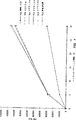
図1は、95℃で時間を変えて行った、グラム陽性微生物ストレプトコッカス・ピオゲンス(Streptococcus pyogenes)からの標的核酸(リボゾームRNA)の遊離を定量する実験をグラフで表したものである。
図2は、既知量の添加されたストレプトコッカス・ピオゲネスのリボゾームRNAに対するハイブリダイゼーション検出系(HPA;下記)の感度を示す実験をグラフで表したものである。
図3は、本明細書記載の方法に従って標的リボゾーム核酸が遊離されたストレプトコッカス・ピオゲネス細胞の希釈物に関して行ったハイブリダイゼーションアッセイの結果をグラフで示したものである。
発明の詳細な説明
特許請求した方法およびキットは、広範な微生物からの核酸の遊離の誘導、および引き続き行われる標的微生物、標的微生物のクラスまたは複数の異なる標的微生物に特異的な核酸の検出および同定に関する透過剤の使用、並びに器具、培地および薬剤の組み合わせに関する。種々の工程、培地および薬剤は一般的に上で議論されている。好ましい具体例の記載を提供する。これらの記載は説明のためだけに提供され、何ら本発明を限定するものではなく、本明細書を結論づける請求の範囲によってのみ本発明は定義される。
所望細胞からの核酸の抽出
核酸が抽出される標的微生物を種々の源から得ることができる。アッセイされる細胞が医学的試料中に含まれていることは必須でない。当業者は、いかなる源の試料であっても本発明に適用できるが、一定の系において用いられる特定の検出手段により検出されるに十分な量の標的微生物(よって、標的核酸)を試料が含んでいることが条件となることを認識するであろう。ポリメラーゼ連鎖反応(PCR)または転写に基づく方法のごとき増幅方法を検出工程の前に用いる場合、もとの試料はそれに対応して比較的少量の標的微生物または標的核酸を含んでいもよい。例えば、アメリカン・ソサエティー・フォー・マイクロバイオロジー(American Society for Microbioilogy),ダイアグノスティック・モレキュラー・マイクロバイオロジー:プリンシプルズ・アンド・アプリケイションズ(Diagnostic Molecular Microbiology:Principles and Applications)56〜70頁(1993年)参照、参照によりこれを本明細書に記載されているものとみなす。
生物学的または生物学的試料について、培養物、血液、組織、唾液、痰、糞便、脳髄液または滑液、血清、尿、あるいは他の液体中で増殖した細胞または微生物(これらに限らない)を包含するすべてのタイプの生理学的標本を用いて、本発明方法を使用することができる。かかる生物学的標本を、ヒト、動物、または植物から得てもよい。本発明に従って環境または食品試料を用いてこれらの試料中に存在する微生物から核酸を遊離させてもよい。好ましくは、本発明を用いて臨床的な口からまたは性器からの綿棒抜き取り物中に含まれる微生物から核酸を遊離する。
細胞試料を得た後、細胞試料を透過性にする試薬を接触させるかまたはその中に入れ、核酸が細胞から遊離するまで約80〜95℃に加熱する。ここに用いる透過性にする試薬は、例えば、セイライン溶液、EDTA溶液または非イオン性界面活性剤溶液からなっていてよい。これらの透過性にする試薬のいずれかを用いる本発明方法を用いて標的微生物から核酸(例えば、RNAおよびDNA、好ましくはリボゾームRNAおよびリボゾームRNA配列をコードするDNA)を抽出することができる。よって、種々の透過性にする試薬を用いて、本明細書記載の方法を用いて細胞から核酸を抽出することができる。
好ましくは、透過性にする試薬は、非イオン性界面活性剤および金属キレート剤の組み合わせからなる。出願人らは、これら2つの薬剤の組み合わせが細胞試料から抽出される未分解核酸の量を最適化することを見いだした。非イオン性界面活性剤は溶液に溶解された場合に荷電しない界面活性剤である。出願人らは、ポリオキシエチレンエーテル(ミズーリ州セントルイスのシグマ(Sigma)社により商品名トリトンX−100およびトリトンX−102の下で市販されている)およびオクチルフェノール−エチレンオキシド縮合物(ミズーリ州セントルイスのシグマ(Sigma)社により商品名ノニデットP−40の下で市販されている)のごとき非イオン性界面活性剤を用いて、本発明核酸抽出法をうまく実施した。よって、当業者は、種々の非イオン性界面活性剤を用いて本発明を実施することができるということを認識するであろう。
透過性にする試薬に添加される金属キレート剤は、マグネシウム、マンガンおよび亜鉛のごとき遊離金属イオンをキレートするかまたはこれらに結合する。理論に拘束されることを望ないが、遊離金属イオンをキレートすることは、所望核酸を分解しうるヌクレアーゼのごとき酵素を阻害すると考えられている。種々の金属キレート剤が市販されておりこの目的に役立つ。出願人らは低コストおよび便利さのためEDTAを使用する。
好ましくは、透過性にする試薬は、約0.01ないし約1%の非イオン性界面活性剤および約1mMないし100mMのEDTA;より好ましくは、0.07%のトリトンX−100および10mMのEDTAを含有する。濃度0.07%のトリトンX−100は、ストレプトコッカス・ピオゲネスからの本質的にすべての標本リボゾームRNAの遊離に十分であることがわかった。そのうえ、10mMより高いEDTA濃度はさらなる利益を提供するとは思われない。
本発明の実施に必須ではないが、透過性にする試薬は弱い緩衝剤、好ましくはHEPES(4−(2−ヒドロキシエチル)−1−ピペラジンエタンスルホン酸)の遊離酸を含み、透過性にする試薬のpHを安定化させるものであってもよい。透過性にする試薬を調製して後の使用のために貯蔵する場合、保存料を透過性にする試薬に添加して貯蔵期間中の望ましくない微生物の増殖を防止してもよい。好ましくは、5.7mMのアジ化ナトリウムを添加する。所望標的核酸がRNAである場合には、溶液のpHを8.0より高くならないように調節すべきである。好ましくは、塩基、すなわち、水酸化リチウムの添加により、透過性にする試薬のpHを7.5に調節する。本明細書開示以外の、pH8.0未満における使用に適する緩衝剤は当業者に知られている。
試料にヌクレアーゼを含有させうる臨床標本または他の源から細胞試料を得る場合、特に、標本核酸がRNAである場合には、アニオン性界面活性剤を透過性にする試薬に添加することも必要であろう。アニオン性界面活性剤は、pH7.0の溶液に溶解された場合、負に帯電する界面活性剤である。好ましくは、これらの場合において透過性にする溶液は1%(w/v)のラウリル硫酸リチウム(LLS)を含有する。理論に拘束されることを望まないが、アニオン性界面活性剤は、臨床標本または他の材料中に見いだされるすべてのヌクレアーゼ、特にリボヌクレアーゼを崩壊または変性させることに役立ち、それゆえ、試料中に核酸の分解を防止すると考えられている。アニオン性界面活性剤は、細胞試料からの核酸の遊離を行うことには必ずしも必須でない。約0.01%ないし約2%の間のラウリル硫酸リチウムが好ましい。最も好ましくは、LLS濃度は0.2%ないし1%の間である。2%よりも高いラウリル硫酸リチウム濃度においては、標的核酸収率が低下する。
細胞試料および透過性にする試薬を混合したならば、混合物を80〜100℃の範囲において1〜30分間加熱すべきである。出願人らは、約80℃以上の種々の温度が効果的であることを見いだした。80℃未満の温度においては標的細胞(すなわち、ストレプトコッカス・ピオゲネス)からの核酸の抽出は完全とは思われない。同様に、混合物が昇温される時間は比較的短い。約100万個のエス・ピオゲネス(S.pyogenes)細胞(一般的には溶解が困難であると考えられているグラム陽性細菌)を含む300μlの試料からのリボゾームRNAの抽出は、95℃、5分以内で良好な核酸の収率となる。よって、好ましい具体例において、細胞試料/透過性にする試薬の混合物を約95℃で約5分間加熱する。
実施例1
エス・ピオゲネスの酵素的溶解と本発明方法との比較
図1は、種々の透過性にするプロトコールに供されたストレプトコッカス・ピオゲネス細胞標品のハイブリダイゼーション速度の比較を示す。この実験において、下記の方法論を用いた。しかしながら、本開示を読んだ後に当業者はハイブリダイゼーションプロトコールおよび検出工程に対する多くの変法を行うであろうし、本明細書に開示された特定のハイブリダイゼーションおよび検出方法は説明のみを目的とする。
1800マイクロリットルの透過性にする試薬(7.4mM HEPES pH7.5、0.07%(v/v)トリトンX−100、10mM EDTA二ナトリウムおよび5.7mMアジ化ナトリウム)を試験管中にピペットで取った。血液寒天上で増殖したストレプトコッカス・ピオゲネス細胞をセイライン溶液で1回洗浄し、次いで、透過性にする試薬中に懸濁して1mlあたり3.0x10⁸個とした。200マイクロリットルのこの細胞懸濁液を、1800μlの透過性にする試薬の入った試験管中に希釈し、懸濁液を95℃で加熱した。そのうち300μlを5、10、15および30分において試験管から取り、氷上に置いた。
この実験におけるハイブリダイゼーションに用いた試験管には、ストレプトコッカス・ピオゲネスに特異的な凍結乾燥プローブ試薬の形状の標識オリゴヌクレオチドプローブが入っていた。特記しないかぎり、これらの試験管およびすべての以下に用いる試薬を、アキュプローブ・A群ストレプトコッカス培養同定キット(AccuProbe Group A Streptococcus culture identification kit)(カリフォルニア州サンジエゴのジェン−プローブ,インコーポレイテッド(Gen-Probe,Inc.)製)から取った(アキュプローブの包装に添付した説明書、ミリマン(Milliman)ら,米国特許第5,232,831号;およびアーノルド(Arnold)ら,米国特許第4,950,613号参照、参照によりこれらを本明細書に記載されているものとみなす)。凍結乾燥された試薬は溶解酵素を含有していたので、2%LLSを含有する50μlの溶液(アキュプローブ試薬2)を各試験管中に混合して凍結乾燥酵素を変性させた。対照試験管には透過性にするための緩衝液中の細胞懸濁液50μlを入れ、37℃で5分間インキュベーションして酵素的溶解を起こし、次いで、アキュプローブ試薬2で100μlとした。各時点につき4本の試験管を用意した。次いで、残りの試験管に熱処理された細胞懸濁液50μlを入れた(もう1つの対照懸濁液には試験用試験管と同じ試薬を入れたが、熱処理工程の間室温に置いた。これを「界面活性剤のみ」の対照と呼んだ)。
試験管を60℃にインキュベーションすることによりハイブリダイゼーションを促進した。各時点の2本の試験管を5分間インキュベーションし、各時点についての残りの1対の試験管を30分間インキュベーションした。ハイブリダイゼーション量、それゆえ、細胞から遊離した検出可能な標的核酸量をHPA(アーノルドら,上の引用文献参照)により測定した。300マイクロイットルの試薬3(選択試薬)を各試験管に添加し、試験管内容物を混合し、さらに60℃で5分間インキュベーションした。ハイブリダイゼーションしたプローブの化学発光を、リーダーIルミノメーター(Leader I luminometer)(カリフォルニア州サンジエゴのジェン−プローブ,インコーポレイテッド製)で測定した。アッセイ試料の化学発光を相対光単位(relative light unit、RLU)で表し、これは、選択リボ核酸にハイブリダイゼーションしたプローブ量に直接比例する。
図1のグラフからわかるように、5分間加熱された細胞試料混合物が30分加熱された細胞試料混合物と等しいシグナルを有していたため、透過性にする緩衝液で処理したエス・ピオゲネス細胞からの標的核酸の抽出は本質的には5分で完了した。出願人らが驚いたことには、透過性にする試薬とともにインキュベーションされた細胞から遊離した標的核酸へのハイブリダイゼーションの程度は、5分間程度の短時間でさえも、酵素処理された細胞から遊離した標的核酸に関して見られる程度の2倍であった。この結果は全く予期しなかったものであり、酵素的溶解工程よりの低コストで高いアッセイ感度を示すことにより本発明方法に有用性を付加するものである。
実施例2
遊離RNA量の評価
出願人らは、上記の好ましい方法を用いることにより、抽出プロセスの間にほとんどすべての細胞リボゾームRNAを細胞から遊離することができると確信する。本発明方法により透過性にされた細胞によって遊離された全細胞リボゾームRNAのパーセンテージを評価するために、出願人らは、下記ハイブリダイゼーションプロテクションアッセイ(HPA)を、(a)既知濃度のエス・ピオゲネス・リボゾームRNA溶液希釈物、および(b)本発明方法により核酸が抽出された既知能度のエス・ピオゲネス細胞の希釈物について行って、実施例1と同様にハイブリダイゼーションに供した。図2は、既知量のリボゾームRNAの希釈物に関してHPAを行った結果をグラフで示したものである。図からわかるように、ハイブリダイゼーション生成物の化学発光は試料中のリボゾームRNA量に比例して増加する。同様に、図3は、図1の場合と同様に核酸が抽出されたエス・ピオゲネス細胞に関してHPAを行った結果をグラフで示したものである。各試料の化学発光は試料中の細胞数に比例して増加する。
各細胞から遊離するリボゾームRNA量の概算値を、図1および2からの結果を比較することにより計算することができる。図2を参照すると、0.25ngの添加エス・ピオゲネスのリボゾームRNAは約15000RLUに対応する。図3を参照すると、15000RLUは約7300個の細胞に対応する。0.25を7300で割ると細胞1個あたり検出されるリボゾームRNA量0.000034ngが得られる。この数値は、細菌細胞中に含有されるリボゾームRNA全量は約0.00002ngであるという別の概算値の実験誤差の範囲内である(ハンドブック・オブ・バイオケミストリー・アンド・バイオフィジックス(Handbook of Biochemistry and Biophysics)(CRC・プレス(CRC Press)1992年参照)。よって、エス・ピオゲネス・リボゾームRNAのすべてではないとしても大部分のパーセントが、本発明の透過性にする方法を用いて抽出されると結論することが合理的である。
実施例3
DNAおよびRNAの両方を遊離させる方法の有効性
本発明方法が細胞にDNAおよびRNA両方を遊離させるということを示すために以下の実験を行った。エス・ピオゲネス(S.pyogenes)、エシェリシア・コリ(Escherichia coli)、カンジダ・アルビカンス(Candida albicans)およびストレプトコッカス・アガラクチアエ(Streptococcus agalactiae)の培養物をを血液寒天上で18時間増殖させた。各生物の10マイクルリットルのループを10mlの滅菌済みセイライン溶液に懸濁し、次いで、2000xgで7分間遠心分離した。上清を傾斜法により取り、各生物の1μlループを12mlの透過性にする試薬(実施例1記載)に添加した。さらに、各試験管の10倍希釈を同じ試薬中に作成した。300マイクロリットルの各懸濁液を3本の別個の試験管に入れた。各セットの1番目および2番目の試験管を、さらなる処理の前に95℃で10分間加熱した。各セットの3番目の試験管を室温に10分間放置した。
細胞を10000xgで5分間遠心分離し、上清をきれいな試験管に移した。各セットにつき、1番目の試験管に15μlの滅菌済みセイライン溶液を入れ、次いで、37℃で3時間インキュベーションした。各セットの2番目および3番目の試験管に、RNAを加水分解するために25μlの4N NaOHを入れ、混合し、次いで、37℃で3時間インキュベーションした。
3時間たった時に、2番目および3番目の試験管に70.5μlの1N HClを入れてpH7.0とした。試験管1に70.5μlのセイライン溶液を入れた。すべての試験管を95℃で5分間加熱して2本鎖DNAを変性させ、次いで、即座に氷上で冷却した。
単一のアクリジニウムエステル標識プローブを使用する以外は本質的に実施例1と同様にハイブリダイゼーションおよびアッセイを行った。このプローブは、すべての細菌およびすべての真菌に共通した配列に十分に相補的なヌクレオチド配列を有していた。50マイクロリトルのプローブを50μlの各試料に添加し、60℃で30分間ハイブリダイゼーションさせた。次いで、以下およびアーノルドら(上記引用文献)に記載されているようにHPA(ハイブリダイゼーションプロテクションアッセイ)法と同様に試料を処理した。
実験結果を下表1に示す。データは、強塩基に耐性のある核酸(すなわちDNA)が本発明方法で処理された細胞から遊離されることを示す。そのうえ、遊離したDNAは核酸ハイブリダイゼーションにより検出可能な特異的な核酸配列を有している。

広範な微生物からの核酸遊離方法の適合性
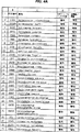
広範な微生物からの標的核酸の抽出のための本発明方法の有効性を決定するために、実施例1記載の透過性にする試薬を用いて同じ濃度の以下のグラム陽性細菌、グラム陰性細菌および酵母に包含される微生物を含有する試料を処理した。この実験に用いたすべての細菌/すべての酵母のプローブは、細菌および真菌に関して非特異的であった。すなわち、プローブは、すべての細菌および真菌に共通したリボゾームRNA配列に十分に相補的であり上記ハイブリダイゼーション条件下で各微生物から遊離したリボゾームRNAとハイブリダイゼーションするヌクレオチド配列を有していた。陰性の対照として、核酸抽出工程後に第2のプローブを遊離核酸に添加することを除き、同じ生物を本発明方法により処理した。第2のプローブは、第1のプローブと同じ濃度でハイブリダイゼーション混合物中に存在し、A群のストレプトコッカス種のリボゾームRNA中に含まれる特異的なヌクレオチド配列に相補的なヌクレオチド配列を有していた。陽性の結果(すなわち、ハイブリダイゼーションしたプローブの検出)を、上記HPA検出法において1000000RLUより大きな値とみなすと定義した。陰性の結果を1000000RLUまたはそれ未満と定義した。
図4は、すべてが実施例1にしたがって処理された微生物の一覧表を掲載するものであり、アッセイ結果を示すものである。図からわかるように、エス・ピオゲネスを除くすべてのアッセイされた微生物は、対照実験において陰性であると試験された。アッセイされた微生物に関する陰性の結果がHPA検出アッセイにおいて3000RLUより大きい場合はなかった。対照的に、本発明方法により透過性にされたすべての試験微生物は、すべての細菌/すべての酵母のHPA検出法により明確な陽性を示すに十分な量のリボゾームRNA標的核酸を遊離した。試験微生物が酵母およびグラム陽性細菌ならびに種々の属のグラム陰性細菌を包含したという事実は、異種微生物集団を含む試料についての本発明方法の広い適合性、および広範な微生物の検出および同定に適合性を有する一般的な核酸抽出試薬として本明細書記載の透過性にする試薬を使用することの有効性を示す。
実施例5
細胞壁は物理的に崩壊されないことの決定
所望細胞試料から核酸が遊離してから、細胞試料を調べて細胞壁が物理的に崩壊したかどうかを決定した。アメリカン・ソサエティー・フォー・マイクロバイオロジー(American Society for Microbiology),マニュアル・オブ・メソッズ・フォー・ジェネラル・バクテリオロジー(Manual of Methods for General Bacteriology)8〜9頁および26〜27頁(1981年)に記載された位相差顕微鏡およびグラム染色をはじめとして、細胞壁の完全性を試験するための多くの方法が存在している。この刊行物の上記部分を参照により本開示の一部とみなす。
本発明方法は、処理される細胞に対して非常に温和であると思われる。分子生物学の方法、例えば、核酸の増幅、核酸の開裂、または他の酵素による反応を含むいくつかの用法にとって、微生物の細胞壁が有意に崩壊されないことは望ましい。例えば、核酸でない細胞成分は引き続き行う抽出核酸に対する操作を妨害しうる。グラム染色を用いることにより、正常なグラム陽性微生物の細胞壁が物理的に崩壊されたがどうかを確かめることができる。
好ましくは、出願人らは、上記マニュアル・オブ・メソッズ・フォー・ジェネラル・バクテリオロジー27〜28頁に記載されたようにグラム染色を行った。グラム染色後の染色細胞の顕微鏡観察の使用は、本発明方法により透過性にした後でさえも紫色に染まりその特徴的な形態を維持するグラム陽性細胞を生じる。この結果は、処理された細菌の細胞壁が完全には物理的に崩壊されなかったことを示す。対照的に、酵素処理されたグラム陽性細胞は赤または黄に染まり、その細胞壁の崩壊を示した。
実施例6
所望核酸の検出および測定
核酸が標本から放出された後、核酸ハイブリダイゼーションおよび検出テクニックを用いて標的核酸を検出し測定する方法が当該分野において知られている。例えば、検出すべき微生物由来の標的核酸に十分に相補的な核酸プローブを用い、該プローブを標的核酸配列にハイブリダイゼーションさせ、次いで、サザンブロット(マニアティス,ティー(Maniatis,T.)ら,モレキュラー・クローニング:ア・ラボラトリー・マニュアル(Molecular Cloning:A Laboratory Manual),コールド・スプリング・ハーバー・ラボラトリー(Cold Spring Harbor Laboratory)(1982年)参照)またはアーノルド(Arnold)ら,クリニカル・ケミストリー(Clin.Chem.))第35巻:1588頁(1989年)により記載された均一溶液フェイズ法(ハイブリダイゼーションプロテクションアッセイまたはHPAと呼ばれる)のごとき当業者に知られた方法によりプローブと標的との2本鎖ハイブリッドを検出することにより、核酸を同定してもよい(これらの文献を参照により本明細書に記載されているものとみなす)。
アーノルドにより記載されたHPA法は、アクリジニウムエステル標識の特異的化学分解に基づいて特異的に加水分解されうるアクリジニウムエステル標識DNAプローブを合成することからなる。プローブがハイブリダイゼーション状態にあるかハイブリダイゼーションしていない状態にあるかにより、選択的化学分解を調べる。核酸二重らせんとの相互作用によりプローブがハイブリダイゼーション状態にある場合、アクリジニウムエステル標識を加水分解から保護する。ハイブリダイゼーションしないプローブは、加水分解から保護されないままのそのアクリジニウムエステル標識を有する。よって、ハイブリダイゼーションしていないプローブに関連したアクリジニウムエステル標識の化学発光は特異的加水分解により急激に失われるが、ハイブリダイゼーションしたプローブに関連した化学発光は最小限の影響受ける。リーダーIルミノメーター(Leader I luminometer)(カリフォルニア州サンジエゴのジェン−プローブ,インコーポレイテッド製)のごとき装置により化学発光を測定してもよい。
本発明の実施に必須ではないが、HPA法を実施するための出願人らの好ましい方法を記載する。
試薬
ウィークス・アイ(Weeks,I.)ら,アクリジニウム・エステルズ・アー・ハイ−スペシフィック−アクティビティー・ラベルズ・イン・アン・イムノアッセイ(Acridiniumu Esters are High-Specific-Activity Labels in an Immunoassay),クリニカル・ケミストリー(Clin.Chem.)第29巻:1474〜1479頁(1983年)(参照により本明細書に記載されているものとみなす)に記載のごとく、アクリジニウムエステル標識試薬をを合成する。アッセイおよび化学発光測定用にポリスチレンまたはポリプロピレン試験管(12x75mm)をノースカロライナ州ニュートンのサーステッド(Sarstedt)から得る。化学発光をリーダーIルミノメーターで測定する。他のすべての物質は標準的な「ウルトラ−ピュア(Ultra-pure)」または試薬グレードのものである。
方法
アクリジニウムエステル標識DNAプローブの調製
標準的なホスホラミダイト化学を用いてオリゴヌクレオチドを合成する。DNA合成中に導入されたアルキルアミンリンカーアームとメチルアクリジニウムフェニルエステルのN−ヒドロキシサクシンイミドエステルとを反応させることにより、アクリジニウムエステルでのDNAプローブの化学標識を行う。アクリジニウムエステル標識プローブを精製し、種々のアッセイフォーマットにおいて使用した後、下記のごとくリーダーIルミノメーターでプローブの化学発光を検出する。
ハイブリダイゼーションプロテクションアッセイ(HPA)
典型的には、1リットルあたり、1%(w/v)のラウリル硫酸リチウム、2mM EDTAおよび2mM[エチレンビス(オキシエチルニトリロ)]四酢酸(EGTA)を含有する0.1Mのコハク酸リチウム緩衝液,pH5.2中、60℃においてハイブリダイゼーション反応を行う。ハイブリダイゼーション体積は50ないし200μlの範囲で、0.05ないし0.5pmolのプローブを含有する。1リットルあたり、10〜50mlのトリトンX−100界面活性剤を含有する四ホウ酸ナトリウム緩衝液(7.0ないし8.5の範囲のpH値)中、60℃において特異的加水分解を行う。
典型的なHPAフォーマットにおいて、100μlの体積として60℃で5〜10分間インキュベーションすることにより標的核酸を含有する試料をDNAプローブとハイブリダイゼーションさせる。次いで、出願人らは、四ホウ酸緩衝液300μlを添加し、さらに60℃で5ないし10分間インキュベーションする。試料を室温で2〜3分冷却した後、2つの自動試薬注入方法のうちの1つを用いてルミノメーターで化学発光を測定する。方法1においては、0.1%(v/v)H2O2および1mM硝酸を含む200μlの溶液を注入してから1秒後、200μlの1〜2M NaOHを注入する。生じる化学発光を2ないし5秒間積算する。方法2においては、1〜2M NaOHを含む0.1%(v/v)H2O2溶液200μlを注入する。生じる化学発光を2ないし5秒間積算する。ハイブリダイゼーションおよび特異的加水分解を包含するこの方法のすべての工程を、1本の12x75mm試験管中で行う。
HPAのごときプローブ検出法の使用により、標的核酸の存在または不存在を検出することができる。アクリジニウムエステル標識DNAプローブアッセイは迅速、高感度、使用が簡単であり、ハイブリダイゼーションしていないプローブにより生じるバックグラウンドが十分に低く、HPAアッセイは臨床研究室において有用である。さらに、HPAフォーマットの感度は、特に、リボゾームRNAの検出と組み合わせた場合、臨床研究室における実際の使用を可能にし、単一コピーの標的配列を有するDNA分子を検出する試験よりも感度を2x10⁴倍まで上昇させる。
本発明方法を行うためのキット
上記方法を行うためのキットを、すでに利用可能な材料および試薬から作成してもよい。キットは、透過性にする試薬、プローブ試薬、および選択試薬を包含するであろう。出願人らの好ましい具体例を読むことにより明らかなように、このキットに修飾を加えると、所望の適用について特異的な核酸プローブとなるプローブ試薬中の核酸プローブを単に選択することにより、疾病、症状または微生物等のごとき生物を示すいかなる核酸配列でも検出することができる。
出願人らの好ましい具体例は、直接ストレプトコッカス・ピオゲネスの試験を行うためのキットである。好ましくは、滅菌済み綿棒を用いることにより患者の喉から試料を得る。喉の綿棒拭き取り物全部をポリプロピレン試験管中の300μlの透過性にする試薬中に入れる。出願人らの透過性にする試薬は、すべてがすでに利用可能な材料から調製され、5.7mMアジ化ナトリウム、7.4mM HEPES(遊離酸),0.07%(v/v)トリトンX−100、1%(w/v)ラウリル硫酸リチウム、10mM EDTA(遊離酸)からなり、十分量の水酸化リチウムを添加してpH7.5とされる。約95℃で約30分以上加熱する場合、透過性にする試薬は、喉の綿棒拭き取り試料中のエス・ピオゲネス細胞から核酸を遊離する。種々の濃度および試薬により本発明がうまく実行されるので、特定の濃度および試薬は出願人らの発明を限定するものであるとは思われない。本開示は、適当な濃度、試薬および条件を選択するための指針を提供し、この開示の後の請求の範囲により完全に定義される本発明の具体例のうちの特別な例を提供する。
次いで、混合物を95℃の加熱ブロック上に10分間置く。加熱後、試験管を室温で5分間放冷する。冷却している間にポリプロピレン試験管の側面に綿棒を押し付けて絞る。冷却後、液体50μlをきれいなポリプロピレン試験管に移す。次に、50μlのプローブ試薬を50μlの試料液体に添加する。好ましくは、プローブ試薬は0.1Mコハク酸リチウム緩衝液,pH5.2、1%(w/v)ラウリル硫酸リチウム、2mM EDTA、2mM EGTAおよびストレプトコッカス・ピオゲネスのリボゾームRNAの領域に特異的なデオキシリボ核酸プローブ0.05pmolを含有する。
次いで、プローブ試薬溶液を60℃で30分間インキュベーションして、抽出工程において溶液中に遊離されたストレプトコッカス・ピオゲネスのリボゾームRNAにアクリジニウムエステル標識プローブをハイブリダイゼーションさせる。インキュベーション後、300μlの選択試薬を添加する、好ましくは、選択試薬は、0.1(w/v)トリトンX−100を含有するpH7.5の0.15M四ホウ酸ナトリウム緩衝液である。
ボルテックスミキサーを用いて得られた溶液を完全に混合し、60℃で7分間インキュベーションしてアクリジニウムエステル標識を特異的加水分解させる。最後のインキュベーション後、プローブ試験管を室温で少なくとも5分間冷却し、次いで、ハイブリダイゼーションしたプローブについてアッセイする。ルミノメーターで化学発光を測定し、試料中のストレプトコッカス・ピオゲネスの存在または不存在をその読みから決定する。
核酸抽出および検出法を詳細に説明したが、本発明から掛け離れることなく他の試薬、濃度および温度を用いることができることが理解されるべきであり、かかる他の試薬、濃度および温度の使用は本開示を読んだ後の当業者には明らかであろう。例えば、本発明方法により遊離された核酸を、選択および検出工程の前の核酸増幅に続けて供することができる。それゆえ、本発明の精神または本質的な特徴から掛け離れていない特別の形態で、本発明は具体化され、行われ、あるいは用いられうる。よって、本発明の具体例は、すべての点で説明的であり、限定的でないと考えられる。本発明の範囲は上の記載よりもむしろ請求の範囲によって示される。請求の範囲と均等な意味および範囲に属するすべての変更は請求の範囲内にあることを意味する。 Background of the Invention
TECHNICAL FIELD OF THE INVENTION
The present invention relates to a method for extracting nucleic acids of a wide range of organisms from cultures, clinical specimens, and other samples suitable for nucleic acid hybridization and other diagnostic methods to specifically identify test organisms. The invention further relates to diagnostic kits that use permeabilizing agents suitable for the extraction of nucleic acids from a wide range of organisms, which are suitable for subsequent hybridization, detection, and quantitative identification of assay organisms.
Background art
The advent of modern methods of molecular biology and genetic engineering has revolutionized disease diagnosis. In recent years, with the development of advanced methods for detection, quantification, and discrimination between microbial species and subspecies, the identification of pathogenic organisms, especially microorganisms such as bacteria and yeasts, has become faster and more accurate. Became. Many, if not most, of these diagnostic methods use inherent differences between the nucleic acid sequences of different species of pathogens and between pathogenic and non-pathogenic microorganisms. The speed, selectivity and sensitivity of these nucleic acid-based diagnostic methods allowed for faster and more accurate patient treatment and therefore made the public healthier.
Diagnostic kits that rely primarily on nucleic acid hybridization have been developed for the diagnosis of many diseases in which organisms under the microscope are involved as either etiology or disease indicators. Not all, but the kit can be used in this way, or tuberculosis (Mycobacterium tuberclosis, a disease transmitted through normal sexual intercourse (Chlamydia trachomatis), Neisseria gonorrhoeae), respiratory disease (Mycoplasma pneumoniae), pharyngitis and rheumatic fever (Streptococcus group A (S. pyogenes)), pharynx It is intended to detect and / or identify microorganisms involved in capping inflammation (Haemophilus influenzae) and viruses such as the pathogenesis of ARC and AIDS (HIV). Easy to use Requires target nucleic acid.
Most methods for extracting microbial nucleic acids require disrupting (dissolving) microbial cell walls and extracting the contents of the cells into a buffer. Subsequently, phenol extraction can be performed to remove lipids, carbohydrates, and proteins from the solution, and the nucleic acid is further purified by precipitation with cold ethanol. The correct method for destroying or lysing a microorganism usually depends on the nature of the organism itself. Until recently, however, research was strongly limited to a single Gram-negative bacterial species, Escherichia coli (Stent, Gunther S, and Calendar, Richard, See Molecular Genetics 51 (2nd edition, 1978)).
Nucleic acid extraction from gram-negative bacteria such as E. coli has traditionally been (a) sonication, abrasive grinding, shaking with glass beads, and shearing force in a French press. Or mechanical force; (b) cell wall weakening by either one or more freeze-thaws or lysis with a lytic enzyme such as lysozyme, followed by a strong detergent or chaotropic reagent Cell wall lysis by treatment with (ie, a reagent that breaks hydrophobic interactions) is used. In both such methods, lysate components include organelles, proteins (including enzymes such as proteases and nucleases), carbohydrates, and lipids, as well as nucleic acids, which requires further nucleic acid purification. there's a possibility that.
Methods involving enzymatic lysis of E. coli cells with surfactants (Godson, GN) and Sinheimer, RI, Biochem. Biophys Acta) 149, 476 (1967) "Lysis of Escherichia coli with a Natural Detergent"). Schein (EPO antibody 0061250) describes a method for recovering recombinant proteins using lysozyme, chaotropic agents, and / or surfactants, and the host bacterium was an E. coli strain.
Lysis of gram-positive microorganisms is much more difficult than gram-negative bacteria because a thicker and denser peptidoglycan layer is the main bacterial cell wall component. Lysis of several gram positive microorganisms using lytic enzymes has been reported (eg, Coleman, SE et al., Infect, and Immun. Volume 2: 563- 569 (1970) “Lysis of Grouped and Ungrouped Streptococci by Lysozyme”; Chasey, Bruce M. And Giufridda Alfred, Applied and Environmental Microbiology, 39: 153-158 (1980) "Rissis of Streptococcus mutans cells・ With mutanolysin, a lytic enzyme prepared from ・Lysis of Streptococcus mutans cells with Mutanolysin, a Lytic Enzyme prepared from a Culture Liquor of Streptomyces gloisporus 1829 ”; Hamada, S. et al., Archves Of Oral Biol. 23: 543-549 (1978); Calendra, GB, and Cole, RM, Infection And Immunity 28: 1033-1037 (1980) See "Lysis and Protprast Formation of Group B Streptococci by Mutanolysin" ).
Clinical samples containing Gram-negative pathogenic bacteria are chaotrope (granididium thiocyanate (GuSCN)); anionic surfactant (sodium dodecyl sulfate (SDS) or N-lauryl sarcosine (sarcosyl)); Non-enzymatic compositions have been described for treatment with a solution containing a chelating agent (ethylenediaminetetraacetic acid (EDTA)); and a reducing agent (β-mercaptoethanol) (Schwartz et al., US Pat. No. 5, , 212,059). Such compositions are described as lysis / hybridization solutions where all samples are used in a single assay.
The cell walls of yeast species containing β-glucan and chitin as main components differ in composition from bacteria; therefore, enzymatic methods for extracting nucleic acids from yeast are often used to lyse bacteria A completely different set of enzymes is used, such as zymolyase with a different specificity than the one that does.
Sheiness and Levine (PCT application WO92 / 07096) describe diagnostic kits for detecting vaginal pathogens. The test organisms were the yeast Candida species, the Gram-negative bacterium Gardnerella vaginalis and the eukaryotic protozoan Trichomonas vaginalis without cell walls. The solution they used was reported to be able to liberate nucleic acids from each of these organisms as detected by nucleic acid hybridization.
Holmes, US Pat. No. 4,830,969 describes a method for isolating nucleic acids by boiling cultured cells in a “lysing agent”.
Wase (EPO Publication No. 0149514) discloses a method for producing flocculants from lysed bacterial cell cultures. One of the various described methods of initiating cell lysis uses heat.
The use of heat alone is effective in the production of intact DNA that is suitable for further biochemical manipulation from certain bacterial Mycobacteria species that have been considered difficult to lyse by many researchers. It was shown (Robson, EPO publication 0547789A1).
All of the above methods involve phenol extraction and release that can cause the release of nucleic acids suitable for nucleic acid hybridization from a wide range of microorganisms in a biological sample (eg, DNA encoding a ribosomal RNA / or ribosomal RNA sequence). It lacks the benefits of a single reagent that does not require ethanol precipitation.
Summary of the Invention
The present invention permeates nucleic acids (eg, RNA and DNA, preferably DNA encoding ribosomal RNA / or ribosomal RNA sequences) from a wide range of organisms, including gram positive and gram negative bacteria, and yeast. Relates to the use of reagents. The reagent does not require the use of a lytic enzyme and can be used at elevated temperatures, preferably between about 80 ° C and 100 ° C, more preferably between about 80 ° C and 95 ° C, and most preferably at about 95 ° C. is there. Furthermore, the present invention employs a reagent that renders a permeant capable of releasing nucleic acids, preferably DNA encoding ribosomal RNA sequences, from a wide range of organisms including Gram positive and Gram negative bacteria and yeast. Oriented to methods and kits. It can then be used for a variety of purposes including, but not limited to, nucleic acid amplification methods such as polymerase chain reaction (PCR), or hybridization with probe oligonucleotides having nucleotide sequences complementary to specific nucleotide sequences of free nucleic acids. Free nucleic acid can be used. Therefore, the compositions, methods, and kits described herein are designed to allow for quick and simple preparation from the organisms contained in the sample.
The advantages of a single nucleic acid extraction method for use with a wide range of microorganisms are: a) reduced time and expense used to train laboratory technicians to perform diagnostic tests; b) reduced manufacturing costs (single transmission) Including the cost and time reduction associated with quality control of enzymatic extraction reagents). Thus, a first object of the present invention is to provide a single reagent capable of inducing the release of detectable amounts of nucleic acids from a wide range of microorganisms.
Again many diagnostic kits rely on the use of enzymes to release nucleic acids from cells. Even with insufficient enzymatic lysis, the yield of target nucleic acid that can be used for hybridization is not good. This is because a particular sample does not contain many assayable cells, and because the target microorganism or nucleic acid is present only in a subpopulation of cells, or the sensitivity of the diagnostic system may be reduced. Because there is. Thus, a second object of the present invention is to provide a nucleic acid extraction method that obtains a better yield of target nucleic acid than other corresponding methods.
A third object of the present invention is an inexpensive nucleic acid extraction method for inclusion in a commercial diagnostic assay kit. The permeabilizing reagent used with the method of the present invention is a relatively inexpensive research chemical reagent and does not need to include expensive lytic enzymes.
It is a fourth object of the present invention to provide a mild and simple method for releasing nucleic acids from cells into solution. Since the method of the present invention leaves the cell wall of many target microorganisms substantially intact after the extraction step, free nucleic acid does not mix with other cellular components. Most undesirable cellular material remains in the cells. The nucleic acid in the supernatant can be separated from the cells by centrifugation and can be assayed individually, but this is not essential for the practice of the method of the invention. Furthermore, the present invention is fast and simple, allowing both skilled and immature laboratory workers to extract nucleic acids, particularly DNA encoding ribosomal RNA sequences, in a single step. . Moreover, the extraction method of the present invention is mild to nucleic acids, and nucleic acids suitable for subsequent use such as hybridization assays can be obtained without further purification.
Since it is preferable to minimize the exposure of laboratory technicians to harmful microorganisms in the first stage of a diagnostic assay, it is a fifth aspect of the present invention to provide a relatively safe method for nucleic acid extraction from clinical samples. Is the purpose. This is in two ways: by minimizing the time required to liberate most of the target nucleic acid from the target cell, and at a high temperature (ie, about 80 ° C. to 100 ° C.) that most of the pathogen is rapidly killed. The permeation step at a temperature between 0 ° C.
The present invention is applicable to organisms other than gram positive and gram negative bacteria and yeast. For example, other cells, such as cultured eukaryotic cells, such as mycoplasma, protozoa, and viruses with coats, and cultured eukaryotic cells that have less or no cell walls than bacteria or yeast are rendered permeabilized by the method of the present invention. sell. In such cases, although mild, the method of the present invention may cause more damage to the cell wall or membrane than in the case of bacteria or yeast.
Definition
Unless stated otherwise in this specification, the following terms have the following meanings for the purposes of this application.
“Enzymatic lysis” means that some or all of the intracellular components after treatment of a cell or group of cells using an enzyme that destroys and opens the cell or group of cells, or that completely or partially digests the cell wall of an organism. Means liberation.
A “surfactant” can have a hydrophobic region or portion that can interact with a hydrophobic solvent and a hydrophobic portion of a cell membrane, and can have a positive or negative charge in solution, or a polar region that has no charge. Means a molecule or class of molecules having a hydrophilic region or portion.
“Nonionic surfactant” means a surfactant that contains at least one polar, uncharged group or ion in its hydrophilic region or portion.
"Ionic surfactant" means a surfactant that contains at least one positively or negatively charged group or ion in a hydrophilic region or portion.
A “metal chelator” or “chelator” is a molecule or molecule of a molecule that can bind, complex, or integrate with a metal ion, thereby reducing the effective concentration of the metal ion in solution. Means class.
“Nucleic acid release” means the release of a sufficient amount of nucleic acid such that the release method is useful in a nucleic acid hybridization assay.
"Nucleic acid" or "nucleic acid group" means a polydeoxyribonucleotide or polyribonucleotide that is at least 2, preferably 10 or more nucleotides in length. The term “nucleic acid” encompasses polynucleotides, oligonucleotides, and DNA or RNA molecules. The term “nucleic acid” can refer to either a single stranded or double stranded polynucleotide, or both.
“Target nucleic acid” means a nucleic acid consisting of a target nucleic acid sequence to be detected. Preferably, such sequences are characteristic of a particular organism.
“Target nucleic acid sequence” or “target sequence” means a specific nucleic acid sequence or a nucleic acid sequence complementary thereto.
“Target organism” means any prokaryotic or eukaryotic species to be identified using the methods described herein, or all viruses. In general, such organisms are included in biological samples such as, but not limited to, stripped or wiped with a cotton swab. Preferably, the organism is a pathogenic microorganism.
“Biological sample” means any specimen or sample containing animal, plant, bacterial, viral, or protist material. Such samples include food or agricultural samples; environmental samples; urine, blood, milk, cerebrospinal fluid, sputum, saliva, stool, lung aspirate, tears, lymph, or semen, body fluids, secretions or exocrine secretions; throat Or swabs from the genitals; and including, but not limited to, cell, virus, plant or animal cell cultures, suspensions or lysates. The biological sample may or may not contain ribonuclease.
"Clinical sample" means a biological sample obtained from a human or animal for testing or testing for the purpose of diagnosis or disease management.
“Complementary” refers to a nucleic acid sequence in which a stable hydrogen bond is formed between a nucleotide base of one nucleic acid strand region and a nucleotide base of another nucleic acid region under conditions suitable for nucleic acid hybridization. It means having. That is, most commonly hydrogen bonds are between adenosine (A) residues on one chain and thymine (T) or uracil (U) residues on the other chain, and guanine on one chain. (G) formed between a residue and cytosine (C) on another chain. Generally, such complementary regions contain about 15 to 100 or more consecutive nucleotides of each nucleic acid strand.
“Sufficiently complementary” means that a double-stranded hydrogen bonding region can be formed under conditions suitable for nucleic acid hybridization. Two nucleic acid strands are sufficiently complementary if they have 100% complementarity over a particular contiguous corresponding region, but are two single stranded nucleic acids having regions of less than 100 complementarity. Can also form a double-stranded region under hybridization conditions. Such regions are not 100% complementary, but can form stable double-stranded regions under hybridization conditions and are therefore considered sufficiently complementary.
“Lysis” means cell degradation, including physical disruption and cell wall and / or membrane disruption, that allows the release of intracellular components, including nucleic acids, into the surrounding medium.
By “permeabilizing” or “permeabilizing” is meant cell wall and / or membrane degradation that causes the release of a detectable amount of nucleic acid from the cell into the surrounding medium.
By “permeabilizing reagent” is meant a chemical or physical factor, or both, that can cause the permeability of a cell or group of cells.
“Selection reagent” refers to a reagent used in a procedure capable of chemically or physically distinguishing a hybridized double-stranded nucleic acid region comprising a nucleic acid probe from a non-hybridized single-stranded nucleic acid and / or nucleic acid probe. means.
“Detection reagent” means a reagent used in a procedure capable of detecting a nucleic acid having a double-stranded nucleic acid region, one of which is a nucleic acid probe.
"Probe reagent" means a reagent that contains a nucleic acid probe having a nucleotide sequence that is sufficiently complementary to the nucleotide sequence of a target nucleic acid, and typically such a probe has a reporter group or moiety that is detectable in a hybridization assay. .
One aspect of the invention relates to a non-enzymatic method for inducing the release of a wide range of microbial nucleic acids, preferably ribosomal RNA and / or DNA encoding ribosomal RNA sequences, into solution. In one example, a sample containing cells to be identified is mixed with an extraction solution containing a nonionic surfactant and a metal chelator. Heat the suspension to about 85-95 ° C. for about 5-15 minutes. Upon heating, the nucleic acid is released into solution without causing microscopic observation of the cell walls of the sample cells. The nucleic acid so released is suitable for hybridization, amplification or other genetic manipulation without further purification.
The present invention provides a rapid, inexpensive and mild method for extracting nucleic acids from microorganisms. By eliminating the use of enzymes to perform the extraction process, the cost, variability, and difficulty associated with enzymatic nucleic acid extraction are significantly reduced.
Furthermore, the present invention eliminates the need for chaotropic agents as part of the lysis reagent or permeation reagent. In general, chaotropic agents are used at very high concentrations and are expensive. Because of these high concentrations, chaotropes can precipitate from the extraction solution during transport or storage of the diagnostic kit. Chaotropes used for nucleic acid extraction may change the conditions required for subsequent hybridization of the probe with the target nucleic acid and are not at all compatible with the use of the extracted nucleic acid in enzymatic reactions such as PCR or other target amplification methods It may be. In the latter case, it is necessary to separate the chaotrope from the extracted nucleic acid, which adds a removal step to the entire protocol and increases the chances of erroneous results. In addition, chaotropes such as GuSCN are difficult to pipet due to their high viscosity. Thus, the absence of chaotropes in methods for the extraction of nucleic acids in clinical samples is economically and logically useful.
In a second embodiment, the microorganism to be assayed may be obtained from a clinical sample or other biological material that may contain a nuclease, eg, a ribonuclease. Preferably, the present invention relates to a method for preparing clinical specimens wiped with throat and genital swabs for the identification of microorganisms by probe hybridization to nucleic acids released from assay microorganisms. In this case, the entire swab wipe may be contacted or immersed in a permeable solution containing a nonionic surfactant and a metal chelator. When the desired target nucleic acid is RNA, an anionic surfactant such as lithium lauryl sulfate or sodium dodecyl sulfate is further added to the permeation agent. Anionic surfactants may inactivate nucleases that degrade sample nucleic acids present in clinical samples. The swab wipes in the permeabilizing solution are then heated to about 80-95 ° C. for about 5-30 minutes, causing the release of nucleic acids from the microorganisms.
The speed and temperature of the nucleic acid extraction process helps to reduce the exposure of laboratory technicians to infectious organisms. At the temperatures of 80-95 ° C described, most organisms in the sample that could potentially include pathogenic organisms such as HIV and hepatitis and tuberculosis pathogens will die. Potential exposure is further reduced because the extraction method is rapid and can be performed without repeatedly transferring the cell suspension from tube to tube.
Another aspect of the present invention is a kit comprising reagents for performing the described extraction method and then ascertaining whether a particular microorganism is present in the sample. The kit includes a permeabilizing reagent, a probe reagent, a selection reagent, and a detection reagent. The kit may include providing a permeabilizing solution containing an ionic surfactant, a nonionic surfactant and a metal chelator, and supplying reagents necessary for detection and identification of the target nucleic acid. . Preferably, the identification and detection reagents include probe reagents containing at least one nucleic acid probe that is sufficiently complementary to one or more nucleic acid sequences specific for the ribosomal RNA of the target microorganism (eg, Hogan (Hogan et al., US Pat. No. 5,216,143 and Milliman and Hammond, US Pat. No. 5,232,831, which are hereby incorporated by reference) See). Probe reagents containing derivatized nucleic acid probes (such as probes containing phosphorothioate and / or methylphosphonate linkages) capable of recognizing and binding to specific nucleic acid sequences are known to those skilled in the art and kits using the extraction method of the present invention It is thought that it can be used in
Other features, uses and advantages of the invention will be apparent to those skilled in the art from the following description of the preferred embodiments, drawings and claims.
[Brief description of the drawings]
FIG. 1 is a graph showing an experiment for quantifying the release of a target nucleic acid (ribosomal RNA) from a Gram-positive microorganism, Streptococcus pyogenes, carried out at 95 ° C. for different times.
FIG. 2 is a graphical representation of experiments showing the sensitivity of the hybridization detection system (HPA; below) to known amounts of added Streptococcus pyogenes ribosomal RNA.
FIG. 3 is a graphical representation of the results of a hybridization assay performed on a dilution of Streptococcus pyogenes cells from which the target ribosomal nucleic acid has been released according to the methods described herein.
Detailed Description of the Invention
The claimed method and kit provide for the use of a permeant for the induction of nucleic acid release from a wide range of microorganisms and the subsequent detection and identification of nucleic acids specific for a target microorganism, a class of target microorganisms or a plurality of different target microorganisms. , And instruments, media and drug combinations. The various processes, media and drugs are generally discussed above. A description of preferred embodiments is provided. These descriptions are provided for illustrative purposes only, and are not intended to limit the invention in any way, and the invention is defined only by the claims that conclude this specification.
Nucleic acid extraction from desired cells
The target microorganism from which the nucleic acid is extracted can be obtained from various sources. It is not essential that the cell being assayed is contained in a medical sample. One skilled in the art can apply the present invention to any source sample, but the sample contains a sufficient amount of the target microorganism (and thus the target nucleic acid) to be detected by the particular detection means used in a given system. You will recognize that being a condition is a requirement. If an amplification method, such as a polymerase chain reaction (PCR) or transcription based method, is used prior to the detection step, the original sample may correspondingly contain a relatively small amount of target microorganism or target nucleic acid. For example, American Society for Microbioilogy, Diagnostic Molecular Microbiology: Principles and Applications, pages 56-70 ( 1993), which is considered to be described herein by reference.
For a biological or biological sample, cells or microorganisms grown in culture, blood, tissue, saliva, sputum, stool, cerebrospinal fluid or synovial fluid, serum, urine, or other fluids, but not limited to The method of the invention can be used with all types of physiological specimens, including Such biological specimens may be obtained from humans, animals or plants. Nucleic acids may be released from microorganisms present in these samples using environmental or food samples according to the present invention. Preferably, the present invention is used to release nucleic acids from microorganisms contained in a swab extract from a clinical mouth or genital.
After obtaining the cell sample, a reagent that renders the cell sample permeable is contacted or placed therein and heated to about 80-95 ° C until the nucleic acid is released from the cell. The permeabilizing reagent used here may comprise, for example, a saline solution, an EDTA solution or a nonionic surfactant solution. Nucleic acids (eg, RNA and DNA, preferably DNA encoding ribosomal RNA and ribosomal RNA sequences) can be extracted from target microorganisms using the methods of the invention using any of these permeabilizing reagents. Thus, nucleic acids can be extracted from cells using the methods described herein using various permeabilizing reagents.
Preferably, the permeabilizing reagent consists of a combination of a nonionic surfactant and a metal chelator. Applicants have found that the combination of these two agents optimizes the amount of undegraded nucleic acid extracted from the cell sample. Nonionic surfactants are surfactants that are not charged when dissolved in solution. Applicants have identified polyoxyethylene ethers (commercially available under the trade names Triton X-100 and Triton X-102 by the company Sigma, St. Louis, MO) and octylphenol-ethylene oxide condensates (of St. Louis, MO). The nucleic acid extraction method of the present invention was successfully performed using a nonionic surfactant such as that sold by Sigma under the trade name Nonidet P-40. Thus, those skilled in the art will recognize that the present invention can be practiced with a variety of nonionic surfactants.
Metal chelators added to the permeabilizing reagent chelate or bind to free metal ions such as magnesium, manganese and zinc. Without wishing to be bound by theory, it is believed that chelating free metal ions inhibits enzymes such as nucleases that can degrade the desired nucleic acid. Various metal chelators are commercially available and serve this purpose. Applicants use EDTA for low cost and convenience.
Preferably, the permeabilizing reagent is about 0.01 to about 1% non-ionic surfactant and about 1 mM to 100 mM EDTA; more preferably 0.07% Triton X-100 and 10 mM EDTA. Containing. A concentration of 0.07% Triton X-100 was found to be sufficient for the release of essentially all sample ribosomal RNA from Streptococcus pyogenes. Moreover, EDTA concentrations higher than 10 mM do not appear to provide further benefits.
Although not essential to the practice of the present invention, the permeabilizing reagent comprises a weak buffer, preferably the free acid of HEPES (4- (2-hydroxyethyl) -1-piperazineethanesulfonic acid), which renders it permeable. It may stabilize the pH of the reagent. When preparing a permeabilizing reagent and storing it for later use, a preservative may be added to the permeabilizing reagent to prevent unwanted microbial growth during storage. Preferably, 5.7 mM sodium azide is added. If the desired target nucleic acid is RNA, the pH of the solution should be adjusted so that it does not rise above 8.0. Preferably, the pH of the permeabilizing reagent is adjusted to 7.5 by the addition of a base, ie lithium hydroxide. Buffers suitable for use at pH below 8.0 other than those disclosed herein are known to those skilled in the art.
When obtaining cell samples from clinical specimens or other sources that can contain nucleases in the sample, especially when the specimen nucleic acid is RNA, it may also be necessary to add it to a reagent that renders the anionic surfactant permeable. I will. An anionic surfactant is a surfactant that is negatively charged when dissolved in a solution of pH 7.0. Preferably, in these cases the permeabilizing solution contains 1% (w / v) lithium lauryl sulfate (LLS). While not wishing to be bound by theory, anionic surfactants help disintegrate or denature all nucleases found in clinical specimens or other materials, particularly ribonucleases, and therefore, nucleic acids in a sample It is thought to prevent the decomposition of. An anionic surfactant is not necessarily essential for releasing nucleic acid from a cell sample. Between about 0.01% and about 2% lithium lauryl sulfate is preferred. Most preferably, the LLS concentration is between 0.2% and 1%. At lithium lauryl sulfate concentrations higher than 2%, the target nucleic acid yield decreases.
Once the cell sample and the permeabilizing reagent are mixed, the mixture should be heated at 80-100 ° C. for 1-30 minutes. Applicants have found that various temperatures above about 80 ° C. are effective. At temperatures below 80 ° C., nucleic acid extraction from target cells (ie, Streptococcus pyogenes) does not appear complete. Similarly, the time during which the mixture is heated is relatively short. Extraction of ribosomal RNA from a 300 μl sample containing approximately 1 million S. pyogenes cells (generally Gram-positive bacteria considered difficult to lyse) Good nucleic acid yield within minutes. Thus, in a preferred embodiment, the cell sample / permeabilizing reagent mixture is heated at about 95 ° C. for about 5 minutes.
Example 1
Comparison of enzymatic lysis of S. pyogenes with the method of the present invention
FIG. 1 shows a comparison of hybridization rates of Streptococcus pyogenes cell preparations subjected to various permeabilization protocols. In this experiment, the following methodology was used. However, after reading this disclosure, one of ordinary skill in the art will make many variations to the hybridization protocols and detection steps, and the specific hybridization and detection methods disclosed herein are for illustrative purposes only.
Pipet 1800 microliters of permeabilizing reagent (7.4 mM HEPES pH 7.5, 0.07% (v / v) Triton X-100, 10 mM disodium EDTA and 5.7 mM sodium azide) into a test tube. I took it. Streptococcus pyogenes cells grown on blood agar are washed once with saline solution and then suspended in a permeabilizing reagent to give 3.0 x 10 per ml.8Individual. 200 microliters of this cell suspension was diluted into a test tube containing 1800 μl of permeabilizing reagent and the suspension was heated at 95 ° C. Of this, 300 μl was removed from the test tubes at 5, 10, 15 and 30 minutes and placed on ice.
The test tube used for hybridization in this experiment contained a labeled oligonucleotide probe in the form of a lyophilized probe reagent specific for Streptococcus pyogenes. Unless otherwise stated, these tubes and all of the following reagents are used in the AccuProbe Group A Streptococcus culture identification kit (Gen-Probe, Inc., San Diego, Calif.). (Inc.)) (instructions attached to the packaging of Accuprobe, Milliman et al., US Pat. No. 5,232,831; and Arnold et al., US Pat. No. 4,950, No. 613, which are considered to be described herein by reference). Since the lyophilized reagent contained lysing enzyme, 50 μl of a solution containing 2% LLS (Accuprobe reagent 2) was mixed into each tube to denature the lyophilized enzyme. Control tubes were filled with 50 μl of cell suspension in permeabilizing buffer, incubated at 37 ° C. for 5 minutes to cause enzymatic lysis, and then made up to 100 μl with
Hybridization was facilitated by incubating the tubes at 60 ° C. Two tubes at each time point were incubated for 5 minutes and the remaining pair of tubes for each time point was incubated for 30 minutes. The amount of hybridization and hence the amount of detectable target nucleic acid released from the cells was measured by HPA (see Arnold et al., Cited above). 300 microliters of Reagent 3 (selection reagent) was added to each tube, the tube contents were mixed, and further incubated at 60 ° C. for 5 minutes. The chemiluminescence of the hybridized probe was measured with a Reader I luminometer (Gen-Probe, Inc., San Diego, Calif.). The chemiluminescence of the assay sample is expressed in relative light units (RLU), which is directly proportional to the amount of probe hybridized to the selected ribonucleic acid.
As can be seen from the graph in FIG. 1, the cell sample mixture heated for 5 minutes had the same signal as the cell sample mixture heated for 30 minutes, so that from S. pyogenes cells treated with the permeabilizing buffer. The extraction of the target nucleic acid was essentially completed in 5 minutes. Applicants were surprised that the degree of hybridization to the target nucleic acid released from the cells incubated with the permeabilizing reagent was released from the enzyme-treated cells, even in a short time of about 5 minutes. Twice that seen for the target nucleic acid. This result is totally unexpected and adds utility to the method of the present invention by showing high assay sensitivity at a lower cost than the enzymatic lysis step.
Example 2
Evaluation of free RNA
Applicants believe that by using the preferred method described above, almost all cellular ribosomal RNA can be released from the cells during the extraction process. In order to assess the percentage of total cellular ribosomal RNA released by cells permeabilized by the method of the present invention, Applicants have performed the following hybridization protection assay (HPA): (a) known concentrations of S. pyogenes. A ribosomal RNA solution dilution and (b) a dilution of S. pyogenes cells of known ability from which nucleic acids were extracted by the method of the present invention were used for hybridization in the same manner as in Example 1. FIG. 2 is a graph showing the results of performing HPA on dilutions of known amounts of ribosomal RNA. As can be seen, the chemiluminescence of the hybridization product increases in proportion to the amount of ribosomal RNA in the sample. Similarly, FIG. 3 is a graph showing the results of performing HPA on S. pyogenes cells from which nucleic acids were extracted as in FIG. The chemiluminescence of each sample increases in proportion to the number of cells in the sample.
An estimate of the amount of ribosomal RNA released from each cell can be calculated by comparing the results from FIGS. Referring to FIG. 2, 0.25 ng of added S. pyogenes ribosomal RNA corresponds to approximately 15000 RLU. Referring to FIG. 3, 15000 RLU corresponds to about 7300 cells. Dividing 0.25 by 7300 yields 0.000034ng of ribosomal RNA detected per cell. This figure is within the experimental error of another estimate that the total amount of ribosomal RNA contained in bacterial cells is about 0.00002 ng (Handbook of Biochemistry and Biophysics). (See CRC Press 1992.) Thus, most if not all percent of S. pyogenes ribosomal RNA is extracted using the permeabilization method of the present invention. It is reasonable to conclude
Example 3
Effectiveness of methods that liberate both DNA and RNA
The following experiment was performed to show that the method of the present invention allows cells to release both DNA and RNA. Cultures of S. pyogenes, Escherichia coli, Candida albicans and Streptococcus agalactiae were grown on blood agar for 18 hours. A 10 microliter loop of each organism was suspended in 10 ml of sterile saline solution and then centrifuged at 2000 xg for 7 minutes. The supernatant was removed by decantation and added to a reagent (described in Example 1) that made 1 μl loop of each
The cells were centrifuged at 10,000 xg for 5 minutes and the supernatant was transferred to a clean tube. For each set, 15 μl of sterile saline solution was placed in the first tube and then incubated at 37 ° C. for 3 hours. The second and third tubes of each set were loaded with 25 μl 4N NaOH to hydrolyze the RNA, mixed and then incubated at 37 ° C. for 3 hours.
After 3 hours, 70.5 μl of 1N HCl was added to the second and third tubes to a pH of 7.0. In test tube 1, 70.5 μl of saline solution was added. All tubes were heated at 95 ° C. for 5 minutes to denature the double stranded DNA and then immediately cooled on ice.
Hybridization and assay were performed essentially as in Example 1 except that a single acridinium ester labeled probe was used. This probe had a nucleotide sequence that was sufficiently complementary to a sequence common to all bacteria and all fungi. 50 microliters of probe was added to 50 μl of each sample and allowed to hybridize at 60 ° C. for 30 minutes. Samples were then processed in the same manner as the HPA (Hybridization Protection Assay) method as described below and in Arnold et al. (Cited above).
The experimental results are shown in Table 1 below. The data show that nucleic acids that are resistant to strong bases (ie DNA) are released from cells treated with the method of the invention. Moreover, the free DNA has a specific nucleic acid sequence that can be detected by nucleic acid hybridization.
Suitability of nucleic acid release methods from a wide range of microorganisms
In order to determine the effectiveness of the method of the present invention for the extraction of target nucleic acids from a wide range of microorganisms, the same concentration of the following Gram positive bacteria, Gram negative bacteria and Samples containing microorganisms included in yeast were processed. All bacterial / all yeast probes used in this experiment were non-specific for bacteria and fungi. That is, the probe had a nucleotide sequence that was sufficiently complementary to the ribosomal RNA sequence common to all bacteria and fungi and hybridized with the ribosomal RNA released from each microorganism under the above hybridization conditions. As a negative control, the same organism was treated according to the method of the invention except that a second probe was added to the free nucleic acid after the nucleic acid extraction step. The second probe was present in the hybridization mixture at the same concentration as the first probe and had a nucleotide sequence complementary to the specific nucleotide sequence contained in the ribosomal RNA of the group A Streptococcus species. . A positive result (ie, detection of hybridized probe) was defined to be considered as a value greater than 1000000 RLU in the HPA detection method. Negative results were defined as 1 million RLU or less.
FIG. 4 lists all of the microorganisms treated according to Example 1 and shows the assay results. As can be seen, all assayed microorganisms except S. pyogenes were tested negative in the control experiment. There were no negative results for the assayed microorganisms greater than 3000 RLU in the HPA detection assay. In contrast, all test microorganisms permeabilized by the method of the present invention released a sufficient amount of ribosomal RNA target nucleic acid to show a clear positive by the HPA detection method of all bacteria / all yeasts. The fact that the test microorganisms included yeast and gram positive bacteria as well as gram negative bacteria of various genera is compatible with the broad suitability of the method of the present invention for samples containing heterogeneous microbial populations and the detection and identification of a wide range of microorganisms. The effectiveness of using the permeabilizing reagents described herein as a general nucleic acid extraction reagent having
Example 5
Deciding that the cell wall is not physically disrupted
After the nucleic acid was released from the desired cell sample, the cell sample was examined to determine if the cell wall was physically disrupted. American Society for Microbiology, Manual of Methods for General Bacteriology 8-9 and 26-27 (1981) There are many methods for examining cell wall integrity, including the phase contrast microscope and Gram staining described. The above portion of this publication is considered part of this disclosure by reference.
The method of the invention appears to be very benign to the cells being treated. For some uses, including molecular biology methods, such as nucleic acid amplification, nucleic acid cleavage, or other enzymatic reactions, it is desirable that the microbial cell wall not be significantly disrupted. For example, cellular components that are not nucleic acids can interfere with subsequent manipulation of extracted nucleic acids. By using Gram staining, it can be ascertained whether the cell walls of normal Gram positive microorganisms have been physically disrupted.
Preferably, Applicants performed Gram staining as described in the Manual of Methods for General Bacteriology, pages 27-28. The use of microscopic observation of stained cells after Gram staining results in Gram positive cells that stain purple and maintain their characteristic morphology even after permeabilization by the method of the present invention. This result indicates that the treated bacterial cell walls were not completely physically disrupted. In contrast, enzyme treated gram positive cells stained red or yellow, indicating collapse of their cell walls.
Example 6
Detection and measurement of desired nucleic acid
Methods for detecting and measuring a target nucleic acid using nucleic acid hybridization and detection techniques after the nucleic acid has been released from the specimen are known in the art. For example, using a nucleic acid probe that is sufficiently complementary to the target nucleic acid from the microorganism to be detected, hybridizing the probe to the target nucleic acid sequence, and then Southern blot (Maniatis, T. et al., Molecular Cloning: see Molecular Cloning: A Laboratory Manual, Cold Spring Harbor Laboratory (1982) or Arnold et al., Clinical Chemistry (Clin. Chem. .)) Double-stranded hybrids of probe and target by methods known to those skilled in the art, such as the homogeneous solution phase method (referred to as hybridization protection assay or HPA) described by Volume 35: 1588 (1989). By detecting the Good (these documents are considered as described herein by reference).
The HPA method described by Arnold consists of synthesizing an acridinium ester labeled DNA probe that can be specifically hydrolyzed based on the specific chemical degradation of the acridinium ester label. Selective chemical degradation is examined depending on whether the probe is in a hybridized state or in a non-hybridized state. When the probe is in a hybridized state by interaction with a nucleic acid duplex, the acridinium ester label is protected from hydrolysis. Non-hybridized probes have their acridinium ester labels that remain unprotected from hydrolysis. Thus, the chemiluminescence of the acridinium ester label associated with the unhybridized probe is rapidly lost by specific hydrolysis, while the chemiluminescence associated with the hybridized probe is minimally affected. Chemiluminescence may be measured with a device such as a Leader I luminometer (Gen-Probe, Inc., San Diego, Calif.).
Although not essential to the practice of the invention, Applicants' preferred method for carrying out the HPA method is described.
reagent
Weeks, I. et al., Acridinium Esters are High-Specific-Activity Labels in an Immunoassay, Clinical Chemistry (Clin Chem.) 29: 1474-1479 (1983), which is considered to be described herein by reference), acridinium ester labeling reagents are synthesized. Polystyrene or polypropylene test tubes (12 x 75 mm) are obtained from Sarstedt, Newton, NC for assay and chemiluminescence measurements. Chemiluminescence is measured with a Reader I luminometer. All other materials are standard “Ultra-pure” or reagent grade.
Method
Preparation of acridinium ester labeled DNA probe
Oligonucleotides are synthesized using standard phosphoramidite chemistry. By reacting the alkylamine linker arm introduced during DNA synthesis with N-hydroxysuccinimide ester of methylacridinium phenyl ester, chemical labeling of the DNA probe with acridinium ester is performed. After acridinium ester labeled probes are purified and used in various assay formats, probe chemiluminescence is detected with a Reader I luminometer as described below.
Hybridization protection assay (HPA)
Typically, 0.1 M lithium succinate buffer containing 1% (w / v) lithium lauryl sulfate, 2 mM EDTA and 2 mM [ethylenebis (oxyethylnitrilo)] tetraacetic acid (EGTA) per liter. Hybridization reaction is performed at 60 ° C. in liquid, pH 5.2. Hybridization volumes range from 50 to 200 μl and contain 0.05 to 0.5 pmol of probe. Specific hydrolysis is carried out at 60 ° C. in sodium tetraborate buffer (pH value in the range of 7.0 to 8.5) containing 10-50 ml of Triton X-100 surfactant per liter.
In a typical HPA format, the sample containing the target nucleic acid is hybridized with the DNA probe by incubating at 60 ° C. for 5-10 minutes in a volume of 100 μl. Applicants then add 300 μl of tetraborate buffer and further incubate at 60 ° C. for 5-10 minutes. After the sample is cooled at room temperature for 2-3 minutes, chemiluminescence is measured with a luminometer using one of two automated reagent injection methods. In Method 1, 0.1% (v / v) H2O2And 1 second after injecting 200 μl of solution containing 1 mM nitric acid, 200 μl of 1-2 M NaOH is injected. The resulting chemiluminescence is integrated for 2 to 5 seconds. In
The presence or absence of the target nucleic acid can be detected by use of a probe detection method such as HPA. The acridinium ester-labeled DNA probe assay is rapid, sensitive, simple to use, and the background generated by unhybridized probes is sufficiently low that the HPA assay is useful in clinical laboratories. In addition, the sensitivity of the HPA format allows practical use in clinical laboratories, especially when combined with ribosomal RNA detection, and is 2x10 more sensitive than tests that detect DNA molecules with a single copy of the target sequence.FourRaise to double.
Kit for carrying out the method of the invention
Kits for performing the above methods may be made from already available materials and reagents. The kit will include a reagent that renders it permeable, a probe reagent, and a selection reagent. As will be apparent by reading Applicants' preferred embodiments, modifications to this kit can be made by simply selecting the nucleic acid probe in the probe reagent to be a specific nucleic acid probe for the desired application. Any nucleic acid sequence indicative of symptoms or organisms such as microorganisms can be detected.
Applicants' preferred embodiment is a kit for direct Streptococcus pyogenes testing. Preferably, the sample is obtained from the patient's throat by using a sterile cotton swab. Place all throat swab wipes in 300 μl of the permeabilizing reagent in a polypropylene tube. Applicants' permeabilizing reagents are all prepared from materials already available, 5.7 mM sodium azide, 7.4 mM HEPES (free acid), 0.07% (v / v) Triton X- It consists of 100, 1% (w / v) lithium lauryl sulfate, 10 mM EDTA (free acid), and a sufficient amount of lithium hydroxide is added to adjust the pH to 7.5. When heated at about 95 ° C. for about 30 minutes or longer, the permeabilizing reagent releases nucleic acids from S. pyogenes cells in the throat swab sample. Because the present invention is successfully practiced with various concentrations and reagents, the particular concentrations and reagents do not appear to limit Applicants' invention. This disclosure provides guidance for selecting appropriate concentrations, reagents and conditions, and provides specific examples of embodiments of the invention that are fully defined by the claims that follow this disclosure.
The mixture is then placed on a 95 ° C. heating block for 10 minutes. After heating, the test tube is allowed to cool at room temperature for 5 minutes. Squeeze a cotton swab against the side of a polypropylene test tube while cooling. After cooling,
The probe reagent solution is then incubated at 60 ° C. for 30 minutes to hybridize the acridinium ester labeled probe to the Streptococcus pyogenes ribosomal RNA released in the solution during the extraction step. After incubation, 300 μl of selection reagent is added, preferably the selection reagent is a 0.15 M sodium tetraborate buffer at pH 7.5 containing 0.1 (w / v) Triton X-100.
The resulting solution is mixed thoroughly using a vortex mixer and incubated at 60 ° C. for 7 minutes to specifically hydrolyze the acridinium ester label. After the final incubation, the probe tube is cooled at room temperature for at least 5 minutes and then assayed for hybridized probe. Chemiluminescence is measured with a luminometer and the presence or absence of Streptococcus pyogenes in the sample is determined from the reading.
Although nucleic acid extraction and detection methods have been described in detail, it should be understood that other reagents, concentrations and temperatures can be used without departing from the present invention, and the use of such other reagents, concentrations and temperatures is It will be apparent to those skilled in the art after reading this disclosure. For example, the nucleic acid released by the method of the present invention can be subjected to nucleic acid amplification prior to the selection and detection step. Thus, the present invention may be embodied, practiced, or used in specific forms that do not depart from the spirit or essential characteristics of the invention. Thus, the embodiments of the present invention are illustrative in all respects and are not considered to be limiting. The scope of the invention is indicated by the appended claims rather than the foregoing description. All changes that come within the meaning and range of equivalency of the claims are within the scope of the claims.
Claims (22)
a)0.01%(体積/体積)ないし1%(体積/体積)の濃度の非イオン性界面活性剤および1mMないし100mMの濃度の金属キレート剤を必須としてなり、アニオン性界面活性剤を含んでいてもよい試薬に試料を接触させること;ついで
b)5分ないし30分間、試料を80℃ないし95℃の温度に加熱して、該微生物の細胞壁が完全には物理的に破壊されることなく該微生物から核酸を遊離させること
を特徴とし、
溶解酵素またはカオトロープの不存在下で行われるものであり、
剪断力または機械的な力を試料に適用せずに該微生物から核酸を遊離させるものである方法。A method for nucleic acid extraction from a microorganism selected from the group consisting of gram positive bacteria, gram negative bacteria and fungi suspected of being present in a sample, comprising the following steps:
a ) 0 . 0.1% (vol / vol) to 1% will nonionic surfactant and 1 mM to 1 100 mM concentration of a metal chelating agent at a concentration (volume / volume) as the essential anionic surfactant B ) heating the sample to a temperature of 80 ° C. to 95 ° C. for 5 minutes to 30 minutes, so that the cell wall of the microorganism is completely Characterized by liberating nucleic acids from the microorganism without being physically destroyed ,
Performed in the absence of lytic enzymes or chaotropes,
A method that liberates nucleic acids from the microorganism without applying shear or mechanical force to the sample.
a)0.01%(体積/体積)ないし1%(体積/体積)の濃度の非イオン性界面活性剤および1mMないし100mMの濃度の金属キレート剤を必須としてなり、アニオン性界面活性剤を含んでいてもよい試薬に試料を接触させること;a) A nonionic surfactant having a concentration of 0.01% (volume / volume) to 1% (volume / volume) and a metal chelating agent having a concentration of 1 mM to 100 mM are essential and include an anionic surfactant. Contacting the sample with an optional reagent;
b)5分ないし30分間、試料を80℃ないし95℃の温度に加熱して、該微生物の細胞壁が完全には物理的に破壊されることなく該微生物から核酸を遊離させること;ついでb) heating the sample to a temperature between 80 ° C. and 95 ° C. for 5 to 30 minutes to liberate nucleic acids from the microorganism without completely destroying the cell wall of the microorganism;
c)試料における疾病、症状または該微生物を示す、工程b)にて遊離された標的核酸の存在または量を検出することc) detecting the presence or amount of the target nucleic acid released in step b) indicating the disease, symptom or the microorganism in the sample
を特徴とし、Characterized by
溶解酵素またはカオトロープの不存在下で行われるものであり、Performed in the absence of lytic enzymes or chaotropes,
剪断力または機械的な力を試料に適用せずに該微生物から核酸を遊離させるものである方法。A method that liberates nucleic acids from the microorganism without applying shear or mechanical force to the sample.
試薬がアニオン性界面活性剤を含み、アニオン性界面活性剤がラウリル硫酸リチウムであり、0.01%ないし2%(重量/体積)の濃度であり、
試薬中の金属キレート剤がEDTAであり、5mMないし50mMの濃度である、
請求項1ないし5のいずれかに記載の方法。Nonionic surfactant in the reagent is a polyoxyethylene p-t-octylphenol, 0. A concentration of 01% to 1 % (volume / volume);
Reagent comprises an anionic surfactant, an anionic surfactant is lithium lauryl sulfate, 0. A concentration of 01% to 2 % (weight / volume),
A metal chelating agent in the reagent is EDTA, at a concentration of 5 mM to 5 0 mM,
The method according to any one of claims 1 to 5.
Applications Claiming Priority (3)
| Application Number | Priority Date | Filing Date | Title |
|---|---|---|---|
| US15894093A | 1993-11-29 | 1993-11-29 | |
| US08/158,940 | 1993-11-29 | ||
| PCT/US1994/013400 WO1995014768A2 (en) | 1993-11-29 | 1994-11-23 | Method for extracting nucleic acids from a wide range of organisms |
Publications (2)
| Publication Number | Publication Date |
|---|---|
| JPH09505479A JPH09505479A (en) | 1997-06-03 |
| JP3866762B2 true JP3866762B2 (en) | 2007-01-10 |
Family
ID=22570371
Family Applications (1)
| Application Number | Title | Priority Date | Filing Date |
|---|---|---|---|
| JP51516595A Expired - Fee Related JP3866762B2 (en) | 1993-11-29 | 1994-11-23 | Nucleic acid extraction methods from a wide range of organisms |
Country Status (11)
| Country | Link |
|---|---|
| US (2) | US5786208A (en) |
| EP (1) | EP0657530B1 (en) |
| JP (1) | JP3866762B2 (en) |
| KR (1) | KR100230909B1 (en) |
| AT (1) | ATE234352T1 (en) |
| AU (1) | AU693836B2 (en) |
| CA (1) | CA2176496C (en) |
| DE (1) | DE69432242T2 (en) |
| DK (1) | DK0657530T3 (en) |
| ES (1) | ES2194022T3 (en) |
| WO (1) | WO1995014768A2 (en) |
Cited By (1)
| Publication number | Priority date | Publication date | Assignee | Title |
|---|---|---|---|---|
| EP2481791A1 (en) | 2011-01-31 | 2012-08-01 | Tokyo University of Agriculture & Technology | Nucleic acid extraction method |
Families Citing this family (91)
| Publication number | Priority date | Publication date | Assignee | Title |
|---|---|---|---|---|
| AT403378B (en) * | 1995-09-11 | 1998-01-26 | Steindl Franz Dipl Ing Dr | Immune and protein complex dissociation |
| AT1140U1 (en) * | 1995-09-11 | 1996-11-25 | Steindl Franz Dipl Ing Dr | IMMUNE AND PROTEIN COMPLEX DISSOCIATION |
| FR2739382B1 (en) * | 1995-09-28 | 1997-11-14 | Oreal | RIBOSOMAL FRACTION AND COMPOSITION CONTAINING THE SAME |
| US6218105B1 (en) * | 1996-07-19 | 2001-04-17 | Kathleen S. Hall | High throughput papilloma virus in vitro infectivity assay |
| US7569342B2 (en) | 1997-12-10 | 2009-08-04 | Sierra Molecular Corp. | Removal of molecular assay interferences |
| US6706486B1 (en) | 1998-06-19 | 2004-03-16 | Amersham Biosciences Ab | Method for the quantitative release of natural or recombinant proteins, polypeptides or peptides |
| AU5670299A (en) * | 1998-10-19 | 2000-05-08 | Cbd Technologies Ltd. | Methods of concentrating microorganisms using affinity separation |
| WO2000066786A2 (en) | 1999-05-03 | 2000-11-09 | Gen-Probe Incorporated | Polynucleotide probes for detection and quantitation of actinomycetes |
| US6376186B1 (en) | 1999-05-03 | 2002-04-23 | Gen-Probe Incorporated | Polynucleotide probes for detection and quantitation of staphylococcus |
| EP1925678B1 (en) | 1999-05-03 | 2009-07-22 | Gen-Probe Incorporated | Polynucleotide matrix-based method of identifying microorganisms |
| US6821770B1 (en) * | 1999-05-03 | 2004-11-23 | Gen-Probe Incorporated | Polynucleotide matrix-based method of identifying microorganisms |
| WO2000066785A2 (en) | 1999-05-03 | 2000-11-09 | Gen-Probe Incorporated | Polynucleotide probes for detection and quantitation of bacteria in the family enterobacteriaceae |
| US6242188B1 (en) * | 1999-07-30 | 2001-06-05 | Applied Gene Technologies, Inc. | Sample processing to release nucleic acids for direct detection |
| US6383783B1 (en) | 1999-09-21 | 2002-05-07 | 3M Innovative Properties Company | Nucleic acid isolation by adhering to hydrophobic solid phase and removing with nonionic surfactant |
| KR20020029476A (en) * | 2000-10-13 | 2002-04-19 | 박제철 | Lysis buffer reagent for gene extraction by one step |
| KR20020029477A (en) * | 2000-10-13 | 2002-04-19 | 박제철 | Method for gene extraction by one step |
| ES2300375T3 (en) | 2000-10-23 | 2008-06-16 | Gen-Probe Incorporated | COMPOSITIONS AND METHODS FOR THE DETECTION OF HUMAN IMMUNODEFICIENCY VIRUS 2 (HIV-2). |
| CU23095A1 (en) * | 2000-11-07 | 2005-11-18 | Cnic Ct Nac Investigaciones | PROCESS FOR QUICK TYPIFICATION OF MICROORGANISMS AND REAGENT GAME EMPLOYED |
| ATE309344T1 (en) | 2001-01-19 | 2005-11-15 | Mueller Drm Ag | METHOD FOR PREPARING BIOMASS FOR PRODUCING CELL LYSATE CONTAINING PLASMID DNA |
| US7691571B2 (en) * | 2001-01-31 | 2010-04-06 | Mayo Foundation For Medical Education And Research | Detection of bordetella |
| AU2002243751A1 (en) * | 2001-01-31 | 2002-08-12 | Mayo Foundation For Medical Education And Research | Detection of herpes simplex virus |
| WO2002061141A1 (en) * | 2001-01-31 | 2002-08-08 | Mayo Foundation For Medical Education And Research | Detection of bordetella |
| US7033762B2 (en) * | 2001-08-29 | 2006-04-25 | Amersham Biosciences Corp | Single nucleotide amplification and detection by polymerase |
| US20030082563A1 (en) * | 2001-10-15 | 2003-05-01 | Bell Constance A. | Detection of bacillus anthracis |
| ATE328119T1 (en) * | 2001-11-02 | 2006-06-15 | Roche Diagnostics Gmbh | DETECTION OF VARIOLA VIRUS |
| DE20117746U1 (en) * | 2001-11-02 | 2002-04-25 | Invitek Biotechnik & Biodesign | Reaction rooms containing complex storage-stable reagent formulations and test kit for the detection and isolation of pathogenic microbial nucleic acids |
| GB0127809D0 (en) * | 2001-11-20 | 2002-01-09 | Dna Res Innovations Ltd | Purification of nucleic acid |
| US20030157728A1 (en) * | 2002-02-20 | 2003-08-21 | Uhl James R. | Swab extraction system |
| US20030215814A1 (en) * | 2002-05-17 | 2003-11-20 | Cockerill Franklin R. | Detection of Shiga toxin- or Shiga-like toxin-producing organisms |
| US20030215954A1 (en) * | 2002-05-17 | 2003-11-20 | Cockerill Franklin R. | Nucleic acid recovery reagents and methods |
| ATE445697T1 (en) | 2002-05-21 | 2009-10-15 | Arkray Inc | METHOD FOR PERFORMING LYSIS OF ACID-RESISTANT BACTERIA AND METHOD FOR PERFORMING GENE AMPLIFICATION OR GENETIC DETECTION THEREFROM |
| US7482116B2 (en) | 2002-06-07 | 2009-01-27 | Dna Genotek Inc. | Compositions and methods for obtaining nucleic acids from sputum |
| US20040038213A1 (en) * | 2002-08-06 | 2004-02-26 | Kwon Jai W. | Genotyping by in situ PCR amplification of a polynucleotide in a tissue biopsy |
| US7074598B2 (en) * | 2002-09-25 | 2006-07-11 | Mayo Foundation For Medical Education And Research | Detection of vancomycin-resistant enterococcus spp. |
| US7365176B2 (en) * | 2002-09-26 | 2008-04-29 | Mayo Foundation For Medical Education And Research | Detection of Epstein-Barr virus |
| US7074599B2 (en) * | 2002-09-27 | 2006-07-11 | Mayo Foundation For Medical Education And Research | Detection of mecA-containing Staphylococcus spp. |
| US7601491B2 (en) * | 2003-02-06 | 2009-10-13 | Becton, Dickinson And Company | Pretreatment method for extraction of nucleic acid from biological samples and kits therefor |
| JPWO2004094634A1 (en) * | 2003-04-22 | 2006-07-13 | アークレイ株式会社 | Nucleic acid isolation method and kit and apparatus for nucleic acid isolation |
| US7267950B2 (en) * | 2003-05-01 | 2007-09-11 | Veridex, Lcc | Rapid extraction of RNA from cells and tissues |
| US20050014165A1 (en) * | 2003-07-18 | 2005-01-20 | California Pacific Medical Center | Biomarker panel for colorectal cancer |
| US7427475B2 (en) * | 2003-11-18 | 2008-09-23 | Mayo Foundation For Medical Education And Research | Detection of group B streptococcus |
| US20050130177A1 (en) | 2003-12-12 | 2005-06-16 | 3M Innovative Properties Company | Variable valve apparatus and methods |
| US7939249B2 (en) * | 2003-12-24 | 2011-05-10 | 3M Innovative Properties Company | Methods for nucleic acid isolation and kits using a microfluidic device and concentration step |
| US7727710B2 (en) * | 2003-12-24 | 2010-06-01 | 3M Innovative Properties Company | Materials, methods, and kits for reducing nonspecific binding of molecules to a surface |
| DE102004010828B8 (en) * | 2004-02-27 | 2006-03-09 | Insilico Biotechnology Gmbh | Method and means for thermally conditioning a cell |
| JP2008511816A (en) * | 2004-08-03 | 2008-04-17 | ベクトン・ディキンソン・アンド・カンパニー | Use of magnetic materials to separate samples |
| EP2071031B1 (en) | 2004-08-27 | 2013-10-09 | Gen-Probe Incorporated | Single-primer nucleic acid amplification methods |
| US20060088862A1 (en) * | 2004-09-30 | 2006-04-27 | Nancy Lee | Drug screening and molecular diagnostic test for early detection of colorectal cancer: reagents, methods, and kits thereof |
| CA2871777C (en) | 2005-03-10 | 2015-07-28 | Matthew J. Hayes | System and methods for detecting multiple optical signals |
| EP1930730B1 (en) | 2005-03-10 | 2019-08-14 | Gen-Probe Incorporated | Systems and methods to perform assays for detecting or quantifying analytes |
| US20060270843A1 (en) * | 2005-05-26 | 2006-11-30 | Hall Gerald E Jr | Methods for isolation of nucleic acids |
| US20060269929A1 (en) * | 2005-05-26 | 2006-11-30 | Hall Gerald E Jr | Methods and kits for DNA purification on polymeric membranes at low ionic strength |
| US20100035331A1 (en) * | 2006-02-15 | 2010-02-11 | Tosoh Corporation | Method for extracting of nucleic acid from biological material |
| US20070238093A1 (en) * | 2006-04-11 | 2007-10-11 | Espy Mark J | Detection of influenza A virus |
| US20070238095A1 (en) * | 2006-04-11 | 2007-10-11 | Mayo Foundation For Medical Education And Research , A Minnesota Corporation | Detection of Influenza A Virus |
| WO2007131138A2 (en) * | 2006-05-03 | 2007-11-15 | Mayo Foundation For Medical Education And Research | Swab extraction device and methods of using |
| PL2017356T3 (en) | 2006-06-06 | 2012-03-30 | Gen Probe Inc | Tagged oliggonucleotides and their use in nucleic acid amplification methods |
| EP2106450A4 (en) * | 2007-01-08 | 2010-03-10 | Medigenes Co Ltd | Dna chip for detection of escherichia coli |
| EP2126138A4 (en) * | 2007-01-08 | 2011-01-19 | Medigenes Co Ltd | Dna chip for detection of staphylococcus aureus |
| EP1978111B1 (en) | 2007-04-02 | 2013-03-27 | Gen-Probe Incorporated | Compositions, kits and related methods for the detection and/or monitoring of Pseudomonas aeruginosa |
| EP2160459A2 (en) | 2007-05-23 | 2010-03-10 | Nature Technology Corp. | Improved e. coli plasmid dna production |
| AU2008260075A1 (en) | 2007-05-31 | 2008-12-11 | California Pacific Medical Center | Method to predict or diagnose a gastrointestinal disorder or disease |
| US9458451B2 (en) | 2007-06-21 | 2016-10-04 | Gen-Probe Incorporated | Multi-channel optical measurement instrument |
| WO2009015299A1 (en) | 2007-07-26 | 2009-01-29 | California Pacific Medical Center | Method to predict or diagnose a gastrointestinal disorder or disease |
| US20110065108A1 (en) * | 2008-02-01 | 2011-03-17 | Siemens Healthcare Diagnostics Inc. | Urine Transport Medium |
| JP5665548B2 (en) * | 2008-02-08 | 2015-02-04 | メイヨ・ファウンデーション・フォー・メディカル・エデュケーション・アンド・リサーチ | Clostridium difficile detection |
| EP2806037B1 (en) | 2008-05-13 | 2016-09-21 | Gen-Probe Incorporated | Inactivatable target capture oligomers for use in the selective hybridization and capture of target nucleic acid sequences |
| CA2746539A1 (en) | 2008-12-30 | 2010-07-08 | Gen-Probe Incorporated | Compositions, kits and related methods for the detection and/or monitoring of listeria |
| US8368882B2 (en) | 2009-01-30 | 2013-02-05 | Gen-Probe Incorporated | Systems and methods for detecting a signal and applying thermal energy to a signal transmission element |
| JP2010189294A (en) * | 2009-02-17 | 2010-09-02 | Shin-Etsu Chemical Co Ltd | Photopolymerizable functional group-containing organosilicon compound and method for producing the same |
| CN113145298B (en) | 2009-05-15 | 2023-09-22 | 简·探针公司 | Method and apparatus for automatic movement of magnet in instrument for performing magnetic separation process |
| AU2010286368B2 (en) | 2009-08-31 | 2015-09-03 | Gen-Probe Incorporated | Dengue virus assay |
| EP2473596B1 (en) * | 2009-09-03 | 2017-12-13 | Becton, Dickinson and Company | Methods and compositions for direct chemical lysis |
| AU2010298202B2 (en) * | 2009-09-25 | 2015-11-05 | Alere San Diego, Inc. | Detection of nucleic acids in crude matrices |
| US9046507B2 (en) | 2010-07-29 | 2015-06-02 | Gen-Probe Incorporated | Method, system and apparatus for incorporating capacitive proximity sensing in an automated fluid transfer procedure |
| US9938590B2 (en) | 2010-09-16 | 2018-04-10 | Gen-Probe Incorporated | Capture probes immobilizable via L-nucleotide tail |
| EP2635703B1 (en) | 2010-11-01 | 2018-03-21 | Gen-Probe Incorporated | Integrated capture and amplification of target nucleic acid for sequencing |
| EP3928715A1 (en) | 2011-06-19 | 2021-12-29 | DNA Genotek, Inc. | Devices, solutions and methods for sample collection |
| WO2013044097A1 (en) | 2011-09-21 | 2013-03-28 | Gen-Probe Incorporated | Methods for amplifying nucleic acid using tag-mediated displacement |
| WO2013116774A1 (en) | 2012-02-01 | 2013-08-08 | Gen-Probe Incorporated | Asymmetric hairpin target capture oligomers |
| WO2013147043A1 (en) * | 2012-03-29 | 2013-10-03 | 株式会社シノテスト | Method for detecting or assaying nucleic acid and reagent kit for detecting or assaying nucleic acid |
| AU2013202805B2 (en) | 2013-03-14 | 2015-07-16 | Gen-Probe Incorporated | System and method for extending the capabilities of a diagnostic analyzer |
| EP3085769B1 (en) * | 2013-12-18 | 2020-04-15 | Asahi Kasei Kabushiki Kaisha | Method for detecting coliform bacteria contained in milk |
| KR102392407B1 (en) | 2014-03-07 | 2022-04-29 | 디엔에이 제노텍 인코퍼레이티드 | Composition and method for stabilizing nucleic acids in biological samples |
| WO2016136324A1 (en) * | 2015-02-24 | 2016-09-01 | 東洋紡株式会社 | Method for stabilizing probe in nucleic acid detection reaction solution |
| JP6876041B2 (en) * | 2015-11-12 | 2021-05-26 | エリコン メテコ アクチェンゲゼルシャフト、ヴォーレン | How to mask components intended to be covered with a sprayed layer |
| JP2017192350A (en) * | 2016-04-21 | 2017-10-26 | 東ソー株式会社 | Method for lysis of mycoplasma pneumoniae and method for detection thereof |
| DE202017007129U1 (en) * | 2016-04-27 | 2019-08-29 | Gen-Probe Incorporated | Lysis reagent for blood cells |
| EP3653697B1 (en) * | 2017-07-13 | 2022-04-20 | Zinexts Life Science Corp. | Sound wave homogenising module and biological sample preparation system |
| US11662281B2 (en) * | 2017-12-13 | 2023-05-30 | Gen-Probe Incorporated | Compositions and methods for biological sample processing |
| WO2023175434A1 (en) | 2022-03-15 | 2023-09-21 | Diagenode S.A. | Detection of methylation status of a dna sample |
Family Cites Families (9)
| Publication number | Priority date | Publication date | Assignee | Title |
|---|---|---|---|---|
| US4830969A (en) * | 1981-08-31 | 1989-05-16 | The Research Foundation Of State University Of New York | Process for the rapid and simple isolation of nucleic acids |
| FI63596C (en) * | 1981-10-16 | 1983-07-11 | Orion Yhtymae Oy | MICROBIA DIAGNOSIS FOERFARANDE SOM GRUNDAR SIG PAO SKIKTSHYBRIDISERING AV NUCLEINSYROR OCH VID FOERFARANDET ANVAENDA KOMBINATIONER AV REAGENSER |
| GB8400816D0 (en) * | 1984-01-12 | 1984-02-15 | Univ Birmingham | Flocculating agent |
| JP3787352B2 (en) * | 1987-09-21 | 2006-06-21 | ジェン−プローブ・インコーポレイテッド | Hybridization protection assay |
| US5212059A (en) * | 1988-01-11 | 1993-05-18 | Microprobe Corporation | Oligonucleotide probes for the detection of periodontal pathogens |
| US5231015A (en) * | 1989-10-18 | 1993-07-27 | Eastman Kodak Company | Methods of extracting nucleic acids and pcr amplification without using a proteolytic enzyme |
| DE69128639T2 (en) * | 1990-10-19 | 1998-04-23 | Microprobe Corp | METHOD AND PHARMACEUTICAL SETS FOR DETECTING MICROORGANISMS ASSOCIATED WITH VAGINAL INFECTIONS |
| JPH04349892A (en) * | 1991-05-21 | 1992-12-04 | Toshiba Corp | Method for isolating nucleic acid |
| AU664050B2 (en) * | 1991-12-18 | 1995-11-02 | Becton Dickinson & Company | Process for lysing mycobacteria |
-
1994
- 1994-11-23 AU AU11837/95A patent/AU693836B2/en not_active Ceased
- 1994-11-23 CA CA002176496A patent/CA2176496C/en not_active Expired - Fee Related
- 1994-11-23 KR KR1019960702789A patent/KR100230909B1/en not_active IP Right Cessation
- 1994-11-23 WO PCT/US1994/013400 patent/WO1995014768A2/en active Application Filing
- 1994-11-23 JP JP51516595A patent/JP3866762B2/en not_active Expired - Fee Related
- 1994-11-29 ES ES94308809T patent/ES2194022T3/en not_active Expired - Lifetime
- 1994-11-29 DK DK94308809T patent/DK0657530T3/en active
- 1994-11-29 AT AT94308809T patent/ATE234352T1/en not_active IP Right Cessation
- 1994-11-29 EP EP94308809A patent/EP0657530B1/en not_active Expired - Lifetime
- 1994-11-29 DE DE69432242T patent/DE69432242T2/en not_active Expired - Lifetime
-
1995
- 1995-10-18 US US08/544,560 patent/US5786208A/en not_active Expired - Lifetime
-
1997
- 1997-01-03 US US08/778,582 patent/US5837452A/en not_active Expired - Lifetime
Cited By (2)
| Publication number | Priority date | Publication date | Assignee | Title |
|---|---|---|---|---|
| EP2481791A1 (en) | 2011-01-31 | 2012-08-01 | Tokyo University of Agriculture & Technology | Nucleic acid extraction method |
| US8877918B2 (en) | 2011-01-31 | 2014-11-04 | Tokyo University Of Agriculture And Technology | Nucleic acid extraction method |
Also Published As
| Publication number | Publication date |
|---|---|
| JPH09505479A (en) | 1997-06-03 |
| WO1995014768A2 (en) | 1995-06-01 |
| CA2176496A1 (en) | 1995-06-01 |
| CA2176496C (en) | 1999-09-28 |
| EP0657530A2 (en) | 1995-06-14 |
| EP0657530B1 (en) | 2003-03-12 |
| DE69432242T2 (en) | 2003-08-28 |
| ATE234352T1 (en) | 2003-03-15 |
| US5786208A (en) | 1998-07-28 |
| AU1183795A (en) | 1995-06-13 |
| US5837452A (en) | 1998-11-17 |
| KR960705922A (en) | 1996-11-08 |
| ES2194022T3 (en) | 2003-11-16 |
| AU693836B2 (en) | 1998-07-09 |
| WO1995014768A3 (en) | 1995-08-03 |
| KR100230909B1 (en) | 1999-12-01 |
| DK0657530T3 (en) | 2003-06-10 |
| DE69432242D1 (en) | 2003-04-17 |
| EP0657530A3 (en) | 1995-08-30 |
Similar Documents
| Publication | Publication Date | Title |
|---|---|---|
| JP3866762B2 (en) | Nucleic acid extraction methods from a wide range of organisms | |
| WO1995014768A9 (en) | Method for extracting nucleic acids from a wide range of organisms | |
| JP2625340B2 (en) | Mycobacterial lysis method | |
| US10100302B2 (en) | Polynucleotide capture materials, and methods of using same | |
| EP0626456B1 (en) | Method for processing mycobacteria-containing sputum samples | |
| US5731150A (en) | IS6110 based molecular detection of mycobacterium tuberculosis | |
| JP5354894B2 (en) | Nucleic acid isolation using polidocanols and derivatives | |
| JPH0430279B2 (en) | ||
| JPH03133379A (en) | Extraction of nucleic acid without using protein decomposing enzyme and pcr augmenting method | |
| EP0745849A2 (en) | Sample processing method for whole blood | |
| JP3134940B2 (en) | Amplification and detection of Mycobacterium avium complex | |
| JP2781749B2 (en) | Mycobacterial lysis method | |
| JPH05292999A (en) | Lysing of mycobacterium | |
| JPH104997A (en) | Method for acquiring nucleic acid from cell | |
| JP2000511058A (en) | Compositions and methods for detecting Mycobacterium Kansasii | |
| EP0552571A1 (en) | Release of intracellular components | |
| JPH05276929A (en) | Processing of sample with disinfectant | |
| JPH07204000A (en) | Method of removing nontarget nucleic acid from biological sample by enzyme |
Legal Events
| Date | Code | Title | Description |
|---|---|---|---|
| A601 | Written request for extension of time |
Free format text: JAPANESE INTERMEDIATE CODE: A601 Effective date: 20050613 |
|
| A602 | Written permission of extension of time |
Free format text: JAPANESE INTERMEDIATE CODE: A602 Effective date: 20050616 |
|
| A601 | Written request for extension of time |
Free format text: JAPANESE INTERMEDIATE CODE: A601 Effective date: 20060131 |
|
| A602 | Written permission of extension of time |
Free format text: JAPANESE INTERMEDIATE CODE: A602 Effective date: 20060203 |
|
| A521 | Request for written amendment filed |
Free format text: JAPANESE INTERMEDIATE CODE: A523 Effective date: 20060822 |
|
| A61 | First payment of annual fees (during grant procedure) |
Free format text: JAPANESE INTERMEDIATE CODE: A61 Effective date: 20061006 |
|
| R150 | Certificate of patent or registration of utility model |
Free format text: JAPANESE INTERMEDIATE CODE: R150 |
|
| FPAY | Renewal fee payment (event date is renewal date of database) |
Free format text: PAYMENT UNTIL: 20091013 Year of fee payment: 3 |
|
| FPAY | Renewal fee payment (event date is renewal date of database) |
Free format text: PAYMENT UNTIL: 20101013 Year of fee payment: 4 |
|
| FPAY | Renewal fee payment (event date is renewal date of database) |
Free format text: PAYMENT UNTIL: 20111013 Year of fee payment: 5 |
|
| FPAY | Renewal fee payment (event date is renewal date of database) |
Free format text: PAYMENT UNTIL: 20111013 Year of fee payment: 5 |
|
| FPAY | Renewal fee payment (event date is renewal date of database) |
Free format text: PAYMENT UNTIL: 20121013 Year of fee payment: 6 |
|
| LAPS | Cancellation because of no payment of annual fees |