JP3753741B2 - Quantitative immunochromatographic assay - Google Patents
Quantitative immunochromatographic assay Download PDFInfo
- Publication number
- JP3753741B2 JP3753741B2 JP53535997A JP53535997A JP3753741B2 JP 3753741 B2 JP3753741 B2 JP 3753741B2 JP 53535997 A JP53535997 A JP 53535997A JP 53535997 A JP53535997 A JP 53535997A JP 3753741 B2 JP3753741 B2 JP 3753741B2
- Authority
- JP
- Japan
- Prior art keywords
- antibody
- particles
- analyte
- control
- amount
- Prior art date
- Legal status (The legal status is an assumption and is not a legal conclusion. Google has not performed a legal analysis and makes no representation as to the accuracy of the status listed.)
- Expired - Fee Related
Links
Images


Classifications
-
- G—PHYSICS
- G01—MEASURING; TESTING
- G01N—INVESTIGATING OR ANALYSING MATERIALS BY DETERMINING THEIR CHEMICAL OR PHYSICAL PROPERTIES
- G01N33/00—Investigating or analysing materials by specific methods not covered by groups G01N1/00 - G01N31/00
- G01N33/48—Biological material, e.g. blood, urine; Haemocytometers
- G01N33/50—Chemical analysis of biological material, e.g. blood, urine; Testing involving biospecific ligand binding methods; Immunological testing
- G01N33/53—Immunoassay; Biospecific binding assay; Materials therefor
- G01N33/543—Immunoassay; Biospecific binding assay; Materials therefor with an insoluble carrier for immobilising immunochemicals
- G01N33/54366—Apparatus specially adapted for solid-phase testing
- G01N33/54386—Analytical elements
- G01N33/54387—Immunochromatographic test strips
- G01N33/54388—Immunochromatographic test strips based on lateral flow
-
- G—PHYSICS
- G01—MEASURING; TESTING
- G01N—INVESTIGATING OR ANALYSING MATERIALS BY DETERMINING THEIR CHEMICAL OR PHYSICAL PROPERTIES
- G01N33/00—Investigating or analysing materials by specific methods not covered by groups G01N1/00 - G01N31/00
- G01N33/48—Biological material, e.g. blood, urine; Haemocytometers
- G01N33/50—Chemical analysis of biological material, e.g. blood, urine; Testing involving biospecific ligand binding methods; Immunological testing
- G01N33/53—Immunoassay; Biospecific binding assay; Materials therefor
- G01N33/558—Immunoassay; Biospecific binding assay; Materials therefor using diffusion or migration of antigen or antibody
Abstract
Description
発明の背景
液体試料、とくに体液試料中の細胞や分析対象物を定量的に分析すると、医師と患者の双方に重要な診断および治療情報が提供されることが多い。たとえば、多様な臨床および治療現場では血液の血小板数がルーチンに評価されるが、血小板数の異常は患者に重大な出血問題を引き起こす可能性があり、多くの基礎疾患の存在を示唆している場合がある。ミオグロビンは心障害のマーカーとして最も早期に出現するものであるため〔マイルら(Mair,J.et al.)Br.Heart J.68:462-468(1992)〕、血液試料中のミオグロビンを定量すれば心筋梗塞の早期診断に役立つ。尿中アルブミン測定により尿を分析してタンパク尿があるかどうかを調べることにより、腎機能や腎障害の程度を判定することができる。抗原抗体反応の高度特異性を利用する免疫試験法〔ケネディーとチャラコンベ(Kennedy,D.M.and S.J.Challacombe)編、ELISA and Other Solid Phase Immunoassays:Theoretical and Practical Aspects,John Wiley and Sons,Chichester(1988)〕は分析対象物測定の一つのアプローチを提供する。試料中の分析対象物の量を定量的に測定する免疫測定法は複雑な多段階手順と実験室現場でしか利用できない高価な分析装置を使用する。既報〔GB2,204,398A;米国特許5,096,837号、5,238,652号、および5,266,497号、ビルンバウムら(Birnbaum,S.et al.)Analytical Biochem.206:168-171(1992);ロバーツとデュルスト(Roberts,M.A.and R.A.Durst)Analytical Chem.67:482-491(1995);およびクリモフら(Klimov,A.D.et al.)Clinical Chem.41:1360(1995)〕に記載のものなどの免疫クロマトグラフィー測定法はより簡便であるが、やはり分析対象物の定量的測定を行うものではない。その代わり、これらの免疫クロマトグラフィー測定法は、実施する試験に関する所定のカットオフ値を超える量の分析対象物が含まれているか(または含まれていないか)を検出するものである。したがって、試料中に存在する分析対象物の量を迅速かつ定量的に測定することができる方法であって、実験室や化学分析の訓練を受けた者を使わなくても実施できる程度に簡便である一般法が求められている。
発明の概要
本発明は、定量的免疫クロマトグラフィー測定法を用いて液体試料中の特定の分析対象物の量を測定する方法、および該測定法に使用する装置に関する。該測定法は迅速抗原測定プラットフォーム(RAMPTM)装置を利用するものである。該装置は、硝酸セルロースやガラス繊維などの適当な材料でできた膜ストリップであって、十分な孔隙度と分析対象物を含む液体による濡れ性を有し、毛細管作用により粒子を移動させる膜ストリップを含む。膜ストリップは適用ポイントと接触領域と検出ゾーンを有し、その接触領域は適用ポイントと検出ゾーンの間にある。接触領域には、コロイド状金属粒子、有機分子、リポソーム、または有機ポリマーラテックス粒子などの粒子の集団が埋め込まれている。粒子は、特定の分析対象物に対する抗体で被覆されている。粒子は、検出を容易にするために、比色標識、蛍光標識、発光標識、またはその他の適当な標識を用いて標識化することができる。検出ゾーンには検出試薬が固定されている。検出試薬は特定の分析対象物に対する抗体であってもよいし、特定の分析対象物そのものであってもよい。装置は、下記要素のうちの1つ以上をさらに含んでいてもよい。すなわち、適用ポイントに支えられた状態で適用ポイントを被覆している適用パッド、接触領域に支えられた状態で接触領域を被覆していて抗体被覆粒子が埋め込まれている接触パッド、接触パッドが存在する場合は接触領域と接触パッドの間の膜に支えられたセパレーターパッド、検出ゾーンがウィッキングパッドと接触領域の間に位置するように検出ゾーンに隣接する膜に支えられたウィッキングパッド、および接触領域に埋め込まれた内部対照粒子と対照検出試薬と対照反応ゾーンを有する内部対照である。
測定を実施するためには、膜ストリップの適用ポイントを、特定の分析対象物について測定しようとする液体試料と接触させる。次いで、分析対象物が試料中に存在する場合は、液体の毛細管作用が膜ストリップを通過して接触領域まで分析対象物を輸送することができる程度に十分な条件下に装置を保つ。さらに分析対象物が接触領域に到達したときに分析対象物が接触領域に埋め込まれた抗体被覆粒子に結合するような適当な条件下に装置を保つ。分析対象物と結合したものを含めた抗体被覆粒子を液体で易動化させ、毛細管作用によりストリップを通過して検出ゾーンまで移動させる。検出試薬は分析対象物と結合した抗体被覆粒子と相互作用するが、検出試薬と、分析対象物と結合した抗体被覆粒子の相互作用により、分析対象物と結合した抗体被覆粒子が検出ゾーン内で停止する。次いで、検出ゾーン内で停止状態となった分析対象物と結合した抗体被覆粒子の量を検出する。液体試料中の特定の分析対象物の量は、検出ゾーン内で停止状態となった分析対象物と結合した抗体被覆粒子の量と関係する。すなわち、特定の分析対象物が検出試薬である場合、液体試料中の分析対象物の量は反比例し、特定の分析対象物に対する抗体が検出試薬である場合、液体試料中の分析対象物の量は正比例する。分析対象物の量は、分析対象物の標準曲線から求める。
別の免疫クロマトグラフィー測定においては、特定の分析対象物について測定しようとする液体試料を装置の検出ゾーンに直接的に適用する。本態様においては、特定の分析対象物に対する抗体が検出試薬である。液体試料中の分析対象物が検出試薬と相互作用し、検出ゾーン内に固定されるような適当な条件下に装置を保つ。次いで、水または適当な緩衝液を膜の適用ポイントに加えて抗体被覆粒子を易動化させ、毛細管作用により検出ゾーン内へと移動させる。さらに抗体被覆粒子と検出ゾーン内に固定された分析対象物の相互作用を可能にする条件下に装置を保つ。抗体被覆粒子と固定化分析対象物の相互作用により、抗体被覆粒子の移動が停止する。上記のように、液体試料中の分析対象物の量は検出ゾーン内で停止させた抗体被覆粒子の量に関係するので、これを標準曲線から求める。
本発明の好ましい態様においては、トロンボスポンジンが特定の分析対象物であり、全血試料または血小板を多く含む血漿試料が液体試料である。凝固した全血または血小板の多い血漿試料中のトロンボスポンジン濃度を測定すると、元の血液試料中の血小板数の尺度となる。この指標は個体が正常な恒常性を維持する能力の重要な尺度であり、化学療法を受けている患者または血小板破壊性障害または血小板産生異常を有する患者など様々な臨床状態においてこれが追跡される。
別の好ましい態様においては、ミオグロビンが特定の分析対象物であり、全血試料が液体試料である。ミオグロビンの濃度とその経時変化が、心筋梗塞の疑いがある症例における心臓障害の早期評価において診断上重要である。
さらに別の好ましい態様においては、ヒト血清アルブミン(本明細書では尿中アルブミンともいう)が特定の分析対象物であり、尿試料が液体試料である。尿中アルブミン濃度はタンパク尿と腎障害の指標であるため、尿中アルブミン値を定量的に測定することで、腎機能不全の程度とその経時変化を評価することができる。
本発明の測定法は簡便、迅速で、通常は分析対象物を含む液体試料または1つの態様においては分析対象物と緩衝液を含む試料以外の試薬を添加する必要がない。本発明の測定法は患者をケアする時点で実施することができ、実施に際して特別な技術を必要としない。さらに、本発明の測定法で使用する装置はすべての分析対象物に共通であるため、様々な分析対象物の測定に使用しやすい。本発明の測定法により、様々な免疫原性分析対象物の定量を行うことができる。
【図面の簡単な説明】
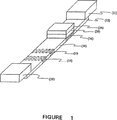
図1は、迅速抗原測定プラットフォーム(RAMPTM)装置の構成図である。
図2は、検出ゾーン内で停止させた粒子の量と抗体被覆粒子上の抗体濃度の関係を示すグラフ図である(トロンボスポンジン被覆濃度240μg/mL、ラテックス濃度0.5%)。
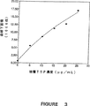
図3は、検出ゾーン内で停止させた粒子の量と検出試薬(トロンボスポンジン)濃度の関係を示すグラフ図である(ラテックス抗体表面濃度2×10⁻⁷g/cm2、ラテックス濃度2%)。
図4は、検出ゾーン内で停止させた粒子の量と抗体被覆粒子濃度の関係を示すグラフ図である(ラテックス抗体表面濃度2×10⁻⁷g/cm2、膜上240μg/mLのトロンボスポンジン15μL)。
図5は、液体試料中のトロンボスポンジンの量と検出ゾーン内で停止させた粒子の量の関係を示すグラフ図である(被覆トロンボスポンジン濃度240μg/mL、ラテックス濃度0.5%)。
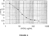
図6は、液体試料中のヒト血清アルブミン(HSA)濃度(低濃度のHSA)と、検出ゾーン内で停止させた標識粒子のシグナルとの関係を示すグラフ図である。
図7は、液体試料中のヒト血清アルブミン(HSA)濃度(高濃度のHSA)と、検出ゾーン内で停止させた標識粒子のシグナルとの関係を示すグラフ図である。
発明の詳細な説明
本発明は、免疫クロマトグラフィー測定法を用いて分析対象物の量を定量的に測定する方法、該方法において有用な装置、および該装置を含むキットに関する。本明細書で説明するように、本願出願人らは溶液状態の可溶性免疫原性分析対象物の値を測定する高感度免疫クロマトグラフィー測定法をすでに開発している。
本明細書で使用する場合、「分析対象物」という用語は、その量を測定しようとする分子または化合物をいう。分析対象物の具体例としては、ホルモンや酵素などのタンパク質、糖タンパク質、ペプチド、小型分子、多糖類、抗体、核酸、医薬品、毒素、ウイルスまたはウイルス粒子、細胞壁の部分、およびその他の化合物などが挙げられる。分析対象物は、分析対象物に対して抗体(以下に説明するもの)を生成することができるという意味の「免疫原性」のものである。好ましい態様においては、トロンボスポンジン、ミオグロビン、または尿中アルブミンが分析対象物である。
本発明の免疫クロマトグラフィー測定法を実施するためには、迅速抗原測定プラットフォーム(RAMPTM)装置を使用する。図1にRAMPTM装置の構成図を示す。RAMPTM装置は下記部材を具備する。すなわち、適用ポイント(12)を有する膜ストリップ(10)、接触領域(14)、および検出ゾーン(16)である。膜ストリップは次の特性を有する物質で作製することができる。すなわち、液体が表面から内部全体を通る毛細管作用を示すことができる程度に十分な孔隙度を有し、毛細管作用により抗体被覆粒子を移動させることができ(すなわち粒子をブロックしてはならない)、分析対象物を含む液体による濡れ性がある(たとえば水性液の場合は親水性、有機溶媒の場合は疎水性)。疎水性表面から親水性表面への変換を記載している米国特許4,340,482号または米国特許4,618,533号に記載のものなどのプロセスにより、水性液との使用のために膜を親水性化させるように膜の疎水性を変化させることができる。膜物質の具体例としては、セルロース、硝酸セルロース、酢酸セルロース、ガラス繊維、ナイロン、ポリ電解質イオン交換膜、アクリル共重合体/ナイロン、およびポリエーテルスルホンなどが挙げられる。好ましい態様においては、膜ストリップは硝酸セルロースでできている。
本明細書で使用する場合、「適用ポイント」(12)という用語は、液体試料を適用する膜上の位置をいう。RAMPTM装置は、膜に支えられた状態で適用ポイントを覆う「適用パッド」(22)を任意に含むことができる。適用パッドは、パッドに適用されたときに膜上の適用ポイントに液体試料を送達することができる吸収物質でできていてよい。代表的な物質としてはセルロースまたはガラス繊維などが挙げられる。
膜の「接触領域」は適用ポイントに隣接している。RAMPTM装置は、膜に支えられた状態で接触領域を被覆する「接触パッド」(24)を任意に含むことができる。接触パッドは吸着物質で作ることができ、代表的な物質としては、セルロース、硝酸セルロース、酢酸セルロース、ガラス繊維、ナイロン、ポリ電解質イオン交換膜、アクリル共重合体/ナイロン、およびポリエーテルスルホンなどが挙げられる。接触パッドが存在する場合、RAMPTM装置は、膜に支えられた「セパレーターパッド」(26)を接触領域と接触パッドの間に任意に含んでいてもよい。セパレーターパッドは吸着物質で作ることができ、代表的な物質としては、セルロース、硝酸セルロース、酢酸セルロース、ガラス繊維、ナイロン、ポリ電解質イオン交換膜、アクリル共重合体/ナイロン、およびポリエーテルスルホンなどが挙げられる。好ましい態様においては、セパレーターパッドと接触パッドの両者が存在する場合、これらのものは同じ物質でできている。
特定の分析対象物に対する抗体(またはその他のタイプの特異的結合分子)で被覆された粒子の集団が、膜の「接触領域」内および/または接触パッドが存在する場合は接触パッド内に埋め込まれている。粒子の数は、粒子のサイズと組成、膜の組成、および測定感度によって異なる。粒子数は通常4×106ないし4×109個程度であるが、4×106個未満でも使用できる。好ましい態様においては、粒子数は約4×108個である。
接触領域に埋め込まれた粒子は、抗体で、またはその他の分析対象物に特異的に結合する剤で被覆することができる粒子である。そのような物質の具体例としては、コロイド状金粒子、コロイド状イオウ粒子、コロイド状セレン粒子、コロイド状硫酸バリウム粒子、コロイド状硫酸鉄粒子、金属ヨウ化物粒子、ハロゲン化銀粒子、シリカ粒子、コロイド状金属(水和)酸化物粒子、コロイド状金属硫化物粒子、コロイド状セレン化鉛粒子、コロイド状セレン化カドミウム粒子、コロイド状金属リン酸塩粒子、コロイド状金属フェライト粒子、上記コロイド状粒子に有機または無機層を被覆したもの、タンパク質またはペプチド分子、リポソーム、または有機ポリマーラテックス粒子などが挙げられる。好ましい態様においては、粒子はポリスチレンラテックスビーズであり、界面活性剤不含スーパーアクティブユニフォームアルデヒド/サルフェートラテックス(インターフェーシャルダイナミックス社(Interfacial Dynamics Corp.,Portland,OR))など界面活性剤非存在下で調製したポリスチレンラテックスビーズがとくに好ましい。粒子のサイズは膜の孔隙度と関係し、粒子は液体の毛細管作用によって膜沿いに輸送されるのに十分な程度に小さいものでなければならない。
粒子は、検出を容易にするために標識化することができる。標識の具体例としては、発光標識、染料などの比色標識、蛍光標識、または電気活性剤(たとえばフェロシアナイド)などの化学標識などが挙げられる。
粒子は、特定の分析対象物に特異的に結合する剤で被覆される。好ましい態様においては、粒子は特定の分析対象物に対する抗体で被覆される。抗体はモノクローナル抗体であってもよいし、ポリクローナル抗体であってもよい。本明細書で使用する場合、「抗体」という用語は、特定の分析対象物に結合するのに十分な抗体断片をいうこともある。あるいは、分析対象物結合部位を有する合成タンパク質など特定の分析対象物に特異的に結合する分子を用いることもできる〔ホリガーとフーゲンブルーム(Holliger,P.and H.R.Hoogenbloom)Trends in Biotechnology 13:7-9(1995);チャモウとアシュケナージ(Chamow,S.M.and A.Ashkenazi)Trends in Biotechnology 14:52-60(1996)〕。別の態様においては、リガンドが特定の分析対象物である場合、該リガンドに結合する受容体を使用することができる。特異性既知の抗体が分析対象物である場合、粒子は分析対象物−抗体に対応する抗原で被覆することができる。
膜の接触領域は適用ポイントと膜の「検出ゾーン」(16)の間にある。本明細書で説明する検出ゾーンとは、「検出試薬」が固定されている膜ストリップ上のあるポイントをいう。1つの態様においては、特定の分析対象物が検出試薬である。第2の態様においては、抗体は粒子上に被覆されているため、分析対象物の同一エピトープまたは分析対象物の異なるエピトープに対する抗体が検出試薬である。
RAMPTM装置は「ウッキングパッド」(28)を任意に含むこともできる。本明細書で使用する場合、「ウィッキングパッド」という用語は、毛細管作用によって膜ストリップの末端まで輸送された溶液を吸い込む吸収物質をいう。そのような物質の具体例としては、セルロース及びガラス繊維などが挙げられる。
測定ごとの膜物性の変動を補正するために、装置はさらに、内部対照粒子と対照検出試薬と対照反応ゾーン(32)を有する内部対照を含むことができる。内部対照粒子は抗体被覆粒子との接触領域に埋め込まれる。「内部対照粒子」は抗体被覆粒子と同一のものであり、内部対照粒子上の抗体が分析対象物に対する抗体と反応しない対照検出試薬に対するものである以外は、同じ表面濃度の抗体で被覆されている。「対照検出試薬」は、測定しようとする分析対象物または抗体被覆粒子上の抗体または検出試薬のいずれとも相互作用を示さない試薬であればよい。好ましい態様においては、キーホールリンペットヘモシアニン(KLH)が対照検出試薬である。対照検出試薬は「対照反応ゾーン」(32)内で膜に被覆される。本明細書で説明する対照反応ゾーンとは、対照検出試薬が固定される膜ストリップ上のあるポイントをいう。対照反応ゾーンは接触領域と検出領域の間にあってよい。あるいは、検出ゾーンは接触領域と対照反応ゾーンの間に位置していてもよい。
本発明の定量的免疫クロマトグラフィー測定法を実施するためには、特定の分析対象物を含む液体試料を準備する。該液体は、膜材料を濡らす液体であって、抗体/抗原反応を支持し(すなわち抗体/抗原相互作用を妨害しない)、毛細管作用による移動が可能な程度に十分に低い粘性を有する液体であればよい。好ましい態様においては、水溶液(体液など)がその液体である。
定量的測定法の第1の態様においては、膜ストリップの適用ポイントを、特定の分析対象物を含む液体試料と接触させる。装置に適用パッドが付いている場合、適用パッドに液体試料を適用すると、パッドが液体試料を適用ポイントまで送達させる。適用ポイントで膜ストリップを特定の分析対象物を含む液体試料と接触させた後、液体が毛細管作用により分析対象物を膜の「接触領域」まで輸送できる条件下に膜ストリップを保つ。
分析対象物が接触領域まで輸送されると、液体中に存在する分析対象物は、接触領域に埋め込まれている抗体被覆粒子に結合する。接触パッドまたはセパレーターパッド付き接触パッドが存在する場合、これらのパッドは抗体被覆粒子の放出制御を促進し、より大量の抗体被覆粒子含有液体試料と接触する。分析対象物が抗体被覆粒子に「結合する」ということは、粒子上に被覆された抗体のうちの1つ以上が特定の分析対象物に結合することを意味する。「不十分に結合した」抗体被覆粒子とは、粒子上の抗体がさらに多くの分析対象物に結合することができるように、粒子に被覆された抗体の結合部位が特定の分析対象物で完全には飽和されていないものをいう。本明細書で説明する場合、特定の分析対象物に不十分に結合した抗体被覆粒子とは、一部の分析対象物に結合するか、分析対象物に結合できないものである。もうそれ以上の分析対象物が抗体被覆粒子に結合できなくなった場合、抗体被覆粒子は分析対象物で「飽和された」という。
液体中の分析対象物が接触領域および/または接触パッドが存在する場合はその接触パッドに埋め込まれた抗体被覆粒子に結合できるような条件下に保たれた抗体被覆粒子を、本明細書では、「接触抗体被覆粒子」という。接触抗体被覆粒子は、液体試料中に分析対象物が存在するかどうかに関わらず、また分析対象物が抗体被覆粒子上の抗体に結合しているかどうかに関わらず、分析対象物が抗体に結合していてもよいし、していなくてもよい。
液体試料に由来する液体の毛細管作用により接触抗体被覆粒子が易動化され、接触抗体被覆粒子が膜上の「検出ゾーン」に向けて膜沿いに移動される。検出試薬に結合すると、接触抗体被覆粒子の移動は停止される。特定の分析対象物が検出試薬である場合、検出試薬は、特定の分析対象物に不十分に結合した接触抗体被覆粒子上の抗体に結合する。特定の分析対象物に対する抗体が検出試薬である場合、検出試薬は、接触抗体被覆粒子上の抗体に結合する分析対象物に結合する。本明細書で使用する場合、「検出試薬−粒子複合体」という用語は、検出試薬と接触抗体被覆粒子の複合体をいう。検出試薬−粒子複合体は検出ゾーン内で停止(たとえば固定)される。
検出ゾーン内で停止させた検出試薬−粒子複合体の量を検出する。抗体被覆粒子が標識されている場合、標識のタイプに適した手段を用いて複合体を検出する。あるいは、検出試薬−粒子複合体の量は、検出ゾーン内で散乱する光を測定するなどの光学的方法によって検出する。検出試薬−粒子複合体の量は、電気伝導度または誘電率(キャパシタンス)を利用して測定することもできる。あるいは、インジウム、ビスマス、ガリウム、またはテルルイオンなどの遊離電気活性剤〔ハイエスら(Hayes et al.)Analytical Chem.66:1860-1865(1994)〕またはフェロシアナイド〔ロバーツとデュルスト(Roberts and Durst)Analytical Chem.67:482-491(1995)〕を電気化学的に検出する方法を用いることもできる。たとえば、リポソームを用いる場合、ロバーツとデュルストが概要を述べているように、検出ゾーンに1滴の洗浄剤を添加することによって、リポソームにカプセル化したフェロシアナイドを遊離させ、遊離したフェロシアナイドを電気化学的に検出することができる。キレート剤−タンパク質コンジュゲートを用いて金属イオンをキレート化する場合、検出ゾーンに1滴の酸を添加することでイオンが遊離し、ハイエスらが記載しているアノードストリッピング電圧測定法により遊離イオンを定量することができる。
次いで、検出ゾーン内で停止させた検出試薬−粒子複合体の量を基に、液体試料中の分析対象物の量を測定する。特定の分析対象物が検出試薬である場合、液体試料中の特定の分析対象物の量は、検出ゾーン内で停止させた検出試薬−粒子複合体の量に反比例する。抗体が検出試薬である場合、液体試料中の特定の分析対象物の量は、検出ゾーン内で停止させた検出試薬−粒子複合体の量に正比例する。
分析対象物の量は、標準曲線を利用して求めることができる。標準曲線は、分析対象物を検出しようとする液体中に既知濃度の分析対象物を含む対照試料系列を調製することによってこれを作成する(分析対象物を除去した血清など)。次いで、この対照試料系列について定量的免疫クロマトグラフィー測定を行う。各対照試料ごとに検出ゾーン内の検出試薬−粒子複合体の量を求め、その量を、対照試料に含まれる分析対象物の濃度の関数としてプロットする。未知量の分析対象物を含む試料(「試験試料」)は、試験試料の検出試薬−粒子複合体の量を求める測定を行い、標準曲線と比較して試験試料中の分析対象物の濃度を求める。単一の標準曲線を作成して、これをすべての試験試料に対して使用することができる。すなわち、各試験試料ごとに標準曲線を作成する必要はない。標準曲線は、検出試薬が変わるごとに再校正する。
測定で内部対照粒子を用いる場合、その内部対照粒子を液体で易動化させ、毛細管作用によって対照反応ゾーンまで移動させる。内部対照粒子は対照反応ゾーン内で対照検出試薬と結合し、内部対照粒子−対照検出試薬複合体(本明細書では対照複合体)と称する)を形成する。対照複合体の量は、検出ゾーン内の検出試薬−粒子複合体の量と同様にして検出される。存在する対照複合体の量に対する検出試薬−粒子複合体の量の比率(R)を用いて標準曲線により、存在する分析対象物の量を求める。標準曲線は、分析対象物を検出しようとする液体中に既知濃度の特定の分析対象物を含む対照試料系列を調製することによってこれを作成する(分析対象物を除去した血清等)。次いで、この対照試料系列について定量的免疫クロマトグラフィー測定を行う。各対照試料ごとにRの値を測定し、得られたR値を、対照試料に含まれる分析対象物の濃度の関数としてプロットする。未知量の分析対象物を含む試料(「試験試料」)は、試験試料のRの値を求める測定を行い、標準曲線と比較して試験試料中の分析対象物濃度を求める。上記同様、すべての試験試料に対して単一の標準曲線を作成してこれを使用することができる。すなわち、各試験試料ごとに標準曲線を作成しなおす必要はない。
本発明の第2の態様においては、適用ポイントではなく膜ストリップの検出ゾーンを液体試料と接触させる。本態様においては、特定の分析対象物に対する抗体が検出試薬である。液体試料中の特定の分析対象物を検出ゾーン内の抗体に結合させるのに十分な条件下に膜ストリップを保つことで、固定化分析対象物を生成させる。続いて、膜の適用ポイントを水または緩衝液と接触させる。緩衝液は膜物質を濡らす水性液であって、抗体/抗原反応を支持し(すなわち抗体/抗原相互作用を妨害しない)、毛細管作用による液体の移動を可能にするのに十分に低い粘性を有する液体であればよい。緩衝液の具体例としては、たとえば食塩水または50mMトリス塩酸、pH7.4などが挙げられる。緩衝液は、接触領域および/または接触パッドで膜に埋め込まれた抗体被覆粒子の集団を検出ゾーンへと輸送する。さらに、固定化分析対象物が抗体被覆粒子と相互作用させるのに十分な条件下に膜ストリップを保つ。固定化分析対象物が抗体被覆粒子と相互作用すると、抗体被覆粒子の移動が停止し、停止分析対象物−粒子複合体ができる。次いで、検出ゾーン内の停止分析対象物−粒子複合体の量を上記のようにして測定し、上記同様に標準曲線を用いて液体試料中の分析対象物の量を求めるが、内部対照はあってもなくても求めることができる。液体試料中の特定の分析対象物の量は、検出ゾーン内の停止分析対象物−粒子複合体の量に正比例する。
本発明の好ましい態様においては、トロンボスポンジンが特定の分析対象物であり、全血試料または全血由来の血小板を多く含む血漿試料が液体試料である。血小板の多い血漿試料は、常法を用いて血液試料から単離する。全血または血小板を多く含む血漿試料を用いてトロンボスポンジンの定量的測定を行うためには、試料を装置にかける前か、試料を装置にかけることによって、血小板からトロンボスポンジンを遊離させなければならない。トロンボスポンジンは、遊離剤や接触活性化などの方法により、全血試料または血小板を多く含む血漿試料中の血小板から遊離させることができる。トロンビン、カルシウムイオノフォアーA23187、ホルボールエステル、および洗浄剤などの遊離剤はいずれもトロンボスポンジンを血小板から遊離させる目的に使用することができる。あるいは、抗凝固剤の非存在下でガラス容器に血液を導入する際に接触活性化を行うことによって開始される自然凝固プロセスによるトロンビン生成でもトロンボスポンジンを十分に遊離させることができる。好ましい態様においては、RAMPTM装置は、血小板からトロンボスポンジンを遊離させる目的に用いる適用パッドを含む。全血試料または血小板を多く含む血漿試料を適用パッドに適用すると、トロンボスポンジンの遊離が起きる。適用パッドはさらに、上記のものなど1つ以上の遊離剤を含浸させることで、トロンボスポンジンの遊離を促進させることができる。本明細書では、遊離剤または接触活性化によって遊離させたトロンボスポンジンを「遊離トロンボスポンジン」と呼ぶ。検出試薬はトロンボスポンジンであってもよく、トロンボスポンジンに対する抗体であってもよく、これら以外の適当な剤であってもよい。トロンボスポンジンの標準曲線は、検出可能なトロンボスポンジンを含まない血清中に既知濃度のトロンボスポンジンを含む対照試料系列を調製することによってこれを作成する。この対照試料系列について定量的免疫クロマトグラフィー測定を行う。各対照試料ごとに検出ゾーン内の検出試薬−粒子複合体の量を求め、得られた値を、対照試料に含まれるトロンボスポンジンの濃度の関数としてプロットする。
試料中のトロンボスポンジンの量を用いて、試料中の血小板から遊離されたトロンボスポンジンの量と血小板数の関係に基づき、個体の血小板数を求めることができる。血小板数と標準試料中のトロンボスポンジンの関係を示す参照曲線を作成することができ、血小板数を試験試料中のトロンボスポンジンの量から求めることができる。あるいは、参照曲線は、トロンボスポンジンの量を既知数の血小板を含む対照血液試料系列中の血小板濃度の関数としてプロットすることができる。トロンボスポンジンと血小板数の関係に関するより詳細な開示は、「血小板粒子タンパク質測定法を利用した血小板数の測定」という名称の、1996年3月29日に提出された米国特許出願番号08/625,770(代理人資料番号UBC95−095)に記載されているが、その開示内容はすべて引用により本願に含まれるものとする。
本発明の別の好ましい態様においては、ミオグロビンが特定の分析対象物である。試料は、たとえば抗凝固処理全血などの全血、血漿、または血清などが挙げられる。好ましい態様においては、試料は全血である。装置は、適用パッドと内部対照(内部対照粒子と対照検出試薬と対照反応ゾーンを有する)を含んでいることが好ましい。また、ミオグロビンに対するモノクローナル抗体を検出試薬として用い、これを検出ゾーン内の膜に被覆することが好ましい。膜を1%PVAなどの適当な剤でブロックする。定量的免疫クロマトグラフィー測定は、液体試料を適用パッドに添加することによって開始され、そして測定が進行する。対照反応ゾーン内の対照複合体の量に対する検出ゾーン内の検出試薬−粒子複合体の量の比率(R)を用いて標準曲線により、存在するミオグロビンの量を求める。標準曲線は、検出可能なミオグロビンを含まない全血、血漿、または血清中に既知濃度のミオグロビンを含む対照試料系列を調製することによってこれを作成する。この対照試料系列について定量的免疫クロマトグラフィー測定を行う。各対照試料ごとにRの値を計算し、得られたR値を、対照試料に含まれるミオグロビンの濃度の関数としてプロットする。
別の好ましい態様においては尿中アルブミンが特定の分析対象物であり、尿試料が液体試料である。アルブミンを検出試薬として用い、これを検出領域中の膜に被覆する。上記のようにして、膜をブロックする。尿試料を適用パッドに適用することによって測定を開始し、測定を進行させる。標準曲線は、検出可能なアルブミンを含まない対照尿試料系列に既知量のアルブミンを加えたものについて定量的免疫クロマトグラフィー測定を行うことによって作成する。
本発明は、本明細書で説明する装置を含むキットも対象となる。上記以外のキット構成物としては、緩衝液、液体採取手段、および標準曲線作成用対照試料などが挙げられる。
以下、実施例により本発明をさらに詳細に説明するが、本発明はこれらに限定されない。
実施例1 トロンボスポンジンの定量的免疫クロマトグラフィー測定
膜の選択とブロッキング剤の選択を容易にするとともに、ラテックス遊離および移動の条件、ラテックス移動停止の条件、およびラテックス移動停止阻害が遊離の分析対象物濃度に依存する程度を調べるために、実験を行った。
A.膜の選択
検出試薬の膜結合特性と膜を通過する毛細管流速を測定することによって、適当な膜を選んだ。膜の検出試薬親和性と結合力および膜上の結合部位と競合する可能性のある分析しようとする液体試料中に存在する緩衝液、ブロッキング試薬、またはタンパク質(血漿タンパク質など)による結合の可逆性がないことが、重要な結合特性である。
1.トロンボスポンジンの膜への平衡結合
平衡条件下で様々な膜に吸着されたトロンボスポンジンの量を測定するとともに、血清タンパク質との競合により膜表面から脱着されたトロンボスポンジン(フラクション)の量を測定するために、実験を行った。使用した膜を表1に示す。

アーチパンチを用いて膜にパンチ穴をあけることによって、円形膜ディスク(直径0.875cm)を作成した。3枚または4枚の膜ディスクの平均乾燥重量を測定した。
乾燥膜ディスクを、上記原液から調製した(i)20、(ii)40、(iii)80、および(iv)200μg/mLのトロンボスポンジンを含む溶液1mLに浸漬し、振盪しないで一夜平衡化させた。次いで、膜を新しい試験管に移し、膜と元のトロンボスポンジン溶液の放射能を測定して、平衡トロンボスポンジン濃度と膜上に結合したトロンボスポンジンの量を求めた。それぞれの膜のトロンボスポンジン結合力を、スカッチャードプロット〔カントールとスキメル(Cantor,C.R.and P.R.Schimmel)Biophysical Chemistry,Part III.The Behavior of Biological Macromolecules,W.H.Freeman Co.,San Francisco(1980),p.856〕により求めた。膜のトロンボスポンジン結合力の概要を表2に示した。
A/E>NT5000>NC5>NC8>S&S NC5>M5>M10>S&S NC8>NF10
有効ポアサイズの小さいS&S N5およびM5膜よりも多くのトロンボスポンジンを結合させると思われるNB8膜を例外として、一般に有効ポアサイズの小さい膜(1および5μm)は有効ポアサイズの大きい膜(8および10μm)より単位重量あたりより多くのトロンボスポンジンを結合させる。これはおそらく、有効ポアサイズの小さい膜の方が有効ポアサイズの大きい膜よりも単位重量あたりの吸着利用可能繊維物質の量が多いためであろう。ザルトリウス社製NC5、ゲルマン社製NT5000、およびS&S社製NC5の結果からわかるように、同様の材料と有効ポアサイズと厚さを有する膜の間でも、結合力に著しい差がある。
次いで、吸着の可逆性を調べた。膜を15分間トリス塩酸緩衝液(50mM、pH7.4)に静止浸漬することで洗浄し、膜上に保持されたトロンボスポンジンの量をガンマ計数法によって測定した。膜を3サイクルの洗浄手順に付し、各洗浄サイクルごとに放射能をカウントして、保持されたトロンボスポンジンの量を求めた。3回目の緩衝液洗浄後、膜を1mLの血清中で15分間のインキュベーションに付し、保持されたトロンボスポンジンの量をガンマ計数法によって測定した。吸着されたトロンボスポンジンを保持する膜を2回または3回の緩衝液洗浄に付したところ、膜空間内の未結合トロンボスポンジンが効果的に除去された。トロンボスポンジンを一夜吸着後に緩衝液で洗浄した膜を、競合性を示す血清タンパク質に曝露させたところ、有意な脱着を示さなかった(データは示さず)。
2.スポットウェッティングによる膜へのトロンボスポンジンの結合
検出試薬を検出ゾーンに適用するために、検出試薬の溶液を液滴状にして膜の検出ゾーンに噴霧または適用する(スポットウェッティング)。本プロセスでは、検出試薬は、繊維を通過する毛細管現象によって溶媒が蒸発または移動する際に濃度変化を示す溶液からアクセス先である膜表面に向かって乾燥するが、これは大量の検出試薬溶液が膜によって平衡化される際に起きるものとは異なるプロセスである。ラテックス免疫クロマトグラフィー測定においては、ターゲットエリアを濡らせることによって検出試薬を適用し、次いで、膜をポリマーまたは洗浄剤でブロックする。ブロッキング剤を適用すると、結合した検出試薬を遊離させることができる。同様に、測定の移動段階では、ウェッティングフロントが膜の上を進みターゲットエリアに到達するため、ブロッキング剤をウェッティングフロント沿いに施用して検出試薬を遊離させることができる。したがって、免疫クロマトグラフィー測定と同じ条件下で膜上のトロンボスポンジン結合特性を調べるとともに、様々なブロッキング剤による乾燥再水和後で適用ポイントで膜が結合トロンボスポンジンを保持する能力を測定するために、実験を行った。
以後の試験用に、結合性の点からザルトリウス社製NC5膜を選択した。膜(ザルトリウスNC5)をストリップ(1.5cm×9.0cm)に切断し、6つの正方形片(1.5cm×1.5cm)に分割した。正方形のサイズは、10μLのトロンボスポンジンが膜の全面にちょうど一杯に広がるように選んだ。
ブロッキング剤によるトロンボスポンジンの可逆性を調べるために、10μLの放射標識トロンボスポンジンを膜ストリップの一端付近でカットした第2の正方形片上にスポットブロットし、一夜乾燥させた。次いで、トロンボスポンジンが結合した片がブロッキング液の真上に位置するようにして膜ストリップをトリス塩酸緩衝液(50mM、pH7.4)中の1%w/vブロッキング剤に浸漬し、ブロッキング液を膜のもう一方の端まで吸い込ませた。次いで、膜ストリップを一夜乾燥させ、片(1.5cm×1.5cm)に切断し、それぞれの片をカウントして、ブロッキング剤によって膜ストリップ沿いに保持および/または遊離されたトロンボスポンジンの量を求めた。結果を表3に示す。
ブロッキング剤を膜上で乾燥させ再水和させることによってどれだけの量のトロンボスポンジンが遊離されるかを測定するために、上記実験でブロッキング剤が膜に吸い込まれてから、トロンボスポンジンスポットを設けた膜ストリップ片を約15分間にわたりトリス塩酸緩衝液に浸漬し、ストリップ片および再平衡化に消費された緩衝液の放射能を再びカウントした。結果を表4に示す。
3.膜を通過する緩衝液と血清の毛細管流速
一定長さの膜を通過する緩衝液と血清の移動速度を測定する実験を行った。表1に示した膜をストリップ(1.0cm×6.0cm)に切断し、6つの片(1.0cm×1.0cm)に分割した。各ストリップの下端部を緩衝液(トリス塩酸、50mM、pH7.4)または新鮮ヒト血清に浸漬した状態で、それぞれの膜ストリップを試験管内に垂直に立てた。液面を2cm移動させた後、液体が1cmづつ移動するのに要する時間を記録した。結果を表5に示す。
B.膜ブロッキング剤の選択
抗体で覆われたラテックスが血清タンパク質の存在下で膜に付着しないような、膜のブロッキング方法について研究を行った。
1.IgGの膜への平衡結合
平衡条件下における種々の膜へのIgGの吸着量を測定するための実験、そして緩衝液とブロッキング剤とによる洗浄サイクル後の表面に保持されるIgGの量を測定するための実験を行った。乾燥状態の膜ディスク(直径=0.875cm)を、(a)5、(b)10、(c)25、(d)50、(e)100そして(f)200μg/mLの放射標識IgGを含有する2mLの溶液に浸し、室温で、振とうさせずに一晩平衡化した。次いで、膜を新しい試験管に移し、膜とIgG溶液の放射能を測定して、平衡となったIgGの濃度と膜への結合量を得た。次いで、トリス−塩酸緩衝液(50mM、pH7.4)中に15分間静置することで洗浄し、ガンマ線を計数して保持されるIgGの量を測定した。次いで、膜を別の緩衝液での洗浄サイクル、そしてトリス−塩酸緩衝液(50mM、pH7.4)中にPVA(平均分子量=15000)を1%含有する溶液に付した。
吸着IgGを有する膜について、新鮮な緩衝液中で2回の再平衡化を行ったため、膜の隙間にある、結合していないIgGのほとんどを除去したと思われた。1%のPVA(15000)溶液により、ニトロセルロース膜に結合していたIgGのかなりの量が置換されたのに対して、ガラス繊維やナイロン膜では、このような多量の結合IgGの脱離は見られなかった。このことは、PVA(15000)は、ガラス繊維やナイロン膜についてよりもニトロセルロース膜について、より良いブロッキング剤であろうことを示すものである。
2.スポット−ウェッティングによる膜へのIgGの結合
免疫クロマトグラフィー測定が行われるのと同様の条件下でのIgG結合特性を測定するための実験、そして、ブロッキング剤溶液と血清中でのインキュベーションを行った後の、膜上に残るIgGの量を測定するための実験を行った。
乾燥状態の膜を正方形片(1.5cm×1.5cm)に切断し、各膜片に10μLの放射標識IgGをスポットブロッティングし、そして3時間風乾させた。膜の小片上に固定したIgGの量をガンマ線の計数により測定した。風乾膜片を最初にトリス−塩酸緩衝液中にPVA(15000)を1%含有する溶液(2mL)中で15分間インキュベーションを行い、次いで新しい試験管に移して再度ガンマカウンターで計数して、保持されたIgGの量を測定した。次いで、膜を2mLの血清中で15分間のインキュベーション(×2)を行い、新しい試験管に移して血清中の各洗浄インキュベーションの後にガンマカウンターで計数した。
結果は、スポットブロッティングによりニトロセルロース膜に結合したIgGのかなりの量が、1%のPVA溶液中での膜のインキュベーションによって置換されることを示した。それに対して、ガラス繊維やナイロン膜からは、比較的少量のIgGしか置換されなかった。それに続く血清によるインキュベーションでは、1%PVAにより既にブロックされたIgGは置換されなかった。
3.ブロックされた膜へのIgGの結合
膜上での種々のブロッキング剤の有効性を測定するための実験、そして緩衝液洗浄サイクルがブロッキング剤にどのような効果を与えるかについて測定するための実験を行った。
アーチ状穴あけ器を用いて、膜を通過するパンチ孔によって円形状の膜ディスク(直径=0.875cm)を得た。乾燥状態の膜ディスクを種々のブロッキング剤の溶液1mLに浸し、一晩平衡化した。次いで、膜ディスクを新しい試験管に移し、200μg/mLの放射標識IgGを含む1mL溶液中でインキュベーションを行う前に、約3時間風乾させた。次いで膜をトリス−塩酸緩衝液(50mM、pH7.4)中で洗浄し、ガンマ線放射を計数してIgG結合量を測定した。緩衝液による洗浄を繰り返し、膜のガンマ線放射について再び計数した。2回目の緩衝液洗浄の後、200mg/mLの放射標識IgGを含む1mLの溶液で15分間膜を再平衡化した。さらなる緩衝液洗浄を行い、膜上に保持されたIgGの量を測定した。
その結果、ブロックされた膜へのIgG結合量は、ブロックされていない膜、即ち、トリス−塩酸緩衝液中でIgGと平衡化した膜への結合量よりも非常に小さいことが示された。ガラス繊維やナイロン膜を除けば、PVA(15000)、PVA(22000)、PVA(49000)及びPVP(40000)は、すべてのニトロセルロース膜へのIgGの結合を効果的にブロックした(データは示さず。)。他の水溶性ポリマー、PEG(6000)、PEG(20000)及びデキストランは、ガラス繊維膜−A/Eの場合(データは示さず。)を除くすべての膜について、PVA及びPVPと同程度の良好なブロッキング剤とはならなかった。中性洗剤(ツィーン20、プルロニックP−105、及びトライトンX−100)の間では、トライトン及びプルロニックよりもツィーン20がニトロセルロース及びナイロン膜を強くブロックするものと考えられた。さらに、トライトン及びプルロニックはガラス繊維(A/E)やナイロン膜(M5及びM10)を効果的にはブロックしなかった。BSAは全般的にすべての膜をかなり強くブロックした。しかし、PVA及びPVPと同程度の効果にまでニトロセルロース膜をブロックすることはなかった。IgG中での膜の再平衡化から、緩衝液による洗浄サイクルは、IgG結合の前に用いたブロッキング剤にはいかなる影響をも与えることはないことが示された(データは示さず。)。
4.ブロックされた膜への血清中のIgGの結合
種々のブロッキング剤によりブロックされた膜への、血清存在下でのIgG結合量を測定するための実験を行った。
アーチ状穴あけ器を用いて、膜を通過するパンチ孔によって円形状の膜ディスク(直径=0.875cm)を得た。乾燥状態の膜ディスクを種々のブロッキング剤の溶液1mLに浸し、一晩平衡化した。次いで、膜ディスクを新しい試験管に移し、200μg/mLの放射標識IgGの1mL血清中でインキュベーションを行う前に、約3時間風乾させた。緩衝液による洗浄前後に、ガンマ線を計数して膜へのIgG結合量を測定した。その結果、血清存在下では、種々のブロッキング剤でプレブロックする膜へのIgGの結合は無視できる程度であることが示された。ブロックされていない膜、即ち、血清と放射標識IgGとを含むトリス−塩酸緩衝液中でインキュベーションが行われたものへは、検出される程度のIgGが結合しなかったことから、血清はブロッキング剤と同様に作用した(データは示さず。)。
C.ラテックスの放出と移動のための条件
ここで記述する測定法において、被覆済みの粒子を乾燥させて膜の接触ゾーン(及び/又は接触パッド)へやる。この実験から、エアーブラシによる懸濁液を噴霧すること、又は手動で小滴を添加することのいずれかによる粒子の適用が許容できることが示された。
ラテックスの放出と移動のための条件を調べるために、30%のショ糖水溶液を膜のある領域に添加し、そして乾燥させた。次いで、ラテックス(緩衝化された15%ショ糖溶液中0.5%)を同じ領域に添加し、そして乾燥させた。次いでこの膜を緩衝液又は血清に浸し、移動を続けさせた。最初のショ糖層は再水和を促進したものの、膜片を経由するラテックス粒子の移動は、特に血清が放出剤として用いられた場合、妨害された。
実験の目的に適用する最も率直な方法は、マイクロピペットでラテックス懸濁液を手動で添加することであった。緩衝化された15%ショ糖溶液で0.25〜2%のラテックスをブロックされた膜に直接添加し、移動が始まる前に少しの間乾燥させた。
アエロ−プロ150エアーブラシ(ハンザ−テヒニク ゲーエムベーハー、ハンブルク、ドイツ)をラテックスの添加のための器具として評価した。このエアーブラシの使用により、膜ではなくラテックスを均一に分散させた。しかし、手動の器具によれば、添加したラテックス懸濁液を定量する手段がなかった。この手法は、加圧空気の噴霧量と速度がメーターで計測できれば有効であろう。このような分散方法はラージスケールでの適用に向いている。
D.ラテックス移動が停止する条件
ここで記述する測定法において、被覆済みの粒子は毛細管現象により検出領域に移動する。ここで、これらは検出試薬と反応し、検出試薬−粒子複合体として固定され、次いで検出される。
ラテックス移動が停止する条件について調べた。ザルトリウスNC8、マイラーで裏打ちされた膜を使用した。緩衝液中のトロンボスポンジンの10μL溶液を検出ゾーンで乾燥させ、一晩かけてその膜を1%PVA(15000)でブロックした。青色の0.29μmのサルフェート/アルデヒドラテックス粒子(IDC)を異なる濃度の抗体で被覆し、1%BSAでブロックした。異なる濃度の粒子を、緩衝化された15%ショ糖溶液に懸濁し、接触領域に添加した。種々の濃度のトロンボスポンジンを含む緩衝液を膜の適用ポイントに添加することにより移動を誘導した。検出ゾーンのビデオイメージを増幅したもののイメージ分析により、検出ゾーンにおける停止したラテックスを定量した。検出ゾーンと膜エリアの周辺間のグレイレベルの違いのトータルを、使用したシグナルとした。
これらの実験結果から、シグナルは、ラテックス抗体表面濃度(図2)、トロンボスポンジン膜被覆濃度(図3)、及びラテックス粒子濃度(図4)に対してほぼ直線に増加することが示された。
このように、抗体表面濃度が高くなるとターゲット領域におけるラテックス粒子の停止数が増加する。さらに、停止した粒子数はラテックス濃度とともに強く増加し、このことは2%ラテックスまで、飽和する傾向がごくわずかであることを示す。約25μg/mLのトロンボスポンジンまで、膜のターゲットエリアへ移動して乾燥した溶液中のトロンボスポンジン濃度の増加とともに停止粒子数が増加した。この量を超えると、トロンボスポンジン濃度が高まっても停止ラテックス量はほとんど増加しなかった(例えば、示された10倍の増加は、25μg/mL値を超えるとほんのわずかの増加にとどまった。)。それゆえに、ラテックス数を変えることにより、ラテックス上の抗体の表面濃度を変えることにより、そしてターゲットエリアへ乾燥させるトロンボスポンジン濃度を変えることにより、独立してターゲットゾーンにおける停止粒子数を制御できることが明らかとなった。
E.ラテックス移動停止阻害の遊離抗原濃度への依存性
これらの実験において、移動緩衝液に種々の濃度の遊離トロンボスポンジンに膜の一端を漬けてラテックスの移動を起こすこと等により、ラテックス移動を起こした。遊離トロンボスポンジンは、ラテックス上の抗体結合部位と競合することにより、ターゲットエリアにおける停止を阻害する。この結果は図5に示され、検出ゾーンにおいて検出されるシグナルは、液体試料中の遊離抗原の濃度の上昇にしたがって連続的に低下した。したがって、このような結果は、視覚的にも定量的にも、遊離トロンボスポンジン濃度依存的に粒子の停止が阻害されることを示した。
実施例2 ヒト血清アルブミンの定量的免疫クロマトグラフィー測定
極めて低濃度のヒト血清アルブミン(HSA)の濃度を測定するための実験を行い、そして定量的免疫測定法による測定が、より複雑で高価な免疫測定法、例えば酵素結合免疫測定法(ELISAs)に匹敵することを示した。HSA低濃度、例えば健常個体を想定したもの、又は腎臓疾患個体からの試料に典型的なHSA高濃度、のいずれかについての実験を行った。
A.低HSA濃度測定
HSAに対するポリクローナル抗体標品を検出試薬として用いた。そしてこの粒子をHSAに対するモノクローナル抗体で覆った。このモノクローナル抗HSA抗体をキャラクタライズして、解離定数Kd=0.012μg/mLを得た。これは、0.012μg/mLの平衡濃度が、HSAを含有するテスト溶液に晒された抗体集団における抗原結合部位の半分を満たすことを示す。
1.ラテックスビーズ粒子の調製
0.98mg/mLの抗体を1mL、スキムミルク粉末(カーネーション)を1.0mL、そして2.0% W/Vのラテックスビーズを0.5gをすべて、総量4.0mL、pH7.2の0.01Mリン酸塩緩衝液とし、平衡化した。ビーズをこのリン酸塩緩衝液で3回洗浄し、次いで、15%ショ糖、0.5%ツィーン20中で0.25%濃度の懸濁液とした。
2.膜の調製
検出試薬として、10μLの0.44mg/mLのポリクローナル孔HSA抗体(シグマケミカル社、セントルイス、ミズーリ州)を、ポアサイズが8μmのニトロセルロース膜(ザルトリウス)の7cm×1cmの小片の基準側の端から4cmのところの、検出ゾーンに添加して、乾燥させた。この膜を1% W/VのPVA 15000(フルカ)でブロックした。ラテックスビーズ懸濁液の5μLを該小片の基準側の端から1cmのところに添加し、乾燥させた。
3.HSAの測定
50mMトリス緩衝液、pH7.3中、0.0001μg/mLから0.4μg/mLの濃度のHSA溶液を調製した。この溶液の120μLを膜の基準側の端に添加し、膜の反対側の端へ、毛細管現象によりこの溶液を移動させた。次いでこの膜を乾燥させ、検出ゾーンに蓄積したラテックスビーズの量を光学イメージ分析により測定した。結果を、HSA濃度の関数としてプロットした(検出ゾーンに蓄積したラテックスビーズの量に対応する)シグナルで、図6に示した。この結果から、測定により0.01μg/mLを下回る(即ち、用いたモノクローナル抗体についてのKd値を下回る)濃度のHSAを検出できることが示される。これらの結果は、臨床的ELISA測定法を用いて得られ得る結果に匹敵し、このことから、測定において使用した抗体のKd値と分析的に等量かより高い濃度を典型的に検出できる。
HSA溶液の代わりに、ヒト健常者の尿を試料として使用して同様の測定を行った。この測定法によって測定されたHSAの存在レベルは、中央臨床化学施設における自動アナライザによって測定されたレベルに一致した:免疫クロマトグラフィー測定法により得られた値である3.1μg/mLと3.4μg/mLは、該自動アナライザによる値、それぞれ3.1μg/mL及び4.1μg/mLに対応した。
B.高HSA濃度測定
精製HSA(シグマケミカル社)を検出試薬として阻害測定を行い、モノクローナル抗体のKdよりもはるかに高いHSA濃度の測定を行った。
1.ラテックスビーズ粒子の調製
アルデヒドラテックスビーズ、直径0.16μm、イエローグリーン蛍光色素で標識済み(インターフェイシャルダイナミクス社)、を用いた。モノクローナル抗体濃度を1.75mg/mL、そしてスキムミルクを使用しない以外は、上記と同様の方法でビーズを調製した。
2.膜の調製
検出試薬として、1mg/mLのHSAを用いて膜を調製した。検出ゾーン上で、バイオドットアプリケーター(バイオドット社、アービン、カリフォルニア州)を用いて2μL/cmでHSAを噴霧し、乾燥させた。
3.HSAの測定
50mMトリス緩衝液、pH7.3中、2μg/mLから250μg/mLの濃度のHSA溶液を調製した。この溶液の200μLを膜の基準側の端上にあるセルロース接触パッドに添加し、膜の反対側の端へ、毛細管現象によりこの溶液を移動させた。次いでこの膜を乾燥させ、検出ゾーンに蓄積したラテックスビーズの量を蛍光強度測定により測定した。結果を、HSA濃度の関数としてプロットした(検出ゾーンに蓄積したラテックスビーズの量に対応する)シグナルで図7に示す。この結果から、HSA濃度の上昇により検出ゾーンにおけるラテックスビーズの停止を阻害することが示される。これらの結果は、この測定法が尿試料中のHSAの想定範囲(約10〜100μg/mL)を超えて鋭敏であることを示す。
均等物
当業者であれば、単に日常的な実験手法を用いることにより、本明細書に記載された本発明の具体的態様に均等な多くのものを認識し、又は確認することができよう。かかる均等物は以下の請求の範囲の範疇に包含される。Background of the Invention
Quantitative analysis of cells and analytes in liquid samples, especially body fluid samples, often provides important diagnostic and therapeutic information to both physicians and patients. For example, blood platelet counts are routinely assessed in various clinical and treatment settings, but abnormal platelet counts can cause serious bleeding problems in patients, suggesting the presence of many underlying diseases There is a case. Since myoglobin appears earliest as a marker of heart damage (Mair, J. et al. Br. Heart J. 68: 462-468 (1992)), quantification of myoglobin in blood samples This will help early diagnosis of myocardial infarction. By analyzing urine by measuring urinary albumin to determine whether there is proteinuria, it is possible to determine the degree of renal function and renal damage. Immunoassay utilizing the high specificity of antigen-antibody reactions (Kennedy, DM and SJ Challacombe, ELISA and Other Solid Phase Immunoassays: Theoretical and Practical Aspects, John Wiley and Sons, Chichester (1988)) Provides an approach to analyte measurement. Immunoassays that quantitatively measure the amount of analyte in a sample use complex multi-step procedures and expensive analyzers that are only available in the laboratory. Previously published [GB 2,204,398A; US Pat. Nos. 5,096,837, 5,238,652, and 5,266,497, Birnbaum, S. et al. Analytical Biochem. 206: 168- 171 (1992); Roberts, MA and RADurst Analytical Chem. 67: 482-491 (1995); and Klimov et al., Clinical Chem. 41: 1360 (1995)]. Immunochromatographic assay methods such as those described are simpler but still do not quantitatively measure analytes. Instead, these immunochromatographic assays detect whether an amount of analyte is included (or not included) that exceeds a predetermined cut-off value for the test to be performed. Therefore, it is a method that can quickly and quantitatively measure the amount of an analyte present in a sample, and is simple enough to be carried out without using a laboratory or a person trained in chemical analysis. There is a need for a general law.
Summary of the Invention
The present invention relates to a method for measuring the amount of a specific analyte in a liquid sample using a quantitative immunochromatographic measurement method, and an apparatus used for the measurement method. The measurement method is a rapid antigen measurement platform (RAMP).TM) Equipment is used. The apparatus is a membrane strip made of a suitable material such as cellulose nitrate or glass fiber, and has sufficient porosity and wettability with a liquid containing an analyte, and moves the particles by capillary action. including. The membrane strip has an application point, a contact area and a detection zone, the contact area being between the application point and the detection zone. Embedded in the contact area is a population of particles such as colloidal metal particles, organic molecules, liposomes, or organic polymer latex particles. The particles are coated with antibodies against a specific analyte. The particles can be labeled with colorimetric labels, fluorescent labels, luminescent labels, or other suitable labels to facilitate detection. A detection reagent is fixed in the detection zone. The detection reagent may be an antibody to a specific analysis target or the specific analysis target itself. The apparatus may further include one or more of the following elements. In other words, there is an application pad that covers the application point while being supported by the application point, a contact pad that covers the contact area and is embedded with antibody-coated particles, and a contact pad that is supported by the contact area. A separator pad supported by a membrane between the contact area and the contact pad, a wicking pad supported by a membrane adjacent to the detection zone such that the detection zone is located between the wicking pad and the contact area, and An internal control with internal control particles, control detection reagent and control reaction zone embedded in the contact area.
In order to perform the measurement, the application point of the membrane strip is brought into contact with the liquid sample to be measured for a specific analyte. Then, if the analyte is present in the sample, the device is kept under conditions sufficient to allow liquid capillary action to transport the analyte through the membrane strip to the contact area. Furthermore, the apparatus is kept under appropriate conditions such that when the analyte reaches the contact area, the analyte binds to the antibody-coated particles embedded in the contact area. Antibody coated particles, including those bound to the analyte, are mobilized with a liquid and moved through the strip to the detection zone by capillary action. The detection reagent interacts with the antibody-coated particles bound to the analyte, but the interaction between the detection reagent and the antibody-coated particles bound to the analyte causes the antibody-coated particles bound to the analyte to move within the detection zone. Stop. Next, the amount of antibody-coated particles bound to the analyte to be stopped in the detection zone is detected. The amount of a particular analyte in the liquid sample is related to the amount of antibody-coated particles that are bound to the analyte that has been stopped in the detection zone. That is, when the specific analyte is a detection reagent, the amount of the analyte in the liquid sample is inversely proportional, and when the antibody against the specific analyte is the detection reagent, the amount of the analyte in the liquid sample Is directly proportional. The amount of the analysis object is obtained from a standard curve of the analysis object.
In another immunochromatographic measurement, a liquid sample to be measured for a specific analyte is applied directly to the detection zone of the device. In this embodiment, an antibody against a specific analyte is a detection reagent. The apparatus is kept under appropriate conditions such that the analyte in the liquid sample interacts with the detection reagent and is immobilized within the detection zone. Water or a suitable buffer is then added to the application point of the membrane to mobilize the antibody-coated particles and move them into the detection zone by capillary action. In addition, the device is kept under conditions that allow interaction of the antibody-coated particles with the analyte immobilized in the detection zone. The movement of the antibody-coated particles is stopped by the interaction between the antibody-coated particles and the immobilized analyte. As described above, since the amount of the analyte in the liquid sample is related to the amount of antibody-coated particles stopped in the detection zone, this is obtained from the standard curve.
In a preferred embodiment of the present invention, thrombospondin is a specific analyte, and a whole blood sample or a platelet-rich plasma sample is a liquid sample. Measuring the concentration of thrombospondin in a clotted whole blood or platelet-rich plasma sample is a measure of the platelet count in the original blood sample. This indicator is an important measure of an individual's ability to maintain normal homeostasis and is followed in a variety of clinical conditions, such as patients undergoing chemotherapy or patients with platelet destructive disorders or abnormal platelet production.
In another preferred embodiment, myoglobin is the specific analyte and the whole blood sample is a liquid sample. Myoglobin concentration and its change over time are diagnostically important in the early assessment of heart damage in cases suspected of myocardial infarction.
In yet another preferred embodiment, human serum albumin (also referred to herein as urinary albumin) is the specific analyte and the urine sample is a liquid sample. Since the urinary albumin concentration is an index of proteinuria and kidney damage, the degree of renal dysfunction and its change with time can be evaluated by quantitatively measuring the urinary albumin level.
The measurement method of the present invention is simple and rapid, and usually does not require the addition of reagents other than the liquid sample containing the analyte or, in one embodiment, the sample containing the analyte and buffer. The measurement method of the present invention can be carried out at the time of caring for a patient, and does not require any special technique for the implementation. Furthermore, since the apparatus used in the measurement method of the present invention is common to all the analytes, it is easy to use for measuring various analytes. With the measurement method of the present invention, various immunogenic analytes can be quantified.
[Brief description of the drawings]
FIG. 1 shows a rapid antigen measurement platform (RAMP).TMFIG.
FIG. 2 is a graph showing the relationship between the amount of particles stopped in the detection zone and the antibody concentration on the antibody-coated particles (thrombospondin coating concentration 240 μg / mL, latex concentration 0.5%).
FIG. 3 is a graph showing the relationship between the amount of particles stopped in the detection zone and the concentration of the detection reagent (thrombospondin) (latex
FIG. 4 is a graph showing the relationship between the amount of particles stopped in the detection zone and the concentration of antibody-coated particles (latex
FIG. 5 is a graph showing the relationship between the amount of thrombospondin in the liquid sample and the amount of particles stopped in the detection zone (coated thrombospondin concentration 240 μg / mL, latex concentration 0.5%).
FIG. 6 is a graph showing the relationship between the human serum albumin (HSA) concentration (low concentration of HSA) in the liquid sample and the signal of the labeled particles stopped in the detection zone.
FIG. 7 is a graph showing the relationship between the human serum albumin (HSA) concentration (high concentration of HSA) in the liquid sample and the signal of the labeled particles stopped in the detection zone.
Detailed Description of the Invention
The present invention relates to a method for quantitatively measuring the amount of an analyte using an immunochromatographic measurement method, a device useful in the method, and a kit including the device. As described herein, Applicants have already developed a highly sensitive immunochromatographic assay that measures the value of soluble immunogenic analytes in solution.
As used herein, the term “analyte” refers to a molecule or compound whose amount is to be measured. Specific examples of analytes include proteins such as hormones and enzymes, glycoproteins, peptides, small molecules, polysaccharides, antibodies, nucleic acids, pharmaceuticals, toxins, viruses or virus particles, cell wall parts, and other compounds. Can be mentioned. Analytes are “immunogenic” in the sense that antibodies (to be described below) can be generated against the analyte. In a preferred embodiment, thrombospondin, myoglobin, or urinary albumin is the analyte.
In order to carry out the immunochromatographic assay of the present invention, a rapid antigen measurement platform (RAMP)TM) Use the device. Figure 1 shows RAMPTMThe block diagram of an apparatus is shown. RAMPTMThe apparatus includes the following members. A membrane strip (10) with an application point (12), a contact area (14) and a detection zone (16). The membrane strip can be made of a material having the following properties: That is, the porosity is sufficient to allow the liquid to exhibit capillary action from the surface through the entire interior, and the antibody-coated particles can be moved by capillary action (ie, the particles must not be blocked) There is wettability by the liquid containing the analysis object (for example, hydrophilic in the case of an aqueous liquid and hydrophobic in the case of an organic solvent). Membranes for use with aqueous liquids by processes such as those described in U.S. Pat. No. 4,340,482 or U.S. Pat. No. 4,618,533 describing the conversion of hydrophobic surfaces to hydrophilic surfaces. The hydrophobicity of the membrane can be changed to render it hydrophilic. Specific examples of membrane materials include cellulose, cellulose nitrate, cellulose acetate, glass fiber, nylon, polyelectrolyte ion exchange membrane, acrylic copolymer / nylon, and polyethersulfone. In a preferred embodiment, the membrane strip is made of cellulose nitrate.
As used herein, the term “application point” (12) refers to the location on the membrane where the liquid sample is applied. RAMPTMThe device can optionally include an “application pad” (22) that covers the application point while supported by the membrane. The application pad may be made of an absorbent material that can deliver a liquid sample to an application point on the membrane when applied to the pad. Typical materials include cellulose or glass fiber.
The “contact area” of the membrane is adjacent to the application point. RAMPTMThe device can optionally include a “contact pad” (24) that covers the contact area while supported by the membrane. Contact pads can be made of adsorbent materials, and typical materials include cellulose, cellulose nitrate, cellulose acetate, glass fiber, nylon, polyelectrolyte ion exchange membrane, acrylic copolymer / nylon, and polyethersulfone. Can be mentioned. RAMP if a contact pad is presentTMThe device may optionally include a “separator pad” (26) supported by the membrane between the contact area and the contact pad. Separator pads can be made of adsorbent materials, and typical materials include cellulose, cellulose nitrate, cellulose acetate, glass fiber, nylon, polyelectrolyte ion exchange membrane, acrylic copolymer / nylon, and polyethersulfone. Can be mentioned. In a preferred embodiment, when both a separator pad and a contact pad are present, these are made of the same material.
A population of particles coated with antibodies (or other types of specific binding molecules) to a particular analyte is embedded within the “contact region” of the membrane and / or within the contact pad if a contact pad is present. ing. The number of particles depends on the size and composition of the particles, the composition of the film, and the measurement sensitivity. The number of particles is usually 4 × 10⁶Or 4 × 10⁹About 4 × 10⁶Even less than can be used. In a preferred embodiment, the number of particles is about 4 × 10.8It is a piece.
The particles embedded in the contact area are particles that can be coated with antibodies or other agents that specifically bind to the analyte. Specific examples of such substances include colloidal gold particles, colloidal sulfur particles, colloidal selenium particles, colloidal barium sulfate particles, colloidal iron sulfate particles, metal iodide particles, silver halide particles, silica particles, Colloidal metal (hydrated) oxide particles, colloidal metal sulfide particles, colloidal lead selenide particles, colloidal cadmium selenide particles, colloidal metal phosphate particles, colloidal metal ferrite particles, colloidal particles described above Examples thereof include those coated with an organic or inorganic layer, protein or peptide molecules, liposomes, or organic polymer latex particles. In a preferred embodiment, the particles are polystyrene latex beads and in the absence of a surfactant, such as a surfactant-free superactive uniform aldehyde / sulfate latex (Interfacial Dynamics Corp., Portland, OR). The prepared polystyrene latex beads are particularly preferred. The size of the particles is related to the porosity of the membrane, and the particles must be small enough to be transported along the membrane by liquid capillary action.
The particles can be labeled to facilitate detection. Specific examples of the label include a luminescent label, a colorimetric label such as a dye, a fluorescent label, or a chemical label such as an electroactive agent (for example, ferrocyanide).
The particles are coated with an agent that specifically binds to a particular analyte. In a preferred embodiment, the particles are coated with antibodies against a specific analyte. The antibody may be a monoclonal antibody or a polyclonal antibody. As used herein, the term “antibody” can refer to an antibody fragment sufficient to bind to a particular analyte. Alternatively, a molecule that specifically binds to a specific analyte, such as a synthetic protein having an analyte binding site, can be used (Holliger, P. and HR Hoogenbloom Trends in Biotechnology 13: 7 -9 (1995); Chamow and SMand A. Ashkenazi Trends in Biotechnology 14: 52-60 (1996)]. In another aspect, when the ligand is a particular analyte, a receptor that binds to the ligand can be used. If an antibody of known specificity is an analyte, the particles can be coated with an antigen corresponding to the analyte-antibody.
The contact area of the membrane is between the application point and the “detection zone” (16) of the membrane. The detection zone described herein refers to a point on the membrane strip to which the “detection reagent” is fixed. In one embodiment, the specific analyte is a detection reagent. In the second embodiment, since the antibody is coated on the particle, an antibody against the same epitope of the analyte or a different epitope of the analyte is the detection reagent.
RAMPTMThe device may optionally include a “wicking pad” (28). As used herein, the term “wicking pad” refers to an absorbent material that sucks in the solution transported to the end of the membrane strip by capillary action. Specific examples of such a substance include cellulose and glass fiber.
To correct for variations in membrane properties from measurement to measurement, the device can further include an internal control having internal control particles, a control detection reagent, and a control reaction zone (32). Internal control particles are embedded in the contact area with the antibody-coated particles. “Internal control particles” are identical to antibody-coated particles and are coated with antibodies of the same surface concentration, except that the antibodies on the internal control particles are for control detection reagents that do not react with antibodies to the analyte. Yes. The “control detection reagent” may be any reagent that does not interact with either the analyte to be measured or the antibody on the antibody-coated particle or the detection reagent. In a preferred embodiment, keyhole limpet hemocyanin (KLH) is the control detection reagent. The control detection reagent is coated on the membrane within the “control reaction zone” (32). The control reaction zone described herein refers to a point on the membrane strip where the control detection reagent is immobilized. The control reaction zone may be between the contact area and the detection area. Alternatively, the detection zone may be located between the contact area and the control reaction zone.
In order to carry out the quantitative immunochromatographic measurement method of the present invention, a liquid sample containing a specific analyte is prepared. The liquid should be a liquid that wets the membrane material, supports the antibody / antigen reaction (ie does not interfere with the antibody / antigen interaction), and has a viscosity that is low enough to allow migration by capillary action. That's fine. In a preferred embodiment, an aqueous solution (such as body fluid) is the liquid.
In the first aspect of the quantitative measurement method, the application point of the membrane strip is brought into contact with a liquid sample containing a specific analyte. If the device has an application pad, applying the liquid sample to the application pad causes the pad to deliver the liquid sample to the application point. After contacting the membrane strip with a liquid sample containing a particular analyte at the application point, the membrane strip is kept under conditions that allow the liquid to transport the analyte to the “contact area” of the membrane by capillary action.
When the analyte is transported to the contact area, the analyte present in the liquid binds to the antibody-coated particles embedded in the contact area. When contact pads or contact pads with separator pads are present, these pads facilitate controlled release of the antibody-coated particles and come into contact with a larger amount of antibody-coated particle-containing liquid sample. By “binding” an analyte to an antibody-coated particle is meant that one or more of the antibodies coated on the particle are bound to a particular analyte. “Insufficiently bound” antibody-coated particles are those where the binding site of the antibody coated on the particle is completely bound by a particular analyte so that the antibodies on the particle can bind to more analyte. Means not saturated. As described herein, antibody-coated particles that are insufficiently bound to a particular analyte are those that bind to some analyte or cannot bind to the analyte. An antibody coated particle is said to be “saturated” with an analyte when no more analyte can bind to the antibody coated particle.
An antibody-coated particle maintained under conditions such that an analyte in a liquid can bind to an antibody-coated particle embedded in the contact area and / or contact pad, if present, is herein referred to as It is called “contact antibody-coated particles”. Contacted antibody-coated particles bind to the antibody regardless of whether the analyte is present in the liquid sample and whether the analyte is bound to the antibody on the antibody-coated particle. You may or may not.
The contact antibody-coated particles are mobilized by the capillary action of the liquid derived from the liquid sample, and the contact antibody-coated particles are moved along the membrane toward the “detection zone” on the membrane. When bound to the detection reagent, the movement of the contact antibody-coated particles is stopped. If the particular analyte is a detection reagent, the detection reagent binds to the antibody on the contact antibody-coated particles that are poorly bound to the particular analyte. When the antibody to a particular analyte is a detection reagent, the detection reagent binds to the analyte that binds to the antibody on the contact antibody-coated particle. As used herein, the term “detection reagent-particle complex” refers to a complex of a detection reagent and contact antibody-coated particles. The detection reagent-particle complex is stopped (eg, immobilized) within the detection zone.
The amount of detection reagent-particle complex stopped in the detection zone is detected. If the antibody-coated particles are labeled, the complex is detected using means appropriate for the type of label. Alternatively, the amount of detection reagent-particle complex is detected by an optical method such as measuring light scattered within the detection zone. The amount of the detection reagent-particle complex can also be measured using electric conductivity or dielectric constant (capacitance). Alternatively, free electroactive agents such as indium, bismuth, gallium, or tellurium ions (Hayes et al. Analytical Chem. 66: 1860-1865 (1994)) or ferrocyanide (Roberts and Durst) Analytical Chem .67: 482-491 (1995)] can also be used. For example, when using liposomes, as outlined by Roberts and Durst, a drop of detergent is added to the detection zone to liberate the ferrocyanide encapsulated in the liposomes and the released ferrocyanide is electrochemically removed. Can be detected. When chelating a metal ion using a chelator-protein conjugate, the ion is liberated by adding a drop of acid to the detection zone, and free ion is measured by the anode stripping voltage measurement method described by Hayes et al. Can be quantified.
Next, the amount of the analyte in the liquid sample is measured based on the amount of the detection reagent-particle complex stopped in the detection zone. When the specific analyte is a detection reagent, the amount of the specific analyte in the liquid sample is inversely proportional to the amount of detection reagent-particle complex stopped in the detection zone. When the antibody is a detection reagent, the amount of a particular analyte in the liquid sample is directly proportional to the amount of detection reagent-particle complex stopped in the detection zone.
The amount of the analysis object can be obtained using a standard curve. A standard curve is created by preparing a control sample series containing a known concentration of analyte in the liquid in which the analyte is to be detected (such as serum from which the analyte has been removed). This control sample series is then subjected to quantitative immunochromatographic measurements. The amount of detection reagent-particle complex in the detection zone is determined for each control sample and the amount is plotted as a function of the concentration of the analyte contained in the control sample. A sample containing an unknown amount of analyte ("test sample") is measured to determine the amount of detection reagent-particle complex in the test sample and compared to a standard curve to determine the concentration of the analyte in the test sample. Ask. A single standard curve can be generated and used for all test samples. That is, it is not necessary to create a standard curve for each test sample. The standard curve is recalibrated each time the detection reagent changes.
When using internal control particles in the measurement, the internal control particles are mobilized with a liquid and moved to the control reaction zone by capillary action. The internal control particles bind to the control detection reagent within the control reaction zone to form an internal control particle-control detection reagent complex (referred to herein as a control complex). The amount of control complex is detected in the same manner as the amount of detection reagent-particle complex in the detection zone. The amount of analyte present is determined by a standard curve using the ratio (R) of the amount of detection reagent-particle complex to the amount of control complex present. A standard curve is created by preparing a control sample series containing a known concentration of a particular analyte in the liquid in which the analyte is to be detected (such as serum from which the analyte has been removed). This control sample series is then subjected to quantitative immunochromatographic measurements. The value of R is measured for each control sample and the resulting R value is plotted as a function of the concentration of the analyte contained in the control sample. A sample containing an unknown amount of analyte ("test sample") is measured to determine the R value of the test sample, and compared to a standard curve to determine the analyte concentration in the test sample. As above, a single standard curve can be generated and used for all test samples. That is, it is not necessary to recreate a standard curve for each test sample.
In the second aspect of the invention, the detection zone of the membrane strip is brought into contact with the liquid sample rather than the application point. In this embodiment, an antibody against a specific analyte is a detection reagent. The immobilized analyte is generated by keeping the membrane strip under conditions sufficient to bind a particular analyte in the liquid sample to the antibody in the detection zone. Subsequently, the application point of the membrane is brought into contact with water or a buffer. The buffer is an aqueous liquid that wets the membrane material and supports the antibody / antigen reaction (ie, does not interfere with antibody / antigen interactions) and has a sufficiently low viscosity to allow fluid movement by capillary action. Any liquid may be used. Specific examples of the buffer include saline or 50 mM Tris-HCl, pH 7.4. The buffer transports the population of antibody-coated particles embedded in the membrane at the contact area and / or contact pad to the detection zone. In addition, the membrane strip is kept under conditions sufficient for the immobilized analyte to interact with the antibody-coated particles. When the immobilized analyte interacts with the antibody-coated particles, the movement of the antibody-coated particles stops and a stopped analyte-particle complex is created. The amount of stopped analyte-particle complex in the detection zone is then measured as described above, and the amount of analyte in the liquid sample is determined using a standard curve as above, but the internal control is not. It can be obtained with or without. The amount of a particular analyte in the liquid sample is directly proportional to the amount of stopped analyte-particle complex in the detection zone.
In a preferred embodiment of the present invention, thrombospondin is a specific analyte, and a whole blood sample or a plasma sample rich in platelets derived from whole blood is a liquid sample. Platelet rich plasma samples are isolated from blood samples using routine methods. To perform a quantitative measurement of thrombospondin using whole blood or platelet-rich plasma samples, the thrombospondin must be released from the platelets before or by applying the sample to the instrument. I must. Thrombospondin can be released from platelets in whole blood samples or plasma samples rich in platelets by methods such as release agents or contact activation. Any release agent such as thrombin, calcium ionophore A23187, phorbol ester, and detergent can be used to release thrombospondin from platelets. Alternatively, thrombospondin can be sufficiently released even by thrombin generation by a natural coagulation process initiated by performing contact activation when blood is introduced into a glass container in the absence of an anticoagulant. In a preferred embodiment, RAMPTMThe device includes an application pad used for the purpose of releasing thrombospondin from platelets. When a whole blood sample or a plasma sample rich in platelets is applied to the application pad, release of thrombospondin occurs. The application pad can be further impregnated with one or more release agents such as those described above to facilitate the release of thrombospondin. As used herein, a thrombospondin released by a release agent or contact activation is referred to as “free thrombospondin”. The detection reagent may be thrombospondin, an antibody against thrombospondin, or any other appropriate agent. A standard curve for thrombospondin is created by preparing a control sample series containing known concentrations of thrombospondin in serum without detectable thrombospondin. Quantitative immunochromatographic measurements are performed on this control sample series. The amount of detection reagent-particle complex in the detection zone is determined for each control sample and the resulting value is plotted as a function of the concentration of thrombospondin contained in the control sample.
Using the amount of thrombospondin in the sample, the platelet count of the individual can be determined based on the relationship between the amount of thrombospondin released from platelets in the sample and the platelet count. A reference curve showing the relationship between the platelet count and thrombospondin in the standard sample can be generated, and the platelet count can be determined from the amount of thrombospondin in the test sample. Alternatively, the reference curve can plot the amount of thrombospondin as a function of platelet concentration in a control blood sample series containing a known number of platelets. A more detailed disclosure regarding the relationship between thrombospondin and platelet count can be found in US patent application Ser. No. 08/625, filed Mar. 29, 1996, entitled “Measurement of Platelet Count Using Platelet Particle Protein Assay”. 770 (agent document number UBC95-095), the entire disclosure of which is incorporated herein by reference.
In another preferred embodiment of the invention, myoglobin is a specific analyte. Examples of the sample include whole blood such as anticoagulated whole blood, plasma, or serum. In a preferred embodiment, the sample is whole blood. The device preferably includes an application pad and an internal control (having internal control particles, control detection reagent and control reaction zone). In addition, it is preferable to use a monoclonal antibody against myoglobin as a detection reagent and coat it on the membrane in the detection zone. The membrane is blocked with a suitable agent such as 1% PVA. A quantitative immunochromatographic measurement is initiated by adding a liquid sample to the application pad and the measurement proceeds. The amount of myoglobin present is determined by a standard curve using the ratio (R) of the amount of detection reagent-particle complex in the detection zone to the amount of control complex in the control reaction zone. A standard curve is created by preparing a control sample series that contains a known concentration of myoglobin in whole blood, plasma, or serum that does not contain detectable myoglobin. Quantitative immunochromatographic measurements are performed on this control sample series. The R value is calculated for each control sample and the resulting R value is plotted as a function of the concentration of myoglobin contained in the control sample.
In another preferred embodiment, urinary albumin is the specific analyte and the urine sample is a liquid sample. Albumin is used as a detection reagent and this is coated on the membrane in the detection region. The membrane is blocked as described above. The measurement is started by applying the urine sample to the application pad and the measurement proceeds. A standard curve is generated by performing quantitative immunochromatographic measurements on a control urine sample series without detectable albumin plus a known amount of albumin.
The present invention is also directed to kits that include the devices described herein. Examples of kit components other than the above include buffers, liquid collection means, and control samples for preparing standard curves.
EXAMPLES Hereinafter, although an Example demonstrates this invention further in detail, this invention is not limited to these.
Example 1 Quantitative immunochromatographic determination of thrombospondin
Experiments were conducted to facilitate the selection of membranes and blocking agents, as well as to examine the conditions for latex release and migration, conditions for latex migration termination, and the extent to which latex migration termination inhibition depends on the concentration of free analyte. went.
A. Membrane selection
Appropriate membranes were selected by measuring the membrane binding properties of the detection reagent and the capillary flow rate through the membrane. Membrane detection reagent affinity and binding strength and reversibility of binding by buffers, blocking reagents, or proteins (such as plasma proteins) present in the liquid sample to be analyzed that may compete with the binding sites on the membrane. It is an important binding property to be absent.
1. Equilibrium binding of thrombospondin to the membrane.
Experiments were performed to measure the amount of thrombospondin adsorbed on various membranes under equilibrium conditions and to determine the amount of thrombospondin (fraction) desorbed from the membrane surface by competition with serum proteins. It was. The membrane used is shown in Table 1.
Circular membrane discs (0.875 cm in diameter) were made by punching holes in the membrane using an arch punch. The average dry weight of 3 or 4 membrane disks was measured.
The dried membrane disk is immersed in 1 mL of a solution containing (i) 20, (ii) 40, (iii) 80, and (iv) 200 μg / mL thrombospondin prepared from the above stock solution and equilibrated overnight without shaking. I let you. The membrane was then transferred to a new tube and the radioactivity of the membrane and the original thrombospondin solution was measured to determine the equilibrium thrombospondin concentration and the amount of thrombospondin bound on the membrane. The thrombospondin binding force of each membrane is expressed in a Scatchard plot (Cantor, CRand PR Schimmel, Biophysical Chemistry, Part III. The Behavior of Biological Macromolecules, WHFreeman Co., San Francisco (1980), p. .856]. A summary of the thrombospondin binding strength of the membrane is shown in Table 2.
A / E> NT5000> NC5> NC8> S & S NC5> M5> M10> S & S NC8> NF10
With the exception of NB8 membrane, which seems to bind more thrombospondin than S & S N5 and M5 membranes with small effective pore size, membranes with small effective pore size (1 and 5 μm) are generally membranes with large effective pore size (8 and 10 μm) Bind more thrombospondin per unit weight. This is probably because a membrane with a smaller effective pore size has a higher amount of adsorbable fiber material per unit weight than a membrane with a larger effective pore size. As can be seen from the results for Sartorius NC5, Germanic NT5000, and S & S NC5, there are significant differences in bonding forces between similar materials and membranes with effective pore sizes and thicknesses.
Next, the reversibility of adsorption was examined. The membrane was washed by static immersion in Tris-HCl buffer (50 mM, pH 7.4) for 15 minutes, and the amount of thrombospondin retained on the membrane was measured by gamma counting. The membrane was subjected to a three-cycle wash procedure and the radioactivity was counted for each wash cycle to determine the amount of thrombospondin retained. After the third buffer wash, the membrane was subjected to a 15 minute incubation in 1 mL of serum and the amount of thrombospondin retained was determined by gamma counting. When the membrane holding the adsorbed thrombospondin was subjected to buffer washing twice or three times, the unbound thrombospondin in the membrane space was effectively removed. Membranes washed with buffer after thrombospondin adsorption overnight showed no significant desorption when exposed to competitive serum proteins (data not shown).
2. Binding of thrombospondin to membranes by spot wetting
In order to apply the detection reagent to the detection zone, the detection reagent solution is sprayed or applied to the detection zone of the membrane in the form of droplets (spot wetting). In this process, the detection reagent is dried from a solution that exhibits a change in concentration as the solvent evaporates or moves by capillary action through the fibers toward the membrane surface that is the access destination. It is a different process than occurs when equilibrated by the membrane. In latex immunochromatography measurements, the detection reagent is applied by wetting the target area and then the membrane is blocked with a polymer or detergent. When a blocking agent is applied, the bound detection reagent can be released. Similarly, in the measurement movement phase, the wetting front travels over the membrane and reaches the target area, so that a blocking agent can be applied along the wetting front to release the detection reagent. Therefore, examine the thrombospondin binding properties on the membrane under the same conditions as immunochromatographic measurements and measure the ability of the membrane to retain bound thrombospondin at the point of application after dry rehydration with various blocking agents An experiment was conducted for this purpose.
For subsequent tests, NC5 membrane made by Sartorius was selected from the viewpoint of binding. The membrane (Sartorius NC5) was cut into strips (1.5 cm × 9.0 cm) and divided into six square pieces (1.5 cm × 1.5 cm). The square size was chosen so that 10 μL of thrombospondin spread just over the entire surface of the membrane.
To examine the reversibility of thrombospondin by the blocking agent, 10 μL of radiolabeled thrombospondin was spot blotted onto a second square piece cut near one end of the membrane strip and allowed to dry overnight. Next, the membrane strip is immersed in a 1% w / v blocking agent in Tris-HCl buffer (50 mM, pH 7.4) so that the piece to which thrombospondin is bound is positioned immediately above the blocking solution, and the blocking solution is added. Was sucked to the other end of the membrane. The membrane strip is then dried overnight, cut into pieces (1.5 cm x 1.5 cm), and each piece is counted to determine the amount of thrombospondin retained and / or released along the membrane strip by the blocking agent. Asked. The results are shown in Table 3.
In order to determine how much thrombospondin is released by drying and rehydrating the blocking agent on the membrane, the thrombospondin spot is taken after the blocking agent has been drawn into the membrane in the above experiment. The membrane strip piece provided with was immersed in Tris-HCl buffer for about 15 minutes and the radioactivity of the strip piece and the buffer consumed for re-equilibration was counted again. The results are shown in Table 4.
3. Capillary flow rate of buffer and serum through the membrane
Experiments were conducted to measure the rate of movement of buffer and serum through a membrane of a certain length. The membrane shown in Table 1 was cut into strips (1.0 cm × 6.0 cm) and divided into six pieces (1.0 cm × 1.0 cm). Each membrane strip was erected vertically in a test tube with the lower end of each strip immersed in buffer (Tris-HCl, 50 mM, pH 7.4) or fresh human serum. After moving the liquid level by 2 cm, the time required for the liquid to move by 1 cm was recorded. The results are shown in Table 5.
B. Selection of membrane blocking agent
Studies were made on how to block the membrane so that the antibody-covered latex does not adhere to the membrane in the presence of serum proteins.
1. Equilibrium binding of IgG to membrane
Experiments were performed to measure the amount of IgG adsorbed on various membranes under equilibrium conditions, and to measure the amount of IgG retained on the surface after a wash cycle with buffer and blocking agent. Dry membrane discs (diameter = 0.875 cm) were loaded with (a) 5, (b) 10, (c) 25, (d) 50, (e) 100 and (f) 200 μg / mL radiolabeled IgG. Immerse in the containing 2 mL solution and equilibrate overnight at room temperature without shaking. The membrane was then transferred to a new tube and the radioactivity of the membrane and IgG solution was measured to obtain the equilibrium concentration of IgG and the amount bound to the membrane. Subsequently, it was washed by standing in Tris-HCl buffer (50 mM, pH 7.4) for 15 minutes, and the amount of IgG retained was measured by counting gamma rays. The membrane was then subjected to another buffer wash cycle and a solution containing 1% PVA (average molecular weight = 15000) in Tris-HCl buffer (50 mM, pH 7.4).
The membrane with adsorbed IgG was re-equilibrated twice in fresh buffer, which seemed to remove most unbound IgG in the membrane gap. A 1% PVA (15000) solution displaced a significant amount of IgG bound to the nitrocellulose membrane, whereas glass fibers and nylon membranes desorbed such large amounts of bound IgG. I couldn't see it. This indicates that PVA (15000) may be a better blocking agent for nitrocellulose membranes than for glass fibers and nylon membranes.
2. Binding of IgG to membrane by spot-wetting
Experiments to determine IgG binding properties under the same conditions as immunochromatographic measurements, and the amount of IgG remaining on the membrane after incubation in serum with blocking agent solution Experiments were performed to measure.
Dried membranes were cut into square pieces (1.5 cm × 1.5 cm), each membrane piece was spot blotted with 10 μL of radiolabeled IgG and allowed to air dry for 3 hours. The amount of IgG immobilized on a small piece of membrane was measured by gamma ray counting. The air-dried membrane pieces are first incubated in a solution (2 mL) containing 1% PVA (15000) in Tris-HCl buffer for 15 minutes, then transferred to a new tube and counted again with a gamma counter. The amount of IgG produced was measured. The membrane was then incubated in 2 mL of serum for 15 minutes (x2), transferred to a new tube and counted with a gamma counter after each wash incubation in serum.
The results showed that a significant amount of IgG bound to the nitrocellulose membrane by spot blotting was replaced by incubation of the membrane in a 1% PVA solution. On the other hand, only a relatively small amount of IgG was replaced from glass fiber or nylon membrane. Subsequent incubation with serum did not replace IgG already blocked by 1% PVA.
3. Binding of IgG to blocked membrane
Experiments were performed to determine the effectiveness of various blocking agents on the membrane and to determine what effect the buffer wash cycle has on the blocking agent.
A circular membrane disk (diameter = 0.875 cm) was obtained by punch holes passing through the membrane using an arcuate punch. The dried membrane disk was immersed in 1 mL of various blocking agent solutions and allowed to equilibrate overnight. The membrane disc was then transferred to a new tube and allowed to air dry for about 3 hours before incubation in a 1 mL solution containing 200 μg / mL radiolabeled IgG. The membrane was then washed in Tris-HCl buffer (50 mM, pH 7.4) and gamma radiation was counted to determine the amount of IgG bound. The washing with buffer was repeated and the membrane was again counted for gamma radiation. After the second buffer wash, the membrane was re-equilibrated with 1 mL of a solution containing 200 mg / mL radiolabeled IgG for 15 minutes. Further buffer washes were performed and the amount of IgG retained on the membrane was measured.
As a result, it was shown that the amount of IgG bound to the blocked membrane was much smaller than the amount bound to the unblocked membrane, that is, the membrane equilibrated with IgG in Tris-HCl buffer. Except for glass fibers and nylon membranes, PVA (15000), PVA (22000), PVA (49000) and PVP (40000) effectively blocked IgG binding to all nitrocellulose membranes (data shown) ). The other water soluble polymers, PEG (6000), PEG (20000) and dextran are as good as PVA and PVP for all membranes except for the glass fiber membrane-A / E (data not shown) It was not a good blocking agent. Among neutral detergents (
4). Binding of IgG in serum to blocked membrane
Experiments were performed to determine the amount of IgG bound to the membranes blocked with various blocking agents in the presence of serum.
A circular membrane disk (diameter = 0.875 cm) was obtained by punch holes passing through the membrane using an arcuate punch. The dried membrane disk was immersed in 1 mL of various blocking agent solutions and allowed to equilibrate overnight. The membrane disk was then transferred to a new tube and allowed to air dry for approximately 3 hours before incubation in 1 mL serum of 200 μg / mL radiolabeled IgG. Before and after washing with the buffer, gamma rays were counted to measure the amount of IgG bound to the membrane. As a result, it was shown that in the presence of serum, IgG binding to the membrane pre-blocked with various blocking agents was negligible. Serum was a blocking agent because undetectable IgG did not bind to unblocked membranes, ie, those incubated in Tris-HCl buffer containing serum and radiolabeled IgG. (Data not shown).
C. Conditions for latex release and transport
In the measurement method described here, the coated particles are dried and passed to the contact zone (and / or contact pad) of the membrane. This experiment showed that application of the particles by either spraying the suspension with an airbrush or manually adding droplets was acceptable.
To examine the conditions for latex release and transfer, 30% aqueous sucrose solution was added to an area of the membrane and allowed to dry. Latex (0.5% in a buffered 15% sucrose solution) was then added to the same area and allowed to dry. The membrane was then immersed in buffer or serum and allowed to continue moving. Although the first sucrose layer promoted rehydration, the migration of latex particles through the membrane pieces was hindered, especially when serum was used as the release agent.
The most straightforward method applied for experimental purposes was to manually add the latex suspension with a micropipette. 0.25-2% latex was added directly to the blocked membrane in a buffered 15% sucrose solution and allowed to dry briefly before the transfer began.
Aero-Pro 150 airbrush (Hansa-Tehnik GmbH, Hamburg, Germany) was evaluated as an instrument for latex addition. By using this airbrush, the latex was uniformly dispersed instead of the film. However, according to the manual instrument, there was no means for quantifying the added latex suspension. This technique will be effective if the spray amount and speed of pressurized air can be measured with a meter. Such a dispersion method is suitable for application on a large scale.
D. Conditions to stop latex movement
In the measurement method described here, the coated particles move to the detection region by capillary action. Here, they react with the detection reagent, are immobilized as detection reagent-particle complexes, and then detected.
The conditions under which latex movement stopped were investigated. Sartorius NC8, mylar-backed membrane was used. A 10 μL solution of thrombospondin in buffer was dried in the detection zone and the membrane was blocked with 1% PVA (15000) overnight. Blue 0.29 μm sulfate / aldehyde latex particles (IDC) were coated with different concentrations of antibody and blocked with 1% BSA. Different concentrations of particles were suspended in buffered 15% sucrose solution and added to the contact area. Migration was induced by adding buffers containing various concentrations of thrombospondin to the application point of the membrane. The stopped latex in the detection zone was quantified by image analysis of an amplified video image of the detection zone. The total difference in gray levels between the detection zone and the periphery of the membrane area was taken as the signal used.
These experimental results showed that the signal increased almost linearly with latex antibody surface concentration (FIG. 2), thrombospondin membrane coating concentration (FIG. 3), and latex particle concentration (FIG. 4). .
Thus, as the antibody surface concentration increases, the number of latex particle stops in the target region increases. Furthermore, the number of particles stopped increases strongly with latex concentration, indicating that there is very little tendency to saturate up to 2% latex. To about 25 μg / mL thrombospondin, the number of stop particles increased with increasing concentration of thrombospondin in the solution transferred to the target area of the membrane and dried. Above this amount, there was little increase in the amount of stopped latex as the thrombospondin concentration increased (eg, the 10-fold increase shown is only a slight increase above the 25 μg / mL value). ). Therefore, the number of stopping particles in the target zone can be controlled independently by changing the number of latexes, by changing the surface concentration of the antibody on the latex, and by changing the concentration of thrombospondin to be dried to the target area. It became clear.
E. Dependence of latex migration arrest inhibition on free antigen concentration
In these experiments, latex migration occurred, for example, by immersing one end of the membrane in free thrombospondin of various concentrations in the migration buffer to cause migration of the latex. Free thrombospondin inhibits arrest in the target area by competing with antibody binding sites on the latex. This result is shown in FIG. 5, where the signal detected in the detection zone decreased continuously as the concentration of free antigen in the liquid sample increased. Thus, these results indicated that particle arrest was inhibited both visually and quantitatively depending on the concentration of free thrombospondin.
Example 2 Quantitative immunochromatographic determination of human serum albumin
Experiments have been conducted to measure very low concentrations of human serum albumin (HSA), and quantitative immunoassay measurements have become more complex and expensive immunoassays, such as enzyme-linked immunoassays (ELISAs). Shown to be comparable. Experiments were conducted on either low HSA concentrations, for example those assuming healthy individuals, or high HSA concentrations typical for samples from kidney disease individuals.
A. Low HSA concentration measurement
A polyclonal antibody preparation against HSA was used as a detection reagent. The particles were then covered with a monoclonal antibody against HSA. This monoclonal anti-HSA antibody was characterized and the dissociation constant Kd= 0.012 μg / mL was obtained. This indicates that an equilibrium concentration of 0.012 μg / mL fills half of the antigen binding sites in the antibody population exposed to the test solution containing HSA.
1. Preparation of latex bead particles
1 mL of 0.98 mg / mL antibody, 1.0 mL of skim milk powder (carnation), and 0.5 g of 2.0% W / V latex beads, all in a total volume of 4.0 mL, pH 7.2 0.01M Equilibrated with phosphate buffer. The beads were washed 3 times with this phosphate buffer and then made into a 0.25% strength suspension in 15% sucrose, 0.5
2. Membrane preparation
As a detection reagent, 10 μL of a 0.44 mg / mL polyclonal pore HSA antibody (Sigma Chemical Co., St. Louis, MO) was applied from the end of the reference side of a 7 cm × 1 cm piece of nitrocellulose membrane (Sartorius) having a pore size of 8 μm. It was added to the detection zone at 4 cm and dried. The membrane was blocked with 1% W / V PVA 15000 (Fluka). 5 μL of the latex bead suspension was added 1 cm from the reference end of the piece and allowed to dry.
3. Measurement of HSA
An HSA solution having a concentration of 0.0001 μg / mL to 0.4 μg / mL in 50 mM Tris buffer, pH 7.3 was prepared. 120 μL of this solution was added to the reference end of the membrane, and the solution was moved by capillary action to the opposite end of the membrane. The membrane was then dried and the amount of latex beads accumulated in the detection zone was measured by optical image analysis. The results are shown in FIG. 6 as a signal (corresponding to the amount of latex beads accumulated in the detection zone) plotted as a function of HSA concentration. From this result, the measured value is below 0.01 μg / mL (ie K for the monoclonal antibody used).dIt is shown that a concentration of HSA (below the value) can be detected. These results are comparable to those that can be obtained using a clinical ELISA assay, and thus the K of the antibody used in the measurement.dA concentration that is analytically equivalent to or higher than the value can typically be detected.
The same measurement was performed using urine of healthy human subjects as a sample instead of the HSA solution. The level of HSA present measured by this assay was consistent with that measured by an automated analyzer in a central clinical chemistry facility: 3.1 μg / mL and 3.4 μg, which were values obtained by immunochromatographic assay. / ML corresponded to the value by the automatic analyzer, 3.1 μg / mL and 4.1 μg / mL, respectively.
B. High HSA concentration measurement
Inhibition measurement was performed using purified HSA (Sigma Chemical Co.) as a detection reagent.dMuch higher HSA concentrations were measured.
1. Preparation of latex bead particles
Aldehyde latex beads, diameter 0.16 μm, labeled with a yellow green fluorescent dye (Interfacial Dynamics) were used. Beads were prepared in the same manner as described above except that the monoclonal antibody concentration was 1.75 mg / mL and skim milk was not used.
2. Membrane preparation
A membrane was prepared using 1 mg / mL HSA as a detection reagent. On the detection zone, HSA was sprayed at 2 μL / cm using a biodot applicator (Biodot, Irvine, Calif.) And dried.
3. Measurement of HSA
HSA solutions with a concentration of 2 μg / mL to 250 μg / mL were prepared in 50 mM Tris buffer, pH 7.3. 200 μL of this solution was added to the cellulose contact pad on the reference end of the membrane and the solution was moved by capillary action to the opposite end of the membrane. The membrane was then dried and the amount of latex beads accumulated in the detection zone was measured by fluorescence intensity measurement. The results are shown in FIG. 7 as a signal plotted (as a function of the amount of latex beads accumulated in the detection zone) as a function of HSA concentration. This result indicates that the increase of the HSA concentration inhibits latex bead termination in the detection zone. These results indicate that this assay is sensitive beyond the expected range of HSA in urine samples (about 10-100 μg / mL).
Equivalent
Those skilled in the art will recognize, or be able to ascertain using no more than routine experimentation, many equivalents to the specific embodiments of the invention described herein. Such equivalents are encompassed by the scope of the following claims.
Claims (11)
b.膜ストリップの適用ポイントに液体試料を接触させる工程;
c.液体がストリップを介する毛細管作用により液体試料中の特定の分析対象物を接触領域へ移動させ得るのに十分な条件下で膜ストリップを維持する工程であって、接触領域はその中に埋め込まれた抗体被覆粒子の集団、ここで抗体は特定の分析対象物に対する抗体である、及びその中に埋め込まれた内部対照粒子の集団、ここで内部対照粒子は対照検出試薬に対する抗体により被覆されている、を有する、工程;
d.特定の分析対象物が抗体被覆粒子に結合し、それにより接触抗体被覆粒子を生じ得るため;試料中の液体がストリップを介する毛細管作用によりその上に固定化された特定の分析対象物を有する検出ゾーンへ接触抗体被覆粒子を移動させ得るため、かつ試料中の液体がストリップを介する毛細管作用によりその上に固定化された対照検出試薬を有する対照反応ゾーンへ内部対照粒子を移動させ得るため;検出ゾーンにおいて固定化された特定の分析対象物が液体試料中の特定の分析対象物で十分には被覆されていない接触抗体被覆粒子に結合し得るため、及び内部対照粒子が対照検出試薬に結合し得るために十分な条件下で膜ストリップをさらに維持する工程;ならびに
e.検出ゾーンにおいて固定化された特定の分析対象物に結合した接触抗体被覆粒子の量及び対照反応ゾーンにおいて対照検出試薬に結合した内部対照粒子の量を測定する工程であって、ここで、液体試料中の特定の分析対象物の量は、対照反応ゾーンにおいて対照検出試薬に結合した内部対照粒子の量に対する検出ゾーンにおいて固定化された特定の分析対象物に結合した抗体被覆粒子の量の比に反比例する、工程
を含む、液体試料中の特定の分析対象物の量の定量方法。a. Providing a rapid antigen measurement platform device comprising a membrane strip comprising an application point, a contact area, a control reaction zone and a detection zone, wherein the contact area is between the application point and the detection zone;
b. Contacting the liquid sample with the application point of the membrane strip;
c. Maintaining the membrane strip under conditions sufficient to allow the liquid to move a particular analyte in the liquid sample to the contact area by capillary action through the strip, the contact area being embedded therein A population of antibody-coated particles, wherein the antibody is an antibody to a particular analyte, and a population of internal control particles embedded therein, wherein the internal control particles are coated with an antibody to a control detection reagent, Having a process;
d. Because a specific analyte binds to antibody-coated particles, thereby producing contact antibody-coated particles; detection with a specific analyte immobilized thereon by capillary action through the strip Detection of the contact antibody-coated particles to the zone and the movement of the internal control particles to a control reaction zone having a control detection reagent immobilized thereon by capillary action through the strip of liquid; detection; Because specific analytes immobilized in the zone can bind to contact antibody-coated particles that are not sufficiently coated with the specific analyte in the liquid sample, and internal control particles bind to the control detection reagent. Further maintaining the membrane strip under conditions sufficient to obtain; and e. Measuring the amount of contact antibody-coated particles bound to a specific analyte immobilized in the detection zone and the amount of internal control particles bound to a control detection reagent in a control reaction zone, wherein a liquid sample The amount of a particular analyte in the ratio of the amount of antibody-coated particles bound to a particular analyte immobilized in the detection zone to the amount of internal control particles bound to the control detection reagent in the control reaction zone. A method for quantifying the amount of a particular analyte in a liquid sample, comprising steps that are inversely proportional.
b.膜ストリップの適用ポイントに液体試料を接触させる工程;
c.液体がストリップを介する毛細管作用により液体試料中の特定の分析対象物を接触領域へ移動させ得るのに十分な条件下で膜ストリップを維持する工程であって、接触領域はその中に埋め込まれた抗体被覆粒子の集団、ここで抗体は特定の分析対象物に対する抗体である、及びその中に埋め込まれた内部対照粒子の集団、ここで内部対照粒子は対照検出試薬に対する抗体により被覆されている、を有する、工程;
d.特定の分析対象物が抗体被覆粒子に結合し、それにより接触抗体被覆粒子を生じ得るため;試料中の液体がストリップを介する毛細管作用により抗体被覆粒子上の抗体と同じエピトープに対する抗体及び抗体被覆粒子上の抗体と異なるエピトープに対する抗体からなる群より選ばれるその上に固定化された抗体を有する検出ゾーンへ接触抗体被覆粒子を移動させ得るため;及び試料中の液体がストリップを介する毛細管作用により内部対照粒子をその上に固定化された対照検出試薬を有する対照反応ゾーンへ移動させ得るため;検出ゾーンにおいて固定化された抗体が接触抗体被覆粒子に結合した分析対象物に結合し得るため、及び内部対照粒子が対照検出試薬に結合し得るために十分な条件下で膜ストリップをさらに維持する工程;ならびに
e.検出ゾーンにおいて固定化された抗体に結合した接触抗体被覆粒子の量及び対照反応ゾーンにおいて対照検出試薬に結合した内部対照粒子の量を測定する工程であって、ここで、液体試料中の特定の分析対象物の量は、対照反応ゾーンにおいて対照検出試薬に結合した内部対照粒子の量に対する検出ゾーンにおいて固定化された抗体に結合した接触抗体被覆粒子の量の比に正比例する、
工程
を含む、液体試料中の特定の分析対象物の量の定量方法。a. Providing a rapid antigen measurement platform device comprising a membrane strip comprising an application point, a contact area, a control reaction zone and a detection zone, wherein the contact area is between the application point and the detection zone;
b. Contacting the liquid sample with the application point of the membrane strip;
c. Maintaining the membrane strip under conditions sufficient to allow the liquid to move a particular analyte in the liquid sample to the contact area by capillary action through the strip, the contact area being embedded therein A population of antibody-coated particles, wherein the antibody is an antibody to a particular analyte, and a population of internal control particles embedded therein, wherein the internal control particles are coated with an antibody to a control detection reagent, Having a process;
d. Because certain analytes bind to antibody-coated particles, thereby producing contact antibody-coated particles; antibodies in the sample and antibody-coated particles against the same epitope as the antibodies on the antibody-coated particles by capillary action through the strip The contact antibody-coated particles can be transferred to a detection zone having an antibody immobilized thereon selected from the group consisting of antibodies directed against different epitopes from the above antibody; Because the control particles can be transferred to a control reaction zone having a control detection reagent immobilized thereon; the antibodies immobilized in the detection zone can bind to the analyte bound to the contact antibody-coated particles; and Further maintaining the membrane strip under conditions sufficient for the internal control particles to bind to the control detection reagent; e. Measuring the amount of contact antibody-coated particles bound to the immobilized antibody in the detection zone and the amount of internal control particles bound to the control detection reagent in the control reaction zone, wherein a specific sample in the liquid sample The amount of analyte is directly proportional to the ratio of the amount of contact antibody coated particles bound to the antibody immobilized in the detection zone to the amount of internal control particles bound to the control detection reagent in the control reaction zone.
A method for quantifying the amount of a specific analyte in a liquid sample, comprising a step.
b.膜ストリップの適用ポイントに液体試料を接触させる工程;
c.液体がストリップを介する毛細管作用により液体試料中の特定の分析対象物を接触領域へ移動させ得るのに十分な条件下で膜ストリップを維持する工程であって、接触領域はその中に埋め込まれた粒子の集団、ここで粒子は分析対象物に特異的に結合する薬剤により被覆されている、及びその中に埋め込まれた内部対照粒子の集団、ここで内部対照粒子は対照検出試薬に対する抗体により被覆されている、を有する、工程;
d.特定の分析対象物が被覆粒子に結合し、それにより接触被覆粒子を生じ得るため;試料中の液体がストリップを介する毛細管作用によりその上に固定化された検出試薬を有する検出ゾーンへ接触被覆粒子を移動させ得るため、かつ試料中の液体がストリップを介する毛細管作用によりその上に固定化された対照検出試薬を有する対照反応ゾーンへ内部対照粒子を移動させ得るため;検出試薬が接触被覆粒子と相互作用し、それにより停止した検出試薬−粒子複合体を生じ得るため;及び内部対照粒子が対照検出試薬に結合し得るために十分な条件下で膜ストリップをさらに維持する工程;ならびに
e.検出ゾーンにおいて停止した検出試薬−粒子複合体の量及び対照反応ゾーンにおいて対照検出試薬に結合した内部対照粒子の量を測定する工程であって、ここで、液体試料中の特定の分析対象物の量は、対照反応ゾーンにおいて対照検出試薬に結合した内部対照粒子の量に対する検出ゾーンにおいて停止した検出試薬−粒子複合体の量の比に関連する、工程
を含む、液体試料中の特定の分析対象物の量の測定方法。a. Providing a rapid antigen measurement platform device comprising a membrane strip comprising an application point, a contact area, a control reaction zone and a detection zone, wherein the contact area is between the application point and the detection zone;
b. Contacting the liquid sample with the application point of the membrane strip;
c. Maintaining the membrane strip under conditions sufficient to allow the liquid to move a particular analyte in the liquid sample to the contact area by capillary action through the strip, the contact area being embedded therein A population of particles, where the particles are coated with an agent that specifically binds to the analyte, and a population of internal control particles embedded therein, where the internal control particles are coated with an antibody against the control detection reagent Is having a process;
d. Because certain analytes bind to the coated particles, thereby producing contact coated particles; the contact coated particles into the detection zone having the detection reagent immobilized thereon by capillary action through the strip And the internal control particles can be transferred to a control reaction zone having a control detection reagent immobilized thereon by capillary action through the strip; and the detection reagent is contacted with the contact-coated particles. Further maintaining the membrane strip under conditions sufficient to interact and thereby stop the detection reagent-particle complex; and internal control particles can bind to the control detection reagent; and e. Measuring the amount of detection reagent-particle complex stopped in the detection zone and the amount of internal control particles bound to the control detection reagent in the control reaction zone, wherein a specific analyte in the liquid sample is measured. The amount of the specific analyte in the liquid sample, including the step, related to the ratio of the amount of detection reagent-particle complex stopped in the detection zone to the amount of internal control particles bound to the control detection reagent in the control reaction zone A method for measuring the quantity of objects.
b.その上に固定化された特定の分析対象物に対する抗体を有する膜ストリップの検出ゾーンに液体試料を接触させる工程、及び特定の分析対象物が検出ゾーンにおいて抗体に結合し、それにより固定化された分析対象物を生じ得るために十分な条件下で膜ストリップを維持する工程;
c.膜ストリップの適用ポイントに緩衝液を接触させる工程、ならびに1)緩衝液が毛細管作用により接触領域に埋め込まれた抗体被覆粒子の集団、ここで抗体は分析対象物に対する抗体である、を検出ゾーンへ移動させ得るため、及び2)緩衝液がストリップを介する毛細管作用により接触領域に埋め込まれた内部対照粒子の集団、ここで内部対照粒子は対照検出試薬に特異的に結合する薬剤により被覆されている、をその上に固定化された対照検出試薬を有する対照反応ゾーンへ移動させ得るために十分な条件下で膜ストリップを維持する工程;
d.1)固定化された分析対象物が抗体被覆粒子と相互作用し、それにより固定化された分析対象物−粒子複合体を生じ得るため、及び2)内部対照粒子が対照検出試薬に結合し得るために十分な条件下で膜ストリップをさらに維持する工程;ならびに
e.検出ゾーンにおいて固定化された分析対象物−粒子複合体の量及び対照反応ゾーンにおいて対照検出試薬に結合した内部対照粒子の量を測定する工程であって、ここで、液体試料中の特定の分析対象物の量は、対照反応ゾーンにおいて対照検出試薬に結合した内部対照粒子の量に対する検出ゾーンにおいて固定化された分析対象物−粒子複合体の量の比に関連する、工程
を含む、液体試料中の特定の分析対象物の量の測定方法。a. Providing a rapid antigen measurement platform device comprising a membrane strip comprising an application point, a contact area, a control reaction zone and a detection zone, wherein the contact area is between the application point and the detection zone;
b. Contacting a liquid sample with a detection zone of a membrane strip having antibodies against a specific analyte immobilized thereon, and the specific analyte bound to and immobilized by the antibody in the detection zone Maintaining the membrane strip under conditions sufficient to produce an analyte;
c. Contacting the buffer with the application point of the membrane strip, and 1) a population of antibody-coated particles in which the buffer is embedded in the contact area by capillary action, wherein the antibody is an antibody against the analyte to the detection zone And 2) a population of internal control particles in which buffer is embedded in the contact area by capillary action through the strip, where the internal control particles are coated with an agent that specifically binds to the control detection reagent Maintaining the membrane strip under conditions sufficient to be able to be transferred to a control reaction zone having a control detection reagent immobilized thereon;
d. 1) because the immobilized analyte can interact with the antibody-coated particles, thereby producing an immobilized analyte-particle complex, and 2) the internal control particles can bind to the control detection reagent. Further maintaining the membrane strip under conditions sufficient for; and e. Measuring the amount of analyte-particle complex immobilized in the detection zone and the amount of internal control particles bound to the control detection reagent in the control reaction zone, wherein the specific analysis in the liquid sample A liquid sample comprising a step wherein the amount of analyte is related to the ratio of the amount of analyte-particle complex immobilized in the detection zone to the amount of internal control particles bound to the control detection reagent in the control reaction zone. Method for measuring the amount of a particular analyte in it.
b.膜ストリップの適用ポイントに液体試料を接触させる工程;
c.液体がストリップを介する毛細管作用により液体試料中の特定の分析対象物を接触領域へ移動させ得るのに十分な条件下で膜ストリップを維持する工程であって、接触領域はその中に埋め込まれた特定の分析対象物に特異的に結合する薬剤により被覆された粒子の集団、及びまたその中に埋め込まれた対照検出試薬に特異的に結合する薬剤により被覆された内部対照粒子の集団、を有する、工程;
d.特定の分析対象物が、特定の分析対象物に特異的に結合する薬剤により被覆された粒子に結合し、それにより接触被覆粒子を生じ得るため;試料中の液体がストリップを介する毛細管作用によりその上に固定化された検出試薬を有する検出ゾーンへ接触被覆粒子を移動させ得るため;試料中の液体がストリップを介する毛細管作用によりその上に固定化された対照検出試薬を有する対照反応ゾーンへ内部対照粒子を移動させ得るため;接触被覆粒子が検出ゾーンにおいて検出試薬に結合し得るため;及び内部対照粒子が対照検出試薬に結合し得るために十分な条件下で膜ストリップをさらに維持する工程;ならびに
e.検出ゾーンにおいて検出試薬に結合した接触被覆粒子の量及び対照反応ゾーンにおいて対照検出試薬に結合した内部対照粒子の量を測定する工程であって、ここで、液体試料中の特定の分析対象物の量は、対照反応ゾーンにおいて対照検出試薬に結合した内部対照粒子の量に対する検出ゾーンにおいて検出試薬に結合した接触被覆粒子の量の比に関連する、工程
を含む、液体試料中の特定の分析対象物の量の定量方法。a. Providing a rapid antigen measurement platform device comprising a membrane strip comprising an application point, a contact area, a control reaction zone and a detection zone, wherein the contact area is between the application point and the detection zone;
b. Contacting the liquid sample with the application point of the membrane strip;
c. Maintaining the membrane strip under conditions sufficient to allow the liquid to move a particular analyte in the liquid sample to the contact area by capillary action through the strip, the contact area being embedded therein Having a population of particles coated with an agent that specifically binds to a particular analyte, and also a population of internal control particles coated with an agent that specifically binds to a control detection reagent embedded therein The process;
d. Because a particular analyte binds to a particle coated with an agent that specifically binds to a particular analyte, thereby producing a contact coated particle; the liquid in the sample is reduced by capillary action through the strip Because the contact coated particles can be transferred to a detection zone having detection reagent immobilized thereon; the liquid in the sample is internal to the control reaction zone having control detection reagent immobilized thereon by capillary action through the strip Further maintaining the membrane strip under conditions sufficient for the contact coated particles to bind to the detection reagent in the detection zone; and for the internal control particles to bind to the control detection reagent; And e. Measuring the amount of contact coated particles bound to the detection reagent in the detection zone and the amount of internal control particles bound to the control detection reagent in the control reaction zone, wherein a specific analyte in the liquid sample is measured. The amount of the specific analyte in the liquid sample, including the step, related to the ratio of the amount of contact coated particles bound to the detection reagent in the detection zone to the amount of internal control particles bound to the control detection reagent in the control reaction zone Quantitative method of quantity of things.
b.ストリップの接触領域に埋め込まれた抗体被覆粒子の集団、ここで該抗体は特定の分析対象物に対する抗体である、及びストリップの接触領域に埋め込まれた内部対照粒子の集団、ここで内部対照粒子は対照検出試薬に対する抗体により被覆されている、
c.その上に固定化された対照検出試薬を有するストリップ上の対照反応ゾーン、ならびに、
d.ストリップの検出ゾーンに固定化された検出試薬
を有する、液体試料中の特定の分析対象物の量を定量的に測定するために使用する迅速抗原測定プラットホーム装置。a. A membrane strip comprising an application point, a contact area , a control reaction zone and a detection zone, wherein the contact area is between the application point and the detection zone;
b. A population of antibody-coated particles embedded in the contact area of the strip, where the antibody is an antibody to a particular analyte, and a population of internal control particles embedded in the contact area of the strip, where the internal control particles are Coated with an antibody against a control detection reagent,
c. A control reaction zone on the strip having a control detection reagent immobilized thereon, and
d. A rapid antigen measurement platform device used to quantitatively measure the amount of a specific analyte in a liquid sample having a detection reagent immobilized in the detection zone of the strip.
Applications Claiming Priority (3)
| Application Number | Priority Date | Filing Date | Title |
|---|---|---|---|
| US08/625,048 US5753517A (en) | 1996-03-29 | 1996-03-29 | Quantitative immunochromatographic assays |
| US08/625,048 | 1996-03-29 | ||
| PCT/US1997/004754 WO1997037222A1 (en) | 1996-03-29 | 1997-03-27 | Quantitative immunochromatographic assays |
Publications (3)
| Publication Number | Publication Date |
|---|---|
| JP2001500249A JP2001500249A (en) | 2001-01-09 |
| JP2001500249A5 JP2001500249A5 (en) | 2005-04-07 |
| JP3753741B2 true JP3753741B2 (en) | 2006-03-08 |
Family
ID=24504361
Family Applications (1)
| Application Number | Title | Priority Date | Filing Date |
|---|---|---|---|
| JP53535997A Expired - Fee Related JP3753741B2 (en) | 1996-03-29 | 1997-03-27 | Quantitative immunochromatographic assay |
Country Status (7)
| Country | Link |
|---|---|
| US (1) | US5753517A (en) |
| EP (1) | EP0890103B1 (en) |
| JP (1) | JP3753741B2 (en) |
| AT (1) | ATE226729T1 (en) |
| CA (1) | CA2250242C (en) |
| DE (1) | DE69716589D1 (en) |
| WO (1) | WO1997037222A1 (en) |
Families Citing this family (100)
| Publication number | Priority date | Publication date | Assignee | Title |
|---|---|---|---|---|
| AU2684488A (en) | 1988-06-27 | 1990-01-04 | Carter-Wallace, Inc. | Test device and method for colored particle immunoassay |
| EP0888552A1 (en) * | 1996-03-20 | 1999-01-07 | Serex, Inc. | Chromatographic immunoassay device and method utilizing particle valency for quantitation |
| DE69705938D1 (en) * | 1996-03-29 | 2001-09-06 | Univ British Columbia | DETERMINING THE NUMBER OF LABELS USING A LATERAL GRAIN PROTEIN |
| IT1292163B1 (en) * | 1997-06-13 | 1999-01-25 | Giorgio Torelli | IMMUNOCROMATOGRAPHIC DEVICE FOR FIXING THE ANALYT |
| US5985675A (en) | 1997-12-31 | 1999-11-16 | Charm Sciences, Inc. | Test device for detection of an analyte |
| ES2323540T3 (en) | 1997-07-16 | 2009-07-20 | Charm Sciences Inc. | METHOD FOR DETECTING THE PRESENCE OF A RESIDUAL ANALYTE IN A SAMPLE. |
| US6194222B1 (en) * | 1998-01-05 | 2001-02-27 | Biosite Diagnostics, Inc. | Methods for monitoring the status of assays and immunoassays |
| US7713703B1 (en) | 2000-11-13 | 2010-05-11 | Biosite, Inc. | Methods for monitoring the status of assays and immunoassays |
| WO1999046591A2 (en) * | 1998-03-10 | 1999-09-16 | Strategic Diagnostics, Inc. | Integrated assay device and methods of production and use |
| CN1326361A (en) * | 1998-09-08 | 2001-12-12 | 亨利福特保健系统公司 | Methods for detecting, preventing, and treating renal disorders by modulating, regulating and inhibiting connective tissue growth factor |
| CA2254223A1 (en) | 1998-11-16 | 2000-05-16 | Biophys, Inc. | Device and method for analyzing a biologic sample |
| US6136610A (en) * | 1998-11-23 | 2000-10-24 | Praxsys Biosystems, Inc. | Method and apparatus for performing a lateral flow assay |
| US6485983B1 (en) * | 1999-05-05 | 2002-11-26 | Intec Science, Inc. | System for electrochemical quantitative analysis of analytes within a solid phase and affinity chromatographic test strip |
| US6528323B1 (en) * | 1999-06-14 | 2003-03-04 | Praxsys Biosystems, Inc. | Bidirectional lateral flow test strip and method |
| WO2001038873A2 (en) * | 1999-11-24 | 2001-05-31 | Biotronic Technologies, Inc. | Devices and methods for detecting analytes using electrosensor having capture reagent |
| US6509196B1 (en) | 2000-01-04 | 2003-01-21 | Response Biomedical Corp. | Compensation for non-specific signals in quantitative immunoassays |
| US6316205B1 (en) * | 2000-01-28 | 2001-11-13 | Genelabs Diagnostics Pte Ltd. | Assay devices and methods of analyte detection |
| JP3680090B2 (en) | 2000-05-25 | 2005-08-10 | 株式会社ニチレイ | Analysis equipment |
| EP2077444A3 (en) * | 2000-09-25 | 2009-11-04 | Panasonic Corporation | Chromatography quantitative measuring apparatus |
| US6887714B2 (en) * | 2000-10-16 | 2005-05-03 | Board Of Trustees Of The University Of Arkansas, N.A. | Microvolume immunoabsorbant assays with amplified electrochemical detection |
| US20040241876A1 (en) * | 2000-12-22 | 2004-12-02 | France Fannes | Flow through assay device, diagnostic kit comprising said assay device and use of said assay device in the detection of an analyte present in a sample |
| US20030162236A1 (en) * | 2001-03-26 | 2003-08-28 | Response Biomedical Corporation | Compensation for variability in specific binding in quantitative assays |
| US6767710B2 (en) | 2001-03-30 | 2004-07-27 | Praxsys Biosystems, Llc | Prewetting stop flow test strip |
| KR20010085007A (en) * | 2001-07-12 | 2001-09-07 | 김희태 | A semi-quantitative membrane immunochromatographe and a method for the preparation thereof |
| JP2004538453A (en) * | 2001-07-18 | 2004-12-24 | スーリャン ゾー, | Test strips for lateral flow assays on samples containing whole cells |
| US7481759B2 (en) * | 2001-08-03 | 2009-01-27 | Cardiac Pacemakers, Inc. | Systems and methods for treatment of coronary artery disease |
| US8934968B2 (en) * | 2001-08-03 | 2015-01-13 | Cardiac Pacemakers, Inc. | Neurostimulation and coronary artery disease treatment |
| US6855561B2 (en) * | 2001-09-10 | 2005-02-15 | Quidel Corporation | Method for adding an apparent non-signal line to a lateral flow assay |
| US7308303B2 (en) * | 2001-11-01 | 2007-12-11 | Advanced Bionics Corporation | Thrombolysis and chronic anticoagulation therapy |
| US8481334B1 (en) | 2001-11-06 | 2013-07-09 | Charm Sciences, Inc. | Method of attaching a ligand to a solid support |
| US7102752B2 (en) * | 2001-12-11 | 2006-09-05 | Kimberly-Clark Worldwide, Inc. | Systems to view and analyze the results from diffraction-based diagnostics |
| US8367013B2 (en) * | 2001-12-24 | 2013-02-05 | Kimberly-Clark Worldwide, Inc. | Reading device, method, and system for conducting lateral flow assays |
| US20030119203A1 (en) | 2001-12-24 | 2003-06-26 | Kimberly-Clark Worldwide, Inc. | Lateral flow assay devices and methods for conducting assays |
| US6837171B1 (en) | 2002-04-29 | 2005-01-04 | Palmer/Snyder Furniture Company | Lightweight table with unitized table top |
| KR100500774B1 (en) * | 2002-01-29 | 2005-07-12 | 영동제약 주식회사 | An apparatus for semi-quantitative bar-code version of immuno-chromatographic assay system for urinary albumin and its method |
| US20030175991A1 (en) * | 2002-03-18 | 2003-09-18 | Ho Winston Z. | Optically-assisted high precision pregnancy progress monitoring |
| EP1493029B1 (en) * | 2002-04-10 | 2010-03-31 | Response Biomedical Corporation | Sensitive immunochromatographic assay |
| US7175992B2 (en) * | 2002-04-10 | 2007-02-13 | Response Biomedical Corporation | Sensitive immunochromatographic assay |
| CN1270186C (en) * | 2002-06-03 | 2006-08-16 | 李祖强 | Household multifunctional health detector able to analyze multiple physiological indexes |
| US7655411B2 (en) * | 2002-08-23 | 2010-02-02 | W2 Holdings, Inc. | Thrombospondin fragments and binding agents in the detection, diagnosis and evaluation of cancer |
| US7285424B2 (en) * | 2002-08-27 | 2007-10-23 | Kimberly-Clark Worldwide, Inc. | Membrane-based assay devices |
| US7314763B2 (en) * | 2002-08-27 | 2008-01-01 | Kimberly-Clark Worldwide, Inc. | Fluidics-based assay devices |
| US7176036B2 (en) * | 2002-09-20 | 2007-02-13 | Arrowhead Center, Inc. | Electroactive microspheres and methods |
| US7781172B2 (en) * | 2003-11-21 | 2010-08-24 | Kimberly-Clark Worldwide, Inc. | Method for extending the dynamic detection range of assay devices |
| US20040101822A1 (en) * | 2002-11-26 | 2004-05-27 | Ulrich Wiesner | Fluorescent silica-based nanoparticles |
| US20040106190A1 (en) * | 2002-12-03 | 2004-06-03 | Kimberly-Clark Worldwide, Inc. | Flow-through assay devices |
| US7247500B2 (en) * | 2002-12-19 | 2007-07-24 | Kimberly-Clark Worldwide, Inc. | Reduction of the hook effect in membrane-based assay devices |
| US20040126897A1 (en) * | 2002-12-19 | 2004-07-01 | 3M Innovative Properties Company | Colorimetric sensors constructed of diacetylene materials |
| US6963007B2 (en) * | 2002-12-19 | 2005-11-08 | 3M Innovative Properties Company | Diacetylenic materials for sensing applications |
| US7585641B2 (en) * | 2003-01-21 | 2009-09-08 | Agdia, Inc. | Immunoassay and method of use |
| US7851209B2 (en) * | 2003-04-03 | 2010-12-14 | Kimberly-Clark Worldwide, Inc. | Reduction of the hook effect in assay devices |
| US20040197819A1 (en) * | 2003-04-03 | 2004-10-07 | Kimberly-Clark Worldwide, Inc. | Assay devices that utilize hollow particles |
| US7300802B2 (en) | 2003-04-25 | 2007-11-27 | Biodigit Laboratories Corp. | Membrane strip biosensor system for point-of-care testing |
| US7393697B2 (en) * | 2003-06-06 | 2008-07-01 | Advantage Diagnostics Corporation | Diagnostic test for analytes in a sample |
| WO2005007868A2 (en) | 2003-07-08 | 2005-01-27 | Accumetrics, Inc. | Controlled platelet activation to monitor therapy of adp antagonists |
| US20070243632A1 (en) * | 2003-07-08 | 2007-10-18 | Coller Barry S | Methods for measuring platelet reactivity of patients that have received drug eluting stents |
| US7722817B2 (en) * | 2003-08-28 | 2010-05-25 | Epocal Inc. | Lateral flow diagnostic devices with instrument controlled fluidics |
| US7713748B2 (en) * | 2003-11-21 | 2010-05-11 | Kimberly-Clark Worldwide, Inc. | Method of reducing the sensitivity of assay devices |
| US7943395B2 (en) * | 2003-11-21 | 2011-05-17 | Kimberly-Clark Worldwide, Inc. | Extension of the dynamic detection range of assay devices |
| US20050112703A1 (en) | 2003-11-21 | 2005-05-26 | Kimberly-Clark Worldwide, Inc. | Membrane-based lateral flow assay devices that utilize phosphorescent detection |
| US7410808B1 (en) | 2003-12-08 | 2008-08-12 | Charm Sciences, Inc. | Method and assay for detection of residues |
| US20050136550A1 (en) * | 2003-12-19 | 2005-06-23 | Kimberly-Clark Worldwide, Inc. | Flow control of electrochemical-based assay devices |
| US7943089B2 (en) * | 2003-12-19 | 2011-05-17 | Kimberly-Clark Worldwide, Inc. | Laminated assay devices |
| AU2004314536A1 (en) * | 2003-12-30 | 2005-08-04 | 3M Innovative Properties Company | Method of enhancing signal detection of cell-wall components of cells |
| US7521226B2 (en) | 2004-06-30 | 2009-04-21 | Kimberly-Clark Worldwide, Inc. | One-step enzymatic and amine detection technique |
| CN101031798B (en) * | 2004-07-29 | 2012-06-27 | 瑞莱诊断体系有限公司 | Quantitative lateral flow system and assay |
| US8475735B2 (en) * | 2004-11-01 | 2013-07-02 | Uma Mahesh Babu | Disposable immunodiagnostic test system |
| EP1877791A2 (en) * | 2004-12-17 | 2008-01-16 | 3M Innovative Properties Company | Colorimetric sensors constructed of diacetylene materials |
| KR100706888B1 (en) | 2005-04-15 | 2007-04-11 | 주식회사 에스디 | Immunoassay device for assaying whole blood and immunoassay method using the same |
| WO2006109311A2 (en) * | 2005-04-15 | 2006-10-19 | Ramot At Tel Aviv University Ltd. | Enzyme-channeling based electrochemical biosensors |
| US7803319B2 (en) * | 2005-04-29 | 2010-09-28 | Kimberly-Clark Worldwide, Inc. | Metering technique for lateral flow assay devices |
| US7858384B2 (en) * | 2005-04-29 | 2010-12-28 | Kimberly-Clark Worldwide, Inc. | Flow control technique for assay devices |
| US7439079B2 (en) * | 2005-04-29 | 2008-10-21 | Kimberly-Clark Worldwide, Inc. | Assay devices having detection capabilities within the hook effect region |
| EP1929301A2 (en) * | 2005-08-23 | 2008-06-11 | Response Biomedical Corporation | Multi-directional immunochromatographic assays |
| US20100317021A1 (en) * | 2006-11-22 | 2010-12-16 | Vinod P. Menon | Fluidic antibody-containing devices and methods |
| WO2008136861A2 (en) * | 2006-11-22 | 2008-11-13 | 3M Innovative Properties Company | Inactivated and dried biological preparations |
| WO2008106021A1 (en) * | 2007-02-26 | 2008-09-04 | Response Biomedical Corporation | Comparative multiple analyte assay |
| CN101435823B (en) * | 2007-11-12 | 2012-08-08 | 无锡中德伯尔生物技术有限公司 | Fluorescent micro-ball immune chromatography test paper strip for detecting residual animal medicine and preparing method thereof |
| CN101918829A (en) * | 2007-11-20 | 2010-12-15 | 3M创新有限公司 | Pick-up unit and method |
| CN101918842A (en) * | 2007-11-20 | 2010-12-15 | 3M创新有限公司 | Method of analyzing a sample for a bacterium using diacetylene-containing polymer sensor |
| EP2271366A4 (en) | 2008-02-28 | 2012-06-20 | 3M Innovative Properties Co | Antibodies to clostridium difficile spores and uses thereof |
| CN101603967B (en) * | 2008-06-10 | 2015-02-18 | 英科新创(厦门)科技有限公司 | Method for fast testing human ABO/Rh/MN blood type and kit |
| US9234889B1 (en) | 2008-12-18 | 2016-01-12 | Charm Sciences, Inc. | Method and test strip for detecting residues |
| DE102009010563A1 (en) | 2009-02-16 | 2010-08-26 | Matthias W. Engel | Device for the detection of analytes in body fluids |
| CA2767950C (en) | 2009-07-27 | 2019-11-12 | Suresensors Ltd | Improvements relating to sensor devices |
| CN102192979A (en) * | 2010-03-08 | 2011-09-21 | 苏州浩欧博生物医药有限公司 | Lateral chromatographic one-step analytical method utilizing non-bibulous film, and assay kit |
| EP2566949B1 (en) | 2010-05-06 | 2023-02-15 | Charm Sciences, Inc. | Reader-incubator apparatus |
| CN102539751A (en) * | 2010-12-09 | 2012-07-04 | 苏州生物医学工程技术研究所 | Immunofluorescence test paper strip and quantitative detection method thereof |
| CN102087293A (en) * | 2010-12-31 | 2011-06-08 | 广州万孚生物技术有限公司 | Immunochromatographic test strip for quantitatively detecting troponin I in whole course and preparation method thereof |
| US8486717B2 (en) | 2011-01-18 | 2013-07-16 | Symbolics, Llc | Lateral flow assays using two dimensional features |
| CN102539783A (en) * | 2011-12-22 | 2012-07-04 | 正元盛邦(天津)生物科技有限公司 | Method for semi-quantitatively diagnosing myoglobin with double-index-line immunochromatography |
| US9874556B2 (en) | 2012-07-18 | 2018-01-23 | Symbolics, Llc | Lateral flow assays using two dimensional features |
| WO2014039591A1 (en) * | 2012-09-04 | 2014-03-13 | Mamenta Edward L | System and method for spatiotemporally analyzed rapid assays |
| CN103336122B (en) * | 2013-06-24 | 2016-01-20 | 中国科学院苏州生物医学工程技术研究所 | The immunofluorescence chromatograph test strip that a kind of platelet antibody detects and detection method |
| CN108051590B (en) | 2013-09-13 | 2020-12-11 | Symbolics有限责任公司 | Lateral flow assay using two-dimensional test and control signal readout modes |
| CN105891481A (en) * | 2015-05-30 | 2016-08-24 | 深圳市贝沃德克生物技术研究院有限公司 | Tumor early detection kit based on biomarkers and preparation method of tumor early detection kit |
| CN105628923B (en) * | 2015-12-24 | 2017-06-27 | 杭州市农业科学研究院 | A kind of sxemiquantitative immunity colloidal gold test paper strip and its application |
| US11119102B1 (en) | 2016-02-16 | 2021-09-14 | Charm Sciences, Inc. | Test device, method, and assembly |
| CN114631025A (en) | 2019-08-29 | 2022-06-14 | 米雷亚·C·阿罗诺维茨 | Quantitative analyte detection in lateral flow immunochemistry |
| CN112098655A (en) * | 2020-08-06 | 2020-12-18 | 南方医科大学 | Heart failure detection kit based on intestinal flora |
Family Cites Families (38)
| Publication number | Priority date | Publication date | Assignee | Title |
|---|---|---|---|---|
| NL7807532A (en) * | 1978-07-13 | 1980-01-15 | Akzo Nv | METAL IMMUNO TEST. |
| DE3484505D1 (en) * | 1983-12-19 | 1991-05-29 | Daiichi Pure Chemicals Co Ltd | IMMUNTEST. |
| US4610960A (en) * | 1983-12-21 | 1986-09-09 | Wisconsin Alumni Research Foundation | Monoclonal antibody to thrombospondin and method for assaying for and isolating thrombospondin |
| US4703017C1 (en) * | 1984-02-14 | 2001-12-04 | Becton Dickinson Co | Solid phase assay with visual readout |
| GB8414363D0 (en) * | 1984-06-05 | 1984-07-11 | Unilever Plc | Carrying out clinical chemical and biological testing |
| JPH0814582B2 (en) * | 1986-02-20 | 1996-02-14 | 富士写真フイルム株式会社 | Multilayer analytical element for immunoreactant quantification |
| AU7399487A (en) * | 1986-05-22 | 1987-12-22 | Unilever Plc | Solid phase immunoassay method |
| JPS6319559A (en) * | 1986-07-11 | 1988-01-27 | Fuji Photo Film Co Ltd | Immune analysis method |
| GB8620893D0 (en) * | 1986-08-29 | 1986-10-08 | Unilever Plc | Assays |
| US5232835A (en) * | 1986-11-07 | 1993-08-03 | Syntex (U.S.A.) Inc. | Qualitative immunochromatographic method and device |
| US5030558A (en) * | 1986-11-07 | 1991-07-09 | Syntex (U.S.A.) Inc. | Qualitative immunochromatographic method and device |
| US5156952A (en) * | 1986-11-07 | 1992-10-20 | Syntex (U.S.A.) Inc. | Qualitative immunochromatographic method and device |
| EP0271204A3 (en) * | 1986-11-07 | 1989-10-25 | Syntex (U.S.A.) Inc. | Immunochromatographic method and device |
| GB8626767D0 (en) * | 1986-11-10 | 1986-12-10 | Unilever Plc | Assays |
| CA1303983C (en) * | 1987-03-27 | 1992-06-23 | Robert W. Rosenstein | Solid phase assay |
| DE291194T1 (en) * | 1987-04-27 | 1992-03-19 | Unilever N.V., Rotterdam, Nl | IMMUNOASSAYS AND DEVICES FOR THIS. |
| US4855240A (en) * | 1987-05-13 | 1989-08-08 | Becton Dickinson And Company | Solid phase assay employing capillary flow |
| US5275785A (en) * | 1987-10-30 | 1994-01-04 | Unilever Patent Holdings B.V. | Test device for detecting an analyte in a liquid sample |
| US5164294A (en) * | 1988-05-17 | 1992-11-17 | Syntex (U.S.A.) Inc. | Method for immunochromatographic analysis |
| US5039607A (en) * | 1988-05-17 | 1991-08-13 | Syntex (U.S.A.) Inc. | Method for immunochromatographic analysis |
| US5334513A (en) * | 1988-05-17 | 1994-08-02 | Syntex (U.S.A.) Inc. | Method for immunochromatographic analysis |
| US5096837A (en) * | 1990-02-08 | 1992-03-17 | Pacific Biotech, Inc. | Immunochromatographic assay and method of using same |
| JP3070764B2 (en) * | 1990-03-12 | 2000-07-31 | バイオサイト・ダイアグノスティックス・インコーポレイテッド | Biological assay device and assay method using the same |
| CA2035595A1 (en) * | 1990-04-12 | 1991-10-13 | Carol A. Miller | Immunoassay test device with thread control element |
| US5238652A (en) * | 1990-06-20 | 1993-08-24 | Drug Screening Systems, Inc. | Analytical test devices for competition assay for drugs of non-protein antigens using immunochromatographic techniques |
| EP0466170B1 (en) * | 1990-07-13 | 1997-11-05 | Canon Kabushiki Kaisha | Detection reagent |
| US5266497A (en) * | 1990-08-31 | 1993-11-30 | Japan Synthetic Rubber Co., Ltd. | Immunochromatographic assay with improved colored latex |
| JP3108115B2 (en) * | 1991-03-28 | 2000-11-13 | ロート製薬株式会社 | Substance detection method by immunochromatography |
| CA2110920A1 (en) * | 1991-06-13 | 1992-12-23 | Sinfu Tzeng | Assay for the detection of specific ligands |
| WO1993003379A1 (en) * | 1991-07-26 | 1993-02-18 | E.I. Du Pont De Nemours And Company | An assay with signal detection in the presence of a suspended solid support |
| WO1993018067A1 (en) * | 1992-03-06 | 1993-09-16 | Abbott Laboratories | Immunocapture assay for direct quantitation of specific lipoprotein cholesterol levels |
| AU4427493A (en) * | 1992-08-03 | 1994-02-10 | Becton Dickinson & Company | Solid phase assay |
| AU659754B2 (en) * | 1992-09-04 | 1995-05-25 | Becton Dickinson & Company | Chromatographic antigen sandwich test for detection of specific antibody and device therefor |
| JPH08511621A (en) * | 1993-06-09 | 1996-12-03 | クイデル コーポレイション | Antigen-specific one-step assay |
| EP0722563A4 (en) * | 1993-08-24 | 1998-03-04 | Metrika Lab Inc | Novel disposable electronic assay device |
| US5385847A (en) * | 1993-12-02 | 1995-01-31 | Miles Inc. | Method for the determination of urinary protein and creatinine |
| FR2721112B1 (en) * | 1994-06-13 | 1996-08-14 | Gks Technologies | Device for rapid diagnosis of any glycated molecule and method of implementation. |
| AUPN527995A0 (en) * | 1995-09-07 | 1995-09-28 | Agen Biomedical Limited | Method and apparatus for semiquantification of an analyte |
-
1996
- 1996-03-29 US US08/625,048 patent/US5753517A/en not_active Expired - Lifetime
-
1997
- 1997-03-27 JP JP53535997A patent/JP3753741B2/en not_active Expired - Fee Related
- 1997-03-27 DE DE69716589T patent/DE69716589D1/en not_active Expired - Lifetime
- 1997-03-27 AT AT97916959T patent/ATE226729T1/en not_active IP Right Cessation
- 1997-03-27 CA CA002250242A patent/CA2250242C/en not_active Expired - Fee Related
- 1997-03-27 EP EP97916959A patent/EP0890103B1/en not_active Expired - Lifetime
- 1997-03-27 WO PCT/US1997/004754 patent/WO1997037222A1/en active IP Right Grant
Also Published As
| Publication number | Publication date |
|---|---|
| CA2250242A1 (en) | 1997-10-09 |
| CA2250242C (en) | 2005-07-26 |
| JP2001500249A (en) | 2001-01-09 |
| ATE226729T1 (en) | 2002-11-15 |
| US5753517A (en) | 1998-05-19 |
| EP0890103B1 (en) | 2002-10-23 |
| EP0890103A1 (en) | 1999-01-13 |
| DE69716589D1 (en) | 2002-11-28 |
| WO1997037222A1 (en) | 1997-10-09 |
Similar Documents
| Publication | Publication Date | Title |
|---|---|---|
| JP3753741B2 (en) | Quantitative immunochromatographic assay | |
| JP5181058B2 (en) | Method and kit for rapidly determining human ABO / RH / MN blood type | |
| JP2504923B2 (en) | Immunological measurement method | |
| US8093057B2 (en) | System for quantitative measurement of glycohemoglobin and method for measuring glycohemoglobin | |
| US4956302A (en) | Lateral flow chromatographic binding assay device | |
| US5166051A (en) | Membranes, membrane overlays for exclusion of erythrocytes, and method for immunoassay of whole blood analytes | |
| EP0306772B1 (en) | Lateral flow chromatographic binding assay device | |
| JP2005189246A (en) | Immunochromatographic assay of controlled sensitivity | |
| JPH11230963A (en) | Apparatus for measurement of plasma or serum sample | |
| DK165932B (en) | Arrangement and method for rapid qualitative and quantitative determination of the presence of a reactive ligand in a fluid | |
| CN105195243B (en) | The magnetic microparticle chemiluminescence micro-fluidic chip that a kind of myoglobins is quantitatively detected | |
| WO2015163384A1 (en) | Immunochromatographic analysis device, immunochromatographic analysis method, and immunochromatographic analysis kit | |
| US7067264B2 (en) | Test device for detecting human blood and method of use | |
| JP6211233B2 (en) | Reagent composition for immunological measurement and use thereof | |
| JPH11133023A (en) | Compound for reducing influence of urea on chromatographic immunoassay using urine sample | |
| EP0254457A1 (en) | Analytical element having spread control zone | |
| EP0253581A1 (en) | Analytical element having water-soluble polymers and determinations using same | |
| JP2574068B2 (en) | Diagnostic assay system | |
| JP3284896B2 (en) | Enzyme immunoassay device and method | |
| JP2572812B2 (en) | Test kit for measuring analytes in pasty samples | |
| JP2004521325A (en) | Flow-through membrane assay for carbohydrates using labeled lectins | |
| JP2002214236A (en) | Method and device for detecting antigen in blood | |
| JPH0694718A (en) | Immunochromatography and device for it | |
| JP7250539B2 (en) | An immunochromatographic test strip for extracting and measuring sugar chain antigens, which can control the development of a sample by impregnating a hydrophobic material with nitrite or a solid acidic reagent. | |
| WO2021066139A1 (en) | Immunochromatographic device for extracting and measuring carbohydrate antigens |
Legal Events
| Date | Code | Title | Description |
|---|---|---|---|
| A621 | Written request for application examination |
Free format text: JAPANESE INTERMEDIATE CODE: A621 Effective date: 20040326 |
|
| A521 | Request for written amendment filed |
Free format text: JAPANESE INTERMEDIATE CODE: A523 Effective date: 20040531 |
|
| A131 | Notification of reasons for refusal |
Free format text: JAPANESE INTERMEDIATE CODE: A131 Effective date: 20050531 |
|
| A521 | Request for written amendment filed |
Free format text: JAPANESE INTERMEDIATE CODE: A523 Effective date: 20050830 |
|
| TRDD | Decision of grant or rejection written | ||
| A01 | Written decision to grant a patent or to grant a registration (utility model) |
Free format text: JAPANESE INTERMEDIATE CODE: A01 Effective date: 20051122 |
|
| A61 | First payment of annual fees (during grant procedure) |
Free format text: JAPANESE INTERMEDIATE CODE: A61 Effective date: 20051214 |
|
| R150 | Certificate of patent or registration of utility model |
Free format text: JAPANESE INTERMEDIATE CODE: R150 |
|
| FPAY | Renewal fee payment (event date is renewal date of database) |
Free format text: PAYMENT UNTIL: 20091222 Year of fee payment: 4 |
|
| FPAY | Renewal fee payment (event date is renewal date of database) |
Free format text: PAYMENT UNTIL: 20091222 Year of fee payment: 4 |
|
| FPAY | Renewal fee payment (event date is renewal date of database) |
Free format text: PAYMENT UNTIL: 20101222 Year of fee payment: 5 |
|
| LAPS | Cancellation because of no payment of annual fees |