JP3733110B2 - Transfection of vertebrate cells by homologous recombination - Google Patents
Transfection of vertebrate cells by homologous recombination Download PDFInfo
- Publication number
- JP3733110B2 JP3733110B2 JP2002359926A JP2002359926A JP3733110B2 JP 3733110 B2 JP3733110 B2 JP 3733110B2 JP 2002359926 A JP2002359926 A JP 2002359926A JP 2002359926 A JP2002359926 A JP 2002359926A JP 3733110 B2 JP3733110 B2 JP 3733110B2
- Authority
- JP
- Japan
- Prior art keywords
- cells
- gene
- dna
- transfected
- primary
- Prior art date
- Legal status (The legal status is an assumption and is not a legal conclusion. Google has not performed a legal analysis and makes no representation as to the accuracy of the status listed.)
- Expired - Fee Related
Links
Images


Classifications
-
- C—CHEMISTRY; METALLURGY
- C12—BIOCHEMISTRY; BEER; SPIRITS; WINE; VINEGAR; MICROBIOLOGY; ENZYMOLOGY; MUTATION OR GENETIC ENGINEERING
- C12N—MICROORGANISMS OR ENZYMES; COMPOSITIONS THEREOF; PROPAGATING, PRESERVING, OR MAINTAINING MICROORGANISMS; MUTATION OR GENETIC ENGINEERING; CULTURE MEDIA
- C12N15/00—Mutation or genetic engineering; DNA or RNA concerning genetic engineering, vectors, e.g. plasmids, or their isolation, preparation or purification; Use of hosts therefor
- C12N15/09—Recombinant DNA-technology
- C12N15/87—Introduction of foreign genetic material using processes not otherwise provided for, e.g. co-transformation
- C12N15/90—Stable introduction of foreign DNA into chromosome
- C12N15/902—Stable introduction of foreign DNA into chromosome using homologous recombination
- C12N15/907—Stable introduction of foreign DNA into chromosome using homologous recombination in mammalian cells
-
- A—HUMAN NECESSITIES
- A61—MEDICAL OR VETERINARY SCIENCE; HYGIENE
- A61K—PREPARATIONS FOR MEDICAL, DENTAL OR TOILETRY PURPOSES
- A61K47/00—Medicinal preparations characterised by the non-active ingredients used, e.g. carriers or inert additives; Targeting or modifying agents chemically bound to the active ingredient
- A61K47/50—Medicinal preparations characterised by the non-active ingredients used, e.g. carriers or inert additives; Targeting or modifying agents chemically bound to the active ingredient the non-active ingredient being chemically bound to the active ingredient, e.g. polymer-drug conjugates
- A61K47/69—Medicinal preparations characterised by the non-active ingredients used, e.g. carriers or inert additives; Targeting or modifying agents chemically bound to the active ingredient the non-active ingredient being chemically bound to the active ingredient, e.g. polymer-drug conjugates the conjugate being characterised by physical or galenical forms, e.g. emulsion, particle, inclusion complex, stent or kit
- A61K47/6901—Conjugates being cells, cell fragments, viruses, ghosts, red blood cells or viral vectors
-
- A—HUMAN NECESSITIES
- A61—MEDICAL OR VETERINARY SCIENCE; HYGIENE
- A61K—PREPARATIONS FOR MEDICAL, DENTAL OR TOILETRY PURPOSES
- A61K48/00—Medicinal preparations containing genetic material which is inserted into cells of the living body to treat genetic diseases; Gene therapy
-
- A—HUMAN NECESSITIES
- A61—MEDICAL OR VETERINARY SCIENCE; HYGIENE
- A61P—SPECIFIC THERAPEUTIC ACTIVITY OF CHEMICAL COMPOUNDS OR MEDICINAL PREPARATIONS
- A61P3/00—Drugs for disorders of the metabolism
- A61P3/06—Antihyperlipidemics
-
- A—HUMAN NECESSITIES
- A61—MEDICAL OR VETERINARY SCIENCE; HYGIENE
- A61P—SPECIFIC THERAPEUTIC ACTIVITY OF CHEMICAL COMPOUNDS OR MEDICINAL PREPARATIONS
- A61P3/00—Drugs for disorders of the metabolism
- A61P3/08—Drugs for disorders of the metabolism for glucose homeostasis
- A61P3/10—Drugs for disorders of the metabolism for glucose homeostasis for hyperglycaemia, e.g. antidiabetics
-
- A—HUMAN NECESSITIES
- A61—MEDICAL OR VETERINARY SCIENCE; HYGIENE
- A61P—SPECIFIC THERAPEUTIC ACTIVITY OF CHEMICAL COMPOUNDS OR MEDICINAL PREPARATIONS
- A61P37/00—Drugs for immunological or allergic disorders
-
- A—HUMAN NECESSITIES
- A61—MEDICAL OR VETERINARY SCIENCE; HYGIENE
- A61P—SPECIFIC THERAPEUTIC ACTIVITY OF CHEMICAL COMPOUNDS OR MEDICINAL PREPARATIONS
- A61P37/00—Drugs for immunological or allergic disorders
- A61P37/02—Immunomodulators
-
- A—HUMAN NECESSITIES
- A61—MEDICAL OR VETERINARY SCIENCE; HYGIENE
- A61P—SPECIFIC THERAPEUTIC ACTIVITY OF CHEMICAL COMPOUNDS OR MEDICINAL PREPARATIONS
- A61P39/00—General protective or antinoxious agents
- A61P39/06—Free radical scavengers or antioxidants
-
- A—HUMAN NECESSITIES
- A61—MEDICAL OR VETERINARY SCIENCE; HYGIENE
- A61P—SPECIFIC THERAPEUTIC ACTIVITY OF CHEMICAL COMPOUNDS OR MEDICINAL PREPARATIONS
- A61P43/00—Drugs for specific purposes, not provided for in groups A61P1/00-A61P41/00
-
- A—HUMAN NECESSITIES
- A61—MEDICAL OR VETERINARY SCIENCE; HYGIENE
- A61P—SPECIFIC THERAPEUTIC ACTIVITY OF CHEMICAL COMPOUNDS OR MEDICINAL PREPARATIONS
- A61P5/00—Drugs for disorders of the endocrine system
- A61P5/06—Drugs for disorders of the endocrine system of the anterior pituitary hormones, e.g. TSH, ACTH, FSH, LH, PRL, GH
-
- A—HUMAN NECESSITIES
- A61—MEDICAL OR VETERINARY SCIENCE; HYGIENE
- A61P—SPECIFIC THERAPEUTIC ACTIVITY OF CHEMICAL COMPOUNDS OR MEDICINAL PREPARATIONS
- A61P5/00—Drugs for disorders of the endocrine system
- A61P5/18—Drugs for disorders of the endocrine system of the parathyroid hormones
-
- A—HUMAN NECESSITIES
- A61—MEDICAL OR VETERINARY SCIENCE; HYGIENE
- A61P—SPECIFIC THERAPEUTIC ACTIVITY OF CHEMICAL COMPOUNDS OR MEDICINAL PREPARATIONS
- A61P7/00—Drugs for disorders of the blood or the extracellular fluid
- A61P7/02—Antithrombotic agents; Anticoagulants; Platelet aggregation inhibitors
-
- A—HUMAN NECESSITIES
- A61—MEDICAL OR VETERINARY SCIENCE; HYGIENE
- A61P—SPECIFIC THERAPEUTIC ACTIVITY OF CHEMICAL COMPOUNDS OR MEDICINAL PREPARATIONS
- A61P7/00—Drugs for disorders of the blood or the extracellular fluid
- A61P7/04—Antihaemorrhagics; Procoagulants; Haemostatic agents; Antifibrinolytic agents
-
- C—CHEMISTRY; METALLURGY
- C07—ORGANIC CHEMISTRY
- C07K—PEPTIDES
- C07K14/00—Peptides having more than 20 amino acids; Gastrins; Somatostatins; Melanotropins; Derivatives thereof
- C07K14/435—Peptides having more than 20 amino acids; Gastrins; Somatostatins; Melanotropins; Derivatives thereof from animals; from humans
- C07K14/475—Growth factors; Growth regulators
- C07K14/505—Erythropoietin [EPO]
-
- C—CHEMISTRY; METALLURGY
- C07—ORGANIC CHEMISTRY
- C07K—PEPTIDES
- C07K14/00—Peptides having more than 20 amino acids; Gastrins; Somatostatins; Melanotropins; Derivatives thereof
- C07K14/435—Peptides having more than 20 amino acids; Gastrins; Somatostatins; Melanotropins; Derivatives thereof from animals; from humans
- C07K14/575—Hormones
- C07K14/605—Glucagons
-
- C—CHEMISTRY; METALLURGY
- C07—ORGANIC CHEMISTRY
- C07K—PEPTIDES
- C07K14/00—Peptides having more than 20 amino acids; Gastrins; Somatostatins; Melanotropins; Derivatives thereof
- C07K14/435—Peptides having more than 20 amino acids; Gastrins; Somatostatins; Melanotropins; Derivatives thereof from animals; from humans
- C07K14/575—Hormones
- C07K14/61—Growth hormones [GH] (Somatotropin)
-
- C—CHEMISTRY; METALLURGY
- C12—BIOCHEMISTRY; BEER; SPIRITS; WINE; VINEGAR; MICROBIOLOGY; ENZYMOLOGY; MUTATION OR GENETIC ENGINEERING
- C12N—MICROORGANISMS OR ENZYMES; COMPOSITIONS THEREOF; PROPAGATING, PRESERVING, OR MAINTAINING MICROORGANISMS; MUTATION OR GENETIC ENGINEERING; CULTURE MEDIA
- C12N15/00—Mutation or genetic engineering; DNA or RNA concerning genetic engineering, vectors, e.g. plasmids, or their isolation, preparation or purification; Use of hosts therefor
- C12N15/09—Recombinant DNA-technology
- C12N15/63—Introduction of foreign genetic material using vectors; Vectors; Use of hosts therefor; Regulation of expression
- C12N15/79—Vectors or expression systems specially adapted for eukaryotic hosts
- C12N15/85—Vectors or expression systems specially adapted for eukaryotic hosts for animal cells
-
- C—CHEMISTRY; METALLURGY
- C07—ORGANIC CHEMISTRY
- C07K—PEPTIDES
- C07K2319/00—Fusion polypeptide
- C07K2319/01—Fusion polypeptide containing a localisation/targetting motif
- C07K2319/02—Fusion polypeptide containing a localisation/targetting motif containing a signal sequence
-
- C—CHEMISTRY; METALLURGY
- C12—BIOCHEMISTRY; BEER; SPIRITS; WINE; VINEGAR; MICROBIOLOGY; ENZYMOLOGY; MUTATION OR GENETIC ENGINEERING
- C12N—MICROORGANISMS OR ENZYMES; COMPOSITIONS THEREOF; PROPAGATING, PRESERVING, OR MAINTAINING MICROORGANISMS; MUTATION OR GENETIC ENGINEERING; CULTURE MEDIA
- C12N2510/00—Genetically modified cells
- C12N2510/02—Cells for production
-
- C—CHEMISTRY; METALLURGY
- C12—BIOCHEMISTRY; BEER; SPIRITS; WINE; VINEGAR; MICROBIOLOGY; ENZYMOLOGY; MUTATION OR GENETIC ENGINEERING
- C12N—MICROORGANISMS OR ENZYMES; COMPOSITIONS THEREOF; PROPAGATING, PRESERVING, OR MAINTAINING MICROORGANISMS; MUTATION OR GENETIC ENGINEERING; CULTURE MEDIA
- C12N2800/00—Nucleic acids vectors
- C12N2800/10—Plasmid DNA
- C12N2800/108—Plasmid DNA episomal vectors
-
- C—CHEMISTRY; METALLURGY
- C12—BIOCHEMISTRY; BEER; SPIRITS; WINE; VINEGAR; MICROBIOLOGY; ENZYMOLOGY; MUTATION OR GENETIC ENGINEERING
- C12N—MICROORGANISMS OR ENZYMES; COMPOSITIONS THEREOF; PROPAGATING, PRESERVING, OR MAINTAINING MICROORGANISMS; MUTATION OR GENETIC ENGINEERING; CULTURE MEDIA
- C12N2830/00—Vector systems having a special element relevant for transcription
- C12N2830/001—Vector systems having a special element relevant for transcription controllable enhancer/promoter combination
- C12N2830/002—Vector systems having a special element relevant for transcription controllable enhancer/promoter combination inducible enhancer/promoter combination, e.g. hypoxia, iron, transcription factor
-
- C—CHEMISTRY; METALLURGY
- C12—BIOCHEMISTRY; BEER; SPIRITS; WINE; VINEGAR; MICROBIOLOGY; ENZYMOLOGY; MUTATION OR GENETIC ENGINEERING
- C12N—MICROORGANISMS OR ENZYMES; COMPOSITIONS THEREOF; PROPAGATING, PRESERVING, OR MAINTAINING MICROORGANISMS; MUTATION OR GENETIC ENGINEERING; CULTURE MEDIA
- C12N2830/00—Vector systems having a special element relevant for transcription
- C12N2830/80—Vector systems having a special element relevant for transcription from vertebrates
- C12N2830/85—Vector systems having a special element relevant for transcription from vertebrates mammalian
-
- C—CHEMISTRY; METALLURGY
- C12—BIOCHEMISTRY; BEER; SPIRITS; WINE; VINEGAR; MICROBIOLOGY; ENZYMOLOGY; MUTATION OR GENETIC ENGINEERING
- C12N—MICROORGANISMS OR ENZYMES; COMPOSITIONS THEREOF; PROPAGATING, PRESERVING, OR MAINTAINING MICROORGANISMS; MUTATION OR GENETIC ENGINEERING; CULTURE MEDIA
- C12N2840/00—Vectors comprising a special translation-regulating system
- C12N2840/20—Vectors comprising a special translation-regulating system translation of more than one cistron
-
- C—CHEMISTRY; METALLURGY
- C12—BIOCHEMISTRY; BEER; SPIRITS; WINE; VINEGAR; MICROBIOLOGY; ENZYMOLOGY; MUTATION OR GENETIC ENGINEERING
- C12N—MICROORGANISMS OR ENZYMES; COMPOSITIONS THEREOF; PROPAGATING, PRESERVING, OR MAINTAINING MICROORGANISMS; MUTATION OR GENETIC ENGINEERING; CULTURE MEDIA
- C12N2840/00—Vectors comprising a special translation-regulating system
- C12N2840/44—Vectors comprising a special translation-regulating system being a specific part of the splice mechanism, e.g. donor, acceptor
Abstract
Description
【0001】
【発明の属する技術分野】
本発明は、直鎖状DNA構築物、該構築物を使用する、タンパク質を生産する方法、一次又は二次細胞に存在する遺伝子の発現を変える方法、トランスフェクトされた一次又は二次細胞、トランスフェクト二次細胞のクローン細胞株を生産する方法、並びにトランスフェクト二次細胞のクローン細胞株に関する。
【0002】
【従来の技術】
ヒトの遺伝子療法を開発する試みは、腎臓移植の初期の成功で健常者の細胞を遺伝疾患患者に移植することができるかもしれないと考えられるようになった1950年代にさかのぼる。ゴーシェ病およびニーマン−ピック病における酵素欠損の発見からまもなく、学者らは、まれな遺伝疾患を治療するために臓器移植、骨髄移植、および酵素投与を検討しはじめた(例えば、非特許文献1参照)。1960年代後期から1970年代初期までに、数名の研究者らは、患者自身の細胞に遺伝子を導入することもできるのではないかと考え、わずか数年後には最初のヒト遺伝子のクローニングが行なわれ当該分野の研究が強化された。
【0003】
最近まで、ヒトの遺伝子療法に関する理論的および実験的研究はほとんどいずれもごくまれな遺伝疾患に注目していて、当該分野の多くの者にとって遺伝子療法とは遺伝疾患を治療するために患者の遺伝子を変更することを意味するようになっていた。ところが、遺伝子療法は単なる遺伝疾患治療よりはるかに応用範囲が広いのである。遺伝子療法は、治療目的のタンパク質の長期投与によって改善されるであろう病因を問わないあらゆる状態を治療または治癒させるために、由来元が治療対象者であるか別の適当なソースである細胞を遺伝的に変更する医学的介入であると表現するのがより適切であろう。したがって、遺伝子療法はインビボのタンパク質産生・送達システムと考えることができ、現在はタンパク質の投与によって治療されている疾患はほとんどすべてが遺伝子療法を用いる治療の対象候補となる。
【0004】
遺伝子療法は、生殖細胞遺伝子療法と体細胞遺伝子療法という2つの分野に分けることができる。生殖細胞遺伝子療法とは、精細胞、卵細胞、接合子、または早期段階の胚に改変を加えることをいう。倫理基準と実用基準のいずれから見ても、生殖細胞遺伝子療法はヒトに使用するには不適当である。生殖細胞遺伝子療法とは対照的に、体細胞遺伝子療法は治療を受けている者だけにしか影響を及ぼさない(体細胞とは完全個体に発育することができない細胞であり、生殖細胞以外の身体細胞すべてを含む)。したがって、体細胞遺伝子療法はヒトにおけるある種の障害の治療と治癒への合理的なアプローチである。
【0005】
体細胞遺伝子療法方式においては、体細胞(たとえば線維芽細胞、肝細胞、または内皮細胞)を患者から採取し、インビトロで培養し、治療上の興味がもたれる単数または複数の遺伝子をトランスフェクトし、キャラクタライゼーションを行ない、患者に戻す。これら5段階を実施する手段は、既知の遺伝子療法方式の顕著な特徴である。
【0006】
現在利用可能な遺伝子療法へのアプローチは、発現させようとする遺伝物質を含むレトロウイルスベクターなどの感染性ベクターを利用する。この様なアプローチは、ベクター製造時に複製応答性ウイルスができてしまう可能性があること、治療用ウイルスと内因性(endogenous)レトロウイルスゲノムの間に組換えが起きて、新たな細胞特異性や宿主範囲をもったり病原性や細胞毒性が増大した感染性物質を生じる可能性があること、多数の細胞に独立的に組み込まれることで腫瘍原性挿入事象の危険が増大すること、レトロウイルス中のクローニング能力が制限され(治療応用性を制限する)対象産物のインビボ発現が短命であることなどの限界がある。遺伝子産物、とくに現在利用可能な方法に伴う危険を回避し長期的製造を可能にする遺伝子産物を提供する、より良いアプローチがあれば有用であろう。治療上の興味がもたれるタンパク質は、該治療作用タンパク質をコードする外因性(exogenous) DNAを適当な細胞に導入することによって製造されるのが普通である。
【0007】
【非特許文献1】
ブラディー(Brady, R. ), NEJM 275:312 (1966)
【0008】
【発明が解決しようとする課題】
本発明は、特にヒトの遺伝子療法に有益な、直鎖状DNA構築物、該構築物を使用する、タンパク質を生産する方法、一次又は二次細胞に存在する遺伝子の発現を変える方法、トランスフェクトされた一次又は二次細胞、トランスフェクト二次細胞のクローン細胞株を生産する方法、並びにトランスフェクト二次細胞のクローン細胞株を提供することを課題とする。
【0009】
【課題を解決するための手段】
本発明は、治療目的の産物(therapeutic product) をコードする外因性DNA、それ自体が治療目的の産物である外因性DNAおよび/またはトランスフェクト(transfected) 細胞に対してそれに対応する非トランスフェクト細胞中で生ずるより高いレベルで遺伝子を発現させる外因性DNAをトランスフェクトされた脊椎動物由来、とりわけ哺乳類由来のトランスフェクト(transfected) 一次および二次体細胞に関する。本発明はさらに、目的の(たとえば治療目的の)産物をコードするかそれ自体が目的の(たとえば治療目的の)産物である外因性遺伝物質(DNA)をトランスフェクトされた脊椎動物由来、とりわけ哺乳類由来のトランスフェクトされた一次および二次体細胞、一次および二次細胞にトランスフェクトを受けさせて外因性遺伝物質を取り込ませる方法、クローン細胞株(clonal cell strains)または不均一細胞株(hetero-genous cell strains)を製造する方法、上記トランスフェクト一次または二次細胞を用いる遺伝子療法の方法、本発明の方法によって作成されたトランスフェクト一次または二次細胞を使用することによって治療目的タンパク質を製造する方法、および該トランスフェクト一次または二次細胞を用いて抗体を製造する方法に関する。
【0010】
一つの態様においては、本発明は、ヒト成長ホルモン(hGH)、エリスロポエチン(EPO)またはインスリノトロピンなど臨床的に有用な産物をコードする外因性遺伝物質(DNAまたはRNA)をトランスフェクトされた脊椎動物由来、とりわけ哺乳類由来のトランスフェクト一次および二次体細胞、一次および二次細胞にトランスフェクトを受けさせてhGH、EPOまたはインスリノトロピンをコードする外因性遺伝物質を組み込ませる方法、hGH、EPOまたはインスリノトロピンをコードする外因性遺伝物質を発現するクローン細胞株または不均一細胞株を製造する方法、本発明のトランスフェクト細胞を使用することによるかhGH、EPOまたはインスリノトロピンをコードするDNAを個体に直接注射することによって、そのものを必要としている個体に対して生理的に有用な量のhGH、EPOまたはインスリノトロピンを提供する方法、および該トランスフェクト一次または二次細胞を用いて該コード化物に対する抗体を製造する方法に関する。
【0011】
別の態様においては、本発明は、脊椎動物、とりわけ哺乳類由来の細胞中でのジーンターゲティング(gene targeting)またはDNAターゲティング(targeting) の方法に関する。すなわち、本発明は、DNAの相同組換えまたはターゲティング(targeting) によって脊椎動物由来の一次または二次細胞にDNAを導入する方法であって、あらかじめ選択された部位で該一次または二次細胞のゲノムDNAに導入されることに関する。あらかじめ選択された部位は、使用するターゲティング配列を決定する。本発明はさらに、本発明の方法によって製造された相同的に組換えられた一次または二次細胞、相同的に組換えられた(HR)一次または二次細胞という、および該HR一次または二次細胞の用途に関する。
【0012】
本発明はまた、脊椎動物由来の一次細胞、二次細胞または不死化(immortalized)細胞中に存在する通常は該細胞中では全く発現されないか細胞中で有意量では発現されない遺伝子を働かせる、すなわち活性化させる方法に関する。相同組換えまたはターゲティングを用いて、対応する非トランスフェクト(nontransfected)細胞中で見られるレベルより高いレベルで遺伝子を発現させる調節配列を有する遺伝子と関係するのが正常である調節領域を置換または不活性化する。したがって、本発明は、トランスフェクト一次、二次または不死化細胞中で目的の産物をコードする内因性遺伝子を働かせる、すなわち活性化させることによってタンパク質を製造する方法に関する。
【0013】
本明細書で使用する場合、一次細胞という用語は、脊椎動物組織ソースから単離した(平板培養する前、すなわち皿やフラスコなどの組織培養容器に付着させる前に)細胞の懸濁液中に存在する細胞、組織から得た外植片中に存在する細胞両者とも一次平板培養された前タイプの細胞、およびこれら平板培養された細胞から得た細胞懸濁液を包含する。二次細胞または二次細胞株という用語は、培養中のその後のすべての段階の細胞をいう。すなわち、一次平板細胞を培養容器から取り出し、再び平板培養(継代)したとき、本明細書中ではこれをその後の継代におけるすべての細胞とともに二次細胞と呼ぶ。二次細胞は、1回以上継代された二次細胞より成る細胞株である。細胞株は、1)1回以上継代され、2)培養中に有限回数の平均集団倍化を示し、3)接触によって阻害される足場依存性増殖の性質を示し(足場依存性は懸濁培養で増殖される細胞には適用されない)、しかも4)不死化されないことを特徴とする二次細胞から成る。「クローン細胞株」とは、単一の創始細胞に由来する細胞株であると定義する。「不均一細胞株」とは、2個以上の創始細胞に由来する細胞株であると定義する。
【0014】
本発明は、ゲノムに安定的に組み込まれているか細胞中でエピソーム的に発現されている外因性DNAでトランスフェクトされた線維芽細胞、ケラチン細胞、上皮細胞、内皮細胞、グリア細胞、神経細胞、血液細胞成分(formed elements) 、筋肉細胞、その他の培養可能体細胞、及び体細胞前駆体等の一次および二次体細胞を包含する。生じた細胞は、それぞれトランスフェクト一次細胞(transfected primary cells) およびトランスフェクト二次細胞と呼ぶ。該外因性DNAは、1)治療目的の産物である翻訳産物(例えばタンパク質)や転写産物(例えばリボザイムやアンチセンス核酸配列)などの産物をコードし、2)それ自体が治療目的の産物(例えば細胞調節タンパク質に結合するか遺伝子発現を変化させるDNA)であるか、3)レシピエント細胞(recipient cells) のゲノムDNAとの相同組換えを受けて内因性遺伝子の発現の変化(増大または低下)をもたらすDNAである。
【0015】
外因性DNAがレシピエント細胞によって発現される翻訳または転写産物をコードする態様においては、生じる産物は細胞内に保持され、細胞膜に取り込まれるか細胞から分泌される。本態様においては、治療目的の産物をコードする外因性DNAは、トランスフェクト細胞中の外因性DNAの発現に充分な付加的(additional)DNA配列とともに細胞に導入され、それらの配列に効果的(operatively) に結合される。
【0016】
外因性DNAが発現されない態様においては、遺伝子産物は存在せず、DNA自体が治療目的の産物となる。本態様においては、外因性DNAは、たとえば細胞調節タンパク質に結合するDNA配列、トランスフェクト一次または二次細胞中に存在するタンパク質、または核酸のキレート形成 (sequestration)に充分なDNA配列、染色体の二次または三次構造を変化させるDNA配列、または転写調節要素であるDNA配列である。外因性DNAを発現させるか利用可能ならしめるために改変された上記一次細胞は、本明細書ではトランスフェクト一次細胞と呼び、組織から取り出し、初めて培地上に置かれた細胞を含む。外因性DNAを発現させるか利用可能ならしめるために改変された二次細胞は、本明細書ではトランスフェクト二次細胞と呼ぶ。
【0017】
外因性DNAがトランスフェクト(レシピエント)細胞のゲノムDNAとの相同組換えを受ける態様においては、該外因性DNAを導入すると、内因性(ゲノム性)配列の全体または一部を置換するか内因性配列を破壊することによって、該内因性遺伝子の発現を支配する内因性配列が無力化される。
【0018】
本発明の方法によってトランスフェクトを受けた一次および二次細胞は3つのタイプまたは範疇に分れる。すなわち、1)治療目的の産物を産生したり含有したりしない採取したままの(as obtained) 細胞、2)治療目的の産物を産生するか含有するが、正常より少ない量(生理的に正常な下限より少ない量)または不完全である細胞、および3)生理的に正常なレベルで治療目的の産物を産生するがその含有量または産生量を増大または強化すべき細胞である。
【0019】
外因性DNAは様々な手法によって一次または二次細胞に導入される。たとえば、治療目的のタンパク質をコードする外因性DNAおよびレシピエント細胞中での発現に必要な付加的DNA配列を含む構築物をエレクトロポレーション、顕微注入、またはその他の手段(たとえばリン酸カルシウム沈殿法、リン酸カルシウム沈殿法変法、ポリブレン沈殿法、リポソーム融合法、受容体介在DNA送達法)によって一次または二次細胞に導入する。あるいは、外因性DNAを含むレトロウイルスベクターなどのベクターを用いることができ、該ベクターに感染させることで細胞を遺伝的に改変させることができる。
【0020】
トランスフェクト一次および二次細胞は、外因性DNA以外にも、発現されると抗生物質抵抗性、細胞毒性物質抵抗性、原栄養性、または表面タンパク質の発現などの選択可能表現型をレシピエントに付与する選択(selectable)マーカーをコードするDNAを任意に含有していてもよい。それが存在することで、外因性DNAを含有する細胞を同定し選択することが可能になる。neo、gpt、dhfr、ada、pac、hyg、mdr、およびhisDなど様々な選択マーカー遺伝子を用いることができる。
【0021】
本発明のトランスフェクト細胞は、トランスフェクト一次細胞の集団、トランスフェクトクローン細胞株、トランスフェクト不均一細胞株、および前記3つのトランスフェクト細胞範疇の一つに属する少なくとも1個の代表的細胞が存在する細胞混合物として、また、1)治療目的のタンパク質(たとえば個体中に存在しないか、個体の生理的要求より産生量が少ないか、欠損しているか、非効率的または不適当に利用されているタンパク質、酵素機能や輸送機能などの新規機能を有するタンパク質)、または2)治療目的の核酸(たとえば調節タンパク質と結合、すなわちキレート形成するDNA、遺伝子発現を阻害するか本質的酵素活性を有するRNA)のいずれかである治療目的の産物の送達に反応する異常または好ましくない状態を有する個体の治療のための送達システムとして有用である。治療目的のタンパク質または核酸を提供する本発明の方法においては、トランスフェクト一次細胞、クローン細胞株または不均一細胞株を、異常または好ましくない状態を治療または予防しようとする個体に対し、生理的に意義のあるレベルで外因性DNAを発現または利用可能にするのに充分な量と適当な経路で投与する。生理的に意義のあるレベルとは、その産物の体内産生レベルに近いか、異常または好ましくない状態の改善をもたらすレベルである。本方法において投与される細胞は、治療目的の産物をコードする外因性DNA、それ自体が治療目的の産物である外因性DNA、または相同組換えによってゲノムDNA中のあらかじめ選ばれた部位に導入され、レシピエント細胞に正常状態ではその細胞中に発現されない産物を産生させるか、対応する非トランスフェクト細胞中で生じるよりも高いレベルで該産物を産生させるよう機能する、調節配列などの外因性DNAのトランスフェクトを受けた細胞である。調節配列(たとえばプロモーター)が導入される態様においては、該調節配列は、正常では遺伝子と関連している調節配列を置換または無力化させ、対応する非トランスフェクト細胞中で生じるレベルより高いレベルでの該遺伝子の発現をもたらす。
【0022】
【発明の実施の形態】
本発明は、治療目的の産物をコードする外因性DNA、それ自体が治療目的の産物である外因性DNA、および/またはトランスフェクト細胞に対してそれに対応する非トランスフェクト細胞中に存在するレベルより高いレベルで遺伝子を発現させる外因性DNAをトランスフェクトされた脊椎動物由来、とりわけ哺乳類由来の一次および二次体細胞に関する。
【0023】
本明細書で説明するように、脊椎動物、とりわけ哺乳類由来の一次または二次細胞に治療目的の産物をコードする外因性DNAをトランスフェクトすると、長期間にわたりインビトロとインビボのいずれにおいてもコードされた治療目的の産物を安定的かつ再現的に産生することが示されている。また、該トランスフェクト一次および二次細胞は、生理的に意義のあるレベルでインビボで該コード化産物を発現し、移植後の回収が可能で、再培養すると増殖し移植前の性質を示すことが示されている。この事実は、当該分野に習熟せる者が予想することと全く対照的である。なぜなら、たとえば、当該分野の専門家でさえも、正常な体細胞の寿命は有限であること、およびレトロウイルスを用いて細胞を遺伝的に改変させないかぎり、遺伝子療法への使用を妨げる適切な移植可能細胞を単離または培養することができないとみているからである。ミラー(Miller, A.D.), Blood, 76:271-278(1990)。ところが、レトロウイルスで処理した線維芽細胞を移植しても、せいぜい一時的に代謝が向上するだけであることが示されており、治療方式としては大きな限界があるとみられている。正常(不死化されていない)線維芽細胞は、「リン酸カルシウム沈殿法を用いてトランスフェクトするのが連続細胞株よりはるかに困難である」という特徴がある。ミラー(Miller, A.D.), Blood, 76:271-278(1990)。さらに、遺伝子療法のためにレトロウイルスを使わない方法を検討するにあたり、当該分野の専門家らは次のように考えるのが普通である。すなわち、「・・・遺伝子送達の効率は低く、・・・療法に必要な数百万の適切な変化を保証するためには、医師は不可能な数の細胞を患者から入手しなければならないであろう」[ベルマ(Verma, I.M.), Scient. Amer., November 1990,pages 68-84 ] 。
【0024】
驚くべきことに、本出願人らは、目的の産物(すなわち治療目的のタンパク質またはそれに対する抗体が産生される抗原である翻訳産物)をコードする外因性DNAを含み、該外因性DNAを安定的に発現するトランスフェクト一次または二次細胞を製造することに成功している。本明細書で説明する方法を用いれば、その他の翻訳産物(天然には産生されない新規タンパク質)や転写産物(たとえばアンチセンスRNAまたはリボザイム)をコードする外因性DNAまたはそれ自体が治療目的の産物(たとえば、トランスフェクト細胞中に存在する調節タンパク質と結合する外因性DNA)である外因性DNAを含むトランスフェクト一次または二次細胞を製造することもできる。
【0025】
本発明の方法は、1)トランスフェクト一次細胞を投与される個体またはその他のソースから得られた一次細胞の集団を提供し、2)該一次細胞または一次細胞由来二次細胞に、上記のような外因性DNAおよび上記のような必要な付加的DNA配列を含むDNA構築物を導入してトランスフェクト一次または二次細胞を製造し、3)トランスフェクト一次または二次細胞を増殖に適した条件下で維持し、4)トランスフェクト一次または二次細胞を同定し、5)増殖に適した培養条件下で十分な時間にわたり維持することによって(4)で同定したトランスフェクト一次または二次細胞からコロニーを作成することで、(4)で同定した(創始)細胞由来の細胞株を作成するというステップから成るものである。本方法の一つの態様においては、DNA構築物に存在する配列とゲノムDNAに存在するDNA配列の間の相同組換えによって外因性DNAをゲノムDNAに導入する。
【0026】
トランスフェクト二次細胞のクローン集団を製造する本発明の方法の一つの態様においては、一次または二次細胞を含有する細胞懸濁液を、治療目的の産物をコードする外因性DNAおよびneo遺伝子などの選択マーカーをコードするDNAと混合する。該2つのDNA配列は、同一のDNA構築物または2つの異なるDNA構築物上に存在する。得られた混合物は、250〜300ボルト、960μファラッドのキャパシタンスで、細胞がDNA構築物を取り込むのに適した一定時間(たとえば14ないし20ミリ秒)というのが通常の条件であるエレクトロポーレーションに付す。別の態様においては、顕微注射を用いてDNA構築物を一次または二次細胞に導入する。いずれの態様においても、外因性DNAを導入すると、トランスフェクト一次または二次細胞ができる。
【0027】
不均一細胞株を製造する本発明の方法においては、単一のトランスフェクト一次または二次細胞を単離して創始細胞として使わないこと以外はクローン細胞株の製造について説明したのと同じステップを実施する。異なる点は、複数のトランスフェクト一次または二次細胞を培養して不均一細胞株を製造することである。
【0028】
本発明はまた、外因性DNAによってコードされるタンパク質に対して特異的な抗体を製造する方法にも関する。該方法においては、求める抗体に対する抗原を発現するトランスフェクト一次または二次細胞を動物レシピエント(たとえばウサギ、マウス、ブタ、イヌ、ネコ、ヤギ、モルモット、ヒツジ、ヒト以外の霊長類)に導入する。動物レシピエントは発現される抗原に対する抗体を産生するが、該抗原は完全タンパク質抗原であってもよいし、完全抗原をコードする無傷の遺伝子の断片によってコードされるペプチドであってもよい。該動物からポリクローナル血清を得る。トランスフェクト一次または二次細胞を使用することによってモノクローナル抗体を製造することもできる。目的のモノクローナル抗体に対する抗原を発現するトランスフェクト一次または二次細胞の動物レシピエントから脾臓細胞を採取する。コプロウスキーら(Koprowski et al. )(米国特許第4,172,124号)の方法やケーラーら(Kohler et al.)[Nature 256:495-497 (1975) ]の方法など公知の方法を用いて該脾臓細胞を骨髄腫細胞と融合させ、目的のモノクローナル抗体を産生するハイブリドーマ細胞を製造する。得られたポリクローナル抗血清およびモノクローナル抗体は、その他の方法によって製造された抗体と同じ目的(たとえば診断、予防、治療の目的)に用いることができる。
【0029】
本発明は、異常または好ましくない状態の治療において応用範囲が広く、様々な産物を個体に提供するために用いることができる。たとえば、本発明は、分泌タンパク質(主に全身性の作用または主に局所性の作用があるもの)、膜タンパク質(たとえば新たな細胞応答性や強化された細胞応答性を付与したり、毒性産物の除去を促進したり、細胞を標識化またはターゲティングする目的で)、または細胞内タンパク質(たとえば遺伝子発現に影響を及ぼしたり自己消化作用を得る目的で)を提供するために用いることができる。また、本発明は、遺伝子発現を変化させるアンチセンス(anti-sence)アプローチにおいて有用な操作RNAを製造するかそれに対して個体中で免疫応答が起きる抗原を提供する(ワクチン投与のように疾患を予防するため、または既存の状態を抑制するために)目的で、細胞タンパク質と結合するかキレート形成するように設計されたDNAを提供するために用いることができる。本発明は、異常または好ましくない状態の治療において、1)治癒性がある(一つの遺伝子療法治療は患者の寿命を延長させる可能性がある)、2)精密な投与が可能である(生理的要求に基づく必要なタンパク質の最適用量を患者の細胞が連続的に決定し送達させるうえに、安定トランスフェクト細胞株は移植に先立ち詳細にインビトロでキャラクタライゼーションされて長期インビボ機能を正確に予想することができる)、3)患者を治療する際に適用が簡単である、4)患者の承諾に関する問題を排除する(1回の遺伝子療法治療を行なった後で毎日タンパク質を注射する必要がなくなる)、5)治療費用が減る(治療目的のタンパク質は患者自身の細胞によって合成されるので、コストのかかるタンパク質製造と精製の投資が不要になる)、および6)安全である(本発明は、患者の細胞を遺伝的に操作するためにレトロウイルスなどの感染性物質を使用することはないので、他の遺伝子療法方式の妨げとなっている安全性と有効性の問題が解決される)という点でとくに有利である。
【0030】
本明細書でさらに説明するように、脊椎動物、とりわけ哺乳類由来の一次または二次細胞にEPOをコードする外因性DNAをトランスフェクトすると、長期間にわたりインビトロとインビボのいずれにおいてもコード化EPOを再現的に産生することが示されている。また、トランスフェクト一次および二次細胞は生理的に意義のあるレベルでインビボでEPOを発現することが示されている。発現されたEPOはヒト尿から精製されたEPOまたは組換えヒトEPOに特有のグリコシル化パターンを有することが示されている。
【0031】
また、本明細書でさらに説明するように、脊椎動物、とりわけ哺乳類由来の一次または二次細胞にhGHをコードする外因性DNAをトランスフェクトすると、コードされたhGHを長期的にインビトロとインビボのいずれにおいても再現的に産生することが示されている。また、該トランスフェクト一次および二次細胞は生理的に意義のあるレベルでインビボでhGHを発現することが示されている。
【0032】
出願人らはまた、EPOをコードする外因性DNAを安定的に発現するトランスフェクト一次または二次細胞、上記トランスフェクト細胞のクローン細胞株および不均一細胞株を製造する方法、クローン及び不均一細胞株を製造する方法、およびEPOを発現するトランスフェクト細胞を用いて該コード産物を生理的に意義のあるレベルで哺乳類個体に送達する方法を完成した。本明細書で説明する構築物および方法は、たとえばEPO産生および/または機能が増大または強化される必要のある[たとえば、低下すなわち正常以下であるか、正常であってもその個体が赤血球産生の強化によって少なくとも一時的に利益を受ける場合(たとえば透析前療法または透析療法実施中、AZTでエイズを治療中、手術後、または化学療法実施中)]個体(ヒト)を治療するのに有用である。
【0033】
やはり本明細書で説明するように、脊椎動物、とりわけ哺乳類由来の一次または二次細胞に、インスリノトロピンをコードする外因性DNAをトランスフェクトすること、およびそれらを用いて、インスリンの産生、機能および/または感受性が低下している個体にインスリノトロピンを提供することができる。本明細書で使用する場合、インスリノトロピンという用語は、たとえばGLP(7−37)、GLP(7−36)、GLP−1(7−35)、GLP−1(7−34)などのグルカゴン様ペプチド1(GLP−1)の誘導体、ならびにインビボのアミド化酵素によって生じるそれらのカルボキシ末端アミド化誘導体、トランケートされた(truncated)GLP−1のそれと実質的に同じ生物活性または血中安定性か、強化された生物活性または安定性をもたらすアミノ酸変化またはその他の変化を有する誘導体を含む。
【0034】
本明細書でさらに説明するように、本出願人らは、DNA構築物またはプラスミド中で一次または二次脊椎動物細胞にDNAを導入し、相同組換えによって該トランスフェクト一次または二次細胞のゲノムに組み込むことができるということも明らかにした。すなわち、本出願人らは、一次および二次哺乳類細胞におけるジーンターゲティングを証明したのである。本出願人らは、さらに、該外因性DNAは相同組換え体(HR)細胞中に目的の機能を有すること、および選択マーカー遺伝子によって付与される検出可能表現型に基づいて正しくターゲティングされた細胞を同定することができるということも証明した。
【0035】
また、本発明は、トランスフェクト一次、二次、または不死化細胞を用いてタンパク質を製造する方法に関する。本方法は、一次細胞、二次細胞、または不死化細胞に治療目的の産物をコードする外因性DNA、または治療目的の産物をコードする内因性遺伝子にターゲティングしかつそれを活性化させるのに十分なDNAをトランスフェクトするものである。たとえば、実施例18fと19と21は、選択された内因性遺伝子のターゲティングおよび活性化によるタンパク質製造について説明している。
【0036】
本出願人らは、選択マーカー遺伝子を含有するプラスミド(プラスミドpcDNEO)、治療目的の産物をコードする遺伝子を含有するプラスミド(プラスミドpXGH5)、または両方の遺伝子を含有するプラスミド(pXGH301)の構築について説明する(実施例3)。また、ヒトゲノム中の特定の遺伝子座(HPRT座)へのターゲティングと薬剤抵抗性表現型に基づく選択に有用なプラスミドの構築についても説明する(実施例18a)。このプラスミドをpE3Neoと呼び、細胞ゲノムのHPRT座へ組み込むと、hprt- 、6−TG抵抗性表現型を有しやはりG418抵抗性を示す細胞ができる。上記のように、本出願人らは、樹立細胞に導入されたDNAがHPRT遺伝子のエキソン3の位置にあることを証明することによって、樹立ヒト線維芽細胞系におけるジーンターゲティングにおいてpE3Neoが正しく機能することを明らかにした(実施例18b)。
【0037】
また、本出願人らは、pE3Neoを用いる一次および二次ヒト皮膚線維芽細胞におけるジーンターゲティングを証明し(実施例18c)、治療目的の産物[ヒト成長ホルモン(hGH)]をコードする遺伝子のヒトゲノムへのターゲティング挿入(targeted insertion)のためのプラスミドの構築について説明する(実施例18d)。本願はさらに、DNA末端の改変がゲノムDNAへのDNAターゲティングを強化すること(実施例18cと実施例18e)、および様々なターゲティングプラスミドの構築を強化することを明らかにする。たとえば、本出願人らは、ヒト細胞中で機能することが知られているネズミプロモーターの支配下にヒト遺伝子をおくための(実施例18fと18i)、遺伝子をフランキングする配列へのターゲティングと様々なスクリーニングおよび選択アプローチを用いるターゲティングされる二次線維芽細胞の単離のための(実施例18g、18h、18j、18k)、コード産物を発現する相同組換え体一次または二次細胞を製造するために、ヒト以外またはヒトの由来のプロモーターの支配下において、一次または二次細胞中では正常に発現されないヒト遺伝子をおくための、ターゲティングプラスミド(実施例18f〜18k)について説明する。
【0038】
さらに本発明の二つの態様について説明するが(実施例19)、いずれも、遺伝子(たとえばヒトEPO遺伝子)の上流にある正常調節配列を変化させて、非トランスフェクト状態では検出可能な量の遺伝子産物を発現しないのが普通である一次または二次細胞株中での遺伝子産物の発現を可能にするものである。一つの態様においては、ターゲティング事象(targeting events)の産物はキメラ性転写ユニットであり、調節要素および効果的に結合されたエキソンが、活性化させようとする目的の内因性遺伝子の上流に位置している。転写、スプライシング、および翻訳の結果、該外因性DNAのエキソン1によってコードされるアミノ酸が、内因性遺伝子中のエキソン2と下流エキソンによってコードされるアミノ酸と融合されたキメラ性タンパク質ができる。第2の態様においては、ターゲティング事象の結果、内因性遺伝子の調節配列とエキソン1配列が、対応する外生配列で置換される。転写、スプライシング、および翻訳の結果、上記したものと同様のキメラ性タンパク質ができる。通常、上記タンパク質の分泌は、膜透過とシグナルペプチドの除去を伴い、その結果、このケースでは、キメラ性シグナルペプチドを欠く正常なタンパク質ができる。いずれの場合も、キメラ性タンパク質はこの時点で目的の調節要素の支配下にある。
【0039】
実施例18f〜18hおよび19は、ヒトEPO遺伝子の上流の正常な調節配列を変化させて、非トランスフェクト状態では検出可能な量のEPOを発現しない一次または二次線維芽細胞株におけるhEPO発現を可能にする態様について説明する。一つの態様においては、ターゲティングの結果、正常EPOタンパク質は無傷のままであるが、マウスメタロチオネインプロモーターの支配下におかれる。実施例18iおよび18jは、類似のターゲティング構築物を用いて一次または二次ヒト線維芽細胞中で内因性成長ホルモン遺伝子を活性化させることを明らかにする。ヒト線維芽細胞中のEPO発現の活性化に関するその他の態様においては、ターゲティング事象の産物はキメラ性転写ユニットであり、ヒト成長ホルモン遺伝子の最初のエキソンはEPOエキソン2−5の上流に位置する。転写(マウスメタロチオネインプロモーターの支配下にある)、スプライシング、および翻訳の産物は、hEPOシグナルペプチドのアミノ酸1−4がhGHのアミノ酸残基1−3で置換されたタンパク質である。このタンパク質のキメラ部分、すなわちシグナルペプチドは分泌の前に細胞から除去される。
【0040】
実施例は、ジーンターゲティングによって内因性遺伝子を活性化する方法であって、hEPOコード領域とhGHタンパク質コード領域の操作やその他の使用を必要としない方法を提供するものである。これらの方法により、通常は不活性の遺伝子を、インビボのタンパク質送達方法(たとえば遺伝子療法)およびインビトロのタンパク質製造(たとえば薬剤製造)にとって望ましい性質を有する細胞中で活性化させてもよい。
【0041】
図7と8は、hEPO遺伝子を転写的に活性化するための2つの方法を説明するものである。細線はhEPO配列を示し、太線はマウスメタロチオネインIプロモーター、点線ボックスはhGHの5’未翻訳領域、黒塗りボックスはhGHのエキソン1、ストライプいりボックスはhEPOのイントロン1に由来する10bpのリンカー、網かけボックスはhEPOの5’未翻訳領域、白抜きボックスはhEPO配列コード配列、HIIIはHindIII部位を示す。
【0042】
本明細書で説明する当該分野における通常の技術を有する者にとって明白な方法、DNA構築物、またはプラスミド、またはそれらに改変を加えたものを用いれば、治療目的の産物(たとえばタンパク質、リボザイム、核酸)をコードする外因性DNAを脊椎動物(たとえば哺乳類、ヒトおよびヒト以外の両者)の一次または二次細胞のゲノム中のあらかじめ選択された部位に挿入することができる。
【0043】
本明細書で説明する方法およびDNA構築物は、様々な目的に用いることができる。該方法は、レシピエントの一次または二次細胞中にすでに存在するDNAを修復、改変、欠失または置換する目的で、あるいは治療目的の産物またはその他の目的産物をコードするかそれ自体が治療目的の産物またはその他の産物である遺伝子またはDNA配列を一次または二次細胞中に(あらかじめ選択された部位に)導入する目的で、あるいは一次または二次細胞レシピエント中に存在する調節配列に対して付加または置換を行なう目的で、あるいは一次または二次細胞中に存在するある遺伝子の全体または一部をノックアウトまたは除去する目的で、あるいは普遍的なドナー細胞を製造する目的で、脊椎動物由来の一次または二次細胞を変化させるために用いることができる。
【0044】
トランスフェクト一次または二次細胞は、選択可能表現型を付与する選択マーカーをコードするDNAを含むことで、同定と単離をしやすくしてもよい。本出願人らは、外因性DNAを安定的に発現するトランスフェクト一次または二次細胞を製造する方法、上記トランスフェクト細胞のクローン細胞株および不均一細胞株、該クローン細胞株および不均一細胞株を製造する方法、および本発明のトランスフェクト一次または二次細胞の集団を使用することにより異常または好ましくない状態を治療または予防する方法も完成した。
【0045】
トランスフェクト細胞
本発明の方法によってトランスフェクトを受けさせる一次および二次細胞は様々な組織から得ることができ、培養状態で維持することができるすべての細胞型を含む。たとえば、本発明の方法によってトランスフェクトを受けさせることができる一次および二次細胞としては、線維芽細胞、ケラチン細胞、上皮細胞(たとえば乳房上皮細胞、腸上皮細胞)、内皮細胞、グリア細胞、神経細胞、血液細胞成分(たとえば白血球、骨髄細胞)、筋肉細胞、およびこれらの型の体細胞の前駆体などが挙げられる。一次細胞は、トランスフェクト一次または二次細胞を投与される個体から得るのが好ましいが、一次細胞は、同種または別種(たとえばマウス、ラット、ウサギ、ネコ、イヌ、ブタ、ウシ、トリ、ヒツジ、ヤギ、ウマ)のドナー(レシピエント以外)から得てもよい。
【0046】
トランスフェクト一次および二次細胞は、表現型の選択の有無にかかわらず、実施例5〜7で説明するようにして製造されたものであり、たとえばEPOやインスリノトロピンをはじめとする治療目的の産物をコードする外因性DNAを発現することが示されている。
【0047】
不死化細胞も本発明の方法によってトランスフェクトを受けさせることができ、遺伝子療法またはタンパク質製造に用いることができる。本発明の方法によるタンパク質製造に有用な不死化ヒト細胞系の例としては、HT1080、HeLa、MCF−7乳癌細胞、K−562白血病細胞、KB癌腫細胞、および2780AD卵巣癌細胞などが挙げられるが、これらに限定されない。
【0048】
外因性DNA
本発明の方法によって一次または二次細胞に組み入れられる外因性DNAは、1)既存の状態を治療するかその発生を予防するのに有用な翻訳または転写産物など一次または二次細胞における発現が望まれる翻訳または転写産物(たとえばEPOまたはインスリノトロピン)をコードするDNA、および2)既存の状態を治療するかその発生を予防するのに有用なDNAなど遺伝子産物はコードしないがそれ自体が有用であるDNAまたは3)レシピエント細胞のゲノムDNAと相同組換えを起こして内因性遺伝子の発現の変化(増大または低下)が起きるDNAである。
【0049】
一次または二次細胞にトランスフェクトされたDNAは目的産物全体をコードすることができるが、該産物のたとえば単数または複数の活性部分または機能性部分をコードすることもできる。該産物は、たとえばホルモン、サイトカイン、抗原、抗体、酵素、凝固因子、輸送タンパク質、受容体、調節タンパク質、構造タンパク質、アンチセンスRNA、リボザイム、または天然には存在しないタンパク質または核酸(すなわち新規タンパク質または新規核酸)であることができる。該DNAは、天然にそれを含むソースから得ることができ、また、遺伝子操作技術や合成法を用いて製造することもできる。一次または二次細胞にトランスフェクトされたDNAは、1つ以上の治療目的の産物をコードすることができる。一次または二次細胞へのトランスフェクト後、該外因性DNAをレシピエント細胞のゲノムに安定的に組み込み(使用するDNA構築物中に存在する追加配列とともに)、そのゲノムから発現されたりその他の機能を示す。あるいは、該外因性DNAは、トランスフェクト一次または二次細胞内にエピソーム的に存在してもよい。
【0050】
目的産物をコードするDNAは、誘導可能プロモーターの支配下で細胞に導入し、できた細胞またはそれを個体に導入したものは該産物を発現しないがそうなるように誘導することができるという結果を得ることができる(すなわち、トランスフェクト細胞が産生された後の移植前または移植後に産生を誘導する)。無論、目的産物をコードするDNAは、導入の時点で(すなわち誘導なしに)発現されるようなやり方で細胞に導入することができる。
【0051】
選択マーカー
様々な選択マーカーを一次または二次細胞に組み込むことができる。たとえば、薬剤抵抗性、原栄養性、細胞毒性物質抵抗性、または表面タンパク質の発現などの選択可能表現型を付与する選択マーカーを用いることができる。用いることのできる選択マーカー遺伝子としては、neo、gpt、dhfr、ada、pac、hyg、およびhisdなどが挙げられる。付与された選択可能表現型は、レシピエントの一次または二次細胞の同定と単離を可能にする。
【0052】
選択マーカーは、ポジティブ選択可能なものとネガティブ選択可能なものの2つの範疇に分けることができる。ポジティブ選択においては、ポジティブ選択マーカーを発現する細胞は選択物質による処理に耐えることができる(neo、gpt、dhfr、ada、pac、hyg、mdrl、およびhisDなど)。ネガティブ選択においては、ネガティブ選択マーカーを発現する細胞は選択物質の存在下で破壊される(たとえばtk、gpt)。
【0053】
DNA構築物
外因性DNAおよび必要に応じて選択マーカーをコードするDNAをレシピエントの一次または二次細胞中での該外因性DNAの発現に必要な追加配列とともに含むDNA構築物を用いて、該コード産物を製造しようとする一次または二次細胞にトランスフェクトを受けさせる。該DNA構築物は、宿主細胞DNAとの相同組換えのためのターゲティング配列を含むこともできる。遺伝子産物をコードしない(および治療目的の産物である)外因性DNA配列を含み、さらに必要に応じて選択マーカーをコードするDNAを含むDNA構築物を用いて、一次または二次細胞にトランスフェクトを受けさせることができる。あるいは、レトロウイルス、ヘルペス、アデノウイルス、アデノウイルス関連性、オタフクカゼウイルス、ポリオウイルスベクターなどの感染性ベクターをこの目的に用いることができる。
【0054】
本発明の一つの態様においては、上記外因性DNAおよび該外因性DNAの発現に必要な配列などの追加配列を含むDNA構築物を用いることができる(たとえばプラスミドpXGH5またはプラスミドpXEPO1)。DNA構築物は、該外因性DNAの発現を支配する誘導可能プロモーターを含むことで誘導可能発現を可能にすることができる。任意には、該DNA構築物は、細菌の複製開始点および細菌中における大規模プラスミド増殖を可能にする細菌の抗生物質抵抗性マーカーを含んでいてもよい。プロモーター、ポリアデニル化部位、およびスプライスジャンクションなどの追加配列とともに、選択マーカーをコードするDNAを含むDNA構築物を用いて、トランスフェクト一次または二次細胞に選択可能表現型を付与することができる(たとえばプラスミドpcDNEO)。該2つのDNA構築物は、本明細書で説明する方法を用いて一次または二次細胞に同時トランスフェクションされる。あるいは、外因性DNAと選択マーカーと追加配列(たとえば該外因性DNAの発現および選択マーカー遺伝子の発現に必要なもの)を含むDNA構築物を用いることもできる。このようなDNA構築物(たとえばhGH遺伝子とneo遺伝子を含むプラスミドPXGH301、またはEPO遺伝子とneo遺伝子を含むプラスミドpE3neoEPO;これらのプラスミドについては、それぞれ図3と6で説明する)。インスリノトロピンをコードする外因性DNAと追加配列(たとえばインスリノトロピン発現に必要な配列)を含む類似の構築物を製造することができる(たとえばプラスミドpXGLP1、実施例11参照)。これらの構築物は、プロモーター、ポリアデニル化部位、およびスプライスジャンクションなどその他の配列だけでなく、選択マーカーをコードするDNAも含むこともできる。
【0055】
DNAが筋肉への注射などによって個体に直接注射される場合、該DNA構築物は、外因性DNA、およびレシピエント細胞への該DNA構築物の進入の際にコード産物(たとえばEPO)の発現に必要かつ十分な調節配列を含む。
【0056】
本発明のもう一つの態様においては、目的産物をコードする外因性DNA、相同組換えのためのターゲティング配列、および任意には1個以上の選択マーカーをコードするDNAを含むDNA構築物を用いて、相同組換えを起こさせる一次または二次細胞にトランスフェクトを受けさせる。本態様においては、外因性DNAの発現に必要なDNA配列も存在するのが普通である。遺伝子産物をコードしない外因性DNA配列を含み(および目的の産物である)、任意には選択マーカーをコードするDNAを含むDNA構築物を用いて、一次および二次細胞にトランスフェクトを受けさせることもできる。
【0057】
外因性DNA、ターゲティング配列および選択マーカーは単一のDNA構築物上または複数の構築物上にのせて細胞に導入することができる。DNA構築物の全長は成分(外因性DNA、ターゲティング配列、選択マーカー遺伝子)の数およびそれぞれの長さによって異なる。構築物の全長は少なくともヌクレオチド20個以上であるのが普通である。外因性DNAがゲノムDNAと相同組換えを起こすのに十分な相同性を有する構築物の場合、該構築物は単一の成分すなわち外因性DNAを含む。本態様においては、外因性DNAは相同性ゆえにゲノムDNA中への組み込みをターゲティングする(target integation) 役目も果たし、追加のターゲティング配列は不要である。このような構築物は、完全遺伝子、遺伝子部分、調節要素またはその一部、または除去されると調節配列と構造配列を機能的に類似させるDNA領域などの常在性DNA配列のノックアウト、置換、または修復に有用である。外因性DNAが選択マーカーである場合にも有用である。
【0058】
第3の態様においては、DNA構築物は、外因性DNAおよび通常は該外因性DNAの両端に存在する1つ以上の分散したターゲティング配列を含む。ターゲティング配列は、採取したままの細胞ゲノム中の、一次または二次細胞ゲノム中に存在するのが普通であるDNA配列である(たとえば必須遺伝子、非必須遺伝子または非コード性遺伝子、または以前の改変によってゲノム中に存在するもの)。このような構築物は、ホルモン、サイトカイン、抗原、抗体、酵素、凝固因子、輸送タンパク質、受容体、調節タンパク質、構造タンパク質、アンチセンスRNA、リボザイム、または天然には存在しないタンパク質または核酸などの治療目的の産物をコードする外因性DNAの組み込みに有用である。とくに、外因性DNAは以下のもののうちの1種をコードすることができる。すなわち、因子VIII、因子IX、エリスロポエチン、アルファ−1型アンチトリプシン、カルシトニン、グルコセレブロシダーゼ、成長ホルモン、低密度リポタンパク質(LDL)受容体、アポリポタンパク質E、IL−2受容体およびその拮抗剤、インスリン、グロビン、免疫グロブリン、触媒抗体、インターロイキン、インスリン様成長因子、スーパーオキシドジスムターゼ、免疫応答物質修飾因子、副甲状腺ホルモン、インターフェロン、神経成長因子、組織プラスミノーゲンアクチベーター、およびコロニー刺激因子である。このような構築物は、トランスフェクト一次または二次細胞中のタンパク質または核酸のキレート形成に十分なDNA配列、細胞調節タンパク質に結合するDNA配列、染色体の二次構造または三次構造を変化させるDNA配列、および一次または二次細胞のゲノムDNA中の転写調節要素であるDNA配列などの治療目的の産物である外因性DNAの組み込みにも有用である。
【0059】
第4の態様においては、DNA構築物は、外因性DNA、ターゲティングDNA配列、および少なくとも1個の選択マーカーをコードするDNAを含む。この第4の態様においては、構築物成分の順序は、ターゲティング配列−外因性DNA−単数または複数の選択マーカーをコードするDNA−ターゲティング配列であってよい。本態様においては、1個以上の選択マーカーが構築物に含まれ、これによって選択可能表現型に基づく選択が可能となる。構築物を安定的に組み込む細胞は選択物質による処理に耐える。安定トランスフェクト細胞のサブセットはHR細胞となるであろうが、このものはPCR、サザンハイブリダイゼーション、および表現型スクリーニングをはじめとする様々な手法によって同定することができる。
【0060】
第5の態様においては、DNA構築物中の成分の順序はターゲティング配列−選択マーカー1−ターゲティング配列−選択マーカー2であってよい。本態様においては、選択マーカー2はネガティブ選択性を示す。すなわち、選択マーカー2の遺伝子産物は、選択マーカー2を発現する細胞を殺す物質(通常は薬物または代謝類似体)を含有する適当な培地処方における成長に対する阻害を基準として選択することができる。選択マーカー1をフランキングするターゲティング配列と宿主細胞ゲノム中の相同配列の組換えは、選択マーカー1のターゲティング組み込み(targeted integration)をもたらすが、選択マーカー2は組み込まれない。このような組換え事象は、選択マーカー1を安定的にトランスフェクトされているが選択マーカー2は安定的にトランスフェクトされていない細胞を生じるが、このような細胞は、選択マーカー1の成長を許す選択物質と選択マーカー2の成長を阻害する選択物質を含有する培地における成長を基準として選択することができる。
【0061】
DNA構築物に関するすべての態様において、外因性DNAは1個以上の産物をコードすることができ、1個以上の治療目的の産物またはそれぞれの1個以上のものであることができるので、複数の産物を送達することが可能となる。
【0062】
一次または二次細胞のトランスフェクションおよびクローン細胞株または不均一細胞株の製造
本発明の方法を図4に概略的に示す。図に示すように、まず、脊椎動物の組織を得るが、これはパンチ生検その他、対象の一次細胞型の組織ソースを得る外科的方法などの公知の手順を用いて行なう。たとえば、線維芽細胞またはケラチン細胞のソースとしてパンチ生検を用いて皮膚を採取する。酵素消化または外片移植などの公知の方法を用いて一次細胞の混合物を組織から得る。酵素消化を用いる場合は、コラゲナーゼ、ヒアルロニダーゼ、ジスパーゼ、プロナーゼ、トリプシン、エラスターゼ、キモトリプシンなどの酵素を用いることができる。
【0063】
生じた一次細胞混合物は直接トランスフェクトに付してもよいし、まず培養し、プレートから移し、再懸濁してからトランスフェクトに付してもよい。一次細胞または二次細胞は、ゲノムに安定的に組み込ませる外因性DNAおよび任意には選択マーカーをコードするDNAと結合させ、トランスフェクトを行なうために処理する。外因性DNAと選択マーカーコードDNAはそれぞれ別の構築物(たとえばpXGH5およびpcDNEO、図1と2参照)上、または、単一の構築物(たとえばpXGH301およびpE3neoEPO、図3と図6参照)上にある。適当な量のDNAを用いて、外因性DNAを含有し正しく発現する少なくとも1個の安定トランスフェクト細胞を産生させる。一般に、0.1〜500μgのDNAを使用する。
【0064】
本発明の方法を用いて1個の選択マーカー遺伝子だけを導入すると、原料細胞10万個あたり170個(エレクトロポーレーションによって処理された588個の原料細胞のうち1個、実施例6)ないし2千個(顕微注入によって処理された49個の原料細胞のうちの1個、実施例5)の安定トランスフェクト細胞ができる。本発明の方法を用いて治療目的の遺伝子と選択マーカー遺伝子を導入すると、原料細胞10万個あたり7個(エレクトロポーレーションによって処理された14,705個の原料細胞のうち1個、実施例6)ないし950個(顕微注入によって処理された105個の原料細胞のうちの1個、実施例5)の安定トランスフェクト細胞ができる。これらの安定トランスフェクト体のうちの43〜90%が治療に遺伝子を発現する。正しく発現する細胞は1個あればよいので、原料細胞はかなり少なくてよいことが明らかである。逆に、本発明の方法よりかなり効率の悪いトランスフェクト方法を用いると、原料細胞ソースとして大量の個体組織を使用しないかぎり、上記細胞は1つも得ることはできないであろう。
【0065】
トランスフェクト一次または二次細胞を製造する本発明の方法の一つの態様においては、実施例で説明するようにエレクトロポーレーションによってトランスフェクトが促進される。エレクトロポーレーションは、単数または複数のDNA構築物の一次または二次細胞への進入をもたらすのに適した電圧とキャパシタンス(および対応する一定時間)で行なわれる。エレクトロポーレーションは、広い電圧範囲(たとえば50〜2000ボルト)と対応するキャパシタンスで行なうことができる。本明細書で説明するように、エレクトロポーレーションは、250〜300ボルトの範囲のエレクトロポーレーション電圧、および960μファラッドのキャパシタンスで行なう場合に非常に効率がよい。通常、合計約0.1〜500μgのDNAを使用する。実施例で説明するように、合計60μgのDNA、250〜300ボルトの電圧、960μファラッドのキャパシタンス、14〜20msecの一定時間という条件を使用すると効率がよいことが示されている。
【0066】
本発明の方法のもう一つの態様においては、顕微注入を用いて一次または二次細胞をトランスフェクトに付す。例えば実施例5参照。あるいは、リン酸カルシウム沈殿法、リン酸カルシウム沈殿法変法、ポリブレン沈殿法、リポソーム融合法、および受容体介在遺伝子送達法などの公知の方法を用いて細胞をトランスフェクトに付すことができる。安定トランスフェクト二次細胞を増殖させるとともにトランスフェクト二次細胞のクローン細胞株を生じる培養条件下で十分な時間にわたり、1個の安定トランスフェクト細胞を単離、培養、継代培養する。あるいは、複数のトランスフェクト細胞を培養、継代培養して、不均一細胞株を作成する。
【0067】
トランスフェクト一次または二次細胞は、治療目的の産物(たとえばEPO)を効果的な量で個体に提供するのに十分なサイズのクローン細胞株または不均一細胞株を産生させるのに十分な回数の倍化を起こす。一般に、たとえば0.1cm2 の皮膚生検標本を採取すると、10万個の細胞を含んでいると考えられる。1個の細胞を用いてクローン細胞株を作成し、約27回の倍化を起こすと1億個のトランスフェクト二次細胞ができる。約10万個の細胞から成るオリジナルのトランスフェクト集団から1個の不均一細胞株を作成する場合、1億個のトランスフェクト細胞を作成するには10回の倍化で十分である。
【0068】
トランスフェクトクローン細胞株または不均一細胞株中の必要な細胞の数は変化し、様々な要素によって変わるが、その要素としては、トランスフェクト細胞の使用、トランスフェクト細胞中の外因性DNAの機能レベル、トランスフェクト細胞の移植の部位(たとえば、使用可能な細胞の数は移植の解剖学的位置によって制限される)、および患者の年齢、体表面積、および臨床状態などが挙げられるがこれらに限定されない。これらの要素を考慮にいれると、明確な成長ホルモン欠損を有する点以外は健常な60kgの患者に治療作用レベルのヒト成長ホルモンを送達するためには、約1〜5億個のトランスフェクト線維芽細胞が必要である。これは、患者の親指の先端に存在する細胞の量にほぼ相当する。
【0069】
外因性DNAのエピソーム発現
細胞内部に存在するがゲノムにまだ組み込まれていないDNA配列をエピソームという。組換え体エピソームは少なくとも3つの状況において有用となる場合がある。すなわち、1)特定の細胞型が外因性DNAを安定的に組み込む能力を欠く場合、2)特定の細胞型がDNAの組み込みによって悪影響を受ける場合、および3)特定の細胞型が、組み込まれたDNAではなくエピソームDNAを持っていて向上した治療機能を示す能力がある場合である。
【0070】
実施例で説明する遺伝子療法のトランスフェクトおよび培養アプローチを用いれば、エピソームのかたちの外因性DNAを脊椎動物の一次および二次細胞に導入することができる。エプスタイン−バールウイルスの複製開始点と核抗原のDNA配列を追加することによってプラスミドpXGH301をそのようなエピソームに変換することができる[イェーツ(Yates, J.L. ), Nature 319:780-7883 (1985)]。あるいは、脊椎動物の自己複製配列を該構築物に導入することができる[ウエイドル(Weidle, U.H.), Gene 73(2):427-437(1988) ]。これらおよびその他のエピソーム的に得られた配列も、選択マーカーを有さないpXGH5などのDNA構築物に含ませることができる。次いで、本願に説明するようにして該エピソーム外因性DNAを一次または二次脊椎動物細胞に導入する(選択マーカーがエピソームに含まれている場合、選択剤を用いてトランスフェクト細胞を処理する)。
【0071】
トランスフェクト二次細胞のクローン細胞株または不均一細胞株の移植
上記および下記実施例において説明する方法によって製造されたトランスフェクト細胞は、治療目的の産物を送達しようとする個体に公知の方法を用いて導入される。次いで、クローン細胞株または不均一細胞株を、様々な投与経路を用いて様々な部位に(たとえば腎臓被膜下、皮下、中枢神経系(鞘内(intrathecal) を含む)、血管内、肝臓内、内臓内、腹腔内(大網内を含む)、または筋肉内移植)公知の方法を用いて個体に導入する。トランスフェクト細胞は個体に移植されると、外因性DNAによってコードされる治療目的の産物を産生するか、外因性DNA自体によって影響を受ける。たとえば、血液中に正常に見られるタンパク質である因子IXの欠損によって引き起こされる出血障害であるB型血友病であると診断された個体は、遺伝子療法治療の対象候補である。患者は微小皮膚生検を受けるが、これは外来で行なうことができる簡単な手順である。マッチの頭程の大きさの皮膚片をたとえば腕の下から採取するが、採取には約1分かかる。試料を処理すると、患者の細胞(この場合は線維芽細胞)が単離され、これに遺伝子操作を加えて、失われた因子IXを産生させる。患者の年齢、体重、および臨床状態に応じて必要数の細胞を大規模培養する。プロセス全体で4〜6週を要するのが普通であり、終了時にやはり外来で、適当な数の遺伝子操作細胞を個体に導入する(たとえば患者の皮膚下に注射して戻すことによって)。この時点で、患者は自己の因子IXを産生する能力を獲得しており、もはや血友病ではなくなっている。
【0072】
類似のアプローチを用いてその他の状態や疾患を治療することもできる。たとえば、ヒト成長ホルモンを発現する一次または二次細胞を移植することでヒト成長ホルモンを個体に投与することによって小人症を治療することができ、EPOを発現する一次または二次細胞を移植することによって貧血を治療することができる。
【0073】
上記実施例以外にも、上記のようにして製造されたインスリノトロピンコード性DNAを含有するトランスフェクト細胞を、インスリン産生、分泌、機能および/または感受性が低下している個体に送達する。これらのトランスフェクト細胞は、公知の方法によって様々な投与部位で(たとえば腎臓、被膜下、皮下、中枢神経系(鞘内(intrathecal) を含む)、血管内、肝臓内、内臓内、腹腔内(大網内を含む)、または筋肉内移植)個体に導入される。トランスフェクト細胞は個体に移植されると、外因性DNAによってコードされるインスリノトロピンを産生する。たとえば、インスリン産生、分泌、または感受性が損なわれている個体は、本明細書で説明するようにして製造されたインスリノトロピンコード性外因性DNA発現トランスフェクト細胞の移植による療法または予防治療を受けることができる。遺伝子操作を行なう細胞は上記のようにして得、同様にして十分な数の細胞を産生するように処理され、個体に再導入される。
【0074】
これらの実施例が示唆するように、使用する細胞は患者特有の遺伝子操作細胞であるのが普通であるが、同種の他の個体または異種から細胞を得ることもできる。そのような細胞の使用は、移植細胞の拒絶を防ぐために免疫抑制剤の投与、組織適合性抗原の変更、またはバリヤーデバイスの使用を必要とする。
【0075】
一つの態様においては、バリヤーデバイス(barrier device)を用いて、レシピエント以外のソース(例えば別の者、又はウシ、イヌ、ブタ、ヤギ、ヒツジ、齧歯類等ヒト以外の哺乳類)から得た移植細胞の拒絶を防止するのに使用される。本態様においては、外因性DNAによってコードされる産物がレシピエントの循環系または組織を通過することを許すが細胞とレシピエント免疫系の接触を防止して、細胞に対するレシピエントの免疫応答(及び起こりうる拒絶)を防ぐ物質(例えばアミコンXM−50などの膜)でできたバリヤーデバイスの中に、本発明のトランスフェクト細胞は置かれる。あるいは、hGH、EPO、又はインスリノトロピンをコードするDNAを例えば筋肉あるいはその他の適当な部位への直接注射法によって導入することができる。本態様においては、DNA構築物は、治療目的の産物(例えばEPO、インスリノトロピン)をコードする外因性DNA及びレシピエント細胞における外因性DNAの発現に十分な調節配列を含んでいる。個体への注射後、DNA構築物は一部のレシピエント細胞によって取り込まれる。DNAは単独で注射することもできるし、生理的に適合性のある担体(例えば生理学的緩衝液)及び任意にはDNA構築物の細胞への進入の効率を高めたり、DNAを安定化させたり、DNAを分解から守る物質等その他の成分を含む処方として注射することもできる。
【0076】
多くの疾患の場合、これは1回きりの治療であるが、他の疾患では頻回の遺伝子療法治療が必要となろう。
【0077】
トランスフェクト一次および二次細胞および細胞株の用途
本明細書で説明するトランスフェクト一次または二次細胞または細胞株は、酵素、ホルモン、サイトカイン、抗原、抗体、凝固因子、アンチセンスRNA、調節タンパク質、転写タンパク質、受容体、構造タンパク質、リボザイム、新規(天然には存在しない)タンパク質および核酸産物、および遺伝子操作DNA等の治療目的の産物の媒体または送達システムとしての応用範囲が広い。例えば、トランスフェクト一次または二次細胞を用いて治療作用タンパク質を提供することができ、そのようなタンパク質としては、ファクターVIII 、ファクターIX、エリスロポエチン、アルファ−1型アンチトリプシン、カルシトニン、グルコセレブロシダーゼ、成長ホルモン、低密度リポタンパク質(LDL)、アポリポタンパク質E、受容体IL−2受容体およびその拮抗剤、インスリン、グロビン、免疫グロブリン類、触媒抗体、インターロイキン類、インスリン様成長因子類、スーパーオキシドジスムターゼ、免疫応答修飾物質、副甲状腺ホルモンおよびインターフェロン、神経成長因子類、組織プラスミノーゲンアクチベーター類、およびコロニー刺激因子類などが挙げられるが、これらに限定されない。あるいは、トランスフェクト一次および二次細胞を用いて個体を免疫することができる(すなわちワクチンとして)。
【0078】
本発明の細胞株の多彩な用途は、以下に示すようにまとめると最も便利であろう。該細胞株は下記の治療目的の産物を送達する目的に用いることができる。1.主に全身作用を示す分泌タンパク質。2.主に局所作用を示す分泌タンパク質。3.新規の、または強化された細胞応答性を付与する膜タンパク質。4.毒性産物の除去を促進する膜タンパク質。5.細胞を標識化またはターゲティングする膜タンパク質。6.細胞内タンパク質。7.遺伝子発現に直接的に影響する細胞内タンパク質。8.自己消化作用を示す細胞内タンパク質。9.調節タンパク質と結合またはキレート形成する遺伝子産物−遺伝子操作DNA。10.リボザイム。11.遺伝子発現を阻害するためのアンチセンス−遺伝子操作RNA。
【0079】
本発明のトランスフェクト一次または二次細胞は、現在は患者の協力としばしば医学スタッフの参加を要して静脈内、筋肉内、または皮下投与されている治療目的のタンパク質(たとえばホルモン、酵素、凝固因子)を投与するために用いることができる。トランスフェクト一次または二次細胞を用いる場合、個体に投与する前に、一般的に単離ポリペプチドを用いるときに必要なポリペプチドを高度に精製する必要はない。また、本発明のトランスフェクト一次または二次細胞は正常に産生されるのと同じように治療目的の産物を産生する。
【0080】
本発明のトランスフェクト一次または二次細胞を使用することの利点の一つは、個体に導入される細胞の数を調節することによって体内に送達される産物の量を調節することができることである。また、場合によっては、産物が不要になったときにトランスフェクト細胞を除去することができる。本発明のトランスフェクト一次または二次細胞を使用する治療のもう一つの利点は、亜鉛、ステロイド剤、またはタンパク質産物または核酸産物の翻訳または転写に影響を及ぼすか核酸産物の安定性に影響を及ぼす物質の投与などによって、治療目的の産物の産生を調節することができることである。
【0081】
本発明において説明するインビボのタンパク質製造および送達システムを用いて治療目的で送達することができるその他の分子としては、グルカゴン様ペプチド1(GLP−1)およびグルカゴン様ペプチド1誘導体(GLP−1誘導体)がある。GLP−1誘導体としては、トランケートされた誘導体GLP−1(7−37)、GLP−1(7−36)、GLP−1(7−35)、GLP−1(7−34)、およびその他のトランケートされたカルボキシ末端アミド化誘導体、ならびにトランケートされたGLP−1誘導体のものと実質的に同じ生物活性または血中安定性、または強化された(トランケートされたGLP−1誘導体のものより大きい)生物活性または血中安定性をもたらすアミノ酸置換、欠失、付加、またはその他の変化(たとえば非アミノ酸成分の付加)を有するGLP−1誘導体などが挙げられる。本明細書で使用する場合、GLP−1誘導体という用語は上記分子すべてを含む。本明細書で使用する場合、GLP−1関連ペプチドという用語はGLP−1とGLP−1誘導体を含む。GLP−1誘導体はインスリノトロピンまたはインクレチンとも呼ばれ、胃腸管細胞によって正常に循環系に分泌される。インビボの研究で、これらのペプチドは内分泌膵臓からのインスリン分泌を刺激しグルカゴン分泌を阻害するとともに末梢組織中のインスリン感受性を増大させる機能を果たすことが明らかにされている[ゴーケら(Goke, R. et al.) (1991) Eur. J.Clin. Inv. 21:135-144;グートニアクら(Gutniak, M. etal.) (1992) New Engl. J. Med. 326:1316-1322]。インスリン非依存型糖尿病(NIDDM)患者は、低下したインスリン感受性を補うために高レベルのインスリンで治療されることが多い。したがって、GLP−1誘導体によるインスリン放出の刺激とインスリン感受性の増大はNIDDM患者にとって利益となるであろう。インスリン分泌のインスリノトロピン誘導刺激は血糖値に強く依存するという事実はとくに重要であり、これらのペプチドはインビボにおける血糖値上昇に反応してインスリン放出を刺激し、最終的には血糖値を下げる作用を示すことが示唆される。
【0082】
相同組換えによる遺伝子の調節配列の置換
本明細書で説明するように、遺伝子ターゲティングを用いて、異なる遺伝子から単離した調節配列、または遺伝子工学的方法を用いて合成した新規調節配列で遺伝子の既存の調節領域を置換することができる。このような調節配列は、プロモーター、エンハンサー、骨格付着領域、ネガティブ調節要素、転写開始部位、調節タンパク質結合部位、またはこれら配列を組み合わせたものから成るものであってよい。(あるいは、産生されるRNAまたはタンパク質の構造または安定性に影響を及ぼす配列は、ターゲティングによって置換、除去、付加、またはその他の改変を加えることができ、そのような配列としては、ポリアデニル化部位、mRNA安定性要素、スプライス部位、タンパク質の輸送または分泌特性を強化または変化させるためのリーダー配列、またはタンパク質またはRNA分子の機能または安定性を変化または向上させるその他の配列などが挙げられる。)
【0083】
いくつかの態様が可能である。まず、ターゲティング事象は、調節配列の単純挿入によって遺伝子を新しい調節配列の支配下におくものであってよい(たとえば新規プロモーターまたはエンハンサーまたはその両者を遺伝子の上流に挿入する)。次に、ターゲティング事象は、組織特異性ネガティブ調節要素の欠失など調節要素の単純欠失であってよい。第3に、ターゲティング事象は、既存の要素を置換するものであってよく、たとえば組織特異性エンハンサーを天然に存在する要素より広いか異なる細胞型特異性を有するエンハンサーで置換することができる。本態様においては、天然に存在する配列が失われ、新規配列が付加される。いずれの場合も、ターゲティングDNAと近接の1個以上の選択マーカー遺伝子を使用することで外因性DNAが宿主細胞ゲノムに組み込まれている細胞の選択を可能にすることによって、ターゲティング事象の判定が促進される。ターゲティングの判定は、ネガティブ選択マーカーが外因性DNAに連結されるがネガティブ選択マーカーがターゲティング配列をフランキングするような、そして宿主細胞ゲノム中の配列との正しい相同組換え事象がネガティブ選択マーカーの安定組み込みをもたらさないような、ネガティブ選択性を示す1個以上のマーカー遺伝子を使用することによっても促進される。この目的に有用なマーカーとしては、単純ヘルペスウイルス・チミジンキナーゼ(TK)遺伝子または細菌キサンチン−グアニンホスホリボシルトランスフェラーゼ(gpt)遺伝子などが挙げられる。
【0084】
以下実施例により本発明を説明するが、本発明はこれらの実施例によって限定されない。
【0085】
【実施例】
実施例1.線維芽細胞の単離
a. 線維芽細胞の起源
ヒトの線維芽細胞は、肝臓、腎臓、肺、皮膚由来の生検試料を含めて、様々な組織から得ることができる。ここに示されている方法は皮膚の線維芽細胞を単離するために最適化されたもので、最小限度の不快感と危険で様々な年齢の個体から容易に得られる(胚および胎児の線維芽細胞はこの方法を用いて同様に単離できるかも知れない)。他の組織から線維芽細胞を単離したいときはこの方法を多少変更する必要があるかも知れない。
【0086】
ヒトの皮膚は環状切除あるいはパンチ生検により得られる。その試料は三つの主要な成分から成っている。それは皮膚そのものの表皮および真皮の層、および真皮の層に付着した筋膜の層である。線維芽細胞は真皮および筋膜の両方から単離することが可能である。
【0087】
b. ヒトの筋膜線維芽細胞の単離
およそ3cm2 の組織をおよそ10mlの洗浄溶液(100units/mlpenicillinG、100μg/ml streptmycin sulfate、0.5 μg/ml Fungisoneを含むHank’s Balanced Salt溶液)の中へ入れ室温で10分間、計3回緩やかに攪拌した。組織を次に10mlの消化溶液(0.1 units/ml collagenase A、2.4 units/ml grade II Dispaseを含んだ洗浄溶液)を入れた100mm組織培養皿に移した。
【0088】
解剖顕微鏡の元に、皮膚を表皮が裏側になるようにする。筋膜細胞は切り裂く事(blunt dissection)により真皮や表皮から分離される。筋膜組織は次に小さい断片(1mm未満)に切断され、回転台の上で37℃で30分間インキュベートされる。酵素/細胞懸濁液は取り除かれ、保存される。そして10ccの消化液を残った組織の断片にさらに加え、その組織は30分間37℃で再インキュベートされる。酵素/細胞懸濁液はプールされ、15ゲージの針に数回通し、150 メッシュスクリーンを装備したCellector Sieve(Sigma)に通す。細胞懸濁液を1100rpm、15分間、室温で遠心分離する。上清は吸引され、ばらばらにされた細胞を10mlの栄養培地(以下を参照)に再懸濁した。線維芽細胞の培養は、細胞培養処理用フラスコ(Corning)の上でおよそ40,000cells/cm2 の濃度で開始された。
【0089】
c. ヒト真皮線維芽細胞の単離
筋膜は上述したように皮膚生検あるいは環状切除試料から遊離される。そして、皮膚は 0.5cm2 未満の小さな断片に切断される。組織を0.25%トリプシンで60分間、37℃でインキュベートする(あるいは、組織をトリプシンで18時間、4℃でインキュベートしてもよい)。解剖顕微鏡下で、真皮と表皮を分離する。真皮の線維芽細胞は、筋膜の線維芽細胞について以前に記述されたように単離される。
【0090】
d. ウサギの線維芽細胞の単離
方法は基本的に以前に記述されたとおりである。皮膚は、剃って、承認された方法で外科的に調整された殺菌溶液を用いて洗浄された領域から取られるべきである。
【0091】
実施例2.線維芽細胞の培養
a. ヒトの線維芽細胞の培養
融合性(confluent) であるとき、最初のカルチャーは通常の方法でトリプシン処理され、およそ10,000cells/cm2 になるように蒔かれる。細胞を5%CO2 を含む湿らせた空気のもと37℃で培養する。ヒト線維芽細胞栄養培地(DMEM、sodium pyruvateを含む高glucose、10−15%子牛血清、20mM HEPES、20mM Lーglutamine、50units/ml penicillin G、10μg/ml streptmycin sulfateを含んでいる)は1週間に2度交換する。
【0092】
b. ウサギ線維芽細胞の培養
細胞はトリプシン処理され、ヒト線維芽細胞について記述されているように培養する。ウサギ線維芽細胞栄養培地は20%子牛血清を含むMCDB−110(Sigma)と調節された培地の1:1溶液で構成されている。調節された培地とは基本的に、2−3日集合培養で生育したウサギ線維芽細胞から取り除かれたヒト線維芽細胞栄養培地(15%子牛血清を含む)である。
【0093】
実施例3.ヒト成長ホルモンおよびネオマイシン耐性遺伝子を持つプラスミド(pXGH 301 )の構築
pXGH301 は二段階の方法で構築された。pBR322 由来のSaII−ClaI断片(pBR322 の23-651の位置)が単離され、そしてSaII−ClaI処理pcD NEOの中へ挿入され、その結果pcDNEOのSV40初期プロモーター領域の上流にBamHI部位が導入された。このプラスミド、pBNEOをBamHI で消化し、SV40の初期プロモーターの支配下にあるneo遺伝子を含む 2.1kbの断片が単離され、BamHIで処理されたpXGH5 に挿入された。neoとhGH遺伝子はお互いに同じ方向に転写される、2.1 kb BamHI断片の単一の挿入を持つプラスミドが単離された。このプラスミドはpXGH301 と命名された(図3)。
【0094】
実施例4.マウスメタロチオネインプロモーターの支配下にあるヒトエリスロポエチン遺伝子を含むプラスミド(pXEPO1)の構築
発現プラスミドpXEPO1 はマウスメタロチオネイン(mMT)プロモーターにより転写を制御されているhEPO遺伝子を持っている。pXEPO1 は次のように構築される。プラスミドpUC19(ATCC#37254)はKpnIおよびBamHIにより処理され、マウスメタロチオネインプロモーター〔Hamer, D. H. and Walling, M., J . Mol . Appl . Gen., 1:273-288(1982)。この断片はまた、ジーンバンクにより利用可能なmMTの配列、即ち、MUSMTI、MUSMTIP、MUSMTIPRMの解析からデザインされたPCRプライマーを用いて、マウスゲノムDNAより単離することが可能である。〕を含む 0.7kbのKpnIーBglII断片にライゲーションされた。結果として得られたクローンはpXQM2 と呼ぶ。
【0095】
hEPO遺伝子は完全なhEPO遺伝子を持ったバクテリオファージλのクローンから単離された。このバクテリオファージは、ヒトSau3 A、つまり0.77kbのヒト遺伝子を持つλベクターLAMBDA DASHの中に構築された部分的なゲノムDNA ライブラリー(Stratagene)をスクリーニングすることにより単離された。この0.77kbの断片は以下に示されたプライマーを用いてポリメラーゼチェインリアクション(PCR)によりヒトゲノムDNAから増幅された。
【0096】
ヒトEPO PCRプライマー:
オリゴ hEPO−1: 5’GTTTGCTCAGCTTGGTGCTTG(SEQ ID NO 1 )(ジーンバンクのHUMERPA配列の2214−2234の位置)
【0097】
オリゴ hEPO−2: 5’TCAAGTTGGCCCTGTGACAT(SEQ ID NO 2)(ジーンバンクのHUMERPA配列の2986−2967の位置)
【0098】
増幅された断片、これはヒトEPO遺伝子のエキソン4 および5 を含んでいるが、放射標識され、そしてヒトゲノムDNAライブラリーをスクリーニングするために用いられた。完全なヒトEPO遺伝子を含む5.4 kbのHindIII−BamHI断片を持つファージは、公表されたDNA配列および制限酵素マッピングデータ〔Lin, FーK., et al., Proc . Natl . Acad . Sci . USA,82:7580-7584(1985)〕に基づいて完全な遺伝子を含んでいると仮定された。
【0099】
4.8 kbのBstEIIーBamHI断片(BstEII部位はジーンバンクHUMERPAの配列の580 の位置であり、BamHI部位はこの部位の4.8 kb3'側の位置にあり、シークエンスされた領域の外側にある)はバクテリオファージのクローンから単離された。精製された断片は大腸菌DNAポリメラーゼのクレノー断片で処理して平滑末端化され、HincIIで処理されたpXQM2 へライゲーションされ、サブクローニングされたmMTプロモーターの3'側に近接したpUC19に由来するポリリンカーに切り込む。取り除かれたBstEII部位がmMTプロモーターに隣接している、一つの配向性が制限酵素地図で同定され、pXEPO1(図5)と命名された。
【0100】
実施例5.顕微注入による一次ヒト線維芽細胞の安定なトランスフェクション
細胞の核へのDNAの直接的な注入は、細胞を安定にトランスフェクトするための別の方法である。一次および二次ヒト包皮線維芽細胞がこの方法により安定にトランスフェクトされる可能性については既に報告されている。プラスミドRV6.9 h(Zheng, H. et al., Proc . Natl . Acad . Sci . USA, 88 : 18 8067-8071(1991))由来の8 kb HindIII断片はゲル電気泳動および陰イオン交換カラム(QIAGEN Inc. )を通す事により精製された。(10μg/ml)のDNAを外径0.1 μmのガラス針を用いて一次あるいは二次ヒト包皮線維芽細胞に注入した。2,000 細胞への注入の後、41個のG418r のクローンが単離された(49の出発細胞中1個)。
【0101】
hGH 発現クローンもまた、顕微注入によって作成された。プラスミドpXGH301 はScaI処理(pUC12骨格の中のampr 遺伝子に1回切れ込みを入れる)により直線化され、陰イオン交換カラム(QIAGEN Inc. )を通すことにより精製され、外径0.1μmのガラス針を用いて一次あるいは二次ヒト包皮線維芽細胞に注入された。2.5-20μg pXGH301 /mlの範囲で幾つかのDNA濃度が用いられた。2,100 細胞への顕微注入の後、20個のG418 耐性クローンが単離された(105 の出発細胞中1個)。G418 細胞の画分は、処理された全ての細胞のおよそ1%である。解析された10クローンの内の9個はhGHを発現した。この実験で単離されたクローンのhGH発現の平均値は0.6 μg/10cells/24hrであり、3 つのクローンはhGHの長期的な発現を研究するために拡大された。3 つのクローンすべてhGHを安定に発現しており、hGHは3つの株に対してそれぞれ33、44および73mpdで常に生産されている。
【0102】
上述された方法を用いて、例えばpE3 neoEPOのような他のDNA構築物を持つ一次ヒト線維芽細胞の安定なトランスフェクションが得られるかも知れない。
【0103】
実施例6.外因性DNAおよび選択マーカー遺伝子による一次および二次線維芽細胞のエレクトロポーレーションを用いたトランスフェクション
対数増殖期あるいは初期定常期の線維芽細胞はトリプシナイズされ、栄養培地を用いてプラスチック表面から洗い流された。一定量の細胞懸濁液はカウントするために取り除かれる、そして、残りの細胞は遠心分離される。上清は吸引され、ペレットは5 mlのエレクトロポーレーションバッファー(20mM HEPES pH 7.3, 137 mM NaCl, 5mM KCl, 0.7 mM Na2 HPO4 ,6mM dextrose)に再懸濁される。細胞は再び遠心分離され、上清は吸引され、細胞は1mg/ml アセチル化ウシ血清アルブミンを含むエレクトロポーレーションバッファーに再懸濁される。最終的な細胞懸濁液はおよそ3×10⁶ cells/mlの細胞を含んでいる。エレクトロポーレーションは再懸濁後すぐに行われるべきである。
【0104】
スーパーコイル状のプラスミドDNAは0.4 cmの電極のギャップを持った滅菌したキュベット(BioーRad)に入れられる。最終DNA濃度は一般に少なくとも120 μg/mlである。0.5 mlの細胞懸濁液(およそ 1.5×10⁶ 細胞を含んでいる)をその後キュベットに加え、細胞懸濁液とDNA溶液とを緩やかに混合する。エレクトロポーレーションはGeneーPulser装置(BioーRad)を用いて行われる。キャパシタンスおよび電圧はそれぞれ 960μF(Farads)および250-300 Vに設定される。電圧が上昇すると、細胞の生存数が減少するが、ゲノムの中に導入されたDNAが安定に組み込まれている生存細胞のパーセンテージが劇的に上昇する。これらのパラメーターが与えられると、およそ14-20 msecのパルス時間が観察されるだろう。
【0105】
エレクトロポーレーションされた細胞はおよそ5分間室温でインキュベートされ、そしてキュベットの内容物はその後滅菌されたトランスファーピペットで緩やかに取り除かれる。細胞は10cmの皿の中の10mlのあらかじめ暖められた栄養培地(上記のように15%の子牛血清を含んでいる)に直接加えれる。そして上述のように培養される。その翌日、培地は吸引され、そして10mlの新鮮な培地と交換され、さらに16-24 時間培養される。その翌日、クローニング効率およびG418 耐性コロニーを選別するために細胞のサブカルチャーが行われる。細胞はトリプシン処理され、カウントされ、そして平板培養される。特に、線維芽細胞はクローニング効率の決定のためには103 cells/10cm dishで培養し、G418選択のためには1-2 × 10⁴ cells/10cm dishで培養する。
【0106】
ヒト線維芽細胞は線維芽細胞栄養培地(15%子牛血清を含む)の中に300-400 μg/mlのG418 (およそ50%の効力を示すGeneticin, disulfate salt; Gibco)を含む培地でG418 耐性の選択をする。クローニング効率はG418 の非存在下で確かめられた。培養された細胞は12−14日間インキュベートされる。そしてその時細胞はホルマリンで固定され、crystal violetで染色され、カウントされ(クローニング効率プレート)あるいはクローニングシリンダーを用いて単離される(G418 プレート)。ウサギ線維芽細胞のエレクトロポーレーションおよび選択は選択条件を除いてヒト線維芽細胞について記述されたのと基本的に同じように行われる。ウサギ線維芽細胞は1 mg/mlのG418 を含む培地で選択される。
【0107】
線維芽細胞は新鮮な切除されたヒト包皮から単離された。カルチャーはDMEM+10%子牛血清の中に50,000cells/cmになるように蒔かれた。カルチャーが融合性(confluent) になったとき、線維芽細胞はトリプシン処理することにより集められ、エレクトロポーレーションによりトランスフェクトされた。トランスフェクト条件はプラスミドpcDNEDを用いたトランスフェクションにより評価された。ほぼ最適な条件(エレクトロポーレーション電圧が250 ボルトでキャパシタンスが 960μF(Farads)でプラスミドpcDNEOが60μg)での典型的なエレクトロポーレーション実験の結果、処理細胞588 個につきG418 コロニー1個(全ての処理細胞のうち0.17%)、あるいは71のクローン化可能な細胞につきG418 コロニー1個が得られた(1.4%)。
【0108】
9回の別々のエレクトロポーレーション実験が至適条件付近(エレクトロポーレーション電圧が300 ボルトでキャパシタンスが 960μFの設定においてプラスミドpcDneoが60μg)で行われ、平均して、1,899 個の処理細胞につき一つのG418 コロニーが観察され(0.05%)、これは、処理細胞の1/882 から1/7,500 の範囲であった。これは平均して、38個のクローン化可能細胞あたり一つのG418 コロニーに相当する(2.6%)。
【0109】
低いパッセージ(low passage) の一次ヒト線維芽細胞をプラスミドpcDNEOおよびpXGH5 を用いたコトランスフェクションによりhGH発現細胞へ変換した。典型的には、二つのプラスミドの等モル数の混合液60μgが至適条件(エレクトロポレーション電圧300 ボルト、キャパシタンス960 μFarads)付近でトランスフェクトされた。このような実験の結果、処理細胞14,705につきG418 コロニーは一つであった。
【0110】
これらおよび同一のトランスフェクション条件で単離された他の細胞についてのhGH発現データは以下に要約されている。最終的に全G418r コロニーの98%が集団培養するために広げることが可能であった。
【0111】
解析されたG418r コロニーの数 154
G418r /hGH発現コロニーの数 65
hGH発現の平均レベル 2.3 μghGH/106Cells/24hr
hGH 発現の最大レベル 23.0μghGH/106Cells/24hr
【0112】
安定なトランスフェクタントはまた、neoおよびhGH遺伝子が同一のプラスミド分子(実施例3)に存在しているDNA構築物である、pXGH301 を用いた一次又は二次ヒト線維芽細胞のエレクトロポーレーションにより作製されている。例えば、1.5 ×10⁶ 細胞が60μgのpXGH301 を用いて300 ボルト、960 μFaradsでエレクトロポーレーションされた。G418 耐性コロニーはトランスフェクトされた二次線維芽細胞から単離され、その頻度は 1.5×10個の処理細胞のうちG418 耐性コロニーが652 であった(2299処理細胞につき1個)。これらのコロニーのおよそ59%がhGHを発現した。
【0113】
実施例7.選択が存在しない状態でのトランスフェクタントの単離
プラスミドpXGH5を用いた一次線維芽細胞の安定なトランスフェクションは、レシピエント線維芽細胞が周囲の培地中にヒト成長ホルモン(hGH)を分泌できるようにする。したがってhGH(あるいは他の発現蛋白質)に対するラジオイムノアッセイは、選択マーカーと選択試薬の使用に代わる単純かつ迅速なトランスフェクタント検索法として用いる事ができる。加えて遺伝子の発現を全く必要としないPCRのような検索法を用いることにより外来DNAの安定な組み込みの真の頻度を決定する事も可能であろう。
【0114】
以下に述べる実験結果より、コトランスフェクトさせた(cotransfected) プラスミドによる選択なしで安定にhGHを発現するトランスフェクタントを、実際に分離できることが示された。これらの実験は一次ヒト包皮線維芽細胞および一次ウサギ皮膚線維芽細胞で成功している。安定な発現トランスフェクタントの頻度は約10コロニーに一つであった。
【0115】
1. ヒト包皮組織
新たに組織から分離された約2.0×10⁶ 個の細胞は300V、960μFaradsの条件で60μgのpXGH5を用いてエレクトロポーレートした。細胞は温めておいた10mlの培地を含む100mm組織培養皿にすばやく蒔き、加湿した5%CO2 大気中37℃で培養した。トランスフェクション2日後に5×10³ 個の細胞を24穴クローニングプレート(Bellco Glass Co.)で継代培養した。24穴プレートのそれぞれの穴は16の、より小さな穴を含んでいる(384穴/プレート)。8日後、大きい24穴中10穴についてラジオイムノアッセイによりhGHの発現を検索した。10穴全てが有意なhGH発現を示した。培地を吸引し新しい培地に交換し、24時間後にhGHを測定した。10穴ともすべて、hGHを再び発現していた。この時点で10穴それぞれのコロニー数をカウントした。大きな穴あたり平均11.5コロニーであった。もし、1穴あたり最低1個の安定なトランスフェクタントがあるとすれば、トランスフェクション頻度の最下限はクローニング可能(clonable)細胞の約8〜9%と計算できる。
【0116】
大きい穴の中の小さな16穴それぞれの個々のコロニーはトリプシン処理され、96穴プレートの16穴に移された。3日後にそれぞれの穴でhGH発現を測定した。16穴のうちの1つは、hGHを発現している細胞を含んでいることが分かった。この穴の細胞を培地に広げた。これらの細胞、HF26−19Mは42mpd まで培養後、260ng/106 cells /24hrのhGHを生産した。
【0117】
上記の実験は他の一次ヒト包皮線維芽細胞培養物を用いてくりかえされた。この実験からhGHを発現する二次クローンが単離された。24mpd まで培養後、この細胞HF24−GHlは、60nghGH/10cells /24hrのhGHを生産した。hGH生産は細胞が45mpd になるまで続け、その時点で細胞を凍結した。トランスフェクション頻度の下限は最初の実験とほぼ同じ(クローニング可能(clonable)細胞の6〜7%)であった。
【0118】
2.一次ウサギ線維芽細胞
一次ウサギ皮膚細胞はpXGH5でトランスフェクトされた。エレクトロポーレーションの条件は上記のヒト組織エレクトロポーレーションに従った。1×10³ 個の細胞を384穴プレート中で継代培養した。七日後、大きい24穴中10穴についてhGHの測定を行った。10穴中9穴でhGHが発現されていた。これらのうちの一つを選んでコロニーを単離した。この穴は小さい16穴に分散した9コロニーを含んでいた。小さな16穴それぞれに入っている細胞をトリプシン処理し、96穴プレートの16穴に移した。引き続いてのhGH測定では、16穴中の1つでhGHが発現していることが示された。このクローンを培地に広げた。hGHの生産は35mpd まで培養後で58ng/10cells /24hrであった。この実験で算出されるトランスフェクト頻度は総計9コロニー中、hGH発現が1コロニーであった(11%)。
【0119】
実施例8.トランスフェクト一次ヒト皮膚線維芽細胞由来の細胞株による長期間 in vitro hGH生産
線維芽細胞は、新たに切除されたヒト皮膚線維芽細胞から単離し、10%の仔牛血清を含むDMEM中で培養した。pcDNEO及びpXGH5の等モル混合物60μgでエレクトロポーレーション〔250V,960μF(Farads)〕を行い、処理された細胞はG418を含む(300μg/ml)培地中で選択した。コロニーは単離され、常法に従い広げ、得られた一次細胞株は、培養中の時間の関数としてのhGH発現の安定性をモニターするため、繰り返し継代培養した。そういった2つの種、HF96−11及びHF96−23から得られたデータを図9に示した。細胞は10%の仔牛血清を含むDMEM中、低レベルのG418(75μg/ml)で保持し、接種密度10000cell/cm2 で継代培養した。培養液は細胞の植え継ぎ(passaging) のための採取に先立つこと24hrで交換した。植え継ぎ時には培養液の適当量をhGH測定のために除去し、それから細胞を回収し、計数し、再び接種した。培地中のhGH濃度は市販されるイムノアッセイを用いて測定した。hGHレベル(μg/106 cells/24hrで示した)は9〜28代の種々の培養継代数に対してプロットした。これらのアッセイの結果は、この長期にわたるin vitro培養期間(例えば、2つの異なる株から得られたデータは、HF96−11が第28代で69mpdに達し、一方でHF96−23が第27代で76mpdに達していることを示している)を通して、hGH発現が著しく一定に保持されることを示している。
【0120】
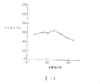
実施例9.トランスフェクトされた一次及び二次ウサギ皮膚線維芽細胞から得られた細胞株による長期間 in vitro hGH生産
線維芽細胞は新たに切除されたウサギ皮膚から単離し、10%の仔牛血清を含むDMEMで培養した。pcDNEO及びpXGH5の等モル混合物60μgでエレクトロポーレーション(250V,960μF)を行い、処理された細胞はG418を含む(1mg/ml)培地中で選択した。コロニーはクローニングシリンダーを用いて単離され常法に従い広げ、得られた一次細胞株は、培養中の時間の関数としてのhGH発現の安定性をモニターするため、繰り返し継代培養した。そのようなうちの一種、RF40/1−3から得られたデータを図10に示した。RF40/1−3細胞は選択不在下でウサギ線維芽細胞栄養培地中で保持され、接種密度10000cells/cm2 で継代培養した。培養液は細胞の継代培養のための回収に先立つこと24hrで交換した。継代時には培養液の一定量をhGH測定のために除去し、それから細胞を回収し、計数し、再び接種した。培地中のhGH濃度は市販されているイムノアッセイ(Nichols institute)を用いて測定した。hGHレベル(μg/106 cells/24hrで示した)はそれぞれおよそ28〜84の培養平均倍加(mpd)レベルにあたる4、8、10、13、16、19、22代の培養継代数に対してプロットした。これらのアッセイの結果は、この長期にわたるin vitro培養期間を通して、hGH発現が著しく一定に保持されており、実質的に約20mpdで観測されるレベル(47μg/106 cells/24hr)に等しい。
【0121】
実施例10.マウスにおけるヒト成長ホルモンの長期発現
ヌードマウスは、遺伝子工学的な操作を受けた細胞の組織移植が治療上有益なタンパク質を動物の体循環に送達する能力を研究するうえで貴重な系をもたらしている。これらの動物は、他に比べて免疫不能(immune-incompetence) であるのである種の一次及び二次ウサギ線維芽細胞を長期にわたってin vivoで生存させ得ると考えられている。
【0122】
腎下部皮膜への細胞の移植のためにマウスはAvertin(2%w/v 2.2.2−トリブロモエタノール、2%v/v2−メチル、2−ブタノールの溶液)を0.017ml/g体重の割合で腹腔注射される。腎臓(一般には左腎臓)は肋骨の下約3mmにつくられた8〜10mmの切開部を通して操作する。皮膚、腹筋、腹膜及び腎側筋膜を腎臓を露出させるために後退(retract) させた。小さな鉗子を用いて腎臓を腹腔から取り出す。27ゲージの皮下注射針が腎皮膜に小さな開口部を作るのに用いられる。20ゲージのI.V.カテーテルを用いて移植されるべき細胞(一般に5〜10μlの容積中に300万個の細胞)は1mlシリンジに吸い込まれ、腎皮膜の下にゆっくりと注入される。細胞がその腎被膜の開口部から遠くへ放出されるように注意する。切開部はステープル一つで筋肉及び皮膚を通して縫合される。軽麻酔がかかるまでメトキシフランをいれた大きなビーカーにネズミをいれた後、血を採取した。眼窩腔から血液を採取するためにパストゥールピペットの先端を眼と眼窩周辺部位の間に入れた。血清のhGHレベルは市販されているキット(Nichol’s Institute)を用いて測定した。
【0123】
我々の最初の実験では、500万個のトランスフェクトウサギ線維芽細胞(RF20−11)をヌードマウスに移植した。このマウスは一年間、その血清中に検出可能なhGHを示していた。発現の経時変化を以下に示す。
【0124】

さらにマウスに96μg/10cells/24hrを発現するウサギ線維芽細胞株(RF40/1−3)を移植したより大規模な実験が行われた。この実験の結果は図11に示す。バーは標準誤差を示し、Nはそれぞれの時点で採取されたマウスの数を示している。これらの結果から、平均血清hGHレベルが14カ月以上も比較的一定を保っており、移植以来平均1〜3ng/mlであることが示される。この期間中に渡って移植細胞に起因するどのようなタイプの副作用も観測されなかった。
【0126】
実施例11.腎下部被膜移植組織由来のhGH生産細胞の再培養
移植細胞の生存能力のストリンジェント(stringent) テストは、動物から切除してin vitroで再培養した際に、生育し、移植前の性質を示す能力とした。24.5μg/106 cells/24hrを発現するRF40/1−3細胞(3×106 cells/マウス)をヌードマウスの腎下部被膜の下に移植した。これらの細胞は移植前に培地中で49mpdを示していた。in vivoで5又は10週間経過後、動物を屠殺し腎臓を回収し、移植組織近傍の細胞(腎臓表面で明白に見える)を顕微切除した。得られた組織をスケールダウンした標準的な酵素的細胞分散法(実施例6)にかけ、単一細胞懸濁物(single cell suspension)をG418を含む培地にいれた。細胞培養のデータは移植後5週のマウス由来の再培養組織5つと移植後10週のマウス由来の再培養組織4つから得られた。回収された細胞は培地中で19mpdまで増殖させ、そしてhGHレベルを、培地に移した後約10日から約50日間の範囲の様々の時点で分析した。これらの実験結果を図12に示した。図12のそれぞれの点は少なくとも独立した4つの回収(recovery)試験の平均(標準偏差を含む)を示している。5週および10週の移植組織の両者の実験において、hGHを高レベルで発現するG418r 細胞が回収され、hGH発現はin vitroでの19回の回収後の倍加の間に渡って比較的安定であった。
【0127】
実施例12.トランスフェクトされた自己由来細胞を移植したウサギでのヒト成 長ホルモンの発現と高力価の抗hGH抗血清の生産
自己移植遺伝子治療実施のためのすべての技術的・論理的要求を探求する実験がウサギで行われている。最初に皮膚生検資料を8匹の生きているウサギから採取する。皮膚線維芽細胞を単離して一次培養した。in vitroで一継代後、pXGH301またはpXGH5とpcDNEOでトランスフェクトした。G418耐性クローンを単離しhGH発現について分析した。10μg/106 cells/日より大を発現するクローンをローラーボトルに広げた。最終的に各クローンの細胞をローラーボトルから回収し、皮膚生検のドナーとして用いられた同一のウサギにSRC細胞移植またはIP注入するために調製した。
【0128】
ウサギ皮膚線維芽細胞を単離するため、沈静に達するまでウサギに塩酸ケタミン(50mg/kg体重)および塩酸キシラジン(5mg/kg体重)の筋肉内注射により麻酔をかける。動物は一部、例えば右骨盤関節上部などを刈り込み、常法を用いて外科手術の準備をする。卵形のパッチを作るように背部と腹部でつながる2つの弓形の切開部を約3cm離して作る。鋸歯状鉗子を用いて表皮を除去し、小鋏でその下の筋膜を除去する。表皮と筋膜を培地中に移し、切開部を3−0ナイロン縫合糸で縫合する。ウサギは回復のため温床上に移す。筋膜線維芽細胞は、実施例1、2及び4で述べるように、培養、トランスフェクトされ、選択される。
【0129】
腎被膜内へ細胞を移植するため、上記の通りにウサギに麻酔する。動物は右側面の横臥位に置き、左肋骨下側を露出させる。動物は剃られ、承認された方法に従って準備をした。12番目の肋骨の下約2cmの所の背腹部の6cmの切開部を通して左腎臓に到達した。切開部を腹筋、腹膜、腎周囲の脂肪・筋膜まで貫通させる。バルフォア開創器を用いて腹部切開口を保持し、腎周囲の脂肪と筋膜の切除を行なう。11ゲージの手術刀を用いて腎臓被膜表面に2〜3mmの切開口を作る。20ゲージのI.V.カテーテルを用いて移植細胞(一般に165〜660μl中に1〜2億個の細胞)を1mlシリンジにいれ、ゆっくりと腎被膜下部に注入する。腎被膜の開口部から遠位に細胞が放出されるように注意する。腹膜口は3−0可吸着性クロミック縫合糸(adsorbable chromic gut)を用いて腹膜および腹筋を縫合して閉じる。皮膚は3−0ナイロン縫合糸と滅菌ガーゼ絆創膏で縫合し、抗生物質(Bactrin) を傷口につける。ウサギは回復のため暖めたパッド上に移す。全身性抗生物質(Tribrissin)を皮膚生検および移植の後に投与する。腹膜内(IP)経路で細胞を導入するため、細胞(3.5〜5.5ml中に7〜11億個)を22ゲージの注射針で注入する。22ゲージの注射針を用いて検体動物(restrained animals)の中耳動脈から血液を採取する。
【0130】
hGH発現レベルおよび動物に導入した細胞数についての関連情報を表1にまとめる。8匹すべての動物で血清hGHレベルが容易に検出可能であった。14日目までにどの動物のhGHも検出されなくなった。これらの動物血清中からの最終的なhGHの消失は予期されていた、というのはヒト成長ホルモンはウサギにとって抗原となることが知られているからである。したがって血清サンプルは抗hGH抗体についてアッセイした(抗ウサギIgGのELISAによって定量される)。それぞれの場合において、血清hGHレベルの減少は抗hGH抗体の増加に一致している。
【0131】
【表1】
これらの結果から、トランスフェクトされた皮膚線維芽細胞を用いたウサギでの自己由来遺伝子治療を行うために技術的障壁がないであろうことが示された。遺伝子工学的に修飾された自己由来細胞によるhGH供給が8匹の実験動物すべてで成功した。予想されたように、血清hGHレベルは、抗hGH抗体の増加に従って減少した。高力値(1:40000)はIPまたはSRCを移植した動物ではまれではなく、このことは1回の移植処置による持続的なタンパク質供給が、脊椎動物の予防接種やタンパク質抗原に対する高力価抗体の生産のための効率的な手法となり得るという提案と一致している。
【0133】
実施例13.トランスフェクトヒトとウサギの二次皮膚線維芽細胞によるインビトロでの hEPO の生産
1. ヒト皮膚線維芽細胞
線維芽細胞は新しく切り出されたヒト皮膚線維芽細胞から分離され、そしてDMEM + 15%牛血清中で培養された。60μg のpcDNEOとpXEPO1の等モル混合物とのエレクトロポレーション(250 ボルト、960 μファラッド)は1継代細胞で行われ、そして処理された細胞はG418を含む培地(300 μg/ml G418 )で選択された。コロニーは単離され、標準的な方法を使って広げられた。56の個々のクローンの分析から由来したデータは表2 に示されている。細胞はDMEM + 15%牛血清中のG418中(300 μg/ml G418 )に保持され、そして10,000細胞/cm2 の接種密度で植えつがれた。培養培地は継代のために細胞が採取される24時間前に変えられた。継代の時には、培養培地の部分はhEPO分析のために除かれ、そして細胞はそれから採取され、数えられ、再び植え継がれた。培地中のhEPO濃度は商業的に利用できるELISA(R & D systems)を用いて決定された。hEPO濃度はmU/106cells/24hrとして表され、そして発現レベルは69から55,591mU/106cells/24hrに広がり、全G418耐性コロニーの19% が検出できる濃度のhEPOを発現した。
【0134】
【表2】
pcDneoとpXEPO1を同時にトランスフェクションすることにより分離されたヒト線維芽細胞由来のクローンの、分泌されたhEPOのグリコシル化の状態の分析がされた。培地がhEPO生産細胞からあつめられ、そしてヒトエリスロポエチンに特異的なマウスモノクローナル抗体(Genzyme Corporation)を用いて免疫沈降された。免疫沈降された物質は12.5%ポリアクリルアミドゲル上で電気泳動され、そしてPVDF膜(Milipore)に写された。膜は免疫沈降に用いた同じ抗hEPOモノクローナル抗体を用いて調べられ、次にHRP 結合ヒツジ抗マウスIgG 抗血清(Cappel)を用いて処理され、その後hEPOを蛍光産物の生産を通して目に見えるようにするために発光検出(ECL Western blotting detection kit, Amershan)を行った。
【0136】
pEXPO1とpcDNEOを同時にトランスフェクションした正常ヒト線維芽細胞によって分泌されたhEPOのウエスタンブロット分析はヒトエリスロポエチンに特異的な抗体と反応する、およそ34kdの分子量を持つ分子であることを証明した。これは、本来存在する完全にグリコシル化されたヒトエリスロポエチンの予想されている大きさである。
【0137】
トランスフェクションされたヒト線維芽細胞クローンによって生産されたヒトエリスロポエチンについて、尿から単離される天然ヒトエリスロポエチン又はチャイニーズハムスター卵巣細胞によって生産された組換えhEPOのN-とO-結合グリコシル化の両方の性質を持つかどうか決定するために分析された。ウエスタンブロット分析はpXEPO1とpcDNEOを同時にトランスフェクションした正常ヒト線維芽細胞のクローン株からの上清上で行われた。上清のサンプルは、最初にエンドグリコシダーゼ−F(New England Nuclear)、ノイラミニダーゼ(Genzyme)あるいはO−グリカナーゼ(Genzyme )を用いて処理された。エンドグリコシダーゼ−Fを用いた処理で分子量が34kdから約27kdへのシフトを生じた。ノイラミニダーゼを用いた処理ではかろうじてバンドの位置のわかるシフトを生じ、一方O−グリカナーゼを用いた処理は免疫反応バンドの大きさが約18.5kdに下がるという大きなシフトを生じた。これらの結果より、尿から分離された天然のヒトエリスロポエチンあるいはチャイニーズハムスター卵巣細胞によって生産された組換えhEPOと得られたそれらとは区別できない〔ブラウンら、(Browne, J. K. et al., Cold Spring Harbor Symp. Quant. Biol. 51:693-702(1986)〕。
【0138】
2. ウサギ線維芽細胞
線維芽細胞は新しく切り出されたウサギ皮膚から分離され、そしてDMEM 10%牛血清で培養された。60μg のpcDNEOとpXEPO1の等モル混合物を用いたエレクトロポレーション(250 ボルト、960 μファラッド)が行われ、そして処理された細胞はG418を含むウサギ線維芽細胞成長培地(1mg/ml G418;実施例2)中で選択された。コロニーは分離され、標準的方法を用いて広められ、そして生じた二次細胞株のhEPO発現を分析された。49の個々のウサギ線維芽細胞クローンから由来したデータは表3に示す。これらのクローンの発現レベルは43から 2,900,000mU/106 細胞/24時間範囲で、全G418耐性クローンの72% が検出できる濃度のhEPOを発現した。
【0139】
【表3】
実施例14.ヒト EPO 遺伝子とネオマイシン耐性遺伝子の両方を持つプラスミドの構築
HPRT配列〔(Genbank entry HUMHPRTB,エドワーズら(Edwards, A et al.), Genomics, 6:593-608(1990)〕内でHPRT遺伝子のエキソン2および3を含む11,960から18,869の位置に広がっている6.9 kbのHindIII 断片がpUC12 のHindIII 部位にサブクローニングされる。生じたクローンはHPRT遺伝子断片のエキソン3内の唯一のXhoIの部位で切断され、そしてpMC1NEO(Stratagene)からのneo 遺伝子を含む1.1 kbのSalI-XhoI 断片が挿入され、エキソン3のコーディング配列を壊す。1方向つまりneo 転写の方向を持ちHPRT転写とは反対方向を持つ物が選ばれ、pE3Neoと名付けられた。pE3neoはHPRT配列とneo 遺伝子の5’側の結合部位に唯一のXhoI部位を持つ。pE3neoはXhoIで切られ、大腸菌DNA ポリメラーゼのクレノウ断片を用いて処理されることにより平滑末端にされる。
【0141】
neo 選択プラスミドpE3Neo内にhEPO遺伝子を挿入するためにプラスミドpXEPO1(実施例3;図5)から5.1 kbのEcoRI-HindIII 断片が分離された。EcoRI 部位はmMT プロモーターの5’側の隣に位置しており、HindIII 部位は5.1 kb離れたhEPOコーディング領域の3’側にある。精製された断片は大腸菌DNA ポリメラーゼのクレノウ断片を用いて処理されることによって平滑末端にされ、上で述べたXhoI消化され、そして平滑末端のされたpE3neo断片につなげられる。大腸菌への形質転換の後、pE3neoに挿入された1 コピーのmMT-hEPO断片を持つプラスミドは隣接するneo 遺伝子と同じ方向にhEPO遺伝子が転写されることを制限酵素分析によって確認された。このプラスミドはpE3neoEPO と名付けられた。hEPOを発現するG418r クローンの直接選択ができることに加えて、この断片はヒトHPRT位へhEPO遺伝子を組み込みさせるためのジーンターゲティングの際にも用いられるだろう。
【0142】
実施例15.hEPO 遺伝子と選択マーカー( pE3neoEPO )を発現するヒト線維芽細胞クローンの分離
線維芽細胞は新しく切り出されたヒト皮膚線維芽細胞から分離され、そしてDMEM + 15%牛血清中で培養された。60μg のスーパーコイル化された(supercoiled)pE3neoEPOを用いたエレクトロポレーション(250ボルト, 960 μファラッド)は一継代細胞で行われ、そして処理された細胞はG418を含む培地(300 μg/ml G418)で選択された。コロニーは分離され、標準的な方法で広げられた。26の個々のクローンの分析に由来するデータは表4に示されている。細胞はDMEM + 15 %牛血清中のG418中(300 μg/ml G418)で保持され、接種密度10,000細胞/cm2 で植えつがれた。培養培地は継代のために細胞を採集する24時間前に変えられた。継代の時、培養培地の一部分はhEPO分析のために取り除かれ、それから細胞は採集され、数を測定され、そして再び植え継がれた。培地中のhEPO濃度は商業的に利用できるELISA(R and D systems)を用いて決定された。hEPO濃度は mU hEPO/106 細胞/24時間としてあらわされ、そして発現レベルは240 から961,620 mU/106 細胞/24時間の範囲であった。全G418耐性クローンの89%が検出できる濃度のhEPOを発現した。
【0143】
【表4】
hEPOを発現するヒト線維芽細胞クローンは60μg のHindIII 消化したpE3neoEPO を用いたエレクトロポレーションによっても分離される。hEPOを発現するウサギ線維芽細胞クローンはウサギ線維芽細胞クローンが1mg/ml G418を含むウサギ線維芽細胞成長培地(実施例2)で選別されるのを除いて、全く同じトランスフェクションの条件でプラスミドpE3neoEPO を用いて分離される。
【0145】
実施例16.生物学的に活性なヒトエリスロポイエチンのマウス中での発現
マウスは、動物の一般循環系で治療学的に有用なタンパク質を供給する能力を持つ、遺伝子学的に修飾された細胞の移植の研究において貴重な系をもたらしている。ヌードマウスの相対的な免疫不全は外来の移植が生物学的機能を保持するのを許し、特定の一次及び二次ウサギ線維芽細胞を長期にわたってインビボで生存させ得るかもしれない。
【0146】
腎下部皮膜への細胞の移植のためにマウスはアベルチン(Avertin) を0.0175ml/g体重の割合で腹腔注射する。腎臓(一般には左腎臓)は肋骨の下約3mmにつくられた8〜10mmの切開部を通して操作する。皮膚、腹筋、腹膜及び腎側筋膜を腎臓を露出させるために萎縮させた。小さな鉗子を用いて腎臓を腹腔から取り出す。27ゲージの皮下注射針が腎皮膜に小さな開口部を作るのに用いられる。20ゲージのI.V.カテーテルを用いて移植されるべき細胞(一般に5〜10μlの容積中に300万個の細胞)は1mlシリンジに回収され、腎皮膜の下にゆっくりと注入される。細胞が腎皮膜の開口部から遠く放出される事に注意が払われる。切開部は一本の繊維(staple)で筋組織及び皮膚を通して縫合される。軽い麻酔がかかるまでメトキシフランをいれた大きなビーカーにネズミをいれた後、血を採取した。眼窩腔から血液を採取するためにパスツールピペットの先端を眼窩周辺部位の間に入れる。血清hEPOレベルは商業的に利用可能なキット(R and D Systems) を用いて決定される。血液の部分もヘマトクリットレベルを決定するためにEDTAで覆った毛細管(Statspin,Norwood,MA)に吸い込まれる。
【0147】
ウサギ皮膚線維芽細胞のクローン株は実施例13で述べられた方法によって分離された。RF115−D4と名付けられたあるクローンではヒトEPO遺伝子が安定してトランスフェクションされていると決定され、そしておよそ786,000mU hEPO/106 細胞/24時間発現された。300万の細胞が6匹のヌードマウスの腎下部皮膜に15づつ移植された。およそ400μlの血液が移植後1日目と7日目に吸い取られ、そしてその後は21日目まですべての日に吸い取られた。この期間の低張性ショックを防ぐために採血の後塩溶液が等量注射された。血液はその後63日目間で週ごとに吸い取られた。同じ採血スケジュールが細胞移植していない10匹のマウスに用いられた。図13aは一般に赤血球数の指標として用いられている血液ヘマトクリット(HCT)に対するこれらの処置の作用が示されており、移植及びコントロール動物について測定されている。コントロール動物においてはHCTは7日目までには急激に落ちて、その後15日目までに大体普通のレベルに戻る。対してhEPO発現細胞の移植を受けた動物は7日目までに高いHCTレベルを示した。HCTは63日目まで高いままで、移植後35日目には1日目のレベルの45%よりも1.4倍高い64%のピークに達する。図13bに示されているように、免疫反応性(immunoreactive)hEPOは移植された動物の血液中で簡単に検出できた(キットの製造元によってhEPO ELISAの感度性は2mU/mlであると決められており(R&D System)、そしてELISAキットで使われている抗体と内因性のマウスEPOは交差反応を示さない)。移植された動物のhEPOレベルは移植後7日目から63日目までに29から9mU/mlまで徐々に落ちた。
【0148】
本実施例は、hEPOを発現し分泌するために遺伝的に処理された普通の皮膚線維芽細胞は、1)インビボで生存して2か月間まで動物の全身循環系にhEPOを供給することができ、そして2)生産されたhEPOは生物学的に機能し、しばしば採血されるコントロール動物に観察されるヘマトクリット値の低下を防止するために働き、コントロールの動物では貧血反応を生じる採血スケジュールを試されたときでさえ、最終的にHCTの増加を生じるという事をはっきりと証明した。
【0149】
実施例17.GLP-1(7-37 )発現プラスミドをトランスフェクションした後の二次ヒト皮膚線維芽細胞株からの GLP-1(7-37 )の発現
GLP-1 の7から37のアミノ酸残基の部分〔GLP-1(7-37);Genebank sequence HUMGLUCG2において7214から7306の塩基対によってコードされている〕はインビボでインスリノトロピン活性を持つことが証明された。プラスミドpXGLP1は、GLP-1(7-37) の部分が、そのN末端において、小胞体を通しての運搬を効果的にするためにヒト成長ホルモン由来の26アミノ酸のシグナルペプチドと融合するように構築される。この融合タンパク質は、分泌前に、分泌産物がGLP−1の7−37残基のみから構成されるように、残基26のすぐC末端側が切断される。シグナルペプチドの発現:GLP-1(1-37) 融合タンパク質はマウスメタロチオネインプロモーターによってコントロールされる。
【0150】
プラスミドpXGLP1は次のように構築される:プラスミドPXGH5〔Selden, R. F., Mol. Cell. Biol. 6:3173-3179(1986) 〕がSmaIで消化され、BgIII 部位を含む2 本鎖のオリゴヌクレオチド(BgIII linkers;New England Biolads )につなげられる。つなげられた産物はBgIII とEcoRI で処理され、そしてヒト成長ホルモン遺伝子の3’側の翻訳されない領域に相当する0.32kbの断片が分離された(Genbank entry HUMGHCSAの6698位のSmaI部位にBgIII linkerを持つ)。hGH 断片も、Genbank entry HUMGHCSAの6698から7321の間を増幅するために設計されたPCR プライマーを用いてヒトゲノムDNA から既知の方法で分離された。マウスメタロチオネイン(mMT )プロモーター〔Harmer, D. H. and Walling, M. J. Mol. Appl. Gen., 1:273-288(1982)〕を含む1.45のEcoRI-BgIII 断片が次に分離された。マウスメタロチオネインプロモーターは、マウスゲノムDNA から、Genbank から利用できるmMT 配列(すなわち Genbank entires MUSMTI, MUSMTIP, MUSMTIPRM)の分析から設計されたPCR プライマーを用いて、既知の方法で分離された。プラスミドpUC19 (ATCC#37254)がEcoRI で切断され、細菌アルカリフォスファターゼを用いて処理された。処理されたプラスミドは上で述べられたhGH やmMT 断片に繋げられた。生ずるプラスミドは、マウスメタロチオネインプロモーターと、BgIII 部位で繋げられたhGH の3 ’側の翻訳されない領域をそれぞれ一コピー持つ。
【0151】
pX1 と名付けられこのプラスミドはBgIII で処理され、完全な長さの線状産物がゲル電気泳動によって精製された。11.1と11.2のオリゴヌクレオチドは、PCR によってヒトゲノムDNA からGLP-1 の7-37のアミノ酸をコードするDNA 配列の増幅のために用いられる。増幅された産物(104 bp)は精製されpXGH5 とオリゴヌクレオチド11.2、11.3、11.4、11.5に混合され、PCR に付す。オリゴヌクレオチド11.3と11.4は相補的でありhGH シグナルペプチドとGLP-1 アミノ酸残基7の間の所望の連結部(junciton)に相当する。500 塩基対の増幅産物は、hGH(Genbank entry HUMGHCSAの5168から5562のヌクレオチド)からの5’側の翻訳されない領域やエキソン1、イントロン1、エキソン2配列の一部を含んでおり、GLP-1 の7-37残基をコードする15の融合されたものがストップコドンの前にある。断片は設計によって、両末端にBamHI 部位がフランクしている。
【0152】
断片はBamHI で切断され、上で述べられたpXI のBgIII 処理物に連結される。連結産物は、GLP-1残基37がmMT プロモーターの遠位になるように、pX1においてmMT プロモーターと3’側の翻訳されないhGH配列を分離してBgIII 部位に挿入されているhGH-GLP-1(7-37) 融合産物の一コピーを用いて分析し同定される。
【0153】
【表5】
他の方法として、小さいサイズのシグナルペプチドと必要とされるGLP-1の一部分で、完全な融合遺伝子が合成的に調製できる。LDL レセプターのシグナルペプチド(アミノ酸残基1−21)、プレプログルカゴン(アミノ酸残基1−20)またはヒト成長ホルモン(アミノ酸残基1−26)をコードするDNA が知られている方法で合成できると考えられ、そして同じように合成したGLP-1の7-37または7-36(すぐにストップコドンがつづく)のアミノ酸をコードするDNA とin vitroで連結される。これらの分子を設計し、合成するのに必要な配列は、Genbank エントリーの HUMLDLRO1(ヒトLDL レセプター)、HUMGLUCG2 (ヒトGLP-1 とグルカゴン配列)とHUMGHCSA(ヒト成長ホルモン)において簡単に利用できる。連結産物はヒト線維芽細胞に使用されるための適当な哺乳類発現ベクターに挿入することができる。プラスミドpMSG(Pharmacia LKB Biotechnology, Piscataway, NJ)は5’側と3’側の翻訳されない配列、スプライス部位、ポリA 付加部位、エンハンサーとヒト皮膚線維芽細胞で用いられるプロモーターを持つので、この目的に適当である。別の方法として、連結産物は適当な5’側の翻訳されない配列を用いて合成され、上で述べえられたプラスミドpX1 に挿入することも可能である。
【0155】
第二のインスリノトロピック(insulinotropic)なGLP-1、つまりGLP-1(7-36)は、上述のオリゴヌクレオチド11.2をオリゴヌクレオチド11.6で置換することによって発現できる。後のクローニング操作の全てはpXGLP1構築のために上述した方法に従い、GLP-1(7-37)特有のC末端のグリシン残基を最終産物が欠くようになる。他に、インスリノトロピンGLP-1(7-36)アミドをつくるために、ペプチジルグリシンアルファーアミド化酵素の活性によって、インビボでこの末端のグリシン残基を除いても良い。
【0156】
プラスミドpXGLP1は、実施例13でpXEPO1とpcDNEOについて述べたとおりにプラスミドpcDNEOと共に一次ヒト線維芽細胞にコトランスフェクト(co-transfect)される。クローンはG418を含む培地で選別され、96穴プレートに移され、そして細胞上清のGLP-1(7-37)活性または免疫反応性について測定する。GLP-1(7-37)活性は、細胞上清をラットインスリノーマRINm5F細胞と一緒に加温放置し、市販のインスリンラジオイムノアッセイ(Coat-a-Count Insulin, DPC, Los Angels, CA)を用い、これらの細胞からインスリン分泌を誘導するための上清の能力を測定することによって決定された。GLP-1(7-37)抗原は、市販の抗GLP-1抗血清(Peninsula Laboratories、Belmont 、CA)を用いて測定される。GLP-1(7-37)ポジティブクローンは実施例16で述べられているようにヌードマウスに移植するため広げられ、そして血液サンプルが血清ヒトGLP-1(7-37)レベルを測定するために採取された。
【0157】
インビボの活性は絶食動物において、腹膜内にグルコースを注射をした後(体重1g あたり1mgのグルコース)、インスリン生成の指数の測定によってモニターされる。一般には、32匹の移植を受けたマウスのグループと受けていないグループは一晩絶食され、28匹がグルコースを注射された。注射後、28匹のマウスは任意に4 匹づつ7 つのグループに分けられ、そして血液が(血清グルコースとインスリンのために)、それぞれのグループについて注射後5、10、20、30、45、60、90分に採取され、絶食コントロールとしてグルコース注射をしないグループとともに行われた。GLP-1(7-37)発現細胞を受けた動物における注射後のインスリン生成の指数(血液中におけるグルコースに対するインスリンの割合)の移植しない動物をうわまる増加は、発現されたペプチドのインビボにおけるインスリノトロピック活性を支持するものである。
【0158】
実施例18.遺伝子ターゲティングによるトランスフェクト細胞株の作製
トランスフェクトするDNAが、相同組み換え事象を通して、染色体DNA配列に導入される時、または部分的に置換する時に、ジーンターゲティングが生じる。このような事象はどのようなトランスフェクト実験の途中でも生じる可能性があるが、それらは非相同又は不正の(illegitimate)組み換えによってプラスミドDNAが組み込まれるという事象が大過剰に起こることによって、通常マスクされている。
【0159】
a. ヒト細胞中のジーンターゲティング事象の選択に有用な構築物の作製
ターゲティングの事象を選択する一つの方法は、トランスフェクトするDNAの組み込みによる遺伝子機能の消失を選択する遺伝子的選択によるものである。ヒトHPRT遺伝子座(locus) はヒポキサンチン−ホスホリボシルトランスフェラーゼ酵素をコードしている。hprt- 細胞は、6−チオグアニン(6−TG)類似のヌクレオシドを含む培地で増殖することにより選択することができる。すなわち、野生型(HPRT+ )対立遺伝子(allele)を持つ細胞は、6−TGによって死滅するが、変異体(hprt- )対立遺伝子を持つ細胞は生きつづけることができる。従って、HPRT遺伝子機能を破壊する、ターゲティングされた事象を保持する細胞が6−TG培地で選択できる。
【0160】
HPRT遺伝子座に対するターゲティングのためのプラスミドを構築するために、HPRT配列〔遺伝子バンク(Genebank)名HUMHPRTB:エドワーズ(Edwards, A.) ら,Genomics 6:593-608(1990)]の11,960〜18,869位を含みかつHPRT遺伝子のエキソン2と3を含む6.9kbのHindIII断片がpUC12のHindIIIの中にサブクローン化される。得られたクローンはHPRT遺伝子断片のエキソン3の唯一のXhoI部位で切断され、pMClNeo(Stratagene)からのneo遺伝子を含む1.1kbのSalI−XhoI断片が挿入されて、エキソン3をコードする部分が除かれる。HPRT転写の方向と反対方向のneo転写の方向を持った一つのオリエンテーション(orientation)が選びだされ、pE3Neoと名付けられた。正常なHPRTエキソン3をneoを欠いた(neo-disrupted) 型のもので置換すると6−TG耐性表現型のhprt- が得られるであろう。このような細胞はG418耐性でもある。
【0161】
b. 確立したヒト線維芽細胞系でのジーンターゲティング
不死化した細胞系におけるターゲティングを示すため、そして、ジーンターゲティングにおいてpE3Neoが適切に機能することを確立させるため、ヒト線維肉腫細胞系HT1080(ATCC CCL 121)がpE3Neoによりエレクトロポーレーションを用いてトランスフェクトされた。
【0162】
HT1080細胞は、エレクトロポーレーションに先立って、15%子牛血清(Hyclone)含有のHAT(ヒポキサンチン/アミノプテリン/キサンチン)補填のDMEM培地で維持された。エレクトロポーレーションの2日前に、細胞をアミノプテリンのない同様の培地に移し変える。等比級数的に増殖した細胞をトリプシン処理し、DMEM/15%子牛血清で希釈し、遠心分離して、PBS(リン酸緩衝化食塩水)に再懸濁させて、1ml当たり1330万細胞の最終細胞体積にする。pE3NeoはHindIIIで処理され、pUC12骨格から8kbのHPRT−neo断片を分離し、フェノール抽出とエタノール沈殿により精製して、600μg/mlの濃度で再懸濁させる。50μl(30μg)を、エレクトロポーレーション用チューブ(cuvette) (0.4cm電極間隔、Bio−Rad Laboratories)に、750μlの細胞懸濁液(細胞数1000万)と共に加える。エレクトロポーレーションが450V,250μ Farads(Bio−Rad Gene Pulser;Bio−RadLaboratories)で行われた。チューブの内容物を直ちに、15%子牛血清含有のDMEM培地に加えて、25mlの培地当たり、100万個の細胞になるような細胞懸濁液を作る。この処理された細胞懸濁液25mlを150mm径の組織培養皿にまいて、37℃、5%炭酸ガス中で培養する。24時間後、G418溶液を直接その皿に加えて、G418として800μg/mlの最終濃度のものにする。5日後、培地をDMEM/15%子牛血清/800μg/ml G418の培地に換える。エレクトロポーレーションの9日後に培地をDMEM/15%子牛血清/800μg/ml G418及び10μM6−チオグアニン含有のものに換える。G418と6−TGに耐性のコロニーがこの2重選別の開始後14〜16日後に、クローニングシリンダーを用いて採取された。
【0163】
HT1080細胞における5個の代表的なターゲティング実験の結果を表6に示す。
【0164】
【表6】
トランスフェクション例5では、G418r コロニーの全収率決定用のコントロール皿によると、33,700個のG418r コロニーが最初1×10⁷ 個の処理細胞から発生することが示された。このように、ターゲットされた現象とターゲットされない現象の比は、66/33,700すなわち1対510である。この5つの実験を合わせると、ターゲットされた現象は、3.6×106 の頻度すなわち処理細胞の0.00036%という頻度で発生する。
【0166】
制限酵素と、neoとHPRT遺伝子から得られたプローブを用いたサザンハイブリダイゼーション実験から、HPRT遺伝子座のHPRTのエキソン3中の予想された位置にneo遺伝子が位置していることが示された。
【0167】
c. 一次及び二次のヒト皮膚線維芽細胞でのジーンターゲティング
pE3NeoをHindIIIで切断し、pUC12骨格から8kbのHPRT−neo断片を分離し、フェノール抽出とエタノール沈殿により精製する。DNAは、2mg/mlとなるよう再懸濁する。0.5mlの容量中、300万個の二次ヒト包皮線維芽細胞が100μgのHindIIIpE3Neo(50μl)と共に、250ボルトと960μファラッドでエレクトロポーレートされた。3回に分けてトランスフェクションを行い、全部で900万個の処理細胞となった。150mm培養皿ごとに、500,000個の細胞がG418選択用にまかれた点を除き、実施例6記載のように細胞を処理し、G418耐性株を選択する。選択条件で10日後に、培養液を400μg/mlのG418と10μM6−TGを含むヒト線維芽細胞栄養培地に換える。2種の薬剤の組み合わせで選択することを、さらに10日継続する。培養皿を顕微鏡で見て、両薬剤に耐性なヒト線維芽細胞コロニーを検索する。G418r t−TGr コロニーの比率は、900万個の処理細胞当たり4個である。これらのコロニーは、コロニーを作り得るすべての細胞の0.0001%(又は、100万分の1)となっている。G418r コロニーの全収量を決定するために用いられるコントロール用培養皿から、最初の9×10⁶ 個の処理細胞から2850個のG418r コロニーが発生し得ることが示された。このように、ターゲットされない現象に対するターゲットされたものの比率は4/2,850すなわち1対712である。制限酵素及び、neoとHPRT遺伝子に由来するプローブを用いたサザンハイブリダイゼーション実験により、HPRT遺伝子座のHPRTエキソン3の中の予想される部位にneo遺伝子が位置し、そしてこれら4個のクローン化された細胞株中に、ターゲティングが生じていることが示される。両薬剤に耐性なコロニーはトランスフェクトされる一次細胞からも単離される(1/3.0×107 )。
【0168】
いくつかのpE3Neoターゲティング実験の結果は表7にまとめられている。HindIIIで処理されたpE3Neoは、直接トランスフェクトされるか、又はトランスフェクションに先立って5’端に単鎖の突出部を発生させるため、エキソヌクレアーゼで処理された(実施例18c参照)。長さが175から930塩基対の範囲の単鎖領域を持ったDNAが試験された。HindIIIのみで分解したpE3neoを用いて、1/799 G418−耐性コロニーが、制限酵素とサザンハイブリダイゼーション分析により、HPRT遺伝子座にターゲティングされた挿入であるneo遺伝子があることが、同定された(全部で24個の目的のクローンが単離された)。175塩基対の突出部を用いた時に、ターゲティングは最大に増強され(約10倍の増強)、1/80 G418r コロニーに、HPRTのターゲティングの診断に役立つ制限断片の存在が示される(全部で9個の目的とするクローンが単離された)。このように、ここに記載された条件と組み換えDNA構築物を用いれば、ターゲティングは通常のヒト線維芽細胞で容易に観察でき、全体のターゲティングの頻度(ターゲットされたクローンの数を、G418耐性株へ安定にトランスフェクトされたクローンの数で割ったもの)は、実施例18eに記載するような方法で、単鎖の突出尾部を含むターゲティング構築物でトランスフェクトすることで増強され得る。
【0169】
【表7】
d. ヒトのゲノムへの治療に有益な遺伝子のターゲティング挿入のための構築物作製とジーンターゲティングでのその利用
治療に有益な遺伝子が、neo遺伝子に隣接するかその近くのHPRTのコーディング領域に挿入されているpE3Neo変異型は、レシピエント一次又は二次細胞染色体の特定の位置に、治療に有益な遺伝子をターゲティングするために使用することができる。HPRT遺伝子座にhGH遺伝子をターゲティングするために、このようなpE3Neo変異型を構築することができる。
【0171】
pXGH5はEcoRIで処理され、hGH遺伝子とそれに結合したマウスメタロチオネン(mMT)プロモーターを含む4.1kb断片が分離される。EcoRIの突出部はE.coli DNAポリメラーゼからのクレノー(Klenow)断片で埋められている。別個にpE3Neoはneo断片とHPRTエキソン3の結合(エキソン3に挿入された3’側の結合)を切断するXhoIで処理する。直鎖状になったプラスミドのXhoI突出端は、E.coli DNAポリメラーゼからのクレノー断片で埋め合わされ、得られた断片が4.1kbの平滑末端のhGH−mMT断片につながれる。つながれた混合物由来のバクテリアのコロニーは、hGH−mMT断片が単一コピー挿入されているか及び方向は合っているかが、制限酵素分析によってスクリーニングされ、neo遺伝子と同じ方向で転写されたhGH遺伝子が選ばれてpE3Neo/hGHと名付けられる。pE3Neo/hGHをHindIIIで切断し、HPRT,neoとmMT−hGH配列を含む12.1kbの断片を切り離す。切断したDNAが処理されて、実施例18cに記載する一次又は二次ヒト線維芽細胞にトランスフェクトされる。G418r TGr コロニーが選択され、実施例18cに記載するように、HPRT遺伝子中へのmMT−hGHとneo配列のターゲティング挿入について分析される。個々のコロニーは市販される免疫測定法(Nichols Institute) を用いて、hGHの発現を測定する。
【0172】
二次ヒト線維芽細胞をpE3Neo/hGHでトランスフェクトし、チオグアニン耐性コロニーが、安定なhGHの発現についておよび制限酵素とサザンハイブリダイゼーション分析法によって、分析された。13個のTGr コロニーが分析され、8個のコロニーが内在性のHPRT遺伝子座にhGH遺伝子が挿入されていると同定されている。8個の株はすべて、hGHの有意な量を安定に発現しており、24時間の平均発現レベルは22.7μg/106 細胞であった。
【0173】
e. ターゲティングを増強させるためのDNA末端の修飾
いくつかの一連の証拠から、E.coli、バクテリオファージ、S.cerevisiaeとアフリカツメガエルにおける或る相同性組み換えの過程に、3’端突出部が関与することが示唆されている。アフリカツメガエルの卵母細胞においては、数百個の塩基対の長さの3’端突出部を持った分子が、制限酵素で消化されて発生する非常に短い突出部(4bp)を持った分子よりも、顕微注入後に非常にすみやかに同様に処理された分子と組み換えを起こした。酵母においては、数百個の塩基対の長さを持った3’端突出部の発生が減数分裂的(meiotic) 組み換えにおける律速段階であるように見える。ヒト細胞の組み換えにおいて3’端突出部が関与するとの証拠は報告されておらず、ある種の修飾DNA基質が、どのような種においても、ターゲティング(相同性組み換えの一種)を促進するということは示されていない。ヒトの細胞において、ターゲティングに関する3’端突出部の効果は、試験されていない。以下の実施例に記載の実験から、5’端突出部が一次及び二次のヒト線維芽細胞のターゲティングに最も有効であることが示された。
【0174】
トランスフェクトするDNA分子の末端を修飾することによって、ターゲティングを増強させることに関する報告はなされていない。この実施例では分子末端を2本鎖型から1本鎖型に変換することによる直鎖状DNA分子の末端の修飾により、一次及び二次のヒト線維芽細胞の染色体へのターゲティングが増強できることを示す。
【0175】
1100μgのプラスミドpE3Neo(実施例18a)がHindIIIで切断される。このDNAは、フェノール抽出とエタノール沈殿の後に、直接用いることができる。あるいはHPRTとneo遺伝子のみを含む8kbのHindIII断片が、ゲル電気泳動法によってpUC12ベクター配列と分離することができる。HindIII切断したDNAのExoIII処理により、両末端に、各フリーの3’末端で始まり、5’の突出端を残す広範なエンドヌクレオティックな切断が起こる。エンドヌクレオティック反応の程度とその結果としての5’端突出部の長さは、ExoIII処理反応の時間を変化させることにより、コントロールできる。HindIII処理されたpE3Neoの100μgのExoIII処理反応が、供給者の推薦する条件に従い、30秒、1分、1.5分、2分、2.5分、3分、3.5分、4分、4.5分と5分の時間で実施された。消化反応の程度をモニターするため、それぞれの時間の点で、ExoIIIで処理されたDNAを1μg含むように採取された試料を、マングビーンヌクレアーゼ(Promega) を用いて供給者の推薦する条件で処理し、その試料をゲル電気泳動で分画する。未処理のHindIII処理されたpE3NeoとExoIIIとマングビーンヌクレアーゼで処理された同じ分子のサイズの違いを測定する。2つに分かれたこのサイズの相違から、分子のそれぞれの末端にある5’端突出部の平均の長さが出る。上述の時間の点と30°での処理では、作りだされる5’端突出部は100〜1000塩基の範囲である。
【0176】
それぞれの反応時点で採取されたExoIII処理のDNA60μg(pE3Neoの全HindIII処理物)が精製され、実施例18cに記載の条件下で一次及び二次のヒト線維芽細胞へエレクトロポーレーションにより導入された。それぞれのExoIII処理調製物でターゲティングがどの程度増強されたかについては、G418r 6−TGr コロニーの数を計測し、ExoIIIで未処理のHindIIIで切断されたpE3Neoによるターゲティングで得られた数と比較することによって定量される。
【0177】
3’端突出部の効果も又、同様の系を用いて定量することができる。この場合、HindIIIで処理されたpE3Neoを用いて、供給者の推薦する条件で時間間隔を変えてバクテリオファージT7遺伝子6エキソヌクレアーゼ(United States Biochemicals)で処理する。処理の程度(末端毎に作りだされた3’端突出部の平均の長さ)とエレクトロポーレーションの条件の決定は、ExoIIIで処理されたDNAに関する記載に準じる。それぞれのT7遺伝子6エキソヌクレアーゼ処理調製物で、ターゲティングが増強される程度については、G418r 6−TGr コロニーの数を計測し、T7遺伝子6エキソヌクレアーゼ未処理のHindIII処理pE3NEOによるターゲティングで得られた数と比較することによって定量される。
【0178】
5’及び3’端突出部を発生させる他の方法も可能である。例えば、相互に部分的にオーバーラップしている2つの直鎖状分子を変性させ、アニーリングすることで、出発時の直鎖状分子と区別できない再アニーリングされた断片とともに、それぞれの分子の両末端に3’端突出部を有する分子、あるいは両末端に5’端突出部を有する分子の混合物が得られる。突出部の長さは2つのDNA断片の間で共通でないDNAの長さから決定される。
【0179】
f. 一次及び二次ヒト線維芽細胞においてマウスメタロチオネンプロモーターの制御下にヒトエリスロポエチン遺伝子を置くためのターゲティングプラスミドの構築
以下に、本件発明の一つの態様を例示する。そこでは、得られたままの状態では有意な量でEPOを発現しない一次及び二次ヒト線維芽細胞株において、ヒトエリスロポエチンが発現できるように、ヒトのエリスロポエチン(EPO)遺伝子の上流に位置する正常なポジティブとネガティブの調節配列を変化させる。
【0180】
ヒトEPOをコードする領域の上流にのみ存在する領域をPCRで増幅することができる。この目的のために用いられる3組のプライマーが、発表されているヒトEPOの解析からデザインされた〔Genbank 名称 HUMEPRPA; Lin, F-K., et al., Proc. Natl. Acad. Sci. USA 82:7580-7584(1985)〕。これらプライマーの組は609,603又は590bpの断片を増幅することができる。
【0181】
【表8】
3つの断片は実質的に重なりあっていて、本件の目的には交換可能なものである。翻訳開始部位を基準に−623〜−14に広がった(HUMERPAヌクレオチドの2〜610位)609bp断片の両端にClaIリンカーが結合される。その結果得られたClaI−結合断片をClaIで消化し、pBluescriptIISK/+(Stratagene)のClaI部位に挿入する。その際の配向性はHUMERPAヌクレオチド位置610がプラスミドポリリンカーのSalI部位に隣接するようにする。このプラスミドp5’EPOをEPO上流部分の断片中に唯一あるFspI又はSfiI部位で、別々に切断し(HUMERPAヌクレオチド位置として各々150と405)、マウスのメタロチオネンプロモーターに連結させる。典型的には、mMT−I遺伝子から得られた1.8kbのEcoRI−BglII〔mMTをコードしない(DNA)配列を含むものとして;Hamer, D.H. and Walling M., J. Mol. Appl. Gen.1: 273 288(1982); この断片はまた、Genbankから入手できるmMT配列、例えばMUSMTI、MUSMTIP、MUSMTIPRMの解析からデザインされるPCRプライマーを用いてマウスゲノムDNAから公知方法により単離することもできる〕を公知方法で平滑末端化し、SfiIで処理した(先端を同様に平滑末端にした)p5’EPOあるいはFspIで処理したp5’EPOと結合させる。得られたクローンの配向性が分析され、前のmMT BglII部位がプラスミドポリリンカー中のSalI部位の近くにあるものが、一次及び二次のヒト線維芽細胞をターゲティングするために用いられる。この配向性は、最終の構築物の中で、HUMERPAヌクレオチド位置610の方向に向かってmMTの転写を指向させる。得られたプラスミドは、FspIとSfiI部位で、それぞれmMTの挿入を行うことから、p5’EPO−mMTF及びp5’EPO−mMTSと名付けられている。
【0183】
さらに上流の配列は、負の調節要素あるいは最初のターゲット配列の上流に存在するエンハンサーの修飾、欠失及び/又は置換が望ましい場合に、有用である。EPOの場合には、肝臓外の組織と腎臓外組織の中で、EPOの発現を抑制する負の調節要素〔Semenza, G.L. et al., Mol. Cell Biol. 10: 930-938(1990) 〕は欠失することができる。6kb断片中に一連の欠失のあるものが調製される。削除された領域は幅広い宿主細胞活性を有するエンハンサーで置換されてもよい〔例えば、サイトメガロウイルス(CMV)からのエンハンサー〕。
【0184】
HUMERPA配列の5’末端の前にBamHI部位(遠位)とHindIII部位(近位)が位置するので、pBluescriptIISK/+ベクター中の609bp5’EPO断片の配向性が選択された。このように、609bp断片の上流部分に通常存在する6kb BamHI−HindIII断片〔Semenza, G.L. et al., Mol. Cell Biol. 10: 930-938(1990) 〕は、公知方法を用いて染色体DNAから単離できる。例えばバクテリオファージ、コスミドあるいは酵母の人為染色体ライブラリー(artificial chromosome library) を、609bpのPCR増幅断片をプローブとしてスクリーニングすることができる。所望のクローンは6kbのBamHI−HindIII断片を有するであろうし、その同定は公知方法で決定されたヒトEPO遺伝子の周辺の制限酵素地図とこの制限酵素地図を比較することによって確認することができる。また別の方法として、609bp断片をプローブとして用いて、EPO遺伝子上流部分のヒトゲノムの制限酵素地図を作成することによって、HUMERPAコーディネート2と609に間に起始し、上流BamHI部位を越えて延びる断片を作る酵素を同定することができる。この断片は、ヒトゲノムDNAの適当に処理したものからゲル電気泳動法で単離することができ、バクテリアあるいは酵母のクローニングベクターに結合させることができる。正しいクローンは609bpの5’EPOプローブにハイブリダイズし、6kbのBamHI−HindIII断片を含有する。単離された6kb断片は、正しい配向性でp5’EPO、p5’EPO−mMTF又はp5’EPO−mMTSに挿入(HindIII部位がHUMERPAヌクレオチド位置2に隣接するように)される。さらに、上流の配列を公知方法で単離することができ、それには染色体ウォーキングテクニックを用いるか、あるいは609bpの5’EPOプローブにハイブリダイズする酵母人為染色体の単離によって行うことができる。
【0185】
以上述べたクローニングのストラテジーは、その後の一次、二次ヒト線維芽細胞のターゲティングされたトランスフェクションを可能にするためのEPOの上流配列のインビトロでの修飾を可能にする。このストラテジーでは、負の調節領域の欠失とともに簡単なmMTプロモーターの挿入、また負の調節領域の欠失と広範囲の宿主細胞活性を有するエンハンサーによる置換について述べている。
【0186】
g. ヒトEPO遺伝子の隣接配列に対するターゲティングと、標的一次、二次および不死化されたヒト線維芽細胞のスクリーニングによる単離
ターゲティングに際し、プラスミドを制限酵素で切断し、プラスミドの骨格(backbone) から挿入配列を切り離す。p5’EPO-mMTSの場合、HindIII と SaII で切断することにより 2.4 kb のターゲティングフラグメントが切り離されるが、このフラグメントは1.8 kbの mMTプロモーターと、該プロモーターの5’、3’側にそれぞれ隣接した405bp と204bp の配列よりなる。この隣接配列は、この構築物を EPO遺伝子の調節領域にターゲティングするためのDNA である。このDNA 、または2.4kb のターゲティングフラグメントのみがフェノール抽出およびエタノール沈澱により精製され、実施例18c に記載された条件下で一次あるいは二次のヒト線維芽細胞にトランスフェクトされる。トランスフェクトされた細胞は、ヒト線維芽細胞栄養培養液中、150mm ディッシュに蒔かれる。48時間後、細胞は 10,000cells/cm2の密度〔1ウエル当たり約20,000細胞の割合。もし106 のクローニング可能な(clonable)細胞に対し 1回の割合でターゲティングを行うならば、(実施例18c)、その際単一の発現コロニーを分離するためのアッセイに約50ウエルが必要であろう〕で24ウエルのディッシュに蒔かれる。トランスフェクトする(transfecting)DNA が EPOの上流の相同領域をターゲットする細胞は、mMT プロモーターの制御下でEPO を発現するだろう。10日後、すべてのウエルの上清は、市販の利用可能なイムノアッセイキット(アムジェン) を用いてEPO の発現についてアッセイされる。 EPOの合成が認められるウエルからのクローンが、既知の方法を用いて分離される。代表的には個々のウェルあるいはプレート中に分離された異なる細胞集団のフラクションをアッセイし、これらのポジティブなウェルのフラクションのアッセイを必要に応じて繰り返し、最終的に1 ウエル当たり1 細胞の割合で蒔かれた96穴のマイクロタイタープレートをスクリーニングすることによりターゲティングされたコロニーが分離される。プレートライセート(plate lysates) 全体からのDNA が、HUMERPA 核酸部位 1の上流に位置するプライマーとmMT に特異的なプライマーを用いたPCR によりフラグメントを増幅することにより、分析され得る。この一対のプライマーは、DNA 配列に基いてその長さが正確に予想されるようなDNA フラグメントを増幅するはずである。ポジティブなプレートがトリプシン処理され、次々と低い希釈で蒔きなおし、ターゲットされた細胞を分離するのに必要なまで、DNA の調製とPCR のステップが繰り返される。
【0187】
ここで述べられたターゲティングの手法は、通常の薬物供給(pharmacologic delivery)のためにhGH を産生させる目的で、不死化されたヒトの細胞(たとえばHT1080線維芽細胞、HeLa、MCF-7 乳癌細胞、K-562 白血病細胞、KB癌細胞、あるいは2780AD卵巣癌細胞) においてhGH の発現を活性化することにもおそらく使用されるだろう。
【0188】
h. ヒト EPO 遺伝子の隣接配列に対するターゲティングと、ターゲティングされた一次、二次および不死化されたヒト線維芽細胞のポジティブあるいはポジティブ/ネガティブ組み合わせ選択システムによる単離
p5’EPO-mMTF、p5’EPO-mMTS、および上流に付加的な6kb の BamHI-Hind III フラグメントを有する誘導体を構築するためのストラテジーは、mMT プロモーターに隣接した neo遺伝子の挿入という付加的なステップを続けて行うことができる。加えて、ネガティブな選択マーカー、例えばgpt[pMSG (ファルマシア) あるいは他の適切な供給源から得られる] は pBluescriptIISK/ +ポリリンカー中の HUMERPA配列に隣接して挿入することができる。前者の場合、G418r コロニーが分離され、ターゲットされたコロニーを同定するためにコロニー集団から調製されたDNA のPCR 増幅、又は制限酵素とサザンハイブリダイゼーション分析により、スクリーニングされる。後者の場合、G418r コロニーは、 gpt遺伝子の組み込み(integration) に対する選択のために、6 -thioxanthine を含む培養液中に蒔かれる〔ベスナード(Besnard, C)ら、Mol.Cell.Biol. 7;4139-4141 (1987)〕。付言するに、 HSV-TK 遺伝子はgpt のような挿入配列とは反対側に配置でき、それにより400 μg/mlのG418、100 μM の6-thioxanthine、25μg/ml gancyclovirを含むヒト線維芽細胞栄養培養液中で細胞を増殖させることにより、neo に対するポジティブ選択あるいはgpt とTK両方に対するネガティブ選択が可能となる。二重のネガティブ選択法は、真のターゲティングに対する、ほぼ完全な選択法となるであろうし、サザンブロット分析は最終的な確証を与えるだろう。
【0189】
ここで述べられたターゲティングの手法は、通常の薬物供給のためにhEPOを産生させる目的で、不死化されたヒトの細胞(たとえばHT1080線維芽細胞、HeLa、MCF-7 乳癌細胞、K-562 白血病細胞、KB癌細胞、あるいは2780AD卵巣癌細胞) においてhEPOの発現を活性化することにもおそらく使用されるだろう。
【0190】
i. 一次のヒト線維芽細胞において、マウスメタロチオネインプロモーターの制御下にヒト成長ホルモン遺伝子を配置することを目的とした、ターゲティングプラスミドの構築
以下の実施例は、一次、二次のヒト線維芽細胞株においてヒト成長ホルモン遺伝子を発現させるために、該ヒト成長ホルモン遺伝子の上流に位置する通常の調節配列を変化させる、という本発明の一態様を説明している。
【0191】
EPO 遺伝子の調節領域に対するターゲティングに関して実施例18f で述べられたのと同様のターゲティング分子が、ヒト成長ホルモン N遺伝子の 5’末に由来するクローン化された DNAフラグメントを利用して得られる。HUMGHCSA(Genbank Entry) ヌクレオチド部位3787-5432 (2つの EcoNI制限酵素部位ではさまれた部分であり、クローニング、あるいはこのフラグメントを含むサブクローンの診断的切断に対して都合の良いサイズのフラグメントを提供する) に相当する約1.8kb のフラグメントが、この領域における HUMGHCSA 配列の分析によってデザインされた PCRプライマーにより増幅される。この領域は、hGH 遺伝子 Nイントロン1 の途中から翻訳開始部位の 5’側より約 1.4kb上流にまで及ぶ。pUC12 が Eco RI とBamHI で切断され、クレノーで処理することにより平滑末端化され、希釈条件化で再環状化される。その結果、プラスミドは Eco RI とBam HI が欠失されたものとなる。このプラスミドはpUC12XEBと命名される。HindIII リンカーが、増幅した hGHフラグメントにライゲートされ、その結果物であるフラグメントは Hind III で切断され、 Hind III で切断された pUC12XEB にライゲートされる。その結果得られたプラスミド、 pUC12XEB- 5’hGH は、 hGH転写開始部位のすぐ上流に位置する 0.5kbフラグメントを除去するために Eco RI とBam HIで切断される。切断された DNAは、mMT-I 遺伝子由来の 1.8kb EcoRI-BglIIフラグメントにライゲートされる。〔mMT のコード配列は含んでいない。ハマーとウォリング(Hamer, D.H. and Walling, M.), J. Mol. Appl. Gen. 1.:273-288 (1982);フラグメントはGenbank から入手可能な mMT配列、たとえばMUSMTI、MUSMTIP 、MUSMTIPRM の分析によりデザインされるPCR プライマーを用いてマウスのゲノムDNA から既知の方法でも分離され得る。〕このプラスミド p 5’hGH-mMT は、上流のhGH 配列(upstream hGH sequences)が両側に隣接するmMT プロモーターを有している。
【0192】
以上述べたクローニングのストラテジーにより、hGH の上流の配列がインビトロで修飾され、引き続いて一次および二次のヒト線維芽細胞のターゲット化トランスフェクションがなされる。このストラテジーは、 mMTプロモーターの単純な挿入を示す。たとえば幅広い宿主細胞特異性を有するエンハンサーを挿入mMT 配列の上流に挿入させるといった、他のストラテジーも考え得る。
【0193】
j. ヒト hGH 遺伝子に隣接する配列のターゲティング、およびターゲティングされた一次、二次および不死化されたヒト線維芽細胞のスクリーニングによる単離
ターゲティングに際し、プラスミドを制限酵素で切断し、プラスミドの骨格から挿入物を切り離す。 p 5’hGH-mMT の場合、HindIII で切断することにより 2.9kbのターゲティングフラグメントが切り離されるが、このフラグメントはこの構築物を hGH遺伝子の調節領域にターゲッッティングするためのDNA が、その 5’および 3’側に隣接した1.8 kbの mMTプロモーターからなる。このDNA 、または2.9kb のターゲティングフラグメントのみがフェノール抽出およびエタノール沈澱により精製され、実施例6に記載された条件下で一次あるいは二次のヒト線維芽細胞にトランスフェクトされる。トランスフェクトされた細胞は、ヒト線維芽細胞栄養培養液存在下、150mm ディッシュに蒔かれる。48時間後、細胞は10,000cells/cm2 の密度〔1ウエル当たり約20,000細胞の割合。もし106 のクローニング可能な(clonable)細胞に対し 1回の割合でターゲティングを行うならば(実施例18c)、その際単一の発現コロニーを分離するためのアッセイに約50ウエルが必要であろう〕で24ウエルのディッシュに蒔かれる。トランスフェクトする(transfecting) DNAがhGH の上流の相同領域をターゲットした細胞は、mMT プロモーターの制御下でhGH を発現するだろう。10日後、すべてのウエルの上清が市販の利用可能なイムノアッセイキット(ニコルズ) を用いてhGH の発現についてのアッセイに供される。 hGHの合成が認められるウエルからのクローンが、既知の方法を用いて分離される。代表的には個々のウェルあるいはプレート中に分離された異なる細胞集団のフラクションをアッセイし、これらのポジティブなウェルのフラクションのアッセイを必要に応じて繰り返し、最終的に1 ウエル当たり1 細胞の割合で蒔かれた96穴のマイクロタイタープレートをスクリーニングすることにより、ターゲティングされたコロニーが分離される。プレートライセート全体からのDNA が、HUMGHCSAヌクレオチド部位 5,432の下流に位置するプライマーとmMT に特異的なプライマーを用いたPCR によってフラグメントを増幅することにより、分析され得る。この一対のプライマーは、DNA 配列に基いてその長さが正確に予想されるようなDNA フラグメントを増幅するはずである。ポジティブなプレートがトリプシン処理され、次々と低い希釈で蒔きなおし、DNA の調製とPCR のステップが、ターゲットされた細胞を分離するのに必要なまで繰り返される。
【0194】
ここで述べられたターゲティングの手法は、通常の薬剤供給のためにhGH を産生させる目的で、不死化されたヒトの細胞(たとえばHT1080線維芽細胞、HeLa、MCF-7 乳癌細胞、K-562 白血病細胞、KB癌細胞、あるいは2780AD卵巣癌細胞) においてhGH の発現を活性化することにもおそらく使用されるだろう。
【0195】
k. ヒト hGH 遺伝子の隣接配列に対するターゲティングと、ターゲティングされた一次、二次および不死化されたヒト線維芽細胞のポジティブあるいはポジティブ/ネガティブ組み合わせ選択システムによる単離
p5’hGH-mMT を構築するためのストラテジーは、mMT プロモーターに隣接した neo遺伝子の挿入という付加的なステップを続けて行うことができる。加えて、ネガティブな選択マーカー、例えばgpt[pMSG (ファルマシア) あるいは他の適切な供給源から得られる] は pUC12ポリリンカー中の HUMGHCSA 配列に隣接して挿入することができる。前者の場合、G418r コロニーが分離され、ターゲットされたコロニーを同定するためにコロニー集団から調製されたDNA のPCR 増幅、又は制限酵素とサザンハイブリダイゼーション分析によりスクリーニングされる。後者の場合、G418r コロニーは、 gpt遺伝子の組み込みのネガティブ選択のために、thioxanthineを含む培養液中に蒔かれる〔ベスナード(Besnard, C)ら、Mol. Cell. Biol. 7;4139-4141(1987) 〕。付言するに、 HSV-TK 遺伝子はgpt のような挿入配列とは反対側に配置でき、それにより、400 μg/mlのG418、100 μM の6-thioxanthine、25μg/ml gancyclovirを含むヒト線維芽細胞栄養培養液中で細胞を増殖させることにより、neo に対するポジティブ選択あるいはgpt とTK両方に対するネガティブ選択が可能となる。二重のネガティブ選択法は、真のターゲティングに対し、ほぼ完全な選択法となるであろう。サザンブロット分析は確認的なものである。
【0196】
ここで述べられたターゲティングの手法は、通常の薬剤供給のためにhGHを産生させる目的で、不死化されたヒトの細胞(たとえばHT1080線維芽細胞、HeLa、MCF-7 乳癌細胞、K-562 白血病細胞、KB癌細胞、あるいは2780AD卵巣癌細胞) においてhGH の発現を活性化することにも、おそらく使用されるだろう。
【0197】
実施例19.ヒトの成長ホルモンとエリスロポエチンの配列が融合されたキメラ型転写単位を生じさせるターゲティングプラスミドの構築
以下の実施例は、得られたままのトランスフェクトされていない状態では検出可能な量の EPOを発現しないような一次あるいは二次の線維芽細胞株において、ヒトEPO 遺伝子の上流に位置する本来の調節配列をhEPOの発現が可能なように変化させる、という本発明の2 つのさらなる態様を説明している。これらの態様において、ターゲティングの結果物は、ヒト成長ホルモン遺伝子第1エキソンが EPOのエキソン 2-5 の上流に位置するようなキメラ型転写単位である。転写、スプライシング、翻訳の産物は、hEPO のシグナルペプチドのアミノ酸 1-4がhGH のアミノ酸残基 1-3に置き変わったようなタンパク質である。2つの態様は、挿入される外来の調節配列の相対的な位置と、最終的にプロセッシングされた転写産物を生産するのに必要であるスプライシングの特異的なパターンに関して異なる。
【0198】
プラスミド pXEPO-10 は、ヒトの第 7染色体上に内在的に存在する hEPO 遺伝子に対するジーンターゲティングにより、hEPOのエキソン 1をhGH のエクンン 1で置き換えるためにデザインされる。プラスミド pXEPO-10 は以下のように構築される。まず、HindIII-Bam HIで切断されたpBluescriptII SK+(Stratagene,LaJolla, CA)中に、hEPOのコード領域の上流に存在する 6kbの HindIII-Bam HI フラグメント( 実施例 18f参照) を挿入することにより、中間的なプラスミド pT163が構築される。このライゲーションの産物は、pT163 を構築するために、 XhoI およびHindIII で切断されpMClneoPolyA [Thomas, K. R. and Capecchi, M. R. Cell 51: 503-512 (1987) 。Stratagene,LaJolla, CAから入手可能] 由来の 1.1kb HindIII-XhoI フラグメントにライゲートされる。オリゴヌクレオチド 13.1-13.4は、融合フラグメント作製のためにポリメラーゼ・チェイン・リアクションに利用される。融合フラグメントとはマウスメタロチオネイン 1(mMT-I) プロモーター−hGH エキソン1 配列が、hEPOイントロン1 の配列に付加的に融合されたものである。まず、オリゴヌクレオチド13.1と13.2が、pXGH5 由来の約 0.73kb のmMT-I プロモーター−hGH エキソン1 フラグメントを増幅するために使用される(図1)。次に、オリゴヌクレオチド 13.3 と13.4が、主としてヒトゲノムDNA 由来の hEPO イントロン1 よりなる約0.57kbのフラグメントを増幅するために、使用される。最後に2 つの増幅されたフラグメントが混合され、最終的な融合フラグメント(融合フラグメント3)を作製するために、オリゴヌクレオチド13.1と13.4でさらに増幅される。融合フラグメント 3は、Sal I 部位によりmMT-I 部分の5 ’側に隣接しており、Xho I 部位により hEPO のイントロン1 配列の3 ’側に隣接している。融合フラグメント 3は Xho IとSal I で切断され、Xho I で切断された pT163にライゲートされる。ライゲーション混合物は E.coli 中にトランスフォームされ(transformed) 、 hEPO イントロン 1配列の3 ’側においてXhoI部位が復活している融合フラグメント3 挿入物を一つだけ有するクローンが同定され、pXEPO-10と命名される。
【0199】
【0200】
プラスミド pXEPO-11 は、mMT-I プロモーターと hEPO 構造遺伝子上流に存在するhGH のエキソン1とプロモーター領域をヒト第7染色体上の内在的な hEPO の領域に配置するように、ジーンターゲティングによりデザインされる。プラスミド pXEPO-11 は以下のように構築される。オリゴヌクレオチド13.1と13.5-13.7 が、融合フラグメントを作製するためにポリメラーゼ・チェイン・リアクションにおいて利用される。融合フラグメントとは、マウスメタロチオネイン I (mMT-I)プロモーター−hGH エキソン1 配列が、 hEPO コード領域に対し-1から-630に相当する hEPO の配列に融合されたものである。最初に、pXGH5 由来の約0.73kbの mMT-Iプロモーター−hGH エキソン1 のフラグメントを増幅するために、オリゴヌクレオチド13.1と13.5が使用される(図1)。次にヒトのゲノムDNA から、主として hEPO のコード領域に対し-1から-620に相当する hEPO の配列よりなる約0.62kbのフラグメントを増幅するために、オリゴヌクレオチド 13.6 と13.7が使用される。オリゴ13.5と13.6の両方は、hGH イントロン1 ─hEPOプロモーター領域に位置する10bpのリンカー配列を含んでおり、その配列は、天然の hEPO イントロン1 のスプライス供与部位に相当する。最後に、 mMT-I部分の5’側の Sal I部位と hEPO プロモーター領域の 3’側の Xho I部位により隣接された最終的な融合フラグメント( 融合フラグメント 6) を作製するために、2 つの増幅されたフラグメントは混合され、さらにオリゴヌクレオチド 13.1 と13.7で増幅される。融合フラグメント6 は、XhoIとSal I で切断され、Xho I で切断されたpT163 に結合される。ライゲーションの混合液は E.coli にトランスフォームされ、hEPOプロモーター配列の3’側に XhoI部位が復活した融合フラグメント6が一つ挿入されたクローンが同定され、pXEPO-11と命名される。
【0201】
【0202】
プラスミドpXEPO-10は、hEPOの配列を両側に連結したmMT-I/hGH 融合物を含む7.3kb のフラグメントを Bam HI とXhoIで切断して遊離することにより、ジーンターゲティングのために使用され得る。このフラグメント(ターゲティングフラグメント1)はhEPOのコード配列を含んでおらず、ヒトのEPO の遺伝子座(locus) にターゲティングを方向づけするhEPOのコード領域の上流-620から約-6620 の配列とhEPOイントロン1の配列のみを有している。ターゲティングフラグメント1は、実施例18c で述べられているのと同様の条件を用いて一次あるいは二次の皮膚線維芽細胞にトランスフェクトされる。G418に耐性のコロニーを選んで96ウエルプレートの各ウエルに入れ、ELISA アッセイ(R & D システム、ミネアポリスMN) によりEPO の発現がスクリーニングされる。トランスフェクト(transfecting) DNAがランダムにヒトゲノム中に組み込まれた(integrates)細胞は、EPO を産生することができない。トランスフェクト(transfecting)DNA が、内在的なhEPOイントロン1および hEPO 上流配列と相同組み換えをおこした細胞は、mMT-I のプロモーター、非転写配列、 hGHの5’非翻訳配列、hGH エキソン1が正常の hEPO のプロモーターと hEPO エキソン1に入れ替わったキメラ遺伝子を含んでいる(図7参照) 。ターゲティングフラグメント1中の非hEPO配列は、hEPOイントロン1の下流のhEPO配列に結合している。天然の hEPO 調節領域のmMT-I プロモーターによる置換は、本来ならEPO を発現しない線維芽細胞中のEPO 遺伝子を活性化する。hEPOエキソン1の hGHエキソン1による置換により、hEPOシグナルペプチドの最初の4アミノ酸が hGHのアミノ酸1-3 に置換されているタンパク質が得られ、翻訳後のプロセッシングにより成熟したタンパク質から除かれ、発現細胞により分泌される機能的なキメラのシグナルペプチドが生成される。
【0203】
プラスミドpXEPO−11は、BamHIとXhoIで処理することにより、両端がhEPO配列によりフランクされた(flanked) mMT−I/hGH融合体を含む7.4kbのフラグメントを得て、ジーンターゲティングに用いることができる。このフラグメント(ターゲティングフラグメント2)は、hEPOをエードする配列を含んでおらず、ヒトEPO領域へのターゲティングを指向するため、hEPOコード領域の−1からおよそ−6620上流にある配列のみを有する。ターゲティングフラグメント2は実施例18gに記載されたものと同様の条件を用いて、一次又は二次ヒト皮膚線維芽細胞にトランスフェクトされる。G418耐性コロニーは96穴プレートの各々の穴に分別され、ELISAアッセイ(R&Dシステムズ,ミネアポリス,ミネソタ)により、EPOの発現でスクリーニングされる。トランスフェクトしたDNAがヒトゲノムの中へランダムにとりこまれた細胞はEPOを産生できない。フラグメントしたDNAが内在性のhEPOプロモーター及び上流の配列と相同組み換えを起こした細胞は、mMT−Iプロモーターと非転写配列、hGH5’非翻訳配列及びhGHエキソン1及び、hEPOイントロン1の最初の10塩基から成る10塩基対リンカーがhEPOをコードする領域に対して−620位(position)に存在するHindIII部位に挿入されたキメラ遺伝子を含んでいる(図8参照)。通常サイレントであるhEPOプロモーターの上流にmMT−Iプロモーターが存在することにより、(5’から3’への)非翻訳メタロチオネイン及びhGH配列、hGHエキソン1、hEPOイントロン1由来の最初の10塩基と同一の10塩基、そして通常のhEPOプロモーターとhEPOエキソン1(EPOをコードする配列に対して−620から+13位)のメッセージ(message) の合成が、一次あるいは二次皮膚線維芽細胞中で導かれるであろう。hEPOイントロン1の10塩基対リンカー配列は、hGHエキソン1を続く下流のhEPOエキソン2のすぐ上流であるスプライスアクセプタ部位に結合させるためのスプライスドナー部位として働く。よってその結果、生じる転写のプロセシングは、hEPOプロモーター、エキソン1そしてイントロン1配列をスプライスアウトするであろう。hGHエキソン1によるhEPOエキソン1の置換により、hEPOシグナルペプチドの最初の4アミノ酸が、hGHのアミノ酸1〜3で置きかわった蛋白が得られ、成熟蛋白から翻訳後プロセシングによって切り離され発現細胞から分泌される機能的なキメラシグナルペプチドを作りだすことになる。
【0204】
pXEPO−10とpXEPO−11に関する一連の構築物は、公知方法を用いて構築されるであろう。これらの構築物においては、mMT−IプロモーターとhGH配列の相対的位置は、mMT−I/hGH配列がhEPO上流配列に挿入される位置と同様、ジーンターゲティングを促進する別のキメラ転写ユニットを作るため、融合転写体のより能率的な発現を生じさせるため、又は、他の望ましい性質を付与するため変えることができる。このような構築物は、通常のhEPO領域を指向したジーンターゲティングにより、hGH−hEPO融合遺伝子が外来性のプロモーターの支配下にあるという同様の結果を与えるであろう。例えば、hEPO遺伝子の上流にある6kbのHindIII−BamHIフラグメント(実施例18f参照)は、neo遺伝子とmMT−Iプロモーター/hGH融合フラグメントの挿入部位に用いうる多くの制限酵素認識部位を有する。このような部位の1つ、HindIII部位のおよそ1.3kb上流にあるBglII部位は、この領域で唯一のものであり、1個又はそれ以上の選択マーカー及び一次、二次あるいは不死化したヒト細胞でのEPO発現の活性化に寄与する他の遺伝子の調節領域の挿入に用いうる。
【0205】
第一に、中間体プラスミドpT164は、hEPOコード領域の上流にある6kb HindIII−BamHIフラグメント(実施例18f)を、HindIII−BamHIで処理されたpBluescriptII SK+〔ストラタジーン,ラジョラ,カリフォルニア(Stratagene, Lajolla, CA) 〕に挿入することにより、構築する。プラスミドpMC1neoPolyA〔トーマス K.R.及びカペッケ M.R.(Thomas, K.R. and Capecchi, M.R.) Cell 51 503-512(1987):ストラタジーン,ラジョラ,カリフォルニア(Stratagene, Lajolla, CA) より市販)はBamHI及びXhoIによって処理され、E.coli DNAポリメラーゼのクレノー断片処理により、平滑末端化し、それによって生じる1.1kbのフラグメントが精製される。pT164はBglIIで処理され、E.coli DNAポリメラーゼのクレノー断片処理により、平滑末端化される。前記2個の平滑末端断片はつなぎ合わされ、コンピテント(competent) E.coli中にトランスフォームされる。1.1kbのneoフラグメントが1個挿入されたクローンを単離し、平滑なXhoI部位とBglII部位の融合にて再生されたBglII部位がプラスミドpT164にある唯一のHindIII部位から1.3kb離れたところに位置しているものを同定するため、制限酵素分析を行う。その結果得られるプラスミドpT165は、neo転写ユニットの5’側にフランキングした唯一のBglII部位で開裂しうる。
【0206】
マウスメタロチオネインI(mMT−I)プロモーター−hGHエキソン1配列が付加的にスプライスドナー部位を含む10塩基対フラグメントに融合したフラグメントを作るため、オリゴヌクレオチド13.8と13.9がポリメラーゼチェインリアクションに用いることができる。より多くのスプライスドナー部位又はコンセンサススプライスドナー部位が使えるかも知れないが、この選ばれたスプライスドナー部位は天然のhEPOイントロン1スプライスドナー部位に対応する。オリゴヌクレオチド(13.8及び13.9)は、pXGH5由来のおよそ0.73kbのmMT−Iプロモーター−hGHエキソン1フラグメント(図1)を増幅するために用いられる。その増幅されたフラグメント(フラグメント7)はBglIIで処理され、BglIIで処理されたpT165につなぎ合わされる。つなぎ合わされた混合物は、E.coliに導入される。mMT−IプロモーターのKpnI部位がneo遺伝子の5’末端の近傍にあり、かつそのmMT−Iプロモーターが唯一のHindIII部位に向かって転写が起こるように配置されたフラグメント7が1個挿入されたものを含むクローンが同定され、pXEPO−12と命名される。
【0207】
【0208】
【0209】
プラスミドpXEPO−12は、neo遺伝子とhEPO配列で両側をフランクされたmMT−I/hGH融合物を含む7.9kbフラグメントを遊離するようBamHIとHindIII処理をすることによりジーンターゲティングに用いることができる。このフラグメント(ターゲティングフラグメント3)は、hEPOをコードする配列を含まず、ヒトEPO領域上流へのターゲティングを指向するためにEPOコーディング領域上流のおよそ−620からおよそ−6620の間にある配列のみを有する。ターゲティングフラグメント3は、実施例18b及び18cに記載されたものと同様の条件を用いて、一次、二次又は不死化ヒト皮膚線維芽細胞にトランスフェクトされる。G418−耐性コロニーは、96穴プレートの各々の穴に分別され、ELISAアッセイ(R&Dシステムス,ミネアポリス,ミネソタ)により、EPOの発現に対してスクリーニングされる。トランスフェクトしたDNAがヒトゲノムの中にランダムにとりこまれた細胞はEPOを生産することができない。トランスフェクトしたDNAが内在性のhEPOプロモーター及び上流の配列と相同組み換えされた細胞は、mMT−Iプロモーターと非転写配列、hGH5’非翻訳配列及びhGHエキソン1、及びhEPOイントロン1の最初の10塩基対から成る10塩基対リンカーがhEPOをコードする領域に対しておよそ−1920の位置にあるBglII部位に挿入されているキメラ遺伝子を含んでいる。通常サイレントであるhEPOプロモーターの上流にmMT−Iプロモーターが存在することにより、(5’から3’への)非翻訳メタロチオネイン及びhGHの配列、hGHエキソン1、hEPOイントロン1の最初の10塩基対と同一のDNA10塩基、及びhEPO上流領域とhEPOエキソン1(EPOをコードする配列に対し、およそ−1920から+13位)のメッセージの合成が一次、二次あるいは不死化線維芽細胞(又は他のイト細胞)中で導かれるであろう。hEPOイントロン1由来の10塩基対リンカー配列は、hGHエキソン1をhEPOエキソン2のすぐ上流にある下流のスプライスアクセプター部位に結合させるためのスプライスドナー部位として働く。その結果生じる転写のプロセシングは、hEPO上流配列、プロモーター領域、エキソン1及びイントロン1配列をスプライスアウトするであろう。pXEPO−10,−11及び−12を用いた場合、メッセージの転写後のプロセシングは、mMT−IプロモーターとhEPOエキソン1の間のhEPO上流配列にある所望のメッセージを作りだすのに必要な生産的スプライシングの発生レベルを下げるスプライスアクセプター部位を除去するインビトロ突然変異法を用いることにより、改良されるかもしれない。hGHエキソン1によるhEPOエキソン1の置換により、hEPOシグナルペプチドの最初の4アミノ酸がhGHのアミノ酸1−3で置きかわったタンパク質が得られ、成熟タンパク質から翻訳後プロセッシングにより切り離され、発現細胞から分泌されるキメラシグナルペプチドが生じる。
【0210】
実施例20.hGH及び/又はneo遺伝子を有するプラスミドによる正常ヒト乳房上皮細胞の安定なトランスフェクション
乳組織から分離された管断片の消化によりヒト乳房上皮細胞(HMEC)を得る。乳組織は脂肪を取り去り、2〜4mm3 の小片に細切する。細切された組織は、ハンクス基本塩溶液(HBSS)で洗い、次にトリプシン化フラスコ〔ベルコ,ヴィンランド,ニュージャージー(Bellco, Vineland, NJ)〕中で10g組織あたりおよそ50mlの酵素溶液の割合で、酵素溶液にひたす。酵素溶液はコラゲナーゼA(ベーリンガーマンハイム,インディアナポリス,インディアナ)1mg/ml及びヒアルロンダーゼ(シグマ,セントルイス,モンタナ)100ユニット/mlを、5%子ウシ血清、1%ペニシリン−ストレプトマイシン(ギブコ,グランドアイランド,ニューヨーク)及び10μg/mlウシインシュリン(シグマ)を含む乳房上皮細胞基本培地(MEBM,クロネケティクス,サンディエゴ,カリフォルニア)で溶解したものから成る。消化は、37℃、5%CO2 の条件下、オービタル振盪機(180rpm)で一晩行われる。消化された組織は、大口径のピペットで繰り返し吸引放出することによりさらに分解させる。管断片(ductal fragment) は、15又は50mlの円錐遠心分離管で1000rpm,10分間遠心し、HBSS(10〜40ml)で3回洗う。ペレットは、凍結用培地(MEBM+20%子ウシ血清+10%DMSO)に再度懸濁させ、液体窒素で凍らせておくか、単一細胞を得るために更に消化を行う。単一細胞を得るために、トリプシン(0.05%トリプシン−EDTA,ギブコ)を管ペレット(0.5mlペレットあたり10mlトリプシン)に加え、オービタル振盪機上で37℃30分(200rpm)インキュベートする。トリプシン処理は、2mlの子ウシ血清を加えて終了させ、細胞は1200rpm10分間の遠心で沈殿させる。細胞ペレットは増殖用培地に再懸濁させ、数を数えて非コーティング培養器に10,000cells/cm2 接種する。HMECは、ウシインシュリン(10μg/ml,シグマ)、上皮細胞成長因子(10ng/ml,コラボラティブリサーチ,ワルサム,マサチューセッツ)、ウシトランスフェリン(5μg/ml,シグマ)、ハイドロコーチゾン(0.14μM,シグマ)及びウシ下垂体抽出物(35μg/ml,シグマ)を含むMEBMからなる血清を含まない成長培地MEGMで通常培養される。
【0211】
培養初期のサブコンフルエント(subconfluent)なHMECがトリプシン処理され、HBSS+15%子ウシ血清に再懸濁され、計数される。細胞は次にペレットにし、10mlのエレクトロポーレーション緩衝液(EPB)に再懸濁して、もう一度ペレットにする。細胞は3×10⁶ cells/mlの割合でEPB+ウシ血清アルブミン(BSA,シグマ)に再懸濁させる。スーパーコイルプラスミドDNAを0.4cm電極ギャップを有する滅菌キュベット(バイオラッド,メルヴィル,ニューヨーク)に加える。0.5mlの細胞懸濁溶液をキュベットに加え、滅菌トランスファーピペット(コスター,ケンブリッジ,マサチューセッツ)を用いてDNAと混合する。エレクトロポーレーションのため、ジーンパルサー機器(バイオラッド,メルヴィル,ニューヨーク)は、容量960μF及び150−400Vにセットする。エレクトロポーレーションに続いて、細胞はトランスファーピペットで除かれ、MEGMを入れた15mlポリプロピレン円錐遠心管に移される。細胞懸濁液は、選別のため、1.5×106 cell/100mm dishの密度で組織培養皿に加えられる。48時間後、細胞は50μg/mlのG418(ジェネチシン,ギブコ)を含むMEGMで再び培養される。選別皿は、2mm2 又はそれ以上大きな独立したコロニーが認められるまで、14〜28日間インキュベートされる。選別期間中、培養物には1週間MEGM+50μg/mlG418を再添加せず、その後、週2回再添加する。コロニーは、クローニングシリンダーで単離され、96又は24穴プレートに移される。
【0212】
HMECは、電圧域150−400Vで、pXGH301(120μg)、pcDNEO(5−60μg)でトランスフェクトされるか又はpXGH5とpcDNEO(各々100μg)でコトランスフェクトされた。23個のhGH発現クローンにおけるhGH発現の平均は2.2μghGH/106 cell/24時間であった。表9はトランスフェクトされたクローンとトランスフェクトされなかったクローンのサンプルが示した細胞分裂の数を要約したものである。データが示すように安定にトランスフェクトされたヒト乳房上皮細胞クローンは20回以上の細胞分裂を示し、インビボでの蛋白供給や遺伝子治療のための細胞移植物としても使えるかも知れない。
【0213】
【表9】
実施例21.不死化ヒト線維芽細胞株におけるヒトEPO領域のターゲティングと活性化
ヒト線維芽細胞中で内在性hEPO遺伝子が活性化されうるという仮説を試すため、ターゲティング構築物pXEPO−13が作られた。まず、マウスのメタロチオネイン−1プロモーター(mMT−I)に結合したhEPO遺伝子の最初のコドンの上流にある63bpのゲノムhEPO配列を含むプラスミドpT22.1が構築された。mMT−IとhEPO配列を結合させるため、オリゴヌクレオチド22.1から22.4がPCRに用いられた。これらのプライマーの性状は次の通りである。22.1は、mMT−I KpnI部位の28bp上流から始まるmMT−Iプロモーターの一部と相同な21塩基オリゴヌクレオチドである。22.2と22.3は、hEPO遺伝子の最初のコドンの35塩基上流に始まる28bpのhEPO配列及びオリゴヌクレオチド22.2の塩基29から始まるmMT−I配列を含むようなhEPO及びmMT−I配列の結合を定める58ヌクレオチドの相補的プライマーであり、天然のmMT−IのBglII部位を有し、mMT−I配列を30塩基延長させる。22.4は21ヌクレオチドの長さでhEPO遺伝子の最初のコドンの725bp下流に始まるhEPO配列と相同である。これらのプライマーは、上述のようにmMT−IとhEPO配列の融合物を含む1.4kbのDNA断片を増幅するのに用いられた。その結果生じるフラグメントは、KpnIで切断され(PCRフラグメントは2つのKpnI部位を含む:mMT−Iプロモーター領域唯一の天然KpnI部位と、hEPO配列中唯一の天然KpnI部位である)、精製される。プラスミドpXEPO1(図5)は、同様にKpnIで切断され、1.4kbフラグメントと6.4kbフラグメントを遊離させた。6.4kbフラグメントは精製され、1.4kbのKpnI PCR融合フラグメントにつながれた。得られた構築物をpT22.1と呼ぶことにした。2番目の中間体、pT22.2はhEPO構造遺伝子の上流にあるおよそ6kbのHindIII−BamHIフラグメント(実施例18f参照)をBamHIとHindIIIで切断したpBSIISK+(ストラタジーン,ラジョラ,カリフォルニア)につないで構築した。3番目の中間体pT22.3の構築はまず、ネオマイシンホスホトランスフェラーゼ遺伝子を含むpMCINEOpolyA(ストラタジーン,ラジョラ,カリフォルニア)から1.1kbのXhoI/BamHIを切り出した。フラグメントは次にDNAポリメラーゼIのクレノー断片(ニューイングランド バイオラブス)で平滑末端化された。このフラグメントは、次にpBSIISK+のHincII部位(同様にしてDNAポリメラーゼIで平滑末端化している)につながれて、pT22.3が作られた。4番目の中間体、pT22.4はpT22.2由来のneo遺伝子を含む1.1kbのXhoI/HindIIIフラグメントを精製し、このフラグメントをXhoIとHindIIIで切断したpT22.2につないで作られた。従ってpT22.4はhEPOフラグメント上流のBamHI−HindIIIのHindIII部位に隣接したneo遺伝子を有する。最後にpXEPO−13は、pT22.1から2.8kb EcoRI/AccIフラグメントをまず切り出すことにより作られた。このフラグメントのEcoRI部位は、mMT−Iプロモーターの5’境界を定め、一方、このフラグメントのAccI部位はhEPOエキソン5の中にある。このようにAccI/EcoRIフラグメントは、ほとんど完全なhEPO発現ユニットを含み、エキソン5の一部と天然のポリアデニル化部位のみを欠く。この2.8kb EcoRI/AccIフラグメントは精製され、DNAポリメラーゼIのクレノー断片処理により平滑末端化され、そしてXhoIで処理され、平滑末端化したpT22.4につなぎ合わされた。
【0215】
HT1080細胞はPvuI−BamHIで切断されたpXEPO−13でトランスフェクトされた。このように切断されたpXEPO−13は次のような3つの断片を作る。amp遺伝子の一部を含む1kbのベクターフラグメント、残りのベクター配列の1.7kbフラグメント及びhEPO,neoおよびmMT−I配列を含む約8.9kbの断片である。8.9kbのBamHI/PvuIフラグメントは、次の配列をBamHI部位から順に含んでいた。6.0kbの上流hEPOゲノム配列、1.1kbのneo転写ユニット、0.7kbのmMT−Iプロモーター及びエキソン5内にトランケートされた2.8kbのhEPOコーディング配列である。上記方法で切断された45μgのpEXPO−13は、1200万個の細胞のエレクトロポーレーションに用いられた(エレクトロポーレーション条件は、実施例18bに記載)。このエレクトロポーレーションは合計8回繰り返され、計9600万細胞に行われた。細胞はmlあたり100万個の細胞密度になるよう培地と混合され、1ml量が各々最少35mlのDMEM/15%子ウシ血清を含む計96個の150mm組織培養プレート(ファルコン)に分注された。翌日、培地は吸引除去され、0.8mg/mlのG418(ギブコ)を含む新しい培地に交換された。10日間のインキュベートの後、各プレートの培地はhEPOのELISA分析(R&Dシステムズ)のため、サンプリングされた。96プレートのうち6個が少なくとも10mU/mlのhEPOを含んでいた。これらのプレートの1つ、第18番がhEPO発現コロニーの精製のために選ばれた。96の150mmプレートの各々がおよそ600のG418耐性コロニー(全96プレート上のG418耐性コロニーの集計見積もりは57,600)を含んでいた。18番プレート上の約600コロニーは、トリプシン処理され、364穴プレート(ステアリン)に50cells/mlで再接種された。1週間のインキュベート後364穴プレートの大きい穴あたり約10個の割合で、単独のコロニーが見えるようになった(このプレートは24の大きい穴の各々の中に16の小さい穴がある)。個々の穴は、この時hEPO発現についてスクリーニングされた。2つの大きい穴が少なくとも20mU/mlのhEPOを含んでいた。A2号の穴で16の小さい穴の間に15のコロニーがあることが認められた。これらの小さい穴の個々の中身はトリプシン処理され、96穴プレートの16の別々の穴に移された。7日間のインキュベートの後、これら各々の穴から培地がhEPOのELISA分析のためサンプリングされた。番号10の穴1つだけがhEPOを含んでいた。この細胞株は、HT165−18A2−10と命名され、定量的EPO分析、RNA単離、DNA単離のため培養に供された。hEPO生産の定量分析の結果は、2,500ミリユニット/100万細胞/24時間であった。
【0216】
hEPOエキソン5のAccI部位から3’非翻訳領域のBglII部位にのびる0.2kbのDNAプローブが、HT165−18A2−10細胞のゲノムDNAのプローブに用いられた。エキソン5のAccI部位でトランケートされたターゲティング構築物,pXEPO−13は、これらAccI/BglII配列を有していないので、hEPO領域のターゲティングのため好適(diagnostic)である。天然hEPO配列が相同的に組み込まれた細胞株だけがAccI/BglII配列と相同配列を含むhEPOmRNAを作るであろう。HT16518A2−10は、32−PでラベルしたAccI/BglIIhEPOプローブとハイブリダイズする予期された大きさのmRNAを発現しているのがノーザンブロットにおいて認められた。制限酵素とサザンブロット分析により、neo遺伝子とmMT−IプロモーターはHT165−18A2−10細胞の2つのhEPOアレルのうちの1つに対してターゲットされたことが確認された。
【0217】
これらの結果により、相同組み換えは、ヒト線維芽細胞中で通常サイレントである遺伝子の調節領域をターゲットし、その遺伝子の機能を活性化させるのに用いうることが示された。
【0218】
配列表フリーテキスト
配列番号:1の塩基配列は、実施例で使用したプライマーの配列である。
配列番号:2の塩基配列は、実施例で使用したプライマーの配列である。
配列番号:3の塩基配列は、実施例で使用したプライマーの配列である。
配列番号:4の塩基配列は、実施例で使用したプライマーの配列である。
配列番号:5の塩基配列は、実施例で使用したプライマーの配列である。
配列番号:6の塩基配列は、実施例で使用したプライマーの配列である。
配列番号:7の塩基配列は、実施例で使用したプライマーの配列である。
配列番号:8の塩基配列は、実施例で使用したプライマーの配列である。
配列番号:9の塩基配列は、実施例で使用したプライマーの配列である。
配列番号:10の塩基配列は、実施例で使用したプライマーの配列である。
配列番号:11の塩基配列は、実施例で使用したプライマーの配列である。
配列番号:12の塩基配列は、実施例で使用したプライマーの配列である。
配列番号:13の塩基配列は、実施例で使用したプライマーの配列である。
配列番号:14の塩基配列は、実施例で使用したプライマーの配列である。
配列番号:15の塩基配列は、実施例で使用したプライマーの配列である。
配列番号:16の塩基配列は、実施例で使用したプライマーの配列である。
配列番号:17の塩基配列は、実施例で使用したプライマーの配列である。
配列番号:18の塩基配列は、実施例で使用したプライマーの配列である。
配列番号:19の塩基配列は、実施例で使用したプライマーの配列である。
配列番号:20の塩基配列は、実施例で使用したプライマーの配列である。
配列番号:21の塩基配列は、実施例で使用したプライマーの配列である。
配列番号:22の塩基配列は、実施例で使用したプライマーの配列である。
配列番号:23の塩基配列は、実施例で使用したプライマーの配列である。
配列番号:24の塩基配列は、実施例で使用したプライマーの配列である。
配列番号:25の塩基配列は、実施例で使用したプライマーの配列である。
【0220】
均等物
当業者であれば、単に常識的実験手法を用いて、ここに述べた発明の具体的態様に対する多くの均等物を知りまた確認し得るであろう。これらのおよび他の全ての均等物は下記のクレームの範疇に含まれるものである。
【0221】
【発明の効果】
本発明により、特にヒトの遺伝子療法に有益な、直鎖状DNA構築物、タンパク質を生産する方法、一次または二次細胞に存在する遺伝子の発現を変える方法、トランスフェクトされた一次または二次細胞、トランスフェクト二次細胞のクローン細胞株を生産する方法、並びにトランスフェクト二次細胞のクローン細胞株が提供される。
【図面の簡単な説明】
【図1】図1は、マウスメタロチオネインプロモーターの支配下にあるヒト成長ホルモン(hGH)遺伝子を含むプラスミドpXGH5の概略図である。
【図2】図2は、プラスミドpcDのBamHI部位に挿入したプラスミドpSV2neoに由来するneoコード領域(BamHI−BglII断片)、pBR322由来のAmp−R配列とpBR322Ori配列、およびSV40由来のポリAと16Sスプライスジャンクションと初期プロモーター領域を含むプラスミドpcDNEOの概略図である。
【図3】図3は、ヒト成長ホルモン遺伝子およびneo遺伝子を含むプラスミドpXGH301の概略図である。
【図4】図4は、本発明の方法の流れ図である。
【図5】図5は、プラスミドpXEPO1の概略図である。黒色実線アークはpUC12バックボーンを示し、矢印はアンピシリン抵抗性遺伝子の転写の方向を示す。点線アークはマウスメタロチオネインプロモーター(pmMT1)を示す。黒ボックスによって遮られた白抜きアークはヒトエリスロポエチンEPO遺伝子を示す(黒ボックスはエキソンを示し、矢印はhEPO転写方向を示す)。制限エンドヌクレアーゼ認識部位の相対的位置を示す。
【図6】図6は、プラスミドpE3neoEPOの概略図である。ヒトエリスロポエチン遺伝子およびneoならびにamp抵抗性遺伝子の位置を示す。矢印は様々な遺伝子の転写の方向を示す。pmMT1はマウスメタロチオネインプロモーター(hEPO発現を促す)を示し、pTKは単純ヘルペスウイルスチミジンキナーゼプロモーター(neo発現を促す)を示す。マップの点線部分は、ヒトHGPRT配列の位置を示す。制限エンドヌクレアーゼ認識部位の相対的位置を示す。
【図7】図7は、hEPO遺伝子を転写的に活性化させる方法の図式である。
【図8】図8は、hEPO遺伝子を転写的に活性化させる方法の図式である。
【図9】図9は、トランスフェクト一次ヒト皮膚線維芽細胞(2系統、HF96−11とHF96−23)による長期インビトロhGH産生の判定結果を示す。
【図10】図10は、トランスフェクト一次ウサギ皮膚線維芽細胞によるインビトロでのヒト成長ホルモン(hGH)発現のグラフ図である。
【図11】図11は、トランスフェクトhGH発現ウサギ線維芽細胞を移植されたマウスにおけるhGHの血清レベルの経時変化を調べるアッセイの結果を示す。
【図12】図12は、腎臓被膜下移植片から回収した細胞におけるヒト成長ホルモン(hGH)発現のグラフ図である。
【図13】図13aは、対照マウスおよびトランスフェクトhEPO発現ウサギ線維芽細胞を移植されたマウスにおけるヘマトクリット(HCT)値を示す。図13bは、トランスフェクトhEPO発現ウサギ線維芽細胞を移植されたマウスの血清中のhEPOを検出するアッセイの結果を示す。[0001]
BACKGROUND OF THE INVENTION
The present invention relates to a linear DNA construct, a method of producing a protein using the construct, a method of altering the expression of a gene present in a primary or secondary cell, a transfected primary or secondary cell, a transfected two The present invention relates to a method for producing a clonal cell line of a secondary cell, and a clonal cell line of a transfected secondary cell.
[0002]
[Prior art]
Attempts to develop human gene therapy date back to the 1950s when it was thought that with early success of kidney transplantation, healthy cells could be transplanted into patients with genetic disorders. Soon after the discovery of enzyme deficiencies in Gaucher and Niemann-Pick disease, scholars began to consider organ transplantation, bone marrow transplantation, and enzyme administration to treat rare genetic disorders (see, for example, Non-Patent Document 1). ). From the late 1960s to the early 1970s, several researchers thought that the gene could be introduced into the patient's own cells, and only a few years later the first human gene was cloned. Research in this area has been strengthened.
[0003]
Until recently, almost all theoretical and experimental research on human gene therapy has focused on rare genetic disorders, and for many in the field, gene therapy is the gene of a patient to treat a genetic disorder. Meant to change. However, gene therapy has a much wider range of application than mere genetic disease treatment. Gene therapy involves the treatment of cells of any origin or other appropriate source to treat or cure any condition of any etiology that would be ameliorated by long-term administration of the therapeutic protein. It would be more appropriate to describe it as a genetically modified medical intervention. Therefore, gene therapy can be considered as an in vivo protein production and delivery system, and almost all diseases currently treated by protein administration are candidates for treatment using gene therapy.
[0004]
Gene therapy can be divided into two areas: germ cell gene therapy and somatic cell gene therapy. Germ cell gene therapy refers to the modification of sperm cells, egg cells, zygotes, or early stage embryos. From both ethical and practical standards, germline gene therapy is unsuitable for human use. In contrast to germ cell gene therapy, somatic cell gene therapy affects only the person being treated (a somatic cell is a cell that cannot grow into a complete individual and is a non-germ cell body. Including all cells). Thus, somatic gene therapy is a reasonable approach to the treatment and cure of certain disorders in humans.
[0005]
In a somatic cell gene therapy system, somatic cells (eg, fibroblasts, hepatocytes, or endothelial cells) are collected from a patient, cultured in vitro, and transfected with one or more genes of therapeutic interest, Perform characterization and return to patient. The means of performing these five steps is a prominent feature of known gene therapy schemes.
[0006]
Currently available gene therapy approaches utilize infectious vectors such as retroviral vectors containing the genetic material to be expressed. Such an approach can lead to the generation of replication-responsive viruses during vector production, recombination between the therapeutic virus and the endogenous retroviral genome, resulting in new cell specificity and May produce infectious agents with host range or increased pathogenicity or cytotoxicity; independent incorporation into many cells increases the risk of oncogenic events, in retroviruses There are limitations such as that the cloning ability of the target product is limited (which limits therapeutic applicability) and the in vivo expression of the target product is short-lived. It would be useful to have a better approach that provides gene products, particularly those that avoid the risks associated with currently available methods and allow long-term manufacturing. Proteins of therapeutic interest are usually produced by introducing exogenous DNA encoding the therapeutic protein into suitable cells.
[0007]
[Non-Patent Document 1]
Brady, R.,NEJM 275: 312 (1966)
[0008]
[Problems to be solved by the invention]
The present invention relates to linear DNA constructs, particularly useful for human gene therapy, methods of producing proteins using the constructs, methods of altering the expression of genes present in primary or secondary cells, transfected It is an object of the present invention to provide a method for producing a clonal cell line of primary or secondary cells, transfected secondary cells, and a clonal cell line of transfected secondary cells.
[0009]
[Means for Solving the Problems]
The present invention relates to exogenous DNA encoding a therapeutic product, exogenous DNA which is itself a therapeutic product and / or a non-transfected cell corresponding thereto. It relates to transfected primary and secondary somatic cells from vertebrates, especially mammals, which have been transfected with exogenous DNA that expresses the genes at higher levels than occur in them. The present invention further relates to vertebrate origin, in particular mammals, that have been transfected with exogenous genetic material (DNA) that encodes a product of interest (eg therapeutic) or is itself a product of interest (eg therapeutic). Transfected primary and secondary somatic cells, methods of transfecting primary and secondary cells to incorporate exogenous genetic material, clonal cell strains or heterogeneous cell lines (hetero- genous cell strains), gene therapy using the transfected primary or secondary cells described above, and producing the target protein by using the transfected primary or secondary cells prepared by the method of the present invention. The present invention relates to a method and a method for producing an antibody using the transfected primary or secondary cell.
[0010]
In one embodiment, the invention provides a spine transfected with exogenous genetic material (DNA or RNA) encoding a clinically useful product such as human growth hormone (hGH), erythropoietin (EPO) or insulinotropin. Transfected primary and secondary somatic cells of animal origin, especially mammals, primary and secondary cells are transfected to incorporate exogenous genetic material encoding hGH, EPO or insulinotropin, hGH, EPO Or a method of producing a clonal or heterogeneous cell line expressing exogenous genetic material encoding insulinotropin, by using the transfected cells of the invention or DNA encoding hGH, EPO or insulinotropin By directly injecting , A method for providing a physiologically useful amount of hGH, EPO or insulinotropin to an individual in need thereof, and a method for producing an antibody against the encoded product using the transfected primary or secondary cells About.
[0011]
In another aspect, the invention relates to a method of gene targeting or DNA targeting in cells from vertebrates, particularly mammals. That is, the present invention is a method for introducing DNA into vertebrate-derived primary or secondary cells by homologous recombination or targeting of DNA, wherein the genome of the primary or secondary cells is selected at a preselected site. It relates to being introduced into DNA. The preselected site determines the targeting sequence to be used. The present invention further refers to homologously recombined primary or secondary cells produced by the methods of the present invention, homologously recombined (HR) primary or secondary cells, and said HR primary or secondary cells. It relates to the use of cells.
[0012]
The present invention also works for genes present in vertebrate-derived primary cells, secondary cells or immortalized cells that are not normally expressed in the cells or expressed in significant amounts in the cells, i.e. activity. It relates to the method of making it. Using homologous recombination or targeting, the regulatory region that is normally associated with a gene having a regulatory sequence that causes the gene to be expressed at a level higher than that found in the corresponding nontransfected cell is replaced or not. Activate. Thus, the present invention relates to a method for producing a protein by working, ie activating, an endogenous gene encoding a product of interest in transfected primary, secondary or immortalized cells.
[0013]
As used herein, the term primary cell refers to a suspension of cells isolated from a vertebrate tissue source (before plating, ie prior to attachment to a tissue culture container such as a dish or flask). Both the cells present, the cells present in explants obtained from tissues, the previous type of cells that have been primarily plated, and the cell suspension obtained from these plated cells. The term secondary cell or secondary cell line refers to cells at all subsequent stages in culture. That is, when primary plate cells are removed from the culture vessel and re-plated (passaged), this is referred to herein as secondary cells along with all cells in subsequent passages. Secondary cells are cell lines that consist of secondary cells that have been passaged one or more times. The cell line is 1) passaged more than once, 2) exhibits a finite number of average population doublings in culture, 3) exhibits anchorage-dependent growth properties that are inhibited by contact (anchorage dependency is suspended) It does not apply to cells grown in culture) and 4) consists of secondary cells that are not immortalized. A “clonal cell line” is defined as a cell line derived from a single founder cell. A “heterogeneous cell line” is defined as a cell line derived from two or more founder cells.
[0014]
The present invention relates to fibroblasts, keratinocytes, epithelial cells, endothelial cells, glial cells, nerve cells transfected with exogenous DNA stably integrated in the genome or episomally expressed in the cells, Includes primary and secondary somatic cells such as blood cell components, muscle cells, other culturable somatic cells, and somatic cell precursors. The resulting cells are referred to as transfected primary cells and transfected secondary cells, respectively. The exogenous DNA encodes a product such as 1) a translation product (eg, a protein) or a transcription product (eg, a ribozyme or an antisense nucleic acid sequence) that is a product for therapeutic purposes. DNA that binds to cell regulatory proteins or changes gene expression) 3) Changes in endogenous gene expression (increase or decrease) following homologous recombination with recipient cell genomic DNA It is DNA which brings about.
[0015]
In embodiments where the exogenous DNA encodes a translation or transcript expressed by the recipient cell, the resulting product is retained within the cell and taken up by the cell membrane or secreted from the cell. In this embodiment, exogenous DNA encoding the product for therapeutic purposes is introduced into the cell along with additional DNA sequences sufficient for expression of the exogenous DNA in the transfected cell and effective against those sequences ( operatively).
[0016]
In embodiments where exogenous DNA is not expressed, there is no gene product and the DNA itself is the product for therapeutic purposes. In this embodiment, exogenous DNA is, for example, a DNA sequence that binds to a cell regulatory protein, a protein present in a transfected primary or secondary cell, or a DNA sequence sufficient for nucleic acid sequestration, a chromosomal A DNA sequence that changes secondary or tertiary structure, or a DNA sequence that is a transcriptional regulatory element. Such primary cells that have been modified to express or make available exogenous DNA are referred to herein as transfected primary cells and include cells that have been removed from the tissue and placed on the medium for the first time. Secondary cells that have been modified to express or make available exogenous DNA are referred to herein as transfected secondary cells.
[0017]
In embodiments where the exogenous DNA undergoes homologous recombination with the genomic DNA of the transfected (recipient) cell, introduction of the exogenous DNA may replace all or part of the endogenous (genomic) sequence or is endogenous. By destroying the sex sequence, the endogenous sequence that governs the expression of the endogenous gene is neutralized.
[0018]
Primary and secondary cells transfected by the method of the present invention fall into three types or categories. 1) as obtained cells that do not produce or contain therapeutic products; 2) produce or contain therapeutic products, but less than normal (physiologically normal) Cells that are less than the lower limit) or incomplete, and 3) cells that produce the product for therapeutic purposes at physiologically normal levels, but whose content or production should be increased or enhanced.
[0019]
Exogenous DNA is introduced into primary or secondary cells by a variety of techniques. For example, constructs containing exogenous DNA encoding a protein of interest for treatment and additional DNA sequences necessary for expression in recipient cells may be electroporated, microinjected, or other means (eg, calcium phosphate precipitation, calcium phosphate precipitation It is introduced into primary or secondary cells by a modified method, polybrene precipitation method, liposome fusion method, receptor-mediated DNA delivery method). Alternatively, a vector such as a retroviral vector containing exogenous DNA can be used, and cells can be genetically modified by infecting the vector.
[0020]
Transfected primary and secondary cells, in addition to exogenous DNA, have recipients selectable phenotypes such as antibiotic resistance, cytotoxicity resistance, prototrophy, or surface protein expression when expressed. It may optionally contain DNA encoding a selectable marker to be imparted. The presence of it makes it possible to identify and select cells containing exogenous DNA. Various selectable marker genes such as neo, gpt, dhfr, ada, pac, hyg, mdr, and hisD can be used.
[0021]
The transfected cells of the present invention include a population of transfected primary cells, a transfected clonal cell line, a transfected heterogeneous cell line, and at least one representative cell belonging to one of the three transfected cell categories. And 1) proteins for therapeutic purposes (for example, they are not present in the individual, are produced less than the individual's physiological requirements, are missing, are inefficiently or inappropriately used) A protein, a protein having a novel function such as an enzyme function or a transport function), or 2) a nucleic acid for therapeutic purposes (for example, DNA that binds to, ie, chelates with, a regulatory protein, RNA that inhibits gene expression or has essential enzyme activity) An abnormal or unfavorable condition that is responsive to the delivery of a therapeutic product that is either It is useful as a delivery system for the treatment of individuals. In the methods of the invention for providing a protein or nucleic acid for therapeutic purposes, a transfected primary cell, clonal cell line or heterogeneous cell line is physiologically treated against an individual seeking to treat or prevent an abnormal or undesirable condition. It is administered by an appropriate route in an amount sufficient to render or make available exogenous DNA at a meaningful level. A physiologically significant level is a level that is close to the level of in-vivo production of the product or that results in an improvement in an abnormal or undesirable condition. The cells to be administered in this method are introduced into a preselected site in genomic DNA by exogenous DNA encoding the therapeutic product, exogenous DNA that is itself a therapeutic product, or by homologous recombination. Exogenous DNA, such as regulatory sequences, that functions to cause a recipient cell to produce a product that is not normally expressed in that cell or to produce the product at a higher level than occurs in a corresponding non-transfected cell. Cells transfected with. In embodiments where a regulatory sequence (eg, a promoter) is introduced, the regulatory sequence replaces or neutralizes the regulatory sequence normally associated with the gene at a level higher than that produced in the corresponding non-transfected cell. Of the gene.
[0022]
DETAILED DESCRIPTION OF THE INVENTION
The present invention relates to exogenous DNA encoding a product for therapeutic purposes, exogenous DNA that is itself a product for therapeutic purposes, and / or levels present in corresponding non-transfected cells relative to transfected cells. It relates to primary and secondary somatic cells derived from vertebrates, especially mammals, transfected with exogenous DNA that expresses genes at high levels.
[0023]
As described herein, transfection of exogenous DNA encoding a therapeutic product into primary or secondary cells from vertebrates, particularly mammals, encoded both in vitro and in vivo over time. It has been shown to produce products for therapeutic purposes stably and reproducibly. In addition, the transfected primary and secondary cells express the encoded product in vivo at a physiologically significant level, can be recovered after transplantation, and proliferate upon re-culture and exhibit pre-transplant properties. It is shown. This fact is in sharp contrast to what one skilled in the field would expect. This is because, for example, even experts in the field have a finite normal somatic life span, and appropriate transplantation that prevents its use for gene therapy unless the cells are genetically modified with retroviruses. This is because it is considered that possible cells cannot be isolated or cultured. Mirror (Miller, A.D.),Blood, 76: 271-278 (1990). However, it has been shown that even when fibroblasts treated with a retrovirus are transplanted, the metabolism is only temporarily improved at best, and it is considered that there is a great limitation as a treatment system. Normal (non-immortalized) fibroblasts are characterized by “much more difficult to transfect using a calcium phosphate precipitation method than continuous cell lines”. Mirror (Miller, A.D.),Blood, 76: 271-278 (1990). In addition, when considering methods that do not use retroviruses for gene therapy, experts in the field generally consider: That is, "... the efficiency of gene delivery is low, ... to ensure the millions of appropriate changes needed for therapy, doctors have to get an impossible number of cells from patients It will be ”[Verma, IM],Scient. Amer., November 1990, pages 68-84].
[0024]
Surprisingly, Applicants include exogenous DNA that encodes a product of interest (ie, a translation product that is an antigen from which a therapeutic protein or antibody thereto is produced), and that exogenous DNA is stable. Has successfully produced transfected primary or secondary cells expressed in Using the methods described herein, exogenous DNA encoding other translation products (new proteins that are not naturally produced) and transcripts (eg, antisense RNA or ribozymes) or products that are of therapeutic interest (such as For example, transfected primary or secondary cells can be produced that contain exogenous DNA, which is exogenous DNA that binds regulatory proteins present in the transfected cells.
[0025]
The methods of the present invention provide 1) a population of primary cells obtained from an individual or other source that is administered the transfected primary cells, and 2) the primary cells or secondary cells derived from primary cells, as described above. Introducing a DNA construct containing the necessary exogenous DNA and the necessary additional DNA sequences as described above to produce transfected primary or secondary cells, and 3) conditions suitable for growth of the transfected primary or secondary cells. 4) identify transfected primary or secondary cells, and 5) colony from transfected primary or secondary cells identified in (4) by maintaining for sufficient time under culture conditions suitable for growth. By creating the cell line, the cell line derived from the (founding) cell identified in (4) is created. In one embodiment of the method, exogenous DNA is introduced into genomic DNA by homologous recombination between the sequences present in the DNA construct and the DNA sequence present in the genomic DNA.
[0026]
In one embodiment of the method of the invention for producing a clonal population of transfected secondary cells, a cell suspension containing primary or secondary cells is treated with exogenous DNA encoding the product for therapeutic purposes, the neo gene, etc. With DNA encoding the selectable marker. The two DNA sequences are present on the same DNA construct or on two different DNA constructs. The resulting mixture is subjected to electroporation with a capacitance of 250-300 volts, 960 μFarad, usually for a period of time suitable for the cells to take up the DNA construct (eg, 14-20 milliseconds). . In another embodiment, the DNA construct is introduced into primary or secondary cells using microinjection. In either embodiment, the introduction of exogenous DNA creates transfected primary or secondary cells.
[0027]
In the method of the invention for producing a heterogeneous cell line, the same steps as described for the production of a clonal cell line are performed except that a single transfected primary or secondary cell is isolated and not used as the founder cell. To do. The difference is that a plurality of transfected primary or secondary cells are cultured to produce a heterogeneous cell line.
[0028]
The present invention also relates to a method for producing an antibody specific for a protein encoded by exogenous DNA. In the method, transfected primary or secondary cells that express an antigen against the desired antibody are introduced into an animal recipient (eg, rabbit, mouse, pig, dog, cat, goat, guinea pig, sheep, non-human primate). . The animal recipient produces an antibody against the expressed antigen, which may be a complete protein antigen or a peptide encoded by a fragment of an intact gene encoding the complete antigen. Polyclonal serum is obtained from the animal. Monoclonal antibodies can also be produced by using transfected primary or secondary cells. Spleen cells are harvested from animal recipients of transfected primary or secondary cells that express an antigen against the monoclonal antibody of interest. Koprowski et al.et al) (U.S. Pat. No. 4,172,124) and Kohler et al.et al.) [Nature 256: 495-497 (1975)] is used to fuse the spleen cells with myeloma cells to produce hybridoma cells producing the desired monoclonal antibody. The obtained polyclonal antiserum and monoclonal antibody can be used for the same purpose as that of the antibody produced by other methods (for example, the purpose of diagnosis, prevention, treatment).
[0029]
The present invention has a wide range of applications in the treatment of abnormal or undesirable conditions and can be used to provide various products to individuals. For example, the present invention provides secreted proteins (mainly systemic or mainly local), membrane proteins (eg, new cell responsiveness or enhanced cell responsiveness, toxic products For the purpose of facilitating the removal of or for the purpose of labeling or targeting cells) or providing intracellular proteins (eg for the purpose of influencing gene expression or obtaining self-digestion). The present invention also provides for the production of engineered RNA useful in an anti-sence approach that alters gene expression or against which an immune response occurs in an individual (such as vaccine administration). It can be used to provide DNA designed to bind to or chelate cellular proteins for the purpose of preventing (or preventing existing conditions). The present invention provides for the treatment of abnormal or unfavorable conditions: 1) curative (one gene therapy treatment can extend the life of a patient), 2) precise administration (physiological) In order for patient cells to continuously determine and deliver optimal doses of required proteins based on demand, stable transfected cell lines should be characterized in detail in advance prior to transplantation to accurately predict long-term in vivo function. 3) is easy to apply when treating patients, 4) eliminates patient compliance issues (no need to inject protein daily after a single gene therapy treatment), 5) Reduces the cost of treatment (the protein for treatment is synthesized by the patient's own cells, eliminating the need for costly protein production and purification investments And 6) are safe (the present invention does not use infectious agents such as retroviruses to genetically manipulate the patient's cells, thus hindering other gene therapy modalities. This is particularly advantageous in that it solves safety and effectiveness problems.
[0030]
As further described herein, transfection of exogenous DNA encoding EPO into primary or secondary cells from vertebrates, particularly mammals, reproduces encoded EPO both in vitro and in vivo over time. It has been shown to produce naturally. Transfected primary and secondary cells have also been shown to express EPO in vivo at physiologically significant levels. Expressed EPO has been shown to have a glycosylation pattern characteristic of EPO purified from human urine or recombinant human EPO.
[0031]
Also, as further described herein, transfection of exogenous DNA encoding hGH into primary or secondary cells from vertebrates, particularly mammals, may result in long-term encoding of hGH in vitro or in vivo. Has also been shown to be reproducible. The transfected primary and secondary cells have also been shown to express hGH in vivo at physiologically significant levels.
[0032]
Applicants also provide transfected primary or secondary cells that stably express exogenous DNA encoding EPO, methods for producing clonal and heterogeneous cell lines of the transfected cells, clones and heterogeneous cells. A method of producing a strain and a method of delivering the encoded product to a mammalian individual at a physiologically significant level using transfected cells expressing EPO have been completed. The constructs and methods described herein may require, for example, that EPO production and / or function be increased or enhanced [eg, reduced or subnormal or normal if the individual has enhanced red blood cell production. At least temporarily (eg, during pre-dialysis or dialysis therapy, during treatment of AIDS with AZT, after surgery, or during chemotherapy)] is useful for treating an individual (human).
[0033]
As also described herein, primary or secondary cells from vertebrates, particularly mammals, are transfected with exogenous DNA encoding insulinotropin and used to produce insulin, function Insulinotropin can be provided to individuals who are and / or have reduced sensitivity. As used herein, the term insulinotropin is a glucagon such as GLP (7-37), GLP (7-36), GLP-1 (7-35), GLP-1 (7-34), etc. Derivatives of peptide-like peptide 1 (GLP-1), as well as their carboxy-terminal amidated derivatives generated by in vivo amidating enzymes, biological activity or blood stability substantially the same as that of truncated GLP-1 , Including derivatives having amino acid changes or other changes that result in enhanced biological activity or stability.
[0034]
As further described herein, Applicants introduce DNA into primary or secondary vertebrate cells in DNA constructs or plasmids, and into the genome of the transfected primary or secondary cells by homologous recombination. It was also clarified that it can be incorporated. That is, Applicants have demonstrated gene targeting in primary and secondary mammalian cells. Applicants have further noted that the exogenous DNA has a function of interest in homologous recombinant (HR) cells and correctly targeted cells based on the detectable phenotype conferred by the selectable marker gene. It was also proved that can be identified.
[0035]
The invention also relates to a method for producing a protein using transfected primary, secondary, or immortalized cells. The method is sufficient to target and activate exogenous DNA encoding a therapeutic product on primary, secondary, or immortalized cells, or an endogenous gene encoding a therapeutic product. DNA is transfected. For example, Examples 18f, 19 and 21 describe protein production by targeting and activation of selected endogenous genes.
[0036]
Applicants describe the construction of a plasmid containing a selectable marker gene (plasmid pcDNEO), a plasmid containing a gene encoding a product for therapeutic purposes (plasmid pXGH5), or a plasmid containing both genes (pXGH301). (Example 3) Also described is the construction of a plasmid useful for targeting to a specific locus (HPRT locus) in the human genome and selection based on drug resistance phenotype (Example 18a). This plasmid is called pE3Neo and when integrated into the HPRT locus of the cell genome,-, Cells with a 6-TG resistance phenotype and still exhibit G418 resistance. As described above, we have demonstrated that pE3Neo functions correctly in gene targeting in established human fibroblast cell lines by demonstrating that the DNA introduced into established cells is at the position of
[0037]
Applicants have also demonstrated gene targeting in primary and secondary human dermal fibroblasts using pE3Neo (Example 18c) and the human genome of the gene encoding the therapeutic product [human growth hormone (hGH)] The construction of a plasmid for targeted insertion is described (Example 18d). The present application further demonstrates that DNA end modification enhances DNA targeting to genomic DNA (Example 18c and Example 18e) and enhances the construction of various targeting plasmids. For example, we have targeted human genes under sequences of murine promoters known to function in human cells (Examples 18f and 18i) Production of homologous recombinant primary or secondary cells expressing the encoded product for isolation of targeted secondary fibroblasts using various screening and selection approaches (Examples 18g, 18h, 18j, 18k) Therefore, a targeting plasmid (Examples 18f to 18k) for placing a human gene that is not normally expressed in primary or secondary cells under the control of a non-human or human-derived promoter will be described.
[0038]
Two aspects of the invention are further described (Example 19), both of which alter the normal regulatory sequence upstream of the gene (eg, the human EPO gene) to detect an amount of gene detectable in the untransfected state. It enables expression of the gene product in primary or secondary cell lines that normally do not express the product. In one embodiment, the product of the targeting events is a chimeric transcription unit, where the regulatory elements and the effectively associated exons are located upstream of the endogenous gene of interest to be activated. ing. Transcription, splicing, and translation result in a chimeric protein in which the amino acid encoded by
[0039]
Examples 18f-18h and 19 alter normal regulatory sequences upstream of the human EPO gene to alter hEPO expression in primary or secondary fibroblast cell lines that do not express detectable amounts of EPO in the untransfected state. The mode which makes it possible is demonstrated. In one embodiment, the normal EPO protein remains intact as a result of targeting, but is under the control of the mouse metallothionein promoter. Examples 18i and 18j demonstrate that similar growth constructs are used to activate endogenous growth hormone genes in primary or secondary human fibroblasts. In other embodiments relating to activation of EPO expression in human fibroblasts, the product of the targeting event is a chimeric transcription unit and the first exon of the human growth hormone gene is located upstream of EPO exons 2-5. The product of transcription (under the control of the mouse metallothionein promoter), splicing, and translation is a protein in which amino acids 1-4 of the hEPO signal peptide are replaced with amino acid residues 1-3 of hGH. The chimeric portion of the protein, ie the signal peptide, is removed from the cell prior to secretion.
[0040]
The examples provide a method of activating endogenous genes by gene targeting, which does not require manipulation or other use of the hEPO coding region and hGH protein coding region. By these methods, normally inactive genes may be activated in cells that have desirable properties for in vivo protein delivery methods (eg, gene therapy) and in vitro protein production (eg, drug production).
[0041]
FIGS. 7 and 8 illustrate two methods for transcriptionally activating the hEPO gene. The thin line indicates the hEPO sequence, the thick line indicates the mouse metallothionein I promoter, the dotted box indicates the 5 ′ untranslated region of hGH, the black box indicates
[0042]
Products, such as proteins, ribozymes, nucleic acids, for therapeutic purposes, using methods, DNA constructs, or plasmids, or modifications thereof that would be apparent to one of ordinary skill in the art as described herein Exogenous DNA encoding can be inserted into preselected sites in the genome of primary or secondary cells of vertebrates (eg, both mammals, humans and non-humans).
[0043]
The methods and DNA constructs described herein can be used for a variety of purposes. The method may be used to repair, modify, delete, or replace DNA already present in the recipient's primary or secondary cells, or to encode a therapeutic product or other product of interest or itself therapeutic purpose. For the purpose of introducing a gene or DNA sequence that is the product of, or other products, into primary or secondary cells (at preselected sites) or against regulatory sequences present in primary or secondary cell recipients Primary for vertebrates, for the purpose of making additions or substitutions, for knocking out or removing all or part of a gene present in primary or secondary cells, or for the purpose of producing universal donor cells Or it can be used to change secondary cells.
[0044]
Transfected primary or secondary cells may contain DNA encoding a selectable marker that confers a selectable phenotype to facilitate identification and isolation. Applicants have disclosed a method for producing transfected primary or secondary cells that stably express exogenous DNA, cloned cell lines and heterogeneous cell lines of the transfected cells, the cloned cell lines and heterogeneous cell lines. And a method of treating or preventing abnormal or undesirable conditions by using a population of transfected primary or secondary cells of the present invention.
[0045]
Transfected cells
Primary and secondary cells that are transfected by the methods of the present invention include all cell types that can be obtained from a variety of tissues and maintained in culture. For example, primary and secondary cells that can be transfected by the methods of the present invention include fibroblasts, keratinocytes, epithelial cells (eg, breast epithelial cells, intestinal epithelial cells), endothelial cells, glial cells, nerves Examples include cells, blood cell components (eg, leukocytes, bone marrow cells), muscle cells, and precursors of these types of somatic cells. The primary cells are preferably obtained from an individual to whom the transfected primary or secondary cells are administered, but the primary cells can be of the same or different species (eg, mouse, rat, rabbit, cat, dog, pig, cow, bird, sheep, It may be obtained from a donor (other than the recipient) of goats and horses.
[0046]
Transfected primary and secondary cells were produced as described in Examples 5-7, with or without phenotypic selection, for example for therapeutic purposes including EPO and insulinotropin. It has been shown to express exogenous DNA encoding the product.
[0047]
Immortalized cells can also be transfected by the methods of the invention and used for gene therapy or protein production. Examples of immortalized human cell lines useful for protein production by the methods of the present invention include HT1080, HeLa, MCF-7 breast cancer cells, K-562 leukemia cells, KB carcinoma cells, and 2780AD ovarian cancer cells. However, it is not limited to these.
[0048]
Exogenous DNA
Exogenous DNA that is incorporated into primary or secondary cells by the method of the present invention is desired to be expressed in 1) primary or secondary cells, such as translation or transcripts useful for treating or preventing the development of an existing condition 2) DNA that encodes a translation or transcription product (eg, EPO or insulinotropin), and 2) DNA that does not encode, but is useful per se, such as DNA useful to treat or prevent the development of an existing condition Certain DNA or 3) DNA that undergoes homologous recombination with the genomic DNA of the recipient cell to cause changes (increase or decrease) in the expression of endogenous genes.
[0049]
DNA transfected into primary or secondary cells can encode the entire product of interest, but can also encode, for example, one or more active or functional portions of the product. The product can be, for example, a hormone, cytokine, antigen, antibody, enzyme, clotting factor, transport protein, receptor, regulatory protein, structural protein, antisense RNA, ribozyme, or non-naturally occurring protein or nucleic acid (ie, a novel protein or Novel nucleic acid). The DNA can be obtained from a source that naturally contains it, and can also be produced using genetic engineering techniques or synthetic methods. DNA transfected into primary or secondary cells can encode one or more therapeutic products. After transfection into primary or secondary cells, the exogenous DNA is stably integrated into the recipient cell's genome (along with additional sequences present in the DNA constructs used) and expressed or otherwise functioned from that genome. Show. Alternatively, the exogenous DNA may be present episomally in the transfected primary or secondary cell.
[0050]
The result is that the DNA encoding the target product is introduced into the cell under the control of an inducible promoter and the resulting cell or one introduced into the individual does not express the product but can be induced to do so. (Ie, induces production before or after transplantation after the transfected cells are produced). Of course, the DNA encoding the target product can be introduced into the cell in such a way that it is expressed at the time of introduction (ie without induction).
[0051]
Selection marker
Various selectable markers can be incorporated into the primary or secondary cells. For example, selectable markers that confer a selectable phenotype such as drug resistance, prototrophy, cytotoxic agent resistance, or surface protein expression can be used. Examples of selectable marker genes that can be used include neo, gpt, dhfr, ada, pac, hyg, and hisd. The given selectable phenotype allows identification and isolation of the recipient's primary or secondary cells.
[0052]
Selectable markers can be divided into two categories: positive selectable and negative selectable. In positive selection, cells that express a positive selection marker can tolerate treatment with a selection agent (such as neo, gpt, dhfr, ada, pac, hyg, mdrl, and hisD). In negative selection, cells that express the negative selection marker are destroyed in the presence of the selection agent (eg, tk, gpt).
[0053]
DNA construct
Production of the encoded product using a DNA construct comprising exogenous DNA and, optionally, DNA encoding a selectable marker, along with additional sequences required for expression of the exogenous DNA in the recipient's primary or secondary cells The primary or secondary cell to be transfected is transfected. The DNA construct can also include a targeting sequence for homologous recombination with host cell DNA. Primary or secondary cells are transfected with a DNA construct that contains exogenous DNA sequences that do not encode gene products (and are products for therapeutic purposes), and optionally DNA that encodes a selectable marker. Can be made. Alternatively, infectious vectors such as retroviruses, herpes, adenovirus, adenovirus related, otafukase virus, poliovirus vectors can be used for this purpose.
[0054]
In one embodiment of the invention, a DNA construct comprising the exogenous DNA and additional sequences such as sequences required for expression of the exogenous DNA can be used (eg, plasmid pXGH5 or plasmid pXEPO1). A DNA construct can allow for inducible expression by including an inducible promoter that governs the expression of the exogenous DNA. Optionally, the DNA construct may include a bacterial origin of replication and a bacterial antibiotic resistance marker that allows large-scale plasmid growth in the bacteria. A DNA construct comprising DNA encoding a selectable marker along with additional sequences such as a promoter, polyadenylation site, and splice junction can be used to confer a selectable phenotype on the transfected primary or secondary cell (eg, a plasmid). pcDNEO). The two DNA constructs are co-transfected into primary or secondary cells using the methods described herein. Alternatively, a DNA construct comprising exogenous DNA, a selectable marker and an additional sequence (for example, those necessary for expression of the exogenous DNA and expression of the selectable marker gene) can be used. Such a DNA construct (eg, plasmid PXGH301 containing the hGH gene and neo gene, or plasmid pE3neoEPO containing the EPO gene and neo gene; these plasmids are described in FIGS. 3 and 6, respectively). Similar constructs can be made that include exogenous DNA encoding insulinotropin and additional sequences (eg, sequences required for insulinotropin expression) (eg, plasmid pXGLP1, see Example 11). These constructs can include DNA encoding a selectable marker as well as other sequences such as promoters, polyadenylation sites, and splice junctions.
[0055]
When DNA is directly injected into an individual, such as by injection into muscle, the DNA construct is necessary for expression of exogenous DNA and the encoded product (eg, EPO) upon entry of the DNA construct into recipient cells and Contains sufficient regulatory sequences.
[0056]
In another embodiment of the present invention, a DNA construct comprising exogenous DNA encoding a target product, a targeting sequence for homologous recombination, and optionally DNA encoding one or more selectable markers is used. Transfect primary or secondary cells that undergo homologous recombination. In this embodiment, there are usually also DNA sequences necessary for the expression of exogenous DNA. Primary and secondary cells can also be transfected with a DNA construct that contains (and is the product of interest) an exogenous DNA sequence that does not encode a gene product, and optionally DNA that encodes a selectable marker. it can.
[0057]
Exogenous DNA, targeting sequences and selectable markers can be introduced into cells on a single DNA construct or on multiple constructs. The total length of the DNA construct depends on the number of components (exogenous DNA, targeting sequence, selectable marker gene) and their respective lengths. The total length of the construct is usually at least 20 nucleotides or more. In the case of a construct where the exogenous DNA has sufficient homology to undergo homologous recombination with the genomic DNA, the construct contains a single component, ie exogenous DNA. In this embodiment, exogenous DNA also serves as a target integation because of homology, and no additional targeting sequences are required. Such constructs may be knockouts, substitutions, or substitutions of resident DNA sequences, such as complete genes, gene parts, regulatory elements or parts thereof, or DNA regions that, when removed, functionally resemble structural sequences with regulatory sequences, or Useful for repair. It is also useful when exogenous DNA is a selectable marker.
[0058]
In a third aspect, the DNA construct comprises exogenous DNA and one or more dispersed targeting sequences that are usually present at both ends of the exogenous DNA. A targeting sequence is a DNA sequence that is normally present in the primary or secondary cell genome in the as-harvested cell genome (eg, an essential gene, a non-essential gene or a non-coding gene, or a previous modification) Present in the genome). Such constructs are for therapeutic purposes such as hormones, cytokines, antigens, antibodies, enzymes, clotting factors, transport proteins, receptors, regulatory proteins, structural proteins, antisense RNAs, ribozymes, or non-naturally occurring proteins or nucleic acids. It is useful for the integration of exogenous DNA encoding the product of In particular, exogenous DNA can encode one of the following: That is, Factor VIII, Factor IX, erythropoietin, alpha-1 antitrypsin, calcitonin, glucocerebrosidase, growth hormone, low density lipoprotein (LDL) receptor, apolipoprotein E, IL-2 receptor and antagonists thereof, Insulin, globin, immunoglobulin, catalytic antibody, interleukin, insulin-like growth factor, superoxide dismutase, immune response modifier, parathyroid hormone, interferon, nerve growth factor, tissue plasminogen activator, and colony stimulating factor is there. Such constructs include DNA sequences sufficient to chelate proteins or nucleic acids in transfected primary or secondary cells, DNA sequences that bind to cell regulatory proteins, DNA sequences that alter the secondary or tertiary structure of chromosomes, It is also useful for the integration of exogenous DNA, which is a product for therapeutic purposes, such as DNA sequences that are transcriptional regulatory elements in the genomic DNA of primary or secondary cells.
[0059]
In a fourth aspect, the DNA construct comprises exogenous DNA, targeting DNA sequence, and DNA encoding at least one selectable marker. In this fourth aspect, the order of the construct components may be targeting sequence-exogenous DNA-DNA-targeting sequence encoding one or more selectable markers. In this aspect, one or more selectable markers are included in the construct, which allows selection based on a selectable phenotype. Cells that stably incorporate the construct will tolerate treatment with the selective agent. A subset of stably transfected cells will be HR cells, which can be identified by various techniques including PCR, Southern hybridization, and phenotypic screening.
[0060]
In a fifth aspect, the order of the components in the DNA construct may be targeting sequence-selection marker 1-targeting sequence-
[0061]
In all embodiments relating to DNA constructs, exogenous DNA can encode one or more products and can be one or more products for therapeutic purposes or one or more of each. Can be delivered.
[0062]
Transfection of primary or secondary cells and production of clonal or heterogeneous cell lines
The method of the present invention is schematically illustrated in FIG. As shown in the figure, vertebrate tissue is first obtained using a known procedure such as punch biopsy or other surgical method of obtaining a primary cell type tissue source of interest. For example, skin is collected using punch biopsy as a source of fibroblasts or keratinocytes. A mixture of primary cells is obtained from the tissue using known methods such as enzymatic digestion or explant transplantation. When enzymatic digestion is used, enzymes such as collagenase, hyaluronidase, dispase, pronase, trypsin, elastase, and chymotrypsin can be used.
[0063]
The resulting primary cell mixture may be directly transfected, or first cultured, transferred from the plate, resuspended and then transfected. Primary or secondary cells are combined with exogenous DNA for stable integration into the genome and optionally with DNA encoding a selectable marker and processed for transfection. The exogenous DNA and the selectable marker encoding DNA are either on separate constructs (eg, pXGH5 and pcDNEO, see FIGS. 1 and 2) or on a single construct (eg, pXGH301 and pE3neoEPO, see FIGS. 3 and 6). An appropriate amount of DNA is used to produce at least one stably transfected cell that contains exogenous DNA and is correctly expressed. In general, 0.1-500 μg of DNA is used.
[0064]
When only one selectable marker gene is introduced using the method of the present invention, 170 cells per 100,000 source cells (one of 588 source cells treated by electroporation, Example 6) to 2 One thousand (1 out of 49 source cells treated by microinjection, Example 5) stably transfected cells are generated. When the gene for treatment and the selection marker gene are introduced using the method of the present invention, 7 out of 100,000 source cells (1 out of 14,705 source cells treated by electroporation, Example 6). ) To 950 (one of 105 source cells treated by microinjection, Example 5) stably transfected cells. Of these stable transfectants, 43-90% express genes for therapy. Obviously, since only one cell is required to be expressed correctly, the number of source cells may be considerably smaller. Conversely, using a transfection method that is much less efficient than the method of the present invention, it will not be possible to obtain any of the above cells unless a large amount of individual tissue is used as the source cell source.
[0065]
In one embodiment of the method of the invention for producing transfected primary or secondary cells, transfection is facilitated by electroporation as described in the Examples. Electroporation is performed at a voltage and capacitance (and corresponding fixed time) appropriate to effect entry of primary or secondary cells into the DNA construct or constructs. Electroporation can be performed with a wide voltage range (eg, 50-2000 volts) and corresponding capacitance. As described herein, electroporation is very efficient when performed with an electroporation voltage in the range of 250-300 volts and a capacitance of 960 μfarad. Usually, a total of about 0.1 to 500 μg of DNA is used. As demonstrated in the examples, it has been shown that it is efficient to use conditions of a total of 60 μg DNA, a voltage of 250-300 volts, a capacitance of 960 μfarad, and a constant time of 14-20 msec.
[0066]
In another embodiment of the method of the invention, primary or secondary cells are transfected using microinjection. See, for example, Example 5. Alternatively, the cells can be transfected using known methods such as calcium phosphate precipitation, modified calcium phosphate precipitation, polybrene precipitation, liposome fusion, and receptor-mediated gene delivery. One stable transfected cell is isolated, cultured and subcultured for a sufficient amount of time under culture conditions to proliferate the stably transfected secondary cells and produce a clonal cell line of the transfected secondary cells. Alternatively, a plurality of transfected cells are cultured and subcultured to create a heterogeneous cell line.
[0067]
Transfected primary or secondary cells are produced at a sufficient number of times to produce a clonal or heterogeneous cell line of sufficient size to provide an individual with an effective amount of a therapeutic product (eg, EPO). Causes doubling. In general, for example 0.1 cm2When a skin biopsy specimen is collected, it is considered that it contains 100,000 cells. When a clonal cell line is prepared using one cell and doubling occurs about 27 times, 100 million transfected secondary cells are produced. If one heterogeneous cell line is generated from an original transfected population of approximately 100,000 cells, 10 doublings are sufficient to generate 100 million transfected cells.
[0068]
The number of cells required in a transfected clonal or heterogeneous cell line varies and depends on various factors, including the use of the transfected cell, the functional level of exogenous DNA in the transfected cell , Sites of transplantation of transfected cells (eg, the number of cells available is limited by the anatomical location of the transplant), and patient age, body surface area, clinical status, etc. . Taking these factors into account, about 1 to 500 million transfected fibroblasts are required to deliver therapeutic levels of human growth hormone to a healthy 60 kg patient except for having a distinct growth hormone deficiency. Cells are needed. This roughly corresponds to the amount of cells present at the tip of the patient's thumb.
[0069]
Episomal expression of exogenous DNA
A DNA sequence present inside a cell but not yet integrated into the genome is called an episome. Recombinant episomes may be useful in at least three situations. Ie, 1) a particular cell type lacks the ability to stably incorporate exogenous DNA, 2) a particular cell type is adversely affected by DNA integration, and 3) a particular cell type is incorporated This is the case with episomal DNA rather than DNA and the ability to show improved therapeutic functions.
[0070]
Using the gene therapy transfection and culture approaches described in the Examples, exogenous exogenous DNA can be introduced into vertebrate primary and secondary cells. Plasmid pXGH301 can be converted to such an episome by adding the Epstein-Barr virus origin of replication and the DNA sequence of the nuclear antigen [Yates, J.L.,Nature 319: 780-7883 (1985)]. Alternatively, vertebrate self-replicating sequences can be introduced into the construct [Weidle, U.H.,Gene 73(2): 427-437 (1988)]. These and other episomally derived sequences can also be included in DNA constructs such as pXGH5 without a selectable marker. The episomal exogenous DNA is then introduced into primary or secondary vertebrate cells as described herein (if the selectable marker is contained in the episome, the transfected cells are treated with a selection agent).
[0071]
Transplantation of clonal or heterogeneous cell lines of transfected secondary cells
Transfected cells produced by the methods described above and in the examples below are introduced using known methods into an individual to be delivered a product for therapeutic purposes. The clonal or heterogeneous cell line is then transferred to various sites using various routes of administration (eg subrenal, subcutaneous, central nervous system (including intrathecal), intravascular, intrahepatic, It is introduced into an individual using a known method (internal organ, intraperitoneal (including omentum), or intramuscular transplantation). When transfected cells are transplanted into an individual, they produce a therapeutic product encoded by the exogenous DNA or are affected by the exogenous DNA itself. For example, individuals diagnosed with type B hemophilia, a bleeding disorder caused by a deficiency of factor IX, a protein normally found in blood, are candidates for gene therapy treatment. The patient undergoes a micro skin biopsy, which is a simple procedure that can be performed on an outpatient basis. A piece of skin about the size of a match is taken, for example, from under the arm, but it takes about 1 minute. When the sample is processed, the patient's cells (in this case fibroblasts) are isolated and genetically engineered to produce the lost factor IX. Depending on the patient's age, weight, and clinical condition, the required number of cells is grown in large scale. The entire process typically takes 4-6 weeks, and is also exogenous at the end, introducing an appropriate number of genetically engineered cells into the individual (eg, by injecting back under the patient's skin). At this point, the patient has acquired the ability to produce his own factor IX and is no longer hemophilia.
[0072]
A similar approach can be used to treat other conditions and diseases. For example, dwarfism can be treated by administering human growth hormone to an individual by transplanting primary or secondary cells that express human growth hormone, and transplanting primary or secondary cells that express EPO. Can treat anemia.
[0073]
In addition to the above examples, transfected cells containing the insulinotropin-encoding DNA produced as described above are delivered to individuals with reduced insulin production, secretion, function and / or sensitivity. These transfected cells can be obtained by known methods at various administration sites (eg, kidney, subcapsular, subcutaneous, central nervous system (including intrathecal), intravascular, intrahepatic, visceral, intraperitoneal ( Intramuscular transplantation), or intramuscular transplantation). When transfected cells are transplanted into individuals, they produce insulinotropin encoded by exogenous DNA. For example, individuals with impaired insulin production, secretion, or susceptibility receive therapeutic or prophylactic treatment by transplantation of transfected cells expressing insulinotropin-encoded exogenous DNA produced as described herein. be able to. Cells to be genetically manipulated are obtained as described above, similarly treated to produce a sufficient number of cells, and reintroduced into the individual.
[0074]
As these examples suggest, the cells used are usually patient-specific genetically engineered cells, but cells can be obtained from other individuals of the same species or from different species. Use of such cells requires administration of immunosuppressive agents, alteration of histocompatibility antigens, or use of barrier devices to prevent rejection of transplanted cells.
[0075]
In one embodiment, a barrier device was used to obtain from a source other than the recipient (eg, another person or a non-human mammal such as a cow, dog, pig, goat, sheep, rodent). Used to prevent rejection of transplanted cells. In this embodiment, the product encoded by the exogenous DNA is allowed to pass through the recipient's circulatory system or tissue, but prevents contact of the cell with the recipient's immune system so that the recipient's immune response to the cell (and The transfected cells of the invention are placed in a barrier device made of a substance that prevents (possible rejection) (eg, a membrane such as Amicon XM-50). Alternatively, DNA encoding hGH, EPO, or insulinotropin can be introduced by direct injection, for example, into muscle or other suitable site. In this embodiment, the DNA construct includes exogenous DNA encoding a product for therapeutic purposes (eg, EPO, insulinotropin) and regulatory sequences sufficient for expression of the exogenous DNA in the recipient cell. After injection into the individual, the DNA construct is taken up by some recipient cells. DNA can be injected alone, can increase the efficiency of entry of the physiologically compatible carrier (eg, physiological buffer) and optionally the DNA construct into the cell, stabilize the DNA, It can also be injected as a formulation containing other ingredients such as substances that protect DNA from degradation.
[0076]
For many diseases, this is a one-time treatment, but other diseases may require frequent gene therapy treatments.
[0077]
Uses for transfected primary and secondary cells and cell lines
Transfected primary or secondary cells or cell lines described herein are enzymes, hormones, cytokines, antigens, antibodies, clotting factors, antisense RNA, regulatory proteins, transcription proteins, receptors, structural proteins, ribozymes, novel Wide range of applications as vehicles or delivery systems for therapeutic and non-naturally occurring proteins and nucleic acid products and genetically engineered DNA. For example, transfected primary or secondary cells can be used to provide therapeutic proteins, such as Factor VIII, Factor IX, erythropoietin, alpha-1 antitrypsin, calcitonin, glucocerebrosidase, Growth hormone, low density lipoprotein (LDL), apolipoprotein E, receptor IL-2 receptor and antagonists thereof, insulin, globin, immunoglobulins, catalytic antibodies, interleukins, insulin-like growth factors, superoxide Examples include, but are not limited to, dismutase, immune response modifiers, parathyroid hormone and interferon, nerve growth factors, tissue plasminogen activators, and colony stimulating factors. Alternatively, the transfected primary and secondary cells can be used to immunize an individual (ie as a vaccine).
[0078]
The versatile uses of the cell lines of the present invention may be most conveniently summarized as shown below. The cell line can be used to deliver the following therapeutic product: 1. A secreted protein that mainly exhibits systemic effects. 2. A secreted protein that mainly exhibits local action. 3. Membrane proteins that confer new or enhanced cell responsiveness. 4). A membrane protein that facilitates the removal of toxic products. 5). A membrane protein that labels or targets cells. 6). Intracellular protein. 7). An intracellular protein that directly affects gene expression. 8). An intracellular protein that exhibits self-digestion. 9. Gene product that binds or chelates with a regulatory protein—genetically engineered DNA. 10. Ribozyme. 11. Antisense-engineered RNA to inhibit gene expression.
[0079]
Transfected primary or secondary cells of the present invention may be used for therapeutic proteins (eg, hormones, enzymes, coagulations) that are currently administered intravenously, intramuscularly, or subcutaneously with patient cooperation and often medical staff participation. Factor) can be used. When using transfected primary or secondary cells, it is generally not necessary to highly purify the required polypeptide when using isolated polypeptides prior to administration to an individual. Also, the transfected primary or secondary cells of the present invention produce a product for therapeutic purposes in the same way as it is normally produced.
[0080]
One advantage of using the transfected primary or secondary cells of the present invention is that the amount of product delivered into the body can be adjusted by adjusting the number of cells introduced into the individual. . In some cases, the transfected cells can be removed when the product is no longer needed. Another advantage of treatment using the transfected primary or secondary cells of the invention is that it affects the translation or transcription of zinc, steroids, or protein products or nucleic acid products or affects the stability of nucleic acid products The production of products for therapeutic purposes can be regulated by the administration of substances.
[0081]
Other molecules that can be delivered for therapeutic purposes using the in vivo protein production and delivery systems described in the present invention include glucagon-like peptide 1 (GLP-1) and glucagon-
[0082]
Replacement of regulatory sequences of genes by homologous recombination
As described herein, gene targeting can be used to replace an existing regulatory region of a gene with a regulatory sequence isolated from a different gene, or a new regulatory sequence synthesized using genetic engineering methods. . Such regulatory sequences may consist of promoters, enhancers, backbone attachment regions, negative regulatory elements, transcription initiation sites, regulatory protein binding sites, or combinations of these sequences. (Alternatively, sequences that affect the structure or stability of the RNA or protein produced can be substituted, removed, added, or otherwise modified by targeting, such as polyadenylation sites, including mRNA stability elements, splice sites, leader sequences to enhance or alter the transport or secretion properties of proteins, or other sequences that alter or improve the function or stability of a protein or RNA molecule.)
[0083]
Several aspects are possible. First, a targeting event may be one that places a gene under the control of a new regulatory sequence by simple insertion of the regulatory sequence (eg, inserting a new promoter or enhancer or both upstream of the gene). The targeting event can then be a simple deletion of a regulatory element, such as a deletion of a tissue-specific negative regulatory element. Third, a targeting event may replace an existing element, for example, a tissue-specific enhancer can be replaced with an enhancer that has a broader or different cell type specificity than the naturally occurring element. In this embodiment, the naturally occurring sequence is lost and a new sequence is added. In either case, targeting events are facilitated by allowing selection of cells that have exogenous DNA integrated into the host cell genome by using one or more selectable marker genes in close proximity to the targeting DNA Is done. Targeting determinations are such that the negative selection marker is linked to exogenous DNA, but the negative selection marker flanks the targeting sequence, and correct homologous recombination events with sequences in the host cell genome ensure that the negative selection marker is stable. It is also facilitated by the use of one or more marker genes that exhibit negative selectivity that do not result in integration. Markers useful for this purpose include herpes simplex virus thymidine kinase (TK) gene or bacterial xanthine-guanine phosphoribosyltransferase (gpt) gene.
[0084]
EXAMPLES The present invention will be described below with reference to examples, but the present invention is not limited to these examples.
[0085]
【Example】
Example 1.Fibroblast isolation
a.Fibroblast origin
Human fibroblasts can be obtained from a variety of tissues, including biopsy samples from liver, kidney, lung, skin. The method presented here is optimized for isolating skin fibroblasts and is easily obtained from individuals of various ages with minimal discomfort and danger (embryonic and fetal fibrosis). Blast cells may be isolated using this method as well). If you want to isolate fibroblasts from other tissues, this method may need to be modified slightly.
[0086]
Human skin is obtained by circular resection or punch biopsy. The sample consists of three main components. It is the epidermis and dermis layer of the skin itself, and the fascial layer attached to the dermis layer. Fibroblasts can be isolated from both the dermis and fascia.
[0087]
b.Isolation of human fascial fibroblasts
About 3cm2Was placed in approximately 10 ml of washing solution (Hank's Balanced Salt solution containing 100 units / ml penicillin G, 100 μg / ml streptmycin sulfate, 0.5 μg / ml Fungusone), and gently agitated 3 times for 10 minutes at room temperature. The tissue was then transferred to a 100 mm tissue culture dish containing 10 ml of digestion solution (washing solution containing 0.1 units / ml collagenase A, 2.4 units / ml grade II Dispase).
[0088]
Under the dissecting microscope, place the skin with the epidermis on the back side. Fascia cells are separated from the dermis and epidermis by blunt dissection. Fascial tissue is then cut into small pieces (less than 1 mm) and incubated on a rotating table at 37 ° C. for 30 minutes. The enzyme / cell suspension is removed and stored. 10 cc of digestion fluid is then added to the remaining tissue fragments and the tissue is reincubated for 30 minutes at 37 ° C. The enzyme / cell suspension is pooled, passed several times through a 15 gauge needle, and passed through a Celltor Sieve (Sigma) equipped with a 150 mesh screen. The cell suspension is centrifuged at 1100 rpm for 15 minutes at room temperature. The supernatant was aspirated and the dissociated cells were resuspended in 10 ml of nutrient medium (see below). Fibroblast culture is approximately 40,000 cells / cm on a cell culture processing flask (Corning).2Started with a concentration of.
[0089]
c.Isolation of human dermal fibroblasts
Fascia is released from a skin biopsy or an annular resection sample as described above. And the skin is 0.5cm2Is cut into smaller pieces. The tissue is incubated with 0.25% trypsin for 60 minutes at 37 ° C (alternatively, the tissue may be incubated with trypsin for 18 hours at 4 ° C). Separate the dermis and epidermis under a dissecting microscope. Dermal fibroblasts are isolated as previously described for fascial fibroblasts.
[0090]
d.Isolation of rabbit fibroblasts
The method is basically as previously described. The skin should be taken from an area that has been shaved and cleaned using a sanitized solution surgically prepared in an approved manner.
[0091]
Example 2.Fibroblast culture
a.Human fibroblast culture
When confluent, the initial culture is trypsinized in the usual way, approximately 10,000 cells / cm2It is asked to become. Cells are 5% CO2Incubate at 37 ° C under humidified air. Human fibroblast nutrient medium (DMEM, high glucose containing sodium pyruvate, 10-15% calf serum, 20 mM HEPES, 20 mM L-glutamine, 50 units / ml penicillin G, 10 μg / ml streptmycin sulfate) Change twice a week.
[0092]
b.Rabbit fibroblast culture
Cells are trypsinized and cultured as described for human fibroblasts. Rabbit fibroblast nutrient medium consists of a 1: 1 solution of MCDB-110 (Sigma) containing 20% calf serum and conditioned medium. The conditioned medium is basically human fibroblast nutrient medium (containing 15% calf serum) removed from rabbit fibroblasts grown in 2-3 day collective culture.
[0093]
Example 3.Plasmid (pXGH with human growth hormone and neomycin resistance gene 301 ) Construction
pXGH301 was constructed in a two-step process. The SaII-ClaI fragment from pBR322 (positions 23-651 of pBR322) was isolated and inserted into SaII-ClaI treated pcD NEO, resulting in the introduction of a BamHI site upstream of the SV40 early promoter region of pcDNEO. It was. This plasmid, pBNEO, was digested with BamHI and a 2.1 kb fragment containing the neo gene under the control of the SV40 early promoter was isolated and inserted into BamHI-treated pXGH5. A plasmid was isolated with a single insertion of a 2.1 kb BamHI fragment in which the neo and hGH genes are transcribed in the same direction. This plasmid was named pXGH301 (FIG. 3).
[0094]
Example 4.Construction of plasmid (pXEPO1) containing human erythropoietin gene under the control of mouse metallothionein promoter
The expression plasmid pXEPO1 has the hEPO gene whose transcription is controlled by the mouse metallothionein (mMT) promoter. pXEPO1 is constructed as follows. Plasmid pUC19 (ATCC # 37254) was treated with KpnI and BamHI, and the mouse metallothionein promoter [Hamer, DH and Walling, M.,J . Mol . Appl . Gen., 1: 273-288 (1982). This fragment can also be isolated from mouse genomic DNA using PCR primers designed from the analysis of the MMT sequences available by Genebank, ie, MUSMTI, MUSMTIP, MUSMTIPRM. ] Was ligated to a 0.7 kb KpnI-BglII fragment. The resulting clone is called pXQM2.
[0095]
The hEPO gene was isolated from a bacteriophage lambda clone carrying the complete hEPO gene. This bacteriophage was isolated by screening a partial genomic DNA library (Stratagene) constructed in human Sau3 A, a lambda vector LAMBDA DASH with a 0.77 kb human gene. This 0.77 kb fragment was amplified from human genomic DNA by polymerase chain reaction (PCR) using the primers shown below.
[0096]
Human EPO PCR primer:
Oligo hEPO-1: 5'GTTTGCTCCAGCTGTGTGCTTG (SEQ ID NO 1) (positions 2214-2234 of the gene bank HUMERPA sequence)
[0097]
Oligo hEPO-2: 5'TCAAGTTGGCCCTGTGACAT (SEQ ID NO 2) (position 2986-2967 of Genebank HUMERPA sequence)
[0098]
The amplified fragment, which contains
[0099]
The 4.8 kb BstEII-BamHI fragment (the BstEII site is at position 580 of the gene bank HUMERPA sequence, the BamHI site is 4.8 kb 3 'to this site, and is outside the sequenced region). Isolated from this clone. The purified fragment is blunt-ended by treatment with the Klenow fragment of E. coli DNA polymerase, ligated to pXQM2 treated with HincII, and cut into a polylinker derived from pUC19 adjacent to the 3 'side of the subcloned mMT promoter. . One orientation, in which the removed BstEII site is adjacent to the mMT promoter, was identified in the restriction map and was named pXEPO1 (FIG. 5).
[0100]
Example 5.Stable transfection of primary human fibroblasts by microinjection
Direct injection of DNA into the nucleus of the cell is another way to stably transfect the cell. The possibility that primary and secondary human foreskin fibroblasts are stably transfected by this method has already been reported. Plasmid RV6.9 h (Zheng, H.et al.,Proc . Natl . Acad . Sci . USA,88 : 18 The 80 kb HindIII fragment from 8067-8071 (1991) was purified by gel electrophoresis and passing through an anion exchange column (QIAGEN Inc.). (10 μg / ml) of DNA was injected into primary or secondary human foreskin fibroblasts using a glass needle having an outer diameter of 0.1 μm. After injection into 2,000 cells, 41 G418rClones were isolated (1 of 49 starting cells).
[0101]
hGH expressing clones were also generated by microinjection. Plasmid pXGH301 was treated with ScaI (amp in the pUC12 backbone)rThe gene is linearly cut once), purified by passing through an anion exchange column (QIAGEN Inc.) and applied to primary or secondary human foreskin fibroblasts using a glass needle with an outer diameter of 0.1 μm. Injected. Several DNA concentrations were used ranging from 2.5-20 μg pXGH301 / ml. After microinjection into 2,100 cells, 20 G418 resistant clones were isolated (1 in 105 starting cells). The fraction of G418 cells is approximately 1% of all cells treated. Nine of the 10 clones analyzed expressed hGH. The average value of hGH expression of the clones isolated in this experiment was 0.6 μg / 10 cells / 24 hr, and three clones were expanded to study long-term expression of hGH. All three clones stably express hGH, and hGH is always produced at 33, 44 and 73 mpd for the three strains, respectively.
[0102]
Using the methods described above, stable transfection of primary human fibroblasts with other DNA constructs such as pE3 neoEPO may be obtained.
[0103]
Example 6.Transfection using electroporation of primary and secondary fibroblasts with exogenous DNA and selectable marker genes
Logarithmic or early stationary phase fibroblasts were trypsinized and washed from the plastic surface using nutrient medium. A certain amount of cell suspension is removed for counting and the remaining cells are centrifuged. The supernatant was aspirated and the pellet was washed with 5 ml of electroporation buffer (20 mM HEPES pH 7.3, 137 mM NaCl, 5 mM KCl, 0.7 mM Na2HPOFour, 6 mM dextrose). The cells are centrifuged again, the supernatant is aspirated, and the cells are resuspended in electroporation buffer containing 1 mg / ml acetylated bovine serum albumin. The final cell suspension is approximately 3 × 10⁶Contains cells / ml of cells. Electroporation should be performed immediately after resuspension.
[0104]
Supercoiled plasmid DNA is placed in a sterile cuvette (Bio-Rad) with an electrode gap of 0.4 cm. The final DNA concentration is generally at least 120 μg / ml. 0.5 ml of cell suspension (approximately 1.5 x 10⁶(Containing cells) is then added to the cuvette and the cell suspension and DNA solution are mixed gently. Electroporation is performed using a Gene-Pulser device (Bio-Rad). Capacitance and voltage are set to 960 μF (Farads) and 250-300 V, respectively. Increasing the voltage decreases the number of viable cells, but dramatically increases the percentage of viable cells that have stably integrated DNA introduced into the genome. Given these parameters, a pulse time of approximately 14-20 msec will be observed.
[0105]
The electroporated cells are incubated for approximately 5 minutes at room temperature, and the cuvette contents are then gently removed with a sterile transfer pipette. Cells are added directly to 10 ml of pre-warmed nutrient medium (containing 15% calf serum as above) in a 10 cm dish. And it culture | cultivates as mentioned above. The next day, the medium is aspirated and replaced with 10 ml of fresh medium and incubated for an additional 16-24 hours. The next day, cell subculture is performed to select for cloning efficiency and G418 resistant colonies. Cells are trypsinized, counted and plated. In particular, fibroblasts can be used to determine cloning efficiency.ThreeIncubate in cells / 10cm dish and 1-2
[0106]
Human fibroblasts are G418 in a medium containing 300-400 μg / ml G418 (Geneticin, disalt salt; Gibco showing approximately 50% potency) in fibroblast nutrient medium (containing 15% calf serum). Select resistance. Cloning efficiency was confirmed in the absence of G418. Cultured cells are incubated for 12-14 days. The cells are then fixed in formalin, stained with crystal violet, counted (cloning efficiency plate) or isolated using a cloning cylinder (G418 plate). Rabbit fibroblast electroporation and selection is performed essentially the same as described for human fibroblasts, except for the selection conditions. Rabbit fibroblasts are selected in a medium containing 1 mg / ml G418.
[0107]
Fibroblasts were isolated from freshly excised human foreskin. Cultures were plated at 50,000 cells / cm in DMEM + 10% calf serum. When the culture became confluent, fibroblasts were collected by trypsinization and transfected by electroporation. Transfection conditions were evaluated by transfection with plasmid pcDNED. Typical electroporation experiments under nearly optimal conditions (electroporation voltage of 250 volts, capacitance of 960 μF (Farads) and plasmid pcDNEO of 60 μg) showed that one G418 colony per 588 cells treated (all treatments) 0.17% of cells), or 1 G418 colony per 71 cloneable cells (1.4%).
[0108]
Nine separate electroporation experiments were performed near optimum conditions (60 μg of plasmid pcDneo at an electroporation voltage of 300 volts and capacitance of 960 μF), averaging one per 1,899 treated cells G418 colonies were observed (0.05%), ranging from 1/882 to 1 / 7,500 of treated cells. This represents on average 1 G418 colony per 38 clonal cells (2.6%).
[0109]
Low passage primary human fibroblasts were converted to hGH expressing cells by co-transfection with plasmids pcDNEO and pXGH5. Typically, 60 μg of an equimolar mixture of the two plasmids was transfected near optimal conditions (
[0110]
The hGH expression data for these and other cells isolated under the same transfection conditions are summarized below. Finally all G418r98% of the colonies could be expanded for mass culture.
[0111]
Analyzed G418rNumber of colonies 154
G418rNumber of colonies expressing /
Average level of hGH expression 2.3 μghGH / 106Cells / 24hr
Maximum level of hGH expression 23.0μghGH / 106Cells / 24hr
[0112]
Stable transfectants are also obtained by electroporation of primary or secondary human fibroblasts with pXGH301, a DNA construct in which the neo and hGH genes are present in the same plasmid molecule (Example 3). Have been made. For example, 1.5 x 10⁶Cells were electroporated with 60 μg pXGH301 at 300 volts, 960 μFarads. G418 resistant colonies were isolated from transfected secondary fibroblasts, with a frequency of 652 out of 1.5 × 10 treated cells (1 per 2299 treated cells). Approximately 59% of these colonies expressed hGH.
[0113]
Example 7.Isolation of transfectants in the absence of selection
Stable transfection of primary fibroblasts with plasmid pXGH5 allows recipient fibroblasts to secrete human growth hormone (hGH) into the surrounding medium. Therefore, radioimmunoassay for hGH (or other expressed protein) can be used as a simple and rapid transfectant search method as an alternative to the use of selection markers and selection reagents. In addition, it would be possible to determine the true frequency of stable integration of foreign DNA by using a search method such as PCR that does not require any gene expression.
[0114]
From the experimental results described below, it was shown that transfectants that stably express hGH can be actually isolated without selection with a cotransfected plasmid. These experiments have been successful with primary human foreskin fibroblasts and primary rabbit dermal fibroblasts. The frequency of stable expression transfectants was one in about 10 colonies.
[0115]
1.Human foreskin tissue
Approximately 2.0 × 10 newly separated from tissue6Cells were electroporated with 60 μg pXGH5 at 300 V, 960 μFarads. Cells are quickly seeded in a 100 mm tissue culture dish containing 10 ml of warm medium and humidified with 5% CO2The cells were cultured at 37 ° C. in the atmosphere. 5 × 10 2 days after transfectionThreeCells were subcultured in a 24-well cloning plate (Bellco Glass Co.). Each hole in the 24-well plate contains 16 smaller holes (384 holes / plate). After 8 days, hGH expression was searched for by radioimmunoassay in 10 out of 24 large holes. All 10 wells showed significant hGH expression. The medium was aspirated and replaced with fresh medium, and hGH was measured after 24 hours. All 10 holes expressed hGH again. At this point, the number of colonies in each of the 10 holes was counted. There was an average of 11.5 colonies per large hole. If there is at least one stable transfectant per well, the lower limit of transfection frequency can be calculated as about 8-9% of clonable cells.
[0116]
Individual colonies in each of the 16 small holes in the large hole were trypsinized and transferred to 16 holes in a 96-well plate. Three days later, hGH expression was measured in each hole. One of the 16 wells was found to contain cells expressing hGH. The cells in this hole were spread on the medium. These cells, HF26-19M, were cultured to 42 mpd and then 260 ng / 106Cells / 24 hr of hGH was produced.
[0117]
The above experiment was repeated with other primary human foreskin fibroblast cultures. A secondary clone expressing hGH was isolated from this experiment. After culturing to 24 mpd, this cell HF24-GH1 produced 60 nghGH / 10 cells / 24 hr hGH. hGH production continued until the cells were 45 mpd, at which time the cells were frozen. The lower limit of transfection frequency was about the same as the first experiment (6-7% of clonable cells).
[0118]
2.Primary rabbit fibroblasts
Primary rabbit skin cells were transfected with pXGH5. The electroporation conditions followed the human tissue electroporation described above. 1 × 10ThreeCells were subcultured in 384-well plates. Seven days later, hGH was measured for 10 out of 24 large holes. HGH was expressed in 9 of 10 holes. One of these was chosen to isolate the colony. This hole contained 9 colonies dispersed in 16 small holes. Cells in each of the small 16 wells were trypsinized and transferred to 16 wells of a 96 well plate. Subsequent hGH measurements showed that hGH was expressed in one of the 16 wells. This clone was spread on the medium. The production of hGH was 58 ng / 10 cells / 24 hr after culturing to 35 mpd. The transfection frequency calculated in this experiment was 1 colony of hGH expression in a total of 9 colonies (11%).
[0119]
Example 8.Long term with cell lines derived from transfected primary human dermal fibroblasts in vitro hGH production
Fibroblasts were isolated from freshly excised human skin fibroblasts and cultured in DMEM containing 10% calf serum. Electroporation [250 V, 960 μF (Farads)] was performed with 60 μg of an equimolar mixture of pcDNEO and pXGH5, and the treated cells were selected in medium containing G418 (300 μg / ml). Colonies were isolated and expanded according to routine methods, and the resulting primary cell lines were repeatedly subcultured to monitor the stability of hGH expression as a function of time during culture. The data obtained from these two species, HF96-11 and HF96-23, are shown in FIG. Cells are maintained at low levels of G418 (75 μg / ml) in DMEM with 10% calf serum and seeded at a density of 10,000 cells / cm2And subcultured. The culture medium was changed 24 hr prior to harvesting for cell passage. At the time of inoculation, an appropriate amount of the culture medium was removed for hGH measurement, and the cells were then collected, counted and inoculated again. The hGH concentration in the medium was measured using a commercially available immunoassay. hGH level (μg / 106cells / 24 hr) was plotted against various culture passage numbers from 9 to 28 generations. The results of these assays show that this extended in vitro culture period (eg, data obtained from two different strains show that HF96-11 reaches 69 mpd in the 28th generation while HF96-23 in the 27th generation. (Representing that it has reached 76 mpd), hGH expression is shown to be kept fairly constant.
[0120]
Example 9.Long-term with cell lines derived from transfected primary and secondary rabbit dermal fibroblasts in vitro hGH production
Fibroblasts were isolated from freshly excised rabbit skin and cultured in DMEM containing 10% calf serum. Electroporation (250 V, 960 μF) was performed with 60 μg of an equimolar mixture of pcDNEO and pXGH5, and the treated cells were selected in medium containing G418 (1 mg / ml). Colonies were isolated using a cloning cylinder and expanded according to conventional methods, and the resulting primary cell lines were repeatedly subcultured to monitor the stability of hGH expression as a function of time during culture. FIG. 10 shows data obtained from one of these types, RF40 / 1-3. RF40 / 1-3 cells are maintained in rabbit fibroblast nutrient medium in the absence of selection and have an inoculation density of 10000 cells / cm2And subcultured. The culture medium was replaced 24 hours prior to harvesting for subculture of cells. At passage, an aliquot of the culture was removed for hGH measurement, and the cells were then harvested, counted and inoculated again. The concentration of hGH in the medium was measured using a commercially available immunoassay (Nichols institute). hGH level (μg / 106cells / 24 hr) are plotted against the number of culture passages at 4, 8, 10, 13, 16, 19, and 22, which are approximately 28-84 culture mean doubling (mpd) levels, respectively. The results of these assays show that hGH expression remains remarkably constant throughout this extended in vitro culture period, with levels substantially observed at about 20 mpd (47 μg / 106cells / 24 hr).
[0121]
Example 10.Long-term expression of human growth hormone in mice
Nude mice provide a valuable system for studying the ability of genetically engineered cell tissue transplants to deliver therapeutically beneficial proteins to the animal's systemic circulation. Because these animals are immune-incompetence compared to others, it is believed that certain primary and secondary rabbit fibroblasts can survive in vivo for extended periods of time.
[0122]
For transplantation of cells into the lower renal capsule, mice were treated with Avertin (2% w / v 2.2.2-tribromoethanol, 2% v / v 2-methyl, 2-butanol solution) at 0.017 ml / g. It is injected intraperitoneally in the proportion of body weight. The kidney (generally the left kidney) is manipulated through an 8-10 mm incision made approximately 3 mm below the ribs. The skin, abdominal muscles, peritoneum and renal fascia were retracted to expose the kidneys. Remove the kidney from the abdominal cavity using small forceps. A 27 gauge hypodermic needle is used to make a small opening in the renal capsule. 20 gauge I.D. V. Cells to be transplanted using a catheter (generally 3 million cells in a volume of 5-10 μl) are drawn into a 1 ml syringe and slowly injected under the renal capsule. Care is taken that the cells are released far from the opening of the kidney capsule. The incision is sutured through the muscle and skin with a single staple. Mice were placed in a large beaker containing methoxyfuran until light anesthesia was applied, and blood was collected. A Pasteur pipette tip was placed between the eye and the periorbital site to collect blood from the orbital cavity. Serum hGH levels were measured using a commercially available kit (Nichol's Institute).
[0123]
In our first experiment, 5 million transfected rabbit fibroblasts (RF20-11) were transplanted into nude mice. This mouse had detectable hGH in its serum for one year. The time course of expression is shown below.
[0124]
Furthermore, a larger experiment was conducted in which mice were transplanted with a rabbit fibroblast cell line (RF40 / 1-3) expressing 96 μg / 10 cells / 24 hr. The results of this experiment are shown in FIG. Bars indicate standard error, and N indicates the number of mice collected at each time point. These results indicate that average serum hGH levels remain relatively constant for over 14 months, with an average of 1-3 ng / ml since transplantation. No type of side effects attributable to the transplanted cells were observed over this period.
[0126]
Example 11.Re-culture of hGH-producing cells derived from lower kidney capsule transplanted tissue
The stringent test of transplanted cell viability was the ability to grow and exhibit pre-transplant properties when excised from the animal and re-cultured in vitro. 24.5 μg / 106cells expressing 24 cells / 24 hr (3 × 10⁶cells / mouse) were transplanted underneath the kidney capsule of nude mice. These cells showed 49 mpd in the medium prior to transplantation. After 5 or 10 weeks in vivo, the animals were sacrificed and the kidneys were collected and the cells near the transplanted tissue (visible on the kidney surface) were microdissected. The resulting tissue was subjected to a standard enzymatic cell dispersion method scaled down (Example 6) and a single cell suspension was placed in a medium containing G418. Cell culture data were obtained from 5 re-cultured tissues derived from
[0127]
Example 12.Human growth in rabbits transplanted with transfected autologous cells Expression of long hormone and production of high-titer anti-hGH antiserum
Experiments have been conducted in rabbits to explore all the technical and logical requirements for carrying out self-transplantation gene therapy. First, skin biopsy material is taken from 8 live rabbits. Skin fibroblasts were isolated and primary cultured. After one passage in vitro, the cells were transfected with pXGH301 or pXGH5 and pcDNEO. G418 resistant clones were isolated and analyzed for hGH expression. 10 μg / 106Clones expressing greater than cells / day were spread in roller bottles. Finally, the cells of each clone were collected from a roller bottle and prepared for SRC cell transplantation or IP injection into the same rabbit used as a skin biopsy donor.
[0128]
To isolate rabbit skin fibroblasts, the rabbit is anesthetized by intramuscular injection of ketamine hydrochloride (50 mg / kg body weight) and xylazine hydrochloride (5 mg / kg body weight) until calm is reached. The animal trims some, eg, the upper right pelvic joint, and prepares for surgery using routine methods. Two arcuate incisions connected at the back and abdomen are made approximately 3 cm apart to make an oval patch. Remove the epidermis with serrated forceps and remove the underlying fascia with a small scissors. The epidermis and fascia are transferred into the medium and the incision is sutured with 3-0 nylon suture. Rabbits are transferred to a hotbed for recovery. Fascial fibroblasts are cultured, transfected and selected as described in Examples 1, 2, and 4.
[0129]
To transplant the cells into the renal capsule, the rabbit is anesthetized as described above. The animal is placed in the recumbent position on the right side, exposing the lower left rib. The animals were shaved and prepared according to approved methods. The left kidney was reached through a 6 cm incision in the dorsoventral region about 2 cm below the 12th rib. The incision is penetrated to the abdominal muscles, peritoneum, and fat / fascia around the kidney. A valfor retractor is used to hold the abdominal incision and the perirenal fat and fascia are removed. An incision of 2-3 mm is made on the surface of the kidney capsule using an 11 gauge surgical knife. 20 gauge I.D. V. Using a catheter, the transplanted cells (generally 100-200 million cells in 165-660 μl) are placed in a 1 ml syringe and slowly injected into the lower kidney capsule. Care is taken to release the cells distally through the opening of the renal capsule. The peritoneal ostium is closed by suturing the peritoneum and abdominal muscles with 3-0 adsorbable chromic gut. The skin is sutured with 3-0 nylon suture and sterile gauze bandage, and antibiotics (Bactrin) are applied to the wound. Rabbits are transferred to a warm pad for recovery. Systemic antibiotics (Tribrissin) are administered after skin biopsy and transplantation. To introduce cells by the intraperitoneal (IP) route, cells (71 to 1.1 billion in 3.5 to 5.5 ml) are injected with a 22 gauge needle. Blood is collected from the middle ear artery of the restrained animals using a 22 gauge needle.
[0130]
Table 1 summarizes relevant information about hGH expression levels and the number of cells introduced into animals. Serum hGH levels were readily detectable in all 8 animals. By
[0131]
[Table 1]
These results indicated that there would be no technical barrier to perform autologous gene therapy in rabbits using transfected dermal fibroblasts. The hGH supply by genetically modified autologous cells was successful in all eight experimental animals. As expected, serum hGH levels decreased with increasing anti-hGH antibodies. High strength values (1: 40000) are not uncommon in animals transplanted with IP or SRC, which means that a continuous protein supply from a single transplantation procedure can result in vertebrate vaccination and high titer antibodies against protein antigens. It is consistent with the proposal that it can be an efficient method for production.
[0133]
Example 13.In vitro by transfected human and rabbit secondary dermal fibroblasts hEPO Production of
1.Human skin fibroblasts
Fibroblasts were isolated from freshly cut human skin fibroblasts and cultured in DMEM + 15% bovine serum. Electroporation (250 volts, 960 μfarad) with an equimolar mixture of 60 μg pcDNEO and pXEPO1 was performed on
[0134]
[Table 2]
Analysis of the glycosylation status of secreted hEPO in clones derived from human fibroblasts isolated by co-transfection of pcDneo and pXEPO1 was performed. Medium was collected from hEPO producing cells and immunoprecipitated using a mouse monoclonal antibody (Genzyme Corporation) specific for human erythropoietin. The immunoprecipitated material was electrophoresed on a 12.5% polyacrylamide gel and transferred to a PVDF membrane (Milipore). Membranes are examined using the same anti-hEPO monoclonal antibody used for immunoprecipitation and then treated with HRP-conjugated sheep anti-mouse IgG antiserum (Cappel), after which hEPO is made visible through the production of fluorescent products. For this purpose, luminescence detection (ECL Western blotting detection kit, Amershan) was performed.
[0136]
Western blot analysis of hEPO secreted by normal human fibroblasts co-transfected with pEXPO1 and pcDNEO proved to be a molecule with a molecular weight of approximately 34 kd that reacts with antibodies specific for human erythropoietin. This is the expected size of the naturally occurring fully glycosylated human erythropoietin.
[0137]
For human erythropoietin produced by transfected human fibroblast clones, both N- and O-linked glycosylation properties of native human erythropoietin isolated from urine or recombinant hEPO produced by Chinese hamster ovary cells Analyzed to determine whether to have. Western blot analysis was performed on supernatants from clonal lines of normal human fibroblasts co-transfected with pXEPO1 and pcDNEO. Supernatant samples were first treated with endoglycosidase-F (New England Nuclear), neuraminidase (Genzyme) or O-glycanase (Genzyme). Treatment with endoglycosidase-F resulted in a shift in molecular weight from 34 kd to about 27 kd. Treatment with neuraminidase barely produced a noticeable shift in the position of the band, while treatment with O-glycanase produced a large shift in which the size of the immune response band dropped to about 18.5 kd. These results are indistinguishable from natural human erythropoietin isolated from urine or recombinant hEPO produced by Chinese hamster ovary cells [Brown et al. (Browne, JK et al., Cold Spring Harbor Symp. Quant. Biol. 51: 693-702 (1986)].
[0138]
2.Rabbit fibroblast
Fibroblasts were isolated from freshly cut rabbit skin and cultured with DMEM 10% bovine serum. Electroporation (250 volts, 960 μfarad) with 60 μg of equimolar mixture of pcDNEO and pXEPO1 was performed, and the treated cells were rabbit fibroblast growth medium containing G418 (1 mg / ml G418; Examples 2) Selected in. Colonies were isolated, spread using standard methods, and the resulting secondary cell lines were analyzed for hEPO expression. Data derived from 49 individual rabbit fibroblast clones are shown in Table 3. The expression level of these clones is 43 to 2,900,000 mU / 106In the cell / 24 hour range, 72% of all G418 resistant clones expressed detectable concentrations of hEPO.
[0139]
[Table 3]
Example 14.Human EPO Construction of plasmid with both gene and neomycin resistance gene
Within the HPRT sequence [(Genbank entry HUMHPRTB, Edwards et al., Edwards et al., Genomics, 6: 593-608 (1990)], extends from 11,960 to 18,869, including
[0141]
In order to insert the hEPO gene into the neo selection plasmid pE3Neo, a 5.1 kb EcoRI-HindIII fragment was isolated from the plasmid pXEPO1 (Example 3; FIG. 5). The EcoRI site is located 5 'next to the mmT promoter, and the HindIII site is 3' to the hEPO coding region 5.1 kb away. The purified fragment is made blunt by treatment with the Klenow fragment of E. coli DNA polymerase, ligated to the XhoI digested and blunt ended pE3neo fragment. After transformation into E. coli, it was confirmed by restriction enzyme analysis that the plasmid with one copy of the mmT-hEPO fragment inserted into pE3neo transcribes the hEPO gene in the same direction as the adjacent neo gene. This plasmid was named pE3neoEPO. G418 expressing hEPOrIn addition to direct selection of clones, this fragment will also be used in gene targeting to integrate the hEPO gene into the human HPRT position.
[0142]
Example 15.hEPO Genes and selectable markers ( pE3neoEPO Of human fibroblast clones expressing
Fibroblasts were isolated from freshly cut human skin fibroblasts and cultured in DMEM + 15% bovine serum. Electroporation (250 volts, 960 μfarad) with 60 μg supercoiled pE3neoEPO was performed on passage cells, and the treated cells were treated with medium containing G418 (300 μg / ml G418 ) Was selected. Colonies were isolated and expanded by standard methods. Data from the analysis of 26 individual clones are shown in Table 4. Cells are maintained in G418 in DMEM + 15% bovine serum (300 μg / ml G418), seeding density 10,000 cells / cm2I was planted. The culture medium was changed 24 hours before harvesting the cells for passage. At the time of passage, a portion of the culture medium was removed for hEPO analysis, and then the cells were harvested, counted and repopulated. The hEPO concentration in the medium was determined using commercially available ELISA (R and D systems). hEPO concentration is mU hEPO / 106 Expressed as cells / 24 hours, and the expression level is 240 to 961,620 mU / 106Range of cells / 24 hours. 89% of all G418 resistant clones expressed detectable concentrations of hEPO.
[0143]
[Table 4]
Human fibroblast clones expressing hEPO are also isolated by electroporation with 60 μg of HindIII digested pE3neoEPO. Rabbit fibroblast clones expressing hEPO are plasmids under exactly the same transfection conditions except that the rabbit fibroblast clones are selected with a rabbit fibroblast growth medium containing 1 mg / ml G418 (Example 2). Separated using pE3neoEPO.
[0145]
Example 16.Expression of biologically active human erythropoietin in mice
Mice provide a valuable system in the study of genetically modified cell transplantation with the ability to supply therapeutically useful proteins in the general circulatory system of animals. The relative immunodeficiency of nude mice allows exogenous transplantation to retain biological function and may allow certain primary and secondary rabbit fibroblasts to survive in vivo for extended periods of time.
[0146]
For transplantation of cells into the lower renal capsule, mice are injected intraperitoneally with Avertin at a rate of 0.0175 ml / g body weight. The kidney (generally the left kidney) is manipulated through an 8-10 mm incision made approximately 3 mm below the ribs. The skin, abdominal muscles, peritoneum, and renal fascia were deflated to expose the kidneys. Remove the kidney from the abdominal cavity using small forceps. A 27 gauge hypodermic needle is used to make a small opening in the renal capsule. 20 gauge I.D. V. Cells to be transplanted using a catheter (generally 3 million cells in a volume of 5-10 μl) are collected in a 1 ml syringe and slowly injected under the renal capsule. Care is taken that the cells are released far from the opening of the renal capsule. The incision is sutured through muscle tissue and skin with a single staple. Blood was collected after placing mice in a large beaker containing methoxyfuran until light anesthesia was applied. A Pasteur pipette tip is placed between the periorbital sites to collect blood from the orbital cavity. Serum hEPO levels are determined using a commercially available kit (R and D Systems). A portion of blood is also drawn into a capillary (Statspin, Norwood, MA) covered with EDTA to determine hematocrit levels.
[0147]
A clonal strain of rabbit skin fibroblasts was isolated by the method described in Example 13. In one clone named RF115-D4 it was determined that the human EPO gene was stably transfected and approximately 786,000 mU hEPO / 106Cells / expressed for 24 hours. Three million cells were transplanted 15 by 15 into the lower kidney capsule of 6 nude mice. Approximately 400 μl of blood was drawn on
[0148]
This example shows that normal dermal fibroblasts genetically treated to express and secrete hEPO are able to: 1) survive in vivo and supply hEPO to the systemic circulation of the animal for up to 2 months. And 2) the produced hEPO is biologically functional and works to prevent the reduction in hematocrit values often observed in blood collected control animals, and controls animals are tested for blood sampling schedules that produce anemia reactions. Even when done, it clearly proved that it eventually resulted in an increase in HCT.
[0149]
Example 17.GLP-1 (7-37 ) From secondary human skin fibroblast cell line after transfection with expression plasmid GLP-1 (7-37 ) Expression
The 7-37 amino acid residue part of GLP-1 [GLP-1 (7-37); encoded by 7214-7306 base pairs in Genebank sequence HUMGLUCG2] has insulinotropic activity in vivo Proven. Plasmid pXGLP1 is constructed so that a portion of GLP-1 (7-37) is fused at its N-terminus with a 26 amino acid signal peptide derived from human growth hormone to effectively transport it through the endoplasmic reticulum. The Prior to secretion, this fusion protein is cleaved immediately on the C-terminal side of
[0150]
Plasmid pXGLP1 is constructed as follows: Plasmid PXGH5 [Selden, RF, Mol. Cell. Biol. 6: 3173-3179 (1986)] is digested with SmaI and is a double-stranded oligonucleotide containing a BgIII site ( BgIII linkers; New England Biolads). The ligated product was treated with BgIII and EcoRI, and a 0.32 kb fragment corresponding to the 3 'untranslated region of the human growth hormone gene was isolated (BgIII linker was added to the SmaI site at position 6698 of Genbank entry HUMGHCSA). Have). The hGH fragment was also isolated from human genomic DNA in a known manner using PCR primers designed to amplify between 6698 and 7321 of Genbank entry HUMGHCSA. Mouse metallothionein (mMT) promoter [Harmer, D. H. and Walling, M.J. Mol. Appl. Gen.,1: 273-288 (1982)], the 1.45 EcoRI-BgIII fragment was then isolated. The mouse metallothionein promoter was isolated from mouse genomic DNA in a known manner using PCR primers designed from the analysis of the mMT sequences available from Genbank (ie Genbank entires MUSMTI, MUSMTIP, MUSMTIPRM). Plasmid pUC19 (ATCC # 37254) was cut with EcoRI and treated with bacterial alkaline phosphatase. The treated plasmid was ligated to the hGH and mmT fragments described above. The resulting plasmid has one copy each of the mouse metallothionein promoter and the 3'-untranslated region of hGH linked at the BgIII site.
[0151]
Named pX1, this plasmid was treated with BgIII and the full length linear product was purified by gel electrophoresis. The 11.1 and 11.2 oligonucleotides are used to amplify DNA sequences encoding 7-37 amino acids of GLP-1 from human genomic DNA by PCR. The amplified product (104 bp) is purified, mixed with pXGH5 and oligonucleotides 11.2, 11.3, 11.4, 11.5 and subjected to PCR. Oligonucleotides 11.3 and 11.4 are complementary and correspond to the desired junction between the hGH signal peptide and GLP-1
[0152]
The fragment is cut with BamHI and ligated to the BXI treatment of pXI described above. The ligated product was hGH-GLP-1 which was inserted into the BgIII site by separating the mMT promoter and the 3 ′ untranslated hGH sequence in pX1 so that GLP-1
[0153]
[Table 5]
Alternatively, a complete fusion gene can be prepared synthetically with a small size signal peptide and the required portion of GLP-1. DNA encoding LDL receptor signal peptide (amino acid residues 1-21), preproglucagon (amino acid residues 1-20) or human growth hormone (amino acid residues 1-26) can be synthesized by known methods. It is considered and ligated in vitro with DNA encoding 7-37 or 7-36 (soon followed by a stop codon) of GLP-1 synthesized in the same way. The sequences required to design and synthesize these molecules are readily available in Genbank entries HUMLDLRO1 (human LDL receptor), HUMGLUCG2 (human GLP-1 and glucagon sequences) and HUMGHCSA (human growth hormone). The ligation product can be inserted into a suitable mammalian expression vector for use in human fibroblasts. Plasmid pMSG (Pharmacia LKB Biotechnology, Piscataway, NJ) has 5 'and 3' untranslated sequences, splice sites, poly A addition sites, enhancers and promoters used in human skin fibroblasts. Is appropriate. Alternatively, the ligation product can be synthesized using the appropriate 5 'untranslated sequence and inserted into the plasmid pX1 mentioned above.
[0155]
A second insulinotropic GLP-1, namely GLP-1 (7-36), can be expressed by replacing oligonucleotide 11.2 described above with oligonucleotide 11.6. All subsequent cloning operations follow the method described above for pXGLP1 construction, resulting in the final product lacking the C-terminal glycine residue unique to GLP-1 (7-37). Alternatively, this terminal glycine residue may be removed in vivo by the activity of peptidylglycine alpha-amidating enzyme to make insulinotropin GLP-1 (7-36) amide.
[0156]
Plasmid pXGLP1 is co-transfected into primary human fibroblasts with plasmid pcDNEO as described for pXEPO1 and pcDNEO in Example 13. Clones are selected with medium containing G418, transferred to 96-well plates and measured for GLP-1 (7-37) activity or immunoreactivity of cell supernatants. GLP-1 (7-37) activity was determined by incubating the cell supernatant with rat insulinoma RINm5F cells and using a commercially available insulin radioimmunoassay (Coat-a-Count Insulin, DPC, Los Angels, CA) It was determined by measuring the ability of the supernatant to induce insulin secretion from these cells. GLP-1 (7-37) antigen is measured using commercially available anti-GLP-1 antiserum (Peninsula Laboratories, Belmont, Calif.). GLP-1 (7-37) positive clones were expanded for transplantation into nude mice as described in Example 16, and blood samples were used to measure serum human GLP-1 (7-37) levels. It was collected.
[0157]
In vivo activity is monitored in fasted animals after intraperitoneal injection of glucose (1 mg glucose / g body weight) and by measuring the index of insulin production. In general, groups of 32 transplanted mice and those not received were fasted overnight and 28 were injected with glucose. After injection, 28 mice were arbitrarily divided into 7 groups of 4 and blood (for serum glucose and insulin), 5, 10, 20, 30, 45, 60 after injection for each group. , Collected at 90 minutes and as a fasting control with a group without glucose injection. The increase in the index of insulin production after injection in animals receiving GLP-1 (7-37) expressing cells (ratio of insulin to glucose in the blood), which is sought in non-transplanted animals, indicates the in vivo expression of the expressed peptide. It supports linotropic activity.
[0158]
Example 18.Generating transfected cell lines by gene targeting
Gene targeting occurs when DNA to be transfected is introduced into, or partially replaces, chromosomal DNA sequences through homologous recombination events. Such events can occur during any transfection experiment, but they are usually masked by the large excess of plasmid DNA integration due to non-homologous or illegal recombination. Has been.
[0159]
a.Construction of useful constructs for selection of gene targeting events in human cells
One method of selecting a targeting event is by genetic selection that selects for loss of gene function due to integration of the transfected DNA. The human HPRT locus encodes the hypoxanthine-phosphoribosyltransferase enzyme. hprt-Cells can be selected by growing in media containing nucleosides similar to 6-thioguanine (6-TG). That is, wild type (HPRT+) Cells with alleles are killed by 6-TG, but mutant (hprt)-) Cells with alleles can continue to live. Thus, cells that retain targeted events that disrupt HPRT gene function can be selected in 6-TG medium.
[0160]
To construct a plasmid for targeting to the HPRT locus, the HPRT sequence [Genebank name HUMHPRTB: Edwards, A. et al.,Genomics 6:593-608 (1990)], a 6.9 kb HindIII fragment containing positions 11,960-18,869 and containing
[0161]
b.Gene targeting in established human fibroblast cell lines
The human fibrosarcoma cell line HT1080 (ATCC CCL 121) was transfected with pE3Neo using electroporation to show targeting in immortalized cell lines and to establish that pE3Neo functions properly in gene targeting. It was done.
[0162]
HT1080 cells were maintained in DMEM medium supplemented with HAT (hypoxanthine / aminopterin / xanthine) containing 15% calf serum (Hyclone) prior to electroporation. Two days before electroporation, the cells are transferred to a similar medium without aminopterin. Cells that have grown exponentially are trypsinized, diluted with DMEM / 15% calf serum, centrifuged, resuspended in PBS (phosphate buffered saline), and 13.3 million cells per ml. To the final cell volume. pE3Neo is treated with HindIII, the 8 kb HPRT-neo fragment is separated from the pUC12 backbone, purified by phenol extraction and ethanol precipitation, and resuspended at a concentration of 600 μg / ml. 50 μl (30 μg) is added to the electroporation tube (0.4 cm electrode spacing, Bio-Rad Laboratories) along with 750 μl of cell suspension (10 million cells). Electroporation was performed at 450V, 250μ Farads (Bio-Rad Gene Pulser; Bio-Rad Laboratories). The contents of the tube are immediately added to DMEM medium containing 15% calf serum to make a cell suspension to 1 million cells per 25 ml medium. This treated cell suspension (25 ml) is spread on a 150 mm diameter tissue culture dish and cultured at 37 ° C. in 5% carbon dioxide gas. After 24 hours, the G418 solution is added directly to the dish to bring G418 to a final concentration of 800 μg / ml. After 5 days, the medium is replaced with DMEM / 15% calf serum / 800 μg / ml G418. Nine days after electroporation, the medium is changed to one containing DMEM / 15% calf serum / 800 μg / ml G418 and 10 μM 6-thioguanine. Colonies resistant to G418 and 6-TG were picked using a cloning cylinder 14-16 days after the start of this double sorting.
[0163]
The results of 5 representative targeting experiments in HT1080 cells are shown in Table 6.
[0164]
[Table 6]
In transfection example 5, G418rAccording to the control dish for determining the total yield of colonies, 33,700 G418rColony first 1 × 10⁷It was shown to arise from a single treated cell. Thus, the ratio of the targeted phenomenon to the untargeted phenomenon is 66 / 33,700 or 1: 510. When these five experiments are combined, the targeted phenomenon is 3.6 × 10⁶Occurs at a frequency of 0.00036% of treated cells.
[0166]
Southern hybridization experiments using restriction enzymes and probes derived from the neo and HPRT genes indicated that the neo gene is located at the expected position in
[0167]
c.Gene targeting in primary and secondary human dermal fibroblasts
pE3Neo is cleaved with HindIII, and an 8 kb HPRT-neo fragment is separated from the pUC12 backbone and purified by phenol extraction and ethanol precipitation. DNA is resuspended to 2 mg / ml. In a volume of 0.5 ml, 3 million secondary human foreskin fibroblasts were electroporated at 250 volts and 960 μfarads with 100 μg HindIIIpE3Neo (50 μl). Transfections were performed in three rounds, resulting in a total of 9 million treated cells. For each 150 mm culture dish, cells are treated as described in Example 6 except that 500,000 cells are seeded for G418 selection and G418 resistant strains are selected. After 10 days under the selection conditions, the culture medium is replaced with human fibroblast nutrient medium containing 400 μg / ml G418 and 10 μM 6-TG. Selection with a combination of two drugs continues for another 10 days. Look at the culture dish under a microscope to search for human fibroblast colonies that are resistant to both drugs. G418rt-TGrThe colony ratio is 4 per 9 million treated cells. These colonies represent 0.0001% (or one millionth) of all cells that can form colonies. G418rFrom the control culture dish used to determine the total yield of colonies, the first 9 × 1062850 G418 from 1 treated cellrIt has been shown that colonies can develop. Thus, the ratio of targeted to untargeted phenomenon is 4 / 2,850 or 1 to 712. Southern hybridization experiments using restriction enzymes and probes derived from the neo and HPRT genes located the neo gene at the expected site in
[0168]
The results of several pE3Neo targeting experiments are summarized in Table 7. PE3Neo treated with HindIII was either directly transfected or treated with exonuclease to generate a single-stranded overhang at the 5 'end prior to transfection (see Example 18c). DNA with a single stranded region ranging in length from 175 to 930 base pairs was tested. Using pE3neo digested with HindIII alone, 1/799 G418-resistant colonies were identified by restriction enzyme and Southern hybridization analysis to have a neo gene that is an insertion targeted to the HPRT locus (all 24 clones of interest were isolated). When using a 175 base pair overhang, targeting is maximally enhanced (approximately 10-fold enhancement) and 1/80 G418rColonies are shown to have restriction fragments that help diagnose HPRT targeting (a total of 9 target clones have been isolated). Thus, using the conditions described herein and the recombinant DNA construct, targeting can be easily observed in normal human fibroblasts, and the overall frequency of targeting (number of targeted clones can be reduced to G418 resistant strains). The number of stably transfected clones) divided by the number of stably transfected clones) can be enhanced by transfection with a targeting construct containing a single-stranded overhang in a manner as described in Example 18e.
[0169]
[Table 7]
d.Construction of constructs for targeted insertion of genes useful for therapy in the human genome and their use in gene targeting
A pE3Neo variant, in which a therapeutically beneficial gene is inserted in the coding region of HPRT adjacent to or near the neo gene, places the therapeutically beneficial gene at a specific location in the recipient primary or secondary cell chromosome. Can be used for targeting. Such a pE3Neo variant can be constructed to target the hGH gene to the HPRT locus.
[0171]
pXGH5 is treated with EcoRI to isolate a 4.1 kb fragment containing the hGH gene and the mouse metallothionene (mMT) promoter linked thereto. EcoRI protrusionsE.coli Filled with Klenow fragment from DNA polymerase. Separately, pE3Neo is treated with XhoI which cleaves the bond between the neo fragment and HPRT exon 3 (3'-side bond inserted into exon 3). The XhoI overhang of the linearized plasmid isE.coli Filled with Klenow fragment from DNA polymerase, the resulting fragment is linked to a 4.1 kb blunt-ended hGH-mMT fragment. Bacterial colonies from the connected mixture are screened by restriction enzyme analysis to determine whether a single copy of the hGH-mMT fragment is inserted and in the correct orientation, and the hGH gene transcribed in the same direction as the neo gene is selected. PE3Neo / hGH. pE3Neo / hGH is cleaved with HindIII, and a 12.1 kb fragment containing HPRT, neo and mMT-hGH sequences is cleaved. The cleaved DNA is processed and transfected into primary or secondary human fibroblasts as described in Example 18c. G418rTGrColonies are selected and analyzed for targeted insertion of mMT-hGH and neo sequences into the HPRT gene as described in Example 18c. Individual colonies are measured for hGH expression using a commercially available immunoassay (Nichols Institute).
[0172]
Secondary human fibroblasts were transfected with pE3Neo / hGH and thioguanine resistant colonies were analyzed for stable hGH expression and by restriction enzyme and Southern hybridization assays. 13 TGsrColonies have been analyzed and eight colonies have been identified as having the hGH gene inserted into the endogenous HPRT locus. All eight strains stably expressed significant amounts of hGH, with an average expression level at 24 hours of 22.7 μg / 10.6It was a cell.
[0173]
e.Modification of DNA ends to enhance targeting
From some series of evidence,E.coliBacteriophage,S.cerevisiae3 'end overhangs have been implicated in certain homologous recombination processes in Xenopus and Xenopus. In Xenopus oocytes, a molecule with a 3 'overhang with a length of several hundred base pairs is a molecule with a very short overhang (4 bp) generated by digestion with a restriction enzyme. Rather, it recombined with the similarly treated molecule very soon after microinjection. In yeast, the occurrence of a 3 'overhang with a length of several hundred base pairs appears to be the rate-limiting step in meiotic recombination. No evidence has been reported that 3 'overhangs are involved in human cell recombination, and certain modified DNA substrates promote targeting (a type of homologous recombination) in any species. Is not shown. In human cells, the effect of 3 'end overhangs on targeting has not been tested. Experiments described in the examples below show that the 5 'end overhang is most effective for targeting primary and secondary human fibroblasts.
[0174]
There have been no reports on enhancing targeting by modifying the ends of transfected DNA molecules. In this example, the modification of the end of a linear DNA molecule by converting the molecular end from a double-stranded type to a single-stranded type can enhance the targeting of primary and secondary human fibroblasts to the chromosome. Show.
[0175]
1100 μg of plasmid pE3Neo (Example 18a) is cut with HindIII. This DNA can be used directly after phenol extraction and ethanol precipitation. Alternatively, an 8 kb HindIII fragment containing only HPRT and the neo gene can be separated from the pUC12 vector sequence by gel electrophoresis. ExoIII treatment of HindIII digested DNA results in extensive endonucleolytic cleavage starting at each free 3 'end and leaving a 5' overhang at both ends. The extent of the endonucleolytic reaction and the resulting length of the 5 'end overhang can be controlled by changing the time of the ExoIII treatment reaction. 100 μg ExoIII treatment reaction of HindIII treated pE3Neo according to the conditions recommended by the supplier, 30 seconds, 1 minute, 1.5 minutes, 2 minutes, 2.5 minutes, 3 minutes, 3.5 minutes, 4 minutes , 4.5 minutes and 5 minutes. To monitor the extent of the digestion reaction, samples collected to contain 1 μg of DNA treated with ExoIII at each time point were treated with the conditions recommended by the supplier using Mung Bean Nuclease (Promega). The sample is fractionated by gel electrophoresis. Measure the difference in size of the same molecule treated with untreated HindIII treated pE3Neo, ExoIII and mung bean nuclease. This difference in size in two parts gives the average length of the 5 'end overhang at each end of the molecule. With the above time point and 30 ° treatment, the 5 'end protrusions created are in the range of 100-1000 bases.
[0176]
60 μg of ExoIII-treated DNA (total HindIII treatment of pE3Neo) collected at each reaction time was purified and introduced into primary and secondary human fibroblasts by electroporation under the conditions described in Example 18c. . For how much targeting was enhanced with each ExoIII treated preparation, see G418r6-TGrThe number of colonies is counted and quantified by comparison with the number obtained by targeting with pE3Neo cleaved with HindIII untreated with ExoIII.
[0177]
The effect of the 3 'end protrusion can also be quantified using a similar system. In this case, pE3Neo treated with HindIII is treated with
[0178]
Other methods of generating 5 'and 3' end protrusions are possible. For example, two linear molecules that partially overlap each other can be denatured and annealed, along with reannealed fragments that are indistinguishable from the starting linear molecule, with both ends of each molecule Molecules having 3 ′ overhangs at the ends or a mixture of molecules having 5 ′ end overhangs at both ends. The length of the overhang is determined from the length of DNA that is not common between the two DNA fragments.
[0179]
f.Construction of a targeting plasmid to place the human erythropoietin gene under the control of the mouse metallothionene promoter in primary and secondary human fibroblasts
Hereinafter, one embodiment of the present invention will be exemplified. There, normals located upstream of the human erythropoietin (EPO) gene so that human erythropoietin can be expressed in primary and secondary human fibroblast cell lines that do not express EPO in significant amounts as obtained. Change positive and negative regulatory sequences.
[0180]
A region that exists only upstream of the region encoding human EPO can be amplified by PCR. Three sets of primers used for this purpose were designed from the published analysis of human EPO [Genbank name HUMEPRPA; Lin, F-K.,et al.,Proc. Natl. Acad. Sci. USA 82: 7580-7584 (1985)]. These primer sets can amplify 609, 603 or 590 bp fragments.
[0181]
[Table 8]
The three pieces overlap substantially and are interchangeable for the purposes of this case. ClaI linkers are attached to both ends of the 609 bp fragment that extends from -623 to -14 based on the translation initiation site (
[0183]
Further upstream sequences are useful when modifications, deletions and / or substitutions of negative regulatory elements or enhancers present upstream of the initial target sequence are desired. In the case of EPO, a negative regulatory element that suppresses EPO expression in tissues outside the liver and tissues outside the kidney [Semenza, G.L.et al.,Mol. Cell Biol.Ten: 930-938 (1990)] can be deleted. Those with a series of deletions in a 6 kb fragment are prepared. The deleted region may be replaced with an enhancer having a broad range of host cell activities [eg, an enhancer from cytomegalovirus (CMV)].
[0184]
Because the BamHI site (distal) and HindIII site (proximal) are located in front of the 5 'end of the HUMERPA sequence, the orientation of the 609 bp 5' EPO fragment in the pBluescriptIISK / + vector was selected. Thus, the 6 kb BamHI-HindIII fragment normally present in the upstream part of the 609 bp fragment [Semenza, G.L.et al.,Mol. Cell Biol.Ten: 930-938 (1990)] can be isolated from chromosomal DNA using known methods. For example, a bacteriophage, cosmid or yeast artificial chromosome library can be screened using a 609 bp PCR amplified fragment as a probe. The desired clone will have a 6 kb BamHI-HindIII fragment, and its identification can be confirmed by comparing this restriction map with the restriction map around the human EPO gene determined by known methods. Alternatively, by using a 609 bp fragment as a probe to create a restriction map of the human genome upstream of the EPO gene, the fragment starts between HUMERPA coordinates 2 and 609 and extends beyond the upstream BamHI site. Can be identified. This fragment can be isolated by gel electrophoresis from appropriately treated human genomic DNA and can be ligated to bacterial or yeast cloning vectors. The correct clone hybridizes to a 609 bp 5 'EPO probe and contains a 6 kb BamHI-HindIII fragment. The isolated 6 kb fragment is inserted into p5'EPO, p5'EPO-mMTF or p5'EPO-mMTS in the correct orientation (so that the HindIII site is adjacent to HUMERPA nucleotide position 2). In addition, upstream sequences can be isolated by known methods, using chromosome walking techniques or by isolating yeast artificial chromosomes that hybridize to the 609 bp 5 'EPO probe.
[0185]
The cloning strategy described above allows in vitro modification of the upstream sequence of EPO to allow targeted transfection of subsequent primary and secondary human fibroblasts. This strategy describes the insertion of a simple mMT promoter along with the deletion of the negative regulatory region, and the deletion of the negative regulatory region and replacement with an enhancer with a broad range of host cell activities.
[0186]
g.Targeting to adjacent sequences of the human EPO gene and isolation by screening of target primary, secondary and immortalized human fibroblasts
For targeting, the plasmid is cleaved with a restriction enzyme to separate the inserted sequence from the plasmid backbone. In the case of p5'EPO-mMTS, the 2.4 kb targeting fragment is cut by cutting with HindIII and SaII. This fragment is separated from the 1.8 kb mMT promoter and 405 bp adjacent to the 5 'and 3' sides of the promoter, respectively. And a 204 bp sequence. This flanking sequence is DNA for targeting this construct to the regulatory region of the EPO gene. This DNA, or only the 2.4 kb targeting fragment, is purified by phenol extraction and ethanol precipitation and transfected into primary or secondary human fibroblasts under the conditions described in Example 18c. Transfected cells are plated in 150 mm dishes in human fibroblast nutrient medium. After 48 hours, cells are 10,000cells / cm2Density of about 20,000 cells per well. If 106(Example 18c) would require about 50 wells in an assay to isolate a single expressing colony). Then it is sown in a 24-well dish. Cells whose transfected DNA targets a homologous region upstream of EPO will express EPO under the control of the mMT promoter. After 10 days, all well supernatants are assayed for expression of EPO using a commercially available immunoassay kit (Amgen). Clones from wells in which EPO synthesis is observed are isolated using known methods. Typically, fractions of different cell populations separated in individual wells or plates are assayed, and these positive well fraction assays are repeated as necessary, finally at a rate of 1 cell per well. Targeted colonies are isolated by screening sown 96-well microtiter plates. DNA from the entire plate lysates can be analyzed by amplifying the fragments by PCR using primers located upstream of HUMERPA
[0187]
The targeting techniques described here are for immortalized human cells (eg HT1080 fibroblasts, HeLa, MCF-7 breast cancer cells, etc.) for the purpose of producing hGH for normal pharmacologic delivery. Probably also used to activate the expression of hGH in K-562 leukemia cells, KB cancer cells or 2780AD ovarian cancer cells).
[0188]
h.Human EPO Targeting adjacent sequences of genes and isolation of targeted primary, secondary and immortalized human fibroblasts with a positive or positive / negative combination selection system
The strategy for constructing a derivative with p5'EPO-mMTF, p5'EPO-mMTS, and an additional 6kb BamHI-Hind III fragment upstream is the additional step of inserting the neo gene adjacent to the mmT promoter. Can be continued. In addition, a negative selectable marker such as gpt [obtained from pMSG (Pharmacia) or other suitable source] can be inserted adjacent to the HUMERPA sequence in the pBluescriptIISK / + polylinker. In the former case, G418rColonies are isolated and screened by PCR amplification of DNA prepared from the colony population or by restriction enzyme and Southern hybridization analysis to identify targeted colonies. In the latter case, G418rColonies are sown in culture medium containing 6-thioxanthine for selection against the integration of the gpt gene (Besnard, C. et al.Mol.Cell.Biol. 7;4139-4141 (1987)]. In addition, the HSV-TK gene can be placed on the opposite side of the insertion sequence, such as gpt, so that human fibroblast nutrition containing 400 μg / ml G418, 100 μM 6-thioxanthine, 25 μg / ml gancyclovir By growing cells in culture, positive selection for neo or negative selection for both gpt and TK is possible. The double negative selection method will be an almost complete selection method for true targeting, and Southern blot analysis will give final confirmation.
[0189]
The targeting techniques described here are for immortalized human cells (eg HT1080 fibroblasts, HeLa, MCF-7 breast cancer cells, K-562 leukemia) with the aim of producing hEPO for normal drug delivery. Could also be used to activate hEPO expression in cells, KB cancer cells, or 2780AD ovarian cancer cells).
[0190]
i.Construction of a targeting plasmid for the purpose of placing the human growth hormone gene under the control of the mouse metallothionein promoter in primary human fibroblasts
The following examples show one aspect of the present invention that normal regulatory sequences located upstream of the human growth hormone gene are altered in order to express the human growth hormone gene in primary and secondary human fibroblast cell lines. An embodiment is described.
[0191]
A targeting molecule similar to that described in Example 18f regarding targeting to the regulatory region of the EPO gene is obtained utilizing a cloned DNA fragment derived from the 5 'end of the human growth hormone N gene. HUMGHCSA (Genbank Entry) nucleotide site 3787-5432 (interposed between the two EcoNI restriction enzyme sites, providing a convenient sized fragment for cloning or diagnostic cleavage of subclones containing this fragment An approximately 1.8 kb fragment corresponding to) is amplified with PCR primers designed by analysis of the HUMGHCSA sequence in this region. This region extends from the middle of hGH
[0192]
The cloning strategy described above modifies the sequence upstream of hGH in vitro, followed by targeted transfection of primary and secondary human fibroblasts. This strategy represents a simple insertion of the mMT promoter. Other strategies are conceivable, for example, inserting an enhancer with broad host cell specificity upstream of the inserted mMT sequence.
[0193]
j.Human hGH Targeting sequences adjacent to genes and isolation by screening of targeted primary, secondary and immortalized human fibroblasts
Upon targeting, the plasmid is cleaved with a restriction enzyme and the insert is cleaved from the plasmid backbone. In the case of p 5'hGH-mMT, cutting with HindIII cleaves the 2.9 kb targeting fragment, but this fragment contains DNA for targeting the construct to the regulatory region of the hGH gene. It consists of a 1.8 kb mMT promoter adjacent to the 3 'side. Only this DNA, or 2.9 kb targeting fragment, is purified by phenol extraction and ethanol precipitation and transfected into primary or secondary human fibroblasts under the conditions described in Example 6. Transfected cells are seeded in 150 mm dishes in the presence of human fibroblast nutrient medium. After 48 hours, cells are 10,000cells / cm2 Density of about 20,000 cells per well. If 106If targeting at a single rate to clonable cells (Example 18c), then about 50 wells would be required for the assay to isolate single expressed colonies] It is sown in a 24-well dish. Cells in which the transfected DNA has targeted a homologous region upstream of hGH will express hGH under the control of the mMT promoter. After 10 days, the supernatants of all wells are subjected to an assay for hGH expression using a commercially available immunoassay kit (Nichols). Clones from wells in which hGH synthesis is observed are isolated using known methods. Typically, fractions of different cell populations separated in individual wells or plates are assayed, and these positive well fraction assays are repeated as necessary, finally at a rate of 1 cell per well. By screening the plated 96-well microtiter plate, the targeted colonies are isolated. DNA from the entire plate lysate can be analyzed by amplifying the fragment by PCR using primers located downstream of HUMGHCSA nucleotide site 5,432 and primers specific for mMT. This pair of primers should amplify a DNA fragment whose length is accurately predicted based on the DNA sequence. Positive plates are trypsinized, reintroduced with successive dilutions, and DNA preparation and PCR steps are repeated until necessary to separate the targeted cells.
[0194]
The targeting techniques described here are for immortalized human cells (eg HT1080 fibroblasts, HeLa, MCF-7 breast cancer cells, K-562 leukemia cells) for the purpose of producing hGH for normal drug delivery. Could also be used to activate the expression of hGH in cells, KB cancer cells, or 2780AD ovarian cancer cells).
[0195]
k.Human hGH Targeting adjacent sequences of genes and isolation of targeted primary, secondary and immortalized human fibroblasts with a positive or positive / negative combination selection system
The strategy for constructing p5'hGH-mMT can be followed by an additional step of inserting the neo gene adjacent to the mmT promoter. In addition, negative selectable markers such as gpt [obtained from pMSG (Pharmacia) or other suitable source] can be inserted adjacent to the HUMGHCSA sequence in the pUC12 polylinker. In the former case, G418rColonies are isolated and screened by PCR amplification of DNA prepared from the colony population or by restriction enzyme and Southern hybridization analysis to identify targeted colonies. In the latter case, G418rColonies are sown in a culture containing thioxanthine for negative selection of gpt gene integration [Besnard, C. et al.Mol. Cell. Biol. 7;4139-4141 (1987)]. In addition, the HSV-TK gene can be placed on the opposite side of the insertion sequence, such as gpt, so that human fibroblasts containing 400 μg / ml G418, 100 μM 6-thioxanthine, 25 μg / ml gancyclovir Growing cells in nutrient cultures allows for positive selection for neo or negative selection for both gpt and TK. A double negative selection method would be an almost perfect selection method for true targeting. Southern blot analysis is confirmatory.
[0196]
The targeting techniques described here are for immortalized human cells (eg HT1080 fibroblasts, HeLa, MCF-7 breast cancer cells, K-562 leukemia) with the aim of producing hGH for normal drug delivery. Could also be used to activate hGH expression in cells, KB cancer cells, or 2780AD ovarian cancer cells).
[0197]
Example 19.Construction of a targeting plasmid that produces a chimeric transcription unit fused with human growth hormone and erythropoietin sequences
The following examples are intended to demonstrate that the primary or secondary fibroblast cell line that does not express detectable amounts of EPO in the intact, untransfected state is the original position located upstream of the human EPO gene. Two further aspects of the invention are described in which the regulatory sequence is altered to allow expression of hEPO. In these embodiments, the targeting result is a chimeric transcription unit in which the first exon of the human growth hormone gene is located upstream of exons 2-5 of EPO. The product of transcription, splicing, and translation is a protein in which amino acids 1-4 of hEPO signal peptide are replaced with amino acid residues 1-3 of hGH. The two aspects differ with respect to the relative position of the inserted foreign regulatory sequences and the specific pattern of splicing that is necessary to produce the final processed transcript.
[0198]
Plasmid pXEPO-10 is designed to replace
[0199]
[0200]
Plasmid pXEPO-11 is designed by gene targeting to place the
[0201]
[0202]
Plasmid pXEPO-10 can be used for gene targeting by cleaving a 7.3 kb fragment containing the mmT-I / hGH fusion with the hEPO sequence ligated on both sides with Bam HI and XhoI. This fragment (Targeting Fragment 1) does not contain the hEPO coding sequence, and the sequence from -620 to about -6620 upstream of the coding region of hEPO that directs targeting to the human EPO locus and
[0203]
Plasmid pXEPO-11 can be treated with BamHI and XhoI to obtain a 7.4 kb fragment containing the MMT-I / hGH fusion flanked by hEPO sequences at both ends and used for gene targeting. it can. This fragment (targeting fragment 2) does not contain a sequence that aids in hEPO and has only a sequence approximately -6620 upstream from -1 of the hEPO coding region to direct targeting to the human EPO region. Targeting
[0204]
A series of constructs for pXEPO-10 and pXEPO-11 will be constructed using known methods. In these constructs, the relative position of the mMT-I promoter and hGH sequence is similar to the position where the mMT-I / hGH sequence is inserted into the hEPO upstream sequence to create another chimeric transcription unit that promotes gene targeting. Can be altered to produce more efficient expression of the fusion transcript, or to confer other desirable properties. Such a construct would give similar results that the hGH-hEPO fusion gene is under the control of an exogenous promoter by gene targeting directed at the normal hEPO region. For example, the 6 kb HindIII-BamHI fragment upstream of the hEPO gene (see Example 18f) has a number of restriction enzyme recognition sites that can be used as insertion sites for the neo gene and the mMT-I promoter / hGH fusion fragment. One such site, the BglII site, approximately 1.3 kb upstream of the HindIII site, is unique in this region and contains one or more selectable markers and primary, secondary or immortalized human cells. Can be used to insert regulatory regions of other genes that contribute to the activation of EPO expression.
[0205]
First, the intermediate plasmid pT164 was obtained by converting a 6 kb HindIII-BamHI fragment (Example 18f) upstream of the hEPO coding region into HindIII-BamHI treated pBluescript II SK + [Stratagene, Lajolla, California. CA)] and insert it into. Plasmid pMC1neoPolyA [Thomas K. R. And Kapekke M. R. (Thomas, K.R. and Capecchi, M.R.)Cell 51 503-512 (1987): Stratagene, La Jolla, CA (commercially available from Stratagene, Lajolla, Calif.) Was treated with BamHI and XhoI,E.coli DNA polymerase treatment with Klenow fragment results in blunt ends and the resulting 1.1 kb fragment is purified. pT164 is treated with BglII,E.coli Blunt ends are obtained by treatment with Klenow fragment of DNA polymerase. The two blunt end fragments are joined together and competentE.coliTransformed inside. A clone with a single 1.1 kb neo fragment inserted was isolated and the BglII site regenerated by fusion of a smooth XhoI and BglII site was 1.3 kb away from the unique HindIII site in plasmid pT164. A restriction enzyme analysis is performed to identify what is located. The resulting plasmid pT165 can be cleaved at the unique BglII site flanked 5 'to the neo transcription unit.
[0206]
Oligonucleotides 13.8 and 13.9 are used for polymerase chain reaction to create a fragment in which the mouse metallothionein I (mM T-I) promoter-
[0207]
[0208]
[0209]
Plasmid pXEPO-12 can be used for gene targeting by treating with BamHI and HindIII to release a 7.9 kb fragment containing the mMT-I / hGH fusion flanked on both sides by the neo gene and hEPO sequences. This fragment (Targeting Fragment 3) does not contain a sequence encoding hEPO and has only a sequence that is between approximately -620 and approximately -6620 upstream of the EPO coding region to direct targeting upstream of the human EPO region. . Targeting
[0210]
Example 20.Stable transfection of normal human breast epithelial cells with plasmids containing hGH and / or neo genes
Human mammary epithelial cells (HMEC) are obtained by digestion of tube segments isolated from breast tissue. Milk tissue removes fat, 2-4mmThreeShred into small pieces. The minced tissue is washed with Hank's basic salt solution (HBSS) and then in a trypsinized flask (Bellco, Vineland, NJ) at a rate of approximately 50 ml enzyme solution per 10 g tissue. Add to enzyme solution. The enzyme solution was collagenase A (Boehringer Mannheim, Indianapolis, Indiana) 1 mg / ml and hyaluronidase (Sigma, St. Louis, Montana) 100 units / ml, 5% calf serum, 1% penicillin-streptomycin (Gibco, Grand Island, New York) and lysed in mammary epithelial cell basal medium (MEBM, Kroneketics, San Diego, Calif.) Containing 10 μg / ml bovine insulin (Sigma). Digestion is at 37 ° C, 5% CO2Under an orbital shaker (180 rpm) overnight. The digested tissue is further degraded by repeated aspiration and release with a large diameter pipette. The ductal fragment is centrifuged at 1000 rpm for 10 minutes in a 15 or 50 ml conical centrifuge tube and washed 3 times with HBSS (10-40 ml). The pellet is resuspended in freezing medium (MEBM + 20% calf serum + 10% DMSO) and frozen in liquid nitrogen or further digested to obtain single cells. To obtain single cells, trypsin (0.05% trypsin-EDTA, Gibco) is added to the tube pellet (10 ml trypsin per 0.5 ml pellet) and incubated at 37 ° C. for 30 minutes (200 rpm) on an orbital shaker. The trypsinization is terminated by adding 2 ml of calf serum and the cells are sedimented by centrifugation at 1200 rpm for 10 minutes. The cell pellet is resuspended in growth medium and counted in an uncoated incubator at 10,000 cells / cm.2Inoculate. HMEC consists of bovine insulin (10 μg / ml, sigma), epidermal growth factor (10 ng / ml, collaborative research, Waltham, Mass.), Bovine transferrin (5 μg / ml, sigma), hydrocortisone (0.14 μM, sigma) and Usually cultured in a serum-free growth medium MEGM consisting of MEBM containing bovine pituitary extract (35 μg / ml, Sigma).
[0211]
Subconfluent HMEC at the beginning of the culture is trypsinized, resuspended in HBSS + 15% calf serum and counted. The cells are then pelleted, resuspended in 10 ml electroporation buffer (EPB) and pelleted again. 3x10 cells6Resuspend in EPB + bovine serum albumin (BSA, Sigma) at a cell / ml ratio. Supercoiled plasmid DNA is added to a sterile cuvette with a 0.4 cm electrode gap (BioRad, Melville, NY). Add 0.5 ml of cell suspension to the cuvette and mix with DNA using a sterile transfer pipette (Costar, Cambridge, Mass.). For electroporation, the Gene Pulser instrument (Bio-Rad, Melville, NY) is set to a capacity of 960 μF and 150-400V. Following electroporation, the cells are removed with a transfer pipette and transferred to a 15 ml polypropylene conical centrifuge tube containing MEGM. The cell suspension is 1.5 x 10 for sorting.6Added to tissue culture dishes at a density of cell / 100 mm dish. After 48 hours, the cells are again cultured in MEGM containing 50 μg / ml G418 (Geneticin, Gibco). Sorting plate is 2mm2Or incubate for 14-28 days until larger independent colonies are found. During the selection period, the culture is not re-added with MEGM + 50 μg / ml G418 for 1 week, and then re-added twice a week. Colonies are isolated with cloning cylinders and transferred to 96 or 24 well plates.
[0212]
HMEC were transfected with pXGH301 (120 μg), pcDNEO (5-60 μg) or co-transfected with pXGH5 and pcDNEO (100 μg each) in the voltage range 150-400V. The average hGH expression in the 23 hGH expressing clones was 2.2 μghGH / 106cell / 24 hours. Table 9 summarizes the number of cell divisions exhibited by samples of transfected and non-transfected clones. As the data show, stably transfected human mammary epithelial cell clones show more than 20 cell divisions and may be used as cell transplants for protein delivery and gene therapy in vivo.
[0213]
[Table 9]
Example 21.Targeting and activation of the human EPO region in an immortalized human fibroblast cell line
To test the hypothesis that the endogenous hEPO gene can be activated in human fibroblasts, the targeting construct pXEPO-13 was created. First, plasmid pT22.1 was constructed containing a 63 bp genomic hEPO sequence upstream of the first codon of the hEPO gene linked to the mouse metallothionein-1 promoter (mM-I). Oligonucleotides 22.1 to 22.4 were used for PCR to link the mMT-I and hEPO sequences. The properties of these primers are as follows. 22.1 is a 21 base oligonucleotide that is homologous to a portion of the mMT-I promoter starting 28 bp upstream of the mMT-I KpnI site. HEPO and mMT-I sequences such that 22.2 and 22.3 include a 28 bp hEPO sequence starting 35 bases upstream of the first codon of the hEPO gene and an mMT-I sequence starting at base 29 of oligonucleotide 22.2. A complementary primer of 58 nucleotides that has a BglII site of native mMT-I, and extends the mMT-I sequence by 30 bases. 22.4 is 21 nucleotides in length and is homologous to the hEPO sequence starting 725 bp downstream of the first codon of the hEPO gene. These primers were used to amplify a 1.4 kb DNA fragment containing a fusion of mMT-I and hEPO sequences as described above. The resulting fragment is cleaved with KpnI (the PCR fragment contains two KpnI sites: the only native KpnI site in the mMT-I promoter region and the only native KpnI site in the hEPO sequence) and purified. Plasmid pXEPO1 (FIG. 5) was similarly cut with KpnI to release the 1.4 kb and 6.4 kb fragments. The 6.4 kb fragment was purified and ligated to a 1.4 kb KpnI PCR fusion fragment. The resulting construct was called pT22.1. The second intermediate, pT22.2, was constructed by connecting an approximately 6 kb HindIII-BamHI fragment upstream of the hEPO structural gene (see Example 18f) to pBSIISK + (Stratagene, La Jolla, Calif.) Cut with BamHI and HindIII. did. The third intermediate, pT22.3, was constructed by first excising 1.1 kb of XhoI / BamHI from pMCINEOpolyA (Stratagene, La Jolla, Calif.) Containing the neomycin phosphotransferase gene. The fragment was then blunted with a Klenow fragment of DNA polymerase I (New England Biolabs). This fragment was then ligated to the HincII site of pBSIISK + (also blunted with DNA polymerase I) to create pT22.3. The fourth intermediate, pT22.4, was made by purifying a 1.1 kb XhoI / HindIII fragment containing the neo gene from pT22.2 and ligating this fragment to pT22.2 cut with XhoI and HindIII. Thus, pT22.4 has a neo gene adjacent to the HindIII site of BamHI-HindIII upstream of the hEPO fragment. Finally, pXEPO-13 was made by first excising a 2.8 kb EcoRI / AccI fragment from pT22.1. The EcoRI site of this fragment delimits the 5 'of the mMT-I promoter, while the AccI site of this fragment is in
[0215]
HT1080 cells were transfected with pXEPO-13 cut with PvuI-BamHI. PXEPO-13 cleaved in this way produces the following three fragments. A 1 kb vector fragment containing part of the amp gene, a 1.7 kb fragment of the remaining vector sequence and an approximately 8.9 kb fragment containing hEPO, neo and mMT-I sequences. The 8.9 kb BamHI / PvuI fragment contained the following sequences in order from the BamHI site. 6.0 kb upstream hEPO genomic sequence, 1.1 kb neo transcription unit, 0.7 kb mMT-I promoter and 2.8 kb hEPO coding sequence truncated in
[0216]
A 0.2 kb DNA probe extending from the AccI site of
[0217]
These results indicate that homologous recombination can be used to target the regulatory region of a gene that is normally silent in human fibroblasts and activate the function of that gene.
[0218]
Sequence listing free text
The base sequence of SEQ ID NO: 1 is the primer sequence used in the examples.
The base sequence of SEQ ID NO: 2 is the primer sequence used in the examples.
The base sequence of SEQ ID NO: 3 is the primer sequence used in the examples.
The base sequence of SEQ ID NO: 4 is the primer sequence used in the examples.
The base sequence of SEQ ID NO: 5 is the primer sequence used in the examples.
The base sequence of SEQ ID NO: 6 is the sequence of the primer used in the examples.
The base sequence of SEQ ID NO: 7 is the primer sequence used in the examples.
The base sequence of SEQ ID NO: 8 is the primer sequence used in the examples.
The base sequence of SEQ ID NO: 9 is the primer sequence used in the examples.
The base sequence of SEQ ID NO: 10 is the sequence of the primer used in the examples.
The base sequence of SEQ ID NO: 11 is the sequence of the primer used in the examples.
The base sequence of SEQ ID NO: 12 is the primer sequence used in the examples.
The base sequence of SEQ ID NO: 13 is the primer sequence used in the examples.
The base sequence of SEQ ID NO: 14 is the primer sequence used in the examples.
The base sequence of SEQ ID NO: 15 is the primer sequence used in the examples.
The base sequence of SEQ ID NO: 16 is the primer sequence used in the examples.
The base sequence of SEQ ID NO: 17 is the primer sequence used in the examples.
The base sequence of SEQ ID NO: 18 is the sequence of the primer used in the examples.
The base sequence of SEQ ID NO: 19 is the primer sequence used in the examples.
The base sequence of SEQ ID NO: 20 is the primer sequence used in the examples.
The base sequence of SEQ ID NO: 21 is the sequence of the primer used in the examples.
The base sequence of SEQ ID NO: 22 is the sequence of the primer used in the examples.
The base sequence of SEQ ID NO: 23 is the primer sequence used in the examples.
The base sequence of SEQ ID NO: 24 is the primer sequence used in the examples.
The base sequence of SEQ ID NO: 25 is the primer sequence used in the examples.
[0220]
Equivalent
Those skilled in the art will know and be able to ascertain using no more than common sense experimental techniques many equivalents to the specific embodiments of the invention described herein. These and all other equivalents are intended to be included within the scope of the following claims.
[0221]
【The invention's effect】
In accordance with the present invention, linear DNA constructs, methods of producing proteins, methods of altering the expression of genes present in primary or secondary cells, transfected primary or secondary cells, particularly useful for human gene therapy, Methods for producing clonal cell lines of transfected secondary cells, as well as clonal cell lines of transfected secondary cells are provided.
[Brief description of the drawings]
FIG. 1 is a schematic representation of plasmid pXGH5 containing the human growth hormone (hGH) gene under the control of the mouse metallothionein promoter.
FIG. 2 shows the neo coding region (BamHI-BglII fragment) derived from plasmid pSV2neo inserted into the BamHI site of plasmid pcD, the Amp-R and pBR322Ori sequences derived from pBR322, and polyA and 16S derived from SV40. FIG. 2 is a schematic diagram of plasmid pcDNEO containing a splice junction and an early promoter region.
FIG. 3 is a schematic diagram of plasmid pXGH301 containing the human growth hormone gene and the neo gene.
FIG. 4 is a flow diagram of the method of the present invention.
FIG. 5 is a schematic representation of plasmid pXEPO1. The black solid line arc indicates the pUC12 backbone, and the arrow indicates the direction of transcription of the ampicillin resistance gene. The dotted arc indicates the mouse metallothionein promoter (pmMT1). The open arc interrupted by the black box indicates the human erythropoietin EPO gene (the black box indicates the exon and the arrow indicates the hEPO transcription direction). The relative position of the restriction endonuclease recognition site is indicated.
FIG. 6 is a schematic representation of plasmid pE3neoEPO. The positions of the human erythropoietin gene and the neo and amp resistance genes are indicated. Arrows indicate the direction of transcription of various genes. pmMT1 represents the mouse metallothionein promoter (promotes hEPO expression), and pTK represents the herpes simplex virus thymidine kinase promoter (promotes neo expression). The dotted line portion of the map indicates the position of the human HGPRT sequence. The relative position of the restriction endonuclease recognition site is indicated.
FIG. 7 is a schematic of a method for transcriptionally activating the hEPO gene.
FIG. 8 is a schematic of a method for transcriptionally activating the hEPO gene.
FIG. 9 shows the results of determination of long-term in vitro hGH production by transfected primary human dermal fibroblasts (two lines, HF96-11 and HF96-23).
FIG. 10 is a graphic representation of human growth hormone (hGH) expression in vitro by transfected primary rabbit dermal fibroblasts.
FIG. 11 shows the results of an assay examining the time course of hGH serum levels in mice transplanted with transfected hGH-expressing rabbit fibroblasts.
FIG. 12 is a graph of human growth hormone (hGH) expression in cells recovered from kidney subcapsular grafts.
FIG. 13a shows hematocrit (HCT) values in control mice and mice transplanted with transfected hEPO-expressing rabbit fibroblasts. FIG. 13b shows the results of an assay that detects hEPO in the serum of mice transplanted with transfected hEPO-expressing rabbit fibroblasts.
Claims (8)
(b)外因性調節配列
を含有したDNA構築物を細胞の染色体DNA内の標的部位で相同組換えした場合、それにより、該調節配列を挿入または発現対象遺伝子の調節領域の全部もしくは一部と置換し、細胞における標的遺伝子の発現を変える、直鎖状DNA構築物、ここで、DNA構築物の各末端はエキソヌクレアーゼにより生成された1本鎖突出であり、一本鎖突出のそれぞれが100〜1000ヌクレオチド長である、直鎖状DNA構築物。When a DNA construct containing (a) a targeting sequence that is homologous to a target site, and (b) an exogenous regulatory sequence, is homologously recombined at the target site in the chromosomal DNA of the cell, thereby inserting or expressing the regulatory sequence A linear DNA construct that replaces all or part of the regulatory region of the gene of interest and alters the expression of the target gene in the cell, where each end of the DNA construct is a single-stranded overhang generated by an exonuclease , A linear DNA construct, each single-stranded overhang being 100-1000 nucleotides in length.
(b)該タンパク質の生産に適する条件下に細胞を培養し、それにより該タンパク質を生産するステップ;ならびに
(c)タンパク質が生産されたことを確認するステップ
を含む、タンパク質を生産する方法。(A) introducing the DNA construct according to claim 1 into HT1080 cells and changing the expression of a target gene encoding the protein ;
(B) culturing cells under conditions suitable for production of the protein, thereby producing the protein; and (c) confirming that the protein was produced.
(b)該一次または二次細胞を培養して、遺伝子の発現が変えられた相同組換え二次細胞のクローン細胞株を生産するステップ、
を含む、一次または二次細胞に存在する遺伝子の発現を変える方法。(A) The DNA construct according to claim 1 containing a regulatory sequence is introduced into a primary or secondary cell under conditions suitable for homologous recombination, whereby the regulatory sequence is inserted or expressed in the regulatory region of the gene to be expressed. Replacing all or part of the gene and changing the expression of the gene; and (b) culturing the primary or secondary cell to produce a clonal cell line of homologous recombinant secondary cells in which the expression of the gene is changed. Step to do,
A method for altering the expression of a gene present in primary or secondary cells, comprising:
Applications Claiming Priority (6)
| Application Number | Priority Date | Filing Date | Title |
|---|---|---|---|
| US78784091A | 1991-11-05 | 1991-11-05 | |
| US78918891A | 1991-11-05 | 1991-11-05 | |
| US787,840 | 1991-11-05 | ||
| US789,188 | 1991-11-05 | ||
| US91153392A | 1992-07-10 | 1992-07-10 | |
| US911,533 | 1992-07-10 |
Related Parent Applications (1)
| Application Number | Title | Priority Date | Filing Date |
|---|---|---|---|
| JP50876793A Division JP3564126B2 (en) | 1991-11-05 | 1992-11-05 | Transfection of vertebrate cells by homologous recombination |
Related Child Applications (1)
| Application Number | Title | Priority Date | Filing Date |
|---|---|---|---|
| JP2004214021A Division JP2005046147A (en) | 1991-11-05 | 2004-07-22 | Transfection of vertebrate cell or the like by homologous recombination |
Publications (2)
| Publication Number | Publication Date |
|---|---|
| JP2003174897A JP2003174897A (en) | 2003-06-24 |
| JP3733110B2 true JP3733110B2 (en) | 2006-01-11 |
Family
ID=27419842
Family Applications (3)
| Application Number | Title | Priority Date | Filing Date |
|---|---|---|---|
| JP50876793A Expired - Fee Related JP3564126B2 (en) | 1991-11-05 | 1992-11-05 | Transfection of vertebrate cells by homologous recombination |
| JP2002359926A Expired - Fee Related JP3733110B2 (en) | 1991-11-05 | 2002-12-11 | Transfection of vertebrate cells by homologous recombination |
| JP2004214021A Pending JP2005046147A (en) | 1991-11-05 | 2004-07-22 | Transfection of vertebrate cell or the like by homologous recombination |
Family Applications Before (1)
| Application Number | Title | Priority Date | Filing Date |
|---|---|---|---|
| JP50876793A Expired - Fee Related JP3564126B2 (en) | 1991-11-05 | 1992-11-05 | Transfection of vertebrate cells by homologous recombination |
Family Applications After (1)
| Application Number | Title | Priority Date | Filing Date |
|---|---|---|---|
| JP2004214021A Pending JP2005046147A (en) | 1991-11-05 | 2004-07-22 | Transfection of vertebrate cell or the like by homologous recombination |
Country Status (19)
| Country | Link |
|---|---|
| US (7) | US5994127A (en) |
| EP (3) | EP0649464B1 (en) |
| JP (3) | JP3564126B2 (en) |
| KR (1) | KR100339316B1 (en) |
| AT (2) | ATE155810T1 (en) |
| AU (1) | AU669499B2 (en) |
| CA (1) | CA2122991A1 (en) |
| DE (2) | DE69221168T2 (en) |
| DK (2) | DK0750044T3 (en) |
| ES (2) | ES2181841T3 (en) |
| GR (1) | GR3025054T3 (en) |
| HK (1) | HK1014033A1 (en) |
| IL (1) | IL103649A0 (en) |
| MX (1) | MX9206376A (en) |
| MY (1) | MY119132A (en) |
| NZ (1) | NZ245015A (en) |
| PT (1) | PT101031B (en) |
| SG (1) | SG44652A1 (en) |
| WO (1) | WO1993009222A2 (en) |
Families Citing this family (163)
| Publication number | Priority date | Publication date | Assignee | Title |
|---|---|---|---|---|
| US6048729A (en) * | 1987-05-01 | 2000-04-11 | Transkaryotic Therapies, Inc. | In vivo protein production and delivery system for gene therapy |
| US7094400B1 (en) * | 1987-05-01 | 2006-08-22 | Shire Human Genetic Therapies, Inc. | Transkaryotic implantation |
| US5538722A (en) * | 1989-06-13 | 1996-07-23 | Stanford University | Isolation, growth, differentiation and genetic engineering of human muscle cells |
| AU635844B2 (en) * | 1989-11-06 | 1993-04-01 | Cell Genesys, Inc. | Production of proteins using homologous recombination |
| ATE264916T1 (en) | 1989-12-22 | 2004-05-15 | Applied Research Systems | DNA CONSTRUCTS TO ACTIVATE AND ALTER THE EXPRESSION OF ENDOGENE GENES |
| US6692737B1 (en) | 1991-11-05 | 2004-02-17 | Transkaryotic Therapies, Inc. | In vivo protein production and delivery system for gene therapy |
| US5968502A (en) | 1991-11-05 | 1999-10-19 | Transkaryotic Therapies, Inc. | Protein production and protein delivery |
| US6270989B1 (en) | 1991-11-05 | 2001-08-07 | Transkaryotic Therapies, Inc. | Protein production and delivery |
| PT101031B (en) * | 1991-11-05 | 2002-07-31 | Transkaryotic Therapies Inc | PROCESS FOR THE SUPPLY OF PROTEINS BY GENETIC THERAPY |
| US6063630A (en) | 1991-11-05 | 2000-05-16 | Transkaryotic Therapies, Inc. | Targeted introduction of DNA into primary or secondary cells and their use for gene therapy |
| FR2686899B1 (en) * | 1992-01-31 | 1995-09-01 | Rhone Poulenc Rorer Sa | NOVEL BIOLOGICALLY ACTIVE POLYPEPTIDES, THEIR PREPARATION AND PHARMACEUTICAL COMPOSITIONS CONTAINING THEM. |
| US6670178B1 (en) | 1992-07-10 | 2003-12-30 | Transkaryotic Therapies, Inc. | In Vivo production and delivery of insulinotropin for gene therapy |
| US6531124B1 (en) | 1992-07-10 | 2003-03-11 | Transkaryotic Therapies, Inc. | In vivo production and delivery of insulinotropin for gene therapy |
| TW402639B (en) | 1992-12-03 | 2000-08-21 | Transkaryotic Therapies Inc | Protein production and protein delivery |
| AU678234B2 (en) | 1993-04-21 | 1997-05-22 | University Of Edinburgh, The | Expression of heterologous genes according to a targeted expression profile |
| SE9402642D0 (en) * | 1994-08-05 | 1994-08-05 | Maria Anvret | Gene Therapy |
| US6419920B1 (en) | 1995-10-25 | 2002-07-16 | Trans Karyotic Therapies, Inc. | Hybrid matrix implants and explants |
| US5965125A (en) * | 1995-10-25 | 1999-10-12 | Transkaryotic Therapies, Inc. | Hybrid matrix implants and explants |
| US5869041A (en) * | 1996-01-12 | 1999-02-09 | The Miriam Hospital | Delivery of bioactive compounds to an organism |
| CA2243718A1 (en) * | 1996-02-06 | 1997-08-14 | Eli Lilly And Company | Diabetes therapy |
| US6458574B1 (en) | 1996-09-12 | 2002-10-01 | Transkaryotic Therapies, Inc. | Treatment of a α-galactosidase a deficiency |
| KR20050084473A (en) | 1996-09-13 | 2005-08-26 | 트랜스케리오틱 쎄러피스, 인코포레이티드 | THERAPY FOR α-GALACTOSIDASE A DEFICIENCY |
| US6083725A (en) | 1996-09-13 | 2000-07-04 | Transkaryotic Therapies, Inc. | Tranfected human cells expressing human α-galactosidase A protein |
| WO1998014592A2 (en) | 1996-10-01 | 1998-04-09 | Geron Corporation | Telomerase reverse transcriptase |
| US5952226A (en) * | 1996-11-05 | 1999-09-14 | Modex Therapeutiques | Hypoxia responsive EPO producing cells |
| CZ293355B6 (en) | 1997-03-14 | 2004-04-14 | Idec Pharmaceuticals Corporation | Method for integrating genes at specific sites in mammalian cells via homologous recombination and appropriate vectors for accomplishing the same |
| US6172211B1 (en) | 1997-07-11 | 2001-01-09 | Boehringer Ingelheim International Gmbh | Nucleic acid encoding tag7 polypeptide |
| US6548296B1 (en) | 1997-07-23 | 2003-04-15 | Roche Diagnostics Gmbh | Methods for identifying human cell lines useful for endogenous gene activation, isolated human lines identified thereby, and uses thereof |
| BRPI9811031B8 (en) * | 1997-07-23 | 2021-07-06 | Roche Diagnostics Gmbh | process for preparing a human epo producing cell, process for preparing human epo, constructing DNA for activating endogenous epo gene as well as plasmid. |
| PT1000154E (en) | 1997-07-23 | 2007-03-30 | Roche Diagnostics Gmbh | Identification of human cell lines for the production of human proteins by endogenous gene activation |
| EP1000154B1 (en) * | 1997-07-23 | 2007-02-21 | Roche Diagnostics GmbH | Identification of human cell lines for the production of human proteins by endogenous gene activation |
| US6391633B1 (en) | 1997-07-23 | 2002-05-21 | Roche Diagnostics Gmbh | Production of erythropoietin by endogenous gene activation |
| US6216034B1 (en) | 1997-08-01 | 2001-04-10 | Genetronics, Inc. | Method of programming an array of needle electrodes for electroporation therapy of tissue |
| US6241701B1 (en) | 1997-08-01 | 2001-06-05 | Genetronics, Inc. | Apparatus for electroporation mediated delivery of drugs and genes |
| US6055453A (en) | 1997-08-01 | 2000-04-25 | Genetronics, Inc. | Apparatus for addressing needle array electrodes for electroporation therapy |
| US7316923B1 (en) | 1997-09-26 | 2008-01-08 | Athersys, Inc. | Compositions and methods for non-targeted activation of endogenous genes |
| US6897066B1 (en) | 1997-09-26 | 2005-05-24 | Athersys, Inc. | Compositions and methods for non-targeted activation of endogenous genes |
| ZA989497B (en) † | 1997-10-20 | 2000-04-19 | Roche Diagnostics Gmbh | Positive-negative selection in homologous recombination. |
| AU744086B2 (en) | 1997-12-03 | 2002-02-14 | Roche Diagnostics Gmbh | Erythropoietin with high specific activity |
| US6337072B1 (en) | 1998-04-03 | 2002-01-08 | Hyseq, Inc. | Interleukin-1 receptor antagonist and recombinant production thereof |
| US6426191B1 (en) | 1998-04-03 | 2002-07-30 | Hyseq, Inc. | Assays involving an IL-1 receptor antagonist |
| US6541623B1 (en) | 1998-04-03 | 2003-04-01 | Hyseq, Inc. | Interleukin—1 receptor antagonist and uses thereof |
| US6294655B1 (en) | 1998-04-03 | 2001-09-25 | Hyseq, Inc. | Anti-interleukin-1 receptor antagonist antibodies and uses thereof |
| US6893838B1 (en) | 1998-04-07 | 2005-05-17 | Genetics Institute Llc | DADD, death activator death domain protein |
| WO1999055866A1 (en) * | 1998-04-24 | 1999-11-04 | Transkaryotic Therapies, Inc. | Delivery of therapeutic proteins via implantation of genetically modified cells in the omentum |
| US6537806B1 (en) * | 1998-06-02 | 2003-03-25 | University Of Washington | Compositions and methods for treating diabetes |
| DK1100576T3 (en) * | 1998-06-26 | 2007-12-03 | Genetronics Inc | Synergism of photodynamic and electroperative effect on cell vitality as novel cytotoxic agent |
| CA2337129A1 (en) | 1998-07-13 | 2000-01-20 | Genetronics, Inc. | Method and apparatus for electrically assisted topical delivery of agents for cosmetic applications |
| US7922709B2 (en) | 1998-07-13 | 2011-04-12 | Genetronics, Inc. | Enhanced delivery of naked DNA to skin by non-invasive in vivo electroporation |
| CA2337652C (en) | 1998-07-13 | 2013-03-26 | Genetronics, Inc. | Skin and muscle-targeted gene therapy by pulsed electrical field |
| US6678556B1 (en) * | 1998-07-13 | 2004-01-13 | Genetronics, Inc. | Electrical field therapy with reduced histopathological change in muscle |
| CA2351671A1 (en) * | 1998-11-24 | 2000-06-08 | Regents Of The University Of Minnesota | Transgenic circulating endothelial cells |
| DE19857609A1 (en) | 1998-12-14 | 2000-06-15 | Hannelore Ehrenreich | Use of erythropoietin for the treatment of human cerebral ischemia |
| BR0010097A (en) | 1999-04-28 | 2002-02-13 | Genetics Inst | Isolated polynucleotide; host cell; process for the production of a protein encoded by the polynucleotide; protein; composition; and; antibody |
| EP2067788B1 (en) | 1999-05-18 | 2015-07-22 | Dyax Corp. | Fab fragment libraries and methods for their use |
| JP4723144B2 (en) | 1999-09-29 | 2011-07-13 | ザ ジェネラル ホスピタル コーポレイション | Polypeptide derivative of parathyroid hormone (PTH) |
| IL149679A0 (en) * | 1999-11-19 | 2002-11-10 | Transkaryotic Therapies Inc | Nucleic acid construct and methods utilizing the same |
| US8168178B2 (en) | 1999-11-30 | 2012-05-01 | Curis, Inc. | Methods and compositions for regulating lymphocyte activity |
| US6951839B1 (en) | 1999-11-30 | 2005-10-04 | Curis, Inc. | Methods and compositions for regulating lymphocyte activity |
| WO2001042442A2 (en) | 1999-12-10 | 2001-06-14 | Cytos Biotechnology Ag | Activation of endogenous genes by genomic introduction of a replicon |
| TW201006846A (en) | 2000-03-07 | 2010-02-16 | Senomyx Inc | T1R taste receptor and genes encidung same |
| EP1728862A1 (en) | 2000-03-16 | 2006-12-06 | Genetica, Inc. | Methods and compositions for RNA interference |
| US20030084471A1 (en) | 2000-03-16 | 2003-05-01 | David Beach | Methods and compositions for RNA interference |
| EP1832599A3 (en) * | 2000-04-12 | 2007-11-21 | Human Genome Sciences, Inc. | Albumin fusion proteins |
| US20050100991A1 (en) * | 2001-04-12 | 2005-05-12 | Human Genome Sciences, Inc. | Albumin fusion proteins |
| US6686188B2 (en) * | 2000-05-26 | 2004-02-03 | Amersham Plc | Polynucleotide encoding a human myosin-like polypeptide expressed predominantly in heart and muscle |
| US6656700B2 (en) * | 2000-05-26 | 2003-12-02 | Amersham Plc | Isoforms of human pregnancy-associated protein-E |
| US20040204379A1 (en) * | 2000-06-19 | 2004-10-14 | Cheng Seng H. | Combination enzyme replacement, gene therapy and small molecule therapy for lysosomal storage diseases |
| KR100407792B1 (en) * | 2000-08-02 | 2003-12-01 | 한국생명공학연구원 | Preparation method of recombinant protein by use of human glucagon-derived peptide analogue as a fusion expression partner |
| WO2002022151A2 (en) | 2000-09-18 | 2002-03-21 | Osteometer Bio Tech A/S | Use of glp-1 and flp-2 peptides for treatment of bone disorders |
| US20080249016A1 (en) * | 2000-09-18 | 2008-10-09 | Sanos Bioscience A/S | Use of GLP-2 in a combination treatment for bone-related disorders |
| US20070135345A1 (en) * | 2000-09-18 | 2007-06-14 | Henriksen Dennis B | Use of GLP-2 for the treatment or prevention, of bone-related disorders |
| US7371721B2 (en) * | 2000-09-18 | 2008-05-13 | Sanos Bioscience A/S | Use of GLP-2 and related compounds for the treatment, prevention, diagnosis, and prognosis of bone-related disorders and calcium homeostasis related syndromes |
| US20020123474A1 (en) * | 2000-10-04 | 2002-09-05 | Shannon Mark E. | Human GTP-Rho binding protein2 |
| EP1345628B1 (en) | 2000-12-20 | 2011-04-13 | F. Hoffmann-La Roche AG | Conjugates of erythropoietin (epo) with polyethylene glycol (peg) |
| PA8536201A1 (en) | 2000-12-29 | 2002-08-29 | Kenneth S Warren Inst Inc | PROTECTION AND IMPROVEMENT OF CELLS, FABRICS AND ORGANS RESPONDING TO Erythropoietin |
| TW201022287A (en) | 2001-01-03 | 2010-06-16 | Senomyx Inc | T1R taste receptors and genes encoding same |
| ES2328796T3 (en) | 2001-03-14 | 2009-11-18 | Myriad Genetics, Inc. | TSG101-GAG INTERACTION AND USE OF THE SAME. |
| US7883856B2 (en) | 2001-04-05 | 2011-02-08 | Senomyx Inc. | Identification of bitter ligands that specifically activate human T2R receptors and related assays for identifying human bitter taste modulators |
| CA2446739A1 (en) * | 2001-05-25 | 2002-12-05 | Human Genome Sciences, Inc. | Chemokine beta-1 fusion proteins |
| CA2451317C (en) | 2001-06-26 | 2016-03-29 | Senomyx, Inc. | T1r hetero-oligomeric taste receptors and cell lines that express said receptors and use thereof for identification of taste compounds |
| US20040204669A1 (en) * | 2001-07-05 | 2004-10-14 | Hofmann Gunter A. | Apparatus for electroporation mediated delivery for drugs and genes |
| WO2003006482A2 (en) | 2001-07-10 | 2003-01-23 | Senomyx, Inc. | Use of specific t2r taste receptors to identify compounds that block bitter taste |
| CN1363654A (en) * | 2001-07-19 | 2002-08-14 | 上海华谊生物技术有限公司 | Genetically engineered bacteria and process for preparing insulinotropic hormone secretion peptide GLP-1 (7-36) |
| ATE445838T1 (en) * | 2001-07-25 | 2009-10-15 | Raptor Pharmaceutical Inc | COMPOSITIONS AND METHODS FOR MODULATING TRANSPORT ACROSS THE BLOOD-BRAIN BARRIER |
| US6867189B2 (en) * | 2001-07-26 | 2005-03-15 | Genset S.A. | Use of adipsin/complement factor D in the treatment of metabolic related disorders |
| US6642003B2 (en) | 2001-08-02 | 2003-11-04 | Cedars-Sinai Medical Center | Human glucose-dependent insulin-secreting cell line |
| US20040078837A1 (en) * | 2001-08-02 | 2004-04-22 | Shannon Mark E. | Four human zinc-finger-containing proteins: MDZ3, MDZ4, MDZ7 and MDZ12 |
| US20040143104A1 (en) * | 2001-08-08 | 2004-07-22 | Wadsworth Samuel C. | Methods of treating diabetes and other blood sugar disorders |
| BR0214217A (en) * | 2001-11-16 | 2004-09-21 | Childrens Medical Center | Increased Organ Function |
| AU2002364587A1 (en) * | 2001-12-21 | 2003-07-30 | Human Genome Sciences, Inc. | Albumin fusion proteins |
| DK1463751T3 (en) | 2001-12-21 | 2013-08-26 | Human Genome Sciences Inc | Albumin Fusion Proteins. |
| CN1296485C (en) * | 2002-01-17 | 2007-01-24 | 英国龙沙生物医药股份有限公司 | Glutamine-auxothrophic human cells capable of producing proteins and capable of growing in a glutamine-free medium |
| US7772459B2 (en) | 2002-02-20 | 2010-08-10 | Bellweather Farms | Transgenic production in saliva |
| US7141240B2 (en) * | 2002-03-12 | 2006-11-28 | Cedars-Sinai Medical Center | Glucose-dependent insulin-secreting cells transfected with a nucleotide sequence encoding GLP-1 |
| BR122017016940B8 (en) | 2002-04-25 | 2021-07-27 | Transkaryotic Therapies Inc | method for preparing a pharmaceutical composition comprising alpha-galactosidase a |
| US7374930B2 (en) * | 2002-05-21 | 2008-05-20 | Expression Genetics, Inc. | GLP-1 gene delivery for the treatment of type 2 diabetes |
| US7459435B2 (en) | 2002-08-29 | 2008-12-02 | Hoffmann-La Roche Inc. | Treatment of disturbances of iron distribution |
| AU2003263552A1 (en) * | 2002-09-09 | 2004-03-29 | Nautilus Biotech | Rational evolution of cytokines for higher stability, the cytokines and encoding nucleic acid molecules |
| US7459436B2 (en) | 2002-11-22 | 2008-12-02 | Hoffmann-La Roche Inc. | Treatment of disturbances of iron distribution |
| US20050048041A1 (en) * | 2003-01-13 | 2005-03-03 | Rao Mahendra S. | Persistent expression of candidate molecule in proliferating stem and progenitor cells for delivery of therapeutic products |
| JP2006517101A (en) * | 2003-01-13 | 2006-07-20 | エス. ラオ、マヘンドラ | Sustained expression of candidate molecules in proliferating stem and progenitor cells to deliver therapeutic products |
| EP2561887B8 (en) | 2003-01-14 | 2016-12-21 | Dana-Farber Cancer Institute, Inc. | Cancer therapy sensitizer |
| US7595306B2 (en) * | 2003-06-09 | 2009-09-29 | Alnylam Pharmaceuticals Inc | Method of treating neurodegenerative disease |
| EP1660859A4 (en) | 2003-08-06 | 2012-10-10 | Senomyx Inc | T1r hetero-oligomeric taste receptors, cell lines that express said receptors, and taste compounds |
| PL1696947T3 (en) * | 2003-12-19 | 2014-08-29 | Hoffmann La Roche | Use of erythropoietin in the treatment of disturbances of iron distribution in chronic inflammatory intestinal diseases |
| EP2329714A1 (en) | 2004-08-03 | 2011-06-08 | Biogen Idec MA Inc. | Influence of TAJ in the neuronal functions |
| EP2338994B1 (en) | 2004-09-02 | 2014-03-19 | Yale University | Regulation of oncogenes by microRNAs |
| EP3782655A1 (en) | 2005-05-17 | 2021-02-24 | Amicus Therapeutics, Inc. | A method for the treatment of pompe disease using 1-deoxynojirimycin and derivatives |
| US8354384B2 (en) | 2005-06-23 | 2013-01-15 | Yale University | Anti-aging micrornas |
| US20100040636A1 (en) | 2005-09-09 | 2010-02-18 | The Johns Hopkins University | Manipulation of Regulatory T Cell and Dc Function By Targeting Neuritin Gene Using Antibodies, Agonists and Antagonists |
| CA2611349C (en) | 2005-10-20 | 2017-01-03 | Senomyx, Inc. | Chimeric human sweet-umami and umami-sweet taste receptors |
| CA2626852A1 (en) | 2005-11-03 | 2007-05-18 | Redpoint Bio Corporation | High throughput screening assay for the trpm5 ion channel |
| DE102006004008A1 (en) | 2006-01-27 | 2007-08-02 | Hannelore Prof. Dr. Dr. Ehrenreich | Treating or preventing multiple sclerosis comprises administering erythropoietin for periods separated by intervals in which no erythropoietin is administered |
| US8129334B2 (en) | 2006-03-31 | 2012-03-06 | The Regents Of The University Of California | Methods and compositions for treating neurodegenerative disorders and Alzheimer'S disease and improving normal memory |
| TWI425917B (en) | 2006-04-21 | 2014-02-11 | Senomyx Inc | Comestible compositions comprising high potency savory flavorants, and processes for producing them |
| JP2007325571A (en) * | 2006-06-09 | 2007-12-20 | Toto Ltd | Method for producing protein using recombinant mammalian cell |
| WO2007150069A2 (en) | 2006-06-23 | 2007-12-27 | Myriad Genetics, Inc. | Dpyd gene variants and use thereof |
| WO2008013861A2 (en) | 2006-07-27 | 2008-01-31 | Redpoint Bio Corporation | Screening assay for inhibitors of trpa1 activation by a lower alkyl phenol |
| EP2120998B1 (en) | 2006-11-28 | 2013-08-07 | HanAll Biopharma Co., Ltd. | Modified erythropoietin polypeptides and uses thereof for treatment |
| WO2008141176A1 (en) | 2007-05-11 | 2008-11-20 | The Trustees Of The University Of Pennsylvania | Methods of treatment of skin ulcers |
| EA200971050A1 (en) | 2007-05-11 | 2010-06-30 | Томас Джефферсон Юниверсити | METHODS OF TREATMENT AND PREVENTION OF NEURODEGENERATIVE DISEASES AND DISORDERS |
| EP2162529B1 (en) | 2007-06-08 | 2019-03-27 | Wake Forest University Health Sciences | Selective cell therapy for the treatment of renal failure |
| US10590391B2 (en) * | 2007-06-08 | 2020-03-17 | Wake Forest University Health Sciences | Selective cell therapy for the treatment of renal failure |
| US9580688B2 (en) * | 2007-06-08 | 2017-02-28 | Wake Forest University Health Sciences | Kidney structures and methods of forming the same |
| EP2006388A1 (en) * | 2007-06-19 | 2008-12-24 | Helmholtz-Zentrum für Infektionsforschung GmbH | Nucleic acid constructs for deactivation and conditional mutagenesis of genes |
| CL2008002054A1 (en) | 2007-07-17 | 2009-05-29 | Hoffmann La Roche | Method for the regeneration of a cation exchange chromatography column after elusion of monopeglated erythropoietin and method to obtain a monopeglated erythropoietin, incorporating the regeneration method of the cation exchange column. |
| CL2008002053A1 (en) | 2007-07-17 | 2009-05-22 | Hoffmann La Roche | Method for the purification of a monopeglated erythropoietin (epompeg) which consists of providing a solution containing mono, poly and non-peglated erythropoietin and passing it through two steps of cation exchange chromatography and a method to produce epo mpeg that includes a purification method. |
| WO2009010107A1 (en) | 2007-07-19 | 2009-01-22 | Hannelore Ehrenreich | Use of epo receptor activation or stimulation for the improvement of the edss score in patients with multiple sclerosis |
| MX353986B (en) | 2007-08-01 | 2017-11-06 | Massachusetts Gen Hospital | Screening methods using g-protein coupled receptors and related compositions. |
| WO2009022656A1 (en) * | 2007-08-10 | 2009-02-19 | Toto Ltd. | Recombinant mammalian cells, method of producing recombinant mammalian cells and method of producing target protein |
| AU2008289485B2 (en) | 2007-08-21 | 2014-08-14 | Senomyx, Inc. | Human T2R bitterness receptors and uses thereof |
| US20090176729A1 (en) * | 2007-12-14 | 2009-07-09 | Alnylam Pharmaceuticals, Inc. | Method of treating neurodegenerative disease |
| CN105274072A (en) | 2008-01-18 | 2016-01-27 | 生物马林药物股份有限公司 | Manufacture of active highly phosphorylated human lysosomal sulfatase enzymes and uses thereof |
| US20090291073A1 (en) * | 2008-05-20 | 2009-11-26 | Ward Keith W | Compositions Comprising PKC-theta and Methods for Treating or Controlling Ophthalmic Disorders Using Same |
| CN102232085A (en) | 2008-09-26 | 2011-11-02 | Ambrx公司 | Modified animal erythropoietin polypeptides and their uses |
| WO2010037224A1 (en) | 2008-10-03 | 2010-04-08 | St. Michael's Hospital | Method for preventing and treating cardiovascular diseases with brca1 |
| CN103937737B (en) * | 2008-11-12 | 2023-05-23 | 因瑞金公司 | Isolated kidney cells and uses thereof |
| US20100178641A1 (en) * | 2009-01-12 | 2010-07-15 | Franklin David Damon | Apparel fastening learning apparatus |
| US8211655B2 (en) | 2009-02-12 | 2012-07-03 | Discoverx Corporation | Wild-type receptor assays |
| WO2011005799A2 (en) | 2009-07-06 | 2011-01-13 | Novartis Ag | Self replicating rna molecules and uses thereof |
| KR20120117728A (en) | 2009-09-23 | 2012-10-24 | 바이오제너릭스 에이지 | Process for the purification of recombinant human erythropoietin(epo), epo thus purified and pharmaceutical compositions comprising same |
| EP2569003B1 (en) | 2010-05-13 | 2017-10-25 | The General Hospital Corporation | Parathyroid hormone analogs and uses thereof |
| AR082319A1 (en) | 2010-07-22 | 2012-11-28 | Biomarin Pharm Inc | PRODUCTION OF A HIGHLY ACTIVE PHOSPHORILED HUMAN N-ACETILGALACTOSAMINE-6-SULPHATASE AND ITS USES |
| RU2566267C2 (en) | 2010-09-14 | 2015-10-20 | Ф. Хоффманн-Ля Рош Аг | Method of purifying pegylated erythropoietin |
| CA3065694A1 (en) | 2010-11-10 | 2012-05-18 | Inregen | Methods of forming injectable formulations for providing regenerative effects to an organ such as a kidney |
| AU2011336467A1 (en) | 2010-12-01 | 2013-07-04 | Spinal Modulation, Inc. | Agent delivery systems for selective neuromodulation |
| US20140227293A1 (en) | 2011-06-30 | 2014-08-14 | Trustees Of Boston University | Method for controlling tumor growth, angiogenesis and metastasis using immunoglobulin containing and proline rich receptor-1 (igpr-1) |
| CA2840989A1 (en) | 2011-07-06 | 2013-01-10 | Novartis Ag | Immunogenic combination compositions and uses thereof |
| WO2013006842A2 (en) | 2011-07-06 | 2013-01-10 | Novartis Ag | Immunogenic compositions and uses thereof |
| WO2013020044A1 (en) | 2011-08-03 | 2013-02-07 | The Charlotte-Mecklenburg Hospital Authority D/B/A Carolinas Medical Center | Treatment of fibrosis using microrna-19b |
| EP2869842A1 (en) | 2012-07-06 | 2015-05-13 | Novartis AG | Immunogenic compositions and uses thereof |
| WO2014138921A1 (en) | 2013-03-15 | 2014-09-18 | Apotex Inc. | Enhanced liquid formulation stability of erythropoietin alpha through purification processing |
| US10196601B2 (en) | 2013-08-20 | 2019-02-05 | Lek Pharmaceuticals D.D. | Cell culture medium and process for controlling α-amidation and/or C-terminal amino acid cleavage of polypeptides |
| EP3194581A4 (en) | 2014-09-15 | 2018-04-25 | Children's Medical Center Corporation | Methods and compositions to increase somatic cell nuclear transfer (scnt) efficiency by removing histone h3-lysine trimethylation |
| MA40871A (en) | 2014-10-29 | 2017-09-05 | Novartis Ag | DIRECT EXPRESSION OF ANTIBODIES |
| WO2018011381A1 (en) | 2016-07-15 | 2018-01-18 | F. Hoffmann-La Roche Ag | Method for purifying pegylated erythropoietin |
| US11123372B2 (en) | 2016-07-29 | 2021-09-21 | Prokidney | Bioactive renal cells for the treatment of chronic kidney disease |
| CN111801120A (en) | 2017-12-29 | 2020-10-20 | 豪夫迈·罗氏有限公司 | Methods for providing pegylated protein compositions |
| JP7137625B2 (en) | 2017-12-29 | 2022-09-14 | エフ.ホフマン-ラ ロシュ アーゲー | Methods for providing PEGylated protein compositions |
| HRP20220388T1 (en) | 2017-12-29 | 2022-05-27 | F. Hoffmann - La Roche Ag | Process for providing pegylated protein composition |
| KR20220148184A (en) * | 2020-03-04 | 2022-11-04 | 가부시키가이샤 메토세라 | Fibroblasts with enhanced erythropoietin-producing ability |
| WO2022159414A1 (en) | 2021-01-22 | 2022-07-28 | University Of Rochester | Erythropoietin for gastroinfestinal dysfunction |
Family Cites Families (55)
| Publication number | Priority date | Publication date | Assignee | Title |
|---|---|---|---|---|
| US4396601A (en) * | 1980-03-26 | 1983-08-02 | The Regents Of The University Of Calif. | Gene transfer in intact mammals |
| US4497796A (en) * | 1980-03-26 | 1985-02-05 | The Regents Of The University Of California | Gene transfer in intact mammals |
| EP0038765B1 (en) * | 1980-04-22 | 1987-09-02 | Institut Pasteur | Vaccine against viral hepatitis b, method and transformed eucaryotic cells for the preparation of this vaccine |
| NZ210501A (en) * | 1983-12-13 | 1991-08-27 | Kirin Amgen Inc | Erythropoietin produced by procaryotic or eucaryotic expression of an exogenous dna sequence |
| US4680174A (en) * | 1984-05-24 | 1987-07-14 | Damon Biotech, Inc. | Induction of immune response by immunization with encapsulated antigen-producing cells |
| CA1215922A (en) * | 1984-05-25 | 1986-12-30 | Connaught Laboratories Limited | Microencapsulation of living tissue and cells |
| AU580145B2 (en) * | 1985-02-13 | 1989-01-05 | Scios Nova Inc. | Human metallothionein-ii promoter in mammalian expression system |
| DE3681787D1 (en) * | 1985-07-05 | 1991-11-07 | Whitehead Biomedical Inst | EXPRESSION OF FOREIGN GENETIC MATERIAL IN EPITHELIC CELLS. |
| US4980286A (en) * | 1985-07-05 | 1990-12-25 | Whitehead Institute For Biomedical Research | In vivo introduction and expression of foreign genetic material in epithelial cells |
| DE3789678T2 (en) * | 1986-02-27 | 1994-08-11 | Snow Brand Milk Products Co Ltd | Production of erythropoietin-producing cells and method for producing erythropoietin using these cells. |
| US5118666A (en) * | 1986-05-05 | 1992-06-02 | The General Hospital Corporation | Insulinotropic hormone |
| DK173067B1 (en) * | 1986-06-27 | 1999-12-13 | Univ Washington | Human erythropoietin gene, method of expression thereof in transfected cell lines, the transfected cell lines |
| GB8615942D0 (en) * | 1986-06-30 | 1986-08-06 | Animal & Food Research Council | Peptide production |
| US5219740A (en) * | 1987-02-13 | 1993-06-15 | Fred Hutchinson Cancer Research Center | Retroviral gene transfer into diploid fibroblasts for gene therapy |
| US5175255A (en) * | 1987-03-23 | 1992-12-29 | Amgen Inc. | Methods for purification of platelet-derived growth factor |
| US6048729A (en) * | 1987-05-01 | 2000-04-11 | Transkaryotic Therapies, Inc. | In vivo protein production and delivery system for gene therapy |
| EP0289034B1 (en) * | 1987-05-01 | 1996-11-27 | The General Hospital Corporation | Transkaryotic implantation |
| US5166059A (en) * | 1987-06-16 | 1992-11-24 | The United States Of America As Represented By The Department Of Health And Human Services | Gene therapy using gene fusions for genetic or acquired disorders |
| US5048477A (en) * | 1987-07-06 | 1991-09-17 | Komatsu Zenoah Kabushiki Kaisha | Fuel supply system for an engine |
| GB8718779D0 (en) * | 1987-08-07 | 1987-09-16 | Grosveld F G | Dna sequence & expression vector |
| WO1989002468A1 (en) * | 1987-09-11 | 1989-03-23 | Whitehead Institute For Biomedical Research | Transduced fibroblasts and uses therefor |
| US4892538A (en) * | 1987-11-17 | 1990-01-09 | Brown University Research Foundation | In vivo delivery of neurotransmitters by implanted, encapsulated cells |
| DE3851153T2 (en) * | 1987-12-11 | 1995-01-05 | Whitehead Biomedical Inst | GENETIC MODIFICATION OF ENDOTHELIAL CELLS. |
| US4890286A (en) * | 1987-12-11 | 1989-12-26 | Sanyo Electric Co., Ltd. | Method and apparatus for decoding error correcting code |
| DE3802640A1 (en) * | 1988-01-29 | 1989-08-03 | Roehm Gmbh | HAIR-RESERVED AASIS PROCEDURE |
| WO1989007136A2 (en) * | 1988-02-05 | 1989-08-10 | Whitehead Institute For Biomedical Research | Modified hepatocytes and uses therefor |
| EP0456640A1 (en) * | 1988-12-13 | 1991-11-21 | UNITED STATES GOVERNMENT as represented by THE SECRETARY OF THE DEPARTMENT OF HEALTH AND HUMAN SERVICES | Genetically engineered endothelial cells and use thereof |
| US5082670A (en) * | 1988-12-15 | 1992-01-21 | The Regents Of The University Of California | Method of grafting genetically modified cells to treat defects, disease or damage or the central nervous system |
| FR2646438B1 (en) * | 1989-03-20 | 2007-11-02 | Pasteur Institut | A METHOD FOR SPECIFIC REPLACEMENT OF A COPY OF A GENE PRESENT IN THE RECEIVER GENOME BY INTEGRATION OF A GENE DIFFERENT FROM THAT OR INTEGRATION |
| GB8907700D0 (en) * | 1989-04-05 | 1989-05-17 | Fine Organics Ltd | Preparation of substituted ethenes |
| GB8909218D0 (en) * | 1989-04-22 | 1989-06-07 | Medical Res Council | Improvements in or relating to enhancers |
| WO1990015863A1 (en) * | 1989-06-13 | 1990-12-27 | The Board Of Directors Of The Leland Stanford Junior University | Isolation growth and differentiation of human muscle cells |
| US5399346A (en) * | 1989-06-14 | 1995-03-21 | The United States Of America As Represented By The Department Of Health And Human Services | Gene therapy |
| US5194596A (en) * | 1989-07-27 | 1993-03-16 | California Biotechnology Inc. | Production of vascular endothelial cell growth factor |
| US5464764A (en) * | 1989-08-22 | 1995-11-07 | University Of Utah Research Foundation | Positive-negative selection methods and vectors |
| AU635844B2 (en) * | 1989-11-06 | 1993-04-01 | Cell Genesys, Inc. | Production of proteins using homologous recombination |
| ATE264916T1 (en) * | 1989-12-22 | 2004-05-15 | Applied Research Systems | DNA CONSTRUCTS TO ACTIVATE AND ALTER THE EXPRESSION OF ENDOGENE GENES |
| US5272071A (en) * | 1989-12-22 | 1993-12-21 | Applied Research Systems Ars Holding N.V. | Method for the modification of the expression characteristics of an endogenous gene of a given cell line |
| WO1991013151A1 (en) * | 1990-02-22 | 1991-09-05 | Biogen, Inc. | Improved expression of polypeptides |
| IT1241123B (en) * | 1990-04-19 | 1993-12-29 | Consiglio Nazionale Ricerche | HUMAN CELLS FOR THE HIGH EXPRESSION OF GENES INSERTED IN DNA EPISOMIAL RECOMBINANTS, THEIR PREPARATION AND THEIR USE |
| CA2084774A1 (en) * | 1990-06-12 | 1991-12-13 | Allan Bradley | Method for homologous recombination in animal and plant cells |
| WO1992003917A1 (en) * | 1990-08-29 | 1992-03-19 | Genpharm International | Homologous recombination in mammalian cells |
| JP3418982B2 (en) * | 1990-10-31 | 2003-06-23 | ソマティクス セラピー コーポレイション | Genetic degeneration of endothelial cells |
| EP0557459B1 (en) * | 1990-11-13 | 1997-10-22 | Immunex Corporation | Bifunctional selectable fusion genes |
| AU9179591A (en) * | 1990-12-26 | 1992-08-17 | Howard Hughes Medical Center | Modified hepatocytes and uses therefor |
| WO1992015676A1 (en) * | 1991-03-08 | 1992-09-17 | The Salk Institute For Biological Studies | Somatic cell gene therapy |
| ATE272121T1 (en) * | 1991-05-06 | 2004-08-15 | Cell Genesys Inc | GENE MANIPULATION AND EXPRESSION USING GENOMIC ELEMENTS |
| WO1992020808A1 (en) * | 1991-05-15 | 1992-11-26 | Cell Genesys, Inc. | Genomic modifications with homologous dna targeting |
| AU2515992A (en) * | 1991-08-20 | 1993-03-16 | Genpharm International, Inc. | Gene targeting in animal cells using isogenic dna constructs |
| US6054288A (en) * | 1991-11-05 | 2000-04-25 | Transkaryotic Therapies, Inc. | In vivo protein production and delivery system for gene therapy |
| US6063630A (en) * | 1991-11-05 | 2000-05-16 | Transkaryotic Therapies, Inc. | Targeted introduction of DNA into primary or secondary cells and their use for gene therapy |
| PT101031B (en) * | 1991-11-05 | 2002-07-31 | Transkaryotic Therapies Inc | PROCESS FOR THE SUPPLY OF PROTEINS BY GENETIC THERAPY |
| US6531124B1 (en) * | 1992-07-10 | 2003-03-11 | Transkaryotic Therapies, Inc. | In vivo production and delivery of insulinotropin for gene therapy |
| US6670178B1 (en) * | 1992-07-10 | 2003-12-30 | Transkaryotic Therapies, Inc. | In Vivo production and delivery of insulinotropin for gene therapy |
| KR960037062A (en) * | 1996-08-02 | 1996-11-19 | 오동덕 | Manufacturing method of therapeutic agent for purulent skin disease |
-
1992
- 1992-11-04 PT PT101031A patent/PT101031B/en not_active IP Right Cessation
- 1992-11-04 NZ NZ245015A patent/NZ245015A/en not_active IP Right Cessation
- 1992-11-05 EP EP92924367A patent/EP0649464B1/en not_active Revoked
- 1992-11-05 WO PCT/US1992/009627 patent/WO1993009222A2/en not_active Application Discontinuation
- 1992-11-05 DE DE69221168T patent/DE69221168T2/en not_active Revoked
- 1992-11-05 EP EP96202037A patent/EP0750044B1/en not_active Expired - Lifetime
- 1992-11-05 IL IL103649A patent/IL103649A0/en not_active IP Right Cessation
- 1992-11-05 MX MX9206376A patent/MX9206376A/en active IP Right Grant
- 1992-11-05 DK DK96202037T patent/DK0750044T3/en active
- 1992-11-05 MY MYPI92002007A patent/MY119132A/en unknown
- 1992-11-05 DE DE69232728T patent/DE69232728T2/en not_active Expired - Fee Related
- 1992-11-05 DK DK92924367.3T patent/DK0649464T3/en active
- 1992-11-05 AU AU30698/92A patent/AU669499B2/en not_active Ceased
- 1992-11-05 ES ES96202037T patent/ES2181841T3/en not_active Expired - Lifetime
- 1992-11-05 EP EP01204619A patent/EP1221477A3/en not_active Withdrawn
- 1992-11-05 SG SG1996005101A patent/SG44652A1/en unknown
- 1992-11-05 CA CA002122991A patent/CA2122991A1/en not_active Abandoned
- 1992-11-05 JP JP50876793A patent/JP3564126B2/en not_active Expired - Fee Related
- 1992-11-05 AT AT92924367T patent/ATE155810T1/en not_active IP Right Cessation
- 1992-11-05 AT AT96202037T patent/ATE221917T1/en not_active IP Right Cessation
- 1992-11-05 ES ES92924367T patent/ES2106891T3/en not_active Expired - Lifetime
-
1994
- 1994-05-04 KR KR10-1994-0701527A patent/KR100339316B1/ko not_active IP Right Cessation
- 1994-11-04 US US08/334,455 patent/US5994127A/en not_active Expired - Lifetime
-
1995
- 1995-05-22 US US08/446,909 patent/US6048524A/en not_active Expired - Lifetime
- 1995-05-22 US US08/446,911 patent/US6048724A/en not_active Expired - Lifetime
-
1997
- 1997-10-15 GR GR970402700T patent/GR3025054T3/en unknown
-
1998
- 1998-12-24 HK HK98115412A patent/HK1014033A1/en not_active IP Right Cessation
-
1999
- 1999-06-08 US US09/328,130 patent/US6846676B2/en not_active Expired - Fee Related
- 1999-10-19 US US09/420,861 patent/US6355241B1/en not_active Expired - Lifetime
-
2002
- 2002-12-11 JP JP2002359926A patent/JP3733110B2/en not_active Expired - Fee Related
-
2004
- 2004-07-07 US US10/885,899 patent/US7410799B2/en not_active Expired - Lifetime
- 2004-07-22 JP JP2004214021A patent/JP2005046147A/en active Pending
-
2008
- 2008-06-24 US US12/145,324 patent/US20100143314A1/en not_active Abandoned
Also Published As
Similar Documents
| Publication | Publication Date | Title |
|---|---|---|
| JP3733110B2 (en) | Transfection of vertebrate cells by homologous recombination | |
| US6187305B1 (en) | Targeted introduction of DNA into primary or secondary cells and their use for gene therapy and protein production | |
| US6670178B1 (en) | In Vivo production and delivery of insulinotropin for gene therapy | |
| US6048729A (en) | In vivo protein production and delivery system for gene therapy | |
| JP2005027673A (en) | Activation and amplification of expression of endogenous gene by homologous recombination | |
| JPH10500570A (en) | DNA construct effective for homologous recombination and use thereof | |
| US6054288A (en) | In vivo protein production and delivery system for gene therapy | |
| WO2002064799A2 (en) | Optimized messenger rna | |
| US20030224477A1 (en) | Optimized promoter constructs | |
| US6692737B1 (en) | In vivo protein production and delivery system for gene therapy | |
| US6531124B1 (en) | In vivo production and delivery of insulinotropin for gene therapy | |
| AU710255B2 (en) | Transfection of vertebrate cells e.g. by homologous recombination | |
| EP1325138A2 (en) | Optimized messenger rna | |
| AU753372B2 (en) | Transfection of vertebrate cells e.g. by homologous recombination | |
| NZ270805A (en) | Transfected primary and secondary cells of vertebrate origin and their use in gene therapy |
Legal Events
| Date | Code | Title | Description |
|---|---|---|---|
| A131 | Notification of reasons for refusal |
Free format text: JAPANESE INTERMEDIATE CODE: A131 Effective date: 20040122 |
|
| A601 | Written request for extension of time |
Free format text: JAPANESE INTERMEDIATE CODE: A601 Effective date: 20040420 |
|
| A602 | Written permission of extension of time |
Free format text: JAPANESE INTERMEDIATE CODE: A602 Effective date: 20040514 |
|
| A521 | Request for written amendment filed |
Free format text: JAPANESE INTERMEDIATE CODE: A523 Effective date: 20040722 |
|
| A02 | Decision of refusal |
Free format text: JAPANESE INTERMEDIATE CODE: A02 Effective date: 20050224 |
|
| A521 | Request for written amendment filed |
Free format text: JAPANESE INTERMEDIATE CODE: A523 Effective date: 20050413 |
|
| A911 | Transfer to examiner for re-examination before appeal (zenchi) |
Free format text: JAPANESE INTERMEDIATE CODE: A911 Effective date: 20050511 |
|
| TRDD | Decision of grant or rejection written | ||
| A01 | Written decision to grant a patent or to grant a registration (utility model) |
Free format text: JAPANESE INTERMEDIATE CODE: A01 Effective date: 20051004 |
|
| A61 | First payment of annual fees (during grant procedure) |
Free format text: JAPANESE INTERMEDIATE CODE: A61 Effective date: 20051014 |
|
| R150 | Certificate of patent or registration of utility model |
Free format text: JAPANESE INTERMEDIATE CODE: R150 |
|
| FPAY | Renewal fee payment (event date is renewal date of database) |
Free format text: PAYMENT UNTIL: 20091021 Year of fee payment: 4 |
|
| FPAY | Renewal fee payment (event date is renewal date of database) |
Free format text: PAYMENT UNTIL: 20091021 Year of fee payment: 4 |
|
| FPAY | Renewal fee payment (event date is renewal date of database) |
Free format text: PAYMENT UNTIL: 20101021 Year of fee payment: 5 |
|
| FPAY | Renewal fee payment (event date is renewal date of database) |
Free format text: PAYMENT UNTIL: 20111021 Year of fee payment: 6 |
|
| FPAY | Renewal fee payment (event date is renewal date of database) |
Free format text: PAYMENT UNTIL: 20121021 Year of fee payment: 7 |
|
| LAPS | Cancellation because of no payment of annual fees |