JP2018509689A - Context generation of report content for radiation reports - Google Patents
Context generation of report content for radiation reports Download PDFInfo
- Publication number
- JP2018509689A JP2018509689A JP2017539237A JP2017539237A JP2018509689A JP 2018509689 A JP2018509689 A JP 2018509689A JP 2017539237 A JP2017539237 A JP 2017539237A JP 2017539237 A JP2017539237 A JP 2017539237A JP 2018509689 A JP2018509689 A JP 2018509689A
- Authority
- JP
- Japan
- Prior art keywords
- structured
- user
- processor
- diagnostic
- text
- Prior art date
- Legal status (The legal status is an assumption and is not a legal conclusion. Google has not performed a legal analysis and makes no representation as to the accuracy of the status listed.)
- Granted
Links
Images


Classifications
-
- G—PHYSICS
- G16—INFORMATION AND COMMUNICATION TECHNOLOGY [ICT] SPECIALLY ADAPTED FOR SPECIFIC APPLICATION FIELDS
- G16H—HEALTHCARE INFORMATICS, i.e. INFORMATION AND COMMUNICATION TECHNOLOGY [ICT] SPECIALLY ADAPTED FOR THE HANDLING OR PROCESSING OF MEDICAL OR HEALTHCARE DATA
- G16H30/00—ICT specially adapted for the handling or processing of medical images
- G16H30/40—ICT specially adapted for the handling or processing of medical images for processing medical images, e.g. editing
-
- A—HUMAN NECESSITIES
- A61—MEDICAL OR VETERINARY SCIENCE; HYGIENE
- A61B—DIAGNOSIS; SURGERY; IDENTIFICATION
- A61B34/00—Computer-aided surgery; Manipulators or robots specially adapted for use in surgery
- A61B34/10—Computer-aided planning, simulation or modelling of surgical operations
-
- G—PHYSICS
- G06—COMPUTING; CALCULATING OR COUNTING
- G06F—ELECTRIC DIGITAL DATA PROCESSING
- G06F3/00—Input arrangements for transferring data to be processed into a form capable of being handled by the computer; Output arrangements for transferring data from processing unit to output unit, e.g. interface arrangements
- G06F3/16—Sound input; Sound output
- G06F3/167—Audio in a user interface, e.g. using voice commands for navigating, audio feedback
-
- G—PHYSICS
- G06—COMPUTING; CALCULATING OR COUNTING
- G06F—ELECTRIC DIGITAL DATA PROCESSING
- G06F40/00—Handling natural language data
- G06F40/30—Semantic analysis
-
- G—PHYSICS
- G16—INFORMATION AND COMMUNICATION TECHNOLOGY [ICT] SPECIALLY ADAPTED FOR SPECIFIC APPLICATION FIELDS
- G16H—HEALTHCARE INFORMATICS, i.e. INFORMATION AND COMMUNICATION TECHNOLOGY [ICT] SPECIALLY ADAPTED FOR THE HANDLING OR PROCESSING OF MEDICAL OR HEALTHCARE DATA
- G16H15/00—ICT specially adapted for medical reports, e.g. generation or transmission thereof
-
- G—PHYSICS
- G16—INFORMATION AND COMMUNICATION TECHNOLOGY [ICT] SPECIALLY ADAPTED FOR SPECIFIC APPLICATION FIELDS
- G16Z—INFORMATION AND COMMUNICATION TECHNOLOGY [ICT] SPECIALLY ADAPTED FOR SPECIFIC APPLICATION FIELDS, NOT OTHERWISE PROVIDED FOR
- G16Z99/00—Subject matter not provided for in other main groups of this subclass
Abstract
医療診断レポートシステムは、診断を為している間に医療画像に対して実行されている診断医の行動を監視し、これら行動に基づいて画像コンテキスト及び関連データを抽出し、次いで該画像コンテキストに基づいて該関連データを構造化された文章へと変換する。該構造化された文章は、煩わしくない態様で診断医に提示され、該診断医が、該構造化された文章を、進行中の診断レポートに挿入するか否かを選択することを可能とする。The medical diagnostic reporting system monitors the diagnostician's actions being performed on the medical image while making a diagnosis, extracts image context and related data based on these actions, and then stores the image context in the image context Based on this, the related data is converted into a structured sentence. The structured text is presented to the diagnostician in an unobtrusive manner, allowing the diagnostician to select whether to insert the structured text into an ongoing diagnostic report .
Description
本発明は、医療診断システムの分野に関し、特に、医療画像システムからのデータを、診断レポートに含ませるための構造化された/テンプレート化された文章に変換することにより、医療レポートの自動化を容易化する、医療診断システムに関する。 The present invention relates to the field of medical diagnostic systems, and in particular, facilitates automation of medical reports by converting data from medical imaging systems into structured / templated text for inclusion in diagnostic reports. The present invention relates to a medical diagnosis system.
医療診断医の時間のかなりの部分は、医療レポートを生成する必要性によって消費される。該レポートは、患者の識別、患者の状況、実施された試験に加え、得られた結果、具体的な所見、及び決定された予後のような、管理情報を含む必要がある。 A significant portion of the medical diagnostician's time is consumed by the need to generate medical reports. The report should include management information such as patient identification, patient status, tests performed, results obtained, specific findings, and determined prognosis.
便利にも、診断医は、診断の基礎となる医療画像にアクセスしながら、診断レポートをタイピング又は口述する。診断医は、特定の組織のような画像における対象領域を識別し、次いで該対象領域内の病変のような異常を識別し得る。診断医は一般に、該異常のサイズ及び/又は体積や、該異常の位置等のような、重要なパラメータを測定するため、医療画像システムを用いる。診断医の好みに依存して、診断医は、これらのパラメータをメモし、次いでこれらのノートを後に用いて、医療レポートを作成するか、又は診断医は、診断システムと同時に動作する音声認識システムを持ち得、診断測定を実行する間に「その場で」診断レポートを口述し得る。 Conveniently, the diagnostician types or dictates the diagnostic report while accessing the medical images on which the diagnosis is based. A diagnostician may identify a region of interest in an image such as a particular tissue and then identify an anomaly such as a lesion within the region of interest. Diagnostic physicians typically use medical imaging systems to measure important parameters such as the size and / or volume of the anomaly, the location of the anomaly, and the like. Depending on the diagnostician's preference, the diagnostician notes these parameters and then uses these notes later to create a medical report, or the diagnostician can use a voice recognition system that operates simultaneously with the diagnostic system And dictate diagnostic reports “on the fly” while performing diagnostic measurements.
幾つかの場合においては、診断レポートは診断医の記録に対してのみ作成されるが、放射線のような幾つかの分野においては、診断医のレポートは、患者の主治医又は執刀医のような他の人物に通信されることを意図され、許容される規格に準拠する必要がある。 In some cases, diagnostic reports are generated only for the diagnostician's records, but in some areas such as radiation, the diagnostician's report may be other than the patient's physician or surgeon. It is intended to be communicated to other people and needs to comply with acceptable standards.
DICOM(Digital Imaging and Communications in Medicine)は、研究所、病院、医師のオフィス等において用いられるコンピュータをネットワーク接続する画像保管通信システム(Picture Archiving and Communication System、PACS)への、複数の製造者からの画像及びネットワークハードウェアの統合を可能とする、医療画像情報を保存、印刷及び通信するための規格である。PACSは、従来のフィルム、CT、MRI、PETスキャン及びその他の医療画像を含む高画質の放射線画像に対する、ネットワークを介したリモートのアクセスを可能とする。 DICOM (Digital Imaging and Communications in Medicine) is a picture archiving and communication system (PACS) that connects computers used in laboratories, hospitals, doctors' offices, etc. from multiple manufacturers. A standard for storing, printing, and communicating medical image information that enables integration of image and network hardware. PACS enables remote access over a network for high quality radiological images including conventional film, CT, MRI, PET scans and other medical images.
アプリケーションレベル(OSIモデルにおける「レベル7」)においては、Health Level-7即ちHL7が、病院情報システム間の臨床及び管理データの伝達のための国際規格のセットを含む。HL7は、概念的な規格(例えばHL7 RIM)、文書規格(例えばHL7 CDA)、アプリケーションの規格(例えばHL7 CCOW)、及びメッセージ規格(例えばHL7 v2.x及びv3.0)を発展させている。 At the application level (“Level 7” in the OSI model), Health Level-7 or HL7 includes a set of international standards for the transfer of clinical and management data between hospital information systems. HL7 evolves conceptual standards (eg HL7 RIM), document standards (eg HL7 CDA), application standards (eg HL7 CCOW) and message standards (eg HL7 v2.x and v3.0).
診断記録システムにおいては、上述した管理情報のような幾つかの情報は、医療画像システムから診断レポートへとコマンドによって伝達され得る。しかしながら、比較、画像基準、測定及び継続調査の提案のような他のデータ要素は、ユーザによって入力される(タイピングされる、口述される等)必要があり、時間を浪費し誤りが生じ易いものとなる。 In the diagnostic recording system, some information, such as the management information described above, can be communicated by commands from the medical imaging system to the diagnostic report. However, other data elements, such as comparisons, image criteria, measurements and suggestions for further investigation, must be entered by the user (typed, dictated, etc.), which is time consuming and error prone It becomes.
また、記述的なテキストは、自ずと文章であり、人物によって異なり得る。音声認識システムにおいては、これらの相違は、自然言語処理又はその他のコンピュータ手法がテキストを解析する困難さを増大させ、診断医の時間が、音声認識システムにより挿入されたテキストを調べることに費やされる。非音声認識システムにおいても、所見の記述における異なる文章の使用が、時々受け手による混乱又は誤った解釈をもたらし得る。 In addition, descriptive text is naturally a sentence and can vary from person to person. In speech recognition systems, these differences increase the difficulty of natural language processing or other computer techniques to parse the text, and diagnostician time is spent looking at text inserted by the speech recognition system. . Even in non-speech recognition systems, the use of different sentences in finding descriptions can sometimes lead to confusion or misinterpretation by the recipient.
診断レポートに含ませるための、医療画像システムからの対象情報の伝送を容易化するシステム及び処理を提供することが、有利となり得る。また、診断レポートに含ませるため、対象情報を標準形式へ変換することも、有利となり得る。 It may be advantageous to provide a system and process that facilitates transmission of subject information from a medical imaging system for inclusion in a diagnostic report. It can also be advantageous to convert the target information into a standard format for inclusion in the diagnostic report.
これらの問題の1つ以上に好適に対処するため、本発明の一実施例においては、医療診断レポートシステムが、診断の作成中に医療画像に対して実行された診断医の行動を監視し、これら行動に基づいて画像コンテキスト及び関連データを抽出し、次いで前記画像コンテキストに基づいて前記関連データを構造化された文章に変換する。前記構造化された文章は、煩わしくない態様で診断医に提示され、診断医が、前記構造化された文章を、進行中の診断レポートに挿入するか否かを選択することを可能とする。代替としては、前記構造化された文章は、診断医がレポートに該文章を即座に含めることを予期して、マシンクリップボードを追加するために用いられる。診断が継続するとき、更なる関連情報が、診断レポートへの任意の挿入のための、更なる構造化された文章へと変換される。ユーザが所与の継続時間内に特定の構造化された文章を挿入することを選択しない場合には、該構造化された文章は削除され、そうでなければ、該構造化された文章が、保管され、後の利用のために取得され得る。 In order to better address one or more of these issues, in one embodiment of the present invention, a medical diagnostic reporting system monitors the diagnostician's actions performed on the medical image during the creation of the diagnosis, Based on these actions, image context and related data are extracted, and then based on the image context, the related data is converted into a structured sentence. The structured text is presented to the diagnostician in an unobtrusive manner, allowing the diagnostician to choose whether to insert the structured text into an ongoing diagnostic report. Alternatively, the structured text is used to add a machine clipboard in anticipation of the diagnostician immediately including the text in the report. As the diagnosis continues, further relevant information is converted into further structured text for optional insertion into the diagnostic report. If the user does not choose to insert a particular structured sentence within a given duration, the structured sentence is deleted, otherwise the structured sentence is It can be stored and retrieved for later use.
前記システムは、前記関連データを前記構造化された文章に変換するための、所定の語彙又は意味的オントロジーベースの照合処理を用いても良い。幾つかの実施例においては、前記診断医は、前記画像コンテキスト及び関連情報を抽出するため、識別画像及び/又は画像中の対象領域の選択肢を与えられる。 The system may use a predetermined vocabulary or semantic ontology based matching process to convert the relevant data into the structured text. In some embodiments, the diagnostician is given a choice of identification images and / or regions of interest in the images to extract the image context and related information.
前記システムは更に、自動データ転送を介して実装されても良い。診断閲覧システムは、該閲覧システムから構造化された文章テキストを取得することができるAPI(application programming interface)を提供する。レポートシステムは、前記診断閲覧システムのものとは異なる販売者によって提供されたものであっても良く、前記診断閲覧システムにより提供されたAPIを起動することにより、前記構造化された文章テキストを自動的に取得及び挿入することができる。 The system may further be implemented via automatic data transfer. The diagnostic browsing system provides an API (Application Programming Interface) that can obtain structured text text from the browsing system. The report system may be provided by a different vendor than that of the diagnostic browsing system, and automatically activates the structured textual text by invoking an API provided by the diagnostic browsing system. Can be acquired and inserted automatically.
本発明は、添付図面を参照しながら、単に例として、更に詳細に説明される。 The invention will now be described in more detail, by way of example only, with reference to the accompanying drawings.
図を通して、同一の参照番号は、類似する又は対応する特徴又は機能を示す。図面は説明の目的のために含められるものであり、本発明の範囲を限定することを意図されたものではない。 Throughout the figures, identical reference numbers indicate similar or corresponding features or functions. The drawings are included for illustrative purposes and are not intended to limit the scope of the invention.
以下の説明において、限定ではなく説明の目的のため、特定の構造、インタフェース、手法等のような特定の詳細が、本発明の概念の完全な理解を提供するため開示される。しかしながら、本発明は、これら特定の詳細から逸脱した、他の実施例においても実施され得ることは、当業者には明らかであろう。同様に、本明細書の文章は、図面において示された実施例に向けられたものであり、請求項に明示的に含まれる限定を超えて、請求される本発明を限定することを意図するものではない。単純さ及び明確さのため、良く知られた装置、回路及び方法の詳細な説明は、不必要な詳細により本発明の説明を不明瞭にすることを避けるために省略される。 In the following description, for purposes of explanation and not limitation, specific details such as specific structures, interfaces, techniques, etc. are disclosed in order to provide a thorough understanding of the concepts of the invention. However, it will be apparent to those skilled in the art that the present invention may be practiced in other embodiments that depart from these specific details. Similarly, the text of this specification is directed to the embodiments shown in the drawings and is intended to limit the claimed invention beyond the limitations explicitly included in the claims. It is not a thing. For simplicity and clarity, detailed descriptions of well-known devices, circuits, and methods are omitted so as not to obscure the description of the present invention with unnecessary detail.
図1は、医療画像から導出された情報の診断レポートへの変換を自動化するためのフロー図の例を示す。 FIG. 1 shows an example of a flow diagram for automating the conversion of information derived from medical images into a diagnostic report.
110において、診断医(ユーザ)が医療画像の診断を実行する間、診断医の行動が監視/記録される。該ユーザは例えば、異常を識別するため、又は異常の不在を確認するために、CTスキャン、MRI、X線等から得られた患者の画像を取得し得る放射線医であっても良い。幾つかの場合においては、放射線医は、時間経過とともに撮られた患者の一連の画像を閲覧して、これら画像を比較し、時間による変化を識別しても良い。 At 110, the diagnostician's behavior is monitored / recorded while the diagnostician (user) performs a medical image diagnosis. The user may be, for example, a radiologist who can acquire patient images obtained from CT scans, MRI, X-rays, etc., to identify abnormalities or to confirm the absence of abnormalities. In some cases, the radiologist may view a series of images of the patient taken over time and compare these images to identify changes over time.
当業者は、所与の時間にユーザが実行している個々のタスクを識別するために、種々の手法又は手法の組み合わせのいずれもが利用され得ることを、認識するであろう。 One skilled in the art will recognize that any of a variety of techniques or combinations of techniques can be utilized to identify individual tasks that a user is performing at a given time.
本発明の一実施例においては、該監視は、診断医が従来の医療診断システム又はツールを利用している間に実行されても良く、ユーザのキーストローク、マウスのクリック、注視点、身振り、音声コマンド等が監視されて、背景で処理され、ユーザの動作に基づいて実行されている各特定のタスク(パン、ズーム、選択、測定、グループ化、ハイライト等)を識別する。 In one embodiment of the present invention, the monitoring may be performed while the diagnostician uses a conventional medical diagnostic system or tool, such as user keystrokes, mouse clicks, gaze points, gestures, Voice commands or the like are monitored to identify each specific task (pan, zoom, select, measure, group, highlight, etc.) that is processed in the background and performed based on the user's actions.
他の実施例においては、医療診断システム又はツールは、どの特定のサブルーチンが、どの順序で呼び出されているかを識別することにより、診断の流れを「追跡」するよう変更されても良い。追跡の複雑さを低減するため、所与のタスクを実行するため呼び出される高いレベルのルーチンが予め定義され、これらルーチンの呼び出しのみが記録される。 In other embodiments, the medical diagnostic system or tool may be modified to “track” the diagnostic flow by identifying which particular subroutines are called in which order. To reduce tracking complexity, high level routines that are called to perform a given task are predefined and only calls to these routines are recorded.
監視された動作、利用されている特定の診断ツール、診断されている特定の臓器、画像の特定のモダリティ等に基づいて、120において診断のコンテキストが決定される。例えば、該コンテキストは、患者、身体部分、症状等の識別、所見のような注記付け及び/又は測定要素の識別、異なる時点における臓器の画像の比較、所見を支持する画像の選択及び識別、等のうちのひとつであっても良い。 Based on the monitored action, the particular diagnostic tool being utilized, the particular organ being diagnosed, the particular modality of the image, etc., a diagnostic context is determined at 120. For example, the context may include identifying patients, body parts, symptoms, etc., annotating and / or measuring elements such as findings, comparing images of organs at different time points, selecting and identifying images that support findings, etc. One of them may be used.
各コンテキスト内において、特定のパラメータがタスクに関連するものとして定義されても良い。例えば、患者のファイルを最初に開くコンテキストにおいては、レポートシステムは、患者の名前、患者の医療プロファイル、現在の日付、診断医の名前といったデータを診断レポートが含むことを予期し得る。特定の画像データがアクセスされると、該システムは、身体部分の識別子、画像セット及び画像セットが生成された日付が、診断レポートに含まれることを予期し得る。一実施例においては、該システムは、診断工程の間に典型的に実行されるシーケンスの1つ以上のモデルに基づいて、コンテキスト及び/又は関連するデータを予期/予測し得る。異なるタイプの診断、異なるタイプの画像モダリティ、異なる診断医等に対しては、異なるモデルが提供されても良い。 Within each context, certain parameters may be defined as related to the task. For example, in the context of opening a patient's file for the first time, the reporting system can expect the diagnostic report to include data such as the patient's name, the patient's medical profile, the current date, and the name of the diagnostician. When specific image data is accessed, the system may expect the body part identifier, image set, and date the image set was generated to be included in the diagnostic report. In one example, the system may expect / predict context and / or associated data based on one or more models of sequences typically performed during the diagnostic process. Different models may be provided for different types of diagnoses, different types of image modalities, different diagnosticians and the like.
病変に対処するコンテキストにおいては、位置及びサイズ(程度、面積及び/又は体積)が一般に関連パラメータであり、形状(楕円、星型等)、組成(流体、固体等)、特徴(良性、悪性等)も関連パラメータとなり得る。時間差画像のコンテキストにおいては、各画像の日付を含む他のパラメータが関連するものとなり得る。関連パラメータは、検査されている特定の身体部分及びその他の因子にも依存し得る。関連パラメータの値は、120において、診断工程の間に、該パラメータが決定されると医療診断システム又はツールから抽出される。 In the context of dealing with lesions, location and size (degree, area and / or volume) are generally relevant parameters, shape (ellipse, star, etc.), composition (fluid, solid, etc.), features (benign, malignant, etc.) ) Can also be a relevant parameter. In the context of time difference images, other parameters including the date of each image can be relevant. Related parameters may also depend on the particular body part being examined and other factors. The value of the relevant parameter is extracted from the medical diagnostic system or tool at 120 when the parameter is determined during the diagnostic process.
130において、抽出された関連情報が、構造化された文章に変換され、該文章の形式は、抽出されたコンテキストに基づくものであっても良い。該構造化された文章は、関連パラメータが挿入される、各コンテキスト内の所定のステートメント又は「骨格」のセットに基づいて生成されても良い。 At 130, the extracted related information may be converted into structured text, and the format of the text may be based on the extracted context. The structured text may be generated based on a predetermined set of statements or “skeletons” in each context into which relevant parameters are inserted.
図2は、構造化文章の骨格210乃至260のセットの例を示し、各骨格はブラケット({})によって挟まれている。骨格210は、パラメータ<last name(姓)>、<first name(名)>、<today's date(今日の日付)>を含み、患者のレコードが最初にアクセスされたときにアクセスされて、現在の患者の名前及び現在の日付を記入(fill in)されても良い。この時点において、骨格220がアクセスされて、患者の性別、年齢及び最初の診断を用いて記入されても良い。
FIG. 2 shows an example of a set of structured
診断医が、最新の検査画像のような、患者のファイルにおける特定のレコードにアクセスすると、骨格230は、検査の名称及び検査の日付を記入されても良い。任意に、当該骨格230は、診断医が当該特定の検査にアクセスしているか否かにかかわらず、レポートに含められる可能性が高い情報として記入されても良い。
When the diagnostician accesses a particular record in the patient's file, such as the latest exam image, the
骨格240は、診断医が以前の検査の画像又は結果にアクセスしたことを該システムが検出したときに、アクセスされて記入されても良い。診断医(又は診断システム)が現在の又は以前の検査画像における対応する特徴を識別すると、骨格250は、該識別された特徴の現在の及び以前のサイズを提供するようアクセスされ記入されても良い。
当業者は、図2の骨格は単に説明の目的のために提示されたものであり、種々の形態のいずれもが利用され得ることを、認識するであろう。例えば、異なる時間において撮られた映像を比較する場合には、導入部の構造化された文章は、
「(<latest date(最新の日付)>,<body part(身体部分)>,<modality(モダリティ)>):(<prior date(前回の日付), <body part>,<modality>)」
の形式のものであっても良く、ここで最新の検査の日付が<latest date>に挿入され、身体部分(例えば「腹部」、「右肺」等)が<body part>に挿入され、モダリティ(例えば「CT」、「MRI」等)が<modality>に挿入される。同様に、適切な挿入が以前の検査についても行われても良い。記号「:」は、「1つ以上」を示すものと定義されても良く、これによれば、所与の形式の繰り返しを用いて、1つよりも多い先行する検査からの情報が挿入されることができる。
Those skilled in the art will recognize that the skeleton of FIG. 2 is presented for illustrative purposes only, and any of a variety of forms can be utilized. For example, when comparing videos taken at different times, the structured text of the intro is:
“(<Latest date>, <body part>, <modality>): (<prior date (previous date), <body part>, <modality>)”
The latest examination date is inserted into <latest date>, and the body part (for example, “abdomen”, “right lung”, etc.) is inserted into <body part>. (For example, “CT”, “MRI”, etc.) is inserted into <modality>. Similarly, appropriate insertion may be performed for previous tests. The symbol “:” may be defined to indicate “one or more”, according to which information from more than one previous test is inserted using a given form of repetition. Can.
病変のような特定の要素タイプの識別においては、構造化された文章は、
「<type(タイプ)>,[<body part>],<location(位置)>,<units(単位)>,<sizel(サイズl)>:<sizeN(サイズN)>」
の形式のものであっても良い。特定のコンテキストに依存して、<location>フィールドは、解剖学的な位置、一般的な位置(「左上」)等の識別子として、座標の形で提供されても良い。同様に、<units>は、測定されたサイズが長さ、面積、体積、角度等のどれを指しているかを識別するよう機能し得る。本例においては、括弧「[」及び「]」は、身体部分が既に明らかに識別されているか否かに依存して、<body part>フィールドが任意であることを示す。
In identifying specific element types such as lesions, structured text is
“<Type>, [<body part>], <location>, <units>, <sizel>: <sizeN (size N)>”
It may be of the form Depending on the particular context, the <location> field may be provided in the form of coordinates, as an identifier of an anatomical location, a general location (“upper left”), etc. Similarly, <units> may function to identify whether the measured size refers to length, area, volume, angle, etc. In this example, parentheses “[” and “]” indicate that the <body part> field is optional, depending on whether the body part has already been clearly identified.
構造化された文章はまた、
「<body part>,<modality>,<view direction(観測方向)>,[<magnification(拡大率)>]」
のように、情報の基となる画像の特定の特徴を識別しても良い。
Structured sentences are also
“<Body part>, <modality>, <view direction>, [<magnification>]”
As described above, a specific feature of an image as a basis of information may be identified.
同様に、構造化された文章はまた、
「[<date-time(日時)>],<series#(シリーズ番号)>,<image#(画像番号)>[:<imageN#(画像N番号)>],<modality>, <body part>
のように、現在の画像への参照を含んでも良い。
Similarly, structured sentences are also
“[<Date-time (date and time)>], <series # (series number)>, <image # (image number)> [: <imageN # (image N number)>], <modality>, <body part>
As well as a reference to the current image.
構造化された文章の特定の形式は、対象の受信者又は対象の媒体に依存しても良いことは、留意されるべきである。例えば対象の受信者が患者である場合には、以上の構造化された文章は例えば、
「この診断は、<prior date>に撮影されたあなたの<body part>の<modality>画像の結果と比較した、<latest date>に撮影されたあなたの<body part>の<modality>画像の結果に基づくものです」
といった様に、より「患者が読み易い」形式のものであっても良い。
It should be noted that the particular form of structured text may depend on the target recipient or target medium. For example, if the intended recipient is a patient, the above structured text is for example:
“This diagnosis is based on the results of your <body part><modality> image taken at <latest date>, compared to the results of your <body part><modality> image taken at <prior date>. It ’s based on the results. ”
In this way, it may be of a format that is “easy to read by the patient”.
構造化された文章は、DICOM、ML7等のような、特定の規格に準拠するものであっても良い。 The structured text may conform to a specific standard such as DICOM, ML7, etc.
同じ関連情報を用いて、異なる形式の構造化された文章が提供されても良いことは、留意されるべきである。即ち、以下に更に詳細に説明されるように、簡潔な形式の構造化された文章が潜在的な選択のため診断医に提示されても良く、実際の診断レポートにはより長い形式の構造化された文章が挿入されても良い。同様に、複数の診断レポートが同時に生成されても良く、1つは医療従事者用のもので、1つは患者用のものであっても良い。 It should be noted that different types of structured text may be provided using the same relevant information. That is, as will be explained in more detail below, a concise form of structured text may be presented to the diagnostician for potential selection, and the actual diagnosis report will have a longer form of structuring. Written text may be inserted. Similarly, multiple diagnostic reports may be generated simultaneously, one for medical personnel and one for patients.
本開示の目的のため、「構造化された文章」とは単に、特定の診断医にかかわらず、また特定の患者にかかわらず、一貫した形式を持つ関連データの組織化である。即ち、2人の異なる診断医が異なる患者に対して「患者が読むことができる」診断レポートを生成した場合でも、関連情報に関するレポートの形式は同じとなる。幾つかの実施例においては、ユーザは構造化された文章の形式を定義することが可能であり、斯かる実施例においては、構造化された文章が生成されると、当該新たな構造化された文章の全ての後続するユーザに対して出力は一貫したものとなる。 For the purposes of this disclosure, “structured text” is simply the organization of relevant data in a consistent format regardless of the particular diagnostician and regardless of the particular patient. That is, even when two different diagnosticians generate a “patient readable” diagnostic report for different patients, the report format for the related information is the same. In some embodiments, the user can define the format of the structured text, and in such embodiments, once the structured text is generated, the new structured text is generated. The output is consistent for all subsequent users of the sentence.
140において、構造化された文章が、診断レポートに含ませるための診断医の考察のために、診断医に提示される。一実施例においては、当該構造化された文章は、診断医のシステムのディスプレイの隅に、又は隣接するディスプレイにおいて表示される窓においてのように、目立ち過ぎない態様で提示される。一般的に、診断医は現在のコンテキストを知っており、最小限の付加的な情報しか必要ではないため、当該構造化された文章は、簡潔な形式での関連データを含むこととなる。 At 140, the structured text is presented to the diagnostician for consideration by the diagnostician for inclusion in the diagnostic report. In one embodiment, the structured text is presented in an inconspicuous manner, such as in a corner of the display of the diagnostician's system or in a window displayed in an adjacent display. Generally, the structured text will contain relevant data in a concise form, since the diagnostician knows the current context and needs minimal additional information.
図3Aは、図2の骨格の例を用いた、現在のセッションの間の診断医の動作に基づく、診断医の選択のための構造化された文章の提示の例を示す。 FIG. 3A shows an example of structured text presentation for diagnostician selection based on the diagnostician's actions during the current session, using the example skeleton of FIG.
診断医が最初に患者のレコードにアクセスすると、骨格210、220、230がアクセスされ、選択可能な要素1、2及び3を提供するため当該患者の情報を記入されても良い。診断医が診断を実行するため画像情報へのアクセスへと進むと、該システムは骨格240、250にアクセスして、図3Aの選択可能な要素4及び5を提供する。
When a diagnostician first accesses a patient record, the
150において、構造化された文章が診断レポートに挿入されることをユーザが欲しているか否かを決定するため、ユーザの入力が監視される。以上に言及されたように、診断医の好みに依存して、個々の選択された文章を結合し更に説明するテキストを追加するために診断医によって後に編集される「ノートブック」において、選択された文章が配置されても良い。代替としては、診断医は例えば、診断医が発話した語を捕捉し、選択された文章が挿入されるべきであることを診断医が示すたびに構造化された文章を直接に挿入する、音声認識システムを用いて、「その場で」診断レポートを生成することを好み得る。 At 150, the user's input is monitored to determine whether the user wants the structured text to be inserted into the diagnostic report. As mentioned above, depending on the preference of the diagnostician, selected in the "notebook" that is later edited by the diagnostician to combine the individual selected sentences and add further descriptive text Sentences may be placed. Alternatively, the diagnostician, for example, captures the words spoken by the diagnostician and inserts a structured sentence directly whenever the diagnostician indicates that the selected sentence should be inserted The recognition system may be used to generate diagnostic reports “on the fly”.
システムの一例においては、ユーザは「それを挿入しなさい」といった命令を発声しても良いし、又は、複数の構造化された文章がユーザに提示されている場合には、ユーザは「3番を挿入しなさい」若しくは「病変の詳細を挿入しなさい」と発話しても良い。例えばキーボード、マウス、タッチパッド、タッチスクリーン等を介したもの、更には身振りの認識、視線追跡等を含む、挿入されるべき構造化された文章を識別するための、種々の手法のうちいずれが用いられても良いことは、当業者は認識するであろう。 In one example of the system, the user may utter a command such as “insert it” or if multiple structured sentences are presented to the user, the user Or “insert lesion details”. Any of a variety of techniques for identifying structured text to be inserted, including via keyboard, mouse, touchpad, touch screen, etc., as well as gesture recognition, eye tracking etc. One skilled in the art will recognize that it may be used.
図1の160において、ユーザが挿入されるべきアイテムを選択すると、165において、構造化された文章が診断レポートに配置される。図3Bの診断レポートの例320は、要素3(骨格230)を除く図3Aの要素の全てを診断医が選択した結果を示す。
In 160 of FIG. 1, when the user selects an item to be inserted, at 165, the structured text is placed in a diagnostic report. The example
選択の際、選択された文章は、190において、ユーザに提示される選択肢から除外されても良い。以上に述べたように、挿入される構造化された文章の形式は、ユーザの選択のために表示される構造化された文章の形式と異なっていても良いが、関連する情報は同じとなる。 Upon selection, the selected text may be excluded from the options presented to the user at 190. As mentioned above, the format of the structured text to be inserted may differ from the format of the structured text displayed for the user's selection, but the related information will be the same .
160において、ユーザが構造化された文章を挿入することを選択しない場合には、各文章が選択のために利用可能とされていた時間が決定され、170において、文章が利用可能であったにもかかわらず所与の制限時間を過ぎても選択されていない場合には、180において、該文章が選択可能な要素から除外される。制限時間の代わりに、一度にユーザに提示される構造化された文章の数が制限されても良く、最も古い構造化された文章が、当該制限が到達されるたびに消去される。特定の実施例及び/又は特定のユーザの好みに依存して、除去された構造化された文章は、後の利用のために保存されても良いし、又は消去されても良い。 If the user does not choose to insert a structured sentence at 160, the time that each sentence was available for selection is determined, and at 170 the sentence was available. If, however, it is not selected after the given time limit, the sentence is excluded from the selectable elements at 180. Instead of a time limit, the number of structured sentences presented to the user at a time may be limited, and the oldest structured sentence is erased each time the limit is reached. Depending on the particular implementation and / or the particular user preference, the removed structured text may be saved for later use or deleted.
該システムは、ブロック110に戻るループによって示されているように、引き続きユーザの診断行動を監視し、診断レポートへの任意の挿入のための構造化された文章を生成する。このようにして、ユーザは、関連情報の診断レポートへの転記の必要から解放され、診断レポートの受信者は、より構造化された形式で関連情報を受け取り、これにより誤り及び/又は誤解を最小化する。 The system continues to monitor the user's diagnostic behavior and generate structured text for any insertion into the diagnostic report, as shown by the loop returning to block 110. In this way, the user is freed from the need to post relevant information to the diagnostic report, and the recipient of the diagnostic report receives the relevant information in a more structured form, thereby minimizing errors and / or misunderstandings. Turn into.
図3の選択可能な文章の以上の例は、診断システムとは独立して選択可能な文章を表示するレポートシステムを示しているが、選択工程は診断システムに一体化されても良いことは、当業者は認識するであろう。 While the above example of selectable text in FIG. 3 shows a report system that displays selectable text independent of the diagnostic system, the selection process may be integrated into the diagnostic system, Those skilled in the art will recognize.
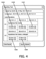
図4は、医療診断システムから導出された情報の診断レポートシステムへの転送を容易化する、ユーザインタフェースの例を示す。本例においては、異なる時点における病変の寸法が、診断システムによってレポートされ、ユーザは、どの情報アイテム410A−C、420A−Bが診断レポートに挿入されるべきかを選択する選択肢を与えられる。直接的な実施例においては、ユーザは、マウスを使ってレポートの1つ以上を選択し、次いで「挿入」キー450上でクリックしても良い。音声認識システムにおいては、ユーザは「1番を挿入する」と発話して、そのことにより3つのレポート410A−Cを挿入しても良いし、又は、「最新のものを挿入する」と発話して、そのことによりレポート410A及び420Aを挿入しても良い。視線追跡の実施例においては、ユーザはレポートを凝視し、次いで該レポートを挿入するために2回瞬きをしても良い。
FIG. 4 illustrates an example user interface that facilitates transfer of information derived from a medical diagnostic system to a diagnostic reporting system. In this example, the dimensions of the lesion at different times are reported by the diagnostic system and the user is given the option to select which
特定の実施例に依存して、選択され表示された情報は、診断レポートに直接にコピーされても良いし、又は、識別された骨格の形式を準拠するよう処理されても良い。 Depending on the particular implementation, the selected and displayed information may be copied directly into the diagnostic report or may be processed to conform to the identified skeleton type.
特に異なる販売者が異なる構成要素を提供している構成において、斯かる一体化を容易化するため、該診断システムは、表示されている情報を外部のシステムにエクスポートするよう構成されたものであっても良いアプリケーションプログラミングインタフェース(API)を含んでも良く、該レポートシステムが、これらAPIを用いて、閲覧システムから情報を取得しても良い。幾つかの実施例においては、該APIは、情報を直接に提供するように構成されても良いし、又は構造化された形式で情報を提供するように構成されても良い。即ち、本発明の処理は、複数の物理的なシステム間に分散されても良い。 In order to facilitate such integration, particularly in configurations where different sellers provide different components, the diagnostic system was configured to export the displayed information to an external system. Application reporting interfaces (APIs) may be included, and the reporting system may use these APIs to obtain information from the browsing system. In some embodiments, the API may be configured to provide information directly or may be configured to provide information in a structured format. That is, the processing of the present invention may be distributed among a plurality of physical systems.
一実施例においては、該APIは、例えば診断システムにおけるこれらパラメータの現在の値を返す「Get(body_part, modality, date)」といった呼び出しを介して、直接にパラメータを提供するように構成されても良い。診断システムが、構造化された文章を提供するよう構成された他の実施例においては、斯かる呼び出しは、図2の骨格250によって生成されるもののような構造化された文章を返す、「Get(Finding(所見))」の形をとるものであっても良い(図3Aにおける選択可能な要素5)。
In one embodiment, the API may be configured to provide parameters directly via a call such as “Get (body_part, modality, date)” that returns the current values of these parameters in the diagnostic system, for example. good. In other embodiments where the diagnostic system is configured to provide structured text, such a call returns a structured text, such as that generated by the
図5は、医療画像から導出された情報の、診断レポートへの転送を容易化する、診断レポートシステムのブロック図の例を示す。図5の診断レポートシステムは、放射線医が診断画像閲覧システムを用いる状況において表される。 FIG. 5 illustrates an example block diagram of a diagnostic report system that facilitates transfer of information derived from medical images to a diagnostic report. The diagnostic report system of FIG. 5 is represented in a situation where a radiologist uses a diagnostic image browsing system.
本実施例においては、放射線医はユーザインタフェース510を介して診断画像閲覧システムと相互作用し、診断工程の間に決定された構造化された文章が、該診断画像閲覧システムの一部であっても良いディスプレイ520において放射線医に提示される。コントローラ590は、診断レポートシステムにおける要素間の相互作用を管理するが、説明の容易さのため、図5におけるコントローラ590とその他の要素のそれぞれとの接続は図示されていない。
In this example, the radiologist interacts with the diagnostic image viewing system via the
行動監視部530は、マウスのクリック/キーのストローク、検査の開始/終了、先行する検査のスクロール/閲覧、画像のリンク付け、病変の測定/注記付け、関連画像の検索及び提案等を含む、診断画像閲覧システムにおいて診断医により実行される行動を、常に監視する。
The
コンテキスト及び内容抽出部540は、診断画像閲覧システムにより提供されたインタラクション及び出力を評価し、現在の診断コンテキストを決定し、完了したタスクに関連する関連データを抽出する。抽出部540は、コンテキスト決定及びデータ抽出を容易化するため医療画像525に直接にアクセスしても良いし、診断画像閲覧システムの出力にアクセスしても良いし、又はその両方であっても良い。
The context and
抽出部540は、現在のコンテキストに依存して異なる評価を実行しても良い。例えば、放射線医が検査をロードしている又は閉じているときには、抽出部540は、どの検査がベースラインとして用いられているかを決定しても良い。以前の検査をスクロール、閲覧又は拡大する、及び/又は現在の画像と依然の画像とをリンク付けする放射線医の動作は、以前のどの検査が実際に用いられているかを特定することを容易化し、これによりベースラインを確立する。この場合には、該システムは、検査のそれぞれの日付、時刻、モダリティ、身体部分(検査の追加を含む)を自動的に捕捉する。
The
放射線医が病変を測定又は注記付けしているとき、抽出部540は、対象となる現在の所見を検出し、所見が注記付け又は測定されている検査の画像/シリーズ情報、日時、身体部分及びモダリティを自動的に捕捉しても良い。例えば抽出部540は、
−XY位置及び注記のテキスト、
−XY位置、所見の長さ/サイズ/体積/角度(利用可能である場合)、
−画像処理アルゴリズム又は解剖学的領域近似アルゴリズム(Zインデクスを用いる)の助力による、解剖学的な位置、身体部分、所見に関連する側性、
−DICOMメタデモからの画像の閲覧(軸方向/矢状方向/冠状方向)、
−所見の現在のウィンドウ幅/レベル、
−2つ/複数の測定が交差するか(そうである場合には単一の所見に併合する)、及び
−画像/シリーズ情報、日付、時刻、モダリティ、検査の身体部分(画像UID、シリーズUIDを含む)及び所見の現在のウィンドウ幅/レベルを含む、キー画像としての現在の画像
を捕捉しても良い。
When the radiologist is measuring or annotating a lesion, the
-XY position and note text,
-XY position, finding length / size / volume / angle (if available),
-Laterality related to anatomical position, body part, findings, with the help of image processing algorithms or anatomical region approximation algorithms (using Z-index),
-Browsing images from DICOM Meta Demo (Axial / Sagittal / Coronal),
-Current window width / level of findings,
-Whether two / multiple measurements intersect (merging into a single finding if so) and-image / series information, date, time, modality, examination body part (image UID, series UID) And the current image as a key image, including the current window width / level of findings.
抽出部540と診断画像閲覧システムとの間にもたらされるインタラクションのレベルに依存して、抽出部540は、コンテキスト及び内容情報を抽出するため種々の手法を用いても良い。例えば、診断画像閲覧システムがHL7メッセージを送信するよう構成され得る場合、抽出部540は、HL7フィードを受信/吸収するよう構成されても良い。診断画像閲覧システムが情報にアクセスするためのAPI(Application Program Interface)を提供する場合、抽出部540は、コンテキスト及び内容情報のためのAPIにクエリを送信するよう構成されても良い。幾つかの実施例においては、抽出部540は、放射線医が関連情報を「クリップボード」にコピーし、次いで該関連情報を「ペースト」コマンドを介して抽出部540に転送することを可能とするよう構成されても良い。コピーされた情報が画像閲覧システムからの画像として捕捉される場合、抽出部540は、該コピーされた画像から情報を抽出するテキスト認識要素を含んでも良い。
Depending on the level of interaction provided between the
文章生成部550は、現在の動作及びそのコンテキストのテンプレート化された/形式化された記述を提供することにより、構造化された文章535を生成するため、抽出された情報を用いる。以上に詳述したように、現在の動作及びそのコンテキストの記述は、ユーザ間での一貫性を維持するため所定のテンプレートを用いても良く、自然言語処理を用いたレポートの容易な解析を可能とする。オントロジー及びテンプレートデータベース535は、構造化された文章555の当該生成を容易化する。
The
エクスポート部560は、ユーザインタフェース510を介して放射線医の選択を受信し、生成された文章を診断レポートへと選択的にコピー及びペーストする。エクスポート部560はまた、動作及びコンテキストの有効性をチェックし、それに応じてシステムメモリを更新する。動作が実行されたが生成された記述は消費されていない場合には、該生成された記述を無効化し、メモリから消去して、潜在的なデータの同期エラーを防ぐ。
The
エクスポート部560は、音声コマンド、マウスクリック、身振り等を含む、種々の方法で、以上に詳述したように、構造化された文章535の転送を実行しても良い。幾つかの実施例においては、エクスポート部560は、殆どのオペレーティングシステムに備えられた「クリップボード」を用いて選択された構造化された文章を受信/コピーし、従来のワードプロセッサとインタラクションすることによって該構造化された文章を診断レポートへとペーストする。
The
本発明は図面及び以上の記述において説明され記載されたが、斯かる説明及び記載は説明するもの又は例示的なものとみなされるべきであり、本発明は開示された実施例に限定されるものではない。 While the invention has been illustrated and described in the drawings and foregoing description, such description and description are to be considered illustrative or exemplary and the invention is limited to the disclosed embodiments. is not.
例えば、本発明は高度にインタラクティブな処理の状況において提示されたが、一実施例においては、診断医によるいずれの関与もなく、各診断が実行されている間に処理がバックグラウンドで実行されるよう、本発明を動作させることも可能である。出力されるレポートは、診断が完了した後に診断医によって編集され得るテキストファイルであっても良い。代替としては、該レポートは、診断医の動作、診断システムの自動化された動作、これら動作の結果、等を含む、診断工程を文書化するテキストファイルであっても良い。 For example, although the present invention has been presented in a highly interactive processing context, in one embodiment, processing is performed in the background while each diagnosis is performed without any involvement by a diagnostician. Thus, it is possible to operate the present invention. The output report may be a text file that can be edited by the diagnostician after the diagnosis is complete. Alternatively, the report may be a text file that documents the diagnostic process, including diagnostician actions, automated actions of the diagnostic system, the results of these actions, and the like.
図面、説明及び添付される請求項を読むことにより、請求される本発明を実施化する当業者によって、開示された実施例に対する他の変形が理解され実行され得る。請求項において、「有する(comprising)」なる語は他の要素又はステップを除外するものではなく、「1つの(a又はan)」なる不定冠詞は複数を除外するものではない。単一のプロセッサ又はその他のユニットが、請求項に列記された幾つかのアイテムの機能を実行しても良い。特定の手段が相互に異なる従属請求項に列挙されているという単なる事実は、これら手段の組み合わせが有利に利用されることができないことを示すものではない。コンピュータプログラムは、他のハードウェアと共に又は他のハードウェアの一部として供給される光記憶媒体又は固体媒体のような適切な媒体上で保存/配布されても良いが、インターネット又はその他の有線若しくは無線通信システムを介してのような、他の形態で配布されても良い。請求項におけるいずれの参照記号も、請求の範囲を限定するものとして解釈されるべきではない。 From reading the drawings, description and appended claims, other variations to the disclosed embodiments can be understood and implemented by those skilled in the art in practicing the claimed invention. In the claims, the word “comprising” does not exclude other elements or steps, and the indefinite article “a” or “an” does not exclude a plurality. A single processor or other unit may fulfill the functions of several items recited in the claims. The mere fact that certain measures are recited in mutually different dependent claims does not indicate that a combination of these measured cannot be used to advantage. The computer program may be stored / distributed on any suitable medium, such as an optical storage medium or solid medium supplied with or as part of other hardware, but the Internet or other wired or It may be distributed in other forms, such as via a wireless communication system. Any reference signs in the claims should not be construed as limiting the claim.
Claims (15)
診断の閲覧中に医療画像に対して実行されるユーザの行動を監視してコンテキストを決定させ、
前記コンテキストに基づいて関連データを決定させ、
前記関連データを構造化された文章へと変換させ、
前記構造化された文章を診断レポートへと挿入する選択肢をユーザに提供させ、
前記ユーザが前記文章を挿入することを選択した場合、前記構造化された文章を含むように前記診断レポートを修正させる
プログラムを含む、持続型コンピュータ読み取り可能媒体。 When executed by a processor, the processor
Monitor user actions performed on medical images while browsing diagnostics to determine context,
Determining relevant data based on the context;
Converting the relevant data into structured text;
Providing the user with an option to insert the structured text into a diagnostic report;
A persistent computer readable medium comprising a program for modifying the diagnostic report to include the structured text if the user chooses to insert the text.
前記関連データを複数の構造化された文章に変換させ、
前記ユーザが前記構造化された文章の1つ以上を選択することを可能とさせ、
前記選択された1つ以上の構造化された文章を含むように前記診断レポートを修正させる、
請求項1に記載の媒体。 The program is stored in the processor.
Converting the related data into a plurality of structured sentences;
Allowing the user to select one or more of the structured sentences;
Modifying the diagnostic report to include the selected one or more structured sentences;
The medium of claim 1.
前記医療画像にアクセスする間のユーザの行動を監視する行動監視部と、
前記ユーザの行動に基づいてコンテキストを決定するコンテキスト抽出部と、
前記コンテキストに基づいて関連データを決定する内容抽出部と、
前記関連データを構造化された文章に変換する文章生成部と、
診断レポートへ含めるためユーザが前記構造化された文章を選択することを可能とするユーザインタフェースと、
前記ユーザが挿入のため前記構造化された文章を選択した場合に、前記構造化された文章を前記診断レポートに挿入するエクスポート部と、
を有する、診断レポートシステム。 A source of medical images relevant to the patient;
An action monitoring unit that monitors user actions while accessing the medical image;
A context extractor for determining a context based on the user's behavior;
A content extractor for determining relevant data based on the context;
A sentence generator for converting the related data into a structured sentence;
A user interface that allows a user to select the structured text for inclusion in a diagnostic report;
An export unit that inserts the structured text into the diagnostic report when the user selects the structured text for insertion;
Having a diagnostic report system.
前記ユーザインタフェースは、ユーザが前記構造化された文章の1つ以上を選択することを可能とし、
前記エクスポート部は、前記選択された1つ以上の構造化された文章を含むように前記診断レポートを修正する、
請求項13に記載のシステム。 The sentence generation unit converts the related data into a plurality of structured sentences,
The user interface allows a user to select one or more of the structured sentences;
The exporter modifies the diagnostic report to include the selected one or more structured sentences;
The system of claim 13.
前記ユーザインタフェースは、前記行動が監視された時間とは異なる時間においてユーザが前記複数の構造化された文章を取得することを可能とする、
請求項14に記載のシステム。 The extraction unit stores each of the plurality of structured sentences in a memory;
The user interface allows a user to obtain the plurality of structured sentences at a time different from the time at which the behavior was monitored;
The system according to claim 14.
Applications Claiming Priority (3)
| Application Number | Priority Date | Filing Date | Title |
|---|---|---|---|
| US201562112183P | 2015-02-05 | 2015-02-05 | |
| US62/112,183 | 2015-02-05 | ||
| PCT/IB2016/050422 WO2016125053A1 (en) | 2015-02-05 | 2016-01-28 | Contextual creation of report content for radiology reporting |
Publications (3)
| Publication Number | Publication Date |
|---|---|
| JP2018509689A true JP2018509689A (en) | 2018-04-05 |
| JP2018509689A5 JP2018509689A5 (en) | 2019-02-28 |
| JP6914839B2 JP6914839B2 (en) | 2021-08-04 |
Family
ID=55310856
Family Applications (1)
| Application Number | Title | Priority Date | Filing Date |
|---|---|---|---|
| JP2017539237A Active JP6914839B2 (en) | 2015-02-05 | 2016-01-28 | Report content context generation for radiation reports |
Country Status (5)
| Country | Link |
|---|---|
| US (1) | US20180092696A1 (en) |
| EP (1) | EP3254211A1 (en) |
| JP (1) | JP6914839B2 (en) |
| CN (1) | CN107209809A (en) |
| WO (1) | WO2016125053A1 (en) |
Families Citing this family (17)
| Publication number | Priority date | Publication date | Assignee | Title |
|---|---|---|---|---|
| US10978187B2 (en) | 2017-08-10 | 2021-04-13 | Nuance Communications, Inc. | Automated clinical documentation system and method |
| US11316865B2 (en) | 2017-08-10 | 2022-04-26 | Nuance Communications, Inc. | Ambient cooperative intelligence system and method |
| CN107563123A (en) * | 2017-09-27 | 2018-01-09 | 百度在线网络技术(北京)有限公司 | Method and apparatus for marking medical image |
| CN109583440B (en) * | 2017-09-28 | 2021-12-17 | 北京西格码列顿信息技术有限公司 | Medical image auxiliary diagnosis method and system combining image recognition and report editing |
| US20190272147A1 (en) * | 2018-03-05 | 2019-09-05 | Nuance Communications, Inc, | System and method for review of automated clinical documentation |
| US11515020B2 (en) | 2018-03-05 | 2022-11-29 | Nuance Communications, Inc. | Automated clinical documentation system and method |
| US11250383B2 (en) | 2018-03-05 | 2022-02-15 | Nuance Communications, Inc. | Automated clinical documentation system and method |
| CN112352243A (en) * | 2018-05-15 | 2021-02-09 | 英德科斯控股私人有限公司 | Expert report editor |
| CN109545302B (en) * | 2018-10-22 | 2023-12-22 | 复旦大学 | Semantic-based medical image report template generation method |
| US10957442B2 (en) * | 2018-12-31 | 2021-03-23 | GE Precision Healthcare, LLC | Facilitating artificial intelligence integration into systems using a distributed learning platform |
| US11227679B2 (en) | 2019-06-14 | 2022-01-18 | Nuance Communications, Inc. | Ambient clinical intelligence system and method |
| US11216480B2 (en) | 2019-06-14 | 2022-01-04 | Nuance Communications, Inc. | System and method for querying data points from graph data structures |
| US11531807B2 (en) | 2019-06-28 | 2022-12-20 | Nuance Communications, Inc. | System and method for customized text macros |
| US11670408B2 (en) | 2019-09-30 | 2023-06-06 | Nuance Communications, Inc. | System and method for review of automated clinical documentation |
| US11699508B2 (en) | 2019-12-02 | 2023-07-11 | Merative Us L.P. | Method and apparatus for selecting radiology reports for image labeling by modality and anatomical region of interest |
| US11720921B2 (en) * | 2020-08-13 | 2023-08-08 | Kochava Inc. | Visual indication presentation and interaction processing systems and methods |
| US11222103B1 (en) | 2020-10-29 | 2022-01-11 | Nuance Communications, Inc. | Ambient cooperative intelligence system and method |
Citations (6)
| Publication number | Priority date | Publication date | Assignee | Title |
|---|---|---|---|---|
| JPH09223129A (en) * | 1996-02-16 | 1997-08-26 | Toshiba Corp | Method and device for supporting document processing |
| JP2005027978A (en) * | 2003-07-09 | 2005-02-03 | Fujitsu Ltd | Medical information system |
| JP2009259000A (en) * | 2008-04-16 | 2009-11-05 | Fujifilm Corp | Document creation support device, document creation support method, and document creation support program |
| US20130251233A1 (en) * | 2010-11-26 | 2013-09-26 | Guoliang Yang | Method for creating a report from radiological images using electronic report templates |
| EP2657866A1 (en) * | 2012-04-24 | 2013-10-30 | Koninklijke Philips N.V. | Creating a radiology report |
| US20150032471A1 (en) * | 2013-07-29 | 2015-01-29 | Mckesson Financial Holdings | Method and computing system for providing an interface between an imaging system and a reporting system |
Family Cites Families (13)
| Publication number | Priority date | Publication date | Assignee | Title |
|---|---|---|---|---|
| US8553949B2 (en) * | 2004-01-22 | 2013-10-08 | DigitalOptics Corporation Europe Limited | Classification and organization of consumer digital images using workflow, and face detection and recognition |
| CN1934589A (en) * | 2004-03-23 | 2007-03-21 | 美国西门子医疗解决公司 | Systems and methods providing automated decision support for medical imaging |
| DE102007020364A1 (en) * | 2007-04-30 | 2008-11-06 | Siemens Ag | Provide a medical report |
| US8588485B2 (en) * | 2008-11-25 | 2013-11-19 | Carestream Health, Inc. | Rendering for improved diagnostic image consistency |
| EP2411931A1 (en) * | 2009-03-26 | 2012-02-01 | Koninklijke Philips Electronics N.V. | A system that automatically retrieves report templates based on diagnostic information |
| US8726324B2 (en) * | 2009-03-27 | 2014-05-13 | Motorola Mobility Llc | Method for identifying image capture opportunities using a selected expert photo agent |
| JP5744182B2 (en) * | 2010-04-19 | 2015-07-01 | コーニンクレッカ フィリップス エヌ ヴェ | Report viewer using radiation descriptors |
| JP2013527503A (en) * | 2010-09-20 | 2013-06-27 | ザ ボード オブ リージェンツ オブ ザ ユニバーシティー オブ テキサス システム | Advanced multimedia structured report |
| JP6060144B2 (en) * | 2011-03-25 | 2017-01-11 | コーニンクレッカ フィリップス エヌ ヴェKoninklijke Philips N.V. | Generating reports based on image data |
| EP2669812A1 (en) * | 2012-05-30 | 2013-12-04 | Koninklijke Philips N.V. | Providing assistance with reporting |
| US9904966B2 (en) * | 2013-03-14 | 2018-02-27 | Koninklijke Philips N.V. | Using image references in radiology reports to support report-to-image navigation |
| US10339504B2 (en) * | 2014-06-29 | 2019-07-02 | Avaya Inc. | Systems and methods for presenting information extracted from one or more data sources to event participants |
| US20160124937A1 (en) * | 2014-11-03 | 2016-05-05 | Service Paradigm Pty Ltd | Natural language execution system, method and computer readable medium |
-
2016
- 2016-01-28 JP JP2017539237A patent/JP6914839B2/en active Active
- 2016-01-28 EP EP16703364.6A patent/EP3254211A1/en not_active Withdrawn
- 2016-01-28 WO PCT/IB2016/050422 patent/WO2016125053A1/en active Application Filing
- 2016-01-28 CN CN201680008872.XA patent/CN107209809A/en active Pending
- 2016-01-28 US US15/548,785 patent/US20180092696A1/en not_active Abandoned
Patent Citations (6)
| Publication number | Priority date | Publication date | Assignee | Title |
|---|---|---|---|---|
| JPH09223129A (en) * | 1996-02-16 | 1997-08-26 | Toshiba Corp | Method and device for supporting document processing |
| JP2005027978A (en) * | 2003-07-09 | 2005-02-03 | Fujitsu Ltd | Medical information system |
| JP2009259000A (en) * | 2008-04-16 | 2009-11-05 | Fujifilm Corp | Document creation support device, document creation support method, and document creation support program |
| US20130251233A1 (en) * | 2010-11-26 | 2013-09-26 | Guoliang Yang | Method for creating a report from radiological images using electronic report templates |
| EP2657866A1 (en) * | 2012-04-24 | 2013-10-30 | Koninklijke Philips N.V. | Creating a radiology report |
| US20150032471A1 (en) * | 2013-07-29 | 2015-01-29 | Mckesson Financial Holdings | Method and computing system for providing an interface between an imaging system and a reporting system |
Also Published As
| Publication number | Publication date |
|---|---|
| EP3254211A1 (en) | 2017-12-13 |
| WO2016125053A1 (en) | 2016-08-11 |
| CN107209809A (en) | 2017-09-26 |
| JP6914839B2 (en) | 2021-08-04 |
| US20180092696A1 (en) | 2018-04-05 |
Similar Documents
| Publication | Publication Date | Title |
|---|---|---|
| JP6914839B2 (en) | Report content context generation for radiation reports | |
| US20220199230A1 (en) | Context driven summary view of radiology findings | |
| CN108475538B (en) | Structured discovery objects for integrating third party applications in an image interpretation workflow | |
| US8744149B2 (en) | Medical image processing apparatus and method and computer-readable recording medium for image data from multiple viewpoints | |
| JP5736007B2 (en) | Apparatus, system, method and program for generating inspection report | |
| JP6657210B2 (en) | Picture archiving system with text image linking based on text recognition | |
| JP6796060B2 (en) | Image report annotation identification | |
| JP2012094127A (en) | Diagnostic result explanation report creation device, diagnostic result explanation report creation method and diagnostic result explanation report creation program | |
| BR112012026477B1 (en) | method for viewing a medical report describing X-ray images | |
| JP7258772B2 (en) | holistic patient radiology viewer | |
| JP5337992B2 (en) | Medical information processing system, medical information processing method, and program | |
| US20060072797A1 (en) | Method and system for structuring dynamic data | |
| JP7102509B2 (en) | Medical document creation support device, medical document creation support method, and medical document creation support program | |
| JP2019149005A (en) | Medical document creation support apparatus, method, and program | |
| US20190108175A1 (en) | Automated contextual determination of icd code relevance for ranking and efficient consumption | |
| US8923582B2 (en) | Systems and methods for computer aided detection using pixel intensity values | |
| JP2019537114A (en) | System and method for determining relevant past radiological examinations using PACS log files | |
| US10282516B2 (en) | Medical imaging reference retrieval | |
| US10235360B2 (en) | Generation of pictorial reporting diagrams of lesions in anatomical structures | |
| JP2024009342A (en) | Document preparation supporting device, method, and program | |
| US20220139512A1 (en) | Mapping pathology and radiology entities | |
| US20200043583A1 (en) | System and method for workflow-sensitive structured finding object (sfo) recommendation for clinical care continuum | |
| KR20210148132A (en) | Generate snip-triggered digital image reports | |
| WO2023157957A1 (en) | Information processing device, information processing method, and information processing program | |
| WO2022215530A1 (en) | Medical image device, medical image method, and medical image program |
Legal Events
| Date | Code | Title | Description |
|---|---|---|---|
| A521 | Request for written amendment filed |
Free format text: JAPANESE INTERMEDIATE CODE: A523 Effective date: 20190117 |
|
| A621 | Written request for application examination |
Free format text: JAPANESE INTERMEDIATE CODE: A621 Effective date: 20190117 |
|
| A977 | Report on retrieval |
Free format text: JAPANESE INTERMEDIATE CODE: A971007 Effective date: 20191021 |
|
| A131 | Notification of reasons for refusal |
Free format text: JAPANESE INTERMEDIATE CODE: A131 Effective date: 20191126 |
|
| A601 | Written request for extension of time |
Free format text: JAPANESE INTERMEDIATE CODE: A601 Effective date: 20200226 |
|
| A521 | Request for written amendment filed |
Free format text: JAPANESE INTERMEDIATE CODE: A523 Effective date: 20200526 |
|
| A02 | Decision of refusal |
Free format text: JAPANESE INTERMEDIATE CODE: A02 Effective date: 20201027 |
|
| A521 | Request for written amendment filed |
Free format text: JAPANESE INTERMEDIATE CODE: A523 Effective date: 20210301 |
|
| C60 | Trial request (containing other claim documents, opposition documents) |
Free format text: JAPANESE INTERMEDIATE CODE: C60 Effective date: 20210301 |
|
| C11 | Written invitation by the commissioner to file amendments |
Free format text: JAPANESE INTERMEDIATE CODE: C11 Effective date: 20210311 |
|
| A911 | Transfer to examiner for re-examination before appeal (zenchi) |
Free format text: JAPANESE INTERMEDIATE CODE: A911 Effective date: 20210419 |
|
| C21 | Notice of transfer of a case for reconsideration by examiners before appeal proceedings |
Free format text: JAPANESE INTERMEDIATE CODE: C21 Effective date: 20210420 |
|
| TRDD | Decision of grant or rejection written | ||
| A01 | Written decision to grant a patent or to grant a registration (utility model) |
Free format text: JAPANESE INTERMEDIATE CODE: A01 Effective date: 20210708 |
|
| A61 | First payment of annual fees (during grant procedure) |
Free format text: JAPANESE INTERMEDIATE CODE: A61 Effective date: 20210714 |
|
| R150 | Certificate of patent or registration of utility model |
Ref document number: 6914839 Country of ref document: JP Free format text: JAPANESE INTERMEDIATE CODE: R150 |