JP2015526192A - High flow basket catheter for efficient continuous infusion peritoneal dialysis - Google Patents
High flow basket catheter for efficient continuous infusion peritoneal dialysis Download PDFInfo
- Publication number
- JP2015526192A JP2015526192A JP2015528512A JP2015528512A JP2015526192A JP 2015526192 A JP2015526192 A JP 2015526192A JP 2015528512 A JP2015528512 A JP 2015528512A JP 2015528512 A JP2015528512 A JP 2015528512A JP 2015526192 A JP2015526192 A JP 2015526192A
- Authority
- JP
- Japan
- Prior art keywords
- tube
- catheter
- auxiliary
- peritoneal dialysis
- drainage
- Prior art date
- Legal status (The legal status is an assumption and is not a legal conclusion. Google has not performed a legal analysis and makes no representation as to the accuracy of the status listed.)
- Pending
Links
Images


Classifications
-
- A—HUMAN NECESSITIES
- A61—MEDICAL OR VETERINARY SCIENCE; HYGIENE
- A61M—DEVICES FOR INTRODUCING MEDIA INTO, OR ONTO, THE BODY; DEVICES FOR TRANSDUCING BODY MEDIA OR FOR TAKING MEDIA FROM THE BODY; DEVICES FOR PRODUCING OR ENDING SLEEP OR STUPOR
- A61M1/00—Suction or pumping devices for medical purposes; Devices for carrying-off, for treatment of, or for carrying-over, body-liquids; Drainage systems
- A61M1/14—Dialysis systems; Artificial kidneys; Blood oxygenators ; Reciprocating systems for treatment of body fluids, e.g. single needle systems for hemofiltration or pheresis
- A61M1/28—Peritoneal dialysis ; Other peritoneal treatment, e.g. oxygenation
- A61M1/285—Catheters therefor
-
- A—HUMAN NECESSITIES
- A61—MEDICAL OR VETERINARY SCIENCE; HYGIENE
- A61M—DEVICES FOR INTRODUCING MEDIA INTO, OR ONTO, THE BODY; DEVICES FOR TRANSDUCING BODY MEDIA OR FOR TAKING MEDIA FROM THE BODY; DEVICES FOR PRODUCING OR ENDING SLEEP OR STUPOR
- A61M1/00—Suction or pumping devices for medical purposes; Devices for carrying-off, for treatment of, or for carrying-over, body-liquids; Drainage systems
- A61M1/14—Dialysis systems; Artificial kidneys; Blood oxygenators ; Reciprocating systems for treatment of body fluids, e.g. single needle systems for hemofiltration or pheresis
- A61M1/28—Peritoneal dialysis ; Other peritoneal treatment, e.g. oxygenation
- A61M1/282—Operational modes
- A61M1/284—Continuous flow peritoneal dialysis [CFPD]
-
- A—HUMAN NECESSITIES
- A61—MEDICAL OR VETERINARY SCIENCE; HYGIENE
- A61M—DEVICES FOR INTRODUCING MEDIA INTO, OR ONTO, THE BODY; DEVICES FOR TRANSDUCING BODY MEDIA OR FOR TAKING MEDIA FROM THE BODY; DEVICES FOR PRODUCING OR ENDING SLEEP OR STUPOR
- A61M25/00—Catheters; Hollow probes
- A61M25/0021—Catheters; Hollow probes characterised by the form of the tubing
- A61M25/0023—Catheters; Hollow probes characterised by the form of the tubing by the form of the lumen, e.g. cross-section, variable diameter
- A61M25/0026—Multi-lumen catheters with stationary elements
- A61M25/003—Multi-lumen catheters with stationary elements characterized by features relating to least one lumen located at the distal part of the catheter, e.g. filters, plugs or valves
- A61M2025/0031—Multi-lumen catheters with stationary elements characterized by features relating to least one lumen located at the distal part of the catheter, e.g. filters, plugs or valves characterized by lumina for withdrawing or delivering, i.e. used for extracorporeal circuit treatment
-
- A—HUMAN NECESSITIES
- A61—MEDICAL OR VETERINARY SCIENCE; HYGIENE
- A61M—DEVICES FOR INTRODUCING MEDIA INTO, OR ONTO, THE BODY; DEVICES FOR TRANSDUCING BODY MEDIA OR FOR TAKING MEDIA FROM THE BODY; DEVICES FOR PRODUCING OR ENDING SLEEP OR STUPOR
- A61M25/00—Catheters; Hollow probes
- A61M25/0067—Catheters; Hollow probes characterised by the distal end, e.g. tips
- A61M25/0068—Static characteristics of the catheter tip, e.g. shape, atraumatic tip, curved tip or tip structure
- A61M2025/0073—Tip designed for influencing the flow or the flow velocity of the fluid, e.g. inserts for twisted or vortex flow
-
- A—HUMAN NECESSITIES
- A61—MEDICAL OR VETERINARY SCIENCE; HYGIENE
- A61M—DEVICES FOR INTRODUCING MEDIA INTO, OR ONTO, THE BODY; DEVICES FOR TRANSDUCING BODY MEDIA OR FOR TAKING MEDIA FROM THE BODY; DEVICES FOR PRODUCING OR ENDING SLEEP OR STUPOR
- A61M25/00—Catheters; Hollow probes
- A61M25/0067—Catheters; Hollow probes characterised by the distal end, e.g. tips
- A61M25/0068—Static characteristics of the catheter tip, e.g. shape, atraumatic tip, curved tip or tip structure
- A61M25/0071—Multiple separate lumens
-
- A—HUMAN NECESSITIES
- A61—MEDICAL OR VETERINARY SCIENCE; HYGIENE
- A61M—DEVICES FOR INTRODUCING MEDIA INTO, OR ONTO, THE BODY; DEVICES FOR TRANSDUCING BODY MEDIA OR FOR TAKING MEDIA FROM THE BODY; DEVICES FOR PRODUCING OR ENDING SLEEP OR STUPOR
- A61M25/00—Catheters; Hollow probes
- A61M25/0067—Catheters; Hollow probes characterised by the distal end, e.g. tips
- A61M25/0074—Dynamic characteristics of the catheter tip, e.g. openable, closable, expandable or deformable
-
- F—MECHANICAL ENGINEERING; LIGHTING; HEATING; WEAPONS; BLASTING
- F04—POSITIVE - DISPLACEMENT MACHINES FOR LIQUIDS; PUMPS FOR LIQUIDS OR ELASTIC FLUIDS
- F04C—ROTARY-PISTON, OR OSCILLATING-PISTON, POSITIVE-DISPLACEMENT MACHINES FOR LIQUIDS; ROTARY-PISTON, OR OSCILLATING-PISTON, POSITIVE-DISPLACEMENT PUMPS
- F04C2270/00—Control; Monitoring or safety arrangements
- F04C2270/04—Force
- F04C2270/041—Controlled or regulated
Abstract
持続注入腹膜透析に使用するカテーテルは注液管および排液管を備え、前記2つの管の一端は透析装置に接続される。前記注液管が複数の注液補助管に分かれ、前記排液管が複数の排液補助管に分かれ、前記すべての補助管は好ましくは単一の外部管に包まれる。前記補助管はそれぞれ、ねじれや詰まりなしに透析液を腹腔内外へ迅速かつ効率的に移動させ、より大きい表面積に到達させるための複数の開口部を含む。前記外部管は透析処置を目視するための光ファイバボアスコープの挿入用トンネルを備える。前記補助管は挿入前シースに覆われ、挿入後にシースが除去されると腹腔内の前記補助管が好ましくはオープンバスケット形状に展開し透析液の持続的注入を可能にする。【選択図】図1AA catheter used for continuous infusion peritoneal dialysis includes an injection tube and a drain tube, and one end of each of the two tubes is connected to a dialyzer. The liquid injection pipe is divided into a plurality of liquid injection auxiliary pipes, the liquid discharge pipe is divided into a plurality of liquid discharge auxiliary pipes, and all the auxiliary pipes are preferably wrapped in a single external pipe. Each of the auxiliary tubes includes a plurality of openings for quickly and efficiently moving dialysate into and out of the abdominal cavity without twisting or clogging and reaching a larger surface area. The outer tube includes a fiber optic borescope insertion tunnel for viewing the dialysis treatment. The auxiliary tube is covered with a sheath before insertion, and when the sheath is removed after insertion, the auxiliary tube in the abdominal cavity is preferably deployed in an open basket shape to allow continuous infusion of dialysate. [Selection] Figure 1A
Description
(関連出願)
本出願は、2012年8月23日出願の米国特許非仮出願第13/592,712号および2013年8月8日出願の米国特許非仮出願第13/961,918号の優先権を主張し、各出願の開示は参照により本明細書に組み込まれる。
(Related application)
This application claims priority to US patent
(発明の属する技術分野)
本出願は、末期腎臓病患者に対して高流量で効率的な腹膜透析を提供するために使用される新規なバスケット型シリコン透析カテーテルに関する。開示するのは、透析処置中に複数の注液管と排液管を使用することにより、透析処置の効率を最大限に高め、所要時間を最小限に抑えるための新たな装置である。新規の腹膜透析カテーテル装置を提示する。使用に際して、本装置は腹膜透析中に高流量で同時に行われる透析液の注入と排出を提供する。本発明は、好ましくはシリコンゴムとカテーテルを腹壁に固定するためのダクロンカフからなる。挿入前にカテーテルは好ましくは引き抜き可能なシースに覆われ、腹腔内に容易に挿入できるように単一のまっすぐな管のように見える。シースは挿入後に除去され、管のバスケット形状が開放される。
(Technical field to which the invention belongs)
The present application relates to a novel basket-type silicon dialysis catheter used to provide high flow and efficient peritoneal dialysis for end-stage renal disease patients. Disclosed is a new device for maximizing the efficiency of a dialysis treatment and minimizing the time required by using multiple infusion and drainage tubes during the dialysis treatment. A novel peritoneal dialysis catheter device is presented. In use, the device provides infusion and drainage of dialysate that occurs simultaneously at high flow rates during peritoneal dialysis. The present invention preferably comprises a silicone rubber and a Dacron cuff for securing the catheter to the abdominal wall. Prior to insertion, the catheter is preferably covered with a retractable sheath and looks like a single straight tube so that it can be easily inserted into the abdominal cavity. The sheath is removed after insertion to open the basket shape of the tube.
透析とは、血液から老廃物や過剰な水分を除去するための処置であり、主に腎不全患者の失われた腎機能を人工的に代行するために使用される。透析は、通常腎臓が体内で果たす機能、すなわち、体液平衡を調整し体内に蓄積された老廃物を除去する機能を代替する役割を果たす。患者は一般的に身体的疾患を引き起こす程度にまで老廃物レベルが高くなると透析が必要となる。 Dialysis is a treatment for removing waste and excess water from blood, and is mainly used to artificially substitute the lost kidney function of patients with renal failure. Dialysis usually serves as a substitute for the function of the kidneys in the body, ie, the function of adjusting fluid balance and removing waste accumulated in the body. Patients generally require dialysis when the level of waste is high enough to cause physical illness.
透析には、大きく分けて血液透析と腹膜透析の2種類がある。血液透析は、体から過剰な老廃物と水分を除去するための特殊なフィルタを使用する。二重管腔カテーテルが、患者の胸部または頸部の静脈に挿入される。血液透析中、血液はカテーテル内にゆっくりと流れ込み、透析膜と呼ばれるフィルタを通過することにより、不要な物質や流体が除去される。透析装置内の溶液が除去された老廃物を捕捉する。濾過終了後、浄化された血液が体内に戻される。 There are two main types of dialysis: hemodialysis and peritoneal dialysis. Hemodialysis uses special filters to remove excess waste and moisture from the body. A double lumen catheter is inserted into a vein in the patient's chest or neck. During hemodialysis, blood slowly flows into the catheter and passes through a filter called a dialysis membrane, thereby removing unnecessary substances and fluids. The waste product from which the solution in the dialyzer is removed is captured. After filtration, the purified blood is returned to the body.
一方、腹膜透析は、機械のようなフィルタを使用するのではなく、患者自身の腹腔内の生体組織(腹膜)をフィルタとして使用する。腹膜透析では、透析液と呼ばれる浄化用透析溶液で腹部を満たすために腹膜カテーテルと呼ばれる柔らかい管を使用する。腹腔の内壁および内部臓器は、透析膜として機能する腹膜と呼ばれる薄膜で覆われている。腹膜は、(a)腎不全患者が尿素、クレアチニン、カリウム等の老廃物を蓄積する腹膜毛細血管内の血液と(b)透析液の2つの流体を含む部分に分かれる。腹膜は、同時に行われる拡散および限外濾過処理により、老廃物と余分な流体が血液から透析液中に移動することを可能にする。透析液はその後腹部から排出され、その際先に除去された老廃物(溶質)を搬送する。透析液を体内に導入して過剰な流体(水分)と溶質を除去する処理は、交換と呼ばれ、透析液が腹腔内に留まる期間は、滞留時間と呼ばれる。 On the other hand, peritoneal dialysis does not use a filter such as a machine, but uses a living tissue (peritoneum) in the abdominal cavity of the patient as a filter. In peritoneal dialysis, a soft tube called a peritoneal catheter is used to fill the abdomen with a purifying dialysis solution called dialysate. The inner wall and internal organs of the abdominal cavity are covered with a thin film called a peritoneum that functions as a dialysis membrane. The peritoneum is divided into portions containing two fluids: (a) blood in a peritoneal capillary where a patient with renal failure accumulates waste products such as urea, creatinine, potassium, and (b) dialysate. The peritoneum allows waste and excess fluid to move from the blood into the dialysate by simultaneous diffusion and ultrafiltration processes. The dialysate is then discharged from the abdomen and carries the waste (solute) removed earlier. The process of introducing the dialysate into the body to remove excess fluid (water) and solutes is called exchange, and the period during which the dialysate stays in the abdominal cavity is called the residence time.
腹膜透析は、透析スケジュールおよび透析場所に関して患者により大きな自立性と柔軟性をもたらし、家庭血液透析と比較した場合、その簡易性、安全性プロファイルおよび費用対効果において、特に仕事を持つ若い患者にとって理想的な透析方法となる。腹膜透析の主な障害として、カテーテル関連の合併症、腹膜機能の継時的劣化、および透析時間の長さが挙げられるが、本発明はこれらの障害に対処し障害を最小限に抑えることを目的とする。 Peritoneal dialysis brings greater independence and flexibility to patients with respect to dialysis schedules and locations, and is ideal for young patients with work, especially in terms of their simplicity, safety profile, and cost effectiveness when compared to home hemodialysis Dialysis method. The main obstacles to peritoneal dialysis include catheter-related complications, perpetual deterioration of peritoneal function, and length of dialysis time.The present invention addresses these disorders and minimizes them. Objective.
自動腹膜透析(APD)は、患者および医療提供者の間で腹膜透析の好ましい方法としてここ数年の間に台頭してきた。自動腹膜透析では、患者の就寝中に小型軽量のサイクラーを使用して上述の処置が行われる。米国では、持続性周期的腹膜透析(CCPD)と夜間間欠的腹膜透析(NIPD)の2種類の自動腹膜透析器または方法が採用されている。CCPDでは、自動サイクラーを使用して患者が就寝中の夜間に複数回の流体交換が行われ、その後患者の腹部に透析液を留めたままにし、丸一日かけてもう一度交換が行われる。もう一方のNIPDでは、夜間の交換回数を多くすることで日中の最終交換が不要となる。 Automated peritoneal dialysis (APD) has emerged over the last few years as a preferred method of peritoneal dialysis among patients and healthcare providers. In automatic peritoneal dialysis, the above procedure is performed using a small and lightweight cycler while the patient is sleeping. In the United States, two types of automatic peritoneal dialysis machines or methods have been employed: continuous cyclic peritoneal dialysis (CCPD) and night intermittent peritoneal dialysis (NIPD). In CCPD, an automatic cycler is used to perform multiple fluid exchanges at night when the patient is asleep, and then keep the dialysate in the patient's abdomen and repeat the exchange over a whole day. In the other NIPD, the final exchange during the day becomes unnecessary by increasing the number of exchanges at night.
タイダル腹膜透析(TPD)として知られる別の技術では、高い透析液流量を提供し、結果的に、血液と透析液間の拡散勾配を大きくする。これにより、透析液の撹拌されていない層の形成を最小限に抑える。透析液と腹膜との継続的な接触により、滞留期間中の溶質および水分の断続的な除去だけでなく、継続的な除去も提供される。いくつかの欧州諸国では、特に一般的なCAPD(持続携行式腹膜透析)やAPD(自動腹膜透析)では限定的な効果しか得られない患者に対して、持続注入腹膜透析(CFPD)も使用されている。CFPDは、腹部内外への透析液の持続的移動を提供する。 Another technique, known as tidal peritoneal dialysis (TPD), provides high dialysate flow and consequently increases the diffusion gradient between blood and dialysate. This minimizes the formation of an unstirred layer of dialysate. The continuous contact between the dialysate and the peritoneum provides not only intermittent removal of solute and moisture during the residence period, but also continuous removal. In some European countries, continuous infusion peritoneal dialysis (CFPD) is also used, especially for patients who have a limited effect with conventional CAPD (continuous ambulatory peritoneal dialysis) and APD (automated peritoneal dialysis). ing. CFPD provides continuous movement of dialysate into and out of the abdomen.
腹膜透析に使用される技術の種類に関係なく、現在使用されているすべての腹膜アクセス装置や腹膜カテーテルは、いくつかの典型的な欠点や合併症を有する。これらの共通の合併症には、腹腔内の流体の減少に起因するカテーテル周りの漏れや排出障害を含む。これらは特に流体排出の最終局面においてしばしば発生し、流体排出に対する抵抗が増加し、腸ループが標準的なカテーテルの先端および側孔に接近すると、カテーテルが閉塞し透析液のカテーテル内外への自由な流出入が妨げられる。 Regardless of the type of technique used for peritoneal dialysis, all currently used peritoneal access devices and peritoneal catheters have some typical drawbacks and complications. These common complications include leakage around the catheter and disturbances of drainage due to decreased fluid in the abdominal cavity. These often occur in the final phase of fluid drainage, increasing resistance to fluid drainage, and as the intestinal loop approaches the tip and side holes of a standard catheter, the catheter becomes occluded and free dialysate fluid enters and exits the catheter. Inflow and outflow is hindered.
現在の腹膜透析装置のもう一つの限界は、腹膜表面積の非効率的な使用である。腹膜は、体表面積1m2当たり約1,200cm2、または平均的な成人一人当たり2,200cm2の表面積を有する。しかしながら、最近の腹膜透析の臨床応用においては、見たところ腹膜表面積の限られた部分だけが処置中に使用されている。透析中に実際に接触される腹膜表面積は、カテーテルと患者の位置によって決定される。癒着形成等の理由で癒着もしくは閉塞している場合、カテーテルの位置によっては、透析液が流体交換に関与しない窪み内に捕捉される可能性がある。これらの問題により、体内からすべての老廃物を除去するために長い時間を要し、処置を完了するまでに大量の透析液が必要となるので費用が増加する。さらに、過剰な透析液が腹部に捕捉される結果となる可能性もある。 Another limitation of current peritoneal dialysis devices is the inefficient use of peritoneal surface area. Peritoneal has about per body surface area 1m 2 1,200cm 2 or average surface area per adult 2,200cm 2,. However, in recent clinical applications of peritoneal dialysis, apparently only a limited portion of the peritoneal surface area is used during the procedure. The peritoneal surface area actually contacted during dialysis is determined by the catheter and patient location. When adhesion or occlusion occurs due to adhesion formation or the like, depending on the position of the catheter, the dialysate may be trapped in a recess that does not participate in fluid exchange. These problems add cost because it takes a long time to remove all waste products from the body and a large amount of dialysate is required to complete the procedure. In addition, excess dialysate may result in trapping in the abdomen.
現在の腹膜透析におけるもう一つの大きな問題は感染症である。処置に使用される装置の特性により、腹腔内から体の外側までプラスチック管が通される。これにより、特に管が適切に洗浄されていない場合や適切に使用されていない場合に、細菌が体内に入り込む可能性が生み出される。 Another major problem in current peritoneal dialysis is infection. Depending on the characteristics of the device used for the procedure, a plastic tube is passed from the abdominal cavity to the outside of the body. This creates the possibility of bacteria entering the body, especially if the tube is not properly washed or used properly.
現在の透析法のさらなる限界は時間である。血液透析においては、治療中の患者の関与は最小限に抑えられるが、患者は非常に厳格なスケジュールと食事制限に従う必要がある。腹膜透析では、スケジュール管理はより柔軟にできるが、毎日の透析に多くの時間を要し、しかも毎日欠かさず行わなければならない。腹腔に透析液を注入し、体内の不要な老廃物と混合してその混合液を排出する処置には、長い時間を要する。一般的な透析スケジュールでは、日に数回の透析が必要であり、一回の透析に約4〜6時間を要する。 A further limitation of current dialysis methods is time. In hemodialysis, patient involvement during treatment is minimized, but patients must follow very strict schedules and dietary restrictions. In peritoneal dialysis, schedule management can be more flexible, but daily dialysis takes a lot of time and must be done every day. It takes a long time to inject the dialysate into the abdominal cavity, mix with unnecessary waste in the body, and discharge the mixture. A typical dialysis schedule requires several dialysis days per day, and approximately 4-6 hours per dialysis.
本発明は新規な腹膜透析カテーテルを開示する。本発明は、好ましくは長さ320mmのシリコンカテーテル装置の使用を含む。管は、好ましくは3.5mmの内径と4.0mmの外径を有するが、カテーテルの代替の寸法の使用もまた可能である。大径管は複数、好ましくは6つの小径管に細分化される。複数の小径管は、1つ以上(好ましくは2つ)の注液路と、1つ以上(好ましくは3つ)の排液路と、透析中に腹腔内を目視できる光ファイバボアスコープを挿入可能な少なくとも1つの管とを含む。 The present invention discloses a novel peritoneal dialysis catheter. The present invention preferably involves the use of a 320 mm long silicon catheter device. The tube preferably has an inner diameter of 3.5 mm and an outer diameter of 4.0 mm, although the use of alternative catheter dimensions is also possible. The large diameter tube is subdivided into a plurality, preferably six small diameter tubes. Multiple small-diameter tubes insert one or more (preferably two) injection channels, one or more (preferably three) drainage channels, and an optical fiber borescope that allows visual inspection of the abdominal cavity during dialysis And at least one possible tube.
好ましい実施形態において、シリコンカテーテルからなる大径管内に合計6つの小径管が入っている。好ましくは、これらは3つの排液管、2つの注液管、および光ファイバボアスコープの挿入のための1つの管を含む。別の実施形態においては、光ファイバスコープ受容のための別個の管は必要なく、1つの注液管が光ファイバスコープ受容のために使用される。好ましくは、複数の管は、好ましくは150mmの主要カテーテル管の一部を介してのみ延びる。これは腹腔内に挿入される管の末端である。複数の管は腹腔内に容易に挿入できるように、単一の大径外部シリコン管に包まれる。近位端、すなわち透析装置に接続する管の末端において、好ましくは大きい内径の単一の注液管および単一の排液管が提供される。単一の注液管は、透析液が腹腔内に注入されるように透析装置に接続され、単一の排液管は、透析処置中に体内から除去されたすべての流体のための排液袋に接続される。 In a preferred embodiment, there are a total of six small diameter tubes in a large diameter tube consisting of a silicone catheter. Preferably, these include three drain tubes, two infusion tubes, and one tube for insertion of a fiber optic borescope. In another embodiment, a separate tube for receiving the fiber optic scope is not required, and one infusion tube is used for receiving the fiber optic scope. Preferably, the plurality of tubes extend only through a portion of the main catheter tube, preferably 150 mm. This is the end of the tube that is inserted into the abdominal cavity. Multiple tubes are wrapped in a single large diameter outer silicone tube so that they can be easily inserted into the abdominal cavity. At the proximal end, i.e. the end of the tube connecting to the dialyzer, a single large infusion tube and a single drain tube are preferably provided. A single infusion tube is connected to the dialyzer so that dialysate is injected into the abdominal cavity, and a single drain tube is a drain for all fluids removed from the body during the dialysis procedure Connected to the bag.
好ましくは長さ約150mmの主要部分において、単一の大径排液管および単一の大径注液管が、好ましくは複数の小径注液管および排液管に分割する。すべての注液管および排液管は、大径外部シリコン管に包まれている。好ましくは、単一の排液管は3つの小径排液管に分割され、単一の注液管は、好ましくは2つの小径注液管に分割される。単一の注液管および排液管から複数の管への分割は、好ましくは、大径注液管と大径排液管が、単一の被覆外部管内で結合する位置で生じる。排液管は好ましくは排液口端部に結合し、注液管は好ましくは注液口端部に結合して、好ましくはY型形状となる。 In the main part, preferably about 150 mm in length, the single large diameter drain pipe and the single large diameter pipe are preferably divided into a plurality of small diameter pipes and drain pipes. All the liquid injection pipes and drainage pipes are enclosed in a large-diameter external silicon pipe. Preferably, the single drainage pipe is divided into three small diameter drainage pipes, and the single injection pipe is preferably split into two small diameter drainage pipes. The division from the single injection pipe and the drain pipe into the plurality of pipes preferably occurs at a position where the large-diameter injection pipe and the large-diameter drain pipe are combined in a single coated outer pipe. The drainage tube is preferably coupled to the end of the drainage port, and the injection tube is preferably coupled to the end of the injection port and is preferably Y-shaped.
注液管と排液管が結合するY型形状の基部が、好ましくは光ファイバボアスコープを選択的に挿入するための管を提供し、それにより本発明を用いて透析を行う医師が、透析が適切に行われていることを確認するために腹腔内を目視可能になる。別の実施形態では、光ファイバスコープを受容するための独立した管を必要とせず、1つの注液管が光ファイバスコープの受容管としても使用される。光ファイバボアスコープを挿入できる管は、好ましくは、注液管と排液管が腹腔内で複数の小径管に分岐する場所である大径被覆管と同じ長さである。好ましい実施形態では、光ファイバボアスコープを挿入するトンネルは、注液管と排液管が結合するY型結合部に開口している。すべての管が結合するY型尾部は、シリコンカテーテルの最初の部分すなわち腹部外の部分を構成する。末端管すなわち腹腔内の小径管は、好ましくは2つの注液管、3つの排液管、およびボアスコープ管を含み、好ましくは拡張可能なシースに包まれている。シースはすべての管を腹腔内に挿入する際、複数の管を所定の位置に保持するために使用される。 A Y-shaped base where the injection and drainage pipes are combined preferably provides a tube for the selective insertion of a fiber optic borescope, whereby the physician performing dialysis using the present invention In order to confirm that this is done properly, the inside of the abdominal cavity becomes visible. In another embodiment, there is no need for a separate tube for receiving the fiber optic scope, and one injection tube is also used as the fiber optic scope receiving tube. The tube into which the optical fiber borescope can be inserted preferably has the same length as the large-diameter coated tube where the liquid injection tube and the drainage tube branch into a plurality of small-diameter tubes in the abdominal cavity. In a preferred embodiment, the tunnel into which the optical fiber borescope is inserted opens at a Y-type coupling portion where the liquid injection tube and the drainage tube are coupled. The Y-tail where all the tubes join constitutes the first part of the silicone catheter, ie the part outside the abdomen. The distal or intraperitoneal small diameter tube preferably includes two infusion tubes, three drainage tubes, and a borescope tube, preferably wrapped in an expandable sheath. The sheath is used to hold multiple tubes in place as all tubes are inserted into the abdominal cavity.
カテーテルを腹膜内に挿入後、管を取り巻く外側のシースは引き抜かれる。これにより、複数の管が分離し展開する。すべての管はバスケット型か、または細長いフットボールやその他の丸みを帯びた構造の外側を形成する。それぞれの管は、好ましくは0.5mm以下の多数の孔を側面に有する。好ましくは、挿入を容易にするためにすべての管は共に端部キャップに接続され、これにより、患者の腹部や腹腔内への管の挿入が非常に容易となり、また挿入のための開口部の大きさを最小限に抑えられる。 After inserting the catheter into the peritoneum, the outer sheath surrounding the tube is withdrawn. Thereby, a some pipe | tube isolate | separates and expand | deploys. All tubes are basket-type or form the outside of an elongated football or other rounded structure. Each tube has a large number of holes on the side, preferably 0.5 mm or less. Preferably, all tubes are connected together to the end cap for ease of insertion, which greatly facilitates the insertion of the tube into the patient's abdomen and abdominal cavity and the opening for insertion. The size can be minimized.
シリコンカテーテルの中間部分には、好ましくは70mmの間隔で2つのダクロンカフが取り付けられている。ダクロンカフはカテーテルの注液管と排液管を囲む合成繊維のシースであり、管の不測の位置ずれを防止する。一方のダクロンカフは好ましくは皮膚位置で管上に配置され、もう一方は好ましくは腹膜位置で管の周りに配置される。ダクロンフェルトカフは、挿入された異物の合成物質であるシリコン管と皮膚との接触面における細菌の侵入を防ぐ生物学的障壁を形成するために使用される。腹膜カテーテルの挿入後、生体組織がダクロンフェルトカフ内に成長し、それによりカテーテルを固定して感染症を防ぐ生物学的障壁を形成する。第2のカフは腹腔のすぐ外側に配置され、その位置でカテーテルを通した穴を閉鎖する。ダクロンカフの網目構造内での線維芽細胞や組織の成長は、カテーテルを所定の位置に固定し滑りを防止するだけでなく、細菌の侵入を防ぐ効率的な障壁を提供する。10〜14日で、カフ内への組織の内方成長がその厚さ全体にわたって実質的に完了する。 Two Dacron cuffs are attached to the middle part of the silicone catheter, preferably at a spacing of 70 mm. The Dacron Cuff is a synthetic fiber sheath that surrounds the injection and drainage tubes of the catheter, preventing accidental misalignment of the tubes. One Dacron cuff is preferably placed over the tube at the skin location and the other is preferably placed around the tube at the peritoneal location. Dacron felt cuffs are used to form a biological barrier that prevents the invasion of bacteria at the interface between the skin and the silicone tube, which is a synthetic substance of inserted foreign matter. After insertion of the peritoneal catheter, biological tissue grows within the Dacron felt cuff, thereby immobilizing the catheter and creating a biological barrier that prevents infection. The second cuff is placed just outside the abdominal cavity and closes the hole through the catheter at that location. The growth of fibroblasts and tissues within the Dacron Cuff network structure not only locks the catheter in place and prevents slippage, but also provides an efficient barrier to prevent bacterial invasion. In 10-14 days, tissue ingrowth into the cuff is substantially complete throughout its thickness.
皮膚の外側に延びるカテーテルの最初の部分、すなわち腹部外の部分は、単に注液管と排液管を腹膜透析装置に接続するための手段を提供する。管のこの部分は、好ましくは、腹膜の内外に透析液を移動するための単一の注液管と単一の排液管とを含む。従って、本発明の好ましい実施形態において、透析装置から注液管に入るすべての透析液は、腹腔に入りそこで膜の表面積を最大限にするために複数の注液管に分割するまで単一の注液管を介して移動する。その後、好ましくは、老廃物や余分な体液(限外濾過)を含む透析液は腹膜から排出されるために収集される。透析液は複数の排液管に入り、複数の排液管は腹部から出ると単一の排液管に収束し透析液を排液袋へと送り戻す。 The first portion of the catheter that extends outside the skin, i.e., the portion outside the abdomen, simply provides a means for connecting the infusion and drainage tubes to the peritoneal dialysis machine. This portion of the tube preferably includes a single infusion tube and a single drain tube for transferring dialysate into and out of the peritoneum. Thus, in a preferred embodiment of the present invention, all dialysate entering the infusion tube from the dialyzer enters the abdominal cavity where it is split into multiple infusion tubes to maximize membrane surface area. Move through the injection tube. Thereafter, preferably the dialysate containing waste and excess body fluid (ultrafiltration) is collected for draining from the peritoneum. The dialysate enters a plurality of drainage pipes, and when the plurality of drainage pipes exit the abdomen, they converge into a single drainage pipe and send the dialysate back to the drainage bag.
本発明のカテーテルの挿入は、外科医の関心と専門知識に応じて、開腹手術技術または標準的な腹腔鏡手法を使用して行われる。腹腔鏡技術は、カテーテルの設置を直接可視化でき、またカテーテルを設置する際に部分大網切除術や癒着溶解も同時に実行できるという利点があるため一般的になりつつある。腹腔鏡技術において、患者は全身麻酔の下で手術を受ける。カテーテルを挿入するために腹部が小さく切開され、患者の腹腔に続くトンネルが形成される。本発明では、複数の管すなわちシースにより一束に保持された管が、トンネルを介して腹腔内に挿入される。挿入後、周囲のシースは管から引き抜かれ、それにより、腹腔内に注液補助管と排液補助管のバスケット型のシステムが形成される。カテーテルは、確実に適切な位置決めをするため、直視下で配置される。内側のダクロンカフが腹膜位置に配置される。カテーテルの透析液漏出障害を防止する再吸収性縫合糸を使用して、外科医は腹膜および腹直筋鞘を縫合する。その後、腹壁の皮下にトンネルが形成され、遠位すなわち外側のダクロンカフがカテーテルの出口部の皮下に配置される。カテーテルは、切開部を閉じる前に動作確認される。 The insertion of the catheter of the present invention is performed using open surgical techniques or standard laparoscopic techniques, depending on the surgeon's interest and expertise. Laparoscopic techniques are becoming popular because they have the advantage that they can directly visualize the placement of the catheter and can also perform partial omentectomy and adhesion lysis at the same time as the catheter is placed. In laparoscopic techniques, patients undergo surgery under general anesthesia. A small incision is made in the abdomen to insert the catheter, and a tunnel is formed following the patient's abdominal cavity. In the present invention, a plurality of tubes, that is, tubes held in a bundle by a sheath, are inserted into the abdominal cavity through a tunnel. After insertion, the surrounding sheath is withdrawn from the tube, thereby forming a basket-type system of an auxiliary injection tube and an auxiliary discharge tube in the abdominal cavity. The catheter is placed under direct view to ensure proper positioning. The inner Dacron cuff is placed at the peritoneal position. Using resorbable sutures that prevent catheter dialysate leakage, the surgeon sutures the peritoneum and rectus abdominis sheath. A tunnel is then formed subcutaneously in the abdominal wall and a distal or outer Dacron cuff is placed subcutaneously at the outlet of the catheter. The catheter is validated before closing the incision.
これが本発明の最も重要な点であり、透析液の注入と排出の両方に使用する単一の管だけを含むすべての従来の透析カテーテルと異なる点である。単一管や単一のコイル管の腹膜透析カテーテル(現在当分野で使用される通常のカテーテル)に比較して、本発明のカテーテル装置は、腹腔のより大きい表面積を占めるので、腹腔の小さい窪みも、毛細血管と腹膜透析液の交換に関与できるようになる。これは、腹膜の透析膜としての効率を向上させるだけでなく、腹膜内における癒着や腸ループなどのその他の身体器官による今後の窪みの形成を減少させる。このような窪みの形成は、患者が腹膜炎または腹膜の炎症や刺激などの症状に苦しんだ後に生じることが多い。 This is the most important aspect of the present invention and is different from all conventional dialysis catheters that contain only a single tube used for both infusion and drainage of dialysate. Compared to single-tube or single-coil peritoneal dialysis catheters (ordinary catheters currently used in the art), the catheter device of the present invention occupies a larger surface area of the abdominal cavity and therefore has a small cavity in the abdominal cavity. Can also be involved in the exchange of capillaries and peritoneal dialysate. This not only improves the efficiency of the peritoneum as a dialysis membrane, but also reduces the formation of future depressions by other body organs such as adhesions and intestinal loops within the peritoneum. Such depression formation often occurs after the patient suffers from symptoms such as peritonitis or inflammation or irritation of the peritoneum.
本発明はカテーテルは挿入後にバスケット形状になるため、単一管の直線型やコイル型のカテーテルに比較して、カテーテル自体のよじれや絡まり、または大網の巻き付きの可能性は無視できるほどに低くなる。従来の腹膜透析では、単一のカテーテルが使用され、透析液を腹腔内に注入できるが排出ができないという透析液の排出不良の合併症がしばしば発生する。排液不良は通常、大網や腸ループが従来のカテーテルに接近しカテーテルの先端や側面の小孔を塞ぎ、それにより小孔を介しての透析液の排出が妨害されるときに起こる。排液不良はまた、大網やその他の組織による単一管やカール型のカテーテルの先端および側面の小孔への付着、従来の単一管カテーテルの位置移動(これは、大網に付着する可能性を増加させる)、および透析中の腹腔内の流体量の減少(これは、透析液がカテーテルに自由に注入/排出する流れを阻害し、注液管や排液管を閉塞させる)を含む他の手段によっても引き起こされる。本発明は、バスケット型カテーテルが、その三次元構造を利用して透析液の注入/排出のために管に開いた小孔の邪魔にならない位置に腸ループと大網を押しのけるため、カテーテルの移動や大網の付着という問題を克服できる。 In the present invention, since the catheter is in a basket shape after insertion, the possibility of kinking or entanglement of the catheter itself or winding of the omentum is negligibly lower than that of a single tube linear type or coil type catheter. Become. In conventional peritoneal dialysis, a single catheter is used, and the complication of poor dialysate drainage often occurs where dialysate can be injected into the abdominal cavity but not drained. Poor drainage usually occurs when the omentum or intestinal loop approaches a conventional catheter and plugs a small hole in the tip or side of the catheter, thereby preventing dialysate drainage through the small hole. Poor drainage can also be caused by omentum and other tissues attached to the ostium and side ostium of a single tube or curled catheter, and the movement of a conventional single tube catheter (which attaches to the omentum) Increase the possibility), and decrease the amount of fluid in the peritoneal cavity during dialysis (this obstructs the flow of dialysate freely into and out of the catheter and obstructs the infusion and drainage tubes) Also caused by other means including. The present invention uses the three-dimensional structure of the basket-type catheter to displace the intestinal loop and omentum to a position that does not interfere with the small hole opened in the tube for infusion / discharge of dialysate. And the problem of adhesion of the omentum can be overcome.
本発明の別の利点は、持続的かつ同時に行われる流体の注入と排出である。透析液のこのような移動により、腹膜内流体量と排液管との間に圧力勾配が作成され、流体の自由な排出と排出不良リスクの最小化が可能となる。複数の注液管と排液管は、従来の単一管を使用する腹膜透析装置に比較してはるかに速いペースで透析液を腹腔に注入し、かつ老廃物や不要な流体の混合物を腹腔から排出することができる。これにより、流体交換がより迅速に行われ、非常に大きい腹膜表面積を覆うため、必要とされる滞留時間を大幅に減少できる。従来技術の腹膜透析において滞留時間を長期にする目的は、腹膜全体が透析液に接触することを可能にし、それにより拡散および限外濾過を介して不要な老廃物(溶質)や流体を除去することにある。本発明においては、実質的に最大限の腹膜表面および腹膜毛細血管が腹膜輸送に関与するため、交換がより迅速なペースで効果的に行われる。 Another advantage of the present invention is the continuous and simultaneous fluid infusion and drainage. Such movement of the dialysate creates a pressure gradient between the amount of fluid in the peritoneum and the drain tube, allowing free drainage of fluid and minimizing the risk of drainage failure. Multiple injection and drainage pipes inject dialysate into the abdominal cavity at a much faster pace than conventional peritoneal dialysis machines using a single tube, and mix waste and unwanted fluid into the peritoneal cavity. Can be discharged from. This allows fluid exchange to occur more quickly and covers a very large peritoneal surface area, thus significantly reducing the required residence time. The purpose of prolonging the residence time in prior art peritoneal dialysis is to allow the entire peritoneum to come into contact with the dialysate, thereby removing unwanted waste (solute) and fluids via diffusion and ultrafiltration. There is. In the present invention, substantially the maximum peritoneal surface and peritoneal capillaries are involved in peritoneal transport, so that replacement is effectively performed at a faster pace.
さらに、腹腔内の複数の位置が個々の注液管の直接の目標に定められるため、透析液が腹膜表面全体を覆うまでの待ち時間が少なくなる。同様に滞留時間終了後、不要な溶質、限外濾過水、および使用済の透析液を腹部から除去するために、複数の排液管を備える。上記利点の両方が、透析処置に必要な時間を実質的に短縮し、またすでに交換に使用された同一流体の再循環を最小限に抑えることで透析クリアランスも改善できる。低い透析クリアランスは、腹膜の窪みにつかえた単一管やコイル型のカテーテルにしばしば起こる問題である。本発明を使用すれば、透析処置を効率的かつより迅速なペースで行うことが可能となり、従って日に複数回の透析が必要な患者はスケジュールを遥かに柔軟にでき、同時により優れた効果を獲得できる。 Furthermore, since a plurality of positions in the abdominal cavity are set as direct targets of the individual injection tubes, the waiting time until the dialysate covers the entire peritoneal surface is reduced. Similarly, a plurality of drainage pipes are provided to remove unnecessary solutes, ultrafiltrated water, and used dialysate from the abdomen after the residence time has ended. Both of the above advantages can substantially reduce the time required for the dialysis treatment and also improve dialysis clearance by minimizing the recirculation of the same fluid already used for replacement. Low dialysis clearance is often a problem with single-tube or coil-type catheters held in the peritoneal cavity. Using the present invention, dialysis treatment can be performed efficiently and at a faster pace, so patients who need multiple dialysis days a day can be much more flexible and at the same time more effective. Can be acquired.
従来技術の説明
出願人および発明者の知る限り、本発明は持続注入腹膜透析のための大幅に改善された構成であり、排液障害の可能性を減少させ、腹膜の関与を最大にし、同時に透析処置に掛かる時間を短縮する。透析処置で直面する主要な障害や欠点を克服できる先行技術はこれまでなかった。従って、現在1)挿入が容易で、2)透析液の注入分布を改善し、3)透析装置として使用可能な腹膜の最大表面積を利用する腹膜交換構成の提供においてさらに効率的な、4)腹腔内透析液の再循環率の低下に効果的な、5)腹膜への外傷を減少させ、6)腹膜透析のCAPD、APD、TPDまたはCFPD技術に使用可能で、かつ7)排出障害の発生を最小限または零にする、改良された腹膜透析カテーテルが必要とされている。本発明のカテーテルは、腹膜透析が長期透析のための成功様式となることを可能にし、それにより既存の透析装置における上記の制限の多くを克服することができる。
DESCRIPTION OF THE PRIOR ART To the best of Applicants 'and inventors' knowledge, the present invention is a greatly improved configuration for continuous infusion peritoneal dialysis, reducing the possibility of drainage disturbances, maximizing peritoneal involvement, and at the same time Reduce the time required for dialysis treatment. To date, there has been no prior art that can overcome the major obstacles and disadvantages encountered in dialysis treatment. Therefore, currently 1) easy insertion, 2) improved infusion distribution of dialysate, 3) more efficient in providing a peritoneal exchange configuration utilizing the maximum surface area of the peritoneum that can be used as a dialyzer, and 4) abdominal cavity Effective in reducing the recirculation rate of internal dialysate 5) Reduces peritoneal trauma, 6) Can be used for CAPD, APD, TPD or CFPD technology of peritoneal dialysis, and 7) Occurrence of discharge disturbance There is a need for an improved peritoneal dialysis catheter that minimizes or eliminates. The catheter of the present invention allows peritoneal dialysis to be a successful mode for long-term dialysis, thereby overcoming many of the above limitations in existing dialysis machines.
本発明は、腹膜透析に使用するための、好ましくはシリコン製のカテーテルを開示する。カテーテルの基本および主要部分は内径および外径を有する。カテーテルは1つ以上の注液管のセットと1つ以上の排液管のセットを含む。シリコンカテーテルの最初の部分すなわち腹膜外の部分は、注液端部および排液端部を有する。両方の主要端部、すなわち排液管の主要端部と注液管の主要端部は透析装置に接続する。カテーテル装置の反対側の端部は腹膜透析を行うために腹壁に挿入される。遠位端において、好ましくは注液管が2つの小径注液(患者に対して)補助管に分岐し、好ましくは排液管が3つの小径排液補助管に分割する。(好ましくは合計5つの)小径補助管はすべて複数の小さい側孔を含み、透析液は管への出入りにこれらの側孔を介することにより腹腔内外へ迅速かつ効率的に移動可能である。シリコンカテーテルは好ましくは、挿入後にカテーテルを腹壁組織に固定するための2つの環状ダクロンカフを備える。管を有するカテーテルの遠位すなわち腹腔内部分は、腹腔内への挿入前は挿入を容易にするために最初はシースに覆われている。管が腹腔内に適切に配置されると、シースは除去され、それにより小径注液管と小径排液管が腹腔内に展開し分離する。シースから解放されると、管は好ましくは開いた「バスケット型」を形成する。この装置を使用して、透析液は注液補助管の孔を介して持続的に腹腔に注入され、排液補助管の孔を介して腹腔から持続的に除去される。これは透析交換処置を完了するために必要な時間を短縮し、さらに(a)腹膜の有効表面積を最大にし、(b)溶質および水分除去のために腹膜全体の濃度勾配を最大にし、(c)別個の排液管を使用することにより腹水除去を最大にして排液不良を防止し、(d)独立した注液システムおよび排液システムを提供することにより透析液の再循環を最小限に抑え、かつ(e)滞留時間の持続的で効率的な使用により透析に要する時間を最小限に抑えることにより、効率を向上させる。本発明はまた、透析を行う医師が実像転送ビデオカメラを介して腹腔内を確認できる光ファイバボアスコープの選択的受容に適合した管を備える。光ファイバボアスコープを挿入可能な管の追加により、医師は透析が適切に機能していることを確認でき、処置中に提示された問題の分析に役立てることができる。 The present invention discloses a catheter, preferably made of silicon, for use in peritoneal dialysis. The basic and main parts of the catheter have an inner diameter and an outer diameter. The catheter includes a set of one or more infusion tubes and a set of one or more drainage tubes. The first part of the silicone catheter, i.e., the extraperitoneal part, has an infusion end and a drain end. Both main ends, i.e. the main end of the drain tube and the main end of the injection tube, connect to the dialyzer. The opposite end of the catheter device is inserted into the abdominal wall for peritoneal dialysis. At the distal end, the infusion tube preferably branches into two small diameter infusion (for the patient) auxiliary tube, and preferably the drainage tube is divided into three small diameter drainage auxiliary tubes. The small diameter auxiliary tubes (preferably a total of 5) all contain a plurality of small side holes, and the dialysate can be moved quickly and efficiently into and out of the abdominal cavity through these side holes to enter and exit the tube. The silicon catheter preferably comprises two annular Dacron cuffs for securing the catheter to the abdominal wall tissue after insertion. The distal or intraperitoneal portion of the catheter with the tube is initially covered with a sheath to facilitate insertion prior to insertion into the abdominal cavity. When the tube is properly positioned within the abdominal cavity, the sheath is removed, thereby deploying and separating the small diameter infusion tube and the small diameter drainage tube into the abdominal cavity. When released from the sheath, the tube preferably forms an open “basket”. Using this device, dialysate is continuously infused into the abdominal cavity through the hole in the infusion auxiliary tube and continuously removed from the abdominal cavity through the hole in the drainage auxiliary tube. This reduces the time required to complete the dialysis exchange procedure, further (a) maximizes the effective surface area of the peritoneum, (b) maximizes the concentration gradient across the peritoneum for solute and water removal, (c ) Maximize ascites removal to prevent drainage failure by using a separate drainage tube, and (d) Minimize recirculation of dialysate by providing an independent infusion and drainage system And (e) improve efficiency by minimizing the time required for dialysis by sustained and efficient use of residence time. The present invention also includes a tube adapted for selective reception of a fiber optic borescope that allows a dialysis physician to view the abdominal cavity via a real-image transfer video camera. The addition of a tube into which a fiber optic borescope can be inserted allows the physician to confirm that dialysis is functioning properly and to help analyze problems presented during the procedure.
添付図面の図1A〜図7Bを参照して本発明を説明する。これらの図面はあくまで典型例を示し、交付された米国特許に対する裁判所の解釈通り、本発明は特許請求の範囲によって定義されるものであり、これらの図面が本発明の請求の範囲を制限するものではないことを理解されたい。 The present invention will be described with reference to FIGS. 1A-7B of the accompanying drawings. These drawings are merely exemplary and, as the court interprets the issued US patent, the present invention is defined by the claims, and these drawings limit the claims of the present invention. Please understand that it is not.
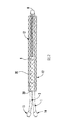
図1Aは本発明の好ましい実施形態を示し、効率的な持続注入腹膜透析のための高流量オープンバスケットカテーテル(以下、「バスケットカテーテル」と称する)が完全に挿入されシースが除去された状態を示す。腹膜透析は透析の一形態であり、透析液と呼ばれる浄化するための透析用流体を体内に導入するために患者の腹腔に管を挿入しておく必要がある。腹腔の壁は、壁側腹膜と呼ばれる薄膜に覆われ、消化管およびその他の内臓は臓側腹膜と呼ばれる腹膜で覆われている。壁側腹膜と臓側腹膜は連続しており、従って腹腔と呼ばれる空洞を形成する。腹膜は腹膜毛細血管およびその他の細胞と共に「透析器」としての役割を果たし、老廃物(尿毒性溶質やカリウム)や余分な体液は、同時進行の拡散および限外濾過処理により血流から透析液内に輸送される。透析液はその後腹部から排出され、先に除去された老廃物を搬送する。透析液を体内に導入して不要な流体を除去する処置は交換と呼ばれ、透析液が腹腔内に留まる期間は滞留(ドウェル)時間と呼ばれる。 FIG. 1A illustrates a preferred embodiment of the present invention with a high flow open basket catheter (hereinafter referred to as “basket catheter”) for efficient continuous infusion peritoneal dialysis fully inserted and the sheath removed. . Peritoneal dialysis is a form of dialysis, and it is necessary to insert a tube into the abdominal cavity of a patient in order to introduce a dialysis fluid called dialysis fluid for purification into the body. The wall of the abdominal cavity is covered with a thin film called the wall side peritoneum, and the digestive tract and other internal organs are covered with the peritoneum called the visceral peritoneum. The parietal peritoneum and visceral peritoneum are continuous and thus form a cavity called the abdominal cavity. The peritoneum, along with peritoneal capillaries and other cells, acts as a “dialyzer”, and waste products (urotoxic solutes and potassium) and excess body fluid are dialyzed from the bloodstream by simultaneous diffusion and ultrafiltration. Be transported within. The dialysate is then drained from the abdomen and carries the waste removed earlier. The treatment of introducing the dialysate into the body and removing unnecessary fluid is called exchange, and the period during which the dialysate stays in the abdominal cavity is called the dwell time.
腹膜透析に使用するオープンバスケットカテーテル11は、透析処置の効率を最大限に高め、かつ透析処置に必要な滞留時間を最小限に抑えるように設計されている。オープンバスケットカテーテル11は、透析液を腹腔内に運ぶ注液トンネルまたは管1と、体腔から不要な流体および老廃物を除去する排液トンネルまたは管2とを含む。 The open basket catheter 11 used for peritoneal dialysis is designed to maximize the efficiency of the dialysis treatment and minimize the residence time required for the dialysis treatment. The open basket catheter 11 includes an injection tunnel or tube 1 that carries dialysate into the abdominal cavity and a drainage tunnel or tube 2 that removes unwanted fluids and waste products from the body cavity.
主注液管1および主排液管2は、主カテーテルの内腔で複数の小径管に分割するように構成される。注液管1および排液管2はベース20で結合するように適合される。図1Aに断面表示「A−A」線として表され図1Bに図示されるベース20において、単一の大径外部保護管30が腹部に挿入されるカテーテルの末端に向かって延びている。外部管30は、ベース20から、小径補助注液管および排液管が出現しバスケット形状に拡張する位置である端部Eまで延びる。本発明の好ましい実施形態では、注液トンネルまたは管1が2つの小径補助トンネルまたは管3および4に分岐し、排液トンネルまたは管2が好ましくは小径補助トンネルまたは管5、6および7に三分割する。単一注液管および単一排液管から補助管3、4、5、6および7への分割は、好ましくはベース20で発生し、すべての補助管3、4、5、6および7は腹部への挿入を容易にするために外部管30に包まれる。小径注液補助管すなわち管3および4には、透析液が流出して腹膜に到達し透析が実施できるように開口部13が並んでいる。同様に、排液補助管すなわち管5、6および7もまた、不要な老廃物および流体を排液管内に回収して体外に排出するための、同一の間隔を空けて(長手方向に沿って管の周りに)配置された開口部13を含む。
The main injection pipe 1 and the main drainage pipe 2 are configured to be divided into a plurality of small diameter pipes in the lumen of the main catheter. The injection tube 1 and the drain tube 2 are adapted to be joined at the
補助管3、4、5、6および7はすべてそれらの遠位端において端部キャップ8に接続し、端部キャップはすべての補助管の先端を密封して共に保持する。補助管はまた好ましくは、カテーテルの周りを囲み腹壁の生体組織内にカテーテルを固定して不測の位置ずれを予防するための合成繊維の包装材である、1つ以上のダクロンカフで共に保持される。一方のダクロンカフ、外側カフ10は、好ましくは皮膚の位置で管に配置され、もう一方のダクロンカフ、内側カフ9は、好ましくは腹膜の位置でカテーテルの周りに配置される。管の中間部および末端部は、好ましくは実質的に長さ約22cmである網状の長いシース12(図2参照)に包まれている。カテーテル11の挿入後、シース12は手動で後方に引き抜かれる。これにより、すべての小径補助管3、4、5、6および7が開放される(図1参照)。解放された補助管は、オープンバスケット型の形状に展開する。外面的には、カテーテル11の管3および4は注入口14と注液管1から続く注液管を、また、管5、6および7は排液管2および排出口15に続く排液管を、それぞれが腹膜透析装置のために提供する。
The
さらに、注液管1および排液管2が結合し複数の補助管または別々の管に分割する位置に、好ましくは、光ファイバボアスコープを受容するための、外部管30の長さで外部管30の中を平行に走る追加の管40が提供される。これにより、本発明を使用して透析を行う医師は、透析処置中の適切な機能を確保するために腹腔内部を確認できる。管40はベース20の注液管1、排液管2および外部管30のY型接合部から始まる。ベース20で結合する単一注液管と単一排液管の断面図を図1Aおよび図1BのA−A線断面図に示す。ベース20で結合する複数の補助注液管と排液管および光ファイバスコープの選択的受容管の断面図を図1Aおよび図1CのB−B線断面図に示す。
Further, the
補助管からシースが外されると、腹腔内のカテーテル11は透析交換を行うことが可能となる。透析液は注液管1および開口部13を介して体内に入る。複数の注液補助管3、4が存在するため、透析液は持続的に腹腔のすべての領域に達することができる。本発明の好ましい実施形態では2つの注液補助管を使用する。しかしながら、本発明において任意の複数の注液管を使用できることは言うまでもない。本発明は、透析液が透析装置から注液管1および注液補助管3、4を介して持続的に腹腔内に注入することを可能にする。
When the sheath is removed from the auxiliary tube, the catheter 11 in the abdominal cavity can be replaced by dialysis. The dialysate enters the body through the injection tube 1 and the
カテーテル装置がバスケット形状であることから、本発明はまた、透析膜としての役割を果たす有効腹膜表面積を拡大することができる。より大きい表面積と腹膜毛細血管の血液と透析液間の連続した交換により、不要な老廃物や流体を持続的に体外に排出するための浄化が増大する。注液補助管と同様に、腹腔全体に広がる複数の排液補助管が使用される。ここでも、腹腔内のカテーテルがバスケット形状であるため、腸ループや大綱が排液管を通る排液を遮断することがなく、従って腹水除去を最大限にできる。これにより、血液コンパートメントから不要な老廃物がより迅速に除去可能となり、これまで交換が行われている間患者が待たなければならなかった滞留期間を大幅に削減できる。本発明の好ましい実施形態では3つの排液補助管が使用されているが、任意の複数の排液管を本発明で使用できることは言うまでもない。 Because the catheter device is basket-shaped, the present invention can also increase the effective peritoneal surface area that serves as a dialysis membrane. The larger surface area and the continuous exchange between peritoneal capillary blood and dialysate increase the purification to continuously drain unwanted waste and fluids out of the body. Similar to the liquid filling auxiliary pipe, a plurality of drainage auxiliary pipes extending over the entire abdominal cavity are used. Again, since the catheter in the abdominal cavity is basket-shaped, the intestinal loop and the rope do not block the drainage through the drainage tube, so that ascites removal can be maximized. This makes it possible to remove unnecessary waste products from the blood compartment more quickly, and greatly reduces the residence time that the patient had to wait for while the replacement was performed. In the preferred embodiment of the present invention, three drainage auxiliary tubes are used, but it goes without saying that any plurality of drainage tubes can be used in the present invention.
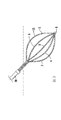
図2は本発明のカテーテル装置11を示す。カテーテルの中央部および末端部はシース12に覆われている。シース12はまた注液補助管3、4と排液補助管5、6および7とを共に所定の位置に保持する。すべての管は遠位端において端部キャップ8により結合され、近位端において外部管30内で結合する。腹腔内へ挿入後、シース12は後方の腹部外に引き抜かれ、それにより注液補助管3、4と排液補助管5、6および7が展開し分離する。図3に示すように、注液および排液補助管は好ましくはバスケット形状に展開する。
FIG. 2 shows the catheter device 11 of the present invention. A central portion and a distal end portion of the catheter are covered with a
図4は、腹腔に挿入された状態にある新規な腹膜カテーテル11の概略断面図を示す。本発明の構成は、補助管の開口部13の閉塞を防止する。すべての既存の腹膜透析カテーテルにおいて、腸ループなどの身体器官がカテーテルの側に留まり流体の注入や排出を阻むのを防止できる構成は存在しなかった。本発明は、図4に示す方法でその障害を克服する。注液および排液管が長手方向に分離したバスケット型を形成する。図示されているように、(管を一緒に保持する)一方の外部ダクロンカフが好ましくは腹壁の皮下組織内に配置され、もう一方の内部ダフロンカフが好ましくは腹壁と腹膜の両方にわたって配置される。
FIG. 4 shows a schematic cross-sectional view of the novel peritoneal catheter 11 in a state of being inserted into the abdominal cavity. The configuration of the present invention prevents the
図5は腹腔内に挿入された状態の本発明の断面図であり、仰臥位に横たわる患者を示す。本発明のカテーテル11は体内で完全に展開すると、腹腔内で拡張し分離してバスケット型の形状となり、小さい窪み内にも広がることで交換に使用する腹膜表面積を最大にする。図に示すように、注液および排液補助管が腹腔内の広い領域に広がり、腹膜のより大きい領域に持続的に透析液をもたらすことによって、(より頻繁で間断のない交換を通して)腹膜全体の濃度勾配を最大にし、同時に腹水および溶質老廃物の除去を最大にする。本発明は、腹腔内の広い表面積を提供するだけではなく、展開した際にその独自の形状を利用して、腸ループやその他の内臓器官が補助管3、4、5、6および7の開口部13を遮断するのを防止する。同様に、本発明の形状は、すべての補助管が好ましくは円形で非外傷性の端部キャップ8により共に保持されているため、カテーテル11が挿入位置から移動し透析液の流れを阻止しないようにする。
FIG. 5 is a cross-sectional view of the present invention inserted into the abdominal cavity showing the patient lying in the supine position. When fully deployed in the body, the catheter 11 of the present invention expands and separates into the abdominal cavity to form a basket-like shape and also extends into a small depression to maximize the peritoneal surface area used for replacement. As shown in the figure, the entire peritoneum (through more frequent and uninterrupted exchanges), as the infusion and drainage aids spread over a wide area within the abdominal cavity and continuously deliver dialysate to a larger area of the peritoneum To maximize the removal of ascites and solute waste. The present invention not only provides a large surface area within the abdominal cavity, but also utilizes its unique shape when deployed so that the intestinal loop and other internal organ officers can open the
図6Aは腹膜透析で使用される一般的な従来例のカテーテルを示す。図に示すように、カテーテルは、透析液の注入および排出の両方を制御する単一の管を備える。従って、従来例において、透析液は体内に注入され、透析液が体内の不要な老廃物と交換されるまでの一定時間(滞留時間と呼ばれる)留まり、その後ゆっくりと排出されるが、これらはすべて同一の単一管を介して行われる。これは、かなりの時間を要するだけでなく、透析液が腹腔内の一カ所に位置するカテーテルからしか排出されないため非効率的でもある。一般的に使用されるコイル状(テンコフ)カテーテルは、大網がカテーテルに巻き付くのを防止するが、透析交換に関与する腹膜の有効表面積を増加させることはない。また、透析液の注入および排出に単一管を使用する従来例においては、透析液の再循環を防止できず、従って透析の効率および溶質や老廃物の浄化の効率が減少する。図6Bは比較として、本発明の腹膜透析用の高流量オープンバスケットカテーテルを示し、注液と排液両方の二重機能を果たす従来例とは異なり、それぞれが単一の機能を果たす複数の注液管および排液管を備えるため、従来のカテーテルではなく二重管を使用して初めて達成可能である透析液の持続的な注液および排液を提供する。 FIG. 6A shows a typical prior art catheter used in peritoneal dialysis. As shown, the catheter includes a single tube that controls both infusion and drainage of dialysate. Therefore, in the conventional example, the dialysate is injected into the body, stays for a certain period of time (called residence time) until the dialysate is replaced with unwanted waste in the body, and then slowly discharged. This is done through the same single tube. This not only takes a considerable amount of time, but is also inefficient because the dialysate is drained from only one catheter located in the abdominal cavity. Commonly used coiled (Tenkov) catheters prevent the omentum from wrapping around the catheter, but do not increase the effective surface area of the peritoneum involved in dialysis exchange. Further, in the conventional example in which a single tube is used for injecting and discharging dialysate, recirculation of dialysate cannot be prevented, and therefore the efficiency of dialysis and the purification of solutes and waste products are reduced. FIG. 6B shows, as a comparison, a high flow open basket catheter for peritoneal dialysis according to the present invention, which is different from the conventional example in which both the injection and drainage functions are performed, and a plurality of injections each of which performs a single function. The provision of a liquid tube and a drain tube provides a continuous infusion and drainage of dialysate that can only be achieved using a double tube rather than a conventional catheter.
従来技術のカテーテルと比較して、本発明のカテーテルは透析液を再循環させることなく単一通過させ、その間に腹膜を透析液に最大限に露出させることができる。交換に使用できる未使用で連続的な透析液の供給により、腹膜全体の溶質除去のための最大値の濃度勾配が達成できる。本発明では、透析液の連続的な注入により滞留容積が小さくて済むため、従来のCCPD、APDおよびNIPDに必要とされる大きい滞留容積に比べて、患者の腹部不快感はほとんどないか全くない。従来技術のカテーテルでは、排液段階の終末部において滞留容積が小さい場合、腸ループや大網が接近してカテーテルの先端や側面の孔を塞ぎ透析液の排出を妨害する原因となる。 Compared to prior art catheters, the catheter of the present invention allows a single passage of dialysate without recirculation, during which the peritoneum is exposed to the maximum extent to the dialysate. With a continuous supply of unused dialysate that can be used for exchange, a maximum concentration gradient for solute removal across the peritoneum can be achieved. The present invention requires little or no patient abdominal discomfort compared to the large residence volume required for conventional CCPD, APD and NIPD, since the residence volume can be reduced by continuous infusion of dialysate. . In the catheter of the prior art, when the retention volume is small at the end of the drainage stage, the intestinal loop and the greater omentum approach to obstruct the discharge of dialysate by closing the hole at the tip and side of the catheter.
さらに、従来技術のカテーテルを使用する排液段階においては、溶質や水分の交換または輸送は行われない。本発明では交換時間を減少させ、かつ濃度勾配、有効腹膜表面積および腹水除去効率を最大にするために別々の注液管および排液管を使用する。 Furthermore, no solute or moisture exchange or transport is performed during the drainage phase using prior art catheters. The present invention uses separate infusion and drainage tubes to reduce exchange time and maximize concentration gradients, effective peritoneal surface area and ascites removal efficiency.
図7Aは、図6Aで説明した挿入位置における標準的な従来例のカテーテルを示す。溶質および不要な老廃物の排出中、大網や腸ループがカテーテルに接近して側孔を塞ぐため、排出される流体への抵抗が増加する。これが、しばしば排液障害や少なくとも排液能力低下の原因となる。図7Aはまた腸ループの間で圧縮され、ねじれた状態にあるカテーテルを示す。 FIG. 7A shows a standard prior art catheter in the insertion position described in FIG. 6A. During the discharge of solutes and unwanted waste, resistance to the drained fluid increases as the omentum and bowel loop approach the catheter and plug the side holes. This often causes drainage problems and at least a decrease in drainage capacity. FIG. 7A also shows the catheter in a compressed and twisted state between the intestinal loops.
図7Bに示すように、本発明は、従来例に共通の腸ループや大網が原因の障害とカテーテル自体が原因の障害の両方を克服できる。図に示す本発明のカテーテル11は挿入後、そのオープンバスケット型の分離した形状により腸ループを脇に押しのける。従って、本発明は透析液の注入分布を改善し、腹膜の接触を増加させ、排出障害を最少にするか完全に取り除く。カテーテルのこの腹腔内部分はまた、カテーテル管を介して持続的かつ同時に行われる注液と排液を促進し、それにより、装置内で行われる交換に必要な時間を大幅に短縮する。透析液は腹腔内に持続的に流入し、膜を介して輸送され一連の流体の動きで装置から排出される余分な水分や溶質を浄化する。 As shown in FIG. 7B, the present invention can overcome both the intestinal loop and omental lesion common to the conventional example and the catheter itself. After insertion, the catheter 11 of the present invention shown in the figure pushes the intestinal loop aside by its open basket type separated shape. Thus, the present invention improves the dialysate infusion distribution, increases peritoneal contact and minimizes or eliminates drainage obstructions. This intraperitoneal portion of the catheter also facilitates continuous and simultaneous infusion and drainage through the catheter tube, thereby significantly reducing the time required for replacement to take place within the device. The dialysate continuously flows into the abdominal cavity and purifies excess water and solutes transported through the membrane and discharged from the device by a series of fluid movements.
当業者は、本発明の範囲から逸脱することなく様々な変更を行うことができ、要素は同等のもので置き換えることができることを理解されたい。さらに、本発明の範囲から逸脱することなく本発明の教示に特定の特徴または材料を適合させる様々な修正がなされてもよい。従って、本発明は開示した特定の実施形態に限定されないが、本発明は特許請求の範囲内に入るすべての実施形態を含むものである。 It should be understood by those skilled in the art that various modifications can be made without departing from the scope of the present invention, and elements can be replaced by equivalents. In addition, various modifications may be made to adapt a particular feature or material to the teachings of the invention without departing from the scope of the invention. Accordingly, the invention is not limited to the specific embodiments disclosed, but the invention includes all embodiments that fall within the scope of the claims.
Claims (28)
長い管を備える注液管と、
長い管を備える排液管と
を具備し、
前記注液管および前記排液管のそれぞれが、前記注液管および前記排液管を透析装置に固定するための近位接続手段を有し、
前記注液管が、2つ以上の分離した小径注液補助管に分かれ、
前記排液管が、2つ以上の分離した小径排液補助管に分かれ、
前記補助管のすべてが、複数の流体搬送開口部を有し、
前記注液補助管および前記排液補助管の少なくとも一部が、単一の外部管に覆われ、かつ、
前記外部管が、透析処置を目視するための光ファイバボアスコープの受容に適合された別個の管をさらに含むことを特徴とする、カテーテル。 A catheter for use in continuous infusion peritoneal dialysis,
An injection tube with a long tube;
A drainage pipe with a long pipe,
Each of the liquid injection pipe and the drainage pipe has a proximal connection means for fixing the liquid injection pipe and the drainage pipe to the dialyzer,
The liquid injection pipe is divided into two or more separate small diameter liquid injection auxiliary pipes;
The drainage pipe is divided into two or more separated small-diameter drainage auxiliary pipes;
All of the auxiliary tubes have a plurality of fluid transfer openings;
At least a part of the liquid injection auxiliary pipe and the drainage auxiliary pipe is covered with a single outer pipe, and
The catheter, wherein the outer tube further comprises a separate tube adapted to receive a fiber optic borescope for viewing a dialysis procedure.
内径および外径を有する長い管を備える注液管と、
内径および外径を有する長い管を備える排液管と
を具備し、
前記注液管および前記排液管のそれぞれが、透析装置に前記注液管および前記排液管を固定するための接続手段を有し、
前記注液管が複数の分離した小径注液補助管を有し、
前記排液管が複数の分離した小径排液補助管を有し、かつ、
前記補助管が、腹腔内外に流体を移動するための複数の開口部を含むことを特徴とする、持続注入腹膜透析に使用するためのカテーテル。 A catheter for use in continuous infusion peritoneal dialysis,
An infusion tube comprising a long tube having an inner diameter and an outer diameter;
A drainage pipe comprising a long pipe having an inner diameter and an outer diameter,
Each of the liquid injection pipe and the drainage pipe has connection means for fixing the liquid injection pipe and the drainage pipe to a dialysis apparatus,
The liquid injection pipe has a plurality of separated small diameter liquid injection auxiliary pipes,
The drainage pipe has a plurality of separated small-diameter drainage auxiliary pipes, and
A catheter for use in continuous infusion peritoneal dialysis, wherein the auxiliary tube includes a plurality of openings for moving fluid into and out of the abdominal cavity.
Applications Claiming Priority (5)
| Application Number | Priority Date | Filing Date | Title |
|---|---|---|---|
| US13/592,712 | 2012-08-23 | ||
| US13/592,712 US9011369B2 (en) | 2012-08-23 | 2012-08-23 | High flux basket catheter for efficient continuous flow peritoneal dialysis |
| US13/961,918 US9078972B2 (en) | 2012-08-23 | 2013-08-08 | High flux basket catheter for efficient, continuous flow peritoneal dialysis |
| US13/961,918 | 2013-08-08 | ||
| PCT/US2013/054266 WO2014031362A1 (en) | 2012-08-23 | 2013-08-09 | High flux basket catheter for efficient, continuous flow peritoneal dialysis |
Publications (2)
| Publication Number | Publication Date |
|---|---|
| JP2015526192A true JP2015526192A (en) | 2015-09-10 |
| JP2015526192A5 JP2015526192A5 (en) | 2016-09-08 |
Family
ID=50148653
Family Applications (1)
| Application Number | Title | Priority Date | Filing Date |
|---|---|---|---|
| JP2015528512A Pending JP2015526192A (en) | 2012-08-23 | 2013-08-09 | High flow basket catheter for efficient continuous infusion peritoneal dialysis |
Country Status (8)
| Country | Link |
|---|---|
| US (1) | US9078972B2 (en) |
| EP (1) | EP2887990A4 (en) |
| JP (1) | JP2015526192A (en) |
| KR (1) | KR20150048165A (en) |
| CN (1) | CN104797286B (en) |
| MX (1) | MX2015002188A (en) |
| SG (1) | SG11201501179XA (en) |
| WO (1) | WO2014031362A1 (en) |
Families Citing this family (15)
| Publication number | Priority date | Publication date | Assignee | Title |
|---|---|---|---|---|
| US9078972B2 (en) | 2012-08-23 | 2015-07-14 | Anmol Gupta | High flux basket catheter for efficient, continuous flow peritoneal dialysis |
| US9011369B2 (en) | 2012-08-23 | 2015-04-21 | Anmol Gupta | High flux basket catheter for efficient continuous flow peritoneal dialysis |
| WO2015048864A1 (en) * | 2013-10-04 | 2015-04-09 | Rodrigues Junior Adilson Costa | Surgical kit for treating peritonitis |
| US11541205B2 (en) | 2015-07-20 | 2023-01-03 | Roivios Limited | Coated urinary catheter or ureteral stent and method |
| US11040172B2 (en) | 2015-07-20 | 2021-06-22 | Strataca Systems Limited | Ureteral and bladder catheters and methods of inducing negative pressure to increase renal perfusion |
| US11040180B2 (en) | 2015-07-20 | 2021-06-22 | Strataca Systems Limited | Systems, kits and methods for inducing negative pressure to increase renal function |
| CN108136163B (en) | 2015-07-20 | 2021-05-04 | 斯卓特凯系统有限责任公司 | Ureteral and bladder catheters and methods of introducing negative pressure to increase renal perfusion |
| US10918827B2 (en) | 2015-07-20 | 2021-02-16 | Strataca Systems Limited | Catheter device and method for inducing negative pressure in a patient's bladder |
| US10512713B2 (en) | 2015-07-20 | 2019-12-24 | Strataca Systems Limited | Method of removing excess fluid from a patient with hemodilution |
| US10926062B2 (en) | 2015-07-20 | 2021-02-23 | Strataca Systems Limited | Ureteral and bladder catheters and methods of inducing negative pressure to increase renal perfusion |
| US10493232B2 (en) | 2015-07-20 | 2019-12-03 | Strataca Systems Limited | Ureteral catheters, bladder catheters, systems, kits and methods for inducing negative pressure to increase renal function |
| US11229771B2 (en) | 2015-07-20 | 2022-01-25 | Roivios Limited | Percutaneous ureteral catheter |
| GB2540818B (en) * | 2015-07-30 | 2017-08-02 | Ayrshire And Arran Health Board | Catheter assembly |
| US10070808B2 (en) | 2016-06-03 | 2018-09-11 | National Guard Health Affairs | Apparatus for in vivo detection and quantification of analytes in the peritoneal fluid |
| WO2018148646A1 (en) * | 2017-02-13 | 2018-08-16 | University Of Pittsburgh - Of The Commonwealth System Of Higher Education | Expandable percutaneous cannula |
Citations (5)
| Publication number | Priority date | Publication date | Assignee | Title |
|---|---|---|---|---|
| US4437856A (en) * | 1981-02-09 | 1984-03-20 | Alberto Valli | Peritoneal catheter device for dialysis |
| JPH05317429A (en) * | 1992-05-20 | 1993-12-03 | Fujikura Ltd | Oscillating catheter |
| JPH09276410A (en) * | 1996-04-11 | 1997-10-28 | Nippon Sherwood Kk | Triple lumen catheter |
| JP2006525081A (en) * | 2003-05-01 | 2006-11-09 | アドバンスド、カーディオバスキュラー、システムズ、インコーポレーテッド | X-ray opaque Nitinol alloy embolization prevention frame |
| JP4236206B1 (en) * | 2007-10-09 | 2009-03-11 | 日本ライフライン株式会社 | Electrode catheter |
Family Cites Families (18)
| Publication number | Priority date | Publication date | Assignee | Title |
|---|---|---|---|---|
| US4693243A (en) * | 1983-01-14 | 1987-09-15 | Buras Sharon Y | Conduit system for directly administering topical anaesthesia to blocked laryngeal-tracheal areas |
| US4681564A (en) * | 1985-10-21 | 1987-07-21 | Landreneau Michael D | Catheter assembly having balloon extended flow path |
| US5336178A (en) * | 1992-11-02 | 1994-08-09 | Localmed, Inc. | Intravascular catheter with infusion array |
| US5254084A (en) | 1993-03-26 | 1993-10-19 | Geary Gregory L | Peritoneal catheter device for dialysis |
| US5807311A (en) | 1996-11-29 | 1998-09-15 | Palestrant; Aubrey M. | Dialysis catheter having rigid and collapsible lumens and related method |
| CN2385733Y (en) * | 1998-10-07 | 2000-07-05 | 杨涛 | Anti-blocking peritoneal dialysis canula |
| US6497676B1 (en) * | 2000-02-10 | 2002-12-24 | Baxter International | Method and apparatus for monitoring and controlling peritoneal dialysis therapy |
| US6913590B2 (en) | 2000-09-22 | 2005-07-05 | Sorenson Development, Inc. | Apparatus and method for peritoneal dialysis |
| US7097635B2 (en) | 2001-01-09 | 2006-08-29 | Rex Medical, L.P. | Guidewire retrieval member for catheter insertion |
| US6814718B2 (en) | 2001-01-09 | 2004-11-09 | Rex Medical, L.P | Dialysis catheter |
| US8323228B2 (en) | 2007-04-12 | 2012-12-04 | Rex Medical L.P. | Dialysis catheter |
| US7077829B2 (en) | 2001-01-09 | 2006-07-18 | Rex Medical, L.P. | Dialysis catheter |
| US7011645B2 (en) | 2001-01-09 | 2006-03-14 | Rex Medical, L.P. | Dialysis catheter |
| US6986752B2 (en) | 2001-01-09 | 2006-01-17 | Rex Medical, Lp | Peritoneal dialysis catheter and insertion method |
| US6758836B2 (en) | 2002-02-07 | 2004-07-06 | C. R. Bard, Inc. | Split tip dialysis catheter |
| CA2751702C (en) | 2002-10-24 | 2014-11-04 | Rex Medical, L.P. | Dialysis catheter |
| CN2650773Y (en) * | 2003-10-17 | 2004-10-27 | 李延国 | Peritoneal dialysis tube |
| US9078972B2 (en) | 2012-08-23 | 2015-07-14 | Anmol Gupta | High flux basket catheter for efficient, continuous flow peritoneal dialysis |
-
2013
- 2013-08-08 US US13/961,918 patent/US9078972B2/en not_active Expired - Fee Related
- 2013-08-09 WO PCT/US2013/054266 patent/WO2014031362A1/en active Application Filing
- 2013-08-09 KR KR1020157007089A patent/KR20150048165A/en active IP Right Grant
- 2013-08-09 CN CN201380053293.3A patent/CN104797286B/en not_active Expired - Fee Related
- 2013-08-09 JP JP2015528512A patent/JP2015526192A/en active Pending
- 2013-08-09 MX MX2015002188A patent/MX2015002188A/en unknown
- 2013-08-09 SG SG11201501179XA patent/SG11201501179XA/en unknown
- 2013-08-09 EP EP13830867.1A patent/EP2887990A4/en not_active Withdrawn
Patent Citations (5)
| Publication number | Priority date | Publication date | Assignee | Title |
|---|---|---|---|---|
| US4437856A (en) * | 1981-02-09 | 1984-03-20 | Alberto Valli | Peritoneal catheter device for dialysis |
| JPH05317429A (en) * | 1992-05-20 | 1993-12-03 | Fujikura Ltd | Oscillating catheter |
| JPH09276410A (en) * | 1996-04-11 | 1997-10-28 | Nippon Sherwood Kk | Triple lumen catheter |
| JP2006525081A (en) * | 2003-05-01 | 2006-11-09 | アドバンスド、カーディオバスキュラー、システムズ、インコーポレーテッド | X-ray opaque Nitinol alloy embolization prevention frame |
| JP4236206B1 (en) * | 2007-10-09 | 2009-03-11 | 日本ライフライン株式会社 | Electrode catheter |
Also Published As
| Publication number | Publication date |
|---|---|
| KR20150048165A (en) | 2015-05-06 |
| CN104797286A (en) | 2015-07-22 |
| EP2887990A1 (en) | 2015-07-01 |
| MX2015002188A (en) | 2015-09-23 |
| EP2887990A4 (en) | 2016-04-27 |
| CN104797286B (en) | 2016-10-19 |
| SG11201501179XA (en) | 2015-03-30 |
| WO2014031362A1 (en) | 2014-02-27 |
| US9078972B2 (en) | 2015-07-14 |
| US20140058316A1 (en) | 2014-02-27 |
Similar Documents
| Publication | Publication Date | Title |
|---|---|---|
| JP2015526192A (en) | High flow basket catheter for efficient continuous infusion peritoneal dialysis | |
| JP4335523B2 (en) | Peritoneal dialysis catheter | |
| US6585681B2 (en) | Methods and apparatus for performing flow-through peritoneal dialysis | |
| US6749580B2 (en) | Catheter | |
| JP4852533B2 (en) | Continuous flow peritoneal dialysis catheter | |
| US9126011B2 (en) | Anti-clotting indwelling catheter | |
| US20140012180A1 (en) | Peritoneal drain and infusion | |
| JP2003531687A (en) | Method and device for pumping fluid into or out of the body | |
| US6869412B2 (en) | Method and device for intravascular plasma fluid removal | |
| US20040034333A1 (en) | Dialysis catheters with optimized user-friendly connections | |
| US9011369B2 (en) | High flux basket catheter for efficient continuous flow peritoneal dialysis | |
| JP2003515393A (en) | Valve port assembly for a fluid transfer procedure having an engagement mating member | |
| Ronco et al. | Continuous flow peritoneal dialysis: a new double lumen catheter | |
| Ronco et al. | The “Ronco” catheter for continuous flow peritoneal dialysis | |
| US10722681B2 (en) | Dialysis catheter | |
| WO2004052441A1 (en) | Dialysis catheter set and method of using same | |
| TWM438912U (en) | Peritoneal dialysis tube |
Legal Events
| Date | Code | Title | Description |
|---|---|---|---|
| A521 | Request for written amendment filed |
Free format text: JAPANESE INTERMEDIATE CODE: A523 Effective date: 20150223 |
|
| A521 | Request for written amendment filed |
Free format text: JAPANESE INTERMEDIATE CODE: A523 Effective date: 20160721 |
|
| A621 | Written request for application examination |
Free format text: JAPANESE INTERMEDIATE CODE: A621 Effective date: 20160721 |
|
| A131 | Notification of reasons for refusal |
Free format text: JAPANESE INTERMEDIATE CODE: A131 Effective date: 20170425 |
|
| A977 | Report on retrieval |
Free format text: JAPANESE INTERMEDIATE CODE: A971007 Effective date: 20170428 |
|
| A02 | Decision of refusal |
Free format text: JAPANESE INTERMEDIATE CODE: A02 Effective date: 20171130 |