JP2009520991A - Led照明源を有する顕微鏡 - Google Patents
Led照明源を有する顕微鏡 Download PDFInfo
- Publication number
- JP2009520991A JP2009520991A JP2008547685A JP2008547685A JP2009520991A JP 2009520991 A JP2009520991 A JP 2009520991A JP 2008547685 A JP2008547685 A JP 2008547685A JP 2008547685 A JP2008547685 A JP 2008547685A JP 2009520991 A JP2009520991 A JP 2009520991A
- Authority
- JP
- Japan
- Prior art keywords
- led
- desired optical
- biological specimen
- light
- light source
- Prior art date
- Legal status (The legal status is an assumption and is not a legal conclusion. Google has not performed a legal analysis and makes no representation as to the accuracy of the status listed.)
- Pending
Links
- 238000005286 illumination Methods 0.000 title description 7
- 230000003287 optical effect Effects 0.000 claims description 42
- 238000000034 method Methods 0.000 claims description 35
- 238000010186 staining Methods 0.000 claims description 16
- 239000000758 substrate Substances 0.000 claims description 10
- 230000002380 cytological effect Effects 0.000 abstract description 7
- 239000003086 colorant Substances 0.000 abstract description 5
- 210000004027 cell Anatomy 0.000 description 17
- 238000007447 staining method Methods 0.000 description 9
- 238000003384 imaging method Methods 0.000 description 8
- 210000000805 cytoplasm Anatomy 0.000 description 5
- 201000010099 disease Diseases 0.000 description 4
- 208000037265 diseases, disorders, signs and symptoms Diseases 0.000 description 4
- 230000002159 abnormal effect Effects 0.000 description 3
- 230000036244 malformation Effects 0.000 description 3
- 238000012360 testing method Methods 0.000 description 3
- WZUVPPKBWHMQCE-UHFFFAOYSA-N Haematoxylin Chemical compound C12=CC(O)=C(O)C=C2CC2(O)C1C1=CC=C(O)C(O)=C1OC2 WZUVPPKBWHMQCE-UHFFFAOYSA-N 0.000 description 2
- 238000001574 biopsy Methods 0.000 description 2
- 238000013461 design Methods 0.000 description 2
- 230000007613 environmental effect Effects 0.000 description 2
- 239000011521 glass Substances 0.000 description 2
- 238000012545 processing Methods 0.000 description 2
- 238000007790 scraping Methods 0.000 description 2
- 206010008342 Cervix carcinoma Diseases 0.000 description 1
- 241000699670 Mus sp. Species 0.000 description 1
- 241000276498 Pollachius virens Species 0.000 description 1
- 208000006105 Uterine Cervical Neoplasms Diseases 0.000 description 1
- 208000003464 asthenopia Diseases 0.000 description 1
- 210000001124 body fluid Anatomy 0.000 description 1
- 239000010839 body fluid Substances 0.000 description 1
- 230000001413 cellular effect Effects 0.000 description 1
- 201000010881 cervical cancer Diseases 0.000 description 1
- 210000003679 cervix uteri Anatomy 0.000 description 1
- 239000003153 chemical reaction reagent Substances 0.000 description 1
- 238000004590 computer program Methods 0.000 description 1
- 230000001086 cytosolic effect Effects 0.000 description 1
- 238000013144 data compression Methods 0.000 description 1
- 238000011161 development Methods 0.000 description 1
- 238000003745 diagnosis Methods 0.000 description 1
- 238000010586 diagram Methods 0.000 description 1
- 239000000975 dye Substances 0.000 description 1
- 238000005516 engineering process Methods 0.000 description 1
- 238000002474 experimental method Methods 0.000 description 1
- 239000000834 fixative Substances 0.000 description 1
- 229910052736 halogen Inorganic materials 0.000 description 1
- 230000001678 irradiating effect Effects 0.000 description 1
- 238000012986 modification Methods 0.000 description 1
- 230000004048 modification Effects 0.000 description 1
- 239000011824 nuclear material Substances 0.000 description 1
- 230000001575 pathological effect Effects 0.000 description 1
- 239000002243 precursor Substances 0.000 description 1
- 238000002360 preparation method Methods 0.000 description 1
- 229910052708 sodium Inorganic materials 0.000 description 1
- 239000011734 sodium Substances 0.000 description 1
- -1 sodium halide Chemical class 0.000 description 1
- 238000001228 spectrum Methods 0.000 description 1
- 210000000115 thoracic cavity Anatomy 0.000 description 1
- 210000001519 tissue Anatomy 0.000 description 1
- 230000000007 visual effect Effects 0.000 description 1
- 238000012800 visualization Methods 0.000 description 1
- 229910052724 xenon Inorganic materials 0.000 description 1
- FHNFHKCVQCLJFQ-UHFFFAOYSA-N xenon atom Chemical compound [Xe] FHNFHKCVQCLJFQ-UHFFFAOYSA-N 0.000 description 1
Images


Classifications
-
- G—PHYSICS
- G02—OPTICS
- G02B—OPTICAL ELEMENTS, SYSTEMS OR APPARATUS
- G02B21/00—Microscopes
- G02B21/06—Means for illuminating specimens
-
- G—PHYSICS
- G02—OPTICS
- G02B—OPTICAL ELEMENTS, SYSTEMS OR APPARATUS
- G02B21/00—Microscopes
- G02B21/06—Means for illuminating specimens
- G02B21/08—Condensers
-
- G—PHYSICS
- G01—MEASURING; TESTING
- G01J—MEASUREMENT OF INTENSITY, VELOCITY, SPECTRAL CONTENT, POLARISATION, PHASE OR PULSE CHARACTERISTICS OF INFRARED, VISIBLE OR ULTRAVIOLET LIGHT; COLORIMETRY; RADIATION PYROMETRY
- G01J3/00—Spectrometry; Spectrophotometry; Monochromators; Measuring colours
- G01J3/12—Generating the spectrum; Monochromators
- G01J3/18—Generating the spectrum; Monochromators using diffraction elements, e.g. grating
-
- G—PHYSICS
- G01—MEASURING; TESTING
- G01N—INVESTIGATING OR ANALYSING MATERIALS BY DETERMINING THEIR CHEMICAL OR PHYSICAL PROPERTIES
- G01N33/00—Investigating or analysing materials by specific methods not covered by groups G01N1/00 - G01N31/00
- G01N33/48—Biological material, e.g. blood, urine; Haemocytometers
-
- G—PHYSICS
- G02—OPTICS
- G02B—OPTICAL ELEMENTS, SYSTEMS OR APPARATUS
- G02B21/00—Microscopes
- G02B21/06—Means for illuminating specimens
- G02B21/08—Condensers
- G02B21/082—Condensers for incident illumination only
- G02B21/084—Condensers for incident illumination only having annular illumination around the objective
-
- G—PHYSICS
- G06—COMPUTING; CALCULATING OR COUNTING
- G06V—IMAGE OR VIDEO RECOGNITION OR UNDERSTANDING
- G06V10/00—Arrangements for image or video recognition or understanding
- G06V10/10—Image acquisition
- G06V10/12—Details of acquisition arrangements; Constructional details thereof
- G06V10/14—Optical characteristics of the device performing the acquisition or on the illumination arrangements
- G06V10/141—Control of illumination
-
- G—PHYSICS
- G06—COMPUTING; CALCULATING OR COUNTING
- G06V—IMAGE OR VIDEO RECOGNITION OR UNDERSTANDING
- G06V20/00—Scenes; Scene-specific elements
- G06V20/60—Type of objects
- G06V20/69—Microscopic objects, e.g. biological cells or cellular parts
- G06V20/693—Acquisition
Abstract
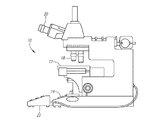
【選択図】図1
Description
Claims (20)
- 生物学的標本を観察する方法であって、当該方法が:
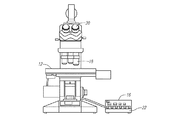
複数の独特の色彩群に配列された発光ダイオード(LED)を有する光源で、前記生物学的標本を照射するステップと;
前記照射された生物学的標本の拡大イメージを生成するステップと;
前記光源により照射される所望の光学特性を選択するステップと;
各々が前記選択された光学特性に基づいた特性を有している、複数の駆動信号を生成するステップと;
前記複数のLED色彩群に前記各々の複数の駆動信号を供給するステップと;
を具え、前記LED色彩群の光強度が、前記所望の光学特性をもたらすように個別に制御されることを特徴とする方法。 - 請求項1に記載の方法において、前記複数のLEDが単一基板上に配列されたLEDダイを具えることを特徴とする方法。
- 請求項1又は2に記載の方法において、前記所望の光学特性が色平衡を含むことを特徴とする方法。
- 請求項3に記載の方法において、前記所望の光学特性が光強度を更に含むことを特徴とする方法。
- 請求項1又は2に記載の方法において、前記所望の光学特性が手入力に応じて選択されることを特徴とする方法。
- 請求項1又は2に記載の方法において、前記所望の光学特性が自動入力に応じて選択されることを特徴とする方法。
- 請求項1又は2に記載の方法において、前記所望の光学特性が前記生物学的標本の観察条件に応じて選択されることを特徴とする方法。
- 請求項7に記載の方法において、前記観察条件が前記生物学的標本に用いられる染色のタイプであることを特徴とする方法。
- 請求項1乃至8のいずれか1項に記載の方法において、前記駆動信号の特性は振幅であることを特徴とする方法。
- 請求項1乃至8のいずれか1項に記載の方法において、前記駆動信号の特性はパルス幅であることを特徴とする方法。
- 請求項1乃至10のいずれか1項に記載の方法において、選択された光学特性に基づいて前記LED色彩群の所望の光強度を算出するステップと、前記算出された光強度に基づいて前記駆動信号の特性を決定するステップと、を具えることを特徴とする方法。
- 生物学的標本を観察するためのシステムであって、当該システムが:
前記生物学的標本の拡大イメージを生成するのに構成される顕微鏡と;
前記生物学的標本を照射するのに構成される光源であって、複数の独特の色彩群に配列された発光ダイオード(LED)を有する前記光源と;
前記光源により照射される光に所望の特性をもたらすための情報を受けるよう構成される入力デバイスと;
各々が前記入力デバイスから得られた前記情報に基づいた特性を有し、前記複数のLED色彩群に各々の複数の駆動信号を供給するように構成された制御回路と;
を具え、前記LED色彩群の光強度が前記所望の光学特性をもたらすように個別に制御されることを特徴とするシステム。 - 請求項12に記載のシステムにおいて、前記複数のLEDが単一基板上に配列されたLEDダイを具えることを特徴とするシステム。
- 請求項12又は13に記載のシステムにおいて、前記所望の光学特性が色平衡を含むことを特徴とするシステム。
- 請求項14に記載のシステムにおいて、前記所望の光学特性が光強度を更に含むことを特徴とするシステム。
- 請求項12乃至15のいずれか1項に記載のシステムにおいて、前記入力デバイスが、前記所望の光学特性を選択するために構成されたユーザ入力デバイスを具えることを特徴とするシステム。
- 請求項12乃至16のいずれか1項に記載のシステムが、前記所望の光学特性を算出するために構成されたプロセッサを更に具えることを特徴とするシステム。
- 請求項17に記載のシステムにおいて、前記プロセッサが観察条件に基づいて前記所望の光学特性を算出するために構成されることを特徴とするシステム。
- 請求項18に記載のシステムにおいて、前記観察条件が前記生物学的標本に用いられる染色のタイプであることを特徴とするシステム。
- 請求項12乃至19のいずれか1項に記載のシステムにおいて、前記駆動信号の特性が振幅であることを特徴とするシステム。
Applications Claiming Priority (2)
| Application Number | Priority Date | Filing Date | Title |
|---|---|---|---|
| US11/313,365 US7433026B2 (en) | 2005-12-20 | 2005-12-20 | Microscope with LED illumination source |
| PCT/US2006/061972 WO2007111735A2 (en) | 2005-12-20 | 2006-12-13 | Microscope with led illumination source |
Publications (2)
| Publication Number | Publication Date |
|---|---|
| JP2009520991A true JP2009520991A (ja) | 2009-05-28 |
| JP2009520991A5 JP2009520991A5 (ja) | 2010-01-28 |
Family
ID=38173033
Family Applications (1)
| Application Number | Title | Priority Date | Filing Date |
|---|---|---|---|
| JP2008547685A Pending JP2009520991A (ja) | 2005-12-20 | 2006-12-13 | Led照明源を有する顕微鏡 |
Country Status (11)
| Country | Link |
|---|---|
| US (1) | US7433026B2 (ja) |
| EP (1) | EP1971955A2 (ja) |
| JP (1) | JP2009520991A (ja) |
| KR (1) | KR101349664B1 (ja) |
| CN (1) | CN101341495B (ja) |
| AU (1) | AU2006340771B2 (ja) |
| BR (1) | BRPI0620052A2 (ja) |
| CA (1) | CA2631722C (ja) |
| IL (1) | IL192118A (ja) |
| TW (1) | TWI368030B (ja) |
| WO (1) | WO2007111735A2 (ja) |
Cited By (3)
| Publication number | Priority date | Publication date | Assignee | Title |
|---|---|---|---|---|
| WO2012029817A1 (ja) * | 2010-08-30 | 2012-03-08 | 三洋電機株式会社 | 観察装置、観察プログラム及び観察システム |
| JP2012048194A (ja) * | 2011-03-30 | 2012-03-08 | Sanyo Electric Co Ltd | 観察装置 |
| WO2012090416A1 (ja) * | 2010-12-28 | 2012-07-05 | オリンパス株式会社 | 検査装置 |
Families Citing this family (28)
| Publication number | Priority date | Publication date | Assignee | Title |
|---|---|---|---|---|
| ITMI20050019A1 (it) * | 2005-01-07 | 2006-07-08 | Fraen Corp Srl | Microscopio a fluorescenza in luce trasmessa e kit di adattamento di un microscopio alla modalita' di lavoro a fluorescenza in luce trasmessa |
| US7561329B2 (en) * | 2006-12-14 | 2009-07-14 | Cytyc Corporation | Illumination source for stained biological samples |
| US20090201577A1 (en) * | 2007-09-05 | 2009-08-13 | Chroma Technology Corporation | Light source |
| US7848019B2 (en) * | 2007-12-10 | 2010-12-07 | Cytyc Corporation | Microscope calibration apparatus and method and stage including calibration apparatus |
| ES2543721T3 (es) * | 2007-12-27 | 2015-08-21 | Cytyc Corporation | Aparato para el control con una sola mano de funciones de microscopio |
| WO2009085702A1 (en) * | 2007-12-27 | 2009-07-09 | Cytyc Corporation | Methods and systems for controlably scanning a cytological specimen |
| GB0809252D0 (en) | 2008-05-21 | 2008-06-25 | Ntnu Technology Transfer As | Underwater hyperspectral imaging |
| DE102009005839A1 (de) * | 2009-01-21 | 2010-07-22 | Carl Zeiss Surgical Gmbh | Lichtquelle für ein optisches Beobachtungsgerät |
| US9310598B2 (en) | 2009-03-11 | 2016-04-12 | Sakura Finetek U.S.A., Inc. | Autofocus method and autofocus device |
| JP5253309B2 (ja) * | 2009-07-02 | 2013-07-31 | オリンパス株式会社 | 顕微鏡システム、及び該制御方法 |
| US10139613B2 (en) | 2010-08-20 | 2018-11-27 | Sakura Finetek U.S.A., Inc. | Digital microscope and method of sensing an image of a tissue sample |
| WO2013074978A1 (en) | 2011-11-17 | 2013-05-23 | Datalogic ADC, Inc. | Systems and methods for reading color optical codes |
| JP5892594B2 (ja) * | 2012-01-24 | 2016-03-23 | 学校法人東京電機大学 | 複数点照明による培養細胞観察システム |
| DE102013103971A1 (de) | 2013-04-19 | 2014-11-06 | Sensovation Ag | Verfahren zum Erzeugen eines aus mehreren Teilbildern zusammengesetzten Gesamtbilds eines Objekts |
| DE102013006996A1 (de) | 2013-04-19 | 2014-10-23 | Carl Zeiss Microscopy Gmbh | Verfahren zur Beleuchtung eines Objektes in einem digitalen Lichtmikroskop, digitales Lichtmikroskop und Hellfeld-Auflichtbeleuchtungsvorrichtung für ein digitales Lichtmikroskop |
| WO2015004843A1 (ja) * | 2013-07-11 | 2015-01-15 | パナソニックIpマネジメント株式会社 | 画像計測装置及び画像計測方法 |
| US10007102B2 (en) | 2013-12-23 | 2018-06-26 | Sakura Finetek U.S.A., Inc. | Microscope with slide clamping assembly |
| JP6550262B2 (ja) * | 2015-05-08 | 2019-07-24 | オリンパス株式会社 | 顕微鏡システムおよび照明操作装置 |
| DE102015116488A1 (de) | 2015-09-29 | 2017-03-30 | Carl Zeiss Microscopy Gmbh | Mikroskop sowie Beleuchtungsvorrichtung und Beleuchtungssatz für ein Mikroskop |
| DE102016116621A1 (de) * | 2016-09-06 | 2019-04-18 | Stiftung Caesar Center Of Advanced European Studies And Research | LED-Beleuchtungsmodul für ein Mikroskop |
| DE102016015870A1 (de) | 2016-09-06 | 2019-04-04 | Stiftung Caesar Center Of Advanced European Studies And Research | LED-Beleuchtungsmodul für ein Mikroskop |
| US11280803B2 (en) | 2016-11-22 | 2022-03-22 | Sakura Finetek U.S.A., Inc. | Slide management system |
| US10268860B2 (en) * | 2016-12-22 | 2019-04-23 | Datalogic ADC, Inc. | White illumination for barcode scanners with improved power efficiency and cost |
| DE102017115658A1 (de) * | 2017-07-12 | 2019-01-17 | Carl Zeiss Microscopy Gmbh | Flackern bei Winkel-variabler Beleuchtung |
| CN108050429A (zh) * | 2017-12-12 | 2018-05-18 | 苏州科医世凯半导体技术有限责任公司 | 一种led多光谱共光路光源照明装置 |
| CN109060309B (zh) * | 2018-06-28 | 2024-04-02 | 广东工业大学 | 一种色差最优分辨配色仪及其测试方法 |
| CN115210753A (zh) * | 2020-05-13 | 2022-10-18 | 3M创新有限公司 | 用于菌落计数的图像中的强度差异补偿 |
| CN116482848B (zh) * | 2023-04-28 | 2023-10-24 | 广州市明美光电技术有限公司 | 一种用于光学仪器的匀光光源系统、方法及显微镜 |
Citations (8)
| Publication number | Priority date | Publication date | Assignee | Title |
|---|---|---|---|---|
| JP2001041882A (ja) * | 1999-07-28 | 2001-02-16 | Kubota Corp | 検卵装置 |
| JP2003513268A (ja) * | 1999-10-29 | 2003-04-08 | サイティック コーポレイション | 細胞学的撮像システムおよび方法 |
| JP2003337365A (ja) * | 2002-05-17 | 2003-11-28 | Mitsutoyo Corp | リング照明装置 |
| WO2004051283A2 (en) * | 2002-11-27 | 2004-06-17 | 3M Innovative Properties Company | Biological growth plate scanner |
| JP2005502060A (ja) * | 2001-09-05 | 2005-01-20 | ジェニコン サイエンスィズ コーポレーション | 共鳴光散乱粒子標識から発生する信号を読み取るための装置 |
| JP2005070021A (ja) * | 2003-08-28 | 2005-03-17 | Yokogawa Electric Corp | 検査用光源装置 |
| WO2005047833A1 (ja) * | 2003-11-14 | 2005-05-26 | Olympus Corporation | マルチスペクトル撮像装置、マルチスペクトル照明装置 |
| JP2005315856A (ja) * | 2004-03-31 | 2005-11-10 | Canon Inc | 記録媒体識別装置、記録装置および記録媒体識別方法 |
Family Cites Families (12)
| Publication number | Priority date | Publication date | Assignee | Title |
|---|---|---|---|---|
| JPH07122694B2 (ja) | 1986-10-16 | 1995-12-25 | オリンパス光学工業株式会社 | 顕微鏡用照明装置 |
| GB9703156D0 (en) * | 1997-02-15 | 1997-04-02 | Univ Strathclyde | Optical element |
| US6459919B1 (en) * | 1997-08-26 | 2002-10-01 | Color Kinetics, Incorporated | Precision illumination methods and systems |
| US6016038A (en) * | 1997-08-26 | 2000-01-18 | Color Kinetics, Inc. | Multicolored LED lighting method and apparatus |
| CN1292286A (zh) * | 1999-09-23 | 2001-04-25 | 上海第二医科大学附属瑞金医院 | 含木芙蓉叶提取物的药剂及制备方法 |
| US6665060B1 (en) * | 1999-10-29 | 2003-12-16 | Cytyc Corporation | Cytological imaging system and method |
| EP1150154B1 (de) | 2000-04-26 | 2003-03-26 | COBRA electronic GmbH | Anordnung und Verfahren zur ringförmigen Beleuchtung, insbesondere zur Auflichtbeleuchtung bei Mikroskopen |
| US6921920B2 (en) | 2001-08-31 | 2005-07-26 | Smith & Nephew, Inc. | Solid-state light source |
| US6683419B2 (en) * | 2002-06-24 | 2004-01-27 | Dialight Corporation | Electrical control for an LED light source, including dimming control |
| DE20304412U1 (de) * | 2003-03-19 | 2003-06-12 | Schott Glas | Steuereinheit für Mischlichtbeleuchtungen |
| JP2005017905A (ja) * | 2003-06-27 | 2005-01-20 | Olympus Corp | 実体顕微鏡 |
| DE10339618A1 (de) * | 2003-08-28 | 2005-03-24 | Leica Microsystems (Schweiz) Ag | Leuchtdioden-Beleuchtung für ein optisches Beobachtungsgerät, insbesondere ein Stereo- oder ein Stereooperationsmikroskop |
-
2005
- 2005-12-20 US US11/313,365 patent/US7433026B2/en active Active
-
2006
- 2006-12-13 TW TW095146714A patent/TWI368030B/zh active
- 2006-12-13 EP EP06850282A patent/EP1971955A2/en not_active Ceased
- 2006-12-13 WO PCT/US2006/061972 patent/WO2007111735A2/en active Application Filing
- 2006-12-13 KR KR1020087014812A patent/KR101349664B1/ko active IP Right Grant
- 2006-12-13 JP JP2008547685A patent/JP2009520991A/ja active Pending
- 2006-12-13 CN CN2006800479096A patent/CN101341495B/zh active Active
- 2006-12-13 BR BRPI0620052-4A patent/BRPI0620052A2/pt not_active Application Discontinuation
- 2006-12-13 AU AU2006340771A patent/AU2006340771B2/en active Active
- 2006-12-13 CA CA2631722A patent/CA2631722C/en active Active
-
2008
- 2008-06-12 IL IL192118A patent/IL192118A/en active IP Right Grant
Patent Citations (8)
| Publication number | Priority date | Publication date | Assignee | Title |
|---|---|---|---|---|
| JP2001041882A (ja) * | 1999-07-28 | 2001-02-16 | Kubota Corp | 検卵装置 |
| JP2003513268A (ja) * | 1999-10-29 | 2003-04-08 | サイティック コーポレイション | 細胞学的撮像システムおよび方法 |
| JP2005502060A (ja) * | 2001-09-05 | 2005-01-20 | ジェニコン サイエンスィズ コーポレーション | 共鳴光散乱粒子標識から発生する信号を読み取るための装置 |
| JP2003337365A (ja) * | 2002-05-17 | 2003-11-28 | Mitsutoyo Corp | リング照明装置 |
| WO2004051283A2 (en) * | 2002-11-27 | 2004-06-17 | 3M Innovative Properties Company | Biological growth plate scanner |
| JP2005070021A (ja) * | 2003-08-28 | 2005-03-17 | Yokogawa Electric Corp | 検査用光源装置 |
| WO2005047833A1 (ja) * | 2003-11-14 | 2005-05-26 | Olympus Corporation | マルチスペクトル撮像装置、マルチスペクトル照明装置 |
| JP2005315856A (ja) * | 2004-03-31 | 2005-11-10 | Canon Inc | 記録媒体識別装置、記録装置および記録媒体識別方法 |
Cited By (5)
| Publication number | Priority date | Publication date | Assignee | Title |
|---|---|---|---|---|
| WO2012029817A1 (ja) * | 2010-08-30 | 2012-03-08 | 三洋電機株式会社 | 観察装置、観察プログラム及び観察システム |
| US9060684B2 (en) | 2010-08-30 | 2015-06-23 | Panasonic Healthcare Holdings Co., Ltd. | Observation device, observation program, and observation system |
| US9578220B2 (en) | 2010-08-30 | 2017-02-21 | Panasonic Healthcare Holdings Co., Ltd. | Observation device, observation program, and observation system |
| WO2012090416A1 (ja) * | 2010-12-28 | 2012-07-05 | オリンパス株式会社 | 検査装置 |
| JP2012048194A (ja) * | 2011-03-30 | 2012-03-08 | Sanyo Electric Co Ltd | 観察装置 |
Also Published As
| Publication number | Publication date |
|---|---|
| KR101349664B1 (ko) | 2014-01-09 |
| TWI368030B (en) | 2012-07-11 |
| US20070139638A1 (en) | 2007-06-21 |
| BRPI0620052A2 (pt) | 2011-11-01 |
| IL192118A0 (en) | 2009-08-03 |
| CA2631722A1 (en) | 2007-10-04 |
| US7433026B2 (en) | 2008-10-07 |
| CN101341495A (zh) | 2009-01-07 |
| AU2006340771A1 (en) | 2007-10-04 |
| KR20080077988A (ko) | 2008-08-26 |
| IL192118A (en) | 2013-01-31 |
| TW200730870A (en) | 2007-08-16 |
| EP1971955A2 (en) | 2008-09-24 |
| WO2007111735A3 (en) | 2008-02-21 |
| CN101341495B (zh) | 2011-09-21 |
| CA2631722C (en) | 2013-07-30 |
| AU2006340771B2 (en) | 2011-04-14 |
| WO2007111735A2 (en) | 2007-10-04 |
Similar Documents
| Publication | Publication Date | Title |
|---|---|---|
| CA2631722C (en) | Microscope with led illumination source | |
| CN107137053B (zh) | 使用伪彩色的诸如显微镜或内窥镜的医疗检查装置 | |
| CN110269700B (zh) | 增强现实手术显微镜和显微镜学方法 | |
| US7411664B2 (en) | Cytological imaging system and method | |
| KR20160037834A (ko) | 의료 이미징 장치 및 사용 방법 | |
| US6348325B1 (en) | Cytological stain composition | |
| JP2010181833A (ja) | 顕微鏡観察システム | |
| CN111107778B (zh) | 医疗图像处理系统、内窥镜系统、诊断支持装置及医疗服务支持装置 | |
| IL254325B1 (en) | Method and device for microscopy | |
| US20190195777A1 (en) | Captured image evaluation apparatus, captured image evaluation method, and captured image evaluation program | |
| US6593102B2 (en) | Cytological stain composition | |
| JP5552298B2 (ja) | 細菌コロニ釣菌装置及びその前処理方法 | |
| CN111936031B (zh) | 医疗图像处理装置 | |
| CN112577905A (zh) | 一种尿液颜色检测方法及分析仪 | |
| KR20120012191A (ko) | 객체간 알지비 색상차이를 최대화하는 내시경용 엘이디 최적 조명장치 | |
| EP1238260B1 (en) | Method for verifying that a specific stain was used on a cytological sample | |
| CN207147955U (zh) | 大便有形成分分析装置 | |
| WO2022209262A1 (ja) | 生体試料観察装置用照明装置、生体試料観察装置、観察装置用照明装置及び観察システム | |
| WO2022209349A1 (ja) | 観察装置用照明装置、観察装置及び観察システム | |
| Bartczak et al. | Spectral video in image-guided microsurgical applications: Integrating imaging technology into the clinical environment and ergonomic considerations | |
| EP1226416B1 (en) | Cytological imaging system and method | |
| WO2022259647A1 (ja) | 情報処理装置、情報処理方法、及び顕微鏡システム | |
| JP6550341B2 (ja) | 病理標本の染色標準化方法 | |
| AU2005200507B2 (en) | Method for verifying that a specific stain was used on a cytological sample | |
| JP2000088746A (ja) | 病理診断用カラー画像輝度階調変換方法 |
Legal Events
| Date | Code | Title | Description |
|---|---|---|---|
| A521 | Request for written amendment filed |
Free format text: JAPANESE INTERMEDIATE CODE: A523 Effective date: 20091207 |
|
| A621 | Written request for application examination |
Free format text: JAPANESE INTERMEDIATE CODE: A621 Effective date: 20091207 |
|
| RD04 | Notification of resignation of power of attorney |
Free format text: JAPANESE INTERMEDIATE CODE: A7424 Effective date: 20100407 |
|
| A977 | Report on retrieval |
Free format text: JAPANESE INTERMEDIATE CODE: A971007 Effective date: 20120125 |
|
| A131 | Notification of reasons for refusal |
Free format text: JAPANESE INTERMEDIATE CODE: A131 Effective date: 20120131 |
|
| A601 | Written request for extension of time |
Free format text: JAPANESE INTERMEDIATE CODE: A601 Effective date: 20120501 |
|
| A602 | Written permission of extension of time |
Free format text: JAPANESE INTERMEDIATE CODE: A602 Effective date: 20120510 |
|
| A131 | Notification of reasons for refusal |
Free format text: JAPANESE INTERMEDIATE CODE: A131 Effective date: 20121204 |
|
| A601 | Written request for extension of time |
Free format text: JAPANESE INTERMEDIATE CODE: A601 Effective date: 20130228 |
|
| A602 | Written permission of extension of time |
Free format text: JAPANESE INTERMEDIATE CODE: A602 Effective date: 20130313 |
|
| A601 | Written request for extension of time |
Free format text: JAPANESE INTERMEDIATE CODE: A601 Effective date: 20130325 |
|
| A602 | Written permission of extension of time |
Free format text: JAPANESE INTERMEDIATE CODE: A602 Effective date: 20130405 |
|
| A02 | Decision of refusal |
Free format text: JAPANESE INTERMEDIATE CODE: A02 Effective date: 20131126 |