JP2009519041A - Probes and methods of use for nucleic acid sequencing - Google Patents
Probes and methods of use for nucleic acid sequencing Download PDFInfo
- Publication number
- JP2009519041A JP2009519041A JP2008545768A JP2008545768A JP2009519041A JP 2009519041 A JP2009519041 A JP 2009519041A JP 2008545768 A JP2008545768 A JP 2008545768A JP 2008545768 A JP2008545768 A JP 2008545768A JP 2009519041 A JP2009519041 A JP 2009519041A
- Authority
- JP
- Japan
- Prior art keywords
- nucleic acid
- probe
- acid molecule
- molecular
- linker
- Prior art date
- Legal status (The legal status is an assumption and is not a legal conclusion. Google has not performed a legal analysis and makes no representation as to the accuracy of the status listed.)
- Pending
Links
Images


Classifications
-
- G—PHYSICS
- G01—MEASURING; TESTING
- G01N—INVESTIGATING OR ANALYSING MATERIALS BY DETERMINING THEIR CHEMICAL OR PHYSICAL PROPERTIES
- G01N33/00—Investigating or analysing materials by specific methods not covered by groups G01N1/00 - G01N31/00
- G01N33/48—Biological material, e.g. blood, urine; Haemocytometers
- G01N33/50—Chemical analysis of biological material, e.g. blood, urine; Testing involving biospecific ligand binding methods; Immunological testing
- G01N33/53—Immunoassay; Biospecific binding assay; Materials therefor
- G01N33/536—Immunoassay; Biospecific binding assay; Materials therefor with immune complex formed in liquid phase
- G01N33/542—Immunoassay; Biospecific binding assay; Materials therefor with immune complex formed in liquid phase with steric inhibition or signal modification, e.g. fluorescent quenching
-
- C—CHEMISTRY; METALLURGY
- C12—BIOCHEMISTRY; BEER; SPIRITS; WINE; VINEGAR; MICROBIOLOGY; ENZYMOLOGY; MUTATION OR GENETIC ENGINEERING
- C12Q—MEASURING OR TESTING PROCESSES INVOLVING ENZYMES, NUCLEIC ACIDS OR MICROORGANISMS; COMPOSITIONS OR TEST PAPERS THEREFOR; PROCESSES OF PREPARING SUCH COMPOSITIONS; CONDITION-RESPONSIVE CONTROL IN MICROBIOLOGICAL OR ENZYMOLOGICAL PROCESSES
- C12Q1/00—Measuring or testing processes involving enzymes, nucleic acids or microorganisms; Compositions therefor; Processes of preparing such compositions
- C12Q1/68—Measuring or testing processes involving enzymes, nucleic acids or microorganisms; Compositions therefor; Processes of preparing such compositions involving nucleic acids
- C12Q1/6813—Hybridisation assays
- C12Q1/6816—Hybridisation assays characterised by the detection means
- C12Q1/6818—Hybridisation assays characterised by the detection means involving interaction of two or more labels, e.g. resonant energy transfer
-
- C—CHEMISTRY; METALLURGY
- C12—BIOCHEMISTRY; BEER; SPIRITS; WINE; VINEGAR; MICROBIOLOGY; ENZYMOLOGY; MUTATION OR GENETIC ENGINEERING
- C12Q—MEASURING OR TESTING PROCESSES INVOLVING ENZYMES, NUCLEIC ACIDS OR MICROORGANISMS; COMPOSITIONS OR TEST PAPERS THEREFOR; PROCESSES OF PREPARING SUCH COMPOSITIONS; CONDITION-RESPONSIVE CONTROL IN MICROBIOLOGICAL OR ENZYMOLOGICAL PROCESSES
- C12Q1/00—Measuring or testing processes involving enzymes, nucleic acids or microorganisms; Compositions therefor; Processes of preparing such compositions
- C12Q1/68—Measuring or testing processes involving enzymes, nucleic acids or microorganisms; Compositions therefor; Processes of preparing such compositions involving nucleic acids
- C12Q1/6869—Methods for sequencing
-
- Y—GENERAL TAGGING OF NEW TECHNOLOGICAL DEVELOPMENTS; GENERAL TAGGING OF CROSS-SECTIONAL TECHNOLOGIES SPANNING OVER SEVERAL SECTIONS OF THE IPC; TECHNICAL SUBJECTS COVERED BY FORMER USPC CROSS-REFERENCE ART COLLECTIONS [XRACs] AND DIGESTS
- Y10—TECHNICAL SUBJECTS COVERED BY FORMER USPC
- Y10T—TECHNICAL SUBJECTS COVERED BY FORMER US CLASSIFICATION
- Y10T436/00—Chemistry: analytical and immunological testing
- Y10T436/14—Heterocyclic carbon compound [i.e., O, S, N, Se, Te, as only ring hetero atom]
- Y10T436/142222—Hetero-O [e.g., ascorbic acid, etc.]
- Y10T436/143333—Saccharide [e.g., DNA, etc.]
Abstract
Description
関連出願に対する相互参照
本出願は、2005年12月12日に双方とも出願された米国仮出願第60/749,729号および第60/749,858号に対する優先権を主張し、参照により本明細書に組み入れられる。
CROSS REFERENCE TO RELATED APPLICATIONS This application claims priority to US Provisional Applications Nos. 60 / 749,729 and 60 / 749,858, both filed December 12, 2005, and is hereby incorporated by reference. Incorporated into the book.
分野
本開示は、プローブ、ならびにDNAおよびRNAなどの核酸分子を配列決定するための方法に関し、研究および臨床的用途における疾病の診断のために使用することができる。
FIELD This disclosure relates to probes and methods for sequencing nucleic acid molecules such as DNA and RNA, and can be used for disease diagnosis in research and clinical applications.
背景
多数の方法が核酸分子を配列決定するために使用されてきた。従来のマクサムギルバート化学分解法は、DNAの化学特異的開裂に関与する(Maxam and Gilbert,Proc.Natl.Acad.Sci,USA 74:560,1977(非特許文献1))。本方法において、放射能標識化DNA分子を、4つの別個の反応混合物内でインキュベートし、それぞれは、特異的識別性(G、A+G、CまたはC+T)の1つまたは2つのヌクレオチドでDNAを部分的に開裂する。得られたDNAフラグメントは、4つの反応のそれぞれを、ゲルの別個のレーンに分けた、ポリアクリルアミドゲル電気泳動によって分離される。DNA配列は、ゲルの4つのレーン内でフラグメントの高分子分離を観察することによって、オートラジオグラフィーの後に決定される。
Background A number of methods have been used to sequence nucleic acid molecules. The conventional Maxam Gilbert chemical degradation method involves chemical-specific cleavage of DNA (Maxam and Gilbert, Proc. Natl. Acad. Sci, USA 74: 560, 1977 (Non-patent Document 1)). In this method, radiolabeled DNA molecules are incubated in four separate reaction mixtures, each of which segments the DNA with one or two nucleotides of specific discrimination (G, A + G, C or C + T). Cleave. The resulting DNA fragments are separated by polyacrylamide gel electrophoresis, dividing each of the four reactions into separate lanes of the gel. The DNA sequence is determined after autoradiography by observing macromolecular separation of the fragments in the four lanes of the gel.
サンガージデオキシ法は、DNAポリメラーゼおよびデオキシヌクレオシド三リン酸およびジデオキシヌクレオシド三リン酸の混合物を用いた合成プライマーの酵素伸長によって、異なる長さのDNA分子の生成に関する(Sanger et al,Proc.Natl.Acad.Sci,USA74:5463,1977(非特許文献2))。反応は、ポリアクリルアミドゲル上の4つの平行なレーンに分離され、オートラジオグラフィーの後に配列が決定される。 The Sanger dideoxy method involves the production of DNA molecules of different lengths by enzymatic extension of synthetic primers using a mixture of DNA polymerase and deoxynucleoside triphosphates and dideoxynucleoside triphosphates (Sanger et al, Proc. Natl. Acad. Sci, USA 74: 5463, 1977 (non-patent document 2)). The reaction is separated into four parallel lanes on a polyacrylamide gel and sequenced after autoradiography.
蛍光ヌクレオチドを使用することにより、放射性ヌクレオチドの必要性がなくなり、DNA配列決定を自動化する手段を提供する(例えば、Ansorgeへの米国特許第5,124,247号(特許文献1)、Proberらへの米国特許第5,242,796号(特許文献2)、Proberらへの米国特許第5,306,618号(特許文献3)、Middendorfらへの米国特許第5,360,523号(非特許文献4)、Pettitへの米国特許第5,556,790号(特許文献5)、Smithへの米国特許第5,821,058号(特許文献6)を参照)。しかしながら、一般に、フルオロフォアを使用する方法は、ゲル電気泳動またはキャピラリー電気泳動を今もなお必要とし、従って緩慢であり巨視的である。 The use of fluorescent nucleotides eliminates the need for radioactive nucleotides and provides a means to automate DNA sequencing (eg, US Pat. No. 5,124,247 to Anserge, to Prober et al.). US Pat. No. 5,242,796 to US Pat. No. 5,306,618 to Probe et al., US Pat. No. 5,360,523 to Middendorf et al. US Pat. No. 5,556,790 to Pettit, US Pat. No. 5,821,058 to Smith, and US Pat. No. 5,821,058 to Smith. In general, however, methods using fluorophores still require gel electrophoresis or capillary electrophoresis and are therefore slow and macroscopic.
蛍光標識dNTPを使用するその他の潜在的障壁は、誰も蛍光標識DNA分子を完全には合成することができなかったことである。従って、相補的核酸鎖の合成を可能にする配列決定方法は、今もなお必要とされている。 Another potential barrier to using fluorescently labeled dNTPs is that no one was able to fully synthesize fluorescently labeled DNA molecules. Therefore, there is still a need for sequencing methods that allow the synthesis of complementary nucleic acid strands.
概要
本開示は、核酸分子の配列決定に使用できる改良型プローブ、および該プローブを使用する方法を提供する。特定の例において、プローブは、1つまたは複数の遺伝子の転写レベルを決定するために使用することができる。例えば、プローブは、個別のRNAの転写を計数し、それによって細胞内で生成された数の推定を提供するために使用することができる。特定の例において、本明細書に開示するプローブおよび方法は、現在利用可能なマイクロアレイ技術の代替物として使用される。
SUMMARY The present disclosure provides improved probes that can be used for sequencing nucleic acid molecules and methods of using the probes. In certain instances, the probe can be used to determine the transcription level of one or more genes. For example, a probe can be used to count transcription of individual RNAs, thereby providing an estimate of the number generated in the cell. In certain instances, the probes and methods disclosed herein are used as an alternative to currently available microarray technology.
特定の例において、「Medusa」と称されるプローブは、1つまたは複数の化学的部分(非加水分解性ヌクレオチドなど)を重合剤に連結する(一部の例では、離間して)、重合剤に結合した1つ以上の(複数など)分子リンカーを有する重合剤を含む。化学的部分は、リンカーから分離することなく、標的核酸分子内の相補的ヌクレオチドに特異的に結合することによって、核酸分子の鋳型鎖に可逆的に結合することができる。開示する例において、可逆的な組み込みは、重合剤の活性部位で発生する。しかしながら、化学的部分は、成長する核酸鎖に永久に組み込むことができないことが理想的である。標的核酸分子内の相補的ヌクレオチドとリンカー上の化学的部分との特異的な結合は、相補的ヌクレオチドとリンカー上の化学的部分との対合を示す特性シグナルの放出によって示される。 In certain instances, a probe referred to as “Medusa” links one or more chemical moieties (such as non-hydrolyzable nucleotides) to a polymerizing agent (in some cases, spaced apart). A polymerization agent having one or more (such as) molecular linkers attached to the agent. The chemical moiety can be reversibly bound to the template strand of the nucleic acid molecule by specifically binding to a complementary nucleotide in the target nucleic acid molecule without being separated from the linker. In the disclosed example, reversible incorporation occurs at the active site of the polymerizing agent. Ideally, however, the chemical moiety cannot be permanently incorporated into the growing nucleic acid strand. Specific binding between a complementary nucleotide in the target nucleic acid molecule and a chemical moiety on the linker is indicated by the emission of a characteristic signal indicative of the pairing of the complementary nucleotide and the chemical moiety on the linker.
重合剤は、配列決定される標的核酸分子に結合することができる活性部位を含み、一部の例において、標的核酸分子に相補的な核酸分子の合成を促進することができ、相補的核酸分子は、相補的ヌクレオチドが相補的核酸分子に組み込まれるように伸長する。重合剤は、線状鎖(相補的核酸分子など)を形成する化学反応で、モノマー分子(ヌクレオチドなど)を一緒に反応させることができる化合物を含む。例示的な重合剤には、DNAポリメラーゼ、RNAポリメラーゼ、および逆転写酵素が含まれるが、これらに限定されるものではない。特定の例において、ポリメラーゼは、GFP−ポリメラーゼである。重合剤の選択は、配列決定される核酸に依存しうる。例えば、標的核酸分子がDNAである場合は、重合剤は、DNAまたはRNAポリメラーゼであってもよいが、標的核酸分子がRNAである場合は、重合剤は、逆転写酵素であってもよい。 The polymerization agent includes an active site that can bind to the target nucleic acid molecule to be sequenced, and in some instances can facilitate the synthesis of a nucleic acid molecule that is complementary to the target nucleic acid molecule. Extends so that the complementary nucleotide is incorporated into the complementary nucleic acid molecule. Polymerizing agents include compounds that can react monomer molecules (such as nucleotides) together in a chemical reaction that forms a linear strand (such as complementary nucleic acid molecules). Exemplary polymerizing agents include, but are not limited to, DNA polymerase, RNA polymerase, and reverse transcriptase. In a particular example, the polymerase is GFP-polymerase. The choice of polymerizing agent can depend on the nucleic acid to be sequenced. For example, when the target nucleic acid molecule is DNA, the polymerization agent may be DNA or RNA polymerase, but when the target nucleic acid molecule is RNA, the polymerization agent may be reverse transcriptase.
リンカーから分離することなく、標的核酸分子の鋳型鎖内で相補的ヌクレオチドと可逆的に結合することができる化学的部分は、非加水分解性ヌクレオチド類似体などのヌクレオチド類似体を含むことができる。かかる類似体は、標的核酸分子内の相補的ヌクレオチドと対になることができるが、伸長している相補的核酸鎖に永久に組み込まれることはない。非加水分解性ヌクレオチド類似体は、当技術分野では公知であり、非加水分解性であるα−β結合を有する非加水分解性ヌクレオチド三リン酸類似体などの、非加水分解性ヌクレオチド三リン酸類似体を含む。 Chemical moieties that can reversibly bind to complementary nucleotides within the template strand of the target nucleic acid molecule without separation from the linker can include nucleotide analogs, such as non-hydrolyzable nucleotide analogs. Such analogs can pair with complementary nucleotides in the target nucleic acid molecule, but are not permanently incorporated into the extending complementary nucleic acid strand. Non-hydrolyzable nucleotide analogs are known in the art and include non-hydrolyzable nucleotide triphosphates, such as non-hydrolyzable nucleotide triphosphate analogs with α-β bonds that are non-hydrolyzable. Includes analogs.
特定の例において、プローブは、少なくとも4つの独立したリンカーを含み、それぞれは、標的核酸分子内の異なるヌクレオチドと特に対合することができるが、伸長している相補的核酸分子に永久に組み込むことができない、異なる化学的部分を有する。その他の例において、プローブは、分岐構造を形成するために結合される複数のリンカーを含み、それぞれの分岐は、標的核酸分子内の異なるヌクレオチドと特に対合することができるが、伸長している相補的核酸分子に永久に組み込まれることはできない、異なる化学的部分を有する。例えば、分岐構造は、1点でのみ重合剤に結合してもよい。 In certain instances, the probe comprises at least four independent linkers, each of which can specifically pair with a different nucleotide in the target nucleic acid molecule, but permanently incorporated into the extending complementary nucleic acid molecule. Have different chemical moieties. In other examples, the probe includes a plurality of linkers joined to form a branched structure, each branch being specifically paired with a different nucleotide in the target nucleic acid molecule, but extending. It has different chemical moieties that cannot be permanently incorporated into complementary nucleic acid molecules. For example, the branched structure may be bonded to the polymerization agent only at one point.
分子リンカーは、重合剤を核酸分子の鋳型(または標的)鎖に可逆的に結合することができる1つまたは複数の化学的部分に連結する。特定の例において、分子リンカーは、標的核酸分子の存在下で、重合剤と化学的部分との相互作用を可能にすると同時に、標的または鋳型核酸分子の非存在下で、重合剤と化学的部分との実質上の絡み合いを回避するように、重合剤および化学的部分を互いに十分な距離をおいて維持する。例えば、分子リンカーは、リンカーの絡み合いを抑制するために十分な距離をおいて重合剤の周囲で離間することができ、重合剤の活性部位に到達するほど十分に長い。一部の例において、分子リンカー(または少なくともその一部分)は、標的核酸分子の非存在下で、重合酵素と化学的部分との相互作用を減少させるように十分な剛性を有する。 Molecular linkers link the polymerizing agent to one or more chemical moieties that can reversibly bind to the template (or target) strand of the nucleic acid molecule. In certain instances, the molecular linker allows the interaction between the polymerizing agent and the chemical moiety in the presence of the target nucleic acid molecule, while simultaneously allowing the polymerizer and chemical moiety in the absence of the target or template nucleic acid molecule. The polymerizer and chemical moiety are maintained at a sufficient distance from each other so as to avoid substantial entanglement with the polymer. For example, the molecular linker can be spaced around the polymerizing agent at a sufficient distance to suppress entanglement of the linker and is long enough to reach the active site of the polymerizing agent. In some examples, the molecular linker (or at least a portion thereof) is sufficiently rigid to reduce the interaction between the polymerizing enzyme and the chemical moiety in the absence of the target nucleic acid molecule.
分子リンカー(または、分子リンカーの一部分である分子ロッドなど、その一部)は、標的核酸分子の非存在下で、望ましくない相互作用を回避するために、重合剤と化学的部分とを十分な距離をおいて離間する柔軟性を考慮して十分な長さを有するが、例えば、重合剤が標的核酸分子に結合する場合、重合剤と化学的部分とが互いに、および標的核酸分子と相互に作用しうる十分な柔軟性を保持する。例えば、分子リンカーの少なくとも一部は、分子リンカーの少なくとも一部が、標的核酸分子の非存在下で、重合剤(重合剤に関連するタグなど)と化学的部分との相互作用を減少させるような十分な剛性および長さとなることを可能し、標的核酸分子の存在下で、重合剤と化学的部分との相互作用を可能にする持続長を有することができる。 The molecular linker (or a portion thereof, such as a molecular rod that is part of the molecular linker) is sufficient to allow the polymerizing agent and the chemical moiety to avoid undesired interactions in the absence of the target nucleic acid molecule. It has a sufficient length in consideration of the flexibility to be spaced apart, but for example when the polymerizing agent binds to the target nucleic acid molecule, the polymerizing agent and the chemical moiety interact with each other and with the target nucleic acid molecule. Retain enough flexibility to work. For example, at least a portion of the molecular linker is such that at least a portion of the molecular linker reduces the interaction between the polymerizing agent (such as a tag associated with the polymerizing agent) and the chemical moiety in the absence of the target nucleic acid molecule. Sufficient rigidity and length, and can have a persistence length that allows the polymerizing agent to interact with the chemical moiety in the presence of the target nucleic acid molecule.
特定の例において、分子リンカーの全長は、二本鎖または一本鎖核酸分子などのリンカーを構成する1つまたは複数の成分の持続長と異なる(それより長いまたは短いなど)。しかしながら、特定の例において、分子リンカーの全長は、標的核酸分子の存在下で、標的核酸分子と同様に、重合剤と化学的部分との有意な相互作用を可能にすると同時に、有意な相互作用が、標的核酸分子の非存在下で、重合剤と化学的部分との間に発生する長さを超えることはない。かかる相互作用は、当技術分野で公知の方法を用いて、例えば、重合剤が、供与体フルオロフォアを含み、1つまたは複数の化学的部分が、FRET対の対応する受容体フルオロフォアを含む場合、受容体発光蛍光を測定することによって測定することができる。その他の例において、重合剤は、標的の非存在下で、化学的部分からフォスターの半径(22から90Åのフォスターの半径など)の少なくとも2倍の距離で実質上維持される。 In certain instances, the total length of the molecular linker is different (eg, longer or shorter) from the persistence length of one or more components that make up the linker, such as a double-stranded or single-stranded nucleic acid molecule. However, in certain instances, the full length of the molecular linker allows significant interaction between the polymerizing agent and the chemical moiety in the presence of the target nucleic acid molecule, as well as significant interaction between the polymerizing agent and the chemical moiety. Does not exceed the length generated between the polymerization agent and the chemical moiety in the absence of the target nucleic acid molecule. Such interactions can be accomplished using methods known in the art, for example, where the polymerizing agent includes a donor fluorophore and one or more chemical moieties include a corresponding acceptor fluorophore of a FRET pair. In this case, it can be measured by measuring the receptor emission fluorescence. In other examples, the polymerizing agent is substantially maintained at a distance of at least twice the foster radius (such as a foster radius of 22 to 90 mm) from the chemical moiety in the absence of the target.
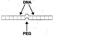
持続長(lp)は、線状鎖のための平均的局所立体構造であり、任意のセグメントによって画定される方向のすべての鎖セグメントの平均的延在の合計を反映する。従って、持続長は、ポリマー鎖の剛性または弾性の基準である。特定の例において、持続長は、実質的には、平均にして68.40度の屈曲で発生する周長距離を測定する屈曲度(従って、鎖の有効弾性)である。従って、持続長は、分子リンカーの組成物に応じて変化する。例えば、二本鎖DNA(dsDNA)分子のための持続長は、一本鎖DNA(ssDNA)分子およびポリエチレングリコール(PEG)のものと異なる。特定の例において、例えば、イオン強度が約0.2Mおよび温度が20℃で、dsDNAは、持続長が約400〜500Å(450〜500Åなど)であり、dsRNAは、持続長が約700〜750Åである。特定の例において、ssDNAは、持続長が約40Å(例えば20℃)である(Clossey and Carlon,Phys.Rev.E.Stat.Nonlin.Soft.Matter.Phys.68(6Ptl):061911,2003)。特定の例において、PEGは、持続長が約3.8Åである。 The persistence length (lp) is the average local conformation for a linear chain and reflects the sum of the average extension of all chain segments in the direction defined by any segment. Thus, the persistence length is a measure of the polymer chain stiffness or elasticity. In a particular example, the persistence length is essentially the degree of flexion (and hence the effective elasticity of the chain) that measures the perimeter distance that occurs with an average of 68.40 degrees of flexion. Thus, the persistence length varies depending on the composition of the molecular linker. For example, the persistence length for double-stranded DNA (dsDNA) molecules is different from that of single-stranded DNA (ssDNA) molecules and polyethylene glycol (PEG). In particular examples, for example, at an ionic strength of about 0.2 M and a temperature of 20 ° C., dsDNA has a persistence length of about 400-500 Å (such as 450-500 Å) and dsRNA has a persistence length of about 700-750 Å. It is. In a particular example, the ssDNA has a persistence length of about 40 mm (eg, 20 ° C.) (Clossey and Carlon, Phys. Rev. E. Stat. Nonlin. Soft. Matter. Phys. 68 (6Ptl): 06111, 2003) . In certain instances, PEG has a persistence length of about 3.8 cm.
特定の例において、分子リンカーには、核酸のポリマーなどの線状ポリマー、アミノ酸、糖、PEG、またはそれらの組み合わせが含まれる。例えば、分子リンカーには、テザー(tether)、分子ロッド、またはそれらの組み合わせが含まれるが、これらに限定されるものではない。例えば、十分な剛性の分子リンカーは、分子ロッド、例えば、dsDNAから成る分子ロッドを含むことができる。一部の例において、十分な剛性の分子リンカーには、テザーによって連結される複数の分子ロッド、または分子ロッドによって連結される複数のテザーが含まれる。テザーの特定の一例は、ポリエチレングリコール(PEG)から成る(または一部の例においては、構成される)分子である。 In particular examples, molecular linkers include linear polymers such as polymers of nucleic acids, amino acids, sugars, PEG, or combinations thereof. For example, molecular linkers include, but are not limited to, tethers, molecular rods, or combinations thereof. For example, a sufficiently rigid molecular linker can include a molecular rod, eg, a molecular rod consisting of dsDNA. In some examples, a sufficiently rigid molecular linker includes a plurality of molecular rods connected by tethers or a plurality of tethers connected by molecular rods. One particular example of a tether is a molecule that consists of (or in some instances is composed of) polyethylene glycol (PEG).
標的核酸分子の非存在下で、重合剤と化学的部分が相互作用して反応を提供しないように、重合剤および化学的部分を、1つまたは複数の分子リンカーによって空間的に分離した方向に連結することができる。しかしながら、分子リンカーは、重合剤および化学的部分に、所定の条件下で、相互に作用するために互いに十分に接近させ、検出可能なシグナルなどの所定の反応、または標的核酸分子との相互作用を引き起こすことを可能にする。例えば、重合剤または化学的部分に関連するタグのうちの少なくとも1つは、供与体および受容体が、互い十分に接近する場合、供与体フルオロフォアタグによる受容体フルオロフォアタグの励起など、別のタグに十分に接近する場合に活性化されうる。 In the absence of the target nucleic acid molecule, the polymerizer and chemical moiety are spatially separated by one or more molecular linkers so that the polymerizer and chemical moiety do not interact to provide a reaction. Can be linked. However, molecular linkers bring the polymerization agent and chemical moiety close enough to interact with each other under certain conditions, certain reactions such as detectable signals, or interactions with target nucleic acid molecules Makes it possible to cause For example, at least one of the tags associated with the polymerizing agent or chemical moiety may be different, such as excitation of the acceptor fluorophore tag by the donor fluorophore tag when the donor and acceptor are sufficiently close to each other. Can be activated if it is close enough to the tag.
さらに、本開示は、標的核酸分子に結合することができ、相補的ヌクレオチドが相補的核酸分子に組み込まれるように伸長する相補的核酸分子の合成を促進することができる活性部位を含む重合剤を提供する。重合剤は、絡み合いを抑制するために、重合剤上で離間する1つまたは複数の分子リンカーをさらに含み、それぞれのリンカーは、標的核酸分子内の相補的ヌクレオチドに特異的に結合することによって、リンカーから分離することなく、核酸分子の鋳型鎖に可逆的に結合することができる、異なる化学的部分(非加水分解性ヌクレオチド類似体など)を有する。特定の例において、重合剤は、リンカーが有する化学的部分を識別するそれぞれの化学的部分に関連するタグをさらに含む。さらに、ポリメラーゼは、リンカーが有する化学的部分を識別する特性シグナルを放出する化学的部分に関連するタグと相互に作用するタグに関連することができる。 Further, the present disclosure provides a polymerizing agent comprising an active site that can bind to a target nucleic acid molecule and facilitate the synthesis of a complementary nucleic acid molecule that extends such that complementary nucleotides are incorporated into the complementary nucleic acid molecule. provide. The polymerization agent further includes one or more molecular linkers spaced apart on the polymerization agent to suppress entanglement, each linker specifically binding to a complementary nucleotide in the target nucleic acid molecule, It has different chemical moieties (such as non-hydrolyzable nucleotide analogs) that can reversibly bind to the template strand of a nucleic acid molecule without separation from the linker. In certain instances, the polymerizing agent further includes a tag associated with each chemical moiety that identifies the chemical moiety possessed by the linker. In addition, the polymerase can be associated with a tag that interacts with a tag associated with the chemical moiety that emits a characteristic signal that identifies the chemical moiety possessed by the linker.
さらに、本開示は、例えば、標的核酸分子の核酸配列を決定するために、開示するナノプローブを使用する方法を提供する。特定の例において、該方法は、特定の標的分子が、試料内に存在するかを決定するために使用され、一部の例において、存在する標的核酸分子の量の定量化を含む。例えば、該方法は、1つまたは複数の核酸突然変異に関連する疾病を有する対象を診断するために、プローブを使用するために提供される。 Furthermore, the present disclosure provides methods of using the disclosed nanoprobes, for example, to determine the nucleic acid sequence of a target nucleic acid molecule. In certain examples, the method is used to determine whether a particular target molecule is present in a sample, and in some instances includes quantifying the amount of target nucleic acid molecule present. For example, the method is provided for using a probe to diagnose a subject having a disease associated with one or more nucleic acid mutations.
配列決定は、インビトロまたはインサイチュー(例えば、顕微鏡用スライド上)およびインビボ(例えば、プローブを細胞内に導入し、mRNAが生成される時にその配列を観察することによって)で行うことができる。該方法は、いくつかの核酸を分子レベルで同時に配列決定することを可能にする。例えば、複数の配列決定反応は、実質上同時に行うことができ、複数の配列決定反応からのシグナルを検出し、核酸配列に変換することができる。 Sequencing can be performed in vitro or in situ (eg, on a microscope slide) and in vivo (eg, by introducing a probe into a cell and observing the sequence when mRNA is generated). The method allows several nucleic acids to be sequenced simultaneously at the molecular level. For example, multiple sequencing reactions can be performed substantially simultaneously, and signals from multiple sequencing reactions can be detected and converted to nucleic acid sequences.
特定の例において、該方法は、オリゴヌクレオチドプライマー、および標的核酸分子内の相補的ヌクレオチドとの塩基対合に、伸長する核酸分子に組み込まれることができ、核酸分子の鋳型鎖に可逆的に結合するリンカーが有する化学的部分を置換することができる加水分解性ヌクレオチド(dATP、dCTP、dGTP、およびdTTPまたはATP、CTP、GTPおよびUTPなど)の混合物の存在下で、標的核酸分子を本明細書に開示するプローブに曝露する工程を含む。一連のシグナルの放出は検出され、シグナルは、相補的ヌクレオチドとのリンカー上の化学的部分の対合を示す複数の特性シグナルの放出を含む。一部の例において、一連のシグナルの放出は、核酸配列に変換される。 In certain instances, the method can be incorporated into an elongating nucleic acid molecule in a base pairing with an oligonucleotide primer and a complementary nucleotide in the target nucleic acid molecule and reversibly binds to the template strand of the nucleic acid molecule. The target nucleic acid molecule is described herein in the presence of a mixture of hydrolyzable nucleotides (such as dATP, dCTP, dGTP, and dTTP or ATP, CTP, GTP, and UTP) that can displace the chemical moiety of the linker Exposing to the probe disclosed in. The release of a series of signals is detected and the signal includes the release of multiple characteristic signals indicative of the pairing of chemical moieties on the linker with complementary nucleotides. In some examples, the release of a series of signals is converted into a nucleic acid sequence.
特定の例において、重合剤は、タグ(供与体フルオロフォアなど)に関連し、化学的部分(非加水分解性A、T/U、CまたはGヌクレオチド類似体など)のそれぞれの異なる型は、リンカーが有する特定の化学的部分を識別する一意的なタグに関連し、重合剤に関連するタグと、化学的部分に関連するタグとの相互作用は、相補的ヌクレオチドとリンカー上の化学的部分との対合を示す特性シグナルの放出を誘発する。特定の例において、タグは、重合剤または化学的部分に直接結合される。しかしながら、タグは、直接結合される必要はなく、代わりに、リンカー上の化学的部分が、相補的ヌクレオチドと対になる場合、特性シグナルの放出を生成するために、重合剤または化学的部分に十分に接近した分子リンカー上に見ることができる。 In certain instances, the polymerizing agent is associated with a tag (such as a donor fluorophore) and each different type of chemical moiety (such as a non-hydrolyzable A, T / U, C or G nucleotide analog) is: The link between the tag associated with the polymerizing agent and the tag associated with the chemical moiety is associated with a unique tag that identifies the specific chemical moiety possessed by the linker, and the chemical moiety on the linker Triggers the release of a characteristic signal indicative of pairing with. In certain instances, the tag is directly attached to the polymerizing agent or chemical moiety. However, the tag need not be directly attached; instead, when the chemical moiety on the linker is paired with a complementary nucleotide, it is attached to the polymerizer or chemical moiety to produce a release of a characteristic signal. It can be seen on a molecular linker close enough.
例えば、重合剤に関連するタグは、供与体フルオロフォアであってもよく、特定の化学的部分を識別するタグは、1つまたは複数の受容体フルオロフォアを含むことができ、重合剤と合成核酸分子に組み込むことができない化学的部分との相互作用は、供与体フルオロフォアによる受容体フルオロフォアの励起を可能にする供与体フルオロフォアに受容体フルオロフォアを接近させる。かかる例において、シグナルを検出する工程は、受容体フルオロフォアから放出された蛍光性シグナル(または供与体フルオロフォアからの減少された放出シグナル)を検出する工程を含むことができる。特定の例において、該方法は、受容体フルオロフォアではなく、供与体フルオロフォアを特に励起する電磁放射(レーザーなど)源によって、供与体フルオロフォアを励起する工程をさらに含む。あるいは、供与体フルオロフォアは、化学発光分子、例えば、エクオリンである。この例において、化学発光法による供与体フルオロフォアは、必然的に励起状態にあるので、供与体フルオロフォアは、電磁放射源による励起を必要としない。この励起は、標的核酸分子と対合する化学的部分に関連する受容体フルオロフォアを励起するのに十分な距離のみエネルギーを移動させることができる波長で、光を放出する供与体を含む。 For example, a tag associated with a polymerizing agent may be a donor fluorophore, and a tag that identifies a particular chemical moiety can include one or more acceptor fluorophores and is synthesized with the polymerizing agent. Interaction with a chemical moiety that cannot be incorporated into the nucleic acid molecule brings the acceptor fluorophore closer to the donor fluorophore that allows excitation of the acceptor fluorophore by the donor fluorophore. In such an example, detecting the signal can include detecting a fluorescent signal emitted from the acceptor fluorophore (or a reduced emission signal from the donor fluorophore). In certain instances, the method further includes exciting the donor fluorophore with a source of electromagnetic radiation (such as a laser) that specifically excites the donor fluorophore, rather than the acceptor fluorophore. Alternatively, the donor fluorophore is a chemiluminescent molecule, such as aequorin. In this example, the chemiluminescent donor fluorophore is necessarily in an excited state, so that the donor fluorophore does not require excitation by an electromagnetic radiation source. This excitation includes a donor that emits light at a wavelength that can transfer energy by a distance sufficient to excite the acceptor fluorophore associated with the chemical moiety that pairs with the target nucleic acid molecule.
特定の例において、プローブは、例えば、ポリメラーゼ成分を基板に結合するリンカー分子を介してアドレス可能な場所で基板に結合または固定される。例となるリンカーには、ストレプトアビジン−ビオチン、ヒスチジン−Ni、S−タグ−S−タンパク質、およびグルタチオン−グルタチオン−S−トランスフェラーゼ(GST)が含まれる。その他の例では、配列決定される標的核酸分子は、例えば、アドレス可能な場所で基板に結合または固定される。特定の例において、オリゴヌクレオチドプライマーは、例えば、5’末端で、基板に固定される。例えば、核酸分子は、5’末端、3’末端、または中間のどこかにより、基板に結合することができる。特定の例において、配列決定反応は、三次元ポリアクリルアミドゲルで行われ、配列決定に必要な試薬のすべては、ゲル内に存在する。 In certain instances, the probe is attached or immobilized to the substrate at an addressable location, for example, via a linker molecule that attaches the polymerase component to the substrate. Exemplary linkers include streptavidin-biotin, histidine-Ni, S-tag-S-protein, and glutathione-glutathione-S-transferase (GST). In other examples, the target nucleic acid molecule to be sequenced is bound or immobilized to a substrate, for example at an addressable location. In certain instances, the oligonucleotide primer is immobilized to the substrate, for example at the 5 'end. For example, the nucleic acid molecule can be bound to the substrate by its 5 'end, 3' end, or anywhere in between. In certain instances, the sequencing reaction is performed on a three-dimensional polyacrylamide gel and all of the reagents necessary for sequencing are present in the gel.
一部の例において、複数のプローブ、プライマー、または核酸分子は、所定のパターン、例えば、アドレス可能な場所で、基板に直接的または間接的に固定される。例えば、薬剤は、規則的なアレイに、またはスライド上に薬剤を含む液滴をマイクロピペット操作することによって、例えば、手動ピペット操作か、もしくは自動アレイヤーを使用し、エッチングされたチャネル内に蒸着することができる。かかる方法は、シグナルが、配列決定反応のそれぞれから検出される場合に、単一の基板上での同時の(または実質上同時の)配列決定を可能にする。一意的な放出シグナルは、例えば、電荷結合素子(CCD)カメラにより検出することができ、基板上の所定の位置から一連のシグナルを検出し、それらを核酸配列に変換することができる。一意的な放出シグナルは、コンピュータ可読媒体に保存することができる。 In some examples, the plurality of probes, primers, or nucleic acid molecules are immobilized directly or indirectly to the substrate in a predetermined pattern, eg, addressable location. For example, the drug is deposited in an etched channel, eg, by manual pipetting or by using an automatic arrayer, by micropipetting droplets containing the drug on a regular array or on a slide be able to. Such methods allow for simultaneous (or substantially simultaneous) sequencing on a single substrate when a signal is detected from each of the sequencing reactions. The unique emission signal can be detected, for example, by a charge coupled device (CCD) camera, which detects a series of signals from a predetermined location on the substrate and converts them into nucleic acid sequences. The unique emission signal can be stored on a computer readable medium.
本開示の前述のおよびその他の特性および利点は、添付図面への参照によって進められる、以下のいくつかの例の詳細な説明からより明白となるであろう。 The foregoing and other features and advantages of the present disclosure will become more apparent from the following detailed description of several examples, which proceeds with reference to the accompanying drawings.
配列表
ヌクレオチド塩基の標準略語を使用して、添付の配列表に記載の核酸配列を示す。特定の例において、核酸配列の鎖を1つのみ示すが、相補的鎖は、示された鎖への任意の言及によって含まれると理解される(例えば、dsDNA分子ロッドの場合)。
Sequence Listing Nucleotide sequences listed in the attached Sequence Listing are shown using standard nucleotide base abbreviations. In particular examples, only one strand of the nucleic acid sequence is shown, but it is understood that the complementary strand is included by any reference to the indicated strand (eg in the case of a dsDNA molecular rod).
SEQ ID NO:1は、例示的な標的配列である。
SEQ ID NO:2は、SEQ ID NO:1の圧縮版である。
SEQ ID NO:3〜26は、図2Cに示すプローブを作成するために使用することができる配列である。
SEQ ID NO:27〜30は、SEQ ID NO:3、5、7、および9とそれぞれ置換することができる配列である。
SEQ ID NO:31〜38は、ヘアピンループを形成することができる配列である。
SEQ ID NO:39は、例示的な標的配列である。
SEQ ID NO: 1 is an exemplary target sequence.
SEQ ID NO: 2 is a compressed version of SEQ ID NO: 1.
SEQ ID NOs: 3-26 are sequences that can be used to make the probe shown in FIG. 2C.
SEQ ID NOs: 27-30 are sequences that can be substituted for SEQ ID NOs: 3, 5, 7, and 9, respectively.
SEQ ID NOs: 31 to 38 are sequences that can form a hairpin loop.
SEQ ID NO: 39 is an exemplary target sequence.
一部の態様の詳細な説明
略語および用語
本開示をより適切に説明し、本開示を実践する上で当業者の指針となるように、用語および方法の説明を以下に提供する。本明細書で使用される限り、「含む」とは、「包含する」を意味し、単数形「1つの(aまたはan)」または「その(the)」は、文脈による別段の明確な指示がない限り、複数形の引例を含む。例えば、「1つの分子リンカー」に対する引例は、1つまたは複数のかかる分子リンカーを含み、「そのプローブ」に対する引例は、1つまたは複数のプローブおよび当業者には公知のその同等物などに対する引例を含む。用語「or」とは、文脈による別段の明確な指示がない限り、記載する代替要素の単一の要素、または2つ以上の要素の組み合わせを指す。例えば、句「1つのテザーまたは1つの分子ロッド」とは、1つまたは複数のテザー、1つまたは複数の分子ロッド、または1つまたは複数のテザーと1つまたは複数の分子ロッドの両方の組み合わせを指す。
Detailed Description Abbreviations and Terminology for Some Aspects The following descriptions of terms and methods are provided to better describe the present disclosure and to guide those of ordinary skill in the art in practicing the present disclosure. As used herein, “comprising” means “including” and the singular “a” or “the” is an explicit indication by the context. Plural references are included unless otherwise noted. For example, references to “a molecular linker” include one or more such molecular linkers, references to “the probe” include references to one or more probes and equivalents known to those skilled in the art, and the like. including. The term “or” refers to a single element of stated alternative elements or a combination of two or more elements, unless the context clearly indicates otherwise. For example, the phrase “one tether or one molecular rod” refers to one or more tethers, one or more molecular rods, or a combination of one or more tethers and one or more molecular rods. Point to.
別段の説明がない限り、本明細書に使用されるすべての技術用語および科学用語は、本開示が属する当業者に一般に理解されるものと同様の意味を有する。本明細書に記載のものと類似または同等の方法および材料は、本開示の実践または検証において使用することができるが、適切な方法および材料を以下に記載する。材料、方法、および例は、一例にすぎず、限定することを意図しない。開示のその他の特性および利点は、以下の詳細な説明および特許請求の範囲から明白である。
Å オングストローム
dsDNA 二本鎖DNA
FRET 蛍光共鳴エネルギー移動
GFP 緑色蛍光タンパク質
LNA ロックト核酸
PEG ポリエチレングリコール
PNA ペプチド核酸
RT 逆転写酵素
ssDNA 一本鎖DNA
Unless defined otherwise, all technical and scientific terms used herein have the same meaning as commonly understood by one of ordinary skill in the art to which this disclosure belongs. Although methods and materials similar or equivalent to those described herein can be used in the practice or verification of the present disclosure, suitable methods and materials are described below. The materials, methods, and examples are illustrative only and not intended to be limiting. Other features and advantages of the disclosure will be apparent from the following detailed description and from the claims.
Å Angstrom dsDNA double-stranded DNA
FRET fluorescence resonance energy transfer GFP green fluorescent protein LNA locked nucleic acid PEG polyethylene glycol PNA peptide nucleic acid RT reverse transcriptase ssDNA single-stranded DNA
受容体フルオロフォア:例えば、約400から900nmの範囲内(約500から800nmの範囲内など)で、供与体フルオロフォアからエネルギーを吸収する化合物。一般に、受容体フルオロフォアは、通常、供与体フルオロフォアの最大吸収波長よりも少なくとも10nm長い(少なくとも20nm長いなど)波長で光を吸収し、約400から900nmに及ぶ波長で最大限の蛍光放射を有する。供与体によって放射されるエネルギーが、受容体を励起することができるように、受容体フルオロフォアは、供与体フルオロフォアの放出と重なる励起スペクトルを有する。理想的には、受容体フルオロフォアは、開示するナノプローブに結合されることができる。 Acceptor fluorophore: A compound that absorbs energy from a donor fluorophore, for example, in the range of about 400 to 900 nm, such as in the range of about 500 to 800 nm. In general, the acceptor fluorophore typically absorbs light at a wavelength that is at least 10 nm longer (eg, at least 20 nm longer) than the maximum absorption wavelength of the donor fluorophore, and maximizes fluorescence emission at wavelengths ranging from about 400 to 900 nm. Have. The acceptor fluorophore has an excitation spectrum that overlaps with the emission of the donor fluorophore so that the energy emitted by the donor can excite the acceptor. Ideally, the receptor fluorophore can be bound to the disclosed nanoprobe.
例示的受容体フルオロフォアには、ローダミンおよびその誘導体(N,N,N’,N’−テトラメチル−6−カルボキシローダミン(TAMRA)、6−カルボキシ−X−ローダミン(ROX)など)、フルオレセイン誘導体(5−カルボキシフルオレセイン(FAM)および2’7’−ジメトキシ−4’5’−ジクロロ−6−カルボキシフルオレセイン(JOE)など)、緑色蛍光タンパク質(GFP)、BODIPY(4,4−ジフルオロ−4−ボラ−3a,4a−ジアザ−s−インダセン)およびシアニン染料が含まれるが、これらに限定されるものではない。特定の例において、受容体フルオロフォアは、塩基などのヌクレオチド類似体、糖、またはヌクレオチドのリン酸(α、β、またはγ)に結合されうる。 Exemplary receptor fluorophores include rhodamine and its derivatives (N, N, N ′, N′-tetramethyl-6-carboxyrhodamine (TAMRA), 6-carboxy-X-rhodamine (ROX), etc.), fluorescein derivatives (Such as 5-carboxyfluorescein (FAM) and 2′7′-dimethoxy-4′5′-dichloro-6-carboxyfluorescein (JOE)), green fluorescent protein (GFP), BODIPY (4,4-difluoro-4- Bora-3a, 4a-diaza-s-indacene) and cyanine dyes, but are not limited to these. In certain instances, the receptor fluorophore can be bound to a nucleotide analog such as a base, a sugar, or a phosphate of a nucleotide (α, β, or γ).
特定の例において、受容体フルオロフォアは、Dabcyl、Glen ResearchからのBlack Hole Quenchers(商標)、Epoch BiosciencesからのEclipse(商標)Dark Quencher、Integrated DNA TechnologiesからのIowa Black(商標)などのダーククエンチャーである。かかる例において、供与体フルオロフォアに十分に接近する場合に受容体フルオロフォアからの放出シグナルの増加を検出する代わりに、クエンチャーに十分に接近する場合に、供与体フルオロフォアからの放出シグナルの減少を検出することができる。 In particular examples, the receptor fluorophore is a trademark of Dabcyl, Black Hole Quenchers ™ from Glen Research, Eclipse ™ Dark Quencher from Epoch Biosciences, a queuing from Integrated DNA TechnologiesBlow, a trademark from Integrated DNA Technologies B It is. In such an example, instead of detecting an increase in the emission signal from the acceptor fluorophore when sufficiently close to the donor fluorophore, the release signal from the donor fluorophore when close enough to the quencher is detected. A decrease can be detected.
活性部位:化学反応が発生するポリメラーゼの領域などの、酵素または抗体の触媒部位。活性部位は、基質との相互作用を可能にする空間的配置に1つまたは複数の残基または原子を含み、後者の反応を発生させる。 Active site: The catalytic site of an enzyme or antibody, such as the region of a polymerase where a chemical reaction occurs. The active site contains one or more residues or atoms in a spatial arrangement that allows interaction with the substrate, causing the latter reaction.
結合:複合体の形成など、2つ以上の分子間の連関。一般に、複合体内の分子の結合が強力であるほど、解離の速度は遅くなる。特異的結合とは、薬剤と標的との間の選択的結合を指す。 Binding: An association between two or more molecules, such as the formation of a complex. In general, the stronger the binding of molecules in a complex, the slower the rate of dissociation. Specific binding refers to selective binding between an agent and a target.
特異的結合の特定の例には、相補的核酸分子への1つの核酸分子のハイブリダイゼーション、タンパク質(ポリメラーゼなど)と標的タンパク質または核酸分子との連関が含まれるが、これらに限定されない。 Specific examples of specific binding include, but are not limited to, the hybridization of one nucleic acid molecule to a complementary nucleic acid molecule, the association of a protein (such as a polymerase) with a target protein or nucleic acid molecule.
特定の例において、十分な量、例えば、その結合の検出を可能にする十分な量のタンパク質が核酸分子への非共有化学結合を形成する場合は、タンパク質は、核酸分子に結合することが知られている。 In certain instances, a protein is known to bind to a nucleic acid molecule if a sufficient amount, eg, a sufficient amount to allow detection of the binding, forms a non-covalent chemical bond to the nucleic acid molecule. It has been.
一例において、その結合の検出を可能にする十分な量のオリゴヌクレオチド分子が、塩基対を形成する、または標的核酸分子にハイブリダイズされる場合は、オリゴヌクレオチド分子(プライマーなど)は、標的核酸分子に結合することが観察される。多くの場合、オリゴヌクレオチドとその標的核酸分子との間の結合は、50%のオリゴヌクレオチドが標的から融解する温度(Tm)に特徴付けられる。高温(Tm)は、低温(Tm)の複合体と比較してより強力または安定した複合体を意味する。 In one example, if a sufficient amount of an oligonucleotide molecule that allows detection of its binding to base pair or hybridize to a target nucleic acid molecule, the oligonucleotide molecule (such as a primer) Is observed to bind to. In many cases, the binding between an oligonucleotide and its target nucleic acid molecule is characterized by the temperature (T m ) at which 50% of the oligonucleotide melts from the target. High temperature (T m ) means a stronger or more stable complex compared to a low temperature (T m ) complex.
特定の例において、結合は、ナノプローブ上に存在する標識を検出することによって評価される。例えば、供与体と受容体フルオロフォアとの相互作用に続いて生成される蛍光性シグナルは、ナノプローブ上のヌクレオチド類似体と標的核酸分子内の相補的ヌクレオチドとの結合の指標として測定することができる。 In certain instances, binding is assessed by detecting a label present on the nanoprobe. For example, the fluorescent signal generated following the interaction between the donor and acceptor fluorophore can be measured as an indication of the binding of the nucleotide analog on the nanoprobe to a complementary nucleotide in the target nucleic acid molecule. it can.
化学的部分:分子の部分基または官能基。例は、標的核酸分子内の相補的ヌクレオチドに特異的に結合することによって、標的核酸分子の鋳型鎖に可逆的に結合することができる、ヌクレオチドなどの薬剤を含む。特定の例において、化学的部分は、分子リンカーを介してプローブに結合され、化学的部分が、標的核酸分子上の相補的ヌクレオチドに特異的に結合する場合、リンカーから分離しない。 Chemical moiety: a partial or functional group of a molecule. Examples include agents such as nucleotides that can reversibly bind to the template strand of a target nucleic acid molecule by specifically binding to complementary nucleotides within the target nucleic acid molecule. In certain instances, the chemical moiety is attached to the probe via a molecular linker, and does not separate from the linker when the chemical moiety specifically binds to a complementary nucleotide on the target nucleic acid molecule.
化学的部分の特定の例には、非加水分解性ヌクレオチド類似体など、成長する相補的核酸鎖に組み込むことができないヌクレオチド類似体を含むが、これに限定されない。 Specific examples of chemical moieties include, but are not limited to, nucleotide analogs that cannot be incorporated into a growing complementary nucleic acid strand, such as non-hydrolyzable nucleotide analogs.
cDNA(相補的DNA):内部、非コードセグメント(イントロン)、および転写を決定する調節配列を欠くDNAの一片。cDNAは、mRNAに相補的であり、逆転写酵素を使用して合成することができる。 cDNA (complementary DNA): A piece of DNA lacking internal, non-coding segments (introns) and regulatory sequences that determine transcription. cDNA is complementary to mRNA and can be synthesized using reverse transcriptase.
相補的:二本鎖DNAまたはRNA鎖は、塩基対の2つの相補的鎖から成る。DNA/RNAに見られるそれぞれの塩基に対して1つの相補的塩基(A/T、およびC/Gなど)があるので、任意の単一鎖に対する相補的鎖を決定することができる。 Complementary: A double-stranded DNA or RNA strand consists of two complementary strands of base pairs. Since there is one complementary base (such as A / T and C / G) for each base found in DNA / RNA, the complementary strand for any single strand can be determined.
検出:薬剤が存在するか、または存在しないかを決定すること。一部の例において、これは、定量化をさらに含むことができる。例えば、特定の例で開示するプローブを使用すると、例えば、化学的部分が、リンカーから分離されることなく、標的核酸分子内の相補的ヌクレオチドに結合するので、化学的部分の検出が可能になる。 Detection: Determining whether a drug is present or absent. In some examples, this can further include quantification. For example, using the probes disclosed in certain examples allows detection of a chemical moiety, for example, because the chemical moiety binds to a complementary nucleotide in the target nucleic acid molecule without being separated from the linker. .
巨視的な数の分子(少なくとも1023の分子など)が、同時に観察することができるように、検出は大量であってもよい。さらに、検出は、顕微鏡法およびバックグラウンドノイズを減少させる全反射などの技術を使用して、単一の分子からのシグナルの識別を含むことができる。個別の分子のスペクトルは、これらの技術によって取得することができる(Ha et al,Proc.Natl.Acad.Sci.USA.93:6264−8,1996)。 Macroscopic number of molecules (such as at least 10 23 molecules), as can be observed simultaneously, the detection may be mass. Furthermore, detection can include discrimination of the signal from a single molecule using techniques such as microscopy and total reflection that reduces background noise. Spectra of individual molecules can be obtained by these techniques (Ha et al, Proc. Natl. Acad. Sci. USA. 93: 6264-8, 1996).
供与体フルオロフォア:エネルギーを受容体フルオロフォアへ移動し、それによって検出可能な蛍光性シグナルを生成することができるフルオロフォアまたは発光分子。一般に、供与体フルオロフォアは、約300から900nm、例えば、約350から800nmの範囲で吸収する化合物である。供与体フルオロフォアは、望ましい励起波長、例えば、約103M−1cm−1超で、強いモル吸光係数を有する。種々の化合物は、フルオレセイン(およびその誘導体)、ローダミン(およびその誘導体)、GFP、フィコエリトリン、BODIPY、DAPI(4’,6−ジアミジノ−2−フェニルインドール)、インド−1、クマリン、ダンシル、テルビウム(およびその誘導体)、およびシアニン染料を含む、供与体蛍光成分として用いることができる。特定の例において、供与体フルオロフォアは、エクオリンなどの化学発光分子である。 Donor fluorophore: A fluorophore or luminescent molecule that can transfer energy to an acceptor fluorophore, thereby producing a detectable fluorescent signal. Generally, the donor fluorophore is a compound that absorbs in the range of about 300 to 900 nm, such as about 350 to 800 nm. The donor fluorophore has a strong molar extinction coefficient at the desired excitation wavelength, eg, greater than about 10 3 M −1 cm −1 . Various compounds include fluorescein (and derivatives thereof), rhodamine (and derivatives thereof), GFP, phycoerythrin, BODIPY, DAPI (4 ′, 6-diamidino-2-phenylindole), indo-1, coumarin, dansyl, terbium ( And its derivatives), and cyanine dyes can be used as donor fluorescent components. In certain instances, the donor fluorophore is a chemiluminescent molecule such as aequorin.
電磁放射:電界強度および磁場強度の同時周期的変動によって伝播され、電波、赤外線、可視光線、紫外線、X線およびガンマ線を含む一連の電磁波。特定の例において、電磁放射は、単色性、方向性、コヒーレンス、偏光および強度の性質を保有することができるレーザーによって放射される。レーザーからのエネルギーが、供与体フルオロフォアを励起するが、受容体フルオロフォアは励起しないように、レーザーは、特定の波長(または波長の比較的狭い範囲にわたり)で光を放射することができる。 Electromagnetic radiation: A series of electromagnetic waves that are propagated by simultaneous periodic variations in electric and magnetic field strength and include radio waves, infrared, visible light, ultraviolet light, X-rays and gamma rays. In certain instances, electromagnetic radiation is emitted by a laser that can possess the properties of monochromaticity, directionality, coherence, polarization and intensity. The laser can emit light at a particular wavelength (or over a relatively narrow range of wavelengths) so that energy from the laser excites the donor fluorophore but not the acceptor fluorophore.
放出シグナル:フルオロフォアが、励起波長で光を吸収した後、フルオロフォアから生成される特定の波長の光。 Emission signal: A specific wavelength of light generated from a fluorophore after it absorbs light at an excitation wavelength.
放出スペクトル:フルオロフォアの後に発生するエネルギースペクトルは、光の特定の波長によって励起される。それぞれのフルオロフォアは、特徴のある放出スペクトルを有する。一例において、個別のフルオロフォア(またはフルオロフォアの一意的な組み合わせ)は、ヌクレオチド類似体に関連し、フルオロフォアからの放出スペクトルは、異なるヌクレオチド類似体間を区別するための手段を提供する。 Emission spectrum: The energy spectrum generated after the fluorophore is excited by a specific wavelength of light. Each fluorophore has a characteristic emission spectrum. In one example, individual fluorophores (or unique combinations of fluorophores) are associated with nucleotide analogs, and the emission spectrum from the fluorophore provides a means to distinguish between different nucleotide analogs.
絡み合う:例えば、複雑に入り組んだ塊において、絡み合うこと。特定の例において、ナノプローブの絡み合いは、標的分子の存在下で、化学的部分(ヌクレオチド類似体など)が、標的核酸分子の相補的ヌクレオチドと相互に作用することを減少または阻止する。その他の特定の例において、ナノプローブの絡み合いは、化学的部分(ヌクレオチド類似体など)間、または化学的部分と重合剤間との望ましくない相互作用、例えば、標的核酸分子との相互作用を阻止する相互作用という結果になる。 Intertwined: For example, entangled in a complex intricate mass. In certain instances, nanoprobe entanglement reduces or prevents chemical moieties (such as nucleotide analogs) from interacting with complementary nucleotides of the target nucleic acid molecule in the presence of the target molecule. In other specific examples, nanoprobe entanglement prevents undesired interactions between chemical moieties (such as nucleotide analogs), or between chemical moieties and polymerizing agents, such as interactions with target nucleic acid molecules. Result in interaction.
励起または励起シグナル:フルオロフォアが、光の異なる(より長いなど)波長を放出する状態までフルオロフォアを励起することが必要な特定の波長の光。 Excitation or excitation signal: light of a particular wavelength that requires the fluorophore to be excited to a state where the fluorophore emits a different (e.g., longer) wavelength of light.
フルオロフォア:光の定義済み波長など、特定の刺激に曝露することによって励起される場合、例えば、異なる波長で、光を放出する(蛍光を発する)化合物。 Fluorophore: A compound that emits light (fluoresces), for example, at a different wavelength when excited by exposure to a specific stimulus, such as a defined wavelength of light.
フルオロフォアは、発光化合物の比較的大きい部類の一部である。発光化合物は、冷光を発する光の特定の波長を必要とせず、エネルギーの化学源を使用する化学発光分子を含む。従って、化学発光分子を使用すると、レーザーなどの、外部の電磁放射源の必要性がなくなる。化学発光分子の例には、エクオリンが含まれるが、これに限定されるものではない(Tsien,1998,Ann.Rev.Biochem.67:509)。 Fluorophores are part of a relatively large class of luminescent compounds. Luminescent compounds include chemiluminescent molecules that do not require a specific wavelength of light that emits cold light and that use a chemical source of energy. Thus, the use of chemiluminescent molecules eliminates the need for external electromagnetic radiation sources such as lasers. Examples of chemiluminescent molecules include, but are not limited to, aequorin (Tsien, 1998, Ann. Rev. Biochem. 67: 509).
本明細書に開示するナノプローブに使用することができる特定のフルオロフォアの例は、4−アセトアミド−4’−イソチオシアナトスチルベン−2,2’ジスルホン酸、アクリジンおよびアクリジンイソチオシアネートなどのアクリジンおよび誘導体、5−(2’−アミノエチル)アミノナフタレン−l−スルホン酸(EDANS)、4−アミノ−N−[3−ビニルスルホニル)フェニル]ナフタルイミド−3,5二硫酸塩(ルシファーイエローVS)、N−(4−アニリノ−l−ナフチル)マレイミド、アントラニルアミド、ブリリアントイエロー、クマリン、7−アミノ−4−メチルクマリン(AMC、クマリン120)などのクマリンおよび誘導体、7−アミノ−4−トリフルオロメチルコウルアリン(クマラン151);シアノシン;4’,6−ジアミニジノ−2−フェニルインドール(DAPI);5’,5”−ジブロモピロガロール−スルホンフタレイン(ブロモピロガロールレッド);7−ジエチルアミノ−3−(4’−イソチオシアナトフェニル)−4−メチルクマリン;ジエチレントリアミンペンタアセテート;4,4’−ジイソチオシアナトジヒドロ−スチルベン−2,2’−ジスルホン酸;4,4’−ジイソチオシアナトスチルベン−2,2’−ジスルホン酸;5−[ジメチルアミノ]ナフタレン−l−塩化スルホニル(DNS、ダンシルクロライド);4−ジメチルアミノフェニルアゾフェニル−4’−イソチオシアネート(DABITC);エオシンおよびエオシンイソチオシアネートなどのエオシンおよび誘導体;エリスロシンBおよびエリスロシンイソチオシアネートなどのエリスロシンおよび誘導体;エチジウム;5−カルボキシフルオレセイン(FAM)、5−(4,6−ジクロロトリアジン−2−イル)アミノフルオレセイン(DTAF)、2’7’−ジメトキシ−4’5’−ジクロロ−6−カルボキシフルオレセイン(JOE)、フルオレセイン、フルオレセインイソチオシアネート(FITC)、およびQFITC(XRITC)などのフルオレセインおよび誘導体;フルオレスカミン;IR144;IR1446;マラカイトグリーンイソチオシアネート;4−メチルウンベリフェロン;オルトクレゾールフタレイン;ニトロチロシン;パラローザニリン;フェノールレッド;B−フィコエリトリン;o−フタルジアルデヒド;ピレン、ピレンブチラートおよびスクシンイミジル1−ピレンブチラートなどのピレンおよび誘導体;リアクティブレッド4(チバクロン-RTM、ブリリアントレッド3B−A);6−カルボキシ−X−ローダミン(ROX)、6−カルボキシローダミン(R6G)、リサミンローダミンB塩化スルホニル、ローダミン(Rhod)、ローダミンB、ローダミン123、ローダミンXイソチオシアネート、スルホローダミンB、スルホローダミン101およびスルホローダミン101(テキサスレッド)の塩化スルホニル誘導体などのローダミンおよび誘導体;N,N,N’,N’−テトラメチル−6−カルボキシローダミン(TAMRA);テトラメチルローダミン;テトラメチルローダミンイソチオシアネート(TRITC);リボフラビン;ロゾール酸およびテルビウムキレート誘導体など、Nazarenkoらへの米国特許第5,866,366号に記載されている。 Examples of specific fluorophores that can be used in the nanoprobes disclosed herein include acridine such as 4-acetamido-4′-isothiocyanatostilbene-2,2′disulfonic acid, acridine and acridine isothiocyanate and Derivatives, 5- (2′-aminoethyl) aminonaphthalene-1-sulfonic acid (EDANS), 4-amino-N- [3-vinylsulfonyl) phenyl] naphthalimide-3,5 disulfate (Lucifer Yellow VS) , N- (4-anilino-1-naphthyl) maleimide, anthranilamide, brilliant yellow, coumarin, 7-amino-4-methylcoumarin (AMC, coumarin 120) and other coumarins and derivatives, 7-amino-4-trifluoro Methylcoulealine (coumaran 151); 4 ′, 6-diaminidino-2-phenylindole (DAPI); 5 ′, 5 ″ -dibromopyrogallol-sulfonephthalein (bromopyrogalol red); 7-diethylamino-3- (4′-isothiocyanatophenyl)- 4-methylcoumarin; diethylenetriaminepentaacetate; 4,4′-diisothiocyanatodihydro-stilbene-2,2′-disulfonic acid; 4,4′-diisothiocyanatostilbene-2,2′-disulfonic acid; 5 -[Dimethylamino] naphthalene-1-sulfonyl chloride (DNS, dansyl chloride); 4-dimethylaminophenylazophenyl-4'-isothiocyanate (DABITC); eosin and derivatives such as eosin and eosin isothiocyanate; erythrosin B and erythrosi Erythrosine and derivatives such as isothiocyanate; ethidium; 5-carboxyfluorescein (FAM), 5- (4,6-dichlorotriazin-2-yl) aminofluorescein (DTAF), 2'7'-dimethoxy-4'5'- Fluoresceins and derivatives such as dichloro-6-carboxyfluorescein (JOE), fluorescein, fluorescein isothiocyanate (FITC), and QFITC (XRITC); fluorescamine; IR144; IR1446; malachite green isothiocyanate; 4-methylumbelliferone; Orthocresolphthalein; nitrotyrosine; pararosaniline; phenol red; B-phycoerythrin; o-phthaldialdehyde; pyrene, pyrene butyrate and succinimid Pyrene and derivatives such as zyl 1-pyrene butyrate; Reactive Red 4 (Cibacron-RTM, Brilliant Red 3B-A); 6-carboxy-X-rhodamine (ROX), 6-carboxyrhodamine (R6G), lissamine rhodamine Rhodamines and derivatives such as sulfonyl chlorides of B sulfonyl chloride, rhodamine (Rhod), rhodamine B, rhodamine 123, rhodamine X isothiocyanate, sulforhodamine B, sulforhodamine 101 and sulforhodamine 101 (Texas Red); N, N, N ', N'-tetramethyl-6-carboxyrhodamine (TAMRA); tetramethylrhodamine; tetramethylrhodamine isothiocyanate (TRITC); riboflavin; rosoleic acid and terbium chelate inducer Conductors and the like are described in US Pat. No. 5,866,366 to Nazarenko et al.
その他の適切なフルオロフォアには、約617nmで放出するチオール反応性ユーロピウムキレート(HeydukおよびHeyduk,Analyt.Biochem.248:216−27,1997;J.Biol.Chem.274:3315−22,1999)の他に、GFP、リサミン(商標)、ジエチルアミノクマリン、フルオレセインクロロトリアジニル、ナフトフルオレセイン、4,7−ジクロロローダミン、およびキサンテン(Leeらへの米国特許第5,800,996号に記載)、ならびにその誘導体が含まれる。一例において、フルオロフォアは、大きなストークシフトタンパク質(Kogure et al.,Nat.Biotech.24:577−81,2006を参照)である。例えば、Molecular Probes(Eugene,OR)より入手可能なものなど、当業者に公知のその他のフルオロフォアもまた使用することができる。 Other suitable fluorophores include thiol-reactive europium chelates that release at about 617 nm (Heyduk and Heyduk, Analyt. Biochem. 248: 216-27, 1997; J. Biol. Chem. 274: 3315-22, 1999). In addition, GFP, Lisamin ™, diethylaminocoumarin, fluorescein chlorotriazinyl, naphthofluorescein, 4,7-dichlororhodamine, and xanthene (described in US Pat. No. 5,800,996 to Lee et al.), As well as derivatives thereof. In one example, the fluorophore is a large stalk shift protein (see Kogure et al., Nat. Biotech. 24: 577-81, 2006). Other fluorophores known to those skilled in the art can also be used, such as those available from Molecular Probes (Eugene, OR).
特定の例において、フルオロフォアは、供与体フルオロフォアまたは受容体フルオロフォアとして使用される。理想的には、フルオロフォアは、標的生体分子に相互に作用するナノプローブの能力を十分に妨害することなく、ナノプローブ要素に結合される能力を有し、光退色に対して安定しており、高量子効率を有する。複数の受容体フルオロフォアが、例えば、単一のナノプローブ上で、または一緒に使用される異なるナノプローブ上で使用される例では、1つのフルオロフォア(または2以上のフルオロフォアの組み合わせ)からの放出が、その他のフルオロフォア(または2つ以上のフルオロフォアの組み合わせ)と区別可能となるように、フルオロフォアは、区別可能な放出スペクトルを有するように有利に選択される。 In certain instances, the fluorophore is used as a donor fluorophore or acceptor fluorophore. Ideally, the fluorophore has the ability to bind to nanoprobe elements without sufficiently interfering with the ability of the nanoprobe to interact with the target biomolecule and is stable to photobleaching. Have high quantum efficiency. In examples where multiple receptor fluorophores are used, eg, on a single nanoprobe or on different nanoprobes used together, from one fluorophore (or a combination of two or more fluorophores) The fluorophore is advantageously selected to have a distinct emission spectrum so that the emission of is distinguishable from other fluorophores (or combinations of two or more fluorophores).
本明細書に開示するフルオロフォアは、供与体フルオロフォアまたは受容体フルオロフォアとして使用することができる。特に有用なフルオロフォアは、ナノプローブ(例えば、ポリメラーゼ、分子リンカー、またはヌクレオチド類似体に)に結合される能力を有し、光退色に対して安定しており、高量子効率を有する。さらに、1つのフルオロフォア(Aに関連する1つのものなど)からの放出が、その他のヌクレオチド類似体(Tに関連する1つのものなど)に関連するフルオロフォアと区別可能となるように、ヌクレオチド類似体の異なる集まり(A、T/U、G、およびCに対応するものなど)に関連するフルオロフォアは、区別可能な放出スペクトルを有するように有利に選択される。 The fluorophores disclosed herein can be used as donor or acceptor fluorophores. Particularly useful fluorophores have the ability to be bound to nanoprobes (eg, to polymerases, molecular linkers, or nucleotide analogs), are stable to photobleaching, and have high quantum efficiency. In addition, nucleotides so that release from one fluorophore (such as one associated with A) is distinguishable from fluorophores associated with other nucleotide analogs (such as one associated with T). Fluorophores associated with different collections of analogs (such as those corresponding to A, T / U, G, and C) are advantageously selected to have distinct emission spectra.
フォスター(または蛍光)共鳴エネルギー移動(FRET):励起フルオロフォア(供与体)が、低エネルギー光吸収分子(受容体)に励起状態エネルギーを移動する過程。このエネルギー移動は、無放射であり、主として供与体フルオロフォアと受容体フルオロフォアとの間の双極子−双極子相互作用による。このエネルギーは、距離、例えば、10〜100Åなど、限定される距離を超えて伝達することができる。FRET効率は、式中R0が、FRET効率が50%の距離である、1/(1+(R/R0)^6)に従い低下する。 Foster (or fluorescence) resonance energy transfer (FRET): The process by which an excited fluorophore (donor) transfers excited state energy to a low energy light absorbing molecule (acceptor). This energy transfer is non-radiative and is mainly due to the dipole-dipole interaction between the donor and acceptor fluorophores. This energy can be transmitted over a limited distance, such as 10 to 100 Å. The FRET efficiency decreases according to 1 / (1+ (R / R0) ^ 6), where R0 is a distance where the FRET efficiency is 50%.
FRET対:蛍光共鳴エネルギー移動(FRET)に携わることができるフルオロフォアの集まり(対など)。使用可能なFRET対の例を以下に記載する。しかしながら、当業者は、フルオロフォアの他の多数の組み合わせも使用できることを認識するであろう。 FRET pairs: A collection of fluorophores (such as pairs) that can engage in fluorescence resonance energy transfer (FRET). Examples of FRET pairs that can be used are described below. However, those skilled in the art will recognize that many other combinations of fluorophores can be used.
FAMは、波長が488nmの光によって最も効率的に励起され、500から650nmのスペクトルで光を放出し、放出極大525nmを有する。FAMは、JOE、TAMRA、およびROXとともに使用するのに適した供与体フルオロフォアである(そのすべては、励起極大514nmを有し、FAMを刺激する光によって有意に刺激されることはない)。 FAM is most efficiently excited by light with a wavelength of 488 nm, emits light in the 500 to 650 nm spectrum, and has an emission maximum of 525 nm. FAM is a donor fluorophore suitable for use with JOE, TAMRA, and ROX (all of which have an excitation maximum of 514 nm and are not significantly stimulated by light that stimulates FAM).
399nmで励起され、511nmで放出する、GFP突然変異体H9−40(Tsien,1998,Ann.Rev.Biochem.67:509)は、BODIPY、フルオレセイン、ローダミングリーンおよびオレゴングリーンとともに使用するのに適した供与体フルオロフォアの役割を果たすことができる。さらに、フルオロフォアテトラメチルローダミン、リサミン(商標)、テキサスレッド、およびナフトフルオレセインは、このGFP突然変異体とともに受容体フルオロフォアとして使用することができる。 GFP mutant H9-40 (Tsien, 1998, Ann. Rev. Biochem. 67: 509), excited at 399 nm and emitting at 511 nm, is suitable for use with BODIPY, fluorescein, rhodamine green and Oregon green Can act as a donor fluorophore. In addition, fluorophore tetramethylrhodamine, Lisamin ™, Texas Red, and naphthofluorescein can be used as receptor fluorophores with this GFP mutant.
フルオロフォア3−(ε−カルボキシ−ペンチル)−3’−エチル−5,5’−ジメチルオキサカルボシアニン(CYA)は、488nmで極大励起され、ひいては、受容体フルオロフォアと使用することができるローダミン誘導体(R6G、TAMRA5およびROXなど)のための供与体フルオロフォアの役割を果たすことができる(Hung et al,Analytical Biochemistry,243:15−27,1996参照)。しかしながら、CYAおよびFAMは双方とも、同じ波長(488nm)で極大励起されるので、適切なFRET対の例ではない。 Fluorophore 3- (ε-carboxy-pentyl) -3′-ethyl-5,5′-dimethyloxacarbocyanine (CYA) is maximally excited at 488 nm and thus can be used with the acceptor fluorophore It can serve as a donor fluorophore for derivatives (such as R6G, TAMRA 5 and ROX) (see Hung et al, Analytical Biochemistry, 243: 15-27, 1996). However, both CYA and FAM are not examples of suitable FRET pairs because they are both maximally excited at the same wavelength (488 nm).
FRET対の1つ特定の例は、GFP2およびYFPである。 One particular example of a FRET pair is GFP2 and YFP.
フルオロフォアが、適切な供与体−受容体FRET対を作出する、当分野で公知の技術の分光測光法を使用し、当業者は、容易に決定することができる。さらに、Grantら(Biosens Bioelectron16:231−7,2001)は、本明細書に開示するナノプローブに使用することができるFRET対の特定の例を提供する。 One skilled in the art can readily determine using spectrophotometry techniques known in the art in which the fluorophore creates an appropriate donor-acceptor FRET pair. In addition, Grant et al. (Biosens Bioelectron 16: 231-7, 2001) provides specific examples of FRET pairs that can be used in the nanoprobes disclosed herein.
融合タンパク質:本来は結合が見られない2つのアミノ酸配列を含むタンパク質。用語「GFP−ポリメラーゼ融合タンパク質」とは、第1のアミノ酸配列および第2のアミノ酸配列を含むタンパク質を指し、第1のアミノ酸配列は、GFP分子(突然変異体または野生型)であり、第2のアミノ酸配列は、ポリメラーゼである。同様に、用語「GFP−エクオリン融合タンパク質」とは、第1のアミノ酸配列および第2のアミノ酸配列を含むタンパク質を指し、第1のアミノ酸配列は、GFP分子(突然変異体または野生型)であり、第2のアミノ酸配列は、エクオリンである。GFP−エクオリン融合タンパク質は、Baubetら(Proc.Natl.Acad.Sci.USA97:7260−5,2000、参照により本明細書に組み入れられる)の方法を使用して生成することができる。 Fusion protein: A protein containing two amino acid sequences that are not normally found to bind. The term “GFP-polymerase fusion protein” refers to a protein comprising a first amino acid sequence and a second amino acid sequence, wherein the first amino acid sequence is a GFP molecule (mutant or wild type), and the second The amino acid sequence of is a polymerase. Similarly, the term “GFP-Aequorin fusion protein” refers to a protein comprising a first amino acid sequence and a second amino acid sequence, wherein the first amino acid sequence is a GFP molecule (mutant or wild type). The second amino acid sequence is aequorin. GFP-Aequorin fusion proteins can be generated using the method of Baubet et al. (Proc. Natl. Acad. Sci. USA 97: 7260-5, 2000, incorporated herein by reference).
これらの融合タンパク質は、式X−Yによって表すことができ、式中、Xは、GFPなどのタグであり、Yは、ポリメラーゼなどの重合剤である。一部の例において、アミノ酸鎖は、融合タンパク質の第1および第2のドメインに連結するために使用することができる。 These fusion proteins can be represented by the formula XY, where X is a tag such as GFP and Y is a polymerizing agent such as polymerase. In some examples, the amino acid chain can be used to link to the first and second domains of the fusion protein.
緑色蛍光タンパク質(GFP):Aequorea victoriaにおける蛍光放出源。本明細書で使用される限り、GFPは、例えば、参照により本明細書に組み入れられる、Tsien,1998,Ann.Rev.Biochem.67:509、およびTsienとHeimへの米国特許第5,777,079号および第5,625,048号に記載されるような、野生型タンパク質、およびスペクトルが移動するその突然変異体の双方を指す。特定の例において、GFPは、レーザーを使用して励起される。その他の例において、GFPは、エクオリン、例えば、GFP−エクオリン融合タンパク質を使用して励起される。 Green fluorescent protein (GFP): A source of fluorescence emission in Aequorea victoria. As used herein, GFP is described, for example, in Tsien, 1998, Ann. Rev. Biochem. 67: 509, and both wild-type protein and its mutants that shift in spectrum, as described in US Pat. Nos. 5,777,079 and 5,625,048 to Tsien and Heim. Point to. In certain instances, GFP is excited using a laser. In other examples, GFP is excited using aequorin, eg, a GFP-aequorin fusion protein.
GFP−ポリメラーゼ:機能性GFP分子および機能性ポリメラーゼの両方を含む組換え型融合タンパク質。GFPは、ポリメラーゼのN−またはC−末端、またはポリメラーゼが、有意な重合活性を保持する限り(例えば、相補的核酸鎖の伸長を触媒する能力を保持する)、ポリメラーゼ内のどこにでも位置することができる。さらに、GFP−ポリメラーゼは、例えば、精製または基板への結合に役立つように、リンカー(リンカー−GFP−ポリメラーゼ)を含むことができる。さらに、例えば、LRETの使用が望ましい場合は、GFP−ポリメラーゼはまた、機能性エクオリン配列を含むことができる。 GFP-polymerase: A recombinant fusion protein containing both a functional GFP molecule and a functional polymerase. GFP is located at the N- or C-terminus of the polymerase, or anywhere within the polymerase as long as the polymerase retains significant polymerization activity (eg, retains the ability to catalyze the elongation of complementary nucleic acid strands). Can do. Further, the GFP-polymerase can include a linker (linker-GFP-polymerase), for example, to aid in purification or binding to a substrate. Further, for example, if the use of LRET is desired, the GFP-polymerase can also include a functional aequorin sequence.
ハイブリダイゼーション:DNA、RNAの2つの鎖の相補的領域間、またはDNAとRNAとの間に塩基対を形成し、それによって二本鎖分子を形成すること。特定の度合いのストリンジェンシーをもたらすハイブリダイゼーション条件は、ハイブリダイゼーションの方法および組成物の性質ならびにハイブリダイズする核酸配列の長さに応じて変化することになる。一般に、ハイブリダイゼーションの温度およびハイブリダイゼーション液のイオン強度(Na+濃度など)は、ハイブリダイゼーションのストリンジェンシーを決定する。特定の度合いのストリンジェンシーに達するためのハイブリダイゼーション条件に関する計算は、Sambrook et al.,(1989)Molecular Cloning,second edition,Cold Spring Harbor Laboratory,Plainview,NY(chapters9and11)に論じられている。以下は、ハイブリダイゼーション条件の例示的な集まりであり、これらに限定されるものではない。 Hybridization: Forming a base pair between complementary regions of two strands of DNA, RNA, or between DNA and RNA, thereby forming a double-stranded molecule. Hybridization conditions that produce a particular degree of stringency will vary depending on the nature of the hybridization method and composition and the length of the hybridizing nucleic acid sequence. In general, the temperature of hybridization and the ionic strength (such as Na + concentration) of the hybridization solution determine the stringency of hybridization. Calculations for hybridization conditions to reach a certain degree of stringency can be found in Sambrook et al. (1989) Molecular Cloning, second edition, Cold Spring Harbor Laboratory, Plainview, NY (chapter 9and11). The following is an exemplary collection of hybridization conditions, but is not limited to these.
非常に高いストリンジェンシー(少なくとも90%の同一性を共有する配列を検出する)
ハイブリダイゼーション:5×SSCにて65℃で16時間
洗浄2回: 2×SSCにて室温(RT)で各15分
洗浄2回: 0.5×SSCにて65°Cで各20分
Very high stringency (detects sequences that share at least 90% identity)
Hybridization: Washing at 65 ° C. for 16 hours with 5 × SSC Twice: Washing with 2 × SSC at room temperature (RT) for 15 minutes each twice: Washing with 0.5 × SSC at 65 ° C. for 20 minutes each
高いストリンジェンシー(少なくとも80%の同一性を共有する配列を検出する)
ハイブリダイゼーション:5×〜6×SSCにて65℃〜70℃で16〜20時間
洗浄2回: 2×SSCにてRTで各5〜20分
洗浄2回: 1×SSCにて55℃〜70℃で各30分
High stringency (detects sequences that share at least 80% identity)
Hybridization: Washed 5 × -6 × SSC at 65 ° C.-70 ° C. for 16-20 hours 2 times: Washed 2 × SSC for 5-20 minutes each at RT: 1 × SSC 55 ° C.-70 30 minutes each at ℃
低いストリンジェンシー(少なくとも50%の同一性を共有する配列を検出する)
ハイブリダイゼーション:6×SSCにてRTから55℃で16〜20時間
少なくとも2回洗浄: 2×〜3×SSCにてRTから55℃で各20〜30分
Low stringency (detects sequences that share at least 50% identity)
Hybridization: Wash at least twice for 16-20 hours at RT from 55 ° C. with 6 × SSC: 20-30 minutes each at 2 ° to 3 × SSC at RT from 55 ° C.
20×SSCは、3.0M NaCl/0.3Mクエン酸三ナトリウムである。 20 × SSC is 3.0M NaCl / 0.3M trisodium citrate.
リンカー:本開示のプローブの基板への結合など、1つの分子を別の分子に結合する構造であり、リンカーの一部分は、基板に機能的に連結され、リンカーの別の部分は、プローブに機能的に連結される。 Linker: A structure that binds one molecule to another molecule, such as binding of a probe of the present disclosure to a substrate, where a portion of the linker is operably linked to the substrate and another portion of the linker functions to the probe Connected.
リンカーの特定の1タイプは、重合剤を1つまたは複数の化学的部分(1つまたは複数のヌクレオチド類似体など)に結合することができる、テザー、ロッド、またはそれらの組み合わせなどの分子リンカーであり、リンカーの一部分は、重合剤に機能的に連結され、リンカーの別の部分は、1つまたは複数の化学的部分に機能的に連結される。 One particular type of linker is a molecular linker, such as a tether, rod, or combination thereof, that can attach a polymerizing agent to one or more chemical moieties (such as one or more nucleotide analogs). Yes, a portion of the linker is operably linked to the polymerization agent and another portion of the linker is operably linked to one or more chemical moieties.
ロックト核酸(LNA(商標)):リボヌクレオシドが、2’−酸素原子と4’−炭素原子との間でメチレン単位に連結される二環式核酸。この連結は、ヌクレオチド類似体のリボフラノース環の柔軟性を制限し、剛性を有する二環式N型立体構造にロックされる。さらに、LNAは、A二本鎖の、より熱力学的に安定した形態から成る立体構造に適合するように、隣接した塩基を誘導する。 Locked nucleic acid (LNA ™): A bicyclic nucleic acid in which a ribonucleoside is linked to a methylene unit between a 2'-oxygen atom and a 4'-carbon atom. This linkage limits the flexibility of the ribofuranose ring of the nucleotide analog and is locked into a rigid bicyclic N-type conformation. In addition, LNA induces adjacent bases to conform to the conformation of the A duplex, a more thermodynamically stable form.
LNAオリゴヌクレオチドは、DNA合成装置を使用して、標準的なホスホラミダイトの化学的性質によって合成することができる。さらに、LNAは、その他の核酸類似体と同様にDNA、RNAと混合することができる。特例の例において、LNAは、分子リンカーの一部として含まれる。 LNA oligonucleotides can be synthesized using standard phosphoramidite chemistry using a DNA synthesizer. Furthermore, LNA can be mixed with DNA and RNA as well as other nucleic acid analogs. In a specific example, LNA is included as part of the molecular linker.
発光共鳴エネルギー移動(LRET):供与体分子が、発光分子であるか、または例えばレーザーの代わりに、発光分子によって励起される、FRETと類似した過程。LRETを使用すると、バックグラウンドの蛍光を減少させることができる。特定の例において、化学発光分子は、外部の電磁放射源の必要性がなく、供与体フルオロフォア(GFPなど)を励起するために使用することができる。その他の例において、発光分子は、発光分子の励起された共鳴が、1つまたは複数の受容体フルオロフォアを励起する供与体である。 Luminescence resonance energy transfer (LRET): A process similar to FRET in which the donor molecule is a luminescent molecule or is excited by, for example, a luminescent molecule instead of a laser. Using LRET can reduce background fluorescence. In certain instances, chemiluminescent molecules can be used to excite a donor fluorophore (such as GFP) without the need for an external source of electromagnetic radiation. In other examples, the luminescent molecule is a donor in which the excited resonance of the luminescent molecule excites one or more acceptor fluorophores.
使用できる発光分子の例には、エクオリンおよびルシフェラーゼが含まれるが、これらに限定されない。470nmで頂点に達する、エクオリンからの生体発光は、供与体GFPフルオロフォアを励起するために使用することができる(Tsien,1998,Ann.Rev.Biochem.67:509;Baubet et al.,2000,Proc.Natl.Acad.Sci.U.S.A.,97:7260−5)。その後、GFPは、本明細書に開示する受容体フルオロフォアを励起する。この例において、エクオリンおよびGFPの両方とも、本開示のナノプローブに結合することができる。555nmで頂点に達する、ホタル(Photinus pyralis)のルシフェラーゼからの生体発光は、本明細書に開示する受容体フルオロフォアを励起することができる。この例において、ルシフェラーゼおよびGFPの両方とも、本開示のナノプローブに結合することができる。ルシフェラーゼが使用される一部の例において、受容体フルオロフォアの双極子は、ルシフェラーゼ光の偏光と整列する。その他の例において、多数のルシフェラーゼ分子は、ナノプローブに隣接してまたは周囲にさえ整列する。例えば、内部または表面上にルシフェラーゼの多くの分子を有する球体、デンドリマーまたはシートを作出することも可能である。 Examples of luminescent molecules that can be used include, but are not limited to, aequorin and luciferase. Bioluminescence from aequorin, peaking at 470 nm, can be used to excite the donor GFP fluorophore (Tsien, 1998, Ann. Rev. Biochem. 67: 509; Baubet et al., 2000, Proc. Natl. Acad. Sci. USA, 97: 7260-5). GFP then excites the receptor fluorophore disclosed herein. In this example, both aequorin and GFP can bind to the nanoprobes of the present disclosure. Bioluminescence from the firefly (Photinus pyralis) luciferase reaching the apex at 555 nm can excite the receptor fluorophore disclosed herein. In this example, both luciferase and GFP can bind to the nanoprobes of the present disclosure. In some examples where luciferase is used, the dipole of the receptor fluorophore aligns with the polarization of the luciferase light. In other examples, multiple luciferase molecules are aligned adjacent to or even around the nanoprobe. For example, it is possible to create spheres, dendrimers or sheets with many molecules of luciferase inside or on the surface.
ナノプローブまたはプローブ:核酸分子を配列決定するために使用できる分子素子。特定の例において、ナノプローブまたはプローブは、受容体および供与体フルオロフォア対などの配列の検出を可能とする1つまたは複数のタグを含む。 Nanoprobe or probe: A molecular element that can be used to sequence nucleic acid molecules. In certain instances, the nanoprobe or probe includes one or more tags that allow detection of sequences such as acceptor and donor fluorophore pairs.
核酸分子(または配列):限定されるものではないが、cDNA、mRNA、ゲノムDNA、および合成(化学的に合成されるなど)DNAまたはRNAを含む、デオキシリボヌクレオチドまたはリボヌクレオチドポリマー。核酸分子は、二本鎖(ds)または一本鎖(ss)であってもよい。一本鎖の場合、核酸分子は、センス鎖またはアンチセンス鎖であってもよい。核酸分子は、天然ヌクレオチド(A、T/U、C、およびGなど)を含むことができ、天然ヌクレオチドの類似体もまた含むことができる。ペプチド核酸(PNA)内など、ペプチド骨格に連結される塩基の集まりは、核酸分子の置換基として使用することができる。 Nucleic acid molecule (or sequence): A deoxyribonucleotide or ribonucleotide polymer that includes, but is not limited to, cDNA, mRNA, genomic DNA, and synthetic (such as chemically synthesized) DNA or RNA. The nucleic acid molecule may be double stranded (ds) or single stranded (ss). If single stranded, the nucleic acid molecule may be the sense strand or the antisense strand. Nucleic acid molecules can include natural nucleotides (such as A, T / U, C, and G), and can also include analogs of natural nucleotides. A collection of bases linked to a peptide backbone, such as within a peptide nucleic acid (PNA), can be used as a substituent on a nucleic acid molecule.
標的核酸分子は、配列決定される核酸であり、当業者に公知の任意の方法(例えば、Ulmerへの米国特許第5,674,743号に記載)によって、精製した形で取得することができる。相補的核酸分子は、標的核酸分子に相補的であり、標的核酸分子を配列決定する場合、伸長される核酸鎖である。 The target nucleic acid molecule is a nucleic acid to be sequenced and can be obtained in purified form by any method known to those skilled in the art (eg, as described in US Pat. No. 5,674,743 to Ulmer). . A complementary nucleic acid molecule is a nucleic acid strand that is complementary to a target nucleic acid molecule and that is elongated when sequencing the target nucleic acid molecule.
ヌクレオチド:糖および1つまたは複数のリン酸基に連結される、ピリミジン、プリン、またはその合成類似体など、塩基を含むモノマー。ヌクレオチドは、ポリヌクレオチド内の1つのモノマーである。ヌクレオチド配列とは、ポリヌクレオチド内の塩基の配列を指す。 Nucleotide: A monomer that includes a base, such as a pyrimidine, purine, or synthetic analog thereof, linked to a sugar and one or more phosphate groups. A nucleotide is a monomer within a polynucleotide. A nucleotide sequence refers to the sequence of bases in a polynucleotide.
DNAの主なヌクレオチドは、デオキシアデノシン5’−三リン酸(dATPまたはA)、デオキシグアノシン5’−三リン酸(dGTPまたはG)、デオキシシチジン5’−三リン酸(dCTPまたはC)およびデオキシチミジン5’−三リン酸(dTTPまたはT)である。RNAの主なヌクレオチドは、アデノシン5’−三リン酸(ATPまたはA)、グアノシン5’−三リン酸(GTPまたはG)、シチジン5’−三リン酸(CTPまたはC)およびウリジン5’−三リン酸(UTPまたはU)である。
The main nucleotides of DNA are deoxyadenosine 5′-triphosphate (dATP or A),
ヌクレオチド前駆体の選択は、配列決定される核酸に依存する。鋳型が、一本鎖DNA分子である場合は、デオキシリボヌクレオチド前駆体(dNTP)は、DNA指令のDNAポリメラーゼの存在下で使用される。あるいは、リボヌクレオチド前駆体(NTP)は、DNA指令のRNAポリメラーゼの存在下で使用される。しかしながら、配列決定される核酸がRNAである場合、dNTPおよびRNA指令のDNAポリメラーゼが使用される。 The choice of nucleotide precursor depends on the nucleic acid to be sequenced. If the template is a single-stranded DNA molecule, deoxyribonucleotide precursor (dNTP) is used in the presence of a DNA directed DNA polymerase. Alternatively, ribonucleotide precursors (NTPs) are used in the presence of DNA-directed RNA polymerase. However, if the nucleic acid to be sequenced is RNA, dNTPs and RNA directed DNA polymerases are used.
本明細書に開示するヌクレオチドは、例えば、Nazarenkoらへの米国特許第5,866,336号(参照により本明細書に組み入れられる)に記載されている、修飾塩基、修飾糖成分、修飾リン酸骨格を含有するヌクレオチドをさらに含む。しかしながら、かかる修飾形態は、成長する核酸鎖へのヌクレオチドの組み込み、または相補的核酸鎖へのヌクレオチドの結合を可能にさせる。本明細書に記載される修飾形態は、核酸合成の停止をもたらさない。 The nucleotides disclosed herein can be modified bases, modified sugar components, modified phosphates, as described, for example, in US Pat. No. 5,866,336 to Nazarenko et al., Incorporated herein by reference. It further comprises a nucleotide containing a backbone. However, such modified forms allow for the incorporation of nucleotides into growing nucleic acid strands or the binding of nucleotides to complementary nucleic acid strands. The modified forms described herein do not result in termination of nucleic acid synthesis.
ヌクレオチドは、構造上の任意の位置で修飾することができる。例には、修飾ヌクレオチド5−フルオロウラシル、5−ブロモウラシル、5−クロロウラシル、5−ヨードウラシル、ヒポキサンチン、キサンチン、アセチルシトシン、5−(カルボキシヒドロキシルメチル)ウラシル、5−カルボキシメチルアミノメチル−2−チオウリジン、5−カルボキシメチルアミノメチルウラシル、ジヒドロウラシル、β−D−ガラクトシルケウオシン、イノシン、N−6−ソペンテニルアデニン、1−メチルグアニン、1−メチルイノシン、2,2−ジメチルグアニン、2−メチルアデニン、2−メチルグアニン、3−メチルシトシン、5−メチルシトシン、N6−アデニン、7−メチルグアニン、5−メチルアミノメチルウラシル、メトキシアミノメチル−2−チオウラシル、β−D−マンノシルケウオシン、5’−メトキシカルボキシメチルウラシル、5−メトキシウラシル、2−メチルチオ−N6−イソペンテニルアデニン、ウラシル−5−オキシ酢酸、シュードウラシル(pseudouracil)、ケウオシン、2−チオシトシン、5−メチル−2−チオウラシル、2−チオウラシル、4−チオウラシル、5−メチルウラシル、ウラシル−5−オキシ酢酸メチルエステル、ウラシル−S−オキシ酢酸、5−メチル−2−チオウラシル、3−(3−アミノ−3−N−2−カルボキシプロピル)ウラシル、および2、6−ジアミノプリンが含まれるが、これらに限定されるものではない。 Nucleotides can be modified at any position on the structure. Examples include the modified nucleotides 5-fluorouracil, 5-bromouracil, 5-chlorouracil, 5-iodouracil, hypoxanthine, xanthine, acetylcytosine, 5- (carboxyhydroxylmethyl) uracil, 5-carboxymethylaminomethyl-2 -Thiouridine, 5-carboxymethylaminomethyluracil, dihydrouracil, β-D-galactosylkewosine, inosine, N-6-sopentenyladenine, 1-methylguanine, 1-methylinosine, 2,2-dimethylguanine, 2-methyladenine, 2-methylguanine, 3-methylcytosine, 5-methylcytosine, N6-adenine, 7-methylguanine, 5-methylaminomethyluracil, methoxyaminomethyl-2-thiouracil, β-D-mannosylke Wooshin, 5 '-Methoxycarboxymethyluracil, 5-methoxyuracil, 2-methylthio-N6-isopentenyladenine, uracil-5-oxyacetic acid, pseudouracil, keosin, 2-thiocytosine, 5-methyl-2-thiouracil, 2 -Thiouracil, 4-thiouracil, 5-methyluracil, uracil-5-oxyacetic acid methyl ester, uracil-S-oxyacetic acid, 5-methyl-2-thiouracil, 3- (3-amino-3-N-2-carboxyl Propyl) uracil, and 2,6-diaminopurine, but are not limited thereto.
構造上の任意の位置でヌクレオチドを修飾するために使用できる修飾糖成分の例には、アラビノース、2−フルオロアラビノース、キシロース、およびヘキソース、またはホスホロチオアート、ホスホロジチオアート、ホスホルアミドチオアート、ホスホロアミダート、ホスホロジアミダート、メチルホスホン酸塩、アルキルホスホトリエステル、もしくはホルムアセタールまたはその類似体などのリン酸骨格の修飾成分が含まれるが、これらに限定されるものではない。 Examples of modified sugar moieties that can be used to modify nucleotides at any structural position include arabinose, 2-fluoroarabinose, xylose, and hexose, or phosphorothioate, phosphorodithioate, phosphoramidothio Examples include, but are not limited to, phosphate backbone modifiers such as art, phosphoramidates, phosphorodiamidates, methylphosphonates, alkylphosphotriesters, or formacetals or analogs thereof.
ヌクレオチド類似体:天然塩基、糖、リン酸骨格、またはそれらの組み合わせの1つまたは複数の修飾を含むヌクレオチド。かかる修飾は、ヌクレオチドが成長する核酸鎖へ組み込まれることが不可能になる可能性をもたらす。特定の例には、非加水分解性ヌクレオチドが含まれる。非加水分解性ヌクレオチドには、αとβリン酸間の酸素が、窒素または炭素と置換されている、モノヌクレオチドおよびトリヌクレオチドが含まれる(Jena Bioscience)。HIV−I逆転写酵素は、窒素で置換されたαおよびβリン酸との間の酸素でdTTPを加水分解することはできない(Ma et al,J.Med.Chem.,35:1938−41,1992)。 Nucleotide analog: A nucleotide that includes one or more modifications of a natural base, sugar, phosphate backbone, or combinations thereof. Such modifications provide the possibility that nucleotides cannot be incorporated into the growing nucleic acid strand. Particular examples include non-hydrolyzable nucleotides. Nonhydrolyzable nucleotides include mononucleotides and trinucleotides in which the oxygen between α and β phosphate is replaced with nitrogen or carbon (Jena Bioscience). HIV-I reverse transcriptase cannot hydrolyze dTTP with oxygen between alpha and beta phosphates substituted with nitrogen (Ma et al, J. Med. Chem., 35: 1938-41). 1992).
ヌクレオチド類似体の「型」とは、検出される共通する特徴を共有のヌクレオチド類似体の集まりのうちの1つを指す。例えば、ヌクレオチド類似体の集まりは、A、T、CおよびG類似体(DNAに対して)またはA、U、CおよびG類似体(RNAに対して)の4つの型に分けることができる。この例において、ヌクレオチド類似体のそれぞれの型は、集まりの中でその他のヌクレオチド類似体から区別可能となるように(例えば、蛍光分光法またはその他の光学的手段によって)、1つまたは複数の受容体フルオロフォアなどの一意的なタグに関連付けられる。 The “type” of a nucleotide analog refers to one of a collection of nucleotide analogs that share a common characteristic to be detected. For example, a collection of nucleotide analogs can be divided into four types: A, T, C and G analogs (for DNA) or A, U, C and G analogs (for RNA). In this example, each type of nucleotide analog is one or more receptors so that it can be distinguished from other nucleotide analogs in the cluster (eg, by fluorescence spectroscopy or other optical means). Associated with a unique tag, such as a body fluorophore.
「C」の代わりに使用できる例示的なヌクレオチド類似体は、G−クランプである(Glen Research)。G−クランプは、三環系アミノエチル−フェノキサジン2’−デオキシシチジン類似体(AP−dC)である。G−クランプは、ホスホラミダイトとして入手可能であり、従って、DNA構造に合成することができる。かかる類似体は、本明細書で提供されるナノプローブに使用することができる(例えば、図1Aに示すプローブの化学的部分22、24、26、28のうちの1つとして、または図1Bに示すdCTP22の代用となることができる)。
An exemplary nucleotide analog that can be used in place of “C” is G-clamp (Glen Research). G-clamp is a tricyclic aminoethyl-phenoxazine 2'-deoxycytidine analog (AP-dC). G-clamps are available as phosphoramidites and can therefore be synthesized into DNA structures. Such analogs can be used in the nanoprobes provided herein (eg, as one of the
オリゴヌクレオチド:少なくとも6つのヌクレオチド、例えば、少なくとも9つ、少なくとも15、少なくとも18、少なくとも24、少なくとも30、少なくとも50、少なくとも100、少なくとも200、またはさらに少なくとも500のヌクレオチドの長さの直鎖ポリヌクレオチド(DNAまたはRNAなど)配列。オリゴヌクレオチドは、改変糖成分などの非天然部分、またはホスホロチオアート−オリゴデオキシヌクレオチドなどの糖間の連結を含むことができる。特定の例において、非天然部分を含むオリゴヌクレオチドは、RNAまたはDNAに結合し、ペプチド核酸(PNA)分子を含むことができる。 Oligonucleotide: a linear polynucleotide (e.g., at least 9, at least 15, at least 18, at least 24, at least 30, at least 50, at least 100, at least 200, or even at least 500 nucleotides in length) DNA or RNA) sequence. Oligonucleotides can include unnatural moieties such as modified sugar moieties, or linkages between sugars such as phosphorothioate-oligodeoxynucleotides. In certain instances, an oligonucleotide comprising a non-natural portion can bind to RNA or DNA and include a peptide nucleic acid (PNA) molecule.
ORF(オープンリーディングフレーム):終止コドンが全くないアミノ酸をコードする一連のヌクレオチドトリプレット(コドン)。通常、これらの配列は、ペプチドに翻訳可能である。 ORF (Open Reading Frame): A series of nucleotide triplets (codons) that encode amino acids without any stop codons. These sequences are usually translatable into peptides.
対合:標的核酸分子上の相補的ヌクレオチドへの化学物質(ヌクレオチド類似体など)の結合など、対に結合する過程。特定の例において、対合は、共有結合の形成をもたらす。その他の例において、対合は、化学結合の形成をもたらすことはない。 Pairing: The process of binding a pair, such as the binding of a chemical (such as a nucleotide analog) to a complementary nucleotide on a target nucleic acid molecule. In certain instances, pairing results in the formation of a covalent bond. In other examples, the pairing does not result in the formation of a chemical bond.
ペプチド核酸(PNA):核酸塩基を有する中性ペプチド様骨格を含む、情報分子の部類であり、従来のオリゴヌクレオチドよりも高い親和性および特異性で、相補的RNAまたはDNAにハイブリダイズすることを可能にする。PNA分子の構造は、DNAと類似しており、デオキシリボースリン酸骨格は、ペプチドに見られるものと類似する骨格と置換されている。特定の例において、PNAは、ヌクレアーゼおよびプロテアーゼに耐性を示す。PNAは、フルオロフォア(例えば、受容体フルオロフォア)など、N(5)−末端で官能基を含むことができる。 Peptide Nucleic Acid (PNA): A class of information molecules that contain a neutral peptide-like backbone with nucleobases and that hybridize to complementary RNA or DNA with higher affinity and specificity than conventional oligonucleotides. enable. The structure of the PNA molecule is similar to DNA, and the deoxyribose phosphate backbone is replaced with a backbone similar to that found in peptides. In certain instances, PNA is resistant to nucleases and proteases. A PNA can include a functional group at the N (5) -terminus, such as a fluorophore (eg, an acceptor fluorophore).
持続長(lp):線状鎖のための平均的局所立体構造であり、任意のセグメントによって画定される方向のすべての鎖セグメントの平均的延在の合計を反映する。特定の例において、持続長は、実質的には、平均にして68.40度の屈曲で発生する周長距離を測定する屈曲度(従って、鎖の有効弾性)である。 Persistence length (lp): The average local conformation for a linear chain, reflecting the sum of the average extension of all chain segments in the direction defined by any segment. In a particular example, the persistence length is essentially the degree of flexion (and hence the effective elasticity of the chain) that measures the perimeter distance that occurs with an average of 68.40 degrees of flexion.
ポリエチレングリコール(PEG):エチレン化合物のポリマー、H(OCH2CH2)nOH。PEG付加は、PEG構造をその他の分子、例えば、標的または活性化可能な部分などの機能分子に付加する行為である。PEGは、水、メタノール、ベンゼン、ジクロロメタンに可溶性であり、ジエチルエーテルおよびヘキサンに不溶性である。 Polyethylene glycol (PEG): polymer of ethylene compound, H (OCH 2 CH 2 ) n OH. PEG addition is the act of adding a PEG structure to other molecules, eg, functional molecules such as targets or activatable moieties. PEG is soluble in water, methanol, benzene, dichloromethane, and insoluble in diethyl ether and hexane.
開示するナノプローブに使用可能なPEGの特定の例には、3〜5ユニットのスペーサー18などの1〜7ユニットのスペーサー18(Integrated DNA Technologies,Coralville,IA)、C3スペーサーホスホラミダイト(1〜10ユニットなど)、スペーサー9(1〜10ユニットなど)、PC(光開裂可能)スペーサー(1〜10ユニットなど)、(すべてIntegrated DNA Technologiesより入手可能)が含まれるが、これらに限定されるものではない。その他の例において、開示するナノプローブで使用できるPEGの長さは、PEGの1から40のモノマーが含まれるが、これらに限定されるものではない。
Specific examples of PEGs that can be used in the disclosed nanoprobes include 1-7 units of spacer 18 (Integrated DNA Technologies, Coralville, IA), such as 3-5 units of
重合剤:線状鎖またはポリマー鎖の三次元網目構造を形成するために、化学反応内にモノマー分子(ヌクレオチドなど)を一緒に反応させることができる化合物。重合剤の特定の例は、ポリメラーゼ、すなわち核酸鋳型に相補的な核酸鎖を合成する酵素である。核酸分子を配列決定するために使用できるポリメラーゼの例には、大腸菌(E.coli)DNAポリメラーゼI、特に、3’から5’エキソヌクレアーゼ活性を有するクレノウフラグメント、Taqポリメラーゼ,逆転写酵素(HIV−I RTまたはLlレトロトランスポゾンの逆転写酵素など)、大腸菌RNAポリメラーゼ、および麦芽RNAポリメラーゼIIが含まれるが、これらに限定されるものではない。 Polymerizer: A compound that allows monomer molecules (such as nucleotides) to react together in a chemical reaction to form a linear chain or a three-dimensional network of polymer chains. A specific example of a polymerizing agent is a polymerase, an enzyme that synthesizes a nucleic acid strand that is complementary to a nucleic acid template. Examples of polymerases that can be used to sequence nucleic acid molecules include E. coli DNA polymerase I, particularly Klenow fragment with 3 ′ to 5 ′ exonuclease activity, Taq polymerase, reverse transcriptase (HIV) -IRT or Ll retrotransposon reverse transcriptase, etc.), E. coli RNA polymerase, and malt RNA polymerase II.
ポリメラーゼの選択は、配列決定される核酸に依存する。鋳型が、一本鎖DNA分子である場合、DNA指令のDNAまたはRNAポリメラーゼを使用することができ、鋳型が、一本鎖RNA分子である場合、逆転写酵素(RNA指令のDNAポリメラーゼなど)を使用することができる。 The choice of polymerase depends on the nucleic acid to be sequenced. When the template is a single-stranded DNA molecule, a DNA-directed DNA or RNA polymerase can be used. When the template is a single-stranded RNA molecule, a reverse transcriptase (such as an RNA-directed DNA polymerase) is used. Can be used.
プライマー:短い核酸分子、例えば、プライマーと標的核酸鎖との間にハイブリッドを形成するために、核酸ハイブリダイゼーションによって、相補的標的核酸分子にアニーリングすることができる、少なくとも9のヌクレオチドの配列。プライマーは、ポリメラーゼ酵素によって、標的核酸分子に沿って延在することができる。従って、例えば、プライマーが、ストリンジェントなハイブリダイゼーション条件下で、標的核酸分子をハイブリダイズするように、個別のプライマーは核酸配列決定のために使用することができ、ここでプライマーの配列は標的核酸分子に特異的である。 Primer: A sequence of at least 9 nucleotides that can be annealed to a complementary target nucleic acid molecule by nucleic acid hybridization to form a short nucleic acid molecule, eg, a hybrid between a primer and a target nucleic acid strand. The primer can extend along the target nucleic acid molecule by a polymerase enzyme. Thus, for example, individual primers can be used for nucleic acid sequencing such that the primer hybridizes to the target nucleic acid molecule under stringent hybridization conditions, where the primer sequence is the target nucleic acid sequence. Specific to the molecule.
特定の例において、プライマーは、配列決定される標的核酸分子に相補的な少なくとも10の連続ヌクレオチドなど、長さが少なくとも10のヌクレオチドである。特異性を強化するために、配列決定される標的核酸分子に相補的な少なくとも12、少なくとも15、少なくとも20、または少なくとも30の連続ヌクレオチドを有するプライマーなど、より長いプライマーを用いることができる。プライマーを調製および使用する方法は、例えば、Sambrook et al(1989)Molecular Cloning:A Laboratory Manual,Cold Spring Harbor,New York;Ausubel et al.(1987)Current Protocols in Molecular Biology,Greene Publ.Assoc.&Wiley−Intersciencesに記載されている。 In certain instances, the primer is at least 10 nucleotides in length, such as at least 10 contiguous nucleotides complementary to the target nucleic acid molecule being sequenced. To enhance specificity, longer primers can be used, such as primers having at least 12, at least 15, at least 20, or at least 30 contiguous nucleotides complementary to the target nucleic acid molecule being sequenced. Methods for preparing and using primers are described, for example, in Sambrook et al (1989) Molecular Cloning: A Laboratory Manual, Cold Spring Harbor, New York; Ausubel et al. (1987) Current Protocols in Molecular Biology, Greene Publ. Assoc. & Wiley-Intersciences.
精製:精製という用語は、絶対的純度を意味せず、むしろ相対的な用語を意図する。従って、例えば、精製GFP−ポリメラーゼタンパク質調製物は、GFP−ポリメラーゼタンパク質が、細胞内の環境にあるタンパク質より純粋であるものである。特定の例において、GFP−ポリメラーゼタンパク質が、調製物の総タンパク質含有量の少なくとも50%を表すように、GFP−ポリメラーゼタンパク質の調製物は精製されるが、例えば、総タンパク質含有量の90%または98%でもよい。 Purification: The term purification does not imply absolute purity, but rather is intended as a relative term. Thus, for example, a purified GFP-polymerase protein preparation is one in which the GFP-polymerase protein is more pure than the protein in the intracellular environment. In particular examples, the preparation of GFP-polymerase protein is purified such that the GFP-polymerase protein represents at least 50% of the total protein content of the preparation, but for example 90% of the total protein content or It may be 98%.
量子ドット:安定的に蛍光を発し、均一の表面領域を保有し、生体分子(1つまたは複数のナノプローブなど)を結合させるように化学修飾することができる、設計された無機半導体結晶ナノ粒子略球状であるが、本開示のナノプローブに結合される量子ドットは、いずれの形状(球状、管状、錐体状、円錐形、または立方体など)であってもよいが、特に適切なナノ粒子は球状である。 Quantum dots: Engineered inorganic semiconductor crystal nanoparticles that fluoresce stably, possess a uniform surface area, and can be chemically modified to bind biomolecules (such as one or more nanoprobes) Although substantially spherical, the quantum dots coupled to the nanoprobes of the present disclosure may be any shape (such as spherical, tubular, conical, conical, or cubic), but particularly suitable nanoparticles Is spherical.
一般に、量子ドットは、既に記載したように、相対的単分散度(例えば、調製物内量子ドット間で約10%未満変化する核の直径を有する)で調製することができる(Bawendi et al.,J.Am.Chem.Soc.115:8706,1993)。当技術分野で公知の量子ドットは、例えば、CdSe、CdS、およびCdTe(集合的に「CdX」と称される)から成る群から選択される核を有する。 In general, quantum dots can be prepared with relative monodispersities (eg, having a nuclear diameter that varies less than about 10% between intra-preparation quantum dots) as previously described (Bawendi et al. J. Am. Chem. Soc. 115: 8706, 1993). Quantum dots known in the art have, for example, a nucleus selected from the group consisting of CdSe, CdS, and CdTe (collectively referred to as “CdX”).
組換え型:組換え型核酸分子またはタンパク質配列は、天然に存在しない配列を有するか、または2つの人工的組み合わせによって作出される配列、さもなければ配列の別個のセグメントを有するものである。特定の例において、この人工的組み合わせは、化学合成によって、または核酸もしくはタンパク質配列の単離セグメントの人為的操作、例えば、遺伝子工学技術によって達成される。特定の例において、dsDNAから成る分子ロッドは、組換え型分子である。 Recombinant: A recombinant nucleic acid molecule or protein sequence is one that has a non-naturally occurring sequence, or that has a sequence created by an artificial combination of two, otherwise a separate segment of the sequence. In certain instances, this artificial combination is achieved by chemical synthesis or by artificial manipulation of isolated segments of nucleic acid or protein sequences, such as genetic engineering techniques. In a particular example, the molecular rod consisting of dsDNA is a recombinant molecule.
逆転写酵素:一般にRNAを使用するが、鋳型としてDNAを使用することができる鋳型特異的DNAポリメラーゼ。 Reverse transcriptase: A template-specific DNA polymerase that generally uses RNA but can use DNA as a template.
標的核酸分子に可逆的に結合:可逆平衡の状態で存在する一時的結合。例えば、ポリメラーゼの活性部位で相補体へのヌクレオチドの一過性対合を含み、ヌクレオチドは、ポリメラーゼによって形成される核酸分子にヌクレオチドを共有結合的に組み込む化学反応(加水分解または共有結合形成など)を受けない。 Reversibly binds to a target nucleic acid molecule: A temporary bond that exists in a reversible equilibrium state. For example, a chemical reaction (such as hydrolysis or covalent bond formation) that includes a transient pairing of nucleotides to a complement at the active site of the polymerase, which covalently incorporates the nucleotide into the nucleic acid molecule formed by the polymerase. Not receive.
RNAポリメラーゼ:DNA鋳型に相補的なリボヌクレオチド前駆体の重合を触媒する酵素。 RNA polymerase: An enzyme that catalyzes the polymerization of a ribonucleotide precursor complementary to a DNA template.
ロッドまたは分子ロッド:分子リンカーの一部など、ナノプローブの一部の剛性を高めるために、ナノプローブの分子リンカー内に含むことができる構造。分子ロッドは、標的核酸分子の非存在下で、化学的部分、重合剤、またはそれらの組み合わせの相互作用を減少させるように十分な剛性を有する。さらに、分子ロッドは、標的生体分子の存在下で、化学的部分、重合剤、またはそれらの組み合わせが相互に作用することを可能にする長さである。 Rod or molecular rod: A structure that can be included in a molecular linker of a nanoprobe to increase the rigidity of a portion of the nanoprobe, such as a portion of a molecular linker. The molecular rod is sufficiently rigid to reduce the interaction of chemical moieties, polymerizing agents, or combinations thereof in the absence of the target nucleic acid molecule. Furthermore, the molecular rod is of a length that allows chemical moieties, polymerizing agents, or combinations thereof to interact in the presence of the target biomolecule.
特定の例において、分子ロッドは、持続長よりも短い長さを有する分子リンカー内に存在し、それによって、標的生体分子の非存在下で、化学的部分、重合剤、またはそれらの組み合わせの相互作用を有意に減少させる。一例において、dsDNAから成る分子ロッドは、dsDNAの持続長、約150のヌクレオチドより短い、10〜140のヌクレオチドの長さを有する。 In certain instances, the molecular rod is present in a molecular linker having a length that is less than the persistence length, thereby allowing the chemical moiety, polymerizer, or combinations thereof to interact with each other in the absence of the target biomolecule. The effect is significantly reduced. In one example, a molecular rod composed of dsDNA has a persistence length of dsDNA, a length of 10 to 140 nucleotides, less than about 150 nucleotides.
例示的な分子ロッドには、dsDNA分子、ペプチド核酸(PNA)、カーボンナノチューブ、ロックト核酸分子(LNA)、微小管、バクテリア、線形ウイルス粒子、ウイルス尾部繊維、またはその他のタンパク質構造(αヘリックスもしくはβバレルを含むタンパク質成分、またはロイシンジッパー構造などのその他のタンパク質構造など)が含まれるが、これらに限定されるものではない。分子ロッドは、DNAから構築される立方体または八面体など、三次元分子構造の一部であってもよい(例えば、Seeman,Sci.Am.290:64−9および72−5,2004を参照)。特定の例において、分子ロッドは、10〜150のヌクレオチド、10〜140のヌクレオチド、20〜100のヌクレオチド、20〜50のヌクレオチド、20〜40のヌクレオチド、30〜50のヌクレオチド、または約20、30、または40のヌクレオチドなど、少なくとも10のヌクレオチド、少なくとも35のヌクレオチド、または150のヌクレオチドまたはそれ未満のdsDNA分子である。 Exemplary molecular rods include dsDNA molecules, peptide nucleic acids (PNA), carbon nanotubes, locked nucleic acid molecules (LNA), microtubules, bacteria, linear virus particles, virus tail fibers, or other protein structures (alpha helix or beta Protein components including barrels, or other protein structures such as leucine zipper structures), but are not limited thereto. Molecular rods may be part of a three-dimensional molecular structure, such as a cube or octahedron constructed from DNA (see, eg, Seeman, Sci. Am. 290: 64-9 and 72-5, 2004). . In particular examples, the molecular rod is 10 to 150 nucleotides, 10 to 140 nucleotides, 20 to 100 nucleotides, 20 to 50 nucleotides, 20 to 40 nucleotides, 30 to 50 nucleotides, or about 20, 30. Or a dsDNA molecule of at least 10 nucleotides, such as 40 nucleotides, at least 35 nucleotides, or 150 nucleotides or less.
試料:核酸分子(例えば、ゲノムDNA、cDNA、RNA、またはmRNA)など、生体分子を含む試料など生物検体。例示的な試料は、末梢血(または血清などその一部)、尿、唾液、組織生検、頬のぬぐい液、外科標本、微細針吸引物、羊水穿刺試料、および剖検材料に存在するものなど、対象からの細胞または細胞溶解物を含むものである。 Sample: A biological specimen such as a sample containing a biomolecule, such as a nucleic acid molecule (eg, genomic DNA, cDNA, RNA, or mRNA). Exemplary samples include those present in peripheral blood (or portions thereof such as serum), urine, saliva, tissue biopsy, buccal wipes, surgical specimens, fine needle aspirates, amniocentesis samples, and autopsy materials, etc. , Including cells or cell lysates from the subject.
一連のシグナル:相補的ヌクレオチドとの化学的部分の対合を示す、標的核酸分子内の相補的ヌクレオチドと化学的部分(ヌクレオチド類似体など)との特異的結合により放出される、光またはスペクトルシグナルなどの電磁シグナルを含む放出シグナルの順次的連続である。特定の例において、一連のシグナルは、一連の受容体フルオロフォア放出シグナルであり、それぞれの一意的なシグナルは、特定の化学的部分に関連する。 A series of signals: light or spectral signals emitted by specific binding of complementary nucleotides and chemical moieties (such as nucleotide analogs) in the target nucleic acid molecule, indicating the pairing of the chemical moiety with the complementary nucleotide Is a sequential sequence of emission signals including electromagnetic signals. In a particular example, the series of signals is a series of receptor fluorophore release signals, each unique signal being associated with a particular chemical moiety.
シグナル:情報を提供する物理的性質に備わる検出可能な変化またはインパルス。開示する方法との関連において、例には、光、例えば、特定の量または波長の光などの電磁シグナルが含まれる。特定の例において、シグナルは、光の消失など、物理事象の消滅である。特性シグナルは、ナノプローブ上の特定のヌクレオチド類似体が、標的核酸分子内の相補的ヌクレオチドと特異的に結合する場合に期待される特定のシグナルである。例えば、特性シグナルは、ヌクレオチド類似体に結合される、または関連するフルオロフォアによって予想することができる、蛍光タグを付けられた非加水分解性ヌクレオチド類似体から放出される、結果として生じるシグナルであってもよい。 Signal: A detectable change or impulse with a physical property that provides information. In the context of the disclosed method, examples include electromagnetic signals such as light, for example, a certain amount or wavelength of light. In a particular example, the signal is the disappearance of a physical event, such as the disappearance of light. A characteristic signal is the specific signal expected when a particular nucleotide analog on the nanoprobe binds specifically to a complementary nucleotide in the target nucleic acid molecule. For example, the characteristic signal is the resulting signal released from a fluorescently tagged non-hydrolyzable nucleotide analog that can be predicted by the fluorophore bound to or associated with the nucleotide analog. May be.
対象:ヒトならびにウシ、ブタ、ウマ、イヌ、ネコ、トリ、爬虫類および魚などの動物の対象を含む、多細胞性脊椎動物の生命のある生物。 Subject: Living organisms of multicellular vertebrate life, including human and animal subjects such as cattle, pigs, horses, dogs, cats, birds, reptiles and fish.
基板:その他の分子(タンパク質または核酸分子など)を結合または内部に埋め込むことができる材料または表面。特定の例において、基板は、ガラスおよび石英を含む光透過性の生体適合性材料から作出される。例えば、基板は、長さ3cm×幅1cm×厚さ0.25cmのガラス顕微鏡用スライドであってもよい。さらにその他の例において、例えば、LRETが使用される場合、基板は不透明であってもよい。一例において、基板は、ゲルマトリクスである。特定の例において、基板は、放物線フローチャネルプロファイルを有するマイクロ流体デバイスである。 Substrate: A material or surface on which other molecules (such as proteins or nucleic acid molecules) can be bound or embedded. In a particular example, the substrate is made from a light transmissive biocompatible material including glass and quartz. For example, the substrate may be a glass microscope slide having a length of 3 cm, a width of 1 cm, and a thickness of 0.25 cm. In yet other examples, the substrate may be opaque, for example when LRET is used. In one example, the substrate is a gel matrix. In a particular example, the substrate is a microfluidic device having a parabolic flow channel profile.
特定の例において、基板は、その他の分子を結合する前に処理される。例えば、ガラス顕微鏡用スライドを、水中で30分間超音波処理によって洗浄し、10%のNaOH中に30分間浸し、蒸留水ですすぎ、80℃で10分間乾燥または一晩風乾することができる。 In certain instances, the substrate is processed before attaching other molecules. For example, glass microscope slides can be cleaned by sonication in water for 30 minutes, soaked in 10% NaOH for 30 minutes, rinsed with distilled water, dried at 80 ° C. for 10 minutes, or air dried overnight.
タグ:例えば、分光測光法、フローサイトメトリー、または顕微鏡法によって検出することができる薬剤。例えば、1つまたは複数のタグは、ナノプローブに結合されることができ、それによって核酸分子の配列決定が可能になる。例示的なタグには、放射性同位元素、フルオロフォア、化学発光薬剤、電荷、酵素およびそれらの組み合わせが含まれる。 Tag: An agent that can be detected by, for example, spectrophotometry, flow cytometry, or microscopy. For example, one or more tags can be attached to a nanoprobe, thereby allowing sequencing of a nucleic acid molecule. Exemplary tags include radioisotopes, fluorophores, chemiluminescent agents, charges, enzymes, and combinations thereof.
テザー:直接的または間接的に、1つまたは複数の化学物質(ヌクレオチド類似体など)を重合剤に連結するために、ナノプローブに含むことができる構造。例えば、1つまたは複数の分子ロッドと組み合わせて、1つまたは複数のテザーは、1つまたは複数の化学物質(ヌクレオチド類似体など)を重合剤に連結するために使用することができる。理想的には、テザーは、標的核酸分子の存在下で、化学物質(ヌクレオチド類似体など)、重合剤、タグ、またはそれらの組み合わせに相互作用させると同時に、テザーが、それ自体と、またはナノプローブのその他の要素とからまる可能性を減少させる長さである。 Tether: A structure that can be included in a nanoprobe to directly or indirectly link one or more chemicals (such as nucleotide analogs) to a polymerization agent. For example, in combination with one or more molecular rods, one or more tethers can be used to link one or more chemicals (such as nucleotide analogs) to the polymerization agent. Ideally, a tether interacts with a chemical (such as a nucleotide analog), a polymerizing agent, a tag, or a combination thereof in the presence of a target nucleic acid molecule while the tether can interact with itself or nano Length that reduces the likelihood of tangling with other elements of the probe.
例示的なテザーには、PEGなどの水溶性長連鎖分子、ペプチド(少なくとも30のアミノ酸のペプチド、例えば、ヘリカーゼをヌクレアーゼに結合するRecBタンパク質の70アミノ酸長の柔軟性のあるテザーの少なくとも30の連続アミノ酸など(Singleton et al.,Nature432:187−93,2004))、糖鎖(2000〜14000の残基など)、脱塩基リン酸ジエステルスペーサー(IDT5’dSpacerなど)、炭水化物鎖(少なくとも10の糖分子)、およびポリカプロラクトン鎖(少なくとも10のモノマーなど)が含まれる。特定の例において、テザーは、PEG、例えば、約23−164Å長のPEGから成る。 Exemplary tethers include a water soluble long chain molecule such as PEG, a peptide (a peptide of at least 30 amino acids, for example, a 70 amino acid long flexible tether of RecB protein that binds helicase to a nuclease. Amino acids and the like (Singleton et al., Nature 432: 187-93, 2004)), sugar chains (such as residues from 2000 to 14000), abasic phosphate diester spacers (such as IDT5'dSpacer), carbohydrate chains (at least 10 sugars) Molecule), and polycaprolactone chains (such as at least 10 monomers). In a particular example, the tether consists of PEG, eg, about 23-164 long PEG.
標的核酸配列または分子:事前に選択した核酸分子、例えば、検出または配列が所望されるもの。核酸分子は、精製した形態である必要はない。その他の種々の生体分子もまた、標的核酸分子内に存在することができる。例えば、標的核酸分子は、細胞または生体試料(その他の核酸分子およびタンパク質を含むことができる)内に存在することができる。 Target nucleic acid sequence or molecule: A preselected nucleic acid molecule, such as one for which detection or sequencing is desired. The nucleic acid molecule need not be in a purified form. Various other biomolecules can also be present in the target nucleic acid molecule. For example, the target nucleic acid molecule can be present in a cell or biological sample (which can include other nucleic acid molecules and proteins).
形質転換:形質転換細胞は、分子生物学技術によって核酸分子を導入された細胞である。本明細書で使用される限り、形質転換という用語は、ウイルスベクターによるトランスフェクション、プラスミドベクターによる形質転換、ならびにエレクトロポレーション、リポフェクション、注入、およびパーティクルガン加速による裸のDNAの導入を含み、核酸分子をかかる細胞に導入することができるすべての技術を含む。特定の例において、細胞は、本明細書に開示するプローブにより形質転換される。 Transformation: A transformed cell is a cell into which a nucleic acid molecule has been introduced by molecular biology techniques. As used herein, the term transformation includes transfection with viral vectors, transformation with plasmid vectors, and introduction of naked DNA by electroporation, lipofection, injection, and particle gun acceleration, Includes all techniques that can introduce molecules into such cells. In certain instances, the cells are transformed with the probes disclosed herein.
十分な条件下:所望の活性を可能にする任意の環境を画定するために使用される句。 Under sufficient conditions: A phrase used to define any environment that allows the desired activity.
例には、例えば、1つまたは複数の突然変異を含む標的核酸分子などの標的核酸分子が、試料内に存在するかどうか決定するために、試料内の標的核酸分子の配列決定を可能にするのに十分な条件下で、プローブを試料に接触させる工程が含まれる。 Examples include the sequencing of a target nucleic acid molecule in a sample to determine whether a target nucleic acid molecule, such as a target nucleic acid molecule that includes one or more mutations, is present in the sample. Contacting the probe with the sample under conditions sufficient for
一意的な放出シグナル:特定のフルオロフォアのための放出スペクトルなど、特定の事象についての情報を伝達する放出シグナルであり、その他のシグナル(その他の放出スペクトルシグナルなど)と区別することができる。標的核酸分子上の相補的塩基との化学的部分の対合が、一意的なシグナルまたはシグナルの組み合わせ(異なる一意的な波長で放出するフルオロフォアなど)をもたらすように、開示する方法に関連する例は、1つまたは複数の個別のフルオロフォアまたはその他のタグを、それぞれの型の化学的部分(A、T/U、CまたはG非加水分解性ヌクレオチド類似体など)に関連させる工程を含む。それぞれの化学的部分は、例では、化学的部分に関連するタグに基づく一意的な放出シグナルを有する。このシグナルは、どの型の化学的部分(A、T/U、CまたはG非加水分解性ヌクレオチド類似体など)が、標的核酸内の相補的ヌクレオチドと対になったか決定するために使用することができ、これらのシグナルは、組み合わせて核酸配列を示す。 Unique emission signal: An emission signal that conveys information about a particular event, such as the emission spectrum for a particular fluorophore, and can be distinguished from other signals (such as other emission spectrum signals). Related to the disclosed methods such that pairing of chemical moieties with complementary bases on the target nucleic acid molecule results in a unique signal or combination of signals (such as fluorophores that emit at different unique wavelengths). Examples include associating one or more individual fluorophores or other tags with each type of chemical moiety (such as an A, T / U, C or G non-hydrolyzable nucleotide analog). . Each chemical moiety, in the example, has a unique release signal based on the tag associated with the chemical moiety. This signal should be used to determine what type of chemical moiety (such as an A, T / U, C or G non-hydrolyzable nucleotide analog) paired with a complementary nucleotide in the target nucleic acid. These signals are combined to indicate the nucleic acid sequence.
シグナルは、異なる波長のみでなく、一意的なスペクトルを形成する、種々の波長で異なる強度を特徴とすることができる。特に、波長が同じ集まりを有する2つのシグナルは、それらが、特定の波長でいくつかの異なる強度を有する場合に区別することができる。 The signal can be characterized not only at different wavelengths, but at different intensities at different wavelengths, forming a unique spectrum. In particular, two signals having the same collection of wavelengths can be distinguished if they have several different intensities at a particular wavelength.
一般戦略
標的核酸分子を配列決定するために使用できる、開示するナノプローブは、重合剤、および標的核酸分子内の相補的ヌクレオチドに結合することができる1つまたは複数の化学的部分を離間する1つまたは複数の分子リンカーを含む。一般に、化学的部分は、伸長している核酸分子に永久に組み込むことができない。リンカーは、標的核酸分子の非存在下で、重合剤と化学的部分とを所望の距離だけ離間して実質上維持するが、標的生体分子の存在下で、それらが実質上相互に作用しうる、長さおよび柔軟性の組み合わせを有する。さらに、分子リンカーは、分子リンカー同士の絡み合いを実質上回避し、リンカーの末端の化学的部分の絡み合いを実質上回避する。表1は、使用可能な重合剤および化学的部分のいくつかの組み合わせを例示する。さらに、重合剤および化学的部分に関連することができる例示的なタグを提供する。
General Strategy The disclosed nanoprobes that can be used to sequence a target nucleic acid molecule separate a polymerization agent and one or more chemical moieties that can bind to complementary nucleotides in the target nucleic acid molecule Contains one or more molecular linkers. In general, a chemical moiety cannot be permanently incorporated into an elongating nucleic acid molecule. The linker substantially maintains the polymerization agent and the chemical moiety separated by the desired distance in the absence of the target nucleic acid molecule, but in the presence of the target biomolecule they can interact substantially. Have a combination of length and flexibility. Furthermore, molecular linkers substantially avoid entanglement between molecular linkers and substantially avoid entanglement of chemical moieties at the ends of the linker. Table 1 illustrates some combinations of polymerizers and chemical moieties that can be used. In addition, exemplary tags that can be associated with the polymerization agent and the chemical moiety are provided.
(表1)例示的な重合剤/化学的部分/タグの組み合わせ

プローブは、最小距離の外側に重合剤と化学的部分との潜在的な相互作用を維持のみする必要がある。さらに、分子成分の場所が、統計的確率の観点のみから表すことができるので、相互作用の非存在は、絶対的ではなく、代わりに、重合剤と化学的部分との間(および化学的部分自体の間)の望ましくない相互作用を所望のレベルまで減少させる方法において、動的分子運動の制限を意味することが理解される。プライマーの存在下で、重合剤が標的核酸分子に結合すると、分子リンカーの柔軟性は、重合剤と化学的部分とが相互に作用するのに十分となる(例えば、非加水分解性ヌクレオチド類似体と、ポリメラーゼの活性部位との相互作用)。 The probe need only maintain a potential interaction between the polymerizer and the chemical moiety outside the minimum distance. Furthermore, since the location of molecular components can only be expressed in terms of statistical probability, the absence of interaction is not absolute, instead it is between the polymerizer and the chemical moiety (and the chemical moiety). It is understood to mean limiting dynamic molecular motion in a way that reduces undesirable interactions (to itself) to a desired level. When the polymerizing agent binds to the target nucleic acid molecule in the presence of the primer, the flexibility of the molecular linker is sufficient for the polymerizing agent and the chemical moiety to interact (eg, non-hydrolyzable nucleotide analogs). And interaction with the active site of the polymerase).
核酸分子を配列決定するためのナノプローブ
本開示は、標的核酸分子を配列決定するためのナノプローブを提供する。特定の例において、開示するナノプローブは、インビトロ、エクスビボ、インサイチュー、またはインビボでも使用される。「Medusa」プローブと称されるプローブには、重合剤、および重合剤上で離間する1つまたは複数の分子リンカーが含まれ、リンカーは、標的核酸分子内の相補的ヌクレオチドと特異的に結合することによって、リンカーから分離することなく、核酸分子の鋳型鎖と可逆的に結合することができる化学的部分を有する。標的核酸分子内の相補的ヌクレオチドへのリンカー上の化学的部分の可逆的な組み込みは、標的核酸分子内の相補的ヌクレオチドとリンカー上の化学的部分との対合を示す、特性シグナルの放出(供与体フルオロフォア放出における減少、または受容体フルオロフォア放出における増加)によって示される。
Nanoprobes for Sequencing Nucleic Acid Molecules The present disclosure provides nanoprobes for sequencing target nucleic acid molecules. In certain examples, the disclosed nanoprobes are also used in vitro, ex vivo, in situ, or in vivo. Probes referred to as “Medusa” probes include a polymerizing agent and one or more molecular linkers spaced apart on the polymerizing agent that specifically bind to complementary nucleotides in the target nucleic acid molecule. By having a chemical moiety that can reversibly bind to the template strand of the nucleic acid molecule without separation from the linker. The reversible incorporation of a chemical moiety on the linker into a complementary nucleotide in the target nucleic acid molecule results in the release of a characteristic signal indicating the pairing of the complementary nucleotide in the target nucleic acid molecule with the chemical moiety on the linker ( Indicated by a decrease in donor fluorophore release or an increase in acceptor fluorophore release).
ポリメラーゼなどの重合剤は、標的核酸分子に結合することができ、相補的ヌクレオチドが相補的核酸分子に組み込まれるように伸長する相補的核酸分子の合成を促進することができる活性部位を含む。特定の例において、相補的ヌクレオチドは、標的核酸分子に相補的な伸長している核酸分子に永久に組み込まれることができる加水分解性ヌクレオチドである。例えば、これは、標的核酸分子に可逆的に結合する場合があるが(例えば、相補的ヌクレオチドとの水素結合を形成することによって)、伸長している相補的核酸分子に永久に組み込むことができない、化学的部分(非加水分解性ヌクレオチド類似体、例えば、非加水分解性dNTPなど)と対照的である。 Polymerizing agents such as polymerases contain active sites that can bind to the target nucleic acid molecule and facilitate the synthesis of complementary nucleic acid molecules that extend such that complementary nucleotides are incorporated into the complementary nucleic acid molecule. In certain instances, a complementary nucleotide is a hydrolyzable nucleotide that can be permanently incorporated into an extending nucleic acid molecule that is complementary to a target nucleic acid molecule. For example, it may reversibly bind to a target nucleic acid molecule (eg, by forming a hydrogen bond with a complementary nucleotide) but cannot be permanently incorporated into an extending complementary nucleic acid molecule. , In contrast to chemical moieties, such as non-hydrolyzable nucleotide analogs such as non-hydrolyzable dNTPs.
リンカーから分離することなく、核酸分子の鋳型鎖に可逆的に結合することができる化学的部分は、標的核酸分子に相補的な伸長している核酸分子に永久に組み込まれるようにならないものである。例えば、化学的部分は、標的核酸分子の鋳型鎖との1つまたは複数の非共有化学結合(水素結合など)を形成することがあるが、かかる結合は、伸長している鎖に共有結合的に組み込まれる加水分解性ヌクレオチド(ATP、GTP、CTP、UTP、dATP、dGTP、dCTP、dTTPなど)、相補的鎖の伸長を可能にするヌクレオチドとの化学的部分の置換を可能にする速度の可逆平衡の状態で存在する。化学的部分は、標的核酸分子内の相補的ヌクレオチドと特異的におよび可逆的に結合することができ、化学的部分に関連する受容体標識を、受容体標識を刺激する供与体標識に十分に接近させる。かかる対合は、対になる特定の化学的部分を特徴とするシグナルの放出をもたらすことができる。例えば、結合が、化学的部分の拡散に関係するので、異なる化学的部分のすべては、同一の速度で重合剤の活性部位に最初に結合することがある。しかしながら、異なる化学的部分の放出の動力学が、形成される結合の数および強度に関係するので、標的鎖内の曝露ヌクレオチドに相補的な化学的部分は、その他の化学的部分より強力に結合し、それより長い間活性部位に結合を保つことになる。この結合時間の増加は、対になる特定の化学的部分を特徴とするシグナルの生成および検出を可能にする。 A chemical moiety that can reversibly bind to the template strand of a nucleic acid molecule without separation from the linker is one that does not become permanently incorporated into an elongating nucleic acid molecule that is complementary to the target nucleic acid molecule. . For example, the chemical moiety may form one or more non-covalent chemical bonds (such as hydrogen bonds) with the template strand of the target nucleic acid molecule, but such bonds are covalently attached to the growing strand. Hydrolyzable nucleotides incorporated into DNA (ATP, GTP, CTP, UTP, dATP, dGTP, dCTP, dTTP, etc.), reversible at a rate allowing substitution of chemical moieties with nucleotides that allow complementary strand extension Exists in equilibrium. The chemical moiety can bind specifically and reversibly to complementary nucleotides in the target nucleic acid molecule, and the acceptor label associated with the chemical moiety is sufficient for the donor label to stimulate the acceptor label. Move closer. Such pairing can result in the emission of a signal characterized by the particular chemical moiety being paired. For example, since bonding is related to the diffusion of chemical moieties, all of the different chemical moieties may initially bind to the active site of the polymerizing agent at the same rate. However, chemical moieties complementary to exposed nucleotides in the target strand bind more strongly than other chemical moieties because the release kinetics of different chemical moieties are related to the number and strength of bonds formed. However, it will remain bound to the active site for longer than that. This increase in binding time allows the generation and detection of signals characterized by a specific chemical moiety in the pair.
かかる化学的部分の特定の例は、ヌクレオチド類似体、例えば、非加水分解性ヌクレオチド類似体である。化学的部分は、標的核酸鎖内の相補的ヌクレオチドと対になるために、重合剤の活性部位と相互に作用する化学的部分の能力、および伸長している核酸分子に可逆的に非共有結合する(通常、積み重ねによる)能力を実質上妨害しない任意の手段によって分子リンカーに結合することができる。例えば、化学的部分が、ヌクレオチド類似体である場合、塩基、糖、またはリン酸を介して、分子リンカーに結合することができる。特定の例において、化学的部分がモノヌクレオチドである場合、塩基または3’リボース炭素上の分子リンカーに結合することができる。 Particular examples of such chemical moieties are nucleotide analogs, such as non-hydrolyzable nucleotide analogs. The chemical moiety is paired with a complementary nucleotide in the target nucleic acid strand to reversibly and non-covalently bind to the growing nucleic acid molecule, and the ability of the chemical moiety to interact with the active site of the polymerizing agent. It can be attached to the molecular linker by any means that does not substantially interfere with its ability to do (usually by stacking). For example, if the chemical moiety is a nucleotide analog, it can be attached to the molecular linker via a base, sugar, or phosphate. In certain instances, when the chemical moiety is a mononucleotide, it can be attached to a molecular linker on the base or 3 'ribose carbon.
特定の例において、化学的部分を重合剤に結合するのに使用される分子リンカーは、複数の点で重合剤に結合される、複数の個別のリンカーを含む。例えば、それぞれが、標的核酸分子内の異なるヌクレオチドに特異的に結合することができる、異なる型の化学的部分を有する、少なくとも4つの独立した分子リンカーは、重合剤に結合することができる。特定の例において、それぞれが、標的核酸分子内の異なるヌクレオチドに特異的に結合することができる、特定の化学的部分に関連する、異なるフルオロフォアまたはフルオロフォアの組み合わせを有する、少なくとも8つの独立した分子リンカーは、重合剤に結合することができる。例えば、8つのリンカーのうちの2つは、dCTPを含むことができ、それぞれのdCTPが、検出可能な別個のシグナルを生成するように、それぞれのdCTPは、異なる受容体フルオロフォアまたはフルオロフォアの組み合わせに関連する。これは、塩基、この場合はGの連続が、より容易に区別されることを可能にする。しかしながらその他の例において、分子リンカーは、例えば自由回転を可能とし、それによってリンカーの末端上の化学的部分が、重合剤活性部位への平等なアクセスを有することを可能にする単一の共有結合によって、ある点で重合剤に結合される。例えば、分子リンカーは、1つの場所で重合剤に連結および結合することができる。特定の例において、分子リンカーは、その他の薬剤(リンカーなど)に連結および結合され、次いで重合剤に結合される。化学的部分を重合剤に結合するために使用される分子リンカーは、重合剤に結合される、分岐構造を形成する複数の分子リンカーを含むことができる。例えば、それぞれの分岐は、標的核酸分子内の異なるヌクレオチドと特異的に結合することができる異なる化学的部分を有することができる。特定の例において、分岐構造は、少なくとも4つの分岐を含み、それぞれの分岐は、異なる化学的部分を有する。一部の例において、分岐分子リンカーは、単一の点で重合剤に結合する。 In certain instances, the molecular linker used to attach the chemical moiety to the polymerizing agent includes a plurality of individual linkers that are attached to the polymerizing agent at multiple points. For example, at least four independent molecular linkers, each having a different type of chemical moiety, that can specifically bind to different nucleotides within the target nucleic acid molecule can be bound to the polymerizing agent. In certain instances, each has at least eight independent fluorophores or combinations of fluorophores associated with a particular chemical moiety that can specifically bind to different nucleotides within the target nucleic acid molecule. The molecular linker can be attached to the polymerizing agent. For example, two of the eight linkers can include dCTP, each dCTP producing a distinct signal that can be detected, such that each dCTP has a different receptor fluorophore or fluorophore. Related to combinations. This allows the base, in this case the G series, to be more easily distinguished. In other examples, however, the molecular linker is capable of, for example, free rotation, thereby allowing a chemical moiety on the end of the linker to have equal access to the polymerizer active site. To bond to the polymerizing agent at some point. For example, the molecular linker can be linked and attached to the polymerization agent in one place. In certain instances, the molecular linker is linked and attached to other agents (such as a linker) and then attached to the polymerization agent. The molecular linker used to attach the chemical moiety to the polymerizing agent can include multiple molecular linkers that form a branched structure that are attached to the polymerizing agent. For example, each branch can have a different chemical moiety that can specifically bind to a different nucleotide in the target nucleic acid molecule. In certain instances, the branched structure includes at least four branches, each branch having a different chemical moiety. In some examples, the branched molecular linker is attached to the polymerizing agent at a single point.
特定の例において重合剤はタグに関連し、化学的部分のそれぞれは、リンカーが有する特定の化学的部分を識別するタグに関連し、重合剤に関連するタグと、化学的部分に関連するタグとの相互作用は、相補的ヌクレオチドとリンカー上の化学的部分との対合を示す特性シグナルの放出を誘発する。プローブの要素とのタグの関連は、該要素とのタグの直接的結合を含むことができる。例えば、重合剤は、GFP−ポリメラーゼ融合タンパク質であってもよい。同様に、化学的部分が、ヌクレオチド類似体である場合、タグは、ヌクレオチド類似体の塩基、糖、またはリン酸に結合することができる。理想的には、プローブの重合剤または化学的部分へのタグの直接的または間接的結合は、その要素の生物学的活性を有意に抑制しない。例えば、理想的には、重合剤へのタグの結合は、ポリメラーゼ活性を20%超低下させない。その他の例において、重合剤または化学的部分へのタグの関連は、その要素へのタグの直接的結合を必要としない。例えば、タグは、分子リンカー(例えば、テザー上またはロッド上)など、プローブのその他の部分上にあってもよい。かかる例において、タグは、重合剤の活性部位での化学的部分の検出、および化学的部分と相補的ヌクレオチドとの対合を可能とするように、重合剤または化学的部分へ十分に接近する。 In certain instances, the polymerizing agent is associated with a tag, and each of the chemical moieties is associated with a tag that identifies the specific chemical moiety that the linker has, and the tag associated with the polymerizing agent and the tag associated with the chemical moiety. Interaction triggers the release of a characteristic signal indicative of the pairing of the complementary nucleotide with the chemical moiety on the linker. The association of a tag with an element of the probe can include direct binding of the tag with the element. For example, the polymerization agent may be a GFP-polymerase fusion protein. Similarly, if the chemical moiety is a nucleotide analog, the tag can be attached to the base, sugar, or phosphate of the nucleotide analog. Ideally, direct or indirect attachment of the tag to the probe polymerizer or chemical moiety does not significantly inhibit the biological activity of the element. For example, ideally, binding of the tag to the polymerization agent does not reduce the polymerase activity by more than 20%. In other examples, the association of a tag to a polymerizing agent or chemical moiety does not require direct attachment of the tag to the element. For example, the tag may be on other portions of the probe, such as a molecular linker (eg, on a tether or rod). In such instances, the tag is sufficiently close to the polymerizer or chemical moiety to allow detection of the chemical moiety at the active site of the polymerizer and pairing of the chemical moiety with a complementary nucleotide. .
特定の例において、重合剤に関連するタグは、それぞれの化学的部分に関連するタグにより供与体−受容体の対を形成し、それによって、供与体−受容体の対の相互作用は、特性シグナルの放出を刺激する。一部の例において、供与体は、受容体が特性シグナルを放出するために反応する刺激を放出するために、外部刺激(レーザーまたは化学発光分子など)を加えることによって刺激される。例えば、重合剤に関連するタグは、供与体フルオロフォアであってもよく、化学的部分に関連するタグのそれぞれは、特定の化学的部分に対して一意的な放出シグナルを放出する1つまたは複数の受容体フルオロフォアを含む。 In certain instances, the tag associated with the polymerizing agent forms a donor-acceptor pair with the tag associated with the respective chemical moiety, whereby the donor-acceptor pair interaction is characteristic. Stimulates signal release. In some examples, the donor is stimulated by applying an external stimulus (such as a laser or chemiluminescent molecule) to release a stimulus that the acceptor responds to emit a characteristic signal. For example, the tag associated with the polymerizing agent may be a donor fluorophore, and each of the tags associated with a chemical moiety is one or more that emits a unique emission signal for a particular chemical moiety. Contains a plurality of receptor fluorophores.
特定の例において、分子リンカーは、ヌクレオチドなどの線状ポリマーを含む。例えば、分子リンカーは、テザー、ロッド、またはそれらの組み合わせを含むことができる。分子リンカーは、リンカーの絡み合いを抑制するために十分な距離をおいて重合剤の周囲で離間することができ、化学的部分が重合剤の活性部位に到達するほど十分に長い。さらに、分子リンカーは、標的核酸鎖の非存在下で、重合剤および化学的部分の実質上の絡み合いを回避するように、重合剤および化学的部分を互いから十分な距離をおいて離間して維持することができる。一部の例において、長さが少なくとも持続長と同じくらい長い分子ロッド、例えば、10〜150のヌクレオチドの長さを有するdsDNAロッドなど、分子リンカーの少なくとも一部(ロッド)は、標的核酸分子の非存在下で、重合剤と化学的部分との相互作用を減少させるのに十分な剛性を有する。特定の例において、分子ロッドは、20〜100のヌクレオチド、例えば、40のヌクレオチドなど、10から140のヌクレオチドのdsDNA配列である。特定の例において、分子ロッドは、120オングストローム(Å)長である。 In certain instances, the molecular linker comprises a linear polymer such as a nucleotide. For example, the molecular linker can include a tether, a rod, or a combination thereof. The molecular linker can be spaced around the polymerizer at a sufficient distance to suppress entanglement of the linker and is long enough for the chemical moiety to reach the active site of the polymerizer. In addition, the molecular linker separates the polymerizer and the chemical moiety at a sufficient distance from each other so as to avoid substantial entanglement of the polymerizer and chemical moiety in the absence of the target nucleic acid strand. Can be maintained. In some examples, at least a portion of the molecular linker (rod), such as a molecular rod whose length is at least as long as the persistence length, eg, a dsDNA rod having a length of 10-150 nucleotides, of the target nucleic acid molecule In the absence, it has sufficient rigidity to reduce the interaction between the polymerizer and the chemical moiety. In certain instances, the molecular rod is a dsDNA sequence of 10 to 140 nucleotides, such as 20 to 100 nucleotides, eg, 40 nucleotides. In a particular example, the molecular rod is 120 angstroms (Å) long.
例えば、分子リンカーの剛性を高めるために、分子リンカーは、分子ロッドによって連結される少なくとも2つのテザーなど、1つまたは複数の分子ロッドを含むことができる。二本鎖DNA分子など、分子ロッドの包含は、プローブの剛性を高めるために含まれてもよく、さらに官能基を分離することもできる。例えば、両端にテザーを有する分子ロッドを含む分子リンカーは、化学的部分および重合剤が、ブラウン運動によって移動する少なくとも2つの点を有することができる。すなわち、それぞれが、テザーによって可能となるように、ロッドの末端に対して、化学的部分の可能性のあるすべての場所を表す、少なくとも2つの雲球体があることになる。これらの球体は、ある程度まで交差する。標的核酸分子の非存在下で、ナノプローブは、実質上交差しない球体を有することができる。一部の例において、分子ロッドの2つの末端間の距離は、2つのテザーの長さの合計に満たない。特定の例において、分子ロッドは、標的核酸分子の非存在下で、ナノプローブ上の供与体および受容体フルオロフォア間のFRETを減少させるために使用される。 For example, to increase the rigidity of the molecular linker, the molecular linker can include one or more molecular rods, such as at least two tethers connected by a molecular rod. Inclusion of molecular rods, such as double stranded DNA molecules, may be included to increase the stiffness of the probe, and further functional groups can be separated. For example, a molecular linker comprising a molecular rod with tethers at both ends can have at least two points where the chemical moiety and the polymerizing agent move by Brownian motion. That is, there will be at least two cloud spheres, each representing all possible chemical moieties for the end of the rod, as enabled by the tether. These spheres intersect to some extent. In the absence of the target nucleic acid molecule, the nanoprobe can have spheres that do not substantially intersect. In some examples, the distance between the two ends of the molecular rod is less than the sum of the two tether lengths. In certain instances, molecular rods are used to reduce FRET between a donor and acceptor fluorophore on a nanoprobe in the absence of a target nucleic acid molecule.
本明細書に開示する重合剤は、標的核酸分子に結合することができ、かつ相補的ヌクレオチドが相補的核酸分子に組み込まれるように伸長する相補的核酸分子の合成を促進することができる活性部位を含む。さらに、重合剤は、絡み合いを有意に回避するのに十分な距離をおいて、重合剤上で離間される1つまたは複数の分子リンカーを含む。それぞれのリンカーは、リンカーから分離することなく、標的核酸分子内の相補的ヌクレオチドに特異的に結合することによって、核酸分子の鋳型鎖に可逆的に結合することができる、異なる化学的部分(非加水分解性ヌクレオチド類似体など)を有する。重合剤は、例えば、重合剤に関連するタグ、およびリンカーが有する化学的部分を識別するそれぞれの化学的部分に関連するタグなどのタグをさらに含むことができる。ポリメラーゼに関連するタグは、リンカーが有する化学的部分を識別する特性シグナルを放出するために、化学的部分に関連するそれぞれのタグを相互に作用することができる。 The polymerization agents disclosed herein are active sites that can bind to a target nucleic acid molecule and facilitate the synthesis of a complementary nucleic acid molecule that extends such that a complementary nucleotide is incorporated into the complementary nucleic acid molecule. including. In addition, the polymerizing agent includes one or more molecular linkers spaced apart on the polymerizing agent at a distance sufficient to significantly avoid entanglement. Each linker has a different chemical moiety (non-binding) that can reversibly bind to the template strand of the nucleic acid molecule by specifically binding to a complementary nucleotide in the target nucleic acid molecule without separation from the linker. Having hydrolyzable nucleotide analogues). The polymerizing agent can further include tags such as, for example, a tag associated with the polymerizing agent and a tag associated with each chemical moiety that identifies the chemical moiety possessed by the linker. The tag associated with the polymerase can interact with each tag associated with the chemical moiety to emit a characteristic signal that identifies the chemical moiety possessed by the linker.
上述のとおり、特定の例において、重合剤(例えば、重合剤に関連するタグ)および化学的部分が、標的生体分子の非存在下で、実質上相互に作用しないように、分子リンカーの長さは、重合剤および化学的部分を互いから十分に離間して維持するものである。例えば、標的分子の存在下または非存在下で、プローブの一部がプローブの1つまたは複数の他の部分と相互に作用するかどうか決定するための方法は、当技術分野では公知である。一例において、特定の長さの分子リンカーが適切であるかどうか決定するために、特定の分子リンカー長を有する本開示のプローブは、本明細書に開示する方法を使用して生成される。特定の例において、複数のプローブが生成され、それぞれは、異なる分子リンカー長を有する。使用するのに適切な分子リンカーの長さを識別するために、供与体フルオロフォアは、分子リンカーの一末端で重合剤に結合され、適切な受容体フルオロフォアは、分子リンカーのもう一方の末端で化学的部分に結合される。特定の例において、供与体および受容体は、FRET対である。分子リンカーの末端が、互いと相互に作用することができるかどうか決定するために、分子リンカーは、標的核酸分子および適切なプライマーの存在下または非存在下で溶液中に入れることができ、例えば分光測光法または蛍光顕微鏡検査法によって受容体放出蛍光が検出される。特定の例において、標的核酸分子およびプライマーが存在する場合にのみ有意な受容体放出蛍光(例えば、所定の閾値を超える)を生成し、標的核酸分子およびプライマーの非存在下では、バックグラウンドレベルの受容体放出蛍光しか生成しない分子リンカーの長さは、本開示のプローブで使用することができる。対照的に、特定の例において、標的核酸分子およびプライマーが存在する場合には有意な受容体放出蛍光を生成せず、または標的核酸分子およびプライマーの非存在下ではバックグラウンドを有意に超える受容体放出蛍光を生成する分子リンカーの長さは、本開示のプローブには使用されない。一部の実施例において、所望の結果を生み出す分子リンカーの長さは、使用する特定のFRET対に応じて変化することができる。例えば、GFP/フルオレセインFRET対がプローブの一部である場合に使用される分子リンカーの長さは、Alexa Fluor430/BODIPY630FRET対が、プローブの一部である場合に使用される分子リンカーの長さと異なる場合がある。 As noted above, in certain examples, the length of the molecular linker so that the polymerizing agent (eg, a tag associated with the polymerizing agent) and the chemical moiety do not substantially interact in the absence of the target biomolecule. Keeps the polymerizer and chemical moieties sufficiently spaced from each other. For example, methods for determining whether a portion of a probe interacts with one or more other portions of the probe in the presence or absence of a target molecule are known in the art. In one example, to determine whether a particular length of molecular linker is appropriate, a probe of the present disclosure having a particular molecular linker length is generated using the methods disclosed herein. In certain examples, multiple probes are generated, each having a different molecular linker length. In order to identify the length of the molecular linker suitable for use, the donor fluorophore is attached to the polymerizing agent at one end of the molecular linker and the appropriate acceptor fluorophore is attached to the other end of the molecular linker. Bound to the chemical moiety. In a particular example, the donor and acceptor are FRET pairs. To determine whether the ends of the molecular linker can interact with each other, the molecular linker can be placed in solution in the presence or absence of the target nucleic acid molecule and the appropriate primer, for example Receptor emission fluorescence is detected by spectrophotometry or fluorescence microscopy. In certain instances, significant receptor emission fluorescence (eg, above a predetermined threshold) is generated only when the target nucleic acid molecule and primer are present, and in the absence of the target nucleic acid molecule and primer, background levels of Molecular linker lengths that produce only acceptor emission fluorescence can be used with the probes of the present disclosure. In contrast, in certain instances, receptors that do not produce significant receptor emission fluorescence when the target nucleic acid molecule and primer are present, or significantly exceed background in the absence of the target nucleic acid molecule and primer. The length of the molecular linker that produces the emitted fluorescence is not used in the probes of the present disclosure. In some examples, the length of the molecular linker that produces the desired result can vary depending on the particular FRET pair used. For example, the length of the molecular linker used when the GFP / fluorescein FRET pair is part of the probe is different from the length of the molecular linker used when the Alexa Fluor430 / BODIPY630FRET pair is part of the probe. There is a case.
標的核酸分子の非存在下で、重合剤と化学的部分との相互作用を減少させるために、分子リンカー(またはその部分)は、(標的核酸分子の存在下または非存在下における、化学的部分自体の間での相互作用と同様に)標的核酸分子の非存在下で、分子リンカーが、重合剤(重合剤に関連するタグ)と化学的部分との相互作用を減少させるのに十分に剛性を有することができる持続長を有することができる。テザーなどの分子リンカーのその他の部分は、標的核酸分子の存在下で、重合剤(そのタグなど)と化学的部分との相互作用(または重合剤と標的核酸分子を有する化学的部分との相互作用)を可能にする。 In order to reduce the interaction between the polymerizing agent and the chemical moiety in the absence of the target nucleic acid molecule, the molecular linker (or part thereof) In the absence of the target nucleic acid molecule (as well as the interaction between itself), the molecular linker is sufficiently rigid to reduce the interaction between the polymerizing agent (the tag associated with the polymerizing agent) and the chemical moiety Can have a persistence length. Other parts of the molecular linker, such as a tether, interact in the presence of the target nucleic acid molecule with the interaction of the polymerizing agent (such as its tag) with the chemical moiety (or Action).
長さの差異が、望ましくない重合剤と化学的部分との相互作用(または化学的部分自体の間の相互作用)を引き起こすのに不十分である限り、分子リンカーの全長は、分子リンカーの特定の要素に対する持続長と同一であっても異なっていてもよい。例えば、分子リンカーが、特定の持続長を有する分子ロッドを含む場合、分子リンカーは、その持続長より短くてもまたは長くてもよい。さらに、分子ロッド長それ自体は、分子ロッドを生成するために使用されるポリマーの持続長より短くてもまたは長くてもよい。特定の例において、分子リンカーは、分子ロッドを含み、ロッドの全長は、分子ロッドを含む分子の持続長より短い(分子ロッドを含む分子の持続長の0.l倍、0.5倍、または1倍など)。さらにその他の特定の例において、リンカーの長さは、その要素のうちのいずれか1つの持続長より長くてもまたは短くてもよい。例えば、分子ロッドを含む分子リンカーに対して、分子リンカーの全長はせいぜい、分子ロッドを含む分子の持続長より5倍短いまたは長い(分子ロッドを含む分子リンカーの持続長の1〜5倍、1〜4倍、または1〜3倍など)。一例において、分子ロッドは、dsDNA(持続長は400〜500Å)から成り、分子ロッドの長さは、400〜500Å(550〜700Åまたは550〜1000Åなど)より長い、または持続長(100〜350Åまたは200〜350Åなど)より短い。 As long as the difference in length is insufficient to cause an undesirable interaction between the polymerizer and the chemical moiety (or the interaction between the chemical moieties themselves), the total length of the molecular linker is It may be the same or different from the persistence length for the element. For example, if the molecular linker comprises a molecular rod with a specific persistence length, the molecular linker may be shorter or longer than that persistence length. Furthermore, the molecular rod length itself may be shorter or longer than the sustained length of the polymer used to produce the molecular rod. In certain examples, the molecular linker comprises a molecular rod, and the total length of the rod is less than the duration of the molecule comprising the molecular rod (0.1 times, 0.5 times the duration of the molecule comprising the molecular rod, or 1x). In yet other specific examples, the length of the linker may be longer or shorter than the persistence length of any one of its elements. For example, for a molecular linker containing a molecular rod, the total length of the molecular linker is at most 5 times shorter or longer than the duration of the molecule containing the molecular rod (1-5 times the duration of the molecular linker containing the molecular rod, 1 ~ 4 times, or 1 to 3 times). In one example, the molecular rod consists of dsDNA (with a persistence length of 400-500 cm) and the length of the molecular rod is longer than 400-500 cm (such as 550-700 cm or 550-1000 mm), or the persistence length (100-350 cm or Shorter than 200-350 mm).
当業者は、1つの持続長において、ロッドの遠末端は多くの場合、それでも元の方向と同じ方向(68.40度)に実質上向いており、この長さのロッド、従って柔軟性は、有用な機能的剛性をそれでも提供することができることを認識するであろう。持続長より長いロッドは、いくつかの用途において受け入れることができる、それ以上の柔軟性を提供する。その他の用途において、リンカーの比較的長い部分は、ブラウン運動、および結合できる標的分子の存在に応じて、部分が一体となる、またはならないようにさせる分子テザーの役割をするために十分に遠く離間するとはいえ、単一のリンカーは、均一の種類の単一の分子から成り(持続長より実質上長い、1000bpのdsDNAなど)、そのリンカーのある部分は、局所的に分子ロッドの役割をし、局所的にナノプローブ機能を提供できるほど十分に近い(例えば、40bp)。DNAのさらなる一部は、例えば、エンハンサー、活性化因子またはリプレッサーを供給するために「ループ」になることができるが、かかる状況は、局所転写因子が、互いに基本的に剛性の有する位置で、DNAに結合する場合に(例えば、GaIR binding sites of E.coli,Semsey et al.,GenesDev.18:1898−907,2004のように)発生する。かかるナノプローブ構造は可能であるが、一般に、本明細書に記載の構造は、持続長より実質上大きくない分子ロッドと、対応する持続長より実質上長い分子テザーとを明確に区別する。実際に見られるdsDNA転写制御機構とは異なり、一般に、本明細書に記載のナノプローブにおいて、分子ロッドおよびテザーは、例えば、PEGを有するdsDNAのように、実質上異なる持続長を有する異なる種類の分子を結合させることによって構築される。 One skilled in the art will note that in one persistence length, the far end of the rod is often still substantially oriented in the same direction (68.40 degrees) as the original direction, and this length of rod and hence flexibility is useful It will be appreciated that a significant functional stiffness can still be provided. Longer than lasting lengths provide more flexibility that can be accepted in some applications. In other applications, the relatively long portions of the linker are spaced far enough apart to act as molecular tethers that cause the portions to become integral or not, depending on the Brownian motion and the presence of target molecules that can bind. Nonetheless, a single linker consists of a single molecule of a uniform type (such as 1000 bp dsDNA, which is substantially longer than the persistence length), and some part of the linker acts as a molecular rod locally. , Close enough to provide nanoprobe function locally (eg 40 bp). A further part of the DNA can become “loops”, for example to supply enhancers, activators or repressors, but this situation is where local transcription factors are essentially rigid relative to each other. Occurs when bound to DNA (eg, GaIR binding sites of E. coli, Semisey et al., Genes Dev. 18: 1898-907, 2004). While such nanoprobe structures are possible, in general, the structures described herein clearly distinguish between molecular rods that are not substantially larger than the sustained length and molecular tethers that are substantially longer than the corresponding sustained length. Unlike the dsDNA transcriptional control mechanisms found in practice, in general, in the nanoprobes described herein, molecular rods and tethers are of different types with substantially different persistence lengths, such as dsDNA with PEG, for example. Constructed by attaching molecules.
持続長は、組成に応じて変化する。例えば、二本鎖DNA(dsDNA)分子の持続長は、一本鎖DNA(ssDNA)分子のものおよびポリエチレングリコール(PEG)と異なる。例えば、dsDNAは、持続長が400〜500Åである。特定の例において、ssDNAは、持続長が約40Åである。特定の例において、PEGは、持続長が約3.8±0.02Åである(Kienberger et al,Single Molecules1:123−8,2000)。 The persistence length varies depending on the composition. For example, the persistence length of a double-stranded DNA (dsDNA) molecule is different from that of a single-stranded DNA (ssDNA) molecule and polyethylene glycol (PEG). For example, dsDNA has a persistence length of 400 to 500cm. In a particular example, ssDNA has a persistence length of about 40cm. In a particular example, PEG has a persistence length of about 3.8 ± 0.02 cm (Kienberger et al, Single Molecules 1: 123-8, 2000).
標的核酸分子の非存在下で、重合剤と化学的部分との相互作用を実質上回避するために(標的核酸分子の存在下または非存在下で、化学的部分自体の間の相互作用と同様に)、かつ標的生体分子の存在下で、重合剤と化学的部分との相互作用が可能となるように、リンカーの長さは、使用される特定の供与体および受容体フルオロフォアに対して、22から90Åの距離など、少なくとも、官能基を少なくともフォスターの半径だけ離間して維持するのに十分である。一部の例において、リンカーの長さは、10から1000Åの距離など、重合剤と化学的部分との電荷を分離するのに十分である。特定の例において、分子リンカーの全長は、10から300Å、10から200Å、20から200Å、20から187Å、20から150Å、60から120Å、または60から200Åなど、約10から500Åである。 To substantially avoid the interaction between the polymerizing agent and the chemical moiety in the absence of the target nucleic acid molecule (similar to the interaction between the chemical moiety itself in the presence or absence of the target nucleic acid molecule And the length of the linker relative to the particular donor and acceptor fluorophore used so that the interaction of the polymerizing agent with the chemical moiety is possible in the presence of the target biomolecule. , Such as a distance of 22 to 90 mm, at least sufficient to keep the functional groups separated by at least the foster radius. In some examples, the length of the linker is sufficient to separate the charge between the polymerizing agent and the chemical moiety, such as a distance of 10 to 1000 mm. In particular examples, the total length of the molecular linker is about 10 to 500 Å, such as 10 to 300 Å, 10 to 200 Å, 20 to 200 Å, 20 to 187 Å, 20 to 150 Å, 60 to 120 Å, or 60 to 200 Å.
分子リンカーの例には、テザー、分子ロッド、またはそれらの組み合わせが含まれるが、これらに限定されるものではない。例えば、分子リンカーは、テザーによって連結される複数の分子ロッド、または分子ロッドによって連結される複数のテザーを含むことができる。ナノプローブ10が、分子リンカー20によって連結され、離間される重合剤12および化学的部分28を含む、特定の一例を図1Aに示し、分子リンカー20は、テザー68、70によって連結される分子ロッド66から構成される。特定の例において、分子ロッドは、約100から200Å(約120〜140Åなど)であり、それぞれのテザーは、約23から187Å(約60Åなど)である。
Examples of molecular linkers include, but are not limited to, tethers, molecular rods, or combinations thereof. For example, the molecular linker can include a plurality of molecular rods connected by tethers or a plurality of tethers connected by molecular rods. A specific example is shown in FIG. 1A where the
重合剤および化学的部分は、互いに、また標的核酸分子と相互に作用することができ、検出可能なシグナルなど、所定の反応を提供する。重合剤および化学的部分が、標的分子の非存在下で、反応を提供するために相互に作用しないように、重合剤および化学的部分は、分子リンカーによって、空間的に分離した方向に維持することができる。しかしながら、分子リンカーは、重合剤および化学的部分に、所定の条件下で、相互に作用するために互いに十分に接近させ、検出可能なシグナルなどの所定の反応を引き起こすことを可能にする。 The polymerizing agent and the chemical moiety can interact with each other and with the target nucleic acid molecule to provide a predetermined reaction, such as a detectable signal. The polymerizer and chemical moiety are maintained in a spatially separated orientation by the molecular linker so that the polymerizer and chemical moiety do not interact to provide a reaction in the absence of the target molecule. be able to. However, molecular linkers allow the polymerization agent and chemical moiety to be sufficiently close to each other to interact under certain conditions and cause certain reactions such as detectable signals.
一例において、分子リンカーのそれぞれは、同じ長さであるか、またはほぼ同一の長さである。その他の例において、2つ以上の分子リンカーは、異なる長さである。多様な長さの分子リンカーを有するプローブは、化学的部分の結合頻度を制御するために使用することができる。例えば、AおよびT非加水分解性ヌクレオチド類似体が、比較的短い分子リンカーに結合されるが、CおよびG非加水分解性ヌクレオチド類似体が、比較的長い分子リンカーに結合される場合、AおよびTヌクレオチド類似体にとって、重合剤の活性部位に到達し、それによって、より強い放出シグナルを生成することはより容易となる。 In one example, each of the molecular linkers is the same length or approximately the same length. In other examples, the two or more molecular linkers are of different lengths. Probes with various lengths of molecular linkers can be used to control the coupling frequency of chemical moieties. For example, if A and T non-hydrolyzable nucleotide analogues are attached to a relatively short molecular linker, but C and G non-hydrolyzable nucleotide analogues are attached to a relatively long molecular linker, then A and For T nucleotide analogues, it is easier to reach the active site of the polymerizing agent, thereby producing a stronger release signal.
重合剤
Medusaプローブは、重合剤を含む。重合剤は、線状鎖を形成する化学反応で、モノマー分子(ヌクレオチドなど)を一緒に反応させることができる化合物(酵素など)である。
Polymerizer The Medusa probe contains a polymerizer. A polymerization agent is a compound (such as an enzyme) that can react monomer molecules (such as nucleotides) together in a chemical reaction that forms a linear chain.
重合剤の特定の例には、DNAまたはRNAポリメラーゼなどのポリメラーゼ、およびリボソームが含まれる。ポリメラーゼの選択は、配列決定される核酸に依存する。例えば、鋳型が、一本鎖DNA分子である場合、DNA指令のDNAまたはRNAポリメラーゼを使用することができ、鋳型が、一本鎖RNA分子である場合、逆転写酵素(RNA指令のDNAポリメラーゼなど)を使用することができる。 Particular examples of polymerizing agents include polymerases such as DNA or RNA polymerase, and ribosomes. The choice of polymerase depends on the nucleic acid to be sequenced. For example, when the template is a single-stranded DNA molecule, DNA-directed DNA or RNA polymerase can be used, and when the template is a single-stranded RNA molecule, reverse transcriptase (RNA-directed DNA polymerase, etc. ) Can be used.
限定されることがないポリメラーゼの特定の例には、大腸菌DNAポリメラーゼI(3’から5’エキソヌクレアーゼ活性を有するクレノウフラグメントなど)、Taqポリメラーゼ、逆転写酵素(HIV−I RT、例えば、Le Grice et al,J.Virol.62:2525:9,1988に開示される一本鎖HIV RTなど)、大腸菌RNAポリメラーゼ、および麦芽RNAポリメラーゼIIが含まれる。 Specific examples of non-limiting polymerases include E. coli DNA polymerase I (such as Klenow fragment with 3 ′ to 5 ′ exonuclease activity), Taq polymerase, reverse transcriptase (HIV-I RT, eg, Le Glice et al, J. Virol. 62: 2525: 9, 1988, etc.), E. coli RNA polymerase, and malt RNA polymerase II.
一例において、ポリメラーゼは、DNAおよびRNAの両方のために配列を提供するために使用することができるHIV逆転写酵素であり、化学的部分は、αとβリン酸との間の酸素が、窒素と置換されるヌクレオチド類似体である。かかるヌクレオチド類似体は、標的核酸分子上の相補的ヌクレオチドと結合することができるが、伸長している相補的鎖に組み込むことができない。 In one example, the polymerase is an HIV reverse transcriptase that can be used to provide sequences for both DNA and RNA, and the chemical moiety is oxygenated between alpha and beta phosphate, nitrogen Is a nucleotide analogue that is substituted. Such nucleotide analogs can bind to complementary nucleotides on the target nucleic acid molecule, but cannot be incorporated into the extending complementary strand.
一例において、ポリメラーゼは、修飾ポリメラーゼである。かかる修飾は、ポリメラーゼの生物学的活性を変化させる、例えば、基質の特異性、前進性、または精度を変化させるために使用することができる。例えば、DNAポリメラーゼの正確性は、突然変異によって向上させることができる(Wisniewski et al.,J.Biol.Chem.274:28175−84,1999)。例えば、かかるポリメラーゼは、折り畳まれていないクロマチン領域と同様に、二本鎖ゲノムDNAを配列決定するために使用することができる。その他の例において、DNAポリメラーゼの前進性は、ポリメラーゼドメインを非特異的dsDNA結合タンパク質(Sso7dなど)に共有結合的に連結することによって改善することができる(例えば、Wang et al.,Nucleic Acids Res.32(3):1197−207,2004参照)。その他の例において、HIV−1のRTは、酵素のヌクレオチド結合特異性を変化させるために、K65の位置に突然変異を含む。 In one example, the polymerase is a modified polymerase. Such modifications can be used to change the biological activity of the polymerase, eg, to change the specificity, forwardness, or accuracy of the substrate. For example, the accuracy of DNA polymerase can be improved by mutation (Wisniewski et al., J. Biol. Chem. 274: 28175-84, 1999). For example, such a polymerase can be used to sequence double-stranded genomic DNA, as well as unfolded chromatin regions. In other examples, DNA polymerase advancement can be improved by covalently linking the polymerase domain to a non-specific dsDNA binding protein (such as Sso7d) (eg, Wang et al., Nucleic Acids Res). .32 (3): 1197-207, 2004). In other examples, the HIV-1 RT contains a mutation at position K65 to alter the nucleotide binding specificity of the enzyme.
本明細書に記載するように、特定の例において、重合剤は、供与体フルオロフォアなどのタグを含む。タグは、ポリメラーゼに直接的に結合される必要はないが(例えば、リンカーを介してポリメラーゼに結合される)、特定の例において、タグはポリメラーゼに結合される。例えば、GFP−ポリメラーゼ融合タンパク質は、標準的分子生物学的方法を使用して生成することができる(例えば、Liu et al,J.Biol.Chem.277:46712−9,2002;およびKratz et al.,Proc.Natl.Acad.Sci.USA,96:1915−20,1999参照)。 As described herein, in certain examples, the polymerizing agent includes a tag, such as a donor fluorophore. The tag need not be directly attached to the polymerase (eg, attached to the polymerase via a linker), but in certain instances, the tag is attached to the polymerase. For example, GFP-polymerase fusion proteins can be generated using standard molecular biology methods (eg, Liu et al, J. Biol. Chem. 277: 46712-9, 2002; and Kratz et al. , Proc. Natl. Acad. Sci. USA, 96: 1915-20, 1999).
従って、ポリメラーゼは、タグの結合を可能にするために、修飾することができる(例えば、1つまたは複数のアミノ酸置換を含む)。例えば、1つまたは複数のアミノ酸は、タグの結合を可能にするために、当技術分野で公知の標準的方法(PCRを使用した部位特異的突然変異誘発法など)を使用してCysと置換することができる。理想的には、ポリメラーゼのアミノ酸配列を変化させることは、相補的核酸分子の合成を促進する能力など、ポリメラーゼの生物学的活性を有意に妨害しない。必要に応じて、溶媒に影響をうけやすいシステイン残基は、セリン残基と置換することができる。例えば、HIV RT p66サブユニットは、フルオロフォアAlexa488など、これらの位置でタグの結合を可能にするために、K287CまたはW24Cに突然変異させることができる(例えば、Rothwell et al.,Proc.Natl.Acad.Sci.USA.100:1655−60,2003およびKensch et al.,J.MoI.Biol.301:1029−39;2000参照)。 Thus, the polymerase can be modified (eg, including one or more amino acid substitutions) to allow binding of the tag. For example, one or more amino acids are replaced with Cys using standard methods known in the art (such as site-directed mutagenesis using PCR) to allow tag attachment. can do. Ideally, altering the amino acid sequence of the polymerase does not significantly interfere with the biological activity of the polymerase, such as the ability to promote the synthesis of complementary nucleic acid molecules. If necessary, cysteine residues that are susceptible to solvent can be replaced with serine residues. For example, the HIV RT p66 subunit can be mutated to K287C or W24C to allow tag binding at these positions, such as the fluorophore Alexa488 (see, eg, Rothwell et al., Proc. Natl. Acad.Sci.USA.100: 1655-60, 2003 and Kensch et al., J. MoI. Biol. 301: 1029-39;
特定の例において、重合剤は、システイン287でRTに結合される供与体フルオロフォアを有する突然変異体HIV−1 RT(K287C)−Tus融合タンパク質である。 In a particular example, the polymerizing agent is a mutant HIV-1 RT (K287C) -Tus fusion protein with a donor fluorophore attached to RT at cysteine 287.
一例において、重合剤は、ストレプトアビジンを含む融合タンパク質である。かかる融合タンパク質は、標準的分子生物学的方法を使用して生成することができる。分子リンカーへのビオチンの結合は、1つまたは複数の分子リンカーをストレプトアビジン−ポリメラーゼ融合タンパク質に結合するために使用することができる。 In one example, the polymerization agent is a fusion protein comprising streptavidin. Such fusion proteins can be generated using standard molecular biology methods. The attachment of biotin to the molecular linker can be used to attach one or more molecular linkers to the streptavidin-polymerase fusion protein.
一例において、ポリメラーゼは、供与体フルオロフォアおよび受容体フルオロフォアの両方を含む。両方のフルオロフォアを含むことによって、ポリメラーゼの立体構造内の運動または変化は、例えば、タイミングシグナルのようにモニタリングすることができる。特定の例において、供与体フルオロフォアは、ポリメラーゼの一面(「フィンガー」など)に結合され、受容体フルオロフォアは、ポリメラーゼのその他の面(「サム(thumb)」など)に結合される。ポリメラーゼが閉状態になるに従い、供与体および受容体フルオロフォアは、供与体が受容体を励起し、検出可能な受容体放出シグナルの生成(または検出可能な供与体放出シグナルの減少)をもたらすように、十分に接近させる。ポリメラーゼが開状態になるに従い、供与体が、十分に受容体を励起できず、検出可能な受容体放出シグナルの減少(または検出可能な供与体放出シグナルの増加)をもたらすように、供与体および受容体フルオロフォアは、十分に離間される。受容体からの放出がモニタリングされる場合、シグナルの増加、続く放出シグナルの減少(または供与体発光がモニタリングされる場合は、逆もまた同様)は、タイミングシグナルとして使用することができる。 In one example, the polymerase includes both a donor fluorophore and an acceptor fluorophore. By including both fluorophores, movement or changes in the conformation of the polymerase can be monitored, for example, as a timing signal. In certain instances, the donor fluorophore is bound to one side of the polymerase (such as a “finger”) and the acceptor fluorophore is bound to the other side of the polymerase (such as a “thumb”). As the polymerase is closed, the donor and acceptor fluorophore cause the donor to excite the acceptor, resulting in the production of a detectable acceptor release signal (or a decrease in the detectable donor release signal). Close enough. As the polymerase is opened, the donor cannot sufficiently excite the acceptor, resulting in a decrease in detectable acceptor release signal (or increased detectable donor release signal) and The acceptor fluorophores are well spaced. If release from the acceptor is monitored, an increase in signal followed by a decrease in the release signal (or vice versa if donor emission is monitored) can be used as a timing signal.
特定の例において、供与体フルオロフォアは、EGFP(励起484nm;放出510nm)であり、受容体フルオロフォアは、EYFP(励起512nm;放出529nm)であり、かかる標識ポリメラーゼを含むプローブの480nmでの照射は、512nmでEYFPを励起する、510nmでのEGFPの放出をもたらす。 In a particular example, the donor fluorophore is EGFP (excitation 484 nm; emission 510 nm) and the acceptor fluorophore is EYFP (excitation 512 nm; emission 529 nm), and irradiation of a probe containing such labeled polymerase at 480 nm Results in the emission of EGFP at 510 nm, exciting EYFP at 512 nm.
一例において、HTV RT p66サブユニットは、K287Cへの供与体フルオロフォアの結合、およびW24Cへの受容体フルオロフォアの結合(または逆もまた同様)を可能にするために、K287CまたはW24Cへ突然変異させる。これらの2つのアミノ酸残基は、それぞれ、ポリメラーゼのフィンガーおよびサム上にあり、従って、それらの間の距離は、ポリメラーゼの状態に応じて変化することができる。従って、かかるポリメラーゼは、タイミングシグナルを取得するために使用することができる。(24または287の位置の周囲のエピトープに対する抗体は、1つのフルオロフォアが結合されている一方で、他方の結合を阻止するために使用することができる。あるいは、その他の化学的性質は、供与体および受容体フルオロフォアを一意的に結合するために使用することも可能である。)
In one example, the HTV RT p66 subunit is mutated to K287C or W24C to allow binding of the donor fluorophore to K287C and binding of the acceptor fluorophore to W24C (or vice versa). Let These two amino acid residues are on the fingers and thumb of the polymerase, respectively, so the distance between them can vary depending on the state of the polymerase. Thus, such a polymerase can be used to obtain a timing signal. (Antibodies to epitopes around
表2は、空間移動度が、通常のポリメラーゼ活性の間に最大限度まで変化するKlentaq 1 DNAポリメラーゼ上のアミノ酸を示す(Li et al,EMBOJ.17:7514−25,1998)。かかる残基は、供与体または受容体フルオロフォアが結合することができる残基の例である。例えば、TRP24は、フィンガー上にあり、LYS287は、HIV−1 RTのサム上にある。ポリメラーゼの開状態から閉状態へ移行の間、それらの距離は、約15Å変化する。
Table 2 shows the amino acids on Klentaq 1 DNA polymerase whose spatial mobility varies to a maximum during normal polymerase activity (Li et al, EMBO J. 17: 7514-25, 1998). Such a residue is an example of a residue to which a donor or acceptor fluorophore can bind. For example,
(表2)空間移動度が、Klentaq 1 DNAポリメラーゼで変化するアミノ酸
化学的部分
上述のように、分子リンカーを介してポリメラーゼに連結される化学的部分は、標的核酸分子内の曝露相補的ヌクレオチドと対合することによって、リンカーから分離されることなく、鋳型核酸分子に結合することができる薬剤である。例えば、化学的部分は、伸長している相補的鎖に永久に組み込まれることなく、検出可能なシグナルを生成するために十分な時間、ポリメラーゼ活性部位に入る。化学的部分は、活性部位との1つまたは複数の化学結合、伸長している相補的鎖との塩基対、および鋳型核酸との結合を形成することがあるが、かかる結合は、加水分解性ヌクレオチドによる化学的部分の置換を可能にするように可逆的であり、加水分解性ヌクレオチドは、伸長している相補的鎖に非可逆的に組み込まれるようになる。これは、重合剤を1つの塩基ごとに前に進める。化学的部分は、リンカーから除去されないため、プローブを再度使用することができる。
Chemical moiety As described above, a chemical moiety that is linked to a polymerase via a molecular linker is paired with an exposed complementary nucleotide in the target nucleic acid molecule without being separated from the linker. Is a drug that can bind to For example, the chemical moiety enters the polymerase active site for a time sufficient to produce a detectable signal without being permanently incorporated into the extending complementary strand. The chemical moiety may form one or more chemical bonds with the active site, base pairs with the extending complementary strand, and bonds with the template nucleic acid, but such bonds are hydrolysable. It is reversible to allow substitution of the chemical moiety by the nucleotide, and the hydrolyzable nucleotide becomes irreversibly incorporated into the extending complementary strand. This advances the polymerizer one base at a time. Since the chemical moiety is not removed from the linker, the probe can be used again.
かかる化学的部分の特定の例には、非加水分解性ヌクレオチド類似体(例えば、非加水分解性ヌクレオチド三リン酸類似体)などの、ヌクレオチド類似体、例えば、モノヌクレオチドと同様に、BODIPY FL AMPPNP(B22356)およびBODIPY FL GMPPNP(B22355)などの、Molecular Probesから入手可能なものが含まれる。特定の例において、非加水分解性ヌクレオチド三リン酸類似体は、非加水分解性であるα−β結合を有する非加水分解性ヌクレオチド三リン酸類似体である。 Particular examples of such chemical moieties include BODIPY FL AMPPNP, as well as nucleotide analogs, such as non-hydrolyzable nucleotide analogs (eg, non-hydrolyzable nucleotide triphosphate analogs). (B22356) and BODIPY FL GMPNPP (B22355) and others available from Molecular Probes. In particular examples, the non-hydrolyzable nucleotide triphosphate analog is a non-hydrolyzable nucleotide triphosphate analog having an α-β bond that is non-hydrolyzable.
非加水分解性ヌクレオチド類似体は、市販されている(例えば、Jena Bioscience(Jena,Germany)は、α,β−メチレン−ATP;α,β−N−dUTP;α,β−C−GTPなど4つすべてのdNTP塩基のための非加水分解性類似体を販売している)。さらに、αとβ−リン酸との間に−NH−または−CH2−基を含む4つすべてのdNTP類似体は、(例えば、Jena Bioscienceによって)商業生産が可能である。 Nonhydrolyzable nucleotide analogs are commercially available (eg, Jena Bioscience (Jena, Germany) uses α, β-methylene-ATP; α, β-N-dUTP; α, β-C-GTP, etc. 4 Sells non-hydrolyzable analogues for all dNTP bases). In addition, all four dNTP analogs containing an —NH— or —CH 2 — group between α and β-phosphate are commercially available (eg, by Jena Bioscience).
特定の例において、本明細書に開示するナノプローブは、多数のヌクレオチド(加水分解性ヌクレオチドなど)を含み、それによって、内蔵型配列決定プローブを提供する。これにより、配列決定プローブが、配列内の限定された数の塩基を報告できるようになる。ヌクレオチドは、分子リンカー上に存在することができる。例えば、γ−リン酸上に最初から結合されているヌクレオチドが使用されると、配列決定される核酸分子に組み込まれ、従って、その分子リンカーは、配列決定反応にもはや加わることはない。 In certain instances, the nanoprobes disclosed herein comprise a large number of nucleotides (such as hydrolyzable nucleotides), thereby providing a self-contained sequencing probe. This allows the sequencing probe to report a limited number of bases in the sequence. Nucleotides can be present on the molecular linker. For example, when a nucleotide that is originally bound on γ-phosphate is used, it is incorporated into the nucleic acid molecule to be sequenced, and thus the molecular linker no longer participates in the sequencing reaction.
テザー
分子リンカーは、プローブに対する柔軟性を提供することができる、1つまたは複数のテザーを含むことができる。理想的には、テザーは、化学的部分の運動を可能にする、例えば、化学的部分が重合剤と、また標的核酸分子(標的核酸分子上の曝露塩基など)と相互に作用するのに十分な柔軟性がある。テザーの長さは、標的核酸分子の非存在下で、化学的部分と重合剤との相互作用を実質上回避し、標的核酸分子の存在下で、化学的部分と重合剤との相互作用が可能になるのに十分であるべきである。しかしながら、テザーは、理想的には、テザーまたは化学的部分の絡み合い(例えば、互いに、または重合剤と)を引き起こすほど長くはない。特定の例において、テザーは、水溶性であり、非毒性である。
The tether molecular linker can include one or more tethers that can provide flexibility to the probe. Ideally, the tether will allow movement of the chemical moiety, eg, enough for the chemical moiety to interact with the polymerizing agent and with the target nucleic acid molecule (such as an exposed base on the target nucleic acid molecule). There is great flexibility. The length of the tether substantially avoids the interaction between the chemical moiety and the polymerizing agent in the absence of the target nucleic acid molecule, and the chemical moiety interacts with the polymerizing agent in the presence of the target nucleic acid molecule. Should be enough to be possible. However, the tether is ideally not long enough to cause entanglement of the tether or chemical moiety (eg, with each other or with the polymerizing agent). In certain instances, the tether is water soluble and nontoxic.
特定の例において、テザーの長さは、標的核酸分子の非存在下で、化学的部分を分離するのに十分長いが、ナノプローブまたは化学的部分のからまりを引き起こすほど長くはなく、化学的部分に標的核酸分子と、および重合剤との相互に作用させるには十分短い。 In certain instances, the tether length is long enough to separate chemical moieties in the absence of the target nucleic acid molecule, but not long enough to cause entanglement of nanoprobes or chemical moieties, It is short enough to allow the portion to interact with the target nucleic acid molecule and the polymerization agent.
テザーとして使用することができる特定の材料の例には、一本鎖DNA分子、糖鎖、ペプチド(RecBタンパク質の2つの部分の間の連結部など)、およびポリエチレングリコール(PEG)または本明細書に開示する特性を有する任意のその他の柔軟性のあるポリマーが含まれるが、これらに限定されるものではない。特定の例において、テザーは、2つ以上のこれらの薬剤から構成される。特定の例において、テザーはPEGを含み、一部の例においては、PEGから成る。 Examples of specific materials that can be used as tethers include single stranded DNA molecules, sugar chains, peptides (such as the junction between two parts of the RecB protein), and polyethylene glycol (PEG) or herein. Including, but not limited to, any other flexible polymer having the properties disclosed in. In certain instances, the tether is composed of two or more of these agents. In particular examples, the tether comprises PEG, and in some examples consists of PEG.
特定の例において、テザーは、20〜200Å、23〜187Å、100〜140Å、または70〜94Å、例えば120Åなど、約10〜500Åである。一例において、テザーは、長さが187Å未満である。 In particular examples, the tether is about 10-500 inches, such as 20-200 inches, 23-187 inches, 100-140 inches, or 70-94 inches, such as 120 inches. In one example, the tether is less than 187 inches in length.
特定の例において、テザーは、2〜4つまたは3〜4つのかかるスペーサーなど、長さが23.4Åである、18−原子PEGスペーサーの3から7つのユニットなどのPEGから成る。PEGは、非毒性で、柔軟性があり、親水性であり、またDNA合成の間にスペーサーとして挿入することができる(SyntheGen,Glen Research)。 In particular examples, the tether consists of PEG, such as 3-7 units of an 18-atom PEG spacer that is 23.4 cm long, such as 2-4 or 3-4 such spacers. PEG is non-toxic, flexible, hydrophilic, and can be inserted as a spacer during DNA synthesis (SyntheGen, Glen Research).
一例において、テザーは、例えば、10〜30のヌクレオチド、10〜20のヌクレオチドなど、10〜40のヌクレオチド、例えば、10のヌクレオチド、20のヌクレオチド、または40のヌクレオチドの長さを有する、一本鎖DNA(ssDNA)分子である。特定の例において、ssDNAテザーは、その他の核酸鎖にアニーリングすることができ、それによって、柔軟性のあるテザーを剛性のある分子ロッドに変換する。理想的には、配列は、それ自体、官能基、または分析される試料内の核酸配列に特異的にハイブリダイズしないものである。 In one example, the tether is a single strand having a length of, for example, 10-40 nucleotides, such as 10-30 nucleotides, 10-20 nucleotides, such as 10 nucleotides, 20 nucleotides, or 40 nucleotides. DNA (ssDNA) molecule. In certain instances, ssDNA tethers can be annealed to other nucleic acid strands, thereby converting flexible tethers into rigid molecular rods. Ideally, the sequence is one that does not specifically hybridize itself to a functional group or nucleic acid sequence in the sample to be analyzed.
一例において、テザーは、糖鎖である(例えば、10〜75、10〜50、または20〜40のなど、10〜100の糖成分の長さを有する)。 In one example, the tether is a sugar chain (eg, having a length of 10-100 sugar components, such as 10-75, 10-50, or 20-40).
一例において、テザーは、電荷付きテザーを含む分子リンカーの絡み合いを緩和するために、電荷(−COO−または−NH3 +など)を含む。 In one example, the tether to mitigate entanglement molecular linker containing a charge tethered charge - including (-COO or -NH 3 +, etc.).
分子ロッド
分子リンカーは、標的核酸分子の非存在下で、化学的部分と重合剤との相互作用(または、例えば、分子リンカーの絡み合いに起因する、標的核酸分子の存在下または非存在下で、化学的部分自体の相互作用)を減少させるために、プローブに十分な剛性を提供することができる、1つまたは複数の分子ロッドを含むことができる。しかしながら、ロッドの長さは、標的核酸分子の存在下で、化学的部分と重合剤との相互作用を可能にするのに十分である。一部の例では、ナノプローブ内の分子ロッドの存在は、絡み合いの可能性を軽減し、重合剤の活性部位への化学的部分の結合の速度を増加することができる。
Molecular rod The molecular linker is the interaction of the chemical moiety with the polymerizing agent in the absence of the target nucleic acid molecule (or in the presence or absence of the target nucleic acid molecule due to, for example, entanglement of the molecular linker, One or more molecular rods can be included that can provide sufficient rigidity to the probe to reduce the interaction of the chemical moiety itself. However, the length of the rod is sufficient to allow the chemical moiety to interact with the polymerizing agent in the presence of the target nucleic acid molecule. In some examples, the presence of molecular rods within the nanoprobe can reduce the potential for entanglement and increase the rate of attachment of the chemical moiety to the active site of the polymerizing agent.
開示するナノプローブは、少なくとも2つの分子ロッド、例えば1、2、3、4、5、6、7、8、9、または10の分子ロッドなど、1つまたは複数の分子ロッドを含むことができる。一例において、1つまたは複数の分子ロッドを使用すると、所要のテザーの長さを減少し、それによって、デバイスの費用および大きさを減少させる。 The disclosed nanoprobes can include one or more molecular rods, such as at least two molecular rods, for example 1, 2, 3, 4, 5, 6, 7, 8, 9, or 10 molecular rods. . In one example, the use of one or more molecular rods reduces the required tether length, thereby reducing the cost and size of the device.
特定の例において、分子ロッドは、dsDNA配列である。dsDNAの長さは、標的核酸分子の存在下で、化学的部分と重合剤との相互作用を可能にするが、標的核酸分子の非存在下で、それらの相互作用を減少させるものである。ナノプローブが供与体および受容体フルオロフォアを含む場合、dsDNAの長さは、標的核酸分子の存在下で、フルオロフォアの相互作用を可能にするが、標的核酸分子の非存在下で、それらの相互作用を減少させるものである。特定の例において、dsDNA分子ロッドは、400〜500Åの持続長にほぼ相当する長さである。しかしながら、ロッドが、標的生体分子の非存在下で、官能基の相互作用を減少させ、分子リンカーの有意な絡み合いを引き起こさない限り、当業者は、より短いまたはより長い長さが使用できることを認識するであろう。特定の例において、dsDNA分子ロッドは、150から200のヌクレオチド、あるいは、10〜140のヌクレオチド、20〜140のヌクレオチド、20〜100のヌクレオチド、20〜50のヌクレオチド、30〜50のヌクレオチド、または例えば35、36、37、38、39、40、41、42、43、44、もしくは45のヌクレオチドなどの30〜40のヌクレオチドなど、10〜150のヌクレオチドである。特定の例において、dsDNA分子ロッドは、少なくとも20のヌクレオチドなど、少なくとも10のヌクレオチドである。特定の例において、分子ロッドは、40の塩基のdsDNAである。塩基は、厚さ3.38Åであり、従って、40の塩基対は、長さ135Åであり、典型的なFRETの距離よりも長い。特定の例において、dsDNAの配列は、NANEVプログラムを使用して選ばれる(Goodman et al.,BioTechniques,38:548−50,2005)。 In a particular example, the molecular rod is a dsDNA sequence. The length of the dsDNA is one that allows the chemical moiety to interact with the polymerizing agent in the presence of the target nucleic acid molecule, but reduces their interaction in the absence of the target nucleic acid molecule. If the nanoprobe includes a donor and acceptor fluorophore, the length of the dsDNA allows the fluorophore to interact in the presence of the target nucleic acid molecule, but in the absence of the target nucleic acid molecule It reduces the interaction. In a particular example, the dsDNA molecular rod is approximately the length corresponding to a persistence length of 400-500 cm. However, one skilled in the art will recognize that shorter or longer lengths can be used as long as the rod does not reduce functional group interactions and cause significant entanglement of the molecular linker in the absence of the target biomolecule. Will do. In certain examples, the dsDNA molecular rod is 150 to 200 nucleotides, alternatively 10 to 140 nucleotides, 20 to 140 nucleotides, 20 to 100 nucleotides, 20 to 50 nucleotides, 30 to 50 nucleotides, or such as 10 to 150 nucleotides, including 30 to 40 nucleotides such as 35, 36, 37, 38, 39, 40, 41, 42, 43, 44, or 45 nucleotides. In certain instances, the dsDNA molecular rod is at least 10 nucleotides, such as at least 20 nucleotides. In a particular example, the molecular rod is a 40 base dsDNA. The base is 3.38 mm thick, so 40 base pairs are 135 mm long, longer than a typical FRET distance. In a specific example, the sequence of dsDNA is selected using the NANEV program (Goodman et al., BioTechniques, 38: 548-50, 2005).
その他の特定の例において、分子ロッドは、ペプチド骨格DNA(ペプチド核酸、PNA)またはロックト核酸(LNA)など、DNAの修飾体または変異体を含むDNA分子から構成される。特定の例において、かかるDNA変異体は、ヘリックス熱安定性およびヌクレアーゼへの耐性を変化させるために使用される。さらにその他の例において、分子ロッドは、カーボンナノチューブ(例えば、長さが100〜200Åであるナノチューブ)から構成される。さらにその他の例において、分子ロッドは、バクテリア、ウイルス粒子、またはウイルス尾部繊維を含む。 In other specific examples, molecular rods are composed of DNA molecules that contain DNA modifications or variants, such as peptide backbone DNA (peptide nucleic acids, PNA) or locked nucleic acids (LNA). In certain instances, such DNA variants are used to alter helix thermostability and nuclease resistance. In yet another example, the molecular rod is composed of carbon nanotubes (eg, nanotubes having a length of 100-200 mm). In yet other examples, the molecular rod comprises bacteria, virus particles, or virus tail fibers.
分岐分子リンカー
特定の例において、例えば、図1Cに示すように、分子リンカーは、分岐構造を形成する複数の部分から構成される。かかる構造は、本明細書に記載の合成DNAの一本鎖を適切にアニーリングすることによって作製することができる。
Branched Molecular Linker In certain examples, for example, as shown in FIG. 1C, the molecular linker is composed of multiple portions that form a branched structure. Such a structure can be made by appropriately annealing a single strand of the synthetic DNA described herein.
あるいは、5−Me−dC分岐ホスホラミダイトは、オリゴヌクレオチド合成の際に使用可能である。5−Me−dC分岐ホスホラミダイトが、鎖の中央に組み込まれるように、第1の鎖の合成を行うことができる。その後鎖の端部は覆われ、次いで5−Me−dC分岐は、障害物が除去され、次いで合成は、第2の分岐を開始する。これは、Y形状の分子を作製する。かかる2つの分子は、相補的配列を有するように合成できるので、4つのアームによって中心側面で単一ロッドを形成することができる。該アームにおける配列、およびこれらのアームに相補的なその他のオリゴヌクレオチドの設計を適切に選択することによって、図1Cに示すような構造を作製することができる。 Alternatively, 5-Me-dC branched phosphoramidites can be used during oligonucleotide synthesis. The first strand can be synthesized such that the 5-Me-dC branched phosphoramidite is incorporated in the middle of the chain. The end of the chain is then covered, then the 5-Me-dC branch is cleared of obstacles and then synthesis begins the second branch. This creates a Y-shaped molecule. Since such two molecules can be synthesized to have complementary sequences, a single rod can be formed on the central side by four arms. By appropriate selection of the sequences in the arms and the design of other oligonucleotides complementary to these arms, a structure as shown in FIG. 1C can be made.
タグ
特定の例において、本明細書に開示のナノプローブは、例えば、標的核酸分子の配列決定を可能にするために、検出可能な標識など、1つまたは複数のタグを含む。使用可能な例示的なタグには、フルオロフォア、化学発光薬剤、および電荷が含まれる。特定の例において、標的核酸分子がキャパシタに接近するに従い、電荷の変化が検出される。
Tags In certain instances, the nanoprobes disclosed herein include one or more tags, such as a detectable label, for example, to allow sequencing of the target nucleic acid molecule. Exemplary tags that can be used include fluorophores, chemiluminescent agents, and charges. In certain instances, a change in charge is detected as the target nucleic acid molecule approaches the capacitor.
特定の例において、ナノプローブは、受容体フルオロフォアおよび1つまたは複数の供与体フルオロフォアを含む。ナノプローブ上の単一供与体または単一受容体フルオロフォアのみを示す図において、当業者は、複数のフルオロフォアが、例えば、シグナルを増加させる、またはスペクトルの組み合わせを提供するために、ナノプローブ上に含まれることができることを理解するであろう。理想的には、受容体および供与体フルオロフォアは、標的生体分子の非存在下で、相互作用を減少させる位置でナノプローブに結合される(それによって、検出可能なシグナルを減少させる)。しかしながら、供与体フルオロフォアが、受容体フルオロフォアを励起し、受容体が、特徴のある波長を放出し、それによって検出可能なシグナルを生成するように、標的生体分子の存在下で、重合剤および化学的部分の互いとの、また標的核酸分子上の曝露塩基との相互作用は、受容体および供与体フルオロフォアが相互に作用することを可能にする。 In certain instances, the nanoprobe includes an acceptor fluorophore and one or more donor fluorophores. In the figure showing only a single donor or single acceptor fluorophore on a nanoprobe, one of ordinary skill in the art will recognize that multiple fluorophores, for example, to increase signal or provide a combination of spectra It will be appreciated that can be included above. Ideally, the acceptor and donor fluorophore are bound to the nanoprobe at a position that reduces the interaction (thus reducing the detectable signal) in the absence of the target biomolecule. However, in the presence of the target biomolecule, the polymerizing agent so that the donor fluorophore excites the acceptor fluorophore and the acceptor emits a characteristic wavelength, thereby producing a detectable signal. The interaction of the chemical moiety with each other and with the exposed base on the target nucleic acid molecule allows the acceptor and donor fluorophore to interact.
特定の例において、供与体フルオロフォアは、大きなストークシフトを有する。これは、供与体励起光周波数によって、受容体フルオロフォアの励起を減少させる。さらに、適切な濾過は、励起波長を減少または除去することができ、検出される受容体からの放出スペクトルのみを残す。 In certain instances, the donor fluorophore has a large Stoke shift. This reduces the excitation of the acceptor fluorophore by the donor excitation light frequency. Furthermore, proper filtration can reduce or eliminate the excitation wavelength, leaving only the emission spectrum from the receptor to be detected.
特定の例において、供与体フルオロフォアは、緑色蛍光タンパク質(GFP)、またはその変異体であり、例えば、1つまたは複数のGFP分子は、標準的分子生物学的方法を使用して、重合剤(例えば、GFP−ポリメラーゼ融合タンパク質を生成するための)にクローン化することができる。その他の特定の例において、供与体フルオロフォアは、エクオリンなどの化学発光分子である。キレートランタニドは、明るく、大きなストークシフトの、鋭い放出スペクトルを有する非退色発光団を提供し、従って、供与体として使用することができる。供与体フルオロフォアとして化学発光分子を使用することにより、外部光源の必要性がなくなる。GFPと同様に、エクオリンなどの化学発光分子は、標準的分子生物学的方法を使用して(例えば、エクオリン−ポリメラーゼ融合タンパク質を生成するために)、重合剤にクローン化することができる。 In certain examples, the donor fluorophore is green fluorescent protein (GFP), or a variant thereof, for example, one or more GFP molecules are polymerized using standard molecular biology methods. Can be cloned into (eg, to produce a GFP-polymerase fusion protein). In other specific examples, the donor fluorophore is a chemiluminescent molecule such as aequorin. Chelated lanthanides provide a non-bleaching luminophore with a bright, large Stoke shift, sharp emission spectrum and can therefore be used as a donor. By using chemiluminescent molecules as the donor fluorophore, the need for an external light source is eliminated. Similar to GFP, chemiluminescent molecules such as aequorin can be cloned into a polymerization agent using standard molecular biology methods (eg, to produce an aequorin-polymerase fusion protein).
重合酵素に連結される化学的部分のそれぞれの異なる型は、1つまたは複数のフルオロフォアなど、1つまたは複数の異なるタグに関連することができる。特定の例において、化学的部分のそれぞれの異なる型は、1つまたは複数の異なる受容体フルオロフォアに関連し、供与体フルオロフォア(または重合剤に関連する発光分子)によって励起することができる。 Each different type of chemical moiety linked to the polymerizing enzyme can be associated with one or more different tags, such as one or more fluorophores. In certain instances, each different type of chemical moiety is associated with one or more different acceptor fluorophores and can be excited by a donor fluorophore (or a luminescent molecule associated with a polymerizing agent).
使用可能な受容体および供与体フルオロフォア対の特定の例には、BODIPYとともに使用するために適切な供与体フルオロフォアとしてのGFP突然変異体H9−40(Tsien,1998,Ann.Rev.Biochem.67:509)、フルオレセイン、ローダミングリーン、オレゴングリーン、テトラメチルローダミン、リサミン(商標)、受容体フルオロフォアとしてのテキサスレッドおよびナフトフルオレセイン、およびフルオレセインのための供与体フルオロフォアとしてのフルオロフォア3−(ε−カルボキシ−ペンチル)−3’−エチル−5、5’−ジメチルオキサカルボシアニン(CYA)、または受容体フルオロフォアとしてのローダミン誘導体(R6G、TAMRA、およびROXなど)が含まれるが、これらに限定されるものではない。受容体および供与体フルオロフォア対のその他の特定の例には、7−ジメチルアミノクマリン−4−酢酸(DMACA)およびフルオレセイン−5−イソチオシアネート(FITC);7−アミノ−4−メチル−3−酢酸クマリン(AMCA)およびフルオレセイン−5−イソチオシアネート(FITC);ならびにフルオレセイン−5−イソチオシアネート(FITC)およびテトラメチルローダミンイソチオシアネート(TRITC)が含まれるが、これらに限定されるものではない。 Specific examples of acceptor and donor fluorophore pairs that can be used include GFP mutant H9-40 as a suitable donor fluorophore for use with BODIPY (Tsien, 1998, Ann. Rev. Biochem. 67: 509), fluorescein, rhodamine green, oregon green, tetramethylrhodamine, Lisamin ™, Texas Red and naphthofluorescein as acceptor fluorophores, and fluorophore 3- (3 as donor fluorophore for fluorescein ε-carboxy-pentyl) -3′-ethyl-5,5′-dimethyloxacarbocyanine (CYA), or rhodamine derivatives as acceptor fluorophores (such as R6G, TAMRA, and ROX). The present invention is not limited to, et al. Other specific examples of acceptor and donor fluorophore pairs include 7-dimethylaminocoumarin-4-acetic acid (DMACA) and fluorescein-5-isothiocyanate (FITC); 7-amino-4-methyl-3- These include, but are not limited to, coumarin acetate (AMCA) and fluorescein-5-isothiocyanate (FITC); and fluorescein-5-isothiocyanate (FITC) and tetramethylrhodamine isothiocyanate (TRITC).
使用可能な蛍光タグの特定に例には、Alexa Fluorシリーズ(Molecular Probes,Eugene,OR)が含まれる。Alexa Fluor430は、430nmで吸収し、その高ストークシフトのために、540nmで遠方に放出し、従って、供与体フルオロフォアとして使用することができる。励起スペクトルは、Alexa Fluor430の540nm放出ピークに重なるので、Alexa Fluor430は、特定の例において、受容体フルオロフォアとしてAlexa Fluors546、555、568、594、647、およびBODIPY630とともに使用することができる。 Examples of fluorescent tags that can be used include the Alexa Fluor series (Molecular Probes, Eugene, OR). Alexa Fluor 430 absorbs at 430 nm and emits far away at 540 nm due to its high stalk shift and can therefore be used as a donor fluorophore. Since the excitation spectrum overlaps with the 540 nm emission peak of Alexa Fluor 430, Alexa Fluor 430 can be used with Alexa Fluors 546, 555, 568, 594, 647, and BODIPY 630 as the acceptor fluorophore in certain examples.
さらに、供与体および受容体分子は、二分子蛍光相補性(BiFC)を使用して設計することができる(Hu et al.,Nat.Biotechnol.21:539−45,2003;Hu et al,Mol Cell.9:789−98,2002)。2つの部分的GFPフラグメントは、相補性、従って蛍光性を与えるために結合する。相補性は、ほんの瞬間だけを要するが、発色団の形成は、t1/2=300秒と長い時間を要する。従って、該方法は、FRETより遅くなる可能性がある。発色団は、永久に形成するので、長続きする結果を提供するために、ナノプローブ内で使用することができる。 In addition, donor and acceptor molecules can be designed using bimolecular fluorescence complementation (BiFC) (Hu et al., Nat. Biotechnol. 21: 539-45, 2003; Hu et al, Mol. Cell.9: 789-98, 2002). Two partial GFP fragments combine to provide complementarity and thus fluorescence. Complementarity requires only a few moments, but chromophore formation takes a long time of t 1/2 = 300 seconds. Therefore, the method can be slower than FRET. Since the chromophore forms permanently, it can be used in nanoprobes to provide long lasting results.
一例において、タグは、クエンチャーである。例えば、クエンチャーが、受容体フルオロフォアと組み合わせて使用される場合は、減少した受容体シグナルは、検出可能なシグナルである。その他の例において、クエンチャーは、供与体フルオロフォアと組み合わせて使用され、減少された受容体シグナルは、検出可能なシグナルである。 In one example, the tag is a quencher. For example, if a quencher is used in combination with a receptor fluorophore, the reduced receptor signal is a detectable signal. In other examples, quenchers are used in combination with a donor fluorophore and the reduced acceptor signal is a detectable signal.
量子ドット
一例において、本明細書に開示する1つまたは複数のMedusaプローブは、量子ドットと称される蛍光ナノ粒子に結合される。量子ドットに結合されるナノプローブは、1つまたは複数の対応する受容体フルオロフォア(例えば、化学的部分に関連する)を含むことができ、量子ドットまたはコーネル(Cornell)ドット(シリカをコーティングしたフルオロフォア)は、供与体フルオロフォアでありうる。特定の例において、ナノプローブは、直接またはリンカーを介して量子ドットに結合され、例えば、量子ドットをコーティングする抗体の例が挙げられる。
Quantum Dots In one example, one or more Medusa probes disclosed herein are bound to fluorescent nanoparticles called quantum dots. A nanoprobe coupled to a quantum dot can include one or more corresponding receptor fluorophores (eg, associated with a chemical moiety), a quantum dot or Cornell dot (silica coated) Fluorophore) can be a donor fluorophore. In certain instances, the nanoprobe is attached to the quantum dot directly or through a linker, for example, an antibody that coats the quantum dot.
量子ドットは、有意なFRETを回避するために、例えば、十分な長さの分子リンカーとともにつなぐことができる。その他の例において、量子ドットは、量子ドットの表面から有意な距離に化学的部分を保持する分子リンカー(4面体構造など)を使用して繋がれる。その後、ナノプローブ上の化学的部分が、標的核酸分子上の相補的な曝露塩基と対になる場合、化学的部分は、FRETによって検出される。 Quantum dots can be coupled, for example, with a sufficiently long molecular linker to avoid significant FRET. In other examples, quantum dots are linked using a molecular linker (such as a tetrahedral structure) that retains a chemical moiety at a significant distance from the surface of the quantum dot. Subsequently, when the chemical moiety on the nanoprobe is paired with a complementary exposed base on the target nucleic acid molecule, the chemical moiety is detected by FRET.
基板へのプローブの結合
特定の例において、本開示のプローブは、例えば、リンカーを介して、基板に結合される。従って、基板に結合されるプローブが、本開示によって提供される。
Binding of Probes to a Substrate In certain instances, the probes of the present disclosure are bound to a substrate, for example, via a linker. Accordingly, a probe coupled to a substrate is provided by the present disclosure.
特定の例において、プローブの重合剤は、ガラスまたはプラスチック材料などの基板に結合される。タンパク質を基板に結合する方法は、当技術分野で公知である。例えば、リンカーは、重合剤を基板に結合するために使用することができる。理想的には、リンカーは、重合剤の生物学的活性を有意に妨害しない。リンカーは、結合の共有結合または非共有手段であることができる。 In certain instances, the probe polymerizer is bound to a substrate, such as a glass or plastic material. Methods for binding proteins to a substrate are known in the art. For example, a linker can be used to attach the polymerizing agent to the substrate. Ideally, the linker does not significantly interfere with the biological activity of the polymerizing agent. A linker can be a covalent or non-covalent means of attachment.
一例において、リンカーは、互いに、ポリメラーゼ上の1つの分子(アフィニティータグなど)、基板上の別のものに対して高親和性を有する、分子の対である。かかる高親和性分子には、ストレプトアビジンとビオチン、ヒスチジンとニッケル(Ni)、ヒスチジンとSi、およびGSTとグルタチオンが含まれる。ポリメラーゼと基板が接触する場合、それらは、高親和性分子の相互作用のために、互いに結合する。例えば、ポリメラーゼは、6X His−タグ(例えば、標準的分子生物学的方法を使用して)を含むように設計され、その後、Siを含む表面に結合させることができる(例えば、Chaetal.,Proteomics4:1965−76,2004参照)。同様に、ポリメラーゼは、S−タグ、グルタチオン−S−トランスフェラーゼ(GST)、またはストレプトアビジンを含むように設計することができる。 In one example, a linker is a pair of molecules that have high affinity for each other, one molecule on a polymerase (such as an affinity tag), another on a substrate. Such high affinity molecules include streptavidin and biotin, histidine and nickel (Ni), histidine and Si, and GST and glutathione. When the polymerase and substrate are in contact, they bind to each other due to the interaction of high affinity molecules. For example, a polymerase can be designed to include a 6X His-tag (eg, using standard molecular biology methods) and then bound to a Si-containing surface (eg, Chaetal., Proteomics 4 : 1965-76, 2004). Similarly, a polymerase can be designed to include an S-tag, glutathione-S-transferase (GST), or streptavidin.
その他の例において、リンカーは、非分岐鎖における2つ以上の炭素原子を有する、直鎖または分岐アミノ−またはメルカプト−炭化水素である。例には、アミノアルキル、アミノアルケニルおよびアミノアルキニル基が含まれる。あるいは、リンカーは、長さで10〜20の炭素のアルキル鎖であり、Si−C直接的結合により、またはエステル、Si−O−C、連結により結合される場合がある(参照により本明細書に組み入れられる、Footeへの米国特許第5,661,028号参照)。その他のリンカーは、Proberらへの米国特許第5,306,518号、コラム19;Wardらへの米国特許第4,711,955号、コラム8〜9;Mathiesらへの米国特許第5,707,804、コラム6〜7(参照によりすべて本明細書に組み入れられる)に記載されている。 In other examples, the linker is a linear or branched amino- or mercapto-hydrocarbon having two or more carbon atoms in the unbranched chain. Examples include aminoalkyl, aminoalkenyl and aminoalkynyl groups. Alternatively, the linker is an alkyl chain of 10-20 carbons in length and may be linked by Si—C direct linkage or by an ester, Si—O—C, linkage (herein by reference). In US Pat. No. 5,661,028 to Foote). Other linkers are described in US Pat. No. 5,306,518 to Probe et al., Column 19; US Pat. No. 4,711,955 to Ward et al., Columns 8-9; US Pat. 707, 804, columns 6-7 (all incorporated herein by reference).
配列決定のための例示的なナノプローブ
本開示は、1つまたは複数の標的核酸配列を配列決定するために使用できる、ナノプローブの複数の例を提供する。
Exemplary Nanoprobes for Sequencing The present disclosure provides multiple examples of nanoprobes that can be used to sequence one or more target nucleic acid sequences.
本開示のプローブの特定の一例を図1Aに示す。プローブ10は、重合剤12、および化学的部分22、24、26、28を重合剤12に結合する、4つの分子リンカー14、16、18、20を含む。化学的部分22、24、26、28は、所定の反応において、互いに、または重合剤12と相互作用することができ、分子リンカー14、16、18、20は、標的核酸の非存在下で、化学的部分22、24、26、28を、互いに、また重合剤12から十分に離間して離して維持する。特定の例において、化学的部分22、24、26、28は、化学的部分22、24、26、28の実質上の絡み合いを回避するために、互いに離間する。
A specific example of a probe of the present disclosure is shown in FIG. 1A.
一部の例において、プローブ10は、重合剤12に関連するタグ30、および化学的部分22、24、26、28に関連する異なるタグ32、34、36、38をそれぞれに含む。プローブ10は、重合剤12に直接結合されるタグ30、および化学的部分24、26、28に直接結合されるタグ34、36、38をそれぞれに示す。対照的に、タグ32は、化学的部分22に直接結合されないが、化学的部分22と標的核酸分子内のその相補的ヌクレオチドとが対になる際にシグナルを放出するために、化学的部分22に十分に接近しているリンカー14と結合する。同様に、タグ30は、リンカーを介して重合剤12に結合することも可能である。
In some examples,
分子リンカー14、16、18、20は、化学的部分22、24、26、28が、標的核酸分子の非存在下で、互いと、または重合剤12と実質上相互に作用しない長さおよび/または剛性を有する。しかしながら標的核酸分子の存在下において、分子リンカー14、16、18、20は、所定の反応において、標的核酸分子の存在下で化学的部分22、24、26、28が重合剤12と十分に相互に作用することを可能にする。例えば、化学的部分22、24、26、28上またはその付近のタグ32、34、36、38が、重合剤12のタグ30と(例えば、タグ30が、重合剤の活性部位に隣接する場合)、および標的核酸分子と相互に作用して、シグナル(光など)を生成できるように、重合剤12は、標的核酸分子の存在下で、標的およびその相補的プライマーに結合する。例えば、図1Bに示すように、プローブ39は、標的核酸分子40(プローブの一部ではない)、および相補的鎖が伸長するその相補的プライマー42(プローブの一部ではない)に結合することができる。図1Bに示すように、標的核酸分子40上の次のヌクレオチドは、「T」44であり、これは相補的化学的部分(本明細書ではモノヌクレオチド「A」26として示される)と特異的に対になることができる。分子リンカー18は、重合剤12の方へ屈曲するほど十分に長くかつ柔軟性があり、モノヌクレオチド「A」26が相補的鎖42と可逆的に相互に作用し、相補的ヌクレオチドT44と対になることを可能にする。しかしながら、「A」26は、相補的鎖42に永久に組み込まれることはない。代わりに、「A」26は、配列決定反応において(プローブの一部ではない、その他のヌクレオチドdCTP48、dGTP50、dTTP52とともに)存在し、伸長している相補的鎖42に組む込むことができる、加水分解性dATP46(プローブの一部ではない)と置換されることになる。標的核酸分子40上の「T」44との「A」26の対合は、重合剤12に関連するタグ30と、「A」26に関連するタグ36との相互作用に起因して、検出可能なシグナルを生成することになる。しかしながら、その他の化学的部分22、24、28は、標的核酸分子40内の「T」44に相補的でないので、検出可能なシグナルを生成するのに十分な時間、重合剤の活性部位に存在しない考えられる。
The
図1Aに示すプローブ10は、重合酵素12上の複数の点への複数の分子リンカー14、16、18、20の結合を示す例である。重合酵素上の単一点への複数の分子リンカーの結合を示す代替の例を図1Cに示す。プローブ60は、重合酵素12、および「ハブ」の役割をする分子ロッド62に結合される複数のテザー14、16、18、20から構成される分子リンカーを含む。分子リンカーは、「ハブ」62が自由に回転することによって重合酵素12への平等なアクセスを提供するリンカー64を介して、ある点で、重合酵素12に結合される。
The
図2A〜Dは、核酸分子を配列決定するために使用することができるMedusaナノプローブを示し、この図において、分子リンカーは、単一点で重合剤に結合される分岐構造である。しかしながら、当業者は、複数の分岐分子リンカーが単一の重合剤に結合可能であることを理解するであろう。 Figures 2A-D show Medusa nanoprobes that can be used to sequence nucleic acid molecules, in which the molecular linker is a branched structure attached to the polymerizing agent at a single point. However, one skilled in the art will understand that multiple branched molecular linkers can be attached to a single polymerizing agent.
図2Aに示すプローブ100は、分岐構造を有する分子リンカー102を含む。分子リンカー102は、テザー106を介して単一点で重合剤104に結合される。分子リンカー102は、複数のテザー106、108、110、112、114、116、118、120、および複数の分子ロッド122、124、126、128を含む。テザー106、108、110、112、114、116、118、120は、プローブに柔軟性を提供し、分子ロッド122、124、126、128は、例えば、分子リンカー102の分岐の絡み合いを減少させるために剛性を提供する。非加水分解性A130、C132、G134、およびT136類似体として本明細書に示す化学的部分130、132、134、136は、テザー112、114、118、120の末端で分子リンカー102にそれぞれ結合する。例えば、4つの非加水分解性ヌクレオチド類似体130、132、134、136は、これらのγ−リン酸によって、アミノ末端ポリエチレングリコール(PEG)テザー112、114、118、120に結合させることができる。テザー112、114、118、120は、それぞれの断片の自由な回転を可能にするので、4つの化学的部分130、132、134、136のそれぞれは、重合剤104への平等なアクセスを有することができる。テザー112、114は、分子ロッド126に結合され、テザー118、120は、分子ロッド128に結合される。分子ロッド126、128は、別の分子ロッド124によって結合されるテザー110、116によって一緒に結合される。分子ロッド124はテザー108に結合し、テザー108は分子ロッド122に結合し、分子ロッド122はテザー106に結合し、テザー106は重合剤104に結合する。例えば、テザー106は、重合剤104上のシステインまたは異なる化学修飾された残基を介して重合剤104に結合されるアミノ末端を含むことができる。特定の例において、テザー106、108、110、112、114、116、118、120は、PEGから構成され、分子ロッド122、124、126、128は、dsDNAから構成される。必要に応じて、dsDNAは、プローブの適切な構造を確認するために使用することができる、図2Aに示すような制限部位(EcoRI、BamHI、PstIまたはHindIIIなど)を含むことができる。
The
さらに、プローブ100は、重合剤104に関連するタグ138、およびそれぞれの化学的部分130、132、134、136に関連する異なるタグ140、142、144、146をそれぞれに含む。プローブ100において、タグ138、140、142、144、146は、重合剤104または化学的部分130、132、134、136に直接的には結合されない。代わりに、タグ138、140、142、144、146は、分子リンカー106、112、114、118、120の一部に位置する。プローブ100は、標的核酸分子150にハイブリダイズされる、プライマー148に結合することが示されている。しかしながら、プライマー148および標的核酸分子150は、プローブ100の一部ではない。分子ロッド122、124、126、128は、例えば、標的核酸分子150の非存在下で、化学的部分130、132、134、136と重合剤104との相互作用を減少させるために、または標的核酸分子150の非存在下で、タグ138とタグ140、142、144、146との相互作用を減少させるために、剛性を提供することができる。さらに、相補的化学的部分が、重合剤104の結合ポケット内の標的核酸分子150に結合される場合、分子ロッド122、124、126、128は、3つの非相補的化学的部分の相互作用を減少させる。
In addition, the
プローブ100の変異体を図2Bに示す。さらに、プローブ200は、分岐構造を有する分子リンカー202を含むが、該分岐構造は、より対称的である。例えば、プローブ100において、化学的部分のうちの1つ(130など)が、重合剤104の活性部位に結合される場合、別の化学的部分のうちの1つ(132など)は、その他の化学的部分(134、136など)よりも近い。これは、例えば供与体および受容体フルオロフォアが、プローブ100に含まれる場合、バックグラウンドシグナルを増加させることがある。対照的に、プローブ200は、すべての化学的部分の、重合剤への同一のアクセスを可能にする。
A variant of
分子リンカー202は、テザー206を介して単一点で重合剤204に結合される。分子リンカー202は、複数のテザー206、208、210、212、214、および複数の分子ロッド216、218、220、222、224を含む。テザー206、208、210、212、214は、プローブに柔軟性を提供し、分子ロッド216、218、220、222、224は、例えば、分子リンカー202の分岐の絡み合いを減少させるために、剛性を提供する。非加水分解性A226、C228、G230、およびT232類似体として本明細書に示す化学的部分226、228、230、232は、テザー208、210、212、214の末端で分子リンカー202にそれぞれ結合される。例えば、4つの非加水分解性ヌクレオチド類似体226、228、230、232は、これらのγ−リン酸塩によって、アミノ末端ポリエチレングリコール(PEG)テザー208、210、212、214に結合させることができる。テザー206、208、210、212、214は、それぞれのMedusa「アーム」の自由な回転を可能にするので、4つの化学的部分226、228、230、232のそれぞれは、重合剤204への平等なアクセスを有する。テザー208、210は、分子ロッド216、218にそれぞれ結合され、分子ロッド216、218は、分子ロッド220に結合される。特定の例において、分子ロッド216、218、222、224は、それぞれ20ヌクレオチドのdsDNAである。さらに、分子ロッド220は、テザー212および214にそれぞれ結合される、分子ロッド222、224に結合される。分子ロッド220は、重合剤204に結合される、テザー206に結合される。特定の例において、テザー206、208、210、212、214は、PEGから構成され、分子ロッド216、218、220、222、224は、dsDNAから構成される。必要に応じて、dsDNAは、プローブの適切な構造を確認するために使用することができる、図2Bに示すような制限部位(EcoRI、BamHI、PstIまたはHindIIIなど)を含むことができる。一例において、重合剤204は、分子ロッド220内にter DNA部位を含むことによって、分子リンカーに結合され、ここでは重合剤204は、ポリメラーゼ−Tus融合タンパク質である。ter DNA部位は、Tusタンパク質に特異的に結合することになる。これによって、テザー206の必要性がなくなる。
さらに、プローブ200は、重合剤204に関連するタグ234、およびそれぞれの化学的部分226、228、230、232に関連する異なるタグ236、238、240、242をそれぞれ含む。プローブ200において、タグ234、236、238、240、242は、重合剤204または化学的部分226、228、230、232に直接的には結合されない。代わりに、タグ234、236、238、240、242は、分子リンカー206、208、210、212、214の一部に位置する。プローブ200は、標的核酸分子244にハイブリダイズされる、プライマー242に結合することが示されている。しかしながら、プライマー242および標的核酸分子244は、プローブ200の一部ではない。分子ロッド216、218、220、222、224は、例えば、標的核酸分子244の非存在下で、化学的部分226、228、230、232と重合剤204との相互作用を減少させるために、または標的核酸分子244の非存在下で、タグ234とタグ236、238、240、242との相互作用を減少させるために、剛性を提供することができる。例えば、分子ロッド220は、標的核酸分子244の非存在下で、分子リンカー202の個別の分岐をタグ234から離れた状態で維持することができる。さらに、相補的化学的部分が、重合剤204の結合ポケット内の標的核酸分子244に結合される場合、分子ロッドは、3つの非相補的化学的部分の相互作用を減少させる。
In addition, the
プローブ100および200の変異体を図2Cに示す。さらに、プローブ300は、分岐構造を有する分子リンカー302を含む。プローブ300は、分子ロッド/テザー(DNA/PEG鎖など)につき1つのアミノ基のみを使用してプローブを構築するための、DNAハイブリダイゼーションの使用法を示す。分子リンカー302は、分子ロッド310によって結合される、テザー306、308を介して単一点で重合剤304に結合される。分子リンカー302は、複数のテザー306、308、312、314、316、318、320、322、324、326および複数の分子ロッド310、328、330、332、334、336、338、340、342、344を含む。テザー306、308、312、314、316、318、320、322、324、326は、プローブに柔軟性を提供し、分子ロッド310、328、330、332、334、336、338、340、342、344は、例えば、分子リンカー302の分岐の絡み合いを減少させるために剛性を提供する。非加水分解性A346、C348、G350、およびT352類似体として本明細書に示す化学的部分346、348、350、352は、テザー318、312、326、320の末端で分子リンカー302にそれぞれ結合される。例えば、4つの非加水分解性ヌクレオチド類似体346、348、350、352は、これらのγ−リン酸によって、アミノ末端ポリエチレングリコール(PEG)テザー318、312、326、320に結合させることができる。テザー314、316、322、324は、それぞれのセグメントの自由な回転を可能にするので、4つの化学的部分346、348、350、352のそれぞれは、重合剤304への平等なアクセスを有する。特定の例において、テザー306、308、312、314、316、318、320、322、324、326は、PEGから構成され、分子ロッド310、328、330、332、334、336、338、340、342、344は、dsDNAから構成される。必要に応じて、dsDNAは、プローブの適切な構造を確認するために使用することができる、図2Cに示すような制限部位(EcoRI、BamHI、PstIまたはHindIIIなど)を含むことができる。
Variants of
さらに、プローブ300は、重合剤304に関連するタグ354、およびそれぞれの化学的部分346、348、350、352に関連する異なるタグ356、358、360、362をそれぞれ含む。プローブ300において、タグ354、356、358、360、362は、重合剤304または化学的部分346、348、350、352に直接的には結合されない。代わりに、タグ354、356、358、360、362は、分子リンカー310、334、328、344、338の一部に位置する。さらに、図2C(両方向矢印)は、例えば、退色によってシグナルの喪失を軽減するために、複数のタグが、いかにそれぞれの化学的部分(複数のフルオロフォアまたはデンドリマーなど)に関連できるかを示す。例えば分子ロッド334は、1つのタグのみを含むssDNAの代わりに、複数のタグ364を含むssDNAとのDNAハイブリダイゼーションによって形成することができる。プローブ300は、標的核酸分子368にハイブリダイズされる、プライマー366に結合することが示されている。しかしながら、プライマー366および標的核酸分子368は、プローブ300の一部ではない。分子ロッド310、328、330、332、334、336、338、340、342、344は、例えば、標的核酸分子368の非存在下で、化学的部分346、348、350、352と重合剤304との相互作用を減少させるために、または標的核酸分子368の非存在下で、タグ354とタグ356、358、360、362との相互作用を減少させるために、剛性を提供することができる。さらに、相補的化学的部分が、重合剤304の結合ポケット内の標的核酸分子368に結合される場合、分子ロッドは、3つの非相補的化学的部分の相互作用を減少させる。
In addition,
プローブ300の変異体を図2Dに示す。プローブ400が分子リンカー402を含むことを除き、プローブ400は、プローブ300と同一であり、単一のタグの代わりに、複数の種類のタグは、それぞれの化学的部分346、348、350、352に関連する。例えば、化学的部分346は、タグ404、406に関連し、化学的部分348は、タグ404、408に関連し、化学的部分350は、タグ404、410に関連し、および化学的部分352は、タグ406、408、410に関連する。いくつかのタグを使用することによって、化学的部分と標的核酸鎖内のその相補的塩基とが対合すると放出されるシグナルに修正を加えることができる。例えば、タグが、受容体フルオロフォアである場合は、プローブ400は検出を可能にし、一部の例において、フルオロフォア退色の修正、または1つまたは複数の分子リンカー分岐の喪失を可能にする。プローブ400は、標的核酸分子368にハイブリダイズされる、プライマー366に結合することが示されている。しかしながら、プライマー366および標的核酸分子368は、プローブ400の一部ではない。
A variant of
図1A〜図1Cおよび図2A〜図2Dに示す化学的部分がヌクレオチド類似体である例において、ヌクレオチド類似体は、糖の3’ヒドロキシルで、もしくはリン酸(α、β、またはγリン酸など)で、または重合剤の活性部位もしくは相補的塩基対合への特異的結合を妨害しないヌクレオチド上の任意の点で、塩基によって分子リンカーに結合することができる。図1A〜図1Cおよび図2A〜図2Dに示す化学的部分は、異なる化学的部分(図1A〜図1Cおよび図2A〜図2Dに示すように)であってもよく、または、同じ化学的部分(この場合、それぞれの型の化学的部分を有するナノプローブは、配列決定反応に含まれ得る)であってもよい。特定の例において、図1A〜図1Cおよび図2A〜図2Dに示す重合剤に関連するタグは、供与体フルオロフォアであり、それぞれの化学的部分に関連するタグは、受容体フルオロフォアを含む。例えば、複数の型の化学的部分が、同一のプローブ上にある場合は、それぞれの化学的部分は、一意的な受容体フルオロフォアまたはフルオロフォアの組み合わせに関連することができる。 In examples where the chemical moiety shown in FIGS. 1A-1C and 2A-2D is a nucleotide analog, the nucleotide analog is at the 3 ′ hydroxyl of the sugar or phosphate (such as α, β, or γ phosphate). ), Or at any point on the nucleotide that does not interfere with the specific binding to the active site or complementary base pairing of the polymerizing agent, can be attached to the molecular linker by a base. The chemical moieties shown in FIGS. 1A-1C and 2A-2D may be different chemical moieties (as shown in FIGS. 1A-1C and 2A-2D), or the same chemical moiety Moieties (in this case, nanoprobes with each type of chemical moiety may be included in the sequencing reaction). In certain examples, the tag associated with the polymerizing agent shown in FIGS. 1A-1C and 2A-2D is a donor fluorophore, and the tag associated with each chemical moiety includes an acceptor fluorophore. . For example, if multiple types of chemical moieties are on the same probe, each chemical moiety can be associated with a unique acceptor fluorophore or combination of fluorophores.
ナノプローブの生成
多くの方法が開示されたナノプローブを生成するために利用可能である。例えば、タグを別の分子に結合させる方法は公知である。さらに、DNA−PEG構造を生成する方法は公知である。特定の方法が本明細書に提供されるが、本開示はこれらの方法に限定されない。
Generation of Nanoprobes Many methods are available for generating the disclosed nanoprobes. For example, methods for binding a tag to another molecule are known. Furthermore, methods for generating DNA-PEG structures are known. Although specific methods are provided herein, the present disclosure is not limited to these methods.
DNA/PEGの合成および結合
分子リンカーが1つまたは複数のDNA分子ロッドおよび1つまたは複数のPEGテザーを含む例において、以下の方法を使用することができる。任意の所望の配列のDNAを、様々な市販のソース(Invitrogen、Synthegen、Sigmaなど)から入手することができる。DNAの配列は、NANEVプログラムを使用して生成することができ、これは「核酸ナノ構造の設計のための発展的方法」を使用する(Goodman et al.,BioTechniques,38:548−50,2005)。本プログラムは、所望の構造のみハイブリダイゼーションにより形成されるように、ナノプローブおけるDNA配列を設計するために使用することができる。特定の例において、PEGテザーは、標準フォスフォラミダイト「スペーサー」として分子リンカー内のどこにでも組み込まれる。DNA配列においてどこでもアミノ基を導入することも可能である。
DNA / PEG Synthesis and Binding In examples where the molecular linker comprises one or more DNA molecular rods and one or more PEG tethers, the following method can be used. DNA of any desired sequence can be obtained from various commercial sources (Invitrogen, Synthegen, Sigma, etc.). The sequence of DNA can be generated using the NANEV program, which uses the “evolutionary method for the design of nucleic acid nanostructures” (Goodman et al., BioTechniques, 38: 548-50, 2005). ). This program can be used to design a DNA sequence in a nanoprobe so that only the desired structure is formed by hybridization. In certain instances, the PEG tether is incorporated anywhere within the molecular linker as a standard phosphoramidite “spacer”. It is also possible to introduce amino groups anywhere in the DNA sequence.
DNA−DNAハイブリダイゼーションの適切な使用により、ナノプローブは、DNA/PEGリンカーにつき1つのアミノ基のみを使用して、構築することができる。これは、例えば、図1Aに示されるように、ナノプローブ上のフルオロフォアまたはタンパク質を結合させるためにアミノ基を使用することを可能にする。 With proper use of DNA-DNA hybridization, nanoprobes can be constructed using only one amino group per DNA / PEG linker. This makes it possible to use amino groups to attach fluorophores or proteins on nanoprobes, for example as shown in FIG. 1A.
一例において、DNA−PEG−NH2−dNTPは、ポリメラーゼが結合したビーズまたは別の基板を使用して、DNA−PEG−NH2から離れて精製される。DNA−PEG−NH2−dNTPは、ポリメラーゼに結合するが、DNA−PEG−NH2は結合しない。 In one example, DNA-PEG-NH 2 -dNTP is purified away from DNA-PEG-NH 2 using polymerase-bound beads or another substrate. DNA-PEG-NH 2 -dNTP binds to the polymerase, but DNA-PEG-NH 2 does not bind.
化学的部分へのタグの結合
タグは、化学的部分に結合させることができる。例えば、化学的部分が、ヌクレオチド類似体である場合、タグは、塩基、糖、α、β、またはγリン酸に結合することができる。
Attaching a tag to a chemical moiety A tag can be attached to a chemical moiety. For example, if the chemical moiety is a nucleotide analog, the tag can be attached to a base, sugar, α, β, or γ phosphate.
化学的部分への分子リンカーの結合
化学的部分は、分子リンカーに結合させることができる。例えば、化学的部分がヌクレオチド類似体である場合、分子リンカーは、塩基、糖(例えば、糖の3´ヒドロキシルで)、α、β、またはγリン酸に結合することができる。理想的には、そのような結合は、化学的部分の、重合剤の活性部位に結合する能力、または相補的ヌクレオチド塩基と対になる能力を阻害しない。化学的部分を分子リンカーに結合させる方法は当技術分野において公知であり、開示される方法は、特定の方法に限定されない。例えば、分子リンカーをγリン酸に結合させるために、5’アミノ変性剤 C6 TFAを利用することができる(Synthegen、Houston,TXおよびIDT、Coralville,IAより入手可能)。ヌクレオチド類似体をリンカーに結合させるために使用可能な別の方法は、米国特許第6,936,702号に開示される(参照により本明細書に組み入れられる)。
Attachment of a molecular linker to a chemical moiety A chemical moiety can be attached to a molecular linker. For example, if the chemical moiety is a nucleotide analog, the molecular linker can be attached to a base, sugar (eg, at the 3 ′ hydroxyl of the sugar), α, β, or γ phosphate. Ideally, such linkage does not inhibit the ability of the chemical moiety to bind to the active site of the polymerizing agent or to pair with a complementary nucleotide base. Methods for attaching chemical moieties to molecular linkers are known in the art, and disclosed methods are not limited to a particular method. For example, the 5 ′ amino modifier C6 TFA can be utilized to attach the molecular linker to the γ-phosphate (available from Synthegen, Houston, TX and IDT, Coralville, IA). Another method that can be used to attach a nucleotide analog to a linker is disclosed in US Pat. No. 6,936,702 (incorporated herein by reference).
重合剤への分子リンカーの結合
一例において、1つまたは複数の分子リンカーは、ter DNA部位に結合させるためにTusタンパク質を使用して、重合剤に結合される。例えば、Tusタンパク質は、標準的なクローニング技術を使用して、重合剤に融合させることができる。分子リンカー部分は、ter DNA部位を含むことができ、これはTusタンパク質に特異的に結合する。Tus−ter結合切断定数は、10−13Mである(Neylon et al.,Microbiol.Mol.Biol.Rev.69:501−26,2005)。
Attachment of a molecular linker to a polymerization agent In one example, one or more molecular linkers are attached to a polymerization agent using a Tus protein to attach to a ter DNA site. For example, Tus protein can be fused to a polymerization agent using standard cloning techniques. The molecular linker moiety can include a ter DNA site, which specifically binds to the Tus protein. Tus-ter bond cleavage constant is 10 −13 M (Neylon et al., Microbiol. Mol. Biol. Rev. 69: 501-26, 2005).
タンパク質リンカー
特定の例において、分子リンカーは、ser−glyなどの柔軟性のあるタンパク質鎖から成る。例えば、分子リンカーを形成する柔軟性のあるループを有するように重合剤を拡張することができる。鎖は、ポリメラーゼを継続させるためにループして戻る。例えば、リシンまたはシステインなどのアミノ酸に結合することにより、化学的部分(dNTPなど)を酵素的に付加することができる。ポリメラーゼは、特定の分子リンカーを認識し、適切な塩基を結合する。例えば、結果が非加水分解性である場合、結合は通常のdNTP上にある。一例において、ポリメラーゼおよび柔軟性のある分子リンカーは、改変されたポリメラーゼに対する遺伝子を有するファージの表面上に発現する。特定のdNTPは、例えば、3’末端上でカラムなどの固体支持体に結合することができる。改変されたポリメラーゼを発現するファージは、dNTPへのポリメラーゼの結合を可能にする条件下で、固体支持体と接触する。これは、改変されたポリメラーゼを発現するファージの選択を可能にする。改変されたポリメラーゼは、GFP、YFP、RFP、CFPおよびエクオリンなどの供与体フルオロフォアをさらに含むことができる。
Protein Linker In certain instances, the molecular linker consists of a flexible protein chain such as ser-gly. For example, the polymerization agent can be extended to have a flexible loop that forms a molecular linker. The strand loops back to allow the polymerase to continue. For example, chemical moieties (such as dNTPs) can be added enzymatically by coupling to amino acids such as lysine or cysteine. The polymerase recognizes the specific molecular linker and binds the appropriate base. For example, if the result is non-hydrolyzable, the linkage is on normal dNTPs. In one example, the polymerase and flexible molecular linker are expressed on the surface of a phage carrying a gene for the modified polymerase. Certain dNTPs can be bound to a solid support such as a column on the 3 ′ end, for example. The phage expressing the modified polymerase is contacted with the solid support under conditions that allow the polymerase to bind to dNTPs. This allows the selection of phage that express the modified polymerase. The modified polymerase can further comprise donor fluorophores such as GFP , YFP, RFP, CFP and aequorin.
標的核酸分子を配列決定する方法
本開示は、2つ以上の標的核酸分子などの標的核酸分子を同時に配列決定する方法を提供する。配列決定は、インビトロ、エクスビボ、インサイチュー(例えば、対象から取得された生体試料を使用して)、またはインビボ(例えば、細胞内で配列決定することにより)で実施することができる。特定の例において、標的核酸鎖は、疾病に関連する1つまたは複数の突然変異を含む。
Methods of Sequencing Target Nucleic Acid Molecules The present disclosure provides methods for simultaneously sequencing target nucleic acid molecules, such as two or more target nucleic acid molecules. Sequencing can be performed in vitro, ex vivo, in situ (eg, using a biological sample obtained from a subject), or in vivo (eg, by sequencing in a cell). In certain instances, the target nucleic acid strand includes one or more mutations associated with the disease.
特定の例において、標的核酸分子は、対象から取得される。例えば、標的核酸分子は、対象から取得した生体試料に存在することができる。別の例において、標的核酸分子は、対象内に存在し、鋳型核酸分子のオリゴヌクレオチドプライマーおよびプローブへの曝露は、対象の細胞へのオリゴヌクレオチドプライマーおよびプローブの導入を含む。 In certain instances, the target nucleic acid molecule is obtained from the subject. For example, the target nucleic acid molecule can be present in a biological sample obtained from the subject. In another example, the target nucleic acid molecule is present in the subject, and exposing the template nucleic acid molecule to the oligonucleotide primer and probe comprises introducing the oligonucleotide primer and probe into the cell of the subject.
特定の例において、標的核酸分子の核酸配列を決定する方法は、オリゴヌクレオチドプライマーおよび加水分解性ヌクレオチドの混合物の存在下で標的核酸分子を本明細書に開示される1つまたは複数のプローブに曝露する段階を含む。加水分解性ヌクレオチドは、標的核酸分子内の相補的ヌクレオチドと対合することによって、伸長する核酸分子に組み込まれ、標的核酸分子上の相補的ヌクレオチドに可逆的に結合する化学的部分を置換することができる。加水分解性ヌクレオチドは、化学的部分に置換される場合、これは、1塩基先に重合剤を進める。化学的部分は、伸長する相補鎖に永久的には組み込まれないため、重合酵素の活性部位によって、最終的に拡散する。一連のシグナルの発光が検出され、特性シグナルの発光は、リンカー上の化学的部分とその相補的ヌクレオチドとの対合を示す。そのような特性シグナルはまた、どの加水分解性ヌクレオチドが、標的核酸分子に相補的である伸長する核酸分子に次に組み込まれるかを示すことができる。特定の例において、一連のシグナルの発光は、核酸配列に変換される。一部の例において、一連のシグナルの発光は、発光共鳴エネルギー移動(LRET)または蛍光共鳴エネルギー移動(FRET)によって生成される。特定の例において、一連のシグナルの発光は、電荷結合素子(CCD)カメラで検出され、核酸配列に変換される。一連のシグナルはまた、コンピュータ可読媒体に保存することもできる。化学的部分はリンカーから除去されないため、プローブを再度使用することができる。 In certain examples, a method for determining the nucleic acid sequence of a target nucleic acid molecule exposes the target nucleic acid molecule to one or more probes disclosed herein in the presence of a mixture of oligonucleotide primers and hydrolyzable nucleotides. Including the steps of: A hydrolyzable nucleotide is incorporated into an elongating nucleic acid molecule by pairing with a complementary nucleotide in the target nucleic acid molecule, replacing a chemical moiety that reversibly binds to the complementary nucleotide on the target nucleic acid molecule. Can do. When a hydrolyzable nucleotide is replaced with a chemical moiety, it advances the polymerization agent one base ahead. Since the chemical moiety is not permanently incorporated into the extending complementary strand, it eventually diffuses through the active site of the polymerizing enzyme. A series of signal emissions are detected, and the emission of the characteristic signal indicates the pairing of a chemical moiety on the linker with its complementary nucleotide. Such a characteristic signal can also indicate which hydrolyzable nucleotides are then incorporated into an elongating nucleic acid molecule that is complementary to the target nucleic acid molecule. In a particular example, the emission of a series of signals is converted into a nucleic acid sequence. In some examples, the emission of a series of signals is generated by emission resonance energy transfer (LRET) or fluorescence resonance energy transfer (FRET). In a particular example, the emission of a series of signals is detected with a charge coupled device (CCD) camera and converted to a nucleic acid sequence. The series of signals can also be stored on a computer readable medium. Since the chemical moiety is not removed from the linker, the probe can be used again.
従って、非標識加水分解性ヌクレオチドは、伸長する鎖に組み込まれる唯一のものであるため、本方法は、伸長する相補的核酸分子に蛍光標識ヌクレオチドを組み込む問題を解決する。得られる相補的核酸分子は、正常ヌクレオチドを含む。 Thus, since unlabeled hydrolysable nucleotides are the only ones incorporated into the extending strand, the method solves the problem of incorporating fluorescently labeled nucleotides into the extending complementary nucleic acid molecule. The resulting complementary nucleic acid molecule contains normal nucleotides.
特定の例において、重合剤はタグに関連し、化学的部分のそれぞれはまた、リンカーが有する特定の化学的部分を識別するタグに関連し、ここで、重合剤に関連するタグと、化学的部分に関連するタグとの相互作用は、化学的部分と相補的ヌクレオチドとの対合を示す特性シグナルの発生を誘発する。上記に記載されるように、タグは、重合剤および化学的部分に直接結合することができるか、または、分子リンカーに間接的に関連する、例えば、重合剤または化学的部分に十分に接近して分子リンカー上に存在することができるかのいずれかである。 In particular examples, the polymerizing agent is associated with a tag, and each of the chemical moieties is also associated with a tag that identifies the specific chemical moiety that the linker has, where the tag associated with the polymerizing agent, Interaction with the tag associated with the moiety triggers the generation of a characteristic signal indicative of the pairing of the chemical moiety with the complementary nucleotide. As described above, the tag can be directly attached to the polymerizing agent and chemical moiety, or indirectly related to the molecular linker, e.g., sufficiently close to the polymerizing agent or chemical moiety. Can be present on the molecular linker.
一部の例において、重合剤に関連するタグは、供与体フルオロフォアを含み、特定の化学的部分を識別するタグは、1つまたは複数の受容体フルオロフォアを含む。重合剤と化学的部分との十分な相互作用、例えば、重合剤内の活性部位での化学的部分の存在、および標的核酸鎖上での化学的部分と相補的ヌクレオチドとの対合は、受容体フルオロフォアを供与体フルオロフォアの近傍に到達させ、供与体フルオロフォアによる受容体フルオロフォアの励起を可能にする。かかる例において、シグナルを検出する段階は、受容体フルオロフォアから放出された蛍光性シグナル(蛍光の増加など)を検出する段階、または供与体フルオロフォアから放出された蛍光性シグナル(蛍光の減少など)を検出する段階を含むことができる。特定の例において、供与体フルオロフォアはGFPであり、受容体フルオロフォアは、BODIPY、フルオレセイン、ローダミングリーン、オレゴングリーン、またはその誘導体である。別の特定の例において、供与体フルオロフォアは、Alexa Fluor 430であり、受容体フルオロフォアは、Alexa Fluor 546、568、594、および647である。 In some examples, the tag associated with the polymerizing agent includes a donor fluorophore and the tag that identifies a particular chemical moiety includes one or more acceptor fluorophores. Sufficient interactions between the polymerizing agent and the chemical moiety, such as the presence of the chemical moiety at the active site within the polymerizing agent, and the pairing of the chemical moiety with the complementary nucleotide on the target nucleic acid strand are acceptable. Allows the donor fluorophore to reach the vicinity of the donor fluorophore, allowing excitation of the acceptor fluorophore by the donor fluorophore. In such examples, detecting the signal includes detecting a fluorescent signal emitted from the acceptor fluorophore (such as an increase in fluorescence) or a fluorescent signal emitted from the donor fluorophore (such as a decrease in fluorescence). ) Can be included. In particular examples, the donor fluorophore is GFP and the acceptor fluorophore is BODIPY, fluorescein, rhodamine green, Oregon green, or a derivative thereof. In another specific example, the donor fluorophore is Alexa Fluor 430 and the acceptor fluorophores are Alexa Fluor 546, 568, 594, and 647.
タグの1つが供与体フルオロフォアである一部の例において、本方法は、リンカー上の化学的部分とその相補的ヌクレオチドとの対合を示す特性シグナルを放出するように1つまたは複数の受容体フルオロフォアを刺激する励起シグナルを放出させるため、供与体フルオロフォアを励起する段階をさらに含む。例えば、供与体は、例えば、特定の波長の電磁放射を放出するレーザーによって提供される光、または狭い範囲内の波長の光のコヒーレントビームなどの電磁放射を使用して、励起することができる。その他の例において、供与体は、発光分子(エクオリンなど)によって励起される。一部の例において、供与体は継続的に励起される。しかしながら、すべての供与体フルオロフォアが外部源による励起を必要とするわけではない。例えば、化学発光による供与体分子は、外部源による励起を必要としない。理想的には、供与体フルオロフォアの励起源は、受容体フルオロフォアを著しく励起しない。 In some instances where one of the tags is a donor fluorophore, the method can include one or more acceptors so as to emit a characteristic signal indicative of the pairing of a chemical moiety on the linker with its complementary nucleotide. The method further includes exciting the donor fluorophore to emit an excitation signal that stimulates the body fluorophore. For example, the donor can be excited using, for example, electromagnetic radiation, such as light provided by a laser that emits electromagnetic radiation of a particular wavelength, or a coherent beam of light of a narrow range of wavelengths. In other examples, the donor is excited by a luminescent molecule (such as aequorin). In some examples, the donor is continuously excited. However, not all donor fluorophores require excitation by an external source. For example, chemiluminescent donor molecules do not require excitation by an external source. Ideally, the excitation source of the donor fluorophore does not significantly excite the acceptor fluorophore.
分子リンカー上の特定の化学的部分(非加水分解性A、C、G、またはTアナログなど)とその相補的ヌクレオチドとの対合を示す特性シグナルの発光は、核酸配列に変換することができる。例えば、各化学的部分がその相補的ヌクレオチドと対合し、顕微鏡の視野において放出される場合、一連の発光シグナルが取得される。例えば、発光シグナルは、顕微鏡対物レンズで収集することができ、化学的部分に関連するそれぞれのタグに対する完全な発光スペクトルは、分光光度計により生成される。完全な発光スペクトルは、顕微鏡の視野において、それぞれの化学的部分がその相補的ヌクレオチドと対合する場合、化学的部分に関連するそれぞれのタグに対して、CCDカメラなどの検出デバイスにより取得される。CCDカメラは、発光スペクトルを収集し、一連の電荷にスペクトルを変換する。シグナルスペクトルとそれぞれのクラスの化学的部分上のタグに対するスペクトルとの間の最小二乗適合などのアルゴリズムを使用して、顕微鏡の視野において、それぞれの核酸について一連の発光スペクトルを核酸配列に変換するために、それぞれの化学的部分の対合についての電荷をコンピュータにより記録することができる。 The emission of a characteristic signal indicating the pairing of a specific chemical moiety on the molecular linker (such as a non-hydrolyzable A, C, G, or T analog) with its complementary nucleotide can be converted to a nucleic acid sequence. . For example, if each chemical moiety is paired with its complementary nucleotide and released in the microscope field of view, a series of luminescent signals are acquired. For example, the luminescent signal can be collected with a microscope objective and a complete emission spectrum for each tag associated with the chemical moiety is generated by a spectrophotometer. A complete emission spectrum is acquired by a detection device, such as a CCD camera, for each tag associated with a chemical moiety when each chemical moiety is paired with its complementary nucleotide in the field of view of the microscope. . A CCD camera collects the emission spectrum and converts the spectrum into a series of charges. To convert a series of emission spectra into nucleic acid sequences for each nucleic acid in the field of view of the microscope using algorithms such as a least squares fit between the signal spectrum and the spectrum for the tag on each class of chemical moiety In addition, the charge for each chemical moiety pairing can be recorded by a computer.
多くの異なるアルゴリズムは、発光スペクトルを核酸配列に変換するために使用することができるが、この特定の例は1つのアプローチを例示する。4つの蛍光スペクトル(Anm、Cnm、GnmおよびT/Unm)は、巨視的測定から生成される。試料から、未知のノイズスペクトル(Snm)が生成される。未知のスペクトルは、非加水分解性アナログの相対的比率を示す4つの重量、a、c、gおよびt/uのみを有する4つの既知のスペクトルの合計であると考えられる。従って、520nm〜523nmで、これは、5つの等式をもたらす。
A520*a+C520*c+G520*g+T520*t=S520
A521*a+C521*c+G521*g+T521*t=S521
A522*a+C522*c+G522*g+T522*t=S522
A523*a+C523*c+G523*g+T523*t=S523
A524*a+C524*c+G524*g+T524*t=S524
Many different algorithms can be used to convert the emission spectrum into a nucleic acid sequence, but this particular example illustrates one approach. Four fluorescence spectra (Anm, Cnm, Gnm and T / Unm) are generated from macroscopic measurements. An unknown noise spectrum (Snm) is generated from the sample. The unknown spectrum is considered to be the sum of the four known spectra with only four weights, a, c, g and t / u indicating the relative proportions of non-hydrolyzable analogs. Thus, from 520 nm to 523 nm, this yields five equations.
A520 * a + C520 * c + G520 * g + T520 * t = S520
A521 * a + C521 * c + G521 * g + T521 * t = S521
A522 * a + C522 * c + G522 * g + T522 * t = S522
A523 * a + C523 * c + G523 * g + T523 * t = S523
A524 * a + C524 * c + G524 * g + T524 * t = S524
例えば、A520などの既知の値を代入し、最小二乗線形回帰を使用することによって、未知の値a、c、g、およびt/uを求める。 For example, the unknown values a, c, g, and t / u are determined by substituting a known value such as A520 and using least squares linear regression.
この特定の例において、重合剤に関連する供与体フルオロフォアは、GFP H9−40であり、以下の通り、化学的部分は受容体フルオロフォアに関連する:非加水分解性Aは、BODIPYで標識され;非加水分解性Tは、フルオレセインで標識され;非加水分解性Cは、ローダミンで標識され;非加水分解性Gは、オレゴングリーンで標識される。別の例において、ポリメラーゼに関連する供与体フルオロフォアは、H9−40であり、以下の通り、化学的部分は受容体フルオロフォアに関連する:非加水分解性Aは、テトラメチルローダミンで標識され;非加水分解性T/Uは、ナフトフルオレセインで標識され;非加水分解性Cは、リサミンで標識され;非加水分解性Gは、テキサスレッドで標識される。それぞれの異なる型の化学的部分とその相補的塩基との対合を検出することができるように、それぞれの受容体フルオロフォアの発光スペクトルがモニタリングされ、それぞれのフルオロフォアのスペクトルは互いに区別することができる。 In this particular example, the donor fluorophore associated with the polymerizing agent is GFP H9-40 and the chemical moiety is associated with the acceptor fluorophore as follows: non-hydrolyzable A is labeled with BODIPY Non-hydrolyzable T is labeled with fluorescein; non-hydrolyzable C is labeled with rhodamine; non-hydrolyzable G is labeled with Oregon Green. In another example, the donor fluorophore associated with the polymerase is H9-40 and the chemical moiety is associated with the acceptor fluorophore as follows: non-hydrolyzable A is labeled with tetramethylrhodamine Non-hydrolyzable T / U is labeled with naphthofluorescein; non-hydrolyzable C is labeled with lissamine; non-hydrolyzable G is labeled with Texas Red. The emission spectrum of each acceptor fluorophore is monitored and the spectra of each fluorophore are distinguished from each other so that the pairing of each different type of chemical moiety with its complementary base can be detected. Can do.
未知のタグの相対的比率を決定する過程は、「線形アンミキシング法」として公知である(Dickinson et al,Biotechniques 31:1272,1274−6,1278,2001)。ナノプローブの収集がヌクレオチド配列の混合を与えることができる一方、個別のナノプローブは、主に単一のヌクレオチド配列を与える。もし、ナノプローブの収集が、一定時期同期化され、または、同時にただ1つの加水分解性ヌクレオチドを付加することにより配列に沿ってそれらを進めることによって(フローセルにおけるように)、収集が実質的に単一の配列を報告する。配列における塩基の相対的比率は、配列ロゴ技術により示すことができる(Schneider and Stephens,Nucleic Acids Res.18:6097−100, 1990)。 The process of determining the relative proportions of unknown tags is known as the “linear unmixing method” (Dickinson et al, Biotechnologies 31: 1272, 1274-6, 1278, 2001). While collection of nanoprobes can provide a mixture of nucleotide sequences, individual nanoprobes provide primarily a single nucleotide sequence. If the collection of nanoprobes is synchronized for a period of time or advances them along the sequence by adding only one hydrolyzable nucleotide at the same time (as in a flow cell), the collection is substantially Report a single sequence. The relative proportions of bases in the sequence can be indicated by sequence logo technology (Schneider and Stephens, Nucleic Acids Res. 18: 6097-100, 1990).
特定の例において、配列決定反応の1つまたは複数の要素は、ガラス、プラスチック、または金属基板などの基板に結合する、または固定される。例えば、プローブ、標的核酸、またはプライマーは、基板に固定することができる。上記に記載されるように、プローブは、重合剤を介して基板に結合することができる。核酸分子(標的核酸またはプライマーなど)は、5’末端、3’末端または内部で、基板に結合することができる。 In particular examples, one or more elements of the sequencing reaction are bound or fixed to a substrate, such as a glass, plastic, or metal substrate. For example, a probe, target nucleic acid, or primer can be immobilized on a substrate. As described above, the probe can be bound to the substrate via a polymerization agent. Nucleic acid molecules (such as target nucleic acids or primers) can be bound to the substrate at the 5 'end, 3' end or internally.
本方法は、実質的に同時に複数の配列決定反応を行う段階と、複数の配列決定反応から一連のシグナルを検出する段階とを含むことができる。例えば、複数の重合剤、鋳型核酸分子、またはオリゴヌクレオチドプライマーは、所定のパターンで基板に直接または間接的に固定することができる。一連のシグナルを検出する段階は、シグナルを、そのようなパターン内の所定の位置に対応する核酸分子に相関させる段階を含むことができる。重合剤、鋳型核酸分子、またはオリゴヌクレオチドプライマーは、基板上に液滴を微量ピペッティングすることにより、規則的なアレイにエッチングされたチャネル内で、所定のパターンで基板に固定することができる。 The method can include performing a plurality of sequencing reactions substantially simultaneously and detecting a series of signals from the plurality of sequencing reactions. For example, a plurality of polymerization agents, template nucleic acid molecules, or oligonucleotide primers can be directly or indirectly immobilized on a substrate in a predetermined pattern. Detecting a series of signals can include correlating the signal with a nucleic acid molecule corresponding to a predetermined position in such a pattern. The polymerizing agent, template nucleic acid molecule, or oligonucleotide primer can be immobilized on the substrate in a predetermined pattern within the channels etched into a regular array by pipetting droplets onto the substrate.
一部の例において、配列決定反応中に存在するヌクレオチドのレベルは、制御される。例えば、プローブは、付加された加水分解性ヌクレオチドの非存在下で、プライマーおよび標的核酸分子とともにインキュベートされる。生成したシグナルは、標的核酸分子上に露出したヌクレオチドを示し、従って、付加されるべき次のヌクレオチドを示す。そのようなヌクレオチドは、その後、付加され、1つまたは複数の位置だけ先にプローブを進める。ヌクレオチドは、除去され(例えば、洗浄により)、サイクルを反復する。 In some examples, the level of nucleotides present during the sequencing reaction is controlled. For example, the probe is incubated with the primer and target nucleic acid molecule in the absence of added hydrolyzable nucleotides. The signal generated indicates the exposed nucleotide on the target nucleic acid molecule and thus indicates the next nucleotide to be added. Such nucleotides are then added and advance the probe one or more positions. Nucleotides are removed (eg, by washing) and the cycle is repeated.
図1Bは、開示されたプローブが核酸分子を配列決定するためにどのように使用され得るかについて、特定の例を提供する。開示された任意のプローブは、プローブ39と置換することができることを当業者は理解するであろう。本方法は、非標識加水分解性ヌクレオチド46、48、50、52(dATP、dCTP、dGTP、およびdTTP;またはRNAポリメラーゼについてはNTPなど)の混合物の存在下で、標的核酸配列40をオリゴヌクレオチドプライマー42およびプローブ39(または、図1A〜Bおよび2A〜Dに示されるものなど、本明細書に示される他の任意のプローブ)に接触させる段階を含むことができる。配列決定される核酸分子40(ナノプローブの部分ではない)の非存在下で、検出可能なシグナルはほとんどない。重合剤12が、プライマー42において標的核酸配列40に結合する場合、塩基44(本明細書では、「T」)は、標的鎖40上に露出される。分子リンカー14、16、18、20は、ブラウン運動によって移動し、末端の化学的部分22、24、26、28を、重合剤12の活性部位における標的核酸分子40上に露出した塩基44に接近させ、対合させることができる。分子リンカー14、16、18、20の末端の化学的部分22、24、26、28は、塩基44に結合するために競争するが、4つの化学的部分の1つだけ(この場合、26)が、露出した塩基44に相補的である。不正確な対合が非相補的化学的部分(この場合、22、24、28)により生じる場合、その化学的部分(この場合、22、24、または28)は、迅速に離れる。しかしながら、正確な対合が生じる場合(本例において、相補的化学的部分26)、正確な化学的部分は、重合剤12の活性部位において相当な時間滞留し、相補的塩基44と対合する。この間、相補的化学的部分26上の対応するタグ36(受容体フルオロフォアなど)は、タグ30に対する重合剤12に関連するタグ30(供与体フルオロフォアなど)に十分に接近してタグ36と相互作用し、それによって、そのような化学的部分26に対して特性シグナル(受容体発光シグナルなど)を生成する。タグ32、34、36、38のそれぞれは、区別できる発光シグナルを生成する。すべての化学的部分22、24、26、28は、活性部位の内外に拡散するが、露出した塩基44に相補的である化学的部分26に関連するタグ36は、活性部位を最も長い時間占有するため、シグナルを独占し、適切に相補的塩基44と対合することができる。化学的部分22、24、26、28は、伸長する核酸鎖42に付加できないため、重合剤の活性部位における化学的部分が非標識加水分解性ヌクレオチド(この場合、46)に置換されない限り、鎖は伸長しない。従って、検出可能なシグナルは、伸長する相補鎖42に組み込まれるべき次の非標識加水分解性ヌクレオチド(この場合、46)を示す。適切な加水分解性ヌクレオチド46は、伸長する相補鎖42に最終的に組み込まれ、重合剤12は1つ先の位置に進む。これにより、標的核酸40上で次の相補的塩基が露出し、サイクルを反復する。変化する発光シグナルは、標的ヌクレオチド配列に対応する。加水分解性ヌクレオチド46が組み込まれるステッピング過程中、供与体シグナルのみが放出される。
FIG. 1B provides a specific example of how the disclosed probes can be used to sequence nucleic acid molecules. One skilled in the art will appreciate that any of the disclosed probes can be substituted for
特定の例において、重合剤12は、顕微鏡スライドなどの基板に(例えば、リンカーにより)結合される。基板は、顕微鏡のステージに取り付けることができる。特定の例において、配列決定反応は、水性の環境で生じ、例えば、ガラスのカバースリップで覆うことにより、乾燥を避けるために密閉することができる。かかる例において、配列決定される核酸40は、アニーリングしたオリゴヌクレオチドプライマー42を有し、固定された重合剤12により結合される。配列決定反応を開始するために、非標識加水分解性ヌクレオチド44、46、48、50の混合物は、ナノプローブとともに付加される。配列決定反応は、上記に記載されるように進行する。配列決定のためのすべての必要な成分が常に供給され、利用可能であるため、外部のポンプデバイスまたはリザーバーは必要としない。
In certain instances, the polymerizing
従って、本方法は、シグナルが核酸分子の大集団を代表する電気泳動ゲル上のパターンなどの高分子事象をモニタリングすることにより配列決定する代わりに、分子レベルにおいて標的核酸分子上の相補的ヌクレオチドに可逆的に結合する化学的部分の対合をモニタリングすることにより核酸分子を配列決定することを可能にする。大きな視野と組み合わせた本方法を使用して、毎秒最大750塩基対の配列決定速度で、同時に、1000以上のDNA分子を配列決定することが可能であり、これは高速のDNAポリメラーゼの速度である(Watson et al,Molecular Biology of the Gene,4th Edition,The Benjamin/Cummings Publishing Co.,Inc.,Menlo Park,California,1987,page110)。コピー/配列決定されるそれぞれのDNA分子、およびそれに関連するプローブは、重合剤媒介反応が生じる視野の特定の部分に対応し得る。 Thus, instead of sequencing by monitoring macromolecular events such as patterns on an electrophoresis gel where the signal is representative of a large population of nucleic acid molecules, the method allows for complementary nucleotides on the target nucleic acid molecule at the molecular level. It makes it possible to sequence nucleic acid molecules by monitoring the reversible binding of chemical moieties. Using this method in combination with a large field of view, it is possible to sequence over 1000 DNA molecules at the same time with sequencing speeds of up to 750 base pairs per second, which is the speed of fast DNA polymerases. (Watson et al, Molecular Biology of the Gene, 4 th Edition, The Benjamin / Cummings Publishing Co., Inc., Menlo Park, California, 1987, page110). Each DNA molecule that is copied / sequenced, and its associated probe, may correspond to a particular part of the field of view where a polymerizer-mediated reaction occurs.
配列決定速度の改変
特定の例において、核酸配列決定の速度は制御される。例えば、配列決定反応は、0〜30℃、例えば、4℃、または室温(20〜25℃など)の低温で実施され、配列決定速度を減少させることができる。これらの低温で、重合剤は、低温で適切に機能することができるものであってよい。この温度範囲は、より狭いスペクトル線、従って、より高い解読複雑性を可能にする。低温は、スペクトルを鋭くし、解読するためのさらに明確なスペクトルを可能にする。重合反応を阻害する可能性があるため、冷凍を避ける。別の例において、配列決定速度を減少させるために、配列決定反応を実施する環境は、20%のFicol70中の9.95cPの粘度などの粘性にさらされる。さらに、ポリメラーゼを遅延させる1つまたは複数の突然変異を含むポリメラーゼなどの突然変異体ポリメラーゼを、配列決定速度を減少させるために使用することができる。配列決定速度はまた、配列決定反応中に存在する遊離の加水分解性ヌクレオチド(dNTPなど)の濃度によって制御することもでき、遊離のdNTPの濃度の減少は、重合速度を減少させる。
Modifying the sequencing rate In certain instances, the rate of nucleic acid sequencing is controlled. For example, the sequencing reaction can be performed at a low temperature of 0-30 ° C., such as 4 ° C., or room temperature (such as 20-25 ° C.) to reduce the sequencing rate. At these low temperatures, the polymerizing agent may be capable of functioning properly at low temperatures. This temperature range allows for narrower spectral lines and thus higher decoding complexity. The low temperature sharpens the spectrum and allows a clearer spectrum to decipher. Avoid freezing, as it can interfere with the polymerization reaction. In another example, to reduce the sequencing rate, the environment in which the sequencing reaction is performed is exposed to a viscosity, such as a viscosity of 9.95 cP in 20
圧縮配列
特定の例において開示される方法は、圧縮配列を提供する。つまり、標的配列がある列において2つ以上の同一塩基を含む場合、本方法は、2つ以上の同一塩基間の差異を検出しない可能性があり、それによって、圧縮配列を生成する。例えば、大腸菌の配列の初めの30塩基である
![]()
![]()
![]()
![]()
![]()
![]()
そのような圧縮配列は一意的であるため、さらに使用可能である。例えば、大腸菌由来のRNAが配列決定され、上記の配列3を生じる場合、RNAの位置は、上記の配列3を全大腸菌ゲノムの圧縮されたバージョンと比較することにより決定することができる。全ヒトゲノムが配列決定される場合、本方法は、個別のmRNA分子を直接計数するために使用することができる。第1の工程は、全ヒトゲノム配列を圧縮することである。その後、NCBI Basic Local Alignment Search Tool(BLAST)、またはその他のプログラムを用い、本開示の配列決定方法により得られた結果を使用して、圧縮されたヒトゲノム配列を検索する。本方法は、ハイスループット分析に対する巨視的処理を必要とせず、遺伝子発現の研究のために極めて有用である。
Such compressed arrays are unique and can be used further. For example, if RNA from E. coli is sequenced to yield
しかしながら、完全な、非圧縮配列の検出を可能にする方法を提供する。例えば、正確な化学的部分(正確な塩基など)が活性部位にある場合のみ、重合剤は、活性部位に近づく。ポリメラーゼ部分の距離の変化は、例えば、FRETにより測定できる。このタイミングシグナルは、重合剤にタグを含むことによって生成することができ、タグの運動は検出される。例えば、重合剤は、供与体および受容体フルオロフォアの対を含むことができ、重合剤の開閉は、フルオロフォアの相互作用により検出可能なシグナル(供与体による受容体の励起、検出することができる得られた受容体発光)を生じる。タイミングシグナルの検出は、取り込みを示すので、2つの塩基が同一である場合であっても、2つの塩基が新生DNAポリマーに付加されていることが明らかである。 However, it provides a method that allows detection of complete, uncompressed sequences. For example, the polymerization agent approaches the active site only if the correct chemical moiety (such as the correct base) is in the active site. The change in the distance of the polymerase moiety can be measured, for example, by FRET. This timing signal can be generated by including a tag in the polymerization agent, and the movement of the tag is detected. For example, the polymerizing agent can include a donor and acceptor fluorophore pair, and the opening and closing of the polymerizing agent can be detected by the interaction of the fluorophore (excitation of the acceptor by the donor, detection of the acceptor). Resulting receptor luminescence). Since detection of the timing signal indicates uptake, it is clear that two bases have been added to the nascent DNA polymer even when the two bases are identical.
完全な、非圧縮配列を検出するために使用することができる別の方法は、フローセルのように、薬剤を付加および除去することができるシステムにおいて、配列決定反応を実施することである。例えば、プローブは、表面(ビーズなど)、および付加したプライマー/標的核酸分子に結合することができる。フローは、4つの加水分解性塩基(dNTPなど)のいずれも提供することができる。化学的部分は、標的核酸分子上の相補的塩基と対合するので、得られるシグナルは、次に付加する必要がある加水分解性塩基を示す。そのような加水分解性塩基は、配列決定反応に加えられ、プローブは、1つ先の塩基に進む。加水分解性塩基はその後除去され、過程は反復される。集団において、プローブは、次の不足塩基への指数関数的遷移を示し、停止する。従って、集団は、時間とともにモニタリングすることができる。付加される2つの塩基がある場合、遷移はより遅くなる。つまり、付加した塩基の数は、遷移の半減期を測定することにより決定することができる。 Another method that can be used to detect complete, uncompressed sequences is to perform the sequencing reaction in a system that can add and remove drugs, such as a flow cell. For example, a probe can bind to a surface (such as a bead) and an added primer / target nucleic acid molecule. The flow can provide any of the four hydrolyzable bases (such as dNTPs). Since the chemical moiety pairs with a complementary base on the target nucleic acid molecule, the resulting signal indicates the hydrolyzable base that needs to be added next. Such hydrolyzable bases are added to the sequencing reaction and the probe is advanced one base. The hydrolyzable base is then removed and the process is repeated. In the population, the probe shows an exponential transition to the next missing base and stops. Thus, the population can be monitored over time. If there are two bases added, the transition will be slower. That is, the number of added bases can be determined by measuring the half-life of the transition.
別の例において、本開示のプローブは、基板に結合され、標的核酸およびそのプライマーに結合される。そのような例において、プローブは、それぞれの化学的部分に関連する同一のタグ(同一の受容体フルオロフォア、または受容体フルオロフォアの組み合わせなど)を含む。重合剤はまた、タグ(供与体フルオロフォアなど)に関連する。非標識加水分解性ヌクレオチドは、例えば、同時に1つの型を付加することができる。非標識加水分解性ヌクレオチドの非存在下で、化学的部分は活性部位にとどまり、検出可能なシグナル(受容体フルオロフォア発光など)を生成する。その後、非標識加水分解性ヌクレオチドの1つが付加される。不正確な非標識加水分解性ヌクレオチドが付加される場合、これは化学的部分を置換せず、従って、検出可能なシグナルは有意に変化しない。対照的に、正確な非標識加水分解性ヌクレオチドが付加される場合、これは化学的部分を置換し、従って、検出可能なシグナルを有意に変化させる(例えば、受容体発光の減少または供与体発光の増加など)。回復のタイミングは、伸長する相補鎖に付加される非標識加水分解性ヌクレオチドの数に依存するため、2つの非標識加水分解性ヌクレオチドが付加される場合、1つの非標識加水分解性ヌクレオチドが付加される場合と比べ、検出可能なシグナルが回復するのに時間を要する。従って、4つの非標識加水分解性ヌクレオチドを通じて循環することにより、完全な配列を得ることができる。 In another example, a probe of the present disclosure is bound to a substrate and bound to a target nucleic acid and its primer. In such an example, the probe includes the same tag (such as the same receptor fluorophore or a combination of receptor fluorophores) associated with each chemical moiety. The polymerizing agent is also associated with a tag (such as a donor fluorophore). Unlabeled hydrolyzable nucleotides can, for example, add one type at the same time. In the absence of unlabeled hydrolyzable nucleotides, the chemical moiety remains in the active site and produces a detectable signal (such as a receptor fluorophore emission). Thereafter, one of the unlabeled hydrolyzable nucleotides is added. If an incorrect unlabeled hydrolyzable nucleotide is added, this will not displace the chemical moiety and therefore the detectable signal will not change significantly. In contrast, when the correct unlabeled hydrolyzable nucleotide is added, it displaces the chemical moiety and thus significantly changes the detectable signal (eg, reduced acceptor emission or donor emission). Increase). The timing of recovery depends on the number of unlabeled hydrolyzable nucleotides added to the extending complementary strand, so when two unlabeled hydrolyzable nucleotides are added, one unlabeled hydrolyzable nucleotide is added. It takes time for the detectable signal to recover as compared to Thus, the complete sequence can be obtained by cycling through the four unlabeled hydrolyzable nucleotides.
完全な、非圧縮配列を検出するために使用することができるさらに別の方法は、ナノプローブ上の化学的部分上の受容体フルオロフォアとは異なる受容体フルオロフォアを特異的に活性化する供与体フルオロフォア(重合剤に結合された第2の供与体フルオロフォアなど)を有するナノプローブを使用することである。配列決定される核酸は、受容体フルオロフォア(第2の供与体フルオロフォアにより励起するものなど)を有する少なくとも1つのヌクレオチドにおいて標識される。ナノプローブは、配列決定される核酸分子に沿って進むため、ポリメラーゼ上の供与体と受容体標識された鋳型核酸分子との間の距離は、変化するシグナルを与える。従って、ナノプローブが2つの同一塩基上に進む場合、受容体から供与体へのシグナルが同時に変化するため、これらを区別することができる。 Yet another method that can be used to detect complete, uncompressed sequences is a donor that specifically activates a receptor fluorophore that is different from the receptor fluorophore on a chemical moiety on the nanoprobe. Using a nanoprobe having a body fluorophore (such as a second donor fluorophore coupled to a polymerizing agent). The nucleic acid to be sequenced is labeled at at least one nucleotide having an acceptor fluorophore (such as that excited by a second donor fluorophore). As the nanoprobe travels along the nucleic acid molecule being sequenced, the distance between the donor on the polymerase and the acceptor labeled template nucleic acid molecule gives a varying signal. Thus, when the nanoprobe proceeds on two identical bases, the signals from the acceptor to the donor change simultaneously, so that they can be distinguished.
単一の塩基の進行は、物理的に決定することができる(Greenleaf and Block, Science,313:801,2006)。そのような方法は、本明細書に開示されるMedusaナノプローブと組み合わせることができる。DNAに沿って進むことは、3.4Å工程(0.34nm)により、および約34度の回転(360°あたり10.6bp)により検出することができる。回転は、配列決定される核酸(DNAなど)に結合された分子リンカー上に蛍光検出器を設置することによって、増幅することができる。DNAは回転するため、34°回転は、より長いアームで、より長い距離を移動する。アームが長いほど、変化が大きい。ポリメラーゼ(本明細書に提供されるMedusaシーケンサーなど)が移動するか、DNAが移動するかのどちらかである。(Greenleaf and Block, Science,313:801,2006の方法を使用して)移動するポリメラーゼを観察することができる。移動する標的核酸分子は、標的核酸分子を標識することにより(例えば、DNA結合GFP融合タンパク質を結合することにより)検出することができる。標識された標的核酸分子は回転するため、標識の回転は、それにより、ステッピングの検出を可能にする。標的核酸分子が回転する場合に偏光の角度が変わるように標識が標的核酸分子に密に結合する場合、これは、標的核酸分子に対して移動するポリメラーゼの検出を可能にする。回転は、それぞれの位置からの光子の数を変える。GFP−DNA結合タンパク質は、ポリメラーゼがそれを過ぎて、またはヌクレオソーム様物体を通じて読み込むために除去することができるように(T7 RNAポリメラーゼに対して観察されているように)、結合される。 The progression of a single base can be physically determined (Greenleaf and Block, Science, 313: 801, 2006). Such methods can be combined with the Medusa nanoprobes disclosed herein. Progressing along the DNA can be detected by the 3.4Å step (0.34 nm) and by a rotation of about 34 degrees (10.6 bp per 360 °). The rotation can be amplified by placing a fluorescence detector on a molecular linker attached to the nucleic acid (such as DNA) to be sequenced. Since DNA rotates, a 34 ° rotation travels longer distances with longer arms. The longer the arm, the greater the change. Either the polymerase (such as the Medusa sequencer provided herein) moves or the DNA moves. The migrating polymerase can be observed (using the method of Greenleaf and Block, Science, 313: 801, 2006). The migrating target nucleic acid molecule can be detected by labeling the target nucleic acid molecule (eg, by binding a DNA binding GFP fusion protein). Since the labeled target nucleic acid molecule rotates, the rotation of the label thereby allows detection of stepping. If the label binds tightly to the target nucleic acid molecule such that the angle of polarization changes when the target nucleic acid molecule rotates, this allows detection of a polymerase that moves relative to the target nucleic acid molecule. The rotation changes the number of photons from each position. The GFP-DNA binding protein is conjugated so that the polymerase can be removed past it or for reading through a nucleosome-like object (as observed for T7 RNA polymerase).
標的核酸分子(標的DNA分子など)の運動を検出するために使用することができる別の方法は、標的核酸分子に沿って、あるいは標的核酸分子周辺のいずれかに結合するナノプローブを使用することである。結合する場合、偏光したFRETシグナルが生じる。分子リンカーは、柔軟性のある、または硬性であることができる。硬性分子リンカーは、DNAから離れて検出点を移動させることができ、レバー分子リンカーを増加させる。別の例は、「Molecular Beacon scissors DNA検出器」を使用することである。これは、2つのアームがDNAの対面上で標的DNA分子に結合する硬性交差型形状分子である。中央の柔軟性のあるヒンジは、リンカーが互いに近接する場合にFRETシグナルが生成され検出されるように、2つの分子リンカーをDNAに結合することを可能にする。このデバイスは、DNAなどの標的核酸分子に結合しない場合、有意な検出可能なFRETシグナルを生成しない。結合する場合、FRETシグナルは検出され、MedusaシーケンサーがDNAを移動する場合、これは変化する偏光シグナルを生成するDNA周辺を移動する。 Another method that can be used to detect the movement of a target nucleic acid molecule (such as a target DNA molecule) is to use a nanoprobe that binds either along or around the target nucleic acid molecule. It is. When bound, a polarized FRET signal is generated. Molecular linkers can be flexible or rigid. The rigid molecular linker can move the detection point away from the DNA, increasing the lever molecular linker. Another example is to use a “Molecular Beacon scissors DNA detector”. This is a rigid cross-shaped molecule in which two arms bind to the target DNA molecule on the opposite side of the DNA. The central flexible hinge allows two molecular linkers to be attached to the DNA so that a FRET signal is generated and detected when the linkers are close together. This device does not produce a significant detectable FRET signal if it does not bind to a target nucleic acid molecule such as DNA. When bound, the FRET signal is detected, and when the Medusa sequencer moves the DNA, it moves around the DNA producing a changing polarization signal.
使用することができるさらに別の方法は、DNAをエチジウムブロマイドなどのDNA挿入色素と接触させることである。双極子は、ポリメラーゼが通過する場合に回転する。あるいは、ピロロdCは、内部蛍光標識としてDNAに組み込むことができる。DNAが回転するにつれて、ピロロdCからのシグナルは、偏光角を吸収し放出する双極子が変化するため、変化する。この双極子軸の変化は、検出することができるシグナル(例えば、エチジウムブロマイドまたはピロロdC由来)の変化を生じる。 Yet another method that can be used is to contact the DNA with a DNA intercalating dye such as ethidium bromide. The dipole rotates as the polymerase passes. Alternatively, pyrrolo dC can be incorporated into DNA as an internal fluorescent label. As the DNA rotates, the signal from pyrrolo dC changes because the dipole that absorbs and emits the polarization angle changes. This change in dipole axis results in a change in the signal that can be detected (eg, from ethidium bromide or pyrrolo dC).
インビボ配列決定
特定の例において、1つまたは複数のMedusaプローブが細胞に導入され、それによって、生細胞内での配列決定を可能にする。例えば、開示されたプローブは、生成されたmRNAの配列を観察するために(例えば、プローブが重合剤として逆転写酵素を含む場合)使用することができる。特定の例において、本方法は、遺伝的疾病または癌を診断するために使用される。
In Vivo Sequencing In certain examples, one or more Medusa probes are introduced into a cell, thereby allowing sequencing within a living cell. For example, the disclosed probes can be used to observe the sequence of the generated mRNA (eg, when the probe includes reverse transcriptase as a polymerization agent). In certain instances, the method is used to diagnose a genetic disease or cancer.
細胞に薬剤を導入する方法は、当技術分野で公知である。一例において、1つまたは複数のMedusaプローブはリポソームに組み込まれ、これは当技術分野で公知の標準的方法を使用して細胞に導入される。別の例において、プローブは、例えば、Sokolら(Proc.Natl.Acad.Sci.USA95:11538−43,1998)の方法を使用して、細胞に直接注入される。特定の例において、細胞は、ヒトなどの哺乳類の対象から取得される。例えば、細胞は、血液サンプル(またはその画分)または頬スワブなど、対象の生体試料から取得することができる。 Methods for introducing drugs into cells are known in the art. In one example, one or more Medusa probes are incorporated into liposomes, which are introduced into cells using standard methods known in the art. In another example, the probe is directly injected into the cell using, for example, the method of Sokol et al. (Proc. Natl. Acad. Sci. USA 95: 11538-43, 1998). In certain instances, the cells are obtained from a mammalian subject such as a human. For example, the cells can be obtained from a biological sample of interest, such as a blood sample (or fraction thereof) or a cheek swab.
細胞に存在するヌクレオチドは、成長する相補鎖(dNTPなどの加水分解性ヌクレオチド)に組み込まれるものである。プライマーはまた、プローブと共に細胞に導入することもできる。特定の例において、プライマーは、例えば分子リンカーを介して、重合剤に結合される。特定の例において、プライマーの配列は、標的配列に基づいて決定される。例えば、特定の突然変異が標的配列に存在するか否かを決定することが目標である場合、プライマー配列は、Medusaプローブをそのような配列に誘導するために使用することができる。あるいは、プライマーが任意の配列に結合するように普遍的塩基から成ることができる(例えば、Berger et al. Nucleic Acids Res.28:2911−4,2000を参照)。 Nucleotides present in the cell are those that are incorporated into the growing complementary strand (hydrolyzable nucleotides such as dNTPs). Primers can also be introduced into cells with probes. In certain instances, the primer is attached to the polymerization agent, for example via a molecular linker. In certain instances, the sequence of the primer is determined based on the target sequence. For example, if the goal is to determine whether a particular mutation is present in a target sequence, a primer sequence can be used to direct the Medusa probe to such a sequence. Alternatively, the primer can consist of universal bases to bind to any sequence (see, eg, Berger et al. Nucleic Acids Res. 28: 2911-4, 2000).
特定の例において、成熟RNA上の選択的スプライシング変異体が識別される。 In certain instances, alternatively spliced variants on mature RNA are identified.
実施例1
タグ付きポリメラーゼ
本実施例は、フルオロフォアまたは発光分子(例えば、供与体フルオロフォア)などの少なくとも1つのタグを含むポリメラーゼを生成するために使用できる方法を記載する。特定のフルオロフォアまたは発光分子を記載するが、その他のタグは類似方法を使用してポリメラーゼに結合することができることを当業者は理解するであろう。
Example 1
Tagged polymerase This example describes a method that can be used to generate a polymerase comprising at least one tag, such as a fluorophore or a luminescent molecule (eg, a donor fluorophore). Although specific fluorophores or luminescent molecules are described, those skilled in the art will appreciate that other tags can be attached to the polymerase using similar methods.
タグの一般的な結合
タグは、標準組換え技術を使用して重合剤に結合させることができる。一般に、タグは、重合剤のNまたはC末端に置かれる。しかしながら、タグはまた、重合剤内の任意のアミノ酸、例えば、タンパク質表面に露出したアミノ酸に結合させることもできる。理想的には、タグ結合は、重合剤の重合活性を著しく妨害しない。例えば、融合重合剤タンパク質は、伸長する核酸分子にヌクレオチドを組み込む能力をさらに有するであろう。
General Binding of Tags Tags can be coupled to a polymerization agent using standard recombinant techniques. Generally, the tag is placed at the N or C terminus of the polymerizing agent. However, the tag can also be attached to any amino acid in the polymerization agent, such as an amino acid exposed on the protein surface. Ideally, tag attachment does not significantly interfere with the polymerization activity of the polymerization agent. For example, a fusion polymerizer protein will further have the ability to incorporate nucleotides into a growing nucleic acid molecule.
融合タンパク質の生成方法は、当業者に公知である(例えば、Sambrook et al,Molecular Cloning,A Laboratory Manual,Cold Spring Harbor Laboratory,Cold Spring Harbor,New York,Chapter17,1989を参照)。タグポリメラーゼ組換え融合タンパク質を調製するために、タグおよびポリメラーゼをコード化した配列を含むベクターを構成することができる。配列は、所望のタグポリメラーゼ組換え融合タンパク質を生成するために順序付けされる。本ベクターは、大腸菌などのバクテリアで発現され、タンパク質は精製される。精製法は、結合したタグに依存することができる。例えば、アフィニティータグはまた、重合剤に結合させる場合、細菌溶解物は、融合タンパク質にあるタグに対して高アフィニティーを有する樹脂を含むカラムに加えられる。溶解物を加え、タグ付き融合タンパク質を結合させた後、非結合タンパク質を流出し、続いて融合タンパク質を溶出する。 Methods for generating fusion proteins are known to those skilled in the art (see, eg, Sambrook et al, Molecular Cloning, A Laboratory Manual, Cold Spring Harbor Laboratory, Cold Spring Harbor, New York, Ch. 198). In order to prepare a tag polymerase recombinant fusion protein, a vector comprising a tag and a sequence encoding a polymerase can be constructed. The sequence is ordered to produce the desired tag polymerase recombinant fusion protein. The vector is expressed in bacteria such as E. coli and the protein is purified. The purification method can depend on the bound tag. For example, if an affinity tag is also attached to the polymerizing agent, the bacterial lysate is added to a column containing a resin that has a high affinity for the tag in the fusion protein. After adding the lysate and binding the tagged fusion protein, the unbound protein is drained, followed by elution of the fusion protein.
あるいは、タグを、化学的方法を使用して重合剤に結合させることができる。例えば、タグは、Cys残基に結合させようとする場合、タグは、マレイミドなどのチオール反応性化合物を使用して、重合剤に結合させることができる。 Alternatively, the tag can be attached to the polymerizing agent using chemical methods. For example, if the tag is to be attached to a Cys residue, the tag can be attached to the polymerizing agent using a thiol-reactive compound such as maleimide.
アフィニティータグ
一例において、アフィニティータグは、GFP−ポリメラーゼタンパク質などの重合剤に結合され、その精製およびその後の基板への結合に役立つ(実施例7を参照)。アフィニティータグの例には、ヒスチジン(His)、ストレプトアビジン、S−タグ、およびグルタチオン−S−トランスフェラーゼ(GST)を含む。当業者に公知の他のタグをまた使用することもできる。市販のベクターは、1つまたは複数のアフィニティータグを含む。これらのベクターは、直接使用することができ、または必要に応じて、タグをコード化した配列は、PCRを使用してベクターから増幅し、その後、上記重合剤を含むベクターなどの異なるベクターに連結することができる。
Affinity tag In one example, an affinity tag is attached to a polymerizing agent, such as a GFP-polymerase protein, to aid in its purification and subsequent binding to a substrate (see Example 7). Examples of affinity tags include histidine (His), streptavidin, S-tag, and glutathione-S-transferase (GST). Other tags known to those skilled in the art can also be used. Commercially available vectors contain one or more affinity tags. These vectors can be used directly or, if desired, the tag-encoded sequence is amplified from the vector using PCR and then ligated to a different vector such as the vector containing the polymerization agent. can do.
6または10の連続したヒスチジン(His)残基部分は、金属イオンのための高アフィニティーを有する。His−6またはHis−10部分は、pETベクター(Novagen,Madison,WI)を使用して重合剤に結合させることができる。GFP−His(Park and Raines,Protein Sci.6:2344−9,1997)およびタンパク質−GFP−His組換えタンパク質の生成は、前述されている(Prescott et al.,FEBS Lett.411:97−101,1997)。Hisを含有する融合タンパク質は、Paborskyら(Anal.Biochem.,234:60−5,1996)に記載されるように精製することができる。手短に言えば、細胞溶解物から得られたタンパク質は、アフィニティークロマトグラフィーを使用してNi2+−NTA−アガロース(QIAGEN,Valencia,CA)に固定化される。非結合タンパク質を洗浄除去した後、例えば、8mMイミダゾール、50mM Tris HCl、pH7.5、150mM NaClを含む緩衝液を使用して、結合組換えタンパク質は、例えば100〜500mMの高濃度のイミダゾールを含む同一の緩衝液を使用して溶出される。 Six or ten consecutive histidine (His) residue moieties have high affinity for metal ions. The His-6 or His-10 moiety can be coupled to the polymerization agent using the pET vector (Novagen, Madison, WI). Production of GFP-His (Park and Raines, Protein Sci. 6: 2344-9, 1997) and protein-GFP-His recombinant protein has been previously described (Prescott et al., FEBS Lett. 411: 97-101). , 1997). His-containing fusion proteins can be purified as described in Paborsky et al. (Anal. Biochem., 234: 60-5, 1996). Briefly, proteins obtained from cell lysates are immobilized on Ni 2+ -NTA-agarose (QIAGEN, Valencia, CA) using affinity chromatography. After washing away unbound protein, the bound recombinant protein contains a high concentration of imidazole, eg, 100-500 mM, using, for example, a buffer containing 8 mM imidazole, 50 mM Tris HCl, pH 7.5, 150 mM NaCl. Elute using the same buffer.
S−タグシステムは、膵臓リボ核酸分解酵素A由来のS−タンパク質と15アミノ酸S−タグペプチドの相互作用に基づく。S−タグ付きタンパク質の精製キットに加えて、S−タグ融合タンパク質を生成するためのいくつかのベクターは、Novagen(Madison,WI)から市販されている。例えば、ベクターpET29a−cおよびpET30a−cを使用することができる。S−タグ融合タンパク質は、S−タグ融合タンパク質を保持するS−タンパク質アガロースビーズを使用して、細胞溶解物をインキュベートすることにより精製される。非結合タンパク質を洗浄除去した後、融合タンパク質は、S−タグペプチドを残す部位特異的プロテアーゼを有するアガロースビーズの培養により放出される。 The S-tag system is based on the interaction of S-protein from pancreatic ribonuclease A with a 15 amino acid S-tag peptide. In addition to S-tagged protein purification kits, several vectors for generating S-tagged fusion proteins are commercially available from Novagen (Madison, Wis.). For example, the vectors pET29a-c and pET30a-c can be used. The S-tag fusion protein is purified by incubating the cell lysate using S-protein agarose beads that retain the S-tag fusion protein. After washing away unbound protein, the fusion protein is released by culturing agarose beads with site-specific protease leaving an S-tag peptide.
アフィニティータグストレプトアビジンは、非常に高いアフィニティーでD−ビオチンに結合する。ストレプトアビジン−融合タンパク質を生成するベクターおよびこれらのタンパク質を精製する方法は、SantoおよびCantorに記載されている(Biochem.Biophys.Res.Commun.176:571−7,1991、参照により本明細書に組み込まれる)。融合タンパク質を精製するために、細胞溶解物は、2−イミノビオチンアガロースカラムに加えられ(その他のビオチンを含有するカラムを使用してもよい)、非結合タンパク質を洗浄除去した後、融合タンパク質は、例えば、6M尿素、50mM酢酸アンモニウム(pH4.0)を使用して溶出される。 Affinity tag streptavidin binds to D-biotin with very high affinity. Vectors that generate streptavidin-fusion proteins and methods for purifying these proteins are described in Santo and Cantor (Biochem. Biophys. Res. Commun. 176: 571-7, 1991, herein incorporated by reference. Incorporated). To purify the fusion protein, the cell lysate is applied to a 2-iminobiotin agarose column (other biotin-containing columns may be used), and after washing away unbound protein, the fusion protein is Elute using, for example, 6 M urea, 50 mM ammonium acetate (pH 4.0).
酵素グルタチオン−S−トランスフェラーゼ(GST)は、グルタチオンに対する高アフィニティーを有する。GST(pGEX)を含有するプラスミド発現ベクターは、Smithへの米国特許第5,654,176号およびSharrocks(Gene,138:105−8,1994)に開示される。pGEXベクターは、Amersham Pharmacia Biotech(Piscataway,NJ)から市販される。細胞溶解物は、グルタチオン−アガロースビーズでインキュベートされ、洗浄後、融合タンパク質は、例えば、5mM還元したグルタチオンを含有する50mM Tris−HCl(pH8.0)を使用して、溶出される。GST−重合剤の融合タンパク質の精製後、GST部分は、特定のタンパク質分解的切断により放出できる。GST−融合タンパク質は不溶性である場合、1%のTriton X−100、1%のTween20、10mM ジチオスレイトールまたは0.03%のNaDodSO4などのグルタチオン−アガロースに結合するために分離しない可溶化剤において、タンパク質が可溶化する場合、アフィニティークロマトグラフィーにより精製することができる。GST−融合タンパク質を可溶化するために使用される他の方法は、FrangioniおよびNeelにより記載される(Anal.Biochem.210:179−87,1993)。
The enzyme glutathione-S-transferase (GST) has a high affinity for glutathione. Plasmid expression vectors containing GST (pGEX) are disclosed in US Pat. No. 5,654,176 to Smith and Sharrocks (Gene, 138: 105-8, 1994). The pGEX vector is commercially available from Amersham Pharmacia Biotech (Piscataway, NJ). The cell lysate is incubated with glutathione-agarose beads, and after washing, the fusion protein is eluted using, for example, 50 mM Tris-HCl (pH 8.0) containing 5 mM reduced glutathione. After purification of the GST-polymerization agent fusion protein, the GST moiety can be released by specific proteolytic cleavage. If the GST-fusion protein is insoluble, a solubilizer that does not separate to bind to glutathione-agarose, such as 1% Triton X-100, 1
組換えGFP−ポリメラーゼ
緑色蛍光タンパク質(GFP)は、GFPの中央にアミノ酸により形成された発色団を含む。野生型GFPは、508nmで発光を生成するために393nmまたは476nmで励起される。GFP突然変異体は、励起および発光スペクトルのどちらかを有する。GFP突然変異体H9−40は、398nmで単一吸収のみを有し、511nmで放出する。赤方移動GFP突然変異体RSGFP4(Delagrave et al.,Biotechnology13:151−4,1995)は、490nmで励起し、505nmで発光する。青方移動GFP突然変異体BFP5は、385nmで吸収し、450nmで放射する(Mitra et al.,Gene,173:13−7,1996)。
Recombinant GFP-polymerase Green fluorescent protein (GFP) contains a chromophore formed by amino acids in the middle of GFP. Wild type GFP is excited at 393 nm or 476 nm to produce emission at 508 nm. GFP mutants have either excitation and emission spectra. GFP mutant H9-40 has only a single absorption at 398 nm and emits at 511 nm. The red-shifting GFP mutant RSGFP4 (Delagrove et al., Biotechnology 13: 151-4, 1995) excites at 490 nm and emits at 505 nm. The blue-shifting GFP mutant BFP5 absorbs at 385 nm and emits at 450 nm (Mitra et al., Gene, 173: 13-7, 1996).
重合剤は、当業者に公知の組換え技術により、GFP−ポリメラーゼなどの融合タンパク質を生成するためにGFPに結合することができる。野生型または突然変異体GFP遺伝子配列およびポリメラーゼ配列を挿入することができる多重クローニング部位(MCS)を含むプラスミド(pGFPなど)は、Clontech(Palo Alto,CA)から市販されている。 The polymerizing agent can be coupled to GFP to produce a fusion protein such as GFP-polymerase by recombinant techniques known to those skilled in the art. Plasmids (such as pGFP) containing multiple cloning sites (MCS) into which wild type or mutant GFP gene sequences and polymerase sequences can be inserted are commercially available from Clontech (Palo Alto, Calif.).
手短に言えば、ポリメラーゼDNAおよびGFPプラスミドの双方は、センス方向において、GFPプラスミドのMCSにポリメラーゼの挿入を可能にする適切な制限酵素で消化される。得られたフラグメントは、連結され、大腸菌などのバクテリアで発現される。発現した組換えGFP−ポリメラーゼは、その後、当業者に公知の方法を使用して精製される。GFPは、ポリメラーゼのNまたはC末端で、またはその間のどこでも(露出したCys残基など)、置くことができる。理想的には、GFP−ポリメラーゼは、重合活性およびGFPの蛍光特性を保持する。 Briefly, both the polymerase DNA and the GFP plasmid are digested in the sense orientation with an appropriate restriction enzyme that allows insertion of the polymerase into the MCS of the GFP plasmid. The resulting fragments are ligated and expressed in bacteria such as E. coli. The expressed recombinant GFP-polymerase is then purified using methods known to those skilled in the art. The GFP can be placed at or between the N or C terminus of the polymerase (such as an exposed Cys residue). Ideally, GFP-polymerase retains the polymerization activity and fluorescence properties of GFP.
組換えGFP−エクオリン−ポリメラーゼ
組換えGFP−エクオリン−ポリメラーゼは、当業者に公知の方法、例えば、Baubetらにより開示された方法を使用して、生成することができる(Proc.Natl.Acad.Sci.USA97:7260−5,2000、参照により本明細書に組み入れられる)。
Recombinant GFP-Aequorin-Polymerase Recombinant GFP-Aequorin-Polymerase can be produced using methods known to those skilled in the art, for example, the method disclosed by Baubet et al. (Proc. Natl. Acad. Sci). USA 97: 7260-5, 2000, incorporated herein by reference).
手短に言えば、エクオリンcDNA(例えば、Genbank Accession No.L29571)、ポリメラーゼDNA、およびGFPプラスミドは、センス方向において、GFPプラスミドのMCSにエクオリンおよびポリメラーゼの挿入を可能にする適切な制限酵素で消化される。得られたフラグメントは、連結され、大腸菌などのバクテリアで発現される。発現した組換えGFP−エクオリン−ポリメラーゼは、その後、上記のように精製される。また、アフィニティータグを、加えることもできる。 Briefly, aequorin cDNA (eg, Genbank Accession No. L29571), polymerase DNA, and GFP plasmid are digested with the appropriate restriction enzymes that allow insertion of aequorin and polymerase into the MCS of the GFP plasmid in the sense orientation. The The resulting fragments are ligated and expressed in bacteria such as E. coli. The expressed recombinant GFP-Aequorin-polymerase is then purified as described above. An affinity tag can also be added.
GFP、エクオリン、およびポリメラーゼ配列の順序付けは、最適化することができる。配列決定のための最適特性を有することを決定するために、得られるGFP−エクオリン−ポリメラーゼを試験する。そのような特性は、タンパク質精製の容易さ、生成されるタンパク質量、放出した化学発光シグナル量、励起後放出した蛍光性シグナル量、GFPおよびエクオリンの蛍光特性の最小変化、およびポリメラーゼ活性量を含むことができる。 The ordering of GFP, aequorin, and polymerase sequences can be optimized. The resulting GFP-Aequorin-polymerase is tested to determine that it has the optimal properties for sequencing. Such properties include ease of protein purification, amount of protein produced, amount of chemiluminescent signal released, amount of fluorescent signal released after excitation, minimal change in fluorescence properties of GFP and aequorin, and amount of polymerase activity. be able to.
ポリメラーゼへのフルオロフォア結合
GFP−ポリメラーゼ融合タンパク質を生成する代替物として、その他のフルオロフォアを使用することができる。特定の例において、蛍光標識された重合剤は、高蛍光収率を有し、重合剤の生物学的活性を保持し、主として、核酸分子の相補的鎖を合成する能力を有する。重合剤は、従って、重合剤の機能を維持するために最大ではない蛍光収率を有する。反応染料でタンパク質を標識化する方法は、当業者には公知である。さらに、分子プローブ(Eugene,OR)などのそのような蛍光染料の製造者は、そのような反応を実施するために指示を提供する。1つまたは複数のフルオロフォアの重合剤への接合に続き、非接合染料は、例えば、ゲルろ過、透析またはその組み合わせにより除去することができる。
Fluorophore binding to polymerase Other fluorophores can be used as an alternative to generating GFP-polymerase fusion proteins. In certain instances, the fluorescently labeled polymerizing agent has a high fluorescence yield, retains the biological activity of the polymerizing agent, and has the ability to primarily synthesize complementary strands of nucleic acid molecules. The polymerizing agent therefore has a fluorescence yield that is not maximal in order to maintain the function of the polymerizing agent. Methods for labeling proteins with reactive dyes are known to those skilled in the art. In addition, manufacturers of such fluorescent dyes such as molecular probes (Eugene, OR) provide instructions for performing such reactions. Following conjugation of one or more fluorophores to the polymerizing agent, the non-conjugated dye can be removed, for example, by gel filtration, dialysis, or a combination thereof.
例えば、アミン反応性フルオロフォアは、重合剤に結合することができる。使用可能なアミン反応性プローブの例は、フルオレセイン、BODIPY、ローダミン、テキサスレッドおよびそれらの誘導体を含むがそれらに限定されない。そのような染料は、N末端で遊離アミンと同様に、ポリメラーゼ内のリジン残基に結合するであろう。アミン反応性フルオロフォアの反応は、通常pH7〜10の範囲のpH値で開始する。 For example, an amine reactive fluorophore can be attached to a polymerizing agent. Examples of amine reactive probes that can be used include, but are not limited to, fluorescein, BODIPY, rhodamine, Texas Red, and derivatives thereof. Such dyes will bind to lysine residues in the polymerase as well as free amines at the N-terminus. The reaction of the amine-reactive fluorophore usually starts at a pH value in the range of pH 7-10.
あるいは、チオール反応性プローブは、蛍光標識されたポリメラーゼを生成するために使用することができる。タンパク質では、チオール基は、システイン残基に存在する。チオールを伴うフローライト反応は、化学的に安定したチオエステルを得るために、生理学的pH領域(pH6.5〜8.0)において、室温で、または室温以下で、通常迅速に行われる。使用可能なチオール反応性プローブの例には、フルオレセイン、BODIPY、クマリン、ローダミン、テキサスレッドおよびそれらの誘導体を含むがそれらに限定されない。 Alternatively, thiol-reactive probes can be used to generate fluorescently labeled polymerase. In proteins, thiol groups are present at cysteine residues. The fluorite reaction with thiols is usually carried out rapidly at or below room temperature in the physiological pH range (pH 6.5-8.0) to obtain chemically stable thioesters. Examples of thiol-reactive probes that can be used include, but are not limited to, fluorescein, BODIPY, coumarin, rhodamine, Texas red, and derivatives thereof.
アルコール(セリン、トレオニン、およびチオシン残基)、カルボン酸およびグルタミンを含むタンパク質の他の官能基は、エーテルフルオロフォアをポリメラーゼに接合するために使用することができる。 Other functional groups of proteins including alcohols (serine, threonine, and thiocin residues), carboxylic acids and glutamine can be used to conjugate the ether fluorophore to the polymerase.
ポリメラーゼに結合することができる別のフルオロフォアは、AllenおよびBenkovicにより記載されるように、4−[N−[(ヨードアセトキシ)エチル]−N−メチルアミノ]−7−ニトロベンズ−2−オキサ−l,3−ジアゾール(IANBD)である(Biochemistry,1989,28:9586)。 Another fluorophore that can bind to the polymerase is 4- [N-[(iodoacetoxy) ethyl] -N-methylamino] -7-nitrobenz-2-oxa-, as described by Allen and Benkovic. l, 3-Diazole (IANBD) (Biochemistry, 1989, 28: 9586).
実施例2
タグ付き化学的部分
本実施例は、1つまたは複数のタグを非加水分解性ヌクレオチド類似体(例えば、dNMPまたはdNDP)などの化学的部分に結合する方法を記載する。さらに、当業者は、市販のタグ付き化学的部分は、本開示のプローブに使用することができることを認識するであろう。
Example 2
Tagged chemical moieties This example describes a method of attaching one or more tags to a chemical moiety, such as a non-hydrolyzable nucleotide analog (eg, dNMP or dNDP). Moreover, those skilled in the art will recognize that commercially available tagged chemical moieties can be used for the probes of the present disclosure.
1つまたは複数の受容体フルオロフォアなどのタグは、化学的部分のいずれの部分にも結合することができる。例えば、化学的部分がヌクレオチド類似体である場合、タグは、塩基、糖質、またはリン酸(α、β、またはγ−リン酸など)に直接的または間接的に結合することができる。理想的には、そのような結合は、鋳型鎖に可逆的結合し、標的核酸鎖において相補的ヌクレオチドと対にする化学的部分の能力を阻害しない。さらに、理想的には、タグは、検出可能なシグナルを提供するために化学的部分に結合することができる。 A tag, such as one or more receptor fluorophores, can be attached to any part of the chemical moiety. For example, if the chemical moiety is a nucleotide analog, the tag can be directly or indirectly linked to a base, carbohydrate, or phosphate (such as α, β, or γ-phosphate). Ideally, such binding binds reversibly to the template strand and does not inhibit the ability of the chemical moiety to pair with a complementary nucleotide in the target nucleic acid strand. Furthermore, ideally, the tag can be coupled to a chemical moiety to provide a detectable signal.
一例において、受容体フルオロフォアなどのタグは、リンカー分子により化学的部分に間接的に結合される。例えば、ストレプトアビジン連鎖を使用することができる。連鎖は、ペプチダーゼ感受性である場合があり、発光後、タグを放出させることができ、シグナルは、標的核酸鎖における化学的部分とその相補的塩基との間の対合の結果として検出される。Hobbsらへの米国特許第5,047,519号および第5,151,507号(参照により本明細書に組み入れられる)は、ヌクレオチドをフルオロフォアから単離するためにリンカーの使用を教示する。リンカーの例は、直鎖アルキレン、C1−C20を含み、任意的に、二重鎖結合、三重結合、アリール基またはN、OまたはSなどのヘテロ原子を含んでもよい。ジラジカル部分の置換基は、C1−C6アルキル、アリール、エステル、エーテル、アミン、アミドまたはクロロ基を含むことができる。 In one example, a tag, such as a receptor fluorophore, is indirectly attached to the chemical moiety by a linker molecule. For example, streptavidin linkage can be used. The linkage may be peptidase sensitive and after luminescence the tag can be released and the signal is detected as a result of the pairing between a chemical moiety in the target nucleic acid strand and its complementary base. US Pat. Nos. 5,047,519 and 5,151,507 to Hobbs et al. (Incorporated herein by reference) teach the use of linkers to isolate nucleotides from fluorophores. Examples of linkers include linear alkylene, a C 1 -C 20, optionally, duplex bond, a triple bond, may contain a hetero atom such as an aryl group or a N, O or S. Substituent diradical moiety can include C 1 -C 6 -alkyl, aryl, esters, ethers, amines, amides or chloro group.
Jena BioScienceは、dGMPCPP、dAMPCPP、dCMPCPP、TMPCPP、dGMPNPP、dAMPNPP、dCMPNPP、およびTMPNPPなどの加水分解性ではない複数の異なるヌクレオチド類似体を提供し、αリン酸とβリン酸の間の酸素の代わりにCは、CH2基を示し、Nは、NH基を示す。 Jena BioScience provides a number of different non-hydrolyzable nucleotide analogs such as dGMPCPP, dAMPCPP, dCMPCPP, TMPCPP, dGMMPNPP, dAMPNPP, dCMPPP, and TMPNPP, and an alternative to oxygen between alpha and beta phosphates C represents a CH 2 group, and N represents an NH group.
化学的部分のそれぞれ異なる型(dAMP、dCMP、dGMPおよびdTMPなど)に関連するタグが異なる供与体フルオロフォアの例において、理想的には、重合剤に供与体フルオロフォアを励起するために使用される周波数は、受容体フルオロフォアの励起スペクトルに有意に重複するとは言えない。しかしながら、各化学的部分は、供与体フルオロフォアからの発光が受容体フルオロフォアを励起するように、重合剤に結合される供与体フルオロフォアの発光スペクトルを重複する励起スペクトルを有する少なくとも1つの受容体フルオロフォアを保有しなければならない。さらに、化学的部分のそれぞれの型(dAMP、dCMP、dGMPおよびdTMPなど)は、それぞれの型(sdAMP、dCMP、dGMPおよびdTMPなど)がその他の型(dAMP、dCMP、dGMPおよびdTMPなど)と区別できる発光シグナル(発光スペクトルなど)を有するように、結合した一意的なタグ(または一意的な組み合わせのタグ)を有するであろう。故に、「A」に相補的である化学的部分は、「C」、「G」、または「T」に相補的である化学的部分と異なる発光シグナルを発し、「C」に相補的である化学的部分は、「T」、「G」、または「A」に相補的である化学的部分と異なる発光シグナルを発し、「G」に相補的である化学的部分は、「C」、「A」、または「T」に相補的である化学的部分と異なる発光シグナルを発し、および「T」に相補的である化学的部分は、「C」、「G」、または「A」に相補的である化学的部分と異なる発光シグナルを発するだろう。RNAの場合、本例において、Uは、Tの代わりに機能するだろう。 In examples of donor fluorophores where tags associated with different types of chemical moieties (such as dAMP, dCMP, dGMP and dTMP) are ideally used to excite the donor fluorophore to the polymerizing agent. Frequency does not significantly overlap the excitation spectrum of the acceptor fluorophore. However, each chemical moiety has at least one acceptor spectrum having an excitation spectrum that overlaps the emission spectrum of the donor fluorophore attached to the polymerizing agent such that the emission from the donor fluorophore excites the acceptor fluorophore. Must possess a body fluorophore. Furthermore, each type of chemical moiety (such as dAMP, dCMP, dGMP, and dTMP) is distinct from each other type (such as dAMP, dCMP, dGMP, and dTMP). It will have a unique tag (or a unique combination of tags) attached so that it has a luminescent signal (such as an emission spectrum). Thus, a chemical moiety that is complementary to “A” emits a different emission signal than a chemical moiety that is complementary to “C”, “G”, or “T” and is complementary to “C”. The chemical moiety emits a different luminescent signal than the chemical moiety that is complementary to “T”, “G”, or “A”, and the chemical moiety that is complementary to “G” is “C”, “ A chemical moiety that emits a different luminescent signal from a chemical moiety that is complementary to “A” or “T” and that is complementary to “T” is complementary to “C”, “G”, or “A” It will emit a luminescent signal that is different from the chemical moiety that is targeted. In the case of RNA, U will function in place of T in this example.
タグ付きの化学的部分が、標的核酸に相補的ヌクレオチドと組み合わせることができることを確実にするのに役立てるために、タグ付きの化学的部分は、例えば、タグが受容体フルオロフォアである場合、蛍光分光光度計を使用して試験することができる。 To help ensure that the tagged chemical moiety can be combined with a complementary nucleotide to the target nucleic acid, the tagged chemical moiety is fluorescent when, for example, the tag is a receptor fluorophore. Can be tested using a spectrophotometer.
オリゴヌクレオチドが4つの塩基突出を持つdsDNAを形成するためにそれ自体にアニーリングするように、Tm値が高いヘアピンを含む5’フルオレセイン標識付きのオリゴヌクレオチドを合成する(Lyakhov et al,Nucl.Acid Res.29:4892−4900,2001)。ヘアピンオリゴヌクレオチドは、DNAの上の鎖および下の鎖の正確な等モル量を可能にし、それは、いずれのアニーリング調製工程を必要としない。ポリメラーゼは、突出部を埋めることができ、付加される次の塩基は、dTTPである。本オリゴヌクレオチドの一バージョンにおいて、3’末端は、Cy3に接合されるdTTPを使用して1つの塩基により延長することができるヒドロキシル末端を有する正常dCを有する。埋めることにより、フルオレセインは、Cy3に近く、FRETは、蛍光分光光度計を使用して2つのフルオロフォア間で測定される。これは、ポリメラーゼがDNAに結合し、与条件下で、重合化することができることを示す。ジデオキシC(IDT、Coralville,IA)で終わる別の5’フルオレセイン標識付きオリゴヌクレオチドが合成される。末端平滑化反応条件(Fill in reaction condition)は、第1オリゴヌクレオチドと同様に設定される。ポリメラーゼが存在する場合、FRETは、5’フルオレセインと、ジデオキシターミネーション化されたオリゴヌクレオチドに共有結合できないdTTP−Cy3との間で観測される。ポリメラーゼなしでは、FRETは観測されず、(ポリメラーゼが存在する場合)観測したFRETは、DNAヘアピン上のその相補塩基にCy3−dTTPを保持するポリメラーゼにより生じることを示す。これらの同一条件下で、dATP、dGTP、dCTPはFRETから離れ競合しないが、dTTPは競合し、不正確なものではなく、正確な次のヌクレオチドを結合させるためのポリメラーゼ−ヘアピン複合体による選択を示す。 A 5 ′ fluorescein labeled oligonucleotide containing a high Tm hairpin is synthesized so that the oligonucleotide anneals to itself to form a dsDNA with four base overhangs (Lyakov et al, Nucl. Acid Res). 29: 4892-4900, 2001). Hairpin oligonucleotides allow precise equimolar amounts of the upper and lower strands of DNA, which do not require any annealing preparation steps. The polymerase can fill the overhang and the next base added is dTTP. In one version of the oligonucleotide, the 3 'end has a normal dC with a hydroxyl end that can be extended by one base using dTTP conjugated to Cy3. By filling, fluorescein is close to Cy3 and FRET is measured between two fluorophores using a fluorescence spectrophotometer. This indicates that the polymerase can bind to DNA and polymerize under given conditions. Another 5 'fluorescein labeled oligonucleotide ending with dideoxy C (IDT, Coralville, IA) is synthesized. The end smoothing reaction condition (Fill in reaction condition) is set similarly to the first oligonucleotide. In the presence of polymerase, FRET is observed between 5 'fluorescein and dTTP-Cy3 that cannot covalently bind to dideoxyterminated oligonucleotides. Without polymerase, FRET is not observed, indicating that observed FRET (when polymerase is present) is caused by a polymerase that retains Cy3-dTTP at its complementary base on the DNA hairpin. Under these same conditions, dATP, dGTP, and dCTP leave the FRET and do not compete, but dTTP competes and is not inaccurate, allowing selection by the polymerase-hairpin complex to bind the exact next nucleotide. Show.
実施例3
化学的部分へのリンカーの結合
本実施例は、図1に示すように、それぞれの分子リンカー14、16、18、20への化学的部分22、24、26、28の結合など、リンカー(PEGなど)を化学的部分(ヌクレオチド類似体など)に結合するために使用することができる方法を記載する。
Example 3
Linking the linker to the chemical moiety This example illustrates the linker (PEG), such as the attachment of the
テザー末端は、アミノ基を含むことができ、ホスホラミダイト連結を介して非加水分解性のdNTPアナログのγ−リン酸に結合することができる。
DNA−PEG−DNA−NH2+PPNP−デオキシリボヌクレオチド→
DNA−PEG−DNA−NH−PPNP−デオキシリボヌクレオチド
式中PPNPは、α,β−P−O−P結合は、イミド基、P−N−Pにより置換されることを意味する。反応は、1M 1−エチル−3−(ジメチルアミノプロピル)カルボジイミド(EDC)、pH6.5、20°Cで10〜12時間、使用する(Chatterji et al.,Methods Enzymol.274:456−78,1996)。
The tether terminus can contain an amino group and can be linked to the non-hydrolyzable dNTP analog γ-phosphate via a phosphoramidite linkage.
DNA-PEG-DNA-NH2 + PPNP-deoxyribonucleotide →
DNA-PEG-DNA-NH-PPNP-deoxyribonucleotide In the formula, PPNP means that the α, β-P—O—P bond is substituted by an imide group, P—N—P. The reaction is used 1M 1-ethyl-3- (dimethylaminopropyl) carbodiimide (EDC), pH 6.5, 20 ° C. for 10-12 hours (Chatterji et al., Methods Enzymol. 274: 456-78, 1996).
さらに、Jena BioScienceは、アミノ基(γ−[(8−アミノオクチル)−イミド]−dUTP)で終わるγリン酸に結合されるテザーを有するdUTPを提供する。 In addition, Jena BioScience provides dUTP with a tether attached to a γ-phosphate that ends with an amino group (γ-[(8-aminooctyl) -imide] -dUTP).
実施例4
例示的分子リンカー
本実施例は、特異な屈曲を有する例示的な分子リンカーを記載する。これは、プローブが極めて特異な運動をしか有さないことを可能にし、絡み合いの可能性を取り除くことができる。
Example 4
Example Molecular Linker This example describes an exemplary molecular linker with a unique bend. This allows the probe to have only a very specific movement and can eliminate the possibility of entanglement.
特定の実施例において、分子リンカーは、隆起を有するdsDNA、欠損した塩基、またはdsDNA内のニック(例えば、中央部付近または中央部で)を含む。別の実施例において、dsDNAは、両鎖における同一位置で短いPEGテザーを含む(図3)。このような分子リンカーは、分子リンカーの柔軟性および剛性をさらに制御するために使用することができる。例えば、屈曲は、重合剤の活性部位に化学的部分の先端を誘導するために分子リンカーの屈曲部を方向付けるための特定角度の範囲で設計することができる。特定の実施例において、dsDNAの3’および5’末端の双方は、回転を避けるためにポリメラーゼに結合される。 In certain examples, the molecular linker includes a dsDNA with a bump, a missing base, or a nick in the dsDNA (eg, near or in the middle). In another example, the dsDNA contains a short PEG tether at the same position on both strands (Figure 3). Such molecular linkers can be used to further control the flexibility and rigidity of the molecular linker. For example, the bend can be designed with a range of specific angles to direct the bend of the molecular linker to direct the tip of the chemical moiety to the active site of the polymerization agent. In certain examples, both the 3 'and 5' ends of the dsDNA are conjugated to a polymerase to avoid rotation.
実施例5
核酸分子を配列決定するためのプローブ
本実施例は、標的核酸分子を配列決定するために使用することができる特定のプローブを記載する。特定のフルオロフォア、分子リンカー、およびポリメラーゼが記載されるが、これらへの変化は、本明細書の教示に基づいて行うことができることを当業者は理解するであろう。
Example 5
Probes for Sequencing Nucleic Acid Molecules This example describes specific probes that can be used to sequence target nucleic acid molecules. Although specific fluorophores, molecular linkers, and polymerases are described, those skilled in the art will appreciate that changes to these can be made based on the teachings herein.
設計は、図2Cに基づくが、ポリメラーゼへの分子リンカーの結合方法は、テザー306、dsDNA310、およびテザー308を除去することにより変更される。dsDNA336は、いかなる切断もなく、Tusタンパク質に対する結合部位(ter)を含む連続dsDNAにより置換される。Tusタンパク質は、HIV−1 RT(例えば、実施例1に記載される方法を使用して)に翻訳的に融合される。
The design is based on FIG. 2C, but the method of attaching the molecular linker to the polymerase is modified by removing
DNA配列は、NANEVプログラムを使用して設計され、図2Cに示される制限酵素認識部位が唯一であることを確保するために確認された。さらに、MaeIII制限酵素認識部位は、ter配列に必然的に出現し、これは、プローブにおいて、唯一である。使用したTus配列は、9つの既知のTus部位からのコンセンサス塩基である。ある対のssDNA配列が互いに相補的であり、いくつかの配列が図2Cに示される制限酵素認識部位を含む制限を考慮し、NANEVプログラムが、構造を進化させるために使用された。 The DNA sequence was designed using the NANEV program and verified to ensure that the restriction enzyme recognition site shown in FIG. 2C is unique. Furthermore, the MaeIII restriction enzyme recognition site necessarily appears in the ter sequence, which is unique in the probe. The Tus sequence used is a consensus base from nine known Tus sites. Given the restriction that a pair of ssDNA sequences are complementary to each other and some sequences contain the restriction enzyme recognition sites shown in FIG. 2C, the NANEV program was used to evolve the structure.
NANEVは、dsDNA鎖の名前に単一文字を使用する。小文字(a、c、g、t、e、h、b、p、m)は、大文字(A、C、G、T、E、H、B、P、M)で標識化された対応するssDNAにハイブリダイズされるssDNAのセグメントを示す。「ハブ」部分がそれらを切断する制限酵素により命名される(E、H、B、P、M)が、それぞれのdsDNAの分岐は、対応する非加水分解性塩基(A、C、G、T)により命名される。例えば、非加水分解性アデノシン346を有する分岐334に関しては、1つのオリゴヌクレオチドは、356−334−1aと称され、フルオロフォア356に結合される。356−334−1aは、332−316−334−318−346−11E−2Aと共にアニーリングする。
NANEV uses a single letter for the name of the dsDNA strand. Lower case letters (a, c, g, t, e, h, b, p, m) are corresponding ssDNA labeled with upper case letters (A, C, G, T, E, H, B, P, M) Shows the segment of ssDNA hybridized. Although the “hub” portions are named by the restriction enzymes that cleave them (E, H, B, P, M), each dsDNA branch has a corresponding non-hydrolyzable base (A, C, G, T). ). For example, with respect to branch 334 having
NANEVを使用して設計された14のdsDNA部分を表3に示す。 The 14 dsDNA portions designed using NANEV are shown in Table 3.
(表3)ナノプローブを生成するための配列
これらの14の成分のいくつかは、PEGにより接合され、10のオリゴヌクレオチドを生成するために適切なフルオロフォアに結合される。IDT(Coralville,IA)またはMidland(Midland,Texas)は商業的に10のオリゴヌクレオチドを以下のように合成することができる。
![]()
![]()
![]()
![]()
![]()
![]()
![]()
![]()
4つの非加水分解性のdNTPは、合成される(例えば、Jena Bioscienceにより):dGMPCPP、dAMPCPP、dCMPCPP、およびTMPCPPであり、ここでCはαとβリン酸との間で酸素の代わりにCH2基を示す。ここで留意すべきは、古い用語TMPCPPは、dTMPCPP、つまりデオキシリボ−TMPCPPを意味することである。(Jena Bioscienceはまた、dGMPNPP、dAMPNPP、dCMPNPP、およびTMPNPPを提供することができ、ここでNは、αとβリン酸の間の酸素の代わりのNH基を示す。ここで留意すべきは、古い用語TMPNPPは、dTMPNPP、つまりデオキシリボ−TMPNPPを意味することである。) Four non-hydrolyzable dNTPs are synthesized (eg, by Jena Bioscience): dGMPCPP, dAMPCPP, dCMPCPP, and TMPCPP, where C is CH between α and β phosphate instead of oxygen. Two groups are shown. It should be noted here that the old term TMPCPP means dTMPCPP, deoxyribo-TMPCPP. (Jena Bioscience can also provide dGMPNPP, dAMPNPP, dCMPNPP, and TMPNPP, where N represents an NH group instead of oxygen between α and β phosphates, which should be noted. (Old term TMPNPP means dTMPNPP, deoxyribo-TMPNPP)
標識およびオリゴヌクレオチドの役割が逆であることを除き、Pierce Technical Resource TR0030.1「修正および標識オリゴヌクレオチド5’リン酸基」から得られる以下の反応プロトコルを使用して、それぞれの非加水分解性のdNTPを、対応するオリゴヌクレオチド分岐におけるアミノ基にそのγリン酸により共有結合させる。 Except that the roles of label and oligonucleotide are reversed, each non-hydrolyzable using the following reaction protocol obtained from Pierce Technical Resource TR0030.1 "Modified and labeled oligonucleotide 5 'phosphate group" Of dNTPs are covalently linked by their gamma phosphates to the amino groups in the corresponding oligonucleotide branch.
1.10μlの反応緩衝液に非加水分解性のdNTPを溶解する。(Pierce社により推奨される反応緩衝液は、「EDTA入りリン酸緩衝生理食塩水(PBS):10mMリン酸ナトリウム、0.15M NaCl、10mM EDTA、pH7.2などの反応緩衝液である。意図した反応を妨害する、10mMより多いリン酸を含有するPBSの使用を避けなければならない。その他のアミンを含まないおよびカルボン酸を含まない緩衝液を代用することができるが、反応をクエンチする1級アミンを含むTrisを避けなければならない。」)
2.オリゴヌクレオチドを10μlの0.1Mイミダゾール、pH6中に溶解し、1mMの最終濃度にする。
3.1.25mg(6.52micromol)のEDC(1−エチル−3−[3−ジメチルアミノプロピル]カルボジイミド塩酸塩、Pierce Product No.22980)を量り、微小遠心管に入れる。
4.EDCを含む前記管に7.5μlの調製した非加水分解性のdNTPを加え、直ちに5μlのオリゴヌクレオチド/イミダゾール溶液を加える。
5.内容物が完全に溶解するまで前記管をボルテックスし、その後内容物を収集するために該管を少しの間、遠心分離する。
6.さらに20μlの0.1Mイミダゾール、pH6をさらに加える。
7.室温で一晩反応物をインキュベートする。
8.10%のポリアクリルアミドゲルに反応生成物から未反応のオリゴヌクレオチドを分離する。
1. Dissolve non-hydrolyzable dNTP in 10 μl of reaction buffer. (The reaction buffer recommended by Pierce is “reaction buffer such as phosphate buffered saline (PBS) with EDTA: 10 mM sodium phosphate, 0.15 M NaCl, 10 mM EDTA, pH 7.2.) The use of PBS containing more than 10 mM phosphate, which interferes with the reaction, must be avoided, other amine-free and carboxylic acid-free buffers can be substituted, but quench the reaction 1 Tris containing secondary amines must be avoided. ”)
2. Oligonucleotides are dissolved in 10 μl 0.1 M imidazole, pH 6, to a final concentration of 1 mM.
3. 1.25 mg (6.52 micromol) of EDC (1-ethyl-3- [3-dimethylaminopropyl] carbodiimide hydrochloride, Pierce Product No. 22980) is weighed and placed in a microcentrifuge tube.
4). Add 7.5 μl of prepared non-hydrolyzable dNTP to the tube containing EDC and immediately add 5 μl of oligonucleotide / imidazole solution.
5. Vortex the tube until the contents are completely dissolved, and then briefly centrifuge the tube to collect the contents.
6). Add another 20 μl of 0.1 M imidazole, pH 6.
7). Incubate the reaction overnight at room temperature.
8. Separate unreacted oligonucleotides from reaction products on 10% polyacrylamide gel.
DNAポリメラーゼへの結合能力により生成物を精製することも可能である。初めに、サイズ排除カラムまたは透析を使用して未反応ヌクレオチドを除去する。次に、HIV−1 RTまたは結合した別のポリメラーゼ(例えば、ニッケルカラムへのヒスチジン−6タグ結合を有するHIV−1 RT)を有するカラムを作製する。鋳型DNAおよびアニーリングしたプライマーDNAを追加することができる。本ポリメラーゼカラムは、ヌクレオチドに結合したオリゴヌクレオチドを、テザーヌクレオチドを持たない未反応のオリゴヌクレオチドと比較して、遅延させなければならない。上記のカルボジイミド架橋結合反応の詳細は、ChatterjiおよびGopalで得られる(Methods Enzymol.274:456−78,1996)。本プロトコルは、それぞれのオリゴヌクレオチドについて別々に実施される。 It is also possible to purify the product by its ability to bind to DNA polymerase. First, unreacted nucleotides are removed using a size exclusion column or dialysis. Next, a column with HIV-1 RT or another polymerase attached (eg, HIV-1 RT with a histidine-6 tag linkage to a nickel column) is made. Template DNA and annealed primer DNA can be added. The polymerase column must delay the oligonucleotide bound to the nucleotide compared to the unreacted oligonucleotide without the tethered nucleotide. Details of the above carbodiimide cross-linking reaction can be obtained from Chatterji and Gopal (Methods Enzymol. 274: 456-78, 1996). This protocol is performed separately for each oligonucleotide.
これにより以下の構造が構築される。
次に、332−316−334−318−346−11E−2A(SEQ ID NO:23);340−322−338−320−352−14P−8T(SEQ ID NO:24);350−326−344−324−342−6G−13H(SEQ ID NO:25);348−312−328−314−330−4C−12B(SEQ ID NO:26);356−334−1a(SEQ ID NO:3);358−328−3c(SEQ ID NO:5);360−344−5g(SEQ ID NO:7);362−338−7t(SEQ ID NO:9);332−336−342−9eMh(SEQ ID NO:11);340−336−330−10pmb(SEQ ID NO:12)の10のオリゴヌクレオチドが、図2Cに示されるプローブの分子リンカー302を形成するため、一緒にハイブリダイズされる。分子リンカー302はその後ゲル電気泳動、排除カラムまたはショ糖密度勾配により精製される。得られた分子リンカー302の構造は、5つの制限酵素を単独でおよび組み合わせて用いて消化して、ポリアクリルアミドゲル上で生成物を観測することにより、試験することができる。
Next, 332-316-334-318-346-11E-2A (SEQ ID NO: 23); 340-322-338-320-352-14P-8T (SEQ ID NO: 24); 350-326-344 -324-342-6G-13H (SEQ ID NO: 25); 348-312-328-314-330-4C-12B (SEQ ID NO: 26); 356-334-1a (SEQ ID NO: 3); 358-328-3c (SEQ ID NO: 5); 360-344-5g (SEQ ID NO: 7); 362-338-7t (SEQ ID NO: 9); 332-336-342-9eMh (SEQ ID NO 11); 10 oligonucleotides of 340-336-330-10 pmb (SEQ ID NO: 12) Hybridized together to form the
大腸菌由来のtus(終結利用因子)遺伝子は、pBAD33tusにクローン化された(Henderson et al,Mol.Genet.Genomics265:941−53,2001,Guzman et al,J.Bacterial.77:4121−30,1995)。HIV−1 RT p66サブユニットは、すべての溶媒利用しやすいシステイン残基はセリン残基に置換し(C38SおよびC280S)、287のリジンを唯一のシステインに置きかえる(K287C)(Kensch et al,J.Mol.Biol.301:1029−39,2000)。287の唯一のシステインは、ポリメラーゼの「サム(thumb)」にあり、ポリメラーゼの活性部位に近いが、DNA結合または活性部位を阻害しないように十分遠く離れている。Tusタンパク質はわずか2つのシステイン、CYS99およびCYS255、があり、双方は完全に埋められているため(PDB 1ECR Kamada et al.,Nature383:598−603,1996)、露出したシステインを避けるためにTusを遺伝子操作する必要はない。Tusは、突然変異HIV−1 RTを用いた翻訳による融合の際に、クローン化される。 The tus (terminating utilization factor) gene from E. coli was cloned into pBAD33tus (Henderson et al, Mol. Genet. Genomics 265: 941-53, 2001, Guzman et al, J. Bacterial. 77: 4121-30, 1995). ). The HIV-1 RT p66 subunit replaces all solvent accessible cysteine residues with serine residues (C38S and C280S) and replaces 287 lysine with a unique cysteine (K287C) (Kensch et al, J. Biol. Mol. Biol. 301: 1029-39, 2000). The only cysteine at 287 is in the polymerase “thumb” and is close to the polymerase active site, but far enough away not to inhibit DNA binding or the active site. The Tus protein has only two cysteines, CYS99 and CYS255, both of which are completely buried (PDB 1 ECR Kamada et al., Nature 383: 598-603, 1996), so Tus is avoided to avoid exposed cysteines. There is no need for genetic manipulation. Tus is cloned upon fusion by translation with the mutant HIV-1 RT.
HIV−1 RT(PDB entry 1RTD Huang et al.,Science282:1669−1675,1998)およびDNAに結合したTus結合(1ECR Kamada et al.,Nature383:598−603,1996)の三次元構造に見られるように、双方のタンパク質のNおよびC末端は、活性部位から十分距離を置いて表面にあるので、2つのタンパク質の融合は、それらの構造または機能を阻害しない。RecBタンパク質の2つの部分を結合する親水性ポリペプチド(PDB entry 1W36,Singleton et al.,Nature432:187−93,2004)は、TusをHIV−1 RTに接続してTus−HIV−1 RTを生成するために使用される。 Found in the three-dimensional structure of HIV-1 RT (PDB entry 1RTD Huang et al., Science 282: 1669-1675, 1998) and Tus binding to DNA (1ECR Kamada et al., Nature 383: 598-603, 1996). Thus, since the N and C termini of both proteins are on the surface well away from the active site, the fusion of the two proteins does not inhibit their structure or function. A hydrophilic polypeptide that binds two parts of the RecB protein (PDB entry 1W36, Singleton et al., Nature 432: 187-93, 2004) connects Tus to HIV-1 RT to connect Tus-HIV-1 RT. Used to generate.
当業者は、TusかHIV−1 RTタンパク質のどちらかが融合物のN末端に置かれ、それらは入れ替え可能であることを認識するであろう。当業者はまた、分離に役立てるために構造のいずれかの末端に6−ヒスチジンタグを置くことができることを認識するであろう。TusのN末端の6−ヒスチジンタグは、結合にほとんど影響がないが、HIV−1のC末端の6−ヒスチジンタグは、ポリメラーゼ活性に既知の影響を全く与えない。 One skilled in the art will recognize that either Tus or HIV-1 RT protein is placed at the N-terminus of the fusion and that they are interchangeable. One skilled in the art will also recognize that a 6-histidine tag can be placed at either end of the structure to aid in separation. The N-terminal 6-histidine tag of Tus has little effect on binding, whereas the C-terminal 6-histidine tag of HIV-1 has no known effect on polymerase activity.
供与体フルオロフォアは、マレイミド標識試薬フルオレセイン−5−マレイミド(Pierce、Rockford,IL製造者の使用説明書を使用)を使用することにより、Tus−HIV−1 RTにおいて唯一のシステインに結合する。この供与体フルオロフォアは、上記の4つの受容体の対のそれぞれとFRETの対を形成する。当業者はまた、相対シグナル強度を必要に応じて調整するために、対応するオリゴヌクレオチドに追加の受容体フルオロフォアを加えることができることも認識するであろう。当業者は、フルオロフォアの多くの他の組み合わせが可能であることを認識するであろう。 The donor fluorophore binds to a unique cysteine in Tus-HIV-1 RT by using the maleimide labeling reagent fluorescein-5-maleimide (using instructions from the Pierce, Rockford, IL manufacturer). This donor fluorophore forms a FRET pair with each of the four acceptor pairs described above. One skilled in the art will also recognize that additional receptor fluorophores can be added to the corresponding oligonucleotides to adjust the relative signal intensity as needed. One skilled in the art will recognize that many other combinations of fluorophores are possible.
完成したプローブを生成するためにTus−HIV−1 RTタンパク質をコアに加える。プローブはその後ゲル電気泳動、排除カラムまたはショ糖密度勾配により精製される。最終プローブ構造は、5つの制限酵素で単独でおよび組合せにより消化し、ポリアクリルアミドゲルの生成物を観測することにより、確認することができる。 Tus-HIV-1 RT protein is added to the core to generate a finished probe. The probe is then purified by gel electrophoresis, exclusion column or sucrose density gradient. The final probe structure can be confirmed by digesting with 5 restriction enzymes alone and in combination and observing the product of the polyacrylamide gel.
第2の実施例において、逆転写酵素は、そのエラー頻度を低減するためにF227Aの定方向突然変異誘発により改変される(Wisniewski et al.,J.Biol.Chem.274:28175−84,1999)。第3の実施例において、それが単鎖Fv(scFv)リンカー配列の場合と同様、TusとHIV−1 RTとの間の結合は、決定される。使用した古典的な配列は、(Gly4Ser)3であるが、ファージディスプレイ技術は、その他の組み合わせを得るために使用することができる(Tang et al.,J.Biol.Chem.271:15682−6,1996;Hennecke et al,Protein Eng.11:405−10,1998)。 In a second example, reverse transcriptase is modified by directed mutagenesis of F227A to reduce its error frequency (Wisniewski et al., J. Biol. Chem. 274: 28175-84, 1999). ). In a third example, the binding between Tus and HIV-1 RT is determined as it is a single chain Fv (scFv) linker sequence. The classical sequence used is (Gly 4 Ser) 3 , but phage display technology can be used to obtain other combinations (Tang et al., J. Biol. Chem. 271: 15682). -6, 1996; Hennecke et al, Protein Eng. 11: 405-10, 1998).
当業者は、多くの他の組み合わせが本願開示のナノプローブに対して可能であることを認識するであろう。 One skilled in the art will recognize that many other combinations are possible for the nanoprobes disclosed herein.
実施例6
核酸分子を配列決定するためのコード化したプローブ
本実施例は、複数のフルオロフォアがそれぞれの非加水分解性のヌクレオチド類似体に関連する、実施例5に記載されるプローブの変異体を記載する。フルオロフォアの特定の組み合わせを記載するが、その他の組み合わせを使用することができることを当業者は理解するであろう。そのようなプローブは、それぞれの化学的部分に関連する受容体フルオロフォアの光退色の検出を可能にし、特定の実施例において、そのような光退色の修正を可能にする。
Example 6
Encoded Probes for Sequencing Nucleic Acid Molecules This example describes a variant of the probe described in Example 5, wherein multiple fluorophores are associated with each non-hydrolyzable nucleotide analog. . Although specific combinations of fluorophores are described, those skilled in the art will appreciate that other combinations can be used. Such probes allow for the detection of photobleaching of the receptor fluorophore associated with the respective chemical moiety, and in certain embodiments, allow for correction of such photobleaching.
プローブの化学的部分のそれぞれが単一受容体フルオロフォアにのみ関連する場合、その受容体フルオロフォアは、退色により失い、受容体発光シグナルはその受容体フルオロフォアから検出されない。この光退色した受容体フルオロフォアからシグナルがない場合、シグナルの喪失が、光退色によるものであるか否か、または一続きの標的配列がその塩基を単に持たないか否かを識別することは困難である。それぞれの化学的部分に2つ以上のタグを関連させることにより、フルオロフォアの退色は、検出することができ、一部の実施例において修正されることができる。 If each of the chemical moieties of the probe is associated with only a single receptor fluorophore, the receptor fluorophore is lost due to fading and no receptor luminescent signal is detected from the receptor fluorophore. In the absence of signal from this photobleached receptor fluorophore, it is possible to identify whether the loss of signal is due to photobleaching or whether the stretch of target sequence simply does not have that base. Have difficulty. By associating more than one tag with each chemical moiety, fading of the fluorophore can be detected and corrected in some embodiments.
図2Dに示されるプローブは、化学的部分346、348、350、352のそれぞれに関連する複数のタグ404、406、408、410を含む。複数の受容体フルオロフォアの含有は、退色により、または分子リンカー部分の喪失により、フルオロフォアの喪失の検出を可能にする。
The probe shown in FIG. 2D includes a plurality of
図2Dでは、異なるタグ354、404、406、408、410は、Hammingエラー修正コードに類似のコードを形成する。これらのタグのいずれか1つが光退色により破壊される場合、得られるシグナルは、損傷を受けていないプローブにより生成されたものとは異なる。例えば、供与体354が破壊される場合、その後、供与体発光は、生成されず、他のフルオロフォアのどれも励起されない。そのようなプローブからの配列データは無視することができる。プローブの「A」アームは、2つの受容体フルオロフォア404および406を有する。相補的Tがポリメラーゼ結合部位の標的DNAにある場合、双方のフルオロフォアは、供与体354により励起される。これは、その他の3つのアームに比べて一意的なスペクトルを生成する。つまり、Aは、404、406に関連し、Cは、404、408に関連し、Gは、404、410に関連し、Tは、406、408、410に関連する。ロッド334のフルオロフォア404は、光退色化され、その後、Aアームは、406のスペクトルのみを含むシグナルを報告する。これは、損傷を受けていないAのシグナルおよびその他の3つのアームにより生成されるシグナルとは異なる。従って、記録コンピュータは、プローブが損傷され、終了しなければならないプローブから読み取るシグナルを決定することができる。Tアーム338は3つのフルオロフォアを有するため、フルオロフォア408および410の双方が同時に破壊される可能性が低い。従って、406からのスペクトルのみを含むシグナルは、標的ヌクレオチドへのAアームの結合によって生じる可能性が高い。従って、コンピュータは、読み込みを終了する前に配列の次の塩基が実際にはAであることを推定することができる。つまり、コンピュータは、エラー検出およびエラー修正を実施することができる。
In FIG. 2D, the
ロッド334上のフルオロフォア406が破壊された場合、受信したシグナルは、スペクトル404のみで構成される。スペクトル404はまた、ロッド328上のフルオロフォア408を退色することにより、またはロッド344上のフルオロフォア410を退色することにより生成されることもできる。従って、シグナル406を発出したアームを、明確に決定することができない。この場合、DNA配列を読み込むコンピュータは、エラーが生じることを含むことができるが、エラーを修正することができないと結論づけることができる。この時、Medusaからの読み込みは終了する。
If the
ロッド328または344上のフルオロフォアの破壊は、ロッド334上のフルオロフォアの破壊と類似の結果を生じる。これらの全ての場合、共通のフルオロフォア404が破壊される場合、あるエラーが検出され、そのエラーは、修正されることができる。404でないその他のフルオロフォア(つまり、ロッド334、328、344上のそれぞれフルオロフォア406、408、410)が破壊される場合、あるエラーが検出され、エラーは、修正されることができない。一度エラーが検出され、修正されると、第二のエラーが不明確な結果を生じるため、配列決定は、終了する。
Destruction of the fluorophore on
ロッド338上の3つのフルオロフォア406、408、410のいずれかは、光退色され、その後残った2つのフルオロフォアはさらに、一意的なシグナルを生じる。例えば、フルオロフォア406が破壊され、その後Tアームが標的ヌクレオチド上のAに結合する場合、シグナルは、フルオロフォア408および410からのスペクトルで構成される。これらのスペクトルは単独で損傷していないプローブにより生成されないため、あるエラーが検出され、そのエラーは、修正されることができる。この場合、その他の3つのアームのように、第二のエラーが不明確な結果を生じるため、エラー修正後、配列決定は、終了される。
Any of the three
その他のコード化スキームは、本明細書に開示されるプローブに対して可能であり、4つ以上のフルオロフォアを有するスキームを含む。当業者は、種々のコード化スキームにより検出され、修正されることができるエラー数を予測することができる。しかしながら、図2Dに示されるものは、実施する4つのフルオロフォアのみ必要とする。図2Cに示されるような基本プローブは、十分な配列データを作製するために4つのフルオロフォアを使用するが、4つのフルオロフォアのみの冗長再配列は、エラーを検出し、修正するプローブの構築を可能にする。プローブ設計は、フルオロフォアの交換を可能にするため、図2Cに示されるように、ssDNA364とssDNA334を交換でき、基本非コード化のプローブが構築されると、図2Dに示されるようにコード化したプローブを作製することを可能にする。実施例5において、コード化プローブは、オリゴヌクレオチド356−334−1a(SEQ ID NO:3)、358−328−3c(SEQ ID NO:5)、360−344−5g(SEQ ID NO:7)、および362−338−7t(SEQ ID NO:9)が以下の4つのオリゴヌクレオチドにより置換される場合を除いて、構築される。
ここで[フルオロフォア404]はCy3(登録商標)(最大吸光度:550nm、最大発光:564)「青色」(図2Cのフルオロフォア358に対応)であり、[フルオロフォア406]は、Cy5(登録商標)(最大吸光度:648nm、最大発光:668)「赤色」(図2Cのフルオロフォア362に対応)であり、[フルオロフォア408]は、テキサスレッド(登録商標)−X(最大吸光度:598nm、最大発光:617)「黄色」(図2Cのフルオロフォア360に対応)であり、[フルオロフォア410]は、ローダミンレッド(商標)−X(最大吸光度:574nm、最大発光:594)「緑色」(図2Cのフルオロフォア356に対応)である。
Other encoding schemes are possible for the probes disclosed herein, including schemes with four or more fluorophores. One skilled in the art can predict the number of errors that can be detected and corrected by various encoding schemes. However, what is shown in FIG. 2D requires only four fluorophores to implement. A basic probe such as that shown in FIG. 2C uses four fluorophores to generate sufficient sequence data, but redundant rearrangements of only four fluorophores constructs probes that detect and correct errors. Enable. The probe design allows exchange of fluorophores so that ssDNA 364 and
Here, [fluorophore 404] is Cy3 (registered trademark) (maximum absorbance: 550 nm, maximum emission: 564) “blue” (corresponding to fluorophore 358 in FIG. 2C), and [fluorophore 406] is Cy5 (registered) Trademark) (maximum absorbance: 648 nm, maximum emission: 668) “red” (corresponding to fluorophore 362 in FIG. 2C), [fluorophore 408] is Texas Red®-X (maximum absorbance: 598 nm, Maximum emission: 617) “yellow” (corresponding to fluorophore 360 in FIG. 2C), [Fluorophore 410] is Rhodamine Red ™ -X (maximum absorbance: 574 nm, maximum emission: 594) “green” ( Corresponding to the
実施例7
基板へのプローブまたは核酸分子の結合
本実施例は、顕微鏡用スライドまたはゲルマトリクスなどの基板への実施例1において生成されたタグ標識された重合剤(例えば、実施例5および6のプローブ部分)、または核酸分子を結合するために使用することができる方法を記載する。配列決定反応中、配列決定される標的核酸分子、オリゴヌクレオチドプライマー、またはプローブは、基板(例えば、顕微鏡の視野において)に結合することができる。
Example 7
Binding of Probes or Nucleic Acid Molecules to a Substrate This example shows the tag-labeled polymerizing agent produced in Example 1 to a substrate such as a microscope slide or gel matrix (eg, the probe portion of Examples 5 and 6) Or a method that can be used to bind nucleic acid molecules. During the sequencing reaction, the target nucleic acid molecule, oligonucleotide primer, or probe to be sequenced can be bound to a substrate (eg, in a microscope field of view).
核酸の結合
基板への核酸(例えば、配列決定される標的核酸分子またはオリゴヌクレオチドプライマー)を結合するいくつかの方法が使用できる。核酸分子は、5’または3’末端、または中間のどこかにより、基板に結合させることができる。例えば、5’ビオチン化プライマーを、合成し(Beaucage,Tetrahedron Letters22:1859−62,1981;Caruthers,Meth.Enzym.154:287−313,1987)、ストレプトアビジンをコーティングした基板表面に付着することができる(Hultman,Nucl.Acids Res.17:4937−46,1989)。別の例において、核酸分子は、Haらにより記載されるように、アミノプロピルシラン化された(APS)ガラス上で乾燥することができる(Proc.Natl.Acad.Sci.USA.93:6264−68,1996、参照により本明細書に組み入れられる)。リンカーを介してオリゴヌクレオチドプライマーを基板に結合する方法は、Cheesemanへの米国特許第5,302,509号に開示され、参照により本明細書に組み入れられる。
Nucleic Acid Binding Several methods for binding nucleic acids (eg, target nucleic acid molecules or oligonucleotide primers to be sequenced) to a substrate can be used. The nucleic acid molecule can be bound to the substrate by the 5 'or 3' end, or somewhere in the middle. For example, a 5 ′ biotinylated primer can be synthesized (Beaucage, Tetrahedron Letters 22: 1859-62, 1981; Caruthers, Meth. Enzym. 154: 287-313, 1987) and attached to a streptavidin-coated substrate surface. (Hultman, Nucl. Acids Res. 17: 4937-46, 1989). In another example, nucleic acid molecules can be dried on aminopropylsilanized (APS) glass as described by Ha et al. (Proc. Natl. Acad. Sci. USA. 93: 6264- 68, 1996, incorporated herein by reference). A method for attaching oligonucleotide primers to a substrate via a linker is disclosed in US Pat. No. 5,302,509 to Cheeseman, which is incorporated herein by reference.
さらに他の例において、核酸分子は、活性シリル部分を核酸分子に結合することにより(例えば、Kumarらにより開示された方法を使用することにより(Nucleic Acids Res.28:e71,2000))、修飾されていない基板に交差結合する。手短に言えば、シランは、以下のように核酸に結合される。メルカプトシラン[(3−メルカプトプロピル)−トリメトキシシラン]を酢酸ナトリウム(30mM、pH4.3)またはクエン酸ナトリウム(30mM、pH4)などの反応緩衝液により5mMの保存液にまで希釈する。メルカプトシランによる5’−チオール−標識ヌクレオチドの結合に対して、1nmolのヌクレオチドを、10〜120分間、室温で20μlの同一の緩衝液中の5nmolのメルカプトシランと反応させる。反応混合物を、直接使用するか、またはガラスの顕微鏡用スライドなどの基板に固定化のために所望の濃度に反応緩衝液で希釈する。5’−アクリル−標識オリゴヌクレオチドは、同一工程を使用して、メルカプトシランに結合される。 In yet other examples, the nucleic acid molecule is modified by attaching an active silyl moiety to the nucleic acid molecule (eg, by using the method disclosed by Kumar et al. (Nucleic Acids Res. 28: e71, 2000)). Cross-bond to the unfinished substrate. Briefly, silane is bound to nucleic acid as follows. Mercaptosilane [(3-mercaptopropyl) -trimethoxysilane] is diluted to a 5 mM stock solution with a reaction buffer such as sodium acetate (30 mM, pH 4.3) or sodium citrate (30 mM, pH 4). For binding of 5'-thiol-labeled nucleotides with mercaptosilane, 1 nmol nucleotide is reacted with 5 nmol mercaptosilane in 20 [mu] l of the same buffer at room temperature for 10-120 minutes. The reaction mixture is used directly or diluted with reaction buffer to the desired concentration for immobilization on a substrate such as a glass microscope slide. The 5'-acryl-labeled oligonucleotide is attached to mercaptosilane using the same process.
5’−チオール−標識ヌクレオチドは、ヘテロ二官能性リンカーN−スクシンイミジル−3−(2−ピリジルジチオール)−プロピオネート(SPDP)またはスクシンイミジル−6−(ヨードアセチル−アミノ)−ヘキサノエート(SIAX)の存在下で、ジメチルスルホキシド(DMSO)中のアミノシラン[(3−アミノプロピル)−トリメトキシシラン]で結合される。ヌクレオチド(最終濃度5〜50μM)は、10μlのDMSO中で2.5nmolのアミノシラン(エタノール中で5mMの溶液から添加)および2.5nmolの二官能性試薬(DMSO中の5mMの原液から添加)と混合され、反応は、1〜2時間、室温で進行させた。 5′-thiol-labeled nucleotides are present in the presence of the heterobifunctional linker N-succinimidyl-3- (2-pyridyldithiol) -propionate (SPDP) or succinimidyl-6- (iodoacetyl-amino) -hexanoate (SIAX). With aminosilane [(3-aminopropyl) -trimethoxysilane] in dimethyl sulfoxide (DMSO). Nucleotides (final concentration 5-50 μM) were added with 2.5 nmol aminosilane (added from a 5 mM solution in ethanol) and 2.5 nmol bifunctional reagent (added from a 5 mM stock solution in DMSO) in 10 μl DMSO. Once mixed, the reaction was allowed to proceed at room temperature for 1-2 hours.
アクリル−標識オリゴヌクレオチド(50〜500pmol)は、10μlの30mM NaOAc、pH4.3中の25nmolのアクリルシラン(γ−メタクリルオキシプロピル−トリメトキシシラン)と混合させる。過硫酸アンモニウム(H2O中10%)およびN,N,N’,N’−テトラメチルエチレンジアミン(TEMED)は、それぞれ最終濃度0.5および2%まで添加され、混合物は、30分間、室温で反応させた。
Acrylic-labeled oligonucleotide (50-500 pmol) is mixed with 25 nmol acrylic silane (γ-methacryloxypropyl-trimethoxysilane) in 10
抱合反応後、反応混合物は、シラン化した核酸と称され、基板へのスポッティングのために直接使用することができる。シラン化核酸を、スライドガラス上に手動で(120nl/スポット)または自動アレイヤー(Genetic Microsystem,Woburn.USA)(1nl/スポット)でスポットすることができる。水溶液中の核酸を、スライドガラス上にスポット後、15分間、室温で加湿チャンバ内に保持し、50℃で5分間乾燥させ、非共有結合核酸を除去するために沸騰水に30秒間浸し、ハイブリダイゼーション前に窒素で乾燥させることができる。DMSO中のヌクレオチドは、スライドガラス上にスポット後、室温で15分間放置し、50℃で10分間乾燥させる。これらのスライドは、DMSO(3×2分)、エタノール(3×2分)および沸騰水(2分)で連続的に洗浄され、その後の使用のために窒素で乾燥させる。 After the conjugation reaction, the reaction mixture is referred to as silanized nucleic acid and can be used directly for spotting on the substrate. Silanized nucleic acids can be spotted on glass slides manually (120 nl / spot) or by automatic arrayer (Genetic Microsystem, Woburn. USA) (1 nl / spot). After spotting the nucleic acid in the aqueous solution on the glass slide, it is kept in a humidified chamber at room temperature for 15 minutes, dried at 50 ° C. for 5 minutes, soaked in boiling water for 30 seconds to remove non-covalently bound nucleic acid, Nitrogen can be dried prior to hybridization. Nucleotides in DMSO are spotted on a glass slide, left at room temperature for 15 minutes, and dried at 50 ° C. for 10 minutes. These slides are washed sequentially with DMSO (3 × 2 minutes), ethanol (3 × 2 minutes) and boiling water (2 minutes) and dried with nitrogen for subsequent use.
オリゴヌクレオチドプライマーなどの基板に結合させる核酸分子に相補的核酸分子をハイブリダイズするために、ハイブリダイズされた核酸分子は、0.1%のTween−20で5×SSC(750mM NaCl、125mMクエン酸ナトリウム、pH7)中の20nMと1μMとの間まで希釈される。ハイブリダイゼーションは、加湿器中のカバースリップ下で、37℃で30分間一晩行われる。非ハイブリダイズおよび非特異的核酸分子は、0.1%のTween−20を含む5×SSCで洗浄され(3×1分)、続いて0.1%のTween−20(2×15分)を含む1×SSCで洗浄することにより除去される。 To hybridize a complementary nucleic acid molecule to a nucleic acid molecule to be bound to a substrate, such as an oligonucleotide primer, the hybridized nucleic acid molecule is 5 × SSC (750 mM NaCl, 125 mM citrate) with 0.1% Tween-20. Dilute to between 20 nM and 1 μM in sodium, pH 7). Hybridization is performed overnight at 37 ° C. under a coverslip in a humidifier. Non-hybridized and non-specific nucleic acid molecules are washed with 5 × SSC containing 0.1% Tween-20 (3 × 1 min) followed by 0.1% Tween-20 (2 × 15 min). It is removed by washing with 1 × SSC containing.
試料核酸分子などのより長い核酸分子をハイブリダイズする場合、ハイブリダイゼーションを、0.1%のSDSおよび1μg/μlの酵母tRNAを有する3×SSC中で、65℃で4時間実施することができる。スライドを、その後室温で0.1%のSDSを含む1×SSC(3×2分)および0.1%のSDS(3×5分)を含む0.1×SSCで洗浄する。 When hybridizing longer nucleic acid molecules, such as sample nucleic acid molecules, hybridization can be performed in 3 × SSC with 0.1% SDS and 1 μg / μl yeast tRNA at 65 ° C. for 4 hours. . Slides are then washed at room temperature with 1 × SSC containing 0.1% SDS (3 × 2 minutes) and 0.1 × SSC containing 0.1% SDS (3 × 5 minutes).
洗浄後、スライドは、窒素ガスで乾燥させることができる。同一の基板上で反復したハイブリダイゼーションが望ましい場合、基板は、1分間、水中で煮沸され、次のハイブリダイゼーション反応に進む前に窒素ガスで乾燥させる。 After washing, the slide can be dried with nitrogen gas. If repeated hybridization on the same substrate is desired, the substrate is boiled in water for 1 minute and dried with nitrogen gas before proceeding to the next hybridization reaction.
3’末端で核酸を結合するために、末端トランスフェラーゼを、その分子を「テール」に使用することができる。 To attach a nucleic acid at the 3 'end, a terminal transferase can be used in the molecule "tail".
プローブの結合
本開示のプローブを、重合剤を介して基板に結合、または固定することができる。例えば、ストレプトアビジン−ポリメラーゼ融合タンパク質を生成することができ(例えば、実施例1に記載される方法を使用して)、その後例えば、MazzolaおよびFodorにより記載されるように、ビオチン化基板に添付することができる(Biophys.J.68:1653−60,1995)または、Itakuraらの(Biochem.Biophys.Res.Commun.196:1504−10,1993)。
Probe Binding The probes of the present disclosure can be bound to or immobilized on a substrate via a polymerization agent. For example, a streptavidin-polymerase fusion protein can be generated (eg, using the method described in Example 1) and then attached to a biotinylated substrate, for example, as described by Mazzola and Fodor. (Biophys. J. 68: 1653-60, 1995) or Itakura et al. (Biochem. Biophys. Res. Commun. 196: 1504-10, 1993).
重合剤を基板に結合する他の方法は、当業者には周知である。例えば、原子間力顕微鏡の顕微鏡チップは、基板の表面を化学変化させるために使用してもよい(Travis,Science268:30−1,1995)。あるいは、重合剤が6〜10の連続的なヒスチジン残基を含む場合、ニッケルコーティングした基板に結合する。例えば、Paborskyら(Anal.Biochem.234:60−5,1996)は、ニッケルをプラスチック基板に結合するための方法を記載する。手短に言えば、マイクロタイターポリスチレンプレートを満たすために、100μlのN,N−ビス[カルボキシメチル]リシン(BCML)を、それぞれのウェルに添加(0,1M NaPQ4中に10mM BCML、pH8)し、室温で一晩インキュベートする。プレートは、その後、200μlの0.05% Tweenで洗浄し、ブロッキングされ(50mM Tris HCl中の3% BSA、pH7.5、150mM NaCl、0.05% Tween)、一連の緩衝液で洗浄される。第1に、50mM Tris HCl、pH7.5、500mM イミダゾール、0.05% Tweenで、第2に、0.05% Tweenで、第3に、100mM EDTA、pH8.0で、最後に0.05% Tweenで洗浄される。次に、プレートを、20分間室温で、10mM NiSO4でインキュベートする。最後に、プレートを、0.05% Tween、その後50mM Tris HCl、500 mM NaCl、pH7.5で洗浄する。 Other methods of bonding the polymerizer to the substrate are well known to those skilled in the art. For example, a microscope tip of an atomic force microscope may be used to chemically change the surface of the substrate (Travis, Science 268: 30-1, 1995). Alternatively, if the polymerizing agent contains 6-10 continuous histidine residues, it binds to a nickel-coated substrate. For example, Paborsky et al. (Anal. Biochem. 234: 60-5, 1996) describe a method for bonding nickel to a plastic substrate. Briefly, 100 μl N, N-bis [carboxymethyl] lysine (BCML) was added to each well (10 mM BCML in 0,1 M NaPQ 4 , pH 8) to fill the microtiter polystyrene plate. Incubate overnight at room temperature. The plate is then washed with 200 μl 0.05% Tween, blocked (3% BSA in 50 mM Tris HCl, pH 7.5, 150 mM NaCl, 0.05% Tween) and washed with a series of buffers. . First, 50 mM Tris HCl, pH 7.5, 500 mM imidazole, 0.05% Tween, second, 0.05% Tween, third, 100 mM EDTA, pH 8.0, and finally 0.05 Washed with% Tween. The plates are then incubated with 10 mM NiSO 4 for 20 minutes at room temperature. Finally, the plate is washed with 0.05% Tween followed by 50 mM Tris HCl, 500 mM NaCl, pH 7.5.
基板へのプローブのランダム結合は、低濃度のプローブで十分でなければならない。視野においてシグナルを配列決定するための密充てんを可能にするために、プローブを、整理されたアレイにおいて、二次元の基板表面に配列することができる。プローブは、Mullerらにより記載されるようにマイクロメータの間隔で間隔をあけることができる(Science 268:272−3, 1995、参照により本明細書に組み入れられる)。さらに、幅約50μm、深さ約10〜20μmのチャネルのパターンを基板において形成し、Footeへの米国特許第5,661,028号に記載されるように、標準フォトリソグラフィー過程を使用し、続いて化学的ウェットエッチングを使用することができる(参照により本明細書に組み入れられる)。非常に小さいチャネルを、ナノリソグラフィー技法を使用して生成することができる。20nmにわたる穴またはチャンバの穴の密度が高い周期的配列は、Parkらの方法による窒化ケイ素でコーティングされた基板に製造される(Science,276:1401−4,1997、参照により本明細書に組み入れられる)。それぞれのチャンバにおいて、単一配列決定反応を実施する。プローブはまた、基板の表面上にプローブを含む液滴を微量ピペットにより規則的なアレイにおいて基板に結合することもできる。液滴を、例えば、ガラスのカバースリップで、蒸発を防止するために覆うことができる。 For random binding of the probe to the substrate, a low concentration of probe should be sufficient. Probes can be arranged on a two-dimensional substrate surface in an ordered array to allow close packing for sequencing signals in the field of view. The probes can be spaced at micrometer intervals as described by Muller et al. (Science 268: 272-3, 1995, incorporated herein by reference). In addition, a pattern of channels about 50 μm wide and about 10-20 μm deep is formed in the substrate, using a standard photolithography process, as described in US Pat. No. 5,661,028 to Foote, followed by Chemical wet etching can be used (incorporated herein by reference). Very small channels can be created using nanolithographic techniques. Periodic arrays with high density of holes or chamber holes spanning 20 nm are fabricated on substrates coated with silicon nitride by the method of Park et al. (Science, 276: 1401-4, 1997, incorporated herein by reference). ). A single sequencing reaction is performed in each chamber. Probes can also bind droplets containing probes on the surface of the substrate to the substrate in a regular array with a micropipette. The droplets can be covered, for example with a glass cover slip, to prevent evaporation.
ゲルマトリクス中の包埋
プローブまたは核酸分子を二次元表面に結合するための代替案として、それらを三次元ゲルマトリクスに包理することができる。例えば、プローブ、核酸分子、またはその双方は、凝固できる液体マトリクスに添加され、その中に薬剤を捕捉する。この型のマトリクスの例は、例えば、Ni2+−NTA−アガロース(QIAGEN,Valencia,CA)などのアガロースおよびアクリルアミドを含む。
Embedding in a gel matrix As an alternative to binding probes or nucleic acid molecules to a two-dimensional surface, they can be embedded in a three-dimensional gel matrix. For example, probes, nucleic acid molecules, or both are added to a liquid matrix that can be coagulated and trap the drug therein. Examples of this type of matrix include, for example, agarose and acrylamide, such as Ni2 + -NTA-agarose (QIAGEN, Valencia, CA).
実施例8
異なる長さのロッドを持つフルオロフォア間の距離の算定
上記に記載されるように、開示されたナノプローブは、例えば、テザー、例えば、PEGから構成されるテザーを含むロッドなどの分子ロッドを介して化学的部分に結合される重合剤を含むことができる(例えば、図1Aの分子リンカー14、16、18を参照)。しかしながら、それらを分離し続ける力がない、遊離型PEG鎖のそれぞれは、一点周辺の集団を形成するために凝縮する。従って、ナノプローブへの1つまたは複数の分子ロッドの添加(例えば、図1Aの分子リンカー20に示されるように)は、重合剤および化学的部分をさらに分離するため、またはフルオロフォアまたはプローブ上の他のタグをさらに分離するために含むことができる。
Example 8
Calculation of the distance between fluorophores with rods of different lengths As described above, the disclosed nanoprobes are via a molecular rod such as a tether, eg, a rod comprising a tether composed of PEG. Can include a polymerizing agent coupled to the chemical moiety (see, eg,
本実施例は、FRETシグナルが例えば、dsDNAから構成することができる分子ロッドの長さによりいかに影響されるかを決定するために使用されるコンピュータシュミレーションを記載する。図1Aの分子リンカー20は、シュミレーションの状況を示す。2つのテザー68、70は、ロッド66に結合され、フルオロフォア38と30との距離が測定される。シュミレーションの目的のために、重合剤12、フルオロフォア30、部分28およびフルオロフォア38はすべて点物体であると考えられる。単一テザーのために、テザーは、非常に薄く(それ自体と交差する問題がない)とすれば、テザーの末端の結合点からの距離は三次元において、ランダムウォークである。それぞれの一次元において、これは小さいランダム工程の合計であり、ガウス分布を得る。三次元において、それは、Maxwell球対称ガス分布である(Schneider,J.Theor.Biol,148:83−123,1991)。個別の分子が特定の方向および距離の状態にあるため、所定の分離を有する2つの該分布の共通集合を統合することだけは適切ではない。この理由から、明示シュミレーションを実施した。
This example describes a computer simulation used to determine how the FRET signal is affected, for example, by the length of a molecular rod that can be constructed from dsDNA. The
該方法を要約するために、それぞれのロッドの長さに対して、2つのテザーを伸長し、チップ間の距離およびその後、FRET効率を算出した。本シュミレーションにおいて、プログラムBite(二重テザー)によりコード化されると、2つのポリマーは、固定ロッドの末端に結合された。それぞれのポリマーは、一連のランダム工程により生成され、ロッド末端から開始する。工程の大きさは、鎖の持続長により与えられる。PEGに対して、持続長は、3.8±0.02Åである。それぞれの工程の方向は、無作為に選ばれた。FRETシグナルは、無作為に延長された鎖のそれぞれの対について算出された。このシグナルは、FRET効率によるテザー鎖末端RおよびFRET半径R0との間の最終距離の関数である。
E=1/((R/R0)6+1)(1)
To summarize the method, for each rod length, two tethers were stretched and the distance between the chips and then the FRET efficiency was calculated. In this simulation, when encoded by the program Bite (dual tether), the two polymers were attached to the ends of the fixed rod. Each polymer is produced by a series of random steps, starting from the rod end. The size of the process is given by the chain length. For PEG, the persistence length is 3.8 ± 0.02 cm. The direction of each process was chosen randomly. A FRET signal was calculated for each pair of randomly extended strands. This signal is a function of the final distance between the tether chain end R and the FRET radius R 0 due to FRET efficiency.
E = 1 / ((R / R 0 ) 6 +1) (1)
プログラムにより使用されるパラメータは、持続長、テザーLの長さ、およびテザー点を分離するロッドの長さDを含む。本過程は、図4に示される分布を得るために1,000回繰り返された。 The parameters used by the program include the persistence length, the length of the tether L, and the length D of the rod separating the tether points. This process was repeated 1,000 times to obtain the distribution shown in FIG.
距離がR、フルオロフォア間がR0である場合、転移効率は、50%である。例えば、R0=60Åを有するFRETの対に対して、それは典型的な距離であり、図4A−Fの120Åのテザーの長さは、ロッドの長さの異なる効果を示す。ロッドの長さが0である(図4Eおよび4F)テザーは、共有点周辺に集まり(図4E)、大きいFRETシグナルをもたらす(図4F)。ロッドの長さが60Åに増加する場合(図4C)、FRET分布シグナルは散らばる(図4D)。時々、2つの末端が閉じ、いくつかのFRETを観測される。著しくは、ロッドの長さが120Åである場合(図4Aおよび4B)、FRETシグナルはほぼ完全に排除される(図4B)。テザーの長さがロッドの長さの2倍である場合、これが生じるので、テザーの多くの潜在的な重複があり、テザーが標的に結合することを可能にする。さらに、FRETシグナルは、ほとんど検出できない。これらの結果は、分子リンカーのロッドを含む特定の利点を示す。 When the distance is R and the distance between fluorophores is R 0 , the transfer efficiency is 50%. For example, for a FRET pair with R 0 = 60 Å, it is a typical distance, and the 120 テ tether length in FIGS. 4A-F shows the different effects of rod length. Tethers with a rod length of 0 (FIGS. 4E and 4F) collect around the common point (FIG. 4E), resulting in a large FRET signal (FIG. 4F). When the length of the rod increases to 60 cm (FIG. 4C), the FRET distribution signal is scattered (FIG. 4D). Sometimes the two ends close and some FRET is observed. Significantly, when the rod length is 120 mm (FIGS. 4A and 4B), the FRET signal is almost completely eliminated (FIG. 4B). This occurs when the length of the tether is twice the length of the rod, so there is a lot of potential overlap of the tether, allowing the tether to bind to the target. Furthermore, the FRET signal is hardly detectable. These results show certain advantages including molecular linker rods.
実施例9
異なる長さのテザーを持つFRET
本実施例は、FRETシグナル上のテザーの長さを変更する効果を決定するために使用される方法を記載する。
Example 9
FRET with tethers of different length
This example describes the method used to determine the effect of changing the tether length on the FRET signal.
Biteプログラムは、種々のテザーの長さで起動された(図5)。左上(図5A)において、非常に短い2Åのテザーは、方程式(1)に与えられる基本FRET曲線をもたらす。テザーの長さが、2から60Å、120Åおよび140Åに増加する場合(図5B、CおよびD)、基本FRET曲線は維持するが、分布は、さらに散らばる。約120Åの分子ロッドの長さおよび120Åのテザーについては、テザーが大いに重複することができたが、FRETはほとんどない。従って、約120Åの分子ロッドおよび120Åのテザーの使用により、2つのテザーが標的分子の結合により1つになるまで、ほとんどFRETがない。 The Bite program was launched with various tether lengths (FIG. 5). In the upper left (FIG. 5A), a very short 2Å tether results in the basic FRET curve given in equation (1). If the tether length increases from 2 to 60, 120 and 140 (FIGS. 5B, C and D), the basic FRET curve is maintained, but the distribution is further scattered. For a molecular rod length of about 120 cm and a tether of 120 cm, the tethers could overlap greatly, but there is little FRET. Thus, with the use of about 120 分子 molecular rods and 120 テ tethers, there is almost no FRET until the two tethers are brought together by target molecule binding.
240Åの長さであるテザーでさえ(図5D)、分布の右側は、120Åの長さのテザーを有するものとほぼ同一である(図5C)。つまり、テザーの長さは、FRETの結果にあまり差異をもたらさないが、分子ロッドの長さは、有意な効果を有する。 Even a tether that is 240 長 long (Fig. 5D), the right side of the distribution is almost identical to that with a 120 長 long tether (Fig. 5C). That is, tether length does not make much difference in FRET results, but molecular rod length has a significant effect.
要約すれば、テザーの長さは、FRETにほとんど効果がないが、分子ロッドの長さは有意に差異を示した。ナノプローブの分子リンカーにおいてロッドを含み、テザーが標的に到達するのに十分過ぎる長さの場合でさえ、FRETは、ほとんど検出できないレベルまで減少する。対照的に、標的の存在下で、FRETシグナルは大きい場合がある。これは、標的の存在または非存在に基づいて出力シグナルに強い分子スイッチを提供する。ナノプローブのための有用なロッド−テザーの結合の実施例は、120Åのロッド、また2つの120Åのテザーを使用する。これらは、都合よくロッドを作製するためのdsDNAの40のヌクレオチド、テザーを作製するためのそれぞれ23Åの5つから6つのPEG18スペーサーで構築される。 In summary, tether length had little effect on FRET, but molecular rod lengths showed significant differences. Even when a rod is included in the molecular probe of the nanoprobe and the tether is too long to reach the target, FRET is reduced to an almost undetectable level. In contrast, in the presence of the target, the FRET signal may be large. This provides a molecular switch that is robust to the output signal based on the presence or absence of the target. An example of a useful rod-tether combination for a nanoprobe uses a 120 rod and two 120 rod tethers. These are conveniently constructed with 40 nucleotides of dsDNA to make a rod, 5 to 6 PEG18 spacers of 23 そ れ ぞ れ each to make a tether.
分子ナノプローブの作用を理解する上で、2つの時間スケールを考慮することができる。分子振動、ピコ秒の時間スケールに、テザーは、多種多様の可能性を調べ、分子ロッドにより分離される2つのテザーチップの結合部は長時間をかけて出現する。例えば、本過程は、分子振動より長い規模の数桁、例えば、100ミリ秒を取ることができた。100ミリ秒は、分子運動の見解から長い時間ではあるが、それはヒトの時間スケールでは1秒の1/10に過ぎない。従って、分子プローブを使用する検出過程は、極めて迅速であると考えられる。 In understanding the action of molecular nanoprobes, two time scales can be considered. On the time scale of molecular vibration, picoseconds, the tether investigates a wide variety of possibilities and the junction of two tether tips separated by molecular rods appears over time. For example, this process could take several orders of magnitude longer than molecular vibrations, for example 100 milliseconds. Although 100 milliseconds is a long time from the viewpoint of molecular motion, it is only 1 / 10th of a second on the human time scale. Therefore, the detection process using a molecular probe is considered very rapid.
実施例10
肉眼的なMedusaシーケンサーの使用
本実施例は、本明細書に記載される肉眼的なプローブの使用方法を記載する。例えば、これらの方法は、単一分子検出の報告なしにMedusaプローブの特性を決定するために使用することができる。
Example 10
Use of the Macroscopic Medusa Sequencer This example describes how to use the macroscopic probe described herein. For example, these methods can be used to characterize Medusa probes without reporting single molecule detection.
一時的滞留事象を観測するために、実施例2に記載されるものなどのヘアピンDNAを使用することができる。使用することができるヘアピンオリゴヌクレオチドの概略図を、図7に例示する。図7の上部は、5’突出部(4つの塩基対など)および蛍光供与体標識(円)を有するヘアピンオリゴヌクレオチドを示す。自由拡散dTTPは、FRET受容体(六角形)で標識化される。標識化dTTPが、ポリメラーゼ(楕円)によってヘアピンに保持される場合、下部は、供与体と受容体との間のFRETを示す。ヘアピンは、次に読まれる塩基として「A」を露出する小さい突出部を有するように設計される。オリゴヌクレオチドは、5’末端上に供与体フルオロフォアを有する。一部の例において、この制御DNAは、受容体フルオロフォア標識化されたdTTP(またはdUTP)により埋められる。これは、FRETおよび受容体発光シグナルをもたらす。 Hairpin DNA such as that described in Example 2 can be used to observe transient residence events. A schematic of a hairpin oligonucleotide that can be used is illustrated in FIG. The top of FIG. 7 shows a hairpin oligonucleotide with a 5 'overhang (such as 4 base pairs) and a fluorescent donor label (circle). Free-diffusion dTTP is labeled with the FRET receptor (hexagon). When labeled dTTP is retained on the hairpin by a polymerase (oval), the bottom shows FRET between the donor and acceptor. The hairpin is designed to have a small protrusion that exposes “A” as the base to be read next. The oligonucleotide has a donor fluorophore on the 5 'end. In some examples, this control DNA is filled with receptor fluorophore labeled dTTP (or dUTP). This results in FRET and receptor luminescence signals.
次に読まれる塩基として「A」および5’末端上の供与体フルオロフォア(Alexa Fluor488)(SEQ ID NO:31、5’末端上のAlexa Flour488を有する)を露出する小さい5’突出部(4つの塩基)を有する単一ヘアピンDNA分子の測定は、画像相関分光法(ICS)を使用して得られた。Olympus FluoView FV1000共焦点顕微鏡を使用して、このヘアピンオリゴヌクレオチドの1nMの試料を画像化した。標識DNAの低濃度は、共焦点の体積(約1フェムトリットル)あたり1つのみのフルオロフォアであることを確実にした。 The next 5 'overhang exposing the donor fluorophore (Alexa Fluor 488) (SEQ ID NO: 31, with Alexa Floor 488 on the 5' end) as the base to be read next and 4 ' Measurement of a single hairpin DNA molecule with 2 bases) was obtained using image correlation spectroscopy (ICS). A 1 nM sample of this hairpin oligonucleotide was imaged using an Olympus FluoView FV1000 confocal microscope. The low concentration of labeled DNA ensured that there was only one fluorophore per confocal volume (approximately 1 femtoliter).
次に読まれる塩基として「A」、5’末端上の供与体フルオロフォア(Alexa Fluor488)、および共有結合した3’受容体標識dUTP(Alexa Fluor594−dUTP)(SEQ ID NO:32)を露出する小さい5’突出部(4つの塩基)を有するヘアピンDNA分子の測定が実施された。FRETは、Molecular Devices Spectra Max Gemini EMプレートリーダーを使用して、約200nMのオリゴヌクレオチドの濃度で、Zeiss LSM510Meta NLO共焦点システムを使用してさらに低濃度(10nMオリゴ)で、観測された。従って、この共焦点法を、ヌクレオチド滞留を検出するために使用してもよい。 Next, “A”, the donor fluorophore on the 5 ′ end (Alexa Fluor 488), and the covalently linked 3 ′ acceptor labeled dUTP (Alexa Fluor594-dUTP) (SEQ ID NO: 32) are exposed as bases to be read. Measurement of hairpin DNA molecules with small 5 'overhangs (4 bases) was performed. FRET was observed at a concentration of about 200 nM oligonucleotide using a Molecular Devices Spectra Max Gemini EM plate reader and at a lower concentration (10 nM oligo) using a Zeiss LSM510 Meta NLO confocal system. Therefore, this confocal method may be used to detect nucleotide retention.
遊離Alexa Fluor555標識ヌクレオチドおよび次に読まれる塩基として「A」および5’末端上のフルオロフォア(Alexa Fluor555)(SEQ ID NO:33)を露出する小さい突出部(4つの塩基)を有するヘアピンDNA分子の双方の測定が蛍光相関分光法(FCS)を使用して、実施された。標識ヘアピン分子は、ヘアピンオリゴヌクレオチドの質量が高いため、標識ヌクレオチドよりもさらにゆっくりと拡散した。従って、FCS法は、ヌクレオチド滞留を検出するために使用してもよい。 Hairpin DNA molecule with free Alexa Fluor 555 labeled nucleotide and “A” as the next base to be read and a small overhang (4 bases) exposing a fluorophore (Alexa Fluor 555) (SEQ ID NO: 33) on the 5 ′ end Both measurements were performed using fluorescence correlation spectroscopy (FCS). Labeled hairpin molecules diffused more slowly than labeled nucleotides due to the high mass of hairpin oligonucleotides. Thus, the FCS method may be used to detect nucleotide retention.
例えば、次に読まれる塩基としてヌクレオチド(「A」または「G」など)および供与体フルオロフォアを露出する小さい突出部(4つの塩基など)を含む3’末端および5’末端上にジデオキシヌクレオチド(ddCなど)を有するヘアピンオリゴヌクレオチドを使用することができる(図7を参照)。このヘアピンオリゴヌクレオチドは、ポリメラーゼにより伸長できず、受容体−標識dNTPは、ヘアピンオリゴヌクレオチドに結合できないため、それは、開示した配列決定のプローブの非加水分解性アーム用のモデルである。そのような伸長できないオリゴヌクレオチドを、ポリメラーゼポケットに滞留するヌクレオチドを観測するために、ポリメラーゼおよび標識ヌクレオチド(標識dNTPなど、例えば、Alexa Fluor647−標識dUTPまたはAlexa Fluor647−標識dCTPなどの受容体−標識dUTP)の存在で画像化することができる。ポケット/鋳型へ結合するヌクレオチドは、溶液中の遊離型ヌクレオチドよりもさらにゆっくりと拡散する。例えば、一致するヌクレオチドと鋳型に相補的でないものとの画像比較を、滞留をモニタリングするために実施することができ、一致したヌクレオチドは、一致しないものよりも長く滞留する。例えば、塩基が特異的に結合するが、非共有結合しないことを示すために、受容体標識dUTPの存在の滞留事象の発生は、dATP、dCTPまたはdGTPによる競争により影響が少ないようであるが、dUTPとの競争により大幅に減少するはずである。 For example, dideoxynucleotides on the 3 ′ and 5 ′ ends that contain nucleotides (such as “A” or “G”) as bases to be read next and small overhangs (such as 4 bases) that expose the donor fluorophore (such as 4 bases). hairpin oligonucleotides with ddC etc.) can be used (see FIG. 7). Since this hairpin oligonucleotide cannot be extended by polymerase and the receptor-labeled dNTP cannot bind to the hairpin oligonucleotide, it is a model for the non-hydrolyzable arm of the disclosed sequencing probe. In order to observe such non-extendible oligonucleotides in the polymerase pocket, the polymerase and labeled nucleotide (such as labeled dNTP, eg, receptor-labeled dUTP such as Alexa Fluor647-labeled dUTP or Alexa Fluor647-labeled dCTP). ) Can be imaged. Nucleotides that bind to the pocket / template diffuse more slowly than free nucleotides in solution. For example, an image comparison between matching nucleotides and those that are not complementary to the template can be performed to monitor residence, where matching nucleotides stay longer than those that do not match. For example, the occurrence of a residence event in the presence of receptor-labeled dUTP appears to be less affected by competition by dATP, dCTP, or dGTP to show that the base specifically binds but not non-covalently. It should decrease significantly due to competition with dUTP.
異なる滞留時間は、標識化した非相補的塩基(例えば、同位置に標識を有する塩基)を使用して示すことができる。例えば、Invitrogen/Molecular ProbesからのdCTPおよびdUTP上の蛍光標識はすべて、C5の位置に結合される。 Different residence times can be indicated using labeled non-complementary bases (eg, bases with labels at the same positions). For example, all fluorescent labels on dCTP and dUTP from Invitrogen / Molecular Probes are attached to the C5 position.
一部の例において、ヘアピンDNAは、離散配置において、個別の分子に滞留するヌクレオチドの測定を許可することにより、表面に結合するためのビオチンを含む。 In some examples, the hairpin DNA includes biotin for binding to the surface by allowing measurement of nucleotides that reside in individual molecules in a discrete arrangement.
滞留をモニタリングするために使用することができる別の例示的な構築物を図8に示す。本実施例において、受容体−標識ヌクレオチド三リン酸(dNTPなど)は、カルボジイミド交差−リンカーを介してリンカー上の1級アミンにトリホスフェートの5’γ−ホスフェートを結合することによるなど、リンカーを介してヘアピンオリゴヌクレオチドに結合する。これは、単一リンカーまたはMedusaの「アーム」を生成する。中間設計において、2つまたは3つのテザー化アームを追加することができる。さらに、アームを、ポリメラーゼに結合することができ、ポリメラーゼを、表面に結合することができる。ポリメラーゼは、遊離型ヌクレオチド三リン酸を付加することにより、ヘアピンDNAに沿って進むように誘導されることができる。例えば、付加される第1塩基がdCTPである場合、付加される第2塩基は、dTTPであり、「アーム」は、標識dUTPを含み、dCTPなしにほとんどシグナルがないはずである。しかしながら、dCTPが付加される場合、ポリメラーゼは進み、DNA供与体を伴うFRETにアーム上に標識dUTPを可能にする。複数の「アーム」を有するMedusaシーケンサーを使用して、滞留時間は、テザー化DNAに塩基を連続的に付加することにより測定することができる。それぞれの塩基が溶液に添加される場合、予測した単一分子スペクトルが観測される。本過程を、リアルタイムで観測でき、単一ヌクレオチド工程を示す。例えば、単一分子検出法は、この構築物を利用してヌクレオチド滞留を検出するために使用されることができる。 Another exemplary construct that can be used to monitor residence is shown in FIG. In this example, receptor-labeled nucleotide triphosphates (such as dNTPs) are linked to the primary phosphate on the linker via a carbodiimide cross-linker, such as by attaching a 5'γ-phosphate of triphosphate. To the hairpin oligonucleotide. This creates a single linker or Medusa “arm”. In the intermediate design, two or three tethered arms can be added. In addition, the arm can bind to the polymerase and the polymerase can bind to the surface. The polymerase can be induced to proceed along the hairpin DNA by adding free nucleotide triphosphates. For example, if the first base added is dCTP, the second base added is dTTP and the “arm” should contain labeled dUTP and there should be little signal without dCTP. However, when dCTP is added, the polymerase proceeds and allows labeled dUTP on the arm for FRET with the DNA donor. Using a Medusa sequencer with multiple “arms”, the residence time can be measured by continuously adding bases to the tethered DNA. As each base is added to the solution, a predicted single molecule spectrum is observed. This process can be observed in real time and represents a single nucleotide step. For example, single molecule detection methods can be used to detect nucleotide retention utilizing this construct.
上記に記載されるヘアピンDNA分子を使用して、FRETは、EcoRIなどの制限酵素(GAATTCは、Fis部位で配列内である)でヘアピンを切断するまたはDNase Iを使用することにより生じることを示すことも可能であり、FRETは減少すべきである。第二オリゴヌクレオチドは、ジデオキシCを伴う3’末端でCを置換するので、次の塩基は、共有結合できない。オリゴヌクレオチドと蛍光dTTPとの混合は、FRETを起こさない。ポリメラーゼの付加は、蛍光dTTPを結合させ、FRETを起こす。dGTP、dATPおよびdCTPは、競合しないが、蛍光dTTPからのFRETは、その後、規定のdTTPとは離れて競合することができる。これらの方法を、dTTPがポリメラーゼにおいて、滞留することを示すのに使用することができる。 Using the hairpin DNA molecule described above, FRET indicates that it occurs by cleaving the hairpin with a restriction enzyme such as EcoRI (GAATTC is in sequence at the Fis site) or by using DNase I It is also possible that FRET should be reduced. The second oligonucleotide replaces C at the 3 'end with dideoxy C, so the next base cannot be covalently linked. Mixing oligonucleotide and fluorescent dTTP does not cause FRET. Addition of polymerase causes fluorescent dTTP to bind and causes FRET. dGTP, dATP and dCTP do not compete, but FRET from the fluorescent dTTP can then compete away from the defined dTTP. These methods can be used to show that dTTP stays in the polymerase.
進歩的には、より複雑な構築物を検討することができ、完全なMedusaプローブをもたらす。例えば、PEGを、上記に記載されるように、ヘアピンオリゴヌクレオチドの5’末端に付加することができ、フルオロフォアおよびTをもたらす。これは、現在、実験的構築物の一部であるため、遊離型蛍光dTTPを必要としない場合を除いて、本質的には、上記のように使用することができる単一アームのMedusaプローブである。第2アームを、ヘアピンの5’末端から伸長した単一鎖DNAにハイブリダイズされるDNAのストレッチを使用することにより構築物に付加することができる。 Progressively, more complex constructs can be considered, resulting in a complete Medusa probe. For example, PEG can be added to the 5 'end of a hairpin oligonucleotide as described above, resulting in a fluorophore and T. This is essentially a single-armed Medusa probe that can be used as described above, except where it does not require free fluorescent dTTP, since it is now part of an experimental construct. . A second arm can be added to the construct by using a stretch of DNA that is hybridized to single stranded DNA extended from the 5 'end of the hairpin.
完全なMedusaプローブを、フルオロフォアを結合することなく最初のヘアピンを使用して試験することができる。
ヘアピンは、いかなるdNTPなしに、Medusa分子とともに混合され、蛍光分光計に溶液を置く。付加される次の塩基はTであるため、Medusaプローブは、Tシグナルを生成する。このシグナルは、溶液中で利用可能な追加のヌクレオチドがないため、安定している。すべてのMedusaプローブは、同一のシグナルを与え、結果が肉眼的に読み込まれることを可能にする。正しい操作を実施しないいずれのMedusaプローブは、バックグラウンドシグナルを与えるため、これは個別のMedusaプローブのエラー率を見積もるための手段を提供する。 The hairpin is mixed with the Medusa molecule without any dNTPs and the solution is placed in a fluorescence spectrometer. Because the next base added is T, the Medusa probe generates a T signal. This signal is stable because there are no additional nucleotides available in solution. All Medusa probes give the same signal and allow the results to be read macroscopically. Since any Medusa probe that does not perform the correct operation gives a background signal, this provides a means to estimate the error rate of individual Medusa probes.
結果は、鋳型鎖がTを含むことを示す。つまり、Medusaプローブは、配列の一塩基を読む。Tがプローブにより報告される塩基であるため、溶液にdTTPを付加することができ、スペクトルを再度決定することができる。Cは、現在ポケット内にあるため、やがて、スペクトルは、変わり、Gシグナルを与える。従って、Medusaプローブは、配列の第2塩基を報告する。これらの方法を使用して報告された配列は、簡潔である。つまり、突出部が、互いに隣接した同種のいくつかの塩基を含んだ場合、プローブは、単一塩基を報告するのみである。 The result shows that the template strand contains T. That is, the Medusa probe reads a single base in the sequence. Since T is the base reported by the probe, dTTP can be added to the solution and the spectrum can be determined again. Over time, the spectrum will change and give a G signal because C is now in the pocket. Therefore, the Medusa probe reports the second base of the sequence. The sequences reported using these methods are concise. That is, if the overhang contains several bases of the same type adjacent to each other, the probe only reports a single base.
以下のように方法が進み、初めから再度開始する。 The method proceeds as follows and starts again from the beginning.
1つの塩基により埋められたヘアピンへのdTTPの付加が、得られる。
2つの塩基により埋められた以下のヘアピンへのdGTPの付加が、得られる。
3つの塩基により埋められたヘアピンへのdCTPの付加は、以下を得る。
ヘアピンに完全に埋められたものへのdATPの付加は、以下を得る。
すべての4つのヌクレオチドは、この時点で溶液中にある。ヘアピンが完全に埋められたので、Medusaプローブは、シグナルを生成するのを停止する。 All four nucleotides are in solution at this point. The Medusa probe stops generating a signal because the hairpin is completely buried.
従って、これらの方法は、すべて4つのMedusaアームおよび単一分子検出システムを必要とせず、配列を完成する状態もテストすることができる。 Thus, all of these methods do not require four Medusa arms and a single molecule detection system, and can also test the completeness of the sequence.
実施例11
顕微鏡システム
本実施例は、本明細書に開示されるプローブを使用して標的核酸分子を配列決定するために使用することができる顕微鏡システムを記載する。
Example 11
This Example describes a microscope system that can be used to sequence target nucleic acid molecules using the probes disclosed herein.
顕微鏡
全反射(TIR)蛍光顕微鏡検査法は、例えば、Pierceら(Nature,388:338,1997;Methods Cell Biol.58:49,1999)、Funatsuら(Nature,374:555,1995)、Weiss(Science,283:1676,1999)およびSchuttら(米国特許第5,017,009号)により記載された方法およびデバイスを使用して、使用することができる。TIRは、屈折率が高い物質を通ってその物質と屈折率がより低い第2物質の接合部分の方へ臨界角未満で光が誘導される場合に生じる光学現象である。この状況において、すべての光は、短距離のみの第2物質に伝播する顕微鏡のエバネセント波を除いて、接合部分から後方へ反射する。特定の例において、TIRおよび単一分子の検出は、通常の蛍光顕微鏡における単一光ファイバーを使用して実施される(例えば、Fang and Tan.Anal.Chem.,71:3101−5,1999、Xu and Yeung.Science,275:1106−9,1997、Ma et al,AnalChem,72:4640−5,2000、およびLevene et al.,Science,299:682−6,2003を参照)。
Microscopy Total reflection (TIR) fluorescence microscopy is described, for example, by Pierce et al. (Nature, 388: 338, 1997; Methods Cell Biol. 58:49, 1999), Funatsu et al. (Nature, 374: 555, 1995), Weiss ( Science, 283: 1676, 1999) and Schutt et al. (US Pat. No. 5,017,009) can be used. TIR is an optical phenomenon that occurs when light is guided below a critical angle through a material having a high refractive index through a junction of the material and a second material having a lower refractive index. In this situation, all the light reflects back from the junction, except for the microscope evanescent wave, which propagates to the second material only a short distance. In certain instances, TIR and single molecule detection is performed using a single optical fiber in a conventional fluorescence microscope (eg, Fang and Tan. Anal. Chem., 71: 3101-5, 1999, Xu and Yeung.Science, 275: 1106-9, 1997, Ma et al, AnalChem, 72: 4640-5, 2000, and Levene et al., Science, 299: 682-6, 2003).
TIR蛍光顕微鏡検査法において、第1物質は、ガラス基板であり、第2物質は、水またはアッセイ法が実施される別の水媒体である。蛍光標識物質が接合部分に接近する場合、エバネセント波のフィールド内で、蛍光分子は、励起することができ、検出された蛍光は、上部液にその後発散する。TIRは、優勢なシグナル対雑音比を生じ、試料の薄層のみが露出されるため、蛍光分子の光退色を減少する。 In TIR fluorescence microscopy, the first material is a glass substrate and the second material is water or another aqueous medium in which the assay is performed. When the fluorescent labeling substance approaches the junction, the fluorescent molecule can be excited within the field of the evanescent wave, and the detected fluorescence will then diverge into the upper liquid. TIR produces a dominant signal-to-noise ratio and reduces the photobleaching of fluorescent molecules because only a thin layer of the sample is exposed.
光退色を減少する方法は、当技術分野で公知であり、開示された方法は、特定の還元方法に限定されない。一例において、共焦点顕微鏡法を、フルオロフォアの光退色を減少させるために使用することができる。共焦点レーザーの一例は、Leica Confocal Spectrophotometer TCS−SP(Leica、Germany)である。共焦点レーザーは、配列決定用ポリメラーゼに照射し、リザーバの残りの部分を暗いままにしておく。これを達成するために、初めにポリメラーゼのために利用可能なボリューム全体を走査することができ、そのため、機能するポリメラーゼを含むそれらの小さい領域を露出するためのみに顕微鏡のプログラムを作成することができる。共焦点顕微鏡法は、三次元において、反応を配列決定するために使用することができる(米国特許第6,982,146号を参照)。共焦点顕微鏡法は、関心対象ではない平面を含まず、採用される配列の総数を増加することを可能にする。これは、実施され、視野あたりに検出されるより多くの配列決定反応をさらに可能にする。 Methods for reducing photobleaching are known in the art, and the disclosed methods are not limited to specific reduction methods. In one example, confocal microscopy can be used to reduce photobleaching of the fluorophore. An example of a confocal laser is Leica Confocal Spectrophotometer TCS-SP (Leica, Germany). A confocal laser illuminates the sequencing polymerase, leaving the rest of the reservoir dark. To achieve this, the entire volume available for the polymerase can be scanned initially, so that a microscope program can only be created to expose those small areas containing the functioning polymerase. it can. Confocal microscopy can be used to sequence reactions in three dimensions (see US Pat. No. 6,982,146). Confocal microscopy does not include planes that are not of interest and allows to increase the total number of sequences employed. This further allows for more sequencing reactions to be performed and detected per field.
近接場光学顕微鏡(NSOM)はまた、本明細書に開示された配列決定法に使用することもできる。NSOMのためのいくつかの方法およびデバイスが記載される(米国特許第5,105,305号およびPCT公報国際公開公報第97/30366号)。NSOMにおいて、可視光よりも小さい直径を有する開口は、試料の表面への近接近(1波長未満など)に位置付けられ、表面上を走査される。光は、プローブの末端において、そのような開口により放出されるか、または収集されることができる。機械的手段または圧電手段は、試料に対してプローブを移動するために提供される。試料と相互に作用する光は、例えば、分光光度計、およびCCDカメラなどにより収集され、検出される。検出された光シグナルの強度は、試料に対するプローブ位置に応じて、デジタルデータの形で典型的に格納される。格納データを核酸配列に変換することができる。NSOMは、サブ波長解像による光学的測定を可能にし、FRETを測定でき、溶液中で十分作用する(Ha et al,Proc.Natl.Acad.Sci.USA.93:6264−8,1996)。標準顕微鏡は、Nanonics Ltd.で市販されているデバイス(Malha,Jerusalem,Israel)を使用して近接場光学顕微鏡に変換することができる。 Near-field optical microscopy (NSOM) can also be used for the sequencing methods disclosed herein. Several methods and devices for NSOM are described (US Pat. No. 5,105,305 and PCT publication WO 97/30366). In NSOM, an aperture having a smaller diameter than visible light is positioned in close proximity (such as less than one wavelength) to the surface of the sample and scanned over the surface. Light can be emitted or collected by such an opening at the end of the probe. Mechanical means or piezoelectric means are provided for moving the probe relative to the sample. The light that interacts with the sample is collected and detected by, for example, a spectrophotometer and a CCD camera. The intensity of the detected light signal is typically stored in the form of digital data, depending on the probe position relative to the sample. Stored data can be converted to nucleic acid sequences. NSOM allows optical measurements by subwavelength resolution, can measure FRET, and works well in solution (Ha et al, Proc. Natl. Acad. Sci. USA. 93: 6264-8, 1996). Standard microscopes were manufactured by Nanonics Ltd. Device (Malha, Jerusalem, Israel) can be used to convert to a near-field optical microscope.
NSOMの一利点は、試料の高解像度を取得できることである。しかしながら、プローブは、基板表面を走査するため、常にモニタリングすることができる配列決定反応数は減少する。この減少の補正に役立てるために、追加のヌクレオチド率を溶液の粘度を増加するか(例えば、PEG、Ficoll、グリセロール、またはその組み合わせを含むことにより、溶液への適切な濃度で)または温度の減少することにより、減少することができる。 One advantage of NSOM is that a high resolution of the sample can be obtained. However, since the probe scans the substrate surface, the number of sequencing reactions that can always be monitored is reduced. To help compensate for this decrease, the rate of additional nucleotides increases the viscosity of the solution (eg, at an appropriate concentration to the solution by including PEG, Ficoll, glycerol, or combinations thereof) or a decrease in temperature This can be reduced.
Kairos Scientificは、蛍光画像顕微分光光度計(FIMS)を提供する。この顕微鏡は、視野において、すべてのピクセルに対して蛍光発光スペクトルを生成する。従って、一意的な発光スペクトルは、相補的核酸鎖に添加される場合、それぞれのヌクレオチドに対して生成される。 Kairos Scientific provides a fluorescence image microspectrophotometer (FIMS). This microscope produces a fluorescence emission spectrum for every pixel in the field of view. Thus, a unique emission spectrum is generated for each nucleotide when added to a complementary nucleic acid strand.
その他の例において、該方法は、例えば、FangおよびTan(Anal.Chem.1999,71:3101−5、参照により本明細書に組み入れられる)により開示されたシステムを使用して、単一分子検出(SMD)を可能にする。簡単に言えば、本システムにおいて、光ファイバーは、溶液に精査するために使用される(例えば、水性の環境または固体表面で)。光ファイバーは、全反射を有し、蛍光分子をエバネント波により励起させるために表面に近づけることを可能にする。フルオロフォアにより生成された蛍光性シグナルは、強化した電荷結合素子(ICCD)ベースの顕微鏡システムにより検出される。光ファイバーを、Newport社から購入することができる(Irvine,CA)。 In other examples, the method uses, for example, single molecule detection using the system disclosed by Fang and Tan (Anal. Chem. 1999, 71: 3101-5, incorporated herein by reference). (SMD) is enabled. Briefly, in the present system, optical fibers are used to probe a solution (eg, in an aqueous environment or solid surface). The optical fiber has total reflection and allows it to be close to the surface in order to excite fluorescent molecules by evanescent waves. The fluorescent signal generated by the fluorophore is detected by an enhanced charge coupled device (ICCD) based microscope system. Optical fibers can be purchased from Newport (Irvine, CA).
さらに他の例において、SMDは、Ungerらにより開示される方法を使用して、実施することができる(BioTechniques,1999,27:1008−14、参照により本明細書に組み入れられる)。簡単に言えば、水銀ランプの励起およびCCDカメラを有する標準蛍光顕微鏡を使用して、単一蛍光分子を、分子が希釈により十分に分離される場合、空中および水溶液中で観察することができる。 In yet other examples, SMD can be performed using the methods disclosed by Unger et al. (BioTechniques, 1999, 27: 1008-14, incorporated herein by reference). Briefly, using a standard fluorescent microscope with a mercury lamp excitation and a CCD camera, single fluorescent molecules can be observed in air and in aqueous solution if the molecules are well separated by dilution.
光退色の減少
光退色を減少させる方法は、当技術分野で公知であり、開示される方法は、特定の還元方法に限定されない。一例において、共焦点顕微鏡法を、フルオロフォアの光退色を減少させるために使用することができる(上記記載)。光退色を減少するために使用することができる別の手段は、例えば、Kitamuraら(Nature,397:129,1999)、OkadaおよびHirokawa(Science,283:1152,1999)、Haradaら(J.Mol.Biol.216:49,1990)に記載されるように、脱酸素剤システムを含む溶液中に試料をインキュベートすることである。溶液の例は、1%のグルコース、0.05mg/mlのグルコースオキシダーゼおよび0.1mg/mlのカタラーゼ、ならびに0.5%の2−メルカプトエタノール、4.5mg/mlのグルコース、216μg/mlのグルコースオキシダーゼ、36μg/mlのカタラーゼ、緩衝液中の2mMのATPを含む。
Reduction of photobleaching Methods for reducing photobleaching are known in the art, and the disclosed methods are not limited to specific reduction methods. In one example, confocal microscopy can be used to reduce photobleaching of the fluorophore (described above). Other means that can be used to reduce photobleaching are described, for example, by Kitamura et al. (Nature, 397: 129, 1999), Okada and Hirokawa (Science, 283: 1152, 1999), Harada et al. (J. Mol). Biol. 216: 49, 1990), incubating the sample in a solution containing an oxygen scavenger system. Examples of solutions are 1% glucose, 0.05 mg / ml glucose oxidase and 0.1 mg / ml catalase, and 0.5% 2-mercaptoethanol, 4.5 mg / ml glucose, 216 μg / ml Contains glucose oxidase, 36 μg / ml catalase, 2 mM ATP in buffer.
光退色を減少するために使用することができる一方法は、カルシウムリン酸でフルオロフォア(また、分子点として知られる)の表面を覆うことである。例えば、60nmのナノ粒子内に閉じ込められる場合、フルオロフォアは、非常に安定していて、有意には遅延しない。本プローブに関して、表面上に1つのアミノ基を有する約0.5〜2nm(1nm〜2nmなど)の小ナノ粒子(またはその他の一意的な結合点)を使用することができる。例えば、アミノ基を有するテザー化フルオロフォア(ナノプローブ上の所望の位置にフルオロフォアの結合を可能にするため)は、表面を覆われ、ナノプローブに結合することができる。層化は、カルボキシル(−COOH)基とともにフルオロフォアをインキュベートし、その後、カルシウムまたはアルミニウムを付加することにより、達成することができる。ホスフェートH2PO4 −の付加において、別の層が形成される。一部の例において、金は、フルオロフォアの表面を覆うために使用される。得られる粒子は、保護被覆に加えて蛍光を強化すると考えられるプラズモン共鳴を有する(Lakowicz,Anal.Biochem.298:1−24,2001を参照)。 One method that can be used to reduce photobleaching is to cover the surface of the fluorophore (also known as molecular points) with calcium phosphate. For example, when confined within 60 nm nanoparticles, the fluorophore is very stable and does not significantly delay. For this probe, small nanoparticles (or other unique attachment points) of about 0.5-2 nm (such as 1 nm-2 nm) with one amino group on the surface can be used. For example, a tethered fluorophore having an amino group (to allow attachment of the fluorophore to a desired location on the nanoprobe) can be covered and bound to the nanoprobe. Layering can be achieved by incubating the fluorophore with carboxyl (—COOH) groups and then adding calcium or aluminum. In the addition of phosphate H 2 PO 4 — another layer is formed. In some examples, gold is used to cover the surface of the fluorophore. The resulting particles have plasmon resonance that is believed to enhance fluorescence in addition to the protective coating (see Lakowicz, Anal. Biochem. 298: 1-24, 2001).
光退色を減少するさらに他の方法は、本明細書に開示されるナノプローブシーケンサーおよび金属島へ隣接した標的核酸分子の設置(Lakowicz,Anal.Biochem.298:1−24,2001)およびTroloxにおけるインキュベーションを含む(Rasnik et al.,Nat.Methods3:891−3,2006)。 Still other methods of reducing photobleaching are described in the nanoprobe sequencer disclosed herein and the placement of target nucleic acid molecules adjacent to a metal island (Lakowicz, Anal. Biochem. 298: 1-24, 2001) and Trolox Incubation is included (Rasnik et al., Nat. Methods 3: 891-3, 2006).
電磁放射線源
特定の例では、電磁放射線は、レーザーにより放出される。使用するレーザーの選択は、使用する特定の供与体フルオロフォアによって決まる。供与体フルオロフォアを励起するためにレーザー光線の波長を選択する。例えば、野生型GFPおよびFITCは、488nmでアルゴンレーザーにより励起することができる。H9−40 GFP突然変異体を励起するために、400nmで(Nichia Chemical Industries Ltd.)または404nmで(Power Technology Inc.,Little Rock,AR)放射する青色レーザーダイオードを使用することができる。当業者に公知の他の電磁放射線源、例えば、HeNeレーザーおよび水銀ランプなどを使用することができる。
Electromagnetic radiation source In certain instances, electromagnetic radiation is emitted by a laser. The choice of laser to use depends on the particular donor fluorophore used. The wavelength of the laser beam is selected to excite the donor fluorophore. For example, wild type GFP and FITC can be excited by an argon laser at 488 nm. To excite the H9-40 GFP mutant, a blue laser diode emitting at 400 nm (Nichia Chemical Industries Ltd.) or 404 nm (Power Technology Inc., Little Rock, AR) can be used. Other sources of electromagnetic radiation known to those skilled in the art can be used, such as HeNe lasers and mercury lamps.
流体工学
流体走査システムの使用は任意である。簡素化のために、すべての必要な試薬を添加し、次に乾燥を避けるためにガラスのカバースリップまたは1滴の油でチャンバを密閉することを選んでもよい。あるいは、ヌクレオチド含有の溶液の遅いフローは、ヌクレオチドを補給し、生成物(ジホスフェート)を除去するために提供されることができる。そのようなシステムは、ヌクレオチドの使用を増加するが、定常状態条件を維持し、連続する配列の長さを増加することができる。
Fluidics The use of fluid scanning systems is optional. For simplicity, you may choose to add all the necessary reagents and then seal the chamber with a glass cover slip or a drop of oil to avoid drying. Alternatively, a slow flow of nucleotide-containing solution can be provided to replenish nucleotides and remove product (diphosphate). Such systems increase the use of nucleotides but can maintain steady state conditions and increase the length of contiguous sequences.
蛍光顕微鏡の段階である液体の処理を実施するコンピュータチップを構築することができる。マイクロマシンおよびマイクロ流体デバイスおよびナノリットルの大きさの液体試料の分配の方法は、前述されている(Service,Science282:399−401,1998;Burns et al.Science 282:484−7 1998)。 A computer chip can be constructed that performs liquid processing, which is the stage of a fluorescence microscope. Methods for dispensing micromachines and microfluidic devices and nanoliter sized liquid samples have been described previously (Service, Science 282: 399-401, 1998; Burns et al. Science 282: 484-7 1998).
検出器
検出器は、分光光度計により生成された発光スペクトルの獲得を可能にする。
Detector The detector allows acquisition of the emission spectrum generated by the spectrophotometer.
CCDカメラを、画像を獲得するための検出器として使用することができる。分光光度計により生成された発光スペクトルは、CCDカメラにより収集され、この入力を電荷に変換する。電荷は、CCD出力によりシグナルに変換される。得られるシグナルは、それぞれの型のヌクレオチド(A、T/U、CまたはG)に関連する特性シグナルとして、デジタル化され、デジタルデータはコンピュータのハードドライブなどのメモリに獲得される。獲得されたデータの合計は、その後、ヌクレオチド配列に処理される。CCDカメラは、Kodak(Rochester,NY)を含む多くの供給元から市販されている。 A CCD camera can be used as a detector to acquire an image. The emission spectrum generated by the spectrophotometer is collected by a CCD camera and converts this input into a charge. The charge is converted into a signal by the CCD output. The resulting signal is digitized as a characteristic signal associated with each type of nucleotide (A, T / U, C or G) and the digital data is acquired in a memory such as a computer hard drive. The total data acquired is then processed into nucleotide sequences. CCD cameras are commercially available from a number of sources, including Kodak (Rochester, NY).
1000×1000ピクセルフィールド(例えば、Kodak Professional DCS 520デジタルカメラ)、またはさらに4096×4096ピクセルフィールド(例えば、Kodak 16.8i、KAF16800)以上を含むカラーCCDカメラにより、それは、最大毎秒750塩基の速度で、1000もの数の核酸を同時に配列決定することが可能である。従って、本明細書に開示されるプローブによる分子配列決定は、1日で全体の染色体またはゲノムを配列決定する可能性がある。ポリメラーゼが通常の六角形配列に配置される場合、約17ピクセルがそれぞれのポリメラーゼに使用可能である。 With a color CCD camera that includes a 1000 × 1000 pixel field (eg, Kodak Professional DCS 520 digital camera), or even a 4096 × 4096 pixel field (eg, Kodak 16.8i, KAF16800), it is at a speed of up to 750 bases per second. As many as 1000 nucleic acids can be sequenced simultaneously. Thus, molecular sequencing with the probes disclosed herein can sequence the entire chromosome or genome in one day. If the polymerases are arranged in a regular hexagonal array, about 17 pixels are available for each polymerase.
あるいは、フィルターを含む単色CCDまたはスペクトルを含むその他の手段を使用することができる。バックグラウンドノイズを低減するために、いずれのCCDカメラを冷却してもよい。 Alternatively, a monochromatic CCD including a filter or other means including a spectrum can be used. Any CCD camera may be cooled to reduce background noise.
核酸の配列決定が行われる割合を、多くの因子により制御することができる。温度の上昇により(耐熱性ポリメラーゼおよびロッド用のPNAを使用して)、またはHPLCなどの場合、高圧で反応が進むことにより高速を得ることができる。より粘性のある溶液を生成することにより、反応温度を下げることにより、利用できる反応性ヌクレオチドをほとんど有しないことにより、または利用できる遊離型の非加水分解性、非蛍光dNTPを有することにより、反応速度を落とすことができる。重合率は、CCD統合率およびコンピュータ記録時間を越えないように、このように制御されることがある。従って、重合率は、蛍光性シグナルがCCDによりさらに確実に読み込まれ、コンピュータにより解釈されるように、このように制御される。 The rate at which nucleic acid sequencing is performed can be controlled by a number of factors. High speeds can be achieved by increasing the temperature (using thermostable polymerase and PNA for rods), or in the case of HPLC, etc., by allowing the reaction to proceed at high pressure. By producing a more viscous solution, lowering the reaction temperature, having little available reactive nucleotides, or having available free non-hydrolyzable, non-fluorescent dNTPs You can slow down. The polymerization rate may be controlled in this way so as not to exceed the CCD integration rate and computer recording time. The polymerization rate is thus controlled in this way so that the fluorescent signal is read more reliably by the CCD and interpreted by the computer.
一例において、該方法は、直接コンピュータに入力する配列決定シグナルを生成するクローズドチャンバデバイスにおいて実施される。該方法は、核酸分子の大集団を代表する電気泳動ゲル上のパターンなど高分子事象をモニタリングすることにより、核酸を配列決定する代わりに、分子レベルにおいて標的核酸分子のその相補的ヌクレオチドとの化学的部分の対合をモニタリングすることにより、核酸分子を配列決定する。反応が開始すると、さらなる液体の処理は必要ない(が、必要に応じて追加されることができる)。従って、機械は、操作中肉眼で見える可動部がなく、迅速な配列決定を促進することができる。 In one example, the method is implemented in a closed chamber device that generates a sequencing signal that is input directly to a computer. Instead of sequencing nucleic acids by monitoring macromolecular events such as patterns on electrophoresis gels that represent a large population of nucleic acid molecules, the method chemistry the target nucleic acid molecule with its complementary nucleotide at the molecular level. Nucleic acid molecules are sequenced by monitoring target pairing. Once the reaction has started, no further liquid treatment is necessary (but can be added as needed). Thus, the machine has no moving parts visible to the naked eye during operation and can facilitate rapid sequencing.
CCDカメラの代替物として、光電子増倍管または増倍化電荷結合素子(ICCD)を使用することができる。 As an alternative to a CCD camera, a photomultiplier tube or a multiplying charge coupled device (ICCD) can be used.
一例において、超電導物質は、FRETシグナルを検出するために使用される。超電導物質ベースの光検出器は、光子の損失がほとんどない単一分子からの全スペクトルを得る次の高感度検出器である。一部の例において、超電導物質は、その転移温度にもたらされる。これは、超電導物質が超電導を損失する温度である。光子が超電導物質に衝突する場合、それは光子エネルギーに比例して超電導を損失する。従って、それは、個別の光子を検出することができるだけでなく、エネルギーに比例するため、光の頻度を与えることもできる(hvにより)。検出器の個別のピクセルは、デバイスを通して電圧を入れることによる遷移で、維持することができる。デバイスの温度が高すぎる場合、抵抗は増加し、電流は減少し、温度は再度減少する。 In one example, a superconducting material is used to detect a FRET signal. Superconductor-based photodetectors are the next sensitive detectors that obtain a full spectrum from a single molecule with little loss of photons. In some examples, the superconducting material is brought to its transition temperature. This is the temperature at which the superconducting material loses superconductivity. When a photon collides with a superconducting material, it loses superconductivity in proportion to the photon energy. Thus, it can not only detect individual photons, but can also give light frequency (by hv) because it is proportional to energy. Individual pixels of the detector can be maintained with transitions by applying a voltage through the device. If the temperature of the device is too high, the resistance will increase, the current will decrease, and the temperature will decrease again.
シグナルの増加
光退色の減少に加えて、検出したシグナルを増加する方法は、当技術分野で公知である。例えば、フルオロフォア付近の金属島の使用に加えて、高開口数顕微鏡対物レンズを使用することができる(Lakowicz,Anal.Biochem.298:1−24,2001)。金属島は、光退色を減少させ、光子数を約100倍に増加させるライフタイムを減少させる。
Increasing Signal In addition to reducing photobleaching, methods for increasing the detected signal are known in the art. For example, in addition to the use of metal islands near the fluorophore, a high numerical aperture microscope objective can be used (Lakowicz, Anal. Biochem. 298: 1-24, 2001). Metal islands reduce photobleaching and reduce lifetime, which increases photon count by about 100 times.
一例において、鏡は、フルオロフォアの上方に配置される。別の例において、試料は、放物面反射鏡により取り囲まれ、それにより焦点でFRETシグナルを増加させる。例えば、Kartalovら(BioTechniques,40:85−90,2006)は、放物線フローチャネルプロファイルを有するマイクロ流体デバイスを提供する。そのようなデバイスは、本明細書に開示される方法において使用することができる。一例において、マイクロ流体デバイスのチャネルは、反応物質を有し、レーザーは、フルオロフォアを励起するためにデバイスの全長に向ける。一例において、全反射(TIR)は、フルオロフォアを励起するために使用される。 In one example, the mirror is placed above the fluorophore. In another example, the sample is surrounded by a parabolic reflector, thereby increasing the FRET signal at the focal point. For example, Kartalov et al. (BioTechniques, 40: 85-90, 2006) provide a microfluidic device having a parabolic flow channel profile. Such devices can be used in the methods disclosed herein. In one example, the channel of the microfluidic device has a reactant and the laser is directed to the entire length of the device to excite the fluorophore. In one example, total internal reflection (TIR) is used to excite the fluorophore.
実施例12
コンピュータシステム
本明細書に開示される方法は、パソコンで実行するコンピュータプログラムのコンピュータ実行可能命令の一般状況で実施することができる。一般に、プログラムモジュールは、ルーチン、プログラム、コンポーネント、データ構造などを含み、特定のタスクを実施、または特定の抽象データ型を実行する。さらに、該方法が、携帯デバイス、マルチプロセッサシステム、マイクロプロセッサを用いるまたはプログラマブル家庭用電化製品、ミニコン、メインフレームコンピュータなどを含む他のコンピュータシステム構成で実行されることを当業者は、理解するであろう。本明細書に開示される方法はまた、タスクが通信ネットワークを介してリンクされている遠隔処理デバイスにより実施される分散コンピューティング環境において実行することができる。分散コンピューティング環境において、プログラムモジュールは、ローカルおよび遠隔メモリ記憶装置の双方に、位置してもよい。
Example 12
Computer System The methods disclosed herein can be implemented in the general context of computer-executable instructions for a computer program executing on a personal computer. Generally, program modules include routines, programs, components, data structures, etc. that perform particular tasks or perform particular abstract data types. Further, those skilled in the art will appreciate that the method is implemented in other computer system configurations using portable devices, multiprocessor systems, microprocessors or including programmable consumer electronics, minicomputers, mainframe computers, etc. I will. The methods disclosed herein can also be practiced in distributed computing environments where tasks are performed by remote processing devices that are linked through a communications network. In a distributed computing environment, program modules may be located in both local and remote memory storage devices.
本明細書に開示される方法の本実装プラットフォームは、ユーザーインターフェースとしてUnixを含む少なくとも1ギガバイトのメインメモリ、1ギガバイトのハードディスクドライブを有するSunコンピュータで実行されるシステムである。アプリケーションソフトは、Pascal、Javaまたは他のコンピュータ言語で書かれている。 The implementation platform of the method disclosed herein is a system running on a Sun computer having at least 1 gigabyte main memory, 1 gigabyte hard disk drive including Unix as a user interface. Application software is written in Pascal, Java or other computer languages.
特定の例において、Hidden Markov Modeling(McKinney et al.,Biophys.J.91:1941−51,2006)は、獲得したFRETデータを分析し、特性化するために使用される。 In a particular example, Hidden Markov Modeling (McKinney et al., Biophys. J. 91: 1941-51, 2006) is used to analyze and characterize acquired FRET data.
特定の例において、線形アンミキシング法は、獲得したFRETデータを分析し、特性化するために使用される。線形アンミキシング法は、そのスペクトルの一因になる個々のフルオロフォア量を決定するために所与のスペクトルから後方へ作用する過程である(例えば、Thaler et al.Biophys.J.89:2736−49,2005を参照)。例えば、99%のA、1%のT、0%のG、0%のCがあることを決定する場合、塩基がAであることを示唆する。 In a particular example, a linear unmixing method is used to analyze and characterize the acquired FRET data. Linear unmixing is a process that works backward from a given spectrum to determine the amount of individual fluorophores that contribute to that spectrum (see, eg, Thaler et al. Biophys. J. 89: 2736-). 49, 2005). For example, determining that there is 99% A, 1% T, 0% G, 0% C suggests that the base is A.
実施例13
プローブを使用する配列決定
本実施例は、本開示のプローブを使用して、種々のソースから核酸を配列決定するための方法を記載する。特定の方法が提供されるが、方法の変化が可能であることを当業者は理解するであろう。
Example 13
Sequencing Using Probes This example describes a method for sequencing nucleic acids from a variety of sources using the probes of this disclosure. Although specific methods are provided, those skilled in the art will appreciate that variations of the methods are possible.
本実施例において、図1Bに示されるプローブを参照する。しかしながら、本明細書に開示されるいずれかのプローブを、本プローブに置換できることを当業者は理解するであろう。本実施例において、重合剤12は、フルオレセイン−HIV−1−逆転写酵素(従って、タグ30はフルオレセインである)であり、化学的部分22、24、26、28は、非加水分解性dCMPCPP、dGMPCPP、dAMPCPP、およびTMPCPPであり、化学的部分に関連するタグ32、34、36、38は、Cy3、Cy5、テキサスレッド、ローダミンレッドである。
In this example, reference is made to the probe shown in FIG. 1B. However, one of ordinary skill in the art will appreciate that any of the probes disclosed herein can be replaced with the present probes. In this example, the polymerizing
配列決定反応は、5μMのプローブ、5μMのプライマー、1ng−1μgの標的核酸配列(1μgの試料核酸など)、および200μMの各dNTPを含み、その反応は、Ambion HIV RT First Strand Synthesis Buffer(10×):500mM Tris−HCl(pH8.3)、750mM KCl、30mM MgCl2、50mM DTTにおいて実施される。一例において、標的核酸は、対象の生体試料から獲得される。特定の例において、反応は、45℃で進む。 The sequencing reaction included 5 μM probe, 5 μM primer, 1 ng-1 μg of target nucleic acid sequence (such as 1 μg of sample nucleic acid), and 200 μM of each dNTP, which reaction was Ambion HIV RT First Strand Buffer (10 × ): Performed in 500 mM Tris-HCl (pH 8.3), 750 mM KCl, 30 mM MgCl 2 , 50 mM DTT. In one example, the target nucleic acid is obtained from a biological sample of interest. In certain instances, the reaction proceeds at 45 ° C.
本実施例において、供与体フルオロフォアは、フルオレセインである。従って、配列決定反応は、供与体を励起する光の波長を放射するレーザーなど(アルゴンレーザーの488nmなど)によって、励起することができる。 In this example, the donor fluorophore is fluorescein. Thus, the sequencing reaction can be excited by a laser or the like that emits a wavelength of light that excites the donor (such as an 488 nm argon laser).
配列決定反応物は、フルオレセインポリメラーゼ12をプライマー/標的核酸配列に結合することができ、標的核酸鎖上で非加水分解性dCMPCPP、dGMPCPP、dAMPCPP、およびTMPCPP 22、24、26、28を現在露出されたヌクレオチド44と対にすることができる状態下で、インキュベートされる。フルオレセインポリメラーゼがオリゴヌクレオチド42によりプライミングされる標的DNAまたはRNA40に結合する場合、分子リンカー14、16、18、20のそれぞれは、非加水分解性dCMPCPP、dGMPCPP、dAMPCPP、およびTMPCPP 22、24、26、28をフルオレセインポリメラーゼ12の活性部位ポケットに入れるために柔軟に伸長することができる。従って、正確に対にされた非加水分解性dAMPCPP 26は、長い間ポリメラーゼ活性部位ポケットに維持される(非相補的非加水分解性22、24、28と比較して)。対応する受容体フルオロフォア36は、供与体フルオロフォアフルオレセイン30に近いので、2つの間にFRETがあり、それによって、非加水分解性dCMPCPP、dGMPCPP、dAMPCPP、およびTMPCPP 22、24、26、28が、配列決定された標的鎖において、相補的塩基とともに現在対合することを示すスペクトル出力を生成する。
The sequencing reaction can bind
配列決定された標的鎖40において相補的塩基44とともに現在対合する受容体フルオロフォア36からの発光シグナルを検出する。例えば、反応は、蛍光顕微鏡下で、または共焦点顕微鏡により観測することができ、顕微鏡は、使用されるそれぞれの受容体フルオロフォアのスペクトルを獲得することができる。
A luminescent signal is detected from the
非加水分解性dCMPCPP、dGMPCPP、dAMPCPP、およびTMPCPP 22、24、26、28(本実施例において26)は、最終的には、加水分解性の非標識ヌクレオチド(この場合、dATP、46)により活性部位において置換され、伸長する相補的鎖に組み込まれる。これは、標的核酸分子40において1塩基先にプローブを進める。これは、標的核酸分子40における次の塩基を露出し、反応は上記に記載されるように反復される。プローブ上の相補的非加水分解性ヌクレオチド類似体が露出した塩基とともに対になる場合、受容体発光シグナルを検出する。従って、プローブは、標的分子のヌクレオチド配列を示すシグナルを変化する時間を生成する。
Non-hydrolyzable dCMPCPP, dGMPCPP, dAMPCPP, and
例えば、得られる発光シグナルは、図6に示されるように、一連のパルスを生成することがある。図は、デジタルエレクトロニクスおよびコンピュータのためのクロックパルスに類似する。時間は、横軸に沿って示し、種々のシグナルの強度は、横線に与えられる。標的配列は、上部の5’ A C G T T C A G T 3’ (SEQ ID NO:39)に沿って与えられる。それぞれの塩基に対する1つの直線がある。4つの蛍光標識非加水分解性ヌクレオチド類似体からの異なったスペクトルシグナルは、標的鎖上の対応する相補的塩基に変換される。ポリメラーゼを閉じる場合に(本明細書に記載されるように、ポリメラーゼは、ポリメラーゼの開口および閉口をモニタリングするタグを含むことができる)、一番下の線CLOSEが示す。標的核酸鎖上のそれぞれの塩基がプローブ上の相補的非加水分解性ヌクレオチド類似体と対になる場合、対応するシグナルが出現する。実際のデータは、示されるものよりも騒々しく、時間の長さは、定期的ではない可能性がある。一対のTが図6に示されるように出くわす場合、それらのシグナルは、ノイズのため、はっきりと識別できないことがある。しかしながら、ポリメラーゼは、第2のTを承認するために開閉しなければならないため、CLOSEシグナルは、2つのTシグナルがある読み込みによりクロッキングを提供する。 For example, the resulting luminescent signal may generate a series of pulses, as shown in FIG. The figure is similar to clock pulses for digital electronics and computers. Time is shown along the horizontal axis, and the intensity of the various signals is given on the horizontal line. The target sequence is given along the top 5 'ACGTTCAGT 3' (SEQ ID NO: 39). There is one straight line for each base. Different spectral signals from the four fluorescently labeled non-hydrolyzable nucleotide analogs are converted to the corresponding complementary bases on the target strand. When the polymerase is closed (as described herein, the polymerase can include a tag that monitors the opening and closing of the polymerase), the bottom line CLOSE indicates. As each base on the target nucleic acid strand is paired with a complementary non-hydrolyzable nucleotide analog on the probe, a corresponding signal appears. The actual data is noisier than shown and the length of time may not be regular. If a pair of Ts are encountered as shown in FIG. 6, their signals may not be clearly discernable due to noise. However, since the polymerase must open and close to approve the second T, the CLOSE signal provides clocking by reading with two T signals.
一例において、該方法は、対象が疾病に関連する突然変異などの標的核酸配列において突然変異を有するかどうか決定するために使用される。 In one example, the method is used to determine whether a subject has a mutation in a target nucleic acid sequence, such as a mutation associated with a disease.
多くの異なる配列を、同時に決定することができる。開示される方法の1つの応用は、プラスミドの配列決定である。ランダムな切り目をプラスミドに導入後、DNAは、固定したプローブを含む基板に添加される。全プラスミドは、その後、多くの点から配列決定される。コンピュータは、すべての配列のトラックを保持し、完全なプラスミド配列にそれらを自動的に組み立てる。 Many different sequences can be determined simultaneously. One application of the disclosed method is plasmid sequencing. After introducing random cuts into the plasmid, the DNA is added to the substrate containing the immobilized probe. The entire plasmid is then sequenced from a number of points. The computer keeps track of all sequences and automatically assembles them into a complete plasmid sequence.
別の使用法は、無作為に化学的に合成した核酸の領域を配列決定するためのものである。使用されるプライマーは、無作為領域外付近の位置に対して特異的である。無作為化された核酸は、固定されたポリメラーゼの領域に置かれる。本方法は、同時に無作為化実験の全結果を獲得することを可能にし、それにより、時間と費用を節約する。 Another use is for sequencing regions of randomly chemically synthesized nucleic acids. The primers used are specific for positions near the outside of the random region. The randomized nucleic acid is placed in the immobilized polymerase region. The method makes it possible to obtain all the results of a randomized experiment at the same time, thereby saving time and money.
ニックトランスレーションは、未知の核酸上の配列決定を開始するために使用することができる。 Nick translation can be used to initiate sequencing on an unknown nucleic acid.
実施例14
一塩基多型の検出
本明細書に開示されるプローブは、未知塩基のちょうど上流の配列を用いるシークエンサーを対象にすることにより、一塩基多型(例えば、Twist et al,Anal Biochem.327:35−44,2004を参照)を決定するために使用することができる。dNTPは、提供されない。Medusaプローブは、プライマーの末端に置き、未知の塩基を報告する。費用削減のために、4つよりも少ないアームを持ち、故に、より少ない塩基を識別する特殊プローブは、多くのアッセイ法が求められる特定の場合に使用することができた。
Example 14
Detection of single nucleotide polymorphisms The probes disclosed herein are directed to sequencers that use sequences just upstream of unknown bases, thereby allowing single nucleotide polymorphisms (eg, Twist et al, Anal Biochem. 327: 35). -44, 2004) can be used. dNTP is not provided. The Medusa probe is placed at the end of the primer and reports the unknown base. To reduce costs, specialized probes that have fewer than four arms and thus identify fewer bases could be used in specific cases where many assays are required.
実施例15
臨床的応用
本実施例は、本明細書に開示される方法がいかに病理学的標本の分析のために使用できるかを記載する。対象から得た標本のソースは、末梢血、尿、唾液、組織生検、微細針吸引物、外科標本、羊水穿刺標本、組織切片、頬標本、および剖検材料を含むことがある。
Example 15
Clinical Application This example describes how the methods disclosed herein can be used for the analysis of pathological specimens. Sources of specimens obtained from the subject may include peripheral blood, urine, saliva, tissue biopsy, fine needle aspirate, surgical specimen, amniocentesis specimen, tissue section, buccal specimen, and autopsy material.
特定の例において、生体試料は、試料中の存在する核酸分子を保存する状態で、スライドガラスなどの基板に取り付けられている。あるいは、核酸は、試料から分離し、その後、実施例13に記載される方法に従うことができる。例えば、細菌染色体および突然変異体を含むヒト遺伝子を配列決定する本方法を使用することができる。本明細書に記載される技術を使用して、ウイルス性の病原菌および/または細菌性の病原菌の存在は、ウイルス性の核酸配列および/または細菌性の核酸配列の存在により検出することができる。さらに、本明細書に開示される方法は、プライマー、プローブ、および4つの非標識加水分解性ヌクレオチドを薄い組織切片に付加することにより元の場所で核酸配列を可能にする。 In a particular example, the biological sample is attached to a substrate such as a glass slide in a state that preserves nucleic acid molecules present in the sample. Alternatively, the nucleic acid can be separated from the sample and then follow the method described in Example 13. For example, the present method of sequencing human genes including bacterial chromosomes and mutants can be used. Using the techniques described herein, the presence of viral and / or bacterial pathogens can be detected by the presence of viral and / or bacterial nucleic acid sequences. Furthermore, the methods disclosed herein allow nucleic acid sequences in situ by adding primers, probes, and four unlabeled hydrolyzable nucleotides to a thin tissue section.
一例において、該方法は、正常、前癌状態、および1つまたは複数の癌に対する癌細胞の生成した遺伝的分析結果に使用される。例えば、これらのそれぞれの細胞型からのcDNAライブラリは、癌細胞の遺伝的分析結果を決定するために配列決定される。開示された方法は、クローニングのない、従来の配列、またはマイクロアレイに直接遺伝子発現データを獲得するために使用することができる。これは、時間と費用を削減することができ、従って決定される組織の型を追加することができる。 In one example, the method is used for the generated genetic analysis results of cancer cells for normal, precancerous, and one or more cancers. For example, cDNA libraries from these respective cell types are sequenced to determine the genetic analysis results of cancer cells. The disclosed methods can be used to acquire gene expression data directly into conventional sequences, or microarrays without cloning. This can save time and money and thus add the type of tissue to be determined.
一例において、生体試料は、例えば、多数の標本からの多数の標本を含む組織マイクロアレイなどの組織を含む。そのようなアレイは、多数の対象から多数の標本、または同一の対象から多数の標本のスクリーニングを可能にする。 In one example, the biological sample includes a tissue such as, for example, a tissue microarray that includes multiple specimens from multiple specimens. Such an array allows screening of multiple samples from multiple subjects or multiple samples from the same subject.
本開示の原則を適用できる多数の可能な実施形態を考慮して、例示した実施例が特定の実施例のみであることを認識し、開示の範囲の制限としてとらえるべきではない。むしろ、開示の範囲は、添付の特許請求の範囲により定義される。従って、これらの特許請求の範囲およびその精神に付随する本発明としてすべて特許請求する。 In view of the many possible embodiments to which the principles of the present disclosure may be applied, it is recognized that the illustrated examples are only specific examples and should not be taken as a limitation on the scope of the disclosure. Rather, the scope of the disclosure is defined by the appended claims. We therefore claim as our invention all that comes with the scope and spirit of these claims.
Claims (68)
標的核酸分子に結合すること、および相補的ヌクレオチドが相補的核酸分子に組み込まれるように、伸長する前記相補的核酸分子の合成を促進することができる、活性部位を有する重合剤と、
前記重合剤上で離間している1つまたは複数の分子リンカーであって、前記リンカーのうちの1つ以上は、前記標的核酸分子内の相補的ヌクレオチドに特異的に結合することによって、前記リンカーから分離することなく、前記標的核酸分子に可逆的に結合することができる化学的部分を有しており、前記標的核酸分子内の相補的ヌクレオチドと前記リンカー上の前記化学的部分との特異的結合は、相補的ヌクレオチドと前記リンカー上の前記化学的部分との対合を示す特性シグナルの放出によって示される、1つまたは複数の分子リンカー
とを含む、プローブ。 A probe for sequencing a nucleic acid molecule,
A polymerizing agent having an active site capable of binding to a target nucleic acid molecule and facilitating synthesis of the complementary nucleic acid molecule to elongate such that complementary nucleotides are incorporated into the complementary nucleic acid molecule;
One or more molecular linkers spaced apart on the polymerizing agent, wherein one or more of the linkers binds specifically to complementary nucleotides in the target nucleic acid molecule, thereby A chemical moiety capable of reversibly binding to the target nucleic acid molecule without separation from the target, and a specific between the complementary nucleotide in the target nucleic acid molecule and the chemical moiety on the linker A probe comprising a complementary nucleotide and one or more molecular linkers indicated by the emission of a characteristic signal indicative of the pairing of the chemical moiety on the linker.
絡み合いを抑制するために前記重合剤上で離間している1つまたは複数の分子リンカーであって、それぞれのリンカーが、前記標的核酸分子内の相補的ヌクレオチドに特異的に結合することによって、前記リンカーから分離することなく核酸分子の前記鋳型鎖に可逆的に結合することができる異なる非加水分解性ヌクレオチド類似体を有している、1つまたは複数の分子リンカーと、
核酸分子の前記鋳型鎖に可逆的に結合することができる前記リンカーが有する前記非加水分解性ヌクレオチド類似体を識別するそれぞれの非加水分解性ヌクレオチド類似体に関連するタグと、
前記リンカーが有する前記非加水分解性ヌクレオチド類似体を識別する特性シグナルを放出するために、前記非加水分解性ヌクレオチド類似体に関連する前記タグと相互に作用する前記ポリメラーゼに関連するタグ
とを含む、重合剤。 An active site capable of binding the target nucleic acid molecule and facilitating synthesis of the complementary nucleic acid molecule to extend such that complementary nucleotides are incorporated into the complementary nucleic acid molecule;
One or more molecular linkers spaced apart on the polymerizing agent to suppress entanglement, wherein each linker specifically binds to a complementary nucleotide in the target nucleic acid molecule, One or more molecular linkers having different non-hydrolyzable nucleotide analogs that can reversibly bind to the template strand of the nucleic acid molecule without separation from the linker;
A tag associated with each non-hydrolyzable nucleotide analog that identifies the non-hydrolyzable nucleotide analog possessed by the linker capable of reversibly binding to the template strand of a nucleic acid molecule;
A tag associated with the polymerase that interacts with the tag associated with the non-hydrolyzable nucleotide analog to emit a characteristic signal that identifies the non-hydrolyzable nucleotide analog possessed by the linker , Polymerization agent.
オリゴヌクレオチドプライマーと、前記標的核酸分子内の相補的ヌクレオチドとハイブリダイズし、前記鋳型核酸分子に可逆的に結合する前記化学的部分を置換することによって、伸長している核酸分子に組み込むことができる加水分解性ヌクレオチドの混合物との存在下で、前記標的核酸分子を請求項1記載のプローブに曝露する工程と、
相補的ヌクレオチドと前記分子リンカー上の前記化学的部分との対合を示す複数の前記特性シグナルの放出を含む、一連のシグナルの放出を検出する工程
とを含む、方法。 A method for determining a nucleic acid sequence of a target nucleic acid molecule comprising:
Can be incorporated into an elongating nucleic acid molecule by hybridizing with an oligonucleotide primer and a complementary nucleotide in the target nucleic acid molecule and replacing the chemical moiety that reversibly binds to the template nucleic acid molecule Exposing the target nucleic acid molecule to the probe of claim 1 in the presence of a mixture of hydrolyzable nucleotides;
Detecting the release of a series of signals comprising the release of a plurality of said characteristic signals indicative of pairing of complementary nucleotides with said chemical moiety on said molecular linker.
Applications Claiming Priority (3)
| Application Number | Priority Date | Filing Date | Title |
|---|---|---|---|
| US74972905P | 2005-12-12 | 2005-12-12 | |
| US74985805P | 2005-12-12 | 2005-12-12 | |
| PCT/US2006/047534 WO2007070572A2 (en) | 2005-12-12 | 2006-12-12 | Probe for nucleic acid sequencing and methods of use |
Publications (2)
| Publication Number | Publication Date |
|---|---|
| JP2009519041A true JP2009519041A (en) | 2009-05-14 |
| JP2009519041A5 JP2009519041A5 (en) | 2010-01-28 |
Family
ID=38051529
Family Applications (1)
| Application Number | Title | Priority Date | Filing Date |
|---|---|---|---|
| JP2008545768A Pending JP2009519041A (en) | 2005-12-12 | 2006-12-12 | Probes and methods of use for nucleic acid sequencing |
Country Status (6)
| Country | Link |
|---|---|
| US (3) | US7871777B2 (en) |
| EP (2) | EP2311982A1 (en) |
| JP (1) | JP2009519041A (en) |
| AT (1) | ATE481505T1 (en) |
| DE (1) | DE602006016984D1 (en) |
| WO (2) | WO2007070572A2 (en) |
Cited By (1)
| Publication number | Priority date | Publication date | Assignee | Title |
|---|---|---|---|---|
| JP2019518466A (en) * | 2016-06-24 | 2019-07-04 | ザ リージェンツ オブ ザ ユニバーシティ オブ カリフォルニア | Nucleic acid synthesis and sequencing using tethered nucleoside triphosphates |
Families Citing this family (104)
| Publication number | Priority date | Publication date | Assignee | Title |
|---|---|---|---|---|
| US10337054B2 (en) | 2004-02-02 | 2019-07-02 | Quantum-Si Incorporated | Enrichment of nucleic acid targets |
| US7968287B2 (en) | 2004-10-08 | 2011-06-28 | Medical Research Council Harvard University | In vitro evolution in microfluidic systems |
| US7998717B2 (en) * | 2005-12-02 | 2011-08-16 | Pacific Biosciences Of California, Inc. | Mitigation of photodamage in analytical reactions |
| WO2007070572A2 (en) * | 2005-12-12 | 2007-06-21 | The Government Of The United States Of America, As Represented By The Secretary Of The Department Of Health And Human Services | Probe for nucleic acid sequencing and methods of use |
| EP2363205A3 (en) | 2006-01-11 | 2014-06-04 | Raindance Technologies, Inc. | Microfluidic Devices And Methods Of Use In The Formation And Control Of Nanoreactors |
| US9562837B2 (en) | 2006-05-11 | 2017-02-07 | Raindance Technologies, Inc. | Systems for handling microfludic droplets |
| EP2530168B1 (en) | 2006-05-11 | 2015-09-16 | Raindance Technologies, Inc. | Microfluidic Devices |
| WO2007146158A1 (en) * | 2006-06-07 | 2007-12-21 | The Trustees Of Columbia University In The City Of New York | Dna sequencing by nanopore using modified nucleotides |
| WO2008097559A2 (en) | 2007-02-06 | 2008-08-14 | Brandeis University | Manipulation of fluids and reactions in microfluidic systems |
| WO2008130623A1 (en) | 2007-04-19 | 2008-10-30 | Brandeis University | Manipulation of fluids, fluid components and reactions in microfluidic systems |
| US9121843B2 (en) | 2007-05-08 | 2015-09-01 | Trustees Of Boston University | Chemical functionalization of solid-state nanopores and nanopore arrays and applications thereof |
| EP2208067B1 (en) | 2007-09-14 | 2019-02-06 | BioMadison, Inc. | Resonance energy transfer assay with cleavage sequence and spacer |
| WO2009062170A1 (en) * | 2007-11-08 | 2009-05-14 | The University Of Chicago | Molecular affinity clamp technology and uses thereof |
| US8252911B2 (en) | 2008-02-12 | 2012-08-28 | Pacific Biosciences Of California, Inc. | Compositions and methods for use in analytical reactions |
| US8652781B2 (en) | 2008-02-12 | 2014-02-18 | Pacific Biosciences Of California, Inc. | Cognate sampling kinetics |
| US8628940B2 (en) | 2008-09-24 | 2014-01-14 | Pacific Biosciences Of California, Inc. | Intermittent detection during analytical reactions |
| WO2009120374A2 (en) | 2008-03-28 | 2009-10-01 | Pacific Biosciences Of California, Inc. | Methods and compositions for nucleic acid sample preparation |
| WO2009120372A2 (en) | 2008-03-28 | 2009-10-01 | Pacific Biosciences Of California, Inc. | Compositions and methods for nucleic acid sequencing |
| US8420366B2 (en) * | 2008-03-31 | 2013-04-16 | Pacific Biosciences Of California, Inc. | Generation of modified polymerases for improved accuracy in single molecule sequencing |
| US8999676B2 (en) | 2008-03-31 | 2015-04-07 | Pacific Biosciences Of California, Inc. | Recombinant polymerases for improved single molecule sequencing |
| EP2274446B1 (en) | 2008-03-31 | 2015-09-09 | Pacific Biosciences of California, Inc. | Two slow-step polymerase enzyme systems and methods |
| ES2614078T3 (en) * | 2008-03-31 | 2017-05-29 | Pacific Biosciences Of California, Inc. | Generation of modified polymerases for improved accuracy in single molecule sequencing |
| EP4047367A1 (en) | 2008-07-18 | 2022-08-24 | Bio-Rad Laboratories, Inc. | Method for detecting target analytes with droplet libraries |
| AU2009288696A1 (en) * | 2008-09-05 | 2010-03-11 | Pacific Biosciences Of California, Inc. | Sequencing by cognate sampling |
| WO2010036287A1 (en) | 2008-09-24 | 2010-04-01 | Pacific Biosciences Of California, Inc. | Intermittent detection during analytical reactions |
| US8383369B2 (en) | 2008-09-24 | 2013-02-26 | Pacific Biosciences Of California, Inc. | Intermittent detection during analytical reactions |
| WO2010117420A2 (en) * | 2009-03-30 | 2010-10-14 | Pacific Biosciences Of California, Inc. | Fret-labeled compounds and uses therefor |
| US8674080B2 (en) | 2009-04-09 | 2014-03-18 | Roche Molecular Systems, Inc. | Dye composition for liquid transfer control |
| US8501405B2 (en) * | 2009-04-27 | 2013-08-06 | Pacific Biosciences Of California, Inc. | Real-time sequencing methods and systems |
| US8409802B2 (en) | 2009-08-14 | 2013-04-02 | Roche Molecular Systems, Inc. | Format of probes to detect nucleic acid differences |
| US9566335B1 (en) | 2009-09-25 | 2017-02-14 | The Governing Council Of The University Of Toronto | Protein sequencing method and reagents |
| WO2011040996A1 (en) | 2009-09-30 | 2011-04-07 | Quantapore, Inc. | Ultrafast sequencing of biological polymers using a labeled nanopore |
| US9605307B2 (en) | 2010-02-08 | 2017-03-28 | Genia Technologies, Inc. | Systems and methods for forming a nanopore in a lipid bilayer |
| US8324914B2 (en) | 2010-02-08 | 2012-12-04 | Genia Technologies, Inc. | Systems and methods for characterizing a molecule |
| US9678055B2 (en) | 2010-02-08 | 2017-06-13 | Genia Technologies, Inc. | Methods for forming a nanopore in a lipid bilayer |
| US10351905B2 (en) | 2010-02-12 | 2019-07-16 | Bio-Rad Laboratories, Inc. | Digital analyte analysis |
| US9366632B2 (en) | 2010-02-12 | 2016-06-14 | Raindance Technologies, Inc. | Digital analyte analysis |
| US9399797B2 (en) | 2010-02-12 | 2016-07-26 | Raindance Technologies, Inc. | Digital analyte analysis |
| EP2534267B1 (en) | 2010-02-12 | 2018-04-11 | Raindance Technologies, Inc. | Digital analyte analysis |
| WO2012009206A2 (en) | 2010-07-12 | 2012-01-19 | Pacific Biosciences Of California, Inc. | Sequencing reactions with alkali metal cations for pulse width control |
| US8834847B2 (en) | 2010-08-12 | 2014-09-16 | Pacific Biosciences Of California, Inc. | Photodamage mitigation compounds and systems |
| US9562897B2 (en) | 2010-09-30 | 2017-02-07 | Raindance Technologies, Inc. | Sandwich assays in droplets |
| US8845880B2 (en) | 2010-12-22 | 2014-09-30 | Genia Technologies, Inc. | Nanopore-based single DNA molecule characterization, identification and isolation using speed bumps |
| US8962242B2 (en) | 2011-01-24 | 2015-02-24 | Genia Technologies, Inc. | System for detecting electrical properties of a molecular complex |
| US9110478B2 (en) | 2011-01-27 | 2015-08-18 | Genia Technologies, Inc. | Temperature regulation of measurement arrays |
| US9364803B2 (en) | 2011-02-11 | 2016-06-14 | Raindance Technologies, Inc. | Methods for forming mixed droplets |
| US9150852B2 (en) | 2011-02-18 | 2015-10-06 | Raindance Technologies, Inc. | Compositions and methods for molecular labeling |
| EP3709018A1 (en) | 2011-06-02 | 2020-09-16 | Bio-Rad Laboratories, Inc. | Microfluidic apparatus for identifying components of a chemical reaction |
| US8658430B2 (en) | 2011-07-20 | 2014-02-25 | Raindance Technologies, Inc. | Manipulating droplet size |
| WO2013066446A1 (en) | 2011-08-01 | 2013-05-10 | The Trustees Of Columbia University In The City Of New York | Conjugates of nano-diamond and magnetic or metallic particles |
| WO2013040446A1 (en) | 2011-09-16 | 2013-03-21 | The Trustees Of Columbia University In The City Of New York | High-precision ghz clock generation using spin states in diamond |
| US9632045B2 (en) | 2011-10-19 | 2017-04-25 | The Trustees Of Columbia University In The City Of New York | Systems and methods for deterministic emitter switch microscopy |
| EP2769202A4 (en) * | 2011-10-20 | 2015-11-18 | Exxonmobil Upstream Res Co | Nanoparticle probes, methods, and systems for use thereof |
| EP2594942A1 (en) * | 2011-11-16 | 2013-05-22 | Koninklijke Philips Electronics N.V. | Long rigid spacers to enhance binding kinetics in immunoassays |
| US8986629B2 (en) | 2012-02-27 | 2015-03-24 | Genia Technologies, Inc. | Sensor circuit for controlling, detecting, and measuring a molecular complex |
| GB2502306A (en) * | 2012-05-22 | 2013-11-27 | Univ Singapore | Microparticle sensor |
| CN104350364B (en) * | 2012-05-23 | 2016-08-17 | 加尔各答大学 | Use the molecular identificalion device of CNT |
| WO2013185137A1 (en) | 2012-06-08 | 2013-12-12 | Pacific Biosciences Of California, Inc. | Modified base detection with nanopore sequencing |
| EP2861768A4 (en) | 2012-06-15 | 2016-03-02 | Genia Technologies Inc | Chip set-up and high-accuracy nucleic acid sequencing |
| US9399766B2 (en) | 2012-10-01 | 2016-07-26 | Pacific Biosciences Of California, Inc. | Recombinant polymerases for incorporation of protein shield nucleotide analogs |
| US9651539B2 (en) | 2012-10-28 | 2017-05-16 | Quantapore, Inc. | Reducing background fluorescence in MEMS materials by low energy ion beam treatment |
| US9605309B2 (en) | 2012-11-09 | 2017-03-28 | Genia Technologies, Inc. | Nucleic acid sequencing using tags |
| US9759711B2 (en) | 2013-02-05 | 2017-09-12 | Genia Technologies, Inc. | Nanopore arrays |
| US10767221B2 (en) | 2013-03-15 | 2020-09-08 | Illumina, Inc. | Enzyme-linked nucleotides |
| CN105102627B (en) | 2013-03-15 | 2018-10-19 | 纽约哥伦比亚大学理事会 | Method for detecting a variety of predetermined compounds in sample |
| CN105283560B (en) | 2013-05-24 | 2018-11-30 | 昆塔波尔公司 | The foranalysis of nucleic acids detected by mixed FRET based on nano-pore |
| US11901041B2 (en) | 2013-10-04 | 2024-02-13 | Bio-Rad Laboratories, Inc. | Digital analysis of nucleic acid modification |
| US9551697B2 (en) | 2013-10-17 | 2017-01-24 | Genia Technologies, Inc. | Non-faradaic, capacitively coupled measurement in a nanopore cell array |
| EP3640349A3 (en) | 2013-10-23 | 2020-07-29 | Roche Sequencing Solutions, Inc. | High speed molecular sensing with nanopores |
| US9322062B2 (en) | 2013-10-23 | 2016-04-26 | Genia Technologies, Inc. | Process for biosensor well formation |
| AU2014348306B2 (en) | 2013-11-17 | 2020-11-12 | Quantum-Si Incorporated | Active-source-pixel, integrated device for rapid analysis of biological and chemical specimens |
| US9944977B2 (en) | 2013-12-12 | 2018-04-17 | Raindance Technologies, Inc. | Distinguishing rare variations in a nucleic acid sequence from a sample |
| EP3194933A1 (en) | 2014-08-08 | 2017-07-26 | Quantum-si Incorporated | Optical system and assay chip for probing, detecting, and analyzing molecules |
| CN112903638A (en) | 2014-08-08 | 2021-06-04 | 宽腾矽公司 | Integrated device with external light source for detection, detection and analysis of molecules |
| EP3194935B1 (en) | 2014-08-08 | 2018-10-31 | Quantum-si Incorporated | Integrated device for temporal binning of received photons |
| CA2963604C (en) | 2014-10-10 | 2023-02-14 | Quantapore, Inc. | Nanopore-based polymer analysis with mutually-quenching fluorescent labels |
| CA2964790C (en) | 2014-10-24 | 2023-02-21 | Quantapore, Inc. | Efficient optical analysis of polymers using arrays of nanostructures |
| US10620195B2 (en) | 2014-11-12 | 2020-04-14 | Technische Universiteit Eindhoven | Dynamic switching biosensor |
| WO2016139643A2 (en) * | 2015-03-04 | 2016-09-09 | University College Dublin, National University Of Ireland, Dublin | Molecular sensors |
| WO2016180430A1 (en) | 2015-05-08 | 2016-11-17 | Curevac Ag | Method for producing rna |
| WO2016185284A1 (en) | 2015-05-20 | 2016-11-24 | Boreal Genomics, Inc. | Method for isolating target nucleic acid using heteroduplex binding proteins |
| US10174363B2 (en) | 2015-05-20 | 2019-01-08 | Quantum-Si Incorporated | Methods for nucleic acid sequencing |
| PL4108769T3 (en) | 2015-05-29 | 2024-02-05 | CureVac Manufacturing GmbH | A method for producing and purifying rna, comprising at least one step of tangential flow filtration |
| US10647981B1 (en) | 2015-09-08 | 2020-05-12 | Bio-Rad Laboratories, Inc. | Nucleic acid library generation methods and compositions |
| TW202139919A (en) | 2016-02-17 | 2021-11-01 | 美商太斯萊特健康股份有限公司 | Sensor and device for lifetime imaging and detection applications |
| WO2018009346A1 (en) | 2016-07-05 | 2018-01-11 | Quantapore, Inc. | Optically based nanopore sequencing |
| EP3538867B1 (en) * | 2016-11-08 | 2024-01-03 | President and Fellows of Harvard College | Multiplexed imaging using merfish, expansion microscopy, and related technologies |
| AU2017378990A1 (en) | 2016-12-19 | 2019-06-20 | Quantum-Si Incorporated | Polymerizing enzymes for sequencing reactions |
| AU2017382316B2 (en) | 2016-12-22 | 2023-02-09 | Quantum-Si Incorporated | Integrated photodetector with direct binning pixel |
| US11224878B2 (en) | 2017-05-05 | 2022-01-18 | Quantum-Si Incorporated | Substrates having modified surface reactivity and antifouling properties in biological reactions |
| WO2018218150A1 (en) | 2017-05-26 | 2018-11-29 | President And Fellows Of Harvard College | Systems and methods for high-throughput image-based screening |
| JP7287942B2 (en) | 2017-07-24 | 2023-06-06 | クアンタム-エスアイ インコーポレイテッド | High Intensity Labeling Reactant Compositions and Methods for Sequencing |
| GB201715684D0 (en) | 2017-09-28 | 2017-11-15 | Univ Gent | Means and methods for single molecule peptide sequencing |
| WO2019165017A1 (en) | 2018-02-23 | 2019-08-29 | The University Of Chicago | Methods and composition involving thermophilic fibronectin type iii (fn3) monobodies |
| WO2019246328A1 (en) | 2018-06-22 | 2019-12-26 | Quantum-Si Incorporated | Integrated photodetector with charge storage bin of varied detection time |
| WO2020018739A1 (en) * | 2018-07-18 | 2020-01-23 | University Of Florida Research Foundation | Rna silencing nanozymes |
| US10876148B2 (en) | 2018-11-14 | 2020-12-29 | Element Biosciences, Inc. | De novo surface preparation and uses thereof |
| US10704094B1 (en) | 2018-11-14 | 2020-07-07 | Element Biosciences, Inc. | Multipart reagents having increased avidity for polymerase binding |
| WO2020154546A1 (en) | 2019-01-23 | 2020-07-30 | Quantum-Si Incorporated | High intensity labeled reactant compositions and methods for sequencing |
| WO2020242901A1 (en) * | 2019-05-24 | 2020-12-03 | Element Biosciences, Inc. | Polymerase-nucleotide conjugates for sequencing by trapping |
| US11287422B2 (en) | 2019-09-23 | 2022-03-29 | Element Biosciences, Inc. | Multivalent binding composition for nucleic acid analysis |
| KR20220079943A (en) | 2019-10-11 | 2022-06-14 | 퀀텀-에스아이 인코포레이티드 | Surface modification in vapor phase |
| CA3168481A1 (en) | 2020-01-21 | 2021-07-29 | Quantum-Si Incorporated | Compounds and methods for selective c-terminal labeling |
| US11198121B1 (en) | 2020-06-10 | 2021-12-14 | Element Biosciences, Inc. | Flow cell systems and devices |
Citations (1)
| Publication number | Priority date | Publication date | Assignee | Title |
|---|---|---|---|---|
| WO2004074503A2 (en) * | 2003-02-21 | 2004-09-02 | Hoser Mark J | Nucleic acid sequencing methods, kits and reagents |
Family Cites Families (74)
| Publication number | Priority date | Publication date | Assignee | Title |
|---|---|---|---|---|
| US4711955A (en) | 1981-04-17 | 1987-12-08 | Yale University | Modified nucleotides and methods of preparing and using same |
| US5821058A (en) | 1984-01-16 | 1998-10-13 | California Institute Of Technology | Automated DNA sequencing technique |
| US5360523A (en) | 1984-03-29 | 1994-11-01 | Li-Cor, Inc. | DNA sequencing |
| US5064754A (en) | 1984-12-14 | 1991-11-12 | Mills Randell L | Genomic sequencing method |
| US5221518A (en) | 1984-12-14 | 1993-06-22 | Mills Randell L | DNA sequencing apparatus |
| US5017009A (en) | 1986-06-26 | 1991-05-21 | Ortho Diagnostic Systems, Inc. | Scattered total internal reflectance immunoassay system |
| CA1340806C (en) | 1986-07-02 | 1999-11-02 | James Merrill Prober | Method, system and reagents for dna sequencing |
| US5242796A (en) | 1986-07-02 | 1993-09-07 | E. I. Du Pont De Nemours And Company | Method, system and reagents for DNA sequencing |
| US5151507A (en) | 1986-07-02 | 1992-09-29 | E. I. Du Pont De Nemours And Company | Alkynylamino-nucleotides |
| US5047519A (en) | 1986-07-02 | 1991-09-10 | E. I. Du Pont De Nemours And Company | Alkynylamino-nucleotides |
| US5654176A (en) | 1987-05-28 | 1997-08-05 | Amrad Corporation Limited | Fusion proteins containing glutathione-s-transferase |
| US4962037A (en) | 1987-10-07 | 1990-10-09 | United States Of America | Method for rapid base sequencing in DNA and RNA |
| US4793705A (en) | 1987-10-07 | 1988-12-27 | The United States Of America As Represented By The United States Department Of Energy | Single molecule tracking |
| US4917462A (en) | 1988-06-15 | 1990-04-17 | Cornell Research Foundation, Inc. | Near field scanning optical microscopy |
| DE3841565C2 (en) | 1988-12-09 | 1998-07-09 | Europ Lab Molekularbiolog | Methods for sequencing nucleic acids |
| US5302509A (en) | 1989-08-14 | 1994-04-12 | Beckman Instruments, Inc. | Method for sequencing polynucleotides |
| US5105305A (en) | 1991-01-10 | 1992-04-14 | At&T Bell Laboratories | Near-field scanning optical microscope using a fluorescent probe |
| US5817462A (en) | 1995-02-21 | 1998-10-06 | Applied Spectral Imaging | Method for simultaneous detection of multiple fluorophores for in situ hybridization and multicolor chromosome painting and banding |
| US5405747A (en) | 1991-09-25 | 1995-04-11 | The Regents Of The University Of California Office Of Technology Transfer | Method for rapid base sequencing in DNA and RNA with two base labeling |
| CH683667A5 (en) | 1992-02-26 | 1994-04-29 | Nestle Sa | aromatic raw material. |
| CA2153387A1 (en) | 1993-01-07 | 1994-07-21 | Hubert Koester | Dna sequencing by mass spectrometry |
| JPH08506664A (en) | 1993-02-01 | 1996-07-16 | セック,リミテッド | Method and apparatus for DNA sequencing |
| US5354985A (en) | 1993-06-03 | 1994-10-11 | Stanford University | Near field scanning optical and force microscope including cantilever and optical waveguide |
| US5389779A (en) | 1993-07-29 | 1995-02-14 | At&T Corp. | Method and apparatus for near-field, scanning, optical microscopy by reflective, optical feedback |
| US5470710A (en) | 1993-10-22 | 1995-11-28 | University Of Utah | Automated hybridization/imaging device for fluorescent multiplex DNA sequencing |
| US5886177A (en) | 1994-01-11 | 1999-03-23 | Isis Pharmaceuticals, Inc. | Phosphate linked oligomers |
| US5654419A (en) | 1994-02-01 | 1997-08-05 | The Regents Of The University Of California | Fluorescent labels and their use in separations |
| US6682886B1 (en) | 1994-04-28 | 2004-01-27 | Gilead Sciences, Inc. | Bivalent binding molecules of 7 transmembrane G protein-coupled receptors |
| US5763594A (en) | 1994-09-02 | 1998-06-09 | Andrew C. Hiatt | 3' protected nucleotides for enzyme catalyzed template-independent creation of phosphodiester bonds |
| US5625048A (en) | 1994-11-10 | 1997-04-29 | The Regents Of The University Of California | Modified green fluorescent proteins |
| US5777079A (en) | 1994-11-10 | 1998-07-07 | The Regents Of The University Of California | Modified green fluorescent proteins |
| US5556790A (en) | 1994-12-05 | 1996-09-17 | Pettit; John W. | Method for Automated DNA sequencing |
| US5614386A (en) | 1995-06-23 | 1997-03-25 | Baylor College Of Medicine | Alternative dye-labeled primers for automated DNA sequencing |
| US5661028A (en) | 1995-09-29 | 1997-08-26 | Lockheed Martin Energy Systems, Inc. | Large scale DNA microsequencing device |
| US6766183B2 (en) | 1995-11-22 | 2004-07-20 | Medtronic Minimed, Inc. | Long wave fluorophore sensor compounds and other fluorescent sensor compounds in polymers |
| US5796909A (en) | 1996-02-14 | 1998-08-18 | Islam; Mohammed N. | All-fiber, high-sensitivity, near-field optical microscopy instrument employing guided wave light collector and specimen support |
| US5800996A (en) | 1996-05-03 | 1998-09-01 | The Perkin Elmer Corporation | Energy transfer dyes with enchanced fluorescence |
| US5780232A (en) | 1996-05-28 | 1998-07-14 | Atom Sciences, Inc. | DNA sequencing, mapping, and diagnostic processes using hybridization and stable isotope labels of DNA |
| US5866336A (en) | 1996-07-16 | 1999-02-02 | Oncor, Inc. | Nucleic acid amplification oligonucleotides with molecular energy transfer labels and methods based thereon |
| US5804386A (en) | 1997-01-15 | 1998-09-08 | Incyte Pharmaceuticals, Inc. | Sets of labeled energy transfer fluorescent primers and their use in multi component analysis |
| US6136543A (en) | 1997-01-31 | 2000-10-24 | Hitachi, Ltd. | Method for determining nucleic acids base sequence and apparatus therefor |
| US6197928B1 (en) | 1997-03-14 | 2001-03-06 | The Regents Of The University Of California | Fluorescent protein sensors for detection of analytes |
| US5998204A (en) | 1997-03-14 | 1999-12-07 | The Regents Of The University Of California | Fluorescent protein sensors for detection of analytes |
| ATE412050T1 (en) | 1997-03-14 | 2008-11-15 | Univ California | FLUORESCENT PROTEIN SENSORS FOR DETECTING ANALYTES |
| US5866366A (en) | 1997-07-01 | 1999-02-02 | Smithkline Beecham Corporation | gidB |
| PT1017848E (en) | 1997-07-28 | 2003-02-28 | Medical Biosystems Ltd | ANALYSIS OF NUCLEIC ACID SEQUENCES |
| AU2460399A (en) | 1998-01-20 | 1999-08-02 | Packard Bioscience Company | Gel pad arrays and methods and systems for making them |
| US20010008766A1 (en) | 1998-03-17 | 2001-07-19 | Sylvia Daunert | Quantitative binding assays using green fluorescent protein as a label |
| US6780591B2 (en) * | 1998-05-01 | 2004-08-24 | Arizona Board Of Regents | Method of determining the nucleotide sequence of oligonucleotides and DNA molecules |
| US6287765B1 (en) * | 1998-05-20 | 2001-09-11 | Molecular Machines, Inc. | Methods for detecting and identifying single molecules |
| US6140132A (en) | 1998-06-09 | 2000-10-31 | The Regents Of The University Of California | Fluorescent protein sensors for measuring the pH of a biological sample |
| US6150176A (en) | 1998-06-09 | 2000-11-21 | The Regents Of The University Of California | Fluorescent protein sensors for measuring the pH of a biological sample |
| US6495664B1 (en) | 1998-07-24 | 2002-12-17 | Aurora Biosciences Corporation | Fluorescent protein sensors of post-translational modifications |
| US6210896B1 (en) | 1998-08-13 | 2001-04-03 | Us Genomics | Molecular motors |
| EP1159453B1 (en) | 1999-03-10 | 2008-05-28 | ASM Scientific, Inc. | A method for direct nucleic acid sequencing |
| US7056661B2 (en) | 1999-05-19 | 2006-06-06 | Cornell Research Foundation, Inc. | Method for sequencing nucleic acid molecules |
| US6982146B1 (en) | 1999-08-30 | 2006-01-03 | The United States Of America As Represented By The Department Of Health And Human Services | High speed parallel molecular nucleic acid sequencing |
| AU7086800A (en) * | 1999-08-30 | 2001-03-26 | Government Of The United States Of America, As Represented By The Secretary Of The Department Of Health And Human Services, The | High speed parallel molecular nucleic acid sequencing |
| US6908736B1 (en) | 1999-10-06 | 2005-06-21 | Medical Biosystems, Ltd. | DNA sequencing method |
| US6627396B1 (en) * | 1999-10-28 | 2003-09-30 | The Regents Of The University Of California | Influenza sensor |
| GB9926336D0 (en) | 1999-11-05 | 2000-01-12 | Isis Innovation | Universal flourescent sensors |
| US6936702B2 (en) | 2000-06-07 | 2005-08-30 | Li-Cor, Inc. | Charge-switch nucleotides |
| CN101525660A (en) | 2000-07-07 | 2009-09-09 | 维西根生物技术公司 | An instant sequencing methodology |
| WO2002044425A2 (en) | 2000-12-01 | 2002-06-06 | Visigen Biotechnologies, Inc. | Enzymatic nucleic acid synthesis: compositions and methods for altering monomer incorporation fidelity |
| US6890556B1 (en) | 2001-02-14 | 2005-05-10 | Northwestern University | Controlled surface-associated delivery of genes and oligonucleotides |
| CA2440754A1 (en) | 2001-03-12 | 2002-09-19 | Stephen Quake | Methods and apparatus for analyzing polynucleotide sequences by asynchronous base extension |
| GB0111459D0 (en) | 2001-05-10 | 2001-07-04 | Isis Innovation | Universal fluorescent sensors |
| US7172865B2 (en) | 2001-08-13 | 2007-02-06 | Saint Louis University | Rapid and sensitive assay for the detection and quantification of coregulators of nucleic acid binding factors |
| US6544746B2 (en) | 2001-08-13 | 2003-04-08 | St. Louis University | Rapid and sensitive proximity-based assay for the detection and quantification of DNA binding proteins |
| US20050267326A1 (en) | 2001-10-02 | 2005-12-01 | Alfred E. Mann Institute For Biomedical Eng. At The University Of Southern California | Percutaneous chemical sensor based on fluorescence resonant energy transfer (FRET) |
| US6790632B2 (en) | 2002-06-17 | 2004-09-14 | Stephen Eliot Zweig | Membrane receptor reagent and assay |
| WO2005077065A2 (en) | 2004-02-09 | 2005-08-25 | The Regents Of The University Of California | Selective high-affinity polydentate ligands and methods of making such |
| US20060211075A1 (en) | 2005-03-02 | 2006-09-21 | The Albert Einstein College Of Medicine Of Yeshiva University | Enzyme sensors including environmentally sensitive or fluorescent labels and uses thereof |
| WO2007070572A2 (en) * | 2005-12-12 | 2007-06-21 | The Government Of The United States Of America, As Represented By The Secretary Of The Department Of Health And Human Services | Probe for nucleic acid sequencing and methods of use |
-
2006
- 2006-12-12 WO PCT/US2006/047534 patent/WO2007070572A2/en active Application Filing
- 2006-12-12 US US12/095,973 patent/US7871777B2/en not_active Expired - Fee Related
- 2006-12-12 JP JP2008545768A patent/JP2009519041A/en active Pending
- 2006-12-12 EP EP10176710A patent/EP2311982A1/en not_active Withdrawn
- 2006-12-12 AT AT06847604T patent/ATE481505T1/en not_active IP Right Cessation
- 2006-12-12 DE DE602006016984T patent/DE602006016984D1/en active Active
- 2006-12-12 EP EP06847604A patent/EP1960550B1/en not_active Not-in-force
- 2006-12-12 WO PCT/US2006/047456 patent/WO2007070542A2/en active Application Filing
- 2006-12-12 US US11/638,160 patent/US8344121B2/en not_active Expired - Fee Related
-
2010
- 2010-12-29 US US12/980,802 patent/US20110111975A1/en not_active Abandoned
Patent Citations (1)
| Publication number | Priority date | Publication date | Assignee | Title |
|---|---|---|---|---|
| WO2004074503A2 (en) * | 2003-02-21 | 2004-09-02 | Hoser Mark J | Nucleic acid sequencing methods, kits and reagents |
Cited By (3)
| Publication number | Priority date | Publication date | Assignee | Title |
|---|---|---|---|---|
| JP2019518466A (en) * | 2016-06-24 | 2019-07-04 | ザ リージェンツ オブ ザ ユニバーシティ オブ カリフォルニア | Nucleic acid synthesis and sequencing using tethered nucleoside triphosphates |
| JP7058228B2 (en) | 2016-06-24 | 2022-04-21 | ザ リージェンツ オブ ザ ユニバーシティ オブ カリフォルニア | Nucleic acid synthesis and sequencing with tethered nucleoside triphosphates |
| CN114874337A (en) * | 2016-06-24 | 2022-08-09 | 加利福尼亚大学董事会 | Nucleic acid synthesis and sequencing using tethered nucleoside triphosphates |
Also Published As
| Publication number | Publication date |
|---|---|
| WO2007070542A2 (en) | 2007-06-21 |
| US7871777B2 (en) | 2011-01-18 |
| WO2007070542A3 (en) | 2008-04-03 |
| WO2007070572A8 (en) | 2007-09-27 |
| WO2007070572A3 (en) | 2008-04-17 |
| EP2311982A1 (en) | 2011-04-20 |
| EP1960550B1 (en) | 2010-09-15 |
| DE602006016984D1 (en) | 2010-10-28 |
| US20110111975A1 (en) | 2011-05-12 |
| WO2007070572A2 (en) | 2007-06-21 |
| ATE481505T1 (en) | 2010-10-15 |
| US8344121B2 (en) | 2013-01-01 |
| US20100227913A1 (en) | 2010-09-09 |
| US20080299565A1 (en) | 2008-12-04 |
| EP1960550A2 (en) | 2008-08-27 |
Similar Documents
| Publication | Publication Date | Title |
|---|---|---|
| US7871777B2 (en) | Probe for nucleic acid sequencing and methods of use | |
| US11807904B2 (en) | FRET-labeled compounds and uses therefor | |
| US6982146B1 (en) | High speed parallel molecular nucleic acid sequencing | |
| EP1226271B1 (en) | Dna sequencing method | |
| US6908736B1 (en) | DNA sequencing method | |
| WO2001016375A2 (en) | High speed parallel molecular nucleic acid sequencing | |
| JP2004513619A (en) | Real-time sequencing | |
| Rubens et al. | Schneider et al. | |
| WO2011108344A1 (en) | Method and device for distinguishing multiple nucleic acid specimens immobilized on substrate |
Legal Events
| Date | Code | Title | Description |
|---|---|---|---|
| A521 | Request for written amendment filed |
Free format text: JAPANESE INTERMEDIATE CODE: A523 Effective date: 20091202 |
|
| A621 | Written request for application examination |
Free format text: JAPANESE INTERMEDIATE CODE: A621 Effective date: 20091202 |
|
| A131 | Notification of reasons for refusal |
Free format text: JAPANESE INTERMEDIATE CODE: A131 Effective date: 20120820 |
|
| A02 | Decision of refusal |
Free format text: JAPANESE INTERMEDIATE CODE: A02 Effective date: 20130130 |