JP2009082442A - Diagnosis support apparatus, its control method, program, and storage medium - Google Patents
Diagnosis support apparatus, its control method, program, and storage medium Download PDFInfo
- Publication number
- JP2009082442A JP2009082442A JP2007256013A JP2007256013A JP2009082442A JP 2009082442 A JP2009082442 A JP 2009082442A JP 2007256013 A JP2007256013 A JP 2007256013A JP 2007256013 A JP2007256013 A JP 2007256013A JP 2009082442 A JP2009082442 A JP 2009082442A
- Authority
- JP
- Japan
- Prior art keywords
- data
- medical
- information
- support apparatus
- result
- Prior art date
- Legal status (The legal status is an assumption and is not a legal conclusion. Google has not performed a legal analysis and makes no representation as to the accuracy of the status listed.)
- Pending
Links
- 238000003745 diagnosis Methods 0.000 title claims abstract description 99
- 238000000034 method Methods 0.000 title claims description 46
- 230000003902 lesion Effects 0.000 claims abstract description 148
- 238000004458 analytical method Methods 0.000 claims description 31
- 238000012937 correction Methods 0.000 claims description 6
- 238000012360 testing method Methods 0.000 claims description 2
- 238000010339 medical test Methods 0.000 claims 2
- 238000007689 inspection Methods 0.000 abstract description 2
- 238000002558 medical inspection Methods 0.000 abstract 4
- 238000012545 processing Methods 0.000 description 20
- 201000010099 disease Diseases 0.000 description 11
- 208000037265 diseases, disorders, signs and symptoms Diseases 0.000 description 11
- 230000006870 function Effects 0.000 description 11
- 241000699666 Mus <mouse, genus> Species 0.000 description 8
- 238000001514 detection method Methods 0.000 description 7
- 238000004364 calculation method Methods 0.000 description 6
- 238000010586 diagram Methods 0.000 description 6
- 238000004195 computer-aided diagnosis Methods 0.000 description 5
- 230000000877 morphologic effect Effects 0.000 description 5
- 206010058467 Lung neoplasm malignant Diseases 0.000 description 4
- 201000011510 cancer Diseases 0.000 description 4
- 238000002591 computed tomography Methods 0.000 description 4
- 201000005202 lung cancer Diseases 0.000 description 4
- 208000020816 lung neoplasm Diseases 0.000 description 4
- 206010028980 Neoplasm Diseases 0.000 description 3
- 201000008982 Thoracic Aortic Aneurysm Diseases 0.000 description 3
- 208000007474 aortic aneurysm Diseases 0.000 description 3
- 208000003457 familial thoracic 1 aortic aneurysm Diseases 0.000 description 3
- 238000003384 imaging method Methods 0.000 description 3
- 210000004072 lung Anatomy 0.000 description 3
- 238000002595 magnetic resonance imaging Methods 0.000 description 3
- 208000028399 Critical Illness Diseases 0.000 description 2
- 206010025205 Lymphadenopathy mediastinal Diseases 0.000 description 2
- 230000002159 abnormal effect Effects 0.000 description 2
- 210000002376 aorta thoracic Anatomy 0.000 description 2
- 238000004820 blood count Methods 0.000 description 2
- 238000002405 diagnostic procedure Methods 0.000 description 2
- 238000005516 engineering process Methods 0.000 description 2
- 210000000265 leukocyte Anatomy 0.000 description 2
- 238000012216 screening Methods 0.000 description 2
- 125000002066 L-histidyl group Chemical group [H]N1C([H])=NC(C([H])([H])[C@](C(=O)[*])([H])N([H])[H])=C1[H] 0.000 description 1
- 241000699670 Mus sp. Species 0.000 description 1
- 206010035664 Pneumonia Diseases 0.000 description 1
- 229910019751 S118-No Inorganic materials 0.000 description 1
- 238000013528 artificial neural network Methods 0.000 description 1
- 239000000470 constituent Substances 0.000 description 1
- 238000007405 data analysis Methods 0.000 description 1
- 239000000284 extract Substances 0.000 description 1
- 239000004973 liquid crystal related substance Substances 0.000 description 1
- 230000036210 malignancy Effects 0.000 description 1
- 238000005259 measurement Methods 0.000 description 1
- 210000005015 mediastinal lymph node Anatomy 0.000 description 1
- 238000000968 medical method and process Methods 0.000 description 1
- 230000001394 metastastic effect Effects 0.000 description 1
- 206010061289 metastatic neoplasm Diseases 0.000 description 1
- 238000012015 optical character recognition Methods 0.000 description 1
- 230000003287 optical effect Effects 0.000 description 1
- 210000000056 organ Anatomy 0.000 description 1
- 230000002093 peripheral effect Effects 0.000 description 1
- 230000005855 radiation Effects 0.000 description 1
- 230000035945 sensitivity Effects 0.000 description 1
Images


Abstract
Description
本発明は、コンピュータが医用検査データから病変部位に関連する情報を取得し、診断に必要な情報のみ選択的に提示することで医師の診断効率向上に寄与する診断支援装置等に関する。 The present invention relates to a diagnosis support apparatus that contributes to improvement of diagnosis efficiency of a doctor by acquiring information related to a lesion site from a medical examination data and selectively presenting only information necessary for diagnosis.
人体内部の情報を取得する装置としてCT(Computed Tomography)、MRI(Magnetic Resonance Imaging)、超音波診断装置を含む様々な装置が開発されている。これらの撮像装置から取得された医用画像データは読影用のモニタ上に表示され、医師はこれらのデータから病変部の状態や経時変化を読み取って診断を行っている。 Various apparatuses including CT (Computed Tomography), MRI (Magnetic Resonance Imaging), and an ultrasonic diagnostic apparatus have been developed as apparatuses for acquiring information inside the human body. Medical image data acquired from these imaging devices is displayed on a monitor for interpretation, and a doctor reads a state of a lesioned part and a change with time from these data to make a diagnosis.
撮像技術の進歩に伴って医用画像の種類や症例あたりのデータ量は増加の一途を辿っており、読影負担の軽減や診断精度向上に寄与する技術が望まれている。コンピュータ支援診断装置(CAD;Computer Aided Diagnosis)はコンピュータを用いた画像処理によって上記の医用画像データから悪性腫瘍をはじめとする異常陰影候補を自動的に検出し、読影負担の軽減や診断精度向上に寄与する。医師はCADが出力した診断支援情報を参照して自らが読影した結果との比較を行い、見落としや誤検出の有無を検討して最終的な診断を行う。 With the advance of imaging technology, the types of medical images and the amount of data per case are steadily increasing, and a technology that contributes to reducing the burden of interpretation and improving diagnostic accuracy is desired. Computer Aided Diagnosis (CAD) automatically detects abnormal shadow candidates such as malignant tumors from the above-mentioned medical image data by computer-based image processing, reducing the burden of interpretation and improving diagnostic accuracy Contribute. The doctor makes a final diagnosis by referring to the diagnosis support information output by the CAD and comparing it with the result of his / her interpretation and examining the presence or absence of an oversight or a false detection.
医師とCADの間で所見の違いが見られる場合をはじめ、CADによる検出結果の妥当性が問われる場面では、異常陰影候補の検出結果だけでなく検出根拠も同時に提示する必要がある。これに対し、コンピュータによる医療診断を行う際に類似症例検索やニューラルネットワークによる判定結果とともに、その判定を算出した根拠として類似度や寄与度を定量的に提示する技術が、例えば、特許文献1、2に開示されている。
しかしながら、上記の特許文献1、2に記載の技術では、診断支援装置が出力する病変部位に関連する情報を参照する場合に、診断支援装置によって示される情報を全て医師が読むのは却って煩雑になるという問題があった。特に、診断支援装置が複数種の病変を検出する場合にはこの問題が顕著に表れ、読影医に対する新たな負荷となる恐れがある。
However, in the techniques described in
本発明は、病変部位に関連する情報の効率的な参照を可能にする診断支援技術の提供を目的とする。 An object of the present invention is to provide a diagnosis support technique that enables efficient reference to information related to a lesion site.
上記の課題を解決するため、本発明にかかる診断支援装置は、被検体の検査結果に基づく医用検査データを入力する医用検査データ入力手段と、
前記医用検査データに基づく病変候補の情報を取得する取得手段と、
前記病変候補の情報のうち、前記医用検査データの重み付けにより求められる医学的重要度を示す情報が予め定められた閾値よりも高くなるか否かを判断する判断手段と、
前記判断手段の判断結果に従い、前記医学的重要度を示す情報が前記閾値よりも高くなるデータを出力するデータ出力手段と、
を備えることを特徴とする。
In order to solve the above problems, a diagnosis support apparatus according to the present invention includes a medical examination data input unit for inputting medical examination data based on a test result of a subject,
Acquiring means for acquiring information of lesion candidates based on the medical examination data;
Judgment means for judging whether information indicating medical importance obtained by weighting of the medical examination data among information on the lesion candidates is higher than a predetermined threshold;
According to the determination result of the determination means, data output means for outputting data in which information indicating the medical importance is higher than the threshold;
It is characterized by providing.
本発明によれば、病変部位に関連する情報の効率的な参照が可能になる。 According to the present invention, it is possible to efficiently refer to information related to a lesion site.
以下、図面を参照して、本発明の好適な実施形態を例示的に詳しく説明する。ただし、この実施の形態に記載されている構成要素はあくまで例示であり、本発明の技術的範囲は、特許請求の範囲によって確定されるのであって、以下の個別の実施形態によって限定されるわけではない。 Hereinafter, exemplary embodiments of the present invention will be described in detail with reference to the drawings. However, the constituent elements described in this embodiment are merely examples, and the technical scope of the present invention is determined by the scope of claims, and is limited by the following individual embodiments. is not.
(第1実施形態)
図1は、発明の第1実施形態にかかる診断支援装置の構成を示す図である。診断支援装置1は、中央処理装置(CPU)100、主メモリ101、磁気ディスク102、表示メモリ103、モニタ104、マウス105、キーボード106を含んでいる。診断支援装置1は、医用検査データ取得装置2とネットワーク(LAN4)を介して通信し、X線CT装置、MRI装置、超音波診断装置、X線単純撮影装置、心電図計をはじめとする、被検体に関するデータを取得することが可能である。
(First embodiment)
FIG. 1 is a diagram showing a configuration of a diagnosis support apparatus according to the first embodiment of the invention. The
医用検査データ取得装置2は、ネットワーク(LAN4)を介してデータベース3と接続し、医用検査データを格納することが可能である。
The medical examination
中央処理装置(CPU)100は、診断支援装置1の各構成要素の全体的な動作を制御することが可能である。
The central processing unit (CPU) 100 can control the overall operation of each component of the
主メモリ101は、診断支援装置1の制御プログラムを格納したり、制御プログラムをはじめとする各種のアプリ−ケーション実行時の作業領域として機能する。磁気ディスク102は、オペレーティングシステム(OS)、周辺機器のデバイスドライブ、後述する診断支援処理等を行うためのプログラムを含む各種アプリケーションソフト等を格納することが可能である。表示メモリ103は、表示用データを一時的に記憶することが可能である。モニタ104は、例えば、CRTモニタや液晶モニタ等であり、表示メモリ103からのデータに基づいて画像を表示することが可能である。マウス105及びキーボード106はユーザによるポインティング入力及び文字等の入力をそれぞれ行う入力手段として機能することが可能である。上記の各構成要素は共通バス107により互いに接続されている。
The
診断支援装置1は、LAN4を介してデータベース3から医用画像などの医用検査データを読み出すことが可能である。また、診断支援装置1に記憶装置、例えばFDD、CD-RWドライブ、MOドライブ、ZIPドライブ等を接続し、それらのドライブから医用検査データを読み込むことも可能である。また、LAN4を経由して医用検査データ取得装置2から直接医用検査データを取得することも可能である。
The diagnosis support
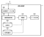
図2は、診断支援装置1の機能構成を説明する図である。診断支援装置1は、医用検査データ入力部201、病変候補取得部202、症例データベース203、医学知識データベース204、判断部205、データ出力部206を機能構成として備える。
FIG. 2 is a diagram illustrating a functional configuration of the
(医用検査データ入力部)
医用検査データ入力部201は、ネットワークを介して、医用検査データ取得装置2またはデータベース3と通信して、被検体の検査結果に基づく医用検査データを診断支援装置1に入力する(読み取る)ことが可能である。また、診断支援装置1に接続された記憶装置、例えば、FDDやCD-RWドライブ、MOドライブ、ZIPドライブを含めた各種記憶媒体から医用検査データを読み取ることも可能である。また、医用検査データ入力部201は、医用検査装置、例えば、X線CT装置、MR装置、超音波診断支援装置、心電図計などの機器から被検体の検査結果に基づく医用画像データを入力することも可能である。また、医用検査データ入力部201は、医用画像データの入力に限定されず、心電図や脳波データ・白血球数などの数値データや、手書きまたは電子化されたカルテ情報などの病変候補取得に関連するテキストデータを含む医用検査データの入力も可能である。
(Medical examination data input part)
The medical examination
(病変候補取得部)
病変候補取得部202は、医用検査データ入力部201により入力された画像・数値・テキストデータに基づいて、被験者に関する病変候補関連データを取得する。例えば、肺癌スクリーニングを行う場合、病変候補の取得方法としてLevel Set法を用いて、病変候補取得部202は、肺野などの読影対象臓器を抽出する。そして、病変候補取得部202は、その領域内で最小方向差分フィルタを適用したり、あるいは3次元曲率を計算することで結節候補を取得することができる。病変候補取得部202は、病変候補の取得において、症例データベース203を参照して過去の症例データとの類似度を比較したり、医学知識データベース204に格納されている診断基準や診断手順のデータを参照可能である。
(Lesion candidate acquisition unit)
The lesion
(症例データベース)
症例データベース203は、医用検査データ取得装置2によって撮影された医用画像データや、心電図や白血球数をはじめとする数値データ、被験者に関するカルテなどのテキストデータを格納する。症例データベース203に格納されるデータには診断支援装置1によるデータ解析結果の値や確定診断結果を含めることもでき、それらの情報を類似症例検索に利用することができる。
(Case database)
The
なお症例データベース203は判断部205だけでなく、病変候補取得部202からも利用することが可能である。症例データベース203は、病変候補取得部202と、判断部205とから独立した構成に限定されず、例えば、判断部205あるいは病変候補取得部202の内部、あるいはLAN4を介して接続される構成であってもよい。
The
(医学知識データベース)
医学知識データベース204は、病変サイズをはじめとする診断基準や、転移病変や合併症をはじめとする検査対象疾病に関連する疾病の情報を格納する。また、原発病変を検出した際に関連する疾病の有無を調べる手順といった診断手順などのデータも格納することが可能である。
(Medical knowledge database)
The
症例データベース203と同様に、医学知識データベース204は判断部205だけでなく病変候補取得部202からも利用できる。さらに、医学知識データベース204は、病変候補取得部202と、判断部205とから独立した構成に限定されず、例えば、判断部205あるいは病変候補取得部202の内部、あるいはLAN4を介して接続される構成であってもよい。
Similar to the
(判断部)
判断部205は、病変候補取得部202により得られた病変候補に関連するデータ(病変候補関連データ)をユーザに対して出力するか否かを判断する。
(Judgment part)
The
病変候補関連データを出力するか否かの判断に際しては、判断部205において、医学的な重要度をはじめ様々な基準と比較して病変候補関連データの出力可否を判断する。医学的な重要度を含め、具体的な出力可否判断の基準及びその算出方法については後に詳細に説明する。また、判断部205は、病変候補関連データ出力可否判断の際には症例データベース203や医学知識データベース204のデータを利用することも可能である。
When determining whether or not to output lesion candidate related data, the
(データ出力部)
判断部205が病変候補関連データを出力すると判断した場合、この判断結果に基づいて、データ出力部206は、病変候補関連データを出力する。病変候補関連データとしては病変候補取得の際に用いた医用画像データの特徴量などの読影根拠や類似症例画像などの読影結果を説明するデータ、診断ガイドラインなどの診療方法に関するデータなどが含まれるが、具体的な内容については後述する。
(Data output part)
When the
次に、図3を参照して診断支援装置1により実行される具体的な処理の手順を説明する。図3は、第1実施形態にかかる診断支援装置1の処理手順を説明するフローチャートであり、本処理はCPU100の全体的な制御の下に、図2で説明した各機能構成部が処理を実行する。
Next, a specific processing procedure executed by the
ステップS31において、医用検査データ入力部201は、医用検査データ取得装置2などから被検体の検査結果に基づく医用検査データを取得し、診断支援装置1に入力する。また、検査目的によって病変候補取得の際の病変検出基準や病変検出手順が異なるため、スクリーニング・精密検査・経過観察といった検査目的の情報をあらかじめ医師が診断支援装置に入力しておくことも可能である。なお検査目的に関する情報が医用検査データのDICOMヘッダに含まれている場合はヘッダから読み出してもよいし、放射線情報システム(RIS;Radiology Information System)から検査オーダ情報を取得する形でもよい。
In step S31, the medical examination
ステップS32において、病変候補取得部202は、ステップS31で入力された医用検査データに基づき病変候補を取得する。この際に医学知識データベース204に格納されている診断基準の情報を利用してもよいし、症例データベース203に格納されている確定診断済みの類似症例データを利用してもよい。病変候補取得の結果として、読影根拠をはじめとする病変候補関連データが得られる。なお病変候補取得部202の処理対象は医用画像データに限定されず、例えば被験者に関する画像以外の計測データあるいはカルテデータなども処理対象であり、それらのデータに基づいて病変候補を取得することが可能である。
In step S32, the lesion
ステップS33において、判断部205は、病変候補取得部202によって取得された病変候補関連データのうち、重み付けにより求められる医学的重要度を示す情報が閾値より高いと判断したデータのみユーザに提示させる。ここで、判断部205において算出する医学的重要度を示す情報Iについて説明する。医学的重要度を示す情報Iは、以下のように定義することができる。
In step S33, the
I=A*B*C
ただし、
A:重要疾病度 (異種疾病間の相対的な重篤度)
B:進行度(病期) (同種疾病内の重篤度)
C:関連病変度
とする。
I = A * B * C
However,
A: Critical illness (relative severity among different diseases)
B: Progression (stage) (severity within the same disease)
C: Related lesion degree
And
例えば、病変候補取得部202において複数種の疾病を検出する場合には、検査目的に応じて下記のように病変ごとの関連病変度Cが設定される。すなわち、
For example, when a plurality of types of diseases are detected in the lesion

となる。またデータ出力可否を示すフラグをFとおくと、 It becomes. If the flag indicating whether data can be output is F,
と表すことができる。 It can be expressed as.
TIについては、例えば、 For T I , for example,
のように、検査の感度に応じて適切な値を設定すればよく、必ずしも上記値に限定されるものではない。 As described above, an appropriate value may be set according to the sensitivity of the inspection, and is not necessarily limited to the above value.
またTABについては、たとえ検査対象の疾病やその関連疾病でない場合でも、重篤な病変であったり、病変の進行度が閾値より高い場合には病変候補に関連するデータを出力するということを示している。本実施形態ではTAB=50に設定するが、必ずしもこの値に限定されるものではない。 For TAB , even if it is not the disease to be examined or its related disease, if it is a serious lesion or the progression of the lesion is higher than the threshold, data related to the lesion candidate is output. Show. In this embodiment, T AB = 50 is set, but it is not necessarily limited to this value.
ここで重要疾病度A、進行度B、関連病変度Cの例を図4に示す。例えば、肺癌の経過観察の場合に、病変候補取得部202により肺野の悪性腫瘍(I期)が1個と短径1.2cmの縦隔リンパ節が1個検出され、胸部大動脈径が4cmであったとすると、悪性腫瘍に関する医学的重要度を示す情報Icは、
Ic=10*4*1=40>20(=TI)
となり、悪性腫瘍に関するデータ出力可否フラグFcは1に設定される。また縦隔リンパ節腫大に関する医学的重要度を示す情報Ilnは、
Iln=10*10*0.5=50>20(=TI)
となり、縦隔リンパ節腫大に関するデータ出力可否フラグFlnは1に設定される。さらに、胸部大動脈瘤に関する医学的重要度を示す情報Ia4は、
Ia4=8*2*0=0<20(=TI)
となり、胸部大動脈瘤に関するデータ出力可否フラグFa4は0に設定される。
Here, examples of the important disease degree A, the progression degree B, and the related lesion degree C are shown in FIG. For example, in the case of follow-up of lung cancer, the lesion
I c = 10 * 4 * 1 = 40> 20 (= T I )
Thus, the data output propriety flag F c regarding the malignant tumor is set to 1. Information I ln indicating the medical importance of mediastinal lymphadenopathy is
I ln = 10 * 10 * 0.5 = 50> 20 (= T I )
Thus, the data output enable / disable flag Fln regarding the mediastinal lymphadenopathy is set to 1. Furthermore, the information I a4 indicating the medical importance regarding the thoracic aortic aneurysm is
I a4 = 8 * 2 * 0 = 0 <20 (= T I )
Thus, the data output permission flag F a4 regarding the thoracic aortic aneurysm is set to 0.
胸部大動脈径が8cmである場合は、データ出力可否フラグFa8は、Ca8=0、かつ、Aa8*Ba8=8*8>50(=TAB)であるのでFa8=1となる。すなわち本来の検査対象病変ではないが、病変の進行度が閾値より高いため胸部大動脈瘤に関するデータが出力される。なお、ここで述べた医学的重要度を示す情報はあくまで一例であり、図4に示した数値例に限定されるものでないことは言うまでもない。 When the thoracic aorta diameter is 8 cm, the data output availability flag F a8 is C a8 = 0 and A a8 * B a8 = 8 * 8> 50 (= T AB ), so F a8 = 1. . In other words, although it is not the original lesion to be examined, the data on the thoracic aortic aneurysm is output because the progress of the lesion is higher than the threshold. It should be noted that the information indicating the medical importance described here is merely an example, and it is needless to say that the information is not limited to the numerical example shown in FIG.
ステップS33の判定により医学的重要度を示す情報が閾値に対して高くないと判定された場合(S33-NO)、処理を終了する。一方、S33の判定により、医学的重要度を示す情報が閾値より高いと判定された場合(S33-Yes)、処理はステップS34に進められる。 When it is determined by the determination in step S33 that the information indicating the medical importance is not higher than the threshold (S33-NO), the process is terminated. On the other hand, if it is determined in S33 that the information indicating the medical importance is higher than the threshold (S33-Yes), the process proceeds to Step S34.
ステップS34において、判断部205は、データ出力部206に出力する病変候補関連データの種類を決定する。病変候補に関連するデータとしては、読影結果の根拠を示す画像特徴量の値や計測数値・患者属性、あるいはそれらを入力とした識別器による計算結果などのデータが含まれ、病変候補関連データがどのような種類のデータかを判断部205は判断する。
In step S34, the
ただし、出力されるデータの種類は必ずしも読影根拠に限定されるものではなく、例えば、類似症例などの参考症例データ、あるいは診療ガイドラインのような診療方法に関するデータであってもよい。また出力するデータ種類についてはあらかじめユーザ側で設定したものを用いることが可能である。 However, the type of data to be output is not necessarily limited to the interpretation basis, and may be, for example, reference case data such as similar cases, or data related to a medical method such as medical guidelines. Further, the data type to be output can be set in advance by the user.
ステップS35において、データ出力部206は、先のステップS33、S34によって出力すると判断された病変候補関連データを出力する。ここで、データの出力先としては、紙、メモリやハードディスクなどの記憶装置、モニタなどの表示装置を含む。
In step S35, the
図5は、病変候補関連データの出力例を示す図である。医用検査データの上に病変候補を指摘するマーク503が表示され(図5(a))、その隣に検出結果の根拠となる画像特徴量や患者属性・経時変化データが表示されている(図5(b))。また病変候補関連データをモニタなどの表示デバイスに出力する場合にはポップアップ形式で表示してもよいし、別ウィンドウで表示しても良い。
FIG. 5 is a diagram illustrating an output example of lesion candidate related data. A
以上説明したように本実施形態によれば、医学的重要度を示す情報が閾値より高い場合のみ病変候補に関連するデータを出力することで、医師は診断支援装置が出力した病変部位に関連する情報を効率的に参照することができる。 As described above, according to the present embodiment, the doctor outputs the data related to the lesion candidate only when the information indicating the medical importance is higher than the threshold, so that the doctor relates to the lesion site output by the diagnosis support apparatus. Information can be referred to efficiently.
(第2実施形態)
第1実施形態では、判断部205の具体的な処理として、医学的重要度を示す情報に応じて病変候補関連データの出力可否を決定する構成を説明した。しかし、本発明の趣旨は、この例に限定されるものでない。例えば、判断部205の処理として、病変候補関連データと医師の読影結果を示すレポートとの間に相違点(差分)がある場合に、判断部205が病変候補関連データを出力する構成について説明する。図6は、第2実施形態にかかる診断支援装置61の機能構成を示す図であり、第1実施形態にかかる診断支援装置1の機能構成と同一の機能構成については同一の参照番号を付して、説明を省略する。
(Second Embodiment)
In the first embodiment, as a specific process of the
レポート入力部207は、診断結果入力手段として機能し、医用検査データに基づいて医師が読影した診断結果を示す情報(読影レポート)を診断支援装置1に入力する。例えば、レポート入力部207は、読影を行った医師により作成された読影レポートが格納されたデータベースからネットワークを介して読影レポートのデータを診断支援装置61に入力することが可能である。また、診断支援装置1に接続された記憶装置(記憶手段)、例えば、FDD、CD-RWドライブ、MOドライブ、ZIPドライブを含む各種記憶媒体からデータを読み取り、読影レポートのデータを診断支援装置61に入力することも可能である。
The
また、レポート入力部207は、キーボードやマウスなどにより、読影を行った医師がコンピュータに読影結果を示す読影レポートデータを直接、診断支援装置61に入力することも可能である。
The
更に、レポート入力部207は、予め定められた定型フォーマットを介して読影の結果を診断支援装置61に入力することも可能である。定型フォーマットには読影の結果を入力するための項目、選択肢を有し、読影を行った医師がその項目への入力、あるいは選択肢の選択に従って入力を行うことにより、読影の結果が診断支援装置61に入力される。あるいは、レポート入力部207はスキャナを含む読取装置や、光学文字認識装置を接続することにより、読影を行った医師が手書きで書いた文章をコンピュータが編集できる形式で診断支援装置61に入力することも可能である。
Furthermore, the
レポート解析部208は、レポート入力部207により入力された医師による読影結果を解析し、医学的な意味を解析する。例えば、レポート解析部208は、図7に示すように形態素解析部701とクラス特定部702、意味特定部704を備えるとともに、クラスを特定するための構造化用辞書703、医用シソーラス辞書705とを備える。形態素解析部701、クラス特定部702、構造化用辞書703、意味特定部704及び医用シソーラス辞書705は、テキスト解析手段として機能する。
The
形態素解析部701は、自然文で記述された読影レポートを形態素(言語の列の中で意味を持つ最小単位)に分割する。構造化用辞書703は医療に関する単語とそのクラスが登録された辞書である。構造化用辞書703に登録されるクラスの種類としては「部位」「疾病名」などが含まれる。
The
クラス特定部702は構造化用辞書703を用いて、形態素解析部701により得られた単語のクラスを特定し、読影レポートの構造化データを生成する。医用シソーラス辞書705を参照することにより意味特定部704は、医療に関する単語間の関係を記述したオントロジを、概念的に上位の単語が階層構造上で上位に位置し、概念的に下位の単語が下位に位置するように、単語を階層的に整理する。意味特定部704は、医用シソーラス辞書705を参照して、レポートの構造化データに含まれる単語の階層位置を表現するシソーラスコードを決定する。またレポート入力部207が選択肢から選択された単語に基づき読影レポートの内容を入力する別の構成の場合、レポート解析部208は、図7に示す構成に限定されない。例えば、レポート入力部207にあらかじめ選択肢として用意した単語に関するシソーラス辞書と、その辞書との照合を行う手段を設けることでレポート解析部208を構成することも可能である。
The
次に、図8を参照して診断支援装置61により実行される具体的な処理の手順を説明する。図8は、第2実施形態にかかる診断支援装置61の処理手順を説明するフローチャートであり、本処理はCPU100の全体的な制御の下に、図6、図7で説明した各機能構成部が処理を実行する。
Next, a specific processing procedure executed by the
ステップS81において、医用検査データ入力部201は、医用検査データ取得装置2などから被検体の検査結果に基づく医用検査データを取得し、診断支援装置1に入力する。この処理の詳細は、第1実施形態のステップS31の場合と同じであるので、説明は省略する。
In step S81, the medical examination
ステップS82において、病変候補取得部202は、ステップS81で入力された医用検査データに基づき病変候補を取得する。この処理の詳細は、第1実施形態のステップS32の場合と同じであるので、説明は省略する。
In step S82, the lesion
ステップS83において、CPU100は、レポート入力部207への読影レポートデータの入力の有無を調べる。診断結果に基づく読影レポートデータが入力されている場合は(S83-Yes)、ステップS84に進み、レポートデータの入力がない場合は(S83-No)、ステップS85に進む。診断結果に基づく情報として、読影レポートデータは、医師の診断結果、計測装置による診断結果、あるいは診断支援装置によって解析済みの画像、テキストあるいは数値データの少なくとも1種類を含む。
In step S83, the
ステップS84において、レポート解析部208は、形態素解析、クラス特定、意味特定を行い、図9に示されるような診断結果を示す所見データ901を生成する。所見データ901には、部位、病変種別、寸法、状態、所見に関するデータが含まれる。そして、レポート解析部208は、入力された読影レポートに基づく所見データ901と、病変候補取得部202によって得られた病変候補関連データとの相違点(差分)を検出する。
In step S84, the
ステップS85において、ステップS84により得られた所見データ901と、病変候補取得部202によって得られた病変候補関連データとの差分について、判断部205は、医学的重要度を示す情報Idiffを計算する。
In step S85, for the difference between the
例えば、Idiffは、次式のように決めることができる。 For example, I diff can be determined as follows:
ただし、Adoc:医師による所見データ中の重要疾病度
Bdoc:医師による所見データ中の進行度(病期)
Cdoc:医師による所見データ中の関連病変度
Acad:病変候補取得部202が算出した重要疾病度
Bcad:病変候補取得部202が算出した進行度(病期)
Ccad:病変候補取得部202が算出した関連病変度
例えば医師による所見が肺野内の良性腫瘍で、病変候補取得部202よって得られたデータが第I期の肺癌であった場合、医学的重要度を示す情報Idiffは、次式にように求められる。
However, A doc : Critical illness in the findings by doctors
B doc : Progression level (stage) in findings data by doctor
C doc : Degree of related lesions in findings data by doctor
A cad : Critical disease degree calculated by the lesion
B cad : degree of progression (stage) calculated by the candidate
C cad : related lesion degree calculated by the lesion
なお、ここで述べた医学的重要度を示す情報はあくまで一例であり、本実施形態における定義に限定されるものではない。 The information indicating the medical importance described here is merely an example, and is not limited to the definition in the present embodiment.
ステップS86において、ステップS85により求められた医学的重要度を示す情報が予め定められた閾値より高い場合、判断部205は、データ出力可否フラグFdiffを1に設定し、ステップS87に進む。また医学的重要度を示す情報が閾値より低い場合はデータ出力可否フラグFdiffを0に設定し、データ出力部206に病変候補に関連するデータを出力せずに判断部205の処理を終了する。
In step S86, when the information indicating the medical importance obtained in step S85 is higher than a predetermined threshold value, the
本実施形態では、数5式の算出結果がIdiff>20であるのでデータ出力可否フラグFdiff=1となる。
In this embodiment, since the calculation result of
ステップS87において、判断部205は、データ出力部206に出力する病変候補関連データの種類を決定する。病変候補に関連するデータとしては、読影結果の根拠を示す画像特徴量の値や計測数値・患者属性、あるいはそれらを入力とした識別器による計算結果などのデータが含まれ、病変候補関連データがどのような種類のデータかを判断部205は判断する。また出力される病変候補関連データは必ずしも読影根拠に限定されるものではない。例えば類似症例などの参考症例データ、あるいは診療ガイドラインのような診療方法に関するデータ、あるいはレポート修正候補などのレポート作成支援に関するデータであってもよい。
In step S87, the
ステップS88において、データ出力部206は、ステップS86によりデータ出力可否フラグFdiffが1に設定された場合、ステップS87で決定された病変候補に関するデータ(病変候補関連データ)を出力する。本実施形態の場合、数5式の算出結果に基づき出力可否フラグFdiff=1であるので肺癌として検出した病変候補に関するデータが出力される。例えば、図9に示される所見データ901が得られた場合に(図9(a))、データ出力部206は読影の根拠となる画像として画像特徴量や患者属性・経時変化データを出力する。モニタ104にはデータ出力部206の出力に基づき、読影の根拠となる画像として画像特徴量や患者属性・経時変化データ902が表示される(図9(b))。
In step S88, when the data output enable / disable flag Fdiff is set to 1 in step S86, the
読影レポートと取得された病変候補との間に差分がある場合、医学的重要度を示す情報が閾値より高い場合に病変候補関連データを出力することで、医師は病変部位関連データを効率的に参照することが可能になる。 When there is a difference between the interpretation report and the acquired lesion candidate, if the information indicating the medical importance is higher than the threshold, the lesion candidate related data is output, so that the doctor efficiently It becomes possible to refer.
(第3実施形態)
第1実施形態では、判断部205の具体的な処理として、医学的重要度を示す情報に応じて病変候補関連データの出力可否を決定する構成を説明した。また、第2実施形態では、病変候補関連データと医師の読影結果を示すレポートとの間に相違点(差分)がある場合に、判断部205が病変候補関連データを出力する構成を説明した。しかし、本発明の趣旨は、この例に限定されるものでない。例えば、判断部205の処理として、特定の病変候補の位置及び出力するデータの種類を医師が直接指定した場合に病変候補関連データを出力する構成を説明する。
(Third embodiment)
In the first embodiment, as a specific process of the
図10は、第3実施形態にかかる診断支援装置1001の機能構成を示す図であり、第1実施形態にかかる診断支援装置1、第2実施形態にかかる診断支援装置61の機能構成と同一の機能構成については同一の参照番号を付して、説明を省略する。
FIG. 10 is a diagram illustrating a functional configuration of the
本実施形態において、判断部205は、レポート解析部208の解析結果と、病変候補取得部202により取得された病変候補の情報とを比較する比較部209を備え、比較部209は両者の相違点(差分)を求める。また、判断部205は、医師が出力させたい病変位置及びデータ種類の指定を受付ける指定部210を備える。指定部210は、医師が病変候補関連データを出力させたい病変位置を指定する位置指定部211と、出力させたい病変候補関連データの種類を指定するデータ種類指定部212を有する。
In the present embodiment, the
次に、図11を参照して診断支援装置1001により実行される具体的な処理の手順を説明する。図11は、第3実施形態にかかる診断支援装置1001の処理手順を説明するフローチャートであり、本処理はCPU100の全体的な制御の下に、図10で説明した各機能構成部が処理を実行する。
Next, a specific processing procedure executed by the
ステップS111において、医用検査データ入力部201は、医用検査データ取得装置2などから被検体の検査結果に基づく医用検査データを取得し、診断支援装置1に入力する。この処理の詳細は、第1実施形態のステップS31の場合と同じであるので、詳細な説明は省略する。
In step S111, the medical examination
ステップS112において、病変候補取得部202は、ステップS111で入力された医用検査データに基づき病変候補を取得する。この処理の詳細は、第1実施形態のステップS32の場合と同じであるので、詳細な説明は省略する。
In step S112, the lesion
ステップS113において、CPU100は、レポート入力部207への読影レポートデータの入力の有無を調べる。読影レポートデータが入力されている場合は(S113-Yes)、ステップS114に進み、レポートデータの入力がない場合は(S113-No)、ステップS115に進む。
In step S113, the
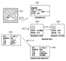
ステップS114において、レポート解析部208は、形態素解析、クラス特定、意味特定を行う。図12は、病変候補関連データの出力例を示す図である。医用検査データの上に病変候補を指摘するマーク1201が表示され、レポート解析部208の処理により、所見データ1202が生成される。所見データ1202には、部位、病変種別、寸法、状態、所見に関するデータが含まれる。そして、比較部209は、入力された読影レポートに基づく所見データ1202と、病変候補取得部202によって得られた病変候補関連データとの比較を行い、相違点(差分)を検出する。
In step S114, the
ステップS115において、ステップS114により得られた所見データ901と、病変候補取得部202によって得られた病変候補関連データとの差分について、判断部205は、医学的重要度を示す情報Idiffを計算する。
In step S115, for the difference between the
ステップS116において、医師が特定の病変候補に関連するデータを参照したい場合に、参照したい病変候補とその病変候補に関して参照したいデータの種類を指定する。指定された病変位置は位置指定部211に入力され、また指定されたデータの種類はデータ種類指定部212に入力され、どの病変に関してどのような種類のデータが出力されるかが決定される。
In step S116, when the doctor wants to refer to data related to a specific lesion candidate, the lesion candidate to be referred to and the type of data to be referred to for the lesion candidate are designated. The designated lesion position is input to the
例えば、図12に示すように医師による所見(肺炎)と病変候補取得部202による病変名(良性腫瘍)とが異なる場合、判断部205の処理とマウス等の入力装置の操作入力とに基づきデータ出力部206はデータ出力を制御する。データ出力部206のデータ出力に基づき、モニタ104に表示される画面が制御される。この場合、データ出力部206は、表示制御手段として機能する。
For example, as shown in FIG. 12, when the findings by the doctor (pneumonia) and the lesion name (benign tumor) by the lesion
マウスオーバー(第1操作)により、医用検査データに基づく診断結果を示す読影根拠の情報1203がモニタ104(表示手段)に表示される。
By the mouse over (first operation),
マウスを左クリック(第2操作)により、類似度が最大の症例が表示される(画面1204)。左クリックを行うと第二位以下の類似度の症例がモニタ104(表示手段)に表示される(画面1205)。左クリック操作を繰り返すことにより、第二位以下の類似度の症例が順次表示される。 By left-clicking the mouse (second operation), the case with the maximum similarity is displayed (screen 1204). When the left click is performed, cases having the second or lower similarity are displayed on the monitor 104 (display means) (screen 1205). By repeating the left click operation, cases having similarities of the second and lower ranks are sequentially displayed.
マウスを右クリック(第3操作)により、診断結果の修正候補(レポート修正候補)が表示され、修正候補を採用するか否かをたずねるメッセージが画面に表示される。OKボタンを押すと診断結果の内容が修正された画面1206がモニタ104(表示手段)に表示される。ユーザ(医師)がマウス等の入力手段の操作により表示すべきデータの切り替えを指定することにより、データ出力部206は、データ出力を制御することができる。
By right-clicking the mouse (third operation), correction candidates for the diagnosis result (report correction candidates) are displayed, and a message asking whether or not to adopt the correction candidates is displayed on the screen. When the OK button is pressed, a screen 1206 in which the contents of the diagnosis result are corrected is displayed on the monitor 104 (display means). When the user (doctor) designates switching of data to be displayed by operating an input unit such as a mouse, the
ステップS117において、データ出力部206は、医学的重要度を示す情報の算出結果(S115)と、病変位置、データの種類(S116)の指定に基づいて、病変候補に関するデータ(病変候補関連データ)を出力する。
In step S117, the
ステップS118において、さらにユーザにより病変候補・データ種類の指定がないか調べ、さらに指定がある場合は(S118−Yes)、ステップS116に処理を戻し、同様の処理を繰り返す。一方、ユーザにより病変候補・データ種類の指定がない場合(S118−No)、処理を終了する。 In step S118, it is further checked whether the user has designated a lesion candidate / data type. If there is further designation (S118-Yes), the process returns to step S116, and the same process is repeated. On the other hand, when the lesion candidate / data type is not designated by the user (S118-No), the process is terminated.
医師が指定した病変位置、データの種類の指定により病変部位に関連データを出力することで、医師は、診断支援装置が出力した病変部位に関連する情報を効率的に参照することができる。 By outputting the related data to the lesion site by specifying the lesion position and data type specified by the doctor, the doctor can efficiently refer to the information related to the lesion site output by the diagnosis support apparatus.
(他の実施形態)
なお、本発明の目的は、前述した実施形態の機能を実現するソフトウェアのプログラムを記録したコンピュータ可読の記憶媒体を、システムあるいは装置に供給することによっても、達成されることは言うまでもない。また、システムあるいは装置のコンピュータ(またはCPUやMPU)が記憶媒体に格納されたプログラムを読出し実行することによっても、達成されることは言うまでもない。
(Other embodiments)
Note that it is needless to say that the object of the present invention can also be achieved by supplying a computer-readable storage medium that records a software program for realizing the functions of the above-described embodiments to a system or apparatus. Needless to say, this can also be achieved by the computer (or CPU or MPU) of the system or apparatus reading and executing the program stored in the storage medium.
この場合、記憶媒体から読出されたプログラム自体が前述した実施形態の機能を実現することになり、そのプログラムを記憶した記憶媒体は本発明を構成することになる。 In this case, the program itself read from the storage medium realizes the functions of the above-described embodiments, and the storage medium storing the program constitutes the present invention.
プログラムを供給するための記憶媒体としては、例えば、フレキシブルディスク、ハードディスク、光ディスク、光磁気ディスク、CD−ROM、CD−R、不揮発性のメモリカード、ROMなどを用いることができる。 As a storage medium for supplying the program, for example, a flexible disk, a hard disk, an optical disk, a magneto-optical disk, a CD-ROM, a CD-R, a nonvolatile memory card, a ROM, or the like can be used.
また、コンピュータが読出したプログラムを実行することにより、前述した実施形態の機能が実現される。また、プログラムの指示に基づき、コンピュータ上で稼働しているOS(オペレーティングシステム)などが実際の処理の一部または全部を行い、その処理によって前述した実施形態が実現される場合も含まれることは言うまでもない。 Further, the functions of the above-described embodiments are realized by executing the program read by the computer. In addition, it is also included that an OS (operating system) or the like running on a computer performs part or all of actual processing based on a program instruction, and the above-described embodiment is realized by the processing. Needless to say.
1 診断支援装置
2 医用検査データ取得装置
3 データベース
4 LAN
100 CPU
101 主メモリ
102 磁気ディスク
103 表示メモリ
104 モニタ
105 マウス
106 キーボード
201 医用検査データ入力部
202 病変候補取得部
203 症例データベース
204 医学知識データベース
205 判断部
206 データ出力部
207 レポート入力部
208 レポート解析部
209 比較部
210 指定部
211 位置指定部
212 データ種類指定部
1 Diagnosis support device
2 Medical examination data acquisition device
3 Database
4 LAN
100 CPU
101 Main memory
102 magnetic disk
103 Display memory
104 Monitor
105 mice
106 keyboard
201 Medical examination data input part
202 Lesion candidate acquisition unit
203 Case Database
204 Medical Knowledge Database
205 Judgment part
206 Data output section
207 Report input section
208 Report Analysis Department
209 Comparison part
210 Designated part
211 Position specification part
212 Data type specification part
Claims (18)
前記医用検査データに基づく病変候補の情報を取得する取得手段と、
前記病変候補の情報のうち、前記医用検査データの重み付けにより求められる医学的重要度を示す情報が予め定められた閾値よりも高くなるか否かを判断する判断手段と、
前記判断手段の判断結果に従い、前記医学的重要度を示す情報が前記閾値よりも高くなるデータを出力するデータ出力手段と、
を備えることを特徴とする診断支援装置。 Medical test data input means for inputting medical test data based on the test result of the subject;
Acquiring means for acquiring information of lesion candidates based on the medical examination data;
Judgment means for judging whether information indicating medical importance obtained by weighting of the medical examination data among information on the lesion candidates is higher than a predetermined threshold;
According to the determination result of the determination means, data output means for outputting data in which information indicating the medical importance is higher than the threshold;
A diagnostic support apparatus comprising:
前記取得手段は、前記医用検査データ入力手段により入力されたデータに基づき、前記病変候補の情報を取得することを特徴とする請求項1または請求項2に記載の診断支援装置。 The medical examination data input means is capable of inputting at least one of medical image data, numerical data, and text data inputted from a medical examination apparatus that examines the subject.
The diagnosis support apparatus according to claim 1 or 2, wherein the acquisition unit acquires information on the lesion candidate based on data input by the medical examination data input unit.
前記診断結果入力手段により入力された前記診断結果の情報を解析する解析手段と、を更に備えることを特徴とする請求項1に記載の診断支援装置。 Diagnostic result input means for inputting diagnostic result information based on the medical examination data;
The diagnosis support apparatus according to claim 1, further comprising: an analysis unit that analyzes information on the diagnosis result input by the diagnosis result input unit.
前記差分の結果に対する重み付けに基づき求められる医学的重要度を示す情報が予め定められた閾値よりも高くなるか否かを判断することを特徴とする請求項4に記載の診断支援装置。 The determination unit obtains a difference between the analysis result of the analysis unit and the information of the lesion candidate acquired by the acquisition unit,
The diagnosis support apparatus according to claim 4, wherein it is determined whether or not information indicating medical importance obtained based on weighting for the difference result is higher than a predetermined threshold value.
前記判断手段は、前記指定手段により指定された前記病変候補に対して医学的重要度を示す情報が前記閾値よりも高くなるか否かを判断し、
前記データ出力手段は、前記判断手段の判断結果に基づき、指定された前記病変候補に対して前記閾値よりも高くなるデータを出力することを特徴とする請求項1に記載の診断支援装置。 It further has a designation means for accepting designation of a lesion candidate to be referred to,
The determination means determines whether or not information indicating medical importance for the lesion candidate specified by the specification means is higher than the threshold value;
The diagnosis support apparatus according to claim 1, wherein the data output unit outputs data that is higher than the threshold value for the designated lesion candidate based on a determination result of the determination unit.
取得手段が、前記医用検査データに基づく病変候補の情報を取得する取得工程と、
判断手段が、前記病変候補の情報のうち、前記医用検査データの重み付けにより求められる医学的重要度を示す情報が予め定められた閾値よりも高くなるか否かを判断する判断工程と、
データ出力手段が、前記判断工程での判断結果に従い、前記医学的重要度を示す情報が前記閾値よりも高くなるデータを出力するデータ出力工程と、
を備えることを特徴とする診断支援装置の制御方法。 Medical examination data input means for inputting medical examination data based on the examination result of the subject;
An acquisition step of acquiring information on candidate lesions based on the medical examination data;
A determination step of determining whether information indicating medical importance obtained by weighting of the medical examination data among information on the lesion candidates is higher than a predetermined threshold;
A data output step in which data output means outputs data in which the information indicating the medical importance is higher than the threshold according to the determination result in the determination step;
A diagnostic support apparatus control method comprising:
前記取得工程では、前記医用検査データ入力工程により入力されたデータに基づき、前記病変候補の情報を取得することを特徴とする請求項9または請求項10に記載の診断支援装置の制御方法。 In the medical examination data input step, it is possible to input at least one of medical image data, numerical data, and text data inputted from a medical examination apparatus that examines the subject.
The method of controlling a diagnosis support apparatus according to claim 9 or 10, wherein in the acquisition step, information on the lesion candidate is acquired based on the data input in the medical examination data input step.
解析手段が、前記診断結果入力工程により入力された前記診断結果の情報を解析する解析工程と、を更に備えることを特徴とする請求項9に記載の診断支援装置の制御方法。 A diagnostic result input step, wherein the diagnostic result input means inputs information of a diagnostic result based on the medical examination data;
The method for controlling the diagnosis support apparatus according to claim 9, further comprising: an analysis step in which the analysis unit analyzes information on the diagnosis result input in the diagnosis result input step.
前記判断工程では、前記指定工程により指定された前記病変候補に対して医学的重要度を示す情報が前記閾値よりも高くなるか否かを判断し、
前記データ出力工程では、前記判断工程の判断結果に基づき、指定された前記病変候補に対して前記閾値よりも高くなるデータを出力することを特徴とする請求項12に記載の診断支援装置の制御方法。 The analysis step includes a designation step for accepting designation of a lesion candidate to be referred to,
In the determining step, it is determined whether or not information indicating medical importance for the lesion candidate specified in the specifying step is higher than the threshold.
13. The diagnosis support apparatus control according to claim 12, wherein, in the data output step, data that is higher than the threshold value is output for the designated lesion candidate based on a determination result of the determination step. Method.
Priority Applications (1)
| Application Number | Priority Date | Filing Date | Title |
|---|---|---|---|
| JP2007256013A JP2009082442A (en) | 2007-09-28 | 2007-09-28 | Diagnosis support apparatus, its control method, program, and storage medium |
Applications Claiming Priority (1)
| Application Number | Priority Date | Filing Date | Title |
|---|---|---|---|
| JP2007256013A JP2009082442A (en) | 2007-09-28 | 2007-09-28 | Diagnosis support apparatus, its control method, program, and storage medium |
Related Child Applications (1)
| Application Number | Title | Priority Date | Filing Date |
|---|---|---|---|
| JP2013229779A Division JP2014059892A (en) | 2013-11-05 | 2013-11-05 | Medical information processing device, medical information processing method, and program |
Publications (2)
| Publication Number | Publication Date |
|---|---|
| JP2009082442A true JP2009082442A (en) | 2009-04-23 |
| JP2009082442A5 JP2009082442A5 (en) | 2010-09-24 |
Family
ID=40656706
Family Applications (1)
| Application Number | Title | Priority Date | Filing Date |
|---|---|---|---|
| JP2007256013A Pending JP2009082442A (en) | 2007-09-28 | 2007-09-28 | Diagnosis support apparatus, its control method, program, and storage medium |
Country Status (1)
| Country | Link |
|---|---|
| JP (1) | JP2009082442A (en) |
Cited By (7)
| Publication number | Priority date | Publication date | Assignee | Title |
|---|---|---|---|---|
| JP2011092685A (en) * | 2009-09-30 | 2011-05-12 | Fujifilm Corp | Diagnosis support system, diagnostic support program and diagnostic support method |
| WO2012099004A1 (en) * | 2011-01-19 | 2012-07-26 | 株式会社 東芝 | Medical image processing apparatus, x-ray ct apparatus and medical image processing program |
| WO2013018363A1 (en) * | 2011-08-04 | 2013-02-07 | パナソニック株式会社 | Similar case search device and similar case search method |
| US8671118B2 (en) | 2010-08-25 | 2014-03-11 | Fujifilm Corporation | Apparatus, method and program for assisting medical report creation and providing medical information |
| CN111096767A (en) * | 2020-01-08 | 2020-05-05 | 南京市第一医院 | Deep learning-based mediastinal lymph node ultrasound elastic image segmentation and classification method |
| JPWO2019008941A1 (en) * | 2017-07-03 | 2020-05-07 | 富士フイルム株式会社 | Medical image processing device, endoscope device, diagnosis support device, medical work support device, and report creation support device |
| EP4343780A1 (en) | 2022-09-21 | 2024-03-27 | FUJIFILM Corporation | Information processing apparatus, information processing method, and information processing program |
Citations (5)
| Publication number | Priority date | Publication date | Assignee | Title |
|---|---|---|---|---|
| JP2002112985A (en) * | 2000-10-06 | 2002-04-16 | Konica Corp | Diagnostic imaging support device |
| JP2004135868A (en) * | 2002-10-17 | 2004-05-13 | Fuji Photo Film Co Ltd | System for abnormal shadow candidate detection process |
| JP2004288047A (en) * | 2003-03-24 | 2004-10-14 | Fujitsu Ltd | Medical examination support system and medical examination support program |
| JP2005224429A (en) * | 2004-02-13 | 2005-08-25 | Fuji Photo Film Co Ltd | Abnormal shadow judging apparatus and program thereof |
| JP2006255065A (en) * | 2005-03-16 | 2006-09-28 | Fuji Photo Film Co Ltd | Image output method, image output device and program |
-
2007
- 2007-09-28 JP JP2007256013A patent/JP2009082442A/en active Pending
Patent Citations (5)
| Publication number | Priority date | Publication date | Assignee | Title |
|---|---|---|---|---|
| JP2002112985A (en) * | 2000-10-06 | 2002-04-16 | Konica Corp | Diagnostic imaging support device |
| JP2004135868A (en) * | 2002-10-17 | 2004-05-13 | Fuji Photo Film Co Ltd | System for abnormal shadow candidate detection process |
| JP2004288047A (en) * | 2003-03-24 | 2004-10-14 | Fujitsu Ltd | Medical examination support system and medical examination support program |
| JP2005224429A (en) * | 2004-02-13 | 2005-08-25 | Fuji Photo Film Co Ltd | Abnormal shadow judging apparatus and program thereof |
| JP2006255065A (en) * | 2005-03-16 | 2006-09-28 | Fuji Photo Film Co Ltd | Image output method, image output device and program |
Cited By (15)
| Publication number | Priority date | Publication date | Assignee | Title |
|---|---|---|---|---|
| US8630467B2 (en) | 2009-09-30 | 2014-01-14 | Fujifilm Corporation | Diagnosis assisting system using three dimensional image data, computer readable recording medium having a related diagnosis assisting program recorded thereon, and related diagnosis assisting method |
| JP2011092685A (en) * | 2009-09-30 | 2011-05-12 | Fujifilm Corp | Diagnosis support system, diagnostic support program and diagnostic support method |
| US8671118B2 (en) | 2010-08-25 | 2014-03-11 | Fujifilm Corporation | Apparatus, method and program for assisting medical report creation and providing medical information |
| US9066654B2 (en) | 2011-01-19 | 2015-06-30 | Kabushiki Kaisha Toshiba | Medical image processing apparatus, an X-ray CT scanner, and a medical image processing program |
| CN103228216A (en) * | 2011-01-19 | 2013-07-31 | 株式会社东芝 | Medical image processing apparatus, x-ray CT apparatus and medical image processing program |
| JP2012147930A (en) * | 2011-01-19 | 2012-08-09 | Toshiba Corp | Medical image processing apparatus, and medical image processing program |
| WO2012099004A1 (en) * | 2011-01-19 | 2012-07-26 | 株式会社 東芝 | Medical image processing apparatus, x-ray ct apparatus and medical image processing program |
| CN103228216B (en) * | 2011-01-19 | 2016-08-10 | 东芝医疗系统株式会社 | Medical image-processing apparatus and X ray CT device |
| WO2013018363A1 (en) * | 2011-08-04 | 2013-02-07 | パナソニック株式会社 | Similar case search device and similar case search method |
| JP5475923B2 (en) * | 2011-08-04 | 2014-04-16 | パナソニック株式会社 | Similar case retrieval apparatus and similar case retrieval method |
| US8934695B2 (en) | 2011-08-04 | 2015-01-13 | Panasonic Corporation | Similar case searching apparatus and similar case searching method |
| JPWO2019008941A1 (en) * | 2017-07-03 | 2020-05-07 | 富士フイルム株式会社 | Medical image processing device, endoscope device, diagnosis support device, medical work support device, and report creation support device |
| US11450425B2 (en) | 2017-07-03 | 2022-09-20 | Fujifilm Corporation | Medical image processing apparatus, endoscope apparatus, diagnostic support apparatus, medical service support apparatus, and report creation support apparatus |
| CN111096767A (en) * | 2020-01-08 | 2020-05-05 | 南京市第一医院 | Deep learning-based mediastinal lymph node ultrasound elastic image segmentation and classification method |
| EP4343780A1 (en) | 2022-09-21 | 2024-03-27 | FUJIFILM Corporation | Information processing apparatus, information processing method, and information processing program |
Similar Documents
| Publication | Publication Date | Title |
|---|---|---|
| US10529045B2 (en) | Information processing apparatus and information processing method | |
| JP5264136B2 (en) | MEDICAL DIAGNOSIS SUPPORT DEVICE, ITS CONTROL METHOD, COMPUTER PROGRAM, AND STORAGE MEDIUM | |
| JP5100285B2 (en) | MEDICAL DIAGNOSIS SUPPORT DEVICE, ITS CONTROL METHOD, PROGRAM, AND STORAGE MEDIUM | |
| JP6749835B2 (en) | Context-sensitive medical data entry system | |
| US7599534B2 (en) | CAD (computer-aided decision) support systems and methods | |
| RU2686627C1 (en) | Automatic development of a longitudinal indicator-oriented area for viewing patient's parameters | |
| US10083166B2 (en) | Apparatus and method for generating inspection report(s) | |
| JP2019153250A (en) | Device, method, and program for supporting preparation of medical document | |
| JP6796060B2 (en) | Image report annotation identification | |
| US20190267120A1 (en) | Medical document creation support apparatus, method, and program | |
| JP2009082442A (en) | Diagnosis support apparatus, its control method, program, and storage medium | |
| JP5997791B2 (en) | Diagnosis support apparatus, control method for diagnosis support apparatus, program, and storage medium | |
| JP7102509B2 (en) | Medical document creation support device, medical document creation support method, and medical document creation support program | |
| JP2014059892A (en) | Medical information processing device, medical information processing method, and program | |
| US11062448B2 (en) | Machine learning data generation support apparatus, operation method of machine learning data generation support apparatus, and machine learning data generation support program | |
| US20220366151A1 (en) | Document creation support apparatus, method, and program | |
| JP5899239B2 (en) | Generating image report diagrams of lesions in anatomical structures | |
| US20220285011A1 (en) | Document creation support apparatus, document creation support method, and program | |
| JP5501491B2 (en) | Diagnosis support apparatus and control method | |
| US20230005580A1 (en) | Document creation support apparatus, method, and program | |
| US20220375562A1 (en) | Document creation support apparatus, document creation support method, and program | |
| EP4287195A1 (en) | Information processing device, method, and program | |
| WO2022230641A1 (en) | Document creation assisting device, document creation assisting method, and document creation assisting program | |
| US20220391599A1 (en) | Information saving apparatus, method, and program and analysis record generation apparatus, method, and program | |
| WO2022239593A1 (en) | Document creation assistance device, document creation assistance method, and document creation assistance program |
Legal Events
| Date | Code | Title | Description |
|---|---|---|---|
| A521 | Request for written amendment filed |
Free format text: JAPANESE INTERMEDIATE CODE: A523 Effective date: 20100810 |
|
| A621 | Written request for application examination |
Free format text: JAPANESE INTERMEDIATE CODE: A621 Effective date: 20100810 |
|
| A977 | Report on retrieval |
Free format text: JAPANESE INTERMEDIATE CODE: A971007 Effective date: 20120524 |
|
| A131 | Notification of reasons for refusal |
Free format text: JAPANESE INTERMEDIATE CODE: A131 Effective date: 20120528 |
|
| A521 | Request for written amendment filed |
Free format text: JAPANESE INTERMEDIATE CODE: A523 Effective date: 20120727 |
|
| A131 | Notification of reasons for refusal |
Free format text: JAPANESE INTERMEDIATE CODE: A131 Effective date: 20120824 |
|
| A521 | Request for written amendment filed |
Free format text: JAPANESE INTERMEDIATE CODE: A523 Effective date: 20121023 |
|
| A02 | Decision of refusal |
Free format text: JAPANESE INTERMEDIATE CODE: A02 Effective date: 20130805 |
|
| A521 | Request for written amendment filed |
Free format text: JAPANESE INTERMEDIATE CODE: A523 Effective date: 20131105 |
|
| A911 | Transfer to examiner for re-examination before appeal (zenchi) |
Free format text: JAPANESE INTERMEDIATE CODE: A911 Effective date: 20131112 |
|
| A912 | Re-examination (zenchi) completed and case transferred to appeal board |
Free format text: JAPANESE INTERMEDIATE CODE: A912 Effective date: 20140131 |