JP2004508534A - Personal computer breath analyzer for health related behavior modification and its method - Google Patents
Personal computer breath analyzer for health related behavior modification and its method Download PDFInfo
- Publication number
- JP2004508534A JP2004508534A JP2001562191A JP2001562191A JP2004508534A JP 2004508534 A JP2004508534 A JP 2004508534A JP 2001562191 A JP2001562191 A JP 2001562191A JP 2001562191 A JP2001562191 A JP 2001562191A JP 2004508534 A JP2004508534 A JP 2004508534A
- Authority
- JP
- Japan
- Prior art keywords
- patient
- breath
- analyzer
- component
- term
- Prior art date
- Legal status (The legal status is an assumption and is not a legal conclusion. Google has not performed a legal analysis and makes no representation as to the accuracy of the status listed.)
- Withdrawn
Links
Images


Classifications
-
- A—HUMAN NECESSITIES
- A61—MEDICAL OR VETERINARY SCIENCE; HYGIENE
- A61B—DIAGNOSIS; SURGERY; IDENTIFICATION
- A61B5/00—Measuring for diagnostic purposes; Identification of persons
- A61B5/08—Detecting, measuring or recording devices for evaluating the respiratory organs
-
- A—HUMAN NECESSITIES
- A61—MEDICAL OR VETERINARY SCIENCE; HYGIENE
- A61B—DIAGNOSIS; SURGERY; IDENTIFICATION
- A61B5/00—Measuring for diagnostic purposes; Identification of persons
-
- A—HUMAN NECESSITIES
- A61—MEDICAL OR VETERINARY SCIENCE; HYGIENE
- A61B—DIAGNOSIS; SURGERY; IDENTIFICATION
- A61B5/00—Measuring for diagnostic purposes; Identification of persons
- A61B5/08—Detecting, measuring or recording devices for evaluating the respiratory organs
- A61B5/082—Evaluation by breath analysis, e.g. determination of the chemical composition of exhaled breath
-
- G—PHYSICS
- G01—MEASURING; TESTING
- G01N—INVESTIGATING OR ANALYSING MATERIALS BY DETERMINING THEIR CHEMICAL OR PHYSICAL PROPERTIES
- G01N33/00—Investigating or analysing materials by specific methods not covered by groups G01N1/00 - G01N31/00
- G01N33/48—Biological material, e.g. blood, urine; Haemocytometers
- G01N33/483—Physical analysis of biological material
- G01N33/497—Physical analysis of biological material of gaseous biological material, e.g. breath
Abstract
患者のデータベース・プロファイルを時系列に維持管理する医療用の息成分分析装置。本装置は、患者のベースライン状態を判定できるように患者が長期にわたって使用可能であり、ベースラインからの大きな逸脱を臨床的に有意なものとして識別する。取得したデータは、患者の自宅の装置によって患者に報告されると共に医師又は健康管理サービス提供者に電子的に送信される。定量試験から定性試験、そして定性分析装置による定量的な概算値の推定にわたる複数の試験を実施できる。対象患者用に一連の試験を選択し、患者の状態に合わせてカスタマイズできる。試験の1つとして、波長が異なる複数のレーザービームを息サンプルに照射し、パターン認識を使用してこれらすべてのレーザービームのスペクトル分析結果を関連付けることができる。A medical breath component analyzer that maintains patient database profiles in chronological order. The device can be used over time by the patient so that the patient's baseline status can be determined, and large deviations from the baseline are identified as clinically significant. The acquired data is reported to the patient by a device at the patient's home and electronically transmitted to a physician or health care service provider. Multiple tests can be performed, ranging from quantitative tests to qualitative tests, and estimation of quantitative estimates by a qualitative analyzer. A series of tests can be selected for the target patient and customized to the patient's condition. In one test, a breath sample may be irradiated with a plurality of laser beams of different wavelengths and pattern recognition may be used to correlate the spectral analysis results of all these laser beams.
Description
【0001】
本発明は医療装置に関するものであり、更に詳しくは吐き出された息中の医学的に重要な成分を分析するための装置に関する。
【0002】
吐き出された息を診断におけるツールとして使用する可能性が認識されてから既に久しい。かつてヒポクラテスは、患者の状態を調べる手がかりとして患者の息の匂いに注目するよう医者に教えた。そして、1784年にAntoine LavoisierとPierre Laplaceがモルモットの呼吸を分析し、動物が酸素を吸って二酸化炭素を吐き出すことを発見している。この発見は、燃焼のプロセスを使用して身体が食物からエネルギーを得ていることを証明した初めての直接的な証拠であった。それ以来、人間の息から既に200種類にも及ぶ化合物が検出されており、その中のいくつかは様々な病気に関連付けられている。
【0003】
これらの息成分の検出装置には、異なる様々な技術が採用されている。例えば、Bowldsによる米国特許第5,422,485号及びPazによる米国特許第5,515,859号においては、赤外光を使用して息中のアルコール含有量を測定している。Sauke他による米国特許第5,543,621号においては、レーザーダイオード分光計が使用されている。これ以外にも、キャビティリングダウン(cavity−ringdown)分光法などのその他の様々なレーザー及び吸収分光器が使用されている。例えば、「吸収分光器:黎明期からキャビティリングダウン分光法まで」、B.A. Paldus及びR. N. Zare、米国化学会シンポジウムシリーズ(1999)、第720号、49〜70ページを参照されたい。更に、その他の技術としては、直接ヘッドスペース分析、ガス液体クロマトグラフィー、大気圧イオン化質量分析法、タンデム質量分析法及び化学法などがある。例えば、「診断における息分析の可能性」、Antony Manolis、臨床化学、29/1(1983)、5〜15ページを参照されたい。尚、化学センサーの中には、いわゆる「電子鼻」と呼ばれるものがあり、これは、成分の識別を物理的又は化学的な特性パターンに基づいて行うものである。このタイプのセンサーは、例えば、カリフォルニア州パサデナに所在するCyrano Sciences社から市販されており、肺炎、口臭、悪性黒色腫などの医学的状態の検出に使用することが示唆されている。
【0004】
しかしながら、これらの技術の多くは複雑且つ高価で、較正も困難である。それらは、経済的な理由から、いままで個人レベルの健康管理には使用されていない。但し、アルコール飲料を提供するバーやその他の場所において、複数の人間が自発的な息アルコール試験を行い、息中の所定アルコールレベルを検出するのに使用できるのではないかという示唆がなされている(Brownその他による米国特許第5,303,575号)。
【0005】
本発明は、較正の難しさや患者ごとのばらつきなどの問題を克服する患者のデータベース・プロファイルを時系列に維持管理する医療用息成分分析装置を開示するものである。本発明においては、患者のベースライン状態を判定できるよう、患者による長期的な使用を意図している。ベースラインからの大きな逸脱が臨床的に有意なものとして識別される。取得したデータは、患者の自宅に設置された装置によって患者に報告されると共に、医師又は健康管理サービス提供者に電子的に送信される。定量テスト、定性テスト、及び定性分析装置による定量的な概算値の推定など、複数のテストを実施できる。特に、異なる出力特性の複数のレーザーによるレーザー分光法を単一の息サンプルに対して使用可能である。かかる複数のレーザー出力をまとめると、テンプレート又はパターン(患者の特性)を形成可能であり、これによって複雑な状態を簡単に識別できる。患者に対して一連の試験を選択し、その患者の状態に合わせてカスタマイズすることもできる。そして、状態の変化を検出すると、更に環境的な情報とユーザー提供の情報を取得し、その変化が臨床的に有意であるかどうかを判定できる。
【0006】
以下、添付の図面を参照して本発明を説明するが、これらの図面においては、同じ要素は同じ参照符号を使用して表されている。まず図1は、本発明による診断用の息分析装置10を示している。このシステムは、後程詳述するように、患者14から息サンプルを採取して該サンプルの定量及び定性分析を行う分析装置12を備えている。息サンプルの診断用の分析には、それに伴う不快感や不自由さが最小限で非侵襲的なサンプル収集が可能であるという利点がある。この分析の結果データは、コンピュータ16に転送されて保存されるが、このコンピュータとしては、キーボードやマウスなどの入力装置18と、ビデオモニターやプリンタ、及びその他のデータ表示手段などの出力装置20と、メモリ22、及び適切なCPU24を備えるマイクロコンピュータが望ましい。更に該コンピュータ16は、電話システムやインターネットなどの情報通信網26と接続されていることが望ましい。
【0007】
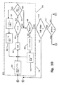
この息分析システム10の基本要素は、図2を参照すれば更に詳細に理解できる。息分析装置12は、サンプリング装置30と、これに接続されたマウスピース28を備えている。該サンプリング装置30は、患者から吐き出された息の一部を採取するが、このサンプルとしては肺の深い部分の肺胞からの息であることが望ましい。この理由は、息の最初の部分が通常「デッド・スペース」エア、つまり、気管、口、又は鼻腔などの上部気道からのものであるからである。かかるデッド・スペース・エアには診断を行うために有益な多数の成分が含まれていない。尚、通常、息の最初の150ミリリットルがデッド・スペース・エアである。一回の息で約500ミリリットルが吐き出され、息の90%は窒素と酸素である。かかる息サンプルは、サンプルの定量及び定性分析に使用する装置に応じて、チャンバ又はトラップ、或いはこれらの両方で採取可能である。通常、トラップには、化学的なもの、極低温のもの、及び吸着性のものの3種類がある。
【0008】
サンプルとしては、ユーザーの息を表し且つその他の影響によって汚染されていないことが重要である。一方、システム10は、時々較正する必要がある。この較正は、成分が既知のガスをサンプリング装置に注入することによって実施可能である。このためにガスを充填したキャニスターが使用されうる。又、使用後にサンプリング装置を浄化して余分な湿気やその他の成分を排出することも重要である。この段階もガスを注入することによって実施可能であり、これらの較正と浄化の2つの機能を1つの段階で実行することもできる。尚、分析装置の中には、ほかのものと比べて比較的安定しており較正の必要が少ない種類も存在する。例えば、キャビティリングダウン分光法の場合、基準、即ち「ゼロ」較正は必要であるが、関連するレーザー又はキャビティを変更しない限り安定している。
【0009】
尚、較正は重要ではあるが、本発明においては、患者のプロファイル又は履歴を維持管理することによって絶対的な基準への依存を減らしている。例えば、患者は、通常、サンプルとして一定容積の息を提供するが、この容積は患者によって異なる。しかしながら、患者ごとに記録を維持管理しているため、サンプルの容積やその他の繰り返し又は一貫性のあるバックグラウンド要素(例えば、空気の質)の重要度を削減又は除去しうる。これはこの装置の有用性に貢献する特徴であり、例えば、個人の自宅において家族の各メンバーは、サンプルを提供すると共に身元情報をコンピュータに入力して自分自身のプロファイルを作成する。
【0010】
サンプルの一部は、まず定量分析装置32によって処理される。かかる定量分析装置には、レーザー分光装置、キャビティリングダウン分光計、或いは定量測定を実行可能な「電子鼻」センサーアレイなどが含まれる。電子鼻センサーシステムには、いくつかの異なる種類の半導体センサー要素によるものが存在するが、最も高感度のものは10メガヘルツ帯で稼動するポリマーコーティングした表面音波(SAW)発振器によるものである。各々の要素は、フェムトグラム(10−15グラム)レベルの吸収質量を簡単に検出可能である。気相のサンプルにさらすことによって、要素の質量の変化パターンが周波数の変化として表され、これが信号処理ネットワークによって解析される。これらの「ニューラルネットワーク」は、既知の化合物に以前さらしたときに「学習」したターゲット気体を表す既知の反応特性とこれらのパターンを比較する信号処理の演算レイヤである。分析が完了すると、本システムは、通常、統計的な重要性又は正確性の確率に従ってその結果を報告する。尚、電子鼻センサーの利点としては、可動部がないことによるそのコンパクトなサイズと安価なコストなどが挙げられる。オンチップメモリの容量と信号処理速度が向上すれば、気体の追跡における電子鼻センサーアレイの有用性は更に向上するであろう。
【0011】
本システムには、更に定性分析装置34も備えることができる。この定性分析用の構成においても電子鼻センサーアレイを使用できる。これ以外にも、イオン移動度分光計検出器、音波検出器、及び光ファイバ検出器などが使用可能である。そして、定量分析装置32と定性分析装置34の両方からの処理データは、コンピュータ16のメモリ22に保存される。尚、定量及び定性分析装置としては、信頼性、正確性、及びコストを考慮し、半導体技術によるものが望ましい。
【0012】
本発明においては、患者のデータは、一定期間にわたって複数のサンプルを取得できるように保存される。この結果、その患者のベースラインが設定され、結果データの傾向分析を実行できる。そして、患者の息の長期的な状態に大きく且つ有意な変化が発生すれば、この変化データを通信網26によって医師又は健康管理サービス提供者に送信できる。従って、キーボード18などのユーザーインターフェイスによって患者14を識別することが重要である。更に、クロック36を設け、コンピュータ16に接続する必要がある。パーソナルコンピュータは、通常、水晶振動子によるリアルタイムクロックを内蔵している。コンピュータ16は、例えば、日、週、又は月などの一定期間にわたって発生する単一のデータ採取セッション及び複数のデータ採取セッションの際に採取した複数のサンプルを区別する必要がある。時系列における息成分の変化の割合は、患者の健康、食生活、又はその他の状態に変化が生じたかどうかを判定するのに重要である。又、更なるセンサー37を追加してもよい。これらの追加センサーとしては、サンプルを採取した状態を判定するための環境情報を提供する温度計、バロメータ、湿度計などの各種センサーが含まれる。又、センサーとして、更に、患者用の体温計や脈拍及び血圧センサー等の患者に関する情報を取得するセンサーを含めてもよい。かかるセンサー37の出力は、息分析で取得したデータと共に保存し、息成分における変化が有意であるかどうかを判定するのに使用する。
【0013】
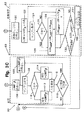
分析装置12及びコンピュータ16によるサンプルの処理は、図3A、3B、及び3Cに示されているフローチャートを参照すれば更に詳しく理解できる。図3は、図3A、3B、及び3Cの互いの関係を表しており、この統合図は患者の息を分析する処理システム50を示している。まず、図3Aに示すように、実施する試験を選択することにより(ステップ52)、システム50を特定の患者に合わせてカスタマイズする必要がある。尚、本装置を使用する際には、いつでも(特に、患者の状態の変化やその他の理由により)患者のプロファイルに試験を追加したり、削除したりできる。そして、実施可能なテストの種類としては、二酸化炭素の含有量、息の温度、アルコール、脂質分解生成物、芳香族化合物、チオ化合物(thio compounds)、アンモニア及びアミン又はハロゲン化合物などがある。これらの成分検出における有用性の例としては、例えば、呼気中のアセトンなどの脂質分解生成物は糖尿病を監視するのに有用である。又、メタンチオール、エタンチオール、或いは硫化ジメチルなどのチオール化合物は、診断において乾癬や排卵などの様々な広範囲な状態を検出するのに有用である。そして、アンモニアの増加は肝臓の病気と関連付けられる。ハロゲン化合物は環境的又は産業的な汚染物質を表す。
【0014】
更に別の試験として、医師の指示に基づいて患者が診断用の試薬を摂取した後に、特定の息成分の分析を行う一連の試験も実施可能である。例えば、尿素、特にC13標識尿素又はC13標識炭水化物を口経摂取して息の中のC13に基づいたCO2を分析し、患者の胃の内壁にヘリコバクターピロリ菌が感染しているかどうか(尿素⇒NH3とCO2)、又は炭水化物の吸収不良、ブドウ糖不耐症、ラクターゼ欠乏症、或いは小腸細菌の異常増殖を判定する。尚、同位元素炭素13はレーザー分光法によって識別できる。これについては、例えば、英国特許第2,218,514号を参照されたい。この結果データは、後述するように(ステップ140)、治療のために担当医に送信される。
【0015】
患者用の試験の選択を完了すると、システムを初期化し(ステップ54)、患者のベースライン、即ち長期的な息の状態履歴の作成を開始する。初期化及びこの後の段階のいずれにおいても、一定の期間にわたって試験を行う際に、ステップ56でサンプルを患者から採取する。そして次に、マイクロプロセッサ16は、この患者用に定量試験が選択されているかどうかを判定する(ステップ58)。定量試験が選択されている場合は、定量試験セグメント60を実行する。定量試験は、選択された成分αについて、定量試験装置32の容量に応じて、同時に或いは連続的に実施する。この試験は、前述のとおり、例えば、レーザー分光装置、キャビティリングダウンレーザー、電子鼻センサーアレイ、又はその他の定量分析装置などの適切な定量分析装置32を使用して実施する(ステップ62)。次に、前回保存されたデータ、即ちベースライン試験データをメモリから呼出し(ステップ64)、新しいデータとこの保存されていた試験データの間の変化量、即ちデルタ情報を判定する(ステップ66)。そして、新しい試験データとデルタ情報をメモリ22に保存する(ステップ68)。試験した成分αが、選択されている定量試験の最後の成分であるかどうかをステップ70で判定する。それが最後の成分又はαでない場合は、ステップ72で新しいαを設定し、この更なる成分αに対する試験を実施する。この試験は、定量分析装置32が複数の分析を行う能力を備えている場合には、単一のサンプルに対して同時に実行することが可能であるが、そうでない場合には、ステップ73で患者の更なるサンプルを要求して連続的に実行する。例えば、キャビティリングダウン分光法は複数の成分を同時に測定する能力を備えている。最後の定量試験が完了すると、本装置の制御装置はステップ74において定性試験を行う必要があるかどうかを問い合わせる。
【0016】
定性テストを実施しない場合には、後程詳述するように、報告プロセス76によってデータを報告する。一方、定性試験を実施する場合であるが、該試験には3つの種類が存在する。まず、その第1は、息成分の存在自体が患者の健康に重要な場合である78。これについては図3Bを参照されたい。これは、患者の長期的な息成分の監視で存在しなかった息成分が新しい試験において検出された場合に特に重要である。この逆の変化、即ち、従来存在していたものが新しい試験において検出されない場合も重要である。本装置においては、メモリ22内で患者のデータ履歴を維持管理しているため、いずれの状態をも検出できる。
【0017】
2番目の種類は、新しく検出された成分が所定の範囲80内におさまっていることが重要な場合である。これについては図3Cを参照されたい。特定成分の存在は定性分析装置34を使用すれば検出可能であるが、その範囲の算定も、定性分析装置を操作することによって実行可能である。この点は、特定成分に対する定量分析装置の使用が経済的に困難な場合にも概算値の取得が可能だという点で重要であるが、担当医に対してより詳細な分析の必要性を通知したり、食事療法などのように患者に減量又は糖尿病の治療を受けさせたりするには概算値で十分である。
【0018】
3番目は、後程詳述するように、定性分析装置を使用してより厳密な概算値82を取得可能な場合である。これについては図3Cを参照されたい。以上の存在と範囲及び概算値の両方の試験結果は定量試験の結果と共に報告76される。
【0019】
次に図3Bを参照すれば、成分βの存在78を定性分析装置(例:電子鼻センサーアレイ)を使用して試験するため、検出レベル(「LODβ」)で所望の成分を検出するための対象患者の前回の設定を呼出している(ステップ84)。次に、ステップ86で定性試験を実行し、ステップ88において成分βが存在するかどうかを判定する。この成分βの試験結果が陰性であれば、最小限、即ちLODβの更に高感度な設定を使用可能かどうかを判定する(ステップ90)。そして、感度を高めることが可能な場合は、必要に応じて、感度を最高、即ちLODMINに調整し(ステップ92)、患者の更なるサンプルを要求して(ステップ94)試験を再び実施する(ステップ86)。尚、定性分析装置の中には、追加サンプルの取得(ステップ94)が不要なものがある。しかしながら、定性試験による連続的な概算推定によって定量的な概算値を取得するには、時々追加サンプルを採取することが必要である。この場合、コンピュータ16は、ユーザー14に対して追加サンプル提供の必要性を通知する。このような最初及び追加のサンプルは、単一のデータ採取イベントと見なしうる。
【0020】
ステップ88で成分が存在すると判定されるか、又は最小限のLODβ設定が既に使用されている場合、その検出装置の性能の限界内でその成分が存在しないことを示している場合、該成分が、試験が必要な最後の成分βであるかどうかを判定する必要がある。最後の成分でない場合には、次の成分の試験を開始し(ステップ98)、ここでも追加サンプルの取得(ステップ100)が必要になる。但し、定量テストと同様に、単一のサンプル又はサンプル・サイクルで複数の成分を同時に識別することも可能である。特に、電子鼻センサーアレイなどのパターン認識型の技術がこれに該当する。又、調整可能なダイオードレーザーも複数成分を識別する能力を備えている。このように、息中に存在する特定成分の診断上の重要性及び特定成分の含有量に加え、他の成分でおなじみの存在パターンにおける当該成分の存在も診断においては重要である。
【0021】
定性的な成分の識別を完了した後に、ステップ102においてそれらの成分のいくつかを定量化することが望ましい場合がある。無論、定量化が必要なのは存在が確認された成分のみである。定量的な概算値が不要な場合は、報告書76を再度生成する。そして、定量的な概算値が必要な場合は、範囲が要求されているのか、それとも、より厳密な概算値が求められているのかを判定する(ステップ104)。
【0022】
範囲が必要な場合は、図3Cに示すように、範囲試験80を開始する。まず患者の第1限度をメモリ22から呼び出す(ステップ106)。この場合、定性分析装置はその成分を認識するのに十分な感度を有していないので、成分βが検出されないように検出レベルLODを特定レベルに設定することが必要である。(これは、選択した最大値より成分が少ないことを意味している)。そして、必要に応じて、新しいサンプルを取得し(ステップ108)、その検出レベルLODで成分βが存在するかどうかを判定する(ステップ110)。その結果、成分βが検出されない場合には、選択した限度より成分が少ないことを報告する(ステップ112)。一方、成分が依然として検出される場合には、成分の濃度が選択した限度を超えていることを報告する(ステップ114)。そして、特定の成分が選択した基準を満たしたか、又は満たさなかったかを表すこのデータを保存する(ステップ116)。これによって、該成分が健康的な十分低いレベルにあるのか、或いは健康的な範囲を超えているのかを十分に判定できる。尚、成分を最大値と最小値の範囲内におさめる必要がある場合には、第2限度の試験(ステップ118)を実行する必要がある。この第2限度の試験を実施する場合は、新しいLODを設定し(ステップ120)、この第2の設定で上述のサイクルを繰り返す。次に、試験結果は、報告セクション76に渡される。
【0023】
更に、図3Cに示すように、定性試験装置を使用し、サブルーチン82において成分の定量的なレベルの概算値を取得することも望ましい。これは、定性分析装置の検出レベルを調整して反復試験を実施することによっても実行可能である。長期間にわたって患者のデータが維持管理されているため、成分βの前回の検出レベルをステップ120においてメモリから呼び出すことができる。これが現在の成分のレベルを検索するための出発点を提供する。新しい検出レベルLODNEWは、前回の検出レベルに選択した定数又は「デルタ」を加算又は減算することによって得られる(ステップ122)。このLODNEWは、(LODLAST+最小値LODMIN)/2からなる新しい概算値を取得することによって小さくする必要がある(ステップ130)。LODNEWとLODLASTの差が事前に選択した限度より小さいと判定された場合には(ステップ132)、所望の精度を達成しているため、このプロセスを停止する必要がある。次に、この情報は、より先に保存される(ステップ134)。そうでなければ、マイクロプロセッサは、LODNEWを既存のサンプル又は新しいサンプル(ステップ124)に適用してこのプロセスを繰り返す。
【0024】
以上、定性分析装置を利用して定量的な概算値を取得する一方法について説明したが、当業者には公知である数値解析法によれば、本発明の開示内容を逸脱することなく同様の結果を取得可能なその他の技法が存在する。
【0025】
定量試験60、存在試験78、範囲試験80、及び定性的な概算値の推定82から取得した結果を報告アルゴリズム76を使用してコンピュータ16によってチェックする。コンピュータ16は、医師が選択した患者のプロファイルの設定に従い、或いは選択した試験52を識別するステップの一部として、成分α(定量的)又はβ(定性的)における重要な変化をチェック(ステップ136)する必要がある。そして、患者の長期的な状態からの有意な逸脱は、患者に報告され(ステップ138)、通信接続26を介した送信(ステップ140)によって医師又は健康管理サービス提供者に報告される。更に、所定レベルを超過するか又は受入れ可能なレベルを下回った重要な成分も報告される。尚、情報通信網26の双方向通信によって、遠隔地に所在する介護者は、遠隔地から追加の試験を選択したり、装置の自己診断を実行したり、装置の設定や試験に関連するその他の機能を実行することができる。
【0026】
患者の長期にわたる息分析の履歴を維持管理することによって、本装置は、患者の治療や健康に有意な大きな変化を識別できる。環境の影響と患者ごとのばらつきは、各々の患者のベースライン状態を設定することによって削減又は除去することができる。以上説明した試験はステップ142で終了し、別の機会にこの試験を更に実施することにより、患者は自分の状態を時系列に監視できる。
【0027】
患者の状態における有意な変化は、適切な統計又は分析技法によって識別可能である。このような複合データにおける有意な変化を判定する技法の1つがBeebeその他による米国特許第5,592,402号に開示されており、この引用によってその内容は本明細書に包含される。選択した試験によって識別された息成分は、異常が存在するかどうかを判定するのに分析可能な複合のデータセットである。逸脱は、それらの値の平均値と予想される統計的な偏差の組み合わせを判定可能な較正セットを設定することによって識別できる。所定レベルの逸脱、例えば予想平均値からの標準偏差の3倍を超える逸脱は統計的に有意であると宣言し、この基準に従って報告可能である。これらの平均値と統計的な偏差は、初期試験期間、即ち制御された状態で取得した一連の初期サンプルを取得することによって設定可能であり、本装置によって累積の平均と偏差を計算するか、又は移動の平均と偏差を維持管理することによってこれらを継続的に更新してもよい。更に、データセットが複雑な場合は、様々なサブパートに分割して有意な逸脱を更に識別することができる。このようなサブパートには、ピーク又は最小値、雑音、ベースライン・オフセット又はベースライン形状などがある。これらのサブパートの各々を監視し、それらが分析において予想される正常な範囲におさまっているかどうかを確認する。これは、どの種類の特性が異常なのかを識別するのに有用である。例えば、複数の患者が特定の息成分について同一の絶対値を示すことがある。この場合、ある患者においては、元々ベースラインのレベルが非常に高いケースがありうる。又、別の患者においては、ベースラインが鋭く上昇したという結果になっているかもしれない。更に別の患者においては、ベースラインが徐々に下がっているという場合もある。更に別の患者においては、この値が大きな変化を生じさせており、ピーク値を超過して統計的に有意になっている場合もある。或いは又、別の患者においては、その選択した成分について定常的にもっと大きなばらつきを示しており、その値の変化自身は統計的に有意ではない場合もありうる。このように、患者別に学習したパターンに基づいて、患者ごとに、異なる報告を提供することができるのである。無論、所定成分の絶対的な最大値又は最小値を設定し、これらの最大値又は最小値を超過した測定値を患者の履歴に関係なく報告することも可能である。
【0028】
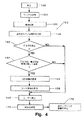
息分析装置10の使用方法を、図4のフローチャート150を参照して更に詳細に説明する。フローチャート150に示すように、呼気分析装置10の使用手順は較正(ステップ152)から始まる。これは、組成が既知のガスを本装置に注入することによって実施可能であり、かかるガスを充填したキャニスターをこの目的に使用してもよい。較正を完了すると、サンプルを採取する(ステップ154)。このステップには、図3を参照して先ほど詳述した手順が含まれる。次に、分析装置10は、前述の追加センサー37を使用してステップ156において環境データを取得する。そして、分析装置10は、保存されている患者の履歴を現在の読取り内容と比較し(ステップ158)、変化しているかどうかを判定する(ステップ160)。変化している場合は、患者の履歴とステップ156で取得した環境要素の観点からその変化が有意であるかどうかを判定する(ステップ162)。その変化が有意であると判定された場合には、分析装置は追加の試験を要求することができる(ステップ164)。この追加試験としては、試験対象の一連の成分には通常含まれていない別の成分の更なる息試験、繰り返し試験、又はセンサー37によって測定する追加の試験、例えば血圧、血液酸素(例えば、患者の指に取り付けた赤外線センサーによる)、心拍、体温などが含まれる。尚、心臓ペースメーカのプログラミング及びデータ転送用ワンドは、このようなセンサー37の1つである。心臓ペースメーカは、しばしば、ペーシングビート数、位置異常ビート数、心房細動又は不整頻拍の発生、又は(電気除細動器/細動除去器用の)心室細動又は頻脈性不整脈などの履歴データを保存する。施した治療に関する情報、しきい値レベル、記録された心電図をペースメーカ又は植え込み型電気除細動器/細動除去器によって保存することができる。この情報を植え込み型心臓電気刺激装置から送信した後に、本装置が維持管理するデータレコードと関連付けることができる。尚、このようなデータ転送技法は公知である。
【0029】
本分析装置は、ユーザー、即ち患者に対してマイクロコンピュータのユーザーインターフェイス(例:キーボード又はマウス)を介して特定のデータを入力するよう要求することもできる。この要求データとしては、食事情報、知覚している一般的な健康状態、最近の運動の量と期間、及び息成分における激しい変化を説明する(即ち、その変化が実際には有意でないことを表す)ものや健康管理サービス提供に重要な情報を提供する類似の要素などが含まれる。
【0030】
かかる追加情報を収集した(ステップ164及び166)後、或いは変化がないか(ステップ160)、又は有意な変化がない(ステップ162)場合には、ユーザー用の報告書が生成され(ステップ168)、該情報が患者の履歴の一部として保存される。この報告書又はデータは、遠隔地の健康管理サービス提供者に対して、即時又はデータ要求に応答する形で送信できる(ステップ170)。最後に、サンプリング装置内における汚染物質の繁殖を防止するために本システムを浄化する(ステップ172)。前述のとおり、これは、組成が既知のガスを注入することによって実行可能であり、較正ステップ172と組み合わせてもよい。
【0031】
単一のサンプルに対する複数の試験は独立的に取り扱うこともでき、いくつかの試験結果を組み合わせて患者の状態或いは特定の成分又は成分セットの存在を表すテンプレート又はパターンを作成することも可能である。電子鼻の技術では、パターン認識を使用して特定成分の存在を検出している。又、単一のサンプルに対して複数のレーザーを使用し、検出とパターン認識をいつくかのレーザーの組み合わせの出力に対して適用できるように帯域幅を拡張することも可能である。単一のレーザーは、通常、特定の限られた周波数の光を放出する能力を備えている。この場合、周波数の多少の調整や変更は可能であるが、単一のレーザー装置が効率的に識別可能な要素や成分は、その選択されたレーザーの周波数特性によって制限される。本発明の検出装置34には、放射周波数が異なる複数のレーザーを含むことが可能である。サンプルの周りに物理的に配置するか、わずかにことなるタイミングで照射するか、或いは、その他の技法によって、これらのレーザーを単一のサンプルに照射できる。鏡、レンズ、又はプリズムなどの光学装置を使用し、選択したレーザーからのビームをサンプルを通って検出装置に向かう経路に沿って案内してもよい。この光学装置を調整すれば、その他のレーザーからのビームをサンプルを通る同一又は類似の経路に沿って案内することが可能である。異なる放射特性を備えたレーザーを同一のサンプルに使用することによって、広範囲なデータポイントセットの取得が可能になる。単一レーザーの3つ又は4つのデータポイントの代わりに、3つのレーザーを使用すれば、同じサンプルから12又はそれ以上のデータポイントを取得できる。この情報は、電子鼻技術によって取得される類似の情報に比べて、より選択的且つより量的に正確であると考えられる。そして、すべてのレーザービームからのこれらの正確な情報を、電子鼻技術に使用されているものと同様のパターン認識技法を使用して一緒に処理可能である。この結果、データパターンやデータパターンにおける変化を時系列に関連付けることによって、より広範囲な状態又は成分を識別することができる。
【0032】
尚、本発明の上述の実施例は模範的なものに過ぎず、当業者であれば、本発明の開示内容を逸脱することなく、設計又は構成に様々な変更や変形が可能であることを理解するであろう。従って、本発明の範囲は、添付の請求項によって規定されるものである。
【図面の簡単な説明】
【図1】
本発明による診断用息分析システムの透視図である。
【図2】
図1のシステムのブロックダイアグラムである。
【図3】
図3A、図3B、図3Cの関係を表す図である。
【図3A】
図1に示すシステムのフローチャートの一部である。
【図3B】
図1に示すシステムのフローチャートの別の部分である。
【図3C】
図1に示すシステムのフローチャートの最後の部分である。
【図4】
図1に示すシステムの使用方法を含む更なるフローチャートである。[0001]
The present invention relates to medical devices, and more particularly to devices for analyzing medically important components in exhaled breath.
[0002]
It has been a long time since the possibility of using exhaled breath as a tool in diagnosis has been recognized. In the past, Hippocrates taught doctors to look at a patient's breath as a clue to their condition. In 1784, Antoine Lavoisier and Pierre Laplace analyzed guinea pig breathing and found that animals inhale oxygen and exhale carbon dioxide. This finding was the first direct evidence that the body gained energy from food using the burning process. Since then, as many as 200 compounds have been detected in human breath, some of which have been linked to various diseases.
[0003]
Various different technologies are employed in these breath component detection devices. For example, US Patent No. 5,422,485 to Bowlds and US Patent No. 5,515,859 to Paz use infrared light to measure alcohol content in breath. In U.S. Pat. No. 5,543,621 to Sauke et al., A laser diode spectrometer is used. In addition, various other laser and absorption spectrometers, such as cavity-ringdown spectroscopy, have been used. For example, "Absorptive spectroscopy: from the dawn to cavity ring-down spectroscopy"; A. Paldus and R.S. N. See Zare, American Chemical Society Symposium Series (1999), No. 720, pp. 49-70. Still other techniques include direct headspace analysis, gas liquid chromatography, atmospheric pressure ionization mass spectrometry, tandem mass spectrometry and chemistry. See, for example, "Possibility of Breath Analysis in Diagnosis", Antony Manolis, Clinical Chemistry, 29/1 (1983), pp. 5-15. Some of the chemical sensors are so-called “electronic nose”, which identifies components based on physical or chemical characteristic patterns. This type of sensor is commercially available, for example, from Cyrano Sciences, Inc. of Pasadena, CA, and has been suggested for use in detecting medical conditions such as pneumonia, halitosis, and melanoma.
[0004]
However, many of these techniques are complex and expensive and difficult to calibrate. They have not heretofore been used for personal level health care for economic reasons. However, it has been suggested that multiple humans can conduct spontaneous breath alcohol tests at bars and other places that serve alcoholic beverages and use them to detect predetermined alcohol levels in the breath. (U.S. Patent No. 5,303,575 to Brown et al.).
[0005]
The present invention discloses a medical breath component analyzer that maintains a database profile of a patient in time series, which overcomes problems such as difficulty in calibration and variations among patients. The present invention is intended for long-term use by a patient so that the patient's baseline condition can be determined. Large deviations from baseline are identified as clinically significant. The acquired data is reported to the patient by a device installed in the patient's home, and is transmitted electronically to a doctor or a health care service provider. A plurality of tests can be performed, such as a quantitative test, a qualitative test, and a quantitative estimation by a qualitative analyzer. In particular, laser spectroscopy with multiple lasers of different output characteristics can be used on a single breath sample. When the plurality of laser outputs are put together, a template or pattern (patient characteristics) can be formed, and thereby a complicated state can be easily identified. A series of tests can be selected for a patient and customized for that patient's condition. When a change in the state is detected, more environmental information and information provided by the user are acquired, and it can be determined whether or not the change is clinically significant.
[0006]
The present invention will now be described with reference to the accompanying drawings, in which the same elements are denoted by the same reference numerals. First, FIG. 1 shows a
[0007]
The basic elements of the
[0008]
It is important that the sample represents the user's breath and is not contaminated by other effects. On the other hand, the
[0009]
It should be noted that while calibration is important, the present invention reduces reliance on absolute criteria by maintaining a patient profile or history. For example, a patient typically provides a fixed volume of breath as a sample, which volume varies from patient to patient. However, maintaining records on a patient-by-patient basis may reduce or eliminate the importance of sample volume and other repetitive or consistent background elements (eg, air quality). This is a feature that contributes to the usefulness of this device, for example, at the home of an individual, each member of the family provides a sample and enters identity information into a computer to create his own profile.
[0010]
A part of the sample is first processed by the
[0011]
The system may further include a
[0012]
In the present invention, patient data is stored so that multiple samples can be obtained over a period of time. As a result, a baseline is set for the patient, and a trend analysis of the result data can be performed. Then, when a significant and significant change occurs in the long-term state of the patient's breath, the change data can be transmitted to the doctor or the health care service provider through the
[0013]
The processing of the sample by the
[0014]
As still another test, a series of tests for analyzing a specific breath component after a patient takes a diagnostic reagent based on a doctor's instruction can be performed. For example, urea, especially C Thirteen Labeled urea or C Thirteen C in the breath by oral ingestion of labeled carbohydrates Thirteen CO based on 2 To determine whether Helicobacter pylori has infected the inner wall of the patient's stomach (urea ⇒ NH 3 And CO 2 ) Or carbohydrate malabsorption, glucose intolerance, lactase deficiency, or abnormal growth of small intestinal bacteria. The isotope carbon 13 can be identified by laser spectroscopy. See, for example, UK Patent No. 2,218,514. This result data is transmitted to the attending physician for treatment, as described below (step 140).
[0015]
Upon completing the selection of the test for the patient, the system is initialized (step 54) and the creation of the patient's baseline, or long-term breathing state history, begins. At both the initialization and subsequent stages, a sample is taken from the patient at
[0016]
If a qualitative test is not performed, the data is reported by a
[0017]
The second type is when it is important that the newly detected component falls within the
[0018]
Third, as will be described in detail later, a case where a more precise
[0019]
Referring now to FIG. 3B, to test for the
[0020]
If it is determined in
[0021]
After completing the qualitative component identification, it may be desirable to quantify some of those components in
[0022]
If a range is required, a
[0023]
Further, as shown in FIG. 3C, it is also desirable to use a qualitative tester to obtain an estimate of the quantitative level of the component in
[0024]
As described above, one method of obtaining a quantitative approximate value using a qualitative analyzer has been described.However, according to a numerical analysis method known to those skilled in the art, similar methods can be used without departing from the disclosure of the present invention. There are other techniques by which results can be obtained.
[0025]
The results obtained from the
[0026]
By maintaining a history of a patient's long-term breath analysis, the device can identify significant changes in the treatment or health of the patient. Environmental effects and patient-to-patient variability can be reduced or eliminated by setting a baseline condition for each patient. The test described above ends at
[0027]
Significant changes in the patient's condition can be identified by appropriate statistical or analytical techniques. One technique for determining significant changes in such composite data is disclosed in U.S. Patent No. 5,592,402 to Beebe et al., The contents of which are hereby incorporated by reference. The breath component identified by the selected test is a composite dataset that can be analyzed to determine if an abnormality is present. Deviations can be identified by setting up a calibration set that can determine a combination of the average of those values and the expected statistical deviation. Deviations of a predetermined level, e.g., more than three standard deviations from the expected mean, are declared statistically significant and can be reported according to this criterion. These averages and statistical deviations can be set during the initial test period, i.e. by taking a series of initial samples taken in a controlled manner, by calculating the cumulative average and deviation by the device, Alternatively, they may be updated continuously by maintaining the mean and deviation of the movement. Furthermore, if the data set is complex, it can be divided into various subparts to further identify significant deviations. Such subparts include peaks or minima, noise, baseline offsets or baseline shapes. Monitor each of these subparts to see if they fall within the normal range expected in the analysis. This is useful for identifying which type of property is abnormal. For example, multiple patients may exhibit the same absolute value for a particular breath component. In this case, in some patients, the baseline level may be originally very high. In other patients, the result may be a sharp rise in the baseline. In still other patients, the baseline may be gradually decreasing. In yet another patient, this value is causing a significant change and may exceed the peak value and become statistically significant. Alternatively, in another patient, the selected component may be constantly exhibiting greater variability, and the change in value itself may not be statistically significant. Thus, different reports can be provided for each patient based on the patterns learned for each patient. Of course, it is also possible to set an absolute maximum or minimum value for a given component and report measurements that exceed these maximum or minimum values, regardless of the patient's history.
[0028]
The method of using the
[0029]
The analyzer may also require a user, ie, a patient, to enter certain data via a microcomputer user interface (eg, keyboard or mouse). The request data may include dietary information, perceived general health, amount and duration of recent exercise, and drastic changes in breath components (i.e., indicate that the changes are not actually significant) ) And similar elements that provide important information for providing health care services.
[0030]
After collecting such additional information (
[0031]
Multiple tests on a single sample can be treated independently, and several test results can be combined to create a template or pattern that is representative of the patient's condition or the presence of a particular component or set of components. . Electronic nose technology uses pattern recognition to detect the presence of specific components. It is also possible to use multiple lasers for a single sample and extend the bandwidth so that detection and pattern recognition can be applied to the output of some laser combinations. A single laser typically has the ability to emit light at a particular limited frequency. In this case, although some adjustment or change of the frequency is possible, the elements and components that can be effectively identified by a single laser device are limited by the frequency characteristics of the selected laser. The
[0032]
The above embodiments of the present invention are merely exemplary, and those skilled in the art will recognize that various changes and modifications can be made in the design or configuration without departing from the disclosure of the invention. Will understand. Accordingly, the scope of the present invention is defined by the appended claims.
[Brief description of the drawings]
FIG.
1 is a perspective view of a diagnostic breath analysis system according to the present invention.
FIG. 2
2 is a block diagram of the system of FIG.
FIG. 3
It is a figure showing the relationship of FIG. 3A, FIG. 3B, and FIG. 3C.
FIG. 3A
2 is a part of a flowchart of the system shown in FIG. 1.
FIG. 3B
2 is another part of the flowchart of the system shown in FIG. 1.
FIG. 3C
It is the last part of the flowchart of the system shown in FIG.
FIG. 4
2 is a further flowchart including a method of using the system shown in FIG. 1.
Claims (22)
前記分析装置に接続され、前記分析装置から息成分信号を受信するコンピュータと、
前記コンピュータに接続されたメモリと、
前記メモリ内に保存され、少なくとも第1息成分信号を表すデータ構造と、
前記保存されている第1成分信号のデータ構造を少なくとも第2息成分信号と比較するコンピュータプログラムとを有することを特徴とする医療装置。A medical device having a breath component analyzer,
A computer connected to the analyzer and receiving a breath component signal from the analyzer;
A memory connected to the computer;
A data structure stored in the memory and representing at least a first breath component signal;
A computer program for comparing the stored data structure of the first component signal with at least a second breath component signal.
前記第1息成分プロファイルをコンピュータがアクセス可能なメモリに保存し、
前記患者から第2息サンプルを採取し、
前記サンプルを分析して第2息成分プロファイルを作成し、
前記第1及び第2息成分プロファイルを比較するステップを有することを特徴とする方法。A method for analyzing a breath component of a patient, comprising the steps of: obtaining a breath sample from the patient and creating a first breath component profile by analyzing components of the sample,
Storing the first breath component profile in a computer accessible memory;
Collecting a second breath sample from the patient;
Analyzing the sample to create a second breath component profile;
Comparing the first and second breath component profiles.
Applications Claiming Priority (2)
| Application Number | Priority Date | Filing Date | Title |
|---|---|---|---|
| US18403900P | 2000-02-22 | 2000-02-22 | |
| PCT/US2001/004112 WO2001063277A1 (en) | 2000-02-22 | 2001-02-08 | Personal computer breath analyzer for health-related behavior modification and method |
Related Child Applications (1)
| Application Number | Title | Priority Date | Filing Date |
|---|---|---|---|
| JP2012040185A Division JP2012143569A (en) | 2000-02-22 | 2012-02-27 | Medical apparatus, and method for analysis of patient breath |
Publications (1)
| Publication Number | Publication Date |
|---|---|
| JP2004508534A true JP2004508534A (en) | 2004-03-18 |
Family
ID=22675333
Family Applications (2)
| Application Number | Title | Priority Date | Filing Date |
|---|---|---|---|
| JP2001562191A Withdrawn JP2004508534A (en) | 2000-02-22 | 2001-02-08 | Personal computer breath analyzer for health related behavior modification and its method |
| JP2012040185A Pending JP2012143569A (en) | 2000-02-22 | 2012-02-27 | Medical apparatus, and method for analysis of patient breath |
Family Applications After (1)
| Application Number | Title | Priority Date | Filing Date |
|---|---|---|---|
| JP2012040185A Pending JP2012143569A (en) | 2000-02-22 | 2012-02-27 | Medical apparatus, and method for analysis of patient breath |
Country Status (6)
| Country | Link |
|---|---|
| US (1) | US20010037070A1 (en) |
| EP (1) | EP1259807A1 (en) |
| JP (2) | JP2004508534A (en) |
| AU (1) | AU2001233341A1 (en) |
| CA (1) | CA2401011A1 (en) |
| WO (1) | WO2001063277A1 (en) |
Cited By (3)
| Publication number | Priority date | Publication date | Assignee | Title |
|---|---|---|---|---|
| JP2009518654A (en) * | 2005-12-06 | 2009-05-07 | アプライド・ナノテック・ホールディングス・インコーポレーテッド | Gas analysis |
| JP2017192724A (en) * | 2014-10-17 | 2017-10-26 | クアルコム,インコーポレイテッド | Breathprint sensor systems, smart inhalers and methods for personal identification |
| JP2019512099A (en) * | 2016-02-19 | 2019-05-09 | パトノミクス アクチエ ボラグPatonomics Ab | Method and apparatus for identifying a temporary emotional state of a living mammal |
Families Citing this family (38)
| Publication number | Priority date | Publication date | Assignee | Title |
|---|---|---|---|---|
| US20050177391A1 (en) * | 2001-10-23 | 2005-08-11 | Hideki Shimizu | Health management system and health management program |
| CN1620502A (en) | 2001-11-09 | 2005-05-25 | 陶氏环球技术公司 | Enzyme-based system and sensor for measuring acetone |
| US7794994B2 (en) | 2001-11-09 | 2010-09-14 | Kemeta, Llc | Enzyme-based system and sensor for measuring acetone |
| US7153272B2 (en) * | 2002-01-29 | 2006-12-26 | Nanotherapeutics, Inc. | Methods of collecting and analyzing human breath |
| AU2003220065A1 (en) | 2002-03-04 | 2003-09-22 | Cyrano Sciences, Inc. | Detection, diagnosis, and monitoring of a medical condition or disease with artificial olfactometry |
| US7101340B1 (en) | 2002-04-12 | 2006-09-05 | Braun Charles L | Spectroscopic breath profile analysis device and uses thereof for facilitating diagnosis of medical conditions |
| AU2003238288A1 (en) | 2003-06-19 | 2005-02-04 | Everest Biomedical Instruments | Breath end-tidal gas monitor |
| CA2584565A1 (en) | 2004-10-28 | 2006-05-04 | Naoki Urushihata | Disease diagnosing system |
| US7874992B2 (en) * | 2006-01-31 | 2011-01-25 | Medtronic, Inc. | Method for continuous baroreflex sensitivity measurement |
| WO2008093263A2 (en) * | 2007-01-30 | 2008-08-07 | Koninklijke Philips Electronics N.V. | Breath analysis device |
| US20090163825A1 (en) * | 2007-12-19 | 2009-06-25 | The Cooper Health System | Non-Invasive Method and System of Signaling a Hyper or Hypoglycemic State |
| US9844333B2 (en) | 2009-03-24 | 2017-12-19 | International Business Machines Corporation | Remote delivery and monitoring of health care |
| US11534081B2 (en) | 2009-08-06 | 2022-12-27 | Peter Theophilos Banos | Methods of and devices for monitoring the effects of cellular stress and damage resulting from radiation exposure |
| US9420971B2 (en) | 2009-10-24 | 2016-08-23 | Carrot Sense, Inc. | Extracorporeal devices and methods for facilitating cessation of undesired behaviors |
| WO2011117572A1 (en) * | 2010-03-25 | 2011-09-29 | Isis Innovation Limited | Analysis of breath |
| GB201020086D0 (en) * | 2010-11-26 | 2011-01-12 | Hypo Safe As | Analysis of EEG signals to detect hypoglycaemia |
| US8814804B2 (en) | 2010-12-13 | 2014-08-26 | Iph, Llc | Interactive blood-alcohol content tester |
| AU2012358370B2 (en) * | 2011-12-21 | 2017-05-18 | Capnia, Inc. | Collection and analysis of a volume of exhaled gas with compensation for the frequency of a breathing parameter |
| KR101187735B1 (en) * | 2012-02-28 | 2012-10-08 | (주) 에이스엔 | Breath odor measurement system |
| US20130253358A1 (en) * | 2012-03-07 | 2013-09-26 | Menssana Research, Inc. | System and method for remote collection and analysis of volatile organic components in breath |
| ES2866183T3 (en) | 2013-01-08 | 2021-10-19 | Capnia Inc | Selection of breath for analysis |
| WO2014127044A1 (en) | 2013-02-12 | 2014-08-21 | Capnia, Inc. | Sampling and storage registry device for breath gas analysis |
| US9442103B1 (en) * | 2013-03-14 | 2016-09-13 | 1A Smart Start Llc | Anti-circumvention apparatus and methods for use in sobriety testing systems |
| GB2517702A (en) * | 2013-08-28 | 2015-03-04 | Ibm | Collaborative electronic nose management in personal devices |
| JP6698527B2 (en) | 2013-08-30 | 2020-05-27 | キャプニア, インク.Capnia, Inc. | Newborn carbon dioxide measurement system |
| JP2017083175A (en) * | 2014-03-14 | 2017-05-18 | 株式会社東芝 | Breath diagnosis device |
| US9152956B1 (en) | 2014-05-08 | 2015-10-06 | Sam G. Habash | Automated kiosk assembly |
| WO2016033382A1 (en) | 2014-08-27 | 2016-03-03 | Capnia, Inc. | Methods for immune globulin administration |
| JP6402992B2 (en) * | 2014-10-03 | 2018-10-10 | 株式会社タニタ | Gas measuring device, gas measuring system, gas measuring method, and gas measuring program |
| US20160249838A1 (en) * | 2015-02-28 | 2016-09-01 | Lawrence Cheng | Method and Apparatus for Effective Detection of Respiratory Blockage Using CO2 Monitor |
| CN107708548B (en) | 2015-04-07 | 2021-06-04 | 凯洛特公司 | System and method for quantification and prediction of smoking behavior |
| US10206572B1 (en) | 2017-10-10 | 2019-02-19 | Carrot, Inc. | Systems and methods for quantification of, and prediction of smoking behavior |
| US10604011B2 (en) | 2015-10-13 | 2020-03-31 | Consumer Safety Technology, Llc | Networked intoxication vehicle immobilization |
| KR102481493B1 (en) * | 2015-12-15 | 2022-12-27 | 삼성전자주식회사 | Electronic apparatus, method for controlling the same and computer-readable recording medium |
| US10663440B2 (en) | 2016-09-09 | 2020-05-26 | Consumer Safety Technology, Llc | Secure data handling in a breath alcohol calibration station |
| US10877008B2 (en) | 2016-09-09 | 2020-12-29 | Consumer Safety Technology, Llc | Reference gas management in a breath alcohol calibration station |
| WO2020160887A1 (en) | 2019-02-06 | 2020-08-13 | Unilever N.V. | A method of demonstrating the benefit of oral hygiene |
| JP2023509639A (en) | 2019-12-30 | 2023-03-09 | シラグ・ゲーエムベーハー・インターナショナル | Systems and methods for assisting individuals in behavior change programs |
Citations (6)
| Publication number | Priority date | Publication date | Assignee | Title |
|---|---|---|---|---|
| JPS63168565A (en) * | 1986-12-22 | 1988-07-12 | アボット・ラボラトリーズ | Ketone measuring method and tool |
| JPS63246673A (en) * | 1986-12-22 | 1988-10-13 | アボット・ラボラトリーズ | Method of detecting ketone and aldehydes and instrument thereof |
| JPH04204056A (en) * | 1990-11-30 | 1992-07-24 | Hitachi Ltd | Stress inspection method and device |
| JPH08173401A (en) * | 1994-08-30 | 1996-07-09 | Boc Group Plc:The | Disease management device |
| JPH0954040A (en) * | 1995-08-09 | 1997-02-25 | Kdk Corp | Method for optically measuring component in exhalation |
| JP2001507795A (en) * | 1997-01-02 | 2001-06-12 | オスメテック パブリック リミテッド カンパニー | State detection method by gas or vapor analysis |
Family Cites Families (8)
| Publication number | Priority date | Publication date | Assignee | Title |
|---|---|---|---|---|
| US5060656A (en) * | 1990-05-22 | 1991-10-29 | Aerosport, Inc. | Metabolic rate analyzer |
| US5296843A (en) * | 1991-03-28 | 1994-03-22 | Sd Laboratories, Inc. | Fluid or vapor diagnostic device |
| US5394236A (en) * | 1992-02-03 | 1995-02-28 | Rutgers, The State University | Methods and apparatus for isotopic analysis |
| WO1993021592A1 (en) * | 1992-04-16 | 1993-10-28 | The Dow Chemical Company | Improved method for interpreting complex data and detecting abnormal instrument or process behavior |
| GB9704676D0 (en) * | 1997-03-06 | 1997-04-23 | Aromascan Plc | Condition indicator |
| US6309360B1 (en) * | 1997-03-17 | 2001-10-30 | James R. Mault | Respiratory calorimeter |
| DE69838812T2 (en) * | 1997-09-11 | 2008-11-27 | Oridion Breathid Ltd. | BEST TEST ANALYSIS DEVICE |
| DE29902593U1 (en) * | 1999-02-13 | 1999-07-15 | Genzyme Virotech Gmbh | Gas analyzer for medical diagnostics |
-
2001
- 2001-02-08 JP JP2001562191A patent/JP2004508534A/en not_active Withdrawn
- 2001-02-08 AU AU2001233341A patent/AU2001233341A1/en not_active Abandoned
- 2001-02-08 CA CA002401011A patent/CA2401011A1/en not_active Abandoned
- 2001-02-08 EP EP01905467A patent/EP1259807A1/en not_active Withdrawn
- 2001-02-08 US US09/779,160 patent/US20010037070A1/en not_active Abandoned
- 2001-02-08 WO PCT/US2001/004112 patent/WO2001063277A1/en not_active Application Discontinuation
-
2012
- 2012-02-27 JP JP2012040185A patent/JP2012143569A/en active Pending
Patent Citations (6)
| Publication number | Priority date | Publication date | Assignee | Title |
|---|---|---|---|---|
| JPS63168565A (en) * | 1986-12-22 | 1988-07-12 | アボット・ラボラトリーズ | Ketone measuring method and tool |
| JPS63246673A (en) * | 1986-12-22 | 1988-10-13 | アボット・ラボラトリーズ | Method of detecting ketone and aldehydes and instrument thereof |
| JPH04204056A (en) * | 1990-11-30 | 1992-07-24 | Hitachi Ltd | Stress inspection method and device |
| JPH08173401A (en) * | 1994-08-30 | 1996-07-09 | Boc Group Plc:The | Disease management device |
| JPH0954040A (en) * | 1995-08-09 | 1997-02-25 | Kdk Corp | Method for optically measuring component in exhalation |
| JP2001507795A (en) * | 1997-01-02 | 2001-06-12 | オスメテック パブリック リミテッド カンパニー | State detection method by gas or vapor analysis |
Cited By (4)
| Publication number | Priority date | Publication date | Assignee | Title |
|---|---|---|---|---|
| JP2009518654A (en) * | 2005-12-06 | 2009-05-07 | アプライド・ナノテック・ホールディングス・インコーポレーテッド | Gas analysis |
| US8903474B2 (en) | 2005-12-06 | 2014-12-02 | Pen Inc. | Analysis of gases |
| JP2017192724A (en) * | 2014-10-17 | 2017-10-26 | クアルコム,インコーポレイテッド | Breathprint sensor systems, smart inhalers and methods for personal identification |
| JP2019512099A (en) * | 2016-02-19 | 2019-05-09 | パトノミクス アクチエ ボラグPatonomics Ab | Method and apparatus for identifying a temporary emotional state of a living mammal |
Also Published As
| Publication number | Publication date |
|---|---|
| WO2001063277A1 (en) | 2001-08-30 |
| US20010037070A1 (en) | 2001-11-01 |
| AU2001233341A1 (en) | 2001-09-03 |
| CA2401011A1 (en) | 2001-08-30 |
| JP2012143569A (en) | 2012-08-02 |
| EP1259807A1 (en) | 2002-11-27 |
Similar Documents
| Publication | Publication Date | Title |
|---|---|---|
| JP2004508534A (en) | Personal computer breath analyzer for health related behavior modification and its method | |
| US6609068B2 (en) | Personal computer breath analyzer for health-related behavior modification and method | |
| EP3624678B1 (en) | Systems and methods for assessing the health status of a patient | |
| Joshi et al. | iGLU 2.0: A new wearable for accurate non-invasive continuous serum glucose measurement in IoMT framework | |
| KR100459339B1 (en) | Glucose concentration measuring system | |
| US5460177A (en) | Method for non-invasive measurement of concentration of analytes in blood using continuous spectrum radiation | |
| US6968221B2 (en) | Low-cost method and apparatus for non-invasively measuring blood glucose levels | |
| EP0522674B1 (en) | Oximeter for reliable clinical determination of blood oxygen saturation in a fetus | |
| US20080004513A1 (en) | VCSEL Tissue Spectrometer | |
| US5086229A (en) | Non-invasive measurement of blood glucose | |
| US9220440B2 (en) | Determining a characteristic respiration rate | |
| US8986207B2 (en) | Systems and methods for providing sensor arrays for detecting physiological characteristics | |
| EP1437087B1 (en) | Blood component spectroscopy analysis system for removing abnormal data | |
| US5360004A (en) | Non-invasive determination of analyte concentration using non-continuous radiation | |
| US9198582B2 (en) | Determining a characteristic physiological parameter | |
| Takla et al. | The problem of artifacts in patient monitor data during surgery: a clinical and methodological review | |
| JP2006126219A (en) | Method and apparatus for multi-spectral analysis in noninvasive infrared spectroscopy | |
| US6064896A (en) | Non-invasive measurement of blood glucose using instruments that have less precise detection capability | |
| EP0623307A1 (en) | Non-invasive determination of constituent concentration using non-continuous radiation | |
| EP0623308A1 (en) | Method for non-invasive measurement of concentration of constituents in blood | |
| US10206628B2 (en) | Method to obtain and validate physiological data | |
| KR20050078924A (en) | Urine component analysis system and method by raman spectroscopy | |
| Alpert et al. | Evaluating the impact of motion artifact on noninvasive blood pressure devices | |
| Raguindin et al. | Predictive COPD Monitoring Device (PCMD) | |
| Sazonova et al. | Activity-based sleep-wake identification in infants |
Legal Events
| Date | Code | Title | Description |
|---|---|---|---|
| A621 | Written request for application examination |
Free format text: JAPANESE INTERMEDIATE CODE: A621 Effective date: 20080204 |
|
| A711 | Notification of change in applicant |
Free format text: JAPANESE INTERMEDIATE CODE: A711 Effective date: 20091225 |
|
| A521 | Written amendment |
Free format text: JAPANESE INTERMEDIATE CODE: A821 Effective date: 20091225 |
|
| A977 | Report on retrieval |
Free format text: JAPANESE INTERMEDIATE CODE: A971007 Effective date: 20100518 |
|
| A131 | Notification of reasons for refusal |
Free format text: JAPANESE INTERMEDIATE CODE: A131 Effective date: 20100525 |
|
| A521 | Written amendment |
Free format text: JAPANESE INTERMEDIATE CODE: A523 Effective date: 20100825 |
|
| A131 | Notification of reasons for refusal |
Free format text: JAPANESE INTERMEDIATE CODE: A131 Effective date: 20101012 |
|
| A601 | Written request for extension of time |
Free format text: JAPANESE INTERMEDIATE CODE: A601 Effective date: 20110111 |
|
| A602 | Written permission of extension of time |
Free format text: JAPANESE INTERMEDIATE CODE: A602 Effective date: 20110118 |
|
| A02 | Decision of refusal |
Free format text: JAPANESE INTERMEDIATE CODE: A02 Effective date: 20111025 |
|
| A521 | Written amendment |
Free format text: JAPANESE INTERMEDIATE CODE: A523 Effective date: 20120227 |
|
| A911 | Transfer to examiner for re-examination before appeal (zenchi) |
Free format text: JAPANESE INTERMEDIATE CODE: A911 Effective date: 20120404 |
|
| A761 | Written withdrawal of application |
Free format text: JAPANESE INTERMEDIATE CODE: A761 Effective date: 20120416 |