JP2004187545A - Method for examining nucleic acid sequence and method for preparing sample - Google Patents
Method for examining nucleic acid sequence and method for preparing sample Download PDFInfo
- Publication number
- JP2004187545A JP2004187545A JP2002357804A JP2002357804A JP2004187545A JP 2004187545 A JP2004187545 A JP 2004187545A JP 2002357804 A JP2002357804 A JP 2002357804A JP 2002357804 A JP2002357804 A JP 2002357804A JP 2004187545 A JP2004187545 A JP 2004187545A
- Authority
- JP
- Japan
- Prior art keywords
- sequence
- test region
- primer
- dna
- bases
- Prior art date
- Legal status (The legal status is an assumption and is not a legal conclusion. Google has not performed a legal analysis and makes no representation as to the accuracy of the status listed.)
- Pending
Links
Images


Abstract
Description
【0001】
【発明の属する技術分野】
本発明は、遺伝子における1塩基多型等の多型及び変異の分離検出方法に関する。さらに言えば、ゲル電気泳動法を用いるDNAの一本鎖高次構造多型(Single Strand Conformation Polymorphism: SSCP)解析法の分離能改良法およびその解析法を用いた遺伝子診断法に関する。
【0002】
【従来の技術】
近年、ヒトゲノムの解析が急速に進み、ヒトのゲノムには、ほぼ1000塩基毎に1つの頻度で一塩基レベルの個人差(多型)があることがわかってきた。これをSingle Nucleotide Polymorphism (SNP)と呼ぶ。この多型は、ゲノム上に高密度に存在するため、ゲノム上での位置を示す詳細な目印となる。例えば、ある病気の疾患集団と正常集団との間で、このゲノム上にマッピングされたSNPの出現頻度の差異を解析することにより、ゲノム上のどの位置にその疾患に関連した遺伝子が配置しているかをつきとめられる。ゲノム上の遺伝子の位置関係が詳細にわかれば、遺伝子間の相互作用などの新しい機能情報の取得につながり、新しい創薬・診断・治療につながる知見が得られるものと期待されている。
【0003】
また、一塩基レベルの遺伝子多型が検出できるということは、遺伝子配列中の突然変異の検出が可能であることを意味する。発がんや転移のメカニズムに関しては、遺伝子変異の関与が示唆されているため、一塩基の多型や変異の分析技術は、がんを始めとする多くの疾病の診断の基礎技術になると期待される。
【0004】
このような、一塩基レベルの多型を分析する方法の1つに一本鎖DNA高次構造多型解析(Single Starnd Conformation Polymorphism:SSCP)法がある。この方法は、M.Oritaらによって報告されている(例えば非特許文献1参照)。これは、一本鎖DNAを非変性ポリアクリルアミドゲル中で電気泳動した時の泳動速度が、同じ長さのDNAであっても、一塩基の配列の違いによって異なることを利用して、一塩基の多型を分離検出する方法である。一塩基の違いによって一本鎖DNAがもつ高次構造(立体構造)が異なり、その高次構造の違いが泳動速度の違いを引き起こしていると考えられている。近年では、生体試料から抽出したDNAの標的部分をPCR(Polymerase Chain Reaction)によって選択的に増幅して分析するPCR−SSCP法が広く行われている。
【0005】
PCR−SSCP法を利用したがんの診断法の一例として、ヘテロ接合性欠損(Loss of Heterozygosity, LOH)という現象に基づくものがある。細胞から抽出したゲノムDNAには、通常は父親由来の遺伝子と母親由来の遺伝子が同数含まれている(ヘテロ接合性)。ところが、がん化した細胞では、特定遺伝子座について欠落や異常な複製が起こっているため、この平衡が失われているという現象 (LOH) がよく見られる。M. Shigyoらの報告(例えば非特許文献2参照)によれば、尿中の細胞のLOHと膀胱の悪性腫瘍に高い相関があることが報告されている。
このLOHを測定する際に、父親由来の遺伝子と母親由来の遺伝子を区別するマーカーとして一塩基多型を用い、SSCP法によってこの一塩基多型を分離検出する。父親由来の遺伝子と母親由来の遺伝子の存在量の不均衡は、電気泳動によって分離されたこの一塩基多型のピーク強度の比によって定量化される。
【0006】
一方、従来、SSCP法には平板ゲル電気泳動またはキャピラリーゲル電気泳動が利用されてきた。平板ゲル(スラブゲル)電気泳動装置には、例えば、アマシャム・ファルマシアバイオテク社の製品GenePhorなどがある。平板ゲル電気泳動装置は、装置構成が簡単で価格も安いが、ゲルの作製、泳動槽への設置、試料の導入、ゲルの泳動槽からの取りだし、ゲルの写真撮影等、手作業の工程が多い。そこで、例えば特許文献1に記載のように、自動化に適し、かつ、内径が100ミクロン以下と細いために電気泳動に伴うジュール熱の放熱が早いキャピラリーゲルを用い、高電圧で高速分析を行うSSCP法が提案されている。キャピラリー電気泳動装置には、アプライドバイオシステムズ社の製品ABI Prism 310のように、分離媒体として流動性ポリマーを用い、測定終了毎にポリマーを自動交換する機能を有していて、多数のサンプルを全自動で連続測定できる装置が、近年急速に普及している。
【0007】
【特許文献1】
特開平6−138090号公報
【非特許文献1】
オリタ(M.Orita)ら、「プロシーディング オブ ナショナル アカデミー オブ サイエンス USA(Proceeding of National Academy of Science of U.S.A.)」 (米国) 1989年、第86巻 p.2766−p.2770
【非特許文献2】
シギョウ(M. Shigyo)、「インターナショナル ジャーナル オブ キャンサー(International Journal of Cancer)」、(米国) 1998年、78巻 p.425−p.429
【0008】
【発明が解決しようとする課題】
上記のように、SSCP法は一塩基多型を測定するのに簡便で有効な方法であり、さらに、キャピラリー電気泳動装置を用いれば、迅速な自動分析も可能である。しかし、SSCP法はその原理からも示唆されるように、一本鎖DNAに一塩基多型が存在しても、その高次構造の違いがもたらす泳動速度の違いが、ゲル電気泳動の分離能に比べて小さい場合には分離できない。また、高次構造およびその構造と泳動速度との関係を理論的に予測することは困難であるため、検査の対象となる一塩基多型の1つ1つについて、電気泳動条件を少しずつ変えて測定を繰り返すことによって、分離が得られる条件を探索しなければならなかった。
特にLOHによる遺伝子診断のような場合には、ピーク強度比を正確に求める必要があるため、一塩基多型の2つのピークが重ならないように十分高い分離能を得ることが必要であり、条件検討に大変な手間を要していた。
【0009】
前出の平板ゲル電気泳動装置GenePhorでは、SSCP法による一塩基多型の分離能は、温度が低くゲル濃度が高い方が良くなる場合が多いことを利用して、15%濃度で架橋度3%程度の架橋ポリアクリルアミドゲルを用い、温度を5−20℃程度の範囲に設定し、最適な測定条件を探索するための作業を支援する配慮がなされている。しかし、前出のABI Prism 310のような全自動キャピラリー電気泳動装置では、測定が終了する毎にキャピラリーに分離媒体(架橋していないポリアクリルアミドなどからなるポリマー溶液)を詰替えるため、架橋ゲルは使用できない。また、架橋していない場合でも高濃度のポリマー溶液は粘性が高いため、詰替えの際の圧力損失が大きく、詰替えに要する時間も長くなるという不都合を招く。
【0010】
また、15℃程度以下の低温では、結露による高電圧のリークや感電を防止するために、装置全体を密閉して内部の湿度を下げるなどの対策が必要になる。前出の平板ゲル電気泳動装置では、泳動とゲルの画像測定は別の装置によって行われ、かつ電気泳動装置は小型であるのであまり問題ではない。しかし、全自動キャピラリー電気泳動装置では、電気泳動とDNAの検出とを同時に行うため、装置が比較的大型である。このため、結露対策が大掛かりにならざるを得なかった。
【0011】
本発明は、上記の問題点に鑑みてなされたものであり、SSCP法による一塩基多型の分析条件の最適化にかかる手間を軽減することを目的とする。さらに言えば、ルーチン分析や診断用途に適した全自動キャピラリー電気泳動装置において、低温や高濃度のポリマーを用いなくても高い分離が得られるようにSSCP法を改良することを目的とする。
【0012】
【課題を解決するための手段】
上記の問題点を解決するために種々検討した結果、一塩基多型の有無によって一本鎖DNAの高次構造を大きく変化させるような塩基配列をPCR反応の際に導入し、それによってSSCP法の条件検討に要する手間及び分離能を顕著に改善することが可能となった。
【0013】
すなわち本発明は、検査領域中に存在する一塩基多型等の塩基配列の差異によってPCR反応で増幅されるDNA断片の高次構造が変化するように設計したプライマーを用い、検査領域を含むDNA断片を増幅する工程を含む、核酸配列の検査方法、試料調製方法、及び上記プライマーを含む核酸配列の検査のためのキットを提供する。
【0014】
【発明の実施の形態】
以下、本発明の試料調製方法及び検査方法の概要を以下に述べる。
【0015】
鋳型となるDNAは、例えば、被験者の血液や組織細胞等から抽出したゲノムDNAや、mRNAからの逆転写によって得られるcDNA等であり、一本鎖DNAであっても二本鎖DNAであっても良い。本発明において、このDNA上にある検査対象となる検査領域は、一塩基多型等の多型部位とそれに隣接する部分からなり、この検査領域の配列を含む領域をPCR反応で増幅する。ゲノム解析の進展の結果、一塩基多型等の変異に関しては既に情報が蓄積されつつあり、検査領域及びその近辺の配列は予め判明していることが多く、本発明は検査領域の配列が予め想定されていることが前提となる。この際、一方のプライマー(第1のプライマー)には、一本鎖DNAまたは二本鎖DNAの一方の鎖における検査領域より3’末端側の配列にハイブリダイズする配列(第1の配列部分)の他に、この第1の配列部分の5’末端側に続いて、アンカー配列(プライマーの5’末端側に付加してあり、一般的には鋳型DNAの配列とは相補でない配列)を設け、そのアンカー配列を、前記検査領域の配列として想定される配列と同一の配列にする(第2の配列部分)。他方のプライマー(第2のプライマー)は、上記のプライマーが鋳型とするDNA鎖の相補鎖(二本鎖DNAにおける他方の鎖)を鋳型とするが、この相補鎖における検査領域より3’末端側の配列にハイブリダイズする配列を有するように設計し、5’末端を蛍光色素等の標識体で標識する。
【0016】
別の観点において、第1のプライマーは、一本鎖DNAまたは二本鎖DNAの一方の鎖における第1の所定の塩基数からなる検査領域の3’末端から5’→3’方向に第2の所定の塩基数だけ離れた第3の所定の塩基数の配列にハイブリダイズする第1の配列部分と、前記第1の所定の塩基数からなり、前記検査領域の配列として想定される配列と同一の配列を持ち、かつ前記第1の配列部分の5’末端側に続く第2の配列部分とを有する。第1の所定の塩基数としては、自己ハイブリダイゼーションを生じる配列であることから、特に限定するものではないが、好ましくは7〜15塩基である。第3の所定の塩基数としては、通常プライマーの配列として用いる範囲であれば良く、特に限定するものではないが、好ましくは17〜30塩基である。第2の所定の塩基数としては、自己ハイブリダイゼーションを妨げず、PCRが好適に行える範囲であれば良く、特に限定するものではないが、好ましくは30〜100塩基である。
【0017】
上記の観点において、第2のプライマーは、前記検査領域の配列の5’末端から3’→5’方向に第4の所定の塩基数だけ離れた第5の所定の塩基数の配列に相補な配列にハイブリダイズする配列を有し、5’末端が標識されている。第4の所定の塩基数としては、PCRが好適に行える範囲であれば良く、特に限定するものではないが、好ましくは30〜100塩基である。第5の所定の塩基数としては、上記の第3の所定の塩基数と同様、通常プライマーの配列として用いる範囲であれば良く、特に限定するものではないが、好ましくは17〜30塩基である。尚、第1のプライマーと第2のプライマーは、当分野において通常行われているように、PCR反応の進行条件を考慮して、Tm値がほぼ等しい値になるように設計する。
【0018】
これらのプライマーを用い、上記DNAを鋳型とするPCR反応を行うことにより、上記検査領域を含むDNA断片を増幅することができ、目的の核酸配列の検査方法に必要な試料(増幅されたDNA断片)が調製される。
【0019】
増幅されたDNA断片は二本鎖の状態であり、これを一本鎖DNA断片に変性することによって、各DNA断片は、その塩基配列に応じて自己ハイブリダイゼーション等を生じ、エネルギー的に安定な高次構造をとるようになる。この場合に、増幅されたDNA断片は検査領域を含む鋳型DNAの配列に加えて上記第1のプライマーのアンカー配列に対応する配列をも含んでいる。
【0020】
従って、一方の鎖の検査領域の配列を含むDNA断片は、その3’末端側に、検査領域の配列として想定される配列と同一の配列であるアンカー配列に相補の配列(第3の配列部分)が付加されており、検査領域の配列が想定された配列そのものである場合には第3の配列部分の配列と完全に相補となり、自己ハイブリダイゼーションを容易に生じる。検査領域の配列が想定された配列と異なる場合、例えば一塩基多型によって第3の配列部分と一塩基のミスマッチがある場合には、自己ハイブリダイゼーションを生じにくくなる。尚、本発明においては、一塩基のみのミスマッチでも明確に識別できる検査方法を提供するが、一塩基多型以外の多型、例えば複数個の塩基の挿入、欠失、置換等の多型及び変異を検出することもできるものであり、特に一塩基多型のみに限定するものではない。
【0021】
一方、上記のDNA断片と対となって増幅されたDNA断片においては、上記他方の鎖における検査領域の配列(上記一方の鎖における検査領域の配列と相補となる配列)を含み、その5’末端側に、アンカー配列と同一の配列、すなわち上記一方の鎖における検査領域の配列を有しているので、上記と同様に、検査領域の配列が想定された配列そのものである場合には自己ハイブリダイゼーションを容易に生じ、検査領域の配列が想定された配列と異なる場合には、自己ハイブリダイゼーションを生じにくくなる。
【0022】
検査領域の配列と第3の配列部分の配列が完全に相補的な場合にのみ自己ハイブリダイゼーションを生じ、一塩基ミスマッチの場合に自己ハイブリダイゼーションを生じないようにするためには、具体的には以下のようにすれば良い。
【0023】
すなわち、検査領域の配列に一塩基の多型が存在すると、一塩基の違いによって融解温度(Tm)は異なる値となる。したがって、例えば、電気泳動の温度を、完全に相補的な場合のハイブリダイゼーションのTm以下にすると、自己ハイブリダイゼーションを生じてヘアピン型の構造を作ることになる。Tm以上に温度を上げると、このヘアピン型構造は解離する。一塩基ミスマッチの場合のハイブリダーゼーションのTmはこれよりも低い値となる。
【0024】
アンカー配列を多型の一方の種類の塩基を含む検査領域の配列と完全に一致する(完全マッチ)ように設計した場合、他方の種類の塩基を含む検査領域の配列とは一塩基ミスマッチとなりTmは下がる。したがって、電気泳動の温度を、完全マッチのTmと一塩基ミスマッチのTmとの間、例えば中間の温度に設定すると、完全マッチのDNAはヘアピン構造を取り、一塩基ミスマッチのDNAはヘアピン構造を取らないことになる。Tm値はハイブリダイゼーションをする塩基配列に応じて変動する値であるが、予め塩基配列が明らかである場合には当業者に公知の手段によって計算することが可能である。
【0025】
上記のように、増幅されたDNA断片が検査領域における多型または変異の状態に応じて一本鎖化した場合の高次構造が、適当な温度を設定することによって、上記アンカー配列の存在によって顕著に異なることとなるため、これらを電気泳動で分離し、レーザー励起蛍光検出法を用いることによって、蛍光色素等で標識されている方の一本鎖DNAを検出することができる。
【0026】
本方法によると、第1に、ヘアピン型構造とそうでない構造という高次構造の違いが、従来のSSCP法における構造変化に比べて大きいため、分離が改善される。このため、粘性の高い高濃度ポリマーや低温を使用しなくても高い分離が得られる。第2に、検査領域の配列のTmの値は公知の方法により計算が可能であることから、分離に好適な温度条件を予め推定できるため、SSCP分析の温度条件を最適化するための作業量を軽減できる。第3に、Tmの計算値から予測される分離に最適な温度が、キャピラリー電気泳動装置に適した30−60℃程度になるように検査領域の配列を設定できる。加えて、複数の多型部位について、分離に最適な温度がほぼ等しくなるように検査領域の配列を設計することによって、同時に一定の温度条件で複数の多型部位の検査を行うことが可能となる。
【0027】
本発明の方法は、一塩基多型の存在を容易に検出することができ、予め多型と電気泳動におけるピーク位置との関係を対照データとして得ておけば、検査対象のDNAにおける一塩基多型の型をDNA断片の泳動ピークの位置から直ちに検出することができる。また、検査領域の配列が異なる複数のDNAが存在する場合に、その存在比をピークの位置及び強度比から算出することができる。
【0028】
更に本発明により、核酸配列の検査のためのキットが提供される。キットは、上記第1のプライマー及び上記第2のプライマーを含み、必要に応じてPCR反応に必要なその他の試薬、例えば酵素、ヌクレオチド、バッファー等、及び好適なPCR反応条件に関するデータ等を含んでいても良い。プライマーそのものの代わりに、プライマーの塩基配列データをキットに含めても良い。その場合には、プライマーを合成する必要があるため、合成のための試薬、プライマーを標識するための蛍光色素等の標識体を含んでいても良い。
【0029】
キットには更に、一塩基多型等の多型または変異を上記プライマーを用いたPCR反応によって増幅されるDNA断片による自己ハイブリダイゼーションの有無を電気泳動で分析するための温度条件のデータを含むものであっても良い。温度条件のデータは、予め検査領域の配列及び想定される多型または変異の型を考慮して、計算値または実測値として提供することができる。
【0030】
本発明のキットは、検査領域に含まれる多型または変異を電気泳動で分析するために使用することができ、具体的には、検査領域における1塩基多型の型を解析したり、検査領域の配列が異なる複数のDNAの存在比を解析するために使用することができる。
【0031】
【実施例】
以下、本発明の実施例を図1により説明する。本発明による核酸配列の検査方法は、DNAの抽出、PCR反応、PCR産物の変性(一本鎖化)、電気泳動分析という4つの工程からなるが、以下工程を追ってその詳細を説明する。
【0032】
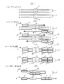
工程1では、ヒトの血液や組織細胞などの生体試料から公知技術によりゲノムDNAを抽出する。図1(a)に示すように、ゲノムDNAは、父親由来1と母親由来11の一対の二本鎖DNAからなり、その多型部位を2、12で示す。図1(a)では、多型の塩基位置が父親由来のDNAではA−Tの組に、母親由来のDNAではG−Cの組になっているものとし、この多型以外の配列の違いはないものとする。また、ゲノムDNA1、11において両端の3’または5’の記号は、それぞれのDNA分子の末端が5’末端、3’末端のいずれであるかを示している。
【0033】
工程2では、ゲノムDNA上の検査領域20、21を間にはさんだ領域をPCR反応で増幅する。図1(b)に示すように、PCRに用いるプライマー組は、蛍光色素8で標識されたプライマー4と、配列7と配列6が5’末端から3’末端に向かってこの順につながった構造のプライマー10とからなる。プライマー10の配列7の部分は、ゲノムDNA1上の多型位置を含む領域(検査領域)20と同じ配列に設計されている。PCRにおいて、これらのプライマー4と10はゲノムDNA上のあらかじめ決めた配列位置にハイブリダイズして伸長する。プライマー4、10からの伸長反応によって形成されるDNA鎖をそれぞれ9,9’で表す。ゲノムDNA11上でも同様の伸長反応が起こる。PCR反応終了後の生成物を図1(c)に示す。父親由来ゲノムDNA1を鋳型として生成される二本鎖のPCR産物13の蛍光色素8で標識された方の鎖は、プライマー4の配列、ゲノムDNA1の検査領域20と同じ配列、プライマー10の相補配列(配列6の相補配列6’と配列7の相補配列7’からなる)が5’末端側からこの順でつながった構造となり、母親由来ゲノムDNA11を鋳型として生成されるPCR産物23の蛍光色素8で標識された方の鎖は、プライマー4の配列、ゲノムDNA11の検査領域21と同じ配列、プライマー10の相補配列(配列6’と配列7’からなる)が5’末端側からこの順でつながった構造となる。
【0034】
工程3では、PCR産物13、23を熱あるいはホルムアミドなどの変性剤を使った公知の方法で変性して一本鎖DNAを得る。
【0035】
工程4では、工程3で得られた一本鎖DNAを電気泳動によって検出する。DNAの検出をレーザー励起蛍光法で行う場合には、図1(d)に示す蛍光色素8が5’末端側に標識された一本鎖DNA14および24が検出される。一本鎖DNA14はゲノムDNA1からのPCR産物であり、多型位置を含む検査領域の配列20はプライマー10のアンカー配列部分7の相補配列7’と完全に相補になっている。一方、一本鎖DNA24はゲノムDNA11からのPCR産物であり、多型位置を含む検査領域の配列21はプライマー10のアンカー配列部分7の相補配列7’と多型位置においてC−Aのミスマッチになっている。従って、配列20と配列7’との融解温度(Tm2)と配列21と配列7’との融解温度(Tm1)を比較すると、Tm2はTm1よりも高くなっている。従って、電気泳動の温度をTm1とTm2の間に設定すれば、図1(d)に示すように、DNA14は自己ハイブリダイゼーションを起こしたヘアピン型になり、DNA24とは大きく異なる立体構造を持つものとなる。このように積極的に構造を変化させることにより、SSCP法の分離能を向上させることができる。
【0036】
本実施例では、プライマー10のアンカー部分の配列7は検査領域の配列20と全く同じ配列であるとしたが、原理的には検査領域の配列20および検査領域の配列21でTmに十分な差が生ずるものであれば配列7はどのような配列であっても構わない。また、Tmは、キャピラリー電気泳動で分析する場合には、20−60℃程度になるように設計するのが好適である。
【0037】
本実施例を、具体的な鋳型DNA配列を用いた実験結果により、さらに詳細に、以下の図2、図3、図4、図5を用いて説明する。
【0038】
実験の条件を以下1.〜9.に示す。
1.検査対象の多型位置
VAV2遺伝子のイントロン領域にある多型であり、ヒト染色体9q34領域(GenBank Accession 番号:AC002111、全長39702塩基)の5680番目の塩基位置。
【0039】
2.検査対象の多型の塩基型
TまたはCの一塩基多型である。
【0040】
3.PCRプライマー配列
Shigyoらの報告(インターナショナル ジャーナル オブ キャンサー、78巻425頁から429頁 1998年(M. Shigyo et al., Int. J. Cancer, 78, pp. 425−429 (1998))に則って以下の配列を有するプライマーペアをPCRに用いた。以下これらをオリジナルのプライマーと呼ぶ。
・オリジナルフォワードプライマーの配列(図1のプライマー4に対応):配列番号2
・オリジナルリバースプライマーの配列(図1の配列6に対応):配列番号3
上記のオリジナルフォワードプライマーとオリジナルリバースプライマーを用いたPCR反応によりVAV2遺伝子のイントロン領域(138塩基長)が増幅される。この領域のフォワード鎖の配列を配列番号1で示す。配列番号1中にyの表記で示しているのは、この塩基位置がTとCのヘテロ構造であることを示している。
【0041】
4.アンカー配列
VAV2遺伝子のイントロン領域(以下簡単のため単にVAV2遺伝子領域と呼ぶ)のフォワード鎖を検出する場合には、オリジナルフォワードプライマーの5’末端を蛍光色素(本実験ではROXを用いた)で標識する。オリジナルリバースプライマーには以下の2種類のアンカー配列を5’側に付加し、2種類のアンカー付加プライマー(図1のプライマー10に対応)を準備した。
・アンカー配列1(11塩基長):配列番号4
・アンカー配列2(13塩基長):配列番号5
すなわち、図1の検査領域の配列20および21の塩基数として、アンカー配列1を用いる場合は11塩基、アンカー配列2を用いる場合は13塩基のように決めたことを示す。また、ここでは、11塩基および13塩基の真中(それぞれ6番目および7番目の塩基位置)に多型の塩基位置を配置した。
【0042】
VAV2遺伝子のリバース鎖を検出する場合には、オリジナルリバースプライマーの5’末端に蛍光色素(本実験ではFAMを用いた)を標識する。オリジナルフォワードプライマーには以下のアンカー配列を5’側に付加した。
【0043】
アンカー配列3(11塩基長):配列番号6
すなわち、図1の検査領域の20、21の塩基数として11塩基に決めたことを示す。また、11塩基の真中(6番目の塩基位置)に多型の塩基位置を配置した。
【0044】
5.ゲノム溶液の調製
公知の方法(例えば、QIA−mini キット;キアゲン社)を用いて全血からゲノムDNAを抽出し、50μg/mLの水溶液に調製した。このゲノムDNAのVAV2遺伝子の所定の多型部分は、T−AペアとC−Gペアのヘテロ構造(父母由来の配列が異なる)であることを、DNAシーケンサで解析して確認した。
【0045】
6.PCR反応
PCR反応キット(Platinum Genotype Tsp DNAポリメラーゼ;Life Technologies社)の標準プロトコールに従って、所定の反応バッファー溶液中のdNTP濃度を0.2mM, MgCl2 濃度を1.5mMに調整した。ゲノムDNA量50‐100ng、酵素量0.6U、プライマー量5‐30pmolを用いて、反応液全量15μLでPCR反応を行った。反応容器は200μLのPCR反応用チューブで、サーマルサイクラーにはUno−ThermoBlock (Biometra社)を用いた。
【0046】
温度条件は、94℃2分間の熱変性後、94℃30秒、55℃30秒、72℃60秒のサイクルを30回行い、最後に72℃10分間の最終伸長反応を行った。上記で55℃のアニ−ル温度は、プライマーの配列に応じて、55〜61℃の範囲で、所望以外のDNAが増幅されないように適宜選択した。
【0047】
7.電気泳動装置
分析装置には、ABI Prism 310 Genetic Analyzerを用いた。キャピラリーは内径50μm、外径350μm、長さ47cmである。このとき、試料の注入端から検出点(レーザーの照射点)までの距離は36cmであった。キャピラリー温度は30℃から60℃の範囲で設定した。試料溶液からのDNA注入は電界注入法によって行い、具体的には、15kVの電圧を5秒間印加した。電気泳動は15kVの電圧を印加しておこなった。
【0048】
8.電気泳動試薬
電気泳動用のバッファーには、1×TBE、10%グリセロールを用いた。分離媒体であるポリマーには非変性ポリマーであるGenescan(アプライド・バイオシステムズ社)を用いた。Genescanは重量比7%濃度で供給されているが、電気泳動用バッファーと同じ1×TBE、10%グリセロール溶液中でポリマー濃度が重量比で6%になるように調製した。
【0049】
9.分析する試料溶液の調製
PCR反応溶液を精製キット(QIA‐quick PCR Purificationキット;キアゲン社)を用いてプライマー除去と脱塩を行い、50μLの滅菌純水で溶出した。ついで、この溶液を水で10倍程度に希釈した後、その希釈液1μLと脱イオン化したホルムアミド10μLとを混合し、94℃で5分間加熱してPCR産物を熱変成(一本鎖化)した後、自然空冷して試料溶液とした。尚、上記において水による希釈の倍率は、測定装置ABI Prism 310において適切な信号強度が得られるように適宜調整した。
【0050】
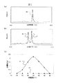
以上1.〜9.に記載した条件で得られた結果の一例を図2に示す。
図2(a)は、蛍光色素(ROX)で標識したオリジナルフォワードプライマーとオリジナルリバースプライマーを用いてPCR反応を行い、36℃でSSCP分析をした例である。横軸は泳動時間、縦軸は蛍光強度を表す。蛍光色素はフォワードプライマーに標識されているので、検出されているのはフォワード鎖である。試料に用いたゲノムDNAが一塩基多型位置においてT/Cヘテロ構造であることに対応して、2つのピークが検出されている。
【0051】
図2(b)は、オリジナルリバースプライマーにアンカー配列1(11塩基長)を付加したプライマーと蛍光色素(ROX)で標識したオリジナルフォワードプライマーを用いてPCR反応を行い、46℃でSSCP分析をした例である。蛍光色素はフォワードプライマーに標識されているので、検出されているのはフォワード鎖である。試料に用いたゲノムDNAが一塩基多型位置においてT/Cヘテロ構造であることに対応して、2つのピークが検出されている。泳動時間が短いほうのピーク32がT、長いほうのピーク33がCの配列を持つものであることを、Tホモ構造およびCホモ構造である別の二種類のゲノムDNAを用いて検証している。図2(a)との比較からわかるように、オリジナルリバースプライマーにアンカー配列1を付加することにより、格段によい分離が得られた。
【0052】
図2(c)は、オリジナルリバースプライマーにアンカー配列2(13塩基長)を付加したプライマーと蛍光色素(ROX)で標識したオリジナルフォワードプライマーを用いてPCR反応を行い、49℃でSSCP分析をした例である。図2(b)の場合と同様に、泳動時間が短いほうのピーク34がT、長いほうのピーク35がCの配列を持つものであることを、Tホモ構造およびCホモ構造である別の二種類のゲノムDNAを用いて検証している。アンカー配列2を付加した場合にもアンカー配列1を付加した場合には若干及ばないものの、オリジナルプライマーと比べた場合には、やはり格段によい分離が得られた。
【0053】
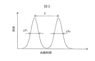
図3を用いて分離能RSの定義を説明する。2つのピークの泳動時間差をTとし、それぞれのピークの半値幅の時間をΔT1、ΔT2とした場合、RS=√(2ln2)・[T/(ΔT1+ΔT2)]で表される。ここでlnは自然対数をあらわす。
【0054】
図4に電気泳動の温度を変化させて分離能RSを測定した結果を示す。曲線36,37,38は、それぞれ、図2(a)(b)(c)の実験結果を得るのに用いたプライマーの組み合わせを用い、電気泳動の温度を変化させて分離能RSを測定した結果を示している。オリジナルのプライマー配列を用いた場合(曲線36)に比べ、リバースプライマーにアンカー配列1(配列番号4)を導入した場合(曲線38)、アンカー配列2(配列番号5)を導入した場合(曲線37)では、分離の最高値は5〜6倍向上した。最高の分離能が得られた温度は、オリジナルプライマーでは36℃、アンカー配列1を導入した場合には49℃、アンカー配列2を導入した場合は46℃であった。
【0055】
図5には、同じくVAV2遺伝子を用い、リバース鎖を検出した結果を示す。
図5(a)はオリジナルプライマーの組を用いた場合、図5(b)はアンカー配列3(配列番号6)をオリジナルフォワードプライマーの5’末端側に付加したプライマーを用いた場合である。図5(c)には図4と同じように、分離能の温度依存性を示した。この場合には、オリジナルプライマーでは分離できなかった(図5(a)のピーク39)ヘテロ構造が、図5(b)のピーク40、41で示すように、アンカー配列の導入で明確に分離できた。このときの温度は39℃であった。一塩基多型は泳動時間が短いほうのピーク40がA、長いほうのピーク33がGであるであることを、フォワード鎖がTホモ構造およびCホモ構造(リバース鎖では、それぞれAホモ、Gホモ)である別の二種類のゲノムDNAを用いて検証している。図5(c)で示すように、最高分離能が得られる温度は39℃であった。
【0056】
以下の表1には、上述の実験結果から得られた最高分離を与える温度の実測値と、アンカー配列のハイブリダイゼーションの融解温度の計算値から得られた最高分離を与える温度の計算値とを示す。
【0057】
表1
最高の分離を与える泳動温度の計算値と実測値、および多型によるヘアピン構造の融解温度の計算値の差と分離能の実測値
【表1】

ここで、融解温度の計算には、一塩基ミスマッチを考慮に入れたNearest Neighbor法に基づく熱力学パラメータを用いたが、特に限定するものではない。Nearest Neighbor法の詳細は主に、SantaLucia他による文献:プロシーディング オブ ナショナル アカデミー オブ サイエンス USA 第95巻 1460頁から1465頁 1998年(Proceeding of National Academy of Science of U.S.A., Vol. 95, pp.1460〜1465, (1998))、およびPeyret他による文献:バイオケミストリー 第38巻 12号 3468頁から3477頁 1999年(Biochemistry, Vol. 38,No. 12, pp.3468〜3477, (1999))に開示されている。最高分離を与える温度は、完全マッチと一塩基ミスマッチのTmの平均値とした。
【0059】
表1に示すように、最高分離を与える温度の計算結果と実験結果とは、約7℃以内でほぼ一致している。この結果は、従来のSSCP法では予測不可能であった分離最適温度を、本実施例においては、ある程度あらかじめ予測できることを示している。このため、計算結果に従って電気泳動の温度を設定しても、ある程度の分離が得られる。また、実験的に最高分離が得られるような泳動温度を探索する場合にも、少ない実験量で最適の値を容易に得ることができる。さらに、最高分離が得られる温度をキャピラリー電気泳動装置で設定しやすい温度条件に設定できるように、アンカー配列を適宜設計することもできる。
【0060】
さらに表1には、一塩基多型によるアンカー配列の融解温度の違い(差)の計算値と、最高分離能の実測値を示した。融解温度差の計算値の大小関係と、最高分離能の実測値の大小関係は一致している。すなわち、一塩基多型による融解温度の計算値の差が最も大きくなるようにアンカー配列を設計することによって、高い分離能が得られる。
【0061】
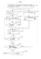
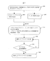
図6には、以上に説明してきた結果を踏まえた、本発明のプライマー設計の工程をフローチャートで示す。
【0062】
検査対象DNAの塩基配列および検査する多型の塩基位置と塩基種のデータセット301を用い、工程302では、まず公知の技術を用いて、PCRプライマーの配列の内の、検査対象のDNAにハイブリダイズする部分(図1に4および6で示した部分であり、オリジナルプライマーと呼ぶ)の塩基配列を設計する。
設計に当たっては、公知の技術により、17ないし30塩基長程度で、一組のプライマーの融解温度がほぼ等しい値になるように、また、プライマーの自己ハイブリダイゼーションや3’末端側でのプライマー同士のハイブリダイゼーションなど、PCRを阻害する要因を除くように考慮する。さらに、PCR増幅産物の長さは、従来のSSCP法で良く用いられている塩基長数十から300塩基長程度になるようにする。このようにして複数組のオリジナルプライマー組の候補を設計しておく。尚、プライマーの設計には、市販のソフトウェアを用いることができる。工程303では、設計した候補の内、一組のオリジナルプライマーを選び、オリゴヌクレオチドを合成する。次いで工程304では、合成したオリジナルプライマーペアを用いてPCR反応を行い、単一長さの特異的な増幅が良好に起こるか否かを、アガロース電気泳動などの適当な公知技術を用いて検定する。
増幅が良好であれば、そのプライマーペアを選定して工程305に進む。増幅が悪い場合は、工程302で設計した別のオリジナルプライマーペアを用いて工程303と304を行う。この過程を繰り返してオリジナルプライマーペアを決定する。
【0063】
尚、過去の文献等で、検査対象の多型部位を含む領域を特異的に増幅できることが実験的に検証されているプライマー配列が報告されている場合には、工程303においてその配列をオリジナルプライマー配列に用いることもできる。
【0064】
一方、工程306では、データセット301を用いて、アンカー配列を設計する。できるだけ高い分離能を得るためには、アンカー配列が多型部位を含む領域と特異的にハイブリダイズし、かつ多型部位の塩基が異なることによる融解温度の差が最も大きくなるようにする。さらに、キャピラリー電気泳動に適した測定条件にするために、融解温度の計算値が30℃から60℃位になるように、またアンカー配列の長さをおおよそ7−15塩基長程度にする。より単純には、アンカー配列は、多型部位を含む7から15塩基長程度の範囲の配列で、多型部位に相当する塩基位置を除いて鋳型DNAと完全に同じ配列とする。アンカー配列が短すぎると類似配列の出現率が高くなり、長すぎるとTm値が高くなりすぎて好ましくない場合がある。アンカー配列内の多型部位に相当する位置とその塩基種類は、多型による融解温度の計算値の差が最も大きくなるようにする。この考え方で、複数のアンカー配列候補を設計する工程が306である。
【0065】
工程307では、工程306で設計したアンカー配列候補のうち、1つを選ぶ。工程305では、工程304で選択したオリジナルプライマー組の片方に工程307で選んだアンカー配列を付加したプライマーを合成する。
【0066】
次いで、工程308では、アンカー配列を付加したプライマーともう一方のオリジナルプライマーを用いて、再びPCR反応を行い、特異的増幅の良否をチェックする。増幅が好ましい時は、次の工程309に進み、好ましくないときは、工程303に戻って別のオリジナルプライマーペアを選択し、再び工程をやりなおす。
【0067】
工程309では、電気泳動分析により、多型の分離能を検定する。この工程において、全自動キャピラリー電気泳動装置を用いる場合には、アンカー配列を付加していないオリジナルプライマーに蛍光標識を行い、PCR増幅を行ってから測定する。電気泳動の温度は、アンカー配列の融解温度から計算される値を初期値として、10℃程度の範囲で前後数点の測定を行えば、最適値を得ることができる。分離が良好な場合は、この工程309で用いたプライマー組と泳動温度の条件が、確立されたプライマーと分析条件310となる。分離が悪い場合には、工程307に戻り、別のアンカー配列候補を用いて工程305からやりなおす。
【0068】
上述のような一連の工程を経て、検査対象の多型部位について実験的にも保証されたプライマーと分析条件を得ることができる。
【0069】
本発明のプライマー設計の一連のプロセスについて、別の実施例を図7に示す。検査対象DNAの塩基配列および検査する多型の塩基位置と塩基種のデータセット301を用い、オリジナルプライマーの設計工程302とアンカー配列の設計工程306により、プライマーペアの候補とそれを用いる場合の推奨PCR条件と電気泳動条件の一覧表311を得ることができる。一覧表311から選択したプライマーペアを工程312において合成し、工程313でPCR反応と電気泳動分析を行い、多型の分離を検証する。良好な分離が得られた場合、プライマーと分析条件310を確定する。良好でない場合には、工程312に戻り、別のプライマーを合成するところからやりなおす。本実施例においては、単純なフローチャートであるため、候補となるプライマーペアを複数一度に評価するのには都合がよい。
【0070】
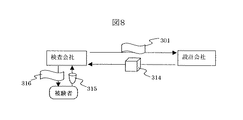
図8と図9には、上記図6と図7で示したプライマーと分析条件の確立を行うサービスビジネスの実施例を示す。
【0071】
図9では、検査会社は、図6および図7で示したデータセット301を設計会社に提示する。設計会社は、図6または図7に示したようなフローチャートに従って、プライマーと分析条件を確立し、試薬としてのプライマーと測定条件を記載したドキュメントを含む検査キット314を提供し対価を得る。検査会社は、被験者から血液や組織などの生体試料315を受け取り、その分析結果316を報告して対価を得る。本実施例では、検査会社においては、自社で多くの時間と人手をかけなくても、所定の多型に関する検査キットを入手できる効果があり、設計会社においては、従来のSSCP法に比べて分析に最適な条件の予測性がよい本発明によって、キットを確立するのに必要な時間と労力を短縮でき、低価格でサービスを提供できるメリットがある。
【0072】
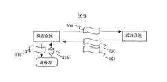
図9では、本発明に基づくサービスビジネスの別の実施例を示す。本実施例では、図8で説明した実施例と同様に、検査対象の多型を含むDNAの配列と多型の配列情報(データセット301)に基づいてプライマー配列を設計して、プライマー組の配列のいくつかの候補をリストアップ(プライマー組の配列一覧表323)し、それに推奨する分析条件(条件のデータ324)を添付する。実験に基づく検証は行わない。実験条件の決定は検査会社が行う。検査会社は設計結果に基づいて実験を行い、最終的なプライマーの選定と分析条件を確立する。そして確立した分析条件を用いて被験者から提供を受けた生体試料315に対する分析結果316を報告する。検査会社は被験者から検査費用を受け取り、設計会社には設計費用を支払う。
【0073】
この実施例においては、設計会社と検査会社の間には、試料やプライマーなどの物のやり取りはなく、データのやりとりだけなので、設計会社にとっては、インターネット上のホームページによってサービスを提供する形態も可能となる。
このため、低価格のサービスが提供できる。本発明においては、従来の技術と異なり、分離に最適な条件が予測可能なため、実験による保証はないものの、ユーザに推奨条件を提示することは可能である。実験設備やノウハウの十分に整った検査会社などの依頼者に対しては、低価格で設計結果のみを提供できる効果がある。
【0074】
【発明の効果】
本発明によれば、PCR−SSCP法に用いるPCRプライマーに、検査領域の配列として想定される配列と同一の配列をアンカー配列として付加することにより、電気泳動測定時にヘアピン状態を起こさせる。ヘアピン状態の融解温度が一塩基の違いによって異なることを利用して、その中間の温度で電気泳動を行うことにより、SSCP法による一塩基多型の分離能を著しく改善することができる。また、分離に最適な温度を予測することが可能となり、従来のSSCP法で必須であった分析条件の最適化に要する手間を大きく低減できる。また、最高の分離を与える電気泳動温度がキャピラリー電気泳動装置に適した温度範囲に入るように、アンカー配列を設計することができる。このため、遺伝子診断の自動化・高精度化に大きな効果がある。さらに、本発明によって、一塩基多型を分離分析するための推奨の実験条件を添付した形で、PCRプライマーの配列あるいは合成したPCRプライマーを提供するサービスビジネスが実現できる。
【0075】
【配列表】
【配列表フリーテキスト】
配列番号2:合成ポリヌクレオチド
配列番号3:合成ポリヌクレオチド
配列番号4:合成ポリヌクレオチド
配列番号5:合成ポリヌクレオチド
配列番号6:合成ポリヌクレオチド
【図面の簡単な説明】
【図1】本発明の原理を工程に沿って示す。
【図2】本発明の原理に基づく電気泳動測定結果の一例を示す。
(a)従来のSSCP法でフォワード鎖を検出して得られた電気泳動スペクトル
(b)本発明でアンカー配列として配列番号4の配列からなるポリヌクレオチドを用いフォワード鎖を検出して得られた電気泳動スペクトル
(c)本発明でアンカー配列として配列番号5の配列からなるポリヌクレオチドを用いフォワード鎖を検出して得られた電気泳動スペクトル
【図3】分離能の定義の説明図を示す。
【図4】図2(a)(b)(c)で示したそれぞれの場合について、分離能と温度との関係を測定した結果を示す。
【図5】(a)従来のSSCP法でリバース鎖を検出して得られた電気泳動スペクトルを示す。
(b)本発明でアンカー配列として配列番号6の配列からなるポリヌクレオチドを用いリバース鎖を検出して得られた電気泳動スペクトルを示す。
(c)(a)(b)それぞれの場合で、分離能の電気泳動温度に対する依存性を示す。
【図6】本発明のPCRプライマーの設計手順のフローチャートを示す。
【図7】本発明のPCRプライマーの別の設計手順のフローチャートを示す。
【図8】図6及び図7で示した設計フローの全部または一部を実施する設計会社を含むビジネスモデルの一実施例を示す。
【図9】図6及び図7で示した設計フローの一部を実施する設計会社を含むビジネスモデルの別の実施例を示す。
【符号の説明】
1.ゲノムDNA(父由来)
11.ゲノムDNA(母由来)
2,12.多型部位
4,10.プライマー
8.蛍光色素
20、21.検査領域の配列
20’、21’.検査領域の配列
20、21の相補配列
7、7’.アンカー配列およびその相補配列
13、23.PCR産物
14、24.一本鎖化したPCR産物のうち、蛍光色素8で標識されている鎖
315.生体試料(血液、組織など)
316.検査結果を記載した書類
301.検査対象の多型部位を含む領域のDNA配列及び多型の位置と塩基種からなるデータ。
314.プライマー及び反応・測定条件が記載された書類を含む検査キット
323.プライマー配列
324.推奨する反応・測定条件が記載された書類[0001]
TECHNICAL FIELD OF THE INVENTION
The present invention relates to a method for separating and detecting a polymorphism such as a single nucleotide polymorphism and a mutation in a gene. More specifically, the present invention relates to a method for improving the separation ability of a single-strand conformation polymorphism (SSCP) analysis method of DNA using gel electrophoresis and a gene diagnosis method using the analysis method.
[0002]
[Prior art]
In recent years, the analysis of the human genome has rapidly progressed, and it has been found that the human genome has individual differences (polymorphisms) at a single nucleotide level at a frequency of approximately every 1000 nucleotides. This is called Single Nucleotide Polymorphism (SNP). Since the polymorphism exists at a high density on the genome, it serves as a detailed mark indicating the position on the genome. For example, by analyzing the difference in the frequency of occurrence of SNPs mapped on this genome between a disease group and a normal group of a certain disease, a gene related to the disease can be located at any position on the genome. I can find the dolphin. It is expected that if the positional relationship of genes on the genome is understood in detail, it will lead to the acquisition of new functional information such as the interaction between genes, and the knowledge that will lead to new drug discovery, diagnosis and treatment.
[0003]
Further, the fact that a gene polymorphism at a single nucleotide level can be detected means that a mutation in a gene sequence can be detected. Since the involvement of gene mutations in the mechanisms of carcinogenesis and metastasis has been suggested, analysis of single nucleotide polymorphisms and mutations is expected to be a basic technology for the diagnosis of many diseases, including cancer. .
[0004]
One of the methods for analyzing such polymorphism at the single nucleotide level is a single-strand DNA conformation polymorphism analysis (SSCP) method. This method is described in M. It has been reported by Orita et al. (For example, see Non-Patent Document 1). This is based on the fact that the electrophoresis speed of single-stranded DNA in a non-denaturing polyacrylamide gel varies depending on the sequence of one base, even for DNA of the same length. This method separates and detects polymorphisms. The higher-order structure (three-dimensional structure) of the single-stranded DNA differs depending on the single-base difference, and it is thought that the difference in the higher-order structure causes a difference in the migration speed. In recent years, a PCR-SSCP method for selectively amplifying and analyzing a target portion of DNA extracted from a biological sample by PCR (Polymerase Chain Reaction) has been widely used.
[0005]
As an example of a cancer diagnosis method using the PCR-SSCP method, there is a method based on the phenomenon of loss of heterozygosity (LOH). Genomic DNA extracted from cells usually contains the same number of genes from the father and genes from the mother (heterozygosity). However, in cancerous cells, a phenomenon in which this equilibrium is lost (LOH) is often seen because a specific locus is missing or abnormally replicated. M. According to the report of Shigyo et al. (For example, see Non-Patent Document 2), it is reported that there is a high correlation between LOH of urine cells and malignant tumor of the bladder.
When measuring the LOH, a single nucleotide polymorphism is used as a marker for distinguishing a gene derived from a father from a gene derived from a mother, and the single nucleotide polymorphism is separated and detected by the SSCP method. The imbalance between the abundance of the paternal gene and the maternal gene is quantified by the ratio of the peak intensities of this single nucleotide polymorphism separated by electrophoresis.
[0006]
On the other hand, conventionally, plate gel electrophoresis or capillary gel electrophoresis has been used for the SSCP method. Examples of the slab gel (slab gel) electrophoresis apparatus include GenePhor, a product of Amersham Pharmacia Biotech. The flat gel electrophoresis device has a simple device configuration and is inexpensive, but manual processes such as gel preparation, installation in the electrophoresis bath, sample introduction, removal of the gel from the electrophoresis bath, and photography of the gel are required. Many. Therefore, as described in Patent Document 1, for example, an SSCP that performs high-speed high-speed analysis at high voltage using a capillary gel that is suitable for automation and has a small inner diameter of 100 μm or less and rapidly releases Joule heat accompanying electrophoresis. A law has been proposed. Capillary electrophoresis apparatuses, such as ABI Prism 310, a product of Applied Biosystems, use a flowable polymer as a separation medium and have a function of automatically exchanging the polymer at the end of each measurement. In recent years, devices capable of automatic and continuous measurement have rapidly spread.
[0007]
[Patent Document 1]
JP-A-6-138090
[Non-patent document 1]
M. Orita et al., "Proceeding of National Academy of Science of USA" (USA), 1989, Vol. 86, p. 2766-p. 2770
[Non-patent document 2]
M. Shigyo, "International Journal of Cancer", (USA) 1998, vol. 78, p. 425-p. 429
[0008]
[Problems to be solved by the invention]
As described above, the SSCP method is a simple and effective method for measuring single nucleotide polymorphisms, and a rapid automatic analysis is also possible using a capillary electrophoresis apparatus. However, the SSCP method, as suggested by its principle, indicates that even if single-stranded DNA has a single nucleotide polymorphism, the difference in migration speed caused by the difference in its higher-order structure is due to the separation ability of gel electrophoresis. If it is smaller than, it cannot be separated. In addition, since it is difficult to theoretically predict the higher-order structure and the relationship between the structure and the migration speed, the electrophoresis conditions were changed little by little for each single nucleotide polymorphism to be tested. In this case, it was necessary to search for conditions under which separation could be obtained by repeating the measurement.
Particularly in the case of genetic diagnosis using LOH, it is necessary to accurately determine the peak intensity ratio. Therefore, it is necessary to obtain a sufficiently high resolution so that the two peaks of the single nucleotide polymorphism do not overlap with each other. It took a lot of trouble to study.
[0009]
In the plate gel electrophoresis apparatus GenePhor described above, the ability to separate single nucleotide polymorphisms by the SSCP method often improves when the temperature is low and the gel concentration is high. %, A temperature is set in a range of about 5 to 20 ° C., and consideration is given to assisting an operation for searching for an optimum measurement condition. However, in a fully automatic capillary electrophoresis apparatus such as the above-mentioned ABI Prism 310, the separation medium (a polymer solution composed of uncrosslinked polyacrylamide or the like) is refilled in the capillary each time the measurement is completed. I can not use it. In addition, even when the polymer solution is not crosslinked, the high-concentration polymer solution has a high viscosity, so that the pressure loss at the time of refilling is large and the time required for refilling becomes longer.
[0010]
Further, at a low temperature of about 15 ° C. or less, it is necessary to take measures such as sealing the entire apparatus and lowering the internal humidity in order to prevent high voltage leakage and electric shock due to dew condensation. In the above-mentioned flat plate gel electrophoresis apparatus, the electrophoresis and the image measurement of the gel are performed by different apparatuses, and the electrophoresis apparatus is small in size. However, a fully automatic capillary electrophoresis apparatus is relatively large because electrophoresis and DNA detection are performed simultaneously. For this reason, dew condensation countermeasures had to be extensive.
[0011]
The present invention has been made in view of the above problems, and has as its object to reduce the labor required for optimizing the analysis conditions for single nucleotide polymorphism by the SSCP method. Furthermore, it is an object of the present invention to improve the SSCP method so that high separation can be obtained without using a low-temperature or high-concentration polymer in a fully automatic capillary electrophoresis apparatus suitable for routine analysis and diagnostic use.
[0012]
[Means for Solving the Problems]
As a result of various investigations to solve the above problems, a nucleotide sequence that greatly changes the higher-order structure of single-stranded DNA depending on the presence or absence of a single nucleotide polymorphism was introduced during the PCR reaction, whereby the SSCP method was used. It has become possible to remarkably improve the labor and resolution required for examining the conditions.
[0013]
That is, the present invention uses a primer designed to change the higher-order structure of a DNA fragment amplified by a PCR reaction due to a difference in base sequence such as a single nucleotide polymorphism present in a test region, and a DNA containing the test region. Provided are a method for testing a nucleic acid sequence, a method for preparing a sample, and a kit for testing a nucleic acid sequence containing the above primer, which comprises a step of amplifying a fragment.
[0014]
BEST MODE FOR CARRYING OUT THE INVENTION
Hereinafter, the outline of the sample preparation method and the inspection method of the present invention will be described below.
[0015]
The DNA serving as a template is, for example, genomic DNA extracted from blood or tissue cells of a subject, cDNA obtained by reverse transcription from mRNA, or the like, and may be single-stranded DNA or double-stranded DNA. Is also good. In the present invention, the test region to be tested on the DNA comprises a polymorphic site such as a single nucleotide polymorphism and a portion adjacent thereto, and a region containing the sequence of the test region is amplified by a PCR reaction. As a result of progress in genome analysis, information on mutations such as single nucleotide polymorphisms has already been accumulated, and the test region and the sequence in the vicinity thereof are often known in advance. It is assumed that this is assumed. At this time, one of the primers (first primer) has a sequence (first sequence portion) that hybridizes to a
[0016]
In another aspect, the first primer is a second primer in the 5 ′ → 3 ′ direction from the 3 ′ end of the test region consisting of the first predetermined number of bases in one strand of single-stranded DNA or double-stranded DNA. A first sequence portion that hybridizes to a sequence of a third predetermined number of bases separated by a predetermined number of bases, and a sequence consisting of the first predetermined number of bases and assumed as a sequence of the test region. A second sequence portion having the same sequence and following the 5 'end of the first sequence portion. The first predetermined number of bases is not particularly limited because it is a sequence that causes self-hybridization, but is preferably 7 to 15 bases. The third predetermined number of bases is not particularly limited as long as it is within a range normally used as a primer sequence, but is preferably 17 to 30 bases. The second predetermined number of bases is not particularly limited as long as it does not hinder self-hybridization and can perform PCR suitably, and is preferably 30 to 100 bases.
[0017]
In the above aspect, the second primer is complementary to a sequence having a fifth predetermined number of bases separated by a fourth predetermined number of bases in the 3 ′ → 5 ′ direction from the 5 ′ end of the sequence of the test region. It has a sequence that hybridizes to the sequence and is labeled at the 5 'end. The fourth predetermined base number is not particularly limited as long as it is within a range in which PCR can be suitably performed, and is preferably 30 to 100 bases. The fifth predetermined number of bases is similar to the third predetermined number of bases as long as it is within a range normally used as a primer sequence, and is not particularly limited, but is preferably 17 to 30 bases. . The first primer and the second primer are designed so that the Tm values are substantially equal in consideration of the progress conditions of the PCR reaction, as is generally performed in the art.
[0018]
By using these primers and performing a PCR reaction using the DNA as a template, a DNA fragment containing the test region can be amplified, and a sample (amplified DNA fragment) required for a target nucleic acid sequence testing method can be amplified. Is prepared.
[0019]
The amplified DNA fragment is in a double-stranded state, and by denaturing it into a single-stranded DNA fragment, each DNA fragment undergoes self-hybridization or the like according to its base sequence, and is energetically stable. It takes a higher-order structure. In this case, the amplified DNA fragment contains, in addition to the sequence of the template DNA including the test region, a sequence corresponding to the anchor sequence of the first primer.
[0020]
Therefore, the DNA fragment containing the sequence of the test region on one strand has, at its 3 ′ end, a sequence complementary to the anchor sequence that is the same sequence as the sequence assumed as the sequence of the test region (the third sequence portion). ) Is added, and when the sequence of the test region is the expected sequence itself, it becomes completely complementary to the sequence of the third sequence portion, and self-hybridization easily occurs. When the sequence of the test region is different from the expected sequence, for example, when there is a single nucleotide mismatch with the third sequence portion due to a single nucleotide polymorphism, self-hybridization hardly occurs. In the present invention, a test method that can clearly identify even a mismatch of only one nucleotide is provided.However, polymorphisms other than single nucleotide polymorphisms, for example, insertion, deletion, substitution, etc. Mutation can also be detected, and it is not particularly limited to single nucleotide polymorphism.
[0021]
On the other hand, the DNA fragment amplified as a pair with the above-mentioned DNA fragment contains the sequence of the test region in the other strand (the sequence complementary to the sequence of the test region in the one strand), and its 5 ′ Since the terminal sequence has the same sequence as the anchor sequence, that is, the sequence of the test region in one of the above-mentioned chains, similarly to the above, if the sequence of the test region is the expected sequence itself, the self-high When hybridization occurs easily and the sequence of the test region is different from the expected sequence, self-hybridization hardly occurs.
[0022]
In order to cause self-hybridization only when the sequence of the test region and the sequence of the third sequence are completely complementary, and to prevent self-hybridization in the case of a single base mismatch, specifically, The following can be performed.
[0023]
That is, when a single nucleotide polymorphism exists in the sequence of the test region, the melting temperature (Tm) becomes a different value depending on the difference of the single nucleotide. Therefore, for example, if the temperature of electrophoresis is set to be equal to or lower than the Tm of hybridization in the case of perfect complementation, self-hybridization occurs and a hairpin type structure is formed. When the temperature is raised above Tm, this hairpin structure dissociates. The Tm of the hybridization in the case of a single base mismatch is a lower value.
[0024]
When the anchor sequence is designed to completely match (completely match) the sequence of the test region containing one type of base of the polymorphism, the base region mismatches with the sequence of the test region containing the other type of base by a single base mismatch, and Tm Goes down. Therefore, when the temperature of electrophoresis is set between the Tm of a perfect match and the Tm of a single base mismatch, for example, at an intermediate temperature, the DNA of a perfect match has a hairpin structure, and the DNA of a single base mismatch has a hairpin structure. Will not be. The Tm value varies depending on the base sequence to be hybridized, but when the base sequence is known in advance, it can be calculated by means known to those skilled in the art.
[0025]
As described above, when the amplified DNA fragment is single-stranded according to the polymorphism or mutation status in the test region, the higher-order structure can be determined by setting an appropriate temperature to determine the presence of the anchor sequence. Since these are markedly different, they can be separated by electrophoresis, and the single-stranded DNA labeled with a fluorescent dye or the like can be detected by using a laser-excited fluorescence detection method.
[0026]
According to the present method, first, the difference between the higher-order structures, that is, the hairpin-type structure and the structure other than the hairpin-type structure, is larger than the structural change in the conventional SSCP method, so that the separation is improved. Therefore, a high separation can be obtained without using a high-concentration polymer having a high viscosity or a low temperature. Second, since the value of Tm of the array of the inspection region can be calculated by a known method, a temperature condition suitable for separation can be estimated in advance, so that the amount of work for optimizing the temperature condition of SSCP analysis Can be reduced. Third, the arrangement of the inspection area can be set such that the optimum temperature for separation predicted from the calculated value of Tm is about 30 to 60 ° C. suitable for the capillary electrophoresis apparatus. In addition, by designing the array of test regions so that the optimal temperature for separation is approximately equal for multiple polymorphic sites, it is possible to simultaneously test multiple polymorphic sites under constant temperature conditions. Become.
[0027]
The method of the present invention can easily detect the presence of a single nucleotide polymorphism, and if the relationship between the polymorphism and the peak position in electrophoresis is obtained in advance as control data, the single nucleotide polymorphism in the DNA to be tested is obtained. The type of the type can be immediately detected from the position of the migration peak of the DNA fragment. When a plurality of DNAs having different test region sequences are present, the abundance ratio can be calculated from the peak position and the intensity ratio.
[0028]
Further, the present invention provides a kit for testing a nucleic acid sequence. The kit includes the first primer and the second primer, and optionally includes other reagents necessary for the PCR reaction, such as enzymes, nucleotides, buffers, and the like, and data on suitable PCR reaction conditions. May be. Instead of the primer itself, base sequence data of the primer may be included in the kit. In this case, since it is necessary to synthesize a primer, the primer may include a reagent for synthesis and a label such as a fluorescent dye for labeling the primer.
[0029]
The kit further includes temperature condition data for electrophoresis analysis of the presence or absence of self-hybridization by a DNA fragment amplified by a PCR reaction using the above primers for a polymorphism or mutation such as a single nucleotide polymorphism. It may be. The temperature condition data can be provided as a calculated value or an actually measured value in consideration of the sequence of the test region and the assumed polymorphism or mutation type in advance.
[0030]
The kit of the present invention can be used to analyze polymorphisms or mutations contained in a test region by electrophoresis. Specifically, a kit for analyzing a single nucleotide polymorphism type in a test region, Can be used to analyze the abundance ratio of a plurality of DNAs having different sequences.
[0031]
【Example】
Hereinafter, an embodiment of the present invention will be described with reference to FIG. The method for testing a nucleic acid sequence according to the present invention comprises four steps: DNA extraction, PCR reaction, denaturation (single-stranded) of a PCR product, and electrophoresis analysis. The details of the steps will be described below.
[0032]
In step 1, genomic DNA is extracted from a biological sample such as human blood or tissue cells by a known technique. As shown in FIG. 1A, the genomic DNA is composed of a pair of double-stranded DNAs of 1 from the father and 11 from the mother, and their polymorphic sites are indicated by 2 and 12. In FIG. 1 (a), it is assumed that the base positions of the polymorphism are in the AT group in DNA derived from the father and in the GC group in DNA derived from the mother. Shall not exist. In the genomic DNAs 1 and 11, the 3 'or 5' symbols at both ends indicate whether the end of each DNA molecule is the 5 'end or the 3' end.
[0033]
In
[0034]
In
[0035]
In
[0036]
In this example, the
[0037]
This example will be described in more detail with reference to FIGS. 2, 3, 4, and 5 below based on experimental results using a specific template DNA sequence.
[0038]
The experimental conditions are as follows. ~ 9. Shown in
1. Inspection polymorphism position
A polymorphism in the intron region of the VAV2 gene, which is the 5680th nucleotide position in human chromosome 9q34 region (GenBank Accession No .: AC002111, full length 39702 bases).
[0039]
2. Base type of polymorphism to be tested
T or C is a single nucleotide polymorphism.
[0040]
3. PCR primer sequence
Shigyo et al. (International Journal of Cancer, vol. 78, pp. 425-429, 1998 (M. Shigyo et al., Int. J. Cancer, 78, pp. 425-429 (1998))). Were used for PCR and are hereinafter referred to as original primers.
Sequence of the original forward primer (corresponding to
Sequence of the original reverse primer (corresponding to sequence 6 in FIG. 1): SEQ ID NO: 3
The intron region (138 bases in length) of the VAV2 gene is amplified by a PCR reaction using the above-mentioned original forward primer and original reverse primer. The sequence of the forward chain in this region is shown as SEQ ID NO: 1. The designation of y in SEQ ID NO: 1 indicates that this base position is a T and C hetero structure.
[0041]
4. Anchor array
When detecting the forward strand of the intron region of the VAV2 gene (hereinafter simply referred to as VAV2 gene region), the 5 'end of the original forward primer is labeled with a fluorescent dye (ROX was used in this experiment). The following two types of anchor sequences were added to the 5 'side of the original reverse primer, and two types of anchor-added primers (corresponding to
-Anchor sequence 1 (11 base length): SEQ ID NO: 4
-Anchor sequence 2 (13 bases long): SEQ ID NO: 5
In other words, the numbers of bases of the
[0042]
When detecting the reverse strand of the VAV2 gene, a fluorescent dye (FAM was used in this experiment) is labeled at the 5 'end of the original reverse primer. The following anchor sequence was added to the 5 'side of the original forward primer.
[0043]
Anchor sequence 3 (11 bases long): SEQ ID NO: 6
In other words, this indicates that the number of
[0044]
5. Preparation of genomic solution
Genomic DNA was extracted from whole blood using a known method (for example, QIA-mini kit; Qiagen), and prepared as a 50 μg / mL aqueous solution. The DNA sequencer analyzed and confirmed that the predetermined polymorphic portion of the VAV2 gene in the genomic DNA had a hetero structure of a TA pair and a CG pair (having different parental sequences).
[0045]
6. PCR reaction
According to a standard protocol of a PCR reaction kit (Platinum Genotype Tsp DNA polymerase; Life Technologies), the dNTP concentration in a predetermined reaction buffer solution was 0.2 mM,
[0046]
As for the temperature conditions, after heat denaturation at 94 ° C. for 2 minutes, a cycle of 94 ° C. for 30 seconds, 55 ° C. for 30 seconds and 72 ° C. for 60 seconds was performed 30 times, and finally a final extension reaction at 72 ° C. for 10 minutes was performed. The annealing temperature of 55 ° C. was appropriately selected within the range of 55 to 61 ° C. in accordance with the sequence of the primer so that undesired DNA was not amplified.
[0047]
7. Electrophoresis device
[0048]
8. Electrophoresis reagent
1 × TBE, 10% glycerol was used as a buffer for electrophoresis. Genescan, a non-modified polymer (Applied Biosystems) was used as the separation medium. Genescan was supplied at a concentration of 7% by weight, but was prepared so that the polymer concentration became 6% by weight in 1 × TBE and a 10% glycerol solution same as the buffer for electrophoresis.
[0049]
9. Preparation of sample solution for analysis
The PCR reaction solution was subjected to primer removal and desalting using a purification kit (QIA-quick PCR Purification Kit; Qiagen), and eluted with 50 μL of sterile pure water. Then, the solution was diluted about 10-fold with water, 1 μL of the diluted solution was mixed with 10 μL of deionized formamide, and the mixture was heated at 94 ° C. for 5 minutes to thermally denature (single-stranded) the PCR product. Thereafter, the sample solution was naturally cooled with air to obtain a sample solution. In the above, the magnification of dilution with water was appropriately adjusted so that an appropriate signal intensity could be obtained in the measurement
[0050]
Above 1. ~ 9. FIG. 2 shows an example of the results obtained under the conditions described in FIG.
FIG. 2A shows an example in which a PCR reaction was performed using an original forward primer and an original reverse primer labeled with a fluorescent dye (ROX), and SSCP analysis was performed at 36 ° C. The horizontal axis represents the migration time, and the vertical axis represents the fluorescence intensity. Since the fluorescent dye is labeled on the forward primer, what is being detected is the forward strand. Two peaks were detected corresponding to the fact that the genomic DNA used in the sample had a T / C heterostructure at the single nucleotide polymorphism position.
[0051]
FIG. 2 (b) shows a PCR reaction using a primer obtained by adding an anchor sequence 1 (11 base length) to an original reverse primer and an original forward primer labeled with a fluorescent dye (ROX), and SSCP analysis was performed at 46 ° C. It is an example. Since the fluorescent dye is labeled on the forward primer, what is being detected is the forward strand. Two peaks were detected corresponding to the fact that the genomic DNA used in the sample had a T / C heterostructure at the single nucleotide polymorphism position. It was verified that the peak 32 having the shorter electrophoresis time had the sequence of T and the
[0052]
FIG. 2 (c) shows a PCR reaction performed using a primer obtained by adding an anchor sequence 2 (13 base length) to an original reverse primer and an original forward primer labeled with a fluorescent dye (ROX), and subjected to SSCP analysis at 49 ° C. It is an example. As in the case of FIG. 2 (b), the fact that the peak 34 having the shorter migration time has the sequence of T and the
[0053]
Using FIG. 3, resolution RSThe definition of is explained. Let T be the migration time difference between the two peaks, and let ΔT be the time of the half width of each peak.1, ΔT2, Then RS= √ (2ln2) · [T / (ΔT1+ ΔT2)]. Here, ln represents a natural logarithm.
[0054]
FIG. 4 shows the resolution R by changing the temperature of electrophoresis.SShows the results of the measurement.
[0055]
FIG. 5 shows the results of detection of the reverse strand using the VAV2 gene.
FIG. 5A shows the case where the set of original primers is used, and FIG. 5B shows the case where the primer having anchor sequence 3 (SEQ ID NO: 6) added to the 5 'end of the original forward primer is used. FIG. 5 (c) shows the temperature dependence of the resolution as in FIG. In this case, the heterostructure that could not be separated by the original primer (
[0056]
Table 1 below shows the measured values of the temperature giving the highest separation obtained from the experimental results described above and the calculated values of the temperature giving the highest separation obtained from the calculated melting temperatures of the hybridization of the anchor sequences. Show.
[0057]
Table 1
Calculated and measured electrophoretic temperatures giving the highest separation, and differences in calculated melting temperatures of hairpin structures due to polymorphisms and measured separability
[Table 1]
Here, a thermodynamic parameter based on the Nearest Neighbor method taking into account single base mismatch was used for the calculation of the melting temperature, but is not particularly limited. Details of the Nearest Neighbor method are mainly described in a document by Santa Lucia et al .: Proceeding of National Academy of Sciences, USA, Vol. 95, pp. 1460 to 1465, 1998 (Proceeding of National Academy of Science. Pp. 1460-1465, (1998)), and Peyret et al .: Biochemistry, Vol. 38, No. 12, pp. 3468-3477, 1999 (Biochemistry, Vol. 1999)). The temperature giving the highest separation was the average value of Tm of perfect match and single base mismatch.
[0059]
As shown in Table 1, the calculation result of the temperature giving the highest separation and the experimental result are almost the same within about 7 ° C. This result indicates that the optimum separation temperature, which could not be predicted by the conventional SSCP method, can be predicted to some extent in this embodiment. For this reason, even if the temperature of electrophoresis is set according to the calculation result, a certain degree of separation can be obtained. Also, when searching for an electrophoresis temperature at which the highest separation can be obtained experimentally, an optimum value can be easily obtained with a small amount of experiment. Further, the anchor arrangement can be appropriately designed so that the temperature at which the maximum separation can be obtained can be set to a temperature condition which can be easily set by the capillary electrophoresis apparatus.
[0060]
Further, Table 1 shows calculated values of the difference (difference) in the melting temperature of the anchor sequence due to the single nucleotide polymorphism and actually measured values of the maximum resolution. The magnitude relationship between the calculated values of the melting temperature differences and the magnitude relationship between the measured values of the highest resolution are in agreement. That is, high resolution can be obtained by designing the anchor sequence so that the difference in the calculated melting temperature due to the single nucleotide polymorphism is maximized.
[0061]
FIG. 6 is a flowchart showing the steps of designing a primer of the present invention based on the results described above.
[0062]
Using the nucleotide sequence of the DNA to be tested and the
In designing, by using a known technique, the melting temperature of a set of primers is about equal to 17 to 30 bases, and the self-hybridization of primers and the primers at the 3 ′ end side are performed so as to have substantially the same value. Consideration should be given to eliminating factors that inhibit PCR, such as hybridization. Furthermore, the length of the PCR amplification product is set to about several tens to about 300 bases, which are often used in the conventional SSCP method. In this way, a plurality of candidates for the original primer set are designed. Commercially available software can be used for designing the primer. In step 303, a set of original primers is selected from the designed candidates, and an oligonucleotide is synthesized. Next, in
If the amplification is good, the primer pair is selected and the process proceeds to step 305. If amplification is poor, steps 303 and 304 are performed using another original primer pair designed in
[0063]
If a primer sequence that has been experimentally verified to be capable of specifically amplifying a region containing the polymorphic site to be tested is reported in a past document or the like, the sequence is replaced with the original primer in step 303. It can also be used for arrays.
[0064]
On the other hand, in
[0065]
In
[0066]
Next, in
[0067]
In
[0068]
Through a series of steps as described above, it is possible to obtain experimentally guaranteed primers and analysis conditions for the polymorphic site to be tested.
[0069]
Another example of a series of processes for designing a primer of the present invention is shown in FIG. Using the
[0070]
FIGS. 8 and 9 show an embodiment of the service business for establishing the primers and analysis conditions shown in FIGS. 6 and 7 above.
[0071]
In FIG. 9, the inspection company presents the
[0072]
FIG. 9 shows another embodiment of the service business according to the present invention. In the present embodiment, similarly to the embodiment described with reference to FIG. 8, a primer sequence is designed based on the sequence of the DNA containing the polymorphism to be tested and the sequence information of the polymorphism (data set 301). Some candidate sequences are listed up (
[0073]
In this embodiment, there is no exchange of materials such as samples and primers between the design company and the inspection company, only the exchange of data, so that the design company can provide the service on the homepage on the Internet. It becomes.
Therefore, a low-cost service can be provided. In the present invention, unlike the conventional technique, the optimal conditions for separation can be predicted, and thus there is no guarantee by experiment, but it is possible to present recommended conditions to the user. It has the effect of providing only design results at a low price to a client such as an inspection company with sufficient experimental facilities and know-how.
[0074]
【The invention's effect】
According to the present invention, a hairpin state is caused at the time of electrophoresis measurement by adding, as an anchor sequence, a sequence identical to a sequence assumed as a sequence of a test region to a PCR primer used in the PCR-SSCP method. By utilizing the fact that the melting temperature in the hairpin state differs depending on the difference between single nucleotides, by performing electrophoresis at an intermediate temperature, the ability to separate single nucleotide polymorphisms by the SSCP method can be remarkably improved. In addition, it is possible to predict an optimum temperature for separation, and it is possible to greatly reduce the labor required for optimizing analysis conditions, which is essential in the conventional SSCP method. In addition, the anchor sequence can be designed such that the electrophoresis temperature that gives the highest separation falls within the temperature range suitable for the capillary electrophoresis apparatus. For this reason, there is a great effect on automation and high accuracy of gene diagnosis. Further, according to the present invention, a service business for providing a sequence of PCR primers or a synthesized PCR primer can be realized with attached recommended experimental conditions for separating and analyzing single nucleotide polymorphisms.
[0075]
[Sequence list]
[Sequence List Free Text]
SEQ ID NO: 2: Synthetic polynucleotide
SEQ ID NO: 3: Synthetic polynucleotide
SEQ ID NO: 4: Synthetic polynucleotide
SEQ ID NO: 5: synthetic polynucleotide
SEQ ID NO: 6: synthetic polynucleotide
[Brief description of the drawings]
FIG. 1 shows the principle of the present invention step by step.
FIG. 2 shows an example of an electrophoretic measurement result based on the principle of the present invention.
(A) Electrophoresis spectrum obtained by detecting the forward chain by the conventional SSCP method
(B) An electrophoresis spectrum obtained by detecting a forward strand using a polynucleotide having the sequence of SEQ ID NO: 4 as an anchor sequence in the present invention.
(C) An electrophoresis spectrum obtained by detecting a forward strand using a polynucleotide having the sequence of SEQ ID NO: 5 as an anchor sequence in the present invention.
FIG. 3 shows an explanatory diagram of the definition of resolution.
FIG. 4 shows the results of measuring the relationship between the resolving power and the temperature in each case shown in FIGS. 2 (a), (b) and (c).
FIG. 5 (a) shows an electrophoresis spectrum obtained by detecting a reverse strand by a conventional SSCP method.
(B) shows an electrophoresis spectrum obtained by detecting a reverse strand using a polynucleotide having the sequence of SEQ ID NO: 6 as an anchor sequence in the present invention.
(C) In each case, (a) and (b), the dependence of the resolution on the electrophoresis temperature is shown.
FIG. 6 shows a flowchart of a procedure for designing a PCR primer of the present invention.
FIG. 7 shows a flowchart of another design procedure of the PCR primer of the present invention.
FIG. 8 shows an embodiment of a business model including a design company that performs all or a part of the design flow shown in FIGS. 6 and 7.
FIG. 9 shows another embodiment of a business model including a design company that implements a part of the design flow shown in FIGS. 6 and 7.
[Explanation of symbols]
1. Genomic DNA (from father)
11. Genomic DNA (from mother)
2,12. Polymorphic site
4,10. Primer
8. Fluorescent dye
20, 21. Array of inspection areas
20 ', 21'. Array of inspection areas
Complementary sequences of 20, 21
7, 7 '. Anchor sequence and its complementary sequence
13, 23. PCR products
14, 24. Single-stranded PCR product labeled with
315. Biological samples (blood, tissue, etc.)
316. Documents describing test results
301. The DNA sequence of the region containing the polymorphic site to be tested, and the data comprising the polymorphic position and base type.
314. Test kit including primers and documents describing reaction and measurement conditions
323. Primer sequence
324. Documents with recommended reaction and measurement conditions
Claims (15)
増幅された前記DNA断片を一本鎖化した後に電気泳動分離する工程と
を含むことを特徴とする、核酸配列の検査方法。A first sequence portion that hybridizes to a sequence on the 3 ′ end side of the test region in one strand of the double-stranded DNA, and a sequence identical to a sequence assumed as a sequence of the test region; A first primer having a second sequence portion following the 5 ′ end of the above sequence portion, and a second primer that hybridizes to a sequence 3 ′ end from the test region in the other strand of the DNA. Amplifying a DNA fragment containing the test region by a PCR reaction using the DNA as a template,
Subjecting the amplified DNA fragment to single-strand separation and then electrophoretically separating the DNA fragment.
増幅された前記DNA断片を一本鎖化した後に電気泳動分離する工程と
を含むことを特徴とする、核酸配列の検査方法。A third predetermined number of bases separated from the 3 ′ end of the test region consisting of the first predetermined number of bases in one strand of the single-stranded DNA or double-stranded DNA by a second predetermined number of bases in a 5 ′ → 3 ′ direction A first sequence portion that hybridizes to a sequence having a predetermined number of bases, and a sequence consisting of the first predetermined number of bases, having the same sequence as the sequence assumed as the sequence of the test region, and A first primer having a second sequence portion following the 5 ′ end of the sequence portion and a fourth predetermined number of bases in the 3 ′ → 5 ′ direction from the 5 ′ end of the test region sequence A fifth primer having a sequence that hybridizes to a sequence complementary to a sequence having a predetermined number of bases and having a 5′-end labeled with a second primer is subjected to the above-described test by a PCR reaction using the DNA as a template. Amplifying a DNA fragment containing the region,
Subjecting the amplified DNA fragment to single-strand separation and then electrophoretically separating the DNA fragment.
増幅された前記DNA断片の自己ハイブリダイゼーションの有無により、前記検査領域の配列を同定する工程と
を含むことを特徴とする、核酸配列の検査方法。A first sequence portion that hybridizes to a sequence on the 3 ′ end side of the test region in one strand of the double-stranded DNA, and a sequence identical to a sequence assumed as a sequence of the test region; A first primer having a second sequence portion following the 5 ′ end of the above sequence portion, and a second primer that hybridizes to a sequence 3 ′ end from the test region in the other strand of the DNA. Amplifying a DNA fragment containing the test region by a PCR reaction using the DNA as a template,
Identifying the sequence of the test region based on the presence or absence of self-hybridization of the amplified DNA fragment.
増幅された前記DNA断片を一本鎖化した後に電気泳動分離する工程と
を含むことを特徴とする、核酸配列の検査方法。A first sequence portion that hybridizes to a sequence on the 3 ′ end side of the test region in one strand of the double-stranded DNA, and a sequence for detecting the sequence of the test region, and the first sequence portion Using a first primer having a second sequence portion following the 5 'end of the first primer and a second primer hybridizing to a sequence 3' from the test region in the other strand of the DNA. Amplifying a DNA fragment containing the test region by a PCR reaction using the DNA as a template,
Subjecting the amplified DNA fragment to single-strand separation and then electrophoretically separating the DNA fragment.
前記DNAの他方の鎖における検査領域より3’末端側の配列にハイブリダイズする第2のプライマー
を含むことを特徴とする、核酸配列の検査のためのキット。A first sequence portion that hybridizes to a sequence on the 3 ′ end side of the test region in one strand of the double-stranded DNA, and a sequence identical to a sequence assumed as a sequence of the test region; And a second primer that hybridizes to a sequence 3 ′ from the test region in the other strand of the DNA, and a second sequence portion following the 5 ′ end of the sequence portion. A kit for testing a nucleic acid sequence, characterized in that:
前記検査領域の配列の5’末端から3’→5’方向に第4の所定の塩基数だけ離れた第5の所定の塩基数の配列に相補な配列にハイブリダイズする配列を有し、5’末端が標識された第2のプライマー
を含むことを特徴とする、核酸配列の検査のためのキット。A third predetermined number of bases separated from the 3 ′ end of the test region consisting of the first predetermined number of bases in one strand of the single-stranded DNA or double-stranded DNA by a second predetermined number of bases in a 5 ′ → 3 ′ direction A first sequence portion that hybridizes to a sequence having a predetermined number of bases, and a sequence consisting of the first predetermined number of bases, having the same sequence as the sequence assumed as the sequence of the test region, and A first primer having a second sequence portion following the 5 'end of the sequence portion;
A sequence that hybridizes to a sequence complementary to a sequence having a fifth predetermined number of bases separated by a fourth predetermined number of bases in the 3 ′ → 5 ′ direction from the 5 ′ end of the sequence of the test region, 'A kit for testing a nucleic acid sequence, comprising a second primer labeled at the end.
Priority Applications (1)
| Application Number | Priority Date | Filing Date | Title |
|---|---|---|---|
| JP2002357804A JP2004187545A (en) | 2002-12-10 | 2002-12-10 | Method for examining nucleic acid sequence and method for preparing sample |
Applications Claiming Priority (1)
| Application Number | Priority Date | Filing Date | Title |
|---|---|---|---|
| JP2002357804A JP2004187545A (en) | 2002-12-10 | 2002-12-10 | Method for examining nucleic acid sequence and method for preparing sample |
Publications (2)
| Publication Number | Publication Date |
|---|---|
| JP2004187545A true JP2004187545A (en) | 2004-07-08 |
| JP2004187545A5 JP2004187545A5 (en) | 2005-10-06 |
Family
ID=32757705
Family Applications (1)
| Application Number | Title | Priority Date | Filing Date |
|---|---|---|---|
| JP2002357804A Pending JP2004187545A (en) | 2002-12-10 | 2002-12-10 | Method for examining nucleic acid sequence and method for preparing sample |
Country Status (1)
| Country | Link |
|---|---|
| JP (1) | JP2004187545A (en) |
Cited By (5)
| Publication number | Priority date | Publication date | Assignee | Title |
|---|---|---|---|---|
| KR100825154B1 (en) | 2006-09-27 | 2008-04-24 | 포항공과대학교 산학협력단 | - mRNA QUANTIFICATION METHOD USING CAPILLARY ELECTROPHORESIS-BASED SINGLE STRAND CONFORMATION POLYMORPHISM |
| US7833716B2 (en) | 2006-06-06 | 2010-11-16 | Gen-Probe Incorporated | Tagged oligonucleotides and their use in nucleic acid amplification methods |
| JP2012511923A (en) * | 2008-12-17 | 2012-05-31 | ビオコールテク | Assessment of potential risks of drugs that induce mood disorders and suicide: use of a dedicated platform |
| WO2015015585A1 (en) * | 2013-07-31 | 2015-02-05 | 株式会社日立製作所 | Gene-mutation analysis device, gene-mutation analysis system, and gene-mutation analysis method |
| JP5723993B2 (en) * | 2012-10-19 | 2015-05-27 | 株式会社日立製作所 | Gene analysis method, gene analysis apparatus and analysis kit |
-
2002
- 2002-12-10 JP JP2002357804A patent/JP2004187545A/en active Pending
Cited By (13)
| Publication number | Priority date | Publication date | Assignee | Title |
|---|---|---|---|---|
| US9284549B2 (en) | 2006-06-06 | 2016-03-15 | Gen-Probe Incorporated | Tagged oligonucleotides and their use in nucleic acid amplification methods |
| US7833716B2 (en) | 2006-06-06 | 2010-11-16 | Gen-Probe Incorporated | Tagged oligonucleotides and their use in nucleic acid amplification methods |
| US8034570B2 (en) | 2006-06-06 | 2011-10-11 | Gen-Probe Incorporated | Tagged oligonucleotides and their use in nucleic acid amplification methods |
| USRE48909E1 (en) | 2006-06-06 | 2022-02-01 | Gen-Probe Incorporated | Tagged oligonucleotides and their use in nucleic acid amplification methods |
| US8278052B2 (en) | 2006-06-06 | 2012-10-02 | Gen-Probe Incorporated | Tagged oligonucleotides and their use in nucleic acid amplification methods |
| US8580510B2 (en) | 2006-06-06 | 2013-11-12 | Gen-Probe Incorporated | Tagged oligonucleotides and their use in nucleic acid amplification methods |
| US10167500B2 (en) | 2006-06-06 | 2019-01-01 | Gen-Probe Incorporated | Tagged oligonucleotides and their use in nucleic acid amplification methods |
| KR100825154B1 (en) | 2006-09-27 | 2008-04-24 | 포항공과대학교 산학협력단 | - mRNA QUANTIFICATION METHOD USING CAPILLARY ELECTROPHORESIS-BASED SINGLE STRAND CONFORMATION POLYMORPHISM |
| JP2017169571A (en) * | 2008-12-17 | 2017-09-28 | アルスディアグAlcediag | Evaluation of potential risk of drug inducing mood disturbance and suicide: use of dedicated platform |
| JP2012511923A (en) * | 2008-12-17 | 2012-05-31 | ビオコールテク | Assessment of potential risks of drugs that induce mood disorders and suicide: use of a dedicated platform |
| JP5723993B2 (en) * | 2012-10-19 | 2015-05-27 | 株式会社日立製作所 | Gene analysis method, gene analysis apparatus and analysis kit |
| WO2015015585A1 (en) * | 2013-07-31 | 2015-02-05 | 株式会社日立製作所 | Gene-mutation analysis device, gene-mutation analysis system, and gene-mutation analysis method |
| US10274459B2 (en) | 2013-07-31 | 2019-04-30 | Hitachi, Ltd. | Gene mutation analyzer, gene mutation analysis system, and gene mutation analysis method |
Similar Documents
| Publication | Publication Date | Title |
|---|---|---|
| EP0907752B1 (en) | Method for determination of nucleic acid sequences and diagnostic applications thereof | |
| US11913066B2 (en) | SE33 mutations impacting genotype concordance | |
| WO2008059578A1 (en) | Multiplex pcr method | |
| JP3844996B2 (en) | Melting curve analysis method for repetitive PCR products | |
| WO2008118839A1 (en) | Exon grouping analysis | |
| CN104894256A (en) | Primers and kit for detecting acetaldehyde dehydrogenase 2 gene rs671 polymorphism site, and PCR (polymerase chain reaction) method thereof | |
| Dearlove | High throughput genotyping technologies | |
| CN110564861A (en) | Fluorescence labeling composite amplification kit for human Y chromosome STR locus and InDel locus and application thereof | |
| KR101119417B1 (en) | Primer set for amplification of obesity gene, reagent for amplification of obesity gene comprising the primer set, and use of the primer set | |
| CN104830991A (en) | Primer and kit for detecting PDGFRA (platelet-derived growth factor receptor alpha) gene D842V polymorphic sites and PCR (polymerase chain reaction) method of primer and kit | |
| CN104830992A (en) | Primer and kit for detecting methylenetetrahydrofolate reductase C677T polymorphic sites and PCR (polymerase chain reaction) method of primer and kit | |
| JP2004187545A (en) | Method for examining nucleic acid sequence and method for preparing sample | |
| CN108823294B (en) | Forensic medicine composite detection kit based on Y-SNP genetic markers of 20 haplotype groups D | |
| JP5319148B2 (en) | Method and array for detecting mutations in target nucleic acid | |
| JP4243262B2 (en) | Oligonucleotide probe, microarray on which the probe is immobilized, and method for designing the probe | |
| JP4336877B2 (en) | Method for detecting β3 adrenergic receptor mutant gene and nucleic acid probe and kit therefor | |
| CN113234838A (en) | Primer pair, product and method for identifying sheep FecB genotype by high-resolution melting curve | |
| USRE44894E1 (en) | Method of detecting or quantitatively determining mitochondrial DNA 3243 variation, and kit therefor | |
| JP4437207B2 (en) | CYP2D6 mutation detection method and nucleic acid probe and kit therefor | |
| JP2008125471A (en) | Multiplex method of nucleic acid amplification | |
| JP2007143420A (en) | Method for judging base sequence | |
| CN114438173B (en) | Composite amplification kit for simultaneously detecting 60 InDel genetic polymorphism sites and application thereof | |
| JP4505839B2 (en) | CYP2D6 * 4 mutation detection method and nucleic acid probe and kit therefor | |
| CN109312397A (en) | The identification of Penta E locus polymorphic human body | |
| JP4437206B2 (en) | CYP2C9 mutation detection method and nucleic acid probe and kit therefor |
Legal Events
| Date | Code | Title | Description |
|---|---|---|---|
| A521 | Written amendment |
Free format text: JAPANESE INTERMEDIATE CODE: A523 Effective date: 20050517 |
|
| A621 | Written request for application examination |
Free format text: JAPANESE INTERMEDIATE CODE: A621 Effective date: 20050517 |
|
| A131 | Notification of reasons for refusal |
Free format text: JAPANESE INTERMEDIATE CODE: A131 Effective date: 20080205 |
|
| A521 | Written amendment |
Free format text: JAPANESE INTERMEDIATE CODE: A523 Effective date: 20080404 |
|
| A131 | Notification of reasons for refusal |
Free format text: JAPANESE INTERMEDIATE CODE: A131 Effective date: 20080507 |
|
| A02 | Decision of refusal |
Free format text: JAPANESE INTERMEDIATE CODE: A02 Effective date: 20080909 |