ES2928593T3 - Therapeutic agents for inner ear hearing impairment - Google Patents
Therapeutic agents for inner ear hearing impairment Download PDFInfo
- Publication number
- ES2928593T3 ES2928593T3 ES17187312T ES17187312T ES2928593T3 ES 2928593 T3 ES2928593 T3 ES 2928593T3 ES 17187312 T ES17187312 T ES 17187312T ES 17187312 T ES17187312 T ES 17187312T ES 2928593 T3 ES2928593 T3 ES 2928593T3
- Authority
- ES
- Spain
- Prior art keywords
- cells
- inner ear
- antibody
- cell
- rapamycin
- Prior art date
- Legal status (The legal status is an assumption and is not a legal conclusion. Google has not performed a legal analysis and makes no representation as to the accuracy of the status listed.)
- Active
Links
- 210000003027 ear inner Anatomy 0.000 title claims abstract description 95
- 208000016354 hearing loss disease Diseases 0.000 title claims abstract description 25
- 239000003814 drug Substances 0.000 title abstract description 11
- 229940124597 therapeutic agent Drugs 0.000 title abstract description 7
- QFJCIRLUMZQUOT-HPLJOQBZSA-N sirolimus Chemical class C1C[C@@H](O)[C@H](OC)C[C@@H]1C[C@@H](C)[C@H]1OC(=O)[C@@H]2CCCCN2C(=O)C(=O)[C@](O)(O2)[C@H](C)CC[C@H]2C[C@H](OC)/C(C)=C/C=C/C=C/[C@@H](C)C[C@@H](C)C(=O)[C@H](OC)[C@H](O)/C(C)=C/[C@@H](C)C(=O)C1 QFJCIRLUMZQUOT-HPLJOQBZSA-N 0.000 claims abstract description 53
- 230000006907 apoptotic process Effects 0.000 claims abstract description 35
- 239000008177 pharmaceutical agent Substances 0.000 claims abstract description 9
- 239000004480 active ingredient Substances 0.000 claims abstract description 7
- 125000000304 alkynyl group Chemical group 0.000 claims abstract description 7
- 125000000217 alkyl group Chemical group 0.000 claims abstract description 6
- 238000000034 method Methods 0.000 claims description 42
- 229960002930 sirolimus Drugs 0.000 claims description 35
- ZAHRKKWIAAJSAO-UHFFFAOYSA-N rapamycin Natural products COCC(O)C(=C/C(C)C(=O)CC(OC(=O)C1CCCCN1C(=O)C(=O)C2(O)OC(CC(OC)C(=CC=CC=CC(C)CC(C)C(=O)C)C)CCC2C)C(C)CC3CCC(O)C(C3)OC)C ZAHRKKWIAAJSAO-UHFFFAOYSA-N 0.000 claims description 33
- 208000004843 Pendred Syndrome Diseases 0.000 claims description 20
- 150000003839 salts Chemical class 0.000 claims description 6
- 238000000338 in vitro Methods 0.000 claims description 5
- 230000002401 inhibitory effect Effects 0.000 claims description 3
- 239000003112 inhibitor Substances 0.000 abstract description 15
- 210000004027 cell Anatomy 0.000 description 256
- 239000002609 medium Substances 0.000 description 42
- 210000000130 stem cell Anatomy 0.000 description 35
- 230000004069 differentiation Effects 0.000 description 23
- 239000006144 Dulbecco’s modified Eagle's medium Substances 0.000 description 22
- 102100024785 Fibroblast growth factor 2 Human genes 0.000 description 22
- 108090000379 Fibroblast growth factor 2 Proteins 0.000 description 22
- 102000011384 Pendrin Human genes 0.000 description 22
- 108050001616 Pendrin Proteins 0.000 description 22
- 238000012258 culturing Methods 0.000 description 21
- 230000006698 induction Effects 0.000 description 19
- 238000010186 staining Methods 0.000 description 18
- IAZDPXIOMUYVGZ-UHFFFAOYSA-N Dimethylsulphoxide Chemical compound CS(C)=O IAZDPXIOMUYVGZ-UHFFFAOYSA-N 0.000 description 15
- 241001465754 Metazoa Species 0.000 description 15
- 230000003833 cell viability Effects 0.000 description 15
- DOGIDQKFVLKMLQ-JTHVHQAWSA-N epoxomicin Chemical compound CC[C@H](C)[C@H](N(C)C(C)=O)C(=O)N[C@@H]([C@@H](C)CC)C(=O)N[C@@H]([C@@H](C)O)C(=O)N[C@@H](CC(C)C)C(=O)[C@@]1(C)CO1 DOGIDQKFVLKMLQ-JTHVHQAWSA-N 0.000 description 15
- 108700002672 epoxomicin Proteins 0.000 description 15
- 241000283973 Oryctolagus cuniculus Species 0.000 description 13
- 239000000427 antigen Substances 0.000 description 13
- 102000036639 antigens Human genes 0.000 description 13
- 108091007433 antigens Proteins 0.000 description 13
- 239000011521 glass Substances 0.000 description 13
- 210000002768 hair cell Anatomy 0.000 description 11
- XZWYZXLIPXDOLR-UHFFFAOYSA-N metformin Chemical compound CN(C)C(=N)NC(N)=N XZWYZXLIPXDOLR-UHFFFAOYSA-N 0.000 description 11
- 229960003105 metformin Drugs 0.000 description 11
- 239000000725 suspension Substances 0.000 description 11
- 241000283707 Capra Species 0.000 description 10
- 239000003102 growth factor Substances 0.000 description 10
- 102000003952 Caspase 3 Human genes 0.000 description 9
- 108090000397 Caspase 3 Proteins 0.000 description 9
- 230000014509 gene expression Effects 0.000 description 9
- 210000000645 stria vascularis Anatomy 0.000 description 9
- PGGUOGKHUUUWAF-ROUUACIJSA-N Calpeptin Chemical compound CCCC[C@@H](C=O)NC(=O)[C@H](CC(C)C)NC(=O)OCC1=CC=CC=C1 PGGUOGKHUUUWAF-ROUUACIJSA-N 0.000 description 8
- 230000024245 cell differentiation Effects 0.000 description 8
- AGVAZMGAQJOSFJ-WZHZPDAFSA-M cobalt(2+);[(2r,3s,4r,5s)-5-(5,6-dimethylbenzimidazol-1-yl)-4-hydroxy-2-(hydroxymethyl)oxolan-3-yl] [(2r)-1-[3-[(1r,2r,3r,4z,7s,9z,12s,13s,14z,17s,18s,19r)-2,13,18-tris(2-amino-2-oxoethyl)-7,12,17-tris(3-amino-3-oxopropyl)-3,5,8,8,13,15,18,19-octamethyl-2 Chemical compound [Co+2].N#[C-].[N-]([C@@H]1[C@H](CC(N)=O)[C@@]2(C)CCC(=O)NC[C@@H](C)OP(O)(=O)O[C@H]3[C@H]([C@H](O[C@@H]3CO)N3C4=CC(C)=C(C)C=C4N=C3)O)\C2=C(C)/C([C@H](C\2(C)C)CCC(N)=O)=N/C/2=C\C([C@H]([C@@]/2(CC(N)=O)C)CCC(N)=O)=N\C\2=C(C)/C2=N[C@]1(C)[C@@](C)(CC(N)=O)[C@@H]2CCC(N)=O AGVAZMGAQJOSFJ-WZHZPDAFSA-M 0.000 description 8
- 230000001939 inductive effect Effects 0.000 description 8
- 238000001000 micrograph Methods 0.000 description 8
- 239000011435 rock Substances 0.000 description 8
- 239000012679 serum free medium Substances 0.000 description 8
- 238000004114 suspension culture Methods 0.000 description 8
- 208000003098 Ganglion Cysts Diseases 0.000 description 7
- 101000599951 Homo sapiens Insulin-like growth factor I Proteins 0.000 description 7
- 102100037852 Insulin-like growth factor I Human genes 0.000 description 7
- 102000011383 Prestin Human genes 0.000 description 7
- 108050001617 Prestin Proteins 0.000 description 7
- 208000005400 Synovial Cyst Diseases 0.000 description 7
- 108010082989 calpeptin Proteins 0.000 description 7
- 210000003855 cell nucleus Anatomy 0.000 description 7
- 150000001875 compounds Chemical class 0.000 description 7
- 210000000630 fibrocyte Anatomy 0.000 description 7
- 239000003550 marker Substances 0.000 description 7
- 210000001778 pluripotent stem cell Anatomy 0.000 description 7
- HJCMDXDYPOUFDY-WHFBIAKZSA-N Ala-Gln Chemical compound C[C@H](N)C(=O)N[C@H](C(O)=O)CCC(N)=O HJCMDXDYPOUFDY-WHFBIAKZSA-N 0.000 description 6
- 102100024505 Bone morphogenetic protein 4 Human genes 0.000 description 6
- 102100031361 Fibroblast growth factor 20 Human genes 0.000 description 6
- 101000762379 Homo sapiens Bone morphogenetic protein 4 Proteins 0.000 description 6
- 101000846532 Homo sapiens Fibroblast growth factor 20 Proteins 0.000 description 6
- 101710098940 Pro-epidermal growth factor Proteins 0.000 description 6
- UIIMBOGNXHQVGW-UHFFFAOYSA-M Sodium bicarbonate Chemical compound [Na+].OC([O-])=O UIIMBOGNXHQVGW-UHFFFAOYSA-M 0.000 description 6
- 238000004115 adherent culture Methods 0.000 description 6
- 230000007423 decrease Effects 0.000 description 6
- 230000003247 decreasing effect Effects 0.000 description 6
- INAAIJLSXJJHOZ-UHFFFAOYSA-N pibenzimol Chemical compound C1CN(C)CCN1C1=CC=C(N=C(N2)C=3C=C4NC(=NC4=CC=3)C=3C=CC(O)=CC=3)C2=C1 INAAIJLSXJJHOZ-UHFFFAOYSA-N 0.000 description 6
- 230000001953 sensory effect Effects 0.000 description 6
- 210000002966 serum Anatomy 0.000 description 6
- 102000009024 Epidermal Growth Factor Human genes 0.000 description 5
- 102100028412 Fibroblast growth factor 10 Human genes 0.000 description 5
- 102100031734 Fibroblast growth factor 19 Human genes 0.000 description 5
- 102100028043 Fibroblast growth factor 3 Human genes 0.000 description 5
- 101000917237 Homo sapiens Fibroblast growth factor 10 Proteins 0.000 description 5
- 101000846394 Homo sapiens Fibroblast growth factor 19 Proteins 0.000 description 5
- 101001060280 Homo sapiens Fibroblast growth factor 3 Proteins 0.000 description 5
- 102100024392 Insulin gene enhancer protein ISL-1 Human genes 0.000 description 5
- 102000026889 Myosin VIIa Human genes 0.000 description 5
- 108010009047 Myosin VIIa Proteins 0.000 description 5
- 239000011324 bead Substances 0.000 description 5
- 239000011248 coating agent Substances 0.000 description 5
- 239000012531 culture fluid Substances 0.000 description 5
- -1 poly(2-hydroxyethyl methacrylate) Polymers 0.000 description 5
- 239000000758 substrate Substances 0.000 description 5
- 108091006112 ATPases Proteins 0.000 description 4
- 102000057290 Adenosine Triphosphatases Human genes 0.000 description 4
- 108091007065 BIRCs Proteins 0.000 description 4
- 108010069156 Connexin 26 Proteins 0.000 description 4
- 102100037156 Gap junction beta-2 protein Human genes 0.000 description 4
- 229930182566 Gentamicin Natural products 0.000 description 4
- CEAZRRDELHUEMR-URQXQFDESA-N Gentamicin Chemical compound O1[C@H](C(C)NC)CC[C@@H](N)[C@H]1O[C@H]1[C@H](O)[C@@H](O[C@@H]2[C@@H]([C@@H](NC)[C@@](C)(O)CO2)O)[C@H](N)C[C@@H]1N CEAZRRDELHUEMR-URQXQFDESA-N 0.000 description 4
- 102100039289 Glial fibrillary acidic protein Human genes 0.000 description 4
- 101710193519 Glial fibrillary acidic protein Proteins 0.000 description 4
- 101000653635 Homo sapiens T-box transcription factor TBX18 Proteins 0.000 description 4
- 102000055031 Inhibitor of Apoptosis Proteins Human genes 0.000 description 4
- 102100029848 T-box transcription factor TBX18 Human genes 0.000 description 4
- 102000004243 Tubulin Human genes 0.000 description 4
- 108090000704 Tubulin Proteins 0.000 description 4
- 108010065472 Vimentin Proteins 0.000 description 4
- 102000013127 Vimentin Human genes 0.000 description 4
- 210000004369 blood Anatomy 0.000 description 4
- 239000008280 blood Substances 0.000 description 4
- 102000014823 calbindin Human genes 0.000 description 4
- 108060001061 calbindin Proteins 0.000 description 4
- 239000003795 chemical substances by application Substances 0.000 description 4
- 239000003797 essential amino acid Substances 0.000 description 4
- 239000013604 expression vector Substances 0.000 description 4
- 239000012737 fresh medium Substances 0.000 description 4
- 229960002518 gentamicin Drugs 0.000 description 4
- 210000005046 glial fibrillary acidic protein Anatomy 0.000 description 4
- 238000001727 in vivo Methods 0.000 description 4
- 239000000203 mixture Substances 0.000 description 4
- 210000002569 neuron Anatomy 0.000 description 4
- 210000005048 vimentin Anatomy 0.000 description 4
- 229940088872 Apoptosis inhibitor Drugs 0.000 description 3
- 102000004888 Aquaporin 1 Human genes 0.000 description 3
- 108090001004 Aquaporin 1 Proteins 0.000 description 3
- 108010037362 Extracellular Matrix Proteins Proteins 0.000 description 3
- 102000010834 Extracellular Matrix Proteins Human genes 0.000 description 3
- 101150112093 FGF9 gene Proteins 0.000 description 3
- 102100037665 Fibroblast growth factor 9 Human genes 0.000 description 3
- HTTJABKRGRZYRN-UHFFFAOYSA-N Heparin Chemical compound OC1C(NC(=O)C)C(O)OC(COS(O)(=O)=O)C1OC1C(OS(O)(=O)=O)C(O)C(OC2C(C(OS(O)(=O)=O)C(OC3C(C(O)C(O)C(O3)C(O)=O)OS(O)(=O)=O)C(CO)O2)NS(O)(=O)=O)C(C(O)=O)O1 HTTJABKRGRZYRN-UHFFFAOYSA-N 0.000 description 3
- 101001027380 Homo sapiens Fibroblast growth factor 9 Proteins 0.000 description 3
- 101001053263 Homo sapiens Insulin gene enhancer protein ISL-1 Proteins 0.000 description 3
- 101000572950 Homo sapiens POU domain, class 3, transcription factor 4 Proteins 0.000 description 3
- 206010021143 Hypoxia Diseases 0.000 description 3
- 239000004677 Nylon Substances 0.000 description 3
- 102100026450 POU domain, class 3, transcription factor 4 Human genes 0.000 description 3
- 239000000158 apoptosis inhibitor Substances 0.000 description 3
- 208000037265 diseases, disorders, signs and symptoms Diseases 0.000 description 3
- 239000002552 dosage form Substances 0.000 description 3
- 210000002242 embryoid body Anatomy 0.000 description 3
- 210000002919 epithelial cell Anatomy 0.000 description 3
- 210000000981 epithelium Anatomy 0.000 description 3
- 210000002744 extracellular matrix Anatomy 0.000 description 3
- 210000001654 germ layer Anatomy 0.000 description 3
- 229960002897 heparin Drugs 0.000 description 3
- 229920000669 heparin Polymers 0.000 description 3
- 230000001146 hypoxic effect Effects 0.000 description 3
- 230000003834 intracellular effect Effects 0.000 description 3
- 210000004498 neuroglial cell Anatomy 0.000 description 3
- 229920001778 nylon Polymers 0.000 description 3
- 229910052760 oxygen Inorganic materials 0.000 description 3
- 239000004033 plastic Substances 0.000 description 3
- 230000008672 reprogramming Effects 0.000 description 3
- 229910000030 sodium bicarbonate Inorganic materials 0.000 description 3
- 235000017557 sodium bicarbonate Nutrition 0.000 description 3
- 239000000243 solution Substances 0.000 description 3
- 210000001323 spiral ganglion Anatomy 0.000 description 3
- 239000006228 supernatant Substances 0.000 description 3
- 238000003026 viability measurement method Methods 0.000 description 3
- CMDJNMACGABCKQ-XVSRHIFFSA-N 4-fluoro-5-[[(2s)-2-methyl-1,4-diazepan-1-yl]sulfonyl]isoquinoline;dihydrate;hydrochloride Chemical compound O.O.Cl.C[C@H]1CNCCCN1S(=O)(=O)C1=CC=CC2=CN=CC(F)=C12 CMDJNMACGABCKQ-XVSRHIFFSA-N 0.000 description 2
- YXHLJMWYDTXDHS-IRFLANFNSA-N 7-aminoactinomycin D Chemical compound C[C@H]1OC(=O)[C@H](C(C)C)N(C)C(=O)CN(C)C(=O)[C@@H]2CCCN2C(=O)[C@@H](C(C)C)NC(=O)[C@H]1NC(=O)C1=C(N)C(=O)C(C)=C2OC(C(C)=C(N)C=C3C(=O)N[C@@H]4C(=O)N[C@@H](C(N5CCC[C@H]5C(=O)N(C)CC(=O)N(C)[C@@H](C(C)C)C(=O)O[C@@H]4C)=O)C(C)C)=C3N=C21 YXHLJMWYDTXDHS-IRFLANFNSA-N 0.000 description 2
- 108700012813 7-aminoactinomycin D Proteins 0.000 description 2
- HGINCPLSRVDWNT-UHFFFAOYSA-N Acrolein Chemical compound C=CC=O HGINCPLSRVDWNT-UHFFFAOYSA-N 0.000 description 2
- 102000003846 Carbonic anhydrases Human genes 0.000 description 2
- 108090000209 Carbonic anhydrases Proteins 0.000 description 2
- 102000010970 Connexin Human genes 0.000 description 2
- 108050001175 Connexin Proteins 0.000 description 2
- 206010011878 Deafness Diseases 0.000 description 2
- DGAQECJNVWCQMB-PUAWFVPOSA-M Ilexoside XXIX Chemical compound C[C@@H]1CC[C@@]2(CC[C@@]3(C(=CC[C@H]4[C@]3(CC[C@@H]5[C@@]4(CC[C@@H](C5(C)C)OS(=O)(=O)[O-])C)C)[C@@H]2[C@]1(C)O)C)C(=O)O[C@H]6[C@@H]([C@H]([C@@H]([C@H](O6)CO)O)O)O.[Na+] DGAQECJNVWCQMB-PUAWFVPOSA-M 0.000 description 2
- ZLMJMSJWJFRBEC-UHFFFAOYSA-N Potassium Chemical compound [K] ZLMJMSJWJFRBEC-UHFFFAOYSA-N 0.000 description 2
- 102000044159 Ubiquitin Human genes 0.000 description 2
- 108090000848 Ubiquitin Proteins 0.000 description 2
- 150000001413 amino acids Chemical class 0.000 description 2
- QVGXLLKOCUKJST-UHFFFAOYSA-N atomic oxygen Chemical compound [O] QVGXLLKOCUKJST-UHFFFAOYSA-N 0.000 description 2
- 230000015572 biosynthetic process Effects 0.000 description 2
- 210000000170 cell membrane Anatomy 0.000 description 2
- 238000005119 centrifugation Methods 0.000 description 2
- 210000004748 cultured cell Anatomy 0.000 description 2
- 231100000895 deafness Toxicity 0.000 description 2
- 201000010099 disease Diseases 0.000 description 2
- 229940079593 drug Drugs 0.000 description 2
- 230000000694 effects Effects 0.000 description 2
- 238000002347 injection Methods 0.000 description 2
- 239000007924 injection Substances 0.000 description 2
- 108010090448 insulin gene enhancer binding protein Isl-1 Proteins 0.000 description 2
- 108010082117 matrigel Proteins 0.000 description 2
- 238000005259 measurement Methods 0.000 description 2
- 210000005087 mononuclear cell Anatomy 0.000 description 2
- 230000006654 negative regulation of apoptotic process Effects 0.000 description 2
- 231100000262 ototoxicity Toxicity 0.000 description 2
- 239000001301 oxygen Substances 0.000 description 2
- 210000005259 peripheral blood Anatomy 0.000 description 2
- 239000011886 peripheral blood Substances 0.000 description 2
- 229910052700 potassium Inorganic materials 0.000 description 2
- 239000011591 potassium Substances 0.000 description 2
- 230000002265 prevention Effects 0.000 description 2
- 229910052708 sodium Inorganic materials 0.000 description 2
- 239000011734 sodium Substances 0.000 description 2
- 230000001629 suppression Effects 0.000 description 2
- 230000001225 therapeutic effect Effects 0.000 description 2
- 125000004169 (C1-C6) alkyl group Chemical group 0.000 description 1
- TZCPCKNHXULUIY-RGULYWFUSA-N 1,2-distearoyl-sn-glycero-3-phosphoserine Chemical compound CCCCCCCCCCCCCCCCCC(=O)OC[C@H](COP(O)(=O)OC[C@H](N)C(O)=O)OC(=O)CCCCCCCCCCCCCCCCC TZCPCKNHXULUIY-RGULYWFUSA-N 0.000 description 1
- JKMHFZQWWAIEOD-UHFFFAOYSA-N 2-[4-(2-hydroxyethyl)piperazin-1-yl]ethanesulfonic acid Chemical compound OCC[NH+]1CCN(CCS([O-])(=O)=O)CC1 JKMHFZQWWAIEOD-UHFFFAOYSA-N 0.000 description 1
- 229920001817 Agar Polymers 0.000 description 1
- 102000004121 Annexin A5 Human genes 0.000 description 1
- 108090000672 Annexin A5 Proteins 0.000 description 1
- 229940121926 Calpain inhibitor Drugs 0.000 description 1
- 102100035037 Calpastatin Human genes 0.000 description 1
- 229940123169 Caspase inhibitor Drugs 0.000 description 1
- 102000011727 Caspases Human genes 0.000 description 1
- 108010076667 Caspases Proteins 0.000 description 1
- 102000016289 Cell Adhesion Molecules Human genes 0.000 description 1
- 108010067225 Cell Adhesion Molecules Proteins 0.000 description 1
- 108020004414 DNA Proteins 0.000 description 1
- 102000004190 Enzymes Human genes 0.000 description 1
- 108090000790 Enzymes Proteins 0.000 description 1
- 229920001917 Ficoll Polymers 0.000 description 1
- 108010010803 Gelatin Proteins 0.000 description 1
- ZWZWYGMENQVNFU-UHFFFAOYSA-N Glycerophosphorylserin Natural products OC(=O)C(N)COP(O)(=O)OCC(O)CO ZWZWYGMENQVNFU-UHFFFAOYSA-N 0.000 description 1
- 239000007995 HEPES buffer Substances 0.000 description 1
- 102100031573 Hematopoietic progenitor cell antigen CD34 Human genes 0.000 description 1
- 101000777663 Homo sapiens Hematopoietic progenitor cell antigen CD34 Proteins 0.000 description 1
- 101001139134 Homo sapiens Krueppel-like factor 4 Proteins 0.000 description 1
- 101001094700 Homo sapiens POU domain, class 5, transcription factor 1 Proteins 0.000 description 1
- 101000984042 Homo sapiens Protein lin-28 homolog A Proteins 0.000 description 1
- 101000713275 Homo sapiens Solute carrier family 22 member 3 Proteins 0.000 description 1
- 101000687905 Homo sapiens Transcription factor SOX-2 Proteins 0.000 description 1
- 101150088952 IGF1 gene Proteins 0.000 description 1
- 206010061218 Inflammation Diseases 0.000 description 1
- 102000001399 Kallikrein Human genes 0.000 description 1
- 108060005987 Kallikrein Proteins 0.000 description 1
- 102100020677 Krueppel-like factor 4 Human genes 0.000 description 1
- 101100094847 Mus musculus Slc22a3 gene Proteins 0.000 description 1
- 206010033109 Ototoxicity Diseases 0.000 description 1
- 102100035423 POU domain, class 5, transcription factor 1 Human genes 0.000 description 1
- 108091000080 Phosphotransferase Proteins 0.000 description 1
- 108010039918 Polylysine Proteins 0.000 description 1
- 229920012196 Polyoxymethylene Copolymer Polymers 0.000 description 1
- 108010069820 Pro-Opiomelanocortin Proteins 0.000 description 1
- 102100027467 Pro-opiomelanocortin Human genes 0.000 description 1
- 102000004245 Proteasome Endopeptidase Complex Human genes 0.000 description 1
- 108090000708 Proteasome Endopeptidase Complex Proteins 0.000 description 1
- 102100025460 Protein lin-28 homolog A Human genes 0.000 description 1
- 102100024270 Transcription factor SOX-2 Human genes 0.000 description 1
- 102000004142 Trypsin Human genes 0.000 description 1
- 108090000631 Trypsin Proteins 0.000 description 1
- 241000251539 Vertebrata <Metazoa> Species 0.000 description 1
- IYOZTVGMEWJPKR-IJLUTSLNSA-N Y-27632 Chemical compound C1C[C@@H]([C@H](N)C)CC[C@@H]1C(=O)NC1=CC=NC=C1 IYOZTVGMEWJPKR-IJLUTSLNSA-N 0.000 description 1
- 108010076089 accutase Proteins 0.000 description 1
- 230000001464 adherent effect Effects 0.000 description 1
- 239000008272 agar Substances 0.000 description 1
- 229940121363 anti-inflammatory agent Drugs 0.000 description 1
- 239000002260 anti-inflammatory agent Substances 0.000 description 1
- 230000002421 anti-septic effect Effects 0.000 description 1
- 229940064004 antiseptic throat preparations Drugs 0.000 description 1
- 230000004900 autophagic degradation Effects 0.000 description 1
- 239000012752 auxiliary agent Substances 0.000 description 1
- 230000001580 bacterial effect Effects 0.000 description 1
- 239000011230 binding agent Substances 0.000 description 1
- 210000000601 blood cell Anatomy 0.000 description 1
- 239000000872 buffer Substances 0.000 description 1
- 108010079785 calpain inhibitors Proteins 0.000 description 1
- 108010044208 calpastatin Proteins 0.000 description 1
- 239000002775 capsule Substances 0.000 description 1
- 230000021164 cell adhesion Effects 0.000 description 1
- 238000004113 cell culture Methods 0.000 description 1
- 239000006143 cell culture medium Substances 0.000 description 1
- 230000025611 cell-substrate adhesion Effects 0.000 description 1
- DQLATGHUWYMOKM-UHFFFAOYSA-L cisplatin Chemical compound N[Pt](N)(Cl)Cl DQLATGHUWYMOKM-UHFFFAOYSA-L 0.000 description 1
- 229960004316 cisplatin Drugs 0.000 description 1
- 210000003477 cochlea Anatomy 0.000 description 1
- 210000000262 cochlear duct Anatomy 0.000 description 1
- 239000003086 colorant Substances 0.000 description 1
- 230000003013 cytotoxicity Effects 0.000 description 1
- 231100000135 cytotoxicity Toxicity 0.000 description 1
- 230000006378 damage Effects 0.000 description 1
- 238000011161 development Methods 0.000 description 1
- 230000018109 developmental process Effects 0.000 description 1
- 239000003085 diluting agent Substances 0.000 description 1
- 239000007884 disintegrant Substances 0.000 description 1
- 208000035475 disorder Diseases 0.000 description 1
- 239000002270 dispersing agent Substances 0.000 description 1
- 238000010494 dissociation reaction Methods 0.000 description 1
- 230000005593 dissociations Effects 0.000 description 1
- 238000012137 double-staining Methods 0.000 description 1
- 239000003937 drug carrier Substances 0.000 description 1
- 238000012377 drug delivery Methods 0.000 description 1
- 210000003981 ectoderm Anatomy 0.000 description 1
- 238000004520 electroporation Methods 0.000 description 1
- 210000001671 embryonic stem cell Anatomy 0.000 description 1
- 239000003995 emulsifying agent Substances 0.000 description 1
- 239000000839 emulsion Substances 0.000 description 1
- 210000001900 endoderm Anatomy 0.000 description 1
- 239000003623 enhancer Substances 0.000 description 1
- 229940088598 enzyme Drugs 0.000 description 1
- 238000002474 experimental method Methods 0.000 description 1
- 239000000284 extract Substances 0.000 description 1
- 229960002435 fasudil Drugs 0.000 description 1
- NGOGFTYYXHNFQH-UHFFFAOYSA-N fasudil Chemical compound C=1C=CC2=CN=CC=C2C=1S(=O)(=O)N1CCCNCC1 NGOGFTYYXHNFQH-UHFFFAOYSA-N 0.000 description 1
- 239000000796 flavoring agent Substances 0.000 description 1
- 235000013355 food flavoring agent Nutrition 0.000 description 1
- 235000003599 food sweetener Nutrition 0.000 description 1
- 230000006870 function Effects 0.000 description 1
- 239000008273 gelatin Substances 0.000 description 1
- 229920000159 gelatin Polymers 0.000 description 1
- 235000019322 gelatine Nutrition 0.000 description 1
- 235000011852 gelatine desserts Nutrition 0.000 description 1
- 239000008187 granular material Substances 0.000 description 1
- 239000001963 growth medium Substances 0.000 description 1
- 210000004263 induced pluripotent stem cell Anatomy 0.000 description 1
- 230000004054 inflammatory process Effects 0.000 description 1
- 239000004615 ingredient Substances 0.000 description 1
- 210000000067 inner hair cell Anatomy 0.000 description 1
- 238000001361 intraarterial administration Methods 0.000 description 1
- 238000007918 intramuscular administration Methods 0.000 description 1
- 239000007927 intramuscular injection Substances 0.000 description 1
- 238000010255 intramuscular injection Methods 0.000 description 1
- 239000007928 intraperitoneal injection Substances 0.000 description 1
- 238000001990 intravenous administration Methods 0.000 description 1
- 238000010253 intravenous injection Methods 0.000 description 1
- 238000002955 isolation Methods 0.000 description 1
- 229960003709 kallidinogenase Drugs 0.000 description 1
- 229930027917 kanamycin Natural products 0.000 description 1
- 229960000318 kanamycin Drugs 0.000 description 1
- SBUJHOSQTJFQJX-NOAMYHISSA-N kanamycin Chemical compound O[C@@H]1[C@@H](O)[C@H](O)[C@@H](CN)O[C@@H]1O[C@H]1[C@H](O)[C@@H](O[C@@H]2[C@@H]([C@@H](N)[C@H](O)[C@@H](CO)O2)O)[C@H](N)C[C@@H]1N SBUJHOSQTJFQJX-NOAMYHISSA-N 0.000 description 1
- 229930182823 kanamycin A Natural products 0.000 description 1
- 239000007788 liquid Substances 0.000 description 1
- 239000006210 lotion Substances 0.000 description 1
- 239000007937 lozenge Substances 0.000 description 1
- 239000000314 lubricant Substances 0.000 description 1
- 210000003794 male germ cell Anatomy 0.000 description 1
- 238000004519 manufacturing process Methods 0.000 description 1
- 238000002483 medication Methods 0.000 description 1
- 210000003716 mesoderm Anatomy 0.000 description 1
- 230000004065 mitochondrial dysfunction Effects 0.000 description 1
- 229940028444 muse Drugs 0.000 description 1
- 230000004770 neurodegeneration Effects 0.000 description 1
- 208000015122 neurodegenerative disease Diseases 0.000 description 1
- 230000019581 neuron apoptotic process Effects 0.000 description 1
- 238000010899 nucleation Methods 0.000 description 1
- 239000002674 ointment Substances 0.000 description 1
- 238000005457 optimization Methods 0.000 description 1
- 238000007911 parenteral administration Methods 0.000 description 1
- 239000008194 pharmaceutical composition Substances 0.000 description 1
- 102000020233 phosphotransferase Human genes 0.000 description 1
- 238000007747 plating Methods 0.000 description 1
- 229920002338 polyhydroxyethylmethacrylate Polymers 0.000 description 1
- 229920000656 polylysine Polymers 0.000 description 1
- 239000013641 positive control Substances 0.000 description 1
- BITYAPCSNKJESK-UHFFFAOYSA-N potassiosodium Chemical compound [Na].[K] BITYAPCSNKJESK-UHFFFAOYSA-N 0.000 description 1
- 239000000843 powder Substances 0.000 description 1
- 238000002360 preparation method Methods 0.000 description 1
- 210000005238 principal cell Anatomy 0.000 description 1
- 230000008569 process Effects 0.000 description 1
- 230000001172 regenerating effect Effects 0.000 description 1
- 238000011160 research Methods 0.000 description 1
- 102000000568 rho-Associated Kinases Human genes 0.000 description 1
- 108010041788 rho-Associated Kinases Proteins 0.000 description 1
- 229950007455 ripasudil Drugs 0.000 description 1
- QFMKPDZCOKCBAQ-NFCVMBANSA-N sar943-nxa Chemical compound C1C[C@@H](O)[C@H](OC)C[C@@H]1C[C@@H](C)[C@H]1OC(=O)[C@@H]2CCCCN2C(=O)C(=O)[C@](O)(O2)[C@H](C)CC[C@H]2C[C@H](OC)/C(C)=C/C=C/C=C/[C@@H](C)C[C@@H](C)C(=O)[C@H](OC)[C@H](O)/C(C)=C/[C@@H](C)CC1 QFMKPDZCOKCBAQ-NFCVMBANSA-N 0.000 description 1
- 241000894007 species Species 0.000 description 1
- 235000015096 spirit Nutrition 0.000 description 1
- 239000007921 spray Substances 0.000 description 1
- 239000003381 stabilizer Substances 0.000 description 1
- 238000007920 subcutaneous administration Methods 0.000 description 1
- 239000007929 subcutaneous injection Substances 0.000 description 1
- 238000010254 subcutaneous injection Methods 0.000 description 1
- 239000000829 suppository Substances 0.000 description 1
- 239000000375 suspending agent Substances 0.000 description 1
- 238000013268 sustained release Methods 0.000 description 1
- 239000012730 sustained-release form Substances 0.000 description 1
- 239000003765 sweetening agent Substances 0.000 description 1
- 208000024891 symptom Diseases 0.000 description 1
- 208000011580 syndromic disease Diseases 0.000 description 1
- 239000006188 syrup Substances 0.000 description 1
- 235000020357 syrup Nutrition 0.000 description 1
- 238000007910 systemic administration Methods 0.000 description 1
- 239000003826 tablet Substances 0.000 description 1
- 239000006068 taste-masking agent Substances 0.000 description 1
- 238000002560 therapeutic procedure Methods 0.000 description 1
- 238000011200 topical administration Methods 0.000 description 1
- 238000001890 transfection Methods 0.000 description 1
- 239000012588 trypsin Substances 0.000 description 1
- 230000035899 viability Effects 0.000 description 1
- 239000000080 wetting agent Substances 0.000 description 1
Classifications
-
- A—HUMAN NECESSITIES
- A61—MEDICAL OR VETERINARY SCIENCE; HYGIENE
- A61K—PREPARATIONS FOR MEDICAL, DENTAL OR TOILETRY PURPOSES
- A61K31/00—Medicinal preparations containing organic active ingredients
- A61K31/33—Heterocyclic compounds
- A61K31/395—Heterocyclic compounds having nitrogen as a ring hetero atom, e.g. guanethidine or rifamycins
- A61K31/435—Heterocyclic compounds having nitrogen as a ring hetero atom, e.g. guanethidine or rifamycins having six-membered rings with one nitrogen as the only ring hetero atom
- A61K31/4353—Heterocyclic compounds having nitrogen as a ring hetero atom, e.g. guanethidine or rifamycins having six-membered rings with one nitrogen as the only ring hetero atom ortho- or peri-condensed with heterocyclic ring systems
- A61K31/436—Heterocyclic compounds having nitrogen as a ring hetero atom, e.g. guanethidine or rifamycins having six-membered rings with one nitrogen as the only ring hetero atom ortho- or peri-condensed with heterocyclic ring systems the heterocyclic ring system containing a six-membered ring having oxygen as a ring hetero atom, e.g. rapamycin
-
- A—HUMAN NECESSITIES
- A61—MEDICAL OR VETERINARY SCIENCE; HYGIENE
- A61K—PREPARATIONS FOR MEDICAL, DENTAL OR TOILETRY PURPOSES
- A61K31/00—Medicinal preparations containing organic active ingredients
- A61K31/13—Amines
- A61K31/155—Amidines (), e.g. guanidine (H2N—C(=NH)—NH2), isourea (N=C(OH)—NH2), isothiourea (—N=C(SH)—NH2)
-
- A—HUMAN NECESSITIES
- A61—MEDICAL OR VETERINARY SCIENCE; HYGIENE
- A61K—PREPARATIONS FOR MEDICAL, DENTAL OR TOILETRY PURPOSES
- A61K31/00—Medicinal preparations containing organic active ingredients
- A61K31/33—Heterocyclic compounds
- A61K31/395—Heterocyclic compounds having nitrogen as a ring hetero atom, e.g. guanethidine or rifamycins
- A61K31/435—Heterocyclic compounds having nitrogen as a ring hetero atom, e.g. guanethidine or rifamycins having six-membered rings with one nitrogen as the only ring hetero atom
- A61K31/439—Heterocyclic compounds having nitrogen as a ring hetero atom, e.g. guanethidine or rifamycins having six-membered rings with one nitrogen as the only ring hetero atom the ring forming part of a bridged ring system, e.g. quinuclidine
-
- A—HUMAN NECESSITIES
- A61—MEDICAL OR VETERINARY SCIENCE; HYGIENE
- A61P—SPECIFIC THERAPEUTIC ACTIVITY OF CHEMICAL COMPOUNDS OR MEDICINAL PREPARATIONS
- A61P27/00—Drugs for disorders of the senses
- A61P27/16—Otologicals
-
- G—PHYSICS
- G01—MEASURING; TESTING
- G01N—INVESTIGATING OR ANALYSING MATERIALS BY DETERMINING THEIR CHEMICAL OR PHYSICAL PROPERTIES
- G01N33/00—Investigating or analysing materials by specific methods not covered by groups G01N1/00 - G01N31/00
- G01N33/48—Biological material, e.g. blood, urine; Haemocytometers
- G01N33/50—Chemical analysis of biological material, e.g. blood, urine; Testing involving biospecific ligand binding methods; Immunological testing
- G01N33/5005—Chemical analysis of biological material, e.g. blood, urine; Testing involving biospecific ligand binding methods; Immunological testing involving human or animal cells
- G01N33/5008—Chemical analysis of biological material, e.g. blood, urine; Testing involving biospecific ligand binding methods; Immunological testing involving human or animal cells for testing or evaluating the effect of chemical or biological compounds, e.g. drugs, cosmetics
- G01N33/5044—Chemical analysis of biological material, e.g. blood, urine; Testing involving biospecific ligand binding methods; Immunological testing involving human or animal cells for testing or evaluating the effect of chemical or biological compounds, e.g. drugs, cosmetics involving specific cell types
-
- G—PHYSICS
- G01—MEASURING; TESTING
- G01N—INVESTIGATING OR ANALYSING MATERIALS BY DETERMINING THEIR CHEMICAL OR PHYSICAL PROPERTIES
- G01N2510/00—Detection of programmed cell death, i.e. apoptosis
Landscapes
- Health & Medical Sciences (AREA)
- Life Sciences & Earth Sciences (AREA)
- Chemical & Material Sciences (AREA)
- Engineering & Computer Science (AREA)
- General Health & Medical Sciences (AREA)
- Medicinal Chemistry (AREA)
- Animal Behavior & Ethology (AREA)
- Public Health (AREA)
- Veterinary Medicine (AREA)
- Pharmacology & Pharmacy (AREA)
- Biomedical Technology (AREA)
- Immunology (AREA)
- Epidemiology (AREA)
- Hematology (AREA)
- Cell Biology (AREA)
- Urology & Nephrology (AREA)
- Molecular Biology (AREA)
- Bioinformatics & Cheminformatics (AREA)
- General Physics & Mathematics (AREA)
- Pathology (AREA)
- Biotechnology (AREA)
- Tropical Medicine & Parasitology (AREA)
- Food Science & Technology (AREA)
- Physics & Mathematics (AREA)
- Analytical Chemistry (AREA)
- Biochemistry (AREA)
- Toxicology (AREA)
- Microbiology (AREA)
- Chemical Kinetics & Catalysis (AREA)
- General Chemical & Material Sciences (AREA)
- Nuclear Medicine, Radiotherapy & Molecular Imaging (AREA)
- Organic Chemistry (AREA)
- Pharmaceuticals Containing Other Organic And Inorganic Compounds (AREA)
- Investigating Or Analysing Biological Materials (AREA)
- Measuring Or Testing Involving Enzymes Or Micro-Organisms (AREA)
- Acyclic And Carbocyclic Compounds In Medicinal Compositions (AREA)
- Micro-Organisms Or Cultivation Processes Thereof (AREA)
- Nitrogen And Oxygen Or Sulfur-Condensed Heterocyclic Ring Systems (AREA)
Abstract
Un objeto de la presente invención es proporcionar nuevos inhibidores de la apoptosis y agentes terapéuticos para la discapacidad auditiva del oído interno. Como agente farmacéutico para este fin, se proporciona un derivado de rapamicina representado por la siguiente fórmula estructural (II) como ingrediente activo: en la que R 1 es un alquilo C 1-6 o un alquinilo C 3-6 , R 2 es H, -CH2-OH o -CH2-CH2-OH, y X es =O, (H, H) o (H, OH). (Traducción automática con Google Translate, sin valor legal)An object of the present invention is to provide new inhibitors of apoptosis and therapeutic agents for inner ear hearing impairment. As a pharmaceutical agent for this purpose, a rapamycin derivative represented by the following structural formula (II) is provided as an active ingredient: wherein R 1 is a C 1-6 alkyl or a C 3-6 alkynyl, R 2 is H , -CH2-OH or -CH2-CH2-OH, and X is =O, (H, H) or (H, OH). (Automatic translation with Google Translate, without legal value)
Description
DESCRIPCIÓNDESCRIPTION
Agentes terapéuticos para el deterioro auditivo del oído internoTherapeutic agents for inner ear hearing impairment
[Campo técnico][Technical field]
La presente invención se refiere a agentes terapéuticos para el deterioro auditivo del oído interno como se define en las reivindicaciones adjuntas.The present invention relates to therapeutic agents for hearing impairment of the inner ear as defined in the appended claims.
[Antecedentes de la técnica][Background art]
En el oído interno, las células ciliadas y las células del ganglio espiral en la cóclea juegan un papel importante en la audición. Debido a la falta de potencial regenerativo de estas células, no puede esperarse ningún efecto terapéutico después de su pérdida. Por este motivo, se considera que el tratamiento del deterioro auditivo con medicamentos es difícil.In the inner ear, hair cells and spiral ganglion cells in the cochlea play an important role in hearing. Due to the lack of regenerative potential of these cells, no therapeutic effect can be expected after their loss. For this reason, treatment of hearing impairment with medication is considered difficult.
En consecuencia, los agentes terapéuticos usados actualmente para el deterioro auditivo son, en el mejor de los casos, potenciadores de la circulación y agentes antiinflamatorios para el deterioro auditivo neurosensorial repentino provocado por trastornos circulatorios y/o inflamación del oído interno (JP-A-2004-123713) y, por lo tanto, se espera el desarrollo de otro medicamento para el deterioro auditivo.Consequently, currently used therapeutic agents for hearing impairment are, at best, circulation enhancers and anti-inflammatory agents for sudden sensorineural hearing impairment caused by circulatory disorders and/or inflammation of the inner ear (JP-A- 2004-123713) and, therefore, the development of another medicine for hearing impairment is expected.
El documento WO 2005/009287 desvela el suministro de fármacos al oído interno sin una pérdida sustancial de la función del oído interno. El documento JP 2004 099537A desvela un agente para prevenir y tratar la sordera, especialmente la sordera del oído interno (tal como el deterioro auditivo inducido por kanamicina) que contiene calidinogenasa.WO 2005/009287 discloses drug delivery to the inner ear without substantial loss of inner ear function. JP 2004 099537A discloses an agent for preventing and treating deafness, especially inner ear deafness (such as kanamycin-induced hearing impairment) containing kallidinogenase.
He et al (Cell Prolif., 2014, 47, 161-171) desvelan que la rapamicina inhibe la apoptosis inducida por acroleína al aliviar la disfunción mitocondrial impulsada por ROS en células germinales masculinas. Fang et al (Biochem & Biophys Reserach Comm. 2014, vol 448. pp 443-447) desvela que la rapamicina alivia la ototoxicidad inducida por cisplatino in vivo. He et al (Cell Prolif., 2014, 47, 161-171) report that rapamycin inhibits acrolein-induced apoptosis by alleviating ROS-driven mitochondrial dysfunction in male germ cells. Fang et al (Biochem & Biophys Research Comm. 2014, vol 448. pp 443-447) disclose that rapamycin alleviates cisplatin-induced ototoxicity in vivo.
Cualquier referencia en la descripción a métodos de tratamiento se refiere a los compuestos, las composiciones farmacéuticas y los medicamentos de la presente invención para su uso en un método para el tratamiento del cuerpo humano (o animal) mediante terapia.Any reference in the description to methods of treatment refers to the compounds, pharmaceutical compositions and medicaments of the present invention for use in a method for the treatment of the human (or animal) body by therapy.
[Sumario de la invención][Summary of the invention]
[Problemas a resolver mediante la invención][Problems to be solved by the invention]
La presente invención se realizó con un objeto de proporcionar novedosos inhibidores de la apoptosis y agentes terapéuticos para el deterioro auditivo del oído interno.The present invention was made for the purpose of providing novel apoptosis inhibitors and therapeutic agents for inner ear hearing impairment.
[Medios para resolver el problema][Means to solve the problem]
Un aspecto de la presente invención es un agente farmacéutico para su uso en el tratamiento del deterioro auditivo del oído interno, que comprende, como un principio activo, un derivado de rapamicina representado por la siguiente fórmula II o una sal farmacéuticamente aceptable del mismo,One aspect of the present invention is a pharmaceutical agent for use in the treatment of inner ear hearing impairment, comprising, as an active ingredient, a rapamycin derivative represented by the following formula II or a pharmaceutically acceptable salt thereof,
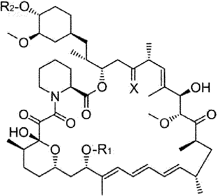
en donde R1 es un alquilo C1-6 o un alquinilo C3-6, R2 es H, -CH2-OH o -CH2-CH2-OH y X es =O, (H, H) o (H, OH), en donde el deterioro auditivo del oído interno está provocado por el síndrome de Pendred. En una realización específica, el derivado de rapamicina puede ser rapamicina.where R1 is C1-6 alkyl or C3-6 alkynyl, R2 is H, -CH2-OH or -CH2-CH2-OH and X is =O, (H, H) or (H, OH), in where the hearing impairment of the inner ear is caused by Pendred syndrome. In a specific embodiment, the rapamycin derivative may be rapamycin.
Un aspecto adicional de la presente invención es un método in vitro para inhibir la apoptosis de células del oído interno derivadas de pacientes con síndrome de Pendred que comprende un derivado de rapamicina representado por la siguiente fórmula II o una sal farmacéuticamente aceptable del mismo:A further aspect of the present invention is an in vitro method for inhibiting apoptosis of inner ear cells derived from patients with Pendred syndrome comprising a rapamycin derivative represented by the following formula II or a pharmaceutically acceptable salt thereof:

en donde R1 es un alquilo C1-6 o un alquinilo C3-6, R2 es H, -CH2-OH o -CH2-CH2 -OH y X es =O, (H, H) o (H, OH).where R 1 is C 1-6 alkyl or C 3-6 alkynyl , R 2 is H, -CH2 -OH or -CH 2 -CH 2 -OH and X is =O, (H, H) or ( H, OH).
[Breve descripción de los dibujos][Brief description of the drawings]
[Figura 1] (A) Micrografía de contraste de fase de células madre del oído interno obtenida en una realización de la presente invención y (B) micrografías de células madre del oído interno teñidas con (a) anticuerpo anti-PAX 2, (b) anticuerpo anti-PAX 8 y (c) anticuerpo anti-SOX2 en una realización de la presente invención.[Figure 1] (A) Phase contrast micrograph of inner ear stem cells obtained in one embodiment of the present invention and (B) micrographs of inner ear stem cells stained with (a) anti-PAX 2 antibody, (b ) anti-PAX 8 antibody and (c) anti-SOX2 antibody in one embodiment of the present invention.
[Figura 2] Micrografías que muestran que espina, miosina VIIa y prestina, que son marcadores de células ciliadas, se expresan cultivando las células madre del oído interno en suspensión y después cultivándolas en presencia de FGF 9 y FGF20 en un ejemplo de la presente invención (Inducción de la diferenciación en células ciliadas). Las células se tiñeron con anticuerpos contra espina, miosina VIIa y prestina (izquierda; tinción triple), un anticuerpo contra miosina VIIa (arriba a la derecha) y un anticuerpo contra espina (abajo a la derecha).[ Figure 2 ] Micrographs showing that spine, myosin VIIa and prestin, which are hair cell markers, are expressed by culturing the inner ear stem cells in suspension and then culturing them in the presence of FGF 9 and FGF20 in an example of the present invention (Induction of differentiation in hair cells). Cells were stained with antibodies against spine, myosin VIIa and prestin (left; triple stain), an antibody against myosin VIIa (upper right) and an antibody against spine (lower right).
[Figura 3] Micrografías que muestran que p27kip1 e ISLET1, que son marcadores de células de soporte, se expresan cultivando las células madre del oído interno en suspensión y después cultivándolas en presencia de f Gf 9 y FGF20 en un ejemplo de la presente invención (Inducción de la diferenciación en células de soporte). Las células se tiñeron con anticuerpos contra p27kip1, ISLET1 y prestina (izquierda; tinción triple) y anticuerpos contra ISLET1 y prestina (derecha; tinción doble).[Figure 3] Micrographs showing that p27kip1 and ISLET1, which are markers for supporting cells, are expressed by culturing the inner ear stem cells in suspension and then culturing them in the presence of fGf 9 and FGF20 in an example of the present invention ( Induction of differentiation in support cells). Cells were stained with antibodies against p27kip1, ISLET1, and prestin (left; triple staining) and antibodies against ISLET1 and prestin (right; double staining).
[Figura 4] Micrografías que muestran que GFAP, que es un marcador para las células gliales del ganglio espiral y la calbindina y la tubulina beta III, que son marcadores de neuronas maduras, se expresan cultivando células madre del oído interno en suspensión y después cultivándolas en presencia de FGF 9 y FGF20 en un ejemplo de la presente invención (induciendo la diferenciación de células ganglionares cocleares). Las células se tiñeron en tinción triple con anticuerpos contra GFAP, calbindina y tubulina beta III, respectivamente.[Figure 4] Micrographs showing that GFAP, which is a marker for spiral ganglion glial cells, and calbindin and beta III tubulin, which are markers for mature neurons, are expressed by culturing inner ear stem cells in suspension and then culturing them in the presence of FGF 9 and FGF20 in one example of the present invention (inducing cochlear ganglion cell differentiation). Cells were triple stained with antibodies against GFAP, calbindin, and beta III tubulin, respectively.
[Figura 5 ] Micrografías que muestran que S100, POU3F4, caldesmona y TBX18, que son marcadores de células mesenquimales perióticas, se expresan cultivando las células madre del oído interno en presencia de bFGF en un ejemplo de la presente invención (Inducción de la diferenciación en células mesenquimales perióticas). Las células se tiñeron en tinción triple con anticuerpos contra S100, caldesmona y TBX18 (arriba a la izquierda) y anticuerpos contra cx26, TBX18 y POU3f4 (arriba a la derecha). Las fotografías inferiores son micrografías de contraste de fase obtenidas después de que las células se cultivaron durante mucho tiempo en presencia de bFGF.[Figure 5 ] Micrographs showing that S100, POU3F4, caldesmone and TBX18, which are periotic mesenchymal cell markers, are expressed by culturing inner ear stem cells in the presence of bFGF in an example of the present invention (Induction of differentiation in periotic mesenchymal cells). Cells were triple stained with antibodies against S100, caldesmone, and TBX18 (upper left) and antibodies against cx26, TBX18, and POU3f4 (upper right). The bottom photographs are phase contrast micrographs obtained after the cells were cultured for a long time in the presence of bFGF.
[Figura 6] Micrografías que muestran que anhidrasa carbónica, acuaporina 1, ATPasa de sodio y potasio, vimentina, conexinas 26 y 30, que son marcadores de fibrocitos cocleares y células de la estría vascular del conducto coclear, se expresan cultivando las células mesenquimatosas perióticas en presencia de bFGF y cultivándolas después en ausencia de bFGF, y que la pendrina se expresa cultivando las células mesenquimatosas perióticas en presencia de bFGF y cultivándolas después en presencia de NaHCO3 en un ejemplo de la presente invención (Inducción de la diferenciación en fibrocitos del oído interno y células positivas para pendrina). Las células se tiñeron en tinción doble con anticuerpos contra CAII y conexina 26 (arriba a la izquierda), anticuerpos contra sodio potasio ATPasa y conexina 26 (medio superior), anticuerpos contra acuaporina 1 y conexina 26 (arriba a la derecha) y anticuerpos contra vimentina y conexina 26 (abajo a la izquierda). Las células se tiñeron en tinción doble con anticuerpos contra CAII y pendrina (abajo a la derecha). [Figure 6] Micrographs showing that carbonic anhydrase, aquaporin 1, sodium and potassium ATPase, vimentin, connexins 26 and 30, which are markers of cochlear fibrocytes and cochlear duct stria vascularis cells, are expressed by culturing the periotic mesenchymal cells in the presence of bFGF and then culturing them in the absence of bFGF, and that pendrin is expressed by culturing the periotic mesenchymal cells in the presence of bFGF and then culturing them in the presence of NaHCO3 in an example of the present invention (Induction of differentiation in ear fibrocytes internal and pendrin-positive cells). Cells were double stained with antibodies against CAII and connexin 26 (upper left), antibodies against sodium potassium ATPase and connexin 26 (upper middle), antibodies against aquaporin 1 and connexin 26 (upper right), and antibodies against vimentin and connexin 26 (lower left). Cells were double stained with antibodies against CAII and pendrin (bottom right).
[Figura 7] Resultados que indican que el daño del epitelio sensorial del oído interno estuvo provocado por la administración de gentamicina en un ejemplo de la presente invención.[ Figure 7 ] Results indicating that damage to the sensory epithelium of the inner ear was caused by the administration of gentamicin in one example of the present invention.
[Figura 8] Fotografías que muestran que se forman agregados intracelulares en células del oído interno derivadas de células iPS de pacientes con síndrome de Pendred en un ejemplo de la presente invención.[ Figure 8 ] Photographs showing that intracellular aggregates are formed in inner ear cells derived from iPS cells from Pendred syndrome patients in an example of the present invention.
[Figura 9] Gráfico que muestra que las viabilidades de las células del oído interno derivadas de células iPS de un paciente con síndrome de Pendred disminuyen cuando las células se someten a estrés en un ejemplo de la presente invención.[ Figure 9 ] Graph showing that the viabilities of inner ear cells derived from iPS cells from a patient with Pendred syndrome are decreased when the cells are subjected to stress in an example of the present invention.
[Figura 10] Gráfico que muestra que la rapamicina suprime la disminución de la viabilidad celular mostrada en la Figura 9 en un ejemplo de la presente invención.[ Figure 10 ] Graph showing that rapamycin suppresses the decrease in cell viability shown in Figure 9 in an example of the present invention.
[Figura 11] Gráfico que muestra que la rapamicina inhibe la apoptosis que provoca la disminución de la viabilidad celular mostrada en la Figura 9 en un ejemplo de la presente invención.[ Figure 11 ] Graph showing that rapamycin inhibits apoptosis causing decreased cell viability shown in Figure 9 in an example of the present invention.
[Figura 12] Gráfico que muestra que la metformina suprime la disminución de la viabilidad celular que se muestra en la Figura 9.[Figure 12] Graph showing that metformin suppresses the decrease in cell viability shown in Figure 9.
[Figura 13] Gráfico que muestra que la metformina inhibe la apoptosis que conduce a una disminución de la viabilidad celular que se muestra en la Figura 9.[Figure 13] Graph showing that metformin inhibits apoptosis leading to decreased cell viability shown in Figure 9.
[Figura 14] Gráfico que muestra que la calpeptina no puede inhibir la apoptosis conduce a la disminución de la viabilidad celular mostrada en la Figura 9 en un ejemplo de referencia de la presente invención.[ Figure 14 ] Graph showing that calpeptin cannot inhibit apoptosis leads to decreased cell viability shown in Figure 9 in a reference example of the present invention.
[Realizaciones para llevar a cabo la invención][Embodiments for carrying out the invention]
En lo sucesivo en el presente documento, las realizaciones de la presente invención completadas basándose en los descubrimientos anteriores se describirán en detalle con referencia a los ejemplos. Los objetos, las características, las ventajas y las ideas de la presente invención son evidentes para los expertos en la materia a partir de la descripción de esta memoria descriptiva. Adicionalmente, los expertos en la materia pueden reproducir fácilmente la presente invención a partir de la descripción del presente documento. Las realizaciones y los ejemplos específicos que se describen a continuación representan realizaciones preferibles de la presente invención, que se dan con el fin de ilustración o explicación.Hereinafter, completed embodiments of the present invention based on the above discoveries will be described in detail with reference to examples. The objects, features, advantages and ideas of the present invention are apparent to those skilled in the art from the description in this specification. Additionally, the present invention can be readily reproduced by those skilled in the art from the description herein. The specific embodiments and examples described below represent preferable embodiments of the present invention, which are given for the purpose of illustration or explanation.
(1) Inhibidores de la apoptosis(1) Inhibitors of apoptosis
Una realización de la presente invención es un inhibidor de la apoptosis para las células del oído interno que contiene, como un principio activo, un derivado de rapamicina (mostrado como la siguiente fórmula estructural II) o una sal farmacéuticamente aceptable del mismo.An embodiment of the present invention is an apoptosis inhibitor for inner ear cells containing, as an active ingredient, a rapamycin derivative (shown as the following structural formula II) or a pharmaceutically acceptable salt thereof.

en donde R1 es un alquilo C1-6 o un alquinilo C3-6, R2 es H, -CH2-OH o -CH2-CH2 -OH y X es =O, (H, H) o (H, OH). where R 1 is C 1-6 alkyl or C 3-6 alkynyl , R 2 is H, -CH2 -OH or -CH 2 -CH 2 -OH and X is =O, (H, H) or ( H, OH).
Las células del oído interno que se van a proteger de la apoptosis derivan de un animal que padece un deterioro auditivo en el oído interno como resultado de la apoptosis provocada por el síndrome de Pendred. Adicionalmente, las células del oído interno que se van a proteger de la apoptosis pueden ser las existentes en el cuerpo de un organismo o células cultivadas. Las especies del organismo no están particularmente limitadas, pero los vertebrados son preferibles y el ser humano es la más preferible. Las células cultivadas pueden ser líneas celulares establecidas, células de cultivo primario o células del oído interno que se han diferenciado a partir de células madre. Los detalles de cómo inducir la diferenciación de células madre en células del oído interno se describirán más adelante.The inner ear cells to be protected from apoptosis are derived from an animal suffering from hearing impairment in the inner ear as a result of apoptosis caused by Pendred syndrome. Additionally, the inner ear cells to be protected from apoptosis may be those existing in the body of an organism or cultured cells. The species of the organism are not particularly limited, but vertebrates are preferable and human is most preferable. The cultured cells can be established cell lines, primary culture cells, or inner ear cells that have differentiated from stem cells. The details of how to induce stem cell differentiation into inner ear cells will be described later.
Como se ha descrito anteriormente, los inhibidores de la apoptosis de la presente invención pueden usarse para las células del oído interno y, por lo tanto, son útiles como agentes farmacéuticos para tratar el deterioro auditivo del oído interno resultante de la apoptosis provocada por el síndrome de Pendred. El término "tratamiento" como se usa en el presente documento incluye la mejora del deterioro auditivo anteriormente mencionado, la prevención de la progresión del deterioro auditivo anteriormente mencionado y la prevención del deterioro auditivo anteriormente mencionado. (2) Derivados de rapamicinaAs described above, the inhibitors of apoptosis of the present invention can be used for cells of the inner ear, and are therefore useful as pharmaceutical agents for treating inner ear hearing impairment resulting from apoptosis caused by the syndrome. of Pendred. The term "treatment" as used herein includes the amelioration of the aforementioned hearing impairment, the prevention of the progression of the aforementioned hearing impairment, and the prevention of the aforementioned hearing impairment. (2) Rapamycin derivatives
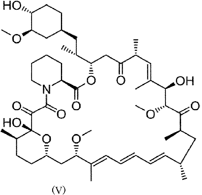
La rapamicina es un compuesto que tiene la siguiente fórmula estructural V.Rapamycin is a compound that has the following structural formula V.
En la presente memoria descriptiva, los derivados de rapamicina incluyen compuestos representados por la siguiente fórmula estructural.As used herein, rapamycin derivatives include compounds represented by the following structural formula.

en donde R1 es un alquilo C1-6 o un alquinilo C3-6 , R2 es H, -CH2-OH o -CH2-CH2 -OH y X es =O, (H, H) o (H, OH). Por ejemplo, dentro del alcance de la fórmula (II), los derivados de rapamicina incluyen (1) derivados de rapamicina 40-O-sustituidos (por ejemplo, 40-O-(2-hidroxi)-etil-rapamicina), (2) 32-desoxo-rapamicina y derivados de la misma y 32-hidroxi-rapamicina y derivados de la misma, (3) derivados de rapamicina 16-O-sustituidos (por ejemplo, 16-pent-2-iniloxi-32-desoxorapamicina, pero la rapamicina es la más preferible.where R 1 is C 1-6 alkyl or C 3-6 alkynyl , R 2 is H, -CH2 -OH or -CH 2 -CH 2 -OH and X is =O, (H, H) or ( H, OH). For example, within the scope of formula (II), rapamycin derivatives include (1) 40-O-substituted rapamycin derivatives (eg, 40-O-(2-hydroxy)-ethyl-rapamycin), (2 ) 32-deoxo-rapamycin and derivatives thereof and 32-hydroxy-rapamycin and derivatives thereof, (3) 16-O-substituted rapamycin derivatives (for example, 16-pent-2-ynyloxy-32-deoxorapamycin, but rapamycin is most preferable.
(3) Inducción de la diferenciación de células madre en células del oído interno (3) Induction of stem cell differentiation into inner ear cells
Ahora se describe un método para inducir la diferenciación de células madre en células del oído interno (en lo sucesivo, también denominado método de inducción de células del oído interno). Las células madre no están particularmente limitadas, pero pueden ejemplificarse las células madre pluripotentes y las células madre del oído interno. El método de inducción de células del oído interno se describe en detalle usando células madre pluripotentes como ejemplo. A method for inducing stem cell differentiation into inner ear cells (hereinafter also called inner ear cell induction method) is now described. Stem cells are not particularly limited, but pluripotent stem cells and inner ear stem cells can be exemplified. The inner ear cell induction method is described in detail using pluripotent stem cells as an example.
Este método de inducción de células del oído interno puede realizarse de acuerdo con el siguiente procedimiento.This inner ear cell induction method can be performed according to the following procedure.

Inducción de la diferenciación de células madre pluripotentes en células madre del oído internoInduction of pluripotent stem cell differentiation into inner ear stem cells
[1] En un método de inducción de células madre pluripotentes a células madre del oído interno, las siguientes etapas se realizan en este orden:[1] In a method of inducing pluripotent stem cells to inner ear stem cells, the following steps are performed in this order:
Primera etapa: cultivar células madre pluripotentes en presencia de un inhibidor de ROCK;First step: culturing pluripotent stem cells in the presence of a ROCK inhibitor;
Segunda etapa: cultivar las células en ausencia de inhibidor de ROCK;Second step: culturing the cells in the absence of ROCK inhibitor;
Tercera etapa: cultivar las células en un medio sin suero;Third stage: culturing the cells in a serum-free medium;
Cuarta etapa: cultivar las células en un medio sin suero que contiene un factor de crecimiento; yFourth step: culturing the cells in a serum-free medium containing a growth factor; Y
Quinta etapa: disociar las células en células individuales.Fifth stage: dissociate the cells into individual cells.
Puede añadirse otra etapa o etapas que no sean esenciales para este método entre las etapas individuales.Another step or steps not essential to this method may be added between the individual steps.
Las células madre pluripotentes no están particularmente limitadas siempre que tengan un potencial de diferenciación pluripotente (es decir, multipotencia o pluripotencia) y los ejemplos incluyen células madre embrionarias (células ES), células madre pluripotentes inducidas (células iPS) y células Muse. Las células con totipotencia son particularmente preferibles.Pluripotent stem cells are not particularly limited as long as they have pluripotent differentiation potential (ie, multipotency or pluripotency) and examples include embryonic stem cells (ES cells), induced pluripotent stem cells (iPS cells) and Muse cells. Cells with totipotency are particularly preferable.
El inhibidor ROCK (quinasa formadora de bobinas en espiral asociada a Rho/quinasa asociada a Rho) no está particularmente limitado y los ejemplos incluyen Y-27632, Clorhidrato de fasudilo, K-115 (Hidrato de clorhidrato de Ripasudil) y DE-104. La concentración óptima del inhibidor de ROCK puede determinarse fácilmente según sea apropiado, pero es preferible del 0,05 % al 0,2 % y es más preferible el 0,1 %.The ROCK (Rho-associated coil-forming kinase/Rho-associated kinase) inhibitor is not particularly limited and examples include Y-27632, Fasudil Hydrochloride, K-115 (Ripasudil Hydrochloride Hydrate) and DE-104. The optimal concentration of the ROCK inhibitor can be easily determined as appropriate, but 0.05% to 0.2% is preferable and 0.1% is more preferable.
El medio usado en la primera y la segunda etapas no está particularmente limitado siempre que las células madre pluripotentes puedan mantenerse en él y mTeSR1 es un ejemplo. La primera etapa se realiza preferentemente durante 1 a 3 días y más preferentemente durante 1 a 2 días. La segunda etapa se realiza preferentemente durante 1 a 3 días y más preferentemente durante 1 a 2 días.The medium used in the first and second steps is not particularly limited as long as pluripotent stem cells can be maintained therein, and mTeSR1 is an example. The first stage is preferably carried out for 1 to 3 days and more preferably for 1 to 2 days. The second stage is carried out preferably for 1 to 3 days and more preferably for 1 to 2 days.
En la segunda etapa, lo que significa la expresión "en ausencia de inhibidor de ROCK" es que un inhibidor de ROCK está sustancialmente ausente y puede estar contenido en una concentración en la que no se observa ningún efecto. In the second step, what is meant by the expression "in the absence of ROCK inhibitor" is that a ROCK inhibitor is substantially absent and may be contained in a concentration where no effect is observed.
Algunos ejemplos del medio sin suero usado en la tercera etapa incluyen DMEM/F12 B27 N2 GlutaMax Aminoácido no esencial. En la tercera etapa, las células se cultivan preferentemente durante 2 a 6 días, más preferentemente durante 2 a 4 días y lo más preferentemente durante 3 días.Some examples of the serum-free medium used in the third step include DMEM/F12 B27 N2 GlutaMax Nonessential Amino Acid. In the third stage, the cells are cultured preferably for 2 to 6 days, more preferably for 2 to 4 days, and most preferably for 3 days.
Algunos ejemplos del medio sin suero usado en la cuarta etapa incluyen DMEM/F12 B27 N2 GlutaMax Aminoácido no esencial, pero es preferible que se use el mismo medio sin suero que en la tercera etapa. Para el factor de crecimiento, puede añadirse al menos uno seleccionado del grupo que consiste en bFGF, FGF3, FGF10 y FGF19, pero es preferible que se añadan todos. Las concentraciones preferibles de ellos son de 10 a 50 ng/ml, 10 a 50 ng/ml, 10 a 50 ng/ml y 10 a 50 ng/ml, respectivamente. Adicionalmente, es preferible que las células se cultiven en presencia de BMP4 en la fase más temprana del cultivo y en ausencia de BMP4 en la última fase. La concentración de BMP4 a añadir en la fase más temprana es preferentemente de 5 a 50 ng/ml. Para el cultico en la última fase, la expresión "en ausencia de BMP4" significa que BMP4 está sustancialmente ausente y puede estar contenido en una concentración en la que no se observa ningún efecto. Las células en la fase más temprana se cultivan preferentemente durante 2 a 6 días, más preferentemente durante 2 a 4 días y lo más preferentemente durante 3 días. Las células en la última fase se cultivan preferentemente durante 2 a 6 días, más preferentemente durante 2 a 4 días y lo más preferentemente durante 3 días. Las células madre del oído interno obtenidas de esta manera tienen potencial de diferenciación a las células madre del oído interno.Some examples of the serum-free medium used in the fourth stage include DMEM/F12 B27 N2 GlutaMax Nonessential Amino Acid, but it is preferable that the same serum-free medium is used as in the third stage. For the growth factor, at least one selected from the group consisting of bFGF, FGF3, FGF10 and FGF19 may be added, but it is preferable that all are added. Preferable concentrations thereof are 10 to 50 ng/ml, 10 to 50 ng/ml, 10 to 50 ng/ml and 10 to 50 ng/ml, respectively. Additionally, it is preferable that the cells are cultured in the presence of BMP4 in the earliest stage of culture and in the absence of BMP4 in the late stage. The concentration of BMP4 to be added in the earliest phase is preferably 5 to 50 ng/ml. For the late stage cultivar, the term "in the absence of BMP4" means that BMP4 is substantially absent and may be contained at a concentration where no effect is observed. Cells in the earliest stage are cultured preferably for 2 to 6 days, more preferably for 2 to 4 days, and most preferably for 3 days. Cells in the late phase are cultured preferably for 2 to 6 days, more preferably for 2 to 4 days, and most preferably for 3 days. Inner ear stem cells obtained in this way have differentiation potential to inner ear stem cells.
Como la quinta etapa, las esferas de las células obtenidas en el cuarto paso se disocian en células individuales. El método de disociar esferas en células individuales no está particularmente limitado y, por ejemplo, puede usarse tripsina o Accutase. Es preferible que después de que las células se disocien con una enzima o por medios mecánicos como el pipeteo, las células individuales disociadas se seleccionan y se retiran los grumos residuales de células, usando, por ejemplo, una malla de nailon para retirar las células indiferenciadas o los grumos.Like the fifth step, the cell spheres obtained in the fourth step are dissociated into individual cells. The method of dissociating beads into individual cells is not particularly limited and, for example, trypsin or Accutase can be used. It is preferable that after the cells are dissociated with an enzyme or by mechanical means such as pipetting, the dissociated individual cells are selected and residual clumps of cells are removed, using, for example, a nylon mesh to remove undifferentiated cells. or the lumps.
Para cultivar células madre del oído interno, usando placas de cultivo recubiertas con un agente de recubrimiento, las células se cultivan en condiciones hipóxicas para mantener su capacidad de células madre (la sexta etapa). El agente de recubrimiento usado no está particularmente limitado, pero la poli-O-fibronectina es la más preferible. Las células madre a cultivar pueden ser las obtenidas en la cuarta etapa o las obtenidas en la quinta etapa. Los ejemplos de medio suplementado con suero usados en esta etapa incluyen DMEM y F12, pero el más preferible es DMEM/F12 que contiene N2 y B27 además del suero (por ejemplo, FBS). La concentración de oxígeno en la condición hipóxica en esta etapa es preferentemente del 4 % al 10 %, más preferentemente del 4 % al 6 % y lo más preferentemente del 4 %. Puede añadirse al menos un factor de crecimiento seleccionado del grupo que consiste en bFGF, EGF e IGF1 al medio de cultivo, pero es preferible que se añadan todos. Sus concentraciones preferibles son de 10 a 30 ng/ml, 10 a 30 ng/ml y 10 a 50 ng/ml, respectivamente.To grow inner ear stem cells, using culture plates coated with a coating agent, the cells are grown under hypoxic conditions to maintain their stem cell capacity (the sixth stage). The coating agent used is not particularly limited, but poly-O-fibronectin is most preferable. The stem cells to be cultivated can be those obtained in the fourth stage or those obtained in the fifth stage. Examples of serum-supplemented medium used in this step include DMEM and F12, but most preferable is DMEM/F12 containing N2 and B27 in addition to serum (eg, FBS). The oxygen concentration in the hypoxic condition at this stage is preferably 4% to 10%, more preferably 4% to 6%, and most preferably 4%. At least one growth factor selected from the group consisting of bFGF, EGF and IGF1 may be added to the culture medium, but it is preferable that all are added. Their preferable concentrations are 10 to 30 ng/ml, 10 to 30 ng/ml and 10 to 50 ng/ml, respectively.
[2] Inducción de la diferenciación de las células madre del oído interno en el epitelio sensorial del oído interno tales como las células de soporte y las células ciliadas, las células ganglionares cocleares y las células de la estría vascular [2] Induction of differentiation of inner ear stem cells into inner ear sensory epithelium such as supporting cells and hair cells, cochlear ganglion cells, and stria vascularis cells
En un método para inducir células madre del oído interno en el epitelio sensorial del oído interno, las células ganglionares cocleares y las células de la estría vascular, las siguientes etapas se realizan en este orden:In a method of inducing inner ear stem cells into inner ear sensory epithelium, cochlear ganglion cells, and stria vascularis cells, the following steps are performed in this order:
Primera etapa: cultivar células madre del oído interno en suspensión; yFirst stage: culturing inner ear stem cells in suspension; Y
Segunda etapa: cultivar las células en presencia de FGF9 y FGF20. Pueden añadirse otras etapas que no sean esenciales para este método entre las etapas.Second step: culturing the cells in the presence of FGF9 and FGF20. Other steps that are not essential to this method may be added between the steps.
En la primera etapa, las células madre del oído interno se cultivan en suspensión. Específicamente, como en la quinta etapa, las células madre del oído interno se disocian en células individuales. Las células disociadas se cultivan después en suspensión; para este fin, se usan placas para cultivo en suspensión con las que se pueden cultivar células en condiciones no adherentes. Por ejemplo, pueden usarse platos para cultivo no adherente tales como placas de plástico para cultivo bacteriano. La expresión "cultivo en suspensión" significa que las células o esferas objetivo se cultivan sin adherirse a la superficie inferior de un recipiente de cultivo y la expresión "cultivo adherente" o "cultivo en un sustrato" significa que las células diana o esferas se cultivan adhiriéndose a la superficie inferior de un recipiente de cultivo. La expresión que las células o esferas se adhieren a la superficie inferior de un recipiente de cultivo durante el cultivo significa que las células o esferas se depositan en la superficie inferior del recipiente de cultivo a través de moléculas de adhesión célula-sustrato contenidas en, por ejemplo, una matriz extracelular (MEC) y se refiere a que las células o esferas no flotan en el líquido de cultivo incluso cuando se agita suavemente el líquido de cultivo. Por otra parte, la expresión que las células o esferas no se adhieren a la superficie inferior de un recipiente de cultivo durante el cultivo significa que las células o esferas no se depositan en la superficie inferior del recipiente de cultivo a través de moléculas de adhesión célula-sustrato contenidas en, por ejemplo, ECM y se refiere a que las células o esferas flotan en el líquido de cultivo cuando el líquido de cultivo se agita suavemente, incluso si están en contacto con la superficie inferior. Para el cultivo adherente, es preferible tratar o recubrir químicamente, con un agente de recubrimiento (por ejemplo, gelatina, polilisina o agar) que promueve la adhesión, la superficie inferior de los platos de plástico, para promover la adhesión de las células a un sustrato. Para cultivo en suspensión, es preferible que la superficie inferior de las placas de plástico no se trate o se recubra con un agente de recubrimiento para evitar la adhesión (por ejemplo, poli(metacrilato de 2-hidroxietilo)) para evitar que las células se adhieran a un sustrato. Las células como un sujeto pueden tardar algún tiempo en adherirse y, por lo tanto, las células pueden flotar libremente durante un cierto período de tiempo, incluso en un cultivo adherente, pero el cultivo se define como un cultivo adherente siempre que las células finalmente se adhieran a un sustrato, cuanto tiempo pueda tomar. El medio usado para el cultivo en suspensión no está particularmente limitado y los ejemplos incluyen DMEM y F12. Es, sin embargo, preferible usar un medio suplementado con suero (por ejemplo, FBS), N2, B27 y/o un factor extrínseco tal como un factor de crecimiento y lo más preferible es usar DMEM/F12 como este medio. El factor extrínseco a añadir puede incluir al menos un factor de crecimiento seleccionado del grupo que consiste en bFGF, EGF, IGF1, Wnt3a, FGF9, FGF20, heparina y un inhibidor de TGFp. Es, sin embargo, es preferible que el medio contenga un factor de crecimiento y heparina y es más preferible que el medio contenga todos los factores de crecimiento indicados anteriormente. Las concentraciones preferibles de los mismos son de 10 a 50, de 10 a 50, de 10 a 100, de 10 a 50, de 10 a 100, de 10 a 100, de 1 a 50 ng/ml, respectivamente, pero no están particularmente limitadas. Las células se cultivan en suspensión preferentemente durante 3 a 7 días, más preferentemente durante 4 a 6 días y lo más preferentemente durante 5 días. In the first stage, stem cells from the inner ear are cultured in suspension. Specifically, as in the fifth stage, the inner ear stem cells dissociate into individual cells. The dissociated cells are then grown in suspension; for this purpose, suspension culture plates are used with which cells can be grown under non-adherent conditions. For example, non-adherent culture dishes such as plastic bacterial culture dishes can be used. The expression "suspension culture" means that the target cells or beads are grown without adhering to the bottom surface of a culture vessel and the expression "adherent culture" or "culture on a substrate" means that the target cells or beads are grown adhering to the bottom surface of a culture vessel. The expression that cells or spheres adhere to the bottom surface of a culture vessel during cultivation means that cells or spheres are deposited on the bottom surface of the culture vessel through cell-substrate adhesion molecules contained in, e.g. example, an extracellular matrix (ECM) and refers to the fact that the cells or spheres do not float in the culture fluid even when the culture fluid is gently agitated. On the other hand, the expression that cells or spheres do not adhere to the bottom surface of a culture vessel during culture means that cells or spheres are not deposited on the bottom surface of the culture vessel through cell adhesion molecules. -substrate contained in, for example, ECM and refers to cells or spheres floating in the culture fluid when the culture fluid is gently agitated, even if they are in contact with the bottom surface. For adherent culture, it is preferable to chemically treat or coat, with an adhesion-promoting coating agent (e.g., gelatin, polylysine, or agar), the bottom surface of plastic dishes, to promote cell adhesion to a substratum. For suspension culture, it is preferable that the bottom surface of the plastic plates is left untreated or coated with a non-adhesion coating agent (e.g., poly(2-hydroxyethyl methacrylate)) to prevent cells from sticking to each other. adhere to a substrate. Cells as a subject may take some time to adhere, and therefore cells may be free-floating for a certain period of time, even in an adherent culture, but the culture is defined as an adherent culture as long as the cells eventually adhere. adhere to a substrate, how long it may take. The medium used for the suspension culture is not particularly limited, and examples include DMEM and F12. It is, however, preferable to use a medium supplemented with serum (eg FBS), N2, B27 and/or an extrinsic factor such as growth factor and most preferably to use DMEM/F12 as this medium. The extrinsic factor to be added may include at least one growth factor selected from the group consisting of bFGF, EGF, IGF1, Wnt3a, FGF9, FGF20, heparin, and a TGFp inhibitor. It is, however, preferable that the medium contains a growth factor and heparin, and it is more preferable that the medium contains all of the growth factors listed above. Preferable concentrations thereof are 10 to 50, 10 to 50, 10 to 100, 10 to 50, 10 to 100, 10 to 100, from 1 to 50 ng/ml, respectively, but are not particularly limited. Cells are cultured in suspension preferably for 3 to 7 days, more preferably for 4 to 6 days, and most preferably for 5 days.
Posteriormente, las células se cultivan más en suspensión después de la adición de medio, preferentemente durante 3 a 7 días, más preferentemente durante 4 a 6 días y lo más preferentemente durante 5 días. El medio a añadir no está particularmente limitado y los ejemplos incluyen DMEM y F12. Es, sin embargo, preferible usar un medio suplementado con suero (por ejemplo, FBS), N2, b27 y/o un factor extrínseco tal como un factor de crecimiento y lo más preferible es usar DMEM/F12 como este medio. Es preferible usar el medio idéntico al usado para el cultivo en suspensión inicial excepto por el factor extrínseco. El factor extrínseco a añadir puede incluir al menos un factor de crecimiento seleccionado del grupo que consiste en bFGF, EGF e IGF1, pero es preferible que el medio los contenga todos. Las concentraciones preferibles de ellos son de 10 a 30 ng/ml, 10 a 30 ng/ml y 10 a 50 ng/ml, respectivamente. Subsequently, the cells are further grown in suspension after the addition of medium, preferably for 3 to 7 days, more preferably for 4 to 6 days, and most preferably for 5 days. The medium to be added is not particularly limited, and examples include DMEM and F12. It is, however, preferable to use a medium supplemented with serum (eg FBS), N2, b27 and/or an extrinsic factor such as growth factor and most preferably to use DMEM/F12 as this medium. It is preferable to use the identical medium to that used for the initial suspension culture except for the extrinsic factor. The extrinsic factor to be added may include at least one growth factor selected from the group consisting of bFGF, EGF and IGF1, but it is preferable that the medium contains all of them. Preferable concentrations thereof are 10 to 30 ng/ml, 10 to 30 ng/ml and 10 to 50 ng/ml, respectively.
Las esferas formadas se recogen después sin que se rompan y se cultivan adherentemente sobre un sustrato usando el mismo medio. Después de 1 hora a 2 días, preferentemente después de 1 hora a 36 horas, preferentemente el día siguiente, el medio se reemplaza con un medio fresco que contiene suero (por ejemplo, FBS), N2, B27 y un factor extrínseco tal como un factor de crecimiento. El factor extrínseco a añadir es preferentemente T3 y/o IGF1. Es preferible usar el medio idéntico al usado para el cultivo en suspensión inicial excepto por el factor extrínseco. Sus concentraciones preferibles son de 10 a 100 y de 1 a 50 ng/ml, respectivamente. Las células ciliadas, las células de soporte, las células ganglionares cocleares y las células de la estría vascular pueden permitirse diferenciarse cultivando adicionalmente las células preferentemente durante 3 días o más y más preferentemente durante 5 días o más. Durante este cultivo, el intercambio de los medios puede ser lo único que pueda hacerse.The spheres formed are then collected without breaking and adherently grown on a substrate using the same medium. After 1 hour to 2 days, preferably after 1 hour to 36 hours, preferably the next day, the medium is replaced with fresh medium containing serum (eg FBS), N2, B27 and an extrinsic factor such as a growth factor. The extrinsic factor to be added is preferably T3 and/or IGF1. It is preferable to use the identical medium to that used for the initial suspension culture except for the extrinsic factor. Their preferable concentrations are 10 to 100 and 1 to 50 ng/ml, respectively. Hair cells, supporting cells, cochlear ganglion cells and stria vascularis cells can be allowed to differentiate by further culturing the cells preferably for 3 days or more and more preferably for 5 days or more. During this culture, exchanging the media may be the only thing that can be done.
[3] Inducción de la diferenciación de las células madre del oído interno en fibrocitos cocleares, células de la estría vascular y células positivas para pendrina a través de células mesenquimales perióticas[3] Induction of differentiation of inner ear stem cells into cochlear fibrocytes, stria vascularis cells, and pendrin-positive cells via periotic mesenchymal cells
Un método de inducción de células madre del oído interno a células mesenquimales perióticas incluye la etapa de cultivar células madre del oído interno en un medio que contiene bFGF. Otras etapas no esenciales para la presente invención pueden añadirse a este método.A method of inducing inner ear stem cells to periotic mesenchymal cells includes the step of culturing inner ear stem cells in a medium containing bFGF. Other steps not essential to the present invention can be added to this method.
El medio usado aquí no está particularmente limitado y los ejemplos incluyen DMEM y F12. Es, sin embargo, preferible usar un medio suplementado con suero (por ejemplo, FBS) y bFGF y lo más preferible es usar DMEM como este medio. Una concentración preferible de bFGF es de 1 a 50 ng/ml. Las células se cultivan preferentemente durante 7 días o más, más preferentemente durante 10 días o más y lo más preferentemente 14 días o más. Después de este cultivo, se obtienen células mesenquimales perióticas que tienen estructuras fibrosas.The medium used here is not particularly limited, and examples include DMEM and F12. It is, however, preferable to use a medium supplemented with serum (eg FBS) and bFGF and most preferably to use DMEM as this medium. A preferable concentration of bFGF is 1 to 50 ng/ml. Cells are preferably cultured for 7 days or longer, more preferably 10 days or longer, and most preferably 14 days or longer. After this culture, periotic mesenchymal cells having fibrous structures are obtained.
Los fibrocitos cocleares y las células de la estría vascular se obtienen cultivando adicionalmente las células en un medio fresco sin bFGF, preferentemente durante 7 días o más, más preferentemente durante 10 días o más y lo más preferentemente durante 14 días o más.Cochlear fibrocytes and stria vascularis cells are obtained by further culturing the cells in fresh medium without bFGF, preferably for 7 days or longer, more preferably 10 days or longer, and most preferably 14 days or longer.
Las células positivas para Pendrina se obtienen cultivando más las células en un medio fresco sin bFGF pero con NaHCO3 añadido preferentemente durante 7 días o más, más preferentemente durante 10 días o más y lo más preferentemente durante 14 días o más. Una concentración preferible de NaHCO3 es del 0,3 % al 1 %.Pendrin-positive cells are obtained by further culturing the cells in fresh medium without bFGF but with added NaHCO3 preferably for 7 days or longer, more preferably 10 days or longer, and most preferably 14 days or longer. A preferable concentration of NaHCO3 is 0.3% to 1%.
(4) Uso de inhibidor de apoptosis(4) Use of inhibitor of apoptosis
Los inhibidores de la apoptosis descritos en (1) pueden usarse in vivo o in vitro. The inhibitors of apoptosis described in (1) can be used in vivo or in vitro.
Cuando se usa un inhibidor de la apoptosis in vivo, puede formularse como se describe a continuación dependiendo de la vía de administración.When an inhibitor of apoptosis is used in vivo, it may be formulated as described below depending on the route of administration.
Además de un principio activo, pueden contenerse diversos componentes que normalmente se usan, si es necesario. Por ejemplo, uno o más vehículos farmacéuticamente aceptables, disgregantes, diluyentes, lubricantes, agentes aromatizantes, colorantes, edulcorantes, agentes enmascarantes del sabor, agentes de suspensión, agentes humectantes, emulsionantes, agentes dispersantes, agentes auxiliares, antisépticos, tampones, aglutinantes, estabilizantes y agentes de recubrimiento pueden estar contenidos.In addition to an active ingredient, various components that are normally used may be contained, if necessary. For example, one or more pharmaceutically acceptable carriers, disintegrants, diluents, lubricants, flavoring agents, colorants, sweeteners, taste masking agents, suspending agents, wetting agents, emulsifiers, dispersing agents, auxiliary agents, antiseptics, buffers, binders, stabilizers and coating agents may be contained.
Como una vía de administración, puede seleccionarse cualquiera de administración sistémica y administración tópica. Específicamente, se selecciona una vía de administración apropiada dependiendo de la enfermedad, el síntoma, etc. Los agentes farmacéuticos de acuerdo con la presente invención pueden administrarse por vía oral o parenteral, pero se prefiere la administración oral. Algunos ejemplos de administraciones parenterales incluyen administraciones subcutánea, intradérmica, intramuscular e intratimpánica distintas de las típicas administraciones intravenosas e intraarteriales. Adicionalmente, puede usarse la administración transmucosa o transdérmica.As a route of administration, any of systemic administration and topical administration can be selected. Specifically, an appropriate administration route is selected depending on the disease, the symptom, etc. Pharmaceutical agents according to the present invention may be administered orally or parenterally, but oral administration is preferred. Some examples of parenteral administrations include subcutaneous, intradermal, intramuscular and intratympanic administrations other than typical intravenous and intraarterial administrations. Additionally, transmucosal or transdermal administration may be used.
La forma de dosificación no está particularmente limitada y pueden usarse diversas formas de dosificación tales como, para administración oral como un ejemplo, comprimidos, cápsulas, polvos, gránulos, tabletas, líquidos, emulsiones, suspensiones, soluciones, bebidas espirituosas, jarabes, extractos o elixires. Para agentes parenterales, los ejemplos incluyen inyecciones tales como inyecciones subcutáneas, inyecciones intravenosas, inyecciones intramusculares e inyecciones intraperitoneales; sistemas o parches terapéuticos transdérmicos, pomadas o lociones; preparaciones sublinguales y parches orales para administración bucal; aerosoles para administración nasal; y supositorios, pero no se limitan a los mismos. Estos agentes formulados pueden producirse mediante métodos conocidos comúnmente usados en un proceso de producción de agentes formulados. Los agentes farmacéuticos de acuerdo con la presente invención pueden estar en una forma de dosificación sostenida o de liberación sostenida.The dosage form is not particularly limited and various dosage forms may be used such as, for oral administration as an example, tablets, capsules, powders, granules, lozenges, liquids, emulsions, suspensions, solutions, spirits, syrups, extracts or elixirs. For parenteral agents, the examples they include injections such as subcutaneous injections, intravenous injections, intramuscular injections, and intraperitoneal injections; transdermal therapeutic systems or patches, ointments or lotions; sublingual preparations and oral patches for buccal administration; sprays for nasal administration; and suppositories, but are not limited thereto. These formulated agents can be produced by known methods commonly used in a formulated agent production process. Pharmaceutical agents according to the present invention may be in sustained-release or sustained-dosage form.
La cantidad del principio activo contenido en el agente farmacéutico puede determinarse apropiadamente dependiendo de, por ejemplo, un intervalo de dosis para el principio activo y el número de administraciones.The amount of the active ingredient contained in the pharmaceutical agent can be appropriately determined depending on, for example, a dosage range for the active ingredient and the number of administrations.
El intervalo de dosis no está particularmente limitado y se selecciona apropiadamente dependiendo de, por ejemplo, la efectividad del ingrediente o ingredientes, el proceso de administración, una vía de administración, el tipo de enfermedad, la información asociada a un sujeto (tales como peso corporal, edad, condición médica y uso de otros medicamentos, etc.) y una decisión de un médico tratante. En general, una dosis adecuada es, por ejemplo, de alrededor de 100 mg a 3000 mg, preferentemente de aproximadamente 2250 mg o menos, más preferentemente de aproximadamente 750 mg o menos por adulto por día y de aproximadamente 100 mg a 2000 mg, preferentemente de aproximadamente 1500 mg o menos, más preferentemente de aproximadamente 750 mg o menos por niño por día. Estas dosis pueden, sin embargo, modificarse usando experimentos de rutina convencionales para la optimización bien conocidos en la técnica. La dosis anterior puede administrarse de una vez al día a varias veces al día.The dose range is not particularly limited and is appropriately selected depending on, for example, the effectiveness of the ingredient(s), the administration process, a route of administration, the type of disease, information associated with a subject (such as weight body size, age, medical condition and use of other medications, etc.) and a decision of a treating physician. In general, a suitable dose is, for example, from about 100 mg to 3000 mg, preferably from about 2250 mg or less, more preferably from about 750 mg or less per adult per day, and from about 100 mg to 2000 mg, preferably about 1500 mg or less, more preferably about 750 mg or less per child per day. These doses can, however, be modified using standard routine experiments for optimization well known in the art. The above dose can be administered from once a day to several times a day.
Por otra parte, la apoptosis de las células del oído interno puede inhibirse in vitro añadiendo un inhibidor de la apoptosis a un medio usado para cultivo celular y usar ese medio para cultivar las células del oído interno. La concentración del inhibidor de la apoptosis que se añade al medio no está particularmente limitada pero es preferentemente de 0,1 nM a 1,0 nM para los derivados de rapamicina.On the other hand, apoptosis of inner ear cells can be inhibited in vitro by adding an apoptosis inhibitor to a medium used for cell culture and using that medium to culture inner ear cells. The concentration of the inhibitor of apoptosis to be added to the medium is not particularly limited but is preferably 0.1 nM to 1.0 nM for the rapamycin derivatives.
(5) Método para tratar el deterioro auditivo del oído interno como resultado de la apoptosis(5) Method for treating hearing impairment of the inner ear as a result of apoptosis
La presente invención puede usarse para tratar el deterioro auditivo del oído interno resultante de la apoptosis mediante la administración de un derivado de rapamicina a un paciente que padece un deterioro auditivo del oído interno como resultado de la apoptosis provocada por el síndrome de Pendred. Los derivados de rapamicina son como se describen en (2). El derivado de rapamicina o la sal farmacéuticamente aceptable del mismo puede administrarse mediante el método que usa el inhibidor de la apoptosis in vivo como se describe en (4).The present invention can be used to treat inner ear hearing impairment resulting from apoptosis by administering a rapamycin derivative to a patient suffering from inner ear hearing impairment resulting from apoptosis caused by Pendred syndrome. Rapamycin derivatives are as described in (2). The rapamycin derivative or pharmaceutically acceptable salt thereof can be administered by the method using the inhibitor of apoptosis in vivo as described in (4).
(6) Método para examinar la apoptosis(6) Method for examining apoptosis
Un método para examinar la apoptosis incluye las etapas de administrar un derivado de rapamicina in vitro a una célula del oído interno, inducir apoptosis en la célula del oído interno; y examinar la apoptosis inducida en la célula del oído interno. Los derivados de rapamicina son como se describen en (2).A method for examining apoptosis includes the steps of administering a rapamycin derivative in vitro to an inner ear cell, inducing apoptosis in the inner ear cell; and examining the apoptosis induced in the inner ear cell. Rapamycin derivatives are as described in (2).
El derivado de rapamicina puede ser un compuesto líder sintetizado a partir de rapamicina usada como un compuesto semilla.The rapamycin derivative may be a leader compound synthesized from rapamycin used as a seed compound.
Las células del oído interno usadas aquí son como se describen en (1).The inner ear cells used here are as described in (1).
Como el método para inducir la apoptosis en las células del oído interno, puede usarse un método conocido y los ejemplos incluyen un tratamiento con epoxomicina (por ejemplo, tratamiento con 0,5 pM de epoxomicina durante 24 horas).As the method for inducing apoptosis in inner ear cells, a known method can be used, and examples include treatment with epoxomicin (for example, treatment with 0.5 pM epoxomicin for 24 hours).
Como el método para examinar la apoptosis inducida de esta manera en las células del oído interno, puede usarse un método conocido y los ejemplos incluyen un método para detectar, usando Anexina V, fosfatidilserina que se ha retirado al exterior de la membrana celular como un resultado de la apoptosis, un método para detectar el fenómeno de que la membrana celular ha permeado 7-AAD (7-amino-actinomicina D), un método para detectar caspasa activada con un inhibidor de caspasa marcado con fluorescencia, SR-VAD-FMK (sulforhodamina-valil-alanil-aspartilfluorometilcetona) y un método para detectar ADN fragmentado.As the method for examining the apoptosis induced in inner ear cells in this way, a known method can be used, and examples include a method for detecting, using Annexin V, phosphatidylserine that has been withdrawn to the outside of the cell membrane as a result of of apoptosis, a method for detecting the phenomenon that the cell membrane has permeated 7-AAD (7-amino-actinomycin D), a method for detecting caspase activated with a fluorescently labeled caspase inhibitor, SR-VAD-FMK ( sulforhodamine-valyl-alanyl-aspartylfluoromethylketone) and a method for detecting fragmented DNA.
[Ejemplos][Examples]
(1) Inducción de la diferenciación de células iPS derivadas de pacientes con síndrome de Pendred en células del oído interno(1) Induction of the differentiation of iPS cells derived from Pendred syndrome patients into inner ear cells
(1-1) Establecimiento de células iPS derivadas de pacientes con síndrome de Pendred(1-1) Establishment of iPS cells derived from patients with Pendred syndrome
Para la reprogramación, se introdujeron los vectores de expresión OCT3/41, SOX2, KLF4, LIN28, L-MYC y p53shRNA en células mononucleares aisladas de sangre periférica de pacientes con síndrome de Pendred. Los detalles se describen a continuación. For reprogramming, the expression vectors OCT3/41, SOX2, KLF4, LIN28, L-MYC and p53shRNA were introduced into mononuclear cells isolated from peripheral blood of Pendred syndrome patients. Details are described below.
El aislamiento de células mononucleares de muestras de sangre periférica de pacientes y la transfección de vectores de expresión en las células mediante electroporación se realizaron como sigue. Se pusieron aproximadamente 8 ml de sangre en un tubo de recogida de sangre Vacutainer que contenía solución ACD (fabricado por BD) y se mezcló. Luego se le añadió un volumen equivalente de PBS y se mezcló. La mezcla de sangre/PBS se colocó en capas sobre Ficoll, cuyo volumen es igual al de la mezcla de sangre/PBS. Después de centrifugar a 400 G durante 30 minutos a 25 °C, una capa intermedia blanca se recogió lentamente con una pipeta, a lo que se le añadieron 12 ml de PBS y la mezcla se centrifugó a 200 G durante 10 minutos a 25 °C. Después de retirar el sobrenadante por aspiración, se añadió un volumen apropiado de KBM-502 para suspender las células y se contó el número de células. 3 x 10567células se extrajeron en cada uno de los tubos de 15 ml y se centrifugaron a 200 g durante 10 minutos a 25 °C. Después de descartar el sobrenadante, las células se suspendieron en un líquido de cultivo que contenía un vector de expresión del factor de reprogramación, se electroporaron usando un dispositivo Nucleofector II, se suspendieron en un medio de cultivo de células sanguíneas KBM-502 que contiene anticuerpo CD34 y se sembraron en células alimentadoras derivadas de ratón. Como vector de expresión del factor de reprogramación, se introdujo en las células una mezcla de pCE-hOCT3/4 (0,63 pg), pCE-hSK (0,63 pg), pCE-hUL (0,63 pg), pCE-mp53DD (0,63 pg) y pCXB-EBNA1 (0,50 pg). Isolation of mononuclear cells from peripheral blood samples of patients and transfection of expression vectors into the cells by electroporation were performed as follows. Approximately 8 ml of blood was put into a Vacutainer blood collection tube containing ACD solution (manufactured by BD) and mixed. Then an equivalent volume of PBS was added and mixed. The blood/PBS mixture was layered on Ficoll, the volume of which is equal to that of the blood/PBS mixture. After centrifugation at 400 G for 30 minutes at 25 °C, a white middle layer was slowly collected with a pipette, to which 12 ml of PBS was added and the mixture was centrifuged at 200 G for 10 minutes at 25 °C. . After removing the supernatant by aspiration, an appropriate volume of KBM-502 was added to suspend the cells, and the number of cells was counted. 3 x 10567 cells were collected in each of the 15 ml tubes and centrifuged at 200 g for 10 minutes at 25 °C. After discarding the supernatant, cells were suspended in a culture fluid containing a reprogramming factor expression vector, electroporated using a Nucleofector II device, suspended in KBM-502 blood cell culture medium containing antibody. CD34 and were seeded on mouse-derived feeder cells. As a reprogramming factor expression vector, a mixture of pCE-hOCT3/4 (0.63 pg), pCE-hSK (0.63 pg), pCE-hUL (0.63 pg), pCE -mp53DD (0.63 pg) and pCXB-EBNA1 (0.50 pg).
Las colonias de células iPS se obtuvieron 3 semanas después de la siembra en las células alimentadoras y las líneas de células iPS derivadas de cada paciente se establecieron a partir de una colonia individual. Su calidad como células iPS se confirmó examinando las expresiones de marcadores de indiferenciación, capacidad de diferenciarse en las tres capas germinales y cariotipos.iPS cell colonies were obtained 3 weeks after seeding on feeder cells and iPS cell lines derived from each patient were established from a single colony. Their quality as iPS cells was confirmed by examining the expressions of markers of undifferentiation, ability to differentiate into the three germ layers, and karyotypes.
Para examinar las expresiones de los marcadores de indiferenciación, la tinción de anticuerpos fluorescentes se realizó añadiendo anticuerpo OCT3/4 de ratón, anticuerpo anti-NANOG de conejo, anticuerpo anti-SSEA3 de rata, anticuerpo anti-SSEA4 de ratón, anticuerpo anti-TRA1-60 de ratón, anticuerpo anti-TRA1-81 de ratón (1:500, 1:1000, 1:300, 1:500, 1:500 y 1:500, respectivamente) después de la recuperación del antígeno en las células iPS fijadas y teñirlas con anticuerpos secundarios específicos para IgG de especies animales respectivas. Las expresiones fueron examinadas por observación bajo un microscopio de fluorescencia.To examine the expressions of the undifferentiation markers, fluorescent antibody staining was performed by adding mouse OCT3/4 antibody, rabbit anti-NANOG antibody, rat anti-SSEA3 antibody, mouse anti-SSEA4 antibody, anti-TRA1 antibody. Mouse -60, mouse anti-TRA1-81 antibody (1:500, 1:1000, 1:300, 1:500, 1:500, and 1:500, respectively) after antigen retrieval in iPS cells fixed and stained with secondary antibodies specific for IgG of respective animal species. The expressions were examined by observation under a fluorescence microscope.
La capacidad para diferenciarse en las tres capas germinales se examinó como sigue. Las células iPS cultivadas en células alimentadoras se separaron de las células alimentadoras usando una solución de disociación y luego se cultivaron en suspensión durante 1 semana en placas de fijación baja (placa de fijación ultra baja Corning) para formar cuerpos embrioides. Una semana después, los cuerpos embrioides formados se recogieron y se sembraron en pocillos recubiertos con poli-O-fibronectina. Los cuerpos embrioides sembrados se cultivaron continuamente durante 3 semanas para la diferenciación. La capacidad de diferenciarse en las tres capas germinales se examinó mediante tinción con anticuerpos fluorescentes 3 semanas después de la siembra. Para la tinción de anticuerpos fluorescentes, se añadieron anticuerpo de tubulina anti-p-3 de ratón (un marcador de ectodermo), anticuerpo anti-SMA de ratón (un marcador de mesodermo) y anticuerpos anti-AFP de ratón (un marcador de endodermo) (1:250, 1:150 y 1:250) a las células fijadas en portaobjetos de vidrio después de la recuperación del antígeno. Posteriormente, se tiñeron con anticuerpos secundarios fluorescentes específicos para IgG de las especies animales respectivas y las expresiones se examinaron mediante observación bajo un microscopio de fluorescencia.The ability to differentiate into the three germ layers was examined as follows. iPS cells grown in feeder cells were separated from feeder cells using a dissociation solution and then cultured in suspension for 1 week in low fixation plates (Corning ultra-low fixation plate) to form embryoid bodies. One week later, the formed embryoid bodies were harvested and seeded into wells coated with poly-O-fibronectin. The seeded embryoid bodies were continuously cultured for 3 weeks for differentiation. The ability to differentiate into the three germ layers was examined by fluorescent antibody staining 3 weeks after plating. For fluorescent antibody staining, mouse anti-p-3 tubulin antibody (an ectoderm marker), mouse anti-SMA antibody (a mesoderm marker), and mouse anti-AFP antibodies (an endoderm marker) were added. ) (1:250, 1:150 and 1:250) to cells fixed on glass slides after antigen retrieval. Subsequently, they were stained with fluorescent secondary antibodies specific for IgG of the respective animal species and the expressions were examined by observation under a fluorescence microscope.
Los cariotipos se determinaron mediante bandas G y se confirmó que todas las líneas tenían un cariotipo normal. Karyotypes were determined by G banding and all lines were confirmed to have a normal karyotype.
(1-2) Inducción de la diferenciación de células iPS en células madre del oído interno(1-2) Induction of iPS cell differentiation into inner ear stem cells
[Método de inducción de diferenciación][Differentiation induction method]
Día 0day 0
1) Se cubrió un pocillo (placa de 6 pocillos) con Matri Gel.1) One well (6-well plate) was covered with Matri Gel.
2) Las células iPS humanas sin alimentadoras en confluencia se trataron con actasa y se separaron de las placas.2) Human iPS cells without feeders at confluence were treated with actase and removed from the plates.
3) Las células se diluyeron con PBS, se centrifugaron y se recogieron.3) Cells were diluted with PBS, spun down and harvested.
4) El sobrenadante se descartó y se suspendieron las células en mTeSR1 con el inhibidor de ROCK (Y27632) (10 pmol/l).4) The supernatant was discarded and the cells were suspended in mTeSR1 with the ROCK inhibitor (Y27632) (10 pmol/l).
5) Solo se obtuvieron células disociadas en células individuales usando una malla de nailon y se contó el número de células usando un hemocitómetro.5) Single cells dissociated into single cells were obtained using a nylon mesh and the number of cells was counted using a hemocytometer.
6) Se añadió mTeSR1 que contenía Y27632 a los pocillos recubiertos con Matri Gel en 1).6) mTeSR1 containing Y27632 was added to Matri Gel-coated wells in 1).
7) Se añadieron células disociadas a 2,0 x 10⁴/cm2 por pocillo.7) Dissociated cells were added at 2.0 x 10⁴/cm2 per well.
Día 1 Day 1
El medio se reemplazó por mTeSRI sin inhibidor de ROCK.The medium was replaced by mTeSRI without ROCK inhibitor.
Día 2Day 2
El medio se reemplazó por un medio sin suero (DMEM/F12 B27 N2 GlutaMax Aminoácido no esencial). Después de eso, el medio se reemplazó diariamente hasta el Día 4.The medium was replaced with serum-free medium (DMEM/F12 B27 N2 GlutaMax Non-essential Amino Acid). After that, the medium was replaced daily until Day 4.
Día 5day 5
El medio se reemplazó por un medio sin suero (DMEM/F12 B27 N2 GlutaMax Aminoácido no esencial suplementado con bFGF, FGF3, FGF10, FGF19 y BMP4 (25 ng/ml, 25 ng/ml, 25 ng/ml, 25 ng/ml, 10 ng/ml, respectivamente)). Después de eso, el medio se reemplazó diariamente hasta el Día 7.The medium was replaced with serum-free medium (DMEM/F12 B27 N2 GlutaMax Non-essential Amino Acid supplemented with bFGF, FGF3, FGF10, FGF19 and BMP4 (25 ng/ml, 25 ng/ml, 25 ng/ml, 25 ng/ml , 10 ng/ml, respectively)). After that, the medium was replaced daily until Day 7.
Día 8day 8
El medio se reemplazó por un medio sin suero (DMEM/F12 B27 N2 GlutaMax Aminoácido no esencial suplementado con bFGF, FGF3, FGF10 y FGF19 (25 ng/ml, 25 ng/ml, 25 ng/ml y 25 ng/ml, respectivamente)). Después de eso, el medio se reemplazó diariamente hasta el Día 10.The medium was replaced with serum-free medium (DMEM/F12 B27 N2 GlutaMax Non-essential Amino Acid supplemented with bFGF, FGF3, FGF10 and FGF19 (25 ng/ml, 25 ng/ml, 25 ng/ml and 25 ng/ml, respectively). )). After that, the medium was replaced daily until Day 10.
Día 11Day 11
El medio se reemplazó por un medio fresco sin suero (DMEM/F12 B27 N2 GlutaMax Aminoácido no esencial suplementado con bFGF, FGF3, FGF10 y FGF19).The medium was replaced with fresh medium without serum (DMEM/F12 B27 N2 GlutaMax Non-essential Amino Acid supplemented with bFGF, FGF3, FGF10 and FGF19).
Día 12day 12
Las células se trataron con actasa, se centrifugaron, se recogieron y se suspendieron en un medio DMEM/F12 N2 B27 suplementado con bFGF, FGF3, FGF10 y FGF19 (25 ng/ml, 25 ng/ml, 25 ng/ml y 25 ng/ml, respectivamente). Las células disociadas en células individuales se recogieron usando una malla de nailon y se sembraron en pocillos recubiertos con poli-O-fibronectina. Las células se cultivaron en condiciones hipóxicas (O2 al 4 % y CO2 al 5 %). En lo sucesivo, el medio se reemplazó por un medio DMEM/F12 N2 B27 bFGF, EGF, IGF1 (20 ng/ml, 20 ng/ml, 50 ng/ml) y se subcultivó cada 6 días más o menos.Cells were treated with actase, spun down, harvested and suspended in DMEM/F12 N2 B27 medium supplemented with bFGF, FGF3, FGF10 and FGF19 (25 ng/ml, 25 ng/ml, 25 ng/ml and 25 ng /ml, respectively). Cells dissociated into single cells were collected using a nylon mesh and seeded into poly-O-fibronectin-coated wells. Cells were grown under hypoxic conditions (4% O 2 and 5% CO 2 ). Thereafter, the medium was replaced with DMEM/F12 N2 B27 bFGF, EGF, IGF1 medium (20 ng/ml, 20 ng/ml, 50 ng/ml) and subcultured every 6 days or so.
Las células madre del oído interno obtenidas se cultivaron y las placas confluentes se fotografiaron con un microscopio de contraste de fase (Figura 1A).The obtained inner ear stem cells were cultured and the confluent plates were photographed with a phase contrast microscope (Figure 1A).
[Tinción de anticuerpos][Antibody stain]
Las células obtenidas se sometieron a tinción de anticuerpos fluorescentes usando anticuerpo anti-PAX2, anticuerpo anti-PAX8 y anticuerpo anti-SOX2 que son marcadores de células madre del oído interno. Para la tinción de anticuerpos fluorescentes, se añadieron anticuerpo anti-PAX2 de conejo, anticuerpo anti-PAX8 de ratón y anticuerpo anti-SOX2 de cabra (diluidos 1:50, 1:100 y 1:100, respectivamente) a las células fijadas en portaobjetos de vidrio después de la recuperación del antígeno. Posteriormente, se tiñeron con anticuerpos secundarios fluorescentes específicos para IgG de especies animales respectivas y se observaron usando un microscopio de fluorescencia. Como controles positivos, los núcleos celulares se tiñeron con Hoechst.The obtained cells were subjected to fluorescent antibody staining using anti-PAX2 antibody, anti-PAX8 antibody and anti-SOX2 antibody which are inner ear stem cell markers. For fluorescent antibody staining, rabbit anti-PAX2 antibody, mouse anti-PAX8 antibody, and goat anti-SOX2 antibody (diluted 1:50, 1:100, and 1:100, respectively) were added to cells fixed in glass slides after antigen retrieval. Subsequently, they were stained with fluorescent secondary antibodies specific for IgG of respective animal species and observed using a fluorescence microscope. As positive controls, cell nuclei were stained with Hoechst.
[Resultados][Results]
Como se muestra en la Figura 1B, aproximadamente el 80 % de las células expresaron los marcadores de células madre del oído interno, lo que indica que este método de inducir la diferenciación en células madre del oído interno es altamente eficiente.As shown in Figure 1B, approximately 80% of the cells expressed the inner ear stem cell markers, indicating that this method of inducing inner ear stem cell differentiation is highly efficient.
(1-3) Inducción de la diferenciación de las células madre del oído interno en células ciliadas, células de sostén y células ganglionares cocleares(1-3) Induction of differentiation of inner ear stem cells into hair cells, supporting cells and cochlear ganglion cells
[Método de inducción de diferenciación][Differentiation induction method]
Las células madre del oído interno obtenidas en (1-2) se trataron con actasa, se separaron de las placas y se recogieron por centrifugación. Se añadió un medio DMEM/F12 N2 B27 suplementado con bFGF, EGF, iGf 1, Wnt3a, FGF9, FGF20 y heparina (más inhibidor de TGFp) (las concentraciones de los factores fueron 25 ng/ml, 25 ng/ml, 50 ng/ml, 20 ng/ml, 50 ng/ml, 50 ng/ml y 10 ng/ml, respectivamente) y las células se cultivaron en suspensión en una placa de baja fijación (placa Corning Ultra-Low Attachment) (aproximadamente 20.000 células/pocillo (96 pocillos). Un día después, comenzó a observarse la formación de esferas. En el quinto día de cultivo en suspensión, se añadió un volumen igual de medio DMEM/F12 N2 B2 suplementado con bFGF, EGF e IGF1 (las concentraciones de los factores fueron 25 ng/ml, 25 ng/ml y 50 ng/ml, respectivamente). The inner ear stem cells obtained in (1-2) were treated with actase, detached from the plates and collected by centrifugation. DMEM/F12 N2 B27 medium supplemented with bFGF, EGF, iGf 1, Wnt3a, FGF9, FGF20, and heparin (plus pTGF inhibitor) was added (factor concentrations were 25 ng/mL, 25 ng/mL, 50 ng /ml, 20 ng/ml, 50 ng/ml, 50 ng/ml and 10 ng/ml, respectively) and cells were cultured in suspension on a low attachment plate (Corning Ultra-Low Attachment plate) (approximately 20,000 cells). /well (96 wells).One day later, sphere formation began to be observed.On the fifth day of suspension culture, an equal volume of DMEM/F12 N2 B2 medium supplemented with bFGF, EGF and IGF1 (the concentrations of the factors were 25 ng/ml, 25 ng/ml and 50 ng/ml, respectively).
En el décimo día del cultivo en suspensión, las esferas se recogieron para que no se rompieran, se transfirieron a placas recubiertas con poli-O-fibronectina y se cultivó de forma adherente sobre las placas (alrededor de 5 10 esferas/placa). El día siguiente, el medio se sustituyó por un medio DMEM/F12 N2 B27 suplementado con T3 e IGF1 (las concentraciones de los factores fueron de 60 ng/ml y 10 ng/ml, respectivamente), después de lo cual el medio se reemplazó cada tres días. En el quinto día del cultivo adherente y más tarde, se obtuvieron células ciliadas, células de sostén y células ganglionares cocleares.On the tenth day of the suspension culture, the beads were collected so as not to break, transferred to poly-O-fibronectin-coated plates and adherently cultured on the plates (about 5 10 beads/plate). The following day, the medium was replaced with DMEM/F12 N2 B27 medium supplemented with T3 and IGF1 (factor concentrations were 60 ng/ml and 10 ng/ml, respectively), after which the medium was replaced. each three days. On the fifth day of adherent culture and later, hair cells, supporting cells and cochlear ganglion cells were obtained.
[Tinción de anticuerpos][Antibody stain]
Para la tinción de anticuerpos fluorescentes, se añadieron anticuerpo anti-miosina VIIa de conejo, anticuerpo anti espina de ratón y anticuerpo anti-prestina de cabra (diluidos 1:200, 1:100 y 1:50, respectivamente) a las células fijadas en portaobjetos de vidrio después de la recuperación del antígeno. Posteriormente, se tiñeron con anticuerpos secundarios fluorescentes específicos para IgG de especies animales respectivas y se observaron bajo un microscopio de fluorescencia.For fluorescent antibody staining, rabbit anti-myosin VIIa antibody, mouse anti-spine antibody, and goat anti-prestin antibody (diluted 1:200, 1:100, and 1:50, respectively) were added to cells fixed in glass slides after antigen retrieval. Subsequently, they were stained with fluorescent secondary antibodies specific for IgG of respective animal species and observed under a fluorescence microscope.
Las células obtenidas se sometieron a tinción de anticuerpos fluorescentes con anticuerpos contra espina, miosina VIIa y prestina, que son marcadores de células ciliadas (Figura 2). Las células también se sometieron a tinción de anticuerpos fluorescentes con anticuerpos contra el islote 1, que es un marcador para las neuronas, y para p27/Kip1 y prestina, que son marcadores de células ciliadas (Figura 3). Adicionalmente, las células obtenidas se sometieron a tinción de anticuerpos fluorescentes con anticuerpos contra GFAP, que es un marcador para las células gliales del ganglio espiral y la calbindina y la tubulina beta III, que son marcadores de neuronas maduras (Figura 4).The obtained cells were subjected to fluorescent antibody staining with antibodies against spine, myosin VIIa and prestin, which are hair cell markers (Figure 2). Cells were also subjected to fluorescent antibody staining with antibodies against islet 1, which is a marker for neurons, and for p27/Kip1 and prestin, which are markers for hair cells (Figure 3). Additionally, the obtained cells were subjected to fluorescent antibody staining with antibodies against GFAP, which is a marker for spiral ganglion glial cells, and calbindin and beta III tubulin, which are markers for mature neurons (Figure 4).
[Resultados][Results]
Como se muestra en las Figuras 2 y 3, se confirmó que se indujo la diferenciación de células positivas para miosina VIIa, espina y prestina, que son marcadores de células ciliadas y de células que expresan p27kip1 o ISLET 1, que son marcadores de células de soporte. Adicionalmente, como se muestra en la Figura 4, se obtuvieron células positivas para calbindina, que es un marcador para las neuronas cocleares y sus células asociadas que expresan GFAP, que expresan las células de la glía. Por lo tanto, en este método, las células ciliadas, las células de sostén y las células ganglionares cocleares, que son las células principales que constituyen las células epiteliales sensoriales del oído interno, se inducen en construcciones tridimensionales similares en topología a las células en los cuerpos vivos. As shown in Figures 2 and 3, it was confirmed that the differentiation of cells positive for myosin VIIa, spine and prestin, which are markers for hair cells, and cells expressing p27kip1 or ISLET 1, which are markers for hair cells, was induced. support. Additionally, as shown in Figure 4, cells positive for calbindin, which is a marker for cochlear neurons, and their associated cells expressing GFAP, which glial cells express, were obtained. Therefore, in this method, hair cells, supporting cells, and cochlear ganglion cells, which are the principal cells constituting the sensory epithelial cells of the inner ear, are induced in three-dimensional constructs similar in topology to the cells in the upper ear. living bodies.
(1-4) Inducción de la diferenciación de células mesenquimales perióticas, fibrocitos cocleares y estría vascular y células positivas para pendrina(1-4) Induction of differentiation of periotic mesenchymal cells, cochlear fibrocytes, and stria vascularis and pendrin-positive cells
El medio de células madre del oído interno obtenido en (1-2) se reemplazó por un medio POMC (DMEM 500 ml (D5796), 5 ml de HEPES 1 M, 30 ml de FBS y 2,0 ml de bFGF (10 ng/ml) y las células se cultivaron en condiciones normales de oxígeno. Se observaron células mesenquimales perióticas desde aproximadamente el décimo día de cultivo. El cultivo continuo dio como resultado estructuras similares a fibrocitos después de 2 semanas (Figura 5). The inner ear stem cell medium obtained in (1-2) was replaced with POMC medium (500 ml DMEM (D5796), 5 ml 1 M HEPES, 30 ml FBS and 2.0 ml bFGF (10 ng /ml) and cells were cultured under normal oxygen conditions.Periotic mesenchymal cells were observed from about the 10th day of culture.Continuous culture resulted in fibrocyte-like structures after 2 weeks (Figure 5).
El medio de las células con estructuras similares a fibrocitos se reemplazó por DMEM que contenía FBS al 10 % y las células se cultivaron durante dos semanas más o menos. Después se obtuvieron fibrocitos cocleares y células de la estría vascular (Figura 6).The medium of cells with fibrocyte-like structures was replaced with DMEM containing 10% FBS and the cells were cultured for about two weeks. Cochlear fibrocytes and stria vascularis cells were then obtained (Figure 6).
También se obtuvieron células positivas para Pendrina usando DMEM que contenía FBS al 10 % suplementado con NaHCOa (0,375 %) (Figura 6).Pendrin-positive cells were also obtained using DMEM containing 10% FBS supplemented with NaHCOa (0.375%) (Figure 6).
[Tinción de anticuerpos][Antibody stain]
Para la tinción de anticuerpos fluorescentes, se añadieron anticuerpo anti-S100 de conejo, anticuerpo POU3F4, anticuerpo anti-caldesmona de ratón y anticuerpo anti-TBX18 de cabra (diluidos 1:3, 1:100, 1:100 y 1:50, respectivamente) a las células fijadas en portaobjetos de vidrio después de la recuperación del antígeno. Posteriormente, se tiñeron con anticuerpos secundarios fluorescentes específicos para IgG de especies animales respectivas y se observaron bajo un microscopio de fluorescenciaFor fluorescent antibody staining, rabbit anti-S100 antibody, POU3F4 antibody, mouse anti-caldesmone antibody, and goat anti-TBX18 antibody were added (diluted 1:3, 1:100, 1:100, and 1:50, respectively) to cells fixed on glass slides after antigen retrieval. Subsequently, they were stained with fluorescent secondary antibodies specific for IgG of respective animal species and observed under a fluorescence microscope.
Como se muestra en la Figura 5, se indujo diferenciación de las células que expresan S100, POU3F4, caldesmona y TBX18, que son marcadores de células mesenquimales perióticas.As shown in Figure 5, differentiation was induced in cells expressing S100, POU3F4, caldesmone and TBX18, which are markers of periotic mesenchymal cells.
Después de la inducción celular usando cada método de inducción, se añadieron anticuerpo anti-anhidrasa carbónica de conejo, anticuerpo anti-acuaporina 1, anticuerpo anti-sodio potasio ATPasa, anticuerpo vimentina, anticuerpo anticonexina 26 de ratón, anticuerpo anti-conexina 30 de ratón y anticuerpo anti-pendrina de cabra (todos los cuales se diluyeron 1:100) a las células fijadas en portaobjetos de vidrio después de la recuperación del antígeno. Posteriormente, se tiñeron con anticuerpos secundarios fluorescentes específicos para IgG de especies animales respectivas y se observaron bajo un microscopio de fluorescencia.After cell induction using each induction method, rabbit anti-carbonic anhydrase antibody, anti-aquaporin 1 antibody, anti-sodium potassium ATPase antibody, vimentin antibody, mouse anti-connexin 26 antibody, mouse anti-connexin 30 antibody were added. and goat anti-pendrin antibody (all of which were diluted 1:100) to cells fixed on glass slides after antigen retrieval. Subsequently, they were stained with fluorescent secondary antibodies specific for IgG of respective animal species and observed under a fluorescence microscope.
Como se muestra en la Figura 6, se expresaron anhidrasa carbónica, acuaporina 1, ATPasa de sodio y potasio, vimentina, conexinas 26 y 30, que se expresan en los fibrocitos cocleares y la estría vascular del conducto coclear, se concluyó que estas células habían sido inducidas. Adicionalmente, como que se expresó pendrina, se concluyó que se indujeron células positivas para pendrina.As shown in Figure 6, carbonic anhydrase, aquaporin 1, sodium and potassium ATPase, vimentin, connexins 26 and 30, which are expressed on cochlear fibrocytes and the stria vascularis of the cochlear duct, it was concluded that these cells had been induced. Additionally, since pendrin was expressed, it was concluded that pendrin-positive cells were induced.
(1-5) Administración de gentamicina que se sabe que tiene toxicidad en el oído interno para las células epiteliales sensoriales del oído interno inducidas por la diferenciación(1-5) Administration of gentamicin known to have inner ear toxicity for sensory inner ear epithelial cells induced differentiation
Se administró una inyección de gentamicina diluida 80 veces durante 10 días a las células epiteliales sensoriales del oído interno cuya diferenciación había sido inducida por este método. El número de células disminuyó significativamente al administrar el presente agente farmacéutico en comparación con un grupo de control, y se demostró que la citotoxicidad por la administración de gentamicina ocurre de la misma manera que en un organismo vivo.An 80-fold diluted gentamicin injection was administered for 10 days to sensory epithelial cells of the inner ear whose differentiation had been induced by this method. The number of cells was significantly decreased by administering the present pharmaceutical agent as compared with a control group, and it was shown that cytotoxicity by gentamicin administration occurs in the same manner as in a living organism.
(2) Formación de agregados intracelulares en células del oído interno derivadas de células iPS de pacientes con síndrome de Pendred(2) Formation of intracellular aggregates in inner ear cells derived from iPS cells from patients with Pendred syndrome
[Tinción de anticuerpos][Antibody stain]
Para la tinción de anticuerpos fluorescentes, se añadieron anticuerpo anti-pendrina de conejo, anticuerpo antiubiquitina de ratón y anticuerpo anti-LC3 de conejo (diluidos 1:100, 1:100 y 1:100, respectivamente) a las células fijadas en portaobjetos de vidrio después de la recuperación del antígeno. Posteriormente, se tiñeron con anticuerpos secundarios fluorescentes específicos para IgG de especies animales respectivas y se observaron bajo un microscopio de fluorescencia.For fluorescent antibody staining, rabbit anti-pendrin antibody, mouse anti-ubiquitin antibody, and rabbit anti-LC3 antibody (diluted 1:100, 1:100, and 1:100, respectively) were added to cells fixed on glass slides. glass after antigen retrieval. Subsequently, they were stained with fluorescent secondary antibodies specific for IgG of respective animal species and observed under a fluorescence microscope.
[Resultados][Results]
Como se muestra en la Figura 8, en las células del oído interno derivadas de pacientes con síndrome de Pendred, la pendrina forma agregados, en los cuales la ubiquitina y LC3b están presentes juntos. Por lo tanto, estos agregados intracelulares se procesan tanto con el sistema de proteasoma de ubiquitina como con la autofagia.As shown in Figure 8, in inner ear cells derived from patients with Pendred syndrome, pendrin forms aggregates, in which ubiquitin and LC3b are present together. Therefore, these intracellular aggregates are processed by both the ubiquitin proteasome system and autophagy.
(3) Susceptibilidad de las células del oído interno derivadas de las células iPS de pacientes con síndrome de Pendred al estrés(3) Susceptibility of inner ear cells derived from iPS cells from patients with Pendred syndrome to stress
[Medición de la viabilidad celular][Cell viability measurement]
Se indujeron células positivas para Pendrina a partir de células iPS derivadas de pacientes y células iPS/ES derivadas de sujetos sanos usando el método anterior. Las células inducidas por la diferenciación se cultivaron en presencia de epoxomicina (0,5 j M) durante 24 horas y después se administró estrés celular a las células.Pendrin-positive cells were induced from patient-derived iPS cells and healthy subject-derived iPS/ES cells using the above method. Differentiation-induced cells were cultured in the presence of epoxomicin (0.5 μM) for 24 hours and then cell stress was administered to the cells.
Para la tinción de anticuerpos fluorescentes, se añadieron anticuerpo anti-pendrina de cabra y anticuerpo anti-Caspasa 3 escindida de conejo (diluidos 1:100 y 1:300, respectivamente) a las células fijadas en portaobjetos de vidrio después de la recuperación del antígeno. A continuación, los núcleos celulares se tiñeron con Hoechst 33258 (diluido 1:1000) y las células se tiñeron con anticuerpos secundarios fluorescentes específicos para IgG de las respectivas especies animales. La fluorescencia resultante se observó bajo un microscopio de fluorescencia y se contó el número de células positivas para pendrina pero negativas para caspasa 3 escindida dentro del campo de visión.For fluorescent antibody staining, goat anti-pendrin antibody and rabbit anti-spleaved Caspase 3 antibody (diluted 1:100 and 1:300, respectively) were added to cells fixed on glass slides after antigen retrieval. . Next, the cell nuclei were stained with Hoechst 33258 (diluted 1:1000) and the cells were stained with fluorescent secondary antibodies specific for IgG of the respective animal species. The resulting fluorescence was observed under a fluorescence microscope and the number of cells positive for pendrin but negative for cleaved caspase 3 within the field of view was counted.
[Resultados][Results]
Como se muestra en la Figura 9, las células del oído interno derivadas de pacientes con síndrome de Pendred son más susceptibles al estrés y tienen una menor viabilidad celular bajo estrés que las células normales.As shown in Figure 9, inner ear cells derived from Pendred syndrome patients are more susceptible to stress and have lower cell viability under stress than normal cells.
(4) Supresión de la disminución de la viabilidad celular de las células del oído interno anteriormente mencionadas con rapamicina(4) Suppression of the decrease in cell viability of the above-mentioned inner ear cells with rapamycin
[Medición de la viabilidad celular][Cell viability measurement]
Se indujeron células positivas para Pendrina a partir de células iPS derivadas de pacientes y células iPS/ES derivadas de sujetos sanos usando el método anterior. Las células inducidas por la diferenciación se sometieron al siguiente tratamiento. En el grupo de administración de rapamicina, se administró rapamicina durante 3 días. En el grupo de estrés celular/rapamicina, se administró rapamicina (0,2 nM) durante 2 días y luego se administró epoxomicina (0,5 j M) como estrés celular durante 24 horas junto con rapamicina. En el grupo de estrés celular, se administró DMSO durante 2 días y luego solo se administró epoxomicina (0,5 j M) como estrés celular durante 24 horas.Pendrin-positive cells were induced from patient-derived iPS cells and healthy subject-derived iPS/ES cells using the above method. Cells induced by differentiation were subjected to the following treatment. In the rapamycin administration group, rapamycin was administered for 3 days. In the cell stress/rapamycin group, rapamycin (0.2 nM) was administered for 2 days and then epoxomicin (0.5 μM) was administered as cell stress for 24 hours together with rapamycin. In the cell stress group, DMSO was administered for 2 days and then only epoxomicin (0.5 μM) was administered as cell stress for 24 hours.
Para la tinción de anticuerpos fluorescentes, se añadieron anticuerpo anti-pendrina de cabra y anticuerpo anti-Caspasa 3 escindida de conejo (1:100 y 1:300, respectivamente) a las células fijadas en portaobjetos de vidrio después de la recuperación del antígeno. A continuación, los núcleos celulares se tiñeron con Hoechst 33258 (diluido 1:1000). Las células se tiñeron con anticuerpos secundarios fluorescentes específicos para IgG de las respectivas especies animales y se observaron bajo un microscopio de fluorescencia para contar el número de células positivas para pendrina pero negativas para caspasa 3 escindida dentro del campo de visión.For fluorescent antibody staining, goat anti-pendrin antibody and rabbit anti-spleaved Caspase 3 antibody (1:100 and 1:300, respectively) were added to cells fixed on glass slides after antigen retrieval. The cell nuclei were then stained with Hoechst 33258 (diluted 1:1000). The Cells were stained with fluorescent secondary antibodies specific for IgG of the respective animal species and observed under a fluorescence microscope to count the number of pendrin-positive but cleaved-caspase-3-negative cells within the field of view.
[Resultados][Results]
Como se muestra en la Figura 10, la rapamicina suprime la disminución de la viabilidad celular cuando las células del oído interno derivadas de pacientes con síndrome de Pendred están estresadas.As shown in Figure 10, rapamycin suppresses the decline in cell viability when inner ear cells derived from Pendred syndrome patients are stressed.
(5) Inhibición de la apoptosis de las células del oído interno anteriormente mencionadas con rapamicina [Medición de células positivas para caspasa 3](5) Inhibition of apoptosis of the above-mentioned inner ear cells with rapamycin [Measurement of caspase 3 positive cells]
Se indujeron células positivas para Pendrina a partir de células iPS derivadas de pacientes y células iPS/ES derivadas de sujetos sanos usando el método anterior. Las células inducidas por la diferenciación se sometieron al siguiente tratamiento. En el grupo de administración de rapamicina, se administró rapamicina durante 3 días. En el grupo de estrés celular/rapamicina, se administró rapamicina (0,2 nM) durante 2 días y luego se administró epoxomicina (0,5 |j M) como estrés celular durante 24 horas junto con rapamicina. En el grupo de estrés celular, se administró DMSO durante 2 días y luego solo se administró epoxomicina (0,5 j M) como estrés celular durante 24 horas.Pendrin-positive cells were induced from patient-derived iPS cells and healthy subject-derived iPS/ES cells using the above method. Cells induced by differentiation were subjected to the following treatment. In the rapamycin administration group, rapamycin was administered for 3 days. In the cell stress/rapamycin group, rapamycin (0.2 nM) was administered for 2 days and then epoxomicin (0.5 | μM) was administered as cell stress for 24 hours together with rapamycin. In the cell stress group, DMSO was administered for 2 days and then only epoxomicin (0.5 μM) was administered as cell stress for 24 hours.
Para la tinción de anticuerpos fluorescentes, se añadieron anticuerpo anti-pendrina de cabra y anticuerpo anti-Caspasa 3 escindida de conejo (1:100 y 1:300, respectivamente) a las células fijadas en portaobjetos de vidrio después de la recuperación del antígeno. A continuación, los núcleos celulares se tiñeron con Hoechst 33258 (diluido 1:1000). Las células se tiñeron con anticuerpos secundarios fluorescentes específicos para IgG de las especies animales sensibles y se observaron bajo un microscopio de fluorescencia para contar el número de células positivas para pendrina y caspasa 3 escindida dentro del campo de visión.For fluorescent antibody staining, goat anti-pendrin antibody and rabbit anti-spleaved Caspase 3 antibody (1:100 and 1:300, respectively) were added to cells fixed on glass slides after antigen retrieval. The cell nuclei were then stained with Hoechst 33258 (diluted 1:1000). Cells were stained with fluorescent secondary antibodies specific for IgG from the sensitive animal species and observed under a fluorescence microscope to count the number of cleaved pendrin and caspase 3 positive cells within the field of view.
[Resultados][Results]
Como se muestra en la Figura 11, la apoptosis es responsable de la disminución de la viabilidad celular cuando las células del oído interno derivadas de pacientes con síndrome de Pendred están estresadas, y la rapamicina inhibe dicha apoptosis.As shown in Figure 11, apoptosis is responsible for decreased cell viability when inner ear cells derived from Pendred syndrome patients are stressed, and rapamycin inhibits apoptosis.
(6) Supresión de la disminución de la viabilidad celular de las células del oído interno anteriormente mencionadas con metformina(6) Suppression of the decrease in cell viability of the aforementioned inner ear cells with metformin
[Medición de las viabilidades celulares][Measurement of cell viabilities]
Se indujeron células positivas para Pendrina a partir de células iPS derivadas de pacientes y células iPS/ES derivadas de sujetos sanos usando el método anterior. Las células inducidas por la diferenciación se sometieron al siguiente tratamiento. En el grupo de administración de rapamicina, se administró rapamicina durante 3 días. En el grupo de estrés celular/rapamicina, se administró rapamicina (0,2 nM) durante 2 días y luego se administró epoxomicina (0,5 j M) como estrés celular durante 24 horas junto con rapamicina. En el grupo de estrés celular, se administró DMSO durante 2 días y luego solo se administró epoxomicina (0,5 j M) como estrés celular durante 24 horas. En el grupo de administración de metformina, se administró metformina 1 mM o 10 mM durante 2 días y luego se administró epoxomicina (0,5 j M) como estrés celular durante 24 horas junto con metformina. Para la tinción de anticuerpos fluorescentes, se añadieron anticuerpo anti-pendrina de cabra y anticuerpo anti-Caspasa 3 escindida de conejo (1:100 y 1:300, respectivamente) a las células fijadas en portaobjetos de vidrio después de la recuperación del antígeno. A continuación, los núcleos celulares se tiñeron con Hoechst 33258 (diluido 1:1000). Las células se tiñeron con anticuerpos secundarios fluorescentes específicos para IgG de las respectivas especies animales y se observaron bajo un microscopio de fluorescencia para contar el número de células positivas para pendrina pero negativas para caspasa 3 escindida dentro del campo de visión.Pendrin-positive cells were induced from patient-derived iPS cells and healthy subject-derived iPS/ES cells using the above method. Cells induced by differentiation were subjected to the following treatment. In the rapamycin administration group, rapamycin was administered for 3 days. In the cell stress/rapamycin group, rapamycin (0.2 nM) was administered for 2 days and then epoxomicin (0.5 μM) was administered as cell stress for 24 hours together with rapamycin. In the cell stress group, DMSO was administered for 2 days and then only epoxomicin (0.5 μM) was administered as cell stress for 24 hours. In the metformin administration group, 1 mM or 10 mM metformin was administered for 2 days and then epoxomicin (0.5 μM) was administered as cell stress for 24 hours together with metformin. For fluorescent antibody staining, goat anti-pendrin antibody and rabbit anti-spleaved Caspase 3 antibody (1:100 and 1:300, respectively) were added to cells fixed on glass slides after antigen retrieval. The cell nuclei were then stained with Hoechst 33258 (diluted 1:1000). Cells were stained with fluorescent secondary antibodies specific for IgG of the respective animal species and observed under a fluorescence microscope to count the number of pendrin-positive but cleaved-caspase-3-negative cells within the field of view.
[Resultados][Results]
Como se muestra en la Figura 12, la meftormina suprime la disminución de las viabilidades celulares cuando las células del oído interno derivadas de pacientes con síndrome de Pendred están estresadas.As shown in Figure 12, mephthormine suppresses the decline in cell viabilities when inner ear cells derived from Pendred syndrome patients are stressed.
(7) Inhibición de la apoptosis en las células del oído interno antes mencionadas con metformina (no forma parte de la invención)(7) Inhibition of apoptosis in the aforementioned inner ear cells with metformin (not part of the invention)
[Medición de la viabilidad celular][Cell viability measurement]
Se indujeron células positivas para Pendrina a partir de células iPS derivadas de pacientes y células iPS/ES derivadas de sujetos sanos usando el método anterior. Las células inducidas por la diferenciación se sometieron a lo siguiente. En el grupo de administración de rapamicina, se administró rapamicina durante 3 días. En el grupo de estrés celular/rapamicina, se administró rapamicina (0,2 nM) durante 2 días y luego se administró epoxomicina (0,5 j M) como estrés celular durante 24 horas junto con rapamicina. En el grupo de estrés celular, se administró DMSO durante 2 días y luego solo se administró epoxomicina (0,5 j M) como estrés celular durante 24 horas. En el grupo de administración de metformina, se administró metformina 1 mM o 10 mM durante 2 días y luego se administró epoxomicina (0,5 j M) como estrés celular durante 24 horas junto con metformina. Para la tinción de anticuerpos fluorescentes, se añadieron anticuerpo anti-pendrina de cabra y anticuerpo anti-Caspasa 3 escindida de conejo (1:100 y 1:300, respectivamente) a las células fijadas en portaobjetos de vidrio después de la recuperación del antígeno. A continuación, los núcleos celulares se tiñeron con Hoechst 33258 (diluido 1:1000). Las células se tiñeron con anticuerpos secundarios fluorescentes específicos para IgG de las respectivas especies animales y se observaron bajo un microscopio de fluorescencia para contar el número de células positivas para pendrina pero negativas para caspasa 3 escindida dentro del campo de visión.Pendrin-positive cells were induced from patient-derived iPS cells and healthy subject-derived iPS/ES cells using the above method. Cells induced for differentiation were subjected to the following. In the rapamycin administration group, rapamycin was administered for 3 days. In the stress group cell/rapamycin, rapamycin (0.2 nM) was administered for 2 days and then epoxomicin (0.5 μM) was administered as cell stress for 24 hours along with rapamycin. In the cell stress group, DMSO was administered for 2 days and then only epoxomicin (0.5 μM) was administered as cell stress for 24 hours. In the metformin administration group, 1 mM or 10 mM metformin was administered for 2 days and then epoxomicin (0.5 μM) was administered as cell stress for 24 hours together with metformin. For fluorescent antibody staining, goat anti-pendrin antibody and rabbit anti-spleaved Caspase 3 antibody (1:100 and 1:300, respectively) were added to cells fixed on glass slides after antigen retrieval. The cell nuclei were then stained with Hoechst 33258 (diluted 1:1000). Cells were stained with fluorescent secondary antibodies specific for IgG of the respective animal species and observed under a fluorescence microscope to count the number of pendrin-positive but cleaved-caspase-3-negative cells within the field of view.
[Resultados][Results]
Como se muestra en la Figura 13, la metformina inhibe la apoptosis cuando las células del oído interno derivadas de pacientes con síndrome de Pendred están estresadas.As shown in Figure 13, metformin inhibits apoptosis when inner ear cells derived from Pendred syndrome patients are stressed.
(8) [Ejemplo de referencia] La calpeptina (la siguiente fórmula estructural VI) no puede inhibir la apoptosis de las células del oído interno antes mencionadas(8) [Reference Example] Calpeptin (the following structural formula VI) cannot inhibit the apoptosis of the aforementioned inner ear cells

Se indujeron células positivas para Pendrina a partir de células iPS derivadas de pacientes y células iPS/ES derivadas de sujetos sanos usando el método anterior. Las células inducidas por la diferenciación se sometieron al siguiente tratamiento. En el grupo de administración de calpeptina, se administró calpeptina durante 3 días. En el grupo de estrés celular/calpeptina, se administró calpeptina (0,5 nM) durante 2 días y luego se administró epoxomicina (0,5 j M) como estrés celular durante 24 horas junto con calpeptina. En el grupo de estrés celular, se administró DMSO durante 2 días y luego solo se administró epoxomicina (0,5 j M) como estrés celular durante 24 horas.Pendrin-positive cells were induced from patient-derived iPS cells and healthy subject-derived iPS/ES cells using the above method. Cells induced by differentiation were subjected to the following treatment. In the calpeptin administration group, calpeptin was administered for 3 days. In the cell stress/calpeptin group, calpeptin (0.5 nM) was administered for 2 days and then epoxomicin (0.5 μM) was administered as cell stress for 24 hours together with calpeptin. In the cell stress group, DMSO was administered for 2 days and then only epoxomicin (0.5 μM) was administered as cell stress for 24 hours.
Para la tinción de anticuerpos fluorescentes, se añadieron anticuerpo anti-pendrina de cabra y anticuerpo anti-Caspasa 3 escindida de conejo (1:100 y 1:300, respectivamente) a las células fijadas en portaobjetos de vidrio después de la recuperación del antígeno. A continuación, los núcleos celulares se tiñeron con Hoechst 33258 (diluido 1:1000). Las células se tiñeron con anticuerpos secundarios fluorescentes específicos para IgG de las respectivas especies animales y se observaron bajo un microscopio de fluorescencia para contar el número de células positivas para pendrina pero negativas para caspasa 3 escindida dentro del campo de visión.For fluorescent antibody staining, goat anti-pendrin antibody and rabbit anti-spleaved Caspase 3 antibody (1:100 and 1:300, respectively) were added to cells fixed on glass slides after antigen retrieval. The cell nuclei were then stained with Hoechst 33258 (diluted 1:1000). Cells were stained with fluorescent secondary antibodies specific for IgG of the respective animal species and observed under a fluorescence microscope to count the number of pendrin-positive but cleaved-caspase-3-negative cells within the field of view.
[Resultados][Results]
La calpeptina es un tipo de inhibidor de la calpaína. Aunque se sabe que inhibe la apoptosis de las neuronas en enfermedades neurodegenerativas y similares, no puede inhibir la apoptosis en las células del oído interno derivadas de las células iPS de pacientes con síndrome de Pendred, como se muestra en la Figura 14. Esto indica que, incluso si un compuesto tiene una actividad inhibidora de la apoptosis en células distintas de las células del oído interno, no está claro si el compuesto puede inhibir la apoptosis en las células del oído interno.Calpeptin is a type of calpain inhibitor. Although it is known to inhibit apoptosis of neurons in neurodegenerative diseases and the like, it cannot inhibit apoptosis in inner ear cells derived from iPS cells from Pendred syndrome patients, as shown in Figure 14. This indicates that Even if a compound has an apoptosis-inhibiting activity in cells other than inner ear cells, it is not clear whether the compound can inhibit apoptosis in inner ear cells.
[Aplicabilidad industrial][Industrial applicability]
La presente invención hizo posible proporcionar novedosos inhibidores de la apoptosis y agentes terapéuticos para el deterioro auditivo del oído interno. The present invention made it possible to provide novel inhibitors of apoptosis and therapeutic agents for inner ear hearing impairment.
Claims (3)


Applications Claiming Priority (1)
| Application Number | Priority Date | Filing Date | Title |
|---|---|---|---|
| JP2015007849 | 2015-01-19 |
Publications (1)
| Publication Number | Publication Date |
|---|---|
| ES2928593T3 true ES2928593T3 (en) | 2022-11-21 |
Family
ID=56416978
Family Applications (2)
| Application Number | Title | Priority Date | Filing Date |
|---|---|---|---|
| ES17187312T Active ES2928593T3 (en) | 2015-01-19 | 2016-01-13 | Therapeutic agents for inner ear hearing impairment |
| ES16740038T Active ES2978203T3 (en) | 2015-01-19 | 2016-01-13 | Therapeutic agent for sensorineural hearing loss |
Family Applications After (1)
| Application Number | Title | Priority Date | Filing Date |
|---|---|---|---|
| ES16740038T Active ES2978203T3 (en) | 2015-01-19 | 2016-01-13 | Therapeutic agent for sensorineural hearing loss |
Country Status (10)
| Country | Link |
|---|---|
| US (2) | US11058669B2 (en) |
| EP (2) | EP3295939B1 (en) |
| JP (2) | JP6716139B2 (en) |
| DK (1) | DK3248597T3 (en) |
| ES (2) | ES2928593T3 (en) |
| FI (1) | FI3248597T3 (en) |
| HU (1) | HUE067321T2 (en) |
| PL (1) | PL3248597T3 (en) |
| PT (1) | PT3248597T (en) |
| WO (1) | WO2016117431A1 (en) |
Families Citing this family (7)
| Publication number | Priority date | Publication date | Assignee | Title |
|---|---|---|---|---|
| KR101785455B1 (en) * | 2016-03-16 | 2017-11-20 | 전남대학교산학협력단 | A pharmaceutical composition including oat extract as an active ingredient for preventing or treating hearing loss |
| BR112019024613A2 (en) * | 2017-05-26 | 2020-06-16 | Kite Pharma, Inc. | METHODS FOR MAKING AND USING EMBRYONIC PROGENITORAL MESENCHIMAL CELLS |
| JP6999896B2 (en) | 2017-09-21 | 2022-01-19 | 学校法人慶應義塾 | How to make an acoustic trauma model animal and the acoustic trauma model animal produced by it |
| CN108310386A (en) * | 2018-04-09 | 2018-07-24 | 南方医科大学 | MTOR signal pathway inhibitors are preparing the purposes in preventing or treating nongenetic dysaudia drug |
| JP2023106640A (en) * | 2020-06-09 | 2023-08-02 | 株式会社オトリンク | Method for producing inner ear hair cells, method for assessing drug, and composition for inducing differentiation of cells |
| KR20220146349A (en) * | 2021-04-23 | 2022-11-01 | 연세대학교 산학협력단 | A Composition for Preventing or Treating Non-syndromic Autosomal-Dominant Deafness Comprising Autophagy Activator |
| WO2024144371A1 (en) * | 2022-12-30 | 2024-07-04 | Arbormed Co., Ltd. | Pharmaceutically active compounds, compositions, and methods for modulating pendrin |
Family Cites Families (13)
| Publication number | Priority date | Publication date | Assignee | Title |
|---|---|---|---|---|
| JP3963495B2 (en) | 1995-05-22 | 2007-08-22 | 生化学工業株式会社 | Apoptosis inhibitor |
| EP0937082A2 (en) | 1996-07-12 | 1999-08-25 | Ariad Pharmaceuticals, Inc. | Materials and method for treating or preventing pathogenic fungal infection |
| ATE264863T1 (en) | 1999-08-24 | 2004-05-15 | Ariad Gene Therapeutics Inc | 28-EPIRAPALOGUE |
| ES2349458T3 (en) * | 2000-01-14 | 2011-01-03 | The Trustees Of The University Of Pennsylvania | O-METHYLATE DERIVATIVES OF RAPAMYCIN TO RELIEF AND INHIBIT DISORDERS. |
| JP2004123713A (en) * | 2002-08-05 | 2004-04-22 | Mitsubishi Pharma Corp | Medicine for preventing and/or treating sudden deafness |
| JP2004099537A (en) * | 2002-09-10 | 2004-04-02 | Sanwa Kagaku Kenkyusho Co Ltd | Agent for preventing and treating deafness |
| WO2005009287A2 (en) * | 2003-07-21 | 2005-02-03 | University Of Maryland, Baltimore | Drug delivery to the inner ear and methods of using same |
| NZ563003A (en) | 2005-04-27 | 2011-03-31 | Univ Florida | Use of a farnesyl transferase inhibitor to enhance autophagic protein degradation in protein conformation disorders |
| RU2010104916A (en) * | 2006-08-16 | 2011-08-20 | Михаил В. Благосклонный (US) | METHOD FOR PREVENTION AND TREATMENT OF AGE DISEASES |
| TWI457336B (en) | 2006-12-28 | 2014-10-21 | Kinex Pharmaceuticals Llc | Composition and methods for modulating a kinase cascade |
| ES2682108T3 (en) * | 2007-05-17 | 2018-09-18 | Athenex, Inc. | Process for the preparation of compositions to modulate a kinase cascade and methods of use of these |
| CN102099013A (en) | 2008-07-14 | 2011-06-15 | 奥德纳米有限公司 | Controlled-release apoptosis modulating compositions and methods for the treatment of otic disorders |
| WO2010118419A2 (en) | 2009-04-10 | 2010-10-14 | Haiyan Qi | Novel anti-aging agents and methods to identify them |
-
2016
- 2016-01-13 US US15/544,419 patent/US11058669B2/en active Active
- 2016-01-13 PT PT167400381T patent/PT3248597T/en unknown
- 2016-01-13 JP JP2016570589A patent/JP6716139B2/en active Active
- 2016-01-13 FI FIEP16740038.1T patent/FI3248597T3/en active
- 2016-01-13 WO PCT/JP2016/050861 patent/WO2016117431A1/en active Application Filing
- 2016-01-13 ES ES17187312T patent/ES2928593T3/en active Active
- 2016-01-13 ES ES16740038T patent/ES2978203T3/en active Active
- 2016-01-13 EP EP17187312.8A patent/EP3295939B1/en active Active
- 2016-01-13 DK DK16740038.1T patent/DK3248597T3/en active
- 2016-01-13 PL PL16740038.1T patent/PL3248597T3/en unknown
- 2016-01-13 HU HUE16740038A patent/HUE067321T2/en unknown
- 2016-01-13 EP EP16740038.1A patent/EP3248597B1/en active Active
-
2017
- 2017-05-24 JP JP2017102712A patent/JP6708346B2/en active Active
- 2017-07-19 US US15/654,267 patent/US11013725B2/en active Active
Also Published As
| Publication number | Publication date |
|---|---|
| US20170333403A1 (en) | 2017-11-23 |
| US11013725B2 (en) | 2021-05-25 |
| DK3248597T3 (en) | 2024-04-08 |
| EP3295939B1 (en) | 2022-09-21 |
| JPWO2016117431A1 (en) | 2017-11-02 |
| EP3248597A4 (en) | 2018-10-10 |
| JP6716139B2 (en) | 2020-07-01 |
| EP3248597A1 (en) | 2017-11-29 |
| HUE067321T2 (en) | 2024-10-28 |
| ES2978203T3 (en) | 2024-09-06 |
| EP3295939A1 (en) | 2018-03-21 |
| PT3248597T (en) | 2024-03-25 |
| WO2016117431A1 (en) | 2016-07-28 |
| FI3248597T3 (en) | 2024-04-17 |
| JP6708346B2 (en) | 2020-06-10 |
| US11058669B2 (en) | 2021-07-13 |
| EP3248597B1 (en) | 2024-03-13 |
| US20170340586A1 (en) | 2017-11-30 |
| PL3248597T3 (en) | 2024-06-24 |
| JP2017200923A (en) | 2017-11-09 |
Similar Documents
| Publication | Publication Date | Title |
|---|---|---|
| ES2928593T3 (en) | Therapeutic agents for inner ear hearing impairment | |
| KR102487142B1 (en) | How to differentiate pluripotent cells | |
| JP7029144B2 (en) | Method for producing glandular pituitary gland or its precursor tissue | |
| WO2014200115A1 (en) | Method for producing renal precursor cells, and drug containing renal precursor cells | |
| CN102787094A (en) | Culture medium, kit used for culturing cells and method for culturing cells | |
| WO2012096461A2 (en) | Composition for suspension culturing of stem cells | |
| Hu et al. | Derivation, expansion, and motor neuron differentiation of human-induced pluripotent stem cells with non-integrating episomal vectors and a defined xenogeneic-free culture system | |
| EP4289941A1 (en) | Method for reprogramming cell | |
| US11168301B2 (en) | Method for producing photoreceptor cells, retinal pigmented epithelial cells, and retinal organoids | |
| WO2021201175A1 (en) | Method for separating pituitary hormone-producing cells and progenitor cells thereof | |
| Gómez‐Flores et al. | Asymmetrical cell division and differentiation are not dependent upon stratification in a corneal epithelial cell line | |
| KR101552047B1 (en) | Method of direct lineage reprogramming for generating dopaminergic neurons from somatic cells using a drug inducible system | |
| US20250034523A1 (en) | Methods of generating human cochlear hair cells | |
| WO2024207102A1 (en) | Compositions and methods for differentiating rpe cells | |
| US20140030244A1 (en) | Extracts isolated from electroporated ambhibian oocytes and use thereof in treating diseases and disorders | |
| Wang et al. | The Differentiation of Stem/Progenitor Cells Derived from the Cochlear Sensory Epithelium into Hair Cells Requires a Specific Spatial Supporting Structure of p27kip1-Positive Cells | |
| KR101552048B1 (en) | Method of direct lineage reprogramming for generating dopaminergic neurons from monocyte using a drug inducible system | |
| TW202043459A (en) | Methods for regulating potency of pluripotent stem cells and applications thereof | |
| CN102787093A (en) | Culture medium, kit used for culturing cells and method for culturing cells | |
| Burek et al. | Generation of an Immortalized Murine Brain Microvascular Endothelial Cell Line as an |