EP4574021A1 - Providing a cardiac signal - Google Patents
Providing a cardiac signal Download PDFInfo
- Publication number
- EP4574021A1 EP4574021A1 EP23218467.1A EP23218467A EP4574021A1 EP 4574021 A1 EP4574021 A1 EP 4574021A1 EP 23218467 A EP23218467 A EP 23218467A EP 4574021 A1 EP4574021 A1 EP 4574021A1
- Authority
- EP
- European Patent Office
- Prior art keywords
- cardiac signal
- cardiac
- ppg
- surrogate
- image data
- Prior art date
- Legal status (The legal status is an assumption and is not a legal conclusion. Google has not performed a legal analysis and makes no representation as to the accuracy of the status listed.)
- Pending
Links
Images


Classifications
-
- A—HUMAN NECESSITIES
- A61—MEDICAL OR VETERINARY SCIENCE; HYGIENE
- A61B—DIAGNOSIS; SURGERY; IDENTIFICATION
- A61B5/00—Measuring for diagnostic purposes; Identification of persons
- A61B5/72—Signal processing specially adapted for physiological signals or for diagnostic purposes
- A61B5/7271—Specific aspects of physiological measurement analysis
- A61B5/7285—Specific aspects of physiological measurement analysis for synchronizing or triggering a physiological measurement or image acquisition with a physiological event or waveform, e.g. an ECG signal
- A61B5/7289—Retrospective gating, i.e. associating measured signals or images with a physiological event after the actual measurement or image acquisition, e.g. by simultaneously recording an additional physiological signal during the measurement or image acquisition
-
- A—HUMAN NECESSITIES
- A61—MEDICAL OR VETERINARY SCIENCE; HYGIENE
- A61B—DIAGNOSIS; SURGERY; IDENTIFICATION
- A61B5/00—Measuring for diagnostic purposes; Identification of persons
- A61B5/05—Detecting, measuring or recording for diagnosis by means of electric currents or magnetic fields; Measuring using microwaves or radio waves
- A61B5/055—Detecting, measuring or recording for diagnosis by means of electric currents or magnetic fields; Measuring using microwaves or radio waves involving electronic [EMR] or nuclear [NMR] magnetic resonance, e.g. magnetic resonance imaging
-
- A—HUMAN NECESSITIES
- A61—MEDICAL OR VETERINARY SCIENCE; HYGIENE
- A61B—DIAGNOSIS; SURGERY; IDENTIFICATION
- A61B6/00—Apparatus or devices for radiation diagnosis; Apparatus or devices for radiation diagnosis combined with radiation therapy equipment
- A61B6/02—Arrangements for diagnosis sequentially in different planes; Stereoscopic radiation diagnosis
- A61B6/03—Computed tomography [CT]
- A61B6/032—Transmission computed tomography [CT]
-
- A—HUMAN NECESSITIES
- A61—MEDICAL OR VETERINARY SCIENCE; HYGIENE
- A61B—DIAGNOSIS; SURGERY; IDENTIFICATION
- A61B6/00—Apparatus or devices for radiation diagnosis; Apparatus or devices for radiation diagnosis combined with radiation therapy equipment
- A61B6/02—Arrangements for diagnosis sequentially in different planes; Stereoscopic radiation diagnosis
- A61B6/03—Computed tomography [CT]
- A61B6/037—Emission tomography
-
- A—HUMAN NECESSITIES
- A61—MEDICAL OR VETERINARY SCIENCE; HYGIENE
- A61B—DIAGNOSIS; SURGERY; IDENTIFICATION
- A61B6/00—Apparatus or devices for radiation diagnosis; Apparatus or devices for radiation diagnosis combined with radiation therapy equipment
- A61B6/48—Diagnostic techniques
- A61B6/486—Diagnostic techniques involving generating temporal series of image data
-
- A—HUMAN NECESSITIES
- A61—MEDICAL OR VETERINARY SCIENCE; HYGIENE
- A61B—DIAGNOSIS; SURGERY; IDENTIFICATION
- A61B6/00—Apparatus or devices for radiation diagnosis; Apparatus or devices for radiation diagnosis combined with radiation therapy equipment
- A61B6/50—Apparatus or devices for radiation diagnosis; Apparatus or devices for radiation diagnosis combined with radiation therapy equipment specially adapted for specific body parts; specially adapted for specific clinical applications
- A61B6/503—Apparatus or devices for radiation diagnosis; Apparatus or devices for radiation diagnosis combined with radiation therapy equipment specially adapted for specific body parts; specially adapted for specific clinical applications for diagnosis of the heart
-
- A—HUMAN NECESSITIES
- A61—MEDICAL OR VETERINARY SCIENCE; HYGIENE
- A61B—DIAGNOSIS; SURGERY; IDENTIFICATION
- A61B6/00—Apparatus or devices for radiation diagnosis; Apparatus or devices for radiation diagnosis combined with radiation therapy equipment
- A61B6/54—Control of apparatus or devices for radiation diagnosis
- A61B6/541—Control of apparatus or devices for radiation diagnosis involving acquisition triggered by a physiological signal
-
- G—PHYSICS
- G01—MEASURING; TESTING
- G01R—MEASURING ELECTRIC VARIABLES; MEASURING MAGNETIC VARIABLES
- G01R33/00—Arrangements or instruments for measuring magnetic variables
- G01R33/20—Arrangements or instruments for measuring magnetic variables involving magnetic resonance
- G01R33/44—Arrangements or instruments for measuring magnetic variables involving magnetic resonance using nuclear magnetic resonance [NMR]
- G01R33/48—NMR imaging systems
- G01R33/54—Signal processing systems, e.g. using pulse sequences ; Generation or control of pulse sequences; Operator console
- G01R33/56—Image enhancement or correction, e.g. subtraction or averaging techniques, e.g. improvement of signal-to-noise ratio and resolution
- G01R33/567—Image enhancement or correction, e.g. subtraction or averaging techniques, e.g. improvement of signal-to-noise ratio and resolution gated by physiological signals, i.e. synchronization of acquired MR data with periodical motion of an object of interest, e.g. monitoring or triggering system for cardiac or respiratory gating
- G01R33/5673—Gating or triggering based on a physiological signal other than an MR signal, e.g. ECG gating or motion monitoring using optical systems for monitoring the motion of a fiducial marker
-
- A—HUMAN NECESSITIES
- A61—MEDICAL OR VETERINARY SCIENCE; HYGIENE
- A61B—DIAGNOSIS; SURGERY; IDENTIFICATION
- A61B5/00—Measuring for diagnostic purposes; Identification of persons
- A61B5/0033—Features or image-related aspects of imaging apparatus, e.g. for MRI, optical tomography or impedance tomography apparatus; Arrangements of imaging apparatus in a room
- A61B5/0037—Performing a preliminary scan, e.g. a prescan for identifying a region of interest
-
- A—HUMAN NECESSITIES
- A61—MEDICAL OR VETERINARY SCIENCE; HYGIENE
- A61B—DIAGNOSIS; SURGERY; IDENTIFICATION
- A61B5/00—Measuring for diagnostic purposes; Identification of persons
- A61B5/0033—Features or image-related aspects of imaging apparatus, e.g. for MRI, optical tomography or impedance tomography apparatus; Arrangements of imaging apparatus in a room
- A61B5/004—Features or image-related aspects of imaging apparatus, e.g. for MRI, optical tomography or impedance tomography apparatus; Arrangements of imaging apparatus in a room adapted for image acquisition of a particular organ or body part
- A61B5/0044—Features or image-related aspects of imaging apparatus, e.g. for MRI, optical tomography or impedance tomography apparatus; Arrangements of imaging apparatus in a room adapted for image acquisition of a particular organ or body part for the heart
-
- A—HUMAN NECESSITIES
- A61—MEDICAL OR VETERINARY SCIENCE; HYGIENE
- A61B—DIAGNOSIS; SURGERY; IDENTIFICATION
- A61B5/00—Measuring for diagnostic purposes; Identification of persons
- A61B5/02—Detecting, measuring or recording for evaluating the cardiovascular system, e.g. pulse, heart rate, blood pressure or blood flow
- A61B5/026—Measuring blood flow
- A61B5/0295—Measuring blood flow using plethysmography, i.e. measuring the variations in the volume of a body part as modified by the circulation of blood therethrough, e.g. impedance plethysmography
-
- A—HUMAN NECESSITIES
- A61—MEDICAL OR VETERINARY SCIENCE; HYGIENE
- A61B—DIAGNOSIS; SURGERY; IDENTIFICATION
- A61B5/00—Measuring for diagnostic purposes; Identification of persons
- A61B5/05—Detecting, measuring or recording for diagnosis by means of electric currents or magnetic fields; Measuring using microwaves or radio waves
- A61B5/0507—Detecting, measuring or recording for diagnosis by means of electric currents or magnetic fields; Measuring using microwaves or radio waves using microwaves or terahertz waves
-
- A—HUMAN NECESSITIES
- A61—MEDICAL OR VETERINARY SCIENCE; HYGIENE
- A61B—DIAGNOSIS; SURGERY; IDENTIFICATION
- A61B5/00—Measuring for diagnostic purposes; Identification of persons
- A61B5/103—Measuring devices for testing the shape, pattern, colour, size or movement of the body or parts thereof, for diagnostic purposes
- A61B5/11—Measuring movement of the entire body or parts thereof, e.g. head or hand tremor or mobility of a limb
- A61B5/1102—Ballistocardiography
-
- A—HUMAN NECESSITIES
- A61—MEDICAL OR VETERINARY SCIENCE; HYGIENE
- A61B—DIAGNOSIS; SURGERY; IDENTIFICATION
- A61B5/00—Measuring for diagnostic purposes; Identification of persons
- A61B5/24—Detecting, measuring or recording bioelectric or biomagnetic signals of the body or parts thereof
- A61B5/316—Modalities, i.e. specific diagnostic methods
- A61B5/318—Heart-related electrical modalities, e.g. electrocardiography [ECG]
-
- A—HUMAN NECESSITIES
- A61—MEDICAL OR VETERINARY SCIENCE; HYGIENE
- A61B—DIAGNOSIS; SURGERY; IDENTIFICATION
- A61B5/00—Measuring for diagnostic purposes; Identification of persons
- A61B5/72—Signal processing specially adapted for physiological signals or for diagnostic purposes
- A61B5/7271—Specific aspects of physiological measurement analysis
- A61B5/7278—Artificial waveform generation or derivation, e.g. synthesizing signals from measured signals
-
- A—HUMAN NECESSITIES
- A61—MEDICAL OR VETERINARY SCIENCE; HYGIENE
- A61B—DIAGNOSIS; SURGERY; IDENTIFICATION
- A61B6/00—Apparatus or devices for radiation diagnosis; Apparatus or devices for radiation diagnosis combined with radiation therapy equipment
- A61B6/52—Devices using data or image processing specially adapted for radiation diagnosis
- A61B6/5294—Devices using data or image processing specially adapted for radiation diagnosis involving using additional data, e.g. patient information, image labeling, acquisition parameters
Definitions
- the present disclosure relates to providing a cardiac signal.
- a system, a computer-implemented method, a computer program product, and a medical imaging system that includes the system, are disclosed.
- Cardiac signals are routinely acquired by physicians.
- the cardiac signals may be acquired for various purposes. For instance, cardiac signals are acquired for the purpose of investigating the functioning of the heart. In such situations the cardiac signals may provide information such as the heart rate and the heart rate variability, which indicate the health of a subject.
- Cardiac signals are also acquired for other purposes, such as to select medical image data for use in image reconstruction via cardiac gating, and to trigger the delivery of radiation therapy to a subject via cardiac gating.
- Cardiac gating is used for image reconstruction purposes with imaging modalities such as computed tomography "CT”, magnetic resonance imaging “MRI”, and positron emission tomography “PET”.
- Cardiac gating-based image reconstruction may be performed in a so-called “prospective gating” mode, or in a so-called “retrospective gating” mode.
- a cardiac signal is used to perform the acquisition of medical image data only for specific parts in the cardiac cycle, usually by starting (i.e. triggering) the acquisition at a specific point in the cardiac cycle followed by data acquisition for a constant period of time. The medical image data from this specific parts is then reconstructed to provide a medical image corresponding to a specific phase of the cardiac cycle.
- cardiac gating In the retrospective gating mode, medical image data is acquired throughout (or over a large part of) the cardiac cycle and a cardiac signal is used to apply timestamps to the medical image data the represent different points in the cardiac cycle. The timestamps are then used to select data for reconstruction so as to provide a medical image corresponding to the specific phase of the cardiac cycle.
- the goal of cardiac gating is to acquire data during a phase of the cardiac cycle where the cardiac motion is small. By reconstructing medical images from data that is acquired only at these specific phases of the cardiac cycle, it is ensured that the anatomy is in a similar position. This reduces image artifacts such as cardiac motion-induced blurring.
- cardiac gating to trigger the delivery of radiation therapy to a subject likewise reduces the impact of cardiac motion.
- the intensity of a radiation therapy beam is modulated based on a cardiac signal so as to ensure that the heart, and consequently the anatomy of a subject, is in a known position in the periods in which the radiation is delivered to the anatomy.
- Cardiac signals are often acquired by placing electrodes on the skin of a subject and acquiring a so-called electrocardiogram, or "ECG".
- ECG electrocardiogram
- the electrodes detect electrical signals that are generated as a result of cardiac muscle depolarization and re-polarization during the cardiac cycle.
- An ECG may be considered to represent the gold standard measurement of cardiac signals due to its direct correspondence with the activity of the cardiac muscle.
- drawbacks associated with acquiring cardiac signals in the form of an ECG For instance, attaching the electrodes to the skin in the required locations is time-consuming, and also inconvenient for a subject.
- the electrodes, and also their leads, may also obstruct features in medical images.
- a PPG is an optically acquired plethysmogram that represents blood volume changes in a tissue bed.
- a PPG is typically acquired using a pulse oximeter which measures changes in optical absorption of the skin.
- a PPG may be acquired by measuring the optical absorption on skin regions such as the finger, the ear, nasal septum, and the forehead.
- a PPG, and also a BCG may be acquired using contact with the body, and also using contact-less techniques.
- a PPG, and also a BCG are both easier to acquire than an ECG.
- both a PPG, and also a BCG have a weaker relationship with the activity of the cardiac muscle. This typically limits their use to the provision of information such as heart rate, and heart rate variability.
- a system for providing a cardiac signal comprises one or more processors configured to:
- medical image data is analyzed in order to provide a reference cardiac signal representing the beating of the heart of the subject. Since the reference cardiac signal is obtained from medical image data, it provides a reliable reference for the beating of the heart.
- the correspondence between the reference cardiac signal and an initial surrogate cardiac signal is then determined.
- the correspondence is then used to generate the cardiac signal from a subsequent surrogate cardiac signal.
- the correspondence may be a delay between the reference cardiac signal and the initial surrogate cardiac signal, for example. Thus, by using the correspondence to generate the cardiac signal, the delay may be accounted-for, and a more accurate cardiac signal is provided.
- the computer-implemented methods disclosed herein may be provided as a non-transitory computer-readable storage medium including computer-readable instructions stored thereon, which, when executed by at least one processor, cause the at least one processor to perform the method.
- the computer-implemented methods may be implemented in a computer program product.
- the computer program product can be provided by dedicated hardware, or hardware capable of running the software in association with appropriate software.
- the functions of the method features can be provided by a single dedicated processor, or by a single shared processor, or by a plurality of individual processors, some of which can be shared.
- the functions of one or more of the method features may for instance be provided by processors that are shared within a networked processing architecture such as a client/server architecture, a peer-to-peer architecture, the Internet, or the Cloud.
- processor or “controller” should not be interpreted as exclusively referring to hardware capable of running software, and can implicitly include, but is not limited to, digital signal processor "DSP” hardware, read only memory “ROM” for storing software, random access memory “RAM”, a non-volatile storage device, and the like.
- DSP digital signal processor
- ROM read only memory
- RAM random access memory
- examples of the present disclosure can take the form of a computer program product accessible from a computer-usable storage medium, or a computer-readable storage medium, the computer program product providing program code for use by or in connection with a computer or any instruction execution system.
- a computer-usable storage medium or a computer readable storage medium can be any apparatus that can comprise, store, communicate, propagate, or transport a program for use by or in connection with an instruction execution system, apparatus, or device.
- the medium can be an electronic, magnetic, optical, electromagnetic, infrared, or a semiconductor system or device or propagation medium.
- Examples of computer-readable media include semiconductor or solid-state memories, magnetic tape, removable computer disks, random access memory "RAM”, read-only memory "ROM”, rigid magnetic disks and optical disks. Current examples of optical disks include compact disk-read only memory "CD-ROM”, compact disk-read/write "CD-R/W”, Blu-Ray TM and DVD.
- Suitable techniques may include machine learning techniques, as well as deep learning techniques such as neural networks.
- one or more neural networks may be trained in a supervised, or in some cases unsupervised, manner, to implement the operations that are performed by the one or more processors.
- FIG. 1 is a schematic diagram illustrating an example of a system 100 for providing a cardiac signal S Cardiac , in accordance with some aspects of the present disclosure.
- Fig. 2 is a flowchart illustrating an example of a computer-implemented method of providing a cardiac signal, in accordance with some aspects of the present disclosure. It is noted that operations that are described as being performed by the one or more processors 110 of the system 100 illustrated in Fig. 1 , may also be performed in the method illustrated in Fig. 2 . Likewise, operations that are described in relation to the method described with reference to Fig.
- the system 100 for providing a cardiac signal S Cardiac comprises one or more processors 110 configured to:
- medical image data is analyzed in order to provide a reference cardiac signal representing the beating of the heart of the subject. Since the reference cardiac signal is obtained from medical image data, it provides a reliable reference for the beating of the heart.
- the correspondence between the reference cardiac signal and an initial surrogate cardiac signal is then determined.
- the correspondence is then used to generate the cardiac signal from a subsequent surrogate cardiac signal.
- the correspondence may be a delay between the reference cardiac signal and the initial surrogate cardiac signal, for example. Thus, by using the correspondence to generate the cardiac signal, the delay may be accounted-for, and a more accurate cardiac signal is provided.
- the medical image data 120 represents a beating of a heart of a subject.
- the medical image data 120 may be generated by various types of medical imaging systems, including two-dimensional "2D” imaging systems, and three-dimensional "3D", or “volumetric", imaging systems.
- 2D medical imaging systems include (spectral) X-ray projection imaging systems, and 2D ultrasound imaging systems.
- volumetric imaging systems include MRI systems, (spectral) computed tomography “CT” imaging systems, positron emission tomography “PET” imaging systems, single photon emission computed tomography "SPECT” imaging systems, 3D ultrasound imaging systems, and so forth.
- CT computed tomography
- PET positron emission tomography
- SPECT single photon emission computed tomography
- 3D ultrasound imaging systems and so forth.
- the medical image data 120 is generated by an MRI system 150.
- the medical image data 120 that is received in the operation S 110 represents a beating of a heart of a subject.
- the medical image data 120 may represent at least a portion of the heart.
- the medical image data 120 may represent a chamber of the heart, such as the left- or the right-ventricle, or a valve of the heart, and so forth.
- Portions of the heart such as these represent the beating of a heart via their pulsation in accordance with the cardiac cycle. For instance, the left ventricle contracts and relaxes as it pumps blood during the cardiac cycle.
- valves within the heart such as the pulmonary valve, the tricuspid valve, and so forth, open and close during the cardiac cycle.
- the medical image data 120 may alternatively represent the beating of a heart in a different manner. For instance, portions of the vasculature that are associated with the heart, such as the aorta, and which are not part of the heart, also pulsate in accordance with the cardiac cycle. Thus, the medical image data 120 may alternatively represent a portion of the vasculature associated with the with the heart, such as at least a portion of the aorta, and which also indicate the beating of the heart via their pulsation in accordance with the cardiac cycle.
- the medical image data 120 is so-called raw data, i.e. data that has not yet been reconstructed into an image.
- the medical image data 120 is reconstructed image data, i.e. data that has already been reconstructed to provide an image.
- Raw data generated by an MRI system may be referred-to as k-space data.
- Raw data that is generated by an MRI system may be referred-to as projection CT data.
- the medical image data 120 may include a temporal sequence of images that represent the beating of a heart of a subject.
- the medical image data 120 may include a temporal sequence of raw datasets that represent the beating of the heart of the subject.
- the medical image data 120 that is received in the operation S110 represents a diagnostic imaging scan of a subject.
- a diagnostic imaging scan includes sufficient information to perform a diagnosis of a subject.
- a diagnostic imaging scan may not be available. It may also be impermissible to perform a dedicated diagnostic imaging scan in order to provide the medical image data 120 due to the additional burden on workflow, or due to the increase X-ray dose to a subject.
- the medical image data 120 represents a surview scan of the subject, or scan of an injected contrast agent bolus in the subject. Such scans are typically acquired as part of the conventional workflow during medical imaging procedures, and they typically include sufficient information to represent the beating of a heart of a subject. Thus, the use of such scans to provide the medical image data 120 simplifies workflow and/or reduces X-ray dose to a subject.
- a surview scan is also known as a localizer radiograph, or a scanogram, or a scout scan.
- a surview scan is typically performed as part of the conventional workflow prior to a diagnostic imaging scan.
- a surview scan is also typically performed at a relatively lower dose than a diagnostic imaging scan.
- a surview typically includes sufficient information to represent the beating of a heart of a subject because it typically captures the pulsation of the heart, either by capturing a portion of the heart, or by capturing a portion of the vasculature associated with the heart, such as the aorta, and which likewise pulsates in accordance with the cardiac cycle.
- the surview scan may be a low-dose helical CT surview scan for example.
- a low-dose helical CT surview is often performed in order to localize the heart prior to performing a diagnostic imaging scan of the heart during a CT imaging procedure.
- a scan of an injected contrast agent bolus in the subject is also often performed as part of the conventional workflow prior to a diagnostic imaging scan.
- the contrast agent may have been injected in order to assess blood flow in the vasculature, or to navigate an interventional device within the vasculature.
- a scan of an injected contrast agent bolus typically includes sufficient information to represent the beating of a heart of a subject because it also typically captures a portion of the heart, or a portion of the vasculature associated with the heart, such as the aorta, and which likewise pulsates in accordance with the cardiac cycle.
- the medical image data 120 that is received in the operation S 110 may be received via any form of data communication, including via wired, or wireless, or optical fiber communication.
- wired data communication the communication may take place via electrical signals that are transmitted on an electrical cable.
- wireless data communication the communication may take place via RF or infrared signals.
- optical fiber data communication the communication takes place via optical signals that are transmitted on an optical fiber.
- initial surrogate cardiac signal data 130 is received.
- the initial surrogate cardiac signal data 130 comprises an initial surrogate cardiac signal S PPG representing the beating of the heart of the subject.
- the initial surrogate cardiac signal data 130 is generated contemporaneously with the medical image data 120.
- the initial surrogate cardiac signal data 130 may be generated by various types of sensors, including for example a photoplethysmography, PPG, sensor 140a, 140b, or by a ballistocardiographic, BCG, sensor, or by a pulse oximetry sensor, or by a pressure sensor, or by a radar sensor. Such sensors generate surrogate cardiac signals, i.e. cardiac signals represent indirect measurements of the activity of the cardiac muscle.
- the initial surrogate cardiac signal data 130 may be generated by a sensor that contacts the subject, or alternatively by a so-called "contact-less" sensor that measures a cardiac signal without contacting the subject.
- a PPG sensor measures optical absorption on a skin region such as a finger, an ear, the nasal septum, and the forehead. The optical absorption in such regions varies in accordance with blood flow, and the blood flow varies during the cardiac cycle. In so doing, a PPG sensor provides a surrogate cardiac signal that represents the beating of the heart of the subject.
- a PPG sensor may contact the skin.
- the PPG sensor may be provided in the form of a finger clip, an ear clip, and so forth.
- a contact-less PPG sensor may alternatively be provided in the form of a camera that measures the optical absorption on such skin regions without contacting the subject. The camera typically measures differences in the intensity of the optical absorption of the skin region arising from deoxygenated, and oxygenated haemoglobin, over different color channels.
- a BCG sensor provides a measurement of a subject's motion arising from the ejection of the blood during the cardiac cycle. In so doing, a BCG sensor provides a surrogate cardiac signal that represents the beating of the heart of the subject.
- a BCG sensor may be provided by various types of motion sensors. The motion sensor may measure a position, or a velocity, or an acceleration of the subject. The BCG sensor may be mechanically coupled to the subject, or alternatively it may be mechanically coupled to a subject support, such as a patient bed.
- a pulse oximetry sensor measures blood oxygen levels and is typically provided in the form of a clip-type device for attachment to a finger, or an earlobe, or a foot.
- a pulse oximetry sensor measures blood oxygen levels via the optical absorption of such regions. The optical absorption is dependent upon the blood flow, which varies during the cardiac cycle. Consequently, a pulse oximetry sensor provides a surrogate cardiac signal that represents the beating of the heart of the subject.
- a pressure sensor may be mechanically coupled to a subject, for instance to a limb of a subject.
- the pressure sensor measures the blood pressure in the subject.
- the blood pressure varies during the cardiac cycle.
- a pressure sensor may provide a surrogate cardiac signal that represents the beating of the heart of the subject.
- a radar sensor may be used to measure a subject's motion arising from the ejection of the blood during the cardiac cycle in a similar manner a BCG sensor.
- a radar sensor may provide a surrogate cardiac signal that represents the beating of the heart of the subject.
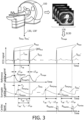
- FIG. 3 is a schematic diagram illustrating an example of various elements of a computer-implemented method of providing a cardiac signal S Cardiac , in accordance with some aspects of the present disclosure.
- the initial surrogate cardiac signal S PPG is generated by a PPG sensor.
- the PPG sensor is in contact with a finger of a subject, as illustrated in the upper portion of Fig. 3 .
- the value of the initial surrogate cardiac signal S PPG corresponds to the optical absorption within a finger.
- the optical absorption within a finger varies in accordance with the blood flow in the finger.
- the blood flow varies during the cardiac cycle.
- a maximum value of the initial surrogate cardiac signal S PPGMax corresponds to an instant of maximum optical absorption in the finger, i.e. the instant of maximum blood volume in the finger
- a minimum value of the initial surrogate cardiac signal S PPGMin corresponds to an instant of minimum optical absorption in the finger, i.e. the instant of minimum blood volume in the finger.
- the initial surrogate cardiac signal data 130 that is received in the operation S 120 is generated contemporaneously with the medical image data 120. Consequently, the initial surrogate cardiac signal data 130 corresponds, time-wise, to the medical image data 120.
- the initial surrogate cardiac signal data 130, and the medical image data 120 may be acquired over a period of time, such as over one or more cardiac cycles.
- the medical image data 120 is analyzed to provide a reference cardiac signal S Image representing the beating of the heart of the subject.
- the medical image data 120 comprises a temporal sequence of images including at least a portion of the heart and/or at least a portion of the aorta.
- the analyzing S 130 the medical image data 120 comprises:
- the at least a portion of the heart, and likewise the at least a portion of the aorta, pulsate in accordance with the cardiac cycle, and so these regions may be used to provide a cardiac signal representing the beating of the heart.
- the medical image data is reconstructed to provide complete images, e.g. volumetric images, or image slices, that represent the complete field of view imaged by the medical image data.
- the medical image data is reconstructed to provide partial images that represent only a portion of the complete field of view imaged by the medical image data and which includes the at least a portion of the heart and/or the at least a portion of the aorta.
- the reconstruction may be performed using known techniques.
- the operation of extracting the reference cardiac signal S Image may be performed using known image processing techniques. For instance, in one example, the operation of extracting the reference cardiac signal S Image comprises:
- the region of interest corresponds to a cardiac chamber.
- the cardiac chamber may be the left ventricle, or the right ventricle, or another cardiac chamber.
- the size of the left ventricle provides a reliable cardiac signal S Image because its size defines known points in the cardiac cycle.
- FIG. 3 An example of the reference cardiac signal S Image representing the beating of the heart of the subject that results from the operation S 130, is shown in the graph labelled "Reference cardiac signal” in Fig. 3 .
- the reference cardiac signal S Image represents the volume of the left ventricle of the heart.
- the reference cardiac signal S Image is obtained by reconstructing a temporal sequence of CT images that include the left ventricle, and evaluating the image intensities within the images in a region of interest that corresponds to the left ventricle.
- the graph labelled "ECG signal”, S ECG is also shown in Fig. 3 . This graph represents a contemporaneously-acquired ECG signal that would have been acquired by monitoring the heart using electrodes.
- the ECG signal, S ECG is also labelled with various reference points in the cardiac cycle, T HB , such as the so-called “R-peak”, labelled R Peak , the end of the so-called “QRS complex”, labelled QRS End , and the end of the so-called T-wave, labelled T End .
- the size of the left ventricle, represented in this graph by the Reference cardiac signal, S Image has known relationship with the cardiac cycle, T HB .
- This relationship is often referred-to as the Wiggers diagram.
- the maximum value of the Reference cardiac signal, S ImageMax i.e. the maximum size of the left ventricle, coincides with the end of the so-called QRS complex", labelled QRS End in the cardiac cycle, T HB .
- the minimum value of the Reference cardiac signal, S ImageMin i.e. the minimum size of the left ventricle, coincides with the end of the so-called T-wave, labelled T End in the cardiac cycle, T HB .
- the low variation of the length of the QRS complex in patients allows the estimation of the so-called R-peak, labelled R Peak in the cardiac cycle, T HB , based on its relationship with the point of maximum left ventricular volume, i.e. S ImageMax , in the Reference cardiac signal, S Image .
- Reference points such as these are important because they represent defined points in the cardiac cycle, T HB .
- the R-peak in an ECG cardiac signal is commonly used as a trigger point in cardiac gating applications.
- the known relationship between the Reference cardiac signal S Image and the cardiac cycle, T HB can be used to predict the occurrence of reference points in the cardiac cycle T HB , such as QRS End , T End , and R Peak , from a measurement of the Reference cardiac signal S Image .
- the medical image data 120 represents the left ventricle.
- the size of the left ventricle serves as a good region of interest to evaluate in the medical image data because it represents the largest myocardial mass, and also because of its known relationship with the cardiac cycle, T HB , as described above.
- the medical image data 120 may be generated from a surview scan.
- the medical image data 120 is reconstructed to provide a temporal series of partial images representing the left ventricle.
- the images may be partial in the sense that they may represent only part of the field of view of view imaged by the medical imaging system.
- Image processing techniques, or a neural network may be used to identify the left ventricle in the partial images, and to calculate its size.
- the size of the left ventricle may be calculated based on its volume, or its area, or another dimension in the partial images.
- the size of the left ventricle is then used as the reference cardiac signal S Image .
- an image intensity within a region of interest in the partial images may be used as the reference cardiac signal S Image .
- the region of interest may correspond to a cardiac chamber, such as the left ventricle, or it may correspond to another portion of the vasculature associated with the heart, such as the aorta.
- the image intensities within such regions of interest also represent the beating of the heart because they typically capture a portion of the heart, or a portion of the vasculature associated with the heart, such as the aorta, and which pulsates in accordance with the cardiac cycle.
- the time points of minimal and maximal cardiac contraction i.e. the time points S ImageMax and S ImageMin in the reference cardiac signal S Image may be determined.
- the medical image data 120 from the surview scan may include some data redundancy. For instance, multiple low-resolution images, or multiple partial images, each corresponding to a different point in the cardiac cycle, or a different phase of the cardiac cycle, may be reconstructed from the medical image data.
- the surview data may be provided by a so-called low-dose helical scan.
- the raw data may be binned into time intervals, each corresponding to a phase of the cardiac cycle, each with a duration of 100 milliseconds, for example, to provide a temporal series of partial reconstructed images representing the contraction of the heart over a complete cardiac cycle.
- Image processing techniques such as those described in a document by Lessick, J., et al., "Automatic determination of differential coronary artery motion minima for cardiac computed tomography optimal phase selection", Acad. Radiol. 2015 Jun;22(6):697-703 , may be applied to the image to identify different phases of the cardiac cycle (e.g. diastole and systole).
- the medical image data 120 being provided by an MRI system
- the surview data the medical image data may be provided in subsets interleaved in k-space. Lowly-resolved images may then be reconstructed from these subsets.
- a measure of the left ventricular volume over time may then be derived by applying known image processing techniques, or using a neural network, to the images.
- a correspondence T D between the reference cardiac signal S Image and the initial surrogate cardiac signal S PPG is determined. This correspondence is determined so that it can be used to provide an association between a subsequently-acquired surrogate cardiac signal S PPG ', and the cardiac cycle, T HB .
- the correspondence may be a time difference, or a phase difference, between the reference cardiac signal S Image and the initial surrogate cardiac signal S PPG .
- the correspondence is a time delay, or a phase delay, between the reference cardiac signal S Image and the initial surrogate cardiac signal S PPG .
- the correspondence may be a time difference, or a phase difference, between a reference point in the reference cardiac signal S Image , such as the reference point S ImageMax or the reference point S ImageMin , and a trigger point in the initial surrogate cardiac signal S PPG , such as the trigger point S PPGMax , or the trigger point S PPGMin .
- the trigger point may alternatively be another trigger point in the initial surrogate cardiac signal S PPG , such as the maximum value of a derivative of the initial surrogate cardiac signal S PPG , or a minimum value of a derivative of the initial surrogate cardiac signal S PPG , or a zero crossing value of a Hilbert transform of the initial surrogate cardiac signal S PPG .
- Such reference points, and trigger points may be used because they are readily-identifiable in these signals.
- the value of the correspondence may be determined in the operation S140 using known signal processing techniques.
- the value of the correspondence may be determined by comparing a time, or a phase, of a reference point in the reference cardiac signal S Image with a time, or a phase, of a trigger points in the initial surrogate cardiac signal S PPG .
- the value of the correspondence may be determined by evaluating a correlation between the reference cardiac signal S Image and the initial surrogate cardiac signal S PPG .
- subsequent surrogate cardiac signal data 130' is received.
- the subsequent surrogate cardiac signal data 130' comprises a subsequent surrogate cardiac signal S PPG ' representing the beating of the heart of the subject.
- the subsequent surrogate cardiac signal data 130' is generated by the same sensor as the initial surrogate cardiac signal data 130, and is simply generated at a later point in time than the initial surrogate cardiac signal data 130.
- the later point in time may correspond to a period within a diagnostic imaging scan, or a period within a radiation therapy session, for example.
- An example of subsequent surrogate cardiac signal data 130' is illustrated on the right-hand side of Fig. 3 , i.e. as the subsequent surrogate cardiac signal S PPG '.
- One or more trigger points may be established in the subsequent surrogate cardiac signal S PPG '. These trigger point(s) correspond to the trigger points mentioned above.
- the one or more trigger points may correspond to a maximum value S PPGMax of the subsequent surrogate cardiac signal S PPG ', or a minimum value S PPGMin of the subsequent surrogate cardiac signal S PPG ', or a maximum value of a derivative of the subsequent surrogate cardiac signal S PPG ', or a minimum value of a derivative of the subsequent surrogate cardiac signal S PPG ', or a zero crossing value of a Hilbert transform of the subsequent surrogate cardiac signal S PPG '.
- the cardiac signal S Cardiac is generated based on the subsequent surrogate cardiac signal S PPG ' and the correspondence T D .
- the cardiac signal S Cardiac is generated by using the correspondence T D to delay the subsequent surrogate cardiac signal S PPG '.
- the correspondence T D may be a time delay between the reference point S ImageMax in the reference cardiac signal S Image , and the trigger point S PPGMax in the initial surrogate cardiac signal S PPG .
- T HB is the period of the cardiac cycle
- the correspondence comprises a time delay T D , or a phase delay, between a reference point in the reference cardiac signal S Image , and a trigger point in the initial surrogate cardiac signal S PPG , and the cardiac signal S Cardiac is generated by using the correspondence T D to delay the subsequent surrogate cardiac signal S PPG ' by a duration T HB - T D , wherein T HB is the period of the cardiac cycle.
- the trigger point in the subsequent surrogate cardiac signal S PPG being synchronized with the expected time or phase of the reference point in the reference cardiac signal.
- This is a useful cardiac signal because it includes an indication, via the delayed trigger point S PPGMax , of the expected time of a known point in the cardiac cycle, i.e. the expected time of the reference point S ImageMax in the reference cardiac signal, which as mentioned above, corresponds to the end of the QRS complex in the cardiac cycle, i.e. the point QRS End in the signal S ECG in Fig. 3 .
- the other trigger points mentioned above such as S PPGMin , the maximum value of a derivative of the subsequent surrogate cardiac signal S PPG ', and so forth, and also the other reference points mentioned above, such as S ImageMin may be used in a similar manner to provide a cardiac signal.
- delaying the subsequent surrogate cardiac signal S PPG ' so as to provide the subsequent surrogate cardiac signal S PPG ' with an indication of the expected time of the reference point S ImageMin in the reference cardiac signal is also a useful cardiac signal because the reference point S ImageMin corresponds to the end of the T-wave in the cardiac cycle, i.e. the point T End in the signal S ECG in Fig. 3 .
- the indication may be provided in a different manner.
- a cardiac signal may be provided which includes a leading edge, and wherein the time, or the phase, of the leading edge is coincident with the expected time of the reference point S ImageMax , S ImageMin in the reference cardiac signal S Image .
- the leading edge serves as the indication of the expected time of the reference point S ImageMax , S ImageMin in the reference cardiac signal S Image .
- a known time or phase relationship ⁇ T RP between the reference cardiac signal S Image and the reference point R Peak , QRS End , T End in the cardiac cycle, T HB is used to generate a cardiac signal S Cardiac that includes an indication of an expected time R PeakEst , or an expected phase, of a reference point R Peak , QRS End , T End in a cardiac cycle T HB of the heart.
- the operation of generating S 160 the cardiac signal S Cardiac comprises generating an indication of an expected time R PeakEst , or an expected phase, of a reference point R Peak , QRS End , T End in a cardiac cycle T HB of the heart, and wherein the indication is generated based on the subsequent surrogate cardiac signal S PPG ', and the correspondence T D , and based further on a known time or phase relationship ⁇ T RP between the reference cardiac signal S Image and the reference point R Peak , QRS End , T End in the cardiac cycle T HB .
- FIG. 3 This example is illustrated in reference Fig. 3 , for the situation in which the cardiac signal S Cardiac (illustrated at the bottom of Fig. 3 ) includes an indication (via the leading edge of the pulse in the cardiac signal S Cardiac of the expected time R PeakEst of the reference point R Peak in the cardiac cycle T HB of the heart.
- the expected time R PeakEst of the reference point R Peak in the cardiac cycle T HB of the heart is illustrated via the dotted trace in the graph labelled "ECG signal".
- the reference point R Peak is referred-to as the R-peak, and it commonly serves as a trigger point for cardiac gating applications.
- the R-peak in the cardiac cycle, T HB is known a high degree of certainty to occur 80 milliseconds to 100 milliseconds prior to the time of maximum volume of the left ventricle.
- the cardiac signal S Cardiac is generated by providing a pulse with a leading edge that occurs at a time T HB - T D - ⁇ T RP after the trigger point S PPGMax .
- the expected times of other reference points in the cardiac cycle of the heart may be indicated in this example.
- the expected time of the reference point QRS End i.e. the end of the QRS complex
- the reference point T End i.e. the end of the T-wave
- the reference point QRS End coincides with the time of maximum volume of the left ventricle, i.e. the time of the maximum value of the Reference cardiac signal, S ImageMax .
- the reference point T End coincides with the time of minimum volume of the left ventricle, i.e. the time of the minimum value of the Reference cardiac signal, S ImageMin .
- the ability to provide an indication, via the cardiac signal S Cardiac , of such reference points in the cardiac cycle improves the accuracy of the cardiac signal and facilitate its use in applications such as cardiac gating.
- a trigger point is established in the subsequent surrogate cardiac signal S PPG ', and this is used in combination with the known time or phase relationship ⁇ T RP described above, to generate a cardiac signal S Cardiac .
- the operation of determining S 140 a correspondence T D between the reference cardiac signal S Image and the initial surrogate cardiac signal S PPG comprises:
- the operation of establishing a trigger point S PPGMax , S PPGMin in the subsequent surrogate cardiac signal S PPG ' may be performed using known signal processing techniques.
- various trigger points may be used.
- the trigger point may correspond to a maximum value S PPGMax of the subsequent surrogate cardiac signal S PPG ', or a minimum value S PPGMin of the subsequent surrogate cardiac signal S PPG ', or a maximum value of a derivative of the subsequent surrogate cardiac signal S PPG ', or a minimum value of a derivative of the subsequent surrogate cardiac signal S PPG ', or a zero crossing value of a Hilbert transform of the subsequent surrogate cardiac signal S PPG '.
- the correspondence is then defined by a time difference, or a phase difference, between a reference point S ImageMax in the reference cardiac signal S Image and the trigger point S PPGMax , S PPGMin .
- the reference point R Peak may be the R-peak, or the end of the QRS complex QRS End , or the end of the T-wave T End , for example.
- the operation of generating an indication of an expected time R PeakEst , or an expected phase, of a reference point R Peak , QRS End , T End in a cardiac cycle T HB of the heart is performed in the same manner as described above, with the correspondence being evaluated as the time difference T D , or the phase difference.
- the cardiac signal S Cardiac is outputted.
- the cardiac signal S Cardiac may be outputted in various ways, including to a display device such as to the display 160 illustrated in Fig. 1 , or to a virtual/ augmented reality display device, or to a printer, or to a computer-readable storage medium, or to the internet, or to the Cloud, and so forth.
- the cardiac signal S Cardiac may be used in various applications. For instance, the cardiac signal S Cardiac may be used to assess the health of a subject. In this situation, the cardiac signal S Cardiac may simply be outputted.
- the cardiac signal S Cardiac may be outputted to a display device, e.g. in the form of a graph, in order for a physician to assess the heart rate, or a heart rate variability of a subject.
- the cardiac signal S Cardiac may be used for other purposes, such as to control the acquisition of medical image data, or to select medical image data for reconstruction into a medical image of the subject.
- the cardiac signal S Cardiac may be outputted to a medical imaging system, or to a radiation therapy system, or to a computer readable storage medium.
- the system 100 may also include one or more of: a medical imaging system for providing the medical image data 120, such as for example the MRI system 150 illustrated in Fig. 1 ; one or more sensors 140a, 140b, such as for example a PPG, sensor 140a, 140b, or a BCG sensor, or a pulse oximetry sensor, or a pressure sensor, or a radar sensor; a display, such as the display 160 illustrated in Fig. 1 , for displaying the cardiac signal S Cardiac , and other outputs generated by the one or more processors 110; a patient bed 170; and a user input device (not illustrated in Fig. 1 ) configured to receive user input in relation to the operations performed by the one or more processors 110, such as a keyboard, a mouse, a touchscreen, and so forth.
- a medical imaging system for providing the medical image data 120, such as for example the MRI system 150 illustrated in Fig. 1 ; one or more sensors 140a, 140b, such as for example a PPG, sensor 140a
- a computer-implemented method of providing a cardiac signal S Cardiac comprises:
- a computer program product comprises instructions which when executed by one or more processors 110, cause the one or more processors to carry out a method of providing a cardiac signal S Cardiac .
- the method comprises:
- the system 100 illustrated in Fig. 1 may be used in various cardiac gating applications.
- the system 100 is used to perform cardiac gating for the purpose of reconstructing medical images.
- a medical imaging system 150 is provided.
- the medical imaging system 150 comprises:
- the medical imaging system 150 may be any of the medical imaging systems mentioned above. For instance it may be a (spectral) X-ray projection imaging system, a 2D/ 3D ultrasound imaging system, an MRI system, a (spectral) CT imaging system, a PET imaging system, or a SPECT imaging system, and so forth.
- the one or more sensors 140a, 140b may be provided by various sensors, including for example a photoplethysmography, PPG, sensor 140a, 140b, or a ballistocardiographic, BCG, sensor, or a pulse oximetry sensor, or a pressure sensor, or a radar sensor.
- the gating may be performed during so-called step-and-shoot imaging procedures, or in a helical imaging procedure.
- prospectively gating the acquisition of the subsequent medical image data may include using the cardiac signal S Cardiac to modulate the X-ray dose.
- the subsequent medical image data in this example is medical image data that is acquired by the medical imaging system 150 at a later point in time to the acquired medical image data 120.
- the medical image data 120 represents a surview scan of the subject, or scan of an injected contrast agent bolus in the subject
- the subsequent medical image data represents a diagnostic imaging scan of the subject.
- a surview scan, and a scan of an injected contrast agent bolus in the subject are typically acquired as part of the conventional workflow during medical imaging procedures, and they typically include sufficient information to represent the beating of a heart of a subject.
- the use of such scans to provide the medical image data 120 simplifies workflow and/or reduces X-ray dose to a subject.
- the medical image data that is acquired is reconstructed.
- the one or more processors of the medical imaging system are configured to:
- the effect of this is to provide reconstructed medical images with reduced cardiac motion-induced blurring.
- system 100 is used to perform cardiac gating for the purpose of triggering the deliver of radiation therapy to a subject.
- the radiation therapy delivery system comprises:
- the radiation therapy delivery system may be an external beam radiation therapy delivery system.
- the beam may be a proton beam, an electron beam, or an X-ray beam.
- the delivery of therapeutic radiation using an X-ray beam is typically referred-to as radiotherapy.
- the intensity of the beam of radiation may be modulated based on the received cardiac signal S Cardiac .
- radiation may be delivered to the subject only in periods corresponding to a known point in the cardiac cycle. In so doing, the heart, and consequently the anatomy of the subject, is in a known position in the periods in which the radiation is delivered to the anatomy. In other words, it reduces the impact of cardiac motion. This helps to reduce the amount of healthy tissue that is affected by the radiation.
Landscapes
- Health & Medical Sciences (AREA)
- Life Sciences & Earth Sciences (AREA)
- Engineering & Computer Science (AREA)
- Medical Informatics (AREA)
- Physics & Mathematics (AREA)
- Biophysics (AREA)
- General Health & Medical Sciences (AREA)
- Nuclear Medicine, Radiotherapy & Molecular Imaging (AREA)
- Biomedical Technology (AREA)
- Pathology (AREA)
- Radiology & Medical Imaging (AREA)
- Heart & Thoracic Surgery (AREA)
- Surgery (AREA)
- High Energy & Nuclear Physics (AREA)
- Molecular Biology (AREA)
- Veterinary Medicine (AREA)
- Public Health (AREA)
- Animal Behavior & Ethology (AREA)
- Optics & Photonics (AREA)
- Physiology (AREA)
- Pulmonology (AREA)
- Cardiology (AREA)
- Signal Processing (AREA)
- Artificial Intelligence (AREA)
- Power Engineering (AREA)
- General Physics & Mathematics (AREA)
- Theoretical Computer Science (AREA)
- Dentistry (AREA)
- Oral & Maxillofacial Surgery (AREA)
- Condensed Matter Physics & Semiconductors (AREA)
- Computer Vision & Pattern Recognition (AREA)
- Psychiatry (AREA)
- Apparatus For Radiation Diagnosis (AREA)
Abstract
A system (100) for providing a cardiac signal (SCardiac), is provided. The system includes one or more processors (110) configured to: receive medical image data (120) representing a beating of a heart of a subject; receive initial surrogate cardiac signal data (130) comprising an initial surrogate cardiac signal (SPPG) representing the beating of the heart of the subject, the initial surrogate cardiac signal data (130) being generated contemporaneously with the medical image data (120); analyze the medical image data (120) to provide a reference cardiac signal (SImage) representing the beating of the heart of the subject; determine a correspondence (TD) between the reference cardiac signal (SImage) and the initial surrogate cardiac signal (SPPG); receive subsequent surrogate cardiac signal data (130') comprising a subsequent surrogate cardiac signal (SPPG') representing the beating of the heart of the subject; generate the cardiac signal (SCardiac) based on the subsequent surrogate cardiac signal (SPPG') and the correspondence (TD); and output the cardiac signal (SCardiac).
Description
- The present disclosure relates to providing a cardiac signal. A system, a computer-implemented method, a computer program product, and a medical imaging system that includes the system, are disclosed.
- Cardiac signals are routinely acquired by physicians. The cardiac signals may be acquired for various purposes. For instance, cardiac signals are acquired for the purpose of investigating the functioning of the heart. In such situations the cardiac signals may provide information such as the heart rate and the heart rate variability, which indicate the health of a subject. Cardiac signals are also acquired for other purposes, such as to select medical image data for use in image reconstruction via cardiac gating, and to trigger the delivery of radiation therapy to a subject via cardiac gating.
- Cardiac gating is used for image reconstruction purposes with imaging modalities such as computed tomography "CT", magnetic resonance imaging "MRI", and positron emission tomography "PET". Cardiac gating-based image reconstruction may be performed in a so-called "prospective gating" mode, or in a so-called "retrospective gating" mode. In the prospective gating mode, a cardiac signal is used to perform the acquisition of medical image data only for specific parts in the cardiac cycle, usually by starting (i.e. triggering) the acquisition at a specific point in the cardiac cycle followed by data acquisition for a constant period of time. The medical image data from this specific parts is then reconstructed to provide a medical image corresponding to a specific phase of the cardiac cycle. In the retrospective gating mode, medical image data is acquired throughout (or over a large part of) the cardiac cycle and a cardiac signal is used to apply timestamps to the medical image data the represent different points in the cardiac cycle. The timestamps are then used to select data for reconstruction so as to provide a medical image corresponding to the specific phase of the cardiac cycle. Usually, the goal of cardiac gating is to acquire data during a phase of the cardiac cycle where the cardiac motion is small. By reconstructing medical images from data that is acquired only at these specific phases of the cardiac cycle, it is ensured that the anatomy is in a similar position. This reduces image artifacts such as cardiac motion-induced blurring. The use of cardiac gating to trigger the delivery of radiation therapy to a subject likewise reduces the impact of cardiac motion. Here, the intensity of a radiation therapy beam is modulated based on a cardiac signal so as to ensure that the heart, and consequently the anatomy of a subject, is in a known position in the periods in which the radiation is delivered to the anatomy.
- Cardiac signals are often acquired by placing electrodes on the skin of a subject and acquiring a so-called electrocardiogram, or "ECG". The electrodes detect electrical signals that are generated as a result of cardiac muscle depolarization and re-polarization during the cardiac cycle. An ECG may be considered to represent the gold standard measurement of cardiac signals due to its direct correspondence with the activity of the cardiac muscle. However, there are drawbacks associated with acquiring cardiac signals in the form of an ECG. For instance, attaching the electrodes to the skin in the required locations is time-consuming, and also inconvenient for a subject. The electrodes, and also their leads, may also obstruct features in medical images.
- Other techniques for acquiring cardiac signals include the use of photoplethysmography to acquire a photoplethysmogram "PPG", and the use of ballistocardiography to acquire a ballistocardiograph "BCG". A PPG is an optically acquired plethysmogram that represents blood volume changes in a tissue bed. A PPG is typically acquired using a pulse oximeter which measures changes in optical absorption of the skin. A PPG may be acquired by measuring the optical absorption on skin regions such as the finger, the ear, nasal septum, and the forehead. A PPG, and also a BCG, may be acquired using contact with the body, and also using contact-less techniques. A PPG, and also a BCG, are both easier to acquire than an ECG. However, both a PPG, and also a BCG, have a weaker relationship with the activity of the cardiac muscle. This typically limits their use to the provision of information such as heart rate, and heart rate variability.
- Consequently, there is a need to improve the accuracy of surrogate cardiac signals, such as those acquired via photoplethysmography, and via ballistocardiography, in order to take advantage of their benefits.
- According to one aspect of the present disclosure, a system for providing a cardiac signal, is provided. The system comprises one or more processors configured to:
- receive medical image data representing a beating of a heart of a subject;
- receive initial surrogate cardiac signal data comprising an initial surrogate cardiac signal representing the beating of the heart of the subject, the initial surrogate cardiac signal data being generated contemporaneously with the medical image data;
- analyze the medical image data to provide a reference cardiac signal representing the beating of the heart of the subject;
- determine a correspondence between the reference cardiac signal and the initial surrogate cardiac signal;
- receive subsequent surrogate cardiac signal data comprising a subsequent surrogate cardiac signal representing the beating of the heart of the subject;
- generate the cardiac signal based on the subsequent surrogate cardiac signal and the correspondence; and
- output the cardiac signal.
- In the above system, medical image data is analyzed in order to provide a reference cardiac signal representing the beating of the heart of the subject. Since the reference cardiac signal is obtained from medical image data, it provides a reliable reference for the beating of the heart. The correspondence between the reference cardiac signal and an initial surrogate cardiac signal is then determined. The correspondence is then used to generate the cardiac signal from a subsequent surrogate cardiac signal. The correspondence may be a delay between the reference cardiac signal and the initial surrogate cardiac signal, for example. Thus, by using the correspondence to generate the cardiac signal, the delay may be accounted-for, and a more accurate cardiac signal is provided.
- Further aspects, features, and advantages of the present disclosure will become apparent from the following description of examples, which is made with reference to the accompanying drawings.
-
-
Fig. 1 is a schematic diagram illustrating an example of asystem 100 for providing a cardiac signal SCardiac, in accordance with some aspects of the present disclosure. -
Fig. 2 is a flowchart illustrating an example of a computer-implemented method of providing a cardiac signal, in accordance with some aspects of the present disclosure. -
Fig. 3 is a schematic diagram illustrating an example of various elements of a computer-implemented method of providing a cardiac signal SCardiac, in accordance with some aspects of the present disclosure. - Examples of the present disclosure are provided with reference to the following description and figures. In this description, for the purposes of explanation, numerous specific details of certain examples are set forth. Reference in the specification to "an example", "an implementation" or similar language means that a feature, structure, or characteristic described in connection with the example is included in at least that one example. It is also to be appreciated that features described in relation to one example may also be used in another example, and that all features are not necessarily duplicated in each example for the sake of brevity. For instance, features described in relation to a system, may be implemented in a computer-implemented method, and in a computer program product, and in a medical imaging system, and in a radiation therapy delivery system, in a corresponding manner.
- It is noted that the computer-implemented methods disclosed herein may be provided as a non-transitory computer-readable storage medium including computer-readable instructions stored thereon, which, when executed by at least one processor, cause the at least one processor to perform the method. In other words, the computer-implemented methods may be implemented in a computer program product. The computer program product can be provided by dedicated hardware, or hardware capable of running the software in association with appropriate software. When provided by a processor, the functions of the method features can be provided by a single dedicated processor, or by a single shared processor, or by a plurality of individual processors, some of which can be shared. The functions of one or more of the method features may for instance be provided by processors that are shared within a networked processing architecture such as a client/server architecture, a peer-to-peer architecture, the Internet, or the Cloud.
- The explicit use of the terms "processor" or "controller" should not be interpreted as exclusively referring to hardware capable of running software, and can implicitly include, but is not limited to, digital signal processor "DSP" hardware, read only memory "ROM" for storing software, random access memory "RAM", a non-volatile storage device, and the like. Furthermore, examples of the present disclosure can take the form of a computer program product accessible from a computer-usable storage medium, or a computer-readable storage medium, the computer program product providing program code for use by or in connection with a computer or any instruction execution system. For the purposes of this description, a computer-usable storage medium or a computer readable storage medium can be any apparatus that can comprise, store, communicate, propagate, or transport a program for use by or in connection with an instruction execution system, apparatus, or device. The medium can be an electronic, magnetic, optical, electromagnetic, infrared, or a semiconductor system or device or propagation medium. Examples of computer-readable media include semiconductor or solid-state memories, magnetic tape, removable computer disks, random access memory "RAM", read-only memory "ROM", rigid magnetic disks and optical disks. Current examples of optical disks include compact disk-read only memory "CD-ROM", compact disk-read/write "CD-R/W", Blu-Ray™ and DVD.
- It is also noted that some operations that are described as being performed by the one or more processors of the systems disclosed herein may be implemented using artificial intelligence techniques. Suitable techniques may include machine learning techniques, as well as deep learning techniques such as neural networks. For instance, one or more neural networks, may be trained in a supervised, or in some cases unsupervised, manner, to implement the operations that are performed by the one or more processors.
- As mentioned above, there is a need to improve the accuracy of surrogate cardiac signals, such as those acquired via photoplethysmography, and via ballistocardiography.
Fig. 1 is a schematic diagram illustrating an example of asystem 100 for providing a cardiac signal SCardiac, in accordance with some aspects of the present disclosure.Fig. 2 is a flowchart illustrating an example of a computer-implemented method of providing a cardiac signal, in accordance with some aspects of the present disclosure. It is noted that operations that are described as being performed by the one ormore processors 110 of thesystem 100 illustrated inFig. 1 , may also be performed in the method illustrated inFig. 2 . Likewise, operations that are described in relation to the method described with reference toFig. 2 may also be performed by the one ormore processors 110 of thesystem 100 illustrated inFig. 1 . With reference toFig. 1 , andFig. 2 , thesystem 100 for providing a cardiac signal SCardiac, comprises one ormore processors 110 configured to: - receive S110
medical image data 120 representing a beating of a heart of a subject; - receive S120 initial surrogate cardiac signal data 130 comprising an initial surrogate cardiac signal SPPG representing the beating of the heart of the subject, the initial surrogate cardiac signal data 130 being generated contemporaneously with the
medical image data 120; - analyze S130 the
medical image data 120 to provide a reference cardiac signal SImage representing the beating of the heart of the subject; - determine S140 a correspondence TD between the reference cardiac signal SImage and the initial surrogate cardiac signal SPPG;
- receive S150 subsequent surrogate cardiac signal data 130' comprising a subsequent surrogate cardiac signal SPPG' representing the beating of the heart of the subject;
- generate S160 the cardiac signal SCardiac based on the subsequent surrogate cardiac signal SPPG' and the correspondence TD; and
- output S170 the cardiac signal SCardiac.
- In the above system, medical image data is analyzed in order to provide a reference cardiac signal representing the beating of the heart of the subject. Since the reference cardiac signal is obtained from medical image data, it provides a reliable reference for the beating of the heart. The correspondence between the reference cardiac signal and an initial surrogate cardiac signal is then determined. The correspondence is then used to generate the cardiac signal from a subsequent surrogate cardiac signal. The correspondence may be a delay between the reference cardiac signal and the initial surrogate cardiac signal, for example. Thus, by using the correspondence to generate the cardiac signal, the delay may be accounted-for, and a more accurate cardiac signal is provided.
- The operations that are performed by the one or
more processors 110 of thesystem 100 are described in more detail below. - Referring initially to the
operation S 110 illustrated inFig. 2 ; in this operation,medical image data 120 is received. Themedical image data 120 represents a beating of a heart of a subject. - The
medical image data 120 may be generated by various types of medical imaging systems, including two-dimensional "2D" imaging systems, and three-dimensional "3D", or "volumetric", imaging systems. Examples of 2D medical imaging systems include (spectral) X-ray projection imaging systems, and 2D ultrasound imaging systems. Examples of volumetric imaging systems include MRI systems, (spectral) computed tomography "CT" imaging systems, positron emission tomography "PET" imaging systems, single photon emission computed tomography "SPECT" imaging systems, 3D ultrasound imaging systems, and so forth. In the example illustrated inFig. 1 , themedical image data 120 is generated by anMRI system 150. - The
medical image data 120 that is received in theoperation S 110 represents a beating of a heart of a subject. In this regard, themedical image data 120 may represent at least a portion of the heart. For instance, themedical image data 120 may represent a chamber of the heart, such as the left- or the right-ventricle, or a valve of the heart, and so forth. Portions of the heart such as these represent the beating of a heart via their pulsation in accordance with the cardiac cycle. For instance, the left ventricle contracts and relaxes as it pumps blood during the cardiac cycle. Likewise, valves within the heart such as the pulmonary valve, the tricuspid valve, and so forth, open and close during the cardiac cycle. Themedical image data 120 may alternatively represent the beating of a heart in a different manner. For instance, portions of the vasculature that are associated with the heart, such as the aorta, and which are not part of the heart, also pulsate in accordance with the cardiac cycle. Thus, themedical image data 120 may alternatively represent a portion of the vasculature associated with the with the heart, such as at least a portion of the aorta, and which also indicate the beating of the heart via their pulsation in accordance with the cardiac cycle. - In some examples, the
medical image data 120 is so-called raw data, i.e. data that has not yet been reconstructed into an image. In other examples, themedical image data 120 is reconstructed image data, i.e. data that has already been reconstructed to provide an image. Raw data generated by an MRI system may be referred-to as k-space data. Raw data that is generated by an MRI system may be referred-to as projection CT data. Thus, themedical image data 120 may include a temporal sequence of images that represent the beating of a heart of a subject. Alternatively, themedical image data 120 may include a temporal sequence of raw datasets that represent the beating of the heart of the subject. - In one example, the
medical image data 120 that is received in the operation S110 represents a diagnostic imaging scan of a subject. A diagnostic imaging scan includes sufficient information to perform a diagnosis of a subject. However, in some situations, a diagnostic imaging scan may not be available. It may also be impermissible to perform a dedicated diagnostic imaging scan in order to provide themedical image data 120 due to the additional burden on workflow, or due to the increase X-ray dose to a subject. In another example, themedical image data 120 represents a surview scan of the subject, or scan of an injected contrast agent bolus in the subject. Such scans are typically acquired as part of the conventional workflow during medical imaging procedures, and they typically include sufficient information to represent the beating of a heart of a subject. Thus, the use of such scans to provide themedical image data 120 simplifies workflow and/or reduces X-ray dose to a subject. - A surview scan is also known as a localizer radiograph, or a scanogram, or a scout scan. A surview scan is typically performed as part of the conventional workflow prior to a diagnostic imaging scan. A surview scan is also typically performed at a relatively lower dose than a diagnostic imaging scan. A surview typically includes sufficient information to represent the beating of a heart of a subject because it typically captures the pulsation of the heart, either by capturing a portion of the heart, or by capturing a portion of the vasculature associated with the heart, such as the aorta, and which likewise pulsates in accordance with the cardiac cycle. The surview scan may be a low-dose helical CT surview scan for example. A low-dose helical CT surview is often performed in order to localize the heart prior to performing a diagnostic imaging scan of the heart during a CT imaging procedure. Similarly, a scan of an injected contrast agent bolus in the subject is also often performed as part of the conventional workflow prior to a diagnostic imaging scan. The contrast agent may have been injected in order to assess blood flow in the vasculature, or to navigate an interventional device within the vasculature. A scan of an injected contrast agent bolus typically includes sufficient information to represent the beating of a heart of a subject because it also typically captures a portion of the heart, or a portion of the vasculature associated with the heart, such as the aorta, and which likewise pulsates in accordance with the cardiac cycle.
- The
medical image data 120 that is received in theoperation S 110 may be received via any form of data communication, including via wired, or wireless, or optical fiber communication. By way of some examples, when wired data communication is used, the communication may take place via electrical signals that are transmitted on an electrical cable. When wireless data communication is used, the communication may take place via RF or infrared signals. When an optical fiber data communication is used, the communication takes place via optical signals that are transmitted on an optical fiber. - Referring now to the
operation S 120 illustrated inFig. 2 ; in this operation, initial surrogate cardiac signal data 130 is received. The initial surrogate cardiac signal data 130 comprises an initial surrogate cardiac signal SPPG representing the beating of the heart of the subject. The initial surrogate cardiac signal data 130 is generated contemporaneously with themedical image data 120. - The initial surrogate cardiac signal data 130 may be generated by various types of sensors, including for example a photoplethysmography, PPG,
sensor - A PPG sensor measures optical absorption on a skin region such as a finger, an ear, the nasal septum, and the forehead. The optical absorption in such regions varies in accordance with blood flow, and the blood flow varies during the cardiac cycle. In so doing, a PPG sensor provides a surrogate cardiac signal that represents the beating of the heart of the subject. A PPG sensor may contact the skin. For instance the PPG sensor may be provided in the form of a finger clip, an ear clip, and so forth. A contact-less PPG sensor may alternatively be provided in the form of a camera that measures the optical absorption on such skin regions without contacting the subject. The camera typically measures differences in the intensity of the optical absorption of the skin region arising from deoxygenated, and oxygenated haemoglobin, over different color channels.
- A BCG sensor provides a measurement of a subject's motion arising from the ejection of the blood during the cardiac cycle. In so doing, a BCG sensor provides a surrogate cardiac signal that represents the beating of the heart of the subject. A BCG sensor may be provided by various types of motion sensors. The motion sensor may measure a position, or a velocity, or an acceleration of the subject. The BCG sensor may be mechanically coupled to the subject, or alternatively it may be mechanically coupled to a subject support, such as a patient bed.
- A pulse oximetry sensor measures blood oxygen levels and is typically provided in the form of a clip-type device for attachment to a finger, or an earlobe, or a foot. A pulse oximetry sensor measures blood oxygen levels via the optical absorption of such regions. The optical absorption is dependent upon the blood flow, which varies during the cardiac cycle. Consequently, a pulse oximetry sensor provides a surrogate cardiac signal that represents the beating of the heart of the subject.
- Similarly, a pressure sensor may be mechanically coupled to a subject, for instance to a limb of a subject. The pressure sensor measures the blood pressure in the subject. The blood pressure varies during the cardiac cycle. Thus, a pressure sensor may provide a surrogate cardiac signal that represents the beating of the heart of the subject. A radar sensor may be used to measure a subject's motion arising from the ejection of the blood during the cardiac cycle in a similar manner a BCG sensor. Thus, a radar sensor may provide a surrogate cardiac signal that represents the beating of the heart of the subject.
- An example of an initial surrogate cardiac signal SPPG representing the beating of the heart of the subject, is shown in the graph labelled "Surrogate cardiac signal" in
Fig. 3. Fig. 3 is a schematic diagram illustrating an example of various elements of a computer-implemented method of providing a cardiac signal SCardiac, in accordance with some aspects of the present disclosure. In the example illustrated inFig. 3 , the initial surrogate cardiac signal SPPG is generated by a PPG sensor. The PPG sensor is in contact with a finger of a subject, as illustrated in the upper portion ofFig. 3 . In this example, the value of the initial surrogate cardiac signal SPPG corresponds to the optical absorption within a finger. As mentioned above, the optical absorption within a finger varies in accordance with the blood flow in the finger. The blood flow varies during the cardiac cycle. A maximum value of the initial surrogate cardiac signal SPPGMax corresponds to an instant of maximum optical absorption in the finger, i.e. the instant of maximum blood volume in the finger, and a minimum value of the initial surrogate cardiac signal SPPGMin corresponds to an instant of minimum optical absorption in the finger, i.e. the instant of minimum blood volume in the finger. - The initial surrogate cardiac signal data 130 that is received in the
operation S 120 is generated contemporaneously with themedical image data 120. Consequently, the initial surrogate cardiac signal data 130 corresponds, time-wise, to themedical image data 120. The initial surrogate cardiac signal data 130, and themedical image data 120, may be acquired over a period of time, such as over one or more cardiac cycles. - Referring now to the operation S130 illustrated in
Fig. 2 ; in this operation, themedical image data 120 is analyzed to provide a reference cardiac signal SImage representing the beating of the heart of the subject. - Various techniques may be used to provide the reference cardiac signal SImage in the operation S130. In one example, the
medical image data 120 comprises a temporal sequence of images including at least a portion of the heart and/or at least a portion of the aorta. In this example, the analyzing S 130 themedical image data 120 comprises: - reconstructing at least a portion of the
medical image data 120 to provide a temporal sequence of images representing the at least a portion of the heart and/or the at least a portion of the aorta; and - extracting the reference cardiac signal SImage representing the beating of the heart of the subject, from the temporal sequence of images.
- As mentioned above, the at least a portion of the heart, and likewise the at least a portion of the aorta, pulsate in accordance with the cardiac cycle, and so these regions may be used to provide a cardiac signal representing the beating of the heart. In one example, the medical image data is reconstructed to provide complete images, e.g. volumetric images, or image slices, that represent the complete field of view imaged by the medical image data. In another example, the medical image data is reconstructed to provide partial images that represent only a portion of the complete field of view imaged by the medical image data and which includes the at least a portion of the heart and/or the at least a portion of the aorta. The reconstruction may be performed using known techniques. The operation of extracting the reference cardiac signal SImage may be performed using known image processing techniques. For instance, in one example, the operation of extracting the reference cardiac signal SImage comprises:
- evaluating an image intensity within a region of interest in the images; and
- generating the reference cardiac signal SImage based on the evaluated image intensities in the images.
- In one example, the region of interest corresponds to a cardiac chamber. The cardiac chamber may be the left ventricle, or the right ventricle, or another cardiac chamber. The size of the left ventricle provides a reliable cardiac signal SImage because its size defines known points in the cardiac cycle.
- An example of the reference cardiac signal SImage representing the beating of the heart of the subject that results from the operation S 130, is shown in the graph labelled "Reference cardiac signal" in
Fig. 3 . In this example, the reference cardiac signal SImage represents the volume of the left ventricle of the heart. The reference cardiac signal SImage is obtained by reconstructing a temporal sequence of CT images that include the left ventricle, and evaluating the image intensities within the images in a region of interest that corresponds to the left ventricle. For reference purposes, the graph labelled "ECG signal", SECG, is also shown inFig. 3 . This graph represents a contemporaneously-acquired ECG signal that would have been acquired by monitoring the heart using electrodes. The ECG signal, SECG, is also labelled with various reference points in the cardiac cycle, THB, such as the so-called "R-peak", labelled RPeak, the end of the so-called "QRS complex", labelled QRSEnd, and the end of the so-called T-wave, labelled TEnd. - As may be seen in the graph labelled "Reference cardiac signal" in
Fig. 3 , the size of the left ventricle, represented in this graph by the Reference cardiac signal, SImage, has known relationship with the cardiac cycle, THB. This relationship is often referred-to as the Wiggers diagram. The maximum value of the Reference cardiac signal, SImageMax, i.e. the maximum size of the left ventricle, coincides with the end of the so-called QRS complex", labelled QRSEnd in the cardiac cycle, THB. The minimum value of the Reference cardiac signal, SImageMin, i.e. the minimum size of the left ventricle, coincides with the end of the so-called T-wave, labelled TEnd in the cardiac cycle, THB. The low variation of the length of the QRS complex in patients, typically 80 milliseconds to 100 milliseconds, allows the estimation of the so-called R-peak, labelled RPeak in the cardiac cycle, THB, based on its relationship with the point of maximum left ventricular volume, i.e. SImageMax, in the Reference cardiac signal, SImage. Reference points such as these are important because they represent defined points in the cardiac cycle, THB. For instance, the R-peak in an ECG cardiac signal is commonly used as a trigger point in cardiac gating applications. Thus, the known relationship between the Reference cardiac signal SImage and the cardiac cycle, THB, can be used to predict the occurrence of reference points in the cardiac cycle THB, such as QRSEnd, TEnd, and RPeak, from a measurement of the Reference cardiac signal SImage. - An example of the analyzing operation S130 is described below in which the
medical image data 120 represents the left ventricle. The size of the left ventricle serves as a good region of interest to evaluate in the medical image data because it represents the largest myocardial mass, and also because of its known relationship with the cardiac cycle, THB, as described above. In this example, themedical image data 120 may be generated from a surview scan. Themedical image data 120 is reconstructed to provide a temporal series of partial images representing the left ventricle. The images may be partial in the sense that they may represent only part of the field of view of view imaged by the medical imaging system. Image processing techniques, or a neural network, may be used to identify the left ventricle in the partial images, and to calculate its size. The size of the left ventricle may be calculated based on its volume, or its area, or another dimension in the partial images. The size of the left ventricle is then used as the reference cardiac signal SImage. Instead of calculating the size of the left ventricle, an image intensity within a region of interest in the partial images may be used as the reference cardiac signal SImage. The region of interest may correspond to a cardiac chamber, such as the left ventricle, or it may correspond to another portion of the vasculature associated with the heart, such as the aorta. The image intensities within such regions of interest also represent the beating of the heart because they typically capture a portion of the heart, or a portion of the vasculature associated with the heart, such as the aorta, and which pulsates in accordance with the cardiac cycle. Having obtained the reference cardiac signal SImage, the time points of minimal and maximal cardiac contraction, i.e. the time points SImageMax and SImageMin in the reference cardiac signal SImage may be determined. - In this example, the
medical image data 120 from the surview scan may include some data redundancy. For instance, multiple low-resolution images, or multiple partial images, each corresponding to a different point in the cardiac cycle, or a different phase of the cardiac cycle, may be reconstructed from the medical image data. In the example of themedical image data 120 being provided by a CT imaging system, the surview data may be provided by a so-called low-dose helical scan. The raw data may be binned into time intervals, each corresponding to a phase of the cardiac cycle, each with a duration of 100 milliseconds, for example, to provide a temporal series of partial reconstructed images representing the contraction of the heart over a complete cardiac cycle. Image processing techniques such as those described in a document by Lessick, J., et al., "Automatic determination of differential coronary artery motion minima for cardiac computed tomography optimal phase selection", Acad. Radiol. 2015 Jun;22(6):697-703, may be applied to the image to identify different phases of the cardiac cycle (e.g. diastole and systole). In the example of themedical image data 120 being provided by an MRI system, the surview data the medical image data may be provided in subsets interleaved in k-space. Lowly-resolved images may then be reconstructed from these subsets. A measure of the left ventricular volume over time may then be derived by applying known image processing techniques, or using a neural network, to the images. - Referring now to the operation S140 illustrated in
Fig. 2 ; in this operation, a correspondence TD between the reference cardiac signal SImage and the initial surrogate cardiac signal SPPG, is determined. This correspondence is determined so that it can be used to provide an association between a subsequently-acquired surrogate cardiac signal SPPG', and the cardiac cycle, THB. - The correspondence may be a time difference, or a phase difference, between the reference cardiac signal SImage and the initial surrogate cardiac signal SPPG. In one example, the correspondence is a time delay, or a phase delay, between the reference cardiac signal SImage and the initial surrogate cardiac signal SPPG. The correspondence may be a time difference, or a phase difference, between a reference point in the reference cardiac signal SImage, such as the reference point SImageMax or the reference point SImageMin, and a trigger point in the initial surrogate cardiac signal SPPG, such as the trigger point SPPGMax, or the trigger point SPPGMin. The trigger point may alternatively be another trigger point in the initial surrogate cardiac signal SPPG, such as the maximum value of a derivative of the initial surrogate cardiac signal SPPG, or a minimum value of a derivative of the initial surrogate cardiac signal SPPG, or a zero crossing value of a Hilbert transform of the initial surrogate cardiac signal SPPG. Such reference points, and trigger points, may be used because they are readily-identifiable in these signals. The value of the correspondence may be determined in the operation S140 using known signal processing techniques. In one example, the value of the correspondence may be determined by comparing a time, or a phase, of a reference point in the reference cardiac signal SImage with a time, or a phase, of a trigger points in the initial surrogate cardiac signal SPPG. In another example, the value of the correspondence may be determined by evaluating a correlation between the reference cardiac signal SImage and the initial surrogate cardiac signal SPPG.
- Referring now to the operation S150 illustrated in
Fig. 2 ; in this operation subsequent surrogate cardiac signal data 130' is received. The subsequent surrogate cardiac signal data 130' comprises a subsequent surrogate cardiac signal SPPG' representing the beating of the heart of the subject. - The subsequent surrogate cardiac signal data 130' is generated by the same sensor as the initial surrogate cardiac signal data 130, and is simply generated at a later point in time than the initial surrogate cardiac signal data 130. The later point in time may correspond to a period within a diagnostic imaging scan, or a period within a radiation therapy session, for example. An example of subsequent surrogate cardiac signal data 130' is illustrated on the right-hand side of
Fig. 3 , i.e. as the subsequent surrogate cardiac signal SPPG'. One or more trigger points may be established in the subsequent surrogate cardiac signal SPPG'. These trigger point(s) correspond to the trigger points mentioned above. For instance, the one or more trigger points may correspond to a maximum value SPPGMax of the subsequent surrogate cardiac signal SPPG', or a minimum value SPPGMin of the subsequent surrogate cardiac signal SPPG', or a maximum value of a derivative of the subsequent surrogate cardiac signal SPPG', or a minimum value of a derivative of the subsequent surrogate cardiac signal SPPG', or a zero crossing value of a Hilbert transform of the subsequent surrogate cardiac signal SPPG'. - Referring now to the operation S160 illustrated in
Fig. 2 ; in this operation, the cardiac signal SCardiac is generated based on the subsequent surrogate cardiac signal SPPG' and the correspondence TD. - In one example, the cardiac signal SCardiac is generated by using the correspondence TD to delay the subsequent surrogate cardiac signal SPPG'. By way of an example, the correspondence TD may be a time delay between the reference point SImageMax in the reference cardiac signal SImage, and the trigger point SPPGMax in the initial surrogate cardiac signal SPPG. This example is illustrated in
Fig. 3 . Delaying the subsequent surrogate cardiac signal SPPG' by a duration THB - TD, wherein THB is the period of the cardiac cycle, results in a delayed version of the subsequent surrogate cardiac signal SPPG', i.e. a cardiac signal SCardiac, in which the trigger point SPPGMax in the subsequent surrogate cardiac signal SPPG is synchronized with the expected time of the reference point SImageMax in the reference cardiac signal SImage. More generally, in this example, the correspondence comprises a time delay TD, or a phase delay, between a reference point in the reference cardiac signal SImage, and a trigger point in the initial surrogate cardiac signal SPPG, and the cardiac signal SCardiac is generated by using the correspondence TD to delay the subsequent surrogate cardiac signal SPPG' by a duration THB - TD, wherein THB is the period of the cardiac cycle. This results in the trigger point in the subsequent surrogate cardiac signal SPPG being synchronized with the expected time or phase of the reference point in the reference cardiac signal. This is a useful cardiac signal because it includes an indication, via the delayed trigger point SPPGMax, of the expected time of a known point in the cardiac cycle, i.e. the expected time of the reference point SImageMax in the reference cardiac signal, which as mentioned above, corresponds to the end of the QRS complex in the cardiac cycle, i.e. the point QRSEnd in the signal SECG inFig. 3 . - It is noted that the other trigger points mentioned above, such as SPPGMin, the maximum value of a derivative of the subsequent surrogate cardiac signal SPPG', and so forth, and also the other reference points mentioned above, such as SImageMin may be used in a similar manner to provide a cardiac signal. For instance, delaying the subsequent surrogate cardiac signal SPPG' so as to provide the subsequent surrogate cardiac signal SPPG' with an indication of the expected time of the reference point SImageMin in the reference cardiac signal, is also a useful cardiac signal because the reference point SImageMin corresponds to the end of the T-wave in the cardiac cycle, i.e. the point TEnd in the signal SECG in
Fig. 3 . - In another example, instead of simply delaying the subsequent surrogate cardiac signal SPPG' to provide a cardiac signal that has an indication (i.e. the trigger point SPPGMax) of the expected time of the reference point SImageMax, SImageMin, in the reference cardiac signal SImage, the indication may be provided in a different manner. For instance, a cardiac signal may be provided which includes a leading edge, and wherein the time, or the phase, of the leading edge is coincident with the expected time of the reference point SImageMax , SImageMin in the reference cardiac signal SImage. Thus, in this example, the leading edge serves as the indication of the expected time of the reference point SImageMax, SImageMin in the reference cardiac signal SImage.
- In another example, a known time or phase relationship ΔTRP between the reference cardiac signal SImage and the reference point RPeak, QRSEnd, TEnd in the cardiac cycle, THB, is used to generate a cardiac signal SCardiac that includes an indication of an expected time RPeakEst, or an expected phase, of a reference point RPeak, QRSEnd, TEnd in a cardiac cycle THB of the heart.
- In this example, the operation of generating
S 160 the cardiac signal SCardiac comprises generating an indication of an expected time RPeakEst, or an expected phase, of a reference point RPeak, QRSEnd, TEnd in a cardiac cycle THB of the heart, and wherein the indication is generated based on the subsequent surrogate cardiac signal SPPG', and the correspondence TD, and based further on a known time or phase relationship ΔTRP between the reference cardiac signal SImage and the reference point RPeak, QRSEnd, TEnd in the cardiac cycle THB. - This example is illustrated in reference
Fig. 3 , for the situation in which the cardiac signal SCardiac (illustrated at the bottom ofFig. 3 ) includes an indication (via the leading edge of the pulse in the cardiac signal SCardiac of the expected time RPeakEst of the reference point RPeak in the cardiac cycle THB of the heart. The expected time RPeakEst of the reference point RPeak in the cardiac cycle THB of the heart is illustrated via the dotted trace in the graph labelled "ECG signal". The reference point RPeak is referred-to as the R-peak, and it commonly serves as a trigger point for cardiac gating applications. The R-peak in the cardiac cycle, THB, is known a high degree of certainty to occur 80 milliseconds to 100 milliseconds prior to the time of maximum volume of the left ventricle. Thus, in this example, the time delay ΔTRP = 80 to 100 milliseconds, provides a known time relationship between the reference cardiac signal SImage and the reference point RPeak in the cardiac cycle THB. In the example illustrated inFig. 3 , the cardiac signal SCardiac is generated by providing a pulse with a leading edge that occurs at a time THB - TD - ΔTRP after the trigger point SPPGMax. - Instead of providing an indication of the expected time of the R-peak in the cardiac cycle, the expected times of other reference points in the cardiac cycle of the heart, may be indicated in this example. For instance, the expected time of the reference point QRSEnd, i.e. the end of the QRS complex, or the reference point TEnd, i.e. the end of the T-wave, may be indicated. For these reference points, there is also a known time or phase relationship between the reference cardiac signal SImage and the reference point in the cardiac cycle THB. For example, the reference point QRSEnd, coincides with the time of maximum volume of the left ventricle, i.e. the time of the maximum value of the Reference cardiac signal, SImageMax. Likewise, the reference point TEnd, coincides with the time of minimum volume of the left ventricle, i.e. the time of the minimum value of the Reference cardiac signal, SImageMin. The ability to provide an indication, via the cardiac signal SCardiac, of such reference points in the cardiac cycle improves the accuracy of the cardiac signal and facilitate its use in applications such as cardiac gating.
- In another example, a trigger point is established in the subsequent surrogate cardiac signal SPPG', and this is used in combination with the known time or phase relationship ΔTRP described above, to generate a cardiac signal SCardiac. In this example, the operation of determining
S 140 a correspondence TD between the reference cardiac signal SImage and the initial surrogate cardiac signal SPPG, comprises: - establishing a trigger point SPPGMax, SPPGMin in the subsequent surrogate cardiac signal SPPG'; and
- calculating a time difference, or a phase difference, between a reference point SImageMax in the reference cardiac signal SImage and the trigger point SPPGMax, SPPGMin; and
- wherein the correspondence is defined by the time difference TD, or the phase difference, respectively; and
- the operation of generating
S 160 the cardiac signal SCardiac comprises:
generating an indication of an expected time RPeakEst, or an expected phase, of a reference point RPeak, QRSEnd, TEnd in a cardiac cycle THB of the heart, based on the trigger point SPPGMax, SPPGMin in the subsequent surrogate cardiac signal SPPG', and the time difference TD, or the phase difference, and a known time or phase relationship ΔTRP between the reference cardiac signal SImage and the reference point RPeak, QRSEnd, TEnd in the cardiac cycle THB. - In this example, the operation of establishing a trigger point SPPGMax, SPPGMin in the subsequent surrogate cardiac signal SPPG' may be performed using known signal processing techniques. As described above, various trigger points may be used. For instance, the trigger point may correspond to a maximum value SPPGMax of the subsequent surrogate cardiac signal SPPG', or a minimum value SPPGMin of the subsequent surrogate cardiac signal SPPG', or a maximum value of a derivative of the subsequent surrogate cardiac signal SPPG', or a minimum value of a derivative of the subsequent surrogate cardiac signal SPPG', or a zero crossing value of a Hilbert transform of the subsequent surrogate cardiac signal SPPG'.
- In this example, the correspondence is then defined by a time difference, or a phase difference, between a reference point SImageMax in the reference cardiac signal SImage and the trigger point SPPGMax, SPPGMin. The reference point RPeak may be the R-peak, or the end of the QRS complex QRSEnd, or the end of the T-wave TEnd, for example.
- In this example, the operation of generating an indication of an expected time RPeakEst, or an expected phase, of a reference point RPeak, QRSEnd, TEnd in a cardiac cycle THB of the heart, is performed in the same manner as described above, with the correspondence being evaluated as the time difference TD, or the phase difference.
- Referring now to the operation S170 illustrated in
Fig. 2 ; in this operation, the cardiac signal SCardiac, is outputted. The cardiac signal SCardiac may be outputted in various ways, including to a display device such as to thedisplay 160 illustrated inFig. 1 , or to a virtual/ augmented reality display device, or to a printer, or to a computer-readable storage medium, or to the internet, or to the Cloud, and so forth. The cardiac signal SCardiac may be used in various applications. For instance, the cardiac signal SCardiac may be used to assess the health of a subject. In this situation, the cardiac signal SCardiac may simply be outputted. For instance, the cardiac signal SCardiac may be outputted to a display device, e.g. in the form of a graph, in order for a physician to assess the heart rate, or a heart rate variability of a subject. In other applications, the cardiac signal SCardiac may be used for other purposes, such as to control the acquisition of medical image data, or to select medical image data for reconstruction into a medical image of the subject. In such applications, the cardiac signal SCardiac may be outputted to a medical imaging system, or to a radiation therapy system, or to a computer readable storage medium. These applications are described in more detail below. - It is noted that the
system 100 may also include one or more of: a medical imaging system for providing themedical image data 120, such as for example theMRI system 150 illustrated inFig. 1 ; one ormore sensors sensor display 160 illustrated inFig. 1 , for displaying the cardiac signal SCardiac, and other outputs generated by the one ormore processors 110; apatient bed 170; and a user input device (not illustrated inFig. 1 ) configured to receive user input in relation to the operations performed by the one ormore processors 110, such as a keyboard, a mouse, a touchscreen, and so forth. - In another example, a computer-implemented method of providing a cardiac signal SCardiac, is provided. The method comprises:
- receiving S110
medical image data 120 representing a beating of a heart of a subject; - receiving S120 initial surrogate cardiac signal data 130 comprising an initial surrogate cardiac signal SPPG representing the beating of the heart of the subject, the initial surrogate cardiac signal data 130 being generated contemporaneously with the medical image data;
- analyzing S130 the
medical image data 120 to provide a reference cardiac signal SImage representing the beating of the heart of the subject; - determining S140 a correspondence TD between the reference cardiac signal SImage and the initial surrogate cardiac signal SPPG;
- receiving S150 subsequent surrogate cardiac signal data 130' comprising a subsequent surrogate cardiac signal SPPG' representing the beating of the heart of the subject;
- generating S160 the cardiac signal SCardiac based on the subsequent surrogate cardiac signal SPPG' and the correspondence TD; and
- outputting S170 the cardiac signal SCardiac.
- In another example, a computer program product is provided. The computer program product comprises instructions which when executed by one or
more processors 110, cause the one or more processors to carry out a method of providing a cardiac signal SCardiac. The method comprises: - receiving S110
medical image data 120 representing a beating of a heart of a subject; - receiving S120 initial surrogate cardiac signal data 130 comprising an initial surrogate cardiac signal SPPG representing the beating of the heart of the subject, the initial surrogate cardiac signal data 130 being generated contemporaneously with the medical image data;
- analyzing S130 the
medical image data 120 to provide a reference cardiac signal SImage representing the beating of the heart of the subject; - determining S140 a correspondence TD between the reference cardiac signal SImage and the initial surrogate cardiac signal SPPG;
- receiving S150 subsequent surrogate cardiac signal data 130' comprising a subsequent surrogate cardiac signal SPPG' representing the beating of the heart of the subject;
- generating S160 the cardiac signal SCardiac based on the subsequent surrogate cardiac signal SPPG' and the correspondence TD; and
- outputting S170 the cardiac signal SCardiac.
- As mentioned above, the
system 100 illustrated inFig. 1 may be used in various cardiac gating applications. In one example, thesystem 100 is used to perform cardiac gating for the purpose of reconstructing medical images. In this example, amedical imaging system 150 is provided. Themedical imaging system 150 comprises: - the
system 100; - one or
more sensors - one or more processors;
- wherein the
medical imaging system 150 is configured to acquire themedical image data 120, and to provide the medical image data to the one ormore processors 110 of thesystem 100; and - wherein the one or more processors of the
medical imaging system 150 are configured to:- receive the cardiac signal SCardiac; and
- trigger the acquisition of subsequent medical image data by the
medical imaging system 150 based on the received cardiac signal SCardiac;
or - cause the
medical imaging system 150 to acquire subsequent medical image data; and - timestamp the acquired subsequent medical image data based on the received cardiac signal SCardiac.
- The first of the options described above may be referred-to as prospective gating. The second of the options described above may be referred-to as retrospective gating. As described above, the use of either gating technique may be used to provide medical image data in which the anatomy is in a similar position, and thereby reduce the impact of cardiac motion-induced blurring in images that are subsequently reconstructed from the data. In this example, the
medical imaging system 150 may be any of the medical imaging systems mentioned above. For instance it may be a (spectral) X-ray projection imaging system, a 2D/ 3D ultrasound imaging system, an MRI system, a (spectral) CT imaging system, a PET imaging system, or a SPECT imaging system, and so forth. The one ormore sensors sensor - The subsequent medical image data in this example is medical image data that is acquired by the
medical imaging system 150 at a later point in time to the acquiredmedical image data 120. In one example, themedical image data 120 represents a surview scan of the subject, or scan of an injected contrast agent bolus in the subject, and the subsequent medical image data represents a diagnostic imaging scan of the subject. As mentioned above, a surview scan, and a scan of an injected contrast agent bolus in the subject, are typically acquired as part of the conventional workflow during medical imaging procedures, and they typically include sufficient information to represent the beating of a heart of a subject. Thus, the use of such scans to provide themedical image data 120 simplifies workflow and/or reduces X-ray dose to a subject. - In a related example, the medical image data that is acquired is reconstructed. In this example, the one or more processors of the medical imaging system are configured to:
- reconstruct one or more medical images from the subsequent medical image data;
or - select medical image data from the subsequent medical image data based on the timestamping; and
- reconstruct the selected medical image data to provide one or more medical images, respectively.
- As described above, the effect of this is to provide reconstructed medical images with reduced cardiac motion-induced blurring..
- In another example, the
system 100 is used to perform cardiac gating for the purpose of triggering the deliver of radiation therapy to a subject.. - In this example, a radiation therapy delivery system, is provided. The radiation therapy delivery system comprises:
- the
system 100; - a
medical imaging system 150; and - one or
more sensors - one or more processors;
- wherein the
medical imaging system 150 is configured to acquire themedical image data 120, and to provide the medical image data to the one ormore processors 110 of thesystem 100; and - wherein the one or more processors of the radiation therapy delivery system are configured to:
- receive the cardiac signal SCardiac; and
- trigger the delivery of radiation to a subject based on the received cardiac signal SCardiac.
- In this example, the radiation therapy delivery system may be an external beam radiation therapy delivery system. The beam may be a proton beam, an electron beam, or an X-ray beam. The delivery of therapeutic radiation using an X-ray beam is typically referred-to as radiotherapy. In this example, the intensity of the beam of radiation may be modulated based on the received cardiac signal SCardiac. For example radiation may be delivered to the subject only in periods corresponding to a known point in the cardiac cycle. In so doing, the heart, and consequently the anatomy of the subject, is in a known position in the periods in which the radiation is delivered to the anatomy. In other words, it reduces the impact of cardiac motion. This helps to reduce the amount of healthy tissue that is affected by the radiation.
- The above examples are to be understood as illustrative of the present disclosure, and not restrictive. Further examples are also contemplated. For instance, the examples described in relation to the
system 100, may also be provided by the computer-implemented method, or by the computer program product, or by the computer-readable storage medium, or by themedical imaging system 150, or by the radiation therapy delivery system, in a corresponding manner. It is to be understood that a feature described in relation to any one example may be used alone, or in combination with other described features, and may be used in combination with one or more features of another of the examples, or a combination of other examples. Furthermore, equivalents and modifications not described above may also be employed without departing from the scope of the invention, which is defined in the accompanying claims. In the claims, the word "comprising" does not exclude other elements or operations, and the indefinite article "a" or "an" does not exclude a plurality. The mere fact that certain features are recited in mutually different dependent claims does not indicate that a combination of these features cannot be used to advantage. Any reference signs in the claims should not be construed as limiting their scope.
Claims (15)
- A system (100) for providing a cardiac signal (SCardiac), the system comprising one or more processors (110) configured to:receive (S110) medical image data (120) representing a beating of a heart of a subject;receive (S120) initial surrogate cardiac signal data (130) comprising an initial surrogate cardiac signal (SPPG) representing the beating of the heart of the subject, the initial surrogate cardiac signal data (130) being generated contemporaneously with the medical image data (120);analyze (S130) the medical image data (120) to provide a reference cardiac signal (SImage) representing the beating of the heart of the subject;determine (S140) a correspondence (TD) between the reference cardiac signal (SImage) and the initial surrogate cardiac signal (SPPG);receive (S150) subsequent surrogate cardiac signal data (130') comprising a subsequent surrogate cardiac signal (SPPG') representing the beating of the heart of the subject;generate (S 160) the cardiac signal (SCardiac) based on the subsequent surrogate cardiac signal (SPPG') and the correspondence (TD); andoutput (S170) the cardiac signal (SCardiac).
- The system according to claim 1, wherein the generating (S160) the cardiac signal (SCardiac) comprises generating an indication of an expected time (RPeakEst), or an expected phase, of a reference point (RPeak, QRSEnd, TEnd) in a cardiac cycle (THB) of the heart, and wherein the indication is generated based on the subsequent surrogate cardiac signal (SPPG'), and the correspondence (TD), and based further on a known time or phase relationship (ΔTRP) between the reference cardiac signal (SImage) and the reference point (RPeak, QRSEnd, TEnd) in the cardiac cycle (THB).
- The system according to claim 1, wherein the determining (S140) a correspondence (TD) between the reference cardiac signal (SImage) and the initial surrogate cardiac signal (SPPG), comprises:establishing a trigger point (SPPGMax, SPPGMin) in the subsequent surrogate cardiac signal (SPPG'); andcalculating a time difference, or a phase difference, between a reference point (SImageMax) in the reference cardiac signal (SImage) and the trigger point (SPPGMax, SPPGMin); andwherein the correspondence is defined by the time difference (TD), or the phase difference, respectively; andwherein the generating (S160) the cardiac signal (SCardiac) comprises:
generating an indication of an expected time (RPeakEst), or an expected phase, of a reference point (RPeak, QRSEnd, TEnd) in a cardiac cycle (THB) of the heart, based on the trigger point (SPPGMax, SPPGMin) in the subsequent surrogate cardiac signal (SPPG'), and the time difference (TD), or the phase difference, and a known time or phase relationship (ΔTRP) between the reference cardiac signal (SImage) and the reference point (RPeak, QRSEnd, TEnd) in the cardiac cycle (THB). - The system according to claim 2 or claim 3, wherein the reference point (RPeak) is the R-peak, or the end of the QRS complex (QRSEnd), or the end of the T-wave (TEnd).
- The system according to claim 3 or claim 4, wherein the trigger point corresponds to a maximum value (SPPGMax) of the subsequent surrogate cardiac signal (SPPG'), or a minimum value (SPPGMin) of the subsequent surrogate cardiac signal (SPPG'), or a maximum value of a derivative of the subsequent surrogate cardiac signal (SPPG'), or a minimum value of a derivative of the subsequent surrogate cardiac signal (SPPG'), or a zero crossing value of a Hilbert transform of the subsequent surrogate cardiac signal (SPPG').
- The system according to any previous claim, wherein the medical image data (120) comprises a temporal sequence of images including at least a portion of the heart and/or at least a portion of the aorta, and wherein the analyzing (S130) the medical image data (120) comprises:reconstructing at least a portion of the medical image data (120) to provide a temporal sequence of images representing the at least a portion of the heart and/or the at least a portion of the aorta; andextracting the reference cardiac signal (SImage) representing the beating of the heart of the subject, from the temporal sequence of images.
- The system according to claim 6, wherein the extracting the reference cardiac signal (SImage) comprises:evaluating an image intensity within a region of interest in the images; andgenerating the reference cardiac signal (SImage) based on the evaluated image intensities in the images.
- The system according to claim 7, wherein the region of interest corresponds to a cardiac chamber.
- The system according to any previous claim, wherein the surrogate cardiac signal data (130, 130') is generated by a photoplethysmography, PPG, sensor (140a, 140b), or by a ballistocardiographic, BCG, sensor, or by a pulse oximetry sensor, or by a pressure sensor, or by a radar sensor.
- The system according to any previous claim, wherein the medical image data (120) is generated by a computed tomography, CT, imaging system, or by a projection X-ray imaging system, or by a magnetic resonance imaging, MRI, system (150), or by a positron emission tomography, PET, imaging system.
- The system according to any previous claim, wherein the medical image data (120) represents a surview scan of the subject, or scan of an injected contrast agent bolus in the subject.
- A computer-implemented method of providing a cardiac signal (SCardiac), the method comprising:receiving (S110) medical image data (120) representing a beating of a heart of a subject;receiving (S120) initial surrogate cardiac signal data (130) comprising an initial surrogate cardiac signal (SPPG) representing the beating of the heart of the subject, the initial surrogate cardiac signal data (130) being generated contemporaneously with the medical image data;analyzing (S130) the medical image data (120) to provide a reference cardiac signal (SImage) representing the beating of the heart of the subject;determining (S140) a correspondence (TD) between the reference cardiac signal (SImage) and the initial surrogate cardiac signal (SPPG);receiving (S150) subsequent surrogate cardiac signal data (130') comprising a subsequent surrogate cardiac signal (SPPG') representing the beating of the heart of the subject;generating (S160) the cardiac signal (SCardiac) based on the subsequent surrogate cardiac signal (SPPG') and the correspondence (TD); andoutputting (S170) the cardiac signal (SCardiac).
- A computer program product comprising instructions which when executed by one or more processors (110), cause the one or more processors to carry out the method according to claim 12.
- A medical imaging system (150) comprising:the system (100) according to any one of claims 1 - 12;one or more sensors (140a, 140b) configured to generate the surrogate cardiac signal data (SPPG, SPPG'); andone or more processors;wherein the medical imaging system (150) is configured to acquire the medical image data (120), and to provide the medical image data to the one or more processors (110) of the system (100); andwherein the one or more processors of the medical imaging system (150) are configured to:receive the cardiac signal (SCardiac); andtrigger the acquisition of subsequent medical image data by the medical imaging system (150) based on the received cardiac signal (SCardiac);
orcause the medical imaging system (150) to acquire subsequent medical image data; andtimestamp the acquired subsequent medical image data based on the received cardiac signal (SCardiac). - The medical imaging system (150) according to claim 14, wherein the one or more processors of the medical imaging system are further configured to:reconstruct one or more medical images from the subsequent medical image data;
orselect medical image data from the subsequent medical image data based on the timestamping; andreconstruct the selected medical image data to provide one or more medical images, respectively.
Priority Applications (2)
| Application Number | Priority Date | Filing Date | Title |
|---|---|---|---|
| EP23218467.1A EP4574021A1 (en) | 2023-12-20 | 2023-12-20 | Providing a cardiac signal |
| PCT/EP2024/085375 WO2025131846A1 (en) | 2023-12-20 | 2024-12-10 | Providing a cardiac signal |
Applications Claiming Priority (1)
| Application Number | Priority Date | Filing Date | Title |
|---|---|---|---|
| EP23218467.1A EP4574021A1 (en) | 2023-12-20 | 2023-12-20 | Providing a cardiac signal |
Publications (1)
| Publication Number | Publication Date |
|---|---|
| EP4574021A1 true EP4574021A1 (en) | 2025-06-25 |
Family
ID=89224277
Family Applications (1)
| Application Number | Title | Priority Date | Filing Date |
|---|---|---|---|
| EP23218467.1A Pending EP4574021A1 (en) | 2023-12-20 | 2023-12-20 | Providing a cardiac signal |
Country Status (2)
| Country | Link |
|---|---|
| EP (1) | EP4574021A1 (en) |
| WO (1) | WO2025131846A1 (en) |
Families Citing this family (1)
| Publication number | Priority date | Publication date | Assignee | Title |
|---|---|---|---|---|
| CN121059140B (en) * | 2025-11-10 | 2026-02-03 | 杭州电子科技大学 | Medical imaging gating system based on non-contact multichannel vibration signal composite judgment |
Citations (7)
| Publication number | Priority date | Publication date | Assignee | Title |
|---|---|---|---|---|
| JP2003250790A (en) * | 2002-03-05 | 2003-09-09 | Canon Inc | Radiation imaging apparatus, radiation imaging method, program, and computer-readable storage medium |
| US20100249574A1 (en) * | 2009-03-24 | 2010-09-30 | Mitsue Miyazaki | Magnetic resonance imaging apparatus and magnetic resonance imaging method |
| JP5159836B2 (en) * | 2010-07-16 | 2013-03-13 | 株式会社東芝 | MRI equipment |
| US20200046300A1 (en) * | 2017-03-17 | 2020-02-13 | Koninklijke Philips N.V. | Cardiac motion signal derived from optical images |
| EP4122386A1 (en) * | 2021-07-20 | 2023-01-25 | Koninklijke Philips N.V. | Synchronisation system with trigger delay |
| EP4141465A1 (en) * | 2021-08-31 | 2023-03-01 | Siemens Healthcare GmbH | Assignment of mr images to cardiac phases |
| US20230380689A1 (en) * | 2022-05-26 | 2023-11-30 | The Board Of Trustees Of The Leland Stanford Junior University | Non-contact MRI in-bore motion sensing method using CW Doppler radar with band-pass filtering |
-
2023
- 2023-12-20 EP EP23218467.1A patent/EP4574021A1/en active Pending
-
2024
- 2024-12-10 WO PCT/EP2024/085375 patent/WO2025131846A1/en active Pending
Patent Citations (7)
| Publication number | Priority date | Publication date | Assignee | Title |
|---|---|---|---|---|
| JP2003250790A (en) * | 2002-03-05 | 2003-09-09 | Canon Inc | Radiation imaging apparatus, radiation imaging method, program, and computer-readable storage medium |
| US20100249574A1 (en) * | 2009-03-24 | 2010-09-30 | Mitsue Miyazaki | Magnetic resonance imaging apparatus and magnetic resonance imaging method |
| JP5159836B2 (en) * | 2010-07-16 | 2013-03-13 | 株式会社東芝 | MRI equipment |
| US20200046300A1 (en) * | 2017-03-17 | 2020-02-13 | Koninklijke Philips N.V. | Cardiac motion signal derived from optical images |
| EP4122386A1 (en) * | 2021-07-20 | 2023-01-25 | Koninklijke Philips N.V. | Synchronisation system with trigger delay |
| EP4141465A1 (en) * | 2021-08-31 | 2023-03-01 | Siemens Healthcare GmbH | Assignment of mr images to cardiac phases |
| US20230380689A1 (en) * | 2022-05-26 | 2023-11-30 | The Board Of Trustees Of The Leland Stanford Junior University | Non-contact MRI in-bore motion sensing method using CW Doppler radar with band-pass filtering |
Non-Patent Citations (4)
| Title |
|---|
| LESSICK, J. ET AL.: "Automatic determination of differential coronary artery motion minima for cardiac computed tomography optimal phase selection", ACAD. RADIOL., vol. 22, no. 6, June 2015 (2015-06-01), pages 697 - 703, XP029157434, DOI: 10.1016/j.acra.2015.01.013 |
| NEDOMA JAN ET AL: "A Novel FBG-Based Triggering System for Cardiac MR Imaging at 3 Tesla: A Pilot Pre-Clinical Study", IEEE ACCESS, IEEE, USA, vol. 8, 30 September 2020 (2020-09-30), pages 181205 - 181223, XP011814352, DOI: 10.1109/ACCESS.2020.3028224 * |
| PAPADEMETRIS X ET AL: "Estimation of 3-D Left Ventricular Deformations From Medical Images Using Biomechanical Models", IEEE TRANSACTIONS ON MEDICAL IMAGING, IEEE, USA, vol. 21, no. 7, 1 July 2002 (2002-07-01), pages 786 - 800, XP002570166, ISSN: 0278-0062, [retrieved on 20021107], DOI: 10.1109/TMI.2002.801163 * |
| WANG WENJIN ET AL: "Fundamentals of Camera-PPG Based Magnetic Resonance Imaging", IEEE JOURNAL OF BIOMEDICAL AND HEALTH INFORMATICS, IEEE, PISCATAWAY, NJ, USA, vol. 26, no. 9, 17 December 2021 (2021-12-17), pages 4378 - 4389, XP011919507, ISSN: 2168-2194, [retrieved on 20211220], DOI: 10.1109/JBHI.2021.3136603 * |
Also Published As
| Publication number | Publication date |
|---|---|
| WO2025131846A1 (en) | 2025-06-26 |
Similar Documents
| Publication | Publication Date | Title |
|---|---|---|
| JP6112735B2 (en) | Method and system for sensing and analyzing cardiac mechanisms | |
| US6510337B1 (en) | Multi-phase cardiac imager | |
| US8255038B2 (en) | System and method for non-uniform image scanning and acquisition | |
| US20140334708A1 (en) | Image processing apparatus and x-ray ct apparatus | |
| JP2017534378A (en) | Method, system, and computer program product for determining hemodynamic parameters using signals obtained by multispectral imaging of blood flow and perfusion | |
| US12376813B2 (en) | Method and device for monitoring a flow of a fluid in a vessel | |
| US11160523B2 (en) | Systems and methods for cardiac imaging | |
| US11471065B2 (en) | Medical image diagnosis apparatus | |
| EP4203781B1 (en) | Blood flow imaging | |
| US9161724B2 (en) | Multi-cardiac sound gated imaging and post-processing of imaging data based on cardiac sound | |
| JP2023126817A (en) | Automatic calculation of trigger delay for triggered magnetic resonance imaging sequences | |
| EP4574021A1 (en) | Providing a cardiac signal | |
| RU2707841C2 (en) | Perfusion imaging | |
| JP4500929B2 (en) | X-ray computed tomography apparatus and its projection data collection method | |
| WO2025110877A1 (en) | Method and device for assessing the effect of treatments on cardiac pressure-volume relation | |
| Wajdan et al. | Automatic detection of valve events by epicardial accelerometer allows estimation of the left ventricular pressure trace and pressure–displacement loop area | |
| US10588586B2 (en) | Cardiac analysis based on vessel characteristics | |
| CN110215203B (en) | ECG signal acquisition method, device, computer equipment and storage medium | |
| US20250072809A1 (en) | Medical image diagnosis apparatus, x-ray computed tomography apparatus, and medical image diagnosis system | |
| Frostelid et al. | Monitoring stroke volume continuously and autonomously using an epicardial accelerometer | |
| Todica et al. | Derivation of a respiration trigger signal in small animal list-mode PET based on respiration-induced variations of the ECG signal | |
| Rybicki et al. | Imaging protocols for cardiac CT |
Legal Events
| Date | Code | Title | Description |
|---|---|---|---|
| PUAI | Public reference made under article 153(3) epc to a published international application that has entered the european phase |
Free format text: ORIGINAL CODE: 0009012 |
|
| STAA | Information on the status of an ep patent application or granted ep patent |
Free format text: STATUS: THE APPLICATION HAS BEEN PUBLISHED |
|
| AK | Designated contracting states |
Kind code of ref document: A1 Designated state(s): AL AT BE BG CH CY CZ DE DK EE ES FI FR GB GR HR HU IE IS IT LI LT LU LV MC ME MK MT NL NO PL PT RO RS SE SI SK SM TR |
|
| STAA | Information on the status of an ep patent application or granted ep patent |
Free format text: STATUS: THE APPLICATION IS DEEMED TO BE WITHDRAWN |