EP4570276A1 - A urinary drainage prosthesis for patients without a bladder and method for manufacturing it - Google Patents
A urinary drainage prosthesis for patients without a bladder and method for manufacturing it Download PDFInfo
- Publication number
- EP4570276A1 EP4570276A1 EP24216329.3A EP24216329A EP4570276A1 EP 4570276 A1 EP4570276 A1 EP 4570276A1 EP 24216329 A EP24216329 A EP 24216329A EP 4570276 A1 EP4570276 A1 EP 4570276A1
- Authority
- EP
- European Patent Office
- Prior art keywords
- prosthesis
- poly
- layer
- collagen
- sodium hyaluronate
- Prior art date
- Legal status (The legal status is an assumption and is not a legal conclusion. Google has not performed a legal analysis and makes no representation as to the accuracy of the status listed.)
- Granted
Links
Images
Classifications
-
- A—HUMAN NECESSITIES
- A61—MEDICAL OR VETERINARY SCIENCE; HYGIENE
- A61L—METHODS OR APPARATUS FOR STERILISING MATERIALS OR OBJECTS IN GENERAL; DISINFECTION, STERILISATION OR DEODORISATION OF AIR; CHEMICAL ASPECTS OF BANDAGES, DRESSINGS, ABSORBENT PADS OR SURGICAL ARTICLES; MATERIALS FOR BANDAGES, DRESSINGS, ABSORBENT PADS OR SURGICAL ARTICLES
- A61L27/00—Materials for grafts or prostheses or for coating grafts or prostheses
- A61L27/14—Macromolecular materials
- A61L27/16—Macromolecular materials obtained by reactions only involving carbon-to-carbon unsaturated bonds
-
- A—HUMAN NECESSITIES
- A61—MEDICAL OR VETERINARY SCIENCE; HYGIENE
- A61L—METHODS OR APPARATUS FOR STERILISING MATERIALS OR OBJECTS IN GENERAL; DISINFECTION, STERILISATION OR DEODORISATION OF AIR; CHEMICAL ASPECTS OF BANDAGES, DRESSINGS, ABSORBENT PADS OR SURGICAL ARTICLES; MATERIALS FOR BANDAGES, DRESSINGS, ABSORBENT PADS OR SURGICAL ARTICLES
- A61L27/00—Materials for grafts or prostheses or for coating grafts or prostheses
- A61L27/14—Macromolecular materials
- A61L27/18—Macromolecular materials obtained otherwise than by reactions only involving carbon-to-carbon unsaturated bonds
-
- A—HUMAN NECESSITIES
- A61—MEDICAL OR VETERINARY SCIENCE; HYGIENE
- A61L—METHODS OR APPARATUS FOR STERILISING MATERIALS OR OBJECTS IN GENERAL; DISINFECTION, STERILISATION OR DEODORISATION OF AIR; CHEMICAL ASPECTS OF BANDAGES, DRESSINGS, ABSORBENT PADS OR SURGICAL ARTICLES; MATERIALS FOR BANDAGES, DRESSINGS, ABSORBENT PADS OR SURGICAL ARTICLES
- A61L27/00—Materials for grafts or prostheses or for coating grafts or prostheses
- A61L27/14—Macromolecular materials
- A61L27/22—Polypeptides or derivatives thereof, e.g. degradation products
- A61L27/24—Collagen
-
- A—HUMAN NECESSITIES
- A61—MEDICAL OR VETERINARY SCIENCE; HYGIENE
- A61L—METHODS OR APPARATUS FOR STERILISING MATERIALS OR OBJECTS IN GENERAL; DISINFECTION, STERILISATION OR DEODORISATION OF AIR; CHEMICAL ASPECTS OF BANDAGES, DRESSINGS, ABSORBENT PADS OR SURGICAL ARTICLES; MATERIALS FOR BANDAGES, DRESSINGS, ABSORBENT PADS OR SURGICAL ARTICLES
- A61L27/00—Materials for grafts or prostheses or for coating grafts or prostheses
- A61L27/28—Materials for coating prostheses
- A61L27/34—Macromolecular materials
-
- A—HUMAN NECESSITIES
- A61—MEDICAL OR VETERINARY SCIENCE; HYGIENE
- A61L—METHODS OR APPARATUS FOR STERILISING MATERIALS OR OBJECTS IN GENERAL; DISINFECTION, STERILISATION OR DEODORISATION OF AIR; CHEMICAL ASPECTS OF BANDAGES, DRESSINGS, ABSORBENT PADS OR SURGICAL ARTICLES; MATERIALS FOR BANDAGES, DRESSINGS, ABSORBENT PADS OR SURGICAL ARTICLES
- A61L27/00—Materials for grafts or prostheses or for coating grafts or prostheses
- A61L27/36—Materials for grafts or prostheses or for coating grafts or prostheses containing ingredients of undetermined constitution or reaction products thereof, e.g. transplant tissue, natural bone, extracellular matrix
- A61L27/38—Materials for grafts or prostheses or for coating grafts or prostheses containing ingredients of undetermined constitution or reaction products thereof, e.g. transplant tissue, natural bone, extracellular matrix containing added animal cells
-
- A—HUMAN NECESSITIES
- A61—MEDICAL OR VETERINARY SCIENCE; HYGIENE
- A61L—METHODS OR APPARATUS FOR STERILISING MATERIALS OR OBJECTS IN GENERAL; DISINFECTION, STERILISATION OR DEODORISATION OF AIR; CHEMICAL ASPECTS OF BANDAGES, DRESSINGS, ABSORBENT PADS OR SURGICAL ARTICLES; MATERIALS FOR BANDAGES, DRESSINGS, ABSORBENT PADS OR SURGICAL ARTICLES
- A61L27/00—Materials for grafts or prostheses or for coating grafts or prostheses
- A61L27/40—Composite materials, i.e. containing one material dispersed in a matrix of the same or different material
-
- A—HUMAN NECESSITIES
- A61—MEDICAL OR VETERINARY SCIENCE; HYGIENE
- A61F—FILTERS IMPLANTABLE INTO BLOOD VESSELS; PROSTHESES; DEVICES PROVIDING PATENCY TO, OR PREVENTING COLLAPSING OF, TUBULAR STRUCTURES OF THE BODY, e.g. STENTS; ORTHOPAEDIC, NURSING OR CONTRACEPTIVE DEVICES; FOMENTATION; TREATMENT OR PROTECTION OF EYES OR EARS; BANDAGES, DRESSINGS OR ABSORBENT PADS; FIRST-AID KITS
- A61F2/00—Filters implantable into blood vessels; Prostheses, i.e. artificial substitutes or replacements for parts of the body; Appliances for connecting them with the body; Devices providing patency to, or preventing collapsing of, tubular structures of the body, e.g. stents
- A61F2/02—Prostheses implantable into the body
- A61F2/04—Hollow or tubular parts of organs, e.g. bladders, tracheae, bronchi or bile ducts
- A61F2002/048—Ureters
-
- A—HUMAN NECESSITIES
- A61—MEDICAL OR VETERINARY SCIENCE; HYGIENE
- A61L—METHODS OR APPARATUS FOR STERILISING MATERIALS OR OBJECTS IN GENERAL; DISINFECTION, STERILISATION OR DEODORISATION OF AIR; CHEMICAL ASPECTS OF BANDAGES, DRESSINGS, ABSORBENT PADS OR SURGICAL ARTICLES; MATERIALS FOR BANDAGES, DRESSINGS, ABSORBENT PADS OR SURGICAL ARTICLES
- A61L2430/00—Materials or treatment for tissue regeneration
- A61L2430/22—Materials or treatment for tissue regeneration for reconstruction of hollow organs, e.g. bladder, esophagus, urether, uterus
Definitions
- the subject of the invention is the prosthesis for urinary drainage for patients following radical cystectomy and the method of manufacturing it.
- the invention finds application in medicine, in patients following radical cystectomy being the consequence of a disease, particularly a cancer disease, or due to urinary bladder trauma and when it can no longer work properly.
- the solutions concerning the reconstruction of the lower urinary tract are already known.
- the French patent FR2116838 reveals an artificial urinary bladder connected to two ureters and the urethra of a patient and it is equipped with three internal flap valves, two of which enable the inflow from the ureters and at the same time, the third one operates reversely to the two flap valves (drain).
- the artificial urinary bladder is equipped with a flap control device, which transmits control via gas in a separate system.
- the system includes a device for controlling pressure in the urinary bladder.
- the fundamental problem concerning the given invention is to ensure the best possible biocompatibility of the prosthesis with a patient's body.
- the urinary drainage prosthesis is composed of a polypropylene or polyester mesh tube modified with the use of cold oxygen plasma to increase its hydrophilicity, with a mat in the form of a cone-shaped collar attached to the distal end, produced by electrospinning of a polyester solution, with the modified surface to increase hydrophilicity, whereby the internal side of the tube is coated with a layer of cross-linked sodium hyaluronate in the form of a film and the external side of the tube is coated with a nanofiber layer made of cross-linked collagen, on which the mesenchymal stem cells are seeded in a preferred version of the invention.
- the subject of the invention is therefore a prosthesis for urinary drainage for patients following radical cystectomy characterized in that it contains a tube which is made of a hydrophilic matrix made of polypropylene or polyester mesh, ended with a collar on one side, preferably in a cone-shaped form, made of micro and/or nanofiber nonwoven fabric prepared from a synthetic fiber-forming polymer or copolymers, natural polymers such as esters, cellulose ethers, collagen, gelatin, fibrinogen, natural silk; coated on the internal surface with a layer of cross-linked sodium hyaluronate of the average molecular weight from 2 000 kD to 2 500 kD in the form of a film with an addition of the plasticizer - a polyol, preferably glycerin in the amount from 10% w/w to 20% w/w of dry sodium hyaluronate, and on the external surface with a layer of type I collagen of the animal origin of the average molecular weight from 250 kD to 350 kD in the form
- the tube is from 8 mm to 12 mm in diameter and 50 mm to 100 mm in length.
- the size of the polypropylene mesh or polyester mesh in the hydrophilic matrix amounts from 35 ⁇ m to 5100 ⁇ m, preferably from 250 ⁇ m to 350 ⁇ m and the spun fiber thickness from 90 ⁇ m to 1000 ⁇ m, optimally from 150 ⁇ m to 350 ⁇ m.
- the collar is made of micro and/or nanofiber nonwoven fabric obtained from a synthetic fiber-forming polymer soluble in organic solvents such as polylactide, polyglycolide, polycaprolactone, polyamide, polyimide, poly(trimethylene carbonate), polydioxanone, polyvinyl acetate, polyvinyl alcohol, poly(N-vinyl-2-pyrrolidone), polyvinyl butyral, polyurethane, poly(ether urethane), polycarbonate, aromatic polycarbonate, poly(ethylene oxide), poly(ethylene glycol), poly(methyl methacrylate), poly(acrylic acid), polyacrylamide, polyacrylonitrile, polyvinyl chloride, poly(vinylidene fluoride), poly(tetrafluoroethylene), poly(ethylene glycol terephthalate), or their mixtures.
- a synthetic fiber-forming polymer soluble in organic solvents such as polylactide, polyglycolide, polycaprolactone, polyamide, polyimide, poly(
- the collar is made of a micro and/or nanofiber nonwoven fabric obtained from a cellulose ester chosen from the group: carboxymethylcellulose, carboxymethylcellulose, methylcellulose, or their mixtures.
- the collar is made of a micro and/or nanofiber nonwoven fabric obtained from a copolymer chosen from the group: L-lactide and caprolactone copolymer, poly(L-lactide-co-caprolactone), or their mixtures.
- a copolymer chosen from the group: L-lactide and caprolactone copolymer, poly(L-lactide-co-caprolactone), or their mixtures.
- the external surface of the tube is coated with autologous or allogenic mesenchymal stem/ stromal cells isolated from bone marrow, adipose tissue, or Wharton's jelly.
- the subject of the method of manufacturing a prosthesis for urinary drainage for patients with planned radical cystectomy is characterized in that the external and internal surfaces of a hydrophilic matrix made of polypropylene or polyester mesh of a tubular shape are modified with cold oxygen plasma to increase their hydrophilicity.
- the internal surface is coated with layers of an aqueous solution of sodium hyaluronate at a concentration from 0.7% w/w to 1.2% w/w, preferably 0.7% w/w crosslinked with the use of butanediol-1,4-ol diglycidyl ether (BDDE) in the amount from 10% w/w to 15% w/w (in terms of dry polymer content) with the addition of a plasticizer in the amount from 10% w/w to 20% w/w (in terms of dry polymer content), whereby each of the consecutively deposited layers of crosslinked sodium hyaluronate undergoes the drying process at the temperature from 30°C to 40°C.
- BDDE butanediol-1,4-ol diglycidyl ether
- the prosthesis undergoes the rinsing process in an aqueous solution of ethyl alcohol at the concentration of 70% w/w and 30% w/w and rinsing twice in demineralized water with the addition of a plasticizer in the amount of 5% w/w, and next freeze drying in the vacuum range from 0.1 mbar to 0.57 mbar for 20-22 hours.
- a nanofiber layer is deposited, which is produced with the use of the electrospinning and/or electrospraying method from a collagen solution of animal origin, dissolved in hexafluoroisopropanol (HFIP) at the concentration from 0.1% w/w to 0.3% w/w, preferably 0.3% w/w.
- HFIP hexafluoroisopropanol
- the prosthesis After coating with the collagen layer, the prosthesis undergoes the crosslinking process in the alcohol solution of N-(3-dimethylaminopropyl)-N'-ethylcarbodiimide (EDC) at the concentration from 5.0% w/w to 5.5% w/w, preferably 5% w/w, for 5-7 days, and next the rinsing process in aqueous solutions of ethyl alcohol 70% w/w and 30% w/w and demineralized water with the addition of a plasticizer in the amount of 5% w/w, and the freeze drying in the vacuum range from 0.1 mbar to 0.57 mbar for 20-22 hours.
- EDC N-(3-dimethylaminopropyl)-N'-ethylcarbodiimide
- the external surface of the prosthesis for urinal drainage is coated with autologous or allogenic mesenchymal phalangeal/ mesenchymal stem/stromal cells isolated from bone marrow, adipose tissue, or Wharton's jelly seeded at the density of 1-100 x 10 6 cells/cm 2 , preferably 10 x 10 6 cells/ cm 2 , whereby the cells are cultivated on the prosthesis for the period of 1-30 days, preferably 7 days.
- the research on a large animal model showed a significant impact of using mesenchymal stem/ stromal cells on the healing processes in reconstructions using the prosthesis in the urinary tract.
- the table below compares the inflammatory reactions in the mesh coated with collagen and the mesh coated with collagen seeded with adipose-derived mesenchymal stem/stromal cells one month after the prosthesis implantation, according to the invention.
- Average time of patency of urinary fistula months
- Average time of the insert prolapse months
- Control group - ureterocutaneostomia 6 0 Reconstruction of the ureter with the prosthesis according to the invention, with the use of tissue glue 12 5
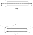
- Fig. 1 shows a schematic view of the prosthesis construction
- Fig. 2 shows the section of the prosthesis construction.
- a rectangle of polypropylene mesh is modified with cold oxygen plasma to increase the hydrophilicity of the surface and then welded to form a tube of 10 mm in diameter.
- a previously prepared solution containing 9 w/w of poly(L-lactide-co-caprolactone), 85 w/w chloroform, and 6 w/w N, N-dimethylformamide is subject to the electrospinning process under the following conditions: the nozzle-collector distance - 20 cm, the temperature 23°C, the nozzle collector voltage - 15kV.
- the electrospun nonwoven fabric is collected on a grounded rotating cylinder of the tubular shape ending with the cone generatrix.
- the obtained mat is placed in a 10% sodium bicarbonate solution NaHCO 3 for 40 days to increase the surface hydrophilicity. After rinsing and drying, the collar, prepared with a micro and nanofiber nonwoven fabric tube, is sewed to the previously prepared polypropylene mesh tube, which was hydrophilically modified.
- the internal surface is coated with a layer of an aqueous solution of sodium hyaluronate at the concentration of 0.7% w/w crosslinked with the use of butanediol-1,4-ol diglycidyl ether (BDDE) in the amount of 15% w/w (in terms of dry polymer content) with the addition of a plasticizer in the amount from 10% w/w (in terms of dry polymer content), whereby each of the consecutively deposited layers of crosslinked sodium hyaluronate undergoes the drying process at the temperature of 35°C.
- BDDE butanediol-1,4-ol diglycidyl ether
- the prosthesis undergoes the rinsing process in an aqueous solution of ethyl alcohol at the concentration of 70% w/w and 30% w/w and rinsing twice in demineralized water with the addition of a plasticizer in the amount of 5% w/w, and next freeze drying in the vacuum range from 0.1 mbar to 0.57 mbar for 20-22 hours.
- a nanofiber layer is deposited, which is produced with the use of the electrospinning method from a collagen solution of animal origin, dissolved in hexafluoroisopropanol (HFIP) at the concentration of 0.3% w/w.
- HFIP hexafluoroisopropanol
- the prosthesis After coating with the collagen layer, the prosthesis undergoes the crosslinking process in the alcohol solution of N-(3-dimethylaminopropyl)-N'-ethylcarbodiimide (EDC) at the concentration of 5.0% w/w for 7 days and following the rinsing process in aqueous solutions of ethyl alcohol 70% w/w and 30% w/w and demineralized water with the addition of a plasticizer in the amount of 5% w/w, and the freeze drying in the vacuum range from 0.1 mbar to 0.57 mbar for 20-22 hours.
- EDC N-(3-dimethylaminopropyl)-N'-ethylcarbodiimide
- a previously welded polypropylene mesh tube is modified with cold oxygen plasma.
- a previously prepared solution containing 9 w/w of poly(L-lactide-co-caprolactone), 85 w/w chloroform, and 6 w/w N, N-dimethylformamide is subject to the electrospinning process under the following conditions: the nozzle-collector distance - 20 cm, the temperature 23°C, the nozzle collector voltage - 15kV.
- the thus obtained nonwoven fabric undergoes hydrophilic modification by immersion in the 8% solution of potassium carbonate K 2 CO 3 for 48 hours. After rinsing and drying, the prepared collar is sewn to the previously prepared polypropylene mesh tube.
- the internal surface is coated with a layer of an aqueous solution of sodium hyaluronate at the concentration of 1.2% w/w crosslinked with the use of butanediol-1,4-ol diglycidyl ether (BDDE) in the amount of 15% w/w (in terms of dry polymer content) with the addition of a plasticizer in the amount of 20% w/w (in terms of dry polymer content), whereby each of the consecutively deposited layers of crosslinked sodium hyaluronate undergoes the drying process at the temperature of 35°C.
- BDDE butanediol-1,4-ol diglycidyl ether
- the prosthesis undergoes the rinsing process in an aqueous solution of ethyl alcohol at the concentration of 70% w/w and 30% w/w and rinsing twice in demineralized water with the addition of a plasticizer in the amount of 5% w/w, and next freeze drying in the vacuum range from 0.1 mbar to 0.57 mbar for 20-22 hours.
- a nanofiber layer is deposited, which is produced with the use of the electrospinning and electrospraying method from a collagen solution of animal origin, dissolved in hexafluoroisopropanol (HFIP) at the concentration of 0.1% w/w.
- HFIP hexafluoroisopropanol
- the prosthesis After coating with the collagen layer, the prosthesis undergoes the crosslinking process in the alcohol solution of N-(3-dimethylaminopropyl)-N'-ethylcarbodiimide (EDC) at the concentration of 5.5% w/w for 5 days and following the rinsing process in aqueous solutions of ethyl alcohol 70% w/w and 30% w/w and demineralized water with the addition of a plasticizer in the amount of 5% w/w, and the freeze drying in the vacuum range from 0.1 mbar to 0.57 mbar for 20-22 hours.
- EDC N-(3-dimethylaminopropyl)-N'-ethylcarbodiimide
- the internal side of matrix 2 is coated with a film formed by crosslinked hyaluronic acid with an average molecular weight of 1320 kD, with glycerin as the plasticizer of 20% w/w.
- the external surface 3 of the polypropylene matrix 1 is coated with an active nanofiber layer of type 1 collagen of animal origin of an average molecular weight of 300 kD.
- the distal end is equipped with a collar 4 prepared of poly(L-lactide-co-caprolactone) in the nanofiber nonwoven, enabling a 10 mm clearance hole 5 in the prosthesis.
- the advantage of the invention over known solutions is the possibility of preparing the prosthesis outside a patient's organism, which allows the accurate adjustment of its parameters to the predicted biochemical model of the artificial urine outlet functioning in a given patient, as well as prosthesis adjustment to a patient's variability, and hence, the patient's prognoses and quality of life in future.
- the superficially modified prosthesis is characterized by the permanent adhesion of polymer layers to the synthetic matrix, complete tightness preventing urine leakage, the developed internal surface enabling the settlement of the cells initiating the growth of the proper natural tissue, and good integration with animal tissue. Applying the prosthesis according to the invention allows for significant shortening of the urinary diversion procedure following radical cystectomy about the known methods.
Landscapes
- Health & Medical Sciences (AREA)
- Chemical & Material Sciences (AREA)
- Life Sciences & Earth Sciences (AREA)
- Dermatology (AREA)
- General Health & Medical Sciences (AREA)
- Veterinary Medicine (AREA)
- Medicinal Chemistry (AREA)
- Oral & Maxillofacial Surgery (AREA)
- Transplantation (AREA)
- Epidemiology (AREA)
- Public Health (AREA)
- Animal Behavior & Ethology (AREA)
- Engineering & Computer Science (AREA)
- Chemical Kinetics & Catalysis (AREA)
- Biomedical Technology (AREA)
- Materials Engineering (AREA)
- Composite Materials (AREA)
- Biophysics (AREA)
- Cell Biology (AREA)
- Zoology (AREA)
- Botany (AREA)
- Materials For Medical Uses (AREA)
Abstract
Description
- The subject of the invention is the prosthesis for urinary drainage for patients following radical cystectomy and the method of manufacturing it. The invention finds application in medicine, in patients following radical cystectomy being the consequence of a disease, particularly a cancer disease, or due to urinary bladder trauma and when it can no longer work properly.
- The solutions concerning the reconstruction of the lower urinary tract are already known. The French patent
FR2116838 - In solutions involving the use of the artificial urinary bladder, a connection between the natural ureters, urethra, and the artificial system is problematic. When the human tissue with its blood supply is connected to artificial systems, insufficient tissue healing may occur, which results in local leakages. Moreover, the defensive reactions of the human body against artificial systems constitute a potential danger to a patient's life. Peritonitis with multi-organ disorders within the abdominal cavity is a possible consequence. Surgical interventions undertaken to eliminate the disorders are, per se, serious and risky procedures.
- Alternative methods of urine drainage after radical cystectomy based on the implantation of urine transporting drains through the connection with an extracorporeal urine collection and removal system which allows its storage (e.g., a percutaneous nephrostomy or ureterostomy with a stoma bag) are also possible. Although such methods cause a specific reduction in quality and comfort of life, the procedures are much safer and have fewer complications.
- The fundamental problem concerning the given invention is to ensure the best possible biocompatibility of the prosthesis with a patient's body.
- According to the invention, the urinary drainage prosthesis is composed of a polypropylene or polyester mesh tube modified with the use of cold oxygen plasma to increase its hydrophilicity, with a mat in the form of a cone-shaped collar attached to the distal end, produced by electrospinning of a polyester solution, with the modified surface to increase hydrophilicity, whereby the internal side of the tube is coated with a layer of cross-linked sodium hyaluronate in the form of a film and the external side of the tube is coated with a nanofiber layer made of cross-linked collagen, on which the mesenchymal stem cells are seeded in a preferred version of the invention.
- The subject of the invention is therefore a prosthesis for urinary drainage for patients following radical cystectomy characterized in that it contains a tube which is made of a hydrophilic matrix made of polypropylene or polyester mesh, ended with a collar on one side, preferably in a cone-shaped form, made of micro and/or nanofiber nonwoven fabric prepared from a synthetic fiber-forming polymer or copolymers, natural polymers such as esters, cellulose ethers, collagen, gelatin, fibrinogen, natural silk; coated on the internal surface with a layer of cross-linked sodium hyaluronate of the average molecular weight from 2 000 kD to 2 500 kD in the form of a film with an addition of the plasticizer - a polyol, preferably glycerin in the amount from 10% w/w to 20% w/w of dry sodium hyaluronate, and on the external surface with a layer of type I collagen of the animal origin of the average molecular weight from 250 kD to 350 kD in the form of a nanofiber layer, whereby the sodium hyaluronate layer amounts from 15% w/w to 25% w/w, preferably 20% w/w of the mass of the prosthesis, and the collagen layer - from 20% w/w to 40% w/w, preferably 30% w/w of the mass of the prosthesis.
- Preferably, the tube is from 8 mm to 12 mm in diameter and 50 mm to 100 mm in length.
- Preferably, the size of the polypropylene mesh or polyester mesh in the hydrophilic matrix amounts from 35 µm to 5100 µm, preferably from 250 µm to 350 µm and the spun fiber thickness from 90 µm to 1000 µm, optimally from 150 µm to 350 µm.
- Preferably the collar is made of micro and/or nanofiber nonwoven fabric obtained from a synthetic fiber-forming polymer soluble in organic solvents such as polylactide, polyglycolide, polycaprolactone, polyamide, polyimide, poly(trimethylene carbonate), polydioxanone, polyvinyl acetate, polyvinyl alcohol, poly(N-vinyl-2-pyrrolidone), polyvinyl butyral, polyurethane, poly(ether urethane), polycarbonate, aromatic polycarbonate, poly(ethylene oxide), poly(ethylene glycol), poly(methyl methacrylate), poly(acrylic acid), polyacrylamide, polyacrylonitrile, polyvinyl chloride, poly(vinylidene fluoride), poly(tetrafluoroethylene), poly(ethylene glycol terephthalate), or their mixtures.
- Preferably, the collar is made of a micro and/or nanofiber nonwoven fabric obtained from a cellulose ester chosen from the group: carboxymethylcellulose, carboxymethylcellulose, methylcellulose, or their mixtures.
- Preferably, the collar is made of a micro and/or nanofiber nonwoven fabric obtained from a copolymer chosen from the group: L-lactide and caprolactone copolymer, poly(L-lactide-co-caprolactone), or their mixtures.
- Preferably, the external surface of the tube is coated with autologous or allogenic mesenchymal stem/ stromal cells isolated from bone marrow, adipose tissue, or Wharton's jelly.
- The subject of the method of manufacturing a prosthesis for urinary drainage for patients with planned radical cystectomy is characterized in that the external and internal surfaces of a hydrophilic matrix made of polypropylene or polyester mesh of a tubular shape are modified with cold oxygen plasma to increase their hydrophilicity. Next, under dynamic conditions, using the (spraying and coating or hard-facing and coating) method, the internal surface is coated with layers of an aqueous solution of sodium hyaluronate at a concentration from 0.7% w/w to 1.2% w/w, preferably 0.7% w/w crosslinked with the use of butanediol-1,4-ol diglycidyl ether (BDDE) in the amount from 10% w/w to 15% w/w (in terms of dry polymer content) with the addition of a plasticizer in the amount from 10% w/w to 20% w/w (in terms of dry polymer content), whereby each of the consecutively deposited layers of crosslinked sodium hyaluronate undergoes the drying process at the temperature from 30°C to 40°C. After all the layers of crosslinked sodium hyaluronate are deposited, the prosthesis undergoes the rinsing process in an aqueous solution of ethyl alcohol at the concentration of 70% w/w and 30% w/w and rinsing twice in demineralized water with the addition of a plasticizer in the amount of 5% w/w, and next freeze drying in the vacuum range from 0.1 mbar to 0.57 mbar for 20-22 hours. Next, on the external surface of the prosthesis, a nanofiber layer is deposited, which is produced with the use of the electrospinning and/or electrospraying method from a collagen solution of animal origin, dissolved in hexafluoroisopropanol (HFIP) at the concentration from 0.1% w/w to 0.3% w/w, preferably 0.3% w/w. After coating with the collagen layer, the prosthesis undergoes the crosslinking process in the alcohol solution of N-(3-dimethylaminopropyl)-N'-ethylcarbodiimide (EDC) at the concentration from 5.0% w/w to 5.5% w/w, preferably 5% w/w, for 5-7 days, and next the rinsing process in aqueous solutions of ethyl alcohol 70% w/w and 30% w/w and demineralized water with the addition of a plasticizer in the amount of 5% w/w, and the freeze drying in the vacuum range from 0.1 mbar to 0.57 mbar for 20-22 hours.
- Preferably in the method according to the invention, the external surface of the prosthesis for urinal drainage is coated with autologous or allogenic mesenchymal phalangeal/ mesenchymal stem/stromal cells isolated from bone marrow, adipose tissue, or Wharton's jelly seeded at the density of 1-100 x 106 cells/cm2, preferably 10 x 106 cells/ cm2, whereby the cells are cultivated on the prosthesis for the period of 1-30 days, preferably 7 days.
- The research on a large animal model showed a significant impact of using mesenchymal stem/ stromal cells on the healing processes in reconstructions using the prosthesis in the urinary tract.
- The table below compares the inflammatory reactions in the mesh coated with collagen and the mesh coated with collagen seeded with adipose-derived mesenchymal stem/stromal cells one month after the prosthesis implantation, according to the invention.
Average time of patency of urinary fistula (months) Average time of the insert prolapse (months) Control group - ureterocutaneostomia 6 0 Reconstruction of the ureter with the prosthesis according to the invention, with the use of tissue glue 12 5 Reconstruction of the ureter with the pre-implanted prosthesis according to the invention, seeded with mesenchymal stem/stromal cells and with the use of tissue glue 18 8 - The invention has been presented in embodiments, and the Scheme in which
Fig. 1 shows a schematic view of the prosthesis construction,Fig. 2 shows the section of the prosthesis construction. - A rectangle of polypropylene mesh is modified with cold oxygen plasma to increase the hydrophilicity of the surface and then welded to form a tube of 10 mm in diameter. A previously prepared solution containing 9 w/w of poly(L-lactide-co-caprolactone), 85 w/w chloroform, and 6 w/w N, N-dimethylformamide is subject to the electrospinning process under the following conditions: the nozzle-collector distance - 20 cm, the temperature 23°C, the nozzle collector voltage - 15kV. The electrospun nonwoven fabric is collected on a grounded rotating cylinder of the tubular shape ending with the cone generatrix. The obtained mat is placed in a 10% sodium bicarbonate solution NaHCO3 for 40 days to increase the surface hydrophilicity. After rinsing and drying, the collar, prepared with a micro and nanofiber nonwoven fabric tube, is sewed to the previously prepared polypropylene mesh tube, which was hydrophilically modified.
- Next, under dynamic conditions, using spraying and deposition, the internal surface is coated with a layer of an aqueous solution of sodium hyaluronate at the concentration of 0.7% w/w crosslinked with the use of butanediol-1,4-ol diglycidyl ether (BDDE) in the amount of 15% w/w (in terms of dry polymer content) with the addition of a plasticizer in the amount from 10% w/w (in terms of dry polymer content), whereby each of the consecutively deposited layers of crosslinked sodium hyaluronate undergoes the drying process at the temperature of 35°C. After all the layers of crosslinked sodium hyaluronate are deposited, the prosthesis undergoes the rinsing process in an aqueous solution of ethyl alcohol at the concentration of 70% w/w and 30% w/w and rinsing twice in demineralized water with the addition of a plasticizer in the amount of 5% w/w, and next freeze drying in the vacuum range from 0.1 mbar to 0.57 mbar for 20-22 hours. Next, on the external surface of the prosthesis, a nanofiber layer is deposited, which is produced with the use of the electrospinning method from a collagen solution of animal origin, dissolved in hexafluoroisopropanol (HFIP) at the concentration of 0.3% w/w. After coating with the collagen layer, the prosthesis undergoes the crosslinking process in the alcohol solution of N-(3-dimethylaminopropyl)-N'-ethylcarbodiimide (EDC) at the concentration of 5.0% w/w for 7 days and following the rinsing process in aqueous solutions of ethyl alcohol 70% w/w and 30% w/w and demineralized water with the addition of a plasticizer in the amount of 5% w/w, and the freeze drying in the vacuum range from 0.1 mbar to 0.57 mbar for 20-22 hours.
- To increase their hydrophilicity, a previously welded polypropylene mesh tube is modified with cold oxygen plasma. A previously prepared solution containing 9 w/w of poly(L-lactide-co-caprolactone), 85 w/w chloroform, and 6 w/w N, N-dimethylformamide is subject to the electrospinning process under the following conditions: the nozzle-collector distance - 20 cm, the temperature 23°C, the nozzle collector voltage - 15kV. The thus obtained nonwoven fabric undergoes hydrophilic modification by immersion in the 8% solution of potassium carbonate K2CO3 for 48 hours. After rinsing and drying, the prepared collar is sewn to the previously prepared polypropylene mesh tube.
- Next, under dynamic conditions, using spraying and deposition, the internal surface is coated with a layer of an aqueous solution of sodium hyaluronate at the concentration of 1.2% w/w crosslinked with the use of butanediol-1,4-ol diglycidyl ether (BDDE) in the amount of 15% w/w (in terms of dry polymer content) with the addition of a plasticizer in the amount of 20% w/w (in terms of dry polymer content), whereby each of the consecutively deposited layers of crosslinked sodium hyaluronate undergoes the drying process at the temperature of 35°C. After all the layers of crosslinked sodium hyaluronate are deposited, the prosthesis undergoes the rinsing process in an aqueous solution of ethyl alcohol at the concentration of 70% w/w and 30% w/w and rinsing twice in demineralized water with the addition of a plasticizer in the amount of 5% w/w, and next freeze drying in the vacuum range from 0.1 mbar to 0.57 mbar for 20-22 hours.
- Next, on the external surface of the prosthesis, a nanofiber layer is deposited, which is produced with the use of the electrospinning and electrospraying method from a collagen solution of animal origin, dissolved in hexafluoroisopropanol (HFIP) at the concentration of 0.1% w/w. After coating with the collagen layer, the prosthesis undergoes the crosslinking process in the alcohol solution of N-(3-dimethylaminopropyl)-N'-ethylcarbodiimide (EDC) at the concentration of 5.5% w/w for 5 days and following the rinsing process in aqueous solutions of ethyl alcohol 70% w/w and 30% w/w and demineralized water with the addition of a plasticizer in the amount of 5% w/w, and the freeze drying in the vacuum range from 0.1 mbar to 0.57 mbar for 20-22 hours.
- The prosthesis for urinary drainage schematically presented in
Fig. 2 comprises matrix 1 in the tubular shape made of polypropylene mesh. The internal side ofmatrix 2 is coated with a film formed by crosslinked hyaluronic acid with an average molecular weight of 1320 kD, with glycerin as the plasticizer of 20% w/w. The external surface 3 of the polypropylene matrix 1 is coated with an active nanofiber layer of type 1 collagen of animal origin of an average molecular weight of 300 kD. The distal end is equipped with a collar 4 prepared of poly(L-lactide-co-caprolactone) in the nanofiber nonwoven, enabling a 10 mm clearance hole 5 in the prosthesis. - The advantage of the invention over known solutions is the possibility of preparing the prosthesis outside a patient's organism, which allows the accurate adjustment of its parameters to the predicted biochemical model of the artificial urine outlet functioning in a given patient, as well as prosthesis adjustment to a patient's variability, and hence, the patient's prognoses and quality of life in future.
- The superficially modified prosthesis is characterized by the permanent adhesion of polymer layers to the synthetic matrix, complete tightness preventing urine leakage, the developed internal surface enabling the settlement of the cells initiating the growth of the proper natural tissue, and good integration with animal tissue. Applying the prosthesis according to the invention allows for significant shortening of the urinary diversion procedure following radical cystectomy about the known methods.
Claims (9)
- A urinary drainage prosthesis for patients following radical cystectomy is characterized in that it comprises a tube made of a hydrophilic matrix made of polypropylene or polyester mesh in the shape of a tube, ended with a collar on one side, preferably in a cone-shaped form, made of micro and/or nanofiber nonwoven fabric prepared from a synthetic fiber-forming polymer or copolymers, natural polymers such as esters, cellulose ethers, collagen, gelatin, fibrinogen, natural silk; coated on the internal surface with a layer of cross-linked sodium hyaluronate of the average molecular weight from 2 000 kD to 2 500 kD in the form of a film with an addition of the plasticizer - a polyol, preferably glycerin in the amount from 10% w/w to 20% w/w of dry sodium hyaluronate, and on the external surface with a layer of type I collagen of the animal origin of the average molecular weight from 250 kD to 350 kD in the form of a nanofibrilar layer, whereby the sodium hyaluronate layer amounts from 15% w/w to 25% w/w, preferably 20% w/w in terms of the mass of the prosthesis, and the collagen layer - from 20% w/w to 40% w/w, preferably 30% w/w in terms of the mass of the prosthesis.
- The urinary drainage prosthesis according to claim 1, characterized in that the tube is from 8 mm to 12 mm in diameter and 50 mm to 100 mm in length.
- The urinary drainage prosthesis according to claim 1, characterized in that the size of the polypropylene mesh or polyester mesh in the hydrophilic matrix amounts from 35 µm to 5100 µm, preferably from 250 µm to 350 µm and the spun fiber thickness from 90 µm to 1000 µm, optimally from 150 µm to 350 µm.
- The urinary drainage prosthesis according to claim 1, characterized in that the collar is made of micro and/or nanofiber nonwoven fabric obtained from a synthetic fiber-forming polymer soluble in organic solvents such as polylactide, polyglycolide, polycaprolactone, polyamide, polyimide, poly(trimethylene carbonate), polydioxanone, polyvinyl acetate, polyvinyl alcohol, poly(N-vinyl-2-pyrrolidone), polyvinyl butyral, polyurethane, poly(ether urethane), polycarbonate, aromatic polycarbonate, poly(ethylene oxide), poly(ethylene glycol), poly(methyl methacrylate), poly(acrylic acid), polyacrylamide, polyacrylonitrile, polyvinyl chloride, poly(vinylidene fluoride), poly(tetrafluoroethylene), poly(ethylene glycol terephthalate), or their mixtures.
- The urinary drainage prosthesis according to claim 1, characterized in that the collar is made of a micro and/or nanofiber nonwoven fabric produced of a cellulose ester chosen from the group: carboxymethylcellulose carboxymethylcellulose, methylcellulose, or their mixtures.
- The urinary drainage prosthesis according to claim 1, characterized in that the collar is made of a micro and/or nanofiber nonwoven fabric obtained from a copolymer chosen from the group: L-lactide and caprolactone copolymer, poly(L-lactide-co-caprolactone), or their mixtures.
- The urinary drainage prosthesis according to any one of claims 1-6, characterized in that the external surface of the tube is coated with autologous or allogenic mesenchymal stem/stromal cells isolated from bone marrow, adipose tissue, or Wharton's jelly.
- A method of manufacturing the urinary drainage prosthesis for patients following radical cystectomy characterized in that the external and internal surfaces of a hydrophilic matrix made of polypropylene or polyester mesh of a tubular shape are modified with cold oxygen plasma in order to increase their hydrophilicity and next, using the spraying and coating, or hardfacing and coating method, the internal surface is coated with layers of an aqueous solution of sodium hyaluronate at a concentration from 0.7% w/w to 1.2% w/w, preferably 0.7% w/w with the addition of a plasticizer in the amount from 10% w/w to 20% w/w (in terms of dry polymer content), crosslinked with the use of butanedi-1,4-ol diglycidyl ether (BDDE) in the amount from 10% w/w to 15% w/w in terms of polymer content, whereby each of the consecutively deposited layers of crosslinked sodium hyaluronate undergoes the drying process at the temperature of 35°C, and after all the layers of crosslinked sodium hyaluronate are deposited, the prosthesis undergoes the rinsing process in an aqueous solution of ethyl alcohol at the concentration of 70% w/w and 30% w/w, and rinsing twice in demineralized water with the addition of a plasticizer in the amount of 5% w/w, and next freeze drying in the vacuum range from 0.1 mbar to 0.57 mbar for 20-22 hours and next, on the external surface of the prosthesis, a nanofiber layer is deposited which is produced with the use of the electrospinning and/or electrospraying method from a solution of collagen of the animal origin, dissolved in hexafluoroisopropanol (HFIP) at the concentration from 0.1% w/w to 0.3% w/w, preferably 0.3% w/w, and after coating with the collagen layer, the prosthesis undergoes the crosslinking process in the alcohol solution of N-(3-dimethylaminopropyl)-N'-ethylcarbodiimide (EDC) at the concentration from 5.0% w/w to 5.5% w/w, preferably 5% w/w, for 5-7 days, and next the rinsing process in aqueous solutions of ethyl alcohol 70% w/w and 30% w/w and demineralized water with the addition of a plasticizer in the amount of 5% w/w, and the freeze drying in the vacuum range from 0.1 mbar to 0.57 mbar for 20-22 hours.
- The method of manufacturing the prosthesis according to claim 8, characterized in that the external surface of the prosthesis is seeded with autologous or allogenic mesenchymal stem/stromal cells isolated from bone marrow, adipose tissue, or Wharton's jelly at the density from 1 to 100 x 106 cells/cm2, preferably 10 x 106 cells/cm2, and the cells cultivation on the surface of prosthesis is performed under static or dynamic conditions, preferably in a bioreactor for 1-30 days, preferably 7 days.
Applications Claiming Priority (1)
| Application Number | Priority Date | Filing Date | Title |
|---|---|---|---|
| PL446936A PL446936A1 (en) | 2023-11-30 | 2023-11-30 | A prosthesis for urinary drainage in patients without a urinary bladder and a method of producing the same |
Publications (3)
| Publication Number | Publication Date |
|---|---|
| EP4570276A1 true EP4570276A1 (en) | 2025-06-18 |
| EP4570276C0 EP4570276C0 (en) | 2025-12-31 |
| EP4570276B1 EP4570276B1 (en) | 2025-12-31 |
Family
ID=93155078
Family Applications (1)
| Application Number | Title | Priority Date | Filing Date |
|---|---|---|---|
| EP24216329.3A Active EP4570276B1 (en) | 2023-11-30 | 2024-11-29 | Urinary drainage prosthesis for bladderless patients and methods for its manufacture |
Country Status (2)
| Country | Link |
|---|---|
| EP (1) | EP4570276B1 (en) |
| PL (1) | PL446936A1 (en) |
Citations (6)
| Publication number | Priority date | Publication date | Assignee | Title |
|---|---|---|---|---|
| FR2116838A5 (en) | 1970-12-09 | 1972-07-21 | Sowinski Kazimierz Maria | PROSTHESIS TO REPLACE A PATIENT'S BLADDER |
| US20080038352A1 (en) * | 1999-02-25 | 2008-02-14 | Virginia Commonwealth University Intellectual Property Foundation | Electroprocessed Collagen and Tissue Engineering |
| WO2010043177A1 (en) * | 2008-10-15 | 2010-04-22 | Zhou Xing | Human body tube substitute of biological induction type |
| US20160262865A1 (en) * | 2013-10-16 | 2016-09-15 | Antonio Sambusseti | Pga tubular patch and relative optional tubular support made of absorbable material for the tissue reconstruction of urethral and/or ureteral removed segments |
| US20180243482A1 (en) * | 2013-05-20 | 2018-08-30 | Yale University | Anti-thrombogenic grafts |
| CN116536246A (en) * | 2023-04-21 | 2023-08-04 | 柔脉医疗(深圳)有限公司 | Three-dimensional artificial tubular tissue and preparation method and application thereof |
Family Cites Families (3)
| Publication number | Priority date | Publication date | Assignee | Title |
|---|---|---|---|---|
| US20020055786A1 (en) * | 1994-08-16 | 2002-05-09 | Anthony Atala | Reconstruction of urological structures with polymeric matrices |
| US10252025B2 (en) * | 2015-10-05 | 2019-04-09 | Justin Garcia | Urinary prostheses |
| FR3109290B1 (en) * | 2020-04-17 | 2025-04-11 | Ph Tech | Medical device comprising an acellular biological matrix and at least one polymer |
-
2023
- 2023-11-30 PL PL446936A patent/PL446936A1/en unknown
-
2024
- 2024-11-29 EP EP24216329.3A patent/EP4570276B1/en active Active
Patent Citations (6)
| Publication number | Priority date | Publication date | Assignee | Title |
|---|---|---|---|---|
| FR2116838A5 (en) | 1970-12-09 | 1972-07-21 | Sowinski Kazimierz Maria | PROSTHESIS TO REPLACE A PATIENT'S BLADDER |
| US20080038352A1 (en) * | 1999-02-25 | 2008-02-14 | Virginia Commonwealth University Intellectual Property Foundation | Electroprocessed Collagen and Tissue Engineering |
| WO2010043177A1 (en) * | 2008-10-15 | 2010-04-22 | Zhou Xing | Human body tube substitute of biological induction type |
| US20180243482A1 (en) * | 2013-05-20 | 2018-08-30 | Yale University | Anti-thrombogenic grafts |
| US20160262865A1 (en) * | 2013-10-16 | 2016-09-15 | Antonio Sambusseti | Pga tubular patch and relative optional tubular support made of absorbable material for the tissue reconstruction of urethral and/or ureteral removed segments |
| CN116536246A (en) * | 2023-04-21 | 2023-08-04 | 柔脉医疗(深圳)有限公司 | Three-dimensional artificial tubular tissue and preparation method and application thereof |
Also Published As
| Publication number | Publication date |
|---|---|
| EP4570276C0 (en) | 2025-12-31 |
| PL446936A1 (en) | 2024-10-21 |
| EP4570276B1 (en) | 2025-12-31 |
Similar Documents
| Publication | Publication Date | Title |
|---|---|---|
| US11969337B2 (en) | In vivo tissue engineering devices, methods and regenerative and cellular medicine employing scaffolds made of absorbable material | |
| US11883276B2 (en) | In vivo tissue engineering devices, methods and regenerative and cellular medicine employing scaffolds made of absorbable material | |
| US11844685B2 (en) | In vivo tissue engineering devices, methods and regenerative and cellular medicine employing scaffolds made of absorbable material | |
| CN101214393A (en) | Nanofiber tissue engineering blood vessel and preparation method thereof | |
| CN104414772A (en) | In-vivo degradable and absorbable artificial medical tissue repairing film | |
| US11883275B2 (en) | In vivo tissue engineering devices, methods and regenerative and cellular medicine employing scaffolds made of absorbable material | |
| CN101708344A (en) | Nanofiber vascular prostheses and preparation method | |
| CN109381732A (en) | Electrostatic spinning dressing, preparation method and the application of growth factor-loaded micromolecular inhibitor | |
| CN102475920A (en) | Preparation method and application of anti-inflammatory drug and growth factor sustained-release system | |
| CN115990293A (en) | Preparation method of medical multilayer self-supporting drug-loaded shell-core structure nanofiber tube | |
| EP4570276B1 (en) | Urinary drainage prosthesis for bladderless patients and methods for its manufacture | |
| CN116510089A (en) | Bacterial nanocellulose/high molecular compound composite tube and preparation and biomedical application thereof | |
| Mathews | Polydioxanone: applications in surgery and tissue engineering | |
| CN115607730B (en) | Degradable bionic artificial trachea | |
| CN113244021A (en) | Support for preventing external auditory canal stenosis through 3D printing acellular matrix and preparation method | |
| CN108434536B (en) | Lacrimal passage repair stent and preparation method thereof | |
| CN101703802A (en) | Nano fiber artificial blood vessel for catalyzing endogenous NO precursor to release NO and preparation method | |
| CN111700710B (en) | Template for tissue engineering material and tissue engineering material | |
| RU2630061C1 (en) | Method for manufacture of three-layer frame for bile duct prosthetics | |
| CN121313912A (en) | A self-generating controlled-release drug-eluting surgical suture for promoting diabetic wound healing, its preparation method and application | |
| CN121944255A (en) | Anti-tumor degradable ureteral stent with bionic structure and preparation method thereof |
Legal Events
| Date | Code | Title | Description |
|---|---|---|---|
| PUAI | Public reference made under article 153(3) epc to a published international application that has entered the european phase |
Free format text: ORIGINAL CODE: 0009012 |
|
| STAA | Information on the status of an ep patent application or granted ep patent |
Free format text: STATUS: THE APPLICATION HAS BEEN PUBLISHED |
|
| AK | Designated contracting states |
Kind code of ref document: A1 Designated state(s): AL AT BE BG CH CY CZ DE DK EE ES FI FR GB GR HR HU IE IS IT LI LT LU LV MC ME MK MT NL NO PL PT RO RS SE SI SK SM TR |
|
| STAA | Information on the status of an ep patent application or granted ep patent |
Free format text: STATUS: REQUEST FOR EXAMINATION WAS MADE |
|
| 17P | Request for examination filed |
Effective date: 20250619 |
|
| GRAP | Despatch of communication of intention to grant a patent |
Free format text: ORIGINAL CODE: EPIDOSNIGR1 |
|
| STAA | Information on the status of an ep patent application or granted ep patent |
Free format text: STATUS: GRANT OF PATENT IS INTENDED |
|
| RIC1 | Information provided on ipc code assigned before grant |
Ipc: A61L 27/16 20060101AFI20251009BHEP Ipc: A61L 27/18 20060101ALI20251009BHEP Ipc: A61L 27/34 20060101ALI20251009BHEP |
|
| GRAS | Grant fee paid |
Free format text: ORIGINAL CODE: EPIDOSNIGR3 |
|
| GRAA | (expected) grant |
Free format text: ORIGINAL CODE: 0009210 |
|
| STAA | Information on the status of an ep patent application or granted ep patent |
Free format text: STATUS: THE PATENT HAS BEEN GRANTED |
|
| INTG | Intention to grant announced |
Effective date: 20251111 |
|
| AK | Designated contracting states |
Kind code of ref document: B1 Designated state(s): AL AT BE BG CH CY CZ DE DK EE ES FI FR GB GR HR HU IE IS IT LI LT LU LV MC ME MK MT NL NO PL PT RO RS SE SI SK SM TR |
|
| REG | Reference to a national code |
Ref country code: CH Ref legal event code: F10 Free format text: ST27 STATUS EVENT CODE: U-0-0-F10-F00 (AS PROVIDED BY THE NATIONAL OFFICE) Effective date: 20251231 Ref country code: GB Ref legal event code: FG4D |
|
| REG | Reference to a national code |
Ref country code: DE Ref legal event code: R096 Ref document number: 602024001946 Country of ref document: DE |
|
| REG | Reference to a national code |
Ref country code: IE Ref legal event code: FG4D |
|
| U01 | Request for unitary effect filed |
Effective date: 20260123 |
|
| U07 | Unitary effect registered |
Designated state(s): AT BE BG DE DK EE FI FR IT LT LU LV MT NL PT RO SE SI Effective date: 20260129 |
|
| PG25 | Lapsed in a contracting state [announced via postgrant information from national office to epo] |
Ref country code: NO Free format text: LAPSE BECAUSE OF FAILURE TO SUBMIT A TRANSLATION OF THE DESCRIPTION OR TO PAY THE FEE WITHIN THE PRESCRIBED TIME-LIMIT Effective date: 20260331 |
|
| PG25 | Lapsed in a contracting state [announced via postgrant information from national office to epo] |
Ref country code: HR Free format text: LAPSE BECAUSE OF FAILURE TO SUBMIT A TRANSLATION OF THE DESCRIPTION OR TO PAY THE FEE WITHIN THE PRESCRIBED TIME-LIMIT Effective date: 20251231 |
|
| PG25 | Lapsed in a contracting state [announced via postgrant information from national office to epo] |
Ref country code: RS Free format text: LAPSE BECAUSE OF FAILURE TO SUBMIT A TRANSLATION OF THE DESCRIPTION OR TO PAY THE FEE WITHIN THE PRESCRIBED TIME-LIMIT Effective date: 20260331 |