EP4566637A1 - Biodegradable 3d printed structure for bone augmentation - Google Patents
Biodegradable 3d printed structure for bone augmentation Download PDFInfo
- Publication number
- EP4566637A1 EP4566637A1 EP24020049.3A EP24020049A EP4566637A1 EP 4566637 A1 EP4566637 A1 EP 4566637A1 EP 24020049 A EP24020049 A EP 24020049A EP 4566637 A1 EP4566637 A1 EP 4566637A1
- Authority
- EP
- European Patent Office
- Prior art keywords
- outer structure
- printed
- biodegradable
- tunnel
- bone augmentation
- Prior art date
- Legal status (The legal status is an assumption and is not a legal conclusion. Google has not performed a legal analysis and makes no representation as to the accuracy of the status listed.)
- Pending
Links
Images


Classifications
-
- A—HUMAN NECESSITIES
- A61—MEDICAL OR VETERINARY SCIENCE; HYGIENE
- A61L—METHODS OR APPARATUS FOR STERILISING MATERIALS OR OBJECTS IN GENERAL; DISINFECTION, STERILISATION OR DEODORISATION OF AIR; CHEMICAL ASPECTS OF BANDAGES, DRESSINGS, ABSORBENT PADS OR SURGICAL ARTICLES; MATERIALS FOR BANDAGES, DRESSINGS, ABSORBENT PADS OR SURGICAL ARTICLES
- A61L27/00—Materials for grafts or prostheses or for coating grafts or prostheses
- A61L27/14—Macromolecular materials
- A61L27/18—Macromolecular materials obtained otherwise than by reactions only involving carbon-to-carbon unsaturated bonds
-
- A—HUMAN NECESSITIES
- A61—MEDICAL OR VETERINARY SCIENCE; HYGIENE
- A61L—METHODS OR APPARATUS FOR STERILISING MATERIALS OR OBJECTS IN GENERAL; DISINFECTION, STERILISATION OR DEODORISATION OF AIR; CHEMICAL ASPECTS OF BANDAGES, DRESSINGS, ABSORBENT PADS OR SURGICAL ARTICLES; MATERIALS FOR BANDAGES, DRESSINGS, ABSORBENT PADS OR SURGICAL ARTICLES
- A61L27/00—Materials for grafts or prostheses or for coating grafts or prostheses
- A61L27/28—Materials for coating prostheses
-
- A—HUMAN NECESSITIES
- A61—MEDICAL OR VETERINARY SCIENCE; HYGIENE
- A61L—METHODS OR APPARATUS FOR STERILISING MATERIALS OR OBJECTS IN GENERAL; DISINFECTION, STERILISATION OR DEODORISATION OF AIR; CHEMICAL ASPECTS OF BANDAGES, DRESSINGS, ABSORBENT PADS OR SURGICAL ARTICLES; MATERIALS FOR BANDAGES, DRESSINGS, ABSORBENT PADS OR SURGICAL ARTICLES
- A61L27/00—Materials for grafts or prostheses or for coating grafts or prostheses
- A61L27/28—Materials for coating prostheses
- A61L27/34—Macromolecular materials
-
- A—HUMAN NECESSITIES
- A61—MEDICAL OR VETERINARY SCIENCE; HYGIENE
- A61L—METHODS OR APPARATUS FOR STERILISING MATERIALS OR OBJECTS IN GENERAL; DISINFECTION, STERILISATION OR DEODORISATION OF AIR; CHEMICAL ASPECTS OF BANDAGES, DRESSINGS, ABSORBENT PADS OR SURGICAL ARTICLES; MATERIALS FOR BANDAGES, DRESSINGS, ABSORBENT PADS OR SURGICAL ARTICLES
- A61L27/00—Materials for grafts or prostheses or for coating grafts or prostheses
- A61L27/40—Composite materials, i.e. containing one material dispersed in a matrix of the same or different material
- A61L27/44—Composite materials, i.e. containing one material dispersed in a matrix of the same or different material having a macromolecular matrix
- A61L27/46—Composite materials, i.e. containing one material dispersed in a matrix of the same or different material having a macromolecular matrix with phosphorus-containing inorganic fillers
-
- A—HUMAN NECESSITIES
- A61—MEDICAL OR VETERINARY SCIENCE; HYGIENE
- A61L—METHODS OR APPARATUS FOR STERILISING MATERIALS OR OBJECTS IN GENERAL; DISINFECTION, STERILISATION OR DEODORISATION OF AIR; CHEMICAL ASPECTS OF BANDAGES, DRESSINGS, ABSORBENT PADS OR SURGICAL ARTICLES; MATERIALS FOR BANDAGES, DRESSINGS, ABSORBENT PADS OR SURGICAL ARTICLES
- A61L27/00—Materials for grafts or prostheses or for coating grafts or prostheses
- A61L27/40—Composite materials, i.e. containing one material dispersed in a matrix of the same or different material
- A61L27/44—Composite materials, i.e. containing one material dispersed in a matrix of the same or different material having a macromolecular matrix
- A61L27/48—Composite materials, i.e. containing one material dispersed in a matrix of the same or different material having a macromolecular matrix with macromolecular fillers
-
- A—HUMAN NECESSITIES
- A61—MEDICAL OR VETERINARY SCIENCE; HYGIENE
- A61L—METHODS OR APPARATUS FOR STERILISING MATERIALS OR OBJECTS IN GENERAL; DISINFECTION, STERILISATION OR DEODORISATION OF AIR; CHEMICAL ASPECTS OF BANDAGES, DRESSINGS, ABSORBENT PADS OR SURGICAL ARTICLES; MATERIALS FOR BANDAGES, DRESSINGS, ABSORBENT PADS OR SURGICAL ARTICLES
- A61L27/00—Materials for grafts or prostheses or for coating grafts or prostheses
- A61L27/50—Materials characterised by their function or physical properties, e.g. injectable or lubricating compositions, shape-memory materials, surface modified materials
- A61L27/56—Porous materials, e.g. foams or sponges
-
- A—HUMAN NECESSITIES
- A61—MEDICAL OR VETERINARY SCIENCE; HYGIENE
- A61L—METHODS OR APPARATUS FOR STERILISING MATERIALS OR OBJECTS IN GENERAL; DISINFECTION, STERILISATION OR DEODORISATION OF AIR; CHEMICAL ASPECTS OF BANDAGES, DRESSINGS, ABSORBENT PADS OR SURGICAL ARTICLES; MATERIALS FOR BANDAGES, DRESSINGS, ABSORBENT PADS OR SURGICAL ARTICLES
- A61L27/00—Materials for grafts or prostheses or for coating grafts or prostheses
- A61L27/50—Materials characterised by their function or physical properties, e.g. injectable or lubricating compositions, shape-memory materials, surface modified materials
- A61L27/58—Materials at least partially resorbable by the body
-
- B—PERFORMING OPERATIONS; TRANSPORTING
- B33—ADDITIVE MANUFACTURING TECHNOLOGY
- B33Y—ADDITIVE MANUFACTURING, i.e. MANUFACTURING OF THREE-DIMENSIONAL [3D] OBJECTS BY ADDITIVE DEPOSITION, ADDITIVE AGGLOMERATION OR ADDITIVE LAYERING, e.g. BY 3D PRINTING, STEREOLITHOGRAPHY OR SELECTIVE LASER SINTERING
- B33Y80/00—Products made by additive manufacturing
-
- A—HUMAN NECESSITIES
- A61—MEDICAL OR VETERINARY SCIENCE; HYGIENE
- A61L—METHODS OR APPARATUS FOR STERILISING MATERIALS OR OBJECTS IN GENERAL; DISINFECTION, STERILISATION OR DEODORISATION OF AIR; CHEMICAL ASPECTS OF BANDAGES, DRESSINGS, ABSORBENT PADS OR SURGICAL ARTICLES; MATERIALS FOR BANDAGES, DRESSINGS, ABSORBENT PADS OR SURGICAL ARTICLES
- A61L2420/00—Materials or methods for coatings medical devices
- A61L2420/04—Coatings containing a composite material such as inorganic/organic, i.e. material comprising different phases
-
- A—HUMAN NECESSITIES
- A61—MEDICAL OR VETERINARY SCIENCE; HYGIENE
- A61L—METHODS OR APPARATUS FOR STERILISING MATERIALS OR OBJECTS IN GENERAL; DISINFECTION, STERILISATION OR DEODORISATION OF AIR; CHEMICAL ASPECTS OF BANDAGES, DRESSINGS, ABSORBENT PADS OR SURGICAL ARTICLES; MATERIALS FOR BANDAGES, DRESSINGS, ABSORBENT PADS OR SURGICAL ARTICLES
- A61L2430/00—Materials or treatment for tissue regeneration
- A61L2430/02—Materials or treatment for tissue regeneration for reconstruction of bones; weight-bearing implants
Definitions
- the present invention relates to biodegradable structures for bone augmentation, more specifically those obtained through 3D printing.
- titanium implants are most often used, which are removed due to their inert nature after a certain time required for bone augmentation (approximately 6 months), which causes certain trauma to the patient as well as additional stress to the newly formed tissue.
- the inventor aimed to find a new structure for bone augmentation that would be cheaper, that would involve fewer complications and that would result in a faster and less stressful treatment procedure for the patient.
- the biggest problem in dental implantology is the lack of the necessary volume of the jaw bone, which atrophies after tooth extraction. Bone loss after tooth extraction, inflammation, or trauma requires an oral surgical procedure for bone augmentation. An adequate bone volume at the desired location is required for the placement of an endosseous implant. During bone ridge augmentation, it is desirable that bone regeneration occurs as quickly as possible. This is achieved through guided bone regeneration (GBR) with autologous or artificially derived bone from synthetic or animal-derived materials.
- GBR guided bone regeneration
- the basic principle of the GBR method is to prevent tissue ingrowth into a bone defect by applying a 3D porous structure to restore the periodontium and to stimulate vascularization for the growth of new bone tissue.
- porous structures that have to be biocompatible and mechanically resistant to provide sufficient space for tissue regeneration and should be resistant to everyday mechanical burdens inside the oral cavity. Additionally, porous structures should integrate with the surrounding tissue and be easily shaped to match the size and shape of the treated area. Previous periodontal treatments employ various inert structures such as titanium which is widely available in clinical applications due to the reduction of the patient's immune response and mechanical resistance.
- the first patent claim indicates that it is a 3D structure comprising a first layer consisting of a solid porous polymer structure containing the first cell type and a second layer consisting of a biocompatible hydrogel with the second cell type, which is in contact with the first layer, which descriptively corresponds to the fact that the first layer is coated with the second layer.
- the new 3D biodegradable structure is based on biodegradable polymer and hydroxyapatite as well as fabrication technology for the creation of an individualized implant for bone tissue augmentation.
- the developed structure represents a replacement for the previous metal implants that are most commonly used in the form of titanium mesh. Namely, by applying a newly developed implant, with full functionality, a secondary surgical procedure is avoided, since over time the implant will be resorbed in the body. Implants of a predetermined shape and dimensions that suit each individual patient and each individual situation will optimize the dentist's time and also facilitate the recovery of the patient due to the reduction of the invasiveness of the procedure. This makes the entire procedure faster, simpler, and painless, but also financially acceptable for patients.
- the inner structure contains both an internal raster and struts, whereby the struts support and hold the gap between the two perforated plates so that the internal raster is formed.
- the inner structure extends longitudinally along the axis of the tunnel, with the axis orientation from the front to the rear of the outer structure, has a width of at least 2/3 of the width of the tunnel and extends through the entire height and length of the outer structure and is firmly connected to the inner vault of the tunnel formed by the outer structure.
- Biodegradable 3D printed structure for bone augmentation 1 disclosed herein is printed with a 3D printer using polylactide (PLA). Essentially, both parts of this structure, the outer structure 100 and the inner structure 200 are printed at the same time. After structure 1 is printed, the inner structure 200 is filled with a chitosan and hydroxyapatite gel, while the outer structure 100 is coated with a chitosan and hydroxyapatite composite.
- PLA polylactide
- dimensions of the outer structure are as follows: the width of the outer structure 100 in the part that forms the base of the letter "U" rotated 180 degrees is 20 mm, the height of the outer structure 100 is 20 mm, wherein the distance between the front side of the outer structure 110 and the rear side of the outer structure 130 is 20 mm.
- the inner structure 200 itself contains multiple perforated plates 210 printed one above the other so that they are bordered by the outer structure 100.
- the inner structure 200 also contains an internal raster 220 and struts 230, wherein the struts 230 support and hold the gap between the two perforated plates 210, so that the internal raster 220 is formed.
- the inner structure extends longitudinally along the axis of the tunnel, with the axis orientation from the front of the outer structure 110 towards the rear of the outer structure 130, has a width of at least 2/3 of the width of the tunnel and extends through the entire height and length of the outer structure 100 and is firmly connected to the inner vault of the tunnel 140 which is formed by the outer structure 100.
Landscapes
- Health & Medical Sciences (AREA)
- Chemical & Material Sciences (AREA)
- Public Health (AREA)
- Veterinary Medicine (AREA)
- Oral & Maxillofacial Surgery (AREA)
- Transplantation (AREA)
- Epidemiology (AREA)
- Life Sciences & Earth Sciences (AREA)
- Animal Behavior & Ethology (AREA)
- General Health & Medical Sciences (AREA)
- Dermatology (AREA)
- Medicinal Chemistry (AREA)
- Engineering & Computer Science (AREA)
- Materials Engineering (AREA)
- Composite Materials (AREA)
- Dispersion Chemistry (AREA)
- Manufacturing & Machinery (AREA)
- Inorganic Chemistry (AREA)
- Chemical Kinetics & Catalysis (AREA)
- Prostheses (AREA)
Abstract
Biodegradable 3D printed structure for bone augmentation (1) comprises an outer structure (100) and an inner structure (200) that are printed from polylactide (PLA), wherein the inner structure (200) is filled with a chitosan and hydroxyapatite gel and the outer structure (100) is coated with a chitosan and hydroxyapatite composite. The outer structure (100) has a tunnel-like shape resembling an elongated projection of the letter "U" rotated 180 degrees in depth. Implants made in the form of a biodegradable 3D-printed structure for bone augmentation as disclosed in the present invention can be adapted to each individual patient and each individual situation, which will optimize the dentist's time required for the procedure and treatment, and will also facilitate the recovery of the patient due to the reduction of the invasiveness of the procedure. This makes the entire procedure faster, simpler, and painless, but also financially acceptable for patients.
Description
- The present invention relates to biodegradable structures for bone augmentation, more specifically those obtained through 3D printing.
- Thousands of surgical procedures are performed daily to replace or regenerate tissue damaged by disease or injury. The main challenge for bone tissue engineering is the development of 3D porous structures as biological substitutes that restore, maintain, or improve the function of damaged tissue.
- In practice, titanium implants are most often used, which are removed due to their inert nature after a certain time required for bone augmentation (approximately 6 months), which causes certain trauma to the patient as well as additional stress to the newly formed tissue.
- The inventor aimed to find a new structure for bone augmentation that would be cheaper, that would involve fewer complications and that would result in a faster and less stressful treatment procedure for the patient.
- The biggest problem in dental implantology is the lack of the necessary volume of the jaw bone, which atrophies after tooth extraction. Bone loss after tooth extraction, inflammation, or trauma requires an oral surgical procedure for bone augmentation. An adequate bone volume at the desired location is required for the placement of an endosseous implant. During bone ridge augmentation, it is desirable that bone regeneration occurs as quickly as possible. This is achieved through guided bone regeneration (GBR) with autologous or artificially derived bone from synthetic or animal-derived materials. The basic principle of the GBR method is to prevent tissue ingrowth into a bone defect by applying a 3D porous structure to restore the periodontium and to stimulate vascularization for the growth of new bone tissue. The success of the GBR method depends on the properties of porous structures that have to be biocompatible and mechanically resistant to provide sufficient space for tissue regeneration and should be resistant to everyday mechanical burdens inside the oral cavity. Additionally, porous structures should integrate with the surrounding tissue and be easily shaped to match the size and shape of the treated area. Previous periodontal treatments employ various inert structures such as titanium which is widely available in clinical applications due to the reduction of the patient's immune response and mechanical resistance.
- However, the main disadvantage of titanium implants is the secondary surgical procedure to remove them, with a risk of infection. According to the inventor's knowledge, a small number of studies have been dedicated to additive technologies such as 3D printing for the production of porous synthetic biodegradable structures. By modulating the structure of biodegradable implants, synthetic materials provide strengths in the order of magnitude of natural bone tissue. In the prior art, the inventor determined the existence of the US patent application
US20220403327A1 for the invention entitled " Customizable 3D cell culture system comprising hydrogel-embedded cells and uses thereof". The first patent claim indicates that it is a 3D structure comprising a first layer consisting of a solid porous polymer structure containing the first cell type and a second layer consisting of a biocompatible hydrogel with the second cell type, which is in contact with the first layer, which descriptively corresponds to the fact that the first layer is coated with the second layer. - Furthermore, the type of biocompatible polymer from which the first layer is made, including PLA, is disclosed in patent claim 8. The use of a biocompatible hydrogel that may contain chitosan and hydroxyapatite is also mentioned in the description of the invention.
- Nowhere does that description of the invention specify the shape of the 3D printed structure that would at least be similar to or correspond to the shape and the printed structure disclosed herein.
- Through the prior art search, the inventor hasn't encountered, nor is it known to him that there might be a solution that would disclose the biodegradable 3D printed structure for bone augmentation with the shape and dimensions as outlined in the present invention.
- Numerous studies indicate that the biocompatibility and bioactivity of synthetic biodegradable polymers is lower compared to natural polymers and bioactive materials used in bone tissue regeneration. Bioactivity as well as faster tissue regeneration in clinical practice is most often achieved by adding various growth factors such as BMP-2, but is controversial due to the high concentrations added to ensure their effect. As an alternative to growth factors, the inventor investigated the application of bioactive calcium phosphate ceramics (hydroxyapatite) that is chemically and structurally similar to the mineral phase of natural bone. The present invention is based on the development of biodegradable and bioactive implants and their fabrication and shaping by 3D printing, as an alternative to inert titanium structures for bone ridge augmentation. The new 3D biodegradable structure is based on biodegradable polymer and hydroxyapatite as well as fabrication technology for the creation of an individualized implant for bone tissue augmentation. The developed structure represents a replacement for the previous metal implants that are most commonly used in the form of titanium mesh. Namely, by applying a newly developed implant, with full functionality, a secondary surgical procedure is avoided, since over time the implant will be resorbed in the body. Implants of a predetermined shape and dimensions that suit each individual patient and each individual situation will optimize the dentist's time and also facilitate the recovery of the patient due to the reduction of the invasiveness of the procedure. This makes the entire procedure faster, simpler, and painless, but also financially acceptable for patients. The material is threecomponent and consists of polylactide (PLA)-medical, thermoplastic biodegradable polymer with suitable mechanical properties, whose printing produces a 3D porous structure of defined pores and shapes suitable for vascularization and formation of a new tissue, it is filled or coated with a hydrogel or a mixture of chitosan and hydroxyapatite. The first tests showed that this 3D structure of PLA has good mechanical properties, while the content i.e. the fact that the structure is coated with a composite of chitosan and hydroxyapatite accelerates bone regeneration. Inert titanium meshes that adapt to the shape and size of the defect with the help of additive manufacturing technologies have been used in previous periodontal treatments. The treatment involves two steps, filling the bone defect with a suitable augmentation material (defect filling), and then closing the defect with a titanium mesh that is covered with a thin membrane. Upon completion of bone augmentation, the titanium mesh is removed with an additional surgical procedure followed by the placement of a dental implant. The 3D printed structure disclosed in the present invention replaces the titanium mesh and its function of protecting the bone defect during bone repair with simultaneous degradation. The shape, size, and microstructure of the outer part are adapted to the defect requirements according to the desired degradation rate and mechanical properties. The inner (lower) part replaces the defect-filling material, accelerating bone formation. The inner part is shaped using 3D printing to match the size and shape of the defect and is made of biodegradable thermoplastic material as a highly porous 3D structure. This structure is then filled with a biodegradable hydrogel containing a bioactive inorganic component to accelerate bone growth. The biodegradable 3D printed bone augmentation structure disclosed in the present invention comprises an outer structure and an inner structure wherein both structures are printed from polylactide (PLA). The inner structure is filled with a chitosan and hydroxyapatite gel, while the outer structure is coated with a chitosan and hydroxyapatite composite. The outer structure has a tunnel-like shape resembling an elongated projection of the letter "U" rotated 180 degrees in depth. The front and the rear side of the outer structure are connected by "X"-shaped connections. Typically, there are five such connections, and when viewed from the lateral side of the outer structure, the interior formed by the tunnel shape of the outer structure is visible. The inner structure contains multiple perforated plates printed one above the other so that they are bordered by the outer structure.
- The inner structure contains both an internal raster and struts, whereby the struts support and hold the gap between the two perforated plates so that the internal raster is formed. The inner structure extends longitudinally along the axis of the tunnel, with the axis orientation from the front to the rear of the outer structure, has a width of at least 2/3 of the width of the tunnel and extends through the entire height and length of the outer structure and is firmly connected to the inner vault of the tunnel formed by the outer structure.
- The accompanying drawings which are incorporated in the description, and which form a part of the invention description, illustrate so far, the best-considered embodiment of the invention and help to explain the basic principles of the invention. This invention is not in any way limited by drawings that are an integral part of the present invention.
-
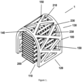
Figure 1 . A view of the biodegradable 3D printed structure for bone augmentation without filling -
Figure 2 . Front view of the biodegradable 3D printed structure for bone augmentation -
Figure 3 . Lateral view of the biodegradable 3D printed structure for bone augmentation -
- 1 - biodegradable 3D printed structure for bone augmentation
- 100 - outer structure
- 110 - front side of the outer structure
- 120 - lateral side of the outer structure
- 130 - rear side of the outer structure
- 140 - inner tunnel vault
- 150 - "X"-shaped connection
- 200 - inner structure
- 210 - perforated plate
- 220 - internal raster
- 230 - strut
- Biodegradable 3D printed structure for
bone augmentation 1 disclosed herein is printed with a 3D printer using polylactide (PLA). Essentially, both parts of this structure, theouter structure 100 and theinner structure 200 are printed at the same time. Afterstructure 1 is printed, theinner structure 200 is filled with a chitosan and hydroxyapatite gel, while theouter structure 100 is coated with a chitosan and hydroxyapatite composite. Theouter structure 100 is printed in the shape of a tunnel in the form of an elongated projection of the letter "U" rotated 180 degrees in depth, where theouter structure 100 contains the front side of theouter structure 110 and the rear side of theouter structure 130, as well as connections in the form of the letter "X" 150 and two lateral sides of theouter structure 120, all achieved by printing on a 3D printer. The front side of theouter structure 110 and the rear side of theouter structure 130 are connected by at least five "X"-shapedconnections 150 so that when viewed from the lateral side of theouter structure 120, the interior formed by the tunnel shape of theouter structure 100 is visible. In one common embodiment dimensions of the outer structure are as follows: the width of theouter structure 100 in the part that forms the base of the letter "U" rotated 180 degrees is 20 mm, the height of theouter structure 100 is 20 mm, wherein the distance between the front side of theouter structure 110 and the rear side of theouter structure 130 is 20 mm. Theinner structure 200 itself contains multipleperforated plates 210 printed one above the other so that they are bordered by theouter structure 100. Theinner structure 200 also contains aninternal raster 220 and struts 230, wherein thestruts 230 support and hold the gap between the twoperforated plates 210, so that theinternal raster 220 is formed. The inner structure extends longitudinally along the axis of the tunnel, with the axis orientation from the front of theouter structure 110 towards the rear of theouter structure 130, has a width of at least 2/3 of the width of the tunnel and extends through the entire height and length of theouter structure 100 and is firmly connected to the inner vault of thetunnel 140 which is formed by theouter structure 100.
Claims (4)
- Biodegradable 3D printed structure for bone augmentation (1) characterized in that it comprises an inner structure (100) and an outer structure (200) wherein both structures are printed from polylactide (PLA) and wherein the inner structure (200) is filled with a chitosan and hydroxyapatite gel, while the outer structure (100) is coated with a chitosan and hydroxyapatite composite.
- Biodegradable 3D printed structure for bone augmentation (1) characterized in that the outer structure (100) has a tunnel-like shape resembling an elongated projection of the letter "U" rotated 180 degrees in depth, wherein the outer structure (100) comprises the front side of the outer structure (110) and the rear side of the outer structure (130), "X"-shaped connections (150) as well as two lateral sides of the outer structure (120), wherein the front side of the outer structure (110) and the rear side of the outer structure (130) are connected by at least five connections in the shape of the letter "X" (150), so that when viewed from the lateral side of the outer structure (120), the interior formed by the tunnel shape of the outer structure (100) is visible.
- Biodegradable 3D printed structure for bone augmentation (1) as claimed in claim 2 characterized in that the dimensions of the outer structure are as follows: the width of the outer structure (100) in the part that forms the base of the letter "U" rotated by 180 degrees is 20 mm, the height of the outer structure (100) is 20 mm as well, wherein the distance between the front side of the outer structure (110) and the rear side of the outer structure (130) is 20 mm.
- Biodegradable 3D printed structure for bone augmentation (1) as claimed in any of the previous patent claims characterized in that the inner structure (200) comprises multiple perforated plates (210) printed one above the other so that they are bordered by the outer structure (100), furthermore the inner structure (200) comprises both an internal raster (220) and struts (230), wherein the struts (230) support and hold the gap between the two perforated plates (210), so that the internal raster (220) is formed, whereby the inner structure extends longitudinally along the axis of the tunnel, with the axis orientation from the front side of the outer structure (110) to the rear side of the outer structure (130), has a width of at least 2/3 of the width of the tunnel and extends through the entire height and length of the outer structure (100) and is firmly connected to the inner vault of the tunnel (140) formed by the outer structure.
Applications Claiming Priority (2)
| Application Number | Priority Date | Filing Date | Title |
|---|---|---|---|
| HRP20231599AA HRP20231599A1 (en) | 2023-12-07 | 2023-12-07 | BIODEGRADABLE 3D PRINTED STRUCTURE FOR BONE AUGMENTATION |
| PCT/HR2023/050005 WO2025120338A1 (en) | 2023-12-07 | 2023-12-13 | Biodegradable 3d printed structure for bone augmentation |
Publications (1)
| Publication Number | Publication Date |
|---|---|
| EP4566637A1 true EP4566637A1 (en) | 2025-06-11 |
Family
ID=89983317
Family Applications (1)
| Application Number | Title | Priority Date | Filing Date |
|---|---|---|---|
| EP24020049.3A Pending EP4566637A1 (en) | 2023-12-07 | 2024-02-02 | Biodegradable 3d printed structure for bone augmentation |
Country Status (1)
| Country | Link |
|---|---|
| EP (1) | EP4566637A1 (en) |
Citations (1)
| Publication number | Priority date | Publication date | Assignee | Title |
|---|---|---|---|---|
| US20220403327A1 (en) | 2019-11-08 | 2022-12-22 | Polyvalor, Limited Partnership | Customizable 3d cell culture system comprising hydrogel-embedded cells and uses thereof |
-
2024
- 2024-02-02 EP EP24020049.3A patent/EP4566637A1/en active Pending
Patent Citations (1)
| Publication number | Priority date | Publication date | Assignee | Title |
|---|---|---|---|---|
| US20220403327A1 (en) | 2019-11-08 | 2022-12-22 | Polyvalor, Limited Partnership | Customizable 3d cell culture system comprising hydrogel-embedded cells and uses thereof |
Non-Patent Citations (3)
| Title |
|---|
| LIU JIE ET AL: "Mechanisms of Guided Bone Regeneration: A Review", THE OPEN DENTISTRY JOURNAL, vol. 8, no. 1, 16 May 2014 (2014-05-16), pages 56 - 65, XP093183973, ISSN: 1874-2106, DOI: 10.2174/1874210601408010056 * |
| NAZEER MUHAMMAD ANWAAR ET AL: "3D printed poly(lactic acid) scaffolds modified with chitosan and hydroxyapatite for bone repair applications", MATERIALS TODAY COMMUNICATIONS, vol. 25, 1 December 2020 (2020-12-01), GB, pages 101515, XP093183427, ISSN: 2352-4928, DOI: 10.1016/j.mtcomm.2020.101515 * |
| TOLEDANO-OSORIO MANUEL ET AL: "Alveolar Bone Ridge Augmentation Using Polymeric Membranes: A Systematic Review and Meta-Analysis", POLYMERS, vol. 13, no. 7, 6 April 2021 (2021-04-06), CH, pages 1172, XP093183971, ISSN: 2073-4360, DOI: 10.3390/polym13071172 * |
Similar Documents
| Publication | Publication Date | Title |
|---|---|---|
| Linde et al. | Creation of new bone by an osteopromotive membrane technique: an experimental study in rats | |
| US8353967B2 (en) | Self-supporting collagen tunnel for guided tissue regeneration and method of using same | |
| Dougherty et al. | The natural history of alloplastic implants in orbital floor reconstruction: an animal model | |
| Maas et al. | Implants in rhinoplasty | |
| EP0466820B1 (en) | Articles for treating periodontal disease and bone defects | |
| EP2959862B1 (en) | Perforated membrane for guided bone and tissue regeneration | |
| KR20080084808A (en) | Maxillary facial bone reinforcement using rhPDGF-BB and biocompatible matrix | |
| TW201215369A (en) | Spinal implant structure and method for manufacturing the same | |
| WO2014079178A1 (en) | Jawbone prosthesis | |
| Ayari | The use of periodontal membranes in the field of periodontology: spotlight on collagen membranes. | |
| Grecchi et al. | Reconstruction of the zygomatic bone with smartbone®: Case report | |
| JP2026035864A (en) | Multilayer collagen structure | |
| Uysal et al. | Comparison of the biological activities of high-density porous polyethylene implants and oxidized regenerated cellulose-wrapped diced cartilage grafts | |
| US5607689A (en) | Latex element designed to achieve guided tissue regeneration in periodontal dental therapy | |
| EP4566637A1 (en) | Biodegradable 3d printed structure for bone augmentation | |
| CN112386345B (en) | Alveolar bone increment support system | |
| WO2025120338A1 (en) | Biodegradable 3d printed structure for bone augmentation | |
| CN211561236U (en) | Bone defect repair support | |
| Fontana et al. | Freeze‐dried dura mater for guided tissue regeneration in post‐extraction dental implants: a clinical and histologic study | |
| US9833544B2 (en) | Biphasic collagen membrane or capsule for guided tissue regeneration | |
| CN108283732B (en) | Bone grafting composite material for alveolar bone increment | |
| US20250331961A1 (en) | Titanium device for guided bone regeneration, and manufacturing process | |
| CN219022453U (en) | Oral bone tissue repair system | |
| EP3309636A1 (en) | Method for producing an occlusive barrier for bone regeneration and occlusive barrier obtained by means of said method | |
| CN112842623A (en) | Method for modifying surface of hydroxyapatite by 3D (three-dimensional) bioprinting degradable artificial rib |
Legal Events
| Date | Code | Title | Description |
|---|---|---|---|
| PUAI | Public reference made under article 153(3) epc to a published international application that has entered the european phase |
Free format text: ORIGINAL CODE: 0009012 |
|
| STAA | Information on the status of an ep patent application or granted ep patent |
Free format text: STATUS: THE APPLICATION HAS BEEN PUBLISHED |
|
| AK | Designated contracting states |
Kind code of ref document: A1 Designated state(s): AL AT BE BG CH CY CZ DE DK EE ES FI FR GB GR HR HU IE IS IT LI LT LU LV MC ME MK MT NL NO PL PT RO RS SE SI SK SM TR |
|
| STAA | Information on the status of an ep patent application or granted ep patent |
Free format text: STATUS: THE APPLICATION IS DEEMED TO BE WITHDRAWN |