EP4553152A1 - Optimized method for amplification and production at scale for regulatory t cells - Google Patents

Optimized method for amplification and production at scale for regulatory t cells Download PDF

Info

Publication number
EP4553152A1
EP4553152A1 EP23834906.2A EP23834906A EP4553152A1 EP 4553152 A1 EP4553152 A1 EP 4553152A1 EP 23834906 A EP23834906 A EP 23834906A EP 4553152 A1 EP4553152 A1 EP 4553152A1
Authority
EP
European Patent Office
Prior art keywords
tregs
expansion
cells
added
scale production
Prior art date
Legal status (The legal status is an assumption and is not a legal conclusion. Google has not performed a legal analysis and makes no representation as to the accuracy of the status listed.)
Pending
Application number
EP23834906.2A
Other languages
German (de)
French (fr)
Other versions
EP4553152A4 (en
Inventor
Haibo Yu
Zhiguang Zhou
Current Assignee (The listed assignees may be inaccurate. Google has not performed a legal analysis and makes no representation or warranty as to the accuracy of the list.)
Central South University
Original Assignee
Central South University
Priority date (The priority date is an assumption and is not a legal conclusion. Google has not performed a legal analysis and makes no representation as to the accuracy of the date listed.)
Filing date
Publication date
Application filed by Central South University filed Critical Central South University
Publication of EP4553152A1 publication Critical patent/EP4553152A1/en
Publication of EP4553152A4 publication Critical patent/EP4553152A4/en
Pending legal-status Critical Current

Links

Images

Classifications

    • CCHEMISTRY; METALLURGY
    • C12BIOCHEMISTRY; BEER; SPIRITS; WINE; VINEGAR; MICROBIOLOGY; ENZYMOLOGY; MUTATION OR GENETIC ENGINEERING
    • C12NMICROORGANISMS OR ENZYMES; COMPOSITIONS THEREOF; PROPAGATING, PRESERVING, OR MAINTAINING MICROORGANISMS; MUTATION OR GENETIC ENGINEERING; CULTURE MEDIA
    • C12N5/00Undifferentiated human, animal or plant cells, e.g. cell lines; Tissues; Cultivation or maintenance thereof; Culture media therefor
    • C12N5/06Animal cells or tissues; Human cells or tissues
    • C12N5/0602Vertebrate cells
    • C12N5/0634Cells from the blood or the immune system
    • C12N5/0636T lymphocytes
    • C12N5/0637Immunosuppressive T lymphocytes, e.g. regulatory T cells or Treg
    • CCHEMISTRY; METALLURGY
    • C12BIOCHEMISTRY; BEER; SPIRITS; WINE; VINEGAR; MICROBIOLOGY; ENZYMOLOGY; MUTATION OR GENETIC ENGINEERING
    • C12NMICROORGANISMS OR ENZYMES; COMPOSITIONS THEREOF; PROPAGATING, PRESERVING, OR MAINTAINING MICROORGANISMS; MUTATION OR GENETIC ENGINEERING; CULTURE MEDIA
    • C12N2500/00Specific components of cell culture medium
    • C12N2500/30Organic components
    • C12N2500/32Amino acids
    • CCHEMISTRY; METALLURGY
    • C12BIOCHEMISTRY; BEER; SPIRITS; WINE; VINEGAR; MICROBIOLOGY; ENZYMOLOGY; MUTATION OR GENETIC ENGINEERING
    • C12NMICROORGANISMS OR ENZYMES; COMPOSITIONS THEREOF; PROPAGATING, PRESERVING, OR MAINTAINING MICROORGANISMS; MUTATION OR GENETIC ENGINEERING; CULTURE MEDIA
    • C12N2500/00Specific components of cell culture medium
    • C12N2500/30Organic components
    • C12N2500/44Thiols, e.g. mercaptoethanol
    • CCHEMISTRY; METALLURGY
    • C12BIOCHEMISTRY; BEER; SPIRITS; WINE; VINEGAR; MICROBIOLOGY; ENZYMOLOGY; MUTATION OR GENETIC ENGINEERING
    • C12NMICROORGANISMS OR ENZYMES; COMPOSITIONS THEREOF; PROPAGATING, PRESERVING, OR MAINTAINING MICROORGANISMS; MUTATION OR GENETIC ENGINEERING; CULTURE MEDIA
    • C12N2500/00Specific components of cell culture medium
    • C12N2500/60Buffer, e.g. pH regulation, osmotic pressure
    • CCHEMISTRY; METALLURGY
    • C12BIOCHEMISTRY; BEER; SPIRITS; WINE; VINEGAR; MICROBIOLOGY; ENZYMOLOGY; MUTATION OR GENETIC ENGINEERING
    • C12NMICROORGANISMS OR ENZYMES; COMPOSITIONS THEREOF; PROPAGATING, PRESERVING, OR MAINTAINING MICROORGANISMS; MUTATION OR GENETIC ENGINEERING; CULTURE MEDIA
    • C12N2501/00Active agents used in cell culture processes, e.g. differentation
    • C12N2501/04Immunosuppressors, e.g. cyclosporin, tacrolimus
    • CCHEMISTRY; METALLURGY
    • C12BIOCHEMISTRY; BEER; SPIRITS; WINE; VINEGAR; MICROBIOLOGY; ENZYMOLOGY; MUTATION OR GENETIC ENGINEERING
    • C12NMICROORGANISMS OR ENZYMES; COMPOSITIONS THEREOF; PROPAGATING, PRESERVING, OR MAINTAINING MICROORGANISMS; MUTATION OR GENETIC ENGINEERING; CULTURE MEDIA
    • C12N2501/00Active agents used in cell culture processes, e.g. differentation
    • C12N2501/20Cytokines; Chemokines
    • C12N2501/23Interleukins [IL]
    • C12N2501/2302Interleukin-2 (IL-2)
    • CCHEMISTRY; METALLURGY
    • C12BIOCHEMISTRY; BEER; SPIRITS; WINE; VINEGAR; MICROBIOLOGY; ENZYMOLOGY; MUTATION OR GENETIC ENGINEERING
    • C12NMICROORGANISMS OR ENZYMES; COMPOSITIONS THEREOF; PROPAGATING, PRESERVING, OR MAINTAINING MICROORGANISMS; MUTATION OR GENETIC ENGINEERING; CULTURE MEDIA
    • C12N2501/00Active agents used in cell culture processes, e.g. differentation
    • C12N2501/50Cell markers; Cell surface determinants
    • C12N2501/505CD4; CD8
    • CCHEMISTRY; METALLURGY
    • C12BIOCHEMISTRY; BEER; SPIRITS; WINE; VINEGAR; MICROBIOLOGY; ENZYMOLOGY; MUTATION OR GENETIC ENGINEERING
    • C12NMICROORGANISMS OR ENZYMES; COMPOSITIONS THEREOF; PROPAGATING, PRESERVING, OR MAINTAINING MICROORGANISMS; MUTATION OR GENETIC ENGINEERING; CULTURE MEDIA
    • C12N2501/00Active agents used in cell culture processes, e.g. differentation
    • C12N2501/50Cell markers; Cell surface determinants
    • C12N2501/599Cell markers; Cell surface determinants with CD designations not provided for elsewhere
    • CCHEMISTRY; METALLURGY
    • C12BIOCHEMISTRY; BEER; SPIRITS; WINE; VINEGAR; MICROBIOLOGY; ENZYMOLOGY; MUTATION OR GENETIC ENGINEERING
    • C12NMICROORGANISMS OR ENZYMES; COMPOSITIONS THEREOF; PROPAGATING, PRESERVING, OR MAINTAINING MICROORGANISMS; MUTATION OR GENETIC ENGINEERING; CULTURE MEDIA
    • C12N2509/00Methods for the dissociation of cells, e.g. specific use of enzymes
    • YGENERAL TAGGING OF NEW TECHNOLOGICAL DEVELOPMENTS; GENERAL TAGGING OF CROSS-SECTIONAL TECHNOLOGIES SPANNING OVER SEVERAL SECTIONS OF THE IPC; TECHNICAL SUBJECTS COVERED BY FORMER USPC CROSS-REFERENCE ART COLLECTIONS [XRACs] AND DIGESTS
    • Y02TECHNOLOGIES OR APPLICATIONS FOR MITIGATION OR ADAPTATION AGAINST CLIMATE CHANGE
    • Y02ATECHNOLOGIES FOR ADAPTATION TO CLIMATE CHANGE
    • Y02A50/00TECHNOLOGIES FOR ADAPTATION TO CLIMATE CHANGE in human health protection, e.g. against extreme weather
    • Y02A50/30Against vector-borne diseases, e.g. mosquito-borne, fly-borne, tick-borne or waterborne diseases whose impact is exacerbated by climate change

Definitions

  • the present disclosure belongs to the field of cell therapy, and in particular relates to an optimized method for expansion and large-scale production of regulatory T cells (Tregs).
  • Tregs Regulatory T cells
  • Tregs are a special T cell subset that maintains the dynamic immune homeostasis by inhibiting and regulating effector T cells. Since the isolation and identification of Tregs by Japanese scholars at the end of the last century, Tregs have become a hot topic in the research of immunity in recent years. A variety of clinical trials related to Tregs have been initiated outside China. The representative Treg therapy for graft versus host disease has confirmed the safety and efficacy of Treg therapy in treating immune diseases. However, Treg therapy for immune diseases is still blank in China. Treg therapy can restore the balance of the immune system to prevent the further damage of autoimmune responses to organs and tissues and improve or even reverse a disease state, thereby fundamentally curing an immune disease.
  • Tregs are mostly Naive T cells because these Tregs are not activated by antigens.
  • Tregs expanded in vitro have excellent functions and low immunogenicity, which brings new hope for the treatment of immune diseases.
  • the team of the present disclosure has established an in vitro isolation, expansion, and functional identification method for cord blood-derived Tregs that was approved as a national invention patent (patent No.: 201710613737.4 ), has carried out the clinical study of treating autoimmune diabetes with cord blood-derived Tregs for the first time internationally, and has preliminarily clarified the safety and efficacy of cord blood-derived Tregs in the treatment of autoimmune diabetes.
  • An optimized method for expansion and large-scale production of Tregs is provided, including the following steps:
  • the cord blood-derived Treg-containing suspension is prepared through the following steps: isolating a peripheral blood mononuclear cell (PBMC) layer from cord blood, removing red blood cells with a red blood cell lysis buffer, washing, and counting; and conducting centrifugation, removing a supernatant, and resuspending.
  • PBMC peripheral blood mononuclear cell
  • the resuspending includes the following steps: after the centrifugation is conducted and the supernatant is removed, resuspending cells with an Easy Buffer to produce 0.5 mL to 6 mL of a suspension with a cell concentration of 5 ⁇ 10 7 cells/mL.
  • the CD4 + CD25 + CD127 - Tregs are magnetically sorted out through the following specific steps: after the cord blood-derived Treg-containing suspension is transferred into the sterile tube matching with the magnetic pole, adding CD25 Positive Selection Cocktail, and incubating for 5 min; vortexing Releasable Rapidsphere for 30 s or more until magnetic bead aggregates disappear; adding the Releasable Rapidsphere; adding a CD4 + T cell enrichment antibody mixture, and incubating for 5 min; adding an Easy Buffer to 10 mL, and gently mixing 2-3 times; placing the sterile tube on the magnetic pole, and incubating for 10 min; preparing a centrifuge tube to collect CD25 - cells, and pouring a liquid in the sterile tube into the centrifuge tube under the magnetic pole; removing the sterile tube from the magnetic pole, adding 10 mL of an Easy Buffer to the sterile tube, gently mixing 2-3 times,
  • an amount of the CD25 Positive Selection Cocktail added is the resuspension volume ⁇ 50 ⁇ L
  • an amount of the Releasable Rapidsphere added is the resuspension volume ⁇ 30 ⁇ L
  • an amount of the CD4 + T cell enrichment antibody mixture added is the resuspension volume ⁇ 50 ⁇ L
  • an amount of the magnetic bead-removing buffer added is the resuspension volume ⁇ 100 ⁇ L
  • an amount of the CD127 high -removing antibody mixture added is the resuspension volume ⁇ 50 ⁇ L.
  • the Tregs carrying magnetic beads are added to a 48-well plate at a concentration of 2 ⁇ 10 5 to 8 ⁇ 10 5 /mL per well, and 0.5 mL of the expansion culture medium is added to each well for culture.
  • the expansion culture medium includes 70.54% to 85.27% of a serum-free medium, 2.5% to 5% of a 4-hydroxyethylpiperazine ethanesulfonic acid buffer, 1% to 2% of a penicillin-streptomycin solution, 1% to 2% of L-glutamine, 50 ⁇ mol/L to 100 ⁇ mol/L of 2-mercaptoethanol, 0 U/mL to 1,000 U/mL of recombinant interleukin-2, 50 nmol/L to 200 nmol/L of rapamycin, and 10% to 20% of AB serum.
  • both the primary culture and the subculture are conducted at 36°C to 38°C and 4% to 6% CO 2 .
  • 0.5 mL to 1 mL of the expansion culture medium and 100 U/mL to 400 U/mL of rhIL-2 are added to cells in each well, and then a culture is conducted.
  • the subculture 0.5 mL of a medium in each well is removed, the remaining medium is thoroughly mixed with cells, cells in each well are passaged to 2 wells, and the medium is supplemented to 1 mL per well; and when the subculture is conducted a third time, the anti-CD3CD28 magnetic beads are removed, and then fresh anti-CD3CD28 magnetic beads are added with a number ratio of the magnetic beads to Tregs being 1:1.
  • Tregs An optimized method for expansion and large-scale production of Tregs was provided. With the method, Tregs could be subjected to expanded culture and stored for treating immune-related diseases.
  • the expansion method was as follows:
  • Table 1 Formula of 50 mL of the expansion culture medium for cord blood-derived Tregs Reagent name Company Item No.
  • 0.5 mL to 1 mL of the expansion culture medium and 100 U/mL to 400 U/mL of rhIL-2 were added to cells in each well, and cells were cultured at 37°C and 5% CO 2 . 3.
  • a first passage was conducted as follows: 0.5 mL of the expansion culture medium was removed, the remaining expansion culture medium was thoroughly mixed with cells, and cells in each well were passaged to 2 wells.
  • An expansion culture medium including 100 U/mL to 400 U/mL of rhIL-2 was added to 1 mL, and cells were cultured at 37°C and 5% CO 2 .
  • a fourth passage was conducted as follows: 0.5 mL of the expansion culture medium was removed, the remaining expansion culture medium was thoroughly mixed with cells, and cells in each well were passaged to 2 wells.
  • An expansion culture medium including 100 U/mL to 400 U/mL of rhIL-2 was added to 1 mL, and cells were cultured at 37°C and 5% CO 2 . 7.
  • a fifth passage was conducted as follows: 0.5 mL of the expansion culture medium was removed, the remaining expansion culture medium was thoroughly mixed with cells, and cells in each well were passaged to 2 wells.
  • An expansion culture medium including 100 U/mL to 400 U/mL of rhIL-2 was added to 1 mL, and cells were cultured at 37°C and 5% CO 2 . 8.
  • a sixth passage was conducted as follows: 0.5 mL of the expansion culture medium was removed, the remaining expansion culture medium was thoroughly mixed with cells, and cells in each well were passaged to 2 wells.
  • An expansion culture medium including 100 U/mL to 400 U/mL of rhIL-2 was added to 1 mL, and cells were cultured at 37°C and 5% CO 2 .
  • a seventh passage was conducted as follows: 0.5 mL of the expansion culture medium was removed, the remaining expansion culture medium was thoroughly mixed with cells, and cells in each well were passaged to 2 wells.
  • An expansion culture medium including 100 U/mL to 400 U/mL of rhIL-2 was added to 1 mL, and cells were cultured at 37°C and 5% CO 2 . 10. The expansion was conducted according to the above method until cells stopped growing. A total culture time was about 40 d to 50 d.
  • contents of components in the expansion culture medium for Tregs were expressed in volume fractions.
  • a volume fraction refers to a volume proportion of each component.
  • the expansion culture medium for Tregs included 70.54% to 85.27% of a serum-free medium, 2.5% to 5% of a 4-hydroxyethylpiperazine ethanesulfonic acid buffer, 1% to 2% of a penicillin-streptomycin solution, 1% to 2% of L-glutamine, 50 ⁇ mol/L to 100 ⁇ mol/L of 2-mercaptoethanol, 0 U/mL to 1,000 U/mL of recombinant interleukin-2, 50 nmol/L to 200 nmol/L of rapamycin, and 10% to 20% of AB serum.
  • the serum-free medium could be X-VIVO 15 w/o Gent or Phenol Red of Lonza, Item No.: 04-744Q.
  • the 4-hydroxyethylpiperazine ethanesulfonic acid buffer could be a Hepes buffer of Hyclone, Item No.: SH3023701.
  • the penicillin-streptomycin solution could be PENICILLIN STREPTOMYCIN of Gibco, 15140122.
  • the L-glutamine could be an L-glutamine solution of sigma, Item No.: G7513-100ML.
  • the 2-mercaptoethanol could be 1000X 2-Mercaptoethanol of Millipore, Item No.: ES-007-E.
  • the recombinant interleukin-2 could be rhrhIL-2 of R&D, Item No.: 202-IL-010.
  • the rapamycin could be rapamycin of sigma, Item No.: R8781-200UL. Of course, the same products produced by other companies could also be adopted.
  • the AB serum was from Gemini, USA, Item No.: 100-512.
  • a preparation method of the above expansion culture medium slightly varied according to the specific use.
  • the components of the expansion culture medium could be thoroughly mixed in advance and then directly added to a well plate, or the components other than the recombinant interleukin-2 for the expansion culture solution could be mixed to produce a premix and then the recombinant interleukin-2 was added.
  • the premix was supplemented to each well. For example, 0.5 mL of the premix was supplemented to 1 mL of a solution in each well. In this case, the corresponding recombinant interleukin-2 should be added based on the amount of the expansion culture medium: 1 mL.
  • An initial number of Tregs was 1 ⁇ 10 6 .
  • a number of expanded Tregs was counted. The counting was conducted through the following specific steps: 10 ⁇ L of a cell suspension was taken and added to an EP tube, 90 ⁇ L of trypan blue was added to the EP tube, and thorough mixing was conducted. 10 ⁇ L of a resulting mixture was taken and added to a hemacytometer, and quickly counted: (sum of numbers in four corners/4) ⁇ 10 5 cells/mL * dilution factor. For cells on lines, cells on the upper and left lines were counted, and cells on the lower and right lines were not counted.
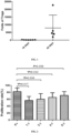
  • An expansion fold of Tregs was 7834.4 ⁇ 8430.4 on day 40 to day 50 of the expansion of Tregs, and an expansion fold of Tregs was 150.6 ⁇ 70.3 on day 18 of the expansion of Tregs, as shown in FIG. 1 .
  • the method of the present disclosure adopts one-step sorting and a new kit, does not require centrifugation, and has a very prominent cell expansion effect (shortened time and high cell viability).
  • a very prominent cell expansion effect Generally, with the extension of an expansion time, an expansion effect is gradually deteriorated.
  • the present disclosure not only increases an effective expansion time to 40 d to 50 d from 18 d, but also will not make an expansion effect gradually deteriorated.
  • the method of the present disclosure greatly increases the expansion generation number and the quantity of Tregs, and can meet the needs of clinical patients.
Landscapes

  • Health & Medical Sciences (AREA)
  • Engineering & Computer Science (AREA)
  • Life Sciences & Earth Sciences (AREA)
  • Immunology (AREA)
  • Biomedical Technology (AREA)
  • Bioinformatics & Cheminformatics (AREA)
  • Zoology (AREA)
  • Organic Chemistry (AREA)
  • Chemical & Material Sciences (AREA)
  • Genetics & Genomics (AREA)
  • Biotechnology (AREA)
  • Wood Science & Technology (AREA)
  • Microbiology (AREA)
  • Hematology (AREA)
  • Biochemistry (AREA)
  • General Engineering & Computer Science (AREA)
  • General Health & Medical Sciences (AREA)
  • Cell Biology (AREA)
  • Micro-Organisms Or Cultivation Processes Thereof (AREA)

Abstract

The present disclosure provides an optimized method for expansion and large-scale production of regulatory T cells (Tregs), including the following steps: preparing a cord blood-derived Treg-containing suspension; transferring the cord blood-derived Treg-containing suspension into a sterile tube matching with a magnetic pole, and magnetically sorting CD4+CD25+CD127- Tregs out; and adding an expansion culture medium to Tregs carrying magnetic beads, conducting a primary culture for 1 d to 2 d, and conducting a subculture once every 1 d to 3 d, where a total culture time is 40 d to 50 d. Through one-step sorting, the method of the present disclosure achieves a very prominent cell expansion effect (shortened time and high cell viability), and increases an effective expansion time to 40 d to 50 d from 18 d. In addition, an expansion effect will not be gradually deteriorated. Thus, the method of the present disclosure greatly increases the expansion generation number and the quantity of Tregs, and can meet the needs of clinical patients.

Description

    TECHNICAL FIELD
  • The present disclosure belongs to the field of cell therapy, and in particular relates to an optimized method for expansion and large-scale production of regulatory T cells (Tregs).
  • BACKGROUND TECHNOLOGY
  • Regulatory T cells (Tregs) are a special T cell subset that maintains the dynamic immune homeostasis by inhibiting and regulating effector T cells. Since the isolation and identification of Tregs by Japanese scholars at the end of the last century, Tregs have become a hot topic in the research of immunity in recent years. A variety of clinical trials related to Tregs have been initiated outside China. The representative Treg therapy for graft versus host disease has confirmed the safety and efficacy of Treg therapy in treating immune diseases. However, Treg therapy for immune diseases is still blank in China. Treg therapy can restore the balance of the immune system to prevent the further damage of autoimmune responses to organs and tissues and improve or even reverse a disease state, thereby fundamentally curing an immune disease.
  • More and more studies have shown that the cord blood is rich in Tregs, which are mostly Naive T cells because these Tregs are not activated by antigens. Tregs expanded in vitro have excellent functions and low immunogenicity, which brings new hope for the treatment of immune diseases. At an early stage, the team of the present disclosure has established an in vitro isolation, expansion, and functional identification method for cord blood-derived Tregs that was approved as a national invention patent (patent No.: 201710613737.4 ), has carried out the clinical study of treating autoimmune diabetes with cord blood-derived Tregs for the first time internationally, and has preliminarily clarified the safety and efficacy of cord blood-derived Tregs in the treatment of autoimmune diabetes. In 2019 and 2020, the Juvenile Diabetes Cure Alliance (JDCA) published the annual reports on the global technologies for treating type 1 diabetes, respectively. The "cord blood-derived Treg" therapy technology has continuously been rated as the leading technology worldwide. Globally, there is currently no clinical trial for using cord blood-derived Tregs to treat an immune disease. Tregs can be extracted from the cord blood, expanded, and then used, which takes 14 d to 21 d. A cell expansion fold each time is about 100, which can only meet the needs of 1 to 2 patients. Therefore, it is urgent to establish an optimized method for in vitro expansion of Tregs to increase the expansion generation number and quantity of Tregs, thereby meeting the needs of clinical patients.
  • CONTENT OF THE INVENTION
  • A technical problem to be solved by the present disclosure: An optimized method for in vitro expansion of Tregs is provided to overcome the deficiencies and shortcomings mentioned in the above background.
  • In order to solve the above technical problem, the present disclosure provides the following technical solutions:
    An optimized method for expansion and large-scale production of Tregs is provided, including the following steps:
    1. (1) preparing a cord blood-derived Treg-containing suspension;
    2. (2) transferring the cord blood-derived Treg-containing suspension into a sterile tube matching with a magnetic pole, and magnetically sorting CD4+CD25+CD127- Tregs out; and
    3. (3) adding an expansion culture medium to Tregs carrying magnetic beads, conducting a primary culture for 1 d to 2 d, and conducting a subculture once every 1 d to 3 d, where a total culture time is 40 d to 50 d or more.
  • In the optimized method for expansion and large-scale production of Tregs, preferably, in the step (1), the cord blood-derived Treg-containing suspension is prepared through the following steps: isolating a peripheral blood mononuclear cell (PBMC) layer from cord blood, removing red blood cells with a red blood cell lysis buffer, washing, and counting; and conducting centrifugation, removing a supernatant, and resuspending.
  • More preferably, the resuspending includes the following steps: after the centrifugation is conducted and the supernatant is removed, resuspending cells with an Easy Buffer to produce 0.5 mL to 6 mL of a suspension with a cell concentration of 5 × 10⁷ cells/mL.
  • In the optimized method for expansion and large-scale production of Tregs, preferably, in the step (2), the CD4+CD25+CD127- Tregs are magnetically sorted out through the following specific steps: after the cord blood-derived Treg-containing suspension is transferred into the sterile tube matching with the magnetic pole, adding CD25 Positive Selection Cocktail, and incubating for 5 min; vortexing Releasable Rapidsphere for 30 s or more until magnetic bead aggregates disappear; adding the Releasable Rapidsphere; adding a CD4+T cell enrichment antibody mixture, and incubating for 5 min; adding an Easy Buffer to 10 mL, and gently mixing 2-3 times; placing the sterile tube on the magnetic pole, and incubating for 10 min; preparing a centrifuge tube to collect CD25- cells, and pouring a liquid in the sterile tube into the centrifuge tube under the magnetic pole; removing the sterile tube from the magnetic pole, adding 10 mL of an Easy Buffer to the sterile tube, gently mixing 2-3 times, placing the sterile tube on the magnetic pole, and incubating for 5 min; adding an Easy Buffer to the initial resuspension volume with all cells on a tube wall rinsed off; adding a magnetic bead-removing buffer; adding a CD127high-removing antibody mixture, and incubating for 5 min; adding an Easy Buffer to 10 mL, and thoroughly mixing 2-3 times; placing the sterile tube on the magnetic pole, and incubating for 5 min; and preparing a centrifuge tube to collect Tregs, and pouring a liquid in the sterile tube into the prepared centrifuge tube under the magnetic pole.
  • More preferably, an amount of the CD25 Positive Selection Cocktail added is the resuspension volume × 50 µL, an amount of the Releasable Rapidsphere added is the resuspension volume × 30 µL, an amount of the CD4+T cell enrichment antibody mixture added is the resuspension volume × 50 µL, an amount of the magnetic bead-removing buffer added is the resuspension volume × 100 µL, and an amount of the CD127high-removing antibody mixture added is the resuspension volume × 50 µL.
  • In the optimized method for expansion and large-scale production of Tregs, preferably, in the step (3), the Tregs carrying magnetic beads are added to a 48-well plate at a concentration of 2 × 10⁵ to 8 × 10⁵/mL per well, and 0.5 mL of the expansion culture medium is added to each well for culture.
  • Preferably, in the step (3), in volume fractions, the expansion culture medium includes 70.54% to 85.27% of a serum-free medium, 2.5% to 5% of a 4-hydroxyethylpiperazine ethanesulfonic acid buffer, 1% to 2% of a penicillin-streptomycin solution, 1% to 2% of L-glutamine, 50 µmol/L to 100 µmol/L of 2-mercaptoethanol, 0 U/mL to 1,000 U/mL of recombinant interleukin-2, 50 nmol/L to 200 nmol/L of rapamycin, and 10% to 20% of AB serum.
  • Preferably, in the step (3), both the primary culture and the subculture are conducted at 36°C to 38°C and 4% to 6% CO2.
  • More preferably, on day 1 to day 2 after inoculation, 0.5 mL to 1 mL of the expansion culture medium and 100 U/mL to 400 U/mL of rhIL-2 are added to cells in each well, and then a culture is conducted.
  • More preferably, during the subculture, 0.5 mL of a medium in each well is removed, the remaining medium is thoroughly mixed with cells, cells in each well are passaged to 2 wells, and the medium is supplemented to 1 mL per well; and when the subculture is conducted a third time, the anti-CD3CD28 magnetic beads are removed, and then fresh anti-CD3CD28 magnetic beads are added with a number ratio of the magnetic beads to Tregs being 1:1.
  • Compared with the prior art, the present disclosure has the following beneficial effects:
    1. 1. The method of the present disclosure adopts one-step sorting and a new kit, does not require centrifugation, and has a very prominent cell expansion effect (shortened time and high cell viability). Generally, with the extension of an expansion time, an expansion effect is gradually deteriorated. However, the present disclosure not only increases an effective expansion time to 40 d to 50 d from 18 d, but also will not make an expansion effect gradually deteriorated. Thus, the method of the present disclosure greatly increases the expansion generation number and the quantity of Tregs, and can meet the needs of clinical patients.
    2. 2. Flow cytometry results of cord blood-derived Tregs produced from the expansion of the present disclosure show that, on day 18 and day 40 to day 50 of expansion, proportions of CD4+CD25+CD127- Tregs are 97.07±2.03% and 87.1±7.2%, respectively, and proportions of CD3+CD8+ Tregs are 4.69±2% and 5.2±3.6%, respectively, which meet the requirements of patients treated in clinical research. Carboxyfluorescein diacetate succinimidyl ester (CFSE) assay results show that cord blood-derived Tregs produced after the expansion can significantly inhibit the proliferation ability of aggressive T cells.
    DESCRIPTION OF THE DRAWINGS
  • To describe the technical solutions in the embodiments of the present disclosure or in the prior art clearly, the accompanying drawings required for describing the embodiments or the prior art will be briefly described below. Apparently, the accompanying drawings in the following description show some embodiments of the present disclosure, and a person of ordinary skill in the art may still derive other accompanying drawings from these accompanying drawings without creative efforts.
    • FIG. 1 shows the comparison of folds of cord blood-derived Tregs on day 18 and day 40 to day 50 of expansion; and
    • FIG. 2 shows the results of CFSE-based functional identification for cord blood-derived T cells on day 18 of expansion.
    SPECIFIC IMPLEMENTATIONS
  • In order to facilitate the understanding of the present disclosure, the present disclosure is described in detail below in conjunction with the accompanying drawings of the specification and the preferred embodiments, but the protection scope of the present disclosure is not limited to the following specific embodiments.
  • Unless otherwise defined, all technical terms used hereinafter have the same meaning as commonly understood by those skilled in the art. The technical terms used herein are merely for the purpose of describing specific embodiments, and are not intended to limit the protection scope of the present disclosure.
  • Unless otherwise specified, various raw materials, reagents, instruments, devices, etc. used in the present disclosure can be purchased from the market or can be prepared by the existing methods.
  • Embodiments:
  • An optimized method for expansion and large-scale production of Tregs was provided. With the method, Tregs could be subjected to expanded culture and stored for treating immune-related diseases. The expansion method was as follows:
  • I. Preparation of cord blood-derived Tregs
    1. 1. Sterilization of a cord blood bag and preparation of cord blood-derived autologous plasma: Cord blood in the cord blood bag was transferred into a 50 mL centrifuge tube and centrifuged under the following conditions: 20°C, 3,000 rpm, 10 min, +8, and -9. A resulting supernatant was collected and transferred to a new 50 mL centrifuge tube and further centrifuged under the following conditions: 20°C, 4,000 rpm, 10 min, +8, and -9. Resulting yellow clear plasma was collected, inactivated in a 56°C water bath for 30 min, and then placed in a refrigerator for later use. The remaining cord blood was resuspended with normal saline in the same volume as the collected plasma.
    2. 2. The cord blood resuspended in normal saline was slowly added to a lymphocyte separation solution (a volume of the cord blood : a volume of the lymphocyte separation solution = 1:1), and centrifugation was conducted under the following conditions: 20°C, *400 rcf to *1610 rcf, 25 min, +0, and -0. After the centrifugation was completed, the cord blood was layered. A PBMC layer was slowly and gently collected with a pipette, where a lymphocyte liquid should be pipetted as little as possible.
    3. 3. The PBMC layer was further centrifuged under the following conditions: 20°C, *580 rcf, 10 min, +8, and -9. 5 mL to 10 mL of a 10× red blood cell lysis buffer and 45 mL of sterilized water were taken and prepared into a red blood cell lysis buffer. After the centrifugation was completed, a resulting supernatant was removed, and the red blood cell lysis buffer was added. 2 min to 10 min later, 0.9% normal saline was added to 35 mL to stop the red blood cell lysis. Centrifugation was conducted under the following conditions: 20°C, *290 rcf, 10 min, +8, and -9. A resulting supernatant was removed. The steps of adding 0.9% normal saline to a specified volume, centrifugation, and removing a resulting supernatant were repeated 2-3 times.
    4. 4. After a supernatant produced after the final centrifugation was removed, resulting cells were loosened by flicking, 0.9% normal saline was added, and 10 µL of a resulting cell solution was taken for counting. 0.9% normal saline was added to the remaining cells to 40 mL. Centrifugation was further conducted under the following conditions: 20°C, *290 rcf, 10 min, +8, and -9. The counting was conducted through the following specific steps: The cell solution taken was added to an EP tube, 90 µL of trypan blue was added to the EP tube, and thorough mixing was conducted. 10 µL of a resulting mixture was taken and added to a hemacytometer, and quickly counted: (sum of numbers in four corners/4) × 105 cells/mL * dilution factor. For cells on lines, cells on the upper and left lines were counted, and cells on the lower and right lines were not counted.
    5. 5. After the centrifugation was completed, a resulting supernatant was removed, and cells were resuspended with an Easy Buffer to produce 0.5 mL to 6 mL of a cell suspension with a cell concentration of 5 × 10⁷ cells/mL.
    6. 6. The cell suspension produced after the resuspending was transferred into a sterile tube matching with a magnetic pole.
    7. 7. A CD25-positive selection antibody was added at an amount of the resuspension volume × 50 µL, and thorough mixing was conducted.
    8. 8. Incubation was conducted for 5 min.
    9. 9. Releasable spheres were vortexed for 30 s or more until magnetic bead aggregates disappeared.
    10. 10. The releasable spheres were added at an amount of the resuspension volume × 30 µL, and thorough mixing was conducted.
    11. 11. A CD4+T cell enrichment antibody mixture (T cell Enrichment Cocktail) was added at an amount of the resuspension volume × 50 µL, and thorough mixing was conducted.
    12. 12. Incubation was conducted for 5 min.
    13. 13. An Easy Buffer was added to 10 mL, and gentle mixing was conducted 2-3 times.
    14. 14. The sterile tube was placed on the magnetic pole without a cap.
    15. 15. Incubation was conducted for 10 min.
    16. 16. A centrifuge tube was prepared to collect CD25- cells, and a liquid in the sterile tube was poured into the centrifuge tube under the magnetic pole.
    17. 17. The sterile tube was removed from the magnetic pole, 10 mL of an Easy Buffer was added, and gentle mixing was conducted 2-3 times. The sterile tube was placed on the magnetic pole, and incubation was conducted for 5 min.
    18. 18. The steps 16 and 17 were repeated twice.
    19. 19. An Easy Buffer was added to the initial resuspension volume with all cells on a tube wall rinsed off.
    20. 20. A magnetic bead-removing buffer (Release Buffer) was added at an amount of the resuspension volume × 100 µL, and vigorous mixing was conducted 5 or more times.
    21. 21. A CD127high-removing antibody mixture (Depletion Cocktail) was added at an amount of the resuspension volume × 50 µL, and thorough mixing was conducted.
    22. 22. Incubation was conducted for 5 min.
    23. 23. An Easy Buffer was added to 10 mL, and thorough mixing was conducted 2-3 times.
    24. 24. The sterile tube was placed on the magnetic pole, and incubation was conducted for 5 min.
    25. 25. A centrifuge tube was prepared to collect Tregs, and a liquid in the sterile tube was poured into the prepared centrifuge tube under the magnetic pole to obtain CD4+CD25+CD127- Tregs.
  • The above process for acquiring CD4+CD25+CD127- Tregs could adopt a EasySep CD4+CD25+CD127- Treg sorting kit, with brand: STEMCELL Technologies and Item No.: 18063.
  • II. Expansion of Tregs

  • 1. Tregs were inoculated into a 48-well plate at 2 × 10⁵ to 8 × 10⁵/mL, 0.5 mL of an expansion culture medium including magnetic beads (a number of the magnetic beads : a number of cells = 1:1) was added per well on day 0 (a formula of the expansion culture medium was shown in Table 1), and the cells were cultured at 37°C and 5% CO2. Table 1 Formula of 50 mL of the expansion culture medium for cord blood-derived Tregs
    Reagent name Company Item No. Volume Final concentration
    X-VIVO 15 w/o Gent or Phenol Red Lonza 04-744Q 35.274ml-42.69ml 70.54%-85.27%
    Hepes Hyclone SH3023701 1.25ml-2.5ml 2.5%-5%
    PENICILLIN STREPTOMYCIN Gibco 15140122 0.5ml-1ml 1%-2%
    L-Glutamine solution sigma G7513-100ML 0.5ml-1ml 1%-2%
    1000X 2-Mercaptoethanol Millipore ES-007-E 50µl-100µl 50-100umol/L
    rhIL-2 R&D 202-IL-010 7.69µl-15.4µl 200-400U/ml
    rapamycin sigma R8781-200UL 1µl-10µl 50-200nmol/L
    AB serum Gemini 100-512 5ml-10ml 10%-20%

    2. On day 1 to day 2 after the inoculation (24 h to 48 h later), 0.5 mL to 1 mL of the expansion culture medium and 100 U/mL to 400 U/mL of rhIL-2 were added to cells in each well, and cells were cultured at 37°C and 5% CO2.
    3. On day 3 to day 4 after the inoculation, a first passage was conducted as follows: 0.5 mL of the expansion culture medium was removed, the remaining expansion culture medium was thoroughly mixed with cells, and cells in each well were passaged to 2 wells. An expansion culture medium including 100 U/mL to 400 U/mL of rhIL-2 was added to 1 mL, and cells were cultured at 37°C and 5% CO2. After the addition, there was 1 mL of the expansion culture medium in each well, and the content of rhIL-2 added was based on 1 mL of the expansion culture medium. The amounts of rhIL-2 added in other steps were the same as the amount in this step.
    4. On day 5 to day 6 after the inoculation, a second passage was conducted as follows: 0.5 mL of the expansion culture medium was removed, the remaining expansion culture medium was thoroughly mixed with cells, and cells in each well were passaged to 2 wells. An expansion culture medium including 100 U/mL to 400 U/mL of rhIL-2 was added to 1 mL, and cells were cultured at 37°C and 5% CO2.
    5. On day 7 to day 10 after the inoculation, a third passage was conducted as follows: The anti-CD3CD28 magnetic beads in a Petri dish were removed, and then washed fresh anti-CD3CD28 magnetic beads (a number of the magnetic beads : a number of Tregs = 1:1) were added. Cells were cultured at 37°C and 5% CO2.
    6. On day 10 to day 11 after the inoculation, a fourth passage was conducted as follows: 0.5 mL of the expansion culture medium was removed, the remaining expansion culture medium was thoroughly mixed with cells, and cells in each well were passaged to 2 wells. An expansion culture medium including 100 U/mL to 400 U/mL of rhIL-2 was added to 1 mL, and cells were cultured at 37°C and 5% CO2.
    7. On day 11 to day 12 after the inoculation, a fifth passage was conducted as follows: 0.5 mL of the expansion culture medium was removed, the remaining expansion culture medium was thoroughly mixed with cells, and cells in each well were passaged to 2 wells. An expansion culture medium including 100 U/mL to 400 U/mL of rhIL-2 was added to 1 mL, and cells were cultured at 37°C and 5% CO2.
    8. On day 12 to day 13 after the inoculation, a sixth passage was conducted as follows: 0.5 mL of the expansion culture medium was removed, the remaining expansion culture medium was thoroughly mixed with cells, and cells in each well were passaged to 2 wells. An expansion culture medium including 100 U/mL to 400 U/mL of rhIL-2 was added to 1 mL, and cells were cultured at 37°C and 5% CO2.
    9. On day 13 to day 14 after the inoculation, a seventh passage was conducted as follows: 0.5 mL of the expansion culture medium was removed, the remaining expansion culture medium was thoroughly mixed with cells, and cells in each well were passaged to 2 wells. An expansion culture medium including 100 U/mL to 400 U/mL of rhIL-2 was added to 1 mL, and cells were cultured at 37°C and 5% CO2.
    10. The expansion was conducted according to the above method until cells stopped growing. A total culture time was about 40 d to 50 d.
  • In this embodiment, contents of components in the expansion culture medium for Tregs were expressed in volume fractions. A volume fraction refers to a volume proportion of each component. The expansion culture medium for Tregs included 70.54% to 85.27% of a serum-free medium, 2.5% to 5% of a 4-hydroxyethylpiperazine ethanesulfonic acid buffer, 1% to 2% of a penicillin-streptomycin solution, 1% to 2% of L-glutamine, 50 µmol/L to 100 µmol/L of 2-mercaptoethanol, 0 U/mL to 1,000 U/mL of recombinant interleukin-2, 50 nmol/L to 200 nmol/L of rapamycin, and 10% to 20% of AB serum.
  • The serum-free medium could be X-VIVO 15 w/o Gent or Phenol Red of Lonza, Item No.: 04-744Q. The 4-hydroxyethylpiperazine ethanesulfonic acid buffer could be a Hepes buffer of Hyclone, Item No.: SH3023701. The penicillin-streptomycin solution could be PENICILLIN STREPTOMYCIN of Gibco, 15140122. The L-glutamine could be an L-glutamine solution of sigma, Item No.: G7513-100ML. The 2-mercaptoethanol could be 1000X 2-Mercaptoethanol of Millipore, Item No.: ES-007-E. The recombinant interleukin-2 could be rhrhIL-2 of R&D, Item No.: 202-IL-010. The rapamycin could be rapamycin of sigma, Item No.: R8781-200UL. Of course, the same products produced by other companies could also be adopted. The AB serum was from Gemini, USA, Item No.: 100-512.
  • A preparation method of the above expansion culture medium slightly varied according to the specific use. For example, when Tregs were recovered and inoculated into a plate, the components of the expansion culture medium could be thoroughly mixed in advance and then directly added to a well plate, or the components other than the recombinant interleukin-2 for the expansion culture solution could be mixed to produce a premix and then the recombinant interleukin-2 was added. Because the recombinant interleukin-2 was basically exhausted in each well during the subculture, in the passage from 1 well to 2 wells, the premix was supplemented to each well. For example, 0.5 mL of the premix was supplemented to 1 mL of a solution in each well. In this case, the corresponding recombinant interleukin-2 should be added based on the amount of the expansion culture medium: 1 mL.
  • Criteria for the above expansion culture test were as follows:
    Cells were inoculated into a 48-well plate at 1 × 10⁶ cells/well/mL, and an expansion culture medium including magnetic beads (a number of the magnetic beads : a number of Tregs = 1:1) was added. The cells were cultured in an environment at 36°C to 38°C and 4% to 6% CO2. Then the cell expansion was conducted according to the established expansion conditions. A formula of the expansion culture medium was as follows: 42.69 parts of a serum-free medium, 1.25 parts of a 4-hydroxyethylpiperazine ethanesulfonic acid buffer, 0.5 part of a penicillin-streptomycin solution, 0.5 part of L-glutamine, 0.05 part of 2-mercaptoethanol, 0.01154 part of recombinant interleukin-2, 0.001 part of rapamycin, and 5 parts of AB serum.
  • An initial number of Tregs was 1 × 10⁶. A number of expanded Tregs was counted. The counting was conducted through the following specific steps: 10 µL of a cell suspension was taken and added to an EP tube, 90 µL of trypan blue was added to the EP tube, and thorough mixing was conducted. 10 µL of a resulting mixture was taken and added to a hemacytometer, and quickly counted: (sum of numbers in four corners/4) × 105 cells/mL * dilution factor. For cells on lines, cells on the upper and left lines were counted, and cells on the lower and right lines were not counted.
  • An expansion fold of Tregs was 7834.4 ± 8430.4 on day 40 to day 50 of the expansion of Tregs, and an expansion fold of Tregs was 150.6 ± 70.3 on day 18 of the expansion of Tregs, as shown in FIG. 1.
  • Experiment 1: A phenotype of cord blood-derived Tregs was stable after expansion
    1. 1. One well of Tregs (5 × 10⁵ to 10 × 105) were taken, and 0.9% normal saline was added to 5 mL (room temperature). Centrifugation was conducted under the following conditions: 20°C, *570 rcf, 5 min, +8, and -9.
    2. 2. A phenotype of Tregs was detected with a PerFix-nc Kit (Item No.: PN B10825, Beckman). This kit included the following three buffers: buffer 1: Fixative Reagent, PN B10827-75tests-liquid; buffer 2: Permeabilizing Reagent, PN B10828-75tests-liquid; and buffer 3: Final 10×Solution, PN B10829-75tests-liquid. A supernatant produced after the centrifugation was removed by a pipette tip with 50 µL to 100 µL of a liquid left, and then 5 µL of the buffer 1 and 5 µL of an Fc Block antibody were added. Shaking was conducted (moderately), and incubation was conducted at room temperature in the dark for 15 min.
    3. 3. 5 µL of each of CD3, CD4, CD25, CD127, and CD8 antibodies was added. Shaking was conducted (moderately), and incubation was conducted at room temperature in the dark for 15 min to 30 min.
    4. 4. 3 mL of the buffer 3 diluted into a working solution (buffer 3 : water = 1:9) was added. Centrifugation was conducted under the following conditions: 20°C, *570 rcf, 5 min, +8, and -9.
    5. 5. Flow cytometry was conducted. Flow cytometry results showed that, on day 18 and day 40 to day 50 of expansion, proportions of CD4+CD25+CD127- Tregs were 97.07±2.03% and 87.1±7.2%, respectively, and proportions of CD3+CD8+ Tregs were 4.69±2% and 5.2±3.6%, respectively, which met the requirements of patients treated in clinical research.
    Experiment 2: Cord blood-derived Tregs could significantly inhibit the proliferation ability of aggressive T cells after expansion
    1. 1. CD4(+)CD25(-) aggressive T cells were recovered, and 1 × 10⁶ cells were taken.
    2. 2. The cells (1 × 10⁶ cells) were resuspended with 1 mL of a serum-free medium.
    3. 3. 1 µL of a cell trace stock solution was added per 1 mL of a cell suspension (CellTrace CFSE Cell Proliferation Kit, Item No.: C34554, Thermofisher scientific. This kit included 10 parts of single-use bottled CellTrace CFSE (component A) and 1 part of bottled dimethyl sulfoxide (DMSO) (component B). The cell trace stock solution was a mixed solution of 18 µL of DMSO and one part of bottled CellTrace CFSE prepared according the instructions of the kit for CFSE staining of cells. Details could be seen on https://www.thermofisher.com/order/catalog/product/C34554).
    4. 4. Incubation was conducted for 20 min at 37°C in the dark.
    5. 5. 5 mL of a washing solution (serum-free medium + 10% fetal bovine serum (FBS) was added.
    6. 6. Incubation was conducted for 5 min at 37°C in the dark.
    7. 7. Centrifugation was conducted under the following conditions: 20°C, *290 rcf, 10 min, +8, and -9.
    8. 8. Cells were resuspended with a medium (serum-free medium + 10% FBS) pre-warmed at 37°C.
    9. 9. One well of Tregs were taken, subjected to magnetic bead removal, counted, and centrifuged under the following conditions: 20°C, *290 rcf, 10 min, +8, and -9.
    10. 10. 1 × 10⁶ Tregs were resuspended with a medium (serum-free medium + 10% FBS) pre-warmed at 37°C.
    11. 11. CD4(+)CD25- T cells and Tregs were co-cultured in a 96-well plate with a number ratio of the CD4(+)CD25- T cells to the Tregs being 1:1, 2:1, and 4:1 (in each well, there was 200 µL in total, and 3 µL of magnetic beads was added). A CD4(+)CD25- T cell group treated with CFSE alone was set as a negative control group, and a CD4(+)CD25- T cell group treated with CFSE (magnetic bead stimulation) was set as a positive control group.
    12. 12. An inhibitory function of cord blood-derived Tregs was detected by flow cytometry.
    13. 13. Results of the CFSE experiment showed that, when CD4(+) CD25-T cells and Tregs were co-cultured in number ratios of 1, 2, 4, and 8:1, inhibitory rates for the proliferation of CD4(+) CD25- T cells were 56.5±11.6%, 60.2±16.1%, 65.4±13.4%, and 71.1±11.2%, respectively. The results indicated that the expanded cord blood-derived Tregs could significantly inhibit the proliferation ability of aggressive T cells, as shown in Tables 2 to 6 and FIG. 2.
    Table 2 Expansion folds of cord blood-derived Tregs on day 18 of expansion
    Sample Initial cell source Number of initial umbilical blood mononuclear cells (UBMCs) (10^8) Number of Tregs on day 0 (10^6) Number of Tregs on day 18 (10^8) Expansion fold (based on cell numbers)
    1 Cryopreserved 1.42 1.4 1.3 93
    2 Cryopreserved 3.25 3.4 4.96 146
    3 Fresh 2.58 1.29 3.58 283
    4 Cryopreserved 2.5 1.52 1.46 96
    5 Cryopreserved 3.2 2.25 4.6 204
    6 Fresh 2.77 3.55 4.71 133
    7 Fresh 2.33 4.6 2.7 99
    Mean ± standard deviation 2.58±0.62 2.57±1.29 3.33±1.54 150.57±70.33
    Table 3 Phenotypic identification results for cord blood-derived T cells on day 18 of expansion
    Sample CD3+CD4+CD25+CD127-Tregs(%) CD8+T cells(%)
    1 93.4 7.58
    2 98.2 5.93
    3 99 6.56
    4 96.2 2.71
    5 95.8 3.05
    6 98.7 2.77
    7 98.2 4.23
    Mean ± standard deviation 97.07±2.03 4..69±2.00
    Table 4 Expansion folds of cord blood-derived T cells on day 40 to day 50 of expansion
    Sample Initial cell source Number of initial UBMCs (10^8) Number of Tregs on day 0 (10^6) Expansion fold (based on cell numbers)
    1 Cryopreserved 3.25 3.4 3960
    2 Fresh 2.58 1.29 22709
    3 Cryopreserved 2.5 1.52 2064
    4 Cryopreserved 3.2 2.25 5945
    5 Fresh 2.77 3.55 4494
    Mean ± standard deviation 7834.4 8430.5
    Table 5 Phenotypic identification results for cord blood-derived T cells on day 40 to day 50 of expansion
    Sample CD3+CD4+CD25+CD127-Tregs(%) CD8+T cells(%)
    1 78.9 2.49
    2 89.8 3.74
    3 92.6 9.28
    Mean ± standard deviation 87.1±7.2 5.2±3.6
    Table 6 Results of CFSE-based functional identification for cord blood-derived T cells on day 40 to day 50 of expansion
    1 2 3 Mean ± standard deviation
    P+ 70.4 87.6 88.6 82.2±10.2
    1:1 44.6 67.8 57.0 56.5±11.6
    2:1 42.7 74.4 63.4 60.2±16.1
    4:1 50.8 77.0 68.5 65.4±13.4
    8:1 59.2 81.4 72.6 71.1±11.2
  • In summary, the method of the present disclosure adopts one-step sorting and a new kit, does not require centrifugation, and has a very prominent cell expansion effect (shortened time and high cell viability). Generally, with the extension of an expansion time, an expansion effect is gradually deteriorated. However, the present disclosure not only increases an effective expansion time to 40 d to 50 d from 18 d, but also will not make an expansion effect gradually deteriorated. Thus, the method of the present disclosure greatly increases the expansion generation number and the quantity of Tregs, and can meet the needs of clinical patients.

Claims (10)

  1. An optimized method for expansion and large-scale production of regulatory T cells (Tregs), comprising the following steps:
    (1) preparing a cord blood-derived Treg-containing suspension;
    (2) transferring the cord blood-derived Treg-containing suspension into a sterile tube matching with a magnetic pole, and magnetically sorting CD4+CD25+CD127- Tregs out; and
    (3) adding an expansion culture medium to Tregs carrying magnetic beads, conducting a primary culture for 1 d to 2 d, and conducting a subculture once every 1 d to 3 d, wherein a total culture time is 40 d to 50 d.
  2. The optimized method for the expansion and large-scale production of the Tregs according to claim 1, wherein in the step (1), the cord blood-derived Treg-containing suspension is prepared through the following steps: isolating a peripheral blood mononuclear cell (PBMC) layer from cord blood, removing red blood cells with a red blood cell lysis buffer, washing, and counting; and conducting centrifugation, removing a supernatant, and resuspending.
  3. The optimized method for the expansion and large-scale production of the Tregs according to claim 2, wherein the resuspending comprises the following steps: after the centrifugation is conducted and the supernatant is removed, resuspending cells with an Easy Buffer to produce 0.5 mL to 6 mL of a suspension with a cell concentration of 5 × 10⁷ cells/mL.
  4. The optimized method for the expansion and large-scale production of the Tregs according to claim 1, wherein in the step (2), the CD4+CD25+CD127- Tregs are magnetically sorted out through the following specific steps: after the cord blood-derived Treg-containing suspension is transferred into the sterile tube matching with the magnetic pole, adding CD25 Positive Selection Cocktail, and incubating for 5 min; vortexing Releasable Rapidsphere for 30 s or more until magnetic bead aggregates disappear; adding the Releasable Rapidsphere; adding a CD4+T cell enrichment antibody mixture, and incubating for 5 min; adding an Easy Buffer to 10 mL, and gently mixing 2-3 times; placing the sterile tube on the magnetic pole, and incubating for 10 min; preparing a centrifuge tube to collect CD25- cells, and pouring a liquid in the sterile tube into the centrifuge tube under the magnetic pole; removing the sterile tube from the magnetic pole, adding 10 mL of the Easy Buffer to the sterile tube, gently mixing 2-3 times, placing the sterile tube on the magnetic pole, and incubating for 5 min; adding the Easy Buffer to an initial resuspension volume with all cells on a tube wall rinsed off; adding a magnetic bead-removing buffer; adding a CD127high-removing antibody mixture, and incubating for 5 min; adding the Easy Buffer to 10 mL, and thoroughly mixing 2-3 times; placing the sterile tube on the magnetic pole, and incubating for 5 min; and preparing a centrifuge tube to collect the Tregs, and pouring a liquid in the sterile tube into the prepared centrifuge tube under the magnetic pole.
  5. The optimized method for the expansion and large-scale production of the Tregs according to claim 4, wherein an amount of the CD25 Positive Selection Cocktail added is a resuspension volume × 50 µL, an amount of the Releasable Rapidsphere added is the resuspension volume × 30 µL, an amount of the CD4+T cell enrichment antibody mixture added is the resuspension volume × 50 µL, an amount of the magnetic bead-removing buffer added is the resuspension volume × 100 µL, and an amount of the CD127high-removing antibody mixture added is the resuspension volume × 50 µL.
  6. The optimized method for the expansion and large-scale production of the Tregs according to claim 1, wherein in the step (3), the Tregs carrying the magnetic beads are added to a 48-well plate at a concentration of 2 × 10⁵ to 8 × 10⁵/mL per well, and 0.5 mL of the expansion culture medium is added to each well for culture.
  7. The optimized method for the expansion and large-scale production of the Tregs according to claim 1, wherein in the step (3), in volume fractions, the expansion culture medium comprises 70.54% to 85.27% of a serum-free medium, 2.5% to 5% of a 4-hydroxyethylpiperazine ethanesulfonic acid buffer, 1% to 2% of a penicillin-streptomycin solution, 1% to 2% of L-glutamine, 50 µmol/L to 100 µmol/L of 2-mercaptoethanol, 0 U/mL to 1,000 U/mL of recombinant interleukin-2, 50 nmol/L to 200 nmol/L of rapamycin, and 10% to 20% of AB serum.
  8. The optimized method for the expansion and large-scale production of the Tregs according to claim 1, wherein in the step (3), both the primary culture and the subculture are conducted at 36°C to 38°C and 4% to 6% CO2.
  9. The optimized method for the expansion and large-scale production of the Tregs according to claim 8, wherein on day 1 to day 2 after inoculation, 0.5 mL to 1 mL of the expansion culture medium and 100 U/mL to 400 U/mL of rhIL-2 are added to cells in each well, and then a culture is conducted.
  10. The optimized method for the expansion and large-scale production of the Tregs according to claim 8, wherein during the subculture, 0.5 mL of a medium in each well is removed, a remaining medium is thoroughly mixed with cells, the cells in each well are passaged to 2 wells, and the medium is supplemented to 1 mL per well; and when the subculture is conducted a third time, anti-CD3CD28 magnetic beads are removed, and then fresh anti-CD3CD28 magnetic beads are added with a number ratio of the magnetic beads to the Tregs being 1:1.
EP23834906.2A 2022-07-08 2023-07-06 OPTIMIZED METHOD FOR AMPLIFICATION AND PRODUCTION ON SCALE FOR REGULAR T-CELLS Pending EP4553152A4 (en)

Applications Claiming Priority (2)

Application Number Priority Date Filing Date Title
CN202210805512.XA CN115305237B (en) 2022-07-08 2022-07-08 A method for the expansion and large-scale production of optimized regulatory T cells
PCT/CN2023/106035 WO2024008139A1 (en) 2022-07-08 2023-07-06 Optimized method for amplification and production at scale for regulatory t cells

Publications (2)

Publication Number Publication Date
EP4553152A1 true EP4553152A1 (en) 2025-05-14
EP4553152A4 EP4553152A4 (en) 2025-11-19

Family

ID=83856775

Family Applications (1)

Application Number Title Priority Date Filing Date
EP23834906.2A Pending EP4553152A4 (en) 2022-07-08 2023-07-06 OPTIMIZED METHOD FOR AMPLIFICATION AND PRODUCTION ON SCALE FOR REGULAR T-CELLS

Country Status (4)

Country Link
US (1) US20250263661A1 (en)
EP (1) EP4553152A4 (en)
CN (1) CN115305237B (en)
WO (1) WO2024008139A1 (en)

Families Citing this family (3)

* Cited by examiner, † Cited by third party
Publication number Priority date Publication date Assignee Title
CN115305236B (en) * 2022-07-08 2023-07-14 中南大学 An in vitro expansion method of cryopreserved umbilical cord blood regulatory T cells with high recovery rate
CN115305237B (en) * 2022-07-08 2023-07-14 中南大学 A method for the expansion and large-scale production of optimized regulatory T cells
CN115737543B (en) * 2022-12-12 2024-12-06 中南大学 Preparation method of regulatory T cell vesicles, composite hydrogel and application thereof

Family Cites Families (14)

* Cited by examiner, † Cited by third party
Publication number Priority date Publication date Assignee Title
CN104278012B (en) * 2014-10-11 2016-11-23 湖南赛诺生物科技股份有限公司 A kind of adult's regulatory T cells amplification in vitro culture medium and using method thereof
CN105505874B (en) * 2015-12-23 2019-06-21 中南大学 A kind of human umbilical cord blood-derived regulatory macrophage and its separation, purification and culture method
EP3645050A4 (en) * 2017-06-25 2021-08-11 Systimmune, Inc. MULTI-SPECIFIC ANTIBODIES AND ASSOCIATED PRODUCTION AND USE PROCESSES
CN107349219B (en) * 2017-07-25 2020-07-14 中南大学湘雅二医院 Application of regulatory T cells in the preparation of medicines for the treatment of immune diabetes, culture expansion solution and culture expansion method thereof
CN108060129A (en) * 2017-12-11 2018-05-22 上海药明生物技术有限公司 Regulatory T cells amplification in vitro method
CN108546678A (en) * 2018-04-19 2018-09-18 王文娟 A kind of in-vitro separation amplification method of human peripheral Treg cells and its application
CA3091491A1 (en) * 2018-04-27 2019-10-31 Seattle Children's Hospital (dba Seattle Children's Research Institute) Expression of human foxp3 in gene edited t cells
WO2021072127A2 (en) * 2019-10-08 2021-04-15 Fred Hutchinson Cancer Research Center Engineered trimeric cd70 proteins and uses thereof
EP4189071A1 (en) * 2020-08-03 2023-06-07 Institut National de la Santé et de la Recherche Médicale (INSERM) Population of treg cells functionally committed to exert a regulatory activity and their use for adoptive therapy
CN112458053B (en) * 2020-11-26 2021-11-02 成都云测医学生物技术有限公司 Umbilical blood Treg cell in-vitro amplification method based on trophoblast cells and application
CN113564117B (en) * 2021-08-23 2023-12-26 山东省齐鲁干细胞工程有限公司 In-vitro expansion optimization method for cryopreserved umbilical cord blood-derived regulatory T cells
CN114703135A (en) * 2022-05-09 2022-07-05 协和干细胞基因工程有限公司 In-vitro amplification method for cryopreserved umbilical cord blood Treg cells
CN115305237B (en) * 2022-07-08 2023-07-14 中南大学 A method for the expansion and large-scale production of optimized regulatory T cells
CN115305236B (en) * 2022-07-08 2023-07-14 中南大学 An in vitro expansion method of cryopreserved umbilical cord blood regulatory T cells with high recovery rate

Also Published As

Publication number Publication date
CN115305237A (en) 2022-11-08
EP4553152A4 (en) 2025-11-19
CN115305237B (en) 2023-07-14
US20250263661A1 (en) 2025-08-21
WO2024008139A1 (en) 2024-01-11

Similar Documents

Publication Publication Date Title
EP4553152A1 (en) Optimized method for amplification and production at scale for regulatory t cells
JP2020055809A (en) Immunomodulation using placental stem cells
EP4553153A1 (en) High recovery rate in-vitro amplification method for cryo-preserved umbilical cord blood regulatory t cells
CN102344906B (en) Hair follicle stem cell separation culture method
JP2021052774A (en) Novel immunoregulatory cells and production methods thereof
WO2010080364A2 (en) Conditioned media and methods of making a conditioned media
CN107349219B (en) Application of regulatory T cells in the preparation of medicines for the treatment of immune diabetes, culture expansion solution and culture expansion method thereof
CN107164324B (en) In-vitro amplification method of cord blood Treg cells
CN115044553B (en) mTOR-targeted tolerant dendritic cell and preparation method and application thereof
CN116376828B (en) Method for inducing CD4+ T cells to generate Treg cells and application
CN102344909B (en) Method for separating human spermatogonial stem cells
CN115505625A (en) Method for predicting clinical treatment effect of cord blood regulatory T cells
CN113073077A (en) Method for culturing clinical-grade umbilical cord blood mesenchymal stem cells by using closed system
CN110982783A (en) Method for culturing spermatogonial stem cells and application thereof
Sun et al. Preparation of single epithelial cells suspension from mouse mammary glands
CN106577637A (en) Preservation method of human amniotic mesenchymal stem cells
CN111154721B (en) NK cell amplification method
CN107841486A (en) BMDC subset CD103+DC extracorporeal culturing method and authentication method
CN108048390A (en) A kind of method and its dedicated kit for preparing vascular endothelial cell
CN106754660A (en) A kind of hair follicle stem cells vitro differentiation is the method for egg mother cell
CN105505874A (en) Human umbilical cord blood derived regulatory macrophages as well as separation and purification and culture methods thereof
CN115478052B (en) A method for isolating and culturing human mononuclear precursor cells in vitro
CN111394301A (en) Application of piceatannol in increasing number of secreted exosomes of pluripotent stem cells and bioactivity
CN117384846B (en) RPE cells, preparations, pharmaceutical compositions, reagents, methods and kits derived from pluripotent stem cells
김현옥 et al. Selective depletion of SSEA-3-and TRA-1-60-positive undifferentiated human embryonic stem cells by magnetic activated cell sorter (MACS)

Legal Events

Date Code Title Description
STAA Information on the status of an ep patent application or granted ep patent

Free format text: STATUS: THE INTERNATIONAL PUBLICATION HAS BEEN MADE

PUAI Public reference made under article 153(3) epc to a published international application that has entered the european phase

Free format text: ORIGINAL CODE: 0009012

STAA Information on the status of an ep patent application or granted ep patent

Free format text: STATUS: REQUEST FOR EXAMINATION WAS MADE

17P Request for examination filed

Effective date: 20250206

AK Designated contracting states

Kind code of ref document: A1

Designated state(s): AL AT BE BG CH CY CZ DE DK EE ES FI FR GB GR HR HU IE IS IT LI LT LU LV MC ME MK MT NL NO PL PT RO RS SE SI SK SM TR

DAV Request for validation of the european patent (deleted)
DAX Request for extension of the european patent (deleted)
A4 Supplementary search report drawn up and despatched

Effective date: 20251020

RIC1 Information provided on ipc code assigned before grant

Ipc: C12N 5/0783 20100101AFI20251014BHEP