EP4552583A1 - Ultrasound diagnostic apparatus - Google Patents
Ultrasound diagnostic apparatus Download PDFInfo
- Publication number
- EP4552583A1 EP4552583A1 EP24211917.0A EP24211917A EP4552583A1 EP 4552583 A1 EP4552583 A1 EP 4552583A1 EP 24211917 A EP24211917 A EP 24211917A EP 4552583 A1 EP4552583 A1 EP 4552583A1
- Authority
- EP
- European Patent Office
- Prior art keywords
- examiner
- support information
- level
- cross
- match rate
- Prior art date
- Legal status (The legal status is an assumption and is not a legal conclusion. Google has not performed a legal analysis and makes no representation as to the accuracy of the status listed.)
- Pending
Links
Images



Classifications
-
- A—HUMAN NECESSITIES
- A61—MEDICAL OR VETERINARY SCIENCE; HYGIENE
- A61B—DIAGNOSIS; SURGERY; IDENTIFICATION
- A61B8/00—Diagnosis using ultrasonic, sonic or infrasonic waves
- A61B8/08—Clinical applications
- A61B8/0833—Clinical applications involving detecting or locating foreign bodies or organic structures
- A61B8/085—Clinical applications involving detecting or locating foreign bodies or organic structures for locating body or organic structures, e.g. tumours, calculi, blood vessels, nodules
-
- A—HUMAN NECESSITIES
- A61—MEDICAL OR VETERINARY SCIENCE; HYGIENE
- A61B—DIAGNOSIS; SURGERY; IDENTIFICATION
- A61B8/00—Diagnosis using ultrasonic, sonic or infrasonic waves
- A61B8/13—Tomography
-
- A—HUMAN NECESSITIES
- A61—MEDICAL OR VETERINARY SCIENCE; HYGIENE
- A61B—DIAGNOSIS; SURGERY; IDENTIFICATION
- A61B8/00—Diagnosis using ultrasonic, sonic or infrasonic waves
- A61B8/42—Details of probe positioning or probe attachment to the patient
- A61B8/4245—Details of probe positioning or probe attachment to the patient involving determining the position of the probe, e.g. with respect to an external reference frame or to the patient
-
- A—HUMAN NECESSITIES
- A61—MEDICAL OR VETERINARY SCIENCE; HYGIENE
- A61B—DIAGNOSIS; SURGERY; IDENTIFICATION
- A61B8/00—Diagnosis using ultrasonic, sonic or infrasonic waves
- A61B8/46—Ultrasonic, sonic or infrasonic diagnostic devices with special arrangements for interfacing with the operator or the patient
- A61B8/461—Displaying means of special interest
- A61B8/463—Displaying means of special interest characterised by displaying multiple images or images and diagnostic data on one display
-
- A—HUMAN NECESSITIES
- A61—MEDICAL OR VETERINARY SCIENCE; HYGIENE
- A61B—DIAGNOSIS; SURGERY; IDENTIFICATION
- A61B8/00—Diagnosis using ultrasonic, sonic or infrasonic waves
- A61B8/52—Devices using data or image processing specially adapted for diagnosis using ultrasonic, sonic or infrasonic waves
- A61B8/5292—Devices using data or image processing specially adapted for diagnosis using ultrasonic, sonic or infrasonic waves using additional data, e.g. patient information, image labeling, acquisition parameters
-
- A—HUMAN NECESSITIES
- A61—MEDICAL OR VETERINARY SCIENCE; HYGIENE
- A61B—DIAGNOSIS; SURGERY; IDENTIFICATION
- A61B8/00—Diagnosis using ultrasonic, sonic or infrasonic waves
- A61B8/58—Testing, adjusting or calibrating the diagnostic device
- A61B8/585—Automatic set-up of the device
Definitions
- the present specification discloses improvement of an ultrasound diagnostic apparatus.
- An ultrasound diagnostic apparatus is an apparatus that forms an ultrasound image or performs various examinations based on reflected waves from a subject, which are obtained by irradiating the subject with ultrasound waves.
- an ultrasound diagnostic apparatus has a function of supporting an examiner (user) such as a doctor or a technician.
- JP2022-127808A discloses an ultrasound diagnostic apparatus that stores an examination protocol in which a series of examination processes and examination execution conditions for each examination process are set, in which the execution conditions are automatically set for each examination process in a case where an examiner selects a certain examination protocol, and the examination process automatically transitions to the next examination process in a case where an examination necessary for the certain examination process has ended.
- an ultrasound tomographic image of an appropriate cross-section it may be necessary to form an ultrasound tomographic image of an appropriate cross-section according to the purpose of the examination.
- an appropriate cross-section tailored to an examination is referred to as a targeted cross-section.
- the targeted cross-section is, for example, defined in information such as the examination protocol disclosed in JP2022-127808A or input to the ultrasound diagnostic apparatus by the examiner.
- the method of applying the ultrasound probe to the subject, the settings of the ultrasound diagnostic apparatus, and the like are collectively referred to as an operation of the ultrasound diagnostic apparatus (by the examiner).
- the examiner in order to support the formation of the ultrasound tomographic image of the targeted cross-section, it is considered to provide the examiner with support information related to the operation of the ultrasound diagnostic apparatus.
- As a simple method it is conceivable to provide the examiner with uniform support information tailored to the targeted cross-section.
- a plurality of examiners attempting to form the ultrasound images of the same targeted cross-section may require different pieces of support information depending on the level of proficiency related to the operation of the ultrasound diagnostic apparatus (simply referred to as a level of proficiency in operation in the present specification). In such a case, it is not appropriate to uniformly provide the same support information to both parties.
- An object of the ultrasound diagnostic apparatus disclosed in the present specification is to provide an ultrasound diagnostic apparatus capable of adaptively outputting support information according to a level of proficiency in operation of an examiner.
- an ultrasound diagnostic apparatus comprising: an examiner level specifying unit that specifies an examiner level indicating a level of proficiency in operation of an examiner for the ultrasound diagnostic apparatus, based on an examiner database in which an examiner ID for uniquely identifying the examiner and the examiner level of the examiner are associated with each other, or an input from the examiner; and an output unit that outputs, based on a support information database in which support information related to an operation of the ultrasound diagnostic apparatus and used to support formation of an ultrasound tomographic image by the examiner is associated with each examiner level, support information with a content tailored to the examiner level specified by the examiner level specifying unit.
- a targeted cross-section specifying unit that specifies a targeted cross-section which is a cross-section classification of an ultrasound tomographic image that needs to be formed, and a match rate calculation unit that executes cross-section recognition processing on a target image which is an ultrasound tomographic image formed in response to an instruction from the examiner, to calculate a match rate between a cross-section classification of the target image and the targeted cross-section
- the support information may be associated with a combination of the examiner level and the match rate in the support information database
- the output unit may output support information with a content tailored to the examiner level specified by the examiner level specifying unit and to the match rate calculated by the match rate calculation unit.
- a numerical range in which the match rate is capable of being taken may be divided into a plurality of match rate levels by one or a plurality of division threshold values, and the support information may be associated with a combination of the examiner level and the match rate level in the support information database, and the division threshold value may be changed according to the examiner level of the examiner, which is specified by the examiner level specifying unit.
- the support information may be associated with a combination of the examiner level and a display aspect of a target image which is an ultrasound tomographic image formed in response to an instruction from the examiner, and the output unit may output support information with a content tailored to the examiner level of the examiner, which is specified by the examiner level specifying unit, and to the display aspect of the target image.
- the support information may be associated with a combination of the examiner level and a measurement content using a target image which is an ultrasound tomographic image formed in response to an instruction from the examiner, and the output unit may output support information with a content tailored to the examiner level of the examiner, which is specified by the examiner level specifying unit, and to the measurement content.
- the support information may be associated with a combination of the examiner level and an operation mode of the ultrasound diagnostic apparatus, and the output unit may output support information with a content tailored to the examiner level of the examiner, which is specified by the examiner level specifying unit, and to the operation mode of the ultrasound diagnostic apparatus.
- the support information may be associated with a combination of the examiner level and an attribute of a subject
- the output unit may output support information with a content tailored to the examiner level of the examiner, which is specified by the examiner level specifying unit, and to the attribute of the subject.
- the support information may be associated with a combination of the examiner level, a first cross-section classification, and a second cross-section classification other than the first cross-section classification
- the output unit may output support information with a content tailored to the examiner level of the examiner, which is specified by the examiner level specifying unit, to a cross-section classification of a target image, which is an ultrasound tomographic image formed in response to an instruction from the examiner, as the first cross-section classification, and to a targeted cross-section, which is a cross-section classification of an ultrasound tomographic image that needs to be formed, as the second cross-section classification.
- a support information editing unit that edits the support information in response to an instruction from a user of the ultrasound diagnostic apparatus may be further provided.
- the match rate calculation unit may calculate the match rate for the same targeted cross-section again after the support information has been output, and assign, in a case where the match rate after the support information has been output is higher than the match rate before the support information is output, a priority flag to the support information in the support information database, and the output unit may preferentially output the support information with the priority flag assigned, in a case where a plurality of pieces of the support information are associated with the examiner level in the support information database, over support information with no priority flag assigned.
- the match rate calculation unit may calculate the match rate for each examination process and store the match rate in a memory, and the output unit may output the calculated match rate for a completed examination process during the examination along the plurality of examination processes.
- the output unit may output, after a plurality of examination processes, each forming an ultrasound tomographic image of a different targeted cross-section, have ended, an examination report that includes, for each of the plurality of examination processes, item values for a plurality of items including the support information output in a case of attempting to form an ultrasound tomographic image of a targeted cross-section related to the examination process, the examination report associating information on the item tailored to the examiner level of the examiner with each targeted cross-section.
- ultrasound diagnostic apparatus capable of adaptively outputting support information according to a level of proficiency in operation of an examiner.
- Fig. 1 is a schematic diagram of a configuration of an ultrasound diagnostic apparatus 10 according to the present embodiment.
- the ultrasound diagnostic apparatus 10 is a medical apparatus installed in a medical institution such as a hospital and used during ultrasound examinations.
- the ultrasound diagnostic apparatus 10 is an apparatus that scans a subject with an ultrasound beam and that generates an ultrasound image based on a reception signal obtained by the scanning.
- the ultrasound diagnostic apparatus 10 has, for example, a plurality of operation modes such as a B-mode, an M-mode, a pulse wave (PW) Doppler mode, a continuous wave (CW) Doppler mode, or a color Doppler mode.
- a B-mode the ultrasound diagnostic apparatus 10 forms an ultrasound tomographic image (B-mode image) in which the amplitude intensity of a reflected wave from a scanning plane is converted into brightness, based on the reception signal.
- the ultrasound diagnostic apparatus 10 forms a Doppler image that shows the motion velocity of a site of the subject or the velocity of the blood flow, based on the difference in frequency between transmission ultrasound waves and reception ultrasound waves.
- the Doppler image shows the motion velocity of the site of the subject or the velocity of the blood flow in colors or the like and is superimposed and displayed on the ultrasound tomographic image.
- the operation mode of the ultrasound diagnostic apparatus 10 can be set in response to an instruction from the examiner who performs an examination by forming an ultrasound image (including an ultrasound tomographic image, a Doppler image, or the like) using the ultrasound diagnostic apparatus 10.
- the ultrasound diagnostic apparatus 10 can provide the examiner with support information related to an operation of the ultrasound diagnostic apparatus 10 and used to support formation of the ultrasound tomographic image by the examiner.
- An ultrasound probe 12 is a device that performs transmission and reception of ultrasound waves with respect to the subject.
- the ultrasound probe 12 includes a transducer element array consisting of a plurality of transducer elements that perform transmission and reception of ultrasound waves with respect to the subject.
- a transmission and reception unit 14 transmits a transmission signal to the ultrasound probe 12 (in detail, each transducer element of the transducer element array) under the control of a controller 26 (which will be described below). Consequently, the ultrasound waves are transmitted from each transducer element toward the subject.
- the transmission and reception unit 14 receives the reception signal from each transducer element that has received the reflected wave from the subject.
- the transmission and reception unit 14 includes an adder and a plurality of delay devices corresponding to the respective transducer elements and performs phase alignment addition processing of aligning and adding phases of the reception signals from the respective transducer elements by using the adder and the plurality of delay devices. As a result, reception beam signals are formed in which information indicating the signal intensity of the reflected wave from the subject is arranged in a depth direction of the subj ect.
- a signal processing unit 16 executes various types of signal processing including filter processing of applying a bandpass filter, detection processing, and the like on the reception beam signals from the transmission and reception unit 14.
- the signal processing unit 16 generates Doppler data indicating the site of the subject or the velocity of the blood flow based on the reception beam signals from the transmission and reception unit 14.
- the signal processing unit 16 generates the Doppler data by performing processing such as quadrature detection of multiplying the reception beam signal by a reference frequency (transmission frequency) to extract Doppler shifts through a low-pass filter, sample gate processing (in a case of the PW Doppler mode) of extracting only the signals from the position of the sample volume, A/D conversion of the signals, and frequency analysis using the fast Fourier transform method (FFT method).
- processing such as quadrature detection of multiplying the reception beam signal by a reference frequency (transmission frequency) to extract Doppler shifts through a low-pass filter, sample gate processing (in a case of the PW Doppler mode) of extracting only the signals from the position of the sample volume, A/D conversion of the signals, and frequency analysis using the fast Fourier transform method (FFT method).
- FFT method fast Fourier transform method
- An image forming unit 18 forms the ultrasound tomographic image (B-mode image) based on the reception beam signal subjected to the signal processing in the signal processing unit 16. First, the image forming unit 18 converts the reception beam signal into data on a coordinate space of the ultrasound image and forms the ultrasound tomographic image based on a coordinate conversion signal. Additionally, in a case where the operation mode of the ultrasound diagnostic apparatus 10 is the Doppler mode, the image forming unit 18 forms a Doppler waveform or a color Doppler image based on a Doppler signal from the signal processing unit 16.
- An output unit 20 performs control to display the ultrasound image, such as the ultrasound tomographic image or the color Doppler image formed by the image forming unit 18, on a display 22.
- the display 22 is a display device configured with, for example, a liquid crystal display, an organic electro luminescence (EL), or the like.
- the output unit 20 can display the ultrasound image (the ultrasound tomographic image, the Doppler image, or the like) on the display 22 in either real-time or freeze display aspect.
- the display aspect of the ultrasound image can be switched in response to an instruction from the examiner.
- the output unit 20 displays, on the display 22 in real-time, the ultrasound images that are successively formed by the processing of the signal processing unit 16 and the image forming unit 18 in response to the reception signals that are successively received by the ultrasound probe 12.
- the output unit 20 displays, on the display 22, the ultrasound image formed at the timing as instructed by the examiner, as a still image.
- the output unit 20 displays the support information on the display 22.
- the output unit 20 may output the support information as a voice from a speaker (not shown) instead of or in addition to displaying the support information on the display 22.
- An input interface 24 is configured with, for example, a button, a trackball, a touch panel, and the like.
- the input interface 24 is used for a user such as an examiner to input a command to the ultrasound diagnostic apparatus 10.
- the controller 26 includes at least one of a general-purpose processor (for example, a central processing unit (CPU) or the like) or a dedicated processor (for example, a graphics processing unit (GPU), an application specific integrated circuit (ASIC), a field-programmable gate array (FPGA), a programmable logic device, or the like).
- the controller 26 may be configured with a plurality of processing devices that are present at physically separated positions and that cooperate with each other, rather than being configured with one processing device.
- the controller 26 controls each unit of the ultrasound diagnostic apparatus 10 in accordance with an ultrasound diagnosis program stored in a memory 28, which will be described below.
- the memory 28 includes a hard disk drive (HDD), a solid state drive (SSD), an embedded multi media card (eMMC), a read only memory (ROM), or the like.
- the memory 28 stores the ultrasound diagnosis program for operating each unit of the ultrasound diagnostic apparatus 10.
- the ultrasound diagnosis program can also be stored in, for example, a computer-readable non-transitory storage medium such as a universal serial bus (USB) memory or a CD-ROM.
- the ultrasound diagnostic apparatus 10 can read the ultrasound diagnosis program from such a storage medium and execute the ultrasound diagnosis program.
- the memory 28 stores a learning model 30 and a support information database (DB) 32.
- DB support information database
- the learning model 30 is, for example, a convolutional neural network (CNN) or the like, but may be any model as long as it exhibits functions to be described below.
- the learning model 30 is trained by using a combination of the ultrasound tomographic image and a cross-section classification (ground truth cross-section classification) of the ultrasound tomographic image as training data, to predict and output the cross-section classification of the input ultrasound tomographic image.
- training processing of the learning model 30 is performed by a training processing device different from the ultrasound diagnostic apparatus 10, and a sufficiently trained learning model 30 is stored in the memory 28.
- the ultrasound diagnostic apparatus 10 may include a training processing unit (not shown in Fig. 1 ), and the training processing unit may perform the training processing of the learning model 30. In this case, the ultrasound diagnostic apparatus 10 serves as a training processing device.
- the details of the training processing of the learning model 30 are as follows.
- a processor of the training processing device inputs the ultrasound tomographic image as training data to the learning model 30.
- the learning model 30 predicts and outputs the cross-section classification of the input ultrasound tomographic image.
- the processor of the training processing device changes parameters in the learning model 30 such that a difference between output data of the learning model 30 and the ground truth cross-section classification included in the training data is reduced. By repeatedly performing such processing, the learning model 30 is trained.
- the learning model 30 that has been sufficiently trained can predict the cross-section classification of the input ultrasound tomographic image with high accuracy.
- the learning model 30 has an activation function, such as a softmax function, in an output layer thereof and outputs, as the output data, a plurality of combinations of the cross-section classifications and the parameters indicating a probability that the input ultrasound tomographic image is the cross-section classification.
- an activation function such as a softmax function
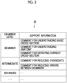
- the support information DB 32 is a database that stores support information related to the operation of the ultrasound diagnostic apparatus 10 and used to support the formation of the ultrasound tomographic image by the examiner.
- the support information is provided to the examiner.
- Fig. 2 is a conceptual diagram showing a first example of the content of the support information DB 32.
- the support information DB 32 different pieces of support information are associated with a plurality of different examiner levels indicating the levels of proficiency in operation for the ultrasound diagnostic apparatus 10.
- a comment as the support information is associated with the examiner level.
- the examiner levels are categorized as "beginner”, “intermediate”, and “advanced”, and comments are associated with the examiner levels "beginner”, “intermediate”, and “advanced”, respectively.
- Fig. 3 is a conceptual diagram showing a second example of the content of the support information DB 32.
- the support information may be associated with each combination of the examiner level and the match rate.
- the match rate is a match rate between the cross-section classification of the ultrasound tomographic image (referred to as a target image in the present specification) formed by transmitting ultrasound waves to the subject in response to an instruction from the examiner and a targeted cross-section which is the cross-section classification of the ultrasound tomographic image that needs to be formed by the examiner.
- the match rate is considerably high, and in a case where the cross-section classification of the target image and the targeted cross-section are cross-sections completely different from each other, the match rate is considerably low.
- the match rate is lower than that in a case where the cross-section classification of the target image and the targeted cross-section are the same, and the match rate is higher than that in a case where the cross-section classification of the target image and the targeted cross-section are cross-sections completely different from each other.
- the match rate may be said to indicate a probability that the cross-section classification of the target image is the targeted cross-section.
- the numerical range in which the match rate can be taken is divided into a plurality of match rate levels by one or a plurality of division threshold values. Then, in the support information DB 32, the support information is associated with a combination of the examiner level and the match rate level.
- the numerical range in which the match rate can be taken is divided into a match rate level of "high” (for example, the match rate is 70% or more), a match rate level of "medium” (for example, the match rate is 50% or more and less than 70%), and a match rate level of "low” (for example, the match rate is less than 50%).
- the match rates of "70%” and "50%” are the division threshold values.
- a different comment is associated with each match rate level as each piece of support information.
- a table as in Fig. 3 is provided for each examiner level. Since the table shown in Fig. 3 is a table for the examiner level "beginner", for example, a comment "After observing major and minor axes, save the maximum diameter of the major axis" is associated with a combination of the examiner level "beginner” and the match rate level "high” as the support information.
- the support information may be associated with each combination of the examiner level and the display aspect of the target image.
- the support information may be associated with each combination of the examiner level, the match rate, and the display aspect of the target image.
- the display aspect of the target image is real-time or freeze.
- a comment is associated with each combination of the examiner level "beginner”, each match rate, and the display aspect "real-time” of the target image, and a comment is associated with each combination of the examiner level "beginner", each match rate, and the display aspect "freeze” of the target image.
- the support information may be associated with each combination of the examiner level and a measurement content using the target image.
- the support information may be associated with each combination of the examiner level, the match rate, and the measurement content using the target image.
- the measurement content is a content of various measurements using the ultrasound image and is, for example, a major-axis measurement of the heart or the like.
- the measurement content may be designated by the examiner.
- an examination protocol in which a series of examination processes and an examination execution condition for each examination process are set may be stored, and in this case, the measurement content may be determined based on the examination protocol selected by the examiner and the progress of the examination protocol.
- the support information may be associated with each combination of the examiner level and the operation mode of the ultrasound diagnostic apparatus 10.
- the support information may be associated with each combination of the examiner level, the match rate, and the operation mode of the ultrasound diagnostic apparatus 10.
- the operation modes of the ultrasound diagnostic apparatus 10 include the B-mode, the PW Doppler mode, the CW Doppler mode, the color Doppler mode, and the like, but the operation modes are not limited to these.
- the support information may be associated with each combination of the examiner level and an attribute of the subject.
- the support information may be associated with each combination of the examiner level, the match rate, and the attribute of the subject.
- the attribute of the subject include the sex, the weight, the disease condition, the country or region, and the facility (hospital or university) where the subject is located, but the attribute of the subject is not limited to these.
- a comment which is the support information, is associated with a combination of the examiner level, the match rate, the display aspect of the target image, the measurement content using the target image, the operation mode of the ultrasound diagnostic apparatus 10, and the attribute of the subject.
- the aspect of the association between the examiner level, the match rate, the display aspect of the target image, the measurement content using the target image, the operation mode of the ultrasound diagnostic apparatus 10, the attribute of the subject, and the support information may be any aspect.
- Fig. 4 is a diagram showing a first example of the reference image as the support information.
- the comment is shown as the support information, but the support information stored in the support information DB 32 may be the reference image as shown in Fig. 4 .
- the reference image is an image for supporting the formation of the ultrasound image and may be, for example, an ultrasound tomographic image showing a targeted cross-section, a schematic diagram, an illustration, a schema diagram, a CT image, an MRI image, an image (including an illustration) showing an appropriate position of the ultrasound probe 12, or the like.
- each site pancreas, inferior vena cava, splenic vein, abdominal aorta, and the like
- the targeted cross-section may be clearly indicated by text, figures, and the like.
- Fig. 5 is a diagram showing a second example of the reference image as the support information.
- the reference image as shown in Fig. 5 as the support information may be associated with a combination of the examiner level, a first cross-section classification, and a second cross-section classification other than the first cross-section classification.
- the support information may be associated with each combination of the examiner level, the match rate, the first cross-section classification, and the second cross-section classification.
- FIG. 5 is a diagram in which the left side of an arrow indicates the position of the ultrasound probe 12 in a case where the ultrasound image of the first cross-section classification is formed, and the right side of the arrow indicates the position of the ultrasound probe 12 in a case where the ultrasound image of the second cross-section classification is formed. Therefore, in a case where the examiner wants to form the ultrasound image of the second cross-section classification as the targeted cross-section but mistakenly forms the ultrasound image of the first cross-section classification (that is, in a case where the ultrasound probe 12 is mistakenly applied as shown in the left side of Fig. 5 ), displaying such a reference image allows the examiner to easily understand how to change the position of the ultrasound probe 12 from the current position to an appropriate position.
- the reference image as shown in Fig. 5 may be a moving image that changes from the left side diagram to the right side diagram.
- a method of using the support information DB 32 will be described below.
- a priority flag associated with each piece of support information shown in Fig. 3 will be described below.
- a support information editing unit 34 edits the support information stored in the support information DB 32 in response to an instruction from the user, such as the examiner of the ultrasound diagnostic apparatus 10 or an instructor who instructs the examiner, which is input via the input interface 24.
- the support information editing unit 34 changes, for example, the comment as the support information stored in the support information DB 32 in response to an instruction from the user.
- the support information editing unit 34 changes, for example, the reference image as the support information stored in the support information DB 32 in response to an instruction from the user.
- An examiner level specifying unit 36 specifies the examiner level of the examiner who is currently using the ultrasound diagnostic apparatus 10.
- a server communicably connected to the ultrasound diagnostic apparatus 10 stores an examiner DB in which an examiner ID for uniquely identifying an examiner and the examiner level of the examiner are associated with each other.
- the examiner level specifying unit 36 specifies the examiner ID of the examiner who is currently using the ultrasound diagnostic apparatus 10 by, for example, a method of requiring the input of an examiner ID and a password or by reading an ID card in which the examiner ID is stored. Then, by referring to the examiner DB, the examiner level associated with the specified examiner ID is specified as the examiner level of the examiner.
- the examiner level specifying unit 36 may specify the examiner level of the examiner based on information input by the examiner via the input interface 24.
- a targeted cross-section specifying unit 38 specifies a cross-section type of the ultrasound tomographic image that needs to be formed by the examiner, that is, the targeted cross-section.
- the ultrasound diagnostic apparatus 10 stores the examination protocol described above, and the targeted cross-section specifying unit 38 specifies the targeted cross-section based on the examination protocol selected by the examiner and the progress of the examination protocol. For example, in the examination protocol, it is assumed that a first examination process is an examination performed using an ultrasound image of a cross-section classification A, and a second examination process is an examination performed using an ultrasound image of a cross-section classification B.
- the targeted cross-section is the cross-section classification A in the first examination process
- the targeted cross-section is the cross-section classification B in the second examination process
- the targeted cross-section specifying unit 38 may specify the targeted cross-section based on the examiner's instruction input via the input interface 24. That is, the targeted cross-section may be designated by the examiner.
- a match rate calculation unit 40 calculates a match rate between the target image and the targeted cross-section specified by the targeted cross-section specifying unit 38 by executing the cross-section recognition processing on the target image. Specifically, the match rate calculation unit 40 inputs the target image to the trained learning model 30 that executes the cross-section recognition processing. Consequently, the learning model 30 predicts and outputs the cross-section classification of the target image. As described above, the learning model 30 outputs the cross-section classification and the parameter indicating the probability that the target image is the cross-section classification. For example, the cross-section classification A is 80%, the cross-section classification B is 15%, and a cross-section classification C is 5%.
- the match rate between the target image and the targeted cross-section the parameter indicating the probability of each cross-section classification output by the learning model 30 is employed.
- the match rate between the target image and the targeted cross-section is 80% in a case where the targeted cross-section is the cross-section classification A
- the match rate between the target image and the targeted cross-section is 15% in a case where the targeted cross-section is the cross-section classification B
- the match rate between the target image and the targeted cross-section is 5% in a case where the targeted cross-section is the cross-section classification C.
- the output unit 20 outputs the support information with the content tailored to the examiner level of the examiner, which is specified by the examiner level specifying unit 36, based on the support information DB 32.
- the output unit 20 outputs the support information associated with the examiner level "beginner" in the support information DB 32 (for example, a comment for understanding the basic cross-section, a comment for understanding the anatomy, a comment for depicting the correct cross-section, or the reference image).
- the output unit 20 outputs the support information associated with the examiner level "intermediate” in the support information DB 32 (for example, a comment for reducing oversights, a comment for reducing errors between examiners, or the reference image).
- the output unit 20 outputs the support information associated with the examiner level "advanced" in the support information DB 32 (for example, a comment or the reference image).
- Fig. 6 is a diagram showing an example of a support information display screen displayed on the display 22 by the output unit 20.
- a comment CO as the support information and a reference image REF as the support information are displayed on a support information output screen together with a target image USI.
- the support information tailored to the examiner level of the examiner is provided to the examiner, it is possible to provide the support information particularly suitable for the examiner.
- the output unit 20 may output the support information with the content tailored to the examiner level specified by the examiner level specifying unit 36 and to the match rate between the cross-section classification of the target image and the targeted cross-section, which is calculated by the match rate calculation unit 40.
- the output unit 20 outputs the support information associated with the match rate level "high" in the support information DB 32 (for example, the comments “After observing major and minor axes, save the maximum diameter of the major axis", “Swelling: a major diameter of 12 cm or more/atrophy: a major diameter of less than 8 cm", or the reference image).
- the output unit 20 outputs the support information associated with the match rate level "medium” in the support information DB 32 (for example, the comments “Be careful not to overlap the posterior liver segment and the right kidney as much as possible", "Use cine search to select a good cross-section", or the reference image).
- the output unit 20 outputs the support information associated with the match rate level "low" in the support information DB 32 (for example, the comments “Try applying the probe to the intercostal space along the right anterior axillary line", “Is there enough gel? Try getting some again", or the reference image).
- the output unit 20 may display, on the support information output screen as shown in Fig. 6 , reference information I such as the match rate between the cross-section classification of the target image and the targeted cross-section, which is calculated by the match rate calculation unit 40, and the targeted cross-section specified by the targeted cross-section specifying unit 38.
- the range in which the match rate to be calculated by the match rate calculation unit 40 can be taken is divided into a plurality of match rate levels by the division threshold values, but the output unit 20 may change the division threshold value according to the examiner level specified by the examiner level specifying unit 36.
- the division threshold value can be set such that the higher the examiner level is, the less likely the match rate level is to increase.
- the output unit 20 sets the division threshold values to "70%" and "40%". That is, a match rate of 70% or more is defined as the match rate level "high”, a match rate of 40% or more and less than 70% is defined as the match rate level "medium”, and a match rate of less than 40% is defined as the match rate level "low”. Consequently, the output unit 20 determines that the match rate level is "high” in a case where the match rate calculated by the match rate calculation unit 40 is 71%, and determines that the match rate level is "medium” in a case where the match rate calculated by the match rate calculation unit 40 is 45%.
- the output unit 20 sets the division threshold values to "80%" and "50%". That is, a match rate of 80% or more is defined as the match rate level "high”, a match rate of 50% or more and less than 80% is defined as the match rate level "medium”, and a match rate of less than 50% is defined as the match rate level "low”. Consequently, the output unit 20 determines that the match rate level is "medium” in a case where the match rate calculated by the match rate calculation unit 40 is 71%, and determines that the match rate level is "low” in a case where the match rate calculated by the match rate calculation unit 40 is 45%.
- the output unit 20 may output the support information with the content tailored to the examiner level specified by the examiner level specifying unit 36 and to the display aspect (that is, real-time or freeze) of the target image designated by the examiner.
- the output unit 20 may output the support information with the content tailored to the examiner level specified by the examiner level specifying unit 36, to the match rate calculated by the match rate calculation unit 40, and to the display aspect of the target image designated by the examiner.
- the output unit 20 outputs the support information associated with the examiner level "beginner”, the match rate level "high”, and the display aspect "real-time” of the target image in the support information DB 32 (for example, the comment "After observing major and minor axes, save the maximum diameter of the major axis" or the reference image).
- the output unit 20 outputs the support information associated with the examiner level “beginner”, the match rate level "high” and the display aspect "freeze” of the target image in the support information DB 32 (for example, the comment "Swelling: a major diameter of 12 cm or more/atrophy: a major diameter of less than 8 cm” or the reference image).
- the output unit 20 may output the support information with the content tailored to the examiner level specified by the examiner level specifying unit 36 and to the measurement content specified by the examiner designation or the examination protocol.
- the output unit 20 may output the support information with the content tailored to the examiner level specified by the examiner level specifying unit 36, to the match rate calculated by the match rate calculation unit 40, and to the measurement content specified by the examiner designation or the examination protocol.
- the output unit 20 outputs the support information associated with the examiner level “beginner”, the match rate level "high”, and the measurement content "major-axis measurement” in the support information DB 32 (for example, the comment "After observing major and minor axes, save the maximum diameter of the major axis" or the reference image).
- the output unit 20 may output the support information with the content tailored to the examiner level specified by the examiner level specifying unit 36 and to the current operation mode of the ultrasound diagnostic apparatus 10.
- the output unit 20 may output the support information with the content tailored to the examiner level specified by the examiner level specifying unit 36, to the match rate calculated by the match rate calculation unit 40, and to the current operation mode of the ultrasound diagnostic apparatus 10.
- the output unit 20 outputs the support information associated with the examiner level "beginner", the match rate level "high", and the operation mode "B-mode” in the support information DB 32 (for example, the comments "After observing major and minor axes, save the maximum diameter of the major axis", “Swelling: a major diameter of 12 cm or more/atrophy: a major diameter of less than 8 cm", or the reference image).
- the output unit 20 may output the support information with the content tailored to the examiner level specified by the examiner level specifying unit 36 and to the attribute of the subject.
- the attribute of the subject corresponding to the target image may be input to the ultrasound diagnostic apparatus 10 by the examiner or may be recorded in the above-described examination protocol.
- the output unit 20 may output the support information with the content tailored to the examiner level specified by the examiner level specifying unit 36, to the match rate calculated by the match rate calculation unit 40, and to the attribute of the subject.
- the output unit 20 outputs the support information associated with the examiner level "beginner", the match rate level "high", and the subject attribute "male, 90 years old” in the support information DB 32 (for example, the comment "After observing major and minor axes, save the maximum diameter of the major axis" or the reference image).
- the output unit 20 may output the support information with the content tailored to the examiner level specified by the examiner level specifying unit 36, to the cross-section type of the target image as the first cross-section classification, and to the targeted cross-section as the second cross-section classification.
- the output unit 20 may output the support information with the content tailored to the examiner level specified by the examiner level specifying unit 36, to the match rate calculated by the match rate calculation unit 40, to the cross-section type of the target image as the first cross-section classification, and to the targeted cross-section as the second cross-section classification.
- the output unit 20 specifies the support information associated with the combination of the examiner level specified by the examiner level specifying unit 36, the match rate calculated by the match rate calculation unit 40, the cross-section type of the target image as the first cross-section classification, and the targeted cross-section specified by the targeted cross-section specifying unit 38 as the second cross-section classification, from the support information DB 32, and outputs the specified support information.
- the cross-section type of the target image here means a cross-section classification with the highest probability of relevance among probabilities of relevance for a plurality of cross-section classifications calculated by the match rate calculation unit 40.
- the reference image as shown in Fig. 5 is associated as the support information with the combination of the examiner level "beginner”, the match rate level “low”, the first cross-section classification "cross-section classification A", and the second cross-section classification "cross-section classification B". That is, in the reference image of Fig. 5 , it is assumed that the left side of the arrow is a diagram indicating the position of the ultrasound probe 12 in a case where the ultrasound image of the first cross-section classification "cross-section classification A" is formed, and the right side of the arrow is a diagram indicating the position of the ultrasound probe 12 in a case where the ultrasound image of the second cross-section classification "cross-section classification B" is formed.
- the output unit 20 outputs the reference image shown in Fig. 5 as the support information.
- the examiner changes the position of the ultrasound probe 12 or changes the settings of the ultrasound diagnostic apparatus 10 based on the output support information. Consequently, the match rate between the cross-section classification of the target image and the targeted cross-section may be improved. In this case, it can be considered that the output support information is effective. On the other hand, even in a case where the support information is output, the match rate between the cross-section classification of the target image and the targeted cross-section may not be improved. In this case, it can be considered that the output support information is not very effective.
- the match rate calculation unit 40 may calculate the match rate between the same targeted cross-section and the cross-section classification of the target image again after the support information has been output by the output unit 20.
- the priority flag (refer to Fig. 3 ) may be assigned to the support information in the support information DB 32. As shown in Fig. 3 , the priority flag may be assigned to the combination of the examiner level, the match rate, and the support information or may be assigned to the combination of the examiner level and the support information.
- the match rate calculation unit 40 does not assign the priority flag to the support information.
- the support information with the priority flag assigned is considered effective information, that is, information that can improve the match rate between the cross-section classification of the target image and the targeted cross-section.
- the output unit 20 may preferentially output the support information with the priority flag assigned, in a case where a plurality of pieces of support information are associated with the examiner level or the match rate in the support information DB 32, over the support information with no priority flag assigned.
- the priority flag is assigned to the comment "Try applying the probe to the intercostal space along the right anterior axillary line” and the priority flag is not assigned to the comment "Is there enough gel? Try getting some again".
- the output unit 20 preferentially outputs the comment "Try applying the probe to the intercostal space along the right anterior axillary line", which is the support information with the priority flag assigned, among the pieces of support information associated with the examiner level "beginner” and the match rate level "low” in the support information DB 32. As a result, it is possible to preferentially output the support information considered to be more effective.
- the ultrasound diagnostic apparatus 10 a case is considered in which an examination is performed along a plurality of examination processes, each forming an ultrasound tomographic image of a different targeted cross-section.
- the examination follows the above-described examination protocol.
- the examiner proceeds with the examination while forming the ultrasound tomographic image of the targeted cross-section defined in each examination process, for the examination process.
- the match rate calculation unit 40 calculates, each time the examiner forms the ultrasound tomographic image (target image) in each examination process, the match rate between the cross-section type of the target image formed in the examination process and the targeted cross-section related to the examination process. That is, the match rate is calculated for each examination process.
- the match rate calculation unit 40 stores the match rate calculated in each examination process in the memory 28 in association with each examination process.
- the output unit 20 may output the calculated match rate for a completed examination process during the examination along the plurality of examination processes. For example, as shown in Fig. 5 , the output unit 20 may read the calculated match rate for the completed examination process from the memory 28 and display a list L of the match rate for each examination process on the display 22.
- a list L of the match rate for each examination process on the display 22.
- an ultrasound tomographic image of a targeted cross-section "left lobe of the liver" is formed in an examination process 1
- an ultrasound tomographic image of a targeted cross-section "pancreatic tail” is formed in an examination process 2
- an ultrasound tomographic image of a targeted cross-section "left kidney” is formed in an examination process 3.
- the examiner can confirm that the match rate between the cross-section classification of the target image and the targeted cross-section in the examination processes 1 and 2 is "high" during the execution of the examination process 3.
- a setting value (proper setting value) of the ultrasound diagnostic apparatus 10 that is appropriate for forming the ultrasound image of the targeted cross-section may be prepared in advance for each examination process, and the output unit 20 may output the comment as the support information in a case where the setting value set by the examiner and the proper setting value deviate from each other by a predetermined amount or more. For example, a comment such as "The gain is 40. Isn't that too low?" is output.
- the output unit 20 may output an examination report after the plurality of examination processes have ended.
- Fig. 7 is a diagram showing an example of an examination report R.
- the examination report R may be output in a form of being displayed on the display 22 or may be output by being printed on a print medium such as paper.
- the examination report R allows, for example, an instructor who instructs a beginner or the like to retrospectively review the content of the examination across the plurality of examination processes performed by the beginner.
- the examination report R includes a plurality of item values for a plurality of items for each of the plurality of examination processes.
- the plurality of items of the examination report R include the support information output in a case of attempting to form the ultrasound tomographic image of the targeted cross-section related to the examination process.
- the examination report R shown in Fig. 6 has "cross-section No.”, "cross-section type”, “match rate”, “required time”, “change parameter”, "erroneous detection cross-section", “comment”, and “image” as the plurality of items.
- cross-section No. is a number for identifying the examination process.
- cross-section type indicates the targeted cross-section in the examination process.
- the item "match rate” is a match rate calculated by the match rate calculation unit 40 in the examination process.
- the item "required time” is a time required by the examiner for the examination in the examination process.
- the time required for the examination in each examination process for example, the time from the start of the examination process to the end of the examination process is automatically measured by a timing function of the ultrasound diagnostic apparatus 10.
- the item "change parameter" is a setting value (particularly, a setting value related to the formation of the ultrasound image or the image quality correction) of the ultrasound diagnostic apparatus 10 changed by the examiner in the examination process.
- a setting value include a gain, a depth, a frame rate, a velocity range, an ROI size, a baseline, a sample volume, an angle, and a reliability degree of SWE/SWM/ATT.
- the item "erroneous detection cross-section” is a cross-section classification in a case where the cross-section classification determined by the match rate calculation unit 40 to have the highest match rate with the cross-section classification of the target image in the examination process is a cross-section classification other than the targeted cross-section of the examination process.
- the item "comment” is a comment output as the support information in the examination process.
- the item "image” is a target image formed by the examiner in the examination process.
- the examination report R may include information (such as an examiner ID or a name) for identifying the attribute of the subject or an examiner who has performed the examination in the plurality of examination processes. These pieces of information are input to the ultrasound diagnostic apparatus 10 by the examiner prior to the examination in the plurality of examination processes.
- the item values need not be associated with all the items for each examination process.
- the output unit 20 determines the item to be associated with the item value for the item tailored to the examiner level of the examiner.
- the output unit 20 outputs the examination report R that associates the information on the item tailored to the examiner level of the examiner with each targeted cross-section.
- the output unit 20 may include, in the examination report R, the item values for all the items described above in a case where the examiner level is "beginner”, and need not include, in the examination report R, the item value for at least one of the item "comment” or the item “erroneous detection cross-section” in a case where the examiner level is "intermediate” (in this case, "-" or the like may be output to the corresponding field, or nothing may be displayed).
- the output unit 20 may determine the item to be associated with the item value for the examination process, according to the match rate for the examination process.
- the output unit 20 may output the examination report R that associates the information on the item tailored to the match rate corresponding to each targeted cross-section with the targeted cross-section.
- the output unit 20 need not include, in the examination report R, the item value for at least one of the item "change parameter" or the item "erroneous detection cross-section" for the examination process in which the match rate level is "high” (for example, the match rate is 70% or more) (in this case, "-" or the like may be output to the corresponding field, or nothing may be displayed).
- the output unit 20 may include, in the examination report R, the item value for at least one of the item "change parameter" or the item "erroneous detection cross-section" for the examination process in which the match rate level is "medium” (for example, the match rate is 50% or more and less than 70%) or the match rate level is "low” (for example, the match rate is less than 50%).
- the output unit 20 may determine the item to be associated with the item value for the examination process, according to the combination of the examiner level of the examiner and the match rate for the examination process. In other words, the output unit 20 may output the examination report R that associates the information on the item tailored to the examiner level of the examiner and to the match rate corresponding to each targeted cross-section with the targeted cross-section.
- the output unit 20 may include, for the examination process in which the examiner level is "intermediate” and the match rate level is “high”, the item value for an item other than the item “comment” or the item “erroneous detection cross-section” in the examination report R and may further include, for the examination process in which the examiner level is "intermediate” and the match rate level is "medium” or "low", the item value for the item "change parameter" in the examination report R.
- the outline of the ultrasound diagnostic apparatus according to the present embodiment is as described above.
- the ultrasound diagnostic apparatus according to the present embodiment is not limited to the above-described embodiment, and various changes can be made without departing from the gist thereof.
- the transmission and reception unit 14, the signal processing unit 16, the image forming unit 18, the output unit 20, the support information editing unit 34, the examiner level specifying unit 36, the targeted cross-section specifying unit 38, and the match rate calculation unit 40, which are shown in Fig. 1 , are functional blocks representing functions implemented by a processor.
- the processor includes at least one of a general-purpose processing device (for example, a CPU or the like) or a dedicated processing device (for example, a GPU, an ASIC, an FPGA, a programmable logic device, or the like).
- the processor may be configured with a plurality of processing devices that are present at physically separated positions and that cooperate with each other, rather than being configured with one processing device.
- each of the above-described units may be implemented by cooperation between hardware, such as a processor, and software.
Landscapes
- Health & Medical Sciences (AREA)
- Life Sciences & Earth Sciences (AREA)
- Engineering & Computer Science (AREA)
- Medical Informatics (AREA)
- Surgery (AREA)
- Pathology (AREA)
- Radiology & Medical Imaging (AREA)
- Biophysics (AREA)
- Biomedical Technology (AREA)
- Heart & Thoracic Surgery (AREA)
- Physics & Mathematics (AREA)
- Molecular Biology (AREA)
- Nuclear Medicine, Radiotherapy & Molecular Imaging (AREA)
- Animal Behavior & Ethology (AREA)
- General Health & Medical Sciences (AREA)
- Public Health (AREA)
- Veterinary Medicine (AREA)
- Computer Vision & Pattern Recognition (AREA)
- Vascular Medicine (AREA)
- Ultra Sonic Daignosis Equipment (AREA)
Abstract
Provided is an ultrasound diagnostic apparatus (12) capable of adaptively outputting support information according to a level of proficiency in operation of an examiner.
In a support information DB, comments as different pieces of support information are associated with a plurality of different examiner levels. An examiner level specifying unit (36) specifies an examiner level of an examiner who is currently using an ultrasound diagnostic apparatus. The output unit (20) outputs support information with a content tailored to the examiner level of the examiner, which is specified by the examiner level specifying unit, based on the support information DB.
Description
- The present specification discloses improvement of an ultrasound diagnostic apparatus.
- An ultrasound diagnostic apparatus is an apparatus that forms an ultrasound image or performs various examinations based on reflected waves from a subject, which are obtained by irradiating the subject with ultrasound waves. Conventionally, an ultrasound diagnostic apparatus has a function of supporting an examiner (user) such as a doctor or a technician.
- For example,
JP2022-127808A - Meanwhile, in an examination (including measurement) using an ultrasound diagnostic apparatus, it may be necessary to form an ultrasound tomographic image of an appropriate cross-section according to the purpose of the examination. In the present specification, an appropriate cross-section tailored to an examination is referred to as a targeted cross-section. The targeted cross-section is, for example, defined in information such as the examination protocol disclosed in
JP2022-127808A - Here, in order to support the formation of the ultrasound tomographic image of the targeted cross-section, it is considered to provide the examiner with support information related to the operation of the ultrasound diagnostic apparatus. As a simple method, it is conceivable to provide the examiner with uniform support information tailored to the targeted cross-section. However, it may not be appropriate to provide uniform support information to the examiner. For example, a plurality of examiners attempting to form the ultrasound images of the same targeted cross-section may require different pieces of support information depending on the level of proficiency related to the operation of the ultrasound diagnostic apparatus (simply referred to as a level of proficiency in operation in the present specification). In such a case, it is not appropriate to uniformly provide the same support information to both parties.
- An object of the ultrasound diagnostic apparatus disclosed in the present specification is to provide an ultrasound diagnostic apparatus capable of adaptively outputting support information according to a level of proficiency in operation of an examiner.
- According to an aspect of the present invention, there is provided an ultrasound diagnostic apparatus comprising: an examiner level specifying unit that specifies an examiner level indicating a level of proficiency in operation of an examiner for the ultrasound diagnostic apparatus, based on an examiner database in which an examiner ID for uniquely identifying the examiner and the examiner level of the examiner are associated with each other, or an input from the examiner; and an output unit that outputs, based on a support information database in which support information related to an operation of the ultrasound diagnostic apparatus and used to support formation of an ultrasound tomographic image by the examiner is associated with each examiner level, support information with a content tailored to the examiner level specified by the examiner level specifying unit.
- A targeted cross-section specifying unit that specifies a targeted cross-section which is a cross-section classification of an ultrasound tomographic image that needs to be formed, and a match rate calculation unit that executes cross-section recognition processing on a target image which is an ultrasound tomographic image formed in response to an instruction from the examiner, to calculate a match rate between a cross-section classification of the target image and the targeted cross-section may be further provided, the support information may be associated with a combination of the examiner level and the match rate in the support information database, and the output unit may output support information with a content tailored to the examiner level specified by the examiner level specifying unit and to the match rate calculated by the match rate calculation unit.
- A numerical range in which the match rate is capable of being taken may be divided into a plurality of match rate levels by one or a plurality of division threshold values, and the support information may be associated with a combination of the examiner level and the match rate level in the support information database, and the division threshold value may be changed according to the examiner level of the examiner, which is specified by the examiner level specifying unit.
- In the support information database, the support information may be associated with a combination of the examiner level and a display aspect of a target image which is an ultrasound tomographic image formed in response to an instruction from the examiner, and the output unit may output support information with a content tailored to the examiner level of the examiner, which is specified by the examiner level specifying unit, and to the display aspect of the target image.
- In the support information database, the support information may be associated with a combination of the examiner level and a measurement content using a target image which is an ultrasound tomographic image formed in response to an instruction from the examiner, and the output unit may output support information with a content tailored to the examiner level of the examiner, which is specified by the examiner level specifying unit, and to the measurement content.
- In the support information database, the support information may be associated with a combination of the examiner level and an operation mode of the ultrasound diagnostic apparatus, and the output unit may output support information with a content tailored to the examiner level of the examiner, which is specified by the examiner level specifying unit, and to the operation mode of the ultrasound diagnostic apparatus.
- In the support information database, the support information may be associated with a combination of the examiner level and an attribute of a subject, and the output unit may output support information with a content tailored to the examiner level of the examiner, which is specified by the examiner level specifying unit, and to the attribute of the subject.
- In the support information database, the support information may be associated with a combination of the examiner level, a first cross-section classification, and a second cross-section classification other than the first cross-section classification, and the output unit may output support information with a content tailored to the examiner level of the examiner, which is specified by the examiner level specifying unit, to a cross-section classification of a target image, which is an ultrasound tomographic image formed in response to an instruction from the examiner, as the first cross-section classification, and to a targeted cross-section, which is a cross-section classification of an ultrasound tomographic image that needs to be formed, as the second cross-section classification.
- A support information editing unit that edits the support information in response to an instruction from a user of the ultrasound diagnostic apparatus may be further provided.
- The match rate calculation unit may calculate the match rate for the same targeted cross-section again after the support information has been output, and assign, in a case where the match rate after the support information has been output is higher than the match rate before the support information is output, a priority flag to the support information in the support information database, and the output unit may preferentially output the support information with the priority flag assigned, in a case where a plurality of pieces of the support information are associated with the examiner level in the support information database, over support information with no priority flag assigned.
- During an examination along a plurality of examination processes, each forming an ultrasound tomographic image of a different targeted cross-section, the match rate calculation unit may calculate the match rate for each examination process and store the match rate in a memory, and the output unit may output the calculated match rate for a completed examination process during the examination along the plurality of examination processes.
- The output unit may output, after a plurality of examination processes, each forming an ultrasound tomographic image of a different targeted cross-section, have ended, an examination report that includes, for each of the plurality of examination processes, item values for a plurality of items including the support information output in a case of attempting to form an ultrasound tomographic image of a targeted cross-section related to the examination process, the examination report associating information on the item tailored to the examiner level of the examiner with each targeted cross-section.
- With the ultrasound diagnostic apparatus disclosed in the present specification, it is possible to provide an ultrasound diagnostic apparatus capable of adaptively outputting support information according to a level of proficiency in operation of an examiner.
-
-
Fig. 1 is a schematic diagram of a configuration of an ultrasound diagnostic apparatus according to the present embodiment. -
Fig. 2 is a conceptual diagram showing a first example of a content of a support information DB. -
Fig. 3 is a conceptual diagram showing a second example of the content of the support information DB. -
Fig. 4 is a diagram showing a first example of a reference image as support information. -
Fig. 5 is a diagram showing a second example of the reference image as the support information. -
Fig. 6 is a diagram showing an example of a support information display screen. -
Fig. 7 is a diagram showing an example of an examination report. -
Fig. 1 is a schematic diagram of a configuration of an ultrasounddiagnostic apparatus 10 according to the present embodiment. The ultrasounddiagnostic apparatus 10 is a medical apparatus installed in a medical institution such as a hospital and used during ultrasound examinations. - The ultrasound
diagnostic apparatus 10 is an apparatus that scans a subject with an ultrasound beam and that generates an ultrasound image based on a reception signal obtained by the scanning. The ultrasounddiagnostic apparatus 10 has, for example, a plurality of operation modes such as a B-mode, an M-mode, a pulse wave (PW) Doppler mode, a continuous wave (CW) Doppler mode, or a color Doppler mode. For example, in the B-mode, the ultrasounddiagnostic apparatus 10 forms an ultrasound tomographic image (B-mode image) in which the amplitude intensity of a reflected wave from a scanning plane is converted into brightness, based on the reception signal. In addition, for example, in the PW Doppler mode, the CW Doppler mode, or the color Doppler mode, the ultrasounddiagnostic apparatus 10 forms a Doppler image that shows the motion velocity of a site of the subject or the velocity of the blood flow, based on the difference in frequency between transmission ultrasound waves and reception ultrasound waves. The Doppler image shows the motion velocity of the site of the subject or the velocity of the blood flow in colors or the like and is superimposed and displayed on the ultrasound tomographic image. The operation mode of the ultrasounddiagnostic apparatus 10 can be set in response to an instruction from the examiner who performs an examination by forming an ultrasound image (including an ultrasound tomographic image, a Doppler image, or the like) using the ultrasounddiagnostic apparatus 10. - Although details will be described below, the ultrasound
diagnostic apparatus 10 can provide the examiner with support information related to an operation of the ultrasounddiagnostic apparatus 10 and used to support formation of the ultrasound tomographic image by the examiner. - An
ultrasound probe 12 is a device that performs transmission and reception of ultrasound waves with respect to the subject. Theultrasound probe 12 includes a transducer element array consisting of a plurality of transducer elements that perform transmission and reception of ultrasound waves with respect to the subject. - A transmission and
reception unit 14 transmits a transmission signal to the ultrasound probe 12 (in detail, each transducer element of the transducer element array) under the control of a controller 26 (which will be described below). Consequently, the ultrasound waves are transmitted from each transducer element toward the subject. In addition, the transmission andreception unit 14 receives the reception signal from each transducer element that has received the reflected wave from the subject. The transmission andreception unit 14 includes an adder and a plurality of delay devices corresponding to the respective transducer elements and performs phase alignment addition processing of aligning and adding phases of the reception signals from the respective transducer elements by using the adder and the plurality of delay devices. As a result, reception beam signals are formed in which information indicating the signal intensity of the reflected wave from the subject is arranged in a depth direction of the subj ect. - A
signal processing unit 16 executes various types of signal processing including filter processing of applying a bandpass filter, detection processing, and the like on the reception beam signals from the transmission andreception unit 14. In addition, in a case where the operation mode of the ultrasounddiagnostic apparatus 10 is the Doppler mode, thesignal processing unit 16 generates Doppler data indicating the site of the subject or the velocity of the blood flow based on the reception beam signals from the transmission andreception unit 14. Specifically, thesignal processing unit 16 generates the Doppler data by performing processing such as quadrature detection of multiplying the reception beam signal by a reference frequency (transmission frequency) to extract Doppler shifts through a low-pass filter, sample gate processing (in a case of the PW Doppler mode) of extracting only the signals from the position of the sample volume, A/D conversion of the signals, and frequency analysis using the fast Fourier transform method (FFT method). - An
image forming unit 18 forms the ultrasound tomographic image (B-mode image) based on the reception beam signal subjected to the signal processing in thesignal processing unit 16. First, theimage forming unit 18 converts the reception beam signal into data on a coordinate space of the ultrasound image and forms the ultrasound tomographic image based on a coordinate conversion signal. Additionally, in a case where the operation mode of the ultrasounddiagnostic apparatus 10 is the Doppler mode, theimage forming unit 18 forms a Doppler waveform or a color Doppler image based on a Doppler signal from thesignal processing unit 16. - An
output unit 20 performs control to display the ultrasound image, such as the ultrasound tomographic image or the color Doppler image formed by theimage forming unit 18, on adisplay 22. Thedisplay 22 is a display device configured with, for example, a liquid crystal display, an organic electro luminescence (EL), or the like. - The
output unit 20 can display the ultrasound image (the ultrasound tomographic image, the Doppler image, or the like) on thedisplay 22 in either real-time or freeze display aspect. The display aspect of the ultrasound image can be switched in response to an instruction from the examiner. In a case of the real-time display aspect, theoutput unit 20 displays, on thedisplay 22 in real-time, the ultrasound images that are successively formed by the processing of thesignal processing unit 16 and theimage forming unit 18 in response to the reception signals that are successively received by theultrasound probe 12. Meanwhile, in a case of the freeze display aspect, theoutput unit 20 displays, on thedisplay 22, the ultrasound image formed at the timing as instructed by the examiner, as a still image. - In addition, although details will be described below, the
output unit 20 displays the support information on thedisplay 22. Theoutput unit 20 may output the support information as a voice from a speaker (not shown) instead of or in addition to displaying the support information on thedisplay 22. - An
input interface 24 is configured with, for example, a button, a trackball, a touch panel, and the like. Theinput interface 24 is used for a user such as an examiner to input a command to the ultrasounddiagnostic apparatus 10. - The
controller 26 includes at least one of a general-purpose processor (for example, a central processing unit (CPU) or the like) or a dedicated processor (for example, a graphics processing unit (GPU), an application specific integrated circuit (ASIC), a field-programmable gate array (FPGA), a programmable logic device, or the like). Thecontroller 26 may be configured with a plurality of processing devices that are present at physically separated positions and that cooperate with each other, rather than being configured with one processing device. Thecontroller 26 controls each unit of the ultrasounddiagnostic apparatus 10 in accordance with an ultrasound diagnosis program stored in amemory 28, which will be described below. - The
memory 28 includes a hard disk drive (HDD), a solid state drive (SSD), an embedded multi media card (eMMC), a read only memory (ROM), or the like. Thememory 28 stores the ultrasound diagnosis program for operating each unit of the ultrasounddiagnostic apparatus 10. The ultrasound diagnosis program can also be stored in, for example, a computer-readable non-transitory storage medium such as a universal serial bus (USB) memory or a CD-ROM. The ultrasounddiagnostic apparatus 10 can read the ultrasound diagnosis program from such a storage medium and execute the ultrasound diagnosis program. - In addition, as shown in
Fig. 1 , thememory 28 stores alearning model 30 and a support information database (DB) 32. - The
learning model 30 is, for example, a convolutional neural network (CNN) or the like, but may be any model as long as it exhibits functions to be described below. Thelearning model 30 is trained by using a combination of the ultrasound tomographic image and a cross-section classification (ground truth cross-section classification) of the ultrasound tomographic image as training data, to predict and output the cross-section classification of the input ultrasound tomographic image. In the present embodiment, training processing of thelearning model 30 is performed by a training processing device different from the ultrasounddiagnostic apparatus 10, and a sufficiently trainedlearning model 30 is stored in thememory 28. However, the ultrasounddiagnostic apparatus 10 may include a training processing unit (not shown inFig. 1 ), and the training processing unit may perform the training processing of thelearning model 30. In this case, the ultrasounddiagnostic apparatus 10 serves as a training processing device. - The details of the training processing of the
learning model 30 are as follows. A processor of the training processing device inputs the ultrasound tomographic image as training data to thelearning model 30. Thelearning model 30 predicts and outputs the cross-section classification of the input ultrasound tomographic image. The processor of the training processing device changes parameters in thelearning model 30 such that a difference between output data of thelearning model 30 and the ground truth cross-section classification included in the training data is reduced. By repeatedly performing such processing, thelearning model 30 is trained. Thelearning model 30 that has been sufficiently trained can predict the cross-section classification of the input ultrasound tomographic image with high accuracy. - The
learning model 30 according to the present embodiment has an activation function, such as a softmax function, in an output layer thereof and outputs, as the output data, a plurality of combinations of the cross-section classifications and the parameters indicating a probability that the input ultrasound tomographic image is the cross-section classification. - The
support information DB 32 is a database that stores support information related to the operation of the ultrasounddiagnostic apparatus 10 and used to support the formation of the ultrasound tomographic image by the examiner. The support information is provided to the examiner. -
Fig. 2 is a conceptual diagram showing a first example of the content of thesupport information DB 32. In thesupport information DB 32, different pieces of support information are associated with a plurality of different examiner levels indicating the levels of proficiency in operation for the ultrasounddiagnostic apparatus 10. In the example inFig. 2 , a comment as the support information is associated with the examiner level. In the example inFig. 2 , the examiner levels are categorized as "beginner", "intermediate", and "advanced", and comments are associated with the examiner levels "beginner", "intermediate", and "advanced", respectively. -
Fig. 3 is a conceptual diagram showing a second example of the content of thesupport information DB 32. In thesupport information DB 32, the support information may be associated with each combination of the examiner level and the match rate. The match rate is a match rate between the cross-section classification of the ultrasound tomographic image (referred to as a target image in the present specification) formed by transmitting ultrasound waves to the subject in response to an instruction from the examiner and a targeted cross-section which is the cross-section classification of the ultrasound tomographic image that needs to be formed by the examiner. For example, in a case where the cross-section classification of the target image and the targeted cross-section are the same, the match rate is considerably high, and in a case where the cross-section classification of the target image and the targeted cross-section are cross-sections completely different from each other, the match rate is considerably low. In a case where the cross-section classification of the target image and the targeted cross-section are different from each other but are close to each other (for example, in a case where the same site is shown but the cross-sections are slightly different or the like), the match rate is lower than that in a case where the cross-section classification of the target image and the targeted cross-section are the same, and the match rate is higher than that in a case where the cross-section classification of the target image and the targeted cross-section are cross-sections completely different from each other. The match rate may be said to indicate a probability that the cross-section classification of the target image is the targeted cross-section. - In the present embodiment, in the
support information DB 32, the numerical range in which the match rate can be taken is divided into a plurality of match rate levels by one or a plurality of division threshold values. Then, in thesupport information DB 32, the support information is associated with a combination of the examiner level and the match rate level. In the example inFig. 3 , the numerical range in which the match rate can be taken is divided into a match rate level of "high" (for example, the match rate is 70% or more), a match rate level of "medium" (for example, the match rate is 50% or more and less than 70%), and a match rate level of "low" (for example, the match rate is less than 50%). In this case, the match rates of "70%" and "50%" are the division threshold values. Then, a different comment is associated with each match rate level as each piece of support information. In thesupport information DB 32, a table as inFig. 3 is provided for each examiner level. Since the table shown inFig. 3 is a table for the examiner level "beginner", for example, a comment "After observing major and minor axes, save the maximum diameter of the major axis" is associated with a combination of the examiner level "beginner" and the match rate level "high" as the support information. - In the
support information DB 32, the support information may be associated with each combination of the examiner level and the display aspect of the target image. In particular, in thesupport information DB 32, the support information may be associated with each combination of the examiner level, the match rate, and the display aspect of the target image. In the present embodiment, the display aspect of the target image is real-time or freeze. In the example inFig. 3 , a comment is associated with each combination of the examiner level "beginner", each match rate, and the display aspect "real-time" of the target image, and a comment is associated with each combination of the examiner level "beginner", each match rate, and the display aspect "freeze" of the target image. - In addition, in the
support information DB 32, the support information may be associated with each combination of the examiner level and a measurement content using the target image. In particular, in thesupport information DB 32, the support information may be associated with each combination of the examiner level, the match rate, and the measurement content using the target image. The measurement content is a content of various measurements using the ultrasound image and is, for example, a major-axis measurement of the heart or the like. The measurement content may be designated by the examiner. In addition, in the ultrasounddiagnostic apparatus 10, an examination protocol in which a series of examination processes and an examination execution condition for each examination process are set may be stored, and in this case, the measurement content may be determined based on the examination protocol selected by the examiner and the progress of the examination protocol. - Further, in the
support information DB 32, the support information may be associated with each combination of the examiner level and the operation mode of the ultrasounddiagnostic apparatus 10. In particular, in thesupport information DB 32, the support information may be associated with each combination of the examiner level, the match rate, and the operation mode of the ultrasounddiagnostic apparatus 10. As described above, the operation modes of the ultrasounddiagnostic apparatus 10 include the B-mode, the PW Doppler mode, the CW Doppler mode, the color Doppler mode, and the like, but the operation modes are not limited to these. - Furthermore, in the
support information DB 32, the support information may be associated with each combination of the examiner level and an attribute of the subject. In particular, in thesupport information DB 32, the support information may be associated with each combination of the examiner level, the match rate, and the attribute of the subject. Examples of the attribute of the subject include the sex, the weight, the disease condition, the country or region, and the facility (hospital or university) where the subject is located, but the attribute of the subject is not limited to these. - In the present embodiment, as shown in
Figs. 2 and3 , a comment, which is the support information, is associated with a combination of the examiner level, the match rate, the display aspect of the target image, the measurement content using the target image, the operation mode of the ultrasounddiagnostic apparatus 10, and the attribute of the subject. The aspect of the association between the examiner level, the match rate, the display aspect of the target image, the measurement content using the target image, the operation mode of the ultrasounddiagnostic apparatus 10, the attribute of the subject, and the support information may be any aspect. -
Fig. 4 is a diagram showing a first example of the reference image as the support information. In the examples inFigs. 2 and3 , the comment is shown as the support information, but the support information stored in thesupport information DB 32 may be the reference image as shown inFig. 4 . The reference image is an image for supporting the formation of the ultrasound image and may be, for example, an ultrasound tomographic image showing a targeted cross-section, a schematic diagram, an illustration, a schema diagram, a CT image, an MRI image, an image (including an illustration) showing an appropriate position of theultrasound probe 12, or the like. As shown inFig. 4 , in the reference image, each site (pancreas, inferior vena cava, splenic vein, abdominal aorta, and the like) in the targeted cross-section may be clearly indicated by text, figures, and the like. -
Fig. 5 is a diagram showing a second example of the reference image as the support information. In thesupport information DB 32, the reference image as shown inFig. 5 as the support information may be associated with a combination of the examiner level, a first cross-section classification, and a second cross-section classification other than the first cross-section classification. In particular, in thesupport information DB 32, the support information may be associated with each combination of the examiner level, the match rate, the first cross-section classification, and the second cross-section classification. The reference image inFig. 5 is a diagram in which the left side of an arrow indicates the position of theultrasound probe 12 in a case where the ultrasound image of the first cross-section classification is formed, and the right side of the arrow indicates the position of theultrasound probe 12 in a case where the ultrasound image of the second cross-section classification is formed. Therefore, in a case where the examiner wants to form the ultrasound image of the second cross-section classification as the targeted cross-section but mistakenly forms the ultrasound image of the first cross-section classification (that is, in a case where theultrasound probe 12 is mistakenly applied as shown in the left side ofFig. 5 ), displaying such a reference image allows the examiner to easily understand how to change the position of theultrasound probe 12 from the current position to an appropriate position. The reference image as shown inFig. 5 may be a moving image that changes from the left side diagram to the right side diagram. - A method of using the
support information DB 32 will be described below. In addition, a priority flag associated with each piece of support information shown inFig. 3 will be described below. - Returning to
Fig. 1 , a supportinformation editing unit 34 edits the support information stored in thesupport information DB 32 in response to an instruction from the user, such as the examiner of the ultrasounddiagnostic apparatus 10 or an instructor who instructs the examiner, which is input via theinput interface 24. The supportinformation editing unit 34 changes, for example, the comment as the support information stored in thesupport information DB 32 in response to an instruction from the user. Alternatively, the supportinformation editing unit 34 changes, for example, the reference image as the support information stored in thesupport information DB 32 in response to an instruction from the user. - An examiner
level specifying unit 36 specifies the examiner level of the examiner who is currently using the ultrasounddiagnostic apparatus 10. In the present embodiment, a server communicably connected to the ultrasounddiagnostic apparatus 10 stores an examiner DB in which an examiner ID for uniquely identifying an examiner and the examiner level of the examiner are associated with each other. The examinerlevel specifying unit 36 specifies the examiner ID of the examiner who is currently using the ultrasounddiagnostic apparatus 10 by, for example, a method of requiring the input of an examiner ID and a password or by reading an ID card in which the examiner ID is stored. Then, by referring to the examiner DB, the examiner level associated with the specified examiner ID is specified as the examiner level of the examiner. In addition, the examinerlevel specifying unit 36 may specify the examiner level of the examiner based on information input by the examiner via theinput interface 24. - A targeted
cross-section specifying unit 38 specifies a cross-section type of the ultrasound tomographic image that needs to be formed by the examiner, that is, the targeted cross-section. In the present embodiment, the ultrasounddiagnostic apparatus 10 stores the examination protocol described above, and the targetedcross-section specifying unit 38 specifies the targeted cross-section based on the examination protocol selected by the examiner and the progress of the examination protocol. For example, in the examination protocol, it is assumed that a first examination process is an examination performed using an ultrasound image of a cross-section classification A, and a second examination process is an examination performed using an ultrasound image of a cross-section classification B. In this case, the targeted cross-section is the cross-section classification A in the first examination process, and the targeted cross-section is the cross-section classification B in the second examination process. In addition, the targetedcross-section specifying unit 38 may specify the targeted cross-section based on the examiner's instruction input via theinput interface 24. That is, the targeted cross-section may be designated by the examiner. - A match
rate calculation unit 40 calculates a match rate between the target image and the targeted cross-section specified by the targetedcross-section specifying unit 38 by executing the cross-section recognition processing on the target image. Specifically, the matchrate calculation unit 40 inputs the target image to the trainedlearning model 30 that executes the cross-section recognition processing. Consequently, thelearning model 30 predicts and outputs the cross-section classification of the target image. As described above, thelearning model 30 outputs the cross-section classification and the parameter indicating the probability that the target image is the cross-section classification. For example, the cross-section classification A is 80%, the cross-section classification B is 15%, and a cross-section classification C is 5%. In the present embodiment, as the match rate between the target image and the targeted cross-section, the parameter indicating the probability of each cross-section classification output by thelearning model 30 is employed. For example, in a case where the output data of thelearning model 30 is the above-described example, the match rate between the target image and the targeted cross-section is 80% in a case where the targeted cross-section is the cross-section classification A, the match rate between the target image and the targeted cross-section is 15% in a case where the targeted cross-section is the cross-section classification B, and the match rate between the target image and the targeted cross-section is 5% in a case where the targeted cross-section is the cross-section classification C. - Hereinafter, details of output processing of the support information by the
output unit 20 will be described. - The
output unit 20 outputs the support information with the content tailored to the examiner level of the examiner, which is specified by the examinerlevel specifying unit 36, based on thesupport information DB 32. For example, assuming that the content of thesupport information DB 32 is the content shown inFig. 2 , in a case where the examiner level specified by the examinerlevel specifying unit 36 is "beginner", theoutput unit 20 outputs the support information associated with the examiner level "beginner" in the support information DB 32 (for example, a comment for understanding the basic cross-section, a comment for understanding the anatomy, a comment for depicting the correct cross-section, or the reference image). In addition, in a case where the examiner level specified by the examinerlevel specifying unit 36 is "intermediate", theoutput unit 20 outputs the support information associated with the examiner level "intermediate" in the support information DB 32 (for example, a comment for reducing oversights, a comment for reducing errors between examiners, or the reference image). In addition, in a case where the examiner level specified by the examinerlevel specifying unit 36 is "advanced", theoutput unit 20 outputs the support information associated with the examiner level "advanced" in the support information DB 32 (for example, a comment or the reference image). -
Fig. 6 is a diagram showing an example of a support information display screen displayed on thedisplay 22 by theoutput unit 20. In the example inFig. 6 , a comment CO as the support information and a reference image REF as the support information are displayed on a support information output screen together with a target image USI. - As described above, according to the present embodiment, since the support information tailored to the examiner level of the examiner is provided to the examiner, it is possible to provide the support information particularly suitable for the examiner.
- In a case where the support information is associated with each combination of the examiner level and the match rate in the support information DB 32 (refer to
Fig. 3 ), theoutput unit 20 may output the support information with the content tailored to the examiner level specified by the examinerlevel specifying unit 36 and to the match rate between the cross-section classification of the target image and the targeted cross-section, which is calculated by the matchrate calculation unit 40. For example, in a case where the examiner level specified by the examinerlevel specifying unit 36 is "beginner", and the match rate calculated by the matchrate calculation unit 40 is 80% (that is, the match rate level is "high"), theoutput unit 20 outputs the support information associated with the match rate level "high" in the support information DB 32 (for example, the comments "After observing major and minor axes, save the maximum diameter of the major axis", "Swelling: a major diameter of 12 cm or more/atrophy: a major diameter of less than 8 cm", or the reference image). In addition, in a case where the examiner level specified by the examinerlevel specifying unit 36 is "beginner", and the match rate calculated by the matchrate calculation unit 40 is 60% (that is, the match rate level is "medium"), theoutput unit 20 outputs the support information associated with the match rate level "medium" in the support information DB 32 (for example, the comments "Be careful not to overlap the posterior liver segment and the right kidney as much as possible", "Use cine search to select a good cross-section", or the reference image). Further, in a case where the examiner level specified by the examinerlevel specifying unit 36 is "beginner", and the match rate calculated by the matchrate calculation unit 40 is 20% (that is, the match rate level is "low"), theoutput unit 20 outputs the support information associated with the match rate level "low" in the support information DB 32 (for example, the comments "Try applying the probe to the intercostal space along the right anterior axillary line", "Is there enough gel? Try getting some again", or the reference image). - In a case where the support information based on the examiner level specified by the examiner
level specifying unit 36 and the match rate calculated by the matchrate calculation unit 40 is output, theoutput unit 20 may display, on the support information output screen as shown inFig. 6 , reference information I such as the match rate between the cross-section classification of the target image and the targeted cross-section, which is calculated by the matchrate calculation unit 40, and the targeted cross-section specified by the targetedcross-section specifying unit 38. - As described above, in the present embodiment, the range in which the match rate to be calculated by the match
rate calculation unit 40 can be taken is divided into a plurality of match rate levels by the division threshold values, but theoutput unit 20 may change the division threshold value according to the examiner level specified by the examinerlevel specifying unit 36. Specifically, the division threshold value can be set such that the higher the examiner level is, the less likely the match rate level is to increase. - For example, in a case where the examiner level specified by the examiner
level specifying unit 36 is "beginner", theoutput unit 20 sets the division threshold values to "70%" and "40%". That is, a match rate of 70% or more is defined as the match rate level "high", a match rate of 40% or more and less than 70% is defined as the match rate level "medium", and a match rate of less than 40% is defined as the match rate level "low". Consequently, theoutput unit 20 determines that the match rate level is "high" in a case where the match rate calculated by the matchrate calculation unit 40 is 71%, and determines that the match rate level is "medium" in a case where the match rate calculated by the matchrate calculation unit 40 is 45%. Meanwhile, in a case where the examiner level specified by the examinerlevel specifying unit 36 is "intermediate", theoutput unit 20 sets the division threshold values to "80%" and "50%". That is, a match rate of 80% or more is defined as the match rate level "high", a match rate of 50% or more and less than 80% is defined as the match rate level "medium", and a match rate of less than 50% is defined as the match rate level "low". Consequently, theoutput unit 20 determines that the match rate level is "medium" in a case where the match rate calculated by the matchrate calculation unit 40 is 71%, and determines that the match rate level is "low" in a case where the match rate calculated by the matchrate calculation unit 40 is 45%. - As a result, since the higher the examiner level is, the less likely the match rate level is to increase, it is possible to output more appropriate support information tailored to the combination of the examiner level of the examiner and the match rate between the target image and the targeted cross-section.
- In a case where the support information is associated with each combination of the examiner level and the display aspect of the target image in the
support information DB 32, theoutput unit 20 may output the support information with the content tailored to the examiner level specified by the examinerlevel specifying unit 36 and to the display aspect (that is, real-time or freeze) of the target image designated by the examiner. In particular, in a case where the support information is associated with each combination of the examiner level, the match rate, and the display aspect of the target image in thesupport information DB 32, theoutput unit 20 may output the support information with the content tailored to the examiner level specified by the examinerlevel specifying unit 36, to the match rate calculated by the matchrate calculation unit 40, and to the display aspect of the target image designated by the examiner. For example, in a case where the examiner level specified by the examinerlevel specifying unit 36 is "beginner", the match rate calculated by the matchrate calculation unit 40 is 80% (that is, the match rate level is "high"), and the display aspect of the target image is "real-time", theoutput unit 20 outputs the support information associated with the examiner level "beginner", the match rate level "high", and the display aspect "real-time" of the target image in the support information DB 32 (for example, the comment "After observing major and minor axes, save the maximum diameter of the major axis" or the reference image). In addition, in a case where the examiner level specified by the examinerlevel specifying unit 36 is "beginner", the match rate calculated by the matchrate calculation unit 40 is 80% (that is, the match rate level is "high"), and the display aspect of the target image is "freeze", theoutput unit 20 outputs the support information associated with the examiner level "beginner", the match rate level "high" and the display aspect "freeze" of the target image in the support information DB 32 (for example, the comment "Swelling: a major diameter of 12 cm or more/atrophy: a major diameter of less than 8 cm" or the reference image). - In a case where the support information is associated with each combination of the examiner level and the measurement content using the target image in the
support information DB 32, theoutput unit 20 may output the support information with the content tailored to the examiner level specified by the examinerlevel specifying unit 36 and to the measurement content specified by the examiner designation or the examination protocol. In particular, in a case where the support information is associated with each combination of the examiner level, the match rate, and the measurement content using the target image in thesupport information DB 32, theoutput unit 20 may output the support information with the content tailored to the examiner level specified by the examinerlevel specifying unit 36, to the match rate calculated by the matchrate calculation unit 40, and to the measurement content specified by the examiner designation or the examination protocol. For example, in a case where the examiner level specified by the examinerlevel specifying unit 36 is "beginner", the match rate calculated by the matchrate calculation unit 40 is 80% (that is, the match rate level is "high"), and the measurement content using the target image is "major-axis measurement", theoutput unit 20 outputs the support information associated with the examiner level "beginner", the match rate level "high", and the measurement content "major-axis measurement" in the support information DB 32 (for example, the comment "After observing major and minor axes, save the maximum diameter of the major axis" or the reference image). - In a case where the support information is associated with each combination of the examiner level and the operation mode of the ultrasound
diagnostic apparatus 10 in thesupport information DB 32, theoutput unit 20 may output the support information with the content tailored to the examiner level specified by the examinerlevel specifying unit 36 and to the current operation mode of the ultrasounddiagnostic apparatus 10. In particular, in a case where the support information is associated with each combination of the examiner level, the match rate, and the operation mode of the ultrasounddiagnostic apparatus 10 in thesupport information DB 32, theoutput unit 20 may output the support information with the content tailored to the examiner level specified by the examinerlevel specifying unit 36, to the match rate calculated by the matchrate calculation unit 40, and to the current operation mode of the ultrasounddiagnostic apparatus 10. For example, in a case where the examiner level specified by the examinerlevel specifying unit 36 is "beginner", the match rate calculated by the matchrate calculation unit 40 is 80% (that is, the match rate level is "high"), and the current operation mode of the ultrasounddiagnostic apparatus 10 is the B-mode, theoutput unit 20 outputs the support information associated with the examiner level "beginner", the match rate level "high", and the operation mode "B-mode" in the support information DB 32 (for example, the comments "After observing major and minor axes, save the maximum diameter of the major axis", "Swelling: a major diameter of 12 cm or more/atrophy: a major diameter of less than 8 cm", or the reference image). - In a case where the support information is associated with each combination of the examiner level and the attribute of the subject in the
support information DB 32, theoutput unit 20 may output the support information with the content tailored to the examiner level specified by the examinerlevel specifying unit 36 and to the attribute of the subject. The attribute of the subject corresponding to the target image may be input to the ultrasounddiagnostic apparatus 10 by the examiner or may be recorded in the above-described examination protocol. In particular, in a case where the support information is associated with each combination of the examiner level, the match rate, and the attribute of the subject in thesupport information DB 32, theoutput unit 20 may output the support information with the content tailored to the examiner level specified by the examinerlevel specifying unit 36, to the match rate calculated by the matchrate calculation unit 40, and to the attribute of the subject. For example, in a case where the examiner level specified by the examinerlevel specifying unit 36 is "beginner", the match rate calculated by the matchrate calculation unit 40 is 80% (that is, the match rate level is "high"), and the attribute of the subject is a 90-year-old male, theoutput unit 20 outputs the support information associated with the examiner level "beginner", the match rate level "high", and the subject attribute "male, 90 years old" in the support information DB 32 (for example, the comment "After observing major and minor axes, save the maximum diameter of the major axis" or the reference image). - In addition, in a case where the support information (refer to
Fig. 5 ) is associated with the combination of the examiner level, the first cross-section classification, and the second cross-section classification in thesupport information DB 32, theoutput unit 20 may output the support information with the content tailored to the examiner level specified by the examinerlevel specifying unit 36, to the cross-section type of the target image as the first cross-section classification, and to the targeted cross-section as the second cross-section classification. In particular, in a case where the support information is associated with each combination of the examiner level, the match rate, the first cross-section classification, and the second cross-section classification in thesupport information DB 32, theoutput unit 20 may output the support information with the content tailored to the examiner level specified by the examinerlevel specifying unit 36, to the match rate calculated by the matchrate calculation unit 40, to the cross-section type of the target image as the first cross-section classification, and to the targeted cross-section as the second cross-section classification. In detail, theoutput unit 20 specifies the support information associated with the combination of the examiner level specified by the examinerlevel specifying unit 36, the match rate calculated by the matchrate calculation unit 40, the cross-section type of the target image as the first cross-section classification, and the targeted cross-section specified by the targetedcross-section specifying unit 38 as the second cross-section classification, from thesupport information DB 32, and outputs the specified support information. The cross-section type of the target image here means a cross-section classification with the highest probability of relevance among probabilities of relevance for a plurality of cross-section classifications calculated by the matchrate calculation unit 40. - Specifically, it is assumed that the reference image as shown in
Fig. 5 is associated as the support information with the combination of the examiner level "beginner", the match rate level "low", the first cross-section classification "cross-section classification A", and the second cross-section classification "cross-section classification B". That is, in the reference image ofFig. 5 , it is assumed that the left side of the arrow is a diagram indicating the position of theultrasound probe 12 in a case where the ultrasound image of the first cross-section classification "cross-section classification A" is formed, and the right side of the arrow is a diagram indicating the position of theultrasound probe 12 in a case where the ultrasound image of the second cross-section classification "cross-section classification B" is formed. In this case, in a case where the examiner level specified by the examinerlevel specifying unit 36 is "beginner", the match rate calculated by the matchrate calculation unit 40 is 20% (that is, the match rate level is "low"), and the cross-section classification of the target image is the cross-section classification A and the targeted cross-section is the cross-section classification B, theoutput unit 20 outputs the reference image shown inFig. 5 as the support information. - In a case where the
output unit 20 outputs the support information, the examiner changes the position of theultrasound probe 12 or changes the settings of the ultrasounddiagnostic apparatus 10 based on the output support information. Consequently, the match rate between the cross-section classification of the target image and the targeted cross-section may be improved. In this case, it can be considered that the output support information is effective. On the other hand, even in a case where the support information is output, the match rate between the cross-section classification of the target image and the targeted cross-section may not be improved. In this case, it can be considered that the output support information is not very effective. - Therefore, the match
rate calculation unit 40 may calculate the match rate between the same targeted cross-section and the cross-section classification of the target image again after the support information has been output by theoutput unit 20. In a case where the match rate after the support information has been output is higher than the match rate before the support information is output, the priority flag (refer toFig. 3 ) may be assigned to the support information in thesupport information DB 32. As shown inFig. 3 , the priority flag may be assigned to the combination of the examiner level, the match rate, and the support information or may be assigned to the combination of the examiner level and the support information. On the other hand, in a case where the match rate after the support information has been output is the same as or lower than the match rate before the support information is output, the matchrate calculation unit 40 does not assign the priority flag to the support information. As a result, the support information with the priority flag assigned is considered effective information, that is, information that can improve the match rate between the cross-section classification of the target image and the targeted cross-section. - Then, the
output unit 20 may preferentially output the support information with the priority flag assigned, in a case where a plurality of pieces of support information are associated with the examiner level or the match rate in thesupport information DB 32, over the support information with no priority flag assigned. For example, as shown inFig. 3 , it is assumed that, among the plurality of pieces of support information associated with the match rate level "low", the priority flag is assigned to the comment "Try applying the probe to the intercostal space along the right anterior axillary line" and the priority flag is not assigned to the comment "Is there enough gel? Try getting some again". Here, in a case where the examiner level specified by the examinerlevel specifying unit 36 is "beginner", the match rate calculated by the matchrate calculation unit 40 is 20% (that is, the match rate level is "low"), theoutput unit 20 preferentially outputs the comment "Try applying the probe to the intercostal space along the right anterior axillary line", which is the support information with the priority flag assigned, among the pieces of support information associated with the examiner level "beginner" and the match rate level "low" in thesupport information DB 32. As a result, it is possible to preferentially output the support information considered to be more effective. - In the ultrasound
diagnostic apparatus 10, a case is considered in which an examination is performed along a plurality of examination processes, each forming an ultrasound tomographic image of a different targeted cross-section. For example, the examination follows the above-described examination protocol. In this case, the examiner proceeds with the examination while forming the ultrasound tomographic image of the targeted cross-section defined in each examination process, for the examination process. The matchrate calculation unit 40 calculates, each time the examiner forms the ultrasound tomographic image (target image) in each examination process, the match rate between the cross-section type of the target image formed in the examination process and the targeted cross-section related to the examination process. That is, the match rate is calculated for each examination process. Here, the matchrate calculation unit 40 stores the match rate calculated in each examination process in thememory 28 in association with each examination process. - The
output unit 20 may output the calculated match rate for a completed examination process during the examination along the plurality of examination processes. For example, as shown inFig. 5 , theoutput unit 20 may read the calculated match rate for the completed examination process from thememory 28 and display a list L of the match rate for each examination process on thedisplay 22. In the example ofFig. 5 , in the plurality of examination processes, an ultrasound tomographic image of a targeted cross-section "left lobe of the liver" is formed in anexamination process 1, an ultrasound tomographic image of a targeted cross-section "pancreatic tail" is formed in anexamination process 2, and an ultrasound tomographic image of a targeted cross-section "left kidney" is formed in anexamination process 3. According to the list L, the examiner can confirm that the match rate between the cross-section classification of the target image and the targeted cross-section in the examination processes 1 and 2 is "high" during the execution of theexamination process 3. - In addition, a setting value (proper setting value) of the ultrasound
diagnostic apparatus 10 that is appropriate for forming the ultrasound image of the targeted cross-section may be prepared in advance for each examination process, and theoutput unit 20 may output the comment as the support information in a case where the setting value set by the examiner and the proper setting value deviate from each other by a predetermined amount or more. For example, a comment such as "The gain is 40. Isn't that too low?" is output. - The
output unit 20 may output an examination report after the plurality of examination processes have ended.Fig. 7 is a diagram showing an example of an examination report R. The examination report R may be output in a form of being displayed on thedisplay 22 or may be output by being printed on a print medium such as paper. The examination report R allows, for example, an instructor who instructs a beginner or the like to retrospectively review the content of the examination across the plurality of examination processes performed by the beginner. - The examination report R includes a plurality of item values for a plurality of items for each of the plurality of examination processes. In particular, the plurality of items of the examination report R include the support information output in a case of attempting to form the ultrasound tomographic image of the targeted cross-section related to the examination process. The examination report R shown in
Fig. 6 has "cross-section No.", "cross-section type", "match rate", "required time", "change parameter", "erroneous detection cross-section", "comment", and "image" as the plurality of items. - The item "cross-section No." is a number for identifying the examination process.
- The item "cross-section type" indicates the targeted cross-section in the examination process.
- The item "match rate" is a match rate calculated by the match
rate calculation unit 40 in the examination process. - The item "required time" is a time required by the examiner for the examination in the examination process.
- For the time required for the examination in each examination process, for example, the time from the start of the examination process to the end of the examination process is automatically measured by a timing function of the ultrasound
diagnostic apparatus 10. - The item "change parameter" is a setting value (particularly, a setting value related to the formation of the ultrasound image or the image quality correction) of the ultrasound
diagnostic apparatus 10 changed by the examiner in the examination process. Examples of such a setting value include a gain, a depth, a frame rate, a velocity range, an ROI size, a baseline, a sample volume, an angle, and a reliability degree of SWE/SWM/ATT. - The item "erroneous detection cross-section" is a cross-section classification in a case where the cross-section classification determined by the match
rate calculation unit 40 to have the highest match rate with the cross-section classification of the target image in the examination process is a cross-section classification other than the targeted cross-section of the examination process. - The item "comment" is a comment output as the support information in the examination process.
- The item "image" is a target image formed by the examiner in the examination process.
- In addition to the above-described information, the examination report R may include information (such as an examiner ID or a name) for identifying the attribute of the subject or an examiner who has performed the examination in the plurality of examination processes. These pieces of information are input to the ultrasound
diagnostic apparatus 10 by the examiner prior to the examination in the plurality of examination processes. - In the examination report R, the item values need not be associated with all the items for each examination process. In particular, the
output unit 20 determines the item to be associated with the item value for the item tailored to the examiner level of the examiner. In other words, theoutput unit 20 outputs the examination report R that associates the information on the item tailored to the examiner level of the examiner with each targeted cross-section. For example, theoutput unit 20 may include, in the examination report R, the item values for all the items described above in a case where the examiner level is "beginner", and need not include, in the examination report R, the item value for at least one of the item "comment" or the item "erroneous detection cross-section" in a case where the examiner level is "intermediate" (in this case, "-" or the like may be output to the corresponding field, or nothing may be displayed). - In addition, the
output unit 20 may determine the item to be associated with the item value for the examination process, according to the match rate for the examination process. In other words, theoutput unit 20 may output the examination report R that associates the information on the item tailored to the match rate corresponding to each targeted cross-section with the targeted cross-section. For example, theoutput unit 20 need not include, in the examination report R, the item value for at least one of the item "change parameter" or the item "erroneous detection cross-section" for the examination process in which the match rate level is "high" (for example, the match rate is 70% or more) (in this case, "-" or the like may be output to the corresponding field, or nothing may be displayed). This is because, in a case where the match rate is high, these items are not very important. On the other hand, theoutput unit 20 may include, in the examination report R, the item value for at least one of the item "change parameter" or the item "erroneous detection cross-section" for the examination process in which the match rate level is "medium" (for example, the match rate is 50% or more and less than 70%) or the match rate level is "low" (for example, the match rate is less than 50%). - Further, the
output unit 20 may determine the item to be associated with the item value for the examination process, according to the combination of the examiner level of the examiner and the match rate for the examination process. In other words, theoutput unit 20 may output the examination report R that associates the information on the item tailored to the examiner level of the examiner and to the match rate corresponding to each targeted cross-section with the targeted cross-section. For example, theoutput unit 20 may include, for the examination process in which the examiner level is "intermediate" and the match rate level is "high", the item value for an item other than the item "comment" or the item "erroneous detection cross-section" in the examination report R and may further include, for the examination process in which the examiner level is "intermediate" and the match rate level is "medium" or "low", the item value for the item "change parameter" in the examination report R. - The outline of the ultrasound diagnostic apparatus according to the present embodiment is as described above. The ultrasound diagnostic apparatus according to the present embodiment is not limited to the above-described embodiment, and various changes can be made without departing from the gist thereof.
- The transmission and
reception unit 14, thesignal processing unit 16, theimage forming unit 18, theoutput unit 20, the supportinformation editing unit 34, the examinerlevel specifying unit 36, the targetedcross-section specifying unit 38, and the matchrate calculation unit 40, which are shown inFig. 1 , are functional blocks representing functions implemented by a processor. The processor includes at least one of a general-purpose processing device (for example, a CPU or the like) or a dedicated processing device (for example, a GPU, an ASIC, an FPGA, a programmable logic device, or the like). The processor may be configured with a plurality of processing devices that are present at physically separated positions and that cooperate with each other, rather than being configured with one processing device. In addition, each of the above-described units may be implemented by cooperation between hardware, such as a processor, and software. -
- 10: ultrasound diagnostic apparatus
- 12: ultrasound probe
- 14: transmission and reception unit
- 16: signal processing unit
- 18: image forming unit
- 20: output unit
- 22: display
- 24: input interface
- 26: controller
- 28: memory
- 30: learning model
- 32: support information DB
- 34: support information editing unit
- 36: examiner level specifying unit
- 38: targeted cross-section specifying unit
- 40: match rate calculation unit
Claims (12)
- An ultrasound diagnostic apparatus (10) comprising:an examiner level specifying unit (36) that specifies an examiner level indicating a level of proficiency in operation of an examiner for the ultrasound diagnostic apparatus, based on an examiner database in which an examiner ID for uniquely identifying the examiner and the examiner level of the examiner are associated with each other, or an input from the examiner; andan output unit (20) that outputs, based on a support information database in which support information related to an operation of the ultrasound diagnostic apparatus and used to support formation of an ultrasound tomographic image by the examiner is associated with each examiner level, support information with a content tailored to the examiner level specified by the examiner level specifying unit.
- The ultrasound diagnostic apparatus according to claim 1, further comprising:a targeted cross-section specifying unit (38) that specifies a targeted cross-section which is a cross-section classification of an ultrasound tomographic image that needs to be formed; anda match rate calculation unit (40) that executes cross-section recognition processing on a target image which is an ultrasound tomographic image formed in response to an instruction from the examiner, to calculate a match rate between a cross-section classification of the target image and the targeted cross-section,wherein the support information is associated with a combination of the examiner level and the match rate in the support information database, andthe output unit (20) outputs support information with a content tailored to the examiner level specified by the examiner level specifying unit and to the match rate calculated by the match rate calculation unit.
- The ultrasound diagnostic apparatus according to claim 2,wherein a numerical range in which the match rate is capable of being taken is divided into a plurality of match rate levels by one or a plurality of division threshold values, and the support information is associated with a combination of the examiner level and the match rate level in the support information database, andthe division threshold value is changed according to the examiner level of the examiner, which is specified by the examiner level specifying unit.
- The ultrasound diagnostic apparatus according to claim 1,wherein, in the support information database, the support information is associated with a combination of the examiner level and a display aspect of a target image which is an ultrasound tomographic image formed in response to an instruction from the examiner, andthe output unit (20) outputs support information with a content tailored to the examiner level of the examiner, which is specified by the examiner level specifying unit, and to the display aspect of the target image.
- The ultrasound diagnostic apparatus according to claim 1,wherein, in the support information database, the support information is associated with a combination of the examiner level and a measurement content using a target image which is an ultrasound tomographic image formed in response to an instruction from the examiner, andthe output unit (20) outputs support information with a content tailored to the examiner level of the examiner, which is specified by the examiner level specifying unit, and to the measurement content.
- The ultrasound diagnostic apparatus according to claim 1,wherein, in the support information database, the support information is associated with a combination of the examiner level and an operation mode of the ultrasound diagnostic apparatus, andthe output unit (20) outputs support information with a content tailored to the examiner level of the examiner, which is specified by the examiner level specifying unit, and to the operation mode of the ultrasound diagnostic apparatus.
- The ultrasound diagnostic apparatus according to claim 1,wherein, in the support information database, the support information is associated with a combination of the examiner level and an attribute of a subject, andthe output unit (20) outputs support information with a content tailored to the examiner level of the examiner, which is specified by the examiner level specifying unit, and to the attribute of the subject.
- The ultrasound diagnostic apparatus according to claim 1,wherein, in the support information database, the support information is associated with a combination of the examiner level, a first cross-section classification, and a second cross-section classification other than the first cross-section classification, andthe output unit (20) outputs support information with a content tailored to the examiner level of the examiner, which is specified by the examiner level specifying unit, to a cross-section classification of a target image, which is an ultrasound tomographic image formed in response to an instruction from the examiner, as the first cross-section classification, and to a targeted cross-section, which is a cross-section classification of an ultrasound tomographic image that needs to be formed, as the second cross-section classification.
- The ultrasound diagnostic apparatus according to any one of claims 1 to 8, further comprising:
a support information editing unit that edits the support information in response to an instruction from a user of the ultrasound diagnostic apparatus. - The ultrasound diagnostic apparatus according to claim 2,wherein the match rate calculation unit (40) calculates the match rate for the same targeted cross-section again after the support information has been output, and assigns, in a case where the match rate after the support information has been output is higher than the match rate before the support information is output, a priority flag to the support information in the support information database, andthe output unit (20) preferentially outputs the support information with the priority flag assigned, in a case where a plurality of pieces of the support information are associated with the examiner level in the support information database, over support information with no priority flag assigned.
- The ultrasound diagnostic apparatus according to claim 2,wherein, during an examination along a plurality of examination processes, each forming an ultrasound tomographic image of a different targeted cross-section, the match rate calculation unit calculates the match rate for each examination process and stores the match rate in a memory, andthe output unit outputs the calculated match rate for a completed examination process during the examination along the plurality of examination processes.
- The ultrasound diagnostic apparatus according to claim 1,
wherein the output unit (20) outputs, after a plurality of examination processes, each forming an ultrasound tomographic image of a different targeted cross-section, have ended, an examination report that includes, for each of the plurality of examination processes, item values for a plurality of items including the support information output in a case of attempting to form an ultrasound tomographic image of a targeted cross-section related to the examination process, the examination report associating information on the item tailored to the examiner level of the examiner with each targeted cross-section.
Applications Claiming Priority (1)
| Application Number | Priority Date | Filing Date | Title |
|---|---|---|---|
| JP2023190802A JP2025078326A (en) | 2023-11-08 | 2023-11-08 | Ultrasound diagnostic equipment |
Publications (2)
| Publication Number | Publication Date |
|---|---|
| EP4552583A1 true EP4552583A1 (en) | 2025-05-14 |
| EP4552583A8 EP4552583A8 (en) | 2026-01-07 |
Family
ID=93460834
Family Applications (1)
| Application Number | Title | Priority Date | Filing Date |
|---|---|---|---|
| EP24211917.0A Pending EP4552583A1 (en) | 2023-11-08 | 2024-11-08 | Ultrasound diagnostic apparatus |
Country Status (4)
| Country | Link |
|---|---|
| US (1) | US20250143673A1 (en) |
| EP (1) | EP4552583A1 (en) |
| JP (1) | JP2025078326A (en) |
| CN (1) | CN119949876A (en) |
Citations (3)
| Publication number | Priority date | Publication date | Assignee | Title |
|---|---|---|---|---|
| US20220087644A1 (en) * | 2020-09-24 | 2022-03-24 | GE Precision Healthcare LLC | Systems and methods for an adaptive interface for an ultrasound imaging system |
| JP2022127808A (en) | 2021-02-22 | 2022-09-01 | 富士フイルムヘルスケア株式会社 | Ultrasound diagnostic apparatus and ultrasound examination assisting method |
| EP4252672A1 (en) * | 2020-11-27 | 2023-10-04 | FUJIFILM Corporation | Information processing device, ultrasonic diagnosis device, information processing method, and information processing program |
Family Cites Families (5)
| Publication number | Priority date | Publication date | Assignee | Title |
|---|---|---|---|---|
| WO2019099364A1 (en) * | 2017-11-14 | 2019-05-23 | Verathon Inc. | Real-time feedback and semantic-rich guidance on quality ultrasound image acquisition |
| US20190388060A1 (en) * | 2018-06-22 | 2019-12-26 | General Electric Company | Imaging system and method with live examination completeness monitor |
| WO2023192603A1 (en) * | 2022-03-31 | 2023-10-05 | Bfly Operations, Inc. | Methods and systems for ultrasound imaging proficiency management, quality assurance review, and analytics |
| US12484880B2 (en) * | 2023-01-30 | 2025-12-02 | Caption Health, Inc. | Systems and methods for volume-based ultrasound guidance and image analysis |
| US20240268792A1 (en) * | 2023-02-15 | 2024-08-15 | Exo Imaging, Inc. | Systems and Methods for User-Assisted Acquisition of Ultrasound Images |
-
2023
- 2023-11-08 JP JP2023190802A patent/JP2025078326A/en active Pending
-
2024
- 2024-10-29 CN CN202411519511.4A patent/CN119949876A/en active Pending
- 2024-10-31 US US18/934,205 patent/US20250143673A1/en active Pending
- 2024-11-08 EP EP24211917.0A patent/EP4552583A1/en active Pending
Patent Citations (3)
| Publication number | Priority date | Publication date | Assignee | Title |
|---|---|---|---|---|
| US20220087644A1 (en) * | 2020-09-24 | 2022-03-24 | GE Precision Healthcare LLC | Systems and methods for an adaptive interface for an ultrasound imaging system |
| EP4252672A1 (en) * | 2020-11-27 | 2023-10-04 | FUJIFILM Corporation | Information processing device, ultrasonic diagnosis device, information processing method, and information processing program |
| JP2022127808A (en) | 2021-02-22 | 2022-09-01 | 富士フイルムヘルスケア株式会社 | Ultrasound diagnostic apparatus and ultrasound examination assisting method |
Also Published As
| Publication number | Publication date |
|---|---|
| JP2025078326A (en) | 2025-05-20 |
| EP4552583A8 (en) | 2026-01-07 |
| US20250143673A1 (en) | 2025-05-08 |
| CN119949876A (en) | 2025-05-09 |
Similar Documents
| Publication | Publication Date | Title |
|---|---|---|
| CN111053572B (en) | Method and system for motion detection and compensation in medical images | |
| CN113397589A (en) | System and method for ultrasound image quality determination | |
| EP1609421A1 (en) | Methods and apparatus for defining a protocol for ultrasound machine | |
| JP5558727B2 (en) | Ultrasonic diagnostic apparatus and data processing program for ultrasonic diagnostic apparatus | |
| WO2021128310A1 (en) | Ultrasonic imaging device, and method for quickly setting ultrasonic automatic workflow | |
| CN107072635A (en) | The quality metric for the multi-hop echocardiogram collection fed back for intermediate user | |
| JP7846418B2 (en) | Fetal heart rate monitoring | |
| US20160081659A1 (en) | Method and system for selecting an examination workflow | |
| BR112020014733A2 (en) | COMPUTER IMPLEMENTED METHOD FOR OBTAINING ANATOMICAL MEASUREMENTS ON AN ULTRASOUND IMAGE, COMPUTER PROGRAM MEDIA, IMAGE ANALYSIS DEVICE AND ULTRASOUND IMAGING METHOD | |
| EP3673814B1 (en) | Acoustic wave diagnostic apparatus and method for controlling acoustic wave diagnostic apparatus | |
| US12004897B2 (en) | Quantitative analysis method for cardiac motion, and ultrasonic system | |
| US20140153358A1 (en) | Medical imaging system and method for providing imaging assitance | |
| US20210052255A1 (en) | Ultrasound guidance dynamic mode switching | |
| CN111671461A (en) | Ultrasonic diagnostic apparatus and display method | |
| US12303326B2 (en) | Method of determining scan planes in the acquisition of ultrasound images and ultrasound system for the implementation of the method | |
| JP6382633B2 (en) | Ultrasonic diagnostic equipment | |
| CN113081030A (en) | Method and system for assisted ultrasound scan plane identification based on M-mode analysis | |
| CN121370224A (en) | Ultrasonic measuring method and system for diaphragm | |
| EP4552583A1 (en) | Ultrasound diagnostic apparatus | |
| EP4552582A1 (en) | Ultrasound diagnostic apparatus | |
| EP2740408B1 (en) | Ultrasound diagnostic method and ultrasound diagnostic apparatus using volume data | |
| EP4248879B1 (en) | Information processing device, information processing method, and program | |
| JP2007222533A (en) | Ultrasonic diagnostic apparatus and ultrasonic image processing method | |
| JP6731275B2 (en) | Ultrasonic diagnostic equipment | |
| Necas | Obstetric Doppler ultrasound: Are we performing it correctly? |
Legal Events
| Date | Code | Title | Description |
|---|---|---|---|
| PUAI | Public reference made under article 153(3) epc to a published international application that has entered the european phase |
Free format text: ORIGINAL CODE: 0009012 |
|
| STAA | Information on the status of an ep patent application or granted ep patent |
Free format text: STATUS: THE APPLICATION HAS BEEN PUBLISHED |
|
| AK | Designated contracting states |
Kind code of ref document: A1 Designated state(s): AL AT BE BG CH CY CZ DE DK EE ES FI FR GB GR HR HU IE IS IT LI LT LU LV MC ME MK MT NL NO PL PT RO RS SE SI SK SM TR |
|
| STAA | Information on the status of an ep patent application or granted ep patent |
Free format text: STATUS: REQUEST FOR EXAMINATION WAS MADE |
|
| 17P | Request for examination filed |
Effective date: 20250828 |
|
| RAP3 | Party data changed (applicant data changed or rights of an application transferred) |
Owner name: FUJIFILM CORPORATION |