EP4552554A1 - In vivo non-invasive measurement of blood parameters using laser enabled swept frequency acoustic interferometry - Google Patents
In vivo non-invasive measurement of blood parameters using laser enabled swept frequency acoustic interferometry Download PDFInfo
- Publication number
- EP4552554A1 EP4552554A1 EP24201977.6A EP24201977A EP4552554A1 EP 4552554 A1 EP4552554 A1 EP 4552554A1 EP 24201977 A EP24201977 A EP 24201977A EP 4552554 A1 EP4552554 A1 EP 4552554A1
- Authority
- EP
- European Patent Office
- Prior art keywords
- peak
- signal
- cwpa
- blood
- time domain
- Prior art date
- Legal status (The legal status is an assumption and is not a legal conclusion. Google has not performed a legal analysis and makes no representation as to the accuracy of the status listed.)
- Granted
Links
Images

Classifications
-
- A—HUMAN NECESSITIES
- A61—MEDICAL OR VETERINARY SCIENCE; HYGIENE
- A61B—DIAGNOSIS; SURGERY; IDENTIFICATION
- A61B5/00—Measuring for diagnostic purposes; Identification of persons
- A61B5/0093—Detecting, measuring or recording by applying one single type of energy and measuring its conversion into another type of energy
- A61B5/0095—Detecting, measuring or recording by applying one single type of energy and measuring its conversion into another type of energy by applying light and detecting acoustic waves, i.e. photoacoustic measurements
-
- A—HUMAN NECESSITIES
- A61—MEDICAL OR VETERINARY SCIENCE; HYGIENE
- A61B—DIAGNOSIS; SURGERY; IDENTIFICATION
- A61B5/00—Measuring for diagnostic purposes; Identification of persons
- A61B5/02—Detecting, measuring or recording for evaluating the cardiovascular system, e.g. pulse, heart rate, blood pressure or blood flow
- A61B5/02028—Determining haemodynamic parameters not otherwise provided for, e.g. cardiac contractility or left ventricular ejection fraction
- A61B5/02035—Determining blood viscosity
-
- A—HUMAN NECESSITIES
- A61—MEDICAL OR VETERINARY SCIENCE; HYGIENE
- A61B—DIAGNOSIS; SURGERY; IDENTIFICATION
- A61B5/00—Measuring for diagnostic purposes; Identification of persons
- A61B5/02—Detecting, measuring or recording for evaluating the cardiovascular system, e.g. pulse, heart rate, blood pressure or blood flow
- A61B5/026—Measuring blood flow
- A61B5/0261—Measuring blood flow using optical means, e.g. infrared light
-
- A—HUMAN NECESSITIES
- A61—MEDICAL OR VETERINARY SCIENCE; HYGIENE
- A61B—DIAGNOSIS; SURGERY; IDENTIFICATION
- A61B5/00—Measuring for diagnostic purposes; Identification of persons
- A61B5/103—Measuring devices for testing the shape, pattern, colour, size or movement of the body or parts thereof, for diagnostic purposes
- A61B5/107—Measuring physical dimensions, e.g. size of the entire body or parts thereof
-
- A—HUMAN NECESSITIES
- A61—MEDICAL OR VETERINARY SCIENCE; HYGIENE
- A61B—DIAGNOSIS; SURGERY; IDENTIFICATION
- A61B5/00—Measuring for diagnostic purposes; Identification of persons
- A61B5/145—Measuring characteristics of blood in vivo, e.g. gas concentration or pH-value ; Measuring characteristics of body fluids or tissues, e.g. interstitial fluid or cerebral tissue
- A61B5/14535—Measuring characteristics of blood in vivo, e.g. gas concentration or pH-value ; Measuring characteristics of body fluids or tissues, e.g. interstitial fluid or cerebral tissue for measuring haematocrit
-
- A—HUMAN NECESSITIES
- A61—MEDICAL OR VETERINARY SCIENCE; HYGIENE
- A61B—DIAGNOSIS; SURGERY; IDENTIFICATION
- A61B5/00—Measuring for diagnostic purposes; Identification of persons
- A61B5/145—Measuring characteristics of blood in vivo, e.g. gas concentration or pH-value ; Measuring characteristics of body fluids or tissues, e.g. interstitial fluid or cerebral tissue
- A61B5/14546—Measuring characteristics of blood in vivo, e.g. gas concentration or pH-value ; Measuring characteristics of body fluids or tissues, e.g. interstitial fluid or cerebral tissue for measuring analytes not otherwise provided for, e.g. ions, cytochromes
-
- A—HUMAN NECESSITIES
- A61—MEDICAL OR VETERINARY SCIENCE; HYGIENE
- A61B—DIAGNOSIS; SURGERY; IDENTIFICATION
- A61B5/00—Measuring for diagnostic purposes; Identification of persons
- A61B5/145—Measuring characteristics of blood in vivo, e.g. gas concentration or pH-value ; Measuring characteristics of body fluids or tissues, e.g. interstitial fluid or cerebral tissue
- A61B5/1495—Calibrating or testing of in-vivo probes
-
- A—HUMAN NECESSITIES
- A61—MEDICAL OR VETERINARY SCIENCE; HYGIENE
- A61B—DIAGNOSIS; SURGERY; IDENTIFICATION
- A61B5/00—Measuring for diagnostic purposes; Identification of persons
- A61B5/72—Signal processing specially adapted for physiological signals or for diagnostic purposes
- A61B5/7235—Details of waveform analysis
-
- A—HUMAN NECESSITIES
- A61—MEDICAL OR VETERINARY SCIENCE; HYGIENE
- A61B—DIAGNOSIS; SURGERY; IDENTIFICATION
- A61B5/00—Measuring for diagnostic purposes; Identification of persons
- A61B5/72—Signal processing specially adapted for physiological signals or for diagnostic purposes
- A61B5/7235—Details of waveform analysis
- A61B5/7253—Details of waveform analysis characterised by using transforms
- A61B5/7257—Details of waveform analysis characterised by using transforms using Fourier transforms
-
- A—HUMAN NECESSITIES
- A61—MEDICAL OR VETERINARY SCIENCE; HYGIENE
- A61B—DIAGNOSIS; SURGERY; IDENTIFICATION
- A61B5/00—Measuring for diagnostic purposes; Identification of persons
- A61B5/72—Signal processing specially adapted for physiological signals or for diagnostic purposes
- A61B5/7235—Details of waveform analysis
- A61B5/7264—Classification of physiological signals or data, e.g. using neural networks, statistical classifiers, expert systems or fuzzy systems
-
- A—HUMAN NECESSITIES
- A61—MEDICAL OR VETERINARY SCIENCE; HYGIENE
- A61B—DIAGNOSIS; SURGERY; IDENTIFICATION
- A61B8/00—Diagnosis using ultrasonic, sonic or infrasonic waves
- A61B8/52—Devices using data or image processing specially adapted for diagnosis using ultrasonic, sonic or infrasonic waves
- A61B8/5215—Devices using data or image processing specially adapted for diagnosis using ultrasonic, sonic or infrasonic waves involving processing of medical diagnostic data
- A61B8/5223—Devices using data or image processing specially adapted for diagnosis using ultrasonic, sonic or infrasonic waves involving processing of medical diagnostic data for extracting a diagnostic or physiological parameter from medical diagnostic data
-
- A—HUMAN NECESSITIES
- A61—MEDICAL OR VETERINARY SCIENCE; HYGIENE
- A61B—DIAGNOSIS; SURGERY; IDENTIFICATION
- A61B2560/00—Constructional details of operational features of apparatus; Accessories for medical measuring apparatus
- A61B2560/02—Operational features
- A61B2560/0223—Operational features of calibration, e.g. protocols for calibrating sensors
- A61B2560/0228—Operational features of calibration, e.g. protocols for calibrating sensors using calibration standards
Definitions
- the embodiments herein generally relate to the field of in-vivo non-invasive measurements of physiological parameters and, more particularly, to a method and system for in vivo non-invasive measurement of blood parameters using Laser Enabled Swept Frequency Acoustic Interferometry (LE-SFAI) principle.
- LE-SFAI Laser Enabled Swept Frequency Acoustic Interferometry
- Measurement of blood parameters is crucial in predictive and diagnostic clinical consultations.
- the blood parameter measurements also referred to as blood tests, have been in vitro and invasive.
- a blood sample is taken from a patient and sent to a laboratory for analysis.
- blood parameters such as blood viscosity is typically estimated indirectly through various parameters, such as hematocrit and total serum protein levels, which can provide some information about blood thickness.
- these methods do not provide a direct measurement of blood viscosity.
- Embodiments of the present disclosure present technological improvements as solutions to one or more of the above-mentioned technical problems recognized by the inventors in conventional systems.
- a method for in vivo non-invasive measurement of blood parameters using Laser Enabled Swept Frequency Acoustic Interferometry (LE-SFAI) principle is provided.
- the method includes acquiring, via an ultrasound sensor, a Continuous Wave Laser diode based Photo Acoustic (CWPA) signal in frequency domain generated from laser excitation controlled by one or more hardware processors and incident on a biological tissue of a subject carrying blood vessel using a Laser Enabled Sweep Frequency Acoustic Interferometry (LE-SFAI) principle, wherein the LE-SFAI enabling a high Signal to Noise Ratio (SNR) CWPA signal.
- the method includes generating a time domain CWPA signal by applying an inverse Fourier transform on the CWPA signal and windowing the time-domain CWPA signal retaining a first peak followed by a second peak.
- the method includes analyzing the time domain CWPA signal to determine a rise time of the first peak in the time domain CWPA signal, a peak amplitude of the first peak and a time difference between the first peak and the second peak. Furthermore, the method includes determining a plurality of blood parameters of the subject, wherein the plurality of blood parameters comprise a blood viscosity, a Red Blood Corpuscle (RBC) concentration and a blood vessel diameter.
- the blood viscosity is determined from the rise time of the time domain CWPA signal from the first peak in accordance with a blood viscosity calibration curve predetermined experimentally by observing relation between change in blood viscosity against change in rise the time domain CWPA signal.
- the RBC concentration is determined from the peak amplitude of the first peak in accordance with a RBC concentration calibration curve predetermined experimentally by observing the relation between change RBC concentration against change in peak amplitude.
- the blood vessel diameter is determined using the product of the time difference between the first peak and the second peak and the velocity of the time domain CWPA signal.
- a system for in vivo non-invasive measurement of blood parameters using Laser Enabled Swept Frequency Acoustic Interferometry (LE-SFAI) principle comprises a memory storing instructions; one or more Input/Output (I/O) interfaces; and one or more hardware processors coupled to the memory via the one or more I/O interfaces, wherein the one or more hardware processors are configured by the instructions acquire, via an ultrasound sensor, a Continuous Wave Laser diode based Photo Acoustic (CWPA) signal in frequency domain generated from laser excitation controlled by one or more hardware processors and incident on a biological tissue of a subject carrying blood vessel using a Laser Enabled Sweep Frequency Acoustic Interferometry (LE-SFAI) principle, wherein the LE-SFAI enabling a high Signal to Noise Ratio (SNR) CWPA signal.
- the system is configured to generate a time domain CWPA signal by applying an inverse Fourier transform on the CWPA signal and windowing the time-domain CWPA signal
- the system is configured to analyze the time domain CWPA signal to determine a rise time of the first peak in the time domain CWPA signal, a peak amplitude of the first peak and a time difference between the first peak and the second peak. Furthermore, the system is configured to determine a plurality of blood parameters of the subject, wherein the plurality of blood parameters comprise a blood viscosity, a Red Blood Corpuscle (RBC) concentration and a blood vessel diameter.
- RBC Red Blood Corpuscle
- the RBC concentration is determined from the peak amplitude of the first peak in accordance with a RBC concentration calibration curve predetermined experimentally by observing the relation between change RBC concentration against change in peak amplitude.
- the blood vessel diameter is determined using the product of the time difference between the first peak and the second peak and the velocity of the time domain CWPA signal.
- one or more non-transitory machine-readable information storage mediums comprising one or more instructions, which when executed by one or more hardware processors causes a method for in vivo non-invasive measurement of blood parameters using Laser Enabled Swept Frequency Acoustic Interferometry (LE-SFAI) principle.
- LE-SFAI Laser Enabled Swept Frequency Acoustic Interferometry
- the method includes acquiring, via an ultrasound sensor, a Continuous Wave Laser diode based Photo Acoustic (CWPA) signal in frequency domain generated from laser excitation controlled by one or more hardware processors and incident on a biological tissue of a subject carrying blood vessel using a Laser Enabled Sweep Frequency Acoustic Interferometry (LE-SFAI) principle, wherein the LE-SFAI enabling a high Signal to Noise Ratio (SNR) CWPA signal.
- the method includes generating a time domain CWPA signal by applying an inverse Fourier transform on the CWPA signal and windowing the time-domain CWPA signal retaining a first peak followed by a second peak.
- the method includes analyzing the time domain CWPA signal to determine a rise time of the first peak in the time domain CWPA signal, a peak amplitude of the first peak and a time difference between the first peak and the second peak. Furthermore, the method includes determining a plurality of blood parameters of the subject, wherein the plurality of blood parameters comprise a blood viscosity, a Red Blood Corpuscle (RBC) concentration and a blood vessel diameter.
- the blood viscosity is determined from the rise time of the time domain CWPA signal from the first peak in accordance with a blood viscosity calibration curve predetermined experimentally by observing relation between change in blood viscosity against change in rise the time domain CWPA signal.
- the RBC concentration is determined from the peak amplitude of the first peak in accordance with a RBC concentration calibration curve predetermined experimentally by observing the relation between change RBC concentration against change in peak amplitude.
- the blood vessel diameter is determined using the product of the time difference between the first peak and the second peak and the velocity of the time domain CWPA signal.
- Embodiments of the present disclosure provide a method and system for in vivo non-invasive measurement of blood parameters using Laser Enabled Swept Frequency Acoustic Interferometry (LE-SFAI) principle.
- a Continuous Wave Laser diode based Photo Acoustic (CWPA) signal in frequency domain generated from laser excitation incident on a biological tissue of a subject carrying blood vessel is acquired by the ultrasound sensor and converted to a windowed time domain CWPA signal limited to two peaks.
- the signal characteristics such as velocity, amplitude, and time difference between first peak and the second peak are obtained to derive blood viscosity, Red Blood Corpuscles (RBC) concentration, and blood vessel diameter.
- RBC Red Blood Corpuscles
- the system set up which is based on LE-SFAI principle is similar to one proposed by applicant in filed patent of addition at Indian Patent office on 19 July 2023, having Indian patent application number 202323048549 titled METHOD AND SYSTEM FOR MEASURING VISCOSITY OF NON-NEWTONIAN FLUIDS.
- the applicant's filed patent of addition mentioned above specifically describes paint viscosity measurement.
- the physics of paint cannot directly apply to blood because of its rheology.
- the parameters of blood when measured in vitro and in vivo can vary significantly.
- the aforementioned patent application does not suggest any method of in vivo measurement.
- it depicts a single parameter such as viscosity and not any other parameter.
- the system disclosed herein modifies the computation logic of estimating blood viscosity, RBC concentration and blood vessel diameter from characteristics observed for the time domain CWPA signal using physics laws applicable specifically to blood as a non-Newtonian fluid.
- PhotoAcoustic (PA) has been widely explored as an imaging and microscopic tool for various biological studies. Despite its promising results, it has not significantly reached the clinical benches.
- One of the important reasons for its non-availability as a medical diagnostic tool could be the usage of high energy pulsed laser which makes the system complex, bulky, costly, and highly operator dependent.
- the applicant's Indian patent application number 202323048549 is an in vitro arrangement, while the system disclosed herein provides an in vivo system set up with non-invasive approach, wherein a laser gun generating the laser excitation and the ultrasound sensor acquiring the CWPA signal are placed in same plane in context of the biological tissue carrying the blood vessel, and work in reflection mode.
- the reflection mode set up enables the system to be deployed on surgical instruments with ease of acquiring the CWPA signal for processing to compute the blood parameters.
- the applicant's patent application mentioned above cannot be applied for in vivo measurement as its excitation source and sensors are placed on opposite sides of the sample. For in vivo measurement it is essential that the source and sensor should be placed on the same side.
- the system setup disclosed herein enables in vivo set up with low cost, non-bulky and provides speedy results without the need for a dedicated operator.
- FIGS. 1A through 4 where similar reference characters denote corresponding features consistently throughout the figures, there are shown preferred embodiments, and these embodiments are described in the context of the following exemplary system and/or method.
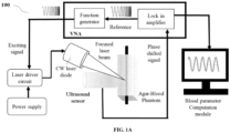
- FIG. 1A is an example architecture of system 100 for in vivo non-invasive measurement of blood parameters using Laser Enabled Swept Frequency Acoustic Interferometry (LE-SFAI), in accordance with some embodiments of the present disclosure.
- the system 100 of FIG 1A is based on applicant's earlier patent of addition filed with Indian patent application number 202323048549 with modification to the laser diode (excitation source) and ultrasound sensor placement, enabling in vivo set up.
- the intensity of a CW laser diode is modulated to generate the acoustic waves in the sample.
- acoustic waves also called CWPA waves
- CWPA waves are further acquired and used with the signal processing algorithm by a blood parameter measurement module to obtain certain features that correspond to the blood viscosity, RBC concentration, and vessel diameter.
- SNR signal-noise ratio
- this study utilizes the principle of laser enabled sweep frequency acoustic interferometry (LE-SFAI) to obtain a high SNR CWPA signal from the complex media.
- LE-SFAI sweep frequency acoustic interferometry
- FIG. 1A shows the setup which mainly comprises two parts, that is the laser excitation system and the acquisition system.
- the CW laser diode at the wavelength 520nm, and 1W peak power is used.
- the intensity of this laser diode is modulated using the custom-built laser driver.
- the laser driver supplies a sinusoidal modulated current of 0.05-0.65 A to the laser diode at a linear frequency sweep from 0.1-1MHz. This modulated current corresponds to the CW laser power of 300mW peak to peak at the sample's plane.
- the CWPA wave generated from the sample due to the laser excitation is acquired by the Ultrasound sensor (V-303 SU) from Olympus Inc.
- the PA signal from the sensor is amplified using the pre-amplifier operating at a constant gain of 60dB and processed in the Vector Network Analyzer (VNA) from Bode-100. Subsequently, the PA signal is stored in the computer and used with the signal processing algorithm to estimate the required parameters.
- VNA Vector Network Analyzer
- the laser excitation and ultrasound sensor are placed in the same plane in reflection mode (i.e. laser and ultrasound sensor are placed at the same side of the sample). Since the study proposed in-vivo applications it is essential to keep both the laser and sensor at the same side. In such cases the laser alignment becomes critical as it should not be obstructed by the ultrasound sensor.
- the design disclosed in FIG. 1A the laser gun is placed obliquely at the sample's plane. By oblique irradiation, the maximum power is transferred to the target sample. Also, the power level at the sample's plane is required to be optimum as the power should not be greater than the American National Standards Institute (ANSI) safety limits for human irradiation.
- ANSI American National Standards Institute
- the power should be sufficient to extract the acoustic information from the blood vessel which is embedded within the tissue. Since the laser diode has inherent low power, it is essential to use the principle of Laser Enabled Swept Frequency Acoustic Interferometry (LE-SFAI). Also, with CWPA signal, by using LE-SFAI, the different parameters such as blood viscosity, vessel diameter and RBC concentration are obtained. The geometry shown in the FIG. 1A . can help to achieve in vivo results by using Ultrasound coupling gel between the tissue and the ultrasound sensor.
- LE-SFAI Laser Enabled Swept Frequency Acoustic Interferometry
- P is a CWPA or PA pressure wave
- Q is a laser enabled heating function
- c s is a speed of sound in the medium
- ⁇ is a thermal expansion coefficient
- c p is a specific heat at constant pressure for a given medium.
- the intensity of each CW laser diode is sinusoidally modulated with the frequency sweep f .
- ⁇ a is an optical absorption coefficient of the sample
- I 0 is an intensity of modulation
- f is a frequency sweep from f 1 Hz to f 2 Hz.
- Equation 3 signifies the generated PA pressure is the sinusoidal function of time with the same frequency as that of laser excitation. It is directly proportional to the intensity of the laser modulation and is directly proportional to the optical absorption of the sample.
- a closed medium is established by the opposite facing walls of the sample container in which the standing waves are generated in certain conditions.
- the sweep frequency PA wave tends to traverse back and forth within the walls of the sample container. As such, it leads to a resonance condition where standing waves are created when an integral multiple of half the wavelength becomes equal to the length of the sample container (the distance between opposite walls of the sample container).
- n is the integer.
- frequency spectra of laser enabled sweep frequency acoustic interferometry (LE-SFAI) generates multiple peaks in response to equation 4.
- the peak amplitude of the first peak in the time domain CWPA signal indicates the optical absorption by the sample and the rise time obtained from the first peak signifies the viscosity of the sample.
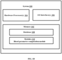
- FIG. 1B illustrates functional block diagram of the system 100 of FIG. 1A , in accordance with some embodiments of the present disclosure.
- the system 100 includes a processor(s) 104, communication interface device(s), alternatively referred as input/output (I/O) interface(s) 106, and one or more data storage devices or a memory 102 operatively coupled to the processor(s) 104.
- the system 100 with one or more hardware processors is configured to execute functions of one or more functional blocks of the system 100.
- the processor(s) 104 can be one or more hardware processors 104.
- the one or more hardware processors 104 can be implemented as one or more microprocessors, microcomputers, microcontrollers, digital signal processors, central processing units, state machines, logic circuitries, and/or any devices that manipulate signals based on operational instructions.
- the one or more hardware processors 104 are configured to fetch and execute computer-readable instructions stored in the memory 102.
- the system 100 can be implemented in a variety of computing systems including laptop computers, notebooks, hand-held devices such as mobile phones, workstations, mainframe computers, servers, and the like.
- the I/O interface(s) 106 can include a variety of software and hardware interfaces, for example, a web interface, a graphical user interface and the like and can facilitate multiple communications within a wide variety of networks N/W and protocol types, including wired networks, for example, LAN, cable, etc., and wireless networks, such as WLAN, cellular and the like.
- the I/O interface (s) 106 can include one or more ports for connecting to a number of external devices or to another server or devices.
- the memory 102 may include any computer-readable medium known in the art including, for example, volatile memory, such as static random access memory (SRAM) and dynamic random access memory (DRAM), and/or non-volatile memory, such as read only memory (ROM), erasable programmable ROM, flash memories, hard disks, optical disks, and magnetic tapes.
- volatile memory such as static random access memory (SRAM) and dynamic random access memory (DRAM)
- DRAM dynamic random access memory
- non-volatile memory such as read only memory (ROM), erasable programmable ROM, flash memories, hard disks, optical disks, and magnetic tapes.
- the memory 102 includes a plurality of modules 110 such as blood parameter computation module.
- the plurality of modules 110 include programs or coded instructions that supplement applications or functions performed by the system 100 for executing different steps involved in the process of in vivo non-invasive measurement of blood parameters using Laser Enabled Swept Frequency Acoustic Interferometry (LE-SFAI),, being performed by the system 100.
- LE-SFAI Laser Enabled Swept Frequency Acoustic Interferometry
- the plurality of modules 110 can include routines, programs, objects, components, and data structures, which performs particular tasks or implement particular abstract data types.
- the plurality of modules 110 may also be used as, signal processor(s), node machine(s), logic circuitries, and/or any other device or component that manipulates signals based on operational instructions.
- the plurality of modules 110 can be used by hardware, by computer-readable instructions executed by the one or more hardware processors 104, or by a combination thereof.
- the plurality of modules 110 can include various sub-modules (not shown).
- the memory 102 may comprise information pertaining to input(s)/output(s) of each step performed by the processor(s) 104 of the system100 and methods of the present disclosure.
- the memory 102 includes a database 108.
- the database (or repository) 108 may include a plurality of abstracted pieces of code for refinement and data that is processed, received, or generated as a result of the execution of the plurality of modules in the module(s) 110.
- the database can store the determine the plurality of blood parameters.
- the data base 108 is shown internal to the system 100, it will be noted that, in alternate embodiments, the database 108 can also be implemented external to the system 100, and communicatively coupled to the system 100.
- the data contained within such an external database may be periodically updated. For example, new data may be added into the database (not shown in FIG. 1A ) and/or existing data may be modified and/or non-useful data may be deleted from the database.
- the data may be stored in an external system, such as a Lightweight Directory Access Protocol (LDAP) directory and a Relational Database Management System (RDBMS). Functions of the components of the system 100 are now explained with reference to steps in flow diagrams in FIG. 2 through FIG. 4 .
- LDAP Lightweight Directory Access Protocol
- RDBMS Relational Database Management System
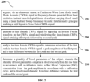
- FIG. 2 is a flow diagram illustrating a method 200 for in vivo non-invasive measurement of blood parameters using LE-SFAI, using the system depicted in FIG. 1A and 1B , in accordance with some embodiments of the present disclosure.
- the system 100 comprises one or more data storage devices or the memory 102 operatively coupled to the processor(s) 104 and is configured to store instructions for execution of steps of the method 200 by the processor(s) or one or more hardware processors 104.
- the steps of the method 200 of the present disclosure will now be explained with reference to the components or blocks of the system 100 as depicted in FIG. 1A and 1B and the steps of flow diagram as depicted in FIG. 2 .
- process steps, method steps, techniques or the like may be described in a sequential order, such processes, methods, and techniques may be configured to work in alternate orders. In other words, any sequence or order of steps that may be described does not necessarily indicate a requirement that the steps be performed in that order.
- the steps of processes described herein may be performed in any order practical. Further, some steps may be performed simultaneously.
- the one or more hardware processors 104 are configured by the instructions to acquire via the ultrasound sensor, the Continuous Wave Laser diode based Photo Acoustic (CWPA) signal in frequency domain generated from laser excitation incident on the biological tissue of a subject carrying blood vessel using a Laser Enabled Sweep Frequency Acoustic Interferometry (LE-SFAI) principle controlled by one or more hardware processors, wherein the LE-SFAI enabling a high Signal to Noise Ratio (SNR) CWPA signal.
- CWPA Continuous Wave Laser diode based Photo Acoustic
- LE-SFAI Laser Enabled Sweep Frequency Acoustic Interferometry
- the laser gun generating the laser excitation and the ultrasound sensor acquiring the CWPA signal are placed in same plane in context of the biological tissue, and work in reflection mode.
- the one or more hardware processors 104 are configured by the instructions to generate the time domain CWPA signal by applying the inverse Fourier transform on the CWPA signal and windowing the time-domain CWPA signal retaining a first peak followed by a second peak.
- the one or more hardware processors 104 are configured by the instructions to analyze the time domain CWPA signal to determine a rise of the time domain CWPA signal from the first peak, a peak amplitude of the first peak and a time difference between the first peak and the second peak.
- the blood parameter computation module executed by the one or more hardware processors 104 is configured by the instructions to determine the plurality of blood parameters of the subject.
- the plurality of blood parameters comprise a blood viscosity, a Red Blood Corpuscles (RBC) concentration and a blood vessel diameter.
- the blood viscosity is determined from the rise time of the time domain CWPA signal from the first peak in accordance with a blood viscosity calibration curve predetermined experimentally by observing relation between change in blood viscosity against change in rise time of the first peak in the time domain CWPA signal.
- the RBC concentration is determined from the peak amplitude of the first peak in accordance with a RBC concentration calibration curve predetermined experimentally by observing the relation between change RBC concentration against change in peak amplitude.
- the blood vessel diameter is determined using the product of the time difference between the first peak and the second peak and the velocity of the time domain CWPA signal.
- Sample preparation Initially, samples having distinct viscosity and optical absorptions are prepared by mixing glycerol and dye in different proportions. First, five samples with different proportions of dye that are 0%, 2%, 4%, 6%, and 8% are added in 40% glycerol. The different concentrations of dye exhibit different optical absorptions which resembles to the variation in the concentration of RBCs. Secondly, five samples of dye with varying glycerol concentrations in the range of 0% to 20% at an interval of 5% are prepared and experimented with. The variation in glycerol concentration simulates the viscosity variation in the blood (dye).
- the tissue mimicking phantoms using agarose were prepared in which 1.5 gm agarose was mixed in 100ml of de-ionized water. The mixture was heated for 2 minutes in the microwave oven, poured into the rectangular container, and allowed to cool at room temperature. After 1 minute of cooling a Polyvinylidene Fluoride Fluoropolymer (PVDE) tube with a diameter of 2mm was embedded within the phantom, and again the phantom gel was allowed to cool for 15-20 minutes. The thickness of the phantom is kept as 12mm and the PVDE tube was inserted at a depth of 4mm from the laser excitation plane. This PVDE tube simulates the artificial blood vessel and during the experiments, three samples of each of the dye-glycerol mixtures are poured into the vessel and CWPA signals are acquired.
- PVDE Polyvinylidene Fluoride Fluoropolymer
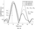
- FIG. 3A shows that the amplitude of the first peak increases with the increase in the dye concentration while the rise time remains the same.
- FIG. 3B shows the observation when three samples of fixed dye but varying glycerol concentration are filled in the vessel tube sequentially. In FIG. 3B the rise time increases linearly with the increase in glycerol concentration, but the amplitude remains proximally the same.
- a second peak can be observed near the first peak.
- the difference between the two consecutive peaks (I and II peaks) is measured as ⁇ t.
- Laparoscopy Surgery In this surgery a harmonic cutter is used. A surgeon needs to be assured whether the vessel within the tissue is intact and not cut. Presently this is done by the experience of the surgeon.
- the system 100 can be integrated with the harmonic cutter to provide a fair idea about the presence of the blood vessel and its diameter.
- Thickening of blood may be related to CVD and to monitor this repeated blood tests are required.
- the proposed method can be useful for determining blood viscosity in a non-invasive way. Also, it could provide a direct measurement of viscosity rather than indirect parameters for predicting blood viscosity as used in conventional methods.
- the method and system disclosed herein estimates the blood viscosity, and RBC concentration and blood vessel diameter.
- the system is very compact, cost-effective, and portable which increases its chance of reaching the clinical benches.
- the hardware device can be any kind of device which can be programmed including e.g. any kind of computer like a server or a personal computer, or the like, or any combination thereof.
- the device may also include means which could be e.g. hardware means like e.g. an application-specific integrated circuit (ASIC), a field-programmable gate array (FPGA), or a combination of hardware and software means, e.g.
- ASIC application-specific integrated circuit
- FPGA field-programmable gate array
- the means can include both hardware means, and software means.
- the method embodiments described herein could be implemented in hardware and software.
- the device may also include software means.
- the embodiments may be implemented on different hardware devices, e.g. using a plurality of CPUs.
- the embodiments herein can comprise hardware and software elements.
- the embodiments that are implemented in software include but are not limited to, firmware, resident software, microcode, etc.
- the functions performed by various components described herein may be implemented in other components or combinations of other components.
- a computer-usable or computer readable medium can be any apparatus that can comprise, store, communicate, propagate, or transport the program for use by or in connection with the instruction execution system, apparatus, or device.
- a computer-readable storage medium refers to any type of physical memory on which information or data readable by a processor may be stored.
- a computer-readable storage medium may store instructions for execution by one or more processors, including instructions for causing the processor(s) to perform steps or stages consistent with the embodiments described herein.
- the term "computer-readable medium” should be understood to include tangible items and exclude carrier waves and transient signals, i.e., be non-transitory. Examples include random access memory (RAM), read-only memory (ROM), volatile memory, nonvolatile memory, hard drives, CD ROMs, DVDs, flash drives, disks, and any other known physical storage media.
Landscapes
- Health & Medical Sciences (AREA)
- Life Sciences & Earth Sciences (AREA)
- Physics & Mathematics (AREA)
- Engineering & Computer Science (AREA)
- Biomedical Technology (AREA)
- Medical Informatics (AREA)
- Animal Behavior & Ethology (AREA)
- Public Health (AREA)
- Surgery (AREA)
- Veterinary Medicine (AREA)
- Biophysics (AREA)
- Pathology (AREA)
- General Health & Medical Sciences (AREA)
- Heart & Thoracic Surgery (AREA)
- Molecular Biology (AREA)
- Physiology (AREA)
- Computer Vision & Pattern Recognition (AREA)
- Artificial Intelligence (AREA)
- Signal Processing (AREA)
- Psychiatry (AREA)
- Optics & Photonics (AREA)
- Mathematical Physics (AREA)
- Cardiology (AREA)
- Acoustics & Sound (AREA)
- Dentistry (AREA)
- Oral & Maxillofacial Surgery (AREA)
- Fuzzy Systems (AREA)
- Hematology (AREA)
- Nuclear Medicine, Radiotherapy & Molecular Imaging (AREA)
- Radiology & Medical Imaging (AREA)
- Evolutionary Computation (AREA)
- Investigating Or Analysing Materials By Optical Means (AREA)
- Measurement Of The Respiration, Hearing Ability, Form, And Blood Characteristics Of Living Organisms (AREA)
Abstract
Description
- The present application claims priority to Indian application no.
Indian Patent Application No. 202321076674, filed on November 9, 2023 - The embodiments herein generally relate to the field of in-vivo non-invasive measurements of physiological parameters and, more particularly, to a method and system for in vivo non-invasive measurement of blood parameters using Laser Enabled Swept Frequency Acoustic Interferometry (LE-SFAI) principle.
- Measurement of blood parameters is crucial in predictive and diagnostic clinical consultations. Conventionally the blood parameter measurements, also referred to as blood tests, have been in vitro and invasive. Herein a blood sample is taken from a patient and sent to a laboratory for analysis.
- Furthermore, blood parameters such as blood viscosity is typically estimated indirectly through various parameters, such as hematocrit and total serum protein levels, which can provide some information about blood thickness. However, these methods do not provide a direct measurement of blood viscosity.
- Any in vitro measurements (laboratory based analysis) separates the component under test such as blood from the living organism, this has an effect on accuracy of estimation of values of blood parameters because the temperature of the blood inside the human body would affect the viscosity. However, in in vitro measurement the temperature would not be the same as that of the blood inside the human body thus, viscosity being a thermophysical property would be misinterpreted when measured in vitro. Furthermore, results of laboratory tests are time consuming, do not provide instant measurements, this delay may be very critical in certain scenarios. For example, certain surgeries, specifically based on laparoscopic techniques where limited surgery region is visible with limitation of the camera field of view and requires on values of the blood parameters or state of a blood vessel for quick critical decisions during the surgical process, current in vitro techniques do not provide any solution. Thus, the analysis is totally dependent on the experience of the surgeon. In certain types of diseases, patients require frequent observation of blood viscosity or Red Blood Corpuscles (RBC) concentration and the like. Thus, in vitro (lab tests) and invasive ( removing blood sample by piercing the patient) frequently is not convenient and is not desired.
- Direct in vivo blood viscosity measurement is challenging due to the complex rheological properties of blood and the dynamic nature of blood flow in the circulatory system. Most clinical assessments focus on related factors and conditions rather than direct measurements of viscosity.
- Attempts have been made to provide in vivo non-invasive measurements for some blood parameters such as glucose level etc.. however blood viscosity, RBC, blood vessel diameter measurement is still a challenge and hardly addressed with compact light weight systems.
- Further, Photoacoustic (PA) sensing techniques have been in use for parameters measurement of various fluids but applying such techniques to fluid like blood, which exhibits non-Newtonian characteristics is further challenging. Thus, the physics of blood as a non-Newtonian fluid needs to be well studied to accurately derive the parameters of interest.
- Embodiments of the present disclosure present technological improvements as solutions to one or more of the above-mentioned technical problems recognized by the inventors in conventional systems.
- For example, in one embodiment, a method for in vivo non-invasive measurement of blood parameters using Laser Enabled Swept Frequency Acoustic Interferometry (LE-SFAI) principle is provided.
- The method includes acquiring, via an ultrasound sensor, a Continuous Wave Laser diode based Photo Acoustic (CWPA) signal in frequency domain generated from laser excitation controlled by one or more hardware processors and incident on a biological tissue of a subject carrying blood vessel using a Laser Enabled Sweep Frequency Acoustic Interferometry (LE-SFAI) principle, wherein the LE-SFAI enabling a high Signal to Noise Ratio (SNR) CWPA signal. Further the method includes generating a time domain CWPA signal by applying an inverse Fourier transform on the CWPA signal and windowing the time-domain CWPA signal retaining a first peak followed by a second peak.
- Furthermore, the method includes analyzing the time domain CWPA signal to determine a rise time of the first peak in the time domain CWPA signal, a peak amplitude of the first peak and a time difference between the first peak and the second peak. Furthermore, the method includes determining a plurality of blood parameters of the subject, wherein the plurality of blood parameters comprise a blood viscosity, a Red Blood Corpuscle (RBC) concentration and a blood vessel diameter. The wherein the blood viscosity is determined from the rise time of the time domain CWPA signal from the first peak in accordance with a blood viscosity calibration curve predetermined experimentally by observing relation between change in blood viscosity against change in rise the time domain CWPA signal. The RBC concentration is determined from the peak amplitude of the first peak in accordance with a RBC concentration calibration curve predetermined experimentally by observing the relation between change RBC concentration against change in peak amplitude. The blood vessel diameter is determined using the product of the time difference between the first peak and the second peak and the velocity of the time domain CWPA signal.
- In another aspect, a system for in vivo non-invasive measurement of blood parameters using Laser Enabled Swept Frequency Acoustic Interferometry (LE-SFAI) principle is provided. The system comprises a memory storing instructions; one or more Input/Output (I/O) interfaces; and one or more hardware processors coupled to the memory via the one or more I/O interfaces, wherein the one or more hardware processors are configured by the instructions acquire, via an ultrasound sensor, a Continuous Wave Laser diode based Photo Acoustic (CWPA) signal in frequency domain generated from laser excitation controlled by one or more hardware processors and incident on a biological tissue of a subject carrying blood vessel using a Laser Enabled Sweep Frequency Acoustic Interferometry (LE-SFAI) principle, wherein the LE-SFAI enabling a high Signal to Noise Ratio (SNR) CWPA signal. Further the system is configured to generate a time domain CWPA signal by applying an inverse Fourier transform on the CWPA signal and windowing the time-domain CWPA signal retaining a first peak followed by a second peak.
- Furthermore, the system is configured to analyze the time domain CWPA signal to determine a rise time of the first peak in the time domain CWPA signal, a peak amplitude of the first peak and a time difference between the first peak and the second peak. Furthermore, the system is configured to determine a plurality of blood parameters of the subject, wherein the plurality of blood parameters comprise a blood viscosity, a Red Blood Corpuscle (RBC) concentration and a blood vessel diameter. The wherein the blood viscosity is determined from the rise time of the time domain CWPA signal from the first peak in accordance with a blood viscosity calibration curve predetermined experimentally by observing relation between change in blood viscosity against change in rise the time domain CWPA signal. The RBC concentration is determined from the peak amplitude of the first peak in accordance with a RBC concentration calibration curve predetermined experimentally by observing the relation between change RBC concentration against change in peak amplitude. The blood vessel diameter is determined using the product of the time difference between the first peak and the second peak and the velocity of the time domain CWPA signal.
- In yet another aspect, there are provided one or more non-transitory machine-readable information storage mediums comprising one or more instructions, which when executed by one or more hardware processors causes a method for in vivo non-invasive measurement of blood parameters using Laser Enabled Swept Frequency Acoustic Interferometry (LE-SFAI) principle.
- The method includes acquiring, via an ultrasound sensor, a Continuous Wave Laser diode based Photo Acoustic (CWPA) signal in frequency domain generated from laser excitation controlled by one or more hardware processors and incident on a biological tissue of a subject carrying blood vessel using a Laser Enabled Sweep Frequency Acoustic Interferometry (LE-SFAI) principle, wherein the LE-SFAI enabling a high Signal to Noise Ratio (SNR) CWPA signal. Further the method includes generating a time domain CWPA signal by applying an inverse Fourier transform on the CWPA signal and windowing the time-domain CWPA signal retaining a first peak followed by a second peak.
- Furthermore, the method includes analyzing the time domain CWPA signal to determine a rise time of the first peak in the time domain CWPA signal, a peak amplitude of the first peak and a time difference between the first peak and the second peak. Furthermore, the method includes determining a plurality of blood parameters of the subject, wherein the plurality of blood parameters comprise a blood viscosity, a Red Blood Corpuscle (RBC) concentration and a blood vessel diameter. The wherein the blood viscosity is determined from the rise time of the time domain CWPA signal from the first peak in accordance with a blood viscosity calibration curve predetermined experimentally by observing relation between change in blood viscosity against change in rise the time domain CWPA signal. The RBC concentration is determined from the peak amplitude of the first peak in accordance with a RBC concentration calibration curve predetermined experimentally by observing the relation between change RBC concentration against change in peak amplitude. The blood vessel diameter is determined using the product of the time difference between the first peak and the second peak and the velocity of the time domain CWPA signal.
- It is to be understood that both the foregoing general description and the following detailed description are exemplary and explanatory only and are not restrictive of the invention, as claimed.
- The accompanying drawings, which are incorporated in and constitute a part of this disclosure, illustrate exemplary embodiments and, together with the description, serve to explain the disclosed principles:
-
FIG. 1A is a system architecture for in vivo non-invasive measurement of blood parameters using Laser Enabled Swept Frequency Acoustic Interferometry (LE-SFAI) principle , in accordance with some embodiments of the present disclosure. -
FIG. 1B illustrates functional block diagram of the system ofFIG. 1A , in accordance with some embodiments of the present disclosure. -
FIG. 2 is a flow diagram illustrating a method for in vivo non-invasive measurement of blood parameters using LE-SFAI principle, using the system depicted inFIG. 1A and1B , in accordance with some embodiments of the present disclosure. -
FIG. 3A andFIG 3B depicts characteristics of time domain continuous wave Photo Acoustic (CWPA) signal acquired by ultrasound sensor of the LE-SFAI based system ofFIG. 1 while experiments are conducted on tissue mimicking phantoms with varying dye concentration (mimicking Red Blood Corpuscles (RBC) concentration) and glycerol concentration (mimicking blood viscosity)respectively, in accordance with some embodiments of the present disclosure. -
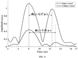
FIG. 4 depicts characteristics of time domain continuous wave Photo Acoustic (CWPA) signal acquired by ultrasound sensors of the LE-SFAI based system ofFIG. 1 while experiments are conducted on tissue mimicking phantoms with varying blood vessel diameter, in accordance with some embodiments of the present disclosure. - It should be appreciated by those skilled in the art that any block diagrams herein represent conceptual views of illustrative systems and devices embodying the principles of the present subject matter. Similarly, it will be appreciated that any flow charts, flow diagrams, and the like represent various processes which may be substantially represented in computer readable medium and so executed by a computer or processor, whether or not such computer or processor is explicitly shown.
- Exemplary embodiments are described with reference to the accompanying drawings. In the figures, the left-most digit(s) of a reference number identifies the figure in which the reference number first appears. Wherever convenient, the same reference numbers are used throughout the drawings to refer to the same or like parts. While examples and features of disclosed principles are described herein, modifications, adaptations, and other implementations are possible without departing from the scope of the disclosed embodiments.
- Embodiments of the present disclosure provide a method and system for in vivo non-invasive measurement of blood parameters using Laser Enabled Swept Frequency Acoustic Interferometry (LE-SFAI) principle. A Continuous Wave Laser diode based Photo Acoustic (CWPA) signal in frequency domain generated from laser excitation incident on a biological tissue of a subject carrying blood vessel is acquired by the ultrasound sensor and converted to a windowed time domain CWPA signal limited to two peaks. The signal characteristics such as velocity, amplitude, and time difference between first peak and the second peak are obtained to derive blood viscosity, Red Blood Corpuscles (RBC) concentration, and blood vessel diameter.
- The system set up, which is based on LE-SFAI principle is similar to one proposed by applicant in filed patent of addition at Indian Patent office on
19 July 2023, having Indian patent application number 202323048549 - PhotoAcoustic (PA) has been widely explored as an imaging and microscopic tool for various biological studies. Despite its promising results, it has not significantly reached the clinical benches. One of the important reasons for its non-availability as a medical diagnostic tool could be the usage of high energy pulsed laser which makes the system complex, bulky, costly, and highly operator dependent.
- Furthermore, the applicant's
Indian patent application number 202323048549 - Referring now to the drawings, and more particularly to
FIGS. 1A through 4 , where similar reference characters denote corresponding features consistently throughout the figures, there are shown preferred embodiments, and these embodiments are described in the context of the following exemplary system and/or method. -
FIG. 1A is an example architecture ofsystem 100 for in vivo non-invasive measurement of blood parameters using Laser Enabled Swept Frequency Acoustic Interferometry (LE-SFAI), in accordance with some embodiments of the present disclosure. Thesystem 100 ofFIG 1A is based on applicant's earlier patent of addition filed withIndian patent application number 202323048549 -
FIG. 1A shows the setup which mainly comprises two parts, that is the laser excitation system and the acquisition system. For sample irradiation, the CW laser diode at the wavelength 520nm, and 1W peak power is used. The intensity of this laser diode is modulated using the custom-built laser driver. The laser driver supplies a sinusoidal modulated current of 0.05-0.65 A to the laser diode at a linear frequency sweep from 0.1-1MHz. This modulated current corresponds to the CW laser power of 300mW peak to peak at the sample's plane. The CWPA wave generated from the sample due to the laser excitation is acquired by the Ultrasound sensor (V-303 SU) from Olympus Inc.™, which is placed in contact with the sample container and the tissue mimicking phantom carrying the blood vessel, via the acoustic coupling gel. Further, the PA signal from the sensor is amplified using the pre-amplifier operating at a constant gain of 60dB and processed in the Vector Network Analyzer (VNA) from Bode-100. Subsequently, the PA signal is stored in the computer and used with the signal processing algorithm to estimate the required parameters. - The important point to be noted here is that the laser excitation and ultrasound sensor are placed in the same plane in reflection mode (i.e. laser and ultrasound sensor are placed at the same side of the sample). Since the study proposed in-vivo applications it is essential to keep both the laser and sensor at the same side. In such cases the laser alignment becomes critical as it should not be obstructed by the ultrasound sensor. Hence, the design disclosed in
FIG. 1A , the laser gun is placed obliquely at the sample's plane. By oblique irradiation, the maximum power is transferred to the target sample. Also, the power level at the sample's plane is required to be optimum as the power should not be greater than the American National Standards Institute (ANSI) safety limits for human irradiation. Secondly, the power should be sufficient to extract the acoustic information from the blood vessel which is embedded within the tissue. Since the laser diode has inherent low power, it is essential to use the principle of Laser Enabled Swept Frequency Acoustic Interferometry (LE-SFAI). Also, with CWPA signal, by using LE-SFAI, the different parameters such as blood viscosity, vessel diameter and RBC concentration are obtained. The geometry shown in theFIG. 1A . can help to achieve in vivo results by using Ultrasound coupling gel between the tissue and the ultrasound sensor. - Physics of blood (Theory): The basic time variant wave equation for the photoacoustic pressure known in the art is as follows provided in equation 1:

- Where, P is a CWPA or PA pressure wave, Q is a laser enabled heating function, cs is a speed of sound in the medium, β is a thermal expansion coefficient, cp is a specific heat at constant pressure for a given medium. Here, the intensity of each CW laser diode is sinusoidally modulated with the frequency sweep f. Following to the CW laser intensity modulation, the heating function modulates at the same frequency and is expressed in equation 2 based study in the art,

- In equation 2, µa is an optical absorption coefficient of the sample, I 0 is an intensity of modulation, ω is an angular frequency given as ω = 2πf. Here, f is a frequency sweep from f1 Hz to f2 Hz. Considering the non-Newtonian fluid (blood) is in form of a biological tissue carrying blood vessel, the blood vessel can be considered as optically thin spherical absorber of radius a, and r is a radial coordinate, the PA wave in frequency domain can be expressed in Equation 3, P =

- Where q = ω.a/cs and ρ is a density of the medium. Equation 3 signifies the generated PA pressure is the sinusoidal function of time with the same frequency as that of laser excitation. It is directly proportional to the intensity of the laser modulation and is directly proportional to the optical absorption of the sample.
- To achieve the high SNR of the CWPA wave or simply PA wave, a closed medium is established by the opposite facing walls of the sample container in which the standing waves are generated in certain conditions. The sweep frequency PA wave tends to traverse back and forth within the walls of the sample container. As such, it leads to a resonance condition where standing waves are created when an integral multiple of half the wavelength becomes equal to the length of the sample container (the distance between opposite walls of the sample container). Considering the length of the container as 'D' and wavelength as λ, the standing waves are formed according to equation 4 below.

- Where n is the integer. As can be noted from applicant's earlier patent of addition filed with
Indian patent application number 202323048549 
- If the velocity of sound in the medium is known then (5) can be rewritten to observe the dimension of the container as:

- Further, the peak amplitude of the first peak in the time domain CWPA signal indicates the optical absorption by the sample and the rise time obtained from the first peak signifies the viscosity of the sample. Thus, this study explores the rise time and peak amplitude, and Δt from the time domain CWPA signal to estimate the blood viscosity, RBC concentration, and vessel diameter.
-
FIG. 1B illustrates functional block diagram of thesystem 100 ofFIG. 1A , in accordance with some embodiments of the present disclosure. - In an embodiment, the
system 100 includes a processor(s) 104, communication interface device(s), alternatively referred as input/output (I/O) interface(s) 106, and one or more data storage devices or amemory 102 operatively coupled to the processor(s) 104. Thesystem 100 with one or more hardware processors is configured to execute functions of one or more functional blocks of thesystem 100. - Referring to the components of
system 100, in an embodiment, the processor(s) 104, can be one ormore hardware processors 104. In an embodiment, the one ormore hardware processors 104 can be implemented as one or more microprocessors, microcomputers, microcontrollers, digital signal processors, central processing units, state machines, logic circuitries, and/or any devices that manipulate signals based on operational instructions. Among other capabilities, the one ormore hardware processors 104 are configured to fetch and execute computer-readable instructions stored in thememory 102. In an embodiment, thesystem 100 can be implemented in a variety of computing systems including laptop computers, notebooks, hand-held devices such as mobile phones, workstations, mainframe computers, servers, and the like. - The I/O interface(s) 106 can include a variety of software and hardware interfaces, for example, a web interface, a graphical user interface and the like and can facilitate multiple communications within a wide variety of networks N/W and protocol types, including wired networks, for example, LAN, cable, etc., and wireless networks, such as WLAN, cellular and the like. In an embodiment, the I/O interface (s) 106 can include one or more ports for connecting to a number of external devices or to another server or devices.
- The
memory 102 may include any computer-readable medium known in the art including, for example, volatile memory, such as static random access memory (SRAM) and dynamic random access memory (DRAM), and/or non-volatile memory, such as read only memory (ROM), erasable programmable ROM, flash memories, hard disks, optical disks, and magnetic tapes. - In an embodiment, the
memory 102 includes a plurality ofmodules 110 such as blood parameter computation module. The plurality ofmodules 110 include programs or coded instructions that supplement applications or functions performed by thesystem 100 for executing different steps involved in the process of in vivo non-invasive measurement of blood parameters using Laser Enabled Swept Frequency Acoustic Interferometry (LE-SFAI),, being performed by thesystem 100. The plurality ofmodules 110, amongst other things, can include routines, programs, objects, components, and data structures, which performs particular tasks or implement particular abstract data types. The plurality ofmodules 110 may also be used as, signal processor(s), node machine(s), logic circuitries, and/or any other device or component that manipulates signals based on operational instructions. Further, the plurality ofmodules 110 can be used by hardware, by computer-readable instructions executed by the one ormore hardware processors 104, or by a combination thereof. The plurality ofmodules 110 can include various sub-modules (not shown). - Further, the
memory 102 may comprise information pertaining to input(s)/output(s) of each step performed by the processor(s) 104 of the system100 and methods of the present disclosure. - Further, the
memory 102 includes adatabase 108. The database (or repository) 108 may include a plurality of abstracted pieces of code for refinement and data that is processed, received, or generated as a result of the execution of the plurality of modules in the module(s) 110. The database can store the determine the plurality of blood parameters. - Although the
data base 108 is shown internal to thesystem 100, it will be noted that, in alternate embodiments, thedatabase 108 can also be implemented external to thesystem 100, and communicatively coupled to thesystem 100. The data contained within such an external database may be periodically updated. For example, new data may be added into the database (not shown inFIG. 1A ) and/or existing data may be modified and/or non-useful data may be deleted from the database. In one example, the data may be stored in an external system, such as a Lightweight Directory Access Protocol (LDAP) directory and a Relational Database Management System (RDBMS). Functions of the components of thesystem 100 are now explained with reference to steps in flow diagrams inFIG. 2 through FIG. 4 . -
FIG. 2 is a flow diagram illustrating amethod 200 for in vivo non-invasive measurement of blood parameters using LE-SFAI, using the system depicted inFIG. 1A and1B , in accordance with some embodiments of the present disclosure. - In an embodiment, the
system 100 comprises one or more data storage devices or thememory 102 operatively coupled to the processor(s) 104 and is configured to store instructions for execution of steps of themethod 200 by the processor(s) or one ormore hardware processors 104. The steps of themethod 200 of the present disclosure will now be explained with reference to the components or blocks of thesystem 100 as depicted inFIG. 1A and1B and the steps of flow diagram as depicted inFIG. 2 . Although process steps, method steps, techniques or the like may be described in a sequential order, such processes, methods, and techniques may be configured to work in alternate orders. In other words, any sequence or order of steps that may be described does not necessarily indicate a requirement that the steps be performed in that order. The steps of processes described herein may be performed in any order practical. Further, some steps may be performed simultaneously. - As depicted in
FIG. 1A and referring to the steps of themethod 200, atstep 202 of themethod 200, the one ormore hardware processors 104 are configured by the instructions to acquire via the ultrasound sensor, the Continuous Wave Laser diode based Photo Acoustic (CWPA) signal in frequency domain generated from laser excitation incident on the biological tissue of a subject carrying blood vessel using a Laser Enabled Sweep Frequency Acoustic Interferometry (LE-SFAI) principle controlled by one or more hardware processors, wherein the LE-SFAI enabling a high Signal to Noise Ratio (SNR) CWPA signal. - The laser gun generating the laser excitation and the ultrasound sensor acquiring the CWPA signal are placed in same plane in context of the biological tissue, and work in reflection mode.
- At
step 204 of themethod 200, the one ormore hardware processors 104 are configured by the instructions to generate the time domain CWPA signal by applying the inverse Fourier transform on the CWPA signal and windowing the time-domain CWPA signal retaining a first peak followed by a second peak. - At
step 206 of themethod 200, the one ormore hardware processors 104 are configured by the instructions to analyze the time domain CWPA signal to determine a rise of the time domain CWPA signal from the first peak, a peak amplitude of the first peak and a time difference between the first peak and the second peak. - At
step 208 of themethod 200, the blood parameter computation module executed by the one ormore hardware processors 104 is configured by the instructions to determine the plurality of blood parameters of the subject. The plurality of blood parameters comprise a blood viscosity, a Red Blood Corpuscles (RBC) concentration and a blood vessel diameter. The blood viscosity is determined from the rise time of the time domain CWPA signal from the first peak in accordance with a blood viscosity calibration curve predetermined experimentally by observing relation between change in blood viscosity against change in rise time of the first peak in the time domain CWPA signal. The RBC concentration is determined from the peak amplitude of the first peak in accordance with a RBC concentration calibration curve predetermined experimentally by observing the relation between change RBC concentration against change in peak amplitude. The blood vessel diameter is determined using the product of the time difference between the first peak and the second peak and the velocity of the time domain CWPA signal. - Experimental results and discussion: The method and system disclosed herein is further explained with experimental set up as in
FIG. 1A , wherein the laser diode incidents the laser excitation on various samples of glycerol (mimicking blood for viscosity) and a dye (red ink) in the glycerol (mimicking RBC concentration in blood) and phantom mimicking biological tissue carrying blood vessel ( using agarose). It can be noted that The red ink optical absorption (µa) at 520 nm is proximal to that of the blood (µa = 1.82 cm-1) - Sample preparation: Initially, samples having distinct viscosity and optical absorptions are prepared by mixing glycerol and dye in different proportions. First, five samples with different proportions of dye that are 0%, 2%, 4%, 6%, and 8% are added in 40% glycerol. The different concentrations of dye exhibit different optical absorptions which resembles to the variation in the concentration of RBCs. Secondly, five samples of dye with varying glycerol concentrations in the range of 0% to 20% at an interval of 5% are prepared and experimented with. The variation in glycerol concentration simulates the viscosity variation in the blood (dye). Further, the tissue mimicking phantoms using agarose were prepared in which 1.5 gm agarose was mixed in 100ml of de-ionized water. The mixture was heated for 2 minutes in the microwave oven, poured into the rectangular container, and allowed to cool at room temperature. After 1 minute of cooling a Polyvinylidene Fluoride Fluoropolymer (PVDE) tube with a diameter of 2mm was embedded within the phantom, and again the phantom gel was allowed to cool for 15-20 minutes. The thickness of the phantom is kept as 12mm and the PVDE tube was inserted at a depth of 4mm from the laser excitation plane. This PVDE tube simulates the artificial blood vessel and during the experiments, three samples of each of the dye-glycerol mixtures are poured into the vessel and CWPA signals are acquired.
- Results and Discussion: In this study, the experiments are performed in two steps. First, the liquid dye-glycerol mixtures are poured into the rectangular container for PA measurement. A plot of time domain CWPA signal for the variation in dye concentration while keeping the glycerol concentration the same is obtained. The amplitude of the PA signal (time domain CWPA signal) is observed to be increasing with the increase in dye concentration. This agrees with equation 3, which indicates that for the given laser illumination if the concentration of optical absorbers increases there is an increase in PA amplitude. The stated findings are confirmed with the optical spectrophotometer results wherein the optical absorption of samples is reported to be 1.4 cm-1, 1.65 cm-1, 1.82 cm-1, 2.1 cm-1, and 2.4 cm-1 respectively for the increase in dye concentration from 2% to 10%. Also, the rise time of the time domain CWPA signal is observed to be almost constant. Similarly, it is observed that the variation in the glycerol concentration from 0% to 20% in the five steps and for the fixed proportion of dye. Here, the time domain CWPA amplitude is found to be almost constant, but the rise time increases linearly with the increase in glycerol concentration.
- Further, experiments and study also indicates that the time domain CWPA signal shifts towards the left (in the time scale) with the increase in glycerol concentration. Since the viscosity affects the velocity of the CWPA wave, this shift is observed. However, irrespective of the time shift of the time domain CWPA signal, it is not considered as the desired feature for estimating viscosity. Since, in PA, the time shift can change significantly due to other reasons (such as a change in penetration depth) as well. The variation in viscosity is also validated with the viscometer observations which give the values as 1.15cP, 1.23cP, 1.3cP, 1.35cP, and 1.8cP for glycerol variation of 0% to 20% respectively.
- The aforementioned findings interpret that the RBC concentration and the blood viscosity can be observed through CWPA measurements by observing the amplitude and rise time of the time domain CWPA wave. To further demonstrate the feasibility of CWPA sensing, the experiments are performed with the developed phantoms.
- Analogous to the previous experiments, in Phantom, initially three samples of fixed glycerol concentration and varying dye concentration are filled sequentially in the PVDE tube, and PA measurements are performed.
FIG. 3A shows that the amplitude of the first peak increases with the increase in the dye concentration while the rise time remains the same. In contrast,FIG. 3B shows the observation when three samples of fixed dye but varying glycerol concentration are filled in the vessel tube sequentially. InFIG. 3B the rise time increases linearly with the increase in glycerol concentration, but the amplitude remains proximally the same. - In
FIG. 3A andFIG. 3B , a second peak can be observed near the first peak. The difference between the two consecutive peaks (I and II peaks) is measured as Δt. This Δt (4.17µs) when multiplied by the velocity of PA wave in blood (dye) that is approximately 1450 m/s, gives a length of 2.9±0.1mm. - It can be examined that the value of Δt1(4.17 ± 0.01 µs) remains the same irrespective of the change in viscosity or optical absorption. Further, when measured with vernier caliper this dimension was found to be the dimension of a vessel.
- To confirm that Δt indicates the dimension of the vessel, a tube with a different diameter (3.6mm) was embedded within the phantom. The PA measurement in
FIG. 4 shows that the Δt2 for the newly inserted tube is 5.11µs which corresponds to the tube diameter of 3.5 mm. Therefore, it is confirmed that with the knowledge of Δt, the dimension of the vessel can be measured. Thus, the above findings indicate that the RBC concentration, blood viscosity, and vessel diameter can be effectively estimated from the time domain CWPA signal response. When used with higher frequencies, this method can also be applied to observe the diameters of the vessels in the sub-millimeter region. - Laparoscopy Surgery: In this surgery a harmonic cutter is used. A surgeon needs to be assured whether the vessel within the tissue is intact and not cut. Presently this is done by the experience of the surgeon. The system 100can be integrated with the harmonic cutter to provide a fair idea about the presence of the blood vessel and its diameter.
- Quick blood test within the clinic: In OPD, for rapid diagnosis of RBC concentrations and blood viscosity doctor can use this probe to get the initial information about the RBC and viscosity and suggest an immediate action. Presently, the doctor suggests certain blood tests, which take at least 1 day to obtain the required information.
- Thickening of blood may be related to CVD and to monitor this repeated blood tests are required. However, the proposed method can be useful for determining blood viscosity in a non-invasive way. Also, it could provide a direct measurement of viscosity rather than indirect parameters for predicting blood viscosity as used in conventional methods.
- Thus, the method and system disclosed herein estimates the blood viscosity, and RBC concentration and blood vessel diameter. The system is very compact, cost-effective, and portable which increases its chance of reaching the clinical benches.
- The written description describes the subject matter herein to enable any person skilled in the art to make and use the embodiments. The scope of the subject matter embodiments is defined by the claims and may include other modifications that occur to those skilled in the art. Such other modifications are intended to be within the scope of the claims if they have similar elements that do not differ from the literal language of the claims or if they include equivalent elements with insubstantial differences from the literal language of the claims.
- It is to be understood that the scope of the protection is extended to such a program and in addition to a computer-readable means having a message therein; such computer-readable storage means contain program-code means for implementation of one or more steps of the method, when the program runs on a server or mobile device or any suitable programmable device. The hardware device can be any kind of device which can be programmed including e.g. any kind of computer like a server or a personal computer, or the like, or any combination thereof. The device may also include means which could be e.g. hardware means like e.g. an application-specific integrated circuit (ASIC), a field-programmable gate array (FPGA), or a combination of hardware and software means, e.g. an ASIC and an FPGA, or at least one microprocessor and at least one memory with software processing components located therein. Thus, the means can include both hardware means, and software means. The method embodiments described herein could be implemented in hardware and software. The device may also include software means. Alternatively, the embodiments may be implemented on different hardware devices, e.g. using a plurality of CPUs.
- The embodiments herein can comprise hardware and software elements. The embodiments that are implemented in software include but are not limited to, firmware, resident software, microcode, etc. The functions performed by various components described herein may be implemented in other components or combinations of other components. For the purposes of this description, a computer-usable or computer readable medium can be any apparatus that can comprise, store, communicate, propagate, or transport the program for use by or in connection with the instruction execution system, apparatus, or device.
- The illustrated steps are set out to explain the exemplary embodiments shown, and it should be anticipated that ongoing technological development will change the manner in which particular functions are performed. These examples are presented herein for purposes of illustration, and not limitation. Further, the boundaries of the functional building blocks have been arbitrarily defined herein for the convenience of the description. Alternative boundaries can be defined so long as the specified functions and relationships thereof are appropriately performed. Alternatives (including equivalents, extensions, variations, deviations, etc., of those described herein) will be apparent to persons skilled in the relevant art(s) based on the teachings contained herein. Such alternatives fall within the scope of the disclosed embodiments. Also, the words "comprising," "having," "containing," and "including," and other similar forms are intended to be equivalent in meaning and be open ended in that an item or items following any one of these words is not meant to be an exhaustive listing of such item or items or meant to be limited to only the listed item or items. It must also be noted that as used herein and in the appended claims, the singular forms "a," "an," and "the" include plural references unless the context clearly dictates otherwise.
- Furthermore, one or more computer-readable storage media may be utilized in implementing embodiments consistent with the present disclosure. A computer-readable storage medium refers to any type of physical memory on which information or data readable by a processor may be stored. Thus, a computer-readable storage medium may store instructions for execution by one or more processors, including instructions for causing the processor(s) to perform steps or stages consistent with the embodiments described herein. The term "computer-readable medium" should be understood to include tangible items and exclude carrier waves and transient signals, i.e., be non-transitory. Examples include random access memory (RAM), read-only memory (ROM), volatile memory, nonvolatile memory, hard drives, CD ROMs, DVDs, flash drives, disks, and any other known physical storage media.
- It is intended that the disclosure and examples be considered as exemplary only, with a true scope of disclosed embodiments being indicated by the following claims.
Claims (6)
- What is claimed is: A processor implemented method (200) for measurement of blood parameters, the method comprising:acquiring (202), via an ultrasound sensor, a Continuous Wave Laser diode based Photo Acoustic (CWPA) signal in frequency domain generated from laser excitation, controlled by one or more hardware processors, and incident on a biological tissue of a subject carrying blood vessel based on a Laser Enabled Sweep Frequency Acoustic Interferometry (LE-SFAI) principle controlled by one or more hardware processors, wherein the LE-SFAI enabling a high Signal to Noise Ratio (SNR) CWPA signal;generating (204), via the one or more hardware processors, a time domain CWPA signal by applying an inverse Fourier transform on the CWPA signal and windowing the time-domain CWPA signal retaining a first peak followed by a second peak;analyzing (206), by the one or more hardware processors, the time domain CWPA signal to determine a rise time of the first peak in the time domain CWPA signal, a peak amplitude of the first peak and a time difference between the first peak and the second peak; anddetermining (208), via the one or more hardware processors, a plurality of blood parameters of the subject, wherein the plurality of blood parameters comprise a blood viscosity, a Red Blood Corpuscles (RBC) concentration and a blood vessel diameter,wherein the blood viscosity is determined from the rise time of the time domain CWPA signal from the first peak in accordance with a blood viscosity calibration curve predetermined experimentally by observing relation between change in blood viscosity against change in rise time of the time domain CWPA signal,wherein the RBC concentration is determined from the peak amplitude of the first peak in accordance with a RBC concentration calibration curve predetermined experimentally by observing relation between change RBC concentration against change in peak amplitude, andwherein the blood vessel diameter is determined using product of the time difference between the first peak and the second peak and the velocity of the time domain CWPA signal.
- The method as claimed in claim 1, wherein a laser gun generating the laser excitation and the ultrasound sensor acquiring the CWPA signal are placed in same plane in context of the biological tissue, and work in reflection mode.
- A system (100) for measurement of blood parameters, the system (100) comprising:a memory (102) storing instructions;one or more Input/Output (I/O) interfaces (106); andone or more hardware processors (104) coupled to the memory (102) via the one or more I/O interfaces (106), wherein the one or more hardware processors (104) are configured by the instructions to:acquire, via an ultrasound sensor, a Continuous Wave Laser diode based Photo Acoustic (CWPA) signal in frequency domain generated from laser excitation controlled by the one or more hardware processors and incident on a biological tissue of a subject carrying blood vessel based on a Laser Enabled Sweep Frequency Acoustic Interferometry (LE-SFAI) principle, wherein the LE-SFAI enabling a high Signal to Noise Ratio (SNR) CWPA signal;generate a time domain CWPA signal by applying an inverse Fourier transform on the CWPA signal and windowing the time-domain CWPA signal retaining a first peak followed by a second peak;analyze the time domain CWPA signal to determine a rise time of the first peak in the time domain CWPA signal, a peak amplitude of the first peak and a time difference between the first peak and the second peak; anddetermine a plurality of blood parameters of the subject, wherein the plurality of blood parameters comprise a blood viscosity, a Red Blood Corpuscles (RBC) concentration and a blood vessel diameter,wherein the blood viscosity is determined from the rise time of the time domain CWPA signal from the first peak in accordance with a blood viscosity calibration curve predetermined experimentally by observing relation between change in blood viscosity against change in the rise time of the time domain CWPA signal,wherein the RBC concentration is determined from the peak amplitude of the first peak in accordance with a RBC concentration calibration curve predetermined experimentally by observing relation between change RBC concentration against change in peak amplitude, andwherein the blood vessel diameter is determined using product of the time difference between the first peak and the second peak and the velocity of the time domain CWPA signal.
- The system as claimed in claim 3, wherein a laser gun generating the laser excitation and the ultrasound sensor acquiring the CWPA signal are placed in same plane in context of the biological tissue, and work in reflection mode.
- One or more non-transitory machine-readable information storage mediums comprising one or more instructions which when executed by one or more hardware processors cause:acquiring, via an ultrasound sensor, a Continuous Wave Laser diode based Photo Acoustic (CWPA) signal in frequency domain generated from laser excitation, controlled by one or more hardware processors, and incident on a biological tissue of a subject carrying blood vessel based on a Laser Enabled Sweep Frequency Acoustic Interferometry (LE-SFAI) principle controlled by one or more hardware processors, wherein the LE-SFAI enabling a high Signal to Noise Ratio (SNR) CWPA signal;generating, a time domain CWPA signal by applying an inverse Fourier transform on the CWPA signal and windowing the time-domain CWPA signal retaining a first peak followed by a second peak;analyzing, the time domain CWPA signal to determine a rise time of the first peak in the time domain CWPA signal, a peak amplitude of the first peak and a time difference between the first peak and the second peak; anddetermining, a plurality of blood parameters of the subject, wherein the plurality of blood parameters comprise a blood viscosity, a Red Blood Corpuscles (RBC) concentration and a blood vessel diameter,wherein the blood viscosity is determined from the rise time of the time domain CWPA signal from the first peak in accordance with a blood viscosity calibration curve predetermined experimentally by observing relation between change in blood viscosity against change in rise time of the time domain CWPA signal,wherein the RBC concentration is determined from the peak amplitude of the first peak in accordance with a RBC concentration calibration curve predetermined experimentally by observing relation between change RBC concentration against change in peak amplitude, andwherein the blood vessel diameter is determined using product of the time difference between the first peak and the second peak and the velocity of the time domain CWPA signal.
- The one or more non-transitory machine-readable information storage mediums as claimed in claim 5, wherein a laser gun generating the laser excitation and the ultrasound sensor acquiring the CWPA signal are placed in same plane in context of the biological tissue, and work in reflection mode.
Applications Claiming Priority (1)
| Application Number | Priority Date | Filing Date | Title |
|---|---|---|---|
| IN202321076674 | 2023-11-09 |
Publications (3)
| Publication Number | Publication Date |
|---|---|
| EP4552554A1 true EP4552554A1 (en) | 2025-05-14 |
| EP4552554B1 EP4552554B1 (en) | 2026-01-14 |
| EP4552554C0 EP4552554C0 (en) | 2026-01-14 |
Family
ID=92895622
Family Applications (1)
| Application Number | Title | Priority Date | Filing Date |
|---|---|---|---|
| EP24201977.6A Active EP4552554B1 (en) | 2023-11-09 | 2024-09-23 | In vivo non-invasive measurement of blood parameters using laser enabled swept frequency acoustic interferometry |
Country Status (2)
| Country | Link |
|---|---|
| US (1) | US12648700B2 (en) |
| EP (1) | EP4552554B1 (en) |
Citations (1)
| Publication number | Priority date | Publication date | Assignee | Title |
|---|---|---|---|---|
| US20230152212A1 (en) * | 2021-11-13 | 2023-05-18 | Tata Consultancy Services Limited | Non-invasive mechanism providing simultaneous determination of viscosity-temperature variation of lubricant |
Family Cites Families (4)
| Publication number | Priority date | Publication date | Assignee | Title |
|---|---|---|---|---|
| US5767407A (en) * | 1996-01-23 | 1998-06-16 | The Regents Of The University Of California | Noninvasive identification of fluids by swept-frequency acoustic interferometry |
| US9965705B2 (en) | 2015-11-03 | 2018-05-08 | Baidu Usa Llc | Systems and methods for attention-based configurable convolutional neural networks (ABC-CNN) for visual question answering |
| CN106951473B (en) | 2017-03-06 | 2019-11-26 | 浙江大学 | The construction method of deep vision question answering system towards dysopia personage |
| EP4036568B1 (en) * | 2021-01-28 | 2025-12-31 | Tata Consultancy Services Limited | METHOD AND SYSTEM FOR DETERMINING THE VISCOSITY INFORMATION OF FLUIDS |
-
2024
- 2024-09-23 EP EP24201977.6A patent/EP4552554B1/en active Active
- 2024-09-25 US US18/896,620 patent/US12648700B2/en active Active
Patent Citations (1)
| Publication number | Priority date | Publication date | Assignee | Title |
|---|---|---|---|---|
| US20230152212A1 (en) * | 2021-11-13 | 2023-05-18 | Tata Consultancy Services Limited | Non-invasive mechanism providing simultaneous determination of viscosity-temperature variation of lubricant |
Non-Patent Citations (2)
| Title |
|---|
| GOREY ABHIJEET ET AL: "Application of continuous-wave photoacoustic sensing to red blood cell morphology", LASERS IN MEDICAL SCIENCE, SPRINGER LONDON, LONDON, vol. 34, no. 3, 22 August 2018 (2018-08-22), pages 487 - 494, XP036727481, ISSN: 0268-8921, [retrieved on 20180822], DOI: 10.1007/S10103-018-2621-7 * |
| ZHAO YUE ET AL: "In vivoblood viscosity characterization based on frequency-resolved photoacoustic measurement", APPLIED PHYSICS LETTERS, AMERICAN INSTITUTE OF PHYSICS, 2 HUNTINGTON QUADRANGLE, MELVILLE, NY 11747, vol. 113, no. 14, 4 October 2018 (2018-10-04), XP012232132, ISSN: 0003-6951, [retrieved on 20181004], DOI: 10.1063/1.5039538 * |
Also Published As
| Publication number | Publication date |
|---|---|
| US12648700B2 (en) | 2026-06-09 |
| US20250152017A1 (en) | 2025-05-15 |
| EP4552554B1 (en) | 2026-01-14 |
| EP4552554C0 (en) | 2026-01-14 |
Similar Documents
| Publication | Publication Date | Title |
|---|---|---|
| US7917312B2 (en) | Photoacoustic doppler flow sensing and imaging | |
| Lin et al. | Handheld optical-resolution photoacoustic microscopy | |
| Li et al. | Time-domain terahertz optoacoustics: manipulable water sensing and dampening | |
| US7359062B2 (en) | High speed spectral domain functional optical coherence tomography and optical doppler tomography for in vivo blood flow dynamics and tissue structure | |
| US8864669B2 (en) | Method and system for tissue imaging and analysis | |
| Gao et al. | Photoacoustic resonance spectroscopy for biological tissue characterization | |
| Hartmann et al. | Identification of ex‐vivo confocal scanning microscopic features and their histological correlates in human skin | |
| Liang et al. | The application of frequency-domain photoacoustics to temperature-dependent measurements of the Grüneisen parameter in lipids | |
| Hajjarian et al. | Tutorial on laser speckle rheology: technology, applications, and opportunities | |
| Vogt et al. | Phantom-based image quality test methods for photoacoustic imaging systems | |
| WO2014118781A1 (en) | Detection, diagnosis and monitoring of osteoporosis by a photo-acoustic method | |
| Bydlon et al. | Advancing optical imaging for breast margin assessment: an analysis of excisional time, cautery, and patent blue dye on underlying sources of contrast | |
| Sinha et al. | Evaluation of frequency domain analysis of a multiwavelength photoacoustic signal for differentiating malignant from benign and normal prostates: ex vivo study with human prostates | |
| US11754482B2 (en) | Method and system for determining viscosity information of fluids | |
| Ren et al. | Exploration and practice in photoacoustic measurement for glucose concentration based on tunable pulsed laser induced ultrasound | |
| WO2018187343A1 (en) | Systems and methods for harmonic acoustography for quantitative margin detection | |
| Hacker et al. | Tutorial on phantoms for photoacoustic imaging applications | |
| Srinivasan et al. | Image-guided Raman spectroscopic recovery of canine cortical bone contrast in situ | |
| Dolenec et al. | Assessing spectral imaging of the human finger for detection of arthritis | |
| Soltani et al. | Label-free detection of brain tumors in a 9L gliosarcoma rat model using stimulated Raman scattering-spectroscopic optical coherence tomography | |
| EP4552554B1 (en) | In vivo non-invasive measurement of blood parameters using laser enabled swept frequency acoustic interferometry | |
| Davydov et al. | Water, Collagen, and Lipid Content in the Human Skin and Muscles Assessed with Near‐Infrared Diffuse Reflectance Spectroscopy and Multi‐Spectral Optoacoustic Tomography | |
| Song et al. | Dual-channel air-pulse optical coherence elastography for frequency-response analysis | |
| Serebrennikova et al. | Feasibility of spectra-based quantification algorithm for non-linear photoacoustic contrast agents | |
| Palmer et al. | Assessing effects of pressure on tumor and normal tissue physiology using an automated self-calibrated, pressure-sensing probe for diffuse reflectance spectroscopy |
Legal Events
| Date | Code | Title | Description |
|---|---|---|---|
| PUAI | Public reference made under article 153(3) epc to a published international application that has entered the european phase |
Free format text: ORIGINAL CODE: 0009012 |
|
| STAA | Information on the status of an ep patent application or granted ep patent |
Free format text: STATUS: THE APPLICATION HAS BEEN PUBLISHED |
|
| AK | Designated contracting states |
Kind code of ref document: A1 Designated state(s): AL AT BE BG CH CY CZ DE DK EE ES FI FR GB GR HR HU IE IS IT LI LT LU LV MC ME MK MT NL NO PL PT RO RS SE SI SK SM TR |
|
| STAA | Information on the status of an ep patent application or granted ep patent |
Free format text: STATUS: REQUEST FOR EXAMINATION WAS MADE |
|
| 17P | Request for examination filed |
Effective date: 20250704 |
|
| GRAP | Despatch of communication of intention to grant a patent |
Free format text: ORIGINAL CODE: EPIDOSNIGR1 |
|
| STAA | Information on the status of an ep patent application or granted ep patent |
Free format text: STATUS: GRANT OF PATENT IS INTENDED |
|
| INTG | Intention to grant announced |
Effective date: 20250908 |
|
| RIC1 | Information provided on ipc code assigned before grant |
Ipc: A61B 5/00 20060101AFI20250830BHEP Ipc: A61B 5/02 20060101ALI20250830BHEP Ipc: A61B 5/026 20060101ALI20250830BHEP Ipc: A61B 5/145 20060101ALI20250830BHEP Ipc: A61B 8/00 20060101ALI20250830BHEP |
|
| GRAS | Grant fee paid |
Free format text: ORIGINAL CODE: EPIDOSNIGR3 |
|
| GRAA | (expected) grant |
Free format text: ORIGINAL CODE: 0009210 |
|
| STAA | Information on the status of an ep patent application or granted ep patent |
Free format text: STATUS: THE PATENT HAS BEEN GRANTED |
|
| AK | Designated contracting states |
Kind code of ref document: B1 Designated state(s): AL AT BE BG CH CY CZ DE DK EE ES FI FR GB GR HR HU IE IS IT LI LT LU LV MC ME MK MT NL NO PL PT RO RS SE SI SK SM TR |
|
| REG | Reference to a national code |
Ref country code: CH Ref legal event code: F10 Free format text: ST27 STATUS EVENT CODE: U-0-0-F10-F00 (AS PROVIDED BY THE NATIONAL OFFICE) Effective date: 20260114 Ref country code: GB Ref legal event code: FG4D |
|
| REG | Reference to a national code |
Ref country code: DE Ref legal event code: R096 Ref document number: 602024002054 Country of ref document: DE |
|
| REG | Reference to a national code |
Ref country code: CH Ref legal event code: R17 Free format text: ST27 STATUS EVENT CODE: U-0-0-R10-R17 (AS PROVIDED BY THE NATIONAL OFFICE) Effective date: 20260203 |
|
| REG | Reference to a national code |
Ref country code: IE Ref legal event code: FG4D |
|
| U01 | Request for unitary effect filed |
Effective date: 20260114 |
|
| U07 | Unitary effect registered |
Designated state(s): AT BE BG DE DK EE FI FR IT LT LU LV MT NL PT RO SE SI Effective date: 20260120 |
|
| PG25 | Lapsed in a contracting state [announced via postgrant information from national office to epo] |
Ref country code: RS Free format text: LAPSE BECAUSE OF FAILURE TO SUBMIT A TRANSLATION OF THE DESCRIPTION OR TO PAY THE FEE WITHIN THE PRESCRIBED TIME-LIMIT Effective date: 20260414 |
|
| PG25 | Lapsed in a contracting state [announced via postgrant information from national office to epo] |
Ref country code: ES Free format text: LAPSE BECAUSE OF FAILURE TO SUBMIT A TRANSLATION OF THE DESCRIPTION OR TO PAY THE FEE WITHIN THE PRESCRIBED TIME-LIMIT Effective date: 20260114 |
|
| PG25 | Lapsed in a contracting state [announced via postgrant information from national office to epo] |
Ref country code: PL Free format text: LAPSE BECAUSE OF FAILURE TO SUBMIT A TRANSLATION OF THE DESCRIPTION OR TO PAY THE FEE WITHIN THE PRESCRIBED TIME-LIMIT Effective date: 20260114 |
|
| PG25 | Lapsed in a contracting state [announced via postgrant information from national office to epo] |
Ref country code: GR Free format text: LAPSE BECAUSE OF FAILURE TO SUBMIT A TRANSLATION OF THE DESCRIPTION OR TO PAY THE FEE WITHIN THE PRESCRIBED TIME-LIMIT Effective date: 20260415 |
|
| PG25 | Lapsed in a contracting state [announced via postgrant information from national office to epo] |
Ref country code: NO Free format text: LAPSE BECAUSE OF FAILURE TO SUBMIT A TRANSLATION OF THE DESCRIPTION OR TO PAY THE FEE WITHIN THE PRESCRIBED TIME-LIMIT Effective date: 20260414 |
|
| PG25 | Lapsed in a contracting state [announced via postgrant information from national office to epo] |
Ref country code: HR Free format text: LAPSE BECAUSE OF FAILURE TO SUBMIT A TRANSLATION OF THE DESCRIPTION OR TO PAY THE FEE WITHIN THE PRESCRIBED TIME-LIMIT Effective date: 20260114 |
|
| PG25 | Lapsed in a contracting state [announced via postgrant information from national office to epo] |
Ref country code: IS Free format text: LAPSE BECAUSE OF FAILURE TO SUBMIT A TRANSLATION OF THE DESCRIPTION OR TO PAY THE FEE WITHIN THE PRESCRIBED TIME-LIMIT Effective date: 20260514 |