EP4530627A1 - Image processing device, image processing method, and program - Google Patents
Image processing device, image processing method, and program Download PDFInfo
- Publication number
- EP4530627A1 EP4530627A1 EP22943638.1A EP22943638A EP4530627A1 EP 4530627 A1 EP4530627 A1 EP 4530627A1 EP 22943638 A EP22943638 A EP 22943638A EP 4530627 A1 EP4530627 A1 EP 4530627A1
- Authority
- EP
- European Patent Office
- Prior art keywords
- image
- cell
- property
- processing apparatus
- property information
- Prior art date
- Legal status (The legal status is an assumption and is not a legal conclusion. Google has not performed a legal analysis and makes no representation as to the accuracy of the status listed.)
- Pending
Links
Images





Classifications
-
- G—PHYSICS
- G06—COMPUTING OR CALCULATING; COUNTING
- G06T—IMAGE DATA PROCESSING OR GENERATION, IN GENERAL
- G06T7/00—Image analysis
- G06T7/0002—Inspection of images, e.g. flaw detection
- G06T7/0012—Biomedical image inspection
-
- G—PHYSICS
- G01—MEASURING; TESTING
- G01N—INVESTIGATING OR ANALYSING MATERIALS BY DETERMINING THEIR CHEMICAL OR PHYSICAL PROPERTIES
- G01N33/00—Investigating or analysing materials by specific methods not covered by groups G01N1/00 - G01N31/00
- G01N33/48—Biological material, e.g. blood, urine; Haemocytometers
- G01N33/50—Chemical analysis of biological material, e.g. blood, urine; Testing involving biospecific ligand binding methods; Immunological testing
- G01N33/53—Immunoassay; Biospecific binding assay; Materials therefor
- G01N33/575—Immunoassay; Biospecific binding assay; Materials therefor for cancer
-
- G—PHYSICS
- G06—COMPUTING OR CALCULATING; COUNTING
- G06V—IMAGE OR VIDEO RECOGNITION OR UNDERSTANDING
- G06V20/00—Scenes; Scene-specific elements
- G06V20/60—Type of objects
- G06V20/69—Microscopic objects, e.g. biological cells or cellular parts
- G06V20/695—Preprocessing, e.g. image segmentation
-
- G—PHYSICS
- G06—COMPUTING OR CALCULATING; COUNTING
- G06V—IMAGE OR VIDEO RECOGNITION OR UNDERSTANDING
- G06V20/00—Scenes; Scene-specific elements
- G06V20/60—Type of objects
- G06V20/69—Microscopic objects, e.g. biological cells or cellular parts
- G06V20/698—Matching; Classification
-
- G—PHYSICS
- G06—COMPUTING OR CALCULATING; COUNTING
- G06T—IMAGE DATA PROCESSING OR GENERATION, IN GENERAL
- G06T2207/00—Indexing scheme for image analysis or image enhancement
- G06T2207/10—Image acquisition modality
- G06T2207/10056—Microscopic image
-
- G—PHYSICS
- G06—COMPUTING OR CALCULATING; COUNTING
- G06T—IMAGE DATA PROCESSING OR GENERATION, IN GENERAL
- G06T2207/00—Indexing scheme for image analysis or image enhancement
- G06T2207/20—Special algorithmic details
- G06T2207/20081—Training; Learning
-
- G—PHYSICS
- G06—COMPUTING OR CALCULATING; COUNTING
- G06T—IMAGE DATA PROCESSING OR GENERATION, IN GENERAL
- G06T2207/00—Indexing scheme for image analysis or image enhancement
- G06T2207/20—Special algorithmic details
- G06T2207/20084—Artificial neural networks [ANN]
-
- G—PHYSICS
- G06—COMPUTING OR CALCULATING; COUNTING
- G06T—IMAGE DATA PROCESSING OR GENERATION, IN GENERAL
- G06T2207/00—Indexing scheme for image analysis or image enhancement
- G06T2207/30—Subject of image; Context of image processing
- G06T2207/30004—Biomedical image processing
- G06T2207/30024—Cell structures in vitro; Tissue sections in vitro
Definitions
- the present invention relates to an image processing apparatus, an image processing method, and a program.
- a technique is disclosed that is related to a trained model which carries out image recognition of an image including a specimen as a subject in pathological diagnosis.
- Patent Literature 1 discloses a technique in which unevenness in samples is suppressed by normalizing whole slide image (WSI) data by carrying out pretreatment with respect to each of a plurality of WSIs in which different dyes are used, and the normalized WSI data is applied to a machine learning model.
- WSI whole slide image
- Patent Literature 1 Published Japanese Translation of PCT International Application Tokuhyo No. 2021-524630
- Patent Literature 1 does not consider a relationship between an image for training and an image input into a machine learning model for image recognition in pathological diagnosis. Therefore, the technique disclosed in Patent Literature 1 has a problem that accuracy in image recognition of an image input into a machine learning model may be lowered.
- Such a problem causes a false positive in which classification as being malignant is made despite being benign in classification between benignancy and malignancy for classifying a specimen cell as a benign cell or a malignant cell.
- a false positive occurs, reinspection is needed even though reinspection is not actually necessary.
- a false negative occurs in which classification as being benign is made despite being malignant, there is also a risk of overlooking cancer. Therefore, in the classification between benignancy and malignancy, it is preferable that the accuracy in image recognition of an input image is high.
- An example aspect of the present invention is accomplished in view of the above problems, and an example object thereof is to provide a technique for improving accuracy in classification of a specimen cell between benignancy and malignancy in pathological diagnosis.
- An image processing apparatus in accordance with an example aspect of the present invention includes: a first acquisition means for acquiring a target image which includes a specimen cell as a subject; a second acquisition means for acquiring property information which indicates a property of a training image that has been used to train a prediction model to predict, upon receipt of input of an image including a cell as a subject, whether the cell is a benign cell or a malignant cell; and a generation means for generating an image by changing a property of the target image with reference to the property information.
- An image processing method in accordance with an example aspect of the present invention includes: acquiring, by an image processing apparatus, a target image which includes a specimen cell as a subject; acquiring, by the image processing apparatus, property information which indicates a property of a training image that has been used to train a prediction model to predict, upon receipt of input of an image including a cell as a subject, whether the cell is a benign cell or a malignant cell; and generating, by the image processing apparatus, an image by changing a property of the target image with reference to the property information.
- a program in accordance with an example aspect of the present invention is a program for causing a computer to function as an image processing apparatus, the program causing the computer to function as: a first acquisition means for acquiring a target image which includes a specimen cell as a subject; a second acquisition means for acquiring property information which indicates a property of a training image that has been used to train a prediction model to predict, upon receipt of input of an image including a cell as a subject, whether the cell is a benign cell or a malignant cell; and a generation means for generating an image by changing a property of the target image with reference to the property information.
- An image processing apparatus 1 in accordance with the present example embodiment is an image processing apparatus that generates an image by changing a property of a target image which includes a specimen cell as a subject.
- the image processing apparatus 1 generates an image by changing a property of a target image in accordance with a property of a training image that has been used to train a prediction model to predict, upon receipt of input of an image including a cell as a subject, whether the cell is a benign cell or a malignant cell.
- a property of an image refers to quality or a feature of an image.
- Examples of the property include: a noise level indicating a magnitude of noise included in an image; a color distribution constituted by hue, lightness, and chroma in an image; resolution indicating the number of pixels included in a predetermined length in an image; and information indicating a size of a predetermined object included in an image as a subject.
- a specific configuration of the prediction model does not limit the present example embodiment, and can be, for example, a convolution neural network (CNN), a recurrent neural network (RNN), or a combination of these networks.
- CNN convolution neural network
- RNN recurrent neural network
- a non-neural network type model such as a random forest or a support vector machine can be used.
- Fig. 1 is a block diagram illustrating the configuration of the image processing apparatus 1 in accordance with the present example embodiment.
- the image processing apparatus 1 includes a first acquisition section 11, a second acquisition section 12, and a generation section 13.
- the first acquisition section 11, the second acquisition section 12, and the generation section 13 are configured to realize the first acquisition means, the second acquisition means, and the generation means in the present example embodiment, respectively.
- the first acquisition section 11 acquires a target image which includes a specimen cell as a subject.
- the first acquisition section 11 supplies the acquired target image to the generation section 13.
- the second acquisition section 12 acquires property information indicating a property of a training image that has been used to train a prediction model to predict, upon receipt of input of an image including a cell as a subject, whether the cell is a benign cell or a malignant cell.
- the second acquisition section 12 supplies the acquired property information to the generation section 13.
- the generation section 13 generates an image by changing, with reference to the property information supplied from the second acquisition section 12, a property of the target image supplied from the first acquisition section 11.
- the image processing apparatus 1 employs the configuration of including: the first acquisition section 11 for acquiring a target image which includes a specimen cell as a subject; the second acquisition section 12 for acquiring property information which indicates a property of a training image that has been used to train a prediction model to predict, upon receipt of input of an image including a cell as a subject, whether the cell is a benign cell or a malignant cell; and the generation section 13 for generating an image by changing a property of the target image with reference to the property information.
- the image processing apparatus 1 in accordance with the present example embodiment generates an image by changing a property of a target image in accordance with a property of a training image that has been used to train a prediction model. Therefore, according to the image processing apparatus 1 in accordance with the present example embodiment, it is possible to bring about an example advantage of improving accuracy in classification of a specimen cell between benignancy and malignancy in pathological diagnosis.
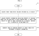
- Fig. 2 is a flowchart illustrating the flow of the image processing method S1 in accordance with the present example embodiment.
- step S11 the first acquisition section 11 acquires a target image which includes a specimen cell as a subject.
- the first acquisition section 11 supplies the acquired target image to the generation section 13.
- step S12 the second acquisition section 12 acquires property information indicating a property of a training image that has been used to train a prediction model to predict, upon receipt of input of an image including a cell as a subject, whether the cell is a benign cell or a malignant cell.
- the second acquisition section 12 supplies the acquired property information to the generation section 13.
- step S13 the generation section 13 generates an image by changing, with reference to the property information supplied from the second acquisition section 12, a property of the target image supplied from the first acquisition section 11.
- the image processing method S1 in accordance with the present example embodiment employs the configuration of including: acquiring a target image which includes a specimen cell as a subject; acquiring property information which indicates a property of a training image that has been used to train a prediction model to predict, upon receipt of input of an image including a cell as a subject, whether the cell is a benign cell or a malignant cell; and generating an image by changing a property of the target image with reference to the property information. Therefore, according to the image processing method S1 in accordance with the present example embodiment, an example advantage similar to that of the foregoing image processing apparatus 1 is brought about.
- a classification apparatus 2 in accordance with the present example embodiment is an apparatus which classifies a specimen cell as a benign cell or a malignant cell.
- the classification apparatus 2 inputs an image including a specimen cell as a subject into a prediction model, which has been trained to predict, upon receipt of input of an image including a cell as a subject, whether the cell is a benign cell or a malignant cell, and thus classifies the specimen cell as a benign cell or a malignant cell.
- the classification apparatus 2 (i) acquires a target image which includes a specimen cell as a subject and (ii) generates an image while making a property of the target image to conform to a property of a training image that has been used to train a prediction model. Then, the classification apparatus 2 inputs the generated image into the prediction model and classifies the specimen cell as a benign cell or a malignant cell with reference to a result obtained by the prediction model. That is, the classification apparatus 2 includes the configuration of the image processing apparatus 1 described above and inputs an image generated by the image processing apparatus 1 into the prediction model, and thus classifies a specimen cell as a benign cell or a malignant cell.
- the training image is an image obtained by digitizing an image which includes a specimen cell as a subject
- the target image is an image obtained by capturing a microscopic image of a specimen cell.
- the classification apparatus 2 can heighten accuracy of the prediction model.
- the classification apparatus 2 can be used, for example, in cytodiagnosis in rapid on-site evaluation (ROSE).
- Fig. 3 is a block diagram illustrating the configuration of the classification apparatus 2 in accordance with the present example embodiment.
- the classification apparatus 2 includes a control section 10, a storage section 21, and an input-output section 22.
- data referred to by the control section 10 (described later) is stored.
- Examples of the data stored in the storage section 21 include property information PI indicating a property of a training image TP used to train a prediction model, a target image SP which includes a specimen cell as a subject, and a training image TP.
- the input-output section 22 is an interface via which data is acquired from another apparatus connected to the classification apparatus 2 and data is outputted to another apparatus which is connected to the classification apparatus 2.
- the input-output section 22 supplies data acquired from another apparatus to the control section 10 and outputs data supplied from the control section 10 to another apparatus.
- the input-output section 22 may be a communication module that communicates with other apparatuses via a network.
- a specific configuration of the network does not limit the present example embodiment, and can be, for example, a wireless local area network (LAN), a wired LAN, a wide area network (WAN), a public network, a mobile data communication network, or a combination of these networks.
- the control section 10 controls constituent elements included in the classification apparatus 2.
- the control section 10 functions also as a first acquisition section 11, a second acquisition section 12, a generation section 13, and a classification section 14, as illustrated in Fig. 3 .
- the first acquisition section 11, the second acquisition section 12, the generation section 13, and the classification section 14 are configured to realize the first acquisition means, the second acquisition means, the generation means, and the classification means in the present example embodiment, respectively.
- the first acquisition section 11 acquires a target image SP which includes a specimen cell as a subject.
- the first acquisition section 11 causes the storage section 21 to store the acquired target image SP.
- the second acquisition section 12 acquires property information PI indicating a property of a training image TP that has been used to train a prediction model to predict, upon receipt of input of an image including a cell as a subject, whether the cell is a benign cell or a malignant cell.
- the second acquisition section 12 may be configured to acquire the property information PI itself, or may acquire one or more training images TP and derive and acquire property information PI from the one or more training images TP.
- the second acquisition section 12 causes the storage section 21 to store the acquired property information PI (and training image TP).
- examples of the property information PI include property information PI indicating a property which has been obtained by averaging respective properties in a plurality of training images TP and property information PI indicating a property which is a median of variations of respective properties in a plurality of training images TP.
- the second acquisition section 12 includes a derivation section 121.
- the derivation section 121 derives property information PI from the training image TP. For example, the derivation section 121 extracts a property of the training image TP and derives property information PI indicating the extracted property. As another example, the derivation section 121 extracts respective properties of a plurality of training images TP, and derives property information PI indicating (i) a property obtained by averaging the extracted properties or (ii) a property which is a median of variations of the respective extracted properties.
- the generation section 13 generates an image by changing a property of a target image SP with reference to the property information PI.
- the generation section 13 supplies the generated image to the classification section 14.
- the generation section 13 generates an image by changing a noise level of the target image SP.
- the generation section 13 generates an image by changing a color distribution of the target image SP.
- the generation section 13 generates an image by changing resolution of the target image SP.
- the generation section 13 generates an image by changing a size of an object included in the target image SP.
- the generation section 13 may generate an image by changing a plurality of properties of the target image SP. For example, the generation section 13 generates an image by changing a noise level, a color distribution, and resolution of the target image SP.
- the generation section 13 may generate an image by changing the properties in a predetermined order or may generate an image by changing the plurality of properties of the target image SP regardless of the order.
- the generation section 13 first generates an image by changing a noise level of a target image SP. Next, the generation section 13 carries out a process of changing a color distribution with respect to the image having the changed noise level, and thus generates an image having the changed color distribution. Then, the generation section 13 carries out a process of changing resolution with respect to the image having the changed color distribution, and thus generates an image having the changed resolution.
- the classification section 14 inputs an image supplied from the generation section 13 into a prediction model and classifies, as a benign cell or a malignant cell, a specimen cell which is included as a subject in the image supplied from the generation section 13.
- the classification section 14 outputs the classification result via the input-output section 22.
- the classification section 14 classifies, as a benign cell, the specimen cell which is included as a subject in the image supplied from the generation section 13. Meanwhile, in a case where the prediction result output from the prediction model indicates that the specimen cell is a malignant cell, the classification section 14 classifies, as a malignant cell, the specimen cell which is included as a subject in the image supplied from the generation section 13.
- Fig. 4 is a flowchart illustrating an example of a flow of a process S2 in which the generation section 13 in accordance with the present example embodiment generates an image by changing a noise level of a target image SP.
- the property information PI is information indicating a noise level in the training image TP, the noise level being a noise level in a background region that includes no object as a subject; and the generation section 13 calculates a noise level by applying a Gaussian filter.
- step S21 the generation section 13 extracts, from the target image SP, a background region that is a region in which no object is included as a subject.
- a method in which the generation section 13 extracts a background region is not particularly limited, and it is possible to employ, for example, a method in which a region having a pixel value that is equal to or less than a predetermined pixel value is extracted as a background region.
- step S22 the generation section 13 applies a Gaussian filter to the extracted background region.
- a sigma value of the Gaussian filter to be applied by the generation section 13 is not particularly limited, and can be, for example, a value such as 1.0 or 1.5.
- step S23 the generation section 13 calculates a noise level in the background region to which the Gaussian filter has been applied. For example, in a case where the property information PI indicates a standard deviation of pixel value as a noise level, the generation section 13 calculates, as a noise level, a standard deviation of pixel value in the background region to which the Gaussian filter has been applied. Then, the generation section 13 calculates a difference between the calculated standard deviation of pixel value in the background region and the standard deviation indicated by the property information PI.
- step S24 the generation section 13 determines whether or not the calculated difference is within a predetermined range.
- step S25 the generation section 13 changes the sigma value of the Gaussian filter to be applied.
- the generation section 13 increases the sigma value by 10%. Meanwhile, in a case where the standard deviation of pixel value in the background region to which the Gaussian filter has been applied is lower than the standard deviation indicated by the property information PI, the generation section 13 decreases the sigma value by 10%. That is, the generation section 13 derives a sigma value with which a difference between the noise level indicated by the property information PI and the noise level in the background region of the target image SP falls within a predetermined range.
- the generation section 13 returns to the process of step S22.
- step S26 the generation section 13 generates an image by changing the noise level by applying, to the target image SP, the Gaussian filter having the sigma value with which a difference between the noise level indicated by the property information PI and the noise level in the background region of the target image SP falls within a predetermined range. That is, the generation section 13 generates an image by changing the noise level of the target image SP so that the noise level in the extracted background region of the target image SP is brought close to the noise level indicated by the property information PI.
- the generation section 13 generates an image by changing the noise level of the target image SP so that the noise level in the extracted background region of the target image SP is brought close to the noise level indicated by the property information PI. Therefore, the generation section 13 can input an image having a noise level equivalent to the noise level of the training image TP into the prediction model, and this makes it possible to improve accuracy in classification of a specimen cell between benignancy and malignancy in pathological diagnosis.
- Fig. 5 is a flowchart illustrating an example of a flow of a process S3 in which the generation section 13 in accordance with the present example embodiment generates an image by changing a color distribution of a target image SP.
- the property information PI is information indicating a color distribution in the training image TP, the color distribution being a color distribution in an object region in which an object is included as a subject.
- the property information PI is information indicating respective histograms of hue, chroma, and lightness in the object region of the training image TP.
- step S31 the generation section 13 extracts, from the target image SP, an object region that is a region in which an object is included as a subject.
- a method in which the generation section 13 extracts an object region is not particularly limited, and it is possible to employ, for example, a method in which a region having a pixel value that is equal to or more than a predetermined pixel value is extracted as an object region.
- step S32 the generation section 13 generates a histogram of a color distribution in the extracted object region as information indicating a color distribution in the extracted object region. For example, the generation section 13 generates respective histograms of hue, chroma, and lightness of the extracted object region.
- step S33 the generation section 13 generates an image by changing color distribution in the extracted object region of the target image SP so that the color distribution is brought close to color distribution indicated by the property information PI.
- Fig. 6 is a diagram illustrating an example of a histogram of hue in the present example embodiment. The following description will discuss a histogram of hue, and similar descriptions apply to histograms of chroma and lightness.
- the generation section 13 calculates an average value mean pi and a left end a pi and a right end b pi of a 95% confidence interval in the histogram of hue indicated by the property information PI.
- the generation section 13 similarly calculates an average value mean sp1 and a left end a sp1 and a right end b sp1 of a 95% confidence interval in the histogram of hue in the object region of the target image SP.
- the generation section 13 uses the formula (1) to change, in a 95% confidence interval of each histogram, a value Var poi1 to conform to a value Var tar , which is a counterpart in the target histogram.
- the value Var poi1 is a value obtained by subtracting a lower limit value from an upper limit value of a 95% confidence interval in a certain hue value In in the target image SP.
- the certain hue value In is converted into a target hue value.
- a histogram of hue in an object region of the image after conversion is illustrated in the lower part of Fig. 6 .
- an average value mean sp2 and a left end a sp2 and a right end b sp2 of a 95% confidence interval in the histogram of hue in the object region after conversion are closer to the average value mean pi and the left end a pi and the right end b pi of the 95% confidence interval in the histogram indicated by the property information PI, as compared with the average value mean sp1 and the left end a sp1 and the right end b sp1 of the 95% confidence interval in the histogram of a color distribution in the object region of the target image SP.
- the generation section 13 generates an image by changing color distribution in the object region of the target image SP so that the color distribution is brought close to the color distribution indicated by the property information PI. Therefore, the generation section 13 can input an image having a color distribution equivalent to the color distribution of the training image TP into the prediction model, and this makes it possible to improve accuracy in classification of a specimen cell between benignancy and malignancy in pathological diagnosis.
- the generation section 13 may change a color distribution of the background region of the target image SP in addition to the object region of the target image SP.
- the generation section 13 may be configured to refer to a background region of the training image TP.
- the generation section 13 may acquire an average value of pixel values of the background region of the training image TP and change pixel values of a background region of a target image SP to the average value.
- the generation section 13 extracts respective background regions of a plurality of training images TP and calculates an average value of pixel values of the extracted background regions. Then, the generation section 13 may change pixel values of the background region of the target image SP to the calculated average value. In this case, the generation section 13 may use a median of variations in pixel values, instead of the average value of pixel values.
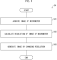
- Fig. 7 is a flowchart illustrating an example of a flow of a process S4 in which the generation section 13 in accordance with the present example embodiment changes resolution of a target image SP.
- the following description will discuss a process in which: the property information PI is information indicating resolution of the training image TP; and the generation section 13 refers to an image which includes a micrometer as a subject.
- step S41 the generation section 13 acquires an image of a micrometer captured with the same resolution as the target image SP.
- Fig. 8 is a diagram illustrating an example of an image of a micrometer in the present example embodiment. As illustrated in an image P1 of Fig. 8 , the generation section 13 acquires an image which includes a micrometer as a subject.
- step S42 the generation section 13 calculates resolution of the image of the micrometer.
- the generation section 13 first generates an image by binarizing the image P1, as illustrated in an image P2 of Fig. 8 .
- the generation section 13 carries out a closing process by carrying out a dilation process and an erosion process on the image P2, as illustrated in an image P3 of Fig. 8 .
- the generation section 13 detects coordinates (X L , Y L ) of a left-end pixel of the micrometer and coordinates (X R , Y R ) of a right-end pixel of the micrometer as illustrated in an image P4 of Fig.
- L represents a length of the micrometer.
- the generation section 13 calculates resolution by calculating, using the formula (2), the number of pixels of the target image SP included in a predetermined length.
- step S43 the generation section 13 generates an image by changing the calculated resolution to resolution indicated by the property information PI.
- the generation section 13 generates an image by changing resolution of the target image SP so that the resolution indicated by the property information PI is equal to the resolution of the target image SP. Therefore, it is possible to input, into the prediction model, an image in which a size of an object included in the training image TP as a subject is equivalent to a size of an object included in the target image SP as a subject, and this makes it possible to improve accuracy in classification of a specimen cell between benignancy and malignancy in pathological diagnosis.
- Fig. 9 is a flowchart illustrating another example of a flow of a process S5 in which the generation section 13 in accordance with the present example embodiment changes resolution of a target image SP.
- the following description will discuss a configuration in which: the property information PI is information indicating a size of a predetermined object which is included in the training image TP as a subject; and the generation section 13 extracts a predetermined object which is included as a subject in the target image SP.
- the predetermined object is not particularly limited, and is preferably an object with a small individual difference in size.
- the predetermined object include an erythrocyte and a lymphocyte.
- the generation section 13 uses an erythrocyte or a lymphocyte with a small individual difference as a reference for a size of an object included in the target image SP. Therefore, it is possible to suitably calculate a size of an object.
- the following description will discuss a case in which the predetermined object is an erythrocyte.
- step S51 the generation section 13 extracts an erythrocyte included as a subject in the target image SP as a predetermined object included as a subject in the target image SP.
- Examples of a method in which the generation section 13 extracts an erythrocyte which is included in the target image SP as a subject include a method using known deep learning segmentation (DL segmentation).
- step S52 the generation section 13 calculates a size of the extracted erythrocyte.
- the generation section 13 may elliptically approximate the extracted erythrocyte and calculate a major axis and a minor axis.
- the generation section 13 may calculate a major axis and a minor axis of an erythrocyte having the largest area. As another example, the generation section 13 may calculate a major axis and a minor axis of an erythrocyte which has a median of variations in areas of the plurality of erythrocytes. As yet another example, the generation section 13 may calculate an average major axis and an average minor axis of the plurality of erythrocytes.
- step S53 the generation section 13 generates an image by changing resolution of the target image SP so that the calculated size (major axis and minor axis) of the erythrocyte is a size (major axis and minor axis) indicated by the property information PI.
- the generation section 13 generates an image by changing resolution of the target image SP so that a size of a predetermined object indicated by the property information PI is equivalent to a size of a predetermined object included in the target image SP as a subject. Therefore, it is possible to input, into the prediction model, an image in which a size of an object included in the training image TP as a subject is equivalent to a size of an object included in the target image SP as a subject, and this makes it possible to improve accuracy in classification of a specimen cell between benignancy and malignancy in pathological diagnosis.
- the classification apparatus 2 in accordance with the present example embodiment generates an image by changing a property of a target image SP which includes a specimen cell as a subject, with reference to property information PI which indicates a property of a training image TP that has been used to train a prediction model to predict, upon receipt of input of an image including a cell as a subject, whether the cell is a benign cell or a malignant cell. Furthermore, the classification apparatus 2 in accordance with the present example embodiment employs the configuration in which the generated image is input into the prediction model.
- the classification apparatus 2 in accordance with the present example embodiment generates an image by changing a property of a target image SP in accordance with a property of a training image TP that has been used to train a prediction model, and then the image is input into the prediction model. Therefore, according to the classification apparatus 2 in accordance with the present example embodiment, it is possible to bring about an example advantage of improving accuracy in classification of a specimen cell between benignancy and malignancy in pathological diagnosis.
- each of the image processing apparatus 1 and the classification apparatus 2 may be implemented by hardware such as an integrated circuit (IC chip), or may be implemented by software.
- the image processing apparatus 1 and the classification apparatus 2 are implemented by, for example, a computer that executes instructions of a program that is software implementing the foregoing functions.
- Fig. 10 illustrates an example of such a computer (hereinafter, referred to as "computer C").
- the computer C includes at least one processor C1 and at least one memory C2.
- the memory C2 stores a program P for causing the computer C to operate as the image processing apparatus 1 and the classification apparatus 2.
- the processor C1 of the computer C retrieves the program P from the memory C2 and executes the program P, so that the functions of the image processing apparatus 1 and the classification apparatus 2 are implemented.
- processor C1 for example, it is possible to use a central processing unit (CPU), a graphic processing unit (GPU), a digital signal processor (DSP), a micro processing unit (MPU), a floating point number processing unit (FPU), a physics processing unit (PPU), a microcontroller, or a combination of these.
- CPU central processing unit
- GPU graphic processing unit
- DSP digital signal processor
- MPU micro processing unit
- FPU floating point number processing unit
- PPU a physics processing unit
- microcontroller or a combination of these.
- the memory C2 include a flash memory, a hard disk drive (HDD), a solid state drive (SSD), and a combination thereof.
- the computer C can further include a random access memory (RAM) in which the program P is loaded in a case where the program P is executed and in which various kinds of data are temporarily stored.
- the computer C can further include a communication interface for carrying out transmission and reception of data with other apparatuses.
- the computer C can further include an input-output interface for connecting input-output apparatuses such as a keyboard, a mouse, a display and a printer.
- the program P can be stored in a computer C-readable, non-transitory, and tangible storage medium M.
- the storage medium M can be, for example, a tape, a disk, a card, a semiconductor memory, a programmable logic circuit, or the like.
- the computer C can obtain the program P via the storage medium M.
- the program P can be transmitted via a transmission medium.
- the transmission medium can be, for example, a communication network, a broadcast wave, or the like.
- the computer C can obtain the program P also via such a transmission medium.
- the present invention is not limited to the foregoing example embodiments, but may be altered in various ways by a skilled person within the scope of the claims.
- the present invention also encompasses, in its technical scope, any example embodiment derived by appropriately combining technical means disclosed in the foregoing example embodiments.
- An image processing apparatus including: a first acquisition means for acquiring a target image which includes a specimen cell as a subject; a second acquisition means for acquiring property information which indicates a property of a training image that has been used to train a prediction model to predict, upon receipt of input of an image including a cell as a subject, whether the cell is a benign cell or a malignant cell; and a generation means for generating an image by changing a property of the target image with reference to the property information.
- the property information is information indicating a noise level in the training image, the noise level being a noise level in a background region that includes no object as a subject; and the generation means extracts a background region from the target image and generates an image by changing a noise level in the target image so that a noise level in the background region of the target image which has been extracted is brought close to the noise level indicated by the property information.
- the image processing apparatus in which: the generation means calculates a noise level by applying a Gaussian filter to the background region of the target image; and the generation means derives a sigma value with which a difference between the noise level indicated by the property information and the noise level in the background region of the target image falls within a predetermined range.
- the property information is information indicating a color distribution in the training image, the color distribution being a color distribution in an object region that includes an object as a subject; and the generation means extracts an object region from the target image and generates an image by changing a color distribution in the object region of the target image which has been extracted is brought close to the color distribution indicated by the property information.
- the property information is information indicating respective histograms of hue, chroma, and lightness in the object region of the training image.
- the property information is information indicating resolution of the training image
- the generation means generates an image by changing resolution of the target image to the resolution indicated by the property information with reference to an image of a micrometer captured in resolution that is identical with that of the target image.
- the property information is information indicating a size of a predetermined object included in the training image as a subject
- the generation means extracts a predetermined object which is included in the target image as a subject and generates an image by changing resolution of the target image so that a size of the predetermined object in the target image which has been extracted is the size indicated by the property information.
- the predetermined object is an erythrocyte or a lymphocyte.
- the image processing apparatus according to any one of supplementary notes 1 through 8, in which: the second acquisition means acquires the training image; and the second acquisition means includes a derivation means for deriving property information from the training image.
- the second acquisition means acquires (i) property information indicating a property which has been obtained by averaging respective properties in a plurality of training images or (ii) property information indicating a property which is a median of variations of respective properties in a plurality of training images.
- the training image is an image obtained by digitizing an image which includes a specimen cell as a subject
- the target image is an image obtained by capturing a microscopic image of a specimen cell.
- a classification apparatus including a classification means for inputting an image generated by an image processing apparatus according to supplementary note 1 into the prediction model, and classifying, as a benign cell or a malignant cell, a specimen cell which is included as a subject in the image generated by the image processing apparatus.
- An image processing method including: acquiring, by an image processing apparatus, a target image which includes a specimen cell as a subject; acquiring, by the image processing apparatus, property information which indicates a property of a training image that has been used to train a prediction model to predict, upon receipt of input of an image including a cell as a subject, whether the cell is a benign cell or a malignant cell; and generating, by the image processing apparatus, an image by changing a property of the target image with reference to the property information.
- a program for causing a computer to function as an image processing apparatus the program causing the computer to function as: a first acquisition means for acquiring a target image which includes a specimen cell as a subject; a second acquisition means for acquiring property information which indicates a property of a training image that has been used to train a prediction model to predict, upon receipt of input of an image including a cell as a subject, whether the cell is a benign cell or a malignant cell; and a generation means for generating an image by changing a property of the target image with reference to the property information.
- An image processing apparatus including at least one processor, the at least one processor carrying out: a first acquisition process of acquiring a target image which includes a specimen cell as a subject; a second acquisition process of acquiring property information which indicates a property of a training image that has been used to train a prediction model to predict, upon receipt of input of an image including a cell as a subject, whether the cell is a benign cell or a malignant cell; and a generation process of generating an image by changing a property of the target image with reference to the property information.
- the image processing apparatus can further include a memory.
- the memory can store a program for causing the at least one processor to carry out the first acquisition process, the second acquisition process, and the generation process.
- the program can be stored in a computer-readable non-transitory tangible storage medium.
Landscapes
- Engineering & Computer Science (AREA)
- Health & Medical Sciences (AREA)
- Life Sciences & Earth Sciences (AREA)
- Molecular Biology (AREA)
- Biomedical Technology (AREA)
- General Physics & Mathematics (AREA)
- General Health & Medical Sciences (AREA)
- Physics & Mathematics (AREA)
- Theoretical Computer Science (AREA)
- Immunology (AREA)
- Hematology (AREA)
- Urology & Nephrology (AREA)
- Chemical & Material Sciences (AREA)
- Multimedia (AREA)
- Pathology (AREA)
- Food Science & Technology (AREA)
- Biochemistry (AREA)
- Cell Biology (AREA)
- Biotechnology (AREA)
- Microbiology (AREA)
- Medicinal Chemistry (AREA)
- Analytical Chemistry (AREA)
- Medical Informatics (AREA)
- Nuclear Medicine, Radiotherapy & Molecular Imaging (AREA)
- Radiology & Medical Imaging (AREA)
- Quality & Reliability (AREA)
- Computer Vision & Pattern Recognition (AREA)
- Image Analysis (AREA)
- Image Processing (AREA)
Abstract
Description
- The present invention relates to an image processing apparatus, an image processing method, and a program.
- A technique is disclosed that is related to a trained model which carries out image recognition of an image including a specimen as a subject in pathological diagnosis.
-
Patent Literature 1 discloses a technique in which unevenness in samples is suppressed by normalizing whole slide image (WSI) data by carrying out pretreatment with respect to each of a plurality of WSIs in which different dyes are used, and the normalized WSI data is applied to a machine learning model. - [Patent Literature 1]
PublishedJapanese Translation of PCT International Application Tokuhyo No. 2021-524630 - The technique disclosed in
Patent Literature 1 does not consider a relationship between an image for training and an image input into a machine learning model for image recognition in pathological diagnosis. Therefore, the technique disclosed inPatent Literature 1 has a problem that accuracy in image recognition of an image input into a machine learning model may be lowered. - Such a problem causes a false positive in which classification as being malignant is made despite being benign in classification between benignancy and malignancy for classifying a specimen cell as a benign cell or a malignant cell. In a case where a false positive occurs, reinspection is needed even though reinspection is not actually necessary. In a case where a false negative occurs in which classification as being benign is made despite being malignant, there is also a risk of overlooking cancer. Therefore, in the classification between benignancy and malignancy, it is preferable that the accuracy in image recognition of an input image is high.
- An example aspect of the present invention is accomplished in view of the above problems, and an example object thereof is to provide a technique for improving accuracy in classification of a specimen cell between benignancy and malignancy in pathological diagnosis.
- An image processing apparatus in accordance with an example aspect of the present invention includes: a first acquisition means for acquiring a target image which includes a specimen cell as a subject; a second acquisition means for acquiring property information which indicates a property of a training image that has been used to train a prediction model to predict, upon receipt of input of an image including a cell as a subject, whether the cell is a benign cell or a malignant cell; and a generation means for generating an image by changing a property of the target image with reference to the property information.
- An image processing method in accordance with an example aspect of the present invention includes: acquiring, by an image processing apparatus, a target image which includes a specimen cell as a subject; acquiring, by the image processing apparatus, property information which indicates a property of a training image that has been used to train a prediction model to predict, upon receipt of input of an image including a cell as a subject, whether the cell is a benign cell or a malignant cell; and generating, by the image processing apparatus, an image by changing a property of the target image with reference to the property information.
- A program in accordance with an example aspect of the present invention is a program for causing a computer to function as an image processing apparatus, the program causing the computer to function as: a first acquisition means for acquiring a target image which includes a specimen cell as a subject; a second acquisition means for acquiring property information which indicates a property of a training image that has been used to train a prediction model to predict, upon receipt of input of an image including a cell as a subject, whether the cell is a benign cell or a malignant cell; and a generation means for generating an image by changing a property of the target image with reference to the property information.
- According to an example aspect of the present invention, it is possible to improve accuracy in classification of a specimen cell between benignancy and malignancy in pathological diagnosis.
-
-
Fig. 1 is a block diagram illustrating a configuration of an image processing apparatus in accordance with a first example embodiment of the present invention. -
Fig. 2 is a flowchart illustrating an image processing method in accordance with the first example embodiment of the present invention. -
Fig. 3 is a block diagram illustrating a configuration of a classification apparatus in accordance with a second example embodiment of the present invention. -
Fig. 4 is a flowchart illustrating an example of a flow of a process in which a generation section in accordance with the second example embodiment of the present invention generates an image by changing a noise level of a target image. -
Fig. 5 is a flowchart illustrating an example of a flow of a process in which the generation section in accordance with the second example embodiment of the present invention generates an image by changing a color distribution of a target image. -
Fig. 6 is a diagram illustrating an example of a histogram of hue in the second example embodiment of the present invention. -
Fig. 7 is a flowchart illustrating an example of a flow of a process in which the generation section in accordance with the second example embodiment of the present invention changes resolution of a target image. -
Fig. 8 is a diagram illustrating an example of an image of a micrometer in the second example embodiment of the present invention. -
Fig. 9 is a flowchart illustrating another example of a flow of a process in which the generation section in accordance with the second example embodiment of the present invention changes resolution of a target image. -
Fig. 10 is a block diagram illustrating an example hardware configuration of the image processing apparatus and the classification apparatus in accordance with the example embodiments of the present invention. - The following description will discuss a first example embodiment of the present invention in detail, with reference to the drawings. The present example embodiment is a basic form of example embodiments described later.
- An
image processing apparatus 1 in accordance with the present example embodiment is an image processing apparatus that generates an image by changing a property of a target image which includes a specimen cell as a subject. For example, theimage processing apparatus 1 generates an image by changing a property of a target image in accordance with a property of a training image that has been used to train a prediction model to predict, upon receipt of input of an image including a cell as a subject, whether the cell is a benign cell or a malignant cell. - A property of an image refers to quality or a feature of an image. Examples of the property include: a noise level indicating a magnitude of noise included in an image; a color distribution constituted by hue, lightness, and chroma in an image; resolution indicating the number of pixels included in a predetermined length in an image; and information indicating a size of a predetermined object included in an image as a subject.
- A specific configuration of the prediction model does not limit the present example embodiment, and can be, for example, a convolution neural network (CNN), a recurrent neural network (RNN), or a combination of these networks. Alternatively, a non-neural network type model such as a random forest or a support vector machine can be used.
- The following description will discuss a configuration of the
image processing apparatus 1 in accordance with the present example embodiment, with reference toFig. 1. Fig. 1 is a block diagram illustrating the configuration of theimage processing apparatus 1 in accordance with the present example embodiment. - As illustrated in
Fig. 1 , theimage processing apparatus 1 includes afirst acquisition section 11, asecond acquisition section 12, and ageneration section 13. Thefirst acquisition section 11, thesecond acquisition section 12, and thegeneration section 13 are configured to realize the first acquisition means, the second acquisition means, and the generation means in the present example embodiment, respectively. - The
first acquisition section 11 acquires a target image which includes a specimen cell as a subject. Thefirst acquisition section 11 supplies the acquired target image to thegeneration section 13. - The
second acquisition section 12 acquires property information indicating a property of a training image that has been used to train a prediction model to predict, upon receipt of input of an image including a cell as a subject, whether the cell is a benign cell or a malignant cell. Thesecond acquisition section 12 supplies the acquired property information to thegeneration section 13. - The
generation section 13 generates an image by changing, with reference to the property information supplied from thesecond acquisition section 12, a property of the target image supplied from thefirst acquisition section 11. - As described above, the
image processing apparatus 1 in accordance with the present example embodiment employs the configuration of including: thefirst acquisition section 11 for acquiring a target image which includes a specimen cell as a subject; thesecond acquisition section 12 for acquiring property information which indicates a property of a training image that has been used to train a prediction model to predict, upon receipt of input of an image including a cell as a subject, whether the cell is a benign cell or a malignant cell; and thegeneration section 13 for generating an image by changing a property of the target image with reference to the property information. - That is, the
image processing apparatus 1 in accordance with the present example embodiment generates an image by changing a property of a target image in accordance with a property of a training image that has been used to train a prediction model. Therefore, according to theimage processing apparatus 1 in accordance with the present example embodiment, it is possible to bring about an example advantage of improving accuracy in classification of a specimen cell between benignancy and malignancy in pathological diagnosis. - The following description will discuss a flow of an image processing method S1 in accordance with the present example embodiment, with reference to
Fig. 2. Fig. 2 is a flowchart illustrating the flow of the image processing method S1 in accordance with the present example embodiment. - In step S11, the
first acquisition section 11 acquires a target image which includes a specimen cell as a subject. Thefirst acquisition section 11 supplies the acquired target image to thegeneration section 13. - In step S12, the
second acquisition section 12 acquires property information indicating a property of a training image that has been used to train a prediction model to predict, upon receipt of input of an image including a cell as a subject, whether the cell is a benign cell or a malignant cell. Thesecond acquisition section 12 supplies the acquired property information to thegeneration section 13. - In step S13, the
generation section 13 generates an image by changing, with reference to the property information supplied from thesecond acquisition section 12, a property of the target image supplied from thefirst acquisition section 11. - As described above, the image processing method S1 in accordance with the present example embodiment employs the configuration of including: acquiring a target image which includes a specimen cell as a subject; acquiring property information which indicates a property of a training image that has been used to train a prediction model to predict, upon receipt of input of an image including a cell as a subject, whether the cell is a benign cell or a malignant cell; and generating an image by changing a property of the target image with reference to the property information. Therefore, according to the image processing method S1 in accordance with the present example embodiment, an example advantage similar to that of the foregoing
image processing apparatus 1 is brought about. - The following description will discuss a second example embodiment of the present invention in detail, with reference to the drawings. The same reference numerals are given to constituent elements which have functions identical with those described in the first example embodiment, and descriptions as to such constituent elements are omitted as appropriate.
- A
classification apparatus 2 in accordance with the present example embodiment is an apparatus which classifies a specimen cell as a benign cell or a malignant cell. - For example, the
classification apparatus 2 inputs an image including a specimen cell as a subject into a prediction model, which has been trained to predict, upon receipt of input of an image including a cell as a subject, whether the cell is a benign cell or a malignant cell, and thus classifies the specimen cell as a benign cell or a malignant cell. - More specifically, the classification apparatus 2 (i) acquires a target image which includes a specimen cell as a subject and (ii) generates an image while making a property of the target image to conform to a property of a training image that has been used to train a prediction model. Then, the
classification apparatus 2 inputs the generated image into the prediction model and classifies the specimen cell as a benign cell or a malignant cell with reference to a result obtained by the prediction model. That is, theclassification apparatus 2 includes the configuration of theimage processing apparatus 1 described above and inputs an image generated by theimage processing apparatus 1 into the prediction model, and thus classifies a specimen cell as a benign cell or a malignant cell. - For example, the training image is an image obtained by digitizing an image which includes a specimen cell as a subject, and the target image is an image obtained by capturing a microscopic image of a specimen cell. In a case where the training image is a digitized image, many training images can be acquired. Therefore, the
classification apparatus 2 can heighten accuracy of the prediction model. In a case where the target image is an image obtained by capturing a microscopic image, theclassification apparatus 2 can be used, for example, in cytodiagnosis in rapid on-site evaluation (ROSE). - The property of an image and the prediction model are as described above.
- The following description will discuss a configuration of the
classification apparatus 2 in accordance with the present example embodiment, with reference toFig. 3. Fig. 3 is a block diagram illustrating the configuration of theclassification apparatus 2 in accordance with the present example embodiment. - As illustrated in
Fig. 3 , theclassification apparatus 2 includes acontrol section 10, astorage section 21, and an input-output section 22. - In the
storage section 21, data referred to by the control section 10 (described later) is stored. Examples of the data stored in thestorage section 21 include property information PI indicating a property of a training image TP used to train a prediction model, a target image SP which includes a specimen cell as a subject, and a training image TP. - The input-
output section 22 is an interface via which data is acquired from another apparatus connected to theclassification apparatus 2 and data is outputted to another apparatus which is connected to theclassification apparatus 2. The input-output section 22 supplies data acquired from another apparatus to thecontrol section 10 and outputs data supplied from thecontrol section 10 to another apparatus. - The input-
output section 22 may be a communication module that communicates with other apparatuses via a network. A specific configuration of the network does not limit the present example embodiment, and can be, for example, a wireless local area network (LAN), a wired LAN, a wide area network (WAN), a public network, a mobile data communication network, or a combination of these networks. - The
control section 10 controls constituent elements included in theclassification apparatus 2. Thecontrol section 10 functions also as afirst acquisition section 11, asecond acquisition section 12, ageneration section 13, and aclassification section 14, as illustrated inFig. 3 . Thefirst acquisition section 11, thesecond acquisition section 12, thegeneration section 13, and theclassification section 14 are configured to realize the first acquisition means, the second acquisition means, the generation means, and the classification means in the present example embodiment, respectively. - The
first acquisition section 11 acquires a target image SP which includes a specimen cell as a subject. Thefirst acquisition section 11 causes thestorage section 21 to store the acquired target image SP. - The
second acquisition section 12 acquires property information PI indicating a property of a training image TP that has been used to train a prediction model to predict, upon receipt of input of an image including a cell as a subject, whether the cell is a benign cell or a malignant cell. Thesecond acquisition section 12 may be configured to acquire the property information PI itself, or may acquire one or more training images TP and derive and acquire property information PI from the one or more training images TP. Thesecond acquisition section 12 causes thestorage section 21 to store the acquired property information PI (and training image TP). - Here, examples of the property information PI include property information PI indicating a property which has been obtained by averaging respective properties in a plurality of training images TP and property information PI indicating a property which is a median of variations of respective properties in a plurality of training images TP.
- As illustrated in
Fig. 3 , thesecond acquisition section 12 includes aderivation section 121. - The
derivation section 121 derives property information PI from the training image TP. For example, thederivation section 121 extracts a property of the training image TP and derives property information PI indicating the extracted property. As another example, thederivation section 121 extracts respective properties of a plurality of training images TP, and derives property information PI indicating (i) a property obtained by averaging the extracted properties or (ii) a property which is a median of variations of the respective extracted properties. - The
generation section 13 generates an image by changing a property of a target image SP with reference to the property information PI. Thegeneration section 13 supplies the generated image to theclassification section 14. - For example, the
generation section 13 generates an image by changing a noise level of the target image SP. As another example, thegeneration section 13 generates an image by changing a color distribution of the target image SP. As yet another example, thegeneration section 13 generates an image by changing resolution of the target image SP. As still another example, thegeneration section 13 generates an image by changing a size of an object included in the target image SP. - The
generation section 13 may generate an image by changing a plurality of properties of the target image SP. For example, thegeneration section 13 generates an image by changing a noise level, a color distribution, and resolution of the target image SP. - In a case where the
generation section 13 generates an image by changing a plurality of properties of the target image SP, thegeneration section 13 may generate an image by changing the properties in a predetermined order or may generate an image by changing the plurality of properties of the target image SP regardless of the order. - In an example configuration in which an image is generated by changing properties in a predetermined order, the
generation section 13 first generates an image by changing a noise level of a target image SP. Next, thegeneration section 13 carries out a process of changing a color distribution with respect to the image having the changed noise level, and thus generates an image having the changed color distribution. Then, thegeneration section 13 carries out a process of changing resolution with respect to the image having the changed color distribution, and thus generates an image having the changed resolution. - A flow of a process which is carried out by the
generation section 13 will be described later. - The
classification section 14 inputs an image supplied from thegeneration section 13 into a prediction model and classifies, as a benign cell or a malignant cell, a specimen cell which is included as a subject in the image supplied from thegeneration section 13. Theclassification section 14 outputs the classification result via the input-output section 22. - For example, in a case where the prediction result output from the prediction model indicates that the specimen cell is a benign cell, the
classification section 14 classifies, as a benign cell, the specimen cell which is included as a subject in the image supplied from thegeneration section 13. Meanwhile, in a case where the prediction result output from the prediction model indicates that the specimen cell is a malignant cell, theclassification section 14 classifies, as a malignant cell, the specimen cell which is included as a subject in the image supplied from thegeneration section 13. - The following description will discuss an example of a process in which the
generation section 13 generates an image by changing a noise level of a target image SP, with reference toFig. 4. Fig. 4 is a flowchart illustrating an example of a flow of a process S2 in which thegeneration section 13 in accordance with the present example embodiment generates an image by changing a noise level of a target image SP. The following description will discuss a process in which: the property information PI is information indicating a noise level in the training image TP, the noise level being a noise level in a background region that includes no object as a subject; and thegeneration section 13 calculates a noise level by applying a Gaussian filter. - In step S21, the
generation section 13 extracts, from the target image SP, a background region that is a region in which no object is included as a subject. A method in which thegeneration section 13 extracts a background region is not particularly limited, and it is possible to employ, for example, a method in which a region having a pixel value that is equal to or less than a predetermined pixel value is extracted as a background region. - In step S22, the
generation section 13 applies a Gaussian filter to the extracted background region. A sigma value of the Gaussian filter to be applied by thegeneration section 13 is not particularly limited, and can be, for example, a value such as 1.0 or 1.5. - In step S23, the
generation section 13 calculates a noise level in the background region to which the Gaussian filter has been applied. For example, in a case where the property information PI indicates a standard deviation of pixel value as a noise level, thegeneration section 13 calculates, as a noise level, a standard deviation of pixel value in the background region to which the Gaussian filter has been applied. Then, thegeneration section 13 calculates a difference between the calculated standard deviation of pixel value in the background region and the standard deviation indicated by the property information PI. - In step S24, the
generation section 13 determines whether or not the calculated difference is within a predetermined range. - In a case where it has been determined in step S24 that the calculated difference is not within the predetermined range (step S24: NO), in step S25, the
generation section 13 changes the sigma value of the Gaussian filter to be applied. - For example, in a case where the standard deviation of pixel value in the background region to which the Gaussian filter has been applied is higher than the standard deviation indicated by the property information PI, the
generation section 13 increases the sigma value by 10%. Meanwhile, in a case where the standard deviation of pixel value in the background region to which the Gaussian filter has been applied is lower than the standard deviation indicated by the property information PI, thegeneration section 13 decreases the sigma value by 10%. That is, thegeneration section 13 derives a sigma value with which a difference between the noise level indicated by the property information PI and the noise level in the background region of the target image SP falls within a predetermined range. - Then, in order to apply the Gaussian filter having the changed sigma value, the
generation section 13 returns to the process of step S22. - In a case where it has been determined in step S24 that the calculated difference is within the predetermined range (step S24: YES), in step S26, the
generation section 13 generates an image by changing the noise level by applying, to the target image SP, the Gaussian filter having the sigma value with which a difference between the noise level indicated by the property information PI and the noise level in the background region of the target image SP falls within a predetermined range. That is, thegeneration section 13 generates an image by changing the noise level of the target image SP so that the noise level in the extracted background region of the target image SP is brought close to the noise level indicated by the property information PI. - Thus, the
generation section 13 generates an image by changing the noise level of the target image SP so that the noise level in the extracted background region of the target image SP is brought close to the noise level indicated by the property information PI. Therefore, thegeneration section 13 can input an image having a noise level equivalent to the noise level of the training image TP into the prediction model, and this makes it possible to improve accuracy in classification of a specimen cell between benignancy and malignancy in pathological diagnosis. - The following description will discuss an example of a process in which the
generation section 13 generates an image by changing a color distribution of a target image SP, with reference toFig. 5. Fig. 5 is a flowchart illustrating an example of a flow of a process S3 in which thegeneration section 13 in accordance with the present example embodiment generates an image by changing a color distribution of a target image SP. The following description will discuss a case in which the property information PI is information indicating a color distribution in the training image TP, the color distribution being a color distribution in an object region in which an object is included as a subject. For example, the property information PI is information indicating respective histograms of hue, chroma, and lightness in the object region of the training image TP. - In step S31, the
generation section 13 extracts, from the target image SP, an object region that is a region in which an object is included as a subject. A method in which thegeneration section 13 extracts an object region is not particularly limited, and it is possible to employ, for example, a method in which a region having a pixel value that is equal to or more than a predetermined pixel value is extracted as an object region. - In step S32, the
generation section 13 generates a histogram of a color distribution in the extracted object region as information indicating a color distribution in the extracted object region. For example, thegeneration section 13 generates respective histograms of hue, chroma, and lightness of the extracted object region. - In step S33, the
generation section 13 generates an image by changing color distribution in the extracted object region of the target image SP so that the color distribution is brought close to color distribution indicated by the property information PI. The following description will discuss an example of a process which is carried out by thegeneration section 13 in step S33, with reference toFig. 6. Fig. 6 is a diagram illustrating an example of a histogram of hue in the present example embodiment. The following description will discuss a histogram of hue, and similar descriptions apply to histograms of chroma and lightness. - In a case where the histogram of hue indicated by the property information PI is a histogram illustrated in the upper part of
Fig. 6 , thegeneration section 13 calculates an average value meanpi and a left end api and a right end bpi of a 95% confidence interval in the histogram of hue indicated by the property information PI. - Next, in a case where the histogram of hue in the object region of the target image SP generated in step S32 is a histogram illustrated in the middle part of
Fig. 6 , thegeneration section 13 similarly calculates an average value meansp1 and a left end asp1 and a right end bsp1 of a 95% confidence interval in the histogram of hue in the object region of the target image SP. - Then, the
generation section 13 calculates a changed value Out using a formula (1) below.
- Variables in the formula (1) are as follows.
- Out: A hue value after change
- In: A hue value of a target image SP
- Vartar: A value obtained by subtracting a lower limit value from an upper limit value of a 95% confidence interval of a hue value In in a target histogram. That is, a value indicated by [right end bpi-left end api].
- Varpoi1: A value obtained by subtracting a lower limit value from an upper limit value of a 95% confidence interval of a hue value In in a histogram of a target image SP. That is, a value indicated by [right end bsp1-left end asp1].
- That is, the
generation section 13 uses the formula (1) to change, in a 95% confidence interval of each histogram, a value Varpoi1 to conform to a value Vartar, which is a counterpart in the target histogram. Here, the value Varpoi1 is a value obtained by subtracting a lower limit value from an upper limit value of a 95% confidence interval in a certain hue value In in the target image SP. Thus, the certain hue value In is converted into a target hue value. - A histogram of hue in an object region of the image after conversion is illustrated in the lower part of
Fig. 6 . As illustrated in the lower part ofFig. 6 , an average value meansp2 and a left end asp2 and a right end bsp2 of a 95% confidence interval in the histogram of hue in the object region after conversion are closer to the average value meanpi and the left end api and the right end bpi of the 95% confidence interval in the histogram indicated by the property information PI, as compared with the average value meansp1 and the left end asp1 and the right end bsp1 of the 95% confidence interval in the histogram of a color distribution in the object region of the target image SP. - Thus, the
generation section 13 generates an image by changing color distribution in the object region of the target image SP so that the color distribution is brought close to the color distribution indicated by the property information PI. Therefore, thegeneration section 13 can input an image having a color distribution equivalent to the color distribution of the training image TP into the prediction model, and this makes it possible to improve accuracy in classification of a specimen cell between benignancy and malignancy in pathological diagnosis. - In the process of changing a color distribution of the target image SP, the
generation section 13 may change a color distribution of the background region of the target image SP in addition to the object region of the target image SP. - For example, the
generation section 13 may change a pixel value RGB of the background region of the target image SP to a predetermined value (e.g., (R,G,B) = (230,230,230) for an image with 256 levels of gray). - As another example, the
generation section 13 may be configured to refer to a background region of the training image TP. For example, thegeneration section 13 may acquire an average value of pixel values of the background region of the training image TP and change pixel values of a background region of a target image SP to the average value. As another example, thegeneration section 13 extracts respective background regions of a plurality of training images TP and calculates an average value of pixel values of the extracted background regions. Then, thegeneration section 13 may change pixel values of the background region of the target image SP to the calculated average value. In this case, thegeneration section 13 may use a median of variations in pixel values, instead of the average value of pixel values. - The following description will discuss an example of a process in which the
generation section 13 changes resolution of a target image SP, with reference toFig. 7. Fig. 7 is a flowchart illustrating an example of a flow of a process S4 in which thegeneration section 13 in accordance with the present example embodiment changes resolution of a target image SP. The following description will discuss a process in which: the property information PI is information indicating resolution of the training image TP; and thegeneration section 13 refers to an image which includes a micrometer as a subject. - In step S41, the
generation section 13 acquires an image of a micrometer captured with the same resolution as the target image SP. The following description will discuss an example of an image of a micrometer, with reference toFig. 8. Fig. 8 is a diagram illustrating an example of an image of a micrometer in the present example embodiment. As illustrated in an image P1 ofFig. 8 , thegeneration section 13 acquires an image which includes a micrometer as a subject. - In step S42, the
generation section 13 calculates resolution of the image of the micrometer. - For example, the
generation section 13 first generates an image by binarizing the image P1, as illustrated in an image P2 ofFig. 8 . Next, thegeneration section 13 carries out a closing process by carrying out a dilation process and an erosion process on the image P2, as illustrated in an image P3 ofFig. 8 . Then, thegeneration section 13 detects coordinates (XL, YL) of a left-end pixel of the micrometer and coordinates (XR, YR) of a right-end pixel of the micrometer as illustrated in an image P4 ofFig. 8 (where a direction along the horizontal direction of the image P4 is defined as an X-axis, and a direction perpendicular to the X-axis is defined as a Y-axis), and calculates resolution Resolution using the following formula (2).
- In the formula (2), L represents a length of the micrometer.
- That is, the
generation section 13 calculates resolution by calculating, using the formula (2), the number of pixels of the target image SP included in a predetermined length. - In step S43, the
generation section 13 generates an image by changing the calculated resolution to resolution indicated by the property information PI. - Thus, the
generation section 13 generates an image by changing resolution of the target image SP so that the resolution indicated by the property information PI is equal to the resolution of the target image SP. Therefore, it is possible to input, into the prediction model, an image in which a size of an object included in the training image TP as a subject is equivalent to a size of an object included in the target image SP as a subject, and this makes it possible to improve accuracy in classification of a specimen cell between benignancy and malignancy in pathological diagnosis. - The following description will discuss another example of a process in which the
generation section 13 changes resolution of a target image SP, with reference toFig. 9. Fig. 9 is a flowchart illustrating another example of a flow of a process S5 in which thegeneration section 13 in accordance with the present example embodiment changes resolution of a target image SP. The following description will discuss a configuration in which: the property information PI is information indicating a size of a predetermined object which is included in the training image TP as a subject; and thegeneration section 13 extracts a predetermined object which is included as a subject in the target image SP. - Here, the predetermined object is not particularly limited, and is preferably an object with a small individual difference in size. Examples of the predetermined object include an erythrocyte and a lymphocyte. In this configuration, the
generation section 13 uses an erythrocyte or a lymphocyte with a small individual difference as a reference for a size of an object included in the target image SP. Therefore, it is possible to suitably calculate a size of an object. The following description will discuss a case in which the predetermined object is an erythrocyte. - In step S51, the
generation section 13 extracts an erythrocyte included as a subject in the target image SP as a predetermined object included as a subject in the target image SP. Examples of a method in which thegeneration section 13 extracts an erythrocyte which is included in the target image SP as a subject include a method using known deep learning segmentation (DL segmentation). - In step S52, the
generation section 13 calculates a size of the extracted erythrocyte. Thegeneration section 13 may elliptically approximate the extracted erythrocyte and calculate a major axis and a minor axis. - In a case where a plurality of erythrocytes are extracted, for example, the
generation section 13 may calculate a major axis and a minor axis of an erythrocyte having the largest area. As another example, thegeneration section 13 may calculate a major axis and a minor axis of an erythrocyte which has a median of variations in areas of the plurality of erythrocytes. As yet another example, thegeneration section 13 may calculate an average major axis and an average minor axis of the plurality of erythrocytes. - In step S53, the
generation section 13 generates an image by changing resolution of the target image SP so that the calculated size (major axis and minor axis) of the erythrocyte is a size (major axis and minor axis) indicated by the property information PI. - Thus, the
generation section 13 generates an image by changing resolution of the target image SP so that a size of a predetermined object indicated by the property information PI is equivalent to a size of a predetermined object included in the target image SP as a subject. Therefore, it is possible to input, into the prediction model, an image in which a size of an object included in the training image TP as a subject is equivalent to a size of an object included in the target image SP as a subject, and this makes it possible to improve accuracy in classification of a specimen cell between benignancy and malignancy in pathological diagnosis. - As described above, the
classification apparatus 2 in accordance with the present example embodiment generates an image by changing a property of a target image SP which includes a specimen cell as a subject, with reference to property information PI which indicates a property of a training image TP that has been used to train a prediction model to predict, upon receipt of input of an image including a cell as a subject, whether the cell is a benign cell or a malignant cell. Furthermore, theclassification apparatus 2 in accordance with the present example embodiment employs the configuration in which the generated image is input into the prediction model. - That is, the
classification apparatus 2 in accordance with the present example embodiment generates an image by changing a property of a target image SP in accordance with a property of a training image TP that has been used to train a prediction model, and then the image is input into the prediction model. Therefore, according to theclassification apparatus 2 in accordance with the present example embodiment, it is possible to bring about an example advantage of improving accuracy in classification of a specimen cell between benignancy and malignancy in pathological diagnosis. - Some or all of the functions of each of the
image processing apparatus 1 and theclassification apparatus 2 may be implemented by hardware such as an integrated circuit (IC chip), or may be implemented by software. - In the latter case, the
image processing apparatus 1 and theclassification apparatus 2 are implemented by, for example, a computer that executes instructions of a program that is software implementing the foregoing functions.Fig. 10 illustrates an example of such a computer (hereinafter, referred to as "computer C"). The computer C includes at least one processor C1 and at least one memory C2. The memory C2 stores a program P for causing the computer C to operate as theimage processing apparatus 1 and theclassification apparatus 2. The processor C1 of the computer C retrieves the program P from the memory C2 and executes the program P, so that the functions of theimage processing apparatus 1 and theclassification apparatus 2 are implemented. - As the processor C1, for example, it is possible to use a central processing unit (CPU), a graphic processing unit (GPU), a digital signal processor (DSP), a micro processing unit (MPU), a floating point number processing unit (FPU), a physics processing unit (PPU), a microcontroller, or a combination of these. Examples of the memory C2 include a flash memory, a hard disk drive (HDD), a solid state drive (SSD), and a combination thereof.
- Note that the computer C can further include a random access memory (RAM) in which the program P is loaded in a case where the program P is executed and in which various kinds of data are temporarily stored. The computer C can further include a communication interface for carrying out transmission and reception of data with other apparatuses. The computer C can further include an input-output interface for connecting input-output apparatuses such as a keyboard, a mouse, a display and a printer.
- The program P can be stored in a computer C-readable, non-transitory, and tangible storage medium M. The storage medium M can be, for example, a tape, a disk, a card, a semiconductor memory, a programmable logic circuit, or the like. The computer C can obtain the program P via the storage medium M. The program P can be transmitted via a transmission medium. The transmission medium can be, for example, a communication network, a broadcast wave, or the like. The computer C can obtain the program P also via such a transmission medium.
- The present invention is not limited to the foregoing example embodiments, but may be altered in various ways by a skilled person within the scope of the claims. For example, the present invention also encompasses, in its technical scope, any example embodiment derived by appropriately combining technical means disclosed in the foregoing example embodiments.
- Some or all of the foregoing example embodiments can also be described as below. Note, however, that the present invention is not limited to the following supplementary notes.
- An image processing apparatus, including: a first acquisition means for acquiring a target image which includes a specimen cell as a subject; a second acquisition means for acquiring property information which indicates a property of a training image that has been used to train a prediction model to predict, upon receipt of input of an image including a cell as a subject, whether the cell is a benign cell or a malignant cell; and a generation means for generating an image by changing a property of the target image with reference to the property information.
- The image processing apparatus according to
supplementary note 1, in which: the property information is information indicating a noise level in the training image, the noise level being a noise level in a background region that includes no object as a subject; and the generation means extracts a background region from the target image and generates an image by changing a noise level in the target image so that a noise level in the background region of the target image which has been extracted is brought close to the noise level indicated by the property information. - The image processing apparatus according to
supplementary note 2, in which: the generation means calculates a noise level by applying a Gaussian filter to the background region of the target image; and the generation means derives a sigma value with which a difference between the noise level indicated by the property information and the noise level in the background region of the target image falls within a predetermined range. - The image processing apparatus according to any one of
supplementary notes 1 through 3, in which: the property information is information indicating a color distribution in the training image, the color distribution being a color distribution in an object region that includes an object as a subject; and the generation means extracts an object region from the target image and generates an image by changing a color distribution in the object region of the target image which has been extracted is brought close to the color distribution indicated by the property information. - The image processing apparatus according to supplementary note 4, in which: the property information is information indicating respective histograms of hue, chroma, and lightness in the object region of the training image.
- The image processing apparatus according to any one of
supplementary notes 1 through 5, in which: the property information is information indicating resolution of the training image; and the generation means generates an image by changing resolution of the target image to the resolution indicated by the property information with reference to an image of a micrometer captured in resolution that is identical with that of the target image. - The image processing apparatus according to any one of
supplementary notes 1 through 6, in which: the property information is information indicating a size of a predetermined object included in the training image as a subject; and the generation means extracts a predetermined object which is included in the target image as a subject and generates an image by changing resolution of the target image so that a size of the predetermined object in the target image which has been extracted is the size indicated by the property information. - The image processing apparatus according to supplementary note 7, in which: the predetermined object is an erythrocyte or a lymphocyte.
- The image processing apparatus according to any one of
supplementary notes 1 through 8, in which: the second acquisition means acquires the training image; and the second acquisition means includes a derivation means for deriving property information from the training image. - The image processing apparatus according to any one of
supplementary notes 1 through 9, in which: the second acquisition means acquires (i) property information indicating a property which has been obtained by averaging respective properties in a plurality of training images or (ii) property information indicating a property which is a median of variations of respective properties in a plurality of training images. - The image processing apparatus according to any one of
supplementary notes 1 through 10, in which: the training image is an image obtained by digitizing an image which includes a specimen cell as a subject; and the target image is an image obtained by capturing a microscopic image of a specimen cell. - A classification apparatus: including a classification means for inputting an image generated by an image processing apparatus according to
supplementary note 1 into the prediction model, and classifying, as a benign cell or a malignant cell, a specimen cell which is included as a subject in the image generated by the image processing apparatus. - An image processing method, including: acquiring, by an image processing apparatus, a target image which includes a specimen cell as a subject; acquiring, by the image processing apparatus, property information which indicates a property of a training image that has been used to train a prediction model to predict, upon receipt of input of an image including a cell as a subject, whether the cell is a benign cell or a malignant cell; and generating, by the image processing apparatus, an image by changing a property of the target image with reference to the property information.
- A program for causing a computer to function as an image processing apparatus, the program causing the computer to function as: a first acquisition means for acquiring a target image which includes a specimen cell as a subject; a second acquisition means for acquiring property information which indicates a property of a training image that has been used to train a prediction model to predict, upon receipt of input of an image including a cell as a subject, whether the cell is a benign cell or a malignant cell; and a generation means for generating an image by changing a property of the target image with reference to the property information.
- Some or all of the foregoing example embodiments can also be described as below.
- An image processing apparatus, including at least one processor, the at least one processor carrying out: a first acquisition process of acquiring a target image which includes a specimen cell as a subject; a second acquisition process of acquiring property information which indicates a property of a training image that has been used to train a prediction model to predict, upon receipt of input of an image including a cell as a subject, whether the cell is a benign cell or a malignant cell; and a generation process of generating an image by changing a property of the target image with reference to the property information.
- Note that the image processing apparatus can further include a memory. The memory can store a program for causing the at least one processor to carry out the first acquisition process, the second acquisition process, and the generation process. The program can be stored in a computer-readable non-transitory tangible storage medium.
-
- 1:
- Image processing apparatus
- 2:
- Classification apparatus
- 10:
- Control section
- 11:
- First acquisition section
- 12:
- Second acquisition section
- 13:
- Generation section
- 14:
- Classification section
- 121:
- Derivation section
Claims (14)
- An image processing apparatus, comprising:a first acquisition means for acquiring a target image which includes a specimen cell as a subject;a second acquisition means for acquiring property information which indicates a property of a training image that has been used to train a prediction model to predict, upon receipt of input of an image including a cell as a subject, whether the cell is a benign cell or a malignant cell; anda generation means for generating an image by changing a property of the target image with reference to the property information.
- The image processing apparatus according to claim 1, wherein:the property information is information indicating a noise level in the training image, the noise level being a noise level in a background region that includes no object as a subject; andthe generation means extracts a background region from the target image and generates an image by changing a noise level in the target image so that a noise level in the background region of the target image which has been extracted is brought close to the noise level indicated by the property information.
- The image processing apparatus according to claim 2, wherein:the generation means calculates a noise level by applying a Gaussian filter to the background region of the target image; andthe generation means derives a sigma value with which a difference between the noise level indicated by the property information and the noise level in the background region of the target image falls within a predetermined range.
- The image processing apparatus according to any one of claims 1 through 3, wherein:the property information is information indicating a color distribution in the training image, the color distribution being a color distribution in an object region that includes an object as a subject; andthe generation means extracts an object region from the target image and generates an image by changing a color distribution in the object region of the target image which has been extracted is brought close to the color distribution indicated by the property information.
- The image processing apparatus according to claim 4, wherein:
the property information is information indicating respective histograms of hue, chroma, and lightness in the object region of the training image. - The image processing apparatus according to any one of claims 1 through 5, wherein:the property information is information indicating resolution of the training image; andthe generation means generates an image by changing resolution of the target image to the resolution indicated by the property information with reference to an image of a micrometer captured in resolution that is identical with that of the target image.
- The image processing apparatus according to any one of claims 1 through 6, wherein:the property information is information indicating a size of a predetermined object included in the training image as a subject; andthe generation means extracts a predetermined object which is included in the target image as a subject and generates an image by changing resolution of the target image so that a size of the predetermined object in the target image which has been extracted is the size indicated by the property information.
- The image processing apparatus according to claim 7, wherein:
the predetermined object is an erythrocyte or a lymphocyte. - The image processing apparatus according to any one of claims 1 through 8, wherein:the second acquisition means acquires the training image; andthe second acquisition means includes a derivation means for deriving property information from the training image.
- The image processing apparatus according to any one of claims 1 through 9, wherein:
the second acquisition means acquires (i) property information indicating a property which has been obtained by averaging respective properties in a plurality of training images or (ii) property information indicating a property which is a median of variations of respective properties in a plurality of training images. - The image processing apparatus according to any one of claims 1 through 10, wherein:the training image is an image obtained by digitizing an image which includes a specimen cell as a subject; andthe target image is an image obtained by capturing a microscopic image of a specimen cell.
- A classification apparatus, comprising:
a classification means for inputting an image generated by an image processing apparatus according to claim 1 into the prediction model, and classifying, as a benign cell or a malignant cell, a specimen cell which is included as a subject in the image generated by the image processing apparatus. - An image processing method, comprising:acquiring, by an image processing apparatus, a target image which includes a specimen cell as a subject;acquiring, by the image processing apparatus, property information which indicates a property of a training image that has been used to train a prediction model to predict, upon receipt of input of an image including a cell as a subject, whether the cell is a benign cell or a malignant cell; andgenerating, by the image processing apparatus, an image by changing a property of the target image with reference to the property information.
- A program for causing a computer to function as an image processing apparatus, said program causing the computer to function as:a first acquisition means for acquiring a target image which includes a specimen cell as a subject;a second acquisition means for acquiring property information which indicates a property of a training image that has been used to train a prediction model to predict, upon receipt of input of an image including a cell as a subject, whether the cell is a benign cell or a malignant cell; anda generation means for generating an image by changing a property of the target image with reference to the property information.
Applications Claiming Priority (1)
| Application Number | Priority Date | Filing Date | Title |
|---|---|---|---|
| PCT/JP2022/021048 WO2023228229A1 (en) | 2022-05-23 | 2022-05-23 | Image processing device, image processing method, and program |
Publications (2)
| Publication Number | Publication Date |
|---|---|
| EP4530627A1 true EP4530627A1 (en) | 2025-04-02 |
| EP4530627A4 EP4530627A4 (en) | 2025-09-24 |
Family
ID=88918809
Family Applications (1)
| Application Number | Title | Priority Date | Filing Date |
|---|---|---|---|
| EP22943638.1A Pending EP4530627A4 (en) | 2022-05-23 | 2022-05-23 | IMAGE PROCESSING DEVICE, IMAGE PROCESSING METHOD AND PROGRAM |
Country Status (3)
| Country | Link |
|---|---|
| EP (1) | EP4530627A4 (en) |
| JP (1) | JPWO2023228229A1 (en) |
| WO (1) | WO2023228229A1 (en) |
Family Cites Families (18)
| Publication number | Priority date | Publication date | Assignee | Title |
|---|---|---|---|---|
| GB2542764A (en) * | 2015-09-23 | 2017-04-05 | Pathxl Ltd | Image processing method and apparatus for normalisation and artefact correction |
| BR112019022447A2 (en) * | 2017-04-27 | 2020-06-09 | Retinopathy Answer Limited | system and method for automated funduscopic image analysis |
| JP2019036255A (en) * | 2017-08-21 | 2019-03-07 | 有限会社ワッツ | Composite server device for remote diagnosis of cancer, etc., remote diagnosis method for cancer, etc., and remote diagnosis program |
| WO2019102829A1 (en) * | 2017-11-24 | 2019-05-31 | 国立大学法人大阪大学 | Image analysis method, image analysis device, image analysis system, image analysis program, and storage medium |
| EP3721373B1 (en) * | 2017-12-07 | 2025-03-05 | Ventana Medical Systems, Inc. | Deep-learning systems and methods for joint cell and region classification in biological images |
| CN108172278B (en) * | 2018-01-24 | 2021-09-10 | 北京航空航天大学 | HE staining pathological image color normalization method |
| JP2019139370A (en) * | 2018-02-07 | 2019-08-22 | パナソニックIpマネジメント株式会社 | Person detector |
| CN111727371B (en) * | 2018-02-15 | 2022-11-04 | 国立大学法人新潟大学 | Discrimination system, procedure and method for hypermutated cancer |
| WO2019199392A1 (en) * | 2018-04-12 | 2019-10-17 | Google Llc | Augmented reality microscope for pathology with overlay of quantitative biomarker data |
| US20210303818A1 (en) * | 2018-07-31 | 2021-09-30 | The Regents Of The University Of Colorado, A Body Corporate | Systems And Methods For Applying Machine Learning to Analyze Microcopy Images in High-Throughput Systems |
| US10902590B2 (en) * | 2018-11-28 | 2021-01-26 | International Business Machines Corporation | Recognizing pathological images captured by alternate image capturing devices |
| JP6676744B1 (en) * | 2018-12-28 | 2020-04-08 | 株式会社Cygames | Image processing method, image processing system and program |
| CN111564205A (en) * | 2020-05-14 | 2020-08-21 | 湖南品信生物工程有限公司 | Pathological image dyeing normalization method and device |
| CN112215801A (en) * | 2020-09-14 | 2021-01-12 | 北京航空航天大学 | Pathological image classification method and system based on deep learning and machine learning |
| WO2022064763A1 (en) * | 2020-09-28 | 2022-03-31 | 富士フイルム株式会社 | Training device, method, and program, image generation device, method, and program, trained model, virtual image, and recording medium |
| US12417619B2 (en) * | 2020-09-29 | 2025-09-16 | Shimadzu Corporation | Image analyzing device |
| CN112348059A (en) * | 2020-10-23 | 2021-02-09 | 北京航空航天大学 | Deep learning-based method and system for classifying multiple dyeing pathological images |
| CN112435743A (en) * | 2020-12-09 | 2021-03-02 | 上海市第一人民医院 | Bladder cancer pathological omics intelligent diagnosis method based on machine learning and prognosis model thereof |
-
2022
- 2022-05-23 JP JP2024522727A patent/JPWO2023228229A1/ja active Pending
- 2022-05-23 EP EP22943638.1A patent/EP4530627A4/en active Pending
- 2022-05-23 WO PCT/JP2022/021048 patent/WO2023228229A1/en not_active Ceased
Also Published As
| Publication number | Publication date |
|---|---|
| WO2023228229A1 (en) | 2023-11-30 |
| JPWO2023228229A1 (en) | 2023-11-30 |
| EP4530627A4 (en) | 2025-09-24 |
Similar Documents
| Publication | Publication Date | Title |
|---|---|---|
| CN106096547B (en) | A recognition-oriented low-resolution face image feature super-resolution reconstruction method | |
| WO2019193702A1 (en) | Image processing device, image processing method, and recording medium having image processing program stored thereon | |
| Lim et al. | Text segmentation in color images using tensor voting | |
| Wah et al. | Analysis on feature extraction and classification of rice kernels for Myanmar rice using image processing techniques | |
| US12608921B2 (en) | Training apparatus, classification apparatus, training method, classification method, and program | |
| Gupta et al. | Grain boundary detection and phase segmentation of SEM ferrite–pearlite microstructure using SLIC and skeletonization | |
| Nakib et al. | A thresholding method based on two-dimensional fractional differentiation | |
| CN119810126A (en) | A method and system for optimizing ore image segmentation based on edge detection | |
| Poorani et al. | Identification of polycystic ovary syndrome in ultrasound images of ovaries using distinct threshold based image segmentation | |
| EP4530627A1 (en) | Image processing device, image processing method, and program | |
| Rashmi et al. | Multi-channel Chan-Vese model for unsupervised segmentation of nuclei from breast histopathological images | |
| CN107464255B (en) | A Ship Target Detection Method Based on Information Amount and Multiscale Anomaly Detection | |
| Fu et al. | A noise-resistant superpixel segmentation algorithm for hyperspectral images | |
| Ahmed et al. | PMNet: A probability map based scaled network for breast cancer diagnosis | |
| Chi et al. | Cluster-based ensemble classification for hyperspectral remote sensing images | |
| EP4530628A1 (en) | Classification device, learning device, classification method, learning method, and program | |
| JP7671034B2 (en) | Estimation device, estimation method, and program | |
| Ahmad et al. | Brain tumor detection using image segmentation and classification | |
| JPWO2023228229A5 (en) | ||
| EP4446403A1 (en) | Classification device, classification method, and program | |
| Raj et al. | A Review Paper: On Various Edge Detection Techniques | |
| EP4604052A1 (en) | Learning device, learning method, program, and classification device | |
| Rehmat et al. | Transforming H&E images into IHC: A Variance-Penalized GAN for Precision Oncology | |
| Oliveira et al. | A Method Based on RBF-DDA Neural Networks for Improving Novelty Detection in Time Series. | |
| EP4625256A1 (en) | Training apparatus, training method, and training program |
Legal Events
| Date | Code | Title | Description |
|---|---|---|---|
| STAA | Information on the status of an ep patent application or granted ep patent |
Free format text: STATUS: THE INTERNATIONAL PUBLICATION HAS BEEN MADE |
|
| PUAI | Public reference made under article 153(3) epc to a published international application that has entered the european phase |
Free format text: ORIGINAL CODE: 0009012 |
|
| STAA | Information on the status of an ep patent application or granted ep patent |
Free format text: STATUS: REQUEST FOR EXAMINATION WAS MADE |
|
| 17P | Request for examination filed |
Effective date: 20241119 |
|
| AK | Designated contracting states |
Kind code of ref document: A1 Designated state(s): AL AT BE BG CH CY CZ DE DK EE ES FI FR GB GR HR HU IE IS IT LI LT LU LV MC MK MT NL NO PL PT RO RS SE SI SK SM TR |
|
| RIC1 | Information provided on ipc code assigned before grant |
Ipc: G06V 20/69 20220101ALI20250528BHEP Ipc: G06T 7/00 20170101ALI20250528BHEP Ipc: G01N 33/48 20060101AFI20250528BHEP |
|
| DAV | Request for validation of the european patent (deleted) | ||
| DAX | Request for extension of the european patent (deleted) | ||
| A4 | Supplementary search report drawn up and despatched |
Effective date: 20250825 |
|
| RIC1 | Information provided on ipc code assigned before grant |
Ipc: G01N 33/48 20060101AFI20250819BHEP Ipc: G06T 7/00 20170101ALI20250819BHEP Ipc: G06V 20/69 20220101ALI20250819BHEP |