EP4529850A1 - A method for the retrospective feedback of the effective patient dose in radiographic imaging - Google Patents
A method for the retrospective feedback of the effective patient dose in radiographic imaging Download PDFInfo
- Publication number
- EP4529850A1 EP4529850A1 EP23200479.6A EP23200479A EP4529850A1 EP 4529850 A1 EP4529850 A1 EP 4529850A1 EP 23200479 A EP23200479 A EP 23200479A EP 4529850 A1 EP4529850 A1 EP 4529850A1
- Authority
- EP
- European Patent Office
- Prior art keywords
- effective dose
- tissue
- patient model
- subject
- computer
- Prior art date
- Legal status (The legal status is an assumption and is not a legal conclusion. Google has not performed a legal analysis and makes no representation as to the accuracy of the status listed.)
- Withdrawn
Links
Images


Classifications
-
- A—HUMAN NECESSITIES
- A61—MEDICAL OR VETERINARY SCIENCE; HYGIENE
- A61B—DIAGNOSIS; SURGERY; IDENTIFICATION
- A61B6/00—Apparatus or devices for radiation diagnosis; Apparatus or devices for radiation diagnosis combined with radiation therapy equipment
- A61B6/02—Arrangements for diagnosis sequentially in different planes; Stereoscopic radiation diagnosis
- A61B6/03—Computed tomography [CT]
- A61B6/032—Transmission computed tomography [CT]
-
- A—HUMAN NECESSITIES
- A61—MEDICAL OR VETERINARY SCIENCE; HYGIENE
- A61B—DIAGNOSIS; SURGERY; IDENTIFICATION
- A61B6/00—Apparatus or devices for radiation diagnosis; Apparatus or devices for radiation diagnosis combined with radiation therapy equipment
- A61B6/48—Diagnostic techniques
- A61B6/488—Diagnostic techniques involving pre-scan acquisition
-
- A—HUMAN NECESSITIES
- A61—MEDICAL OR VETERINARY SCIENCE; HYGIENE
- A61B—DIAGNOSIS; SURGERY; IDENTIFICATION
- A61B6/00—Apparatus or devices for radiation diagnosis; Apparatus or devices for radiation diagnosis combined with radiation therapy equipment
- A61B6/52—Devices using data or image processing specially adapted for radiation diagnosis
- A61B6/5211—Devices using data or image processing specially adapted for radiation diagnosis involving processing of medical diagnostic data
- A61B6/5229—Devices using data or image processing specially adapted for radiation diagnosis involving processing of medical diagnostic data combining image data of a patient, e.g. combining a functional image with an anatomical image
- A61B6/5247—Devices using data or image processing specially adapted for radiation diagnosis involving processing of medical diagnostic data combining image data of a patient, e.g. combining a functional image with an anatomical image combining images from an ionising-radiation diagnostic technique and a non-ionising radiation diagnostic technique, e.g. X-ray and ultrasound
-
- A—HUMAN NECESSITIES
- A61—MEDICAL OR VETERINARY SCIENCE; HYGIENE
- A61B—DIAGNOSIS; SURGERY; IDENTIFICATION
- A61B6/00—Apparatus or devices for radiation diagnosis; Apparatus or devices for radiation diagnosis combined with radiation therapy equipment
- A61B6/54—Control of apparatus or devices for radiation diagnosis
- A61B6/542—Control of apparatus or devices for radiation diagnosis involving control of exposure
- A61B6/544—Control of apparatus or devices for radiation diagnosis involving control of exposure dependent on patient size
-
- G—PHYSICS
- G06—COMPUTING OR CALCULATING; COUNTING
- G06T—IMAGE DATA PROCESSING OR GENERATION, IN GENERAL
- G06T3/00—Geometric image transformations in the plane of the image
- G06T3/14—Transformations for image registration, e.g. adjusting or mapping for alignment of images
-
- G—PHYSICS
- G16—INFORMATION AND COMMUNICATION TECHNOLOGY [ICT] SPECIALLY ADAPTED FOR SPECIFIC APPLICATION FIELDS
- G16H—HEALTHCARE INFORMATICS, i.e. INFORMATION AND COMMUNICATION TECHNOLOGY [ICT] SPECIALLY ADAPTED FOR THE HANDLING OR PROCESSING OF MEDICAL OR HEALTHCARE DATA
- G16H50/00—ICT specially adapted for medical diagnosis, medical simulation or medical data mining; ICT specially adapted for detecting, monitoring or modelling epidemics or pandemics
- G16H50/20—ICT specially adapted for medical diagnosis, medical simulation or medical data mining; ICT specially adapted for detecting, monitoring or modelling epidemics or pandemics for computer-aided diagnosis, e.g. based on medical expert systems
-
- A—HUMAN NECESSITIES
- A61—MEDICAL OR VETERINARY SCIENCE; HYGIENE
- A61B—DIAGNOSIS; SURGERY; IDENTIFICATION
- A61B6/00—Apparatus or devices for radiation diagnosis; Apparatus or devices for radiation diagnosis combined with radiation therapy equipment
- A61B6/46—Arrangements for interfacing with the operator or the patient
- A61B6/467—Arrangements for interfacing with the operator or the patient characterised by special input means
- A61B6/468—Arrangements for interfacing with the operator or the patient characterised by special input means allowing annotation or message recording
-
- A—HUMAN NECESSITIES
- A61—MEDICAL OR VETERINARY SCIENCE; HYGIENE
- A61B—DIAGNOSIS; SURGERY; IDENTIFICATION
- A61B6/00—Apparatus or devices for radiation diagnosis; Apparatus or devices for radiation diagnosis combined with radiation therapy equipment
- A61B6/52—Devices using data or image processing specially adapted for radiation diagnosis
- A61B6/5294—Devices using data or image processing specially adapted for radiation diagnosis involving using additional data, e.g. patient information, image labeling, acquisition parameters
-
- G—PHYSICS
- G06—COMPUTING OR CALCULATING; COUNTING
- G06T—IMAGE DATA PROCESSING OR GENERATION, IN GENERAL
- G06T7/00—Image analysis
- G06T7/0002—Inspection of images, e.g. flaw detection
- G06T7/0012—Biomedical image inspection
Definitions
- the present invention relates to radiographic imaging, and in particular to a computer-implemented method and apparatus for determining an effective dose to a subject from a radiographic examination, to a radiographic imaging system, to a computer program product, and to a computer-readable storage medium.
- Radiography is an imaging technique using X-rays, gamma rays, or similar ionizing radiation and non-ionizing radiation to view the internal form of an object.
- Applications of radiography include medical.
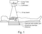
- Fig. 1 illustrates an exemplary X-ray examination on a table.
- a beam of X-rays is produced by an X-ray generator and is projected toward a subject.
- a certain amount of the X-rays or other radiation is absorbed by the subject, dependent on the subject's density and structural composition.
- the X-rays that pass through the subject are captured behind the subject by an X-ray detector, which may be either photographic film or a digital detector.
- Fig. 1 One problem may be overlapping organs, as shown in Fig. 1 , which may have different sensitivities to X-ray.
- the thickness, location, and to some extend the shape of the organs may also vary from patient to patient.
- a computer-implemented method for determining an effective dose to a subject from a radiographic examination in which a radiographic imaging device with a radiation source and a radiation detector is used to acquire a radiographic image of the subject, the method comprising:
- the present disclosure proposes to adapt a 3D patient model to a particular patient by using a set of non-rigid registration parameters.
- the 3D patient model may be a generic representation of a surface of a human or part of a human, e.g., chest.
- the 3D patient model may be not specific to any patient or is specific to a patient meeting a norm.
- the 3D patient model includes one or more internal organs and/or one or more tissues.
- the 3D patient model is an annotated 3D model with annotated data comprising tissue information, which is used to obtain a radiation specific weighting factor and a tissue specific weighting factor of at least one internal organ and/or at least one type of tissues within the 3D patient model.
- the tissue information may comprise an organ identifier used to identify at least one organ within the 3D patient model and/or a tissue identifier used to identify a tissue type of at least one within the 3D patient model.
- the internal organ e.g., liver
- the organ identifier may be used to retrieve the tissue specific absorption coefficients and weighting factors from a database for the calculation of a local effective dose
- the tissue information may comprise the absorption coefficient and the tissue weighting factors. This may be beneficial if the internal organ (e.g., bone marrow) comprise two or more types of tissues.
- the tissue information may comprise an attenuation coefficient of at least one tissue type of tissues and a tissue specific weighting factor of at least one tissue type of the tissues within the 3D patient model. From this adapted 3D patient model, the tissue specific absorption coefficients and weighting factors may be taken to determine a local effective dose.
- the effective dose of the complete irradiated area which may be logged in e.g., an X-ray logbook to collect information about the applied effective dose over time. This may be compared with the limits set by local regulatory authorities.
- the local effective dose e.g., within specific organs, which can be logged in an X-ray logbook to collect information about the applied effective dose over time. This can be compared with the limits set by local regulatory authorities.
- this information can be used to give the radiographer feedback not only according to the unnecessarily irradiated area (which is state of the art nowadays) but also according the unnecessarily applied effective dose which is significantly more relevant for the patient.
- a data-driven model is applied to determine the set of non-rigid registration parameters, wherein the data-driven model has been trained to determine the set of non-rigid registration parameters for a model-to-patient registration using the acquired radiographic image of the subject, and depth image data of the subject.
- the data-driven model may include a neural network model, such as a fully connected neural network, convolutional neural network, or another neural network.
- a neural network model such as a fully connected neural network, convolutional neural network, or another neural network.
- the data-driven model comprises a neural network.
- the 3D patient model is generated based on one or more Computed Tomography, CT, datasets or one or more Magnetic Resonance, MR, datasets.
- the one or more CT datasets may comprise CT image data acquired from one or more patients.
- the one or more MR datasets may comprise MR image data acquired from one or more patients.
- the tissue information comprises an organ identifier used to identify at least one organ within the 3D patient model and/or a tissue identifier used to identify a tissue type of at least one tissue within the 3D patient model.
- the organ identifier may be the name of the at least one internal organ (e.g., kidney) or any other identifier (e.g., code) used to identify the at least one organ within the 3D patient model.
- the tissue identifier may be the name of the tissue type or any other identifier (e.g., code) used to identify the tissue type of the at least one tissue within the 3D patient model.
- the organ identifier and/or the tissue identifier may be used to retrieve various information from a database.
- the organ identifier and/or the tissue identifier may be used to retrieve a radiation specific weighting factor and a tissue specific weighting factor of the at least one organ and/ the at least one tissue from the database.
- Such information may be used to determine a local effective dose.
- the tissue information comprises one or more of:
- the internal organ may comprise two or more different types of tissues.
- a long bone which consists among others of cortical bone and spongy bone tissue which have different attenuation coefficients and different tissue specific weighting factors.
- the effective dose comprises one or more of:
- the method further comprising: providing one or more of the following information:
- the body part comprises one or more of: at least one organ and/or at least one tissue.
- the radiation source is configured to emit X-rays, or gamma rays.
- an apparatus comprising a processor configured to perform the steps according to the first aspect and any associated example.
- a radiographic imaging system comprises a radiographic imaging device comprising a radiation source and a radiation detector configured to acquire a radiographic image of a subject, a depth camera configured to acquire a depth image of the subject, and the apparatus according to the second aspect to determine an effective dose to the subject in radiographic imaging.
- the radiographic imaging device comprises one or more of:
- a computer program product comprising instructions which, when the program is executed by a processor, cause the processor to carry out the steps of the method according to the first aspect and any associated example.
- a computer-readable storage medium having stored thereon the computer program product.
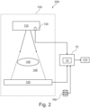
- Fig. 2 illustrates an exemplary radiographic imaging system 200.
- the radiographic imaging system 200 comprises a radiographic imaging device 100 and an effective dose determination apparatus 10.
- an X-ray imaging device 100 is provided as an example of the radiographic imaging device. While reference is made to the X-ray imaging device 100 in the following description, it will be appreciated that the device, method, and system disclosed herein could also be applied to other radiographic imaging modality, such as imaging using gamma rays.
- the X-ray imaging device 100 comprises an X-ray source 110 and an X-ray detector 120 configured to acquire X-ray image data of a subject 150.
- the X-ray source 110 and the X-ray detector 120 are separated by an examination region 140 for performing an X-ray imaging operation on the subject 150 when the subject is located within the examination region.
- the solid lines extending between the X-ray source 110 and the X-ray detector 120 indicate the volumetric extent of the overlap between the X-ray beam emitted by the X-ray source 110, and the X-ray radiation-sensitive region of the X-ray detector 120, within which the X-ray image data may be generated. The volumetric extent of this overlap defines the examination region 140.
- the subject may for example be a human body or a portion of the human body.
- the X-ray source is mounted to the ceiling via a gantry, and the X-ray detector is mounted to a stand and held in the vertical position.
- Alternative arrangements, mounting arrangements, and positions of the X-ray source 110 and the X-ray detector 120 may also be used.
- the depth camera 130 is configured to view the examination region 140 in order to generate depth camera image data representing the subject 150 when the subject is located within the examination region 140.
- the depth camera image data may therefore represent a three-dimensional shape of a surface of the subject 150.
- the depth camera 130 is arranged within a housing of the X-ray source 110.
- the depth camera 130 may alternatively be positioned elsewhere in order to view the examination region 140.
- the depth camera may be arranged on a wall, or on a ceiling of a room in which the X-ray imaging device 100 is located, or it may be mechanically coupled to a floor-based stand in the room.
- the depth camera 130 may alternatively be mobile.
- Examples of the depth camera 130 may include, but are not limited to, a time-of-flight (TOF) camera, a structured-light camera, and a stereo vision camera.
- the TOF camera generates depth camera image data representing the range of the objects by using the time taken for emitted light pulses to travel from the position of the camera to objects in a scene and back again.
- the structured-light camera an optical pattern is projected on to the surface of the objects within a scene, and the disparity between the original projected pattern and the pattern that is deformed by the surface of the objects is imaged by one or more cameras.
- the stereo vision camera different views of a scene are used to compute a depth map of the scene.
- the effective dose determination apparatus 10 comprises a processor 12.
- the effective dose determination apparatus 10 may be implemented in numerous ways (e.g., such as with dedicated hardware) to perform the functions described herein.
- a "processor” is one example of the effective dose determination apparatus 10, which employs one or more microprocessors that may be programmed using software (e.g., microcode) to perform various functions described herein.
- the effective dose determination apparatus 10 may be implemented with or without employing a processor, and also may be implemented as a combination of dedicated hardware to perform some functions and a processor (e.g., one or more programmed microprocessors and associated circuitry) to perform other functions.
- a processor may be associated with one or more storage media (generically referred to herein as "memory,” e.g., volatile and non-volatile computer memory).
- the storage media may be encoded with one or more programs that, when executed on one or more processors and/or controllers, perform at least some of the functions described herein.
- Various storage media may be fixed within the effective dose determination apparatus 10 or may be transportable, such that the one or more programs stored thereon can be loaded into the effective dose determination apparatus 10 so as to implement various aspects of the present disclosure described herein.
- program or “computer program” are used herein in a generic sense to refer to any type of computer code (e.g., software or microcode) that can be employed to program one or more processors.
- the effective dose determination apparatus 10 is configured to receive the depth image data from the depth camera 130 and the X-ray image data from the X-ray imaging device 100.
- the effective dose determination apparatus 10 may receive the depth camera image data and the X-ray image data via any form of digital communication.
- the communication path may be direct, or indirect.
- the effective dose determination apparatus 10 may be connected to the depth camera 130 and the X-ray detector 120 via a direct wired or wireless communication path, such as an electrical cable, or a wireless infrared or RF communication path such as Bluetooth, as illustrated by the arrows connecting these items in Fig. 2 .
- the communication path may be indirect, and the effective dose determination apparatus 10 may in communication with the depth camera 130 and the X-ray imaging device 100 via the internet, the cloud, or a computer readable storage memory.
- the effective dose determination apparatus 10 is configured to receive a 3D patient model.
- the 3D patient model includes one or more internal organs and/or one or more tissues.
- the 3D patient model may have been created from 3-dimensional data like CT or MR data. These datasets may be segmented into organ and tissue types.
- the 3D patient model may be a generic representation of a surface of a human or part of a human, e.g., chest. Different models may be used for different body types, such as a male or female model.
- the 3D patient model may be not specific to the patient.
- the 3D patient model may be a statistical shape model.
- the 3D patient model may be not specific to any patient or is specific to a patient meeting a norm. Any representation may be used for the model.
- the 3D patient model may be formed from a mesh, such as a mesh or triangles. Other meshes may be used. Other representations of a 3D surface may be used.
- the 3D patient model may further comprise annotated data with tissue information.
- the tissue information is used to obtain a radiation specific weighting factor and a tissue specific weighting factor of at least one internal organ and/or at least one type of tissues within the 3D patient model.
- the tissue information may comprise an organ identifier used to identify at least one organ within the 3D patient model and/or a tissue identifier used to identify a tissue type of at least one within the 3D patient model.
- the organ identifier may be used to retrieve the tissue specific absorption coefficients and weighting factors from a database for the calculation of a local effective dose
- the tissue information may comprise the absorption coefficient and the tissue weighting factors. This may be beneficial if the internal organ (e.g., bone marrow) comprise two or more types of tissues.
- the tissue information may comprise an attenuation coefficient of at least one tissue type of tissues and a tissue specific weighting factor of at least one tissue type of the tissues within the 3D patient model. From this adapted 3D patient model, the tissue specific absorption coefficients and weighting factors may be taken to determine a local effective dose.
- the attenuation coefficient is a measure of how much the incident energy beam (e.g., X-rays) is weakened by the material it is passing through, and is dependent on the spectrum of the radiation.
- the tissue specific weighting factor is a relative measure of the risk of stochastic effects that might result from irradiation of that specific tissue. It accounts for the variable radiation sensitivities of organs and tissues in the body to ionizing radiation.
- the effective dose determination apparatus 10 may receive the 3D patient model from a database 160 shown in Fig. 2 , which may store a plurality of 3D patient models representing a surface of two or more different body parts (e.g., chest, leg, etc.) of a human.
- the effective dose determination apparatus 10 is further configured to determine a set of non-rigid registration parameters for an adaption of the 3D patient model to the subject.
- the effective dose determination apparatus 10 may apply a data-driven model to determine the set of non-rigid registration parameters.
- the data-driven model has been trained to determine the set of non-rigid registration parameters for a model-to-patient registration using the depth camera image data received from the depth camera 130 and the X-ray image data received from the X-ray imaging device 100.
- the data-driven model may include a suitable algorithm that is trained on the basis of appropriate training data.
- the data-driven model may include a neural network model, such as a fully connected neural network, convolutional neural network, or another neural network.
- Fig. 3 illustrates a flow chart diagram of an exemplary method 300 for training a neural network.
- a 3D patient model is generated, e.g., based on one or more CT datasets and/or one or more MR datasets.
- the 3D patient model may be a generic representation of a surface of a human or part of a human.
- the 3D patient model may be not specific to the patient.
- the 3D patient model may be a statistical shape model.
- the 3D patient model may be not specific to any patient or is specific to a patient meeting a norm.
- the 3D patient model is an annotated 3D patient model.
- the annotated data may comprise one or more of the following tissue information, information about one or more organs and/or tissues within the 3D patient model, an attenuation coefficient of a tissue type of at least one tissue within the 3D patient model, and a tissue specific weighting factor of a tissue type of at least one tissue within the 3D patient model.
- an artificial X-ray image and a depth map are created using one or more CT datasets.
- This artificial X-ray image and depth map are also referred to as target X-ray image and depth map.
- the artificial X-ray image may be obtained by converting 3D CT image data into a 2D projection.
- the depth map comprises information representing a three-dimensional shape of a surface of a body part of a patient.
- the depth map may be created by contouring an outer surface of one or more CT images.
- an initial set of non-rigid registration parameters created by a neural network is selected.
- the neural network model arrangement or architecture may be defined.
- the definition may be by configuration or programming of the learning.
- the number of layers or units, type of learning, and other characteristics of the network are controlled by the programmer or user.
- one or more aspects e.g., number of nodes, number of layers or units, or type of learning
- the 3D patient model is registered using the initial set of non-rigid registration parameters created by the neural network to create an X-ray image and a depth map.
- a loss value is determined using the difference between the X-ray image and the depth map created from the 3D patient model and the target X-ray image and depth map.
- the loss value is used to update the neural network.
- the updated neural network is used to obtain a set of updated registration parameters, and the optimization is repeated until a sufficient quality is reached.
- the sufficient quality may be indicated by a predefined loss value.
- the effective dose determination apparatus 10 is further configured to register the 3D patient model to the subject using the set of determined non-rigid registration parameters, and to determine the effective dose based on the registered 3D patient model, geometry data of the radiographic imaging device, and generator data of the radiation source.
- the effective dose may include one or more of an effective dose per volume element, an effective dose per organ, and a total effective dose.
- the geometry data of the X-ray imaging device 100 may be determined in different ways.
- the positions of at least one of the X-ray source 110 and the X-ray detector 120 may be fixed positions.
- the positions of the X-ray source 110 and the X-ray detector may be mechanically attaching the relevant item to a reference position, such as the wall, the ceiling, the floor, and so forth.
- the geometry data of the X-ray imaging device 100 may be determined using calibration data that represents the fixed position(s).
- the positions of both the X-ray source 110 and the X-ray detector 120 are fixed.
- the spatial relationship between the X-ray source 110 and the X-ray detector 120 may be determined based on calibration data representing the relative positions of the X-ray source 110 and the X-ray detector 120.
- the positions of one or both of the X-ray source 110 and the X-ray detector 120 may be movable to any position e.g., along rails.
- the spatial relationship between the X-ray source 110 and the X-ray detector 120 may be determined using a position sensor.
- suitable position sensors may include, but are not limited to, laser-based optical rangefinders, radiofrequency, and ultrasound ranging transponders, and optical cameras that are configured to track the positions of fiducial markers disposed on one or more of the X-ray source 110 and the X-ray detector 120.
- the X-ray generator provides operator control of the radiographic techniques, including tube voltage (kVp), tube current (mA), and exposure duration, and delivers power to the x-ray tube.
- the generator data of the X-ray source 110 may include one or more of the above-mentioned parameters.
- the generator data of the X-ray source 110 may comprise the X-ray source spectra and the tube current.
- the generator data of the X-ray source 110 may be obtained in several ways. In some examples, the generator data of the X-ray source 110 may be obtained from an operating console of the X-ray imaging device 100. In some examples, the generator data of the X-ray source 110 may be provided by a user input.
- the generator data of the X-ray source 110 may be provided by a sensor device, e.g., an X-ray tube current measuring sensor. In some examples, the generator data of the X-ray source may be obtained using two or more of the above-described approaches.
- the determination of the effective dose may be simplified using cubic voxels and no scatter. In practice, minor deviations due to non-cubic voxels and scatter may be considered.
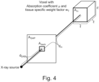
- Fig. 4 shows a principal geometry which can be used for the calculations for a single voxel. The proportions within the figure are not correct. The intention is to introduce the relevant quantities for the later calculations, which are presented afterwards.
- D Total DAP / A DAP
- the entrance area A in of the voxel has the size l 2 .
- the total effective dose or the effective dose per organ can now be calculated by summing up the effective dose of all voxels respectively for all voxels belonging to a specific organ.
- the results may be stored e.g., in a data storage and/or presented to a user e.g., via a display.
- a display 170 may be configured to receive the results from the effective dose determination apparatus 10 via any form of digital communication.
- the communication path may be direct, or indirect.
- the display 170 may be connected to the effective dose determination apparatus 10 via a direct wired or wireless communication path, such as an electrical cable, or a wireless infrared or RF communication path such as Bluetooth, as illustrated by the arrows connecting these items in Fig. 2 .
- the communication path may be indirect, and the effective dose determination apparatus 10 may be in communication with the display 170 via the internet, the cloud, or a computer readable storage memory.
- the display 170 may be any appropriate display, such as a display on a mobile device, a display of a personal computer, etc.
- GUI graphic user interface
- the total effective dose may be shown on the display, optionally together with recommended values for the examination or limits set by local regulatory authorities.
- overlays may show selected organs together with the effective dose for this organ.
- the accumulated total or organ specific dose over a specific time period can be shown.
- a warning may be shown if the accumulated dose reaches a critical level.
- Fig. 5 illustrates a flow chart diagram of an exemplary computer-implemented method 400 for determining an effective dose to a subject from a radiographic examination, in which a radiographic imaging device with a radiation source and a radiation detector is used to acquire a radiographic image of the subject.
- the radiographic imaging device may be an X-ray imaging device shown in Fig. 2 .
- the method 400 comprises the step of providing a 3D patient model.
- the 3D patient model may have been created from 3-dimensional data like CT or MR data. These datasets may be segmented into organ and tissue types.
- the 3D patient model may be a generic representation of a surface of a human or part of a human. Different models may be used for different body types, such as a male or female model.
- the 3D patient model may be not specific to the patient.
- the 3D patient model may be a statistical shape model.
- the 3D patient model may be not specific to any patient or is specific to a patient meeting a norm. Any representation may be used for the model.
- the 3D patient model may be formed from a mesh, such as a mesh or triangles.
- the 3D patient model may further comprise annotated data with tissue information.
- tissue information may include, but are not limited to, information about one or more organs and/or tissues within 3D patient model, an attenuation coefficient of a tissue type of at least one tissue within the 3D patient model, and a tissue specific weighting factor of a tissue type of at least one tissue within the 3D patient model.
- the attenuation coefficient is a measure of how much the incident energy beam (e.g., X-rays) is weakened by the material it is passing through, and is dependent on the spectrum of the radiation.
- the tissue specific weighting factor is a relative measure of the risk of stochastic effects that might result from irradiation of that specific tissue.
- the 3D patient model may be provided by a database 160 shown in Fig. 2 , which may store a plurality of 3D patient models of different body parts.
- the method 400 comprises the step of determining a set of non-rigid registration parameters for an adaption of the 3D patient model to the subject.
- a data-driven model may be applied, which has been trained to determine the set of non-rigid registration parameters for a model-to-patient registration using the depth camera image data and the X-ray image data.
- the data-driven model may include a suitable algorithm that is trained on the basis of appropriate training data.
- the data-driven model may include a neural network model, such as a fully connected neural network, convolutional neural network, or another neural network.
- An exemplary training method is described in detail hereinbefore and in particular with respect to the flow chart diagram shown in Fig. 3 .
- the method 400 comprises the step of registering the 3D patient model to the subject using the set of determined non-rigid registration parameters.
- the method 400 comprises the step of determining the effective dose based on the registered 3D patient model and one or more system dependent parameters of the radiation imaging device.
- the determination of the effective dose may be simplified using cubic voxels and no scatter. In practice, minor deviations due to non-cubic voxels and scatter may be considered.
- Fig. 4 shows the principal geometry which can be used for the calculations for a single voxel.
- a computer program or a computer program element is provided that is characterized by being adapted to execute the method steps of the method according to one of the preceding embodiments, on an appropriate system.
- the computer program element might therefore be stored on a computer unit, which might also be part of an embodiment of the present invention.
- This computing unit may be adapted to perform or induce a performing of the steps of the method described above. Moreover, it may be adapted to operate the components of the above described apparatus.
- the computing unit can be adapted to operate automatically and/or to execute the orders of a user.
- a computer program may be loaded into a working memory of a data processor.
- the data processor may thus be equipped to carry out the method of the invention.
- This exemplary embodiment of the invention covers both, a computer program that right from the beginning uses the invention and a computer program that by means of an up-date turns an existing program into a program that uses the invention.
- the computer program element might be able to provide all necessary steps to fulfil the procedure of an exemplary embodiment of the method as described above.
- a computer readable medium such as a CD-ROM
- the computer readable medium has a computer program element stored on it which computer program element is described by the preceding section.
- a computer program may be stored and/or distributed on a suitable medium, such as an optical storage medium or a solid state medium supplied together with or as part of other hardware, but may also be distributed in other forms, such as via the internet or other wired or wireless telecommunication systems.
- a suitable medium such as an optical storage medium or a solid state medium supplied together with or as part of other hardware, but may also be distributed in other forms, such as via the internet or other wired or wireless telecommunication systems.
- the computer program may also be presented over a network like the World Wide Web and can be downloaded into the working memory of a data processor from such a network.
- a medium for making a computer program element available for downloading is provided, which computer program element is arranged to perform a method according to one of the previously described embodiments of the invention.
Landscapes
- Health & Medical Sciences (AREA)
- Engineering & Computer Science (AREA)
- Life Sciences & Earth Sciences (AREA)
- Medical Informatics (AREA)
- Public Health (AREA)
- Biomedical Technology (AREA)
- General Health & Medical Sciences (AREA)
- Physics & Mathematics (AREA)
- Pathology (AREA)
- Biophysics (AREA)
- Veterinary Medicine (AREA)
- Optics & Photonics (AREA)
- High Energy & Nuclear Physics (AREA)
- Radiology & Medical Imaging (AREA)
- Nuclear Medicine, Radiotherapy & Molecular Imaging (AREA)
- Heart & Thoracic Surgery (AREA)
- Molecular Biology (AREA)
- Surgery (AREA)
- Animal Behavior & Ethology (AREA)
- Theoretical Computer Science (AREA)
- Computer Vision & Pattern Recognition (AREA)
- General Physics & Mathematics (AREA)
- Data Mining & Analysis (AREA)
- Databases & Information Systems (AREA)
- Epidemiology (AREA)
- Primary Health Care (AREA)
- Pulmonology (AREA)
- Apparatus For Radiation Diagnosis (AREA)
Abstract
The present invention relates to radiographic imaging. In order to improve the effective dose estimation, there is provided a computer-implemented method (400) for determining an effective dose to a subject from a radiographic examination, in which a radiographic imaging device with a radiation source and a radiation detector is used to acquire a radiographic image of the subject, the method comprising:
a) providing (410) a three-dimensional, 3D, patient model;
b) determining (420) a set of non-rigid registration parameters for an adaption of the 3D patient model to the subject;
c) registering (430) the 3D patient model to the subject using the set of determined non-rigid registration parameters; and
d) determining (440) the effective dose based on the registered 3D patient model of the body part, geometry data of the radiographic imaging device and generator data of the radiation source.
a) providing (410) a three-dimensional, 3D, patient model;
b) determining (420) a set of non-rigid registration parameters for an adaption of the 3D patient model to the subject;
c) registering (430) the 3D patient model to the subject using the set of determined non-rigid registration parameters; and
d) determining (440) the effective dose based on the registered 3D patient model of the body part, geometry data of the radiographic imaging device and generator data of the radiation source.
Description
- The present invention relates to radiographic imaging, and in particular to a computer-implemented method and apparatus for determining an effective dose to a subject from a radiographic examination, to a radiographic imaging system, to a computer program product, and to a computer-readable storage medium.
- Radiography is an imaging technique using X-rays, gamma rays, or similar ionizing radiation and non-ionizing radiation to view the internal form of an object. Applications of radiography include medical.
Fig. 1 illustrates an exemplary X-ray examination on a table. To create an image in the radiography, a beam of X-rays is produced by an X-ray generator and is projected toward a subject. A certain amount of the X-rays or other radiation is absorbed by the subject, dependent on the subject's density and structural composition. The X-rays that pass through the subject are captured behind the subject by an X-ray detector, which may be either photographic film or a digital detector. - Nowadays, the determination of the effective dose is based on rough estimates. One problem may be overlapping organs, as shown in
Fig. 1 , which may have different sensitivities to X-ray. The thickness, location, and to some extend the shape of the organs may also vary from patient to patient. - There may be a need to improve the effective dose estimation.
- According to a first aspect of the present invention, there is provided a computer-implemented method for determining an effective dose to a subject from a radiographic examination, in which a radiographic imaging device with a radiation source and a radiation detector is used to acquire a radiographic image of the subject, the method comprising:
- a) providing a three-dimensional, 3D, patient model, wherein the 3D patient model comprises annotated data with tissue information;
- b) determining a set of non-rigid registration parameters for an adaption of the 3D patient model to the subject ;
- c) registering the 3D patient model to the subject using the set of determined non-rigid registration parameters; and
- d) determining the effective dose based on the registered 3D patient model, geometry data of the radiographic imaging device, and generator data of the radiation source.
- Accordingly, the present disclosure proposes to adapt a 3D patient model to a particular patient by using a set of non-rigid registration parameters. The 3D patient model may be a generic representation of a surface of a human or part of a human, e.g., chest. The 3D patient model may be not specific to any patient or is specific to a patient meeting a norm. The 3D patient model includes one or more internal organs and/or one or more tissues. The 3D patient model is an annotated 3D model with annotated data comprising tissue information, which is used to obtain a radiation specific weighting factor and a tissue specific weighting factor of at least one internal organ and/or at least one type of tissues within the 3D patient model. In some examples, the tissue information may comprise an organ identifier used to identify at least one organ within the 3D patient model and/or a tissue identifier used to identify a tissue type of at least one within the 3D patient model. For example, if the internal organ (e.g., liver) comprises a single type of tissues (e.g., liver), the organ identifier may be used to retrieve the tissue specific absorption coefficients and weighting factors from a database for the calculation of a local effective dose, In some examples, the tissue information may comprise the absorption coefficient and the tissue weighting factors. This may be beneficial if the internal organ (e.g., bone marrow) comprise two or more types of tissues. In such case, the tissue information may comprise an attenuation coefficient of at least one tissue type of tissues and a tissue specific weighting factor of at least one tissue type of the tissues within the 3D patient model. From this adapted 3D patient model, the tissue specific absorption coefficients and weighting factors may be taken to determine a local effective dose.
- This can be used among others to determine one or more of the following valuable information:
The effective dose of the complete irradiated area, which may be logged in e.g., an X-ray logbook to collect information about the applied effective dose over time. This may be compared with the limits set by local regulatory authorities. - The local effective dose, e.g., within specific organs, which can be logged in an X-ray logbook to collect information about the applied effective dose over time. This can be compared with the limits set by local regulatory authorities.
- Together with a software which calculates the optimal collimation it is possible to check whether tissue was unnecessarily irradiated. In this case, this information can be used to give the radiographer feedback not only according to the unnecessarily irradiated area (which is state of the art nowadays) but also according the unnecessarily applied effective dose which is significantly more relevant for the patient.
- According to an exemplary embodiment of the first aspect of the present invention, a data-driven model is applied to determine the set of non-rigid registration parameters, wherein the data-driven model has been trained to determine the set of non-rigid registration parameters for a model-to-patient registration using the acquired radiographic image of the subject, and depth image data of the subject.
- In one example, the data-driven model may include a neural network model, such as a fully connected neural network, convolutional neural network, or another neural network.
- According to an exemplary embodiment of the first aspect of the present invention, the data-driven model comprises a neural network.
- According to an exemplary embodiment of the first aspect of the present invention, the 3D patient model is generated based on one or more Computed Tomography, CT, datasets or one or more Magnetic Resonance, MR, datasets.
- The one or more CT datasets may comprise CT image data acquired from one or more patients. The one or more MR datasets may comprise MR image data acquired from one or more patients.
- According to an exemplary embodiment of the first aspect of the present invention, the tissue information comprises an organ identifier used to identify at least one organ within the 3D patient model and/or a tissue identifier used to identify a tissue type of at least one tissue within the 3D patient model.
- The organ identifier may be the name of the at least one internal organ (e.g., kidney) or any other identifier (e.g., code) used to identify the at least one organ within the 3D patient model.
- The tissue identifier may be the name of the tissue type or any other identifier (e.g., code) used to identify the tissue type of the at least one tissue within the 3D patient model.
- The organ identifier and/or the tissue identifier may be used to retrieve various information from a database. For example, the organ identifier and/or the tissue identifier may be used to retrieve a radiation specific weighting factor and a tissue specific weighting factor of the at least one organ and/ the at least one tissue from the database. Such information may be used to determine a local effective dose.
- According to an exemplary embodiment of the first aspect of the present invention, the tissue information comprises one or more of:
- an attenuation coefficient of a tissue type of at least one tissue within the 3D patient model; and
- a tissue specific weighting factor of a tissue type of at least one tissue within the 3D patient model.
- In one example, the internal organ may comprise two or more different types of tissues. For example, a long bone which consists among others of cortical bone and spongy bone tissue which have different attenuation coefficients and different tissue specific weighting factors. In such case, it may be beneficial to include the attenuation coefficients and tissue specific weighting factors of different tissue types into the tissue information.
- According to an exemplary embodiment of the first aspect of the present invention, the effective dose comprises one or more of:
- an effective dose per volume element;
- an effective dose per organ; and
- an effective dose of a total irradiated area.
- According to an exemplary embodiment of the first aspect of the present invention, the method further comprising:
providing one or more of the following information: - an effective dose of the total irradiated area;
- an effective dose of a specific organ;
- information from one or more previous radiographic examinations;
- an accumulated total and/or an organ-specific dose over a specific time period; and
- a warning signal if the accumulated total and/or the organ-specific dose reaches a predefined threshold.
- According to an exemplary embodiment of the first aspect of the present invention, the body part comprises one or more of: at least one organ and/or at least one tissue.
- According to an exemplary embodiment of the first aspect of the present invention, the radiation source is configured to emit X-rays, or gamma rays.
- According to a second aspect of the present invention, there is provided an apparatus comprising a processor configured to perform the steps according to the first aspect and any associated example.
- According to a third aspect of the present invention, there is provided a radiographic imaging system. The radiographic imaging system comprises a radiographic imaging device comprising a radiation source and a radiation detector configured to acquire a radiographic image of a subject, a depth camera configured to acquire a depth image of the subject, and the apparatus according to the second aspect to determine an effective dose to the subject in radiographic imaging.
- According to an exemplary embodiment of the third aspect of the present invention, the radiographic imaging device comprises one or more of:
- an X-ray imaging device; and
- a Gamma ray imaging.
- According to a further aspect of the present invention, there is provided a computer program product comprising instructions which, when the program is executed by a processor, cause the processor to carry out the steps of the method according to the first aspect and any associated example.
- According to another aspect of the present invention, there is provided a computer-readable storage medium having stored thereon the computer program product.
- These and other aspects of the present invention will become apparent from and be elucidated with reference to the embodiments described hereinafter.
- These and other aspects of the invention will be apparent from and elucidated further with reference to the embodiments described by way of examples in the following description and with reference to the accompanying drawings, in which
-
Fig. 1 illustrates an X-ray examination on a table. -
Fig. 2 illustrates an exemplary radiographic imaging device. -
Fig. 3 illustrates a flow chart diagram of an exemplary training method. -
Fig. 4 illustrates a principal geometry which can be used for the calculations for a single voxel. -
Fig. 5 illustrates a flow chart diagram of an exemplary computer-implemented method for determining an effective dose to a subject from a radiographic examination. - It should be noted that the figures are purely diagrammatic and not drawn to scale. In the figures, elements which correspond to elements already described may have the same reference numerals. Examples, embodiments or optional features, whether indicated as non-limiting or not, are not to be understood as limiting the invention as claimed.
-
Fig. 2 illustrates an exemplaryradiographic imaging system 200. Theradiographic imaging system 200 comprises aradiographic imaging device 100 and an effectivedose determination apparatus 10. - In the illustrated example, an
X-ray imaging device 100 is provided as an example of the radiographic imaging device. While reference is made to theX-ray imaging device 100 in the following description, it will be appreciated that the device, method, and system disclosed herein could also be applied to other radiographic imaging modality, such as imaging using gamma rays. - The
X-ray imaging device 100 comprises anX-ray source 110 and anX-ray detector 120 configured to acquire X-ray image data of a subject 150. TheX-ray source 110 and theX-ray detector 120 are separated by anexamination region 140 for performing an X-ray imaging operation on the subject 150 when the subject is located within the examination region. The solid lines extending between theX-ray source 110 and theX-ray detector 120 indicate the volumetric extent of the overlap between the X-ray beam emitted by theX-ray source 110, and the X-ray radiation-sensitive region of theX-ray detector 120, within which the X-ray image data may be generated. The volumetric extent of this overlap defines theexamination region 140. The subject may for example be a human body or a portion of the human body. In one example, the X-ray source is mounted to the ceiling via a gantry, and the X-ray detector is mounted to a stand and held in the vertical position. Alternative arrangements, mounting arrangements, and positions of theX-ray source 110 and theX-ray detector 120 may also be used. - The
depth camera 130 is configured to view theexamination region 140 in order to generate depth camera image data representing the subject 150 when the subject is located within theexamination region 140. The depth camera image data may therefore represent a three-dimensional shape of a surface of the subject 150. In the illustrated example, thedepth camera 130 is arranged within a housing of theX-ray source 110. However, thedepth camera 130 may alternatively be positioned elsewhere in order to view theexamination region 140. The depth camera may be arranged on a wall, or on a ceiling of a room in which theX-ray imaging device 100 is located, or it may be mechanically coupled to a floor-based stand in the room. Thedepth camera 130 may alternatively be mobile. Examples of thedepth camera 130 may include, but are not limited to, a time-of-flight (TOF) camera, a structured-light camera, and a stereo vision camera. The TOF camera generates depth camera image data representing the range of the objects by using the time taken for emitted light pulses to travel from the position of the camera to objects in a scene and back again. In the structured-light camera, an optical pattern is projected on to the surface of the objects within a scene, and the disparity between the original projected pattern and the pattern that is deformed by the surface of the objects is imaged by one or more cameras. In the stereo vision camera, different views of a scene are used to compute a depth map of the scene. - The effective
dose determination apparatus 10 comprises aprocessor 12. The effectivedose determination apparatus 10 may be implemented in numerous ways (e.g., such as with dedicated hardware) to perform the functions described herein. A "processor" is one example of the effectivedose determination apparatus 10, which employs one or more microprocessors that may be programmed using software (e.g., microcode) to perform various functions described herein. The effectivedose determination apparatus 10 may be implemented with or without employing a processor, and also may be implemented as a combination of dedicated hardware to perform some functions and a processor (e.g., one or more programmed microprocessors and associated circuitry) to perform other functions. Examples of device components that may be employed in various embodiments of the present disclosure include, but are not limited to, conventional microprocessors, application specific integrated circuits (ASICs), and field-programmable gate arrays (FPGAs). In various implementations, a processor may be associated with one or more storage media (generically referred to herein as "memory," e.g., volatile and non-volatile computer memory). In some implementations, the storage media may be encoded with one or more programs that, when executed on one or more processors and/or controllers, perform at least some of the functions described herein. Various storage media may be fixed within the effectivedose determination apparatus 10 or may be transportable, such that the one or more programs stored thereon can be loaded into the effectivedose determination apparatus 10 so as to implement various aspects of the present disclosure described herein. The terms "program" or "computer program" are used herein in a generic sense to refer to any type of computer code (e.g., software or microcode) that can be employed to program one or more processors. - The effective
dose determination apparatus 10 is configured to receive the depth image data from thedepth camera 130 and the X-ray image data from theX-ray imaging device 100. The effectivedose determination apparatus 10 may receive the depth camera image data and the X-ray image data via any form of digital communication. The communication path may be direct, or indirect. For example, the effectivedose determination apparatus 10 may be connected to thedepth camera 130 and theX-ray detector 120 via a direct wired or wireless communication path, such as an electrical cable, or a wireless infrared or RF communication path such as Bluetooth, as illustrated by the arrows connecting these items inFig. 2 . Alternatively, the communication path may be indirect, and the effectivedose determination apparatus 10 may in communication with thedepth camera 130 and theX-ray imaging device 100 via the internet, the cloud, or a computer readable storage memory. - To determine an effective dose, the effective
dose determination apparatus 10 is configured to receive a 3D patient model. The 3D patient model includes one or more internal organs and/or one or more tissues. The 3D patient model may have been created from 3-dimensional data like CT or MR data. These datasets may be segmented into organ and tissue types. The 3D patient model may be a generic representation of a surface of a human or part of a human, e.g., chest. Different models may be used for different body types, such as a male or female model. The 3D patient model may be not specific to the patient. For example, the 3D patient model may be a statistical shape model. The 3D patient model may be not specific to any patient or is specific to a patient meeting a norm. Any representation may be used for the model. In one example, the 3D patient model may be formed from a mesh, such as a mesh or triangles. Other meshes may be used. Other representations of a 3D surface may be used. In one example, the 3D patient model may further comprise annotated data with tissue information. The tissue information is used to obtain a radiation specific weighting factor and a tissue specific weighting factor of at least one internal organ and/or at least one type of tissues within the 3D patient model. In some examples, the tissue information may comprise an organ identifier used to identify at least one organ within the 3D patient model and/or a tissue identifier used to identify a tissue type of at least one within the 3D patient model. For example, if the internal organ (e.g., liver) comprises a single type of tissues (e.g., liver), the organ identifier may be used to retrieve the tissue specific absorption coefficients and weighting factors from a database for the calculation of a local effective dose, In some examples, the tissue information may comprise the absorption coefficient and the tissue weighting factors. This may be beneficial if the internal organ (e.g., bone marrow) comprise two or more types of tissues. In such case, the tissue information may comprise an attenuation coefficient of at least one tissue type of tissues and a tissue specific weighting factor of at least one tissue type of the tissues within the 3D patient model. From this adapted 3D patient model, the tissue specific absorption coefficients and weighting factors may be taken to determine a local effective dose. The attenuation coefficient is a measure of how much the incident energy beam (e.g., X-rays) is weakened by the material it is passing through, and is dependent on the spectrum of the radiation. The tissue specific weighting factor is a relative measure of the risk of stochastic effects that might result from irradiation of that specific tissue. It accounts for the variable radiation sensitivities of organs and tissues in the body to ionizing radiation. In one example, the effectivedose determination apparatus 10 may receive the 3D patient model from adatabase 160 shown inFig. 2 , which may store a plurality of 3D patient models representing a surface of two or more different body parts (e.g., chest, leg, etc.) of a human. - The effective
dose determination apparatus 10 is further configured to determine a set of non-rigid registration parameters for an adaption of the 3D patient model to the subject. As an example, the effectivedose determination apparatus 10 may apply a data-driven model to determine the set of non-rigid registration parameters. The data-driven model has been trained to determine the set of non-rigid registration parameters for a model-to-patient registration using the depth camera image data received from thedepth camera 130 and the X-ray image data received from theX-ray imaging device 100. The data-driven model may include a suitable algorithm that is trained on the basis of appropriate training data. The data-driven model may include a neural network model, such as a fully connected neural network, convolutional neural network, or another neural network. -
Fig. 3 illustrates a flow chart diagram of anexemplary method 300 for training a neural network. Atblock 310, a 3D patient model is generated, e.g., based on one or more CT datasets and/or one or more MR datasets. As described above, the 3D patient model may be a generic representation of a surface of a human or part of a human. The 3D patient model may be not specific to the patient. For example, the 3D patient model may be a statistical shape model. The 3D patient model may be not specific to any patient or is specific to a patient meeting a norm. The 3D patient model is an annotated 3D patient model. The annotated data may comprise one or more of the following tissue information, information about one or more organs and/or tissues within the 3D patient model, an attenuation coefficient of a tissue type of at least one tissue within the 3D patient model, and a tissue specific weighting factor of a tissue type of at least one tissue within the 3D patient model. - At
block 320, an artificial X-ray image and a depth map are created using one or more CT datasets. This artificial X-ray image and depth map are also referred to as target X-ray image and depth map. For example, the artificial X-ray image may be obtained by converting 3D CT image data into a 2D projection. The depth map comprises information representing a three-dimensional shape of a surface of a body part of a patient. The depth map may be created by contouring an outer surface of one or more CT images. - At
block 330, an initial set of non-rigid registration parameters created by a neural network is selected. For training the neural network, the neural network model arrangement or architecture may be defined. The definition may be by configuration or programming of the learning. The number of layers or units, type of learning, and other characteristics of the network are controlled by the programmer or user. In some examples, one or more aspects (e.g., number of nodes, number of layers or units, or type of learning) are defined and selected by the machine during the learning. - At
block 340, the 3D patient model is registered using the initial set of non-rigid registration parameters created by the neural network to create an X-ray image and a depth map. - At
block 350, a loss value is determined using the difference between the X-ray image and the depth map created from the 3D patient model and the target X-ray image and depth map. - At block 360, the loss value is used to update the neural network.
- At block 370, the updated neural network is used to obtain a set of updated registration parameters, and the optimization is repeated until a sufficient quality is reached. The sufficient quality may be indicated by a predefined loss value.
- Turning back to
Fig. 2 , the effectivedose determination apparatus 10 is further configured to register the 3D patient model to the subject using the set of determined non-rigid registration parameters, and to determine the effective dose based on the registered 3D patient model, geometry data of the radiographic imaging device, and generator data of the radiation source. The effective dose may include one or more of an effective dose per volume element, an effective dose per organ, and a total effective dose. - The geometry data of the
X-ray imaging device 100 may be determined in different ways. - In some examples, the positions of at least one of the
X-ray source 110 and theX-ray detector 120 may be fixed positions. For example, the positions of theX-ray source 110 and the X-ray detector may be mechanically attaching the relevant item to a reference position, such as the wall, the ceiling, the floor, and so forth. In these examples, the geometry data of theX-ray imaging device 100 may be determined using calibration data that represents the fixed position(s). In some examples, the positions of both theX-ray source 110 and theX-ray detector 120 are fixed. In these examples, the spatial relationship between theX-ray source 110 and theX-ray detector 120 may be determined based on calibration data representing the relative positions of theX-ray source 110 and theX-ray detector 120. - In some examples, the positions of one or both of the
X-ray source 110 and theX-ray detector 120 may be movable to any position e.g., along rails. In these examples, the spatial relationship between theX-ray source 110 and theX-ray detector 120 may be determined using a position sensor. Examples of suitable position sensors may include, but are not limited to, laser-based optical rangefinders, radiofrequency, and ultrasound ranging transponders, and optical cameras that are configured to track the positions of fiducial markers disposed on one or more of theX-ray source 110 and theX-ray detector 120. - The X-ray generator provides operator control of the radiographic techniques, including tube voltage (kVp), tube current (mA), and exposure duration, and delivers power to the x-ray tube. The generator data of the
X-ray source 110 may include one or more of the above-mentioned parameters. As an example, the generator data of theX-ray source 110 may comprise the X-ray source spectra and the tube current. The generator data of theX-ray source 110 may be obtained in several ways. In some examples, the generator data of theX-ray source 110 may be obtained from an operating console of theX-ray imaging device 100. In some examples, the generator data of theX-ray source 110 may be provided by a user input. In some examples, the generator data of theX-ray source 110 may be provided by a sensor device, e.g., an X-ray tube current measuring sensor. In some examples, the generator data of the X-ray source may be obtained using two or more of the above-described approaches. - There are various approaches to determine the effective dose. In one example, the determination of the effective dose may be simplified using cubic voxels and no scatter. In practice, minor deviations due to non-cubic voxels and scatter may be considered.
Fig. 4 shows a principal geometry which can be used for the calculations for a single voxel. The proportions within the figure are not correct. The intention is to introduce the relevant quantities for the later calculations, which are presented afterwards. - It may be assumed that the dose area product DAP for an area ADAP at a distance dDAP from the X-ray source is known. This means that the total dose DTotal irradiated over the complete area can be calculated as:

- For any subarea ASub of ADAP (Aback is a special case of such a subarea), the irradiated dose DSub at a distance dDAP from the X-ray source can be calculated as:

- Let's assume that the X-Ray beam goes into the z-direction and the entrance area of the voxel is the x-y-plane of the voxel and has a distance din from the X-ray source. Let's also assume that we have cubic voxels with an edge length l.
- The entrance area Ain of the voxel has the size l2. The back projected entrance area Aback (back projected to distance dDAP ) can be calculated as:

- In case of a surface voxel of the patient model, which is from the perspective of the X-ray tube not obscured by other objects, the entrance dose can be calculated as:

- Using the absorption coefficient µ of the tissue within the voxel the exit dose of the voxel can be calculated as:

- The difference between entrance dose and exit dose is the absorbed Dose Dabs within this voxel:

- On the other hand, is the exit dose Dout the input dose of the next voxel at z-position din + l. Iteration over all voxel along the beam delivers the absorbed dose for all voxels along the beam.
- Using the radiation specific weight factor wR , which is 1 for X-ray, the equivalent dose per voxel H can now be calculated as follows:

- Using the tissue specific weight factor wT , which is defined by international or local authorities, the effective dose per voxel E can be calculates as follows:

- The total effective dose or the effective dose per organ can now be calculated by summing up the effective dose of all voxels respectively for all voxels belonging to a specific organ.
- The results may be stored e.g., in a data storage and/or presented to a user e.g., via a display. For example, as shown in
Fig. 2 , adisplay 170 may be configured to receive the results from the effectivedose determination apparatus 10 via any form of digital communication. The communication path may be direct, or indirect. For example, thedisplay 170 may be connected to the effectivedose determination apparatus 10 via a direct wired or wireless communication path, such as an electrical cable, or a wireless infrared or RF communication path such as Bluetooth, as illustrated by the arrows connecting these items inFig. 2 . Alternatively, the communication path may be indirect, and the effectivedose determination apparatus 10 may be in communication with thedisplay 170 via the internet, the cloud, or a computer readable storage memory. Thedisplay 170 may be any appropriate display, such as a display on a mobile device, a display of a personal computer, etc. - On the
display 170, a graphic user interface (GUI) may be provided to present one or more of the following results to a user. In some examples, the total effective dose may be shown on the display, optionally together with recommended values for the examination or limits set by local regulatory authorities. In some examples, overlays may show selected organs together with the effective dose for this organ. In some examples, together with information from old examination, the accumulated total or organ specific dose over a specific time period can be shown. In some examples, a warning may be shown if the accumulated dose reaches a critical level. -
Fig. 5 illustrates a flow chart diagram of an exemplary computer-implementedmethod 400 for determining an effective dose to a subject from a radiographic examination, in which a radiographic imaging device with a radiation source and a radiation detector is used to acquire a radiographic image of the subject. For example, the radiographic imaging device may be an X-ray imaging device shown inFig. 2 . - At
block 410, themethod 400 comprises the step of providing a 3D patient model. The 3D patient model may have been created from 3-dimensional data like CT or MR data. These datasets may be segmented into organ and tissue types. The 3D patient model may be a generic representation of a surface of a human or part of a human. Different models may be used for different body types, such as a male or female model. The 3D patient model may be not specific to the patient. For example, the 3D patient model may be a statistical shape model. The 3D patient model may be not specific to any patient or is specific to a patient meeting a norm. Any representation may be used for the model. In one example, the 3D patient model may be formed from a mesh, such as a mesh or triangles. Other meshes may be used. Other representations of a 3D surface may be used. In one example, the 3D patient model may further comprise annotated data with tissue information. Examples of the tissue information may include, but are not limited to, information about one or more organs and/or tissues within 3D patient model, an attenuation coefficient of a tissue type of at least one tissue within the 3D patient model, and a tissue specific weighting factor of a tissue type of at least one tissue within the 3D patient model. The attenuation coefficient is a measure of how much the incident energy beam (e.g., X-rays) is weakened by the material it is passing through, and is dependent on the spectrum of the radiation. The tissue specific weighting factor is a relative measure of the risk of stochastic effects that might result from irradiation of that specific tissue. In one example, the 3D patient model may be provided by adatabase 160 shown inFig. 2 , which may store a plurality of 3D patient models of different body parts. - At
block 420, themethod 400 comprises the step of determining a set of non-rigid registration parameters for an adaption of the 3D patient model to the subject. For example, a data-driven model may be applied, which has been trained to determine the set of non-rigid registration parameters for a model-to-patient registration using the depth camera image data and the X-ray image data. The data-driven model may include a suitable algorithm that is trained on the basis of appropriate training data. In one example, the data-driven model may include a neural network model, such as a fully connected neural network, convolutional neural network, or another neural network. An exemplary training method is described in detail hereinbefore and in particular with respect to the flow chart diagram shown inFig. 3 . - At
block 430, themethod 400 comprises the step of registering the 3D patient model to the subject using the set of determined non-rigid registration parameters. - At
block 440, themethod 400 comprises the step of determining the effective dose based on the registered 3D patient model and one or more system dependent parameters of the radiation imaging device. The determination of the effective dose may be simplified using cubic voxels and no scatter. In practice, minor deviations due to non-cubic voxels and scatter may be considered.Fig. 4 shows the principal geometry which can be used for the calculations for a single voxel. - In another exemplary embodiment of the present invention, a computer program or a computer program element is provided that is characterized by being adapted to execute the method steps of the method according to one of the preceding embodiments, on an appropriate system.
- The computer program element might therefore be stored on a computer unit, which might also be part of an embodiment of the present invention. This computing unit may be adapted to perform or induce a performing of the steps of the method described above. Moreover, it may be adapted to operate the components of the above described apparatus. The computing unit can be adapted to operate automatically and/or to execute the orders of a user. A computer program may be loaded into a working memory of a data processor. The data processor may thus be equipped to carry out the method of the invention.
- This exemplary embodiment of the invention covers both, a computer program that right from the beginning uses the invention and a computer program that by means of an up-date turns an existing program into a program that uses the invention.
- Further on, the computer program element might be able to provide all necessary steps to fulfil the procedure of an exemplary embodiment of the method as described above.
- According to a further exemplary embodiment of the present invention, a computer readable medium, such as a CD-ROM, is presented wherein the computer readable medium has a computer program element stored on it which computer program element is described by the preceding section.
- A computer program may be stored and/or distributed on a suitable medium, such as an optical storage medium or a solid state medium supplied together with or as part of other hardware, but may also be distributed in other forms, such as via the internet or other wired or wireless telecommunication systems.
- However, the computer program may also be presented over a network like the World Wide Web and can be downloaded into the working memory of a data processor from such a network. According to a further exemplary embodiment of the present invention, a medium for making a computer program element available for downloading is provided, which computer program element is arranged to perform a method according to one of the previously described embodiments of the invention.
- It has to be noted that embodiments of the invention are described with reference to different subject matters. In particular, some embodiments are described with reference to method type claims whereas other embodiments are described with reference to the device type claims. However, a person skilled in the art will gather from the above and the following description that, unless otherwise notified, in addition to any combination of features belonging to one type of subject matter also any combination between features relating to different subject matters is considered to be disclosed with this application. However, all features can be combined providing synergetic effects that are more than the simple summation of the features.
- While the invention has been illustrated and described in detail in the drawings and foregoing description, such illustration and description are to be considered illustrative or exemplary and not restrictive. The invention is not limited to the disclosed embodiments. Other variations to the disclosed embodiments can be understood and effected by those skilled in the art in practicing a claimed invention, from a study of the drawings, the disclosure, and the dependent claims.
- In the claims, the word "comprising" does not exclude other elements or steps, and the indefinite article "a" or "an" does not exclude a plurality. A single processor or other unit may fulfil the functions of several items re-cited in the claims. The mere fact that certain measures are re-cited in mutually different dependent claims does not indicate that a combination of these measures cannot be used to advantage. Any reference signs in the claims should not be construed as limiting the scope.
Claims (15)
- A computer-implemented method (400) for determining an effective dose to a subject from a radiographic examination, in which a radiographic imaging device with a radiation source and a radiation detector is used to acquire a radiographic image of the subject, the method comprising:a) providing (410) a three-dimensional, 3D, patient model, wherein the 3D patient model comprises annotated data including tissue information;b) determining (420) a set of non-rigid registration parameters for an adaption of the 3D patient model to the subject;c) registering (430) the 3D patient model to the subject using the set of determined non-rigid registration parameters; andd) determining (440) the effective dose based on the registered 3D patient model, geometry data of the radiographic imaging device, and generator data of the radiation source.
- The computer-implemented method according to claim 1,
wherein a data-driven model is applied to determine the set of non-rigid registration parameters, wherein the data-driven model has been trained to determine the set of non-rigid registration parameters for a model-to-patient registration using the acquired radiographic image of the subject, and depth image data of the subject. - The computer-implemented method according to claim 2,
wherein the data-driven model comprises a neural network. - The computer-implemented method according to any one of the preceding claims,
wherein the 3D patient model is generated based on one or more Computed Tomography, CT, datasets or one or more Magnetic Resonance, MR, datasets acquired from one or more subjects. - The computer-implemented method according to any one of the preceding claims,
wherein the tissue information comprises an organ identifier used to identify at least one organ within the 3D patient model and/or a tissue identifier used to identify a tissue type of at least one tissue within the 3D patient model. - The computer-implemented method according to any one of the preceding claims,
wherein the annotated data further comprises one or more of:- an attenuation coefficient of a tissue type of at least one tissue within the 3D patient model; and- a tissue specific weighting factor of a tissue type of at least one tissue within the 3D patient model. - The computer-implemented method according to any one of the preceding claims,
wherein the effective dose comprises one or more of:- an effective dose per volume element;- an effective dose per organ; and- an effective dose of a total irradiated area. - The computer-implemented method according to any one of the preceding claims, further comprising:
providing one or more of the following information:- an effective dose of the total irradiated area;- an effective dose of a specific organ;- information from one or more previous radiographic examinations;- an accumulated total and/or an organ-specific dose over a specific time period; and- a warning signal if the accumulated total and/or the organ-specific dose reaches a predefined threshold. - The computer-implemented method according to any one of the preceding claims,
wherein the 3D patient model comprises one or more of: at least one organ and/or at least one tissue. - The computer-implemented method according to any one of the preceding claims,
wherein the radiation source is configured to emit X-rays or gamma rays. - An effective dose determination apparatus (10) comprising a processor (12) configured to perform the steps according to any one of the preceding claims.
- A radiographic imaging system (200), comprising:- a radiographic imaging device (100) comprising a radiation source (110) and a radiation detector (120) configured to acquire a radiographic image of a subject;- a depth camera (130) configured to acquire depth image data of the subject; and- the apparatus according to claim 11 to determine an effective dose to the subject in radiographic imaging.
- The radiographic imaging system according to claim 12,
wherein the radiographic imaging device comprises one or more of:- an X-ray imaging device; and- a Gamma ray imaging. - A computer program product comprising instructions which, when the program is executed by a processor, cause the processor to carry out the steps of the method of any one of claims 1 to 11.
- A computer-readable storage medium having stored thereon the computer program product of claim 14.
Priority Applications (3)
| Application Number | Priority Date | Filing Date | Title |
|---|---|---|---|
| EP23200479.6A EP4529850A1 (en) | 2023-09-28 | 2023-09-28 | A method for the retrospective feedback of the effective patient dose in radiographic imaging |
| PCT/EP2024/076029 WO2025067972A1 (en) | 2023-09-28 | 2024-09-18 | A method for the retrospective feedback of the effective patient dose in radiographic imaging |
| CN202480062372.9A CN121941448A (en) | 2023-09-28 | 2024-09-18 | Method for retrospective feedback of effective patient dose in radiographic imaging |
Applications Claiming Priority (1)
| Application Number | Priority Date | Filing Date | Title |
|---|---|---|---|
| EP23200479.6A EP4529850A1 (en) | 2023-09-28 | 2023-09-28 | A method for the retrospective feedback of the effective patient dose in radiographic imaging |
Publications (1)
| Publication Number | Publication Date |
|---|---|
| EP4529850A1 true EP4529850A1 (en) | 2025-04-02 |
Family
ID=88236509
Family Applications (1)
| Application Number | Title | Priority Date | Filing Date |
|---|---|---|---|
| EP23200479.6A Withdrawn EP4529850A1 (en) | 2023-09-28 | 2023-09-28 | A method for the retrospective feedback of the effective patient dose in radiographic imaging |
Country Status (3)
| Country | Link |
|---|---|
| EP (1) | EP4529850A1 (en) |
| CN (1) | CN121941448A (en) |
| WO (1) | WO2025067972A1 (en) |
Citations (3)
| Publication number | Priority date | Publication date | Assignee | Title |
|---|---|---|---|---|
| US20120148131A1 (en) * | 2010-12-08 | 2012-06-14 | Radimetrics, Inc. | Generating a suitable model for estimating patient radiation dose resulting from medical imaging scans |
| US20200000425A1 (en) * | 2018-06-29 | 2020-01-02 | Shanghai United Imaging Healthcare Co., Ltd. | Methods and systems for modulating radiation dose |
| US20230080631A1 (en) * | 2021-09-10 | 2023-03-16 | GE Precision Healthcare LLC | Patient anatomy and task specific automatic exposure control in computed tomography |
-
2023
- 2023-09-28 EP EP23200479.6A patent/EP4529850A1/en not_active Withdrawn
-
2024
- 2024-09-18 WO PCT/EP2024/076029 patent/WO2025067972A1/en active Pending
- 2024-09-18 CN CN202480062372.9A patent/CN121941448A/en active Pending
Patent Citations (3)
| Publication number | Priority date | Publication date | Assignee | Title |
|---|---|---|---|---|
| US20120148131A1 (en) * | 2010-12-08 | 2012-06-14 | Radimetrics, Inc. | Generating a suitable model for estimating patient radiation dose resulting from medical imaging scans |
| US20200000425A1 (en) * | 2018-06-29 | 2020-01-02 | Shanghai United Imaging Healthcare Co., Ltd. | Methods and systems for modulating radiation dose |
| US20230080631A1 (en) * | 2021-09-10 | 2023-03-16 | GE Precision Healthcare LLC | Patient anatomy and task specific automatic exposure control in computed tomography |
Non-Patent Citations (2)
| Title |
|---|
| LEE MIN SUN ET AL: "Deep-dose: a voxel dose estimation method using deep convolutional neural network for personalized internal dosimetry", SCIENTIFIC REPORTS, vol. 9, no. 1, 16 July 2019 (2019-07-16), US, XP093108820, ISSN: 2045-2322, Retrieved from the Internet <URL:https://www.nature.com/articles/s41598-019-46620-y> [retrieved on 20190716], DOI: 10.1038/s41598-019-46620-y * |
| MAIER JOSCHA ET AL: "Real-Time Patient-Specific CT Dose Estimation using a Deep Convolutional Neural Network", 2018 IEEE NUCLEAR SCIENCE SYMPOSIUM AND MEDICAL IMAGING CONFERENCE PROCEEDINGS (NSS/MIC), IEEE, 10 November 2018 (2018-11-10), pages 1 - 3, XP033613047, DOI: 10.1109/NSSMIC.2018.8824626 * |
Also Published As
| Publication number | Publication date |
|---|---|
| CN121941448A (en) | 2026-04-28 |
| WO2025067972A1 (en) | 2025-04-03 |
Similar Documents
| Publication | Publication Date | Title |
|---|---|---|
| EP3689241B1 (en) | X-ray image processing method and x-ray image processing apparatus | |
| EP2018627B1 (en) | Deformable registration of images for image guided radiation therapy | |
| US8977026B2 (en) | Methods and systems for locating a region of interest in an object | |
| CN105641814B (en) | Apparatus, method and procedure for processing medical images and radiotherapy apparatus | |
| JP2002263097A (en) | Radiographic tomograph | |
| JP2010517672A (en) | Motion compensation and treatment of quantitative data analysis | |
| CN107106867A (en) | NMR projection is imaged | |
| CN107252318A (en) | Acquisition protocols valuator device | |
| JP5797197B2 (en) | Signal processing apparatus, signal processing program, and computer-readable recording medium on which signal processing program is recorded | |
| US9125286B2 (en) | X-ray dose estimation technique | |
| KR102469141B1 (en) | Medical image processing apparatus, medical image processing method, and program | |
| JP5495886B2 (en) | Patient positioning system | |
| CN117897100A (en) | Object Visualization in X-ray Imaging | |
| EP4529850A1 (en) | A method for the retrospective feedback of the effective patient dose in radiographic imaging | |
| US20200410673A1 (en) | System and method for image decomposition of a projection image | |
| EP3854307A1 (en) | Skin dose mapping | |
| JP2010269165A (en) | X-ray ct apparatus | |
| TWI597618B (en) | Treatment plan device | |
| EP3624692B1 (en) | Improved geometry measurements in x-ray image | |
| US20210007684A1 (en) | System and method of low dose ct fluoroscopy via aperture control | |
| Albiol et al. | Densitometric radiographic imaging with contour sensors | |
| US20220230304A1 (en) | Method, computer program product and computer system for providing an approximate image | |
| JP4644292B2 (en) | X-ray CT apparatus and image display method thereof | |
| EP4674350A1 (en) | Method and device for prospective effective dose estimation | |
| Van der Bijl | Verification of patient position for proton therapy using portal X-rays and digitally reconstructed radiographs |
Legal Events
| Date | Code | Title | Description |
|---|---|---|---|
| PUAI | Public reference made under article 153(3) epc to a published international application that has entered the european phase |
Free format text: ORIGINAL CODE: 0009012 |
|
| STAA | Information on the status of an ep patent application or granted ep patent |
Free format text: STATUS: THE APPLICATION HAS BEEN PUBLISHED |
|
| AK | Designated contracting states |
Kind code of ref document: A1 Designated state(s): AL AT BE BG CH CY CZ DE DK EE ES FI FR GB GR HR HU IE IS IT LI LT LU LV MC ME MK MT NL NO PL PT RO RS SE SI SK SM TR |
|
| STAA | Information on the status of an ep patent application or granted ep patent |
Free format text: STATUS: THE APPLICATION IS DEEMED TO BE WITHDRAWN |
|
| 18D | Application deemed to be withdrawn |
Effective date: 20251003 |