EP4523712A1 - Artificial cartilage implant and manufacturing method therefor - Google Patents
Artificial cartilage implant and manufacturing method therefor Download PDFInfo
- Publication number
- EP4523712A1 EP4523712A1 EP23819802.2A EP23819802A EP4523712A1 EP 4523712 A1 EP4523712 A1 EP 4523712A1 EP 23819802 A EP23819802 A EP 23819802A EP 4523712 A1 EP4523712 A1 EP 4523712A1
- Authority
- EP
- European Patent Office
- Prior art keywords
- section
- artificial cartilage
- polymer gel
- implant
- hydroxyapatite particles
- Prior art date
- Legal status (The legal status is an assumption and is not a legal conclusion. Google has not performed a legal analysis and makes no representation as to the accuracy of the status listed.)
- Pending
Links
Images


Classifications
-
- A—HUMAN NECESSITIES
- A61—MEDICAL OR VETERINARY SCIENCE; HYGIENE
- A61L—METHODS OR APPARATUS FOR STERILISING MATERIALS OR OBJECTS IN GENERAL; DISINFECTION, STERILISATION OR DEODORISATION OF AIR; CHEMICAL ASPECTS OF BANDAGES, DRESSINGS, ABSORBENT PADS OR SURGICAL ARTICLES; MATERIALS FOR BANDAGES, DRESSINGS, ABSORBENT PADS OR SURGICAL ARTICLES
- A61L27/00—Materials for grafts or prostheses or for coating grafts or prostheses
- A61L27/40—Composite materials, i.e. containing one material dispersed in a matrix of the same or different material
- A61L27/44—Composite materials, i.e. containing one material dispersed in a matrix of the same or different material having a macromolecular matrix
- A61L27/46—Composite materials, i.e. containing one material dispersed in a matrix of the same or different material having a macromolecular matrix with phosphorus-containing inorganic fillers
-
- A—HUMAN NECESSITIES
- A61—MEDICAL OR VETERINARY SCIENCE; HYGIENE
- A61F—FILTERS IMPLANTABLE INTO BLOOD VESSELS; PROSTHESES; DEVICES PROVIDING PATENCY TO, OR PREVENTING COLLAPSING OF, TUBULAR STRUCTURES OF THE BODY, e.g. STENTS; ORTHOPAEDIC, NURSING OR CONTRACEPTIVE DEVICES; FOMENTATION; TREATMENT OR PROTECTION OF EYES OR EARS; BANDAGES, DRESSINGS OR ABSORBENT PADS; FIRST-AID KITS
- A61F2/00—Filters implantable into blood vessels; Prostheses, i.e. artificial substitutes or replacements for parts of the body; Appliances for connecting them with the body; Devices providing patency to, or preventing collapsing of, tubular structures of the body, e.g. stents
- A61F2/02—Prostheses implantable into the body
- A61F2/30—Joints
- A61F2/30756—Cartilage endoprostheses
-
- A—HUMAN NECESSITIES
- A61—MEDICAL OR VETERINARY SCIENCE; HYGIENE
- A61F—FILTERS IMPLANTABLE INTO BLOOD VESSELS; PROSTHESES; DEVICES PROVIDING PATENCY TO, OR PREVENTING COLLAPSING OF, TUBULAR STRUCTURES OF THE BODY, e.g. STENTS; ORTHOPAEDIC, NURSING OR CONTRACEPTIVE DEVICES; FOMENTATION; TREATMENT OR PROTECTION OF EYES OR EARS; BANDAGES, DRESSINGS OR ABSORBENT PADS; FIRST-AID KITS
- A61F2/00—Filters implantable into blood vessels; Prostheses, i.e. artificial substitutes or replacements for parts of the body; Appliances for connecting them with the body; Devices providing patency to, or preventing collapsing of, tubular structures of the body, e.g. stents
- A61F2/02—Prostheses implantable into the body
- A61F2/30—Joints
- A61F2/3094—Designing or manufacturing processes
-
- A—HUMAN NECESSITIES
- A61—MEDICAL OR VETERINARY SCIENCE; HYGIENE
- A61F—FILTERS IMPLANTABLE INTO BLOOD VESSELS; PROSTHESES; DEVICES PROVIDING PATENCY TO, OR PREVENTING COLLAPSING OF, TUBULAR STRUCTURES OF THE BODY, e.g. STENTS; ORTHOPAEDIC, NURSING OR CONTRACEPTIVE DEVICES; FOMENTATION; TREATMENT OR PROTECTION OF EYES OR EARS; BANDAGES, DRESSINGS OR ABSORBENT PADS; FIRST-AID KITS
- A61F2/00—Filters implantable into blood vessels; Prostheses, i.e. artificial substitutes or replacements for parts of the body; Appliances for connecting them with the body; Devices providing patency to, or preventing collapsing of, tubular structures of the body, e.g. stents
- A61F2/02—Prostheses implantable into the body
- A61F2/30—Joints
- A61F2/3094—Designing or manufacturing processes
- A61F2/30965—Reinforcing the prosthesis by embedding particles or fibres during moulding or dipping
-
- A—HUMAN NECESSITIES
- A61—MEDICAL OR VETERINARY SCIENCE; HYGIENE
- A61L—METHODS OR APPARATUS FOR STERILISING MATERIALS OR OBJECTS IN GENERAL; DISINFECTION, STERILISATION OR DEODORISATION OF AIR; CHEMICAL ASPECTS OF BANDAGES, DRESSINGS, ABSORBENT PADS OR SURGICAL ARTICLES; MATERIALS FOR BANDAGES, DRESSINGS, ABSORBENT PADS OR SURGICAL ARTICLES
- A61L27/00—Materials for grafts or prostheses or for coating grafts or prostheses
- A61L27/02—Inorganic materials
- A61L27/12—Phosphorus-containing materials, e.g. apatite
-
- A—HUMAN NECESSITIES
- A61—MEDICAL OR VETERINARY SCIENCE; HYGIENE
- A61L—METHODS OR APPARATUS FOR STERILISING MATERIALS OR OBJECTS IN GENERAL; DISINFECTION, STERILISATION OR DEODORISATION OF AIR; CHEMICAL ASPECTS OF BANDAGES, DRESSINGS, ABSORBENT PADS OR SURGICAL ARTICLES; MATERIALS FOR BANDAGES, DRESSINGS, ABSORBENT PADS OR SURGICAL ARTICLES
- A61L27/00—Materials for grafts or prostheses or for coating grafts or prostheses
- A61L27/14—Macromolecular materials
-
- A—HUMAN NECESSITIES
- A61—MEDICAL OR VETERINARY SCIENCE; HYGIENE
- A61L—METHODS OR APPARATUS FOR STERILISING MATERIALS OR OBJECTS IN GENERAL; DISINFECTION, STERILISATION OR DEODORISATION OF AIR; CHEMICAL ASPECTS OF BANDAGES, DRESSINGS, ABSORBENT PADS OR SURGICAL ARTICLES; MATERIALS FOR BANDAGES, DRESSINGS, ABSORBENT PADS OR SURGICAL ARTICLES
- A61L27/00—Materials for grafts or prostheses or for coating grafts or prostheses
- A61L27/50—Materials characterised by their function or physical properties, e.g. injectable or lubricating compositions, shape-memory materials, surface modified materials
- A61L27/52—Hydrogels or hydrocolloids
-
- A—HUMAN NECESSITIES
- A61—MEDICAL OR VETERINARY SCIENCE; HYGIENE
- A61F—FILTERS IMPLANTABLE INTO BLOOD VESSELS; PROSTHESES; DEVICES PROVIDING PATENCY TO, OR PREVENTING COLLAPSING OF, TUBULAR STRUCTURES OF THE BODY, e.g. STENTS; ORTHOPAEDIC, NURSING OR CONTRACEPTIVE DEVICES; FOMENTATION; TREATMENT OR PROTECTION OF EYES OR EARS; BANDAGES, DRESSINGS OR ABSORBENT PADS; FIRST-AID KITS
- A61F2/00—Filters implantable into blood vessels; Prostheses, i.e. artificial substitutes or replacements for parts of the body; Appliances for connecting them with the body; Devices providing patency to, or preventing collapsing of, tubular structures of the body, e.g. stents
- A61F2/02—Prostheses implantable into the body
- A61F2/30—Joints
- A61F2002/30001—Additional features of subject-matter classified in A61F2/28, A61F2/30 and subgroups thereof
- A61F2002/30003—Material related properties of the prosthesis or of a coating on the prosthesis
- A61F2002/30004—Material related properties of the prosthesis or of a coating on the prosthesis the prosthesis being made from materials having different values of a given property at different locations within the same prosthesis
-
- A—HUMAN NECESSITIES
- A61—MEDICAL OR VETERINARY SCIENCE; HYGIENE
- A61F—FILTERS IMPLANTABLE INTO BLOOD VESSELS; PROSTHESES; DEVICES PROVIDING PATENCY TO, OR PREVENTING COLLAPSING OF, TUBULAR STRUCTURES OF THE BODY, e.g. STENTS; ORTHOPAEDIC, NURSING OR CONTRACEPTIVE DEVICES; FOMENTATION; TREATMENT OR PROTECTION OF EYES OR EARS; BANDAGES, DRESSINGS OR ABSORBENT PADS; FIRST-AID KITS
- A61F2/00—Filters implantable into blood vessels; Prostheses, i.e. artificial substitutes or replacements for parts of the body; Appliances for connecting them with the body; Devices providing patency to, or preventing collapsing of, tubular structures of the body, e.g. stents
- A61F2/02—Prostheses implantable into the body
- A61F2/30—Joints
- A61F2002/30001—Additional features of subject-matter classified in A61F2/28, A61F2/30 and subgroups thereof
- A61F2002/30108—Shapes
- A61F2002/30199—Three-dimensional shapes
- A61F2002/30299—Three-dimensional shapes umbrella-shaped or mushroom-shaped
-
- A—HUMAN NECESSITIES
- A61—MEDICAL OR VETERINARY SCIENCE; HYGIENE
- A61F—FILTERS IMPLANTABLE INTO BLOOD VESSELS; PROSTHESES; DEVICES PROVIDING PATENCY TO, OR PREVENTING COLLAPSING OF, TUBULAR STRUCTURES OF THE BODY, e.g. STENTS; ORTHOPAEDIC, NURSING OR CONTRACEPTIVE DEVICES; FOMENTATION; TREATMENT OR PROTECTION OF EYES OR EARS; BANDAGES, DRESSINGS OR ABSORBENT PADS; FIRST-AID KITS
- A61F2/00—Filters implantable into blood vessels; Prostheses, i.e. artificial substitutes or replacements for parts of the body; Appliances for connecting them with the body; Devices providing patency to, or preventing collapsing of, tubular structures of the body, e.g. stents
- A61F2/02—Prostheses implantable into the body
- A61F2/30—Joints
- A61F2/30756—Cartilage endoprostheses
- A61F2002/30761—Support means for artificial cartilage, e.g. cartilage defect covering membranes
-
- A—HUMAN NECESSITIES
- A61—MEDICAL OR VETERINARY SCIENCE; HYGIENE
- A61F—FILTERS IMPLANTABLE INTO BLOOD VESSELS; PROSTHESES; DEVICES PROVIDING PATENCY TO, OR PREVENTING COLLAPSING OF, TUBULAR STRUCTURES OF THE BODY, e.g. STENTS; ORTHOPAEDIC, NURSING OR CONTRACEPTIVE DEVICES; FOMENTATION; TREATMENT OR PROTECTION OF EYES OR EARS; BANDAGES, DRESSINGS OR ABSORBENT PADS; FIRST-AID KITS
- A61F2/00—Filters implantable into blood vessels; Prostheses, i.e. artificial substitutes or replacements for parts of the body; Appliances for connecting them with the body; Devices providing patency to, or preventing collapsing of, tubular structures of the body, e.g. stents
- A61F2/02—Prostheses implantable into the body
- A61F2/30—Joints
- A61F2/30767—Special external or bone-contacting surface, e.g. coating for improving bone ingrowth
- A61F2/30771—Special external or bone-contacting surface, e.g. coating for improving bone ingrowth applied in original prostheses, e.g. holes or grooves
- A61F2002/3085—Special external or bone-contacting surface, e.g. coating for improving bone ingrowth applied in original prostheses, e.g. holes or grooves with a threaded, e.g. self-tapping, bone-engaging surface, e.g. external surface
-
- A—HUMAN NECESSITIES
- A61—MEDICAL OR VETERINARY SCIENCE; HYGIENE
- A61F—FILTERS IMPLANTABLE INTO BLOOD VESSELS; PROSTHESES; DEVICES PROVIDING PATENCY TO, OR PREVENTING COLLAPSING OF, TUBULAR STRUCTURES OF THE BODY, e.g. STENTS; ORTHOPAEDIC, NURSING OR CONTRACEPTIVE DEVICES; FOMENTATION; TREATMENT OR PROTECTION OF EYES OR EARS; BANDAGES, DRESSINGS OR ABSORBENT PADS; FIRST-AID KITS
- A61F2310/00—Prostheses classified in A61F2/28 or A61F2/30 - A61F2/44 being constructed from or coated with a particular material
- A61F2310/00005—The prosthesis being constructed from a particular material
- A61F2310/00179—Ceramics or ceramic-like structures
- A61F2310/00293—Ceramics or ceramic-like structures containing a phosphorus-containing compound, e.g. apatite
-
- A—HUMAN NECESSITIES
- A61—MEDICAL OR VETERINARY SCIENCE; HYGIENE
- A61F—FILTERS IMPLANTABLE INTO BLOOD VESSELS; PROSTHESES; DEVICES PROVIDING PATENCY TO, OR PREVENTING COLLAPSING OF, TUBULAR STRUCTURES OF THE BODY, e.g. STENTS; ORTHOPAEDIC, NURSING OR CONTRACEPTIVE DEVICES; FOMENTATION; TREATMENT OR PROTECTION OF EYES OR EARS; BANDAGES, DRESSINGS OR ABSORBENT PADS; FIRST-AID KITS
- A61F2310/00—Prostheses classified in A61F2/28 or A61F2/30 - A61F2/44 being constructed from or coated with a particular material
- A61F2310/00389—The prosthesis being coated or covered with a particular material
- A61F2310/00592—Coating or prosthesis-covering structure made of ceramics or of ceramic-like compounds
- A61F2310/00796—Coating or prosthesis-covering structure made of a phosphorus-containing compound, e.g. hydroxy(l)apatite
-
- A—HUMAN NECESSITIES
- A61—MEDICAL OR VETERINARY SCIENCE; HYGIENE
- A61L—METHODS OR APPARATUS FOR STERILISING MATERIALS OR OBJECTS IN GENERAL; DISINFECTION, STERILISATION OR DEODORISATION OF AIR; CHEMICAL ASPECTS OF BANDAGES, DRESSINGS, ABSORBENT PADS OR SURGICAL ARTICLES; MATERIALS FOR BANDAGES, DRESSINGS, ABSORBENT PADS OR SURGICAL ARTICLES
- A61L2430/00—Materials or treatment for tissue regeneration
- A61L2430/06—Materials or treatment for tissue regeneration for cartilage reconstruction, e.g. meniscus
Definitions
- Patent Literature 1 and PTL 2 describe the application of a composite, in which hydroxyapatite particles are dispersed in a polymer gel, to a living body.
- PTL 3 describes that calcium ions dispersed inside a polymer gel are reacted with phosphorus in a living body to form hydroxyapatite inside the polymer gel.
- PTL 4 describes using a composite, in which a hydrogel is covered with hydroxyapatite, as an artificial bone.
- Non-Patent Literature (hereinafter, referred to as NPL) 1 describes using a composite in which fine hydroxyapatite particles are dispersed in a polymer gel as a substitute for cartilage.
- the present invention provides an artificial cartilage implant that includes: a bone implant section including a polymer gel and hydroxyapatite particles dispersed at least in a vicinity of a surface of the polymer gel; and an artificial cartilage section including a polymer gel, in which when a cross section of the polymer gel of the artificial cartilage section is observed, no hydroxyapatite particle is distributed in a region from a surface to a depth of 1000 ⁇ m, or in a case where hydroxyapatite particles are distributed in the region, a content of the hydroxyapatite particles at the depth of 1000 ⁇ m from the surface is less than 0.1% by weight.
- the present invention provides a method for manufacturing the above artificial cartilage implant, the method including: preparing a polymer gel; and forming the hydroxyapatite particles in a vicinity of a surface of a region corresponding to the bone implant section by sequentially bringing an alkali metal phosphate aqueous solution and a calcium salt aqueous solution into contact with the region corresponding to the bone implant section while protecting a region corresponding to the artificial cartilage section, the region corresponding to the bone implant and the region corresponding to the artificial cartilage section being in the polymer gel.
- the present invention also provides a method for manufacturing the above artificial cartilage implant, the method including: preparing a polymer gel; forming the hydroxyapatite particles in a vicinity of a surface of the polymer gel by sequentially bringing an alkali metal phosphate aqueous solution and a calcium salt aqueous solution into contact with the polymer gel; and removing the hydroxyapatite particles in a region corresponding to the artificial cartilage section by bringing an acidic solution into contact with the region corresponding to the artificial cartilage section while protecting a region corresponding to the bone implant section, the region corresponding to the artificial cartilage section and the region corresponding to the bone implant being in the polymer gel after the forming of the hydroxyapatite particles.
- a numerical range represented by using a term “to” or “-” means a range including numerical values described before and after the term as lower and upper limits.
- the upper limit value or the lower limit value described in a certain numerically limited range may be replaced with the upper limit value or the lower limit value of the numerically limited range described in another stepwise manner.
- An artificial cartilage implant is a member that functions as a substitute for cartilage by being implanted in a living body.
- the present invention will be described with reference to an embodiment.
- the artificial cartilage implant of the present invention is not limited to the above-described embodiment.
- FIG. 1A is a front view of an artificial cartilage implant according to an embodiment.
- artificial cartilage implant 10 of the present embodiment has a structure such that a spherical crown having a diameter larger than that of a cylinder is joined to an end surface of the cylinder.
- the shape of artificial cartilage implant 10 is not limited to the shape described above, and may be, for example, a structure such that the corners at one end of a cylinder is radius chamfered, as illustrated in FIGS. 1B and 1C .
- the artificial cartilage implant may have a structure such that a screw groove is formed in a part thereof (bone implant section 11 in FIG. 1D ).
- the shape of artificial cartilage implant 10 is not limited to a columnar shape, and may be various shapes according to the shape of a portion of a living body in which artificial cartilage implant 10 is used.
- artificial cartilage implant 10 in any shape includes a section to be implanted into a bone (herein, simply referred to as "bone implant section") 11 and artificial cartilage section 12.
- the ratio of the height of bone implant section 11 to the height of artificial cartilage section 12 in artificial cartilage implant 10 is appropriately selected according to the shape of the bone into which artificial cartilage implant 10 is to be embedded or the shape of the cartilage that artificial cartilage section 12 to replace.
- Hydroxyapatite particles may be distributed throughout the entire region from the surface to a depth of 1000 ⁇ m; however, for example, the hydroxyapatite particles may be distributed in greater numbers on the surface side, and the distribution of hydroxyapatite particles may become small or almost nonexistent on the deeper side. That is, in bone implant section 11, the total amount of hydroxyapatite particles included in the region from the surface to a depth of 1000 ⁇ m may be 1% by weight or more based on the total amount of bone implant section 11 in the region from the surface to a depth of 1000 ⁇ m.
- the "cross section of a polymer gel of bone implant section 11” refers to a cross section obtained by cutting the polymer gel substantially perpendicularly to the upper surface (the surface on the artificial cartilage section 12 side) of bone implant section 11 such that the cutting passes through the center of bone implant section 11.
- the cross section is obtained by cutting a cylindrical polymer gel along its central axis.
- depth from the surface refers to the distance from the surface in the direction of the central axis.
- the thickness of the polymer gel in the depth direction from the surface can be quantified by observation of a cross section using a field emission transmission electron microscope (FE-TEM) or the like.
- R1 is smaller than 1000 ⁇ m
- artificial cartilage section 12 is a portion to be used as a substitute for cartilage.
- artificial cartilage section 12 is composed of a cylindrical portion and a spherical crown portion joined to an end surface of the cylinder.
- Artificial cartilage section 12 is mainly composed of polymer gel, and the polymer gel of artificial cartilage section 12 is formed integrally with the polymer gel of bone implant section 11.
- the amount of hydroxyapatite in the vicinity of the surface of the polymer gel is less than a certain value. Specifically, when the cross section of the polymer gel of artificial cartilage section 12 is observed, no hydroxyapatite particles are distributed in a region from the surface to a depth of 1000 ⁇ m, or in a case where hydroxyapatite particles are distributed in the region, the content of the hydroxyapatite particles at a depth of 1000 ⁇ m from the surface is less than 0.1% by weight.
- the content of hydroxyapatite particles in the polymer gel of artificial cartilage section 12 is preferably 0.05% by weight or less.
- the content of hydroxyapatite particles in the polymer gel of artificial cartilage section 12 can be calculated by taking the polymer gel of artificial cartilage section 12 out and subjecting the sample to the same method as in the calculation of the content of hydroxyapatite particles in the polymer gel of bone implant section 11.
- the polymer gel included in the above-described artificial cartilage implant 10 may be any polymer gel that is capable of dispersing hydroxyapatite particles therein and has biocompatibility.
- Examples of the polymer gel include polymer gels having a hydroxyl group, an amide group, an ester group, and the like.
- an acidic solution is used in the case of dispersing (forming) hydroxyapatite particles in a polymer gel. Accordingly, it is preferable that the polymer gel is less likely to decrease in elastic modulus and strength and further is less likely to decrease in swelling or shrinking even when the polymer gel is brought into contact with an acidic solution. Accordingly, a polymer gel including a first mesh structure and a second mesh structure that is entangled with the first mesh structure, namely a double mesh structure (hereinafter, also referred to as a "double network gel"), is particularly preferable. Such a double network gel undergoes little change in elastic modulus or strength even when immersed in an acidic solution, and is also less likely to swell or shrink.
- the double network gel can be produced as follows. However, the production method of a double network gel is not limited to the following method. First, a first polymerization solution including a first monomer, a polymerization initiator, and a crosslinking agent is prepared. Then, the first monomer is polymerized and crosslinked to form the first mesh structure. Subsequently, a second polymerization solution including a second monomer, a polymerization initiator, and a second crosslinking agent is prepared, and the first mesh structure is immersed in the second polymerization solution to diffuse and permeate the second polymerization solution into the first mesh structure. Subsequently, the first mesh structure is taken out, and the polymerization and cross-linking of the second monomer are performed.
- the polymerization and crosslinking method of the first monomer and the second monomer is not particularly limited, and can be performed, for example, by heating or light irradiation.
- first monomer and second monomer examples include monomers having a charge, such as 2-acrylamido-2-methylpropanesulfonic acid, acrylic acid, methacrylic acid, and the like; and electrically neutral monomers such as acrylamide, N',N-dimethylacrylamide, N-isopropylacrylamide, vinylpyridine, styrene, methyl methacrylate, fluorine-containing unsaturated monomers (for example, trifluoroethyl acrylate and the like), hydroxyethyl acrylate, vinyl acetate, and the like.
- first monomer and the second monomer only one of these may be used, or two or more of these may be used.
- the first monomer and the second monomer may be of the same type or composition, or they may be of different types or compositions.
- examples of the first crosslinking agent and the second crosslinking agent include the following: crosslinking agents not including a degradable bond, such as divinylbenzene, divinylpyridine, divinylbiphenyl, and divinylsulfone; and crosslinking agents including a degradable bond, such as N,N'-methylenebisacrylamide and ethylene glycol dimethacrylate.
- degradable bond refers to a bond that is easily hydrolyzed under an acidic condition, and examples thereof include ester bond, amide bond, thioester bond, and the like.
- first crosslinking agent and the second crosslinking agent only one of the crosslinking agents may be used, or two or more of these may be used, independently. However, it is preferable that 50 mol% or more of the first crosslinking agent is a crosslinking agent not including a degradable bond. Similarly, it is preferable that 50 mol% or more of the second crosslinking agent is a crosslinking agent not including a degradable bond.
- the proportion of the crosslinking agent not including a degradable bond is more preferably 60 mol% or more, and further preferably 75 mol% or more, independently.
- the first crosslinking agent and the second crosslinking agent may be the same in type and composition.
- the amount (mol) of the first crosslinking agent used is preferably 0. 1 to 50 mol% with respect to the amount (mol) of the first monomer used.
- the amount (mol) of the second crosslinking agent is preferably 0.001 to 20 mol% with respect to the amount (mol) of the second monomer used.
- the polymerization initiator used in the first polymerization solution or the second polymerization solution is appropriately selected according to the type of monomer and the polymerization method.
- the polymerization initiator include watersoluble thermal catalysts such as potassium persulfate, redox initiators such as potassium persulfate-sodium thiosulfate, thermal polymerization initiators such as azobisisobutyronitrile (AIBN) and benzoyl peroxide (BPO), and photopolymerization initiators such as 2-oxoglutaric acid, benzophenone, and hydrogen peroxide.
- the double network gel preferably has an initial elastic modulus ratio X defined by the Equation (1) below of 50% or more.
- initial elastic modulus ratio 50% or more, after artificial cartilage implant 10 is embedded in a bone, artificial cartilage section 12 has an appropriate elasticity and is more likely to function as a substitute for cartilage.
- X E 2 / E 1 ⁇ 100
- E2 represents the elastic modulus of the polymer gel after being subjected to an immersion treatment in a hydrochloric acid aqueous solution with a concentration of 0.05 mol/L at 60°C for 72 hours
- E1 represents the elastic modulus of the polymer gel before the immersion treatment.
- M1 (mol%) is defined as the structural amount derived from the first monomer in the above double network gel and M2 (mol%) is defined as the structural amount derived from the second monomer
- M1:M2 is 1:2 to 1:100, more preferably 1:3 to 1:50, and even more preferably 1:3 to 1:30. Such ratio is more likely to further increase the mechanical strength of the double network gel.
- the water content of the double network gel is preferably 10 to 99.9%.
- the compressive fracture stress of the double network gel is preferably 1 to 100 MPa.
- the compressive fracture stress is the stress at which a double network gel fractures when the double network gel is sandwiched between two flat plates and compressed at a predetermined rate (a rate at which the thickness of the sample is compressed by 10% per minute).
- the tensile fracture force is preferably 0.1 to 100 MPa.
- the fracture energy is preferably 100 J/m 2 or more.
- the tensile stress and the fracture energy are measured by preparing a dumbbell-type hydrogel (ISO-37-4 standard size: length 11 mm, width 2.0 mm, and thickness 3.0 mm) and using a tensile tester (Tensilon RTC-1310A, manufactured by Orientec Co., Ltd.) at a test speed of 100 mm/min.
- a dumbbell-type hydrogel ISO-37-4 standard size: length 11 mm, width 2.0 mm, and thickness 3.0 mm
- a tensile tester Teensilon RTC-1310A, manufactured by Orientec Co., Ltd.
- a hydroxyapatite particle refers to a particle of a calcium phosphate compound represented by Ca 10 (PO 4 ) 6 (OH) 2 .
- the shape of the hydroxyapatite particle is usually spherical with protrusions.
- the hydroxyapatite particles may be included as primary particles in the polymer gel (mainly bone implant section 11) or may be included as an aggregate.
- the average particle diameter of the hydroxyapatite particles in the above polymer gel is preferably about 1 to 100,000 nm, and more preferably 10 to 10,000 nm. When the average particle diameter of the hydroxyapatite particles is within the range, the particles are more likely to react with inorganic substances in the living body to form a dense structure.
- the average particle diameter of the above-described hydroxyapatite particles is a value calculated as an average value of the particle diameters of 100 hydroxyapatite particles in a cross section of artificial cartilage implant 10 measured with a field emission transmission electron microscope (FE-TEM) or the like.
- Artificial cartilage implant 10 of the present embodiment may further include components other than the above-described polymer gel and hydroxyapatite particles, as long as the effects and purposes of the present invention are not impaired. Artificial cartilage implant 10 may also include regions other than bone implant section 11 and artificial cartilage section 12 as long as the effects and purposes of the present invention are not impaired.
- An artificial cartilage implant of the present invention includes a bone implant section and an artificial cartilage section.
- the artificial cartilage implant includes hydroxyapatite particles in the vicinity of the surface of the polymer gel of the bone implant section; therefore, the joining strength between the bone implant section and a bone is more likely to be improved when the artificial cartilage implant is embedded in the bone. More specifically, the hydroxyapatite particles on the surface of the polymer gel of the bone implant section or inside the bone implant section react with inorganic components in the living body to form a dense structure. Further, as the structure integrates with the bone, the joining strength between the artificial cartilage implant and the bone becomes very high.
- the artificial cartilage section of the artificial cartilage implant of the present invention has a very small amount of hydroxyapatite particles present in the vicinity of the surface. For this reason, the artificial cartilage section is less likely to become ceramic-like, which would be caused by the deposition of inorganic components in the body. As a result, the strength and elasticity are less likely to change over time. Accordingly, the artificial cartilage section can maintain a low elastic modulus over a long period of time and can easily function as a substitute for cartilage.
- the artificial cartilage implant including the bone implant section and the artificial cartilage section described above can be manufactured, for example, by the following two methods.
- the manufacturing method of an artificial cartilage implant is not limited to these methods.
- the first manufacturing method includes the following steps: preparing a polymer gel (polymer gel preparation step); and forming hydroxyapatite particles in the vicinity of a surface of a region (in the polymer gel) corresponding to the bone implant section by sequentially bringing an alkali metal phosphate aqueous solution and a calcium salt aqueous solution into contact with the region of the polymer gel corresponding to the bone implant section while protecting a region (in the polymer gel) corresponding to the artificial cartilage section (hydroxyapatite particle formation step).
- polymer gel having a desired shape may be prepared.
- a commercially available polymer gel may be used as it is.
- a commercially available polymer gel may be processed or molded into a desired shape.
- a double network gel having a desired shape may be prepared based on the production method of the double network gel described above.
- the polymer gel can be molded into a desired shape by using a mold when the first monomer is polymerized and when the second monomer is polymerized.
- an alkali metal phosphate aqueous solution and a calcium salt aqueous solution are sequentially brought into contact with the region (in the polymer gel) corresponding to the bone implant section while protecting the region (in the polymer gel) corresponding to the artificial cartilage section.
- the method for protecting the region corresponding to the artificial cartilage section is not particularly limited, as long as the region corresponding to the artificial cartilage section substantially does not contact an alkaline metal phosphate aqueous solution or a calcium salt aqueous solution.
- the region corresponding to the artificial cartilage section may be protected by bringing the alkali metal phosphate aqueous solution and the calcium salt aqueous solution into contact with only the polymer gel in the region corresponding to the bone implant section, while preventing the polymer gel in the region corresponding to the artificial cartilage section from coming into contact with the alkali metal phosphate aqueous solution and the calcium salt aqueous solution.
- the region of the polymer gel corresponding to the artificial cartilage section may be temporarily covered and protected with a member such as masking tape.
- the region corresponding to the bone implant section may be sequentially immersed in an alkali metal phosphate aqueous solution and a calcium salt aqueous solution to form hydroxyapatite particles in the region corresponding to the bone implant section, or only the region corresponding to the bone implant section may be sequentially coated with an alkali metal phosphate aqueous solution and a calcium salt aqueous solution to form hydroxyapatite particles in the region corresponding to the bone implant section.
- the region corresponding to the artificial cartilage section may be protected with a masking tape or the like, and the entire polymer gel may be sequentially immersed in an alkali metal phosphate aqueous solution and a calcium salt aqueous solution.
- the alkali metal phosphate aqueous solution used in the present step may be any aqueous solution of an alkali metal phosphate, and examples of the alkali metal phosphate include disodium hydrogen phosphate, dipotassium hydrogen phosphate, and the like.
- the calcium salt aqueous solution may be any aqueous solution of a calcium salt, and examples of the calcium salt include calcium chloride, calcium carbonate, and the like.
- the order in which the polymer gel (region corresponding to the bone implant section) is brought into contact with the alkali metal phosphate aqueous solution and the calcium salt aqueous solution is not particularly limited, and either may be first. Further, the number of times of bringing these solutions into contact with the polymer gel is not particularly limited, either, and may be, for example, once for each solution or twice or more for each solution. By increasing the number of contacts, it is possible to increase the amount of hydroxyapatite particles included in the vicinity of the surface of the polymer gel of the bone implant section. Further, the contact time between the polymer gel and each solution is appropriately selected according to the concentration of each solution, the size of the polymer gel, and the desired amount of hydroxyapatite particles. After the contact between the polymer gel and each solution, the polymer gel may be washed with pure water or the like as necessary.
- the second manufacturing method includes the following steps: preparing polymer gel (polymer gel preparation step); forming the hydroxyapatite particles in the vicinity of a surface of the polymer gel by sequentially bringing an alkali metal phosphate aqueous solution and a calcium salt aqueous solution into contact with the polymer gel (hydroxyapatite particle formation step); and removing the hydroxyapatite particles in a region (of the polymer gel after forming the hydroxyapatite particles) corresponding to the artificial cartilage section by bringing an acidic solution into contact with the region corresponding to the artificial cartilage section while protecting a region corresponding to the bone implant section (hydroxyapatite particle removal step).
- the polymer gel preparation step is the same as the polymer gel preparation step in the first manufacturing method described above, and thus, a detailed description thereof will be omitted here.
- the polymer gel is sequentially brought into contact with an alkali metal phosphate aqueous solution and a calcium salt aqueous solution.
- the entire polymer gel may be brought into contact with an alkali metal phosphate aqueous solution and a calcium salt aqueous solution without distinguishing between the bone implant section and the artificial cartilage section.
- only the region corresponding to the bone implant section may be brought into contact with the alkali metal phosphate aqueous solution and the calcium salt aqueous solution while protecting the region corresponding to the bone implant section.
- the types of the alkali metal phosphate aqueous solution and the calcium salt aqueous solution, the method of bringing the polymer gel into contact with each solution, the contact time, and the like are the same as those in the first manufacturing method.
- the hydroxyapatite particles in the region (of the polymer gel after the hydroxyapatite particle formation step) corresponding to the artificial cartilage section are removed by bringing an acidic solution into contact with the region corresponding to the artificial cartilage section while protecting the region corresponding to the bone implant section.
- the method for protecting the bone implant section is the same as the method for protecting the artificial cartilage section in the hydroxyapatite particle formation step of the first manufacturing method.
- the following may be used: a method in which only the polymer gel in the region corresponding to the artificial cartilage section is immersed in the acidic solution such that the acidic solution does not contact the region corresponding to the bone implant section, or a method in which the acidic solution is applied only to the polymer gel in the region corresponding to the artificial cartilage section.
- the region corresponding to the bone implant section may be covered with a protective material such as masking tape, and the entire polymer gel may be brought into contact with an acidic solution.
- the type of acidic solution used in the present step is not particularly limited, and examples thereof include aqueous solutions of hydrochloric acid, phosphoric acid, sulfuric acid, nitric acid, and the like.
- the time for treating the region corresponding to the artificial cartilage section with an acidic solution is not particularly limited, and is appropriately selected according to the amount of hydroxyapatite particles formed in the hydroxyapatite particle formation step. After the contact between the polymer gel and the acidic solution, the polymer gel may be washed with pure water or the like as necessary.
- the manufacturing method of the artificial cartilage implant described above can produce, by a simple method, an artificial cartilage implant that can be used stably over a long period of time with little change even when used in a living body.
- a first monomer (2-acrylamido-2-methylpropanesulfonic acid (AMPS)
- AMPS acrylamido-2-methylpropanesulfonic acid
- DVB divinylbenzene
- an initiator 2-oxoglutaric acid
- a second monomer N',N-dimethylacrylamide (DMAAm)
- a crosslinking agent divininylbenzene
- an initiator 2-oxoglutaric acid
- the region of the DN gel produced above corresponding to the artificial cartilage section was masked with masking tape. Subsequently, the DN gel was immersed overnight in a tris(hydroxymethyl)aminomethane aqueous solution (1.0 mol/L) adjusted to pH 9.0. The sample was then immersed in pure water for 10 seconds to remove the excess tris(hydroxymethyl)aminomethane aqueous solution. The excess water was wiped off with a cloth or the like. Subsequently, the DN gel was immersed in a calcium chloride aqueous solution (0.5 mol/L) for 2 minutes, then immersed again in pure water for 10 seconds, and the excess water was wiped off.
- hydroxyapatite particles are mainly distributed in a region from the arc surface of the polymer gel of the bone implant section (cylindrical portion with a radius of 3 mm and a height of 4 mm) to the inside by 500 ⁇ m, and the content of the hydroxyapatite particles in the bone implant section was 33% by weight. Accordingly, the content of hydroxyapatite particles in a region from the surface of the polymer gel of the bone implant section to 1000 ⁇ m is 36% by weight or more, since the volume fraction (A) in the region from the surface to 1000 ⁇ m is 0.89. That is, in the artificial cartilage implant, the amount of hydroxyapatite particles in the artificial cartilage section was very small, while the amount of hydroxyapatite particles in the bone implant section was sufficiently large.
- An artificial cartilage implant was produced in the same manner as in Sample 2, except that the hydroxyapatite particle removal step was not performed.
- an artificial cartilage implant (Sample 8) including hydroxyapatite particles in the vicinity of the entire surface of a polymer gel was produced.
- the immersion in the calcium chloride aqueous solution and the immersion in the dipotassium hydrogen phosphate aqueous solution were each performed 15 times.
- an artificial cartilage implant (Sample 3 described above) consisting only of polymer gel was produced without performing immersion in a calcium chloride aqueous solution and immersion in a dipotassium hydrogen phosphate aqueous solution.
- an artificial cartilage implant in which a bone implant section includes a large amount of hydroxyapatite particles and an artificial cartilage section includes a small amount of hydroxyapatite particles, the artificial cartilage implant can be fixed to the bone and can be used stably for a long period of time.
- the artificial cartilage implant of the present invention can be used stably over a long period of time with little change even when used in a living body. Accordingly, the present invention is very useful in the medical field.
Landscapes
- Health & Medical Sciences (AREA)
- Chemical & Material Sciences (AREA)
- General Health & Medical Sciences (AREA)
- Veterinary Medicine (AREA)
- Animal Behavior & Ethology (AREA)
- Oral & Maxillofacial Surgery (AREA)
- Transplantation (AREA)
- Life Sciences & Earth Sciences (AREA)
- Public Health (AREA)
- Engineering & Computer Science (AREA)
- Dermatology (AREA)
- Epidemiology (AREA)
- Medicinal Chemistry (AREA)
- Heart & Thoracic Surgery (AREA)
- Vascular Medicine (AREA)
- Orthopedic Medicine & Surgery (AREA)
- Cardiology (AREA)
- Biomedical Technology (AREA)
- Inorganic Chemistry (AREA)
- Manufacturing & Machinery (AREA)
- Rheumatology (AREA)
- Dispersion Chemistry (AREA)
- Composite Materials (AREA)
- Materials Engineering (AREA)
- Materials For Medical Uses (AREA)
- Prostheses (AREA)
Abstract
Description
- The present invention relates to an artificial cartilage implant and a method for manufacturing the artificial cartilage implant.
- Polymer gels have a structure such that a resin is crosslinked in a three-dimensional shape, and their use in vivo has been considered in recent years due to their high elasticity and ease of obtaining good biocompatibility. For example, Patent Literature (hereinafter, referred to as PTL) 1 and PTL 2 describe the application of a composite, in which hydroxyapatite particles are dispersed in a polymer gel, to a living body. Further, PTL 3 describes that calcium ions dispersed inside a polymer gel are reacted with phosphorus in a living body to form hydroxyapatite inside the polymer gel. Further, PTL 4 describes using a composite, in which a hydrogel is covered with hydroxyapatite, as an artificial bone.
- Further, Non-Patent Literature (hereinafter, referred to as NPL) 1 describes using a composite in which fine hydroxyapatite particles are dispersed in a polymer gel as a substitute for cartilage.
-
- PTL 1
Japanese Patent Application Laid-Open No. H1-305959 - PTL 2
Japanese Patent Application Laid-Open No. 2004-262976 - PTL 3
Japanese Patent Application Laid-Open No. 2004-321501 - PTL 4
WO1999/058447 - NPL 1
Takayuki Nonoyama, "Double-Network Hydrogels Strongly Bondable to Bones by Spontaneous Osteogenesis Penetration", Advanced Materials, 2016, 28, 6740-6745 - In any of the above-described techniques, hydroxyapatite covers the entire surface of the polymer gel, or hydroxyapatite particles are dispersed throughout the entire polymer gel. However, the present inventors have diligently studied the use of such a composite in a portion of a living body where elasticity is required (for example, cartilage), and it has been found that fracture or the like occurs over time. The reason for this is that the inorganic component present in the living body adheres to the hydroxyapatite of the above composite, and the properties of a portion including the hydroxyapatite become close to those of ceramics. As a result, when a load is applied to the portion, it is considered that strain occurs and fracture occurs.
- The present invention has been made in view of the above problems. Specifically, an object of the present invention is to provide an artificial cartilage implant that undergoes little change in a living body and can be used stably over a long period of time, and a method for manufacturing the artificial cartilage implant.
- The present invention provides an artificial cartilage implant that includes: a bone implant section including a polymer gel and hydroxyapatite particles dispersed at least in a vicinity of a surface of the polymer gel; and an artificial cartilage section including a polymer gel, in which
when a cross section of the polymer gel of the artificial cartilage section is observed, no hydroxyapatite particle is distributed in a region from a surface to a depth of 1000 µm, or in a case where hydroxyapatite particles are distributed in the region, a content of the hydroxyapatite particles at the depth of 1000 µm from the surface is less than 0.1% by weight. - The present invention provides a method for manufacturing the above artificial cartilage implant, the method including: preparing a polymer gel; and forming the hydroxyapatite particles in a vicinity of a surface of a region corresponding to the bone implant section by sequentially bringing an alkali metal phosphate aqueous solution and a calcium salt aqueous solution into contact with the region corresponding to the bone implant section while protecting a region corresponding to the artificial cartilage section, the region corresponding to the bone implant and the region corresponding to the artificial cartilage section being in the polymer gel.
- The present invention also provides a method for manufacturing the above artificial cartilage implant, the method including: preparing a polymer gel; forming the hydroxyapatite particles in a vicinity of a surface of the polymer gel by sequentially bringing an alkali metal phosphate aqueous solution and a calcium salt aqueous solution into contact with the polymer gel; and removing the hydroxyapatite particles in a region corresponding to the artificial cartilage section by bringing an acidic solution into contact with the region corresponding to the artificial cartilage section while protecting a region corresponding to the bone implant section, the region corresponding to the artificial cartilage section and the region corresponding to the bone implant being in the polymer gel after the forming of the hydroxyapatite particles.
- An artificial cartilage implant of the present invention undergoes little change even when the artificial cartilage implant is used in a living body and therefore can be used stably over a long period of time. Further, a manufacturing method of an artificial cartilage implant of the present invention can produce the above artificial cartilage implant by a simple method.
-
-
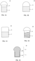
FIGS. 1A to 1D are front views of artificial cartilage implants according to an embodiment, andFIG. 1E is a cross-sectional view of the artificial cartilage implant illustrated inFIG. 1A ; -
FIG. 2A is a schematic diagram for describing a method of performing a Push-Out test in an example, andFIG. 2B is a graph showing the test results of the Push-Out test; and -
FIG. 3 is a graph showing the results of the fracture stress test in the example. - In the present specification, a numerical range represented by using a term "to" or "-" means a range including numerical values described before and after the term as lower and upper limits. In the numerically limited range described in the present specification, the upper limit value or the lower limit value described in a certain numerically limited range may be replaced with the upper limit value or the lower limit value of the numerically limited range described in another stepwise manner.
- An artificial cartilage implant is a member that functions as a substitute for cartilage by being implanted in a living body. Hereinafter, the present invention will be described with reference to an embodiment. However, the artificial cartilage implant of the present invention is not limited to the above-described embodiment.
-
FIG. 1A is a front view of an artificial cartilage implant according to an embodiment. As illustrated inFIG. 1A ,artificial cartilage implant 10 of the present embodiment has a structure such that a spherical crown having a diameter larger than that of a cylinder is joined to an end surface of the cylinder. The shape ofartificial cartilage implant 10 is not limited to the shape described above, and may be, for example, a structure such that the corners at one end of a cylinder is radius chamfered, as illustrated inFIGS. 1B and 1C . Further, for example, as illustrated inFIG. 1D , the artificial cartilage implant may have a structure such that a screw groove is formed in a part thereof (bone implant section 11 inFIG. 1D ). Further, the shape ofartificial cartilage implant 10 is not limited to a columnar shape, and may be various shapes according to the shape of a portion of a living body in whichartificial cartilage implant 10 is used. - As illustrated in
FIGS. 1A to 1D ,artificial cartilage implant 10 in any shape includes a section to be implanted into a bone (herein, simply referred to as "bone implant section") 11 andartificial cartilage section 12. The ratio of the height ofbone implant section 11 to the height ofartificial cartilage section 12 inartificial cartilage implant 10 is appropriately selected according to the shape of the bone into whichartificial cartilage implant 10 is to be embedded or the shape of the cartilage thatartificial cartilage section 12 to replace. -
Bone implant section 11 is a portion for embedding in a bone, and in the present embodiment, as illustrated inFIG. 1A , is a cylindrical portion.Bone implant section 11 includes a polymer gel and hydroxyapatite particles dispersed at least in the vicinity of the surface of the polymer gel. In the present embodiment, as illustrated in the cross-sectional view ofFIG. 1E (cross-sectional view of the artificial cartilage implant illustrated inFIG. 1A ),hydroxyapatite particles 101 are dispersed in the outer peripheral surface ofcylindrical polymer gel 100 ofbone implant section 11 and in the vicinity of the bottom surface on one side. Inbone implant section 11, however, hydroxyapatite particles may be dispersed throughout the entire polymer gel. - The amount of hydroxyapatite particles in the polymer gel of
bone implant section 11 is not particularly limited as long as it is possible to form a dense structure with inorganic components in the body and to enhance the joining strength with a bone whenbone implant section 11 is embedded in the bone. When the cross section of the polymer gel ofbone implant section 11 is observed, it is preferable that hydroxyapatite particles are distributed in a region from the surface to a depth of 1000 µm, and the content of the hydroxyapatite particles at a depth of 1000 µm from the surface is 0.1% by weight or more and 80% by weight or less. It is more preferable that the lower limit value of the content is 0.5% by weight. Hydroxyapatite particles may be distributed throughout the entire region from the surface to a depth of 1000 µm; however, for example, the hydroxyapatite particles may be distributed in greater numbers on the surface side, and the distribution of hydroxyapatite particles may become small or almost nonexistent on the deeper side. That is, inbone implant section 11, the total amount of hydroxyapatite particles included in the region from the surface to a depth of 1000 µm may be 1% by weight or more based on the total amount ofbone implant section 11 in the region from the surface to a depth of 1000 µm. When the amount of hydroxyapatite particles in the vicinity of the surface of the polymer gel inbone implant section 11 is within the above range, the joining strength betweenbone implant section 11 and the bone is more likely to become high whenartificial cartilage implant 10 is embedded in the bone. In the present specification, the "cross section of a polymer gel ofbone implant section 11" refers to a cross section obtained by cutting the polymer gel substantially perpendicularly to the upper surface (the surface on theartificial cartilage section 12 side) ofbone implant section 11 such that the cutting passes through the center ofbone implant section 11. In the present embodiment, the cross section is obtained by cutting a cylindrical polymer gel along its central axis. Further, in the present specification, "depth from the surface" refers to the distance from the surface in the direction of the central axis. - The content of hydroxyapatite particles in a region from the surface to a depth of 1000 µm in
bone implant section 11 is calculated as follows by using a thermogravimetric analyzing apparatus. The polymer gel ofbone implant section 11, including hydroxyapatite particles and water, is taken, and is heated at a temperature rising rate of 10°C per minute in a range of 30°C to 600°C. The water content is determined by the total weight change from 30°C to a temperature at which the differential thermal analysis value indicates 0 (approximately 150 to 200°C). The hydroxyapatite particle fraction is determined from the final residue at 600°C, and the polymer gel fraction is determined by the difference between the total weight and the sum of the weights of water and the hydroxyapatite particles. The thickness of the polymer gel in the depth direction from the surface can be quantified by observation of a cross section using a field emission transmission electron microscope (FE-TEM) or the like. The content (C) in percent by weight of the hydroxyapatite particles included up to a depth of 1000 µm is calculated from the calculation formula (C) = (B) ÷ {(A) × (D) + (B)}, where the volume fraction (A) up to a depth of 1000 µm from the surface of the polymer gel is determined by calculation, and the total amount (B) of the hydroxyapatite particles and the content (D) of the polymer gel component are determined by the thermogravimetric analysis. For example, in a case where the polymer gel has a cylindrical shape, when the radius of the cylinder is R1 (µm) and the height is L1 (µm), the volume fraction of the region from the surface to a depth of 1000 µm is calculated as (A) = { (π × (R1)2 × L1) - (π × (R1 - 1000)2 × L1) } ÷ (π × (R1)2 × L1). In a case where R1 is smaller than 1000 µm, the content of the hydroxyapatite particles is calculated as (A) = 1. Further, in a case where the hydroxyapatite particles are present in a range deeper than 1000 µm from the surface, the volume fraction (E) of the hydroxyapatite particles from the surface to 1000 µm is also determined by calculation, and (C) is calculated by the calculation formula of (C) = {(B) × (E)} ÷ {(A) × (D) + (B) × (E)}. - On the other hand,
artificial cartilage section 12 is a portion to be used as a substitute for cartilage. In the present embodiment, as illustrated inFIG. 1A ,artificial cartilage section 12 is composed of a cylindrical portion and a spherical crown portion joined to an end surface of the cylinder.Artificial cartilage section 12 is mainly composed of polymer gel, and the polymer gel ofartificial cartilage section 12 is formed integrally with the polymer gel ofbone implant section 11. - In
artificial cartilage section 12, the amount of hydroxyapatite in the vicinity of the surface of the polymer gel is less than a certain value. Specifically, when the cross section of the polymer gel ofartificial cartilage section 12 is observed, no hydroxyapatite particles are distributed in a region from the surface to a depth of 1000 µm, or in a case where hydroxyapatite particles are distributed in the region, the content of the hydroxyapatite particles at a depth of 1000 µm from the surface is less than 0.1% by weight. The content of hydroxyapatite particles in the polymer gel ofartificial cartilage section 12 is preferably 0.05% by weight or less. When the amount of hydroxyapatite particles in the vicinity of the surface of the polymer gel ofartificial cartilage section 12 is within the above range, the strength and elasticity ofartificial cartilage section 12 are less likely to change over time whenartificial cartilage implant 10 is embedded in a bone. In the present specification, the "cross section of a polymer gel ofartificial cartilage section 12" refers to a cross section obtained by cutting the polymer gel substantially perpendicularly to the bottom surface (the surface onbone implant section 11 side) ofartificial cartilage section 12 such that the cutting passes through the center ofartificial cartilage section 12. In the present embodiment, the cross section of the polymer gel with the above-described shape is cut through the central axis. Further, the content of hydroxyapatite particles in the polymer gel ofartificial cartilage section 12 can be calculated by taking the polymer gel ofartificial cartilage section 12 out and subjecting the sample to the same method as in the calculation of the content of hydroxyapatite particles in the polymer gel ofbone implant section 11. - The polymer gel included in the above-described artificial cartilage implant 10 (
bone implant section 11 and artificial cartilage section 12) may be any polymer gel that is capable of dispersing hydroxyapatite particles therein and has biocompatibility. Examples of the polymer gel include polymer gels having a hydroxyl group, an amide group, an ester group, and the like. Specific examples of materials for the polymer gel include polyvinyl alcohol; materials having an amide group such as poly(N-methylacrylamide), poly(N-ethylacrylamide), and poly(acrylamide); materials having an ester group such as poly(methoxyethyl acrylate), poly(ethoxyethyl acrylate), poly(methoxyethyl methacrylate), and poly(ethoxyethyl methacrylate); and the like. - On the other hand, as described in the manufacturing method of
artificial cartilage implant 10 below, an acidic solution is used in the case of dispersing (forming) hydroxyapatite particles in a polymer gel. Accordingly, it is preferable that the polymer gel is less likely to decrease in elastic modulus and strength and further is less likely to decrease in swelling or shrinking even when the polymer gel is brought into contact with an acidic solution. Accordingly, a polymer gel including a first mesh structure and a second mesh structure that is entangled with the first mesh structure, namely a double mesh structure (hereinafter, also referred to as a "double network gel"), is particularly preferable. Such a double network gel undergoes little change in elastic modulus or strength even when immersed in an acidic solution, and is also less likely to swell or shrink. - The double network gel can be produced as follows. However, the production method of a double network gel is not limited to the following method. First, a first polymerization solution including a first monomer, a polymerization initiator, and a crosslinking agent is prepared. Then, the first monomer is polymerized and crosslinked to form the first mesh structure. Subsequently, a second polymerization solution including a second monomer, a polymerization initiator, and a second crosslinking agent is prepared, and the first mesh structure is immersed in the second polymerization solution to diffuse and permeate the second polymerization solution into the first mesh structure. Subsequently, the first mesh structure is taken out, and the polymerization and cross-linking of the second monomer are performed. Thus, a double network gel in which the first mesh structure and the second mesh structure are entangled with each other is obtained. The polymerization and crosslinking method of the first monomer and the second monomer is not particularly limited, and can be performed, for example, by heating or light irradiation.
- Examples of the above first monomer and second monomer include monomers having a charge, such as 2-acrylamido-2-methylpropanesulfonic acid, acrylic acid, methacrylic acid, and the like; and electrically neutral monomers such as acrylamide, N',N-dimethylacrylamide, N-isopropylacrylamide, vinylpyridine, styrene, methyl methacrylate, fluorine-containing unsaturated monomers (for example, trifluoroethyl acrylate and the like), hydroxyethyl acrylate, vinyl acetate, and the like. As the first monomer and the second monomer, only one of these may be used, or two or more of these may be used. The first monomer and the second monomer may be of the same type or composition, or they may be of different types or compositions.
- Further, examples of the first crosslinking agent and the second crosslinking agent include the following: crosslinking agents not including a degradable bond, such as divinylbenzene, divinylpyridine, divinylbiphenyl, and divinylsulfone; and crosslinking agents including a degradable bond, such as N,N'-methylenebisacrylamide and ethylene glycol dimethacrylate. In the present specification, the term "degradable bond" refers to a bond that is easily hydrolyzed under an acidic condition, and examples thereof include ester bond, amide bond, thioester bond, and the like. As the first crosslinking agent and the second crosslinking agent, only one of the crosslinking agents may be used, or two or more of these may be used, independently. However, it is preferable that 50 mol% or more of the first crosslinking agent is a crosslinking agent not including a degradable bond. Similarly, it is preferable that 50 mol% or more of the second crosslinking agent is a crosslinking agent not including a degradable bond. The proportion of the crosslinking agent not including a degradable bond is more preferably 60 mol% or more, and further preferably 75 mol% or more, independently. By using a large amount of each of the first and second crosslinking agents that does not contain a decomposable bond, the polymer gel becomes less likely to decompose when forming hydroxyapatite particles. The first crosslinking agent and the second crosslinking agent may be the same in type and composition. The amount (mol) of the first crosslinking agent used is preferably 0. 1 to 50 mol% with respect to the amount (mol) of the first monomer used. On the other hand, the amount (mol) of the second crosslinking agent is preferably 0.001 to 20 mol% with respect to the amount (mol) of the second monomer used. Thus, a double network gel in which the hardness of the first cross-linked structural body is high and the elasticity of the second cross-linked structural body is high is obtained.
- Further, the polymerization initiator used in the first polymerization solution or the second polymerization solution is appropriately selected according to the type of monomer and the polymerization method. Examples of the polymerization initiator include watersoluble thermal catalysts such as potassium persulfate, redox initiators such as potassium persulfate-sodium thiosulfate, thermal polymerization initiators such as azobisisobutyronitrile (AIBN) and benzoyl peroxide (BPO), and photopolymerization initiators such as 2-oxoglutaric acid, benzophenone, and hydrogen peroxide.
- The double network gel preferably has an initial elastic modulus ratio X defined by the Equation (1) below of 50% or more. When the initial elastic modulus ratio is 50% or more, after
artificial cartilage implant 10 is embedded in a bone,artificial cartilage section 12 has an appropriate elasticity and is more likely to function as a substitute for cartilage.
- In the above Equation (1), E2 represents the elastic modulus of the polymer gel after being subjected to an immersion treatment in a hydrochloric acid aqueous solution with a concentration of 0.05 mol/L at 60°C for 72 hours, and E1 represents the elastic modulus of the polymer gel before the immersion treatment.
- Further, when M1 (mol%) is defined as the structural amount derived from the first monomer in the above double network gel and M2 (mol%) is defined as the structural amount derived from the second monomer, it is preferable that M1:M2 is 1:2 to 1:100, more preferably 1:3 to 1:50, and even more preferably 1:3 to 1:30. Such ratio is more likely to further increase the mechanical strength of the double network gel.
- In addition, the water content of the double network gel is preferably 10 to 99.9%. Further, the compressive fracture stress of the double network gel is preferably 1 to 100 MPa. The compressive fracture stress is the stress at which a double network gel fractures when the double network gel is sandwiched between two flat plates and compressed at a predetermined rate (a rate at which the thickness of the sample is compressed by 10% per minute). On the other hand, the tensile fracture force is preferably 0.1 to 100 MPa. Further, the fracture energy is preferably 100 J/m2 or more. The tensile stress and the fracture energy are measured by preparing a dumbbell-type hydrogel (ISO-37-4 standard size:
length 11 mm, width 2.0 mm, and thickness 3.0 mm) and using a tensile tester (Tensilon RTC-1310A, manufactured by Orientec Co., Ltd.) at a test speed of 100 mm/min. - A hydroxyapatite particle refers to a particle of a calcium phosphate compound represented by Ca10(PO4)6(OH)2. The shape of the hydroxyapatite particle is usually spherical with protrusions. The hydroxyapatite particles may be included as primary particles in the polymer gel (mainly bone implant section 11) or may be included as an aggregate. The average particle diameter of the hydroxyapatite particles in the above polymer gel is preferably about 1 to 100,000 nm, and more preferably 10 to 10,000 nm. When the average particle diameter of the hydroxyapatite particles is within the range, the particles are more likely to react with inorganic substances in the living body to form a dense structure. The average particle diameter of the above-described hydroxyapatite particles is a value calculated as an average value of the particle diameters of 100 hydroxyapatite particles in a cross section of
artificial cartilage implant 10 measured with a field emission transmission electron microscope (FE-TEM) or the like. -
Artificial cartilage implant 10 of the present embodiment may further include components other than the above-described polymer gel and hydroxyapatite particles, as long as the effects and purposes of the present invention are not impaired.Artificial cartilage implant 10 may also include regions other thanbone implant section 11 andartificial cartilage section 12 as long as the effects and purposes of the present invention are not impaired. - An artificial cartilage implant of the present invention includes a bone implant section and an artificial cartilage section. Further, the artificial cartilage implant includes hydroxyapatite particles in the vicinity of the surface of the polymer gel of the bone implant section; therefore, the joining strength between the bone implant section and a bone is more likely to be improved when the artificial cartilage implant is embedded in the bone. More specifically, the hydroxyapatite particles on the surface of the polymer gel of the bone implant section or inside the bone implant section react with inorganic components in the living body to form a dense structure. Further, as the structure integrates with the bone, the joining strength between the artificial cartilage implant and the bone becomes very high. On the other hand, the artificial cartilage section of the artificial cartilage implant of the present invention has a very small amount of hydroxyapatite particles present in the vicinity of the surface. For this reason, the artificial cartilage section is less likely to become ceramic-like, which would be caused by the deposition of inorganic components in the body. As a result, the strength and elasticity are less likely to change over time. Accordingly, the artificial cartilage section can maintain a low elastic modulus over a long period of time and can easily function as a substitute for cartilage.
- The artificial cartilage implant including the bone implant section and the artificial cartilage section described above can be manufactured, for example, by the following two methods. However, the manufacturing method of an artificial cartilage implant is not limited to these methods.
- The first manufacturing method includes the following steps: preparing a polymer gel (polymer gel preparation step); and forming hydroxyapatite particles in the vicinity of a surface of a region (in the polymer gel) corresponding to the bone implant section by sequentially bringing an alkali metal phosphate aqueous solution and a calcium salt aqueous solution into contact with the region of the polymer gel corresponding to the bone implant section while protecting a region (in the polymer gel) corresponding to the artificial cartilage section (hydroxyapatite particle formation step).
- In the polymer gel preparation step in the first manufacturing method, polymer gel having a desired shape may be prepared. For example, a commercially available polymer gel may be used as it is. Further, a commercially available polymer gel may be processed or molded into a desired shape. Further, a double network gel having a desired shape may be prepared based on the production method of the double network gel described above. During the production of a double network gel, the polymer gel can be molded into a desired shape by using a mold when the first monomer is polymerized and when the second monomer is polymerized.
- Further, in the hydroxyapatite particle formation step in the first manufacturing method, an alkali metal phosphate aqueous solution and a calcium salt aqueous solution are sequentially brought into contact with the region (in the polymer gel) corresponding to the bone implant section while protecting the region (in the polymer gel) corresponding to the artificial cartilage section. The method for protecting the region corresponding to the artificial cartilage section is not particularly limited, as long as the region corresponding to the artificial cartilage section substantially does not contact an alkaline metal phosphate aqueous solution or a calcium salt aqueous solution. For example, the region corresponding to the artificial cartilage section may be protected by bringing the alkali metal phosphate aqueous solution and the calcium salt aqueous solution into contact with only the polymer gel in the region corresponding to the bone implant section, while preventing the polymer gel in the region corresponding to the artificial cartilage section from coming into contact with the alkali metal phosphate aqueous solution and the calcium salt aqueous solution. Further, the region of the polymer gel corresponding to the artificial cartilage section may be temporarily covered and protected with a member such as masking tape.
- Specifically, only the region corresponding to the bone implant section may be sequentially immersed in an alkali metal phosphate aqueous solution and a calcium salt aqueous solution to form hydroxyapatite particles in the region corresponding to the bone implant section, or only the region corresponding to the bone implant section may be sequentially coated with an alkali metal phosphate aqueous solution and a calcium salt aqueous solution to form hydroxyapatite particles in the region corresponding to the bone implant section. Further, the region corresponding to the artificial cartilage section may be protected with a masking tape or the like, and the entire polymer gel may be sequentially immersed in an alkali metal phosphate aqueous solution and a calcium salt aqueous solution.
- The alkali metal phosphate aqueous solution used in the present step may be any aqueous solution of an alkali metal phosphate, and examples of the alkali metal phosphate include disodium hydrogen phosphate, dipotassium hydrogen phosphate, and the like. In addition, the calcium salt aqueous solution may be any aqueous solution of a calcium salt, and examples of the calcium salt include calcium chloride, calcium carbonate, and the like.
- The order in which the polymer gel (region corresponding to the bone implant section) is brought into contact with the alkali metal phosphate aqueous solution and the calcium salt aqueous solution is not particularly limited, and either may be first. Further, the number of times of bringing these solutions into contact with the polymer gel is not particularly limited, either, and may be, for example, once for each solution or twice or more for each solution. By increasing the number of contacts, it is possible to increase the amount of hydroxyapatite particles included in the vicinity of the surface of the polymer gel of the bone implant section. Further, the contact time between the polymer gel and each solution is appropriately selected according to the concentration of each solution, the size of the polymer gel, and the desired amount of hydroxyapatite particles. After the contact between the polymer gel and each solution, the polymer gel may be washed with pure water or the like as necessary.
- On the other hand, the second manufacturing method includes the following steps: preparing polymer gel (polymer gel preparation step); forming the hydroxyapatite particles in the vicinity of a surface of the polymer gel by sequentially bringing an alkali metal phosphate aqueous solution and a calcium salt aqueous solution into contact with the polymer gel (hydroxyapatite particle formation step); and removing the hydroxyapatite particles in a region (of the polymer gel after forming the hydroxyapatite particles) corresponding to the artificial cartilage section by bringing an acidic solution into contact with the region corresponding to the artificial cartilage section while protecting a region corresponding to the bone implant section (hydroxyapatite particle removal step). The polymer gel preparation step is the same as the polymer gel preparation step in the first manufacturing method described above, and thus, a detailed description thereof will be omitted here.
- In the hydroxyapatite particle formation step in the second manufacturing method, the polymer gel is sequentially brought into contact with an alkali metal phosphate aqueous solution and a calcium salt aqueous solution. At this time, the entire polymer gel may be brought into contact with an alkali metal phosphate aqueous solution and a calcium salt aqueous solution without distinguishing between the bone implant section and the artificial cartilage section. However, as in the first manufacturing method, only the region corresponding to the bone implant section may be brought into contact with the alkali metal phosphate aqueous solution and the calcium salt aqueous solution while protecting the region corresponding to the bone implant section. The types of the alkali metal phosphate aqueous solution and the calcium salt aqueous solution, the method of bringing the polymer gel into contact with each solution, the contact time, and the like are the same as those in the first manufacturing method.
- Further, in the hydroxyapatite particle removal step in the second manufacturing method, the hydroxyapatite particles in the region (of the polymer gel after the hydroxyapatite particle formation step) corresponding to the artificial cartilage section are removed by bringing an acidic solution into contact with the region corresponding to the artificial cartilage section while protecting the region corresponding to the bone implant section. The method for protecting the bone implant section is the same as the method for protecting the artificial cartilage section in the hydroxyapatite particle formation step of the first manufacturing method. For example, the following may be used: a method in which only the polymer gel in the region corresponding to the artificial cartilage section is immersed in the acidic solution such that the acidic solution does not contact the region corresponding to the bone implant section, or a method in which the acidic solution is applied only to the polymer gel in the region corresponding to the artificial cartilage section. Further, the region corresponding to the bone implant section may be covered with a protective material such as masking tape, and the entire polymer gel may be brought into contact with an acidic solution.
- The type of acidic solution used in the present step is not particularly limited, and examples thereof include aqueous solutions of hydrochloric acid, phosphoric acid, sulfuric acid, nitric acid, and the like. The time for treating the region corresponding to the artificial cartilage section with an acidic solution is not particularly limited, and is appropriately selected according to the amount of hydroxyapatite particles formed in the hydroxyapatite particle formation step. After the contact between the polymer gel and the acidic solution, the polymer gel may be washed with pure water or the like as necessary.
- The manufacturing method of the artificial cartilage implant described above can produce, by a simple method, an artificial cartilage implant that can be used stably over a long period of time with little change even when used in a living body.
- Hereinafter, the present invention will be described in more detail with reference to Examples. The scope of the present invention is not limited to these examples, and modifications to the embodiments are possible without departing from the spirit of the present invention.
- To a mixed solvent of pure water and dimethyl sulfoxide (DMSO), 1 mol/L of a first monomer (2-acrylamido-2-methylpropanesulfonic acid (AMPS)), 0.04 mol/L of a crosslinking agent (divinylbenzene (DVB)), and 0.01 mol/L of an initiator (2-oxoglutaric acid) were added to obtain a first polymerization solution. The obtained first polymerization solution was deoxygenated with nitrogen gas and poured into a glass mold (having a shape such that a bottom surface of a hemisphere with a radius of 4 mm and a height of 2 mm is connected to one end surface of a cylinder with a radius of 3 mm and a height of 4 mm). Subsequently, the first polymerization solution was irradiated with ultraviolet light from a UV lamp (22 W, 0.34 A) with a wavelength of 365 nm for 8 hours at room temperature, and the first monomer was polymerized and crosslinked to produce an AMPS gel (first mesh structure).
- Subsequently, 2 mol/L of a second monomer (N',N-dimethylacrylamide (DMAAm)), 0.3 mol/L of a crosslinking agent (divinylbenzene), and 0.005 mol/L of an initiator (2-oxoglutaric acid) were dissolved in a mixed solvent of water and DMSO to obtain a second polymerization solution. Subsequently, the above-described AMPS gel (first mesh structure) was immersed in the second polymerization solution for 24 hours, and the second polymerization solution was diffused and penetrated into the AMPS gel. Subsequently, the AMPS gel was taken out from the second polymerization solution and installed in the same glass mold as described above. Then, the AMPS gel was irradiated with ultraviolet light from a UV lamp (22 W, 0.34 A) with a wavelength of 365 nm for 8 hours at room temperature. Thus, the DMAAm monomer diffused in the AMPS gel was polymerized and crosslinked to obtain a double network gel (DN gel) in which a first mesh structure and a second mesh structure having different characteristics were entangled with each other.
- The region of the DN gel produced above corresponding to the artificial cartilage section was masked with masking tape. Subsequently, the DN gel was immersed overnight in a tris(hydroxymethyl)aminomethane aqueous solution (1.0 mol/L) adjusted to pH 9.0. The sample was then immersed in pure water for 10 seconds to remove the excess tris(hydroxymethyl)aminomethane aqueous solution. The excess water was wiped off with a cloth or the like. Subsequently, the DN gel was immersed in a calcium chloride aqueous solution (0.5 mol/L) for 2 minutes, then immersed again in pure water for 10 seconds, and the excess water was wiped off. Subsequently, the sample was immersed in a dipotassium hydrogen phosphate aqueous solution (0.3 mol/L) for 2 minutes. The process from immersion in the calcium chloride aqueous solution to immersion in the dipotassium hydrogen phosphate aqueous solution was defined as one cycle, and five cycles were performed. The DN gel that has been subjected to the immersion treatment was immersed overnight in a calcium chloride aqueous solution adjusted to
pH 11 at 37°C, and then the masking tape was removed. The above processes obtained an artificial cartilage implant in which hydroxyapatite particles are selectively dispersed in the vicinity of the surface of polymer gel (DN gel) in the bone implant section. - The artificial cartilage implant produced by the above method was cut along the central axis, and the cut surface was observed with a field emission transmission electron microscope (FE-TEM). In the cross-sectional observation, the hydroxyapatite particles were not distributed in a region from the arc surface of the polymer gel of the artificial cartilage section (portion with a shape such that a bottom surface of a hemisphere with a radius of 4 mm and a height of 2 mm is connected) to the inside by 1000 µm. On the other hand, hydroxyapatite particles are mainly distributed in a region from the arc surface of the polymer gel of the bone implant section (cylindrical portion with a radius of 3 mm and a height of 4 mm) to the inside by 500 µm, and the content of the hydroxyapatite particles in the bone implant section was 33% by weight. Accordingly, the content of hydroxyapatite particles in a region from the surface of the polymer gel of the bone implant section to 1000 µm is 36% by weight or more, since the volume fraction (A) in the region from the surface to 1000 µm is 0.89. That is, in the artificial cartilage implant, the amount of hydroxyapatite particles in the artificial cartilage section was very small, while the amount of hydroxyapatite particles in the bone implant section was sufficiently large.
- In the same manner as in Sample 1, a double network gel (DN gel) was prepared.
- Except for not masking the region corresponding to the artificial cartilage section with masking tape, the same procedure as in Sample 1 was performed, including five cycles of immersion in a calcium chloride aqueous solution and immersion in a dipotassium hydrogen phosphate aqueous solution, resulting in the formation of hydroxyapatite particles in the vicinity of the entire surface of the DN gel.
- Only the region of the DN gel corresponding to the artificial cartilage section was immersed in a 10 mass% aqueous hydrochloric acid aqueous solution, and the hydroxyapatite particles in the region corresponding to the artificial cartilage section were removed. Thus, an artificial cartilage implant in which hydroxyapatite particles are selectively dispersed in the vicinity of the surface of the polymer gel (DN gel) in the bone implant section was obtained.
- The artificial cartilage implant produced by the above method was cut along the central axis, and the cut surface was observed with a field emission transmission electron microscope (FE-TEM). In the cross-sectional observation, hydroxyapatite particles were not distributed in a region from the surface of the polymer gel of the artificial cartilage section to the inside by 1000 µm. On the other hand, hydroxyapatite particles are mainly distributed in a region from the surface of the polymer gel of the bone implant section to the inside by 500 µm, and the content of the hydroxyapatite particles in the bone implant section is 33% by weight or more. Accordingly, it can be said that the content of hydroxyapatite particles in a region from the surface of the polymer gel of the bone implant section to 1000 µm is 36% by weight or more. That is, in the artificial cartilage implant, the amount of hydroxyapatite particles in the artificial cartilage section was very small, while the amount of hydroxyapatite particles in the bone implant section was sufficiently large.
- An artificial cartilage implant was produced in the same manner as in Sample 2, except that the hydroxyapatite particle removal step was not performed.
- The artificial cartilage implant produced by the above method was cut along the central axis, and the cut surface was observed with a field emission transmission electron microscope (FE-TEM). In the cross-sectional observation, hydroxyapatite particles were mainly distributed in regions from the surfaces of the polymer gel of the artificial cartilage section and the bone implant section to the inside by 500 µm, and the content of the hydroxyapatite particles in the artificial cartilage section and the bone implant section, that is, in the artificial cartilage implant, was 33% by weight. Accordingly, it can be said that the content of hydroxyapatite particles in regions from the surfaces of the polymer gel of the artificial cartilage section and the bone implant section to the inside by 1000 µm is 36% by weight or more. That is, in the artificial cartilage implant, a large amount of hydroxyapatite particles was dispersed in the vicinity of the surfaces of the artificial cartilage section and the bone implant section.
- A cartilage defect was created in rabbit femoral trochleae (patellar facing surfaces), and the bone implant sections of the artificial cartilage implants produced in Sample 1 and Sample 2 were embedded. Further, the mobility of the knee was monitored over a period of 12 months, and there were no issues.
- In the same manner as in Sample 1, an artificial cartilage implant (Sample 4) including hydroxyapatite particles in the vicinity of the surface (approximately 150 µm from the surface) of the polymer gel of the bone implant section was produced. Further, the number of times of immersion in the calcium chloride aqueous solution and immersion in the dipotassium hydrogen phosphate aqueous solution was changed to produce an artificial cartilage implant (Sample 5) including hydroxyapatite particles in the vicinity of the surface (approximately 300 µm from the surface) of the polymer gel of the bone implant section. Further, an artificial cartilage implant (Sample 6) consisting only of polymer gel was produced without performing immersion in a calcium chloride aqueous solution and immersion in a dipotassium hydrogen phosphate aqueous solution.
- Each of these artificial cartilage implants (Samples 4 to 6) was implanted in a rabbit femoral trochlea (patellar facing surface) in the same manner as in the knee mobility score measurement. As shown in
FIG. 2A , each femur (cartilage 21 and bone 22) eight weeks after the implantation of the artificial cartilage implant was fixed tolower jig 120 made of metal, andartificial cartilage implant 10 was pushed out from thebone 22 side to thecartilage 21 side (direction indicated by the arrow inFIG. 2A ) withindenter 110. The maximum value of the force required to push outartificial cartilage implant 10 was used as the fracture force (N).FIG. 2B shows the results. - As illustrated in
FIG. 2B , compared with the fracture force in a case where the bone implant section does not include hydroxyapatite particles (0 µm (Sample 6)), the fracture force was significantly large in a case where the bone implant section includes hydroxyapatite particles (150 µm (Sample 4) and 300 µm (Sample 5)), and it is clear that the bone and the bone implant section are firmly bonded to each other. - An artificial cartilage implant (Sample 7) including hydroxyapatite particles in the vicinity of the entire surface of the polymer gel was produced by the same method as in Sample 2 (except that the hydroxyapatite particle removal step was not performed). In the hydroxyapatite particle formation step, the immersion in the calcium chloride aqueous solution and the immersion in the dipotassium hydrogen phosphate aqueous solution were each performed 10 times.
- Similarly, an artificial cartilage implant (Sample 8) including hydroxyapatite particles in the vicinity of the entire surface of a polymer gel was produced. In the hydroxyapatite particle formation step, the immersion in the calcium chloride aqueous solution and the immersion in the dipotassium hydrogen phosphate aqueous solution were each performed 15 times.
- Further, an artificial cartilage implant (Sample 3 described above) consisting only of polymer gel was produced without performing immersion in a calcium chloride aqueous solution and immersion in a dipotassium hydrogen phosphate aqueous solution.
- These artificial cartilage implants (Samples 3, 7, and 8) were immersed in a simulated body fluid. The fracture stress of the polymer gel before and after 8 weeks of immersion was measured using a Tensilon universal testing machine RTC-1310A (manufactured by Orientec Co., Ltd.). The results at this time are illustrated in
FIG. 3 . As shown inFIG. 3 , in a case where the artificial cartilage implant does not include hydroxyapatite particles, the fracture stress did not decrease. On the other hand, in the case of including hydroxyapatite particles, the fracture stress was significantly reduced. - From the results of the above Push-Out test, it can be said that, in the bone implant section of the artificial cartilage implant, when the surface vicinity of the polymer gel includes hydroxyapatite particles, the joining strength is significantly increased when the artificial cartilage implant is embedded in a living body and excellent effects can be obtained. On the other hand, the results of the above-described fracture stress test indicate that, in the artificial cartilage section, the fracture stress does not decrease and the artificial cartilage section can be used stably over a long period of time when the surface vicinity of the polymer gel does not include hydroxyapatite particles. That is, by forming an artificial cartilage implant in which a bone implant section includes a large amount of hydroxyapatite particles and an artificial cartilage section includes a small amount of hydroxyapatite particles, the artificial cartilage implant can be fixed to the bone and can be used stably for a long period of time.
- The present application is entitled to and claims the benefit of
Japanese Patent Application No. 2022-094413 - The artificial cartilage implant of the present invention can be used stably over a long period of time with little change even when used in a living body. Accordingly, the present invention is very useful in the medical field.
-
- 10 Artificial cartilage implant
- 11 Bone implant section
- 12 Artificial cartilage section
- 21 Cartilage
- 22 Bone
- 100 Polymer gel
- 101 Hydroxyapatite particle
- 110 Indenter
- 120 Lower jig
Claims (6)
- An artificial cartilage implant, comprising:a bone implant section including a polymer gel and hydroxyapatite particles dispersed at least in a vicinity of a surface of the polymer gel; andan artificial cartilage section including a polymer gel, whereinwhen a cross section of the polymer gel of the artificial cartilage section is observed, no hydroxyapatite particle is distributed in a region from a surface to a depth of 1000 µm, or in a case where hydroxyapatite particles are distributed in the region, a content of the hydroxyapatite particles at the depth of 1000 µm from the surface is less than 0.1% by weight.
- The artificial cartilage implant according to claim 1, wherein:
when a cross section of the polymer gel of the bone implant section is observed, the hydroxyapatite particles are distributed in a region from a surface to a depth of 1000 µm, and a content of the hydroxyapatite particles at the depth of 1000 µm from the surface is 0.1% by weight to 80% by weight. - The artificial cartilage implant according to claim 1, wherein:
the polymer gel of the bone implant section and the polymer gel of the artificial cartilage section are integrated with each other. - The artificial cartilage implant according to claim 1, wherein:the polymer gel of the bone implant section and the polymer gel of the artificial cartilage section each include a first mesh structure and a second mesh structure that is entangled with the first mesh structure; andfracture energy of the artificial cartilage implant is 100 J/m2 or more.
- A method for manufacturing the artificial cartilage implant according to any one of claims 1 to 4, the method comprising:preparing a polymer gel; andforming the hydroxyapatite particles in a vicinity of a surface of a region corresponding to the bone implant section by sequentially bringing an alkali metal phosphate aqueous solution and a calcium salt aqueous solution into contact with the region corresponding to the bone implant section while protecting a region corresponding to the artificial cartilage section, the region corresponding to the bone implant and the region corresponding to the artificial cartilage section being in the polymer gel.
- A method for manufacturing the artificial cartilage implant according to any one of claims 1 to 4, the method comprising:preparing a polymer gel;forming the hydroxyapatite particles in a vicinity of a surface of the polymer gel by sequentially bringing an alkali metal phosphate aqueous solution and a calcium salt aqueous solution into contact with the polymer gel; andremoving the hydroxyapatite particles in a region corresponding to the artificial cartilage section by bringing an acidic solution into contact with the region corresponding to the artificial cartilage section while protecting a region corresponding to the bone implant section, the region corresponding to the artificial cartilage section and the region corresponding to the bone implant being in the polymer gel after the forming of the hydroxyapatite particles.
Applications Claiming Priority (2)
| Application Number | Priority Date | Filing Date | Title |
|---|---|---|---|
| JP2022094413A JP7790720B2 (en) | 2022-06-10 | 2022-06-10 | Artificial cartilage implant and its manufacturing method |
| PCT/JP2023/020821 WO2023238822A1 (en) | 2022-06-10 | 2023-06-05 | Artificial cartilage implant and manufacturing method therefor |
Publications (2)
| Publication Number | Publication Date |
|---|---|
| EP4523712A1 true EP4523712A1 (en) | 2025-03-19 |
| EP4523712A4 EP4523712A4 (en) | 2025-08-13 |
Family
ID=89118358
Family Applications (1)
| Application Number | Title | Priority Date | Filing Date |
|---|---|---|---|
| EP23819802.2A Pending EP4523712A4 (en) | 2022-06-10 | 2023-06-05 | ARTIFICIAL CARTILAGE IMPLANT AND MANUFACTURING METHOD THEREFOR |
Country Status (5)
| Country | Link |
|---|---|
| EP (1) | EP4523712A4 (en) |
| JP (1) | JP7790720B2 (en) |
| KR (1) | KR20250005496A (en) |
| CN (1) | CN119317454A (en) |
| WO (1) | WO2023238822A1 (en) |
Family Cites Families (6)
| Publication number | Priority date | Publication date | Assignee | Title |
|---|---|---|---|---|
| JPH01305959A (en) | 1988-06-03 | 1989-12-11 | Arusu Japan:Kk | Artificial cartilage |
| KR100384184B1 (en) | 1998-05-08 | 2003-05-14 | 닛폰 유시 가부시키가이샤 | Hydroxyapatite, composite, processes for producing these, and use of these |
| JP3914501B2 (en) | 2003-02-17 | 2007-05-16 | 財団法人川村理化学研究所 | Polymer gel composite and method for producing the same |
| JP2004321501A (en) | 2003-04-24 | 2004-11-18 | Kyocera Corp | Bioprosthetic member and method of manufacturing the same |
| EP3706663A4 (en) | 2017-11-07 | 2021-07-28 | Duke University | TRIPLE NETWORK HYDROGEL IMPLANTS FOR CARTILAGE REPAIR |
| JP7683201B2 (en) | 2020-12-15 | 2025-05-27 | コニカミノルタ株式会社 | Mobile radiography device and program |
-
2022
- 2022-06-10 JP JP2022094413A patent/JP7790720B2/en active Active
-
2023
- 2023-06-05 WO PCT/JP2023/020821 patent/WO2023238822A1/en not_active Ceased
- 2023-06-05 EP EP23819802.2A patent/EP4523712A4/en active Pending
- 2023-06-05 KR KR1020247040818A patent/KR20250005496A/en active Pending
- 2023-06-05 CN CN202380045674.0A patent/CN119317454A/en active Pending
Also Published As
| Publication number | Publication date |
|---|---|
| JP2023180810A (en) | 2023-12-21 |
| JP7790720B2 (en) | 2025-12-23 |
| EP4523712A4 (en) | 2025-08-13 |
| WO2023238822A1 (en) | 2023-12-14 |
| CN119317454A (en) | 2025-01-14 |
| KR20250005496A (en) | 2025-01-09 |
Similar Documents
| Publication | Publication Date | Title |
|---|---|---|
| Sennakesavan et al. | Acrylic acid/acrylamide based hydrogels and its properties-A review | |
| Jastram et al. | Rheological properties of hydrogels based on ionic liquids | |
| US5824079A (en) | Swelling type copolymeric composite material with self-fixation characteristics | |
| Okada et al. | Tissue reactions to subcutaneously implanted, surface‐modified silicones | |
| EP2380600A1 (en) | Polymer sliding material, artificial joint member, medical appliance, and manufacturing method therefor | |
| CN110835382B (en) | A kind of self-soluble high-strength and fast-viscous hydrogel and its preparation method and application | |
| US20050158535A1 (en) | Methods for making porous ceramic structures | |
| JP2001337298A (en) | Ophthalmic lens material having hydrophilic surface and method for producing the same | |
| JP3914501B2 (en) | Polymer gel composite and method for producing the same | |
| US8552100B2 (en) | Flexible hydrogel-based functional composite materials | |
| O'donnell et al. | Amorphous calcium phosphate composites with improved mechanical properties1 | |
| KR101027537B1 (en) | PH sensitive conductive hydrogel using carbon nanotubes and method for preparing same | |
| JP7790720B2 (en) | Artificial cartilage implant and its manufacturing method | |
| KR102422864B1 (en) | Hydrogels and methods for preparing hydrogels | |
| WO2013180228A1 (en) | Sliding material and method for producing same | |
| KR20190050025A (en) | Tissue Expander for Dental Applications and Method for Fabricating the Same | |
| JP2003215509A (en) | Method for producing soft contact lens made of silicone hydrogel having hydrophilic surface and soft contact lens | |
| Greenberg et al. | Polymer–ceramic composite for tooth‐root implant | |
| Kenny et al. | The influence of poly (acrylic acid) molar mass on the properties of polyalkenoate cements formed from zinc oxide/apatite mixtures | |
| JP2020180240A (en) | Hydrogel and manufacture method thereof | |
| WO2022054743A1 (en) | Method for forming graft layer, method for producing composite, and treatment liquid for forming graft layer | |
| JP5850417B2 (en) | Polymer gel and method for producing the same | |
| RU2155556C1 (en) | Method for manufacturing base part of artificial denture | |
| CN116199911B (en) | A double-crosslinked hydrogel and its application as wound healing dressing | |
| Mastalska-Popławska et al. | Rheological parameters of polymer-silicate gels |
Legal Events
| Date | Code | Title | Description |
|---|---|---|---|
| STAA | Information on the status of an ep patent application or granted ep patent |
Free format text: STATUS: THE INTERNATIONAL PUBLICATION HAS BEEN MADE |
|
| PUAI | Public reference made under article 153(3) epc to a published international application that has entered the european phase |
Free format text: ORIGINAL CODE: 0009012 |
|
| STAA | Information on the status of an ep patent application or granted ep patent |
Free format text: STATUS: REQUEST FOR EXAMINATION WAS MADE |
|
| 17P | Request for examination filed |
Effective date: 20241210 |
|
| AK | Designated contracting states |
Kind code of ref document: A1 Designated state(s): AL AT BE BG CH CY CZ DE DK EE ES FI FR GB GR HR HU IE IS IT LI LT LU LV MC ME MK MT NL NO PL PT RO RS SE SI SK SM TR |
|
| A4 | Supplementary search report drawn up and despatched |
Effective date: 20250714 |
|
| RIC1 | Information provided on ipc code assigned before grant |
Ipc: A61L 27/14 20060101AFI20250708BHEP Ipc: A61L 27/12 20060101ALI20250708BHEP Ipc: A61L 27/46 20060101ALI20250708BHEP Ipc: A61L 27/52 20060101ALI20250708BHEP |
|
| DAV | Request for validation of the european patent (deleted) | ||
| DAX | Request for extension of the european patent (deleted) |