EP4510144A1 - Generating natural language text describing a change between sets of medical imaging data - Google Patents
Generating natural language text describing a change between sets of medical imaging data Download PDFInfo
- Publication number
- EP4510144A1 EP4510144A1 EP23191392.2A EP23191392A EP4510144A1 EP 4510144 A1 EP4510144 A1 EP 4510144A1 EP 23191392 A EP23191392 A EP 23191392A EP 4510144 A1 EP4510144 A1 EP 4510144A1
- Authority
- EP
- European Patent Office
- Prior art keywords
- imaging data
- training
- medical imaging
- medical
- machine learning
- Prior art date
- Legal status (The legal status is an assumption and is not a legal conclusion. Google has not performed a legal analysis and makes no representation as to the accuracy of the status listed.)
- Pending
Links
Images


Classifications
-
- G—PHYSICS
- G16—INFORMATION AND COMMUNICATION TECHNOLOGY [ICT] SPECIALLY ADAPTED FOR SPECIFIC APPLICATION FIELDS
- G16H—HEALTHCARE INFORMATICS, i.e. INFORMATION AND COMMUNICATION TECHNOLOGY [ICT] SPECIALLY ADAPTED FOR THE HANDLING OR PROCESSING OF MEDICAL OR HEALTHCARE DATA
- G16H30/00—ICT specially adapted for the handling or processing of medical images
- G16H30/40—ICT specially adapted for the handling or processing of medical images for processing medical images, e.g. editing
-
- G—PHYSICS
- G06—COMPUTING OR CALCULATING; COUNTING
- G06V—IMAGE OR VIDEO RECOGNITION OR UNDERSTANDING
- G06V10/00—Arrangements for image or video recognition or understanding
- G06V10/40—Extraction of image or video features
-
- G—PHYSICS
- G06—COMPUTING OR CALCULATING; COUNTING
- G06N—COMPUTING ARRANGEMENTS BASED ON SPECIFIC COMPUTATIONAL MODELS
- G06N3/00—Computing arrangements based on biological models
- G06N3/02—Neural networks
- G06N3/04—Architecture, e.g. interconnection topology
- G06N3/0464—Convolutional networks [CNN, ConvNet]
-
- G—PHYSICS
- G06—COMPUTING OR CALCULATING; COUNTING
- G06T—IMAGE DATA PROCESSING OR GENERATION, IN GENERAL
- G06T7/00—Image analysis
- G06T7/0002—Inspection of images, e.g. flaw detection
- G06T7/0012—Biomedical image inspection
- G06T7/0014—Biomedical image inspection using an image reference approach
- G06T7/0016—Biomedical image inspection using an image reference approach involving temporal comparison
-
- G—PHYSICS
- G06—COMPUTING OR CALCULATING; COUNTING
- G06V—IMAGE OR VIDEO RECOGNITION OR UNDERSTANDING
- G06V10/00—Arrangements for image or video recognition or understanding
- G06V10/40—Extraction of image or video features
- G06V10/44—Local feature extraction by analysis of parts of the pattern, e.g. by detecting edges, contours, loops, corners, strokes or intersections; Connectivity analysis, e.g. of connected components
-
- G—PHYSICS
- G06—COMPUTING OR CALCULATING; COUNTING
- G06V—IMAGE OR VIDEO RECOGNITION OR UNDERSTANDING
- G06V10/00—Arrangements for image or video recognition or understanding
- G06V10/40—Extraction of image or video features
- G06V10/44—Local feature extraction by analysis of parts of the pattern, e.g. by detecting edges, contours, loops, corners, strokes or intersections; Connectivity analysis, e.g. of connected components
- G06V10/443—Local feature extraction by analysis of parts of the pattern, e.g. by detecting edges, contours, loops, corners, strokes or intersections; Connectivity analysis, e.g. of connected components by matching or filtering
- G06V10/449—Biologically inspired filters, e.g. difference of Gaussians [DoG] or Gabor filters
- G06V10/451—Biologically inspired filters, e.g. difference of Gaussians [DoG] or Gabor filters with interaction between the filter responses, e.g. cortical complex cells
- G06V10/454—Integrating the filters into a hierarchical structure, e.g. convolutional neural networks [CNN]
-
- G—PHYSICS
- G06—COMPUTING OR CALCULATING; COUNTING
- G06V—IMAGE OR VIDEO RECOGNITION OR UNDERSTANDING
- G06V10/00—Arrangements for image or video recognition or understanding
- G06V10/40—Extraction of image or video features
- G06V10/60—Extraction of image or video features relating to illumination properties, e.g. using a reflectance or lighting model
-
- G—PHYSICS
- G06—COMPUTING OR CALCULATING; COUNTING
- G06V—IMAGE OR VIDEO RECOGNITION OR UNDERSTANDING
- G06V10/00—Arrangements for image or video recognition or understanding
- G06V10/40—Extraction of image or video features
- G06V10/62—Extraction of image or video features relating to a temporal dimension, e.g. time-based feature extraction; Pattern tracking
-
- G—PHYSICS
- G06—COMPUTING OR CALCULATING; COUNTING
- G06V—IMAGE OR VIDEO RECOGNITION OR UNDERSTANDING
- G06V10/00—Arrangements for image or video recognition or understanding
- G06V10/70—Arrangements for image or video recognition or understanding using pattern recognition or machine learning
- G06V10/74—Image or video pattern matching; Proximity measures in feature spaces
-
- G—PHYSICS
- G06—COMPUTING OR CALCULATING; COUNTING
- G06V—IMAGE OR VIDEO RECOGNITION OR UNDERSTANDING
- G06V10/00—Arrangements for image or video recognition or understanding
- G06V10/70—Arrangements for image or video recognition or understanding using pattern recognition or machine learning
- G06V10/74—Image or video pattern matching; Proximity measures in feature spaces
- G06V10/75—Organisation of the matching processes, e.g. simultaneous or sequential comparisons of image or video features; Coarse-fine approaches, e.g. multi-scale approaches; using context analysis; Selection of dictionaries
- G06V10/757—Matching configurations of points or features
-
- G—PHYSICS
- G06—COMPUTING OR CALCULATING; COUNTING
- G06V—IMAGE OR VIDEO RECOGNITION OR UNDERSTANDING
- G06V10/00—Arrangements for image or video recognition or understanding
- G06V10/70—Arrangements for image or video recognition or understanding using pattern recognition or machine learning
- G06V10/74—Image or video pattern matching; Proximity measures in feature spaces
- G06V10/761—Proximity, similarity or dissimilarity measures
-
- G—PHYSICS
- G06—COMPUTING OR CALCULATING; COUNTING
- G06V—IMAGE OR VIDEO RECOGNITION OR UNDERSTANDING
- G06V10/00—Arrangements for image or video recognition or understanding
- G06V10/70—Arrangements for image or video recognition or understanding using pattern recognition or machine learning
- G06V10/77—Processing image or video features in feature spaces; using data integration or data reduction, e.g. principal component analysis [PCA] or independent component analysis [ICA] or self-organising maps [SOM]; Blind source separation
- G06V10/774—Generating sets of training patterns; Bootstrap methods, e.g. bagging or boosting
-
- G—PHYSICS
- G06—COMPUTING OR CALCULATING; COUNTING
- G06V—IMAGE OR VIDEO RECOGNITION OR UNDERSTANDING
- G06V10/00—Arrangements for image or video recognition or understanding
- G06V10/70—Arrangements for image or video recognition or understanding using pattern recognition or machine learning
- G06V10/82—Arrangements for image or video recognition or understanding using pattern recognition or machine learning using neural networks
-
- G—PHYSICS
- G16—INFORMATION AND COMMUNICATION TECHNOLOGY [ICT] SPECIALLY ADAPTED FOR SPECIFIC APPLICATION FIELDS
- G16H—HEALTHCARE INFORMATICS, i.e. INFORMATION AND COMMUNICATION TECHNOLOGY [ICT] SPECIALLY ADAPTED FOR THE HANDLING OR PROCESSING OF MEDICAL OR HEALTHCARE DATA
- G16H30/00—ICT specially adapted for the handling or processing of medical images
- G16H30/20—ICT specially adapted for the handling or processing of medical images for handling medical images, e.g. DICOM, HL7 or PACS
-
- G—PHYSICS
- G16—INFORMATION AND COMMUNICATION TECHNOLOGY [ICT] SPECIALLY ADAPTED FOR SPECIFIC APPLICATION FIELDS
- G16H—HEALTHCARE INFORMATICS, i.e. INFORMATION AND COMMUNICATION TECHNOLOGY [ICT] SPECIALLY ADAPTED FOR THE HANDLING OR PROCESSING OF MEDICAL OR HEALTHCARE DATA
- G16H50/00—ICT specially adapted for medical diagnosis, medical simulation or medical data mining; ICT specially adapted for detecting, monitoring or modelling epidemics or pandemics
- G16H50/20—ICT specially adapted for medical diagnosis, medical simulation or medical data mining; ICT specially adapted for detecting, monitoring or modelling epidemics or pandemics for computer-aided diagnosis, e.g. based on medical expert systems
-
- G—PHYSICS
- G16—INFORMATION AND COMMUNICATION TECHNOLOGY [ICT] SPECIALLY ADAPTED FOR SPECIFIC APPLICATION FIELDS
- G16H—HEALTHCARE INFORMATICS, i.e. INFORMATION AND COMMUNICATION TECHNOLOGY [ICT] SPECIALLY ADAPTED FOR THE HANDLING OR PROCESSING OF MEDICAL OR HEALTHCARE DATA
- G16H50/00—ICT specially adapted for medical diagnosis, medical simulation or medical data mining; ICT specially adapted for detecting, monitoring or modelling epidemics or pandemics
- G16H50/70—ICT specially adapted for medical diagnosis, medical simulation or medical data mining; ICT specially adapted for detecting, monitoring or modelling epidemics or pandemics for mining of medical data, e.g. analysing previous cases of other patients
-
- G—PHYSICS
- G06—COMPUTING OR CALCULATING; COUNTING
- G06V—IMAGE OR VIDEO RECOGNITION OR UNDERSTANDING
- G06V2201/00—Indexing scheme relating to image or video recognition or understanding
- G06V2201/03—Recognition of patterns in medical or anatomical images
Definitions
- the invention relates to a method and an apparatus for analyzing a change between two sets of medical imaging data.
- follow-up imaging is commonly recommended to monitor progression of the condition.
- a radiologist may review the follow-up image and the baseline image, and write a report describing any progression in the condition.
- interpretation of the follow-up image and/or writing of the report is a time-consuming task.
- the images may include artifacts which are not relevant to the progression of the condition. For example, where the lighting or imaging angle used to capture the baseline image differs from those used to take the follow-up image, artifacts may be visible. Other irrelevant changes may be apparent in the images. This can make interpretation and/or reporting of the follow-up and baseline images difficult.
- a computer-implemented method for generating natural language text describing a change between first medical imaging data and second medical imaging data comprising: obtaining imaging data representative of the first medical imaging data and the second medical imaging data, or of a difference between the first medical imaging data and the second medical imaging data; inputting the imaging data into a first trained machine learning model to generate an image feature vector representative of the difference between the first medical imaging data and the second medical imaging data; and inputting data representative of the image feature vector into a second trained machine learning model to generate the natural language text describing the change between the first medical imaging data and the second medical imaging data.
- the first trained machine learning model has been trained to generate, based on an input of given differential imaging data representing a difference between given first medical imaging data and given second medical imaging data, an image feature vector.
- the second trained machine learning model has been trained to generate, based on an input of given data representative of the image feature vector, natural language text describing a change between the given first medical imaging data and the given second medical imaging data.
- the method comprises generating a second medical text report associated with the second medical imaging data, based on a first medical text report associated with the first medical imaging data and the natural language text describing the change.
- generating the second medical text report comprises inputting the first medical text report and the natural language text describing the change to a natural language processing model.
- the natural language processing model is a trained natural language processing machine learning model.
- the trained natural language processing machine learning model is a trained large language model.
- the trained large language model has been trained to generate a second medical text report, based on an input of a given first medical text report, given natural language text describing the change, and a text prompt comprising an instruction to generate the second medical text report.
- the method comprises transmitting the natural language text describing the change and/or the second medical text report to a storage device for storage at the storage device.
- the method comprises transmitting the natural language text describing the change and/or the second medical text report to a display device for display on the display device.
- the first medical imaging data is for a patient and has been captured at a first time and the second medical imaging data is for the patient and has been captured at a second, later, time.
- the first medical imaging data and the second medical imaging data are for a particular region of the patient.
- the change represents the occurrence, progression, regression, or disappearance of a medical abnormality.
- the method comprises: obtaining the first medical imaging data and the second medical imaging data, the first medical imaging data comprising a plurality of first intensity values and the second medical imaging data comprising a plurality of corresponding, second, intensity values; and for each of the plurality of first intensity values, comparing the first intensity value with the corresponding second intensity value, to obtain a differential intensity value, wherein the imaging data comprises the obtained differential intensity values.
- comparing the first intensity value with the second intensity value comprises performing a subtraction operation using the first intensity value and the second intensity value.
- the method comprises: prior to comparing the first intensity values with the second intensity values, registering the first medical imaging data with the second medical imaging data by transforming the plurality of first intensity values and the plurality of second intensity values into one coordinate system.
- generating the image feature vector comprises: inputting the imaging data into a first component trained machine learning model of the first trained machine learning model to generate one or more attention maps; and inputting data representative of the one or more attention maps into a second component trained machine learning model of the first trained machine learning model to generate the image feature vector.
- the method comprises generating the data representative of the one or more attention maps by applying a dimension reduction operation to each of the one or more attention maps.
- the one or more attention maps comprise a plurality of attention maps, and each attention map relates to a different segment of the imaging data.
- a computer implemented training method of training a machine learning model for generating natural language text describing a change between first medical imaging data and second medical imaging data comprising: providing the machine learning model, the machine learning model comprising: (a) a first machine learning model configured to generate, based on an input of given imaging data representative of given first medical imaging data and given second medical imaging data, or of a difference between the given first medical imaging data and the given second medical imaging data, an image feature vector, and (b) a second machine learning model configured to generate, based on an input of data representative of the image feature vector, natural language text describing a change between the given first medical imaging data and the given second medical imaging data; providing training data comprising a plurality of sets of training imaging data, each set of training imaging data representative of first training medical imaging data and second training medical imaging data, or of a difference between the first training medical imaging data and the second training medical imaging data, the training data further comprising, for each set of training imaging data, ground truth natural language text
- providing the training data comprises generating, for each set of training imaging data, the ground truth natural language text, by inputting a first training medical text report associated with the first training medical imaging data and a second training medical text report associated with the second training medical imaging data, into a third, trained machine learning model.
- the third trained machine learning model is a trained large language model.
- the trained large language model has been trained to generate ground truth natural language text, based on an input of a given first training medical text report associated with given first training medical imaging data, a given second training medical text report associated with given second training medical imaging data, and a text prompt comprising an instruction to generate the ground truth natural language text.
- the training data comprises, for each set of training imaging data, training data representative of the first training medical text report and the second training medical text report;
- the machine learning model comprises (c) a text encoder machine learning model configured to generate, based on an input of given data representative of: a given first medical text report associated with the given first medical imaging data and a given second medical text report associated with the given second medical imaging data, a text feature vector;
- the method comprises training the machine learning model based on the training data so as to minimize a further loss function between: the text feature vectors generated for the first and second training medical text reports by the text encoder machine learning model, and the image feature vectors generated for the corresponding sets of training imaging data by the first machine learning model.
- the further loss function is a contrastive loss function.
- minimizing the contrastive loss function comprises: reducing a distance in the common embedding space between: an image feature vector generated for a given set of the plurality of sets of training imaging data, and a text feature vector generated for the given set; and/or increasing a distance in the common embedding space between: one of: an image feature vector generated for a first set of the plurality of sets of training imaging data and a text feature vector generated for the first set, and one of: an image feature vector generated for a second set of the plurality of sets of training imaging data and a text feature vector generated for the second set.
- apparatus configured to perform the method according to the first aspect and/or the second aspect.
- the apparatus comprises the storage device and/or the display device.
- a computer program which, when executed by a computer, causes the computer to perform the method according to the first aspect and/or the second aspect.
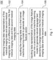
- FIG. 1 there is illustrated a computer implemented method 100 for generating natural language text describing a change between first medical imaging data 302 and second medical imaging data 304.
- the method 100 comprises:
- natural language text describing a change between first medical imaging data 302 and second medical imaging data 304 may be generated.
- the image feature vector Zi can be generated that encodes features relevant to the changes between the first medical imaging data 302 and the second medical imaging data 304. Accordingly, for example, the progression of a condition of a patient depicted in the first medical imaging data 302 and the second medical imaging data 304, can be extracted and encoded into the image feature vector Zi.
- the second trained machine learning model 350 uses this image feature vector to generate the natural language text describing the change. Accordingly, the generated natural language text describing the change is pertinent to the relevant change(s) between the first medical imaging data and the second medical imaging data.
- This generated natural language text may provide for automation of the interpretation and/or reporting of the second medical image, such as a follow up image.
- this text may represent, or be used to generate, a report for the follow-up image.
- this generated natural language text may allow for assistance in the interpretation and/or reporting of a follow-up image to be provided.
- a radiologist may use this generated text to guide their interpretation and/or reporting of the follow-up image.
- the natural language text describing the change may be used by a medical professional in a variety of ways.
- the medical professional may recommend a course of treatment, or decide that further follow-up imaging is required.
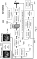
- Method 300 is now described in detail with reference to Figure 3 .
- Method 300 is a specific example of method 100 and includes the steps of method 100.
- the method 300 comprises, in step 102, obtaining imaging data 306 representative of first medical imaging data 302 and second medical imaging data 304, or of a difference between the first medical imaging data 302 and the second medical imaging data 304.
- the imaging data 306 comprises differential imaging data 306 representing a difference between the first medical imaging data 302 and the second medical imaging data 304.
- Each of the first medical imaging data 302 and the second medical imaging data 304 may comprise an array of elements each having a value.
- the elements may be pixels, each pixel having at least one value.
- the at least one value may correspond to or otherwise be representative of an output signal of the medical imaging technique used to generate the medical imaging data.
- the value of an element e.g. pixel
- each element may only have one value.
- each element may have or otherwise be associated with multiple values.
- the multiple values of a given element may represent the values of respective multiple signal channels.
- each signal channel may represent a different medical imaging signal or property of the imaging subject.
- the at least one value may comprise an element (e.g. pixel) intensity value.
- an output signal from the medical imaging may be mapped onto a pixel intensity value, for example a value within a defined range of intensity values.
- the intensity value may correspond to a value in the range 0 to 255, where 0 represents a ⁇ black' pixel and 255 represents a 'white' pixel, for example.
- the intensity value may correspond to a value in the range 0 to 65536.
- each pixel may have three intensity values, e.g. one each for Red, Green, and Blue channels. It will be appreciated that other values may be used.
- the first medical imaging data 302 is for a patient and has been captured at a first time and the second medical imaging data 304 is for the patient and has been captured at a second, later, time.
- the first medical imaging data 302 may form part of a baseline study of the patient.
- a first medical text report 312 may be written for the baseline study.
- the second medical imaging data 304 may form part of a follow-up study of the patient.
- the follow-up study may be intended to track progression (or regression) of a medical abnormality 308, and/or to identify occurrence (or disappearance) of a medical abnormality 308 after the baseline study.
- the first medical imaging data 302 and the second medical imaging data 304 are for a particular region of the patient.
- the first medical imaging data 302 and the second medical imaging data 304 may be for an abdominal region of the patient, or for a limb of the patient.
- Generating natural language text describing the change where the first medical imaging data 302 is for a patient and has been captured at a first time and the second medical imaging data 304 is for the patient and has been captured at a second, later, time enables computer implemented interpretation and/or reporting of a follow-up medical image. This in turn enables the progression of a condition of the patient to be monitored without necessarily requiring a human to analyze the first medical imaging data 302 and the second medical imaging data 304. Alternatively or additionally, this may enable computer implemented assistance in the interpretation and/or reporting of a follow-up medical image, or the monitoring of the progression of a condition of a patient, by a medical professional.
- the method 300 comprises obtaining the first medical imaging data 302 and the second medical imaging data 304.
- the first medical imaging data 302 and the second medical imaging data 304 may be retrieved from a storage such as a memory (see e.g. memory 604 in Figure 6 , described in more detail below).
- the first medical imaging data 302 may comprise a plurality of first intensity values
- the second medical imaging data 304 may comprise a plurality of corresponding, second, intensity values. That is, each of the first intensity values in the first medical imaging data 302 corresponds to a certain second intensity value in the second medical imaging data 304.
- Each of the first intensity values may be associated with a respective pixel location, and each of the second intensity values may be associated with one of the pixel locations, to provide this correspondence.
- the method 300 comprises image pre-processing 332 of the first medical imaging data 302 and the second medical imaging data 304.
- the image pre-processing 332 may comprise image registration.
- Image registration generally involves transforming the plurality of first intensity values and the plurality of second intensity values into one coordinate system.
- the image registration may comprise associating each of the first intensity values with its corresponding second intensity value, and the respective pixel locations may be defined during the image registration.
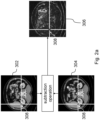
- the method 300 comprises, for each of the plurality of first intensity values, comparing the first intensity value with the corresponding second intensity value, to obtain a differential intensity value.
- the differential imaging data 306 comprises the obtained differential intensity values.
- comparing the first intensity value with the second intensity value may comprise performing a subtraction operation using the first intensity value and the second intensity value.
- comparing the first intensity value with the second intensity value may comprise subtracting the second intensity value from the first intensity value, or vice versa. Subtracting the second intensity value from the first intensity value, or vice versa, may allow a computationally simple method to obtain the differential imaging data 306.
- the regions in which the intensity value of the second medical imaging data 304 is lower than the intensity value of the first medical imaging data 302 are shown in light grey, and the regions in which the intensity value of the second medical imaging data 304 is higher than the intensity value of the first medical imaging data 302 are shown in dark grey.
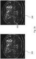
- a medical abnormality 308 is shown on each of the first medical imaging data 302, the second medical imaging data 304, and the differential medical imaging data. The medical abnormality 308 has undergone a subtle change in appearance between the time at which the first medical imaging data 302 was captured and the time at which the second medical imaging data 304 was captured.
- Figure 2b shows two copies of the differential medical imaging data, with the position of the abnormality indicated on the second copy.
- a medical abnormality 308 may be difficult to identify a medical abnormality 308 from visual inspection of the differential imaging data 306 alone. Indeed, it may also be difficult for a computer vision system to identify a medical abnormality 308 using the differential imaging data 306 alone.
- Some examples described herein enable a first trained machine learning model 340 to identify a change between the first medical imaging data 302 and the second medical imaging data 304, by training the machine learning model using, in addition to medical imaging data, medical text reports associated with the medical imaging data. This will be described further with reference to Figure 5 .
- the method 300 comprises, in step 104, inputting the imaging data 306 into a first trained machine learning model 340 to generate an image feature vector Zi representative of the difference between the first medical imaging data 302 and the second medical imaging data 304.
- the imaging data 306 is the differential imaging data 306, and the image feature vector Zi is representative of the differential imaging data.
- the first trained machine learning model 340 has been trained to generate, based on an input of given differential imaging data representing a difference between given first medical imaging data and given second medical imaging data, an image feature vector Zi.
- the training process is described in further detail below with reference to Figure 5 .
- Any machine learning model described herein may comprise a neural network, such as a convolutional neural network or a transformer.
- generating the image feature vector Zi comprises inputting the differential imaging data 306 into a first component trained machine learning model of the first trained machine learning model 340 to generate one or more attention maps 322.
- the first component trained machine learning model comprises an image encoder 342.
- the image encoder 342 has been trained to generate, based on an input of given differential imaging data 306, one or more attention maps.
- the image encoder 342 may be capable of producing an attention map which indicates features of the differential imaging data 306 that may represent information pertinent to a medical text report.
- Each attention map may be indicative of the extent to which a particular portion or segment of the differential imaging data 306 contributes to the encoding or a particular part of the encoding performed by the image encoder 342.
- each attention map may relate to a different segment, or a different context, of the differential imaging data 306.
- one or more attention maps may each highlight the particular segment or part(s) of the differential imaging data that the image encoder 342 determines corresponds to a respective particular context.
- a particular context may be that part(s) correspond to a medical abnormality.
- a segment may comprise a sub-plurality of the differential intensity values.
- the image encoder 342 may segment the differential imaging data 306 into the attention maps according to the contribution of each particular segment, or context, to the encoding.
- the image encoder may be configured to segment the differential imaging data 306 based on the respective locations of possible abnormalities occurring in the differential imaging data 306.
- the method 300 comprises inputting data representative of the one or more attention maps 322 into a second component trained machine learning model 344 of the first trained machine learning model 340 to generate the image feature vector Zi.
- the second component trained machine learning model 344 has been trained to generate, based on an input of a given attention map, an image feature vector Zi.
- the data representative of the one or more attention maps 322 may comprise the one or more attention maps 322.
- the method 300 comprises generating the data representative of the one or more attention maps 322 by applying a dimension reduction operation to each of the one or more attention maps 322.
- Applying the dimension reduction operation may reduce the number of pixels in the differential imaging data 306 and/or the amount of data used to store an attention map.
- the dimension reduction operation may comprise an average pooling operation.
- a medical abnormality 308 is likely to be represented by a plurality of adjacent or nearby pixels. Therefore, applying the dimension reduction operation can reduce the number of pixels used to represent the differential imaging data 306, without substantially removing or dampening information relating to the medical abnormality 308 as represented by the pixels prior to the application of the dimension reduction operation.
- the dimension reduction operation such as average pooling, is a fixed operation applied to the pixels, in the sense that it does not itself involve applying a machine learning model to the one or more attention maps 322.
- the second component machine learning model comprises a multi-layer perceptron (MLP) 1.
- MLP multi-layer perceptron
- the multi-layer perceptron has been trained to generate, based on an input of data representative of one or more given attention maps 322 (whether the dimension reduction operation has been applied to the given attention maps 322 or not), an image feature vector Zi.
- Generating the image feature vector Zi may comprise inputting data representative of the one or more attention maps 322 (whether the dimension reduction operation has been applied to the attention maps 322 or not) to the MLP 1 to generate the image feature vector Zi.
- the image feature vector Zi may accurately represent features of the imaging data 306 that would be relevant for a medical professional analyzing the first medical imaging data 302 and the second medical imaging data 304.
- the way in which the first machine learning model 440 may be trained to generate an image feature vector Zi with such properties is explained below with reference to Figure 5 .
- the method 300 comprises, at step 106, inputting data representative of the image feature vector Zi into a second trained machine learning model 350 to generate natural language text describing a change between the first medical imaging data 302 and the second medical imaging data 304.
- the second trained machine learning model 350 may have been trained to generate, based on an input of given data representative of the image feature vector Zi, natural language text describing a change between the given first medical imaging data and the given second medical imaging data.
- the training process is described in further detail below with reference to Figure 5 .
- the invention may enable the automatic generation of a description of a change between first medical imaging data 302 and second medical imaging data 304.
- the medical professional may use the description of the change to, for example, diagnose a medical condition, or recommend treatment.
- the radiologist or other medical professional can understand what change(s) has/have occurred between the first medical imaging data 302 and the second medical imaging data 304, without needing to evaluate the first medical imaging data 302 or the second medical imaging data 304.
- a radiologist visually inspecting the first medical imaging data 302 and the second medical imaging data 304, or the differential imaging data 306, may miss a change that has occurred.
- a change between two sets of medical imaging data may allow such overlooking to be avoided.
- the change represents the occurrence or progression of a medical abnormality 308.
- the change may represent the initial appearance or growth of a tumour between the first medical imaging data 302 and the second medical imaging data 304.
- the initial appearance of a medical abnormality 308 may be identified, or the progression of the medical abnormality 308 may be tracked, by generating the natural language text describing the change.
- a change in the patient's condition may be monitored.
- the method 300 comprises generating a second medical text report 314 associated with the second medical imaging data 304, based on a first medical text report 312 associated with the first medical imaging data 302 and the natural language text describing the change.
- the first medical text report 312 may describe the first medical imaging data 302 or any feature in the first medical imaging data 302.
- the first medical text report 312 comprises at least one sentence such as "There is a hypodense lesion in the right lobe of the liver".
- the second medical text report 314 may describe the second medical imaging data 304 or any feature in the second medical imaging data 304.
- the second medical text report 314 comprises at least one sentence such as "Again visualized is a hypodense lesion in the right lobe of the liver.
- the second medical text report 31 may describe any change that has occurred between the first medical imaging data and the second medical imaging data.
- the natural language text describing the change comprises at least one sentence such as "There is an increase in the size of an hypodense lesion in the right lobe of the liver".
- the first medical text report 312 may act as a guide in generating the second medical text report 314.
- a second medical text report 314 that focuses on changes as compared to the information described in the first medical text report can be provided.
- generating the second medical text report 314 may comprise inputting the first medical text report 312 and the natural language text describing the change to a natural language processing model.
- the natural language processing model is a trained natural language processing machine learning model.
- the trained natural language processing machine learning model is a trained large language model, such as ChatGPT or GPT-4.
- the trained large language model has been trained to (among other things) generate a second medical text report 314, based on an input of a given first medical text report 312, given natural language text describing the change, and a text prompt comprising an instruction to generate the second medical text report 314.
- generating the second medical text report 314 may comprise inputting the following to the large language model:
- the efficiency with which the second medical text report 314 is generated can be improved compared to a medical professional writing the second medical text report 314.
- the contents of the second medical text report 314 may be more pertinent to the change described in the natural language text.
- FIG. 4 and 5 there is illustrated a computer implemented training method 400, 400a of training a machine learning model for generating natural language text describing a change between first medical imaging data 302 and second medical imaging data 304.
- the first trained machine learning model 340 and the second trained machine learning model 350 described above with reference to Figures 1 to 3 may be (or have been) trained according to the training method 400 outlined in Figures 4 and 5 .
- the trained natural language processing machine learning model e.g. ChatGPT
- the computer-implemented training method 400a illustrated in Figure 5 is a specific example of the computer-implemented training method 400 illustrated in Figure 4 .
- the training method 400, 400a comprises, in step 202, providing the machine learning model.
- the machine learning model comprises (a) a first machine learning model 440 configured to generate, based on an input of given imaging data representative of given first medical imaging data and given second medical imaging data, or of a difference between the given first medical imaging data and the given second medical imaging data, an image feature vector Zi.
- the given imaging data is given differential imaging data representing the difference between the given first medical imaging data and the given second medical imaging data.
- the first machine learning model 440 may have similar architecture to or the same architecture as the first trained machine learning model 340, for example it may comprise the first component machine learning model and the second component machine-learning model. This architecture is abbreviated in Figure 5 .
- the machine learning model also comprises (b) a second machine learning model 450 configured to generate, based on an input of data representative of the image feature vector Zi, natural language text describing a change between the given first medical imaging data and the given second medical imaging data.
- the second machine learning model 450 may have similar architecture to or the same architecture as the second trained machine learning model 350.
- the machine learning model comprises (c) a text encoder machine learning model.
- the text encoder machine learning model is configured to generate a text feature vector Zt, based on an input of data, where the data is representative of a given first medical text report associated with the given first medical imaging data and a given second medical text report associated with the given second medical imaging data.
- the data representative of the given first medical text report and the given second medical text report may comprise data representative of the first medical text report and data representative of the second medical text report. Additionally, or alternatively, the data representative of the given first medical text report and the given second medical text report may comprise data representative of the given natural language text describing the change.
- the training method 400, 400a comprises, in step 204, providing training data.
- the training data comprises a plurality of sets of training imaging data 406, each set of training imaging data 406 representative of first training medical imaging data 402 and second training medical imaging data 404, or of a difference between the first training medical imaging data 402 and the second training medical imaging data 404.
- the training data further comprises, for each set of training image data 406, ground truth natural language text describing a change between the first training medical imaging data 402 and the second training medical imaging data 404.
- the training imaging data is training differential imaging data representing the difference between the first training medical imaging data 402 and the second training medical imaging data 404.
- providing the training data comprises generating, for each set of training differential imaging data 406, the ground truth natural language text.
- generating the ground truth natural language text is performed by inputting a first training medical text report 412 associated with the first training medical imaging data 402 and a second training medical text report 414 associated with the second training medical imaging data 404 into a third, trained machine learning model 460.
- the training data may comprise, for each set of training differential imaging data 406, the first training medical text report 412 and the second training medical text report 414.
- the third training machine learning model 460 may be a trained large language model.
- the third training machine learning model 460 may be the same as the trained large language model described above with reference to Figures 1 to 3 (e.g.
- ChatGPT or GPT-4 may be a different large language model.
- the trained large language model may have been trained to (among other things) generate the ground truth natural language text, based on an input of a given first training medical text report associated with given first training medical imaging data, a given second training medical text report associated with given second training medical imaging data, and a text prompt comprising an instruction to generate the ground truth natural language text.
- generating the natural language text may comprise inputting the following to the large language model:
- the second machine learning model 450 can accurately output natural language text describing the change.
- trained machine learning model 460 may provide an automated yet accurate way to generate the ground truth natural language text.
- the training method 400, 400a comprises, in step 206, training the machine learning model based on the training data.
- Training the machine learning model comprises training the machine learning model based on the training data so as to minimize a loss function 460 between the natural language text generated for the sets of training imaging data 406 by the machine learning model and the corresponding ground truth natural language text for the sets of training imaging data 406.
- the training imaging data 406 is training differential imaging data 406.
- Minimizing the loss function 460 may comprise reducing a distance in a change-description embedding space between a change-description text feature vector and a ground truth text feature vector.
- the change-description text feature vector is a vector representation in the change-description embedding space of the natural language text generated for a given set of training differential imaging data 406.
- the ground truth text feature vector is a vector representation in the change-description embedding space of the ground truth natural language text.
- the loss function 460 may comprise a term which decreases as this distance decreases. Any distance referred to herein may comprise a Euclidean distance or a cosine distance, for example.
- the training method 400a comprises training the machine learning model based on the training data so as to minimize a further loss function 470 between: the text feature vectors Zt generated for the first 412 and second 414 training medical text reports by the text encoder machine learning model 480, and the image feature vectors Zi generated for the corresponding sets of training differential imaging data 406 by the first machine learning model 440.
- the text encoder machine learning model 480 is configured to generate a text feature vector Zt.
- the text feature vector Zt is generated by inputting the first training medical text report 412 and the second training medical text report 414 to the third trained machine learning model 460 to generate natural language text.
- This natural language text may be identical to the ground truth natural language text described above.
- the natural language text is input to the text encoder machine learning model 480.
- the text encoder machine learning model 480 may include a multi-layer perceptron (MLP) 2.
- MLP multi-layer perceptron
- the text encoder machine learning model 480 then generates, using the MLP 2, the text feature vector Zt.
- the text feature vectors Zt and the image feature vectors Zi may be represented in a common embedding space.
- the common embedding space may be the same as the change-description embedding space, or it may be different.
- minimizing the further loss function 470 may comprise reducing a distance in the common embedding space between an image feature vector Zi generated for a given set of the plurality of sets of training differential imaging data 406 and a text feature vector Zt generated for the given set. That is, the further loss function 470 may tend to attract an image feature vector Zi to the text feature vector Zt generated for the same set of training differential imaging data 406 in the common embedding space.
- the further loss function 470 typically has the effect that during performance of the method 300 of Figures 1 to 3 (i.e. during inference), the image feature vector Zi generated by the first trained machine learning model 340 captures the features that would be considered relevant when writing the natural language text describing the change between the first medical imaging data 302 and the second medical imaging data 304. This is because, by virtue of the training using the further loss function 470, the first machine learning model 440 is trained to generate an image feature vector Zi that is close to the text feature vector Zt in the common embedding space, regardless of the contents of the training data of the set.
- the image feature vector Zi effectively represents the same information by nature of the further loss function. The same applies to the one or more attention maps 322.
- the further loss function 470 is a contrastive loss function 460.
- Minimizing the contrastive loss function 460 comprises, in addition to the above property of the further loss function, increasing a distance in the common embedding space between an image Zi or text feature vector Zt generated for one set of the plurality of sets of training differential imaging data 406, and an image Zi or text feature vector Zt generated for another set of the plurality of sets of training differential imaging data 406. That is, the contrastive loss function 460 tends to separate feature vectors belonging to different sets from one another.
- the contrastive loss function typically has a functional form max S Z i 1 d Z t 1 d + S Z i 2 d Z t 2 d min S Z i 1 d Z t 2 d + S Z i 2 d Z t 1 d
- S ( x,y ) is a function whose value decreases as the distance between x and y increases
- 1d is a first set of the plurality of sets of training data
- 2d is a second set of the plurality of sets of training data different from the first set.
- the contrastive loss function 460 thus typically has the effect that the image feature vector Zi generated for one set of differential imaging data 306 (during the inference stage i.e. the methods illustrated in Figures 1 and 3 ) is dissimilar from the image feature vector Zi generated for another set of differential imaging data 306.
- the training method 400, 400a may comprise training the machine learning model by applying the loss function 460 and the further loss function 470 alternately to the training data.
- the first machine learning model 440 and the text encoder machine learning model 480 may process the training data to update parameters of each of the first machine learning model 440 and the text encoder machine learning model 480.
- the parameters of the second machine learning model 450 may be frozen; that is to say that they are not updated during the first stage.
- the second machine learning model 450 may process the training data to update the parameters of the second machine learning model 450.
- the parameters of the first machine learning model 440 and the text encoder machine learning model 480 may be frozen; that is to say that they are not updated during the second stage.
- the first stage and the second stage may be repeated alternately and iteratively until each of the first machine learning model 440, the text encoder machine learning model 480, and the second machine learning model 480 have been trained.
- the parameters of the third trained machine learning model 460 may be generally held fixed during both the first stage and the second stage, as the third trained machine learning model 460 has already been trained.
- the machine learning model may process each set of training data in this two-stage manner.
- the apparatus 600 comprises an input interface 606, an output interface 608, a processor 602, and a memory 604.
- the processor 602 and the memory 604 may be configured to perform the method 100, 300 or the training method 400, 400a according to any one of the examples described above with reference to Figures 1 to 5 .
- the memory may store instructions which, when executed by the processor 602 cause the processor 602 to perform the method 100, 300 according to any one of the examples described above with reference to Figures 1 to 3 , and/or the training method 400, 400a according to any one of the examples described above with reference to Figures 4 to 5 .
- the instructions may be stored on any computer readable medium, for example any non-transitory computer readable medium.
- the input interface 606 may receive imaging data 306, the processor 602 may implement the method 300 described above with reference to Figures 1 to 3 to generate the natural language text describing the change, and the processor 602 may output, via the output interface 608, the natural language text describing the change, or the second medical text report 314.
- the natural language text describing the change may be transmitted to a structured storage (not shown) so that the output data is stored in the structured storage.
- the natural language text describing the change, or the second medical text report 314 may be transmitted to a display device (not shown) to allow a user to review the output data.
- the output data may be stored, alternatively or additionally, in the memory 604.
- the input interface 606 may receive the training data
- the processor 608 may implement training of the machine learning model as described above with reference to Figures 4 and 5
- the processor 602 may output, via the output interface 608, the trained machine learning model or data representing the trained machine learning model.
- the trained machine learning model or data representing the trained machine learning model may be stored in an external storage (not shown) or transmitted to another computer (not shown) for use by the other computer (not shown) to perform the method 300 according to any one of the examples described above with reference to Figures 1 to 3 .
- the trained machine learning model or data representing the trained machine learning model may be alternatively or additionally stored in the memory 604 or another local storage of the apparatus 600, for example for use by the apparatus 600 in implementing the method 300 according to any one of the examples described above with reference to Figures 1 to 3 .
- the apparatus 600 may be implemented as a processing system and/or a computer. It will be appreciated that the methods according to any one of the examples described above with reference to Figures 1 to 5 are computer implemented methods, and that these methods may be implemented by the apparatus 600.
- the imaging data is differential imaging data representing a difference between first medical imaging data and second medical imaging data
- the imaging data may instead be representative of the first medical imaging data and the second medical imaging data.
- obtaining the imaging data may comprise concatenating the first medical imaging data and the second medical imaging data.
- the method may then comprise, instead of inputting the differential imaging data into the first trained machine learning model, inputting the concatenated imaging data into the first trained machine learning model.
- the first trained machine learning model by virtue of the training using the further loss function, is nevertheless capable of generating an image feature vector representative of the difference between the first medical imaging data and the second medical imaging data.
- each set of training imaging data may comprise a concatenation of the first training medical imaging data and the second training medical imaging data.
Landscapes
- Engineering & Computer Science (AREA)
- Health & Medical Sciences (AREA)
- Theoretical Computer Science (AREA)
- General Health & Medical Sciences (AREA)
- Medical Informatics (AREA)
- Physics & Mathematics (AREA)
- General Physics & Mathematics (AREA)
- Computer Vision & Pattern Recognition (AREA)
- Multimedia (AREA)
- Public Health (AREA)
- Evolutionary Computation (AREA)
- Databases & Information Systems (AREA)
- Artificial Intelligence (AREA)
- Biomedical Technology (AREA)
- Software Systems (AREA)
- Primary Health Care (AREA)
- Epidemiology (AREA)
- Computing Systems (AREA)
- Data Mining & Analysis (AREA)
- Radiology & Medical Imaging (AREA)
- Nuclear Medicine, Radiotherapy & Molecular Imaging (AREA)
- Pathology (AREA)
- Molecular Biology (AREA)
- Life Sciences & Earth Sciences (AREA)
- Biodiversity & Conservation Biology (AREA)
- Biophysics (AREA)
- Mathematical Physics (AREA)
- General Engineering & Computer Science (AREA)
- Computational Linguistics (AREA)
- Quality & Reliability (AREA)
- Image Analysis (AREA)
Abstract
A computer implemented method (100) and apparatus for generating natural language text describing a change between first medical imaging data and second medical imaging data is disclosed. Imaging data representative of the first medical imaging data and the second medical imaging data, or of a difference between the first medical imaging data and the second medical imaging data, is obtained. The imaging data is input into a first trained machine learning model to generate an image feature vector representative of the difference between the first medical imaging data and the second medical imaging data. Data representative of the image feature vector is input into a second trained machine learning model to generate the natural language text describing the change between the first medical imaging data and the second medical imaging data.
Description
- The invention relates to a method and an apparatus for analyzing a change between two sets of medical imaging data.
- When a patient is diagnosed with a condition using a baseline medical image, follow-up imaging is commonly recommended to monitor progression of the condition. After the follow-up image is taken, a radiologist may review the follow-up image and the baseline image, and write a report describing any progression in the condition. However, interpretation of the follow-up image and/or writing of the report is a time-consuming task.
- In addition, when comparing a baseline image taken at the time of diagnosis with a follow-up image taken later, the images may include artifacts which are not relevant to the progression of the condition. For example, where the lighting or imaging angle used to capture the baseline image differs from those used to take the follow-up image, artifacts may be visible. Other irrelevant changes may be apparent in the images. This can make interpretation and/or reporting of the follow-up and baseline images difficult.
- It is desirable to automate, or at least provide computer implemented assistance in, the interpretation and/or reporting of follow-up medical images.
- According to a first aspect of the present invention, there is provided a computer-implemented method for generating natural language text describing a change between first medical imaging data and second medical imaging data, the method comprising: obtaining imaging data representative of the first medical imaging data and the second medical imaging data, or of a difference between the first medical imaging data and the second medical imaging data; inputting the imaging data into a first trained machine learning model to generate an image feature vector representative of the difference between the first medical imaging data and the second medical imaging data; and inputting data representative of the image feature vector into a second trained machine learning model to generate the natural language text describing the change between the first medical imaging data and the second medical imaging data.
- Optionally, the first trained machine learning model has been trained to generate, based on an input of given differential imaging data representing a difference between given first medical imaging data and given second medical imaging data, an image feature vector.
- Optionally, the second trained machine learning model has been trained to generate, based on an input of given data representative of the image feature vector, natural language text describing a change between the given first medical imaging data and the given second medical imaging data.
- Optionally, the method comprises generating a second medical text report associated with the second medical imaging data, based on a first medical text report associated with the first medical imaging data and the natural language text describing the change.
- Optionally, generating the second medical text report comprises inputting the first medical text report and the natural language text describing the change to a natural language processing model.
- Optionally, the natural language processing model is a trained natural language processing machine learning model.
- Optionally, the trained natural language processing machine learning model is a trained large language model.
- Optionally, the trained large language model has been trained to generate a second medical text report, based on an input of a given first medical text report, given natural language text describing the change, and a text prompt comprising an instruction to generate the second medical text report.
- Optionally, the method comprises transmitting the natural language text describing the change and/or the second medical text report to a storage device for storage at the storage device.
- Optionally, the method comprises transmitting the natural language text describing the change and/or the second medical text report to a display device for display on the display device.
- Optionally, the first medical imaging data is for a patient and has been captured at a first time and the second medical imaging data is for the patient and has been captured at a second, later, time.
- Optionally, the first medical imaging data and the second medical imaging data are for a particular region of the patient.
- Optionally, the change represents the occurrence, progression, regression, or disappearance of a medical abnormality. Optionally, the method comprises: obtaining the first medical imaging data and the second medical imaging data, the first medical imaging data comprising a plurality of first intensity values and the second medical imaging data comprising a plurality of corresponding, second, intensity values; and for each of the plurality of first intensity values, comparing the first intensity value with the corresponding second intensity value, to obtain a differential intensity value, wherein the imaging data comprises the obtained differential intensity values.
- Optionally, comparing the first intensity value with the second intensity value comprises performing a subtraction operation using the first intensity value and the second intensity value.
- Optionally, the method comprises: prior to comparing the first intensity values with the second intensity values, registering the first medical imaging data with the second medical imaging data by transforming the plurality of first intensity values and the plurality of second intensity values into one coordinate system.
- Optionally, generating the image feature vector comprises: inputting the imaging data into a first component trained machine learning model of the first trained machine learning model to generate one or more attention maps; and inputting data representative of the one or more attention maps into a second component trained machine learning model of the first trained machine learning model to generate the image feature vector.
- Optionally, the method comprises generating the data representative of the one or more attention maps by applying a dimension reduction operation to each of the one or more attention maps.
- Optionally, the one or more attention maps comprise a plurality of attention maps, and each attention map relates to a different segment of the imaging data.
- According to a second aspect of the present invention, there is provided a computer implemented training method of training a machine learning model for generating natural language text describing a change between first medical imaging data and second medical imaging data, the method comprising: providing the machine learning model, the machine learning model comprising: (a) a first machine learning model configured to generate, based on an input of given imaging data representative of given first medical imaging data and given second medical imaging data, or of a difference between the given first medical imaging data and the given second medical imaging data, an image feature vector, and (b) a second machine learning model configured to generate, based on an input of data representative of the image feature vector, natural language text describing a change between the given first medical imaging data and the given second medical imaging data; providing training data comprising a plurality of sets of training imaging data, each set of training imaging data representative of first training medical imaging data and second training medical imaging data, or of a difference between the first training medical imaging data and the second training medical imaging data, the training data further comprising, for each set of training imaging data, ground truth natural language text describing a change between the first training medical imaging data and the second training medical imaging data; and training the machine learning model based on the training data so as to minimize a loss function between the natural language text generated for the sets of training imaging data by the machine learning model and the corresponding ground truth natural language text for the sets of training imaging data.
- Optionally, providing the training data comprises generating, for each set of training imaging data, the ground truth natural language text, by inputting a first training medical text report associated with the first training medical imaging data and a second training medical text report associated with the second training medical imaging data, into a third, trained machine learning model.
- Optionally, the third trained machine learning model is a trained large language model.
- Optionally, the trained large language model has been trained to generate ground truth natural language text, based on an input of a given first training medical text report associated with given first training medical imaging data, a given second training medical text report associated with given second training medical imaging data, and a text prompt comprising an instruction to generate the ground truth natural language text.
- Optionally, the training data comprises, for each set of training imaging data, training data representative of the first training medical text report and the second training medical text report; the machine learning model comprises (c) a text encoder machine learning model configured to generate, based on an input of given data representative of: a given first medical text report associated with the given first medical imaging data and a given second medical text report associated with the given second medical imaging data, a text feature vector; and the method comprises training the machine learning model based on the training data so as to minimize a further loss function between: the text feature vectors generated for the first and second training medical text reports by the text encoder machine learning model, and the image feature vectors generated for the corresponding sets of training imaging data by the first machine learning model.
- Optionally, the further loss function is a contrastive loss function.
- Optionally, minimizing the contrastive loss function comprises: reducing a distance in the common embedding space between: an image feature vector generated for a given set of the plurality of sets of training imaging data, and a text feature vector generated for the given set; and/or increasing a distance in the common embedding space between: one of: an image feature vector generated for a first set of the plurality of sets of training imaging data and a text feature vector generated for the first set, and one of: an image feature vector generated for a second set of the plurality of sets of training imaging data and a text feature vector generated for the second set.
- According to a third aspect of the present invention, there is provided apparatus configured to perform the method according to the first aspect and/or the second aspect.
- Optionally, the apparatus comprises the storage device and/or the display device.
- According to a fourth aspect of the present invention, there is provided a computer program which, when executed by a computer, causes the computer to perform the method according to the first aspect and/or the second aspect.
-
-
Figure 1 is a flow diagram illustrating schematically a method for generating natural language text describing a change between first medical imaging data and second medical imaging data; -
Figure 2a is a flow diagram illustrating schematically a method for obtaining differential imaging data from first medical imaging data and second medical imaging data; -
Figure 2b is an illustration of the difficulties associated with identifying a medical abnormality from obtaining differential imaging data by visual inspection; -
Figure 3 is a flow diagram illustrating schematically in more detail a method according to the method illustrated inFigure 1 ; -
Figure 4 is a flow diagram illustrating schematically a method for training a machine learning model to perform the method illustrated inFigure 1 ; -
Figure 5 is a flow diagram illustrating schematically in more detail a method according to the method illustrated inFigure 4 ; -
Figure 6 is a diagram illustrating schematically an apparatus according to an example. - Independent of the grammatical term usage, individuals with male, female or other gender identities are included within the term.
- Referring to
Figure 1 , there is illustrated a computer implementedmethod 100 for generating natural language text describing a change between firstmedical imaging data 302 and secondmedical imaging data 304. - In broad overview, the
method 100 comprises: - in
step 102, obtainingimaging data 306 representative of firstmedical imaging data 302 and secondmedical imaging data 304, or of a difference between the firstmedical imaging data 302 and the second medical imaging data 304 (seeFigure 2a ); - in
step 104, inputting theimaging data 306 into a first trainedmachine learning model 340 to generate an image feature vector Zi (for example, the first trainedmachine learning model 340 inFigure 3 ) - in
step 106, inputting data representative of the image feature vector Zi into a second trainedmachine learning model 350 to generate the natural language text describing the change between the firstmedical imaging data 302 and the second medical imaging data 304 (for example, see the second trainedmachine learning model 350 inFigure 3 ) . - Accordingly, natural language text describing a change between first
medical imaging data 302 and secondmedical imaging data 304 may be generated. - By generating the image feature vector Zi based on the
imaging data 306 using the first trainedmachine learning model 340, the image feature vector Zi can be generated that encodes features relevant to the changes between the firstmedical imaging data 302 and the secondmedical imaging data 304. Accordingly, for example, the progression of a condition of a patient depicted in the firstmedical imaging data 302 and the secondmedical imaging data 304, can be extracted and encoded into the image feature vector Zi. The second trainedmachine learning model 350 uses this image feature vector to generate the natural language text describing the change. Accordingly, the generated natural language text describing the change is pertinent to the relevant change(s) between the first medical imaging data and the second medical imaging data. This generated natural language text may provide for automation of the interpretation and/or reporting of the second medical image, such as a follow up image. For example, this text may represent, or be used to generate, a report for the follow-up image. Alternatively, or additionally, this generated natural language text may allow for assistance in the interpretation and/or reporting of a follow-up image to be provided. For example, a radiologist may use this generated text to guide their interpretation and/or reporting of the follow-up image. - The natural language text describing the change may be used by a medical professional in a variety of ways. For example, the medical professional may recommend a course of treatment, or decide that further follow-up imaging is required.
- An
example method 300 is now described in detail with reference toFigure 3 .Method 300 is a specific example ofmethod 100 and includes the steps ofmethod 100. - As mentioned, the
method 300 comprises, instep 102, obtainingimaging data 306 representative of firstmedical imaging data 302 and secondmedical imaging data 304, or of a difference between the firstmedical imaging data 302 and the secondmedical imaging data 304. In this example, theimaging data 306 comprisesdifferential imaging data 306 representing a difference between the firstmedical imaging data 302 and the secondmedical imaging data 304. - Each of the first
medical imaging data 302 and the secondmedical imaging data 304 may comprise an array of elements each having a value. The elements may be pixels, each pixel having at least one value. The at least one value may correspond to or otherwise be representative of an output signal of the medical imaging technique used to generate the medical imaging data. For example, for Magnetic Resonance Imaging, the value of an element (e.g. pixel) may correspond to or represent a rate at which excited nuclei, in a region corresponding to the element, return to an equilibrium state. In some examples, each element may only have one value. However, in other examples, each element may have or otherwise be associated with multiple values. For example, the multiple values of a given element may represent the values of respective multiple signal channels. For example, each signal channel may represent a different medical imaging signal or property of the imaging subject. In some examples, the at least one value may comprise an element (e.g. pixel) intensity value. For example, an output signal from the medical imaging may be mapped onto a pixel intensity value, for example a value within a defined range of intensity values. For example, for a greyscale image, the intensity value may correspond to a value in the range 0 to 255, where 0 represents a `black' pixel and 255 represents a 'white' pixel, for example. As another example, for example as in the case of USHORT medical image data, the intensity value may correspond to a value in the range 0 to 65536. As another example, in a color image (e.g. where different colors represent different properties of the imaging subject) each pixel may have three intensity values, e.g. one each for Red, Green, and Blue channels. It will be appreciated that other values may be used. - In some examples, the first
medical imaging data 302 is for a patient and has been captured at a first time and the secondmedical imaging data 304 is for the patient and has been captured at a second, later, time. For example, the firstmedical imaging data 302 may form part of a baseline study of the patient. As shown inFigure 3 , a firstmedical text report 312 may be written for the baseline study. The secondmedical imaging data 304 may form part of a follow-up study of the patient. The follow-up study may be intended to track progression (or regression) of amedical abnormality 308, and/or to identify occurrence (or disappearance) of amedical abnormality 308 after the baseline study. In this example, the firstmedical imaging data 302 and the secondmedical imaging data 304 are for a particular region of the patient. For example, the firstmedical imaging data 302 and the secondmedical imaging data 304 may be for an abdominal region of the patient, or for a limb of the patient. - Generating natural language text describing the change where the first
medical imaging data 302 is for a patient and has been captured at a first time and the secondmedical imaging data 304 is for the patient and has been captured at a second, later, time, enables computer implemented interpretation and/or reporting of a follow-up medical image. This in turn enables the progression of a condition of the patient to be monitored without necessarily requiring a human to analyze the firstmedical imaging data 302 and the secondmedical imaging data 304. Alternatively or additionally, this may enable computer implemented assistance in the interpretation and/or reporting of a follow-up medical image, or the monitoring of the progression of a condition of a patient, by a medical professional. - In some examples, the
method 300 comprises obtaining the firstmedical imaging data 302 and the secondmedical imaging data 304. For example, the firstmedical imaging data 302 and the secondmedical imaging data 304 may be retrieved from a storage such as a memory (seee.g. memory 604 inFigure 6 , described in more detail below). The firstmedical imaging data 302 may comprise a plurality of first intensity values, and the secondmedical imaging data 304 may comprise a plurality of corresponding, second, intensity values. That is, each of the first intensity values in the firstmedical imaging data 302 corresponds to a certain second intensity value in the secondmedical imaging data 304. Each of the first intensity values may be associated with a respective pixel location, and each of the second intensity values may be associated with one of the pixel locations, to provide this correspondence. - In this example, the
method 300 comprises image pre-processing 332 of the firstmedical imaging data 302 and the secondmedical imaging data 304. For example, theimage pre-processing 332 may comprise image registration. Image registration generally involves transforming the plurality of first intensity values and the plurality of second intensity values into one coordinate system. The image registration may comprise associating each of the first intensity values with its corresponding second intensity value, and the respective pixel locations may be defined during the image registration. - In some examples, the
method 300 comprises, for each of the plurality of first intensity values, comparing the first intensity value with the corresponding second intensity value, to obtain a differential intensity value. In these examples, thedifferential imaging data 306 comprises the obtained differential intensity values. For example, as illustrated inFigure 2a , comparing the first intensity value with the second intensity value may comprise performing a subtraction operation using the first intensity value and the second intensity value. For example, comparing the first intensity value with the second intensity value may comprise subtracting the second intensity value from the first intensity value, or vice versa. Subtracting the second intensity value from the first intensity value, or vice versa, may allow a computationally simple method to obtain thedifferential imaging data 306. In thedifferential imaging data 306 shown inFigures 2a and2b , the regions in which the intensity value of the secondmedical imaging data 304 is lower than the intensity value of the firstmedical imaging data 302 are shown in light grey, and the regions in which the intensity value of the secondmedical imaging data 304 is higher than the intensity value of the firstmedical imaging data 302 are shown in dark grey. Amedical abnormality 308 is shown on each of the firstmedical imaging data 302, the secondmedical imaging data 304, and the differential medical imaging data. Themedical abnormality 308 has undergone a subtle change in appearance between the time at which the firstmedical imaging data 302 was captured and the time at which the secondmedical imaging data 304 was captured. -
Figure 2b shows two copies of the differential medical imaging data, with the position of the abnormality indicated on the second copy. As will be appreciated from thedifferential imaging data 306 shown inFigure 2b , it may be difficult to identify amedical abnormality 308 from visual inspection of thedifferential imaging data 306 alone. Indeed, it may also be difficult for a computer vision system to identify amedical abnormality 308 using thedifferential imaging data 306 alone. Some examples described herein enable a first trainedmachine learning model 340 to identify a change between the firstmedical imaging data 302 and the secondmedical imaging data 304, by training the machine learning model using, in addition to medical imaging data, medical text reports associated with the medical imaging data. This will be described further with reference toFigure 5 . - As mentioned, the
method 300 comprises, instep 104, inputting theimaging data 306 into a first trainedmachine learning model 340 to generate an image feature vector Zi representative of the difference between the firstmedical imaging data 302 and the secondmedical imaging data 304. In this example, theimaging data 306 is thedifferential imaging data 306, and the image feature vector Zi is representative of the differential imaging data. - In this example, the first trained
machine learning model 340 has been trained to generate, based on an input of given differential imaging data representing a difference between given first medical imaging data and given second medical imaging data, an image feature vector Zi. The training process is described in further detail below with reference toFigure 5 . Any machine learning model described herein may comprise a neural network, such as a convolutional neural network or a transformer. - In this example, generating the image feature vector Zi comprises inputting the
differential imaging data 306 into a first component trained machine learning model of the first trainedmachine learning model 340 to generate one or more attention maps 322. In this example, the first component trained machine learning model comprises animage encoder 342. Theimage encoder 342 has been trained to generate, based on an input of givendifferential imaging data 306, one or more attention maps. Through training the first trainedmachine learning model 340 with thefurther loss function 470 described below with reference toFigure 5 , theimage encoder 342 may be capable of producing an attention map which indicates features of thedifferential imaging data 306 that may represent information pertinent to a medical text report. - Each attention map may be indicative of the extent to which a particular portion or segment of the
differential imaging data 306 contributes to the encoding or a particular part of the encoding performed by theimage encoder 342. For example, each attention map may relate to a different segment, or a different context, of thedifferential imaging data 306. For example, one or more attention maps may each highlight the particular segment or part(s) of the differential imaging data that theimage encoder 342 determines corresponds to a respective particular context. For example, a particular context may be that part(s) correspond to a medical abnormality. A segment may comprise a sub-plurality of the differential intensity values. Theimage encoder 342 may segment thedifferential imaging data 306 into the attention maps according to the contribution of each particular segment, or context, to the encoding. For example, the image encoder may be configured to segment thedifferential imaging data 306 based on the respective locations of possible abnormalities occurring in thedifferential imaging data 306. - In this example, the
method 300 comprises inputting data representative of the one or more attention maps 322 into a second component trainedmachine learning model 344 of the first trainedmachine learning model 340 to generate the image feature vector Zi. In this examples, the second component trainedmachine learning model 344 has been trained to generate, based on an input of a given attention map, an image feature vector Zi. - The data representative of the one or more attention maps 322 may comprise the one or more attention maps 322. However, in this example, the
method 300 comprises generating the data representative of the one or more attention maps 322 by applying a dimension reduction operation to each of the one or more attention maps 322. Applying the dimension reduction operation may reduce the number of pixels in thedifferential imaging data 306 and/or the amount of data used to store an attention map. The dimension reduction operation may comprise an average pooling operation. In general, amedical abnormality 308 is likely to be represented by a plurality of adjacent or nearby pixels. Therefore, applying the dimension reduction operation can reduce the number of pixels used to represent thedifferential imaging data 306, without substantially removing or dampening information relating to themedical abnormality 308 as represented by the pixels prior to the application of the dimension reduction operation. In some examples, the dimension reduction operation, such as average pooling, is a fixed operation applied to the pixels, in the sense that it does not itself involve applying a machine learning model to the one or more attention maps 322. - In this example, the second component machine learning model comprises a multi-layer perceptron (MLP) 1. The multi-layer perceptron has been trained to generate, based on an input of data representative of one or more given attention maps 322 (whether the dimension reduction operation has been applied to the given attention maps 322 or not), an image feature vector Zi. Generating the image feature vector Zi may comprise inputting data representative of the one or more attention maps 322 (whether the dimension reduction operation has been applied to the attention maps 322 or not) to the
MLP 1 to generate the image feature vector Zi. - The image feature vector Zi may accurately represent features of the
imaging data 306 that would be relevant for a medical professional analyzing the firstmedical imaging data 302 and the secondmedical imaging data 304. The way in which the firstmachine learning model 440 may be trained to generate an image feature vector Zi with such properties is explained below with reference toFigure 5 . - As mentioned, the
method 300 comprises, atstep 106, inputting data representative of the image feature vector Zi into a second trainedmachine learning model 350 to generate natural language text describing a change between the firstmedical imaging data 302 and the secondmedical imaging data 304. - The second trained
machine learning model 350 may have been trained to generate, based on an input of given data representative of the image feature vector Zi, natural language text describing a change between the given first medical imaging data and the given second medical imaging data. The training process is described in further detail below with reference toFigure 5 . - By "describing a change", it is meant that the natural language text typically describes the change in such a way that the description of the change is comprehensible by a medical professional. Thus, the invention may enable the automatic generation of a description of a change between first
medical imaging data 302 and secondmedical imaging data 304. The medical professional may use the description of the change to, for example, diagnose a medical condition, or recommend treatment. In any case, by generating such natural language text, the radiologist or other medical professional can understand what change(s) has/have occurred between the firstmedical imaging data 302 and the secondmedical imaging data 304, without needing to evaluate the firstmedical imaging data 302 or the secondmedical imaging data 304. Moreover, a radiologist visually inspecting the firstmedical imaging data 302 and the secondmedical imaging data 304, or thedifferential imaging data 306, may miss a change that has occurred. By generating natural language text describing the change according to the methods illustrated inFigures 1 and3 , a change between two sets of medical imaging data may allow such overlooking to be avoided. - In some examples, the change represents the occurrence or progression of a
medical abnormality 308. For example, the change may represent the initial appearance or growth of a tumour between the firstmedical imaging data 302 and the secondmedical imaging data 304. In these examples, the initial appearance of amedical abnormality 308 may be identified, or the progression of themedical abnormality 308 may be tracked, by generating the natural language text describing the change. In examples in which the firstmedical imaging data 302 is for a patient and has been captured at a first time and the secondmedical imaging data 304 is for the patient and has been captured at a second, later, time, a change in the patient's condition may be monitored. - In this example, the
method 300 comprises generating a secondmedical text report 314 associated with the secondmedical imaging data 304, based on a firstmedical text report 312 associated with the firstmedical imaging data 302 and the natural language text describing the change. The firstmedical text report 312 may describe the firstmedical imaging data 302 or any feature in the firstmedical imaging data 302. In this example, the firstmedical text report 312 comprises at least one sentence such as "There is a hypodense lesion in the right lobe of the liver". The secondmedical text report 314 may describe the secondmedical imaging data 304 or any feature in the secondmedical imaging data 304. In this example, the secondmedical text report 314 comprises at least one sentence such as "Again visualized is a hypodense lesion in the right lobe of the liver. There is an increase in size". The secondmedical text report 314, like the natural language text describing the change, may describe any change that has occurred between the first medical imaging data and the second medical imaging data. In this example, the natural language text describing the change comprises at least one sentence such as "There is an increase in the size of an hypodense lesion in the right lobe of the liver". - By generating the second
medical text report 314 based on not only the natural language text describing the change but also the firstmedical text report 312, the firstmedical text report 312 may act as a guide in generating the secondmedical text report 314. Thus, a secondmedical text report 314 that focuses on changes as compared to the information described in the first medical text report can be provided. - In this example, generating the second
medical text report 314 may comprise inputting the firstmedical text report 312 and the natural language text describing the change to a natural language processing model. In this example, the natural language processing model is a trained natural language processing machine learning model. Specifically, in this example, the trained natural language processing machine learning model is a trained large language model, such as ChatGPT or GPT-4. In this example, the trained large language model has been trained to (among other things) generate a secondmedical text report 314, based on an input of a given firstmedical text report 312, given natural language text describing the change, and a text prompt comprising an instruction to generate the secondmedical text report 314. For example, generating the secondmedical text report 314 may comprise inputting the following to the large language model: - "Based on the below baseline radiology report relating to a baseline radiology image, and the below description of changes that have occurred between the baseline radiology image and a follow-up radiology image, write a follow-up radiology report describing the follow-up radiology image.
Here is the baseline radiology report.
[first medical text report 312]
Here is the description of changes that have occurred between the baseline radiology image and the follow-up radiology image.
[natural language text describing the change]", or alternatively, - "Here is a baseline radiology report relating to a baseline radiology image.
[first medical text report 312]
Here is a description of changes that have occurred between the baseline radiology image and a follow-up radiology image.
[natural language text describing the change]
Write a follow-up radiology report describing the follow-up radiology image.", - By generating the second
medical text report 314 using a natural language processing model, the efficiency with which the secondmedical text report 314 is generated can be improved compared to a medical professional writing the secondmedical text report 314. By using a trained natural language processing machine learning model or a large language model to generate the secondmedical text report 314, the contents of the secondmedical text report 314 may be more pertinent to the change described in the natural language text. - Referring to
Figures 4 and5 , there is illustrated a computer implementedtraining method medical imaging data 302 and secondmedical imaging data 304. The first trainedmachine learning model 340 and the second trainedmachine learning model 350 described above with reference toFigures 1 to 3 may be (or have been) trained according to thetraining method 400 outlined inFigures 4 and5 . Meanwhile, the trained natural language processing machine learning model (e.g. ChatGPT) may have been trained in a separate process, and may be used in the course of thetraining method 400 in its already trained form. The computer-implementedtraining method 400a illustrated inFigure 5 is a specific example of the computer-implementedtraining method 400 illustrated inFigure 4 . - The
training method step 202, providing the machine learning model. - The machine learning model comprises (a) a first
machine learning model 440 configured to generate, based on an input of given imaging data representative of given first medical imaging data and given second medical imaging data, or of a difference between the given first medical imaging data and the given second medical imaging data, an image feature vector Zi. In this example, the given imaging data is given differential imaging data representing the difference between the given first medical imaging data and the given second medical imaging data. The firstmachine learning model 440 may have similar architecture to or the same architecture as the first trainedmachine learning model 340, for example it may comprise the first component machine learning model and the second component machine-learning model. This architecture is abbreviated inFigure 5 . The machine learning model also comprises (b) a secondmachine learning model 450 configured to generate, based on an input of data representative of the image feature vector Zi, natural language text describing a change between the given first medical imaging data and the given second medical imaging data. The secondmachine learning model 450 may have similar architecture to or the same architecture as the second trainedmachine learning model 350. - In this example, the machine learning model comprises (c) a text encoder machine learning model. The text encoder machine learning model is configured to generate a text feature vector Zt, based on an input of data, where the data is representative of a given first medical text report associated with the given first medical imaging data and a given second medical text report associated with the given second medical imaging data. The data representative of the given first medical text report and the given second medical text report may comprise data representative of the first medical text report and data representative of the second medical text report. Additionally, or alternatively, the data representative of the given first medical text report and the given second medical text report may comprise data representative of the given natural language text describing the change.
- The
training method step 204, providing training data. - The training data comprises a plurality of sets of
training imaging data 406, each set oftraining imaging data 406 representative of first trainingmedical imaging data 402 and second trainingmedical imaging data 404, or of a difference between the first trainingmedical imaging data 402 and the second trainingmedical imaging data 404. The training data further comprises, for each set oftraining image data 406, ground truth natural language text describing a change between the first trainingmedical imaging data 402 and the second trainingmedical imaging data 404. In this example, the training imaging data is training differential imaging data representing the difference between the first trainingmedical imaging data 402 and the second trainingmedical imaging data 404. - In this example, providing the training data comprises generating, for each set of training
differential imaging data 406, the ground truth natural language text. In this example, generating the ground truth natural language text is performed by inputting a first trainingmedical text report 412 associated with the first trainingmedical imaging data 402 and a second trainingmedical text report 414 associated with the second trainingmedical imaging data 404 into a third, trainedmachine learning model 460. In this case, the training data may comprise, for each set of trainingdifferential imaging data 406, the first trainingmedical text report 412 and the second trainingmedical text report 414. The third trainingmachine learning model 460 may be a trained large language model. The third trainingmachine learning model 460 may be the same as the trained large language model described above with reference toFigures 1 to 3 (e.g. ChatGPT or GPT-4), or it may be a different large language model. The trained large language model may have been trained to (among other things) generate the ground truth natural language text, based on an input of a given first training medical text report associated with given first training medical imaging data, a given second training medical text report associated with given second training medical imaging data, and a text prompt comprising an instruction to generate the ground truth natural language text. For example, generating the natural language text may comprise inputting the following to the large language model: - "Write a description of any differences between the following baseline and follow-up radiology reports.
Here is the baseline radiology report.
[given first training medical text report]
Here is the follow-up radiology report.
[given second training medical text report]",
or alternatively, - "The following radiology reports describe respectively two medical images of the same patient, taken at different times.
Here is the first radiology report.
[given first training medical text report]
Here is the second radiology report.
[given second training medical text report]
Write a description of any changes that occurred between the times at which the medical images were taken.", - By training the machine learning model using ground truth natural language text describing a change between the first training
medical imaging data 402 and the second trainingmedical imaging data 404, during the inference process, the secondmachine learning model 450 can accurately output natural language text describing the change. Inputting the first trainingmedical text report 412 and the secondmedical text report 314 to the third, trainedmachine learning model 460 may provide an automated yet accurate way to generate the ground truth natural language text. - The
training method step 206, training the machine learning model based on the training data. - Training the machine learning model comprises training the machine learning model based on the training data so as to minimize a
loss function 460 between the natural language text generated for the sets oftraining imaging data 406 by the machine learning model and the corresponding ground truth natural language text for the sets oftraining imaging data 406. As stated above, in this example, thetraining imaging data 406 is trainingdifferential imaging data 406. - Minimizing the
loss function 460 may comprise reducing a distance in a change-description embedding space between a change-description text feature vector and a ground truth text feature vector. The change-description text feature vector is a vector representation in the change-description embedding space of the natural language text generated for a given set of trainingdifferential imaging data 406. The ground truth text feature vector is a vector representation in the change-description embedding space of the ground truth natural language text. For example, theloss function 460 may comprise a term which decreases as this distance decreases. Any distance referred to herein may comprise a Euclidean distance or a cosine distance, for example. - In this example, the
training method 400a comprises training the machine learning model based on the training data so as to minimize afurther loss function 470 between: the text feature vectors Zt generated for the first 412 and second 414 training medical text reports by the text encodermachine learning model 480, and the image feature vectors Zi generated for the corresponding sets of trainingdifferential imaging data 406 by the firstmachine learning model 440. - As mentioned above, the text encoder
machine learning model 480 is configured to generate a text feature vector Zt. In this example, the text feature vector Zt is generated by inputting the first trainingmedical text report 412 and the second trainingmedical text report 414 to the third trainedmachine learning model 460 to generate natural language text. This natural language text may be identical to the ground truth natural language text described above. Then, the natural language text is input to the text encodermachine learning model 480. The text encodermachine learning model 480 may include a multi-layer perceptron (MLP) 2. The text encodermachine learning model 480 then generates, using theMLP 2, the text feature vector Zt. The text feature vectors Zt and the image feature vectors Zi may be represented in a common embedding space. The common embedding space may be the same as the change-description embedding space, or it may be different. - In some examples, minimizing the
further loss function 470 may comprise reducing a distance in the common embedding space between an image feature vector Zi generated for a given set of the plurality of sets of trainingdifferential imaging data 406 and a text feature vector Zt generated for the given set. That is, thefurther loss function 470 may tend to attract an image feature vector Zi to the text feature vector Zt generated for the same set of trainingdifferential imaging data 406 in the common embedding space. - The
further loss function 470 typically has the effect that during performance of themethod 300 ofFigures 1 to 3 (i.e. during inference), the image feature vector Zi generated by the first trainedmachine learning model 340 captures the features that would be considered relevant when writing the natural language text describing the change between the firstmedical imaging data 302 and the secondmedical imaging data 304. This is because, by virtue of the training using thefurther loss function 470, the firstmachine learning model 440 is trained to generate an image feature vector Zi that is close to the text feature vector Zt in the common embedding space, regardless of the contents of the training data of the set. Where the text feature vector Zt is generated using the natural language text generated from the first trainingmedical imaging data 402 and the second trainingmedical imaging data 404, the image feature vector Zi effectively represents the same information by nature of the further loss function. The same applies to the one or more attention maps 322. - In some examples, the
further loss function 470 is acontrastive loss function 460. Minimizing thecontrastive loss function 460 comprises, in addition to the above property of the further loss function, increasing a distance in the common embedding space between an image Zi or text feature vector Zt generated for one set of the plurality of sets of trainingdifferential imaging data 406, and an image Zi or text feature vector Zt generated for another set of the plurality of sets of trainingdifferential imaging data 406. That is, thecontrastive loss function 460 tends to separate feature vectors belonging to different sets from one another. The contrastive loss function typically has a functional form

- The
contrastive loss function 460 thus typically has the effect that the image feature vector Zi generated for one set of differential imaging data 306 (during the inference stage i.e. the methods illustrated inFigures 1 and3 ) is dissimilar from the image feature vector Zi generated for another set ofdifferential imaging data 306. This typically has the effect that the natural language text describing the change that is generated by the second trainedmachine learning model 350 is also dissimilar for two different sets ofdifferential imaging data 306. This may help to generate more accurate natural language text describing the change, because it typically reduces the likelihood of generating the same natural language text describing the change for two manifestly different sets ofdifferential imaging data 306, for example two sets ofdifferential imaging data 306 showing different medical abnormalities . - The
training method loss function 460 and thefurther loss function 470 alternately to the training data. For example, in a first stage, the firstmachine learning model 440 and the text encodermachine learning model 480 may process the training data to update parameters of each of the firstmachine learning model 440 and the text encodermachine learning model 480. In the first stage, the parameters of the secondmachine learning model 450 may be frozen; that is to say that they are not updated during the first stage. In a second stage, the secondmachine learning model 450 may process the training data to update the parameters of the secondmachine learning model 450. In the second stage, the parameters of the firstmachine learning model 440 and the text encodermachine learning model 480 may be frozen; that is to say that they are not updated during the second stage. The first stage and the second stage may be repeated alternately and iteratively until each of the firstmachine learning model 440, the text encodermachine learning model 480, and the secondmachine learning model 480 have been trained. The parameters of the third trainedmachine learning model 460 may be generally held fixed during both the first stage and the second stage, as the third trainedmachine learning model 460 has already been trained. The machine learning model may process each set of training data in this two-stage manner. - Referring to
Figure 6 , there is illustrated anapparatus 600 according to an example. Theapparatus 600 comprises aninput interface 606, anoutput interface 608, aprocessor 602, and amemory 604. Theprocessor 602 and thememory 604 may be configured to perform themethod training method Figures 1 to 5 . The memory may store instructions which, when executed by theprocessor 602 cause theprocessor 602 to perform themethod Figures 1 to 3 , and/or thetraining method Figures 4 to 5 . The instructions may be stored on any computer readable medium, for example any non-transitory computer readable medium. - For example, the
input interface 606 may receiveimaging data 306, theprocessor 602 may implement themethod 300 described above with reference toFigures 1 to 3 to generate the natural language text describing the change, and theprocessor 602 may output, via theoutput interface 608, the natural language text describing the change, or the secondmedical text report 314. In some examples, the natural language text describing the change may be transmitted to a structured storage (not shown) so that the output data is stored in the structured storage. In some examples, the natural language text describing the change, or the secondmedical text report 314, may be transmitted to a display device (not shown) to allow a user to review the output data. In some examples, the output data may be stored, alternatively or additionally, in thememory 604. - As another example, alternatively or additionally, the
input interface 606 may receive the training data, theprocessor 608 may implement training of the machine learning model as described above with reference toFigures 4 and5 , and theprocessor 602 may output, via theoutput interface 608, the trained machine learning model or data representing the trained machine learning model. In some examples, the trained machine learning model or data representing the trained machine learning model may be stored in an external storage (not shown) or transmitted to another computer (not shown) for use by the other computer (not shown) to perform themethod 300 according to any one of the examples described above with reference toFigures 1 to 3 . In some examples, the trained machine learning model or data representing the trained machine learning model may be alternatively or additionally stored in thememory 604 or another local storage of theapparatus 600, for example for use by theapparatus 600 in implementing themethod 300 according to any one of the examples described above with reference toFigures 1 to 3 . - The
apparatus 600 may be implemented as a processing system and/or a computer. It will be appreciated that the methods according to any one of the examples described above with reference toFigures 1 to 5 are computer implemented methods, and that these methods may be implemented by theapparatus 600. - Although in some of the above examples the imaging data is differential imaging data representing a difference between first medical imaging data and second medical imaging data, it should be understood that in other examples the imaging data may instead be representative of the first medical imaging data and the second medical imaging data. For example, obtaining the imaging data may comprise concatenating the first medical imaging data and the second medical imaging data. The method may then comprise, instead of inputting the differential imaging data into the first trained machine learning model, inputting the concatenated imaging data into the first trained machine learning model. The first trained machine learning model, by virtue of the training using the further loss function, is nevertheless capable of generating an image feature vector representative of the difference between the first medical imaging data and the second medical imaging data.
- This equally applies to the training imaging data; that is to say that instead of the
training method - The above examples are to be understood as illustrative examples of the invention. It is to be understood that any feature described in relation to any one example may be used alone, or in combination with other features described, and may also be used in combination with one or more features of any other of the examples, or any combination of any other of the examples. Furthermore, equivalents and modifications not described above may also be employed without departing from the scope of the invention, which is defined in the accompanying claims.
Claims (15)
- A computer-implemented method (100) for generating natural language text describing a change between first medical imaging data (302) and second medical imaging data (304), the method (100) comprising:obtaining imaging data representative of the first medical imaging data (302) and the second medical imaging data (304), or of a difference between the first medical imaging data (302) and the second medical imaging data (304);inputting the imaging data into a first trained machine learning model (340) to generate an image feature vector representative of the difference between the first medical imaging data (302) and the second medical imaging data (304); andinputting data representative of the image feature vector into a second trained machine learning model (350) to generate the natural language text describing the change between the first medical imaging data (302) and the second medical imaging data (304).
- The method (100) of claim 1, comprising:
generating a second medical text report (314) associated with the second medical imaging data (304), based on a first medical text report associated with the first medical imaging data (302) and the natural language text describing the change. - The method (100) of claim 1 or claim 2, wherein the first medical imaging data (302) is for a patient and has been captured at a first time and the second medical imaging data (304) is for the patient and has been captured at a second, later, time.
- The method (100) of any one of claim 1 to claim 3, wherein the change represents the occurrence, progression, regression, or disappearance of a medical abnormality (308).
- The method (100) of any one of claim 1 to claim 4, comprising:obtaining the first medical imaging data (302) and the second medical imaging data (304), the first medical imaging data (302) comprising a plurality of first intensity values and the second medical imaging data (304) comprising a plurality of corresponding, second, intensity values; andfor each of the plurality of first intensity values, comparing the first intensity value with the corresponding second intensity value, to obtain a differential intensity value,wherein the imaging data comprises the obtained differential intensity values.
- The method (100) of claim 5, wherein comparing the first intensity value with the second intensity value comprises performing a subtraction operation using the first intensity value and the second intensity value.
- The method (100) of any one of claim 1 to claim 6, wherein generating the image feature vector comprises:inputting the imaging data into a first component trained machine learning model of the first trained machine learning model (340) to generate one or more attention maps (322); andinputting data representative of the one or more attention maps (322) into a second component trained machine learning model (344) of the first trained machine learning model (340) to generate the image feature vector.
- The method (100) of claim 7, wherein the method (100) comprises generating the data representative of the one or more attention maps (322) by applying a dimension reduction operation to each of the one or more attention maps (322).
- The method (100) of claim 7 or claim 8, wherein the one or more attention maps (322) comprise a plurality of attention maps (322), and each attention map relates to a different segment of the imaging data.
- A computer implemented training method (400, 400a) of training a machine learning model for generating natural language text describing a change between first medical imaging data (302) and second medical imaging data (304), the method (400, 400a) comprising:providing (202) the machine learning model, the machine learning model comprising:(a) a first machine learning model (440) configured to generate, based on an input of given imaging data representative of given first medical imaging data and given second medical imaging data (304), or of a difference between the given first medical imaging data and the given second medical imaging data, an image feature vector, and(b) a second machine learning model (450) configured to generate, based on an input of data representative of the image feature vector, natural language text describing a change between the given first medical imaging data and the given second medical imaging data;providing (204) training data comprising a plurality of sets of training imaging data, each set of training imaging data representative of first training medical imaging data (402) and second training medical imaging data (404), or of a difference between the first training medical imaging data (402) and the second training medical imaging data (404), the training data further comprising, for each set of training imaging data, ground truth natural language text describing a change between the first training medical imaging data (402) and the second training medical imaging data (404); andtraining (206) the machine learning model based on the training data so as to minimize a loss function (460) between the natural language text generated for the sets of training imaging data by the machine learning model and the corresponding ground truth natural language text for the sets of training imaging data.
- The method (400, 400a) of claim 10, wherein providing the training data comprises generating, for each set of training imaging data, the ground truth natural language text, by inputting a first training medical text report (412) associated with the first training medical imaging data (402) and a second training medical text report (414) associated with the second training medical imaging data (404), into a third, trained machine learning model (460).
- The method (400, 400a) of claim 11, wherein:the training data comprises, for each set of training imaging data, training data representative of the first training medical text report and the second training medical text report;the machine learning model comprises
(c) a text encoder machine learning model (480) configured to generate, based on an input of given data representative of:a given first medical text report associated with the given first medical imaging data anda given second medical text report (314) associated with the given second medical imaging data,
a text feature vector; andthe method (400, 400a) comprises training the machine learning model based on the training data so as to minimize a further loss function (470) between:the text feature vectors generated for the first (412) and second (414) training medical text reports by the text encoder machine learning model (480), andthe image feature vectors generated for the corresponding sets of training imaging data by the first machine learning model (440). - The method (100) of any one of claim 1 to claim 9, wherein the first trained machine learning model and the second trained machine learning model have been trained according to the training method (400, 400a) of any one of claim 10 to claim 12.
- Apparatus configured to perform the method (100, 400, 400a) according to any one of claim 1 to claim 13.
- A computer program which, when executed by a computer, causes the computer to perform the method (100, 400, 400a) according to any one of claim 1 to claim 13.
Priority Applications (3)
| Application Number | Priority Date | Filing Date | Title |
|---|---|---|---|
| EP23191392.2A EP4510144A1 (en) | 2023-08-14 | 2023-08-14 | Generating natural language text describing a change between sets of medical imaging data |
| US18/676,791 US20250062010A1 (en) | 2023-08-14 | 2024-05-29 | Generating natural language text describing a change between sets of medical imaging data |
| CN202411113161.1A CN119494962A (en) | 2023-08-14 | 2024-08-14 | Generate natural language text describing changes between medical imaging datasets |
Applications Claiming Priority (1)
| Application Number | Priority Date | Filing Date | Title |
|---|---|---|---|
| EP23191392.2A EP4510144A1 (en) | 2023-08-14 | 2023-08-14 | Generating natural language text describing a change between sets of medical imaging data |
Publications (1)
| Publication Number | Publication Date |
|---|---|
| EP4510144A1 true EP4510144A1 (en) | 2025-02-19 |
Family
ID=87571688
Family Applications (1)
| Application Number | Title | Priority Date | Filing Date |
|---|---|---|---|
| EP23191392.2A Pending EP4510144A1 (en) | 2023-08-14 | 2023-08-14 | Generating natural language text describing a change between sets of medical imaging data |
Country Status (3)
| Country | Link |
|---|---|
| US (1) | US20250062010A1 (en) |
| EP (1) | EP4510144A1 (en) |
| CN (1) | CN119494962A (en) |
Citations (2)
| Publication number | Priority date | Publication date | Assignee | Title |
|---|---|---|---|---|
| US20210335464A1 (en) * | 2018-11-21 | 2021-10-28 | Enlitic, Inc. | Accession number correction system and methods for use therewith |
| US20230041804A1 (en) * | 2021-08-04 | 2023-02-09 | GE Precision Healthcare LLC | Methods and systems for early detection and localization of a lesion |
Family Cites Families (1)
| Publication number | Priority date | Publication date | Assignee | Title |
|---|---|---|---|---|
| US10679345B2 (en) * | 2017-12-20 | 2020-06-09 | International Business Machines Corporation | Automatic contour annotation of medical images based on correlations with medical reports |
-
2023
- 2023-08-14 EP EP23191392.2A patent/EP4510144A1/en active Pending
-
2024
- 2024-05-29 US US18/676,791 patent/US20250062010A1/en active Pending
- 2024-08-14 CN CN202411113161.1A patent/CN119494962A/en active Pending
Patent Citations (2)
| Publication number | Priority date | Publication date | Assignee | Title |
|---|---|---|---|---|
| US20210335464A1 (en) * | 2018-11-21 | 2021-10-28 | Enlitic, Inc. | Accession number correction system and methods for use therewith |
| US20230041804A1 (en) * | 2021-08-04 | 2023-02-09 | GE Precision Healthcare LLC | Methods and systems for early detection and localization of a lesion |
Non-Patent Citations (1)
| Title |
|---|
| ANONYMOUS: "Machine learning - Wikipedia", 13 August 2023 (2023-08-13), XP093115203, Retrieved from the Internet <URL:https://en.wikipedia.org/w/index.php?title=Machine_learning&oldid=1170235043> [retrieved on 20231229] * |
Also Published As
| Publication number | Publication date |
|---|---|
| CN119494962A (en) | 2025-02-21 |
| US20250062010A1 (en) | 2025-02-20 |
Similar Documents
| Publication | Publication Date | Title |
|---|---|---|
| US11900613B2 (en) | Image segmentation method and apparatus, model training method and apparatus, device, and storage medium | |
| US10726555B2 (en) | Joint registration and segmentation of images using deep learning | |
| US10607114B2 (en) | Trained generative network for lung segmentation in medical imaging | |
| Vadisetty et al. | Generative AI: A Pix2pix-GAN-Based Machine Learning Approach for Robust and Efficient Lung Segmentation | |
| CN111932561A (en) | Real-time colonoscopy image segmentation method and device based on integrated knowledge distillation | |
| US11610316B2 (en) | Method of computing a boundary | |
| CN118397066B (en) | Lesion infiltration depth evaluation system, method and storage medium based on ultrasonic endoscope | |
| Sindhwani et al. | Semi‐automatic outlining of levator hiatus | |
| Sanchez et al. | FetMRQC: Automated quality control for fetal brain MRI | |
| CN119206369A (en) | Zero-shot image anomaly detection and localization method based on pre-trained vision-language model | |
| CN112598633A (en) | Fundus image quality evaluation method based on dark channel and bright channel | |
| US20250062010A1 (en) | Generating natural language text describing a change between sets of medical imaging data | |
| US12333773B2 (en) | Explaining a model output of a trained model | |
| Joshi | Segmentation of Teeth in Panoramic X-ray Image Using U-net Algorithm | |
| US11288800B1 (en) | Attribution methodologies for neural networks designed for computer-aided diagnostic processes | |
| EP4510143A1 (en) | Determining data representing a change between sets of medical imaging data | |
| CN115409988B (en) | Methods, systems, devices, and storage media for constructing medical image segmentation models | |
| US12333702B2 (en) | Method for automatic quality inspection of an aeronautical part | |
| CN118247825A (en) | Skin detection method, device, electronic equipment and storage medium | |
| Cui | EUnet++: Enhanced Unet++ Architecture Incorporating 3DCNN and CBAM Modules for Brain Tumor Image Segmentation | |
| Bergamin et al. | Integrating Background Knowledge in Medical Semantic Segmentation with Logic Tensor Networks | |
| Khan et al. | Voting neural network (vnn) for endoscopic image segmentation | |
| CN111626972B (en) | CT image reconstruction method, model training method and equipment | |
| Aziz et al. | A Comprehensive Framework Analysis of Cycle GAN-Based Modality Translation: Enhancing Brain Tumor Diagnostics from FLAIR to T2w | |
| KR20230171799A (en) | Cervical image diagnosis preprocessing system |
Legal Events
| Date | Code | Title | Description |
|---|---|---|---|
| PUAI | Public reference made under article 153(3) epc to a published international application that has entered the european phase |
Free format text: ORIGINAL CODE: 0009012 |
|
| STAA | Information on the status of an ep patent application or granted ep patent |
Free format text: STATUS: REQUEST FOR EXAMINATION WAS MADE |
|
| 17P | Request for examination filed |
Effective date: 20230814 |
|
| AK | Designated contracting states |
Kind code of ref document: A1 Designated state(s): AL AT BE BG CH CY CZ DE DK EE ES FI FR GB GR HR HU IE IS IT LI LT LU LV MC ME MK MT NL NO PL PT RO RS SE SI SK SM TR |