EP4506890A1 - Method of detecting human internal organs anomalies - Google Patents
Method of detecting human internal organs anomalies Download PDFInfo
- Publication number
- EP4506890A1 EP4506890A1 EP24465532.0A EP24465532A EP4506890A1 EP 4506890 A1 EP4506890 A1 EP 4506890A1 EP 24465532 A EP24465532 A EP 24465532A EP 4506890 A1 EP4506890 A1 EP 4506890A1
- Authority
- EP
- European Patent Office
- Prior art keywords
- cnns
- computer
- test
- anomalies
- biometric
- Prior art date
- Legal status (The legal status is an assumption and is not a legal conclusion. Google has not performed a legal analysis and makes no representation as to the accuracy of the status listed.)
- Granted
Links
Images



Classifications
-
- G—PHYSICS
- G06—COMPUTING OR CALCULATING; COUNTING
- G06T—IMAGE DATA PROCESSING OR GENERATION, IN GENERAL
- G06T7/00—Image analysis
- G06T7/0002—Inspection of images, e.g. flaw detection
- G06T7/0012—Biomedical image inspection
-
- G—PHYSICS
- G06—COMPUTING OR CALCULATING; COUNTING
- G06T—IMAGE DATA PROCESSING OR GENERATION, IN GENERAL
- G06T5/00—Image enhancement or restoration
- G06T5/60—Image enhancement or restoration using machine learning, e.g. neural networks
-
- G—PHYSICS
- G06—COMPUTING OR CALCULATING; COUNTING
- G06T—IMAGE DATA PROCESSING OR GENERATION, IN GENERAL
- G06T2207/00—Indexing scheme for image analysis or image enhancement
- G06T2207/10—Image acquisition modality
- G06T2207/10132—Ultrasound image
-
- G—PHYSICS
- G06—COMPUTING OR CALCULATING; COUNTING
- G06T—IMAGE DATA PROCESSING OR GENERATION, IN GENERAL
- G06T2207/00—Indexing scheme for image analysis or image enhancement
- G06T2207/20—Special algorithmic details
- G06T2207/20081—Training; Learning
-
- G—PHYSICS
- G06—COMPUTING OR CALCULATING; COUNTING
- G06T—IMAGE DATA PROCESSING OR GENERATION, IN GENERAL
- G06T2207/00—Indexing scheme for image analysis or image enhancement
- G06T2207/30—Subject of image; Context of image processing
- G06T2207/30004—Biomedical image processing
- G06T2207/30044—Fetus; Embryo
Definitions
- the invention refers to a method of detecting anomalies of the human internal organs, using Artificial Intelligence based on deep learning and statistical learning.
- the invention is particularly suited for improved detection of fetal congenital anomalies starting with the second trimester of pregnancy.
- Fetal morphology is the most important prenatal diagnostic tool for determining congenital anomalies. It allows the evaluation of the organs' structure and functionality. Fetal morphology is not a routine procedure. The detection rate of congenital anomalies for a sonographer with more than 2000 examinations is only 52% accuracy, while for a sonographer with less than 2000 examinations is 32.5% accuracy. The pre- and post-natal discrepancy of the congenital anomalies detection by a manually performed examination was reported to be 29% in live births (7% having an impact on long-term care), and 23% in fetal autopsies. A correct set prenatal diagnosis improves neonatal and paediatric care as well as neurological and surgical outcome. Current approaches have limitations, the reported sensitivity being between 27.5% and 96%.
- Congenital anomalies are the most common cause of fetal death, infant mortality and morbidity during childhood. Worldwide, every year, 7.9 million children are born with congenital anomalies. Even if some congenital anomalies can be controlled and treated, 3.2 million of these children will be disabled for life. An undiagnosed fetus with congenital abnormalities leads to the birth of a child whose life is threatened, who may have one or more disabilities. These things affect the family's daily routine and finances.
- the level of damage is difficult to assess, as it depends on the severity of the disease and the family's health insurance situation. In addition to the social and economic impact, the mental impact on the family must also be considered.
- the technical problem solved by the present invention is the automatic detection of the biometric planes and the subsequent automatic detection of the organ's anomalies. This is achieved by using accessible 2D ultrasound scans and by combining artificial intelligence algorithms such as convolutional neural networks and statistical learning.
- the computer-implemented method of detecting human internal organs anomalies comprises the following steps:
- each of the steps C), D and E) is performed by means of a respective set of Convolutional Neural Networks - abbreviated as CNNs.
- each set of CNNs has an architecture established by Differential Evolution - abbreviated DE.
- the CNNs of each set of CNNs are hierarchized depending on the result of one or more statistical tests chosen from: ANOVA test, Tukey test, z-test, Anderson-Darling test, Jarque-Bera test, Levene test, Brown-Forsythe test.
- the CNNs of each set of CNNs are clustered using the Gaussian Mixture Modelling.
- each of the steps C), D) and E) comprises the sub-steps:
- the invention further refers to a computer program product comprising instructions which, when the program is executed by a computer, cause the computer to carry out the method according to the invention.
- ⁇ must be interpreted broadly, i.e. any machine comprising a processor and capable to be programmed to automatically carry out sequences of arithmetic or logical operations.
- the invention further refers to computer-readable medium having stored thereon the computer program product according to the invention.
- the method according the invention has the main advantage that it significantly improves the reliability of organ anomaly detection and it serves as a valuable support for insufficiently experienced or tired doctors. Further advantages are:
- the method according to the invention is based on Deep Learning - abbreviated DL and Convolutional Neural Networks - CNNs.
- DL is the subset of machine learning methods based on artificial neural networks with representation learning.
- a CNN is a special case of artificial neural networks, namely a regularized type of feed-forward neural network that learns feature engineering by itself via filters (or kernel) optimization.
- CNNs have many layers used to process data.
- the architecture of a CNN consists of three types of layers: convolutional layer, pooling layer, and fully connected layer.
- the convolutional layer uses filters that perform convolution operations by scanning the input and producing a feature map.
- the pooling layer is applied after a convolutional layer and down samples the feature map producing spatial invariance.
- the fully connected layer works on a flattened input, where each input is connected to all neurons.
- the CNNs have hyperparameters such as: the size of the filter, the stride (the number of pixels by which the window moves after each operation), and zero-padding. CNNs are very effective at processing images, videos and sounds.
- CNNs are trained using Supervised Learning.
- Supervised Learning is a training method wherein the network is presented with previously human classified data.
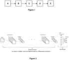
- Figure 1 shows the method steps according to the invention, namely:
- the CNNs on which the method according to the invention is based need to have a specific architecture and need prior training.
- the architecture of each CNN is established by Differential Evolution - abbreviated DE.
- DE is a heuristic optimization algorithm that mimics the natural biological evolution process. It generates a temporary individual having as starting point the differences within populations, followed by an evolutionary restructuration of the population.
- DE uses a population-based global search strategy.
- the mutation operation uses one-on-one competition.
- DE explores in parallel different solution and evaluates them using a fitness function.
- the CNNs built using these generated architectures are trained and their accuracies and losses over the validation dataset are recorded. Each CNNs' loss will represent the candidate solution's fitness value. We apply the mutation, recombination to each candidate solution and select the best performing one.
- Figure 2 shows an example of CNN architecture building, using Differential Evolution.
- the CNNs of each set of CNNs are hierarchized depending on the result of one or more statistical tests chosen from: ANOVA test, Tukey test, z-test, Anderson-Darling test, Jarque-Bera test, Levene test, Brown-Forsythe test.
- the Anderson-Darlin test uses distance-test based on the empirical cumulative distribution function and puts more weight on the observations in the tail of the distribution.
- the Jarque & Bera test is a Lagrange multiplier test. It studies the skewness and kurtosis of the sample data to see if they match a normal distribution.
- Levene and Brown Forsythe tests are used if the data sample is not governed by the Gaussian distribution.
- ANOVA Analysis of Variance compares the means of two or more independent data samples to see whether there are significant differences between them. It uses the F-distribution.
- Tukey's posthoc test uses the studentized range distribution. It compares all possible pairs of means. The test compares concurrently all the pairwise comparisons and identifies the difference between the groups means that are greater than the expected standard error.
- the power of a statistical test is the probability that the test's result obtained on a small set would remain the same if applied on a larger scale. Power analysis is used in this example to compute the number of independent computer runs needed to achieve 95% power.
- Figure 3 shows general case diagram, showing the decision-making process for determining the biometric plane / organ identification / anomaly identification.
- a plurality of CNNs namely CNN 1, CNN 2, ... CNN n, are grouped into a plurality of clusters, namely Cluster 1, ..., Cluster k.
- the CNNs of each set of CNNs are clustered using the Gaussian Mixture Modelling.
- Gaussian Mixture Modelling clustering technique uses a superposition of multiple Gaussian distributions.
- the mean, standard deviation, and covariance matrix must be estimated using the expectation maximization algorithm.
- FIG. 4 shows a particular case diagram corresponding to a decision-making process for determining a biometric plane.
- this particular case there are four CNNs clustered into two clusters - Cluster 1 and Cluster 2. After running the statistical tests mentioned above, to each CNN is assigned an accuracy and a weight. Each cluster is also assigned a weight.
- Each CNN determined a biometric plane (i.e. a "label"). In this case, CNN 1 determined label 6, CNN 2 determined label 1, CNN 3 determined label 3 and CNN 4 determined label 4. Then, a final weight for each CNN is calculated. In this particular case, because CNN 1 final weight value is the biggest of all four, the output decision will be the label indicated by CNN 1, namely "6", i.e. the biometric plane number six.
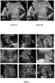
- Figure 5A shows an unprocessed ultrasound image frame of a fetal morphology scan during the second trimester of pregnancy.
- This image includes text information (the date, the name of the doctor, different parameters, etc.) and an artefact (the trapeze shape). The text and artefact are not necessary for carrying out the method according to the invention.
- Figure 5B shows the pre-processed image of figure 5A , wherein the text and the artefact have been removed (using software such as CV2 and Keras-OCR) and the deleted portions have been redrawn using the colour of the adjacent pixels.
- FIG. 6 shows nine biometric planes determined by means of the method according to the invention. Said biometric planes are (starting from top row, left to right):
- Figure 7 shows organs identification and anomalies detection, by means of the method according to the invention, in each of the biometrical planes determined in figure 6 .
- the organs are indicated to the user by a rectangle including the probability of correct identification of said organs.
- the status of each organ normal/abnormal will be displayed in a report when the fetal morphology is over.
- An example of such report is shown in figure 8 , wherein :
- the power analysis revealed that 50 independent computer runs are needed to achieve 95% statistical power with type I error 0.05.
- ACA SD CNN1 87.93 2.99 CNN2 73.97 2.23 CNN3 46.16 2.13 CNN4 93.22 1.60 CNN5 88.66 1.87 CNN6 48.75 2.72
- the standard deviation SD value needs to be as small as possible, preferably below 3. In our example, all CNNs satisfy this condition.
- CNN1 vs. CNN5, CNN4 vs. CNN5, and CNN5 vs. CNN6 have equal variances, (p-level > 0.05).
- the weights of the synergetic weighted voting systems are ⁇ 33.0, 33.5, 33.3 ⁇ .
- a quantitative comparison was used using a hold-out testing move.
- the example system performance was compared with the performances of standalone networks (ResNet50, EfficientV2S, MobileNetV3Large), hard voting, soft voting, and bagging strategy.
- the performance on hold-out test is shown in Table 6 Table 6
- SWVS System Weighted Voting System
- SWVS System Weighted Voting System
- a further CNN7 was used to detect the organs.
- Table 7 View plane Organ Precision Recall F1-score Rand Index 3 vessels plus bladder Bladder 0.891 0.882 0.884 0.850 Vessels 0.923 0.942 0.930 0.921 Abdominal circumference Aorta 0.660 0.656 0.759 0.722 Confluence 0.792 0.799 0.796 0.767 Rib 0.742 0.701 0.733 0.702 Spine 0.755 0.748 0.754 0.721 Stomach 0.861 0.872 0.870 0.821 Bladder Bladder 0.988 0.976 0.978 0.934 Echogenic Intestines 0.862 0.894 0.890 0.864 Gallbladder Gallbladder 0.981 0.975 0.987 0.964 Longitudinal kidney Kidney 0.832 0.876 0.871 0.852 Cord insertion sagittal Intestines 0.833 0.803 0.822 0.819 Wall and cord 0.912 0.906 0.909 0.899 Transab
- the precision values are very good (i.e. over 0.9) for the bladder, vessels, stomach, bladder, intestines, gallbladder, kidney, wall cord; is average (0.66) for the aorta; are good (i.e. between 0.7-0.9) for the rest of the organs.
- the above-mentioned "average” and “good” precision values can be increased to "very good” by further training of the CNNs.
Landscapes
- Engineering & Computer Science (AREA)
- Physics & Mathematics (AREA)
- General Physics & Mathematics (AREA)
- Theoretical Computer Science (AREA)
- Medical Informatics (AREA)
- General Health & Medical Sciences (AREA)
- Health & Medical Sciences (AREA)
- Nuclear Medicine, Radiotherapy & Molecular Imaging (AREA)
- Radiology & Medical Imaging (AREA)
- Quality & Reliability (AREA)
- Computer Vision & Pattern Recognition (AREA)
- Image Analysis (AREA)
- Ultra Sonic Daignosis Equipment (AREA)
- Measuring And Recording Apparatus For Diagnosis (AREA)
Abstract
A) receiving data input consisting in a 2D ultrasonographic motion video of a human body or body part,
B) converting the ultrasonographic motion video into a plurality of consecutive still image frames,
C) determining a plurality of biometric planes from the still image frames,
D) identifying at least one internal organ from each biometric plane,
E) detecting if said at least one internal organ is normal or susceptible to have anomalies,
F) indicating the detected anomalies to a user.
Description
- The invention refers to a method of detecting anomalies of the human internal organs, using Artificial Intelligence based on deep learning and statistical learning. The invention is particularly suited for improved detection of fetal congenital anomalies starting with the second trimester of pregnancy.
- Fetal morphology is the most important prenatal diagnostic tool for determining congenital anomalies. It allows the evaluation of the organs' structure and functionality.
Fetal morphology is not a routine procedure. The detection rate of congenital anomalies for a sonographer with more than 2000 examinations is only 52% accuracy, while for a sonographer with less than 2000 examinations is 32.5% accuracy. The pre- and post-natal discrepancy of the congenital anomalies detection by a manually performed examination was reported to be 29% in live births (7% having an impact on long-term care), and 23% in fetal autopsies. A correct set prenatal diagnosis improves neonatal and paediatric care as well as neurological and surgical outcome. Current approaches have limitations, the reported sensitivity being between 27.5% and 96%.
Errors due to human misinterpretation are a cause of concern. Doctors might not detect congenital anomalies and possible treatments may not be prepared in time or applied (termination of pregnancy, preparing the neonatology team, surgical team, etc.). Congenital anomalies are the most common cause of fetal death, infant mortality and morbidity during childhood. Worldwide, every year, 7.9 million children are born with congenital anomalies. Even if some congenital anomalies can be controlled and treated, 3.2 million of these children will be disabled for life.
An undiagnosed fetus with congenital abnormalities leads to the birth of a child whose life is threatened, who may have one or more disabilities. These things affect the family's daily routine and finances. The level of damage is difficult to assess, as it depends on the severity of the disease and the family's health insurance situation. In addition to the social and economic impact, the mental impact on the family must also be considered.
The lack of necessary sonography know-how, fatigue, time pressure, fetal involuntary movement, and different characteristics of the patients, such as obesity (in case of adult patients), might make it difficult, or in some cases, impossible for a sonographer to get a clear ultrasound image. - Several attempts to create an automatic system for interpreting fetal morphologies have been reported in literature.
Namburete et al. ("Fully automated alignment of 3D fetal brain ultrasound to a canonical reference space using multi-task learning ") proposed a fully connected neural network to segment the 3D fetal brain.
Torrents-Barrena, J. et al. ("Assessment of radiomics and deep learning for the segmentation of fetal and maternal anatomy in magnetic resonance imaging and ultrasound ") discloses that a segmentation of fetal lungs and brain was created using deep learning, sequential feature selection techniques, and support vector machines for magnetic resonance and ultrasound images.
All the above systems share the following disadvantages: - they use only one Artificial Intelligence method;
- they segment only parts of the fetus, not the fetus as a whole;
- they use 3D/4D or magnetic resonance images, which require special medical training, special conditions and are expensive.
- The technical problem solved by the present invention is the automatic detection of the biometric planes and the subsequent automatic detection of the organ's anomalies. This is achieved by using accessible 2D ultrasound scans and by combining artificial intelligence algorithms such as convolutional neural networks and statistical learning.
- The computer-implemented method of detecting human internal organs anomalies, according to the invention, comprises the following steps:
- A) receiving data input consisting in a 2D ultrasonographic motion video of a human body (in case of a fetus) or body part (in case of an adult),
- B) converting the ultrasonographic motion video into a plurality of consecutive still image frames,
- C) determining a plurality of biometric planes from the still image frames,
- D) identifying at least one internal organ from each biometric plane,
- E) detecting if said at least one internal organ is normal or susceptible to have anomalies,
- F) indicating the detected anomalies to a user.
- In a preferred embodiment, each of the steps C), D and E) is performed by means of a respective set of Convolutional Neural Networks - abbreviated as CNNs.
- In a preferred embodiment, each set of CNNs has an architecture established by Differential Evolution - abbreviated DE.
- In a preferred embodiment, the CNNs of each set of CNNs are hierarchized depending on the result of one or more statistical tests chosen from: ANOVA test, Tukey test, z-test, Anderson-Darling test, Jarque-Bera test, Levene test, Brown-Forsythe test.
- In a preferred embodiment, the CNNs of each set of CNNs are clustered using the Gaussian Mixture Modelling.
- In a preferred embodiment, each of the steps C), D) and E) comprises the sub-steps:
- x1) determining the weight of each cluster and of the best performing CNNs from said clusters,
- x2) performing a synergetic weighted voting by the CNNs in order to, depending on the respective step C), D) or E), determine the biometric planes, identify the internal organs or detect the organs anomalies, respectively.
- The invention further refers to a computer program product comprising instructions which, when the program is executed by a computer, cause the computer to carry out the method according to the invention.
- The term "computer" must be interpreted broadly, i.e. any machine comprising a processor and capable to be programmed to automatically carry out sequences of arithmetic or logical operations.
- The invention further refers to computer-readable medium having stored thereon the computer program product according to the invention.
- The method according the invention has the main advantage that it significantly improves the reliability of organ anomaly detection and it serves as a valuable support for insufficiently experienced or tired doctors. Further advantages are:
- provides the automatic detection of the biometric planes and the subsequent automatic detection of the organ's anomalies;
- provides guidance for the ultrasound probe in view of acquiring an optimal ultrasonographic motion video;
- provides patients with high quality medical care, at significantly lower costs in respect to 3D/4D or MRI;
- helps the sonographers to establish a correct diagnosis, by bringing the technology to every small town, even in poor countries, and to increase confidence in the results of the morphological examination;
- dramatically reduces the risks of misinterpretation due to fatigue, time pressure, fetal involuntary movement, contractions or different patient characteristics.
- The method according to the invention is based on Deep Learning - abbreviated DL and Convolutional Neural Networks - CNNs.
- DL is the subset of machine learning methods based on artificial neural networks with representation learning.
- A CNN is a special case of artificial neural networks, namely a regularized type of feed-forward neural network that learns feature engineering by itself via filters (or kernel) optimization.
- The difference between CNNs and classical artificial neural networks consists in the number of layers. CNNs have many layers used to process data. The architecture of a CNN consists of three types of layers: convolutional layer, pooling layer, and fully connected layer. The convolutional layer uses filters that perform convolution operations by scanning the input and producing a feature map. The pooling layer is applied after a convolutional layer and down samples the feature map producing spatial invariance. The fully connected layer works on a flattened input, where each input is connected to all neurons.
- The CNNs have hyperparameters such as: the size of the filter, the stride (the number of pixels by which the window moves after each operation), and zero-padding. CNNs are very effective at processing images, videos and sounds.
- CNNs are trained using Supervised Learning. Supervised Learning is a training method wherein the network is presented with previously human classified data.
- The invention will be described in connection with the
figures 1-8 , representing: -
Figure 1 : method steps according to the invention -
Figure 2 : CNN architecture built using Differential Evolution -
Figure 3 : general case diagram, showing the decision-making process for determining the biometric plane(s)/organ identification/anomaly identification -
Figure 4 : particular case diagram, showing the decision-making process for determining the biometric plane -
Figure 5A : an unprocessed ultrasound image frame (2D image) -
Figure 5B : the pre-processed image offigure 5A -
Figure 6 : different determined biometric planes -
Figure 7 : organs identification -
Figure 8 : example of organs status report -
Figure 1 shows the method steps according to the invention, namely: - step A: receiving data input consisting in a 2D ultrasonographic motion video of a human body or body part,
- step B: converting the ultrasonographic motion video into a plurality of consecutive still image frames,
- step C: determining a plurality of biometric planes from the still image frames,
- step D: identification of at least one internal organ from each biometric plane,
- step E: detecting if said at least one internal organ is normal or susceptible to have anomalies,
- step F: indicating the detected anomalies to a user.
- The CNNs on which the method according to the invention is based need to have a specific architecture and need prior training.
The architecture of each CNN is established by Differential Evolution - abbreviated DE. DE is a heuristic optimization algorithm that mimics the natural biological evolution process. It generates a temporary individual having as starting point the differences within populations, followed by an evolutionary restructuration of the population. DE uses a population-based global search strategy. The mutation operation uses one-on-one competition. DE explores in parallel different solution and evaluates them using a fitness function.
An initial population of q individuals that represent the candidate architectures are randomly generated, Xi = {λi, nHi, fwi, fhi, Cpi, Fi}, where λ represents the number of convolutional hidden layer, nH the number of neurons in the hidden layers, fw, fh the filter's width and height, Cp the recombination parameter, and F the variation factor. The CNNs built using these generated architectures are trained and their accuracies and losses over the validation dataset are recorded. Each CNNs' loss will represent the candidate solution's fitness value. We apply the mutation, recombination to each candidate solution and select the best performing one. -
Figure 2 shows an example of CNN architecture building, using Differential Evolution. - The CNNs of each set of CNNs are hierarchized depending on the result of one or more statistical tests chosen from: ANOVA test, Tukey test, z-test, Anderson-Darling test, Jarque-Bera test, Levene test, Brown-Forsythe test.
- The Anderson-Darlin test uses distance-test based on the empirical cumulative distribution function and puts more weight on the observations in the tail of the distribution.
- The Jarque & Bera test is a Lagrange multiplier test. It studies the skewness and kurtosis of the sample data to see if they match a normal distribution.
- Levene and Brown Forsythe tests are used if the data sample is not governed by the Gaussian distribution.
- The above statistical tests that verify the normality and variance are used in this example on the sample that holds the performances of the CNNs recorded while running them on the validation set for a certain number of times, number determined by the power analysis test.
- ANOVA (Analysis of Variance) compares the means of two or more independent data samples to see whether there are significant differences between them. It uses the F-distribution.
- Tukey's posthoc test uses the studentized range distribution. It compares all possible pairs of means. The test compares concurrently all the pairwise comparisons and identifies the difference between the groups means that are greater than the expected standard error.
- The power of a statistical test is the probability that the test's result obtained on a small set would remain the same if applied on a larger scale. Power analysis is used in this example to compute the number of independent computer runs needed to achieve 95% power.
-
Figure 3 shows general case diagram, showing the decision-making process for determining the biometric plane / organ identification / anomaly identification.
A plurality of CNNs, namelyCNN 1,CNN 2, ... CNN n, are grouped into a plurality of clusters, namelyCluster 1, ..., Cluster k. - The CNNs of each set of CNNs are clustered using the Gaussian Mixture Modelling. Gaussian Mixture Modelling clustering technique uses a superposition of multiple Gaussian distributions. For a d-variate dimension X the probability distribution function uses µ, σ, and Σ (the mean, standard deviation, and covariance matrix):

For k clusters, the mean, standard deviation, and covariance matrix must be estimated using the expectation maximization algorithm. - From each cluster the best performing CNN will be kept and will be assigned a weight directly proportional to its performance normalized with the performance of the cluster it belongs to (a convex combination of average performances)


The output decision regarding the establishment of the biometric plane / organ identification / anomaly identification will be taken collaboratively through a synergetic weighted voting system. -
Figure 4 shows a particular case diagram corresponding to a decision-making process for determining a biometric plane.
In this particular case there are four CNNs clustered into two clusters -Cluster 1 andCluster 2.
After running the statistical tests mentioned above, to each CNN is assigned an accuracy and a weight. Each cluster is also assigned a weight. Each CNN determined a biometric plane (i.e. a "label"). In this case,CNN 1determined label 6,CNN 2determined label 1,CNN 3determined label 3 andCNN 4determined label 4. Then, a final weight for each CNN is calculated.
In this particular case, becauseCNN 1 final weight value is the biggest of all four, the output decision will be the label indicated byCNN 1, namely "6", i.e. the biometric plane number six. -
Figure 5A shows an unprocessed ultrasound image frame of a fetal morphology scan during the second trimester of pregnancy. This image includes text information (the date, the name of the doctor, different parameters, etc.) and an artefact (the trapeze shape). The text and artefact are not necessary for carrying out the method according to the invention. -
Figure 5B shows the pre-processed image offigure 5A , wherein the text and the artefact have been removed (using software such as CV2 and Keras-OCR) and the deleted portions have been redrawn using the colour of the adjacent pixels. -
Figure 6 shows nine biometric planes determined by means of the method according to the invention.
Said biometric planes are (starting from top row, left to right): - vessels plus bladder plane
- abdominal circumference plane
- anteroposterior kidney plane; bladder plane
- echogenic plane
- gallbladder plane
- longitudinal kidney plane
- cord insertion sagittal plane
- transabdominal cord insertion plane
-
Figure 7 shows organs identification and anomalies detection, by means of the method according to the invention, in each of the biometrical planes determined infigure 6 . The organs are indicated to the user by a rectangle including the probability of correct identification of said organs. The status of each organ normal/abnormal will be displayed in a report when the fetal morphology is over. An example of such report is shown infigure 8 , wherein : - the "X" in a red circle means that the respective organs have an anomaly (or anomalies) or the organ was not identified (or was missing),
- the tik in a green circle means that the respective organs were identified and are normal.
- It is obvious that, although the method according to the invention provides a reliable automatic organ anomalies detection, the final medical decision and diagnostic belongs to the doctor.
- An example of carrying out the method according the invention, applied to the second trimester fetal morphology ultrasound scans, is described in the following paragraphs.
- Six CNNs are used in this example. The numbers of trainable and non trainable parameters for each CNN are shown in Table 1.
Table 1 Trainable parameters Non-trainable parameters Total parameters CNN1 2, 104, 326 23, 587, 712 25, 692, 038 CNN2 1, 55, 750 7, 37, 504 8, 93, 254 CNN3 2, 689, 414 21, 802, 784 24, 492, 198 CNN4 1, 317, 894 20, 331, 360 21, 649, 254 CNN5 990, 214 2, 996, 352 3, 986, 566 CNN6 2, 104, 326 20,861,480 22, 965, 806 - In this example the power analysis revealed that 50 independent computer runs are needed to achieve 95% statistical power with type I error 0.05. In this example we have used the mean performance (accuracy) and corresponding standard deviation. The accuracy is quantified as

- The average accuracy (ACA) and standard deviation (SD) of the six CNNs built using Differential Evolution is shown in Table 2.
Table 2 ACA SD CNN1 87.93 2.99 CNN2 73.97 2.23 CNN3 46.16 2.13 CNN4 93.22 1.60 CNN5 88.66 1.87 CNN6 48.75 2.72 - The standard deviation SD value needs to be as small as possible, preferably below 3. In our example, all CNNs satisfy this condition.
- The Normality tests results obtained on this example are shown in table 3.
Table 3 Anderson-Darling Jarque Bera AD p-level JB p-level CNN1 15.64 0.000 16.09 0.000 CNN2 16.26 0.000 16.51 0.000 CNN3 15.50 0.000 16.32 0.000 CNN4 16.62 0.000 16.55 0.000 CNN5 16.43 0.000 16.50 0.000 CNN6 15.22 0.000 16.25 0.000 

- In this example, the equality of variances showed that: CNN1 vs. CNN5, CNN4 vs. CNN5, and CNN5 vs. CNN6 have equal variances, (p-level > 0.05).
- In this example, we made use of the fact that each one of the CNNs has been run independently 50 times, which means that we have the same number of observations in our samples, hence we can presume that the variances are equal and proceed with applying one-way ANOVA.
- The one-way ANOVA technique test results are shown in Table 4.
Table 4 SS df MS F p-level 58615 5 11723 2197.4 0.000 - SS = sum of squares
- df = degrees of freedom
- MS = mean squares
- F = ratio
- In this example, the Tukey's posthoc test revealed that there are statistically significant differences between all CNNs, except CNN1, CNN4, and CNN5.
Therefore, CNN1, CNN4 and CNN5 were all clustered in the same cluster.
The cluster system is shown in Table 5.Table 5 No.of clusters p-level Members of each cluster Mean Performance %/ SD 3 0.00003 C1 ={CNN1, CNN4, CNN5} 89.93 / 0.6 C2 ={CNN2} 73.97 / 2.23 C3 ={CNN3, CNN6} 50.75 / 0.29 - In this example the weights of the synergetic weighted voting systems are {33.0, 33.5, 33.3}.
A quantitative comparison was used using a hold-out testing move. The example system performance was compared with the performances of standalone networks (ResNet50, EfficientV2S, MobileNetV3Large), hard voting, soft voting, and bagging strategy.
The performance on hold-out test is shown in Table 6Table 6 SWVS 89.22 ResNet50 77.01 EfficientV2S 78.98 MobileNetV3Large 77.95 Hard voting 77.66 Soft voting 80.12 Bagging strategy 80.76 - A further CNN7 was used to detect the organs. In this example we have used as performance measures: Precision, Recall, F1-score, Jaccard Index, and Probabilistic Rand index.



- The detailed classification per organ using real time ultrasound motion video are shown in Table 7.
Table 7 View plane Organ Precision Recall F1- score Rand Index 3 vessels plus bladder Bladder 0.891 0.882 0.884 0.850 Vessels 0.923 0.942 0.930 0.921 Abdominal circumference Aorta 0.660 0.656 0.759 0.722 Confluence 0.792 0.799 0.796 0.767 Rib 0.742 0.701 0.733 0.702 Spine 0.755 0.748 0.754 0.721 Stomach 0.861 0.872 0.870 0.821 Bladder Bladder 0.988 0.976 0.978 0.934 Echogenic Intestines 0.862 0.894 0.890 0.864 Gallbladder Gallbladder 0.981 0.975 0.987 0.964 Longitudinal kidney Kidney 0.832 0.876 0.871 0.852 Cord insertion sagittal Intestines 0.833 0.803 0.822 0.819 Wall and cord 0.912 0.906 0.909 0.899 Transabdominal cord insertion Wall and cord 0.763 0.733 0.744 0.745 - As seen in table 7, the precision values are very good (i.e. over 0.9) for the bladder, vessels, stomach, bladder, intestines, gallbladder, kidney, wall cord; is average (0.66) for the aorta; are good (i.e. between 0.7-0.9) for the rest of the organs. The above-mentioned "average" and "good" precision values can be increased to "very good" by further training of the CNNs.
-
- 1. Namburete, A. et al., Fully automated alignment of 3D fetal brain ultrasound to a canonical reference space using multi-task learning. Med Image Anal. 46, 1-14, 2018.
- 2. Torrents-Barrena, J. et al. Assessment of radiomics and deep learning for the segmenation of fetal and maternal anatomy in magnetic resonance imaging and ultrasound. Acad. Radiol., S1076-6332(19)30575-6, 2019.
Claims (7)
- A computer-implemented method of detecting human internal organs anomalies, comprising the steps:A) receiving data input consisting in a 2D ultrasonographic motion video of a human body or body part,B) converting the ultrasonographic motion video into a plurality of consecutive still image frames,C) determining a plurality of biometric planes from the still image frames,D) identifying at least one internal organ from each biometric plane,E) detecting if said at least one internal organ is normal or susceptible to have anomalies,F) indicating the detected anomalies to a user.
- Computer-implemented method according to claim 1, wherein each of the steps C), D and E) is performed by means of a respective set of Convolutional Neural Networks (CNNs).
- Computer-implemented method according to claim 2, wherein each set of CNNs has an architecture established by Differential Evolution (DE).
- Computer-implemented method according to claim 3, wherein:- the CNNs of each set of CNNs are hierarchized depending on the result of one or more statistical tests chosen from: ANOVA test, Tukey test, z-test, Anderson-Darling test, Jarque-Bera test, Levene test, Brown-Forsythe test.- the CNNs of each set of CNNs are clustered using the Gaussian Mixture Modelling.
- Computer-implemented method according to any of the claims 2-4, wherein each of the steps C), D) and E) comprises the sub-steps:x1) determining the weight of each cluster and of the best performing CNNs from said clustersx2) performing a synergetic weighted voting by the CNNs in order to, depending on the respective step C), D) or E), determine the biometric planes, identify the internal organs or detect the organs anomalies, respectively.
- A computer program product comprising instructions which, when the program is executed by a computer, cause the computer to carry out the method of any of the claims 1-5.
- A computer-readable medium having stored thereon the computer program product of claim 6.
Applications Claiming Priority (1)
| Application Number | Priority Date | Filing Date | Title |
|---|---|---|---|
| ROA202300431A RO138166A0 (en) | 2023-08-07 | 2023-08-07 | System for shape recognition and anomaly detection in fetal morphology using artificial intelligence |
Publications (3)
| Publication Number | Publication Date |
|---|---|
| EP4506890A1 true EP4506890A1 (en) | 2025-02-12 |
| EP4506890C0 EP4506890C0 (en) | 2025-10-22 |
| EP4506890B1 EP4506890B1 (en) | 2025-10-22 |
Family
ID=91275034
Family Applications (1)
| Application Number | Title | Priority Date | Filing Date |
|---|---|---|---|
| EP24465532.0A Active EP4506890B1 (en) | 2023-08-07 | 2024-06-13 | Method of detecting human internal organs anomalies |
Country Status (2)
| Country | Link |
|---|---|
| EP (1) | EP4506890B1 (en) |
| RO (1) | RO138166A0 (en) |
Citations (1)
| Publication number | Priority date | Publication date | Assignee | Title |
|---|---|---|---|---|
| US20230038364A1 (en) * | 2020-01-16 | 2023-02-09 | Koninklijke Philips N.V. | Method and system for automatically detecting anatomical structures in a medical image |
-
2023
- 2023-08-07 RO ROA202300431A patent/RO138166A0/en unknown
-
2024
- 2024-06-13 EP EP24465532.0A patent/EP4506890B1/en active Active
Patent Citations (1)
| Publication number | Priority date | Publication date | Assignee | Title |
|---|---|---|---|---|
| US20230038364A1 (en) * | 2020-01-16 | 2023-02-09 | Koninklijke Philips N.V. | Method and system for automatically detecting anatomical structures in a medical image |
Non-Patent Citations (5)
| Title |
|---|
| "Deep Learning", 18 November 2016, article GOODFELLOW IAN ET AL: "Bagging and Other Ensemble Methods", pages: 256 - 256, XP093217320 * |
| NAMBURETE ET AL., FULLY AUTOMATED ALIGNMENT OF 3D FETAL BRAIN ULTRASOUND TO A CANONICAL REFERENCE SPACE USING MULTI-TASK LEARNING |
| NAMBURETE, A ET AL.: "Fully automated alignment of 3D fetal brain ultrasound to a canonical reference space using multi-task learning", MED IMAGE ANAL, vol. 46, 2018, pages 1 - 14, XP085381905, DOI: 10.1016/j.media.2018.02.006 |
| TORRENTS-BARRENA, J ET AL.: "Assessment of radiomics and deep learning for the segmenation of fetal and maternal anatomy in magnetic resonance imaging and ultrasound", ACAD. RADIOL., vol. S1076-6332, no. 19, 2019, pages 30575 - 6 |
| TORRENTS-BARRENA, J. ET AL., ASSESSMENT OF RADIOMICS AND DEEP LEARNING FOR THE SEGMENTATION OF FETAL AND MATERNAL ANATOMY IN MAGNETIC RESONANCE IMAGING AND ULTRASOUND |
Also Published As
| Publication number | Publication date |
|---|---|
| EP4506890C0 (en) | 2025-10-22 |
| EP4506890B1 (en) | 2025-10-22 |
| RO138166A0 (en) | 2024-05-30 |
Similar Documents
| Publication | Publication Date | Title |
|---|---|---|
| Krishna et al. | Standard fetal ultrasound plane classification based on stacked ensemble of deep learning models | |
| EP3964136B1 (en) | System and method for guiding a user in ultrasound assessment of a fetal organ | |
| Zuo et al. | Deep learning-based eye-tracking analysis for diagnosis of Alzheimer's disease using 3D comprehensive visual stimuli | |
| Pasyar et al. | Hybrid classification of diffuse liver diseases in ultrasound images using deep convolutional neural networks | |
| WO2002039895A1 (en) | A method and a system for combining automated psychiatric profiling from combined input images of brain scans with observed expert and automated interpreter using a neural network | |
| Samant et al. | Evaluation of artificial neural networks in prediction of essential hypertension | |
| CN114038564A (en) | Noninvasive risk prediction method for diabetes | |
| Cao et al. | Multi-modality fusion learning for the automatic diagnosis of optic neuropathy | |
| Sengun et al. | Automatic liver segmentation from CT images using deep learning algorithms: a comparative study | |
| Ivanescu et al. | Evolutionary computation paradigm to determine deep neural networks architectures | |
| Zeng et al. | Automated detection of diabetic retinopathy using a binocular siamese-like convolutional network | |
| Prodhan et al. | Combination of the features of pre-trained Xception and VGG16 models to identify childhood pneumonia from chest X-ray images | |
| EP4506890A1 (en) | Method of detecting human internal organs anomalies | |
| EP4156021A1 (en) | A method and system for annotation of medical images | |
| Vetriselvi et al. | Deep Learning Based Medical Image Segmentation in Prenatal Prediction of Neurodevelopmental Disorders | |
| Jasmine et al. | MNR2NeXt-50: Segmentation and quantification of epicardial fat from cardiac CT images using transfer learning with an optimized ensemble model | |
| Tsai et al. | Fusion of diffusion weighted MRI and clinical data for predicting functional outcome after acute ischemic stroke with deep contrastive learning | |
| Nyaupane | Pneumonia of chest x-ray images detection using vgg architectures | |
| Ingle et al. | Review on skin cancer detection using AI | |
| Lins et al. | Selection and classification of COVID-19 CT images using artificial intelligence: A case study in a Brazilian university hospital | |
| Morita et al. | Pediatric brain CT image segmentation methods for effective age prediction models | |
| Wang et al. | Diagnosis of heart disease via CNNs | |
| Thongseesang et al. | Revolutionizing Mild Traumatic Brain Injury Detection: An AI-Based Automated Framework | |
| Singh et al. | Deep Learning Algorithms For Enhancing Image Processing In Healthcare Applications | |
| Agarwal et al. | Identification of heart failure from cine-MRI images using pattern-based features |
Legal Events
| Date | Code | Title | Description |
|---|---|---|---|
| PUAI | Public reference made under article 153(3) epc to a published international application that has entered the european phase |
Free format text: ORIGINAL CODE: 0009012 |
|
| STAA | Information on the status of an ep patent application or granted ep patent |
Free format text: STATUS: THE APPLICATION HAS BEEN PUBLISHED |
|
| AK | Designated contracting states |
Kind code of ref document: A1 Designated state(s): AL AT BE BG CH CY CZ DE DK EE ES FI FR GB GR HR HU IE IS IT LI LT LU LV MC ME MK MT NL NO PL PT RO RS SE SI SK SM TR |
|
| STAA | Information on the status of an ep patent application or granted ep patent |
Free format text: STATUS: REQUEST FOR EXAMINATION WAS MADE |
|
| 17P | Request for examination filed |
Effective date: 20250221 |
|
| GRAP | Despatch of communication of intention to grant a patent |
Free format text: ORIGINAL CODE: EPIDOSNIGR1 |
|
| STAA | Information on the status of an ep patent application or granted ep patent |
Free format text: STATUS: GRANT OF PATENT IS INTENDED |
|
| GRAS | Grant fee paid |
Free format text: ORIGINAL CODE: EPIDOSNIGR3 |
|
| INTG | Intention to grant announced |
Effective date: 20250728 |
|
| GRAA | (expected) grant |
Free format text: ORIGINAL CODE: 0009210 |
|
| STAA | Information on the status of an ep patent application or granted ep patent |
Free format text: STATUS: THE PATENT HAS BEEN GRANTED |
|
| AK | Designated contracting states |
Kind code of ref document: B1 Designated state(s): AL AT BE BG CH CY CZ DE DK EE ES FI FR GB GR HR HU IE IS IT LI LT LU LV MC ME MK MT NL NO PL PT RO RS SE SI SK SM TR |
|
| REG | Reference to a national code |
Ref country code: CH Ref legal event code: F10 Free format text: ST27 STATUS EVENT CODE: U-0-0-F10-F00 (AS PROVIDED BY THE NATIONAL OFFICE) Effective date: 20251022 Ref country code: GB Ref legal event code: FG4D |
|
| REG | Reference to a national code |
Ref country code: DE Ref legal event code: R096 Ref document number: 602024001019 Country of ref document: DE |
|
| REG | Reference to a national code |
Ref country code: IE Ref legal event code: FG4D |
|
| U01 | Request for unitary effect filed |
Effective date: 20251030 |
|
| U07 | Unitary effect registered |
Designated state(s): AT BE BG DE DK EE FI FR IT LT LU LV MT NL PT RO SE SI Effective date: 20251105 |
|
| PG25 | Lapsed in a contracting state [announced via postgrant information from national office to epo] |
Ref country code: ES Free format text: LAPSE BECAUSE OF FAILURE TO SUBMIT A TRANSLATION OF THE DESCRIPTION OR TO PAY THE FEE WITHIN THE PRESCRIBED TIME-LIMIT Effective date: 20251022 |
|
| PG25 | Lapsed in a contracting state [announced via postgrant information from national office to epo] |
Ref country code: NO Free format text: LAPSE BECAUSE OF FAILURE TO SUBMIT A TRANSLATION OF THE DESCRIPTION OR TO PAY THE FEE WITHIN THE PRESCRIBED TIME-LIMIT Effective date: 20260122 |
|
| PG25 | Lapsed in a contracting state [announced via postgrant information from national office to epo] |
Ref country code: HR Free format text: LAPSE BECAUSE OF FAILURE TO SUBMIT A TRANSLATION OF THE DESCRIPTION OR TO PAY THE FEE WITHIN THE PRESCRIBED TIME-LIMIT Effective date: 20251022 |
|
| PG25 | Lapsed in a contracting state [announced via postgrant information from national office to epo] |
Ref country code: RS Free format text: LAPSE BECAUSE OF FAILURE TO SUBMIT A TRANSLATION OF THE DESCRIPTION OR TO PAY THE FEE WITHIN THE PRESCRIBED TIME-LIMIT Effective date: 20260122 |
|
| PG25 | Lapsed in a contracting state [announced via postgrant information from national office to epo] |
Ref country code: IS Free format text: LAPSE BECAUSE OF FAILURE TO SUBMIT A TRANSLATION OF THE DESCRIPTION OR TO PAY THE FEE WITHIN THE PRESCRIBED TIME-LIMIT Effective date: 20260222 |
|
| PG25 | Lapsed in a contracting state [announced via postgrant information from national office to epo] |
Ref country code: PL Free format text: LAPSE BECAUSE OF FAILURE TO SUBMIT A TRANSLATION OF THE DESCRIPTION OR TO PAY THE FEE WITHIN THE PRESCRIBED TIME-LIMIT Effective date: 20251022 |