EP4498088A1 - Autoantibodies against nav1.5 channel as biomarkers for the diagnosis of brugada syndrome - Google Patents

Autoantibodies against nav1.5 channel as biomarkers for the diagnosis of brugada syndrome Download PDF

Info

Publication number
EP4498088A1
EP4498088A1 EP23187367.0A EP23187367A EP4498088A1 EP 4498088 A1 EP4498088 A1 EP 4498088A1 EP 23187367 A EP23187367 A EP 23187367A EP 4498088 A1 EP4498088 A1 EP 4498088A1
Authority
EP
European Patent Office
Prior art keywords
autoantibodies
seq
channel
extracellular loops
nav
Prior art date
Legal status (The legal status is an assumption and is not a legal conclusion. Google has not performed a legal analysis and makes no representation as to the accuracy of the status listed.)
Withdrawn
Application number
EP23187367.0A
Other languages
German (de)
French (fr)
Inventor
Luigi Anastasia
Adriana Tarantino
Andrea Ghiroldi
Giuseppe CICONTE
Carlo Pappone
Current Assignee (The listed assignees may be inaccurate. Google has not performed a legal analysis and makes no representation or warranty as to the accuracy of the list.)
Cardiomix SRL
Original Assignee
Cardiomix SRL
Priority date (The priority date is an assumption and is not a legal conclusion. Google has not performed a legal analysis and makes no representation as to the accuracy of the date listed.)
Filing date
Publication date
Application filed by Cardiomix SRL filed Critical Cardiomix SRL
Priority to EP23187367.0A priority Critical patent/EP4498088A1/en
Priority to PCT/EP2024/071047 priority patent/WO2025021888A1/en
Priority to CN202480048884.XA priority patent/CN121941925A/en
Publication of EP4498088A1 publication Critical patent/EP4498088A1/en
Withdrawn legal-status Critical Current

Links

Images

Classifications

    • GPHYSICS
    • G01MEASURING; TESTING
    • G01NINVESTIGATING OR ANALYSING MATERIALS BY DETERMINING THEIR CHEMICAL OR PHYSICAL PROPERTIES
    • G01N33/00Investigating or analysing materials by specific methods not covered by groups G01N1/00 - G01N31/00
    • G01N33/48Biological material, e.g. blood, urine; Haemocytometers
    • G01N33/50Chemical analysis of biological material, e.g. blood, urine; Testing involving biospecific ligand binding methods; Immunological testing
    • G01N33/53Immunoassay; Biospecific binding assay; Materials therefor
    • G01N33/564Immunoassay; Biospecific binding assay; Materials therefor for pre-existing immune complex or autoimmune disease, i.e. systemic lupus erythematosus, rheumatoid arthritis, multiple sclerosis, rheumatoid factors or complement components C1-C9
    • CCHEMISTRY; METALLURGY
    • C07ORGANIC CHEMISTRY
    • C07KPEPTIDES
    • C07K16/00Immunoglobulins [IG], e.g. monoclonal or polyclonal antibodies
    • C07K16/18Immunoglobulins [IG], e.g. monoclonal or polyclonal antibodies against material from animals or humans
    • C07K16/28Immunoglobulins [IG], e.g. monoclonal or polyclonal antibodies against material from animals or humans against receptors, cell surface antigens or cell surface determinants
    • GPHYSICS
    • G01MEASURING; TESTING
    • G01NINVESTIGATING OR ANALYSING MATERIALS BY DETERMINING THEIR CHEMICAL OR PHYSICAL PROPERTIES
    • G01N33/00Investigating or analysing materials by specific methods not covered by groups G01N1/00 - G01N31/00
    • G01N33/48Biological material, e.g. blood, urine; Haemocytometers
    • G01N33/50Chemical analysis of biological material, e.g. blood, urine; Testing involving biospecific ligand binding methods; Immunological testing
    • G01N33/68Chemical analysis of biological material, e.g. blood, urine; Testing involving biospecific ligand binding methods; Immunological testing involving proteins, peptides or amino acids
    • G01N33/6872Intracellular protein regulatory factors and their receptors, e.g. including ion channels
    • GPHYSICS
    • G01MEASURING; TESTING
    • G01NINVESTIGATING OR ANALYSING MATERIALS BY DETERMINING THEIR CHEMICAL OR PHYSICAL PROPERTIES
    • G01N2800/00Detection or diagnosis of diseases
    • G01N2800/32Cardiovascular disorders
    • G01N2800/326Arrhythmias, e.g. ventricular fibrillation, tachycardia, atrioventricular block, torsade de pointes
    • GPHYSICS
    • G01MEASURING; TESTING
    • G01NINVESTIGATING OR ANALYSING MATERIALS BY DETERMINING THEIR CHEMICAL OR PHYSICAL PROPERTIES
    • G01N2800/00Detection or diagnosis of diseases
    • G01N2800/52Predicting or monitoring the response to treatment, e.g. for selection of therapy based on assay results in personalised medicine; Prognosis

Definitions

  • the present invention relates to autoantibodies against NaV1.5 channel, their use as biomarker for the diagnosis of Brugada syndrome in human beings and relative methods of detection and therapeutic approach to target this new discovered form of autoimmunity.
  • the Brugada Syndrome is an inherited arrhythmogenic disease that poses an increased risk of sudden cardiac death (SCD), accounting for 5-40% of SCD cases in individuals under 40 years of age [1].
  • SCD sudden cardiac death
  • the syndrome typically manifests with cardiac arrest or syncope, occurring in the third and fourth decade of life [2] [3] however, the majority of patients are asymptomatic with structurally normal heart, and they are usually diagnosed incidentally.
  • ECG electrocardiogram
  • SCN5A which encodes the alpha subunit of the voltage-gated sodium channel NaV1.5, is the primary causative gene for Brugada syndrome [8].
  • SCN5A which encodes the alpha subunit of the voltage-gated sodium channel NaV1.5
  • SCN5A is the primary causative gene for Brugada syndrome [8].
  • the genetic etiology remains unclear in approximately 70-75% of cases, and a single mutation does not fully account for the Brugada syndrome phenotype [9], rendering genetic testing inadequate as a standalone diagnostic tool for the disease.
  • Brugada syndrome is not solely driven by genetic factors, as evidenced by histological changes in the right ventricular myocardium of type 1 Brugada patients [10, 11] and the presence of inflammatory infiltrates and fibrosis in the right ventricular outflow tract of Brugada patients has been reported [12, 13]. Furthermore, emerging evidence suggests a potential role of autoimmunity in Brugada syndrome, which has long been overlooked in cardiac arrhythmias. The discovery of autoantibodies associated with arrhythmogenic right ventricular cardiomyopathy (ARVC) has shed light on the autoimmune implications in Brugada syndrome [14, 15].
  • ARVC arrhythmogenic right ventricular cardiomyopathy
  • Autoantibodies can interfere with ion channels and receptors involved in cardiac electrophysiology, triggering arrhythmias. For instance, autoantibodies targeting ⁇ 1-adrenergic receptors have been reported in various cardiac diseases, including ischemic cardiomyopathy and Chagas' disease [16, 17]. Additionally, IgG antibodies against the voltage-gated KCNQ1 K+ channel (Kv7.1 or KvLQT1) have been detected in patients with dilated cardiomyopathy, resulting in a shortened QTc interval [18]. Autoantibodies targeting the cardiac voltage-gated Na+ channel have also been found in patients with idiopathic high-degree AV block, leading to reduced sodium current (INa) density in rat cardiomyocytes [19].
  • the inventors have developed an in-vitro model of NaV1.5 overexpressing channels with improved purification capabilities and the ability to test autoantibody binding under denaturing and native conditions, mimicking the physiological environment.
  • Previous studies have shown that autoantibodies against NaV1.5 can affect sodium ion currents in vivo in rats [20]. This mechanism aligns with the activity of autoantibodies against channel or receptor proteins found in other diseases, such as NMDAR encephalitis, where autoantibodies promote receptor internalization and induce electrophysiological changes in neurons [21-23].
  • the autoantibodies for use as biomarkers for the diagnosis of Brugada Syndrome of the invention are directed against a binding site of the extracellular loops of NaV 1.5 channel, wherein said binding site may be selected from the group consisting of:
  • the invention further relates to an in vitro method for detecting the presence or detecting the amount of at least one of the autoantibodies directed against the extracellular loops of NaV 1.5 channel of the invention in a biological sample of a human being suspected of having Brugada syndrome.
  • said human being may be either asymptomatic or at high risk for Brugada Syndrome due to family history, previous events of heart atrial and/or ventricular fibrillation, diabetes or obesity.
  • the in vitro method comprises the following steps:
  • said antigens are fragments of at least 7 amino acids from the binding sites of extracellular loops of NaV1.5 channel.
  • said binding sites of extracellular loops of NaV1.5 channel are selected between SEQ ID NO:2-SEQ ID NO:7.
  • said antigens are labelled.
  • the biological sample is selected from the group consisting of plasma, PBMCs, whole blood, serum and peripheral blood, or a combination thereof.
  • the biological sample is plasma and/or PBMCs.
  • the detection of the binding of the antigens to the autoantibodies is performed by ELISA or Western blotting.
  • steroids such as Prednisone, Methylprednisolone, and Dexamethasone
  • medicament like Colchicine and Hydroxychloroquine (Plaquenil)
  • Paquenil may be used for the treatment of Brugada syndrome.
  • the present invention further contemplates an autoimmune therapeutic agent specifically targeting to the autoantibodies directed to the extracellular loops of NaV 1.5 channel for use in the treatment of Brugada syndrome.
  • the autoimmune therapeutic agent is characterized in that it comprises one or more antigenic fragments of the extracellular loops of the Nav 1.5 protein.
  • the autoimmune therapeutic agent comprises sequences of at least about 10 amino acids from the extracellular loops of the Nav 1.5 protein, said extracellular loops being selected from the group of SEQ ID NO:2-SEQ ID NO:7.
  • Another possible therapeutic approach contemplates targeting B cells thus disrupting the production of autoantibodies that can trigger inflammation mediated by immune complexes.
  • Potential approved drugs such as Rituximab, Ocrelizumab, Ofatumumab, and Inebilizumab which have already demonstrated efficacy in treating various immune and autoimmune diseases B-cells mediated may be useful for the therapeutic treatment.
  • nanobody-based multivalent or multispecific to target Ab-Nav1.5 in order to restore regular activity of NaV1.5 channels.
  • these nanobodies will serve as valuable tools for in vivo imaging of the cardiac substrate affected by Brugada syndrome, allowing for effective monitoring.
  • the invention further contemplates the use of the antagonist or the autoimmune therapeutic agent in combination with other therapy for Brugada syndrome in order to achieve a more comprehensive and effective treatment.
  • the present invention is further directed to a kit for detecting autoantibodies against extracellular loops of Nav 1.5 channel in a biological sample, the kit comprising one or more antigens that specifically binds to the autoantibodies against Nav 1.5.
  • said antigens comprises one or more antigenic fragments of the extracellular loops of the Nav 1.5 protein, said extracellular loops being selected from the group of SEQ ID NO:2-SEQ ID NO:7.
  • said antigens are labelled or attached to a solid support.
  • said antibodies are labelled with fluorescents.
  • Said solid support is preferably a multi-well plate.
  • said kit is an ELISA kit.
  • the invention is directed to the use of one or more of the autoantibodies, for the diagnosis of Brugada Syndrome in a human being by detection in a biological sample, wherein said biological sample may be selected from the group consisting of plasma, PBMCs, whole blood, serum and peripheral blood, or a combination thereof.
  • the invention contemplates the use of one or more of the autoantibodies of the invention as marker for monitoring the efficacy of a therapeutic treatment of Brugada Syndrome disease by detection of the autoantibodies titer in a biological sample of a treated patient.
  • Said biological sample may be selected from the group consisting of plasma, PBMCs, whole blood, serum and peripheral blood, or a combination thereof.
  • said therapeutic treatment could be pharmacological or interventional-surgical, including ablation.
  • EXAMPLE 1 Detection of NAVI.5 channel autoantibodies in plasma from Brugada patients
  • Brugada patients diagnosed with Brugada Syndrome were included in this study [24]. Participants were classified into three main subgroups: 1) BrS patients with SCN5A mutations; 2) BrS patients without SCN5A variants; and 3) healthy controls. Additionally, five patients underwent catheter ablation. The study protocol was approved by the local Institutional Ethics Committee, and written informed consent was obtained from all participants in accordance with the Declaration of Helsinki (NCT02641431; NCT03106701) [25, 26]. All patients met the diagnostic criteria for BrS, including the presence of a spontaneous or drug-induced type 1 Brugada ECG pattern. Clinical data, medical history, 12-lead ECG recordings, and implantable cardioverter-defibrillator (ICD) outcomes were collected from medical records.
  • ICD implantable cardioverter-defibrillator
  • HEK293 cells were cultured in DMEM high-glucose medium supplemented with 10% fetal bovine serum, 1% penicillin-streptomycin, and 1% glutamine at 37°C under 5% CO 2 .
  • mice were housed under standard conditions with a 12-hour light/dark cycle and ad libitum access to food and water. The mice were kept in the same controlled environment.
  • Blood (25 ml) was centrifuged at 1000g for 15 minutes to isolate plasma, followed by centrifugation of the supernatant at 2000g for 15 minutes. The supernatant was collected, aliquoted, and stored at -20°C.
  • IgG antibodies were isolated using the PureProteome TM Protein G Magnetic Bead System. The isolation was performed under non-denaturing conditions with high salt concentration and nearly neutral pH.
  • a full-length cDNA encoding human SCN5A was synthesized and cloned into pcDNA 3.1(+).
  • HEK293 cells were transfected with the pcDNA 3.1(+)/ SCN5A construct using ViaFect transfection reagent (Promega) according to the manufacturer's instructions.
  • Cells were growth in medium consisting of Dulbecco's Modified Eagle Medium (DMEM, Life Technologies) supplemented with 10% fetal bovine serum (FBS, Sigma), 2 mM glutamine (Merck), and 1X penicillin/streptomycin (Euroclone) and selected with G418 at a specified concentration.
  • DMEM Dulbecco's Modified Eagle Medium
  • FBS fetal bovine serum
  • Merck 2 mM glutamine
  • 1X penicillin/streptomycin Euroclone
  • the cells were maintained at 37°C in a 5% CO2 and 95% air-humidified atmosphere. Harvested cells were lysed, and the cleared lysate was collected after centrifugation. The successful transfection and expression of NaV1.5 channels were confirmed through Western blot analysis.
  • Cells were lysed in a lysis buffer containing 150 mM NaCl, 50 mM Tris-HCl pH 7.5, 1% Triton X-100, 0.5% sodium deoxycholate, and 0.1% SDS, supplemented with protease and protein phosphatase inhibitors. Total protein concentration was measured using the BCA assay. Proteins were denatured and reduced in a Laemmli-b-mercaptoethanol mixture at 100°C for 5 min. Subsequently, proteins (30 ⁇ g) were loaded onto a 10% SDS-PAGE gel and transferred to a nitrocellulose membrane.
  • Nonspecific binding was blocked, and the membrane was incubated overnight at 4°C with a primary rabbit monoclonal anti-NaV1.5 antibody (1:2000 dilution, clone D9J7S, Cell Signalling). After washing, the membrane was incubated with an appropriate anti-rabbit-Alexafluoor546-conjugated secondary antibody (1:2000 dilution) for 1 hour at room temperature. Following further washes, the membrane was incubated with plasma from patients or controls diluted 1:3 in PBS for 2 hours at room temperature. After additional washes, a secondary antibody for human IgG-FITC was added for 1 hour at room temperature. Immunoreactive bands were visualized using an enhanced chemiluminescence detection kit (ECL Advance, GE Healthcare).
  • ECL Advance enhanced chemiluminescence detection kit
  • Immunoprecipitation of the NaV1.5 channel protein from transfected HEK-293 cells was performed using IgG from serum of patients and healthy controls specific for NaV1.5 channel. The immunoprecipitation was carried out by incubating the antibodies with Dynabeads Protein G for 30 minutes at room temperature. After washing, the beads were incubated with total cell lysate for 2 hours at room temperature with orbital shaking. Following additional washes, Laemmli-b-mercaptoethanol sample buffer was added to the beads, and the eluted fraction was subjected to SDS-PAGE.
  • Immunofluorescence assays were conducted on adult C57BL-6 mouse heart tissue. Left ventricle sections, 12 ⁇ m thick, were prepared using a cryostat and mounted onto gelatin-coated histological slides. The sections were thawed, rehydrated, and subjected to antigen unmasking. Non-specific binding was blocked using a solution containing normal donkey serum and bovine serum albumin. Incubation with patient plasma diluted 1:50 in PBS with 2% NDS and 2% BSA was performed, followed by washing and incubation with an anti-human IgG-FITC secondary antibody.
  • the sections were incubated with a primary rabbit monoclonal anti-NaV1.5 antibody (1:200 dilution; clone D9J7S, Cell Signaling), followed by washing and incubation with an appropriate anti-rabbit secondary antibody conjugated with Cy3. After further washes, the sections were mounted with Vectashield mounting medium containing DAPI. Images were captured using a Leica Thunder microscope ( ⁇ 40 objective).
  • CBA-IF Cell-based assay immunofluorescence
  • the human Nav1.5 channel has the following amino acid sequence:

Landscapes

  • Health & Medical Sciences (AREA)
  • Life Sciences & Earth Sciences (AREA)
  • Immunology (AREA)
  • Chemical & Material Sciences (AREA)
  • Molecular Biology (AREA)
  • Engineering & Computer Science (AREA)
  • Hematology (AREA)
  • Urology & Nephrology (AREA)
  • Biomedical Technology (AREA)
  • General Health & Medical Sciences (AREA)
  • Medicinal Chemistry (AREA)
  • Biochemistry (AREA)
  • Cell Biology (AREA)
  • General Physics & Mathematics (AREA)
  • Physics & Mathematics (AREA)
  • Biotechnology (AREA)
  • Pathology (AREA)
  • Microbiology (AREA)
  • Organic Chemistry (AREA)
  • Food Science & Technology (AREA)
  • Proteomics, Peptides & Aminoacids (AREA)
  • Analytical Chemistry (AREA)
  • Genetics & Genomics (AREA)
  • Biophysics (AREA)
  • Rehabilitation Therapy (AREA)
  • Rheumatology (AREA)
  • Measuring Or Testing Involving Enzymes Or Micro-Organisms (AREA)

Abstract

The present invention relates to autoantibodies against NaV1.5 channel, their use as biomarker for the diagnosis of Brugada syndrome in human beings and relative methods of detection and therapeutic approach to target this new discovered form of autoimmunity.

Description

  • The present invention relates to autoantibodies against NaV1.5 channel, their use as biomarker for the diagnosis of Brugada syndrome in human beings and relative methods of detection and therapeutic approach to target this new discovered form of autoimmunity.
  • The Brugada Syndrome (BrS) is an inherited arrhythmogenic disease that poses an increased risk of sudden cardiac death (SCD), accounting for 5-40% of SCD cases in individuals under 40 years of age [1]. The syndrome typically manifests with cardiac arrest or syncope, occurring in the third and fourth decade of life [2] [3] however, the majority of patients are asymptomatic with structurally normal heart, and they are usually diagnosed incidentally.
  • It is diagnosed by the presence of coved-type ST-segment elevation in right precordial leads on an electrocardiogram (ECG), which can manifest spontaneously or after provocative drug testing with intravenous sodium channel blockers [4]. While some patients experience cardiac arrest or syncope in their third and fourth decades of life, the majority of patients are asymptomatic with structurally normal hearts [2]. Brugada syndrome is a complex disorder influenced by both genetic and non-genetic factors [5, 6] involving mutations in more than 23 genes encoding sodium, potassium, and calcium channels, as well as proteins responsible for their trafficking [7].
  • Among these genes, SCN5A, which encodes the alpha subunit of the voltage-gated sodium channel NaV1.5, is the primary causative gene for Brugada syndrome [8]. However, the genetic etiology remains unclear in approximately 70-75% of cases, and a single mutation does not fully account for the Brugada syndrome phenotype [9], rendering genetic testing inadequate as a standalone diagnostic tool for the disease.
  • Brugada syndrome is not solely driven by genetic factors, as evidenced by histological changes in the right ventricular myocardium of type 1 Brugada patients [10, 11] and the presence of inflammatory infiltrates and fibrosis in the right ventricular outflow tract of Brugada patients has been reported [12, 13]. Furthermore, emerging evidence suggests a potential role of autoimmunity in Brugada syndrome, which has long been overlooked in cardiac arrhythmias. The discovery of autoantibodies associated with arrhythmogenic right ventricular cardiomyopathy (ARVC) has shed light on the autoimmune implications in Brugada syndrome [14, 15].
  • Autoantibodies can interfere with ion channels and receptors involved in cardiac electrophysiology, triggering arrhythmias. For instance, autoantibodies targeting β1-adrenergic receptors have been reported in various cardiac diseases, including ischemic cardiomyopathy and Chagas' disease [16, 17]. Additionally, IgG antibodies against the voltage-gated KCNQ1 K+ channel (Kv7.1 or KvLQT1) have been detected in patients with dilated cardiomyopathy, resulting in a shortened QTc interval [18]. Autoantibodies targeting the cardiac voltage-gated Na+ channel have also been found in patients with idiopathic high-degree AV block, leading to reduced sodium current (INa) density in rat cardiomyocytes [19].
  • Overall, there is a need of further investigating the involvement of immune system in Brugada syndrome beyond genetic factors to fully comprehend the disease.
  • Previous studies have shown abnormal protein distribution in Brugada syndrome myocardium, such as α-actin, keratin-24, and connexin-43, which may affect the trafficking processes of sodium channel complexes [12]. These proteins have also been identified as potential targets of autoantibodies, indicating an abnormal immune response. However, the presence of NaV1.5 channel autoantibodies in Brugada syndrome plasma has not been conclusively demonstrated, primarily due to challenges in obtaining purified protein for testing and its relatively lower abundance compared to other putative proteins.
  • The authors of present invention have now addressed the pathophysiology of Brugada syndrome beyond genetic factors by investigating the presence and impact of autoantibodies against the NaV1.5 channel.
  • In particular, the inventors have developed an in-vitro model of NaV1.5 overexpressing channels with improved purification capabilities and the ability to test autoantibody binding under denaturing and native conditions, mimicking the physiological environment. Previous studies have shown that autoantibodies against NaV1.5 can affect sodium ion currents in vivo in rats [20]. This mechanism aligns with the activity of autoantibodies against channel or receptor proteins found in other diseases, such as NMDAR encephalitis, where autoantibodies promote receptor internalization and induce electrophysiological changes in neurons [21-23].
  • By targeting autoimmunity, these outbreaking findings hold promise for the development of therapeutic strategies and improved patient care in Brugada syndrome. Mitigating the detrimental effects of autoantibodies on NaV1.5 channels could potentially lead to better management of Brugada syndrome and its associated arrhythmias.
  • Therefore, it is an object of the present invention autoantibodies directed against extracellular loops of NaV 1.5 channel (SEQ ID NO:1) for use as biomarkers for the diagnosis of Brugada Syndrome in a human being.
  • Preferably the autoantibodies for use as biomarkers for the diagnosis of Brugada Syndrome of the invention are directed against a binding site of the extracellular loops of NaV 1.5 channel, wherein said binding site may be selected from the group consisting of:
    1. i) VFALIGLQLFMGNLRHKCVRNFTALNGTNGSVEADGLVWESLDLYLS DPENYLLKNGTSDVLLCGNSSDAGTCPEGYRCLKAGENPDHGYTSFDSFA WAFLALFRL (SEQ ID NO:2)
    2. ii) FGKNYSELRDSDSGLLPRWHMMDFFHAFLIIFRILCG (SEQ ID NO:3)
    3. iii) SIMGVNLFAGKFGRCINQTEGDLPLNYTIVNNKSQCESLNLTGELYW TKVKVNFDNVGAGYLALLQ-1414 (SEQ ID NO: 4)
    4. iv) VILSIVGTVLSDIIQKYFFSPTLFRVIRLARIGRIL (SEQ ID NO:5)
    5. v) IYSIFGMANFAYVKWEAGIDDMFNFQTFANSMLCLFQI (SEQ ID NO:6)
    6. vi) AGWDGLLSPILNTGPPYCDPTLPNSNGSRGDCGSPAVGILFFTT (SEQ ID NO:7).
  • The invention further relates to an in vitro method for detecting the presence or detecting the amount of at least one of the autoantibodies directed against the extracellular loops of NaV 1.5 channel of the invention in a biological sample of a human being suspected of having Brugada syndrome.
  • In a preferred embodiment of the invention said human being may be either asymptomatic or at high risk for Brugada Syndrome due to family history, previous events of heart atrial and/or ventricular fibrillation, diabetes or obesity. According to a preferred embodiment the in vitro method comprises the following steps:
    1. a) contacting the biological sample with one or more antigens that specifically binds to the autoantibodies directed against extracellular loops of NaV1.5 channel; and
    2. b) detecting the binding of the antigen to the autoantibodies in the biological sample.
  • More preferably said antigens are fragments of at least 7 amino acids from the binding sites of extracellular loops of NaV1.5 channel. In a preferred embodiment said binding sites of extracellular loops of NaV1.5 channel are selected between SEQ ID NO:2-SEQ ID NO:7.
  • According to a preferred embodiment of the in vitro method of detection of autoantibodies against Na V1.5 channel of the invention, said antigens are labelled. In a further preferred embodiment of the in vitro method of the invention the biological sample is selected from the group consisting of plasma, PBMCs, whole blood, serum and peripheral blood, or a combination thereof. Preferably, the biological sample is plasma and/or PBMCs.
  • In a preferred embodiment of the in vitro method of detection of autoantibodies against NaV1.5 channel of the invention, the detection of the binding of the antigens to the autoantibodies is performed by ELISA or Western blotting.
  • It is a further object of the invention an antagonist of the binding of the autoantibodies to the extracellular loops of NaV 1.5 channel for use therapeutic agent in the treatment of Brugada syndrome.
  • For example, steroids such as Prednisone, Methylprednisolone, and Dexamethasone, as well as medicament like Colchicine and Hydroxychloroquine (Plaquenil) may be used for the treatment of Brugada syndrome.
  • The present invention further contemplates an autoimmune therapeutic agent specifically targeting to the autoantibodies directed to the extracellular loops of NaV 1.5 channel for use in the treatment of Brugada syndrome.
  • According to a preferred embodiment of the invention the autoimmune therapeutic agent is characterized in that it comprises one or more antigenic fragments of the extracellular loops of the Nav 1.5 protein.
  • Preferably, the autoimmune therapeutic agent comprises sequences of at least about 10 amino acids from the extracellular loops of the Nav 1.5 protein, said extracellular loops being selected from the group of SEQ ID NO:2-SEQ ID NO:7.
  • Another possible therapeutic approach contemplates targeting B cells thus disrupting the production of autoantibodies that can trigger inflammation mediated by immune complexes. Potential approved drugs, such as Rituximab, Ocrelizumab, Ofatumumab, and Inebilizumab which have already demonstrated efficacy in treating various immune and autoimmune diseases B-cells mediated may be useful for the therapeutic treatment.
  • Alternatively, it is contemplated a nanobody-based multivalent or multispecific to target Ab-Nav1.5 in order to restore regular activity of NaV1.5 channels.
  • Additionally, these nanobodies will serve as valuable tools for in vivo imaging of the cardiac substrate affected by Brugada syndrome, allowing for effective monitoring.
  • The invention further contemplates the use of the antagonist or the autoimmune therapeutic agent in combination with other therapy for Brugada syndrome in order to achieve a more comprehensive and effective treatment.
  • The present invention is further directed to a kit for detecting autoantibodies against extracellular loops of Nav 1.5 channel in a biological sample, the kit comprising one or more antigens that specifically binds to the autoantibodies against Nav 1.5. Preferably said antigens comprises one or more antigenic fragments of the extracellular loops of the Nav 1.5 protein, said extracellular loops being selected from the group of SEQ ID NO:2-SEQ ID NO:7.
  • In a preferred embodiment of the kit of the invention, said antigens are labelled or attached to a solid support. Preferably, said antibodies are labelled with fluorescents.
  • Said solid support is preferably a multi-well plate. Preferably, said kit is an ELISA kit.
  • Moreover, the invention is directed to the use of one or more of the autoantibodies, for the diagnosis of Brugada Syndrome in a human being by detection in a biological sample, wherein said biological sample may be selected from the group consisting of plasma, PBMCs, whole blood, serum and peripheral blood, or a combination thereof.
  • Finally, the invention contemplates the use of one or more of the autoantibodies of the invention as marker for monitoring the efficacy of a therapeutic treatment of Brugada Syndrome disease by detection of the autoantibodies titer in a biological sample of a treated patient. Said biological sample may be selected from the group consisting of plasma, PBMCs, whole blood, serum and peripheral blood, or a combination thereof. According to a preferred embodiment said therapeutic treatment could be pharmacological or interventional-surgical, including ablation. The present invention will now be described, for non-limiting illustrative purposes, according to a preferred embodiment thereof, with particular reference to the attached figures, wherein:
    • Figure 1, shows autoantibodies detection against NaV1.5 channel in Brugada syndrome patient's plasma. Panel A) Western blot: proteins from lysed cells were separated in one-dimensional SDS-PAGE; NaV1.5 protein band was seen at 250-kd in lysates of transfected HEK cell (HEK-NaV1.5), but not in lysates of the non-transfected (HEK-WT) cells. Anti-rabbit Anti-NaV1.5 and anti-human-IgG probed the co-localization of IgGs from BrS patients with and without SCN5A mutations but not with healthy control. Panel B shows immunoprecipitation (IP) of NaV1.5 protein with IgG purified from plasma of BrS Patients and healthy control (n=3).
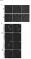
    • Figure 2 shows representative fluorescence images validating the presence of NaV1.5 autoantibodies in plasma from patients with Brugada syndrome. Panel A: Representative fluorescence image displaying BrS positive and negative samples. Red fluorescence highlights NaV1.5 expression, while green fluorescence indicates the presence of bound human antibodies. The same plasma was tested on both transfected and non-transfected cells (HEK-WT), Panel B: Representative fluorescence image illustrating BrS positive and negative plasma. Red fluorescence indicates NaV1.5 expression, while green fluorescence serves as an indicator of human IgGs.
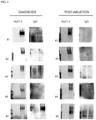
    • Figure 3 shows detection of autoantibodies against NaV1.5 protein from plasma of patients with Brugada syndrome at the time of diagnosis (right panel) and after catheter ablation (left panel). Western blot analysis: proteins from lysed cells were separated using one-dimensional SDS-PAGE. A NaV1.5 protein band at 250-kd was observed in lysates from transfected HEK cells (HEK-NaV1.5) but not in lysates from untransfected cells (HEK-WT). Co-localization of IgGs from Brugada syndrome patients with and without SCN5A mutations, but not from Post Ablated (PA) patients was studied with anti-rabbit anti-NaV1.5 and anti-human IgG antibodies.
    • Figure 4 shows detection of autoantibodies against NaV1.5 protein in the plasma of Brugada syndrome patients. Western blot analysis: proteins from lysed cells were separated by one-dimensional SDS-PAGE. A NaV1.5 protein band at 250-kd was observed in lysates from transfected HEK cells (HEK-NaV1.5) but not in lysates from untransfected cells (HEK-WT). Co-localization of IgGs from BrS patients with and without SCN5A mutations, but not from healthy controls, was studied with anti-rabbit anti-NaV1.5 and anti-human IgG antibodies.
    • Figure 5 shows a comparison of representative fluorescence image of Brugada syndrome positive and negative plasma. Panel A: Representative fluorescence image showing BrS positive and negative plasma. Red fluorescence indicates NaV1.5 expression, while green fluorescence serves as an indicator of human IgGs. Panel B): Menders coefficient.
    • Figure 6 shows the identification of putative binding sites of autoantibodies on NaV1.5 protein. Panel A) Amino acid sequence of NaV1.5; Panel B) Identification of six potential regions that serve as binding sites for autoantibodies. The 10 amino acids critical for binding on the solid support are highlighted in yellow; Panel C) Pymol structure showing the core of the NaV1.5 channel and the extracellular loop. The following examples are merely illustrative and should not be considered limiting the scope of the present invention.
    EXAMPLE 1: Detection of NAVI.5 channel autoantibodies in plasma from Brugada patients METHODS Human samples
  • Brugada patients diagnosed with Brugada Syndrome (BrS) at the Arrhythmology Department of IRCCS Policlinico San Donato were included in this study [24]. Participants were classified into three main subgroups: 1) BrS patients with SCN5A mutations; 2) BrS patients without SCN5A variants; and 3) healthy controls. Additionally, five patients underwent catheter ablation. The study protocol was approved by the local Institutional Ethics Committee, and written informed consent was obtained from all participants in accordance with the Declaration of Helsinki (NCT02641431; NCT03106701) [25, 26]. All patients met the diagnostic criteria for BrS, including the presence of a spontaneous or drug-induced type 1 Brugada ECG pattern. Clinical data, medical history, 12-lead ECG recordings, and implantable cardioverter-defibrillator (ICD) outcomes were collected from medical records.
  • Cell culture
  • HEK293 cells were cultured in DMEM high-glucose medium supplemented with 10% fetal bovine serum, 1% penicillin-streptomycin, and 1% glutamine at 37°C under 5% CO2.
  • Mouse Model
  • Adult C57BL-6 mice were housed under standard conditions with a 12-hour light/dark cycle and ad libitum access to food and water. The mice were kept in the same controlled environment.
  • Whole Exome Sequencing
  • All patients were studied using a Next Generation Sequencing panel of genes, including SCN5A, from peripheral blood extracted DNA. The DNA was extracted from peripheral blood and processed to obtain libraries containing approximately 575 kb of genomic DNA, using 50 nanograms of DNA input quantity. The libraries were subjected to deep sequencing, and after removing duplicates and filtering low-quality reads, an average target coverage of 100X was achieved. Sanger sequencing was used to validate the Next Generation Sequencing data, following the guidelines of the American College of Medical Genetics (ACMG) [27]. Variants were annotated with information from well-known public databases, including dbSNP, dbNSFP, ExAC, and ClinVar.
  • Plasma Collection and Processing
  • Blood (25 ml) was centrifuged at 1000g for 15 minutes to isolate plasma, followed by centrifugation of the supernatant at 2000g for 15 minutes. The supernatant was collected, aliquoted, and stored at -20°C.
  • IgG Isolation
  • IgG antibodies were isolated using the PureProteome Protein G Magnetic Bead System. The isolation was performed under non-denaturing conditions with high salt concentration and nearly neutral pH.
  • Stable cell line generation and transient transfections
  • To express the NaV1.5 channel protein, a full-length cDNA encoding human SCN5A was synthesized and cloned into pcDNA 3.1(+). HEK293 cells were transfected with the pcDNA 3.1(+)/SCN5A construct using ViaFect transfection reagent (Promega) according to the manufacturer's instructions. Cells were growth in medium consisting of Dulbecco's Modified Eagle Medium (DMEM, Life Technologies) supplemented with 10% fetal bovine serum (FBS, Sigma), 2 mM glutamine (Merck), and 1X penicillin/streptomycin (Euroclone) and selected with G418 at a specified concentration. The cells were maintained at 37°C in a 5% CO2 and 95% air-humidified atmosphere. Harvested cells were lysed, and the cleared lysate was collected after centrifugation. The successful transfection and expression of NaV1.5 channels were confirmed through Western blot analysis.
  • Western Blot
  • Cells were lysed in a lysis buffer containing 150 mM NaCl, 50 mM Tris-HCl pH 7.5, 1% Triton X-100, 0.5% sodium deoxycholate, and 0.1% SDS, supplemented with protease and protein phosphatase inhibitors. Total protein concentration was measured using the BCA assay. Proteins were denatured and reduced in a Laemmli-b-mercaptoethanol mixture at 100°C for 5 min. Subsequently, proteins (30 µg) were loaded onto a 10% SDS-PAGE gel and transferred to a nitrocellulose membrane. Nonspecific binding was blocked, and the membrane was incubated overnight at 4°C with a primary rabbit monoclonal anti-NaV1.5 antibody (1:2000 dilution, clone D9J7S, Cell Signalling). After washing, the membrane was incubated with an appropriate anti-rabbit-Alexafluoor546-conjugated secondary antibody (1:2000 dilution) for 1 hour at room temperature. Following further washes, the membrane was incubated with plasma from patients or controls diluted 1:3 in PBS for 2 hours at room temperature. After additional washes, a secondary antibody for human IgG-FITC was added for 1 hour at room temperature. Immunoreactive bands were visualized using an enhanced chemiluminescence detection kit (ECL Advance, GE Healthcare).
  • NaV 1.5 Immunoprecipitation
  • Immunoprecipitation of the NaV1.5 channel protein from transfected HEK-293 cells was performed using IgG from serum of patients and healthy controls specific for NaV1.5 channel. The immunoprecipitation was carried out by incubating the antibodies with Dynabeads Protein G for 30 minutes at room temperature. After washing, the beads were incubated with total cell lysate for 2 hours at room temperature with orbital shaking. Following additional washes, Laemmli-b-mercaptoethanol sample buffer was added to the beads, and the eluted fraction was subjected to SDS-PAGE.
  • Immunofluorescence on Mouse Tissue
  • Immunofluorescence assays were conducted on adult C57BL-6 mouse heart tissue. Left ventricle sections, 12 µm thick, were prepared using a cryostat and mounted onto gelatin-coated histological slides. The sections were thawed, rehydrated, and subjected to antigen unmasking. Non-specific binding was blocked using a solution containing normal donkey serum and bovine serum albumin. Incubation with patient plasma diluted 1:50 in PBS with 2% NDS and 2% BSA was performed, followed by washing and incubation with an anti-human IgG-FITC secondary antibody. Subsequently, the sections were incubated with a primary rabbit monoclonal anti-NaV1.5 antibody (1:200 dilution; clone D9J7S, Cell Signaling), followed by washing and incubation with an appropriate anti-rabbit secondary antibody conjugated with Cy3. After further washes, the sections were mounted with Vectashield mounting medium containing DAPI. Images were captured using a Leica Thunder microscope (×40 objective).
  • Cell-based assay immunofluorescence (CBA-IF)
  • To assess the binding of IgGs-NaV1.5 autoantibodies, live HEK293 WT and HEK293 cells expressing NaV1.5 protein were used. The cells were plated on coverslips and incubated with patient plasma diluted 1:3 in DMEM HG supplemented with 1% L-glutamine, 1% penicillin-streptomycin, and 5% heat-inactivated serum. After washing, the cells were fixed in cold methanol and subjected to blocking. Incubation with NaV1.5 antibody was performed, followed by washing and staining with a rabbit-secondary antibody and FITC anti-human IgG. Nuclear staining was performed using DAPI. Images were captured using a Leica Thunder microscope (×40 objective).
  • Colocalization quantification
  • Confocal microscopy images were randomly captured for all groups, and colocalization analysis was performed using the JACoP plugin in the FIJI software. The Manders' coefficient was calculated to quantify colocalization.
  • Prediction of putative NaV 1.5 autoantibody binding-site on NaV1.5 channel protein
  • To explore potential binding sites on the NaV1.5 channel protein for autoantibodies, molecular modeling was conducted using the crystal structure of the NaV1.5 channel protein downloaded from Uniprot SCN5A (Figure 6). Pymol software was employed to predict favorable interactions between the autoantibodies and the target protein, facilitating the rational design of binding sites.
  • RESULTS Autoantibody Detection Against NaV1.5 Protein in Brugada syndrome plasma
  • A cohort of 100 Brugada Syndrome (BrS) patients was included in the study. Western blot analysis was initially conducted to detect the presence of IgG autoantibodies against the NaV1.5 channel in plasma samples from the patients. Among the BrS patients tested positive for the Ajmaline test (n = 53), 48 patients without SCN5A mutations and 5 patients carrying SCN5A variants exhibited the autoantibody. In contrast, plasma samples from 45 subjects who tested negative in the Ajmaline test showed no presence of the autoantibody (Figure 1A and Figure 4). To further confirm the presence of IgG against the NaV1.5 channel, immunoprecipitation assays were performed. IgGs isolated from the plasma of patients positive for the autoantibody exhibited binding to NaV1.5 protein (Figure 1B) extracted from HEK cell lysate.
  • Binding of NaV1.5 by Plasma IgG from BrS Patients on Cells Over-expressing NaV1.5 Channel
  • To investigate the binding of NaV1.5 by plasma IgG from BrS patients, double immunofluorescence labelling was performed on mouse heart slides using an anti-NaV1.5 monoclonal antibody and plasma from BrS patients and healthy controls. Colocalization of the immunofluorescence signal was observed when the monoclonal anti-NaV1.5 antibody and plasma from BrS patients were used, while no signal was observed with healthy control plasma (Figure 2A). Similar results were obtained with immunolabeling on cells over-expressing the NaV1.5 channel using plasma from BrS patients. The anti-NaV1.5 antibodies from BrS patients specifically bound to NaV1.5-transfected cells, but not mock-transfected cells (Figure 2D and Figure 5).
  • Undetectable Autoantibody against NaV1.5 in BrS Patients following Catheter Ablation
  • To assess the impact of catheter ablation on the presence of autoantibodies against NaV1.5, a subset of five patients from the initial cohort was analyzed after six months from the ablation procedure. Western blot analysis showed an absence of IgG autoantibodies against NaV1.5 in these patients following epicardium ablation (Figure 3).
  • Prediction of Putative NaV1.5 Autoantibody Binding-Sites on NaV1.5 Channel Protein
  • The human Nav1.5 channel has the following amino acid sequence:
    Figure imgb0001
    Figure imgb0002
  • Using molecular modelling, the authors identified six (DI-DVI) putative binding sites for autoantibodies on the NaV1.5 channel protein extracellular loops (in bold in SEQ ID NO:1) as represented in Figure 6:
    • DI S5-S6 (263-368) 106 aa:
      Figure imgb0003
    • DII S5-S6 (861-897) 37 aa:
      FGKNYSELRDSDSGLLPRWHMMDFFHAFLIIFRILCG (SEQ ID NO:3)
    • DIII S5-S6 (1349-1414) 66 aa
      Figure imgb0004
    • DIV S3-S4 (1598-1634) 36 aa
      VILSIVGTVLSDIIQKYFFSPTLFRVIRLARIGRIL (SEQ ID NO:5)
    • DV S5-S6 (1670-1707) 38 aa
      IYSIFGMANFAYVKWEAGIDDMFNFQTFANSMLCLFQI (SEQ ID NO:6)
    • DVI S5-S6 (1711-1754) 44aa
      AGWDGLLSPILNTGPPYCDPTLPNSNGSRGDCGSPAVGILFFTT (SEQ ID NO:7)
    REFERENCES
    1. 1. Antzelevitch, C. and B. Patocskai, Brugada Syndrome: Clinical, Genetic, Molecular, Cellular, and Ionic Aspects. Curr Probl Cardiol, 2016. 41(1): p. 7-57.
    2. 2. Nademanee, K., et al., Fibrosis, Connexin-43, and Conduction Abnormalities in the Brugada Syndrome. J Am Coll Cardiol, 2015. 66(18): p. 1976-1986.
    3. 3. Priori, S.G., et al., 2015 ESC Guidelines for the management of patients with ventricular arrhythmias and the prevention of sudden cardiac death: The Task Force for the Management of Patients with Ventricular Arrhythmias and the Prevention of Sudden Cardiac Death of the European Society of Cardiology (ESC). Endorsed by: Association for European Paediatric and Congenital Cardiology (AEPC). Eur Heart J, 2015. 36(41): p. 2793-2867.
    4. 4. Batchvarov, V.N., The Brugada Syndrome - Diagnosis, Clinical Implications and Risk Stratification. Eur Cardiol, 2014. 9(2): p. 82-87.
    5. 5. Ghiroldi, A., et al., Alterations of the Sialylation Machinery in Brugada Syndrome. Int J Mol Sci, 2022. 23(21).
    6. 6. Keller, D.I., et al., Brugada syndrome and fever: genetic and molecular characterization of patients carrying SCN5A mutations. Cardiovasc Res, 2005. 67(3): p. 510-9.
    7. 7. Barc, J., et al., Genome-wide association analyses identify new Brugada syndrome risk loci and highlight a new mechanism of sodium channel regulation in disease susceptibility. Nat Genet, 2022. 54(3): p. 232-239.
    8. 8. Sonoda, K., et al., Copy number variations of SCN5A in Brugada syndrome. Heart Rhythm, 2018. 15(8): p. 1179-1188.
    9. 9. Wijeyeratne, Y.D., et al., SCN5A Mutation Type and a Genetic Risk Score Associate Variably With Brugada Syndrome Phenotype in SCN5A Families. Circ Genom Precis Med, 2020. 13(6): p. e002911.
    10. 10. Frustaci, A., et al., Cardiac histological substrate in patients with clinical phenotype of Brugada syndrome. Circulation, 2005. 112(24): p. 3680-7.
    11. 11. Corrado, D., et al., Relationship Between Arrhythmogenic Right Ventricular Cardiomyopathy and Brugada Syndrome: New Insights From Molecular Biology and Clinical Implications. Circ Arrhythm Electrophysiol, 2016. 9(4): p. e003631.
    12. 12. Miles, C., et al., Biventricular Myocardial Fibrosis and Sudden Death in Patients With Brugada Syndrome. J Am Coll Cardiol, 2021. 78(15): p. 1511-1521.
    13. 13. Pieroni, M., et al., Electroanatomic and Pathologic Right Ventricular Outflow Tract Abnormalities in Patients With Brugada Syndrome. J Am Coll Cardiol, 2018. 72(22): p. 2747-2757.
    14. 14. Chatterjee, D., et al., An autoantibody identifies arrhythmogenic right ventricular cardiomyopathy and participates in its pathogenesis. Eur Heart J, 2018. 39(44): p. 3932-3944.
    15. 15. Meraviglia, V., et al., Inflammation in the Pathogenesis of Arrhythmogenic Cardiomyopathy: Secondary Event or Active Driver? Front Cardiovasc Med, 2021. 8: p. 784715.
    16. 16. Patel, P.A. and A.F. Hernandez, Targeting anti-beta-1-adrenergic receptor antibodies for dilated cardiomyopathy. Eur J Heart Fail, 2013. 15(7): p. 724-9.
    17. 17. Labovsky, V., et al., Anti-beta1-adrenergic receptor autoantibodies in patients with chronic Chagas heart disease. Clin Exp Immunol, 2007. 148(3): p. 440-9.
    18. 18. Li, J., et al., Anti-KCNQ1 K(+) channel autoantibodies increase IKs current and are associated with QT interval shortening in dilated cardiomyopathy. Cardiovasc Res, 2013. 98(3): p. 496-503.
    19. 19. Korkmaz, S., et al., Provocation of an autoimmune response to cardiac voltage-gated sodium channel NaV1.5 induces cardiac conduction defects in rats. J Am Coll Cardiol, 2013. 62(4): p. 340-9.
    20. 20. Chatterjee, D., et al., An autoantibody profile detects Brugada syndrome and identifies abnormally expressed myocardial proteins. Eur Heart J, 2020. 41(30): p. 2878-2890.
    21. 21. Mader, S., et al., Complement activating antibodies to myelin oligodendrocyte glycoprotein in neuromyelitis optica and related disorders. J Neuroinflammation, 2011. 8: p. 184.
    22. 22. Duong, S.L. and H. Pruss, Molecular disease mechanisms of human antineuronal monoclonal autoantibodies. Trends Mol Med, 2023. 29(1): p. 20-34.
    23. 23. Kobayashi, S., et al., Autoantibody-induced internalization of nicotinic acetylcholine receptor alpha3 subunit exogenously expressed in human embryonic kidney cells. J Neuroimmunol, 2013. 257(1-2): p. 102-6.
    24. 24. Pappone, C. and V. Santinelli, Brugada Syndrome: Progress in Diagnosis and Management. Arrhythm Electrophysiol Rev, 2019. 8(1): p. 13-18.
    25. 25. Pappone, C., et al., Assessing the Malignant Ventricular Arrhythmic Substrate in Patients With Brugada Syndrome. J Am Coll Cardiol, 2018. 71(15): p. 1631-1646.
    26. 26. Pappone, C., et al., Electrical Substrate Elimination in 135 Consecutive Patients With Brugada Syndrome. Circ Arrhythm Electrophysiol, 2017. 10(5): p. e005053.
    27. 27. Richards, S., et al., Standards and guidelines for the interpretation of sequence variants: a joint consensus recommendation of the American College of Medical Genetics and Genomics and the Association for Molecular Pathology. Genet Med, 2015. 17(5): p. 405-24.

Claims (20)

  1. Autoantibodies directed against extracellular loops of NaV 1.5 channel (SEQ ID NO:1) for use as biomarkers for the diagnosis of Brugada Syndrome in a human being.
  2. Autoantibodies for use according to claim 1, directed against a binding site of the extracellular loops of NaV 1.5 channel, said binding site being selected from the group consisting of:
    i) VFALIGLQLFMGNLRHKCVRNFTALNGTNGSVEADGLVWESLDLYLS DPENYLLKNGTSDVLLCGNSSDAGTCPEGYRCLKAGENPDHGYTSFDSFA WAFLALFRL (SEQ ID NO:2)
    ii) FGKNYSELRDSDSGLLPRWHMMDFFHAFLIIFRILCG (SEQ ID NO:3)
    iii) SIMGVNLFAGKFGRCINQTEGDLPLNYTIVNNKSQCESLNLTGELYW TKVKVNFDNVGAGYLALLQ-1414 (SEQ ID NO: 4)
    iv) VILSIVGTVLSDIIQKYFFSPTLFRVIRLARIGRIL (SEQ ID NO:5)
    v) IYSIFGMANFAYVKWEAGIDDMFNFQTFANSMLCLFQI (SEQ ID NO:6)
    vi) AGWDGLLSPILNTGPPYCDPTLPNSNGSRGDCGSPAVGILFFTT (SEQ ID NO:7).
  3. An in vitro method for detecting the presence or detecting the amount of at least one of the autoantibodies directed against the extracellular loops of NaV 1.5 channel according to anyone of claim 1-2 in a biological sample of a human being suspected of having Brugada syndrome.
  4. An in vitro method according to claim 3, which comprises the following steps:
    a) contacting the biological sample with one or more antigens that specifically binds to the autoantibodies directed against extracellular loops of NaV1.5 channel; and
    b) detecting the binding of the antigen to the autoantibodies in the biological sample.
  5. An in vitro method according to claim 4, wherein said antigens are labelled.
  6. An in vitro method according to anyone of the claims 4-5, wherein said antigens are fragments of at least 10 amino acids from the binding sites of extracellular loops of NaV1.5 channel.
  7. An in vitro method according to claim 6 wherein said binding sites of extracellular loops of NaV1.5 channel are selected between SEQ ID NO:2-SEQ ID NO:7.
  8. An in vitro method of according to anyone of the claims 3-7, wherein the biological sample is selected from the group consisting of plasma, PBMCs, whole blood, serum and peripheral blood, or a combination thereof.
  9. An in vitro method according to anyone of the claims 3-7, wherein the detection of the binding of the probe to the autoantibodies is performed by ELISA or Western blotting.
  10. An antagonist of the binding of the autoantibodies to the extracellular loops of NaV 1.5 channel according to claim 1 or 2, for use therapeutic agent in the treatment of Brugada syndrome.
  11. An autoimmune therapeutic agent specifically targeting to the autoantibodies directed to the extracellular loops of NaV 1.5 channel according to claim 1 or 2. for use in the treatment of Brugada syndrome.
  12. An autoimmune therapeutic agent according to claim 11, characterized in that it comprises one or more antigenic fragments of the extracellular loops of the Nav 1.5 protein.
  13. An autoimmune therapeutic agent according to claim 12, characterized in that it comprises sequences of at least about 10 amino acids from the extracellular loops of the Nav 1.5 protein, said extracellular loops being selected from the group of SEQ ID NO:2-SEQ ID NO:7.
  14. The antagonist or the autoimmune therapeutic agent for use according to anyone of the claims 10-13, in combination with other therapy for Brugada Syndrome.
  15. A kit for detecting autoantibodies against extracellular loops of Nav 1.5 channel in a biological sample, the kit comprising one or more antigens that specifically binds to the autoantibodies against Nav 1.5.
  16. A kit according to claim 15, wherein said antigens comprises one or more antigenic fragments of the extracellular loops of the Nav 1.5 protein, said extracellular loops being selected from the group of SEQ ID NO:2-SEQ ID NO:7.
  17. A kit according to anyone of claim 15-16, wherein said antigens are labelled or attached to a solid support.
  18. Use of one or more of the autoantibodies according to anyone of the claims 1-2, for the diagnosis of Brugada Syndrome in a human being by detection in a biological sample, wherein said biological sample is selected from the group consisting of plasma, PBMCs, whole blood, serum and peripheral blood, or a combination thereof.
  19. Use of one or more of the autoantibodies according to anyone of the claims 1-2, as marker for monitoring the efficacy of a therapeutic treatment of Brugada Syndrome disease by detection of the autoantibodies titer.
  20. Use of one or more of the autoantibodies according to claim 19, wherein said therapeutic treatment is pharmacological or interventional-surgical, including ablation.
EP23187367.0A 2023-07-24 2023-07-24 Autoantibodies against nav1.5 channel as biomarkers for the diagnosis of brugada syndrome Withdrawn EP4498088A1 (en)

Priority Applications (3)

Application Number Priority Date Filing Date Title
EP23187367.0A EP4498088A1 (en) 2023-07-24 2023-07-24 Autoantibodies against nav1.5 channel as biomarkers for the diagnosis of brugada syndrome
PCT/EP2024/071047 WO2025021888A1 (en) 2023-07-24 2024-07-24 Autoantibodies against nav1.5 channel as biomarkers for the diagnosis of brugada syndrome
CN202480048884.XA CN121941925A (en) 2023-07-24 2024-07-24 Autoantibodies against NaV1.5 channel protein as biomarkers for the diagnosis of Brugada syndrome

Applications Claiming Priority (1)

Application Number Priority Date Filing Date Title
EP23187367.0A EP4498088A1 (en) 2023-07-24 2023-07-24 Autoantibodies against nav1.5 channel as biomarkers for the diagnosis of brugada syndrome

Publications (1)

Publication Number Publication Date
EP4498088A1 true EP4498088A1 (en) 2025-01-29

Family

ID=87474291

Family Applications (1)

Application Number Title Priority Date Filing Date
EP23187367.0A Withdrawn EP4498088A1 (en) 2023-07-24 2023-07-24 Autoantibodies against nav1.5 channel as biomarkers for the diagnosis of brugada syndrome

Country Status (1)

Country Link
EP (1) EP4498088A1 (en)

Citations (3)

* Cited by examiner, † Cited by third party
Publication number Priority date Publication date Assignee Title
US20150018251A1 (en) * 2012-02-01 2015-01-15 Centre National De La Recherche Scientifique Protein chips, preparation and use thereof
WO2020220136A1 (en) * 2019-05-02 2020-11-05 The Hospital For Sick Children Detection of biomarkers associated with brugada syndrome
WO2022180559A1 (en) * 2021-02-26 2022-09-01 Cardiomix S.R.L. Protein and metabolite blood biomarkers for the diagnosis of brugada syndrome

Patent Citations (3)

* Cited by examiner, † Cited by third party
Publication number Priority date Publication date Assignee Title
US20150018251A1 (en) * 2012-02-01 2015-01-15 Centre National De La Recherche Scientifique Protein chips, preparation and use thereof
WO2020220136A1 (en) * 2019-05-02 2020-11-05 The Hospital For Sick Children Detection of biomarkers associated with brugada syndrome
WO2022180559A1 (en) * 2021-02-26 2022-09-01 Cardiomix S.R.L. Protein and metabolite blood biomarkers for the diagnosis of brugada syndrome

Non-Patent Citations (29)

* Cited by examiner, † Cited by third party
Title
ANTZELEVITCH, CB. PATOCSKAI: "Brugada Syndrome: Clinical, Genetic, Molecular, Cellular, and Ionic Aspects", CURR PROBL CARDIOL, vol. 41, no. 1, 2016, pages 7 - 57, XP029336082, DOI: 10.1016/j.cpcardiol.2015.06.002
BARC, J. ET AL.: "Genome-wide association analyses identify new Brugada syndrome risk loci and highlight a new mechanism of sodium channel regulation in disease susceptibility", NAT GENET, vol. 54, no. 3, 2022, pages 232 - 239, XP037716002, DOI: 10.1038/s41588-021-01007-6
BATCHVAROV, V.N.: "The Brugada Syndrome - Diagnosis, Clinical Implications and Risk Stratification", EUR CARDIOL, vol. 9, no. 2, 2014, pages 82 - 87
CHATTERJEE D. ET AL.: "An autoantibody profile detects Brugada syndrome and identifies abnormally expressed myocardial proteins", EUR. HEART J., vol. 41, no. 30, 7 August 2020 (2020-08-07), pages 2878 - 2890, XP093120705 *
CHATTERJEE, D. ET AL.: "An autoantibody profile detects Brugada syndrome and identifies abnormally expressed myocardial proteins", EUR HEART J, vol. 41, no. 30, 2020, pages 2878 - 2890
CHATTERJEE, D.: "An autoantibody identifies arrhythmogenic right ventricular cardiomyopathy and participates in its pathogenesis", HEART J, vol. 39, no. 44, 2018, pages 3932 - 3944, XP055716111, DOI: 10.1093/eurheartj/ehy567
CORRADO, D. ET AL.: "Relationship Between Arrhythmogenic Right Ventricular Cardiomyopathy and Brugada Syndrome: New Insights From Molecular Biology and Clinical Implications", CIRC ARRHYTHM ELECTROPHYSIOL, vol. 9, no. 4, 2016, pages e003631
DUONG, S.L.H. PRUSS: "Molecular disease mechanisms of human antineuronal monoclonal autoantibodies", TRENDS MOL MED, vol. 29, no. 1, 2023, pages 20 - 34
FRUSTACI, A. ET AL.: "Cardiac histological substrate in patients with clinical phenotype of Brugada syndrome", CIRCULATION, vol. 112, no. 24, 2005, pages 3680 - 7
GHIROLDI, A. ET AL.: "Alterations of the Sialylation Machinery in Brugada Syndrome", INT J MOL SCI, vol. 23, no. 21, 2022
KELLER, D.I. ET AL.: "Brugada syndrome and fever: genetic and molecular characterization of patients carrying SCN5A mutations", CARDIOVASC RES, vol. 67, no. 3, 2005, pages 510 - 9, XP004985954, DOI: 10.1016/j.cardiores.2005.03.024
KOBAYASHI, S. ET AL.: "Autoantibody-induced internalization of nicotinic acetylcholine receptor alpha3 subunit exogenously expressed in human embryonic kidney cells", J NEUROIMMUNOL, vol. 257, no. 1-2, 2013, pages 102 - 6, XP028529669, DOI: 10.1016/j.jneuroim.2012.12.010
KORKMAZ, S. ET AL.: "Provocation of an autoimmune response to cardiac voltage-gated sodium channel Na V]. 5 induces cardiac conduction defects in rats", J AM COLL CARDIOL, vol. 62, no. 4, 2013, pages 340 - 9, XP028677263, DOI: 10.1016/j.jacc.2013.04.041
LABOVSKY, V. ET AL.: "Anti-betal-adrenergic receptor autoantibodies in patients with chronic Chagas heart disease", CLIN EXP IMMUNOL, vol. 148, no. 3, 2007, pages 440 - 9
LI, J. ET AL.: "Anti-KCNQ1 K(+) channel autoantibodies increase IKs current and are associated with QT interval shortening in dilated cardiomyopathy", CARDIOVASC RES, vol. 98, no. 3, 2013, pages 496 - 503, XP055971475, DOI: 10.1093/cvr/cvt046
MADER, S. ET AL.: "Complement activating antibodies to myelin oligodendrocyte glycoprotein in neuromyelitis optica and related disorders", J NEUROINFLAMMATION, vol. 8, 2011, pages 184, XP021131500, DOI: 10.1186/1742-2094-8-184
MERAVIGLIA, V. ET AL.: "Inflammation in the Pathogenesis of Arrhythmogenic Cardiomyopathy: Secondary Event or Active Driver?", FRONT CARDIOVASC, vol. 8, 2021, pages 784715
MILES, C. ET AL.: "Biventricular Myocardial Fibrosis and Sudden Death in Patients With Brugada Syndrome", J AM COLL CARDIOL, vol. 78, no. 15, 2021, pages 1511 - 1521
NADEMANEE, K. ET AL.: "Fibrosis, Connexin-43, and Conduction Abnormalities in the Brugada Syndrome", J AM COLL CARDIOL, vol. 66, no. 18, 2015, pages 1976 - 1986, XP029308441, DOI: 10.1016/j.jacc.2015.08.862
PAPPONE, C. ET AL.: "Assessing the Malignant Ventricular Arrhythmic Substrate in Patients With Brugada Syndrome", J AM COLL CARDIOL, vol. 71, no. 15, 2018, pages 1631 - 1646, XP085376761, DOI: 10.1016/j.jacc.2018.02.022
PAPPONE, C. ET AL.: "Electrical Substrate Elimination in 135 Consecutive Patients With Brugada Syndrome", CIRC ARRHYTHM ELECTROPHYSIOL, vol. 10, no. 5, 2017, pages e005053
PAPPONE, C.V. SANTINELLI: "Brugada Syndrome: Progress in Diagnosis and Management", ARRHYTHM ELECTROPHYSIOL REV, vol. 8, no. 1, 2019, pages 13 - 18
PATEL, P.A.A.F. HERNANDEZ: "Targeting anti-beta-1-adrenergic receptor antibodies for dilated cardiomyopathy", EUR J HEART FAIL, vol. 15, no. 7, 2013, pages 724 - 9
PIERONI, M.: "Electroanatomic and Pathologic Right Ventricular Outflow Tract Abnormalities in Patients With Brugada Syndrome", COLL CARDIOL, vol. 72, no. 22, 2018, pages 2747 - 2757
PRIORI, S. G.: "2015 ESC Guidelines for the management of patients with ventricular arrhythmias and the prevention of sudden cardiac death:The Task Force for the Management of Patients with Ventricular Arrhythmias and the Prevention of Sudden Cardiac Death of the European Society of Cardiology (ESC). Endorsed by: ", EUR HEART J, vol. 36, no. 41, 2015, pages 2793 - 2867
RICHARDS, S. ET AL.: "Standards and guidelines for the interpretation of sequence variants: a joint consensus recommendation of the American College of Medical Genetics and Genomics and the Association for Molecular Pathology", GENET MED, vol. 17, no. 5, 2015, pages 405 - 24, XP093036319, DOI: 10.1038/gim.2015.30
SONODA, K. ET AL.: "Copy number variations of SCN5A in Brugada syndrome", HEART RHYTHM, vol. 15, no. 8, 2018, pages 1179 - 1188
WIJEYERATNE, Y.D.: "SCN5A Mutation Type and a Genetic Risk Score Associate Variably With Brugada Syndrome Phenotype in SCN5A Families", CIRC GENOM PRECIS MED, vol. 13, no. 6, 2020, pages e002911
XU S.-Z. ET AL.: "Generation of functional ion-channel tools by E3 targeting", NATURE BIOTECHNOL., vol. 23, no. 10, October 2005 (2005-10-01), pages 1289 - 1293, XP002415111 *

Similar Documents

Publication Publication Date Title
Ma et al. Composition of the intranuclear inclusions of fragile X-associated tremor/ataxia syndrome
Kattygnarath et al. MOG1: a new susceptibility gene for Brugada syndrome
Thanh et al. Characterization of revertant muscle fibers in Duchenne muscular dystrophy, using exon-specific monoclonal antibodies against dystrophin
Ishikawa et al. Novel SCN3B mutation associated with brugada syndrome affects intracellular trafficking and function of Nav1. 5
Grohmann et al. Characterization of Ighmbp2 in motor neurons and implications for the pathomechanism in a mouse model of human spinal muscular atrophy with respiratory distress type 1 (SMARD1)
Gavriilidis et al. The MTM1–UBQLN2–HSP complex mediates degradation of misfolded intermediate filaments in skeletal muscle
Rizzo et al. Role of HLA-G 14bp deletion/insertion and+ 3142C> G polymorphisms in the production of sHLA-G molecules in relapsing-remitting multiple sclerosis
Daumy et al. Targeted resequencing identifies TRPM4 as a major gene predisposing to progressive familial heart block type I
Yu et al. Gene therapy targeting protein trafficking regulator MOG1 in mouse models of Brugada syndrome, arrhythmias, and mild cardiomyopathy
WO2018226590A1 (en) Peptides as biomarkers in the diagnosis, confirmation and treatment of a neurological disorder and immunoprofiling in neurodegenerative disease
EP2861618B1 (en) Bag3 as biochemical serum and tissue marker
Tarantino et al. NaV1. 5 autoantibodies in Brugada syndrome: pathogenetic implications
Gangfuss et al. Proteomic and morphological insights and clinical presentation of two young patients with novel mutations of BVES (POPDC1)
Christiansen et al. ANO5-related muscle diseases: From clinics and genetics to pathology and research strategies
EP4498088A1 (en) Autoantibodies against nav1.5 channel as biomarkers for the diagnosis of brugada syndrome
EP4614149A1 (en) Autoantibodies against nav1.5 channel as biomarkers for the diagnosis of brugada syndrome
US20250145704A1 (en) AUTOANTIBODIES AGAINST NaV1.5 CHANNEL AS BIOMARKERS FOR THE DIAGNOSIS OF BRUGADA SYNDROME AND RELATED THERAPEUTIC METHODS
Wang et al. Brugada syndrome caused by sodium channel dysfunction associated with a SCN1B variant A197V
US20170269103A1 (en) Assessment of Heart Failure and Arrhythmic Risk Stratification by Measuring Circulating Hu Proteins
WO2025021888A1 (en) Autoantibodies against nav1.5 channel as biomarkers for the diagnosis of brugada syndrome
Huang et al. Whole-exome sequencing and electrophysiological study reveal a novel loss-of-function mutation of KCNA10 in epinephrine provoked long QT syndrome with familial history of sudden cardiac death
Simkin et al. Andersen’s syndrome mutants produce a knockdown of inwardly rectifying K+ channel in mouse skeletal muscle in vivo
Ji et al. A novel DPP6 variant in Chinese families causes early repolarization syndrome
EP4644902A1 (en) Anti nav1.5 channel autoantibodies as biomarkers for predicting the risk of cardiac arrhythmia and sudden cardiac death in cancer patients
WO2014122275A1 (en) Method for the diagnosis and monitoring of autoinmune diseases

Legal Events

Date Code Title Description
PUAI Public reference made under article 153(3) epc to a published international application that has entered the european phase

Free format text: ORIGINAL CODE: 0009012

STAA Information on the status of an ep patent application or granted ep patent

Free format text: STATUS: THE APPLICATION HAS BEEN PUBLISHED

AK Designated contracting states

Kind code of ref document: A1

Designated state(s): AL AT BE BG CH CY CZ DE DK EE ES FI FR GB GR HR HU IE IS IT LI LT LU LV MC ME MK MT NL NO PL PT RO RS SE SI SK SM TR

STAA Information on the status of an ep patent application or granted ep patent

Free format text: STATUS: THE APPLICATION IS DEEMED TO BE WITHDRAWN

18D Application deemed to be withdrawn

Effective date: 20250730