EP4491711A1 - Production method for organoid - Google Patents
Production method for organoid Download PDFInfo
- Publication number
- EP4491711A1 EP4491711A1 EP23766878.5A EP23766878A EP4491711A1 EP 4491711 A1 EP4491711 A1 EP 4491711A1 EP 23766878 A EP23766878 A EP 23766878A EP 4491711 A1 EP4491711 A1 EP 4491711A1
- Authority
- EP
- European Patent Office
- Prior art keywords
- organoid
- inhibitor
- medium
- tissue
- organoids
- Prior art date
- Legal status (The legal status is an assumption and is not a legal conclusion. Google has not performed a legal analysis and makes no representation as to the accuracy of the status listed.)
- Pending
Links
Images





Classifications
-
- C—CHEMISTRY; METALLURGY
- C12—BIOCHEMISTRY; BEER; SPIRITS; WINE; VINEGAR; MICROBIOLOGY; ENZYMOLOGY; MUTATION OR GENETIC ENGINEERING
- C12N—MICROORGANISMS OR ENZYMES; COMPOSITIONS THEREOF; PROPAGATING, PRESERVING, OR MAINTAINING MICROORGANISMS; MUTATION OR GENETIC ENGINEERING; CULTURE MEDIA
- C12N5/00—Undifferentiated human, animal or plant cells, e.g. cell lines; Tissues; Cultivation or maintenance thereof; Culture media therefor
- C12N5/06—Animal cells or tissues; Human cells or tissues
- C12N5/0602—Vertebrate cells
- C12N5/0693—Tumour cells; Cancer cells
-
- C—CHEMISTRY; METALLURGY
- C12—BIOCHEMISTRY; BEER; SPIRITS; WINE; VINEGAR; MICROBIOLOGY; ENZYMOLOGY; MUTATION OR GENETIC ENGINEERING
- C12N—MICROORGANISMS OR ENZYMES; COMPOSITIONS THEREOF; PROPAGATING, PRESERVING, OR MAINTAINING MICROORGANISMS; MUTATION OR GENETIC ENGINEERING; CULTURE MEDIA
- C12N5/00—Undifferentiated human, animal or plant cells, e.g. cell lines; Tissues; Cultivation or maintenance thereof; Culture media therefor
- C12N5/06—Animal cells or tissues; Human cells or tissues
- C12N5/0602—Vertebrate cells
- C12N5/0679—Cells of the gastro-intestinal tract
-
- C—CHEMISTRY; METALLURGY
- C12—BIOCHEMISTRY; BEER; SPIRITS; WINE; VINEGAR; MICROBIOLOGY; ENZYMOLOGY; MUTATION OR GENETIC ENGINEERING
- C12N—MICROORGANISMS OR ENZYMES; COMPOSITIONS THEREOF; PROPAGATING, PRESERVING, OR MAINTAINING MICROORGANISMS; MUTATION OR GENETIC ENGINEERING; CULTURE MEDIA
- C12N5/00—Undifferentiated human, animal or plant cells, e.g. cell lines; Tissues; Cultivation or maintenance thereof; Culture media therefor
- C12N5/0018—Culture media for cell or tissue culture
-
- G—PHYSICS
- G01—MEASURING; TESTING
- G01N—INVESTIGATING OR ANALYSING MATERIALS BY DETERMINING THEIR CHEMICAL OR PHYSICAL PROPERTIES
- G01N33/00—Investigating or analysing materials by specific methods not covered by groups G01N1/00 - G01N31/00
- G01N33/48—Biological material, e.g. blood, urine; Haemocytometers
- G01N33/50—Chemical analysis of biological material, e.g. blood, urine; Testing involving biospecific ligand binding methods; Immunological testing
- G01N33/5005—Chemical analysis of biological material, e.g. blood, urine; Testing involving biospecific ligand binding methods; Immunological testing involving human or animal cells
- G01N33/5008—Chemical analysis of biological material, e.g. blood, urine; Testing involving biospecific ligand binding methods; Immunological testing involving human or animal cells for testing or evaluating the effect of chemical or biological compounds, e.g. drugs, cosmetics
- G01N33/5011—Chemical analysis of biological material, e.g. blood, urine; Testing involving biospecific ligand binding methods; Immunological testing involving human or animal cells for testing or evaluating the effect of chemical or biological compounds, e.g. drugs, cosmetics for testing antineoplastic activity
-
- G—PHYSICS
- G01—MEASURING; TESTING
- G01N—INVESTIGATING OR ANALYSING MATERIALS BY DETERMINING THEIR CHEMICAL OR PHYSICAL PROPERTIES
- G01N33/00—Investigating or analysing materials by specific methods not covered by groups G01N1/00 - G01N31/00
- G01N33/48—Biological material, e.g. blood, urine; Haemocytometers
- G01N33/50—Chemical analysis of biological material, e.g. blood, urine; Testing involving biospecific ligand binding methods; Immunological testing
- G01N33/5005—Chemical analysis of biological material, e.g. blood, urine; Testing involving biospecific ligand binding methods; Immunological testing involving human or animal cells
- G01N33/5008—Chemical analysis of biological material, e.g. blood, urine; Testing involving biospecific ligand binding methods; Immunological testing involving human or animal cells for testing or evaluating the effect of chemical or biological compounds, e.g. drugs, cosmetics
- G01N33/5082—Supracellular entities, e.g. tissue, organisms
- G01N33/5088—Supracellular entities, e.g. tissue, organisms of vertebrates
-
- C—CHEMISTRY; METALLURGY
- C12—BIOCHEMISTRY; BEER; SPIRITS; WINE; VINEGAR; MICROBIOLOGY; ENZYMOLOGY; MUTATION OR GENETIC ENGINEERING
- C12N—MICROORGANISMS OR ENZYMES; COMPOSITIONS THEREOF; PROPAGATING, PRESERVING, OR MAINTAINING MICROORGANISMS; MUTATION OR GENETIC ENGINEERING; CULTURE MEDIA
- C12N2500/00—Specific components of cell culture medium
- C12N2500/70—Undefined extracts
- C12N2500/80—Undefined extracts from animals
-
- C—CHEMISTRY; METALLURGY
- C12—BIOCHEMISTRY; BEER; SPIRITS; WINE; VINEGAR; MICROBIOLOGY; ENZYMOLOGY; MUTATION OR GENETIC ENGINEERING
- C12N—MICROORGANISMS OR ENZYMES; COMPOSITIONS THEREOF; PROPAGATING, PRESERVING, OR MAINTAINING MICROORGANISMS; MUTATION OR GENETIC ENGINEERING; CULTURE MEDIA
- C12N2501/00—Active agents used in cell culture processes, e.g. differentation
- C12N2501/10—Growth factors
- C12N2501/105—Insulin-like growth factors [IGF]
-
- C—CHEMISTRY; METALLURGY
- C12—BIOCHEMISTRY; BEER; SPIRITS; WINE; VINEGAR; MICROBIOLOGY; ENZYMOLOGY; MUTATION OR GENETIC ENGINEERING
- C12N—MICROORGANISMS OR ENZYMES; COMPOSITIONS THEREOF; PROPAGATING, PRESERVING, OR MAINTAINING MICROORGANISMS; MUTATION OR GENETIC ENGINEERING; CULTURE MEDIA
- C12N2501/00—Active agents used in cell culture processes, e.g. differentation
- C12N2501/10—Growth factors
- C12N2501/11—Epidermal growth factor [EGF]
-
- C—CHEMISTRY; METALLURGY
- C12—BIOCHEMISTRY; BEER; SPIRITS; WINE; VINEGAR; MICROBIOLOGY; ENZYMOLOGY; MUTATION OR GENETIC ENGINEERING
- C12N—MICROORGANISMS OR ENZYMES; COMPOSITIONS THEREOF; PROPAGATING, PRESERVING, OR MAINTAINING MICROORGANISMS; MUTATION OR GENETIC ENGINEERING; CULTURE MEDIA
- C12N2501/00—Active agents used in cell culture processes, e.g. differentation
- C12N2501/10—Growth factors
- C12N2501/113—Acidic fibroblast growth factor (aFGF, FGF-1)
-
- C—CHEMISTRY; METALLURGY
- C12—BIOCHEMISTRY; BEER; SPIRITS; WINE; VINEGAR; MICROBIOLOGY; ENZYMOLOGY; MUTATION OR GENETIC ENGINEERING
- C12N—MICROORGANISMS OR ENZYMES; COMPOSITIONS THEREOF; PROPAGATING, PRESERVING, OR MAINTAINING MICROORGANISMS; MUTATION OR GENETIC ENGINEERING; CULTURE MEDIA
- C12N2501/00—Active agents used in cell culture processes, e.g. differentation
- C12N2501/10—Growth factors
- C12N2501/115—Basic fibroblast growth factor (bFGF, FGF-2)
-
- C—CHEMISTRY; METALLURGY
- C12—BIOCHEMISTRY; BEER; SPIRITS; WINE; VINEGAR; MICROBIOLOGY; ENZYMOLOGY; MUTATION OR GENETIC ENGINEERING
- C12N—MICROORGANISMS OR ENZYMES; COMPOSITIONS THEREOF; PROPAGATING, PRESERVING, OR MAINTAINING MICROORGANISMS; MUTATION OR GENETIC ENGINEERING; CULTURE MEDIA
- C12N2501/00—Active agents used in cell culture processes, e.g. differentation
- C12N2501/10—Growth factors
- C12N2501/15—Transforming growth factor beta (TGF-β)
-
- C—CHEMISTRY; METALLURGY
- C12—BIOCHEMISTRY; BEER; SPIRITS; WINE; VINEGAR; MICROBIOLOGY; ENZYMOLOGY; MUTATION OR GENETIC ENGINEERING
- C12N—MICROORGANISMS OR ENZYMES; COMPOSITIONS THEREOF; PROPAGATING, PRESERVING, OR MAINTAINING MICROORGANISMS; MUTATION OR GENETIC ENGINEERING; CULTURE MEDIA
- C12N2501/00—Active agents used in cell culture processes, e.g. differentation
- C12N2501/10—Growth factors
- C12N2501/155—Bone morphogenic proteins [BMP]; Osteogenins; Osteogenic factor; Bone inducing factor
-
- C—CHEMISTRY; METALLURGY
- C12—BIOCHEMISTRY; BEER; SPIRITS; WINE; VINEGAR; MICROBIOLOGY; ENZYMOLOGY; MUTATION OR GENETIC ENGINEERING
- C12N—MICROORGANISMS OR ENZYMES; COMPOSITIONS THEREOF; PROPAGATING, PRESERVING, OR MAINTAINING MICROORGANISMS; MUTATION OR GENETIC ENGINEERING; CULTURE MEDIA
- C12N2501/00—Active agents used in cell culture processes, e.g. differentation
- C12N2501/40—Regulators of development
- C12N2501/415—Wnt; Frizzeled
-
- C—CHEMISTRY; METALLURGY
- C12—BIOCHEMISTRY; BEER; SPIRITS; WINE; VINEGAR; MICROBIOLOGY; ENZYMOLOGY; MUTATION OR GENETIC ENGINEERING
- C12N—MICROORGANISMS OR ENZYMES; COMPOSITIONS THEREOF; PROPAGATING, PRESERVING, OR MAINTAINING MICROORGANISMS; MUTATION OR GENETIC ENGINEERING; CULTURE MEDIA
- C12N2503/00—Use of cells in diagnostics
- C12N2503/02—Drug screening
-
- C—CHEMISTRY; METALLURGY
- C12—BIOCHEMISTRY; BEER; SPIRITS; WINE; VINEGAR; MICROBIOLOGY; ENZYMOLOGY; MUTATION OR GENETIC ENGINEERING
- C12N—MICROORGANISMS OR ENZYMES; COMPOSITIONS THEREOF; PROPAGATING, PRESERVING, OR MAINTAINING MICROORGANISMS; MUTATION OR GENETIC ENGINEERING; CULTURE MEDIA
- C12N2513/00—3D culture
-
- C—CHEMISTRY; METALLURGY
- C12—BIOCHEMISTRY; BEER; SPIRITS; WINE; VINEGAR; MICROBIOLOGY; ENZYMOLOGY; MUTATION OR GENETIC ENGINEERING
- C12N—MICROORGANISMS OR ENZYMES; COMPOSITIONS THEREOF; PROPAGATING, PRESERVING, OR MAINTAINING MICROORGANISMS; MUTATION OR GENETIC ENGINEERING; CULTURE MEDIA
- C12N2533/00—Supports or coatings for cell culture, characterised by material
- C12N2533/90—Substrates of biological origin, e.g. extracellular matrix, decellularised tissue
-
- G—PHYSICS
- G01—MEASURING; TESTING
- G01N—INVESTIGATING OR ANALYSING MATERIALS BY DETERMINING THEIR CHEMICAL OR PHYSICAL PROPERTIES
- G01N2500/00—Screening for compounds of potential therapeutic value
- G01N2500/10—Screening for compounds of potential therapeutic value involving cells
Definitions
- the present invention relates to a production method for an organoid. More specifically, the present invention relates to a production method for an organoid and a screening method for an anticancer drug.
- Priority is claimed on US provisional application No. US 63/317,552, filed in the United States on March 8, 2022 , the content of which is incorporated herein by reference.
- Drug screening which comprehensively searches for the effects of a large number of therapeutic drug candidates, is a fundamental technology in drug discovery.
- cancer drug discovery in the related art a method has been adopted in which primary screening is carried out using a cancer cell line established from a patient-derived cancer tissue, and safety and side effects are confirmed using an animal model.
- cancer cell lines may lack many of the properties of actual clinical tumors and the sensitivity to anticancer drugs.
- the cancer even in a case where cancer occurs in the same organ, the cancer exhibits different genetic and biological characteristics from patient to patient. It is difficult to comprehensively cover this diversity in cancer cell lines.
- many of the anticancer drug candidates selected using cancer cell lines have not yet been applied clinically, and this "valley of death" in drug discovery research has become a major medical and social issue.
- the present inventors have pioneered the development of an organoid technology for permanent three-dimensional culture of tissue stem cells (see, for example,
- Non-Patent Documents 1 and 2 The present inventors have also constructed cancer organoid biobanks for colorectal cancer, pancreatic cancer, gastric cancer, and the like by applying the organoid technology to various patient-derived cancer tissues (see, for example, Non-Patent Documents 3 to 5).
- Patient-derived cancer organoids retain many of the characteristics of cancer tissue of the patient, and cancer organoid biobanks are intended to be applied as next-generation cancer research resources to basic cancer research, cancer diagnostic tools, drug discovery, personalized treatment, and the like.
- an object of the present invention is to provide a technology that efficiently enables large-scale culture of an organoid, regardless of whether the organoid is derived from a normal tissue or derived from a tumor tissue.
- the present invention includes the following aspects.
- the present invention it is possible to provide a technology that efficiently enables large-scale culture of an organoid, regardless of whether the organoid is derived from a normal tissue or derived from a tumor tissue.
- the present invention provides a production method for an organoid, including a step of dissociating a biological tissue and a step of carrying out suspension culture of the dissociated biological tissue in a state of being dispersed in a medium to form an organoid, in which the medium is substantially free of a p38 inhibitor and contains the following components (i) to (iv).
- the production method according to the present embodiment makes it possible to carry out stable large-scale culture of a normal tissue-derived organoid and to achieve large-scale production of the normal tissue-derived organoid.
- the production method according to the present embodiment can also be said to be a culture method for an organoid.
- the large-scale culture means, for example, proliferating organoids of 10 6 to 10 7 cells from a tissue derived from a living body.
- the biological tissue may be a biological tissue derived from a patient, or may be an organoid which has already been established.
- the production method according to the present embodiment can also be said to be a subculture method for an organoid.
- the biological tissue is preferably an epithelial tissue.
- the epithelial tissue include an intestinal epithelial tissue.
- the intestinal tract is an organ in the human body that has the largest area in contact with the external environment, and has functions essential for maintaining life such as digestion and absorption. Most of the functions of the intestinal tract are carried out by the intestinal epithelium that covers an inner layer of the intestinal tract.
- the intestinal epithelium is composed of two compartments: villi, which consist of three lineages of differentiated cells (mucus-producing cells, absorptive epithelial cells, and endocrine cells), and crypts, which mainly consist of undifferentiated proliferative cells.
- the epithelial tissue is preferably a tissue containing epithelial stem cells or epithelial cells.
- the epithelial cells include differentiated epithelial cells and epithelial stem cells.
- the "epithelial stem cell” refers to a cell having a long-term self-replication function and an ability to differentiate into an epithelial differentiated cell, and means a stem cell derived from an epithelial tissue.
- epithelial tissue examples include tissues of cornea, oral mucosa, skin, conjunctiva, bladder, renal tubule, kidney, digestive organs (esophagus, stomach, duodenum, small intestine (including jejunum and ileum), and large intestine (including colon)), liver, pancreas, mammary gland, salivary gland, tear gland, prostate, hair root, trachea, lung, and the like.
- the biological tissue is preferably a tissue derived from a human or non-human mammal.
- the non-human mammal includes, but is not particularly limited to, a mouse, a rat, a cow, a pig, a rabbit, a monkey, and the like.
- the biological tissue may be a tumor tissue or a cancer tissue, or may be a normal tissue.
- the tumor tissue means a tissue that has become tumorous
- the cancer tissue means a tissue that has become cancerous.
- Dissociating a biological tissue means fragmenting the biological tissue and dividing the fragmented biological tissues into cell clusters each consisting of one cell to several cells.
- “several” may mean about 2 to 100 cells.
- the dissociation of the biological tissue may be carried out by mechanical shearing or by an enzymatic treatment with collagenase, dispase I, liberase, trypsin, TrypLE Express (available from Thermo Fisher Scientific, Inc.), or the like.
- the organoid may have poor recovery after being dissociated into a single cell.
- the present inventors have found that a p38 inhibitor, which has recently been considered to be essential for culturing human intestinal organoids, inhibits the growth of organoids from single cells.
- the present inventors further found that replacing the p38 inhibitor with a combination of IGF-1 and FGF-2 promoted the growth of organoids from single cells.
- general organoid culture protocols in the related art include a step of embedding the dissociated organoid in an extracellular matrix such as Matrigel (registered trademark).
- an extracellular matrix such as Matrigel (registered trademark).
- the organoid embedded in the extracellular matrix may be difficult to scale up in culture.
- the organoid embedded in the extracellular matrix may exhibit a large variation in growth.
- the production method according to the present embodiment is carried out in such a manner that the dissociated biological tissue is subjected to suspension culture in a state of being dispersed in a medium. That is, in the production method according to the present embodiment, the dissociated biological tissue is not embedded in an extracellular matrix, but is subjected to suspension culture in a state in which cells are dispersed in a medium containing an extracellular matrix.
- the production method according to the present embodiment makes it possible to achieve stable large-scale culture of not only a cancer organoid but also a normal tissue-derived organoid.
- the number of cells by accurately aligning the number of cells at the time of seeding the cells, it is possible to suppress the variation in the number of cells among wells.
- sorting by a cell sorter can be used.
- Examples of p38 inhibitors that have been used in the culture of organoids in the related art include SB202190 (CAS number: 152121-30-7 ), SB203580 ( CAS number: 152121-47-6 ), VX-702 ( CAS number: 745833-23-2 ), VX-745 ( CAS number: 209410-46-8 ), PD169316 28 CAS number: 152121-53-4 ), RO4402257 ( CAS number: 1449811-01-2 ), and BIRB796 ( CAS number: 285983-48-4 ).
- the production method according to the present embodiment uses a medium that is substantially free of a p38 inhibitor.
- the phrase "substantially free of a p38 inhibitor” means that no p38 inhibitor is intentionally added to a medium, and means that the unavoidable incorporation of the p38 inhibitor is permitted within a range in which the effects of the present embodiment are exhibited.
- the medium used in the production method according to the present embodiment contains an extracellular matrix.
- the extracellular matrix is a non-cellular component and includes various polysaccharides, water, elastins, and glycoproteins. Examples of the glycoproteins include collagen, entactin (nidogen), fibronectin, and laminin.
- a commercially available extracellular matrix may be used as the extracellular matrix.
- the commercially available extracellular matrix include an extracellular matrix protein (available from Thermo Fisher Scientific, Inc.), a basement membrane preparation derived from Engelbreth-Holm-Swarm (EHS) mouse sarcoma cells (for example, Matrigel (registered trademark), available from Corning Incorporated), and ProNectin (available from Sigma-Aldrich Co. LLC).
- the medium used in the production method according to the present embodiment preferably contains 0.1% to 10% by volume, for example, 0.1% to 5% by volume, for example, 0.1% to 3% by volume, or for example, about 2% by volume, of an extracellular matrix.
- the medium used in the production method according to the present embodiment contains IGF-1.
- IGF-1 also known as somatomedin C, is a factor secreted primarily in the liver in response to stimulation by growth hormone (GH).
- the concentration of IGF-1 contained in the medium may be, for example, 5 ng/mL to 1 ⁇ g/mL, for example, 10 ng/mL to 1 ⁇ g/mL, and for example, 50 to 500 ng/mL.
- the medium used in the production method according to the present embodiment contains FGF-2.
- FGF-2 is a basic fibroblast growth factor, also called bFGF.
- the concentration of FGF-2 contained in the medium may be, for example, 5 ng/mL to 1 ⁇ g/mL, for example, 10 ng/mL to 1 ⁇ g/mL, and for example, 50 to 500 ng/mL.
- organoids from single cells is promoted by replacing a p38 inhibitor, which has been considered to be essential for the culture of organoids in the related art, with a combination of IGF-1 and FGF-2.
- a p38 inhibitor which has been considered to be essential for the culture of organoids in the related art
- the medium used in the production method according to the present embodiment contains at least one selected from the group consisting of a Wnt agonist, a BMP inhibitor, a TGF- ⁇ inhibitor, and EGF.
- the medium used in the production method according to the present embodiment may contain two selected from the group consisting of a Wnt agonist, a BMP inhibitor, a TGF- ⁇ inhibitor, and EGF, may contain three selected from the group consisting of a Wnt agonist, a BMP inhibitor, a TGF- ⁇ inhibitor, and EGF, or may contain a Wnt agonist, a BMP inhibitor, a TGF- ⁇ inhibitor, and EGF.
- the Wnt agonist refers to a substance that activates T-cell factor (hereinafter, sometimes referred to as "TCF")/lymphoid enhancer factor (hereinafter, sometimes referred to as "LEF”)-mediated transcription in a cell.
- TCF T-cell factor
- LEF lymphoid enhancer factor
- the Wnt agonist is not limited to a Wnt family protein, and includes an agonist that binds to and activates members of the Frizzled receptor family, an inhibitor of intracellular ⁇ -catenin degradation, and an activator of TCF/LEF.
- the Wnt agonist is preferably at least one selected from the group consisting of a Wnt protein, an R-spondin, and a GSK-3 ⁇ inhibitor.
- the Wnt agonist is a substance that stimulates Wnt activity in a cell by at least 10%, preferably at least 20%, more preferably at least 30%, still more preferably at least 50%, particularly preferably at least 90%, and most preferably 100% of the level of Wnt activity in the presence of the Wnt agonist, as compared to the level of Wnt activity in the absence of the Wnt agonist.
- the Wnt activity can be evaluated, for example, by measuring a transcriptional activity of Wnt by a TCF reporter assay.
- the Wnt agonist is preferably a combination of a Wnt protein and an R-spondin.
- Wnt protein is derived from is not particularly limited, and Wnt proteins derived from various organisms can be used. Above all, a mammal-derived Wnt protein is preferable. The mammal is the same as described hereinbefore.
- Examples of the mammalian Wnt protein include Wnt1, Wnt2, Wnt2b, Wnt3, Wnt3a, Wnt4, Wnt5a, Wnt5b, Wnt6, Wnt7a, Wnt7b, Wnt8a, Wnt8b, Wnt9a, Wnt9b, Wnt10a, Wnt10b, Wnt11, and Wnt16.
- Wnt protein may be used alone, or two or more types of Wnt proteins may be used in combination.
- the Wnt protein may be a Wnt protein prepared using Wnt protein-expressing cells, or may be a commercially available Wnt protein.
- the Wnt protein may be a fragment of the Wnt protein or may be a Wnt protein containing an amino acid sequence other than the amino acid sequence of the Wnt protein as long as it has a Wnt activity.
- Examples of the amino acid sequence other than the amino acid sequence of the Wnt protein include an amino acid sequence of an affinity tag.
- the amino acid sequence of the Wnt protein does not need to be completely identical to an amino acid sequence that is available from a known database such as GenBank, and may be an amino acid sequence in which one to several amino acids have been deleted, substituted, or added in the amino acid sequence that is available from a known database, as long as it has a Wnt activity.
- the number of from one to several is preferably 10 or less, more preferably 7 or less, and still more preferably 6 or less.
- the concentration of the Wnt protein contained in the medium may be, for example, 50 ng/mL to 10 ⁇ g/mL, for example, 100 ng/mL to 10 ⁇ g/mL, for example, 200 ng/mL to 1 ⁇ g/mL, and for example, 300 ng/mL to 1 ⁇ g/mL.
- the Wnt protein forms a complex with afamin.
- the afamin is a glycoprotein that belongs to an albumin family and is known to be present in body fluids such as blood.
- Serum added to a cell culture medium contains afamin derived from an animal from which the serum was collected.
- the serum contains impurities or the like other than afamin, so it is preferable to use afamin alone without using the serum.
- afamin is derived from is not particularly limited, and afamin derived from various organisms can be used. Above all, mammal-derived afamin is preferable. The mammal is the same as described hereinbefore.
- the afamin may be natural afamin contained in serum or the like, which is purified by a known method, or may be recombinant afamin.
- the recombinant afamin can be produced by appropriately using a known genetic recombination technology.
- the recombinant afamin can be produced, for example, by inserting DNA encoding afamin into a known expression vector, introducing the resulting expression vector into an appropriate host cell to express recombinant afamin, and purifying the recombinant afamin using a known purification method.
- the recombinant afamin may be afamin with an affinity tag added.
- the affinity tag to be added is not particularly limited and can be appropriately selected from known affinity tags and then used.
- the affinity tag is preferably an affinity tag which is recognized by a specific antibody, and examples thereof include a FLAG tab, an MYC tag, an HA tag, and a V5 tag.
- the above-mentioned Wnt protein has strong hydrophobicity because a specific serine residue is modified with a fatty acid (palmitoleic acid). Therefore, it is widely known that the Wnt protein is prone to aggregation or denaturation in an aqueous solution, making purification and storage of the Wnt protein extremely difficult. Meanwhile, it has been reported that the modification of this specific serine residue with a fatty acid is essential for the physiological activity of the Wnt protein and is involved in binding to members of the Frizzled receptor family.
- the Wnt protein binds to afamin on a one-to-one basis to form a complex in an aqueous solution and can therefore be solubilized while maintaining a high physiological activity. Therefore, a Wnt protein-afamin complex may be produced by culture of cells expressing both a Wnt protein and afamin, or a Wnt protein-afamin complex may be produced by co-culture of Wnt protein-expressing cells and afamin-expressing cells. The Wnt protein-afamin complex may be added to the medium in the form of a conditioned medium.
- the concentration of afamin contained in the medium may be, for example, 50 ng/mL to 10 ⁇ g/mL, for example, 100 ng/mL to 10 ⁇ g/mL, for example, 200 ng/mL to 1 ⁇ g/mL, and for example, 300 ng/mL to 1 ⁇ g/mL.
- R-spondin examples include the R-spondin family consisting of R-spondin 1, R-spondin 2, R-spondin 3, and R-spondin 4.
- the R-spondin family is a secretory protein known to be involved in the activation and regulation of the Wnt signaling pathway.
- the R-spondin to be used may be a fragment of the R-spondin or an R-spondin containing an amino acid sequence other than the amino acid sequence of the R-spondin, such as an affinity tag, as long as it has an R-spondin activity.
- One type of R-spondin may be used alone, or two or more types of R-spondins may be used in combination.
- the concentration of R-spondin contained in the medium may be, for example, 50 ng/mL to 10 ⁇ g/mL, for example, 100 ng/mL to 10 ⁇ g/mL, for example, 200 ng/mL to 1 ⁇ g/mL, and for example, 300 ng/mL to 1 ⁇ g/mL.
- the R-spondin may be added to the medium in the form of a conditioned medium.
- GSK-3 ⁇ inhibitor examples include CHIR99021 ( CAS number: 252917-06-9 ), kenpaullone ( CAS number: 142273-20-9 ), and 6-bromoindirubin-3'-oxime (BIO, CAS number: 667463-62-9 ).
- the BMP inhibitor examples include substances having an inhibitory activity of preferably 50% or more, more preferably 70% or more, still more preferably 80% or more, and particularly preferably 90% or more, as compared with the level of BMP activity in the absence of the inhibitor.
- the BMP inhibitory activity can be evaluated by measuring the transcriptional activity of BMP.
- the BMP inhibitor is preferably a naturally occurring BMP-binding protein, examples of which include a chordin-like protein such as noggin, gremlin, chordin, or a chordin domain; a follistatin-related protein such as follistatin or a follistatin domain; a DAN-like protein such as a differential screening-selected gene aberrative in neuroblastoma (DAN) or a DAN cysteine-knot domain; sclerostin (SOST); decorin; and ⁇ -2-macroglobulin.
- a chordin-like protein such as noggin, gremlin, chordin, or a chordin domain
- a follistatin-related protein such as follistatin or a follistatin domain
- a DAN-like protein such as a differential screening-selected gene aberrative in neuroblastoma (DAN) or a DAN cysteine-knot domain
- SOST s
- the concentration of the BMP inhibitor contained in the medium may be, for example, 10 ng/mL to 1 ⁇ g/mL, for example, 20 ng/mL to 1 ⁇ g/mL, for example, 50 ng/mL to 1 ⁇ g/mL, and for example, about 100 ng/mL.
- TGF- ⁇ is a type of growth factor and is produced in almost all cells of the kidney, bone marrow, platelets, and the like. There are five subtypes ( ⁇ 1 to ⁇ 5) in TGF- ⁇ . In addition, it is known that TGF- ⁇ promotes the proliferation of osteoblasts and the synthesis and proliferation of connective tissues such as collagen and exhibits an inhibitory effect against epithelial cell proliferation and osteoclasts.
- the TGF- ⁇ inhibitor is, for example, a drug that blocks or inhibits the binding of TGF- ⁇ to a TGF- ⁇ receptor and binds to TGF- ⁇ to form a complex which neutralizes the TGF- ⁇ activity.
- the TGF- ⁇ inhibitor is, for example, a drug that binds to a TGF- ⁇ receptor to block or inhibit the binding of TGF- ⁇ to the TGF- ⁇ receptor and acts as an antagonist or inverse agonist.
- TGF- ⁇ inhibitor examples include substances having an inhibitory activity of preferably 50% or more, more preferably 70% or more, still more preferably 80% or more, and particularly preferably 90% or more, as compared with the level of TGF- ⁇ activity in the absence of the inhibitor.
- TGF- ⁇ inhibitor examples include A83-01 ( CAS number: 909910-43-6 ), SB-431542 ( CAS number: 301836-41-9 ), SB-505124 ( CAS number: 694433-59-5 ), SB-525334 ( CAS number: 356559-20-1 ), LY364947 ( CAS number: 396129-53-6 ), SD-208 ( CAS number: 627536-09-8 ), and SJN2511 ( CAS number: 446859-33-2 ).
- A83-01 is preferable.
- the concentration of the TGF- ⁇ inhibitor contained in the medium may be, for example, 100 nM to 1 ⁇ M, for example, 200 nM to 1 ⁇ M, for example, 300 nM to 1 ⁇ M, or for example, about 500 nM.
- the medium may contain a mitogenic growth factor in addition to the above-mentioned components.
- a mitogenic growth factor examples include an epidermal growth factor (EGF), a transforming growth factor- ⁇ (TGF- ⁇ ), and a keratinocyte growth factor (KGF). These mitogenic growth factors may be used alone or in combination of two or more thereof.
- EGF is a potent mitogen for a variety of cultured ectodermal cells and mesodermal cells and has a significant effect on the differentiation of some fibroblasts and specific cells.
- the EGF precursor exists as a membrane-bound molecule that is proteolytically cleaved to generate a 53-amino acid peptide hormone that stimulates cells.
- EGF is preferable as the mitogenic growth factor.
- concentration of EGF contained in the medium may be, for example, 5 to 500 ng/mL, for example, 10 to 400 ng/mL, and for example, about 50 ng/mL.
- the suspension culture of the dissociated biological tissue is carried out while stirring the medium.
- the organoids can be proliferated more rapidly than in a case of the suspension culture.
- the present invention provides a screening method for an anticancer drug, including a step of carrying out suspension culture of a normal tissue-derived organoid and a tumor tissue-derived organoid in a state of being dispersed in a medium in the presence of a test substance; and a step of evaluating an effect of the test substance on the normal tissue-derived organoid and the tumor tissue-derived organoid, in which the test substance that has few side effects on the normal tissue-derived organoid and is effective on the tumor tissue-derived organoid is shown to be an anticancer drug that is effective against the tumor tissue, and the medium is substantially free of a p38 inhibitor and contains the following components (i) to (iv).
- the screening method according to the present embodiment makes it possible to apply normal tissue-derived organoids, along with tumor tissue-derived organoids, to drug screening. As a result, it is possible to evaluate side effects and safety of a drug on normal tissues. In addition, as will be described later in Examples, it is possible to screen for a drug that specifically suppresses the growth of tumor tissue-derived organoids without affecting the proliferation of normal tissue-derived organoids.
- the test substance is not particularly limited, and for example, a natural compound library, a synthetic compound library, or an existing drug library can be used.
- the screening method according to the present embodiment makes it possible to carry out high-throughput screening (HTS) having high accuracy, robustness, and reproducibility.
- HTS high-throughput screening
- the normal tissue-derived organoid is preferably an organoid produced by the above-mentioned production method.
- the extracellular matrix, IGF-1, FGF-2, Wnt agonist, BMP inhibitor, TGF- ⁇ inhibitor, EGF, organoid, and the like are the same as those described hereinbefore.
- Normal colon tissues and neoplastic colon tissues were obtained from any of endoscopic biopsies or surgically resected specimens. Samples were stored in ice-cold phosphate buffered saline (PBS) until the treatment. Fresh tissue samples were cut into small pieces, washed at least 10 times with ice-cold PBS, and then digested with liberase (TH grade, available from F. Hoffmann-La Roche Ltd.) at 37°C for 60 min while being vigorously pipetted every 15 min. The remaining fragments were further treated with TrypLE Express (available from Thermo Fisher Scientific, Inc.) at 37°C for 20 min.
- PBS phosphate buffered saline
- the supernatant was collected and centrifuged at 200 ⁇ g for 3 min at 4°C.
- the cell pellet was suspended in Matrigel (registered trademark) (growth factor reduced, available from BD Biosciences) and dispensed into a 48-well culture plate (25 ⁇ L of Matrigel (registered trademark) per well).
- Matrigel registered trademark
- the names of the organoid lines used and the properties of the tissues from which the organoids were derived are shown in Table 1 below.
- NL indicates a normal tissue
- TA indicates a tubular adenoma
- SSL indicates a serrated lesion
- well indicates a well-differentiated adenocarcinoma
- mod indicates a moderately differentiated adenocarcinoma
- por indicates a poorly differentiated adenocarcinoma
- muc indicates a mucinous adenocarcinoma.
- CRC Human colorectal cancer
- Matrigel registered trademark
- the medium used was a medium in which a basal medium was supplemented with various combinations of growth factors depending on whether the organoid is derived from a normal tissue or a tumor tissue, and depending on the origin of the tumor in a case where the organoid is derived from a tumor tissue.
- Orbital Shaker MaxQ 2000 CO2 Plus, CO 2 Resistant available from Thermo Fisher Scientific, Inc.
- the basal medium used was a medium in which an advanced DMEM/F12 was supplemented with penicillin/streptomycin, 10 mM HEPES, 2 mM GlutaMAX, 1 ⁇ B27 (available from Thermo Fisher Scientific, Inc.), 10 nM gastrin I (available from Sigma-Aldrich Co. LLC), and 1 mM N-acetylcysteine (available from FUJIFILM Wako Pure Chemical Corporation).
- the normal tissue-derived organoids were maintained in an improved medium or a conventional medium.
- the improved medium was a medium in which a basal medium was supplemented with the following seven types of growth factors.
- the conventional medium was the same as the improved medium, except that 10 ⁇ M SB202190 (available from Sigma-Aldrich Co. LLC) was added instead of IGF-1 and FGF-2. In the tumor organoids, some growth factors were not required due to genetic mutations.
- the subculture was carried out by collecting and washing the organoids and fragmenting the organoids by mechanical shearing or digestion with TrypLE Express (available from Thermo Fisher Scientific, Inc.). For 2 days after the subculture, the fragments of the organoids were cultured in a fresh medium containing 10 ⁇ M Y-27632 (available from FUJIFILM Wako Pure Chemical Corporation) to suppress cell death.
- the standard inhibitor kit (SCADS Inhibitor Kit) drug library was provided by the Screening Committee of Anticancer Drugs on New Academic Area Research, Cancer Research Support Project, Grant-in-Aid for Scientific Research from The Ministry of Education, Culture, Sports, Science and Technology, Japan.
- organoids were labeled with a green fluorescent protein (GFP) using PB-CMV-MCS-EF1 ⁇ -GFP-Puro PiggyBac (PB513B-1, available from System Biosciences, LLC.) and Super PiggyBac transposase (PB210-20, available from System Biosciences, LLC.) vectors.
- GFP green fluorescent protein
- the organoids were dissociated with TrypLE Express (available from Thermo Fisher Scientific, Inc.) and filtered using a cell strainer (pore size: 20 ⁇ m, available from Sysmex Corporation). Using a cell sorter (SH800, available from Sony Corporation), 800 to 1,500 cells were sorted into each well of a 384-well plate.
- the organoids were treated with a test compound 24 hours or 96 hours after seeding.
- the organoids were grown for 6 to 8 days, and the colony area was measured based on the fluorescence of GFP using an IN Cell Analyzer 6000 confocal imaging system (available from GE Healthcare Life Sciences).
- IN Cell Analyzer 6000 confocal imaging system available from GE Healthcare Life Sciences.
- bright field imaging and luminescence-based ATP measurement were carried out using Cell3iMager duos (available from SCREEN Holdings Co., Ltd.) and CellTiter-Glo cell viability assay (available from Promega Corporation).
- the bright field image was analyzed using software integrated into the Cell3iMager. Default parameters were used for the pretreatment and the object detection, and supervised viability determination was applied to the machine learning.
- the cell viability was determined using a CellTiter-Glo cell viability assay (available from Promega Corporation).
- the dose-response curve was fitted to the drug response data using a Bayesian sigmoid model.
- the drug response data was obtained in triplicate as drug responses at 7 points at a 2-fold concentration, including 6 or more drug-free controls.
- the proteasome inhibitor MG-132 was tested at a maximum concentration of 1 ⁇ M, giving 100% cell death and an accurate estimation of the background signal.
- I max represents a viability of a drug-free control
- I min represents an average of background signals
- ⁇ pos represents a position of a sigmoid curve
- ⁇ shape represents steepness
- 1c represents the log concentration
- the Markov chain Monte Carlo simulation was carried out using rstan (v.2.18.0) to estimate the parameters.
- the initial values were randomly generated from the prior distribution. Following 1,000 burn-in iterations, 1,000 iterations were sampled from four independent chains to obtain a posterior distribution. The median of the posterior distribution was used as the parameter estimate value.
- ⁇ pos had a normal prior distribution with a 99% probability mass covering a range from 1/100 of the tested minimum concentration to 100 times the tested maximum concentration.
- I max had a uniform prior distribution between 0 and 5.
- AUC values were estimated using trapezoidal approximation. The AUC values were determined as a part of a rectangle that covers the tested concentration range and a region surrounded by I max and I min in a vertical direction.
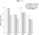
- "CRC17b”, “CRC25a”, “CRC32”, and “NLCRC32” are the names of organoid lines. The properties of each organoid line are shown in Table 1 above.
- CRC17b, CRC25a, and CRC32 are CRC organoids
- NLCRC32 is a normal tissue-derived organoid.
- FIG. 2 is a graph showing the results of calculating a doubling time of the organoids under each culture condition.
- HTS Healthcare high-throughput screening
- organoids are unevenly dissociated, particularly, in a case where the organoids are dissociated by mechanical shearing, selective proliferation of organoids occurs from a large fragment of the organoid, leading to variation in the proliferation of the organoids among wells.
- the organoids were labeled with GFP and sorted into individual wells of a 384-well plate by flow cytometric cell sorting. Fluorescent labeling of the organoids also has made it become possible to carry out automated imaging of the organoids and measurement of the proliferation of the organoids using a high-content cell image analyzer.
- the drug library included 9 types of anticancer drugs and 47 types of low-molecular-weight inhibitors targeting cancer-related signaling pathways, which are used in clinical practice.
- the organoid lines shown in Table 1 above 6 normal colon organoid lines and 20 CRC organoid lines were subjected to HTS. HTS was carried out with a total of 1,438 types of drug-organoid combinations using 237 screening plates.
- Fluorescence measurement and ATP-based luminescence assay showed similar results for many drugs.
- the bright field imaging analysis also gave results similar to those of the ATP-based luminescence assay.
- morphology varied among the CRC organoids, so the determination of whether the cells are alive or dead by the bright field imaging analysis required post hoc parameter calibration for each organoid line. For this reason, the determination of whether the cells are alive or dead by the bright field imaging analysis tends to impair the ease of screening and reduce the throughput.
- HTS using human normal colon organoids has been difficult to carry out in the related art due to poor recovery of the human normal colon organoids after dissociation thereof into single cells.
- the present inventors have found that a p38 inhibitor, which has recently been considered to be essential for culturing human intestinal organoids, inhibits the growth of organoids from single cells.
- the present inventors also found that replacing the p38 inhibitor with a combination of IGF-1 and FGF-2 promoted the growth of organoids from single cells.
- Organoids were cultured in a conventional medium containing a p38 inhibitor (SB202190) and in an improved medium containing IGF-1 and FGF-2 instead of the p38 inhibitor, and the proliferation of the organoids was measured based on the area of the fluorescent dye. 800 cells of each type were sorted and cultured in a 384-well plate.
- FIG. 3 is a graph showing the results of measuring the proliferation of an organoid.
- the left side of FIG. 3 shows the results for the normal colon organoid, and the right side of FIG. 3 shows the results for the CRC organoid.
- each dot indicates the measurement result for one well.
- the bars in the graph indicate the average value ⁇ standard deviation.
- FIG. 4 is a graph showing the results of calculating a coefficient of variation of an area of a fluorescent dye.
- the left side of FIG. 4 shows the results for the normal colon organoid, and the right side of FIG. 4 shows the results for the CRC organoid.
- FIG. 5 is a graph showing the results of calculating a Z'-factor for each organoid.
- FIG. 6 is a view showing the results of all drug-organoid combinations in HTS carried out in Experimental Example 2.
- drug responses are shown as Z-scores of log 2 (IC 50 ).
- Molecular targets are shown for some drugs.
- 82% of the screening plates exhibited a Z'-factor greater than 0.5.
- FIG. 7 is a graph showing the distribution of Z'-factors in 237 screening plates. 58 screening plates were seeded with normal colon organoids and 139 screening plates were seeded with CRC organoids. As shown in FIG. 7 , no significant difference was observed in the Z'-factor between normal colon organoids and CRC organoids.
- FIG. 8 is a graph showing a root mean square error (r.m.s.e) of a sigmoid response curve fitted to a dose-response curve. The average value of the root mean square error was 0.119, indicating the robustness of the curve fitting.
- FIG. 9 is a graph showing the results of comparing log 2 (IC 50 ) of 56 types of drugs for a normal colon organoid line (NLCRC15) acquired in triplicate between repeated experiments. As a result, it was confirmed that the results of three independent assays showed a high correlation.
- FIG. 10 is a graph showing the results of calculating Spearman's correlation coefficients for all pairs of organoid lines based on values of IC 50 .
- each dot indicates one organoid line pair.
- the bars in the graph indicate the average value ⁇ standard deviation.
- the six types of normal colon organoid lines showed comparable drug responses.
- the CRC organoid lines showed significant differences in drug responses.
- the drug responses were significantly different between the CRC organoid lines, but as shown in FIG. 6 , the CRC organoids showed similar responses to drugs that share the same targets, such as an EGFR inhibitor, an mTOR inhibitor, and a topoisomerase inhibitor.
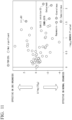
- FIG. 11 is a volcano plot showing the results of MANOVA.
- each dot indicates one type of drug. Dots with diagonal lines indicate drugs for which a statistically significant difference was observed. More than 60% of the drugs (36/56, 64.3%) showed a higher proliferation inhibitory activity in normal colon organoids than in CRC organoids. This was thought to be due to the rapid proliferation of normal colon organoids. Despite this bias, it was shown that the BET bromodomain protein inhibitor (+)-JQ1 ( CAS number: 1268524-70-4 ) suppressed the growth of organoids in a cancer-specific manner.
- each dot indicates one type of organoid line.
- FIG. 13 is a set of representative micrographs showing a response of a normal colon organoid and a CRC organoid to (+)-JQ1.
- the scale bar is 500 ⁇ m.
- "Normal” indicates the results for normal colon organoids
- "CRC7" and “CRC17a” indicate the names of CRC organoid lines.
- the CRC organoids were cultured in the same manner as in Example 1.
- the CRC organoids obtained by suspension culture were centrifuged, and the pellet was suspended in cold Matrigel.
- 0.2 mL of the CRC organoid suspension (1 ⁇ 10 6 cells/injection) was transplanted with subcutaneous injection into four sites on the left and right sides of nude mice.
- Tumor volumes were measured over time with an electronic caliber. After the tumor volume reached 100 to 150 mm 3 , cetuximab (40 mg/kg, available from Merck & Co., Inc.) was intraperitoneally injected every 3 days for 7 doses. In addition, nab-paclitaxel (20 mg/kg, available from Taiho Pharmaceutical Co., Ltd.) was intravenously injected twice a week for six doses. As a result of continuing the measurement of tumor volume, it was confirmed that the results of the drug effect in vivo were in good agreement with the results of HTS obtained in vitro.
- the present invention it is possible to provide a technology that efficiently enables large-scale culture of an organoid, regardless of whether the organoid is derived from a normal tissue or derived from a tumor tissue. According to the present invention, it is possible to carry out HTS screening of drugs that have few side effects on normal tissues and are specifically effective against tumor tissues.
Landscapes
- Health & Medical Sciences (AREA)
- Engineering & Computer Science (AREA)
- Life Sciences & Earth Sciences (AREA)
- Biomedical Technology (AREA)
- Chemical & Material Sciences (AREA)
- Biotechnology (AREA)
- Bioinformatics & Cheminformatics (AREA)
- Genetics & Genomics (AREA)
- Zoology (AREA)
- Organic Chemistry (AREA)
- Wood Science & Technology (AREA)
- Cell Biology (AREA)
- Immunology (AREA)
- Biochemistry (AREA)
- Microbiology (AREA)
- General Health & Medical Sciences (AREA)
- Hematology (AREA)
- Urology & Nephrology (AREA)
- Molecular Biology (AREA)
- General Engineering & Computer Science (AREA)
- Tropical Medicine & Parasitology (AREA)
- General Physics & Mathematics (AREA)
- Pathology (AREA)
- Analytical Chemistry (AREA)
- Physics & Mathematics (AREA)
- Medicinal Chemistry (AREA)
- Food Science & Technology (AREA)
- Toxicology (AREA)
- Oncology (AREA)
- Gastroenterology & Hepatology (AREA)
- Measuring Or Testing Involving Enzymes Or Micro-Organisms (AREA)
- Micro-Organisms Or Cultivation Processes Thereof (AREA)
Abstract
Description
- The present invention relates to a production method for an organoid. More specifically, the present invention relates to a production method for an organoid and a screening method for an anticancer drug. Priority is claimed on US provisional application No.
US 63/317,552, filed in the United States on March 8, 2022 - Drug screening, which comprehensively searches for the effects of a large number of therapeutic drug candidates, is a fundamental technology in drug discovery. In cancer drug discovery in the related art, a method has been adopted in which primary screening is carried out using a cancer cell line established from a patient-derived cancer tissue, and safety and side effects are confirmed using an animal model.
- However, cancer cell lines may lack many of the properties of actual clinical tumors and the sensitivity to anticancer drugs. In addition, even in a case where cancer occurs in the same organ, the cancer exhibits different genetic and biological characteristics from patient to patient. It is difficult to comprehensively cover this diversity in cancer cell lines. As a result, many of the anticancer drug candidates selected using cancer cell lines have not yet been applied clinically, and this "valley of death" in drug discovery research has become a major medical and social issue.
- The present inventors have pioneered the development of an organoid technology for permanent three-dimensional culture of tissue stem cells (see, for example,
- Non-Patent
Documents 1 and 2). The present inventors have also constructed cancer organoid biobanks for colorectal cancer, pancreatic cancer, gastric cancer, and the like by applying the organoid technology to various patient-derived cancer tissues (see, for example, Non-PatentDocuments 3 to 5). - Patient-derived cancer organoids retain many of the characteristics of cancer tissue of the patient, and cancer organoid biobanks are intended to be applied as next-generation cancer research resources to basic cancer research, cancer diagnostic tools, drug discovery, personalized treatment, and the like.
- Recent overseas clinical trials have shown that the drug sensitivity of patient-derived cancer organoids correlates with the response of clinical tumors to anticancer drugs, and there are also hopes for the implementation of organoid medicine, which uses organoids to predict the therapeutic effects of chemotherapy.
-
- Non-Patent Document 1: Sato T, et al., Single Lgr5 stem cells build crypt-villus structures in vitro without a mesenchymal niche, Nature, 459, 262-266, 2009.
- Non-Patent Document 2: Sato T, et al., Long-term Expansion of Epithelial Organoids From Human Colon, Adenoma, Adenocarcinoma, and Barrett's Epithelium, Gastroenterology, 141, 1762-1772, 2011.
- Non-Patent Document 3: Fujii M, et al., A Colorectal Tumor Organoid Library Demonstrates Progressive Loss of Niche Factor Requirements during Tumorigenesis, Cell Stem Cell, 18, 827-838, 2016.
- Non-Patent Document 4: Seino T, et al., Human Pancreatic Tumor Organoids Reveal Loss of Stem Cell Niche Factor Dependence during Disease Progression, Cell Stem Cell, 22, 454-467, 2018.
- Non-Patent Document 5: Nanki K, et al., Divergent Routes toward Wnt and R-spondin Niche Independency during Human Gastric Carcinogenesis, Cell, 174, 856-869, 2018.
- However, it is difficult to evaluate side effects and safety on normal tissues with drug screening in the related art. It is possible to culture a normal tissue by using the organoid technology. However, it has been difficult to efficiently carry out large-scale culture of a normal tissue-derived organoid with culture technologies in the related art. For this reason, it was not possible to introduce the normal tissue-derived organoid into drug screening.
- Therefore, an object of the present invention is to provide a technology that efficiently enables large-scale culture of an organoid, regardless of whether the organoid is derived from a normal tissue or derived from a tumor tissue.
- The present invention includes the following aspects.
- [1] A production method for an organoid, including a step of dissociating a biological tissue and a step of carrying out suspension culture of the dissociated biological tissue in a state of being dispersed in a medium to form an organoid, in which the medium is substantially free of a p38 inhibitor and contains the following components (i) to (iv).
- (i) 0.1% to 10% by volume of an extracellular matrix
- (ii) insulin-like growth factor 1 (IGF-1)
- (iii) fibroblast growth factor 2 (FGF-2)
- (iv) at least one selected from the group consisting of a Wnt agonist, a bone morphogenetic protein (BMP) inhibitor, a transforming growth factor-β (TGF-β) inhibitor, and an epidermal growth factor (EGF)
- [2] The production method according to [1], in which the suspension culture is carried out while stirring the medium.
- [3] The production method according to [1] or [2], in which the biological tissue is a normal tissue.
- [4] The production method according to any one of [1] to [3], in which the Wnt agonist is at least one selected from the group consisting of a Wnt protein, an R-spondin, and a GSK-3β inhibitor.
- [5] The production method according to any one of [1] to [4], in which the Wnt agonist is a Wnt protein and an R-spondin.
- [6] The production method according to [5], in which the Wnt protein is a complex with afamin.
- [7] A screening method for an anticancer drug, including a step of carrying out suspension culture of a normal tissue-derived organoid and a tumor tissue-derived organoid in a state of being dispersed in a medium in a presence of a test substance; and a step of evaluating an effect of the test substance on the normal tissue-derived organoid and the tumor tissue-derived organoid, in which the test substance that has few side effects on the normal tissue-derived organoid and is effective on the tumor tissue-derived organoid is shown to be an anticancer drug that is effective against the tumor tissue, and the medium is substantially free of a p38 inhibitor and contains the following components (i) to (iv).
- (i) 0.1% to 10% by volume of an extracellular matrix
- (ii) IGF-1
- (iii) FGF-2
- (iv) at least one selected from the group consisting of a Wnt agonist, a BMP inhibitor, a TGF-β inhibitor, and an EGF
- [8] The screening method according to [7], in which the normal tissue-derived organoid is an organoid produced by the production method according to [3]. Advantageous Effects of Invention
- According to the present invention, it is possible to provide a technology that efficiently enables large-scale culture of an organoid, regardless of whether the organoid is derived from a normal tissue or derived from a tumor tissue.
-
- [
FIG. 1 ] is a graph showing the results of measuring the proliferation of an organoid in Experimental Example 1. - [
FIG. 2 ] is a graph showing the results of calculating a doubling time of an organoid in Experimental Example 1. - [
FIG. 3 ] is a graph showing the results of measuring the proliferation of an organoid in Experimental Example 2. - [
FIG. 4 ] is a graph showing the results of calculating a coefficient of variation of an area of a fluorescent dye in Experimental Example 2. - [
FIG. 5 ] is a graph showing the results of calculating a Z'-factor for each organoid in Experimental Example 2. - [
FIG. 6 ] is a view showing the results of all drug-organoid combinations in high-throughput screening (HTS) carried out in Experimental Example 2. - [
FIG. 7 ] is a graph showing the distribution of Z'-factors in 237 screening plates in Experimental Example 3. - [
FIG. 8 ] is a graph showing a root mean square error (r.m.s.e) of a sigmoid response curve fitted to a dose-response curve in Experimental Example 3. - [
FIG. 9 ] is a graph showing the results of comparing log2 (IC50) of 56 types of drugs for a normal colon organoid line (NLCRC15) acquired in triplicate in Experimental Example 3 between repeated experiments. - [
FIG. 10 ] is a graph showing the results of calculating Spearman's correlation coefficients for all pairs of organoid lines based on values of IC50 in Experimental Example 3. - [
FIG. 11 ] is a volcano plot showing the results of multivariate analysis of variance (MANOVA) in Experimental Example 4. - [
FIG. 12 ] is a graph showing the IC50 of (+)-JQ1 for a normal colon organoid (n = 6) and a CRC organoid (n = 15), measured in Experimental Example 4. - [
FIG. 13 ] is a set of representative micrographs showing a response of a normal colon organoid and a CRC organoid to (+)-JQ1, acquired in Experimental Example 4. The scale bar is 500 µm. - In one embodiment, the present invention provides a production method for an organoid, including a step of dissociating a biological tissue and a step of carrying out suspension culture of the dissociated biological tissue in a state of being dispersed in a medium to form an organoid, in which the medium is substantially free of a p38 inhibitor and contains the following components (i) to (iv).
- (i) 0.1% to 10% by volume of an extracellular matrix
- (ii) IGF-1
- (iii) FGF-2
- (iv) at least one selected from the group consisting of a Wnt agonist, a BMP inhibitor, a TGF-β inhibitor, and an EGF
- As will be described later in Examples, the production method according to the present embodiment makes it possible to carry out stable large-scale culture of a normal tissue-derived organoid and to achieve large-scale production of the normal tissue-derived organoid. The production method according to the present embodiment can also be said to be a culture method for an organoid. In the present embodiment, the large-scale culture means, for example, proliferating organoids of 106 to 107 cells from a tissue derived from a living body.
- In the production method according to the present embodiment, the biological tissue may be a biological tissue derived from a patient, or may be an organoid which has already been established. In a case where the biological tissue is an organoid, the production method according to the present embodiment can also be said to be a subculture method for an organoid.
- In the production method according to the present embodiment, the biological tissue is preferably an epithelial tissue. Examples of the epithelial tissue include an intestinal epithelial tissue. The intestinal tract is an organ in the human body that has the largest area in contact with the external environment, and has functions essential for maintaining life such as digestion and absorption. Most of the functions of the intestinal tract are carried out by the intestinal epithelium that covers an inner layer of the intestinal tract. The intestinal epithelium is composed of two compartments: villi, which consist of three lineages of differentiated cells (mucus-producing cells, absorptive epithelial cells, and endocrine cells), and crypts, which mainly consist of undifferentiated proliferative cells. The epithelial tissue is preferably a tissue containing epithelial stem cells or epithelial cells. The epithelial cells include differentiated epithelial cells and epithelial stem cells. The "epithelial stem cell" refers to a cell having a long-term self-replication function and an ability to differentiate into an epithelial differentiated cell, and means a stem cell derived from an epithelial tissue. Examples of the epithelial tissue include tissues of cornea, oral mucosa, skin, conjunctiva, bladder, renal tubule, kidney, digestive organs (esophagus, stomach, duodenum, small intestine (including jejunum and ileum), and large intestine (including colon)), liver, pancreas, mammary gland, salivary gland, tear gland, prostate, hair root, trachea, lung, and the like.
- The biological tissue is preferably a tissue derived from a human or non-human mammal. The non-human mammal includes, but is not particularly limited to, a mouse, a rat, a cow, a pig, a rabbit, a monkey, and the like. In the production method according to the present embodiment, the biological tissue may be a tumor tissue or a cancer tissue, or may be a normal tissue. Here, the tumor tissue means a tissue that has become tumorous, and the cancer tissue means a tissue that has become cancerous.
- Dissociating a biological tissue means fragmenting the biological tissue and dividing the fragmented biological tissues into cell clusters each consisting of one cell to several cells. Here, "several" may mean about 2 to 100 cells. The dissociation of the biological tissue may be carried out by mechanical shearing or by an enzymatic treatment with collagenase, dispase I, liberase, trypsin, TrypLE Express (available from Thermo Fisher Scientific, Inc.), or the like.
- It is known that the organoid may have poor recovery after being dissociated into a single cell. On the other hand, the present inventors have found that a p38 inhibitor, which has recently been considered to be essential for culturing human intestinal organoids, inhibits the growth of organoids from single cells. The present inventors further found that replacing the p38 inhibitor with a combination of IGF-1 and FGF-2 promoted the growth of organoids from single cells.
- In addition, general organoid culture protocols in the related art include a step of embedding the dissociated organoid in an extracellular matrix such as Matrigel (registered trademark). However, the organoid embedded in the extracellular matrix may be difficult to scale up in culture. In addition, the organoid embedded in the extracellular matrix may exhibit a large variation in growth.
- On the other hand, the production method according to the present embodiment is carried out in such a manner that the dissociated biological tissue is subjected to suspension culture in a state of being dispersed in a medium. That is, in the production method according to the present embodiment, the dissociated biological tissue is not embedded in an extracellular matrix, but is subjected to suspension culture in a state in which cells are dispersed in a medium containing an extracellular matrix.
- As will be described later in Examples, the production method according to the present embodiment makes it possible to achieve stable large-scale culture of not only a cancer organoid but also a normal tissue-derived organoid. In addition, by accurately aligning the number of cells at the time of seeding the cells, it is possible to suppress the variation in the number of cells among wells. In order to align the number of cells, for example, sorting by a cell sorter can be used.
- Examples of p38 inhibitors that have been used in the culture of organoids in the related art include SB202190 (CAS number: 152121-30-7), SB203580 (CAS number: 152121-47-6), VX-702 (CAS number: 745833-23-2), VX-745 (CAS number: 209410-46-8), PD169316 28CAS number: 152121-53-4), RO4402257 (CAS number: 1449811-01-2), and BIRB796 (CAS number: 285983-48-4).
- On the other hand, the production method according to the present embodiment uses a medium that is substantially free of a p38 inhibitor. In the present specification, the phrase "substantially free of a p38 inhibitor" means that no p38 inhibitor is intentionally added to a medium, and means that the unavoidable incorporation of the p38 inhibitor is permitted within a range in which the effects of the present embodiment are exhibited.
- The medium used in the production method according to the present embodiment contains an extracellular matrix. The extracellular matrix is a non-cellular component and includes various polysaccharides, water, elastins, and glycoproteins. Examples of the glycoproteins include collagen, entactin (nidogen), fibronectin, and laminin.
- A commercially available extracellular matrix may be used as the extracellular matrix. Examples of the commercially available extracellular matrix include an extracellular matrix protein (available from Thermo Fisher Scientific, Inc.), a basement membrane preparation derived from Engelbreth-Holm-Swarm (EHS) mouse sarcoma cells (for example, Matrigel (registered trademark), available from Corning Incorporated), and ProNectin (available from Sigma-Aldrich Co. LLC).
- The medium used in the production method according to the present embodiment preferably contains 0.1% to 10% by volume, for example, 0.1% to 5% by volume, for example, 0.1% to 3% by volume, or for example, about 2% by volume, of an extracellular matrix.
- The medium used in the production method according to the present embodiment contains IGF-1. IGF-1, also known as somatomedin C, is a factor secreted primarily in the liver in response to stimulation by growth hormone (GH). The concentration of IGF-1 contained in the medium may be, for example, 5 ng/mL to 1 µg/mL, for example, 10 ng/mL to 1 µg/mL, and for example, 50 to 500 ng/mL.
- The medium used in the production method according to the present embodiment contains FGF-2. FGF-2 is a basic fibroblast growth factor, also called bFGF. The concentration of FGF-2 contained in the medium may be, for example, 5 ng/mL to 1 µg/mL, for example, 10 ng/mL to 1 µg/mL, and for example, 50 to 500 ng/mL.
- As will be described later in Examples, the growth of organoids from single cells is promoted by replacing a p38 inhibitor, which has been considered to be essential for the culture of organoids in the related art, with a combination of IGF-1 and FGF-2. As a result, it is possible to achieve stable large-scale culture of not only a cancer organoid but also a normal tissue-derived organoid.
- The medium used in the production method according to the present embodiment contains at least one selected from the group consisting of a Wnt agonist, a BMP inhibitor, a TGF-β inhibitor, and EGF. The medium used in the production method according to the present embodiment may contain two selected from the group consisting of a Wnt agonist, a BMP inhibitor, a TGF-β inhibitor, and EGF, may contain three selected from the group consisting of a Wnt agonist, a BMP inhibitor, a TGF-β inhibitor, and EGF, or may contain a Wnt agonist, a BMP inhibitor, a TGF-β inhibitor, and EGF.
- The Wnt agonist refers to a substance that activates T-cell factor (hereinafter, sometimes referred to as "TCF")/lymphoid enhancer factor (hereinafter, sometimes referred to as "LEF")-mediated transcription in a cell. The Wnt agonist is not limited to a Wnt family protein, and includes an agonist that binds to and activates members of the Frizzled receptor family, an inhibitor of intracellular β-catenin degradation, and an activator of TCF/LEF.
- The Wnt agonist is preferably at least one selected from the group consisting of a Wnt protein, an R-spondin, and a GSK-3β inhibitor.
- The Wnt agonist is a substance that stimulates Wnt activity in a cell by at least 10%, preferably at least 20%, more preferably at least 30%, still more preferably at least 50%, particularly preferably at least 90%, and most preferably 100% of the level of Wnt activity in the presence of the Wnt agonist, as compared to the level of Wnt activity in the absence of the Wnt agonist. The Wnt activity can be evaluated, for example, by measuring a transcriptional activity of Wnt by a TCF reporter assay.
- The Wnt agonist is preferably a combination of a Wnt protein and an R-spondin. Where the Wnt protein is derived from is not particularly limited, and Wnt proteins derived from various organisms can be used. Above all, a mammal-derived Wnt protein is preferable. The mammal is the same as described hereinbefore. Examples of the mammalian Wnt protein include Wnt1, Wnt2, Wnt2b, Wnt3, Wnt3a, Wnt4, Wnt5a, Wnt5b, Wnt6, Wnt7a, Wnt7b, Wnt8a, Wnt8b, Wnt9a, Wnt9b, Wnt10a, Wnt10b, Wnt11, and Wnt16. One type of Wnt protein may be used alone, or two or more types of Wnt proteins may be used in combination.
- The Wnt protein may be a Wnt protein prepared using Wnt protein-expressing cells, or may be a commercially available Wnt protein. The Wnt protein may be a fragment of the Wnt protein or may be a Wnt protein containing an amino acid sequence other than the amino acid sequence of the Wnt protein as long as it has a Wnt activity. Examples of the amino acid sequence other than the amino acid sequence of the Wnt protein include an amino acid sequence of an affinity tag. In addition, the amino acid sequence of the Wnt protein does not need to be completely identical to an amino acid sequence that is available from a known database such as GenBank, and may be an amino acid sequence in which one to several amino acids have been deleted, substituted, or added in the amino acid sequence that is available from a known database, as long as it has a Wnt activity. Here, the number of from one to several is preferably 10 or less, more preferably 7 or less, and still more preferably 6 or less.
- The concentration of the Wnt protein contained in the medium may be, for example, 50 ng/mL to 10 µg/mL, for example, 100 ng/mL to 10 µg/mL, for example, 200 ng/mL to 1 µg/mL, and for example, 300 ng/mL to 1 µg/mL.
- It is preferable that the Wnt protein forms a complex with afamin. The afamin is a glycoprotein that belongs to an albumin family and is known to be present in body fluids such as blood. Serum added to a cell culture medium contains afamin derived from an animal from which the serum was collected. The serum contains impurities or the like other than afamin, so it is preferable to use afamin alone without using the serum.
- Where the afamin is derived from is not particularly limited, and afamin derived from various organisms can be used. Above all, mammal-derived afamin is preferable. The mammal is the same as described hereinbefore.
- The afamin may be natural afamin contained in serum or the like, which is purified by a known method, or may be recombinant afamin. The recombinant afamin can be produced by appropriately using a known genetic recombination technology. The recombinant afamin can be produced, for example, by inserting DNA encoding afamin into a known expression vector, introducing the resulting expression vector into an appropriate host cell to express recombinant afamin, and purifying the recombinant afamin using a known purification method.
- The recombinant afamin may be afamin with an affinity tag added. The affinity tag to be added is not particularly limited and can be appropriately selected from known affinity tags and then used. The affinity tag is preferably an affinity tag which is recognized by a specific antibody, and examples thereof include a FLAG tab, an MYC tag, an HA tag, and a V5 tag.
- The above-mentioned Wnt protein has strong hydrophobicity because a specific serine residue is modified with a fatty acid (palmitoleic acid). Therefore, it is widely known that the Wnt protein is prone to aggregation or denaturation in an aqueous solution, making purification and storage of the Wnt protein extremely difficult. Meanwhile, it has been reported that the modification of this specific serine residue with a fatty acid is essential for the physiological activity of the Wnt protein and is involved in binding to members of the Frizzled receptor family.
- The Wnt protein binds to afamin on a one-to-one basis to form a complex in an aqueous solution and can therefore be solubilized while maintaining a high physiological activity. Therefore, a Wnt protein-afamin complex may be produced by culture of cells expressing both a Wnt protein and afamin, or a Wnt protein-afamin complex may be produced by co-culture of Wnt protein-expressing cells and afamin-expressing cells. The Wnt protein-afamin complex may be added to the medium in the form of a conditioned medium.
- The concentration of afamin contained in the medium may be, for example, 50 ng/mL to 10 µg/mL, for example, 100 ng/mL to 10 µg/mL, for example, 200 ng/mL to 1 µg/mL, and for example, 300 ng/mL to 1 µg/mL.
- Examples of the R-spondin include the R-spondin family consisting of R-
spondin 1, R-spondin 2, R-spondin 3, and R-spondin 4. The R-spondin family is a secretory protein known to be involved in the activation and regulation of the Wnt signaling pathway. The R-spondin to be used may be a fragment of the R-spondin or an R-spondin containing an amino acid sequence other than the amino acid sequence of the R-spondin, such as an affinity tag, as long as it has an R-spondin activity. One type of R-spondin may be used alone, or two or more types of R-spondins may be used in combination. - The concentration of R-spondin contained in the medium may be, for example, 50 ng/mL to 10 µg/mL, for example, 100 ng/mL to 10 µg/mL, for example, 200 ng/mL to 1 µg/mL, and for example, 300 ng/mL to 1 µg/mL. The R-spondin may be added to the medium in the form of a conditioned medium.
- Examples of the GSK-3β inhibitor include CHIR99021 (CAS number: 252917-06-9), kenpaullone (CAS number: 142273-20-9), and 6-bromoindirubin-3'-oxime (BIO, CAS number: 667463-62-9).
- Examples of the BMP inhibitor include substances having an inhibitory activity of preferably 50% or more, more preferably 70% or more, still more preferably 80% or more, and particularly preferably 90% or more, as compared with the level of BMP activity in the absence of the inhibitor. The BMP inhibitory activity can be evaluated by measuring the transcriptional activity of BMP.
- The BMP inhibitor is preferably a naturally occurring BMP-binding protein, examples of which include a chordin-like protein such as noggin, gremlin, chordin, or a chordin domain; a follistatin-related protein such as follistatin or a follistatin domain; a DAN-like protein such as a differential screening-selected gene aberrative in neuroblastoma (DAN) or a DAN cysteine-knot domain; sclerostin (SOST); decorin; and α-2-macroglobulin.
- The concentration of the BMP inhibitor contained in the medium may be, for example, 10 ng/mL to 1 µg/mL, for example, 20 ng/mL to 1 µg/mL, for example, 50 ng/mL to 1 µg/mL, and for example, about 100 ng/mL.
- TGF-β is a type of growth factor and is produced in almost all cells of the kidney, bone marrow, platelets, and the like. There are five subtypes (β1 to β5) in TGF-β. In addition, it is known that TGF-β promotes the proliferation of osteoblasts and the synthesis and proliferation of connective tissues such as collagen and exhibits an inhibitory effect against epithelial cell proliferation and osteoclasts. In general, the TGF-β inhibitor is, for example, a drug that blocks or inhibits the binding of TGF-β to a TGF-β receptor and binds to TGF-β to form a complex which neutralizes the TGF-β activity. In addition, the TGF-β inhibitor is, for example, a drug that binds to a TGF-β receptor to block or inhibit the binding of TGF-β to the TGF-β receptor and acts as an antagonist or inverse agonist.
- Examples of the TGF-β inhibitor include substances having an inhibitory activity of preferably 50% or more, more preferably 70% or more, still more preferably 80% or more, and particularly preferably 90% or more, as compared with the level of TGF-β activity in the absence of the inhibitor.
- Examples of the TGF-β inhibitor include A83-01 (CAS number: 909910-43-6), SB-431542 (CAS number: 301836-41-9), SB-505124 (CAS number: 694433-59-5), SB-525334 (CAS number: 356559-20-1), LY364947 (CAS number: 396129-53-6), SD-208 (CAS number: 627536-09-8), and SJN2511 (CAS number: 446859-33-2). Among these, A83-01 is preferable.
- The concentration of the TGF-β inhibitor contained in the medium may be, for example, 100 nM to 1 µM, for example, 200 nM to 1 µM, for example, 300 nM to 1 µM, or for example, about 500 nM.
- In the production method according to the present embodiment, the medium may contain a mitogenic growth factor in addition to the above-mentioned components. Examples of the mitogenic growth factor include an epidermal growth factor (EGF), a transforming growth factor-α (TGF-α), and a keratinocyte growth factor (KGF). These mitogenic growth factors may be used alone or in combination of two or more thereof.
- EGF is a potent mitogen for a variety of cultured ectodermal cells and mesodermal cells and has a significant effect on the differentiation of some fibroblasts and specific cells. The EGF precursor exists as a membrane-bound molecule that is proteolytically cleaved to generate a 53-amino acid peptide hormone that stimulates cells.
- Above all, EGF is preferable as the mitogenic growth factor. The concentration of EGF contained in the medium may be, for example, 5 to 500 ng/mL, for example, 10 to 400 ng/mL, and for example, about 50 ng/mL.
- In the production method according to the present embodiment, it is preferable that the suspension culture of the dissociated biological tissue is carried out while stirring the medium. In addition, as will be described later in Examples, in a case of carrying out the suspension culture with stirring, the organoids can be proliferated more rapidly than in a case of the suspension culture.
- In one embodiment, the present invention provides a screening method for an anticancer drug, including a step of carrying out suspension culture of a normal tissue-derived organoid and a tumor tissue-derived organoid in a state of being dispersed in a medium in the presence of a test substance; and a step of evaluating an effect of the test substance on the normal tissue-derived organoid and the tumor tissue-derived organoid, in which the test substance that has few side effects on the normal tissue-derived organoid and is effective on the tumor tissue-derived organoid is shown to be an anticancer drug that is effective against the tumor tissue, and the medium is substantially free of a p38 inhibitor and contains the following components (i) to (iv).
- (i) 0.1% to 10% by volume of an extracellular matrix
- (ii) IGF-1
- (iii) FGF-2
- (iv) at least one selected from the group consisting of a Wnt agonist, a BMP inhibitor, a TGF-β inhibitor, and an EGF
- In the related art, it has been difficult to achieve stable large-scale culture of a normal tissue-derived organoid, and thus it has not been possible to incorporate the normal tissue-derived organoid into drug screening. For this reason, it has been difficult to evaluate side effects and safety of a drug on normal tissues.
- On the other hand, as will be described later in Examples, the screening method according to the present embodiment makes it possible to apply normal tissue-derived organoids, along with tumor tissue-derived organoids, to drug screening. As a result, it is possible to evaluate side effects and safety of a drug on normal tissues. In addition, as will be described later in Examples, it is possible to screen for a drug that specifically suppresses the growth of tumor tissue-derived organoids without affecting the proliferation of normal tissue-derived organoids.
- As will be described later in Examples, by accurately aligning the number of cells at the time of seeding organoids, it is possible to suppress the variation in the number of cells among wells. In order to align the number of cells, for example, sorting by a cell sorter can be used.
- In the screening method according to the present embodiment, the test substance is not particularly limited, and for example, a natural compound library, a synthetic compound library, or an existing drug library can be used. The screening method according to the present embodiment makes it possible to carry out high-throughput screening (HTS) having high accuracy, robustness, and reproducibility.
- In the screening method according to the present embodiment, the normal tissue-derived organoid is preferably an organoid produced by the above-mentioned production method. In the screening method according to the present embodiment, the extracellular matrix, IGF-1, FGF-2, Wnt agonist, BMP inhibitor, TGF-β inhibitor, EGF, organoid, and the like are the same as those described hereinbefore.
- Hereinafter, the present invention will be described in more detail by way of Examples; however, the present invention is not intended to be limited to the following Examples.
- Seven newly established organoid lines and patient-derived organoid lines established in a previous study were used. All samples used for organoid establishment and biological analysis were obtained from Keio University Hospital or the University of Tokyo Hospital with written informed consent. The present study was approved by the ethics committees of both research institutes.
- Normal colon tissues and neoplastic colon tissues were obtained from any of endoscopic biopsies or surgically resected specimens. Samples were stored in ice-cold phosphate buffered saline (PBS) until the treatment. Fresh tissue samples were cut into small pieces, washed at least 10 times with ice-cold PBS, and then digested with liberase (TH grade, available from F. Hoffmann-La Roche Ltd.) at 37°C for 60 min while being vigorously pipetted every 15 min. The remaining fragments were further treated with TrypLE Express (available from Thermo Fisher Scientific, Inc.) at 37°C for 20 min. Subsequently, the supernatant was collected and centrifuged at 200 × g for 3 min at 4°C. Subsequently, the cell pellet was suspended in Matrigel (registered trademark) (growth factor reduced, available from BD Biosciences) and dispensed into a 48-well culture plate (25 µL of Matrigel (registered trademark) per well). The names of the organoid lines used and the properties of the tissues from which the organoids were derived are shown in Table 1 below. In Table 1, in the column of "Histology", "NL" indicates a normal tissue, "TA" indicates a tubular adenoma, "SSL" indicates a serrated lesion, "well" indicates a well-differentiated adenocarcinoma, "mod" indicates a moderately differentiated adenocarcinoma, "por" indicates a poorly differentiated adenocarcinoma, and "muc" indicates a mucinous adenocarcinoma. In addition, in the column of "Position", "A" indicates the ascending colon, "T" indicates the transverse colon, "D" indicates the descending colon, "S" indicates the sigmoid colon, "R" indicates the rectum, "LM" indicates liver metastasis, and "C" indicates the cecum. In addition, in the column of "Gender", "F" indicates a female and "M" indicates a male. In addition, in the column of "Culture conditions", "W" indicates Wnt, "E" indicates EGF, "N" indicates noggin, "R" indicates R-Spondin1, "A" indicates A83-01, "S" indicates SB202190, and "-" indicates that no growth factor is required.
[Table 1] Organoid name Histology Position Gender Age Stage Culture conditions NLSL1 NL A F 68 - WENARFI NLCRC15 NL T? F 84 - WENARFI NLCRC25 NL T? M 50 - WENARFI NLCRC26 NL ? F 66 - WENARFI NLCRC32 NL D M 68 - WENARFI NLCRC33 NL T F 74 - WENARFI NLCRC35 NL R M 50 - WENARFI NLSL3 NL A M 69 - WENARFI Ad2 TA A A 40 - ENAFI SL3 SSL A M 69 - WENRA CRC5 well D M 58 I ENAFI CRC7 mod S M 85 II ENA CRC11 mod C M 75 II - CRC12 mod A M 45 II ENA CRC13 muc T F 70 II WENAR CRC15 por T F 84 II ENA CRC17a por, mod A M 82 III ENA CRC17b por, mod A M 82 III - CRC18a muc C F 70 III WENARAFI CRC18b muc C F 70 III WENARAFI CRC20 mod R F 67 IV E CRC23b mod R M 72 IV EFI CRC24b mod R F 48 IV ENAFI CRC25a mod T M 50 IV ENA CRC25c mod LM M 50 IV ENA CRC28 mod LM M 63 IV EA CRC31 por, mod R M 45 II ENFI CRC32 por, mod D M 68 IV - CRC33 mod T F 74 II WNRA CRC34 mod R F 40 IV ENAFI - Human colorectal cancer (CRC) organoids were cultured in a medium containing 2% by volume of Matrigel (registered trademark) (available from Corning Incorporated). The medium used was a medium in which a basal medium was supplemented with various combinations of growth factors depending on whether the organoid is derived from a normal tissue or a tumor tissue, and depending on the origin of the tumor in a case where the organoid is derived from a tumor tissue. In a case of stirring the medium, Orbital Shaker MaxQ 2000 CO2 Plus, CO2 Resistant (available from Thermo Fisher Scientific, Inc.) was used.
- The basal medium used was a medium in which an advanced DMEM/F12 was supplemented with penicillin/streptomycin, 10 mM HEPES, 2 mM GlutaMAX, 1 × B27 (available from Thermo Fisher Scientific, Inc.), 10 nM gastrin I (available from Sigma-Aldrich Co. LLC), and 1 mM N-acetylcysteine (available from FUJIFILM Wako Pure Chemical Corporation).
- The normal tissue-derived organoids were maintained in an improved medium or a conventional medium. The improved medium was a medium in which a basal medium was supplemented with the following seven types of growth factors.
- 20% afamin-Wnt3A serum-free conditioned medium
- 10% R-spondin-1 conditioned medium
- 50 ng/mL mouse recombinant EGF (available from Thermo Fisher Scientific, Inc.)
- 500 nM A83-01 (available from Tocris Bioscience)
- 100 ng/mL mouse recombinant noggin (available from PeproTech Inc.)
- 100 ng/mL recombinant human IGF-1 (available from BioLegend, Inc.)
- 50 ng/mL recombinant human FGF-basic (FGF-2) (available from PeproTech Inc.)
- The conventional medium was the same as the improved medium, except that 10 µM SB202190 (available from Sigma-Aldrich Co. LLC) was added instead of IGF-1 and FGF-2. In the tumor organoids, some growth factors were not required due to genetic mutations.
- The subculture was carried out by collecting and washing the organoids and fragmenting the organoids by mechanical shearing or digestion with TrypLE Express (available from Thermo Fisher Scientific, Inc.). For 2 days after the subculture, the fragments of the organoids were cultured in a fresh medium containing 10 µM Y-27632 (available from FUJIFILM Wako Pure Chemical Corporation) to suppress cell death.
- The standard inhibitor kit (SCADS Inhibitor Kit) drug library was provided by the Screening Committee of Anticancer Drugs on New Academic Area Research, Cancer Research Support Project, Grant-in-Aid for Scientific Research from The Ministry of Education, Culture, Sports, Science and Technology, Japan.
- In the present study, a total of 23 organoid lineages were subjected to screening. Prior to screening, the organoids were labeled with a green fluorescent protein (GFP) using PB-CMV-MCS-EF1α-GFP-Puro PiggyBac (PB513B-1, available from System Biosciences, LLC.) and Super PiggyBac transposase (PB210-20, available from System Biosciences, LLC.) vectors.
- The organoids were dissociated with TrypLE Express (available from Thermo Fisher Scientific, Inc.) and filtered using a cell strainer (pore size: 20 µm, available from Sysmex Corporation). Using a cell sorter (SH800, available from Sony Corporation), 800 to 1,500 cells were sorted into each well of a 384-well plate.
- The organoids were treated with a test compound 24 hours or 96 hours after seeding. The organoids were grown for 6 to 8 days, and the colony area was measured based on the fluorescence of GFP using an IN Cell Analyzer 6000 confocal imaging system (available from GE Healthcare Life Sciences). In addition, bright field imaging and luminescence-based ATP measurement were carried out using Cell3iMager duos (available from SCREEN Holdings Co., Ltd.) and CellTiter-Glo cell viability assay (available from Promega Corporation).
- The bright field image was analyzed using software integrated into the Cell3iMager. Default parameters were used for the pretreatment and the object detection, and supervised viability determination was applied to the machine learning. In the evaluation assay of (+)-JQ1, the cell viability was determined using a CellTiter-Glo cell viability assay (available from Promega Corporation).
- The dose-response curve was fitted to the drug response data using a Bayesian sigmoid model. The drug response data was obtained in triplicate as drug responses at 7 points at a 2-fold concentration, including 6 or more drug-free controls. In all the screening plates, the proteasome inhibitor MG-132 was tested at a maximum concentration of 1 µM, giving 100% cell death and an accurate estimation of the background signal.
- The sigmoid response curve of 4 parameters was fitted as follows. First, the viability of the organoid was normalized by dividing by the average of the drug-free controls. The viability Xlc of the organoid was estimated to be an average value of Expression (1).

- In Expression (1), Imax represents a viability of a drug-free control, Imin represents an average of background signals, βpos represents a position of a sigmoid curve, and βshape represents steepness. 1c represents the log concentration.
- It was assumed that the viability Xlc of the organoid has a gamma distribution with variance Var(Xlc) = BE(Xlc). Here, B represents a noise parameter. It was also assumed that the viability Xmax of the drug-free control has a gamma distribution with variance Var(Xmax) = BImax.
- The Markov chain Monte Carlo simulation was carried out using rstan (v.2.18.0) to estimate the parameters. The initial values were randomly generated from the prior distribution. Following 1,000 burn-in iterations, 1,000 iterations were sampled from four independent chains to obtain a posterior distribution. The median of the posterior distribution was used as the parameter estimate value. βpos had a normal prior distribution with a 99% probability mass covering a range from 1/100 of the tested minimum concentration to 100 times the tested maximum concentration. Imax had a uniform prior distribution between 0 and 5. Imin was reparameterized as Imin = αImax where α has a uniform prior distribution between 0 and 1. B and βshape had low-informative prior probabilities (half-Cauchy distribution with location = 0, scale = 10). In a case where the R convergence metric was > 1.1 or in a case where the average absolute error was > 0.3, the curve fitting was deemed insufficient and discarded without further downstream analysis.
- AUC values were estimated using trapezoidal approximation. The AUC values were determined as a part of a rectangle that covers the tested concentration range and a region surrounded by Imax and Imin in a vertical direction.
- Human colorectal cancer (CRC) organoids were subjected to suspension culture in a medium supplemented with 2% by volume of Matrigel (registered trademark), and the proliferation thereof was examined. For the purpose of comparison, the culture was also carried out by embedding the organoids in droplets of Matrigel (registered trademark), which is a culture method in the related art. In addition, suspension culture with stirring, which involves suspension culture of the organoids and stirring of the medium, was also carried out.
-
FIG. 1 is a graph showing the results of measuring the proliferation of organoids under each culture condition (n = 3). InFIG. 1 , "CRC17b", "CRC25a", "CRC32", and "NLCRC32" are the names of organoid lines. The properties of each organoid line are shown in Table 1 above. CRC17b, CRC25a, and CRC32 are CRC organoids, and NLCRC32 is a normal tissue-derived organoid. As a result, it was revealed that in any of the organoids, the organoids more rapidly proliferated to a scale of 106 to 107 cells in a case of carrying out the suspension culture than in a case of carrying out the culture embedded in droplets of Matrigel (registered trademark). In addition, in a case of carrying out the suspension culture with stirring, the organoids proliferated even more rapidly. -
FIG. 2 is a graph showing the results of calculating a doubling time of the organoids under each culture condition. As a result, it was found that, compared to the culture embedded in droplets of Matrigel (registered trademark), the suspension culture tends to have a shorter doubling time, and the suspension culture with stirring tends to have a further shorter doubling time. - In a case where the organoids are unevenly dissociated, particularly, in a case where the organoids are dissociated by mechanical shearing, selective proliferation of organoids occurs from a large fragment of the organoid, leading to variation in the proliferation of the organoids among wells.
- Therefore, in order to accurately distribute the same number of living single cells per one culture well, the organoids were labeled with GFP and sorted into individual wells of a 384-well plate by flow cytometric cell sorting. Fluorescent labeling of the organoids also has made it become possible to carry out automated imaging of the organoids and measurement of the proliferation of the organoids using a high-content cell image analyzer.
- Using a drug library containing 56 types of drugs, the drug sensitivity of CRC organoids and normal colon organoids to each drug was analyzed. The drug library included 9 types of anticancer drugs and 47 types of low-molecular-weight inhibitors targeting cancer-related signaling pathways, which are used in clinical practice. Among the organoid lines shown in Table 1 above, 6 normal colon organoid lines and 20 CRC organoid lines were subjected to HTS. HTS was carried out with a total of 1,438 types of drug-organoid combinations using 237 screening plates.
- Drugs from the drug library were added 4 days after seeding the cells, and the proliferation of the organoids was calculated based on the area of the fluorescent dye. In order to confirm that the results obtained by this method are comparable to the results obtained by other methods, the viability of organoids was also analyzed by an ATP-based luminescence assay and bright field imaging analysis.
- Fluorescence measurement and ATP-based luminescence assay showed similar results for many drugs. In addition, the bright field imaging analysis also gave results similar to those of the ATP-based luminescence assay. However, morphology varied among the CRC organoids, so the determination of whether the cells are alive or dead by the bright field imaging analysis required post hoc parameter calibration for each organoid line. For this reason, the determination of whether the cells are alive or dead by the bright field imaging analysis tends to impair the ease of screening and reduce the throughput.
- HTS using human normal colon organoids has been difficult to carry out in the related art due to poor recovery of the human normal colon organoids after dissociation thereof into single cells. On the other hand, the present inventors have found that a p38 inhibitor, which has recently been considered to be essential for culturing human intestinal organoids, inhibits the growth of organoids from single cells. The present inventors also found that replacing the p38 inhibitor with a combination of IGF-1 and FGF-2 promoted the growth of organoids from single cells.
- Organoids were cultured in a conventional medium containing a p38 inhibitor (SB202190) and in an improved medium containing IGF-1 and FGF-2 instead of the p38 inhibitor, and the proliferation of the organoids was measured based on the area of the fluorescent dye. 800 cells of each type were sorted and cultured in a 384-well plate.
-
FIG. 3 is a graph showing the results of measuring the proliferation of an organoid. The left side ofFIG. 3 shows the results for the normal colon organoid, and the right side ofFIG. 3 shows the results for the CRC organoid. InFIG. 3 , each dot indicates the measurement result for one well. The bars in the graph indicate the average value ± standard deviation. InFIG. 3 , "*" indicates that there is a significant difference at P = 9.51 × 10⁻¹³ (NLCRC32 line, n = 22 wells), P = 1.12 × 10⁻²¹ (CRC24b line, n = 44 wells), and P = 4.52 × 10⁻²¹ (CRC31 line, n = 36 wells). -
FIG. 4 is a graph showing the results of calculating a coefficient of variation of an area of a fluorescent dye. The left side ofFIG. 4 shows the results for the normal colon organoid, and the right side ofFIG. 4 shows the results for the CRC organoid. InFIG. 4 , "*" indicates that there is a significant difference at P = 7.28 × 10⁻⁸ (NLCRC32 line, n = 22 wells), P = 5.69 × 10⁻⁵ (CRC24b line, n = 44 wells), and P = 3.95 × 10⁻² (CRC31 line, n = 36 wells). -
FIG. 5 is a graph showing the results of calculating a Z'-factor for each organoid. As a result, it was revealed that application of the improved medium makes it possible to achieve not only improved culture efficiency of the organoids, but also uniform culture of even normal colon organoids that had been sorted into single cells. In addition, since the Z'-factor was higher than 0.5, it was demonstrated that the present screening system was a high-quality HTS system. - The above results demonstrate that the drug screening of various organoids including a normal tissue-derived organoid can be carried out by the HTS system of the present experimental example.
-
FIG. 6 is a view showing the results of all drug-organoid combinations in HTS carried out in Experimental Example 2. InFIG. 6 , drug responses are shown as Z-scores of log2 (IC50). Molecular targets are shown for some drugs. In HTS, 82% of the screening plates exhibited a Z'-factor greater than 0.5. -
FIG. 7 is a graph showing the distribution of Z'-factors in 237 screening plates. 58 screening plates were seeded with normal colon organoids and 139 screening plates were seeded with CRC organoids. As shown inFIG. 7 , no significant difference was observed in the Z'-factor between normal colon organoids and CRC organoids. - Each drug response was summarized as the 50% inhibitory concentration (IC50), the steepness of the dose-response curve, and the area under the dose-response curve.
FIG. 8 is a graph showing a root mean square error (r.m.s.e) of a sigmoid response curve fitted to a dose-response curve. The average value of the root mean square error was 0.119, indicating the robustness of the curve fitting. -
FIG. 9 is a graph showing the results of comparing log2 (IC50) of 56 types of drugs for a normal colon organoid line (NLCRC15) acquired in triplicate between repeated experiments. As a result, it was confirmed that the results of three independent assays showed a high correlation. - The above results demonstrate that the HTS system of Experimental Example 2 has high accuracy, robustness, and reproducibility.
-
FIG. 10 is a graph showing the results of calculating Spearman's correlation coefficients for all pairs of organoid lines based on values of IC50. InFIG. 10 , each dot indicates one organoid line pair. The bars in the graph indicate the average value ± standard deviation. In addition, "*" indicates that there is a significant difference at P = 1.34 × 10⁻⁶, and "**" indicates that there is a significant difference at P = 3.61 × 10⁻⁷. In addition, "Normal-Normal" indicates the results of a pair of normal colon organoid lines (n = 15), "Normal-CRC" indicates the results of a pair of a normal colon organoid line and a CRC organoid line (n = 120), and "CRC-CRC" indicates the results of a pair of CRC organoid lines (n = 190). - As a result, the six types of normal colon organoid lines showed comparable drug responses. On the other hand, the CRC organoid lines showed significant differences in drug responses. The drug responses were significantly different between the CRC organoid lines, but as shown in
FIG. 6 , the CRC organoids showed similar responses to drugs that share the same targets, such as an EGFR inhibitor, an mTOR inhibitor, and a topoisomerase inhibitor. - From these results, it was considered that the heterogeneity in drug response of CRC organoids is not due to technical issues but is due to genetic or epigenetic diversity of CRC organoids.
- Based on the results of HTS carried out in Experimental Example 2, drug responses to normal colon organoids and CRC organoids were compared using multivariate analysis of variance (MANOVA).
-
FIG. 11 is a volcano plot showing the results of MANOVA. InFIG. 11 , each dot indicates one type of drug. Dots with diagonal lines indicate drugs for which a statistically significant difference was observed. More than 60% of the drugs (36/56, 64.3%) showed a higher proliferation inhibitory activity in normal colon organoids than in CRC organoids. This was thought to be due to the rapid proliferation of normal colon organoids. Despite this bias, it was shown that the BET bromodomain protein inhibitor (+)-JQ1 (CAS number: 1268524-70-4) suppressed the growth of organoids in a cancer-specific manner. -
FIG. 12 is a graph showing the IC50 of (+)-JQ1 for a normal colon organoid (n = 6) and a CRC organoid (n = 15). InFIG. 12 , each dot indicates one type of organoid line. The bars in the graph indicate the average value ± standard deviation. "*" indicates that there is a significant difference at P = 1.38 × 10⁻³. -
FIG. 13 is a set of representative micrographs showing a response of a normal colon organoid and a CRC organoid to (+)-JQ1. The scale bar is 500 µm. InFIG. 13 , "Normal" indicates the results for normal colon organoids, and "CRC7" and "CRC17a" indicate the names of CRC organoid lines. - The above results demonstrate that it is useful to use a normal organoid together with a cancer organoid for the discovery of a potential therapeutic method.
- All animal experiments were carried out with the approval of the Laboratory Animal Center, Keio University School of Medicine. The verification was carried out by xenografting CRC organoids into nude mice to determine whether the results of HTS using in vitro cultured organoids were the same as those in vivo.
- The CRC organoids were cultured in the same manner as in Example 1. The CRC organoids obtained by suspension culture were centrifuged, and the pellet was suspended in cold Matrigel. 0.2 mL of the CRC organoid suspension (1 × 10⁶ cells/injection) was transplanted with subcutaneous injection into four sites on the left and right sides of nude mice.
- Tumor volumes were measured over time with an electronic caliber. After the tumor volume reached 100 to 150 mm3, cetuximab (40 mg/kg, available from Merck & Co., Inc.) was intraperitoneally injected every 3 days for 7 doses. In addition, nab-paclitaxel (20 mg/kg, available from Taiho Pharmaceutical Co., Ltd.) was intravenously injected twice a week for six doses. As a result of continuing the measurement of tumor volume, it was confirmed that the results of the drug effect in vivo were in good agreement with the results of HTS obtained in vitro.
- According to the present invention, it is possible to provide a technology that efficiently enables large-scale culture of an organoid, regardless of whether the organoid is derived from a normal tissue or derived from a tumor tissue. According to the present invention, it is possible to carry out HTS screening of drugs that have few side effects on normal tissues and are specifically effective against tumor tissues.
Claims (8)
- A production method for an organoid, comprising:a step of dissociating a biological tissue; anda step of carrying out suspension culture of the dissociated biological tissue in a state of being dispersed in a medium to form an organoid,wherein the medium is substantially free of a p38 inhibitor and contains the following components (i) to (iv),(i) 0.1% to 10% by volume of an extracellular matrix,(ii) insulin-like growth factor 1 (IGF-1),(iii) fibroblast growth factor 2 (FGF-2), and(iv) at least one selected from the group consisting of a Wnt agonist, a bone morphogenetic protein (BMP) inhibitor, a transforming growth factor-β (TGF-β) inhibitor, and an epidermal growth factor (EGF).
- The production method according to claim 1,
wherein the suspension culture is carried out while stirring the medium. - The production method according to claim 1 or 2,
wherein the biological tissue is a normal tissue. - The production method according to any one of claims 1 to 3,
wherein the Wnt agonist is at least one selected from the group consisting of a Wnt protein, an R-spondin, and a GSK-3β inhibitor. - The production method according to any one of claims 1 to 4,
wherein the Wnt agonist is a Wnt protein and an R-spondin. - The production method according to claim 5,
wherein the Wnt protein is a complex with afamin. - A screening method for an anticancer drug, comprising:a step of carrying out suspension culture of a normal tissue-derived organoid and a tumor tissue-derived organoid in a state of being dispersed in a medium in a presence of a test substance; anda step of evaluating an effect of the test substance on the normal tissue-derived organoid and the tumor tissue-derived organoid,wherein the test substance that has few side effects on the normal tissue-derived organoid and suppresses proliferation of the tumor tissue-derived organoid is shown to be an anticancer drug that is effective against the tumor tissue, andthe medium is substantially free of a p38 inhibitor and contains the following components (i) to (iv),(i) 0.1% to 10% by volume of an extracellular matrix,(ii) IGF-1,(iii) FGF-2, and(iv) at least one selected from the group consisting of a Wnt agonist, a BMP inhibitor, a TGF-β inhibitor, and an EGF.
- The screening method according to claim 7,
wherein the normal tissue-derived organoid is an organoid produced by the production method according to claim 3.
Applications Claiming Priority (2)
| Application Number | Priority Date | Filing Date | Title |
|---|---|---|---|
| US202263317552P | 2022-03-08 | 2022-03-08 | |
| PCT/JP2023/008765 WO2023171702A1 (en) | 2022-03-08 | 2023-03-08 | Production method for organoid |
Publications (2)
| Publication Number | Publication Date |
|---|---|
| EP4491711A1 true EP4491711A1 (en) | 2025-01-15 |
| EP4491711A4 EP4491711A4 (en) | 2026-03-11 |
Family
ID=87935082
Family Applications (1)
| Application Number | Title | Priority Date | Filing Date |
|---|---|---|---|
| EP23766878.5A Pending EP4491711A4 (en) | 2022-03-08 | 2023-03-08 | MANUFACTURING PROCESS FOR ORGANOID |
Country Status (5)
| Country | Link |
|---|---|
| US (1) | US20250179439A1 (en) |
| EP (1) | EP4491711A4 (en) |
| JP (1) | JPWO2023171702A1 (en) |
| CN (1) | CN119095951A (en) |
| WO (1) | WO2023171702A1 (en) |
Family Cites Families (5)
| Publication number | Priority date | Publication date | Assignee | Title |
|---|---|---|---|---|
| GB201111244D0 (en) * | 2011-06-30 | 2011-08-17 | Konink Nl Akademie Van Wetenschappen Knaw | Culture media for stem cells |
| EP3690022B1 (en) * | 2016-05-18 | 2026-03-18 | Keio University | Cell culture medium for culturing organoid, culture method, and organoid |
| CA3112650A1 (en) * | 2018-03-29 | 2019-10-03 | The University Of North Carolina At Chapel Hill | Stem/progenitor cells from duodenal brunner's glands and methods of isolating and using them |
| US12545871B2 (en) * | 2019-05-23 | 2026-02-10 | Keio University | Culture container for culturing epithelial cells and use thereof |
| AU2020399785A1 (en) * | 2019-12-12 | 2022-06-30 | The Walter And Eliza Hall Institute Of Medical Research | Organoid cultures |
-
2023
- 2023-03-08 WO PCT/JP2023/008765 patent/WO2023171702A1/en not_active Ceased
- 2023-03-08 EP EP23766878.5A patent/EP4491711A4/en active Pending
- 2023-03-08 US US18/843,858 patent/US20250179439A1/en active Pending
- 2023-03-08 CN CN202380036362.3A patent/CN119095951A/en active Pending
- 2023-03-08 JP JP2024506364A patent/JPWO2023171702A1/ja active Pending
Also Published As
| Publication number | Publication date |
|---|---|
| EP4491711A4 (en) | 2026-03-11 |
| JPWO2023171702A1 (en) | 2023-09-14 |
| CN119095951A (en) | 2024-12-06 |
| WO2023171702A1 (en) | 2023-09-14 |
| US20250179439A1 (en) | 2025-06-05 |
Similar Documents
| Publication | Publication Date | Title |
|---|---|---|
| Georgakopoulos et al. | Long-term expansion, genomic stability and in vivo safety of adult human pancreas organoids | |
| EP3418376B1 (en) | Cell culture medium, culture method, and organoid | |
| US20220135952A1 (en) | Culture Medium | |
| EP2772534B1 (en) | Culturing colorectal epithelial stem cells and transplanting colorectal epithelium | |
| JP2026077857A (en) | Immune cell organoid co-culture | |
| EP3158056B1 (en) | Single cell-derived organoids | |
| ES2973213T3 (en) | Cell culture medium for organoid culture, organoid and culture method | |
| EP2450707B1 (en) | Lung tissue model | |
| JP7788858B2 (en) | Methods of Producing and Using Liver Cells | |
| Krzysiek‐Maczka et al. | Role of Helicobacter pylori infection in cancer‐associated fibroblast‐induced epithelial‐mesenchymal transition in vitro | |
| JP2020501534A (en) | Colonic organoids and methods of making and using same | |
| KR20120003855A (en) | Culture medium for epithelial stem cells and organoids comprising said stem cells | |
| EP4596677A1 (en) | Drug for culturing organoid in absence of extracellular matrix | |
| US20150361392A1 (en) | Liquid culturing of epithelial stem cells | |
| WO2019006132A1 (en) | Single lung cell-derived organoids | |
| US20250179439A1 (en) | Production method for organoid | |
| Farrow et al. | Characterization of tumor-derived pancreatic stellate cells | |
| CN110121555B (en) | Blood-brain barrier containing engineered endothelial cells | |
| Li et al. | Advance in human epithelial-derived organoids research | |
| CN120813681A (en) | Establishing a novel human tissue organoid culture system | |
| CA3254901A1 (en) | Methods for co-culturing genotoxic bacteria and organoids | |
| Detert et al. | Progress and challenges in the development of advanced pancreatic cancer organoids | |
| Katsuda et al. | DOCK10 Regulates Insulin Hypersecretion in Insulinoma and Serves as a Diagnostic and Therapeutic Target | |
| Manieri et al. | Structure, signals, and cellular elements of the mouse gastric mesenchymal niche | |
| EP4257672A1 (en) | Method for producing two-dimensional small intestinal organoids having villus structure |
Legal Events
| Date | Code | Title | Description |
|---|---|---|---|
| STAA | Information on the status of an ep patent application or granted ep patent |
Free format text: STATUS: THE INTERNATIONAL PUBLICATION HAS BEEN MADE |
|
| PUAI | Public reference made under article 153(3) epc to a published international application that has entered the european phase |
Free format text: ORIGINAL CODE: 0009012 |
|
| STAA | Information on the status of an ep patent application or granted ep patent |
Free format text: STATUS: REQUEST FOR EXAMINATION WAS MADE |
|
| 17P | Request for examination filed |
Effective date: 20241002 |
|
| AK | Designated contracting states |
Kind code of ref document: A1 Designated state(s): AL AT BE BG CH CY CZ DE DK EE ES FI FR GB GR HR HU IE IS IT LI LT LU LV MC ME MK MT NL NO PL PT RO RS SE SI SK SM TR |
|
| DAV | Request for validation of the european patent (deleted) | ||
| DAX | Request for extension of the european patent (deleted) | ||
| A4 | Supplementary search report drawn up and despatched |
Effective date: 20260209 |
|
| RIC1 | Information provided on ipc code assigned before grant |
Ipc: C12N 5/071 20100101AFI20260203BHEP |