EP4480536A1 - Positioning device, radiation therapy device, and positioning method - Google Patents
Positioning device, radiation therapy device, and positioning method Download PDFInfo
- Publication number
- EP4480536A1 EP4480536A1 EP23756144.4A EP23756144A EP4480536A1 EP 4480536 A1 EP4480536 A1 EP 4480536A1 EP 23756144 A EP23756144 A EP 23756144A EP 4480536 A1 EP4480536 A1 EP 4480536A1
- Authority
- EP
- European Patent Office
- Prior art keywords
- fluoroscopic image
- imaging
- treatment
- couch
- image
- Prior art date
- Legal status (The legal status is an assumption and is not a legal conclusion. Google has not performed a legal analysis and makes no representation as to the accuracy of the status listed.)
- Pending
Links
Images
Classifications
-
- A—HUMAN NECESSITIES
- A61—MEDICAL OR VETERINARY SCIENCE; HYGIENE
- A61N—ELECTROTHERAPY; MAGNETOTHERAPY; RADIATION THERAPY; ULTRASOUND THERAPY
- A61N5/00—Radiation therapy
- A61N5/10—X-ray therapy; Gamma-ray therapy; Particle-irradiation therapy
- A61N5/1048—Monitoring, verifying, controlling systems and methods
- A61N5/1064—Monitoring, verifying, controlling systems and methods for adjusting radiation treatment in response to monitoring
- A61N5/1069—Target adjustment, e.g. moving the patient support
-
- A—HUMAN NECESSITIES
- A61—MEDICAL OR VETERINARY SCIENCE; HYGIENE
- A61N—ELECTROTHERAPY; MAGNETOTHERAPY; RADIATION THERAPY; ULTRASOUND THERAPY
- A61N5/00—Radiation therapy
- A61N5/10—X-ray therapy; Gamma-ray therapy; Particle-irradiation therapy
-
- A—HUMAN NECESSITIES
- A61—MEDICAL OR VETERINARY SCIENCE; HYGIENE
- A61N—ELECTROTHERAPY; MAGNETOTHERAPY; RADIATION THERAPY; ULTRASOUND THERAPY
- A61N5/00—Radiation therapy
- A61N5/10—X-ray therapy; Gamma-ray therapy; Particle-irradiation therapy
- A61N5/1048—Monitoring, verifying, controlling systems and methods
- A61N5/1049—Monitoring, verifying, controlling systems and methods for verifying the position of the patient with respect to the radiation beam
-
- A—HUMAN NECESSITIES
- A61—MEDICAL OR VETERINARY SCIENCE; HYGIENE
- A61N—ELECTROTHERAPY; MAGNETOTHERAPY; RADIATION THERAPY; ULTRASOUND THERAPY
- A61N5/00—Radiation therapy
- A61N5/10—X-ray therapy; Gamma-ray therapy; Particle-irradiation therapy
- A61N5/1077—Beam delivery systems
- A61N5/1081—Rotating beam systems with a specific mechanical construction, e.g. gantries
-
- A—HUMAN NECESSITIES
- A61—MEDICAL OR VETERINARY SCIENCE; HYGIENE
- A61N—ELECTROTHERAPY; MAGNETOTHERAPY; RADIATION THERAPY; ULTRASOUND THERAPY
- A61N5/00—Radiation therapy
- A61N5/10—X-ray therapy; Gamma-ray therapy; Particle-irradiation therapy
- A61N5/1048—Monitoring, verifying, controlling systems and methods
- A61N5/1049—Monitoring, verifying, controlling systems and methods for verifying the position of the patient with respect to the radiation beam
- A61N2005/1056—Monitoring, verifying, controlling systems and methods for verifying the position of the patient with respect to the radiation beam by projecting a visible image of the treatment field
-
- A—HUMAN NECESSITIES
- A61—MEDICAL OR VETERINARY SCIENCE; HYGIENE
- A61N—ELECTROTHERAPY; MAGNETOTHERAPY; RADIATION THERAPY; ULTRASOUND THERAPY
- A61N5/00—Radiation therapy
- A61N5/10—X-ray therapy; Gamma-ray therapy; Particle-irradiation therapy
- A61N5/1048—Monitoring, verifying, controlling systems and methods
- A61N5/1049—Monitoring, verifying, controlling systems and methods for verifying the position of the patient with respect to the radiation beam
- A61N2005/1061—Monitoring, verifying, controlling systems and methods for verifying the position of the patient with respect to the radiation beam using an x-ray imaging system having a separate imaging source
- A61N2005/1062—Monitoring, verifying, controlling systems and methods for verifying the position of the patient with respect to the radiation beam using an x-ray imaging system having a separate imaging source using virtual X-ray images, e.g. digitally reconstructed radiographs [DRR]
-
- A—HUMAN NECESSITIES
- A61—MEDICAL OR VETERINARY SCIENCE; HYGIENE
- A61N—ELECTROTHERAPY; MAGNETOTHERAPY; RADIATION THERAPY; ULTRASOUND THERAPY
- A61N5/00—Radiation therapy
- A61N5/10—X-ray therapy; Gamma-ray therapy; Particle-irradiation therapy
- A61N5/1048—Monitoring, verifying, controlling systems and methods
- A61N2005/1074—Details of the control system, e.g. user interfaces
-
- A—HUMAN NECESSITIES
- A61—MEDICAL OR VETERINARY SCIENCE; HYGIENE
- A61N—ELECTROTHERAPY; MAGNETOTHERAPY; RADIATION THERAPY; ULTRASOUND THERAPY
- A61N5/00—Radiation therapy
- A61N5/10—X-ray therapy; Gamma-ray therapy; Particle-irradiation therapy
- A61N2005/1085—X-ray therapy; Gamma-ray therapy; Particle-irradiation therapy characterised by the type of particles applied to the patient
- A61N2005/1087—Ions; Protons
-
- A—HUMAN NECESSITIES
- A61—MEDICAL OR VETERINARY SCIENCE; HYGIENE
- A61N—ELECTROTHERAPY; MAGNETOTHERAPY; RADIATION THERAPY; ULTRASOUND THERAPY
- A61N5/00—Radiation therapy
- A61N5/10—X-ray therapy; Gamma-ray therapy; Particle-irradiation therapy
- A61N5/1048—Monitoring, verifying, controlling systems and methods
- A61N5/1064—Monitoring, verifying, controlling systems and methods for adjusting radiation treatment in response to monitoring
Definitions
- the present disclosure relates to a positioning device, a radiotherapy device, and a positioning method.
- Radiotherapy in which a patient is irradiated with radiation.
- the radiation used in radiotherapy is broadly classified into uncharged particle beam such as X-ray or gamma ray, and charged particle beam such as proton beam or carbon beam. Radiotherapy using the latter charged particle beam is generally called particle therapy.
- the dose decreases at a constant rate from a shallow position to a deep position in the body.
- a dose distribution black curve
- a peak of energy loss at a specific depth can be formed. Therefore, by matching the peak of the energy loss of the charged particle beam with the position of a tumor, it is possible to greatly reduce the dose of the charged particle beam with, which normal tissue at a position deeper than the tumor is irradiated.
- a method for positioning a patient in radiotherapy there is a method which uses X-ray fluoroscopic images (Digital Radiography: DR) obtained by imaging a patient lying on a treatment-couch from two different directions by two sets of X-ray tubes and a flat panel detector (FPD).
- DR Digital Radiography
- FPD flat panel detector
- an X-ray fluoroscopic image obtained by imaging a patient at the time of radiotherapy is compared with a simulated-X-ray fluoroscopic image created from a computed tomography (CT) image which is used when creating a treatment plan, and positioning of the patient is performed such that there is a match in the position of a positioning target structure such as a bone between the X-ray fluoroscopic image and the simulated-X-ray fluoroscopic image.
- CT computed tomography
- an X-ray fluoroscopic image may include structures other than a structure which is to be positioned, such as soft tissue and tools for securing the patient, or the placement of a bone, which is a structure to be positioned, may change from the time of the treatment plan.
- the structure captured in the X-ray fluoroscopic image and the simulated-X-ray fluoroscopic image do not match one another across the entire image.
- the patient is positioned using a region of interest (ROI), which is set as a region, on the X-ray fluoroscopic image, where the structure to be positioned is present.
- ROI region of interest
- the setting of the region of interest is usually performed by a user who is a medical worker drawing the region of interest on an image.
- PTLs 1 and 2 disclose techniques for achieving an optimum value by using a smaller number of repetitive calculations.
- the optimization calculation by adding an optimization process in a one-dimensional direction with respect to a direction along the imaging axis for capturing the X-ray fluoroscopic image after the optimization process for each component is completed, the number of calculations for repeating the optimization process in the optimization calculation is reduced.
- PTL 3 discloses a technique of reducing the number of X-ray fluoroscopic images by evaluating the optimization of the translation amount with respect to the direction along the imaging axis only in one direction orthogonal to the fluoroscopic imaging axis, thereby reducing the time taken to position the patient.
- An object of the present disclosure is to provide a positioning device, a radiotherapy device, and a positioning method that enable a further reduction in calculation time.
- a positioning device that controls a position of a treatment-couch on which a subject is mounted, the positioning device including: an image acquisition part that acquires a plurality of fluoroscopic images obtained by imaging the subject by detecting, on a detection surface via the subject, light from a light source for each of a plurality of imaging axes; a creation part that creates, for each of the plurality of imaging axes, a simulated fluoroscopic image obtained by projecting a three-dimensional fluoroscopic image of the subject onto a detection surface with respect to the imaging axis; and a calculation processing part that obtains, for each of the plurality of imaging axes, a correction axis obtained by correcting the imaging axis based on a deviation amount between the fluoroscopic image and the simulated fluoroscopic image corresponding to the imaging axis, and calculates, as a treatment-couch motion amount by which the treatment-couch is to be moved, a motion amount from an intersection of the plurality
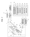
- FIG. 1 is a diagram illustrating a general arrangement of a particle therapy system according to an embodiment of the present disclosure.
- a particle therapy system A illustrated in FIG. 1 is a radiotherapy device having a group of devices for irradiating a patient B, who is a subject, with a particle beam as a target.
- the particle therapy system A includes an accelerator 1, a beam transport system 2, a gantry 3, an irradiation nozzle 4, FPDs 5A and 5B, X-ray tubes 6A and 6B, a treatment-couch 7, a robot arm 8, a communication device 9, a data server 10, a treatment planning system 11, an X-ray fluoroscopic imaging device 12, a treatment-couch control system 13, and a patient positioning device 20.
- the accelerator 1 is a particle beam generator that generates a particle beam for irradiating patient B, and accelerates and outputs the particle beam until the particle beam has energy suitable for treatment of patient B.
- the beam transport system 2 transports the particle beam output from the accelerator 1 to the gantry 3.
- the type of the particle beam is not particularly' limited, and is, for example, a proton beam or a carbon beam, or the like.
- the gantry 3 and the irradiation nozzle 4 are irradiation devices that irradiate patient B with the particle beam transported from the accelerator 1.
- the gantry 3 adjusts an irradiation angle at which patient B is irradiated with the particle beam transported from the accelerator 1.
- the gantry 3 has a rotation mechanism capable of rotating 360° around patient B, and adjusts the irradiation angle by rotating.
- the irradiation nozzle 4 is provided to the gantry 3 and irradiates patient B with the particle beam transported to the gantry 3.
- a mechanism for adjusting the shape of the particle beam to match the shape of the target volume of the patient may be incorporated in the irradiation nozzle 4.
- the FPDs 5A and 5B and the X-ray tubes 6A and 6B constitute an imaging system that performs fluoroscopic imaging of patient B.
- the FPDs 5A and 5B are plane detectors that capture images of patient B by detecting X-rays, which are imaging light, on a detection surface.
- the X-ray tubes 6A and 6B are light sources that output X-rays.
- the treatment-couch 7 is a table on which patient B is mounted when patient B is irradiated with the particle beam.
- the robot arm 8 is a device that moves the treatment-couch 7. Specifically, the robot arm 8 performs, with respect to the treatment-couch 7, translational motion in a plurality of translational directions along each of a plurality of axes of motion and rotational motion in a plurality of rotational directions about a plurality of rotation axes.
- a rotation axis is the same as an axis of motion, and there are three axes of motion (rotation axes).
- each axis of motion is directed in a direction from right to left (RL direction) as viewed from patient B lying on the treatment-couch 7 on their back, a direction from the foot to the head (SI direction) of patient B, and a direction from the back to the abdomen (AP direction).
- the communication device 9 communicably connects the data server 10, the treatment planning system 11, and the patient positioning device 20 to each other.
- the data server 10 is a storage device that stores various types of information regarding the particle therapy of patient B.
- the data server 10 stores, for example, a three-dimensional fluoroscopic image of patient B and treatment plan information indicating a treatment plan for patient B.
- the three-dimensional fluoroscopic image includes information indicating the shape and electron density of the patient in voxel units.
- the three-dimensional fluoroscopic image is, for example, a computed tomography (CT) captured image, and is generated in advance (before creating treatment plan information on patient B).
- the treatment plan information is generated based on a three-dimensional fluoroscopic image.
- the treatment plan information includes planned placement information indicating a planned placement, which is a placement for patient B at the time of treatment.
- the placement of patient B indicates the position and angle (posture) of patient B and is determined by the position and angle of the treatment-couch 7.
- the treatment planning system 11 creates a treatment plan for patient B based on the three-dimensional fluoroscopic image stored in the data server 10, and stores, in the data server 10, treatment plan information indicating the treatment plan.
- the X-ray fluoroscopic imaging device 12 controls each of the FPD 5A and the X-ray tube 6A, and the FPD 5B and the X-ray tube 6B, acquires a plurality of X-ray fluoroscopic images obtained by imaging patient B from different angles as fluoroscopic images, and transmits the acquired X-ray fluoroscopic images to the patient positioning device 20.
- the treatment-couch control system 13 adjusts the placement of patient B by controlling the robot arm 8 to adjust the placement of the treatment-couch 7.
- the patient positioning device 20 executes positioning processing of patient B based on the three-dimensional fluoroscopic image and the treatment plan information which are stored in the data server 10, and the X-ray fluoroscopic image acquired by the X-ray fluoroscopic imaging device 12.
- the positioning processing of patient B is processing, before starting the particle therapy of patient B, in which patient B, who is lying on the treatment-couch 7, is placed in the same manner as the planned placement indicated by the treatment plan information.
- the patient positioning device 20 controls the robot arm 8 via the treatment-couch control system 13 to adjust the position and angle of the treatment-couch 7, thereby placing patient B in the same manner as the planned placement.
- the particle therapy on patient B is actually performed. Specifically, the particle beam accelerated to energy suitable for treatment by the accelerator 1 is transported to the gantry 3 via the beam transport system 2. The particle beam is deflected in an appropriate direction by the gantry 3 and passes through the irradiation nozzle 4 to irradiate the target volume of patient B.
- the patient positioning device 20 includes an image acquisition part 21, a simulated-X-ray fluoroscopic image creation part 22, an ROI drawing part 23, an image collation part 24, an image display part 25, and a control section 26.
- the image acquisition part 21 acquires a three-dimensional fluoroscopic image from the data server 10 via the communication device 9, and acquires the X-ray fluoroscopic image from the X-ray fluoroscopic imaging device 12.
- the simulated-X-ray fluoroscopic image creation part 22 is a creation part that creates a plurality of simulated-X-ray fluoroscopic images which are a plurality of simulated fluoroscopic images obtained by projecting a three-dimensional fluoroscopic image acquired by the image acquisition part 21 onto each of a plurality of planes corresponding to each imaging axis for capturing the X-ray fluoroscopic image.
- the simulated-X-ray fluoroscopic image creation part 22 creates the simulated-X-ray fluoroscopic images by placing the three-dimensional image of patient B on the same virtual space as the imaging system that generated the X-ray fluoroscopic image and performing projection processing.
- the surface corresponding to the imaging axis is, for example, a detection surface of the FPD corresponding to the imaging axis, that is, a surface substantially orthogonal to the imaging axis.
- the ROI drawing part 23 specifies an ROI which is a region of interest, on the simulated-X-ray fluoroscopic image, which is used for positioning the patient. Specifically, the ROI drawing part 23 specifies the ROI by displaying the simulated-X-ray fluoroscopic image to enable the user to draw the ROI on the simulated-X-ray fluoroscopic image.
- the ROI is drawn to include, for example, a structure to be positioned such as a bone.
- the image display part 25 is a display part that displays various types of information and images.
- the image display part 25 displays X-ray fluoroscopic images, simulated-X-ray fluoroscopic images, and ROI images indicating an ROI region, and the like.
- the control section 26 controls the treatment-couch control system 13 based on the treatment-couch motion amount calculated by the image collation part 24 to move the treatment-couch 7, thereby adjusting the placement of patient B.
- the patient positioning device 20 having the above functions can be implemented by means of an information processing device capable of performing various types of information processing, such as a computer device.
- the information processing device includes, for example, an arithmetic element, a storage medium, and a communication interface, and, if necessary, further includes an input part such as a mouse and a keyboard, and a display part such as a display.
- the arithmetic element is, for example, a processor such as a central processing unit (CPU) and a field-programmable gate array (FPGA).
- the storage medium include a magnetic storage medium such as a hard disk drive (HDD), and a semiconductor storage medium such as a random-access memory (RAM), a read-only memory (ROM), and a solid state drive (SSD).
- a combination of an optical disk such as a digital versatile disk (DVD) and an optical disk drive may be used as the storage medium.
- Another high-value storage medium such as a magnetic tape medium may also be used as the storage medium.
- the patient positioning device 20 may be configured by so-called cloud computing in which a plurality of information processing devices are capable of communicating via a communication network.
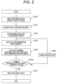
- FIG. 2 is a flowchart to illustrate an example of patient positioning processing.
- the setup position is a position for placing patient B in the same manner as the planned placement.
- the position of the surface of the body of patient B on the treatment-couch 7 is measured using an infrared laser installed in the treatment room, and patient B is placed in the setup position of the treatment-couch 7 based on this position.
- the control section 26 acquires treatment plan information from the data server 10, and controls the robot arm 8 via the treatment-couch control system 13 based on the planned placement information included in the treatment plan information to move the treatment-couch 7 on which patient B is lying so that the placement of patient B is the planned placement indicated by the planned placement information (step S100).
- the positioning target structure of patient B lying on the treatment-couch 7 is included in the X-ray irradiation region formed by the FPDs 5A and 5B and the X-ray tubes 6A and 6B.
- the simulated-X-ray fluoroscopic image creation part 22 acquires the three-dimensional fluoroscopic image from the data server 10, and creates two simulated-X-ray fluoroscopic images corresponding to the two imaging axes from the three-dimensional fluoroscopic image (step S102) .
- the image collation part 24 calculates, for each imaging axis, an amount of deviation in the two-dimensional direction between an X-ray fluoroscopic image and a simulated-X-ray fluoroscopic image corresponding to the imaging axis, as the two-dimensional motion amount, on the detection surface, of the FPD corresponding to the imaging axis (step S103) .
- a calculation method for calculating an image deviation amount a method is known in which the simulated-X-ray fluoroscopic image is scanned in the horizontal direction and the vertical direction with respect to the X-ray fluoroscopic image to search for a position in which the similarity between the images is the highest.
- sequential calculation of the similarity is necessary, and the calculation amount increases. Therefore, in the present embodiment, a calculation method using a phase-only correlation method (POC) method, which is known as a method enabling image collation at high-speed, will be described.
- POC phase-only correlation method
- the POC method is a method for performing image matching (alignment) using only a phase component obtained from two-dimensional discrete Fourier transform on an image, and has a feature of being resistant to disturbance such as a change in image luminance.
- the POC method differs from a method for performing matching based on feature points of an image such as edges or corners, and enables alignment to be performed accurately even for images in which no clear features are present.
- the image collation part 24 calculates a two-dimensional motion amount, which is the deviation amount between an X-ray fluoroscopic image and a simulated-X-ray fluoroscopic image, by using a POC function (phase-only correlation function), which is used as a matching evaluation index according to the POC method.
- a POC function phase-only correlation function
- Images f(n 1 , n 2 ) and g(n 1 , n 2 ) are both images having N 1 ⁇ N 2 pixels.
- n 1 and n 2 are integers
- the two-dimensional discrete Fourier transform (DFT) of the images f (n 1 , n 2 ) and g (n 1 , n 2 ) is expressed by the following Formulas (1) and (2).
- a F (k 1 , k 2 ) and A G (k 1 , k 2 ) are amplitude spectra, and ⁇ F (k 1 , k 2 ) and ⁇ G (k 1 , k 2 ) are phase spectra.
- the POC function r (n 1 , n 2 ) is defined as a two-dimensional inverse discrete Fourier transform (IDFT) of the normalized mutual power spectrum by the following equation (4) .
- IDFT inverse discrete Fourier transform
- the POC function has a sharp peak called a correlation peak in a case where the images of interest f(n 1 , n 2 ) and g(n 1 , n 2 ) are similar to each other.
- the height of the correlation peak represents the linearity of the phase difference spectrum of the images f (n 1 , n 2 ) and g(n 1 , n 2 ) with respect to the frequency, and when the phase difference spectrum is linear with respect to the frequency, the height of the correlation peak is 1.
- the height of the correlation peak is useful as a measure representing image similarity, and is used in image matching and the like.
- the coordinates of the correlation peak represent a relative deviation amount of the image.
- the image collation part 24 calculates the two-dimensional motion amount by using, as the images f (n 1 , n 2 ) and g(n 1 , n 2 ), images obtained by multiplying the X-ray fluoroscopic image and the simulated-X-ray fluoroscopic image by a window function.
- the window function is, for example, a two-dimensional Hanning window (w(n 1 , n 2 ) expressed by Formula (5) .
- w n 1 n 2 1 + cos ⁇ n 1 M 1 2 ⁇ 1 + cos ⁇ n 2 M 2 2 2
- the image collation part 24 calculates, for each imaging axis, the amount of deviation in the two-dimensional direction between the position and the origin of the correlation peak of the phase-only correlation function calculated from the X-ray fluoroscopic image and the simulated-X-ray fluoroscopic image, as the two-dimensional motion amount.
- the image collation part 24 executes three-dimensional motion amount calculation processing to calculate, as a treatment-couch motion amount by which the treatment-couch 7 is to be moved, a three-dimensional motion amount, based on the two-dimensional motion amount of each imaging axis (step S104) .
- FIGS. 3 to 5 are diagrams to illustrate three-dimensional motion amount calculation processing in more detail.
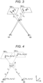
- FIG. 3 is a diagram to illustrate processing to obtain a correction axis from a two-dimensional motion amount
- FIG. 4 is a diagram to illustrate processing to obtain a three-dimensional motion amount from a correction axis
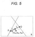
- FIG. 5 is an enlarged view of the common perpendicular illustrated in FIG. 4 .
- the distortion correction may be performed by, for example, an imaging device that captures X-ray fluoroscopic images, or may be performed using the patient positioning device 20.
- the following three-dimensional motion amount calculation processing can be performed.
- the center point of the detection surface 5A1 of the FPD 5A is defined as O A
- the center point of the detection surface 5B1 of the FPD 5B is defined as O B
- the imaging axes are a line L 1 connecting the center point O A of the detection surface 5A1 to the X-ray tube 6A, and a line L 2 connecting the center point O B of the detection surface 5B1 to the X-ray tube 6B.
- the image collation part 24 sets an intersection of the imaging axes L 1 and L 2 as a reference position IC (origin of the coordinate system), and sets, as correction axes, a line l 1 connecting the position where the reference position IC is moved along the detection surface 5A1 of the FPD 5A by the two-dimensional motion amount corresponding to the imaging axis L 1 to the X-ray tube 6A, and a line l 2 connecting the position where the reference position IC is moved along the detection surface 5B1 of the FPD 5B by the two-dimensional motion amount corresponding to the imaging axis L2 to the X-ray tube 6B.
- the position of the X-ray tube 6A is A' (x a , y a , z a ), the position of the matched center point on the detection surface 5A1 of the FPD 5A is B (x b , y b , z b ), the position of the X-ray tube 6B is _C (x c , y c , z c ), and the position of the matched center point on the detection surface 5B1 of the FPD 5B is D (x a , y d , z d ).
- the coordinates of each of the positions A to D are three-dimensional coordinates in a coordinate system (X, Y, Z) set in advance in the treatment room, in which the gantry 3 and the treatment-couch 7 are arranged, and the reference position IC is set as the origin of the coordinate system.
- the matched center point is an intersection point between the correction axes l 1 and l 2 and the detection surfaces 5A1 and 5B1.
- the lines l 1 and l 2 which are the correction axes, ideally intersect with each other, but in practice may not intersect with each other due to a minute error in the position of the matched center point, which depends on the image resolution, and a minute deviation in machine placement between the FPDs 5A and 5B and the X-ray tubes 6A and 6B, and the like.
- the image collation part 24 sets the points at which the distance between the line l 1 and the line l 2 is the smallest on the lines l 1 and l 2 as points P and Q, and calculates the position of the midpoint of a line segment l 3 connecting point P and point Q using three-dimensional coordinates.
- Line l 3 is a common perpendicular that is perpendicular to both lines l 1 and l 2 that are twisted relative to each other.
- the length of the line l 3 is referred to as the common perpendicular length.
- Points P and Q are the feet of the common perpendicular l 3 .
- p is a position vector (x p , y p , z p ) of point P
- q is a position vector (x q , y q , z q ) of point Q
- a is a position vector (x a , y a , z a ) of point A
- c is a position vector (x c , y c , z c ) of point C
- u is a direction vector of the line l 1
- v is a direction vector of the line l 2 .
- the three-dimensional motion amount is the motion amount from the intersection of the imaging axes to the midpoint M of line segment PQ, and in the present embodiment, because the intersection of the imaging axes is set as the origin, the value of the coordinates of the midpoint M of line segment PQ can be calculated as the three-dimensional motion amount, which is the treatment-couch motion amount.
- the image collation part 24 calculates the three-dimensional motion amount and the common perpendicular length according to the above method. In a case where the lines l 1 and l 2 , which are correction axes, intersect each other, the image collation part 24 calculates the three-dimensional motion amount by regarding the intersection of lines l 1 and l 2 as the midpoint M of line segment PQ.
- the image collation part 24 determines whether the common perpendicular length is less than a threshold value (for example, 1 mm) (step S105).
- a threshold value for example, 1 mm
- the common perpendicular length is ideally zero, that is, lines l 1 and l 2 intersect each other. The longer the common perpendicular length is, the less consistency is obtained between the two-dimensional motion amount and the three-dimensional motion amount of the simulated-X-ray fluoroscopic image and the X-ray fluoroscopic image.
- the image collation part 24 may determine whether the correlation peak of the POC function is less than the threshold value instead of the common perpendicular length.
- step S105 the image collation part 24 changes at least one of the X-ray fluoroscopic image and the simulated-X-ray fluoroscopic image (step S106), and returns to the processing of step S102.
- image changes may be executed by returning the partial image indicating the ROI to the original X-ray fluoroscopic image and the simulated-X-ray fluoroscopic image.
- the image changes may be realized by acquiring another X-ray fluoroscopic image from the X-ray fluoroscopic imaging device 12 or by creating another simulated-X-ray fluoroscopic image from another three-dimensional fluoroscopic image.
- step S105 the control section 26 moves the treatment-couch 7 via the treatment-couch control system 13 based on the three-dimensional motion amount calculated by the image collation part 24 (step S107), and ends the patient positioning processing. It is thus possible to move the patient from the current placement to the placement at the time of treatment planning, whereupon the actual particle beam irradiation is performed.
- control section 26 moves the treatment-couch 7 by the three-dimensional motion amount, and then translates and rotates the treatment-couch 7 by the translation amount and the rotation amount calculated by the fine adjustment processing.
- the translation amount is calculated for each of the plurality of axes of motion of the treatment-couch 7
- the rotation amount is calculated for each of the plurality of axes of rotation of the treatment-couch 7.
- the image collation part 24 may perform the processing described in PTLs 1 to 3, for example.
- the image collation part 24 may perform processing to calculate, based on the similarity, the motion amount of the treatment-couch 7 such that each fluoroscopic image and each simulated fluoroscopic image maximally coincide with each other, for each of a plurality of translation directions along each of a plurality of optimization axes including a plurality of imaging axes and a plurality of rotation directions around a plurality of rotation axes.
- a particle therapy system is illustrated as an example of the radiotherapy device, but the radiotherapy device is not limited to a particle therapy system, and may be a radiotherapy system using a non-particle beam such as X-rays.
- the accelerator 1 includes, for example, an electron beam accelerator that outputs X-rays.
- the image acquisition part 21 acquires a plurality of X-ray fluoroscopic images obtained by imaging the subject.
- the simulated-X-ray fluoroscopic image creation part 22 creates a simulated fluoroscopic image obtained by projecting a three-dimensional fluoroscopic image of the subject onto the detection surface with respect to the imaging axis.
- the image collation part 24 obtains a correction axis obtained by correcting the imaging axis based on a two-dimensional motion amount, which is a deviation amount between the X-ray fluoroscopic image and the simulated-X-ray fluoroscopic image corresponding to the imaging axis, and calculates, as a treatment-couch motion amount by which the treatment-couch is to be moved, a motion amount from an intersection of the imaging axes to a midpoint of a common perpendicular of the correction axes. Therefore, because the patient can be positioned without performing a repetitive calculation in the optimization calculation, the calculation time can be further reduced.
- the image collation part 24 obtains, as the correction axis, an axis connecting the position where the intersection of the imaging axes is moved by the two-dimensional motion amount, to the X-ray tube, for each of the plurality of imaging axes.
- the image collation part 24 obtains, as the correction axis, an axis connecting the position where the intersection of the imaging axes is moved by the two-dimensional motion amount, to the X-ray tube, for each of the plurality of imaging axes.
- the image collation part 24 calculates the two-dimensional motion amount based on the position of the peak of the phase-only correlation function calculated from the X-ray fluoroscopic image and the simulated-X-ray fluoroscopic image. In this case, because the two-dimensional motion amount can be calculated without performing sequential calculation such as optimization calculation, the calculation time can be further reduced.
- the image collation part 24 calculates the phase-only correlation function from the ROI set on the X-ray fluoroscopic image and the simulated-X-ray fluoroscopic image. Therefore, the two-dimensional motion amount can be calculated more appropriately.
- the image collation part 24 changes at least one of the X-ray fluoroscopic image and the simulated-X-ray fluoroscopic image to redetermine the correction axis.
- the image collation part 24 changes at least one of the X-ray fluoroscopic image and the simulated-X-ray fluoroscopic image to redetermine the correction axis.
- the image changes are executed by performing predetermined image processing. Therefore, it is not necessary to re-image the X-ray fluoroscopic image or re-create the simulated-X-ray fluoroscopic image from a three-dimensional fluoroscopic image, thereby enabling a further reduction in the calculation time.
- the image collation part 24 further calculates the motion amounts in the translation direction and the rotation direction of the treatment-couch 7 based on the similarity between each corrected simulated fluoroscopic image obtained by correcting each simulated-X-ray fluoroscopic image based on the treatment-couch motion amount, and each fluoroscopic image. In this case, more accurate positioning is possible. Note that, even in this case, because each simulated fluoroscopic image is corrected based on the treatment-couch motion amount, it is possible to prevent the position of the patient at the start of fine adjustment from being greatly separated from the planned position, and hence it is possible to suppress an increase in the number of repeated calculations in the optimization calculation. Therefore, the calculation time can be further reduced.
Landscapes
- Health & Medical Sciences (AREA)
- Engineering & Computer Science (AREA)
- Biomedical Technology (AREA)
- Pathology (AREA)
- Nuclear Medicine, Radiotherapy & Molecular Imaging (AREA)
- Radiology & Medical Imaging (AREA)
- Life Sciences & Earth Sciences (AREA)
- Animal Behavior & Ethology (AREA)
- General Health & Medical Sciences (AREA)
- Public Health (AREA)
- Veterinary Medicine (AREA)
- Radiation-Therapy Devices (AREA)
Abstract
Description
- The present disclosure relates to a positioning device, a radiotherapy device, and a positioning method.
- One known example of a cancer treatment method is radiotherapy, in which a patient is irradiated with radiation. The radiation used in radiotherapy is broadly classified into uncharged particle beam such as X-ray or gamma ray, and charged particle beam such as proton beam or carbon beam. Radiotherapy using the latter charged particle beam is generally called particle therapy.
- In the case of an uncharged particle beam, the dose decreases at a constant rate from a shallow position to a deep position in the body. On the other hand, in the case of a charged particle beam, a dose distribution (black curve) having a peak of energy loss at a specific depth can be formed. Therefore, by matching the peak of the energy loss of the charged particle beam with the position of a tumor, it is possible to greatly reduce the dose of the charged particle beam with, which normal tissue at a position deeper than the tumor is irradiated.
- For this reason, in radiotherapy, it is important to accurately irradiate a target tumor with a desired dose of radiation to improve the therapeutic effect. In order to accurately irradiate the tumor with radiation, it is necessary to align the position of the patient in the same position as the planned position determined by a treatment plan created in advance. This alignment of the patient position is called patient positioning.
- As a method for positioning a patient in radiotherapy, there is a method which uses X-ray fluoroscopic images (Digital Radiography: DR) obtained by imaging a patient lying on a treatment-couch from two different directions by two sets of X-ray tubes and a flat panel detector (FPD). In this method, an X-ray fluoroscopic image obtained by imaging a patient at the time of radiotherapy is compared with a simulated-X-ray fluoroscopic image created from a computed tomography (CT) image which is used when creating a treatment plan, and positioning of the patient is performed such that there is a match in the position of a positioning target structure such as a bone between the X-ray fluoroscopic image and the simulated-X-ray fluoroscopic image.
- In general, an X-ray fluoroscopic image may include structures other than a structure which is to be positioned, such as soft tissue and tools for securing the patient, or the placement of a bone, which is a structure to be positioned, may change from the time of the treatment plan. In such a situation, the structure captured in the X-ray fluoroscopic image and the simulated-X-ray fluoroscopic image do not match one another across the entire image. In this case, the patient is positioned using a region of interest (ROI), which is set as a region, on the X-ray fluoroscopic image, where the structure to be positioned is present. Note that the setting of the region of interest is usually performed by a user who is a medical worker drawing the region of interest on an image.
- Automatic alignment to position the patient is performed by using the translation amount and the rotation amount of the treatment-couch on which the patient is sleeping as parameters and using an optimization calculation to calculate optimum values of the parameters. Normally, the translation amount has three components along three axes (x, y, z) orthogonal to each other, and the rotation amount has three components (pitch, roll, and yaw) for which the three axes serve as rotation axes. Therefore, in the optimization calculation, the optimization process for each of the six components is repeatedly performed to calculate the optimum values of the parameters. Furthermore, the three axes defining the translation amount coincide with the axes of motion of the treatment-couch for placing the patient in a planned position, the x axis being oriented in a direction from right to left (a right-left (RL) direction) as viewed from the patient lying on the treatment-couch on their back, the y axis being oriented in a direction from the foot to the head (a superior-inferior (SI) direction), and the z axis being oriented in a direction from the back to the abdomen (an anterior-posterior (AP) direction).
- However, in a case where the position of the patient at the start of positioning is far from the planned position, the change in the similarity between the images serving as the determination indexes is small in the optimization calculation, and it is not possible to use such a feature that the similarity increases toward the optimum position, and the optimum value of the parameter cannot be reached, or the calculation time may increase due to an increase in the number of repeated calculations in the optimization calculation.
- In contrast,
PTLs - In addition,
PTL 3 discloses a technique of reducing the number of X-ray fluoroscopic images by evaluating the optimization of the translation amount with respect to the direction along the imaging axis only in one direction orthogonal to the fluoroscopic imaging axis, thereby reducing the time taken to position the patient. -
- PTL 1:
Japanese Patent No. 6668902 - PTL 2:
WO 2018/225234 A - PTL 3:
JP 2013-99431 A - However, in the techniques disclosed in
PTLs 1 to 3, there is no change in performing optimization using the similarity of images. Therefore, in a case where the position of the patient at the start of positioning is far away from the planned position and the change in the similarity between images becomes small, or other such cases, it may not be possible to suppress an increase in calculation time due to an increase in the number of repeated calculations in the optimization calculation. - An object of the present disclosure is to provide a positioning device, a radiotherapy device, and a positioning method that enable a further reduction in calculation time.
- A positioning device according to one aspect of the present disclosure is a positioning device that controls a position of a treatment-couch on which a subject is mounted, the positioning device including: an image acquisition part that acquires a plurality of fluoroscopic images obtained by imaging the subject by detecting, on a detection surface via the subject, light from a light source for each of a plurality of imaging axes; a creation part that creates, for each of the plurality of imaging axes, a simulated fluoroscopic image obtained by projecting a three-dimensional fluoroscopic image of the subject onto a detection surface with respect to the imaging axis; and
a calculation processing part that obtains, for each of the plurality of imaging axes, a correction axis obtained by correcting the imaging axis based on a deviation amount between the fluoroscopic image and the simulated fluoroscopic image corresponding to the imaging axis, and calculates, as a treatment-couch motion amount by which the treatment-couch is to be moved, a motion amount from an intersection of the plurality of imaging axes to a midpoint of a common perpendicular of the correction axes. - The present invention enables a further reduction in the calculation time.
-
- [
FIG. 1] FIG. 1 is a diagram illustrating a general arrangement of a particle therapy system according to an embodiment of the present disclosure. - [
FIG. 2] FIG. 2 is a flowchart to illustrate an example of patient positioning processing. - [
FIG. 3] FIG. 3 is a diagram to illustrate processing to obtain a correction axis from a two-dimensional motion amount. - [
FIG. 4] FIG. 4 is a diagram to illustrate processing to obtain a three-dimensional motion amount from a correction axis. - [
FIG. 5] FIG. 5 is an enlarged view of a common perpendicular illustrated inFIG. 4 . - Hereinafter, an embodiment of the present disclosure will be described with reference to the drawings.
- Note that the descriptions and drawings hereinbelow are examples to illustrate the present invention, and omissions and simplifications are made, as appropriate, to clarify the invention. The present invention can also be carried out in various other modes. Unless otherwise specified, each constituent element may be singular or plural. Note that, in all the drawings to illustrate the embodiment, parts having the same functions are assigned the same reference signs, and repetitive descriptions thereof will be omitted. In addition, the position, size, shape, range, and the like of each constituent element illustrated in the drawings may not represent the actual position, size, shape, range, and the like, in order to facilitate understanding of the invention. Therefore, the present invention is not limited to or by the position, size, shape, range, and the like disclosed in the drawings. Further, in a case where there is a plurality of constituent elements having the same or similar functions, same may be described with different subscripts added to the same reference signs. However, in a case where it is not necessary to distinguish between a plurality of constituent elements, descriptions may sometimes be provided with the subscripts omitted.
-
FIG. 1 is a diagram illustrating a general arrangement of a particle therapy system according to an embodiment of the present disclosure. A particle therapy system A illustrated inFIG. 1 is a radiotherapy device having a group of devices for irradiating a patient B, who is a subject, with a particle beam as a target. The particle therapy system A includes anaccelerator 1, abeam transport system 2, agantry 3, anirradiation nozzle 4,FPDs X-ray tubes couch 7, arobot arm 8, acommunication device 9, adata server 10, atreatment planning system 11, an X-rayfluoroscopic imaging device 12, a treatment-couch control system 13, and apatient positioning device 20. - The
accelerator 1 is a particle beam generator that generates a particle beam for irradiating patient B, and accelerates and outputs the particle beam until the particle beam has energy suitable for treatment of patient B. Thebeam transport system 2 transports the particle beam output from theaccelerator 1 to thegantry 3. The type of the particle beam is not particularly' limited, and is, for example, a proton beam or a carbon beam, or the like. - The
gantry 3 and theirradiation nozzle 4 are irradiation devices that irradiate patient B with the particle beam transported from theaccelerator 1. Thegantry 3 adjusts an irradiation angle at which patient B is irradiated with the particle beam transported from theaccelerator 1. Specifically, thegantry 3 has a rotation mechanism capable of rotating 360° around patient B, and adjusts the irradiation angle by rotating. Theirradiation nozzle 4 is provided to thegantry 3 and irradiates patient B with the particle beam transported to thegantry 3. A mechanism for adjusting the shape of the particle beam to match the shape of the target volume of the patient may be incorporated in theirradiation nozzle 4. - The
FPDs X-ray tubes FPDs X-ray tubes FPD 5A and theX-ray tube 6A are arranged to face each other such that the X-rays output from theX-ray tube 6A are detected by theFPD 5A through patient B, and theFPD 5B and theX-ray tube 6B are arranged to face each other such that the X-rays output from theX-ray tube 6B are detected by theFPD 5B through patient B. An axis connecting the center of theFPD 5A to theX-ray tube 6A and an axis connecting the center of theFPD 5B to theX-ray tube 6B are two imaging axes for imaging patient B. The two imaging axes are preferably orthogonal to each other, but need not be orthogonal to each other. The particle therapy system A may include three or more FPDs and three or more X-ray tubes. In this case, the number of imaging axes is also three or more. - The treatment-
couch 7 is a table on which patient B is mounted when patient B is irradiated with the particle beam. Therobot arm 8 is a device that moves the treatment-couch 7. Specifically, therobot arm 8 performs, with respect to the treatment-couch 7, translational motion in a plurality of translational directions along each of a plurality of axes of motion and rotational motion in a plurality of rotational directions about a plurality of rotation axes. In the present embodiment, a rotation axis is the same as an axis of motion, and there are three axes of motion (rotation axes). In addition, each axis of motion is directed in a direction from right to left (RL direction) as viewed from patient B lying on the treatment-couch 7 on their back, a direction from the foot to the head (SI direction) of patient B, and a direction from the back to the abdomen (AP direction). - The
communication device 9 communicably connects thedata server 10, thetreatment planning system 11, and thepatient positioning device 20 to each other. - The
data server 10 is a storage device that stores various types of information regarding the particle therapy of patient B. Thedata server 10 stores, for example, a three-dimensional fluoroscopic image of patient B and treatment plan information indicating a treatment plan for patient B. The three-dimensional fluoroscopic image includes information indicating the shape and electron density of the patient in voxel units. The three-dimensional fluoroscopic image is, for example, a computed tomography (CT) captured image, and is generated in advance (before creating treatment plan information on patient B). The treatment plan information is generated based on a three-dimensional fluoroscopic image. In addition, the treatment plan information includes planned placement information indicating a planned placement, which is a placement for patient B at the time of treatment. The placement of patient B indicates the position and angle (posture) of patient B and is determined by the position and angle of the treatment-couch 7. - The
treatment planning system 11 creates a treatment plan for patient B based on the three-dimensional fluoroscopic image stored in thedata server 10, and stores, in thedata server 10, treatment plan information indicating the treatment plan. - The X-ray
fluoroscopic imaging device 12 controls each of theFPD 5A and theX-ray tube 6A, and theFPD 5B and theX-ray tube 6B, acquires a plurality of X-ray fluoroscopic images obtained by imaging patient B from different angles as fluoroscopic images, and transmits the acquired X-ray fluoroscopic images to thepatient positioning device 20. In the present embodiment, there are two X-ray fluoroscopic images. - The treatment-
couch control system 13 adjusts the placement of patient B by controlling therobot arm 8 to adjust the placement of the treatment-couch 7. - The
patient positioning device 20 executes positioning processing of patient B based on the three-dimensional fluoroscopic image and the treatment plan information which are stored in thedata server 10, and the X-ray fluoroscopic image acquired by the X-rayfluoroscopic imaging device 12. - The positioning processing of patient B is processing, before starting the particle therapy of patient B, in which patient B, who is lying on the treatment-
couch 7, is placed in the same manner as the planned placement indicated by the treatment plan information. Thepatient positioning device 20 controls therobot arm 8 via the treatment-couch control system 13 to adjust the position and angle of the treatment-couch 7, thereby placing patient B in the same manner as the planned placement. - When the positioning processing is complete, the particle therapy on patient B is actually performed. Specifically, the particle beam accelerated to energy suitable for treatment by the
accelerator 1 is transported to thegantry 3 via thebeam transport system 2. The particle beam is deflected in an appropriate direction by thegantry 3 and passes through theirradiation nozzle 4 to irradiate the target volume of patient B. - Hereinafter, the
patient positioning device 20 will be described in more detail. - As illustrated in
FIG. 1 , thepatient positioning device 20 includes animage acquisition part 21, a simulated-X-ray fluoroscopicimage creation part 22, anROI drawing part 23, animage collation part 24, animage display part 25, and acontrol section 26. - The
image acquisition part 21 acquires a three-dimensional fluoroscopic image from thedata server 10 via thecommunication device 9, and acquires the X-ray fluoroscopic image from the X-rayfluoroscopic imaging device 12. - The simulated-X-ray fluoroscopic
image creation part 22 is a creation part that creates a plurality of simulated-X-ray fluoroscopic images which are a plurality of simulated fluoroscopic images obtained by projecting a three-dimensional fluoroscopic image acquired by theimage acquisition part 21 onto each of a plurality of planes corresponding to each imaging axis for capturing the X-ray fluoroscopic image. The simulated-X-ray fluoroscopicimage creation part 22 creates the simulated-X-ray fluoroscopic images by placing the three-dimensional image of patient B on the same virtual space as the imaging system that generated the X-ray fluoroscopic image and performing projection processing. The surface corresponding to the imaging axis is, for example, a detection surface of the FPD corresponding to the imaging axis, that is, a surface substantially orthogonal to the imaging axis. - The
ROI drawing part 23 specifies an ROI which is a region of interest, on the simulated-X-ray fluoroscopic image, which is used for positioning the patient. Specifically, theROI drawing part 23 specifies the ROI by displaying the simulated-X-ray fluoroscopic image to enable the user to draw the ROI on the simulated-X-ray fluoroscopic image. The ROI is drawn to include, for example, a structure to be positioned such as a bone. - The
image collation part 24 is a calculation processing part that calculates a treatment-couch motion amount by which the treatment-couch 7 is to be moved, based on the X-ray fluoroscopic image and the simulated-X-ray fluoroscopic image. In a case where the ROI is designated by theROI drawing part 23, theimage collation part 24 may calculate the treatment-couch motion amount based on the X-ray fluoroscopic image in the ROI and the simulated-X-ray fluoroscopic image. In the present embodiment, the treatment-couch motion amount includes the motion amount in each of the plurality of translation directions. - The
image display part 25 is a display part that displays various types of information and images. For example, theimage display part 25 displays X-ray fluoroscopic images, simulated-X-ray fluoroscopic images, and ROI images indicating an ROI region, and the like. - The
control section 26 controls the treatment-couch control system 13 based on the treatment-couch motion amount calculated by theimage collation part 24 to move the treatment-couch 7, thereby adjusting the placement of patient B. - The
patient positioning device 20 having the above functions can be implemented by means of an information processing device capable of performing various types of information processing, such as a computer device. The information processing device includes, for example, an arithmetic element, a storage medium, and a communication interface, and, if necessary, further includes an input part such as a mouse and a keyboard, and a display part such as a display. - The arithmetic element is, for example, a processor such as a central processing unit (CPU) and a field-programmable gate array (FPGA). Examples of the storage medium include a magnetic storage medium such as a hard disk drive (HDD), and a semiconductor storage medium such as a random-access memory (RAM), a read-only memory (ROM), and a solid state drive (SSD). In addition, a combination of an optical disk such as a digital versatile disk (DVD) and an optical disk drive may be used as the storage medium. Another high-value storage medium such as a magnetic tape medium may also be used as the storage medium.
- A program such as firmware is stored on the storage medium. When the operation of the
patient positioning device 20 is started (for example, when the power is turned on), the arithmetic element reads the program from the storage medium and executes the program, thus realizingparts 21 to 27 of thepatient positioning device 20, and executing an entire series of control processes. In addition to the program, the storage medium stores data and the like necessary for each instance of processing of thepatient positioning device 20. - Note that the
patient positioning device 20 according to the present embodiment may be configured by so-called cloud computing in which a plurality of information processing devices are capable of communicating via a communication network. - Hereinafter, the patient positioning processing by the
patient positioning device 20 will be described in more detail with reference toFIGS. 2 to 5 . -
FIG. 2 is a flowchart to illustrate an example of patient positioning processing. - Note that it is assumed that patient B is arranged in a setup position of the treatment-
couch 7. The setup position is a position for placing patient B in the same manner as the planned placement. For example, the position of the surface of the body of patient B on the treatment-couch 7 is measured using an infrared laser installed in the treatment room, and patient B is placed in the setup position of the treatment-couch 7 based on this position. - In the patient positioning processing, first, the
control section 26 acquires treatment plan information from thedata server 10, and controls therobot arm 8 via the treatment-couch control system 13 based on the planned placement information included in the treatment plan information to move the treatment-couch 7 on which patient B is lying so that the placement of patient B is the planned placement indicated by the planned placement information (step S100). At this time, the positioning target structure of patient B lying on the treatment-couch 7 is included in the X-ray irradiation region formed by theFPDs X-ray tubes - Thereafter, the
image acquisition part 21 acquires a plurality of X-ray fluoroscopic images obtained by imaging patient B from a plurality of different directions via the X-ray fluoroscopic imaging device 12 (step S101). In the present embodiment, theimage acquisition part 21 acquires two X-ray fluoroscopic images captured from two directions along two imaging axes. - The simulated-X-ray fluoroscopic
image creation part 22 acquires the three-dimensional fluoroscopic image from thedata server 10, and creates two simulated-X-ray fluoroscopic images corresponding to the two imaging axes from the three-dimensional fluoroscopic image (step S102) . - The
image collation part 24 calculates, for each imaging axis, an amount of deviation in the two-dimensional direction between an X-ray fluoroscopic image and a simulated-X-ray fluoroscopic image corresponding to the imaging axis, as the two-dimensional motion amount, on the detection surface, of the FPD corresponding to the imaging axis (step S103) . - As a calculation method for calculating an image deviation amount, a method is known in which the simulated-X-ray fluoroscopic image is scanned in the horizontal direction and the vertical direction with respect to the X-ray fluoroscopic image to search for a position in which the similarity between the images is the highest. However, using this method, sequential calculation of the similarity is necessary, and the calculation amount increases. Therefore, in the present embodiment, a calculation method using a phase-only correlation method (POC) method, which is known as a method enabling image collation at high-speed, will be described.
- The POC method is a method for performing image matching (alignment) using only a phase component obtained from two-dimensional discrete Fourier transform on an image, and has a feature of being resistant to disturbance such as a change in image luminance. In addition, the POC method differs from a method for performing matching based on feature points of an image such as edges or corners, and enables alignment to be performed accurately even for images in which no clear features are present.
- In the present embodiment, the
image collation part 24 calculates a two-dimensional motion amount, which is the deviation amount between an X-ray fluoroscopic image and a simulated-X-ray fluoroscopic image, by using a POC function (phase-only correlation function), which is used as a matching evaluation index according to the POC method. - Here, two images to be subjected to a calculation method using the POC method are set as images f(n1, n2) and g(n1, n2). Images f(n1, n2) and g(n1, n2) are both images having N1×N2 pixels. n1 and n2 are discrete spatial indexes indicating pixels, and n1 = -M1,..., M1, n2 = -M2,..., M2. Note that n1 and n2 are integers, M1 and M2 are positive integers, and N1 = 2M1+1 and N1 = 2M1+1 are satisfied.
- The two-dimensional discrete Fourier transform (DFT) of the images f (n1, n2) and g (n1, n2) is expressed by the following Formulas (1) and (2).

- Here, k1(=-M1, ... , M1) and k2(= -M2,..., M2) are discrete frequency indexes (integers), and WN1 = exp(-j · 2π/N1) and wN1 = exp (-j · 2π/N2) are rotation factors. AF(k1, k2) and AG(k1, k2) are amplitude spectra, and θF(k1, k2) and θG(k1, k2) are phase spectra.. In addition, the sum Σn1,n2 indicates a sum Σn1=-M1 M1Σn2=-M2 M2 across the entire discrete space index.
- The normalized mutual power spectra of the images f(n1, n2) and g(n1, n2) are expressed by the following Formula (3) using the functions F (k1, k2) and G(k1, k2) after the Fourier transform.


- The POC function r (n1, n2) is defined as a two-dimensional inverse discrete Fourier transform (IDFT) of the normalized mutual power spectrum by the following equation (4) .

- Here, the sum Σk1, k2 indicates a sum Fk1=-M1 M1Σk2=-M2 M2 across the entire discrete frequency index.
- The POC function has a sharp peak called a correlation peak in a case where the images of interest f(n1, n2) and g(n1, n2) are similar to each other. The height of the correlation peak represents the linearity of the phase difference spectrum of the images f (n1, n2) and g(n1, n2) with respect to the frequency, and when the phase difference spectrum is linear with respect to the frequency, the height of the correlation peak is 1. The height of the correlation peak is useful as a measure representing image similarity, and is used in image matching and the like. Furthermore, the coordinates of the correlation peak represent a relative deviation amount of the image. For example, the amount of coordinate deviation of the correlation peak with respect to the image origin (n1=0, n2=0) can be used as the deviation amount of the images f(n1, n2) and g(n1, n2).
- Note that, using the two-dimensional DFT, because cyclic convolution is assumed at the edge of the target image, discontinuity that should not originally exist , appears at the edge of the image. In the present embodiment, in order to reduce such discontinuity, the
image collation part 24 calculates the two-dimensional motion amount by using, as the images f (n1, n2) and g(n1, n2), images obtained by multiplying the X-ray fluoroscopic image and the simulated-X-ray fluoroscopic image by a window function. The window function is, for example, a two-dimensional Hanning window (w(n1, n2) expressed by Formula (5) .
- Using the POC method as described above, the
image collation part 24 calculates, for each imaging axis, the amount of deviation in the two-dimensional direction between the position and the origin of the correlation peak of the phase-only correlation function calculated from the X-ray fluoroscopic image and the simulated-X-ray fluoroscopic image, as the two-dimensional motion amount. - The description now returns to
FIG. 2 . Theimage collation part 24 executes three-dimensional motion amount calculation processing to calculate, as a treatment-couch motion amount by which the treatment-couch 7 is to be moved, a three-dimensional motion amount, based on the two-dimensional motion amount of each imaging axis (step S104) . - In the three-dimensional motion amount calculation processing, the
image collation part 24 first obtains, for each imaging axis, a correction axis obtained by correcting the imaging axis based on a two-dimensional motion amount, which is a deviation amount between the X-ray fluoroscopic image and the simulated-X-ray fluoroscopic image corresponding to the imaging axis. Theimage collation part 24 then calculates the motion amount from the intersection of the respective imaging axes to the midpoint of the common perpendicular of the respective correction axes, as the three-dimensional motion amount constituting the treatment-couch motion amount. -
FIGS. 3 to 5 are diagrams to illustrate three-dimensional motion amount calculation processing in more detail. Specifically,FIG. 3 is a diagram to illustrate processing to obtain a correction axis from a two-dimensional motion amount;FIG. 4 is a diagram to illustrate processing to obtain a three-dimensional motion amount from a correction axis; andFIG. 5 is an enlarged view of the common perpendicular illustrated inFIG. 4 . Note that it is assumed that the distortion of the X-ray fluoroscopic image is removed in advance through distortion correction according to the placement angle of theFPDs patient positioning device 20. In addition, even if the distortion correction is not performed, the following three-dimensional motion amount calculation processing can be performed. - As illustrated in
FIG. 3 , the center point of the detection surface 5A1 of theFPD 5A is defined as OA, and the center point of the detection surface 5B1 of theFPD 5B is defined as OB. The imaging axes are a line L1 connecting the center point OA of the detection surface 5A1 to theX-ray tube 6A, and a line L2 connecting the center point OB of the detection surface 5B1 to theX-ray tube 6B. - The
image collation part 24 sets an intersection of the imaging axes L1 and L2 as a reference position IC (origin of the coordinate system), and sets, as correction axes, a line l1 connecting the position where the reference position IC is moved along the detection surface 5A1 of theFPD 5A by the two-dimensional motion amount corresponding to the imaging axis L1 to theX-ray tube 6A, and a line l2 connecting the position where the reference position IC is moved along the detection surface 5B1 of theFPD 5B by the two-dimensional motion amount corresponding to the imaging axis L2 to theX-ray tube 6B. - As illustrated in
FIG. 4 , the position of theX-ray tube 6A is A' (xa, ya, za), the position of the matched center point on the detection surface 5A1 of theFPD 5A is B (xb, yb, zb), the position of theX-ray tube 6B is _C (xc, yc, zc), and the position of the matched center point on the detection surface 5B1 of theFPD 5B is D (xa, yd, zd). The coordinates of each of the positions A to D are three-dimensional coordinates in a coordinate system (X, Y, Z) set in advance in the treatment room, in which thegantry 3 and the treatment-couch 7 are arranged, and the reference position IC is set as the origin of the coordinate system. The matched center point is an intersection point between the correction axes l1 and l2 and the detection surfaces 5A1 and 5B1. - The lines l1 and l2, which are the correction axes, ideally intersect with each other, but in practice may not intersect with each other due to a minute error in the position of the matched center point, which depends on the image resolution, and a minute deviation in machine placement between the
FPDs X-ray tubes - In a case where the line l1 and the line l2 do not intersect with each other, the
image collation part 24 sets the points at which the distance between the line l1 and the line l2 is the smallest on the lines l1 and l2 as points P and Q, and calculates the position of the midpoint of a line segment l3 connecting point P and point Q using three-dimensional coordinates. Line l3 is a common perpendicular that is perpendicular to both lines l1 and l2 that are twisted relative to each other. Hereinafter, the length of the line l3 is referred to as the common perpendicular length. Points P and Q are the feet of the common perpendicular l3. - Point P of the line l1 and point Q of the line l2 can be expressed as vectors using the parameter s and t as follows.


- Here, p is a position vector (xp, yp, zp) of point P, q is a position vector (xq, yq, zq) of point Q, a is a position vector (xa, ya, za) of point A, c is a position vector (xc, yc, zc) of point C, u is a direction vector of the line l1, and v is a direction vector of the line l2.
- Because the line segment PQ is orthogonal to each of the direction vectors u and v, the inner product of the direction vector w of the line segment PQ and the direction vectors u and v is zero. That is, w · u = 0 and w · v = 0. Note that the operator "·" indicates an inner product.
- The values of the parameter variables s and t. can be obtained by solving the two equations of the inner product as simultaneous linear equations. As a result, the coordinates of points P and Q and the common perpendicular length L, which is the length of the line segment PQ, are given as L = |q-p| = (|p|+|q|-2p·q)1/2 = {(xq-xp)2 + (yq-yp)2 + (zq-zp)2}1/2.
- Furthermore, coordinates (xm, ym, zm)M(xm, ym, zm) = ((xq+xp)/2, (yq+yp)/2, (zq+zp)/2) of the midpoint M of the line segment PQ are obtained. As a result, the three-dimensional motion amount is the motion amount from the intersection of the imaging axes to the midpoint M of line segment PQ, and in the present embodiment, because the intersection of the imaging axes is set as the origin, the value of the coordinates of the midpoint M of line segment PQ can be calculated as the three-dimensional motion amount, which is the treatment-couch motion amount.
- The
image collation part 24 calculates the three-dimensional motion amount and the common perpendicular length according to the above method. In a case where the lines l1 and l2, which are correction axes, intersect each other, theimage collation part 24 calculates the three-dimensional motion amount by regarding the intersection of lines l1 and l2 as the midpoint M of line segment PQ. - The description now returns to
FIG. 3 . Upon calculating the three-dimensional motion amount and the common perpendicular length according to the above method in step 104, theimage collation part 24 determines whether the common perpendicular length is less than a threshold value (for example, 1 mm) (step S105). Note that the common perpendicular length is ideally zero, that is, lines l1 and l2 intersect each other. The longer the common perpendicular length is, the less consistency is obtained between the two-dimensional motion amount and the three-dimensional motion amount of the simulated-X-ray fluoroscopic image and the X-ray fluoroscopic image. Further, theimage collation part 24 may determine whether the correlation peak of the POC function is less than the threshold value instead of the common perpendicular length. - In a case where the common perpendicular length is equal to or greater than the threshold value (step S105: No), the
image collation part 24 changes at least one of the X-ray fluoroscopic image and the simulated-X-ray fluoroscopic image (step S106), and returns to the processing of step S102. - Image changes are made, for example, by executing predetermined image processing on the images to be changed, which are at least one of the X-ray fluoroscopic image and the simulated-X-ray fluoroscopic image. The predetermined image processing is, for example, filter processing to highlight the edge of the image(s) to be changed, processing to extract a specific partial image from the image(s) to be changed, and the like. A partial image is, for example, an image indicating the ROI, and such processing may be executed by the
ROI drawing part 23. In addition, when the processing of steps S103 to S104 is executed first, an image indicating the ROI extracted from the X-ray fluoroscopic image and the simulated-X-ray fluoroscopic image may be used. In this case, image changes may be executed by returning the partial image indicating the ROI to the original X-ray fluoroscopic image and the simulated-X-ray fluoroscopic image. The image changes may be realized by acquiring another X-ray fluoroscopic image from the X-rayfluoroscopic imaging device 12 or by creating another simulated-X-ray fluoroscopic image from another three-dimensional fluoroscopic image. - On the other hand, in a case where the common perpendicular length is less than the threshold value (step S105: Yes), the
control section 26 moves the treatment-couch 7 via the treatment-couch control system 13 based on the three-dimensional motion amount calculated by the image collation part 24 (step S107), and ends the patient positioning processing. It is thus possible to move the patient from the current placement to the placement at the time of treatment planning, whereupon the actual particle beam irradiation is performed. - The patient positioning processing described above is merely an example, and the present invention is not limited thereto. For example, after calculating the treatment-couch motion amount by means of the above processing, the
image collation part 24 may perform fine adjustment processing to calculate the translation amount and the rotation amount of the treatment-couch 7 by means of an optimization calculation based on the similarity between each corrected simulated fluoroscopic image obtained by correcting the simulated-X-ray fluoroscopic image based on the treatment-couch motion amount, and each fluoroscopic image. In this case, thecontrol section 26 moves the treatment-couch 7 via the treatment-couch control system 13 based on the three-dimensional motion amount calculated by theimage collation part 24 and the translation amount and the rotation amount calculated by the fine adjustment processing. For example, thecontrol section 26 moves the treatment-couch 7 by the three-dimensional motion amount, and then translates and rotates the treatment-couch 7 by the translation amount and the rotation amount calculated by the fine adjustment processing. Note that the translation amount is calculated for each of the plurality of axes of motion of the treatment-couch 7, and the rotation amount is calculated for each of the plurality of axes of rotation of the treatment-couch 7. - In the fine adjustment processing, the
image collation part 24 may perform the processing described inPTLs 1 to 3, for example. In addition, theimage collation part 24 may perform processing to calculate, based on the similarity, the motion amount of the treatment-couch 7 such that each fluoroscopic image and each simulated fluoroscopic image maximally coincide with each other, for each of a plurality of translation directions along each of a plurality of optimization axes including a plurality of imaging axes and a plurality of rotation directions around a plurality of rotation axes. - In the present embodiment, a particle therapy system is illustrated as an example of the radiotherapy device, but the radiotherapy device is not limited to a particle therapy system, and may be a radiotherapy system using a non-particle beam such as X-rays. In this case, the
accelerator 1 includes, for example, an electron beam accelerator that outputs X-rays. - As described above, according to the present embodiment, the
image acquisition part 21 acquires a plurality of X-ray fluoroscopic images obtained by imaging the subject. For each imaging axis of the X-ray fluoroscopic image, the simulated-X-ray fluoroscopicimage creation part 22 creates a simulated fluoroscopic image obtained by projecting a three-dimensional fluoroscopic image of the subject onto the detection surface with respect to the imaging axis. For each of the plurality of imaging axes, theimage collation part 24 obtains a correction axis obtained by correcting the imaging axis based on a two-dimensional motion amount, which is a deviation amount between the X-ray fluoroscopic image and the simulated-X-ray fluoroscopic image corresponding to the imaging axis, and calculates, as a treatment-couch motion amount by which the treatment-couch is to be moved, a motion amount from an intersection of the imaging axes to a midpoint of a common perpendicular of the correction axes. Therefore, because the patient can be positioned without performing a repetitive calculation in the optimization calculation, the calculation time can be further reduced. - Further, in the present embodiment, the
image collation part 24 obtains, as the correction axis, an axis connecting the position where the intersection of the imaging axes is moved by the two-dimensional motion amount, to the X-ray tube, for each of the plurality of imaging axes. In this case, it is possible to calculate a suitable correction axis according to the deviation between the X-ray fluoroscopic image and the simulated-X-ray fluoroscopic image, and thus more accurate positioning becomes possible. - In the present embodiment, the
image collation part 24 calculates the two-dimensional motion amount based on the position of the peak of the phase-only correlation function calculated from the X-ray fluoroscopic image and the simulated-X-ray fluoroscopic image. In this case, because the two-dimensional motion amount can be calculated without performing sequential calculation such as optimization calculation, the calculation time can be further reduced. - In the present embodiment, the
image collation part 24 calculates the phase-only correlation function from the ROI set on the X-ray fluoroscopic image and the simulated-X-ray fluoroscopic image. Therefore, the two-dimensional motion amount can be calculated more appropriately. - Further, in the present embodiment, in a case where the length of the common 'perpendicular of the correction axes is equal to or greater than the threshold value, the
image collation part 24 changes at least one of the X-ray fluoroscopic image and the simulated-X-ray fluoroscopic image to redetermine the correction axis. In this case, it is possible to obtain a correction axis with which the two-dimensional motion amount and the three-dimensional motion amount of the simulated-X-ray fluoroscopic image and the X-ray fluoroscopic image are further aligned, and thus more accurate positioning becomes possible. - Furthermore, in the present embodiment, the image changes are executed by performing predetermined image processing. Therefore, it is not necessary to re-image the X-ray fluoroscopic image or re-create the simulated-X-ray fluoroscopic image from a three-dimensional fluoroscopic image, thereby enabling a further reduction in the calculation time.
- In the present embodiment, the
image collation part 24 further calculates the motion amounts in the translation direction and the rotation direction of the treatment-couch 7 based on the similarity between each corrected simulated fluoroscopic image obtained by correcting each simulated-X-ray fluoroscopic image based on the treatment-couch motion amount, and each fluoroscopic image. In this case, more accurate positioning is possible. Note that, even in this case, because each simulated fluoroscopic image is corrected based on the treatment-couch motion amount, it is possible to prevent the position of the patient at the start of fine adjustment from being greatly separated from the planned position, and hence it is possible to suppress an increase in the number of repeated calculations in the optimization calculation. Therefore, the calculation time can be further reduced. - The above-described embodiment of the present disclosure is an example to illustrate the present disclosure and is not intended to limit the scope of the present disclosure only to this embodiment. A person skilled in the art is able to carry out the present disclosure in various other aspects without departing from the scope of the present disclosure.
-
- A
- particle therapy system
- B
- patient
- 1
- accelerator
- 2
- beam transport system
- 3
- gantry
- 4
- irradiation nozzle
- 5A
- FPD
- 5B
- FPD
- 6A
- X-ray tube
- 6B
- X-ray tube
- 7
- treatment-couch
- 8
- robot arm
- 9
- communication device patient
- 10
- data server
- 11
- treatment planning system
- 12
- X-ray fluoroscopic imaging device
- 13
- treatment-couch control system
- 20
- patient positioning device
- 21
- simulated-X-ray fluoroscopic image creation part
- 23
- ROI drawing part
- 24
- image collation part
- 25
- image display part
- 26
- control section
Claims (9)
- A positioning device that controls a position of a treatment-couch on which a subject is mounted, the positioning device comprising:an image acquisition part that acquires a plurality of fluoroscopic images obtained by imaging the subject by detecting, on a detection surface via the subject, light from a light source for each of a plurality of imaging axes;a creation part that creates, for each of the plurality of imaging axes, a simulated fluoroscopic image obtained by projecting a three-dimensional fluoroscopic image of the subject onto the detection surface with respect to the imaging axis; anda calculation processing part that obtains, for each of the plurality of imaging axes, a correction axis obtained by correcting the imaging axis based on a deviation amount between the fluoroscopic image and the simulated fluoroscopic image corresponding to the imaging axis, and calculates, as a treatment-couch motion amount by which the treatment-couch is to be moved, a motion amount from an intersection of the plurality of imaging axes to a midpoint of a common perpendicular of the correction axes.
- The positioning device according to claim 1, wherein the calculation processing part obtains, as the correction axis, an axis connecting the position where the intersection is moved by the deviation amount, to the light source, for each of the plurality of imaging axes.
- The positioning device according to claim 1, wherein the calculation processing part calculates the deviation amount based on a position of a peak of a phase-only correlation function calculated from the fluoroscopic image and the simulated fluoroscopic image.
- The positioning device according to claim 3, wherein the calculation processing part calculates the phase-only correlation function from a region of interest set on the fluoroscopic image and the simulated fluoroscopic image.
- The positioning device according to claim 1, wherein, in a case where a length of the common perpendicular is equal to or greater than a threshold value, the calculation processing part changes at least one of the fluoroscopic image and the simulated fluoroscopic image to redetermine the correction axis.
- The positioning device according to claim 5, wherein the calculation processing part changes at least one of the fluoroscopic image and the simulated fluoroscopic image by performing predetermined image processing on at least one of the fluoroscopic image and the simulated fluoroscopic image.
- The positioning device according to claim 1, wherein the calculation processing part further calculates motion amounts in a translation direction and a rotation direction of the treatment-couch based on a similarity between each corrected simulated fluoroscopic image obtained by correcting each simulated fluoroscopic image based on the treatment-couch motion amount, and each fluoroscopic image.
- A radiotherapy device, comprising:the positioning device according to claim 1;a treatment-couch control system that moves the treatment-couch based on a treatment-couch motion amount calculated by the positioning device; andan irradiation device that uses radiation to irradiate a subject mounted on the moved treatment-couch.
- A positioning method using a positioning device that controls a position of a treatment-couch on which a subject is mounted, the positioning method comprising:acquiring a plurality of fluoroscopic images obtained by imaging the subject by receiving, on a detection surface via the subject, light from a light source for each of a plurality of imaging axes;creating, for each of the plurality of imaging axes, a simulated fluoroscopic image obtained by projecting a three-dimensional fluoroscopic image of the subject onto the detection surface with respect to the imaging axis; andobtaining, for each of the plurality of imaging axes, a correction axis obtained by correcting the imaging axis based on a deviation amount between the fluoroscopic image and the simulated fluoroscopic image corresponding to the imaging axis, and calculating, as a treatment-couch motion amount by which the treatment-couch is to be moved, a motion amount from the foot to a midpoint of a common perpendicular of each correction axis.
Applications Claiming Priority (2)
| Application Number | Priority Date | Filing Date | Title |
|---|---|---|---|
| JP2022023707A JP7791002B2 (en) | 2022-02-18 | 2022-02-18 | Positioning device, radiation therapy device, and positioning method |
| PCT/JP2023/002740 WO2023157616A1 (en) | 2022-02-18 | 2023-01-27 | Positioning device, radiation therapy device, and positioning method |
Publications (2)
| Publication Number | Publication Date |
|---|---|
| EP4480536A1 true EP4480536A1 (en) | 2024-12-25 |
| EP4480536A4 EP4480536A4 (en) | 2026-01-14 |
Family
ID=87578408
Family Applications (1)
| Application Number | Title | Priority Date | Filing Date |
|---|---|---|---|
| EP23756144.4A Pending EP4480536A4 (en) | 2022-02-18 | 2023-01-27 | POSITIONING DEVICE, RADIATION THERAPY DEVICE AND POSITIONING PROCEDURES |
Country Status (5)
| Country | Link |
|---|---|
| US (1) | US20240390701A1 (en) |
| EP (1) | EP4480536A4 (en) |
| JP (1) | JP7791002B2 (en) |
| CN (1) | CN118302229A (en) |
| WO (1) | WO2023157616A1 (en) |
Families Citing this family (2)
| Publication number | Priority date | Publication date | Assignee | Title |
|---|---|---|---|---|
| JP2024084030A (en) * | 2022-12-12 | 2024-06-24 | 株式会社日立製作所 | BEAM TRANSPORT SYSTEM AND METHOD, ACCELERATOR WITH BEAM TRANSPORT SYSTEM AND ION SOURCE HAVING SUCH ACCELERATOR - Patent application |
| WO2025050358A1 (en) * | 2023-09-07 | 2025-03-13 | 华东医院 | Patient position adjustment system for radiotherapy |
Family Cites Families (69)
| Publication number | Priority date | Publication date | Assignee | Title |
|---|---|---|---|---|
| WO1998002091A1 (en) * | 1996-07-11 | 1998-01-22 | The Board Of Trustees Of The Leland Stanford Junior University | High-speed inter-modality image registration via iterative feature matching |
| DE19953177A1 (en) * | 1999-11-04 | 2001-06-21 | Brainlab Ag | Method to position patient exactly for radiation therapy or surgery; involves comparing positions in landmarks in X-ray image and reconstructed image date, to determine positioning errors |
| US6674833B2 (en) * | 2000-09-07 | 2004-01-06 | Cbyon, Inc. | Virtual fluoroscopic system and method |
| US6907281B2 (en) * | 2000-09-07 | 2005-06-14 | Ge Medical Systems | Fast mapping of volumetric density data onto a two-dimensional screen |
| US6714810B2 (en) * | 2000-09-07 | 2004-03-30 | Cbyon, Inc. | Fluoroscopic registration system and method |
| DE10051370A1 (en) * | 2000-10-17 | 2002-05-02 | Brainlab Ag | Method and appliance for exact positioning of patient for radiation therapy and radio surgery with which only one camera is used to determine and compensate for positional error |
| US7260426B2 (en) * | 2002-11-12 | 2007-08-21 | Accuray Incorporated | Method and apparatus for tracking an internal target region without an implanted fiducial |
| DE10322738A1 (en) * | 2003-05-20 | 2004-12-16 | Siemens Ag | Markerless automatic 2D C scan and preoperative 3D image fusion procedure for medical instrument use uses image based registration matrix generation |
| US7204640B2 (en) * | 2003-08-29 | 2007-04-17 | Accuray, Inc. | Apparatus and method for registering 2D radiographic images with images reconstructed from 3D scan data |
| US7315636B2 (en) * | 2003-09-18 | 2008-01-01 | Accuray, Inc. | Generation of reconstructed images |
| US7522779B2 (en) * | 2004-06-30 | 2009-04-21 | Accuray, Inc. | Image enhancement method and system for fiducial-less tracking of treatment targets |
| US7327865B2 (en) * | 2004-06-30 | 2008-02-05 | Accuray, Inc. | Fiducial-less tracking with non-rigid image registration |
| US7366278B2 (en) * | 2004-06-30 | 2008-04-29 | Accuray, Inc. | DRR generation using a non-linear attenuation model |
| US7231076B2 (en) * | 2004-06-30 | 2007-06-12 | Accuray, Inc. | ROI selection in image registration |
| US7426318B2 (en) * | 2004-06-30 | 2008-09-16 | Accuray, Inc. | Motion field generation for non-rigid image registration |
| US8989349B2 (en) * | 2004-09-30 | 2015-03-24 | Accuray, Inc. | Dynamic tracking of moving targets |
| US7415095B2 (en) * | 2004-10-01 | 2008-08-19 | Siemens Aktiengesellschaft | System and method utilizing adaptive radiation therapy framework |
| US7330578B2 (en) * | 2005-06-23 | 2008-02-12 | Accuray Inc. | DRR generation and enhancement using a dedicated graphics device |
| US7302033B2 (en) * | 2005-06-29 | 2007-11-27 | Accuray Incorporated | Imaging geometry for image-guided radiosurgery |
| US7831073B2 (en) * | 2005-06-29 | 2010-11-09 | Accuray Incorporated | Precision registration of X-ray images to cone-beam CT scan for image-guided radiation treatment |
| US7684647B2 (en) * | 2005-11-16 | 2010-03-23 | Accuray Incorporated | Rigid body tracking for radiosurgery |
| US7835500B2 (en) * | 2005-11-16 | 2010-11-16 | Accuray Incorporated | Multi-phase registration of 2-D X-ray images to 3-D volume studies |
| US7453984B2 (en) * | 2006-01-19 | 2008-11-18 | Carestream Health, Inc. | Real-time target confirmation for radiation therapy |
| US7418079B2 (en) * | 2006-05-23 | 2008-08-26 | Carestream Health, Inc. | System for the real-time detection of targets for radiation therapy |
| JP4126318B2 (en) * | 2006-06-23 | 2008-07-30 | 三菱重工業株式会社 | Radiotherapy apparatus control apparatus and radiotherapy apparatus control method |
| US7620144B2 (en) * | 2006-06-28 | 2009-11-17 | Accuray Incorporated | Parallel stereovision geometry in image-guided radiosurgery |
| US8831706B2 (en) * | 2006-11-03 | 2014-09-09 | Accuray Incorporated | Fiducial-less tracking of a volume of interest |
| US7623623B2 (en) * | 2007-06-29 | 2009-11-24 | Accuray Incorporated | Non-collocated imaging and treatment in image-guided radiation treatment systems |
| JP4981966B2 (en) * | 2009-11-18 | 2012-07-25 | 三菱重工業株式会社 | Radiotherapy apparatus control method and radiotherapy apparatus control apparatus |
| WO2011106433A1 (en) * | 2010-02-24 | 2011-09-01 | Accuray Incorporated | Gantry image guided radiotherapy system and related treatment delivery methods |
| US8559596B2 (en) * | 2010-06-08 | 2013-10-15 | Accuray Incorporated | Target Tracking for image-guided radiation treatment |
| US8824630B2 (en) * | 2010-10-29 | 2014-09-02 | Accuray Incorporated | Method and apparatus for treating a target's partial motion range |
| JP5611091B2 (en) * | 2011-03-18 | 2014-10-22 | 三菱重工業株式会社 | Radiotherapy apparatus control apparatus, processing method thereof, and program |
| JP5575022B2 (en) * | 2011-03-18 | 2014-08-20 | 三菱重工業株式会社 | Radiotherapy apparatus control apparatus, processing method thereof, and program |
| JP2013099431A (en) * | 2011-11-08 | 2013-05-23 | Natl Inst Of Radiological Sciences | Automatic positioning device and method for patient in radiotherapy, and program for automatic positioning for patient |
| JP6004464B2 (en) * | 2012-03-19 | 2016-10-05 | 国立大学法人北海道大学 | Radiotherapy control apparatus and radiotherapy control program |
| JP6083761B2 (en) * | 2012-05-25 | 2017-02-22 | 国立大学法人静岡大学 | Pupil detection method, corneal reflection detection method, face posture detection method, and pupil tracking method |
| JP6351011B2 (en) * | 2014-09-18 | 2018-07-04 | 株式会社島津製作所 | Positioning device and method of operating positioning device |
| JP6437286B2 (en) * | 2014-11-26 | 2018-12-12 | 株式会社東芝 | Image processing apparatus, image processing program, image processing method, and treatment system |
| JP2016116659A (en) * | 2014-12-19 | 2016-06-30 | 株式会社東芝 | Medical image processing device, treatment system, medical image processing method, and medical image processing program |
| US10065049B2 (en) * | 2016-01-25 | 2018-09-04 | Accuray Incorporated | Presenting a sequence of images associated with a motion model |
| JP6668902B2 (en) | 2016-04-12 | 2020-03-18 | 株式会社島津製作所 | Positioning device and method of operating positioning device |
| JP6281849B2 (en) * | 2016-04-22 | 2018-02-21 | 国立研究開発法人量子科学技術研究開発機構 | Patient automatic positioning apparatus and method in radiation therapy, and patient automatic positioning program |
| JP6742153B2 (en) * | 2016-05-24 | 2020-08-19 | 株式会社日立製作所 | Radiation irradiation system and moving body tracking device |
| US10342996B2 (en) * | 2016-08-29 | 2019-07-09 | Accuray Incorporated | Online angle selection in rotational imaging and tracking systems |
| US10532224B2 (en) * | 2016-08-29 | 2020-01-14 | Accuray Incorporated | Offline angle selection in rotational imaging and tracking systems |
| US12697510B2 (en) * | 2017-01-06 | 2026-08-04 | Accuray Llc | Partial view interfraction treatment target motion management using volumetric imaging |
| US10713801B2 (en) * | 2017-01-06 | 2020-07-14 | Accuray Incorporated | Image registration of treatment planning image, intrafraction 3D image, and intrafraction 2D x-ray image |
| US10863955B2 (en) * | 2017-01-06 | 2020-12-15 | Accuray Incorporated | Coordinated motion of a rotating 2D x-ray imager and a linear accelerator |
| WO2018168766A1 (en) * | 2017-03-14 | 2018-09-20 | 国立大学法人北海道大学 | Radiation therapy apparatus |
| JP7018604B2 (en) * | 2017-03-16 | 2022-02-14 | 東芝エネルギーシステムズ株式会社 | Subject positioning device, subject positioning method, subject positioning program and radiation therapy system |
| US11478662B2 (en) * | 2017-04-05 | 2022-10-25 | Accuray Incorporated | Sequential monoscopic tracking |
| WO2018225234A1 (en) * | 2017-06-09 | 2018-12-13 | 株式会社島津製作所 | Positioning device and positioning method |
| JP2019017867A (en) * | 2017-07-20 | 2019-02-07 | 株式会社東芝 | Information processing apparatus, information processing system, and program |
| JP7057934B2 (en) * | 2017-12-07 | 2022-04-21 | 株式会社日立製作所 | Radiation therapy system |
| JP7553040B2 (en) * | 2017-12-20 | 2024-09-18 | 国立研究開発法人量子科学技術研究開発機構 | Medical device, method for controlling medical device, and program |
| JP7140320B2 (en) * | 2017-12-20 | 2022-09-21 | 国立研究開発法人量子科学技術研究開発機構 | MEDICAL DEVICE, METHOD OF CONTROLLING MEDICAL DEVICE, AND PROGRAM |
| JP7178009B2 (en) * | 2017-12-20 | 2022-11-25 | 国立研究開発法人量子科学技術研究開発機構 | MEDICAL DEVICE, METHOD OF CONTROLLING MEDICAL DEVICE, AND PROGRAM |
| JP7116944B2 (en) * | 2017-12-20 | 2022-08-12 | 国立研究開発法人量子科学技術研究開発機構 | MEDICAL DEVICE, METHOD OF CONTROLLING MEDICAL DEVICE, AND PROGRAM |
| US10813205B2 (en) * | 2018-01-12 | 2020-10-20 | Accuray Incorporated | Detecting motion by using a low dose x-ray image |
| JP6996711B2 (en) * | 2018-03-20 | 2022-01-17 | 国立研究開発法人量子科学技術研究開発機構 | Medical image processing equipment, treatment systems, and medical image processing programs |
| US10918885B2 (en) * | 2018-09-27 | 2021-02-16 | Varian Medical Systems International Ag | Systems, methods and devices for automated target volume generation |
| JP7287828B2 (en) * | 2019-04-26 | 2023-06-06 | 株式会社日立製作所 | Patient positioning system, method and program |
| JP7311109B2 (en) * | 2019-05-14 | 2023-07-19 | 東芝エネルギーシステムズ株式会社 | medical image processing device, medical image processing program, medical device, and treatment system |
| JP7252847B2 (en) * | 2019-07-08 | 2023-04-05 | 株式会社日立製作所 | Motion tracking device, radiotherapy system, and method of operating motion tracking device |
| JP7444387B2 (en) * | 2019-10-10 | 2024-03-06 | 東芝エネルギーシステムズ株式会社 | Medical image processing devices, medical image processing programs, medical devices, and treatment systems |
| US11278743B1 (en) * | 2020-09-08 | 2022-03-22 | Accuray Incorporated | Selecting imaging perspectives to optimize tracking volume detectability and model quality |
| JP7711888B2 (en) * | 2021-03-12 | 2025-07-23 | 東芝エネルギーシステムズ株式会社 | Medical image processing device, medical image processing method, medical image processing program, and radiation therapy device |
| US12257455B2 (en) * | 2022-09-30 | 2025-03-25 | Siemens Healthineers International Ag | Virtual portal image for treatment setup in a radiation therapy system |
-
2022
- 2022-02-18 JP JP2022023707A patent/JP7791002B2/en active Active
-
2023
- 2023-01-27 EP EP23756144.4A patent/EP4480536A4/en active Pending
- 2023-01-27 WO PCT/JP2023/002740 patent/WO2023157616A1/en not_active Ceased
- 2023-01-27 US US18/695,208 patent/US20240390701A1/en active Pending
- 2023-01-27 CN CN202380013406.0A patent/CN118302229A/en active Pending
Also Published As
| Publication number | Publication date |
|---|---|
| EP4480536A4 (en) | 2026-01-14 |
| CN118302229A (en) | 2024-07-05 |
| US20240390701A1 (en) | 2024-11-28 |
| WO2023157616A1 (en) | 2023-08-24 |
| JP7791002B2 (en) | 2025-12-23 |
| JP2023120705A (en) | 2023-08-30 |
Similar Documents
| Publication | Publication Date | Title |
|---|---|---|
| US9830718B2 (en) | Image processor, image processing method, and treatment system | |
| US9230322B2 (en) | Image processor, treatment system, and image processing method | |
| US20160263399A1 (en) | Particle beam therapy system, and method for operating particle beam therapy system | |
| EP3777686B1 (en) | Medical image processing device, medical image processing method, and program | |
| DE112021004133T5 (en) | Medical image processing device, treatment system, medical image processing method and program | |
| JP6351011B2 (en) | Positioning device and method of operating positioning device | |
| US20240390701A1 (en) | Positioning device, radiotherapy device, and positioning method | |
| CN107281652A (en) | Positioner and localization method | |
| JP2000140137A (en) | Radiation therapy patient positioning method and apparatus | |
| JP6305250B2 (en) | Image processing apparatus, treatment system, and image processing method | |
| US10507338B2 (en) | Particle beam radiotherapy system | |
| JP7341667B2 (en) | Medical image processing equipment, X-ray diagnostic equipment, and medical information processing systems | |
| US20240366963A1 (en) | Positioning device, radiotherapy device, and positioning method | |
| US20220401758A1 (en) | Patient anatomical structure change detection method, patient anatomical structure change detection device, and computer program | |
| US20230368421A1 (en) | Radiation therapy device, medical image processing device, radiation therapy method, and storage medium | |
| CN113587810A (en) | Method and device for generating light source position | |
| JP2020065840A (en) | X-ray equipment | |
| JP7577591B2 (en) | Positioning device, particle beam therapy device, and positioning method | |
| JP7563951B2 (en) | Positioning device, radiation therapy system, and positioning method | |
| EP3338860A1 (en) | Registration of particle beam radiography data | |
| US12440702B2 (en) | Radiation treatment system comprising a CT apparatus, a treatment planning apparatus, and a radiation control apparatus, radiation control method, and storage medium | |
| WO2019012686A1 (en) | Particle beam treatment device and drr image creation method | |
| JP2024158687A (en) | Positioning device, radiation therapy device, and positioning method | |
| JP2021153952A (en) | Accuracy verification device, radiation therapy system, accuracy verification method and computer program | |
| JP7553264B2 (en) | Positioning device, radiation therapy device, positioning method, and computer program |
Legal Events
| Date | Code | Title | Description |
|---|---|---|---|
| STAA | Information on the status of an ep patent application or granted ep patent |
Free format text: STATUS: THE INTERNATIONAL PUBLICATION HAS BEEN MADE |
|
| PUAI | Public reference made under article 153(3) epc to a published international application that has entered the european phase |
Free format text: ORIGINAL CODE: 0009012 |
|
| STAA | Information on the status of an ep patent application or granted ep patent |
Free format text: STATUS: REQUEST FOR EXAMINATION WAS MADE |
|
| 17P | Request for examination filed |
Effective date: 20240312 |
|
| AK | Designated contracting states |
Kind code of ref document: A1 Designated state(s): AL AT BE BG CH CY CZ DE DK EE ES FI FR GB GR HR HU IE IS IT LI LT LU LV MC ME MK MT NL NO PL PT RO RS SE SI SK SM TR |
|
| DAV | Request for validation of the european patent (deleted) | ||
| DAX | Request for extension of the european patent (deleted) | ||
| A4 | Supplementary search report drawn up and despatched |
Effective date: 20251211 |
|
| RIC1 | Information provided on ipc code assigned before grant |
Ipc: A61N 5/10 20060101AFI20251205BHEP |