EP4467564A1 - Antibody-bound lipid nanoparticle comprising antibody bound to membrane scaffold protein - Google Patents
Antibody-bound lipid nanoparticle comprising antibody bound to membrane scaffold protein Download PDFInfo
- Publication number
- EP4467564A1 EP4467564A1 EP23747397.0A EP23747397A EP4467564A1 EP 4467564 A1 EP4467564 A1 EP 4467564A1 EP 23747397 A EP23747397 A EP 23747397A EP 4467564 A1 EP4467564 A1 EP 4467564A1
- Authority
- EP
- European Patent Office
- Prior art keywords
- antibody
- bound
- lipid nanoparticles
- present
- lipid
- Prior art date
- Legal status (The legal status is an assumption and is not a legal conclusion. Google has not performed a legal analysis and makes no representation as to the accuracy of the status listed.)
- Pending
Links
Images







Classifications
-
- A—HUMAN NECESSITIES
- A61—MEDICAL OR VETERINARY SCIENCE; HYGIENE
- A61K—PREPARATIONS FOR MEDICAL, DENTAL OR TOILETRY PURPOSES
- A61K47/00—Medicinal preparations characterised by the non-active ingredients used, e.g. carriers or inert additives; Targeting or modifying agents chemically bound to the active ingredient
- A61K47/50—Medicinal preparations characterised by the non-active ingredients used, e.g. carriers or inert additives; Targeting or modifying agents chemically bound to the active ingredient the non-active ingredient being chemically bound to the active ingredient, e.g. polymer-drug conjugates
- A61K47/69—Medicinal preparations characterised by the non-active ingredients used, e.g. carriers or inert additives; Targeting or modifying agents chemically bound to the active ingredient the non-active ingredient being chemically bound to the active ingredient, e.g. polymer-drug conjugates the conjugate being characterised by physical or galenical forms, e.g. emulsion, particle, inclusion complex, stent or kit
- A61K47/6921—Medicinal preparations characterised by the non-active ingredients used, e.g. carriers or inert additives; Targeting or modifying agents chemically bound to the active ingredient the non-active ingredient being chemically bound to the active ingredient, e.g. polymer-drug conjugates the conjugate being characterised by physical or galenical forms, e.g. emulsion, particle, inclusion complex, stent or kit the form being a particulate, a powder, an adsorbate, a bead or a sphere
- A61K47/6927—Medicinal preparations characterised by the non-active ingredients used, e.g. carriers or inert additives; Targeting or modifying agents chemically bound to the active ingredient the non-active ingredient being chemically bound to the active ingredient, e.g. polymer-drug conjugates the conjugate being characterised by physical or galenical forms, e.g. emulsion, particle, inclusion complex, stent or kit the form being a particulate, a powder, an adsorbate, a bead or a sphere the form being a solid microparticle having no hollow or gas-filled cores
- A61K47/6929—Medicinal preparations characterised by the non-active ingredients used, e.g. carriers or inert additives; Targeting or modifying agents chemically bound to the active ingredient the non-active ingredient being chemically bound to the active ingredient, e.g. polymer-drug conjugates the conjugate being characterised by physical or galenical forms, e.g. emulsion, particle, inclusion complex, stent or kit the form being a particulate, a powder, an adsorbate, a bead or a sphere the form being a solid microparticle having no hollow or gas-filled cores the form being a nanoparticle, e.g. an immuno-nanoparticle
-
- A—HUMAN NECESSITIES
- A61—MEDICAL OR VETERINARY SCIENCE; HYGIENE
- A61K—PREPARATIONS FOR MEDICAL, DENTAL OR TOILETRY PURPOSES
- A61K48/00—Medicinal preparations containing genetic material which is inserted into cells of the living body to treat genetic diseases; Gene therapy
- A61K48/0008—Medicinal preparations containing genetic material which is inserted into cells of the living body to treat genetic diseases; Gene therapy characterised by an aspect of the 'non-active' part of the composition delivered, e.g. wherein such 'non-active' part is not delivered simultaneously with the 'active' part of the composition
- A61K48/0025—Medicinal preparations containing genetic material which is inserted into cells of the living body to treat genetic diseases; Gene therapy characterised by an aspect of the 'non-active' part of the composition delivered, e.g. wherein such 'non-active' part is not delivered simultaneously with the 'active' part of the composition wherein the non-active part clearly interacts with the delivered nucleic acid
- A61K48/0033—Medicinal preparations containing genetic material which is inserted into cells of the living body to treat genetic diseases; Gene therapy characterised by an aspect of the 'non-active' part of the composition delivered, e.g. wherein such 'non-active' part is not delivered simultaneously with the 'active' part of the composition wherein the non-active part clearly interacts with the delivered nucleic acid the non-active part being non-polymeric
-
- A—HUMAN NECESSITIES
- A61—MEDICAL OR VETERINARY SCIENCE; HYGIENE
- A61K—PREPARATIONS FOR MEDICAL, DENTAL OR TOILETRY PURPOSES
- A61K47/00—Medicinal preparations characterised by the non-active ingredients used, e.g. carriers or inert additives; Targeting or modifying agents chemically bound to the active ingredient
- A61K47/50—Medicinal preparations characterised by the non-active ingredients used, e.g. carriers or inert additives; Targeting or modifying agents chemically bound to the active ingredient the non-active ingredient being chemically bound to the active ingredient, e.g. polymer-drug conjugates
- A61K47/51—Medicinal preparations characterised by the non-active ingredients used, e.g. carriers or inert additives; Targeting or modifying agents chemically bound to the active ingredient the non-active ingredient being chemically bound to the active ingredient, e.g. polymer-drug conjugates the non-active ingredient being a modifying agent
- A61K47/68—Medicinal preparations characterised by the non-active ingredients used, e.g. carriers or inert additives; Targeting or modifying agents chemically bound to the active ingredient the non-active ingredient being chemically bound to the active ingredient, e.g. polymer-drug conjugates the non-active ingredient being a modifying agent the modifying agent being an antibody, an immunoglobulin or a fragment thereof, e.g. an Fc-fragment
- A61K47/6835—Medicinal preparations characterised by the non-active ingredients used, e.g. carriers or inert additives; Targeting or modifying agents chemically bound to the active ingredient the non-active ingredient being chemically bound to the active ingredient, e.g. polymer-drug conjugates the non-active ingredient being a modifying agent the modifying agent being an antibody, an immunoglobulin or a fragment thereof, e.g. an Fc-fragment the modifying agent being an antibody or an immunoglobulin bearing at least one antigen-binding site
- A61K47/6849—Medicinal preparations characterised by the non-active ingredients used, e.g. carriers or inert additives; Targeting or modifying agents chemically bound to the active ingredient the non-active ingredient being chemically bound to the active ingredient, e.g. polymer-drug conjugates the non-active ingredient being a modifying agent the modifying agent being an antibody, an immunoglobulin or a fragment thereof, e.g. an Fc-fragment the modifying agent being an antibody or an immunoglobulin bearing at least one antigen-binding site the antibody targeting a receptor, a cell surface antigen or a cell surface determinant
-
- A—HUMAN NECESSITIES
- A61—MEDICAL OR VETERINARY SCIENCE; HYGIENE
- A61K—PREPARATIONS FOR MEDICAL, DENTAL OR TOILETRY PURPOSES
- A61K48/00—Medicinal preparations containing genetic material which is inserted into cells of the living body to treat genetic diseases; Gene therapy
- A61K48/0091—Purification or manufacturing processes for gene therapy compositions
-
- A—HUMAN NECESSITIES
- A61—MEDICAL OR VETERINARY SCIENCE; HYGIENE
- A61K—PREPARATIONS FOR MEDICAL, DENTAL OR TOILETRY PURPOSES
- A61K9/00—Medicinal preparations characterised by special physical form
- A61K9/48—Preparations in capsules, e.g. of gelatin, of chocolate
- A61K9/50—Microcapsules having a gas, liquid or semi-solid filling; Solid microparticles or pellets surrounded by a distinct coating layer, e.g. coated microspheres, coated drug crystals
- A61K9/51—Nanocapsules; Nanoparticles
- A61K9/5107—Excipients; Inactive ingredients
- A61K9/5123—Organic compounds, e.g. fats, sugars
-
- C—CHEMISTRY; METALLURGY
- C07—ORGANIC CHEMISTRY
- C07K—PEPTIDES
- C07K16/00—Immunoglobulins [IG], e.g. monoclonal or polyclonal antibodies
-
- C—CHEMISTRY; METALLURGY
- C12—BIOCHEMISTRY; BEER; SPIRITS; WINE; VINEGAR; MICROBIOLOGY; ENZYMOLOGY; MUTATION OR GENETIC ENGINEERING
- C12N—MICROORGANISMS OR ENZYMES; COMPOSITIONS THEREOF; PROPAGATING, PRESERVING, OR MAINTAINING MICROORGANISMS; MUTATION OR GENETIC ENGINEERING; CULTURE MEDIA
- C12N15/00—Mutation or genetic engineering; DNA or RNA concerning genetic engineering, vectors, e.g. plasmids, or their isolation, preparation or purification; Use of hosts therefor
- C12N15/09—Recombinant DNA-technology
- C12N15/63—Introduction of foreign genetic material using vectors; Vectors; Use of hosts therefor; Regulation of expression
- C12N15/79—Vectors or expression systems specially adapted for eukaryotic hosts
- C12N15/85—Vectors or expression systems specially adapted for eukaryotic hosts for animal cells
-
- C—CHEMISTRY; METALLURGY
- C12—BIOCHEMISTRY; BEER; SPIRITS; WINE; VINEGAR; MICROBIOLOGY; ENZYMOLOGY; MUTATION OR GENETIC ENGINEERING
- C12N—MICROORGANISMS OR ENZYMES; COMPOSITIONS THEREOF; PROPAGATING, PRESERVING, OR MAINTAINING MICROORGANISMS; MUTATION OR GENETIC ENGINEERING; CULTURE MEDIA
- C12N15/00—Mutation or genetic engineering; DNA or RNA concerning genetic engineering, vectors, e.g. plasmids, or their isolation, preparation or purification; Use of hosts therefor
- C12N15/09—Recombinant DNA-technology
- C12N15/87—Introduction of foreign genetic material using processes not otherwise provided for, e.g. co-transformation
- C12N15/88—Introduction of foreign genetic material using processes not otherwise provided for, e.g. co-transformation using microencapsulation, e.g. using amphiphile liposome vesicle
-
- A—HUMAN NECESSITIES
- A61—MEDICAL OR VETERINARY SCIENCE; HYGIENE
- A61K—PREPARATIONS FOR MEDICAL, DENTAL OR TOILETRY PURPOSES
- A61K48/00—Medicinal preparations containing genetic material which is inserted into cells of the living body to treat genetic diseases; Gene therapy
- A61K48/0008—Medicinal preparations containing genetic material which is inserted into cells of the living body to treat genetic diseases; Gene therapy characterised by an aspect of the 'non-active' part of the composition delivered, e.g. wherein such 'non-active' part is not delivered simultaneously with the 'active' part of the composition
- A61K48/0025—Medicinal preparations containing genetic material which is inserted into cells of the living body to treat genetic diseases; Gene therapy characterised by an aspect of the 'non-active' part of the composition delivered, e.g. wherein such 'non-active' part is not delivered simultaneously with the 'active' part of the composition wherein the non-active part clearly interacts with the delivered nucleic acid
- A61K48/0041—Medicinal preparations containing genetic material which is inserted into cells of the living body to treat genetic diseases; Gene therapy characterised by an aspect of the 'non-active' part of the composition delivered, e.g. wherein such 'non-active' part is not delivered simultaneously with the 'active' part of the composition wherein the non-active part clearly interacts with the delivered nucleic acid the non-active part being polymeric
-
- A—HUMAN NECESSITIES
- A61—MEDICAL OR VETERINARY SCIENCE; HYGIENE
- A61K—PREPARATIONS FOR MEDICAL, DENTAL OR TOILETRY PURPOSES
- A61K48/00—Medicinal preparations containing genetic material which is inserted into cells of the living body to treat genetic diseases; Gene therapy
- A61K48/005—Medicinal preparations containing genetic material which is inserted into cells of the living body to treat genetic diseases; Gene therapy characterised by an aspect of the 'active' part of the composition delivered, i.e. the nucleic acid delivered
-
- C—CHEMISTRY; METALLURGY
- C07—ORGANIC CHEMISTRY
- C07K—PEPTIDES
- C07K2317/00—Immunoglobulins specific features
- C07K2317/50—Immunoglobulins specific features characterized by immunoglobulin fragments
- C07K2317/52—Constant or Fc region; Isotype
-
- C—CHEMISTRY; METALLURGY
- C07—ORGANIC CHEMISTRY
- C07K—PEPTIDES
- C07K2317/00—Immunoglobulins specific features
- C07K2317/60—Immunoglobulins specific features characterized by non-natural combinations of immunoglobulin fragments
- C07K2317/62—Immunoglobulins specific features characterized by non-natural combinations of immunoglobulin fragments comprising only variable region components
- C07K2317/622—Single chain antibody (scFv)
-
- C—CHEMISTRY; METALLURGY
- C07—ORGANIC CHEMISTRY
- C07K—PEPTIDES
- C07K2319/00—Fusion polypeptide
- C07K2319/01—Fusion polypeptide containing a localisation/targetting motif
- C07K2319/03—Fusion polypeptide containing a localisation/targetting motif containing a transmembrane segment
Definitions
- the present invention relates to an antibody-bound lipid nanoparticle containing an antibody-conjugated membrane scaffold protein, and more specifically, to an antibody-bound lipid nanoparticle that contains a lipid and an antibody-conjugated membrane scaffold protein and thus can be easily prepared and has excellent specific target ability.
- mRNA acts to deliver genetic information from DNA to the organ that synthesizes proteins in the cytoplasm. When this mRNA enters the cell, protein is synthesized within the cell and an immune response occurs.
- mRNA vaccines are being developed using mRNA having this property.
- mRNA has the problems that gene expression is suppressed due to interference caused by the immune response and it is difficult for the mRNA to enter the cytoplasm due to the large size thereof.
- lipid nanoparticles have been developed to facilitate mRNA entrance to the cytoplasm and the value thereof is increasing as they are recently used as carriers for mRNA vaccines.
- LNPs lipid nanoparticles
- lipid nanoparticles with excellent target delivery ability have been developed by combining the excellent target delivery ability of antibodies with lipid nanoparticles.
- Conventional methods use chemical conjugation, thus disadvantageously making the overall process complicated and the yield low.
- conventional methods have a disadvantage in which the chemical identity is unclear and an affinity tag is attached to the antibody and thus it is thus difficult to identify the binding when injected in vivo.
- an object of the present invention to provide an antibody-bound lipid nanoparticle that can be prepared in a simple manner using an antibody that can bind to a lipid nanoparticle (LNP) and has excellent target delivery ability and a method of producing the same.
- LNP lipid nanoparticle
- an antibody-bound lipid nanoparticle in which an antibody or antibody fragment conjugated with a membrane scaffold protein is bound to a hydrophobic moiety of a lipid constituting a lipid nanoparticle via the membrane scaffold protein through a hydrophobic bond.
- the membrane scaffold protein is preferably an amphipathic protein having a helix structure.
- the membrane scaffold protein may be a fragment of the membrane scaffold protein that maintains the helix structure and amphipathic property.
- the antibody fragment is preferably scFv or scFV-Fc.
- the antibody or antibody fragment conjugated with the membrane scaffold protein is preferably produced by binding a gene encoding the antibody or antibody fragment to a gene encoding the membrane scaffold protein and expressing the result.
- the lipid nanoparticle preferably contains a substance encapsulated therein.
- the encapsulated substance is preferably nucleic acid.
- an antibody-bound lipid nanoparticle by mixing a membrane scaffold protein-conjugated antibody with a lipid.
- the antibody-bound lipid nanoparticle is preferably further mixed with an encapsulated substance.
- the antibody-bound lipid nanoparticle according to the present invention contains a membrane scaffold protein-conjugated antibody, thus exhibits excellent specific delivery ability to target cells and can be easily prepared.
- the present invention provides an antibody-bound lipid nanoparticle in which an antibody or antibody fragment conjugated with a membrane scaffold protein is bound to a hydrophobic moiety of a lipid constituting a lipid nanoparticle via the membrane scaffold protein through a hydrophobic bond.
- a conventional method of producing antibody-bound lipid nanoparticles includes the following steps: 1) mixing a lipid mixture constituting a lipid nanoparticle with an encapsulated substance to produce a lipid nanoparticle; 2) binding the lipid nanoparticle to a compound capable of binding to an antibody; and 3) binding an antibody to the compound, and thus includes at least three steps (see FIG. 1 ).
- antibody-bound lipid nanoparticles can be produced without a separate antibody-binding process using an antibody conjugated with a membrane scaffold protein (see FIG. 3 ). That is, when the encapsulated substance, the lipid mixture, and the antibody of the present invention (so-called "grabber antibody”) were mixed, the lipid nanoparticle is formed and at the same time, the antibody conjugated with the membrane scaffold protein was disposed on the surface of the lipid particle to obtain the antibody-bound lipid nanoparticle (see the top diagram of FIG. 2 ).
- the conventional three-step production process can be simplified into a one-step process.
- antibody-bound lipid nanoparticles were produced by adding the antibody (so-called "grabber antibody”) of the present invention to the produced lipid nanoparticles (see the bottom diagram of FIG. 2 ). This means that specificity can be imparted to lipid nanoparticles by modifying lipid nanoparticles with antibodies merely by simple addition without changing the conventional lipid nanoparticle production process.
- the membrane scaffold protein is a protein that has a helix structure, is amphipathic, and acts to mediate the lipid and antibody of the antibody-bound lipid nanoparticle of the present invention.
- the membrane scaffold protein binds to the hydrophobic moiety of the lipid that constitutes the lipid nanoparticle based on a hydrophobic bond.
- a representative example of the membrane scaffold protein is apolipoprotein.
- Apolipoprotein is a protein that is present specifically in plasma lipoproteins and is known to stabilize the structure of lipoproteins, activate enzymes involved in lipoprotein metabolism, and act as a ligand for lipoprotein receptors present on the cell surface.
- the apolipoprotein is, for example, apolipoprotein A1 (ApoA-I), apolipoprotein A2 (ApoA-2), apolipoprotein B (ApoB), apolipoprotein C (ApoC), apolipoprotein E (ApoE), MSP1 (membrane scaffold protein 1), MSP1D1, MSP1D2, MSP1E1, MSP1E2, MSP1E3, MSP1E3D1, MSP2, MSP2N1, MSP2N2, MSP2N3, or the like.
- Apo-A1 which is mentioned as an example of apolipoprotein, is a protein that is composed of a 28 kDa single polypeptide containing 243 amino acids, has 8 repeating unit domains consisting of 11 amino acids or 22 amino acids, and has 60 to 75% of an alpha-helix proportion with a secondary structure constituting HDL. It is known that ApoA-I is mainly used as a component of high-density lipoprotein (HDL), which acts to remove cholesterol from surrounding tissues and deliver the cholesterol to the liver or other lipoproteins.
- ApoE is a protein that is composed of a 33 kDa single polypeptide consisting of 299 amino acids and mediates the delivery of cholesterol like ApoA1.
- a fragment of the membrane scaffold protein that maintains the helix structure and amphipathic properties may be used as the membrane scaffold protein.
- a part (fragment) of the membrane scaffold protein, rather than the entire membrane scaffold protein, may be used as long as the helix structure and amphipathic properties of the membrane scaffold protein are not lost.
- the antibody or antibody fragment binds to lipid nanoparticles and serves to impart specific targeting ability to the target cell to which the antibody or antibody fragment may be bound.
- the antibody-bound lipid nanoparticles of the present invention may use various antibodies or antibody fragments depending on the characteristics of the target cell.
- the antibody fragment is preferably scFv, or scFv bound with scFv and Fc to provide specific targeting ability.
- the antibody conjugated with the membrane scaffold protein may be produced by binding a gene encoding the antibody to a gene encoding the membrane scaffold protein, and then expressing the result.
- the antibody-bound lipid nanoparticle of the present invention preferably contains a substance encapsulated therein.
- the encapsulated substance is preferably a nucleic acid.
- the nucleic acid is, for example, mRNA.
- Lipid nanoparticles are mainly used to safely deliver the encapsulated substance to the target.
- Foreign mRNA has the problems in which gene expression is suppressed due to interference with the human's immune response and it is difficult to induce foreign mRNA into cells due to the large size thereof.
- the mRNA vaccine solves these problems using lipid nanoparticles and the antibody-bound lipid nanoparticles developed in the present invention may also be used as a carrier to deliver the mRNA vaccine.
- Lipid nanoparticles are preferably prepared by mixing an ionizable lipid, a structural helper lipid, cholesterol, and polyethylene glycol.
- the lipid nanoparticles thus prepared are easily encapsulated in drugs such as nucleic acids, easily pass through cell membranes, and have excellent retention in the body.
- the present invention provides a method of producing an antibody-bound lipid nanoparticle by mixing a membrane scaffold protein-conjugated antibody with a lipid.
- the method according to the present invention is capable of simplifying the conventional three-step production process into a one-step production process using the membrane scaffold protein-conjugated antibody.
- the antibody-bound lipid nanoparticles are preferably further mixed with an encapsulated substance.
- the encapsulated substance is an mRNA vaccine.
- the antibody-bound lipid nanoparticles may be used as a carrier to transport the mRNA vaccine.
- Example 1 Production of plasmid for producing antibodies for forming lipid nanoparticles
- Cloning was performed using pET-22b(+) or pcDNA3.1(+) as a vector and T4 DNA polymerase to prepare a plasmid for producing a single-chain antibody fragment (Gr-scFv, see (a) in FIG. 3 ) for forming lipid nanoparticles that consists of a variable region of heavy chain (VH), a variable region of light chain (VL), and a membrane scaffold protein (MSP).
- VH variable region of heavy chain
- VL variable region of light chain
- MSP membrane scaffold protein
- PCR was performed using primers designed such that the pET-22b(+) or pcDNA3.1(+) vector is complementary to 15 bp of both ends of the VH-linker-VL-linker-Apolipoprotein insert. Then, the result was treated with DpnI enzyme for 2 hours at 37°C to remove the methylated backbone plasmid for PCR. The result was reacted with T4 DNA polymerase at 25°C for 150 seconds and on ice for 10 minutes to induce hydrogen bonding between the vector and the insert.
- 1 ⁇ L of the reaction product was mixed with 100 ⁇ L of Escherichia coli TOP10 competent cells, incubated on ice for 30 minutes, and heat-shocked at 42°C for 45 seconds for gene transduction. Then, 900 ⁇ L of SOC (super optimal broth with catabolite repression) liquid medium was added to the result, followed by incubation at 37°C for 1 hour, and centrifugation (13,000 rpm, 1 minute) to obtain cells. The obtained cells were resuspended in 100 ⁇ L of SOC liquid medium, plated on carbenicillin LB (Luria-Bertani) solid medium, and incubated at 37°C for 16 hours to form colonies.
- SOC super optimal broth with catabolite repression
- the colonies were inoculated into LB liquid medium containing 100 ug/mL of carbenicillin and incubated at 37°C for 16 hours, and then the plasmid for producing single-chain antibody fragments for forming lipid nanoparticles was purified using a plasmid purification kit.
- a plasmid for producing a single chain antibody for forming lipid nanoparticles was produced in the same manner as in Example 1-1 using a pcDNA3.1(+) vector and a VH-linker-VL-hinge-Fc-linker-apolipoprotein insert to produce a plasmid for producing single chain antibody for forming lipid nanoparticles (Gr-scFv-Fc, see (b) in FIG. 3 ) that consists of a variable region of heavy chain (VH), a variable region of light chain (VL), a fragment crystallizable region (Fc region), and a membrane scaffold protein (MSP).
- VH variable region of heavy chain
- VL variable region of light chain
- Fc region fragment crystallizable region
- MSP membrane scaffold protein
- Plasmids for producing an antibody that forms lipid nanoparticles were produced in the same manner as in Example 1-1 using the pMAZ vector, VH-CH-linker-apolipoprotein, or VL-CL insert in order to produce a plasmid consisting of a variable region of heavy chain (VH), a constant region of heavy chain (CH), and a membrane scaffold protein (MSP), and a plasmid consisting of a variable region of light chain (VL) and a constant region of light chain (CL), as plasmids for producing an antibody (Gr-antibody, see (c) of FIG. 3 ) that forms lipid nanoparticles.
- VH variable region of heavy chain
- CH constant region of heavy chain
- MSP membrane scaffold protein
- VL variable region of light chain
- CL constant region of light chain
- the antibody for forming lipid nanoparticles according to the present invention was produced using the plasmid produced in Example 1.
- the plasmid was injected into the expression host, E. coli BL21 (DE3) at 42°C, plated on carbenicillin LB (Luria-Bertani) solid medium at a concentration of 100 ⁇ g/mL, and incubated at 37°C for 16 hours to form colonies and the transformed E. coli strains were selected. Each transformed strain was inoculated into LB liquid medium containing 100 ug/mL of carbenicillin, cultured at 37°C for 16 hours, and then subcultured.
- the subculture solution was incubated until the optical density (OD 600 ) measured at a wavelength of 600 nm reached 1.5, and 0.5 mM IPTG (isopropyl ⁇ -D-1-thiogalactopyranoside) was added thereto, followed by culture at 25°C for 6 hours to express single-chain antibody fragments that form lipid nanoparticles. Then, the culture solution was centrifuged (4,000 rpm, 10 minutes) to obtain cells. The obtained cells were pulverized using a sonicator and separated into soluble and insoluble fractions through centrifugation (15,000 rpm, 10 minutes).
- the plasmid using the pcDNA3.1(+) vector produced in Example 1-1 was expressed using a mammalian cell protein expression system.
- HEK-293F human embryonic kidney 293F used as an expression host was incubated in an aqueous suspended cell state at 37°C and 120 rpm in the presence of 8% CO 2 to prepare 180 mL of a cell fluid (1.1 ⁇ 10 6 cells/mL) .
- a cell fluid 1.1 ⁇ 10 6 cells/mL
- PEI polyethylenimine
- the cells were incubated for 96 hours at 37°C and 120 rpm in the presence of 8% CO 2 and then centrifuged (8, 000 g, 10 minutes) to remove the cells and obtain the supernatant.
- the supernatant was mixed with phosphate buffer at pH 7.0 in a ratio of 1:1 and purified using proL agarose beads. Substances that failed to bind to the beads were removed by treatment with a phosphate buffer solution of pH 7.0. Single-chain antibody fragments were purified by treatment with a solution containing 0.1 M glycine at pH 2.5 to remove the bond between the single-chain antibody fragments and agarose beads, and then the pH was returned to a neutral range using 1 M pH 8.0 Tris buffer.
- HEK-293F human embryonic kidney 293F used as an expression host was incubated in an aqueous suspended cell state at 37°C and 120 rpm in the presence of 8% CO 2 to prepare 180 mL of a cell fluid (1.1 ⁇ 10 6 cells/mL) .
- a cell fluid 1.1 ⁇ 10 6 cells/mL
- PEI polyethylenimine
- the cells were incubated for 96 hours at 37°C and 120 rpm in the presence of 8% CO 2 and then centrifuged (8, 000 g, 10 minutes) to remove the cells and obtain the supernatant.
- the supernatant was mixed with phosphate buffer at pH 7.0 in a ratio of 1:1 and purified using proL agarose beads. Substances that failed to bind to the beads were removed by treatment with a phosphate buffer solution of pH 7.0. Single-chain antibody fragments were purified by treatment with a solution containing 0.1 M glycine at pH 2.5 to remove the bond between the single-chain antibody fragments and agarose beads, and then the pH was returned to a neutral range using 1 M pH 8.0 Tris buffer.
- the plasmid using the pcDNA3.1(+) vector produced in Example 1-3 was expressed using the mammalian cell expression system.
- HEK-293F human embryonic kidney 293F used as an expression host was incubated in an aqueous suspended cell state at 37°C and 120 rpm in the presence of 8% CO 2 to prepare 180 mL of a cell fluid (1.1 ⁇ 10 6 cells/mL) .
- a cell fluid 1.1 ⁇ 10 6 cells/mL
- PEI polyethylenimine
- the cells were incubated for 144 hours at 37°C and 120 rpm in the presence of 8% CO 2 and then centrifuged (8, 000 g, 10 minutes) to remove the cells and obtain the supernatant.
- the supernatant was mixed with phosphate buffer at pH 7.0 in a ratio of 1:1 and purified using proL agarose beads. Substances that failed to bind to the beads were removed by treatment with a phosphate buffer solution of pH 7.0.
- the antibody was purified by treating a solution containing 0.1 M glycine at pH 2.5 to remove the bond between the antibody fragments and agarose beads, and then pH was returned to a neutral range using 1 M pH 8.0 Tris buffer.
- 83.6 kDa represents a protein consisting of a variable region of heavy chain (VH), a constant region of heavy chain (CH), and apolipoprotein
- 23.4 kDa represents a protein consisting of a variable region of light chain (VL) and a constant region of light chain (CL).
- the adhesion of membrane scaffold proteins to lipid nanoparticles was detected to determine whether or not membrane scaffold proteins could attach to antibody-bound lipid nanoparticles by mixing the antibody for forming lipid nanoparticles of the present invention with lipid nanoparticles.
- SM102 (CAS No. 2089251-47-6 ), DSPC (1,2-distearoyl-sn-glycero-3-phosphocholine), cholesterol, and DMG-PEG-2000 (1,2-dimyristoyl-rac-glycero-3-methoxypolyethylene glycol-2000) were dissolved in ethanol to prepare a lipid solution. Meanwhile, 75 ⁇ L of messenger ribonucleic acid (messenger RNA, mRNA) was mixed with aqueous phase mRNA buffer (10.25 mM citric acid, 2.25 mM tri-sodium citrate, pH 3.0) to prepare an RNA solution.
- messenger RNA messenger ribonucleic acid
- lipid solution and the RNA solution prepared above were mixed using a microfluidic device such that the molar ratio of SM102:DSPC:cholesterol:DMG-PEG-2000 was 50:10:38.5:1.5, and the molar ratio of the amino group of SM102, which is an ionized lipid, to the phosphate group of mRNA (N/P ratio) was 6. Then, PBS was added to the mixture and diluted to a 4-fold volume ratio to prepare a lipid nanoparticle solution.
- MSP1E3D1 derived from apolipoprotein AI as a membrane scaffold protein was mixed with the lipid nanoparticle solution prepared in 3-1 above at a molar ratio of lipid to protein (LP ratio) of 240:1, and incubated at 4°C for 16 hours to attach membrane scaffold proteins to lipid nanoparticles.
- LP ratio molar ratio of lipid to protein
- a 280 of lipid nanoparticles (LNP), membrane scaffold protein (MSP), and membrane scaffold protein-attached lipid nanoparticles (MSP-LNP) were determined through size exclusion chromatography ( FIG. 8 ).
- the A 280 peak shifts when lipid nanoparticles (LNP) are mixed with the membrane scaffold protein (MSP), which means that the membrane scaffold protein (MSP) is intactly attached to the lipid nanoparticle (LNP).
- LNP lipid nanoparticles
- MSP membrane scaffold protein
- the particle sizes of the lipid nanoparticle (LNP) and the membrane scaffold protein-attached lipid nanoparticle (MSP-LNP) were measured using dynamic light scattering ( FIG. 9 ).
- the membrane scaffold protein-attached lipid nanoparticle has a large particle size, which indicates that the membrane scaffold protein (MSP) is intactly attached to the lipid nanoparticle (LNP).
- MSP membrane scaffold protein
- the results mean that the antibody for forming the lipid nanoparticle of the present invention containing the membrane scaffold protein has excellent adhesion to the lipid nanoparticle.
- Example 4 Production of antibody-bound lipid nanoparticles of the present invention using antibodies for forming lipid nanoparticles
- antibody-bound lipid nanoparticles were produced using the single-chain antibody for forming the lipid nanoparticles of the present invention produced in Example 2-2.
- the lipid nanoparticles and the single chain antibody for forming the lipid nanoparticles of the present invention were produced at different mixing times and different mixing ratios, and the optimized mixing time and ratio were selected.
- SM102 (CAS No. 2089251-47-6 ), DSPC (1,2-distearoyl-sn-glycero-3-phosphocholine), cholesterol, and DMG-PEG-2000 (1,2-dimyristoyl-rac-glycero-3-methoxypolyethylene glycol-2000) were dissolved in ethanol to prepare a lipid solution. Meanwhile, 75 ⁇ L of messenger ribonucleic acid (messenger RNA, mRNA) was mixed with aqueous phase mRNA buffer (10.25 mM citric acid, 2.25 mM tri-sodium citrate, pH 3.0) to prepare an RNA solution.
- messenger RNA messenger ribonucleic acid
- the lipid solution and the RNA solution prepared above were mixed using a microfluidic device such that the molar ratio of SM102:DSPC:cholesterol:DMG-PEG-2000 was adjusted to 50:10:38.5:1.5, and the molar ratio of the amino group of SM102, which is an ionized lipid, to the phosphate group of mRNA (N/P ratio) was adjusted to 6. Then, the mixture was diluted to a 4-fold volume ratio using PBS to form lipid nanoparticles.
- lipid nanoparticles of the present invention was added thereto such that a molar ratio of the lipid to protein (LP ratio) was adjusted to 240:1, and reacted at 37°C for 1 hour to form antibody-bound lipid nanoparticles.
- LP ratio molar ratio of the lipid to protein
- the experimental group to which the single-chain antibody for forming the lipid nanoparticles of the present invention was added during the first dilution was referred to as "early”
- the experimental group to which the single-chain antibody for forming the lipid nanoparticles of the present invention was added during the second dilution was referred to as “middle”
- the experimental group to which the single-chain antibody for forming the lipid nanoparticles of the present invention was added during the third dilution was referred to as "late”
- the experimental group to which the single-chain antibody for forming the lipid nanoparticles of the present invention was added in uniform portions during a total of 3 dilutions was referred to as "total".
- lipid nanoparticles and antibody-bound lipid nanoparticles were diluted in 180 ⁇ L of dPBS.
- the dilution was further mixed with 200 ⁇ L of dPBS or 1% Triton X-100 in dPBS, followed by reaction for 10 minutes to prepare a dilute solution of lipid nanoparticles and a dilute solution of antibody-bound lipid nanoparticles.
- the mRNA concentration of the experimental group diluted using dPBS is shown as a concentration of unencapsulated free mRNA
- the mRNA concentration of the experimental group diluted with 1% Triton X-100 in dPBS is shown as the total mRNA concentration. Encapsulation was calculated as (1-(free mRNA/total mRNA)) ⁇ 100%.
- SM102 (CAS No. 2089251-47-6 ), DSPC (1,2-distearoyl-sn-glycero-3-phosphocholine), cholesterol, and DMG-PEG-2000 (1,2-dimyristoyl-rac-glycero-3-methoxypolyethylene glycol-2000) were dissolved in ethanol to prepare a lipid solution. Meanwhile, 75 ⁇ L of messenger ribonucleic acid (messenger RNA, mRNA) was mixed with aqueous phase mRNA buffer (10.25 mM citric acid, 2.25 mM tri-sodium citrate, pH 3.0) to prepare an RNA solution.
- messenger RNA messenger ribonucleic acid
- the lipid solution and the RNA solution prepared above were mixed using a microfluidic device such that the molar ratio of SM102:DSPC:cholesterol:DMG-PEG-2000 was 50:10:38.5:1.5, and the molar ratio of the amino group of SM102, which is an ionized lipid, to the phosphate group of mRNA (N/P ratio) was 6.
- the mixture was diluted 4 times by volume using PBS to form lipid nanoparticles.
- Single-chain antibodies for forming the lipid nanoparticles of the present invention were added to the lipid nanoparticles at various lipid:protein molar ratios (LP ratio), and reacted at 4°C for 16 hours to form antibody-bound lipid nanoparticles.
- Example 4-1 the encapsulation was calculated by performing Ribogreen assay in the same manner as in Example 4-1 ( FIG. 11 ) and the size of the formed antibody-bound lipid nanoparticles of the present invention was measured by performing dynamic light scattering in the same manner as in Example 3-2 ( FIG. 12 ).
- the storage stability of the antibody-bound lipid nanoparticles of the present invention was determined.
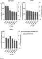
- the antibody-bound lipid nanoparticle of the present invention containing Firefly luciferase mRNA, a fragment of the NLDC145 antibody that binds to the ligand of MutuDC1940, and a membrane scaffold protein (MSP1E3D1, ApoA1 or ApoE3) was produced using the method of Example 4-2. Then, changes in mRNA encapsulation were observed while the antibody-bound lipid nanoparticle was stored at 4°C. On the day at which the antibody-bound lipid nanoparticle was formed and 7 days later, Ribogreen assay was performed in the same manner as in Example 4-1 to calculate the encapsulation ( FIG. 13 ).
- the mRNA encapsulation of the antibody-bound lipid nanoparticles of the present invention does not decrease even 7 days after production.
- the results mean that mRNA is not released from the lipid nanoparticles even when the antibody for forming the lipid nanoparticles of the present invention was attached to the lipid nanoparticles.
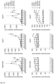
- the antibody-bound lipid nanoparticles of the present invention produced in Example 5-1 were stored at 4°C and dynamic light scattering was performed in the same manner as in Example 3-2 to determine the size change of the antibody-bound lipid nanoparticles for 7 days ( FIG. 14 ). Meanwhile, in FIG. 14 , the top and the bottom diagrams show the same experimental results wherein the top diagram shows the result including the experimental group with an LP ratio of 500:1, and the bottom diagram shows the result excluding the experimental group with an LP ratio of 500:1.
- the antibody-bound lipid nanoparticles of the present invention maintain a constant size over time when the LP ratio is greater than 5,000:1, but the size increases dramatically when the LP ratio is 500:1 to 1,000:1. This indicates that stable antibody-bound lipid nanoparticles were formed only when the LP ratio exceeded 5,000:1.
- the antibody-bound lipid nanoparticles of the present invention were produced in the same manner as in Example 5-1, except that DiD (DiIC18(5); 1,1'-dioctadecyl-3,3,3',3'-tetramethylindodicarbocyanine, 4-chlorobenzenesulfonate salt) dye was added at a molar ratio of 1% of the lipid to produce DID-antibody-bound lipid nanoparticles.
- DiD DiD (DiIC18(5); 1,1'-dioctadecyl-3,3,3',3'-tetramethylindodicarbocyanine, 4-chlorobenzenesulfonate salt) dye was added at a molar ratio of 1% of the lipid to produce DID-antibody-bound lipid nanoparticles.
- A549 cells grown using RPMI-1640 medium and MutuDC1940 cells grown in IMDM medium supplemented with 10 mM HEPES and 50 ⁇ M ⁇ -mercaptoethanol were prepared. Meanwhile, the A549 cells were used as an experimental group that did not express the target ligand and the MutuDC1940 cells were used as an experimental group that constantly expressed the target ligand.
- 100 ⁇ L of the A549 cells were seeded at a concentration of 1.5 ⁇ 10 5 cells/mL per well in a 96 well black plate.
- the MutuDC1940 cells were coated with collagen I at a concentration of 50 ug/mL to ensure efficient attachment to the plate, and then 100 ⁇ L of MutuDC1940 cells were seeded at a concentration of 3.5 ⁇ 10 5 cells/mL per well.
- the cells were incubated for one day to ensure efficient attachment to the plate and then were treated with the antibody-bound lipid nanoparticles of the present invention such that the final lipid concentration was adjusted to 10 ⁇ g/mL.
- the cells were incubated at 37°C in the presence of 5% CO 2 for 30 minutes and then the supernatant was removed.
- PBS phosphate buffered saline
- a luciferase assay was performed to determine whether or not the antibody-bound lipid nanoparticles of the present invention produced in Example 5-1 successfully delivered mRNA to target cells.
- A549 cells grown in RPMI-1640 medium and MutuDC1940 cells grown in IMDM medium supplemented with 10m M HEPES and 50 ⁇ M ⁇ -mercaptoethanol were prepared. Meanwhile, the A549 cells were used as an experimental group in which serum is absent and the target ligand is not expressed, and the MutuDC1940 cells were used as an experimental group in which serum is present and the target ligand is expressed.
- 500 ⁇ L of the A549 cells were seeded in a 96 well black plate at a concentration of 2 ⁇ 10 5 cells/mL per well.
- the MutuDC1940 cells were coated with collagen I at a concentration of 50 ug/mL to ensure efficient attachment to the plate, and then 500 ⁇ L of MutuDC1940 cells were seeded at a concentration of 4 ⁇ 10 5 cells/mL per well.
- the cells were incubated for one day to ensure efficient adhesion to the plate and then were treated with the antibody-bound lipid nanoparticles of the present invention at 125 ng of total mRNA per well.
- the cells were incubated for 12 hours at 37°C in the presence of 5% CO 2 and then the supernatant was removed.
- the cells were treated with 200 ⁇ L of cell lysis buffer at 37°C for 5 minutes to lyse the cells, and then 100 ⁇ L of the supernatant was transferred to a white 96 well plate. After adding 50 ⁇ L of luciferase substrate, luminescence was measured using a plate reader and the relative expression level of the Firefly luciferase gene was determined based on the experimental group treated with general lipid nanoparticles ( FIG. 16 ).
- the antibody-bound lipid nanoparticles of the present invention had a lower gene expression level than general lipid nanoparticles in most cases.
- the expression level in cells that did not express the target ligand increases.
- the expression level increased approximately 5 to 20 times at the LP ratio of 5,000:1 to 10,000:1 for MSP1E3D1, at the LP ratio of 500:1 to 10,000:1 for ApoA1, and at the LP ratio of 500:1 to 1,000:1 for ApoE3.
- the antibody-bound lipid nanoparticle of the present invention exhibits more specific delivery ability than general antibody-bound lipid nanoparticles, because general lipid nanoparticles delivered randomly the contained mRNA, but the antibody-bound lipid nanoparticle of the present invention contains membrane scaffold proteins to suppress the delivery of mRNA to random cells, and contains an antibody to improve the delivery of mRNA to cells.
- luciferase assay was performed in the same manner as in 5-1 to determine gene expression levels, except that A549 cells grown in RPMI-1640 medium supplemented with 10% FBS and MutuDC1940 cells grown in IMDM medium supplemented with 10% FBS, 10 mM HEPES, and 50 ⁇ M ⁇ -mercaptoethanol were used.
- the antibody-bound lipid nanoparticle of the present invention had a lower gene expression level than general lipid nanoparticles in most cases.
- the expression level increased approximately 5 to 20 times at a ratio of 5,000:1 to 100,000:1 for MSP1E3D1, at a ratio of 1,000:1 ⁇ 100,000:1 for ApoA1, and a ratio of 500:1 to 100,000:1 for ApoE3.
- the antibody-bound lipid nanoparticle of the present invention exhibits more specific delivery ability than general antibody-bound lipid nanoparticles, because general lipid nanoparticles delivered the contained mRNA randomly without cell specificity, but the antibody-bound lipid nanoparticle of the present invention contains membrane scaffold proteins to suppress the delivery of mRNA to random cells, and contains an antibody to improve the delivery of mRNA to cells.
Landscapes
- Health & Medical Sciences (AREA)
- Life Sciences & Earth Sciences (AREA)
- Chemical & Material Sciences (AREA)
- Engineering & Computer Science (AREA)
- Genetics & Genomics (AREA)
- General Health & Medical Sciences (AREA)
- Bioinformatics & Cheminformatics (AREA)
- Medicinal Chemistry (AREA)
- Biotechnology (AREA)
- Organic Chemistry (AREA)
- Pharmacology & Pharmacy (AREA)
- Animal Behavior & Ethology (AREA)
- Public Health (AREA)
- Veterinary Medicine (AREA)
- Epidemiology (AREA)
- Molecular Biology (AREA)
- Biomedical Technology (AREA)
- Biochemistry (AREA)
- General Engineering & Computer Science (AREA)
- Wood Science & Technology (AREA)
- Zoology (AREA)
- Immunology (AREA)
- Biophysics (AREA)
- Physics & Mathematics (AREA)
- Plant Pathology (AREA)
- Microbiology (AREA)
- Nanotechnology (AREA)
- Manufacturing & Machinery (AREA)
- Cell Biology (AREA)
- Optics & Photonics (AREA)
- Proteomics, Peptides & Aminoacids (AREA)
- Peptides Or Proteins (AREA)
- Dispersion Chemistry (AREA)
- Medicines That Contain Protein Lipid Enzymes And Other Medicines (AREA)
- Medicines Containing Antibodies Or Antigens For Use As Internal Diagnostic Agents (AREA)
- Pharmaceuticals Containing Other Organic And Inorganic Compounds (AREA)
- Medicinal Preparation (AREA)
Abstract
Description
- The present invention relates to an antibody-bound lipid nanoparticle containing an antibody-conjugated membrane scaffold protein, and more specifically, to an antibody-bound lipid nanoparticle that contains a lipid and an antibody-conjugated membrane scaffold protein and thus can be easily prepared and has excellent specific target ability.
- mRNA acts to deliver genetic information from DNA to the organ that synthesizes proteins in the cytoplasm. When this mRNA enters the cell, protein is synthesized within the cell and an immune response occurs.
- mRNA vaccines are being developed using mRNA having this property. However, mRNA has the problems that gene expression is suppressed due to interference caused by the immune response and it is difficult for the mRNA to enter the cytoplasm due to the large size thereof.
- In an attempt to address these problems, lipid nanoparticles (LNP) have been developed to facilitate mRNA entrance to the cytoplasm and the value thereof is increasing as they are recently used as carriers for mRNA vaccines. However, the method of delivering lipid nanoparticles (LNPs) selectively to cells remains insufficient.
- Accordingly, lipid nanoparticles with excellent target delivery ability have been developed by combining the excellent target delivery ability of antibodies with lipid nanoparticles. Conventional methods use chemical conjugation, thus disadvantageously making the overall process complicated and the yield low. In addition, conventional methods have a disadvantage in which the chemical identity is unclear and an affinity tag is attached to the antibody and thus it is thus difficult to identify the binding when injected in vivo.
- Therefore, it is an object of the present invention to provide an antibody-bound lipid nanoparticle that can be prepared in a simple manner using an antibody that can bind to a lipid nanoparticle (LNP) and has excellent target delivery ability and a method of producing the same.
- In accordance with one aspect of the present invention, provided is an antibody-bound lipid nanoparticle in which an antibody or antibody fragment conjugated with a membrane scaffold protein is bound to a hydrophobic moiety of a lipid constituting a lipid nanoparticle via the membrane scaffold protein through a hydrophobic bond.
- In the antibody-bound lipid nanoparticle of the present invention, the membrane scaffold protein is preferably an amphipathic protein having a helix structure. In this case, the membrane scaffold protein may be a fragment of the membrane scaffold protein that maintains the helix structure and amphipathic property.
- In the antibody-bound lipid nanoparticle of the present invention, the antibody fragment is preferably scFv or scFV-Fc.
- In the antibody-bound lipid nanoparticle of the present invention, the antibody or antibody fragment conjugated with the membrane scaffold protein is preferably produced by binding a gene encoding the antibody or antibody fragment to a gene encoding the membrane scaffold protein and expressing the result.
- In the antibody-bound lipid nanoparticle of the present invention, the lipid nanoparticle preferably contains a substance encapsulated therein. In this case, the encapsulated substance is preferably nucleic acid.
- In accordance with another aspect of the present invention, provided is a method of producing an antibody-bound lipid nanoparticle by mixing a membrane scaffold protein-conjugated antibody with a lipid.
- In the method, the antibody-bound lipid nanoparticle is preferably further mixed with an encapsulated substance.
- The antibody-bound lipid nanoparticle according to the present invention contains a membrane scaffold protein-conjugated antibody, thus exhibits excellent specific delivery ability to target cells and can be easily prepared.
-
-
FIG. 1 is a schematic diagram illustrating a conventional method of producing antibody-bound lipid nanoparticles. -
FIG. 2 is a schematic diagram illustrating a method of producing an antibody-bound lipid nanoparticle using the antibody (grabber antibody) of the present invention. Meanwhile, the top diagram ofFIG. 2 illustrates a method of producing an antibody-bound lipid nanoparticle by mixing with the antibody (grabber antibody) of the present invention while producing the lipid nanoparticle, and the bottom diagram ofFIG. 2 illustrates a method of producing an antibody-bound lipid nanoparticle by adding the antibody (grabber antibody) of the present invention to the produced lipid nanoparticle. -
FIG. 3 is a schematic diagram illustrating the structure of the antibody (grabber antibody) of the present invention. -
FIG. 4 shows the result of SDS gel electrophoresis to identify production of a single chain antibody fragment (grabber single chain variable fragment, Gr-scFv) of the present invention using an E. coli protein expression system, wherein 10B4 andNLDC 145 represent the antibodies used, and ApoA1, MSP1E3D1, and ApoeE3 represent the membrane scaffold proteins used. -
FIG. 5 shows the result of SDS gel electrophoresis to identify production of a single chain antibody fragment (grabber single chain variable fragment, Gr-scFv) of the present invention using a mammalian cell protein expression system. -
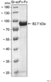
FIG. 6 shows the result of SDS gel electrophoresis to identify production of a single chain antibody fragment (grabber single chain variable fragment-Fc, Gr-scFv-Fc) of the present invention using a mammalian cell protein expression system. -
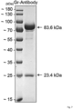
FIG. 7 shows the result of SDS gel electrophoresis to identify production of an antibody (grabber antibody, Gr-antibody) of the present invention using a mammalian cell protein expression system. -
FIG. 8 shows A280 of a membrane scaffold protein (MSP), a lipid nanoparticle (LNP), and a mixture (MSP LNP) of the lipid nanoparticle and the membrane scaffold protein (MSP) measured through size exclusion chromatography in order to determine whether or not the antibody of the present invention can bind to the lipid nanoparticle. -
FIG. 9 shows the particle size of a lipid nanoparticle (LNP), and a mixture (MSP LNP) of the lipid nanoparticle and a membrane scaffold protein measured through dynamic light scattering in order to determine whether or not the antibody of the present invention can bind to the lipid nanoparticle. -
FIG. 10 shows the mRNA encapsulation (%) of the antibody-bound lipid nanoparticle at each mixing time, while the antibody of the present invention is mixed during the process of forming lipid nanoparticles, in order to optimize the mixing time of the lipid nanoparticle with the antibody of the present invention. -
FIG. 11 shows the mRNA encapsulation (%) of antibody-bound lipid nanoparticles produced by mixing the antibody with lipid nanoparticles at different molar ratios (LP ratios) of the lipid to protein, in order to optimize the mixing time of the lipid nanoparticle with the antibody of the present invention. -
FIG. 12 shows the particle sizes of antibody-bound lipid nanoparticles, which are produced by mixing the antibody with lipid nanoparticles at different molar ratios (LP ratios) of the lipid to protein, measured through dynamic light scattering, in order to optimize the mixing time of the lipid nanoparticle with the antibody of the present invention. -
FIG. 13 shows changes in encapsulation observed while the antibody-bound lipid nanoparticles of the present invention are stored at 4°C for 7 days to determine the storage stability of the antibody-bound lipid nanoparticles. -
FIG. 14 shows the size of the antibody-bound lipid nanoparticles of the present invention observed while the antibody-bound lipid nanoparticles are stored at 4°C for 7 days to determine the storage stability of the antibody-bound lipid nanoparticles. -
FIG. 15 shows fluorescence measured to determine relative binding ability after treating MutuDC1940 cells expressing the target ligand and A549 cells not expressing the target ligand with antibody-bound lipid nanoparticles containing DiD dye, in order to determine whether or not the antibody-bound lipid nanoparticles of the present invention specifically bind to the target cells. -
FIG. 16 shows relative gene expression levels in cells measured after treating MutuDC1940 cells expressing the target ligand and A549 cells not expressing the target ligand with antibody-bound lipid nanoparticles, in order to determine whether or not the antibody-bound lipid nanoparticles of the present invention deliver mRNA specifically to the target cells. -
FIG. 17 shows relative gene expression levels in cells measured after treating MutuDC1940 cells expressing the target ligand and A549 cells not expressing the target ligand in the presence of a serum with antibody-bound lipid nanoparticles, in order to determine whether or not the antibody-bound lipid nanoparticles of the present invention deliver mRNA specifically to the target cells even if they are hindered by serum. - The present invention provides an antibody-bound lipid nanoparticle in which an antibody or antibody fragment conjugated with a membrane scaffold protein is bound to a hydrophobic moiety of a lipid constituting a lipid nanoparticle via the membrane scaffold protein through a hydrophobic bond.
- A conventional method of producing antibody-bound lipid nanoparticles (having a configuration in which antibodies are bound to the surface of lipid nanoparticles) includes the following steps: 1) mixing a lipid mixture constituting a lipid nanoparticle with an encapsulated substance to produce a lipid nanoparticle; 2) binding the lipid nanoparticle to a compound capable of binding to an antibody; and 3) binding an antibody to the compound, and thus includes at least three steps (see
FIG. 1 ). - However, in the present invention, antibody-bound lipid nanoparticles can be produced without a separate antibody-binding process using an antibody conjugated with a membrane scaffold protein (see
FIG. 3 ). That is, when the encapsulated substance, the lipid mixture, and the antibody of the present invention (so-called "grabber antibody") were mixed, the lipid nanoparticle is formed and at the same time, the antibody conjugated with the membrane scaffold protein was disposed on the surface of the lipid particle to obtain the antibody-bound lipid nanoparticle (see the top diagram ofFIG. 2 ). This means that, when using an antibody conjugated with a membrane scaffold protein, the conventional three-step production process can be simplified into a one-step process. - In addition, it was found that antibody-bound lipid nanoparticles were produced by adding the antibody (so-called "grabber antibody") of the present invention to the produced lipid nanoparticles (see the bottom diagram of
FIG. 2 ). This means that specificity can be imparted to lipid nanoparticles by modifying lipid nanoparticles with antibodies merely by simple addition without changing the conventional lipid nanoparticle production process. - Meanwhile, in the present invention, the membrane scaffold protein (MSP) is a protein that has a helix structure, is amphipathic, and acts to mediate the lipid and antibody of the antibody-bound lipid nanoparticle of the present invention. In this case, the membrane scaffold protein binds to the hydrophobic moiety of the lipid that constitutes the lipid nanoparticle based on a hydrophobic bond.
- A representative example of the membrane scaffold protein is apolipoprotein. Apolipoprotein is a protein that is present specifically in plasma lipoproteins and is known to stabilize the structure of lipoproteins, activate enzymes involved in lipoprotein metabolism, and act as a ligand for lipoprotein receptors present on the cell surface. The apolipoprotein is, for example, apolipoprotein A1 (ApoA-I), apolipoprotein A2 (ApoA-2), apolipoprotein B (ApoB), apolipoprotein C (ApoC), apolipoprotein E (ApoE), MSP1 (membrane scaffold protein 1), MSP1D1, MSP1D2, MSP1E1, MSP1E2, MSP1E3, MSP1E3D1, MSP2, MSP2N1, MSP2N2, MSP2N3, or the like.
- Apo-A1, which is mentioned as an example of apolipoprotein, is a protein that is composed of a 28 kDa single polypeptide containing 243 amino acids, has 8 repeating unit domains consisting of 11 amino acids or 22 amino acids, and has 60 to 75% of an alpha-helix proportion with a secondary structure constituting HDL. It is known that ApoA-I is mainly used as a component of high-density lipoprotein (HDL), which acts to remove cholesterol from surrounding tissues and deliver the cholesterol to the liver or other lipoproteins. In addition, ApoE is a protein that is composed of a 33 kDa single polypeptide consisting of 299 amino acids and mediates the delivery of cholesterol like ApoA1.
- In addition, in the present invention, a fragment of the membrane scaffold protein that maintains the helix structure and amphipathic properties may be used as the membrane scaffold protein. In other words, a part (fragment) of the membrane scaffold protein, rather than the entire membrane scaffold protein, may be used as long as the helix structure and amphipathic properties of the membrane scaffold protein are not lost.
- Meanwhile, in the present invention, the antibody or antibody fragment binds to lipid nanoparticles and serves to impart specific targeting ability to the target cell to which the antibody or antibody fragment may be bound. The antibody-bound lipid nanoparticles of the present invention may use various antibodies or antibody fragments depending on the characteristics of the target cell. In this case, the antibody fragment is preferably scFv, or scFv bound with scFv and Fc to provide specific targeting ability.
- Meanwhile, in the present invention, the antibody conjugated with the membrane scaffold protein may be produced by binding a gene encoding the antibody to a gene encoding the membrane scaffold protein, and then expressing the result.
- Meanwhile, the antibody-bound lipid nanoparticle of the present invention preferably contains a substance encapsulated therein. In this case, the encapsulated substance is preferably a nucleic acid. The nucleic acid is, for example, mRNA.
- Lipid nanoparticles are mainly used to safely deliver the encapsulated substance to the target. Foreign mRNA has the problems in which gene expression is suppressed due to interference with the human's immune response and it is difficult to induce foreign mRNA into cells due to the large size thereof. The mRNA vaccine solves these problems using lipid nanoparticles and the antibody-bound lipid nanoparticles developed in the present invention may also be used as a carrier to deliver the mRNA vaccine.
- Lipid nanoparticles are preferably prepared by mixing an ionizable lipid, a structural helper lipid, cholesterol, and polyethylene glycol. The lipid nanoparticles thus prepared are easily encapsulated in drugs such as nucleic acids, easily pass through cell membranes, and have excellent retention in the body.
- In another aspect, the present invention provides a method of producing an antibody-bound lipid nanoparticle by mixing a membrane scaffold protein-conjugated antibody with a lipid.
- As described above, the method according to the present invention is capable of simplifying the conventional three-step production process into a one-step production process using the membrane scaffold protein-conjugated antibody.
- Meanwhile, in the method of producing the antibody-bound lipid nanoparticles of the present invention, the antibody-bound lipid nanoparticles are preferably further mixed with an encapsulated substance. The encapsulated substance is an mRNA vaccine. In this case, the antibody-bound lipid nanoparticles may be used as a carrier to transport the mRNA vaccine.
- Hereinafter, the present invention will be described in more detail with reference to the following examples. The scope of the present invention is not limited to the examples and encompasses modifications of the technical concept equivalent thereto.
- In this example, an attempt was made to produce a plasmid capable of producing an antibody for forming lipid nanoparticles of the present invention.
- Cloning was performed using pET-22b(+) or pcDNA3.1(+) as a vector and T4 DNA polymerase to prepare a plasmid for producing a single-chain antibody fragment (Gr-scFv, see (a) in
FIG. 3 ) for forming lipid nanoparticles that consists of a variable region of heavy chain (VH), a variable region of light chain (VL), and a membrane scaffold protein (MSP). - Specifically, PCR was performed using primers designed such that the pET-22b(+) or pcDNA3.1(+) vector is complementary to 15 bp of both ends of the VH-linker-VL-linker-Apolipoprotein insert. Then, the result was treated with DpnI enzyme for 2 hours at 37°C to remove the methylated backbone plasmid for PCR. The result was reacted with T4 DNA polymerase at 25°C for 150 seconds and on ice for 10 minutes to induce hydrogen bonding between the vector and the insert. 1 µL of the reaction product was mixed with 100 µL of Escherichia coli TOP10 competent cells, incubated on ice for 30 minutes, and heat-shocked at 42°C for 45 seconds for gene transduction. Then, 900 µL of SOC (super optimal broth with catabolite repression) liquid medium was added to the result, followed by incubation at 37°C for 1 hour, and centrifugation (13,000 rpm, 1 minute) to obtain cells. The obtained cells were resuspended in 100 µL of SOC liquid medium, plated on carbenicillin LB (Luria-Bertani) solid medium, and incubated at 37°C for 16 hours to form colonies. The colonies were inoculated into LB liquid medium containing 100 ug/mL of carbenicillin and incubated at 37°C for 16 hours, and then the plasmid for producing single-chain antibody fragments for forming lipid nanoparticles was purified using a plasmid purification kit.
- A plasmid for producing a single chain antibody for forming lipid nanoparticles was produced in the same manner as in Example 1-1 using a pcDNA3.1(+) vector and a VH-linker-VL-hinge-Fc-linker-apolipoprotein insert to produce a plasmid for producing single chain antibody for forming lipid nanoparticles (Gr-scFv-Fc, see (b) in
FIG. 3 ) that consists of a variable region of heavy chain (VH), a variable region of light chain (VL), a fragment crystallizable region (Fc region), and a membrane scaffold protein (MSP). - Plasmids for producing an antibody that forms lipid nanoparticles were produced in the same manner as in Example 1-1 using the pMAZ vector, VH-CH-linker-apolipoprotein, or VL-CL insert in order to produce a plasmid consisting of a variable region of heavy chain (VH), a constant region of heavy chain (CH), and a membrane scaffold protein (MSP), and a plasmid consisting of a variable region of light chain (VL) and a constant region of light chain (CL), as plasmids for producing an antibody (Gr-antibody, see (c) of
FIG. 3 ) that forms lipid nanoparticles. - In this example, the antibody for forming lipid nanoparticles according to the present invention was produced using the plasmid produced in Example 1.
- In order to produce single chain antibody fragments (grabber single chain variable fragment, Gr-scFv) for forming lipid nanoparticles having the protein sequences of SEQ ID NOS. 1 to 9, the plasmid using the pET-22b(+) vector produced in Example 1-1 was expressed using the E. coli protein expression system.
- Specifically, the plasmid was injected into the expression host, E. coli BL21 (DE3) at 42°C, plated on carbenicillin LB (Luria-Bertani) solid medium at a concentration of 100 µg/mL, and incubated at 37°C for 16 hours to form colonies and the transformed E. coli strains were selected. Each transformed strain was inoculated into LB liquid medium containing 100 ug/mL of carbenicillin, cultured at 37°C for 16 hours, and then subcultured. The subculture solution was incubated until the optical density (OD600) measured at a wavelength of 600 nm reached 1.5, and 0.5 mM IPTG (isopropyl β-D-1-thiogalactopyranoside) was added thereto, followed by culture at 25°C for 6 hours to express single-chain antibody fragments that form lipid nanoparticles. Then, the culture solution was centrifuged (4,000 rpm, 10 minutes) to obtain cells. The obtained cells were pulverized using a sonicator and separated into soluble and insoluble fractions through centrifugation (15,000 rpm, 10 minutes).
- Meanwhile, the proteins in each fraction were detected by SDS gel electrophoresis. The result showed that the single chain antibody fragment for forming lipid nanoparticles of the present invention was produced intact (
FIG. 4 ). Meanwhile, inFIG. 4 , 10B4 andNLDC 145 represent the antibodies used, and ApoA1, MSP1E3D1, and ApoeE3 represent the membrane scaffold proteins used. - In addition, in order to produce single-chain antibody fragments for forming lipid nanoparticles, the plasmid using the pcDNA3.1(+) vector produced in Example 1-1 was expressed using a mammalian cell protein expression system.
- Specifically, HEK-293F (human embryonic kidney 293F) used as an expression host was incubated in an aqueous suspended cell state at 37°C and 120 rpm in the presence of 8% CO2 to prepare 180 mL of a cell fluid (1.1 × 10⁶ cells/mL) . For transfection, 250 µg of plasmid and 750 µg of PEI (polyethylenimine) were mixed with 20 mL of medium and then mixed with the prepared cell fluid. Then, the cells were incubated for 96 hours at 37°C and 120 rpm in the presence of 8% CO2 and then centrifuged (8, 000 g, 10 minutes) to remove the cells and obtain the supernatant. The supernatant was mixed with phosphate buffer at pH 7.0 in a ratio of 1:1 and purified using proL agarose beads. Substances that failed to bind to the beads were removed by treatment with a phosphate buffer solution of pH 7.0. Single-chain antibody fragments were purified by treatment with a solution containing 0.1 M glycine at pH 2.5 to remove the bond between the single-chain antibody fragments and agarose beads, and then the pH was returned to a neutral range using 1 M pH 8.0 Tris buffer.
- Meanwhile, the purified single-chain antibody fragment was detected by SDS gel electrophoresis. The result showed that the single-chain antibody fragment for forming lipid nanoparticle of the present invention was produced intact (
FIG. 5 ). - In order to produce single chain antibody fragments (grabber single chain variable fragment-Fc, Gr-scFv-Fc) for forming lipid nanoparticles having the protein sequences of SEQ ID NOS. 10 to 18, the plasmid using the pcDNA3.1(+) vector produced in Example 1-2 was expressed using the mammalian cell expression system.
- Specifically, HEK-293F (human embryonic kidney 293F) used as an expression host was incubated in an aqueous suspended cell state at 37°C and 120 rpm in the presence of 8% CO2 to prepare 180 mL of a cell fluid (1.1 × 10⁶ cells/mL) . For transfection, 250 µg of plasmid and 750 µg of PEI (polyethylenimine) were mixed with 20 mL of medium and then mixed with the prepared cell fluid. Then, the cells were incubated for 96 hours at 37°C and 120 rpm in the presence of 8% CO2 and then centrifuged (8, 000 g, 10 minutes) to remove the cells and obtain the supernatant. The supernatant was mixed with phosphate buffer at pH 7.0 in a ratio of 1:1 and purified using proL agarose beads. Substances that failed to bind to the beads were removed by treatment with a phosphate buffer solution of pH 7.0. Single-chain antibody fragments were purified by treatment with a solution containing 0.1 M glycine at pH 2.5 to remove the bond between the single-chain antibody fragments and agarose beads, and then the pH was returned to a neutral range using 1 M pH 8.0 Tris buffer.
- Meanwhile, the purified single-chain antibody fragment was detected by SDS gel electrophoresis. The result showed that the single-chain antibody fragment forming lipid nanoparticle of the present invention was produced intact (
FIG. 6 ). - In order to produce an antibody (Gr-antibody) for forming lipid nanoparticles having the IgL sequence of SEQ ID NOS. 19 to 21, and having the IgH sequence of SEQ ID NOS. 22 to 30, the plasmid using the pcDNA3.1(+) vector produced in Example 1-3 was expressed using the mammalian cell expression system.
- Specifically, HEK-293F (human embryonic kidney 293F) used as an expression host was incubated in an aqueous suspended cell state at 37°C and 120 rpm in the presence of 8% CO2 to prepare 180 mL of a cell fluid (1.1 × 10⁶ cells/mL) . For transfection, 250 µg of plasmid and 750 µg of PEI (polyethylenimine) were mixed with 20 mL of medium and then mixed with the prepared cell fluid. Then, the cells were incubated for 144 hours at 37°C and 120 rpm in the presence of 8% CO2 and then centrifuged (8, 000 g, 10 minutes) to remove the cells and obtain the supernatant. The supernatant was mixed with phosphate buffer at pH 7.0 in a ratio of 1:1 and purified using proL agarose beads. Substances that failed to bind to the beads were removed by treatment with a phosphate buffer solution of pH 7.0. The antibody was purified by treating a solution containing 0.1 M glycine at pH 2.5 to remove the bond between the antibody fragments and agarose beads, and then pH was returned to a neutral range using 1 M pH 8.0 Tris buffer.
- Meanwhile, the purified antibody was detected by SDS gel electrophoresis. The result showed that the antibody for forming lipid nanoparticles of the present invention was produced intact (
FIG. 7 ). - Meanwhile, in
FIG. 7 , 83.6 kDa represents a protein consisting of a variable region of heavy chain (VH), a constant region of heavy chain (CH), and apolipoprotein, and 23.4 kDa represents a protein consisting of a variable region of light chain (VL) and a constant region of light chain (CL). - In this example, the adhesion of membrane scaffold proteins to lipid nanoparticles was detected to determine whether or not membrane scaffold proteins could attach to antibody-bound lipid nanoparticles by mixing the antibody for forming lipid nanoparticles of the present invention with lipid nanoparticles.
- SM102 (CAS No. 2089251-47-6), DSPC (1,2-distearoyl-sn-glycero-3-phosphocholine), cholesterol, and DMG-PEG-2000 (1,2-dimyristoyl-rac-glycero-3-methoxypolyethylene glycol-2000) were dissolved in ethanol to prepare a lipid solution. Meanwhile, 75 µL of messenger ribonucleic acid (messenger RNA, mRNA) was mixed with aqueous phase mRNA buffer (10.25 mM citric acid, 2.25 mM tri-sodium citrate, pH 3.0) to prepare an RNA solution. The lipid solution and the RNA solution prepared above were mixed using a microfluidic device such that the molar ratio of SM102:DSPC:cholesterol:DMG-PEG-2000 was 50:10:38.5:1.5, and the molar ratio of the amino group of SM102, which is an ionized lipid, to the phosphate group of mRNA (N/P ratio) was 6. Then, PBS was added to the mixture and diluted to a 4-fold volume ratio to prepare a lipid nanoparticle solution.
- MSP1E3D1 derived from apolipoprotein AI as a membrane scaffold protein was mixed with the lipid nanoparticle solution prepared in 3-1 above at a molar ratio of lipid to protein (LP ratio) of 240:1, and incubated at 4°C for 16 hours to attach membrane scaffold proteins to lipid nanoparticles.
- Then, A280 of lipid nanoparticles (LNP), membrane scaffold protein (MSP), and membrane scaffold protein-attached lipid nanoparticles (MSP-LNP) were determined through size exclusion chromatography (
FIG. 8 ). - As can be seen from
FIG. 8 , the A280 peak shifts when lipid nanoparticles (LNP) are mixed with the membrane scaffold protein (MSP), which means that the membrane scaffold protein (MSP) is intactly attached to the lipid nanoparticle (LNP). - In addition, the particle sizes of the lipid nanoparticle (LNP) and the membrane scaffold protein-attached lipid nanoparticle (MSP-LNP) were measured using dynamic light scattering (
FIG. 9 ). - As can be seen from
FIG. 9 , the membrane scaffold protein-attached lipid nanoparticle has a large particle size, which indicates that the membrane scaffold protein (MSP) is intactly attached to the lipid nanoparticle (LNP). - The results mean that the antibody for forming the lipid nanoparticle of the present invention containing the membrane scaffold protein has excellent adhesion to the lipid nanoparticle.
- In this example, antibody-bound lipid nanoparticles were produced using the single-chain antibody for forming the lipid nanoparticles of the present invention produced in Example 2-2.
- In addition, the lipid nanoparticles and the single chain antibody for forming the lipid nanoparticles of the present invention were produced at different mixing times and different mixing ratios, and the optimized mixing time and ratio were selected.
- SM102 (CAS No. 2089251-47-6), DSPC (1,2-distearoyl-sn-glycero-3-phosphocholine), cholesterol, and DMG-PEG-2000 (1,2-dimyristoyl-rac-glycero-3-methoxypolyethylene glycol-2000) were dissolved in ethanol to prepare a lipid solution. Meanwhile, 75 µL of messenger ribonucleic acid (messenger RNA, mRNA) was mixed with aqueous phase mRNA buffer (10.25 mM citric acid, 2.25 mM tri-sodium citrate, pH 3.0) to prepare an RNA solution. The lipid solution and the RNA solution prepared above were mixed using a microfluidic device such that the molar ratio of SM102:DSPC:cholesterol:DMG-PEG-2000 was adjusted to 50:10:38.5:1.5, and the molar ratio of the amino group of SM102, which is an ionized lipid, to the phosphate group of mRNA (N/P ratio) was adjusted to 6. Then, the mixture was diluted to a 4-fold volume ratio using PBS to form lipid nanoparticles. At this time, the dilution was performed three times, a single chain antibody for forming the lipid nanoparticles of the present invention was added thereto such that a molar ratio of the lipid to protein (LP ratio) was adjusted to 240:1, and reacted at 37°C for 1 hour to form antibody-bound lipid nanoparticles.
- At this time, the experimental group to which the single-chain antibody for forming the lipid nanoparticles of the present invention was added during the first dilution was referred to as "early", the experimental group to which the single-chain antibody for forming the lipid nanoparticles of the present invention was added during the second dilution was referred to as "middle", the experimental group to which the single-chain antibody for forming the lipid nanoparticles of the present invention was added during the third dilution was referred to as "late", and the experimental group to which the single-chain antibody for forming the lipid nanoparticles of the present invention was added in uniform portions during a total of 3 dilutions was referred to as "total".
- Then, a RiboGreen assay was performed to determine whether or not the antibody-bound lipid nanoparticles of the present invention were successfully formed.
- Specifically, 20 µL of each of lipid nanoparticles and antibody-bound lipid nanoparticles were diluted in 180 µL of dPBS. The dilution was further mixed with 200 µL of dPBS or 1% Triton X-100 in dPBS, followed by reaction for 10 minutes to prepare a dilute solution of lipid nanoparticles and a dilute solution of antibody-bound lipid nanoparticles. 100 µL of the RNA solution in the standard concentration range (dPBS standard and 0.5% Triton X-100 in dPBS standard), 100 µL of the prepared lipid nanoparticle dilution solution and 100 µL of the prepared antibody-bound lipid nanoparticle dilution solution were added to a 96 well plate and reacted at 25°C for 5 minutes. Then, excitation/emission = 485/530 nm was measured using a fluorescence spectrophotometer to determine the mRNA concentration (
FIG. 10 ). - Meanwhile, in
FIG. 10 , the mRNA concentration of the experimental group diluted using dPBS is shown as a concentration of unencapsulated free mRNA, and the mRNA concentration of the experimental group diluted with 1% Triton X-100 in dPBS is shown as the total mRNA concentration. Encapsulation was calculated as (1-(free mRNA/total mRNA)) × 100%. - This indicates that antibody-bound lipid nanoparticles were formed intact even when mixed with single-chain antibodies for forming the lipid nanoparticles of the present invention while the lipid nanoparticles were formed. In addition, this indicates that the encapsulation of mRNA increases as the single chain antibody for forming the lipid nanoparticles of the present invention was added later. Therefore, in the following experiment, the antibody-bound lipid nanoparticles of the present invention were produced by adding the single-chain antibody for forming the lipid nanoparticles of the present invention after the lipid nanoparticles were formed intact.
- SM102 (CAS No. 2089251-47-6), DSPC (1,2-distearoyl-sn-glycero-3-phosphocholine), cholesterol, and DMG-PEG-2000 (1,2-dimyristoyl-rac-glycero-3-methoxypolyethylene glycol-2000) were dissolved in ethanol to prepare a lipid solution. Meanwhile, 75 µL of messenger ribonucleic acid (messenger RNA, mRNA) was mixed with aqueous phase mRNA buffer (10.25 mM citric acid, 2.25 mM tri-sodium citrate, pH 3.0) to prepare an RNA solution. The lipid solution and the RNA solution prepared above were mixed using a microfluidic device such that the molar ratio of SM102:DSPC:cholesterol:DMG-PEG-2000 was 50:10:38.5:1.5, and the molar ratio of the amino group of SM102, which is an ionized lipid, to the phosphate group of mRNA (N/P ratio) was 6. The mixture was diluted 4 times by volume using PBS to form lipid nanoparticles. Single-chain antibodies for forming the lipid nanoparticles of the present invention were added to the lipid nanoparticles at various lipid:protein molar ratios (LP ratio), and reacted at 4°C for 16 hours to form antibody-bound lipid nanoparticles.
- Then, the encapsulation was calculated by performing Ribogreen assay in the same manner as in Example 4-1 (
FIG. 11 ) and the size of the formed antibody-bound lipid nanoparticles of the present invention was measured by performing dynamic light scattering in the same manner as in Example 3-2 (FIG. 12 ). - The results showed that the encapsulation was as high as about 95% at an LP ratio of 5,000:1 to 100,000:1, and that the size was increased because the single chain antibody for forming the lipid nanoparticles of the present invention was attached to the lipid nanoparticles.
- In this example, the storage stability of the antibody-bound lipid nanoparticles of the present invention was determined.
- The antibody-bound lipid nanoparticle of the present invention containing Firefly luciferase mRNA, a fragment of the NLDC145 antibody that binds to the ligand of MutuDC1940, and a membrane scaffold protein (MSP1E3D1, ApoA1 or ApoE3) was produced using the method of Example 4-2. Then, changes in mRNA encapsulation were observed while the antibody-bound lipid nanoparticle was stored at 4°C. On the day at which the antibody-bound lipid nanoparticle was formed and 7 days later, Ribogreen assay was performed in the same manner as in Example 4-1 to calculate the encapsulation (
FIG. 13 ). - As can be seen from
FIG. 13 , the mRNA encapsulation of the antibody-bound lipid nanoparticles of the present invention does not decrease even 7 days after production. The results mean that mRNA is not released from the lipid nanoparticles even when the antibody for forming the lipid nanoparticles of the present invention was attached to the lipid nanoparticles. - The antibody-bound lipid nanoparticles of the present invention produced in Example 5-1 were stored at 4°C and dynamic light scattering was performed in the same manner as in Example 3-2 to determine the size change of the antibody-bound lipid nanoparticles for 7 days (
FIG. 14 ). Meanwhile, inFIG. 14 , the top and the bottom diagrams show the same experimental results wherein the top diagram shows the result including the experimental group with an LP ratio of 500:1, and the bottom diagram shows the result excluding the experimental group with an LP ratio of 500:1. - As can be seen from
FIG. 14 , the antibody-bound lipid nanoparticles of the present invention maintain a constant size over time when the LP ratio is greater than 5,000:1, but the size increases dramatically when the LP ratio is 500:1 to 1,000:1. This indicates that stable antibody-bound lipid nanoparticles were formed only when the LP ratio exceeded 5,000:1. - In this example, whether or not the antibody-bound lipid nanoparticles of the present invention specifically bind to target cells was determined.
- The antibody-bound lipid nanoparticles of the present invention were produced in the same manner as in Example 5-1, except that DiD (DiIC18(5); 1,1'-dioctadecyl-3,3,3',3'-tetramethylindodicarbocyanine, 4-chlorobenzenesulfonate salt) dye was added at a molar ratio of 1% of the lipid to produce DID-antibody-bound lipid nanoparticles.
- A549 cells grown using RPMI-1640 medium and MutuDC1940 cells grown in IMDM medium supplemented with 10 mM HEPES and 50 µM β-mercaptoethanol were prepared. Meanwhile, the A549 cells were used as an experimental group that did not express the target ligand and the MutuDC1940 cells were used as an experimental group that constantly expressed the target ligand.
- 100 µL of the A549 cells were seeded at a concentration of 1.5 × 10⁵ cells/mL per well in a 96 well black plate. The MutuDC1940 cells were coated with collagen I at a concentration of 50 ug/mL to ensure efficient attachment to the plate, and then 100 µL of MutuDC1940 cells were seeded at a concentration of 3.5 × 10⁵ cells/mL per well. The cells were incubated for one day to ensure efficient attachment to the plate and then were treated with the antibody-bound lipid nanoparticles of the present invention such that the final lipid concentration was adjusted to 10 µg/mL. The cells were incubated at 37°C in the presence of 5% CO2 for 30 minutes and then the supernatant was removed. The cells were washed twice with PBS (phosphate buffered saline) and then treated with 100 µL of cell lysis buffer at 37°C for 5 minutes to lyse the cells. Then, fluorescence was measured at Ex/Em = 644/663 nm using a plate reader to confirm the relative binding ability of the antibody-bound lipid nanoparticles of the present invention (
FIG. 15 ). - As can be seen from
FIG. 15 , in the experimental group treated with A549 cells not expressing the target ligand, there is no difference between the antibody-bound lipid nanoparticles of the present invention and general lipid nanoparticles, whereas, in the experimental group treated with MutuDC1940 cells expressing the target ligand, the antibody-bound lipid nanoparticles of the present invention exhibit higher fluorescence values. The results mean that the antibody-bound lipid nanoparticles of the present invention can bind better and specifically to target cells expressing the target ligand. - In this example, whether or not the antibody-bound lipid nanoparticles of the present invention specifically deliver captured mRNA to target cells was determined.
- A luciferase assay was performed to determine whether or not the antibody-bound lipid nanoparticles of the present invention produced in Example 5-1 successfully delivered mRNA to target cells.
- A549 cells grown in RPMI-1640 medium and MutuDC1940 cells grown in IMDM medium supplemented with 10m M HEPES and 50 µM β-mercaptoethanol were prepared. Meanwhile, the A549 cells were used as an experimental group in which serum is absent and the target ligand is not expressed, and the MutuDC1940 cells were used as an experimental group in which serum is present and the target ligand is expressed.
- 500 µL of the A549 cells were seeded in a 96 well black plate at a concentration of 2×10⁵ cells/mL per well. The MutuDC1940 cells were coated with collagen I at a concentration of 50 ug/mL to ensure efficient attachment to the plate, and then 500 µL of MutuDC1940 cells were seeded at a concentration of 4 × 10⁵ cells/mL per well. The cells were incubated for one day to ensure efficient adhesion to the plate and then were treated with the antibody-bound lipid nanoparticles of the present invention at 125 ng of total mRNA per well. The cells were incubated for 12 hours at 37°C in the presence of 5% CO2 and then the supernatant was removed. The cells were treated with 200 µL of cell lysis buffer at 37°C for 5 minutes to lyse the cells, and then 100 µL of the supernatant was transferred to a white 96 well plate. After adding 50 µL of luciferase substrate, luminescence was measured using a plate reader and the relative expression level of the Firefly luciferase gene was determined based on the experimental group treated with general lipid nanoparticles (
FIG. 16 ). - As can be seen from
FIG. 16 , in the experimental group treated with A549 cells that did not express the target ligand, the antibody-bound lipid nanoparticles of the present invention had a lower gene expression level than general lipid nanoparticles in most cases. In addition, it can be seen that, as the amount of the antibody of the present invention decreases, the expression level in cells that did not express the target ligand increases. - On the other hand, in the experimental group treated with MutuDC1940 cells expressing the target ligand, the expression level increased approximately 5 to 20 times at the LP ratio of 5,000:1 to 10,000:1 for MSP1E3D1, at the LP ratio of 500:1 to 10,000:1 for ApoA1, and at the LP ratio of 500:1 to 1,000:1 for ApoE3.
- The results indicate that the antibody-bound lipid nanoparticle of the present invention exhibits more specific delivery ability than general antibody-bound lipid nanoparticles, because general lipid nanoparticles delivered randomly the contained mRNA, but the antibody-bound lipid nanoparticle of the present invention contains membrane scaffold proteins to suppress the delivery of mRNA to random cells, and contains an antibody to improve the delivery of mRNA to cells.
- Whether or not the antibody-bound lipid nanoparticles of the present invention successfully delivered mRNA to target cells, like in Example 7-1, was determined even in the presence of serum without an influence from serum.
- For this purpose, luciferase assay was performed in the same manner as in 5-1 to determine gene expression levels, except that A549 cells grown in RPMI-1640 medium supplemented with 10% FBS and MutuDC1940 cells grown in IMDM medium supplemented with 10% FBS, 10 mM HEPES, and 50 µM β-mercaptoethanol were used.
- As can be seen from
FIG. 17 , in the experimental group treated with A549 cells that did not express the target ligand, the antibody-bound lipid nanoparticle of the present invention had a lower gene expression level than general lipid nanoparticles in most cases. - On the other hand, in the group containing the scFv (NLDC145) of the antibody when treated with MutuDC1940 cells expressing the target ligand, the expression level increased approximately 5 to 20 times at a ratio of 5,000:1 to 100,000:1 for MSP1E3D1, at a ratio of 1,000:1~100,000:1 for ApoA1, and a ratio of 500:1 to 100,000:1 for ApoE3.
- The results show that, even in the presence of serum such as bodily fluids, the antibody-bound lipid nanoparticle of the present invention exhibits more specific delivery ability than general antibody-bound lipid nanoparticles, because general lipid nanoparticles delivered the contained mRNA randomly without cell specificity, but the antibody-bound lipid nanoparticle of the present invention contains membrane scaffold proteins to suppress the delivery of mRNA to random cells, and contains an antibody to improve the delivery of mRNA to cells.
Claims (9)
- An antibody-bound lipid nanoparticle in which an antibody or antibody fragment conjugated with a membrane scaffold protein is bound to a hydrophobic moiety of a lipid constituting a lipid nanoparticle via the membrane scaffold protein through a hydrophobic bond.
- The antibody-bound lipid nanoparticle according to claim 1, wherein the membrane scaffold protein is an amphipathic protein having a helix structure.
- The antibody-bound lipid nanoparticle according to claim 2, wherein the membrane scaffold protein is a fragment of the membrane scaffold protein that maintains a helix structure and amphipathic properties.
- The antibody-bound lipid nanoparticle according to claim 1, wherein the antibody fragment is scFv or scFV-Fc.
- The antibody-bound lipid nanoparticle according to claim 1, wherein the antibody or antibody fragment conjugated with the membrane scaffold protein is produced by binding a gene encoding the antibody or antibody fragment to a gene encoding the membrane scaffold protein and expressing the result.
- The antibody-bound lipid nanoparticle according to claim 1, wherein the lipid nanoparticle comprises a substance encapsulated therein.
- The antibody-bound lipid nanoparticle according to claim 6, wherein the encapsulated substance is nucleic acid.
- A method of producing an antibody-bound lipid nanoparticle by mixing a membrane scaffold protein-conjugated antibody with a lipid.
- The method according to claim 8, wherein the antibody-bound lipid nanoparticle is further mixed with an encapsulated substance.
Applications Claiming Priority (3)
| Application Number | Priority Date | Filing Date | Title |
|---|---|---|---|
| KR20220013592 | 2022-01-28 | ||
| KR1020230010355A KR102698524B1 (en) | 2022-01-28 | 2023-01-26 | Antibody-binding lipid nanoparticle with antibody bound to membrane scaffold protein |
| PCT/KR2023/001298 WO2023146356A1 (en) | 2022-01-28 | 2023-01-27 | Antibody-bound lipid nanoparticle comprising antibody bound to membrane scaffold protein |
Publications (2)
| Publication Number | Publication Date |
|---|---|
| EP4467564A1 true EP4467564A1 (en) | 2024-11-27 |
| EP4467564A4 EP4467564A4 (en) | 2026-01-21 |
Family
ID=87472287
Family Applications (1)
| Application Number | Title | Priority Date | Filing Date |
|---|---|---|---|
| EP23747397.0A Pending EP4467564A4 (en) | 2022-01-28 | 2023-01-27 | Antibody-bound lipidanoparticles with antibody bound to membrane framework protein |
Country Status (5)
| Country | Link |
|---|---|
| US (1) | US12427204B2 (en) |
| EP (1) | EP4467564A4 (en) |
| JP (1) | JP2025505427A (en) |
| CN (1) | CN118786139B (en) |
| WO (1) | WO2023146356A1 (en) |
Families Citing this family (2)
| Publication number | Priority date | Publication date | Assignee | Title |
|---|---|---|---|---|
| WO2026010380A1 (en) * | 2024-07-02 | 2026-01-08 | 성균관대학교산학협력단 | Pharmaceutical lipid nanoparticle composition with improved targeted delivery ability and safety |
| WO2026014855A1 (en) * | 2024-07-08 | 2026-01-15 | 알엔에이진(주) | Fusion protein comprising apolipoprotein and fc region-binding peptide and uses thereof |
Family Cites Families (19)
| Publication number | Priority date | Publication date | Assignee | Title |
|---|---|---|---|---|
| US7592008B2 (en) * | 2000-11-20 | 2009-09-22 | The Board Of Trustees Of The University Of Illinois, A Body Corporate And Politic Of The State Of Illinois | Membrane scaffold proteins |
| WO2002040501A2 (en) * | 2000-11-20 | 2002-05-23 | The Board Of Trustees Of The University Of Illinois | Membrane scaffold proteins |
| AU2003302676A1 (en) * | 2002-12-03 | 2004-06-23 | Blanchette Rockefeller Neurosciences Institute | Artificial low-density lipoprotein carriers for transport of substances across the blood-brain barrier |
| AU2004212944B2 (en) * | 2003-02-14 | 2008-08-21 | Children's Hospital & Research Center At Oakland | Lipophilic drug delivery vehicle and methods of use thereof |
| KR100953917B1 (en) | 2009-04-30 | 2010-04-22 | 고려대학교 산학협력단 | Lipopeptides with specific affinity to fc region of antibodies and antigen-recognizing lipid nanoparticles comprising the same |
| KR101343791B1 (en) | 2010-02-02 | 2013-12-20 | 고려대학교 산학협력단 | Lipopeptides with specific affinity to Fc region of antibodies and antigen-recognizing lipid nanoparticles comprising the same |
| KR101536037B1 (en) * | 2012-02-24 | 2015-07-13 | 성균관대학교산학협력단 | Antibody-drug conjugate comprising apolipoprotein |
| JP6763545B2 (en) * | 2015-04-15 | 2020-09-30 | 国立大学法人京都大学 | Targeted amphipathic nanocarriers and methods for their production |
| US11541100B2 (en) * | 2016-07-15 | 2023-01-03 | MVRIX Co., Ltd. | Pharmaceutical composition comprising nanoperforator for preventing or treating viral infectious diseases |
| MX2019004913A (en) | 2016-10-26 | 2019-09-16 | Curevac Ag | Lipid nanoparticle mrna vaccines. |
| KR101994279B1 (en) | 2017-11-03 | 2019-06-28 | 서울대학교산학협력단 | Manufacturing method of nanodisc comprising an olfactory receptor protein and nanodisc comprising an olfactory receptor protein manufactured by the same |
| US20190307892A1 (en) * | 2018-04-04 | 2019-10-10 | Eriochem Usa, Llc | Targeted drug delivery and therapeutic methods using apo-e modified lipid nanoparticles |
| KR101933199B1 (en) | 2018-04-26 | 2018-12-31 | 을지대학교 산학협력단 | Production method for highly orientated gold nano particle-antibody conjugate and dry eye syndrome diagnostic kit using the same |
| CN110960688A (en) * | 2018-09-30 | 2020-04-07 | 复旦大学 | Low-toxicity biomimetic nanosystem and preparation method for improving the efficacy of pancreatic cancer |
| US20220204994A1 (en) * | 2019-04-05 | 2022-06-30 | Precision Biosciences, Inc. | Methods of preparing populations of genetically-modified immune cells |
| CN114667293A (en) * | 2019-09-24 | 2022-06-24 | Mvrix株式会社 | Nano-perforators with enhanced antiviral activity |
| KR20210085296A (en) | 2019-12-30 | 2021-07-08 | 재단법인대구경북과학기술원 | Composition for thermotherapy of circulating tumor cells comprising magnetic nanoparticles bound with antibodies, preparation method thereof, and method for treating circulating tumor cells using the same |
| KR102692009B1 (en) | 2020-04-03 | 2024-08-07 | 서울대학교산학협력단 | Pharmaceutical composition for treatment of cancers comprising lipid-photothermal nanoparticle conjugated antibody |
| WO2023046931A1 (en) * | 2021-09-23 | 2023-03-30 | Bio-Trip B.V. | Apolipoprotein fusion proteins for cell-specific immune regulation |
-
2023
- 2023-01-27 WO PCT/KR2023/001298 patent/WO2023146356A1/en not_active Ceased
- 2023-01-27 CN CN202380024472.8A patent/CN118786139B/en active Active
- 2023-01-27 EP EP23747397.0A patent/EP4467564A4/en active Pending
- 2023-01-27 US US18/833,679 patent/US12427204B2/en active Active
- 2023-01-27 JP JP2024544982A patent/JP2025505427A/en active Pending
Also Published As
| Publication number | Publication date |
|---|---|
| CN118786139B (en) | 2026-05-01 |
| WO2023146356A1 (en) | 2023-08-03 |
| EP4467564A4 (en) | 2026-01-21 |
| JP2025505427A (en) | 2025-02-26 |
| US20250152732A1 (en) | 2025-05-15 |
| US12427204B2 (en) | 2025-09-30 |
| CN118786139A (en) | 2024-10-15 |
Similar Documents
| Publication | Publication Date | Title |
|---|---|---|
| Liang et al. | Chondrocyte-specific genomic editing enabled by hybrid exosomes for osteoarthritis treatment | |
| Limoni et al. | Engineered exosomes for targeted transfer of siRNA to HER2 positive breast cancer cells | |
| JP4799736B2 (en) | Antibody fragment targeted immunoliposomes for systemic gene delivery | |
| JP5814363B2 (en) | Microvesicles derived from cell protoplasts and uses thereof | |
| US5908777A (en) | Lipidic vector for nucleic acid delivery | |
| US12427204B2 (en) | Antibody-bound lipid nanoparticle comprising antibody bound to membrane scaffold protein | |
| Chen et al. | Cell Surface Engineering by Phase‐Separated Coacervates for Antibody Display and Targeted Cancer Cell Therapy | |
| US20210353769A1 (en) | Surface modified extracellular vesicles | |
| Ohno et al. | Exosome-mediated targeted delivery of miRNAs | |
| CN106619515A (en) | Liposomal compositions and uses of same | |
| KR102698524B1 (en) | Antibody-binding lipid nanoparticle with antibody bound to membrane scaffold protein | |
| US6974698B1 (en) | Methods for delivering biologically active molecules into cells | |
| CA3246387A1 (en) | Lipid based nanoparticles for targeted gene delivery to the brain | |
| Rosenbluh et al. | Non-endocytic penetration of core histones into petunia protoplasts and cultured cells: a novel mechanism for the introduction of macromolecules into plant cells | |
| WO2014058179A2 (en) | Low-density lipoprotein analogue nanoparticles, and composition comprising same for targeted diagnosis and treatment of liver | |
| CN116271102A (en) | Double membrane vesicle containing modified protein for drug delivery and its preparation method and application | |
| US20030143742A1 (en) | Compositions for receptor/liposome mediated transfection and methods of using same | |
| WO2012008361A1 (en) | Method for introducing nucleic acid to cell and nucleic acid complex | |
| Hwang et al. | Recombinant mussel adhesive protein as a gene delivery material | |
| WO1997041243A2 (en) | A cytoplasmic gene expression system which utilizes a prokaryotic rna polymerase autogene | |
| WO2016167367A1 (en) | Targeting amphiphilic nanocarrier and method for producing same | |
| WO2024081641A2 (en) | Methods for preparing protein-conjugated nanocarriers | |
| CA2568354C (en) | Preparation of antibody or an antibody fragment-targeted immunoliposomes for systemic administration of therapeutic or diagnostic agents and uses thereof | |
| Kim et al. | Enhanced tumor-targeted gene delivery by immunolipoplexes conjugated with the humanized anti-TAG-72 Fab'fragments | |
| CN103180440B (en) | Method for introducing nucleic acid into cells and nucleic acid complex |
Legal Events
| Date | Code | Title | Description |
|---|---|---|---|
| STAA | Information on the status of an ep patent application or granted ep patent |
Free format text: STATUS: THE INTERNATIONAL PUBLICATION HAS BEEN MADE |
|
| PUAI | Public reference made under article 153(3) epc to a published international application that has entered the european phase |
Free format text: ORIGINAL CODE: 0009012 |
|
| STAA | Information on the status of an ep patent application or granted ep patent |
Free format text: STATUS: REQUEST FOR EXAMINATION WAS MADE |
|
| 17P | Request for examination filed |
Effective date: 20240818 |
|
| AK | Designated contracting states |
Kind code of ref document: A1 Designated state(s): AL AT BE BG CH CY CZ DE DK EE ES FI FR GB GR HR HU IE IS IT LI LT LU LV MC ME MK MT NL NO PL PT RO RS SE SI SK SM TR |
|
| DAV | Request for validation of the european patent (deleted) | ||
| DAX | Request for extension of the european patent (deleted) | ||
| REG | Reference to a national code |
Ref country code: DE Ref legal event code: R079 Free format text: PREVIOUS MAIN CLASS: C07K0016000000 Ipc: A61K0047690000 |
|
| A4 | Supplementary search report drawn up and despatched |
Effective date: 20251222 |
|
| RIC1 | Information provided on ipc code assigned before grant |
Ipc: A61K 47/69 20170101AFI20251216BHEP Ipc: A61K 9/51 20060101ALI20251216BHEP Ipc: C07K 16/00 20060101ALI20251216BHEP Ipc: C07K 14/775 20060101ALI20251216BHEP Ipc: C12N 15/113 20100101ALI20251216BHEP Ipc: A61K 31/7088 20060101ALI20251216BHEP Ipc: A61K 48/00 20060101ALI20251216BHEP |