EP4464270A1 - Electrodes and electrode configurations for a basket catheter - Google Patents
Electrodes and electrode configurations for a basket catheter Download PDFInfo
- Publication number
- EP4464270A1 EP4464270A1 EP24175869.7A EP24175869A EP4464270A1 EP 4464270 A1 EP4464270 A1 EP 4464270A1 EP 24175869 A EP24175869 A EP 24175869A EP 4464270 A1 EP4464270 A1 EP 4464270A1
- Authority
- EP
- European Patent Office
- Prior art keywords
- electrodes
- electrode
- spines
- attached
- spine
- Prior art date
- Legal status (The legal status is an assumption and is not a legal conclusion. Google has not performed a legal analysis and makes no representation as to the accuracy of the status listed.)
- Granted
Links
Images





Classifications
-
- A—HUMAN NECESSITIES
- A61—MEDICAL OR VETERINARY SCIENCE; HYGIENE
- A61B—DIAGNOSIS; SURGERY; IDENTIFICATION
- A61B5/00—Measuring for diagnostic purposes; Identification of persons
- A61B5/24—Detecting, measuring or recording bioelectric or biomagnetic signals of the body or parts thereof
- A61B5/25—Bioelectric electrodes therefor
- A61B5/279—Bioelectric electrodes therefor specially adapted for particular uses
- A61B5/28—Bioelectric electrodes therefor specially adapted for particular uses for electrocardiography [ECG]
- A61B5/283—Invasive
- A61B5/287—Holders for multiple electrodes, e.g. electrode catheters for electrophysiological study [EPS]
-
- A—HUMAN NECESSITIES
- A61—MEDICAL OR VETERINARY SCIENCE; HYGIENE
- A61B—DIAGNOSIS; SURGERY; IDENTIFICATION
- A61B18/00—Surgical instruments, devices or methods for transferring non-mechanical forms of energy to or from the body
- A61B18/04—Surgical instruments, devices or methods for transferring non-mechanical forms of energy to or from the body by heating
- A61B18/12—Surgical instruments, devices or methods for transferring non-mechanical forms of energy to or from the body by heating by passing a current through the tissue to be heated, e.g. high-frequency current
- A61B18/14—Probes or electrodes therefor
- A61B18/1492—Probes or electrodes therefor having a flexible, catheter-like structure, e.g. for heart ablation
-
- A—HUMAN NECESSITIES
- A61—MEDICAL OR VETERINARY SCIENCE; HYGIENE
- A61B—DIAGNOSIS; SURGERY; IDENTIFICATION
- A61B18/00—Surgical instruments, devices or methods for transferring non-mechanical forms of energy to or from the body
-
- A—HUMAN NECESSITIES
- A61—MEDICAL OR VETERINARY SCIENCE; HYGIENE
- A61B—DIAGNOSIS; SURGERY; IDENTIFICATION
- A61B5/00—Measuring for diagnostic purposes; Identification of persons
- A61B5/24—Detecting, measuring or recording bioelectric or biomagnetic signals of the body or parts thereof
- A61B5/316—Modalities, i.e. specific diagnostic methods
- A61B5/318—Heart-related electrical modalities, e.g. electrocardiography [ECG]
- A61B5/367—Electrophysiological study [EPS], e.g. electrical activation mapping or electro-anatomical mapping
-
- A—HUMAN NECESSITIES
- A61—MEDICAL OR VETERINARY SCIENCE; HYGIENE
- A61B—DIAGNOSIS; SURGERY; IDENTIFICATION
- A61B5/00—Measuring for diagnostic purposes; Identification of persons
- A61B5/48—Other medical applications
- A61B5/4836—Diagnosis combined with treatment in closed-loop systems or methods
-
- A—HUMAN NECESSITIES
- A61—MEDICAL OR VETERINARY SCIENCE; HYGIENE
- A61B—DIAGNOSIS; SURGERY; IDENTIFICATION
- A61B5/00—Measuring for diagnostic purposes; Identification of persons
- A61B5/68—Arrangements of detecting, measuring or recording means, e.g. sensors, in relation to patient
- A61B5/6846—Arrangements of detecting, measuring or recording means, e.g. sensors, in relation to patient specially adapted to be brought in contact with an internal body part, i.e. invasive
- A61B5/6847—Arrangements of detecting, measuring or recording means, e.g. sensors, in relation to patient specially adapted to be brought in contact with an internal body part, i.e. invasive mounted on an invasive device
- A61B5/6852—Catheters
- A61B5/6858—Catheters with a distal basket, e.g. expandable basket
-
- A—HUMAN NECESSITIES
- A61—MEDICAL OR VETERINARY SCIENCE; HYGIENE
- A61B—DIAGNOSIS; SURGERY; IDENTIFICATION
- A61B5/00—Measuring for diagnostic purposes; Identification of persons
- A61B5/68—Arrangements of detecting, measuring or recording means, e.g. sensors, in relation to patient
- A61B5/6846—Arrangements of detecting, measuring or recording means, e.g. sensors, in relation to patient specially adapted to be brought in contact with an internal body part, i.e. invasive
- A61B5/6867—Arrangements of detecting, measuring or recording means, e.g. sensors, in relation to patient specially adapted to be brought in contact with an internal body part, i.e. invasive specially adapted to be attached or implanted in a specific body part
- A61B5/6869—Heart
-
- A—HUMAN NECESSITIES
- A61—MEDICAL OR VETERINARY SCIENCE; HYGIENE
- A61B—DIAGNOSIS; SURGERY; IDENTIFICATION
- A61B18/00—Surgical instruments, devices or methods for transferring non-mechanical forms of energy to or from the body
- A61B2018/00053—Mechanical features of the instrument of device
- A61B2018/00059—Material properties
- A61B2018/00071—Electrical conductivity
- A61B2018/00077—Electrical conductivity high, i.e. electrically conducting
-
- A—HUMAN NECESSITIES
- A61—MEDICAL OR VETERINARY SCIENCE; HYGIENE
- A61B—DIAGNOSIS; SURGERY; IDENTIFICATION
- A61B18/00—Surgical instruments, devices or methods for transferring non-mechanical forms of energy to or from the body
- A61B2018/00053—Mechanical features of the instrument of device
- A61B2018/0016—Energy applicators arranged in a two- or three dimensional array
-
- A—HUMAN NECESSITIES
- A61—MEDICAL OR VETERINARY SCIENCE; HYGIENE
- A61B—DIAGNOSIS; SURGERY; IDENTIFICATION
- A61B18/00—Surgical instruments, devices or methods for transferring non-mechanical forms of energy to or from the body
- A61B2018/00053—Mechanical features of the instrument of device
- A61B2018/00214—Expandable means emitting energy, e.g. by elements carried thereon
- A61B2018/00267—Expandable means emitting energy, e.g. by elements carried thereon having a basket shaped structure
-
- A—HUMAN NECESSITIES
- A61—MEDICAL OR VETERINARY SCIENCE; HYGIENE
- A61B—DIAGNOSIS; SURGERY; IDENTIFICATION
- A61B18/00—Surgical instruments, devices or methods for transferring non-mechanical forms of energy to or from the body
- A61B2018/00315—Surgical instruments, devices or methods for transferring non-mechanical forms of energy to or from the body for treatment of particular body parts
- A61B2018/00345—Vascular system
- A61B2018/00351—Heart
-
- A—HUMAN NECESSITIES
- A61—MEDICAL OR VETERINARY SCIENCE; HYGIENE
- A61B—DIAGNOSIS; SURGERY; IDENTIFICATION
- A61B18/00—Surgical instruments, devices or methods for transferring non-mechanical forms of energy to or from the body
- A61B2018/00315—Surgical instruments, devices or methods for transferring non-mechanical forms of energy to or from the body for treatment of particular body parts
- A61B2018/00345—Vascular system
- A61B2018/00351—Heart
- A61B2018/00357—Endocardium
-
- A—HUMAN NECESSITIES
- A61—MEDICAL OR VETERINARY SCIENCE; HYGIENE
- A61B—DIAGNOSIS; SURGERY; IDENTIFICATION
- A61B18/00—Surgical instruments, devices or methods for transferring non-mechanical forms of energy to or from the body
- A61B2018/00315—Surgical instruments, devices or methods for transferring non-mechanical forms of energy to or from the body for treatment of particular body parts
- A61B2018/00345—Vascular system
- A61B2018/00351—Heart
- A61B2018/00375—Ostium, e.g. ostium of pulmonary vein or artery
-
- A—HUMAN NECESSITIES
- A61—MEDICAL OR VETERINARY SCIENCE; HYGIENE
- A61B—DIAGNOSIS; SURGERY; IDENTIFICATION
- A61B18/00—Surgical instruments, devices or methods for transferring non-mechanical forms of energy to or from the body
- A61B2018/00571—Surgical instruments, devices or methods for transferring non-mechanical forms of energy to or from the body for achieving a particular surgical effect
- A61B2018/00577—Ablation
-
- A—HUMAN NECESSITIES
- A61—MEDICAL OR VETERINARY SCIENCE; HYGIENE
- A61B—DIAGNOSIS; SURGERY; IDENTIFICATION
- A61B18/00—Surgical instruments, devices or methods for transferring non-mechanical forms of energy to or from the body
- A61B2018/00571—Surgical instruments, devices or methods for transferring non-mechanical forms of energy to or from the body for achieving a particular surgical effect
- A61B2018/00613—Irreversible electroporation
-
- A—HUMAN NECESSITIES
- A61—MEDICAL OR VETERINARY SCIENCE; HYGIENE
- A61B—DIAGNOSIS; SURGERY; IDENTIFICATION
- A61B18/00—Surgical instruments, devices or methods for transferring non-mechanical forms of energy to or from the body
- A61B2018/00636—Sensing and controlling the application of energy
- A61B2018/00642—Sensing and controlling the application of energy with feedback, i.e. closed loop control
-
- A—HUMAN NECESSITIES
- A61—MEDICAL OR VETERINARY SCIENCE; HYGIENE
- A61B—DIAGNOSIS; SURGERY; IDENTIFICATION
- A61B18/00—Surgical instruments, devices or methods for transferring non-mechanical forms of energy to or from the body
- A61B2018/00636—Sensing and controlling the application of energy
- A61B2018/00696—Controlled or regulated parameters
- A61B2018/00702—Power or energy
-
- A—HUMAN NECESSITIES
- A61—MEDICAL OR VETERINARY SCIENCE; HYGIENE
- A61B—DIAGNOSIS; SURGERY; IDENTIFICATION
- A61B18/00—Surgical instruments, devices or methods for transferring non-mechanical forms of energy to or from the body
- A61B2018/00636—Sensing and controlling the application of energy
- A61B2018/00773—Sensed parameters
- A61B2018/00839—Bioelectrical parameters, e.g. ECG, EEG
-
- A—HUMAN NECESSITIES
- A61—MEDICAL OR VETERINARY SCIENCE; HYGIENE
- A61B—DIAGNOSIS; SURGERY; IDENTIFICATION
- A61B18/00—Surgical instruments, devices or methods for transferring non-mechanical forms of energy to or from the body
- A61B2018/00636—Sensing and controlling the application of energy
- A61B2018/00773—Sensed parameters
- A61B2018/00875—Resistance or impedance
-
- A—HUMAN NECESSITIES
- A61—MEDICAL OR VETERINARY SCIENCE; HYGIENE
- A61B—DIAGNOSIS; SURGERY; IDENTIFICATION
- A61B18/00—Surgical instruments, devices or methods for transferring non-mechanical forms of energy to or from the body
- A61B18/04—Surgical instruments, devices or methods for transferring non-mechanical forms of energy to or from the body by heating
- A61B18/12—Surgical instruments, devices or methods for transferring non-mechanical forms of energy to or from the body by heating by passing a current through the tissue to be heated, e.g. high-frequency current
- A61B18/14—Probes or electrodes therefor
- A61B2018/1405—Electrodes having a specific shape
-
- A—HUMAN NECESSITIES
- A61—MEDICAL OR VETERINARY SCIENCE; HYGIENE
- A61B—DIAGNOSIS; SURGERY; IDENTIFICATION
- A61B18/00—Surgical instruments, devices or methods for transferring non-mechanical forms of energy to or from the body
- A61B18/04—Surgical instruments, devices or methods for transferring non-mechanical forms of energy to or from the body by heating
- A61B18/12—Surgical instruments, devices or methods for transferring non-mechanical forms of energy to or from the body by heating by passing a current through the tissue to be heated, e.g. high-frequency current
- A61B18/14—Probes or electrodes therefor
- A61B2018/1467—Probes or electrodes therefor using more than two electrodes on a single probe
Definitions
- the present invention relates generally to medical devices, and in particular medical probes with electrodes, and further relates to, but not exclusively, medical probes suitable for mapping and ablation of cardiac tissues.
- Cardiac arrhythmias such as atrial fibrillation (AF) occur when regions of cardiac tissue abnormally conduct electric signals to adjacent tissue. This disrupts the normal cardiac cycle and causes asynchronous rhythm. Certain procedures exist for treating arrhythmia, including surgically disrupting the origin of the signals causing the arrhythmia and disrupting the conducting pathway for such signals. By selectively ablating cardiac tissue by application of energy via a catheter, it is sometimes possible to cease or modify the propagation of unwanted electrical signals from one portion of the heart to another.
- AF atrial fibrillation
- Regions of cardiac tissue can be mapped by a medical probe to identify the abnormal electrical signals.
- the same or different medical probe can be used to perform ablation.
- Some example probes include a number of spines with electrodes positioned thereon. The electrodes are generally attached to the spines and the spines are configured to bow radially outward when deployed from a sheath. The electrodes are then brought into contact with tissue for mapping or ablation of the tissue.
- the electrodes must generally be arranged on the spines in such a way that electrodes on adjacent spines are offset from each other. This is due to the limited space within the sheath and because the electrodes cannot fit together within the sheath if electrodes on adjacent spines are aligned with each other.
- an electrode comprising a body including a first end and a second end.
- the body can extend along a longitudinal axis from the first end to the second end.
- the body can be configured for attachment to a spine of an expandable basket assembly such that the first end is oriented for contacting tissue when the expandable basket assembly is in an expanded configuration.
- the first end can comprise a conductive surface forming a generally triangular shape and an area that is greater than the second end.
- the body can converge from the first end to the second end along the longitudinal axis.
- a medical probe comprising a tubular shaft including a proximal end and a distal end, the tubular shaft extending along a longitudinal axis, and an expandable basket assembly coupled to the distal end of the tubular shaft.
- the expandable basket assembly comprising a plurality of spines configured to bow radially outward from the longitudinal axis when the expandable basket assembly is in an expanded configuration and a plurality of electrodes attached to the plurality of spines
- Each electrode of the plurality of electrodes comprising a body including a first end and a second end, the body being attached to a respective spine of the plurality of spines such that the first end is oriented for contacting tissue when the expandable basket assembly is in the expanded configuration.
- the first end can comprise a conductive surface forming a generally triangular shape and an area that is greater than the second end and the body can converge from the first end to the second end.
- the disclosed technology can include a medical probe comprising a tubular shaft including a proximal end and a distal end.
- the tubular shaft can extend along a longitudinal axis.
- the medical probe can further comprise an expandable basket assembly coupled to the distal end of the tubular shaft.
- the expandable basket assembly can comprise a plurality of spines configured to bow radially outward from the longitudinal axis when the expandable basket assembly is in an expanded configuration.
- the expandable basket can comprise a plurality of electrodes attached to the plurality of spines.
- Each electrode of the plurality of electrodes can include a body comprising a tetrahedral shape.
- the tetrahedral shape can comprise an outwardly-facing end and an inwardly-facing end.
- the outwardly-facing end can comprise a conductive surface including an area that is greater than an area of the inwardly-facing end and be configured to contact tissue when the expandable basket assembly is in the expanded configuration.
- the body can converge from the outwardly-facing end to the inwardly-facing end.
- the terms “about” or “approximately” for any numerical values or ranges indicate a suitable dimensional tolerance that allows the part or collection of components to function for its intended purpose as described herein. More specifically, “about” or “approximately” may refer to the range of values ⁇ 20% of the recited value, e.g. "about 90%” may refer to the range of values from 71% to 110%.
- the terms "patient,” “host,” “user,” and “subject” refer to any human or animal subject and are not intended to limit the systems or methods to human use, although use of the subject invention in a human patient represents a preferred embodiment.
- proximal indicates a location closer to the operator or physician whereas “distal” indicates a location further away to the operator or physician.
- vasculature of a "patient,” “host,” “user,” and “subject” can be vasculature of a human or any animal.
- an animal can be a variety of any applicable type, including, but not limited thereto, mammal, veterinarian animal, livestock animal or pet type animal, etc.
- the animal can be a laboratory animal specifically selected to have certain characteristics similar to a human (e.g., rat, dog, pig, monkey, or the like).
- the subject can be any applicable human patient, for example.
- physician can include a doctor, surgeon, technician, scientist, operator, or any other individual or delivery instrumentation associated with delivery of a multi-electrode catheter for the treatment of drug refractory atrial fibrillation to a subject.
- IRE irreversible electroporation
- PEF pulsed electric field
- PFA pulsed field ablation
- Ablating or ablation as it relates to the devices and corresponding systems of this disclosure is used throughout this disclosure in reference to non-thermal ablation of cardiac tissue for certain conditions including, but not limited to, arrhythmias, atrial flutter ablation, pulmonary vein isolation, supraventricular tachycardia ablation, and ventricular tachycardia ablation.
- the term “ablate” or “ablation” also includes known methods, devices, and systems to achieve various forms of bodily tissue ablation as understood by a person skilled in
- bipolar and unipolar when used to refer to ablation schemes describe ablation schemes which differ with respect to electrical current path and electric field distribution.
- Bipolar refers to ablation scheme utilizing a current path between two electrodes that are both positioned at a treatment site; current density and electric flux density is typically approximately equal at each of the two electrodes.
- Unipolar refers to ablation scheme utilizing a current path between two electrodes where one electrode having a high current density and high electric flux density is positioned at a treatment site, and a second electrode having comparatively lower current density and lower electric flux density is positioned remotely from the treatment site.
- biphasic pulse and “monophasic pulse” refer to respective electrical signals.
- Biphasic pulse refers to an electrical signal having a positive-voltage phase pulse (referred to herein as “positive phase”) and a negative-voltage phase pulse (referred to herein as “negative phase”).
- Monophasic pulse refers to an electrical signal having only a positive or only a negative phase.
- a system providing the biphasic pulse is configured to prevent application of a direct current voltage (DC) to a patient.
- DC direct current voltage
- the average voltage of the biphasic pulse can be zero volts with respect to ground or other common reference voltage.
- the system can include a capacitor or other protective component.
- each phase of the biphasic and/or monophasic pulse preferably has a square shape having an essentially constant voltage amplitude during a majority of the phase duration.
- Phases of the biphasic pulse are separated in time by an interphase delay.
- the interphase delay duration is preferably less than or approximately equal to the duration of a phase of the biphasic pulse.
- the interphase delay duration is more preferably about 25% of the duration of the phase of the biphasic pulse.
- tubular and tube are to be construed broadly and are not limited to a structure that is a right cylinder or strictly circumferential in cross-section or of a uniform cross-section throughout its length.
- the tubular structures are generally illustrated as a substantially right cylindrical structure.
- the tubular structures may have a tapered or curved outer surface without departing from the scope of the present disclosure.
- the present disclosure is related to systems, method or uses and devices that can be used for IRE ablation of cardiac tissue to treat cardiac arrhythmias.
- Ablative energies are typically provided to cardiac tissue by a tip portion of a catheter which can deliver ablative energy alongside the tissue to be ablated.
- Some example catheters include three-dimensional structures at the tip portion and are configured to administer ablative energy from various electrodes positioned on the three-dimensional structures. Ablative procedures incorporating such example catheters can be visualized using fluoroscopy.
- a thermal technique such as radio frequency (RF) energy and cryoablation
- RF radio frequency
- cryoablation to correct a malfunctioning heart
- cardiac electropotentials need to be measured at various locations of the myocardium.
- temperature measurements during ablation provide data enabling the efficacy of the ablation.
- the electropotentials and the temperatures are measured before, during, and after the actual ablation.
- RF approaches can have risks that can lead to tissue charring, burning, steam pop, phrenic nerve palsy, pulmonary vein stenosis, and esophageal fistula.
- Cryoablation is an alternative approach to RF ablation that can reduce some thermal risks associated with RF ablation.
- maneuvering cryoablation devices and selectively applying cryoablation is generally more challenging compared to RF ablation; therefore, cryoablation is not viable in certain anatomical geometries which may be reached by electrical ablation devices.
- the present disclosure can include electrodes configured for RF ablation, cryoablation, and/or irreversible electroporation (IRE).
- IRE can be referred to throughout this disclosure interchangeably as pulsed electric field (PEF) ablation and pulsed field ablation (PFA).
- PEF pulsed electric field
- PFA pulsed field ablation
- IRE as discussed in this disclosure is a non-thermal cell death technology that can be used for ablation of atrial arrhythmias.
- biphasic voltage pulses are applied to disrupt cellular structures of myocardium.
- the biphasic pulses are non-sinusoidal and can be tuned to target cells based on electrophysiology of the cells.
- IRE In contrast, to ablate using RF, a sinusoidal voltage waveform is applied to produce heat at the treatment area, indiscriminately heating all cells in the treatment area. IRE therefore has the capability to spare adjacent heat sensitive structures or tissues which would be of benefit in the reduction of possible complications known with ablation or isolation modalities. Additionally, or alternatively, monophasic pulses can be utilized.
- Electroporation can be induced by applying a pulsed electric field across biological cells to cause reversable (temporary) or irreversible (permanent) creation of pores in the cell membrane.
- the cells have a transmembrane electrostatic potential that is increased above a resting potential upon application of the pulsed electric field. While the transmembrane electrostatic potential remains below a threshold potential, the electroporation is reversable, meaning the pores can close when the applied pulse electric field is removed, and the cells can self-repair and survive. If the transmembrane electrostatic potential increases beyond the threshold potential, the electroporation is irreversible, and the cells become permanently permeable.
- the cells die due to a loss of homeostasis and typically die by apoptosis.
- cells of differing types have differing threshold potential. For instance, heart cells have a threshold potential of approximately 500 V/cm, whereas for bone it is 3000 V/cm. These differences in threshold potential allow IRE to selectively target tissue based on threshold potential.
- the solution of this disclosure includes systems and methods for applying electrical signals from catheter electrodes positioned in the vicinity of myocardial tissue to generate a generate ablative energy to ablate the myocardial tissue.
- the systems and methods can be effective to ablate targeted tissue by inducing irreversible electroporation.
- the systems and methods can be effective to induce reversible electroporation as part of a diagnostic procedure. Reversible electroporation occurs when the electricity applied with the electrodes is below the electric field threshold of the target tissue allowing cells to repair. Reversible electroporation does not kill the cells but allows a physician to see the effect of reversible electroporation on electrical activation signals in the vicinity of the target location.
- Example systems and methods for reversible electroporation is disclosed in U.S. Patent Publication 2021/0162210 , the entirety of which is incorporated herein by reference and attached in the Appendix hereto.
- the pulsed electric field, and its effectiveness to induce reversible and/or irreversible electroporation, can be affected by physical parameters of the system and biphasic pulse parameters of the electrical signal.
- Physical parameters can include electrode contact area, electrode spacing, electrode geometry, etc. Examples presented herein generally include physical parameters adapted to effectively induce reversible and/or irreversible electroporation.
- Biphasic pulse parameters of the electrical signal can include voltage amplitude, pulse duration, pulse interphase delay, inter-pulse delay, total application time, delivered energy, etc.
- parameters of the electrical signal can be adjusted to induce both reversible and irreversible electroporation given the same physical parameters. Examples of various systems and methods of ablation including IRE are presented in U.S.
- System 10 includes multiple catheters, which are percutaneously inserted by physician 24 through the patient's 23 vascular system into a chamber or vascular structure of a heart 12.
- a delivery sheath catheter is inserted into the left or right atrium near a desired location in heart 12.
- a plurality of catheters can be inserted into the delivery sheath catheter so as to arrive at the desired location.
- the plurality of catheters may include catheters dedicated for sensing Intracardiac Electrogram (IEGM) signals, catheters dedicated for ablating and/or catheters dedicated for both sensing and ablating.
- An example catheter 14 that is configured for sensing and ablating is illustrated herein.
- Physician 24 brings a distal tip 28 (sometimes referred to herein as basket catheter 28 or expandable basket assembly) of catheter 14 into contact with the heart wall for sensing a target site in heart 12.
- physician 24 would similarly bring a distal end of an ablation catheter to a target site for ablating.
- Catheter 14 is an exemplary catheter that includes one and preferably multiple electrodes 26 optionally distributed over a plurality of spines 22 at distal tip 28 and configured to sense the IEGM signals.
- Catheter 14 may additionally include a position sensor 29 embedded in or near distal tip 28 for tracking position and orientation of distal tip 28.
- position sensor 29 is a magnetic based position sensor including three magnetic coils for sensing three-dimensional (3D) position and orientation.
- Magnetic based position sensor 29 may be operated together with a location pad 25 including a plurality of magnetic coils 32 configured to generate magnetic fields in a predefined working volume. Real time position of distal tip 28 of catheter 14 may be tracked based on magnetic fields generated with location pad 25 and sensed by magnetic based position sensor 29. Details of the magnetic based position sensing technology are described in U.S. Patent Nos.
- System 10 includes one or more electrode patches 38 positioned for skin contact on patient 23 to establish location reference for location pad 25 as well as impedance-based tracking of electrodes 26.
- impedance-based tracking electrical current is directed toward electrodes 26 and sensed at electrode skin patches 38 so that the location of each electrode can be triangulated via the electrode patches 38. Details of the impedance-based location tracking technology are described in US Patent Nos. 7,536,218 ; 7,756,576 ; 7,848,787 ; 7,869,865 ; and 8,456,182 , the entireties of each of which are incorporated herein by reference and attached in the Appendix hereto.
- a recorder 11 displays electrograms 21 captured with body surface ECG electrodes 18 and intracardiac electrograms (IEGM) captured with electrodes 26 of catheter 14.
- Recorder 11 may include pacing capability for pacing the heart rhythm and/or may be electrically connected to a standalone pacer.
- System 10 may include an ablation energy generator 50 that is adapted to conduct ablative energy to one or more of electrodes at a distal tip of a catheter configured for ablating.
- Energy produced by ablation energy generator 50 may include, but is not limited to, radiofrequency (RF) energy or pulsed-field ablation (PFA) energy, including monopolar or bipolar high-voltage DC pulses as may be used to effect irreversible electroporation (IRE), or combinations thereof.
- RF radiofrequency
- PFA pulsed-field ablation
- IRE irreversible electroporation
- Patient interface unit (PIU) 30 is an interface configured to establish electrical communication between catheters, electrophysiological equipment, power supply and a workstation 55 for controlling operation of system 10.
- Electrophysiological equipment of system 10 may include for example, multiple catheters, location pad 25, body surface ECG electrodes 18, electrode patches 38, ablation energy generator 50, and recorder 11.
- PIU 30 additionally includes processing capability for implementing real-time computations of location of the catheters and for performing ECG calculations.
- Workstation 55 includes memory, processor unit with memory or storage with appropriate operating software loaded therein, and user interface capability. Workstation 55 may provide multiple functions, optionally including (1) modeling the endocardial anatomy in three-dimensions (3D) and rendering the model or anatomical map 20 for display on a display device 27, (2) displaying on display device 27 activation sequences (or other data) compiled from recorded electrograms 21 in representative visual indicia or imagery superimposed on the rendered anatomical map 20, (3) displaying real-time location and orientation of multiple catheters within the heart chamber, and (4) displaying on display device 27 sites of interest such as places where ablation energy has been applied.
- One commercial product embodying elements of the system 10 is available as the CARTO TM 3 System, available from Biosense Webster, Inc., 31 Technology Drive, Suite 200, Irvine, CA 92618, USA.
- FIG. 2 is a schematic pictorial illustration showing a perspective view of a basket catheter 28 with electrodes 26 in an expanded form, such as by being advanced out of a sheath 80 (shown in FIG. 4B ), in accordance the disclosed technology.
- the basket catheter 28 can include a plurality of spines 22 that can bow radially outward from a longitudinal axis 86 when in an expanded form.
- the spines 22 can be formed from a biocompatible resilient material such as nitinol such that the spines 22 are naturally biased to expand outwardly to the expanded form.
- Each spine 22 can include one or more electrodes 26 attached thereto.
- the electrodes 26 can be configured such that the electrodes 26 can be disposed on the spines 22 in alignment on adjacent spines 22 as shown in FIG. 2 .
- the basket catheter 28 of the disclosed technology can be configured to include a greater number of electrodes 26 than previous catheter designs.
- the electrodes 26 can be configured such that the electrodes can collapse together in a tight configuration as shown in FIGs. 4A-7 .
- the electrodes 26 described herein can be configured for mapping and/or ablation of tissue.
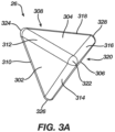
- FIG. 3A is a perspective view of an electrode 26, in accordance with the disclosed technology.
- the electrodes 26 can each have a generally tetrahedral shape.
- the electrodes 26 can be attached to the spines 22 in such a way that an outwardly-facing surface 302 (also referred to herein as a "first end” and the "first face") of the electrode 26 can have a greater surface area than an inwardly-facing end 328 of the electrode 26 (also referred to herein as the "second end” or the "fourth corner”).
- the electrodes 26, when attached to the spines 22 and arranged on the basket catheter 28, can be configured to collapse together in a tight configuration like wedges of an orange.
- the electrodes 26 enable the basket catheter 28 to comprise a greater number of electrodes 26 while still being able to collapse and fit within a sheath 80.
- All or some of the electrode 26 can comprise a conductive material.
- all of the electrode 26 can be made entirely from gold, silver, platinum, palladium, stainless steel (and their respective alloys).
- only a portion of the electrode 26 e.g., the first face 302 can be made from the just-recited materials. These materials also have high thermal conductivity which allows the minimal heat generated on the tissue (i.e., by the ablation energy delivered to the tissue) to be conducted through the electrodes to the back side of the electrodes (i.e., the portions of the electrodes on the inner sides of the spines), and then to the blood pool in heart 12.
- the electrodes can be configured to deliver electrical pulses having a peak voltage of at least 900 volts (V).
- the outwardly-facing surface 302 can be configured for contacting tissue and, thus, by having a greater surface area than the inwardly-facing end 328, the outwardly-facing surface 302 can be better configured for detecting electrophysiological signals and/or for providing ablative energy to the tissue.
- the electrodes 26 can have a plurality of faces forming the generally tetrahedral shape.
- the electrodes 26 can have at least a first face 302 (the outwardly-facing side), a second face 304, a third face 306, and a fourth face 308 (not visible in the view shown in FIG. 3A ).
- the electrodes 26 can have a plurality of edges between the plurality of faces.
- the electrodes 26 can have a first edge 310, a second edge 312, a third edge 314, a fourth edge 316, a fifth edge 318, and a sixth edge 320 (not fully visible in FIG. 3A ).
- Each of the first edge 310, a second edge 312, a third edge 314, a fourth edge 316, a fifth edge 318, and a sixth edge 320 can be rounded to prevent damage to tissue and other components of the medical probe (e.g., the sheath 80).
- the plurality of edges can converge together at a plurality of corners.
- the plurality of corners can include a first corner 322, a second corner 324, a third corner 326, and a fourth corner 328 (also referred to herein as "second end” or “inwardly-facing end”).
- first corner 322 a first corner 322
- second corner 324 a third corner 326
- fourth corner 328 also referred to herein as "second end” or "inwardly-facing end”
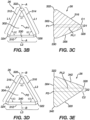
- Each of the faces, edges, and corners just described and shown in FIG. 3A correspond to like faces, edges, and corners shown in FIGs. 3B-3E .
- FIG. 3B is a front view of the electrode 26,
- FIG. 3C is a section view of the electrode of FIG. 3B
- FIG. 3D is a top view of the electrode
- FIG. 3E is a section view of the electrode of FIG. 3D , in accordance with the disclosed technology.
- the views shown in FIGs. 3B-3E illustrate the various features of the electrodes 26, including the general angles and proportions of an example electrodes.
- One of skill in the art will appreciate that the views of the electrode shown and described in relation to FIGs. 3B-3E are offered for illustrative purposes and should not be construed as limiting. Other electrodes 26 having varying shapes, angles, proportions, etc. can similarly be construed within the scope of this disclosure.
- the electrode 26 can have a first face 302 (outwardly-facing side) that is configured to contact tissue.
- the first face 302 can be formed generally in the shape of a triangle.
- the first face 302 is shown as forming an isosceles triangle with the second edge 312 and the third edge 314 having a first length L1 that is different than the length L2 of the first edge 310. Because the first face 302 forms an isosceles triangle, the first angle ⁇ 1 between the second edge 312 and the third edge 314 can be different than the second angle ⁇ 2 between the first edge 310 and the second edge 312 which is equal to the angle ⁇ 2 between the first edge 310 and the third edge 312.
- the various angles and lengths of the faces can be configured according to various other examples.
- the first face 302 can form an acute triangle, an obtuse triangle, an equilateral triangle, scalene triangle, right triangle, etc.
- the electrode 26 is shown as having a first face 302 (and other faces, 304, 306, 308) forming a triangle, it will be appreciated that the one or more faces of the triangle can form shapes other than triangles.
- various other permutations of the disclosed technology can be configured such that the outwardly-facing side has a greater surface area than the inwardly-facing side such that the electrodes 26 can collapse together to form a tight grouping of electrodes 26 capable of fitting within the sheath 80.
- FIG. 3C is a cross-sectional view of the electrode 26 taken along line A-A of FIG. 3B .
- Line A-A extends from the fourth corner 328 (inwardly-facing end) through the middle of the first face 302 to a point 330 disposed in the middle of the first edge 310.
- the electrode 26 can be configured such that the fourth corner 328 (inwardly-facing end) can be offset a distance D1 from a centerline C1 extending perpendicularly from a point P1 that is at the center of the triangle formed by the first face 302.
- the center line C1 extending perpendicularly to the first face 302 from a first point P1 at a center of the first face 302 is spaced a distance D1 apart from a line PL1 parallel to the first line and extending through a second point at a center of the fourth corner 328 ("inwardly-facing end" or "second end”).
- the electrode 26 can form a tetrahedron that is not a regular tetrahedron.
- the fourth corner 328 of the electrodes 26 can be configured to better fit together when the electrodes 26 are grouped together as will be described in greater detail in relation to FIGs. 5-7 .
- the second face 304 of the electrode 26 can form an equilateral triangle. That is, the second edge 312, the fourth edge 316, and the fifth edge 318 can each have the same length L3 and form third angles ⁇ 3 between each of the second edge 312, the fourth edge 316, and the fifth edge 318 that are all equal to each other.
- the configuration of the second face 304 shown in FIG. 3D is offered for illustrative purposes and the second face 304 can be configured according to various other permutations.
- FIG. 3E is a cross-sectional view of the electrode 26 taken along line B-B of FIG. 3D .
- Line B-B extends from the fourth corner 328 (inwardly-facing end) through the middle of the second face 304 to a point 340 disposed in the middle of the second edge 312.
- the cross-sectional view of FIG. 3E illustrates that the second end 328 can be disposed such that it is offset in alignment a distance D2 from a centerline C2 extending perpendicularly from a point P2 at the center of the second face 304.
- a centerline C2 extending perpendicularly to the second face 304 from a second point P2 at a center of the second face 304 is spaced a distance D2 apart from a line PL2 parallel to the center line C2 and extending through a point at a center of the fourth corner 328 ("inwardly-facing end" or "second end”).
- the fourth corner 328 (inwardly-facing end) of the electrodes 26 can be configured to better fit together when the electrodes 26 are grouped together as will be described in greater detail in relation to FIGs. 5-7 .
- the distances D1 and D2 can be changed to affect the configuration of the tetrahedral shape of the electrode 26 as suitable for the particular application.
- the basket catheter 28 can include a plurality of flexible spines 22 that are formed at the end of a tubular shaft 84 and are connected at a proximal end to the tubular shaft 84 and at a distal end to a distal hub 210.
- physician 34 can deploy basket catheter 28 by extending tubular shaft 84 from sheath 80 causing the basket catheter 28 to exit the sheath 80 and transition to the expanded form (as shown in FIG. 2 ).
- the electrodes 26 are configured according to the shapes shown and described herein, the electrodes 26 can be configured to collapse together and form a tight group for delivery from, and retraction into, the sheath.
- Spines 22 may have elliptical (e.g., circular) or rectangular (that may appear to be flat) cross-sections, and include a flexible, resilient material (e.g., a shape-memory alloy such as nickel-titanium, also known as Nitinol or other materials such as cobalt chromium, stainless steel, titanium, or even a polymer material) forming a strut as will be described in greater detail herein.
- a shape-memory alloy such as nickel-titanium, also known as Nitinol or other materials such as cobalt chromium, stainless steel, titanium, or even a polymer material
- the disclosed technology can be applicable to basket catheter 28 formed from a single spine 22 or multiple spines 22 with each spine 22 being attached at both ends (as just described).
- the spines 22 can be formed from a single sheet of planar material. In some examples, the spines 22 can be formed from the single sheet of planar material such that the spines 22 converge toward a central intersection. In other examples, the spines 22 can be formed individually and then attached at both ends as shown and just described.
- the spine 22 can be electrically isolated from the electrode 26 to prevent arcing from the electrode 26 to the spine 22.
- insulative jackets (not shown) can be positioned between the spine 22 and the electrode 26, but one of skill in the art will appreciate that other insulative coverings are contemplated.
- an insulative coating can be applied to the spine 22, the electrodes 26, or both.
- the insulative jackets can be made from a biocompatible, electrically insulative material such as polyamide-polyether (Pebax) copolymers, polyethylene terephthalate (PET), urethanes, polyimide, parylene, silicone, etc.
- insulative material can include biocompatible polymers including, without limitation, polyetheretherketone (PEEK), polyglycolic acid (PGA), poly(lactic-co-glycolic acid) copolymer (PLGA), polycaprolactive (PCL), poly(3-hydroxybutyrate-co-3 -hydroxyvalerate) (PHBV), poly-L-lactide, polydioxanone, polycarbonates, and polyanhydrides with the ratio of certain polymers being selected to control the degree of inflammatory response.
- PEEK polyetheretherketone
- PGA polyglycolic acid
- PLGA poly(lactic-co-glycolic acid) copolymer
- PCL polycaprolactive
- PHBV poly(3-hydroxybutyrate-co-3 -hydroxyvalerate)
- poly-L-lactide polydioxanone
- polycarbonates and polyanhydrides with the ratio of certain polymers being selected to control the degree of inflammatory response.
- Insulative jackets may also include one or more additives or fillers, such as, for example, polytetrafluoroethylene (PTFE), boron nitride, silicon nitride, silicon carbide, aluminum oxide, aluminum nitride, zinc oxide, and the like.
- PTFE polytetrafluoroethylene
- FIG. 5 illustrates a perspective view of the electrodes 26 attached to the spines 22 in a collapsed form.
- the electrodes 26 can be attached to the spines 22 such that a majority of the electrode 26 is disposed on an outer-facing side of the spine 22.
- the electrodes 26 can be arranged in an alternating first orientation 26A and second orientation 26B.
- the second orientation 26B can be flipped 180 degrees from first orientation 26A such that the electrodes 26 are better able to fit together when in the collapsed form (as shown in FIGs. 4A-7 ).
- FIG. 5 illustrates a perspective view of the electrodes 26 attached to the spines 22 in a collapsed form.
- the electrodes 26 can be attached to the spines 22 such that a majority of the electrode 26 is disposed on an outer-facing side of the spine 22.
- the electrodes 26 can be arranged in an alternating first orientation 26A and second orientation 26B.
- the second orientation 26B can be flipped 180 degrees from first orientation 26A such that the electrodes 26 are better
- the first face 302 can be arranged in an outwardly-facing orientation while the fourth corner 328 (second end) is arranged in an inwardly-facing orientation.
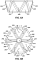
- the fourth corners 328 can converge and define a lumen 610 extending through the grouping of electrodes 26.
- the lumen 610 can be useful for ensure sufficient room remains for the electrodes 26 to align together with some variance in alignment.
- the lumen 610 can be configured to permit irrigation fluid to flow therethrough if, for example, it is desirable for irrigation fluid to flow through the probe even when the basket catheter 28 is withdrawn into the sheath 80.
- FIG. 6C illustrates another example arrangement of electrodes 26 in the collapsed configuration.
- the electrodes 26 comprise a rounded first face 602 that can help to prevent the electrodes 26 from damaging tissue or other components of the medical probe (e.g., the interior of the sheath, the hemostasis valve, etc.). That is, the rounded first face 602 can comprise a compound curved surface that presents a rounded profile with respect to the longitudinal axis 86. As shown, the rounded first face 602 can be outwardly-facing and configured to contact tissue. Furthermore, the rounded first face 602 can make it easier to withdraw the basket catheter 28 into the sheath 80.
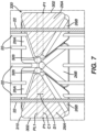
- FIG. 7 is a section view of the spines 22 and electrodes 26 taken along line C-C of FIG. 6B .
- the electrode 26 can be configured such that the fourth corner 328 (inwardly-facing end) can be offset a distance D1 from a centerline C1 extending perpendicularly from a point P1 that is at the center of the triangle formed by the first face 302.
- the second end 328 of each electrode 26 can converge toward a point in the middle of the electrode 26 grouping that is offset from a center of the first face 302.
- the first edge 310 can be oriented generally perpendicular with respect to the spine 22.
- the electrodes 26 can be configured in various other configurations that enable the electrodes 26 to collapse together in a compact manner thereby permitting electrodes 26 to be attached to adjacent spines 22 while still being able to fit compactly within the sheath 80.
Landscapes
- Health & Medical Sciences (AREA)
- Life Sciences & Earth Sciences (AREA)
- Surgery (AREA)
- Engineering & Computer Science (AREA)
- Animal Behavior & Ethology (AREA)
- Veterinary Medicine (AREA)
- Biomedical Technology (AREA)
- Heart & Thoracic Surgery (AREA)
- Medical Informatics (AREA)
- Molecular Biology (AREA)
- Public Health (AREA)
- General Health & Medical Sciences (AREA)
- Physics & Mathematics (AREA)
- Cardiology (AREA)
- Biophysics (AREA)
- Pathology (AREA)
- Nuclear Medicine, Radiotherapy & Molecular Imaging (AREA)
- Otolaryngology (AREA)
- Plasma & Fusion (AREA)
- Physiology (AREA)
- Surgical Instruments (AREA)
- Measurement And Recording Of Electrical Phenomena And Electrical Characteristics Of The Living Body (AREA)
Abstract
Description
- This application claims, under 35 U.S.C. § 119(e), priority to and the benefit of
U.S. Provisional Patent Application No. 63/502,543, filed May 16, 2023 - The present invention relates generally to medical devices, and in particular medical probes with electrodes, and further relates to, but not exclusively, medical probes suitable for mapping and ablation of cardiac tissues.
- Cardiac arrhythmias, such as atrial fibrillation (AF), occur when regions of cardiac tissue abnormally conduct electric signals to adjacent tissue. This disrupts the normal cardiac cycle and causes asynchronous rhythm. Certain procedures exist for treating arrhythmia, including surgically disrupting the origin of the signals causing the arrhythmia and disrupting the conducting pathway for such signals. By selectively ablating cardiac tissue by application of energy via a catheter, it is sometimes possible to cease or modify the propagation of unwanted electrical signals from one portion of the heart to another.
- Regions of cardiac tissue can be mapped by a medical probe to identify the abnormal electrical signals. The same or different medical probe can be used to perform ablation. Some example probes include a number of spines with electrodes positioned thereon. The electrodes are generally attached to the spines and the spines are configured to bow radially outward when deployed from a sheath. The electrodes are then brought into contact with tissue for mapping or ablation of the tissue. The electrodes must generally be arranged on the spines in such a way that electrodes on adjacent spines are offset from each other. This is due to the limited space within the sheath and because the electrodes cannot fit together within the sheath if electrodes on adjacent spines are aligned with each other. Offset alignment of electrodes, however, results in fewer electrodes being attached to the spines which can make mapping and ablation less accurate. Accordingly, there is a need in the art for electrode designs that will enable electrodes to be aligned on adjacent spines and permit for a greater number of electrodes to be attached to the spines while still being able to fit within a sheath. These and other problems can be addressed by the technology disclosed herein.
- There is provided, in accordance with the disclosed technology, an electrode comprising a body including a first end and a second end. The body can extend along a longitudinal axis from the first end to the second end. The body can be configured for attachment to a spine of an expandable basket assembly such that the first end is oriented for contacting tissue when the expandable basket assembly is in an expanded configuration. The first end can comprise a conductive surface forming a generally triangular shape and an area that is greater than the second end. The body can converge from the first end to the second end along the longitudinal axis.
- A medical probe comprising a tubular shaft including a proximal end and a distal end, the tubular shaft extending along a longitudinal axis, and an expandable basket assembly coupled to the distal end of the tubular shaft. The expandable basket assembly comprising a plurality of spines configured to bow radially outward from the longitudinal axis when the expandable basket assembly is in an expanded configuration and a plurality of electrodes attached to the plurality of spines Each electrode of the plurality of electrodes comprising a body including a first end and a second end, the body being attached to a respective spine of the plurality of spines such that the first end is oriented for contacting tissue when the expandable basket assembly is in the expanded configuration. The first end can comprise a conductive surface forming a generally triangular shape and an area that is greater than the second end and the body can converge from the first end to the second end.
- The disclosed technology can include a medical probe comprising a tubular shaft including a proximal end and a distal end. The tubular shaft can extend along a longitudinal axis. The medical probe can further comprise an expandable basket assembly coupled to the distal end of the tubular shaft. The expandable basket assembly can comprise a plurality of spines configured to bow radially outward from the longitudinal axis when the expandable basket assembly is in an expanded configuration. The expandable basket can comprise a plurality of electrodes attached to the plurality of spines.
- Each electrode of the plurality of electrodes can include a body comprising a tetrahedral shape. The tetrahedral shape can comprise an outwardly-facing end and an inwardly-facing end. The outwardly-facing end can comprise a conductive surface including an area that is greater than an area of the inwardly-facing end and be configured to contact tissue when the expandable basket assembly is in the expanded configuration. The body can converge from the outwardly-facing end to the inwardly-facing end.
- The present disclosure will be more fully understood from the following detailed description of embodiments thereof, taken together with the drawings, in which:
-
FIG. 1 is a schematic pictorial illustration of a medical system including a medical probe with a distal end comprising electrodes, in accordance with the disclosed technology; -
FIG. 2 is a schematic pictorial illustration of a basket catheter comprising electrodes, in accordance with the disclosed technology; -
FIG. 3A is a perspective view of an electrode, in accordance with the disclosed technology; -
FIG. 3B is a front view of the electrode, in accordance with the disclosed technology; -
FIG. 3C is a section view of the electrode ofFIG. 3B taken along line A-A, in accordance with the disclosed technology; -
FIG. 3D is a top view of the electrode, in accordance with the disclosed technology; -
FIG. 3E is a section view of the electrode ofFIG. 3D take along line B-B, in accordance with the disclosed technology; -
FIG. 4A is a schematic pictorial illustration of the medical probe, in accordance with the disclosed technology; -
FIG. 4B is a schematic pictorial illustration of the medical probe in a sheath, in accordance with the disclosed technology; -
FIG. 5 is a perspective view of a plurality of spines and electrodes of the medical probe, in accordance with the disclosed technology; -
FIG. 6A is a sideview of a grouping of electrodes, in accordance with the disclosed technology; -
FIG. 6B is a top view of a grouping of electrodes, in accordance with the disclosed technology; -
FIG. 6C is a top view of another grouping of electrodes, in accordance with another example of the disclosed technology; and -
FIG. 7 is a section view of the spines and electrodes taken along line C-C ofFIG. 6B , in accordance with the disclosed technology. - The following detailed description should be read with reference to the drawings, in which like elements in different drawings are identically numbered. The drawings, which are not necessarily to scale, depict selected embodiments and are not intended to limit the scope of the invention. The detailed description illustrates by way of example, not by way of limitation, the principles of the invention. This description will clearly enable one skilled in the art to make and use the invention, and describes several embodiments, adaptations, variations, alternatives and uses of the invention, including what is presently believed to be the best mode of carrying out the invention.
- As used herein, the terms "about" or "approximately" for any numerical values or ranges indicate a suitable dimensional tolerance that allows the part or collection of components to function for its intended purpose as described herein. More specifically, "about" or "approximately" may refer to the range of values ±20% of the recited value, e.g. "about 90%" may refer to the range of values from 71% to 110%.
- In addition, as used herein, the terms "patient," "host," "user," and "subject" refer to any human or animal subject and are not intended to limit the systems or methods to human use, although use of the subject invention in a human patient represents a preferred embodiment. As well, the term "proximal" indicates a location closer to the operator or physician whereas "distal" indicates a location further away to the operator or physician.
- As discussed herein, vasculature of a "patient," "host," "user," and "subject" can be vasculature of a human or any animal. It should be appreciated that an animal can be a variety of any applicable type, including, but not limited thereto, mammal, veterinarian animal, livestock animal or pet type animal, etc. As an example, the animal can be a laboratory animal specifically selected to have certain characteristics similar to a human (e.g., rat, dog, pig, monkey, or the like). It should be appreciated that the subject can be any applicable human patient, for example.
- As discussed herein, "physician" can include a doctor, surgeon, technician, scientist, operator, or any other individual or delivery instrumentation associated with delivery of a multi-electrode catheter for the treatment of drug refractory atrial fibrillation to a subject.
- As discussed herein, the term "ablate" or "ablation", as it relates to the devices and corresponding systems of this disclosure, refers to components and structural features configured to reduce or prevent the generation of erratic cardiac signals in the cells by utilizing non-thermal energy, such as irreversible electroporation (IRE), referred throughout this disclosure interchangeably as pulsed electric field (PEF) and pulsed field ablation (PFA). Ablating or ablation as it relates to the devices and corresponding systems of this disclosure is used throughout this disclosure in reference to non-thermal ablation of cardiac tissue for certain conditions including, but not limited to, arrhythmias, atrial flutter ablation, pulmonary vein isolation, supraventricular tachycardia ablation, and ventricular tachycardia ablation. The term "ablate" or "ablation" also includes known methods, devices, and systems to achieve various forms of bodily tissue ablation as understood by a person skilled in the relevant art.
- As discussed herein, the terms "bipolar" and "unipolar" when used to refer to ablation schemes describe ablation schemes which differ with respect to electrical current path and electric field distribution. "Bipolar" refers to ablation scheme utilizing a current path between two electrodes that are both positioned at a treatment site; current density and electric flux density is typically approximately equal at each of the two electrodes. "Unipolar" refers to ablation scheme utilizing a current path between two electrodes where one electrode having a high current density and high electric flux density is positioned at a treatment site, and a second electrode having comparatively lower current density and lower electric flux density is positioned remotely from the treatment site.
- As discussed herein, the terms "biphasic pulse" and "monophasic pulse" refer to respective electrical signals. "Biphasic pulse" refers to an electrical signal having a positive-voltage phase pulse (referred to herein as "positive phase") and a negative-voltage phase pulse (referred to herein as "negative phase"). "Monophasic pulse" refers to an electrical signal having only a positive or only a negative phase. Preferably, a system providing the biphasic pulse is configured to prevent application of a direct current voltage (DC) to a patient. For instance, the average voltage of the biphasic pulse can be zero volts with respect to ground or other common reference voltage. Additionally, or alternatively, the system can include a capacitor or other protective component. Where voltage amplitude of the biphasic and/or monophasic pulse is described herein, it is understood that the expressed voltage amplitude is an absolute value of the approximate peak amplitude of each of the positive-voltage phase and/or the negative-voltage phase. Each phase of the biphasic and monophasic pulse preferably has a square shape having an essentially constant voltage amplitude during a majority of the phase duration. Phases of the biphasic pulse are separated in time by an interphase delay. The interphase delay duration is preferably less than or approximately equal to the duration of a phase of the biphasic pulse. The interphase delay duration is more preferably about 25% of the duration of the phase of the biphasic pulse.
- As discussed herein, the terms "tubular" and "tube" are to be construed broadly and are not limited to a structure that is a right cylinder or strictly circumferential in cross-section or of a uniform cross-section throughout its length. For example, the tubular structures are generally illustrated as a substantially right cylindrical structure. However, the tubular structures may have a tapered or curved outer surface without departing from the scope of the present disclosure.
- The present disclosure is related to systems, method or uses and devices that can be used for IRE ablation of cardiac tissue to treat cardiac arrhythmias. Ablative energies are typically provided to cardiac tissue by a tip portion of a catheter which can deliver ablative energy alongside the tissue to be ablated. Some example catheters include three-dimensional structures at the tip portion and are configured to administer ablative energy from various electrodes positioned on the three-dimensional structures. Ablative procedures incorporating such example catheters can be visualized using fluoroscopy.
- Ablation of cardiac tissue using application of a thermal technique, such as radio frequency (RF) energy and cryoablation, to correct a malfunctioning heart is a well-known procedure. Typically, to successfully ablate using a thermal technique, cardiac electropotentials need to be measured at various locations of the myocardium. In addition, temperature measurements during ablation provide data enabling the efficacy of the ablation. Typically, for an ablation procedure using a thermal technique, the electropotentials and the temperatures are measured before, during, and after the actual ablation. RF approaches can have risks that can lead to tissue charring, burning, steam pop, phrenic nerve palsy, pulmonary vein stenosis, and esophageal fistula. Cryoablation is an alternative approach to RF ablation that can reduce some thermal risks associated with RF ablation. However maneuvering cryoablation devices and selectively applying cryoablation is generally more challenging compared to RF ablation; therefore, cryoablation is not viable in certain anatomical geometries which may be reached by electrical ablation devices.
- The present disclosure can include electrodes configured for RF ablation, cryoablation, and/or irreversible electroporation (IRE). IRE can be referred to throughout this disclosure interchangeably as pulsed electric field (PEF) ablation and pulsed field ablation (PFA). IRE as discussed in this disclosure is a non-thermal cell death technology that can be used for ablation of atrial arrhythmias. To ablate using IRE/PEF, biphasic voltage pulses are applied to disrupt cellular structures of myocardium. The biphasic pulses are non-sinusoidal and can be tuned to target cells based on electrophysiology of the cells. In contrast, to ablate using RF, a sinusoidal voltage waveform is applied to produce heat at the treatment area, indiscriminately heating all cells in the treatment area. IRE therefore has the capability to spare adjacent heat sensitive structures or tissues which would be of benefit in the reduction of possible complications known with ablation or isolation modalities. Additionally, or alternatively, monophasic pulses can be utilized.
- Electroporation can be induced by applying a pulsed electric field across biological cells to cause reversable (temporary) or irreversible (permanent) creation of pores in the cell membrane. The cells have a transmembrane electrostatic potential that is increased above a resting potential upon application of the pulsed electric field. While the transmembrane electrostatic potential remains below a threshold potential, the electroporation is reversable, meaning the pores can close when the applied pulse electric field is removed, and the cells can self-repair and survive. If the transmembrane electrostatic potential increases beyond the threshold potential, the electroporation is irreversible, and the cells become permanently permeable. As a result, the cells die due to a loss of homeostasis and typically die by apoptosis. Generally, cells of differing types have differing threshold potential. For instance, heart cells have a threshold potential of approximately 500 V/cm, whereas for bone it is 3000 V/cm. These differences in threshold potential allow IRE to selectively target tissue based on threshold potential.
- The solution of this disclosure includes systems and methods for applying electrical signals from catheter electrodes positioned in the vicinity of myocardial tissue to generate a generate ablative energy to ablate the myocardial tissue. In some examples, the systems and methods can be effective to ablate targeted tissue by inducing irreversible electroporation. In some examples, the systems and methods can be effective to induce reversible electroporation as part of a diagnostic procedure. Reversible electroporation occurs when the electricity applied with the electrodes is below the electric field threshold of the target tissue allowing cells to repair. Reversible electroporation does not kill the cells but allows a physician to see the effect of reversible electroporation on electrical activation signals in the vicinity of the target location. Example systems and methods for reversible electroporation is disclosed in
U.S. Patent Publication 2021/0162210 , the entirety of which is incorporated herein by reference and attached in the Appendix hereto. - The pulsed electric field, and its effectiveness to induce reversible and/or irreversible electroporation, can be affected by physical parameters of the system and biphasic pulse parameters of the electrical signal. Physical parameters can include electrode contact area, electrode spacing, electrode geometry, etc. Examples presented herein generally include physical parameters adapted to effectively induce reversible and/or irreversible electroporation. Biphasic pulse parameters of the electrical signal can include voltage amplitude, pulse duration, pulse interphase delay, inter-pulse delay, total application time, delivered energy, etc. In some examples, parameters of the electrical signal can be adjusted to induce both reversible and irreversible electroporation given the same physical parameters. Examples of various systems and methods of ablation including IRE are presented in
U.S. Patent Publications 2021/0169550A1 ,2021/0169567A1 ,2021/0169568A1 ,2021/0161592A1 ,2021/0196372A1 ,2021/0177503A1 , and2021/0186604A1 , the entireties of each of which are incorporated herein by reference and attached in the Appendix hereto. - Reference is made to
FIG. 1 showing an example catheter-based electrophysiology mapping andablation system 10.System 10 includes multiple catheters, which are percutaneously inserted byphysician 24 through the patient's 23 vascular system into a chamber or vascular structure of aheart 12. Typically, a delivery sheath catheter is inserted into the left or right atrium near a desired location inheart 12. Thereafter, a plurality of catheters can be inserted into the delivery sheath catheter so as to arrive at the desired location. The plurality of catheters may include catheters dedicated for sensing Intracardiac Electrogram (IEGM) signals, catheters dedicated for ablating and/or catheters dedicated for both sensing and ablating. Anexample catheter 14 that is configured for sensing and ablating is illustrated herein.Physician 24 brings a distal tip 28 (sometimes referred to herein asbasket catheter 28 or expandable basket assembly) ofcatheter 14 into contact with the heart wall for sensing a target site inheart 12. For ablation,physician 24 would similarly bring a distal end of an ablation catheter to a target site for ablating. -
Catheter 14 is an exemplary catheter that includes one and preferablymultiple electrodes 26 optionally distributed over a plurality ofspines 22 atdistal tip 28 and configured to sense the IEGM signals.Catheter 14 may additionally include aposition sensor 29 embedded in or neardistal tip 28 for tracking position and orientation ofdistal tip 28. Optionally and preferably,position sensor 29 is a magnetic based position sensor including three magnetic coils for sensing three-dimensional (3D) position and orientation. - Magnetic based
position sensor 29 may be operated together with alocation pad 25 including a plurality ofmagnetic coils 32 configured to generate magnetic fields in a predefined working volume. Real time position ofdistal tip 28 ofcatheter 14 may be tracked based on magnetic fields generated withlocation pad 25 and sensed by magnetic basedposition sensor 29. Details of the magnetic based position sensing technology are described inU.S. Patent Nos. 5,391,199 ;5,443,489 ;5,558,091 ;6,172,499 ;6,239,724 ;6,332,089 ;6,484,118 ;6,618,612 ;6,690,963 ;6,788,967 ;6,892,091 , the entireties of each of which are incorporated herein by reference and attached in the Appendix hereto. -
System 10 includes one ormore electrode patches 38 positioned for skin contact onpatient 23 to establish location reference forlocation pad 25 as well as impedance-based tracking ofelectrodes 26. For impedance-based tracking, electrical current is directed towardelectrodes 26 and sensed atelectrode skin patches 38 so that the location of each electrode can be triangulated via theelectrode patches 38. Details of the impedance-based location tracking technology are described inUS Patent Nos. 7,536,218 ;7,756,576 ;7,848,787 ;7,869,865 ; and8,456,182 , the entireties of each of which are incorporated herein by reference and attached in the Appendix hereto. - A
recorder 11 displays electrograms 21 captured with bodysurface ECG electrodes 18 and intracardiac electrograms (IEGM) captured withelectrodes 26 ofcatheter 14.Recorder 11 may include pacing capability for pacing the heart rhythm and/or may be electrically connected to a standalone pacer. -
System 10 may include anablation energy generator 50 that is adapted to conduct ablative energy to one or more of electrodes at a distal tip of a catheter configured for ablating. Energy produced byablation energy generator 50 may include, but is not limited to, radiofrequency (RF) energy or pulsed-field ablation (PFA) energy, including monopolar or bipolar high-voltage DC pulses as may be used to effect irreversible electroporation (IRE), or combinations thereof. - Patient interface unit (PIU) 30 is an interface configured to establish electrical communication between catheters, electrophysiological equipment, power supply and a
workstation 55 for controlling operation ofsystem 10. Electrophysiological equipment ofsystem 10 may include for example, multiple catheters,location pad 25, bodysurface ECG electrodes 18,electrode patches 38,ablation energy generator 50, andrecorder 11. Optionally and preferably,PIU 30 additionally includes processing capability for implementing real-time computations of location of the catheters and for performing ECG calculations. -
Workstation 55 includes memory, processor unit with memory or storage with appropriate operating software loaded therein, and user interface capability.Workstation 55 may provide multiple functions, optionally including (1) modeling the endocardial anatomy in three-dimensions (3D) and rendering the model oranatomical map 20 for display on adisplay device 27, (2) displaying ondisplay device 27 activation sequences (or other data) compiled from recordedelectrograms 21 in representative visual indicia or imagery superimposed on the renderedanatomical map 20, (3) displaying real-time location and orientation of multiple catheters within the heart chamber, and (4) displaying ondisplay device 27 sites of interest such as places where ablation energy has been applied. One commercial product embodying elements of thesystem 10 is available as the CARTO™ 3 System, available from Biosense Webster, Inc., 31 Technology Drive, Suite 200, Irvine, CA 92618, USA. -
FIG. 2 is a schematic pictorial illustration showing a perspective view of abasket catheter 28 withelectrodes 26 in an expanded form, such as by being advanced out of a sheath 80 (shown inFIG. 4B ), in accordance the disclosed technology. As shown inFIG. 2 , thebasket catheter 28 can include a plurality ofspines 22 that can bow radially outward from alongitudinal axis 86 when in an expanded form. Thespines 22 can be formed from a biocompatible resilient material such as nitinol such that thespines 22 are naturally biased to expand outwardly to the expanded form. - Each
spine 22 can include one ormore electrodes 26 attached thereto. As will be described in greater detail herein, theelectrodes 26 can be configured such that theelectrodes 26 can be disposed on thespines 22 in alignment onadjacent spines 22 as shown inFIG. 2 . Thus, thebasket catheter 28 of the disclosed technology can be configured to include a greater number ofelectrodes 26 than previous catheter designs. Theelectrodes 26 can be configured such that the electrodes can collapse together in a tight configuration as shown inFIGs. 4A-7 . Theelectrodes 26 described herein can be configured for mapping and/or ablation of tissue. -
FIG. 3A is a perspective view of anelectrode 26, in accordance with the disclosed technology. As shown, theelectrodes 26 can each have a generally tetrahedral shape. In this way, theelectrodes 26 can be attached to thespines 22 in such a way that an outwardly-facing surface 302 (also referred to herein as a "first end" and the "first face") of theelectrode 26 can have a greater surface area than an inwardly-facingend 328 of the electrode 26 (also referred to herein as the "second end" or the "fourth corner"). Because of the generally tetrahedral shape, theelectrodes 26, when attached to thespines 22 and arranged on thebasket catheter 28, can be configured to collapse together in a tight configuration like wedges of an orange. Thus, theelectrodes 26 enable thebasket catheter 28 to comprise a greater number ofelectrodes 26 while still being able to collapse and fit within asheath 80. - All or some of the
electrode 26 can comprise a conductive material. For example, all of theelectrode 26 can be made entirely from gold, silver, platinum, palladium, stainless steel (and their respective alloys). Alternatively, only a portion of the electrode 26 (e.g., the first face 302) can be made from the just-recited materials. These materials also have high thermal conductivity which allows the minimal heat generated on the tissue (i.e., by the ablation energy delivered to the tissue) to be conducted through the electrodes to the back side of the electrodes (i.e., the portions of the electrodes on the inner sides of the spines), and then to the blood pool inheart 12. The electrodes can be configured to deliver electrical pulses having a peak voltage of at least 900 volts (V). - The outwardly-facing
surface 302 can be configured for contacting tissue and, thus, by having a greater surface area than the inwardly-facingend 328, the outwardly-facingsurface 302 can be better configured for detecting electrophysiological signals and/or for providing ablative energy to the tissue. Furthermore, theelectrodes 26 can have a plurality of faces forming the generally tetrahedral shape. For example, theelectrodes 26 can have at least a first face 302 (the outwardly-facing side), asecond face 304, athird face 306, and a fourth face 308 (not visible in the view shown inFIG. 3A ). Furthermore, theelectrodes 26 can have a plurality of edges between the plurality of faces. For example, theelectrodes 26 can have afirst edge 310, asecond edge 312, athird edge 314, afourth edge 316, afifth edge 318, and a sixth edge 320 (not fully visible inFIG. 3A ). Each of thefirst edge 310, asecond edge 312, athird edge 314, afourth edge 316, afifth edge 318, and asixth edge 320 can be rounded to prevent damage to tissue and other components of the medical probe (e.g., the sheath 80). The plurality of edges can converge together at a plurality of corners. The plurality of corners can include afirst corner 322, asecond corner 324, athird corner 326, and a fourth corner 328 (also referred to herein as "second end" or "inwardly-facing end"). Each of the faces, edges, and corners just described and shown inFIG. 3A correspond to like faces, edges, and corners shown inFIGs. 3B-3E . - Turning now to
FIGs. 3B-3E, FIG. 3B is a front view of theelectrode 26,FIG. 3C is a section view of the electrode ofFIG. 3B, FIG. 3D is a top view of the electrode, andFIG. 3E is a section view of the electrode ofFIG. 3D , in accordance with the disclosed technology. The views shown inFIGs. 3B-3E illustrate the various features of theelectrodes 26, including the general angles and proportions of an example electrodes. One of skill in the art will appreciate that the views of the electrode shown and described in relation toFIGs. 3B-3E are offered for illustrative purposes and should not be construed as limiting.Other electrodes 26 having varying shapes, angles, proportions, etc. can similarly be construed within the scope of this disclosure. - As shown in
FIG. 3B , theelectrode 26 can have a first face 302 (outwardly-facing side) that is configured to contact tissue. Thefirst face 302 can be formed generally in the shape of a triangle. In this non-limiting example, thefirst face 302 is shown as forming an isosceles triangle with thesecond edge 312 and thethird edge 314 having a first length L1 that is different than the length L2 of thefirst edge 310. Because thefirst face 302 forms an isosceles triangle, the first angle θ1 between thesecond edge 312 and thethird edge 314 can be different than the second angle θ2 between thefirst edge 310 and thesecond edge 312 which is equal to the angle θ2 between thefirst edge 310 and thethird edge 312. As will be appreciated, however, the various angles and lengths of the faces can be configured according to various other examples. For instance, thefirst face 302 can form an acute triangle, an obtuse triangle, an equilateral triangle, scalene triangle, right triangle, etc. Furthermore, although theelectrode 26 is shown as having a first face 302 (and other faces, 304, 306, 308) forming a triangle, it will be appreciated that the one or more faces of the triangle can form shapes other than triangles. Thus, various other permutations of the disclosed technology can be configured such that the outwardly-facing side has a greater surface area than the inwardly-facing side such that theelectrodes 26 can collapse together to form a tight grouping ofelectrodes 26 capable of fitting within thesheath 80. -
FIG. 3C is a cross-sectional view of theelectrode 26 taken along line A-A ofFIG. 3B . Line A-A extends from the fourth corner 328 (inwardly-facing end) through the middle of thefirst face 302 to apoint 330 disposed in the middle of thefirst edge 310. As shown inFIG. 3C , theelectrode 26 can be configured such that the fourth corner 328 (inwardly-facing end) can be offset a distance D1 from a centerline C1 extending perpendicularly from a point P1 that is at the center of the triangle formed by thefirst face 302. That is, the center line C1 extending perpendicularly to thefirst face 302 from a first point P1 at a center of thefirst face 302 is spaced a distance D1 apart from a line PL1 parallel to the first line and extending through a second point at a center of the fourth corner 328 ("inwardly-facing end" or "second end"). Stated otherwise, theelectrode 26 can form a tetrahedron that is not a regular tetrahedron. In this way, thefourth corner 328 of theelectrodes 26 can be configured to better fit together when theelectrodes 26 are grouped together as will be described in greater detail in relation toFIGs. 5-7 . - As shown in
FIG. 3D , thesecond face 304 of theelectrode 26 can form an equilateral triangle. That is, thesecond edge 312, thefourth edge 316, and thefifth edge 318 can each have the same length L3 and form third angles Θ3 between each of thesecond edge 312, thefourth edge 316, and thefifth edge 318 that are all equal to each other. As before, the configuration of thesecond face 304 shown inFIG. 3D is offered for illustrative purposes and thesecond face 304 can be configured according to various other permutations. -
FIG. 3E is a cross-sectional view of theelectrode 26 taken along line B-B ofFIG. 3D . Line B-B extends from the fourth corner 328 (inwardly-facing end) through the middle of thesecond face 304 to apoint 340 disposed in the middle of thesecond edge 312. Similar to the cross-section of theelectrode 26 shown inFIG. 3C , the cross-sectional view ofFIG. 3E illustrates that thesecond end 328 can be disposed such that it is offset in alignment a distance D2 from a centerline C2 extending perpendicularly from a point P2 at the center of thesecond face 304. That is, a centerline C2 extending perpendicularly to thesecond face 304 from a second point P2 at a center of thesecond face 304 is spaced a distance D2 apart from a line PL2 parallel to the center line C2 and extending through a point at a center of the fourth corner 328 ("inwardly-facing end" or "second end"). In this way, the fourth corner 328 (inwardly-facing end) of theelectrodes 26 can be configured to better fit together when theelectrodes 26 are grouped together as will be described in greater detail in relation toFIGs. 5-7 . As will be appreciated, the distances D1 and D2 can be changed to affect the configuration of the tetrahedral shape of theelectrode 26 as suitable for the particular application. - As shown in
FIG. 4A , thebasket catheter 28 can include a plurality offlexible spines 22 that are formed at the end of atubular shaft 84 and are connected at a proximal end to thetubular shaft 84 and at a distal end to adistal hub 210. During a medical procedure, physician 34 can deploybasket catheter 28 by extendingtubular shaft 84 fromsheath 80 causing thebasket catheter 28 to exit thesheath 80 and transition to the expanded form (as shown inFIG. 2 ). As shown inFIGs. 4A and 4B , because theelectrodes 26 are configured according to the shapes shown and described herein, theelectrodes 26 can be configured to collapse together and form a tight group for delivery from, and retraction into, the sheath. -
Spines 22 may have elliptical (e.g., circular) or rectangular (that may appear to be flat) cross-sections, and include a flexible, resilient material (e.g., a shape-memory alloy such as nickel-titanium, also known as Nitinol or other materials such as cobalt chromium, stainless steel, titanium, or even a polymer material) forming a strut as will be described in greater detail herein. The disclosed technology can be applicable tobasket catheter 28 formed from asingle spine 22 ormultiple spines 22 with eachspine 22 being attached at both ends (as just described). - The
spines 22 can be formed from a single sheet of planar material. In some examples, thespines 22 can be formed from the single sheet of planar material such that thespines 22 converge toward a central intersection. In other examples, thespines 22 can be formed individually and then attached at both ends as shown and just described. - As will be appreciated, the
spine 22 can be electrically isolated from theelectrode 26 to prevent arcing from theelectrode 26 to thespine 22. For example, insulative jackets (not shown) can be positioned between thespine 22 and theelectrode 26, but one of skill in the art will appreciate that other insulative coverings are contemplated. For example, an insulative coating can be applied to thespine 22, theelectrodes 26, or both. The insulative jackets can be made from a biocompatible, electrically insulative material such as polyamide-polyether (Pebax) copolymers, polyethylene terephthalate (PET), urethanes, polyimide, parylene, silicone, etc. In some examples, insulative material can include biocompatible polymers including, without limitation, polyetheretherketone (PEEK), polyglycolic acid (PGA), poly(lactic-co-glycolic acid) copolymer (PLGA), polycaprolactive (PCL), poly(3-hydroxybutyrate-co-3 -hydroxyvalerate) (PHBV), poly-L-lactide, polydioxanone, polycarbonates, and polyanhydrides with the ratio of certain polymers being selected to control the degree of inflammatory response. Insulative jackets may also include one or more additives or fillers, such as, for example, polytetrafluoroethylene (PTFE), boron nitride, silicon nitride, silicon carbide, aluminum oxide, aluminum nitride, zinc oxide, and the like. -
FIG. 5 illustrates a perspective view of theelectrodes 26 attached to thespines 22 in a collapsed form. As shown, theelectrodes 26 can be attached to thespines 22 such that a majority of theelectrode 26 is disposed on an outer-facing side of thespine 22. As better shown inFIGs. 6A and 6B , theelectrodes 26 can be arranged in an alternatingfirst orientation 26A andsecond orientation 26B. Thesecond orientation 26B can be flipped 180 degrees fromfirst orientation 26A such that theelectrodes 26 are better able to fit together when in the collapsed form (as shown inFIGs. 4A-7 ). Furthermore, as can be seen inFIG. 6B , thefirst face 302 can be arranged in an outwardly-facing orientation while the fourth corner 328 (second end) is arranged in an inwardly-facing orientation. In the example shown inFIG. 6B , thefourth corners 328 can converge and define alumen 610 extending through the grouping ofelectrodes 26. Thelumen 610, for example, can be useful for ensure sufficient room remains for theelectrodes 26 to align together with some variance in alignment. Furthermore, in some examples, thelumen 610 can be configured to permit irrigation fluid to flow therethrough if, for example, it is desirable for irrigation fluid to flow through the probe even when thebasket catheter 28 is withdrawn into thesheath 80. -
FIG. 6C illustrates another example arrangement ofelectrodes 26 in the collapsed configuration. In this example, theelectrodes 26 comprise a roundedfirst face 602 that can help to prevent theelectrodes 26 from damaging tissue or other components of the medical probe (e.g., the interior of the sheath, the hemostasis valve, etc.). That is, the roundedfirst face 602 can comprise a compound curved surface that presents a rounded profile with respect to thelongitudinal axis 86. As shown, the roundedfirst face 602 can be outwardly-facing and configured to contact tissue. Furthermore, the roundedfirst face 602 can make it easier to withdraw thebasket catheter 28 into thesheath 80. -
FIG. 7 is a section view of thespines 22 andelectrodes 26 taken along line C-C ofFIG. 6B . As shown inFIG. 7 , and previously described in relation toFIG. 3C , theelectrode 26 can be configured such that the fourth corner 328 (inwardly-facing end) can be offset a distance D1 from a centerline C1 extending perpendicularly from a point P1 that is at the center of the triangle formed by thefirst face 302. Thus, thesecond end 328 of eachelectrode 26 can converge toward a point in the middle of theelectrode 26 grouping that is offset from a center of thefirst face 302. Furthermore, as shown, thefirst edge 310 can be oriented generally perpendicular with respect to thespine 22. As will be appreciated, however, theelectrodes 26 can be configured in various other configurations that enable theelectrodes 26 to collapse together in a compact manner thereby permittingelectrodes 26 to be attached toadjacent spines 22 while still being able to fit compactly within thesheath 80. - The disclosed technology described herein can be further understood according to the following clauses:
- Clause 1: An electrode for a medical probe, the electrode comprising a body including a first end and a second end, the body extending along a longitudinal axis from the first end to the second end, the body configured for attachment to a spine of an expandable basket assembly such that the first end is oriented for contacting tissue when the expandable basket assembly is in an expanded configuration, the first end comprising a conductive surface forming a generally triangular shape and an area that is greater than the second end, and the body converging from the first end to the second end along the longitudinal axis.
- Clause 2: The electrode of clause 1, the generally triangular shape of the first end comprising an acute triangle.
- Clause 3: The electrode of clause 2, the generally triangular shape of the first end comprising an isosceles triangle.
- Clause 4: The electrode of clause 2, the generally triangular shape of the first end comprising an equilateral triangle.
- Clause 5: The electrode of any of the preceding clauses, the body comprising a tetrahedron.
- Clause 6: The electrode of clause 5, wherein the second end is misaligned with the first end such that a first line extending perpendicularly to the conductive surface from a first point at a center of the first end is spaced a distance apart from a second line parallel to the first line and extending through a second point at a center of the second end.
- Clause 7: The electrode of any of the preceding clauses, the body being configured for attachment to the spine such that a first edge of the generally triangular shape is oriented perpendicular to the spine.
- Clause 8: The electrode of any of the preceding clauses, the body comprising rounded edges.
- Clause 9: The electrode of any of the preceding clauses, wherein the body comprises a material selected from a group consisting of gold, palladium, silver, platinum, stainless steel and combinations thereof.
- Clause 10: The electrode of any of the preceding clauses, the electrode being configured for ablation of the tissue.
- Clause 11: The electrode of any of the preceding clauses, the conductive surface forming the generally triangular shape comprising a compound curved surface that presents a rounded profile with respect to the longitudinal axis.
- Clause 12: The electrode of any of the preceding clauses, the electrode being configured for mapping electrophysiological characteristics of the tissue.
- Clause 13: A medical probe comprising: a tubular shaft including a proximal end and a distal end, the tubular shaft extending along a longitudinal axis; and an expandable basket assembly coupled to the distal end of the tubular shaft, the expandable basket assembly comprising: a plurality of spines configured to bow radially outward from the longitudinal axis when the expandable basket assembly is in an expanded configuration; and a plurality of electrodes attached to the plurality of spines, each electrode of the plurality of electrodes comprising: a body including a first end and a second end, the body being attached to a respective spine of the plurality of spines such that the first end is oriented for contacting tissue when the expandable basket assembly is in the expanded configuration, the first end comprising a conductive surface forming a generally triangular shape and an area that is greater than the second end, and the body converging from the first end to the second end.
- Clause 14: The medical probe of clause 13, the generally triangular shape of the first end comprising an acute triangle.
- Clause 15: The medical probe of
clause 14, the generally triangular shape of the first end comprising an isosceles triangle. - Clause 16: The medical probe of
clause 14, the generally triangular shape of the first end comprising an equilateral triangle. - Clause 17: The medical probe of any of clauses 13-16, the body comprising a tetrahedron.
- Clause 18: The medical probe of clause 17, wherein the second end is misaligned with the first end such that a first line extending perpendicularly to the conductive surface from a first point at a center of the first end is spaced a distance apart from a second line parallel to the first line and extending through a second point at a center of the second end.
- Clause 19: The medical probe of any of clauses 13-18, wherein the plurality of electrodes are attached to each of the spines such that each adjacent spine comprises an electrode, and wherein the plurality of electrodes are aligned in a direction perpendicular to the longitudinal axis.
- Clause 20: The medical probe of clause 19, the body of each electrode of the plurality of electrodes being attached to the respective spine such that a first edge of the generally triangular shape is oriented perpendicular to the spine.
- Clause 21: The medical probe of
clause 20, wherein a first electrode of the plurality of electrodes is attached to the respective spine in a first orientation such that the first edge is at a distal end of the body, and wherein a second electrode of the plurality of electrodes is attached to an adjacent spine of the plurality of spines in a second orientation such that the first edge is at a proximal end of the body. - Clause 22: The medical probe of
clause 21, wherein the plurality of electrodes are attached to the plurality of spines such that the plurality of electrodes are attached to adjacent spines in alternating first and second orientations. - Clause 23: The medical probe of any of clauses 13-22, the body comprising rounded edges.
- Clause 24: The medical probe of any of clauses 13-23, wherein the body comprises a material selected from a group consisting of gold, palladium, silver, platinum, stainless steel and combinations thereof.
- Clause 25: The medical probe of any of clauses 13-24, the plurality of electrodes being configured for ablation of the tissue.
- Clause 26: The medical probe of
clause 25, the plurality of electrodes being configured to deliver electrical pulses for irreversible electroporation the pulses comprising a peak voltage of at least 900 volts (V). - Clause 27: The medical probe of any of clauses 13-26, the plurality of electrodes being configured for mapping electrophysiological characteristics of the tissue.
- Clause 28: The medical probe of any of clauses 13-27, wherein each spine of the plurality of spines comprises a material selected from a group consisting of nitinol, cobalt chromium, stainless steel, titanium and combinations thereof.
- Clause 29: The medical probe of any of clauses 13-28, wherein the spine comprises a polymer material.
- Clause 30: A medical probe comprising: a tubular shaft including a proximal end and a distal end, the tubular shaft extending along a longitudinal axis; and an expandable basket assembly coupled to the distal end of the tubular shaft, the expandable basket assembly comprising: a plurality of spines configured to bow radially outward from the longitudinal axis when the expandable basket assembly is in an expanded configuration; and a plurality of electrodes attached to the plurality of spines, each electrode of the plurality of electrodes including a body comprising a tetrahedral shape, the tetrahedral shape comprising an outwardly-facing end and an inwardly-facing end, the outwardly-facing end including a conductive surface comprising an area that is greater than an area of the inwardly-facing end and configured to contact tissue when the expandable basket assembly is in the expanded configuration, the body converging from the outwardly-facing end to the inwardly-facing end.
- Clause 31: The medical probe of
clause 30, the outwardly-facing end comprising generally triangular shape, the generally triangular shape comprising an acute triangle. - Clause 32: The medical probe of clause 31, the generally triangular shape comprising an isosceles triangle.
- Clause 33: The medical probe of
clause 32, the generally triangular shape comprising an equilateral triangle. - Clause 34: The medical probe of any of clauses 30-33, wherein the inwardly-facing end is misaligned with the outwardly-facing end such that a first line extending perpendicularly to the conductive surface from a first point at a center of the first end is spaced a distance apart from a second line parallel to the first line and extending through a second point at a center of the second end.
- Clause 35: The medical probe of any of clauses 30-34, the body being configured for attachment to the spine such that a first edge of the generally triangular shape is oriented perpendicular to the spine.
- Clause 36: The medical probe of any of clauses 31-35, wherein the plurality of electrodes are attached to each of the spines such that each adjacent spine comprises an electrode, and wherein the plurality of electrodes are aligned in a direction perpendicular to the longitudinal axis.
- Clause 37: The medical probe of clause 36, the body of each electrode of the plurality of electrodes being attached to the respective spine such that a first edge of the generally triangular shape is oriented perpendicular to the spine.
- Clause 38: The medical probe of clause 37, wherein a first electrode of the plurality of electrodes is attached to the respective spine in a first orientation such that the first edge is at a distal end of the body, and wherein a second electrode of the plurality of electrodes is attached to an adjacent spine of the plurality of spines in a second orientation such that the first edge is at a proximal end of the body.
- Clause 39: The medical probe of
clause 38, wherein the plurality of electrodes are attached to the plurality of spines such that the plurality of electrodes are attached to adjacent spines in alternating first and second orientations. - Clause 40: The medical probe of any of clauses 30-39, the body comprising rounded edges.
- Clause 41: The medical probe of any of clauses 30-40, wherein the body comprises a material selected from a group consisting of gold, palladium, silver, platinum, stainless steel and combinations thereof.
- Clause 42: The medical probe of any of clauses 30-41, the plurality of electrodes being configured for ablation of the tissue.
- Clause 43: The medical probe of clause 42, the plurality of electrodes being configured to deliver electrical pulses for irreversible electroporation the pulses comprising a peak voltage of at least 900 volts (V).
- Clause 44: The medical probe of any of clauses 30-41, the electrode being configured for mapping electrophysiological characteristics of the tissue.
- The embodiments described above are cited by way of example, and the present invention is not limited by what has been particularly shown and described hereinabove. Rather, the scope of the invention includes both combinations and sub combinations of the various features described hereinabove, as well as variations and modifications thereof which would occur to persons skilled in the art upon reading the foregoing description and which are not disclosed in the prior art.
Claims (15)
- An electrode for a medical probe, the electrode comprising:a body including a first end and a second end, the body extending along a longitudinal axis from the first end to the second end, the body configured for attachment to a spine of an expandable basket assembly such that the first end is oriented for contacting tissue when the expandable basket assembly is in an expanded configuration,the first end comprising a conductive surface forming a generally triangular shape and an area that is greater than the second end, andthe body converging from the first end to the second end along the longitudinal axis.
- A medical probe comprising:a tubular shaft including a proximal end and a distal end, the tubular shaft extending along a longitudinal axis; andan expandable basket assembly coupled to the distal end of the tubular shaft, the expandable basket assembly comprising:a plurality of spines configured to bow radially outward from the longitudinal axis when the expandable basket assembly is in an expanded configuration; anda plurality of electrodes attached to the plurality of spines, each electrode of the plurality of electrodes comprising:a body including a first end and a second end, the body being attached to a respective spine of the plurality of spines such that the first end is oriented for contacting tissue when the expandable basket assembly is in the expanded configuration,the first end comprising a conductive surface forming a generally triangular shape and an area that is greater than the second end, andthe body converging from the first end to the second end.
- The electrode of claim 1 or the medical probe of claim 2, the body comprising a tetrahedron.
- The electrode or medical probe of claim 3, wherein the second end is misaligned with the first end such that a first line extending perpendicularly to the conductive surface from a first point at a center of the first end is spaced a distance apart from a second line parallel to the first line and extending through a second point at a center of the second end.
- The electrode of any of claims 1, 3 or 4, the body being configured for attachment to the spine such that a first edge of the generally triangular shape is oriented perpendicular to the spine.
- The electrode of any of claims 1, 3, 4 or 5, the electrode being configured for ablation of the tissue.
- The electrode of any of claims 1, 3, 4, 5 or 6, the conductive surface forming the generally triangular shape comprising a compound curved surface that presents a rounded profile with respect to the longitudinal axis.
- The electrode of any of claims 1, 3, 4, 5, 6 or 7, the electrode being configured for mapping electrophysiological characteristics of the tissue.
- The medical probe of any of claims 2-4, wherein the plurality of electrodes are attached to each of the spines such that each adjacent spine comprises an electrode, and wherein the plurality of electrodes are aligned in a direction perpendicular to the longitudinal axis.
- The medical probe of claim 9, the body of each electrode of the plurality of electrodes being attached to the respective spine such that a first edge of the generally triangular shape is oriented perpendicular to the spine, optionallywherein a first electrode of the plurality of electrodes is attached to the respective spine in a first orientation such that the first edge is at a distal end of the body, andwherein a second electrode of the plurality of electrodes is attached to an adjacent spine of the plurality of spines in a second orientation such that the first edge is at a proximal end of the body, further optionallywherein the plurality of electrodes are attached to the plurality of spines such that the plurality of electrodes are attached to adjacent spines in alternating first and second orientations.
- A medical probe comprising:a tubular shaft including a proximal end and a distal end, the tubular shaft extending along a longitudinal axis; andan expandable basket assembly coupled to the distal end of the tubular shaft, the expandable basket assembly comprising:a plurality of spines configured to bow radially outward from the longitudinal axis when the expandable basket assembly is in an expanded configuration; anda plurality of electrodes attached to the plurality of spines, each electrode of the plurality of electrodes including a body comprising a tetrahedral shape, the tetrahedral shape comprising an outwardly-facing end and an inwardly-facing end, the outwardly-facing end including a conductive surface comprising an area that is greater than an area of the inwardly-facing end and configured to contact tissue when the expandable basket assembly is in the expanded configuration, the body converging from the outwardly-facing end to the inwardly-facing end.
- The medical probe of claim 11, wherein the inwardly-facing end is misaligned with the outwardly-facing end such that a first line extending perpendicularly to the conductive surface from a first point at a center of the first end is spaced a distance apart from a second line parallel to the first line and extending through a second point at a center of the second end.
- The medical probe of claim 11 or claim 12, wherein the plurality of electrodes are attached to each of the spines such that each adjacent spine comprises an electrode, and wherein the plurality of electrodes are aligned in a direction perpendicular to the longitudinal axis.
- The medical probe of claim 11, the body of each electrode of the plurality of electrodes being attached to the respective spine such that a first edge of the conductive surface is oriented perpendicular to the spine.
- The medical probe of claim 13, wherein a first electrode of the plurality of electrodes is attached to the respective spine in a first orientation such that a first edge of the conductive surface is at a distal end of the body, andwherein a second electrode of the plurality of electrodes is attached to an adjacent spine of the plurality of spines in a second orientation such that a first edge of the conductive surface is at a proximal end of the body, optionallywherein the plurality of electrodes are attached to the plurality of spines such that the plurality of electrodes are attached to adjacent spines in alternating first and second orientations.
Applications Claiming Priority (2)
| Application Number | Priority Date | Filing Date | Title |
|---|---|---|---|
| US202363502543P | 2023-05-16 | 2023-05-16 | |
| US18/639,061 US20240382251A1 (en) | 2023-05-16 | 2024-04-18 | Electrodes and electrode configurations for a basket catheter |
Publications (2)
| Publication Number | Publication Date |
|---|---|
| EP4464270A1 true EP4464270A1 (en) | 2024-11-20 |
| EP4464270B1 EP4464270B1 (en) | 2026-05-20 |
Family
ID=91128183
Family Applications (1)
| Application Number | Title | Priority Date | Filing Date |
|---|---|---|---|
| EP24175869.7A Active EP4464270B1 (en) | 2023-05-16 | 2024-05-15 | ELECTRODES AND ELECTRODE CONFIGURATIONS FOR A BASKET CATHETER |
Country Status (5)
| Country | Link |
|---|---|
| US (1) | US20240382251A1 (en) |
| EP (1) | EP4464270B1 (en) |
| JP (1) | JP2024166148A (en) |
| CN (1) | CN118986359A (en) |
| IL (1) | IL312761A (en) |
Citations (25)
| Publication number | Priority date | Publication date | Assignee | Title |
|---|---|---|---|---|
| US5391199A (en) | 1993-07-20 | 1995-02-21 | Biosense, Inc. | Apparatus and method for treating cardiac arrhythmias |
| US5558091A (en) | 1993-10-06 | 1996-09-24 | Biosense, Inc. | Magnetic determination of position and orientation |
| US6172499B1 (en) | 1999-10-29 | 2001-01-09 | Ascension Technology Corporation | Eddy current error-reduced AC magnetic position measurement system |
| US6239724B1 (en) | 1997-12-30 | 2001-05-29 | Remon Medical Technologies, Ltd. | System and method for telemetrically providing intrabody spatial position |
| US6332089B1 (en) | 1996-02-15 | 2001-12-18 | Biosense, Inc. | Medical procedures and apparatus using intrabody probes |
| US6484118B1 (en) | 2000-07-20 | 2002-11-19 | Biosense, Inc. | Electromagnetic position single axis system |
| US6618612B1 (en) | 1996-02-15 | 2003-09-09 | Biosense, Inc. | Independently positionable transducers for location system |
| US6690963B2 (en) | 1995-01-24 | 2004-02-10 | Biosense, Inc. | System for determining the location and orientation of an invasive medical instrument |
| US6892091B1 (en) | 2000-02-18 | 2005-05-10 | Biosense, Inc. | Catheter, method and apparatus for generating an electrical map of a chamber of the heart |
| US7536218B2 (en) | 2005-07-15 | 2009-05-19 | Biosense Webster, Inc. | Hybrid magnetic-based and impedance-based position sensing |
| US7756576B2 (en) | 2005-08-26 | 2010-07-13 | Biosense Webster, Inc. | Position sensing and detection of skin impedance |
| US7848787B2 (en) | 2005-07-08 | 2010-12-07 | Biosense Webster, Inc. | Relative impedance measurement |
| US7869865B2 (en) | 2005-01-07 | 2011-01-11 | Biosense Webster, Inc. | Current-based position sensing |
| US8456182B2 (en) | 2008-09-30 | 2013-06-04 | Biosense Webster, Inc. | Current localization tracker |
| US20210161592A1 (en) | 2019-12-03 | 2021-06-03 | Biosense Webster (Israel) Ltd. | Pulse Generator for Irreversible Electroporation |
| US20210162210A1 (en) | 2019-12-03 | 2021-06-03 | Biosense Webster (Israel) Ltd. | Using reversible electroporation on cardiac tissue |
| US20210169550A1 (en) | 2019-12-05 | 2021-06-10 | Biosense Webster (Israel) Ltd. | Generating and interleaving of irreversible-electroporation and radiofrequnecy ablation (ire/rfa) waveforms |
| US20210169568A1 (en) | 2019-12-09 | 2021-06-10 | Biosense Webster (Israel) Ltd. | Oriented irreversible-electroporation (ire) pulses to compensate for cell size and orientation |
| US20210169567A1 (en) | 2019-12-09 | 2021-06-10 | Biosense Webster (Israel) Ltd. | Irreversible-electroporation (ire) balloon catheter with membrane-insulated high-voltage balloon wires |
| US20210177503A1 (en) | 2019-12-11 | 2021-06-17 | Biosense Webster (Israel) Ltd. | Regulating delivery of irreversible electroporation pulses according to transferred energy |
| US20210186604A1 (en) | 2019-12-24 | 2021-06-24 | Biosense Webster (Israel) Ltd. | Irreversible electroporation (ire) based on field, contact force and time |
| US20210196372A1 (en) | 2019-12-31 | 2021-07-01 | Biosense Webster (Israel) Ltd. | Using irrigation on irreversible-electroporation (ire) electrodes to prevent arcing |
| CN114027967A (en) * | 2021-11-10 | 2022-02-11 | 心航路医学科技(广州)有限公司 | Pulsed electric field balloon component and ablation catheter device using same |
| US20220071694A1 (en) * | 2020-09-10 | 2022-03-10 | Biosense Webster (Israel) Ltd. | IRE Ablation Systems and Protocols Using a Basket Catheter |
| EP4215144A1 (en) * | 2022-01-20 | 2023-07-26 | Biosense Webster (Israel) Ltd. | Electrode assemblies of a basket catheter having mechanical retainers and methods of the same |
Family Cites Families (3)
| Publication number | Priority date | Publication date | Assignee | Title |
|---|---|---|---|---|
| US8347891B2 (en) * | 2002-04-08 | 2013-01-08 | Medtronic Ardian Luxembourg S.A.R.L. | Methods and apparatus for performing a non-continuous circumferential treatment of a body lumen |
| US7468062B2 (en) * | 2004-11-24 | 2008-12-23 | Ablation Frontiers, Inc. | Atrial ablation catheter adapted for treatment of septal wall arrhythmogenic foci and method of use |
| EP1895927A4 (en) * | 2005-06-20 | 2011-03-09 | Medtronic Ablation Frontiers | Ablation catheter |
-
2024
- 2024-04-18 US US18/639,061 patent/US20240382251A1/en active Pending
- 2024-05-09 IL IL312761A patent/IL312761A/en unknown
- 2024-05-14 CN CN202410593755.0A patent/CN118986359A/en active Pending
- 2024-05-15 JP JP2024079272A patent/JP2024166148A/en active Pending
- 2024-05-15 EP EP24175869.7A patent/EP4464270B1/en active Active
Patent Citations (27)
| Publication number | Priority date | Publication date | Assignee | Title |
|---|---|---|---|---|
| US5443489A (en) | 1993-07-20 | 1995-08-22 | Biosense, Inc. | Apparatus and method for ablation |
| US5391199A (en) | 1993-07-20 | 1995-02-21 | Biosense, Inc. | Apparatus and method for treating cardiac arrhythmias |
| US5558091A (en) | 1993-10-06 | 1996-09-24 | Biosense, Inc. | Magnetic determination of position and orientation |
| US6690963B2 (en) | 1995-01-24 | 2004-02-10 | Biosense, Inc. | System for determining the location and orientation of an invasive medical instrument |
| US6332089B1 (en) | 1996-02-15 | 2001-12-18 | Biosense, Inc. | Medical procedures and apparatus using intrabody probes |
| US6618612B1 (en) | 1996-02-15 | 2003-09-09 | Biosense, Inc. | Independently positionable transducers for location system |
| US6788967B2 (en) | 1997-05-14 | 2004-09-07 | Biosense, Inc. | Medical diagnosis, treatment and imaging systems |
| US6239724B1 (en) | 1997-12-30 | 2001-05-29 | Remon Medical Technologies, Ltd. | System and method for telemetrically providing intrabody spatial position |
| US6172499B1 (en) | 1999-10-29 | 2001-01-09 | Ascension Technology Corporation | Eddy current error-reduced AC magnetic position measurement system |
| US6892091B1 (en) | 2000-02-18 | 2005-05-10 | Biosense, Inc. | Catheter, method and apparatus for generating an electrical map of a chamber of the heart |
| US6484118B1 (en) | 2000-07-20 | 2002-11-19 | Biosense, Inc. | Electromagnetic position single axis system |
| US7869865B2 (en) | 2005-01-07 | 2011-01-11 | Biosense Webster, Inc. | Current-based position sensing |
| US7848787B2 (en) | 2005-07-08 | 2010-12-07 | Biosense Webster, Inc. | Relative impedance measurement |
| US7536218B2 (en) | 2005-07-15 | 2009-05-19 | Biosense Webster, Inc. | Hybrid magnetic-based and impedance-based position sensing |
| US7756576B2 (en) | 2005-08-26 | 2010-07-13 | Biosense Webster, Inc. | Position sensing and detection of skin impedance |
| US8456182B2 (en) | 2008-09-30 | 2013-06-04 | Biosense Webster, Inc. | Current localization tracker |
| US20210161592A1 (en) | 2019-12-03 | 2021-06-03 | Biosense Webster (Israel) Ltd. | Pulse Generator for Irreversible Electroporation |
| US20210162210A1 (en) | 2019-12-03 | 2021-06-03 | Biosense Webster (Israel) Ltd. | Using reversible electroporation on cardiac tissue |
| US20210169550A1 (en) | 2019-12-05 | 2021-06-10 | Biosense Webster (Israel) Ltd. | Generating and interleaving of irreversible-electroporation and radiofrequnecy ablation (ire/rfa) waveforms |
| US20210169568A1 (en) | 2019-12-09 | 2021-06-10 | Biosense Webster (Israel) Ltd. | Oriented irreversible-electroporation (ire) pulses to compensate for cell size and orientation |
| US20210169567A1 (en) | 2019-12-09 | 2021-06-10 | Biosense Webster (Israel) Ltd. | Irreversible-electroporation (ire) balloon catheter with membrane-insulated high-voltage balloon wires |
| US20210177503A1 (en) | 2019-12-11 | 2021-06-17 | Biosense Webster (Israel) Ltd. | Regulating delivery of irreversible electroporation pulses according to transferred energy |
| US20210186604A1 (en) | 2019-12-24 | 2021-06-24 | Biosense Webster (Israel) Ltd. | Irreversible electroporation (ire) based on field, contact force and time |
| US20210196372A1 (en) | 2019-12-31 | 2021-07-01 | Biosense Webster (Israel) Ltd. | Using irrigation on irreversible-electroporation (ire) electrodes to prevent arcing |
| US20220071694A1 (en) * | 2020-09-10 | 2022-03-10 | Biosense Webster (Israel) Ltd. | IRE Ablation Systems and Protocols Using a Basket Catheter |
| CN114027967A (en) * | 2021-11-10 | 2022-02-11 | 心航路医学科技(广州)有限公司 | Pulsed electric field balloon component and ablation catheter device using same |
| EP4215144A1 (en) * | 2022-01-20 | 2023-07-26 | Biosense Webster (Israel) Ltd. | Electrode assemblies of a basket catheter having mechanical retainers and methods of the same |
Also Published As
| Publication number | Publication date |
|---|---|
| US20240382251A1 (en) | 2024-11-21 |
| JP2024166148A (en) | 2024-11-28 |
| IL312761A (en) | 2024-12-01 |
| CN118986359A (en) | 2024-11-22 |
| EP4464270B1 (en) | 2026-05-20 |
Similar Documents
| Publication | Publication Date | Title |
|---|---|---|
| US20230346455A1 (en) | Basket catheter with force sensor having bayonet mount | |
| EP4268748A2 (en) | Irrigation hub for an ablation catheter | |
| EP4494589A1 (en) | Electrode attachments for basket catheters | |
| EP4393428A1 (en) | Contact force sensors for basket catheters and methods of using thereof | |
| US20240382251A1 (en) | Electrodes and electrode configurations for a basket catheter | |
| EP4382060B1 (en) | Electrodes for basket catheters | |
| US20260013923A1 (en) | Electrode having retention features for use with a medical probe | |
| EP4527332A1 (en) | Medical device with a monolithic spine framework | |
| EP4527333A1 (en) | Medical device with an irrigation manifold | |
| US20250099160A1 (en) | Medical device with an end effector including connecting hubs and an electrode array | |
| US20250082383A1 (en) | Medical device with tactile feedback for spine deployment | |
| EP4541303A1 (en) | Flexible circuit electrodes for lasso catheter | |
| EP4574074A1 (en) | Mapping and ablation catheter | |
| US20240398351A1 (en) | Medical probe end effector with hinges | |
| EP4473928A1 (en) | Expandable basket assemblies and expandable basket assemblies with electrode wire strain relief | |
| US20240374305A1 (en) | Multi-piece basket catheter comprising hinges | |
| CN118141505A (en) | Electrodes for Basket Catheters |
Legal Events
| Date | Code | Title | Description |
|---|---|---|---|
| PUAI | Public reference made under article 153(3) epc to a published international application that has entered the european phase |
Free format text: ORIGINAL CODE: 0009012 |
|
| STAA | Information on the status of an ep patent application or granted ep patent |
Free format text: STATUS: THE APPLICATION HAS BEEN PUBLISHED |
|
| AK | Designated contracting states |
Kind code of ref document: A1 Designated state(s): AL AT BE BG CH CY CZ DE DK EE ES FI FR GB GR HR HU IE IS IT LI LT LU LV MC ME MK MT NL NO PL PT RO RS SE SI SK SM TR |
|
| STAA | Information on the status of an ep patent application or granted ep patent |
Free format text: STATUS: REQUEST FOR EXAMINATION WAS MADE |
|
| 17P | Request for examination filed |
Effective date: 20250423 |
|
| GRAP | Despatch of communication of intention to grant a patent |
Free format text: ORIGINAL CODE: EPIDOSNIGR1 |
|
| STAA | Information on the status of an ep patent application or granted ep patent |
Free format text: STATUS: GRANT OF PATENT IS INTENDED |
|
| RIC1 | Information provided on ipc code assigned before grant |
Ipc: A61B 18/14 20060101AFI20250901BHEP Ipc: A61B 18/00 20060101ALN20250901BHEP |
|
| INTG | Intention to grant announced |
Effective date: 20250915 |
|
| GRAJ | Information related to disapproval of communication of intention to grant by the applicant or resumption of examination proceedings by the epo deleted |
Free format text: ORIGINAL CODE: EPIDOSDIGR1 |
|
| STAA | Information on the status of an ep patent application or granted ep patent |
Free format text: STATUS: REQUEST FOR EXAMINATION WAS MADE |
|
| GRAP | Despatch of communication of intention to grant a patent |
Free format text: ORIGINAL CODE: EPIDOSNIGR1 |
|
| STAA | Information on the status of an ep patent application or granted ep patent |
Free format text: STATUS: GRANT OF PATENT IS INTENDED |
|
| INTC | Intention to grant announced (deleted) | ||
| INTG | Intention to grant announced |
Effective date: 20251216 |
|
| RIC1 | Information provided on ipc code assigned before grant |
Ipc: A61B 18/14 20060101AFI20251205BHEP Ipc: A61B 18/00 20060101ALN20251205BHEP |
|
| GRAS | Grant fee paid |
Free format text: ORIGINAL CODE: EPIDOSNIGR3 |
|
| GRAA | (expected) grant |
Free format text: ORIGINAL CODE: 0009210 |
|
| STAA | Information on the status of an ep patent application or granted ep patent |
Free format text: STATUS: THE PATENT HAS BEEN GRANTED |