EP4459626A1 - Method of adjudicating an imaged lesion - Google Patents
Method of adjudicating an imaged lesion Download PDFInfo
- Publication number
- EP4459626A1 EP4459626A1 EP23171524.4A EP23171524A EP4459626A1 EP 4459626 A1 EP4459626 A1 EP 4459626A1 EP 23171524 A EP23171524 A EP 23171524A EP 4459626 A1 EP4459626 A1 EP 4459626A1
- Authority
- EP
- European Patent Office
- Prior art keywords
- lesion
- computer
- blood
- machine learning
- imaged
- Prior art date
- Legal status (The legal status is an assumption and is not a legal conclusion. Google has not performed a legal analysis and makes no representation as to the accuracy of the status listed.)
- Pending
Links
Images

Classifications
-
- G—PHYSICS
- G16—INFORMATION AND COMMUNICATION TECHNOLOGY [ICT] SPECIALLY ADAPTED FOR SPECIFIC APPLICATION FIELDS
- G16H—HEALTHCARE INFORMATICS, i.e. INFORMATION AND COMMUNICATION TECHNOLOGY [ICT] SPECIALLY ADAPTED FOR THE HANDLING OR PROCESSING OF MEDICAL OR HEALTHCARE DATA
- G16H50/00—ICT specially adapted for medical diagnosis, medical simulation or medical data mining; ICT specially adapted for detecting, monitoring or modelling epidemics or pandemics
- G16H50/20—ICT specially adapted for medical diagnosis, medical simulation or medical data mining; ICT specially adapted for detecting, monitoring or modelling epidemics or pandemics for computer-aided diagnosis, e.g. based on medical expert systems
-
- G—PHYSICS
- G16—INFORMATION AND COMMUNICATION TECHNOLOGY [ICT] SPECIALLY ADAPTED FOR SPECIFIC APPLICATION FIELDS
- G16H—HEALTHCARE INFORMATICS, i.e. INFORMATION AND COMMUNICATION TECHNOLOGY [ICT] SPECIALLY ADAPTED FOR THE HANDLING OR PROCESSING OF MEDICAL OR HEALTHCARE DATA
- G16H30/00—ICT specially adapted for the handling or processing of medical images
- G16H30/40—ICT specially adapted for the handling or processing of medical images for processing medical images, e.g. editing
-
- A—HUMAN NECESSITIES
- A61—MEDICAL OR VETERINARY SCIENCE; HYGIENE
- A61B—DIAGNOSIS; SURGERY; IDENTIFICATION
- A61B5/00—Measuring for diagnostic purposes; Identification of persons
- A61B5/0059—Measuring for diagnostic purposes; Identification of persons using light, e.g. diagnosis by transillumination, diascopy, fluorescence
- A61B5/0062—Arrangements for scanning
- A61B5/0066—Optical coherence imaging
-
- A—HUMAN NECESSITIES
- A61—MEDICAL OR VETERINARY SCIENCE; HYGIENE
- A61B—DIAGNOSIS; SURGERY; IDENTIFICATION
- A61B5/00—Measuring for diagnostic purposes; Identification of persons
- A61B5/0093—Detecting, measuring or recording by applying one single type of energy and measuring its conversion into another type of energy
- A61B5/0095—Detecting, measuring or recording by applying one single type of energy and measuring its conversion into another type of energy by applying light and detecting acoustic waves, i.e. photoacoustic measurements
-
- A—HUMAN NECESSITIES
- A61—MEDICAL OR VETERINARY SCIENCE; HYGIENE
- A61B—DIAGNOSIS; SURGERY; IDENTIFICATION
- A61B5/00—Measuring for diagnostic purposes; Identification of persons
- A61B5/72—Signal processing specially adapted for physiological signals or for diagnostic purposes
- A61B5/7235—Details of waveform analysis
- A61B5/7264—Classification of physiological signals or data, e.g. using neural networks, statistical classifiers, expert systems or fuzzy systems
- A61B5/7267—Classification of physiological signals or data, e.g. using neural networks, statistical classifiers, expert systems or fuzzy systems involving training the classification device
-
- A—HUMAN NECESSITIES
- A61—MEDICAL OR VETERINARY SCIENCE; HYGIENE
- A61B—DIAGNOSIS; SURGERY; IDENTIFICATION
- A61B6/00—Apparatus or devices for radiation diagnosis; Apparatus or devices for radiation diagnosis combined with radiation therapy equipment
- A61B6/02—Arrangements for diagnosis sequentially in different planes; Stereoscopic radiation diagnosis
- A61B6/03—Computed tomography [CT]
- A61B6/032—Transmission computed tomography [CT]
-
- A—HUMAN NECESSITIES
- A61—MEDICAL OR VETERINARY SCIENCE; HYGIENE
- A61B—DIAGNOSIS; SURGERY; IDENTIFICATION
- A61B6/00—Apparatus or devices for radiation diagnosis; Apparatus or devices for radiation diagnosis combined with radiation therapy equipment
- A61B6/02—Arrangements for diagnosis sequentially in different planes; Stereoscopic radiation diagnosis
- A61B6/03—Computed tomography [CT]
- A61B6/037—Emission tomography
-
- A—HUMAN NECESSITIES
- A61—MEDICAL OR VETERINARY SCIENCE; HYGIENE
- A61B—DIAGNOSIS; SURGERY; IDENTIFICATION
- A61B6/00—Apparatus or devices for radiation diagnosis; Apparatus or devices for radiation diagnosis combined with radiation therapy equipment
- A61B6/50—Apparatus or devices for radiation diagnosis; Apparatus or devices for radiation diagnosis combined with radiation therapy equipment specially adapted for specific body parts; specially adapted for specific clinical applications
-
- A—HUMAN NECESSITIES
- A61—MEDICAL OR VETERINARY SCIENCE; HYGIENE
- A61B—DIAGNOSIS; SURGERY; IDENTIFICATION
- A61B6/00—Apparatus or devices for radiation diagnosis; Apparatus or devices for radiation diagnosis combined with radiation therapy equipment
- A61B6/52—Devices using data or image processing specially adapted for radiation diagnosis
- A61B6/5205—Devices using data or image processing specially adapted for radiation diagnosis involving processing of raw data to produce diagnostic data
-
- A—HUMAN NECESSITIES
- A61—MEDICAL OR VETERINARY SCIENCE; HYGIENE
- A61B—DIAGNOSIS; SURGERY; IDENTIFICATION
- A61B6/00—Apparatus or devices for radiation diagnosis; Apparatus or devices for radiation diagnosis combined with radiation therapy equipment
- A61B6/52—Devices using data or image processing specially adapted for radiation diagnosis
- A61B6/5211—Devices using data or image processing specially adapted for radiation diagnosis involving processing of medical diagnostic data
-
- A—HUMAN NECESSITIES
- A61—MEDICAL OR VETERINARY SCIENCE; HYGIENE
- A61B—DIAGNOSIS; SURGERY; IDENTIFICATION
- A61B8/00—Diagnosis using ultrasonic, sonic or infrasonic waves
- A61B8/08—Clinical applications
- A61B8/0833—Clinical applications involving detecting or locating foreign bodies or organic structures
- A61B8/085—Clinical applications involving detecting or locating foreign bodies or organic structures for locating body or organic structures, e.g. tumours, calculi, blood vessels, nodules
-
- A—HUMAN NECESSITIES
- A61—MEDICAL OR VETERINARY SCIENCE; HYGIENE
- A61B—DIAGNOSIS; SURGERY; IDENTIFICATION
- A61B8/00—Diagnosis using ultrasonic, sonic or infrasonic waves
- A61B8/52—Devices using data or image processing specially adapted for diagnosis using ultrasonic, sonic or infrasonic waves
- A61B8/5215—Devices using data or image processing specially adapted for diagnosis using ultrasonic, sonic or infrasonic waves involving processing of medical diagnostic data
-
- G—PHYSICS
- G01—MEASURING; TESTING
- G01N—INVESTIGATING OR ANALYSING MATERIALS BY DETERMINING THEIR CHEMICAL OR PHYSICAL PROPERTIES
- G01N33/00—Investigating or analysing materials by specific methods not covered by groups G01N1/00 - G01N31/00
- G01N33/48—Biological material, e.g. blood, urine; Haemocytometers
- G01N33/50—Chemical analysis of biological material, e.g. blood, urine; Testing involving biospecific ligand binding methods; Immunological testing
- G01N33/68—Chemical analysis of biological material, e.g. blood, urine; Testing involving biospecific ligand binding methods; Immunological testing involving proteins, peptides or amino acids
-
- G—PHYSICS
- G06—COMPUTING OR CALCULATING; COUNTING
- G06N—COMPUTING ARRANGEMENTS BASED ON SPECIFIC COMPUTATIONAL MODELS
- G06N20/00—Machine learning
-
- G—PHYSICS
- G06—COMPUTING OR CALCULATING; COUNTING
- G06N—COMPUTING ARRANGEMENTS BASED ON SPECIFIC COMPUTATIONAL MODELS
- G06N3/00—Computing arrangements based on biological models
- G06N3/02—Neural networks
- G06N3/08—Learning methods
- G06N3/09—Supervised learning
-
- G—PHYSICS
- G06—COMPUTING OR CALCULATING; COUNTING
- G06T—IMAGE DATA PROCESSING OR GENERATION, IN GENERAL
- G06T7/00—Image analysis
- G06T7/0002—Inspection of images, e.g. flaw detection
- G06T7/0012—Biomedical image inspection
-
- G—PHYSICS
- G16—INFORMATION AND COMMUNICATION TECHNOLOGY [ICT] SPECIALLY ADAPTED FOR SPECIFIC APPLICATION FIELDS
- G16H—HEALTHCARE INFORMATICS, i.e. INFORMATION AND COMMUNICATION TECHNOLOGY [ICT] SPECIALLY ADAPTED FOR THE HANDLING OR PROCESSING OF MEDICAL OR HEALTHCARE DATA
- G16H10/00—ICT specially adapted for the handling or processing of patient-related medical or healthcare data
- G16H10/40—ICT specially adapted for the handling or processing of patient-related medical or healthcare data for data related to laboratory analysis, e.g. patient specimen analysis
-
- G—PHYSICS
- G16—INFORMATION AND COMMUNICATION TECHNOLOGY [ICT] SPECIALLY ADAPTED FOR SPECIFIC APPLICATION FIELDS
- G16H—HEALTHCARE INFORMATICS, i.e. INFORMATION AND COMMUNICATION TECHNOLOGY [ICT] SPECIALLY ADAPTED FOR THE HANDLING OR PROCESSING OF MEDICAL OR HEALTHCARE DATA
- G16H50/00—ICT specially adapted for medical diagnosis, medical simulation or medical data mining; ICT specially adapted for detecting, monitoring or modelling epidemics or pandemics
- G16H50/30—ICT specially adapted for medical diagnosis, medical simulation or medical data mining; ICT specially adapted for detecting, monitoring or modelling epidemics or pandemics for calculating health indices; for individual health risk assessment
-
- G—PHYSICS
- G16—INFORMATION AND COMMUNICATION TECHNOLOGY [ICT] SPECIALLY ADAPTED FOR SPECIFIC APPLICATION FIELDS
- G16H—HEALTHCARE INFORMATICS, i.e. INFORMATION AND COMMUNICATION TECHNOLOGY [ICT] SPECIALLY ADAPTED FOR THE HANDLING OR PROCESSING OF MEDICAL OR HEALTHCARE DATA
- G16H50/00—ICT specially adapted for medical diagnosis, medical simulation or medical data mining; ICT specially adapted for detecting, monitoring or modelling epidemics or pandemics
- G16H50/70—ICT specially adapted for medical diagnosis, medical simulation or medical data mining; ICT specially adapted for detecting, monitoring or modelling epidemics or pandemics for mining of medical data, e.g. analysing previous cases of other patients
-
- G—PHYSICS
- G06—COMPUTING OR CALCULATING; COUNTING
- G06N—COMPUTING ARRANGEMENTS BASED ON SPECIFIC COMPUTATIONAL MODELS
- G06N3/00—Computing arrangements based on biological models
- G06N3/02—Neural networks
- G06N3/04—Architecture, e.g. interconnection topology
- G06N3/0464—Convolutional networks [CNN, ConvNet]
-
- G—PHYSICS
- G06—COMPUTING OR CALCULATING; COUNTING
- G06T—IMAGE DATA PROCESSING OR GENERATION, IN GENERAL
- G06T2207/00—Indexing scheme for image analysis or image enhancement
- G06T2207/10—Image acquisition modality
- G06T2207/10072—Tomographic images
- G06T2207/10081—Computed x-ray tomography [CT]
-
- G—PHYSICS
- G06—COMPUTING OR CALCULATING; COUNTING
- G06T—IMAGE DATA PROCESSING OR GENERATION, IN GENERAL
- G06T2207/00—Indexing scheme for image analysis or image enhancement
- G06T2207/10—Image acquisition modality
- G06T2207/10072—Tomographic images
- G06T2207/10088—Magnetic resonance imaging [MRI]
-
- G—PHYSICS
- G06—COMPUTING OR CALCULATING; COUNTING
- G06T—IMAGE DATA PROCESSING OR GENERATION, IN GENERAL
- G06T2207/00—Indexing scheme for image analysis or image enhancement
- G06T2207/10—Image acquisition modality
- G06T2207/10072—Tomographic images
- G06T2207/10101—Optical tomography; Optical coherence tomography [OCT]
-
- G—PHYSICS
- G06—COMPUTING OR CALCULATING; COUNTING
- G06T—IMAGE DATA PROCESSING OR GENERATION, IN GENERAL
- G06T2207/00—Indexing scheme for image analysis or image enhancement
- G06T2207/10—Image acquisition modality
- G06T2207/10072—Tomographic images
- G06T2207/10104—Positron emission tomography [PET]
-
- G—PHYSICS
- G06—COMPUTING OR CALCULATING; COUNTING
- G06T—IMAGE DATA PROCESSING OR GENERATION, IN GENERAL
- G06T2207/00—Indexing scheme for image analysis or image enhancement
- G06T2207/10—Image acquisition modality
- G06T2207/10116—X-ray image
-
- G—PHYSICS
- G06—COMPUTING OR CALCULATING; COUNTING
- G06T—IMAGE DATA PROCESSING OR GENERATION, IN GENERAL
- G06T2207/00—Indexing scheme for image analysis or image enhancement
- G06T2207/10—Image acquisition modality
- G06T2207/10132—Ultrasound image
-
- G—PHYSICS
- G06—COMPUTING OR CALCULATING; COUNTING
- G06T—IMAGE DATA PROCESSING OR GENERATION, IN GENERAL
- G06T2207/00—Indexing scheme for image analysis or image enhancement
- G06T2207/20—Special algorithmic details
- G06T2207/20081—Training; Learning
-
- G—PHYSICS
- G06—COMPUTING OR CALCULATING; COUNTING
- G06T—IMAGE DATA PROCESSING OR GENERATION, IN GENERAL
- G06T2207/00—Indexing scheme for image analysis or image enhancement
- G06T2207/20—Special algorithmic details
- G06T2207/20084—Artificial neural networks [ANN]
-
- G—PHYSICS
- G06—COMPUTING OR CALCULATING; COUNTING
- G06T—IMAGE DATA PROCESSING OR GENERATION, IN GENERAL
- G06T2207/00—Indexing scheme for image analysis or image enhancement
- G06T2207/30—Subject of image; Context of image processing
- G06T2207/30004—Biomedical image processing
- G06T2207/30056—Liver; Hepatic
-
- G—PHYSICS
- G06—COMPUTING OR CALCULATING; COUNTING
- G06T—IMAGE DATA PROCESSING OR GENERATION, IN GENERAL
- G06T2207/00—Indexing scheme for image analysis or image enhancement
- G06T2207/30—Subject of image; Context of image processing
- G06T2207/30004—Biomedical image processing
- G06T2207/30061—Lung
-
- G—PHYSICS
- G06—COMPUTING OR CALCULATING; COUNTING
- G06T—IMAGE DATA PROCESSING OR GENERATION, IN GENERAL
- G06T2207/00—Indexing scheme for image analysis or image enhancement
- G06T2207/30—Subject of image; Context of image processing
- G06T2207/30004—Biomedical image processing
- G06T2207/30068—Mammography; Breast
-
- G—PHYSICS
- G06—COMPUTING OR CALCULATING; COUNTING
- G06T—IMAGE DATA PROCESSING OR GENERATION, IN GENERAL
- G06T2207/00—Indexing scheme for image analysis or image enhancement
- G06T2207/30—Subject of image; Context of image processing
- G06T2207/30004—Biomedical image processing
- G06T2207/30084—Kidney; Renal
-
- G—PHYSICS
- G06—COMPUTING OR CALCULATING; COUNTING
- G06T—IMAGE DATA PROCESSING OR GENERATION, IN GENERAL
- G06T2207/00—Indexing scheme for image analysis or image enhancement
- G06T2207/30—Subject of image; Context of image processing
- G06T2207/30004—Biomedical image processing
- G06T2207/30096—Tumor; Lesion
-
- G—PHYSICS
- G06—COMPUTING OR CALCULATING; COUNTING
- G06T—IMAGE DATA PROCESSING OR GENERATION, IN GENERAL
- G06T2207/00—Indexing scheme for image analysis or image enhancement
- G06T2207/30—Subject of image; Context of image processing
- G06T2207/30204—Marker
Definitions
- Various medical conditions can be detected using diagnostic imaging techniques. Particularly in the case of an imaged lesion indicating the presence of a tumor, it is important to be able to correctly assess the nature of the lesion. A false negative diagnosis results in the medical condition going undetected. A false positive diagnosis can be followed by unnecessary and costly invasive procedures.
- It is known to apply machine learning to assist in identifying imaged lesions for example a neural network can be trained on many CT images to learn to classify a lesion, primarily on the basis of shape and size.
- the use of the known machine learning methods is largely limited to classification. Therefore, in order to adjudicate a lesion, i.e. to reach a decision as regards the degree of malignancy of the lesion, a clinician must rely on experience and/or avail of further diagnostic tests.
- the assessment of a medical condition preferably involves at least one further laboratory diagnostic test.
- Diagnostic imaging and laboratory diagnostics are fundamentally different in the way they query underlying information. This can be used to good effect in the context of complex clinical questions such as assessing the malignancy of a tumor or lesion, helping a clinician to plan a suitable schedule for subsequent patient management. For example, if a diagnostic image and a blood test both independently suggest malignancy, an invasive procedure such as biopsy or resection can be justified with a high degree of confidence.
- Blood tests each configured to detect a certain combination of biological markers present above certain threshold levels in the blood (biological markers present in the blood are generally referred to simply as "blood markers"). Blood tests can differ in their range of validity across various subgroups such as lesion size range, histopathological subtype, patient age, patient smoking history, nodule composition, disease prevalence etc. Blood tests can also differ with regard to the clinical question, i.e.
- the computer-implemented method for use in adjudicating an imaged lesion comprises steps of: receiving a diagnostic image showing a lesion; processing the diagnostic image in a machine learning algorithm trained to assess or classify an imaged lesion and trained to propose, on the basis of lesion class, a blood test panel comprising markers most suited to adjudicate the imaged lesion; and outputting the proposed blood test panel to a user.
- the proposed blood test panel may represent a single blood test or several blood tests.
- a clinician can then order one or more blood samples to be obtained and processed according to the proposed blood test panel, and can then adjudicate the lesion from the blood test results.
- An advantage of the inventive computer-implemented method is that an analysis of image-based features is sufficient for the machine learning algorithm to recommend the most promising blood test panel for that patient in order to reliably adjudicate the lesion, i.e. to answer a specific clinical question such as the degree of malignancy of the lesion.
- a lesion is classified as low risk, intermediate risk, or high risk in the context of its malignancy.
- subsequent patient management can involve surveillance imaging, biopsy, surgery, radiation therapy, ablation etc. as appropriate.
- ordering of a subsequent blood test is of little clinical benefit. This might be the case for a very large lesion (greater than 30 mm).
- the clinical impact of a blood test is highest if an imaged lesion cannot be assigned unequivocally to a risk group, i.e. the malignancy risk of the lesion is at the threshold between low risk and intermediate risk, or at the threshold between intermediate risk and high risk.
- the machine learning algorithm can choose blood markers on the basis of their estimated effect size.
- the machine learning algorithm preferably disregards any blood marker with a small effect size and selects only blood markers with large effect sizes. In this way, the machine learning algorithm can propose a blood test panel that is most useful in identifying the correct risk group into which the lesion can be assigned, i.e. the machine learning algorithm provides a way of more accurately adjudicating a lesion.
- the machine learning algorithm for use in the inventive computer-implemented method is preferably a multi-class classifier configured to classify an imaged lesion into one of a plurality of predefined classes; and to identify a blood panel which will deliver the most accurate results in a subsequent step of adjudicating the imaged lesion.
- Training such a machine learning algorithm preferably comprises the steps of:
- the object of the invention is also achieved by a computer program product with a computer program that is directly loadable into the memory of a data-processing apparatus, and which comprises program units to perform the steps of the inventive computer-implemented method when the program is executed by the control unit.
- the machine learning algorithm can be realized as a module of a computer program which can run on any suitable platform and which comprises program units to perform the steps of the inventive computer-implemented method to classify an imaged lesion, to identify blood markers suited to adjudicate the imaged lesion, and to output a blood test panel comprising the identified blood markers.
- a computer program that incorporates the trained machine learning algorithm and has means of receiving relevant input a diagnostic image and any other relevant inputs as described above can be input to the computer program in any suitable file format
- a means of outputting the proposed blood panel is referred to herein as an "adjudication program" or "adjudication assistant".
- the computer program can be configured to receive or read a diagnostic image (and any other relevant inputs) in any suitable format and can present the proposed blood test panel to the user in any suitable manner (e.g. to print the result and/or to send it to a further device such as a computer, a mobile device, a central laboratory, etc.).
- the inventive computer-implemented method can be used by a clinician to assist in stratifying patients into different risk groups with corresponding diagnostic workup.
- the imaging modality used to obtain the diagnostic image can be computed tomography (CT), magnetic resonance imaging (MRI), positron emission tomography (PET), single-photon emission computed tomography (SPECT), X-ray imaging, ultrasound imaging, optical coherence tomography (OCT), photoacoustic imaging, etc.
- CT computed tomography
- MRI magnetic resonance imaging
- PET positron emission tomography
- SPECT single-photon emission computed tomography
- X-ray imaging ultrasound imaging
- OCT optical coherence tomography
- photoacoustic imaging etc.
- the diagnostic image is a computed tomography image (in the form of a 3D volume or a 2D slice), for example an image can be a process spectrally resolved CT image, a photon-counting CT image; a high-resolution CT image, a contrast-enhanced CT image, a pair of inspiratory and expiratory scans, a gated CT scan, a stationary multi-source CT scan.
- a computed tomography image in the form of a 3D volume or a 2D slice
- an image can be a process spectrally resolved CT image, a photon-counting CT image; a high-resolution CT image, a contrast-enhanced CT image, a pair of inspiratory and expiratory scans, a gated CT scan, a stationary multi-source CT scan.
- the inventive computer-implemented method is applicable to various kinds of lesion, for example the imaged lesion can be any of: a pulmonary lesion, an adrenal lesion, a renal lesion, a hepatic lesion, a pancreatic lesion, a mammary lesion. Any of these can be a tumor, and it is important to reliably determine whether the lesion is benign or malignant. If the lesion is malignant, it is important to reliably assess the degree of malignancy.
- the invention is particularly suited to assist in adjudicating lesions such as pulmonary nodules, since these can be difficult for a clinician to classify from visual assessment of a diagnostic image, and it can be difficult for a clinician to choose suitable biomarkers to assist in adjudicating an imaged pulmonary nodule.
- the machine learning algorithm or "AI model” may include an artificial neural network to apply deep learning techniques.
- the machine learning algorithm is trained to propose a suitable blood test panel to most accurately adjudicate an imaged lesion.
- the machine learning algorithm can be implemented as a multi-task classifier which first classifies the imaged lesion into one of several predefined classes, and then identifies the blood test panel that will be best at adjudicating the lesion.
- the machine learning algorithm may be assumed to comprise a neural network such as a convolutional neural network (CNN).
- CNN convolutional neural network
- Any suitable CNN architecture may be used, for example AlexNet.
- a supervised training procedure of such a neural network preferably comprises a technique of backward-propagation to find the best set of parameters that will enable the network to infer a ground truth from a diagnostic image, in this case to infer the lesion class of an imaged lesion in its first task, and to infer the most suitable blood test panel in its second task.
- the training procedure is preferably configured to achieve a favourably low cost function.
- the lesion classification stage of the machine learning algorithm preferably influences the internal configuration of the proposed blood test panel, i.e. the operating point along the receiver operating characteristic curve (ROC curve) of the entire test panel, the decision thresholds of individual blood markers, the weights of the individual blood markers, etc.
- ROC curve receiver operating characteristic curve
- a training data set preferably also comprises annotated image data.
- the size, shape and position of a lesion - along with various other factors - are highly relevant to the question of its malignancy. Therefore, in a preferred embodiment of the invention, the training method comprises a step of annotating an image of a training dataset to identify features including any of: lesion size, lesion quantity, lesion composition, lesion location, lesion shape, lesion density, texture features, semantic features, vascular convergence, cavitation, calcification.
- a training data set is preferably also augmented by the occurrence and extent of comorbidities such as emphysema, fibrosis, edema, consolidation, opacities, pleural effusion, atelectasis, pulmonary emboli, etc. Further relevant information can be the patient's subcutaneous and visceral fat content, bone mineral density, muscle density, organ volumes, etc.
- a training data set is preferably also augmented by relevant clinical factors such as patient age, sex, smoking history, history of cancer, familial cancer predisposition, occupation, etc.
- Some blood markers are detectable in the bloodstream only if the tumor sheds sufficient material in the first place.
- ctDNA freely circulating tumor DNA
- ctDNA is released into the bloodstream through apoptotic or necrotic processes, and predominately in the case of larger tumor sizes.
- other systemic markers can show higher diagnostic accuracy for smaller nodule sizes, for example autoantibodies and/or micro-RNA (miRNA).
- miRNA micro-RNA
- the machine learning algorithm is trained to compile a blood test panel comprising any markers such as: carcinoembryonic antigen (CEA), pro-gastrin-releasing-peptide (ProGRP), cytokeratins such as Cyfra 21-1, SCC, neuron specific enolase (NSE), C-reactive protein (CRP), interleukin-6 (IL-6), serum amyloid A1 (SAA1), circulating tumor DNA (ctDNA) (mutations and genetic alterations, methylation patterns, fragmentation patterns); circulating tumor cells (chromosome abnormalities); proteins (autoantibodies, oncofetal antigens, structural proteins, hormones, glycoproteins, storage proteins, enzymes), circulating RNA, micro-RNA (miRNA), metabolites, RNA, sugars, exosomes, extracellular vesicles, etc.
- CEA carcinoembryonic antigen
- ProGRP pro-gastrin-releasing-peptide
- cytokeratins such as Cyfra 21
- the blood markers are chosen depending on the class to which the lesion has been assigned as well as other factors such as its size. For example, certain blood markers have different sensitivities, so that, if the size of the imaged lesion suggests that it might be outside or near of the range of validity of a certain marker, that marker can be assigned a lower weight in the blood panel. Depending how well the shape characteristics of a lesion correlate with the accuracy of a blood marker, the weight of such a marker can be set accordingly.
- the machine learning algorithm preferably applies imaging characteristics such as spiculation and nodule type to configure detection thresholds and risk score weighting factors of markers of a fixed blood test panel.
- a fixed blood test panel can be a proprietary panel or can have been compiled by a laboratory or a clinician.
- a diagnostic image could show a pulmonary nodule located in the periphery of the lung, with solid appearance, homogeneous density and without spiculation: these features are indicative of small cell lung cancer (SCLC).
- SCLC small cell lung cancer
- the machine learning algorithm preferably specifies a high threshold for the SCC biomarker in the subsequent laboratory processing step, thus according a relatively high weight to a negative SCC test result (this is because SCLC must have low SCC values). In this way, the sensitivity of the test is increased for SCLC. Since SCLC is the most aggressive form of lung cancer, a high specificity of diagnostic imaging and laboratory blood testing would then accurately indicate a high probability of malignancy for the imaged lesion. The physician and patient can be assured that a subsequent invasive and costly medical procedure is justified.
- each training data set also includes a blood panel and laboratory test results carried out for that patient.
- a blood panel need not have been scheduled in connection with an imaged lesion, and it is sufficient that the blood test was performed after detection of the lesion by diagnostic imaging.
- each training data set also includes the patient outcome, e.g. whether or not the imaged lesion led to cancer.
- the accuracy of a machine learning algorithm depends to a large extent on the number of datasets with which it is trained.
- the machine learning algorithm is trained using at least several hundred training data sets, more preferably several thousand training data sets. Images used to obtain the machine learning algorithm can be obtained from diagnostic imaging modalities used in one or more clinics. Equally, the machine learning algorithm could also be trained with synthetically generated images in order to improve its accuracy in classifying lesions.
- the trained machine learning algorithm can then be used by a clinician to assist in adjudicating an imaged lesion.
- a suitable diagnostic image is obtained and input to the machine learning algorithm, which returns a proposed blood test panel comprising a set of blood markers chosen to deliver the most conclusive results.
- the machine learning algorithm can continue to learn by including the actual patient outcome some time after adjudication of a lesion using a blood test panel proposed by the machine learning algorithm.
- the machine learning algorithm may also propose a suitable laboratory technique for analysis of the proposed blood panel.
- a laboratory analysis technique can be any of: a next-generation sequencing technique, a polymerase chain reaction technique (PCR), a mass spectrometry technique, an immunoassay technique, a fluorescence in-situ hybridization (FISH) technique, an electrochemical sensing technique.
- the machine learning algorithm may foresee that the proposed blood panel - even though it comprises the most suitable markers - may return an inconclusive result. Therefore, the machine learning algorithm is preferably also trained to also identify one or more further biomarkers for an additional laboratory test. For example, should the machine learning algorithm predict an inconclusive blood panel result, it may propose one or more further biological markers that may then be helpful in adjudicating the classified lesion. Such biological markers may be obtained from a nasal swab, urine, a bronchial swab, saliva, breast milk, etc.
- the inventive adjudication program can run on a processing unit connected to an imaging modality in a clinic. Equally, such a computer program can run on a remote server or cloud computing arrangement.
- the adjudication program comprises a software module configured to automatically order the recommended blood test.
- the adjudication program comprises a FHIR (Fast Healthcare Interoperability Resources) communication module configured to pre-fetch clinical data from an electronic medical record of the patient as additional input to the machine learning algorithm.
- the machine learning algorithm is configured to recommend several blood tests to be performed at different stages to facilitate a long-term trend analysis. This can be especially useful if lesion classification is at the threshold between two risk groups.
- the diagnostic image is represented by a pulmonary CT image
- the lesion is represented by a pulmonary nodule.
- inventive computer-implemented method is applicable to various kinds of lesion, and the images and lesions shown in the drawings are merely exemplary.
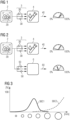
- Figure 1 illustrates the inventive computer-implemented method.
- An image 2 is obtained for a patient, showing a pulmonary nodule 20.
- the image 2 is processed by the machine learning algorithm 1NN, which outputs a blood test panel 3 comprising the set of blood markers 30 that is most likely to return a conclusive result regarding the malignancy (or not) of the imaged lesion.
- a clinician can then order a blood sample to be obtained and sent to a laboratory for processing.
- the blood sample can be processed according to the blood test panel 3 using any suitable technique such as next-generation sequencing, polymerase chain reaction (PCR), mass spectrometry, enzyme-linked immunosorbent assay (ELISA), fluorescence in-situ hybridization (FISH), etc.
- the machine learning algorithm 1NN may also recommend a different processing technique as appropriate.
- a dial represents a scale between 0% (lowest risk of malignancy, i.e. the imaged tumor is most likely benign) and 100% (highest risk of malignancy).
- the machine learning algorithm will have compiled a blood panel which, after laboratory processing of the blood sample 4, will return information allowing the clinician to accurately determine the malignancy of the lesion.
- the outcome of the adjudication is represented symbolically by the dial indicator. In this example, the indicator is clearly positioned within one of three risk groups.
- Figure 2 illustrates a further aspect of the inventive computer-implemented method.
- the machine learning algorithm 1NN predicts that the proposed blood panel 3 may be inconclusive, and has identified one or more further biomarkers 50 for an additional laboratory test 6.
- the diagram shows that the results 40 of the blood sample 4 tested according to the blood panel 3 are indeed inconclusive in this case (the "dial indicator” is positioned at a threshold between two risk groups), and that the additional laboratory test 6 delivers a conclusive result 60.
- the lower "dial indicator” is clearly positioned within the intermediate risk group, i.e. the lesion can be reliably adjudicated and the clinician can decide on subsequent procedure with more confidence. More importantly, the patient need not - at this stage - undergo an unnecessary and costly invasive procedure more appropriate to a high-risk tumor.
- Figure 3 illustrates an exemplary relationship between lesion size (X-axis) and the diagnostic accuracy (Y-axis) of two exemplary biomarkers.
- the diagram shows that the diagnostic accuracy curve 30C1 of a first biomarker is highest for midsize lesions and low for small lesions and large lesions, while the diagnostic accuracy curve 30C2 of a second biomarker is high only for large lesions.
- the machine learning algorithm applies this type of information when putting together a diagnostic blood panel 3 for an imaged lesion 20.
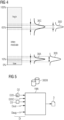
- Figure 4 illustrates a further aspect of using biomarkers to adjudicate a lesion.
- the vertical axis represents a pre-test probability of malignancy.
- the diagram can be understood as follows: if the likelihood of a lesion being malignant is 65% or more, a biopsy or similar is justified; if the likelihood of a lesion being malignant is below 65%, the clinician may order a PET-CT or other follow-up CT and/or a less invasive procedure such as a percutaneous biopsy); if the likelihood of a lesion being malignant is less than 10%, the clinician may opt for surveillance of the patient.
- biomarkers are suited to obtaining a conclusive probability of malignancy.
- biomarker 30A (represented by a diagnostic accuracy curve) is unsuited for adjudicating between a low probability of malignancy and an intermediate probability of malignancy (its effect size is small), while biomarker 30B is better suited (its effect size is large); similarly, biomarker 30C is suited for adjudicating between a low probability of malignancy and an intermediate probability of malignancy, while biomarker 30D is unsuited.
- the machine learning algorithm 1NN applies this type of information when putting together a set of biomarkers 30 for a diagnostic blood panel 3.
- FIG. 5 illustrates a training stage of the machine learning algorithm 1NN.
- training data sets are input to the machine learning algorithm 1NN.
- Each training data set Di comprises an image D2 showing a lesion D20, the lesion class D20C, the blood panel D3 performed for that patient, and the established malignancy of the imaged lesion, i.e. the patient outcome Dout.
- Each dataset Di can be augmented with other relevant information Daug such as co-morbidities of the patient, the patient's EHR, etc.
- the machine learning algorithm 1NN is trained with at least 100 such training data sets Di. More preferably however, the machine learning algorithm 1NN is trained with several thousand training data sets Di. Training is deemed complete when the machine learning algorithm 1NN is able to classify an imaged lesion with a high degree of accuracy and is able to select a set of most suitable blood markers from an extensive blood marker database 30DB, presenting this as a proposed blood panel 3 to assist in reliably adjudicating the imaged lesion.
- the blood marker database 330DB is not restricted to the blood markers appearing in the blood panels D3 of the training data sets Di, but can include further blood markers also, for example, markers chosen on the basis of patient outcome data, biopsy data or follow-up imaging data.
- Figure 6 illustrates an exemplary embodiment of the invention.
- an image 2 showing a lesion 20 is input to the machine learning algorithm 1NN.
- the machine learning algorithm 1NN classifies the lesion 20 into one of a predetermined set of lesion classes C1, ..., Cn.
- the machine learning algorithm 1NN has classified the lesion 20 as belonging to class Cx.
- the machine learning algorithm 1NN identifies a blood test panel (with one or more blood markers) that will return the most conclusive information regarding the lesion 20.
- the machine learning algorithm 1NN can compile a blood panel from a plurality of individual blood markers whose specifics are stored in a database 30DB.
- the machine learning algorithm 1NN can also choose from several predefined blood panels 3P1, ..., 3Pn (e.g. proprietary blood panels), each with a previously established set of blood markers and linked to a predefined diagnostic technique.
- the machine learning algorithm 1NN outputs the proposed blood panel 3 to a user.
- Figure 7 shows a conventional approach.
- a pulmonary CT image 2 is obtained for a patient, showing a pulmonary nodule 20.
- the image 2 is processed by a machine learning algorithm 7NN which has been trained to classify the lesion.
- classification can be done to a high degree of accuracy, allowing a clinician to adjudicate the lesion 20 as benign or malignant with some degree confidence, based on the lesion class 7C returned by the machine learning algorithm 7NN.
- the clinician may then order blood tests T1, ..., Tn on various different biomarkers.
- a single blood test chosen on the basis of the lesion class 7C determined using the prior art approach is therefore usually only of limited use in the adjudication of a lesion.
- the results of a single blood test are often inconclusive, and the clinician may resort to an invasive procedure in order to obtain certainty for the patient. This can add significantly to the overall cost of treatment and can cause undue stress to the patient.
Landscapes
- Health & Medical Sciences (AREA)
- Engineering & Computer Science (AREA)
- Life Sciences & Earth Sciences (AREA)
- Medical Informatics (AREA)
- General Health & Medical Sciences (AREA)
- Public Health (AREA)
- Biomedical Technology (AREA)
- Physics & Mathematics (AREA)
- Pathology (AREA)
- Molecular Biology (AREA)
- Radiology & Medical Imaging (AREA)
- Nuclear Medicine, Radiotherapy & Molecular Imaging (AREA)
- Biophysics (AREA)
- Veterinary Medicine (AREA)
- Heart & Thoracic Surgery (AREA)
- Surgery (AREA)
- Animal Behavior & Ethology (AREA)
- Theoretical Computer Science (AREA)
- Computer Vision & Pattern Recognition (AREA)
- Data Mining & Analysis (AREA)
- Optics & Photonics (AREA)
- Primary Health Care (AREA)
- Epidemiology (AREA)
- High Energy & Nuclear Physics (AREA)
- General Physics & Mathematics (AREA)
- Artificial Intelligence (AREA)
- Databases & Information Systems (AREA)
- Evolutionary Computation (AREA)
- Mathematical Physics (AREA)
- Software Systems (AREA)
- Quality & Reliability (AREA)
- Hematology (AREA)
- General Engineering & Computer Science (AREA)
- Chemical & Material Sciences (AREA)
- Urology & Nephrology (AREA)
- Immunology (AREA)
- Pulmonology (AREA)
- Computing Systems (AREA)
- Cell Biology (AREA)
- Microbiology (AREA)
Abstract
Description
- Various medical conditions can be detected using diagnostic imaging techniques. Particularly in the case of an imaged lesion indicating the presence of a tumor, it is important to be able to correctly assess the nature of the lesion. A false negative diagnosis results in the medical condition going undetected. A false positive diagnosis can be followed by unnecessary and costly invasive procedures. It is known to apply machine learning to assist in identifying imaged lesions, for example a neural network can be trained on many CT images to learn to classify a lesion, primarily on the basis of shape and size. However, the use of the known machine learning methods is largely limited to classification. Therefore, in order to adjudicate a lesion, i.e. to reach a decision as regards the degree of malignancy of the lesion, a clinician must rely on experience and/or avail of further diagnostic tests.
- Therefore, in addition to diagnostic imaging, the assessment of a medical condition preferably involves at least one further laboratory diagnostic test. Diagnostic imaging and laboratory diagnostics are fundamentally different in the way they query underlying information. This can be used to good effect in the context of complex clinical questions such as assessing the malignancy of a tumor or lesion, helping a clinician to plan a suitable schedule for subsequent patient management. For example, if a diagnostic image and a blood test both independently suggest malignancy, an invasive procedure such as biopsy or resection can be justified with a high degree of confidence.
- However, there is a vast number of established and proprietary blood tests, each configured to detect a certain combination of biological markers present above certain threshold levels in the blood (biological markers present in the blood are generally referred to simply as "blood markers"). Blood tests can differ in their range of validity across various subgroups such as lesion size range, histopathological subtype, patient age, patient smoking history, nodule composition, disease prevalence etc. Blood tests can also differ with regard to the clinical question, i.e. whether the lesion is benign or malignant, whether the lesion is a primary or secondary tumor, whether the lesion is aggressive or indolent, the level of invasiveness of the lesion, whether the lesion is a small-cell or non-small-cell carcinoma, whether the lesion shows druggable molecular alterations, etc. Clearly, it is not practicable to apply every test for every patient.
- It is therefore an object of the invention to provide an improved way of combining a diagnostic imaging procedure and a laboratory diagnostic procedure.
- This object is achieved by the claimed method of adjudicating an imaged lesion and by the claimed machine learning algorithm.
- According to the invention, the computer-implemented method for use in adjudicating an imaged lesion comprises steps of: receiving a diagnostic image showing a lesion; processing the diagnostic image in a machine learning algorithm trained to assess or classify an imaged lesion and trained to propose, on the basis of lesion class, a blood test panel comprising markers most suited to adjudicate the imaged lesion; and outputting the proposed blood test panel to a user.
- In the context of the invention, the proposed blood test panel may represent a single blood test or several blood tests. A clinician can then order one or more blood samples to be obtained and processed according to the proposed blood test panel, and can then adjudicate the lesion from the blood test results. An advantage of the inventive computer-implemented method is that an analysis of image-based features is sufficient for the machine learning algorithm to recommend the most promising blood test panel for that patient in order to reliably adjudicate the lesion, i.e. to answer a specific clinical question such as the degree of malignancy of the lesion. After using the inventive computer-implemented method, it can be sufficient to carry out a single blood test on a panel comprising the set of blood biomarkers that will provide a most informative result to help the clinician obtain an answer to a specific clinical question. This is an improvement over known procedures in which several successive blood tests may be required in order to facilitate satisfactory adjudication of a lesion.
- Typically, a lesion is classified as low risk, intermediate risk, or high risk in the context of its malignancy. Depending on the risk group, subsequent patient management can involve surveillance imaging, biopsy, surgery, radiation therapy, ablation etc. as appropriate. However, if a lesion is unambiguously classified in a risk group, ordering of a subsequent blood test is of little clinical benefit. This might be the case for a very large lesion (greater than 30 mm). The clinical impact of a blood test is highest if an imaged lesion cannot be assigned unequivocally to a risk group, i.e. the malignancy risk of the lesion is at the threshold between low risk and intermediate risk, or at the threshold between intermediate risk and high risk. In such threshold cases, the machine learning algorithm can choose blood markers on the basis of their estimated effect size.
- In the claimed computer-implemented method, the machine learning algorithm preferably disregards any blood marker with a small effect size and selects only blood markers with large effect sizes. In this way, the machine learning algorithm can propose a blood test panel that is most useful in identifying the correct risk group into which the lesion can be assigned, i.e. the machine learning algorithm provides a way of more accurately adjudicating a lesion.
- The machine learning algorithm for use in the inventive computer-implemented method is preferably a multi-class classifier configured to classify an imaged lesion into one of a plurality of predefined classes; and to identify a blood panel which will deliver the most accurate results in a subsequent step of adjudicating the imaged lesion.
- Training such a machine learning algorithm preferably comprises the steps of:
- A) compiling a training data set comprising an image showing a lesion, a classification of the lesion, and the patient outcome;
- B) assigning a ground truth to the training data set, which ground truth comprises a set of biomarkers of a blood panel performed for the respective patient and the blood panel result;
- C) applying the machine learning algorithm to the training data set to identify a set of blood panel biomarkers approximating the ground truth; and
- The object of the invention is also achieved by a computer program product with a computer program that is directly loadable into the memory of a data-processing apparatus, and which comprises program units to perform the steps of the inventive computer-implemented method when the program is executed by the control unit.
- The machine learning algorithm can be realized as a module of a computer program which can run on any suitable platform and which comprises program units to perform the steps of the inventive computer-implemented method to classify an imaged lesion, to identify blood markers suited to adjudicate the imaged lesion, and to output a blood test panel comprising the identified blood markers. In the context of the invention, a computer program that incorporates the trained machine learning algorithm and has means of receiving relevant input (a diagnostic image and any other relevant inputs as described above can be input to the computer program in any suitable file format) and a means of outputting the proposed blood panel is referred to herein as an "adjudication program" or "adjudication assistant". The computer program can be configured to receive or read a diagnostic image (and any other relevant inputs) in any suitable format and can present the proposed blood test panel to the user in any suitable manner (e.g. to print the result and/or to send it to a further device such as a computer, a mobile device, a central laboratory, etc.). The inventive computer-implemented method can be used by a clinician to assist in stratifying patients into different risk groups with corresponding diagnostic workup.
- Particularly advantageous embodiments and features of the invention are given by the dependent claims, as revealed in the following description. Features of different claim categories may be combined as appropriate to give further embodiments not described herein.
- In the context of the invention, expressions such as "machine learning algorithm", "neural network", "artificial intelligence tool" and "AI model" shall be understood to be synonyms and may be used interchangeably. The terms "blood test panel", "blood panel" and "test panel" may be used interchangeably.
- The imaging modality used to obtain the diagnostic image can be computed tomography (CT), magnetic resonance imaging (MRI), positron emission tomography (PET), single-photon emission computed tomography (SPECT), X-ray imaging, ultrasound imaging, optical coherence tomography (OCT), photoacoustic imaging, etc. In the following, without restricting the invention in any way, it may be assumed that the diagnostic image is a computed tomography image (in the form of a 3D volume or a 2D slice), for example an image can be a process spectrally resolved CT image, a photon-counting CT image; a high-resolution CT image, a contrast-enhanced CT image, a pair of inspiratory and expiratory scans, a gated CT scan, a stationary multi-source CT scan.
- The inventive computer-implemented method is applicable to various kinds of lesion, for example the imaged lesion can be any of: a pulmonary lesion, an adrenal lesion, a renal lesion, a hepatic lesion, a pancreatic lesion, a mammary lesion. Any of these can be a tumor, and it is important to reliably determine whether the lesion is benign or malignant. If the lesion is malignant, it is important to reliably assess the degree of malignancy. The invention is particularly suited to assist in adjudicating lesions such as pulmonary nodules, since these can be difficult for a clinician to classify from visual assessment of a diagnostic image, and it can be difficult for a clinician to choose suitable biomarkers to assist in adjudicating an imaged pulmonary nodule.
- The machine learning algorithm or "AI model" may include an artificial neural network to apply deep learning techniques. The machine learning algorithm is trained to propose a suitable blood test panel to most accurately adjudicate an imaged lesion. The machine learning algorithm can be implemented as a multi-task classifier which first classifies the imaged lesion into one of several predefined classes, and then identifies the blood test panel that will be best at adjudicating the lesion.
- In the following, the machine learning algorithm may be assumed to comprise a neural network such as a convolutional neural network (CNN). Any suitable CNN architecture may be used, for example AlexNet. A supervised training procedure of such a neural network preferably comprises a technique of backward-propagation to find the best set of parameters that will enable the network to infer a ground truth from a diagnostic image, in this case to infer the lesion class of an imaged lesion in its first task, and to infer the most suitable blood test panel in its second task. The training procedure is preferably configured to achieve a favourably low cost function.
- The lesion classification stage of the machine learning algorithm preferably influences the internal configuration of the proposed blood test panel, i.e. the operating point along the receiver operating characteristic curve (ROC curve) of the entire test panel, the decision thresholds of individual blood markers, the weights of the individual blood markers, etc.
- In addition to the image, the associated blood test for that patient, and the patient outcome (e.g. development of lung cancer and the elapsed time to diagnosis), a training data set preferably also comprises annotated image data. The size, shape and position of a lesion - along with various other factors - are highly relevant to the question of its malignancy. Therefore, in a preferred embodiment of the invention, the training method comprises a step of annotating an image of a training dataset to identify features including any of: lesion size, lesion quantity, lesion composition, lesion location, lesion shape, lesion density, texture features, semantic features, vascular convergence, cavitation, calcification.
- A training data set is preferably also augmented by the occurrence and extent of comorbidities such as emphysema, fibrosis, edema, consolidation, opacities, pleural effusion, atelectasis, pulmonary emboli, etc. Further relevant information can be the patient's subcutaneous and visceral fat content, bone mineral density, muscle density, organ volumes, etc. A training data set is preferably also augmented by relevant clinical factors such as patient age, sex, smoking history, history of cancer, familial cancer predisposition, occupation, etc.
- Some blood markers are detectable in the bloodstream only if the tumor sheds sufficient material in the first place. For example, freely circulating tumor DNA (ctDNA) is released into the bloodstream through apoptotic or necrotic processes, and predominately in the case of larger tumor sizes. Equally, other systemic markers can show higher diagnostic accuracy for smaller nodule sizes, for example autoantibodies and/or micro-RNA (miRNA). The machine learning algorithm learns to identify which markers are most suitable for including in a blood test panel to adjudicate a lesion on the basis of its class.
- The machine learning algorithm is trained to compile a blood test panel comprising any markers such as: carcinoembryonic antigen (CEA), pro-gastrin-releasing-peptide (ProGRP), cytokeratins such as Cyfra 21-1, SCC, neuron specific enolase (NSE), C-reactive protein (CRP), interleukin-6 (IL-6), serum amyloid A1 (SAA1), circulating tumor DNA (ctDNA) (mutations and genetic alterations, methylation patterns, fragmentation patterns); circulating tumor cells (chromosome abnormalities); proteins (autoantibodies, oncofetal antigens, structural proteins, hormones, glycoproteins, storage proteins, enzymes), circulating RNA, micro-RNA (miRNA), metabolites, RNA, sugars, exosomes, extracellular vesicles, etc.
- The blood markers are chosen depending on the class to which the lesion has been assigned as well as other factors such as its size. For example, certain blood markers have different sensitivities, so that, if the size of the imaged lesion suggests that it might be outside or near of the range of validity of a certain marker, that marker can be assigned a lower weight in the blood panel. Depending how well the shape characteristics of a lesion correlate with the accuracy of a blood marker, the weight of such a marker can be set accordingly.
- The machine learning algorithm preferably applies imaging characteristics such as spiculation and nodule type to configure detection thresholds and risk score weighting factors of markers of a fixed blood test panel. A fixed blood test panel can be a proprietary panel or can have been compiled by a laboratory or a clinician.
- For example, a diagnostic image could show a pulmonary nodule located in the periphery of the lung, with solid appearance, homogeneous density and without spiculation: these features are indicative of small cell lung cancer (SCLC). The machine learning algorithm preferably specifies a high threshold for the SCC biomarker in the subsequent laboratory processing step, thus according a relatively high weight to a negative SCC test result (this is because SCLC must have low SCC values). In this way, the sensitivity of the test is increased for SCLC. Since SCLC is the most aggressive form of lung cancer, a high specificity of diagnostic imaging and laboratory blood testing would then accurately indicate a high probability of malignancy for the imaged lesion. The physician and patient can be assured that a subsequent invasive and costly medical procedure is justified.
- Since the machine learning algorithm is to be trained to select a set of blood markers that will assist in adjudicating a lesion, each training data set also includes a blood panel and laboratory test results carried out for that patient. For the training data set, a blood panel need not have been scheduled in connection with an imaged lesion, and it is sufficient that the blood test was performed after detection of the lesion by diagnostic imaging. Preferably, each training data set also includes the patient outcome, e.g. whether or not the imaged lesion led to cancer.
- The accuracy of a machine learning algorithm depends to a large extent on the number of datasets with which it is trained. In a preferred embodiment of the invention, the machine learning algorithm is trained using at least several hundred training data sets, more preferably several thousand training data sets. Images used to obtain the machine learning algorithm can be obtained from diagnostic imaging modalities used in one or more clinics. Equally, the machine learning algorithm could also be trained with synthetically generated images in order to improve its accuracy in classifying lesions.
- The trained machine learning algorithm can then be used by a clinician to assist in adjudicating an imaged lesion. With the inventive computer-implemented method, a suitable diagnostic image is obtained and input to the machine learning algorithm, which returns a proposed blood test panel comprising a set of blood markers chosen to deliver the most conclusive results.
- Of course, the machine learning algorithm can continue to learn by including the actual patient outcome some time after adjudication of a lesion using a blood test panel proposed by the machine learning algorithm.
- In a preferred embodiment of the invention, the machine learning algorithm may also propose a suitable laboratory technique for analysis of the proposed blood panel. A laboratory analysis technique can be any of: a next-generation sequencing technique, a polymerase chain reaction technique (PCR), a mass spectrometry technique, an immunoassay technique, a fluorescence in-situ hybridization (FISH) technique, an electrochemical sensing technique.
- However, even with accurate classification of the lesion and choice of suitable blood markers, the machine learning algorithm may foresee that the proposed blood panel - even though it comprises the most suitable markers - may return an inconclusive result. Therefore, the machine learning algorithm is preferably also trained to also identify one or more further biomarkers for an additional laboratory test. For example, should the machine learning algorithm predict an inconclusive blood panel result, it may propose one or more further biological markers that may then be helpful in adjudicating the classified lesion. Such biological markers may be obtained from a nasal swab, urine, a bronchial swab, saliva, breast milk, etc.
- The inventive adjudication program can run on a processing unit connected to an imaging modality in a clinic. Equally, such a computer program can run on a remote server or cloud computing arrangement.
- In a preferred embodiment of the invention, the adjudication program comprises a software module configured to automatically order the recommended blood test. In a further preferred embodiment of the invention, the adjudication program comprises a FHIR (Fast Healthcare Interoperability Resources) communication module configured to pre-fetch clinical data from an electronic medical record of the patient as additional input to the machine learning algorithm. In a further preferred embodiment of the invention, the machine learning algorithm is configured to recommend several blood tests to be performed at different stages to facilitate a long-term trend analysis. This can be especially useful if lesion classification is at the threshold between two risk groups.
- Other objects and features of the present invention will become apparent from the following detailed descriptions considered in conjunction with the accompanying drawings. It is to be understood, however, that the drawings are designed solely for the purposes of illustration and not as a definition of the limits of the invention.
-
Figure 1 illustrates the inventive computer-implemented method of adjudicating an imaged lesion; -
Figure 2 illustrates a further aspect of the inventive computer-implemented method; -
Figure 3 illustrates an exemplary relationship between lesion size and the diagnostic accuracy of biomarkers; -
Figure 4 illustrates a further aspect of using biomarkers to adjudicate a lesion; -
Figure 5 illustrates a training stage of the machine learning algorithm used by the inventive computer-implemented method; -
Figure 6 illustrates an exemplary embodiment of the invention; -
Figure 7 shows a conventional approach in adjudicating an imaged lesion. - In the diagrams, like numbers refer to like objects throughout. Objects in the diagrams are not necessarily drawn to scale.
- In the following diagrams, the diagnostic image is represented by a pulmonary CT image, and the lesion is represented by a pulmonary nodule. However, as indicated above, the inventive computer-implemented method is applicable to various kinds of lesion, and the images and lesions shown in the drawings are merely exemplary.
-
Figure 1 illustrates the inventive computer-implemented method. Animage 2 is obtained for a patient, showing apulmonary nodule 20. Theimage 2 is processed by the machine learning algorithm 1NN, which outputs ablood test panel 3 comprising the set ofblood markers 30 that is most likely to return a conclusive result regarding the malignancy (or not) of the imaged lesion. - A clinician can then order a blood sample to be obtained and sent to a laboratory for processing. The blood sample can be processed according to the
blood test panel 3 using any suitable technique such as next-generation sequencing, polymerase chain reaction (PCR), mass spectrometry, enzyme-linked immunosorbent assay (ELISA), fluorescence in-situ hybridization (FISH), etc. The machine learning algorithm 1NN may also recommend a different processing technique as appropriate. - On the right-hand side of the diagram, a dial represents a scale between 0% (lowest risk of malignancy, i.e. the imaged tumor is most likely benign) and 100% (highest risk of malignancy). The machine learning algorithm will have compiled a blood panel which, after laboratory processing of the
blood sample 4, will return information allowing the clinician to accurately determine the malignancy of the lesion. The outcome of the adjudication is represented symbolically by the dial indicator. In this example, the indicator is clearly positioned within one of three risk groups. -
Figure 2 illustrates a further aspect of the inventive computer-implemented method. Here, the machine learning algorithm 1NN predicts that the proposedblood panel 3 may be inconclusive, and has identified one or morefurther biomarkers 50 for anadditional laboratory test 6. The diagram shows that theresults 40 of theblood sample 4 tested according to theblood panel 3 are indeed inconclusive in this case (the "dial indicator" is positioned at a threshold between two risk groups), and that theadditional laboratory test 6 delivers aconclusive result 60. In this exemplary embodiment, the lower "dial indicator" is clearly positioned within the intermediate risk group, i.e. the lesion can be reliably adjudicated and the clinician can decide on subsequent procedure with more confidence. More importantly, the patient need not - at this stage - undergo an unnecessary and costly invasive procedure more appropriate to a high-risk tumor. -
Figure 3 illustrates an exemplary relationship between lesion size (X-axis) and the diagnostic accuracy (Y-axis) of two exemplary biomarkers. The diagram shows that the diagnostic accuracy curve 30C1 of a first biomarker is highest for midsize lesions and low for small lesions and large lesions, while the diagnostic accuracy curve 30C2 of a second biomarker is high only for large lesions. The machine learning algorithm applies this type of information when putting together adiagnostic blood panel 3 for an imagedlesion 20. -
Figure 4 illustrates a further aspect of using biomarkers to adjudicate a lesion. The vertical axis represents a pre-test probability of malignancy. Representing an exemplary situation, the diagram can be understood as follows: if the likelihood of a lesion being malignant is 65% or more, a biopsy or similar is justified; if the likelihood of a lesion being malignant is below 65%, the clinician may order a PET-CT or other follow-up CT and/or a less invasive procedure such as a percutaneous biopsy); if the likelihood of a lesion being malignant is less than 10%, the clinician may opt for surveillance of the patient. However, not all biomarkers are suited to obtaining a conclusive probability of malignancy. For example,biomarker 30A (represented by a diagnostic accuracy curve) is unsuited for adjudicating between a low probability of malignancy and an intermediate probability of malignancy (its effect size is small), whilebiomarker 30B is better suited (its effect size is large); similarly,biomarker 30C is suited for adjudicating between a low probability of malignancy and an intermediate probability of malignancy, whilebiomarker 30D is unsuited. The machine learning algorithm 1NN applies this type of information when putting together a set ofbiomarkers 30 for adiagnostic blood panel 3. -
Figure 5 illustrates a training stage of the machine learning algorithm 1NN. Here, training data sets are input to the machine learning algorithm 1NN. Each training data set Di comprises an image D2 showing a lesion D20, the lesion class D20C, the blood panel D3 performed for that patient, and the established malignancy of the imaged lesion, i.e. the patient outcome Dout. Each dataset Di can be augmented with other relevant information Daug such as co-morbidities of the patient, the patient's EHR, etc. - The machine learning algorithm 1NN is trained with at least 100 such training data sets Di. More preferably however, the machine learning algorithm 1NN is trained with several thousand training data sets Di. Training is deemed complete when the machine learning algorithm 1NN is able to classify an imaged lesion with a high degree of accuracy and is able to select a set of most suitable blood markers from an extensive blood marker database 30DB, presenting this as a proposed
blood panel 3 to assist in reliably adjudicating the imaged lesion. - The blood marker database 330DB is not restricted to the blood markers appearing in the blood panels D3 of the training data sets Di, but can include further blood markers also, for example, markers chosen on the basis of patient outcome data, biopsy data or follow-up imaging data.
-
Figure 6 illustrates an exemplary embodiment of the invention. Here, animage 2 showing alesion 20 is input to the machine learning algorithm 1NN. In a first stage, the machine learning algorithm 1NN classifies thelesion 20 into one of a predetermined set of lesion classes C1, ..., Cn. In this example, the machine learning algorithm 1NN has classified thelesion 20 as belonging to class Cx. - In a subsequent stage, the machine learning algorithm 1NN identifies a blood test panel (with one or more blood markers) that will return the most conclusive information regarding the
lesion 20. The machine learning algorithm 1NN can compile a blood panel from a plurality of individual blood markers whose specifics are stored in a database 30DB. In this exemplary embodiment, the machine learning algorithm 1NN can also choose from several predefined blood panels 3P1, ..., 3Pn (e.g. proprietary blood panels), each with a previously established set of blood markers and linked to a predefined diagnostic technique. In a final step, the machine learning algorithm 1NN outputs the proposedblood panel 3 to a user. -
Figure 7 shows a conventional approach. Here, apulmonary CT image 2 is obtained for a patient, showing apulmonary nodule 20. Theimage 2 is processed by a machine learning algorithm 7NN which has been trained to classify the lesion. For some types and size of lesion, classification can be done to a high degree of accuracy, allowing a clinician to adjudicate thelesion 20 as benign or malignant with some degree confidence, based on thelesion class 7C returned by the machine learning algorithm 7NN. However, not all lesions can be reliably adjudicated. In an attempt to obtain a reliable diagnosis for the patient, the clinician may then order blood tests T1, ..., Tn on various different biomarkers. Several tests are generally required, because the known blood tests generally have either a high negative predictive or a high positive predictive value, but rarely both. A single blood test chosen on the basis of thelesion class 7C determined using the prior art approach is therefore usually only of limited use in the adjudication of a lesion. The results of a single blood test are often inconclusive, and the clinician may resort to an invasive procedure in order to obtain certainty for the patient. This can add significantly to the overall cost of treatment and can cause undue stress to the patient. - Although the present invention has been disclosed in the form of preferred embodiments and variations thereon, it will be understood that numerous additional modifications and variations could be made thereto without departing from the scope of the invention.
- For the sake of clarity, it is to be understood that the use of "a" or "an" throughout this application does not exclude a plurality, and "comprising" does not exclude other steps or elements. The mention of a "unit" or a "module" does not preclude the use of more than one unit or module.
- Independent of the grammatical term usage, individuals with male, female or other gender identities are included within the term.
Claims (15)
- A computer-implemented method for use in adjudicating an imaged lesion (20), which method comprises- obtaining a diagnostic image (2) showing a lesion (20);- inputting the diagnostic image (2) to a machine learning algorithm (1NN) previously trained to classify the imaged lesion (20) and to propose, on the basis of the lesion class (Cx), a blood test panel (3) comprising markers (30) most suited to adjudicate the imaged lesion (20); and- outputting the proposed blood test panel (3) to a user.
- A computer-implemented method according to the preceding claim, wherein the blood test panel (3) comprises markers (30) from the group of: circulating tumor DNA, circulating tumor cells, proteins, circulating RNA, metabolites, sugars, exosomes.
- A computer-implemented method according to any of the preceding claims, wherein the machine learning algorithm (1NN) is trained to estimate the malignancy risk of the imaged lesion (20), and to include at least one marker (30) with a large effect size in the blood test panel (3) when the estimated malignancy risk is at a threshold between two risk groups.
- A computer-implemented method according to any of the preceding claims, wherein the blood test panel (3) includes one or more of: an operating point along a receiver operating characteristic curve of the blood test panel (3), a decision threshold of a blood marker (30), a weight of a blood marker (30) .
- A computer-implemented method according to any of the preceding claims, wherein the machine learning algorithm (1NN) is trained to identify at least one laboratory analysis technique for analysis of the proposed blood test panel (3).
- A computer-implemented method according to the preceding claim, wherein a laboratory analysis technique is any of: a next generation sequencing technique, a polymerase chain reaction technique, a mass spectrometry technique, an immunoassay technique, a fluorescence in-situ hybridization technique, an electrochemical sensing technique.
- A computer-implemented method according to any of the preceding claims, wherein the machine learning algorithm (1NN) is trained to identify a number of further biological markers (50) to assist in adjudicating the imaged lesion (20) .
- A computer-implemented method according to the preceding claim, wherein a further biological marker (50) is obtainable from any of: a nasal swab, urine, a bronchial swab, saliva.
- A computer-implemented method according to any of the preceding claims, wherein the diagnostic image is a computed tomography image (2).
- A computer-implemented method according to any of the preceding claims, wherein the imaged lesion is any of: a pulmonary lesion (20), an adrenal lesion, a renal lesion, a hepatic lesion, a pancreatic lesion, a mammary lesion.
- A computer-implemented method according to any of the preceding claims, wherein the machine learning algorithm (1NN) is a two-step classifier configured- to classify an imaged lesion (20) into one of a plurality of predefined classes (C1, ..., Cn);- to identify a number of blood markers (30) suited to adjudicate the imaged lesion (20); and- to output a blood test panel (3) comprising the identified blood markers (30).
- A method of adjudicating an imaged lesion (20), which method comprises the steps of- obtaining a diagnostic image (2) showing a lesion (20);- processing the diagnostic image (2) using the computer-implemented method according to any of claims 1 to 11 and receiving the proposed blood test panel (3); and- obtaining a blood sample (4) and performing laboratory processing of the blood sample (4) according to the blood test panel (3).
- A data processing apparatus adapted to perform the steps of the method according to any of claims 1 to 11, which data processing apparatus comprises- an input interface for obtaining a diagnostic image (2);- a machine learning algorithm (1NN) previously trained to classify an imaged lesion (20) and to specify, on the basis of the lesion class (Cx), a blood test panel (3) most suited to adjudicate the imaged lesion (20); and- a means of outputting the proposed blood test panel (3) to a user.
- A computer program product comprising instructions which, when the program is executed by a computer, cause the computer to carry out the steps of the method according to any of claims 1 to 11.
- A computer-readable data carrier having stored thereon the computer program product of claim 14.
Priority Applications (3)
| Application Number | Priority Date | Filing Date | Title |
|---|---|---|---|
| EP23171524.4A EP4459626A1 (en) | 2023-05-04 | 2023-05-04 | Method of adjudicating an imaged lesion |
| US18/652,286 US20240370999A1 (en) | 2023-05-04 | 2024-05-01 | Method of adjudicating an imaged lesion |
| CN202410547357.5A CN118899074A (en) | 2023-05-04 | 2024-05-06 | Computer-implemented method for determining imaging lesions and method for determining imaging lesions |
Applications Claiming Priority (1)
| Application Number | Priority Date | Filing Date | Title |
|---|---|---|---|
| EP23171524.4A EP4459626A1 (en) | 2023-05-04 | 2023-05-04 | Method of adjudicating an imaged lesion |
Publications (1)
| Publication Number | Publication Date |
|---|---|
| EP4459626A1 true EP4459626A1 (en) | 2024-11-06 |
Family
ID=86329633
Family Applications (1)
| Application Number | Title | Priority Date | Filing Date |
|---|---|---|---|
| EP23171524.4A Pending EP4459626A1 (en) | 2023-05-04 | 2023-05-04 | Method of adjudicating an imaged lesion |
Country Status (3)
| Country | Link |
|---|---|
| US (1) | US20240370999A1 (en) |
| EP (1) | EP4459626A1 (en) |
| CN (1) | CN118899074A (en) |
Citations (2)
| Publication number | Priority date | Publication date | Assignee | Title |
|---|---|---|---|---|
| US20200160997A1 (en) * | 2018-11-02 | 2020-05-21 | University Of Central Florida Research Foundation, Inc. | Method for detection and diagnosis of lung and pancreatic cancers from imaging scans |
| CN114998644B (en) * | 2022-05-10 | 2023-02-07 | 中国医学科学院肿瘤医院 | Tumor diagnosis system and its construction method, terminal equipment and storage medium |
Family Cites Families (5)
| Publication number | Priority date | Publication date | Assignee | Title |
|---|---|---|---|---|
| CN109478231A (en) * | 2016-04-01 | 2019-03-15 | 20/20基因系统股份有限公司 | Methods and compositions to help distinguish benign from malignant radiographically apparent pulmonary nodules |
| AU2021259608A1 (en) * | 2020-04-22 | 2022-11-17 | Dermtech, Inc. | Teledermatology system and methods |
| WO2021247577A1 (en) * | 2020-06-01 | 2021-12-09 | 2020 Genesystems | Methods and software systems to optimize and personalize the frequency of cancer screening blood tests |
| US11935230B2 (en) * | 2020-06-03 | 2024-03-19 | Siemens Healthineers Ag | AI-based image analysis for the detection of normal images |
| US11948687B2 (en) * | 2021-05-24 | 2024-04-02 | Nantcell, Inc. | Deep learning models for region-of-interest determination |
-
2023
- 2023-05-04 EP EP23171524.4A patent/EP4459626A1/en active Pending
-
2024
- 2024-05-01 US US18/652,286 patent/US20240370999A1/en active Pending
- 2024-05-06 CN CN202410547357.5A patent/CN118899074A/en active Pending
Patent Citations (2)
| Publication number | Priority date | Publication date | Assignee | Title |
|---|---|---|---|---|
| US20200160997A1 (en) * | 2018-11-02 | 2020-05-21 | University Of Central Florida Research Foundation, Inc. | Method for detection and diagnosis of lung and pancreatic cancers from imaging scans |
| CN114998644B (en) * | 2022-05-10 | 2023-02-07 | 中国医学科学院肿瘤医院 | Tumor diagnosis system and its construction method, terminal equipment and storage medium |
Also Published As
| Publication number | Publication date |
|---|---|
| US20240370999A1 (en) | 2024-11-07 |
| CN118899074A (en) | 2024-11-05 |
Similar Documents
| Publication | Publication Date | Title |
|---|---|---|
| US12586685B2 (en) | Multimodal machine learning based clinical predictor | |
| Reig et al. | Machine learning in breast MRI | |
| Kuan et al. | Deep learning for lung cancer detection: tackling the kaggle data science bowl 2017 challenge | |
| CN114503213B (en) | Disease detection from weakly annotated convolutional medical images using convolutional long short-term memory | |
| KR102289277B1 (en) | Medical image diagnosis assistance apparatus and method generating evaluation score about a plurality of medical image diagnosis algorithm | |
| Zou et al. | A promising approach for screening pulmonary hypertension based on frontal chest radiographs using deep learning: a retrospective study | |
| US20240395407A1 (en) | Methods and systems for digital pathology assessment of cancer via deep learning | |
| US11471096B2 (en) | Automatic computerized joint segmentation and inflammation quantification in MRI | |
| US20250104827A1 (en) | Systems and methods for determining cancer therapy via deep learning | |
| US12387329B2 (en) | Method for biomarker estimation | |
| WO2021263000A2 (en) | Machine learning based medical data checker | |
| Abdulkareem et al. | Generalizable framework for atrial volume estimation for cardiac CT images using deep learning with quality control assessment | |
| Hirsch et al. | High-performance open-source Ai for breast cancer detection and localization in MRI | |
| JP2025533816A (en) | Systems and methods for multimodal prediction of patient outcomes | |
| US20230255582A1 (en) | Method of performing lung nodule assessment | |
| US20250005745A1 (en) | Systems and methods for determining cancer therapy via deep learning | |
| Kotsyfakis et al. | The application of machine learning to imaging in hematological oncology: A scoping review | |
| WO2024118842A1 (en) | Systems and methods for detecting tertiary lymphoid structures | |
| US20240370999A1 (en) | Method of adjudicating an imaged lesion | |
| US12254990B2 (en) | Methods and systems for determining health of an organ based on medical image data using artificial intelligence | |
| Astaraki | Advanced machine learning methods for oncological image analysis | |
| US20250238923A1 (en) | Apparatus and method for multimodal data fusion for accurate diagnosis | |
| US20260120882A1 (en) | Methods and systems for determining heal th of an organ based on medical image data using artificial intelligence | |
| Ramkumar et al. | Primary Tumor and Lung Cancer Prediction on Dual Energy CT Images for Nodal Metastasis using Deep Learning Technique | |
| Karimpour et al. | A computer‐aided diagnosis (CAD) system based on convolutional neural networks for lung cancer diagnosis from 2D [18F]‐PET/CT images |
Legal Events
| Date | Code | Title | Description |
|---|---|---|---|
| PUAI | Public reference made under article 153(3) epc to a published international application that has entered the european phase |
Free format text: ORIGINAL CODE: 0009012 |
|
| STAA | Information on the status of an ep patent application or granted ep patent |
Free format text: STATUS: THE APPLICATION HAS BEEN PUBLISHED |
|
| AK | Designated contracting states |
Kind code of ref document: A1 Designated state(s): AL AT BE BG CH CY CZ DE DK EE ES FI FR GB GR HR HU IE IS IT LI LT LU LV MC ME MK MT NL NO PL PT RO RS SE SI SK SM TR |
|
| STAA | Information on the status of an ep patent application or granted ep patent |
Free format text: STATUS: REQUEST FOR EXAMINATION WAS MADE |
|
| 17P | Request for examination filed |
Effective date: 20241118 |
|
| RBV | Designated contracting states (corrected) |
Designated state(s): AL AT BE BG CH CY CZ DE DK EE ES FI FR GB GR HR HU IE IS IT LI LT LU LV MC ME MK MT NL NO PL PT RO RS SE SI SK SM TR |