EP4451223A1 - Frequency domain phase retrieval method and device for two-photon synthetic aperture microscope - Google Patents
Frequency domain phase retrieval method and device for two-photon synthetic aperture microscope Download PDFInfo
- Publication number
- EP4451223A1 EP4451223A1 EP23853727.8A EP23853727A EP4451223A1 EP 4451223 A1 EP4451223 A1 EP 4451223A1 EP 23853727 A EP23853727 A EP 23853727A EP 4451223 A1 EP4451223 A1 EP 4451223A1
- Authority
- EP
- European Patent Office
- Prior art keywords
- phase
- pupil function
- pupil
- photon
- phase retrieval
- Prior art date
- Legal status (The legal status is an assumption and is not a legal conclusion. Google has not performed a legal analysis and makes no representation as to the accuracy of the status listed.)
- Pending
Links
Images

Classifications
-
- G—PHYSICS
- G02—OPTICS
- G02B—OPTICAL ELEMENTS, SYSTEMS OR APPARATUS
- G02B27/00—Optical systems or apparatus not provided for by any of the groups G02B1/00 - G02B26/00, G02B30/00
- G02B27/0012—Optical design, e.g. procedures, algorithms, optimisation routines
-
- G—PHYSICS
- G02—OPTICS
- G02B—OPTICAL ELEMENTS, SYSTEMS OR APPARATUS
- G02B21/00—Microscopes
- G02B21/36—Microscopes arranged for photographic purposes or projection purposes or digital imaging or video purposes including associated control and data processing arrangements
- G02B21/365—Control or image processing arrangements for digital or video microscopes
- G02B21/367—Control or image processing arrangements for digital or video microscopes providing an output produced by processing a plurality of individual source images, e.g. image tiling, montage, composite images, depth sectioning, image comparison
-
- G—PHYSICS
- G02—OPTICS
- G02B—OPTICAL ELEMENTS, SYSTEMS OR APPARATUS
- G02B27/00—Optical systems or apparatus not provided for by any of the groups G02B1/00 - G02B26/00, G02B30/00
- G02B27/58—Optics for apodization or superresolution; Optical synthetic aperture systems
-
- G—PHYSICS
- G06—COMPUTING OR CALCULATING; COUNTING
- G06F—ELECTRIC DIGITAL DATA PROCESSING
- G06F16/00—Information retrieval; Database structures therefor; File system structures therefor
- G06F16/20—Information retrieval; Database structures therefor; File system structures therefor of structured data, e.g. relational data
- G06F16/23—Updating
-
- G—PHYSICS
- G06—COMPUTING OR CALCULATING; COUNTING
- G06F—ELECTRIC DIGITAL DATA PROCESSING
- G06F16/00—Information retrieval; Database structures therefor; File system structures therefor
- G06F16/20—Information retrieval; Database structures therefor; File system structures therefor of structured data, e.g. relational data
- G06F16/24—Querying
-
- G—PHYSICS
- G06—COMPUTING OR CALCULATING; COUNTING
- G06T—IMAGE DATA PROCESSING OR GENERATION, IN GENERAL
- G06T17/00—Three-dimensional [3D] modelling for computer graphics
Definitions
- the present application relates to the technical field of phase retrieval, and particularly relates to a frequency domain phase retrieval method and apparatus for 2-photon synthetic aperture microscopy.
- a Phase Retrieval (PR) method is a technology that is usually used in wide-field microscopy and super-resolution microscopy.
- PR Phase Retrieval
- an ideal system without optical distortion has pupil functions whose phases are all zero. Due to inevitable optical distortion of the system, an extra phase difference will be introduced on a pupil plane.
- PSF Point Spread Function
- the images are reconstructed by a Point Spread Function (PSF) of the system established by an optical wave model, and quality of the reconstructed images will be decreased due to the phase difference brought by the optical distortion.
- PSF Point Spread Function
- phase retrieval method restores the phases of the pupil functions of the optical system, so that a PSF matched with actual performance of the optical system is well established, and finally a diffraction limit deconvolution reconstruction image is acquired.
- the phase retrieval method is more widely used, particularly some microscopic methods involving PSF engineering technology.
- the phase retrieval method is an essential link in the image reconstruction process.
- three-dimensional information may be reconstructed by light field microscopy; and angle information is decoded into the three-dimensional information by using the PSF of the microscopy system.
- the present application provides a frequency domain phase retrieval method and apparatus for a 2-photon synthetic aperture microscopy, for solving problems in related art that optical strength distribution is changed since a micro-lens array used for generating multi-angle information is arranged on a pupil plane or an image plane of light field microscopy, phase retrieval is difficult, effect and quality of an image reconstruction result are difficultly increased and higher reconstruction performance cannot be ensured.
- Embodiments in a first aspect of the present application provide a frequency domain phase retrieval method for a 2-photon synthetic aperture microscopy.
- the method includes: performing phase retrieval at different single views based on the 2-photon synthetic aperture microscopy to generate phase retrieval pupil functions; performing phase retrieval initial estimation by taking a pupil function at a previous view as pupil function at a next view to perform aperture synthesis, and traversing all angular distribution in a light field, to obtain multiple rounds of pupil function phase distribution; and generating a final pupil function until phase distribution convergence based on the multiple rounds of pupil function phase distribution, to characterize pupil function phase distribution of systematical distortion.
- the performing phase retrieval at different single views to generate phase retrieval pupil functions includes: collecting axial multi-layer Point Spread Functions, PSFs, of the 2-photon synthetic aperture microscopy excited at different views; and generating the pupil functions based on the axial multi-layer PSF to simulate an objective lens and phase differences in an optical system.
- PSFs Point Spread Functions
- the performing phase retrieval initial estimation by taking the pupil function at the previous view as the pupil function at the next view to perform aperture synthesis includes: obtaining the initial estimation based on phase points obtained at the previous view and a strength distribution matrix of optical strength at the previous view.
- the method further includes: extracting Point Spread Functions, PSFs, from the final pupil function; and performing deconvolution for three-dimensional reconstruction based on the PSFs to generate a reconstruction result.
- PSFs Point Spread Functions
- Embodiments in a second aspect of the present application provide a frequency domain phase retrieval apparatus for a 2-photon synthetic aperture microscopy.
- the apparatus includes: a first generating module configured to perform phase retrieval at different single views based on the 2-photon synthetic aperture microscopy to generate phase retrieval pupil functions; an obtaining module configured to perform phase retrieval initial estimation by taking a pupil function at a previous view as pupil function at a next view to perform aperture synthesis, and traversing all angular distribution in a light field, to obtain multiple rounds of pupil function phase distribution; and a characterizing module configured to generate a final pupil function until phase distribution convergence based on the multiple rounds of pupil function phase distribution, to characterize pupil function phase distribution of systematical distortion.
- the first generating module includes: a collection unit configured to collect axial multi-layer Point Spread Functions, PSFs, of the 2-photon synthetic aperture microscopy excited at different views; and a simulation unit configured to generate the pupil functions based on the axial multi-layer PSFs to simulate an objective lens and phase differences in an optical system.
- a collection unit configured to collect axial multi-layer Point Spread Functions, PSFs, of the 2-photon synthetic aperture microscopy excited at different views
- a simulation unit configured to generate the pupil functions based on the axial multi-layer PSFs to simulate an objective lens and phase differences in an optical system.
- the obtaining module includes: an obtaining unit configured to obtain the initial estimation based on phase points obtained at the previous view and a strength distribution matrix of optical strength at the previous view.
- the apparatus further includes: an extracting module configured to extract Point Spread Functions, PSFs, from the final pupil function; and a second generating module configured to perform deconvolution for three-dimensional reconstruction based on the PSFs to generate a reconstruction result.
- an extracting module configured to extract Point Spread Functions, PSFs, from the final pupil function
- a second generating module configured to perform deconvolution for three-dimensional reconstruction based on the PSFs to generate a reconstruction result.
- Embodiments in a third aspect of the present application provide an electronic device.
- the electronic device includes a memory, a processor, and a computer program stored in the memory and executable on the processor.
- the processor when executing the computer program, implements the frequency domain phase retrieval method for 2-photon synthetic aperture microscopy according to the above embodiments.
- Embodiments in a fourth aspect of the present application provide a computer readable storage medium.
- a computer program is stored on the computer readable storage medium.
- the computer program when executed by the processor, implements the above frequency domain phase retrieval method for 2-photon synthetic aperture microscopy.
- the Point Spread Functions of the system under different views calculated through the pupil function, effect of a reconstruction algorithm and quality of a reconstruction result in the 2-photon synthetic aperture microscopy method are increased; and the PSFs extracted from the pupil function are subjected to deconvolution for three-dimensional reconstruction by estimating the phase difference in the optical system, so that excellent reconstruction performance is ensured, and the performance is better than that of theoretical PSFs generated under a rotational average and an ideal situation.
- a frequency domain phase retrieval method and apparatus for 2-photon synthetic aperture microscopy are described below with reference to drawings.
- problems in related art mentioned in the background that optical strength distribution is changed since a micro-lens array used for generating multi-angle information is arranged on a pupil plane or an image plane of light field microscopy, phase retrieval is difficult, effect and quality of the image reconstruction result are difficultly increased and higher reconstruction performance cannot be ensured, the present application provides a frequency domain phase retrieval method for a 2-photon synthetic aperture microscopy.
- point spread functions of the system under different views are calculated through the pupil function, effect of a reconstruction algorithm and quality of a reconstruction result in the 2-photon synthetic aperture microscopy method are increased; and PSFs extracted from the pupil function are subjected to deconvolution for three-dimensional reconstruction by estimating the phase difference in the optical system, so that better reconstruction performance is ensured, and the performance is better than that of theoretical PSFs generated under a rotational average and an ideal situation.
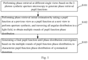
- Fig. 1 is a flow chart of a frequency domain phase retrieval method for 2-photon synthetic aperture microscopy according to embodiments of the present application.
- the frequency domain phase retrieval method for 2-photon synthetic aperture microscopy includes the following actions.
- phase retrieval is performed at different single views based on the 2-photon synthetic aperture microscopy to generate phase retrieval pupil functions.
- the 2-photon synthetic aperture microscopy in the embodiments of the present application is an imaging technology whose excitation at different angles is obtained by a light beam with a diameter much smaller than an aperture of an objective lens entering the objective lens perpendicular to an aperture plane from different positions.
- An optical strength distribution on the aperture plane of the objective lens is different with incidence at different angles.
- the phase brought by systematical distortion is constant. Therefore, in the embodiments of the present application, a full-aperture distortion phase may be calculated from small-aperture imaging results at different angles to be used for estimating PSFs of the optical system at all the angles.
- the phase retrieval is performed at different single views based on an imaging characteristic of the 2-photon synthetic aperture microscopy to generate phase retrieval pupil functions.
- an estimated pupil function is generated from a three-dimensional strength PSF by using a phase retrieval algorithm provided by Hanser et al.
- basis is provided for simulating an objective lens and a phase difference in the optical system through the generated pupil function; and the pupil function is easily modified by calculating a phase difference function, so that characteristics of the PSFs under a phase difference imaging condition are reproduced exactly, thereby increasing effect of the reconstruction algorithm and quality of the reconstruction result in the 2-photon synthetic aperture microscopy method.
- the performing phase retrieval at different single views to generate the phase retrieval pupil functions includes: collecting axial multi-layer Point Spread Functions, PSFs, of the 2-photon synthetic aperture microscopy excited at different views; and generating the pupil functions based on the axial multi-layer PSFs to simulate an objective lens and phase differences in the optical system.
- PSFs Point Spread Functions
- the axial multi-layer Point Spread Functions, PSFs, of the 2-photon synthetic aperture microscopy excited at different views may be collected; measured strength values of the PSF at different axial positions may replace a calculated strength distribution; and the pupil function may be generated through the axial multi-layer PSF. Therefore, the objective lens and the phase difference in the optical system are simulated; and accuracy of reproduced characteristics of PSFs under the phase difference imaging condition is further ensured.
- phase retrieval initial estimation is performed by taking a pupil function at a previous view as a pupil function at a next view to perform aperture synthesis, and all angular distribution in a light field is traversed to obtain multiple rounds of pupil function phase distribution.
- phase retrieval technology is generally as follows: when the aperture of the objective lens is filled, a distortion phase is restored by fully utilizing Numerical Aperture (NA) of the objective lens, and 2-photon synthetic aperture imaging may be performed.
- NA Numerical Aperture
- 2-photon synthetic aperture imaging may be performed.
- the aperture of the objective lens at a single view is generally not filled.
- the phase retrieval initial estimation may be performed by taking the pupil function estimated from the previous view as the pupil function at the next view, to perform the next aperture synthesis, and multiple rounds of pupil function phase distribution after aperture synthesis may be obtained after all angular distribution in the light field is traversed. Therefore, the PSF for the incoming object from multiple angles realizes large-aperture synthesis from small-apertures; and implementation difficulty of the phase retrieval is decreased.
- the performing phase retrieval initial estimation by taking the pupil function at the previous view as the pupil function at the next view to perform aperture synthesis includes: obtaining the initial estimation based on phase points obtained at the previous view and a strength distribution matrix of optical strength at the previous view.
- the initial estimation at the next view may be obtained by multiplying the phase points obtained at the previous view by a matrix that takes the optical strength distribution at the previous view as weights, thereby providing a better systematic PSF for subsequent reconstruction and ensuring an image reconstruction result with best quality.
- a final pupil function is generated until phase distribution convergence based on the multiple rounds of pupil function phase distribution, to characterize pupil function phase distribution of systematical distortion.
- the phase retrieval initial estimation may be iterated and all the angular distribution in the light field may be traversed based on the multiple rounds of pupil function phase distribution after aperture synthesis; and the final pupil function is generated until phase distribution convergence to characterize the pupil function phase distribution of systematical distortion, thereby increasing quality of the image reconstruction result and further ensuring that the performance is better than that of theoretical PSFs generated under a rotational average and an ideal situation.
- the method further includes: extracting PSFs from the final pupil function; and performing deconvolution for three-dimensional reconstruction based on the PSFs to generate a reconstruction result.
- phase retrieval method in the embodiments of the present application may effectively estimate the phase distribution of the pupil plane and generate new PSFs at multiple angles by using the estimated phase.
- the PSFs may be extracted from the final pupil function, and the extracted PSFs are subjected to deconvolution for three-dimensional reconstruction to generate a reconstruction result, thereby ensuring that the performance is better than that of theoretical PSFs generated under a rotational average and an ideal situation.
- the imaging principle of the 2-photon synthetic aperture microscopy in the embodiments of the present application is as follows: multiple small-aperture light beams at different views enter the objective lens, and optical imaging of extending 2-photon excitation depth of field is formed.
- the small-aperture light beams enter the pupil plane of the objective lens from different positions to form excitations of the sample at different angles.
- a common method is position excitation in the matrix form, such as 5*5 angles or 9*9 angles, or multi-angle excitation of uniformly distributed circular pupil positions. Images obtained through the multi-angle excitation are subjected to deconvolution for three-dimensional reconstruction, and then complete three-dimensional imaging information of the sample can be obtained.
- the phase difference will be brought to the optical system due to the inevitable distortion.
- the phase distribution of the pupil plane may be effectively estimated; new PSFs at multiple angles are generated through the estimated phase; and the PSFs obtained through corresponding phase retrieval are subjected to deconvolution for three-dimensional reconstruction.
- the performance will be greatly better than that of theoretical PSFs generated under a rotational average and an ideal situation.
- the method in the embodiments of the present application may include the following actions.
- the axial multi-layer PSF of the 2-photon synthetic aperture microscopy excited at different views may be collected; and the measured strength value of the PSF at different axial positions will be used for replacing the calculated strength distribution.
- Multi-view phase retrieval method is provided: the phase retrieval pupil function is used for retrieval at the next angle for iterative update.
- a single-view pupil function obtained through one phase retrieval may serve as an initial phase retrieval value at the next angle for iterative update.
- the method in the embodiments of the present application may include the following steps:
- the pupil function is used for PSF generation at a single view.
- the plane at different axial positions may be subjected to Fourier transform to obtain respective strength distribution and phase distribution.
- the plane is refocused to a focal plane.
- Each complex field in the embodiments of the present application is subjected to inverse Fourier transform and then refocused to a focal plane image.
- corresponding graphs may be averaged to obtain a new pupil function.
- Phase distribution matrix points of the pupil function are multiplied by a strength distribution matrix to serve as the next view for performing initial phase distribution estimation of phase retrieval.
- final pupil function points at the single view obtained through iteration convergence are multiplied by a pupil function strength distribution matrix at the corresponding view (the strength distribution matrix serves as weights); and then the product serves as initial phase distribution of phase retrieval at the next angle for performing single-view phase retrieval estimation.
- all the views may be traversed for one round of iteration; and multiple rounds of iteration are performed until final pupil function convergence of the 2-photon synthetic aperture microscopy system.
- the frequency domain phase retrieval method for 2-photon synthetic aperture microscopy according to embodiments of the present application is provided.
- the Point Spread Functions of the system under different views calculated through the pupil function, effect of the reconstruction algorithm and quality of the reconstruction result in the 2-photon synthetic aperture microscopy method are increased; and PSFs extracted from the pupil function are subjected to deconvolution for three-dimensional reconstruction by estimating the phase difference in the optical system, so that better reconstruction performance is ensured, and the performance is better than that of theoretical PSFs generated under a rotational average and an ideal situation.
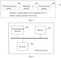
- Fig. 5 is a schematic structural diagram of a frequency domain phase retrieval apparatus for a 2-photon synthetic aperture microscopy according to embodiments of the present application.
- the frequency domain phase retrieval apparatus 10 for 2-photon synthetic aperture microscopy includes: a first generating module 100, an obtaining module 200 and a characterizing module 300.
- the first generating module 100 is configured to perform phase retrieval at different single views based on the 2-photon synthetic aperture microscopy to generate phase retrieval pupil functions.
- the obtaining module 200 is configured to perform phase retrieval initial estimation by taking a pupil function at a previous view as a pupil function at a next view to perform aperture synthesis, and traverse all angular distribution in a light field, to obtain multiple rounds of pupil function phase distribution.
- the characterizing module 300 is configured to generate a final pupil function until phase distribution convergence based on the multiple rounds of pupil function phase distribution, to characterize pupil function phase distribution of systematical distortion.
- the first generating module 100 includes: a collection unit and a simulation unit.
- the collection unit is configured to collect axial multi-layer Point Spread Functions, PSFs, of the 2-photon synthetic aperture microscopy excited at different views.
- the simulation unit is configured to generate the pupil functions based on the axial multi-layer PSFs to simulate an objective lens and phase differences in an optical system.
- the obtaining module 200 includes: an obtaining unit.
- the obtaining unit is configured to obtain the initial estimation based on phase points obtained at the previous view and a strength distribution matrix of optical strength at the previous view.
- the frequency domain phase retrieval apparatus 10 for 2-photon synthetic aperture microscopy further includes: an extracting module and a second generating module.
- the extracting module is configured to extract PSFs from the final pupil function.
- the second generating module is configured to perform deconvolution for three-dimensional reconstruction based on the PSFs to generate a reconstruction result.
- the frequency domain phase retrieval apparatus for 2-photon synthetic aperture microscopy according to embodiments of the present application is provided.
- the Point Spread Functions of the system under different views calculated through the pupil function, effect of the reconstruction algorithm and quality of the reconstruction result in the 2-photon synthetic aperture microscopy method are increased; and PSFs extracted from the pupil function are subjected to deconvolution for three-dimensional reconstruction by estimating the phase difference in the optical system, so that better reconstruction performance is ensured, and the performance is better than that of theoretical PSFs generated under a rotational average and an ideal situation.
- Fig. 6 is a schematic structural diagram of an electronic device according to embodiments of the present application.
- the electronic device may include: a memory 601, a processor 602, and a computer program that is stored in the memory 601 and executable in the processor 602.
- the processor 602 is configured to implement, when executing the computer program, the frequency domain phase retrieval method for 2-photon synthetic aperture microscopy according to the above embodiments.
- the electronic device includes: a communication interface 603 configured for communication between the memory 601 and the processor 602.
- the memory 601 is configured to store the computer program that is executable in the processor 602.
- the memory 601 may include a high-speed RAM memory and may further include a non-volatile memory, such as at least one disk memory.
- the communication interface 603, the memory 601 and the processor 602 may be connected with one another through buses to implement communication with one another.
- the buses may be an Industry Standard Architecture (ISA) bus, a Peripheral Component Interconnect (PCI) bus or an Extended Industry Standard Architecture (EISA) bus, etc.
- ISA Industry Standard Architecture
- PCI Peripheral Component Interconnect
- EISA Extended Industry Standard Architecture
- the buses may be divided into an address bus, a data bus, a control bus and the like. For simple representation, the buses are only represented by a thick line in Fig. 6 , but it does not mean that there is only one bus or one kind of bus.
- the memory 601, the processor 602 and the communication interface 603 are integrated on one chip, communication among the memory 601, the processor 602 and the communication interface 603 may be implemented through an internal interface.
- the processor 602 may be a Central Processing Unit (CPU), or an Application Specific Integrated Circuit (ASIC), or be configured as one or more integrated circuits for implementing the embodiments of the present application.
- CPU Central Processing Unit
- ASIC Application Specific Integrated Circuit
- the present embodiment further provides a computer readable storage medium which has a computer program stored thereon.
- the above frequency domain phase retrieval method for 2-photon synthetic aperture microscopy is implemented when the computer program is executed by the processor.
- Logics and/or steps represented in the flow chart or described herein in other modes may be specifically implemented in any computer readable medium to be used by instruction execution systems, apparatuses or devices (such as a computer-based system, a system including a processor, or any other system that can fetch instructions from the instruction execution system, apparatus or device and execute the instructions), or to be used in combination with these instruction execution systems, apparatuses or devices.
- the "computer readable medium” may be any apparatus that may include, store, communicate, propagate or transmit programs to be used by the instruction execution systems, apparatuses or devices or to be used in combination with these instruction execution systems, apparatuses or devices.
- the computer readable medium include: an electrical connection part (electronic apparatus) having one or N wirings, a portable computer cassette (magnetic apparatus), a random access memory (RAM), a read-only memory (ROM), an erasable programmable read-only memory (EPROM or a flash memory), an optical fiber apparatus and a portable CD read-only memory (CDROM).
- the computer readable medium even may be paper on which the programs may be printed or any other appropriate medium, because the paper or any other medium may be subjected to optical scanning and then edited and interpreted or the programs are obtained electronically by processing the paper or any other medium in any other appropriate manner when necessary. Then, the programs are stored in the computer memory.
- N steps or methods may be implemented by software or firmware that is stored in the memory and executed by an appropriate instruction execution system.
- the steps or methods may be implemented by any or a combination thereof in the following known technologies in the art: a discrete logic circuit having a logic gate circuit used for realizing logic functions for data signals, an application-specific integrated circuit having an appropriate combinational logic gate circuit, a programmable gate array (PGA), a field-programmable gate array (FPGA), etc.
- total or partial steps for realizing the method in the above embodiments may be completed by programs instructing associated hardware.
- the programs may be stored in a computer readable storage medium.
- the programs include one of the steps in the method embodiments or a combination of the steps during execution.
- each unit may also be of physical existence alone; and two or more units may also be integrated in a module.
- the above integrated module may be implemented in the form of hardware, and may also be implemented in the form of software function module. If implemented in the form of software function module and sold or used serving as an independent product, the integrated module may also be stored in a computer readable storage medium.
- the above mentioned storage medium may be a read-only memory, a magnetic disk or a compact disc.
Landscapes
- Physics & Mathematics (AREA)
- Engineering & Computer Science (AREA)
- General Physics & Mathematics (AREA)
- Theoretical Computer Science (AREA)
- Optics & Photonics (AREA)
- Multimedia (AREA)
- Data Mining & Analysis (AREA)
- Databases & Information Systems (AREA)
- General Engineering & Computer Science (AREA)
- Computational Linguistics (AREA)
- Geometry (AREA)
- Software Systems (AREA)
- Computer Vision & Pattern Recognition (AREA)
- Computer Graphics (AREA)
- Chemical & Material Sciences (AREA)
- Analytical Chemistry (AREA)
- Microscoopes, Condenser (AREA)
Abstract
Description
- The present application is based on and claims a priority to the
Chinese patent application No. 202310254841.4 on March 07, 2023 - The present application relates to the technical field of phase retrieval, and particularly relates to a frequency domain phase retrieval method and apparatus for 2-photon synthetic aperture microscopy.
- A Phase Retrieval (PR) method is a technology that is usually used in wide-field microscopy and super-resolution microscopy. In the wide-field microscopy, an ideal system without optical distortion has pupil functions whose phases are all zero. Due to inevitable optical distortion of the system, an extra phase difference will be introduced on a pupil plane. In a process of reconstructing images through deconvolution, the images are reconstructed by a Point Spread Function (PSF) of the system established by an optical wave model, and quality of the reconstructed images will be decreased due to the phase difference brought by the optical distortion. The phases of the pupil functions of the optical system are restored by the phase retrieval method, so that a PSF matched with actual performance of the optical system is well established, and finally a diffraction limit deconvolution reconstruction image is acquired. However, in the super-resolution microscopy such as single molecule localization microscopy, super-resolution fluorescence microscopy and stimulated emission depletion microscopy, the phase retrieval method is more widely used, particularly some microscopic methods involving PSF engineering technology. The phase retrieval method is an essential link in the image reconstruction process.
- In related art, depending on three-dimensional imaging capability of a wide-field microscopy imaging technology, three-dimensional information may be reconstructed by light field microscopy; and angle information is decoded into the three-dimensional information by using the PSF of the microscopy system.
- However, in the related art, problems that optical strength distribution is changed since a micro-lens array used for generating multi-angle information is arranged on a pupil plane or an image plane of light field microscopy, phase retrieval is difficult, effect and quality of an image reconstruction result are difficultly increased and higher reconstruction performance cannot be ensured urgently need to be solved.
- The present application provides a frequency domain phase retrieval method and apparatus for a 2-photon synthetic aperture microscopy, for solving problems in related art that optical strength distribution is changed since a micro-lens array used for generating multi-angle information is arranged on a pupil plane or an image plane of light field microscopy, phase retrieval is difficult, effect and quality of an image reconstruction result are difficultly increased and higher reconstruction performance cannot be ensured.
- Embodiments in a first aspect of the present application provide a frequency domain phase retrieval method for a 2-photon synthetic aperture microscopy. The method includes: performing phase retrieval at different single views based on the 2-photon synthetic aperture microscopy to generate phase retrieval pupil functions; performing phase retrieval initial estimation by taking a pupil function at a previous view as pupil function at a next view to perform aperture synthesis, and traversing all angular distribution in a light field, to obtain multiple rounds of pupil function phase distribution; and generating a final pupil function until phase distribution convergence based on the multiple rounds of pupil function phase distribution, to characterize pupil function phase distribution of systematical distortion.
- In an embodiment of the present application, the performing phase retrieval at different single views to generate phase retrieval pupil functions includes: collecting axial multi-layer Point Spread Functions, PSFs, of the 2-photon synthetic aperture microscopy excited at different views; and generating the pupil functions based on the axial multi-layer PSF to simulate an objective lens and phase differences in an optical system.
- In an embodiment of the present application, the performing phase retrieval initial estimation by taking the pupil function at the previous view as the pupil function at the next view to perform aperture synthesis includes: obtaining the initial estimation based on phase points obtained at the previous view and a strength distribution matrix of optical strength at the previous view.
- In an embodiment of the present application, the method further includes: extracting Point Spread Functions, PSFs, from the final pupil function; and performing deconvolution for three-dimensional reconstruction based on the PSFs to generate a reconstruction result.
- Embodiments in a second aspect of the present application provide a frequency domain phase retrieval apparatus for a 2-photon synthetic aperture microscopy. The apparatus includes: a first generating module configured to perform phase retrieval at different single views based on the 2-photon synthetic aperture microscopy to generate phase retrieval pupil functions; an obtaining module configured to perform phase retrieval initial estimation by taking a pupil function at a previous view as pupil function at a next view to perform aperture synthesis, and traversing all angular distribution in a light field, to obtain multiple rounds of pupil function phase distribution; and a characterizing module configured to generate a final pupil function until phase distribution convergence based on the multiple rounds of pupil function phase distribution, to characterize pupil function phase distribution of systematical distortion.
- In an embodiment of the present application, the first generating module includes: a collection unit configured to collect axial multi-layer Point Spread Functions, PSFs, of the 2-photon synthetic aperture microscopy excited at different views; and a simulation unit configured to generate the pupil functions based on the axial multi-layer PSFs to simulate an objective lens and phase differences in an optical system.
- In an embodiment of the present application, the obtaining module includes: an obtaining unit configured to obtain the initial estimation based on phase points obtained at the previous view and a strength distribution matrix of optical strength at the previous view.
- In an embodiment of the present application, the apparatus further includes: an extracting module configured to extract Point Spread Functions, PSFs, from the final pupil function; and a second generating module configured to perform deconvolution for three-dimensional reconstruction based on the PSFs to generate a reconstruction result.
- Embodiments in a third aspect of the present application provide an electronic device. The electronic device includes a memory, a processor, and a computer program stored in the memory and executable on the processor. The processor, when executing the computer program, implements the frequency domain phase retrieval method for 2-photon synthetic aperture microscopy according to the above embodiments.
- Embodiments in a fourth aspect of the present application provide a computer readable storage medium. A computer program is stored on the computer readable storage medium. The computer program, when executed by the processor, implements the above frequency domain phase retrieval method for 2-photon synthetic aperture microscopy.
- In the embodiments of the present application, the Point Spread Functions of the system under different views calculated through the pupil function, effect of a reconstruction algorithm and quality of a reconstruction result in the 2-photon synthetic aperture microscopy method are increased; and the PSFs extracted from the pupil function are subjected to deconvolution for three-dimensional reconstruction by estimating the phase difference in the optical system, so that excellent reconstruction performance is ensured, and the performance is better than that of theoretical PSFs generated under a rotational average and an ideal situation. Therefore, the problems in the related art that optical strength distribution is changed since a micro-lens array used for generating multi-angle information is arranged on a pupil plane or an image plane of light field microscopy, phase retrieval is difficult, the effect and the quality of the image reconstruction result are difficultly increased and higher reconstruction performance cannot be ensured are solved.
- Additional aspects and advantages of the present application will be partially described below, and will be partially obvious in descriptions below or understood through practice of the present application.
- The above and/or additional aspects and advantages of the present application will be obvious and easily understood in descriptions of the embodiments in combination with drawings below, wherein:
-
Fig. 1 is a flow chart of a frequency domain phase retrieval method for a 2-photon synthetic aperture microscopy according to embodiments of the present application; -
Fig. 2 is a principle diagram of a frequency domain phase retrieval method for a 2-photon synthetic aperture microscopy according to an embodiment of the present application; -
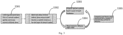
Fig. 3 is a flow chart of a frequency domain phase retrieval method for a 2-photon synthetic aperture microscopy according to an embodiment of the present application; -
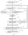
Fig. 4 is an algorithm flow chart of a frequency domain phase retrieval method for a 2-photon synthetic aperture microscopy according to an embodiment of the present application; -
Fig. 5 is a schematic structural diagram of a frequency domain phase retrieval apparatus for a 2-photon synthetic aperture microscopy according to embodiments of the present application; and -
Fig. 6 is a schematic structural diagram of an electronic device according to embodiments of the present application. - Embodiments of the present application will be described in detail below. Examples of the embodiments are illustrated in the drawings, through which identical or similar symbols refer to identical or similar components or components having identical or similar functions. Embodiments described with reference drawings below are illustrative and are intended to be used for explaining the present application rather than limiting the present application.
- A frequency domain phase retrieval method and apparatus for 2-photon synthetic aperture microscopy according to embodiments of the present application are described below with reference to drawings. With respect to problems in related art mentioned in the background that optical strength distribution is changed since a micro-lens array used for generating multi-angle information is arranged on a pupil plane or an image plane of light field microscopy, phase retrieval is difficult, effect and quality of the image reconstruction result are difficultly increased and higher reconstruction performance cannot be ensured, the present application provides a frequency domain phase retrieval method for a 2-photon synthetic aperture microscopy. In the method, point spread functions of the system under different views are calculated through the pupil function, effect of a reconstruction algorithm and quality of a reconstruction result in the 2-photon synthetic aperture microscopy method are increased; and PSFs extracted from the pupil function are subjected to deconvolution for three-dimensional reconstruction by estimating the phase difference in the optical system, so that better reconstruction performance is ensured, and the performance is better than that of theoretical PSFs generated under a rotational average and an ideal situation. Therefore, the problems in the related art that the optical strength distribution is changed since the micro-lens array used for generating multi-angle information is arranged on the pupil plane or the image plane of light field microscopy, phase retrieval is difficult, the effect and the quality of the image reconstruction result are difficultly increased and better reconstruction performance cannot be ensured are solved.
- Specifically,
Fig. 1 is a flow chart of a frequency domain phase retrieval method for 2-photon synthetic aperture microscopy according to embodiments of the present application. - As shown in
Fig. 1 , the frequency domain phase retrieval method for 2-photon synthetic aperture microscopy includes the following actions. - In the action at S 101, phase retrieval is performed at different single views based on the 2-photon synthetic aperture microscopy to generate phase retrieval pupil functions.
- It can be understood that, the 2-photon synthetic aperture microscopy in the embodiments of the present application is an imaging technology whose excitation at different angles is obtained by a light beam with a diameter much smaller than an aperture of an objective lens entering the objective lens perpendicular to an aperture plane from different positions. An optical strength distribution on the aperture plane of the objective lens is different with incidence at different angles. However, the phase brought by systematical distortion is constant. Therefore, in the embodiments of the present application, a full-aperture distortion phase may be calculated from small-aperture imaging results at different angles to be used for estimating PSFs of the optical system at all the angles.
- During actual execution according to embodiments of the present application, the phase retrieval is performed at different single views based on an imaging characteristic of the 2-photon synthetic aperture microscopy to generate phase retrieval pupil functions. For example, an estimated pupil function is generated from a three-dimensional strength PSF by using a phase retrieval algorithm provided by Hanser et al. Thus, basis is provided for simulating an objective lens and a phase difference in the optical system through the generated pupil function; and the pupil function is easily modified by calculating a phase difference function, so that characteristics of the PSFs under a phase difference imaging condition are reproduced exactly, thereby increasing effect of the reconstruction algorithm and quality of the reconstruction result in the 2-photon synthetic aperture microscopy method.
- In an embodiment of the present application, the performing phase retrieval at different single views to generate the phase retrieval pupil functions includes: collecting axial multi-layer Point Spread Functions, PSFs, of the 2-photon synthetic aperture microscopy excited at different views; and generating the pupil functions based on the axial multi-layer PSFs to simulate an objective lens and phase differences in the optical system.
- As a possible implementation according to the embodiments of the present application, the axial multi-layer Point Spread Functions, PSFs, of the 2-photon synthetic aperture microscopy excited at different views may be collected; measured strength values of the PSF at different axial positions may replace a calculated strength distribution; and the pupil function may be generated through the axial multi-layer PSF. Therefore, the objective lens and the phase difference in the optical system are simulated; and accuracy of reproduced characteristics of PSFs under the phase difference imaging condition is further ensured.
- In the action at S 102, phase retrieval initial estimation is performed by taking a pupil function at a previous view as a pupil function at a next view to perform aperture synthesis, and all angular distribution in a light field is traversed to obtain multiple rounds of pupil function phase distribution.
- It can be understood that, the phase retrieval technology is generally as follows: when the aperture of the objective lens is filled, a distortion phase is restored by fully utilizing Numerical Aperture (NA) of the objective lens, and 2-photon synthetic aperture imaging may be performed. The aperture of the objective lens at a single view is generally not filled.
- During actual execution according to the embodiments of the present application, the phase retrieval initial estimation may be performed by taking the pupil function estimated from the previous view as the pupil function at the next view, to perform the next aperture synthesis, and multiple rounds of pupil function phase distribution after aperture synthesis may be obtained after all angular distribution in the light field is traversed. Therefore, the PSF for the incoming object from multiple angles realizes large-aperture synthesis from small-apertures; and implementation difficulty of the phase retrieval is decreased.
- In an embodiment of the present application, the performing phase retrieval initial estimation by taking the pupil function at the previous view as the pupil function at the next view to perform aperture synthesis includes: obtaining the initial estimation based on phase points obtained at the previous view and a strength distribution matrix of optical strength at the previous view.
- During actual execution according to the embodiments of the present application, the initial estimation at the next view may be obtained by multiplying the phase points obtained at the previous view by a matrix that takes the optical strength distribution at the previous view as weights, thereby providing a better systematic PSF for subsequent reconstruction and ensuring an image reconstruction result with best quality.
- In the action at S103, a final pupil function is generated until phase distribution convergence based on the multiple rounds of pupil function phase distribution, to characterize pupil function phase distribution of systematical distortion.
- During actual execution according to the embodiments of the present application, the phase retrieval initial estimation may be iterated and all the angular distribution in the light field may be traversed based on the multiple rounds of pupil function phase distribution after aperture synthesis; and the final pupil function is generated until phase distribution convergence to characterize the pupil function phase distribution of systematical distortion, thereby increasing quality of the image reconstruction result and further ensuring that the performance is better than that of theoretical PSFs generated under a rotational average and an ideal situation.
- In an embodiment of the present application, the method further includes: extracting PSFs from the final pupil function; and performing deconvolution for three-dimensional reconstruction based on the PSFs to generate a reconstruction result.
- It can be understood that, a phase difference will be brought to an optical system due to inevitable distortion. The phase retrieval method in the embodiments of the present application may effectively estimate the phase distribution of the pupil plane and generate new PSFs at multiple angles by using the estimated phase.
- During actual execution, since the phase difference in the optical system may be estimated in the embodiments of the present application, and the generation process of the PSFs is matched with the objective lens model, the PSFs may be extracted from the final pupil function, and the extracted PSFs are subjected to deconvolution for three-dimensional reconstruction to generate a reconstruction result, thereby ensuring that the performance is better than that of theoretical PSFs generated under a rotational average and an ideal situation.
- Specifically, as shown in
Figs. 2 to 4 , operating principles of the frequency domain phase retrieval method for 2-photon synthetic aperture microscopy in the embodiments of the present application are described in detail by virtue of a specific embodiment. - As shown in
Fig. 2 , the imaging principle of the 2-photon synthetic aperture microscopy in the embodiments of the present application is as follows: multiple small-aperture light beams at different views enter the objective lens, and optical imaging of extending 2-photon excitation depth of field is formed. The small-aperture light beams enter the pupil plane of the objective lens from different positions to form excitations of the sample at different angles. A common method is position excitation in the matrix form, such as 5*5 angles or 9*9 angles, or multi-angle excitation of uniformly distributed circular pupil positions. Images obtained through the multi-angle excitation are subjected to deconvolution for three-dimensional reconstruction, and then complete three-dimensional imaging information of the sample can be obtained. - It can be understood that, the phase difference will be brought to the optical system due to the inevitable distortion. In the embodiments of the present application, the phase distribution of the pupil plane may be effectively estimated; new PSFs at multiple angles are generated through the estimated phase; and the PSFs obtained through corresponding phase retrieval are subjected to deconvolution for three-dimensional reconstruction. The performance will be greatly better than that of theoretical PSFs generated under a rotational average and an ideal situation.
- Then, as shown in
Fig. 3 , the method in the embodiments of the present application may include the following actions. - At S301: An axial multi-layer PSF of the 2-photon synthetic aperture microscopy excited at different views is collected.
- In the embodiments of the present application, the axial multi-layer PSF of the 2-photon synthetic aperture microscopy excited at different views may be collected; and the measured strength value of the PSF at different axial positions will be used for replacing the calculated strength distribution.
- At S302: Multi-view phase retrieval method is provided: the phase retrieval pupil function is used for retrieval at the next angle for iterative update.
- In the embodiments of the present application, a single-view pupil function obtained through one phase retrieval may serve as an initial phase retrieval value at the next angle for iterative update.
- At S303: Weighted aperture synthesis based on pupil strength distribution is performed.
- At S304: Phase retrieval at different single views is performed.
- At S305: Pupil function phase distribution that is capable of characterizing systematical distortion is generated.
- As shown in
Fig. 4 , the method in the embodiments of the present application may include the following steps: - At S401: Initial estimation of the pupil function with phase distribution being 0 is created.
- At S402: The pupil function is used for PSF generation at a single view.
- At S403: A PSF plane at different axial positions is generated through defocus.
- At S404: A strength value of the obtained PSF at different axial positions is measured.
- At S405: The calculated strength distribution is replaced with the measured strength value.
- In the embodiments of the present application, the plane at different axial positions may be subjected to Fourier transform to obtain respective strength distribution and phase distribution.
- At S406: The plane is refocused to a focal plane.
- Each complex field in the embodiments of the present application is subjected to inverse Fourier transform and then refocused to a focal plane image.
- At S407: The graphs are averaged to obtain the pupil function.
- In the embodiments of the present application, corresponding graphs may be averaged to obtain a new pupil function.
- At S408: Nonzero values outside the aperture are removed by using the pupil function, and smooth filtering is performed.
- At S409: Next iteration is performed by using the new pupil function.
- At S410: The final pupil function at a single view is obtained.
- At S411: Phase distribution matrix points of the pupil function are multiplied by a strength distribution matrix to serve as the next view for performing initial phase distribution estimation of phase retrieval.
- In the embodiments of the present application, final pupil function points at the single view obtained through iteration convergence are multiplied by a pupil function strength distribution matrix at the corresponding view (the strength distribution matrix serves as weights); and then the product serves as initial phase distribution of phase retrieval at the next angle for performing single-view phase retrieval estimation.
- At S412: The aperture at all views is traversed to obtain the final pupil function of the 2-photon synthetic aperture microscopy system.
- In the embodiments of the present application, all the views may be traversed for one round of iteration; and multiple rounds of iteration are performed until final pupil function convergence of the 2-photon synthetic aperture microscopy system.
- The frequency domain phase retrieval method for 2-photon synthetic aperture microscopy according to embodiments of the present application is provided. The Point Spread Functions of the system under different views calculated through the pupil function, effect of the reconstruction algorithm and quality of the reconstruction result in the 2-photon synthetic aperture microscopy method are increased; and PSFs extracted from the pupil function are subjected to deconvolution for three-dimensional reconstruction by estimating the phase difference in the optical system, so that better reconstruction performance is ensured, and the performance is better than that of theoretical PSFs generated under a rotational average and an ideal situation. Therefore, the problems in the related art that the optical strength distribution is changed since the micro-lens array used for generating multi-angle information is arranged on the pupil plane or the image plane of light field microscopy, phase retrieval is difficult, the effect and the quality of the image reconstruction result are difficultly increased and better reconstruction performance cannot be ensured are solved.
- Then, the frequency domain phase retrieval apparatus for the 2-photon synthetic aperture microscopy according to embodiments of the present application is described with reference to drawings.
-
Fig. 5 is a schematic structural diagram of a frequency domain phase retrieval apparatus for a 2-photon synthetic aperture microscopy according to embodiments of the present application. - As shown in
Fig. 5 , the frequency domainphase retrieval apparatus 10 for 2-photon synthetic aperture microscopy includes: afirst generating module 100, an obtainingmodule 200 and acharacterizing module 300. - Specifically, the
first generating module 100 is configured to perform phase retrieval at different single views based on the 2-photon synthetic aperture microscopy to generate phase retrieval pupil functions. - The obtaining
module 200 is configured to perform phase retrieval initial estimation by taking a pupil function at a previous view as a pupil function at a next view to perform aperture synthesis, and traverse all angular distribution in a light field, to obtain multiple rounds of pupil function phase distribution. - The
characterizing module 300 is configured to generate a final pupil function until phase distribution convergence based on the multiple rounds of pupil function phase distribution, to characterize pupil function phase distribution of systematical distortion. - In an embodiment of the present application, the
first generating module 100 includes: a collection unit and a simulation unit. - The collection unit is configured to collect axial multi-layer Point Spread Functions, PSFs, of the 2-photon synthetic aperture microscopy excited at different views.
- The simulation unit is configured to generate the pupil functions based on the axial multi-layer PSFs to simulate an objective lens and phase differences in an optical system.
- In an embodiment of the present application, the obtaining
module 200 includes: an obtaining unit. - The obtaining unit is configured to obtain the initial estimation based on phase points obtained at the previous view and a strength distribution matrix of optical strength at the previous view.
- In an embodiment of the present application, the frequency domain
phase retrieval apparatus 10 for 2-photon synthetic aperture microscopy further includes: an extracting module and a second generating module. - The extracting module is configured to extract PSFs from the final pupil function.
- The second generating module is configured to perform deconvolution for three-dimensional reconstruction based on the PSFs to generate a reconstruction result.
- It should be noted that, the above explanation of the embodiments of the frequency domain phase retrieval method for 2-photon synthetic aperture microscopy is also applicable to the frequency domain phase retrieval apparatus for the 2-photon synthetic aperture microscopy in the present embodiment. Unnecessary details are not given herein.
- The frequency domain phase retrieval apparatus for 2-photon synthetic aperture microscopy according to embodiments of the present application is provided. The Point Spread Functions of the system under different views calculated through the pupil function, effect of the reconstruction algorithm and quality of the reconstruction result in the 2-photon synthetic aperture microscopy method are increased; and PSFs extracted from the pupil function are subjected to deconvolution for three-dimensional reconstruction by estimating the phase difference in the optical system, so that better reconstruction performance is ensured, and the performance is better than that of theoretical PSFs generated under a rotational average and an ideal situation. Therefore, the problems in the related art that the optical strength distribution is changed since the micro-lens array used for generating multi-angle information is arranged on the pupil plane or the image plane of light field microscopy, phase retrieval is difficult, the effect and the quality of the image reconstruction result are difficultly increased and better reconstruction performance cannot be ensured are solved.
-
Fig. 6 is a schematic structural diagram of an electronic device according to embodiments of the present application. The electronic device may include:
amemory 601, aprocessor 602, and a computer program that is stored in thememory 601 and executable in theprocessor 602. - The
processor 602 is configured to implement, when executing the computer program, the frequency domain phase retrieval method for 2-photon synthetic aperture microscopy according to the above embodiments. - Further, the electronic device includes:
acommunication interface 603 configured for communication between thememory 601 and theprocessor 602. - The
memory 601 is configured to store the computer program that is executable in theprocessor 602. - The
memory 601 may include a high-speed RAM memory and may further include a non-volatile memory, such as at least one disk memory. - If the
memory 601, theprocessor 602 and thecommunication interface 603 are implemented independently, thecommunication interface 603, thememory 601 and theprocessor 602 may be connected with one another through buses to implement communication with one another. The buses may be an Industry Standard Architecture (ISA) bus, a Peripheral Component Interconnect (PCI) bus or an Extended Industry Standard Architecture (EISA) bus, etc. The buses may be divided into an address bus, a data bus, a control bus and the like. For simple representation, the buses are only represented by a thick line inFig. 6 , but it does not mean that there is only one bus or one kind of bus. - In a specific implementation, if the
memory 601, theprocessor 602 and thecommunication interface 603 are integrated on one chip, communication among thememory 601, theprocessor 602 and thecommunication interface 603 may be implemented through an internal interface. - The
processor 602 may be a Central Processing Unit (CPU), or an Application Specific Integrated Circuit (ASIC), or be configured as one or more integrated circuits for implementing the embodiments of the present application. - The present embodiment further provides a computer readable storage medium which has a computer program stored thereon. The above frequency domain phase retrieval method for 2-photon synthetic aperture microscopy is implemented when the computer program is executed by the processor.
- In the description of the specification, the description of reference terms "an embodiment", "some embodiments", "example", "specific example" or "some examples", etc. means that specific features, structures, materials or characteristics illustrated in combination with the embodiment or example are included in at least one embodiment or example of the present application. In this specification, exemplary statements for the above terms do not must aim at the same embodiment or example. Moreover, the described specific features, structures, materials or characteristics can be combined appropriately in any one or N embodiments or examples. In addition, without conflict, those skilled in the art can combine and integrate different embodiments or examples illustrated in the specification and the features of different embodiments or examples.
- In addition, the terms such as "first" and "second" are only used for the purpose of description, rather than being understood to indicate or imply relative importance or hint the number of indicated technical features. Thus, the features limited by "first" and "second" can explicitly or impliedly comprise at least one of the features. In the description of the present application, the meaning of "N" is at least two, such as two, three, etc. unless otherwise clearly specified.
- The flow chart or any procedure or method description described in any other manner herein may be understood to represent a module, a fragment or a part including codes of executable instructions in one or N steps used for realizing customized logic functions or procedures. Moreover, the scope of preferred implementations of the present application includes additional implementations, throughout which the functions may be executed out of the illustrated or discussed sequence, including a basically simultaneous manner or a reverse order according to the involved functions, which should be understood by those skilled in the art in the embodiments of the present application.
- Logics and/or steps represented in the flow chart or described herein in other modes, i.e., a definite sequence table of executable instructions that may be considered to be used for realizing logic functions, may be specifically implemented in any computer readable medium to be used by instruction execution systems, apparatuses or devices (such as a computer-based system, a system including a processor, or any other system that can fetch instructions from the instruction execution system, apparatus or device and execute the instructions), or to be used in combination with these instruction execution systems, apparatuses or devices. For the present description, the "computer readable medium" may be any apparatus that may include, store, communicate, propagate or transmit programs to be used by the instruction execution systems, apparatuses or devices or to be used in combination with these instruction execution systems, apparatuses or devices. More specific examples (a non-exhaustive list) of the computer readable medium include: an electrical connection part (electronic apparatus) having one or N wirings, a portable computer cassette (magnetic apparatus), a random access memory (RAM), a read-only memory (ROM), an erasable programmable read-only memory (EPROM or a flash memory), an optical fiber apparatus and a portable CD read-only memory (CDROM). In addition, the computer readable medium even may be paper on which the programs may be printed or any other appropriate medium, because the paper or any other medium may be subjected to optical scanning and then edited and interpreted or the programs are obtained electronically by processing the paper or any other medium in any other appropriate manner when necessary. Then, the programs are stored in the computer memory.
- It should be understood that, various parts of the present application may be implemented by hardware, software, firmware or combinations thereof. In the above implementations, N steps or methods may be implemented by software or firmware that is stored in the memory and executed by an appropriate instruction execution system. For example, if the implementation by hardware is the same as that in another implementation, the steps or methods may be implemented by any or a combination thereof in the following known technologies in the art: a discrete logic circuit having a logic gate circuit used for realizing logic functions for data signals, an application-specific integrated circuit having an appropriate combinational logic gate circuit, a programmable gate array (PGA), a field-programmable gate array (FPGA), etc.
- Those ordinary skilled in the art may understand that, total or partial steps for realizing the method in the above embodiments may be completed by programs instructing associated hardware. The programs may be stored in a computer readable storage medium. The programs include one of the steps in the method embodiments or a combination of the steps during execution.
- In addition, various functional units in each embodiment of the present application may be integrated in a processing module; each unit may also be of physical existence alone; and two or more units may also be integrated in a module. The above integrated module may be implemented in the form of hardware, and may also be implemented in the form of software function module. If implemented in the form of software function module and sold or used serving as an independent product, the integrated module may also be stored in a computer readable storage medium.
- The above mentioned storage medium may be a read-only memory, a magnetic disk or a compact disc. Although the embodiments of the present application have been illustrated and described above, it should be understood that, the above embodiments are illustrative and cannot be understood as a limitation to the present application. Changes, modifications, replacements and transformations of the above embodiments may be made by those ordinary skilled in the art in the scope of the present application.
Claims (10)
- A frequency domain phase retrieval method for a 2-photon synthetic aperture microscopy, comprising:performing phase retrieval at different single views based on the 2-photon synthetic aperture microscopy to generate phase retrieval pupil functions;performing phase retrieval initial estimation by taking a pupil function at a previous view as a pupil function at a next view to perform aperture synthesis, and traversing all angular distribution in a light field, to obtain multiple rounds of pupil function phase distribution; andgenerating a final pupil function until phase distribution convergence based on the multiple rounds of pupil function phase distribution, to characterize pupil function phase distribution of systematical distortion.
- The method according to claim 1, wherein the performing phase retrieval at different single views to generate the phase retrieval pupil functions comprises:collecting axial multi-layer Point Spread Functions, PSFs, of the 2-photon synthetic aperture microscopy excited at different views; andgenerating the pupil functions based on the axial multi-layer PSFs to simulate an objective lens and phase differences in an optical system.
- The method according to claim 1, wherein the performing phase retrieval initial estimation by taking the pupil function at the previous view as the pupil function at the next view to perform aperture synthesis comprises:
obtaining the initial estimation based on phase points obtained at the previous view and a strength distribution matrix of optical strength at the previous view. - The method according to claim 1, further comprising:extracting Point Spread Functions, PSFs, from the final pupil function; andperforming deconvolution for three-dimensional reconstruction based on the PSFs to generate a reconstruction result.
- A frequency domain phase retrieval apparatus for a 2-photon synthetic aperture microscopy, comprising:a first generating module configured to perform phase retrieval at different single views based on the 2-photon synthetic aperture microscopy to generate phase retrieval pupil functions;an obtaining module configured to perform phase retrieval initial estimation by taking a pupil function at a previous view as a pupil function at a next view to perform aperture synthesis, and traverse all angular distribution in a light field, to obtain multiple rounds of pupil function phase distribution; anda characterizing module configured to generate a final pupil function until phase distribution convergence based on the multiple rounds of pupil function phase distribution, to characterize pupil function phase distribution of systematical distortion.
- The apparatus according to claim 5, wherein the first generating module comprises:a collection unit configured to collect axial multi-layer Point Spread Functions, PSFs, of the 2-photon synthetic aperture microscopy excited at different views; anda simulation unit configured to generate the pupil functions based on the axial multi-layer PSFs to simulate an objective lens and phase differences in an optical system.
- The apparatus according to claim 5, wherein the obtaining module comprises:
an obtaining unit configured to obtain the initial estimation based on phase points obtained at the previous view and a strength distribution matrix of optical strength at the previous view. - The apparatus according to claim 5, further comprising:an extracting module configured to extract Point Spread Functions, PSFs, from the final pupil function; anda second generating module configured to perform deconvolution for three-dimensional reconstruction based on the PSFs to generate a reconstruction result.
- An electronic device, comprising: a memory, a processor, and a computer program stored in the memory and executable on the processor, wherein the processor is configured to implement, when executing the computer program, the frequency domain phase retrieval method for the 2-photon synthetic aperture microscopy according to any one of claims 1 to 4.
- A computer readable storage medium, having a computer program stored thereon, wherein the computer program, when executed by the processor, implements the frequency domain phase retrieval method for the 2-photon synthetic aperture microscopy according to any one of claims 1 to 4.
Applications Claiming Priority (2)
| Application Number | Priority Date | Filing Date | Title |
|---|---|---|---|
| CN202310254841.4A CN116416380B (en) | 2023-03-07 | 2023-03-07 | Frequency domain phase retrieval method and device for two-photon synthetic aperture microscope |
| PCT/CN2023/105686 WO2024183196A1 (en) | 2023-03-07 | 2023-07-04 | Frequency domain phase retrieval method and device for two-photon synthetic aperture microscope |
Publications (2)
| Publication Number | Publication Date |
|---|---|
| EP4451223A1 true EP4451223A1 (en) | 2024-10-23 |
| EP4451223A4 EP4451223A4 (en) | 2025-02-12 |
Family
ID=87054143
Family Applications (1)
| Application Number | Title | Priority Date | Filing Date |
|---|---|---|---|
| EP23853727.8A Pending EP4451223A4 (en) | 2023-03-07 | 2023-07-04 | Frequency domain phase retrieval method and device for two-photon synthetic aperture microscope |
Country Status (3)
| Country | Link |
|---|---|
| EP (1) | EP4451223A4 (en) |
| CN (1) | CN116416380B (en) |
| WO (1) | WO2024183196A1 (en) |
Families Citing this family (1)
| Publication number | Priority date | Publication date | Assignee | Title |
|---|---|---|---|---|
| CN119179066B (en) * | 2024-11-25 | 2025-02-07 | 中国人民解放军国防科技大学 | Multi-aperture imaging system cooperative positioning point calibration method and device |
Family Cites Families (10)
| Publication number | Priority date | Publication date | Assignee | Title |
|---|---|---|---|---|
| US8620065B2 (en) * | 2010-04-09 | 2013-12-31 | The Regents Of The University Of Colorado | Methods and systems for three dimensional optical imaging, sensing, particle localization and manipulation |
| US8922636B1 (en) * | 2010-08-20 | 2014-12-30 | The United States Of America As Represented By The Secretary Of The Navy | Synthetic aperture imaging for fluid flows |
| US9864184B2 (en) * | 2012-10-30 | 2018-01-09 | California Institute Of Technology | Embedded pupil function recovery for fourier ptychographic imaging devices |
| US11092795B2 (en) * | 2016-06-10 | 2021-08-17 | California Institute Of Technology | Systems and methods for coded-aperture-based correction of aberration obtained from Fourier ptychography |
| CN108364342B (en) * | 2017-01-26 | 2021-06-18 | 中国科学院脑科学与智能技术卓越创新中心 | Light field microscope system and method and device for reconstructing three-dimensional information |
| CN108550108B (en) * | 2017-09-28 | 2020-11-03 | 武汉大学 | Fourier laminated imaging image reconstruction method based on phase iteration minimization |
| CN109615651B (en) * | 2019-01-29 | 2022-05-20 | 清华大学 | Three-dimensional microscopic imaging method and system based on light field microscopic system |
| CN112002008A (en) * | 2019-05-27 | 2020-11-27 | 万维数码智能有限公司 | A three-dimensional object detection device and method integrating optical and visual intelligence technologies |
| WO2021026381A1 (en) * | 2019-08-06 | 2021-02-11 | Purdue Research Foundation | Pinpointing single molecule positions with in situ point spread function retrieval for three dimensional nanoscopy of whole cells and tissues |
| CN110906883B (en) * | 2019-12-02 | 2021-09-07 | 中国科学院光电技术研究所 | A High-Resolution 3D Detection Method Integrating Multiocular Vision and Synthetic Aperture Imaging |
-
2023
- 2023-03-07 CN CN202310254841.4A patent/CN116416380B/en active Active
- 2023-07-04 EP EP23853727.8A patent/EP4451223A4/en active Pending
- 2023-07-04 WO PCT/CN2023/105686 patent/WO2024183196A1/en not_active Ceased
Also Published As
| Publication number | Publication date |
|---|---|
| WO2024183196A1 (en) | 2024-09-12 |
| CN116416380B (en) | 2024-06-07 |
| CN116416380A (en) | 2023-07-11 |
| EP4451223A4 (en) | 2025-02-12 |
Similar Documents
| Publication | Publication Date | Title |
|---|---|---|
| CN114092325B (en) | Fluorescent image super-resolution reconstruction method and device, computer equipment and medium | |
| CN102057397A (en) | System and method for producing an optically sectioned image using both structured and uniform illumination | |
| JP2013117848A5 (en) | ||
| JP2013117848A (en) | Image processing apparatus and image processing method | |
| JP2015104136A (en) | Machine vision inspection system and method for obtaining images with extended depth of field | |
| EP3709258B1 (en) | Generating composite image from multiple images captured for subject | |
| CN111105416B (en) | A method and system for automatically grading the degree of myeloid cell proliferation | |
| CN113433130A (en) | Method and device for generating confocal imaging by wide-field imaging | |
| JP2017524155A (en) | Method and apparatus for imaging an object | |
| CN114155340B (en) | Scanning light field data reconstruction method, device, electronic device and storage medium | |
| Kim et al. | Blind deconvolution of 3D fluorescence microscopy using depth‐variant asymmetric PSF | |
| Rudnaya et al. | A derivative-based fast autofocus method in electron microscopy | |
| EP3291532A1 (en) | Image processing device and image processing method | |
| CN116416380B (en) | Frequency domain phase retrieval method and device for two-photon synthetic aperture microscope | |
| Arnison et al. | Wavefront coding fluorescence microscopy using high aperture lenses | |
| EP4455642A1 (en) | Digital adaptive optical method and apparatus for multiphoton microscopy | |
| CN116740321A (en) | Repairing method, device and equipment for display grid and storage medium | |
| CN113724150B (en) | Structured light microscopic reconstruction method and device without high signal-to-noise ratio truth image | |
| EP4451035A1 (en) | Two-photon synthetic aperture microscopy system and method based on focus-tunable lens | |
| CN116337832B (en) | Two-photon microscopy imaging method and device based on synthetic aperture | |
| EP4455640B1 (en) | Two-photon synthetic aperture imaging method and apparatus based on diffraction-limit-sized small aperture | |
| CN117314915B (en) | In-situ detection method, device and storage medium for microstructure processing | |
| CN121883278B (en) | A method and apparatus for synthesizing total focusing digital slices based on physical optics models | |
| CN117372592A (en) | Light field three-dimensional reconstruction method and device based on color difference clues and neural network | |
| CN118368404A (en) | Resolution testing method of camera module, electronic equipment and storage medium |
Legal Events
| Date | Code | Title | Description |
|---|---|---|---|
| STAA | Information on the status of an ep patent application or granted ep patent |
Free format text: STATUS: UNKNOWN |
|
| STAA | Information on the status of an ep patent application or granted ep patent |
Free format text: STATUS: THE INTERNATIONAL PUBLICATION HAS BEEN MADE |
|
| PUAI | Public reference made under article 153(3) epc to a published international application that has entered the european phase |
Free format text: ORIGINAL CODE: 0009012 |
|
| STAA | Information on the status of an ep patent application or granted ep patent |
Free format text: STATUS: REQUEST FOR EXAMINATION WAS MADE |
|
| 17P | Request for examination filed |
Effective date: 20240223 |
|
| AK | Designated contracting states |
Kind code of ref document: A1 Designated state(s): AL AT BE BG CH CY CZ DE DK EE ES FI FR GB GR HR HU IE IS IT LI LT LU LV MC ME MK MT NL NO PL PT RO RS SE SI SK SM TR |
|
| REG | Reference to a national code |
Ref country code: DE Ref legal event code: R079 Free format text: PREVIOUS MAIN CLASS: G06T0017000000 Ipc: G02B0027580000 |
|
| A4 | Supplementary search report drawn up and despatched |
Effective date: 20250115 |
|
| RIC1 | Information provided on ipc code assigned before grant |
Ipc: G02B 21/00 20060101ALI20250109BHEP Ipc: G02B 27/00 20060101ALI20250109BHEP Ipc: G02B 27/58 20060101AFI20250109BHEP |
|
| STAA | Information on the status of an ep patent application or granted ep patent |
Free format text: STATUS: EXAMINATION IS IN PROGRESS |
|
| 17Q | First examination report despatched |
Effective date: 20250613 |
|
| REG | Reference to a national code |
Ref country code: DE Ref legal event code: R079 Free format text: PREVIOUS MAIN CLASS: G02B0027580000 Ipc: G02B0021360000 |
|
| GRAP | Despatch of communication of intention to grant a patent |
Free format text: ORIGINAL CODE: EPIDOSNIGR1 |
|
| STAA | Information on the status of an ep patent application or granted ep patent |
Free format text: STATUS: GRANT OF PATENT IS INTENDED |
|
| RIC1 | Information provided on ipc code assigned before grant |
Ipc: G02B 21/36 20060101AFI20260323BHEP Ipc: G02B 27/58 20060101ALI20260323BHEP |
|
| INTG | Intention to grant announced |
Effective date: 20260402 |