EP4443154A1 - Disease or health state inspection method and inspection system - Google Patents
Disease or health state inspection method and inspection system Download PDFInfo
- Publication number
- EP4443154A1 EP4443154A1 EP22901216.6A EP22901216A EP4443154A1 EP 4443154 A1 EP4443154 A1 EP 4443154A1 EP 22901216 A EP22901216 A EP 22901216A EP 4443154 A1 EP4443154 A1 EP 4443154A1
- Authority
- EP
- European Patent Office
- Prior art keywords
- biomarker
- disease
- health condition
- examining
- condition according
- Prior art date
- Legal status (The legal status is an assumption and is not a legal conclusion. Google has not performed a legal analysis and makes no representation as to the accuracy of the status listed.)
- Pending
Links
Images


Classifications
-
- G—PHYSICS
- G16—INFORMATION AND COMMUNICATION TECHNOLOGY [ICT] SPECIALLY ADAPTED FOR SPECIFIC APPLICATION FIELDS
- G16H—HEALTHCARE INFORMATICS, i.e. INFORMATION AND COMMUNICATION TECHNOLOGY [ICT] SPECIALLY ADAPTED FOR THE HANDLING OR PROCESSING OF MEDICAL OR HEALTHCARE DATA
- G16H50/00—ICT specially adapted for medical diagnosis, medical simulation or medical data mining; ICT specially adapted for detecting, monitoring or modelling epidemics or pandemics
- G16H50/20—ICT specially adapted for medical diagnosis, medical simulation or medical data mining; ICT specially adapted for detecting, monitoring or modelling epidemics or pandemics for computer-aided diagnosis, e.g. based on medical expert systems
-
- A—HUMAN NECESSITIES
- A61—MEDICAL OR VETERINARY SCIENCE; HYGIENE
- A61B—DIAGNOSIS; SURGERY; IDENTIFICATION
- A61B5/00—Measuring for diagnostic purposes; Identification of persons
- A61B5/08—Measuring devices for evaluating the respiratory organs
- A61B5/082—Evaluation by breath analysis, e.g. determination of the chemical composition of exhaled breath
-
- A—HUMAN NECESSITIES
- A61—MEDICAL OR VETERINARY SCIENCE; HYGIENE
- A61B—DIAGNOSIS; SURGERY; IDENTIFICATION
- A61B5/00—Measuring for diagnostic purposes; Identification of persons
- A61B5/145—Measuring characteristics of blood in vivo, e.g. gas concentration or pH-value ; Measuring characteristics of body fluids or tissues, e.g. interstitial fluid or cerebral tissue
-
- A—HUMAN NECESSITIES
- A61—MEDICAL OR VETERINARY SCIENCE; HYGIENE
- A61B—DIAGNOSIS; SURGERY; IDENTIFICATION
- A61B5/00—Measuring for diagnostic purposes; Identification of persons
- A61B5/72—Signal processing specially adapted for physiological signals or for diagnostic purposes
- A61B5/7235—Details of waveform analysis
- A61B5/7264—Classification of physiological signals or data, e.g. using neural networks, statistical classifiers, expert systems or fuzzy systems
- A61B5/7267—Classification of physiological signals or data, e.g. using neural networks, statistical classifiers, expert systems or fuzzy systems involving training the classification device
-
- G—PHYSICS
- G01—MEASURING; TESTING
- G01N—INVESTIGATING OR ANALYSING MATERIALS BY DETERMINING THEIR CHEMICAL OR PHYSICAL PROPERTIES
- G01N27/00—Investigating or analysing materials by the use of electric, electrochemical, or magnetic means
- G01N27/62—Investigating or analysing materials by the use of electric, electrochemical, or magnetic means by investigating the ionisation of gases, e.g. aerosols; by investigating electric discharges, e.g. emission of cathode
- G01N27/622—Ion mobility spectrometry
- G01N27/624—Differential mobility spectrometry [DMS]; Field asymmetric-waveform ion mobility spectrometry [FAIMS]
-
- G—PHYSICS
- G01—MEASURING; TESTING
- G01N—INVESTIGATING OR ANALYSING MATERIALS BY DETERMINING THEIR CHEMICAL OR PHYSICAL PROPERTIES
- G01N33/00—Investigating or analysing materials by specific methods not covered by groups G01N1/00 - G01N31/00
- G01N33/48—Biological material, e.g. blood, urine; Haemocytometers
- G01N33/483—Physical analysis of biological material
- G01N33/487—Physical analysis of biological material of liquid biological material
- G01N33/493—Physical analysis of biological material of liquid biological material urine
-
- G—PHYSICS
- G01—MEASURING; TESTING
- G01N—INVESTIGATING OR ANALYSING MATERIALS BY DETERMINING THEIR CHEMICAL OR PHYSICAL PROPERTIES
- G01N33/00—Investigating or analysing materials by specific methods not covered by groups G01N1/00 - G01N31/00
- G01N33/48—Biological material, e.g. blood, urine; Haemocytometers
- G01N33/483—Physical analysis of biological material
- G01N33/497—Physical analysis of biological material of gaseous biological material, e.g. breath
-
- G—PHYSICS
- G16—INFORMATION AND COMMUNICATION TECHNOLOGY [ICT] SPECIALLY ADAPTED FOR SPECIFIC APPLICATION FIELDS
- G16H—HEALTHCARE INFORMATICS, i.e. INFORMATION AND COMMUNICATION TECHNOLOGY [ICT] SPECIALLY ADAPTED FOR THE HANDLING OR PROCESSING OF MEDICAL OR HEALTHCARE DATA
- G16H10/00—ICT specially adapted for the handling or processing of patient-related medical or healthcare data
- G16H10/40—ICT specially adapted for the handling or processing of patient-related medical or healthcare data for data related to laboratory analysis, e.g. patient specimen analysis
-
- G—PHYSICS
- G16—INFORMATION AND COMMUNICATION TECHNOLOGY [ICT] SPECIALLY ADAPTED FOR SPECIFIC APPLICATION FIELDS
- G16H—HEALTHCARE INFORMATICS, i.e. INFORMATION AND COMMUNICATION TECHNOLOGY [ICT] SPECIALLY ADAPTED FOR THE HANDLING OR PROCESSING OF MEDICAL OR HEALTHCARE DATA
- G16H50/00—ICT specially adapted for medical diagnosis, medical simulation or medical data mining; ICT specially adapted for detecting, monitoring or modelling epidemics or pandemics
- G16H50/70—ICT specially adapted for medical diagnosis, medical simulation or medical data mining; ICT specially adapted for detecting, monitoring or modelling epidemics or pandemics for mining of medical data, e.g. analysing previous cases of other patients
Definitions
- the present invention relates to disease or health condition examination method and examination system.
- Non Patent Literature 1 a method for discriminating a predetermined disease on the basis of biomarkers in a biological sample is known.
- Non Patent Literature 1 when a disease is discriminated using principal component analysis, the accuracy of discriminating a predetermined disease is higher than that of conventional clinical markers, but the accuracy is still insufficient, and higher accuracy is required.
- a method for examining a disease or health condition is a method for examining a disease or health condition based on a biomarker in a biological sample.
- the method for examining a disease or health condition includes a first step, a second step, a third step, and a fourth step.
- the first step extracts a biomarker from a biological sample.
- the second step ionizes the extracted biomarker.
- the third step obtains a three-dimensional profile by passing the ionized biomarker between two electrodes and providing the two electrodes with a plurality of electrical signals.
- the fourth step discriminates a predetermined disease using a discriminator obtained by learning the three-dimensional profile and the type of disease in association with each other.
- the method for examining a disease or health condition according to the first aspect can discriminate a predetermined disease with higher accuracy by using a discriminator obtained by learning the three-dimensional profile and the type of disease in association with each other.
- a method for examining a disease or health condition according to a second aspect is the method for examining a disease or health condition according to the first aspect, in which the discriminator simultaneously discriminates multiple types of diseases.
- a method for examining a disease or health condition according to a third aspect is the method for examining a disease or health condition according to the first aspect or the second aspect, in which the biological sample is at least one of exhaled breath, serum, blood, saliva, plasma, nipple aspiration material, synovial fluid, cerebrospinal fluid, sweat, urine, feces, tears, gas emitted from skin, tracheal washes, swabbed material, needle aspiration material, semen, vaginal fluid, and pre-ejaculate.
- the biological sample is at least one of exhaled breath, serum, blood, saliva, plasma, nipple aspiration material, synovial fluid, cerebrospinal fluid, sweat, urine, feces, tears, gas emitted from skin, tracheal washes, swabbed material, needle aspiration material, semen, vaginal fluid, and pre-ejaculate.
- a method for examining a disease or health condition according to a fourth aspect is the method for examining a disease or health condition according to any one of the first to third aspects, in which the biological sample is exhaled breath or urine.
- a method for examining a disease or health condition according to a fifth aspect is the method for examining a disease or health condition according to any one of the first to fourth aspects, in which the biomarker is at least one of a gene biomarker, a cell biomarker, an organic compound biomarker, a metabolic biomarker, a saccharide biomarker, a lipid biomarker, a heterocyclic biomarker, an elemental compound biomarker, an image biomarker, an anthropological biomarker, a personal habit biomarker, a disease state biomarker, and an expression biomarker.
- the biomarker is at least one of a gene biomarker, a cell biomarker, an organic compound biomarker, a metabolic biomarker, a saccharide biomarker, a lipid biomarker, a heterocyclic biomarker, an elemental compound biomarker, an image biomarker, an anthropological biomarker, a personal habit biomarker, a disease state biomarker, and
- a method for examining a disease or health condition according to a sixth aspect is the method for examining a disease or health condition according to any one of the first to fifth aspects, in which the biomarker is an organic compound biomarker generated for metabolism in vivo.
- a method for examining a disease or health condition according to a seventh aspect is the method for examining a disease or health condition according to the sixth aspect, in which the organic compound biomarker is a volatile organic compound.
- a method for examining a disease or health condition according to an eighth aspect is the method for examining a disease or health condition according to the seventh aspect, in which the volatile organic compound is volatilome.
- a method for examining a disease or health condition according to a ninth aspect is the method for examining a disease or health condition according to any one of the first to eighth aspects, in which the discriminator discriminates non-disease, mild cancer, and severe cancer.
- a method for examining a disease or health condition according to a tenth aspect is the method for examining a disease or health condition according to any one of the first to ninth aspects, in which the third step obtains the three-dimensional profile by utilizing a fact that mobility of ions changes between a low electric field and a high electric field.
- An examination system for a disease or health condition is an examination system for a disease or health condition based on a biomarker in a biological sample.
- the examination system for a disease or health condition includes a first device, a second device, and a third device.
- the first device extracts a biomarker from a biological sample.
- the second device ionizes the extracted biomarker.
- the second device obtains a three-dimensional profile by passing the ionized biomarker between two electrodes and providing the two electrodes with a plurality of electrical signals.
- the third device discriminates a predetermined disease using a discriminator obtained by learning the three-dimensional profile and the type of disease in association with each other.
- the examination system for a disease or health condition according to the eleventh aspect can discriminate a predetermined disease with higher accuracy by using the discriminator obtained by learning the three-dimensional profile and the type of disease in association with each other.
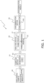
- FIG. 1 is a configuration diagram of an examination system 1 for a disease or health condition.
- the examination system 1 discriminates a predetermined disease on the basis of a biomarker 50 in a biological sample 40.
- the examination system 1 includes a sampling device 10 (first device), an analysis device 20 (second device), and a discrimination device 30 (third device).
- the sampling device 10 extracts the biomarker 50 from the biological sample 40.
- the biological sample 40 is at least one of exhaled breath, serum, blood, saliva, plasma, nipple aspiration material, synovial fluid, cerebrospinal fluid, sweat, urine, feces, tears, gas emitted from skin, tracheal washes, swabbed material, needle aspiration material, semen, vaginal fluid, and pre-ejaculate.
- the biological sample 40 in the present embodiment is exhaled breath or urine.
- the biomarker 50 is at least one of a gene biomarker, a cell biomarker, an organic compound biomarker, a metabolic biomarker, a saccharide biomarker, a lipid biomarker, a heterocyclic biomarker, an elemental compound biomarker, an image biomarker, an anthropological biomarker, a personal habit biomarker, a disease state biomarker, and an expression biomarker.
- the biomarker 50 in the present embodiment is an organic compound biomarker generated for metabolism in vivo.
- VOC Volatile Organic Compound
- the VOC in the present embodiment is volatilome (volatile metabolite).
- the sampling device 10 adsorbs VOC in the biological sample 40 to an adsorbent, thereby extracting VOC from the biological sample 40. Then, the sampling device 10 desorbs the VOC from the adsorbent, and uniformly (stably) flows the VOC into the analysis device 20 for a certain period of time.
- the analysis device 20 analyzes the VOC extracted from the biological sample 40 by the sampling device 10 using FAIMS (Field Asymmetric Ion Mobility Spectrometry, High-Field Asymmetric Waveform Ion Mobility Spectrometry).
- the analysis device 20 is, for example, Lonestar from Owlstone.
- FAIMS is an analysis technique utilizing the fact that mobility of ions changes between a low electric field and a high electric field.
- the analysis device 20 includes a processor such as a CPU or a GPU, and a storage device such as a RAM, a ROM, or an HDD.
- the analysis device 20 includes a keyboard and a mouse for inputting various commands and various information.
- the analysis device 20 includes a monitor for displaying a FAIMS profile 21 and the like to be described later.
- the analysis device 20 includes a network interface device for communicating with the discrimination device 30 via a communication line (not illustrated).
- the analysis device 20 first ionizes the VOC extracted from the biological sample 40 by the sampling device 10. The analysis device 20 then generates the FAIMS profile 21 by passing the ionized VOC between two electrodes and providing the two electrodes with a plurality of electrical signals.
- the FAIMS profile 21 records current values of cations and current values of anions flowing between two electrodes for each combination of a dispersion field (DF) and a compensation voltage (CV).
- FIG. 2 is a diagram illustrating an example of the FAIMS profile 21.
- the FAIMS profile 21 is represented by a two-dimensional array in which the first dimension (vertical axis) is the dispersion field (hereinafter, may be referred to as DF.) and the second dimension (horizontal axis) is the compensation voltage (hereinafter, may be referred to as CV).
- FIG. 2 is a FAIMS profile 21 recording the current values of cations (hereinafter, may be referred to as the FAIMS profile 21 of cations.), and the right diagram of FIG. 2 is a FAIMS profile 21 recording the current values of anions (hereinafter, may be referred to as the FAIMS profile 21 of anions.).
- the DF of "0% to 100%" is divided into 51 stages, and the CV of "-6 V to +6 V" is divided into 512 stages.
- the FAIMS profiles 21 of cations and anions in FIG. 2 are a two-dimensional array having a size of "51 ⁇ 512", respectively.
- the magnitude of the current value of each cell of the FAIMS profile 21 is expressed by the shade of color.
- the biological sample 40 When the biological sample 40 is collected from a patient with a disease and a healthy person, it is known that the concentrations of specific substances contained in the VOC extracted from the biological sample 40 is different between the patient and the healthy person.
- Non Patent Literature 2 shows that when exhaled breath is collected from a patient with colorectal cancer and a healthy person, the concentrations of substances in the following Table 1 contained in VOCs extracted from the exhaled breath are different between the patient and the healthy person.
- Non Patent Literature 3 shows that when exhaled breath is collected from a patient with colorectal cancer and a healthy person, the concentrations of substances in the following Table 2 contained in VOCs extracted from the exhaled breath are different between the patient and the healthy person.
- Non Patent Literature 4 shows that when urine is collected from a patient with colorectal cancer and a healthy person, the concentrations of substances in the following Table 3 contained in VOCs extracted from the urine are different between the patient and the healthy person.
- Table 3 Substances in VOCs with different concentrations Hexanal Hexen-1-ol 4-methyl-1 -hexene Acetaldehyde Ethylene Oxide Oxalic Acid Dimethyl Diazene Cyclobutyl Amine Oxepane Acetone 2-Pentanone 3-methyl-2-Butanone 2,3-Butanedione 4-Heptanone 3-Heptanone 2,4-dimethyl-3-Pentanone Acetyloxime-Pyridine Carboxaldehyde Hydrocinnamoyl-Bezene-ethanamine Styrene Dimethyl-Thiourea Allyl Isothiocyanate Isothiocyanato-cyclopropane 2-cyano-acetamide Methoxy-phenyl-oxi
- Non Patent Literature 5 shows that when urine is collected from a patient with colorectal cancer and a healthy person, the concentrations of substances in the following Table 4 contained in VOCs extracted from the urine are different between the patient and the healthy person.
- Table 4 Substances in VOCs with different concentrations Octane, 2,2,6-trimethyl Ethylbenzene Xylene Octanal Undecanal NonaA23:A38 Decanal Benzene, 1,3-bis(1-methylethenyl) 1-Undecanol Acetone 2-Pentanone 2-Heptanone 2,4-Di-tert-butylphenol Heptanal Heptadecane 3,4-Dimethylcyclohexanol 5-Hepten-2-ol 6-methyl-Hepten-2-ol Hexanal Biphenyl Cyclopentanone, 2-methyl Methane, isocyanato Acetophenone Naphthalene
- Non Patent Literature 6 shows that when exhaled breath is collected from a patient with liver cancer and a healthy person, the concentrations of substances in the following Table 5 contained in VOCs extracted from the exhaled breath are different between the patient and the healthy person.
- Table 5 Substances in VOCs with different concentrations Ethane Acetaldehyde (E)-2-Nonene Acetone Trimethylamine Propanol Methylhexane Acrylonitrile Benzene Dimethyl Sulfide Hydrogen Sulfide Isoprene Ethanol Octene Carbon Disulfide Ammonia Nonene Pentane Heptane Decene Acetonitrile Triethyl Amine
- Non Patent Literature 7 shows that when exhaled breath is collected from a patient with pancreatic cancer and a healthy person, the concentrations of substances in the following Table 6 contained in VOCs extracted from the exhaled breath are different between the patient and the healthy person.
- Table 6 Substances in VOCs with different concentrations Methanol Acetonitrile Ethanol MEK Acetone Isoprene n-Propanol Benzene Toluene n-Heptane
- FIG. 3 is a diagram for comparing FAIMS profiles 21 of the patient with a disease and a healthy person.
- FIG. 3 using urine as a biological sample, the FAIMS profiles 21 of a patient with kidney disease and a healthy person are compared.
- the left is the FAIMS profile 21 of the patient with kidney disease and the right is the FAIMS profile 21 of the healthy person.
- the upper side is the FAIMS profile 21 of cations and the lower side is the FAIMS profile 21 of anions.
- a diagram showing a difference in the FAIMS profiles 21 between the patient with kidney disease and the healthy person for each of the FAIMS profiles 21 of cations and anions is depicted.
- the magnitude of the difference in the current value of each cell of the FAIMS profile 21 is expressed by the shade of color.
- the shade of color can be seen, it can be seen that the distribution of the current value of the FAIMS profile 21 is different between the patient with kidney disease and the healthy person.
- the analysis device 20 stacks the generated FAIMS profiles 21 of cations and anions in the direction of the third dimension to obtain a three-dimensional profile 22 (three-dimensional array). For example, in the case of FIG. 2 , the analysis device 20 obtains a three-dimensional profile 22 having a size of "51 ⁇ 512 ⁇ 2" by stacking the FAIMS profiles 21 of cations and anions.
- the discrimination device 30 is a general computer.
- the discrimination device 30 discriminates a predetermined disease using a discriminator 342 obtained by learning the three-dimensional profile 22 and the type of disease in association with each other.
- FIG. 4 is a control block diagram of the discrimination device 30.
- the discrimination device 30 mainly includes an input unit 31, a display unit 32, a storage unit 33, a communication unit 34, and a control unit 39.
- the input unit 31 is a keyboard and a mouse. Various commands and various types of information for the discrimination device 30 can be input using the input unit 31.
- the display unit 32 is a monitor.
- the display unit 32 can display a learning situation and the like.
- the communication unit 34 is a network interface device for communicating with the analysis device 20 via a communication line (not illustrated).
- the storage unit 33 is a storage device such as a RAM, a ROM, and an HDD.
- the storage unit 33 stores a program implemented by the control unit 39, data necessary for implementing the program, and the like.
- the storage unit 33 particularly stores learning data 341 and the discriminator 342.
- the learning data 341 is data for performing learning of the discriminator 342. Specifically, the learning data 341 is data in which the three-dimensional profile 22 of VOC extracted from the biological sample 40 has been associated with the type of disease of a person from which the biological sample 40 has been collected.
- the three-dimensional profile 22 is obtained from the analysis device 20 via the communication unit 34, for example.
- the type of disease is input by a user from the input unit 31, for example.
- the control unit 39 is a processor such as a CPU or a GPU.
- the control unit 39 reads and executes the program stored in the storage unit 33 to implement various functions of the discrimination device 30.
- the control unit 39 can also write a calculation result to the storage unit 33 and read information stored in the storage unit 33 in accordance with the program.
- the control unit 39 includes a learning unit 391 and a discrimination unit 392 as functional blocks.
- the learning unit 391 performs learning of the discriminator 342 by using the learning data 341.
- the size of the three-dimensional profile 22 of the learning data 341 is "51 ⁇ 512 ⁇ 2".
- the learning data 341 is data in which the three-dimensional profile 22 has been associated with one of a "patient with a predetermined disease” and a "healthy person”.
- the discriminator 342 discriminates whether it is a "patient with a predetermined disease” or a "healthy person” based on the three-dimensional profile 22.
- the discriminator 342 uses a model combining a convolutional neural network (CNN) and a recurrent neural network (RNN).
- CNN convolutional neural network
- RNN recurrent neural network
- FIG. 5 is a diagram illustrating a configuration of the discriminator 342. As illustrated in FIG. 5 , the discriminator 342 includes an input layer, first to fourth intermediate layers, and an output layer.
- the array is extracted row by row in ascending order of DF (25 times in total), and input to the RNN (Conv1D + RNN).
- RNN recurrent layer
- one-dimensional convolution (Conv1D) is performed on the dimension of CV.
- the size of the data flowing through the discriminator 342 changes from "128 ⁇ 12" to "32 ⁇ 6".
- the probability of being a "patient with a predetermined disease” and the probability of being a "healthy person” are calculated by the fully connected layer. From the calculated probability, whether it is a "patient with a predetermined disease” or a "healthy person” is discriminated using a predetermined threshold value. As a result, the size of the data flowing through the discriminator 342 changes from "6" to "2".
- the discrimination unit 392 discriminates a predetermined disease using the discriminator 342 learned by the learning unit 391.
- the discriminator 342 discriminates whether it is a "patient with a predetermined disease” or a "healthy person” from the three-dimensional profile 22.
- a three-dimensional profile 22 is obtained by the analysis device 20.
- the discriminator 342 uses, as the learning data 341, data in which the three-dimensional profile 22 has been associated with one of the "patient with a disease” and the "healthy person".
- FIG. 6 is a diagram illustrating a discrimination result by principal component analysis.
- the "patient with a disease” and the "healthy person” are plotted on a two-dimensional eigenspace consisting of a first principal component PC1 and a second principal component PC2.
- the contribution ratio of the first principal component PC1 is 20.4%
- the contribution ratio of the second principal component PC2 is 12.3%.
- a relatively light-colored plot represents the "healthy person”
- a relatively dark-colored plot represents the "patient with a disease”.
- the analysis result showed that the "patient with a disease" and the "healthy person” were mixed.
- FIG. 7 is a diagram illustrating a discrimination result by the discriminator 342.
- FIG. 7 depicts ROC curves of each time when learning is performed by stratified 10-fold cross-validation and an average ROC curve of these curves.
- AUC Average Under the Curve
- the discriminator 342 discriminates between the "patient with a disease” and the "healthy person” with high accuracy.
- all the features (current values) constituting the FAIMS profile 21 are considered for discrimination, so that the discrimination accuracy can be improved.
- step S1 the sampling device 10 extracts VOC from the biological sample 40.
- step S2 the analysis device 20 ionizes the VOC extracted from the biological sample 40.
- step S3 the analysis device 20 passes the ionized VOC between two electrodes and provides the two electrodes with a plurality of electrical signals.
- step S4 the analysis device 20 generates FAIMS profiles 21 of cations and anions.
- step S5 the analysis device 20 stacks the FAIMS profiles 21 of cations and anions to obtain a three-dimensional profile 22.
- step S6 the discrimination device 30 discriminates whether it is a "patient with a predetermined disease” or a "healthy person” using the discriminator 342 obtained by learning the three-dimensional profile 22 and the "patient with a predetermined disease” or the "healthy person” in association with each other.
- Non Patent Literature 1 when a disease is discriminated using principal component analysis as in Non Patent Literature 1, the accuracy of discriminating a predetermined disease is higher than that of conventional clinical markers, but as shown by verification, the accuracy is still insufficient, and higher accuracy is required.
- the examination system 1 of the present embodiment is an examination system of a disease or health condition based on a VOC (biomarker) in a biological sample 40.
- the examination system 1 includes a sampling device 10, an analysis device 20, and a discrimination device 30.
- the sampling device 10 extracts VOC from the biological sample 40.
- the analysis device 20 ionizes the extracted VOC.
- the analysis device 20 obtains a three-dimensional profile 22 by passing the ionized VOC between two electrodes and providing the two electrodes with a plurality of electrical signals.
- the analysis device 20 obtains the three-dimensional profile 22 using FAIMS.
- the discrimination device 30 discriminates a predetermined disease using a discriminator 342 obtained by learning the three-dimensional profile 22 and the type of disease in association with each other.
- the examination system 1 can discriminate a predetermined disease with higher accuracy by using the discriminator 342 obtained by learning the three-dimensional profile 22 and the type of disease in association with each other.
- the learning data 341 was data in which the three-dimensional profile 22 had been associated with one of the "patient with a predetermined disease" and the "healthy person”.
- the learning data 341 may be data in which the three-dimensional profile 22 has been associated with multiple types of diseases.
- the learning data 341 is, for example, data in which the three-dimensional profile 22 has been associated with any of a "patient with colorectal cancer", a "patient with kidney cancer", and the "healthy person”.
- the discriminator 342 can simultaneously discriminate multiple types of diseases by providing the number of nodes to be discriminated in the output layer.
- the learning data 341 was data in which the three-dimensional profile 22 had been associated with one of the "patient with a predetermined disease" and the "healthy person”.
- the learning data 341 may be data in which the three-dimensional profile 22 has been associated with any of "non-disease", “stage 1 cancer”, “stage 2 cancer”, “stage 3 cancer”, and “stage 4 cancer”.
- the discriminator 342 can discriminate "non-disease”, “mild cancer (for example, cancers of stages 1 and 2)", and “severe cancer (for example, cancers of stages 3 and 4)" by providing the number of nodes to be discriminated in the output layer.
Landscapes
- Health & Medical Sciences (AREA)
- Engineering & Computer Science (AREA)
- Life Sciences & Earth Sciences (AREA)
- Physics & Mathematics (AREA)
- Biomedical Technology (AREA)
- General Health & Medical Sciences (AREA)
- Chemical & Material Sciences (AREA)
- Pathology (AREA)
- Medical Informatics (AREA)
- Public Health (AREA)
- Molecular Biology (AREA)
- Biophysics (AREA)
- General Physics & Mathematics (AREA)
- Urology & Nephrology (AREA)
- Biochemistry (AREA)
- Analytical Chemistry (AREA)
- Immunology (AREA)
- Hematology (AREA)
- Spectroscopy & Molecular Physics (AREA)
- Food Science & Technology (AREA)
- Medicinal Chemistry (AREA)
- Epidemiology (AREA)
- Data Mining & Analysis (AREA)
- Primary Health Care (AREA)
- Databases & Information Systems (AREA)
- Heart & Thoracic Surgery (AREA)
- Surgery (AREA)
- Artificial Intelligence (AREA)
- Veterinary Medicine (AREA)
- Animal Behavior & Ethology (AREA)
- Chemical Kinetics & Catalysis (AREA)
- Electrochemistry (AREA)
- Physiology (AREA)
- Optics & Photonics (AREA)
- Evolutionary Computation (AREA)
- Signal Processing (AREA)
- Pulmonology (AREA)
- Psychiatry (AREA)
- Computer Vision & Pattern Recognition (AREA)
- Mathematical Physics (AREA)
Abstract
Description
- The present invention relates to disease or health condition examination method and examination system.
- As described in Non Patent Literature 1, a method for discriminating a predetermined disease on the basis of biomarkers in a biological sample is known.
- As in Non Patent Literature 1, when a disease is discriminated using principal component analysis, the accuracy of discriminating a predetermined disease is higher than that of conventional clinical markers, but the accuracy is still insufficient, and higher accuracy is required.
- A method for examining a disease or health condition according to a first aspect is a method for examining a disease or health condition based on a biomarker in a biological sample. The method for examining a disease or health condition includes a first step, a second step, a third step, and a fourth step. The first step extracts a biomarker from a biological sample. The second step ionizes the extracted biomarker. The third step obtains a three-dimensional profile by passing the ionized biomarker between two electrodes and providing the two electrodes with a plurality of electrical signals. The fourth step discriminates a predetermined disease using a discriminator obtained by learning the three-dimensional profile and the type of disease in association with each other.
- The method for examining a disease or health condition according to the first aspect can discriminate a predetermined disease with higher accuracy by using a discriminator obtained by learning the three-dimensional profile and the type of disease in association with each other.
- A method for examining a disease or health condition according to a second aspect is the method for examining a disease or health condition according to the first aspect, in which the discriminator simultaneously discriminates multiple types of diseases.
- A method for examining a disease or health condition according to a third aspect is the method for examining a disease or health condition according to the first aspect or the second aspect, in which the biological sample is at least one of exhaled breath, serum, blood, saliva, plasma, nipple aspiration material, synovial fluid, cerebrospinal fluid, sweat, urine, feces, tears, gas emitted from skin, tracheal washes, swabbed material, needle aspiration material, semen, vaginal fluid, and pre-ejaculate.
- A method for examining a disease or health condition according to a fourth aspect is the method for examining a disease or health condition according to any one of the first to third aspects, in which the biological sample is exhaled breath or urine.
- A method for examining a disease or health condition according to a fifth aspect is the method for examining a disease or health condition according to any one of the first to fourth aspects, in which the biomarker is at least one of a gene biomarker, a cell biomarker, an organic compound biomarker, a metabolic biomarker, a saccharide biomarker, a lipid biomarker, a heterocyclic biomarker, an elemental compound biomarker, an image biomarker, an anthropological biomarker, a personal habit biomarker, a disease state biomarker, and an expression biomarker.
- A method for examining a disease or health condition according to a sixth aspect is the method for examining a disease or health condition according to any one of the first to fifth aspects, in which the biomarker is an organic compound biomarker generated for metabolism in vivo.
- A method for examining a disease or health condition according to a seventh aspect is the method for examining a disease or health condition according to the sixth aspect, in which the organic compound biomarker is a volatile organic compound.
- A method for examining a disease or health condition according to an eighth aspect is the method for examining a disease or health condition according to the seventh aspect, in which the volatile organic compound is volatilome.
- A method for examining a disease or health condition according to a ninth aspect is the method for examining a disease or health condition according to any one of the first to eighth aspects, in which the discriminator discriminates non-disease, mild cancer, and severe cancer.
- A method for examining a disease or health condition according to a tenth aspect is the method for examining a disease or health condition according to any one of the first to ninth aspects, in which the third step obtains the three-dimensional profile by utilizing a fact that mobility of ions changes between a low electric field and a high electric field.
- An examination system for a disease or health condition according to an eleventh aspect is an examination system for a disease or health condition based on a biomarker in a biological sample. The examination system for a disease or health condition includes a first device, a second device, and a third device. The first device extracts a biomarker from a biological sample. The second device ionizes the extracted biomarker. The second device obtains a three-dimensional profile by passing the ionized biomarker between two electrodes and providing the two electrodes with a plurality of electrical signals. The third device discriminates a predetermined disease using a discriminator obtained by learning the three-dimensional profile and the type of disease in association with each other.
- The examination system for a disease or health condition according to the eleventh aspect can discriminate a predetermined disease with higher accuracy by using the discriminator obtained by learning the three-dimensional profile and the type of disease in association with each other.
-
-
FIG. 1 is a configuration diagram of an examination system. -
FIG. 2 is a diagram illustrating an example of a FAIMS profile. -
FIG. 3 is a diagram for comparing FAIMS profiles between a patient with a disease and a healthy person. -
FIG. 4 is a control block diagram of a discrimination device. -
FIG. 5 is a diagram illustrating a configuration of a discriminator. -
FIG. 6 is a diagram illustrating a discrimination result by principal component analysis. -
FIG. 7 is a diagram illustrating a discrimination result by the discriminator. -
FIG. 8 is an explanatory flowchart of an example of processing of the examination system. -
FIG. 1 is a configuration diagram of an examination system 1 for a disease or health condition. The examination system 1 discriminates a predetermined disease on the basis of abiomarker 50 in abiological sample 40. - As illustrated in
FIG. 1 , the examination system 1 includes a sampling device 10 (first device), an analysis device 20 (second device), and a discrimination device 30 (third device). - The
sampling device 10 extracts thebiomarker 50 from thebiological sample 40. - The
biological sample 40 is at least one of exhaled breath, serum, blood, saliva, plasma, nipple aspiration material, synovial fluid, cerebrospinal fluid, sweat, urine, feces, tears, gas emitted from skin, tracheal washes, swabbed material, needle aspiration material, semen, vaginal fluid, and pre-ejaculate. Thebiological sample 40 in the present embodiment is exhaled breath or urine. - The
biomarker 50 is at least one of a gene biomarker, a cell biomarker, an organic compound biomarker, a metabolic biomarker, a saccharide biomarker, a lipid biomarker, a heterocyclic biomarker, an elemental compound biomarker, an image biomarker, an anthropological biomarker, a personal habit biomarker, a disease state biomarker, and an expression biomarker. Thebiomarker 50 in the present embodiment is an organic compound biomarker generated for metabolism in vivo. Furthermore, in the present embodiment, VOC (Volatile Organic Compound) is used as the organic compound biomarker. In other words, the VOC in the present embodiment is volatilome (volatile metabolite). - As illustrated in
FIG. 1 , thesampling device 10 adsorbs VOC in thebiological sample 40 to an adsorbent, thereby extracting VOC from thebiological sample 40. Then, thesampling device 10 desorbs the VOC from the adsorbent, and uniformly (stably) flows the VOC into theanalysis device 20 for a certain period of time. - The
analysis device 20 analyzes the VOC extracted from thebiological sample 40 by thesampling device 10 using FAIMS (Field Asymmetric Ion Mobility Spectrometry, High-Field Asymmetric Waveform Ion Mobility Spectrometry). Theanalysis device 20 is, for example, Lonestar from Owlstone. - FAIMS is an analysis technique utilizing the fact that mobility of ions changes between a low electric field and a high electric field.
- The
analysis device 20 includes a processor such as a CPU or a GPU, and a storage device such as a RAM, a ROM, or an HDD. In addition, theanalysis device 20 includes a keyboard and a mouse for inputting various commands and various information. Further, theanalysis device 20 includes a monitor for displaying a FAIMSprofile 21 and the like to be described later. Furthermore, theanalysis device 20 includes a network interface device for communicating with thediscrimination device 30 via a communication line (not illustrated). - The
analysis device 20 first ionizes the VOC extracted from thebiological sample 40 by thesampling device 10. Theanalysis device 20 then generates the FAIMSprofile 21 by passing the ionized VOC between two electrodes and providing the two electrodes with a plurality of electrical signals. - The FAIMS
profile 21 records current values of cations and current values of anions flowing between two electrodes for each combination of a dispersion field (DF) and a compensation voltage (CV).FIG. 2 is a diagram illustrating an example of the FAIMSprofile 21. InFIG. 2 , theFAIMS profile 21 is represented by a two-dimensional array in which the first dimension (vertical axis) is the dispersion field (hereinafter, may be referred to as DF.) and the second dimension (horizontal axis) is the compensation voltage (hereinafter, may be referred to as CV). The left diagram ofFIG. 2 is aFAIMS profile 21 recording the current values of cations (hereinafter, may be referred to as theFAIMS profile 21 of cations.), and the right diagram ofFIG. 2 is aFAIMS profile 21 recording the current values of anions (hereinafter, may be referred to as theFAIMS profile 21 of anions.). InFIG. 2 , the DF of "0% to 100%" is divided into 51 stages, and the CV of "-6 V to +6 V" is divided into 512 stages. In other words, the FAIMS profiles 21 of cations and anions inFIG. 2 are a two-dimensional array having a size of "51 × 512", respectively. InFIG. 2 , the magnitude of the current value of each cell of theFAIMS profile 21 is expressed by the shade of color. - When the
biological sample 40 is collected from a patient with a disease and a healthy person, it is known that the concentrations of specific substances contained in the VOC extracted from thebiological sample 40 is different between the patient and the healthy person. - For example,
Non Patent Literature 2 shows that when exhaled breath is collected from a patient with colorectal cancer and a healthy person, the concentrations of substances in the following Table 1 contained in VOCs extracted from the exhaled breath are different between the patient and the healthy person.[Table 1] Substances in VOCs with different concentrations Ethanol Acetic acid Methylbenzene Dimethyl heptane Hexanal Octane, 4-methyl Butanoic acid Ethylbenzene Xylene 2-Butoxy ethanol Decane Benzaldehyde Octanal Undecane 2-Ethyl-1-hexanol 3,3-Dimethyl octane Nonanal Dodecane Decanal Benzoic acid 4,6-Dimethyl dodecane Tridecane Benzene, 1,3-bis(1-methylethenyl) Nonanoic acid Tetradecane 2,4,4,6,6,8,8-Heptamethyl-1-nonene Decanoic acid Ethanone, 1-[4-(1-methyl-ethenyl)-phenyl] Pentadecane Nonadecane 5,9-Undecadien-2-one, 6,10-dimethyl Butyl hydroxy toluene - In addition, Non Patent Literature 3 shows that when exhaled breath is collected from a patient with colorectal cancer and a healthy person, the concentrations of substances in the following Table 2 contained in VOCs extracted from the exhaled breath are different between the patient and the healthy person.
[Table 2] Substances in VOCs with different concentrations NonaA23:A38 4-Methyl-2-pentanone Decanal 2-Methylbutane 1,2-Pentadiene 2-Methylpentane 3-Methylpentane Methylcyclopentane Cyclohexane Methylcyclohexane 1,3-Dimethylbenzene 4-Methyloctane 1,4-Dimethylbenzene 4-methylundecane Trimethyldecane - In addition,
Non Patent Literature 4 shows that when urine is collected from a patient with colorectal cancer and a healthy person, the concentrations of substances in the following Table 3 contained in VOCs extracted from the urine are different between the patient and the healthy person.[Table 3] Substances in VOCs with different concentrations Hexanal Hexen-1-ol 4-methyl-1 -hexene Acetaldehyde Ethylene Oxide Oxalic Acid Dimethyl Diazene Cyclobutyl Amine Oxepane Acetone 2-Pentanone 3-methyl-2- Butanone 2,3-Butanedione 4-Heptanone 3- Heptanone 2,4-dimethyl-3-Pentanone Acetyloxime-Pyridine Carboxaldehyde Hydrocinnamoyl-Bezene-ethanamine Styrene Dimethyl-Thiourea Allyl Isothiocyanate Isothiocyanato-cyclopropane 2-cyano-acetamide Methoxy-phenyl-oxime Ethylbenzoic acid (pentyl ester) Carbamic acid (methyl ester) - In addition,
Non Patent Literature 5 shows that when urine is collected from a patient with colorectal cancer and a healthy person, the concentrations of substances in the following Table 4 contained in VOCs extracted from the urine are different between the patient and the healthy person.[Table 4] Substances in VOCs with different concentrations Octane, 2,2,6-trimethyl Ethylbenzene Xylene Octanal Undecanal NonaA23:A38 Decanal Benzene, 1,3-bis(1-methylethenyl) 1-Undecanol Acetone 2-Pentanone 2- Heptanone 2,4-Di-tert-butylphenol Heptanal Heptadecane 3,4-Dimethylcyclohexanol 5-Hepten-2-ol 6-methyl-Hepten-2-ol Hexanal Biphenyl Cyclopentanone, 2-methyl Methane, isocyanato Acetophenone Naphthalene - In addition,
Non Patent Literature 6 shows that when exhaled breath is collected from a patient with liver cancer and a healthy person, the concentrations of substances in the following Table 5 contained in VOCs extracted from the exhaled breath are different between the patient and the healthy person.[Table 5] Substances in VOCs with different concentrations Ethane Acetaldehyde (E)-2-Nonene Acetone Trimethylamine Propanol Methylhexane Acrylonitrile Benzene Dimethyl Sulfide Hydrogen Sulfide Isoprene Ethanol Octene Carbon Disulfide Ammonia Nonene Pentane Heptane Decene Acetonitrile Triethyl Amine - In addition,
Non Patent Literature 7 shows that when exhaled breath is collected from a patient with pancreatic cancer and a healthy person, the concentrations of substances in the following Table 6 contained in VOCs extracted from the exhaled breath are different between the patient and the healthy person.[Table 6] Substances in VOCs with different concentrations Methanol Acetonitrile Ethanol MEK Acetone Isoprene n-Propanol Benzene Toluene n-Heptane - Such differences in the concentrations of specific substances contained in the VOCs affect the
FAIMS profile 21. Specifically, the distribution of current values in theFAIMS profile 21 is different between the patient with a disease and the healthy person due to the difference in the concentrations of specific substances contained in the VOCs.FIG. 3 is a diagram for comparing FAIMS profiles 21 of the patient with a disease and a healthy person. InFIG. 3 , using urine as a biological sample, the FAIMS profiles 21 of a patient with kidney disease and a healthy person are compared. In the region to the left of the arrow inFIG. 3 , the left is theFAIMS profile 21 of the patient with kidney disease and the right is theFAIMS profile 21 of the healthy person. Also, the upper side is theFAIMS profile 21 of cations and the lower side is theFAIMS profile 21 of anions. In the region to the right of the arrow inFIG. 3 , a diagram showing a difference in the FAIMS profiles 21 between the patient with kidney disease and the healthy person for each of the FAIMS profiles 21 of cations and anions is depicted. In the diagram showing the difference, the magnitude of the difference in the current value of each cell of theFAIMS profile 21 is expressed by the shade of color. In the diagram showing the difference, since the shade of color can be seen, it can be seen that the distribution of the current value of theFAIMS profile 21 is different between the patient with kidney disease and the healthy person. - Next, the
analysis device 20 stacks the generated FAIMS profiles 21 of cations and anions in the direction of the third dimension to obtain a three-dimensional profile 22 (three-dimensional array). For example, in the case ofFIG. 2 , theanalysis device 20 obtains a three-dimensional profile 22 having a size of "51 × 512 × 2" by stacking the FAIMS profiles 21 of cations and anions. - The
discrimination device 30 is a general computer. Thediscrimination device 30 discriminates a predetermined disease using adiscriminator 342 obtained by learning the three-dimensional profile 22 and the type of disease in association with each other. -
FIG. 4 is a control block diagram of thediscrimination device 30. As illustrated inFIG. 4 , thediscrimination device 30 mainly includes aninput unit 31, adisplay unit 32, astorage unit 33, acommunication unit 34, and acontrol unit 39. - The
input unit 31 is a keyboard and a mouse. Various commands and various types of information for thediscrimination device 30 can be input using theinput unit 31. - The
display unit 32 is a monitor. Thedisplay unit 32 can display a learning situation and the like. - The
communication unit 34 is a network interface device for communicating with theanalysis device 20 via a communication line (not illustrated). - The
storage unit 33 is a storage device such as a RAM, a ROM, and an HDD. Thestorage unit 33 stores a program implemented by thecontrol unit 39, data necessary for implementing the program, and the like. - The
storage unit 33 particularlystores learning data 341 and thediscriminator 342. - The learning
data 341 is data for performing learning of thediscriminator 342. Specifically, the learningdata 341 is data in which the three-dimensional profile 22 of VOC extracted from thebiological sample 40 has been associated with the type of disease of a person from which thebiological sample 40 has been collected. The three-dimensional profile 22 is obtained from theanalysis device 20 via thecommunication unit 34, for example. The type of disease is input by a user from theinput unit 31, for example. - The
control unit 39 is a processor such as a CPU or a GPU. Thecontrol unit 39 reads and executes the program stored in thestorage unit 33 to implement various functions of thediscrimination device 30. Thecontrol unit 39 can also write a calculation result to thestorage unit 33 and read information stored in thestorage unit 33 in accordance with the program. - The
control unit 39 includes alearning unit 391 and adiscrimination unit 392 as functional blocks. - The
learning unit 391 performs learning of thediscriminator 342 by using thelearning data 341. In the present embodiment, it is assumed that the size of the three-dimensional profile 22 of the learningdata 341 is "51 × 512 × 2". Furthermore, it is assumed that the learningdata 341 is data in which the three-dimensional profile 22 has been associated with one of a "patient with a predetermined disease" and a "healthy person". In other words, thediscriminator 342 discriminates whether it is a "patient with a predetermined disease" or a "healthy person" based on the three-dimensional profile 22. In addition, thediscriminator 342 uses a model combining a convolutional neural network (CNN) and a recurrent neural network (RNN). -
FIG. 5 is a diagram illustrating a configuration of thediscriminator 342. As illustrated inFIG. 5 , thediscriminator 342 includes an input layer, first to fourth intermediate layers, and an output layer. - Between the input layer and the first intermediate layer, two-dimensional convolution (Conv2D) is performed on the dimensions of DF and CV. As a result, the size of the data flowing through the
discriminator 342 is changed from "51 × 512 × 2" to "25 × 128 × 4". - Between the first intermediate layer and the second intermediate layer, the array is extracted row by row in ascending order of DF (25 times in total), and input to the RNN (Conv1D + RNN). In each step of the RNN, one-dimensional convolution is performed on the dimension of CV, and the dimension is reduced. Since the
FAIMS profile 21 has continuity of current values in the dimension of DF, inputting into a recurrent layer (RNN) reduces the parameters and also improves the performance of the model. As a result, the size of the data flowing through thediscriminator 342 changes from "25 × 128 × 4" to "128 × 12". - Between the second intermediate layer and the third intermediate layer, one-dimensional convolution (Conv1D) is performed on the dimension of CV. As a result, the size of the data flowing through the
discriminator 342 changes from "128 × 12" to "32 × 6". - Between the third intermediate layer and the fourth intermediate layer, a dimension is reduced by average pooling. As a result, the size of the data flowing through the
discriminator 342 changes from "32 × 6" to "6". - Between the fourth intermediate layer and the output layer, the probability of being a "patient with a predetermined disease" and the probability of being a "healthy person" are calculated by the fully connected layer. From the calculated probability, whether it is a "patient with a predetermined disease" or a "healthy person" is discriminated using a predetermined threshold value. As a result, the size of the data flowing through the
discriminator 342 changes from "6" to "2". - The
discrimination unit 392 discriminates a predetermined disease using thediscriminator 342 learned by thelearning unit 391. In the present embodiment, thediscriminator 342 discriminates whether it is a "patient with a predetermined disease" or a "healthy person" from the three-dimensional profile 22. - In this verification, the discrimination accuracy between the conventional principal component analysis and the
discriminator 342 was compared using urine collected from the "patient with a disease" and the "healthy person". - Both discrimination methods are the same until a three-
dimensional profile 22 is obtained by theanalysis device 20. In the principal component analysis, a three-dimensional profile 22 having a size of "51 × 512 × 2" was processed into a one-dimensional array having 52224 (= 51 × 512 × 2) elements. Thediscriminator 342 uses, as the learningdata 341, data in which the three-dimensional profile 22 has been associated with one of the "patient with a disease" and the "healthy person". -
FIG. 6 is a diagram illustrating a discrimination result by principal component analysis. InFIG. 6 , the "patient with a disease" and the "healthy person" are plotted on a two-dimensional eigenspace consisting of a first principal component PC1 and a second principal component PC2. The contribution ratio of the first principal component PC1 is 20.4%, and the contribution ratio of the second principal component PC2 is 12.3%. InFIG. 6 , a relatively light-colored plot represents the "healthy person", and a relatively dark-colored plot represents the "patient with a disease". In the principal component analysis, as shown inFIG. 6 , the analysis result showed that the "patient with a disease" and the "healthy person" were mixed. In general, in the principal component analysis, nonlinearity between features (current values) and contamination of a small number of unique sample data impair the representativeness of correlation coefficients, and distort principal components extracted thereafter. In addition, as shown inFIG. 6 , when only the first principal component PC1 and the second principal component PC2 having a large influence on the sample data are plotted and discriminated, all features (current values) cannot be considered for discrimination in the principal component analysis, due to the influence of ignoring the remaining principal components, and the like. Therefore, it is difficult to discriminate between the "patient with a disease" and the "healthy person" by principal component analysis. -
FIG. 7 is a diagram illustrating a discrimination result by thediscriminator 342.FIG. 7 depicts ROC curves of each time when learning is performed by stratified 10-fold cross-validation and an average ROC curve of these curves. As illustrated inFIG. 7 , since the AUC (Area Under the Curve) of the average ROC curve is 0.86 ± 0.08, it can be seen that thediscriminator 342 discriminates between the "patient with a disease" and the "healthy person" with high accuracy. In thediscriminator 342, all the features (current values) constituting theFAIMS profile 21 are considered for discrimination, so that the discrimination accuracy can be improved. - An example of processing of the examination system 1 will be described with reference to the flowchart of
FIG. 8 . - As shown in step S1, the
sampling device 10 extracts VOC from thebiological sample 40. - Upon completion of step S1, as shown in step S2, the
analysis device 20 ionizes the VOC extracted from thebiological sample 40. - Upon completion of step S2, as shown in step S3, the
analysis device 20 passes the ionized VOC between two electrodes and provides the two electrodes with a plurality of electrical signals. - Upon completion of step S3, as shown in step S4, the
analysis device 20 generates FAIMS profiles 21 of cations and anions. - Upon completion of step S4, as shown in step S5, the
analysis device 20 stacks the FAIMS profiles 21 of cations and anions to obtain a three-dimensional profile 22. - Upon completion of step S5, as shown in step S6, the
discrimination device 30 discriminates whether it is a "patient with a predetermined disease" or a "healthy person" using thediscriminator 342 obtained by learning the three-dimensional profile 22 and the "patient with a predetermined disease" or the "healthy person" in association with each other. - (5-1)
As described in Non Patent Literature 1, a method for discriminating a predetermined disease on the basis of biomarkers in a biological sample is known. - However, when a disease is discriminated using principal component analysis as in Non Patent Literature 1, the accuracy of discriminating a predetermined disease is higher than that of conventional clinical markers, but as shown by verification, the accuracy is still insufficient, and higher accuracy is required.
- The examination system 1 of the present embodiment is an examination system of a disease or health condition based on a VOC (biomarker) in a
biological sample 40. The examination system 1 includes asampling device 10, ananalysis device 20, and adiscrimination device 30. Thesampling device 10 extracts VOC from thebiological sample 40. Theanalysis device 20 ionizes the extracted VOC. Theanalysis device 20 obtains a three-dimensional profile 22 by passing the ionized VOC between two electrodes and providing the two electrodes with a plurality of electrical signals. In particular, theanalysis device 20 obtains the three-dimensional profile 22 using FAIMS. Thediscrimination device 30 discriminates a predetermined disease using adiscriminator 342 obtained by learning the three-dimensional profile 22 and the type of disease in association with each other. - The examination system 1 can discriminate a predetermined disease with higher accuracy by using the
discriminator 342 obtained by learning the three-dimensional profile 22 and the type of disease in association with each other. - In the present embodiment, the learning
data 341 was data in which the three-dimensional profile 22 had been associated with one of the "patient with a predetermined disease" and the "healthy person". - However, the learning
data 341 may be data in which the three-dimensional profile 22 has been associated with multiple types of diseases. The learningdata 341 is, for example, data in which the three-dimensional profile 22 has been associated with any of a "patient with colorectal cancer", a "patient with kidney cancer", and the "healthy person". At this time, thediscriminator 342 can simultaneously discriminate multiple types of diseases by providing the number of nodes to be discriminated in the output layer. - In the present embodiment, the learning
data 341 was data in which the three-dimensional profile 22 had been associated with one of the "patient with a predetermined disease" and the "healthy person". - However, the learning
data 341 may be data in which the three-dimensional profile 22 has been associated with any of "non-disease", "stage 1 cancer", "stage 2 cancer", "stage 3 cancer", and "stage 4 cancer". At this time, thediscriminator 342 can discriminate "non-disease", "mild cancer (for example, cancers of stages 1 and 2)", and "severe cancer (for example, cancers of stages 3 and 4)" by providing the number of nodes to be discriminated in the output layer. - (6-3)
The embodiments of the present disclosure have been described above. It will be understood that various changes to modes and details can be made without departing from the gist and scope of the present disclosure recited in the claims. -
- 1: Examination system
- 10: First device
- 20: Second device
- 22: Three-dimensional profile
- 30: Third device
- 342: Discriminator
- 40: Biological sample
- 50: Biomarker
- S1: First step
- S2: Second step
- S3 to 5: Third step
- S6: Fourth step
-
- Non Patent Literature 1: Christopher Psutka, Marina Yamada, Akihisa Matsuda, Kazuya Yamahatsu, Satoshi Matsumoto, Toshihiko Kitayama, Nobuo Nakano, Jyunichi Koyano, Tohru Mikoshiba, Masao Miyashita "FAIMS technology in urinary volatile organic compound analysis to detect colorectal cancer" AACR2017, Abstract No. 5303
- Non Patent Literature 2: D. F. Altomare, A. Picciariello, M. T. Rotelli, M. DeFazio, A. Aresta, C. G. Zambonin, L. Vincenti, P. Trerotoli, N. De Vietro "Chemical signature of colorectal cancer: case-control study for profiling the breath print" BJS Open 2020; 4: 1189-1199
- Non Patent Literature 3: D F Altomare, M Di Lena, F Porcelli, L Trizio, E Travaglio, M Tutino, S Dragonieri, V Memeo, G de Gennaro "Exhaled volatile organic compounds identify patients with colorectal cancer" British Journal of Surgery 2013; 100: 144-150
- Non Patent Literature 4: Ramesh P Arasaradnam, Michael J McFarlane, Courtenay Ryan-Fisher, Erik Westenbrink, Phoebe Hodges, Matthew G Thomas, Samantha Chambers, Nicola O'Connell, Catherine Bailey, Christopher Harmston, Chuka U Nwokolo, Kama D Bardhan, James A Covington "Detection of colorectal cancer (CRC) by urinary volatile organic compound analysis" PLOS ONE 2014; 9:
- Non Patent Literature 5: Heena Tyagi, Emma Daulton, Ayman S. Bannaga, Ramesh P. Arasaradnam and James A. Covington "Non-Invasive Detection and Staging of Colorectal Cancer Using a Portable Electronic Nose" Sensors 2021; 21: 5440-
- Non Patent Literature 6: Galen Miller-Atkins, Lou-Anne Acevedo-Moreno, David Grove, Raed A. Dweik, Adriano R. Tonelli, J. Mark Brown, Daniela S. Allende, Federico Aucejo, Daniel M. Rotroff "Breath Metabolomics Provides an Accurate and Noninvasive Approach for Screening Cirrhosis, Primary, and Secondary Liver Tumors" Hepatology Communications, Vol. 4, No. 7, 2020
- Non Patent Literature 7: Andrea Princivalle, Lorenzo Monasta, Giovanni Butturini, Claudio Bassi & Luigi Perbellini "Pancreatic ductal adenocarcinoma can be detected by analysis of volatile organic compounds (VOCs) in alveolar air" BMC Cancer volume 18, Article number: 529 (2018)
Claims (11)
- A method for examining a disease or health condition based on a biomarker (50) in a biological sample (40), the method comprising:a first step (S 1) of extracting the biomarker from the biological sample;a second step (S2) of ionizing the extracted biomarker;a third step (S3 to 5) of obtaining a three-dimensional profile (22) by passing the ionized biomarker between two electrodes and providing the two electrodes with a plurality of electrical signals; anda fourth step (S6) of discriminating a predetermined disease using a discriminator (342) obtained by learning the three-dimensional profile and a type of disease in association with each other.
- The method for examining a disease or health condition according to claim 1, wherein the discriminator simultaneously discriminates multiple types of diseases.
- The method for examining a disease or health condition according to claim 1 or 2, wherein the biological sample is at least one of exhaled breath, serum, blood, saliva, plasma, nipple aspiration material, synovial fluid, cerebrospinal fluid, sweat, urine, feces, tears, gas emitted from skin, tracheal washes, swabbed material, needle aspiration material, semen, vaginal fluid, and pre-ejaculate.
- The method for examining a disease or health condition according to any one of claims 1 to 3, wherein the biological sample is exhaled breath or urine.
- The method for examining a disease or health condition according to any one of claims 1 to 4, wherein the biomarker is at least one of a gene biomarker, a cell biomarker, an organic compound biomarker, a metabolic biomarker, a saccharide biomarker, a lipid biomarker, a heterocyclic biomarker, an elemental compound biomarker, an image biomarker, an anthropological biomarker, a personal habit biomarker, a disease state biomarker, and an expression biomarker.
- The method for examining a disease or health condition according to any one of claims 1 to 5, wherein the biomarker is an organic compound biomarker generated for metabolism in vivo.
- The method for examining a disease or health condition according to claim 6, wherein the organic compound biomarker is a volatile organic compound.
- The method for examining a disease or health condition according to claim 7, wherein the volatile organic compound is volatilome.
- The method for examining a disease or health condition according to any one of claims 1 to 8, wherein the discriminator discriminates non-disease, mild cancer, and severe cancer.
- The method for examining a disease or health condition according to any one of claims 1 to 9, wherein the third step obtains the three-dimensional profile by utilizing a fact that mobility of ions changes between a low electric field and a high electric field.
- An examination system (1) for a disease or health condition based on a biomarker (50) in a biological sample (40), the examination system (1) comprising:a first device (10) configured to extract the biomarker from the biological sample;a second device (20) configured to obtain a three-dimensional profile (22) by ionizing the extracted biomarker, passing the ionized biomarker between two electrodes, and providing the two electrodes with a plurality of electrical signals; anda third device (30) configured to discriminate a predetermined disease using a discriminator (342) obtained by learning the three-dimensional profile and a type of disease in association with each other.
Applications Claiming Priority (2)
| Application Number | Priority Date | Filing Date | Title |
|---|---|---|---|
| JP2021194205A JP7374424B2 (en) | 2021-11-30 | 2021-11-30 | Methods and systems for testing diseases or health conditions |
| PCT/JP2022/043673 WO2023100783A1 (en) | 2021-11-30 | 2022-11-28 | Disease or health state inspection method and inspection system |
Publications (2)
| Publication Number | Publication Date |
|---|---|
| EP4443154A1 true EP4443154A1 (en) | 2024-10-09 |
| EP4443154A4 EP4443154A4 (en) | 2025-04-30 |
Family
ID=86612087
Family Applications (1)
| Application Number | Title | Priority Date | Filing Date |
|---|---|---|---|
| EP22901216.6A Pending EP4443154A4 (en) | 2021-11-30 | 2022-11-28 | Method and system for inspecting a disease or health condition |
Country Status (5)
| Country | Link |
|---|---|
| US (1) | US20240331865A1 (en) |
| EP (1) | EP4443154A4 (en) |
| JP (1) | JP7374424B2 (en) |
| CN (1) | CN118043659B (en) |
| WO (1) | WO2023100783A1 (en) |
Family Cites Families (7)
| Publication number | Priority date | Publication date | Assignee | Title |
|---|---|---|---|---|
| KR20150012247A (en) * | 2012-04-25 | 2015-02-03 | 아토나프 가부시키가이샤 | System which provides content |
| JP6278658B2 (en) * | 2013-10-24 | 2018-02-14 | アトナープ株式会社 | Analysis method |
| JP6280997B1 (en) * | 2016-10-31 | 2018-02-14 | 株式会社Preferred Networks | Disease onset determination device, disease onset determination method, disease feature extraction device, and disease feature extraction method |
| JP7262736B2 (en) * | 2018-10-31 | 2023-04-24 | 公立大学法人北九州市立大学 | Methods and systems for diagnosing cancer |
| US20200243164A1 (en) * | 2019-01-30 | 2020-07-30 | Bioinformatics Solutions Inc. | Systems and methods for patient-specific identification of neoantigens by de novo peptide sequencing for personalized immunotherapy |
| US20200305849A1 (en) * | 2019-03-29 | 2020-10-01 | Egesta Technologies, LLC | Systems and methods for monitoring microbiome markers/molecules in waste environments |
| CN111297403B (en) * | 2020-02-25 | 2021-08-27 | 中国矿业大学 | Rapid and accurate screening and early warning system for pulmonary fibrosis lesion of pneumoconiosis group |
-
2021
- 2021-11-30 JP JP2021194205A patent/JP7374424B2/en active Active
-
2022
- 2022-11-28 CN CN202280066428.9A patent/CN118043659B/en active Active
- 2022-11-28 EP EP22901216.6A patent/EP4443154A4/en active Pending
- 2022-11-28 WO PCT/JP2022/043673 patent/WO2023100783A1/en not_active Ceased
- 2022-11-28 US US18/710,917 patent/US20240331865A1/en not_active Abandoned
Also Published As
| Publication number | Publication date |
|---|---|
| EP4443154A4 (en) | 2025-04-30 |
| US20240331865A1 (en) | 2024-10-03 |
| JP7374424B2 (en) | 2023-11-07 |
| CN118043659A (en) | 2024-05-14 |
| CN118043659B (en) | 2024-11-22 |
| WO2023100783A1 (en) | 2023-06-08 |
| JP2023080714A (en) | 2023-06-09 |
Similar Documents
| Publication | Publication Date | Title |
|---|---|---|
| Beccaria et al. | Preliminary investigation of human exhaled breath for tuberculosis diagnosis by multidimensional gas chromatography–Time of flight mass spectrometry and machine learning | |
| Zhang et al. | Discrimination between malignant and benign ovarian tumors by plasma metabolomic profiling using ultra performance liquid chromatography/mass spectrometry | |
| CN113711044B (en) | Biomarker for detecting colorectal cancer or adenoma and method thereof | |
| Frank et al. | Interpreting top-down mass spectra using spectral alignment | |
| de Castro et al. | The analytical process to search for metabolomics biomarkers | |
| Ramakrishnan et al. | A method for comparative metabolomics in urine using high resolution mass spectrometry | |
| US20170023575A1 (en) | Identification of blood based metabolite biomarkers of pancreatic cancer | |
| CN103592389A (en) | LC/MS (liquid chromatography-mass spectrometer) metabonomics analysis method based on serum of GDM (gestational diabetes mellitus) patient | |
| Chung et al. | Next‐generation pathology practices with mass spectrometry imaging | |
| Liang et al. | Metabolomics of alcoholic liver disease: a clinical discovery study | |
| CN109920473B (en) | General method for analyzing metabonomics marker weight | |
| KR102094802B1 (en) | Method for diagnosing Behcet's disease by using urine metabolomics | |
| Fernández Requena et al. | LiLA: lipid lung-based ATLAS built through a comprehensive workflow designed for an accurate lipid annotation | |
| Donald et al. | Advances in ion mobility-mass spectrometry: Fundamentals, instrumentation and applications | |
| Liang et al. | Lipidomics analysis based on liquid chromatography mass spectrometry for hepatocellular carcinoma and intrahepatic cholangiocarcinoma | |
| US20240331865A1 (en) | Disease or health condition examination method and examination system | |
| Sui et al. | Proteomic profiling of nephrotic syndrome in serum using magnetic bead based sample fractionation & MALDI-TOF MS | |
| CN117129590A (en) | Application of endogenous metabolites in the preparation of diagnostic products for aplastic anemia | |
| Di Giovanni et al. | Correlations for untargeted GC× GC-HRTOF-MS metabolomics of colorectal cancer | |
| CN119881339B (en) | A Longevity Biomarker Based on Serum Metabolomics and Its Application | |
| Chen et al. | Machine learning assisted breathomic approach for early-stage thoracic cancer detection | |
| CN120927945B (en) | Diagnostic marker for liver cancer and benign liver disease and application thereof | |
| Zhang et al. | Bayesian peptide peak detection for high resolution TOF mass spectrometry | |
| Sui et al. | Proteomic profiling of uremia in serum using magnetic bead-based sample fractionation and MALDI-TOF MS | |
| Zheng et al. | Machine learning-enhanced direct mass spectrometry analysis of non-volatile breath metabolites for rapid and accurate lung cancer screening |
Legal Events
| Date | Code | Title | Description |
|---|---|---|---|
| STAA | Information on the status of an ep patent application or granted ep patent |
Free format text: STATUS: THE INTERNATIONAL PUBLICATION HAS BEEN MADE |
|
| PUAI | Public reference made under article 153(3) epc to a published international application that has entered the european phase |
Free format text: ORIGINAL CODE: 0009012 |
|
| STAA | Information on the status of an ep patent application or granted ep patent |
Free format text: STATUS: REQUEST FOR EXAMINATION WAS MADE |
|
| 17P | Request for examination filed |
Effective date: 20240517 |
|
| AK | Designated contracting states |
Kind code of ref document: A1 Designated state(s): AL AT BE BG CH CY CZ DE DK EE ES FI FR GB GR HR HU IE IS IT LI LT LU LV MC ME MK MT NL NO PL PT RO RS SE SI SK SM TR |
|
| DAV | Request for validation of the european patent (deleted) | ||
| DAX | Request for extension of the european patent (deleted) | ||
| A4 | Supplementary search report drawn up and despatched |
Effective date: 20250328 |
|
| RIC1 | Information provided on ipc code assigned before grant |
Ipc: G16H 50/20 20180101ALI20250324BHEP Ipc: G16H 50/70 20180101ALI20250324BHEP Ipc: G16H 10/40 20180101ALI20250324BHEP Ipc: G01N 33/493 20060101ALI20250324BHEP Ipc: A61B 5/08 20060101ALI20250324BHEP Ipc: A61B 5/00 20060101ALI20250324BHEP Ipc: A61B 5/145 20060101ALI20250324BHEP Ipc: G01N 33/497 20060101ALI20250324BHEP Ipc: G01N 27/624 20210101AFI20250324BHEP |
|
| STAA | Information on the status of an ep patent application or granted ep patent |
Free format text: STATUS: EXAMINATION IS IN PROGRESS |
|
| 17Q | First examination report despatched |
Effective date: 20251007 |