EP4434472A2 - Tactile blood pressure imager - Google Patents
Tactile blood pressure imager Download PDFInfo
- Publication number
- EP4434472A2 EP4434472A2 EP24193414.0A EP24193414A EP4434472A2 EP 4434472 A2 EP4434472 A2 EP 4434472A2 EP 24193414 A EP24193414 A EP 24193414A EP 4434472 A2 EP4434472 A2 EP 4434472A2
- Authority
- EP
- European Patent Office
- Prior art keywords
- pressure
- sensor
- blood
- blood pressure
- sensor array
- Prior art date
- Legal status (The legal status is an assumption and is not a legal conclusion. Google has not performed a legal analysis and makes no representation as to the accuracy of the status listed.)
- Granted
Links
Images







Classifications
-
- A—HUMAN NECESSITIES
- A61—MEDICAL OR VETERINARY SCIENCE; HYGIENE
- A61B—DIAGNOSIS; SURGERY; IDENTIFICATION
- A61B5/00—Measuring for diagnostic purposes; Identification of persons
- A61B5/02—Detecting, measuring or recording for evaluating the cardiovascular system, e.g. pulse, heart rate, blood pressure or blood flow
- A61B5/021—Measuring pressure in heart or blood vessels
- A61B5/022—Measuring pressure in heart or blood vessels by applying pressure to close blood vessels, e.g. against the skin; Ophthalmodynamometers
-
- A—HUMAN NECESSITIES
- A61—MEDICAL OR VETERINARY SCIENCE; HYGIENE
- A61B—DIAGNOSIS; SURGERY; IDENTIFICATION
- A61B5/00—Measuring for diagnostic purposes; Identification of persons
- A61B5/02—Detecting, measuring or recording for evaluating the cardiovascular system, e.g. pulse, heart rate, blood pressure or blood flow
- A61B5/02007—Evaluating blood vessel condition, e.g. elasticity, compliance
-
- A—HUMAN NECESSITIES
- A61—MEDICAL OR VETERINARY SCIENCE; HYGIENE
- A61B—DIAGNOSIS; SURGERY; IDENTIFICATION
- A61B5/00—Measuring for diagnostic purposes; Identification of persons
- A61B5/02—Detecting, measuring or recording for evaluating the cardiovascular system, e.g. pulse, heart rate, blood pressure or blood flow
- A61B5/021—Measuring pressure in heart or blood vessels
- A61B5/02108—Measuring pressure in heart or blood vessels from analysis of pulse wave characteristics
- A61B5/02116—Measuring pressure in heart or blood vessels from analysis of pulse wave characteristics of pulse wave amplitude
-
- A—HUMAN NECESSITIES
- A61—MEDICAL OR VETERINARY SCIENCE; HYGIENE
- A61B—DIAGNOSIS; SURGERY; IDENTIFICATION
- A61B5/00—Measuring for diagnostic purposes; Identification of persons
- A61B5/02—Detecting, measuring or recording for evaluating the cardiovascular system, e.g. pulse, heart rate, blood pressure or blood flow
- A61B5/021—Measuring pressure in heart or blood vessels
- A61B5/02141—Details of apparatus construction, e.g. pump units or housings therefor, cuff pressurising systems, arrangements of fluid conduits or circuits
-
- A—HUMAN NECESSITIES
- A61—MEDICAL OR VETERINARY SCIENCE; HYGIENE
- A61B—DIAGNOSIS; SURGERY; IDENTIFICATION
- A61B5/00—Measuring for diagnostic purposes; Identification of persons
- A61B5/02—Detecting, measuring or recording for evaluating the cardiovascular system, e.g. pulse, heart rate, blood pressure or blood flow
- A61B5/021—Measuring pressure in heart or blood vessels
- A61B5/022—Measuring pressure in heart or blood vessels by applying pressure to close blood vessels, e.g. against the skin; Ophthalmodynamometers
- A61B5/02233—Occluders specially adapted therefor
-
- A—HUMAN NECESSITIES
- A61—MEDICAL OR VETERINARY SCIENCE; HYGIENE
- A61B—DIAGNOSIS; SURGERY; IDENTIFICATION
- A61B5/00—Measuring for diagnostic purposes; Identification of persons
- A61B5/02—Detecting, measuring or recording for evaluating the cardiovascular system, e.g. pulse, heart rate, blood pressure or blood flow
- A61B5/021—Measuring pressure in heart or blood vessels
- A61B5/022—Measuring pressure in heart or blood vessels by applying pressure to close blood vessels, e.g. against the skin; Ophthalmodynamometers
- A61B5/0225—Measuring pressure in heart or blood vessels by applying pressure to close blood vessels, e.g. against the skin; Ophthalmodynamometers the pressure being controlled by electric signals, e.g. derived from Korotkoff sounds
-
- A—HUMAN NECESSITIES
- A61—MEDICAL OR VETERINARY SCIENCE; HYGIENE
- A61B—DIAGNOSIS; SURGERY; IDENTIFICATION
- A61B5/00—Measuring for diagnostic purposes; Identification of persons
- A61B5/02—Detecting, measuring or recording for evaluating the cardiovascular system, e.g. pulse, heart rate, blood pressure or blood flow
- A61B5/021—Measuring pressure in heart or blood vessels
- A61B5/022—Measuring pressure in heart or blood vessels by applying pressure to close blood vessels, e.g. against the skin; Ophthalmodynamometers
- A61B5/0225—Measuring pressure in heart or blood vessels by applying pressure to close blood vessels, e.g. against the skin; Ophthalmodynamometers the pressure being controlled by electric signals, e.g. derived from Korotkoff sounds
- A61B5/02255—Measuring pressure in heart or blood vessels by applying pressure to close blood vessels, e.g. against the skin; Ophthalmodynamometers the pressure being controlled by electric signals, e.g. derived from Korotkoff sounds the pressure being controlled by plethysmographic signals, e.g. derived from optical sensors
-
- A—HUMAN NECESSITIES
- A61—MEDICAL OR VETERINARY SCIENCE; HYGIENE
- A61B—DIAGNOSIS; SURGERY; IDENTIFICATION
- A61B5/00—Measuring for diagnostic purposes; Identification of persons
- A61B5/103—Measuring devices for testing the shape, pattern, colour, size or movement of the body or parts thereof, for diagnostic purposes
- A61B5/107—Measuring physical dimensions, e.g. size of the entire body or parts thereof
- A61B5/1075—Measuring physical dimensions, e.g. size of the entire body or parts thereof for measuring dimensions by non-invasive methods, e.g. for determining thickness of tissue layer
-
- A—HUMAN NECESSITIES
- A61—MEDICAL OR VETERINARY SCIENCE; HYGIENE
- A61B—DIAGNOSIS; SURGERY; IDENTIFICATION
- A61B5/00—Measuring for diagnostic purposes; Identification of persons
- A61B5/103—Measuring devices for testing the shape, pattern, colour, size or movement of the body or parts thereof, for diagnostic purposes
- A61B5/107—Measuring physical dimensions, e.g. size of the entire body or parts thereof
- A61B5/1079—Measuring physical dimensions, e.g. size of the entire body or parts thereof using optical or photographic means
-
- A—HUMAN NECESSITIES
- A61—MEDICAL OR VETERINARY SCIENCE; HYGIENE
- A61B—DIAGNOSIS; SURGERY; IDENTIFICATION
- A61B5/00—Measuring for diagnostic purposes; Identification of persons
- A61B5/68—Arrangements of detecting, measuring or recording means, e.g. sensors, in relation to patient
- A61B5/6801—Arrangements of detecting, measuring or recording means, e.g. sensors, in relation to patient specially adapted to be attached to or worn on the body surface
- A61B5/6802—Sensor mounted on worn items
- A61B5/681—Wristwatch-type devices
-
- A—HUMAN NECESSITIES
- A61—MEDICAL OR VETERINARY SCIENCE; HYGIENE
- A61B—DIAGNOSIS; SURGERY; IDENTIFICATION
- A61B5/00—Measuring for diagnostic purposes; Identification of persons
- A61B5/68—Arrangements of detecting, measuring or recording means, e.g. sensors, in relation to patient
- A61B5/6801—Arrangements of detecting, measuring or recording means, e.g. sensors, in relation to patient specially adapted to be attached to or worn on the body surface
- A61B5/6843—Monitoring or controlling sensor contact pressure
-
- A—HUMAN NECESSITIES
- A61—MEDICAL OR VETERINARY SCIENCE; HYGIENE
- A61B—DIAGNOSIS; SURGERY; IDENTIFICATION
- A61B8/00—Diagnosis using ultrasonic, sonic or infrasonic waves
- A61B8/04—Measuring blood pressure
-
- A—HUMAN NECESSITIES
- A61—MEDICAL OR VETERINARY SCIENCE; HYGIENE
- A61B—DIAGNOSIS; SURGERY; IDENTIFICATION
- A61B8/00—Diagnosis using ultrasonic, sonic or infrasonic waves
- A61B8/08—Clinical applications
- A61B8/0891—Clinical applications for diagnosis of blood vessels
-
- A—HUMAN NECESSITIES
- A61—MEDICAL OR VETERINARY SCIENCE; HYGIENE
- A61B—DIAGNOSIS; SURGERY; IDENTIFICATION
- A61B8/00—Diagnosis using ultrasonic, sonic or infrasonic waves
- A61B8/44—Constructional features of the ultrasonic, sonic or infrasonic diagnostic device
- A61B8/4416—Constructional features of the ultrasonic, sonic or infrasonic diagnostic device related to combined acquisition of different diagnostic modalities, e.g. combination of ultrasound and X-ray acquisitions
-
- G—PHYSICS
- G01—MEASURING; TESTING
- G01L—MEASURING FORCE, STRESS, TORQUE, WORK, MECHANICAL POWER, MECHANICAL EFFICIENCY, OR FLUID PRESSURE
- G01L1/00—Measuring force or stress, in general
- G01L1/24—Measuring force or stress, in general by measuring variations of optical properties of material when it is stressed, e.g. by photoelastic stress analysis using infrared, visible light, ultraviolet
-
- A—HUMAN NECESSITIES
- A61—MEDICAL OR VETERINARY SCIENCE; HYGIENE
- A61B—DIAGNOSIS; SURGERY; IDENTIFICATION
- A61B2560/00—Constructional details of operational features of apparatus; Accessories for medical measuring apparatus
- A61B2560/02—Operational features
- A61B2560/0223—Operational features of calibration, e.g. protocols for calibrating sensors
-
- A—HUMAN NECESSITIES
- A61—MEDICAL OR VETERINARY SCIENCE; HYGIENE
- A61B—DIAGNOSIS; SURGERY; IDENTIFICATION
- A61B2562/00—Details of sensors; Constructional details of sensor housings or probes; Accessories for sensors
- A61B2562/02—Details of sensors specially adapted for in-vivo measurements
- A61B2562/0209—Special features of electrodes classified in A61B5/24, A61B5/25, A61B5/283, A61B5/291, A61B5/296, A61B5/053
- A61B2562/0214—Capacitive electrodes
-
- A—HUMAN NECESSITIES
- A61—MEDICAL OR VETERINARY SCIENCE; HYGIENE
- A61B—DIAGNOSIS; SURGERY; IDENTIFICATION
- A61B2562/00—Details of sensors; Constructional details of sensor housings or probes; Accessories for sensors
- A61B2562/02—Details of sensors specially adapted for in-vivo measurements
- A61B2562/0233—Special features of optical sensors or probes classified in A61B5/00
-
- A—HUMAN NECESSITIES
- A61—MEDICAL OR VETERINARY SCIENCE; HYGIENE
- A61B—DIAGNOSIS; SURGERY; IDENTIFICATION
- A61B2562/00—Details of sensors; Constructional details of sensor housings or probes; Accessories for sensors
- A61B2562/02—Details of sensors specially adapted for in-vivo measurements
- A61B2562/0247—Pressure sensors
-
- A—HUMAN NECESSITIES
- A61—MEDICAL OR VETERINARY SCIENCE; HYGIENE
- A61B—DIAGNOSIS; SURGERY; IDENTIFICATION
- A61B2562/00—Details of sensors; Constructional details of sensor housings or probes; Accessories for sensors
- A61B2562/04—Arrangements of multiple sensors of the same type
Definitions
- the present invention relates generally to a non-invasive estimation and continuous monitoring of arterial blood pressure, and, more specifically, to the use of skin surface displacements and forces for directly measuring and monitoring the arterial blood pressure.
- a method is directed to continuously, non-invasively, and directly measuring blood pressure, and includes providing a calibrated measurement device having a blood-flow control balloon and a sensor array. The method further includes placing the sensor array in a non-invasive manner over the surface of a patch of skin connected to an artery by adjoining soft tissues and inflating the blood-flow control balloon with a controlled amount of pressure.
- changes occur in the artery size and shape, and in the forces the artery applies on the surrounding tissue. Consequently, a change occurs in the contact between the skin and the sensor array that is detected, via the sensor array, during a heartbeat cycle.
- the changes in artery geometry and artery forces correspond to spatio-temporal signals from the artery.
- the spatio-temporal signals are measured and processed, via a processor, to determine blood-pressure parameters.
- a calibrated measurement device is directed to continuously, non-invasively, and directly measuring blood pressure.
- the calibrated measurement device includes a strap, a blood-flow control balloon coupled to the strap, the blood-flow control balloon having an inflated state in which a controlled amount of pressure isolates a spatio-temporal signal from an artery without compromising venous and lymphatic circulation or flow in other arteries of a limb.
- the calibrated measurement device further includes a sensor array on the surface of the blood-flow control balloon to non-invasively and directly measure and monitor blood pressure.
- the sensor array detects changes in the artery geometry and forces during a heartbeat cycle and when the blood-flow control balloon is in the inflated state. The changes correspond to spatio-temporal signals from the artery.
- the sensor array is implemented based on a gel membrane with a painted surface, with a structured light or with optical markers mounted on an outside surface of the blood-flow control balloon.
- the sensor array is implemented based on a painted surface, with optical markers or a structured light pattern on the material of the blood-flow control balloon on its inside surface or on the outside surface.
- the sensor array is implemented based on optical markers or a structured light pattern projected directly on the skin surface.
- a sensor array is implemented based on deformation sensing by photometric stereo with the help of gel membrane with a painted surface and colored lights.
- another exemplary aspect is directed to a sensor array that is implemented based on discrete force or displacement sensors.
- the sensor array is implemented by assembling discrete force or deformation sensors such as resistive, piezoelectric, or capacitive elements.
- a gel membrane is mounted on an outside surface of the blood-flow control balloon.
- the gel membrane is replaced by a painted surface or a structured light pattern on the material of the blood-flow control balloon on its inside surface or on the outside surface.
- a sensor is coupled to a mechanical stage and is used for varying a mounting force (e.g., in a bench top device).
- a mounting force e.g., in a bench top device.
- the mounting force is adjusted with the help of a mechanical stage that is coupled with force and/or displacement sensors.
- the blood-flow control balloon is added together with (1) a pressure sensor that provides a direct measure of the spatial average of forces on a patch of contact with the object (with or without the gel membrane), (2) an air pump that is used to actively change the contact conditions (e.g., geometrical and force changes), and (3) a controller that provides a degree of control over contact conditions and their changes over time, particularly with feedback from the pressure sensor.
- Gels or other fluids other than air filling the blood-flow control balloon are optionally considered based on respective measurement tasks.
- a method is directed to continuously, non-invasively, and directly measuring blood pressure in an artery.
- the method includes providing a calibrated measurement device having strap for mounting on a limb, the strap having mounted on its internal surface a blood-flow control balloon, a strap control balloon, and a strap force sensor.
- the blood-flow control balloon has mounted on its internal surface a sensor array.
- the method further includes placing the strap over a limb such that the sensor array is in contact with a skin surface over an artery in a non-invasive manner, and inflating the blood-flow control balloon with a controlled amount of pressure without compromising venous and lymphatic circulation or flow in other arteries of the limb.
- the method further includes detecting, via the sensor array, changes in the artery geometry and forces during a heartbeat cycle, the changes corresponding to spatio-temporal signals from the artery.
- the strap control balloon is inflated with a controlled amount of pressure to apply a force between the strap and the skin surface. Based on detection by the sensor array and the strap force sensor, fine-tuning pressure is applied by at least one of the blood-flow control balloon or the strap control balloon to enhance detection of the spatio-temporal signals from the artery.
- the spatio-temporal signals are measured and processed, via a processor, to determine blood-pressure parameters.
- the present disclosure is generally directed to a novel approach, several methods, and associated instrument configurations to estimate and continuously monitor intra-arterial blood pressure (BP) over time.
- direct measurements are made for systolic blood pressure (SBP), mean arterial blood pressure (MAP) and diastolic blood pressure (DBP).
- a Tactile Blood Pressure Imager also referred more generally to as a Blood Pressure Imager (BPI) is used to measure beat-to-beat variability in SBP, MAP, and DBP, including respiratory variance in blood pressure, over time.
- BPI Blood Pressure Imager
- the TBPI is able to identify abnormal blood pressure patterns, such as, white coat hypertension, masked hypertension, and non-dipping and other abnormal patterns during sleep and morning blood pressure surge.
- the TBPI facilitates accurate assessment of treatment effectiveness and better tailoring of therapy, and further identifies lifestyle motivations, such as for reducing smoking and stress, needed to lower patients' blood pressure.
- the TBPI continuously tracks heart rate (HR) and, thus, HR variability, and any abnormal heart rhythms over time.
- the BPI also continuously tracks respiration rate (RR) and abnormal respiratory patterns.
- the TBPI further tracks beat-to-beat blood pressure and is a non-invasive alternative to invasive intra-arterial blood pressure monitoring.
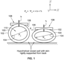
- FIG. 1 a schematic illustration shows how a change of blood pressure changes the curvature of a vessel wall 100 and, hence, a skin surface 102.
- Blood pressure has a direct relationship to an artery size and shape (for example, diameter 104), which, in turn, govern the spatial distribution of deformations of and forces on a contact surface 106 of a force or deformation sensor array 108.
- the sensor array 108 is in contact with the skin surface 102, particularly at locations of superficial arteries 110, such as the radial artery at the wrist.
- Dynamics of the blood pressure inside the artery 110 impose spatio-temporal dynamics in a contact surface profile. This basic principal is used to estimate and track blood pressure continuously.
- the sensor array 108 captures the variation of the arterial cross-sections over space and time.
- the blood pressure has a direct relationship with the shape and size of the arterial cross-sections, which in turn governs the contact surface profile (spatial distribution of deformation and forces) in-between the skin surface 102 and the sensor array 108.
- dynamics of the blood pressure inside the vessel impose spatio-temporal dynamics in the contact surface profile that can be measured noninvasively.
- a TBPI is in the form of an exemplary calibrated measurement device 200 that includes a flexible strap 201 containing (1) a strap force sensor (SFS) 202, which is a force sensitive resistor (SFR), (2) a strap control balloon 204 for tightness adjustment of the strap 200, and (3) a blood-flow control balloon 206, also referred to as a mounting force adjustment balloon (MFAB), which is mounted to a rigid support 207.
- a sensor array 208 is interfaced between the blood-flow control balloon 206 and a skin surface 210 to capture spatio-temporal contact conditions.
- the blood-flow control balloon 206 is actuated by an air pump 212, and an air pressure sensor 214 connected to the air pump 212 acts as the sensor for the blood-flow control balloon 206.
- the device 200 includes a controller 217 (with a processor and memory device) configured to receive signals from the strap force sensor 202 and/or the sensor array 208.
- the controller 217 is further optionally configured to process the signals to determine parameters of blood pressure, including estimation and monitoring of blood pressure.
- the controller 217 is integrated with the device 200, e.g., mounted to or placed within the strap 201, or is a separate component from the device 200, e.g., it is an external device (such as a computer) communicatively coupled to the device 200.
- the device 200 includes a display 219 for displaying various parameters associated with monitoring of blood pressure.
- the display 219 is optionally mounted to or integrated with the device 200.
- the device 200 is communicatively coupled to an external device 221, such as a mobile phone of a user.
- the user uses an application on the mobile phone 221 to perform one or more tasks associated with the device 200, including, for example, controlling inflation of the strap control balloon 204 and/or the blood-flow control balloon 206, receiving parameters of the monitored artery, etc.
- the blood-flow control balloon 206 is initialized to a reference air pressure (e.g., 30-40 mmHg). Then, the blood-flow control balloon 206 and the sensor array 208 are placed above a peripheral artery 216 (e.g., the radial artery at the wrist) such that the sensor array 208 is approximately centered over the artery 216 so that it captures the pulsation (variation of spatio-temporal contact condition caused by variation of blood pressure over a cardiac cycle) at a central region of the sensor array 208, while the blood-flow control balloon 206 controls the blood flow in the artery 216 underneath.
- a reference air pressure e.g., 30-40 mmHg
- the strap control balloon 204 is inflated to a level such that the reading from the strap force sensor 202 reaches a certain value. Based on the spatial information obtained from the sensor array 208, the dominant pulsating area is locked. The dominant pulsating area is the one in which temporal dynamics are above certain threshold, when the blood-flow control balloon 206 and the strap force sensor 202 are adjusted to an optimum predetermined value.
- the strap control balloon 204 and the strap force sensor 202 are primarily used for fine tuning the mounting of the calibrated measurement device 200 to ensure optimal contact of the sensor array 208 with the skin surface 210, and for standardization of mounting the calibrated measurement device 200 across subjects. Signals obtained from the strap force sensor 202 and the strap control balloon 204 are used for blood pressure estimation and monitoring.
- One beneficial and unique aspect of the calibrated measurement device 200 is the effective isolation of the primary measurement and manipulation area over the targeted artery 216 from the rest of the area under the strap 201.
- the achieved isolation eliminates the need for a complete cuff around the limb.
- the unique design of the calibrated measurement device 200 enables continuous use of the calibrated measurement device 200 on a limb without compromise to the venous and lymphatic circulation or to flow in other arteries of the limb.
- the sensor array 208 which is positioned between the blood-flow control balloon 206 and the skin surface 210 captures the spatio-temporal contact conditions as the blood-flow control balloon 206 inflates and deflates during blood pressure estimation phase, and subsequently maintains a nominal pressure during blood pressure monitoring phase.
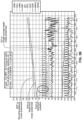
- data shows estimated spatio-temporal signals and blood pressure markers.
- the TBPI (such as the calibrated measurement device 200) is calibrated by estimating a user's DBP, MAP, and SBP. This is achieved by using the TBPI itself.
- the calibration process involves gradual increase and decrease of the pressure in MFAB, which is termed as pressure sweep. In the rising sweep the air pressure in MFAB is gradually increased from a low value ( ⁇ 30 mmHg) at a rate standard for conventional blood pressure measurement (2-4 mmHg/s).
- the rising sweep continues until the air pump saturates ( ⁇ 270 mmHg) or the pulsation amplitude of the measured signals is attenuated below a certain threshold provided it reaches some nominal value (e.g., 180 mmHg). Then, in falling sweep the air pressure is decreased back to the low pressure ( ⁇ 30 mmHg). This enables blood pressure calibration and estimation twice over a complete sweep.
- MFAB pressure mean pressure (averaged over cardiac cycle) in MFAB, termed "MFAB pressure,” is considered as primary measurement for blood pressure estimation.
- the different features of the spatio-temporal signal from the underlying sensor array are considered as markers for SBP or MAP or DBP estimation.
- the MFAB pressure at which the markers reach certain thresholds are mapped to either SBP or MAP or DBP.
- the markers used in the blood pressure estimation method are classified in two groups.
- the markers in Group-1 are the spatial match/mismatch of pulsation over a cardiac cycle within the contact region between the skin and the sensor array.
- the markers in Group-2 are the amplitude of the pulsation averaged over space. Markers from both Group-1 and Group-2 are estimated simultaneously over any half-sweep. The blood pressure estimation based on multiple markers is combined to improve accuracy and robustness of the final blood pressure estimation.
- the spatial information from the locked pulsating area is split into proximal and distal regions in relation to heart or direction of blood flow in the vessel (e.g., Y axis of FIG. 1 ).
- the average height of the pulsation is calculated for proximal and the distal halves individually.
- These two temporal signals effectively represent the proximal and distal pressure waves.
- their similarity and dissimilarity between proximal and distal regions are considered as the blood pressure markers. Estimating these blood pressure markers is a beneficial and unique application of the TBPI.
- the Group-1 markers are basically dependent on the spatial information of the pressure wave.
- the dot-product of the proximal and distal pressure waves is calculated over the duration of the most recent cardiac cycle and is estimated in real-time.
- the marker is termed as a selected dot-product (SDP). While SDP is a quantitative measure of similarity, a selected cross-product (SCP) is one measure of dissimilarity that is graphically represented as the area under a Lissajous plot of normalized proximal vs. distal pressure wave.
- SID instantaneous difference
- the first markers in this group provide the deformation or force information obtained from the sensor array averaged over space, termed as image average height (IAH) and passed through a high pass filter and amplitude estimator. The amplitude estimation is performed over the duration of the most recent cardiac cycle.
- the Group-2 markers do not fundamentally depend on the spatial information of the pressure wave. However, because these markers are derived from the dominant pulsating area, the markers use the spatial information as obtained from the sensor array.
- the MFAB pressure at which Group-2 markers reach the maximum is mapped to MAP.
- the air pressure range through which the markers remain above certain threshold(s) indicates the intra-arterial pulse pressure (PP) that is the difference between SBP and DBP.
- the MFAB pressure at which the markers cross a certain "first-falling" threshold is mapped to SBP. After reaching the maximum, the markers again cross another "second-falling" threshold and the corresponding MFAB pressure is mapped to DBP.
- the Group-2 markers are also directly obtained from the MFAB pressure signal passed through a high-pass filter and an amplitude estimator. Over a half sweep the mean or low-pass filtered MFAB pressure at which any of these markers reaches maximum is mapped to MAP. The range of mean MFAB pressure through which the markers remain above certain threshold(s) indicates the intra-arterial PP. Thus, focusing on the falling half of the sweep, the mean MFAB pressure at which a marker crosses a certain first-falling threshold is mapped to SBP. After reaching the maximum, the marker again crosses the same or a different second-falling threshold and the mean MFAB pressure corresponding to that is mapped to DBP.
- the markers of Group-2 are derived from the high-pass filtered MFAB pressure and are similar to the markers of a standard oscillometric method except that in TBPI the MFAB pressure signal is obtained from an isolated patch of skin region connected by soft tissues to the artery instead of the skin region covering the entire circumference of a limb, as in the case of traditional blood pressure cuffs.
- the blood pressure is continuously tracked using the TBPI.
- the pressure in the MFAB is increased back to a level so that the pulsation in the captured signals reaches certain amplitude or signal to noise ratio (SNR).
- SNR signal to noise ratio
- IAH is calibrated over two points: a maximum is mapped to SBP and a minimum to DBP as obtained from a calibration method. This two-point calibration is sufficient to identify the small variation of blood pressure over the operating point. If the operating point shifts above a threshold, which may be caused by a large change of the blood pressure or based on an artifact invalidating the linear calibration to a great extent, a recalibration is performed.
- the pressure signal from MFAB is also calibrated and used for continuous monitoring.
- IAH shows lower sensitivity to motion and material relaxation related artifacts.
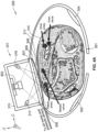
- a TBPI is in the form of another exemplary calibrated measurement device 300 in which a sensor array 308 is implemented based on deformation sensing with the help of a photometric stereo effect captured by a camera 303.
- the device 300 contains a flexible strap 301, a strap force sensor 302, a strap control balloon 304, and a blood-flow control balloon 306 supported on a transparent rigid support (such as glass) 307.
- a sensor surface 309 is in contact with a skin surface 310, which is coated with pigment, such as bronze dust, that creates a specular reflective layer 311.
- a transparent and soft silicone elastomer (such as gel) is used as the substrate of the reflective layer 311.
- the gel is supported by the glass support 307 such that the reflective surface of the gel remains in close contact to the skin surface 310.
- the camera 303 is mounted perpendicularly on a frame 315 above the glass support 307.
- the primary measurement in this embodiment is the ZX plane curvature of the contact surface 309 and its variation along they Y-axis, i.e., the longitudinal axis of the blood vessel.
- two arrays of highly directional (-15° beam angle) red and green LEDs 317, 319 are placed along the Y-axis.
- the full camera assembly 321 is optically isolated from ambient light. To avoid the camera 303 capturing the direct reflection of the LEDs 317, 319 from the optical interfaces, the LEDs 317, 319 are placed at a low grazing angle.
- a bench-top version 321 of the calibrated measurement device 300 includes 5 or 6 degree of freedom positioners and an adjustable back support 323 together with a ball-socket joint 325.
- the back-support can support a phantom artery or an animal body part or the human wrist.
- the system has at least a mounting force or pressure (MPF) sensor.
- MPF sensing is done with 6-DoF force sensors or with a force sensitive resistor (FSR).
- FSR force sensitive resistor
- the bench-top version 321 further has a version of TBPI, such as the wearable part 327 described below.
- a wearable version 329 of the calibrated measurement device 300 includes a machine vision camera 331, a lead from force sensing resistor FSR 333, and a glass support 335 wrapped in a transparent strap 336.
- a red LED array 337 is inside the transparent strap 336, and an air pump actuated balloon 339 is attached with a primary MPF sensor.
- Independently adjustable red and green LED regulators 341, 343 are further included in the wearable version 329, as well as a USB 3.0 camera 345 for capturing video images.
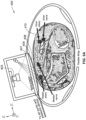
- a TBPI is in the form of another exemplary calibrated measurement device 400 in which a sensor array 408 is implemented based on structured light with projected light patterns (such as stripes) or optical markers (such as dots or holes) on the inside or outside surface of 408 that is in contact with the skin surface 410 (or, for example, directly on the skin surface 210).
- the movement of these patterns or markers is captured by a camera 403 that may also be placed at a different position and orientation than what is shown in Fig. 5A .
- the sensor array 408 is implemented based on triangulation of optical patterns or markers on a sensing surface 409 (which is an outer surface of the sensor array 408) or the inside surface of the sensor array 408 (or, for example, directly on the skin surface 210). Based on the data from the camera placed appropriately to capture the images of the patterns or markers on a surface in contact with a skin surface 410 or directly on the skin surface 210, the deformation along the Z-axis is captured from the motion of the markers in the XY plane. Thus, the change of curvature in the ZX plane is sensed through the marker movement in the X-axis, as viewed by the camera 403.
- the calibrated measurement device 400 optionally includes one or more components of any of the other exemplary calibrated measurement device described in this disclosure.
- a bench-top version 421 and a wearable version 429 include a projector 440 and a machine vision camera 441.
- a TBPI is in the form of another exemplary calibrated measurement device 500 in which a sensor array 508 is implemented based on discrete force or displacement sensors 550.
- the sensor array 508 is implemented by assembling discrete force or deformation sensors 550 such as resistive, piezoelectric, or capacitive elements 550.
- the calibrated measurement device 500 is similar, but not necessarily identical, to one or more of the other exemplary calibrated measurement devices described.
- the calibrated measurement device 500 includes a flexible strap 501, a strap force sensor 502, a strap control balloon 504, and a blood-flow control balloon 506 supported on a rigid support 507.
- the sensors 550 are attached to the undersurface of the blood-flow control balloon 506.
- the calibrated measurement device 500 is mounted on a wrist 551.
- the discrete sensors described above can be made of a thin film of piezoelectric polymer, such as polyvinylidene fluoride (PVDF), with both sides coated with silver ink acting as electrodes.
- the electrodes are further coated with an insulating material to avoid electrical conduction between the skin and the sensor.
- the thickness of the sensing element is kept thin enough to achieve good mechanical coupling with the skin.
- only two sensing elements are used, one for the detection of proximal pulsation and the other for distal. The number of elements can be increased to extract more complex markers if necessary.
- the signals from the sensor are passed through the signal conditioner and fed to the TBPI processor. These two proximal and distal signals from piezo film sensors can be equivalently treated as IAH described above for the purpose of BP estimation.
- an experimental setup 600 shows animal experiment for a rabbit model ( FIG. 7A ) and a pig model ( FIGs. 7B and 7C ).
- the setup 600 includes an anesthesia apparatus 660, a back support 662, a catheter 664 to access artery, and a drug and saline drip 666.
- the setup 600 further includes an optical fiber 668 connected to a tip of a fiberoptic pressure sensor (e.g., from FISO), a height adjustment control 670 of a 6-DoF positioner, a version of TBPI 672, a position lock 674, a 6-DoF force sensor 676 (e.g., from ATI).
- the setup 600 also includes the anesthetized pig 678, the anesthetized rabbit 680, TPS 682 from ICU Medical, and a 6 th DoF (Zrot) 684 added to the positioner for animal experiment.
- a sensor array measures movement of an arterial wall directly.
- ultrasonic sensing elements of the sensor array directly measure a curvature of a targeted blood vessel, instead of its impression on the skin surface.
- This embodiment optionally includes one or more components of any of the disclosed calibrated measurement devices, with the modification of having at least one ultrasonic sensing element on the sensor array.
- a sensor is coupled to a mechanical stage and is used for varying a mounting force (e.g., in a bench top device).
- a mounting force e.g., in a bench top device.
- the mounting force is adjusted with the help of a mechanical stage that is coupled with force and/or displacement sensors.
- the spatio-mechanical state of the skin in the BPI is measured by a sensor array, such as the sensor arrays disclosed above.
- the sensor array includes, for example, an optical sensor (e.g., photometric stereo or structured light) as described above.
- ultrasound imaging and/or tactile sensor arrays are used instead of or in addition to the optical sensor.
- the sensor array is positioned above an artery, with the radial artery being one example of the artery.
- Varying skin pressure is applied with an external device coupled with a force sensor.
- the sensor arrays described above are some implementations of the force sensor, while the externa device includes the blood-flow control balloons.
- the spatio-temporal variation in the contact force and/or geometry is recorded as the skin pressure is varied.
- the characteristic spatio-temporal signals corresponding to systolic and diastolic pressure are determined. Examples of the characteristic spatio-temporal signals include contrasting between upstream and downstream as described above or maximal pulsation.
- the determined values of the characteristic spatio-temporal signals are used to calibrate the measurement device, and the calibration is used to estimate blood pressure continuously by analyzing the continuous spatio-temporal signal. For improved continous monitoring, an optical sensor for an oscillometric method provides beneficial results.
- the disclosed device and method for monitoring intra-arterial blood pressure eliminates the need for an intrusive limb-encircling pressure cuff and high pressures needed to occlude the target artery, while simultaneously occluding lymphatics, veins and other arteries and, thus, cutting off blood circulation in the limb (e.g., arm).
- the limb e.g., arm
- the radial artery may be occluded by the blood-flow control balloon, the blood circulation due to ulnar artery, veins, and lymphatics remains intact.
- the disclosed device and method for monitoring intra-arterial blood pressure provides an instantaneous blood pressure vs. time tracing continuously based on direct observation of blood pressure on skin geometry.
- the continuous tracing is obtained without relying on models derived from indirect empirical correlations or trademarked proprietary algorithms that cannot be independently validated or critiqued scientifically.
- the disclosed device and method for monitoring intra-arterial blood pressure facilitates direct derivation of SBP, MAP, and/or DBP from the continuous blood pressure vs. time trace for each heartbeat.
- a further benefit is that the disclosed device and method measure blood pressure over multiple heart beats and respiratory cycles to provide more accurate measure of blood pressure and valuable information on beat-to-beat blood pressure variability.
- the disclosed device and method provides additional data of clinical interest such as heart rate, heart rhythm, respiratory rate, reparatory pattern and apnea.
- the device for monitoring intra-arterial blood pressure is a wearable device that is integrated with existing mobile phones. Data is transferred between the device and a mobile phone instantly over low-bandwidth cellular connection for online/remote management of hypertension.
- the disclosed device and method for monitoring intra-arterial blood pressure provides continuous ambulatory blood pressure monitoring that identifies abnormal blood pressure patterns, including white coat hypertension, masked hypertension, non-dipping or increase in blood pressure during sleep and the morning blood pressure surge.
- the monitoring further provides an accurate assessment of treatment effectiveness and improved tailoring of therapy, identifies lifestyle motivations needed to lower patients' blood pressure (e.g., reducing smoking and stress), and provides graphical feedback to review with patients and which aids in patient compliance with treatment.
- the disclosed device and method provides a 24-hour reliable beat-to-beat monitoring of blood pressure for use in acute hospital settings as an alternative to invasive (using intra-arterial catheters) monitoring.
- other benefits of include affordability, based on the BPI being optionally built from off-the-shelf manufactured components, and lack of requirement for an additional power source, based on the BPI being optionally battery-driven.
- a device for such exemplary purposes includes the flexible strap, the strap force sensor, the strap control balloon, and/or the blood-flow control balloon.
- optical sensors or other skin surface sensing elements as disclosed above are used to augment and improve the performance of calibrated measurement devices.
- specific combinations of the flexible strap, sensing means optical, ultrasound, capacitance-based tactile array, etc.), calibration method, and blood pressure monitoring are optimally implemented depending on the artery location (radial, brachial, etc.), purpose (monitoring for well-being, screening, emergency care, etc.), and setting (hospital, home, wearable, etc.).
Landscapes
- Health & Medical Sciences (AREA)
- Life Sciences & Earth Sciences (AREA)
- Physics & Mathematics (AREA)
- Surgery (AREA)
- General Health & Medical Sciences (AREA)
- Veterinary Medicine (AREA)
- Public Health (AREA)
- Biophysics (AREA)
- Pathology (AREA)
- Engineering & Computer Science (AREA)
- Biomedical Technology (AREA)
- Heart & Thoracic Surgery (AREA)
- Medical Informatics (AREA)
- Molecular Biology (AREA)
- Animal Behavior & Ethology (AREA)
- Cardiology (AREA)
- Vascular Medicine (AREA)
- Physiology (AREA)
- Ophthalmology & Optometry (AREA)
- Nuclear Medicine, Radiotherapy & Molecular Imaging (AREA)
- Radiology & Medical Imaging (AREA)
- Dentistry (AREA)
- Oral & Maxillofacial Surgery (AREA)
- Hematology (AREA)
- General Physics & Mathematics (AREA)
- Measuring Pulse, Heart Rate, Blood Pressure Or Blood Flow (AREA)
- Ultra Sonic Daignosis Equipment (AREA)
- Measuring And Recording Apparatus For Diagnosis (AREA)
Abstract
Description
- This invention was made with government support under grant no. 1 U01 EB018823001A1 awarded by the National Institutes of Health (NIH). The government has certain rights in the invention.
- This application claims priority to
U.S. Provisional Patent Application No. 62/684,726, filed June 13, 2018 U.S. Provisional Patent Application No. 62/652,180, filed April 3, 2018 - The present invention relates generally to a non-invasive estimation and continuous monitoring of arterial blood pressure, and, more specifically, to the use of skin surface displacements and forces for directly measuring and monitoring the arterial blood pressure.
- According to one aspect of the present disclosure, a method is directed to continuously, non-invasively, and directly measuring blood pressure, and includes providing a calibrated measurement device having a blood-flow control balloon and a sensor array. The method further includes placing the sensor array in a non-invasive manner over the surface of a patch of skin connected to an artery by adjoining soft tissues and inflating the blood-flow control balloon with a controlled amount of pressure. In response to the inflating or deflating of the blood-flow control balloon, changes occur in the artery size and shape, and in the forces the artery applies on the surrounding tissue. Consequently, a change occurs in the contact between the skin and the sensor array that is detected, via the sensor array, during a heartbeat cycle. The changes in artery geometry and artery forces correspond to spatio-temporal signals from the artery. The spatio-temporal signals are measured and processed, via a processor, to determine blood-pressure parameters.
- According to another aspect of the present disclosure, a calibrated measurement device is directed to continuously, non-invasively, and directly measuring blood pressure. The calibrated measurement device includes a strap, a blood-flow control balloon coupled to the strap, the blood-flow control balloon having an inflated state in which a controlled amount of pressure isolates a spatio-temporal signal from an artery without compromising venous and lymphatic circulation or flow in other arteries of a limb. The calibrated measurement device further includes a sensor array on the surface of the blood-flow control balloon to non-invasively and directly measure and monitor blood pressure. The sensor array detects changes in the artery geometry and forces during a heartbeat cycle and when the blood-flow control balloon is in the inflated state. The changes correspond to spatio-temporal signals from the artery.
- Optionally, in the above aspect of the present disclosure, the sensor array is implemented based on a gel membrane with a painted surface, with a structured light or with optical markers mounted on an outside surface of the blood-flow control balloon. However, in another exemplary aspect of the present disclosure, the sensor array is implemented based on a painted surface, with optical markers or a structured light pattern on the material of the blood-flow control balloon on its inside surface or on the outside surface. Further, in another exemplary aspect of the present disclosure, the sensor array is implemented based on optical markers or a structured light pattern projected directly on the skin surface.
- In accordance with another exemplary aspect of the present disclosure, a sensor array is implemented based on deformation sensing by photometric stereo with the help of gel membrane with a painted surface and colored lights. Optionally, another exemplary aspect is directed to a sensor array that is implemented based on discrete force or displacement sensors. Specifically, the sensor array is implemented by assembling discrete force or deformation sensors such as resistive, piezoelectric, or capacitive elements.
- Optionally, in one or more of the above-described aspects of the present disclosure, a gel membrane is mounted on an outside surface of the blood-flow control balloon. However, in another exemplary aspect of the present disclosure, the gel membrane is replaced by a painted surface or a structured light pattern on the material of the blood-flow control balloon on its inside surface or on the outside surface.
- According to yet another alternative exemplary embodiment, a sensor is coupled to a mechanical stage and is used for varying a mounting force (e.g., in a bench top device). In this embodiment, instead of using a blood-flow control balloon, the mounting force is adjusted with the help of a mechanical stage that is coupled with force and/or displacement sensors.
- To additionally modulate the contact conditions, the blood-flow control balloon is added together with (1) a pressure sensor that provides a direct measure of the spatial average of forces on a patch of contact with the object (with or without the gel membrane), (2) an air pump that is used to actively change the contact conditions (e.g., geometrical and force changes), and (3) a controller that provides a degree of control over contact conditions and their changes over time, particularly with feedback from the pressure sensor. Gels or other fluids other than air filling the blood-flow control balloon are optionally considered based on respective measurement tasks.
- According to yet another aspect of the present disclosure, a method is directed to continuously, non-invasively, and directly measuring blood pressure in an artery. The method includes providing a calibrated measurement device having strap for mounting on a limb, the strap having mounted on its internal surface a blood-flow control balloon, a strap control balloon, and a strap force sensor. The blood-flow control balloon has mounted on its internal surface a sensor array. The method further includes placing the strap over a limb such that the sensor array is in contact with a skin surface over an artery in a non-invasive manner, and inflating the blood-flow control balloon with a controlled amount of pressure without compromising venous and lymphatic circulation or flow in other arteries of the limb. In response to inflating the blood-flow control balloon, the method further includes detecting, via the sensor array, changes in the artery geometry and forces during a heartbeat cycle, the changes corresponding to spatio-temporal signals from the artery. The strap control balloon is inflated with a controlled amount of pressure to apply a force between the strap and the skin surface. Based on detection by the sensor array and the strap force sensor, fine-tuning pressure is applied by at least one of the blood-flow control balloon or the strap control balloon to enhance detection of the spatio-temporal signals from the artery. The spatio-temporal signals are measured and processed, via a processor, to determine blood-pressure parameters.
- Additional aspects of the disclosure will be apparent to those of ordinary skill in the art in view of the detailed description of various embodiments, which is made with reference to the drawings, a brief description of which is provided below.
-
-
FIG. 1 is a schematic illustration showing changes in curvature of a vessel wall. -
FIG. 2 is a cross-sectional illustration showing components of a calibrated measurement device, according to one embodiment of the present disclosure. -
FIG. 3A shows a first portion of data related to spatio-temporal signals and blood pressure markers, the data including raw spatial information, processed spatial information, and a height image. -
FIG. 3B shows a second portion of data related to the spatio-temporal signals and blood pressure markers ofFIG. 3A , the data including plots for various signals. -
FIG. 3C shows a third portion of data related to the spatio-temporal signals and blood pressure markers ofFIG. 3A , the data including plots of group markers. -
FIG. 3D shows a fourth portion of data related to the spatio-temporal signals and blood pressure markers ofFIG. 3A , the data including values for various signals. -
FIG. 4A is a cross-sectional illustration showing components of a calibrated measurement device including a camera, according to another embodiment of the present disclosure. -
FIG 4B shows a camera setup of the calibrated measurement device ofFIG. 4A with light-emitting diode (LED) regulators. -
FIG. 4C is a perspective view illustrating a bench-top version of the calibrated measurement device ofFIG. 4A . -
FIG. 4D is a perspective view illustrating a wearable version of the calibrated measurement device ofFIG. 4A . -
FIG. 5A is a cross-sectional illustration showing components of a calibrated measurement device including optical markers, according to another embodiment of the present disclosure. -
FIG. 5B is a perspective view illustrating a bench-top version of the calibrated measurement device ofFIG. 5A . -
FIG. 5C is a perspective view illustrating a wearable version of the calibrated measurement device ofFIG. 5A . -
FIG. 6A is a cross-sectional illustration showing components of a calibrated measurement device including piezoelectric sensors, according to another embodiment of the present disclosure. -
FIG. 6B is a perspective view illustrating the piezoelectric sensors of the calibrated measurement device ofFIG. 5A attached to a balloon undersurface. -
FIG. 6C is a perspective view illustrating the calibrated measurement device ofFIG. 5A mounted on a wrist. -
FIG. 7A is perspective illustration showing an animal experiment with a rabbit model. -
FIG. 7B is an enlarged perspective illustration showing an experimental setup for a pig model. -
FIG. 7C is a perspective illustration showing an overall view of the experimental setup ofFIG. 7B . - While the invention is susceptible to various modifications and alternative forms, specific embodiments have been shown by way of example in the drawings and will be described in detail herein. It should be understood, however, that the invention is not intended to be limited to the particular forms disclosed. Rather, the invention is to cover all modifications, equivalents, and alternatives falling within the spirit and scope of the invention as defined by the appended claims.
- The present disclosure is generally directed to a novel approach, several methods, and associated instrument configurations to estimate and continuously monitor intra-arterial blood pressure (BP) over time. Based on the disclosed methodology, direct measurements are made for systolic blood pressure (SBP), mean arterial blood pressure (MAP) and diastolic blood pressure (DBP). A Tactile Blood Pressure Imager (TBPI), also referred more generally to as a Blood Pressure Imager (BPI), is used to measure beat-to-beat variability in SBP, MAP, and DBP, including respiratory variance in blood pressure, over time. Based on this, the TBPI is able to identify abnormal blood pressure patterns, such as, white coat hypertension, masked hypertension, and non-dipping and other abnormal patterns during sleep and morning blood pressure surge. The TBPI facilitates accurate assessment of treatment effectiveness and better tailoring of therapy, and further identifies lifestyle motivations, such as for reducing smoking and stress, needed to lower patients' blood pressure. The TBPI continuously tracks heart rate (HR) and, thus, HR variability, and any abnormal heart rhythms over time. The BPI also continuously tracks respiration rate (RR) and abnormal respiratory patterns. The TBPI further tracks beat-to-beat blood pressure and is a non-invasive alternative to invasive intra-arterial blood pressure monitoring.
- Referring to
FIG. 1 , a schematic illustration shows how a change of blood pressure changes the curvature of avessel wall 100 and, hence, askin surface 102. Blood pressure has a direct relationship to an artery size and shape (for example, diameter 104), which, in turn, govern the spatial distribution of deformations of and forces on acontact surface 106 of a force ordeformation sensor array 108. Thesensor array 108 is in contact with theskin surface 102, particularly at locations ofsuperficial arteries 110, such as the radial artery at the wrist. Dynamics of the blood pressure inside theartery 110 impose spatio-temporal dynamics in a contact surface profile. This basic principal is used to estimate and track blood pressure continuously. - If the
sensor array 108 is mounted and pressed on top of theskin surface 102 over asuperficial blood vessel 110, thesensor array 108 captures the variation of the arterial cross-sections over space and time. The blood pressure has a direct relationship with the shape and size of the arterial cross-sections, which in turn governs the contact surface profile (spatial distribution of deformation and forces) in-between theskin surface 102 and thesensor array 108. Thus, dynamics of the blood pressure inside the vessel impose spatio-temporal dynamics in the contact surface profile that can be measured noninvasively. - Referring to
FIG. 2 , a TBPI is in the form of an exemplary calibratedmeasurement device 200 that includes aflexible strap 201 containing (1) a strap force sensor (SFS) 202, which is a force sensitive resistor (SFR), (2) astrap control balloon 204 for tightness adjustment of thestrap 200, and (3) a blood-flow control balloon 206, also referred to as a mounting force adjustment balloon (MFAB), which is mounted to arigid support 207. Asensor array 208 is interfaced between the blood-flow control balloon 206 and askin surface 210 to capture spatio-temporal contact conditions. The blood-flow control balloon 206 is actuated by anair pump 212, and anair pressure sensor 214 connected to theair pump 212 acts as the sensor for the blood-flow control balloon 206. - Optionally, the
device 200 includes a controller 217 (with a processor and memory device) configured to receive signals from thestrap force sensor 202 and/or thesensor array 208. Thecontroller 217 is further optionally configured to process the signals to determine parameters of blood pressure, including estimation and monitoring of blood pressure. Thecontroller 217 is integrated with thedevice 200, e.g., mounted to or placed within thestrap 201, or is a separate component from thedevice 200, e.g., it is an external device (such as a computer) communicatively coupled to thedevice 200. - According to another optional configuration, the
device 200 includes adisplay 219 for displaying various parameters associated with monitoring of blood pressure. Thedisplay 219 is optionally mounted to or integrated with thedevice 200. - According to another optional configuration, the
device 200 is communicatively coupled to anexternal device 221, such as a mobile phone of a user. The user uses an application on themobile phone 221 to perform one or more tasks associated with thedevice 200, including, for example, controlling inflation of thestrap control balloon 204 and/or the blood-flow control balloon 206, receiving parameters of the monitored artery, etc. - The blood-
flow control balloon 206 is initialized to a reference air pressure (e.g., 30-40 mmHg). Then, the blood-flow control balloon 206 and thesensor array 208 are placed above a peripheral artery 216 (e.g., the radial artery at the wrist) such that thesensor array 208 is approximately centered over theartery 216 so that it captures the pulsation (variation of spatio-temporal contact condition caused by variation of blood pressure over a cardiac cycle) at a central region of thesensor array 208, while the blood-flow control balloon 206 controls the blood flow in theartery 216 underneath. - Subsequently, the
strap control balloon 204 is inflated to a level such that the reading from thestrap force sensor 202 reaches a certain value. Based on the spatial information obtained from thesensor array 208, the dominant pulsating area is locked. The dominant pulsating area is the one in which temporal dynamics are above certain threshold, when the blood-flow control balloon 206 and thestrap force sensor 202 are adjusted to an optimum predetermined value. Thestrap control balloon 204 and thestrap force sensor 202 are primarily used for fine tuning the mounting of the calibratedmeasurement device 200 to ensure optimal contact of thesensor array 208 with theskin surface 210, and for standardization of mounting the calibratedmeasurement device 200 across subjects. Signals obtained from thestrap force sensor 202 and thestrap control balloon 204 are used for blood pressure estimation and monitoring. - One beneficial and unique aspect of the calibrated
measurement device 200 is the effective isolation of the primary measurement and manipulation area over the targetedartery 216 from the rest of the area under thestrap 201. The achieved isolation eliminates the need for a complete cuff around the limb. The unique design of the calibratedmeasurement device 200 enables continuous use of the calibratedmeasurement device 200 on a limb without compromise to the venous and lymphatic circulation or to flow in other arteries of the limb. Thesensor array 208, which is positioned between the blood-flow control balloon 206 and theskin surface 210 captures the spatio-temporal contact conditions as the blood-flow control balloon 206 inflates and deflates during blood pressure estimation phase, and subsequently maintains a nominal pressure during blood pressure monitoring phase. - Referring to
FIGs. 3A-3D , data shows estimated spatio-temporal signals and blood pressure markers. After TBPI mounting is standardized, the TBPI (such as the calibrated measurement device 200) is calibrated by estimating a user's DBP, MAP, and SBP. This is achieved by using the TBPI itself. The calibration process involves gradual increase and decrease of the pressure in MFAB, which is termed as pressure sweep. In the rising sweep the air pressure in MFAB is gradually increased from a low value (~30 mmHg) at a rate standard for conventional blood pressure measurement (2-4 mmHg/s). The rising sweep continues until the air pump saturates (∼270 mmHg) or the pulsation amplitude of the measured signals is attenuated below a certain threshold provided it reaches some nominal value (e.g., 180 mmHg). Then, in falling sweep the air pressure is decreased back to the low pressure (~30 mmHg). This enables blood pressure calibration and estimation twice over a complete sweep. - To shorten the calibration time, either the rising or falling half of the sweep can be rapid, while the calibration is performed on the slower half. For estimation of blood pressure from the sweep, the mean pressure (averaged over cardiac cycle) in MFAB, termed "MFAB pressure," is considered as primary measurement for blood pressure estimation. The different features of the spatio-temporal signal from the underlying sensor array are considered as markers for SBP or MAP or DBP estimation. The MFAB pressure at which the markers reach certain thresholds are mapped to either SBP or MAP or DBP. The markers used in the blood pressure estimation method are classified in two groups. The markers in Group-1 are the spatial match/mismatch of pulsation over a cardiac cycle within the contact region between the skin and the sensor array. The markers in Group-2 are the amplitude of the pulsation averaged over space. Markers from both Group-1 and Group-2 are estimated simultaneously over any half-sweep. The blood pressure estimation based on multiple markers is combined to improve accuracy and robustness of the final blood pressure estimation.
- Referring to Group-1 markers, the spatial information from the locked pulsating area is split into proximal and distal regions in relation to heart or direction of blood flow in the vessel (e.g., Y axis of
FIG. 1 ). After segmentation, the average height of the pulsation is calculated for proximal and the distal halves individually. These two temporal signals effectively represent the proximal and distal pressure waves. After high-pass filtering and normalizing of their heights over the past cardiac cycle, their similarity and dissimilarity between proximal and distal regions are considered as the blood pressure markers. Estimating these blood pressure markers is a beneficial and unique application of the TBPI. The Group-1 markers are basically dependent on the spatial information of the pressure wave. To quantitatively measure the similarity between the proximal and distal pressure waves, the dot-product of the proximal and distal pressure waves is calculated over the duration of the most recent cardiac cycle and is estimated in real-time. The marker is termed as a selected dot-product (SDP). While SDP is a quantitative measure of similarity, a selected cross-product (SCP) is one measure of dissimilarity that is graphically represented as the area under a Lissajous plot of normalized proximal vs. distal pressure wave. - Another approach of measuring dissimilarity quantitatively is to compute a selected instantaneous difference (SID) signal of the normalized proximal and distal pressure waves and, then, to consider the height of the SID over a cardiac cycle as a blood pressure marker. SID is also passed through different band-pass filters to capture the higher harmonics originated due to turbulent blood flow in the artery when it starts collapsing. These signals are termed as SF1, SF2, and so on, and corresponding blood pressure markers are calculated similar to SID (see
FIG. 2 ). - Referring to Group-2 markers, the first markers in this group provide the deformation or force information obtained from the sensor array averaged over space, termed as image average height (IAH) and passed through a high pass filter and amplitude estimator. The amplitude estimation is performed over the duration of the most recent cardiac cycle. The Group-2 markers do not fundamentally depend on the spatial information of the pressure wave. However, because these markers are derived from the dominant pulsating area, the markers use the spatial information as obtained from the sensor array. The MFAB pressure at which Group-2 markers reach the maximum is mapped to MAP. The air pressure range through which the markers remain above certain threshold(s) indicates the intra-arterial pulse pressure (PP) that is the difference between SBP and DBP. Thus, focusing on the falling half of the sweep, the MFAB pressure at which the markers cross a certain "first-falling" threshold is mapped to SBP. After reaching the maximum, the markers again cross another "second-falling" threshold and the corresponding MFAB pressure is mapped to DBP.
- The Group-2 markers are also directly obtained from the MFAB pressure signal passed through a high-pass filter and an amplitude estimator. Over a half sweep the mean or low-pass filtered MFAB pressure at which any of these markers reaches maximum is mapped to MAP. The range of mean MFAB pressure through which the markers remain above certain threshold(s) indicates the intra-arterial PP. Thus, focusing on the falling half of the sweep, the mean MFAB pressure at which a marker crosses a certain first-falling threshold is mapped to SBP. After reaching the maximum, the marker again crosses the same or a different second-falling threshold and the mean MFAB pressure corresponding to that is mapped to DBP. The markers of Group-2 are derived from the high-pass filtered MFAB pressure and are similar to the markers of a standard oscillometric method except that in TBPI the MFAB pressure signal is obtained from an isolated patch of skin region connected by soft tissues to the artery instead of the skin region covering the entire circumference of a limb, as in the case of traditional blood pressure cuffs.
- The blood pressure is continuously tracked using the TBPI. After the calibration of the TBPI is complete, the pressure in the MFAB is increased back to a level so that the pulsation in the captured signals reaches certain amplitude or signal to noise ratio (SNR). Under this condition, IAH is calibrated over two points: a maximum is mapped to SBP and a minimum to DBP as obtained from a calibration method. This two-point calibration is sufficient to identify the small variation of blood pressure over the operating point. If the operating point shifts above a threshold, which may be caused by a large change of the blood pressure or based on an artifact invalidating the linear calibration to a great extent, a recalibration is performed. Instead of IAH, the pressure signal from MFAB is also calibrated and used for continuous monitoring. However, IAH shows lower sensitivity to motion and material relaxation related artifacts.
- Referring to
FIGs. 4A and4B , a TBPI is in the form of another exemplary calibratedmeasurement device 300 in which asensor array 308 is implemented based on deformation sensing with the help of a photometric stereo effect captured by acamera 303. Thedevice 300 contains aflexible strap 301, astrap force sensor 302, astrap control balloon 304, and a blood-flow control balloon 306 supported on a transparent rigid support (such as glass) 307. In this embodiment, asensor surface 309 is in contact with askin surface 310, which is coated with pigment, such as bronze dust, that creates a specularreflective layer 311. A transparent and soft silicone elastomer (such as gel) is used as the substrate of thereflective layer 311. The gel is supported by theglass support 307 such that the reflective surface of the gel remains in close contact to theskin surface 310. Thecamera 303 is mounted perpendicularly on aframe 315 above theglass support 307. - As shown in
FIG. 1 , curvature of theblood vessel 110 along a ZX plane changes with difference between Pin and the ambient pressure Pout. Therefore, the primary measurement in this embodiment is the ZX plane curvature of thecontact surface 309 and its variation along they Y-axis, i.e., the longitudinal axis of the blood vessel. Thus, two arrays of highly directional (-15° beam angle) red andgreen LEDs full camera assembly 321 is optically isolated from ambient light. To avoid thecamera 303 capturing the direct reflection of theLEDs LEDs - Referring to
FIG. 4C , a bench-top version 321 of the calibratedmeasurement device 300 includes 5 or 6 degree of freedom positioners and anadjustable back support 323 together with a ball-socket joint 325. The back-support can support a phantom artery or an animal body part or the human wrist. In all the cases, the system has at least a mounting force or pressure (MPF) sensor. In bench-top experiments MPF sensing is done with 6-DoF force sensors or with a force sensitive resistor (FSR). The bench-top version 321 further has a version of TBPI, such as thewearable part 327 described below. - Referring to
FIG. 4D , awearable version 329 of the calibratedmeasurement device 300 includes amachine vision camera 331, a lead from forcesensing resistor FSR 333, and aglass support 335 wrapped in atransparent strap 336. Ared LED array 337 is inside thetransparent strap 336, and an air pump actuatedballoon 339 is attached with a primary MPF sensor. Independently adjustable red andgreen LED regulators wearable version 329, as well as a USB 3.0camera 345 for capturing video images. - Referring to
FIG. 5A , a TBPI is in the form of another exemplary calibratedmeasurement device 400 in which asensor array 408 is implemented based on structured light with projected light patterns (such as stripes) or optical markers (such as dots or holes) on the inside or outside surface of 408 that is in contact with the skin surface 410 (or, for example, directly on the skin surface 210). The movement of these patterns or markers is captured by acamera 403 that may also be placed at a different position and orientation than what is shown inFig. 5A . In this embodiment, which is similar to but not necessarily identical to the embodiments described above, thesensor array 408 is implemented based on triangulation of optical patterns or markers on a sensing surface 409 (which is an outer surface of the sensor array 408) or the inside surface of the sensor array 408 (or, for example, directly on the skin surface 210). Based on the data from the camera placed appropriately to capture the images of the patterns or markers on a surface in contact with askin surface 410 or directly on theskin surface 210, the deformation along the Z-axis is captured from the motion of the markers in the XY plane. Thus, the change of curvature in the ZX plane is sensed through the marker movement in the X-axis, as viewed by thecamera 403. The calibratedmeasurement device 400 optionally includes one or more components of any of the other exemplary calibrated measurement device described in this disclosure. Referring further toFIGs. 5B and 5C , a bench-top version 421 and awearable version 429 include aprojector 440 and amachine vision camera 441. - Referring to
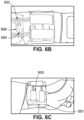
FIG. 6A , a TBPI is in the form of another exemplary calibratedmeasurement device 500 in which asensor array 508 is implemented based on discrete force ordisplacement sensors 550. Specifically, thesensor array 508 is implemented by assembling discrete force ordeformation sensors 550 such as resistive, piezoelectric, orcapacitive elements 550. The calibratedmeasurement device 500 is similar, but not necessarily identical, to one or more of the other exemplary calibrated measurement devices described. For example, the calibratedmeasurement device 500 includes aflexible strap 501, astrap force sensor 502, astrap control balloon 504, and a blood-flow control balloon 506 supported on arigid support 507. Referring further toFIG. 6B , thesensors 550 are attached to the undersurface of the blood-flow control balloon 506. Referring further toFIG. 6C , the calibratedmeasurement device 500 is mounted on awrist 551. - Optionally, the discrete sensors described above can be made of a thin film of piezoelectric polymer, such as polyvinylidene fluoride (PVDF), with both sides coated with silver ink acting as electrodes. The electrodes are further coated with an insulating material to avoid electrical conduction between the skin and the sensor. The thickness of the sensing element is kept thin enough to achieve good mechanical coupling with the skin. In the simplest arrangement, only two sensing elements are used, one for the detection of proximal pulsation and the other for distal. The number of elements can be increased to extract more complex markers if necessary. The signals from the sensor are passed through the signal conditioner and fed to the TBPI processor. These two proximal and distal signals from piezo film sensors can be equivalently treated as IAH described above for the purpose of BP estimation.
- Referring to
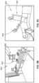
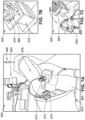
FIGs. 7A-7C , anexperimental setup 600 shows animal experiment for a rabbit model (FIG. 7A ) and a pig model (FIGs. 7B and 7C ). Thesetup 600 includes ananesthesia apparatus 660, a back support 662, acatheter 664 to access artery, and a drug andsaline drip 666. Thesetup 600 further includes anoptical fiber 668 connected to a tip of a fiberoptic pressure sensor (e.g., from FISO), aheight adjustment control 670 of a 6-DoF positioner, a version ofTBPI 672, aposition lock 674, a 6-DoF force sensor 676 (e.g., from ATI). Thesetup 600 also includes the anesthetizedpig 678, the anesthetizedrabbit 680,TPS 682 from ICU Medical, and a 6th DoF (Zrot) 684 added to the positioner for animal experiment. - According to an alternative exemplary embodiment, a sensor array measures movement of an arterial wall directly. For example, in this embodiment ultrasonic sensing elements of the sensor array directly measure a curvature of a targeted blood vessel, instead of its impression on the skin surface. This embodiment optionally includes one or more components of any of the disclosed calibrated measurement devices, with the modification of having at least one ultrasonic sensing element on the sensor array.
- According to another alternative exemplary embodiment a sensor is coupled to a mechanical stage and is used for varying a mounting force (e.g., in a bench top device). In this embodiment, instead of using a blood-flow control balloon, the mounting force is adjusted with the help of a mechanical stage that is coupled with force and/or displacement sensors.
- Thus, in accordance with the disclosed embodiments, the spatio-mechanical state of the skin in the BPI is measured by a sensor array, such as the sensor arrays disclosed above. The sensor array includes, for example, an optical sensor (e.g., photometric stereo or structured light) as described above. In other examples, ultrasound imaging and/or tactile sensor arrays are used instead of or in addition to the optical sensor. The sensor array is positioned above an artery, with the radial artery being one example of the artery.
- Varying skin pressure is applied with an external device coupled with a force sensor. The sensor arrays described above are some exemples of the force sensor, while the externa device includes the blood-flow control balloons. The spatio-temporal variation in the contact force and/or geometry is recorded as the skin pressure is varied. The characteristic spatio-temporal signals corresponding to systolic and diastolic pressure are determined. Examples of the characteristic spatio-temporal signals include contrasting between upstream and downstream as described above or maximal pulsation. The determined values of the characteristic spatio-temporal signals are used to calibrate the measurement device, and the calibration is used to estimate blood pressure continuously by analyzing the continuous spatio-temporal signal. For improved continous monitoring, an optical sensor for an oscillometric method provides beneficial results.
- According to one exemplary benefit of the present disclosure, the disclosed device and method for monitoring intra-arterial blood pressure eliminates the need for an intrusive limb-encircling pressure cuff and high pressures needed to occlude the target artery, while simultaneously occluding lymphatics, veins and other arteries and, thus, cutting off blood circulation in the limb (e.g., arm). For example, during the calibration phase of the disclosed BPI, while the radial artery may be occluded by the blood-flow control balloon, the blood circulation due to ulnar artery, veins, and lymphatics remains intact.
- According to another exemplary benefit of the present disclosure, the disclosed device and method for monitoring intra-arterial blood pressure provides an instantaneous blood pressure vs. time tracing continuously based on direct observation of blood pressure on skin geometry. The continuous tracing is obtained without relying on models derived from indirect empirical correlations or trademarked proprietary algorithms that cannot be independently validated or critiqued scientifically.
- According to yet another exemplary benefit of the present disclosure, the disclosed device and method for monitoring intra-arterial blood pressure facilitates direct derivation of SBP, MAP, and/or DBP from the continuous blood pressure vs. time trace for each heartbeat. A further benefit is that the disclosed device and method measure blood pressure over multiple heart beats and respiratory cycles to provide more accurate measure of blood pressure and valuable information on beat-to-beat blood pressure variability. Yet another benefit is that the disclosed device and method provides additional data of clinical interest such as heart rate, heart rhythm, respiratory rate, reparatory pattern and apnea.
- According to a further benefit of the present disclosure, the device for monitoring intra-arterial blood pressure is a wearable device that is integrated with existing mobile phones. Data is transferred between the device and a mobile phone instantly over low-bandwidth cellular connection for online/remote management of hypertension.
- According to further benefits of the present disclosure, the disclosed device and method for monitoring intra-arterial blood pressure provides continuous ambulatory blood pressure monitoring that identifies abnormal blood pressure patterns, including white coat hypertension, masked hypertension, non-dipping or increase in blood pressure during sleep and the morning blood pressure surge. The monitoring further provides an accurate assessment of treatment effectiveness and improved tailoring of therapy, identifies lifestyle motivations needed to lower patients' blood pressure (e.g., reducing smoking and stress), and provides graphical feedback to review with patients and which aids in patient compliance with treatment.
- According to other benefits of the present disclosure, the disclosed device and method provides a 24-hour reliable beat-to-beat monitoring of blood pressure for use in acute hospital settings as an alternative to invasive (using intra-arterial catheters) monitoring. Additionally, other benefits of include affordability, based on the BPI being optionally built from off-the-shelf manufactured components, and lack of requirement for an additional power source, based on the BPI being optionally battery-driven.
- The disclosed device and method is further useful for other purposes, including, for example, blood draw or catheter insertion. For example, a device for such exemplary purposes includes the flexible strap, the strap force sensor, the strap control balloon, and/or the blood-flow control balloon. Optionally, optical sensors or other skin surface sensing elements as disclosed above are used to augment and improve the performance of calibrated measurement devices. Optionally, yet, specific combinations of the flexible strap, sensing means (optical, ultrasound, capacitance-based tactile array, etc.), calibration method, and blood pressure monitoring are optimally implemented depending on the artery location (radial, brachial, etc.), purpose (monitoring for well-being, screening, emergency care, etc.), and setting (hospital, home, wearable, etc.).
- While the present invention has been described with reference to one or more particular embodiments, those skilled in the art will recognize that many changes may be made thereto without departing from the spirit and scope of the present invention. Each of these embodiments and obvious variations thereof is contemplated as falling within the spirit and scope of the invention. It is also contemplated that additional embodiments according to aspects of the present invention may combine any number of features from any of the embodiments described herein.
Claims (16)
- A method for measuring blood pressure based on skin surface displacements and forces, the method comprising:providing a calibrated measurement device (200, 500) having a sensor array (208, 508) with a sensor surface;placing the sensor surface in direct contact with a skin surface (210, 310) connected to an artery;applying a controlled amount of pressure by a first external device coupled with the sensor array (208, 508) to press the sensor surface against the skin surface (210, 310) with a respective force;in response to the respective force, causing a skin deformation in the skin surface (210, 310) based on size and shape changes of the artery;in response to the skin deformation of the skin surface (210, 310), causing a sensor deformation to the sensor surface;in response to the sensor deformation, detecting a deformation signal of the sensor surface, via the sensor array (208, 508), during a heartbeat cycle, the deformation signal corresponding to spatio-temporal contact conditions of the artery; andmeasuring and processing, via a processor, the deformation signal to determine one or more blood-pressure parameters.
- The method of claim 1, wherein the controlled amount of pressure is applied by a blood-flow control balloon (206, 506) as a first external device,and wherein the controlled amount of pressure is achieved by inflating or deflating the blood-flow control balloon (206, 506),and wherein the blood-flow control balloon (206, 506) has an inflated state in which a controlled amount of pressure isolates a spatio-temporal signal from the artery without compromising venous and lymphatic circulation or flow in other arteries of a limb.
- The method of any one of claims 1 or 2, wherein the sensor array (208, 508) receives said deformation signal from one or more of a tactile sensor, an ultrasound imaging sensor, a resistive sensor, a piezoelectric sensor, and a capacitive sensor.
- The method of any one of claims 1 to 3,wherein the determination of blood pressure parameter includes estimating, by a controller (217) that includes the processor and a memory device,of a user's diastolic blood pressure, mean arterial blood pressure and systolic blood pressure by a pressure sweep, the pressure sweep including a gradual increase and decrease of the controlled amount of pressure in the first external device for the calibration of the calibrated measurement device (200, 500)and whereinin the rising sweep the air pressure is increased from a first predetermined value at a rate standard for blood pressure measurement, the rising sweep continuing until either a saturation pressure is reached, or a pulsation amplitude of the deformation signal is attenuated below a predetermined threshold if the air pressure reaches a second predetermined value,and whereinin the falling sweep the air pressure is decreased back to the low first predetermined value.
- The method of claim 4,wherein in order to shorten the calibration time, one of the rising or falling halves of the pressure sweep is faster than the other half and the calibration is done on the slower half,and wherein, for estimation of blood pressure from the sweep, the mean pressure averaged over a cardiac cycle in mean arterial blood pressure is the primary measurement for estimating the blood pressure.
- The method of any one of claims 1 to 5, further comprising displaying at least one of the one or more blood-pressure parameters on a display (219), the display (219) being communicatively coupled with the controller (217) comprising the processor.
- The method of any one of claims 1 to 6, further comprising controlling the amount of pressure via a second external device (221), the second external device (221) being communicatively coupled with the calibrated measurement device (200, 500).
- The method of any one of claims 1 to 7, further comprising projecting a structured light from the sensor surface, the structured light including projected light patterns, such as stripes.
- The method of any one of claims 1 to 7, further comprising projecting a structured light from the sensor surface, the structured light including optical markers, such as dots or holes.
- The method of any one of claims 8 or 9, further comprising capturing, via a camera (403), movement of the structured light.
- A system for measuring blood pressure based on skin surface displacements and forces, the system comprising a calibrated measurement device (200, 500) and a processor, the processor communicatively coupled to the calibrated measurement device (200, 500), the system configured to apply the method of any of the claims 1 to 10,
whereinthe calibrated measurement device (200, 500) has a sensor array (208, 508) with a sensor surface, the sensor array (208, 508) configured to be in direct contact with a skin surface (210, 310) connected to an artery for non-invasively measuring of blood pressure,the calibrated measurement device (200, 500) has a first external device configured to apply a controlled amount of pressure to press the sensor surface against the skin surface (210, 310) with a respective force,the sensor array (208, 508) is configured to be pressed against the skin surface (210, 310) by said first external device,the sensor surface is configured to undergo sensor deformation in response to a skin deformation of the skin surface (210, 310) based on size and shape of the artery, said skin deformation occurring in response to the respective force,the sensor array (208, 508) is configured to detect in response to the sensor deformation, a deformation signal of the sensor surface, during a heartbeat cycle, the deformation signal corresponding to spatio-temporal contact conditions of the artery,the processor is configured to measure and process the deformation signal to determine a blood-pressure parameter. - The system of claim 11,wherein the first external device is a blood-flow control balloon (206, 506) configured to be coupled with the sensor array (208, 508),wherein the blood-flow control balloon (206, 506) is configured to inflate and deflate in order to achieve the controlled amount of pressure,and wherein the blood-flow control balloon (206, 506) is configured in its inflated state to contain a controlled amount of pressure which isolate a spatio-temporal signal from an artery without compromising venous and lymphatic circulation or flow in other arteries of a limb.
- The system of any one of claims 11 or 12, wherein the sensor array (208, 508) includes one or more of a tactile sensor, an ultrasound imaging sensor, a resistive sensor, a piezoelectric sensor, and a capacitive sensor.
- The system of any one of claims 11 to 13,wherein the system further comprises a controller (217) that includes the processor and a memory device,wherein the controller (217) is configured to estimate blood pressure from a pressure sweep, in order to calibrate the calibrated measurement device (200, 500), said pressure sweep including a gradual increase and decrease of the controlled amount of pressure in the first external device.
- The system of any one of claims 11 to 14, further comprising:- a display (219) communicatively coupled with the controller (217), the display (219) being configured to display at least one of the one or more blood-pressure parameters;
and/or- a second external device (221) communicatively coupled with the balloon, the second external device (221) being configured to control the inflating or deflating the balloon, wherein the second external device (221) is a mobile phone. - The system of any one of claims 11 to 15,
whereinthe sensor array (208, 408, 508) is configured to include an optical sensor emitting structured light,the system further comprises a camera (403) configured for capturing the structured light,
and whereinthe structured light is selected from a group consisting of a light pattern and an optical marker, wherein the light pattern includes one or more stripes, and wherein the optical marker includes one or more dots or holes.
Applications Claiming Priority (4)
| Application Number | Priority Date | Filing Date | Title |
|---|---|---|---|
| US201862652180P | 2018-04-03 | 2018-04-03 | |
| US201862684726P | 2018-06-13 | 2018-06-13 | |
| PCT/US2019/025011 WO2019195120A1 (en) | 2018-04-03 | 2019-03-29 | Tactile blood pressure imager |
| EP19781541.8A EP3773183B1 (en) | 2018-04-03 | 2019-03-29 | Tactile blood pressure imager |
Related Parent Applications (2)
| Application Number | Title | Priority Date | Filing Date |
|---|---|---|---|
| EP19781541.8A Division-Into EP3773183B1 (en) | 2018-04-03 | 2019-03-29 | Tactile blood pressure imager |
| EP19781541.8A Division EP3773183B1 (en) | 2018-04-03 | 2019-03-29 | Tactile blood pressure imager |
Publications (4)
| Publication Number | Publication Date |
|---|---|
| EP4434472A2 true EP4434472A2 (en) | 2024-09-25 |
| EP4434472A3 EP4434472A3 (en) | 2024-12-11 |
| EP4434472C0 EP4434472C0 (en) | 2026-02-25 |
| EP4434472B1 EP4434472B1 (en) | 2026-02-25 |
Family
ID=68101329
Family Applications (3)
| Application Number | Title | Priority Date | Filing Date |
|---|---|---|---|
| EP24193414.0A Active EP4434472B1 (en) | 2018-04-03 | 2019-03-29 | Tactile blood pressure imager |
| EP19781541.8A Active EP3773183B1 (en) | 2018-04-03 | 2019-03-29 | Tactile blood pressure imager |
| EP22213084.1A Active EP4166074B1 (en) | 2018-04-03 | 2019-03-29 | Tactile blood pressure imager |
Family Applications After (2)
| Application Number | Title | Priority Date | Filing Date |
|---|---|---|---|
| EP19781541.8A Active EP3773183B1 (en) | 2018-04-03 | 2019-03-29 | Tactile blood pressure imager |
| EP22213084.1A Active EP4166074B1 (en) | 2018-04-03 | 2019-03-29 | Tactile blood pressure imager |
Country Status (16)
| Country | Link |
|---|---|
| US (3) | US11883140B2 (en) |
| EP (3) | EP4434472B1 (en) |
| JP (2) | JP7422672B2 (en) |
| KR (2) | KR102732232B1 (en) |
| CN (2) | CN118902421A (en) |
| AU (1) | AU2019249818B2 (en) |
| BR (1) | BR112020019909A2 (en) |
| CA (1) | CA3094403A1 (en) |
| ES (3) | ES3061641T3 (en) |
| IL (1) | IL277022B2 (en) |
| MX (1) | MX2020010450A (en) |
| RU (1) | RU2020131254A (en) |
| SA (1) | SA520420190B1 (en) |
| SG (1) | SG11202008316UA (en) |
| WO (1) | WO2019195120A1 (en) |
| ZA (1) | ZA202005840B (en) |
Families Citing this family (14)
| Publication number | Priority date | Publication date | Assignee | Title |
|---|---|---|---|---|
| WO2020206372A1 (en) * | 2019-04-03 | 2020-10-08 | Pb Inc. | Temperature sensor patch wirelessly connected to a smart device |
| CN114786570B (en) * | 2019-10-18 | 2025-02-14 | 3目标知识产权有限公司 | Physiological parameter sensing system and method |
| WO2021237185A1 (en) * | 2020-05-21 | 2021-11-25 | Jeffrey Reynolds | Methods, systems and machine readable programs for cuffless blood pressure measurement |
| EP4192344B1 (en) * | 2020-08-10 | 2026-02-11 | Dynocardia, Inc. | Optomechanical method to measure arterial pulse and assess cardiopulmonary hemodynamics |
| EP3973857A1 (en) * | 2020-09-29 | 2022-03-30 | Koninklijke Philips N.V. | Estimating central blood pressure |
| JP7669667B2 (en) | 2020-11-06 | 2025-04-30 | オムロンヘルスケア株式会社 | Sphygmomanometer |
| KR102857422B1 (en) * | 2021-03-29 | 2025-09-08 | 고쿠리츠다이가쿠호진 도호쿠다이가쿠 | Hardness calculating device, hardness measuring device and hardness calculating method |
| JP2024516573A (en) * | 2021-04-16 | 2024-04-16 | 3 エイム アイピー プロプライエトリー リミテッド | Physiological parameter sensing system and method - Patents.com |
| CN113925477B (en) * | 2021-07-08 | 2023-10-27 | 哈尔滨铂云医疗器械有限公司 | Automatic calibration system of electronic sphygmomanometer |
| SE2151527A1 (en) * | 2021-12-14 | 2023-06-15 | Pusensor Ab | A system and method for assessment of a blood flow |
| US12189846B1 (en) * | 2023-07-04 | 2025-01-07 | Huawei Technologies Co., Ltd. | Wearable device with non-optical sensing nodes |
| WO2025014708A1 (en) | 2023-07-07 | 2025-01-16 | Dynocardia, Inc. | Spatiotemporal-based detection and correction of motion artifact for measurement of arterial pressure waveform |
| EP4714340A1 (en) * | 2024-09-18 | 2026-03-25 | Nederlandse Organisatie voor toegepast- natuurwetenschappelijk onderzoek TNO | Wearable blood pressure monitoring device |
| CN119606333B (en) * | 2025-02-12 | 2025-05-13 | 北京大学第三医院(北京大学第三临床医学院) | A device for non-invasively visualizing blood flow, curvature and diameter of radial artery |
Family Cites Families (36)
| Publication number | Priority date | Publication date | Assignee | Title |
|---|---|---|---|---|
| SU685275A1 (en) | 1978-01-04 | 1979-09-15 | Предприятие П/Я А-3469 | Apparatus for direct measuring of blood pressure |
| US4245648A (en) | 1978-09-20 | 1981-01-20 | Trimmer Gordon A | Method and apparatus for measuring blood pressure and pulse rate |
| CS272057B1 (en) * | 1987-03-27 | 1991-01-15 | Jan Doc Mudr Csc Penaz | Blood pressure automatic non-invasive meter |
| US5533511A (en) * | 1994-01-05 | 1996-07-09 | Vital Insite, Incorporated | Apparatus and method for noninvasive blood pressure measurement |
| JP3243228B2 (en) * | 1998-05-28 | 2002-01-07 | マイクロライフ システムズ エージー | An arterial blood pressure measurement device that is not inserted into the body |
| CN2371945Y (en) * | 1999-04-21 | 2000-04-05 | 陆渭明 | Wrist device for non-wound continuous measuring blood pressure |
| WO2000072750A1 (en) * | 1999-06-01 | 2000-12-07 | Massachusetts Institute Of Technology | Cuffless continuous blood pressure monitor |
| CN100398058C (en) * | 2000-04-21 | 2008-07-02 | 陆渭明 | Non-wound method and device for measuring blood pressure |
| US6533729B1 (en) | 2000-05-10 | 2003-03-18 | Motorola Inc. | Optical noninvasive blood pressure sensor and method |
| US20040044288A1 (en) * | 2002-09-03 | 2004-03-04 | Miguel Gorenberg | Apparatus and method for non-invasive monitoring of cardiac output |
| US7179228B2 (en) * | 2004-04-07 | 2007-02-20 | Triage Wireless, Inc. | Cuffless system for measuring blood pressure |
| JP2006280485A (en) | 2005-03-09 | 2006-10-19 | Motoharu Hasegawa | Blood pressure detecting device, blood pressure detecting method, blood pressure detecting program, and strain sensor for blood pressure detection |
| EP2162059B1 (en) * | 2007-06-12 | 2021-01-13 | Sotera Wireless, Inc. | Vital sign monitor and method for measuring blood pressure using optical, electrical, and pressure waveforms |
| US20090118628A1 (en) * | 2007-11-01 | 2009-05-07 | Triage Wireless, Inc. | System for measuring blood pressure featuring a blood pressure cuff comprising size information |
| JP5045476B2 (en) * | 2008-02-08 | 2012-10-10 | オムロンヘルスケア株式会社 | Detection unit for blood pressure information measuring device and blood pressure information measuring device |
| JP5449336B2 (en) * | 2008-06-19 | 2014-03-19 | マサチューセッツ インスティテュート オブ テクノロジー | Contact sensor using elastic imaging |
| WO2010048528A2 (en) * | 2008-10-23 | 2010-04-29 | Skeletal Dynamics Llc | Non-invasive blood pressure monitoring device and method |
| JP2010131247A (en) * | 2008-12-05 | 2010-06-17 | Omron Healthcare Co Ltd | Blood pressure measuring apparatus |
| AU2011267954B2 (en) * | 2010-06-13 | 2014-07-24 | Angiometrix Corporation | Diagnostic kit and method for measuring balloon dimension in vivo |
| US20130144176A1 (en) * | 2010-06-25 | 2013-06-06 | Drexel University | Non-invasive blood pressure sensor |
| CA2825368A1 (en) * | 2011-02-03 | 2012-08-09 | Yoram Palti | Transthoracic cardio-pulmonary monitor |
| WO2012149209A1 (en) * | 2011-04-26 | 2012-11-01 | The Regents Of The University Of California | Assessing endothelial function using a blood pressure cuff |
| US20130339043A1 (en) * | 2012-06-13 | 2013-12-19 | Advanced Vision Solutions, Inc. | Vision correction prescription and health assessment facility |
| WO2014030174A2 (en) * | 2012-08-24 | 2014-02-27 | Healthcare Technology Innovation Centre | Automated evaluation of arterial stiffness for a non-invasive screening |
| CN103110431B (en) * | 2012-09-12 | 2015-04-01 | 中国科学院深圳先进技术研究院 | Noninvasive continuous blood pressure measurement device |
| US20140276150A1 (en) * | 2013-03-15 | 2014-09-18 | Ying Sun | Apparatus for Acoustic Measurements of Physiological Signals with Automated Interface Controls |
| US9600072B2 (en) | 2013-08-27 | 2017-03-21 | Prakash C R J Naidu | Wearable tactile display |
| US10039455B2 (en) * | 2014-05-19 | 2018-08-07 | Qualcomm Incorporated | Continuous calibration of a blood pressure measurement device |
| CN107106054B (en) * | 2014-09-08 | 2021-11-02 | 苹果公司 | Blood pressure monitoring with a multifunctional wrist-worn device |
| US10076252B2 (en) * | 2015-04-02 | 2018-09-18 | Microsoft Technology Licensing, Llc | Sizable wrist-worn pressure sensing device |
| EP3311736B1 (en) * | 2015-06-19 | 2021-05-26 | Shinano Kenshi Co., Ltd. | Biological information acquiring device |
| KR102434701B1 (en) * | 2015-09-01 | 2022-08-22 | 삼성전자주식회사 | Apparatus and method for acquiring bio- information and apparatus for detecting bio- information |
| WO2017136772A1 (en) * | 2016-02-03 | 2017-08-10 | Angilytics Inc. | Non-invasive and non-occlusive blood pressure monitoring devices and methods |
| US11020058B2 (en) * | 2016-02-12 | 2021-06-01 | Qualcomm Incorporated | Methods and devices for calculating blood pressure based on measurements of arterial blood flow and arterial lumen |
| WO2018025280A2 (en) * | 2016-08-05 | 2018-02-08 | Indian Institute Of Technology Bombay | Hand-based blood pressure measurement system, apparatus and method |
| RU2638712C1 (en) | 2016-11-07 | 2017-12-15 | Федеральное государственное бюджетное учреждение науки Институт радиотехники и электроники им. В.А. Котельникова Российской академии наук | Pneumatic sensor for continuous non-invasive measurement of arterial pressure |
-
2019
- 2019-03-29 EP EP24193414.0A patent/EP4434472B1/en active Active
- 2019-03-29 ES ES19781541T patent/ES3061641T3/en active Active
- 2019-03-29 EP EP19781541.8A patent/EP3773183B1/en active Active
- 2019-03-29 ES ES22213084T patent/ES3023591T3/en active Active
- 2019-03-29 RU RU2020131254A patent/RU2020131254A/en unknown
- 2019-03-29 KR KR1020207030963A patent/KR102732232B1/en active Active
- 2019-03-29 CA CA3094403A patent/CA3094403A1/en active Pending
- 2019-03-29 JP JP2020549066A patent/JP7422672B2/en active Active
- 2019-03-29 AU AU2019249818A patent/AU2019249818B2/en active Active
- 2019-03-29 WO PCT/US2019/025011 patent/WO2019195120A1/en not_active Ceased
- 2019-03-29 IL IL277022A patent/IL277022B2/en unknown
- 2019-03-29 EP EP22213084.1A patent/EP4166074B1/en active Active
- 2019-03-29 US US17/044,321 patent/US11883140B2/en active Active
- 2019-03-29 SG SG11202008316UA patent/SG11202008316UA/en unknown
- 2019-03-29 CN CN202410706940.6A patent/CN118902421A/en active Pending
- 2019-03-29 BR BR112020019909-5A patent/BR112020019909A2/en unknown
- 2019-03-29 ES ES24193414T patent/ES3063414T3/en active Active
- 2019-03-29 CN CN201980023078.6A patent/CN112512414B/en active Active
- 2019-03-29 MX MX2020010450A patent/MX2020010450A/en unknown
- 2019-03-29 KR KR1020247032631A patent/KR102828124B1/en active Active
-
2020
- 2020-09-21 ZA ZA2020/05840A patent/ZA202005840B/en unknown
- 2020-09-22 SA SA520420190A patent/SA520420190B1/en unknown
-
2023
- 2023-12-11 US US18/535,484 patent/US20240099597A1/en active Pending
-
2024
- 2024-01-16 JP JP2024004459A patent/JP7669539B2/en active Active
- 2024-03-13 US US18/604,009 patent/US20240245312A1/en active Pending
Also Published As
Similar Documents
| Publication | Publication Date | Title |
|---|---|---|
| US20240245312A1 (en) | Tactile Blood Pressure Imager | |
| JP6054584B2 (en) | Treatment system having a patient interface for acquiring a patient's life state | |
| US11013421B2 (en) | Blood pressure estimation using finger-wearable sensor array | |
| EP2112902B1 (en) | Pad for measuring systolic blood pressure in the ankle using ppg | |
| EP4192344B1 (en) | Optomechanical method to measure arterial pulse and assess cardiopulmonary hemodynamics | |
| KR20200129811A (en) | Blood Pressure Meter And Method For Measuring Blood Pressure Using The Same | |
| EP4555920A1 (en) | A wearable device for continuous blood pressure monitoring | |
| HK40041155A (en) | Tactile blood pressure imager | |
| HK40041155B (en) | Tactile blood pressure imager | |
| Panula | Blood pressure and hemodynamic monitoring from the fingertip | |
| CN120284227A (en) | A dual-modal blood pressure assessment system for critically ill patients | |
| Gupta | Blood Pressure Monitoring | |
| HK1218613A1 (en) | Body-worn system for measuring continuous non-invasive blood pressure (cnibp) |
Legal Events
| Date | Code | Title | Description |
|---|---|---|---|
| PUAI | Public reference made under article 153(3) epc to a published international application that has entered the european phase |
Free format text: ORIGINAL CODE: 0009012 |
|
| STAA | Information on the status of an ep patent application or granted ep patent |
Free format text: STATUS: THE APPLICATION HAS BEEN PUBLISHED |
|
| AC | Divisional application: reference to earlier application |
Ref document number: 3773183 Country of ref document: EP Kind code of ref document: P |
|
| AK | Designated contracting states |
Kind code of ref document: A2 Designated state(s): AL AT BE BG CH CY CZ DE DK EE ES FI FR GB GR HR HU IE IS IT LI LT LU LV MC MK MT NL NO PL PT RO RS SE SI SK SM TR |
|
| RIN1 | Information on inventor provided before grant (corrected) |
Inventor name: BISWAS, ABHIJIT Inventor name: ADELSON, EDWARD HOWARD Inventor name: THANIKACHALAM, MOHAN Inventor name: SRINIVASAN, MANDAYAM A. |
|
| REG | Reference to a national code |
Ref country code: DE Ref legal event code: R079 Free format text: PREVIOUS MAIN CLASS: A61B0008040000 Ipc: A61B0005022000 Ref country code: DE Ref legal event code: R079 Ref document number: 602019081991 Country of ref document: DE Free format text: PREVIOUS MAIN CLASS: A61B0008040000 Ipc: A61B0005022000 |
|
| PUAL | Search report despatched |
Free format text: ORIGINAL CODE: 0009013 |
|
| P01 | Opt-out of the competence of the unified patent court (upc) registered |
Free format text: CASE NUMBER: APP_54520/2024 Effective date: 20241003 |
|
| AK | Designated contracting states |
Kind code of ref document: A3 Designated state(s): AL AT BE BG CH CY CZ DE DK EE ES FI FR GB GR HR HU IE IS IT LI LT LU LV MC MK MT NL NO PL PT RO RS SE SI SK SM TR |
|
| RIC1 | Information provided on ipc code assigned before grant |
Ipc: A61B 8/04 20060101ALI20241105BHEP Ipc: A61B 5/021 20060101ALI20241105BHEP Ipc: A61B 5/02 20060101ALI20241105BHEP Ipc: A61B 5/022 20060101AFI20241105BHEP |
|
| STAA | Information on the status of an ep patent application or granted ep patent |
Free format text: STATUS: REQUEST FOR EXAMINATION WAS MADE |
|
| 17P | Request for examination filed |
Effective date: 20250129 |
|
| STAA | Information on the status of an ep patent application or granted ep patent |
Free format text: STATUS: EXAMINATION IS IN PROGRESS |
|
| 17Q | First examination report despatched |
Effective date: 20250325 |
|
| GRAP | Despatch of communication of intention to grant a patent |
Free format text: ORIGINAL CODE: EPIDOSNIGR1 |
|
| STAA | Information on the status of an ep patent application or granted ep patent |
Free format text: STATUS: GRANT OF PATENT IS INTENDED |
|
| INTG | Intention to grant announced |
Effective date: 20251020 |
|
| GRAS | Grant fee paid |
Free format text: ORIGINAL CODE: EPIDOSNIGR3 |
|
| GRAA | (expected) grant |
Free format text: ORIGINAL CODE: 0009210 |
|
| STAA | Information on the status of an ep patent application or granted ep patent |
Free format text: STATUS: THE PATENT HAS BEEN GRANTED |
|
| AC | Divisional application: reference to earlier application |
Ref document number: 3773183 Country of ref document: EP Kind code of ref document: P |
|
| AK | Designated contracting states |
Kind code of ref document: B1 Designated state(s): AL AT BE BG CH CY CZ DE DK EE ES FI FR GB GR HR HU IE IS IT LI LT LU LV MC MK MT NL NO PL PT RO RS SE SI SK SM TR |
|
| REG | Reference to a national code |
Ref country code: CH Ref legal event code: F10 Free format text: ST27 STATUS EVENT CODE: U-0-0-F10-F00 (AS PROVIDED BY THE NATIONAL OFFICE) Effective date: 20260225 Ref country code: GB Ref legal event code: FG4D |
|
| REG | Reference to a national code |
Ref country code: CH Ref legal event code: R17 Free format text: ST27 STATUS EVENT CODE: U-0-0-R10-R17 (AS PROVIDED BY THE NATIONAL OFFICE) Effective date: 20260226 |
|
| REG | Reference to a national code |
Ref country code: IE Ref legal event code: FG4D |
|
| REG | Reference to a national code |
Ref country code: CH Ref legal event code: U11 Free format text: ST27 STATUS EVENT CODE: U-0-0-U10-U11 (AS PROVIDED BY THE NATIONAL OFFICE) Effective date: 20260401 |
|
| U01 | Request for unitary effect filed |
Effective date: 20260225 |
|
| U07 | Unitary effect registered |
Designated state(s): AT BE BG DE DK EE FI FR IT LT LU LV MT NL PT RO SE SI Effective date: 20260303 |
|
| P04 | Withdrawal of opt-out of the competence of the unified patent court (upc) registered |
Free format text: CASE NUMBER: APP_54520_1/2024 Effective date: 20260303 |
|
| PGFP | Annual fee paid to national office [announced via postgrant information from national office to epo] |
Ref country code: GB Payment date: 20260311 Year of fee payment: 8 |
|
| PGFP | Annual fee paid to national office [announced via postgrant information from national office to epo] |
Ref country code: IE Payment date: 20260304 Year of fee payment: 8 |
|
| REG | Reference to a national code |
Ref country code: ES Ref legal event code: FG2A Ref document number: 3063414 Country of ref document: ES Kind code of ref document: T3 Effective date: 20260416 |
|
| U20 | Renewal fee for the european patent with unitary effect paid |
Year of fee payment: 8 Effective date: 20260320 |