EP4434436A1 - Video editing device, video editing method, and recording medium - Google Patents
Video editing device, video editing method, and recording medium Download PDFInfo
- Publication number
- EP4434436A1 EP4434436A1 EP21964734.4A EP21964734A EP4434436A1 EP 4434436 A1 EP4434436 A1 EP 4434436A1 EP 21964734 A EP21964734 A EP 21964734A EP 4434436 A1 EP4434436 A1 EP 4434436A1
- Authority
- EP
- European Patent Office
- Prior art keywords
- video
- timing
- endoscopic
- edited
- lesion
- Prior art date
- Legal status (The legal status is an assumption and is not a legal conclusion. Google has not performed a legal analysis and makes no representation as to the accuracy of the status listed.)
- Withdrawn
Links
Images






Classifications
-
- A—HUMAN NECESSITIES
- A61—MEDICAL OR VETERINARY SCIENCE; HYGIENE
- A61B—DIAGNOSIS; SURGERY; IDENTIFICATION
- A61B1/00—Instruments for performing medical examinations of the interior of cavities or tubes of the body by visual or photographical inspection, e.g. endoscopes; Illuminating arrangements therefor
- A61B1/04—Instruments for performing medical examinations of the interior of cavities or tubes of the body by visual or photographical inspection, e.g. endoscopes; Illuminating arrangements therefor combined with photographic or television appliances
- A61B1/045—Control thereof
-
- G—PHYSICS
- G16—INFORMATION AND COMMUNICATION TECHNOLOGY [ICT] SPECIALLY ADAPTED FOR SPECIFIC APPLICATION FIELDS
- G16H—HEALTHCARE INFORMATICS, i.e. INFORMATION AND COMMUNICATION TECHNOLOGY [ICT] SPECIALLY ADAPTED FOR THE HANDLING OR PROCESSING OF MEDICAL OR HEALTHCARE DATA
- G16H30/00—ICT specially adapted for the handling or processing of medical images
- G16H30/40—ICT specially adapted for the handling or processing of medical images for processing medical images, e.g. editing
-
- A—HUMAN NECESSITIES
- A61—MEDICAL OR VETERINARY SCIENCE; HYGIENE
- A61B—DIAGNOSIS; SURGERY; IDENTIFICATION
- A61B1/00—Instruments for performing medical examinations of the interior of cavities or tubes of the body by visual or photographical inspection, e.g. endoscopes; Illuminating arrangements therefor
- A61B1/00002—Operational features of endoscopes
- A61B1/00043—Operational features of endoscopes provided with output arrangements
- A61B1/00045—Display arrangement
- A61B1/0005—Display arrangement combining images e.g. side-by-side, superimposed or tiled
-
- G—PHYSICS
- G06—COMPUTING OR CALCULATING; COUNTING
- G06V—IMAGE OR VIDEO RECOGNITION OR UNDERSTANDING
- G06V10/00—Arrangements for image or video recognition or understanding
- G06V10/40—Extraction of image or video features
- G06V10/62—Extraction of image or video features relating to a temporal dimension, e.g. time-based feature extraction; Pattern tracking
-
- G—PHYSICS
- G06—COMPUTING OR CALCULATING; COUNTING
- G06V—IMAGE OR VIDEO RECOGNITION OR UNDERSTANDING
- G06V20/00—Scenes; Scene-specific elements
- G06V20/40—Scenes; Scene-specific elements in video content
- G06V20/46—Extracting features or characteristics from the video content, e.g. video fingerprints, representative shots or key frames
-
- G—PHYSICS
- G16—INFORMATION AND COMMUNICATION TECHNOLOGY [ICT] SPECIALLY ADAPTED FOR SPECIFIC APPLICATION FIELDS
- G16H—HEALTHCARE INFORMATICS, i.e. INFORMATION AND COMMUNICATION TECHNOLOGY [ICT] SPECIALLY ADAPTED FOR THE HANDLING OR PROCESSING OF MEDICAL OR HEALTHCARE DATA
- G16H30/00—ICT specially adapted for the handling or processing of medical images
- G16H30/20—ICT specially adapted for the handling or processing of medical images for handling medical images, e.g. DICOM, HL7 or PACS
Definitions
- the present disclosure relates to editing of a video obtained by an endoscopic examination.
- Patent Document 1 proposes a method of adding time stamps to the moving image file, during the examination, by the operation of the operator of the endoscope in the endoscopic examination system, thereby to easily retrieve the moving image of the time zone with significant information.
- Patent Document 1 Japanese Patent Application Laid-open under No. JP 2018-166989
- a video editing device comprising:
- a video editing method comprising:
- a recording medium recording a program, the program causing a computer to execute processing of:
- the examination image can be efficiently checked after the endoscopic examination is completed.
- FIG. 1 shows a schematic configuration of an endoscopic examination system 100.
- the endoscopic examination system 100 acquires the times when the examiner of the endoscopic examination took the still image and the times when the lesions were detected by the image analysis of AI. Then, the endoscopic examination system 100 extracts the partial videos of a predetermined time period before and after the acquired times to generate and display an edited video. This enables the examiner to check the examination video efficiently after the endoscopic examination.
- the endoscopic examination system 100 mainly includes an image processing device 1, a display device 2, and an endoscope 3 connected to the image processing device 1.
- the display device 2 is a display or the like for displaying images on the basis of the display signal supplied from the image processing device 1.
- the endoscope 3 mainly includes an operation unit 36 used by the examiner to input instructions such as air supply, water supply, angle adjustment, and the photographing instruction, a shaft 37 having flexibility and inserted into an organ of a subject to be examined, a tip portion 38 with a built-in image-taking unit such as an ultra-compact imaging element, and a connection unit 39 for connection with the image processing device 1.
- an operation unit 36 used by the examiner to input instructions such as air supply, water supply, angle adjustment, and the photographing instruction
- a shaft 37 having flexibility and inserted into an organ of a subject to be examined
- a tip portion 38 with a built-in image-taking unit such as an ultra-compact imaging element
- a connection unit 39 for connection with the image processing device 1.
- the subjects of examination may be gastrointestinal (digestive organs) such as the stomach, esophagus, small intestine, and duodenum, as well as the large intestine.
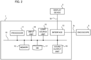
- FIG. 2 shows a hardware configuration of the image processing device 1.
- the image processing device 1 mainly includes a processor 11, a memory 12, an interface 13, an input unit 14, a light source unit 15, a sound output unit 16, and a data base (hereinafter referred to as "DB") 17. These elements are connected with each other via a data bus 19.
- DB data base
- the processor 11 executes a predetermined processing by executing a program stored in the memory 12.
- the processor 11 is a processor such as a CPU (Central Processing Unit), a GPU (Graphics Processing Unit), and a TPU (Tensor Processing Unit).
- the processor 11 may be configured by multiple processors.
- the processor 11 is an example of a computer.
- the memory 12 is configured by various volatile memories used as a working memory and non-volatile memories for storing information needed for the processing of the image processing device 1, such as a RAM (Random Access Memory) and a ROM (Read Only Memory).
- the memory 12 may include an external storage device such as a hard disk connected to or incorporated in the image processing device 1, and may include a storage medium such as a removable flash memory or a disk medium.
- the memory 12 stores a program for the image processing device 1 to execute processing in the present example embodiment.
- the memory 12 temporarily stores a series of endoscopic videos Ic taken by the endoscope 3 during the endoscopic examination, based on the control of the processor 11. Further, the memory 12 temporarily stores the lesion images photographed in response to the photographing instructions by the examiner during the endoscopic examination. These images are stored in the memory 12 in association with, for example, subject identification information (e.g., the patient ID) and time stamp information, etc.
- subject identification information e.g., the patient ID
- time stamp information e.g., etc.
- the interface 13 performs an interface operation between the image processing device 1 and the external devices. For example, the interface 13 supplies the display data Id generated by the processor 11 to the display device 2. Also, the interface 13 supplies the illumination light generated by the light source unit 15 to the endoscope 3. Further, the interface 13 supplies an electrical signal indicating the endoscopic video Ic supplied from the endoscope 3 to the processor 11.
- the interface 13 may be a communication interface such as a network adapter for wired or wireless communication with an external device, or may be a hardware interface compliant with a USB (Universal Serial Bus), SATA (Serial Advanced Technology Attachment), etc.
- the input unit 14 generates an input signal based on the operation of the examiner.
- the input unit 14 is, for example, a button, a touch panel, a remote controller, a voice input device, or the like.
- the light source unit 15 generates the light to be delivered to the tip portion 38 of the endoscope 3.
- the light source unit 15 may also incorporate a pump or the like for delivering water or air to be supplied to the endoscope 3.
- the sound output unit 16 outputs the sound based on the control of the processor 11.
- the DB 17 stores the endoscopic videos acquired by the past endoscopic examinations of the subject, and the lesion information.
- the lesion information includes lesion images and associated information.
- the DB 17 may include an external storage device, such as a hard disk connected to or incorporated in the image processing device 1, and may include a storage medium, such as a removable flash memory. Instead of providing the DB 17 in the endoscopic examination system 100, the DB 17 may be provided in an external server or the like to acquire associated information from the server through communication.
- FIG. 3 is a block diagram showing a functional configuration of the image processing device 1.
- the image processing device 1 functionally includes a position detection unit 21, an AI detection unit 22, an examination data generation unit 23, an AI determination unit 24, and a video editing unit 25.
- the image processing device 1 receives the endoscopic video Ic from the endoscope 3.
- the endoscopic video Ic is inputted into the position detection unit 21, the AI detection unit 22, the examination data generation unit 23, and the video editing unit 25.
- the position detection unit 21 detects the position of the endoscope 3, i.e., the imaging position of the endoscopic video, based on the endoscopic video Ic. Specifically, the position detection unit 21 detects the imaging position by image analysis of the inputted endoscopic video Ic.
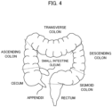
- the imaging position may be three-dimensional coordinates in the organ of the examination target, but may be at least an information indicating one of a plurality of regions in the organ of the examination target. For example, as shown in FIG.
- the large intestine includes multiple regions (sites) such as the cecum, the ascending colon, the transverse colon, the descending colon, the sigmoid colon, and the rectum. Therefore, when the examination target is the large intestine, the position detection unit 21 may detect whether at least the imaging position belongs to any of the above-described regions.
- the position detection unit 21 can estimate which region of the large intestine the imaging position at that time belongs to, based on the pattern of the mucous membrane, the presence or absence of the folds, the shape of the folds in the endoscopic video, the number of the folds passed by the movement of the endoscope 3, and the like.
- the position detection unit 21 may estimate the imaging position by estimating the movement speed of the endoscope 3 based on the endoscopic video and calculating the movement distance in the large intestine based on the movement speed and the time.
- the position detection unit 21 may detect the imaging position using the insertion length of the endoscope 3 inserted into the organ.
- the detection method of the imaging position in the organ of the examination target is not limited to a specific method.
- the position detection unit 21 outputs the detected imaging position to the examination data generation unit 23.
- the AI detection unit 22 performs image analysis based on the endoscopic video Ic to determine whether or not lesions exist.
- the AI detection unit 22 detects lesion-like parts included in the endoscopic video using an image recognition model prepared in advance. When detecting the lesion-like part, the AI detection unit 22 generates a lesion image which is a still image. Further, the AI detection unit 22 acquires the time information at the time when it detects the lesion-like part.
- the AI detection unit 22 generates AI detection data including the lesion images and the time information, and outputs the AI detection data to the examination data generation unit 23 and the AI determination unit 24. On the other hand, when the AI determination unit does not detect any lesion-like part, it outputs the determination result indicating that there is no lesion.
- patient information of the patient who is a subject is inputted through the input unit 14.
- the patient information is information that uniquely identifies a patient, and may be a personal identification number such as My Number in addition to the patient name and/or the ID uniquely assigned to each patient.
- the patient information is inputted to the examination data generation unit 23.
- the examination data generation unit 23 generates a still image based on the photographing instruction of the examiner during the endoscopic examination. Further, the examination data generation unit 23 acquires the time information at the time when the examiner made the photographing instruction.
- the examination data generating unit 23 generates examination data including the endoscopic video Ic, the photographing position detected by the position detection unit 21, the patient information, the AI detection data generated by the AI detection unit 22, the still image taken based on the examiner's photographing instruction, and the time information at the time when the examiner made the photographing instruction, and stores the examination data in the memory 12. Further, the examination data generation unit 23 outputs the generated examination data to the AI determination unit 24 and the video editing unit 25.
- the AI determination unit 24 acquires the still image taken on the basis of the photographing instruction of the examiner and the still image of the lesion-like part detected by the AI from the AI detection data generated by the AI detection unit 22 and the examination data generated by the examination data generation unit 23. Then, the AI determination unit 24 performs image analysis on the acquired still images, and performs qualitative determination to determine whether the lesion-like part is neoplastic or non-neoplastic. The AI determination unit 24 outputs the result of the qualitative determination to the video editing unit 25.
- the still images taken based on the photographing instruction of the examiner may include the still images in which the lesion-like part is not captured, such as the still images of the residue, in addition to the still images in which the lesion-like part is captured.
- the AI determination unit 24 does not detect the lesion-like part in the still image captured based on the photographing instruction of the examiner, it does not perform the qualitative determination and outputs the determination result indicating that there is no lesion.
- the video editing unit 25 generates the video data Ie using the endoscopic video Ic, the examination data inputted from the examination data generation unit 23, and the result of the quality determination inputted from the AI determination unit 24, and outputs the generated video data Ie to the display device 2.
- FIG. 5 shows an example of the result of the endoscopic examination of a patient.
- FIG. 5 shows a model image of the large intestine for convenience.
- the model image is an image of a schematic diagram of the large intestine, on which the examination result is superimposed.
- the examination result the result of inserting the endoscope from the anus to the cecum, the positions of the lesion candidate parts detected by the AI, the position photographed by the doctor, and the type of the sections are displayed.
- the result of inserting the endoscope from the anus to the cecum is indicated by the dotted line.
- the star marks indicate the positions where the AI has detected the lesion candidate part
- the black circle indicates the position photographed by the doctor.
- the endoscopic video is classified into a plurality of sections, and the plurality of sections are distinguished and displayed by the thickness of the line and the type of the line.
- the sections where neither the doctor nor the AI recognized the lesion are indicated by the thin broken line.
- the section where the doctor photographed but the AI did not detect the lesion is indicated by the thin solid line.
- the section where both the doctor and the AI recognized the lesion is shown with thick dashed line.
- the sections where the AI detected the lesion but the doctor did not photograph are indicated by the thick solid lines.
- FIG. 6 shows a method of editing video by the video editing unit 25.
- the video editing unit 25 generates an edited video based on the endoscopic video Ic and the examination data generated by the examination data generation unit 23.
- the video editing unit 25 extracts the partial videos of a predetermined time period before and after the time when the doctor instructed the photographing or the AI detected the lesion-like part from the endoscopic video Ic, and generates the edited video 1.
- the video editing unit 25 extracts the sections (P2 to P5 in this example) including the position where the doctor instructed the photographing (circle) or the position where the AI detected the lesion-like part (star mark) from the sections included in the captured video (P1 to P6), and generates the edited video 1 by concatenating those sections in a time series.
- the section from which the partial video is extracted is not limited to the above example.
- the video editing unit 25 may extract the sections including the position where the AI detected the lesion candidate part but the doctor did not instruct the photographing (P2, P4 in this example), and generate the edited video 2.
- the video editing unit 25 may generate the edited video based on the qualitative determination by the AI determination unit 24.
- the video editing unit 25 may extract only the sections including the position where the AI determination unit 24 performed the qualitative determination to generate the edited video.
- the video editing unit 25 may extract only the sections including the lesion determined to be neoplastic by the the qualitative determination by the AI determination unit 24 and generate the edited video. In this way, the examiner can efficiently review the examination result by viewing the edited video after the endoscopic examination.
- FIG. 7 shows a display example of the edited video.
- FIG. 7 is a display example of the display device 2 when the edited video generated by the video editing unit 25 is reproduced as described above.
- the video 31 being reproduced is displayed in the display area 30, in addition to the information such as the patient name, the patient ID, the examination date, and the current imaging position.
- the videos of the sections P2 to P5 are reproduced in this order as the video 31.
- the imaging position changes.
- the examiner can efficiently check significant positions in the endoscopic video by watching the edited video.
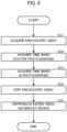
- FIG. 8 is a flowchart of display processing by the image processing device 1. This processing is realized by the processor 11 shown in FIG. 2 , which executes a pre-prepared program and operates as each element shown in FIG. 3 .
- the examination data generation unit 23, the AI detection unit 22, and the video editing unit 25 acquire the endoscopic video Ic (step S11).
- the examination data generation unit 23 acquires the time when the doctor instructs the photographing (step S12).
- the AI detection unit 22 acquires the time of detecting the lesion-like part (step S13).
- the video editing unit 25 extracts the partial videos of a predetermined time period before and after the time when the examiner instructed the photographing and the time when the AI detection unit 22 detected the lesion-like part from the endoscopic video Ic.
- the video editing unit 25 generates the edited video by editing the endoscopic video Ic (step S14) and outputs it to the display device 2.
- the displaying device 2 reproduces the received edited video (step S15).
- the edited video is reproduced as shown in FIG. 7 .
- the video editing unit 25 cuts the video of the section where neither the doctor nor the AI recognizes the lesion to create the edited video.
- the video editing unit 25 may edit the video such that the section where neither the doctor nor the AI recognizes the lesion is not cut, but is reproduced at a high speed.
- the section where the doctor or the AI recognized the lesion is reproduced at the normal speed, and the section where neither the doctor nor the AI recognized the lesion is reproduced at the higher speed than the normal speed. Therefore, the examiner or the like can take a quick look of the section where neither the doctor nor the AI recognizes the lesion at high speed and carefully observe the section where the doctor or the AI recognized the lesion at the normal speed.
- the video of a predetermined condition such as the timing when the examiner instructed photographing or the timing when the AI detected the lesion-like part
- the application of the present disclosure is not limited thereto.
- the user may select the sections to be reproduced as shown in FIG. 9 .
- FIG. 9 shows a display example at the start of reproducing the edited video.
- the image display area 41 and the section buttons 42 are displayed in the display area 40, in addition to the basic information such as the patient name, the patient ID and the examination date.
- the endoscope video is divided into the section where neither the doctor nor the AI recognized the lesion (section A), the section where the doctor photographed but the AI did not detect the lesion (section B), the section where the doctor and the AI recognized the lesion (section C), and the section where the AI detected the lesion but the doctor did not photograph (section D).
- the section button 42 of the section that he or she wants to reproduce from among these sections, the video of the selected section is displayed on the video display area 41.
- the user can easily display a digest video of the sections that he or she wants to check.
- a capsule endoscope may be used instead.
- the doctor can efficiently check the video by extracting the partial video of a predetermined time period before and after the time point of detecting the lesion.
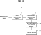
- FIG. 10 is a block diagram showing a functional configuration of a video editing device according to a second example embodiment.
- the video editing device 70 includes a video acquisition means 71, a first timing acquisition means 72, a second timing acquisition means 73, and an editing means 74.
- FIG. 11 is a flowchart illustrating processing performed by the video editing device according to the second example embodiment.
- the video acquisition means 71 acquires an endoscopic video taken by an endoscope (step S71).
- the first timing acquisition means 72 acquires a first timing at which a lesion is detected from the endoscopic video (step S72).
- the second timing acquisition means 73 acquires a second timing at which an examiner instructs photographing based on the endoscopic video (step S73).
- the editing means 74 generates an edited video by extracting, from the endoscopic video, a partial video of a predetermined time period before and after a timing corresponding to at least one of the first timing and the second timing, and output the edited video (step S74).
- the video editing device 70 of the second example embodiment it is possible to generate an edited video efficiently showing a required portion from the examination video of the endoscopic examination.
- a video editing device comprising:
- the video editing device according to claim 1, wherein the editing means generates the edited video by extracting the partial video for the timing corresponding to the first timing and not corresponding to the second timing.
- the video editing device further comprising a determination means configured to perform qualitative determination for the partial video of the first timing and the second timing, wherein the editing means generates the edited video by extracting the partial video on which the qualitative determination is performed.
- the video editing device further comprising a display means configured to divide the endoscopic video into plural types of sections based on whether or not the endoscopic video corresponds to the first timing and the second timing, and display the plural of types of sections as options, wherein the editing means generates the edited video by extracting the partial video belonging to the section of the type selected by a user from among the plural types of sections.
- the video editing device according to any one of claims 1 to 4, wherein the editing means generates the edited video in which the partial video is reproduced at a normal speed and a part of the endoscopic video other than the partial video is reproduced at a higher speed than the normal speed.
- a video editing method comprising:
- a recording medium recording a program, the program causing a computer to execute processing of:
Landscapes
- Health & Medical Sciences (AREA)
- Engineering & Computer Science (AREA)
- Life Sciences & Earth Sciences (AREA)
- General Health & Medical Sciences (AREA)
- Public Health (AREA)
- Medical Informatics (AREA)
- Nuclear Medicine, Radiotherapy & Molecular Imaging (AREA)
- Radiology & Medical Imaging (AREA)
- Physics & Mathematics (AREA)
- Surgery (AREA)
- Epidemiology (AREA)
- Primary Health Care (AREA)
- General Physics & Mathematics (AREA)
- Theoretical Computer Science (AREA)
- Multimedia (AREA)
- Optics & Photonics (AREA)
- Biophysics (AREA)
- Pathology (AREA)
- Biomedical Technology (AREA)
- Heart & Thoracic Surgery (AREA)
- Molecular Biology (AREA)
- Animal Behavior & Ethology (AREA)
- Veterinary Medicine (AREA)
- Computer Vision & Pattern Recognition (AREA)
- Endoscopes (AREA)
Abstract
Description
- The present disclosure relates to editing of a video obtained by an endoscopic examination.
- The examination image of the endoscope may be reexamined, when the doctor looks back the examination result of the endoscopic examination or when the doctor wants to obtain the opinion of other doctors. However, since the time period of the image is long, the reexamination takes a lot of time. From this viewpoint,
Patent Document 1 proposes a method of adding time stamps to the moving image file, during the examination, by the operation of the operator of the endoscope in the endoscopic examination system, thereby to easily retrieve the moving image of the time zone with significant information. - Patent Document 1: Japanese Patent Application Laid-open under No.
JP 2018-166989 - Recently, there have been proposed techniques for detecting lesions using AI (Artificial Intelligence). Therefore, it is desirable to be able to efficiently refer to the examination image including not only the images selected by the operator of the endoscope but also the images of the lesions detected by AI.
- It is an object of the present disclosure to create an edited video from an endoscopic examination image, by which necessary parts can be efficiently viewed.
- According to an example aspect of the present disclosure, there is provided a video editing device comprising:
- a video acquisition means configured to acquire an endoscopic video taken by an endoscope;
- a first timing acquisition means configured to acquire a first timing at which a lesion is detected from the endoscopic video;
- a second timing acquisition means configured to acquire a second timing at which an examiner instructs photographing based on the endoscopic video; and
- an editing means configured to generate an edited video by extracting, from the endoscopic video, a partial video of a predetermined time period before and after a timing corresponding to at least one of the first timing and the second timing, and output the edited video.
- According to another example aspect of the present disclosure, there is provided a video editing method comprising:
- acquiring an endoscopic video taken by an endoscope;
- acquiring a first timing at which a lesion is detected from the endoscopic video;
- acquiring a second timing at which an examiner instructs photographing based on the endoscopic video;
- generating an edited video by extracting, from the endoscopic video, a partial video of a predetermined time period before and after a timing corresponding to at least one of the first timing and the second timing; and
- outputting the edited video.
- According to still another example aspect of the present disclosure, there is provided a recording medium recording a program, the program causing a computer to execute processing of:
- acquiring an endoscopic video captured by an endoscope;
- acquiring a first timing at which a lesion is detected from the endoscopic video;
- acquiring a second timing at which an examiner instructs photographing based on the endoscopic video;
- generating an edited video by extracting, from the endoscopic video, a partial video of a predetermined time period before and after a timing corresponding to at least one of the first timing and the second timing; and
- outputting the edited video.
- According to the present disclosure, the examination image can be efficiently checked after the endoscopic examination is completed.
-
- [
FIG. 1] FIG. 1 is a block diagram showing a schematic configuration of an endoscopic examination system. - [
FIG. 2] FIG. 2 is a block diagram showing a hardware configuration of an image processing device. - [
FIG. 3] FIG. 3 is a block diagram showing a functional configuration of the image processing device. - [
FIG. 4] FIG. 4 schematically shows a structure of a large intestine. - [
FIG. 5] FIG. 5 is a diagram showing an example of an examination result. - [
FIG. 6] FIG. 6 is a diagram showing a method of generating an edited video. - [
FIG. 7] FIG. 7 is a display example of reproducing an edited video. - [
FIG. 8] FIG. 8 is a flowchart of display processing by the image processing device. - [
FIG. 9] FIG. 9 is a display example of the edited video at the start of reproduction. - [
FIG. 10] FIG. 10 is a block diagram showing a functional configuration of a video editing device of a second example embodiment. - [
FIG. 11] FIG. 11 is a flowchart of processing by the video editing device of the second example embodiment. - Preferred example embodiments of the present disclosure will be described with reference to the accompanying drawings.
-
FIG. 1 shows a schematic configuration of anendoscopic examination system 100. When the examination (including the treatment) using the endoscope is performed, theendoscopic examination system 100 acquires the times when the examiner of the endoscopic examination took the still image and the times when the lesions were detected by the image analysis of AI. Then, theendoscopic examination system 100 extracts the partial videos of a predetermined time period before and after the acquired times to generate and display an edited video. This enables the examiner to check the examination video efficiently after the endoscopic examination. - As shown in
FIG. 1 , theendoscopic examination system 100 mainly includes animage processing device 1, adisplay device 2, and anendoscope 3 connected to theimage processing device 1. - The
image processing device 1 acquires a video (i.e., a moving image, hereinafter also referred to as an "endoscopic video Ic") captured by theendoscope 3 during the endoscopic examination from theendoscope 3, and displays display data for the check by the examiner of the endoscopic examination on thedisplay device 2. Specifically, theimage processing device 1 acquires a moving image of organs captured by theendoscope 3 as an endoscopic video Ic during the endoscopic examination. In addition, when the examiner finds a lesion during the endoscopic examination, he or she operates theendoscope 3 to input a photographing instruction of the lesion position. Based on the photographing instruction by the examiner, theimage processing device 1 generates a lesion image capturing the lesion position. Specifically, theimage processing device 1 generates the lesion image which is a still image, from the endoscopic video Ic which is a moving image, on the basis of the photographing instruction of the examiner. - The
display device 2 is a display or the like for displaying images on the basis of the display signal supplied from theimage processing device 1. - The
endoscope 3 mainly includes anoperation unit 36 used by the examiner to input instructions such as air supply, water supply, angle adjustment, and the photographing instruction, ashaft 37 having flexibility and inserted into an organ of a subject to be examined, atip portion 38 with a built-in image-taking unit such as an ultra-compact imaging element, and aconnection unit 39 for connection with theimage processing device 1. - While the following explanation is mainly given on the processing of endoscopic examination for a large intestine, the subjects of examination may be gastrointestinal (digestive organs) such as the stomach, esophagus, small intestine, and duodenum, as well as the large intestine.
- In addition, the part to be detected in the endoscopic examination is not limited to the lesion part, but may be any part (also referred to as a "region of interest") in which the examiner needs attention. The region of interest may be, for example, a lesion part, a part where inflammation has occurred, a part where a surgical scar or other cut has occurred, a part where a fold or protrusion has occurred, or a part where the
tip portion 38 of theendoscope 3 is likely to contact (easily stuck) the intraluminal wall surface. -
FIG. 2 shows a hardware configuration of theimage processing device 1. Theimage processing device 1 mainly includes aprocessor 11, amemory 12, aninterface 13, aninput unit 14, alight source unit 15, asound output unit 16, and a data base (hereinafter referred to as "DB") 17. These elements are connected with each other via adata bus 19. - The
processor 11 executes a predetermined processing by executing a program stored in thememory 12. Theprocessor 11 is a processor such as a CPU (Central Processing Unit), a GPU (Graphics Processing Unit), and a TPU (Tensor Processing Unit). Theprocessor 11 may be configured by multiple processors. Theprocessor 11 is an example of a computer. - The
memory 12 is configured by various volatile memories used as a working memory and non-volatile memories for storing information needed for the processing of theimage processing device 1, such as a RAM (Random Access Memory) and a ROM (Read Only Memory). Incidentally, thememory 12 may include an external storage device such as a hard disk connected to or incorporated in theimage processing device 1, and may include a storage medium such as a removable flash memory or a disk medium. Thememory 12 stores a program for theimage processing device 1 to execute processing in the present example embodiment. - Also, the
memory 12 temporarily stores a series of endoscopic videos Ic taken by theendoscope 3 during the endoscopic examination, based on the control of theprocessor 11. Further, thememory 12 temporarily stores the lesion images photographed in response to the photographing instructions by the examiner during the endoscopic examination. These images are stored in thememory 12 in association with, for example, subject identification information (e.g., the patient ID) and time stamp information, etc. - The
interface 13 performs an interface operation between theimage processing device 1 and the external devices. For example, theinterface 13 supplies the display data Id generated by theprocessor 11 to thedisplay device 2. Also, theinterface 13 supplies the illumination light generated by thelight source unit 15 to theendoscope 3. Further, theinterface 13 supplies an electrical signal indicating the endoscopic video Ic supplied from theendoscope 3 to theprocessor 11. Theinterface 13 may be a communication interface such as a network adapter for wired or wireless communication with an external device, or may be a hardware interface compliant with a USB (Universal Serial Bus), SATA (Serial Advanced Technology Attachment), etc. - The
input unit 14 generates an input signal based on the operation of the examiner. Theinput unit 14 is, for example, a button, a touch panel, a remote controller, a voice input device, or the like. Thelight source unit 15 generates the light to be delivered to thetip portion 38 of theendoscope 3. Thelight source unit 15 may also incorporate a pump or the like for delivering water or air to be supplied to theendoscope 3. Thesound output unit 16 outputs the sound based on the control of theprocessor 11. - The
DB 17 stores the endoscopic videos acquired by the past endoscopic examinations of the subject, and the lesion information. The lesion information includes lesion images and associated information. TheDB 17 may include an external storage device, such as a hard disk connected to or incorporated in theimage processing device 1, and may include a storage medium, such as a removable flash memory. Instead of providing theDB 17 in theendoscopic examination system 100, theDB 17 may be provided in an external server or the like to acquire associated information from the server through communication. -
FIG. 3 is a block diagram showing a functional configuration of theimage processing device 1. Theimage processing device 1 functionally includes aposition detection unit 21, anAI detection unit 22, an examinationdata generation unit 23, anAI determination unit 24, and avideo editing unit 25. - The
image processing device 1 receives the endoscopic video Ic from theendoscope 3. The endoscopic video Ic is inputted into theposition detection unit 21, theAI detection unit 22, the examinationdata generation unit 23, and thevideo editing unit 25. Theposition detection unit 21 detects the position of theendoscope 3, i.e., the imaging position of the endoscopic video, based on the endoscopic video Ic. Specifically, theposition detection unit 21 detects the imaging position by image analysis of the inputted endoscopic video Ic. Here, the imaging position may be three-dimensional coordinates in the organ of the examination target, but may be at least an information indicating one of a plurality of regions in the organ of the examination target. For example, as shown inFIG. 4 , the large intestine includes multiple regions (sites) such as the cecum, the ascending colon, the transverse colon, the descending colon, the sigmoid colon, and the rectum. Therefore, when the examination target is the large intestine, theposition detection unit 21 may detect whether at least the imaging position belongs to any of the above-described regions. - Specifically, the
position detection unit 21 can estimate which region of the large intestine the imaging position at that time belongs to, based on the pattern of the mucous membrane, the presence or absence of the folds, the shape of the folds in the endoscopic video, the number of the folds passed by the movement of theendoscope 3, and the like. Theposition detection unit 21 may estimate the imaging position by estimating the movement speed of theendoscope 3 based on the endoscopic video and calculating the movement distance in the large intestine based on the movement speed and the time. In addition to the image analysis of the endoscopic video, theposition detection unit 21 may detect the imaging position using the insertion length of theendoscope 3 inserted into the organ. In the present example embodiment, the detection method of the imaging position in the organ of the examination target is not limited to a specific method. Theposition detection unit 21 outputs the detected imaging position to the examinationdata generation unit 23. - The
AI detection unit 22 performs image analysis based on the endoscopic video Ic to determine whether or not lesions exist. TheAI detection unit 22 detects lesion-like parts included in the endoscopic video using an image recognition model prepared in advance. When detecting the lesion-like part, theAI detection unit 22 generates a lesion image which is a still image. Further, theAI detection unit 22 acquires the time information at the time when it detects the lesion-like part. TheAI detection unit 22 generates AI detection data including the lesion images and the time information, and outputs the AI detection data to the examinationdata generation unit 23 and theAI determination unit 24. On the other hand, when the AI determination unit does not detect any lesion-like part, it outputs the determination result indicating that there is no lesion. - Further, to the
image processing device 1, patient information of the patient who is a subject is inputted through theinput unit 14. The patient information is information that uniquely identifies a patient, and may be a personal identification number such as My Number in addition to the patient name and/or the ID uniquely assigned to each patient. The patient information is inputted to the examinationdata generation unit 23. - The examination
data generation unit 23 generates a still image based on the photographing instruction of the examiner during the endoscopic examination. Further, the examinationdata generation unit 23 acquires the time information at the time when the examiner made the photographing instruction. The examinationdata generating unit 23 generates examination data including the endoscopic video Ic, the photographing position detected by theposition detection unit 21, the patient information, the AI detection data generated by theAI detection unit 22, the still image taken based on the examiner's photographing instruction, and the time information at the time when the examiner made the photographing instruction, and stores the examination data in thememory 12. Further, the examinationdata generation unit 23 outputs the generated examination data to theAI determination unit 24 and thevideo editing unit 25. - The
AI determination unit 24 acquires the still image taken on the basis of the photographing instruction of the examiner and the still image of the lesion-like part detected by the AI from the AI detection data generated by theAI detection unit 22 and the examination data generated by the examinationdata generation unit 23. Then, theAI determination unit 24 performs image analysis on the acquired still images, and performs qualitative determination to determine whether the lesion-like part is neoplastic or non-neoplastic. TheAI determination unit 24 outputs the result of the qualitative determination to thevideo editing unit 25. Incidentally, the still images taken based on the photographing instruction of the examiner may include the still images in which the lesion-like part is not captured, such as the still images of the residue, in addition to the still images in which the lesion-like part is captured. When theAI determination unit 24 does not detect the lesion-like part in the still image captured based on the photographing instruction of the examiner, it does not perform the qualitative determination and outputs the determination result indicating that there is no lesion. - The
video editing unit 25 generates the video data Ie using the endoscopic video Ic, the examination data inputted from the examinationdata generation unit 23, and the result of the quality determination inputted from theAI determination unit 24, and outputs the generated video data Ie to thedisplay device 2. -
FIG. 5 shows an example of the result of the endoscopic examination of a patient.FIG. 5 shows a model image of the large intestine for convenience. The model image is an image of a schematic diagram of the large intestine, on which the examination result is superimposed. Here, as the examination result, the result of inserting the endoscope from the anus to the cecum, the positions of the lesion candidate parts detected by the AI, the position photographed by the doctor, and the type of the sections are displayed. Specifically, the result of inserting the endoscope from the anus to the cecum is indicated by the dotted line. Also, the star marks indicate the positions where the AI has detected the lesion candidate part, and the black circle indicates the position photographed by the doctor. Then, the endoscopic video is classified into a plurality of sections, and the plurality of sections are distinguished and displayed by the thickness of the line and the type of the line. Here, the sections where neither the doctor nor the AI recognized the lesion (the cecum and the rectum in this example) are indicated by the thin broken line. The section where the doctor photographed but the AI did not detect the lesion (the sigmoid colon in this case) is indicated by the thin solid line. The section where both the doctor and the AI recognized the lesion (the transverse colon in this case) is shown with thick dashed line. The sections where the AI detected the lesion but the doctor did not photograph (the ascending colon and the descending colon in this case) are indicated by the thick solid lines. -
FIG. 6 shows a method of editing video by thevideo editing unit 25. Thevideo editing unit 25 generates an edited video based on the endoscopic video Ic and the examination data generated by the examinationdata generation unit 23. In one example, thevideo editing unit 25 extracts the partial videos of a predetermined time period before and after the time when the doctor instructed the photographing or the AI detected the lesion-like part from the endoscopic video Ic, and generates the editedvideo 1. Here, thevideo editing unit 25 extracts the sections (P2 to P5 in this example) including the position where the doctor instructed the photographing (circle) or the position where the AI detected the lesion-like part (star mark) from the sections included in the captured video (P1 to P6), and generates the editedvideo 1 by concatenating those sections in a time series. - The section from which the partial video is extracted is not limited to the above example. In another example, the
video editing unit 25 may extract the sections including the position where the AI detected the lesion candidate part but the doctor did not instruct the photographing (P2, P4 in this example), and generate the editedvideo 2. In yet another example, thevideo editing unit 25 may generate the edited video based on the qualitative determination by theAI determination unit 24. For example, thevideo editing unit 25 may extract only the sections including the position where theAI determination unit 24 performed the qualitative determination to generate the edited video. Alternatively, thevideo editing unit 25 may extract only the sections including the lesion determined to be neoplastic by the the qualitative determination by theAI determination unit 24 and generate the edited video. In this way, the examiner can efficiently review the examination result by viewing the edited video after the endoscopic examination. - Next, a display example by the
display device 2 will be described. -
FIG. 7 shows a display example of the edited video.FIG. 7 is a display example of thedisplay device 2 when the edited video generated by thevideo editing unit 25 is reproduced as described above. In the display example ofFIG. 7 , thevideo 31 being reproduced is displayed in thedisplay area 30, in addition to the information such as the patient name, the patient ID, the examination date, and the current imaging position. For example, when the editedvideo 1 shown inFIG. 6 is being reproduced, the videos of the sections P2 to P5 are reproduced in this order as thevideo 31. Further, when the section being reproduced as thevideo 31 changes, the imaging position changes. Thus, the examiner can efficiently check significant positions in the endoscopic video by watching the edited video. - Next, display processing for performing the above-mentioned display will be described.
FIG. 8 is a flowchart of display processing by theimage processing device 1. This processing is realized by theprocessor 11 shown inFIG. 2 , which executes a pre-prepared program and operates as each element shown inFIG. 3 . - First, the examination
data generation unit 23, theAI detection unit 22, and thevideo editing unit 25 acquire the endoscopic video Ic (step S11). Next, the examinationdata generation unit 23 acquires the time when the doctor instructs the photographing (step S12). TheAI detection unit 22 acquires the time of detecting the lesion-like part (step S13). Next, thevideo editing unit 25 extracts the partial videos of a predetermined time period before and after the time when the examiner instructed the photographing and the time when theAI detection unit 22 detected the lesion-like part from the endoscopic video Ic. Then, thevideo editing unit 25 generates the edited video by editing the endoscopic video Ic (step S14) and outputs it to thedisplay device 2. The displayingdevice 2 reproduces the received edited video (step S15). Thus, the edited video is reproduced as shown inFIG. 7 . - In the above-described example embodiment, as illustrated in
FIG. 6 , thevideo editing unit 25 cuts the video of the section where neither the doctor nor the AI recognizes the lesion to create the edited video. Instead, thevideo editing unit 25 may edit the video such that the section where neither the doctor nor the AI recognizes the lesion is not cut, but is reproduced at a high speed. In this case, when the edited video is reproduced, the section where the doctor or the AI recognized the lesion is reproduced at the normal speed, and the section where neither the doctor nor the AI recognized the lesion is reproduced at the higher speed than the normal speed. Therefore, the examiner or the like can take a quick look of the section where neither the doctor nor the AI recognizes the lesion at high speed and carefully observe the section where the doctor or the AI recognized the lesion at the normal speed. - In the above-described example embodiment, the video of a predetermined condition, such as the timing when the examiner instructed photographing or the timing when the AI detected the lesion-like part, is displayed as the edited video. However, the application of the present disclosure is not limited thereto. For example, the user may select the sections to be reproduced as shown in
FIG. 9 . -
FIG. 9 shows a display example at the start of reproducing the edited video. Specifically, inFIG. 9 , theimage display area 41 and thesection buttons 42 are displayed in thedisplay area 40, in addition to the basic information such as the patient name, the patient ID and the examination date. The endoscope video is divided into the section where neither the doctor nor the AI recognized the lesion (section A), the section where the doctor photographed but the AI did not detect the lesion (section B), the section where the doctor and the AI recognized the lesion (section C), and the section where the AI detected the lesion but the doctor did not photograph (section D). When the user presses thesection button 42 of the section that he or she wants to reproduce from among these sections, the video of the selected section is displayed on thevideo display area 41. Thus, the user can easily display a digest video of the sections that he or she wants to check. - While the above example embodiment uses an endoscope of the type in which a shaft is inserted to directly observe the gastrointestinal tract, a capsule endoscope may be used instead. When the capsule endoscope is used to detect the lesion, the doctor can efficiently check the video by extracting the partial video of a predetermined time period before and after the time point of detecting the lesion.
-
FIG. 10 is a block diagram showing a functional configuration of a video editing device according to a second example embodiment. Thevideo editing device 70 includes a video acquisition means 71, a first timing acquisition means 72, a second timing acquisition means 73, and an editing means 74. -
FIG. 11 is a flowchart illustrating processing performed by the video editing device according to the second example embodiment. The video acquisition means 71 acquires an endoscopic video taken by an endoscope (step S71). The first timing acquisition means 72 acquires a first timing at which a lesion is detected from the endoscopic video (step S72). The second timing acquisition means 73 acquires a second timing at which an examiner instructs photographing based on the endoscopic video (step S73). The editing means 74 generates an edited video by extracting, from the endoscopic video, a partial video of a predetermined time period before and after a timing corresponding to at least one of the first timing and the second timing, and output the edited video (step S74). - According to the
video editing device 70 of the second example embodiment, it is possible to generate an edited video efficiently showing a required portion from the examination video of the endoscopic examination. - A part or all of the above example embodiments may also be described as in the following supplementary notes, but are not limited to:
- A video editing device comprising:
- a video acquisition means configured to acquire an endoscopic video taken by an endoscope;
- a first timing acquisition means configured to acquire a first timing at which a lesion is detected from the endoscopic video;
- a second timing acquisition means configured to acquire a second timing at which an examiner instructs photographing based on the endoscopic video; and
- an editing means configured to generate an edited video by extracting, from the endoscopic video, a partial video of a predetermined time period before and after a timing corresponding to at least one of the first timing and the second timing, and output the edited video.
- The video editing device according to
claim 1, wherein the editing means generates the edited video by extracting the partial video for the timing corresponding to the first timing and not corresponding to the second timing. - The video editing device according to
claim 1, further comprising a determination means configured to perform qualitative determination for the partial video of the first timing and the second timing,
wherein the editing means generates the edited video by extracting the partial video on which the qualitative determination is performed. - The video editing device according to
claim 1, further comprising a display means configured to divide the endoscopic video into plural types of sections based on whether or not the endoscopic video corresponds to the first timing and the second timing, and display the plural of types of sections as options,
wherein the editing means generates the edited video by extracting the partial video belonging to the section of the type selected by a user from among the plural types of sections. - The video editing device according to any one of
claims 1 to 4, wherein the editing means generates the edited video by concatenating the partial videos in a time series. - The video editing device according to any one of
claims 1 to 4, wherein the editing means generates the edited video in which the partial video is reproduced at a normal speed and a part of the endoscopic video other than the partial video is reproduced at a higher speed than the normal speed. - A video editing method comprising:
- acquiring an endoscopic video taken by an endoscope;
- acquiring a first timing at which a lesion is detected from the endoscopic video;
- acquiring a second timing at which an examiner instructs photographing based on the endoscopic video;
- generating an edited video by extracting, from the endoscopic video, a partial video of a predetermined time period before and after a timing corresponding to at least one of the first timing and the second timing; and
- outputting the edited video.
- A recording medium recording a program, the program causing a computer to execute processing of:
- acquiring an endoscopic video captured by an endoscope;
- acquiring a first timing at which a lesion is detected from the endoscopic video;
- acquiring a second timing at which an examiner instructs photographing based on the endoscopic video;
- generating an edited video by extracting, from the endoscopic video, a partial video of a predetermined time period before and after a timing corresponding to at least one of the first timing and the second timing; and
- outputting the edited video.
- While the present disclosure has been described with reference to the example embodiments and examples, the present disclosure is not limited to the above example embodiments and examples. Various changes which can be understood by those skilled in the art within the scope of the present disclosure can be made in the configuration and details of the present disclosure.
-
- 1 Image processing device
- 2 Display device
- 3 Endoscope
- 11 Processor
- 12 Memory
- 17 Database (DB)
- 21 Position detection unit
- 22 AI detection unit
- 23 Examination data generation unit
- 24 AI determination unit
- 25 Video editing unit
- 100 Endoscopic examination system
Claims (8)
- A video editing device comprising:a video acquisition means configured to acquire an endoscopic video taken by an endoscope;a first timing acquisition means configured to acquire a first timing at which a lesion is detected from the endoscopic video;a second timing acquisition means configured to acquire a second timing at which an examiner instructs photographing based on the endoscopic video; andan editing means configured to generate an edited video by extracting, from the endoscopic video, a partial video of a predetermined time period before and after a timing corresponding to at least one of the first timing and the second timing, and output the edited video.
- The video editing device according to claim 1, wherein the editing means generates the edited video by extracting the partial video for the timing corresponding to the first timing and not corresponding to the second timing.
- The video editing device according to claim 1, further comprising a determination means configured to perform qualitative determination for the partial video of the first timing and the second timing,
wherein the editing means generates the edited video by extracting the partial video on which the qualitative determination is performed. - The video editing device according to claim 1, further comprising a display means configured to divide the endoscopic video into plural types of sections based on whether or not the endoscopic video corresponds to the first timing and the second timing, and display the plural of types of sections as options,
wherein the editing means generates the edited video by extracting the partial video belonging to the section of the type selected by a user from among the plural types of sections. - The video editing device according to any one of claims 1 to 4, wherein the editing means generates the edited video by concatenating the partial videos in a time series.
- The video editing device according to any one of claims 1 to 4, wherein the editing means generates the edited video in which the partial video is reproduced at a normal speed and a part of the endoscopic video other than the partial video is reproduced at a higher speed than the normal speed.
- A video editing method comprising:acquiring an endoscopic video taken by an endoscope;acquiring a first timing at which a lesion is detected from the endoscopic video;acquiring a second timing at which an examiner instructs photographing based on the endoscopic video;generating an edited video by extracting, from the endoscopic video, a partial video of a predetermined time period before and after a timing corresponding to at least one of the first timing and the second timing; andoutputting the edited video.
- A recording medium recording a program, the program causing a computer to execute processing of:acquiring an endoscopic video captured by an endoscope;acquiring a first timing at which a lesion is detected from the endoscopic video;acquiring a second timing at which an examiner instructs photographing based on the endoscopic video;generating an edited video by extracting, from the endoscopic video, a partial video of a predetermined time period before and after a timing corresponding to at least one of the first timing and the second timing; andoutputting the edited video.
Applications Claiming Priority (1)
| Application Number | Priority Date | Filing Date | Title |
|---|---|---|---|
| PCT/JP2021/042368 WO2023089719A1 (en) | 2021-11-18 | 2021-11-18 | Video editing device, video editing method, and recording medium |
Publications (2)
| Publication Number | Publication Date |
|---|---|
| EP4434436A1 true EP4434436A1 (en) | 2024-09-25 |
| EP4434436A4 EP4434436A4 (en) | 2025-01-08 |
Family
ID=86396443
Family Applications (1)
| Application Number | Title | Priority Date | Filing Date |
|---|---|---|---|
| EP21964734.4A Withdrawn EP4434436A4 (en) | 2021-11-18 | 2021-11-18 | Video editing device, video editing method, and recording medium |
Country Status (3)
| Country | Link |
|---|---|
| US (1) | US20250140384A1 (en) |
| EP (1) | EP4434436A4 (en) |
| WO (1) | WO2023089719A1 (en) |
Family Cites Families (9)
| Publication number | Priority date | Publication date | Assignee | Title |
|---|---|---|---|---|
| JP2007075163A (en) * | 2005-09-09 | 2007-03-29 | Olympus Medical Systems Corp | Image display device |
| JP2008301968A (en) * | 2007-06-06 | 2008-12-18 | Olympus Medical Systems Corp | Endoscopic image processing device |
| JP2015195845A (en) * | 2014-03-31 | 2015-11-09 | 富士フイルム株式会社 | Endoscope system, operation method of endoscope system, processor device, operation method of processor device |
| JP2018166989A (en) | 2017-03-30 | 2018-11-01 | Hoya株式会社 | Electronic endoscope system and processor |
| US11055843B2 (en) * | 2017-06-14 | 2021-07-06 | Electronics And Telecommunications Research Institute | Capsule endoscope for determining lesion area and receiving device |
| JP7265543B2 (en) * | 2017-10-17 | 2023-04-26 | ヴェリリー ライフ サイエンシズ エルエルシー | System and method for segmenting surgical video |
| CN111936032B (en) * | 2018-04-13 | 2024-03-26 | 富士胶片株式会社 | Image processing device, endoscope system, and image processing method |
| WO2020165978A1 (en) * | 2019-02-13 | 2020-08-20 | オリンパス株式会社 | Image recording device, image recording method, and image recording program |
| JP7346285B2 (en) * | 2019-12-24 | 2023-09-19 | 富士フイルム株式会社 | Medical image processing device, endoscope system, operating method and program for medical image processing device |
-
2021
- 2021-11-18 US US18/701,673 patent/US20250140384A1/en active Pending
- 2021-11-18 EP EP21964734.4A patent/EP4434436A4/en not_active Withdrawn
- 2021-11-18 WO PCT/JP2021/042368 patent/WO2023089719A1/en not_active Ceased
Also Published As
| Publication number | Publication date |
|---|---|
| JPWO2023089719A1 (en) | 2023-05-25 |
| EP4434436A4 (en) | 2025-01-08 |
| WO2023089719A1 (en) | 2023-05-25 |
| US20250140384A1 (en) | 2025-05-01 |
Similar Documents
| Publication | Publication Date | Title |
|---|---|---|
| CN101677756B (en) | Image information display processing device and display processing method | |
| EP4434435A1 (en) | Information processing device, information processing method, and recording medium | |
| JP4009583B2 (en) | Medical image recording system | |
| CN105142493A (en) | Image management device | |
| JP2009022446A (en) | System and method for integrated display in medicine | |
| JP2006055262A (en) | Image display device, image display method and image display program | |
| WO2023126999A1 (en) | Image processing device, image processing method, and storage medium | |
| WO2021176664A1 (en) | Inspection support system, inspection support method, and program | |
| US20250281022A1 (en) | Endoscopy support device, endoscopy support method, and recording medium | |
| EP4434436A1 (en) | Video editing device, video editing method, and recording medium | |
| WO2023148812A1 (en) | Image processing device, image processing method, and storage medium | |
| US12593957B2 (en) | Endoscopic examination support apparatus, endoscopic examination support method, and recording medium | |
| US20240382067A1 (en) | Medical assistance system and medical assistance method | |
| EP4434433A1 (en) | Information display device, information display method, and recording medium | |
| WO2024121885A1 (en) | Information processing device, information processing method, and recording medium | |
| JP4027842B2 (en) | Medical image recording device | |
| EP4434432A1 (en) | Image display device, image display method, and recording medium | |
| EP4434434A1 (en) | Information processing device, information processing method, and recording medium | |
| US12599284B2 (en) | Endoscopic examination support apparatus, endoscopic examination support method, and recording medium | |
| US20240233121A1 (en) | Endoscopic examination support apparatus, endoscopic examination support method, and recording medium | |
| WO2023175916A1 (en) | Medical assistance system and image display method | |
| JP7647873B2 (en) | Image processing device, image processing method, and program | |
| JP7750418B2 (en) | Endoscopic examination support device, endoscopic examination support method, and program | |
| WO2024185099A1 (en) | Endoscopy assistance device, endoscopy assistance method, and recording medium | |
| JP7607803B2 (en) | Medical support system, report creation support method and information processing device |
Legal Events
| Date | Code | Title | Description |
|---|---|---|---|
| STAA | Information on the status of an ep patent application or granted ep patent |
Free format text: STATUS: THE INTERNATIONAL PUBLICATION HAS BEEN MADE |
|
| PUAI | Public reference made under article 153(3) epc to a published international application that has entered the european phase |
Free format text: ORIGINAL CODE: 0009012 |
|
| STAA | Information on the status of an ep patent application or granted ep patent |
Free format text: STATUS: REQUEST FOR EXAMINATION WAS MADE |
|
| 17P | Request for examination filed |
Effective date: 20240517 |
|
| AK | Designated contracting states |
Kind code of ref document: A1 Designated state(s): AL AT BE BG CH CY CZ DE DK EE ES FI FR GB GR HR HU IE IS IT LI LT LU LV MC MK MT NL NO PL PT RO RS SE SI SK SM TR |
|
| A4 | Supplementary search report drawn up and despatched |
Effective date: 20241209 |
|
| RIC1 | Information provided on ipc code assigned before grant |
Ipc: G16H 30/40 20180101ALI20241203BHEP Ipc: G16H 30/20 20180101ALI20241203BHEP Ipc: A61B 1/045 20060101AFI20241203BHEP |
|
| DAV | Request for validation of the european patent (deleted) | ||
| DAX | Request for extension of the european patent (deleted) | ||
| STAA | Information on the status of an ep patent application or granted ep patent |
Free format text: STATUS: THE APPLICATION IS DEEMED TO BE WITHDRAWN |
|
| 18D | Application deemed to be withdrawn |
Effective date: 20250708 |