EP4424699A1 - Hbv core protein binding compounds and uses thereof - Google Patents
Hbv core protein binding compounds and uses thereof Download PDFInfo
- Publication number
- EP4424699A1 EP4424699A1 EP23159984.6A EP23159984A EP4424699A1 EP 4424699 A1 EP4424699 A1 EP 4424699A1 EP 23159984 A EP23159984 A EP 23159984A EP 4424699 A1 EP4424699 A1 EP 4424699A1
- Authority
- EP
- European Patent Office
- Prior art keywords
- peptide
- seq
- compound
- hbv
- cpp
- Prior art date
- Legal status (The legal status is an assumption and is not a legal conclusion. Google has not performed a legal analysis and makes no representation as to the accuracy of the status listed.)
- Ceased
Links
- 150000001875 compounds Chemical class 0.000 title claims abstract description 58
- 230000027455 binding Effects 0.000 title claims description 43
- 101710132601 Capsid protein Proteins 0.000 title claims description 27
- 108090000765 processed proteins & peptides Proteins 0.000 claims abstract description 163
- 241000700721 Hepatitis B virus Species 0.000 claims abstract description 69
- 102000004196 processed proteins & peptides Human genes 0.000 claims abstract description 59
- 208000015181 infectious disease Diseases 0.000 claims abstract description 23
- 239000008194 pharmaceutical composition Substances 0.000 claims abstract description 23
- 208000019423 liver disease Diseases 0.000 claims abstract description 14
- 238000011282 treatment Methods 0.000 claims abstract description 13
- 238000000034 method Methods 0.000 claims abstract description 12
- 108010051109 Cell-Penetrating Peptides Proteins 0.000 claims description 48
- 102000020313 Cell-Penetrating Peptides Human genes 0.000 claims description 48
- 150000001413 amino acids Chemical group 0.000 claims description 44
- 229940024606 amino acid Drugs 0.000 claims description 43
- 235000001014 amino acid Nutrition 0.000 claims description 43
- ROHFNLRQFUQHCH-YFKPBYRVSA-N L-leucine Chemical compound CC(C)C[C@H](N)C(O)=O ROHFNLRQFUQHCH-YFKPBYRVSA-N 0.000 claims description 17
- ROHFNLRQFUQHCH-UHFFFAOYSA-N Leucine Natural products CC(C)CC(N)C(O)=O ROHFNLRQFUQHCH-UHFFFAOYSA-N 0.000 claims description 17
- DHMQDGOQFOQNFH-UHFFFAOYSA-N Glycine Chemical compound NCC(O)=O DHMQDGOQFOQNFH-UHFFFAOYSA-N 0.000 claims description 16
- COLNVLDHVKWLRT-QMMMGPOBSA-N L-phenylalanine Chemical compound OC(=O)[C@@H](N)CC1=CC=CC=C1 COLNVLDHVKWLRT-QMMMGPOBSA-N 0.000 claims description 16
- 230000002209 hydrophobic effect Effects 0.000 claims description 15
- COLNVLDHVKWLRT-UHFFFAOYSA-N phenylalanine Natural products OC(=O)C(N)CC1=CC=CC=C1 COLNVLDHVKWLRT-UHFFFAOYSA-N 0.000 claims description 13
- LYCAIKOWRPUZTN-UHFFFAOYSA-N Ethylene glycol Chemical compound OCCO LYCAIKOWRPUZTN-UHFFFAOYSA-N 0.000 claims description 12
- AGPKZVBTJJNPAG-WHFBIAKZSA-N L-isoleucine Chemical compound CC[C@H](C)[C@H](N)C(O)=O AGPKZVBTJJNPAG-WHFBIAKZSA-N 0.000 claims description 12
- 229960000310 isoleucine Drugs 0.000 claims description 12
- AGPKZVBTJJNPAG-UHFFFAOYSA-N isoleucine Natural products CCC(C)C(N)C(O)=O AGPKZVBTJJNPAG-UHFFFAOYSA-N 0.000 claims description 12
- FFEARJCKVFRZRR-BYPYZUCNSA-N L-methionine Chemical compound CSCC[C@H](N)C(O)=O FFEARJCKVFRZRR-BYPYZUCNSA-N 0.000 claims description 10
- 239000002202 Polyethylene glycol Substances 0.000 claims description 10
- 229930182817 methionine Natural products 0.000 claims description 10
- 229920001223 polyethylene glycol Polymers 0.000 claims description 10
- 125000003396 thiol group Chemical group [H]S* 0.000 claims description 10
- 239000004475 Arginine Substances 0.000 claims description 9
- ODKSFYDXXFIFQN-UHFFFAOYSA-N arginine Natural products OC(=O)C(N)CCCNC(N)=N ODKSFYDXXFIFQN-UHFFFAOYSA-N 0.000 claims description 9
- 235000018417 cysteine Nutrition 0.000 claims description 9
- XUJNEKJLAYXESH-UHFFFAOYSA-N cysteine Natural products SCC(N)C(O)=O XUJNEKJLAYXESH-UHFFFAOYSA-N 0.000 claims description 9
- 239000004471 Glycine Substances 0.000 claims description 8
- KZSNJWFQEVHDMF-BYPYZUCNSA-N L-valine Chemical compound CC(C)[C@H](N)C(O)=O KZSNJWFQEVHDMF-BYPYZUCNSA-N 0.000 claims description 8
- KZSNJWFQEVHDMF-UHFFFAOYSA-N Valine Natural products CC(C)C(N)C(O)=O KZSNJWFQEVHDMF-UHFFFAOYSA-N 0.000 claims description 8
- 230000004931 aggregating effect Effects 0.000 claims description 8
- 208000006454 hepatitis Diseases 0.000 claims description 8
- 229960004295 valine Drugs 0.000 claims description 8
- 239000004474 valine Substances 0.000 claims description 8
- MTCFGRXMJLQNBG-UHFFFAOYSA-N Serine Natural products OCC(N)C(O)=O MTCFGRXMJLQNBG-UHFFFAOYSA-N 0.000 claims description 7
- 239000003814 drug Substances 0.000 claims description 7
- 230000001684 chronic effect Effects 0.000 claims description 6
- 231100000283 hepatitis Toxicity 0.000 claims description 6
- WHUUTDBJXJRKMK-VKHMYHEASA-N L-glutamic acid Chemical compound OC(=O)[C@@H](N)CCC(O)=O WHUUTDBJXJRKMK-VKHMYHEASA-N 0.000 claims description 5
- DCXYFEDJOCDNAF-UHFFFAOYSA-N Asparagine Natural products OC(=O)C(N)CC(N)=O DCXYFEDJOCDNAF-UHFFFAOYSA-N 0.000 claims description 4
- WHUUTDBJXJRKMK-UHFFFAOYSA-N Glutamic acid Natural products OC(=O)C(N)CCC(O)=O WHUUTDBJXJRKMK-UHFFFAOYSA-N 0.000 claims description 4
- QNAYBMKLOCPYGJ-REOHCLBHSA-N L-alanine Chemical compound C[C@H](N)C(O)=O QNAYBMKLOCPYGJ-REOHCLBHSA-N 0.000 claims description 4
- DCXYFEDJOCDNAF-REOHCLBHSA-N L-asparagine Chemical compound OC(=O)[C@@H](N)CC(N)=O DCXYFEDJOCDNAF-REOHCLBHSA-N 0.000 claims description 4
- CKLJMWTZIZZHCS-REOHCLBHSA-N L-aspartic acid Chemical compound OC(=O)[C@@H](N)CC(O)=O CKLJMWTZIZZHCS-REOHCLBHSA-N 0.000 claims description 4
- 235000004279 alanine Nutrition 0.000 claims description 4
- 229960001230 asparagine Drugs 0.000 claims description 4
- 235000009582 asparagine Nutrition 0.000 claims description 4
- 229940009098 aspartate Drugs 0.000 claims description 4
- 229930195712 glutamate Natural products 0.000 claims description 4
- 235000013922 glutamic acid Nutrition 0.000 claims description 4
- 239000004220 glutamic acid Substances 0.000 claims description 4
- ZDXPYRJPNDTMRX-UHFFFAOYSA-N glutamine Natural products OC(=O)C(N)CCC(N)=O ZDXPYRJPNDTMRX-UHFFFAOYSA-N 0.000 claims description 4
- 108010094020 polyglycine Proteins 0.000 claims description 4
- 229920000232 polyglycine polymer Polymers 0.000 claims description 4
- 238000007912 intraperitoneal administration Methods 0.000 claims description 3
- 238000001990 intravenous administration Methods 0.000 claims description 3
- 239000000546 pharmaceutical excipient Substances 0.000 claims description 3
- 238000007920 subcutaneous administration Methods 0.000 claims description 3
- NZBONMFLYFGTAC-BYPYZUCNSA-N (2r)-2-amino-2-methyl-3-sulfanylpropanoic acid Chemical compound SC[C@@](N)(C)C(O)=O NZBONMFLYFGTAC-BYPYZUCNSA-N 0.000 claims description 2
- TUMGFDAVJXBHMU-BYPYZUCNSA-N (2s)-2-amino-5-sulfanylpentanoic acid Chemical compound OC(=O)[C@@H](N)CCCS TUMGFDAVJXBHMU-BYPYZUCNSA-N 0.000 claims description 2
- LJRDOKAZOAKLDU-UDXJMMFXSA-N (2s,3s,4r,5r,6r)-5-amino-2-(aminomethyl)-6-[(2r,3s,4r,5s)-5-[(1r,2r,3s,5r,6s)-3,5-diamino-2-[(2s,3r,4r,5s,6r)-3-amino-4,5-dihydroxy-6-(hydroxymethyl)oxan-2-yl]oxy-6-hydroxycyclohexyl]oxy-4-hydroxy-2-(hydroxymethyl)oxolan-3-yl]oxyoxane-3,4-diol;sulfuric ac Chemical compound OS(O)(=O)=O.N[C@@H]1[C@@H](O)[C@H](O)[C@H](CN)O[C@@H]1O[C@H]1[C@@H](O)[C@H](O[C@H]2[C@@H]([C@@H](N)C[C@@H](N)[C@@H]2O)O[C@@H]2[C@@H]([C@@H](O)[C@H](O)[C@@H](CO)O2)N)O[C@@H]1CO LJRDOKAZOAKLDU-UDXJMMFXSA-N 0.000 claims description 2
- MAEDLSNGVQYGPK-UHFFFAOYSA-N 2,2-diaminoacetic acid Chemical compound NC(N)C(O)=O MAEDLSNGVQYGPK-UHFFFAOYSA-N 0.000 claims description 2
- TUMGFDAVJXBHMU-UHFFFAOYSA-N 2-amino-5-sulfanylpentanoic acid Chemical compound OC(=O)C(N)CCCS TUMGFDAVJXBHMU-UHFFFAOYSA-N 0.000 claims description 2
- XUJNEKJLAYXESH-REOHCLBHSA-N L-Cysteine Chemical compound SC[C@H](N)C(O)=O XUJNEKJLAYXESH-REOHCLBHSA-N 0.000 claims description 2
- FFFHZYDWPBMWHY-VKHMYHEASA-N L-homocysteine Chemical compound OC(=O)[C@@H](N)CCS FFFHZYDWPBMWHY-VKHMYHEASA-N 0.000 claims description 2
- PEEHTFAAVSWFBL-UHFFFAOYSA-N Maleimide Chemical compound O=C1NC(=O)C=C1 PEEHTFAAVSWFBL-UHFFFAOYSA-N 0.000 claims description 2
- 150000001335 aliphatic alkanes Chemical group 0.000 claims description 2
- 238000011260 co-administration Methods 0.000 claims description 2
- 239000000833 heterodimer Substances 0.000 claims description 2
- 239000000710 homodimer Substances 0.000 claims description 2
- 229960001639 penicillamine Drugs 0.000 claims description 2
- 239000000539 dimer Substances 0.000 abstract description 82
- 125000003275 alpha amino acid group Chemical group 0.000 abstract description 26
- 210000000234 capsid Anatomy 0.000 description 42
- 210000004027 cell Anatomy 0.000 description 42
- ZMXDDKWLCZADIW-UHFFFAOYSA-N N,N-Dimethylformamide Chemical compound CN(C)C=O ZMXDDKWLCZADIW-UHFFFAOYSA-N 0.000 description 36
- 239000000243 solution Substances 0.000 description 34
- 125000005647 linker group Chemical group 0.000 description 32
- 238000000111 isothermal titration calorimetry Methods 0.000 description 15
- 238000002474 experimental method Methods 0.000 description 12
- 239000000872 buffer Substances 0.000 description 11
- 231100000673 dose–response relationship Toxicity 0.000 description 10
- 238000004448 titration Methods 0.000 description 10
- YMWUJEATGCHHMB-UHFFFAOYSA-N Dichloromethane Chemical compound ClCCl YMWUJEATGCHHMB-UHFFFAOYSA-N 0.000 description 9
- LFQSCWFLJHTTHZ-UHFFFAOYSA-N Ethanol Chemical compound CCO LFQSCWFLJHTTHZ-UHFFFAOYSA-N 0.000 description 9
- LOKCTEFSRHRXRJ-UHFFFAOYSA-I dipotassium trisodium dihydrogen phosphate hydrogen phosphate dichloride Chemical compound P(=O)(O)(O)[O-].[K+].P(=O)(O)([O-])[O-].[Na+].[Na+].[Cl-].[K+].[Cl-].[Na+] LOKCTEFSRHRXRJ-UHFFFAOYSA-I 0.000 description 9
- 239000002953 phosphate buffered saline Substances 0.000 description 9
- 230000002776 aggregation Effects 0.000 description 8
- 238000004220 aggregation Methods 0.000 description 8
- 238000002347 injection Methods 0.000 description 8
- 239000007924 injection Substances 0.000 description 8
- 108090000623 proteins and genes Proteins 0.000 description 8
- 230000015572 biosynthetic process Effects 0.000 description 7
- 208000002672 hepatitis B Diseases 0.000 description 7
- 239000013612 plasmid Substances 0.000 description 7
- WFDIJRYMOXRFFG-UHFFFAOYSA-N Acetic anhydride Chemical compound CC(=O)OC(C)=O WFDIJRYMOXRFFG-UHFFFAOYSA-N 0.000 description 6
- IJGRMHOSHXDMSA-UHFFFAOYSA-N Atomic nitrogen Chemical compound N#N IJGRMHOSHXDMSA-UHFFFAOYSA-N 0.000 description 6
- 108020004414 DNA Proteins 0.000 description 6
- RTZKZFJDLAIYFH-UHFFFAOYSA-N Diethyl ether Chemical compound CCOCC RTZKZFJDLAIYFH-UHFFFAOYSA-N 0.000 description 6
- NQRYJNQNLNOLGT-UHFFFAOYSA-N Piperidine Chemical compound C1CCNCC1 NQRYJNQNLNOLGT-UHFFFAOYSA-N 0.000 description 6
- FAPWRFPIFSIZLT-UHFFFAOYSA-M Sodium chloride Chemical compound [Na+].[Cl-] FAPWRFPIFSIZLT-UHFFFAOYSA-M 0.000 description 6
- 239000011230 binding agent Substances 0.000 description 6
- 239000002609 medium Substances 0.000 description 6
- 238000002493 microarray Methods 0.000 description 6
- 238000012545 processing Methods 0.000 description 6
- 235000018102 proteins Nutrition 0.000 description 6
- 102000004169 proteins and genes Human genes 0.000 description 6
- WHUUTDBJXJRKMK-GSVOUGTGSA-N D-glutamic acid Chemical compound OC(=O)[C@H](N)CCC(O)=O WHUUTDBJXJRKMK-GSVOUGTGSA-N 0.000 description 5
- 241000700605 Viruses Species 0.000 description 5
- 230000000903 blocking effect Effects 0.000 description 5
- 239000007853 buffer solution Substances 0.000 description 5
- 238000010511 deprotection reaction Methods 0.000 description 5
- 238000000502 dialysis Methods 0.000 description 5
- 230000035772 mutation Effects 0.000 description 5
- 238000001890 transfection Methods 0.000 description 5
- CQTGBCFGAAYOCY-ZCRNMIQFSA-N (3S)-3-[[(2S)-2-[[(2S)-2-[[(2S)-2-[[(2S,3R)-2-[[(2S)-2-[[(2S)-2-[[(2S)-2-acetamido-3-methylbutanoyl]amino]-3-methylbutanoyl]amino]-3-phenylpropanoyl]amino]-3-hydroxybutanoyl]amino]-3-hydroxypropanoyl]amino]-3-(1H-indol-3-yl)propanoyl]amino]-5-amino-5-oxopentanoyl]amino]-4-[[(2S)-1-[[(2S)-1-[[(2S)-1-[[(2S)-1-[[(2S)-1-amino-3-methyl-1-oxobutan-2-yl]amino]-3-(1H-indol-3-yl)-1-oxopropan-2-yl]amino]-1-oxo-3-phenylpropan-2-yl]amino]-4-methyl-1-oxopentan-2-yl]amino]-3-(4-hydroxyphenyl)-1-oxopropan-2-yl]amino]-4-oxobutanoic acid Chemical compound CC(C)C[C@H](NC(=O)[C@H](Cc1ccc(O)cc1)NC(=O)[C@H](CC(O)=O)NC(=O)[C@H](CCC(N)=O)NC(=O)[C@H](Cc1c[nH]c2ccccc12)NC(=O)[C@H](CO)NC(=O)[C@@H](NC(=O)[C@H](Cc1ccccc1)NC(=O)[C@@H](NC(=O)[C@@H](NC(C)=O)C(C)C)C(C)C)[C@@H](C)O)C(=O)N[C@@H](Cc1ccccc1)C(=O)N[C@@H](Cc1c[nH]c2ccccc12)C(=O)N[C@@H](C(C)C)C(N)=O CQTGBCFGAAYOCY-ZCRNMIQFSA-N 0.000 description 4
- QNAYBMKLOCPYGJ-UWTATZPHSA-N D-alanine Chemical compound C[C@@H](N)C(O)=O QNAYBMKLOCPYGJ-UWTATZPHSA-N 0.000 description 4
- QNAYBMKLOCPYGJ-UHFFFAOYSA-N D-alpha-Ala Natural products CC([NH3+])C([O-])=O QNAYBMKLOCPYGJ-UHFFFAOYSA-N 0.000 description 4
- 239000006144 Dulbecco’s modified Eagle's medium Substances 0.000 description 4
- 108010088535 Pep-1 peptide Proteins 0.000 description 4
- DTQVDTLACAAQTR-UHFFFAOYSA-N Trifluoroacetic acid Chemical compound OC(=O)C(F)(F)F DTQVDTLACAAQTR-UHFFFAOYSA-N 0.000 description 4
- 238000003384 imaging method Methods 0.000 description 4
- 238000000338 in vitro Methods 0.000 description 4
- 238000004895 liquid chromatography mass spectrometry Methods 0.000 description 4
- 210000004962 mammalian cell Anatomy 0.000 description 4
- BDAGIHXWWSANSR-UHFFFAOYSA-N methanoic acid Natural products OC=O BDAGIHXWWSANSR-UHFFFAOYSA-N 0.000 description 4
- 229910052757 nitrogen Inorganic materials 0.000 description 4
- ZGYICYBLPGRURT-UHFFFAOYSA-N tri(propan-2-yl)silicon Chemical compound CC(C)[Si](C(C)C)C(C)C ZGYICYBLPGRURT-UHFFFAOYSA-N 0.000 description 4
- BUSBCPMSNBMUMT-BYPYZUCNSA-N (2s)-2-amino-2-cyclopropylacetic acid Chemical compound OC(=O)[C@@H](N)C1CC1 BUSBCPMSNBMUMT-BYPYZUCNSA-N 0.000 description 3
- NPDBDJFLKKQMCM-SCSAIBSYSA-N (2s)-2-amino-3,3-dimethylbutanoic acid Chemical compound CC(C)(C)[C@H](N)C(O)=O NPDBDJFLKKQMCM-SCSAIBSYSA-N 0.000 description 3
- LPBSHGLDBQBSPI-YFKPBYRVSA-N (2s)-2-amino-4,4-dimethylpentanoic acid Chemical compound CC(C)(C)C[C@H](N)C(O)=O LPBSHGLDBQBSPI-YFKPBYRVSA-N 0.000 description 3
- GPYTYOMSQHBYTK-LURJTMIESA-N (2s)-2-azaniumyl-2,3-dimethylbutanoate Chemical compound CC(C)[C@](C)([NH3+])C([O-])=O GPYTYOMSQHBYTK-LURJTMIESA-N 0.000 description 3
- WAMWSIDTKSNDCU-ZETCQYMHSA-N (2s)-2-azaniumyl-2-cyclohexylacetate Chemical compound OC(=O)[C@@H](N)C1CCCCC1 WAMWSIDTKSNDCU-ZETCQYMHSA-N 0.000 description 3
- XBPKRVHTESHFAA-LURJTMIESA-N (2s)-2-azaniumyl-2-cyclopentylacetate Chemical compound OC(=O)[C@@H](N)C1CCCC1 XBPKRVHTESHFAA-LURJTMIESA-N 0.000 description 3
- 125000003088 (fluoren-9-ylmethoxy)carbonyl group Chemical group 0.000 description 3
- SDRDMLFVFFTPAW-UHFFFAOYSA-N 2-methyl-2-(4-methylanilino)propanoic acid Chemical compound CC1=CC=C(NC(C)(C)C(O)=O)C=C1 SDRDMLFVFFTPAW-UHFFFAOYSA-N 0.000 description 3
- 108091003079 Bovine Serum Albumin Proteins 0.000 description 3
- 210000003967 CLP Anatomy 0.000 description 3
- 101100189913 Caenorhabditis elegans pept-1 gene Proteins 0.000 description 3
- 208000000419 Chronic Hepatitis B Diseases 0.000 description 3
- IAZDPXIOMUYVGZ-UHFFFAOYSA-N Dimethylsulphoxide Chemical compound CS(C)=O IAZDPXIOMUYVGZ-UHFFFAOYSA-N 0.000 description 3
- ZGUNAGUHMKGQNY-ZETCQYMHSA-N L-alpha-phenylglycine zwitterion Chemical group OC(=O)[C@@H](N)C1=CC=CC=C1 ZGUNAGUHMKGQNY-ZETCQYMHSA-N 0.000 description 3
- OUYCCCASQSFEME-QMMMGPOBSA-N L-tyrosine Chemical group OC(=O)[C@@H](N)CC1=CC=C(O)C=C1 OUYCCCASQSFEME-QMMMGPOBSA-N 0.000 description 3
- JGFZNNIVVJXRND-UHFFFAOYSA-N N,N-Diisopropylethylamine (DIPEA) Chemical compound CCN(C(C)C)C(C)C JGFZNNIVVJXRND-UHFFFAOYSA-N 0.000 description 3
- 108090001074 Nucleocapsid Proteins Proteins 0.000 description 3
- 229920002873 Polyethylenimine Polymers 0.000 description 3
- 102100021988 Sodium/bile acid cotransporter Human genes 0.000 description 3
- KPFBUSLHFFWMAI-HYRPPVSQSA-N [(8r,9s,10r,13s,14s,17r)-17-acetyl-6-formyl-3-methoxy-10,13-dimethyl-1,2,7,8,9,11,12,14,15,16-decahydrocyclopenta[a]phenanthren-17-yl] acetate Chemical compound C1C[C@@H]2[C@](CCC(OC)=C3)(C)C3=C(C=O)C[C@H]2[C@@H]2CC[C@](OC(C)=O)(C(C)=O)[C@]21C KPFBUSLHFFWMAI-HYRPPVSQSA-N 0.000 description 3
- WNNNWFKQCKFSDK-UHFFFAOYSA-N allylglycine Chemical compound OC(=O)C(N)CC=C WNNNWFKQCKFSDK-UHFFFAOYSA-N 0.000 description 3
- 125000000637 arginyl group Chemical group N[C@@H](CCCNC(N)=N)C(=O)* 0.000 description 3
- 238000003556 assay Methods 0.000 description 3
- 238000004113 cell culture Methods 0.000 description 3
- 229920002678 cellulose Polymers 0.000 description 3
- 101150038575 clpS gene Proteins 0.000 description 3
- 239000007771 core particle Substances 0.000 description 3
- 230000008878 coupling Effects 0.000 description 3
- 238000010168 coupling process Methods 0.000 description 3
- 238000005859 coupling reaction Methods 0.000 description 3
- 210000000172 cytosol Anatomy 0.000 description 3
- 229940079593 drug Drugs 0.000 description 3
- 230000036541 health Effects 0.000 description 3
- 206010073071 hepatocellular carcinoma Diseases 0.000 description 3
- 231100000844 hepatocellular carcinoma Toxicity 0.000 description 3
- 210000003494 hepatocyte Anatomy 0.000 description 3
- 230000001965 increasing effect Effects 0.000 description 3
- 238000012482 interaction analysis Methods 0.000 description 3
- 238000004519 manufacturing process Methods 0.000 description 3
- 230000004048 modification Effects 0.000 description 3
- 238000012986 modification Methods 0.000 description 3
- 239000000178 monomer Substances 0.000 description 3
- 238000010647 peptide synthesis reaction Methods 0.000 description 3
- 239000000816 peptidomimetic Substances 0.000 description 3
- 239000011148 porous material Substances 0.000 description 3
- 230000004845 protein aggregation Effects 0.000 description 3
- 239000011780 sodium chloride Substances 0.000 description 3
- 239000007790 solid phase Substances 0.000 description 3
- 238000001757 thermogravimetry curve Methods 0.000 description 3
- 210000002845 virion Anatomy 0.000 description 3
- 230000003612 virological effect Effects 0.000 description 3
- MYRTYDVEIRVNKP-UHFFFAOYSA-N 1,2-Divinylbenzene Chemical compound C=CC1=CC=CC=C1C=C MYRTYDVEIRVNKP-UHFFFAOYSA-N 0.000 description 2
- LINBWYYLPWJQHE-UHFFFAOYSA-N 3-(9h-fluoren-9-ylmethoxycarbonylamino)propanoic acid Chemical compound C1=CC=C2C(COC(=O)NCCC(=O)O)C3=CC=CC=C3C2=C1 LINBWYYLPWJQHE-UHFFFAOYSA-N 0.000 description 2
- OSWFIVFLDKOXQC-UHFFFAOYSA-N 4-(3-methoxyphenyl)aniline Chemical compound COC1=CC=CC(C=2C=CC(N)=CC=2)=C1 OSWFIVFLDKOXQC-UHFFFAOYSA-N 0.000 description 2
- 206010008909 Chronic Hepatitis Diseases 0.000 description 2
- DCXYFEDJOCDNAF-UWTATZPHSA-N D-Asparagine Chemical compound OC(=O)[C@H](N)CC(N)=O DCXYFEDJOCDNAF-UWTATZPHSA-N 0.000 description 2
- MTCFGRXMJLQNBG-UWTATZPHSA-N D-Serine Chemical compound OC[C@@H](N)C(O)=O MTCFGRXMJLQNBG-UWTATZPHSA-N 0.000 description 2
- 229930195711 D-Serine Natural products 0.000 description 2
- 229930182846 D-asparagine Natural products 0.000 description 2
- CKLJMWTZIZZHCS-UWTATZPHSA-N D-aspartic acid Chemical compound OC(=O)[C@H](N)CC(O)=O CKLJMWTZIZZHCS-UWTATZPHSA-N 0.000 description 2
- 229930195713 D-glutamate Natural products 0.000 description 2
- ZDXPYRJPNDTMRX-GSVOUGTGSA-N D-glutamine Chemical compound OC(=O)[C@H](N)CCC(N)=O ZDXPYRJPNDTMRX-GSVOUGTGSA-N 0.000 description 2
- 229930195715 D-glutamine Natural products 0.000 description 2
- 206010019799 Hepatitis viral Diseases 0.000 description 2
- TWRXJAOTZQYOKJ-UHFFFAOYSA-L Magnesium chloride Chemical compound [Mg+2].[Cl-].[Cl-] TWRXJAOTZQYOKJ-UHFFFAOYSA-L 0.000 description 2
- 108010052285 Membrane Proteins Proteins 0.000 description 2
- SECXISVLQFMRJM-UHFFFAOYSA-N N-Methylpyrrolidone Chemical compound CN1CCCC1=O SECXISVLQFMRJM-UHFFFAOYSA-N 0.000 description 2
- 108010043958 Peptoids Proteins 0.000 description 2
- 208000037581 Persistent Infection Diseases 0.000 description 2
- 108091006611 SLC10A1 Proteins 0.000 description 2
- 108020005202 Viral DNA Proteins 0.000 description 2
- 238000003491 array Methods 0.000 description 2
- 229940098773 bovine serum albumin Drugs 0.000 description 2
- 210000004899 c-terminal region Anatomy 0.000 description 2
- 229940035385 calmol Drugs 0.000 description 2
- 229910052799 carbon Inorganic materials 0.000 description 2
- 125000003178 carboxy group Chemical group [H]OC(*)=O 0.000 description 2
- 210000000170 cell membrane Anatomy 0.000 description 2
- 239000001913 cellulose Substances 0.000 description 2
- 208000019425 cirrhosis of liver Diseases 0.000 description 2
- 230000034994 death Effects 0.000 description 2
- 231100000517 death Toxicity 0.000 description 2
- 238000011161 development Methods 0.000 description 2
- 230000018109 developmental process Effects 0.000 description 2
- 201000010099 disease Diseases 0.000 description 2
- 208000037265 diseases, disorders, signs and symptoms Diseases 0.000 description 2
- 238000009826 distribution Methods 0.000 description 2
- 238000000605 extraction Methods 0.000 description 2
- 239000012091 fetal bovine serum Substances 0.000 description 2
- 235000019253 formic acid Nutrition 0.000 description 2
- 230000005182 global health Effects 0.000 description 2
- 238000004128 high performance liquid chromatography Methods 0.000 description 2
- 238000001727 in vivo Methods 0.000 description 2
- 230000003993 interaction Effects 0.000 description 2
- 150000002611 lead compounds Chemical class 0.000 description 2
- 239000007788 liquid Substances 0.000 description 2
- 210000004185 liver Anatomy 0.000 description 2
- 238000005259 measurement Methods 0.000 description 2
- 239000012528 membrane Substances 0.000 description 2
- 238000001000 micrograph Methods 0.000 description 2
- 239000000203 mixture Substances 0.000 description 2
- 239000002245 particle Substances 0.000 description 2
- 239000008188 pellet Substances 0.000 description 2
- 239000012071 phase Substances 0.000 description 2
- 238000002360 preparation method Methods 0.000 description 2
- 230000005180 public health Effects 0.000 description 2
- 238000000746 purification Methods 0.000 description 2
- 239000011541 reaction mixture Substances 0.000 description 2
- 239000011347 resin Substances 0.000 description 2
- 229920005989 resin Polymers 0.000 description 2
- 238000007363 ring formation reaction Methods 0.000 description 2
- 238000011894 semi-preparative HPLC Methods 0.000 description 2
- 239000001509 sodium citrate Substances 0.000 description 2
- 238000001228 spectrum Methods 0.000 description 2
- 238000003756 stirring Methods 0.000 description 2
- UCSJYZPVAKXKNQ-HZYVHMACSA-N streptomycin Chemical compound CN[C@H]1[C@H](O)[C@@H](O)[C@H](CO)O[C@H]1O[C@@H]1[C@](C=O)(O)[C@H](C)O[C@H]1O[C@@H]1[C@@H](NC(N)=N)[C@H](O)[C@@H](NC(N)=N)[C@H](O)[C@H]1O UCSJYZPVAKXKNQ-HZYVHMACSA-N 0.000 description 2
- 239000000126 substance Substances 0.000 description 2
- 238000006467 substitution reaction Methods 0.000 description 2
- 238000003786 synthesis reaction Methods 0.000 description 2
- 230000001225 therapeutic effect Effects 0.000 description 2
- 238000002560 therapeutic procedure Methods 0.000 description 2
- 150000003573 thiols Chemical class 0.000 description 2
- ITMCEJHCFYSIIV-UHFFFAOYSA-N triflic acid Chemical compound OS(=O)(=O)C(F)(F)F ITMCEJHCFYSIIV-UHFFFAOYSA-N 0.000 description 2
- 238000010200 validation analysis Methods 0.000 description 2
- 238000005406 washing Methods 0.000 description 2
- KLBPUVPNPAJWHZ-UMSFTDKQSA-N (2r)-2-(9h-fluoren-9-ylmethoxycarbonylamino)-3-tritylsulfanylpropanoic acid Chemical compound C([C@@H](C(=O)O)NC(=O)OCC1C2=CC=CC=C2C2=CC=CC=C21)SC(C=1C=CC=CC=1)(C=1C=CC=CC=1)C1=CC=CC=C1 KLBPUVPNPAJWHZ-UMSFTDKQSA-N 0.000 description 1
- BDNKZNFMNDZQMI-UHFFFAOYSA-N 1,3-diisopropylcarbodiimide Chemical compound CC(C)N=C=NC(C)C BDNKZNFMNDZQMI-UHFFFAOYSA-N 0.000 description 1
- PXFBZOLANLWPMH-UHFFFAOYSA-N 16-Epiaffinine Natural products C1C(C2=CC=CC=C2N2)=C2C(=O)CC2C(=CC)CN(C)C1C2CO PXFBZOLANLWPMH-UHFFFAOYSA-N 0.000 description 1
- JKMHFZQWWAIEOD-UHFFFAOYSA-N 2-[4-(2-hydroxyethyl)piperazin-1-yl]ethanesulfonic acid Chemical compound OCC[NH+]1CCN(CCS([O-])(=O)=O)CC1 JKMHFZQWWAIEOD-UHFFFAOYSA-N 0.000 description 1
- KIUMMUBSPKGMOY-UHFFFAOYSA-N 3,3'-Dithiobis(6-nitrobenzoic acid) Chemical compound C1=C([N+]([O-])=O)C(C(=O)O)=CC(SSC=2C=C(C(=CC=2)[N+]([O-])=O)C(O)=O)=C1 KIUMMUBSPKGMOY-UHFFFAOYSA-N 0.000 description 1
- GANZODCWZFAEGN-UHFFFAOYSA-N 5-mercapto-2-nitro-benzoic acid Chemical group OC(=O)C1=CC(S)=CC=C1[N+]([O-])=O GANZODCWZFAEGN-UHFFFAOYSA-N 0.000 description 1
- -1 6 amino acids Chemical class 0.000 description 1
- HJCMDXDYPOUFDY-WHFBIAKZSA-N Ala-Gln Chemical compound C[C@H](N)C(=O)N[C@H](C(O)=O)CCC(N)=O HJCMDXDYPOUFDY-WHFBIAKZSA-N 0.000 description 1
- 238000009010 Bradford assay Methods 0.000 description 1
- UXVMQQNJUSDDNG-UHFFFAOYSA-L Calcium chloride Chemical compound [Cl-].[Cl-].[Ca+2] UXVMQQNJUSDDNG-UHFFFAOYSA-L 0.000 description 1
- OKTJSMMVPCPJKN-UHFFFAOYSA-N Carbon Chemical compound [C] OKTJSMMVPCPJKN-UHFFFAOYSA-N 0.000 description 1
- 150000008574 D-amino acids Chemical class 0.000 description 1
- KDXKERNSBIXSRK-RXMQYKEDSA-N D-lysine Chemical compound NCCCC[C@@H](N)C(O)=O KDXKERNSBIXSRK-RXMQYKEDSA-N 0.000 description 1
- 102000053602 DNA Human genes 0.000 description 1
- 241000588724 Escherichia coli Species 0.000 description 1
- OTMSDBZUPAUEDD-UHFFFAOYSA-N Ethane Chemical compound CC OTMSDBZUPAUEDD-UHFFFAOYSA-N 0.000 description 1
- 206010016654 Fibrosis Diseases 0.000 description 1
- 239000007995 HEPES buffer Substances 0.000 description 1
- 241000700739 Hepadnaviridae Species 0.000 description 1
- 208000037262 Hepatitis delta Diseases 0.000 description 1
- 241000724709 Hepatitis delta virus Species 0.000 description 1
- 239000007836 KH2PO4 Substances 0.000 description 1
- 239000000232 Lipid Bilayer Substances 0.000 description 1
- 108010028921 Lipopeptides Proteins 0.000 description 1
- 239000004472 Lysine Substances 0.000 description 1
- KDXKERNSBIXSRK-UHFFFAOYSA-N Lysine Natural products NCCCCC(N)C(O)=O KDXKERNSBIXSRK-UHFFFAOYSA-N 0.000 description 1
- 241000701076 Macacine alphaherpesvirus 1 Species 0.000 description 1
- 102000018697 Membrane Proteins Human genes 0.000 description 1
- 108091034117 Oligonucleotide Proteins 0.000 description 1
- 229930040373 Paraformaldehyde Natural products 0.000 description 1
- 229930182555 Penicillin Natural products 0.000 description 1
- JGSARLDLIJGVTE-MBNYWOFBSA-N Penicillin G Chemical compound N([C@H]1[C@H]2SC([C@@H](N2C1=O)C(O)=O)(C)C)C(=O)CC1=CC=CC=C1 JGSARLDLIJGVTE-MBNYWOFBSA-N 0.000 description 1
- LCTONWCANYUPML-UHFFFAOYSA-M Pyruvate Chemical compound CC(=O)C([O-])=O LCTONWCANYUPML-UHFFFAOYSA-M 0.000 description 1
- 241001515849 Satellite Viruses Species 0.000 description 1
- 101710195873 Sodium/bile acid cotransporter Proteins 0.000 description 1
- 229930006000 Sucrose Natural products 0.000 description 1
- CZMRCDWAGMRECN-UGDNZRGBSA-N Sucrose Chemical compound O[C@H]1[C@H](O)[C@@H](CO)O[C@@]1(CO)O[C@@H]1[C@H](O)[C@@H](O)[C@H](O)[C@@H](CO)O1 CZMRCDWAGMRECN-UGDNZRGBSA-N 0.000 description 1
- 102400000368 Surface protein Human genes 0.000 description 1
- 229920004890 Triton X-100 Polymers 0.000 description 1
- 239000013504 Triton X-100 Substances 0.000 description 1
- 108010015780 Viral Core Proteins Proteins 0.000 description 1
- 239000003875 Wang resin Substances 0.000 description 1
- JLCPHMBAVCMARE-UHFFFAOYSA-N [3-[[3-[[3-[[3-[[3-[[3-[[3-[[3-[[3-[[3-[[3-[[5-(2-amino-6-oxo-1H-purin-9-yl)-3-[[3-[[3-[[3-[[3-[[3-[[5-(2-amino-6-oxo-1H-purin-9-yl)-3-[[5-(2-amino-6-oxo-1H-purin-9-yl)-3-hydroxyoxolan-2-yl]methoxy-hydroxyphosphoryl]oxyoxolan-2-yl]methoxy-hydroxyphosphoryl]oxy-5-(5-methyl-2,4-dioxopyrimidin-1-yl)oxolan-2-yl]methoxy-hydroxyphosphoryl]oxy-5-(6-aminopurin-9-yl)oxolan-2-yl]methoxy-hydroxyphosphoryl]oxy-5-(6-aminopurin-9-yl)oxolan-2-yl]methoxy-hydroxyphosphoryl]oxy-5-(6-aminopurin-9-yl)oxolan-2-yl]methoxy-hydroxyphosphoryl]oxy-5-(6-aminopurin-9-yl)oxolan-2-yl]methoxy-hydroxyphosphoryl]oxyoxolan-2-yl]methoxy-hydroxyphosphoryl]oxy-5-(5-methyl-2,4-dioxopyrimidin-1-yl)oxolan-2-yl]methoxy-hydroxyphosphoryl]oxy-5-(4-amino-2-oxopyrimidin-1-yl)oxolan-2-yl]methoxy-hydroxyphosphoryl]oxy-5-(5-methyl-2,4-dioxopyrimidin-1-yl)oxolan-2-yl]methoxy-hydroxyphosphoryl]oxy-5-(5-methyl-2,4-dioxopyrimidin-1-yl)oxolan-2-yl]methoxy-hydroxyphosphoryl]oxy-5-(6-aminopurin-9-yl)oxolan-2-yl]methoxy-hydroxyphosphoryl]oxy-5-(6-aminopurin-9-yl)oxolan-2-yl]methoxy-hydroxyphosphoryl]oxy-5-(4-amino-2-oxopyrimidin-1-yl)oxolan-2-yl]methoxy-hydroxyphosphoryl]oxy-5-(4-amino-2-oxopyrimidin-1-yl)oxolan-2-yl]methoxy-hydroxyphosphoryl]oxy-5-(4-amino-2-oxopyrimidin-1-yl)oxolan-2-yl]methoxy-hydroxyphosphoryl]oxy-5-(6-aminopurin-9-yl)oxolan-2-yl]methoxy-hydroxyphosphoryl]oxy-5-(4-amino-2-oxopyrimidin-1-yl)oxolan-2-yl]methyl [5-(6-aminopurin-9-yl)-2-(hydroxymethyl)oxolan-3-yl] hydrogen phosphate Polymers Cc1cn(C2CC(OP(O)(=O)OCC3OC(CC3OP(O)(=O)OCC3OC(CC3O)n3cnc4c3nc(N)[nH]c4=O)n3cnc4c3nc(N)[nH]c4=O)C(COP(O)(=O)OC3CC(OC3COP(O)(=O)OC3CC(OC3COP(O)(=O)OC3CC(OC3COP(O)(=O)OC3CC(OC3COP(O)(=O)OC3CC(OC3COP(O)(=O)OC3CC(OC3COP(O)(=O)OC3CC(OC3COP(O)(=O)OC3CC(OC3COP(O)(=O)OC3CC(OC3COP(O)(=O)OC3CC(OC3COP(O)(=O)OC3CC(OC3COP(O)(=O)OC3CC(OC3COP(O)(=O)OC3CC(OC3COP(O)(=O)OC3CC(OC3COP(O)(=O)OC3CC(OC3COP(O)(=O)OC3CC(OC3COP(O)(=O)OC3CC(OC3CO)n3cnc4c(N)ncnc34)n3ccc(N)nc3=O)n3cnc4c(N)ncnc34)n3ccc(N)nc3=O)n3ccc(N)nc3=O)n3ccc(N)nc3=O)n3cnc4c(N)ncnc34)n3cnc4c(N)ncnc34)n3cc(C)c(=O)[nH]c3=O)n3cc(C)c(=O)[nH]c3=O)n3ccc(N)nc3=O)n3cc(C)c(=O)[nH]c3=O)n3cnc4c3nc(N)[nH]c4=O)n3cnc4c(N)ncnc34)n3cnc4c(N)ncnc34)n3cnc4c(N)ncnc34)n3cnc4c(N)ncnc34)O2)c(=O)[nH]c1=O JLCPHMBAVCMARE-UHFFFAOYSA-N 0.000 description 1
- NERFNHBZJXXFGY-UHFFFAOYSA-N [4-[(4-methylphenyl)methoxy]phenyl]methanol Chemical compound C1=CC(C)=CC=C1COC1=CC=C(CO)C=C1 NERFNHBZJXXFGY-UHFFFAOYSA-N 0.000 description 1
- 238000010521 absorption reaction Methods 0.000 description 1
- PBCJIPOGFJYBJE-UHFFFAOYSA-N acetonitrile;hydrate Chemical compound O.CC#N PBCJIPOGFJYBJE-UHFFFAOYSA-N 0.000 description 1
- 239000002253 acid Substances 0.000 description 1
- 230000004913 activation Effects 0.000 description 1
- 239000000654 additive Substances 0.000 description 1
- 238000013019 agitation Methods 0.000 description 1
- 230000029936 alkylation Effects 0.000 description 1
- 238000005804 alkylation reaction Methods 0.000 description 1
- 239000012080 ambient air Substances 0.000 description 1
- 150000001412 amines Chemical class 0.000 description 1
- 125000000539 amino acid group Chemical group 0.000 description 1
- 238000013459 approach Methods 0.000 description 1
- 230000004071 biological effect Effects 0.000 description 1
- 230000005540 biological transmission Effects 0.000 description 1
- 239000001110 calcium chloride Substances 0.000 description 1
- 229910001628 calcium chloride Inorganic materials 0.000 description 1
- 235000011148 calcium chloride Nutrition 0.000 description 1
- 229940124765 capsid inhibitor Drugs 0.000 description 1
- 239000000969 carrier Substances 0.000 description 1
- 238000012512 characterization method Methods 0.000 description 1
- 239000007795 chemical reaction product Substances 0.000 description 1
- 239000003153 chemical reaction reagent Substances 0.000 description 1
- 239000003795 chemical substances by application Substances 0.000 description 1
- 230000007882 cirrhosis Effects 0.000 description 1
- 238000010367 cloning Methods 0.000 description 1
- 239000012141 concentrate Substances 0.000 description 1
- 238000012937 correction Methods 0.000 description 1
- 125000000151 cysteine group Chemical group N[C@@H](CS)C(=O)* 0.000 description 1
- 238000010790 dilution Methods 0.000 description 1
- 239000012895 dilution Substances 0.000 description 1
- 238000006471 dimerization reaction Methods 0.000 description 1
- 238000007599 discharging Methods 0.000 description 1
- BNIILDVGGAEEIG-UHFFFAOYSA-L disodium hydrogen phosphate Chemical compound [Na+].[Na+].OP([O-])([O-])=O BNIILDVGGAEEIG-UHFFFAOYSA-L 0.000 description 1
- 229910000397 disodium phosphate Inorganic materials 0.000 description 1
- 239000003937 drug carrier Substances 0.000 description 1
- 230000000694 effects Effects 0.000 description 1
- 230000008030 elimination Effects 0.000 description 1
- 238000003379 elimination reaction Methods 0.000 description 1
- 238000005516 engineering process Methods 0.000 description 1
- 230000002708 enhancing effect Effects 0.000 description 1
- LCFXLZAXGXOXAP-QPJJXVBHSA-N ethyl (2e)-2-cyano-2-hydroxyiminoacetate Chemical compound CCOC(=O)C(=N\O)\C#N LCFXLZAXGXOXAP-QPJJXVBHSA-N 0.000 description 1
- 230000005284 excitation Effects 0.000 description 1
- 238000011049 filling Methods 0.000 description 1
- 238000000799 fluorescence microscopy Methods 0.000 description 1
- 230000008014 freezing Effects 0.000 description 1
- 238000007710 freezing Methods 0.000 description 1
- 239000011521 glass Substances 0.000 description 1
- 208000006359 hepatoblastoma Diseases 0.000 description 1
- 230000001553 hepatotropic effect Effects 0.000 description 1
- 244000052637 human pathogen Species 0.000 description 1
- 238000005286 illumination Methods 0.000 description 1
- 238000007654 immersion Methods 0.000 description 1
- 230000036039 immunity Effects 0.000 description 1
- 238000011065 in-situ storage Methods 0.000 description 1
- 238000011534 incubation Methods 0.000 description 1
- 230000001939 inductive effect Effects 0.000 description 1
- 238000011081 inoculation Methods 0.000 description 1
- 229940047124 interferons Drugs 0.000 description 1
- 230000003834 intracellular effect Effects 0.000 description 1
- 210000005229 liver cell Anatomy 0.000 description 1
- 238000011068 loading method Methods 0.000 description 1
- 229910001629 magnesium chloride Inorganic materials 0.000 description 1
- 238000007726 management method Methods 0.000 description 1
- 239000000463 material Substances 0.000 description 1
- 238000002483 medication Methods 0.000 description 1
- 210000004779 membrane envelope Anatomy 0.000 description 1
- 108091005601 modified peptides Proteins 0.000 description 1
- 238000003032 molecular docking Methods 0.000 description 1
- 229910000402 monopotassium phosphate Inorganic materials 0.000 description 1
- 125000004433 nitrogen atom Chemical group N* 0.000 description 1
- 239000002777 nucleoside Substances 0.000 description 1
- 150000003833 nucleoside derivatives Chemical class 0.000 description 1
- 125000003729 nucleotide group Chemical group 0.000 description 1
- 230000008520 organization Effects 0.000 description 1
- 125000006353 oxyethylene group Chemical group 0.000 description 1
- 125000004430 oxygen atom Chemical group O* 0.000 description 1
- 239000000123 paper Substances 0.000 description 1
- 229920002866 paraformaldehyde Polymers 0.000 description 1
- 229940049954 penicillin Drugs 0.000 description 1
- 239000002831 pharmacologic agent Substances 0.000 description 1
- 239000008363 phosphate buffer Substances 0.000 description 1
- 108010054442 polyalanine Proteins 0.000 description 1
- 108010011110 polyarginine Proteins 0.000 description 1
- 229920001184 polypeptide Polymers 0.000 description 1
- GNSKLFRGEWLPPA-UHFFFAOYSA-M potassium dihydrogen phosphate Chemical compound [K+].OP(O)([O-])=O GNSKLFRGEWLPPA-UHFFFAOYSA-M 0.000 description 1
- 239000000843 powder Substances 0.000 description 1
- 238000007639 printing Methods 0.000 description 1
- 239000000047 product Substances 0.000 description 1
- 238000000159 protein binding assay Methods 0.000 description 1
- 238000001742 protein purification Methods 0.000 description 1
- 230000000717 retained effect Effects 0.000 description 1
- 238000004007 reversed phase HPLC Methods 0.000 description 1
- 238000012552 review Methods 0.000 description 1
- 238000007480 sanger sequencing Methods 0.000 description 1
- 230000035945 sensitivity Effects 0.000 description 1
- 235000020183 skimmed milk Nutrition 0.000 description 1
- 239000012064 sodium phosphate buffer Substances 0.000 description 1
- JAJWGJBVLPIOOH-IZYKLYLVSA-M sodium taurocholate Chemical compound [Na+].C([C@H]1C[C@H]2O)[C@H](O)CC[C@]1(C)[C@@H]1[C@@H]2[C@@H]2CC[C@H]([C@@H](CCC(=O)NCCS([O-])(=O)=O)C)[C@@]2(C)[C@@H](O)C1 JAJWGJBVLPIOOH-IZYKLYLVSA-M 0.000 description 1
- 238000007614 solvation Methods 0.000 description 1
- 239000002904 solvent Substances 0.000 description 1
- 230000009870 specific binding Effects 0.000 description 1
- 230000003595 spectral effect Effects 0.000 description 1
- 230000007863 steatosis Effects 0.000 description 1
- 231100000240 steatosis hepatitis Toxicity 0.000 description 1
- 229960005322 streptomycin Drugs 0.000 description 1
- 239000000758 substrate Substances 0.000 description 1
- 239000005720 sucrose Substances 0.000 description 1
- 229910052717 sulfur Inorganic materials 0.000 description 1
- 125000004434 sulfur atom Chemical group 0.000 description 1
- 238000012360 testing method Methods 0.000 description 1
- 238000012546 transfer Methods 0.000 description 1
- 239000013638 trimer Substances 0.000 description 1
- HRXKRNGNAMMEHJ-UHFFFAOYSA-K trisodium citrate Chemical compound [Na+].[Na+].[Na+].[O-]C(=O)CC(O)(CC([O-])=O)C([O-])=O HRXKRNGNAMMEHJ-UHFFFAOYSA-K 0.000 description 1
- 229940038773 trisodium citrate Drugs 0.000 description 1
- 125000002221 trityl group Chemical group [H]C1=C([H])C([H])=C([H])C([H])=C1C([*])(C1=C(C(=C(C(=C1[H])[H])[H])[H])[H])C1=C([H])C([H])=C([H])C([H])=C1[H] 0.000 description 1
- 229960005486 vaccine Drugs 0.000 description 1
- 230000007502 viral entry Effects 0.000 description 1
- 201000001862 viral hepatitis Diseases 0.000 description 1
- 230000010464 virion assembly Effects 0.000 description 1
Images





Classifications
-
- C—CHEMISTRY; METALLURGY
- C07—ORGANIC CHEMISTRY
- C07K—PEPTIDES
- C07K7/00—Peptides having 5 to 20 amino acids in a fully defined sequence; Derivatives thereof
- C07K7/04—Linear peptides containing only normal peptide links
- C07K7/06—Linear peptides containing only normal peptide links having 5 to 11 amino acids
-
- A—HUMAN NECESSITIES
- A61—MEDICAL OR VETERINARY SCIENCE; HYGIENE
- A61P—SPECIFIC THERAPEUTIC ACTIVITY OF CHEMICAL COMPOUNDS OR MEDICINAL PREPARATIONS
- A61P1/00—Drugs for disorders of the alimentary tract or the digestive system
- A61P1/16—Drugs for disorders of the alimentary tract or the digestive system for liver or gallbladder disorders, e.g. hepatoprotective agents, cholagogues, litholytics
-
- A—HUMAN NECESSITIES
- A61—MEDICAL OR VETERINARY SCIENCE; HYGIENE
- A61P—SPECIFIC THERAPEUTIC ACTIVITY OF CHEMICAL COMPOUNDS OR MEDICINAL PREPARATIONS
- A61P31/00—Antiinfectives, i.e. antibiotics, antiseptics, chemotherapeutics
- A61P31/12—Antivirals
- A61P31/20—Antivirals for DNA viruses
-
- C—CHEMISTRY; METALLURGY
- C07—ORGANIC CHEMISTRY
- C07K—PEPTIDES
- C07K14/00—Peptides having more than 20 amino acids; Gastrins; Somatostatins; Melanotropins; Derivatives thereof
- C07K14/005—Peptides having more than 20 amino acids; Gastrins; Somatostatins; Melanotropins; Derivatives thereof from viruses
-
- C—CHEMISTRY; METALLURGY
- C07—ORGANIC CHEMISTRY
- C07K—PEPTIDES
- C07K7/00—Peptides having 5 to 20 amino acids in a fully defined sequence; Derivatives thereof
- C07K7/04—Linear peptides containing only normal peptide links
- C07K7/08—Linear peptides containing only normal peptide links having 12 to 20 amino acids
-
- A—HUMAN NECESSITIES
- A61—MEDICAL OR VETERINARY SCIENCE; HYGIENE
- A61K—PREPARATIONS FOR MEDICAL, DENTAL OR TOILETRY PURPOSES
- A61K38/00—Medicinal preparations containing peptides
-
- C—CHEMISTRY; METALLURGY
- C07—ORGANIC CHEMISTRY
- C07K—PEPTIDES
- C07K2319/00—Fusion polypeptide
- C07K2319/01—Fusion polypeptide containing a localisation/targetting motif
- C07K2319/10—Fusion polypeptide containing a localisation/targetting motif containing a tag for extracellular membrane crossing, e.g. TAT or VP22
-
- C—CHEMISTRY; METALLURGY
- C12—BIOCHEMISTRY; BEER; SPIRITS; WINE; VINEGAR; MICROBIOLOGY; ENZYMOLOGY; MUTATION OR GENETIC ENGINEERING
- C12N—MICROORGANISMS OR ENZYMES; COMPOSITIONS THEREOF; PROPAGATING, PRESERVING, OR MAINTAINING MICROORGANISMS; MUTATION OR GENETIC ENGINEERING; CULTURE MEDIA
- C12N2730/00—Reverse transcribing DNA viruses
- C12N2730/00011—Details
- C12N2730/10011—Hepadnaviridae
- C12N2730/10111—Orthohepadnavirus, e.g. hepatitis B virus
- C12N2730/10122—New viral proteins or individual genes, new structural or functional aspects of known viral proteins or genes
-
- C—CHEMISTRY; METALLURGY
- C12—BIOCHEMISTRY; BEER; SPIRITS; WINE; VINEGAR; MICROBIOLOGY; ENZYMOLOGY; MUTATION OR GENETIC ENGINEERING
- C12N—MICROORGANISMS OR ENZYMES; COMPOSITIONS THEREOF; PROPAGATING, PRESERVING, OR MAINTAINING MICROORGANISMS; MUTATION OR GENETIC ENGINEERING; CULTURE MEDIA
- C12N2730/00—Reverse transcribing DNA viruses
- C12N2730/00011—Details
- C12N2730/10011—Hepadnaviridae
- C12N2730/10111—Orthohepadnavirus, e.g. hepatitis B virus
- C12N2730/10133—Use of viral protein as therapeutic agent other than vaccine, e.g. apoptosis inducing or anti-inflammatory
Definitions
- the present invention relates to a compound comprising a multimer, preferably a dimer, of peptides or peptide analogues each comprising the amino acid sequence X 1 X 2 LGX 3 X 4 and a linker connecting the peptides or peptide analogues, and pharmaceutical compositions and kits comprising said compound.
- the present invention further relates to the compound for use in the treatment of a Hepatitis B virus (HBV) infection or a liver disease caused by HBV.
- HBV Hepatitis B virus
- the present invention also relates to a method of the treatment of a HBV infection or a liver disease caused by HBV.
- Hepatitis B virus is a major human pathogen from the Hepadnaviridae family that consists of an outer lipid envelope and an icosahedral protein nucleocapsid core that encapsulates partially double-stranded DNA.
- HBV remains a global health problem.
- HBV infection is the main cause of liver cirrhosis and hepatocellular carcinoma, estimated to be responsible for ⁇ 800,000 deaths annually (Block et al., 2021; World Health Organisation, 2022).
- the World Health Organization set a goal of eliminating viral hepatitis as a public health threat by 2030. Achieving this goal requires the development of novel therapeutic treatments to cure chronic hepatitis B, medications that will prevent its transmission and expanding the vaccination program to prevent new infections (Block et al., 2021; Cox et al., 2020).
- the available therapies include nucleoside/nucleotide analogues and alpha-interferons, which are effective in reducing the viral load, but are poorly effective in curing persistent infection (Block et al., 2021; Hu et al., 2019).
- nucleoside/nucleotide analogues and alpha-interferons which are effective in reducing the viral load, but are poorly effective in curing persistent infection.
- the available therapies include nucleoside/nucleotide analogues and alpha-interferons, which are effective in reducing the viral load, but are poorly effective in curing persistent infection (Block et al., 2021; Hu et al., 2019).
- 13 capsid inhibitors as can be seen on the Website of the Hepatitis B Foundation (https://www.hepb.org/treatment-and-management/drug-watch/).
- this object is solved by using a compound of the present invention for binding and/or aggregating the core protein of Hepatitis B virus (HBV).
- HBV Hepatitis B virus
- this object is solved by providing the compound of the present invention or the pharmaceutical composition of the present invention for use in the treatment of a Hepatitis B virus (HBV) infection or a liver disease caused by HBV.
- HBV Hepatitis B virus
- this object is solved by a method for the treatment of a Hepatitis B virus (HBV) infection or a liver disease caused by HBV, comprising the administration of a therapeutically effective amount of a compound of the present invention or the pharmaceutical composition of the present invention to a subject in need thereof.
- HBV Hepatitis B virus
- the present inventors have developed compounds that bind and/or aggregate the core protein of Hepatitis B virus.
- a compound according to the present invention comprises
- the compounds of the present invention comprise a multimer of peptides or peptide analogues P.
- the multimer can be a dimer, trimer, tetramer, pentamer or comprise more than five peptides or peptide analogues P.
- a “peptide analogue” as used herein refers to a modified peptide or to a compound that mimics structural or functional aspects of a peptide.
- the term “peptidomimetic” can be used interchangeably with the term “peptide analogue”. Examples are D-peptides, ⁇ -peptides or peptoids. In peptoids, the side chain is connected to the nitrogen of the peptide backbone, instead of the ⁇ -carbon as in peptides.
- the altered chemical structure of a peptide analogue or peptidomimetic is designed to advantageously adjust the molecular properties, such as stability or biological activity while maintaining structural or functional features.
- Peptide analogues or peptidomimetics can also comprise non-natural amino acids.
- Non-natural amino acids can be generated from their native analogs via modifications, such as amine alkylation, side chain substitution, structural bond extension cyclization, and isosteric replacements within the amino acid backbone.
- the peptide or peptide analogue P comprises the amino acid sequence X 1 X 2 LGX 3 X 4 (SEQ ID NO. 1), wherein:
- the peptides or peptide analogues of the present invention comprise amino acids, which can be both L- or D-amino acids.
- the amino acid in position X 1 is a non-natural amino acid comprising a hydrophilic exchange.
- non-natural amino acids comprising hydrophilic exchange(s) include but are not limited to D-serine (s), D-glutamine (q), D-asparagine (n), D-glutamate (e), D-aspartate (d) and D-alanine (a), and further amino acid side chains with a carboxyl group in their side chain.
- the amino acid in position X 2 and/or X 4 is a non-natural amino acid comprising a hydrophobic exchange.
- non-natural amino acids comprising hydrophobic exchange(s) include but are not limited to 3-aminopentane-3-carboxylic acid, L-cyclopentylglycine, L-cyclopropylglycine, L-cyclohexylglycine, L-t-leucine, L-neopentylglycine, alpha-methyl-L-valine, allylglycine, L-phenylglycine and L-Tyr or L-Phe substituted with halogene (Br, J or Cl).
- the amino acid in position X 3 is a non-natural amino acid comprising a hydrophobic exchange or a hydrophilic exchange.
- X 1 is serine (S)
- X 2 is leucine (L)
- X 3 is arginine (R)
- X 4 is leucine (L) or methionine (M).
- P comprises or consists of the amino acid sequence SLLGR X 4 (SEQ ID NO. 2), wherein X 4 is selected from methionine (M), valine (V), leucine (L), isoleucine (I), phenylalanine (F), and non-natural amino acids comprising hydrophobic exchange(s), as defined above, preferably from M, L, I, V or F.
- X 4 is preferably L or M.
- Said amino acid sequence of SEQ ID NO. 2 is the core binding motif or essential sequence for binding to the HBV core protein.
- the compound of the present invention comprises at least two of said core binding motifs or essential sequences.
- the peptide or peptide analogue P has a length of at least 6 amino acids.
- the length is in the range of 6 to 13 amino acids, preferably 6 to 12 amino acids, such as 6 amino acids, 10 amino acids or 12 amino acids.
- the peptide or peptide analogue P has a length of 10 amino acids
- the peptide or peptide analogue P comprises or consists of the amino acid sequence of GSLLGR X 4 KGA (SEQ ID NO. 3) or MHRSLLGR X 4 KGA (SEQ ID NO. 4), wherein X 4 is selected from methionine (M), valine (V), leucine (L), isoleucine (I), phenylalanine (F), and non-natural amino acids comprising hydrophobic exchange(s), as defined above, preferably from M, L, I, V or F.
- X 4 is preferably L or M.
- the peptide or peptide analogue P comprises or consists of the amino acid sequence of SLLGR X 4 SEQ ID NO. 2, GSLLGR X 4 KGA SEQ ID NO. 3, or MHRSLLGR X 4 KGA SEQ ID NO. 4, wherein X 4 is selected from methionine (M), valine (V), leucine (L), isoleucine (I), phenylalanine (F), and non-natural amino acids comprising hydrophobic exchange(s), as defined above, preferably from M, L, I, V or F, as defined above.
- X 4 is preferably L or M.
- the peptide or peptide analogue P comprises or consists of an amino acid sequence which is selected from SLLGRM SEQ ID NO. 5, SLLGRL SEQ ID NO. 6, SLLGRI SEQ ID NO. 7, SLLGRV SEQ ID NO. 8, SLLGRF SEQ ID NO. 9, GSLLGRMKGA SEQ ID NO. 10, GSLLGRLKGA SEQ ID NO. 11, GSLLGRIKGA SEQ ID NO. 12, GSLLGRVKGA SEQ ID NO. 13, GSLLGRFKGA SEQ ID NO. 14, MHRSLLGRMKGA SEQ ID NO. 15, MHRSLLGRLKGA SEQ ID NO. 16, MHRSLLGRIKGA SEQ ID NO. 17, MHRSLLGRVKGA SEQ ID NO.
- MHRSLLGRFKGA SEQ ID NO. 19 more preferably SLLGRM SEQ ID NO. 5, SLLGRL SEQ ID NO. 6, GSLLGRMKGA SEQ ID NO. 10, GSLLGRLKGA SEQ ID NO. 11, MHRSLLGRMKGA SEQ ID NO. 15, and MHRSLLGRLKGA SEQ ID NO. 16.
- the compound of the present invention has formula I (P) m - L - (Z) n (I), wherein
- m is 2.
- the compound has formula Ia:
- the two peptides or peptide analogues P can be identical in their amino acid sequence or can have a different amino acid sequence.
- the two peptides or peptide analogues P can have the same length or have a different length.
- the dimer is considered a homodimer, when the two peptides or peptide analogues are identical in their amino acid sequence and their length.
- the dimer is considered a heterodimer, when the two peptides or peptide analogues differ in their amino acid sequence or their length or both.
- m is 3.
- the compound has formula Ib:
- the three peptides or peptide analogues P can be identical in their amino acid sequence or can have a different amino acid sequence.
- the three peptides or peptide analogues P can have the same length or have a different length.
- m is 4.
- the compound has formula Ic:
- the four peptides or peptide analogues P can be identical in their amino acid sequence or can have a different amino acid sequence.
- the four peptides or peptide analogues P can have the same length or have a different length.
- m is 5.
- the compound has formula Id:
- the five peptides or peptide analogues P can be identical in their amino acid sequence or can have a different amino acid sequence.
- the five peptides or peptide analogues P can have the same length or have a different length.
- the compounds of the present invention comprise a linker L which connects the peptides or peptide analogues P.
- the linker has a length of 60 to 160 Angstrom ( ⁇ ), preferably 60 to 100 ⁇ .
- the linker has a length of 80 ⁇ .
- the linker may be covalently bound to the peptides or peptide analogues simultaneously. Said linker may be bound to the C-terminus of each peptide or peptide analogue. In one embodiment, the linker is bound to a cysteine situated at the C-terminal end of each peptide or peptide analogue. The binding may be via the sulfur atom comprised in the cysteine. The said linker may be attached to the C-terminal end of each peptide or peptide analogue, for example via an amide bond with the terminal amino acid.
- the linker keeps the peptides or peptide analogues comprised in the multimer, such as the dimer, at a certain distance from each other, so that they can interact with several (such as two) binding sites simultaneously, hence enhancing the affinity of the compound of the invention.
- the several (such as two) binding sites are located on different HBV core particles (such as two different HBV core particles).
- the linker brings the peptides or peptide analogues P into a preferred or suitable proximity to induce aggregation.
- the linker may keep the peptides or peptide analogues comprised in the multimer, such as the dimer, at a distance between 60 and 160 ⁇ from each other.
- the distance may be at least 60 ⁇ .
- the distance may also be at least 70 ⁇ .
- the distance may also be at least 80 ⁇ .
- the distance may also be at least 90 ⁇ .
- the distance may also be at least 100 ⁇ .
- the distance may also be about 90 ⁇ .
- the distance may also be about 100 ⁇ .
- the distance may also be about 110 ⁇ .
- the distance may also be about 120 ⁇ .
- a preferred distance is about 80 ⁇ .
- the linker L comprises or consists of
- the linker L comprises at least two ethylene glycol moieties, such as 12, and/or at least two glycine, and/or polyglycine.
- the linker L comprises at least 12 ethylene glycol moieties which results in a distance of at least 60 ⁇ between the two peptides.
- ethylene glycol moiety also called oxyethylene, as used herein refers to the structural unit that constitutes a polyethylene glycol (PEG).
- N PEG is a linker derived from the classical PEG, but where one or more of the backbone oxygen atoms is replaced with a nitrogen atom.
- the linker L comprises the following structure
- the compound of the present invention comprises a further moiety Z which is connected to the linker L.
- the further moiety Z is a reactive group.
- Said reactive group is for binding to a cell-penetrating peptide (CPP).
- CPP cell-penetrating peptide
- a cell penetrating peptide refers to a peptide characterized by the ability to cross the plasma membrane of mammalian cells, and thereby may give rise to the intracellular delivery of cargo molecules, such as peptides, proteins, oligonucleotides to which it is linked.
- the CPP preferably comprises at least 4 and at the most 13 amino acid residues selected from arginine and/or lysine. In one embodiment, the CPP comprises 5 arginine residues. In one embodiment, the CPP comprises 10 arginine residues.
- the reactive group is a thiol group.
- the CPP is a thiol-reactive CPP.
- TNB-R10 is a CPP containing 10 arginine residues and a cysteine bound to 5-thio-2-nitro-benzoic acid for reacting with the thiol group:
- the further moiety Z is a moiety comprising said reactive group.
- said moiety comprising the thiol group is cysteine, homocysteine, 2-amino-5-mercaptopentanoic acid, alpha-methyl-cysteine, penicillamine, or 5-mercapto-norvaline, preferably cysteine.
- the CPP is a thiol-reactive CPP, as discussed above.
- the further moiety Z is a CPP.
- the CPP is directly bound to the linker, such as via an amide bond.
- a pharmaceutical composition comprising a compound of the present invention optionally comprises one or more pharmaceutically acceptable excipients and/or carrier.
- the pharmaceutical composition is suitable for intravenous, subcutaneous, intranasal, buccal, sublingual, oral, rectal and/or intraperitoneal administration.
- the present invention provides a kit comprising
- the CPP is a thiol-reactive CPP, as discussed above.
- the present invention provides the use of a compound of the present invention for binding and/or aggregating the core protein of Hepatitis B virus (HBV).
- HBV Hepatitis B virus
- Said binding and/or aggregating of the HBV core protein is for preventing the virion formation and/or reducing the viral load.
- the present invention provides a compound of the present invention or a pharmaceutical composition of the present invention for use in medicine.
- the present invention provides a compound of the present invention or a pharmaceutical composition of the present invention for use in the treatment of a Hepatitis B virus (HBV) infection or a liver disease caused by HBV.
- HBV Hepatitis B virus
- the liver disease caused by HBV is hepatitis.
- the CPP is a thiol-reactive CPP, as discussed above.
- the present invention provides a method for the treatment of a Hepatitis B virus (HBV) infection or a liver disease caused by HBV.
- HBV Hepatitis B virus
- Said method comprises the administration of a therapeutically effective amount of a compound of the present invention or the pharmaceutical composition of the present invention to a subject in need thereof.
- a “therapeutically effective amount” of a compound of the present invention refers to the amount which has to be administered to a subject in need thereof in order to achieve a desired therapeutic result or outcome.
- the skilled artisan will be able to determine said therapeutically effective amount and the suitable administration regimen.
- the HBV infection is a chronic HBV infection.
- the liver disease caused by HBV is hepatitis.
- the method comprises the co-administration of a cell penetrating peptide (CPP).
- CPP cell penetrating peptide
- the CPP is a thiol-reactive CPP, as discussed above.
- the administration of the compound or the pharmaceutical composition is preferably intravenous, subcutaneous, intranasal, buccal, sublingual, oral, rectal and intraperitoneal.
- Hepatitis B is a global public health threat with hundreds of millions of people affected worldwide. Hepatitis B virus is a cause of this disease. The available therapy has low efficacy in curing persistent infection; therefore, an exhaustive effort is done to bring new therapeutics to the market.
- HBV hepatotropic hepatitis B virus
- HDV satellite virus hepatitis D
- HCC hepatocellular carcinoma
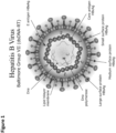
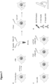
- HBV consists of a lipid bilayer with surface proteins that envelopes a capsid, a multimer assembled from HBc proteins, that contains the viral DNA ( Figure 1 ).
- the virus uses the surface lipopeptide pre-S1 for docking to mature liver cells via the hepatocyte sodium/bile acid cotransporter (sodium taurocholate co-transporting polypeptide - NTCP) and subsequently entering the cells (Tian et al., 2021; Watashi & Wakita, 2015).
- a recently EMA-approved peptide-based drug Hepcludex is based on the viral pre-S1 and also binds NTCP, thereby blocking the viral entry to hepatocytes (Gripon et al., 2005; Volz et al., 2013).
- the peptide LLGRMK (SEQ ID NO. 20) bound the full-length wild type HBc (fl wt HBc) and the related octa-peptide ALLGRMKG (SEQ ID NO. 21) inhibited HBc's interaction to the surface protein L-HBs (Dyson & Murray, 1995).
- the peptide GSLLGRMKGA (SEQ ID NO. 10) was seen to bind the tips of the spikes of recombinantly purified HBc capsids(Böttcher et al., 1998).
- GSLLGRMKGA SEQ ID NO. 10
- the related peptides SLLGRMKGA SEQ ID NO. 22
- SLLGRMKG SEQ ID NO. 23
- the tips of capsid spikes are the binding sites for the peptides MHRSLLGRMKGA (Pep1) (SEQ ID NO. 15) and GSLLGRMKGA (Pep2) (SEQ ID NO.
- MHRSLLGRMKGA and SLLGRM (SEQ ID NOs. 15, 10 and 5), we designed and synthesized peptidic dimers that bind HBc capsids with low micro- to nanomolar affinity and aggregate them in vitro. More importantly, in combination with thiol-reactive cell penetrating peptides, our MHRSLLGRMKGA-based thiolated dimeric superbinder P1dC, induces HBc core protein aggregation in live mammalian cells expressing recombinant HBV core protein. These new core protein aggregating peptidic dimers have great potential as new lead compounds for the interference with the DNA protein shield of HBV.
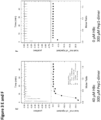
- the peptides SLLGRM, GSLLGRMKGA and MHRSLLGRMKGA (SEQ ID NOs. 5, 15 and 10) bind to wt and mutant HBc variants with micromolar affinity (Makbul, Khayenko, et al., 2021).
- To increase the affinity of these peptides we used the concept of avidity that is exceptionally useful in the case of HBc capsids, with 90 or 120 potential binding sites.
- We synthesised dimeric versions of SLLGRM, GSLLGRMKGA and MHRSLLGRMKGA (SEQ ID NOs. 5, 15 and 10) and connected them with PEG linkers ( Figure 2 ).
- the tips of the capsid spikes are spaced about 6 nm apart, so a simple dimer (as for example SLLGRM-SLLGRM) would not be long enough to simultaneously bind two capsid spikes. Therefore, the linker which connects the two peptides must be long enough to bridge the capsid spikes which are spaced about 6 nm apart. Additionally, the linker should be flexible so the dimers can assume a multitude of different conformations ( Figure 2E ).
- the thiol enables cell delivery with the help of thiol-reactive cell penetrating peptides, that are cleaved off the active molecule within the cell cytosol (Schneider, Benz, et al., 2021; Schneider, Kithil, et al., 2021).
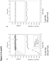
- P1dC retained the nanomolar binding affinity to HBV core protein ( Figure 5A,B , Table 1), while the control scrambled analogue scrP1dC showed no binding to the viral core protein ( Figure 5C,D ).
- P1dC is suitable as a new lead compound for the treatment of chronic hepatitis.
- Pep2 at high micromolar concentrations can reduce the production HB viruses to ca. 30 % in Hep G2, liver hepatitis model cells (Böttcher et al., 1998).
- P1dC, having nanomolar affinity, evident aggregating properties, and established in-cell delivery can further reduce the production of HB viruses or even block it completely.
- FIG. 3 SLLGRM-, Pep2- and Pep1- dimers bindHBV core proteins in low micromolar to nanomolar affinities. Interaction analysis of HBV capsids and the SLLGRM-, Pep2- and Pep1- dimers by ITC.
- N stoichiometry of the interaction
- K association constant
- ⁇ H binding enthalpy
- ⁇ S binding entropy
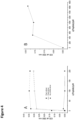
- FIG. 4 The peptidic dimers induce turbidity in HBc solutions. Assessment of HBc solution turbidity with peptide-dimers at 350 nm.
- FIG. 5 P1dC binds HBc with nanomolar affinity. Interaction analysis of HBV capsids with P1dC and the scrambled dimer scrP1dC by ITC.
- FIG. 6 The peptide dimers bind to the tips of the spikes of the HBV core protein.
- HBc capsids with bound SLLGRMdimer or P1dC imaged by electron cryomicroscopy.
- Figure 7 Experimental setup for live cell experiments.
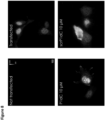
- FIG. 8 Transfection experiments with P1dC and scrP1dC.
- Exemplary images of wild-type and HBc expressing mammalian cells that were treated either with P1dC or the scrambled scrP1dC.
- the wild-type cells show no fluorescence, indicating the lack of HBc expression and the lack of unspecific binding of the HBc antibody or the secondary antibody.
- the untreated transfected HEK293 cells show a homogenous fluorescence in the nucleus area and some fluorescent punctae in the cytosol, indicating a homogenous distribution of HBc in the nucleus area with some of HBc located in the cytosol.
- Cells treated with P1dC show an anormal morphology and large fluorescent lumps, indicating aggregates of HBc protein.
- Cells treated with scrP1dC show an anormal morphology but no fluorescent lumps, implying the lack of HBc aggregation.
- peptide chain was elongated by adding the desired amino acid (AA, 3 eq.) with ethylcyanohydroxyiminoacetate (Oxyma, 3 eq.) and N,N'-diisopropylcarbodiimide (DIC, 3 eq.). Capping was done with N,N-diisopropylethylamine (DIEA, 50 eq.) and acetic anhydride (50 eq.) in N-methyl-2-pyrrolidone for 30 min. Coupling efficiency was monitored by measuring the absorption of the dibenzofulvene-piperidine adduct after deprotection.
- DIEA N,N-diisopropylethylamine
- acetic anhydride 50 eq.
- the peptide chain was elongated with further identical deprotection-conjugation cycles and after the completion the peptides were cleaved from the resin using a cocktail of 94% trifluoracetic acid (TFA), 3% H 2 O, 3% Triisopropylsilane (TIPS), for 4 hours at RT.
- TFA trifluoracetic acid
- TIPS Triisopropylsilane
- the peptides were precipitated in ice-cold ether and then purified with HPLC and analyzed by LC-MS, as described below.
- a 10-mer oligoarginine peptide connected through di-8-Amino-3,6-dioxaoctanoic acid (DOA) linker to a cysteine (C-DOA-DOA-RRRRRRRRRR; SEQ ID NO. 24) was reacted with 10 equivalents of 5,5-dithio-bis-(2-nitrobenzoic acid) in 1: 1 DMF : 0.1 M phosphate buffer for 30 min with agitation at room temperature (RT). Then the reaction mixture was directly injected in semi-preparative HPLC, purified and analyzed by LC-MS, as described below.
- the crude peptides and peptidic compounds were purified by reverse phase HPLC using water acetonitrile gradient with 0.1% formic acid (FA).
- LC-MS validation was performed with similar gradient and LC-MS grade solvents.
- Semi-preparative HPLC was performed on Shimadzu Prominence equipped with a diode-array detector (DAD) system using a C18 reverse-phase column (Phenomenex, Aillesburg, Germany), Onyx Monolithic HD-C18 100 ⁇ 4.6 mm or Onyx Monolithic C18 100 ⁇ 10 mm).
- DAD diode-array detector
- ⁇ SPOT peptide arrays (Dikmans et al., 2006) were synthesized using a MultiPep RSi robot (CEM GmbH, Kamp-Lintford, Germany) on in-house produced, acid labile, amino-functionalized, cellulose membrane discs containing 9-fluorenylmethyloxycarbonyl- ⁇ -alanine (Fmoc- ⁇ -Ala) linkers (average loading: 130 nmol/disc-4 mm diameter). Synthesis was initiated by Fmoc deprotection using 20% piperidine in DMF followed by washing with DMF and ethanol (EtOH).
- Peptide chain elongation was achieved using a coupling solution consisting of preactivated amino acids (0.5 M) with Oxyma (1 M) and DIC (1 M) in DMF (1:1:1, aa:oxyma:DIC). Couplings were carried out for 3 ⁇ 30 min, followed by capping (4% acetic anhydride in DMF) and washes with DMF and EtOH. Synthesis was finalized by deprotection with 20% piperidine in DMF (2 ⁇ 4 ⁇ L/disc for 10 min each), followed by washing with DMF and EtOH.
- Dried discs were transferred to 96 deep-well blocks and treated, while shaking, with side-chain deprotection solution, consisting of 90% TFA, 2% DCM, 5% H 2 O and 3% TIPS (150 ⁇ L/well) for 1.5 h at RT. Afterward, the deprotection solution was removed and the discs were solubilized overnight (ON) at RT, while shaking, using a solvation mixture containing 88.5% TFA, 4% trifluoromethanesulfonic acid (TFMSA), 5% H2O and 2.5% TIPS (250 ⁇ L/well).
- side-chain deprotection solution consisting of 90% TFA, 2% DCM, 5% H 2 O and 3% TIPS (150 ⁇ L/well) for 1.5 h at RT. Afterward, the deprotection solution was removed and the discs were solubilized overnight (ON) at RT, while shaking, using a solvation mixture containing 88.5% TFA, 4% trifluoromethanes
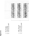
- microarray slides were blocked for 60 min in 5% (w/v) skimmed milk powder (Carl Roth) phosphate-buffered saline (PBS; 137 mM NaCl, 2.7 mM KCl, 10 mM Na 2 HPO 4 , 1.8 mM KH 2 PO 4 , pH 7.4). After blocking, the slides were incubated for 30 min with 55 nM (monomer equivalent) of HBc in the blocking buffer, then washed 3 times with PBS.
- PBS phosphate-buffered saline
- HBc was detected with a primary 1:2500 diluted mAb16988 (anti-hepatitis B virus antibody, core antigen, clone C1-5, aa 74-89, Millipore Sigma, Darmstadt, Germany) and a secondary 1:5000 diluted HRP-coupled Anti-mouse antibody (#31430, Invitrogen).
- the antibodies were applied in blocking buffer for 30 min, with three PBS washes between the antibodies and after applying the secondary antibody.
- the chemiluminescent readout was obtained using SuperSignal West Femto maximum sensitive substrate (Thermo Scientific GmbH, Schrö, Germany) with a c400 Azure imaging system (lowest sensitivity, 10 seconds exposure time).
- HBV core protein binding was successfully determined in microarray format.
- CLP capsid like particles
- HBc capsid core protein
- all numbers for concentrations, ITC parameters etc. refer to a monomeric HBc.
- the dialysed sample was removed from the dialysis tube and concentrated in a centrifuge using a concentrator (30 kDa MWCO Spin-X UF 6 mL, Corning Inc., Corning, NY, USA). Before the determination of the concentration by the Bradford assay (Roti Nanoquant, Carl Roth GmbH Co. KG, Düsseldorf, Germany) the concentrate was filtered (centrifugal filter unit Ultrafree MC, pore size of 100 nm, Merck KGaA, Darmstadt, Germany). The dimeric peptides were dissolved in the buffer obtained from the dialysis of capsids.
- thermograms were integrated and the corresponding isotherms were fitted using a one site model. All obtained thermodynamic parameters refer to concentrations of monomeric HBc. All ITC experiments were complemented with control experiments where solutions of dimeric peptides were titrated into dialysis buffer.
- Example 9 Cryogenic grid preparation of capsids in complex with peptide-dimers
- ethane was used as medium (liquefied by liquid nitrogen) with the help of a Vitrobot (mark IV, FEI Company, Hillsboro, OR, USA) using filter papers of Whatman (type 541).
- the Vitrobot had following settings: no wait and drain times, 6 s of blot time, blot force of 25 and a nominal humidity of 100 %.
- the frozen grids were stored in liquid nitrogen for at least one night before being used for image acquisition.
- Cryo-EM was done as previously described (Makbul, Kraft, et al., 2021).
- Movies were acquired with the software EPU on a Krios G3 electron microscope equipped with a Falcon III camera (Thermo Fisher Scientific, Hillsboro, OR, USA) in linear mode at a magnification of 75,000 with an accelerating voltage of 300 kV. The total exposure was 40 e-/ ⁇ 2 and was fractionated across 20 fractions.
- HBc capsids with bound P1C dimers 3 movies were acquired per hole and one hole was acquired per stage position.
- For HBc capsids with bound SLLGRM dimers at each stage position three movies were acquired per hole from the central hole and the four closest neighbouring holes.
- Example 11 Modelling of Cryo-EM maps, refinement of PDB files and their validation
- Example 13 HEK293 cell cultures and transfection
- HEK293 cells were cultured in DMEM (GIBCO), supplemented with GlutaMax and pyruvate (GIBCO), 10% fetal bovine serum (GIBCO) and 1% Penicillin/Streptomycin (Sigma) at 37°C and with 5% CO 2 .
- the cells were plated on 0.15 mm thick 18 mm glass coverslips coated with 35 ⁇ g/ml Poly-D-Lysine in a 12-well plate and were transfected with the cloned 1 ⁇ g plasmid DNA per coverslip using PEI (Polyethylenimine). The transfection was performed at 60-80% confluence. Shortly before transfection the medium was changed to fresh DMEM.
- the DNA was added to 100 ⁇ l DMEM without additives and mixed, 4 ⁇ l fresh PEI (1 mg/ml) was added, mixed immediately and incubated for 20 min at RT.
- the transfection mix was pipetted dropwise on cells while swirling, and incubated overnight.
- the medium was changed to fresh DMEM with 2% FBS after 12-24 hours, and on the following day the cells were used for live assays, then fixed and stained.
- Live HEK293 cells expressing HBc were incubated with 10 ⁇ M P1dC and with 10 ⁇ M of the scrambled version of the peptide, both peptides in-situ activated with the reactive CPP. After the incubation the treated and the untreated live HEK293 cells expressing HBc and also wild-type HEK293 cells were washed and fixed with 0.1 M sodium phosphate buffer pH 7.4 containing 4% paraformaldehyde (EM grade, Polysciences) and 1% sucrose for 10-20 min at 37°C.
- 0.1 M sodium phosphate buffer pH 7.4 containing 4% paraformaldehyde EM grade, Polysciences
- the coverslips with the cell samples were inserted in an imaging chamber (Ludin Chamber Type 1, Life Imaging Services) and imaged in PBS.
- the measurements were taken from distinct samples with a sample size ⁇ 2, for each group.
- a series of images, used to generate the datapoints, were acquired from different regions of the sample, each region having a distinct group of cells.
- the samples were imaged on an inverted Leica DMI6000B microscope with a 100x oil-immersion objective (NA 1.49) using a Leica DFC9000 GTC VSC-05760 sCMOS camera (16-bit, image pixel size: 130 nm).
- the 628/40 excitation and 692/40 emission filter was used for DyLight650, 10 images were acquired at a frame rate (exposure time) of 100 ms and constant illumination intensity to ensure comparability. n ⁇ 10.
Landscapes
- Chemical & Material Sciences (AREA)
- Health & Medical Sciences (AREA)
- Organic Chemistry (AREA)
- Life Sciences & Earth Sciences (AREA)
- General Health & Medical Sciences (AREA)
- Medicinal Chemistry (AREA)
- Molecular Biology (AREA)
- Proteomics, Peptides & Aminoacids (AREA)
- Genetics & Genomics (AREA)
- Biophysics (AREA)
- Biochemistry (AREA)
- Virology (AREA)
- General Chemical & Material Sciences (AREA)
- Chemical Kinetics & Catalysis (AREA)
- Public Health (AREA)
- Veterinary Medicine (AREA)
- Gastroenterology & Hepatology (AREA)
- Pharmacology & Pharmacy (AREA)
- Nuclear Medicine, Radiotherapy & Molecular Imaging (AREA)
- Animal Behavior & Ethology (AREA)
- Engineering & Computer Science (AREA)
- Communicable Diseases (AREA)
- Bioinformatics & Cheminformatics (AREA)
- Oncology (AREA)
- Biotechnology (AREA)
- Peptides Or Proteins (AREA)
- Medicines That Contain Protein Lipid Enzymes And Other Medicines (AREA)
Abstract
The present invention relates to a compound comprising a multimer, preferably a dimer, of peptides or peptide analogues each comprising the amino acid sequence X1X2LGX3X4 and a linker connecting the peptides or peptide analogues, and pharmaceutical compositions and kits comprising said compound. The present invention further relates to the compound for use in the treatment of a Hepatitis B virus (HBV) infection or a liver disease caused by HBV. The present invention also relates to a method of the treatment of a HBV infection or a liver disease caused by HBV.
Description
- The present invention relates to a compound comprising a multimer, preferably a dimer, of peptides or peptide analogues each comprising the amino acid sequence X1X2LGX3X4 and a linker connecting the peptides or peptide analogues, and pharmaceutical compositions and kits comprising said compound. The present invention further relates to the compound for use in the treatment of a Hepatitis B virus (HBV) infection or a liver disease caused by HBV. The present invention also relates to a method of the treatment of a HBV infection or a liver disease caused by HBV.
- Hepatitis B virus (HBV) is a major human pathogen from the Hepadnaviridae family that consists of an outer lipid envelope and an icosahedral protein nucleocapsid core that encapsulates partially double-stranded DNA. Despite the worldwide vaccination program, HBV remains a global health problem. In 2019, at least 296 million people, or 4% of the world's population, had chronic Hepatitis B. HBV infection is the main cause of liver cirrhosis and hepatocellular carcinoma, estimated to be responsible for ~800,000 deaths annually (Block et al., 2021; World Health Organisation, 2022). In 2016, the World Health Organization set a goal of eliminating viral hepatitis as a public health threat by 2030. Achieving this goal requires the development of novel therapeutic treatments to cure chronic hepatitis B, medications that will prevent its transmission and expanding the vaccination program to prevent new infections (Block et al., 2021; Cox et al., 2020).
- For HBV, so far, the available therapies include nucleoside/nucleotide analogues and alpha-interferons, which are effective in reducing the viral load, but are poorly effective in curing persistent infection (Block et al., 2021; Hu et al., 2019). However over 50 drugs are currently in development, among them 13 capsid inhibitors, as can be seen on the Website of the Hepatitis B Foundation (https://www.hepb.org/treatment-and-management/drug-watch/).
- There is a need in the art for improved means and methods for treating HBV infections as well as chronic hepatitis B.
- According to the present invention this object is solved by a compound comprising
- (i) a multimer of a peptide or peptide analogue P,
wherein the peptide or peptide analogue P comprises the amino acid sequence
X1X2LGX3X4 SEQ ID NO. 1,
wherein- X1 is selected from serine (S), glutamine (Q), asparagine (N), glycine (G), glutamate (E), aspartate (D), alanine (A), and non-natural amino acids comprising hydrophilic exchange(s),
- X2 is selected from leucine (L), isoleucine (I) and non-natural amino acids comprising hydrophobic exchange(s),
- X3 is selected from arginine (R), phenylalanine (F), glutamic acid (E) and non-natural amino acids comprising hydrophobic or hydrophilic exchange(s),
- X4 is selected from methionine (M), valine (V), leucine (L), isoleucine (I) phenylalanine (F), and non-natural amino acids comprising hydrophobic exchange(s), and wherein the peptide or peptide analogue has a length of 6 to 13 amino acids,
- (ii) a linker L connecting the peptides or peptide analogues P,
wherein the linker has a length of 60 to 160 Å. - According to the present invention this object is solved by a pharmaceutical composition comprising
- a compound of the present invention, and
- optionally, pharmaceutically acceptable excipient(s) and/or carrier.
- According to the present invention this object is solved by using a compound of the present invention for binding and/or aggregating the core protein of Hepatitis B virus (HBV).
- According to the present invention this object is solved by providing the compound of the present invention or the pharmaceutical composition of the present invention for use in medicine.
- According to the present invention this object is solved by providing the compound of the present invention or the pharmaceutical composition of the present invention for use in the treatment of a Hepatitis B virus (HBV) infection or a liver disease caused by HBV.
- According to the present invention this object is solved by a kit comprising
- (a) a compound of the present invention or a pharmaceutical composition of the present invention, and
- (b) a cell penetrating peptide (CPP), preferably a thiol-reactive CPP.
- According to the present invention this object is solved by a method for the treatment of a Hepatitis B virus (HBV) infection or a liver disease caused by HBV, comprising the administration of a therapeutically effective amount of a compound of the present invention or the pharmaceutical composition of the present invention to a subject in need thereof.
- Before the present invention is described in more detail below, it is to be understood that this invention is not limited to the particular methodology, protocols and reagents described herein as these may vary. It is also to be understood that the terminology used herein is for the purpose of describing particular embodiments only and is not intended to limit the scope of the present invention which will be limited only by the appended claims. Unless defined otherwise, all technical and scientific terms used herein have the same meanings as commonly understood by one of ordinary skill in the art. For the purpose of the present invention, all references cited herein are incorporated by reference in their entireties.
- Concentrations, amounts, and other numerical data may be expressed or presented herein in a range format. It is to be understood that such a range format is used merely for convenience and brevity and thus should be interpreted flexibly to include not only the numerical values explicitly recited as the limits of the range, but also to include all the individual numerical values or sub-ranges encompassed within that range as if each numerical value and sub-range is explicitly recited. As an illustration, a numerical range of "6 to 13" should be interpreted to include not only the explicitly recited values of 6 to 13, but also include individual values and sub-ranges within the indicated range. Thus, included in this numerical range are individual values such as 6, 7, 8, 9, 10, 11, 12, 13 and sub-ranges such as from 6 to 12, 6 to 10, etc. This same principle applies to ranges reciting only one numerical value, such as "at least 6". Furthermore, such an interpretation should apply regardless of the breadth of the range or the characteristics being described.
- The present inventors have developed compounds that bind and/or aggregate the core protein of Hepatitis B virus.
- A compound according to the present invention comprises
- (i) a multimer of a peptide or peptide analogue P, and
- (ii) a linker L connecting the peptides.
- The compounds of the present invention comprise a multimer of peptides or peptide analogues P.
- The multimer can be a dimer, trimer, tetramer, pentamer or comprise more than five peptides or peptide analogues P.
- A "peptide analogue" as used herein refers to a modified peptide or to a compound that mimics structural or functional aspects of a peptide. The term "peptidomimetic" can be used interchangeably with the term "peptide analogue". Examples are D-peptides, β-peptides or peptoids. In peptoids, the side chain is connected to the nitrogen of the peptide backbone, instead of the α-carbon as in peptides. The altered chemical structure of a peptide analogue or peptidomimetic is designed to advantageously adjust the molecular properties, such as stability or biological activity while maintaining structural or functional features. Peptide analogues or peptidomimetics can also comprise non-natural amino acids. These modifications involve changes to the peptide that will not occur naturally, such as altered backbones or altered sidechains. Non-natural amino acids can be generated from their native analogs via modifications, such as amine alkylation, side chain substitution, structural bond extension cyclization, and isosteric replacements within the amino acid backbone.
- The peptide or peptide analogue P comprises the amino acid sequence
X1X2LGX3X4 (SEQ ID NO. 1),
wherein: - X1 is selected from serine (S), glutamine (Q), asparagine (N), glycine (G), glutamate (E), aspartate (D) and alanine (A), and non-natural amino acids comprising hydrophilic exchange(s),
- such as D-serine (s), D-glutamine (q), D-asparagine (n), D-glutamate (e), D-aspartate (d) and D-alanine (a), and further amino acid side chains with a carboxyl group in their side chain.
- X2 is selected from leucine (L), isoleucine (I) and non-natural amino acids comprising hydrophobic exchange(s),
- such as 3-aminopentane-3-carboxylic acid, L-cyclopentylglycine, L-cyclopropylglycine, L-cyclohexylglycine, L-t-leucine, L-neopentylglycine, alpha-methyl-L-valine, and allylglycine.
- X3 is selected from arginine (R), phenylalanine (F), glutamic acid (E) and non-natural amino acids comprising hydrophobic or hydrophilic exchange(s),
- such as L-phenylglycine, and L-Tyr or L-Phe substituted with halogene (Br, J or Cl).
- X4 is selected from methionine (M), valine (V), leucine (L), isoleucine (I), phenylalanine (F), and non-natural amino acids comprising hydrophobic exchange(s),
- such as 3-aminopentane-3-carboxylic acid, L-cyclopentylglycine, L-cyclopropylglycine, L-cyclohexylglycine, L-t-leucine, L-neopentylglycine, alpha-methyl-L-valine, allylglycine, L-phenylglycine, and L-Tyr or L-Phe substituted with halogene (Br, J or Cl).
- In a preferred embodiment,
- X1 is selected from serine (S), glutamine (Q), asparagine (N), glycine (G), glutamate (E), aspartate (D) and alanine (A),
- X2 is selected from leucine (L) and isoleucine (I),
- X3 is selected from arginine (R), phenylalanine (F), and glutamic acid (E), preferably arginine (R) and phenylalanine (F),
- X4 is selected from methionine (M), valine (V), leucine (L), isoleucine (I) and phenylalanine (F).
- The peptides or peptide analogues of the present invention comprise amino acids, which can be both L- or D-amino acids.
- In one embodiment, the amino acid in position X1 is a non-natural amino acid comprising a hydrophilic exchange. Examples for such non-natural amino acids comprising hydrophilic exchange(s) include but are not limited to D-serine (s), D-glutamine (q), D-asparagine (n), D-glutamate (e), D-aspartate (d) and D-alanine (a), and further amino acid side chains with a carboxyl group in their side chain.
- In one embodiment, the amino acid in position X2 and/or X4 is a non-natural amino acid comprising a hydrophobic exchange.
- Examples for such non-natural amino acids comprising hydrophobic exchange(s) include but are not limited to 3-aminopentane-3-carboxylic acid, L-cyclopentylglycine, L-cyclopropylglycine, L-cyclohexylglycine, L-t-leucine, L-neopentylglycine, alpha-methyl-L-valine, allylglycine, L-phenylglycine and L-Tyr or L-Phe substituted with halogene (Br, J or Cl).
- In one embodiment, the amino acid in position X3 is a non-natural amino acid comprising a hydrophobic exchange or a hydrophilic exchange.
- X1 is preferably serine (S).
- X2 is preferably leucine (L).
- X3 is preferably arginine (R),
- X4 is preferably leucine (L) or methionine (M).
- In one embodiment, X1 is serine (S), X2 is leucine (L), X3 is arginine (R) and/or X4 is leucine (L) or methionine (M).
- In one embodiment, P comprises or consists of the amino acid sequence SLLGRX4 (SEQ ID NO. 2), wherein X4 is selected from methionine (M), valine (V), leucine (L), isoleucine (I), phenylalanine (F), and non-natural amino acids comprising hydrophobic exchange(s), as defined above, preferably from M, L, I, V or F. X4 is preferably L or M.
- Said amino acid sequence of SEQ ID NO. 2 is the core binding motif or essential sequence for binding to the HBV core protein.
- According to the invention, the compound of the present invention comprises at least two of said core binding motifs or essential sequences.
- The peptide or peptide analogue P has a length of at least 6 amino acids.
- The length is in the range of 6 to 13 amino acids, preferably 6 to 12 amino acids, such as 6 amino acids, 10 amino acids or 12 amino acids.
- In a preferred embodiment, the peptide or peptide analogue P has a length of 10 amino acids
- In one embodiment, the peptide or peptide analogue P comprises or consists of the amino acid sequence of GSLLGRX4 KGA (SEQ ID NO. 3) or MHRSLLGRX4 KGA (SEQ ID NO. 4), wherein X4 is selected from methionine (M), valine (V), leucine (L), isoleucine (I), phenylalanine (F), and non-natural amino acids comprising hydrophobic exchange(s), as defined above, preferably from M, L, I, V or F.
- X4 is preferably L or M.
- In one embodiment, the peptide or peptide analogue P comprises or consists of the amino acid sequence of
SLLGRX4 SEQ ID NO. 2, GSLLGRX4 KGA SEQ ID NO. 3, or MHRSLLGRX4 KGA SEQ ID NO. 4, - X4 is preferably L or M.
- In one embodiment, the peptide or peptide analogue P comprises or consists of an amino acid sequence which is selected from
SLLGRM SEQ ID NO. 5, SLLGRL SEQ ID NO. 6, SLLGRI SEQ ID NO. 7, SLLGRV SEQ ID NO. 8, SLLGRF SEQ ID NO. 9, GSLLGRMKGA SEQ ID NO. 10, GSLLGRLKGA SEQ ID NO. 11, GSLLGRIKGA SEQ ID NO. 12, GSLLGRVKGA SEQ ID NO. 13, GSLLGRFKGA SEQ ID NO. 14, MHRSLLGRMKGA SEQ ID NO. 15, MHRSLLGRLKGA SEQ ID NO. 16, MHRSLLGRIKGA SEQ ID NO. 17, MHRSLLGRVKGA SEQ ID NO. 18, and MHRSLLGRFKGA SEQ ID NO. 19, SLLGRM SEQ ID NO. 5, SLLGRL SEQ ID NO. 6, GSLLGRMKGA SEQ ID NO. 10, GSLLGRLKGA SEQ ID NO. 11, MHRSLLGRMKGA SEQ ID NO. 15, and MHRSLLGRLKGA SEQ ID NO. 16. - In a preferred embodiment, the compound of the present invention has formula I
(P)m - L - (Z)n (I),
wherein - P is the peptide or peptide analogue,
- m is at least 2, preferably, 2, 3, 4 or 5,
- L is the linker connecting the peptides or peptide analogues P,
- Z is a further moiety and n is 0 or 1.
- In one embodiment, m is 2. The compound has formula Ia:

- The two peptides or peptide analogues P can be identical in their amino acid sequence or can have a different amino acid sequence.
- The two peptides or peptide analogues P can have the same length or have a different length. The dimer is considered a homodimer, when the two peptides or peptide analogues are identical in their amino acid sequence and their length.
- The dimer is considered a heterodimer, when the two peptides or peptide analogues differ in their amino acid sequence or their length or both.
- In one embodiment, m is 3. The compound has formula Ib:

- The three peptides or peptide analogues P can be identical in their amino acid sequence or can have a different amino acid sequence.
- The three peptides or peptide analogues P can have the same length or have a different length.
- In one embodiment, m is 4. The compound has formula Ic:

- The four peptides or peptide analogues P can be identical in their amino acid sequence or can have a different amino acid sequence.
- The four peptides or peptide analogues P can have the same length or have a different length.
- In one embodiment, m is 5. The compound has formula Id:
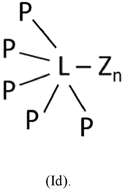
- The five peptides or peptide analogues P can be identical in their amino acid sequence or can have a different amino acid sequence.
- The five peptides or peptide analogues P can have the same length or have a different length.
- The compounds of the present invention comprise a linker L which connects the peptides or peptide analogues P.
- The linker has a length of 60 to 160 Angstrom (Å), preferably 60 to 100 Å.
- In a preferred embodiment the linker has a length of 80 Å.
- The linker may be covalently bound to the peptides or peptide analogues simultaneously. Said linker may be bound to the C-terminus of each peptide or peptide analogue. In one embodiment, the linker is bound to a cysteine situated at the C-terminal end of each peptide or peptide analogue. The binding may be via the sulfur atom comprised in the cysteine. The said linker may be attached to the C-terminal end of each peptide or peptide analogue, for example via an amide bond with the terminal amino acid.
- The linker keeps the peptides or peptide analogues comprised in the multimer, such as the dimer, at a certain distance from each other, so that they can interact with several (such as two) binding sites simultaneously, hence enhancing the affinity of the compound of the invention. In a preferred embodiment, the several (such as two) binding sites are located on different HBV core particles (such as two different HBV core particles).
- The linker brings the peptides or peptide analogues P into a preferred or suitable proximity to induce aggregation.
- The linker may keep the peptides or peptide analogues comprised in the multimer, such as the dimer, at a distance between 60 and 160 Å from each other. The distance may be at least 60 Å. The distance may also be at least 70 Å. The distance may also be at least 80 Å. The distance may also be at least 90 Å. The distance may also be at least 100 Å. The distance may also be about 90 Å. The distance may also be about 100 Å. The distance may also be about 110 Å. The distance may also be about 120 Å. A preferred distance is about 80 Å.
- Preferably, the linker L comprises or consists of
- an alkane chain,
- diaminoacetic acid,
- maleimide,
- ethylene glycol,
- polyethylene glycol (PEG),
- NPEG,
- glycine,
- polyglycine, or
- any combination thereof.
- In a preferred embodiment, the linker L comprises at least two ethylene glycol moieties, such as 12, and/or at least two glycine,
and/or polyglycine. - In one embodiment, the linker L comprises at least 12 ethylene glycol moieties which results in a distance of at least 60 Å between the two peptides.
- The term "ethylene glycol moiety", also called oxyethylene, as used herein refers to the structural unit that constitutes a polyethylene glycol (PEG).
- The term "NPEG ", is a linker derived from the classical PEG, but where one or more of the backbone oxygen atoms is replaced with a nitrogen atom.
- In one embodiment, the linker L comprises the following structure
- [-NH-(CH2)2-O-(CH2)2-O-(CO)-]4-NH-(CH2)4-CH(R1)-NH-[-(CO)-CH2-O-(CH2)2-O-(CH2)2-NH]4-.
- further moiety
- In a preferred embodiment, the compound of the present invention comprises a further moiety Z which is connected to the linker L.
- In one embodiment, the further moiety Z is a reactive group.
- Said reactive group is for binding to a cell-penetrating peptide (CPP).
- A cell penetrating peptide (CPP) as used herein refers to a peptide characterized by the ability to cross the plasma membrane of mammalian cells, and thereby may give rise to the intracellular delivery of cargo molecules, such as peptides, proteins, oligonucleotides to which it is linked.
- The CPP preferably comprises at least 4 and at the most 13 amino acid residues selected from arginine and/or lysine. In one embodiment, the CPP comprises 5 arginine residues. In one embodiment, the CPP comprises 10 arginine residues.
- In one embodiment, the reactive group is a thiol group. In the embodiment, where the reactive group is a thiol group, the CPP is a thiol-reactive CPP.
- An example for a thiol-reactive CPP is TNB-R10, which is a CPP containing 10 arginine residues and a cysteine bound to 5-thio-2-nitro-benzoic acid for reacting with the thiol group:

- In one embodiment, the further moiety Z is a moiety comprising said reactive group.
- In the embodiment, where the reactive group is a thiol group, said moiety comprising the thiol group is cysteine, homocysteine, 2-amino-5-mercaptopentanoic acid, alpha-methyl-cysteine, penicillamine, or 5-mercapto-norvaline, preferably cysteine.
- In one embodiment, where the reactive group is a thiol group, the CPP is a thiol-reactive CPP, as discussed above.
- In a preferred embodiment the linker has a compound comprising a thiol group as further moiety Z attached as follows:

- See also
Figure 2E . - In one embodiment, the further moiety Z is a CPP.
- In this embodiment, the CPP is directly bound to the linker, such as via an amide bond.
- As outlined above, the present invention provides a pharmaceutical composition comprising a compound of the present invention.
- A pharmaceutical composition comprising a compound of the present invention optionally comprises one or more pharmaceutically acceptable excipients and/or carrier.
- Preferably, the pharmaceutical composition is suitable for intravenous, subcutaneous, intranasal, buccal, sublingual, oral, rectal and/or intraperitoneal administration.
- As outlined above, the present invention provides a kit comprising
- (a) a compound of the present invention or a pharmaceutical composition of the present invention, and
- (b) a cell-penetrating peptide (CPP).
- In a preferred embodiment, the CPP is a thiol-reactive CPP, as discussed above.
- As outlined above, the present invention provides the use of a compound of the present invention for binding and/or aggregating the core protein of Hepatitis B virus (HBV).
- Said binding and/or aggregating of the HBV core protein is for preventing the virion formation and/or reducing the viral load.
- As outlined above, the present invention provides a compound of the present invention or a pharmaceutical composition of the present invention for use in medicine.
- As outlined above, the present invention provides a compound of the present invention or a pharmaceutical composition of the present invention for use in the treatment of a Hepatitis B virus (HBV) infection or a liver disease caused by HBV.
- In one embodiment, the HBV infection is a chronic HBV infection.
- In one embodiment, the liver disease caused by HBV is hepatitis.
- In one embodiment, the use comprises the use of a CPP.
- In a preferred embodiment, the CPP is a thiol-reactive CPP, as discussed above.
- As outlined above, the present invention provides a method for the treatment of a Hepatitis B virus (HBV) infection or a liver disease caused by HBV.
- Said method comprises the administration of a therapeutically effective amount of a compound of the present invention or the pharmaceutical composition of the present invention to a subject in need thereof.
- A "therapeutically effective amount" of a compound of the present invention refers to the amount which has to be administered to a subject in need thereof in order to achieve a desired therapeutic result or outcome. The skilled artisan will be able to determine said therapeutically effective amount and the suitable administration regimen.
- In one embodiment, the HBV infection is a chronic HBV infection.
- In one embodiment, the liver disease caused by HBV is hepatitis.
- In one embodiment, the method comprises the co-administration of a cell penetrating peptide (CPP).
- In a preferred embodiment, the CPP is a thiol-reactive CPP, as discussed above.
- The administration of the compound or the pharmaceutical composition is preferably intravenous, subcutaneous, intranasal, buccal, sublingual, oral, rectal and intraperitoneal.
- Hepatitis B is a global public health threat with hundreds of millions of people affected worldwide. Hepatitis B virus is a cause of this disease. The available therapy has low efficacy in curing persistent infection; therefore, an exhaustive effort is done to bring new therapeutics to the market.
- We have developed a peptidic compound capable of affecting the core protein of the virus, that encircles the viral DNA. We invented a synthetic HBc binding dimeric peptidic superbinder, that has nanomolar affinity and induces core protein aggregation in vitro and evidently in live cells expressing the recombinant HBV core protein HBc. Our novel HBc aggregator has great potential as lead substance for the interference with the envelopment of the nucleocapsid.
- The hepatotropic hepatitis B virus (HBV) and its satellite virus hepatitis D (HDV) infect the liver (Watashi & Wakita, 2015). An infection with HBV is usually cleared from the body with lifelong immunity but a significant number of infections become chronic. Chronic hepatitis can cause severe complications including steatosis, cirrhosis and hepatocellular carcinoma (HCC). Despite the existence of a vaccine which efficiently protects against HBV, with 296 million chronic carriers and over 800 000 of hepatitis-related yearly deaths, the disease remains a global health problem (Block et al., 2021; World Health Organisation, 2022).
- HBV consists of a lipid bilayer with surface proteins that envelopes a capsid, a multimer assembled from HBc proteins, that contains the viral DNA (
Figure 1 ). The virus uses the surface lipopeptide pre-S1 for docking to mature liver cells via the hepatocyte sodium/bile acid cotransporter (sodium taurocholate co-transporting polypeptide - NTCP) and subsequently entering the cells (Tian et al., 2021; Watashi & Wakita, 2015). A recently EMA-approved peptide-based drug Hepcludex is based on the viral pre-S1 and also binds NTCP, thereby blocking the viral entry to hepatocytes (Gripon et al., 2005; Volz et al., 2013). - More promising peptides were identified in a screen of a phage library. The peptide LLGRMK (SEQ ID NO. 20) bound the full-length wild type HBc (fl wt HBc) and the related octa-peptide ALLGRMKG (SEQ ID NO. 21) inhibited HBc's interaction to the surface protein L-HBs (Dyson & Murray, 1995). In a subsequent study, using Cryo-EM and image processing, the peptide GSLLGRMKGA (SEQ ID NO. 10) was seen to bind the tips of the spikes of recombinantly purified HBc capsids(Böttcher et al., 1998). Additionally, the same study demonstrated that GSLLGRMKGA (SEQ ID NO. 10) and the related peptides SLLGRMKGA (SEQ ID NO. 22) and SLLGRMKG (SEQ ID NO. 23) were able to inhibit HBV production in Hep G2 human hepatoblastoma cell lines with IC50 values of 0.8, 2.4 and 6.4 µM, respectively. In a recent study, we confirmed that the tips of capsid spikes are the binding sites for the peptides MHRSLLGRMKGA (Pep1) (SEQ ID NO. 15) and GSLLGRMKGA (Pep2) (SEQ ID NO. 10) with KD values of 26 and 68 µM, respectively (Makbul, Khayenko, et al., 2021). In the same study we identified SLLGRM (SEQ ID NO. 5) as the shortest peptide still able to bind recombinant HBc capsids with a KD value of 130 µM. However, the relatively low affinity and inability to effectively penetrate the cell membrane make these peptides poor pharmacological agents.
- Based on GSLLGRMKGA, MHRSLLGRMKGA and SLLGRM (SEQ ID NOs. 15, 10 and 5), we designed and synthesized peptidic dimers that bind HBc capsids with low micro- to nanomolar affinity and aggregate them in vitro. More importantly, in combination with thiol-reactive cell penetrating peptides, our MHRSLLGRMKGA-based thiolated dimeric superbinder P1dC, induces HBc core protein aggregation in live mammalian cells expressing recombinant HBV core protein. These new core protein aggregating peptidic dimers have great potential as new lead compounds for the interference with the DNA protein shield of HBV.
- The peptides SLLGRM, GSLLGRMKGA and MHRSLLGRMKGA (SEQ ID NOs. 5, 15 and 10) bind to wt and mutant HBc variants with micromolar affinity (Makbul, Khayenko, et al., 2021). To increase the affinity of these peptides, we used the concept of avidity that is exceptionally useful in the case of HBc capsids, with 90 or 120 potential binding sites. We synthesised dimeric versions of SLLGRM, GSLLGRMKGA and MHRSLLGRMKGA (SEQ ID NOs. 5, 15 and 10) and connected them with PEG linkers (
Figure 2 ). - The tips of the capsid spikes are spaced about 6 nm apart, so a simple dimer (as for example SLLGRM-SLLGRM) would not be long enough to simultaneously bind two capsid spikes. Therefore, the linker which connects the two peptides must be long enough to bridge the capsid spikes which are spaced about 6 nm apart. Additionally, the linker should be flexible so the dimers can assume a multitude of different conformations (
Figure 2E ). - Using isothermal titration calorimetry (ITC), we first determined that SLLGRM-, GSLLGRMKGA-, and MHRSLLGRMKGA-dimers bind the HBc capsids (
Figure 3 ). Gratifyingly, the KD values of 4.89, 1.91 and 0.312 µM of the dimers, were a 26-, 35- and 83-fold affinity increase over the respective monomers (130, 68 and 26 µM) (Makbul, Khayenko, et al., 2021) (Table 1).Table 1 Summary of thermodynamic parameters obtained by ITC experiments using the peptide dimers and fl wt HBc capsids. In case of the Pep2-dimer the deviations represent deviations of fit since only one ITC experiment was performed. N represents the stoichiometry. Peptide [SEQ ID NO. of monomer] N 
KD µM 

Repeats SLLGRM-dimer [SEQ ID NO. 5] 0.145 ±0.023 208200 ±31284 4.89 ±0.74 -25316 ±2748 -62 ±10 5 Pep2-dimer [SEQ ID NO. 5] 0.157 ±0.014 524000 ±107000 1.91 ±0.39 -29890 ±3100 -76 ±n/a 1 Pep1-dimer [SEQ ID NO. 10] 0.189 ±0.035 5752500 ±5798628 0.312 ±0.211 -25868 ±3922 -58 ±15 4 P1dC [SEQ ID NO. 15] 0.196 ±0.005 2486667 ±228384 0.42 ±0.038 -21700 ±624 -45 ±2 3 - Furthermore, we have noticed that the baselines of the thermograms of the Pep2- and the Pep1-dimers showed strong fluctuations in form of many endothermic and exothermic peaks (
Figure 3 ), suggesting protein aggregation. To evaluate the probable aggregation, we have measured the turbidity of HBc solutions with increasing concentrations of the peptidic dimers. All peptides showed increased turbidity with increasing concentrations, with Pep1- and Pep2-dimers inducing turbidity at low micromolar concentrations, while the SLLGRM-dimer induced turbidity at high micromolar concentrations, in accordance with their KD values (Figure 4 ). The turbidity increase of HBc solution upon the addition of the peptidic dimers further implied peptide-induced aggregation of core particles. - Motivated by the aggregating properties of the peptidic dimers and their possible interference with the virion assembly we have decided to modify the highest-affinity Pep1-based dimer to enable its in-vivo application. The peptidic dimer needs to be delivered within the cell to affect the HBV core protein. To this end we have introduced a thiol carrying moiety, cysteine, within the dimer architecture, creating a thiolated Pep1-dimer analogue, P1dC (
Figure 2D and F). The thiol enables cell delivery with the help of thiol-reactive cell penetrating peptides, that are cleaved off the active molecule within the cell cytosol (Schneider, Benz, et al., 2021; Schneider, Kithil, et al., 2021). P1dC retained the nanomolar binding affinity to HBV core protein (Figure 5A,B , Table 1), while the control scrambled analogue scrP1dC showed no binding to the viral core protein (Figure 5C,D ). - EM-maps of HBc capsids with bound SLLGRM dimers or P1dC confirmed that the peptides bind to the tips of the spikes (
Figure 6 ). The length of the resolved peptide density accommodates some 6 amino acids for both constructs. This suggests that a similar core binds to the tips of the spikes. Interestingly, SLLGRM dimers bind to both spikes of the asymmetric unit, while monomeric SLLGRM (SEQ ID DO. 5) binds only to the spikes closest to the threefold-symmetry axis (Makbul, Khayenko, et al., 2021). This implies that dimerization allows addressing the less affine binding site on HBc-capsids, which was impossible with the equivalent monomeric binder. The EM-maps confirm that the SLLGRM dimers and P1dC, both bind with a molar ratio of 0.25 to HBc capsids. The EM-maps did not resolve the linker region, which agrees with a flexible linker that has no specific binding site on the capsid surface. Our next goal was to demonstrate that P1dC affects HBV core protein within living mammalian cells. We incubated live HEK293 cells expressing HBc with P1dC and the scrambled scrP1dC together with the thiol-reactive cell penetrating peptide, then fixed the cells and visualized the HBV core proteins with the help of antibodies (Figure 7 ). Satisfyingly, we observed HBc aggregates in cells treated with P1dC, while cells incubated with the scrambled dimer showed HBc distribution similar to the untreated cells (Figure 8 ). - Based on known weak peptide binders of HBc we were able to develop high-affinity dimeric aggregator of the core protein, capable of HBc aggregation in living cells. We have based our strongest in-vivo binder, P1dC, on the strongest binder we identified using the ITC experiments, Pep1-dimer. It is worth mentioning that also other HBV core protein binding dimers we designed and synthesized showed aggregation potential in-vitro and this library of potential HBc aggregators could further be extended since there is a number of tolerable mutations in the binding sequence (
Figure 9 ). Furthermore, medicinal chemistry approaches can be applied to develop an even stronger peptidic binder, by, e.g., introducing cyclization or non-natural amino acids to the binding sequence. - We observed the formation of strongly fluorescing patches within living cells upon administration of the P1dC (
Figure 8 ) indicating the formation of HBc aggregates. The assembly of HB viruses within infected hepatocytes requires the formation of intact capsids surrounding the HBV genome followed by envelopment of the nucleocapsid and the export of virions. Our peptide dimers would likely interfere with the virion formation by aggregating HBc mono- or multimers, or even whole capsids. - P1dC is suitable as a new lead compound for the treatment of chronic hepatitis. Already several decades ago it was demonstrated that Pep2 at high micromolar concentrations can reduce the production HB viruses to ca. 30 % in Hep G2, liver hepatitis model cells (Böttcher et al., 1998). P1dC, having nanomolar affinity, evident aggregating properties, and established in-cell delivery can further reduce the production of HB viruses or even block it completely.
- The following examples and drawings illustrate the present invention without, however, limiting the same thereto.
-
- Figure 1.
- Structure of a Hepatitis B virus.
- Figure 2.
- Structure of specific peptides of the present invention.
- A) SLLGRM-dimer.
- B) GSLLGRMKGA-dimer (Pep2-dimer).
- C) MHRSLLGRMKGA-dimer (Pep1-dimer).
- D) MHRSLLGRMKGA-dimer-C (P1dC).
- E) Preferred linker with a length of 80 Å, including a cysteine (thiol group).
-
Figure 3 . SLLGRM-, Pep2- and Pep1- dimers bindHBV core proteins in low micromolar to nanomolar affinities. Interaction analysis of HBV capsids and the SLLGRM-, Pep2- and Pep1- dimers by ITC. - A) An exemplary titration of 150 µM HBc solution with 1500 µM solution of the SLLGRM-dimer. This ITC experiment was repeated 5 times under similar conditions (Table 1). A clear dose-response curve is derived from the heat injection profile.
- B) A control titration of an empty HBc buffer solution with 1500 µM solution of SLLGRM-dimer. No dose-response is evident.
- C) A titration of 50 µM HBc solution with 500 µM solution of the Pep2-dimer (Table 1). A clear dose-response curve is derived from the heat injection profile.
- D) A control titration of an empty HBc buffer solution with 500 µM solution of Pep2-dimer. No dose-response is evident.
- E) An exemplary titration of 40 µM HBc solution with 300 µM solution of the Pep1-dimer. This ITC experiment was repeated 4 times under similar conditions (Table 1). A clear dose-response curve is derived from the heat injection profile.
- F) A control titration of an empty HBc buffer solution with 300 µM solution of Pep1-dimer. No dose-response is evident.
- N: stoichiometry of the interaction, K: association constant, ΔH: binding enthalpy and ΔS: binding entropy. Chi^2/DoF describes the goodness of the fit.
-
Figure 4 . The peptidic dimers induce turbidity in HBc solutions. Assessment of HBc solution turbidity with peptide-dimers at 350 nm. - A) 10µM HBc solution was titrated with different concentrations of peptide dimers. The filled and open circles and the triangles represent data points for the Pep1-, Pep2- and SLLGRM-dimers. The HBc protein buffer was used as reference baseline for the measurements. The peptide-dimers are well soluble in buffer A and do not show any signs of turbidity at 350 nm (data not shown). Pep1- and Pep2-dimers induce turbidity in low micromolar concentration.
- B) 50µM HBc solution was titrated with different concentrations of SLLGRM-dimer. The SLLGRM-dimer induces turbidity in high micromolar concentrations.
-
Figure 5 . P1dC binds HBc with nanomolar affinity. Interaction analysis of HBV capsids with P1dC and the scrambled dimer scrP1dC by ITC. - A) An exemplary titration of 25 µM HBc solution with 100 µM solution of P1dC. This ITC experiment was repeated 4 times under similar conditions (Table 1). A clear dose-response curve is derived from the heat injection profile.
- B) A control titration of an empty HBc buffer solution with 100 µM solution of P1dC. No dose-response is evident.
- C) A titration of 25 µM HBc solution with 100 µM solution of the scrambled dimer scrP1dC. No dose-response is evident.
- D) A control titration of an empty HBc buffer solution with 100 µM solution of scrP1dC. No dose-response is evident.
-
Figure 6 . The peptide dimers bind to the tips of the spikes of the HBV core protein. HBc capsids with bound SLLGRMdimer or P1dC imaged by electron cryomicroscopy. - A) and B) show selected areas of micrographs of HBc capsids with bound SLLGRM-dimers (A) or with bound P1dC (B). One exemplary aggregate of multiple HBc capsids is indicated by an arrow in each micrograph.
- C) and D) show close-ups of the asymmetric unit of HBc capsids with bound SLLGRM dimers (C) or with bound P1C dimers (D). Models of a single asymmetric unit consisting of two HBc dimers is fitted into the asymmetric unit. Both maps show a density at the tips of the spikes (arrow) that accounts for approximately six amino acids of the dimeric aggregator. The position of the symmetry axes of the icosahedral capsid is labelled with numbers in (C).
-
Figure 7 . Experimental setup for live cell experiments. - A) HEK293 cells are transfected with a HBc coding plasmid and after 24 hours are expressing the protein.
- B) The thiolated peptide dimers are pre-incubated with TNB-CPP, a thiol-reactive cell penetrating peptide, for 30 min, and then applied for 1 hour on live cells.
- C) Wild-type and transfected live cells are used for the experiment.
- D) After the application of the peptide dimers on transfected live cells, the cells are fixed, labelled for HBc with antibodies and imaged with a wide-field microscope.
-
Figure 8 . Transfection experiments with P1dC and scrP1dC. - Exemplary images of wild-type and HBc expressing mammalian cells that were treated either with P1dC or the scrambled scrP1dC. The wild-type cells show no fluorescence, indicating the lack of HBc expression and the lack of unspecific binding of the HBc antibody or the secondary antibody. The untreated transfected HEK293 cells show a homogenous fluorescence in the nucleus area and some fluorescent punctae in the cytosol, indicating a homogenous distribution of HBc in the nucleus area with some of HBc located in the cytosol. Cells treated with P1dC show an anormal morphology and large fluorescent lumps, indicating aggregates of HBc protein. Cells treated with scrP1dC show an anormal morphology but no fluorescent lumps, implying the lack of HBc aggregation.
-
Figure 9 . Mutation of the M to L, I, V or F in the core binding motif "SLLGRM" does not affect the binding. - A) A chemiluminescence intensity peptide microarray readout of point mutations of GSLLGRMKGA-derived peptides [SEQ ID NO. 10] incubated with HBV core protein.
- B) A heatmap of the readout in A). White within the grey-scale indicates greatly reduced binding or the lack of it and identifies the core binding sequence, mutations in which could abolish the binding. SLLGRM [SEQ ID NO. 5] is the core binding sequence, but, according to the microarray, M to L, I, V or F substitution would not negatively affect the binding. Additionally, R in the core binding sequence may also be substituted without significant effect on the binding. The extended binding motif, i.e., amino acids before and after SLLGRM, play a secondary role in the binding of the peptide to HBc. Possibly further mutations are tolerated within the extended motif.
- Several experimental tests were performed to demonstrate the present invention and are described below. They refer to:
- The peptides were produced using standard solid phase peptide synthesis with Fmoc chemistry. Shortly, 1% divinylbenzene Wang resin, preloaded with a 9-fluorenylmethyloxycarbonyl-Cysteine(Trityl)-OH [Fmoc-Cys(Trt)-OH] (0.4 mmol/g) was swollen in dimethylformamide (DMF) for 30 min. Then, Fmoc was removed using 20% piperidine in DMF solution and the resin was washed with DMF and dichloromethane (DCM). After washes the peptide chain was elongated by adding the desired amino acid (AA, 3 eq.) with ethylcyanohydroxyiminoacetate (Oxyma, 3 eq.) and N,N'-diisopropylcarbodiimide (DIC, 3 eq.). Capping was done with N,N-diisopropylethylamine (DIEA, 50 eq.) and acetic anhydride (50 eq.) in N-methyl-2-pyrrolidone for 30 min. Coupling efficiency was monitored by measuring the absorption of the dibenzofulvene-piperidine adduct after deprotection. The peptide chain was elongated with further identical deprotection-conjugation cycles and after the completion the peptides were cleaved from the resin using a cocktail of 94% trifluoracetic acid (TFA), 3% H2O, 3% Triisopropylsilane (TIPS), for 4 hours at RT. The peptides were precipitated in ice-cold ether and then purified with HPLC and analyzed by LC-MS, as described below.
- Conclusion: Peptide analogues have been synthesized and purified successfully.
- A 10-mer oligoarginine peptide connected through di-8-Amino-3,6-dioxaoctanoic acid (DOA) linker to a cysteine (C-DOA-DOA-RRRRRRRRRR; SEQ ID NO. 24) was reacted with 10 equivalents of 5,5-dithio-bis-(2-nitrobenzoic acid) in 1: 1 DMF : 0.1 M phosphate buffer for 30 min with agitation at room temperature (RT). Then the reaction mixture was directly injected in semi-preparative HPLC, purified and analyzed by LC-MS, as described below.
- Conclusion: The peptide analogues have been successfully activated.
- The crude peptides and peptidic compounds were purified by reverse phase HPLC using water acetonitrile gradient with 0.1% formic acid (FA). LC-MS validation was performed with similar gradient and LC-MS grade solvents. Semi-preparative HPLC was performed on Shimadzu Prominence equipped with a diode-array detector (DAD) system using a C18 reverse-phase column (Phenomenex, Aschaffenburg, Germany), Onyx Monolithic HD-
C18 100×4.6 mm or OnyxMonolithic C18 100×10 mm). Purity and structural identity were verified using a DAD equipped 1260 Infinity II HPLC with a C18 reverse-phase column (OnyxMonolithic C18 50×2 mm), coupled to a mass selective detector single quadruple system (Agilent Technologies, Santa Clara, CA, US) in ESI+ mode. - Conclusion: The peptides and peptide analogous have been successfully purified and characterized.
- µSPOT peptide arrays (Dikmans et al., 2006) were synthesized using a MultiPep RSi robot (CEM GmbH, Kamp-Lintford, Germany) on in-house produced, acid labile, amino-functionalized, cellulose membrane discs containing 9-fluorenylmethyloxycarbonyl-β-alanine (Fmoc-β-Ala) linkers (average loading: 130 nmol/disc-4 mm diameter). Synthesis was initiated by Fmoc deprotection using 20% piperidine in DMF followed by washing with DMF and ethanol (EtOH). Peptide chain elongation was achieved using a coupling solution consisting of preactivated amino acids (0.5 M) with Oxyma (1 M) and DIC (1 M) in DMF (1:1:1, aa:oxyma:DIC). Couplings were carried out for 3 × 30 min, followed by capping (4% acetic anhydride in DMF) and washes with DMF and EtOH. Synthesis was finalized by deprotection with 20% piperidine in DMF (2 × 4 µL/disc for 10 min each), followed by washing with DMF and EtOH. Dried discs were transferred to 96 deep-well blocks and treated, while shaking, with side-chain deprotection solution, consisting of 90% TFA, 2% DCM, 5% H2O and 3% TIPS (150 µL/well) for 1.5 h at RT. Afterward, the deprotection solution was removed and the discs were solubilized overnight (ON) at RT, while shaking, using a solvation mixture containing 88.5% TFA, 4% trifluoromethanesulfonic acid (TFMSA), 5% H2O and 2.5% TIPS (250 µL/well). The resulting peptide-cellulose conjugates (PCCs) were precipitated with ice-cold ether (0.7 mL/well) and spun down at 2000 × g for 10 min at 4 °C, followed by two additional washes of the formed pellet with ice-cold ether. The resulting pellets were dissolved in DMSO (250 µL/well) to give final stocks. PCC solutions were mixed 2:1 with saline-sodium citrate buffer (150 mM NaCl, 15 mM trisodium citrate, pH 7.0) and transferred to a 384-well plate. For transfer of the PCC solutions to white-coated CelluSpot blank slides (76 × 26 mm, Intavis AG Peptide Services GmbH and CO. KG, Tübingen, Germany), a SlideSpotter (CEM GmbH) was used. After completion of the printing procedure, slides were left to dry ON.
- Conclusion: Peptides were successfully displayed in array format.
- The microarray slides were blocked for 60 min in 5% (w/v) skimmed milk powder (Carl Roth) phosphate-buffered saline (PBS; 137 mM NaCl, 2.7 mM KCl, 10 mM Na2HPO4, 1.8 mM KH2PO4, pH 7.4). After blocking, the slides were incubated for 30 min with 55 nM (monomer equivalent) of HBc in the blocking buffer, then washed 3 times with PBS. HBc was detected with a primary 1:2500 diluted mAb16988 (anti-hepatitis B virus antibody, core antigen, clone C1-5, aa 74-89, Millipore Sigma, Darmstadt, Germany) and a secondary 1:5000 diluted HRP-coupled Anti-mouse antibody (#31430, Invitrogen). The antibodies were applied in blocking buffer for 30 min, with three PBS washes between the antibodies and after applying the secondary antibody. The chemiluminescent readout was obtained using SuperSignal West Femto maximum sensitive substrate (Thermo Scientific GmbH, Schwerte, Germany) with a c400 Azure imaging system (lowest sensitivity, 10 seconds exposure time). Binding intensities were quantified with ImageJ using the "microarray profile" plugin (OptiNav Inc, Bellevue, WA, USA). The raw grayscale intensities for each position were obtained for the left and right sides of the internal duplicate on each microarray slide, n = 4 arrays in total. Blank spots were used to determine the average background grayscale value that was subtracted from the raw grayscale intensities of non-blank spots. Afterward, the spot intensities were normalized to the average grayscale value of the 15 replicates of peptide binder P2 (GSLLGRMKGA; SEQ ID NO. 10).
- HBV core protein binding was successfully determined in microarray format.
- Abbreviations used:
- Pep1 MHRSLLGRMKGA [SEQ ID NO. 15],
- Pep2 GSLLGRMKGA [SEQ ID NO. 10],
- Pep1 dimer MHRSLLGRMKGA PEG linked dimer, MW 3985 Da
- Pep2 dimer GSLLGRMKGA PEG linked dimer, MW 3250 Da
- P1dC P1-dimer which was modified to enter the cells, MW 4088 Da
- scrPldC scrambled version of P1dC, MW 4088 Da
- The HBV core protein forms, when over-expressed in E. coli, capsid like particles (CLP) of icosahedral symmetry with triangulation numbers of T=4 or T=3. For the sake of simplicity, we call theses CLPs as capsids and the HBV core protein as HBc. In this study we used exclusively capsids formed by recombinant fl wt HBc. Nevertheless, all numbers for concentrations, ITC parameters etc. refer to a monomeric HBc. We prepared the capsids according to the same procedure as described before (Makbul, Kraft, et al., 2021). Our purification procedure delivers mainly (ca. 95 %) T =4 capsids with 240 HBc subunits. A small fraction has T = 3 with 180 HBc subunits (data not shown).
- Conclusion: HBc CLPs were successfully purified
- Purified and pooled fractions (ca 8 ml) of capsids were filtered (Rotilabo syringe filter with a pore size of 220 nm, Carl Roth GmbH Co. KG, Karlsruhe, Germany), dialysed against 1.4 L buffer A (40 mM HEPES, 200 mM NaCl, 1 mM MgCl2, 1 mM CaCl2, pH 7.5) using a dialysis membrane tube (Spectra Por Biotech cellulose ester tube, 1 MDa MWCO, Spectrum Laboratories, Inc., Rancho Dominguez, CA, USA). The dialysis was performed at 4 °C under gentle stirring for 16 h overnight. Next day, the dialysed sample was removed from the dialysis tube and concentrated in a centrifuge using a concentrator (30 kDa MWCO Spin-X UF 6 mL, Corning Inc., Corning, NY, USA). Before the determination of the concentration by the Bradford assay (Roti Nanoquant, Carl Roth GmbH Co. KG, Karlsruhe, Germany) the concentrate was filtered (centrifugal filter unit Ultrafree MC, pore size of 100 nm, Merck KGaA, Darmstadt, Germany). The dimeric peptides were dissolved in the buffer obtained from the dialysis of capsids. In this buffer the SLLGRM-dimers and P2-dimers have solubilities of at least 8 mM and the P1-dimer of at least 2 mM. Before filling the ITC cell and syringe, all samples were degassed for 10 minutes at 20 °C (ThermoVac, Malvern Panalytical, Malvern, Worcestershire, UK). Using a MicroCal iTC200 instrument (Malvern Panalytical, Malvern, Worcestershire, UK) solutions of dimeric peptides were titrated into solutions of capsids according to specifications in the Table 2. The resulting thermograms and isotherms were processed and fitted by the Origin software supplied with the iTC200 instrument. The thermograms were integrated and the corresponding isotherms were fitted using a one site model. All obtained thermodynamic parameters refer to concentrations of monomeric HBc. All ITC experiments were complemented with control experiments where solutions of dimeric peptides were titrated into dialysis buffer.
- Conclusion: Direct binding of the dimeric peptides was confirmed in solution. Binding stoichiometry, binding affinity and thermodynamic parameters were determined.
Table 2 iTC200 instrument's specifications used for the interaction analysis between peptide-dimers and capsids. Number of injections 14-16 Injection parameters: Cell Temperature (° C) 20 Volume (µl) 2.5 - 2.8 Reference power (µCal/s) 11 duration (s) 5 - 5.6 Initial delay (s) 60 -180 Spacing (s) 180/240 Syringe concentration (mM) 0.1 - 0.75 Filter period (s) 5/1 Cell concentration (mM) 0.025 - 0.15 fl wt HBc Cell volume (ml) 0,2009 Stirring speed (rpm) 600 Feedback mode high First injection No first injection - All peptides were dissolved in buffer A and the capsid solutions were filtered once (centrifugal filter unit Ultrafree MC, pore size of 100 nm, Merck KGaA, Darmstadt, Germany). The concentrations of the P1-, P2- and SLLGRM-dimers were varied between 0.1-100 µM and the concentration of HBc kept constant at 10 µM for the P1- and P2-dimer and at 50 µM for the SLLGRM-dimer. All experiments were performed using a standard photometer (GENESYS™ UV/VIS spectral photometer, Thermo Fisher Scientific, Hillsboro, OR, USA) at RT and at a wavelength of 350 nm using disposable UV transparent cuvettes.
- Conclusion: The propensity to induce aggregation of HBVags in vitro was successfully determined for the tested dimers.
- In a plasma cleaner (model PDC-002. Harrick Plasma, Ithaca, NY, USA) holey carbon grids (R1.2/1.3, 300 mesh Cu grids, Quantifoil Micro Tools, Jena, Germany) were made hydrophilic by plasma discharging. This was done at a pressure of 29 Pa for 2 minutes using ambient air as plasma medium at "medium power" of the instrument. First, solutions of purified HBc (200 µM) in complex with the P1dC- and the SLLGRM-dimer (each 400 µM) were prepared in buffer A and 3.5 µl aliquots of each sample were applied onto the grids. For plunge freezing of grids ethane was used as medium (liquefied by liquid nitrogen) with the help of a Vitrobot (mark IV, FEI Company, Hillsboro, OR, USA) using filter papers of Whatman (type 541). The Vitrobot had following settings: no wait and drain times, 6 s of blot time, blot force of 25 and a nominal humidity of 100 %. The frozen grids were stored in liquid nitrogen for at least one night before being used for image acquisition.
- Conclusion: Capsids and Capsids complexed with the dimers were successfully prepared for Cryo-EM imaging.
- Cryo-EM was done as previously described (Makbul, Kraft, et al., 2021). In brief: Movies were acquired with the software EPU on a Krios G3 electron microscope equipped with a Falcon III camera (Thermo Fisher Scientific, Hillsboro, OR, USA) in linear mode at a magnification of 75,000 with an accelerating voltage of 300 kV. The total exposure was 40 e-/Å2 and was fractionated across 20 fractions. For HBc capsids with bound P1C dimers, 3 movies were acquired per hole and one hole was acquired per stage position. For HBc capsids with bound SLLGRM dimers, at each stage position three movies were acquired per hole from the central hole and the four closest neighbouring holes. The different movie positions at the same stage position were centered with image shift. Each image shift position was treated as a different optics group in the subsequent image processing. Image processing was done with Relion 3.1 as previously described (Makbul, Kraft, et al., 2021) imposing icosahedral symmetry. At the end of the image processing with relion, the particles images of the final map were imported to CryoSparc 4.02 and were further refined with none uniform (Punjani et al., 2020), including global and local CTF refinement and Ewald's sphere correction. The final 3D-maps from the CryoSparc none unidform refinement were filtered with deepemhancer for better interpretability.
- Conclusion: Cryo-EM and image processing for Capsids complexed with the dimers was successfully conducted.
- For modelling of the EM densities of HBc in complex with the dimeric peptides the PDB file 7od6 (Makbul, Khayenko, et al., 2021) was used as starting model. This model represents the asymmetric unit of the HBc capsids with T =4. After slight modifications, the PDB model was fitted into the EM-map as a rigid body and refined iteratively by the software packages Coot (Casañal et al., 2020) and Phenix (Liebschner et al., 2019) and validated by Molprobidity (Prisant et al., 2020). The EM-map used for model building corresponds to a section of the entire EM-map generated by Chimera. The resolution of the density at the tips of the capsids which we attributed to the binding segments of the dimeric peptides was low. Therefore, these densities could only be modelled as poly-alanine chains. All figures showing EM-densities with or without the corresponding PDB models were prepared with Chimera (Yang et al., 2012). Conclusion: Cryo-EM maps were successfully refined and validated for Capsids complexed with the dimers.
- Full length wild type (fl wt) HBc (genotype D; strain ayw; GenBank: V01460.1, MQLFHLCLIISCSCPTVQASKLCLGWLWGMDIDPYKEFGATVELSFLPSDFFPSVRDL LDTASALYREALESPEHCSPHHTALRQAILCWGELMTLATWVGVNLEDPASRDLVVS YVNTNMGLKFRQLLWFHISCLTFGRETVIEYLVSFGVWIRTPPAYRPPNAPILSTLPET TVVRRRGRSPRRRTPSPRRRRSQSPRRRRSQSRESQC; SEQ ID NO. 25) (Galibert et al., 1979) was cloned into the pEGFP-C2 vector (Clontech) using Gibson assembly (Gibson, 2011) by replacing the gene sequence coding for eGFP. The vector and insert were amplified by PCR and purified by gel extraction (FastGene Gel/PCR Extraction Kit, Nippon Genetics Europe GmbH, Düren, Germany). The purified PCR products were assembled into a single plasmid construct using a home-made Gibson assembly reaction mixture. An aliquot of the reaction product was transformed into XL1 blue cells, plated onto LB-amp agar-plates and grown at 37 °C ON. Six colonies were used for the inoculation of 6 × 5 ml LB-amp medium. The cell cultures were grown under vigorous shaking in an incubator at 37 °C ON. Next day, the plasmid DNA was extracted from the cell cultures (FastGene Plasmid Mini Kit, Nippon Genetics Europe GmbH, Düren, Germany) and sequenced by Sanger sequencing (Microsynth Seqlab GmbH, Göttingen, Germany). A plasmid construct containing the correct gene sequence of HBc was used for endotoxin-free plasmid DNA preparation (NucleoBond Xtra Midi EF, MACHEREY-NAGEL GmbH & Co. KG, Düren, Germany).
- Conclusion: Full length wild type HBc (genotype D; strain ayw; GenBank: V01460.1) was successfully cloned into the pEGFP-C2 vector.
- HEK293 cells were cultured in DMEM (GIBCO), supplemented with GlutaMax and pyruvate (GIBCO), 10% fetal bovine serum (GIBCO) and 1% Penicillin/Streptomycin (Sigma) at 37°C and with 5% CO2. The cells were plated on 0.15 mm thick 18 mm glass coverslips coated with 35 µg/ml Poly-D-Lysine in a 12-well plate and were transfected with the cloned 1 µg plasmid DNA per coverslip using PEI (Polyethylenimine). The transfection was performed at 60-80% confluence. Shortly before transfection the medium was changed to fresh DMEM. The DNA was added to 100 µl DMEM without additives and mixed, 4 µl fresh PEI (1 mg/ml) was added, mixed immediately and incubated for 20 min at RT. The transfection mix was pipetted dropwise on cells while swirling, and incubated overnight. The medium was changed to fresh DMEM with 2% FBS after 12-24 hours, and on the following day the cells were used for live assays, then fixed and stained.
- Conclusion: HEK293 cells were successfully cultured and transfected with HBc.
- Live HEK293 cells expressing HBc were incubated with 10 µM P1dC and with 10 µM of the scrambled version of the peptide, both peptides in-situ activated with the reactive CPP. After the incubation the treated and the untreated live HEK293 cells expressing HBc and also wild-type HEK293 cells were washed and fixed with 0.1 M sodium phosphate buffer pH 7.4 containing 4% paraformaldehyde (EM grade, Polysciences) and 1% sucrose for 10-20 min at 37°C. After three rinses in phosphate buffered saline (PBS), the cells were permeabilized with 0.1% Triton X-100 in PBS for 10 min at RT, rinsed again and blocked for 1 h in PBS with 3% bovine serum albumin (BSA). Then, primary mAb16988 (MilliporeSigma) and secondary DyLight650 (#84545, Invitrogen) antibodies were applied sequentially with 1:500 dilution in blocking solution for 1 hour.Wide field fluorescence microscopy
- The coverslips with the cell samples were inserted in an imaging chamber (
Ludin Chamber Type 1, Life Imaging Services) and imaged in PBS. The measurements were taken from distinct samples with a sample size ≥ 2, for each group. A series of images, used to generate the datapoints, were acquired from different regions of the sample, each region having a distinct group of cells. - The samples were imaged on an inverted Leica DMI6000B microscope with a 100x oil-immersion objective (NA 1.49) using a Leica DFC9000 GTC VSC-05760 sCMOS camera (16-bit, image pixel size: 130 nm). The 628/40 excitation and 692/40 emission filter was used for DyLight650, 10 images were acquired at a frame rate (exposure time) of 100 ms and constant illumination intensity to ensure comparability. n≥10.
- Conclusion: Aggregation of HBc was confirmed for P1dC. In transfected HEK293 cells.
- The features disclosed in the foregoing description, in the claims and/or in the accompanying drawings may, both separately and in any combination thereof, be material for realizing the invention in diverse forms thereof.
-
- Block, T. M., Chang, K.-M., & Guo, J.-T. (2021). Prospects for the Global Elimination of Hepatitis B. Annual Review of Virology, 8(1), 437-458. https://doi.org/10.1146/annurev-virology-091919-062728
- Böttcher, B., Tsuji, N., Takahashi, H., Dyson, M. R., Zhao, S., Crowther, R. A., & Murray, K. (1998). Peptides that block hepatitis B virus assembly: analysis by cryomicroscopy, mutagenesis and transfection. The EMBO Journal, 17(23), 6839-6845. https://doi.org/10.1093/emboj/17.23.6839
- Casañal, A., Lohkamp, B., & Emsley, P. (2020). Current developments in Coot for macromolecular model building of Electron Cryo-microscopy and Crystallographic Data. Protein Science, 29(4), 1055-1064. https://doi.org/10.1002/pro.3791
- Cox, A. L., El-Sayed, M. H., Kao, J. H., Lazarus, J. v., Lemoine, M., Lok, A. S., & Zoulim, F. (2020). Progress towards elimination goals for viral hepatitis. Nature Reviews Gastroenterology and Hepatology, 17(9), 533-542. https://doi.org/10.1038/s41575-020-0332-6
- Dikmans, A., Beutling, U., Schmeisser, E., Thiele, S., & Frank, R. (2006). SC2: A novel process for manufacturing multipurpose high-density chemical microarrays. QSAR and Combinatorial Science, 25(11), 1069-1080. https://doi.org/10.1002/qsar.200640130
- Dyson, M. R., & Murray, K. (1995). Selection of peptide inhibitors of interactions involved in complex protein assemblies: association of the core and surface antigens of hepatitis B virus. Proceedings of the National Academy of Sciences, 92(6), 2194-2198. https://doi.org/10.1073/pnas.92.6.2194
- Galibert, F., Mandart, E., Fitoussi, F., Tiollais, P., & Chamay, P. (1979). Nucleotide sequence of the hepatitis B virus genome (subtype ayw) cloned in E. coli. Nature, 281(5733), 646-650. https://doi.org/10.1038/281646a0
- Gibson, D. G. (2011). Enzymatic Assembly of Overlapping DNA Fragments (pp. 349-361). https://doi.org/10.1016/B978-0-12-385120-8.00015-2
- Gripon, P., Cannie, I., & Urban, S. (2005). Efficient Inhibition of Hepatitis B Virus Infection by Acylated Peptides Derived from the Large Viral Surface Protein. Journal of Virology, 79(3), 1613-1622. https://doi.org/10.1128/JVI.79.3.1613-1622.2005
- Hu, J., Protzer, U., & Siddiqui, A. (2019). Revisiting Hepatitis B Virus: Challenges of Curative Therapies. Journal of Virology, 93(20). https://doi.org/10.1128/JVI.01032-19
- Liebschner, D., Afonine, P. v., Baker, M. L., Bunkóczi, G., Chen, V. B., Croll, T. I., Hintze, B., Hung, L.-W., Jain, S., McCoy, A. J., Moriarty, N. W., Oeffner, R. D., Poon, B. K., Prisant, M. G., Read, R. J., Richardson, J. S., Richardson, D. C., Sammito, M. D., Sobolev, O. v., ... Adams, P. D. (2019). Macromolecular structure determination using X-rays, neutrons and electrons: recent developments in Phenix. Acta Crystallographica Section D Structural Biology, 75(10), 861-877. https://doi.org/10.1107/S2059798319011471
- Makbul, C., Khayenko, V., Maric, H. M., & Böttcher, B. (2021). Conformational Plasticity of Hepatitis B Core Protein Spikes Promotes Peptide Binding Independent of the Secretion Phenotype. Microorganisms, 9(5), 956. https://doi.org/10.3390/microorganisms9050956
- Makbul, C., Kraft, C., Crrießmann, M., Rasmussen, T., Katzenberger, K., Lappe, M., Pfarr, P., Stoffer, C., Stöhr, M., Wandinger, A.-M., & Böttcher, B. (2021). Binding of a Pocket Factor to Hepatitis B Virus Capsids Changes the Rotamer Conformation of Phenylalanine 97. Viruses, 13(11), 2115. https://doi.org/10.3390/v13112115
- Prisant, M. G., Williams, C. J., Chen, V. B., Richardson, J. S., & Richardson, D. C. (2020). New tools in MolProbity validation: CaBLAM for CryoEM backbone, UnDowser to rethink "waters," and NGL Viewer to recapture online 3D graphics. Protein Science, 29(1), 315-329. https://doi.org/10.1002/pro.3786
- Punjani, A., Zhang, H., & Fleet, D. J. (2020). Non-uniform refinement: adaptive regularization improves single-particle cryo-EM reconstruction. Nature Methods, 17(12), 1214-1221. https://doi.org/10.1038/s41592-020-00990-8
- Schneider, A. F. L., Benz, L. S., Lehmann, M., & Hackenberger, C. P. R. (2021). Cell-Permeable Nanobodies Allow Dual-Color Super-Resolution Microscopy in Untransfected Living Cells. Angewandte Chemie International Edition, 60(40), 22075-22080. https://doi.org/10.1002/anie.202103068
- Schneider, A. F. L., Kithil, M., Cardoso, M. C., Lehmann, M., & Hackenberger, C. P. R. (2021). Cellular uptake of large biomolecules enabled by cell-surface-reactive cell-penetrating peptide additives. Nature Chemistry, 13(6), 530-539. https://doi. org/10.103 8/s415 57-021-00661-x
- Tian, J., Li, C., & Li, W. (2021). Entry of hepatitis B virus: going beyond NTCP to the nucleus. Current Opinion in Virology, 50, 97-102. https://doi.org/10.1016/j.coviro.2021.08.001
- Volz, T., Allweiss, L., ḾBarek, M. ben, Warlich, M., Lohse, A. W., Pollok, J. M., Alexandrov, A., Urban, S., Petersen, J., Lütgehetmann, M., & Dandri, M. (2013). The entry inhibitor Myrcludex-B efficiently blocks intrahepatic virus spreading in humanized mice previously infected with hepatitis B virus. Journal of Hepatology, 58(5), 861-867. https://doi.org/10.1016/j.jhep.2012.12.008
- Watashi, K., & Wakita, T. (2015). Hepatitis B Virus and Hepatitis D Virus Entry, Species Specificity, and Tissue Tropism. Cold Spring Harbor Perspectives in Medicine, 5(8), a021378. https://doi.org/10.1101/cshperspect.a021378
- World Health Organisation. (2022). Hepatitis B fact sheet. https://www.who.int/en/news-room/fact-sheets/detail/hepatitis-b
- Yang, Z., Lasker, K., Schneidman-Duhovny, D., Webb, B., Huang, C. C., Pettersen, E. F., Goddard, T. D., Meng, E. C., Sali, A., & Ferrin, T. E. (2012). UCSF Chimera, MODELLER, and IMP: An integrated modeling system. Journal of Structural Biology, 179(3), 269-278. https://doi.org/10.1016/j.jsb.2011.09.006


Claims (15)
- A compound comprising(i) a multimer of a peptide or peptide analogue P,
wherein the peptide or peptide analogue P comprises the amino acid sequence
X1X2LGX3X4 SEQ ID NO. 1,
whereinX1 is selected from serine (S), glutamine (Q), asparagine (N), glycine (G), glutamate (E), aspartate (D), alanine (A), and non-natural amino acids comprising hydrophilic exchange(s),X2 is selected from leucine (L), isoleucine (I), and non-natural amino acids comprising hydrophobic exchange(s),X3 is selected from arginine (R), phenylalanine (F), glutamic acid (E) and non-natural amino acids comprising hydrophobic or hydrophilic exchange(s),X4 is selected from methionine (M), valine (V), leucine (L), isoleucine (I), phenylalanine (F), and non-natural amino acids comprising hydrophobic exchange(s),and wherein the peptide has a length of 6 to 13 amino acids, preferably 6 to 12 amino acids,
and(ii) a linker L connecting the peptides or peptide analogues P,
wherein the linker has a length of 60 to 160 Å, preferably 60 to 100 Å. - The compound of claim 1, whereinX1 is serine (S), and/orX2 is leucine (L), and/orX3 is arginine (R), and/orX4 is selected from methionine (M), valine (V), leucine (L), isoleucine (I) and phenylalanine (F), preferably L or M,wherein, preferably, the peptide or peptide analogue P comprises the amino acid sequence of
SLLGRX4 SEQ ID NO. 2, GSLLGRX4 KGA SEQ ID NO. 3, or MHRSLLGRX4 KGA SEQ ID NO. 4 - The compound of claim 1, wherein the peptide or peptide analogue P comprises or consists of a amino acid sequence which is selected from
SLLGRM SEQ ID NO. 5, SLLGRL SEQ ID NO. 6, SLLGRI SEQ ID NO. 7, SLLGRV SEQ ID NO. 8, SLLGRF SEQ ID NO. 9, GSLLGRMKGA SEQ ID NO. 10, GSLLGRLKGA SEQ ID NO. 11, GSLLGRIKGA SEQ ID NO. 12, GSLLGRVKGA SEQ ID NO. 13, GSLLGRFKGA SEQ ID NO. 14, MHRSLLGRMKGA SEQ ID NO. 15, MHRSLLGRLKGA SEQ ID NO. 16, MHRSLLGRIKGA SEQ ID NO. 17, MHRSLLGRVKGA SEQ ID NO. 18, and MHRSLLGRFKGA SEQ ID NO. 19. - The compound of any one of claims 1 to 3, wherein the compound has formula I
(P)m - L - (Z)n (I),
whereinP is the peptide or peptide analogue,m is at least 2, preferably, 2, 3, 4 or 5,L is the linker connecting the peptides or peptide analogues P,Z is a further moiety and n is 0 or 1. - The compound of claim 4, wherein m is 2 and each peptide or peptide analogue P has the same amino acid sequence and the same length (homodimer); or the two peptides or peptide analogues have a different amino acid sequence and/or a different length (heterodimer).
- The compound of any one of the preceding claims, wherein the linker L comprises or consists of an alkane chain, diaminoacetic acid, maleimide, ethylene glycol, polyethylene glycol (PEG), NPEG, glycine, polyglycine, or any combination thereof.
- The compound of any one of the preceding claims, wherein the linker L comprises at least two ethylene glycol moieties, such as 12, and at least two glycine,and/or polyglycine,and/or wherein the linker L comprises the following structure- [-NH-(CH2)2-O-(CH2)2-O-(CO)-]4-NH-(CH2)4-CH(R1)-NH-[-(CO)-CH2-O-(CH2)2-O-(CH2)2-NH]4-,and/or wherein the linker has a length of about 80 Å.
- The compound of any one of claims 4 to 7, wherein the further moiety Z is(a) a reactive group or a moiety comprising said reactive group, wherein said reactive group is for binding to a cell-penetrating peptide (CPP),
or(b) a CPP,wherein said reactive group is preferably a thiol group and said CPP is preferably a thiol-reactive CPP,and/or wherein said compound comprising said reactive group, preferably thiol group, is a cysteine, homocysteine, 2-amino-5-mercaptopentanoic acid, alpha-methyl-cysteine, penicillamine, or 5-mercapto-norvaline. - A pharmaceutical composition comprisinga compound of any one of claims 1 to 8, andoptionally, pharmaceutically acceptable excipient(s) and/or carrier.
- Use of a compound of any one of claims 1 to 8 for binding and/or aggregating the core protein of Hepatitis B virus (HBV).
- The compound of any one of claims 1 to 8 or the pharmaceutical composition of claim 10 for use in medicine.
- The compound of any one of claims 1 to 8 or the pharmaceutical composition of claim 10 for use in the treatment of a Hepatitis B virus (HBV) infection or a liver disease caused by HBV,
wherein the HBV infection is preferably a chronic HBV infection, and/or wherein the liver disease caused by HBV is preferably hepatitis. - The compound or pharmaceutical composition for use according to claim 12, comprising the use of a CPP, preferably a thiol-reactive CPP.
- A kit comprising(a) a compound of any one of claims 1 to 8 or a pharmaceutical composition of claim 10, and(b) a CPP, preferably a thiol-reactive CPP.
- A method for the treatment of a Hepatitis B virus (HBV) infection or a liver disease caused by HBV, comprising the administration of a therapeutically effective amount of a compound of any one of claims 1 to 8 or the pharmaceutical composition of claim 9 to a subject in need thereof,wherein the HBV infection is preferably a chronic HBV infection, and/or wherein the liver disease caused by HBV is preferably hepatitis,optionally comprising the co-administration of a cell penetrating peptide (CPP), preferably a thiol-reactive CPP,wherein the administration of the compound or the pharmaceutical composition is preferably intravenous, subcutaneous, intranasal, buccal, sublingual, oral, rectal or intraperitoneal.
Priority Applications (2)
| Application Number | Priority Date | Filing Date | Title |
|---|---|---|---|
| EP23159984.6A EP4424699A1 (en) | 2023-03-03 | 2023-03-03 | Hbv core protein binding compounds and uses thereof |
| PCT/EP2024/055612 WO2024184315A1 (en) | 2023-03-03 | 2024-03-04 | Hbv core protein binding compounds and uses thereof |
Applications Claiming Priority (1)
| Application Number | Priority Date | Filing Date | Title |
|---|---|---|---|
| EP23159984.6A EP4424699A1 (en) | 2023-03-03 | 2023-03-03 | Hbv core protein binding compounds and uses thereof |
Publications (1)
| Publication Number | Publication Date |
|---|---|
| EP4424699A1 true EP4424699A1 (en) | 2024-09-04 |
Family
ID=85570265
Family Applications (1)
| Application Number | Title | Priority Date | Filing Date |
|---|---|---|---|
| EP23159984.6A Ceased EP4424699A1 (en) | 2023-03-03 | 2023-03-03 | Hbv core protein binding compounds and uses thereof |
Country Status (2)
| Country | Link |
|---|---|
| EP (1) | EP4424699A1 (en) |
| WO (1) | WO2024184315A1 (en) |
Citations (1)
| Publication number | Priority date | Publication date | Assignee | Title |
|---|---|---|---|---|
| WO2000032625A1 (en) * | 1998-12-04 | 2000-06-08 | Biogen, Inc. | Hbv core antigen particles with multiple immunogenic components attached via peptide ligands |
-
2023
- 2023-03-03 EP EP23159984.6A patent/EP4424699A1/en not_active Ceased
-
2024
- 2024-03-04 WO PCT/EP2024/055612 patent/WO2024184315A1/en not_active Ceased
Patent Citations (1)
| Publication number | Priority date | Publication date | Assignee | Title |
|---|---|---|---|---|
| WO2000032625A1 (en) * | 1998-12-04 | 2000-06-08 | Biogen, Inc. | Hbv core antigen particles with multiple immunogenic components attached via peptide ligands |
Non-Patent Citations (24)
| Title |
|---|
| BLOCK, T. M.CHANG, K.-M.GUO, J.-T.: "Prospects for the Global Elimination of Hepatitis B", ANNUAL REVIEW OF VIROLOGY, vol. 8, no. 1, 2021, pages 437 - 458, Retrieved from the Internet <URL:https://doi.org/10.1146/annurev-virology-091919-062728> |
| BOTTCHER, B., TSUJI, N., TAKAHASHI, H., DYSON, M. R., ZHAO, S., CROWTHER, R. A., MURRAY, K.: "Peptides that block hepatitis B virus assembly: analysis by cryomicroscopy, mutagenesis and transfection", THE EMBO JOURNAL, vol. 17, no. 23, 1998, pages 6839 - 6845, XP002135118, DOI: 10.1093/emboj/17.23.6839 |
| CASANAL, A., LOHKAMP, B., EMSLEY, P.: "Current developments in Coot for macromolecular model building of Electron Cryo-microscopy and Crystallographic Data", PROTEIN SCIENCE, vol. 29, no. 4, 2020, pages 1055 - 1064, Retrieved from the Internet <URL:https://doi.org/10.1002/pro.3791> |
| COX, A. L., EL-SAYED, M. H., KAO, J. H., LAZARUS, J. V., LEMOINE, M., LOK, A. S., ZOULIM, F.: "Progress towards elimination goals for viral hepatitis", GASTROENTEROLOGY AND HEPATOLOGY, vol. 17, no. 9, 2020, pages 533 - 542, XP037227759, Retrieved from the Internet <URL:https://doi.org/10.1038/s41575-020-0332-6> DOI: 10.1038/s41575-020-0332-6 |
| DIKMANS, A.BEUTLING, U.SCHMEISSER, E.THIELE, S.FRANK, R.: "SC2: A novel process for manufacturing multipurpose high-density chemical microarrays", QSAR AND COMBINATORIAL SCIENCE, vol. 25, no. 11, 2006, pages 1069 - 1080, Retrieved from the Internet <URL:https://doi.org/10.1002/qsar.200640130> |
| DYSON, M. R.MURRAY, K.: "Selection of peptide inhibitors of interactions involved in complex protein assemblies: association of the core and surface antigens of hepatitis B virus", PROCEEDINGS OF THE NATIONAL ACADEMY OF SCIENCES, vol. 92, no. 6, 1995, pages 2194 - 2198, XP002141206, Retrieved from the Internet <URL:https://doi.org/10.1073/pnas.92.6.2194> DOI: 10.1073/pnas.92.6.2194 |
| GALIBERT, F.MANDART, E.FITOUSSI, F.TIOLLAIS, P.CHAMAY, P.: "Nucleotide sequence of the hepatitis B virus genome (subtype ayw) cloned in E. coli", NATURE, vol. 281, no. 5733, 1979, pages 646 - 650, XP000673538, Retrieved from the Internet <URL:https://doi.org/10.1038/281646a0> DOI: 10.1038/281646a0 |
| GIBSON, D. G., ENZYMATIC ASSEMBLY OF OVERLAPPING DNA FRAGMENTS, 2011, pages 349 - 361, Retrieved from the Internet <URL:https://doi.org/10.1016/B978-0-12-385120-8.00015-2> |
| GRIPON, P.CANNIE, I.URBAN, S.: "Efficient Inhibition of Hepatitis B Virus Infection by Acylated Peptides Derived from the Large Viral Surface Protein", JOURNAL OF VIROLOGY, vol. 79, no. 3, 2005, pages 1613 - 1622, XP002494027, Retrieved from the Internet <URL:https://doi.org/10.1128/JVI.79.3.1613-1622.2005> DOI: 10.1128/JVI.79.3.1613-1622.2005 |
| HU, J., PROTZER, U., SIDDIQUI, A.: "Revisiting Hepatitis B Virus: Challenges of Curative Therapies", JOURNAL OF VIROLOGY, vol. 93, no. 20, 2019, Retrieved from the Internet <URL:https://doi.org/10.1128/JVI.01032-19> |
| LIEBSCHNER, D., AFONINE, P. V., BAKER, M. L., BUNKÓCZI, G., CHEN, V. B., CROLL, T. I., HINTZE, B., HUNG, L.-W., JAIN, S., MCCOY, A: "Macromolecular structure determination using X-rays, neutrons and electrons: recent developments in Phenix", ACTA CRYSTALLOGRAPHICA SECTION D STRUCTURAL BIOLOGY, vol. 75, no. 10, 2019, pages 861 - 877, Retrieved from the Internet <URL:https://doi.org/10.1107/S2059798319011471> |
| MAKBUL CIHAN ET AL: "Conformational Plasticity of Hepatitis B Core Protein Spikes Promotes Peptide Binding Independent of the Secretion Phenotype", MICROORGANISMS, vol. 9, 956, 29 May 2021 (2021-05-29), pages 1-20, XP093064777, ISSN: 2076-2607, DOI: 10.3390/microorganisms9050956 * |
| MAKBUL, C., KHAYENKO, V., MARIC, H. M., BOTTCHER, B.: "Conformational Plasticity of Hepatitis B Core Protein Spikes Promotes Peptide Binding Independent of the Secretion Phenotype", MICROORGANISMS, vol. 9, no. 5, 2021, pages 956, Retrieved from the Internet <URL:https://doi.org/10.3390/microorganisms9050956> |
| PAN X B ET AL: "Artificial recombinant cell-penetrating peptides interfere with envelopment of hepatitis B virus nucleocapsid and viral production", ANTIVIRAL RESEARCH, ELSEVIER BV, NL, vol. 89, no. 1, January 2011 (2011-01-01), pages 109 - 114, XP027586733, ISSN: 0166-3542, [retrieved on 20110101] * |
| PRISANT, M. G., WILLIAMS, C. J., CHEN, V. B., RICHARDSON, J. S., RICHARDSON, D. C.: "New tools in MolProbity validation: CaBLAM for CryoEM backbone, UnDowser to rethink ''waters,'' and NGL Viewer to recapture online 3D graphics", PROTEIN SCIENCE, vol. 29, no. 1, 2020, pages 315 - 329, Retrieved from the Internet <URL:https://doi.org/10.1002/pro.3786> |
| PUNJANI, A.ZHANG, H.FLEET, D. J.: "Non-uniform refinement: adaptive regularization improves single-particle cryo-EM reconstruction", NATURE METHODS, vol. 17, no. 12, 2020, pages 1214 - 1221, XP037308581, Retrieved from the Internet <URL:https://doi.org/10.1038/s41592-020-00990-8> DOI: 10.1038/s41592-020-00990-8 |
| SCHNEIDER, A. F. L., KITHIL, M., CARDOSO, M. C., LEHMANN, M., HACKENBERGER, C. P. R.: "Cellular uptake of large biomolecules enabled by cell-surface-reactive cell-penetrating peptide additives", NATURE CHEMISTRY, vol. 13, no. 6, 2021, pages 530 - 539, XP037470253, Retrieved from the Internet <URL:https://doi.org/10.1038/s41557-021-00661-x> DOI: 10.1038/s41557-021-00661-x |
| SCHNEIDER, A. F. L.BENZ, L. S.LEHMANN, M.HACKENBERGER, C. P. R.: "Cell-Permeable Nanobodies Allow Dual-Color Super-Resolution Microscopy in Untransfected Living Cells", ANGEWANDTE CHEMIE INTERNATIONAL EDITION, vol. 60, no. 40, 2021, pages 22075 - 22080, Retrieved from the Internet <URL:https://doi.org/10.1002/anie.202103068> |
| STOFFER, C., STAHR, M., WANDINGER, A.-M., BOTTCHER, B.: "Binding of a Pocket Factor to Hepatitis B Virus Capsids Changes the Rotamer Conformation of Phenylalanine 97", VIRUSES, vol. 13, no. 11, 2021, pages 2115, Retrieved from the Internet <URL:https://doi.org/10.3390/v13112115> |
| TIAN, J.LI, C.LI, W.: "Entry of hepatitis B virus: going beyond NTCP to the nucleus", CURRENT OPINION IN VIROLOGY, vol. 50, 2021, pages 97 - 102, Retrieved from the Internet <URL:https://doi.org/10.1016/j.coviro.2021.08.001> |
| VOLZ, T.ALLWEISS, L.MBAREK, M. BENWARLICH, M.LOHSE, A. W.POLLOK, J. M.ALEXANDROV, A.URBAN, S.PETERSEN, J.LUTGEHETMANN, M.: "The entry inhibitor Myrcludex-B efficiently blocks intrahepatic virus spreading in humanized mice previously infected with hepatitis B virus", JOURNAL OF HEPATOLOGY, vol. 58, no. 5, 2013, pages 861 - 867, XP028578660, DOI: 10.1016/j.jhep.2012.12.008 |
| WATASHI, K.WAKITA, T.: "Hepatitis B Virus and Hepatitis D Virus Entry, Species Specificity, and Tissue Tropism", COLD SPRING HARBOR PERSPECTIVES IN MEDICINE, vol. 5, no. 8, 2015, pages a021378, Retrieved from the Internet <URL:https://doi.org/10.1101/cshperspect.a021378> |
| WORLD HEALTH ORGANISATION, HEPATITIS BFACT SHEET, 2022, Retrieved from the Internet <URL:https://www.who.int/en/news-room/fact-sheets/detail/hepatitis-b> |
| YANG, Z., LASKER, K., SCHNEIDMAN-DUHOVNY, D., WEBB, B., HUANG, C. C., PETTERSEN, E. F., GODDARD, T. D., MENG, E. C., SALI, A., FER: "UCSF Chimera, MODELLER, and IMP: An integrated modeling system", JOURNAL OF STRUCTURAL BIOLOGY, vol. 179, no. 3, 2012, pages 269 - 278, Retrieved from the Internet <URL:https://doi.org/10.1016/j.jsb.2011.09.006> |
Also Published As
| Publication number | Publication date |
|---|---|
| WO2024184315A1 (en) | 2024-09-12 |
Similar Documents
| Publication | Publication Date | Title |
|---|---|---|
| US20240140999A1 (en) | Stabilized peptide-mediated targeted protein degradation | |
| JP6077543B2 (en) | peptide | |
| EP2673004B1 (en) | Hydrophobic modified peptides and their use for liver specific targeting | |
| EP2858661B1 (en) | Stabilized antiviral fusion helices | |
| CN113474045A (en) | PD-L1 specific bicyclic peptide ligands | |
| JP2010505444A (en) | Polypeptide ligands for targeting to cartilage and methods of use thereof | |
| US20180094030A1 (en) | Cell penetrating peptides & methods of identifying cell penetrating peptides | |
| CN105189747A (en) | Modification of polypeptides | |
| JP2009131255A (en) | Transfection complex | |
| CN102197046A (en) | peptidomimetic macrocycle | |
| EP2714096A1 (en) | Compositions and methods for transport across the blood brain barrier | |
| US20230211003A1 (en) | Conjugate compounds for preventing and/or treating hbv and/or hdv infections, liver diseases andfor targeting ntcp | |
| US20180148479A1 (en) | Multitarget-directed bio-inorganic hybrid structure | |
| AU2022242421A2 (en) | Peptide and peptide-containing composition | |
| CN111867616A (en) | NTCP inhibitors | |
| WO2024184315A1 (en) | Hbv core protein binding compounds and uses thereof | |
| CN104177476B (en) | The polypeptide of a kind of targeted human cancerous cell and application thereof | |
| Khayenko et al. | Induction of hepatitis B core protein aggregation targeting an unconventional binding site | |
| Brugnano et al. | Cell penetrating peptides can exert biological activity: a review. | |
| RU2377247C2 (en) | Molecular conjugate on basis of synthetic analogues of luliberin and application thereof as dna delivery system to cells of hormone-sensitive tumours (versions) | |
| CN100365014C (en) | Tumor targeting agent and its application | |
| US20260049107A1 (en) | Artificial viral capsid | |
| KR20250138134A (en) | Fused molecules comprising targeted protein degrader and cell penetrating peptide and uses thereof | |
| CN121925425A (en) | peptides | |
| JP2001525335A (en) | Inhibition of nuclear transport by backbone cyclized peptide analogs. |
Legal Events
| Date | Code | Title | Description |
|---|---|---|---|
| PUAI | Public reference made under article 153(3) epc to a published international application that has entered the european phase |
Free format text: ORIGINAL CODE: 0009012 |
|
| STAA | Information on the status of an ep patent application or granted ep patent |
Free format text: STATUS: THE APPLICATION HAS BEEN PUBLISHED |
|
| AK | Designated contracting states |
Kind code of ref document: A1 Designated state(s): AL AT BE BG CH CY CZ DE DK EE ES FI FR GB GR HR HU IE IS IT LI LT LU LV MC ME MK MT NL NO PL PT RO RS SE SI SK SM TR |
|
| STAA | Information on the status of an ep patent application or granted ep patent |
Free format text: STATUS: THE APPLICATION HAS BEEN REFUSED |
|
| 18R | Application refused |
Effective date: 20240917 |