EP4421821A1 - Calculation module, system and computer-implemented method for determining at least one cause of a visual fatigue of a patient - Google Patents
Calculation module, system and computer-implemented method for determining at least one cause of a visual fatigue of a patient Download PDFInfo
- Publication number
- EP4421821A1 EP4421821A1 EP23305257.0A EP23305257A EP4421821A1 EP 4421821 A1 EP4421821 A1 EP 4421821A1 EP 23305257 A EP23305257 A EP 23305257A EP 4421821 A1 EP4421821 A1 EP 4421821A1
- Authority
- EP
- European Patent Office
- Prior art keywords
- values
- normalized
- calculation module
- patient
- parameters
- Prior art date
- Legal status (The legal status is an assumption and is not a legal conclusion. Google has not performed a legal analysis and makes no representation as to the accuracy of the status listed.)
- Pending
Links
Images


Classifications
-
- G—PHYSICS
- G16—INFORMATION AND COMMUNICATION TECHNOLOGY [ICT] SPECIALLY ADAPTED FOR SPECIFIC APPLICATION FIELDS
- G16H—HEALTHCARE INFORMATICS, i.e. INFORMATION AND COMMUNICATION TECHNOLOGY [ICT] SPECIALLY ADAPTED FOR THE HANDLING OR PROCESSING OF MEDICAL OR HEALTHCARE DATA
- G16H50/00—ICT specially adapted for medical diagnosis, medical simulation or medical data mining; ICT specially adapted for detecting, monitoring or modelling epidemics or pandemics
- G16H50/30—ICT specially adapted for medical diagnosis, medical simulation or medical data mining; ICT specially adapted for detecting, monitoring or modelling epidemics or pandemics for calculating health indices; for individual health risk assessment
-
- A—HUMAN NECESSITIES
- A61—MEDICAL OR VETERINARY SCIENCE; HYGIENE
- A61B—DIAGNOSIS; SURGERY; IDENTIFICATION
- A61B5/00—Measuring for diagnostic purposes; Identification of persons
- A61B5/103—Measuring devices for testing the shape, pattern, colour, size or movement of the body or parts thereof, for diagnostic purposes
- A61B5/11—Measuring movement of the entire body or parts thereof, e.g. head or hand tremor or mobility of a limb
- A61B5/1103—Detecting muscular movement of the eye, e.g. eyelid movement
-
- A—HUMAN NECESSITIES
- A61—MEDICAL OR VETERINARY SCIENCE; HYGIENE
- A61B—DIAGNOSIS; SURGERY; IDENTIFICATION
- A61B5/00—Measuring for diagnostic purposes; Identification of persons
- A61B5/16—Devices for psychotechnics; Testing reaction times ; Devices for evaluating the psychological state
- A61B5/163—Devices for psychotechnics; Testing reaction times ; Devices for evaluating the psychological state by tracking eye movement, gaze, or pupil change
-
- G—PHYSICS
- G08—SIGNALLING
- G08B—SIGNALLING SYSTEMS, e.g. PERSONAL CALLING SYSTEMS; ORDER TELEGRAPHS; ALARM SYSTEMS
- G08B21/00—Alarms responsive to a single specified undesired or abnormal condition and not otherwise provided for
- G08B21/02—Alarms for ensuring the safety of persons
- G08B21/06—Alarms for ensuring the safety of persons indicating a condition of sleep, e.g. anti-dozing alarms
-
- G—PHYSICS
- G16—INFORMATION AND COMMUNICATION TECHNOLOGY [ICT] SPECIALLY ADAPTED FOR SPECIFIC APPLICATION FIELDS
- G16H—HEALTHCARE INFORMATICS, i.e. INFORMATION AND COMMUNICATION TECHNOLOGY [ICT] SPECIALLY ADAPTED FOR THE HANDLING OR PROCESSING OF MEDICAL OR HEALTHCARE DATA
- G16H10/00—ICT specially adapted for the handling or processing of patient-related medical or healthcare data
- G16H10/60—ICT specially adapted for the handling or processing of patient-related medical or healthcare data for patient-specific data, e.g. for electronic patient records
-
- G—PHYSICS
- G16—INFORMATION AND COMMUNICATION TECHNOLOGY [ICT] SPECIALLY ADAPTED FOR SPECIFIC APPLICATION FIELDS
- G16H—HEALTHCARE INFORMATICS, i.e. INFORMATION AND COMMUNICATION TECHNOLOGY [ICT] SPECIALLY ADAPTED FOR THE HANDLING OR PROCESSING OF MEDICAL OR HEALTHCARE DATA
- G16H50/00—ICT specially adapted for medical diagnosis, medical simulation or medical data mining; ICT specially adapted for detecting, monitoring or modelling epidemics or pandemics
- G16H50/20—ICT specially adapted for medical diagnosis, medical simulation or medical data mining; ICT specially adapted for detecting, monitoring or modelling epidemics or pandemics for computer-aided diagnosis, e.g. based on medical expert systems
Definitions
- This disclosure relates to devices and methods for determining the causes of visual fatigue of a patient.
- CVS computer vision syndrome
- digital eye strain are often used interchangeably to describe different eye and vision-related problems that can result from a wide variety of causes, such as prolonged near work, extensive usage of digital devices (computers, tablets, smartphones, and e-readers), tear film quality, poor visual habits, poorly corrected, or uncorrected refractive errors.
- the symptoms of visual fatigue may also be caused by poor lighting, glare on a digital screen, improper viewing distances, long work hours, alert levels, light spectrum, reduced contrast, small font, and uncorrected, undercorrected or poorly corrected refractive errors.
- the methods of state of the art for visual fatigue assessment are classified as subjective or objective.
- Subjective assessments usually use questionnaires.
- Objective assessments usually use the results of tests realized by or on the patient.
- the methods of state of the art generally do not determine a cause of visual fatigue; they only quantify the overall amount of visual fatigue, usually followed by the prescription of one of the traditional anti-fatigue solutions such as blue-light filters and/or lenses with an addition used to near vision.
- the goal of this disclosure is to provide methods and devices allowing the determination of the causes of visual fatigue and eventually determining a more accurate treatment to mitigate the visual fatigue.
- One aspect of this disclosure is a calculation module for determining at least one cause of a visual fatigue of a patient.
- the calculation module is configured to obtain values associated respectively with physiological parameters of the patient, to obtain causality coefficients associated respectively with the physiological parameters, to normalize the values to obtain normalized values associated respectively with the physiological parameters, to determine normalized scores associated respectively with the physiological parameters, based on the causality coefficients and the normalized values and to select at least one of the physiological parameters, using the normalized scores, as the at least one cause of the visual fatigue.
- the calculation module of this disclosure allows a multi-factorial determination of the cause of the visual fatigue.
- the calculation module of this disclosure may identify the real causes of visual fatigue for each patient according to their needs, lifestyle, work and entertainment habits and health conditions etc. Later, one can employ various bricks of anti-fatigue methods to personalize the solution to mitigate the visual fatigue of the patient.
- Another aspect of this disclosure is a system for determining at least one cause of a visual fatigue of a patient, the system comprising a calculation module for determining at least one cause of a visual fatigue of a patient and a display unit.
- the calculation module is configured to obtain values associated respectively with physiological parameters of the patient, to obtain causality coefficients associated respectively with the physiological parameters, to normalize the values to obtain normalized values associated respectively with the physiological parameters, to determine normalized scores associated respectively with the physiological parameters, based on the causality coefficients and the normalized values, to select at least one of the physiological parameters, using the normalized scores, as the at least one cause of the visual fatigue and to command the display unit to display the at least one selected physiological parameter.
- Another aspect of this disclosure is a computer implemented method for determining at least one cause of a visual fatigue of a patient.
- the computer implemented method comprises obtaining values associated respectively with physiological parameters of the patient, obtaining causality coefficients associated respectively with the physiological parameters, normalizing the values to obtain normalized values associated respectively to the values, determining normalized scores associated respectively with the physiological parameters, based on the causality coefficients and the normalized values and selecting at least one of the physiological parameters, using the normalized scores, as the at last one cause of the visual fatigue.
- Another aspect of this disclosure is a computer program comprising instructions which, when the computer program is executed by a computer, cause the computer to carry out a method for determining at least one cause of a visual fatigue of a patient.
- the method comprises obtaining values associated respectively with physiological parameters of the patient, obtaining causality coefficients associated respectively with the physiological parameters, normalizing the values to obtain normalized values associated respectively to the values, determining normalized scores associated respectively with the physiological parameters, based on the causality coefficients and the normalized values and selecting at least one of the physiological parameters, using the normalized scores, as the at last one cause of the visual fatigue.
- a computer may include a memory and a processor.
- processors include microprocessors, microcontrollers, graphics processing units (GPUs), central processing units (CPUs), application processors, digital signal processors (DSPs), reduced instruction set computing (RISC) processors, systems on a chip (SoC), field-programmable gate arrays (FPGAs), programmable logic devices (PLDs), state machines, gated logic, discrete hardware circuits, and other suitable hardware configured to perform the various functionality described throughout this disclosure.
- the memory may be a computer-readable media.
- such computer-readable media may include a random-access memory (RAM), a read-only memory (ROM), an electrically erasable programmable ROM (EEPROM), optical disk storage, magnetic disk storage, other magnetic storage devices, combinations of the aforementioned types of computer-readable media, or any other medium that may be used to store computer executable code in the form of instructions or data structures that may be accessed by the processor of the computer.
- RAM random-access memory
- ROM read-only memory
- EEPROM electrically erasable programmable ROM
- optical disk storage magnetic disk storage
- magnetic disk storage other magnetic storage devices
- combinations of the aforementioned types of computer-readable media or any other medium that may be used to store computer executable code in the form of instructions or data structures that may be accessed by the processor of the computer.
- Another aspect of this disclosure is a non-transitory program storage device, readable by a computer, tangibly embodying a program of instructions executable by the computer to perform a method for determining at least one cause of a visual fatigue of a patient.
- the method comprises obtaining values associated respectively with physiological parameters of the patient, obtaining causality coefficients associated respectively with the physiological parameters, normalizing the values to obtain normalized values associated respectively to the values, determining normalized scores associated respectively with the physiological parameters, based on the causality coefficients and the normalized values and selecting at least one of the physiological parameters, using the normalized scores, as the at last one cause of the visual fatigue.
- Figure 1 represents a system 101 for determining at least one cause of a visual fatigue of a patient.
- the system 101 comprises a calculation module 102 and a display unit 103.
- the calculation module 102 comprises a memory 102-a and a processor 102-b.
- processors 102-b examples include microprocessors, microcontrollers, graphics processing units (GPUs), central processing units (CPUs), application processors, digital signal processors (DSPs), reduced instruction set computing (RISC) processors, systems on a chip (SoC), baseband processors, field-programmable gate arrays (FPGAs), programmable logic devices (PLDs), state machines, gated logic, discrete hardware circuits, and other suitable hardware configured to perform the various functionality described throughout this disclosure.
- GPUs graphics processing units
- CPUs central processing units
- DSPs digital signal processors
- RISC reduced instruction set computing
- SoC systems on a chip
- SoC systems on a chip
- SoC baseband processors
- FPGAs field-programmable gate arrays
- PLDs programmable logic devices
- state machines gated logic, discrete hardware circuits, and other suitable hardware configured to perform the various functionality described throughout this disclosure.
- the memory 102-a is computer-readable media.
- such computer-readable media may include a random-access memory (RAM), a read-only memory (ROM), an electrically erasable programmable ROM (EEPROM), optical disk storage, magnetic disk storage, other magnetic storage devices, combinations of the aforementioned types of computer-readable media, or any other medium that may be used to store computer executable code in the form of instructions or data structures that may be accessed by the processor 102-a of the calculation module 102.
- the calculation module 102 may be included in independent module, for example, a smartphone or a computer, a system on chip (SoC) or a graphics processing unit (GPU).
- the calculation module 102 may also be a virtual machine located on a cloud network or a server not co-located with the patient or the system 101.
- the display unit 103 can be an electronic device or module for the visual presentation of data or images.
- the system 101 can be for example a smartphone, in this case the display unit 103 is the screen of the smartphone and the calculation module 102 can be the system on chip of the smartphone.
- the system 101 can be for example a computer, in this case the display unit 103 is the screen of the computer and the calculation module 102 can be constituted of the motherboard, the memory and the processor of the computer.
- the memory 102-a may store a computer program comprising instructions which, when the program is executed by the processor 102-b, cause the control module 102 to carry out a method, for example a computer implemented method, for determining at least one cause of a visual fatigue of a patient.
- the method as presented in figure 2 comprises:
- the figure 3 represents another embodiment of the method for determining the cause.
- environmental parameters are taken into account.
- the method for determining the at least one cause of the visual fatigue of the patient also comprises:
- the method can also comprise a step of commanding the display unit to display the physiological cause (represented by the at least one selected physiological parameter) and/or the environmental cause (represented by the at least one selected environmental parameter).
- the method can also comprise a step of determining a treatment to mitigate the visual fatigue and a step of command the display unit to display the treatment.
- the treatment can depend on the physiological cause and/or the environmental cause.
- the treatment may also vary from region to region, clinician to clinician, etc.
- the physiological parameters can be chosen among:
- the environmental parameters can be chosen among:
- the environmental parameters can also be chosen among parameters representing a glare on the screen or any working surface.
- the physiological parameters and the environmental parameters may include dry eye, blinking patterns, tear film patterns, uncorrected refractive errors, accommodation- and vergence-related factors, blue light, glare, lighting environment, display content and settings, posture, etc., have been identified as causes of visual fatigue.
- TBT Dry eye-related Dry eye diagnosis Tear film stability
- BL Blink amplitude Blink rate Tear volume Accommodation-related Amplitude Facility Vergence Vergence response abnormality Vergence amplitude abnormality Light-related Glare presence Blue light (BL) presence (e.g., no BL protection)
- the normalized scores associated respectively with the parameters of the same group can be summed together or averaged, possibly with a weighted average. In this case during the steps 205 and 305 the group with the highest sum or average is selected as the cause of the visual fatigue.
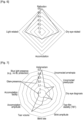
- the figure 4 represents an example of the values of the average of the different group for a theoretical subject whose visual fatigue problems are most likely related to dry eye, accommodation, and (uncorrected or not substantially) corrected refractive error.
- the normalized values are comprised:
- the step 202 and 303 of normalization can be based on at least one of:
- the value representing the direction may be negative, for example -1, for parameters for which the higher the value, the less it affects the visual fatigue of the patient or maybe positive, for example 1, for the parameters for which the lower the value, the less it affects the visual fatigue of the patient.
- the normalization is realized by:
- the first predetermined value may be for example 0, 1 or 100 and the second predetermined value may be for example -100, -1 or 0.
- the first example is also called binary normalization.
- the normalized values indicates that the parameter is likely to contribute to visual fatigue. For example, the presence of glare would result in a normalized value of 1.
- the normalization is realized by:
- the second example is also called normalization based on range of normality.
- the range of normality (for example based on literature or on clinical experience) will be normalized between 0 and 1 whereby 1 indicates the bad end of the normality range and 0 indicates the good end of normality range.
- an accommodative facility normally ranges between 10 and 20 cycles per minute with the general assumption that the more the better.
- a normalized value of 1 corresponds to 10 cycles per minute
- a normalized value of 0 corresponds to 20 cycles per minute.
- the deviation factor may be equal to 3.
- the normalized value can also be multiplied by the value representing the direction of the evolution of the physiological parameters.
- the goal is to normalize values to be almost between -1 and 1 whereby -1 indicates the "good" end of the normality and 1 indicates the "bad" end of normality.
- direction parameter is -1 for values that the more the better, 1 for the less the better.
- Number 3 multiplying the standard deviation is chosen based on the fact that a normal distribution has more than 99% of the values within the difference of 3 standard deviations from the mean. For example, for the parameter Tear Film Stability, if the measured tear film break up time is 4 s or 9 s whereas the population mean is 5 s and the standard deviation is 2 s, and it the longer the better so direction parameter is -1. Then the normalized scores will be 0.17 and -0.67.
- the first normalized scores can be obtained by multiplying the first normalized values with the first causality coefficients.
- the second normalized scores can be obtained by multiplying the second normalized values with the second causality coefficients.
- the first and second causality coefficients are obtained using Hill's causality analysis.
- the Hill's causality analysis allows the obtention of Hill's causality criteria associated respectively with the physiological parameters and the environmental parameters. The higher the Hill's causality criterion is the higher the associated parameter (physiological or environmental) affects the visual fatigue of a patient.
- first and second causality coefficients in the range of 0 to 16.
- the step 205 can be configured to select the physiological parameter associated with the highest normalized score, as the at least one physiological cause of the visual fatigue.
- the step 305 can be configured to select the environmental parameter associated with the highest normalized score, as the at least one environmental cause of the visual fatigue.
- two causes of the visual fatigue can be determined.
- a first cause can be associated with the physiological parameters and a second cause can associated with the environmental parameters.
- Visual fatigue causes Treatments Dry eye Insulation frames, IR filters, Eye drops Blinking Blink training, Blink reminders Tear film IR filters, Insulation frames Uncorrected astigmatism Precise prescription Uncorrected presbyopia Near additions, PAL Accommodation problem Additions Convergence problem Prisms Blue light Blue light filters Glare Anti-glare coating, Peripheral gradient transmission methods, Workstation adjustment, Light adjustment Lighting Light adjustment, Portable personal lighting Display Screen lighting and color adjustment, Display content adjustment Posture Posture monitoring, Posture training
- the posture monitoring, posture training, blink training and blink reminders can be realized using an application implemented for example on the smartphone of the patient.
- the values can be obtained via measurements or from another device.
- the order of obtaining these values can take different combinations.
- Figure 5 represents a specific order of the obtention of the values of the parameters (physiological and environmental).
- the table below shows a first example of calculation of the visual fatigue profile of a patient.
- the normalized value representing the presence/absence (1/0) of a parameter associated with visual fatigue. This normalized value is multiplied by the causality coefficient (normalized Hill's causality) and averaged across each group.
- the highest causality coefficient is associated with the presence of astigmatism with a score of 1 indicating that from the parameters evaluated, astigmatism is most likely to cause visual fatigue.
- the Hill's causality ranges between and 15 and 7.
- the figure 6 represents the different sums associated respectively with the groups for this first example.
- his most likely cause of visual fatigue is related to absence or incorrect refraction in combination with dry eye-related problems and light-related problems. Accordingly, proper refraction and dry eye management followed by advisory is indicated as treatment.
- each parameter is normalized based on "Good end” and "Bad end” of the range where "Good end” corresponds to 0 and "Bad end” corresponds to 1. This normalized value is multiplied by the causality coefficient (normalized Hill's causality criterion). In this example, each parameter is considered individually and after that the scores are averaged across each group.
- the figure 7 represents the different sums associated respectively with the parameters for this second example.
- his most likely cause of visual fatigue is related to uncorrected or undercorrected astigmatism in combination with presence of glare and insufficient tear volume. Accordingly, correction of astigmatism, advisory, and dry eye management is indicated as treatment.
Landscapes
- Health & Medical Sciences (AREA)
- Engineering & Computer Science (AREA)
- Medical Informatics (AREA)
- Public Health (AREA)
- Life Sciences & Earth Sciences (AREA)
- Biomedical Technology (AREA)
- General Health & Medical Sciences (AREA)
- Pathology (AREA)
- Physics & Mathematics (AREA)
- Primary Health Care (AREA)
- Epidemiology (AREA)
- Veterinary Medicine (AREA)
- Surgery (AREA)
- Animal Behavior & Ethology (AREA)
- Biophysics (AREA)
- Molecular Biology (AREA)
- Data Mining & Analysis (AREA)
- Heart & Thoracic Surgery (AREA)
- Databases & Information Systems (AREA)
- Hospice & Palliative Care (AREA)
- Social Psychology (AREA)
- Psychology (AREA)
- Psychiatry (AREA)
- Business, Economics & Management (AREA)
- Educational Technology (AREA)
- Ophthalmology & Optometry (AREA)
- Physiology (AREA)
- Dentistry (AREA)
- Oral & Maxillofacial Surgery (AREA)
- Developmental Disabilities (AREA)
- Child & Adolescent Psychology (AREA)
- General Physics & Mathematics (AREA)
- Emergency Management (AREA)
- Measurement Of The Respiration, Hearing Ability, Form, And Blood Characteristics Of Living Organisms (AREA)
- Medical Treatment And Welfare Office Work (AREA)
- Measuring And Recording Apparatus For Diagnosis (AREA)
Abstract
Description
- This disclosure relates to devices and methods for determining the causes of visual fatigue of a patient.
- Visual fatigue and the terms computer vision syndrome (CVS) and digital eye strain are often used interchangeably to describe different eye and vision-related problems that can result from a wide variety of causes, such as prolonged near work, extensive usage of digital devices (computers, tablets, smartphones, and e-readers), tear film quality, poor visual habits, poorly corrected, or uncorrected refractive errors.
- The symptoms of visual fatigue may also be caused by poor lighting, glare on a digital screen, improper viewing distances, long work hours, alert levels, light spectrum, reduced contrast, small font, and uncorrected, undercorrected or poorly corrected refractive errors.
- The methods of state of the art for visual fatigue assessment are classified as subjective or objective. Subjective assessments usually use questionnaires. Objective assessments usually use the results of tests realized by or on the patient. However, the methods of state of the art generally do not determine a cause of visual fatigue; they only quantify the overall amount of visual fatigue, usually followed by the prescription of one of the traditional anti-fatigue solutions such as blue-light filters and/or lenses with an addition used to near vision.
- Thus, the goal of this disclosure is to provide methods and devices allowing the determination of the causes of visual fatigue and eventually determining a more accurate treatment to mitigate the visual fatigue.
- The following presents a simplified summary to provide a basic understanding of various aspects of this disclosure. This summary is not an extensive overview of all contemplated aspects and is intended to neither identify key or critical elements of all aspects nor delineate the scope of any or all aspects. The sole purpose is to present some concepts of one or more aspects in a simplified form as a prelude to the more detailed description that is presented later.
- One aspect of this disclosure is a calculation module for determining at least one cause of a visual fatigue of a patient. The calculation module is configured to obtain values associated respectively with physiological parameters of the patient, to obtain causality coefficients associated respectively with the physiological parameters, to normalize the values to obtain normalized values associated respectively with the physiological parameters, to determine normalized scores associated respectively with the physiological parameters, based on the causality coefficients and the normalized values and to select at least one of the physiological parameters, using the normalized scores, as the at least one cause of the visual fatigue.
- Therefore, and compared to the state-of-the-art calculation modules that only consider visual fatigue as caused by insufficient accommodation, the calculation module of this disclosure allows a multi-factorial determination of the cause of the visual fatigue.
- The calculation module of this disclosure may identify the real causes of visual fatigue for each patient according to their needs, lifestyle, work and entertainment habits and health conditions etc. Later, one can employ various bricks of anti-fatigue methods to personalize the solution to mitigate the visual fatigue of the patient.
- Another aspect of this disclosure is a system for determining at least one cause of a visual fatigue of a patient, the system comprising a calculation module for determining at least one cause of a visual fatigue of a patient and a display unit. The calculation module is configured to obtain values associated respectively with physiological parameters of the patient, to obtain causality coefficients associated respectively with the physiological parameters, to normalize the values to obtain normalized values associated respectively with the physiological parameters, to determine normalized scores associated respectively with the physiological parameters, based on the causality coefficients and the normalized values, to select at least one of the physiological parameters, using the normalized scores, as the at least one cause of the visual fatigue and to command the display unit to display the at least one selected physiological parameter.
- Another aspect of this disclosure is a computer implemented method for determining at least one cause of a visual fatigue of a patient. The computer implemented method comprises obtaining values associated respectively with physiological parameters of the patient, obtaining causality coefficients associated respectively with the physiological parameters, normalizing the values to obtain normalized values associated respectively to the values, determining normalized scores associated respectively with the physiological parameters, based on the causality coefficients and the normalized values and selecting at least one of the physiological parameters, using the normalized scores, as the at last one cause of the visual fatigue.
- Another aspect of this disclosure is a computer program comprising instructions which, when the computer program is executed by a computer, cause the computer to carry out a method for determining at least one cause of a visual fatigue of a patient. The method comprises obtaining values associated respectively with physiological parameters of the patient, obtaining causality coefficients associated respectively with the physiological parameters, normalizing the values to obtain normalized values associated respectively to the values, determining normalized scores associated respectively with the physiological parameters, based on the causality coefficients and the normalized values and selecting at least one of the physiological parameters, using the normalized scores, as the at last one cause of the visual fatigue.
- A computer may include a memory and a processor. Examples of processors include microprocessors, microcontrollers, graphics processing units (GPUs), central processing units (CPUs), application processors, digital signal processors (DSPs), reduced instruction set computing (RISC) processors, systems on a chip (SoC), field-programmable gate arrays (FPGAs), programmable logic devices (PLDs), state machines, gated logic, discrete hardware circuits, and other suitable hardware configured to perform the various functionality described throughout this disclosure. The memory may be a computer-readable media. By way of example, and not limitation, such computer-readable media may include a random-access memory (RAM), a read-only memory (ROM), an electrically erasable programmable ROM (EEPROM), optical disk storage, magnetic disk storage, other magnetic storage devices, combinations of the aforementioned types of computer-readable media, or any other medium that may be used to store computer executable code in the form of instructions or data structures that may be accessed by the processor of the computer.
- Another aspect of this disclosure is a non-transitory program storage device, readable by a computer, tangibly embodying a program of instructions executable by the computer to perform a method for determining at least one cause of a visual fatigue of a patient. The method comprises obtaining values associated respectively with physiological parameters of the patient, obtaining causality coefficients associated respectively with the physiological parameters, normalizing the values to obtain normalized values associated respectively to the values, determining normalized scores associated respectively with the physiological parameters, based on the causality coefficients and the normalized values and selecting at least one of the physiological parameters, using the normalized scores, as the at last one cause of the visual fatigue.
- For a more complete understanding of the description provided herein and the advantages thereof, reference is now made to the brief descriptions below, taken in connection with the accompanying drawings and detailed description, wherein like reference numerals represent like parts.
-
Figure 1 represents the system for determining at least one cause of a visual fatigue of a patient. -
Figure 2 represents a first example of the method for determining at least one cause of a visual fatigue of a patient. -
Figure 3 represents a second example of the method for determining at least one cause of a visual fatigue of a patient. -
Figure 4 represents an example of values of the physiological parameters of a patient. -
Figure 5 represents a specific order of obtention of the values of the parameters. -
Figure 6 represents an example of values of groups of physiological parameters of a patient. -
Figure 7 represents another example of values of physiological parameters of a patient. - The detailed description set forth below in connection with the appended drawings is intended as a description of various possible embodiments and is not intended to represent the only embodiments in which the concepts described herein may be practiced. The detailed description includes specific details for the purpose of providing a thorough understanding of various concepts. However, it will be apparent to those skilled in the art that these concepts may be practiced without these specific details. In some instances, well-known structures and components are shown in block diagram form to avoid obscuring such concepts.
-
Figure 1 represents asystem 101 for determining at least one cause of a visual fatigue of a patient. Thesystem 101 comprises acalculation module 102 and adisplay unit 103. - The
calculation module 102 comprises a memory 102-a and a processor 102-b. - Examples of processors 102-b include microprocessors, microcontrollers, graphics processing units (GPUs), central processing units (CPUs), application processors, digital signal processors (DSPs), reduced instruction set computing (RISC) processors, systems on a chip (SoC), baseband processors, field-programmable gate arrays (FPGAs), programmable logic devices (PLDs), state machines, gated logic, discrete hardware circuits, and other suitable hardware configured to perform the various functionality described throughout this disclosure.
- The memory 102-a is computer-readable media. By way of example, and not limitation, such computer-readable media may include a random-access memory (RAM), a read-only memory (ROM), an electrically erasable programmable ROM (EEPROM), optical disk storage, magnetic disk storage, other magnetic storage devices, combinations of the aforementioned types of computer-readable media, or any other medium that may be used to store computer executable code in the form of instructions or data structures that may be accessed by the processor 102-a of the
calculation module 102. - The
calculation module 102 may be included in independent module, for example, a smartphone or a computer, a system on chip (SoC) or a graphics processing unit (GPU). Thecalculation module 102 may also be a virtual machine located on a cloud network or a server not co-located with the patient or thesystem 101. - The
display unit 103 can be an electronic device or module for the visual presentation of data or images. - The
system 101 can be for example a smartphone, in this case thedisplay unit 103 is the screen of the smartphone and thecalculation module 102 can be the system on chip of the smartphone. - The
system 101 can be for example a computer, in this case thedisplay unit 103 is the screen of the computer and thecalculation module 102 can be constituted of the motherboard, the memory and the processor of the computer. - To realize this determination of at least one case of a visual fatigue of a patient, the memory 102-a may store a computer program comprising instructions which, when the program is executed by the processor 102-b, cause the
control module 102 to carry out a method, for example a computer implemented method, for determining at least one cause of a visual fatigue of a patient. The method as presented infigure 2 comprises: - a
step 201 of obtaining first values associated respectively with physiological parameters of the patient, - a
step 202 of obtaining first causality coefficients associated respectively with the physiological parameters, - a
step 203 of normalizing the first values to obtain first normalized values associated respectively with the physiological parameters, - a
step 204 of determining first normalized scores associated respectively with the physiological parameters, based on the first causality coefficients and the first normalized values, - a
step 205 of selecting at least one of the physiological parameters, using the first normalized scores, as the at last one physiological cause of the visual fatigue. - The
figure 3 represents another embodiment of the method for determining the cause. In this embodiment in addition to physiological parameters of the patient, environmental parameters are taken into account. - In the embodiment of
figure 3 , in addition to the steps representedfigure 2 , the method for determining the at least one cause of the visual fatigue of the patient also comprises: - a
step 301 of obtain second values associated respectively with environmental parameters, - a
step 302 of obtaining second causality coefficients associated respectively with the environmental parameters, - a
step 303 of normalizing the second values to obtain second normalized values associated respectively with the environmental parameters - a
step 304 of determining second normalized scores associated respectively with the environmental parameters, by multiplying the second causality coefficients and the second normalized values, - a
step 305 of selecting one of the environmental parameters, using the second normalized scores, as an environmental cause of the visual fatigue. - Therefore, using the embodiment of the
figure 3 one can determine a physiological cause of the visual fatigue and an environmental cause of the visual fatigue. - The method can also comprise a step of commanding the display unit to display the physiological cause (represented by the at least one selected physiological parameter) and/or the environmental cause (represented by the at least one selected environmental parameter). The method can also comprise a step of determining a treatment to mitigate the visual fatigue and a step of command the display unit to display the treatment. The treatment can depend on the physiological cause and/or the environmental cause.
- The treatment may also vary from region to region, clinician to clinician, etc.
- The physiological parameters can be chosen among:
- parameters related to a visual health of the patient,
- ∘ parameters related to a refraction of the patient (astigmatism, uncorrected ametropia, uncorrected presbyopia)
- ∘ parameters related to an abnormality of a vergence of the patient (vergence response abnormality, vergence amplitude abnormality
- parameters representing a dryness of an eye of the patient
- ∘ dry eye diagnosis
- ∘ parameters representing a stability of a tear film
- ∘ blink amplitude
- ∘ blink rate
- ∘ tear volume
- parameters related to an accommodation capacity of the patient
- ∘ amplitude of the accommodation
- ∘ accommodation facility
- The environmental parameters can be chosen among:
- parameters characteristic of a light received by the patient,
- a luminance level of the light received by the patient,
- an amount of UV in the light received by the patient,
- a contrast between a luminance level of a light originated from an area of interest and a luminance level of an ambient light surrounding the area of interest,
- parameters characteristic an air surrounding the patient,
- a level of a humidity of the air surrounding the patient and
- parameters characteristic of a pollution of the air surrounding the patient.
- The environmental parameters can also be chosen among parameters representing a glare on the screen or any working surface.
- In other words, the physiological parameters and the environmental parameters may include dry eye, blinking patterns, tear film patterns, uncorrected refractive errors, accommodation- and vergence-related factors, blue light, glare, lighting environment, display content and settings, posture, etc., have been identified as causes of visual fatigue.
- Some of the parameters can be grouped for example in the following manner
Groups Parameters Refraction-related Astigmatism Uncorrected ametropia Uncorrected presbyopia Dry eye-related Dry eye diagnosis Tear film stability (TBT) Blink amplitude Blink rate Tear volume Accommodation-related Amplitude Facility Vergence Vergence response abnormality Vergence amplitude abnormality Light-related Glare presence Blue light (BL) presence (e.g., no BL protection) - The above set of parameters and groups is just an example and abbreviated version of the full picture of possible parameters causing visual fatigue. Other parameters and other groups can also be used to obtain a more precise determination of the cause of the visual fatigue.
- In embodiments, after the
steps steps - The
figure 4 represents an example of the values of the average of the different group for a theoretical subject whose visual fatigue problems are most likely related to dry eye, accommodation, and (uncorrected or not substantially) corrected refractive error. - Since the nature of the variety of parameters and their measurement units is very wide (for example refraction in diopters, tear film stability in seconds, or accommodative facility in cycles per minute), there is a need to normalize the scores to allow an accurate comparison.
- Generally, the normalized values are comprised:
- between -100 and 100,
- between 0 and 100
- between -1 and 1 or
- between 0 and 1
- The
step - a range of possible values of a parameter associated with the at least one of the values,
- a mean value of the range,
- a median value of the range
- a standard deviation of the range,
- a span of the range,
- a detection level of the parameter associated with the at least one of the values, and of
- a value representing a direction of an evolution of the physiological parameter associated with the at least one of the values.
- The value representing the direction may be negative, for example -1, for parameters for which the higher the value, the less it affects the visual fatigue of the patient or maybe positive, for example 1, for the parameters for which the lower the value, the less it affects the visual fatigue of the patient.
- In a first example, the normalization is realized by:
- comparing the at least one of the values with the detection level,
- when the at least one of the values is above the detection level, the normalized value is equal to a first predetermined value,
- when the at least one of the values is below the detection level, the normalized value is equal to a second predetermined value.
- The first predetermined value may be for example 0, 1 or 100 and the second predetermined value may be for example -100, -1 or 0.
- The first example is also called binary normalization.
- In other words, in this first example the normalized values indicates that the parameter is likely to contribute to visual fatigue. For example, the presence of glare would result in a normalized value of 1.
- For parameters with associated values that are a continuous variable one can realize this binary normalization by determining whether the value falls within a normality range of the values of this parameter. For example, a value of uncorrected astigmatism larger than 0.25 D would indicate that this parameter can affect visual fatigue therefore one can set the normalized value to 1.
- In a second example, the normalization is realized by:
- subtracting the mean to the at least one of the values,
- dividing the subtraction by half the span, to obtain the normalized value.
- The second example is also called normalization based on range of normality.
- In this second example, for parameters with binary values, the normalization leads to the same results as the first example.
- For parameters with continuous values, the range of normality (for example based on literature or on clinical experience) will be normalized between 0 and 1 whereby 1 indicates the bad end of the normality range and 0 indicates the good end of normality range. For example, an accommodative facility normally ranges between 10 and 20 cycles per minute with the general assumption that the more the better. Thus, a normalized value of 1 corresponds to 10 cycles per minute, and a normalized value of 0 corresponds to 20 cycles per minute.
- In a third example the normalization is realized by:
- subtracting the mean to the at least one of the values,
- dividing the subtraction by the standard deviation of the range multiplied by a deviation factor to obtain the normalized value.
- The deviation factor may be equal to 3. In this third example the normalized value can also be multiplied by the value representing the direction of the evolution of the physiological parameters. The equation to calculate the normalized score is then:

- value [-1,1] is the normalized value between -1 and 1
- valuemeasured is the measure value of the parameter
- valuemean is the mean of the values of the parameter
- valuestandard deviation is the standard deviation of the values of the parameter
- direction direction of the evolution of the parameter
- For parameters with continuous values, one can also normalize the value with the mean and standard deviation in the specific population that each patient belongs to (determined by, not limited to, age, gender, and ethnicity).
- The goal is to normalize values to be almost between -1 and 1 whereby -1 indicates the "good" end of the normality and 1 indicates the "bad" end of normality. direction parameter is -1 for values that the more the better, 1 for the less the better. Number 3 multiplying the standard deviation is chosen based on the fact that a normal distribution has more than 99% of the values within the difference of 3 standard deviations from the mean. For example, for the parameter Tear Film Stability, if the measured tear film break up time is 4 s or 9 s whereas the population mean is 5 s and the standard deviation is 2 s, and it the longer the better so direction parameter is -1. Then the normalized scores will be 0.17 and -0.67.
- This normalized value can be further normalized to be between 0 and 1 by using the equation:

- In a nutshell to realize the normalization according to the third example one can:
- During a first step determine the direction parameter according to whether the outcome parameter is the more or the less the better.
- During a second step find the population mean and standard deviation from literature.
- During a third step calculate the normalized score using the above equation.
- This equation normalizes scores to the range of -1 to 1. It can be further normalized to 0 to 1 if needed to be consistent with other factors by using value [0,1] = (value [-1,1] + 1)/2 Where value [-1,1] is the normalized value between -1 and 1 and value [0,1] is the normalized value between 0 and 1.
- The first normalized scores can be obtained by multiplying the first normalized values with the first causality coefficients. The second normalized scores can be obtained by multiplying the second normalized values with the second causality coefficients.
- The first and second causality coefficients are obtained using Hill's causality analysis. The Hill's causality analysis allows the obtention of Hill's causality criteria associated respectively with the physiological parameters and the environmental parameters. The higher the Hill's causality criterion is the higher the associated parameter (physiological or environmental) affects the visual fatigue of a patient.
- Therefore, the Hill's causality analysis helps to rank the different causes of visual fatigue.
- Using the Hill's causality analysis one can obtain first and second causality coefficients in the range of 0 to 16. In this case one can advantageously normalize the first and second causality coefficients to have these coefficients in the range between 0 to 1.
- The table below presents example of Hill' causality coefficients and causality coefficients for different parameters.
Group Parameter Hill's causality Causality coefficient Refraction Astigmatism 16 1 Uncorrected ametropia 10 0.625 Uncorrected presbyopia 12 0.75 Dry eye-related Dry eye diagnosis 14 0.875 Tear film stability (TBT) 12 0.75 Blink amplitude 13 0.8125 Blink rate 9 0.5625 Tear volume 6 0.375 Accommod ation Amplitude 6 0.375 Facility 7 0.4375 Light-related Glare presence 14 0.875 Blue light presence (e.g., no BL protection) 15 0.9375 - The
step 205 can be configured to select the physiological parameter associated with the highest normalized score, as the at least one physiological cause of the visual fatigue. - The
step 305 can be configured to select the environmental parameter associated with the highest normalized score, as the at least one environmental cause of the visual fatigue. - In other embodiments one can select during step 205 a plurality of the physiological parameters. For example, one can select a predetermined number of the physiological parameters or one can select the physiological parameters having an associated first normalized score above a first predetermined threshold.
- Similarly, one can select during step 305 a plurality of the environmental parameters. For example, one can select a predetermined number of the environmental parameters, or one can select the environmental parameters having an associated second normalized score above a second predetermined threshold.
- In embodiments two causes of the visual fatigue can be determined. A first cause can be associated with the physiological parameters and a second cause can associated with the environmental parameters.
- In other embodiments one can also determine a single cause by comparing the first normalized score of the psychological cause and the second normalized score of the environment cause and by selecting the single cause as the one among the environment cause and a physiological cause with the highest normalized score.
- The following table describes for the different visual fatigue causes examples of treatments.
Visual fatigue causes Treatments Dry eye Insulation frames, IR filters, Eye drops Blinking Blink training, Blink reminders Tear film IR filters, Insulation frames Uncorrected astigmatism Precise prescription Uncorrected presbyopia Near additions, PAL Accommodation problem Additions Convergence problem Prisms Blue light Blue light filters Glare Anti-glare coating, Peripheral gradient transmission methods, Workstation adjustment, Light adjustment Lighting Light adjustment, Portable personal lighting Display Screen lighting and color adjustment, Display content adjustment Posture Posture monitoring, Posture training - The posture monitoring, posture training, blink training and blink reminders can be realized using an application implemented for example on the smartphone of the patient.
- During the
steps Group Measure Method Unit Refraction Astigmatism Subjective refraction Diopter Uncorrected ametropia Subjective refraction Diopter Uncorrected presbyopia Subjective refraction Diopter Dry eye-related Dry eye diagnosis Comprehensiv e evaluation Binary - yes 1 or no 0 Tear film stability (TBT) Tear break time seconds Blink amplitude Observation, eye tracking E.g., percentage of eye closure during blinking Blink rate Observation, eye tracking Blinks per minute Tear volume Shirmers test milimeters Accommodation Amplitude E.g., push up test Diopter Facility Facility test Cycles per minute Light-related Glare presence History, questionnaire Binary - yes 1 or no 0 Alternatively, proportion of time Blue light presence (e.g., no BL protection) History, questionnaire Binary - yes 1 or no 0 Alternatively, proportion of time - The values can be obtained via measurements or from another device. The order of obtaining these values can take different combinations.
Figure 5 represents a specific order of the obtention of the values of the parameters (physiological and environmental). - The table below shows a first example of calculation of the visual fatigue profile of a patient. The normalized value representing the presence/absence (1/0) of a parameter associated with visual fatigue. This normalized value is multiplied by the causality coefficient (normalized Hill's causality) and averaged across each group.
Group Parameter Normalized value (presence of the problem) Hill's causality Causality coefficient Normalized score Average of scores Refraction Astigmatism 1 16 1 1 0.583333 333 Uncorrected ametropia 0 10 0.625 0 Uncorrected presbyopia 1 12 0.75 0.75 Dry eye-related Dry eye diagnosis 1 14 0.875 0.875 0.4375 Tear film stability (TBT) 1 12 0.75 0.75 Blink amplitude 0 13 0.8125 0 Blink rate 1 9 0.5625 0.5625 Tear volume 0 6 0.375 0 Accommod ation Amplitude 1 6 0.375 0.375 0.1875 Facility 0 7 0.4375 0 Light-related Glare presence 1 14 0.875 0.875 0.4375 Blue light presence (e.g., no BL protection) 0 15 0.9375 0 - In this example the highest causality coefficient is associated with the presence of astigmatism with a score of 1 indicating that from the parameters evaluated, astigmatism is most likely to cause visual fatigue. For the rest of the parameters listed in the table above, the Hill's causality ranges between and 15 and 7.
- The
figure 6 represents the different sums associated respectively with the groups for this first example. For this patient his most likely cause of visual fatigue is related to absence or incorrect refraction in combination with dry eye-related problems and light-related problems. Accordingly, proper refraction and dry eye management followed by advisory is indicated as treatment. - The table below shows a second example of calculation of the visual fatigue profile of a patient. The value of each parameter is normalized based on "Good end" and "Bad end" of the range where "Good end" corresponds to 0 and "Bad end" corresponds to 1. This normalized value is multiplied by the causality coefficient (normalized Hill's causality criterion). In this example, each parameter is considered individually and after that the scores are averaged across each group.
Group Parameter Valu e Goo d end Bad end Normalized value Hill's causalit y Causali ty coeffici ent Normalized score Average of scores Refraction Astigmatism 0.5 0 1 0.5 16 1 0.5 0.242 Uncorrected ametropia 0.25 0 4 0.0625 10 0.625 0.0391 Uncorrected presbyopia 0.5 0 2 0.25 12 0.75 0.1875 Dry eye diagnosis 0 0 1 0 14 0.875 0 0.117 Dry eye-related Tear film stability (TBT) 25 35 10 0.4 12 0.75 0.3 Blink amplitude 100 100 0 0 13 0.8125 0 Blink rate 25 30 1 0.172 9 0.5625 0.0970 Tear volume 7 10.5 3.5 0.5 6 0.375 0.1875 Accommodati on Acommodative amplitude 50 10 200 0.211 6 0.375 0.0789 0.0395 Acommodative facility 20 20 10 0 7 0.4375 0 Light-related Glare presence 50 0 100 0.5 14 0.875 0.4375 0.3125 Blue light presence (e.g., no BL protection) 20 0 100 0.2 15 0.9375 0.1875 - The
figure 7 represents the different sums associated respectively with the parameters for this second example. For this patient his most likely cause of visual fatigue is related to uncorrected or undercorrected astigmatism in combination with presence of glare and insufficient tear volume. Accordingly, correction of astigmatism, advisory, and dry eye management is indicated as treatment.
Claims (15)
- Calculation module (102) for determining at least one cause of a visual fatigue of a patient, the calculation module (102) being configured:- to obtain values associated respectively with physiological parameters of the patient,- to obtain causality coefficients associated respectively with the physiological parameters,- to normalize the values to obtain normalized values associated respectively with the physiological parameters,- to determine normalized scores associated respectively with the physiological parameters, based on the causality coefficients and the normalized values and,- to select at least one of the physiological parameters, using the normalized scores, as the at least one cause of the visual fatigue.
- Calculation module (102) according to the claim 1, the calculation module (102) being configured to normalize at least one of the values based on at least one of:- a range of possible values of the physiological parameter associated with the at least one of the values,- a mean value of the range,- a standard deviation of the range,- a span of the range,- a detection level of the physiological parameter associated with the at least one of the values, and of- a value representing a direction of an evolution of the physiological parameter associated with the at least one of the values.
- Calculation module (102) according to the claim 2, the calculation module (102) being configured to normalize the at least one of the values by:- comparing the at least one of the values with the detection level,when the at least one of the values is above the detection level, the normalized value is equal to a first predetermined value,when the at least one of the values is below the detection level, the normalized value is equal to a second predetermined value.
- Calculation module (102) according to the claim 2 or 3, the calculation module (102) being configured to normalize the at least one of the values by- subtracting the mean to the at least one of the values,- dividing the subtraction by half the span, to obtain the normalized value.
- Calculation module (102) according to any one of the claims 2 to 4, the calculation module (102) being configured to normalize the at least one of the values by- subtracting the mean to the at least one of the values,- dividing the subtraction by the standard deviation of the range multiplied by a deviation factor to obtain the normalized value.
- Calculation module (102) according to any one of the claims 1 to 5, the calculation module (102) being configured:- to sum normalized scores associated respectively to a part of the physiological parameters to obtain an aggregated score associated to the part of the physiological parameters,- to select the at least one of the physiological parameters, also using the aggregated score.
- Calculation module (102) according to the claim 6, the calculation module (102) being also configured:
to normalize the aggregated score. - Calculation module (102) according to any one of the claims 1 to 7, the physiological parameters being chosen among:• parameters related to a visual health of the patient,• parameters representing a dryness of an eye of the patient and• parameters related to an accommodation capacity of the patient.
- Calculation module (102) according to any of the claims 1 to 8, the calculation module (102) being configured- to select the physiological parameter associated with the highest normalized score, as the at least one cause of the visual fatigue.
- Calculation module (102) according to any one of the claims 1 to 9,the values being first values,the causality coefficients being first causality coefficients,the normalized values being first normalized values,the normalized scores being first normalized scoresthe calculation module (102) being also configured:- to obtain second values associated respectively with environmental parameters,- to obtain second causality coefficients associated respectively with the environmental parameters,- to normalize the second values to obtain second normalized values associated respectively with the environmental parameters- to determine second normalized scores associated respectively with the environmental parameters, by multiplying the second causality coefficients and the second normalized values,- to select one of the environmental parameters, using the second normalized scores, as an environmental cause of the visual fatigue.
- Calculation module (102) according to the claim 10, the environmental parameters being chosen among:- parameters characteristic of a light received by the patient,- a luminance level of the light received by the patient,- an amount of UV in the light received by the patient,- a contrast between a luminance level of a light originated from an area of interest and a luminance level of an ambient light surrounding the area of interest,- parameters characteristic of an air surrounding the patient,- a level of a humidity of the air surrounding the patient and- parameters characteristic of a pollution of the air surrounding the patient.
- Calculation module (102) according to any one of the claims 1 to 11, the calculation module (102) being also configured:
to command a display unit (103) to display the at least one selected physiological parameter and/or the at least one selected environmental parameter. - Calculation module (102) according to the claim 12, the calculation module (102) being also configured to command the display unit (103) to display a treatment to mitigate the visual fatigue,
the treatment depending on the at least one selected physiological parameter and/or the at least one selected environmental parameter. - System (101) for determining at least one cause of a visual fatigue of a patient, the system comprising a calculation module (102) for determining at least one cause of a visual fatigue of a patient and a display unit (103),
the calculation module (102) being configured:- to obtain values associated respectively with physiological parameters of the patient,- to obtain causality coefficients associated respectively with the physiological parameters,- to normalize the values to obtain normalized values associated respectively with the physiological parameters,- to determine normalized scores associated respectively with the physiological parameters, based on the causality coefficients and the normalized values,- to select at least one of the physiological parameters, using the normalized scores, as the at least one cause of the visual fatigue,
and- to command the display unit (103) to display the at least one selected physiological parameter. - Computer implemented method for determining at least one cause of a visual fatigue of a patient, the computer implemented method comprising:- (201) obtaining values associated respectively with physiological parameters of the patient,- (202) obtaining causality coefficients associated respectively with the physiological parameters,- (203) normalizing the values to obtain normalized values associated respectively to the values,- (204) determining normalized scores associated respectively with the physiological parameters, based on the causality coefficients and the normalized values,- (205) selecting at least one of the physiological parameters, using the normalized scores, as the at last one cause of the visual fatigue.
Priority Applications (5)
| Application Number | Priority Date | Filing Date | Title |
|---|---|---|---|
| EP23305257.0A EP4421821A1 (en) | 2023-02-27 | 2023-02-27 | Calculation module, system and computer-implemented method for determining at least one cause of a visual fatigue of a patient |
| JP2025549730A JP2026506207A (en) | 2023-02-27 | 2024-02-08 | Computational module, system, and computer-implemented method for identifying at least one cause of visual fatigue in a patient - Patent Application 20070122999 |
| CN202480007294.2A CN120513490A (en) | 2023-02-27 | 2024-02-08 | Computing module, system and computer-implemented method for determining at least one cause of visual fatigue of a patient |
| PCT/EP2024/053198 WO2024179800A1 (en) | 2023-02-27 | 2024-02-08 | Calculation module, system and computer-implemented method for determining at least one cause of a visual fatigue of a patient |
| KR1020257026224A KR20250157493A (en) | 2023-02-27 | 2024-02-08 | Computational module, system and computer-implemented method for determining at least one cause of visual fatigue in a patient |
Applications Claiming Priority (1)
| Application Number | Priority Date | Filing Date | Title |
|---|---|---|---|
| EP23305257.0A EP4421821A1 (en) | 2023-02-27 | 2023-02-27 | Calculation module, system and computer-implemented method for determining at least one cause of a visual fatigue of a patient |
Publications (1)
| Publication Number | Publication Date |
|---|---|
| EP4421821A1 true EP4421821A1 (en) | 2024-08-28 |
Family
ID=85571210
Family Applications (1)
| Application Number | Title | Priority Date | Filing Date |
|---|---|---|---|
| EP23305257.0A Pending EP4421821A1 (en) | 2023-02-27 | 2023-02-27 | Calculation module, system and computer-implemented method for determining at least one cause of a visual fatigue of a patient |
Country Status (5)
| Country | Link |
|---|---|
| EP (1) | EP4421821A1 (en) |
| JP (1) | JP2026506207A (en) |
| KR (1) | KR20250157493A (en) |
| CN (1) | CN120513490A (en) |
| WO (1) | WO2024179800A1 (en) |
Citations (1)
| Publication number | Priority date | Publication date | Assignee | Title |
|---|---|---|---|---|
| EP3113135A1 (en) * | 2015-07-01 | 2017-01-04 | Essilor International (Compagnie Generale D'optique) | Module and method for determining visual fatigue |
-
2023
- 2023-02-27 EP EP23305257.0A patent/EP4421821A1/en active Pending
-
2024
- 2024-02-08 KR KR1020257026224A patent/KR20250157493A/en active Pending
- 2024-02-08 WO PCT/EP2024/053198 patent/WO2024179800A1/en not_active Ceased
- 2024-02-08 JP JP2025549730A patent/JP2026506207A/en active Pending
- 2024-02-08 CN CN202480007294.2A patent/CN120513490A/en active Pending
Patent Citations (1)
| Publication number | Priority date | Publication date | Assignee | Title |
|---|---|---|---|---|
| EP3113135A1 (en) * | 2015-07-01 | 2017-01-04 | Essilor International (Compagnie Generale D'optique) | Module and method for determining visual fatigue |
Non-Patent Citations (4)
| Title |
|---|
| FENG YONG ET AL: "Investigation of Weighted Scales for Measuring Visual Fatigue in Screening Tasks", 2021 43RD ANNUAL INTERNATIONAL CONFERENCE OF THE IEEE ENGINEERING IN MEDICINE & BIOLOGY SOCIETY (EMBC), IEEE, 1 November 2021 (2021-11-01), pages 5768 - 5771, XP034041616, DOI: 10.1109/EMBC46164.2021.9630334 * |
| HIRZLE TERESA ET AL: "Understanding, Addressing, and Analysing Digital Eye Strain in Virtual Reality Head-Mounted Displays", ACM TRANSACTIONS ON COMPUTER-HUMAN INTERACTION, ACM, NEW YORK, NY, US, vol. 29, no. 4, 31 March 2022 (2022-03-31), pages 1 - 80, XP059024132, ISSN: 1073-0516, DOI: 10.1145/3492802 * |
| ROSENFIELD MARK: "Computer vision syndrome: a review of ocular causes and potential treatments : Computer vision syndrome", OPHTHALMIC AND PHYSIOLOGICAL OPTICS., vol. 31, no. 5, 12 April 2011 (2011-04-12), GB, pages 502 - 515, XP093069131, ISSN: 0275-5408, DOI: 10.1111/j.1475-1313.2011.00834.x * |
| XIAO-WEI SHI ET AL: "Visual fatigue diversity research of static and dynamic lighting environment", INDUSTRIAL ENGINEERING AND ENGINEERING MANAGEMENT (IE&EM), 2011 IEEE 18TH INTERNATIONAL CONFERENCE ON, IEEE, 3 September 2011 (2011-09-03), pages 291 - 294, XP032056014, ISBN: 978-1-61284-446-6, DOI: 10.1109/ICIEEM.2011.6035160 * |
Also Published As
| Publication number | Publication date |
|---|---|
| CN120513490A (en) | 2025-08-19 |
| KR20250157493A (en) | 2025-11-04 |
| WO2024179800A1 (en) | 2024-09-06 |
| JP2026506207A (en) | 2026-02-20 |
Similar Documents
| Publication | Publication Date | Title |
|---|---|---|
| JP4675400B2 (en) | Optical system performance evaluation method and optical system design method | |
| CN102186408B (en) | Systems and methods for prescription prescriptions for visual aid devices | |
| US20040263786A1 (en) | Metrics to predict subjective impact of eye's wave aberration | |
| RU2470279C2 (en) | Method to assess spectacle lenses, method to calculate spectacle lenses with its usage, method to manufacture spectacle lenses, system of manufacturing spectacle lenses and spectacle lenses | |
| JP7405849B2 (en) | Method and device for evaluating the effectiveness of spectacle lenses in suppressing visual acuity decline | |
| Yammouni et al. | An investigation of low power convex lenses (adds) for eyestrain in the digital age (CLEDA) | |
| Dirani et al. | Adult-onset myopia: the Genes in Myopia (GEM) twin study | |
| US9468371B2 (en) | Statistical autorefractor | |
| US12345960B2 (en) | Method for determining a specific near vision power of an ophthalmic lens | |
| US20150234999A1 (en) | Systems, methods and computer program products for determining a consolidated disease risk score from risk factors and a level of composite risk | |
| US12106472B2 (en) | Eye state assessment method and electronic device | |
| CN116634920A (en) | subjective refraction system | |
| CN111919159A (en) | Adjustment of subjective refraction and objective refraction | |
| WO2023079019A1 (en) | A method and system for determining a risk of an onset or progression of myopia | |
| Carkeet et al. | Slope of psychometric functions and termination rule analysis for low contrast acuity charts | |
| Ravikumar et al. | Image quality metric derived refractions predicted to improve visual acuity beyond habitual refraction for patients with Down syndrome | |
| CN114974582B (en) | Myopia risk prediction method, device, electronic device and medium | |
| WO2024179800A1 (en) | Calculation module, system and computer-implemented method for determining at least one cause of a visual fatigue of a patient | |
| US20240310654A1 (en) | Ophthalmic lenses with optimized visual performance | |
| CN109171640B (en) | Corneal state evaluation device and corneal state evaluation method and application | |
| Markoulli et al. | Clinical utility of irx3 in keratoconus | |
| CN112545493A (en) | Height evaluation method and terminal equipment | |
| WO2022240586A1 (en) | Methods and systems for optimizing refractive refraction of human eyes | |
| Kang et al. | Deep learning-based longitudinal prediction of childhood myopia progression using fundus image sequences and baseline refraction data | |
| Saha et al. | Keratoconus: a probe into the refractive symmetry |
Legal Events
| Date | Code | Title | Description |
|---|---|---|---|
| PUAI | Public reference made under article 153(3) epc to a published international application that has entered the european phase |
Free format text: ORIGINAL CODE: 0009012 |
|
| STAA | Information on the status of an ep patent application or granted ep patent |
Free format text: STATUS: THE APPLICATION HAS BEEN PUBLISHED |
|
| AK | Designated contracting states |
Kind code of ref document: A1 Designated state(s): AL AT BE BG CH CY CZ DE DK EE ES FI FR GB GR HR HU IE IS IT LI LT LU LV MC ME MK MT NL NO PL PT RO RS SE SI SK SM TR |
|
| STAA | Information on the status of an ep patent application or granted ep patent |
Free format text: STATUS: REQUEST FOR EXAMINATION WAS MADE |
|
| 17P | Request for examination filed |
Effective date: 20250131 |
|
| STAA | Information on the status of an ep patent application or granted ep patent |
Free format text: STATUS: EXAMINATION IS IN PROGRESS |
|
| 17Q | First examination report despatched |
Effective date: 20250602 |