EP4411648A1 - Pet apparatus and a medical information processing method - Google Patents
Pet apparatus and a medical information processing method Download PDFInfo
- Publication number
- EP4411648A1 EP4411648A1 EP24150798.7A EP24150798A EP4411648A1 EP 4411648 A1 EP4411648 A1 EP 4411648A1 EP 24150798 A EP24150798 A EP 24150798A EP 4411648 A1 EP4411648 A1 EP 4411648A1
- Authority
- EP
- European Patent Office
- Prior art keywords
- length
- data
- waveform
- frame length
- list mode
- Prior art date
- Legal status (The legal status is an assumption and is not a legal conclusion. Google has not performed a legal analysis and makes no representation as to the accuracy of the status listed.)
- Pending
Links
Images




Classifications
-
- G—PHYSICS
- G06—COMPUTING OR CALCULATING; COUNTING
- G06T—IMAGE DATA PROCESSING OR GENERATION, IN GENERAL
- G06T12/00—Tomographic reconstruction from projections
- G06T12/10—Image preprocessing, e.g. calibration, positioning of sources or scatter correction
-
- A—HUMAN NECESSITIES
- A61—MEDICAL OR VETERINARY SCIENCE; HYGIENE
- A61B—DIAGNOSIS; SURGERY; IDENTIFICATION
- A61B6/00—Apparatus or devices for radiation diagnosis; Apparatus or devices for radiation diagnosis combined with radiation therapy equipment
- A61B6/02—Arrangements for diagnosis sequentially in different planes; Stereoscopic radiation diagnosis
- A61B6/03—Computed tomography [CT]
- A61B6/037—Emission tomography
-
- A—HUMAN NECESSITIES
- A61—MEDICAL OR VETERINARY SCIENCE; HYGIENE
- A61B—DIAGNOSIS; SURGERY; IDENTIFICATION
- A61B6/00—Apparatus or devices for radiation diagnosis; Apparatus or devices for radiation diagnosis combined with radiation therapy equipment
- A61B6/50—Apparatus or devices for radiation diagnosis; Apparatus or devices for radiation diagnosis combined with radiation therapy equipment specially adapted for specific body parts; specially adapted for specific clinical applications
- A61B6/503—Apparatus or devices for radiation diagnosis; Apparatus or devices for radiation diagnosis combined with radiation therapy equipment specially adapted for specific body parts; specially adapted for specific clinical applications for diagnosis of the heart
-
- A—HUMAN NECESSITIES
- A61—MEDICAL OR VETERINARY SCIENCE; HYGIENE
- A61B—DIAGNOSIS; SURGERY; IDENTIFICATION
- A61B6/00—Apparatus or devices for radiation diagnosis; Apparatus or devices for radiation diagnosis combined with radiation therapy equipment
- A61B6/52—Devices using data or image processing specially adapted for radiation diagnosis
- A61B6/5288—Devices using data or image processing specially adapted for radiation diagnosis involving retrospective matching to a physiological signal
-
- G—PHYSICS
- G06—COMPUTING OR CALCULATING; COUNTING
- G06T—IMAGE DATA PROCESSING OR GENERATION, IN GENERAL
- G06T7/00—Image analysis
- G06T7/20—Analysis of motion
-
- G—PHYSICS
- G06—COMPUTING OR CALCULATING; COUNTING
- G06T—IMAGE DATA PROCESSING OR GENERATION, IN GENERAL
- G06T2207/00—Indexing scheme for image analysis or image enhancement
- G06T2207/10—Image acquisition modality
- G06T2207/10072—Tomographic images
- G06T2207/10104—Positron emission tomography [PET]
-
- G—PHYSICS
- G06—COMPUTING OR CALCULATING; COUNTING
- G06T—IMAGE DATA PROCESSING OR GENERATION, IN GENERAL
- G06T2211/00—Image generation
- G06T2211/40—Computed tomography
- G06T2211/412—Dynamic
Definitions
- Embodiments described herein relate generally to a PET apparatus and a medical information processing method.
- This disclosure relates to list-mode data gating in positron emission tomography (PET) systems as a means to eliminate motion-related inaccuracies and improve image quality in PET imaging.
- PET positron emission tomography
- Patient motion is a main source of blurring and artifacts in positron emission tomography (PET) imaging.
- Respiratory motion and cardiac motion during PET data acquisition can degrade quantitation performance by blurring images, leading to over-estimation of lesion volumes and under-estimation of lesion activity.
- Gating methods have been used to improve the quality of PET images. Typically, these methods need to use an external device to detect a biosignal, for example, a respiration waveform, electrocardiography (ECG), etc.
- ECG electrocardiography
- motion correction can be addressed by gating acquired data in which motion may have occurred. Gating involves dividing data into separates chunks (gates) within which motion is negligible. This may occur during voluntary or involuntary movement of the patient, and may include, for example, movement due to respiration or heartbeat.
- gating is done by attaching motion sensors to a patient during a PET scan.
- Such external motion sensors make PET scans more cumbersome as their use requires motion information to be successfully recorded during the scan. Moreover, if motion is not recorded correctly or properly synchronized with the scan, then motion correction often is hampered.
- a data-driven algorithm can first divide the PET acquisitions into small temporal frames, and then principal component analysis (PCA) or independent component analysis (ICA) can be applied on the frames.
- PCA principal component analysis
- ICA independent component analysis
- the biosignal can be extracted based on the data variation across all the frames.
- a respiratory signal can be modeled by the first component of the PCA.
- the PET data can be binned and reconstructed into multiple gates (for visualizing the motion) or a single gate (for generation of a single, motion-reduced image).
- the signal from the PCA can also include a cardiac signal.
- a positron emission tomography (PET) apparatus includes a processing unit.
- the processing unit is configured to obtain list mode data representing radiation detected during an imaging scan.
- the processing unit is configured to generate divided data from the list mode data for each time interval of a first length, the divided data having a time width of a second length longer than the first length.
- the processing unit is configured to determine a respiratory waveform based on the divided data.
- the processing unit is configured to reconstruct an image based on the respiratory waveform and the list mode data.
- the processing unit may set the second length according to a cardiac cycle length.
- the list mode data may be data affected by quasi-periodic motion of an imaging object, and the processing unit may be configured to: determine a first frame length having the first length, divide the list mode data into first non-overlapping frames of a first frame length, and process the first non-overlapping frames to determine a cardiac cycle length; determine a second frame length having the second length longer than the first frame length, based on the determined cardiac cycle length; re-bin the list mode data into overlapping frames having the second frame length, based on the non-overlapping frames having the first frame length, to generate the divided data; apply a principal component analysis (PCA) process to the divided data to determine the respiratory waveform; determine a cardiac waveform using the respiratory waveform; and reconstruct an image based on the list mode data using the respiratory waveform and the cardiac waveform.
- PCA principal component analysis
- the processing unit may apply the PCA process to the first non-overlapping frames to determine a first waveform, determine peaks of the first waveform, determine the cardiac cycle length based on a frequency of the peaks of the first waveform, and determine the second frame length based on the cardiac cycle length.
- the frequency of the peaks of the first waveform may be a cardiac beating rate.
- the second frame length may be a value less than a frequency of peaks of the respiratory waveform.
- the second frame length may be 1 second.
- the processing unit may set the first frame length to a value less than 0.4 seconds.
- the processing unit may reconstruct the image based on the list mode data processed using the respiratory waveform and the cardiac waveform by applying a respiratory gate based on the respiratory waveform and removing the cardiac waveform.
- the processing unit may determine the second frame length by measuring the cardiac cycle length of the object via an ECG device.
- the overlapping frames having the second frame length may overlap by the first frame length.
- the processing unit may re-bin the list mode data into a sinogram as processing of re-binning into overlapping frames having the second frame length.
- a medical information processing method includes: obtaining list mode data representing radiation detected during an imaging scan; generating divided data from the list mode data for each time interval of a first length, the divided data having a time width of a second length longer than the first length; and determining a respiratory waveform based on the divided data.
- the second length may be set according to a cardiac cycle length.
- the medical information processing method may comprise: determining a first frame length having the first length, dividing the list mode data into first non-overlapping frames of a first frame length, the list mode data being affected by quasi-periodic motion of an imaging object, and processing the first non-overlapping frames to determine a cardiac cycle length; determining a second frame length having the second length longer than the first frame length, based on the determined cardiac cycle length; re-binning the list mode data into overlapping frames having the second frame length, based on the non-overlapping frames having the first frame length, to generate the divided data; applying a principal component analysis (PCA) process to the divided data to determine the respiratory waveform; determining a cardiac waveform using the respiratory waveform; and reconstructing an image based on the list mode data using the respiratory waveform and the cardiac waveform.
- PCA principal component analysis
- the medical information processing method may comprise applying the PCA process to the first non-overlapping frames to determine a first waveform, determining peaks of the first waveform, determining the cardiac cycle length based on a frequency of the peaks of the first waveform, and determining the second frame length based on the cardiac cycle length.
- the frequency of the peaks of the first waveform may be a cardiac beating rate.
- the second frame length may be a value less than a frequency of peaks of the respiratory waveform.
- the second frame length may be 1 second.
- the first frame length may be a value less than 0.4 seconds.
- the medical information processing method may comprise reconstructing the image based on the list mode data processed using the determined respiratory waveform and the determined cardiac waveform by applying a respiratory gate based on the determined respiratory waveform and removing the cardiac waveform.
- the medical information processing method may comprise determining the second frame length by measuring the cardiac cycle length of the object via an ECG device.
- the overlapping frames having the second frame length may overlap by the first frame length.
- a non-transitory computer-readable storage medium stores a computer program that causes a computer to perform processes of: obtaining list mode data representing radiation detected during an imaging scan; generating divided data from the list mode data for each time interval of a first length, the divided data having a time width of a second length longer than the first length; and determining a respiratory waveform based on the divided data.
- the program may cause a computer to perform processes of: determining a first frame length having the first length, dividing the list mode data into first non-overlapping frames of a first frame length, the list mode data being affected by quasi-periodic motion of an imaging object, and processing the first non-overlapping frames to determine a cardiac cycle length; determining a second frame length having the second length longer than the first frame length, based on the determined cardiac cycle length; re-binning the list mode data into overlapping frames having the second frame length, based on the non-overlapping frames having the first frame length, to generate the divided data; applying a principal component analysis (PCA) process to the divided data to determine the respiratory waveform; determining a cardiac waveform using the respiratory waveform; and reconstructing an image based on the list mode data using the respiratory waveform and the cardiac waveform.
- PCA principal component analysis
- a positron emission tomography (PET) scanner provided according to one aspect of the present invention includes a processing unit configured to receive list mode data; divide the list mode data into first and second data, wherein time windows of the first and the second data are determined based on cardiac beating; and estimate a respiratory waveform based on the first and the second data.
- a processing unit configured to receive list mode data; divide the list mode data into first and second data, wherein time windows of the first and the second data are determined based on cardiac beating; and estimate a respiratory waveform based on the first and the second data.
- the time windows of the first and the second data may be determined based on one cycle of the cardiac beating.
- the time windows of the first and the second data may comprise the one cycle of the cardiac beating.
- the time windows of the first and the second data may be about 1 second.
- the time windows of the first and the second data may be overlapping at least part of one another.
- FIG. 1 shows a schematic of a whole-body PET scan with and without respiratory gating, according to an embodiment of the present disclosure.
- a lung mass can be detected based on a scan duration of approximately 2 to 3 minutes for the highlighted portion in which the lung mass is positioned.
- retrospective respiratory gating can be applied. While there can be fewer counts, a sharper image can be produced using the respiratory gating. This can lead to a change or improvement in a staging diagnosis for a patient.
- the gating can be performed by synchronizing data acquisition with the gate and then retrospectively sorting the data for eventual image reconstruction.
- FIG. 2A shows a schematic of an overview of a PET system for gating in list-mode acquisition, according to an embodiment.
- a respiratory gating system can be synchronized to the imaging apparatus, which can include a timing or clock sync.
- the PET event stream can be generated with the respiratory gating or cycle data.
- a beam of the imaging apparatus can be active when a predetermined portion of the respiratory cycle occurs, which can be the target gate.
- a portion of the entire PET event stream can be filtered for event occurring during the predetermined portion of the respiratory cycle (e.g., with the beam active, with the gate on/active, etc.).
- FIG. 2B shows a schematic of processing of sorting the PET event stream into bins, according to an embodiment of the present disclosure.
- a complete respiratory cycle can be divided into multiple bins.
- one complete respiratory cycle can be divided into three bins, and the three bins can correspond to the divided portions shown in FIG. 2A based on the output from the respiratory gating system.
- the events from each cycle can be divided into one of the three corresponding bins and aggregated over all cycles used for data processing.
- the three "total" bins together can represent the average data over all cycles.
- the divided bin data can individually be used to generate image reconstructions.
- a reconstruction can be generated for the data in the first bin, a separate reconstruction can be generated for the data in the second bin, and another separate reconstruction can be generated for the data in the third bin.
- a cine image with three volumes can be generated, or, if desired, only the data for "bin 2" can be used since the "bin 2" data can correspond to the target gate event.
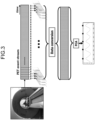
- FIG. 3 shows a schematic for data-driven gating for respiratory motion, according to an embodiment of the present disclosure.
- acquired PET data can be divided into small frames so that by measuring the variances between frames using PCA, a respiratory signal can be detected.
- the respiratory waveform cycle can be between 3 sec to 5 sec. Therefore, the sampling rate can be at least 5Hz or higher to capture respiratory motion.
- an external device need not be included with the imaging apparatus for the respiratory gating measurement.
- the list mode data can be regrouped or reorganized into short frames.
- the short frames can be formed into sinograms or images.
- the sinograms or images can then be reformed into feature vectors.
- PCA can be performed on the vectors to capture a motion signal among the short frames.
- respiratory triggers can be determined by analyzing the local maxima.
- cardiac beating occurs in the same image, both cardiac and respiratory motion appear in the waveform, and PCA can potentially not recognize the difference between the cardiac beating and the respiratory cycle.
- the cardiac beating signal can disturb the desired respiratory waveform, and both phase gating and amplitude gating can be confounded by the cardiac beating signal.
- One method of distinguishing the two motions can include frequency selection of the waveform in a frequency domain since cardiac beating is faster or more frequent than respiratory motion. However, method can require extra frequency domain processing.
- the processing described below allows the cardiac signal to be removed in data-driven respiratory gating.
- the processing according to an embodiment increases the frame length of each small frame such that each frame can include an entire cardiac beating cycle. This allows the difference between frames caused by the cardiac beating signal to be suppressed. Moreover, by keeping the steps between frames short, the frames can capture the respiratory waveform.
- the frame length is also described as a time window.
- the processing according to the embodiment generates, for each time interval (step) of a first length, divided data with a time width (frame length) of a second length longer than the first length, from list mode data, and determines a respiratory waveform based on the divided data.
- Such processing can remove a cardiac signal in data-driven respiratory gating.
- gating without the use of external motion sensors or the like simplifies PET imaging and enables high-quality image reconstruction by eliminating the effects of patient motion.
- FIG. 4 below describes the processing according to an embodiment.
- FIG. 4 shows a non-limiting example of a flow chart for a medical information processing method 400 of identifying and removing a cardiac beating signal, according to an embodiment.
- list mode data representing radiation detected during an imaging scan is obtained via scanning an object such as a patient with a PET imaging apparatus.
- the list mode data is list mode data of a gated bed, which includes both cardiac beating and respiratory motion.
- List mode data is data affected by quasi-periodic motion of an imaging object. That is, although cardiac beating and respiration are periodic motions, the influence of both of these causes the list mode data to be affected by quasi-periodic motion that resembles periodic motion, rather than merely periodic motion.
- the obtained list mode data can be split into short frames without overlap.
- the frame split at step 410 is an example of a first non-overlapping frame.
- the frame length of the first non-overlapping frame is also described as a first frame length. That is, at step 410, a first frame length having a first length is determined and the list mode data is divided into first non-overlapping frames each having the first frame length.
- the short frames can be 0.1 seconds. For instance, consider a scenario where a patient's regular breathing cycle lasts 3 seconds. A data acquisition session can capture approximately 30 breathing cycles. Consequently, the resulting list mode data spanning a duration of 90 seconds can be divided into 900 short frames, each with a duration of 0.1 seconds.
- the data of each short frame can be processed.
- the data can be re-binned into a sinogram, or a time of flight (TOF) plotting, or a reconstruction, among others. It may be appreciated that other methods can be used to extract the motion from the portions or chunks of data.
- TOF time of flight
- PCA can be applied to the multi-frame data.
- a waveform can be extracted, and triggers can be determined or identified by analyzing local maxima.
- the triggers can refer to a point of interest of the respiratory or the cardiac motion cycle.
- the peak of each cycle can be the determined trigger(s).
- one cycle of respiratory motion or one cycle of cardiac beating can occur between two triggers.
- a frequency of the identified triggers can be determined. For example, applying the PCA process to the first non-overlapping frames divided at step 410 can determine a first waveform representing cardiac motion, determine peaks of the first waveform, and determine a cardiac cycle length based on a frequency of the peaks of the first waveform.
- the list mode data can be re-binned with a short step size and a long frame length with a predetermined overlap.
- the data rebinned at step 435 is also described as duplicate data.
- Duplicate data is an example of divided data generated for each time interval of a first length, the divided data having a time width of a second length longer than the first length.
- the frame length of the overlapping frames is also described as a second frame length.
- the second frame length can be based on, for example, the frequency of the triggers such that each frame includes a whole cycle of the cardiac beating.
- the second frame length can be a set value, such as 1 second, or 0.8 seconds, or 0.6 seconds, or 0.4 seconds, or any value less than a duration of the respiratory cycle.
- the data of each long frame can be processed.
- the data can be rebinned to a sinogram, or a time of flight (TOF) plotting, or a reconstruction as previously described.
- TOF time of flight
- PCA can be applied again after re-binning the list mode data to generate the multi-frame data.
- a new waveform such as the respiratory waveform
- triggers can be determined or identified by analyzing local maxima.
- the new waveform can be without the cardiac beating.
- the respiratory waveform can be removed from the combined waveform to determine the cardiac waveform.
- the combined waveform can be the waveform generated in step 425.
- an image can be generated or reconstructed based on the data having the cardiac waveform and the respiratory waveform identified and accounted for. For example, once the respiratory waveform is obtained and the corresponding triggers determined, respiratory gating can be performed. It may be appreciated that other methods can be used, such as phase gating, amplitude gating, etc.
- the medical information processing method 400 can proceed from step 405 to step 435 without determining the frequency of the triggers, such as the cardiac cycle length.
- the set value for the frame length can be used, such as 1 second.
- the second frame length of the overlapping frame generated at step 435 is set according to the cardiac cycle length.
- the second frame length may be set according to the cardiac cycle length determined by processing of steps 410 to 425 and the like, or according to a preset cardiac cycle length.
- FIG. 5 shows a non-limiting example of a flow chart for a medical information processing method 401 of identifying and removing a cardiac beating signal with an auxiliary device, according to an embodiment of the present disclosure.
- list mode data for a gated bed including both cardiac beating and respiratory motion can be obtained via scanning an object such as a patient with the PET imaging apparatus.
- the list mode data can be re-binned with a short step size and a long frame length with a predetermined overlap.
- the frame length can be based on the frequency of the triggers such that each frame includes a whole cycle of the cardiac beating.
- the cardiac beating rate or cycle can be provided by an EKG signal, for example, or any other input from a cardiac measuring device.
- the data of each long frame can be processed.
- the data can be re-binned to a sinogram, or a time of flight (TOF) plotting, or a reconstruction.
- TOF time of flight
- PCA can be applied to the multi-frame data.
- the respiratory waveform can be removed from the combined waveform to determine the cardiac waveform.
- an image can be generated or reconstructed based on the data having the cardiac waveform and the respiratory waveform identified and accounted for.
- FIG. 6A shows a schematic for data splitting of data-driven gating, according to an embodiment of the present disclosure.
- FIG. 6B shows a schematic for data splitting of data-driven gating, according to an embodiment of the present disclosure.
- the data splitting method of FIG. 6A can have a 0.2 second step size with no overlap, and can represent a common data splitting method.
- the length of the frame can be longer, such as 1 second, and there can be overlap between frames, while the step size can still be 0.2 seconds. The overlap can help to identify and remove the cardiac signal.
- FIG. 7 shows a data driven signal and removal of the cardiac signal, according to an embodiment of the present disclosure.
- the less smooth trace is the trace with the data driven gating frame length of, for example, 0.2 seconds, and a step size of, for example, 0.2 seconds, and no overlap between frames, thus yielding many peaks and eventually many triggers.
- the smoother trace is the trace with the data driven gating frame length of, for example, 1 second, a step size of, for example, 0.2 seconds again, and an introduced overlap between frames, thus yielding fewer peaks and eventually fewer triggers.
- the frame length can be increased so that each frame covers an entire cardiac beating cycle.
- the bottom graph shows the data from the top graph with the longer frame length and overlapping frames applied to yield smoother traces with fewer peaks, thus capturing the respiratory cycle.
- FIG. 8 shows a series of reconstructed images describing the effect of gating with and without frame overlap, according to an embodiment of the present disclosure.
- FIG. 8 shows a reconstructed image from ungated, Anzai-gated, 1 second-gated, and 0.2 second-gated data.
- the data driven gating frame length of 0.2 seconds still includes respiratory motion blur, indicated by the arrows.
- the Anzai-gated and the data driven gating frame length of 1 second reconstructed images result in less motion blur. This is especially apparent when compared with the ungated reconstructed image.
- the data driven gating frame length of 1 second yields a reconstructed image that is very similar to the reconstructed image using a dedicated, auxiliary device to measure the respiratory cycle (Anzai-gated), thus demonstrating the effectiveness of the longer frame length with overlap.
- FIGS. 9A and 9B show a non-limiting example of a PET scanner 1100 that can implement the medical information processing methods 400 and 401.
- the PET scanner 1100 includes a number of gamma-ray detectors (GRDs) (e.g., GRD1, GRD2, through GRDN) that are each configured as rectangular detector modules.
- the detector ring includes 40 GRDs.
- Each GRD can include a two-dimensional array of individual detector crystals, which absorb gamma radiation and emit scintillation photons.
- the scintillation photons can be detected by a two-dimensional array of photomultiplier tubes (PMTs) that are also arranged in the GRD.
- a light guide can be disposed between the array of detector crystals and the PMTs.
- the scintillation photons can be detected by an array a silicon photomultipliers (SiPMs), and each individual detector crystals can have a respective SiPM.
- SiPMs silicon photomultipliers
- Each photodetector e.g., PMT or SiPM
- the photons emitted from one detector crystal can be detected by more than one photodetector, and, based on the analog signal produced at each photodetector, the detector crystal corresponding to the detection event can be determined using Anger logic and crystal decoding, for example.
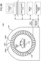
- FIG. 9B shows a schematic view of a PET scanner system having gamma-ray (gamma-ray) photon counting detectors (GRDs) arranged to detect gamma-rays emitted from an object OBJ.
- the GRDs can measure the timing, position, and energy corresponding to each gamma-ray detection.
- the gamma-ray detectors are arranged in a ring, as shown in FIGS. 9A and 9B .
- the detector crystals can be scintillator crystals, which have individual scintillator elements arranged in a two-dimensional array and the scintillator elements can be any known scintillating material.
- the PMTs can be arranged such that light from each scintillator element is detected by multiple PMTs to enable Anger arithmetic and crystal decoding of scintillation event.
- FIG. 9B shows an example of the arrangement of the PET scanner 1100, in which the object OBJ to be imaged rests on a table 1116 and the GRD modules GRD1 through GRDN are arranged circumferentially around the object OBJ and the table 1116.
- the GRDs can be fixedly connected to a circular component 1120 that is fixedly connected to the gantry 1140.
- the gantry 1140 houses many parts of the PET imager.
- the gantry 1140 of the PET imager also includes an open aperture through which the object OBJ and the table 1116 can pass, and gamma-rays emitted in opposite directions from the object OBJ due to an annihilation event can be detected by the GRDs and timing and energy information can be used to determine coincidences for gamma-ray pairs.
- circuitry and hardware is also shown for acquiring, storing, processing, and distributing gamma-ray detection data.
- the circuitry and hardware include: a processor 1170, a network controller 1174, a memory 1178, and a data acquisition system (DAS) 1176.
- the PET imager also includes a data channel that routes detection measurement results from the GRDs to the DAS 1176, the processor 1170, the memory 1178, and the network controller 1174.
- the DAS 1176 can control the acquisition, digitization, and routing of the detection data from the detectors. In one implementation, the DAS 1176 controls the movement of the bed 1116.
- the processor 1170 performs functions including reconstructing images from the detection data, pre-reconstruction processing of the detection data, and post-reconstruction processing of the image data, as discussed herein.
- the processor 1170 can be configured to perform various steps of the medical information processing methods 400 and/or 401 described above and variations thereof.
- the processor 1170 is an example of a processing unit. For example, the processor 1170 obtains list mode data representing radiation detected during an imaging scan, generates divided data from the list mode data for each time interval of a first length, the divided data having a time width of a second length longer than the first length, determines a respiratory waveform based on the divided data, and reconstruct an image based on the respiratory waveform and the list mode data.
- the processor 1170 can include a CPU that can be implemented as discrete logic gates, as an Application Specific Integrated Circuit (ASIC), a Field Programmable Gate Array (FPGA) or other Complex Programmable Logic Device (CPLD).
- An FPGA or CPLD implementation may be coded in VHDL, Verilog, or any other hardware description language and the code may be stored in an electronic memory directly within the FPGA or CPLD, or as a separate electronic memory.
- the memory may be non-volatile, such as ROM, EPROM, EEPROM or FLASH memory.
- the memory can also be volatile, such as static or dynamic RAM, and a processor, such as a microcontroller or microprocessor, may be provided to manage the electronic memory as well as the interaction between the FPGA or CPLD and the memory.
- the CPU in the processor 1170 can execute a computer program including a set of computer-readable instructions that perform various steps of method 100 and/or method 200, the program being stored in any of the above-described non-transitory electronic memories and/or a hard disk drive, CD, DVD, FLASH drive or any other known storage media.
- the computer-readable instructions may be provided as a utility application, background daemon, or component of an operating system, or combination thereof, executing in conjunction with a processor, such as a Xenon processor from Intel of America or an Opteron processor from AMD of America and an operating system, such as Microsoft VISTA, UNIX, Solaris, LINUX, Apple, MAC-OS and other operating systems known to those skilled in the art.
- CPU can be implemented as multiple processors cooperatively working in parallel to perform the instructions.
- the memory 1178 can be a hard disk drive, CD-ROM drive, DVD drive, FLASH drive, RAM, ROM or any other electronic storage known in the art.
- the network controller 1174 can interface between the various parts of the PET imager. Additionally, the network controller 1174 can also interface with an external network.
- the external network can be a public network, such as the Internet, or a private network such as an LAN or WAN network, or any combination thereof and can also include PSTN or ISDN sub-networks.
- the external network can also be wired, such as an Ethernet network, or can be wireless such as a cellular network including EDGE, 11G and 4G wireless cellular systems.
- the wireless network can also be WiFi, Bluetooth, or any other wireless form of communication that is known.
- a cardiac signal can be removed in data-driven respiratory gating.
Landscapes
- Health & Medical Sciences (AREA)
- Engineering & Computer Science (AREA)
- Life Sciences & Earth Sciences (AREA)
- Medical Informatics (AREA)
- Physics & Mathematics (AREA)
- General Health & Medical Sciences (AREA)
- Nuclear Medicine, Radiotherapy & Molecular Imaging (AREA)
- Veterinary Medicine (AREA)
- Public Health (AREA)
- Biophysics (AREA)
- High Energy & Nuclear Physics (AREA)
- Surgery (AREA)
- Animal Behavior & Ethology (AREA)
- Optics & Photonics (AREA)
- Pathology (AREA)
- Radiology & Medical Imaging (AREA)
- Biomedical Technology (AREA)
- Heart & Thoracic Surgery (AREA)
- Molecular Biology (AREA)
- Computer Vision & Pattern Recognition (AREA)
- Theoretical Computer Science (AREA)
- General Physics & Mathematics (AREA)
- Physiology (AREA)
- Multimedia (AREA)
- Cardiology (AREA)
- Dentistry (AREA)
- Oral & Maxillofacial Surgery (AREA)
- Nuclear Medicine (AREA)
Abstract
A PET apparatus (1100) of an embodiment includes a processing unit (1170) configured to: obtain list mode data representing radiation detected during an imaging scan; generate divided data from the list mode data for each time interval of a first length, the divided data having a time width of a second length longer than the first length; determine a respiratory waveform based on the divided data, and; reconstruct an image based on the respiratory waveform and the list mode data.
Description
- Embodiments described herein relate generally to a PET apparatus and a medical information processing method.
- This disclosure relates to list-mode data gating in positron emission tomography (PET) systems as a means to eliminate motion-related inaccuracies and improve image quality in PET imaging.
- The background description provided herein is for the purpose of generally presenting the context of the disclosure. Work of the presently named inventors, to the extent the work is described in this background section, as well as aspects of the description that may not otherwise qualify as prior art at the time of filing, are neither expressly nor impliedly admitted as prior art against the present disclosure.
- Patient motion is a main source of blurring and artifacts in positron emission tomography (PET) imaging. Respiratory motion and cardiac motion during PET data acquisition can degrade quantitation performance by blurring images, leading to over-estimation of lesion volumes and under-estimation of lesion activity. Gating methods have been used to improve the quality of PET images. Typically, these methods need to use an external device to detect a biosignal, for example, a respiration waveform, electrocardiography (ECG), etc.
- In some contexts, motion correction can be addressed by gating acquired data in which motion may have occurred. Gating involves dividing data into separates chunks (gates) within which motion is negligible. This may occur during voluntary or involuntary movement of the patient, and may include, for example, movement due to respiration or heartbeat.
- In some PET systems, gating is done by attaching motion sensors to a patient during a PET scan. Such external motion sensors make PET scans more cumbersome as their use requires motion information to be successfully recorded during the scan. Moreover, if motion is not recorded correctly or properly synchronized with the scan, then motion correction often is hampered.
- Recently, data-driven approaches have been developed for extracting the biosignal from raw PET data, or from reconstructed images. For example, a data-driven algorithm can first divide the PET acquisitions into small temporal frames, and then principal component analysis (PCA) or independent component analysis (ICA) can be applied on the frames. The biosignal can be extracted based on the data variation across all the frames.
- For instance, where there is respiratory motion, normally the principal variation can be caused by the respiratory motion. Thus, a respiratory signal can be modeled by the first component of the PCA. Once the respiratory motion is estimated, the PET data can be binned and reconstructed into multiple gates (for visualizing the motion) or a single gate (for generation of a single, motion-reduced image). However, when there is a heart beating in the acquisitions, the signal from the PCA can also include a cardiac signal.
- Therefore, a method to separate the two signals so as to have better gating, either cardiac or respiratory gating, is desired.
- A positron emission tomography (PET) apparatus provided according to one aspect of the present invention includes a processing unit. The processing unit is configured to obtain list mode data representing radiation detected during an imaging scan. The processing unit is configured to generate divided data from the list mode data for each time interval of a first length, the divided data having a time width of a second length longer than the first length. The processing unit is configured to determine a respiratory waveform based on the divided data. The processing unit is configured to reconstruct an image based on the respiratory waveform and the list mode data.
- The processing unit may set the second length according to a cardiac cycle length.
- The list mode data may be data affected by quasi-periodic motion of an imaging object, and the processing unit may be configured to: determine a first frame length having the first length, divide the list mode data into first non-overlapping frames of a first frame length, and process the first non-overlapping frames to determine a cardiac cycle length; determine a second frame length having the second length longer than the first frame length, based on the determined cardiac cycle length; re-bin the list mode data into overlapping frames having the second frame length, based on the non-overlapping frames having the first frame length, to generate the divided data; apply a principal component analysis (PCA) process to the divided data to determine the respiratory waveform; determine a cardiac waveform using the respiratory waveform; and reconstruct an image based on the list mode data using the respiratory waveform and the cardiac waveform.
- The processing unit may apply the PCA process to the first non-overlapping frames to determine a first waveform, determine peaks of the first waveform, determine the cardiac cycle length based on a frequency of the peaks of the first waveform, and determine the second frame length based on the cardiac cycle length.
- The frequency of the peaks of the first waveform may be a cardiac beating rate.
- The second frame length may be a value less than a frequency of peaks of the respiratory waveform.
- The second frame length may be 1 second.
- The processing unit may set the first frame length to a value less than 0.4 seconds.
- The processing unit may reconstruct the image based on the list mode data processed using the respiratory waveform and the cardiac waveform by applying a respiratory gate based on the respiratory waveform and removing the cardiac waveform.
- The processing unit may determine the second frame length by measuring the cardiac cycle length of the object via an ECG device.
- The overlapping frames having the second frame length may overlap by the first frame length.
- The processing unit may re-bin the list mode data into a sinogram as processing of re-binning into overlapping frames having the second frame length.
- A medical information processing method according to one aspect of the present invention includes: obtaining list mode data representing radiation detected during an imaging scan; generating divided data from the list mode data for each time interval of a first length, the divided data having a time width of a second length longer than the first length; and determining a respiratory waveform based on the divided data.
- In the medical information processing method, the second length may be set according to a cardiac cycle length.
- The medical information processing method may comprise: determining a first frame length having the first length, dividing the list mode data into first non-overlapping frames of a first frame length, the list mode data being affected by quasi-periodic motion of an imaging object, and processing the first non-overlapping frames to determine a cardiac cycle length; determining a second frame length having the second length longer than the first frame length, based on the determined cardiac cycle length; re-binning the list mode data into overlapping frames having the second frame length, based on the non-overlapping frames having the first frame length, to generate the divided data; applying a principal component analysis (PCA) process to the divided data to determine the respiratory waveform; determining a cardiac waveform using the respiratory waveform; and reconstructing an image based on the list mode data using the respiratory waveform and the cardiac waveform.
- The medical information processing method may comprise applying the PCA process to the first non-overlapping frames to determine a first waveform, determining peaks of the first waveform, determining the cardiac cycle length based on a frequency of the peaks of the first waveform, and determining the second frame length based on the cardiac cycle length.
- In the medical information processing method, the frequency of the peaks of the first waveform may be a cardiac beating rate.
- In the medical information processing method, the second frame length may be a value less than a frequency of peaks of the respiratory waveform.
- In the medical information processing method, the second frame length may be 1 second.
- In the medical information processing method, the first frame length may be a value less than 0.4 seconds.
- The medical information processing method may comprise reconstructing the image based on the list mode data processed using the determined respiratory waveform and the determined cardiac waveform by applying a respiratory gate based on the determined respiratory waveform and removing the cardiac waveform.
- The medical information processing method may comprise determining the second frame length by measuring the cardiac cycle length of the object via an ECG device.
- In the medical information processing method, the overlapping frames having the second frame length may overlap by the first frame length.
- A non-transitory computer-readable storage medium provided according to one aspect of the present invention stores a computer program that causes a computer to perform processes of: obtaining list mode data representing radiation detected during an imaging scan; generating divided data from the list mode data for each time interval of a first length, the divided data having a time width of a second length longer than the first length; and determining a respiratory waveform based on the divided data.
- The program may cause a computer to perform processes of: determining a first frame length having the first length, dividing the list mode data into first non-overlapping frames of a first frame length, the list mode data being affected by quasi-periodic motion of an imaging object, and processing the first non-overlapping frames to determine a cardiac cycle length; determining a second frame length having the second length longer than the first frame length, based on the determined cardiac cycle length; re-binning the list mode data into overlapping frames having the second frame length, based on the non-overlapping frames having the first frame length, to generate the divided data; applying a principal component analysis (PCA) process to the divided data to determine the respiratory waveform; determining a cardiac waveform using the respiratory waveform; and reconstructing an image based on the list mode data using the respiratory waveform and the cardiac waveform.
- A positron emission tomography (PET) scanner provided according to one aspect of the present invention includes a processing unit configured to receive list mode data; divide the list mode data into first and second data, wherein time windows of the first and the second data are determined based on cardiac beating; and estimate a respiratory waveform based on the first and the second data.
- The time windows of the first and the second data may be determined based on one cycle of the cardiac beating.
- The time windows of the first and the second data may comprise the one cycle of the cardiac beating.
- The time windows of the first and the second data may be about 1 second.
- The time windows of the first and the second data may be overlapping at least part of one another.
-
-
FIG. 1 is a schematic of a whole-body PET scan with and without respiratory gating, according to an embodiment. -
FIG. 2A is a schematic of a PET system for gating in list-mode acquisition, according to an embodiment. -
FIG. 2B is a schematic of processing of sorting the PET event stream into bins, according to an embodiment. -
FIG. 3 is a schematic for data-driven gating for respiratory motion, according to an embodiment. -
FIG. 4 is a diagram showing a non-limiting example of a flow chart for a method of identifying and removing a cardiac beating signal, according to an embodiment. -
FIG. 5 is a diagram showing a non-limiting example of a flow chart for a method of identifying and removing a cardiac beating signal with an auxiliary device, according to an embodiment. -
FIG. 6A is a schematic for data splitting of data-driven gating, according to an embodiment. -
FIG. 6B is a schematic for data splitting of data-driven gating, according to an embodiment. -
FIG. 7 is a diagram showing another example of removing the cardiac signal, according to an embodiment. -
FIG. 8 is a diagram showing a series of reconstructed images describing the effect of gating with and without frame overlap, according to an embodiment. -
FIG. 9A is an illustration of a perspective view of a PET scanner apparatus, according to embodiments. -
FIG. 9B is a schematic of a PET scanner apparatus and associated hardware, according to embodiments. - The following disclosure provides many different embodiments, or examples, for implementing different features of the provided subject matter. To simplify the present disclosure, specific examples of components and arrangements are described below. These are, of course, merely examples and are not intended to be limiting.
- For example, the order of discussion of the different steps as described herein has been presented for the sake of clarity. In general, these steps can be performed in any suitable order. Additionally, although each of the different features, techniques, configurations, etc. herein may be discussed in different places of this disclosure, it is intended that each of the concepts can be executed independently of each other or in combination with each other. Accordingly, the present disclosure can be embodied and viewed in many different ways.
- Furthermore, as used herein, the words "a," "an," and the like generally carry a meaning of "one or more," unless stated otherwise.
- Numerous modifications and variations are possible in light of the above teachings. It is therefore to be understood that within the scope of the appended claims, the disclosure can be practiced otherwise than as specifically described herein.
- Note that while the following embodiments are described in the context of respiratory gating, it is not meant to be restrictive. One skilled in the art will recognize that the concepts and principles discussed can be extended to other gating applications, such as cardiac gating, for example.
-
FIG. 1 shows a schematic of a whole-body PET scan with and without respiratory gating, according to an embodiment of the present disclosure. In an embodiment, on the left (in the circle) ofFIG. 1 , a lung mass can be detected based on a scan duration of approximately 2 to 3 minutes for the highlighted portion in which the lung mass is positioned. On the right (in the circle) ofFIG. 1 , retrospective respiratory gating can be applied. While there can be fewer counts, a sharper image can be produced using the respiratory gating. This can lead to a change or improvement in a staging diagnosis for a patient. The gating can be performed by synchronizing data acquisition with the gate and then retrospectively sorting the data for eventual image reconstruction. -
FIG. 2A shows a schematic of an overview of a PET system for gating in list-mode acquisition, according to an embodiment. In an embodiment, a respiratory gating system can be synchronized to the imaging apparatus, which can include a timing or clock sync. During the synchronized data acquisition, the PET event stream can be generated with the respiratory gating or cycle data. As shown, a beam of the imaging apparatus can be active when a predetermined portion of the respiratory cycle occurs, which can be the target gate. Thus, a portion of the entire PET event stream can be filtered for event occurring during the predetermined portion of the respiratory cycle (e.g., with the beam active, with the gate on/active, etc.). -
FIG. 2B shows a schematic of processing of sorting the PET event stream into bins, according to an embodiment of the present disclosure. In an embodiment, a complete respiratory cycle can be divided into multiple bins. For example, as shown, one complete respiratory cycle can be divided into three bins, and the three bins can correspond to the divided portions shown inFIG. 2A based on the output from the respiratory gating system. The events from each cycle can be divided into one of the three corresponding bins and aggregated over all cycles used for data processing. Thus, the three "total" bins together can represent the average data over all cycles. The divided bin data can individually be used to generate image reconstructions. For example, a reconstruction can be generated for the data in the first bin, a separate reconstruction can be generated for the data in the second bin, and another separate reconstruction can be generated for the data in the third bin. A cine image with three volumes can be generated, or, if desired, only the data for "bin 2" can be used since the "bin 2" data can correspond to the target gate event. -
FIG. 3 shows a schematic for data-driven gating for respiratory motion, according to an embodiment of the present disclosure. In an embodiment, in data-driven respiratory gating, acquired PET data can be divided into small frames so that by measuring the variances between frames using PCA, a respiratory signal can be detected. For example, the respiratory waveform cycle can be between 3 sec to 5 sec. Therefore, the sampling rate can be at least 5Hz or higher to capture respiratory motion. Notably, an external device need not be included with the imaging apparatus for the respiratory gating measurement. In brief, the list mode data can be regrouped or reorganized into short frames. The short frames can be formed into sinograms or images. The sinograms or images can then be reformed into feature vectors. PCA can be performed on the vectors to capture a motion signal among the short frames. Subsequently, respiratory triggers can be determined by analyzing the local maxima. - However, when cardiac beating occurs in the same image, both cardiac and respiratory motion appear in the waveform, and PCA can potentially not recognize the difference between the cardiac beating and the respiratory cycle. The cardiac beating signal can disturb the desired respiratory waveform, and both phase gating and amplitude gating can be confounded by the cardiac beating signal. One method of distinguishing the two motions can include frequency selection of the waveform in a frequency domain since cardiac beating is faster or more frequent than respiratory motion. However, method can require extra frequency domain processing.
- Therefore, in the present embodiment, the processing described below allows the cardiac signal to be removed in data-driven respiratory gating. For example, the processing according to an embodiment increases the frame length of each small frame such that each frame can include an entire cardiac beating cycle. This allows the difference between frames caused by the cardiac beating signal to be suppressed. Moreover, by keeping the steps between frames short, the frames can capture the respiratory waveform. The frame length is also described as a time window.
- That is, the processing according to the embodiment generates, for each time interval (step) of a first length, divided data with a time width (frame length) of a second length longer than the first length, from list mode data, and determines a respiratory waveform based on the divided data. Such processing can remove a cardiac signal in data-driven respiratory gating. In other words, gating without the use of external motion sensors or the like simplifies PET imaging and enables high-quality image reconstruction by eliminating the effects of patient motion.
-
FIG. 4 below describes the processing according to an embodiment.FIG. 4 shows a non-limiting example of a flow chart for a medicalinformation processing method 400 of identifying and removing a cardiac beating signal, according to an embodiment. - In an embodiment, at
step 405, list mode data representing radiation detected during an imaging scan is obtained via scanning an object such as a patient with a PET imaging apparatus. The list mode data is list mode data of a gated bed, which includes both cardiac beating and respiratory motion. - List mode data is data affected by quasi-periodic motion of an imaging object. That is, although cardiac beating and respiration are periodic motions, the influence of both of these causes the list mode data to be affected by quasi-periodic motion that resembles periodic motion, rather than merely periodic motion.
- In an embodiment, at
step 410, the obtained list mode data can be split into short frames without overlap. The frame split atstep 410 is an example of a first non-overlapping frame. The frame length of the first non-overlapping frame is also described as a first frame length. That is, atstep 410, a first frame length having a first length is determined and the list mode data is divided into first non-overlapping frames each having the first frame length. For example, the short frames can be 0.1 seconds. For instance, consider a scenario where a patient's regular breathing cycle lasts 3 seconds. A data acquisition session can capture approximately 30 breathing cycles. Consequently, the resulting list mode data spanning a duration of 90 seconds can be divided into 900 short frames, each with a duration of 0.1 seconds. - In an embodiment, at
step 415, the data of each short frame can be processed. For example, the data can be re-binned into a sinogram, or a time of flight (TOF) plotting, or a reconstruction, among others. It may be appreciated that other methods can be used to extract the motion from the portions or chunks of data. - In an embodiment, at
step 420, PCA can be applied to the multi-frame data. - In an embodiment, at
step 425, a waveform can be extracted, and triggers can be determined or identified by analyzing local maxima. The triggers can refer to a point of interest of the respiratory or the cardiac motion cycle. For example, the peak of each cycle can be the determined trigger(s). Thus, one cycle of respiratory motion or one cycle of cardiac beating can occur between two triggers. - In an embodiment, at
step 430, a frequency of the identified triggers can be determined. For example, applying the PCA process to the first non-overlapping frames divided atstep 410 can determine a first waveform representing cardiac motion, determine peaks of the first waveform, and determine a cardiac cycle length based on a frequency of the peaks of the first waveform. - In an embodiment, at
step 435, the list mode data can be re-binned with a short step size and a long frame length with a predetermined overlap. The data rebinned atstep 435 is also described as duplicate data. Duplicate data is an example of divided data generated for each time interval of a first length, the divided data having a time width of a second length longer than the first length. - The frame length of the overlapping frames is also described as a second frame length. The second frame length can be based on, for example, the frequency of the triggers such that each frame includes a whole cycle of the cardiac beating. The second frame length can be a set value, such as 1 second, or 0.8 seconds, or 0.6 seconds, or 0.4 seconds, or any value less than a duration of the respiratory cycle.
- In an embodiment, at
step 440, the data of each long frame can be processed. For example, the data can be rebinned to a sinogram, or a time of flight (TOF) plotting, or a reconstruction as previously described. - In an embodiment, at
step 445, PCA can be applied again after re-binning the list mode data to generate the multi-frame data. - In an embodiment, at
step 450, a new waveform, such as the respiratory waveform, can be determined, and triggers can be determined or identified by analyzing local maxima. Notably, the new waveform can be without the cardiac beating. - In an embodiment, at
step 455, the respiratory waveform can be removed from the combined waveform to determine the cardiac waveform. The combined waveform can be the waveform generated instep 425. - In an embodiment, at
step 460, an image can be generated or reconstructed based on the data having the cardiac waveform and the respiratory waveform identified and accounted for. For example, once the respiratory waveform is obtained and the corresponding triggers determined, respiratory gating can be performed. It may be appreciated that other methods can be used, such as phase gating, amplitude gating, etc. - In an embodiment, the medical
information processing method 400 can proceed fromstep 405 to step 435 without determining the frequency of the triggers, such as the cardiac cycle length. Instead, the set value for the frame length can be used, such as 1 second. - For example, the second frame length of the overlapping frame generated at
step 435 is set according to the cardiac cycle length. The second frame length may be set according to the cardiac cycle length determined by processing ofsteps 410 to 425 and the like, or according to a preset cardiac cycle length. - Additionally or alternatively,
FIG. 5 shows a non-limiting example of a flow chart for a medicalinformation processing method 401 of identifying and removing a cardiac beating signal with an auxiliary device, according to an embodiment of the present disclosure. - In an embodiment, at
step 470, list mode data for a gated bed including both cardiac beating and respiratory motion can be obtained via scanning an object such as a patient with the PET imaging apparatus. - In an embodiment, at
step 475, similar to the previous description, the list mode data can be re-binned with a short step size and a long frame length with a predetermined overlap. The frame length can be based on the frequency of the triggers such that each frame includes a whole cycle of the cardiac beating. The cardiac beating rate or cycle can be provided by an EKG signal, for example, or any other input from a cardiac measuring device. - In an embodiment, at
step 480, similar to the previous description, the data of each long frame can be processed. For example, the data can be re-binned to a sinogram, or a time of flight (TOF) plotting, or a reconstruction. - In an embodiment, at
step 485, similar to the previous description, PCA can be applied to the multi-frame data. - In an embodiment, at
step 490, similar to the previous description, the respiratory waveform can be removed from the combined waveform to determine the cardiac waveform. - In an embodiment, at
step 495, similar to the previous description, an image can be generated or reconstructed based on the data having the cardiac waveform and the respiratory waveform identified and accounted for. -
FIG. 6A shows a schematic for data splitting of data-driven gating, according to an embodiment of the present disclosure.FIG. 6B shows a schematic for data splitting of data-driven gating, according to an embodiment of the present disclosure. In an embodiment, the data splitting method ofFIG. 6A can have a 0.2 second step size with no overlap, and can represent a common data splitting method. However, for the data splitting method described herein and with reference toFIG. 6B , the length of the frame can be longer, such as 1 second, and there can be overlap between frames, while the step size can still be 0.2 seconds. The overlap can help to identify and remove the cardiac signal. - To this end,
FIG. 7 shows a data driven signal and removal of the cardiac signal, according to an embodiment of the present disclosure. The less smooth trace is the trace with the data driven gating frame length of, for example, 0.2 seconds, and a step size of, for example, 0.2 seconds, and no overlap between frames, thus yielding many peaks and eventually many triggers. The smoother trace is the trace with the data driven gating frame length of, for example, 1 second, a step size of, for example, 0.2 seconds again, and an introduced overlap between frames, thus yielding fewer peaks and eventually fewer triggers. - In an embodiment, the frame length can be increased so that each frame covers an entire cardiac beating cycle. The bottom graph shows the data from the top graph with the longer frame length and overlapping frames applied to yield smoother traces with fewer peaks, thus capturing the respiratory cycle.
-
FIG. 8 shows a series of reconstructed images describing the effect of gating with and without frame overlap, according to an embodiment of the present disclosure. In an embodiment,FIG. 8 shows a reconstructed image from ungated, Anzai-gated, 1 second-gated, and 0.2 second-gated data. As shown, it is clear that the data driven gating frame length of 0.2 seconds still includes respiratory motion blur, indicated by the arrows. In contrast, the Anzai-gated and the data driven gating frame length of 1 second reconstructed images result in less motion blur. This is especially apparent when compared with the ungated reconstructed image. Notably, the data driven gating frame length of 1 second yields a reconstructed image that is very similar to the reconstructed image using a dedicated, auxiliary device to measure the respiratory cycle (Anzai-gated), thus demonstrating the effectiveness of the longer frame length with overlap. -
FIGS. 9A and9B show a non-limiting example of aPET scanner 1100 that can implement the medicalinformation processing methods PET scanner 1100 includes a number of gamma-ray detectors (GRDs) (e.g., GRD1, GRD2, through GRDN) that are each configured as rectangular detector modules. According to one implementation, the detector ring includes 40 GRDs. In another implementation, there are 48 GRDs, and the higher number of GRDs is used to create a larger bore size for thePET scanner 1100. - Each GRD can include a two-dimensional array of individual detector crystals, which absorb gamma radiation and emit scintillation photons. The scintillation photons can be detected by a two-dimensional array of photomultiplier tubes (PMTs) that are also arranged in the GRD. A light guide can be disposed between the array of detector crystals and the PMTs.
- Alternatively, the scintillation photons can be detected by an array a silicon photomultipliers (SiPMs), and each individual detector crystals can have a respective SiPM.
- Each photodetector (e.g., PMT or SiPM) can produce an analog signal that indicates when scintillation events occur, and an energy of the gamma ray producing the detection event. Moreover, the photons emitted from one detector crystal can be detected by more than one photodetector, and, based on the analog signal produced at each photodetector, the detector crystal corresponding to the detection event can be determined using Anger logic and crystal decoding, for example.
-
FIG. 9B shows a schematic view of a PET scanner system having gamma-ray (gamma-ray) photon counting detectors (GRDs) arranged to detect gamma-rays emitted from an object OBJ. The GRDs can measure the timing, position, and energy corresponding to each gamma-ray detection. In one implementation, the gamma-ray detectors are arranged in a ring, as shown inFIGS. 9A and9B . The detector crystals can be scintillator crystals, which have individual scintillator elements arranged in a two-dimensional array and the scintillator elements can be any known scintillating material. The PMTs can be arranged such that light from each scintillator element is detected by multiple PMTs to enable Anger arithmetic and crystal decoding of scintillation event. -
FIG. 9B shows an example of the arrangement of thePET scanner 1100, in which the object OBJ to be imaged rests on a table 1116 and the GRD modules GRD1 through GRDN are arranged circumferentially around the object OBJ and the table 1116. The GRDs can be fixedly connected to acircular component 1120 that is fixedly connected to thegantry 1140. Thegantry 1140 houses many parts of the PET imager. Thegantry 1140 of the PET imager also includes an open aperture through which the object OBJ and the table 1116 can pass, and gamma-rays emitted in opposite directions from the object OBJ due to an annihilation event can be detected by the GRDs and timing and energy information can be used to determine coincidences for gamma-ray pairs. - In
FIG. 9B , circuitry and hardware is also shown for acquiring, storing, processing, and distributing gamma-ray detection data. The circuitry and hardware include: aprocessor 1170, anetwork controller 1174, amemory 1178, and a data acquisition system (DAS) 1176. The PET imager also includes a data channel that routes detection measurement results from the GRDs to theDAS 1176, theprocessor 1170, thememory 1178, and thenetwork controller 1174. TheDAS 1176 can control the acquisition, digitization, and routing of the detection data from the detectors. In one implementation, theDAS 1176 controls the movement of thebed 1116. Theprocessor 1170 performs functions including reconstructing images from the detection data, pre-reconstruction processing of the detection data, and post-reconstruction processing of the image data, as discussed herein. - The
processor 1170 can be configured to perform various steps of the medicalinformation processing methods 400 and/or 401 described above and variations thereof. Theprocessor 1170 is an example of a processing unit. For example, theprocessor 1170 obtains list mode data representing radiation detected during an imaging scan, generates divided data from the list mode data for each time interval of a first length, the divided data having a time width of a second length longer than the first length, determines a respiratory waveform based on the divided data, and reconstruct an image based on the respiratory waveform and the list mode data. - The
processor 1170 can include a CPU that can be implemented as discrete logic gates, as an Application Specific Integrated Circuit (ASIC), a Field Programmable Gate Array (FPGA) or other Complex Programmable Logic Device (CPLD). An FPGA or CPLD implementation may be coded in VHDL, Verilog, or any other hardware description language and the code may be stored in an electronic memory directly within the FPGA or CPLD, or as a separate electronic memory. Further, the memory may be non-volatile, such as ROM, EPROM, EEPROM or FLASH memory. The memory can also be volatile, such as static or dynamic RAM, and a processor, such as a microcontroller or microprocessor, may be provided to manage the electronic memory as well as the interaction between the FPGA or CPLD and the memory. - Alternatively, the CPU in the
processor 1170 can execute a computer program including a set of computer-readable instructions that perform various steps of method 100 and/or method 200, the program being stored in any of the above-described non-transitory electronic memories and/or a hard disk drive, CD, DVD, FLASH drive or any other known storage media. Further, the computer-readable instructions may be provided as a utility application, background daemon, or component of an operating system, or combination thereof, executing in conjunction with a processor, such as a Xenon processor from Intel of America or an Opteron processor from AMD of America and an operating system, such as Microsoft VISTA, UNIX, Solaris, LINUX, Apple, MAC-OS and other operating systems known to those skilled in the art. Further, CPU can be implemented as multiple processors cooperatively working in parallel to perform the instructions. - The
memory 1178 can be a hard disk drive, CD-ROM drive, DVD drive, FLASH drive, RAM, ROM or any other electronic storage known in the art. - The
network controller 1174, such as an Intel Ethernet PRO network interface card from Intel Corporation of America, can interface between the various parts of the PET imager. Additionally, thenetwork controller 1174 can also interface with an external network. As can be appreciated, the external network can be a public network, such as the Internet, or a private network such as an LAN or WAN network, or any combination thereof and can also include PSTN or ISDN sub-networks. The external network can also be wired, such as an Ethernet network, or can be wireless such as a cellular network including EDGE, 11G and 4G wireless cellular systems. The wireless network can also be WiFi, Bluetooth, or any other wireless form of communication that is known. - While this specification contains many specific implementation details, these should not be construed as limitations on the scope of what may be claimed, but rather as descriptions of features that may be specific to particular embodiments.
- Certain features that are described in this specification in the context of separate embodiments can also be implemented in combination in a single embodiment. Conversely, various features that are described in the context of a single embodiment can also be implemented in multiple embodiments separately or in any suitable sub-combination. Moreover, although features may be described above as acting in certain combinations and even initially claimed as such, one or more features from a claimed combination can in some cases be excised from the combination, and the claimed combination may be directed to a sub-combination or variation of a sub-combination.
- In the preceding description, specific details have been set forth, such as a particular geometry of a processing system and descriptions of various components and processes used therein. It should be understood, however, that techniques herein may be practiced in other embodiments that depart from these specific details, and that such details are for purposes of explanation and not limitation. Embodiments disclosed herein have been described with reference to the accompanying drawings. Similarly, for purposes of explanation, specific numbers, materials, and configurations have been set forth in order to provide a thorough understanding. Nevertheless, embodiments may be practiced without such specific details. Components having substantially the same functional constructions are denoted by like reference characters, and thus any redundant descriptions may be omitted.
- Various techniques have been described as multiple discrete operations to assist in understanding the various embodiments. The order of description should not be construed as to imply that these operations are necessarily order dependent. Indeed, these operations need not be performed in the order of presentation. Operations described may be performed in a different order than the described embodiment. Various additional operations may be performed and/or described operations may be omitted in additional embodiments.
- According to at least one embodiment described above, a cardiac signal can be removed in data-driven respiratory gating.
- While certain embodiments have been described, these embodiments have been presented by way of example only, and are not intended to limit the scope of the inventions. Indeed, the novel embodiments described herein may be embodied in a variety of other forms; furthermore, various omissions, substitutions and changes in the form of the embodiments described herein may be made without departing from the scope of the invention as defined by the appended claims.
Claims (13)
- A positron emission tomography (PET) apparatus (1100) comprising a processing unit (1170) configured to:obtain list mode data representing radiation detected during an imaging scan;generate divided data from the list mode data for each time interval of a first length, the divided data having a time width of a second length longer than the first length;determine a respiratory waveform based on the divided data; andreconstruct an image based on the respiratory waveform and the list mode data.
- The PET apparatus according to claim 1, wherein the processing unit sets the second length according to a cardiac cycle length.
- The PET apparatus according to claim 1, whereinthe list mode data is data affected by quasi-periodic motion of an imaging object,the processing unit may be configured to:determine a first frame length having the first length, divide the list mode data into first non-overlapping frames of a first frame length, and process the first non-overlapping frames to determine a cardiac cycle length;determine a second frame length having the second length longer than the first frame length, based on the determined cardiac cycle length;re-bin the list mode data into overlapping frames having the second frame length, based on the non-overlapping frames having the first frame length, to generate the divided data;apply a principal component analysis (PCA) process to the divided data to determine the respiratory waveform;determine a cardiac waveform using the respiratory waveform; andreconstruct an image based on the list mode data using the respiratory waveform and the cardiac waveform.
- The PET apparatus according to claim 3, wherein the processing unit applies the PCA process to the first non-overlapping frames to determine a first waveform, determine peaks of the first waveform, determine the cardiac cycle length based on a frequency of the peaks of the first waveform, and determine the second frame length based on the cardiac cycle length.
- The PET apparatus according to claim 4, wherein the frequency of the peaks of the first waveform is a cardiac beating rate.
- The PET apparatus according to claim 3, wherein the second frame length is a value less than a frequency of peaks of the respiratory waveform.
- The PET apparatus according to claim 6, wherein the second frame length is 1 second.
- The PET apparatus according to claim 4, wherein the processing unit sets the first frame length to a value less than 0.4 seconds.
- The PET apparatus according to claim 3, wherein the processing unit reconstructs the image based on the list mode data processed using the respiratory waveform and the cardiac waveform by applying a respiratory gate based on the respiratory waveform and removing the cardiac waveform.
- The PET apparatus according to claim 3, further comprising an electrocardiogram (ECG) device, wherein
the processing unit determines the second frame length by measuring the cardiac cycle length of the object via the ECG device. - The PET apparatus according to claim 3, wherein the overlapping frames having the second frame length overlap by the first frame length.
- The PET apparatus according to claim 3, wherein the processing unit re-bins the list mode data into a sinogram as processing of re-binning into overlapping frames having the second frame length.
- A medical information processing method comprising:obtaining list mode data representing radiation detected during an imaging scan;generating divided data from the list mode data for each time interval of a first length, the divided data having a time width of a second length longer than the first length; anddetermining a respiratory waveform based on the divided data.
Applications Claiming Priority (2)
| Application Number | Priority Date | Filing Date | Title |
|---|---|---|---|
| US202363479052P | 2023-01-09 | 2023-01-09 | |
| US18/336,741 US12518445B2 (en) | 2023-01-09 | 2023-06-16 | Method and apparatus for cardiac signal removal in data-driven respiratory gating in positron emission tomography systems |
Publications (1)
| Publication Number | Publication Date |
|---|---|
| EP4411648A1 true EP4411648A1 (en) | 2024-08-07 |
Family
ID=89535827
Family Applications (1)
| Application Number | Title | Priority Date | Filing Date |
|---|---|---|---|
| EP24150798.7A Pending EP4411648A1 (en) | 2023-01-09 | 2024-01-08 | Pet apparatus and a medical information processing method |
Country Status (3)
| Country | Link |
|---|---|
| US (1) | US12518445B2 (en) |
| EP (1) | EP4411648A1 (en) |
| JP (1) | JP2024098156A (en) |
Family Cites Families (8)
| Publication number | Priority date | Publication date | Assignee | Title |
|---|---|---|---|---|
| US8285359B2 (en) * | 2003-11-26 | 2012-10-09 | General Electric Company | Method and system for retrospective gating using multiple inputs |
| US7574249B2 (en) * | 2005-02-08 | 2009-08-11 | General Electric Company | Device-less gating of physiological movement for improved image detection |
| JP5545881B2 (en) | 2011-03-14 | 2014-07-09 | 株式会社リガク | CT image processing apparatus and CT image processing method |
| US9392958B2 (en) * | 2012-05-30 | 2016-07-19 | Siemens Aktiengesellschaft | Method of attenuation correction of positron emission tomography data and combined positron emission tomography and magnetic resonance tomography system |
| US9655522B2 (en) * | 2014-10-10 | 2017-05-23 | Cedars-Sinai Medical Center | Method and system for “push-button” comprehensive cardiac MR examination using continuous self-gated 3D radial imaging |
| DE102015206362B3 (en) * | 2015-04-09 | 2016-07-21 | Siemens Healthcare Gmbh | Multicyclic dynamic CT imaging |
| US10736596B2 (en) * | 2018-06-28 | 2020-08-11 | Siemens Medical Solutions Usa, Inc. | System to improve nuclear image of moving volume |
| GB2606207B (en) * | 2021-04-29 | 2024-06-05 | Siemens Healthcare Gmbh | Image reconstruction method |
-
2023
- 2023-06-16 US US18/336,741 patent/US12518445B2/en active Active
-
2024
- 2024-01-08 EP EP24150798.7A patent/EP4411648A1/en active Pending
- 2024-01-09 JP JP2024001430A patent/JP2024098156A/en active Pending
Non-Patent Citations (2)
| Title |
|---|
| OTTAVIA BERTOLLI: "Data-driven methods for respiratory signal detection in positron emission tomography", 28 April 2018 (2018-04-28), XP093169881, Retrieved from the Internet <URL:https://discovery.ucl.ac.uk/id/eprint/10046541> [retrieved on 20240603] * |
| THIELEMANS KRIS ET AL: "Data-driven dual-gating for cardiac PET", 2014 IEEE NUCLEAR SCIENCE SYMPOSIUM AND MEDICAL IMAGING CONFERENCE (NSS/MIC), IEEE, 8 November 2014 (2014-11-08), pages 1 - 4, XP032879926, DOI: 10.1109/NSSMIC.2014.7430902 * |
Also Published As
| Publication number | Publication date |
|---|---|
| US20240233211A1 (en) | 2024-07-11 |
| JP2024098156A (en) | 2024-07-22 |
| US12518445B2 (en) | 2026-01-06 |
Similar Documents
| Publication | Publication Date | Title |
|---|---|---|
| US11179128B2 (en) | Methods and systems for motion detection in positron emission tomography | |
| EP2707853B1 (en) | List mode dynamic image reconstruction | |
| US11918390B2 (en) | Methods and systems for motion detection in positron emission tomography | |
| CN102098964B (en) | Reverse Data Reconstruction of Optimal Temporal Sampling for Counting in Physiological List-Mode Nuclear Imaging | |
| US11410349B2 (en) | Methods for data driven respiratory motion estimation | |
| EP2049020B1 (en) | Gated ct with irregular sampling for slow ct acquisition | |
| US20190133542A1 (en) | Systems and methods for data-driven respiratory gating in positron emission tomography | |
| CN102781331A (en) | Method and apparatus for using time of flight information to detect and correct for motion in imaging scans | |
| US20200000424A1 (en) | System to improve nuclear image of moving volume | |
| CN102885636A (en) | Method for generating combined tomography emission and transmission display and inspection system | |
| CN102361587A (en) | Heart segmentation in cardiac rest and stress imaging | |
| JP6668085B2 (en) | Medical image diagnostic apparatus and medical image processing apparatus | |
| US12518445B2 (en) | Method and apparatus for cardiac signal removal in data-driven respiratory gating in positron emission tomography systems | |
| JP2017086903A (en) | Medical image diagnostic system, morphological image diagnostic apparatus, and nuclear medicine image diagnostic apparatus | |
| US20250012935A1 (en) | Partially-gated pet imaging | |
| JP6682243B2 (en) | Medical image diagnostic apparatus, image processing apparatus, and image processing method | |
| Daouk et al. | Improved attenuation correction via appropriate selection of respiratory-correlated PET data | |
| US20240225585A1 (en) | Method and apparatus for performing single gating in positron emission tomograpy sytems | |
| CN118303905A (en) | Medical information processing method and medical information processing device | |
| US20250232490A1 (en) | Population-based data-driven gating based on clustering short-frame data features | |
| US20250228513A1 (en) | Nuclear medicine diagnosis apparatus and method | |
| US20260069231A1 (en) | Determination of respiratory waveform from singles rates | |
| JP2013117488A (en) | Radiation detection apparatus and radiation detection program |
Legal Events
| Date | Code | Title | Description |
|---|---|---|---|
| PUAI | Public reference made under article 153(3) epc to a published international application that has entered the european phase |
Free format text: ORIGINAL CODE: 0009012 |
|
| STAA | Information on the status of an ep patent application or granted ep patent |
Free format text: STATUS: REQUEST FOR EXAMINATION WAS MADE |
|
| 17P | Request for examination filed |
Effective date: 20240108 |
|
| AK | Designated contracting states |
Kind code of ref document: A1 Designated state(s): AL AT BE BG CH CY CZ DE DK EE ES FI FR GB GR HR HU IE IS IT LI LT LU LV MC ME MK MT NL NO PL PT RO RS SE SI SK SM TR |
|
| STAA | Information on the status of an ep patent application or granted ep patent |
Free format text: STATUS: EXAMINATION IS IN PROGRESS |
|
| 17Q | First examination report despatched |
Effective date: 20260304 |